On connection, linearization and duplication coefficients of ...
Upload
khangminh22Category
view
1download
0
REGULATION OF CENTROSOME DUPLICATION BY 14-3-3
PROTEINS AND ITS CONSEQUENCES FOR REGULATING
NEOPLASTIC PROGRESSION
By
BOSE ARUNABHA HIREN SUREKHA
(LIFE09201204013)
Tata Memorial Centre, Mumbai
A thesis submitted to the
Board of Studies Life Sciences
In partial fulfillment of requirements
for the Degree of
DOCTOR OF PHILOSOPHY
of
HOMI BHABHA NATIONAL INSTITUTE
JULY, 2019

STATEMENT BY AUTHOR
This dissertation has been submitted in partial fulfillment of requirements for an advanced
degree at Homi Bhabha National Institute (HBNI) and is deposited in the Library to be made
available to borrowers under rules of the HBNI.
Brief quotations from this dissertation are allowable without special permission, provided that
accurate acknowledgement of source is made. Requests for permission for extended quotation
from or reproduction of this manuscript in whole or in part may be granted by the Competent
Authority of HBNI when in his or her judgment the proposed use of the material is in the
interests of scholarship. In all other instances, however, permission must be obtained from the
author.
Arunabha Bose.

DECLARATION
I, hereby declare that the investigation presented in the thesis has been carried out by me. The
work is original and has not been submitted earlier as a whole or in part for a degree /
diploma at this or any other Institution / University.
Arunabha Bose.
List of Publications arising from the thesis
Journal
1. Bose, A. & Dalal, S. N. 14-3-3 proteins mediate the localization of Centrin2 to centrosome.
Journal of Biosciences, (2019), 44, 1-10 .
2. Mukhopadhyay, A., Sehgal, L.*, Bose, A*. et al. 14-3-3γ Prevents Centrosome
Amplification and Neoplastic Progression. Sci. Rep., (2016), 6, 1–19 (*indicates equal
contribution).
3. Bose, A., et. al, 14-3-3γ prevents centrosome duplication by inhibiting NPM1 function
(manuscript submitted).
Chapters in books and lectures notes
1. Bose, A. and Dalal, S.N. Centrosome amplification and tumorigenesis – cause or effect?
(2019) in “The Golgi apparatus and Centriole - Functions, interactions and role in disease”,
Springer Publications.
Conference/Symposium
1. Presented a poster titled, “Regulation of centrosome duplication by 14-3-3γ” at the EMBO
– Centrosome and Spindle Bodies conference held in Heidelberg, Germany from the 24th
–
27th
of September, 2017.
2. Participated in the “STED imaging workshop” organized by Leica, at IISER Pune, from
the 25th
– 27th
of October, 2016.
3. Participated in the workshop on “Transmission Electron Microscopy” organized by
ACTREC and JEOL India, from the 6th
– 7th
of October 2016.

4. Presented a poster titled, “Regulation of centrosome duplication by 14-3-3 proteins” at the
XXXIX All India Cell Biology Conference (AICBC) – Cellular Organization and Dynamics,
from the 04th
– 10th
of December, 2015.
5. Participated in the 12th
National Research Scholar’s Meet, held at ACTREC from the 15th
–
16th
of November 2015
6. Presented a poster titled, “Regulation of centrosome duplication by 14-3-3 proteins and its
consequences for regulating neoplastic progression” at the international conference,
“Carcinogenesis”, held at ACTREC from the 11th
– 13th
of February 2015.

This Thesis is dedicated to my
family who have taught me
everything I know and always
believed in me.
ACKNOWLEDGEMENTS
The work that is presented in this thesis would be absolutely impossible without the presence
and support of a lot of people. I am glad I have this opportunity to thank all of them here,
because I know I won’t be able to say all of this in person.
When I think about all the people who have helped me on this journey, there is no one who
has had as huge an impact on me as my PhD supervisor, Sorab. Sorab has taught me so much
in so many ways (he will of course say that I’ve forgotten everything). He has been a guide, a
friend, a mentor and a tough task master. His best quality is his ability to always cheer us up
when we go to him deflated with failed experiments, while also making us rein in our
enthusiasm when we get too excited about a good result. Even when he gets mad at us, it is
mostly because he genuinely believes that we can be better. He has taught me the importance
of designing well controlled experiments and the value of being dispassionate while assessing
data. I’m not exaggerating when I say that he is one of the kindest people I know. Thank you
for everything.
I thank Dr. Sudeep Gupta (Director, ACTREC), Dr. Shubhada Chiplunkar (ex-Director,
ACTREC), and Dr. Rajiv Sarin (ex-Director, ACTREC) for providing us with good
infrastructure, academic and technical support. I would like to thank Dr. Neelam Shirsat (DC
Chairperson), Dr. G.B. Maru (ex-DC Chairperson), Dr. Prasanna Venkatraman and Dr.
Dibyendu Bhattacharyya (my DC members) for all their valuable suggestions during my
PhD. A special thanks to Dr. Venkatraman for all the 14-3-3 collaboration work and kudos
for being an all-round star and inspiration (I know everyone from my lab will agree).
I thank ACTREC for providing me with the PhD fellowship all these years and Sorab for
funding me these past two years. I also thank DBT for funding this project.
I would like to thank Prasanna lab members, Kruti, Somavally and Mukund for all the 14-3-3
related work. Dr. Tapas Kundu and Suchismita, I thank for all your help with the NPM1
experiments.
This project involved a lot of imaging work, for which I will be eternally grateful to Vaishali
Ma’am, Tanuja Ma’am and Jayraj at the ACTREC imaging facility. I have learnt a lot from
Vaishali Ma’am and admire her curiosity and constant endeavour to get better at everything
she does. I would like to thank all the members from Common instrument facility (Mr. Uday
Dandekar, always ready to help, no matter the time), EM facility (Siddhi and Dr. Vinita
Sawant), Flow Cytometry facility (Mrs. Rekha and Mrs. Shyamal), DNA sequencing facility,
IT facility (Anand Sir), Library (Mrs. Mugdha, Mrs. Swati), SCOPE Cell (Dr. Nalini
Hasgekar, Dr. Aparna Bagwe, and Dr. Ojaswini Upasini), Administration (Mrs. Sharvari,
Mrs. Chitra, Mrs. Alka and Mr. Anil), Accounts and Program office (Mrs. Maya Dolas) for
their help.
My time in the lab was made so much more memorable by all the members of Sorab lab.
Srikanta, Sonali, Sarika, Kumar, Mansa, Neelima, Lalit, Amitabha, Mugdha, Nazia, Rahul,
Amol, Monika, Bhagyashree, Neha, Poonam, Rajan, Suruchi, Pawar Kaka, Arun, Vishal and
all the trainees in the lab, especially the trainees who have worked with me, Prafull,
Keerthana, Teja, Shreya, Sufi, Harshini and Sveta. I have to thank Basu for lighting up the
lab with his ready humour and songs and for being someone I could always turn to for help.
Sarika (Wifey), thank you for being the “hero” of my life, for listening to all my ramblings

and bringing so much “clarity” to my thoughts. Sonali, for being the best friend one could ask
for and a great source of comfort in some tough times.
I thank ACTREC for the giving me the opportunity to forge some life-long friendships. Asmi,
Ram, Manish, Usha and Mayuri have always brought a smile to my face and I have enjoyed
stealing food from all of you. I thank my batchmates, Saujanya, Niru, Pratik, Gopal, Bhavik,
Sameer, Jacinth, Prajish, Bhushan and Mukul for all their warmth and camaraderie. This
work would be impossible without the constant lending and borrowing of reagents and
instruments from all the labs in ACTREC, for which I have to thank the entire ACTREC
student community.
There’s not much I can say that can describe the depth of gratitude I feel towards my family.
I thank Awwa and Baba for instilling in me a sense of curiosity and wonder at all things,
since as long as I can remember. They are the voice in my head and my conscience, that I
take everywhere with me. My sister, Poorvi, is the light of my life and I thank her and my
parents for all their restraint and unconditional love in putting up with my temper. My
husband, Patanjali, is a gift to me in every way and I cherish (and unfortunately, sometimes
take advantage of) his patience and kindness. I have to thank my in-laws for giving me so
much love and understanding. My one friend and enemy, Neha, you know how important you
are to me, so I’m not going to say more. It fills me with great joy to imagine how happy you
all would be to hold this thesis in your hands!
1
INDEX
Section Contents Page no.
i. Synopsis 5
ii. List of tables 22
iii. List of figures 24
1. Introduction 29
1.1 The cell cycle. 30
1.1.1 Regulation of the cell cycle. 33
1.1.2 Regulation of CDKs. 34
1.1.3 Cell cycle checkpoints. 39
1.2 14-3-3 proteins. 44
1.2.1 Structure of 14-3-3 proteins. 44
1.2.2 Target recognition by 14-3-3 proteins. 48
1.2.3 14-3-3 proteins and dimerization. 50
1.2.4 Modes of action of 14-3-3 proteins. 51
1.3 14-3-3γ. 54
1.3.1 Functions of 14-3-3γ. 54
1.3.2 Expression of 14-3-3γ in cancers. 59
1.3.3 Mouse models of 14-3-3γ. 60
1.4 The centrosome. 61
1.4.1 Centriolar architecture. 63
1.4.2 Centriolar appendages. 66
1.4.3 G1-G2 tether. 68
1.4.4 The centrosome duplication cycle. 69
1.4.5 Functions of the centrosome. 75
2
1.4.6 Centrosomes and cancer. 79
1.4.7 Centrosome clustering. 81
2. Aims and Objectives 84
3. Materials and Methods 86
3.1 Plasmids and constructs. 87
3.2 Cell lines and transfection. 106
3.3 Antibodies and Western Blotting. 107
3.4 Immunofluorescence. 110
3.5 GST pulldown. 114
3.6 Co-Immunoprecipitation. 117
3.7 Live cell imaging. 118
3.8 Electron microscopy. 118
3.9 Effect of overexpression of CDC25C in the sh-14-3-3ε cells. 119
3.10 RT-PCR to determine mRNA levels of Cep170 in the sh-14-
3-3γ cells.
119
3.11 Soft agar assay. 121
4. Results 123
4.1 Does 14-3-3 binding to centrosomal proteins inhibit
centrosome licensing and duplication?
124
4.1.1 The multiple centrosomes observed upon loss of 14-3-3γ are
able to function as MTOC’s.
124
4.1.2 Loss of 14-3-3γ leads to increased levels of activated Aurora-
A kinase and SAS6.
126
4.1.3 Increased anchorage independence of the higher passage 14-
3-3γ knockdown cells.
127
3
4.1.4 Centrosome amplification in the sh-14-3-3γ cells occurs due
to premature phosphorylation of NPM1 at T199.
129
4.1.5 Effect of overexpression of CDC25C in the 14-3-3ε
knockdown cells.
131
4.1.6 14-3-3 proteins form a complex with centrosomal proteins. 133
4.1.7 Centrin2. 134
4.1.8 γ-tubulin. 151
4.1.9 γ-tubulin complex protein 2. 155
4.1.10 Cep170. 158
4.1.11 How do negatively charged residues in the peptide binding
groove of 14-3-3 proteins regulate ligand function?
161
4.2 How do 14-3-3 proteins regulate centrosome clustering? 205
4.2.1 Generation of HeLa Kyoto α-tubulin-EGFP/H2B-mCherry
cells with a knockdown of 14-3-3γ.
206
5. Discussion. 212
5.1 The centrosome amplification observed upon a knockdown
of 14-3-3γ is accompanied by an increase in the levels of p-
T288 Aurora a kinase.
213
5.2 Binding to 14-3-3γ and 14-3-3ε is essential for the
centrosomal localization of Centrin2.
214
5.3 The centrosomal localization of γ-tubulin depends on its
middle Tubulin/FtsZ, 2 layer sandwich domain.
217
5.4 Cep170 levels decrease in the sh-14-3-3γ cells. 217
5.5 Expression of mutants of the peptide binding groove of 14-3-
3γ affects centrosome number.
218
4
5.6 The single centrosome phenotype is due to a failure of
centriole duplication.
219
5.7 Cells expressing the 14-3-3γ mutants exhibit mitotic defects. 220
5.8 14-3-3γ inhibits centriole duplication by inhibiting NPM1
function.
222
5.9 Mutants of 14-3-3 and oligomerization. 226
5.10 The different contributions of 14-3-3 proteins in the
centrosome cycle.
226
5.11 Conclusion. 227
6. Bibliography 228

5
Synopsis
Version approved during the meeting of Standing Committee of Deans held during 29-30 Nov 2013
Homi Bhabha National Institute
SYNOPSIS OF Ph. D. THESIS
SYNOPSIS
(Limited to 10 pages in double spacing)
SYNOPSIS
Introduction
The cell cycle ensures that duplicated DNA is divided equally into two daughter cells.
Progression through a cell cycle involves the sequential activation and deactivation of
cyclin-dependent kinases (CDKs) (1). CDK activity is dependent on the partner
cyclins and is regulated both positively and negatively by phosphorylation (reviewed
in (2)). CDK inhibitors (CKIs) bind to and inactivate CDK–cyclin complexes (2).
Sequential activation of different CDKs is responsible for controlling the onset of S
1. Name of the Student: Arunabha Bose
2. Name of the Constituent Institution: Tata Memorial Centre, ACTREC
3. Enrolment No.: LIFE09201204013
4. Title of the Thesis: Regulation of centrosome duplication by 14-3-3 proteins and its
consequences for regulating neoplastic progression.
5. Board of Studies: Life Sciences
Version approved during the meeting of Standing Committee of Deans held during 29-30 Nov 2013
phase and mitosis, and for ensuring that, with certain notable exceptions, S-phase
always alternates with M-phase in each cell cycle (3).
Accurate genome segregation in the cell-division cycle is mediated by the
microtubule organizing function of the centrosome. The centrosome is a membrane-
less organelle, comprising of two centrioles, a mother and a daughter, surrounded by a
proteinaceous cloud, the pericentriolar matrix (PCM) (4,5). The two centrioles differ
in age, maturity and the amount of PCM they nucleate. Centrosome duplication is
initiated at the G1–S transition of the cycle, at the same time at which DNA
replication is initiated, and is coincident with cdk2-dependent phosphorylation of
centrosomal substrates and the subsequent moving apart or ‘splitting’ of the centriole
pair (6). Procentriole nucleation occurs orthogonal to each mother centriole during S
phase (7). These nascent procentrioles become mature full-length structures by the
end of G2 (8). By M-phase both centrosomes have acquired the maximal amount of
PCM and migrate to the two ends of the spindle to form the poles (9,10). Following
cytokinesis, a normal diploid cell inherits one centrosome. Centrosome amplification
contributes to tumorigenesis while the presence of less than two centrosomes in a
mitotic cell leads to errors in genome segregation (11-14). Therefore, regulation of
centrosome duplication is an essential process that is required for accurate genome
segregation.
14-3-3 proteins are a group of small, dimeric, acidic proteins with seven isoforms in
mammalian cells (15). Most 14-3-3 proteins bind to their ligands via one of two
consensus motifs, RSXpSXP or RXYFXpSXP, although a number of ligands bind to
14-3-3 in a phospho-independent manner (16-18). Previous studies have shown that
only two isoforms, 14-3-3ε and 14-3-3 γ, can bind to and inhibit cdc25C function
(19). 14-3-3 binding to cdc25C controls both cdc25C localization and cdc25C activity
(20,21). Previous studies have also shown that the loss of 14-3-3γ leads to an increase
in centrosome number, chromosome instability and tumorigenesis (22).
Version approved during the meeting of Standing Committee of Deans held during 29-30 Nov 2013
One of the goals of this study is to understand how loss of 14-3-3γ leads to
centrosome duplication, as the mechanisms that mediate centriole licensing and
duplication are still unclear. In order to answer this question, we have undertaken two
approaches. In the first approach, we have attempted to identify which centrosomal
proteins 14-3-3 proteins bind to. Upon identifying these proteins, we have tried to
map the 14-3-3 binding site on these proteins and tested for the phenotypes that occur
upon loss of 14-3-3 binding. In the second approach, we have endeavoured to
understand which residues in 14-3-3 proteins are important for mediating ligand
binding.
Also, it has been shown that with an increase in passage of cells harbouring a
knockdown of 14-3-3γ, there is an increase in centrosome clustering (22). Clustering
of multiple centrosomes furnishes an adaptive advantage to cancer cells, which are
usually aneuploidy (23,24). These experiments have been performed in fixed samples,
so one of the goals of this study is to study how clustering occurs in the 14-3-3γ
knockdown cells in a live cell imaging system.
Objectives
1. Does 14-3-3 binding to centrosomal proteins inhibit centrosome licensing and
duplication?
2. Does centrosome clustering increase upon 14-3-3γ knockdown?
Results and discussion
1. Does 14-3-3 binding to centrosomal proteins inhibit centrosome licensing and
duplication?
1.1 Which centrosomal proteins do 14-3-3 proteins bind to?
Previous results have demonstrated that 14-3-3γ binds to centrosomal proteins such as
γ-tubulin and centrin using FRET (22). This was confirmed in GST pull-down assays,
in which it was demonstrated that 14-3-3 proteins formed a complex with γ-tubulin,
Centrin2, Cep-170, and GCP2, all centrosomal proteins. Based on these results, a
Version approved during the meeting of Standing Committee of Deans held during 29-30 Nov 2013
motif scan was performed to identify putative 14-3-3 binding sites on these proteins.
However, the scan did not generate any positive results for the proteins Centrin2,
GCP2 and γ-tubulin. To address this question, several deletions of the centrosomal
proteins described above were designed and were tested for their ability to form a
complex with 14-3-3 proteins.
Centrin2 is the most ubiquitously expressed isoform of the centrin family of proteins
(25). In mammalian cells, Centrin-2 is essential for centriole biogenesis (26). Centrin2
localizes to the distal lumen of centrioles throughout the cell cycle (27) and has four
EF hand domains. An analysis of several N and C-terminal deletions of Centrin2
demonstrated that 14-3-3 proteins bind to Centrin2 via the first EF hand domain. The
14-3-3 binding deficient mutant is also unable to localize to the centrosome. Based on
our results, we hypothesize that binding to 14-3-3 proteins is essential for the
centrosomal localisation of Centrin2 (28).
Three mutants of γ-tubulin, each with a deletion of progressive C terminal domains
were cloned into pECFP-N1 vector. All the mutants express correctly and localize to
the centrosome. We then performed GST pull-down assays using GST tagged 14-3-3γ
in order to map the 14-3-3γ binding site on γ tubulin. We mapped the binding to the
first domain of γ tubulin, i.e., the FtsZ / GTPase domain. In order to confirm this, we
are generating a mutant that does not express this FtsZ/GTPase domain.
Three mutants of GCP2, each with a deletion of progressive C terminal domains were
cloned into ECFP-N1 vector. All the mutants express correctly and localize to the
centrosome. Further, GST pulldowns and Co-IP assays need to be standardised in
order to map the 14-3-3 binding site.
Upon performing a bioinformatics scan to search for putative 14-3-3 binding sites on
Cep170, we found that there were three such sites; Thr-644, Thr-1078 and Thr-1259.
We have generated site directed mutants of Cep170 and are testing their binding with
14-3-3γ. We have found that the levels of Cep170 are decreased in HCT116 derived
Version approved during the meeting of Standing Committee of Deans held during 29-30 Nov 2013
14-3-3γ knockdown cells in Western blot experiments. However, we were unable to
observe the same in immunofluorescence experiments.
1.2 How do negatively charged residues in the peptide binding groove of 14-3-3
proteins regulate ligand function?
Most ligands of 14-3-3 proteins bind via a phosphorylated serine at a defined motif,
RSXpSXP or RXYFXpSXP, although a number of ligands bind to 14-3-3 in a
phospho-independent manner (16-18). It has been demonstrated that the 14-3-3
residues important for phosphor-peptide binding are conserved within all 14-3-3
isoforms. The binding site for the phosphor-serine consists of a basic pocket
composed of Lys-50, Arg-57 and Arg-128 and Tyr-129, within the third and fifth
helices (16,17,29). We wanted to understand if there are other residues with the 14-3-
3 peptide binding groove that also contribute to ligand binding. A sequence alignment
of the seven 14-3-3 isoforms demonstrated that there are two negatively charged
residues, an Aspartate 129 (D129) and a Glutamate 136 (E136) in 14-3-3γ, that are
conserved within the peptide binding groove of all 14-3-3 isoforms. It has been
demonstrated that 14-3-3 possess ATPase activity and that mutation of the Aspartate
129 to an Alanine (D129A) results in an increase in ATPase activity and
oligomerization (30). Therefore, we decided to test the contribution of these two
negatively charged residues to ligand binding by mutating them to Alanine.
To this end, site directed mutants of 14-3-3γ, D129A, E136A and D129AE136A were
generated and cloned into an mOrange CMV vector. As a knockdown of 14-3-3γ
leads to an increase in centrosome number (22), we wished to determine the effect of
these mutant 14-3-3 proteins on centrosome duplication by over-expressing them in
HCT116 cells. Centrosome number was determined in 100 transfected mitotic cells as
described previously (22). Cells transfected with either the vector control or the WT
construct showed the presence of two centrosomes in mitotic cells. In contrast, a
statistically significant proportion of mitotic cells expressing the D129A construct
Version approved during the meeting of Standing Committee of Deans held during 29-30 Nov 2013
contained a single centrosome while expression of the E136A mutant led to the
presence of >2 centrosomes in mitotic cells. Expression of the double mutant,
D129AE136A leads to the presence of two centrosomes in mitosis, an example of
intragenic complementation suggesting that each of these mutants can suppress the
phenotype of the other mutant. Similar results were observed in the HCT116 derived
vector control and 14-3-3γ knockdown cells and in other cell lines such as HEK293
and HaCaT cells. Given that the phenotypes were observed in all cell types all further
experiments were performed in HCT116 cells.
In order to determine centriolar organization in cells containing single or multiple
centrosomes, HCT116 cells were co-transfected with each of the mOrange 14-3-3γ
constructs and EGFP centrin2 to visualise centrioles. After synchronization at mitosis,
the cells were stained with antibodies to pericentrin and DAPI to visualize DNA.
Cells transfected with either mOrange alone or the WT construct showed two
Centrin2 dots in each pericentrin cloud. In contrast, cells transfected with the D129A
mutant, which contained single centrosomes based on pericentrin staining, showed the
presence of 2 Centrin2 dots within the single pericentrin cloud. This suggested that
there could be a defect in duplication or disjunction of the centriolar pair. Cells
transfected with the E136A mutant, which showed multiple pericentrin dots also
displayed two Centrin2 dots contained within each pericentrin cloud. A similar
phenotype was observed in cells expressing the D129AE136A mutant.
To test whether the two centrioles seen in the single centrosomes observed upon
expression of the D129A mutant had a defect in duplication or disjunction, we stained
for Cep68, an intercentrosomal linker protein. We observed the presence of two dots
for Cep68, colocalising with each of the 2 Centrin2 dots. Therefore, we can conclude
that the single centrosomes seen in cells expressing the D129A mutant are due to a
defect in duplication. We also stained for Ninein, a subdistal appendage marker, in
order to test the age of the two centrioles seen in cells expressing the D129A mutant.
Version approved during the meeting of Standing Committee of Deans held during 29-30 Nov 2013
Ninein localises specifically to the older of the two mother centrioles in interphase.
We observed that the two Centrin2 dots seen in cells expressing the D129A mutant
co-localised with a single ninein dot. Given the results of the Cep68 and the ninein
staining, it means that the single centrosome observed in cells expressing the D129A
mutant is a centrosome with disengaged centrioles that are unable to duplicate.
Based on the data obtained, we concluded that there is a centrosome duplication
defect in cells with a single centrosome expressing the 14-3-3γ D129A mutant, i.e.
they were unable to form procentrioles. In order to rescue the single centrosome
defect, we overexpressed certain proteins that are extremely essential for procentriole
formation. Overexpression of Plk4 increases centriole numbers and leads to de novo
centrosome formation (31). Over-expression of either Cdk1 or Cdk1-AF resulted in an
increase in centrosome over-duplication in HCT116 cells (22). Increased Cdk2
activity allows cells to accumulate multiple centrosomes (32). However, when tested,
these constructs were unable to reverse the single centrosome phenotype.
Another protein required for centriole duplication is Nucleophosmin 1(NPM1)
(33,34). Phosphorylation of NPM1 at a Threonine 199 (T199) residue by cdk2
releases NPM1 from the centrosome (34). This acts as a licensing factor for centriole
duplication and triggers centriole biogenesis. HCT116 cells were co-transfected with
each of the mOrange tagged 14-3-3γ mutants and either Flag epitope tagged WT
NPM1 a phosphor-deficient mutant (T199A) or a phosphor-mimetic mutant (T199D)
to determine if NPM1 expression could lead to an override of the single centrosome
or multiple centrosome phenotype observed with the 14-3-3γ mutants. We observed
that wild type NPM1 (WT) was able to partially rescue the single centrosome
phenotype seen in cells expressing the D129A mutant. Expression of the T199D
mutant was able to completely rescue the single centrosome phenotype seen in cells
expressing the D129A mutant. Expression of the T199A mutant was able to rescue the
multiple centrosome phenotype in cells expressing the E136A construct. These results
Version approved during the meeting of Standing Committee of Deans held during 29-30 Nov 2013
suggested that the phenotypes observed in cells expressing the D129A and E136A
mutants might be due to the interaction between NPM1 and 14-3-3γ.
The WT and 14-3-3γ mutant constructs were cloned into HA pcDNA3 transfected
into HCT116 cells. Co-immunoprecipitation assays demonstrated that the D129A
mutant bound with greater efficiency to NPM1 as compared to the WT. The E136A
mutant did not bind to NPM1. The D129AE136A mutant bound to NPM1 with a
lower affinity as compared to WT.
A motif scan identified three putative 14-3-3 binding sites on NPM1 S48, S143 and
S292. We performed site directed mutagenesis to convert each of the serine residues
to alanine so that they can no longer be phosphorylated. Upon testing their binding to
14-3-3γ using a GST pull-down assay, we determined that only the S48A mutant was
unable to bind to 14-3-3γ. We tested the effect of the S48A mutant on centrosome
number by co-transfecting it into HCT116 cells along with each of the 14-3-3γ
mutants. We found that the S48A is able to rescue the single centrosome phenotype
seen upon expression of D129A.
Since the NPM1 S48A mutant is able to reverse the single centrosome phenotype seen
upon expression of D129A, we performed site directed mutagenesis to create an
NPM1 S48E mutant. It is hypothesized that the S48E is a phosphomimetic mutant.
We observed that the S48E mutant binds to 14-3-3γ. We tested the effect of the S48E
mutant on centrosome number and found that it reverses the multiple centrosome
phenotype observed upon expression of E136A.
The T199 phosphorylation status of the NPM1 WT, S48A and S48E mutants was also
tested. To this end, HCT116 cells were transfected with each of the ECFP tagged
NPM1 constructs and T199A was used as a negative control for T199
phosphorylation. We hypothesized that since the S48A mutant is able to rescue the
single centrosome phenotype, it should be highly phosphorylated at T199.
Conversely, since the S48E mutant is able to rescue the multiple centrosome
Version approved during the meeting of Standing Committee of Deans held during 29-30 Nov 2013
phenotype, it should have T199 phospho levels comparable to that seen upon
expression of NPM1 T199A. And that is what we observed.
Our results suggest that the centrosome phenotypes observed upon expression of the
different 14-3-3γ mutants are due to differential binding of these mutants to NPM1. In
case of the D129A mutant, it binds to NPM1 with a high affinity. This inhibits the
ability of NPM1 to dissociate from the centrosome upon phosphorylation by CDK2 at
T199. Expression of 14-3-3γ binding deficient mutant, NPM1 S48A, is therefore able
to reverse this phenotype. In case of the E136A mutant, which is unable to bind to
NPM1, NPM1 dissociates from the centrosome prematurely, which leads to
centrosome amplification. Expression of the S48E mutant, which binds to 14-3-3γ, is
thus able to reverse this phenotype. The D129AE136A mutant behaves like WT 14-3-
3γ due to intragenic complementation.
2. Does centrosome clustering increase upon 14-3-3γ knockdown?
Normal cells with multiple centrosomes undergo a multipolar mitosis. A multipolar
mitosis leads to massive aneuploidy and has negative consequences on the viability of
cells (35). However, most transformed cells with multiple centrosomes undergo a
clustered mitosis (6). It has been proven that the clustering phenotype shown by cells
with multiple centrosomes is a mechanism leading to increased survival of
transformed cells (23,24).
It has been demonstrated that a loss of 14-3-3γ gives rise to multiple centrosomes
(22). Further, it has also been demonstrated that with an increase in passage of the 14-
3-3γ, there is an increase in the percentage of cells undergoing a clustered mitosis
(22). However, these experiments were performed in fixed samples and the cells
counted were mainly prophase cells. Centrosome clustering is a phenomenon that can
be truly tested only in anaphase cells.
In order to perform the above experiments and to follow the 14-3-3γ knockdown cells
across the cell cycle, we tried to generate HeLa cells with a stable 14-3-3γ
Version approved during the meeting of Standing Committee of Deans held during 29-30 Nov 2013
knockdown. These cells already express H2B-mCherry and α-tubulin GFP. For this
purpose, previously verified 14-3-3γ shRNA was cloned into a pLKO Hygro vector
(20). Several Hygromycin resistant 14-3-3γ knockdown clones were obtained.
However, most of the clones with a knockdown of 14-3-3γ did not survive. And the
clones that did survive did not harbour a knockdown of 14-3-3γ. According to a paper
published shortly thereafter, upon depletion of 14-3-3γ, HeLa cells display a delay of
the cell cycle in the G2/M phase and a decrease in cell proliferation (36). Therefore,
we concluded that HeLa cells are not a good model system for our experiments.
To follow the fate of the 14-3-3γ knockdown cells across the cell cycle, using live cell
imaging, the following construct was generated. We cloned the H2B-mCherry – IRES
-- γ-tubulin-GFP into a pcDNA3 puro vector. Based on a transient transfection of this
construct into the vector control and the 14-3-3γ knockdown cells, we were able to
observe centrosome amplification in the 14-3-3γ knockdown cell line in an interphase
cell. More experiments are needed to verify if the 14-3-3γ knockdown cells with
multiple centrosomes prefer a clustered mitosis over a multipolar one with an increase
in passage. Also, stable cell lines can be prepared to study the same.
Bibliography
1. Nurse, P. (1997) Checkpoint pathways come of age. Cell 91, 865-867
2. Lim, S., and Kaldis, P. (2013) Cdks, cyclins and CKIs: roles beyond cell cycle
regulation. Development 140, 3079-3093
3. Obaya, A. J., and Sedivy, J. M. (2002) Regulation of cyclin-Cdk activity in
mammalian cells. Cellular and molecular life sciences : CMLS 59, 126-142
4. Winey, M., and O'Toole, E. (2014) Centriole structure. Philosophical
transactions of the Royal Society of London. Series B, Biological sciences 369
5. Conduit, P. T., Wainman, A., and Raff, J. W. (2015) Centrosome function and
assembly in animal cells. Nature reviews. Molecular cell biology 16, 611-624
Version approved during the meeting of Standing Committee of Deans held during 29-30 Nov 2013
6. Marthiens, V., Piel, M., and Basto, R. (2012) Never tear us apart--the
importance of centrosome clustering. J Cell Sci 125, 3281-3292
7. Nigg, E. A., and Holland, A. J. (2018) Once and only once: mechanisms of
centriole duplication and their deregulation in disease. Nature reviews.
Molecular cell biology 19, 297-312
8. Wang, W. J., Soni, R. K., Uryu, K., and Tsou, M. F. (2011) The conversion of
centrioles to centrosomes: essential coupling of duplication with segregation. J
Cell Biol 193, 727-739
9. Brinkley, B. R., Cox, S. M., Pepper, D. A., Wible, L., Brenner, S. L., and
Pardue, R. L. (1981) Tubulin assembly sites and the organization of
cytoplasmic microtubules in cultured mammalian cells. J Cell Biol 90, 554-
562
10. Tang, C. J., Fu, R. H., Wu, K. S., Hsu, W. B., and Tang, T. K. (2009) CPAP is
a cell-cycle regulated protein that controls centriole length. Nat Cell Biol 11,
825-831
11. Basto, R., Brunk, K., Vinadogrova, T., Peel, N., Franz, A., Khodjakov, A., and
Raff, J. W. (2008) Centrosome amplification can initiate tumorigenesis in
flies. Cell 133, 1032-1042
12. Levine, M. S., Bakker, B., Boeckx, B., Moyett, J., Lu, J., Vitre, B., Spierings,
D. C., Lansdorp, P. M., Cleveland, D. W., Lambrechts, D., Foijer, F., and
Holland, A. J. (2017) Centrosome Amplification Is Sufficient to Promote
Spontaneous Tumorigenesis in Mammals. Developmental cell 40, 313-
322.e315
13. Kazazian, K., Go, C., Wu, H., Brashavitskaya, O., Xu, R., Dennis, J. W.,
Gingras, A. C., and Swallow, C. J. (2017) Plk4 Promotes Cancer Invasion and
Metastasis through Arp2/3 Complex Regulation of the Actin Cytoskeleton.
Cancer Res 77, 434-447
Version approved during the meeting of Standing Committee of Deans held during 29-30 Nov 2013
14. Sir, J. H., Putz, M., Daly, O., Morrison, C. G., Dunning, M., Kilmartin, J. V.,
and Gergely, F. (2013) Loss of centrioles causes chromosomal instability in
vertebrate somatic cells. J Cell Biol 203, 747-756
15. Aitken, A. (2006) 14-3-3 proteins: a historic overview. Seminars in cancer
biology 16, 162-172
16. Yaffe, M. B., Rittinger, K., Volinia, S., Caron, P. R., Aitken, A., Leffers, H.,
Gamblin, S. J., Smerdon, S. J., and Cantley, L. C. (1997) The structural basis
for 14-3-3:phosphopeptide binding specificity. Cell 91, 961-971
17. Rittinger, K., Budman, J., Xu, J., Volinia, S., Cantley, L. C., Smerdon, S. J.,
Gamblin, S. J., and Yaffe, M. B. (1999) Structural analysis of 14-3-3
phosphopeptide complexes identifies a dual role for the nuclear export signal
of 14-3-3 in ligand binding. Molecular cell 4, 153-166
18. Coblitz, B., Shikano, S., Wu, M., Gabelli, S. B., Cockrell, L. M., Spieker, M.,
Hanyu, Y., Fu, H., Amzel, L. M., and Li, M. (2005) C-terminal recognition by
14-3-3 proteins for surface expression of membrane receptors. The Journal of
biological chemistry 280, 36263-36272
19. Dalal, S. N., Yaffe, M. B., and DeCaprio, J. A. (2004) 14-3-3 family members
act coordinately to regulate mitotic progression. Cell cycle (Georgetown, Tex.)
3, 672-677
20. Hosing, A. S., Kundu, S. T., and Dalal, S. N. (2008) 14-3-3 Gamma is
required to enforce both the incomplete S phase and G2 DNA damage
checkpoints. Cell cycle (Georgetown, Tex.) 7, 3171-3179
21. Telles, E., Hosing, A. S., Kundu, S. T., Venkatraman, P., and Dalal, S. N.
(2009) A novel pocket in 14-3-3epsilon is required to mediate specific
complex formation with cdc25C and to inhibit cell cycle progression upon
activation of checkpoint pathways. Exp Cell Res 315, 1448-1457
Version approved during the meeting of Standing Committee of Deans held during 29-30 Nov 2013
22. Mukhopadhyay, A., Sehgal, L., Bose, A., Gulvady, A., Senapati, P., Thorat,
R., Basu, S., Bhatt, K., Hosing, A. S., Balyan, R., Borde, L., Kundu, T. K., and
Dalal, S. N. (2016) 14-3-3 gamma Prevents Centrosome Amplification and
Neoplastic Progression. Sci Rep 6, 26580
23. Kwon, M., Godinho, S. A., Chandhok, N. S., Ganem, N. J., Azioune, A.,
Thery, M., and Pellman, D. (2008) Mechanisms to suppress multipolar
divisions in cancer cells with extra centrosomes. Genes & development 22,
2189-2203
24. Ganem, N. J., Godinho, S. A., and Pellman, D. (2009) A mechanism linking
extra centrosomes to chromosomal instability. Nature 460, 278-282
25. Wolfrum, U., and Salisbury, J. L. (1998) Expression of centrin isoforms in the
mammalian retina. Exp Cell Res 242, 10-17
26. Salisbury, J. L., Suino, K. M., Busby, R., and Springett, M. (2002) Centrin-2 is
required for centriole duplication in mammalian cells. Current biology : CB
12, 1287-1292
27. Paoletti, A., Moudjou, M., Paintrand, M., Salisbury, J. L., and Bornens, M.
(1996) Most of centrin in animal cells is not centrosome-associated and
centrosomal centrin is confined to the distal lumen of centrioles. J Cell Sci 109
( Pt 13), 3089-3102
28. Bose, A., and Dalal, S. N. (2019) 14-3-3 proteins mediate the localization of
Centrin2 to centrosome. Journal of Biosciences 44, 42
29. Brunet, A., Kanai, F., Stehn, J., Xu, J., Sarbassova, D., Frangioni, J. V., Dalal,
S. N., DeCaprio, J. A., Greenberg, M. E., and Yaffe, M. B. (2002) 14-3-3
transits to the nucleus and participates in dynamic nucleo-cytoplasmic
transport. J. Cell Biol. 156, 817-828
30. Ramteke, M. P., Shelke, P., Ramamoorthy, V., Somavarapu, A. K., Gautam,
A. K., Nanaware, P. P., Karanam, S., Mukhopadhyay, S., and Venkatraman, P.
Version approved during the meeting of Standing Committee of Deans held during 29-30 Nov 2013
(2014) Identification of a novel ATPase activity in 14-3-3 proteins--evidence
from enzyme kinetics, structure guided modeling and mutagenesis studies.
FEBS letters 588, 71-78
31. Habedanck, R., Stierhof, Y. D., Wilkinson, C. J., and Nigg, E. A. (2005) The
Polo kinase Plk4 functions in centriole duplication. Nat Cell Biol 7, 1140-1146
32. Mantel, C., Braun, S. E., Reid, S., Henegariu, O., Liu, L., Hangoc, G., and
Broxmeyer, H. E. (1999) p21(cip-1/waf-1) deficiency causes deformed
nuclear architecture, centriole overduplication, polyploidy, and relaxed
microtubule damage checkpoints in human hematopoietic cells. Blood 93,
1390-1398
33. Okuda, M., Horn, H. F., Tarapore, P., Tokuyama, Y., Smulian, A. G., Chan, P.
K., Knudsen, E. S., Hofmann, I. A., Snyder, J. D., Bove, K. E., and Fukasawa,
K. (2000) Nucleophosmin/B23 is a target of CDK2/cyclin E in centrosome
duplication. Cell 103, 127-140
34. Tokuyama, Y., Horn, H. F., Kawamura, K., Tarapore, P., and Fukasawa, K.
(2001) Specific phosphorylation of nucleophosmin on Thr(199) by cyclin-
dependent kinase 2-cyclin E and its role in centrosome duplication. The
Journal of biological chemistry 276, 21529-21537
35. Ring, D., Hubble, R., and Kirschner, M. (1982) Mitosis in a cell with multiple
centrioles. J Cell Biol 94, 549-556
36. Dar, A., Wu, D., Lee, N., Shibata, E., and Dutta, A. (2014) 14-3-3 proteins
play a role in the cell cycle by shielding cdt2 from ubiquitin-mediated
degradation. Mol Cell Biol 34, 4049-4061
Publications in Refereed Journal:
a. Published: -
- Bose, A. & Dalal, S. N. 14-3-3 proteins mediate the localization of Centrin2 to
centrosome. Journal of Biosciences 44, 42, (2019).
Version approved during the meeting of Standing Committee of Deans held during 29-30 Nov 2013
- Mukhopadhyay, A., Sehgal, L.*, Bose, A*. et al. 14-3-3γ Prevents
Centrosome Amplification and Neoplastic Progression. Sci. Rep. 6, 1–19
(2016) (*indicates equal contribution).
b. Accepted:
c. Communicated: Bose, A., et. al, 14-3-3γ inhibits centrosome duplication by
inhibiting NPM1 function.
Other Publications:
Book/Book Chapter: Bose, A. and Dalal, S.N. Centrosome amplification and
tumorigenesis – cause or effect? (2019) in “The Golgi apparatus and Centriole -
Functions, interactions and role in disease”, Springer Publications.
Conference/Symposium:
- Presented a poster titled, “Regulation of centrosome duplication by 14-3-3γ” at the
EMBO – Centrosome and Spindle Bodies conference held in Heidelberg, Germany
from the 24th – 27th of September, 2017.
- Participated in the “STED imaging workshop” organized by Leica, at IISER Pune,
from the 25th – 27th of October, 2016.
- Participated in the workshop on “Transmission Electron Microscopy” organized by
ACTREC and JEOL India, from the 6th – 7th of October 2016.
- Presented a poster titled, “Regulation of centrosome duplication by 14-3-3 proteins”
at the XXXIX All India Cell Biology Conference (AICBC) – Cellular Organization
and Dynamics, from the 04th – 10th of December, 2015.
- Participated in the 12th National Research Scholar’s Meet, held at ACTREC from the
15th – 16th of November 2015
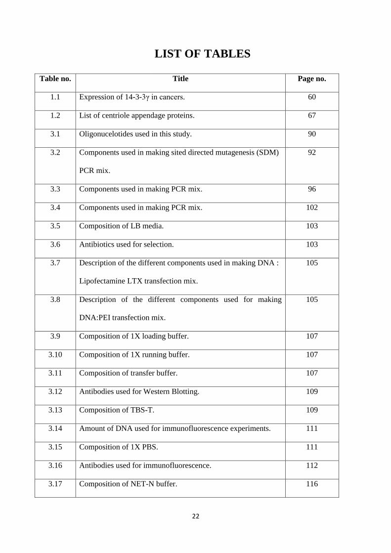
22
LIST OF TABLES
Table no. Title Page no.
1.1 Expression of 14-3-3γ in cancers. 60
1.2 List of centriole appendage proteins. 67
3.1 Oligonucelotides used in this study. 90
3.2 Components used in making sited directed mutagenesis (SDM)
PCR mix.
92
3.3 Components used in making PCR mix. 96
3.4 Components used in making PCR mix. 102
3.5 Composition of LB media. 103
3.6 Antibiotics used for selection. 103
3.7 Description of the different components used in making DNA :
Lipofectamine LTX transfection mix.
105
3.8 Description of the different components used for making
DNA:PEI transfection mix.
105
3.9 Composition of 1X loading buffer. 107
3.10 Composition of 1X running buffer. 107
3.11 Composition of transfer buffer. 107
3.12 Antibodies used for Western Blotting. 109
3.13 Composition of TBS-T. 109
3.14 Amount of DNA used for immunofluorescence experiments. 111
3.15 Composition of 1X PBS. 111
3.16 Antibodies used for immunofluorescence. 112
3.17 Composition of NET-N buffer. 116
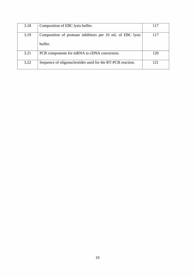
23
3.18 Composition of EBC lysis buffer. 117
3.19 Composition of protease inhibitors per 10 mL of EBC lysis
buffer.
117
3.21 PCR components for mRNA to cDNA conversion. 120
3.22 Sequence of oligonucleotides used for the RT-PCR reaction. 121
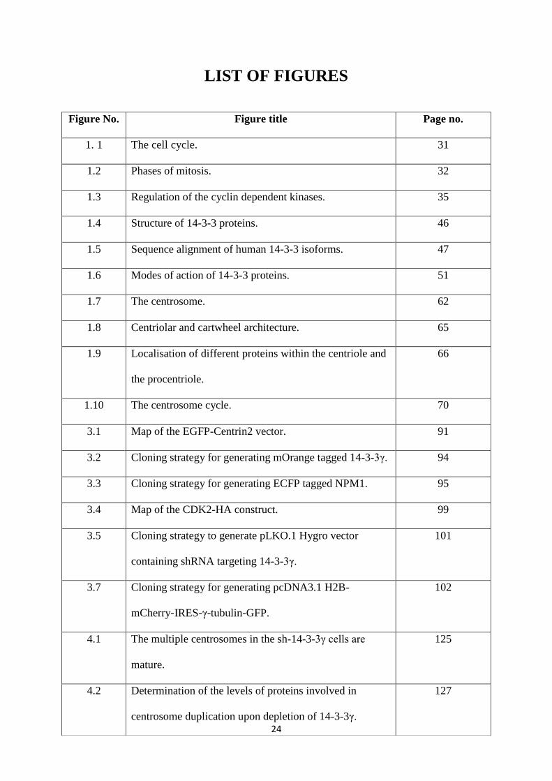
24
LIST OF FIGURES
Figure No. Figure title Page no.
1. 1 The cell cycle. 31
1.2 Phases of mitosis. 32
1.3 Regulation of the cyclin dependent kinases. 35
1.4 Structure of 14-3-3 proteins. 46
1.5 Sequence alignment of human 14-3-3 isoforms. 47
1.6 Modes of action of 14-3-3 proteins. 51
1.7 The centrosome. 62
1.8 Centriolar and cartwheel architecture. 65
1.9 Localisation of different proteins within the centriole and
the procentriole.
66
1.10 The centrosome cycle. 70
3.1 Map of the EGFP-Centrin2 vector. 91
3.2 Cloning strategy for generating mOrange tagged 14-3-3γ. 94
3.3 Cloning strategy for generating ECFP tagged NPM1. 95
3.4 Map of the CDK2-HA construct. 99
3.5 Cloning strategy to generate pLKO.1 Hygro vector
containing shRNA targeting 14-3-3γ.
101
3.7 Cloning strategy for generating pcDNA3.1 H2B-
mCherry-IRES-γ-tubulin-GFP.
102
4.1 The multiple centrosomes in the sh-14-3-3γ cells are
mature.
125
4.2 Determination of the levels of proteins involved in
centrosome duplication upon depletion of 14-3-3γ.
127
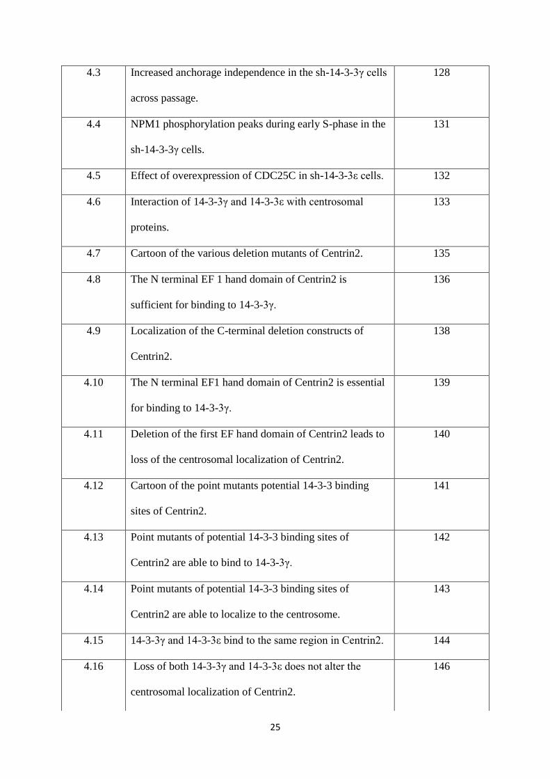
25
4.3 Increased anchorage independence in the sh-14-3-3γ cells
across passage.
128
4.4 NPM1 phosphorylation peaks during early S-phase in the
sh-14-3-3γ cells.
131
4.5 Effect of overexpression of CDC25C in sh-14-3-3ε cells. 132
4.6 Interaction of 14-3-3γ and 14-3-3ε with centrosomal
proteins.
133
4.7 Cartoon of the various deletion mutants of Centrin2. 135
4.8 The N terminal EF 1 hand domain of Centrin2 is
sufficient for binding to 14-3-3γ.
136
4.9 Localization of the C-terminal deletion constructs of
Centrin2.
138
4.10 The N terminal EF1 hand domain of Centrin2 is essential
for binding to 14-3-3γ.
139
4.11 Deletion of the first EF hand domain of Centrin2 leads to
loss of the centrosomal localization of Centrin2.
140
4.12 Cartoon of the point mutants potential 14-3-3 binding
sites of Centrin2.
141
4.13 Point mutants of potential 14-3-3 binding sites of
Centrin2 are able to bind to 14-3-3γ.
142
4.14 Point mutants of potential 14-3-3 binding sites of
Centrin2 are able to localize to the centrosome.
143
4.15 14-3-3γ and 14-3-3ε bind to the same region in Centrin2. 144
4.16 Loss of both 14-3-3γ and 14-3-3ε does not alter the
centrosomal localization of Centrin2.
146
26
4.17 Expression of a 14-3-3 binding-deficient mutant of
Centrin2 does not interfere with spindle organization.
148
4.18 Expression of a 14-3-3 binding-deficient mutant of
Centrin2 does not interfere with mitotic progression.
150
4.19 The deletion constructs of γ-tubulin. 152
4.20 Localisation of the γ-tubulin deletion constructs. 153
4.21 Mapping the 14-3-3γ binding site on γ-tubulin. 154
4.22 Deletion constructs of GCP2. 156
4.23 Localisation of the GCP2 deletion mutants. 157
4.24 Putative 14-3-3 binding sites on Cep170. 158
4.25 Loss of 14-3-3γ results in the decrease of Cep170 at both
protein and mRNA levels.
159
4.26 Intensity of Cep170 at the centrosome in sh-14-3-3γ cells
is not altered.
161
4.27 Sequence alignment of the peptide binding groove of 14-
3-3 isoforms.
162
4.28 Expression of the mOrange tagged 14-3-3γ constructs. 163
4.29 Effect of the expression of the 14-3-3γ mutants on
centrosome number.
164
4.30 Effect of the 14-3-3γ mutants in the sh-14-3-3γ cells. 166
4.31 Effect of the mutants is conserved in HaCat and HEK293
cell lines.
168
4.32 The three possibilities that can lead to the presence of a
single centrosome in mitosis. (1) defect in separation, (2)
169
27
defect in disengagement and (3) defect in duplication.
4.33 Centriolar organization in cells expressing the different
14-3-3γ mutants.
170
4.34 Ultrastructure of the centrosomes. 172
4.35 Organization of intercentriolar linker proteins in cells
expressing the mutants.
174
4.36 Determination of centriolar age in cells expressing the
mutants.
176
4.37 Spindle organization in cells expressing the mutants. 178
4.38 Duration of mitosis in cells expressing the mutants. 181
4.39 Localization of the mutants in interphase. 183
4.40 Effect of overxpression of Plk4, CDK1 and CDK2 on the
single centrosome phenotype.
187
4.41 Role of T199 mutants of NPM1 in the centrosome cycle. 189
4.42 Reversal of the centrosome phenotype by NPM1. 191
4.43 Reversal of the single centrosome phenotype is specific to
the T199D mutant of NPM1.
192
4.44 Interaction of GST-14-3-3γ with NPM1. 193
4.45 The 14-3-3γ mutants interact differentially with NPM1. 194
4.46 Mapping the 14-3-3 binding site of NPM1. 195
4.47 Interaction of the S48 mutants with 14-3-3γ. 196
4.48 Localization of the S48 mutants of NPM1. 198
4.49 Effect of S48 mutants of NPM1 on centrosome number. 201
4.50 p-T199 status of the S48 mutants of NPM1. 203
28
4.51 14-3-3γ inhibits the ability of NPM1 to activate ROCKII. 204
4.52 Generation of a knockdown of 14-3-3γ in the HeLa Kyoto
cell line.
207
4.53 Generation of the pcDNA3.1 –γ-tubulin-GFP-IRES-H2B-
mCherry construct.
210
4.54 Expression of the pcDNA3.1H2B-mCherry-IRES-γ-
tubulin-GFP.
211
5.1 Model describing the role of 14-3-3 proteins in regulating
the centrosomal localization of Centrin2.
216
5.2 Representation of the connection between 14-3-3 and
centrosomal proteins.
218
5.3 Model explaining the regulation of NPM1 function by 14-
3-3γ.
225
5.4 Model representing the different contributions of 14-3-3
proteins to the centrosome cycle.
227

29
1. Introduction
30
1. Introduction
1.1 The cell cycle
The cell cycle is a structured series of events which ensures that replicated DNA is separated
accurately into two daughter cells (fig. 1.1) [reviewed in (1,2)]. Two of the most important
events that occur during the cell cycle are; precise copying of the genome (S phase), and
segregation of the duplicated genome into daughter cells (M phase) [reviewed in (2,3)].
These two phases are temporally distinct and are divided due to the existence of two gap
phases (G1 and G2) (4). It is extremely essential that the replication and segregation phases
are distinct and alternate, to ensure the maintenance of ploidy. This ordered progression is
ensured by the unidirectional nature of the cell cycle; once a cell crosses the G1/S transition,
it is committed to replicate its DNA [reviewed in (5)]. Similarly, once a cell has crossed the
G2/M transition, it is committed to completing mitosis and will not usually revert to a second
G2 [reviewed in (5)].
Mammalian cells are sensitive to mitogenic stimuli in their environment during a specific
time in the early G1 phase of the cell cycle, known as the restriction point (R) (6). Here,
the cell assesses the cumulative effect of all growth factor stimuli, or the absence of them.
If the signals are favourable to growth, then the cell enters the cell cycle. Under certain
conditions, including high cell density and an absence of growth factors, mammalian cells
will accumulate in a state of 2n DNA content. This is known as the G0 phase or
quiescence, which is an out-of-cycle phase and could be one of minimal metabolism
[reviewed in (7)]. At other times during the cell cycle, the absence of mitogens does not
31
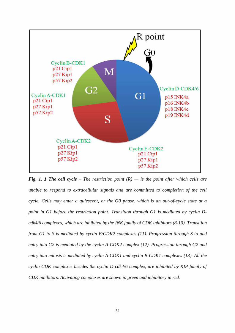
Fig. 1. 1 The cell cycle – The restriction point (R) — is the point after which cells are
unable to respond to extracellular signals and are committed to completion of the cell
cycle. Cells may enter a quiescent, or the G0 phase, which is an out-of-cycle state at a
point in G1 before the restriction point. Transition through G1 is mediated by cyclin D-
cdk4/6 complexes, which are inhibited by the INK family of CDK inhibitors (8-10). Transition
from G1 to S is mediated by cyclin E/CDK2 complexes (11). Progression through S to and
entry into G2 is mediated by the cyclin A-CDK2 complex (12). Progression through G2 and
entry into mitosis is mediated by cyclin A-CDK1 and cyclin B-CDK1 complexes (13). All the
cyclin-CDK complexes besides the cyclin D-cdk4/6 complex, are inhibited by KIP family of
CDK inhibitors. Activating complexes are shown in green and inhibitory in red.
32
affect cell-cycle progression and the cells will proceed through the cell cycle, back to the
responsive period. There are four phases in the cell cycle; G1, S, G2 and M (fig. 1.1).
G1 – This is the first gap phase in the cell cycle. During this phase, the cell can either be
committed to division or withdraw from the cell cycle and enter a resting, G0 phase. This
decision depends on a number of extracellular growth signals (14). The cell also synthesizes
proteins necessary for DNA replication.
S – This is the synthesis phase of the cell cycle, during which the cell replicates its DNA.
During S phase, the cell also duplicates its centrosome, an organelle essential for
chromosome segregation.
G2 – This is the second gap phase in the cell cycle where the cell prepares for the next phase,
mitosis. Organelles and proteins essential for mitosis are synthesized in this phase.
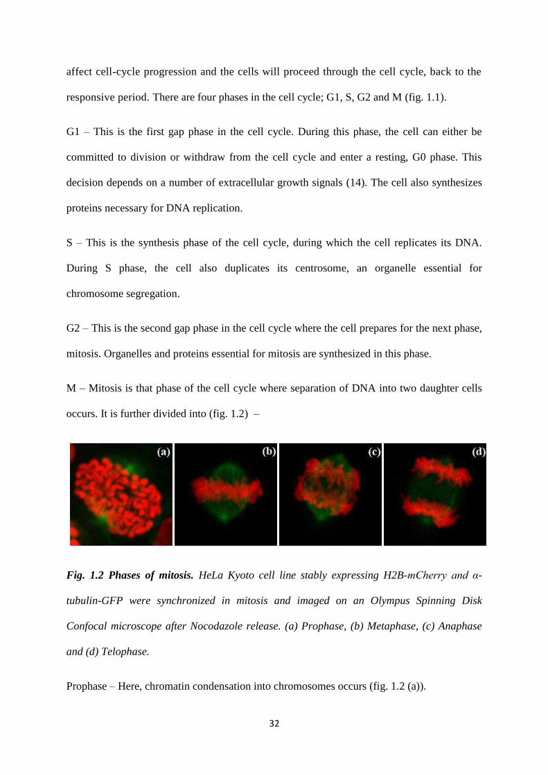
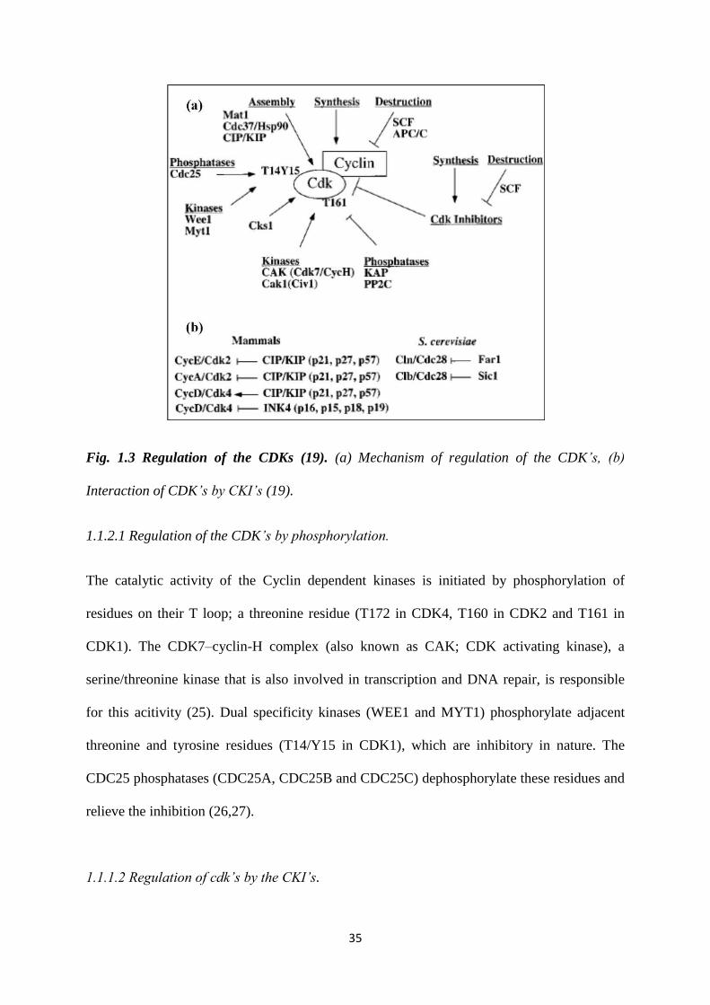
M – Mitosis is that phase of the cell cycle where separation of DNA into two daughter cells
occurs. It is further divided into (fig. 1.2) –
Fig. 1.2 Phases of mitosis. HeLa Kyoto cell line stably expressing H2B-mCherry and α-
tubulin-GFP were synchronized in mitosis and imaged on an Olympus Spinning Disk
Confocal microscope after Nocodazole release. (a) Prophase, (b) Metaphase, (c) Anaphase
and (d) Telophase.
Prophase – Here, chromatin condensation into chromosomes occurs (fig. 1.2 (a)).
33
Metaphase – Spindle assembly occurs and the chromosomes align at the metaphase plate.
Each kinetochore has to be attached to a spindle microtubule before the next phase can begin
(fig. 1.2 (b)).
Anaphase – The spindle starts shortening, and the paired chromatids are pulled apart towards
each end of the bipolar spindle (fig. 1.2 (c)).
Telophase – The single chromatids reach the poles of the spindle and cell division is initiated
(fig. 1.2 (d)).
Cytokinesis – The cell divides into two daughter cells, each with a full complement of the
genome.
There are exceptions to this ordered cell cycle. The development of the endosperm in plants
occurs due to formation of a syncytium, a large cell comprising 100 nuclei post fertilization
(15). The Drosophila embryo also develops as a syncytium, as a series of nuclear
duplications without cell divisions (16). Salivary glands of Chironomous contain Polytene
chromosomes, which are created due to repeated rounds of chromosome duplication without
segregation (17). Previously, it was believed that neurons exist in a post-mitotic state;
however, recent reports indicate tetraploidization in neurons (18).
1.1.1 Regulation of the cell cycle
Ordered succession through a cell cycle is mediated by the activity of a heterodimer made of
two classes of proteins; the cyclins and the cyclin dependent kinases (CDK’s). The cyclins
are the regulatory subunit, while the cyclin dependent kinases are the catalytic subunit (19).
Physical interaction with the cyclin controls the catalytic activity of the CDK (20,21).
Multiple CDK’s are bound by multiple cyclins in animal cells, the abundance of the cyclins is
the determining factor. The expression of cyclins is cyclic in nature; cyclins are expressed

34
and undergo degradation, depending on the cell cycle phase. Each of the cyclins is under
different transcriptional and proteolytic controls. Activation of a CDK by a cyclin, thus lends
temporal control of its activity and confers substrate specificity via the bound cyclin.
Different cyclin-CDK complexes are active during distinct phases of the cell cycle. The
cyclin D-CDK4/6 complex is active during G1, the cyclin E-CDK2 complex during the G1-S
transition, cyclin A-CDK2 during S phase and cyclin A/B-CDK1 prior to and during mitosis
up to the end of metaphase (fig. 1.1) (22-24). In mammals, each cyclin has multiple family
members, A1, A2, A3, B1, B2, D1, D2, D3, E1 and E2 (19).
1.1.2 Regulation of CDK’s.
Regulation of the CDKs occurs at many levels. These include, increased cyclin concentration,
leading to binding to CDKs, activating and inhibitory phosphorylation (T14Y15 and T161),
Cdk inhibitors (CIP/KIP and INK4), and SCF (Skp1/Cul1/F-box protein) ubiquitin ligase and
the APC/C (Anaphase Promoting Complex/Cyclosome) mediated ubiquitination and
subsequent degradation of cyclins and Cdk inhibitors (fig. 1.3).
35
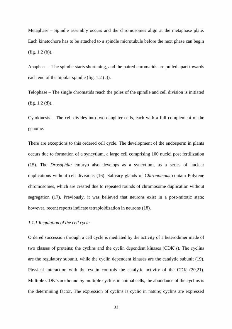
Fig. 1.3 Regulation of the CDKs (19). (a) Mechanism of regulation of the CDK’s, (b)
Interaction of CDK’s by CKI’s (19).
1.1.2.1 Regulation of the CDK’s by phosphorylation.
The catalytic activity of the Cyclin dependent kinases is initiated by phosphorylation of
residues on their T loop; a threonine residue (T172 in CDK4, T160 in CDK2 and T161 in
CDK1). The CDK7–cyclin-H complex (also known as CAK; CDK activating kinase), a
serine/threonine kinase that is also involved in transcription and DNA repair, is responsible
for this acitivity (25). Dual specificity kinases (WEE1 and MYT1) phosphorylate adjacent
threonine and tyrosine residues (T14/Y15 in CDK1), which are inhibitory in nature. The
CDC25 phosphatases (CDC25A, CDC25B and CDC25C) dephosphorylate these residues and
relieve the inhibition (26,27).
1.1.1.2 Regulation of cdk’s by the CKI’s.
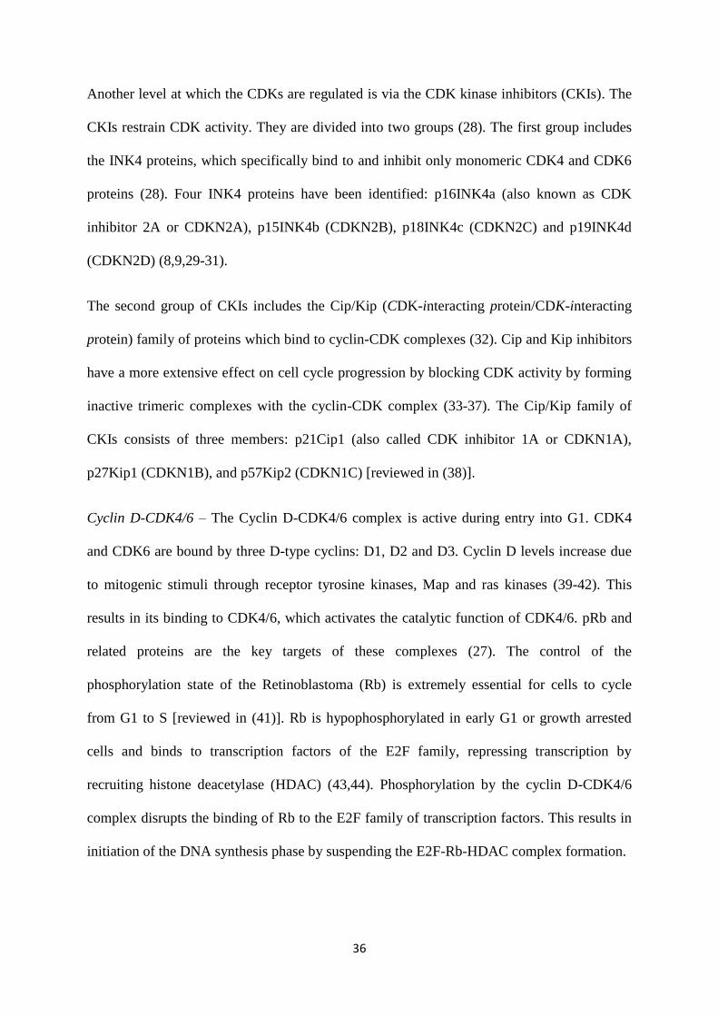
36
Another level at which the CDKs are regulated is via the CDK kinase inhibitors (CKIs). The
CKIs restrain CDK activity. They are divided into two groups (28). The first group includes
the INK4 proteins, which specifically bind to and inhibit only monomeric CDK4 and CDK6
proteins (28). Four INK4 proteins have been identified: p16INK4a (also known as CDK
inhibitor 2A or CDKN2A), p15INK4b (CDKN2B), p18INK4c (CDKN2C) and p19INK4d
(CDKN2D) (8,9,29-31).
The second group of CKIs includes the Cip/Kip (CDK-interacting protein/CDK-interacting
protein) family of proteins which bind to cyclin-CDK complexes (32). Cip and Kip inhibitors
have a more extensive effect on cell cycle progression by blocking CDK activity by forming
inactive trimeric complexes with the cyclin-CDK complex (33-37). The Cip/Kip family of
CKIs consists of three members: p21Cip1 (also called CDK inhibitor 1A or CDKN1A),
p27Kip1 (CDKN1B), and p57Kip2 (CDKN1C) [reviewed in (38)].
Cyclin D-CDK4/6 – The Cyclin D-CDK4/6 complex is active during entry into G1. CDK4
and CDK6 are bound by three D-type cyclins: D1, D2 and D3. Cyclin D levels increase due
to mitogenic stimuli through receptor tyrosine kinases, Map and ras kinases (39-42). This
results in its binding to CDK4/6, which activates the catalytic function of CDK4/6. pRb and
related proteins are the key targets of these complexes (27). The control of the
phosphorylation state of the Retinoblastoma (Rb) is extremely essential for cells to cycle
from G1 to S [reviewed in (41)]. Rb is hypophosphorylated in early G1 or growth arrested
cells and binds to transcription factors of the E2F family, repressing transcription by
recruiting histone deacetylase (HDAC) (43,44). Phosphorylation by the cyclin D-CDK4/6
complex disrupts the binding of Rb to the E2F family of transcription factors. This results in
initiation of the DNA synthesis phase by suspending the E2F-Rb-HDAC complex formation.
37
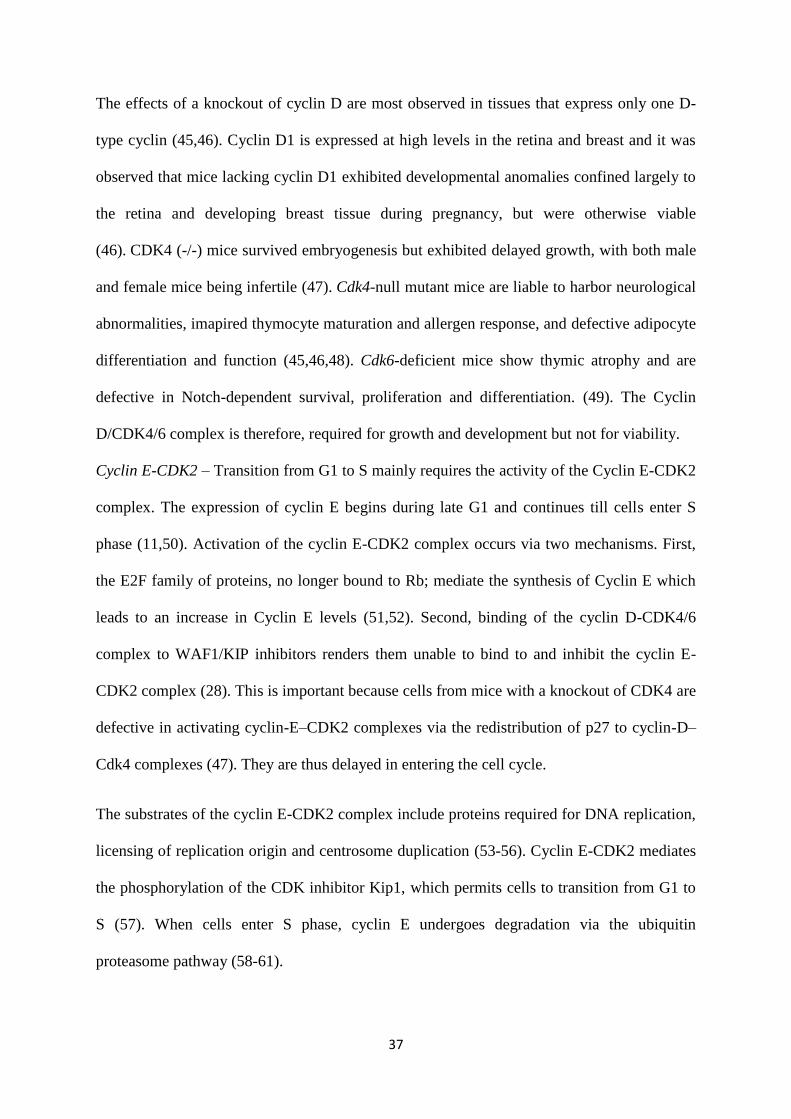
The effects of a knockout of cyclin D are most observed in tissues that express only one D-
type cyclin (45,46). Cyclin D1 is expressed at high levels in the retina and breast and it was
observed that mice lacking cyclin D1 exhibited developmental anomalies confined largely to
the retina and developing breast tissue during pregnancy, but were otherwise viable
(46). CDK4 (-/-) mice survived embryogenesis but exhibited delayed growth, with both male
and female mice being infertile (47). Cdk4-null mutant mice are liable to harbor neurological
abnormalities, imapired thymocyte maturation and allergen response, and defective adipocyte
differentiation and function (45,46,48). Cdk6-deficient mice show thymic atrophy and are
defective in Notch-dependent survival, proliferation and differentiation. (49). The Cyclin
D/CDK4/6 complex is therefore, required for growth and development but not for viability.
Cyclin E-CDK2 – Transition from G1 to S mainly requires the activity of the Cyclin E-CDK2
complex. The expression of cyclin E begins during late G1 and continues till cells enter S
phase (11,50). Activation of the cyclin E-CDK2 complex occurs via two mechanisms. First,
the E2F family of proteins, no longer bound to Rb; mediate the synthesis of Cyclin E which
leads to an increase in Cyclin E levels (51,52). Second, binding of the cyclin D-CDK4/6
complex to WAF1/KIP inhibitors renders them unable to bind to and inhibit the cyclin E-
CDK2 complex (28). This is important because cells from mice with a knockout of CDK4 are
defective in activating cyclin-E–CDK2 complexes via the redistribution of p27 to cyclin-D–
Cdk4 complexes (47). They are thus delayed in entering the cell cycle.
The substrates of the cyclin E-CDK2 complex include proteins required for DNA replication,
licensing of replication origin and centrosome duplication (53-56). Cyclin E-CDK2 mediates
the phosphorylation of the CDK inhibitor Kip1, which permits cells to transition from G1 to
S (57). When cells enter S phase, cyclin E undergoes degradation via the ubiquitin
proteasome pathway (58-61).

38
Cdk2-null mice are viable and have no defects apart from male and female sterility due to
meiotic defects, with a life span of 2 yrs (62,63). It is possible that other cdk’s might
compensate for its absence. Cyclin E1 or E2 deletion is also as harmless as cdk2 deletion, as
the male knockout mice have reduced fertility, but no other phenotype (64,65). However,
defects in duplication cycles in placental trophoblast giant cells resulted in embryonic
lethality by E11.5, in mice with a double knockout of both cyclin E1 and E2 (65). Cyclin E is
therefore, not essential for mitotic cells in embryonic development, but for endoreplicating
cells. Also, it is possible that the cyclin E proteins have redundant functions within a class
and a dual knockout is thus, fatal.
Cyclin A-CDK2 and Cyclin A/CDK1 – The cyclin A-CDK2 complex is responsible for cells
to go through S phase, whereas the cyclin A-CDK1 mediates entry into mitosis. Cyclin A
unbound CDK is inactive as it is inaccessible to substrate due to the presence of an
unphosphorylated T-loop region (20). Also, the catalytic residues in the CDK are not
arranged in the correct conformation for catalysis (12,66). Upon Cyclin A binding, CDK2
and CDK1 undergo a conformational change that makes them accessible to the substrate,
ATP (67). Substrates of the kinase activity of the cyclin A-CDK2 complex in S phase initiate
DNA replication (68-70). Cyclin A-CDK2 is also necessary for the synchronization of the
end of the S phase with activation of the mitotic cyclin-CDKs (71). The cyclin A-CDK1
complex promotes mitotic entry by phosphorylating Bora, which mediates Aurora-A
dependent Plk1 phosphorylation (72). Myosin phosphatase targeting subunit 1 (MYPT1) is
also a cyclin A-CDK1 substrate that modulates Plk1 at kinetochores in prometaphase (73).
The Anaphase Promoting Complex (APC) mediated proteolysis of cyclin A is essential for
mitotic exit (74).
Cyclin A function is required for the division of hematopoietic and embryonic stem cells, as
germline Cyclin A2 knockout mice die at embryonic stage E5.5 (75,76). The importance of
39
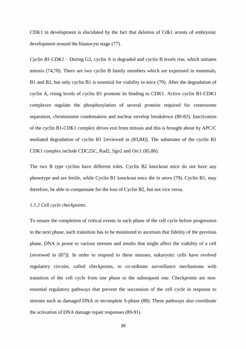
CDK1 in development is elucidated by the fact that deletion of Cdk1 arrests of embryonic
development around the blastocyst stage (77).
Cyclin B1-CDK1 – During G2, cyclin A is degraded and cyclin B levels rise, which initiates
mitosis (74,78). There are two cyclin B family members which are expressed in mammals,
B1 and B2, but only cyclin B1 is essential for viability in mice (79). After the degradation of
cyclin A, rising levels of cyclin B1 promote its binding to CDK1. Active cyclin B1-CDK1
complexes regulate the phosphorylation of several proteins required for centrosome
separation, chromosome condensation and nuclear envelop breakdown (80-82). Inactivation
of the cyclin B1-CDK1 complex drives exit from mitosis and this is brought about by APC/C
mediated degradation of cyclin B1 [reviewed in (83,84)]. The substrates of the cyclin B1
CDK1 complex include CDC25C, Rad2, Sgo2 and Orc1 (85,86).
The two B type cyclins have different roles. Cyclin B2 knockout mice do not have any
phenotype and are fertile, while Cyclin B1 knockout mice die in utero (79). Cyclin B1, may
therefore, be able to compensate for the loss of Cyclin B2, but not vice versa.
1.1.2 Cell cycle checkpoints.
To ensure the completion of critical events in each phase of the cell cycle before progression
to the next phase, each transition has to be monitored to ascertain that fidelity of the previous
phase. DNA is prone to various stresses and insults that might affect the viability of a cell
[reviewed in (87)]. In order to respond to these stresses, eukaryotic cells have evolved
regulatory circuits, called checkpoints, to co-ordinate surveillance mechanisms with
transition of the cell cycle from one phase to the subsequent one. Checkpoints are non-
essential regulatory pathways that prevent the succession of the cell cycle in response to
stresses such as damaged DNA or incomplete S-phase (88). These pathways also coordinate
the activation of DNA damage repair responses (89-91).
40
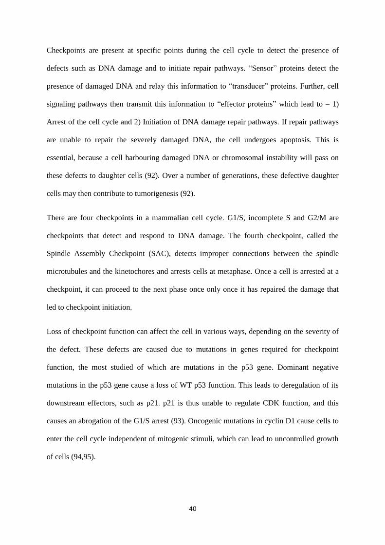
Checkpoints are present at specific points during the cell cycle to detect the presence of
defects such as DNA damage and to initiate repair pathways. “Sensor” proteins detect the
presence of damaged DNA and relay this information to “transducer” proteins. Further, cell
signaling pathways then transmit this information to “effector proteins” which lead to – 1)
Arrest of the cell cycle and 2) Initiation of DNA damage repair pathways. If repair pathways
are unable to repair the severely damaged DNA, the cell undergoes apoptosis. This is
essential, because a cell harbouring damaged DNA or chromosomal instability will pass on
these defects to daughter cells (92). Over a number of generations, these defective daughter
cells may then contribute to tumorigenesis (92).
There are four checkpoints in a mammalian cell cycle. G1/S, incomplete S and G2/M are
checkpoints that detect and respond to DNA damage. The fourth checkpoint, called the
Spindle Assembly Checkpoint (SAC), detects improper connections between the spindle
microtubules and the kinetochores and arrests cells at metaphase. Once a cell is arrested at a
checkpoint, it can proceed to the next phase once only once it has repaired the damage that
led to checkpoint initiation.
Loss of checkpoint function can affect the cell in various ways, depending on the severity of
the defect. These defects are caused due to mutations in genes required for checkpoint
function, the most studied of which are mutations in the p53 gene. Dominant negative
mutations in the p53 gene cause a loss of WT p53 function. This leads to deregulation of its
downstream effectors, such as p21. p21 is thus unable to regulate CDK function, and this
causes an abrogation of the G1/S arrest (93). Oncogenic mutations in cyclin D1 cause cells to
enter the cell cycle independent of mitogenic stimuli, which can lead to uncontrolled growth
of cells (94,95).
41
G1/S checkpoint – This checkpoint is present just before the cell can enter S phase and
prevents replication of damaged DNA. It also prevents the synthesis of proteins required for
S phase progression. The checkpoint can be initiated by exposure to Ionizing Radiation (IR)
or DNA damaging agents such as lovastatin, cisplatin and bleomycin (96-98). Upon exposure
to DNA damaging agents, ATM (ataxia telangiectasia, mutated) and ATR (ataxia
telangiectasia and Rad3-related) kinases phosphorylate the checkpoint serine/ threonine
kinases, CHK1 (checkpoint kinase 1) and CHK2 (checkpoint kinase 2) (99). These kinases
then phosphorylate CDC25A, leading to its ubiquitination and degradation (100-102).
Inactivation of CDC25A prevents activation of cyclin E-CDK2 complexes, thus inhibiting
G1/S transition (100). Activated ATM and ATR also stabilise p53 levels (103-105). Increased
p53 levels lead to an increase in the levels of its downstream effector protein, p21, which
inhibits the cyclin E-CDK2 complex (106,107).
Another mechanism for ensuring G1/S arrest is via the INK4 and Cip/Kip families of CKIs.
Expression of INK4 titrates CDK4 away from the cyclin D-CDK4 complex and cyclin D is
degraded via the ubiquitin proteasome pathway (8,108). Also, in early G1 phase, binding of
p21Cip1 and p27Kip1 inhibits the cyclin E-CDK2 and cyclin A-CDK2 (10,93,109). For entry
in S phase, the cyclin D-CDK4/6 complex must sequester the p21Cip1 and p27Kip1 away
from the cyclin E-CDK2 and cyclin A-CDK2 complexes. Degradation of D-type cyclins
therefore prevents the titration of p21Cip1 and p27Kip1 by cyclin D-CDK4/6 complexes. The
Cip/Kip bound cyclin E-CDK2 and cyclin A-CDK2 complexes thus prevent S phase entry
(110).
Incomplete S-phase checkpoint – This checkpoint monitors DNA replication and the presence
of stalled replication forks. When the incomplete S-phase checkpoint is initiated, it prevents
replication initiation and fork progression, and therefore, slows down DNA replication (111-
113). The cell thus has time to repair damaged DNA and complete DNA replication. Loss of
42
the incomplete S-phase checkpoint results in cells entering mitosis prematurely, via
Premature Chromatin Condensation (PCC), which can be lethal (114). The incomplete S-
phase checkpoint is activated upon exposure to UV radiation, X-rays or chemical mutagens
such as hydroxyurea. Exposure to these agents leads to the formation of pyrimidine dimers,
DNA methylation, depletion of deoxyribonucleotides and damaged DNA (115,116).
ATR senses the presence of single stranded DNA lesions and stalled replication forks, which
lead to its activation and phosphorylates and activates Chk1 and Chk2. This results in the
inhibition of CDC25A, CDC25B and CDC25C, which averts the activation of cyclin E-
CDK2, cyclinA-CDK2 and cyclin A-CDK1 and thus, G2 entry. It also prevents activation of
CDK1 by its dephosphorylation on Thr 14 and Tyr 15. This prevents cyclin B1-CDK1
complex activation and inhibits progression into mitosis (117-125).
G2/M checkpoint – The G2/M checkpoint is activated upon DNA damage to cells that are in
G2 or cells that acquired DNA damage in previous cell cycle phases but escaped checkpoint
activity (126,127). It is activated by agents such as ionizing radiation, Adriamycin and
Bleomycin, that cause double strand breaks. The G2/M is the last checkpoint that monitors
DNA damage before cells enter mitosis. It has been shown that cells lacking the p53, which
are unable to arrest at the G1/S checkpoint, might depend on the G2/M checkpoint to repair
DNA damage (128). Inability of cells to undergo the G2/M arrest upon DNA damage can
result in the production of daughter cells with defective DNA and can contribute to
tumourigenesis.
This checkpoint is activated by the rad family of proteins that sense double strand DNA
damage (129). This results in the activation of the ATM and ATR family of kinases, which
phosphorylate Chk1 and Chk2 kinases (117,130). The ultimate objective of this checkpoint is
preventing the activation of the cyclin B1-CDK1 complex. This is achieved by the
43
inactivation of the CDC25 phosphatases A, B and C, due to phosphorylation by Chk1, Chk2
and C-TAK1 (117-125,131,132). This phosphorylation results in the generation of a 14-3-3
binding site on CDC25C at a serine 216 residue (121,133). 14-3-3 proteins sequester
CDC25C in the cytoplasm and prevents its nuclear localisation. This inhibits its ability to
activate the cyclin B1-CDK1 complex and thus prevents mitotic progression. The inhibitory
phosphorylation on CDK1 is further accentuated by the association of Wee1 with 14-3-3
proteins. This binding activates Wee1 and maintains the inhibitory phosphorylation of CDK1
(134,135).
Spindle Assembly Checkpoint (SAC) – During mitosis, replicated chromosomes have to be
divided into two daughter cells. The paired chromosomes are attached to the spindle
microtubules via kinetochores at the metaphase plate, followed by segregation during
anaphase into two daughter cells. Inability of the chromosomes to attach correctly to the
spindle microtubules can result in the two daughter cells receiving unequal complements of
the genome, which may lead to apoptosis or disease. The Spindle Assembly Checkpoint
(SAC) prevents the anaphase transition of a cell in metaphase with incorrect attachment
between the kinetochores and the mitotic spindle [reviewed in (136)]. It delays anaphase
transition until all the chromosomes are properly attached to the mitotic spindle.
The SAC is activated upon loss of spindle pole tension, unattached kinetochores at metaphase
or by certain spindle poisons such as nocodazole (137). The goal of the SAC is preventing the
degradation of the APC/C. Several proteins regulate the SAC - BUB1 (budding uninhibited
by benzimidazole 1) and the MAD (mitotic-arrest deficient) family of proteins, MAD1,
MAD2 and MAD3 (BUBR1 in humans) (138-140). The target of the SAC is the co-factor of
the ubiquitin ligase APC/C, CDC20 (141-144). Activation of the SAC hinders the ability of
CDC20 to stimulate APC/C-mediated ubiquitination of two key substrates, cyclin B and
Securin, thus preventing their degradation by the 26S proteosome. Cyclin B degradation is
44
required for mitotic exit (84). Destruction of Securin leads to activation of Separase, which
results in anaphase onset by cleaving the cohesion complex that holding sister chromatids
together (145-147). Thus, by controlling CDC20 function, the SAC allows chromosomes to
align properly on the metaphase plate and attach to the spindle poles via microtubules. The
checkpoint is quenched when the chromosomes are oriented correctly. This releases the cell
from a mitotic arrest, allowing anaphase to begin.
1.2 14-3-3 proteins.
14-3-3 proteins are a family of small, dimeric, acidic, regulatory proteins [reviewed in (148)].
They were first isolated as abundant, soluble proteins from bovine brain tissue lysates and
were theorized to be brain specific (149). 14-3-3 proteins derive their name from the elution
fraction of bovine brain tissue lysates containing these proteins following DEAE-cellulose
chromatography and their migration position after starch gel electrophoresis (149). The 14-3-
3 proteins were extracted by the authors’ from the 14th fraction of bovine brain lysate in a
DEAE cellulose column and fractions 3.3 of the final step (149). Subsequent research has
revealed that 14-3-3 proteins contribute to regulation of the cell cycle, cellular metabolism,
apoptosis, cell-cell adhesion, protein transport, transcription and malignant transformation
(150-156).
14-3-3 proteins were the first proteins demonstrated to bind to phosphorylated Serine or
Threonine residues (157,158). There are seven isoforms of 14-3-3 proteins in mammalian
cells; β, γ, ε, σ, η, τ (or θ) and ζ, with δ and α being the phosphoforms of β and ζ, respectively
(148,159). These isoforms were named with respect to their order of elution on HPLC
(160,161). The different isoforms are quite homologous, but contain a few regions of
variation. 14-3-3 proteins are found in most eukaryotic organisms, from fungi to mammals
[reviewed in (162)]. Multiple 14-3-3 isoforms are expressed across eukaryotes, from two in
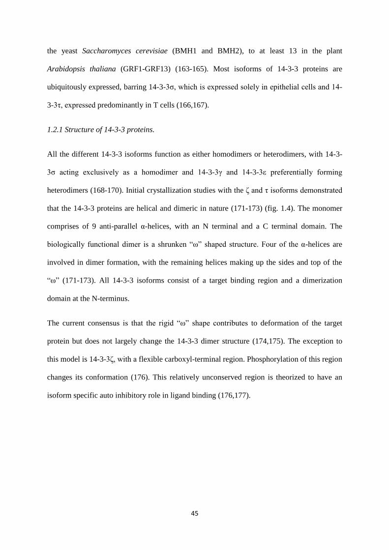
45
the yeast Saccharomyces cerevisiae (BMH1 and BMH2), to at least 13 in the plant
Arabidopsis thaliana (GRF1-GRF13) (163-165). Most isoforms of 14-3-3 proteins are
ubiquitously expressed, barring 14-3-3σ, which is expressed solely in epithelial cells and 14-
3-3τ, expressed predominantly in T cells (166,167).
1.2.1 Structure of 14-3-3 proteins.
All the different 14-3-3 isoforms function as either homodimers or heterodimers, with 14-3-
3σ acting exclusively as a homodimer and 14-3-3γ and 14-3-3ε preferentially forming
heterodimers (168-170). Initial crystallization studies with the ζ and τ isoforms demonstrated
that the 14-3-3 proteins are helical and dimeric in nature (171-173) (fig. 1.4). The monomer
comprises of 9 anti-parallel α-helices, with an N terminal and a C terminal domain. The
biologically functional dimer is a shrunken “ω” shaped structure. Four of the α-helices are
involved in dimer formation, with the remaining helices making up the sides and top of the
“ω” (171-173). All 14-3-3 isoforms consist of a target binding region and a dimerization
domain at the N-terminus.
The current consensus is that the rigid “ω” shape contributes to deformation of the target
protein but does not largely change the 14-3-3 dimer structure (174,175). The exception to
this model is 14-3-3ζ, with a flexible carboxyl-terminal region. Phosphorylation of this region
changes its conformation (176). This relatively unconserved region is theorized to have an
isoform specific auto inhibitory role in ligand binding (176,177).
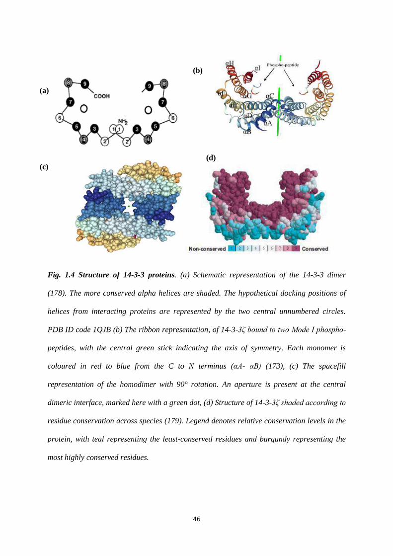
46
Fig. 1.4 Structure of 14-3-3 proteins. (a) Schematic representation of the 14-3-3 dimer
(178). The more conserved alpha helices are shaded. The hypothetical docking positions of
helices from interacting proteins are represented by the two central unnumbered circles.
PDB ID code 1QJB (b) The ribbon representation, of 14-3-3ζ bound to two Mode I phospho-
peptides, with the central green stick indicating the axis of symmetry. Each monomer is
coloured in red to blue from the C to N terminus (αA- αB) (173), (c) The spacefill
representation of the homodimer with 90° rotation. An aperture is present at the central
dimeric interface, marked here with a green dot, (d) Structure of 14-3-3ζ shaded according to
residue conservation across species (179). Legend denotes relative conservation levels in the
protein, with teal representing the least-conserved residues and burgundy representing the
most highly conserved residues.
(a)
(c)
(b)
(d)

47
Fig. 1.5 Sequence alignment of human 14-3-3 isoforms. Conserved residues are highlighted
by a red line in the sequence alignment.
There are five conserved sequence blocks within the 14-3-3 family of proteins. These blocks
comprise the end of helix αA, the αA/αB-connecting loop, the helices involved in ligand
binding (αC, αG, αE and αI), and the start of helix αB, that constitute an essential element of
the dimer interface (174,178) (fig. 1.5). A large negatively charged channel is created due to
the dimer. Invariant regions that are conserved throughout isoforms mainly line the interior of
this channel with variable residues present on the exterior side of the protein. This channel is
important for the recognition of target proteins.
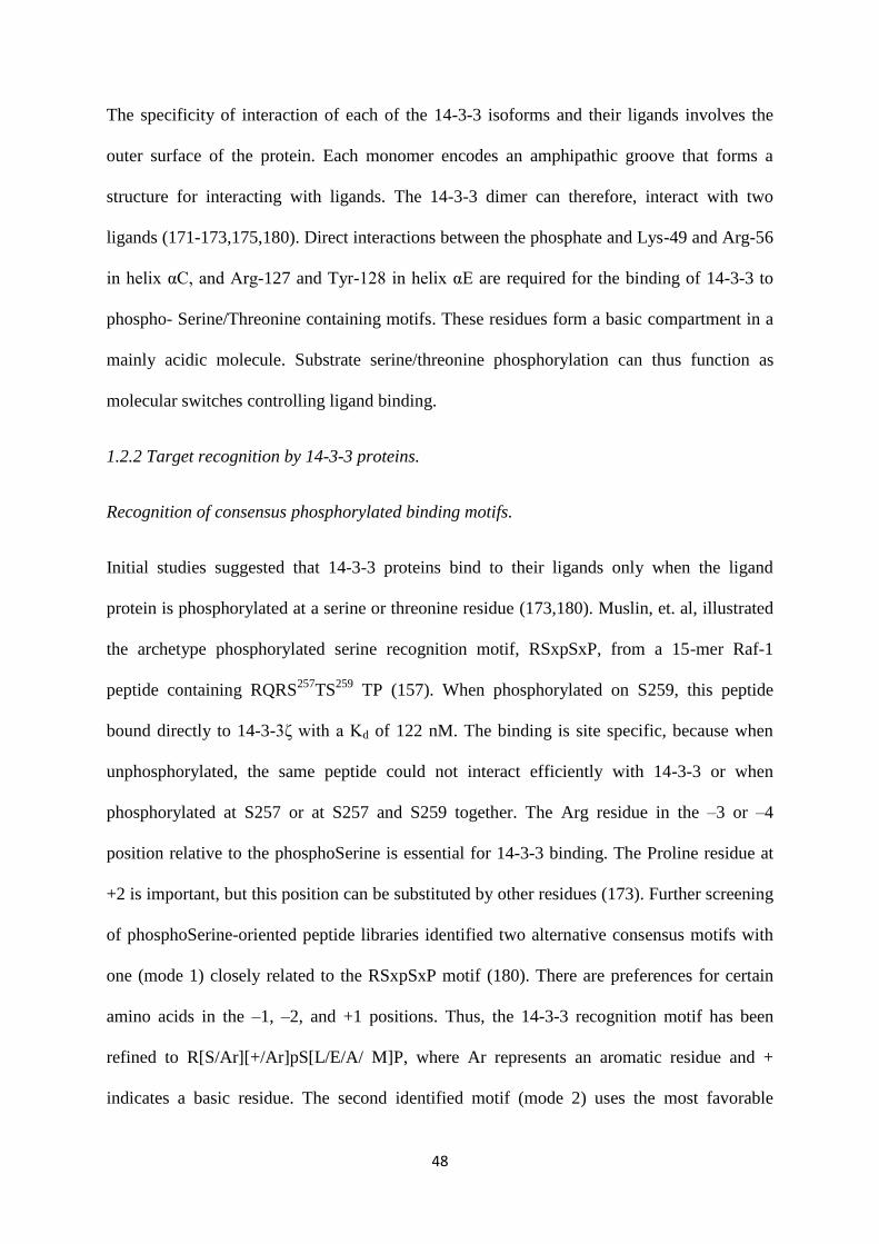
48
The specificity of interaction of each of the 14-3-3 isoforms and their ligands involves the
outer surface of the protein. Each monomer encodes an amphipathic groove that forms a
structure for interacting with ligands. The 14-3-3 dimer can therefore, interact with two
ligands (171-173,175,180). Direct interactions between the phosphate and Lys-49 and Arg-56
in helix αC, and Arg-127 and Tyr-128 in helix αE are required for the binding of 14-3-3 to
phospho- Serine/Threonine containing motifs. These residues form a basic compartment in a
mainly acidic molecule. Substrate serine/threonine phosphorylation can thus function as
molecular switches controlling ligand binding.
1.2.2 Target recognition by 14-3-3 proteins.
Recognition of consensus phosphorylated binding motifs.
Initial studies suggested that 14-3-3 proteins bind to their ligands only when the ligand
protein is phosphorylated at a serine or threonine residue (173,180). Muslin, et. al, illustrated
the archetype phosphorylated serine recognition motif, RSxpSxP, from a 15-mer Raf-1
peptide containing RQRS257
TS259
TP (157). When phosphorylated on S259, this peptide
bound directly to 14-3-3ζ with a Kd of 122 nM. The binding is site specific, because when
unphosphorylated, the same peptide could not interact efficiently with 14-3-3 or when
phosphorylated at S257 or at S257 and S259 together. The Arg residue in the –3 or –4
position relative to the phosphoSerine is essential for 14-3-3 binding. The Proline residue at
+2 is important, but this position can be substituted by other residues (173). Further screening
of phosphoSerine-oriented peptide libraries identified two alternative consensus motifs with
one (mode 1) closely related to the RSxpSxP motif (180). There are preferences for certain
amino acids in the –1, –2, and +1 positions. Thus, the 14-3-3 recognition motif has been
refined to R[S/Ar][+/Ar]pS[L/E/A/ M]P, where Ar represents an aromatic residue and +
indicates a basic residue. The second identified motif (mode 2) uses the most favorable
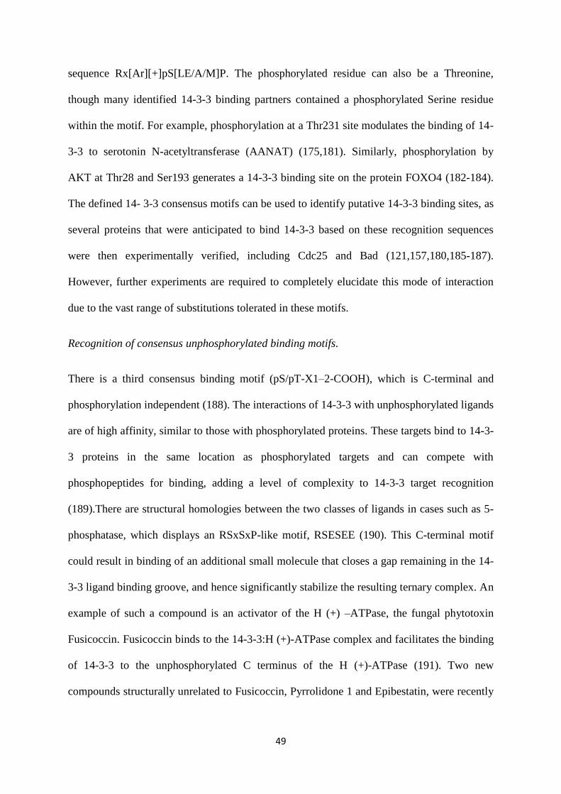
49
sequence Rx[Ar][+]pS[LE/A/M]P. The phosphorylated residue can also be a Threonine,
though many identified 14-3-3 binding partners contained a phosphorylated Serine residue
within the motif. For example, phosphorylation at a Thr231 site modulates the binding of 14-
3-3 to serotonin N-acetyltransferase (AANAT) (175,181). Similarly, phosphorylation by
AKT at Thr28 and Ser193 generates a 14-3-3 binding site on the protein FOXO4 (182-184).
The defined 14- 3-3 consensus motifs can be used to identify putative 14-3-3 binding sites, as
several proteins that were anticipated to bind 14-3-3 based on these recognition sequences
were then experimentally verified, including Cdc25 and Bad (121,157,180,185-187).
However, further experiments are required to completely elucidate this mode of interaction
due to the vast range of substitutions tolerated in these motifs.
Recognition of consensus unphosphorylated binding motifs.
There is a third consensus binding motif (pS/pT-X1–2-COOH), which is C-terminal and
phosphorylation independent (188). The interactions of 14-3-3 with unphosphorylated ligands
are of high affinity, similar to those with phosphorylated proteins. These targets bind to 14-3-
3 proteins in the same location as phosphorylated targets and can compete with
phosphopeptides for binding, adding a level of complexity to 14-3-3 target recognition
(189).There are structural homologies between the two classes of ligands in cases such as 5-
phosphatase, which displays an RSxSxP-like motif, RSESEE (190). This C-terminal motif
could result in binding of an additional small molecule that closes a gap remaining in the 14-
3-3 ligand binding groove, and hence significantly stabilize the resulting ternary complex. An
example of such a compound is an activator of the H (+) –ATPase, the fungal phytotoxin
Fusicoccin. Fusicoccin binds to the 14-3-3:H (+)-ATPase complex and facilitates the binding
of 14-3-3 to the unphosphorylated C terminus of the H (+)-ATPase (191). Two new
compounds structurally unrelated to Fusicoccin, Pyrrolidone 1 and Epibestatin, were recently

50
identified and they can selectively activate the H (+)-ATPase by stabilizing its complex with
the 14-3-3 protein (192,193).
1.2.3 14-3-3 proteins and dimerization.
Deletion experiments have determined that the N terminal is essential for dimerization. Work
by Gu and Du, 1998, demonstrated that deletion of even the first 32 amino acids can disrupt
the dimerization of 14-3-3ζ (194). Several monomeric forms of 14-3-3 proteins bind to their
phosphorylated ligands with unchanged affinity as compared to the dimeric forms in vitro
(194-197). But in vivo, dimerization deficient mutants of 14-3-3 are unable to associate with
phosphorylated ligands as well as wild type protein (198). A ligand binding deficient mutant
of 14-3-3, K49E, is capable of forming heterodimers with endogenous WT 14-3-3 (199,200).
However, this heterodimer is also unable to bind ligands, which elucidates the importance of
the 14-3-3 dimer for ligand binding (200). In at least some cases, the dimer of 14-3-3 is
essential for it to perform its functions (197). Dimerization deficient mutants of 14-3-3
proteins can bind phosphorylated Raf-1 (197). Yet, the kinase exists in an inactive
conformation, as its activity requires the “bridging” function of the dimeric 14-3-3 proteins
(197).
A single polypeptide with two 14-3-3 binding sites can bind to 14-3-3 with a 30-fold increase
in binding affinity (180). Therefore, a single polypeptide harbouring two 14-3-3 binding sites
of low affinity sites will be able to bind to dimeric 14-3-3. This has been demonstrated in the
case of 14-3-3 ligands such as Wee1, keratin 18, Cbl, IGF-I receptor and IRS-1, which all
need multiple phosphorylation sites for stable binding to 14-3-3 (201-205). Recently, it has
been demonstrated that the inhibition of 14-3-3 dimerization can be used in anti-cancer
therapy. Woodcock, J. et. al., have identified sphingosine mimetics that can result in
mitochondrial-mediated apoptosis by making dimeric 14-3-3 susceptible to phosphorylation
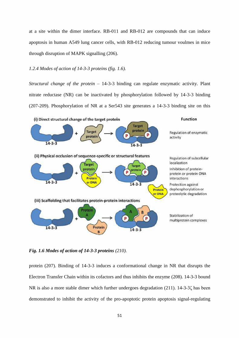
51
at a site within the dimer interface. RB-011 and RB-012 are compounds that can induce
apoptosis in human A549 lung cancer cells, with RB-012 reducing tumour voulmes in mice
through disruption of MAPK signalling (206).
1.2.4 Modes of action of 14-3-3 proteins (fig. 1.6).
Structural change of the protein – 14-3-3 binding can regulate enzymatic activity. Plant
nitrate reductase (NR) can be inactivated by phosphorylation followed by 14-3-3 binding
(207-209). Phosphorylation of NR at a Ser543 site generates a 14-3-3 binding site on this
Fig. 1.6 Modes of action of 14-3-3 proteins (210).
protein (207). Binding of 14-3-3 induces a conformational change in NR that disrupts the
Electron Transfer Chain within its cofactors and thus inhibits the enzyme (208). 14-3-3 bound
NR is also a more stable dimer which further undergoes degradation (211). 14-3-3ζ has been
demonstrated to inhibit the activity of the pro-apoptotic protein apoptosis signal-regulating
52
kinase 1 (ASK-1) (212). Tumour necrosis factor (TNF) mediated phosphorylation of ASK-1
at a Ser 966 site results in its kinase domain being bound by 14-3-3ζ, which blocks its
accessibility and negatively affects its conformation (212,213).
On the other hand, 14-3-3 binding can also activate enzymes. For example, tryptophan and
tyrosine hydroxylases are phosphorylated in the presence of Ca2+
, followed by 14-3-3η
binding resulting in a conformational change and full activity (160,195). For serotonin-N-
acetyltransferase (AANAT), phosphorylation of the residue Thr31 leads to binding of 14-3-
3ζ and a conformational change, which releases the “fly-casting” mechanism and activates
the protein (175,214).
Regulation of the Raf- 1 protein kinase by 14-3-3ζ is complex and three 14-3-3 binding sites
are involved. In cells non-stimulated EGF or serum, 14-3-3ζ keeps Raf-1 in an inactive
conformation by binding to two sites, Ser 259 and Ser621 (197). Upon stimulation, 14-3-3ζ is
displaced from the Ser259 site, but it remains attached to the Ser621 site. This results in
intermediate activation of Raf-1. For full catalytic activity, the 14-3-3ζ protein binds to two
different phosphorylated serines, thus stabilizing the active conformation (197).
Scaffolding as a means to modulate protein-protein interaction – Both Raf-1 and Bcr protein
kinases can bind to a single 14-3-3β dimer resulting in interaction between the two protein
kinases (215). 14-3-3β acts as a bridge, connecting Raf-1 and Bcr, which are unable to
interact in the absence of 14-3-3β (215). 14-3-3 also affects the binding of BAD to Bcl-XL.
During survival conditions, BAD is phosphorylated on S112, S136 and S155 (186,187,216-
218). 14-3-3ζ binding maintains BAD in the cytoplasm and aids in the phosphorylation of the
S155 site (186,216). This phosphorylation prevents binding of BAD to the anti-apoptic
proteins Bcl-2 and Bcl-XL (216-218).
53
Chibby (Cby), a conserved antagonist of β-catenin, has a vital role in the canonical Wnt
signalling pathway [106]. Cby interacts with the C-terminal activation domain of β-catenin
and inhibits its ability to be transcriptionally activated. Mass spectrometry studies have
shown that 14-3-3ε and 14-3-3ζ recognize a Ser20 phosphorylated residue on Cby and
sequester it in the cytoplasm (219). 14-3-3, Cby and β-catenin form a stable, tripartite
complex, which leads to the nuclear export of β-catenin and terminates its signalling (219).
Tertramerization of the tumour suppressor protein p53 is required for its activity, in this
conformation it binds to DNA with high affinity or interact more proficiently with its ligands
[reviewed in (220)]. 14-3-3 proteins bind to a site in its intrinsically disordered C-terminal
domain and activate the DNA binding affinity and increase the stability of p53 (221-223). In
vitro studies indicate, that 14-3-3γ and 14-3-3ε can accelerate the tetramerization of p53 even
at low p53 dimer concentration (224). This augments the binding p53 to sequence-specific
DNA and thus controls p53 activity (224).
Sequestering of structural or sequence-specific features - It has been demonstrated in vitro
that 14-3-3 β, ε, γ, σ and ζ can bind to wild type CDC25C (152). In vivo, only 14-3-3γ and
14-3-3ε can bind to CDC25C (152). Binding of 14-3-3γ or 14-3-3ε at a Ser216 residue
sequesters CDC25C phosphatase in the cytoplasm, whereas they cannot bind to the mutant
S216A (152). Disruption of 14-3-3 binding to CDC25C, exposes a nuclear localization
sequence on CDC25C. This results in its translocation to the nucleus where it activates CDK1
and leads to mitotic entry (121,133,225,226).
Another group of proteins that undergoes this type of regulation are the FOXO proteins, a
subgroup of forkhead transcription factors that play a central role in cell-cycle regulation,
differentiation, stress response and apoptosis [reviewed in (227)]. Akt kinase phosphorylation
creates two 14-3-3 binding motifs on FOXO4, Thr28and Ser193; one of which is embedded
54
within the NLS (182,228). Several studies demonstrated that upon binding to 14-3-3ζ, the
resulting FOXO4:14-3-3ζ complex is translocated to the cytosol where the bound 14-3-3
protein masks the NLS of FOXO4 and inhibits its re-entry into the nucleus
(182,184,200,229,230).
1.3 14-3-3γ
14-3-3γ (known as YWHAG in humans) is one of the isoforms of 14-3-3 proteins that was
first isolated in 1991 by Isobe T. et. al., while performing a fractionation of bovine brain
lysates (231). This isoform was theorised to be expressed only in the brain, but has since been
isolated from a number of different tissues, such as skeletal muscle and the heart (231,232).
1.3.1 Functions of 14-3-3γ
14-3-3γ performs various functions in a cell by participating in different cellular pathways.
Some of these are described below.
1.3.1.1 Regulation of cell cycle checkpoint control.
One of the ways by which 14-3-3 proteins regulate the G2/M transition is via binding to
CDC25C (121,131,225,226). CDC25C, a dual specificity phosphatase activates the mitotic
complex, CyclinB-CDK1, leading to the entry of cells into mitosis (233). During interphase
or cell cycle arrest, to prevent the entry of cells into mitosis, CDC25C is phosphorylated at a
Serine residue at position 216 (S216) which leads to the creation of a 14-3-3 binding site
(121). 14-3-3 then sequesters CDC25C in the cytoplasm, prevents activation of the mitotic
complex, cyclinB-CDK1 and thus, mitotic entry (226). Of the different isoforms of 14-3-3,
only 14-3-3γ and 14-3-3ε were found to interact with CDC25C in vivo (152). To test whether
14-3-3γ binding to CDC25C had any effect on CDC25C function, the 14-3-3γ-CDC25C
complex formation was disrupted by expressing an S216A CDC25C mutant which failed to
55
bind 14-3-3γ (152). The cells expressing S216A CDC25C mutant showed premature
chromatin condensation (152). PCC is seen in S phase cells in which premature mitosis is
induced by the override of the incomplete S phase and G2 checkpoint pathways (152). In
HCT116 cells, disruption of endogenous 14-3-3γ-CDC25C complex by loss of 14-3-3γ also
resulted in premature activation of cyclinB-CDK1 complex by CDC25C and increased PCC
(234).
14-3-3γ also regulates the G2/M transition by interacting with Cdt2 (cell division cycle
protein 2) (235). Cdt2 has a role in the cell cycle due to its function as the substrate
recognition adaptor of the CRL4Cdt2
E3 ubiquitin ligase complex. This complex is required
for the proteasomal degradation of Cdt1 (DNA replication licensing factor), p21 (cyclin-
dependent kinase [CDK] inhibitor), and Set8 (histone methyltransferase) in S phase
[reviewed in (236)]. Cdt2 itself is regulated by SCFFbxO11
-mediated proteasomal degradation.
It has been reported that 14-3-3γ interacts with Cdt2 phosphorylated at threonine 464 (T464)
and shields it from proteasomal degradation (235). Further, loss of 14-3-3γ leads to a delay of
the cell cycle in the G2/M phase and a decrease in cell proliferation (235). This occurs due to
the accumulation of the CRL4Cdt2 substrate, Set8 methyltransferase.
One of the kinases that phosphorylate CDC25 proteins and generate a 14-3-3 binding site is
Chk1 (124). Upon DNA damage, activation of Chk1 prevents cell cycle progression and
causes a G2/M arrest via CDC25C (237,238). Phosphorylation of Chk1 at Ser317 and Ser345
by ATR, in response to DNA damage mediates autophosphorylation of Chk1 at Ser296 (239).
This is a binding site for 14-3-3γ (239). The 14-3-3γ dimer is able to bind to both Chk1 and
CDC25A which leads to phosphorylation of CDC25A at a Ser76 residue by Chk1 (239). This
further leads to degradation of CDC25A in a β-TRCP dependent manner. 14-3-3γ is thus
required for checkpoint activation via Chk1 (239).
56
1.3.1.2 Regulation of cell-cell adhesion.
Mice harbouring a knockdown of 14-3-3γ are sterile, due to defects in cell-cell adhesion
(154). It has been demonstrated that loss of 14-3-3γ inhibits desmosome formation in
HCT116 cells. Under normal conditions, PKCµ phosphorylated plakoglobin (PG) at a Ser236
residue, which is a 14-3-3 binding site (154). 14-3-3γ then loads PG onto the KIF5B-KLC1
complex, which is then transported to the desmosome (154). However, upon loss of 14-3-3γ,
this pathway is affected and desmosome formation cannot be inititated, leading to defects in
cell adhesion (154). This can be restored by artificially targeting PG to the desmosome (240).
1.3.1.3 Regulation of adeno-associated virus type 2 replication.
Adeno-associated virus type 2 is a non-pathogenic human parvovirus which requires a helper
virus for efficient DNA replication. One of the proteins expressed by the virus is Rep68, the
function of which is to replicate DNA and site specific integration. It was found that
phosphorylation of Rep68 at Ser535 induces its binding to a 14-3-3γ-14-3-3ε to form a
ternary complex (241,242). A Rep68 mutant which is unable to bind to 14-3-3 proteins has
increased affinity for DNA and is more efficient in viral DNA replication (243).
1.3.1.4 Regulation of smooth muscle activation.
14-3-3γ levels have been shown to rise in rat carotid arteries, post surgery (243). Arteries are
composed of vascular smooth muscle cells (VMSCs), hyper proliferation of which leads to
the harmful narrowing of arteries post surgery. 14-3-3γ might have a function in muscle
tissue, given its high expression in VSMCs and skeletal and heart muscle (243). It has been
demonstrated that upon exposure of VSMCs to growth stimuli such as Platelet Derived
Growth Factor (PDGF), different isoforms of PKC phosphorylate 14-3-3γ, which then
57
interacts with Raf (244). This interaction might promote the activation and proliferation of
the VSMCs, resulting in the formation of lesions.
1.3.1.5 Regulation of p53 activity by 14-3-3γ and suppression of 14-3-3γ expression by p53.
p53 initiates the transcription of genes involved in cell-cycle arrest, senescence, apoptosis and
DNA repair, and is thus important for the preservation of genome integrity. p53 activity can
be regulated by both post-translational modifications and protein–protein interactions. It is a
tumor suppressor protein, which is frequently mutated in cancers. It was observed that 14-3-
3γ binds to p53 in vitro and in vivo (245). The 14-3-3 binding deficient mutants of p53 were
unable to induce cell cycle arrest in response to IR, which implies that the p53 binding to 14-
3-3 is important for p53 function (245). 14-3-3γ helps in oligomerization of p53 and the 14-3-
3γ-p53 complex had higher affinity to bind DNA when compared to p53 alone (222,224,246).
Conversely, it has been demonstrated that in non-small cell lung cancers, that loss of
functional p53 correlates with an increase in 14-3-3γ levels (247). Upon exposure to IR, p53
binds to the promoter of 14-3-3γ and suppresses its expression (247). p53 also enhances the
ubiquitination of 14-3-3γ and thus regulates its levels (248).
1.3.1.6 ATPase activity
Recent reports have shown that 14-3-3 proteins possess ATPase activity (249). Pure
recombinant human 14-3-3 ζ, γ, ε and τ isoforms hydrolyze ATP with similar Km and kcat
values. This activity can be altered by mutation of Asp124 or Arg55 to Alanine and results in
a gain or loss of function. The activity could be due to the presence of an as yet unknown
binding pocket (249).
58
1.3.1.7 Role in neurodegenerative disorders
14-3-3γ levels are often deregulated in different neurodegenerative disorders. Mutations in α-
synuclein are the cause of aggregate formation (Lewy bodies), which are a hallmark of
Parkinson’s disease. 14-3-3 proteins interact with α-synuclein and are seen in α-syn inclusion
bodies (250). Over-expression of α-synuclein in mouse model revealed a reduction in the
levels of 14-3-3γ, 14-3-3ζ and 14-3-3ε (251). Conversely, over-expression of 14-3-3γ, 14-3-
3ε and 14-3-3ζ in a neuroglioma cell line led to the reduction of the α-synuclein aggregates.
14-3-3γ overexpression in dooaminergic cells also provided protection against cytotoxicity
due to drugs such as Rotenone and 1-Methyl-4- phenyl pyridinium (251). Thus, 14-3-3γ
might have a protective function against Parkinson’s disease.
Leucine rich repeat kinase-2 (LRRK2) is a multidomain protein and is mutated in familial
Parkinsons’s disease. PKA mediated binding of 14-3-3γ at a S1444 site regulates the kinase
activity of LRRK2 (252). Mutation of 14-3-3 interaction with LRRK2 is hampered in
R1441C/G/H-mediated PD pathogenesis. LRRK2 is phosphorylated by protein kinase A at
multiple sites. LRRK2 phosphorylated at S1444 residue serves as binding site for 14-3-3γ.
The kinase activity of LRRK2 is attenuated by binding to 14-3-3γ. Mutation of this binding
site to an alanine results in loss of 14-3-3 binding. This is important, because a LRRK2
overexpression knock-in model showed lysosomal dysfunction and accumulation of α-
synuclein enriched Lewy Bodies (253). Further, increased levels of 14-3-3γ in the
cersbrospinal fluid are a diagnostic marker for the transmissible spongiform encephalopathy
(TSE) Creutzfeldt-Jakob disease (CJD) (254,255).
1.3.1.8 Regulation of class switching in B cells by 14-3-3γ
14-3-3 proteins bind to supercoiled as well as cruciform DNA (256,257). 14-3-3γ specifically
binds to the multiple 5'-AGCT-3' repeats in the IgH locus switch (S) regions, which is where
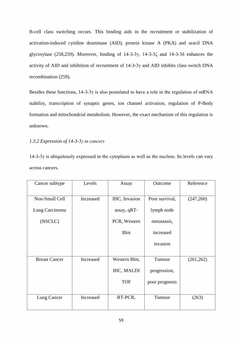
59
B-cell class switching occurs. This binding aids in the recruitment or stabilization of
activation-induced cytidine deaminase (AID), protein kinase A (PKA) and uracil DNA
glycosylase (258,259). Moreover, binding of 14-3-3γ, 14-3-3ζ and 14-3-3δ enhances the
activity of AID and inhibition of recruitment of 14-3-3γ and AID inhibits class switch DNA
recombination (259).
Besides these functions, 14-3-3γ is also postulated to have a role in the regulation of mRNA
stability, transcription of synaptic genes, ion channel activation, regulation of P-Body
formation and mitochondrial metabolism. However, the exact mechanism of this regulation is
unknown.
1.3.2 Expression of 14-3-3γ in cancers
14-3-3γ is ubiquitously expressed in the cytoplasm as well as the nucleus. Its levels can vary
across cancers.
Cancer subtype Levels Assay Outcome Reference
Non-Small Cell
Lung Carcinoma
(NSCLC)
Increased IHC, Invasion
assay, qRT-
PCR, Western
Blot
Poor survival,
lymph node
metastasis,
increased
invasion
(247,260)
Breast Cancer Increased Western Blot,
IHC, MALDI
TOF
Tumour
progression,
poor prognosis
(261,262)
Lung Cancer Increased RT-PCR, Tumour (263)
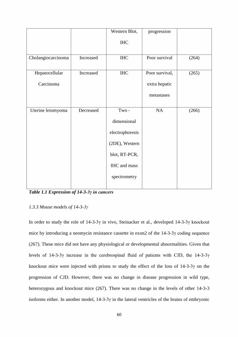
60
Western Blot,
IHC
progression
Cholangiocarcinoma Increased IHC Poor survival (264)
Hepatocellular
Carcinoma
Increased IHC Poor survival,
extra hepatic
metastases
(265)
Uterine leiomyoma
Decreased Two -
dimensional
electrophoresis
(2DE), Western
blot, RT-PCR,
IHC and mass
spectrometry
NA (266)
Table 1.1 Expression of 14-3-3γ in cancers
1.3.3 Mouse models of 14-3-3γ
In order to study the role of 14-3-3γ in vivo, Steinacker et al., developed 14-3-3γ knockout
mice by introducing a neomycin resistance cassette in exon2 of the 14-3-3γ coding sequence
(267). These mice did not have any physiological or developmental abnormalities. Given that
levels of 14-3-3γ increase in the cerebrospinal fluid of patients with CJD, the 14-3-3γ
knockout mice were injected with prions to study the effect of the loss of 14-3-3γ on the
progression of CJD. However, there was no change in disease progression in wild type,
heterozygous and knockout mice (267). There was no change in the levels of other 14-3-3
isoforms either. In another model, 14-3-3γ in the lateral ventricles of the brains of embryonic
61
mice was knocked down using shRNA (268). These mice had thicker stems and highly
branched processes (268). Interestingly, the over-expression of 14-3-3γ in electroporated
embryos also led to a delay in neuronal migration (269).
Mouse models with a partial loss of function of the gene of interest are essential in order to
study the effect of genes which are involved in multiple pathways. Such mutants are called
hypomorphic mutants. In order to generate a hypomorphic mutant of 14-3-3γ, lentiviruses
harbouring 14-3-3γ shRNA were injected into the testes of pre-pubescent male mice (154).
These pre-founder male mice were sterile and the testis section of these mice showed loss of
cell-cell adhesion, and cell-matrix adhesion leading to collapse of the blood-testis barrier
(BTB) and defects in spermatogonial stem cell differentiation and spermatocyte development
and maturation (154).
1.4 The centrosome.
Error-free segregation of chromosomes in mitosis depends on the correct organization of the
mitotic spindle. In mammalian cells, this is dependent on centrosome function. The
centrosome is a membrane-less organelle, composed of two centrioles surrounded by a
proteinaceous toroid, the pericentriolar matrix (PCM). Initially identified by Flemming and
van Beneden, the name centrosome was given to this organelle by T. Boveri and literally
means “central body”, alluding to its close association with the nucleus [reviewed in (270)]. It
is the principal microtubule organizing centre (MTOC) in a mammalian cell. Due to its
ability to organise microtubules, the centrosome exerts an influence on various processes in a
cell; cellular trafficking, mediating an immune response, cellular signaling, cell polarity and
cell cycle progression [reviewed in (271-274)]. Given their contribution to numerous cellular
processes, any defects in centrosome structure or number can lead to several disease
conditions [reviewed in (275)].
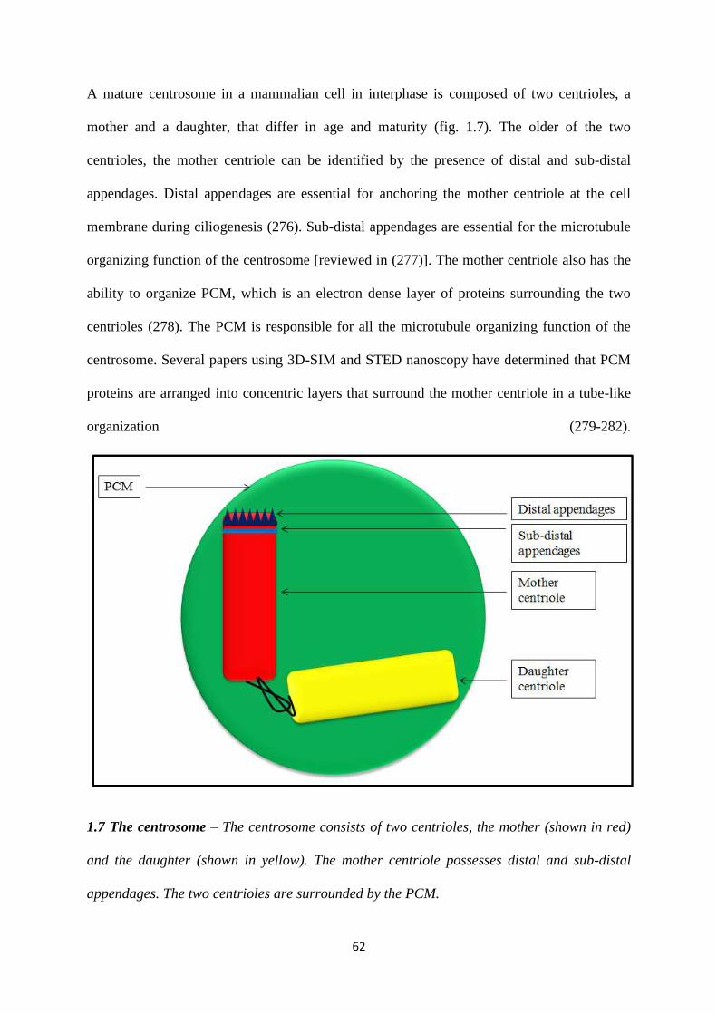
62
A mature centrosome in a mammalian cell in interphase is composed of two centrioles, a
mother and a daughter, that differ in age and maturity (fig. 1.7). The older of the two
centrioles, the mother centriole can be identified by the presence of distal and sub-distal
appendages. Distal appendages are essential for anchoring the mother centriole at the cell
membrane during ciliogenesis (276). Sub-distal appendages are essential for the microtubule
organizing function of the centrosome [reviewed in (277)]. The mother centriole also has the
ability to organize PCM, which is an electron dense layer of proteins surrounding the two
centrioles (278). The PCM is responsible for all the microtubule organizing function of the
centrosome. Several papers using 3D-SIM and STED nanoscopy have determined that PCM
proteins are arranged into concentric layers that surround the mother centriole in a tube-like
organization (279-282).
1.7 The centrosome – The centrosome consists of two centrioles, the mother (shown in red)
and the daughter (shown in yellow). The mother centriole possesses distal and sub-distal
appendages. The two centrioles are surrounded by the PCM.
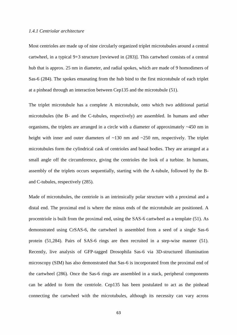
63
1.4.1 Centriolar architecture
Most centrioles are made up of nine circularly organized triplet microtubules around a central
cartwheel, in a typical 9+3 structure [reviewed in (283)]. This cartwheel consists of a central
hub that is approx. 25 nm in diameter, and radial spokes, which are made of 9 homodimers of
Sas-6 (284). The spokes emanating from the hub bind to the first microtubule of each triplet
at a pinhead through an interaction between Cep135 and the microtubule (51).
The triplet microtubule has a complete A microtubule, onto which two additional partial
microtubules (the B- and the C-tubules, respectively) are assembled. In humans and other
organisms, the triplets are arranged in a circle with a diameter of approximately ~450 nm in
height with inner and outer diameters of ~130 nm and ~250 nm, respectively. The triplet
microtubules form the cylindrical cask of centrioles and basal bodies. They are arranged at a
small angle off the circumference, giving the centrioles the look of a turbine. In humans,
assembly of the triplets occurs sequentially, starting with the A-tubule, followed by the B-
and C-tubules, respectively (285).
Made of microtubules, the centriole is an intrinsically polar structure with a proximal and a
distal end. The proximal end is where the minus ends of the microtubule are positioned. A
procentriole is built from the proximal end, using the SAS-6 cartwheel as a template (51). As
demonstrated using CrSAS-6, the cartwheel is assembled from a seed of a single Sas-6
protein (51,284). Pairs of SAS-6 rings are then recruited in a step-wise manner (51).
Recently, live analysis of GFP-tagged Drosophila Sas-6 via 3D-structured illumination
microscopy (SIM) has also demonstrated that Sas-6 is incorporated from the proximal end of
the cartwheel (286). Once the Sas-6 rings are assembled in a stack, peripheral components
can be added to form the centriole. Cep135 has been postulated to act as the pinhead
connecting the cartwheel with the microtubules, although its necessity can vary across
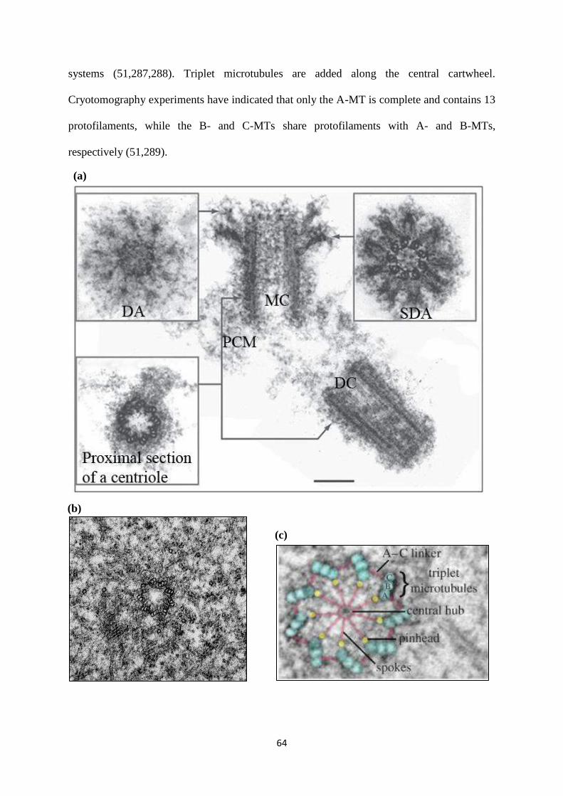
64
systems (51,287,288). Triplet microtubules are added along the central cartwheel.
Cryotomography experiments have indicated that only the A-MT is complete and contains 13
protofilaments, while the B- and C-MTs share protofilaments with A- and B-MTs,
respectively (51,289).
(a)
(b)
(c)
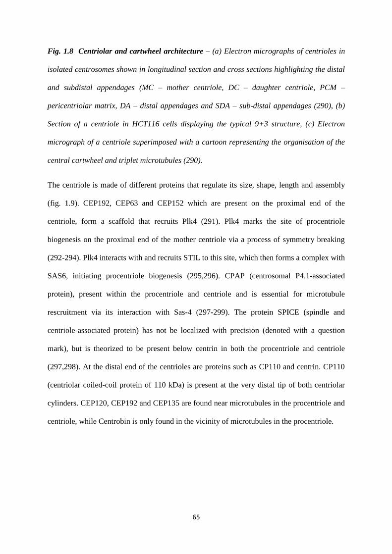
65
Fig. 1.8 Centriolar and cartwheel architecture – (a) Electron micrographs of centrioles in
isolated centrosomes shown in longitudinal section and cross sections highlighting the distal
and subdistal appendages (MC – mother centriole, DC – daughter centriole, PCM –
pericentriolar matrix, DA – distal appendages and SDA – sub-distal appendages (290), (b)
Section of a centriole in HCT116 cells displaying the typical 9+3 structure, (c) Electron
micrograph of a centriole superimposed with a cartoon representing the organisation of the
central cartwheel and triplet microtubules (290).
The centriole is made of different proteins that regulate its size, shape, length and assembly
(fig. 1.9). CEP192, CEP63 and CEP152 which are present on the proximal end of the
centriole, form a scaffold that recruits Plk4 (291). Plk4 marks the site of procentriole
biogenesis on the proximal end of the mother centriole via a process of symmetry breaking
(292-294). Plk4 interacts with and recruits STIL to this site, which then forms a complex with
SAS6, initiating procentriole biogenesis (295,296). CPAP (centrosomal P4.1-associated
protein), present within the procentriole and centriole and is essential for microtubule
rescruitment via its interaction with Sas-4 (297-299). The protein SPICE (spindle and
centriole-associated protein) has not be localized with precision (denoted with a question
mark), but is theorized to be present below centrin in both the procentriole and centriole
(297,298). At the distal end of the centrioles are proteins such as CP110 and centrin. CP110
(centriolar coiled-coil protein of 110 kDa) is present at the very distal tip of both centriolar
cylinders. CEP120, CEP192 and CEP135 are found near microtubules in the procentriole and
centriole, while Centrobin is only found in the vicinity of microtubules in the procentriole.
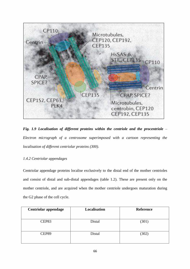
66
Fig. 1.9 Localisation of different proteins within the centriole and the procentriole –
Electron micrograph of a centrosome superimposed with a cartoon representing the
localisation of different centriolar proteins (300).
1.4.2 Centriolar appendages
Centriolar appendage proteins localise exclusively to the distal end of the mother centrioles
and consist of distal and sub-distal appendages (table 1.2). These are present only on the
mother centriole, and are acquired when the mother centriole undergoes maturation during
the G2 phase of the cell cycle.
Centriolar appendage Localisation Reference
CEP83 Distal (301)
CEP89 Distal (302)
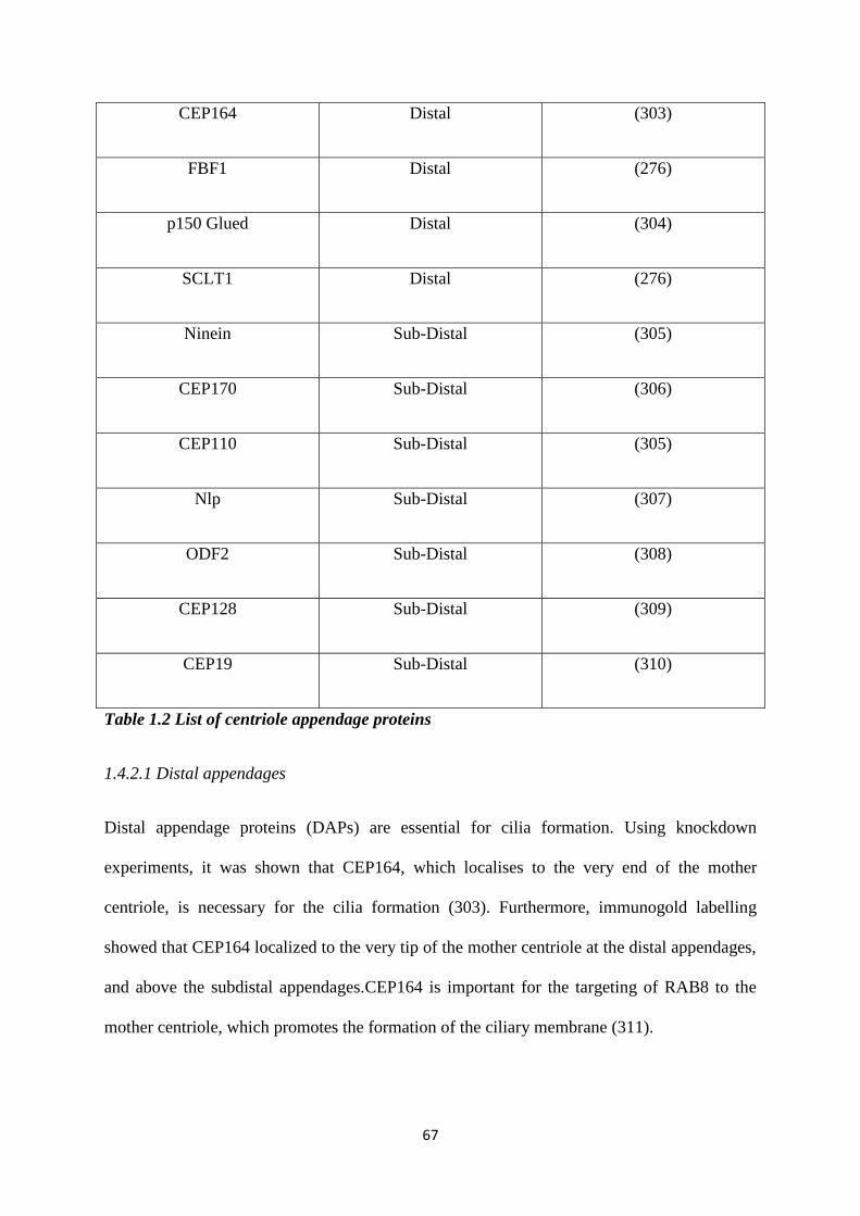
67
CEP164 Distal (303)
FBF1 Distal (276)
p150 Glued Distal (304)
SCLT1 Distal (276)
Ninein Sub-Distal (305)
CEP170 Sub-Distal (306)
CEP110 Sub-Distal (305)
Nlp Sub-Distal (307)
ODF2 Sub-Distal (308)
CEP128 Sub-Distal (309)
CEP19 Sub-Distal (310)
Table 1.2 List of centriole appendage proteins
1.4.2.1 Distal appendages
Distal appendage proteins (DAPs) are essential for cilia formation. Using knockdown
experiments, it was shown that CEP164, which localises to the very end of the mother
centriole, is necessary for the cilia formation (303). Furthermore, immunogold labelling
showed that CEP164 localized to the very tip of the mother centriole at the distal appendages,
and above the subdistal appendages.CEP164 is important for the targeting of RAB8 to the
mother centriole, which promotes the formation of the ciliary membrane (311).
68
CEP83 recruits CEP89 and SCLT1 to the distal appendages, subsequently, SCLT1 targets
CEP164 and FBF1 to DAPs. CEP164 and FBF1 are the downstream components in the DAP
assembly pathway and their localization to the centrioles is not required for targeting of other
DAPs (276).
1.4.2.2 Sub-distal appendages
Sub-distal appendage proteins (SDAPs) help the mother centriole to nucleate microtubules.
ODF2 was shown to be required for the formation of both DAPs and SDAPs (312).
Knockdown experiments have demonstrated that loss of NIN, DCTN1 and KIF3A (all
localizing to the SDAP region), resulted in MT disorganization and loss of focused MT asters
at the mother centriole (304,313). Knockout experiments have shown that there exists a
heirarchy between proteins localising at the SDAP region. The first set of proteins named the
ODF2 group consists of ODF2, CEP128, and Centriolin, with ODF2 being the most upstream
component (309). The second subgroup, termed the NIN group, consists of NIN, CEP170,
DCTN1, and KIF2A, with NIN being the most upstream component. Importantly, depletion
of any member of the ODF2 group can abolish the localization of every component of the
NIN group to the distal end of the mother centriole, but not vice versa.
1.4.3 G1-G2 tether
The G1-G2 tether is a proteinaceous linker that connects the proximal end of the two
centrioles upon disengagement. C-NAP1 is present at the proximal end of the mother
centrioles and the each pool of C-NAP1 is connected to the other by rootletin fibres
(314,315). It is postulated that LRRC45 helps to link the rootletin fibres with C-NAP1 (316).
STED nanoscopy experiments have demonstrated that a network of Cep68 binds to rootletin
and increases the thickness of the rootletin fibres (315). Prior to centrosome separation, the
degradation of the tether is initiated by the activity of the NIMA related kinase, Nek2 (317).
69
1.4.4 The centrosome duplication cycle
For ensuring the accurate segregation of the genome to the two daughter cells, the canonical
centrosome duplication cycle is synchronized with the DNA replication cycle. Similar to the
DNA duplication cycle, the typical centrosome duplication cycle is also semi-conservative;
the new centrioles are formed using the previously existing centrioles as a template. There are
two rules that govern the centrosome cycle -
1. Copy number control - This regulates how many centrioles can form adjacent to a
previously existing centriole. This is essential, because the presence of more than one
pro-centriole perpendicular to the mother centriole can affect the organization of the
spindle.
2. Cell cycle control - This regulates the number of times in a cell cycle the centrosome
can duplicate. Centrosome amplification is one of the results of loss of this control
and is frequently observed in transformed cells.
The centrosome duplication cycle consists of four different phases; 1) Disengagement, 2)
Duplication, 3) Maturation and 4) Separation. The four steps are discussed below (fig. 1.10).
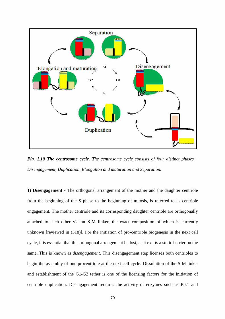
70
Fig. 1.10 The centrosome cycle. The centrosome cycle consists of four distinct phases –
Disengagement, Duplication, Elongation and maturation and Separation.
1) Disengagement - The orthogonal arrangement of the mother and the daughter centriole
from the beginning of the S phase to the beginning of mitosis, is referred to as centriole
engagement. The mother centriole and its corresponding daughter centriole are orthogonally
attached to each other via an S-M linker, the exact composition of which is currently
unknown [reviewed in (318)]. For the initiation of pro-centriole biogenesis in the next cell
cycle, it is essential that this orthogonal arrangement be lost, as it exerts a steric barrier on the
same. This is known as disengagement. This disengagement step licenses both centrioles to
begin the assembly of one procentriole at the next cell cycle. Dissolution of the S-M linker
and establishment of the G1-G2 tether is one of the licensing factors for the initiation of
centriole duplication. Disengagement requires the activity of enzymes such as Plk1 and
71
Separase (319). Both these enzymes act to facilitate the loss of the S-M linker, although their
substrates are unknown. Pericentrin is postulated to be one of the substrates of Separase, as
expression of a Pericentrin B mutant that cannot be cleaved by Separase results in an absence
of disengagement in HeLa cells(320,321). Also, Separase is active only once in the cell cycle;
during anaphase transition (322). This ensures the copy number control of the centrosome
cycle. Inhibition of Plk1 activity using the small molecule BI2536, inhibits centriole
disengagement during late G2 or early mitosis (319).
Nucleophosmin1 (NPM1) is a nucleolar protein that has been demonstrated to have a role to
play in centriole disengagement. NPM1 has been demonstrated to localise between the paired
centrioles of unduplicated centrosomes (323). It has been shown that phosphorylation of
NPM1 at a Thr199 residue by CDK2 or CDK1 results in its displacement from the
centrosome (56,324). Expression of a phospho-deficient mutant of NPM1 (T199A) has been
demonstrated to inhibit centrosome duplication, while a phospho-mimetic mutant (T199D),
promotes centrosome amplification (325,326).
Several recent reports have suggested that there might be other factors that govern centriole
disengagement, apart from the loss of the orthogonal configuration (327-329). One of them
has been demonstrated to be the distance between the mother and daughter centriole. Using
light microscopy in HeLa cells, it has been demonstrated that centrioles are competent for
duplication when they are at a distance of ~80 nm (330).
Before procentriole biogenesis can occur, the daughter centriole that is disengaged needs to
become proficient at functioning as a mother centriole in the next cycle. This includes its
ability to become duplication competent and being able to nucleate microtubules. This is
achieved via a process called, “centriole to centrosome conversion”. This process is mediated
by CDK1 and starts with the initial loss of the central cartwheel comprising of SAS6 dimers

72
(331). Next, the cartwheel-less centriole needs to be stabilized via the acquisition of Cep295
due to PLK1 activity (332). Further, the centrioles recruit Cep63, Cep152 and Cep192 to
form a scaffold at the proximal end and are ready for procentriole biogenesis (291,333,334).
2) Duplication - Duplication is regulated by proteins such as Plk4, STIL, SAS6, Cep152,
Cep192 and CDK2. As per the rule of copy number control in the centrosome cycle, only one
procentriole must form adjacent to each mother centriole. This is brought about in the
following manner. As the first step, Cep152 and Cep192 are recruited to the pre-existing
centrioles that will serve as templates for the formation of the daughter centrioles. These two
proteins form a scaffold around the centrioles (291). This scaffold recruits Plk4 to the
centrioles. Plk4 is known as the master regulator of centriole duplication and its over-
expression can result in centrosome amplification and in the generation of several
procentrioles from a single mother centriole (335). Upon recruitment at the centriole, Plk4
initially localises in a ring-like manner around the centriole (292,293). To ensure the
formation of only one procentriole from the mother centriole, this ring is focused into a single
dot. One of the ways this is theorized to occur is due to its stabilization by STIL (294).
Several such Plk4-STIL complexes are formed and there is a competition for the highest
concentration of the Plk4-STIL complex (294). The point with the highest concentration of
this complex marks the site of procentriole biogenesis (294). Another model suggests that the
auto-activation and lateral inhibition activity of Plk4 determines where the ring will coalesce
into a spot marking the site of procentriole biogenesis (295,296).
CDK1 is also essential for centrosome duplication. Recent studies in Xenopus egg extracts
and HeLa cells indicate that Cyclin B1-CDK1 competes with Plk4 for binding to STIL (336).
The Cyclin B1- CDK1 complex binds to STIL at the same site as Plk4. Therefore, when its
levels are high during mitosis, it blocks the interaction between Plk4 and STIL by
sequestering STIL. Once Cyclin B1 levels decrease after anaphase transition, Plk4 can bind
73
to STIL and participate in procentriole biogenesis. CDK1 thus prevents centrosome
amplification by preventing premature complex formation between Plk4 and STIL (336).
After recruitment of Plk4 and STIL marks the site of pro-centriole formation, SAS6 dimers
localise to the same site and initiate pro-centriole assembly by assembling into higher order
oligomers (337). SAS6 oligomerization is the building block for the 9-fold symmetrical
centriole.
3) Elongation and Maturation - During late S to G2, once Plk4 has marked the site of pro-
centriole biogenesis, centriole elongation is carried out by proteins such as CPAP, CP110 and
SAS6 (338). Dimers of SAS6, the 9-fold symmetrical cartwheel protein, assemble into higher
order oligomers and stack at the procentriole. Due to its nine-fold symmetry, it is able to act
as a scaffold around which microtubules are assembled (337). CPAP is a tubulin dimer
binding capping protein that, along with CP110, promotes microtubule assembly and controls
centriolar length (298). CPAP and CP110 have opposing roles in the control of centriolar
length (Schmidt, 2009, Current Biology). Unusually long centrioles can form due to
overexpression of CPAP or by a reduction in the levels of CP110 (339). The growing
microtubule triplets are linked to the SAS6 cartwheel via Cep135 (297). Centrin and hPOC5
localise to the distal lumen of centrioles and are essential for elongation at the distal end
(340).
Along with procentriolar elongation, another event that occurs is the expansion of the PCM.
This is necessary so that each of the two centrosomes can function as an independent MTOC.
This includes the recruitment of γ-Tubulin Ring Complexes (γ-TURCs), which nucleate
microtubules. In cultured vertebrate cells, PLK1 phosphorylates Pericentrin to initiate the
centrosomal recruitment of several PCM proteins such as Aurora A and Cep192 (341). PLK1
phosphorylation of NEK9 leads to the phosphorylation of NEDD1 (also known as GCP-WD),
which induces the recruitment of γ-tubulin to centrosomes at mitosis (342,343). CDK5
74
regulatory subunit-associated protein 2 (CDK5RAP2), Pericentrin and CEP192 mediated
phosphorylation of NEDD1 can recruit γ-TURCs into the PCM (342). CEP192 is an
important activator of centrosomal Aurora A and helps to coordinate the feedback activation
of Aurora A and PLK1. Plk1 activity also recruits Cep215, a PCM protein, to the two
centrosomes (342). Aurora A kinase, in conjunction with Plk1, phosphorylates components of
the γ-TURCs, thus making them competent for the recruitment of microtubules (341).
Experiments in C. elegans and D. melanogaster have demonstrated that loss of Aurora A can
lead to abnormalities in centrosome maturation, with a reduction in the accumulation of α-
tubulin and γ-tubulin at the centrosome (344,345). Centriole maturation also involves the
acquisition of distal and sub distal appendages by the newly formed mother centriole. The
recruitment of the sub-distal appendage protein ODF2 is one of the steps of this process
(312). Ninein and Cep170 also function as sub-distal appendages and bind microtubules
(346). Distal appendages such as Cep164, Cep89, Cep 83, FBF1 and SCLT1 are essential for
membrane docking and ciliogenesis (276)
4) Separation – The G1-G2 tether is made of proteins such as C-Nap1 (Cep250), rootletin,
Cep68 and LRRC45 (314,315,317,347-349). For the formation of a mitotic spindle, it is
essential that the G1-G2 tether connecting the two centrosomes be degraded. This is brought
about by the phosphorylation of C-NAP1 and rootletin by the NIMA related kinase, Nek2
(317). This phosphorylation leads to their displacement from the proximal end of the mother
centrioles (314). Plk1 mediated degradation of Cep68, further displaces Cep215, a PCM
protein, from the centrosome, resulting in loss of the G1-G2 tether (349).
Once the G1-G2 tether has undergone dissolution, the two centrosomes have to be separated,
so as to form the mitotic spindle. This occurs primarily via the anti-parallel sliding action of
the plus-end directed motor protein, Eg5 (350,351). Inhibition of Eg5 results in the presence
of a monopolar spindle, with two centrosomes at a single pole (351). Additionally, proteins
75
such as dynein, Lis1 and CLIP-70, which are minus-end directed, contribute to spindle
formation by inducing microtubule sliding in the direction opposite to that of Eg5. The
combined action of these motor proteins and their binding partners promotes migration of the
centrosomes to the two poles (352,353). Once the centrosomes are separated, they migrate to
each end of the cell and form the mitotic spindle.
Fate of the centrioles upon exit from the cell cycle.
In post mitotic cells that have exited the cell cycle, formation of the primary cilium occurs.
This is a single cilium that is observed only in resting or G0 cells. It is essential for sensing
extracellular growth stimuli and is involved in many crucial pathways such as Hedgehog,
Wnt and platelet- derived growth factor (PDGF) signaling. In a quiescent cell, the two
centrioles in a centrosome can migrate to the plasma membrane, where the mother centriole –
also referred to as the basal body – docks below the plasma membrane via the fusion of
vesicles that cap the distal appendages [reviewed in (354)]. Then, lengthening of nine MT
doublets of the mother centriole occurs and they form the nine doublets of the axoneme
respectively. Centrioles are also required to begin the formation of motile cilia and flagella
[reviewed in (355,356)]. Here, there are two additional centrally located MTs that form in the
transition zone.
1.4.5 Functions of the centrosome.
Centrosomes can bind more than 100 regulatory proteins, which implies its functions in a
several cellular processes.
1.4.5.1 Microtubule organization.
The cellular functions of microtubules, such an intracellular transport and the formation of a
mitotic spindle, depend on microtubules being arranged in an ordered array. To assemble and
76
re-organize these arrays during different phases of the cell cycle or for different processes,
cells need to be able to control their growth. This control is achieved by tethering them at an
organizing centre, the predominant of which in a mammalian cell is the centrosome. The
main microtubule nucleator is the γ-TURC, which forms a template for microtubule assembly
[reviewed in (357)]. γ-TURCs present at the centrosome anchor the minus end of
microtubules. The MTOC function of the centrosome is most apparent in its role in formation
of the spindle. The number of centrosomes in a cell determines how many poles a mitotic
spindle can have. The presence of less than two centrosomes in a mitotic cell leads to mitotic
delay and a high rate of chromosomal instability (358). Centrosome amplification leads to the
organization of a multipolar spindle, which is deleterious to normal cells (359,360). Mice
overexpressing Plk4 in the developing CNS displayed centrosome amplification and a
multipolar mitosis which results in aneuploidy and microcephaly (361). However,
transformed cells are able to bypass the massive aneuploidy brought about by multiple
centrosomes, by clustering their centrosomes and forming a pseudo-bipolar spindle (360).
1.4.5.2 Regulation of the cell cycle.
Due to the coupling of the cell cycle with the centrosome cycle, there are several cell cycle
proteins that play a role in the centrosome cycle and vice versa. It has been demonstrated in
Chinese Hamster Ovary (CHO) cells that Cyclin E and cyclin A contain a centrosomal
localisation sequence (CLS) and that expression of a cyclin E mutant with a deletion of the
CLS results in an inhibition of DNA replication (362). Expression of a mutant cyclin E that
harboured the CLS but was unable to bind to CDK2 accelerated S phase entry (362). The
accelerated S-phase entry is due to the centrosomal recruitment of MCM5 and Orc1 via the
cyclin A CLS (363). Experiments in C. elegans embryos have shown that Aurora A at the
centrosome controls nuclear envelope breakdown, which might occur due to its effect on the
actomyosin networks (364-366). Further, it has also been demonstrated that the cyclin B-
77
CDK1 complex is first phosphorylated by Plk1 at the centrosome during prophase (367). The
centrosome thus acts as an integration point for several cell cycle processes.
1.4.5.3 Immune response.
Centrosomes have been observed to move to the contact site of immune cell function. When
immune cells such as T-cell lymphocytes first contact an antigen, the centrosome is
positioned at that site. This re-orients the organization of the microtubule and its associated
proteins. Due to the microtubules being aligned at the point of contact, microtubule
associated organelles and secretory cytolytic vesicles can now access secretory sites and the
cell is able to give rise to an immune reaction. Upon movement of the centrosome form the
site of contact, the microtubules are re-organized again and the immune reaction is halted
[reviewed in (273)]. The centrosome plays an important role in the initiation of an immune
response.
1.4.5.4 Cellular signaling.
Several proteins involved in the DNA damage response, such as p53, Chk1, Chk2, Brca1 and
Brca2 localize to centrosomes, in addition to their cytoplasmic and / or nuclear localizations
[reviewed in (368)]. Centrin, a protein that is essential for procentriole biogenesis, interacts
with a core component of nucleotide excision repair (NER), Xeroderma Pigmentosum group
C protein, (369). A knockout of all Centrin isoforms in chicken DT40 cells resulted in delays
in NER, but no discernible centrosome phenotype (370). A single nucleotide change in the
Pericentrin gene (PCNT3109G>T
) causes Seckel syndrome and results in its loss from the
centrosome (371,372). Cells isolated from patients with Seckel syndrome also exhibit a
defect in ATR checkpoint signalling (371,372). Similarly, a knockdown of Cep164 in
Zebrafish was shown to cause nephronophthisis-related ciliopathies and dysregulated DDR
78
(373). All these reports elucidate the role of the centrosome as a cellular signalling
integration point.
1.4.5.5 Actin organization.
Microtubules and actin are two of the main cytoskeletal elements in a cell regulating cellular
architecture. The centrosome is the fundamental MTOC in a cell. However, several studies
suggest that it plays a role in organizing the actin network too. Centrosomes purified from the
human T-lymphocyte Jurkat cell line could generate large radial arrays of actin filaments,
when seeded on glass coverslips in the presence of 1 µM purified actin monomers (374). It
was further demonstrated that this nucleation was modulated by PCM1 via its recruitment of
the nucleation promoting factor WASH and Arp2/3 complex at the centrosome (374).
Centrosome-associated Arp2/3 locally concentrates F-actin in resting lymphocytes (375).
This is essential for tethering the centrosome to the nucleus. Upon lymphocyte activation,
Arp2/3 levels at the centrosome decrease, which releases the centrosome from being bound to
the centrosome so that it can migrate to the synapse (375). An inverse co-relation exists
between actin and microtubule nucleation at the centrosome (376). The centrosome thus has a
dual role in organizing the cytoskeleton.
1.4.5.6 Cell migration and polarity.
In wound healing assays using fibroblast monolayers, the centrosome is localized between
the nucleus and the leading edge of cells (377). Due to this, both the Golgi complex and the
endocytic recycling compartment are positioned between the nucleus and the leading edge
(377). Cells lining the tissue periphery lose cell-cell contact to be able to migrate during
epithelial-to-mesenchymal transition (EMT). This involves a change in polarity from apico-
basal to migratory front and rear end polarity. In developing mouse embryos, cultured 3D cell
aggregates and micro-patterned cell doublets, the change in polarity involves the movement
79
of centrosomes from their off-centered position next to intercellular junctions, toward
extracellular matrix adhesions on the opposite side of the nucleus, results in the reversal of
internal polarity (378). This movement also included the disassembly of the microtubule
network (378).
1.4.5.7 Asymmetric cell division.
Asymmetric cell division is essential for the maintenance of a stem cell pool. One daughter
cell continues as the stem cell, whereas the other undergoes differentiation. It was first
demonstrated using Drosophila neuroblasts that there is a preference for centrosome
inheritance between the two daughter cells of a stem cell (379). When centrosomes split
in Drosophila neuroblasts, the younger centrosome is retained by the stem cells. The older
centrosome is inherited by the differentiating daughter cell and lacks PCM. This centrosome
migrates away to the other side of the cell before it begins rebuilding its PCM (379). This
inheritance pattern is cell-type specific, as in the case of Drosophila male germline stem cells,
the mother centriole is the one that is retained by the stem cell (380). The functional
consequences of these inheritance patterns are unknown.
1.4.6 Centrosomes and cancer
Deregulation of any step of the centrosome cycle can result in centrosome abnormalities.
These can be classified into two types – a) structural, and b) numerical. Both these
abnormalities can contribute to neoplastic progression.
The overexpression of Ninein like protein (Nlp) in MCF10A derived 3D acini can lead to
excessive cell proliferation and formation of defective acini (381). The overexpression of Nlp
results in a diffuse localization of E cadherin at adherens junctions (382). The adherens
junctions are thus, weak and lead to increased mechanical stress when a cell in the epithelial

80
tissue enters mitosis (382). Also the microtubules in such cells are more stabilized (382).
When such a cell enters mitosis, due to the excessive mechanical stress, it buds out from the
original site and might become metastatic (382). In addition to the increased invasion,
structural alterations to the centrosome, could also lead to aneuploidy which often contributes
to tumour progression (383).
Copy number changes in centrosome number can occur due to centrosome over-duplication,
defects in cytokinesis or due to the de novo formation of centrosomes. Several tumour cells
show centrosome amplification, suggesting that the change in centrosome number could drive
aneuploidy and thus, tumour progression (384). However, it is only recently that the causative
link between centrosome amplification and tumour progression has been established. The
first evidence was derived from experiments in Drosophila. No spontaneous tumours formed
in transgenic flies overexpressing Plk4 and they were also viable (385). However, in a tissue
transplantation assay, brain tissue from flies overexpressing Plk4 was transplanted into the
abdomen of WT hosts (385). It was observed that that a significant percentage of the hosts
developed tumours with multiple centrosomes and metastatic colonies (385). In a similar
transplantation assay, Castellanos et. al., screened for the ability of mutants of different
centrosomal proteins to affect tumour formation in Drosphila (386). They were able to
demonstrate that 2% of the WT hosts with wing discs from flies overexpressing Sak
transplanted into their abdomen could develop tumours (386).
Studies on Plk4 overexpression have shown significant centrosome amplification in the liver
and skin, but not in the lung or kidney (387). Despite the centrosome amplification, there was
no increase in spontaneous tumor formation or decreased survival compared to controls.
Decreasing p53 levels alone induced many tumors, but Plk4 overexpression did not enhance
the same (387). It was observed that there was an increase in the speed of tumour initiation in
81
p53-/- mice with either tissue wide or only an epidermis specific overexpression of Plk4
(388,389).
Inversely, overexpression of Plk4 can initiate tumorigenesis, in a mouse intestinal neoplasia
model (390). In a recent study, widespread overexpression of Plk4 in mice demonstrated for
the first time that centrosome amplification can initiate tumourigenesis (390). Mice were
followed for more than 35 weeks post Plk4 overexpression and it was found that from 35
weeks of age, the mice exhibited a dramatic increase in the spontaneous formation of
lymphomas, squamous cell carcinomas, and sarcomas. Like many human tumours, these also
exhibited aneuploidy and chromosomal instability. There was also a reduction in the levels of
p53 and its target genes (390). These results suggest that centrosome amplification, in
conjunction with p53 inactivation over time, can result in tumour formation.
1.4.7 Centrosome clustering
Normal cells with centrosome amplification undergo a multipolar mitosis and eventual
apoptosis (391). A multi-polar mitosis in most tumour cells also leads to cell death, ostensibly
due to loss of genes that are absolutely required for cell viability. Experiments in a transgenic
Zebrafish line overexpressing Plk4 under a heat shock promoter demonstrate that
neuroepithelial cells harbouring centrosome amplification undergo apoptosis (392). Similarly
mice with centrosome amplification in the CNS due to conditional overexpression of Plk4
exhibit microcephaly, due to increased apoptosis (393). It is interesting to study how
transformed cells harbouring multiple centrosomes prevent the massive aneuploidy resulting
from multi-polar mitoses. In order to tolerate the burden of extra centrosomes, transformed
cells have developed a mechanism called clustering (391). In transformed cells with multiple
centrosomes, during mitosis, centrosomes are not organized in a multipolar spindle. in fact,
they migrate to and cluster at two poles leading to the formation of a pseudobipolar spindle
82
during metaphase (360,385,394). This pseudobipolar spindle is less efficient at separating
chromosomes and contributes to errors in kinetochore-microtubule attachments, especially
the formation of merotelic attachments, which contributes to the presence of lagging
chromosomes and an increase in chromosome instability (CIN) (359,395). However, the CIN
is still at a low enough rate, that the tumor cells survive and give rise to neoplastic clones.
Multiple studies have attempted to identify gene products/molecular pathways required for
centrosome clustering. This could lead to the identification of small molecules that inhibit
clustering, as this might result in multi-polar mitoses resulting in killing of tumour cells. A
screen developed by Kwon et al attempted to identify genes required for clustering using
Drosophila S2 cells (360). siRNA mediated knockdown of the motor protein HSET,
induced the formation of multipolar spindles in human cancer cells harbouring multiple
centrosomes (360). A reduction in the levels of proteins that are essential for the Spindle
Assembly Checkpoint (SAC); Mad2, BubR1 (human Bub1), and CENP-E can also generate
multi-polar spindles in S2 cells (360). Several actin binding proteins were also identified in
these screens suggesting that disruption of actin dynamics could inhibit centrosome clustering
(360,396). This is consistent with recent data from the Godinho laboratory, which suggests
that normal cells are unable to cluster centrosomes due to the presence of E-cadherin, which
confers less actin contractility on these cells (397).
Proteins that are a part of the chromosomal passenger complex (CPC), proteins of the Ndc80
complex, Cep164, and Aurora B, which all contribute to spindle tension, are also required for
centrosome clustering, as shown in a genome wide RNAi screen in UPCI:SCC114, a human
Oral Squamous Cell Carcinoma (OSCC) cell line (396). Depletion of Aurora A in a panel of
83
Acute Myeloid Leukemia cell lines also results in formation of a multipolar spindle and non-
proliferation (398).
Several drugs that bind to cytoskeletal elements, such as Griseofulvin, CP-673451 and
crenolanib, inhibit clustering demonstrating the contribution of the cytoskeleton towards
clustering (399,400). CCB02, a drug that inhibits the interaction of CPAP and tubulin
activates extra centrosomes to undergo increased PCM expansion (299). This causes cells to
undergo revert from a clustered mitosis to a multipolar one, prolonging mitosis, and eventual
cell death. 3D-organotypic invasion assays using tyrosine kinase inhibitor (TKI)-resistant
EGFR-mutant non-small-cell lung cancers show that CCB02 has anti-invasive activity (299).
Loss of 14-3-3γ leads to premature CDC25C activation and hence cdk1 activation, which
leads to an increase in centrosome number. This is accompanied by an increase in the number
of cells with pseudo-bipolar spindles with passage, an increase in aneuploidy and tumour
formation (326). However, expression of a CDC25C S216A mutant, that is deficient for 14-3-
3 binding, in 14-3-3γ knockdown cells, leads to a reversal of the clustering phenotype and a
decrease in tumour growth and cell viability, presumably due to prematurely high levels of
cdk1 activity in interphase and mitosis (326). All of these results suggest that centrosome
clustering is a complex phenotype that requires multiple cellular pathways.

84
2. Aims and Objectives

85
2. Aims and objectives
- Does 14-3-3 binding to centrosomal proteins inhibit centrosome licensing and
duplication?
- How do 14-3-3 proteins regulate centrosome clustering?
86
3. Materials and Methods
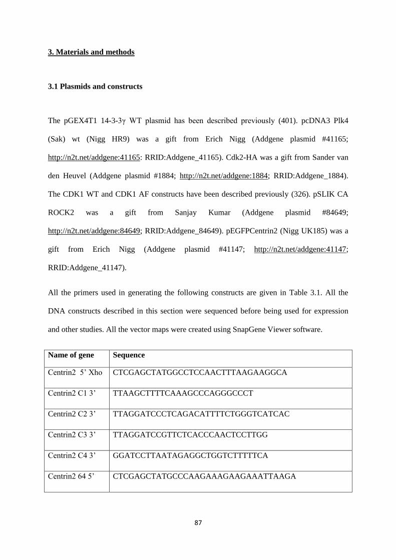
87
3. Materials and methods
3.1 Plasmids and constructs
The pGEX4T1 14-3-3γ WT plasmid has been described previously (401). pcDNA3 Plk4
(Sak) wt (Nigg HR9) was a gift from Erich Nigg (Addgene plasmid #41165;
http://n2t.net/addgene:41165: RRID:Addgene_41165). Cdk2-HA was a gift from Sander van
den Heuvel (Addgene plasmid #1884; http://n2t.net/addgene:1884; RRID:Addgene_1884).
The CDK1 WT and CDK1 AF constructs have been described previously (326). pSLIK CA
ROCK2 was a gift from Sanjay Kumar (Addgene plasmid #84649;
http://n2t.net/addgene:84649; RRID:Addgene_84649). pEGFPCentrin2 (Nigg UK185) was a
gift from Erich Nigg (Addgene plasmid #41147; http://n2t.net/addgene:41147;
RRID:Addgene_41147).
All the primers used in generating the following constructs are given in Table 3.1. All the
DNA constructs described in this section were sequenced before being used for expression
and other studies. All the vector maps were created using SnapGene Viewer software.
Name of gene Sequence
Centrin2 5’ Xho CTCGAGCTATGGCCTCCAACTTTAAGAAGGCA
Centrin2 C1 3’ TTAAGCTTTTCAAAGCCCAGGGCCCT
Centrin2 C2 3’ TTAGGATCCCTCAGACATTTTCTGGGTCATCAC
Centrin2 C3 3’ TTAGGATCCGTTCTCACCCAACTCCTTGG
Centrin2 C4 3’ GGATCCTTAATAGAGGCTGGTCTTTTTCA
Centrin2 64 5’ CTCGAGCTATGCCCAAGAAAGAAGAAATTAAGA
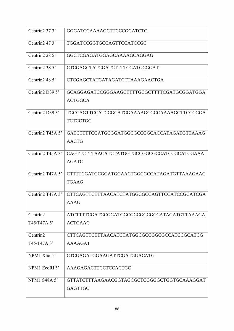
88
Centrin2 37 3’ GGGATCCAAAAGCTTCCCGGATCTC
Centrin2 47 3’ TGGATCCGGTGCCAGTTCCATCCGC
Centrin2 28 5’ GGCTCGAGATGGAGCAAAAGCAGGAG
Centrin2 38 5’ CTCGAGCTATGGATCTTTTCGATGCGGAT
Centrin2 48 5’ CTCGAGCTATGATAGATGTTAAAGAACTGA
Centrin2 D39 5’ GCAGGAGATCCGGGAAGCTTTTGCGCTTTTCGATGCGGATGGA
ACTGGCA
Centrin2 D39 3’ TGCCAGTTCCATCCGCATCGAAAAGCGCCAAAAGCTTCCCGGA
TCTCCTGC
Centrin2 T45A 5’ GATCTTTTCGATGCGGATGGCGCCGGCACCATAGATGTTAAAG
AACTG
Centrin2 T45A 3’ CAGTTCTTTAACATCTATGGTGCCGGCGCCATCCGCATCGAAA
AGATC
Centrin2 T47A 5’ CTTTTCGATGCGGATGGAACTGGCGCCATAGATGTTAAAGAAC
TGAAG
Centrin2 T47A 3’ CTTCAGTTCTTTAACATCTATGGCGCCAGTTCCATCCGCATCGA
AAAG
Centrin2
T45/T47A 5’
ATCTTTTCGATGCGGATGGCGCCGGCGCCATAGATGTTAAAGA
ACTGAAG
Centrin2
T45/T47A 3’
CTTCAGTTCTTTAACATCTATGGCGCCGGCGCCATCCGCATCG
AAAAGAT
NPM1 Xho 5’ CTCGAGATGGAAGATTCGATGGACATG
NPM1 EcoRI 3’ AAAGAGACTTCCTCCACTGC
NPM1 S48A 5’ GTTATCTTTAAGAACGGTAGCGCTCGGGGCTGGTGCAAAGGAT
GAGTTGC
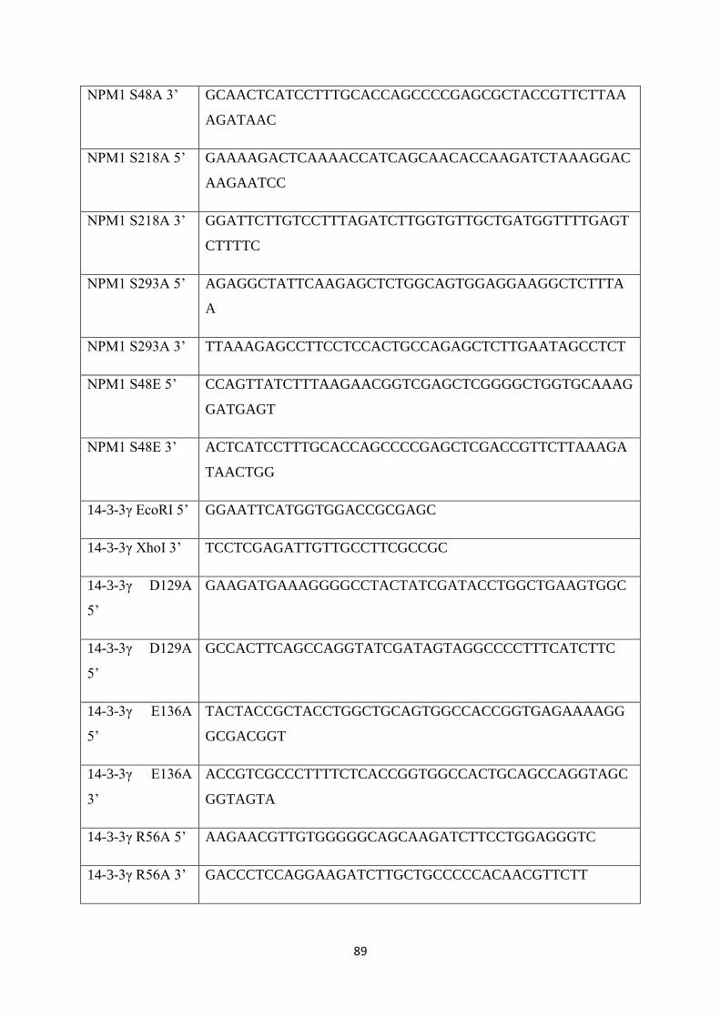
89
NPM1 S48A 3’ GCAACTCATCCTTTGCACCAGCCCCGAGCGCTACCGTTCTTAA
AGATAAC
NPM1 S218A 5’ GAAAAGACTCAAAACCATCAGCAACACCAAGATCTAAAGGAC
AAGAATCC
NPM1 S218A 3’ GGATTCTTGTCCTTTAGATCTTGGTGTTGCTGATGGTTTTGAGT
CTTTTC
NPM1 S293A 5’ AGAGGCTATTCAAGAGCTCTGGCAGTGGAGGAAGGCTCTTTA
A
NPM1 S293A 3’ TTAAAGAGCCTTCCTCCACTGCCAGAGCTCTTGAATAGCCTCT
NPM1 S48E 5’ CCAGTTATCTTTAAGAACGGTCGAGCTCGGGGCTGGTGCAAAG
GATGAGT
NPM1 S48E 3’ ACTCATCCTTTGCACCAGCCCCGAGCTCGACCGTTCTTAAAGA
TAACTGG
14-3-3γ EcoRI 5’ GGAATTCATGGTGGACCGCGAGC
14-3-3γ XhoI 3’ TCCTCGAGATTGTTGCCTTCGCCGC
14-3-3γ D129A
5’
GAAGATGAAAGGGGCCTACTATCGATACCTGGCTGAAGTGGC
14-3-3γ D129A
5’
GCCACTTCAGCCAGGTATCGATAGTAGGCCCCTTTCATCTTC
14-3-3γ E136A
5’
TACTACCGCTACCTGGCTGCAGTGGCCACCGGTGAGAAAAGG
GCGACGGT
14-3-3γ E136A
3’
ACCGTCGCCCTTTTCTCACCGGTGGCCACTGCAGCCAGGTAGC
GGTAGTA
14-3-3γ R56A 5’ AAGAACGTTGTGGGGGCAGCAAGATCTTCCTGGAGGGTC
14-3-3γ R56A 3’ GACCCTCCAGGAAGATCTTGCTGCCCCCACAACGTTCTT
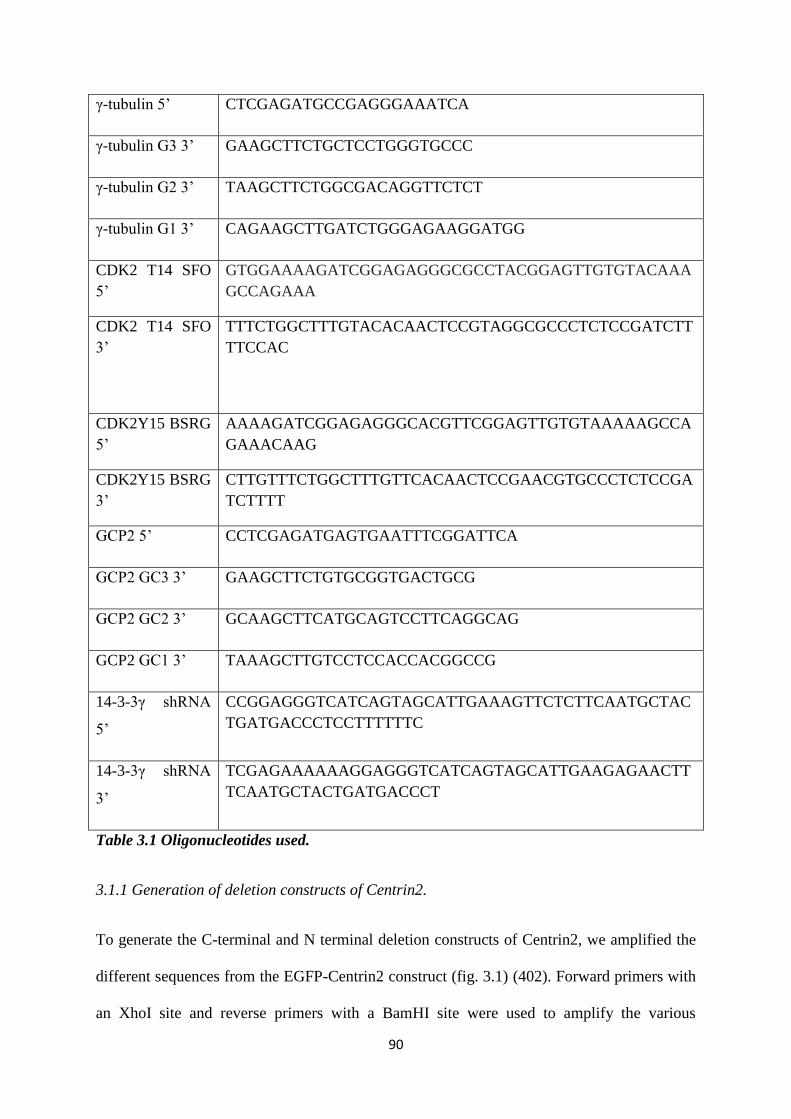
90
γ-tubulin 5’ CTCGAGATGCCGAGGGAAATCA
γ-tubulin G3 3’ GAAGCTTCTGCTCCTGGGTGCCC
γ-tubulin G2 3’ TAAGCTTCTGGCGACAGGTTCTCT
γ-tubulin G1 3’ CAGAAGCTTGATCTGGGAGAAGGATGG
CDK2 T14 SFO
5’
GTGGAAAAGATCGGAGAGGGCGCCTACGGAGTTGTGTACAAA
GCCAGAAA
CDK2 T14 SFO
3’
TTTCTGGCTTTGTACACAACTCCGTAGGCGCCCTCTCCGATCTT
TTCCAC
CDK2Y15 BSRG
5’
AAAAGATCGGAGAGGGCACGTTCGGAGTTGTGTAAAAAGCCA
GAAACAAG
CDK2Y15 BSRG
3’
CTTGTTTCTGGCTTTGTTCACAACTCCGAACGTGCCCTCTCCGA
TCTTTT
GCP2 5’ CCTCGAGATGAGTGAATTTCGGATTCA
GCP2 GC3 3’ GAAGCTTCTGTGCGGTGACTGCG
GCP2 GC2 3’ GCAAGCTTCATGCAGTCCTTCAGGCAG
GCP2 GC1 3’ TAAAGCTTGTCCTCCACCACGGCCG
14-3-3γ shRNA
5’
CCGGAGGGTCATCAGTAGCATTGAAAGTTCTCTTCAATGCTAC
TGATGACCCTCCTTTTTTC
14-3-3γ shRNA
3’
TCGAGAAAAAAGGAGGGTCATCAGTAGCATTGAAGAGAACTT
TCAATGCTACTGATGACCCT
Table 3.1 Oligonucleotides used.
3.1.1 Generation of deletion constructs of Centrin2.
To generate the C-terminal and N terminal deletion constructs of Centrin2, we amplified the
different sequences from the EGFP-Centrin2 construct (fig. 3.1) (402). Forward primers with
an XhoI site and reverse primers with a BamHI site were used to amplify the various
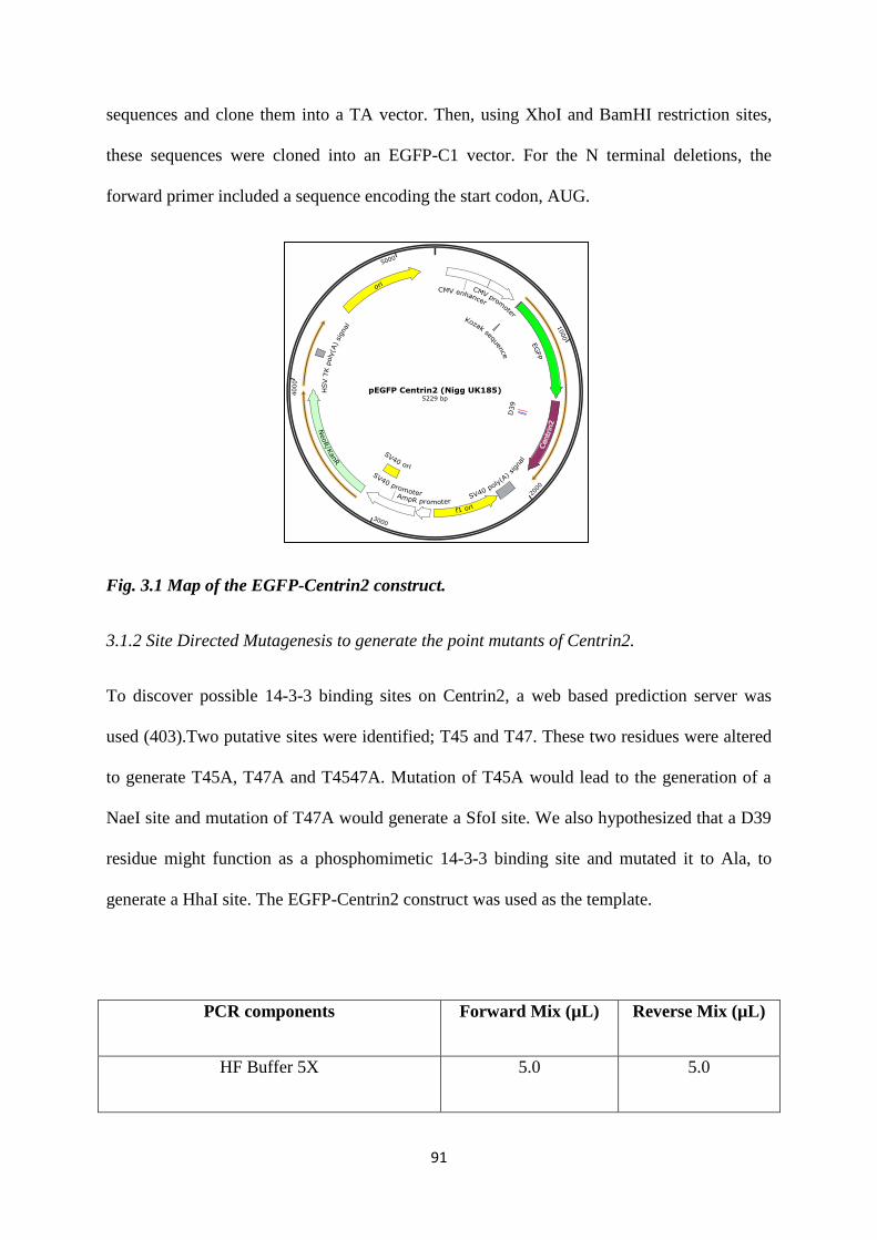
91
sequences and clone them into a TA vector. Then, using XhoI and BamHI restriction sites,
these sequences were cloned into an EGFP-C1 vector. For the N terminal deletions, the
forward primer included a sequence encoding the start codon, AUG.
Fig. 3.1 Map of the EGFP-Centrin2 construct.
3.1.2 Site Directed Mutagenesis to generate the point mutants of Centrin2.
To discover possible 14-3-3 binding sites on Centrin2, a web based prediction server was
used (403).Two putative sites were identified; T45 and T47. These two residues were altered
to generate T45A, T47A and T4547A. Mutation of T45A would lead to the generation of a
NaeI site and mutation of T47A would generate a SfoI site. We also hypothesized that a D39
residue might function as a phosphomimetic 14-3-3 binding site and mutated it to Ala, to
generate a HhaI site. The EGFP-Centrin2 construct was used as the template.
PCR components Forward Mix (µL) Reverse Mix (µL)
HF Buffer 5X 5.0 5.0
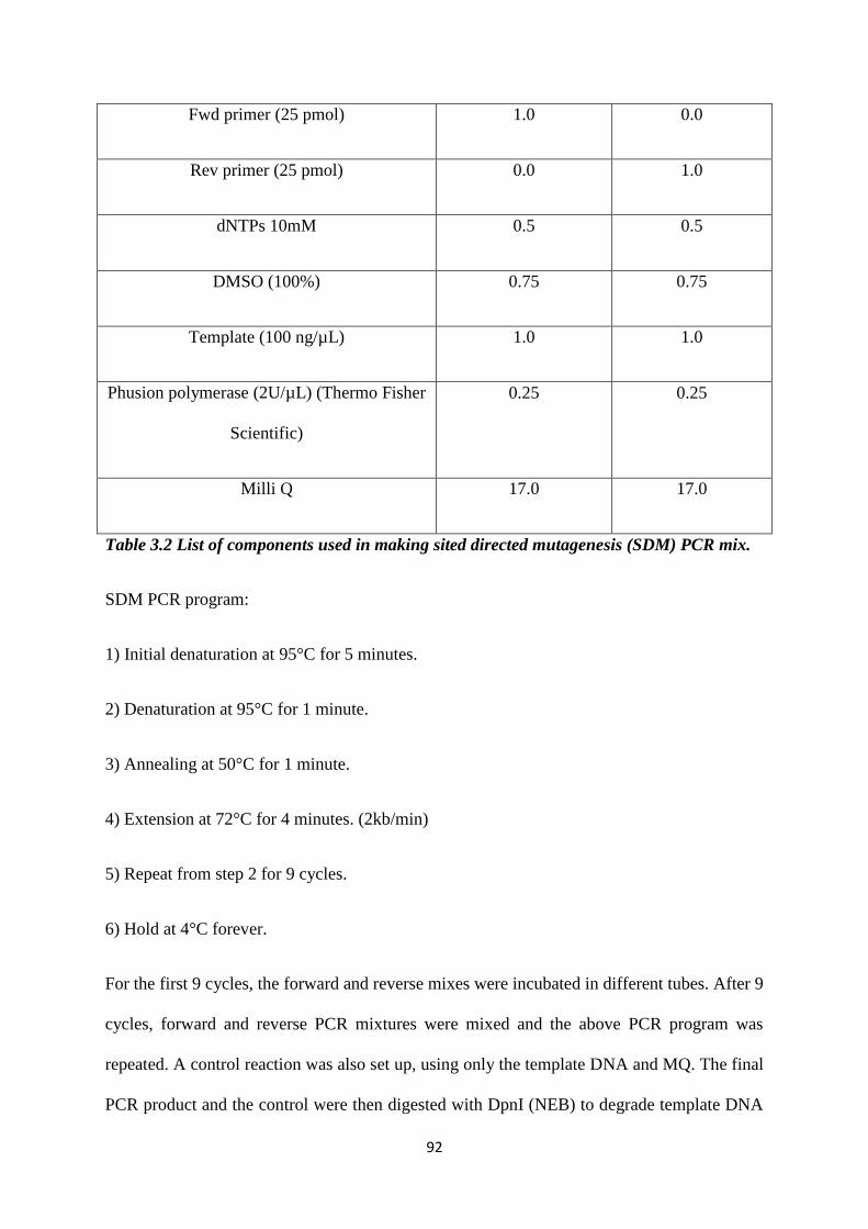
92
Fwd primer (25 pmol) 1.0 0.0
Rev primer (25 pmol) 0.0 1.0
dNTPs 10mM 0.5 0.5
DMSO (100%) 0.75 0.75
Template (100 ng/µL) 1.0 1.0
Phusion polymerase (2U/µL) (Thermo Fisher
Scientific)
0.25 0.25
Milli Q 17.0 17.0
Table 3.2 List of components used in making sited directed mutagenesis (SDM) PCR mix.
SDM PCR program:
1) Initial denaturation at 95°C for 5 minutes.
2) Denaturation at 95°C for 1 minute.
3) Annealing at 50°C for 1 minute.
4) Extension at 72°C for 4 minutes. (2kb/min)
5) Repeat from step 2 for 9 cycles.
6) Hold at 4°C forever.
For the first 9 cycles, the forward and reverse mixes were incubated in different tubes. After 9
cycles, forward and reverse PCR mixtures were mixed and the above PCR program was
repeated. A control reaction was also set up, using only the template DNA and MQ. The final
PCR product and the control were then digested with DpnI (NEB) to degrade template DNA
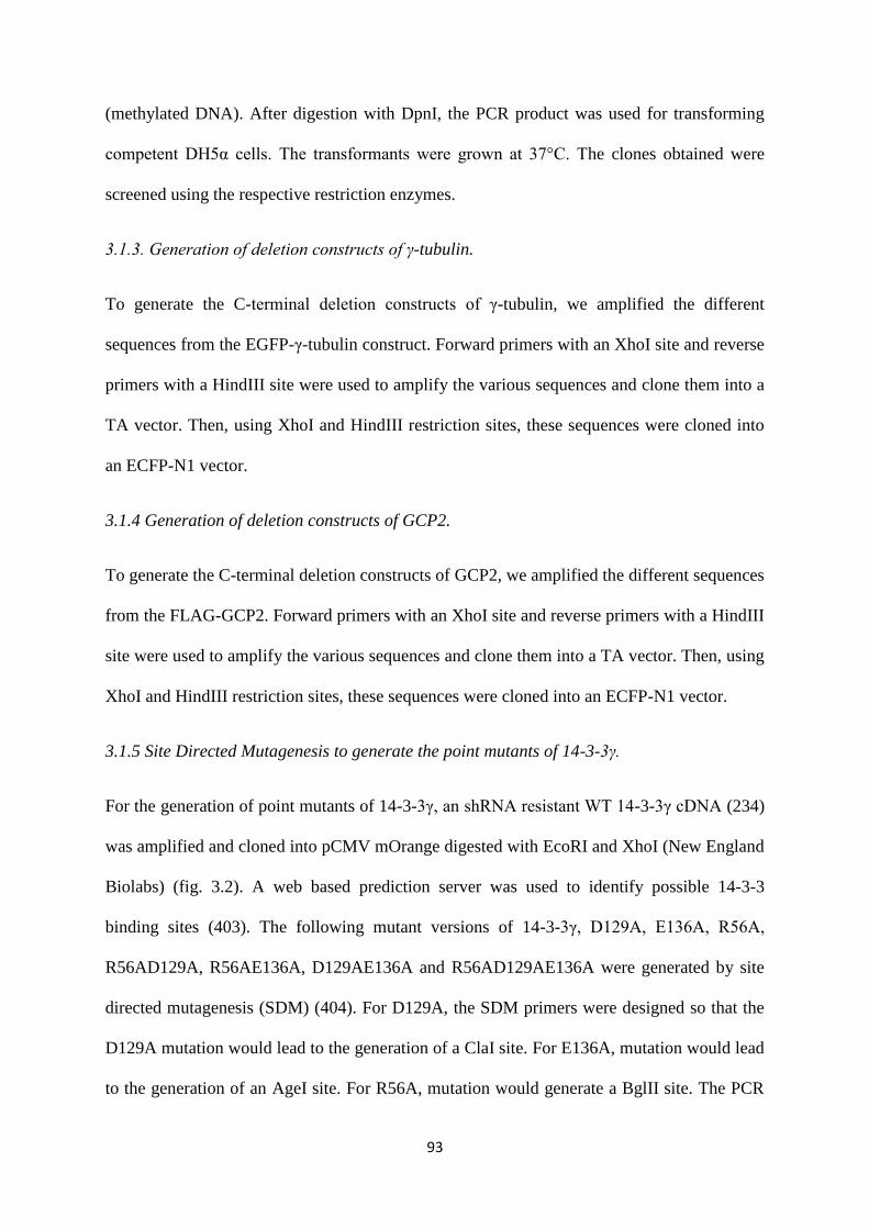
93
(methylated DNA). After digestion with DpnI, the PCR product was used for transforming
competent DH5α cells. The transformants were grown at 37°C. The clones obtained were
screened using the respective restriction enzymes.
3.1.3. Generation of deletion constructs of γ-tubulin.
To generate the C-terminal deletion constructs of γ-tubulin, we amplified the different
sequences from the EGFP-γ-tubulin construct. Forward primers with an XhoI site and reverse
primers with a HindIII site were used to amplify the various sequences and clone them into a
TA vector. Then, using XhoI and HindIII restriction sites, these sequences were cloned into
an ECFP-N1 vector.
3.1.4 Generation of deletion constructs of GCP2.
To generate the C-terminal deletion constructs of GCP2, we amplified the different sequences
from the FLAG-GCP2. Forward primers with an XhoI site and reverse primers with a HindIII
site were used to amplify the various sequences and clone them into a TA vector. Then, using
XhoI and HindIII restriction sites, these sequences were cloned into an ECFP-N1 vector.
3.1.5 Site Directed Mutagenesis to generate the point mutants of 14-3-3γ.
For the generation of point mutants of 14-3-3γ, an shRNA resistant WT 14-3-3γ cDNA (234)
was amplified and cloned into pCMV mOrange digested with EcoRI and XhoI (New England
Biolabs) (fig. 3.2). A web based prediction server was used to identify possible 14-3-3
binding sites (403). The following mutant versions of 14-3-3γ, D129A, E136A, R56A,
R56AD129A, R56AE136A, D129AE136A and R56AD129AE136A were generated by site
directed mutagenesis (SDM) (404). For D129A, the SDM primers were designed so that the
D129A mutation would lead to the generation of a ClaI site. For E136A, mutation would lead
to the generation of an AgeI site. For R56A, mutation would generate a BglII site. The PCR
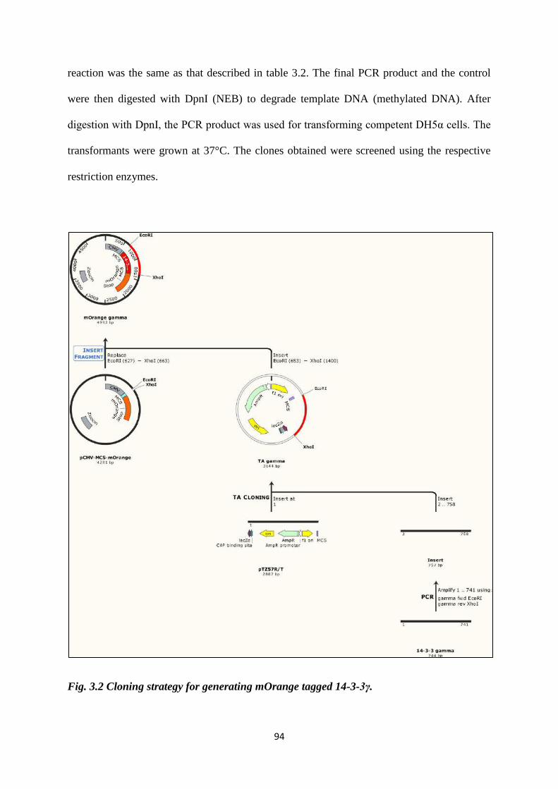
94
reaction was the same as that described in table 3.2. The final PCR product and the control
were then digested with DpnI (NEB) to degrade template DNA (methylated DNA). After
digestion with DpnI, the PCR product was used for transforming competent DH5α cells. The
transformants were grown at 37°C. The clones obtained were screened using the respective
restriction enzymes.
Fig. 3.2 Cloning strategy for generating mOrange tagged 14-3-3γ.
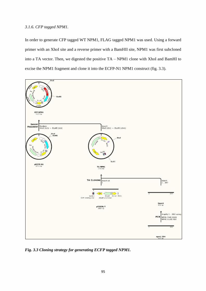
95
3.1.6. CFP tagged NPM1.
In order to generate CFP tagged WT NPM1, FLAG tagged NPM1 was used. Using a forward
primer with an XhoI site and a reverse primer with a BamHII site, NPM1 was first subcloned
into a TA vector. Then, we digested the positive TA – NPM1 clone with XhoI and BamHI to
excise the NPM1 fragment and clone it into the ECFP-N1 NPM1 construct (fig. 3.3).
Fig. 3.3 Cloning strategy for generating ECFP tagged NPM1.
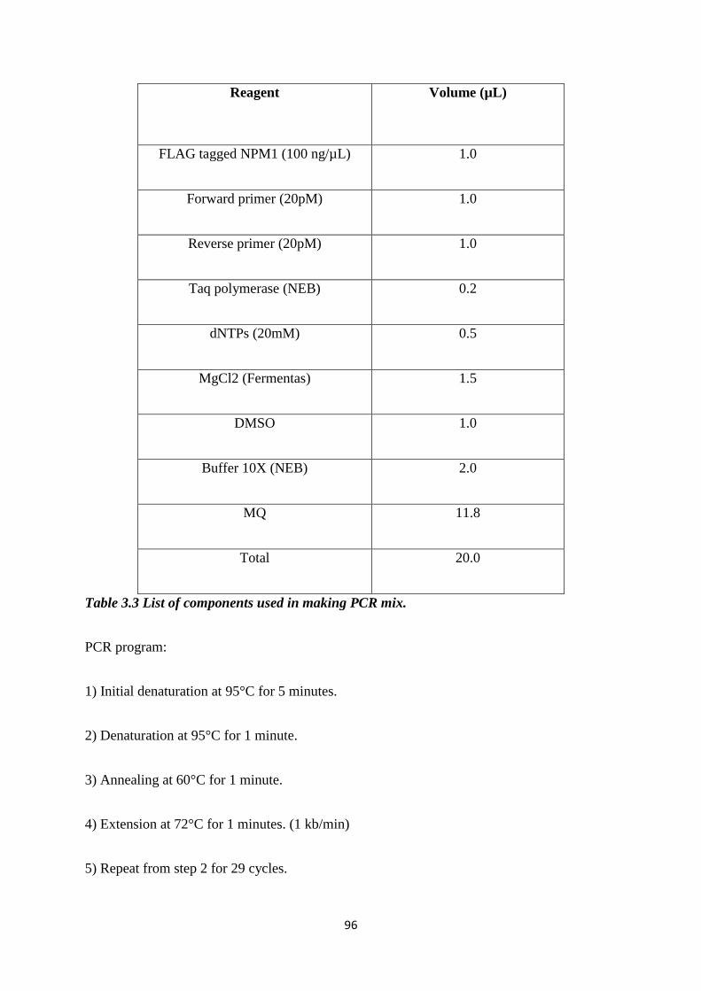
96
Reagent
Volume (µL)
FLAG tagged NPM1 (100 ng/µL) 1.0
Forward primer (20pM) 1.0
Reverse primer (20pM) 1.0
Taq polymerase (NEB) 0.2
dNTPs (20mM) 0.5
MgCl2 (Fermentas) 1.5
DMSO 1.0
Buffer 10X (NEB) 2.0
MQ 11.8
Total 20.0
Table 3.3 List of components used in making PCR mix.
PCR program:
1) Initial denaturation at 95°C for 5 minutes.
2) Denaturation at 95°C for 1 minute.
3) Annealing at 60°C for 1 minute.
4) Extension at 72°C for 1 minutes. (1 kb/min)
5) Repeat from step 2 for 29 cycles.
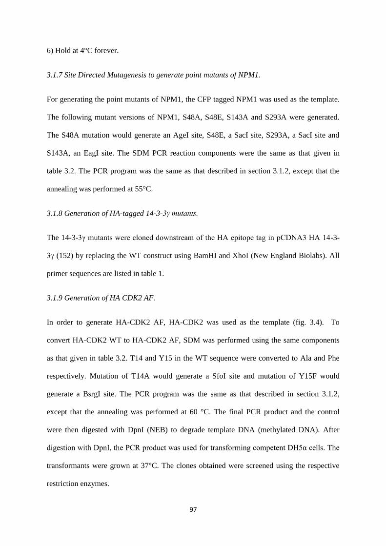
97
6) Hold at 4°C forever.
3.1.7 Site Directed Mutagenesis to generate point mutants of NPM1.
For generating the point mutants of NPM1, the CFP tagged NPM1 was used as the template.
The following mutant versions of NPM1, S48A, S48E, S143A and S293A were generated.
The S48A mutation would generate an AgeI site, S48E, a SacI site, S293A, a SacI site and
S143A, an EagI site. The SDM PCR reaction components were the same as that given in
table 3.2. The PCR program was the same as that described in section 3.1.2, except that the
annealing was performed at 55°C.
3.1.8 Generation of HA-tagged 14-3-3γ mutants.
The 14-3-3γ mutants were cloned downstream of the HA epitope tag in pCDNA3 HA 14-3-
3γ (152) by replacing the WT construct using BamHI and XhoI (New England Biolabs). All
primer sequences are listed in table 1.
3.1.9 Generation of HA CDK2 AF.
In order to generate HA-CDK2 AF, HA-CDK2 was used as the template (fig. 3.4). To
convert HA-CDK2 WT to HA-CDK2 AF, SDM was performed using the same components
as that given in table 3.2. T14 and Y15 in the WT sequence were converted to Ala and Phe
respectively. Mutation of T14A would generate a SfoI site and mutation of Y15F would
generate a BsrgI site. The PCR program was the same as that described in section 3.1.2,
except that the annealing was performed at 60 °C. The final PCR product and the control
were then digested with DpnI (NEB) to degrade template DNA (methylated DNA). After
digestion with DpnI, the PCR product was used for transforming competent DH5α cells. The
transformants were grown at 37°C. The clones obtained were screened using the respective
restriction enzymes.
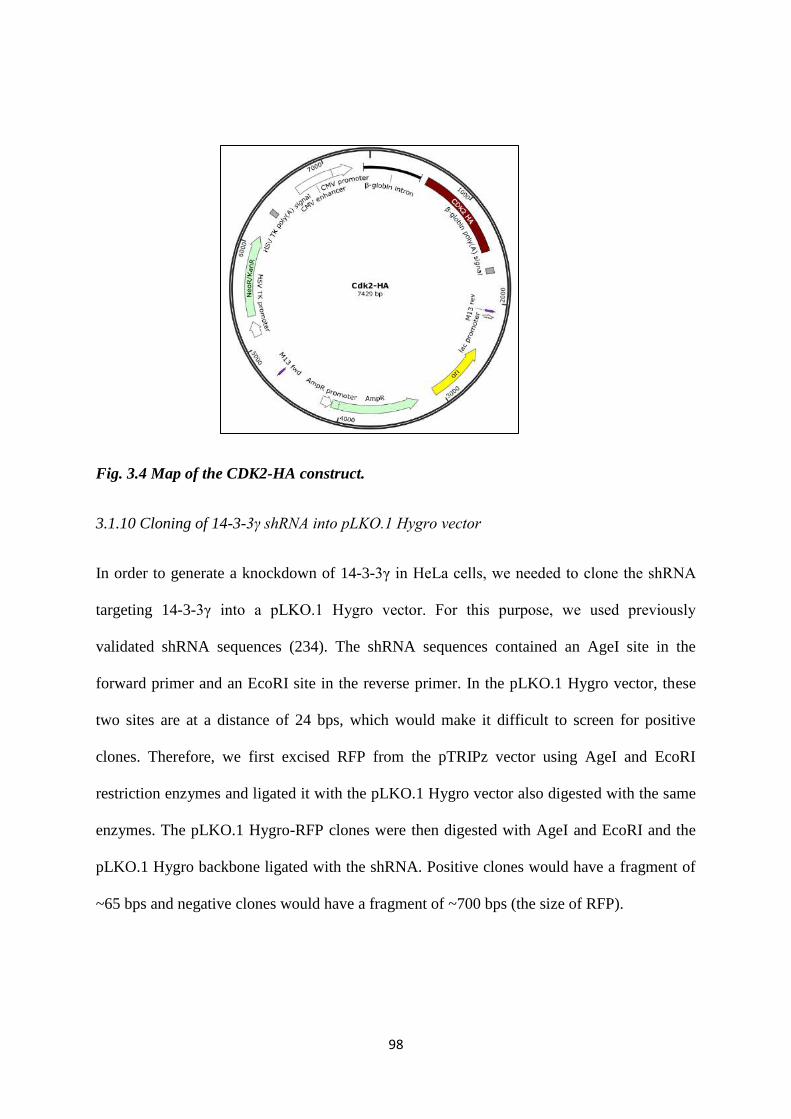
98
Fig. 3.4 Map of the CDK2-HA construct.
3.1.10 Cloning of 14-3-3γ shRNA into pLKO.1 Hygro vector
In order to generate a knockdown of 14-3-3γ in HeLa cells, we needed to clone the shRNA
targeting 14-3-3γ into a pLKO.1 Hygro vector. For this purpose, we used previously
validated shRNA sequences (234). The shRNA sequences contained an AgeI site in the
forward primer and an EcoRI site in the reverse primer. In the pLKO.1 Hygro vector, these
two sites are at a distance of 24 bps, which would make it difficult to screen for positive
clones. Therefore, we first excised RFP from the pTRIPz vector using AgeI and EcoRI
restriction enzymes and ligated it with the pLKO.1 Hygro vector also digested with the same
enzymes. The pLKO.1 Hygro-RFP clones were then digested with AgeI and EcoRI and the
pLKO.1 Hygro backbone ligated with the shRNA. Positive clones would have a fragment of
~65 bps and negative clones would have a fragment of ~700 bps (the size of RFP).
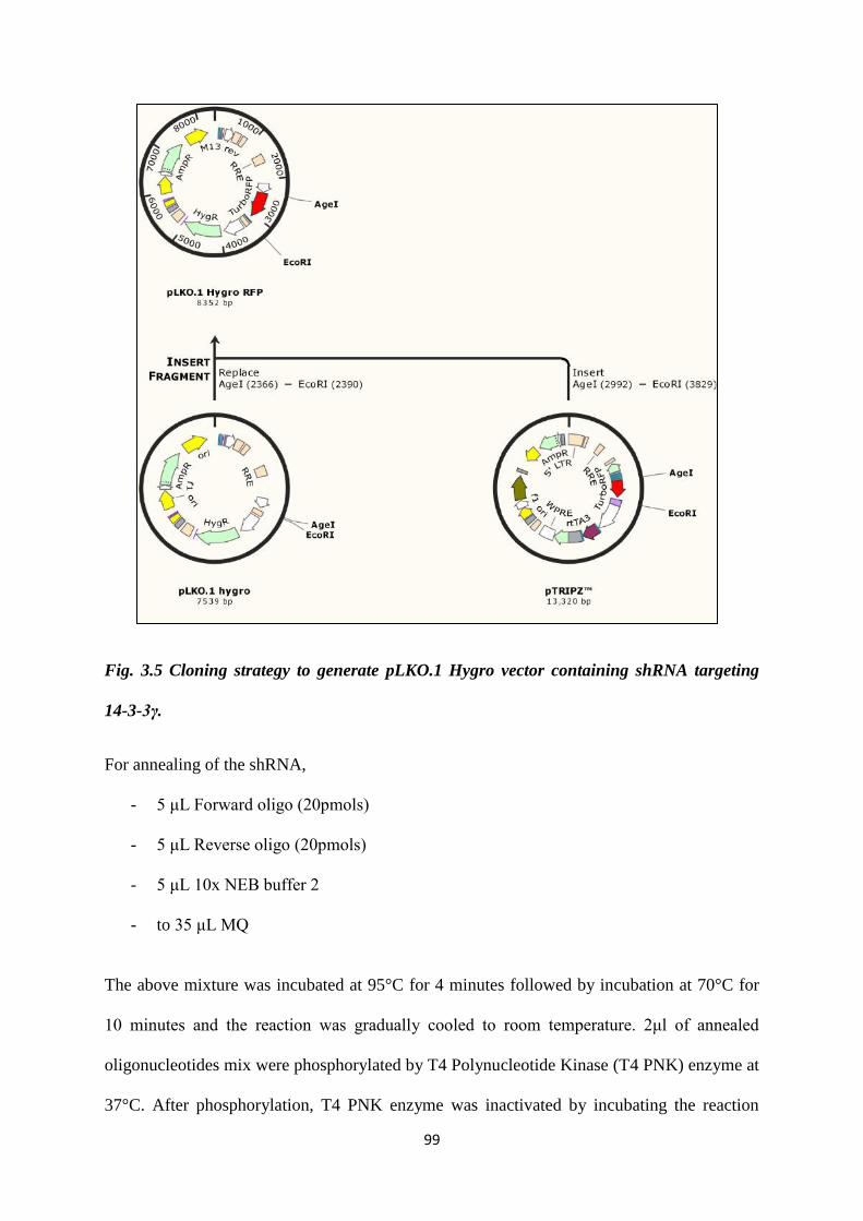
99
Fig. 3.5 Cloning strategy to generate pLKO.1 Hygro vector containing shRNA targeting
14-3-3γ.
For annealing of the shRNA,
- 5 μL Forward oligo (20pmols)
- 5 μL Reverse oligo (20pmols)
- 5 μL 10x NEB buffer 2
- to 35 μL MQ
The above mixture was incubated at 95°C for 4 minutes followed by incubation at 70°C for
10 minutes and the reaction was gradually cooled to room temperature. 2μl of annealed
oligonucleotides mix were phosphorylated by T4 Polynucleotide Kinase (T4 PNK) enzyme at
37°C. After phosphorylation, T4 PNK enzyme was inactivated by incubating the reaction
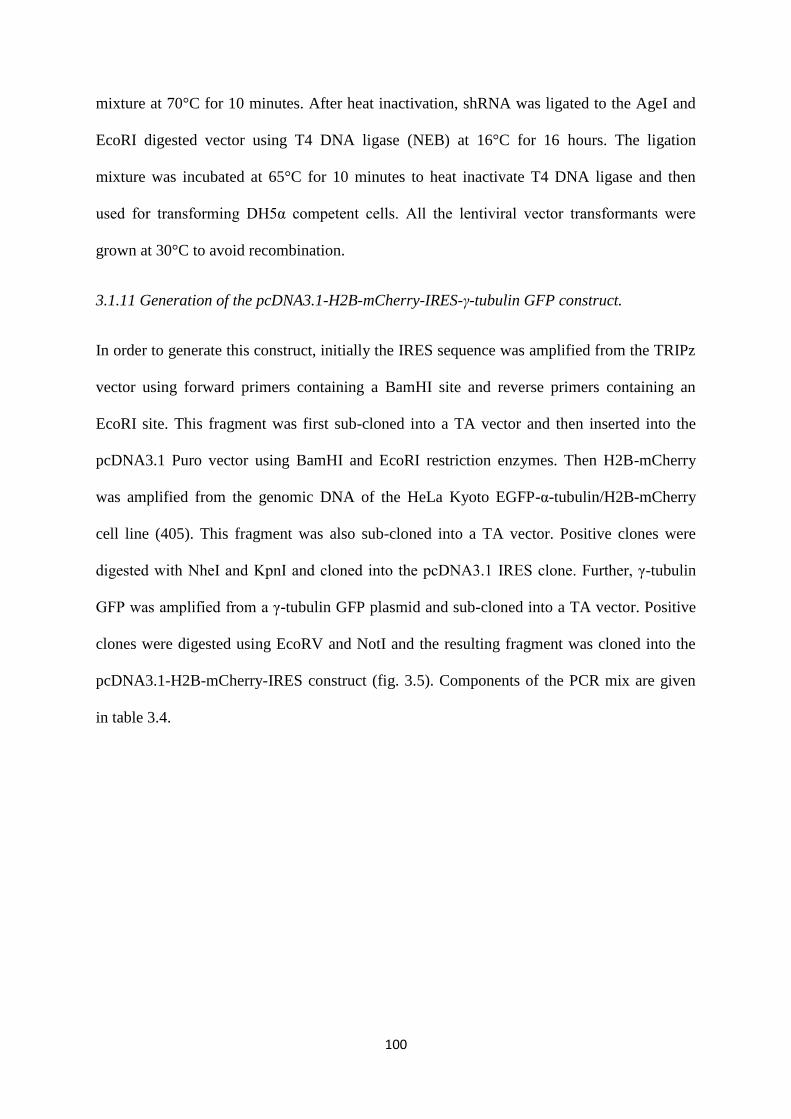
100
mixture at 70°C for 10 minutes. After heat inactivation, shRNA was ligated to the AgeI and
EcoRI digested vector using T4 DNA ligase (NEB) at 16°C for 16 hours. The ligation
mixture was incubated at 65°C for 10 minutes to heat inactivate T4 DNA ligase and then
used for transforming DH5α competent cells. All the lentiviral vector transformants were
grown at 30°C to avoid recombination.
3.1.11 Generation of the pcDNA3.1-H2B-mCherry-IRES-γ-tubulin GFP construct.
In order to generate this construct, initially the IRES sequence was amplified from the TRIPz
vector using forward primers containing a BamHI site and reverse primers containing an
EcoRI site. This fragment was first sub-cloned into a TA vector and then inserted into the
pcDNA3.1 Puro vector using BamHI and EcoRI restriction enzymes. Then H2B-mCherry
was amplified from the genomic DNA of the HeLa Kyoto EGFP-α-tubulin/H2B-mCherry
cell line (405). This fragment was also sub-cloned into a TA vector. Positive clones were
digested with NheI and KpnI and cloned into the pcDNA3.1 IRES clone. Further, γ-tubulin
GFP was amplified from a γ-tubulin GFP plasmid and sub-cloned into a TA vector. Positive
clones were digested using EcoRV and NotI and the resulting fragment was cloned into the
pcDNA3.1-H2B-mCherry-IRES construct (fig. 3.5). Components of the PCR mix are given
in table 3.4.
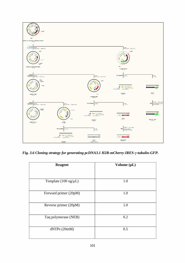
101
Fig. 3.6 Cloning strategy for generating pcDNA3.1 H2B-mCherry-IRES-γ-tubulin-GFP.
Reagent
Volume (µL)
Template (100 ng/µL) 1.0
Forward primer (20pM) 1.0
Reverse primer (20pM) 1.0
Taq polymerase (NEB) 0.2
dNTPs (20mM) 0.5

102
MgCl2 (Fermentas) 1.5
Buffer 10X (NEB) 2.0
MQ 11.8
Total 20.0
Table 3.4 List of components used in making PCR mix.
PCR program:
1) Initial denaturation at 95°C for 5 minutes.
2) Denaturation at 95°C for 1 minute.
3) Annealing for 1 minute at
- 52°C for IRES
- 55°C for H2B-mCherry
- 53°C for γ-tubulin-GFP
4) Extension at 72°C for
- 50 secs for IRES (1 kb/min)
- 1 minute 20 secs for H2B-mCherry
- 2 minutes 20 secs for γ-tubulin-GFP
5) Repeat from step 2 for 29 cycles.
6) Hold at 4°C forever.
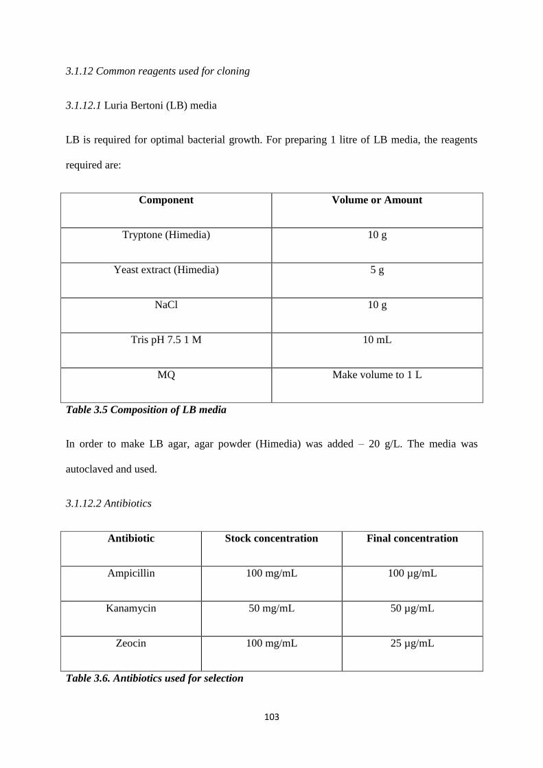
103
3.1.12 Common reagents used for cloning
3.1.12.1 Luria Bertoni (LB) media
LB is required for optimal bacterial growth. For preparing 1 litre of LB media, the reagents
required are:
Component Volume or Amount
Tryptone (Himedia) 10 g
Yeast extract (Himedia) 5 g
NaCl 10 g
Tris pH 7.5 1 M 10 mL
MQ Make volume to 1 L
Table 3.5 Composition of LB media
In order to make LB agar, agar powder (Himedia) was added – 20 g/L. The media was
autoclaved and used.
3.1.12.2 Antibiotics
Antibiotic Stock concentration Final concentration
Ampicillin 100 mg/mL 100 µg/mL
Kanamycin 50 mg/mL 50 µg/mL
Zeocin 100 mg/mL 25 µg/mL
Table 3.6. Antibiotics used for selection
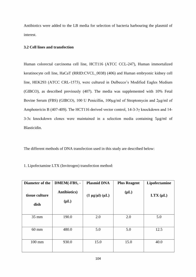
104
Antibiotics were added to the LB media for selection of bacteria harbouring the plasmid of
interest.
3.2 Cell lines and transfection
Human colorectal carcinoma cell line, HCT116 (ATCC CCL-247), Human immortalized
keratinocyte cell line, HaCaT (RRID:CVCL_0038) (406) and Human embryonic kidney cell
line, HEK293 (ATCC CRL-1573), were cultured in Dulbecco’s Modified Eagles Medium
(GIBCO), as described previously (407). The media was supplemented with 10% Fetal
Bovine Serum (FBS) (GIBCO), 100 U Penicillin, 100µg/ml of Streptomycin and 2μg/ml of
Amphotericin B (407-409). The HCT116 derived vector control, 14-3-3γ knockdown and 14-
3-3ε knockdown clones were maintained in a selection media containing 5µg/ml of
Blasticidin.
The different methods of DNA transfection used in this study are described below:
1. Lipofectamine LTX (Invitrogen) transfection method:
Diameter of the
tissue culture
dish
DMEM(-FBS, -
Antibiotics)
(µL)
Plasmid DNA
(1 μg/μl) (µL)
Plus Reagent
(µL)
Lipofectamine
LTX (µL)
35 mm 190.0 2.0 2.0 5.0
60 mm 480.0 5.0 5.0 12.5
100 mm 930.0 15.0 15.0 40.0
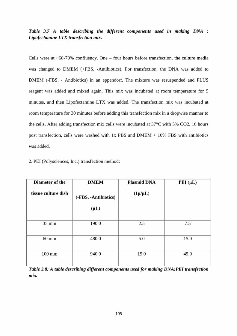
105
Table 3.7 A table describing the different components used in making DNA :
Lipofectamine LTX transfection mix.
Cells were at ~60-70% confluency. One – four hours before transfection, the culture media
was changed to DMEM (+FBS, -Antibiotics). For transfection, the DNA was added to
DMEM (-FBS, - Antibiotics) in an eppendorf. The mixture was resuspended and PLUS
reagent was added and mixed again. This mix was incubated at room temperature for 5
minutes, and then Lipofectamine LTX was added. The transfection mix was incubated at
room temperature for 30 minutes before adding this transfection mix in a dropwise manner to
the cells. After adding transfection mix cells were incubated at 37°C with 5% CO2. 16 hours
post transfection, cells were washed with 1x PBS and DMEM + 10% FBS with antibiotics
was added.
2. PEI (Polysciences, Inc.) transfection method:
Diameter of the
tissue culture dish
DMEM
(-FBS, -Antibiotics)
(μL)
Plasmid DNA
(1μ/μL)
PEI (μL)
35 mm 190.0 2.5 7.5
60 mm 480.0 5.0 15.0
100 mm 940.0 15.0 45.0
Table 3.8: A table describing different components used for making DNA:PEI transfection
mix.
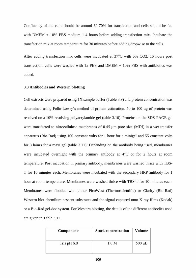
106
Confluency of the cells should be around 60-70% for transfection and cells should be fed
with DMEM + 10% FBS medium 1-4 hours before adding transfection mix. Incubate the
transfection mix at room temperature for 30 minutes before adding dropwise to the cells.
After adding transfection mix cells were incubated at 37°C with 5% CO2. 16 hours post
transfection, cells were washed with 1x PBS and DMEM + 10% FBS with antibiotics was
added.
3.3 Antibodies and Western blotting
Cell extracts were prepared using 1X sample buffer (Table 3.9) and protein concentration was
determined using Folin-Lowry’s method of protein estimation. 50 to 100 μg of protein was
resolved on a 10% resolving polyacrylamide gel (table 3.10). Proteins on the SDS-PAGE gel
were transferred to nitrocellulose membranes of 0.45 μm pore size (MDI) in a wet transfer
apparatus (Bio-Rad) using 100 constant volts for 1 hour for a minigel and 55 constant volts
for 3 hours for a maxi gel (table 3.11). Depending on the antibody being used, membranes
were incubated overnight with the primary antibody at 4°C or for 2 hours at room
temperature. Post incubation in primary antibody, membranes were washed thrice with TBS-
T for 10 minutes each. Membranes were incubated with the secondary HRP antibody for 1
hour at room temperature. Membranes were washed thrice with TBS-T for 10 minutes each.
Membranes were flooded with either PicoWest (Thermoscientific) or Clarity (Bio-Rad)
Western blot chemiluminescent substrates and the signal captured onto X-ray films (Kodak)
or a Bio-Rad gel-doc system. For Western blotting, the details of the different antibodies used
are given in Table 3.12.
Components Stock concentration Volume
Tris pH 6.8 1.0 M 500 μL
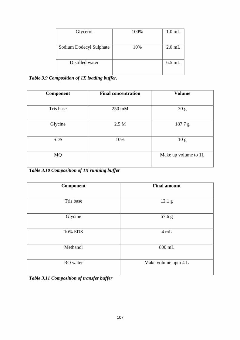
107
Glycerol 100% 1.0 mL
Sodium Dodecyl Sulphate 10% 2.0 mL
Distilled water 6.5 mL
Table 3.9 Composition of 1X loading buffer.
Component Final concentration Volume
Tris base 250 mM 30 g
Glycine 2.5 M 187.7 g
SDS 10% 10 g
MQ Make up volume to 1L
Table 3.10 Composition of 1X running buffer
Component Final amount
Tris base 12.1 g
Glycine 57.6 g
10% SDS 4 mL
Methanol 800 mL
RO water Make volume upto 4 L
Table 3.11 Composition of transfer buffer
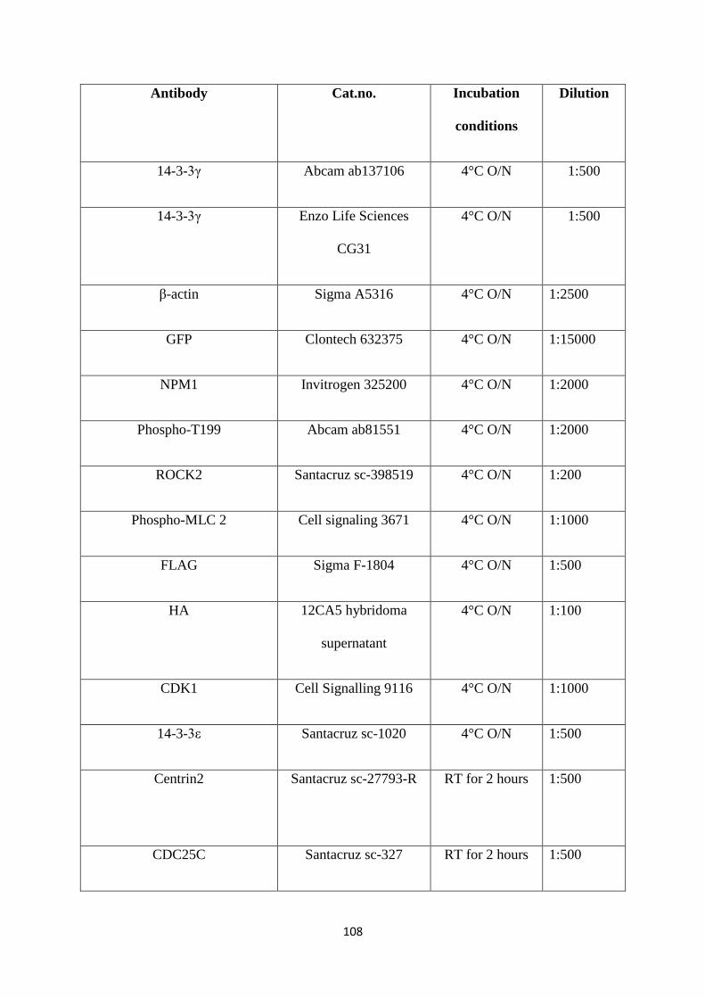
108
Antibody Cat.no. Incubation
conditions
Dilution
14-3-3γ Abcam ab137106 4°C O/N 1:500
14-3-3γ Enzo Life Sciences
CG31
4°C O/N 1:500
β-actin Sigma A5316 4°C O/N 1:2500
GFP Clontech 632375 4°C O/N 1:15000
NPM1 Invitrogen 325200 4°C O/N 1:2000
Phospho-T199 Abcam ab81551 4°C O/N 1:2000
ROCK2 Santacruz sc-398519 4°C O/N 1:200
Phospho-MLC 2 Cell signaling 3671 4°C O/N 1:1000
FLAG Sigma F-1804 4°C O/N 1:500
HA 12CA5 hybridoma
supernatant
4°C O/N 1:100
CDK1 Cell Signalling 9116 4°C O/N 1:1000
14-3-3ε Santacruz sc-1020 4°C O/N 1:500
Centrin2 Santacruz sc-27793-R
RT for 2 hours 1:500
CDC25C Santacruz sc-327 RT for 2 hours 1:500
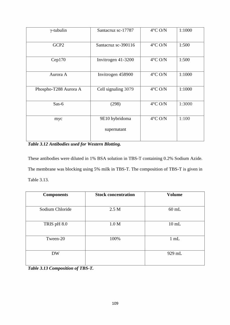
109
γ-tubulin Santacruz sc-17787 4°C O/N 1:1000
GCP2 Santacruz sc-390116 4°C O/N 1:500
Cep170 Invitrogen 41-3200 4°C O/N 1:500
Aurora A Invitrogen 458900 4°C O/N 1:1000
Phospho-T288 Aurora A Cell signaling 3079 4°C O/N 1:1000
Sas-6 (298) 4°C O/N 1:3000
myc 9E10 hybridoma
supernatant
4°C O/N 1:100
Table 3.12 Antibodies used for Western Blotting.
These antibodies were diluted in 1% BSA solution in TBS-T containing 0.2% Sodium Azide.
The membrane was blocking using 5% milk in TBS-T. The composition of TBS-T is given in
Table 3.13.
Components Stock concentration Volume
Sodium Chloride 2.5 M 60 mL
TRIS pH 8.0 1.0 M 10 mL
Tween-20 100% 1 mL
DW 929 mL
Table 3.13 Composition of TBS-T.
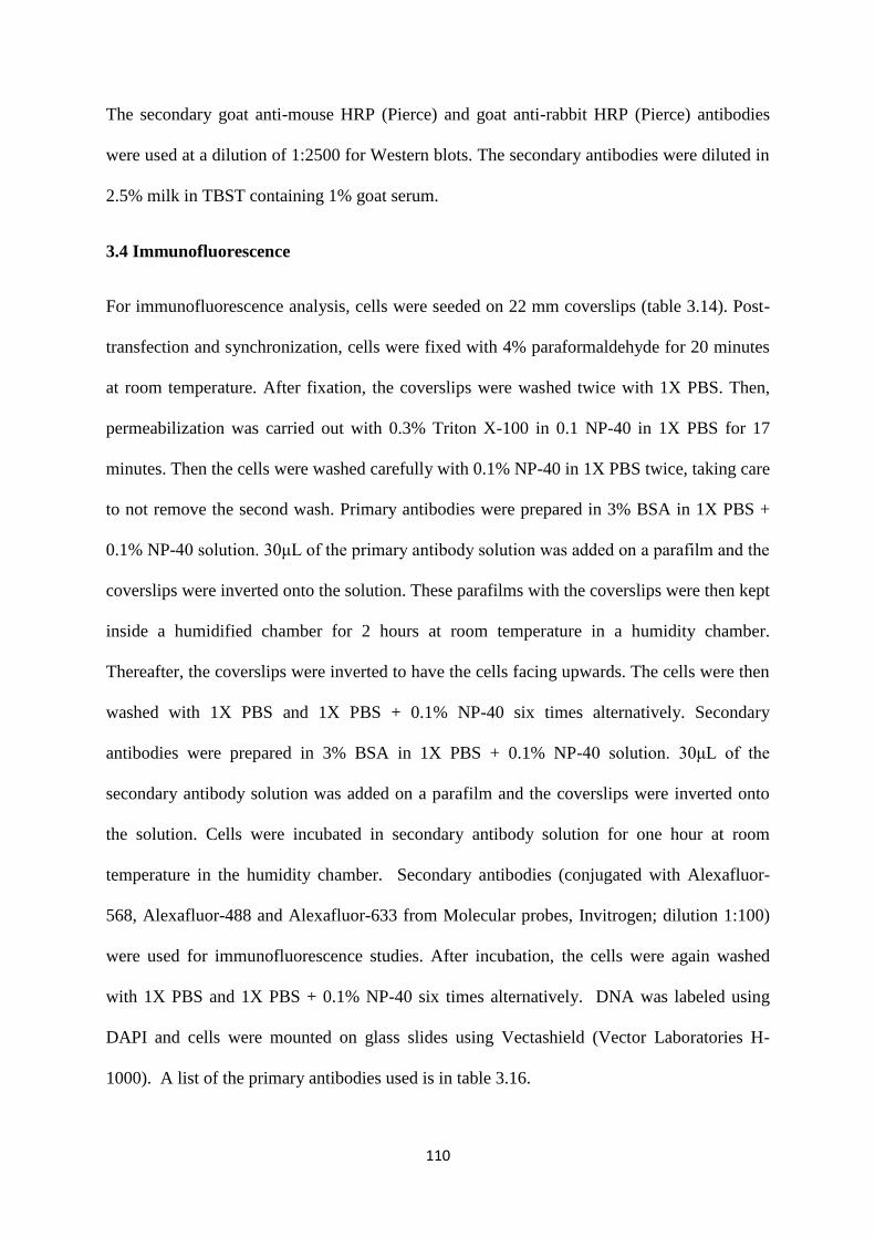
110
The secondary goat anti-mouse HRP (Pierce) and goat anti-rabbit HRP (Pierce) antibodies
were used at a dilution of 1:2500 for Western blots. The secondary antibodies were diluted in
2.5% milk in TBST containing 1% goat serum.
3.4 Immunofluorescence
For immunofluorescence analysis, cells were seeded on 22 mm coverslips (table 3.14). Post-
transfection and synchronization, cells were fixed with 4% paraformaldehyde for 20 minutes
at room temperature. After fixation, the coverslips were washed twice with 1X PBS. Then,
permeabilization was carried out with 0.3% Triton X-100 in 0.1 NP-40 in 1X PBS for 17
minutes. Then the cells were washed carefully with 0.1% NP-40 in 1X PBS twice, taking care
to not remove the second wash. Primary antibodies were prepared in 3% BSA in 1X PBS +
0.1% NP-40 solution. 30μL of the primary antibody solution was added on a parafilm and the
coverslips were inverted onto the solution. These parafilms with the coverslips were then kept
inside a humidified chamber for 2 hours at room temperature in a humidity chamber.
Thereafter, the coverslips were inverted to have the cells facing upwards. The cells were then
washed with 1X PBS and 1X PBS + 0.1% NP-40 six times alternatively. Secondary
antibodies were prepared in 3% BSA in 1X PBS + 0.1% NP-40 solution. 30μL of the
secondary antibody solution was added on a parafilm and the coverslips were inverted onto
the solution. Cells were incubated in secondary antibody solution for one hour at room
temperature in the humidity chamber. Secondary antibodies (conjugated with Alexafluor-
568, Alexafluor-488 and Alexafluor-633 from Molecular probes, Invitrogen; dilution 1:100)
were used for immunofluorescence studies. After incubation, the cells were again washed
with 1X PBS and 1X PBS + 0.1% NP-40 six times alternatively. DNA was labeled using
DAPI and cells were mounted on glass slides using Vectashield (Vector Laboratories H-
1000). A list of the primary antibodies used is in table 3.16.
111
DNA Amount of DNA transfected (µg)
14-3-3γ-mOrange 1.0
EGFP-Centrin2 1.0
ECFP-NPM1 0.5
FLAG-NPM1 1.0
myc-Plk4 1.0
HA-CDK2 0.5
CDK1 0.5
Table 3.14 Amount of DNA used
Component Volume or amount
NaCl 8 g
KH2PO4 0.2
KCl 0.2
Na2HPO4 2.18
MQ Make up volume to 1L
Table 3.15 Composition of 1X PBS
Antibody Cat. No. Dilution
Pericentrin Abcam ab4448 1:1000
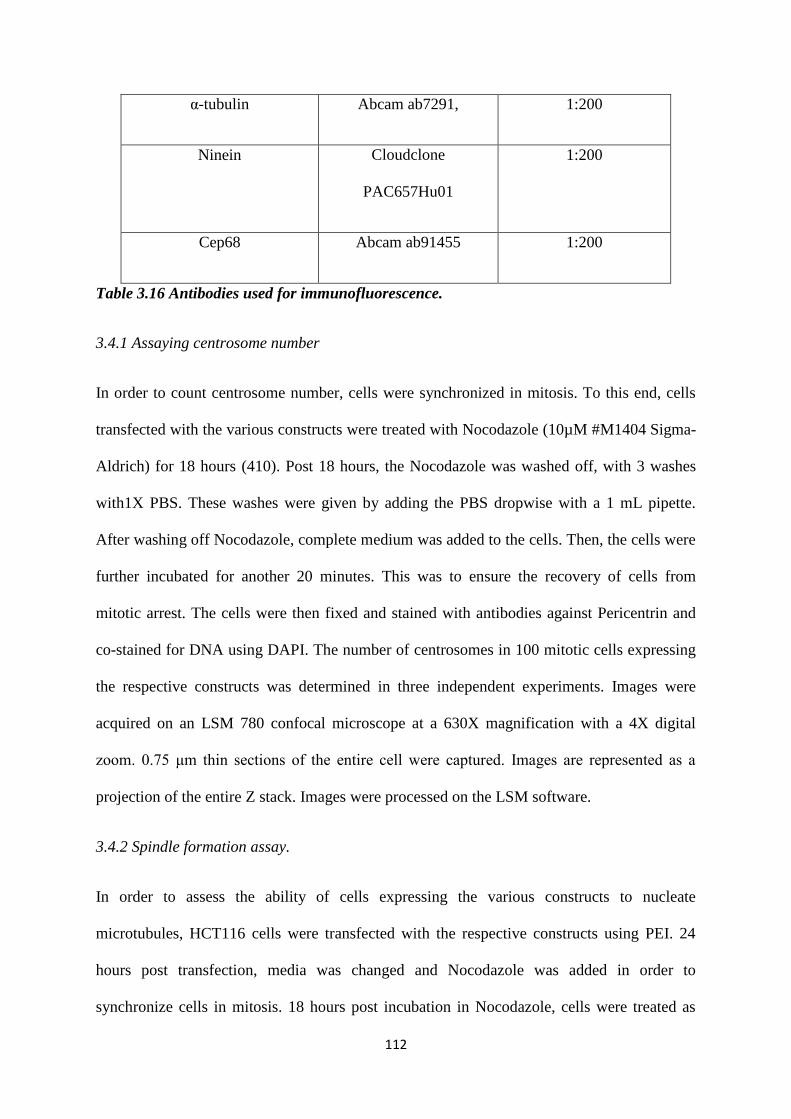
112
α-tubulin Abcam ab7291, 1:200
Ninein Cloudclone
PAC657Hu01
1:200
Cep68 Abcam ab91455 1:200
Table 3.16 Antibodies used for immunofluorescence.
3.4.1 Assaying centrosome number
In order to count centrosome number, cells were synchronized in mitosis. To this end, cells
transfected with the various constructs were treated with Nocodazole (10µM #M1404 Sigma-
Aldrich) for 18 hours (410). Post 18 hours, the Nocodazole was washed off, with 3 washes
with1X PBS. These washes were given by adding the PBS dropwise with a 1 mL pipette.
After washing off Nocodazole, complete medium was added to the cells. Then, the cells were
further incubated for another 20 minutes. This was to ensure the recovery of cells from
mitotic arrest. The cells were then fixed and stained with antibodies against Pericentrin and
co-stained for DNA using DAPI. The number of centrosomes in 100 mitotic cells expressing
the respective constructs was determined in three independent experiments. Images were
acquired on an LSM 780 confocal microscope at a 630X magnification with a 4X digital
zoom. 0.75 μm thin sections of the entire cell were captured. Images are represented as a
projection of the entire Z stack. Images were processed on the LSM software.
3.4.2 Spindle formation assay.
In order to assess the ability of cells expressing the various constructs to nucleate
microtubules, HCT116 cells were transfected with the respective constructs using PEI. 24
hours post transfection, media was changed and Nocodazole was added in order to
synchronize cells in mitosis. 18 hours post incubation in Nocodazole, cells were treated as
113
described in section 3.4.2. Post fixation, cells were stained with antibodies against Pericentrin
and α-tubulin and costained for DNA using DAPI. Images were acquired on a Leica SP8
confocal microscope at a 630X magnification with a 4X digital zoom. 0.75 μm thin sections
of the entire cell were captured. Images are represented as a projection of the entire Z stack.
Images were processed on the Leica LASX software.
3.4.3 Synchronization in G2 using RO3306.
To visualize the organization of the intercentrosomal linker protein Cep68 and to determine
the age of the two centrioles in cells expressing the different mOrange tagged 14-3-3γ
constructs, cells needed to be arrested in G2. Therefore, we used the cdk1 inhibitor, RO3306
(9µM #SML0569 Sigma-Aldrich) (411). HCT116 cells were co-transfected with each of the
mOrange tagged 14-3-3γ constructs and EGFP-Centrin2. 24 hours post transfection, the
medium was changed and cells were treated with RO3306 for 24 hours (411). After
synchronization in mitosis, cells were fixed and stained using Cep68 or Ninein.
Synchronization was confirmed using flow cytometry. Images were acquired on a Leica SP8
confocal microscope at a 630X magnification with a 4X digital zoom. 0.75 μm thin sections
of the entire cell were captured. Images are represented as a projection of the entire Z stack.
Images were processed on the Leica LASX software.
3.4.4 ROCK2 activation and Calpeptin treatment
To test the effect of ROCK2 activation using the Doxycycline inducible ROCK2 CA
construct, cells were first transfected with the mOrange constructs. 24 hours post transfection,
medium was changed and Doxycyline was added to the medium at a concentration of
2µg/mL. 6 hours post Doxycycline treatment, Nocodazole was added to synchronize the cells
in mitois. 18 hours post Nocodazole treatment, cells were fixed and stained with antibodies
against Pericentrin and centrosome number counted.
114
To test the effect of ROCK2 activation using Calpeptin, transfected cells were first
synchronized to G1/S using Mimosine (80µM Sigma Aldrich (#M0253)) for 20 hours (410).
6 hours post-release from Mimosine, when the cells were at S phase, cells were treated with
Calpeptin (5µM Santacruz (#202516)) for 30 minutes. Calpeptin was washed off and
Nocodazole was added to synchronize cells in mitosis (410). 18 hours post Nocodazole
treatment, cells were fixed and stained with antibodies against Pericentrin and centrosome
number counted. The synchronization was confirmed using flow cytometry as described
(326).
3.4.5 Determination of the localization of tagged constructs
In order to determine the localization of the different deletion constructs of Centrin2, γ-
tubulin and GCP2, HCT116 cells were transfected with each of the deletion constructs. 24
hours post transfection, medium was changed. 48 hours post transfection, cells were fixed
and stained with antibodies against Pericentrin as a centrosomal marker and costained for
DNA using DAPI.
For assessing the localization of the mOrange-14-3-3γ constructs, HCT116 cells were
transfected with each of the constructs. 24 hours post transfection, cells were treated with
mimosine (80µM Sigma Aldrich (#M0253)) for 20 hours (410). After synchronization in
G1/S, cells were harvested at different time points to obtain cells in S phase and G2 phase.
Harvested cells were processed for immunofluorescence and flow cytometry.
3.5 GST pulldown
3.5.1 GST protein preparation
BL21 cells were transformed with pGEX4T1 (GST) vector, pGEX4T1-14-3-3γ or pGEX4T1-
14-3-3ε. A single colony of the transformed cells was inoculated in 10 ml of LB broth
115
containing Ampicillin (100mg/ml) and incubated at 37°C O/N at 200 rpm (rotations per
minute). This culture broth was used to inoculate 100 ml LB broth (10 times the volume of
the inoculum) containing Ampicillin in a 1.0 L flask followed by incubation at 37°C for 1
hour at 200 rpm. After 1 hour of incubation, 10 μL of 1M IPTG was added to this culture and
the flasks were further incubated at 37°C for 3 hours at 200 rpm. After 3 hours the cells were
harvested by centrifugation at 4000 rpm for 10 minutes at 4°C. The supernatant was
discarded and cells were suspended gently in ice cold 0.1% Triton X-100 in PBS, using a
10ml pipette while ensuring that the frothing is minimum. To lyse the bacterial cells, this
suspension was sonicated using a sonicator (Branson) at 50 duty cycles for 10 seconds and
then placed on ice for 10 seconds. The sonication was repeated 4 times (totally sonicated 5
times). The suspension was centrifuged at 10,000 rpm for 15 minutes at 4°C. The pellet was
then discarded and the supernatant was transferred to 15mL screw cap conical tubes and 150
μL of a 50% slurry of Glutathione Sepharose beads (Amersham) was added and incubated on
a rocker at 4°C for 1 hour. After 1 hour, the beads were pelleted by centrifugation at 1000
rpm for 3 minutes at 4°C. The supernatant was discarded, 1ml of NET-N buffer (composition
given in Table 3.17) was added and beads were transferred to a 1.5mL Eppendorf using a cut-
tip. The beads were pelleted by centrifugation at 1000 rpm for 3 minutes at 4°C. Beads were
washed thrice with NET-N. After the wash, beads were re-suspended in 150μl of NET-N. to
determine the purity of the GST tagged proteins, 5μl of the concentrate was resolved on a
10% SDS-PAGE gel and stained with Coomassie blue.
3.5.2 GST pulldown
To determine the ability of 14-3-3γ and 14-3-3ε to interact with various centrosomal proteins,
EBC extracts of HCT116 cells from one 100 mm plate were prepared (table 3.18). 5% of the
extract was used as Whole Cell Extract (WCE) and the rest was incubated with the various
GST fusion proteins as indicated O/N. 16 hours post incubation, the beads were washed
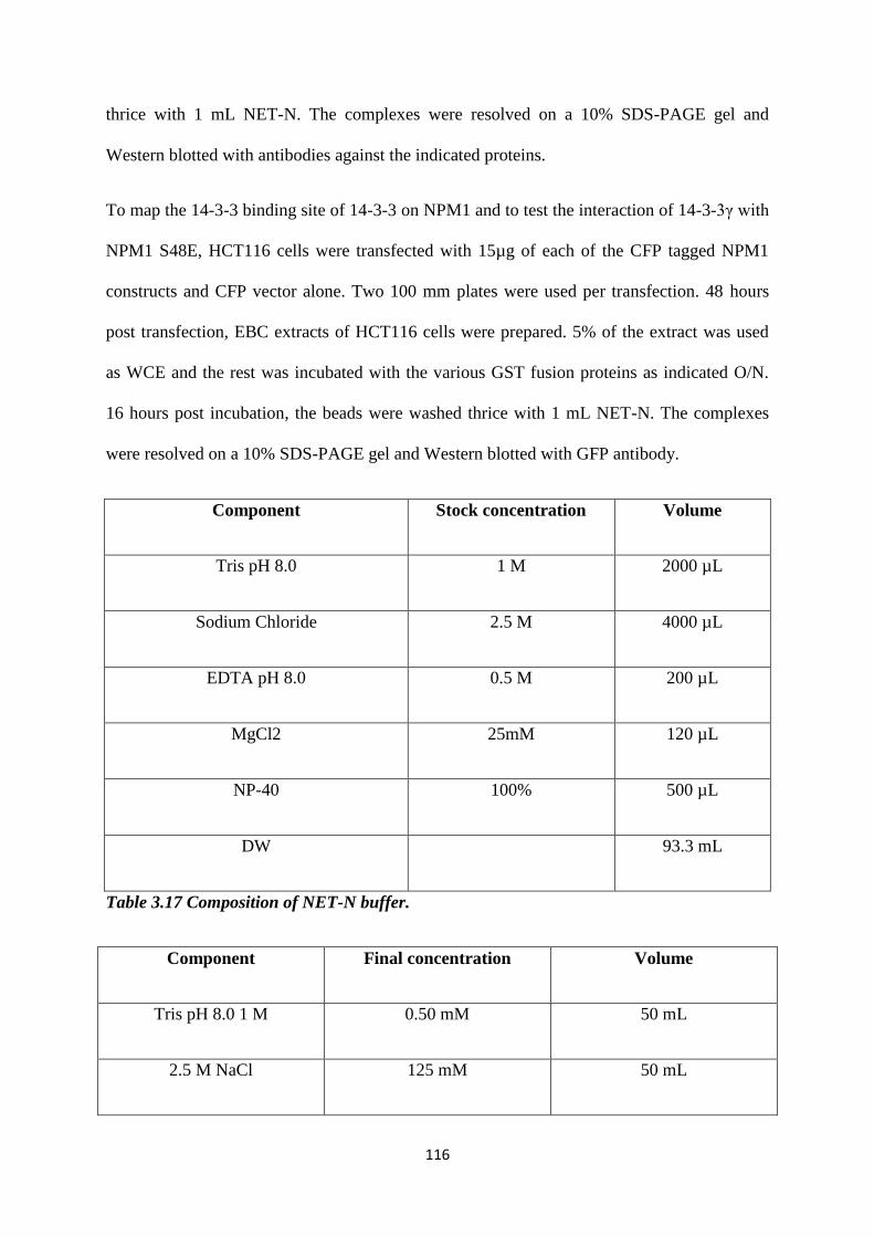
116
thrice with 1 mL NET-N. The complexes were resolved on a 10% SDS-PAGE gel and
Western blotted with antibodies against the indicated proteins.
To map the 14-3-3 binding site of 14-3-3 on NPM1 and to test the interaction of 14-3-3γ with
NPM1 S48E, HCT116 cells were transfected with 15µg of each of the CFP tagged NPM1
constructs and CFP vector alone. Two 100 mm plates were used per transfection. 48 hours
post transfection, EBC extracts of HCT116 cells were prepared. 5% of the extract was used
as WCE and the rest was incubated with the various GST fusion proteins as indicated O/N.
16 hours post incubation, the beads were washed thrice with 1 mL NET-N. The complexes
were resolved on a 10% SDS-PAGE gel and Western blotted with GFP antibody.
Component Stock concentration Volume
Tris pH 8.0 1 M 2000 µL
Sodium Chloride 2.5 M 4000 µL
EDTA pH 8.0 0.5 M 200 µL
MgCl2 25mM 120 µL
NP-40 100% 500 µL
DW 93.3 mL
Table 3.17 Composition of NET-N buffer.
Component Final concentration Volume
Tris pH 8.0 1 M 0.50 mM 50 mL
2.5 M NaCl 125 mM 50 mL
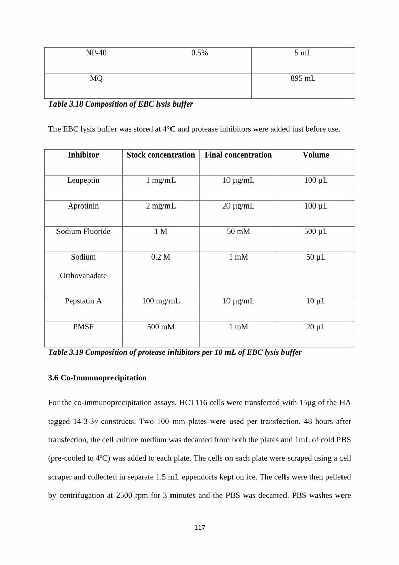
117
NP-40 0.5% 5 mL
MQ 895 mL
Table 3.18 Composition of EBC lysis buffer
The EBC lysis buffer was stored at 4°C and protease inhibitors were added just before use.
Inhibitor Stock concentration Final concentration Volume
Leupeptin 1 mg/mL 10 µg/mL 100 µL
Aprotinin 2 mg/mL 20 µg/mL 100 µL
Sodium Fluoride 1 M 50 mM 500 µL
Sodium
Orthovanadate
0.2 M 1 mM 50 µL
Pepstatin A 100 mg/mL 10 µg/mL 10 µL
PMSF 500 mM 1 mM 20 µL
Table 3.19 Composition of protease inhibitors per 10 mL of EBC lysis buffer
3.6 Co-Immunoprecipitation
For the co-immunoprecipitation assays, HCT116 cells were transfected with 15µg of the HA
tagged 14-3-3γ constructs. Two 100 mm plates were used per transfection. 48 hours after
transfection, the cell culture medium was decanted from both the plates and 1mL of cold PBS
(pre-cooled to 4ºC) was added to each plate. The cells on each plate were scraped using a cell
scraper and collected in separate 1.5 mL eppendorfs kept on ice. The cells were then pelleted
by centrifugation at 2500 rpm for 3 minutes and the PBS was decanted. PBS washes were
118
given so as to remove all traces of media. Thereafter, 1ml of EBC lysis buffer containing
protease inhibitors (as mentioned in 3.19) was added to the cells and incubated for 15 minutes
on ice. This was followed by centrifugation at 13,000 rpm for 15 minutes at 4ºC. The
supernatant was transferred to a new 1.5 ml eppendorf. 50 μL of this supernatant was kept
separately and used as 5% whole cell extract. To this, 25μL of 3X sample buffer was added,
boiled in a water bath for 7 minutes and stored at -80ºC. The rest of the supernatant was
incubated with 100 µL of anti-HA antibody (12CA5 hybridoma supernatant) for 4 hours.
After 4 hours, 30 µL of a 50% slurry of Protein-G-Sepharose was used to precipitate the HA-
tagged 14-3-3γ proteins. After 1 hour, the beads were washed thrice with 1 mL NET-N. after
boiling with 3X sample buffer, the complexes were resolved on a 10% SDS-PAGE gel and
Western blotted with antibodies against the indicated proteins.
3.7 Live cell imaging
To estimate the duration of mitosis, HCT116 cells seeded in a glass-bottomed dish were co-
transfected with the respective constructs. 48 hours post-transfection, and 2 hours before
imaging, cells were treated with nocodazole (410). One hour prior to imaging, ascorbic acid
(100 µM) was added to the media, to reduce photo-bleaching (412,413). Just before imaging,
nocodazole was removed and the cells were fed with media containing ascorbic acid again.
During imaging, cells were maintained at 5% CO2 and 95% RH. All images were acquired on
a Leica SP8 confocal microscope at 1000X magnification with 4X optical zoom. Images were
acquired at 20-minute intervals. Images were processed using the Leica LASX software.
3.8 Electron microscopy
To study centrosome amplification and organization at higher magnification in cells
expressing the different 14-3-3γ mutants, the cells were visualized under a transmission
electron microscope. Cells synchronized at M-phase were fixed with 3% glutaraldehyde,
119
washed with 0.1 M of sodium cacodylate pH 7.4, and post-fixed with 1%
osmium tetroxide (Tedpella). Cells were then dehydrated and processed. Grids were
contrasted with alcoholic uranyl acetate for 30 seconds and dipped once in lead citrate. The
grids were observed under a JEOL 1400Plus transmission electron microscope, at an
accelerating voltage of 120 KeV and at 10000X.
3.9 Effect of overexpression of CDC25C in the sh-14-3-3ε cells.
In order to assess the effect of the overexpression of CDC25C in the sh-14-3-3ε cells on
centrosome number, HCT116 cells were co-transfected with 1 µg each of the previously
described pTU6 and pTU6 E7 vector and GFP or GFP CDC25C (234). sh-14-3-3γ cells were
used as the positive control. 48 hours post transfection, the cells were treated with
Nocodazole to synchronize them in mitosis. Post synchronization, the cells were processed
for Western blotting or immunofluorescence and centrosome number was determined as
described previously.
3.10 RT-PCR to determine mRNA levels of Cep170 in the sh-14-3-3γ cells.
HCT116 derived vector control and sh-14-3-3γ cells were washed with 1X PBS and TRIZOL
(Thermo Fisher Scientific) was added to the cells (1ml for 35 mm tissue culture plate) and
incubated at room temperature for 5 minutes for dissociation of nucleoprotein complex.
Samples were collected in an eppendorf, 200μl of chloroform was added, vortexed and
incubated at room temperature for 2-3 minutes. Samples were centrifuged at 13000 rpm for
15 minutes at 4°C. The aqueous phase was collected in another Eppendorf and 500μl of
isopropanol was added and incubated at room temperature for 10 minutes to precipitate the
RNA. Samples were centrifuged at 13000 rpm for 15 minutes at 4°C followed by 75%
ethanol wash (75ml 100% Ethanol and 25 ml nuclease free water) at 10000 rpm for 5 minutes
at 4°C. The RNA pellet was dried for 10 minutes at room temperature and 25μl of
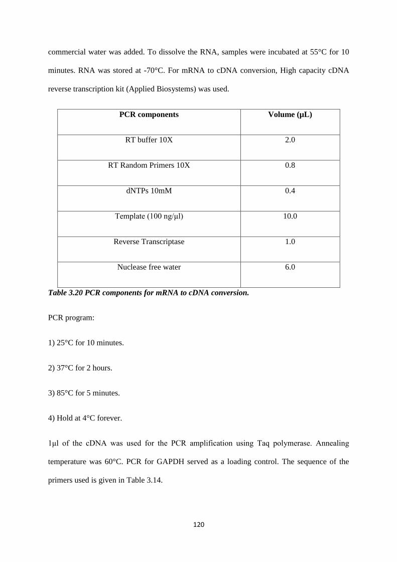
120
commercial water was added. To dissolve the RNA, samples were incubated at 55°C for 10
minutes. RNA was stored at -70°C. For mRNA to cDNA conversion, High capacity cDNA
reverse transcription kit (Applied Biosystems) was used.
PCR components Volume (µL)
RT buffer 10X 2.0
RT Random Primers 10X 0.8
dNTPs 10mM 0.4
Template (100 ng/μl) 10.0
Reverse Transcriptase 1.0
Nuclease free water 6.0
Table 3.20 PCR components for mRNA to cDNA conversion.
PCR program:
1) 25°C for 10 minutes.
2) 37°C for 2 hours.
3) 85°C for 5 minutes.
4) Hold at 4°C forever.
1μl of the cDNA was used for the PCR amplification using Taq polymerase. Annealing
temperature was 60°C. PCR for GAPDH served as a loading control. The sequence of the
primers used is given in Table 3.14.

121
Name of oligonucleotide Sequence
GAPDH 5’ TGCACCACCAACTGCTTAGC
GAPDH 3’ GGCATGGACTGTGGTCATGAG
Cep170 5’ CACCGTGTTACTAGCTCTGCG
Cep 170 3’ CCATCTCCTGGCTTGGTCTG
14-3-3γ 5’ GAGCCACTGTCGAATG
14-3-3γ 3’ CGCTGCAATTCTTGATC
Table 3.21 Sequence of oligonucleotides used for the RT-PCR reaction.
3.11 Soft agar assay.
The soft agar colony formation assay is performed to determine the ability of cells to grow
independent of anchorage (407). Low melting point (LMP) agarose of two different
concentrations were prepared, 1.6% and 0.8%, and autoclaved to maintain sterility. After
autoclaving, both the LMP agarose solutions were kept on a float in a water bath set at 40ºC
to prevent the agarose from solidifying. The 1.6% LMP agarose was then mixed with equal
volumes of 2X DMEM + antibiotic + FBS mix (a mixture of sterile 2X concentrated DMEM
solution, 2X concentrated relevant antibiotic and 20% FBS). Upon mixing, the final
concentration of agarose became 0.8%, the concentration of DMEM and antibiotic becomes
1X and concentration of FBS became 10% (which is the concentration required for optimal
growth of cells). Now 2 ml of this solution was poured onto 35mm cell culture plates slowly
122
and allowed to form a thick solid layer on the plate. This was called the lower layer of the
soft agar plate.
Next, the cells of the relevant clones were trypsinized and resuspended in 1ml of 2X DMEM
media. The cells are then counted using a haemocytometer to determine the concentration of
the cells in the solution. Meanwhile, the 0.8% LMP agarose was mixed with equal volumes
of the 2X DMEM + antibiotic + FBS mix to make a final volume of 0.4% LMP agarose +
DMEM solution. To 1 ml of this solution, 2500 cells were resuspended slowly to form a
homogenous suspension and spread evenly on top of the lower layer of soft agar plate and
allowed to settle down and form a semi-solid upper layer. Thereafter, the plates were kept in
a tissue culture incubator maintained at 37ºC, 5% CO2, for three weeks. A volume of 150μl
to 200μl of fresh DMEM media with the relevant antibiotics were added on top of the soft
agar plates every 2 days during these three weeks of incubation. Thereafter the total number
of colonies formed on the soft agar plate was counted by making grids at the bottom of the
soft agar plate. Three independent experiments were performed in triplicates for each clone.

123
4. Results
124
4. Results
4.1 Does 14-3-3 binding to centrosomal proteins inhibit centrosome licensing and
duplication?
4.1.1 Are the multiple centrosomes observed upon loss of 14-3-3γ able to function as
MTOCs?
Previous studies from our lab have demonstrated that a knockdown of 14-3-3γ results in the
presence of excess γ-tubulin positive dots (234). Further experiments demonstrated that loss
of 14-3-3γ lead to the presence of multiple Centrin, Cep170 and Ninein foci (326). It was also
shown that the multiple γ-tubulin foci were able to function as centrosomes, by performing
dual staining for α- and γ-tubulin. We wished to confirm this phenotype using another
centrosomal marker and performed dual staining for the centrosome using Centrin and
Pericentrin (a PCM protein) (280,281). We transfected HCT116 derived vector control and
sh-14-3-3γ cells with GFP Centrin and synchronized the cells in mitosis using nocodazole. 48
hours post transfection, we fixed the cells and stained for Pericentrin and counterstained for
DNA using DAPI. The number of centrosomes in a 100 transfected mitotic cells was
determined. In both the vector control and the sh-14-3-3γ cells, we observed that each GFP
Centrin1 foci co-localized with Pericentrin staining. In case of the sh-14-3-3γ cells, multiple
Centrin1 foci were found to co-localize with the Pericentrin staining. This demonstrates that
the multiple Centrin dots observed in the sh-14-3-3γ cells are able to function as MTOC’s
(326) (fig. 4.1).
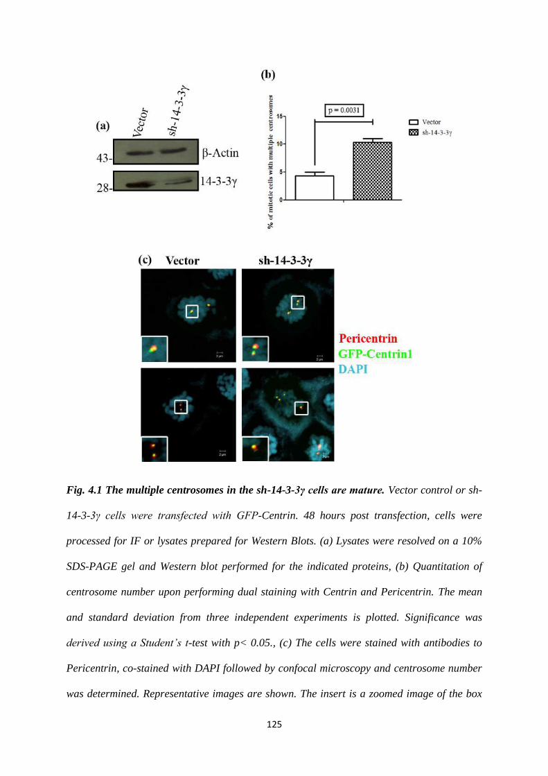
125
Fig. 4.1 The multiple centrosomes in the sh-14-3-3γ cells are mature. Vector control or sh-
14-3-3γ cells were transfected with GFP-Centrin. 48 hours post transfection, cells were
processed for IF or lysates prepared for Western Blots. (a) Lysates were resolved on a 10%
SDS-PAGE gel and Western blot performed for the indicated proteins, (b) Quantitation of
centrosome number upon performing dual staining with Centrin and Pericentrin. The mean
and standard deviation from three independent experiments is plotted. Significance was
derived using a Student’s t-test with p< 0.05., (c) The cells were stained with antibodies to
Pericentrin, co-stained with DAPI followed by confocal microscopy and centrosome number
was determined. Representative images are shown. The insert is a zoomed image of the box
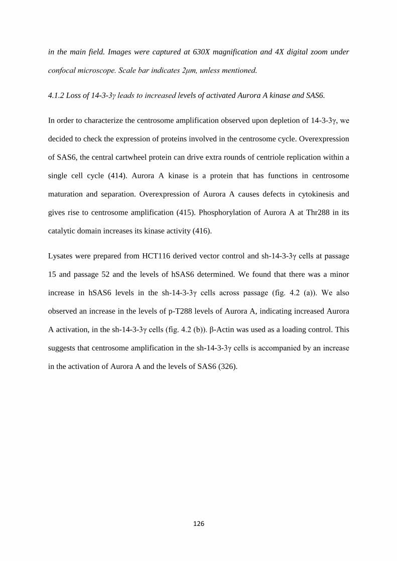
126
in the main field. Images were captured at 630X magnification and 4X digital zoom under
confocal microscope. Scale bar indicates 2μm, unless mentioned.
4.1.2 Loss of 14-3-3γ leads to increased levels of activated Aurora A kinase and SAS6.
In order to characterize the centrosome amplification observed upon depletion of 14-3-3γ, we
decided to check the expression of proteins involved in the centrosome cycle. Overexpression
of SAS6, the central cartwheel protein can drive extra rounds of centriole replication within a
single cell cycle (414). Aurora A kinase is a protein that has functions in centrosome
maturation and separation. Overexpression of Aurora A causes defects in cytokinesis and
gives rise to centrosome amplification (415). Phosphorylation of Aurora A at Thr288 in its
catalytic domain increases its kinase activity (416).
Lysates were prepared from HCT116 derived vector control and sh-14-3-3γ cells at passage
15 and passage 52 and the levels of hSAS6 determined. We found that there was a minor
increase in hSAS6 levels in the sh-14-3-3γ cells across passage (fig. 4.2 (a)). We also
observed an increase in the levels of p-T288 levels of Aurora A, indicating increased Aurora
A activation, in the sh-14-3-3γ cells (fig. 4.2 (b)). β-Actin was used as a loading control. This
suggests that centrosome amplification in the sh-14-3-3γ cells is accompanied by an increase
in the activation of Aurora A and the levels of SAS6 (326).
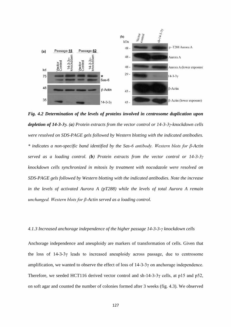
127
Fig. 4.2 Determination of the levels of proteins involved in centrosome duplication upon
depletion of 14-3-3γ. (a) Protein extracts from the vector control or 14-3-3γ-knockdown cells
were resolved on SDS-PAGE gels followed by Western blotting with the indicated antibodies.
* indicates a non-specific band identified by the Sas-6 antibody. Western blots for β-Actin
served as a loading control. (b) Protein extracts from the vector control or 14-3-3γ
knockdown cells synchronized in mitosis by treatment with nocodazole were resolved on
SDS-PAGE gels followed by Western blotting with the indicated antibodies. Note the increase
in the levels of activated Aurora A (pT288) while the levels of total Aurora A remain
unchanged. Western blots for β-Actin served as a loading control.
4.1.3 Increased anchorage independence of the higher passage 14-3-3-γ knockdown cells
Anchorage independence and aneuploidy are markers of transformation of cells. Given that
the loss of 14-3-3γ leads to increased aneuploidy across passage, due to centrosome
amplification, we wanted to observe the effect of loss of 14-3-3γ on anchorage independence.
Therefore, we seeded HCT116 derived vector control and sh-14-3-3γ cells, at p15 and p52,
on soft agar and counted the number of colonies formed after 3 weeks (fig. 4.3). We observed
(a)
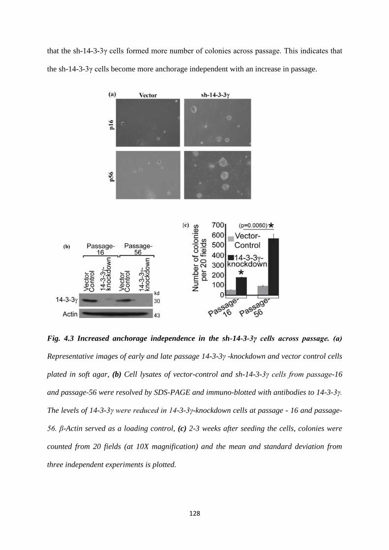
128
that the sh-14-3-3γ cells formed more number of colonies across passage. This indicates that
the sh-14-3-3γ cells become more anchorage independent with an increase in passage.
Fig. 4.3 Increased anchorage independence in the sh-14-3-3γ cells across passage. (a)
Representative images of early and late passage 14-3-3γ -knockdown and vector control cells
plated in soft agar, (b) Cell lysates of vector-control and sh-14-3-3γ cells from passage-16
and passage-56 were resolved by SDS-PAGE and immuno-blotted with antibodies to 14-3-3γ.
The levels of 14-3-3γ were reduced in 14-3-3γ-knockdown cells at passage - 16 and passage-
56. β-Actin served as a loading control, (c) 2-3 weeks after seeding the cells, colonies were
counted from 20 fields (at 10X magnification) and the mean and standard deviation from
three independent experiments is plotted.
129
4.1.4 Centrosome amplification in the sh-14-3-3γ cells occurs due to premature
phosphorylation of NPM1 at T199.
Nucleophosmin 1 (NPM1) is an oligomeric, nucleolar phosphoprotein that functions as a
molecular chaperone for both proteins and nucleic acids [reviewed in (417)]. Wild-type
NPM1 forms pentameric oligomers through interactions at the amino-terminal core domain.
Previous studies have demonstrated that changes in the oligomerization state of NPM1 may
affect its function (418). NPM1 is phosphorylated by cdk2/cyclin E at the G1/S transition at a
Threonine 199 residue, which acts as a licensing factor for centrosome duplication (325).
NPM1 associates with unduplicated centrosomes, but upon phosphorylation at Thr199 by
CDK2/cyclin E at the G1/S transition, it dissociates from the centrosome, leading to
centrosome duplication (325). In vitro kinase assays determined that the cyclin B1/CDK1
could also phosphorylate NPM1 at T199 (326). It has been demonstrated that a depletion of
14-3-3γ leads to an increase in p-T199 levels of NPM1 (326). Overexpression of WT NPM1
and a phosphomimetic T199D mutant leads to increased centrosome amplification in both the
vector control and the sh-14-3-3γ cells (326). Expression of a T199A mutant was able to
decrease centrosome amplification.
It had been demonstrated that the centrosome amplification observed in the sh-14-3-3γ cells
begins in the S-phase. If the increased levels of p-T199 were the cause of the centrosome
amplification, levels of p-T199 NPM1 would also increase from S-phase onwards. In order to
test the same, we synchronized HCT116 derived vector control and sh-14-3-3γ cells cells at
the G1/S phase using mimosine and harvested them at different time points (fig. 4.4 (a and
b)). Then, we prepared lysates and performed a Western blot for cyclin B1 total NPM1 and p-
T199 NPM1. We found that p-T199 NPM1 in the vector control cells first appears at 10
hours post release from mimosine, when the majority of the cells are in G2 phase. Whereas,
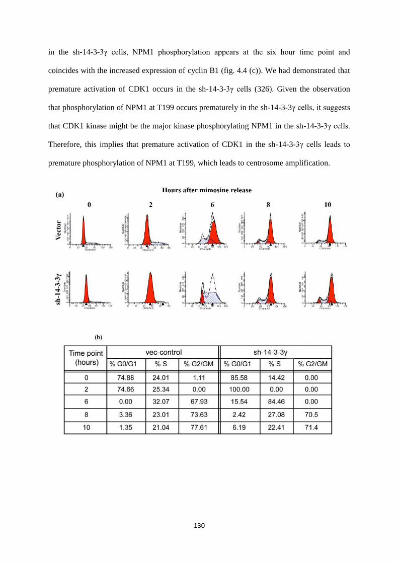
130
in the sh-14-3-3γ cells, NPM1 phosphorylation appears at the six hour time point and
coincides with the increased expression of cyclin B1 (fig. 4.4 (c)). We had demonstrated that
premature activation of CDK1 occurs in the sh-14-3-3γ cells (326). Given the observation
that phosphorylation of NPM1 at T199 occurs prematurely in the sh-14-3-3γ cells, it suggests
that CDK1 kinase might be the major kinase phosphorylating NPM1 in the sh-14-3-3γ cells.
Therefore, this implies that premature activation of CDK1 in the sh-14-3-3γ cells leads to
premature phosphorylation of NPM1 at T199, which leads to centrosome amplification.
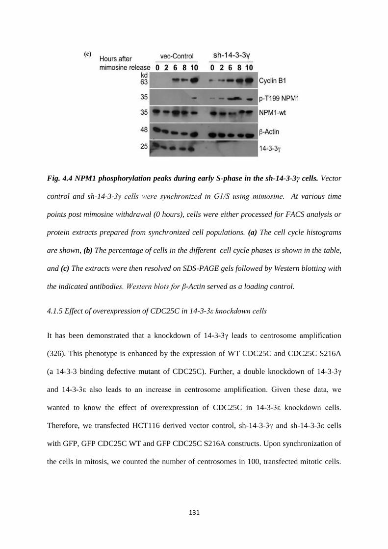
131
Fig. 4.4 NPM1 phosphorylation peaks during early S-phase in the sh-14-3-3γ cells. Vector
control and sh-14-3-3γ cells were synchronized in G1/S using mimosine. At various time
points post mimosine withdrawal (0 hours), cells were either processed for FACS analysis or
protein extracts prepared from synchronized cell populations. (a) The cell cycle histograms
are shown, (b) The percentage of cells in the different cell cycle phases is shown in the table,
and (c) The extracts were then resolved on SDS-PAGE gels followed by Western blotting with
the indicated antibodies. Western blots for β-Actin served as a loading control.
4.1.5 Effect of overexpression of CDC25C in 14-3-3ε knockdown cells
It has been demonstrated that a knockdown of 14-3-3γ leads to centrosome amplification
(326). This phenotype is enhanced by the expression of WT CDC25C and CDC25C S216A
(a 14-3-3 binding defective mutant of CDC25C). Further, a double knockdown of 14-3-3γ
and 14-3-3ε also leads to an increase in centrosome amplification. Given these data, we
wanted to know the effect of overexpression of CDC25C in 14-3-3ε knockdown cells.
Therefore, we transfected HCT116 derived vector control, sh-14-3-3γ and sh-14-3-3ε cells
with GFP, GFP CDC25C WT and GFP CDC25C S216A constructs. Upon synchronization of
the cells in mitosis, we counted the number of centrosomes in 100, transfected mitotic cells.
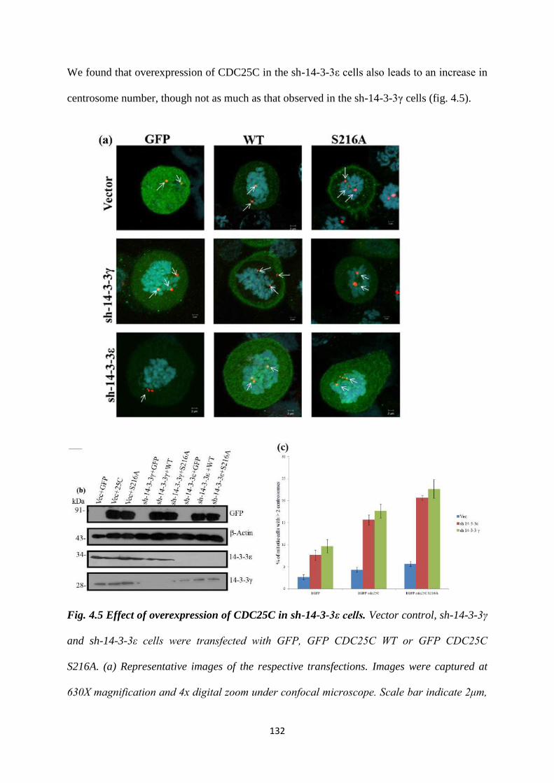
132
We found that overexpression of CDC25C in the sh-14-3-3ε cells also leads to an increase in
centrosome number, though not as much as that observed in the sh-14-3-3γ cells (fig. 4.5).
Fig. 4.5 Effect of overexpression of CDC25C in sh-14-3-3ε cells. Vector control, sh-14-3-3γ
and sh-14-3-3ε cells were transfected with GFP, GFP CDC25C WT or GFP CDC25C
S216A. (a) Representative images of the respective transfections. Images were captured at
630X magnification and 4x digital zoom under confocal microscope. Scale bar indicate 2μm,
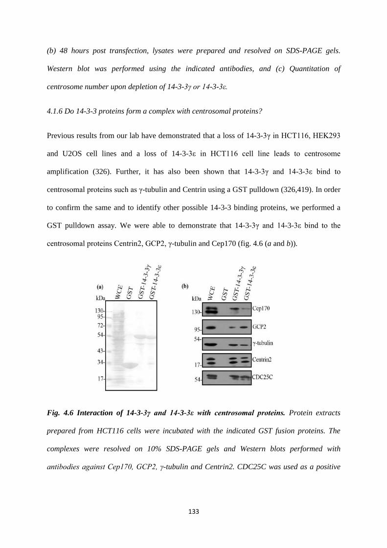
133
(b) 48 hours post transfection, lysates were prepared and resolved on SDS-PAGE gels.
Western blot was performed using the indicated antibodies, and (c) Quantitation of
centrosome number upon depletion of 14-3-3γ or 14-3-3ε.
4.1.6 Do 14-3-3 proteins form a complex with centrosomal proteins?
Previous results from our lab have demonstrated that a loss of 14-3-3γ in HCT116, HEK293
and U2OS cell lines and a loss of 14-3-3ε in HCT116 cell line leads to centrosome
amplification (326). Further, it has also been shown that 14-3-3γ and 14-3-3ε bind to
centrosomal proteins such as γ-tubulin and Centrin using a GST pulldown (326,419). In order
to confirm the same and to identify other possible 14-3-3 binding proteins, we performed a
GST pulldown assay. We were able to demonstrate that 14-3-3γ and 14-3-3ε bind to the
centrosomal proteins Centrin2, GCP2, γ-tubulin and Cep170 (fig. 4.6 (a and b)).
Fig. 4.6 Interaction of 14-3-3γ and 14-3-3ε with centrosomal proteins. Protein extracts
prepared from HCT116 cells were incubated with the indicated GST fusion proteins. The
complexes were resolved on 10% SDS-PAGE gels and Western blots performed with
antibodies against Cep170, GCP2, γ-tubulin and Centrin2. CDC25C was used as a positive
134
control. Note that all the proteins form a complex with GST-14-3-3γ and GST-14-3-3ε but not
GST alone. (a) Indicates Poonceau S stained membrane and (b) is the Western blot.
Further, in order to understand how 14-3-3 proteins might regulate the centrosome cycle, we
decided to map the 14-3-3 binding site on these proteins. In order to do so, we used the
Scansite motif prediction webserver (420). We were unable to detect any putative 14-3-3
binding sites on the proteins Centrin2, γ-tubulin or GCP2. However, we found three potential
14-3-3 binding sites on the protein Cep170. Based on these results, in order to map the 14-3-3
binding sites, we decided to generate domain deletion constructs of proteins that did not
possess a consensus 14-3-3 binding site. Cep170, we decided to generate site directed
mutants of the potential 14-3-3 binding sites. Also, given that 14-3-3ε did not interact with
Centrin and γ-tubulin in a FRET based assay, but 14-3-3γ did, we decided to study the
interaction of 14-3-3γ with centrosomal proteins (326).
4.1.7 Centrin2
Centrins are small, highly conserved members of the EF-hand superfamily of calcium-
binding proteins that are expressed throughout eukaryotes. They have a role in ensuring the
duplication and appropriate functioning of the ciliary basal bodies in ciliated cells (reviewed
in (421)). They have been localized to the distal end of centrioles and are essential for
centriole duplication (422).
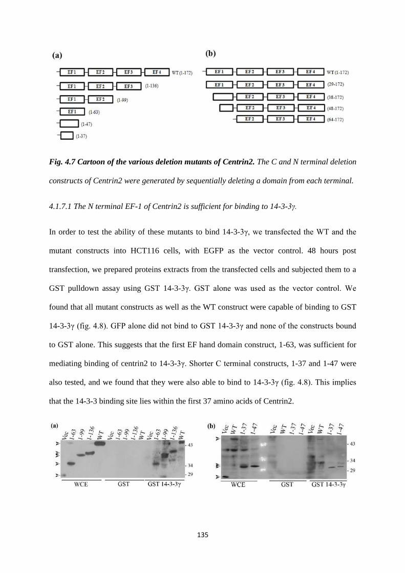
135
Fig. 4.7 Cartoon of the various deletion mutants of Centrin2. The C and N terminal deletion
constructs of Centrin2 were generated by sequentially deleting a domain from each terminal.
4.1.7.1 The N terminal EF-1 of Centrin2 is sufficient for binding to 14-3-3γ.
In order to test the ability of these mutants to bind 14-3-3γ, we transfected the WT and the
mutant constructs into HCT116 cells, with EGFP as the vector control. 48 hours post
transfection, we prepared proteins extracts from the transfected cells and subjected them to a
GST pulldown assay using GST 14-3-3γ. GST alone was used as the vector control. We
found that all mutant constructs as well as the WT construct were capable of binding to GST
14-3-3γ (fig. 4.8). GFP alone did not bind to GST 14-3-3γ and none of the constructs bound
to GST alone. This suggests that the first EF hand domain construct, 1-63, was sufficient for
mediating binding of centrin2 to 14-3-3γ. Shorter C terminal constructs, 1-37 and 1-47 were
also tested, and we found that they were also able to bind to 14-3-3γ (fig. 4.8). This implies
that the 14-3-3 binding site lies within the first 37 amino acids of Centrin2.
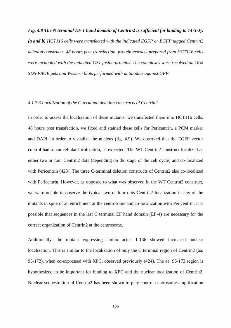
136
Fig. 4.8 The N terminal EF 1 hand domain of Centrin2 is sufficient for binding to 14-3-3γ.
(a and b) HCT116 cells were transfected with the indicated EGFP or EGFP tagged Centrin2
deletion constructs. 48 hours post transfection, protein extracts prepared from HCT116 cells
were incubated with the indicated GST fusion proteins. The complexes were resolved on 10%
SDS-PAGE gels and Western blots performed with antibodies against GFP.
4.1.7.3 Localization of the C-terminal deletion constructs of Centrin2
In order to assess the localization of these mutants, we transfected them into HCT116 cells.
48 hours post transfection, we fixed and stained these cells for Pericentrin, a PCM marker
and DAPI, in order to visualize the nucleus (fig. 4.9). We observed that the EGFP vector
control had a pan-cellular localization, as expected. The WT Centrin2 construct localized as
either two or four Centrin2 dots (depending on the stage of the cell cycle) and co-localized
with Pericentrin (423). The three C-terminal deletion constructs of Centrin2 also co-localized
with Pericentrin. However, as opposed to what was observed in the WT Centrin2 construct,
we were unable to observe the typical two or four dots Centrin2 localization in any of the
mutants in spite of an enrichment at the centrosome and co-localization with Pericentrin. It is
possible that sequences in the last C terminal EF hand domain (EF-4) are necessary for the
correct organization of Centrin2 at the centrosome.
Additionally, the mutant expressing amino acids 1-136 showed increased nuclear
localization. This is similar to the localization of only the C terminal region of Centrin2 (aa.
95-172), when co-expressed with XPC, observed previously (424). The aa. 95-172 region is
hypothesized to be important for binding to XPC and the nuclear localization of Centrin2.
Nuclear sequestration of Centrin2 has been shown to play control centrosome amplification
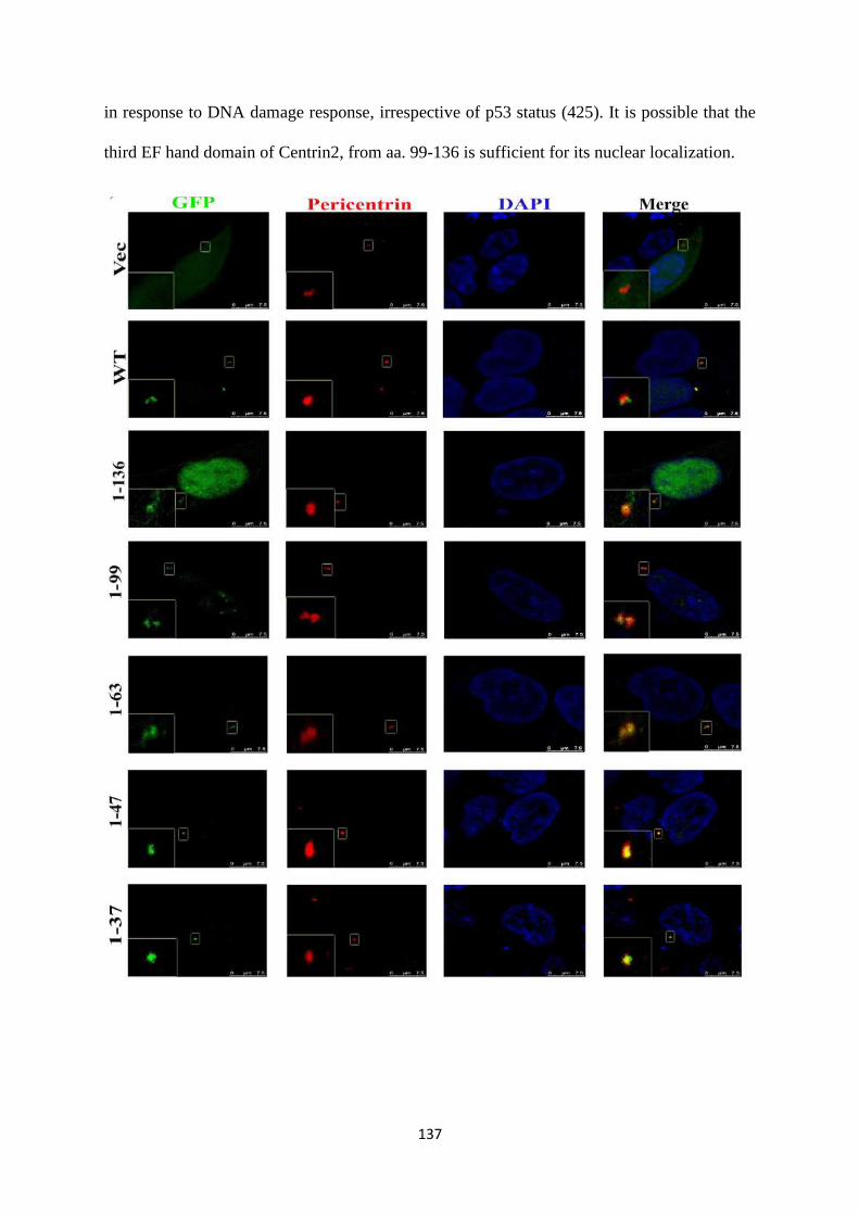
137
in response to DNA damage response, irrespective of p53 status (425). It is possible that the
third EF hand domain of Centrin2, from aa. 99-136 is sufficient for its nuclear localization.
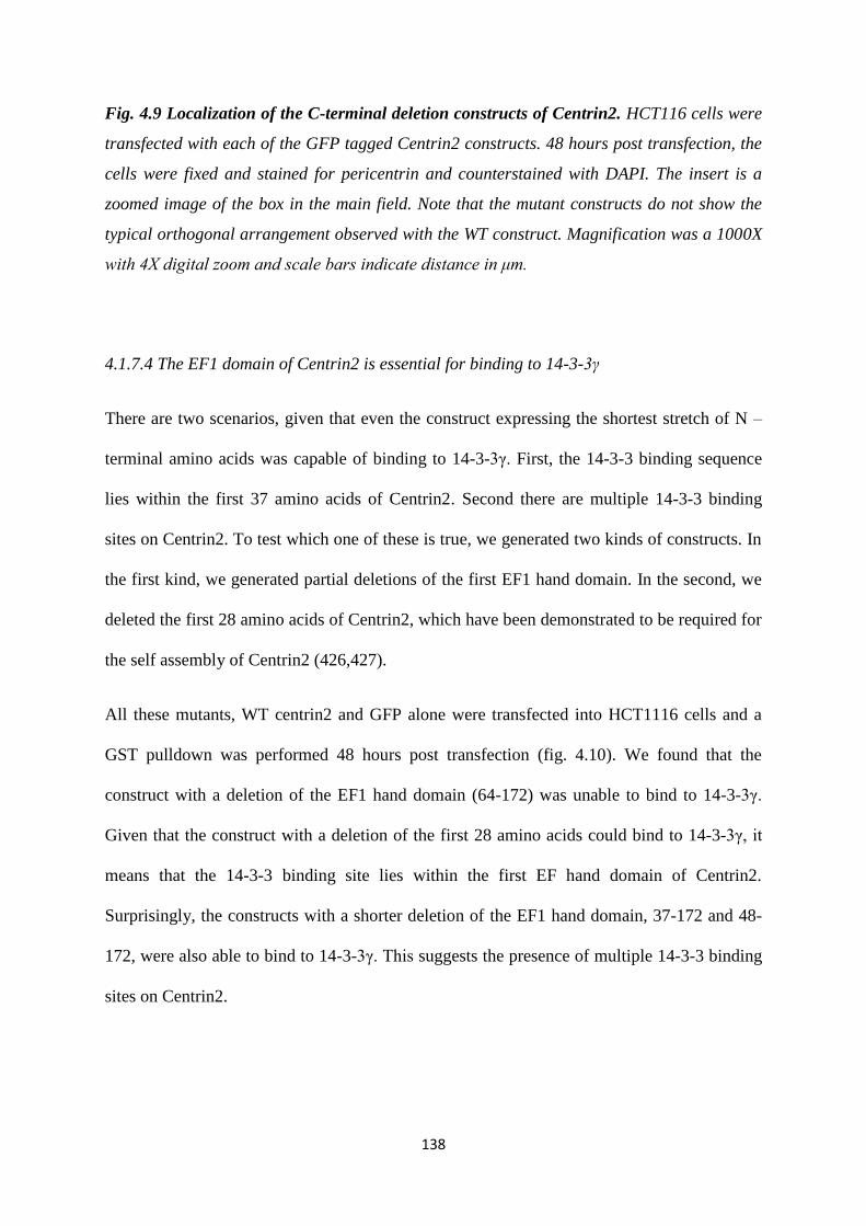
138
Fig. 4.9 Localization of the C-terminal deletion constructs of Centrin2. HCT116 cells were
transfected with each of the GFP tagged Centrin2 constructs. 48 hours post transfection, the
cells were fixed and stained for pericentrin and counterstained with DAPI. The insert is a
zoomed image of the box in the main field. Note that the mutant constructs do not show the
typical orthogonal arrangement observed with the WT construct. Magnification was a 1000X
with 4X digital zoom and scale bars indicate distance in μm.
4.1.7.4 The EF1 domain of Centrin2 is essential for binding to 14-3-3γ
There are two scenarios, given that even the construct expressing the shortest stretch of N –
terminal amino acids was capable of binding to 14-3-3γ. First, the 14-3-3 binding sequence
lies within the first 37 amino acids of Centrin2. Second there are multiple 14-3-3 binding
sites on Centrin2. To test which one of these is true, we generated two kinds of constructs. In
the first kind, we generated partial deletions of the first EF1 hand domain. In the second, we
deleted the first 28 amino acids of Centrin2, which have been demonstrated to be required for
the self assembly of Centrin2 (426,427).
All these mutants, WT centrin2 and GFP alone were transfected into HCT1116 cells and a
GST pulldown was performed 48 hours post transfection (fig. 4.10). We found that the
construct with a deletion of the EF1 hand domain (64-172) was unable to bind to 14-3-3γ.
Given that the construct with a deletion of the first 28 amino acids could bind to 14-3-3γ, it
means that the 14-3-3 binding site lies within the first EF hand domain of Centrin2.
Surprisingly, the constructs with a shorter deletion of the EF1 hand domain, 37-172 and 48-
172, were also able to bind to 14-3-3γ. This suggests the presence of multiple 14-3-3 binding
sites on Centrin2.
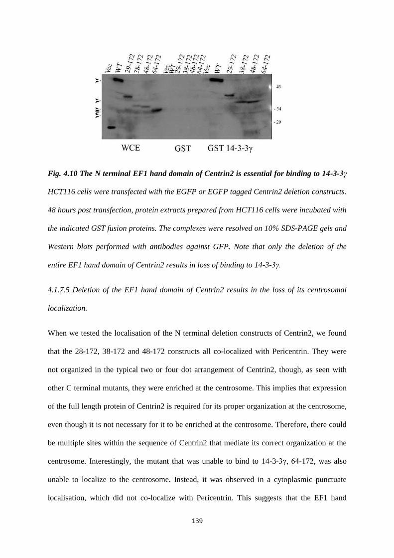
139
Fig. 4.10 The N terminal EF1 hand domain of Centrin2 is essential for binding to 14-3-3γ
HCT116 cells were transfected with the EGFP or EGFP tagged Centrin2 deletion constructs.
48 hours post transfection, protein extracts prepared from HCT116 cells were incubated with
the indicated GST fusion proteins. The complexes were resolved on 10% SDS-PAGE gels and
Western blots performed with antibodies against GFP. Note that only the deletion of the
entire EF1 hand domain of Centrin2 results in loss of binding to 14-3-3γ.
4.1.7.5 Deletion of the EF1 hand domain of Centrin2 results in the loss of its centrosomal
localization.
When we tested the localisation of the N terminal deletion constructs of Centrin2, we found
that the 28-172, 38-172 and 48-172 constructs all co-localized with Pericentrin. They were
not organized in the typical two or four dot arrangement of Centrin2, though, as seen with
other C terminal mutants, they were enriched at the centrosome. This implies that expression
of the full length protein of Centrin2 is required for its proper organization at the centrosome,
even though it is not necessary for it to be enriched at the centrosome. Therefore, there could
be multiple sites within the sequence of Centrin2 that mediate its correct organization at the
centrosome. Interestingly, the mutant that was unable to bind to 14-3-3γ, 64-172, was also
unable to localize to the centrosome. Instead, it was observed in a cytoplasmic punctuate
localisation, which did not co-localize with Pericentrin. This suggests that the EF1 hand
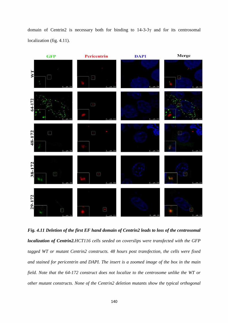
140
domain of Centrin2 is necessary both for binding to 14-3-3γ and for its centrosomal
localization (fig. 4.11).
Fig. 4.11 Deletion of the first EF hand domain of Centrin2 leads to loss of the centrosomal
localization of Centrin2.HCT116 cells seeded on coverslips were transfected with the GFP
tagged WT or mutant Centrin2 constructs. 48 hours post transfection, the cells were fixed
and stained for pericentrin and DAPI. The insert is a zoomed image of the box in the main
field. Note that the 64-172 construct does not localize to the centrosome unlike the WT or
other mutant constructs. None of the Centrin2 deletion mutants show the typical orthogonal
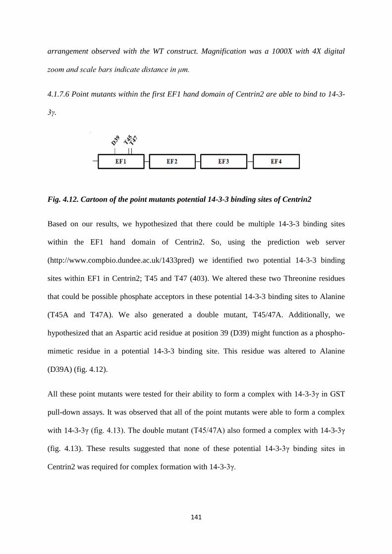
141
arrangement observed with the WT construct. Magnification was a 1000X with 4X digital
zoom and scale bars indicate distance in μm.
4.1.7.6 Point mutants within the first EF1 hand domain of Centrin2 are able to bind to 14-3-
3γ.
Fig. 4.12. Cartoon of the point mutants potential 14-3-3 binding sites of Centrin2
Based on our results, we hypothesized that there could be multiple 14-3-3 binding sites
within the EF1 hand domain of Centrin2. So, using the prediction web server
(http://www.compbio.dundee.ac.uk/1433pred) we identified two potential 14-3-3 binding
sites within EF1 in Centrin2; T45 and T47 (403). We altered these two Threonine residues
that could be possible phosphate acceptors in these potential 14-3-3 binding sites to Alanine
(T45A and T47A). We also generated a double mutant, T45/47A. Additionally, we
hypothesized that an Aspartic acid residue at position 39 (D39) might function as a phospho-
mimetic residue in a potential 14-3-3 binding site. This residue was altered to Alanine
(D39A) (fig. 4.12).
All these point mutants were tested for their ability to form a complex with 14-3-3γ in GST
pull-down assays. It was observed that all of the point mutants were able to form a complex
with 14-3-3γ (fig. 4.13). The double mutant (T45/47A) also formed a complex with 14-3-3γ
(fig. 4.13). These results suggested that none of these potential 14-3-3γ binding sites in
Centrin2 was required for complex formation with 14-3-3γ.
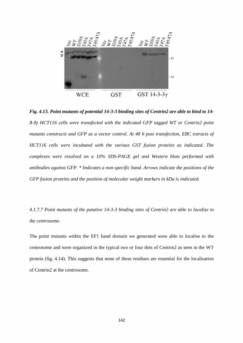
142
Fig. 4.13. Point mutants of potential 14-3-3 binding sites of Centrin2 are able to bind to 14-
3-3γ HCT116 cells were transfected with the indicated GFP tagged WT or Centrin2 point
mutants constructs and GFP as a vector control. At 48 h post transfection, EBC extracts of
HCT116 cells were incubated with the various GST fusion proteins as indicated. The
complexes were resolved on a 10% SDS-PAGE gel and Western blots performed with
antibodies against GFP. * Indicates a non-specific band. Arrows indicate the positions of the
GFP fusion proteins and the position of molecular weight markers in kDa is indicated.
4.1.7.7 Point mutants of the putative 14-3-3 binding sites of Centrin2 are able to localize to
the centrosome.
The point mutants within the EF1 hand domain we generated were able to localise to the
centrosome and were organized in the typical two or four dots of Centrin2 as seen in the WT
protein (fig. 4.14). This suggests that none of these residues are essential for the localisation
of Centrin2 at the centrosome.
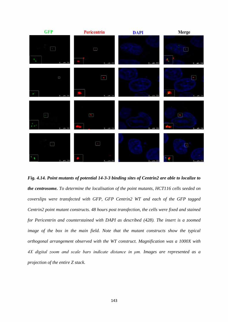
143
Fig. 4.14. Point mutants of potential 14-3-3 binding sites of Centrin2 are able to localize to
the centrosome. To determine the localisation of the point mutants, HCT116 cells seeded on
coverslips were transfected with GFP, GFP Centrin2 WT and each of the GFP tagged
Centrin2 point mutant constructs. 48 hours post transfection, the cells were fixed and stained
for Pericentrin and counterstained with DAPI as described (428). The insert is a zoomed
image of the box in the main field. Note that the mutant constructs show the typical
orthogonal arrangement observed with the WT construct. Magnification was a 1000X with
4X digital zoom and scale bars indicate distance in μm. Images are represented as a
projection of the entire Z stack.
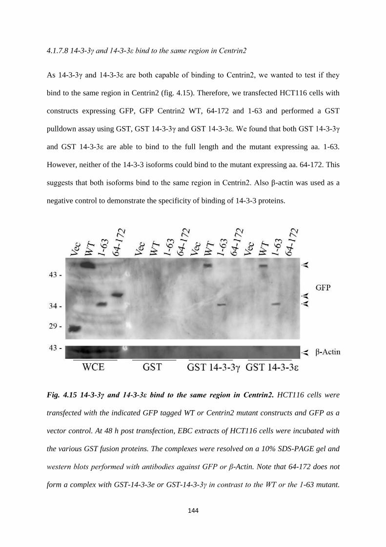
144
4.1.7.8 14-3-3γ and 14-3-3ε bind to the same region in Centrin2
As 14-3-3γ and 14-3-3ε are both capable of binding to Centrin2, we wanted to test if they
bind to the same region in Centrin2 (fig. 4.15). Therefore, we transfected HCT116 cells with
constructs expressing GFP, GFP Centrin2 WT, 64-172 and 1-63 and performed a GST
pulldown assay using GST, GST 14-3-3γ and GST 14-3-3ε. We found that both GST 14-3-3γ
and GST 14-3-3ε are able to bind to the full length and the mutant expressing aa. 1-63.
However, neither of the 14-3-3 isoforms could bind to the mutant expressing aa. 64-172. This
suggests that both isoforms bind to the same region in Centrin2. Also β-actin was used as a
negative control to demonstrate the specificity of binding of 14-3-3 proteins.
Fig. 4.15 14-3-3γ and 14-3-3ε bind to the same region in Centrin2. HCT116 cells were
transfected with the indicated GFP tagged WT or Centrin2 mutant constructs and GFP as a
vector control. At 48 h post transfection, EBC extracts of HCT116 cells were incubated with
the various GST fusion proteins. The complexes were resolved on a 10% SDS-PAGE gel and
western blots performed with antibodies against GFP or β-Actin. Note that 64-172 does not
form a complex with GST-14-3-3e or GST-14-3-3γ in contrast to the WT or the 1-63 mutant.
145
Arrows indicate the positions of the GFP fusion proteins and the position of molecular
weight markers in kDa is indicated. β-Actin serves as a negative control.
4.1.7.9 Depletion of both 14-3-3γ and 14-3-3ε does not alter the centrosomal localization of
Centrin2
Next, we wished to determine if the loss of 14-3-3γ, 14-3-3ε or both could result in a loss of
the centrosomal localisation of Centrin2. To this end, we co-transfected previously described
shRNA constructs of 14-3-3γ and 14-3-3ε and GFP Centrin2 in HCT116 cells (234,401). At
72 hours post transfection, the cells were fixed and stained for pericentrin and counterstained
with DAPI (fig. 4.16 (a)). We determined that the constructs were functional by performing a
Western blot for both proteins and using β-actin as a loading control (fig. 4.16 (b)). We found
that there was a decrease in the levels of both proteins. In addition, as has been previously
reported, centrosome amplification was observed upon loss of these proteins, suggesting that
these constructs are functional and there was an inhibition of CDC25C function (fig. 4.16 (c))
(326). Loss of 14-3-3γ, 14-3-3ε or both proteins did not result in an alteration in the
localization of Centrin2. It is possible that due to an incomplete knockdown of both 14-3-3γ
and 14-3-3ε, there is sufficient 14-3-3 protein available to mediate Centrin2 localization.
Alternatively, it is possible that loss of one or more isoforms is not sufficient to induce a
defect in Centrin2 localization, as, like Raf and CDC25C, Centrin2 binds to multiple 14-3-3
isoforms (152,429).
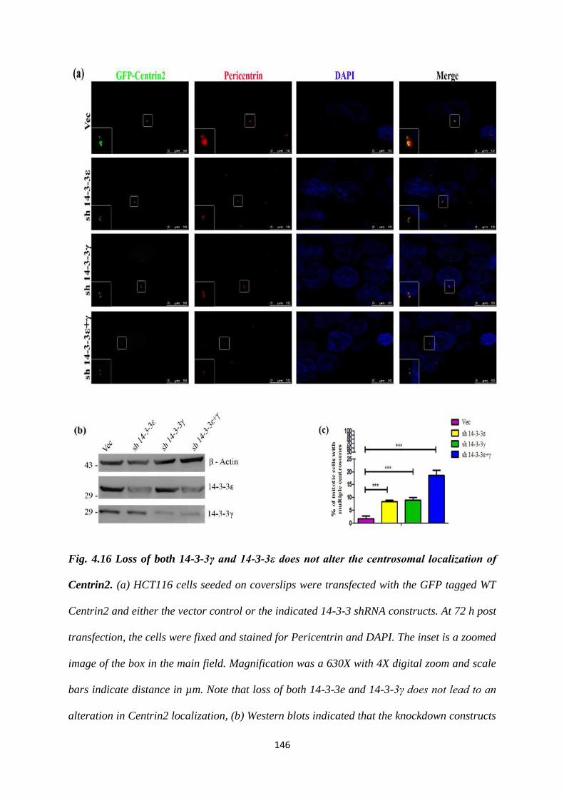
146
Fig. 4.16 Loss of both 14-3-3γ and 14-3-3ε does not alter the centrosomal localization of
Centrin2. (a) HCT116 cells seeded on coverslips were transfected with the GFP tagged WT
Centrin2 and either the vector control or the indicated 14-3-3 shRNA constructs. At 72 h post
transfection, the cells were fixed and stained for Pericentrin and DAPI. The inset is a zoomed
image of the box in the main field. Magnification was a 630X with 4X digital zoom and scale
bars indicate distance in µm. Note that loss of both 14-3-3e and 14-3-3γ does not lead to an
alteration in Centrin2 localization, (b) Western blots indicated that the knockdown constructs
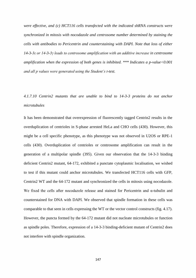
147
were effective, and (c) HCT116 cells transfected with the indicated shRNA constructs were
synchronized in mitosis with nocodazole and centrosome number determined by staining the
cells with antibodies to Pericentrin and counterstaining with DAPI. Note that loss of either
14-3-3ε or 14-3-3γ leads to centrosome amplification with an additive increase in centrosome
amplification when the expression of both genes is inhibited. *** Indicates a p-value<0.001
and all p values were generated using the Student’s t-test.
4.1.7.10 Centrin2 mutants that are unable to bind to 14-3-3 proteins do not anchor
microtubules
It has been demonstrated that overexpression of fluorescently tagged Centrin2 results in the
overduplication of centrioles in S-phase arrested HeLa and CHO cells (430). However, this
might be a cell specific phenotype, as this phenotype was not observed in U2OS or RPE-1
cells (430). Overduplication of centrioles or centrosome amplification can result in the
generation of a multipolar spindle (395). Given our observation that the 14-3-3 binding
deficient Centrin2 mutant, 64-172, exhibited a punctate cytoplasmic localisation, we wished
to test if this mutant could anchor microtubules. We transfected HCT116 cells with GFP,
Centrin2 WT and the 64-172 mutant and synchronized the cells in mitosis using nocodazole.
We fixed the cells after nocodazole release and stained for Pericentrin and α-tubulin and
counterstained for DNA with DAPI. We observed that spindle formation in these cells was
comparable to that seen in cells expressing the WT or the vector control constructs (fig. 4.17).
However, the puncta formed by the 64-172 mutant did not nucleate microtubules or function
as spindle poles. Therefore, expression of a 14-3-3 binding-deficient mutant of Centrin2 does
not interfere with spindle organization.
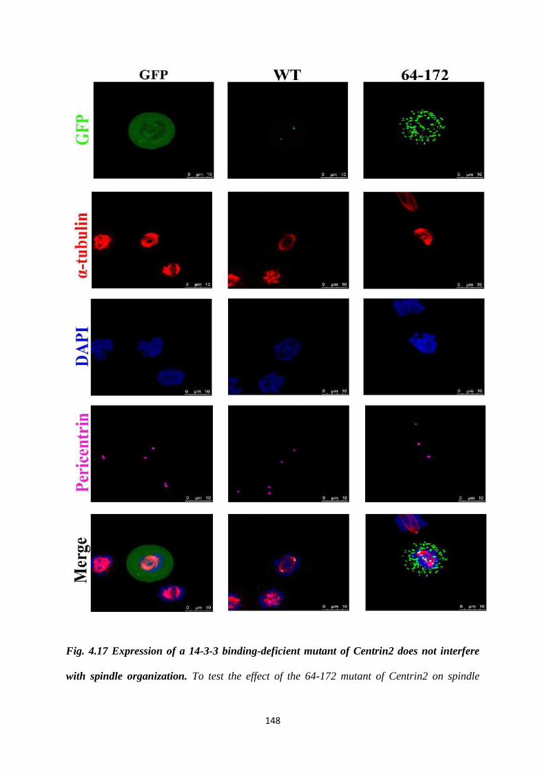
148
Fig. 4.17 Expression of a 14-3-3 binding-deficient mutant of Centrin2 does not interfere
with spindle organization. To test the effect of the 64-172 mutant of Centrin2 on spindle
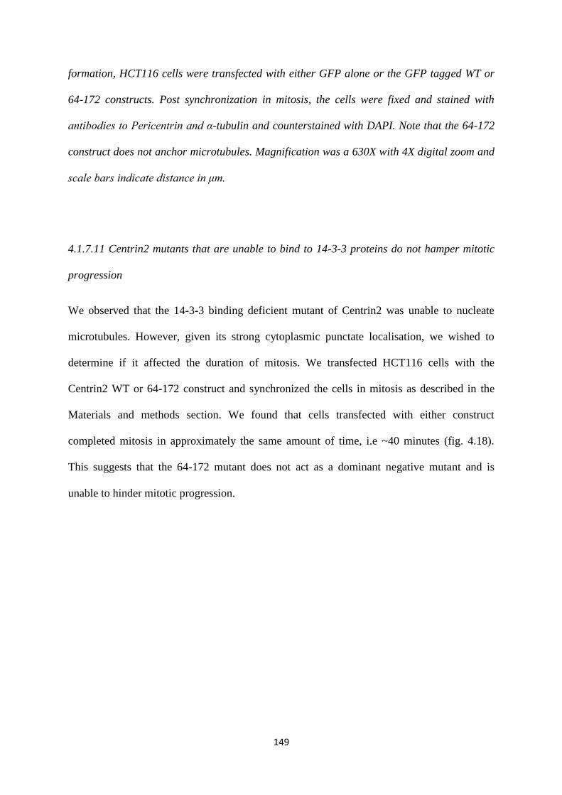
149
formation, HCT116 cells were transfected with either GFP alone or the GFP tagged WT or
64-172 constructs. Post synchronization in mitosis, the cells were fixed and stained with
antibodies to Pericentrin and α-tubulin and counterstained with DAPI. Note that the 64-172
construct does not anchor microtubules. Magnification was a 630X with 4X digital zoom and
scale bars indicate distance in μm.
4.1.7.11 Centrin2 mutants that are unable to bind to 14-3-3 proteins do not hamper mitotic
progression
We observed that the 14-3-3 binding deficient mutant of Centrin2 was unable to nucleate
microtubules. However, given its strong cytoplasmic punctate localisation, we wished to
determine if it affected the duration of mitosis. We transfected HCT116 cells with the
Centrin2 WT or 64-172 construct and synchronized the cells in mitosis as described in the
Materials and methods section. We found that cells transfected with either construct
completed mitosis in approximately the same amount of time, i.e ~40 minutes (fig. 4.18).
This suggests that the 64-172 mutant does not act as a dominant negative mutant and is
unable to hinder mitotic progression.
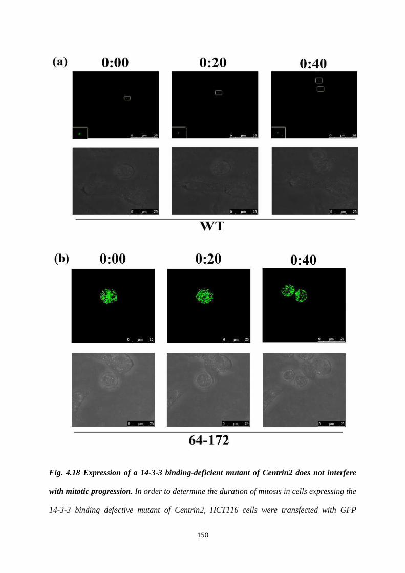
150
Fig. 4.18 Expression of a 14-3-3 binding-deficient mutant of Centrin2 does not interfere
with mitotic progression. In order to determine the duration of mitosis in cells expressing the
14-3-3 binding defective mutant of Centrin2, HCT116 cells were transfected with GFP
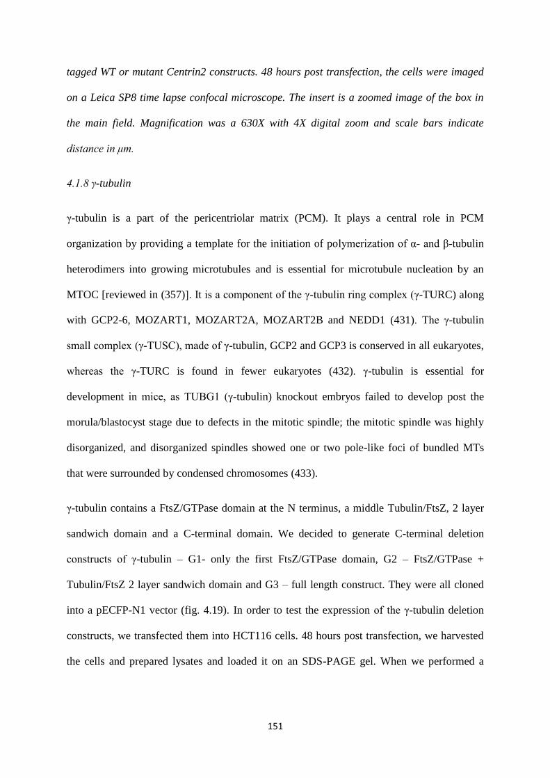
151
tagged WT or mutant Centrin2 constructs. 48 hours post transfection, the cells were imaged
on a Leica SP8 time lapse confocal microscope. The insert is a zoomed image of the box in
the main field. Magnification was a 630X with 4X digital zoom and scale bars indicate
distance in μm.
4.1.8 γ-tubulin
γ-tubulin is a part of the pericentriolar matrix (PCM). It plays a central role in PCM
organization by providing a template for the initiation of polymerization of α- and β-tubulin
heterodimers into growing microtubules and is essential for microtubule nucleation by an
MTOC [reviewed in (357)]. It is a component of the γ-tubulin ring complex (γ-TURC) along
with GCP2-6, MOZART1, MOZART2A, MOZART2B and NEDD1 (431). The γ-tubulin
small complex (γ-TUSC), made of γ-tubulin, GCP2 and GCP3 is conserved in all eukaryotes,
whereas the γ-TURC is found in fewer eukaryotes (432). γ-tubulin is essential for
development in mice, as TUBG1 (γ-tubulin) knockout embryos failed to develop post the
morula/blastocyst stage due to defects in the mitotic spindle; the mitotic spindle was highly
disorganized, and disorganized spindles showed one or two pole-like foci of bundled MTs
that were surrounded by condensed chromosomes (433).
γ-tubulin contains a FtsZ/GTPase domain at the N terminus, a middle Tubulin/FtsZ, 2 layer
sandwich domain and a C-terminal domain. We decided to generate C-terminal deletion
constructs of γ-tubulin – G1- only the first FtsZ/GTPase domain, G2 – FtsZ/GTPase +
Tubulin/FtsZ 2 layer sandwich domain and G3 – full length construct. They were all cloned
into a pECFP-N1 vector (fig. 4.19). In order to test the expression of the γ-tubulin deletion
constructs, we transfected them into HCT116 cells. 48 hours post transfection, we harvested
the cells and prepared lysates and loaded it on an SDS-PAGE gel. When we performed a
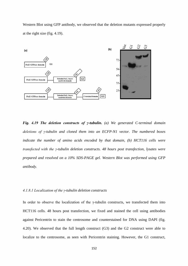
152
Western Blot using GFP antibody, we observed that the deletion mutants expressed properly
at the right size (fig. 4.19).
Fig. 4.19 The deletion constructs of γ-tubulin. (a) We generated C-terminal domain
deletions of γ-tubulin and cloned them into an ECFP-N1 vector. The numbered boxes
indicate the number of amino acids encoded by that domain, (b) HCT116 cells were
transfected with the γ-tubulin deletion constructs. 48 hours post transfection, lysates were
prepared and resolved on a 10% SDS-PAGE gel. Western Blot was performed using GFP
antibody.
4.1.8.1 Localization of the γ-tubulin deletion constructs
In order to observe the localization of the γ-tubulin constructs, we transfected them into
HCT116 cells. 48 hours post transfection, we fixed and stained the cell using antibodies
against Pericentrin to stain the centrosome and counterstained for DNA using DAPI (fig.
4.20). We observed that the full length construct (G3) and the G2 construct were able to
localize to the centrosome, as seen with Pericentrin staining. However, the G1 construct,
(a) (b)
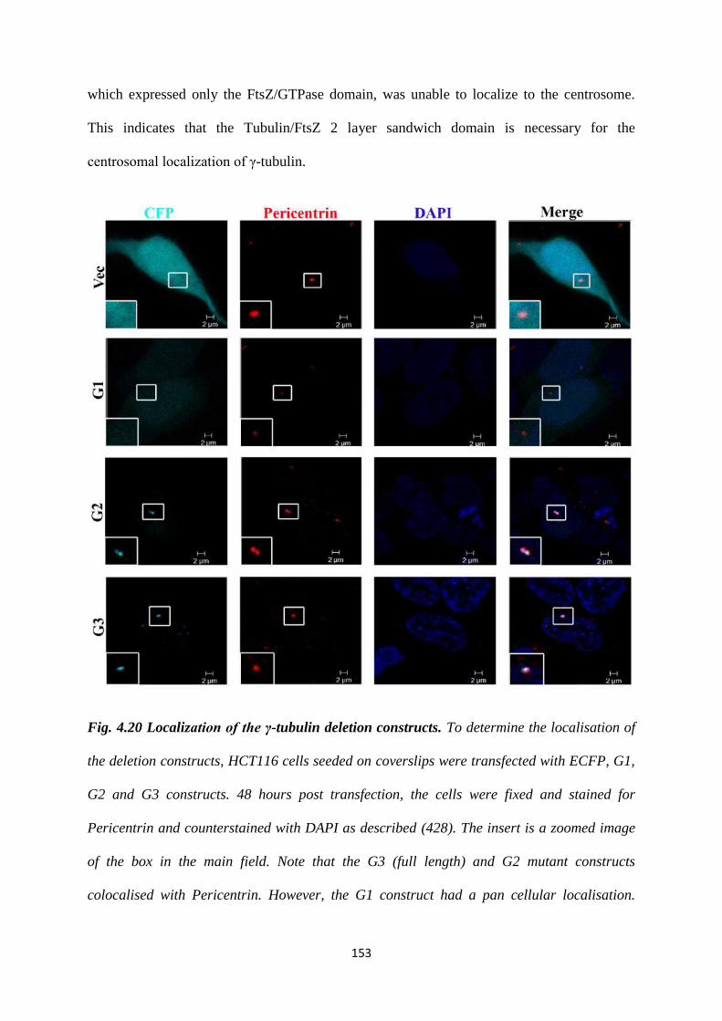
153
which expressed only the FtsZ/GTPase domain, was unable to localize to the centrosome.
This indicates that the Tubulin/FtsZ 2 layer sandwich domain is necessary for the
centrosomal localization of γ-tubulin.
Fig. 4.20 Localization of the γ-tubulin deletion constructs. To determine the localisation of
the deletion constructs, HCT116 cells seeded on coverslips were transfected with ECFP, G1,
G2 and G3 constructs. 48 hours post transfection, the cells were fixed and stained for
Pericentrin and counterstained with DAPI as described (428). The insert is a zoomed image
of the box in the main field. Note that the G3 (full length) and G2 mutant constructs
colocalised with Pericentrin. However, the G1 construct had a pan cellular localisation.
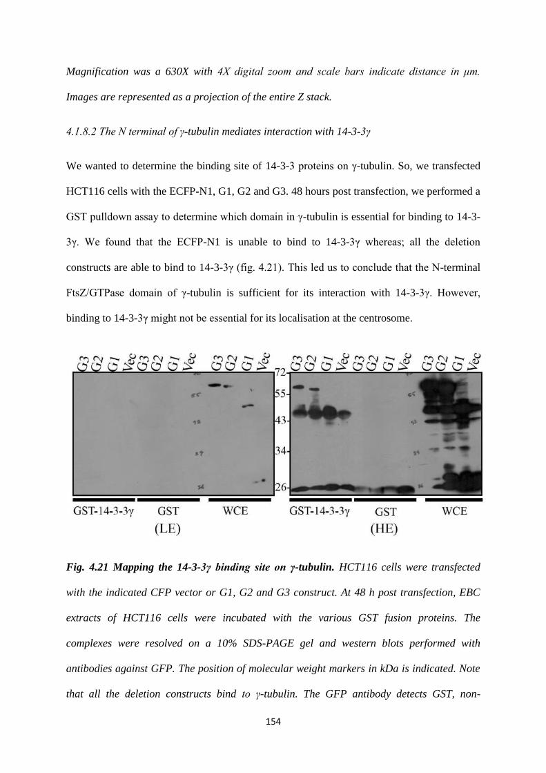
154
Magnification was a 630X with 4X digital zoom and scale bars indicate distance in μm.
Images are represented as a projection of the entire Z stack.
4.1.8.2 The N terminal of γ-tubulin mediates interaction with 14-3-3γ
We wanted to determine the binding site of 14-3-3 proteins on γ-tubulin. So, we transfected
HCT116 cells with the ECFP-N1, G1, G2 and G3. 48 hours post transfection, we performed a
GST pulldown assay to determine which domain in γ-tubulin is essential for binding to 14-3-
3γ. We found that the ECFP-N1 is unable to bind to 14-3-3γ whereas; all the deletion
constructs are able to bind to 14-3-3γ (fig. 4.21). This led us to conclude that the N-terminal
FtsZ/GTPase domain of γ-tubulin is sufficient for its interaction with 14-3-3γ. However,
binding to 14-3-3γ might not be essential for its localisation at the centrosome.
Fig. 4.21 Mapping the 14-3-3γ binding site on γ-tubulin. HCT116 cells were transfected
with the indicated CFP vector or G1, G2 and G3 construct. At 48 h post transfection, EBC
extracts of HCT116 cells were incubated with the various GST fusion proteins. The
complexes were resolved on a 10% SDS-PAGE gel and western blots performed with
antibodies against GFP. The position of molecular weight markers in kDa is indicated. Note
that all the deletion constructs bind to γ-tubulin. The GFP antibody detects GST, non-

155
specifically, and the band for G1 appears just above the GST-14-3-3γ band. LE indicates a
Lower Exposure and HE indicates a Higher Exposure.
4.1.9 γ-tubulin complex protein 2 (GCP2)
GCP2 is one of the three proteins that form the γ-TUSC, with γ-tubulin and GCP3. It is a
homologue of the yeast spindle pole body protein Spc97p (434). Since it is a component of
the γ-TUSC, it is essential for microtubule nucleation. A null mutation of Arabidopsis GCP2
severely impaired the development of male and female gametophytes (435). A knockdown of
GCP2 in the glioblastoma cell line T98G led to an accumulation of cells in G2/M phase
(436). This was accompanied by mitotic delay, although the cells were able to complete
mitosis (436). Structurally, GCP2 consists of the domain that bears homology to the Spc97p
protein. We decided to generate C terminal truncated mutants that did not express the Spc97p
domain. We generated two deletion mutants; GC1 – aa. 1 – 220 and GC2 – aa. 1 – 738. Both
these sequences were cloned into the ECFP-N1 vector, along with the full length cDNA.
In order to test the expression of the GCP2 deletion constructs, we transfected them into
HCT116 cells. 48 hours post transfection, we harvested the cells and prepared lysates and
loaded it on an SDS-PAGE gel. When we performed a Western Blot using GFP antibody, we
observed that the deletion mutants expressed properly at the expected size (fig. 4.22).
(a) (b)
156
Fig. 4.22 Deletion constructs of GCP2. (a) Cartoon representing the deletion constructs of
GCP2. We generated C-terminal domain deletions of GCP2 and cloned them into an ECFP-
N1 vector. The numbered boxes indicate the number of amino acids encoded by that domain,
(b) Expression of the deletion constructs. HCT116 cells were transfected with the GCP2
deletion constructs. 48 hours post transfection, lysates were prepared and resolved on a 10%
SDS-PAGE gel. Western Blot was performed using GFP antibody.
4.1.9.1 Localization of the GCP2 deletion constructs
In order to observe the localization of the GCP2 constructs, we transfected them into
HCT116 cells. 48 hours post transfection, we fixed and stained the cell using antibodies
against Pericentrin to stain the centrosome and counterstained for DNA using DAPI. We
observed that each of the deletion constructs co-localized with the Pericentrin staining (fig.
4.23). This suggests that Spc97p domain is not essential for the centrosomal localisation of
GCP2. However, the intensity of the GC1 construct at the centrosome was much weaker as
compared to the other constructs. Deletion of the Spc97p domain might therefore, affect the
centrosomal recruitment of GCP2.
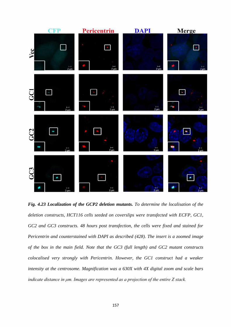
157
Fig. 4.23 Localization of the GCP2 deletion mutants. To determine the localisation of the
deletion constructs, HCT116 cells seeded on coverslips were transfected with ECFP, GC1,
GC2 and GC3 constructs. 48 hours post transfection, the cells were fixed and stained for
Pericentrin and counterstained with DAPI as described (428). The insert is a zoomed image
of the box in the main field. Note that the GC3 (full length) and GC2 mutant constructs
colocalised very strongly with Pericentrin. However, the GC1 construct had a weaker
intensity at the centrosome. Magnification was a 630X with 4X digital zoom and scale bars
indicate distance in μm. Images are represented as a projection of the entire Z stack.
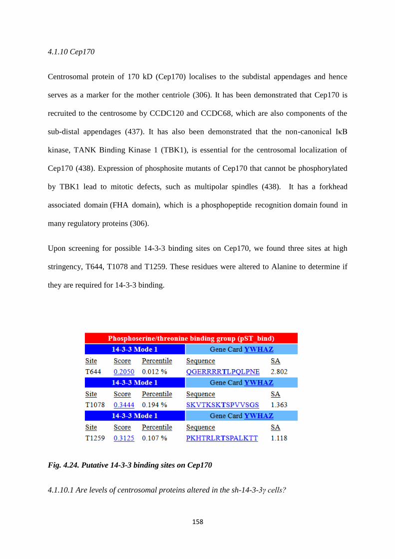
158
4.1.10 Cep170
Centrosomal protein of 170 kD (Cep170) localises to the subdistal appendages and hence
serves as a marker for the mother centriole (306). It has been demonstrated that Cep170 is
recruited to the centrosome by CCDC120 and CCDC68, which are also components of the
sub-distal appendages (437). It has also been demonstrated that the non-canonical IκB
kinase, TANK Binding Kinase 1 (TBK1), is essential for the centrosomal localization of
Cep170 (438). Expression of phosphosite mutants of Cep170 that cannot be phosphorylated
by TBK1 lead to mitotic defects, such as multipolar spindles (438). It has a forkhead
associated domain (FHA domain), which is a phosphopeptide recognition domain found in
many regulatory proteins (306).
Upon screening for possible 14-3-3 binding sites on Cep170, we found three sites at high
stringency, T644, T1078 and T1259. These residues were altered to Alanine to determine if
they are required for 14-3-3 binding.
Fig. 4.24. Putative 14-3-3 binding sites on Cep170
4.1.10.1 Are levels of centrosomal proteins altered in the sh-14-3-3γ cells?
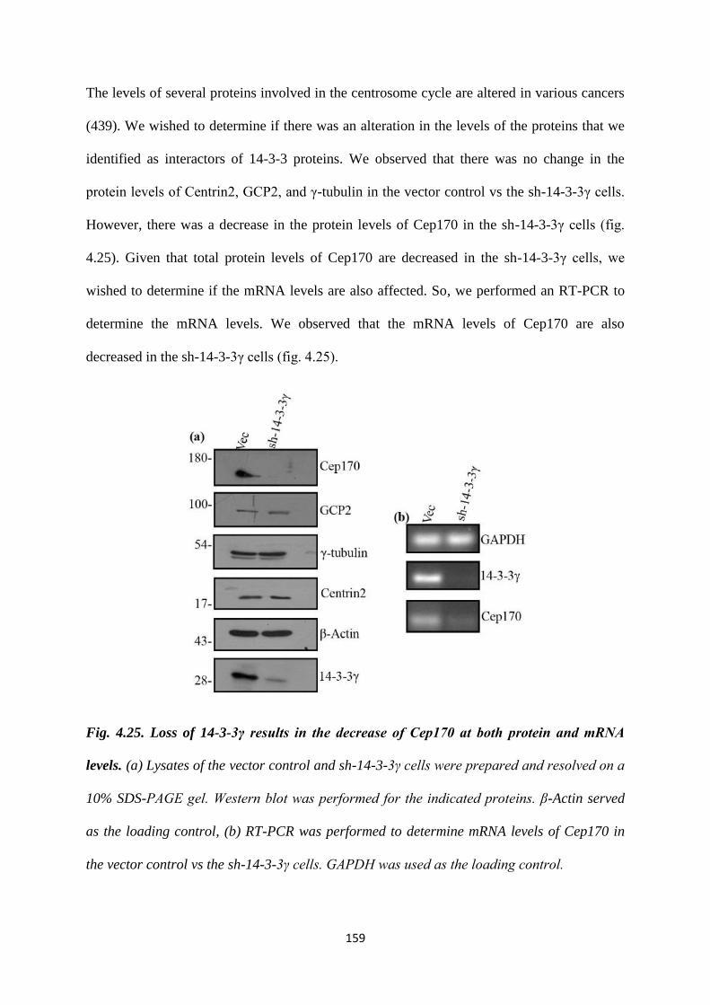
159
The levels of several proteins involved in the centrosome cycle are altered in various cancers
(439). We wished to determine if there was an alteration in the levels of the proteins that we
identified as interactors of 14-3-3 proteins. We observed that there was no change in the
protein levels of Centrin2, GCP2, and γ-tubulin in the vector control vs the sh-14-3-3γ cells.
However, there was a decrease in the protein levels of Cep170 in the sh-14-3-3γ cells (fig.
4.25). Given that total protein levels of Cep170 are decreased in the sh-14-3-3γ cells, we
wished to determine if the mRNA levels are also affected. So, we performed an RT-PCR to
determine the mRNA levels. We observed that the mRNA levels of Cep170 are also
decreased in the sh-14-3-3γ cells (fig. 4.25).
Fig. 4.25. Loss of 14-3-3γ results in the decrease of Cep170 at both protein and mRNA
levels. (a) Lysates of the vector control and sh-14-3-3γ cells were prepared and resolved on a
10% SDS-PAGE gel. Western blot was performed for the indicated proteins. β-Actin served
as the loading control, (b) RT-PCR was performed to determine mRNA levels of Cep170 in
the vector control vs the sh-14-3-3γ cells. GAPDH was used as the loading control.
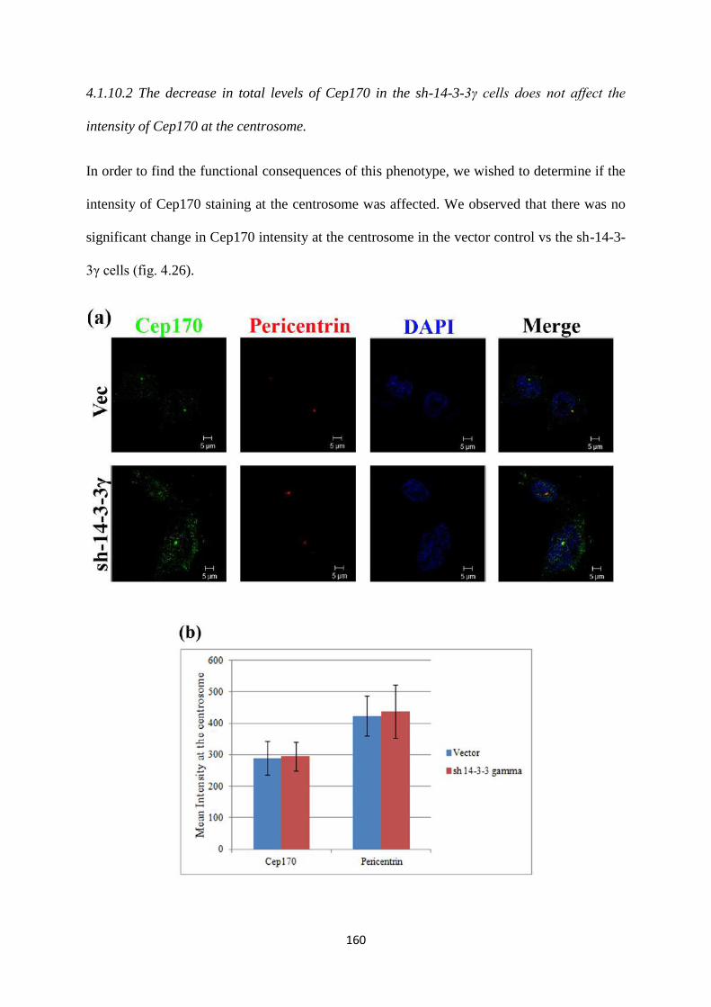
160
4.1.10.2 The decrease in total levels of Cep170 in the sh-14-3-3γ cells does not affect the
intensity of Cep170 at the centrosome.
In order to find the functional consequences of this phenotype, we wished to determine if the
intensity of Cep170 staining at the centrosome was affected. We observed that there was no
significant change in Cep170 intensity at the centrosome in the vector control vs the sh-14-3-
3γ cells (fig. 4.26).
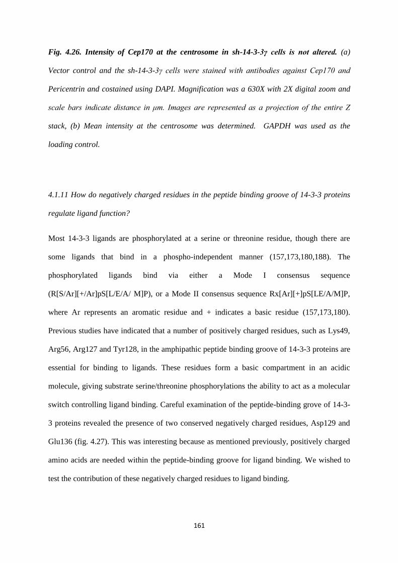
161
Fig. 4.26. Intensity of Cep170 at the centrosome in sh-14-3-3γ cells is not altered. (a)
Vector control and the sh-14-3-3γ cells were stained with antibodies against Cep170 and
Pericentrin and costained using DAPI. Magnification was a 630X with 2X digital zoom and
scale bars indicate distance in μm. Images are represented as a projection of the entire Z
stack, (b) Mean intensity at the centrosome was determined. GAPDH was used as the
loading control.
4.1.11 How do negatively charged residues in the peptide binding groove of 14-3-3 proteins
regulate ligand function?
Most 14-3-3 ligands are phosphorylated at a serine or threonine residue, though there are
some ligands that bind in a phospho-independent manner (157,173,180,188). The
phosphorylated ligands bind via either a Mode I consensus sequence
(R[S/Ar][+/Ar]pS[L/E/A/ M]P), or a Mode II consensus sequence Rx[Ar][+]pS[LE/A/M]P,
where Ar represents an aromatic residue and + indicates a basic residue (157,173,180).
Previous studies have indicated that a number of positively charged residues, such as Lys49,
Arg56, Arg127 and Tyr128, in the amphipathic peptide binding groove of 14-3-3 proteins are
essential for binding to ligands. These residues form a basic compartment in an acidic
molecule, giving substrate serine/threonine phosphorylations the ability to act as a molecular
switch controlling ligand binding. Careful examination of the peptide-binding grove of 14-3-
3 proteins revealed the presence of two conserved negatively charged residues, Asp129 and
Glu136 (fig. 4.27). This was interesting because as mentioned previously, positively charged
amino acids are needed within the peptide-binding groove for ligand binding. We wished to
test the contribution of these negatively charged residues to ligand binding.
162
Fig. 4.27 Sequence alignment of the peptide binding groove of 14-3-3 isoforms.
4.1.11.1 Site directed mutagenesis of 14-3-3γ WT
We have demonstrated previously, that a loss of 14-3-3γ leads to centrosome amplification
(326). Therefore, we decided to test the effect of these mutants on centrosome number. Using
Site Directed Mutagenesis (SDM), we mutated the Asp129 and Glu136 to obtain D129A and
E136A. We also generated a double mutant – D129AE136A. Arg56 has been demonstrated
to be important for ligand binding (180). We mutated this to Alanine, as a residue known to
have an effect on ligand binding. We finally generated multiple mutants, R56A, D129A,
E136A, R56AD129A, R56AE136A, D129AE136A and R56AD129AE136A. shRNA
resistant WT 14-3-3γ was cloned into a pCMV-mOrange vector. Then, SDM was performed
to generate the various combinations of mutant proteins. The clones were sequenced and
verified.
4.1.11.2 Testing the expression of the 14-3-3γ mutants
In order to test the expression of the 14-3-3γ mutants, we transfected HCT116 cells with each
of the constructs. 48 hours post transfection, we harvested the cells and performed protein
estimation. Equal concentrations of the lysates were loaded onto an SDS-PAGE gel and
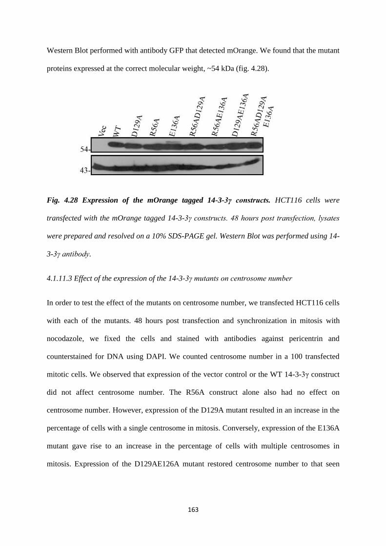
163
Western Blot performed with antibody GFP that detected mOrange. We found that the mutant
proteins expressed at the correct molecular weight, ~54 kDa (fig. 4.28).
Fig. 4.28 Expression of the mOrange tagged 14-3-3γ constructs. HCT116 cells were
transfected with the mOrange tagged 14-3-3γ constructs. 48 hours post transfection, lysates
were prepared and resolved on a 10% SDS-PAGE gel. Western Blot was performed using 14-
3-3γ antibody.
4.1.11.3 Effect of the expression of the 14-3-3γ mutants on centrosome number
In order to test the effect of the mutants on centrosome number, we transfected HCT116 cells
with each of the mutants. 48 hours post transfection and synchronization in mitosis with
nocodazole, we fixed the cells and stained with antibodies against pericentrin and
counterstained for DNA using DAPI. We counted centrosome number in a 100 transfected
mitotic cells. We observed that expression of the vector control or the WT 14-3-3γ construct
did not affect centrosome number. The R56A construct alone also had no effect on
centrosome number. However, expression of the D129A mutant resulted in an increase in the
percentage of cells with a single centrosome in mitosis. Conversely, expression of the E136A
mutant gave rise to an increase in the percentage of cells with multiple centrosomes in
mitosis. Expression of the D129AE126A mutant restored centrosome number to that seen
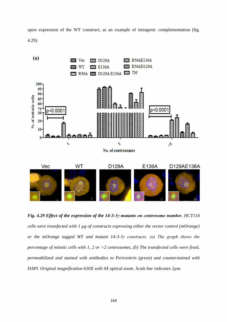
164
upon expression of the WT construct, as an example of intragenic complementation (fig.
4.29).
Fig. 4.29 Effect of the expression of the 14-3-3γ mutants on centrosome number. HCT116
cells were transfected with 1 µg of constructs expressing either the vector control (mOrange)
or the mOrange tagged WT and mutant 14-3-3γ constructs. (a) The graph shows the
percentage of mitotic cells with 1, 2 or >2 centrosomes, (b) The transfected cells were fixed,
permeabilized and stained with antibodies to Pericentrin (green) and counterstained with
DAPI. Original magnification 630X with 4X optical zoom. Scale bar indicates 2µm.
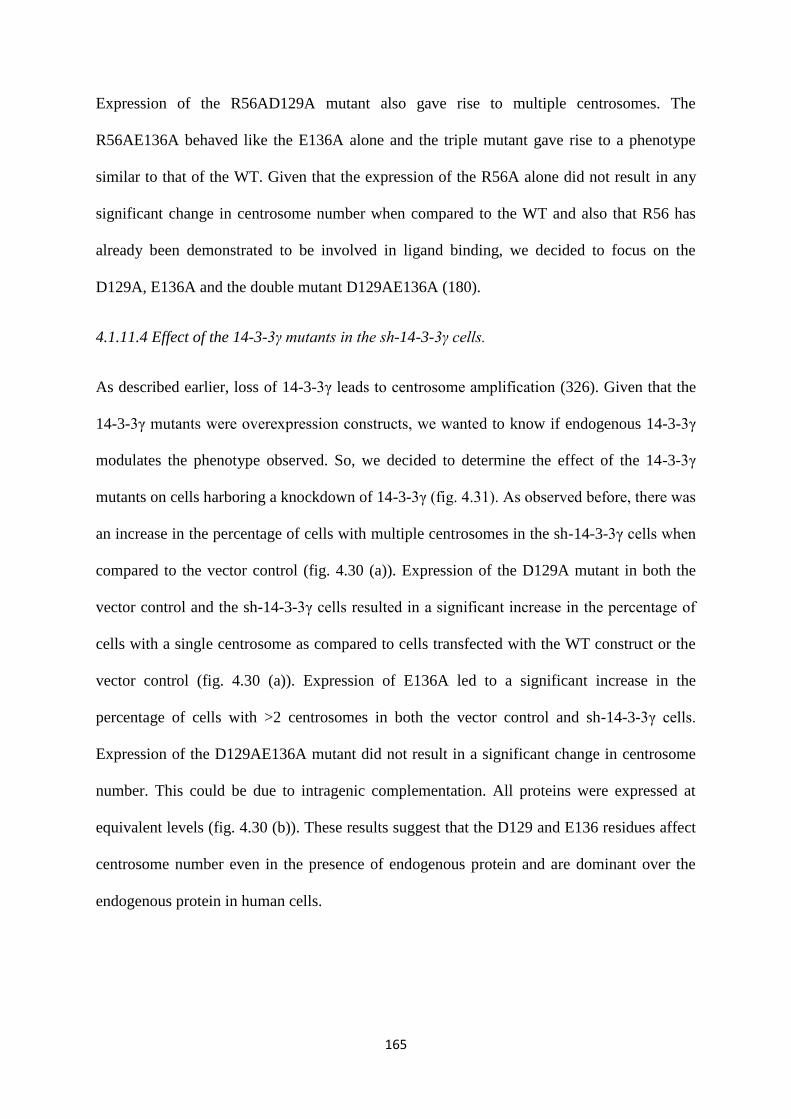
165
Expression of the R56AD129A mutant also gave rise to multiple centrosomes. The
R56AE136A behaved like the E136A alone and the triple mutant gave rise to a phenotype
similar to that of the WT. Given that the expression of the R56A alone did not result in any
significant change in centrosome number when compared to the WT and also that R56 has
already been demonstrated to be involved in ligand binding, we decided to focus on the
D129A, E136A and the double mutant D129AE136A (180).
4.1.11.4 Effect of the 14-3-3γ mutants in the sh-14-3-3γ cells.
As described earlier, loss of 14-3-3γ leads to centrosome amplification (326). Given that the
14-3-3γ mutants were overexpression constructs, we wanted to know if endogenous 14-3-3γ
modulates the phenotype observed. So, we decided to determine the effect of the 14-3-3γ
mutants on cells harboring a knockdown of 14-3-3γ (fig. 4.31). As observed before, there was
an increase in the percentage of cells with multiple centrosomes in the sh-14-3-3γ cells when
compared to the vector control (fig. 4.30 (a)). Expression of the D129A mutant in both the
vector control and the sh-14-3-3γ cells resulted in a significant increase in the percentage of
cells with a single centrosome as compared to cells transfected with the WT construct or the
vector control (fig. 4.30 (a)). Expression of E136A led to a significant increase in the
percentage of cells with >2 centrosomes in both the vector control and sh-14-3-3γ cells.
Expression of the D129AE136A mutant did not result in a significant change in centrosome
number. This could be due to intragenic complementation. All proteins were expressed at
equivalent levels (fig. 4.30 (b)). These results suggest that the D129 and E136 residues affect
centrosome number even in the presence of endogenous protein and are dominant over the
endogenous protein in human cells.
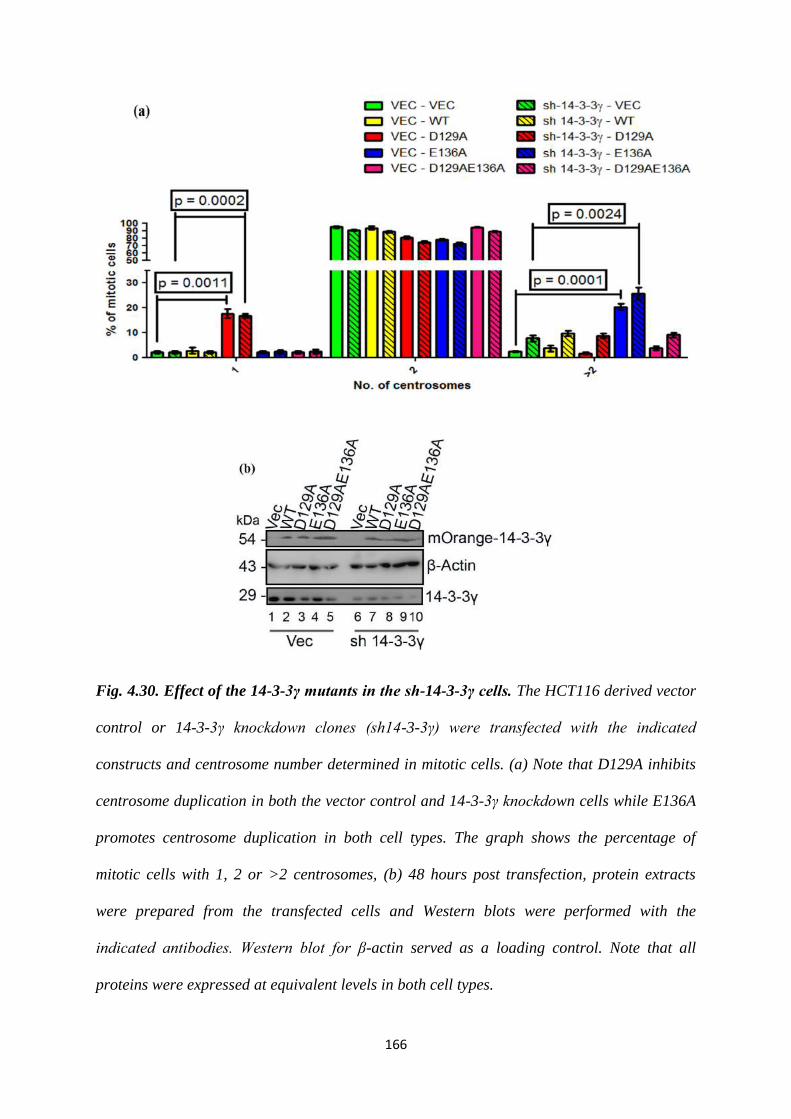
166
Fig. 4.30. Effect of the 14-3-3γ mutants in the sh-14-3-3γ cells. The HCT116 derived vector
control or 14-3-3γ knockdown clones (sh14-3-3γ) were transfected with the indicated
constructs and centrosome number determined in mitotic cells. (a) Note that D129A inhibits
centrosome duplication in both the vector control and 14-3-3γ knockdown cells while E136A
promotes centrosome duplication in both cell types. The graph shows the percentage of
mitotic cells with 1, 2 or >2 centrosomes, (b) 48 hours post transfection, protein extracts
were prepared from the transfected cells and Western blots were performed with the
indicated antibodies. Western blot for β-actin served as a loading control. Note that all
proteins were expressed at equivalent levels in both cell types.
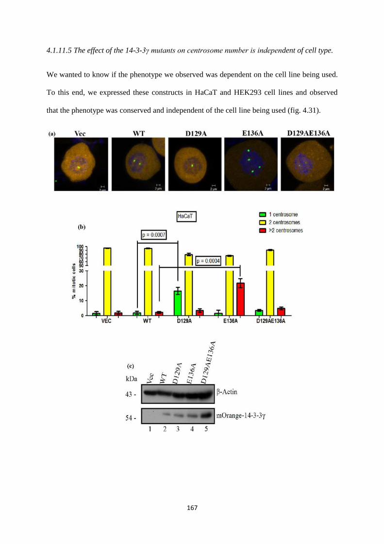
167
4.1.11.5 The effect of the 14-3-3γ mutants on centrosome number is independent of cell type.
We wanted to know if the phenotype we observed was dependent on the cell line being used.
To this end, we expressed these constructs in HaCaT and HEK293 cell lines and observed
that the phenotype was conserved and independent of the cell line being used (fig. 4.31).
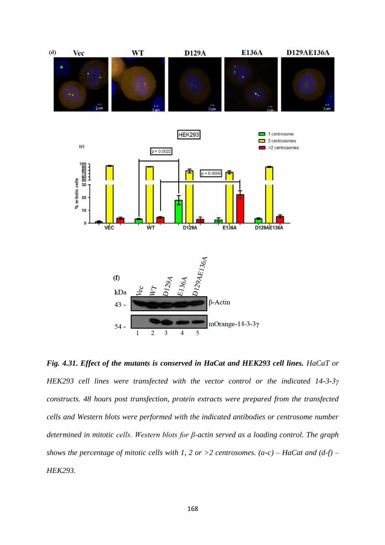
168
Fig. 4.31. Effect of the mutants is conserved in HaCat and HEK293 cell lines. HaCaT or
HEK293 cell lines were transfected with the vector control or the indicated 14-3-3γ
constructs. 48 hours post transfection, protein extracts were prepared from the transfected
cells and Western blots were performed with the indicated antibodies or centrosome number
determined in mitotic cells. Western blots for β-actin served as a loading control. The graph
shows the percentage of mitotic cells with 1, 2 or >2 centrosomes. (a-c) – HaCat and (d-f) –
HEK293.
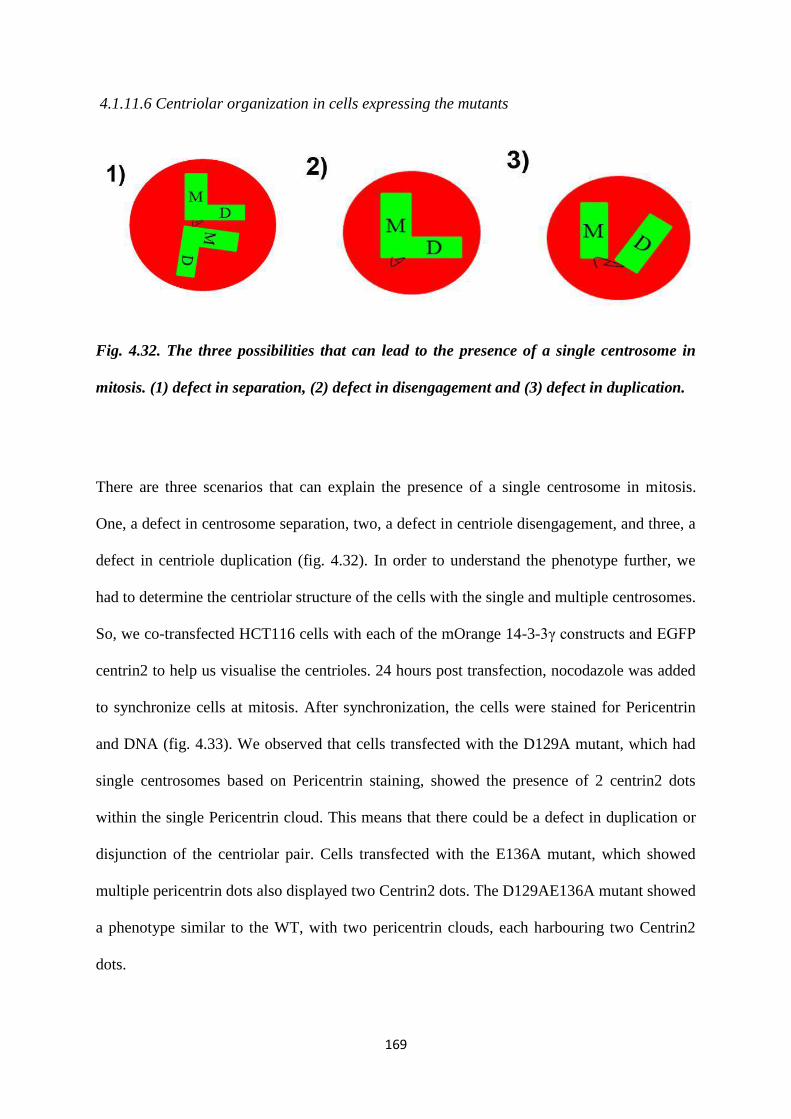
169
4.1.11.6 Centriolar organization in cells expressing the mutants
Fig. 4.32. The three possibilities that can lead to the presence of a single centrosome in
mitosis. (1) defect in separation, (2) defect in disengagement and (3) defect in duplication.
There are three scenarios that can explain the presence of a single centrosome in mitosis.
One, a defect in centrosome separation, two, a defect in centriole disengagement, and three, a
defect in centriole duplication (fig. 4.32). In order to understand the phenotype further, we
had to determine the centriolar structure of the cells with the single and multiple centrosomes.
So, we co-transfected HCT116 cells with each of the mOrange 14-3-3γ constructs and EGFP
centrin2 to help us visualise the centrioles. 24 hours post transfection, nocodazole was added
to synchronize cells at mitosis. After synchronization, the cells were stained for Pericentrin
and DNA (fig. 4.33). We observed that cells transfected with the D129A mutant, which had
single centrosomes based on Pericentrin staining, showed the presence of 2 centrin2 dots
within the single Pericentrin cloud. This means that there could be a defect in duplication or
disjunction of the centriolar pair. Cells transfected with the E136A mutant, which showed
multiple pericentrin dots also displayed two Centrin2 dots. The D129AE136A mutant showed
a phenotype similar to the WT, with two pericentrin clouds, each harbouring two Centrin2
dots.
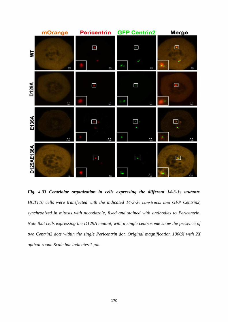
170
Fig. 4.33 Centriolar organization in cells expressing the different 14-3-3γ mutants.
HCT116 cells were transfected with the indicated 14-3-3γ constructs and GFP Centrin2,
synchronized in mitosis with nocodazole, fixed and stained with antibodies to Pericentrin.
Note that cells expressing the D129A mutant, with a single centrosome show the presence of
two Centrin2 dots within the single Pericentrin dot. Original magnification 1000X with 2X
optical zoom. Scale bar indicates 1 µm.
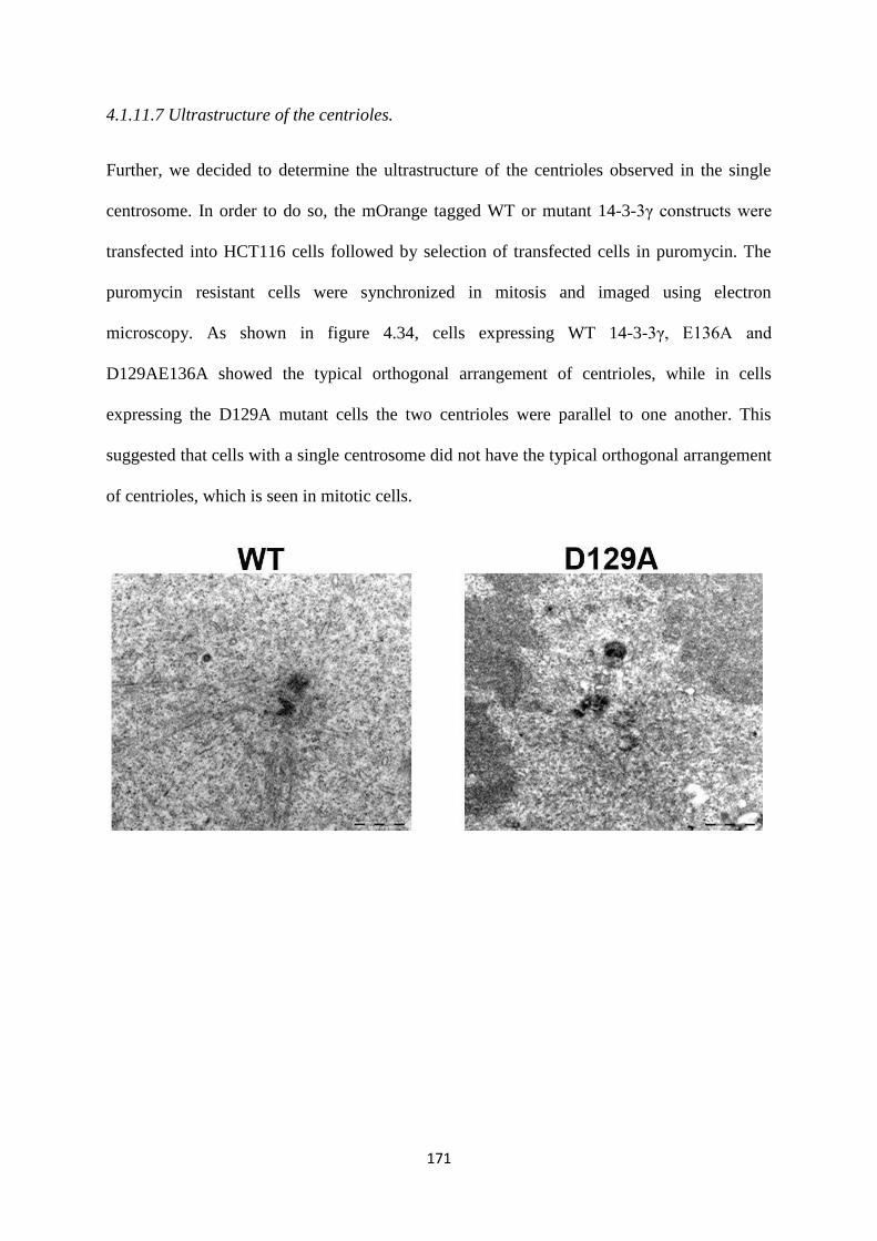
171
4.1.11.7 Ultrastructure of the centrioles.
Further, we decided to determine the ultrastructure of the centrioles observed in the single
centrosome. In order to do so, the mOrange tagged WT or mutant 14-3-3γ constructs were
transfected into HCT116 cells followed by selection of transfected cells in puromycin. The
puromycin resistant cells were synchronized in mitosis and imaged using electron
microscopy. As shown in figure 4.34, cells expressing WT 14-3-3γ, E136A and
D129AE136A showed the typical orthogonal arrangement of centrioles, while in cells
expressing the D129A mutant cells the two centrioles were parallel to one another. This
suggested that cells with a single centrosome did not have the typical orthogonal arrangement
of centrioles, which is seen in mitotic cells.
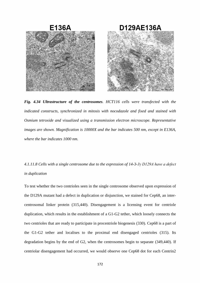
172
Fig. 4.34 Ultrastructure of the centrosomes. HCT116 cells were transfected with the
indicated constructs, synchronized in mitosis with nocodazole and fixed and stained with
Osmium tetroxide and visualized using a transmission electron microscope. Representative
images are shown. Magnification is 10000X and the bar indicates 500 nm, except in E136A,
where the bar indicates 1000 nm.
4.1.11.8 Cells with a single centrosome due to the expression of 14-3-3γ D129A have a defect
in duplication
To test whether the two centrioles seen in the single centrosome observed upon expression of
the D129A mutant had a defect in duplication or disjunction, we stained for Cep68, an inter-
centrosomal linker protein (315,440). Disengagement is a licensing event for centriole
duplication, which results in the establishment of a G1-G2 tether, which loosely connects the
two centrioles that are ready to participate in procentriole biogenesis (330). Cep68 is a part of
the G1-G2 tether and localises to the proximal end disengaged centrioles (315). Its
degradation begins by the end of G2, when the centrosomes begin to separate (349,440). If
centriolar disengagement had occurred, we would observe one Cep68 dot for each Centrin2
173
dot, in a 1:1 ratio. HCT116 cells were co-tranfected with each of the mOrange constructs and
GFP Centrin2. 48 hours post transfection and synchronization in G2 with RO3306 (a CDK1
inhibitor), we fixed and stained the cells with antibodies against Cep68 and counterstained for
DNA with DAPI (411,441). We observed that in cells expressing the WT, E136A and the
D129AE136A mutant, that each contained 4 centrioles, there was a 2:1 ratio for
Centrin2:Cep68 signal; 2 pairs of Centrin2 dots each with one Cep68 dot (fig. 4.36). In cells
expressing the D129A mutant, with a single centrosome, there was a 1:1 ratio for
Centrin2:Cep68 staining; each Centrin2 dot was associated with a Cep68 dot (fig. 4.35). This
suggests that the two centrioles observed in the cells expressing the D129A mutant had
undergone disengagement, but were unable to initiate duplication.
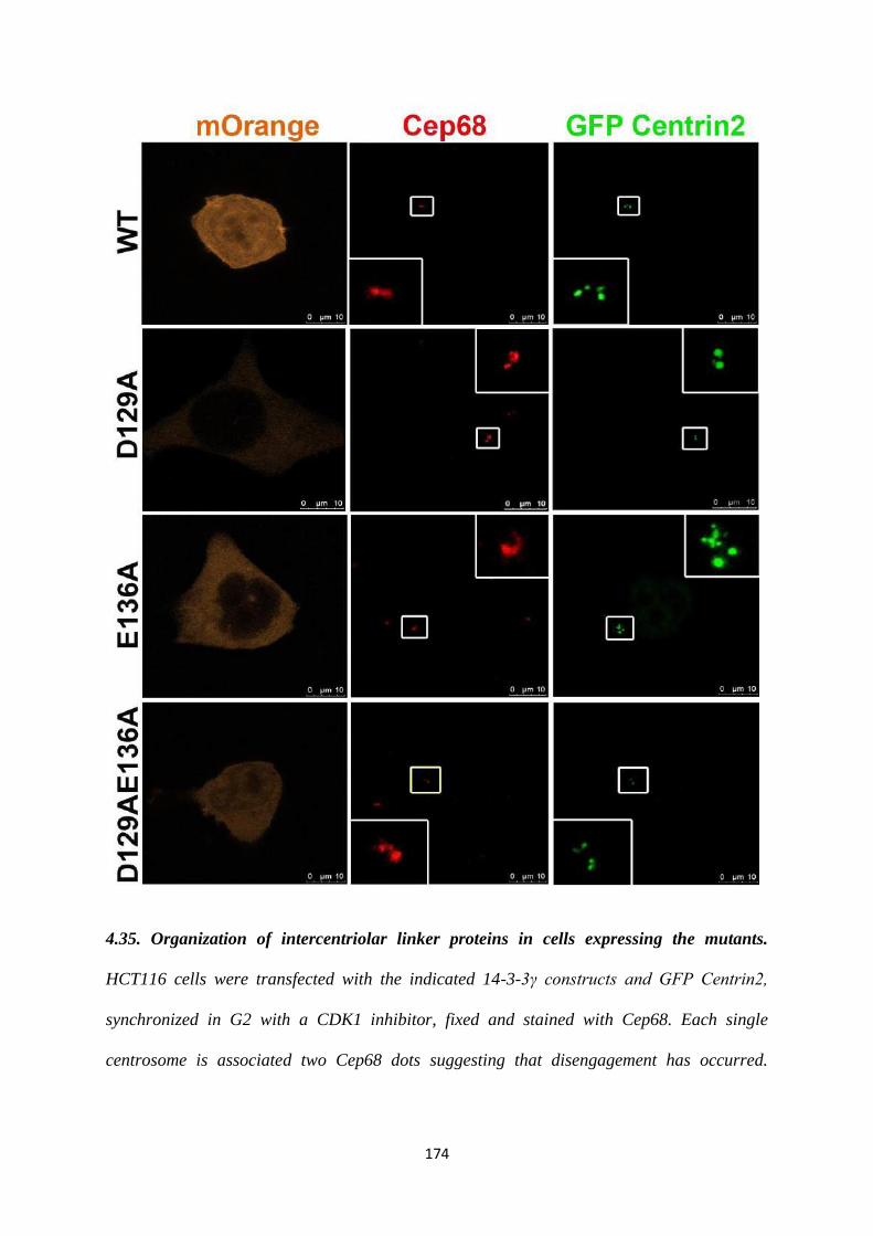
174
4.35. Organization of intercentriolar linker proteins in cells expressing the mutants.
HCT116 cells were transfected with the indicated 14-3-3γ constructs and GFP Centrin2,
synchronized in G2 with a CDK1 inhibitor, fixed and stained with Cep68. Each single
centrosome is associated two Cep68 dots suggesting that disengagement has occurred.
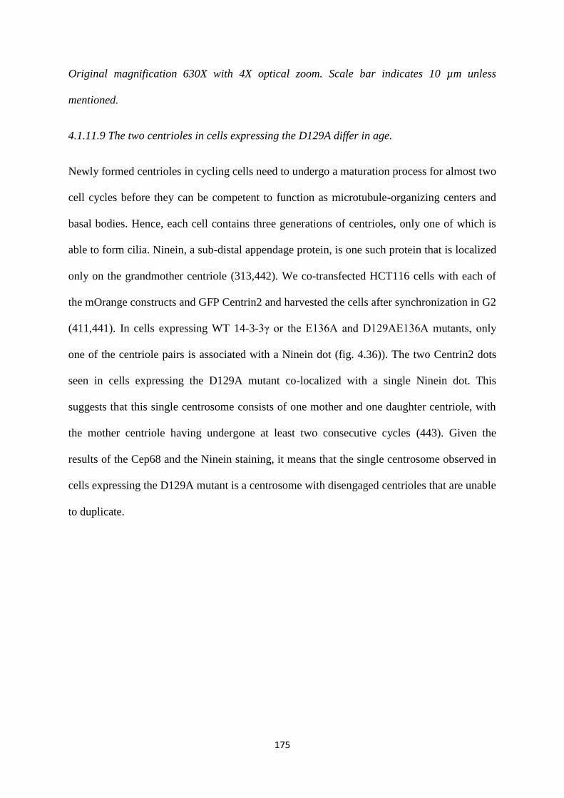
175
Original magnification 630X with 4X optical zoom. Scale bar indicates 10 µm unless
mentioned.
4.1.11.9 The two centrioles in cells expressing the D129A differ in age.
Newly formed centrioles in cycling cells need to undergo a maturation process for almost two
cell cycles before they can be competent to function as microtubule-organizing centers and
basal bodies. Hence, each cell contains three generations of centrioles, only one of which is
able to form cilia. Ninein, a sub-distal appendage protein, is one such protein that is localized
only on the grandmother centriole (313,442). We co-transfected HCT116 cells with each of
the mOrange constructs and GFP Centrin2 and harvested the cells after synchronization in G2
(411,441). In cells expressing WT 14-3-3γ or the E136A and D129AE136A mutants, only
one of the centriole pairs is associated with a Ninein dot (fig. 4.36)). The two Centrin2 dots
seen in cells expressing the D129A mutant co-localized with a single Ninein dot. This
suggests that this single centrosome consists of one mother and one daughter centriole, with
the mother centriole having undergone at least two consecutive cycles (443). Given the
results of the Cep68 and the Ninein staining, it means that the single centrosome observed in
cells expressing the D129A mutant is a centrosome with disengaged centrioles that are unable
to duplicate.
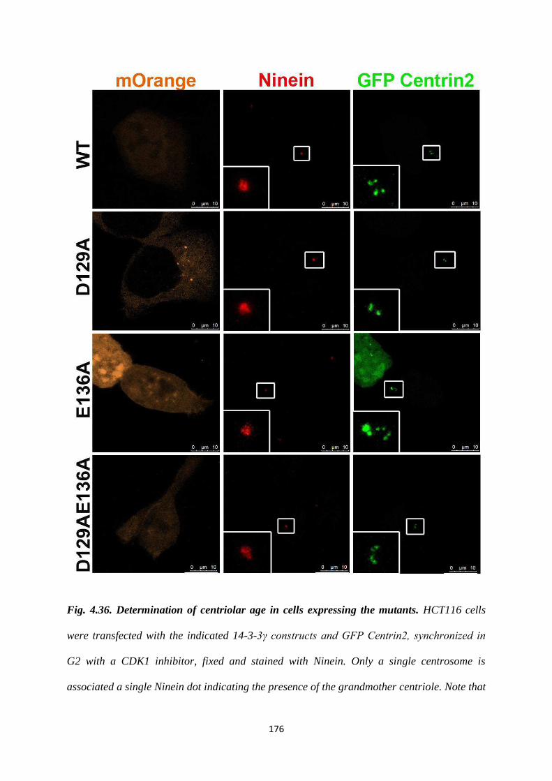
176
Fig. 4.36. Determination of centriolar age in cells expressing the mutants. HCT116 cells
were transfected with the indicated 14-3-3γ constructs and GFP Centrin2, synchronized in
G2 with a CDK1 inhibitor, fixed and stained with Ninein. Only a single centrosome is
associated a single Ninein dot indicating the presence of the grandmother centriole. Note that
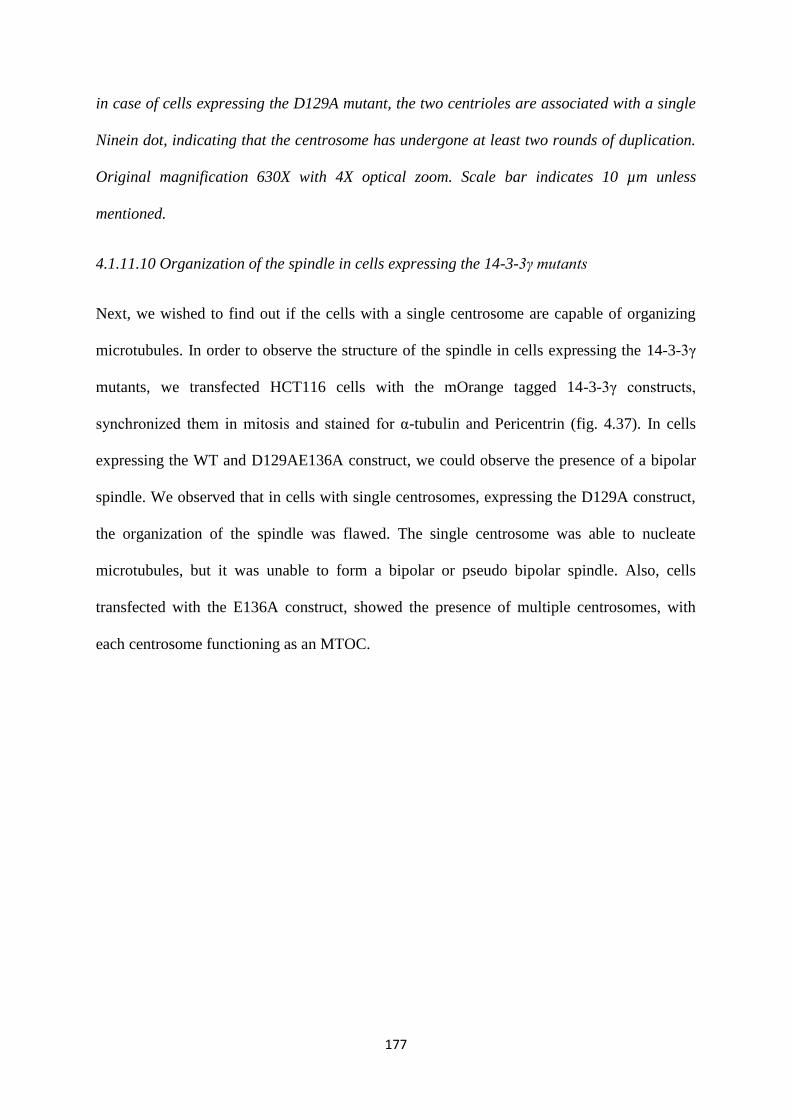
177
in case of cells expressing the D129A mutant, the two centrioles are associated with a single
Ninein dot, indicating that the centrosome has undergone at least two rounds of duplication.
Original magnification 630X with 4X optical zoom. Scale bar indicates 10 µm unless
mentioned.
4.1.11.10 Organization of the spindle in cells expressing the 14-3-3γ mutants
Next, we wished to find out if the cells with a single centrosome are capable of organizing
microtubules. In order to observe the structure of the spindle in cells expressing the 14-3-3γ
mutants, we transfected HCT116 cells with the mOrange tagged 14-3-3γ constructs,
synchronized them in mitosis and stained for α-tubulin and Pericentrin (fig. 4.37). In cells
expressing the WT and D129AE136A construct, we could observe the presence of a bipolar
spindle. We observed that in cells with single centrosomes, expressing the D129A construct,
the organization of the spindle was flawed. The single centrosome was able to nucleate
microtubules, but it was unable to form a bipolar or pseudo bipolar spindle. Also, cells
transfected with the E136A construct, showed the presence of multiple centrosomes, with
each centrosome functioning as an MTOC.
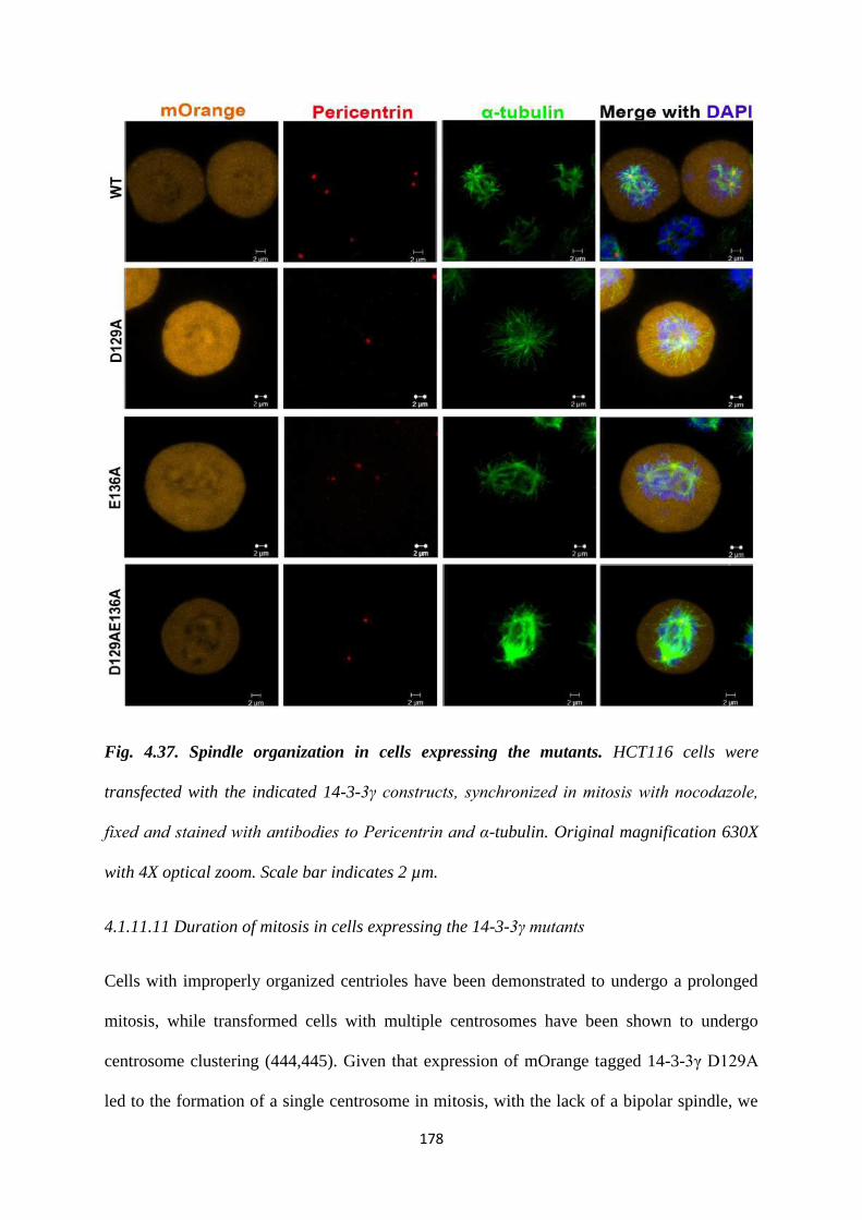
178
Fig. 4.37. Spindle organization in cells expressing the mutants. HCT116 cells were
transfected with the indicated 14-3-3γ constructs, synchronized in mitosis with nocodazole,
fixed and stained with antibodies to Pericentrin and α-tubulin. Original magnification 630X
with 4X optical zoom. Scale bar indicates 2 µm.
4.1.11.11 Duration of mitosis in cells expressing the 14-3-3γ mutants
Cells with improperly organized centrioles have been demonstrated to undergo a prolonged
mitosis, while transformed cells with multiple centrosomes have been shown to undergo
centrosome clustering (444,445). Given that expression of mOrange tagged 14-3-3γ D129A
led to the formation of a single centrosome in mitosis, with the lack of a bipolar spindle, we
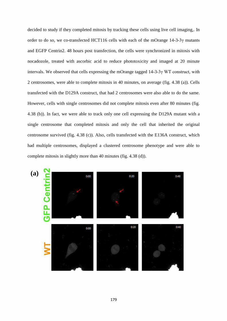
179
decided to study if they completed mitosis by tracking these cells using live cell imaging,. In
order to do so, we co-transfected HCT116 cells with each of the mOrange 14-3-3γ mutants
and EGFP Centrin2. 48 hours post transfection, the cells were synchronized in mitosis with
nocadozole, treated with ascorbic acid to reduce phototoxicity and imaged at 20 minute
intervals. We observed that cells expressing the mOrange tagged 14-3-3γ WT construct, with
2 centrosomes, were able to complete mitosis in 40 minutes, on average (fig. 4.38 (a)). Cells
transfected with the D129A construct, that had 2 centrosomes were also able to do the same.
However, cells with single centrosomes did not complete mitosis even after 80 minutes (fig.
4.38 (b)). In fact, we were able to track only one cell expressing the D129A mutant with a
single centrosome that completed mitosis and only the cell that inherited the original
centrosome survived (fig. 4.38 (c)). Also, cells transfected with the E136A construct, which
had multiple centrosomes, displayed a clustered centrosome phenotype and were able to
complete mitosis in slightly more than 40 minutes (fig. 4.38 (d)).
(a)
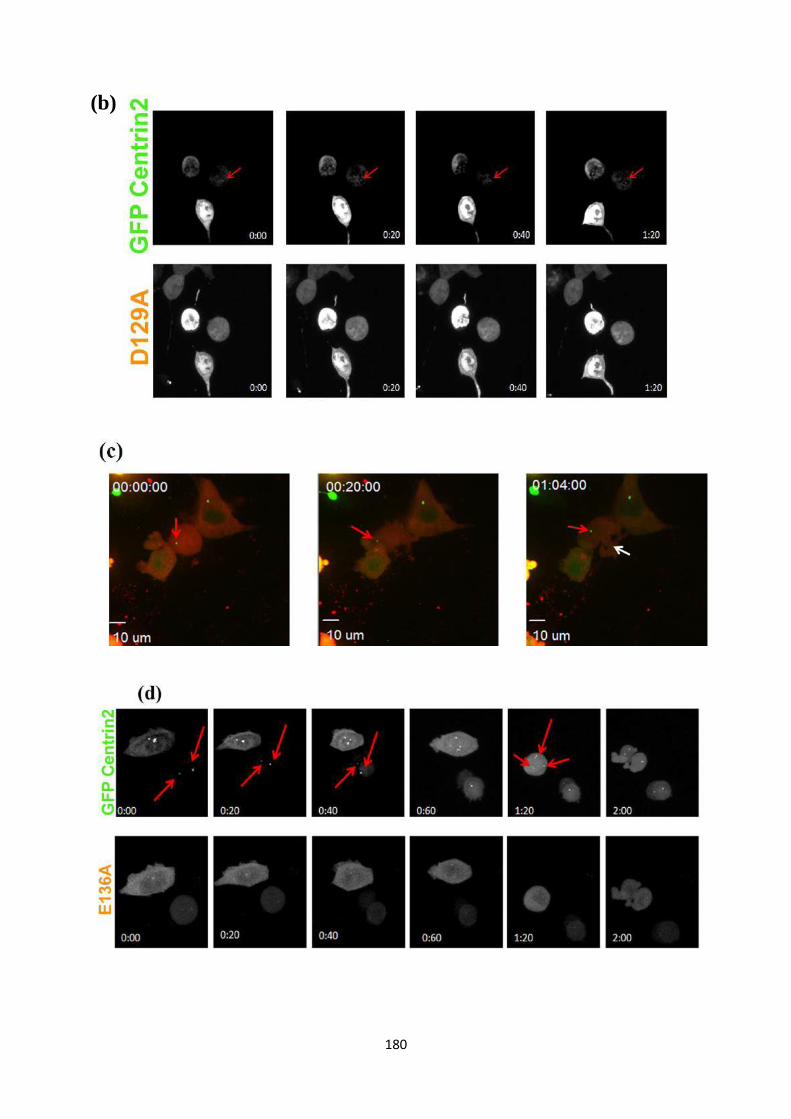
180
(b)
181
Fig. 4.38. Duration of mitosis in cells expressing the mutants. HCT116 cells transfected
with the indicated constructs and GFP Centrin2 were synchronized in mitosis with
nocodazole. After washing out the nocodazole the cells were imaged using live cell
microscopy as described in materials and methods. Note that cells expressing D129A fail to
complete cell division. Arrows indicate transfected cells. Original magnification 630X with
4X optical zoom. Scale bar indicates 1 µm unless mentioned.
4.1.11.12 Localization of the 14-3-3 mutants in interphase
Previous reports indicate that a 14-3-3ε mutant that was defective for ligand binding
accumulated in the nucleus (200). We wished to determine if we could infer anything about
the difference in phenotypes seen upon expression of these 14-3-3γ mutants based on their
localization. In order to determine if the centrosome phenotype was due to a difference in
cellular localization of each of the mutants, we transfected each of the 14-3-3γ constructs into
HCT116 cells and synchronized with mimosine. We harvested and fixed the cells at different
stages of the cell cycle, to analyze if there was a difference in the cellular localization of the
mutants as compared to the wild type, and if the difference is dependent on the stage of the
cell cycle. We observed that as opposed to the 14-3-3γ WT, which had a pan cellular
localization, each of the mutants; D129A, E136A and D129AE136A had a predominantly
cytoplasmic localization during all stages of the cell cycle. Hence, we can conclude that the
cellular localization of the mutants probably does not affect the centrosome cycle (fig. 4.39).
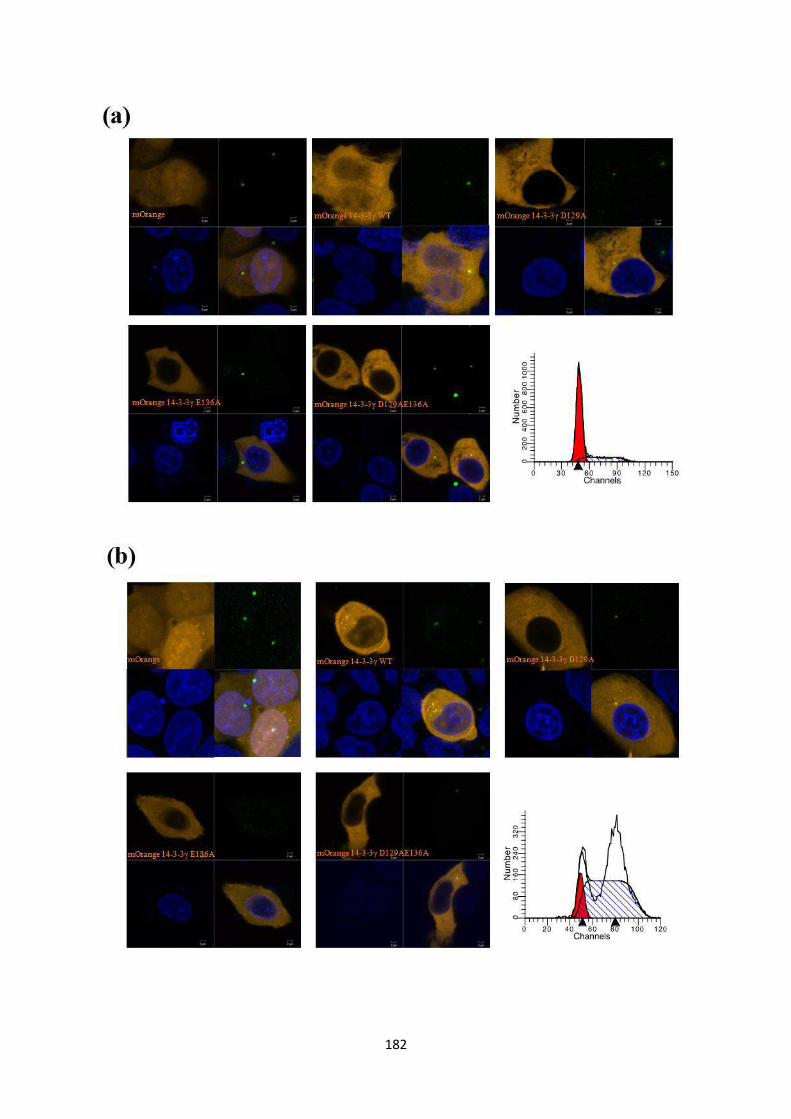
182
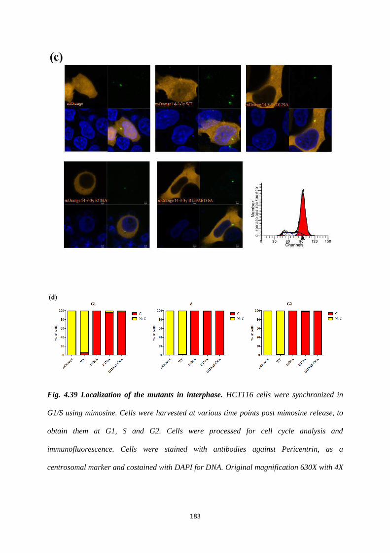
183
Fig. 4.39 Localization of the mutants in interphase. HCT116 cells were synchronized in
G1/S using mimosine. Cells were harvested at various time points post mimosine release, to
obtain them at G1, S and G2. Cells were processed for cell cycle analysis and
immunofluorescence. Cells were stained with antibodies against Pericentrin, as a
centrosomal marker and costained with DAPI for DNA. Original magnification 630X with 4X
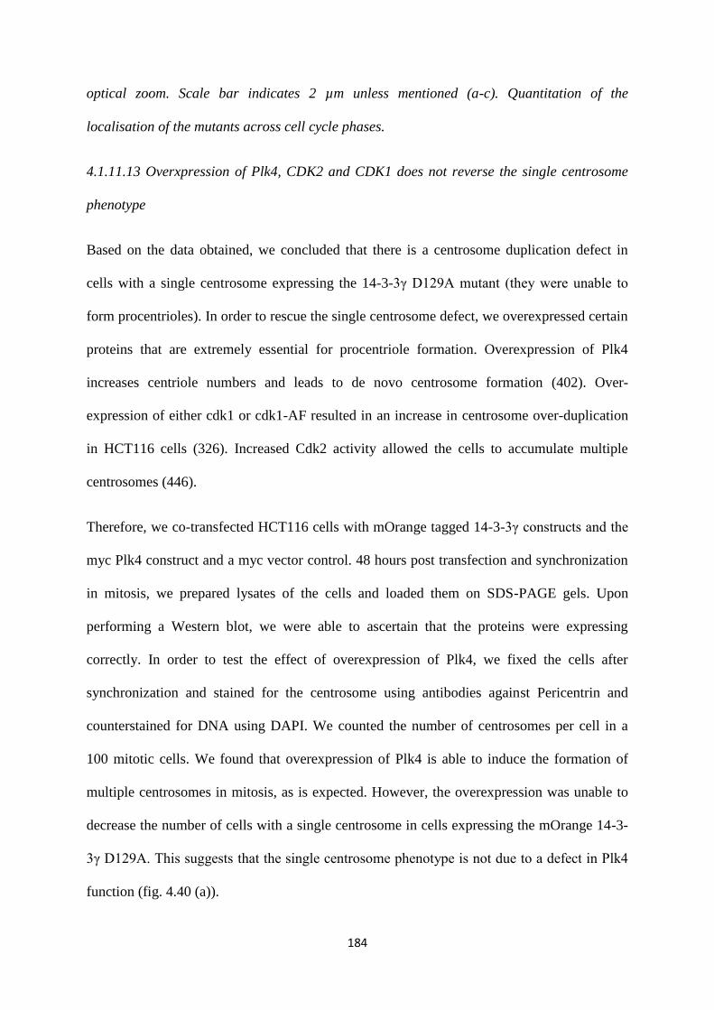
184
optical zoom. Scale bar indicates 2 µm unless mentioned (a-c). Quantitation of the
localisation of the mutants across cell cycle phases.
4.1.11.13 Overxpression of Plk4, CDK2 and CDK1 does not reverse the single centrosome
phenotype
Based on the data obtained, we concluded that there is a centrosome duplication defect in
cells with a single centrosome expressing the 14-3-3γ D129A mutant (they were unable to
form procentrioles). In order to rescue the single centrosome defect, we overexpressed certain
proteins that are extremely essential for procentriole formation. Overexpression of Plk4
increases centriole numbers and leads to de novo centrosome formation (402). Over-
expression of either cdk1 or cdk1-AF resulted in an increase in centrosome over-duplication
in HCT116 cells (326). Increased Cdk2 activity allowed the cells to accumulate multiple
centrosomes (446).
Therefore, we co-transfected HCT116 cells with mOrange tagged 14-3-3γ constructs and the
myc Plk4 construct and a myc vector control. 48 hours post transfection and synchronization
in mitosis, we prepared lysates of the cells and loaded them on SDS-PAGE gels. Upon
performing a Western blot, we were able to ascertain that the proteins were expressing
correctly. In order to test the effect of overexpression of Plk4, we fixed the cells after
synchronization and stained for the centrosome using antibodies against Pericentrin and
counterstained for DNA using DAPI. We counted the number of centrosomes per cell in a
100 mitotic cells. We found that overexpression of Plk4 is able to induce the formation of
multiple centrosomes in mitosis, as is expected. However, the overexpression was unable to
decrease the number of cells with a single centrosome in cells expressing the mOrange 14-3-
3γ D129A. This suggests that the single centrosome phenotype is not due to a defect in Plk4
function (fig. 4.40 (a)).
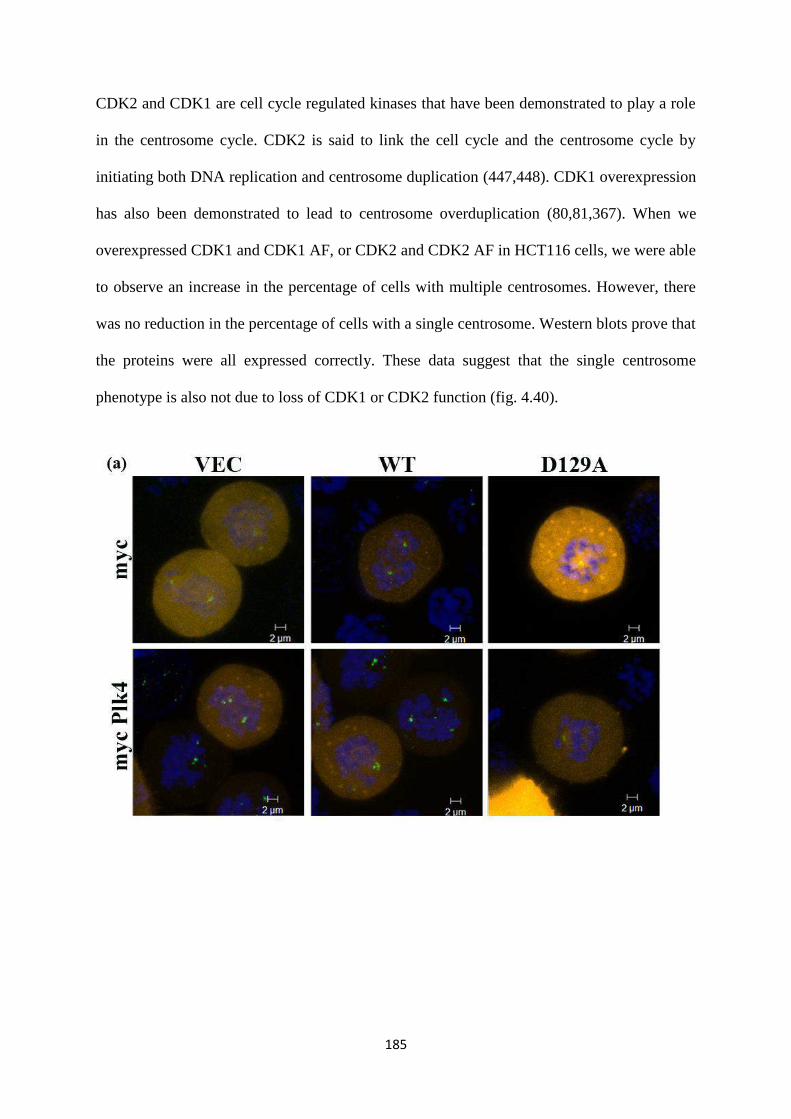
185
CDK2 and CDK1 are cell cycle regulated kinases that have been demonstrated to play a role
in the centrosome cycle. CDK2 is said to link the cell cycle and the centrosome cycle by
initiating both DNA replication and centrosome duplication (447,448). CDK1 overexpression
has also been demonstrated to lead to centrosome overduplication (80,81,367). When we
overexpressed CDK1 and CDK1 AF, or CDK2 and CDK2 AF in HCT116 cells, we were able
to observe an increase in the percentage of cells with multiple centrosomes. However, there
was no reduction in the percentage of cells with a single centrosome. Western blots prove that
the proteins were all expressed correctly. These data suggest that the single centrosome
phenotype is also not due to loss of CDK1 or CDK2 function (fig. 4.40).
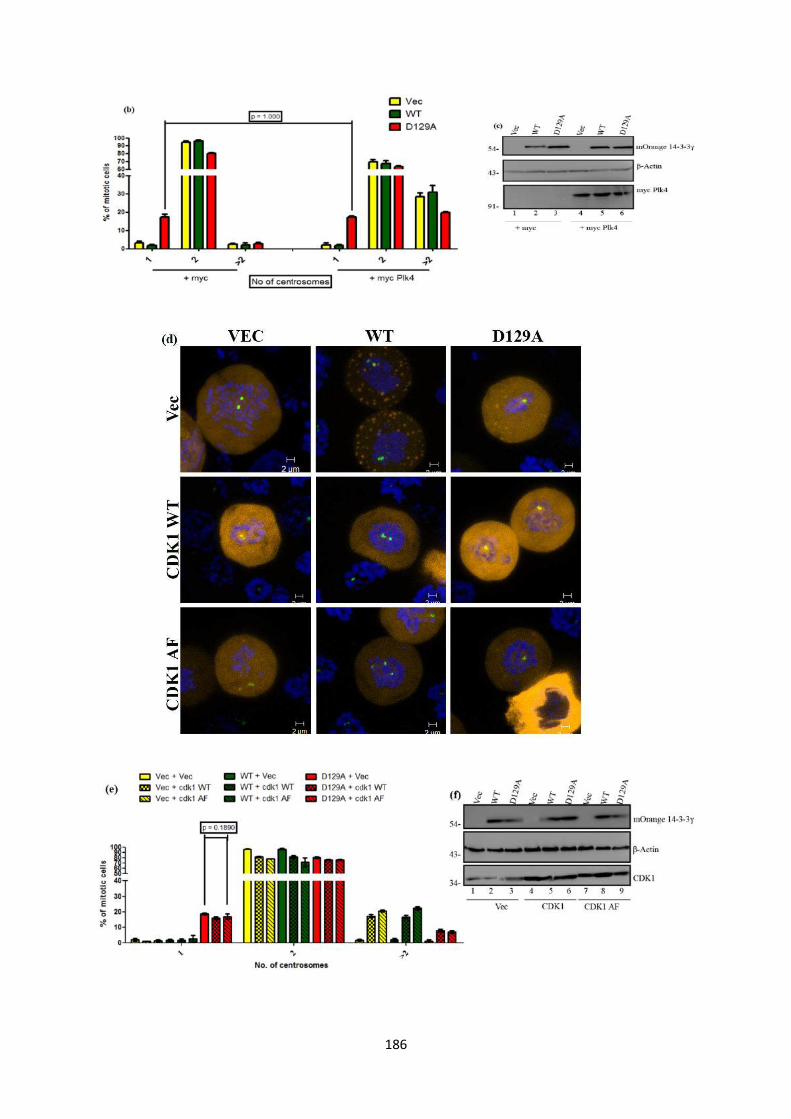
186
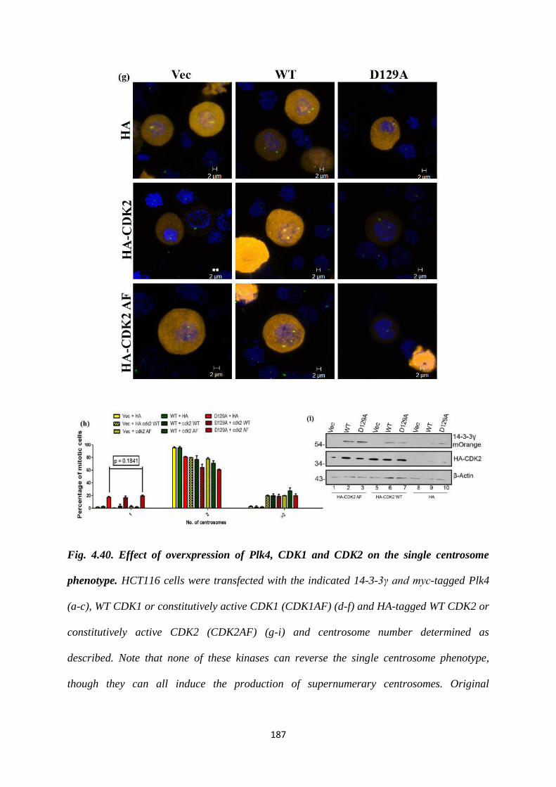
187
Fig. 4.40. Effect of overxpression of Plk4, CDK1 and CDK2 on the single centrosome
phenotype. HCT116 cells were transfected with the indicated 14-3-3γ and myc-tagged Plk4
(a-c), WT CDK1 or constitutively active CDK1 (CDK1AF) (d-f) and HA-tagged WT CDK2 or
constitutively active CDK2 (CDK2AF) (g-i) and centrosome number determined as
described. Note that none of these kinases can reverse the single centrosome phenotype,
though they can all induce the production of supernumerary centrosomes. Original
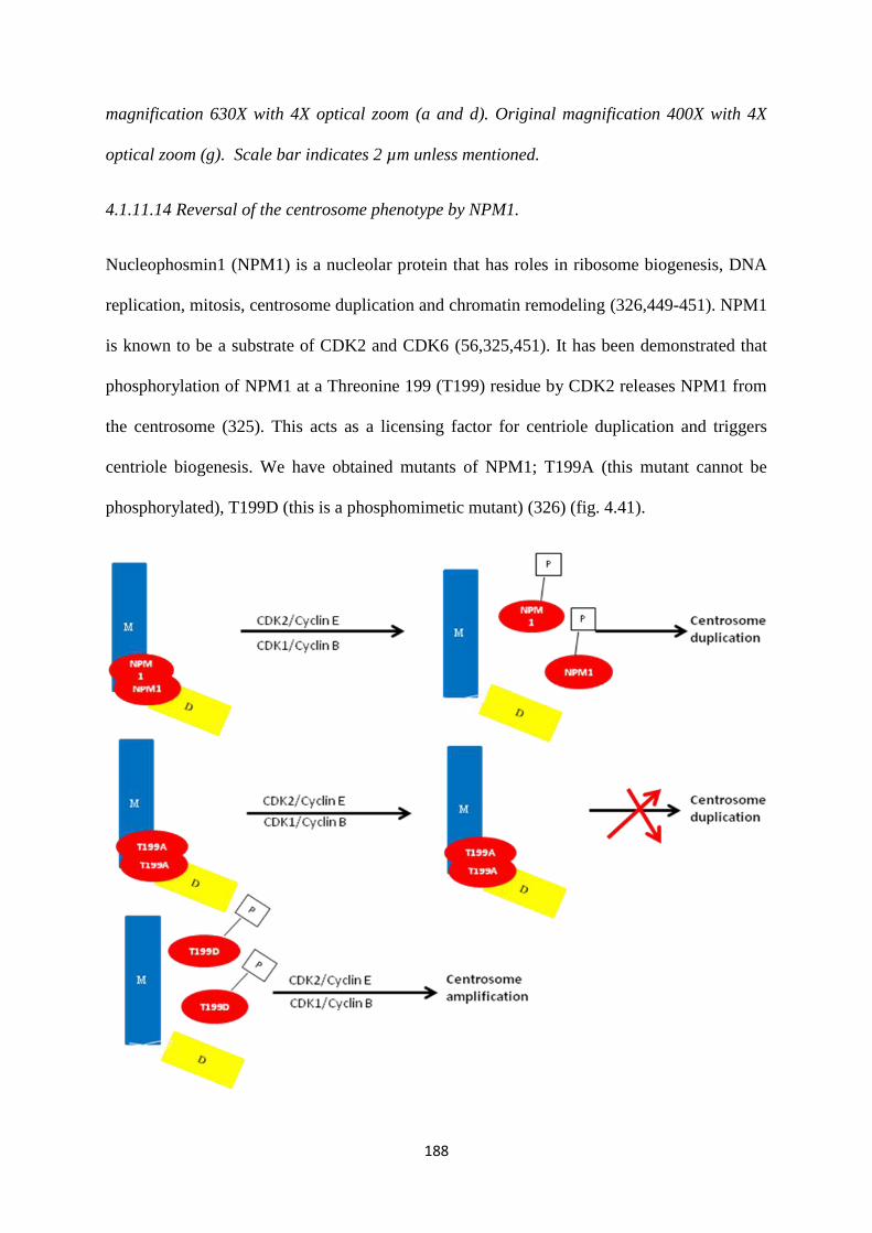
188
magnification 630X with 4X optical zoom (a and d). Original magnification 400X with 4X
optical zoom (g). Scale bar indicates 2 µm unless mentioned.
4.1.11.14 Reversal of the centrosome phenotype by NPM1.
Nucleophosmin1 (NPM1) is a nucleolar protein that has roles in ribosome biogenesis, DNA
replication, mitosis, centrosome duplication and chromatin remodeling (326,449-451). NPM1
is known to be a substrate of CDK2 and CDK6 (56,325,451). It has been demonstrated that
phosphorylation of NPM1 at a Threonine 199 (T199) residue by CDK2 releases NPM1 from
the centrosome (325). This acts as a licensing factor for centriole duplication and triggers
centriole biogenesis. We have obtained mutants of NPM1; T199A (this mutant cannot be
phosphorylated), T199D (this is a phosphomimetic mutant) (326) (fig. 4.41).
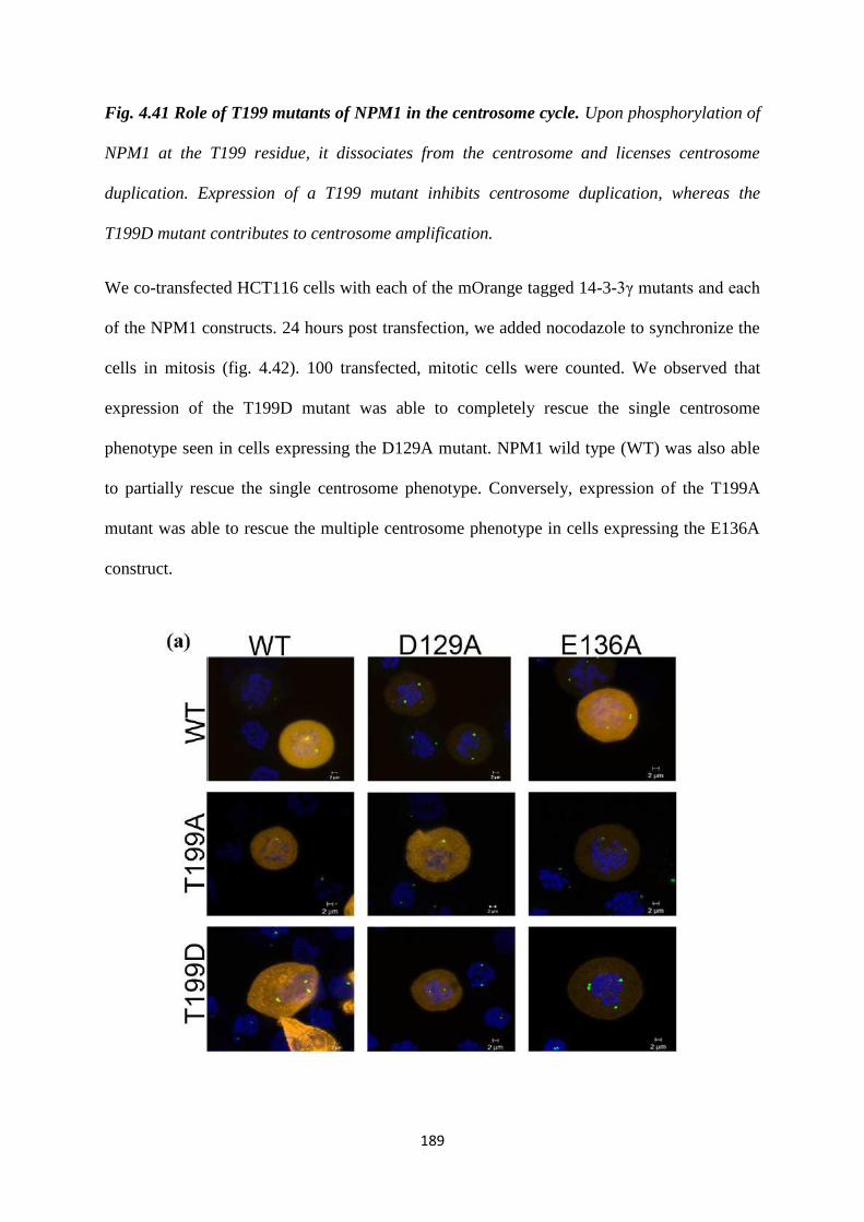
189
Fig. 4.41 Role of T199 mutants of NPM1 in the centrosome cycle. Upon phosphorylation of
NPM1 at the T199 residue, it dissociates from the centrosome and licenses centrosome
duplication. Expression of a T199 mutant inhibits centrosome duplication, whereas the
T199D mutant contributes to centrosome amplification.
We co-transfected HCT116 cells with each of the mOrange tagged 14-3-3γ mutants and each
of the NPM1 constructs. 24 hours post transfection, we added nocodazole to synchronize the
cells in mitosis (fig. 4.42). 100 transfected, mitotic cells were counted. We observed that
expression of the T199D mutant was able to completely rescue the single centrosome
phenotype seen in cells expressing the D129A mutant. NPM1 wild type (WT) was also able
to partially rescue the single centrosome phenotype. Conversely, expression of the T199A
mutant was able to rescue the multiple centrosome phenotype in cells expressing the E136A
construct.
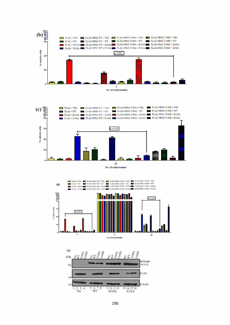
190
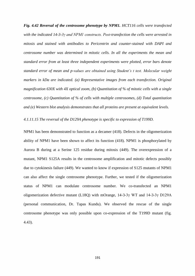
191
Fig. 4.42 Reversal of the centrosome phenotype by NPM1. HCT116 cells were transfected
with the indicated 14-3-3γ and NPM1 constructs. Post-transfection the cells were arrested in
mitosis and stained with antibodies to Pericentrin and counter-stained with DAPI and
centrosome number was determined in mitotic cells. In all the experiments the mean and
standard error from at least three independent experiments were plotted, error bars denote
standard error of mean and p-values are obtained using Student’s t test. Molecular weight
markers in kDa are indicated. (a) Representative images from each transfection. Original
magnification 630X with 4X optical zoom, (b) Quantitation of % of mitotic cells with a single
centrosome, (c) Quantitation of % of cells with multiple centrosomes, (d) Total quantitation
and (e) Western blot analysis demonstrates that all proteins are present at equivalent levels.
4.1.11.15 The reversal of the D129A phenotype is specific to expression of T199D.
NPM1 has been demonstrated to function as a decamer (418). Defects in the oligomerization
ability of NPM1 have been shown to affect its function (418). NPM1 is phosphorylated by
Aurora B during at a Serine 125 residue during mitosis (449). The overexpression of a
mutant, NPM1 S125A results in the centrosome amplification and mitotic defects possibly
due to cytokinesis failure (449). We wanted to know if expression of S125 mutants of NPM1
can also affect the single centrosome phenotype. Further, we tested if the oligomerization
status of NPM1 can modulate centrosome number. We co-transfected an NPM1
oligomerization defective mutant (L18Q) with mOrange, 14-3-3γ WT and 14-3-3γ D129A
(personal communication, Dr. Tapas Kundu). We observed the rescue of the single
centrosome phenotype was only possible upon co-expression of the T199D mutant (fig.
4.43).
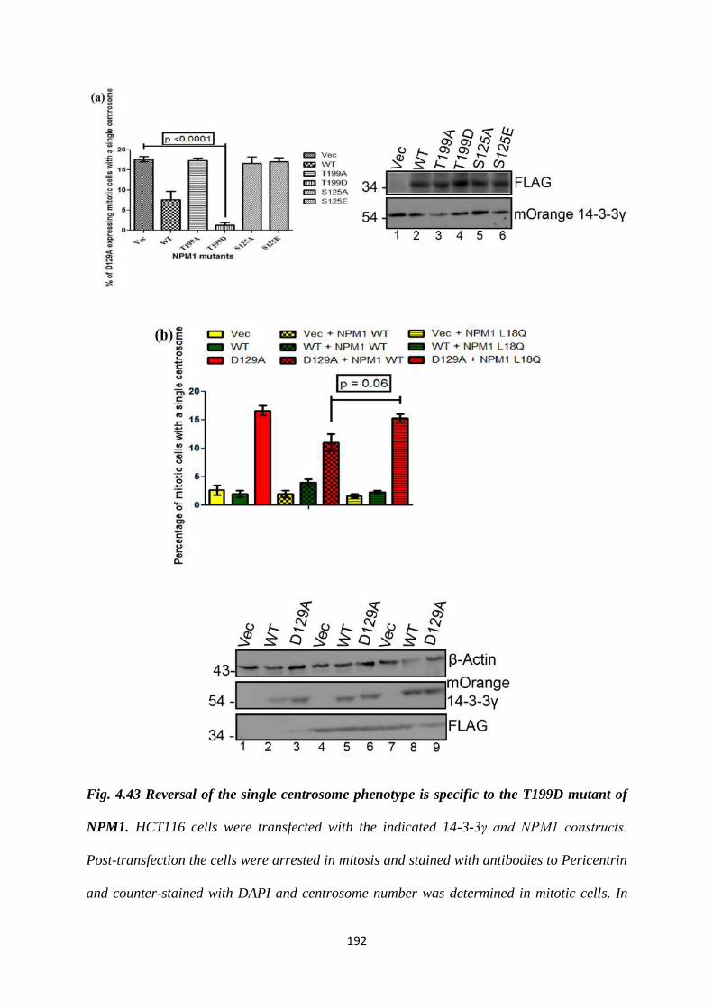
192
Fig. 4.43 Reversal of the single centrosome phenotype is specific to the T199D mutant of
NPM1. HCT116 cells were transfected with the indicated 14-3-3γ and NPM1 constructs.
Post-transfection the cells were arrested in mitosis and stained with antibodies to Pericentrin
and counter-stained with DAPI and centrosome number was determined in mitotic cells. In
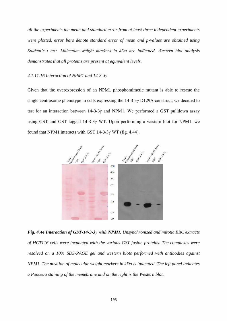
193
all the experiments the mean and standard error from at least three independent experiments
were plotted, error bars denote standard error of mean and p-values are obtained using
Student’s t test. Molecular weight markers in kDa are indicated. Western blot analysis
demonstrates that all proteins are present at equivalent levels.
4.1.11.16 Interaction of NPM1 and 14-3-3γ
Given that the overexpression of an NPM1 phosphomimetic mutant is able to rescue the
single centrosome phenotype in cells expressing the 14-3-3γ D129A construct, we decided to
test for an interaction between 14-3-3γ and NPM1. We performed a GST pulldown assay
using GST and GST tagged 14-3-3γ WT. Upon performing a western blot for NPM1, we
found that NPM1 interacts with GST 14-3-3γ WT (fig. 4.44).
Fig. 4.44 Interaction of GST-14-3-3γ with NPM1. Unsynchronized and mitotic EBC extracts
of HCT116 cells were incubated with the various GST fusion proteins. The complexes were
resolved on a 10% SDS-PAGE gel and western blots performed with antibodies against
NPM1. The position of molecular weight markers in kDa is indicated. The left panel indicates
a Ponceau staining of the memebrane and on the right is the Western blot.
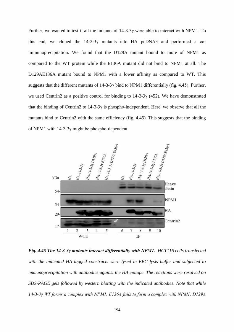
194
Further, we wanted to test if all the mutants of 14-3-3γ were able to interact with NPM1. To
this end, we cloned the 14-3-3γ mutants into HA pcDNA3 and performed a co-
immunoprecipitation. We found that the D129A mutant bound to more of NPM1 as
compared to the WT protein while the E136A mutant did not bind to NPM1 at all. The
D129AE136A mutant bound to NPM1 with a lower affinity as compared to WT. This
suggests that the different mutants of 14-3-3γ bind to NPM1 differentially (fig. 4.45). Further,
we used Centrin2 as a positive control for binding to 14-3-3γ (452). We have demonstrated
that the binding of Centrin2 to 14-3-3γ is phospho-independent. Here, we observe that all the
mutants bind to Centrin2 with the same efficiency (fig. 4.45). This suggests that the binding
of NPM1 with 14-3-3γ might be phospho-dependent.
Fig. 4.45 The 14-3-3γ mutants interact differentially with NPM1. HCT116 cells transfected
with the indicated HA tagged constructs were lysed in EBC lysis buffer and subjected to
immunoprecipitation with antibodies against the HA epitope. The reactions were resolved on
SDS-PAGE gels followed by western blotting with the indicated antibodies. Note that while
14-3-3γ WT forms a complex with NPM1, E136A fails to form a complex with NPM1. D129A
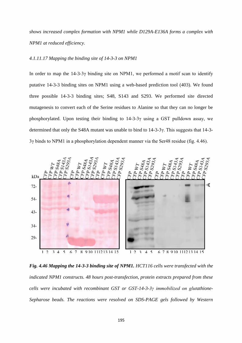
195
shows increased complex formation with NPM1 while D129A-E136A forms a complex with
NPM1 at reduced efficiency.
4.1.11.17 Mapping the binding site of 14-3-3 on NPM1
In order to map the 14-3-3γ binding site on NPM1, we performed a motif scan to identify
putative 14-3-3 binding sites on NPM1 using a web-based prediction tool (403). We found
three possible 14-3-3 binding sites; S48, S143 and S293. We performed site directed
mutagenesis to convert each of the Serine residues to Alanine so that they can no longer be
phosphorylated. Upon testing their binding to 14-3-3γ using a GST pulldown assay, we
determined that only the S48A mutant was unable to bind to 14-3-3γ. This suggests that 14-3-
3γ binds to NPM1 in a phosphorylation dependent manner via the Ser48 residue (fig. 4.46).
Fig. 4.46 Mapping the 14-3-3 binding site of NPM1. HCT116 cells were transfected with the
indicated NPM1 constructs. 48 hours post-transfection, protein extracts prepared from these
cells were incubated with recombinant GST or GST-14-3-3γ immobilized on glutathione-
Sepharose beads. The reactions were resolved on SDS-PAGE gels followed by Western
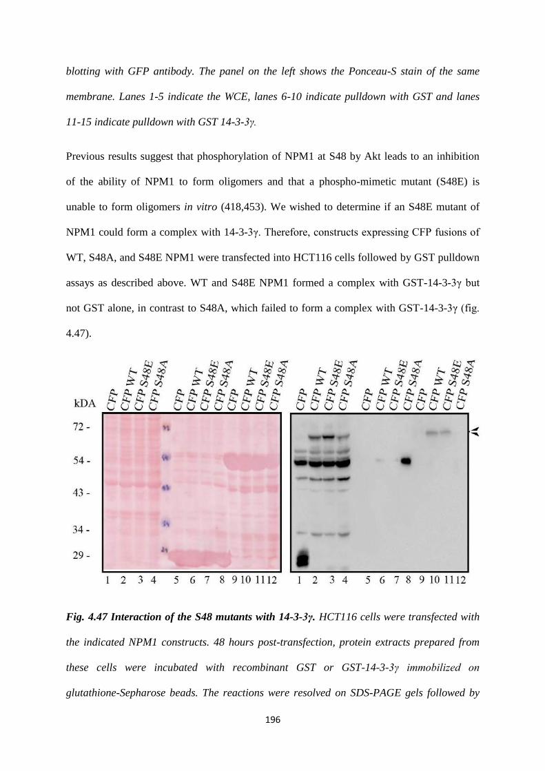
196
blotting with GFP antibody. The panel on the left shows the Ponceau-S stain of the same
membrane. Lanes 1-5 indicate the WCE, lanes 6-10 indicate pulldown with GST and lanes
11-15 indicate pulldown with GST 14-3-3γ.
Previous results suggest that phosphorylation of NPM1 at S48 by Akt leads to an inhibition
of the ability of NPM1 to form oligomers and that a phospho-mimetic mutant (S48E) is
unable to form oligomers in vitro (418,453). We wished to determine if an S48E mutant of
NPM1 could form a complex with 14-3-3γ. Therefore, constructs expressing CFP fusions of
WT, S48A, and S48E NPM1 were transfected into HCT116 cells followed by GST pulldown
assays as described above. WT and S48E NPM1 formed a complex with GST-14-3-3γ but
not GST alone, in contrast to S48A, which failed to form a complex with GST-14-3-3γ (fig.
4.47).
Fig. 4.47 Interaction of the S48 mutants with 14-3-3γ. HCT116 cells were transfected with
the indicated NPM1 constructs. 48 hours post-transfection, protein extracts prepared from
these cells were incubated with recombinant GST or GST-14-3-3γ immobilized on
glutathione-Sepharose beads. The reactions were resolved on SDS-PAGE gels followed by
197
Western blotting with GFP antibody. The panel on the left shows the Ponceau-S stain of the
same membrane. Lanes 1-4 indicate the WCE, lanes 5-8 indicate pulldown with GST and
lanes 9-12 indicate pulldown with GST 14-3-3γ. S48A fails to form a complex with 14-3-3γ,
while S48E forms a complex with 14-3-3γ.
4.1.11.18 Localisation of the S48 mutants of NPM1
NPM1 is a nucleolar protein and its localisation is phosphorylation dependent, as it has been
demonstrated that the S48E mutant of NPM1 has a nuclear localisation (453). In order to
confirm the same, we transfected HCT116 cells with CFP, CFP-NPM1 WT and S48 mutants.
48 hours post transfection, we fixed and stained the cells with antibodies against pericentrin
and counterstained for DNA using DAPI. We observed that as shown previously, the NPM1
WT and S48A mutants displayed a nucleolar localization, whereas the S48E mutant had a
nuclear localization. We were unable to observe any NPM1 at the centrosome for any of the
constructs (fig. 4.48).
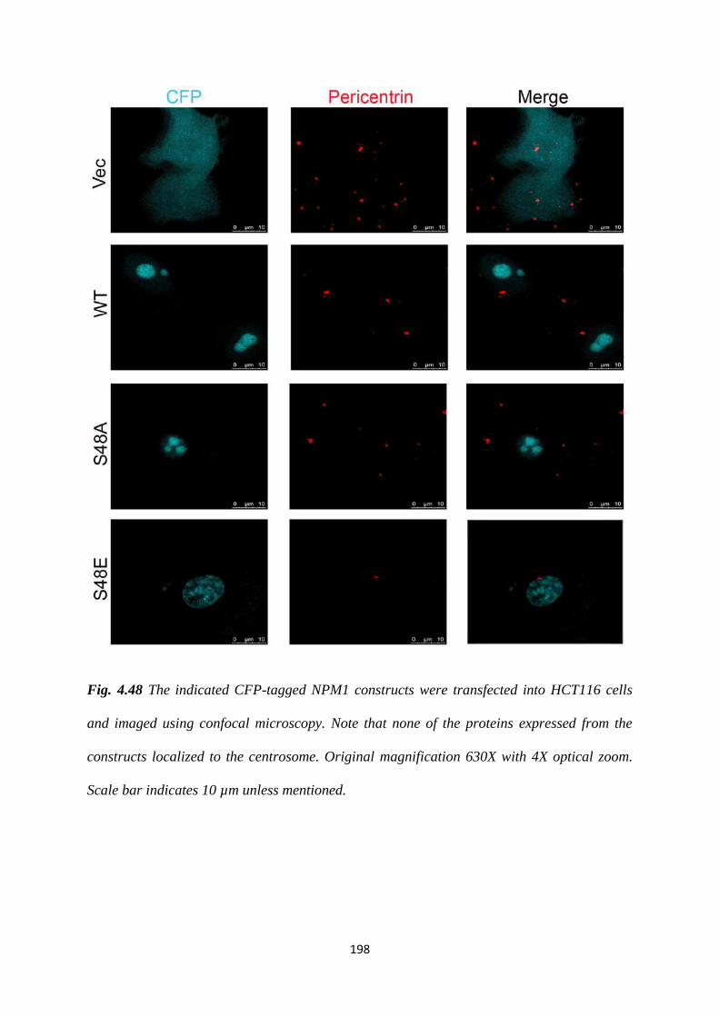
198
Fig. 4.48 The indicated CFP-tagged NPM1 constructs were transfected into HCT116 cells
and imaged using confocal microscopy. Note that none of the proteins expressed from the
constructs localized to the centrosome. Original magnification 630X with 4X optical zoom.
Scale bar indicates 10 µm unless mentioned.
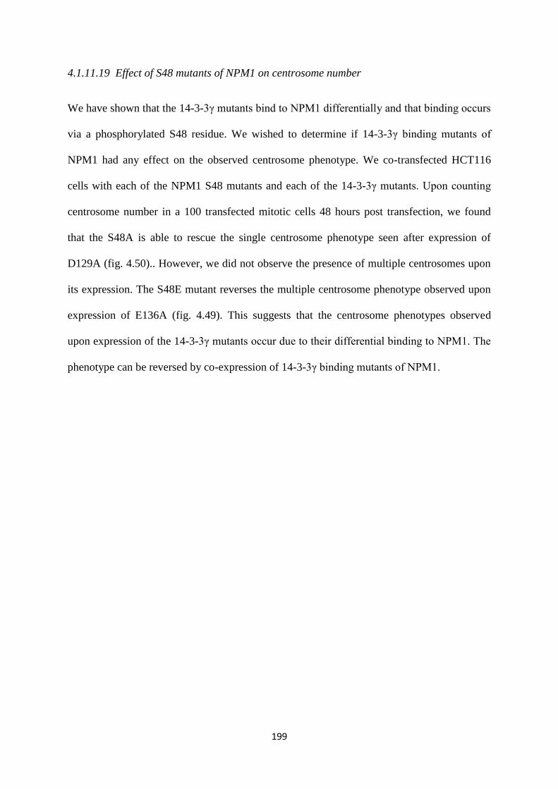
199
4.1.11.19 Effect of S48 mutants of NPM1 on centrosome number
We have shown that the 14-3-3γ mutants bind to NPM1 differentially and that binding occurs
via a phosphorylated S48 residue. We wished to determine if 14-3-3γ binding mutants of
NPM1 had any effect on the observed centrosome phenotype. We co-transfected HCT116
cells with each of the NPM1 S48 mutants and each of the 14-3-3γ mutants. Upon counting
centrosome number in a 100 transfected mitotic cells 48 hours post transfection, we found
that the S48A is able to rescue the single centrosome phenotype seen after expression of
D129A (fig. 4.50).. However, we did not observe the presence of multiple centrosomes upon
its expression. The S48E mutant reverses the multiple centrosome phenotype observed upon
expression of E136A (fig. 4.49). This suggests that the centrosome phenotypes observed
upon expression of the 14-3-3γ mutants occur due to their differential binding to NPM1. The
phenotype can be reversed by co-expression of 14-3-3γ binding mutants of NPM1.

200
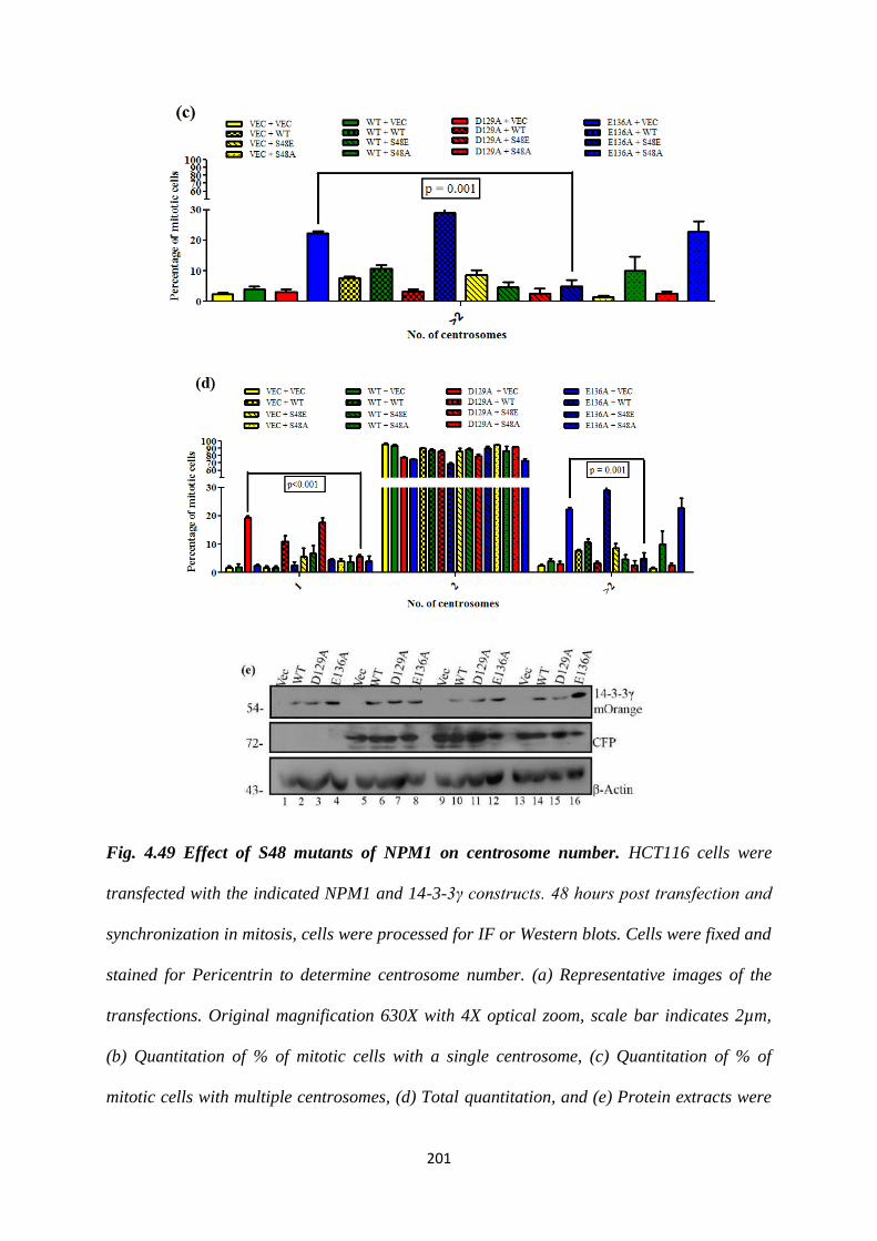
201
Fig. 4.49 Effect of S48 mutants of NPM1 on centrosome number. HCT116 cells were
transfected with the indicated NPM1 and 14-3-3γ constructs. 48 hours post transfection and
synchronization in mitosis, cells were processed for IF or Western blots. Cells were fixed and
stained for Pericentrin to determine centrosome number. (a) Representative images of the
transfections. Original magnification 630X with 4X optical zoom, scale bar indicates 2µm,
(b) Quantitation of % of mitotic cells with a single centrosome, (c) Quantitation of % of
mitotic cells with multiple centrosomes, (d) Total quantitation, and (e) Protein extracts were
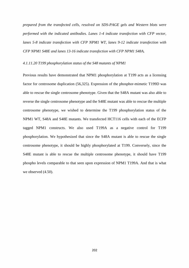
202
prepared from the transfected cells, resolved on SDS-PAGE gels and Western blots were
performed with the indicated antibodies. Lanes 1-4 indicate transfection with CFP vector,
lanes 5-8 indicate transfection with CFP NPM1 WT, lanes 9-12 indicate transfection with
CFP NPM1 S48E and lanes 13-16 indicate transfection with CFP NPM1 S48A.
4.1.11.20 T199 phosphorylation status of the S48 mutants of NPM1
Previous results have demonstrated that NPM1 phosphorylation at T199 acts as a licensing
factor for centrosome duplication (56,325). Expression of the phosphor-mimetic T199D was
able to rescue the single centrosome phenotype. Given that the S48A mutant was also able to
reverse the single centrosome phenotype and the S48E mutant was able to rescue the multiple
centrosome phenotype, we wished to determine the T199 phosphorylation status of the
NPM1 WT, S48A and S48E mutants. We transfected HCT116 cells with each of the ECFP
tagged NPM1 constructs. We also used T199A as a negative control for T199
phosphorylation. We hypothesized that since the S48A mutant is able to rescue the single
centrosome phenotype, it should be highly phosphorylated at T199. Conversely, since the
S48E mutant is able to rescue the multiple centrosome phenotype, it should have T199
phospho levels comparable to that seen upon expression of NPM1 T199A. And that is what
we observed (4.50).
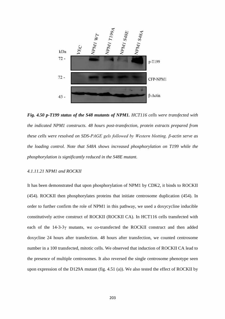
203
Fig. 4.50 p-T199 status of the S48 mutants of NPM1. HCT116 cells were transfected with
the indicated NPM1 constructs. 48 hours post-transfection, protein extracts prepared from
these cells were resolved on SDS-PAGE gels followed by Western blotting. β-actin serve as
the loading control. Note that S48A shows increased phosphorylation on T199 while the
phosphorylation is significantly reduced in the S48E mutant.
4.1.11.21 NPM1 and ROCKII
It has been demonstrated that upon phosphorylation of NPM1 by CDK2, it binds to ROCKII
(454). ROCKII then phosphorylates proteins that initiate centrosome duplication (454). In
order to further confirm the role of NPM1 in this pathway, we used a doxycycline inducible
constitutively active construct of ROCKII (ROCKII CA). In HCT116 cells transfected with
each of the 14-3-3γ mutants, we co-transfected the ROCKII construct and then added
doxycline 24 hours after transfection. 48 hours after transfection, we counted centrosome
number in a 100 transfected, mitotic cells. We observed that induction of ROCKII CA lead to
the presence of multiple centrosomes. It also reversed the single centrosome phenotype seen
upon expression of the D129A mutant (fig. 4.51 (a)). We also tested the effect of ROCKII by

204
using Calpeptin, a chemical activator of ROCKII. Addition of Calpeptin also reversed the
effect of D129A (fig. 4.51 (b)).
Fig. 4.51 14-3-3γ inhibits the ability of NPM1 to activate ROCKII. (a) HCT116 cells were
transfected with the indicated 14-3-3γ constructs and a doxycycline-inducible constitutively
active ROCKII construct. The cells were cultured in the presence and absence of doxycycline
and centrosome number determined in mitotic cells as described above. Note that the
constitutively active ROCKII construct only induces centrosome duplication in the presence
of doxycycline. Western blot indicates overexpression of ROCKII upon doxycycline induction.
(b)HCT116 cells were transfected with the indicated 14-3-3γ constructs and treated with
either the vector control or the ROCKII activator Calpeptin and centrosome number
determined in mitotic cells. Note that Calpeptin reverses the single centrosome phenotype in
cells expressing D129A. In all the experiments the mean and standard error from at least
three independent experiments were plotted, error bars denote standard error of mean and p-
values are obtained using Student’s t test. Molecular weight markers in kDa are indicated.
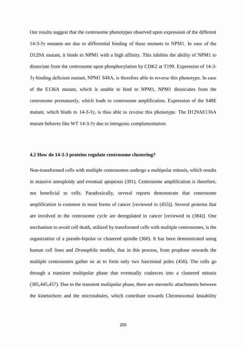
205
Our results suggest that the centrosome phenotypes observed upon expression of the different
14-3-3γ mutants are due to differential binding of these mutants to NPM1. In case of the
D129A mutant, it binds to NPM1 with a high affinity. This inhibits the ability of NPM1 to
dissociate from the centrosome upon phosphorylation by CDK2 at T199. Expression of 14-3-
3γ binding deficient mutant, NPM1 S48A, is therefore able to reverse this phenotype. In case
of the E136A mutant, which is unable to bind to NPM1, NPM1 dissociates from the
centrosome prematurely, which leads to centrosome amplification. Expression of the S48E
mutant, which binds to 14-3-3γ, is thus able to reverse this phenotype. The D129AE136A
mutant behaves like WT 14-3-3γ due to intragenic complementation.
4.2 How do 14-3-3 proteins regulate centrosome clustering?
Non-transformed cells with multiple centrosomes undergo a multipolar mitosis, which results
in massive aneuploidy and eventual apoptosis (391). Centrosome amplification is therefore,
not beneficial to cells. Paradoxically, several reports demonstrate that centrosome
amplification is common in most forms of cancer [reviewed in (455)]. Several proteins that
are involved in the centrosome cycle are deregulated in cancer [reviewed in (384)]. One
mechanism to avoid cell death, utilized by transformed cells with multiple centrosomes, is the
organization of a pseudo-bipolar or clustered spindle (360). It has been demonstrated using
human cell lines and Drosophila models, that in this process, from prophase onwards the
multiple centrosomes gather so as to form only two functional poles (456). The cells go
through a transient multipolar phase that eventually coalesces into a clustered mitosis
(385,445,457). Due to the transient multipolar phase, there are merotelic attachments between
the kinetochore and the microtubules, which contribute towards Chromosomal Instability
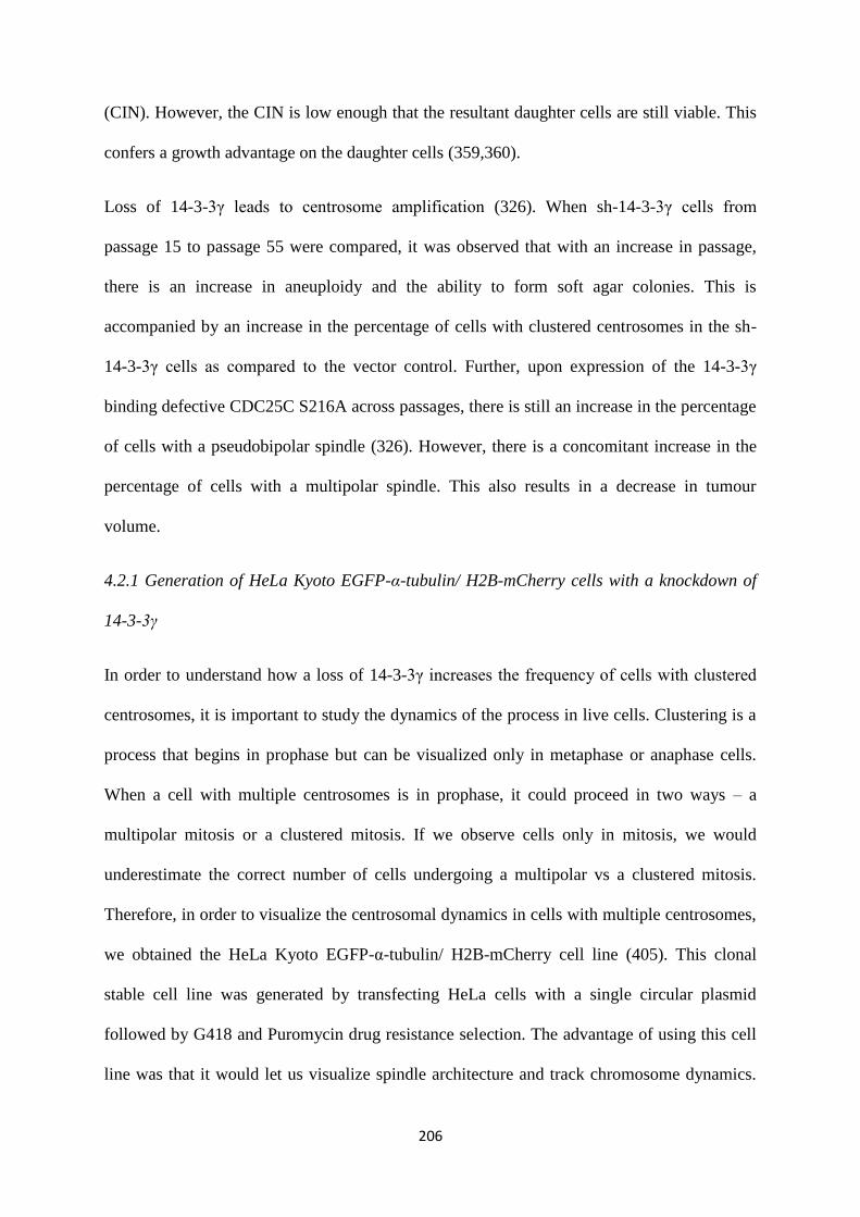
206
(CIN). However, the CIN is low enough that the resultant daughter cells are still viable. This
confers a growth advantage on the daughter cells (359,360).
Loss of 14-3-3γ leads to centrosome amplification (326). When sh-14-3-3γ cells from
passage 15 to passage 55 were compared, it was observed that with an increase in passage,
there is an increase in aneuploidy and the ability to form soft agar colonies. This is
accompanied by an increase in the percentage of cells with clustered centrosomes in the sh-
14-3-3γ cells as compared to the vector control. Further, upon expression of the 14-3-3γ
binding defective CDC25C S216A across passages, there is still an increase in the percentage
of cells with a pseudobipolar spindle (326). However, there is a concomitant increase in the
percentage of cells with a multipolar spindle. This also results in a decrease in tumour
volume.
4.2.1 Generation of HeLa Kyoto EGFP-α-tubulin/ H2B-mCherry cells with a knockdown of
14-3-3γ
In order to understand how a loss of 14-3-3γ increases the frequency of cells with clustered
centrosomes, it is important to study the dynamics of the process in live cells. Clustering is a
process that begins in prophase but can be visualized only in metaphase or anaphase cells.
When a cell with multiple centrosomes is in prophase, it could proceed in two ways – a
multipolar mitosis or a clustered mitosis. If we observe cells only in mitosis, we would
underestimate the correct number of cells undergoing a multipolar vs a clustered mitosis.
Therefore, in order to visualize the centrosomal dynamics in cells with multiple centrosomes,
we obtained the HeLa Kyoto EGFP-α-tubulin/ H2B-mCherry cell line (405). This clonal
stable cell line was generated by transfecting HeLa cells with a single circular plasmid
followed by G418 and Puromycin drug resistance selection. The advantage of using this cell
line was that it would let us visualize spindle architecture and track chromosome dynamics.
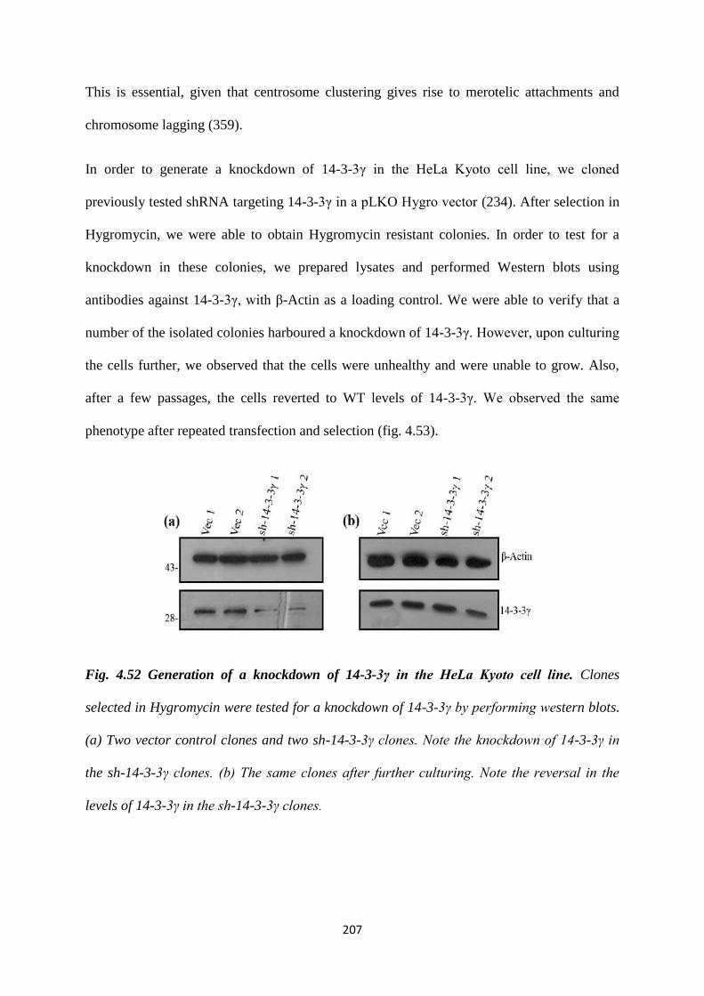
207
This is essential, given that centrosome clustering gives rise to merotelic attachments and
chromosome lagging (359).
In order to generate a knockdown of 14-3-3γ in the HeLa Kyoto cell line, we cloned
previously tested shRNA targeting 14-3-3γ in a pLKO Hygro vector (234). After selection in
Hygromycin, we were able to obtain Hygromycin resistant colonies. In order to test for a
knockdown in these colonies, we prepared lysates and performed Western blots using
antibodies against 14-3-3γ, with β-Actin as a loading control. We were able to verify that a
number of the isolated colonies harboured a knockdown of 14-3-3γ. However, upon culturing
the cells further, we observed that the cells were unhealthy and were unable to grow. Also,
after a few passages, the cells reverted to WT levels of 14-3-3γ. We observed the same
phenotype after repeated transfection and selection (fig. 4.53).
Fig. 4.52 Generation of a knockdown of 14-3-3γ in the HeLa Kyoto cell line. Clones
selected in Hygromycin were tested for a knockdown of 14-3-3γ by performing western blots.
(a) Two vector control clones and two sh-14-3-3γ clones. Note the knockdown of 14-3-3γ in
the sh-14-3-3γ clones. (b) The same clones after further culturing. Note the reversal in the
levels of 14-3-3γ in the sh-14-3-3γ clones.

208
Recently, a paper was published where it was demonstrated using HeLa cells, that 14-3-3
proteins shield Cdt2 from proteasomal degradation (235). Cdt2 is the substrate recognition
adaptor of CRL4Cdt2
E3 ubiquitin ligase complex. CRLCdt2
is essential for the proteasomal
degradation of several cell cycle proteins, such as Cdt1, E2F1, p21and Set8, for the
maintenance of genomic stability [reviewed in (236)]. FbxO11 mediated proteasomal
degradation of Cdt2 leads to cell cycle exit (236). This degradation can be inhibited by its
phosphorylation at a Thr464 residue by CDK1 and CDK2. This phosphorylation also
generates a binding site for 14-3-3γ, which then shields Cdt2 from degradation and stabilizes
it. Upon depletion of 14-3-3γ, there is an increase in the levels of substrates of CRL4Cdt2
, such
as p21 and Set8 (235). This leads to an arrest of the cells at the G2/M phase due to
accumulation of Set8. This could explain why we were unable to generate a knockdown of
14-3-3γ in the HeLa cell line.
It is unclear whether this is a cell line specific phenotype, as transient knockdown of 14-3-3γ
has been created in HEK293 and U2OS cell lines (326). Moreover, HCT116 cells harbouring
a stable knockdown of 14-3-3γ have been generated (234). But given the data from this
publication, we decided to change our model cell line and used the previously generated
HCT116 derived vector control and 14-3-3γ knockdown cells for our experiments. Therefore,
we wished to generate a single construct expressing γ-tubulin-GFP and H2B-mCherry.
Tagging γ-tubulin, which is a PCM protein, would help us track the centrosome and study
centrosome clustering better.
In order to be able to express two proteins using a single promoter, we first cloned an Internal
Ribosome Entry Site (IRES) sequence into the MCS of the pcDNA3.1 Puromycin vector (fig.
4.54 (a)). An IRES is an RNA sequence that forms a complex secondary structure that allows
the initiation of translation from any position within an mRNA immediately downstream
from where the IRES is located. We first cloned mCherry H2B downstream of an (IRES)
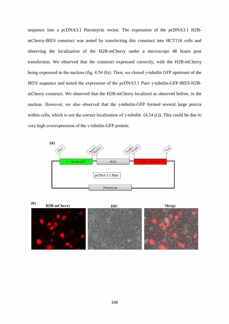
209
sequence into a pcDNA3.1 Puromycin vector. The expression of the pcDNA3.1 H2B-
mCherry-IRES construct was tested by transfecting this construct into HCT116 cells and
observing the localization of the H2B-mCherry under a microscope 48 hours post
transfection. We observed that the construct expressed correctly, with the H2B-mCherry
being expressed in the nucleus (fig. 4.54 (b)). Then, we cloned γ-tubulin GFP upstream of the
IRES sequence and tested the expression of the pcDNA3.1 Puro γ-tubulin-GFP-IRES-H2B-
mCherry construct. We observed that the H2B-mCherry localized as observed before, in the
nucleus. However, we also observed that the γ-tubulin-GFP formed several large puncta
within cells, which is not the correct localisation of γ-tubulin (4.54 (c)). This could be due to
very high overexpression of the γ-tubulin-GFP protein.
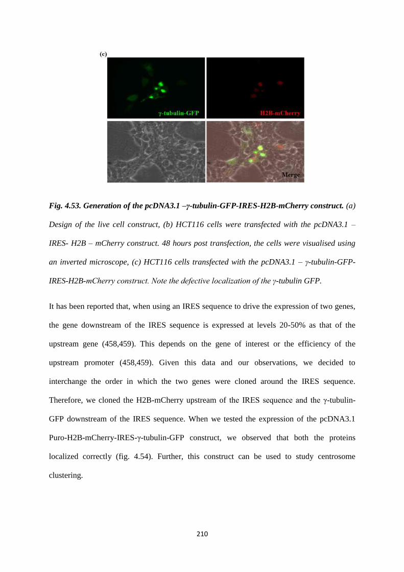
210
Fig. 4.53. Generation of the pcDNA3.1 –γ-tubulin-GFP-IRES-H2B-mCherry construct. (a)
Design of the live cell construct, (b) HCT116 cells were transfected with the pcDNA3.1 –
IRES- H2B – mCherry construct. 48 hours post transfection, the cells were visualised using
an inverted microscope, (c) HCT116 cells transfected with the pcDNA3.1 – γ-tubulin-GFP-
IRES-H2B-mCherry construct. Note the defective localization of the γ-tubulin GFP.
It has been reported that, when using an IRES sequence to drive the expression of two genes,
the gene downstream of the IRES sequence is expressed at levels 20-50% as that of the
upstream gene (458,459). This depends on the gene of interest or the efficiency of the
upstream promoter (458,459). Given this data and our observations, we decided to
interchange the order in which the two genes were cloned around the IRES sequence.
Therefore, we cloned the H2B-mCherry upstream of the IRES sequence and the γ-tubulin-
GFP downstream of the IRES sequence. When we tested the expression of the pcDNA3.1
Puro-H2B-mCherry-IRES-γ-tubulin-GFP construct, we observed that both the proteins
localized correctly (fig. 4.54). Further, this construct can be used to study centrosome
clustering.
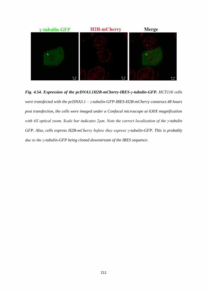
211
Fig. 4.54. Expression of the pcDNA3.1H2B-mCherry-IRES-γ-tubulin-GFP. HCT116 cells
were transfected with the pcDNA3.1 – γ-tubulin-GFP-IRES-H2B-mCherry construct.48 hours
post transfection, the cells were imaged under a Confocal microscope at 630X magnification
with 4X optical zoom. Scale bar indicates 2µm. Note the correct localization of the γ-tubulin
GFP. Also, cells express H2B-mCherry before they express γ-tubulin-GFP. This is probably
due to the γ-tubulin-GFP being cloned downstream of the IRES sequence.

212
5. Discussion

213
5. Discussion.
The results presented in this thesis suggest that 14-3-3 proteins perform several functions in
the centrosome cycle. Binding to 14-3-3γ and 14-3-3ε is essential for the centrosomal
localization of the Centrin2 (452). The functional consequences of this loss of localization
remain to be determined. 14-3-3 proteins are also involved in the centrosome cycle by
inhibiting the premature activation of CDC25C (326). The loss of 14-3-3γ results in
centrosome amplification due to increased phosphorylation of the T199 residue in NPM1 by
CDK1 (326). Further, negatively charged residues within the peptide binding groove of 14-3-
3γ can affect binding of 14-3-3 to NPM1. This binding plays an inhibitory role on centrosome
duplication, as expression of the E136A mutant, which cannot bind to NPM1 leads to
centrosome amplification, and expression of the D129A mutant, which binds with higher
affinity to NPM1, inhibits centrosome duplication.
5.1 The centrosome amplification observed upon a knockdown of 14-3-3γ is
accompanied by an increase in the levels of p-T288 Aurora A kinase.
The primary function of Aurora-A is to promote bipolar spindle assembly but it also has roles
in centrosome maturation and separation, acentrosomal and centrosomal spindle assembly,
kinetochore function, cytokinesis and cell fate determination (341,344,345,366,398,415,460-
466). Aurora-A activation is dependent on its phosphorylation on residue T288 in the
activation loop (467). Our results indicate that Aurora A pT288 levels are higher in the sh-14-
3-3γ cells as compared to the vector control cells. Centrosome amplification in the sh-14-3-3γ
cells is due to premature CDC25C and CDK1 activation. The connection between premature
CDK1 activation and increased Aurora A activation is not clear. Aurora A has not been
shown to play a role in centriole biogenesis, although it has roles in centrosome separation. It
is possible that its increased activation is an outcome of the centrosome amplification
214
observed in the sh-14-3-3γ cells. The increase in AuroraA activity might lead to separation of
the duplicated centrosomes and thus facilitate centrosome duplication.
5.2 Binding to 14-3-3γ and 14-3-3ε is essential for the centrosomal localization of
Centrin2.
Centrin2 is a protein that localizes to the distal lumen of both centrioles (423). Loss of
Centrin2 results in centriole “dilution”, with the generation of daughter cells with only a
single centriole (422). These daughter cells do not survive beyond the fourth cell division
(422). Centrin2 also has functions in DNA damage repair; being a part of the Xeroderma
Pigmentosum Complex (XPC) it is necessary for nucleotide excision repair (369).
Previous reports have demonstrated that the C terminal region of Centrin2, which consists of
the EF3 and EF4 domains (aa. 94-172), are necessary and sufficient for its centrosomal
localization and binding to XPC (424). 14-3-3 proteins have been shown to modulate the sub-
cellular localization of their ligands (219,226,240,468). Results from this thesis demonstrate
that 14-3-3 proteins bind to Centrin2 and that this binding is important for its centrosomal
localization. Expression of a 14-3-3 binding deficient mutant of Centrin2 results in a pan-
cytoplasmic, punctate localization of Centrin2, but none of the puncta localize to the
centrosome. This mutant is not dominant negative, as its expression does not interfere with
mitotic progression or spindle formation. While we have been unable to identify the precise
site of 14-3-3 and Centrin2 interaction, our results suggest that the binding is indpendent of
phosphorylation on Centrin2. There could also be at least two possible regions within
Centrin2 that mediate its binding to 14-3-3 proteins; amino acids 29-37 and amino acids 48-
63. These could form an as yet uncharacterized 14-3-3 binding site. The other possibility is
that the interaction is indirect and that other proteins are required to mediate the interaction of
14-3-3 proteins and Centrin2. The interaction of these proteins and Centrin2 might be
215
stabilized by 14-3-3 binding and thus help in recruiting Centrin2 to the centrosome. 14-3-3
proteins and Centrin2 may be part of a multi-protein complex, which might be essential for
centriole biogenesis and for the accurate localization of Centrin2 at the centrosome.
Several approaches have been taken to explain how the localization of Centrin2 to the
centrosome is regulated (424,469,470). Dantas, et. al., used chicken DT40 cells expressing
Centrin2 mutants that were unable to bind to Calcium. Their results suggest that the
centrosomal localization of Centrin2 depends on its ability to bind to Calcium, as the Alanine
mutants of D41, D77, D114 and D150, do not localize at the centrosome (470). Based on or
results, this does not seem to be the case. The construct expressing aa. 1-37, which does not
contain any of the four calcium binding residues, is able to localize to the centrosome. These
discrepancies could be due to differences in centriole biogenesis between species.
Experiments in XP4PASV cells, a human fibroblast cell line deficient for XPC, have
demonstrated that both, its localization at the centrosome and binding to XPC are dependent
upon three amino acids in EF3 and one amino acid in EF4 of Centrin2 (424). Yet, all the C-
terminal deletion constructs used in this thesis accumulate at the centrosome and co-localize
with Pericentrin, although not with the characteristic 2 or 4 dot organization. This suggests
that after Centrin2 is directed to the centrosome, other factors might bind to its C-terminal
end and mediate its accurate localization at the centriolar lumen.
X-ray crystallographic studies of mouse Centrin1 indicate that the N-terminus of one
Centrin1 monomer might form a complex with the C-terminus of another Centrin1 monomer
(471). This suggests that the N-terminal mutants of Centrin2 might form a complex with the
endogenous Centrin2. However, the entire EF1 domain in Centrin1 is required for
dimerization with the C-terminal EF hands in Centrin1 and the 1-37 mutant does not have the
entire EF1 hand. Therefore, it is unlikely that the 1-37 mutant localizes to the centrosome by
forming a dimer with the endogenous Centrin2. Further, the 14-3-3 binding defective mutant,
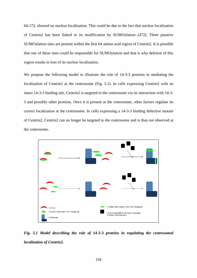
216
64-172, showed no nuclear localization. This could be due to the fact that nuclear localization
of Centrin2 has been linked to its modification by SUMOylation (472). Three putative
SUMOylation sites are present within the first 64 amino acid region of Centrin2. It is possible
that one of these sites could be responsible for SUMOylation and that is why deletion of this
region results in loss of its nuclear localization.
We propose the following model to illustrate the role of 14-3-3 proteins in mediating the
localization of Centrin2 at the centrosome (Fig. 5.1). In cells expressing Centrin2 with an
intact 14-3-3 binding site, Centrin2 is targeted to the centrosome via its interaction with 14-3-
3 and possibly other proteins. Once it is present at the centrosome, other factors regulate its
correct localization at the centrosome. In cells expressing a 14-3-3 binding defective mutant
of Centrin2, Centrin2 can no longer be targeted to the centrosome and is thus not observed at
the centrosome.
Fig. 5.1 Model describing the role of 14-3-3 proteins in regulating the centrosomal
localization of Centrin2.
217
5.3 The centrosomal localization of γ-tubulin depends on its middle Tubulin/FtsZ, 2
layer sandwich domain.
γ-tubulin is essential for microtubule nucleation, being a part of the γ-Tubulin Small Complex
(γ-TuSC) and the γ-Tubulin Ring Complex (γ-TURC). Several reports have focussed on its
activity at the centrosome and its connection to various microtubule nucleation pathways
(reviewed in (473)). The results in this thesis indicate that its middle Tubulin/FtsZ, 2 layer
sandwich domain is essential for its centrosomal localization, as its deletion results in loss of
the centrosomal localization of γ-tubulin. This domain might not be essential for binding to
14-3-3γ, as the G1 mutant is still able to bind to 14-3-3γ, although it is possible that there are
multiple 14-3-3 binding sites on γ-tubulin. Further experiments are needed to understand
which other proteins might be necessary for its centrosomal localization.
5.4 Cep170 levels decrease in the sh-14-3-3γ cells.
Cep170 is a sub-distal appendage protein that is specific to the mother centriole. Our results
indicate that both mRNA as well as protein levels of Cep170 decrease in the sh-14-3-3γ cells
as compared to the vector control cells. Given that this decrease does not seem to affect the
intensity of Cep170 at the centrosome, it is difficult to understand its functional significance.
Experiments in the mouse F9 cell line with a knockout of Odf2, another sub-distal appendage
protein show that these cells lack sub-distal appendages and are unable to form primary cilia
(312). Odf2 has been demonstrated to be one of the first proteins that is recruited to the sub-
distal appendages (437). It is possible that this is why its loss has such a severe phenotype.
Further, it is not possible to study the formation of primary cilia in HCT116 cells as these are
actively cycling cells and do not undergo quiescence.
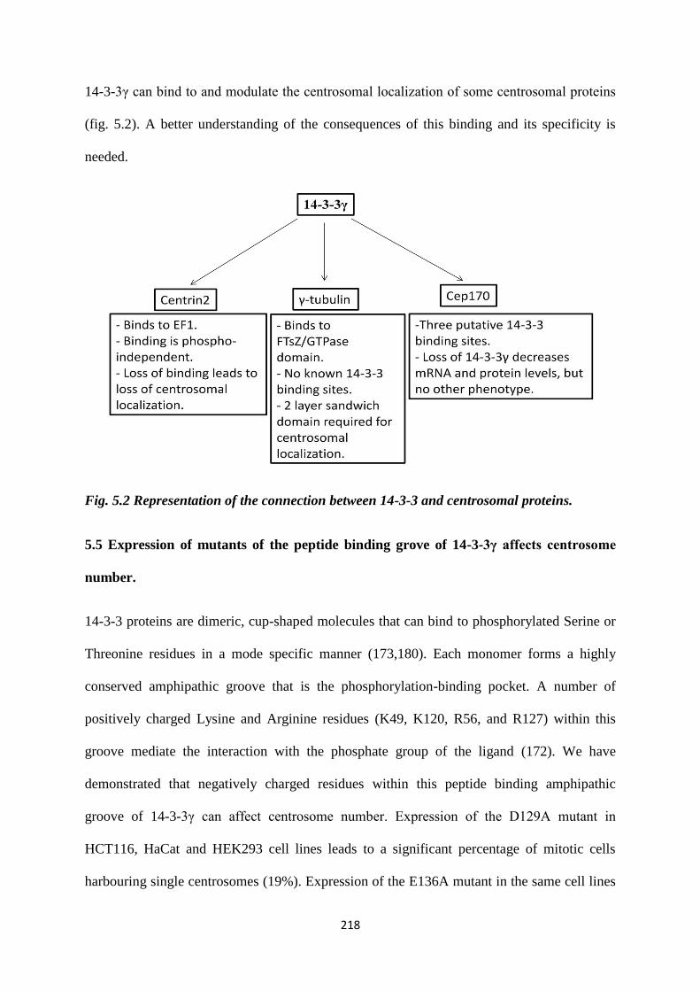
218
14-3-3γ can bind to and modulate the centrosomal localization of some centrosomal proteins
(fig. 5.2). A better understanding of the consequences of this binding and its specificity is
needed.
Fig. 5.2 Representation of the connection between 14-3-3 and centrosomal proteins.
5.5 Expression of mutants of the peptide binding grove of 14-3-3γ affects centrosome
number.
14-3-3 proteins are dimeric, cup-shaped molecules that can bind to phosphorylated Serine or
Threonine residues in a mode specific manner (173,180). Each monomer forms a highly
conserved amphipathic groove that is the phosphorylation-binding pocket. A number of
positively charged Lysine and Arginine residues (K49, K120, R56, and R127) within this
groove mediate the interaction with the phosphate group of the ligand (172). We have
demonstrated that negatively charged residues within this peptide binding amphipathic
groove of 14-3-3γ can affect centrosome number. Expression of the D129A mutant in
HCT116, HaCat and HEK293 cell lines leads to a significant percentage of mitotic cells
harbouring single centrosomes (19%). Expression of the E136A mutant in the same cell lines
219
results in the presence of multiple centrosomes in mitotic cells (21%). Interestingly,
expression of the D129AE136A mutant did not result in any change in centrosome number.
This suggests that this mutant shows intragenic complementation. We also observed a similar
phenotype in the HCT116 derived vector control and sh-14-3-3γ cells. This implies that the
phenotype is actually dominant over the endogenous protein in the cells. Further, knock-in
experiments are needed to test the effect of the mutants. Also, given that these residues are
highly conserved across isoforms and that different 14-3-3 isoforms can have overlapping
functions, it is possible that similar mutations in other isoforms might result in a similar
phenotype. It would be interesting to study the effect of altering these residues in other
isoforms and determine their affect on isoform function.
5.6 The single centrosome phenotype is due to a failure of centriole duplication.
By performing dual staining for centrosomes, using GFP Centrin2, Pericentrin, Cep68 and
Ninein as markers, we were able to demonstrate that the single and multiple centrosomes
observed are due to defects in centriole duplication. Under normal conditions, mitotic cells
would have 4 Centrin2 dots and 2 Pericentrin clouds, a ratio of 4:2 for Centrin2:Pericentrin,
indicative of two mature centrosomes. Cells expressing the WT 14-3-3γ show
Centrin2:Pericentrin staining in the ratio of 4:2 in mitosis, while the single centrosome
observed in cells expressing the D129A mutant shows Centrin2:Pericentrin staining in the
ratio of 2:1 in mitosis. As described in section 4.1.11.6 and figure 4.32, this could be due to a
defect in separation, disengagement or duplication. In order to understand the phenotype
better, we performed dual staining using Centrin2:Cep68 and Centrin2:Ninein. In normal
cells in G2, there would be 4 Centrin2 dots and 2 Cep68 dots, indicative of centrosomes that
had undergone disengagement and duplication. In cells expressing the WT 14-3-3γ construct,
we observed Centrin2:Cep68 staining in the ration of 4:2, which is expected. In cells
expressing the D129A construct, with a single centrosome, we observed Centrin2:cep68
220
staining in the ratio 2:2, which is indicative of a centrosome that has undergone
disengagement but is unable to duplicate. Further, a normal G2 cell would stain for
Centrin2:Ninein in the ratio of 4:1, indicative of 4 centrioles, of which only one is the
grandmother centriole staining positively for Ninein, which is what we observed in cells
expressing the WT 14-3-3γ construct. In cells expressing the D129A construct, with a single
centrosome, we observed Centrin2:Ninein staining in the ratio 2:1. Taken together with the
Cep68 staining, this is indicative of a centrosome that has undergone disengagement but is
unable to initiate duplication. Previous reports in RPE1 cells indicate that a 2:2 ratio of
Centrin:C-Nap1 exists in cells with disengaged centrioles (334). C-Nap1 is an
intercentrosomal linker protein that has been shown to localize to MTOC competent
centrioles (334,474). In the same report, hSas-6, which is a protein that localizes to the
central cartwheel, has been shown to associate exclusively with daughter centrioles that are
MTOC incompetent. It might be useful to study the pattern of hSas-6 in cells expressing these
14-3-3γ mutants. Also, it will be intersting to further characterize the multiple centrosome
phenotype using daughter centriole specific markers such as hSas-6 and Centrobin (475).
This would help us understand whether the daughter centrioles are formed de novo or using
the mother centriole as the template.
5.7 Cells expressing the 14-3-3γ mutants exhibit mitotic defects.
Cells expressing the WT 14-3-3γ complete mitosis in ~40 minutes. We observed that cells
expressing the D129A mutant, that have a single centrosome in mitosis, are unable to
complete mitosis even after 120 minutes in mitosis. In HeLa cells, a knockdown of Centrin2
leads to the presence of a unicentriolar spindle pole. Here, due to “centriole dilution”, the two
centrioles split, migrate to each pole and nucleate spindle microtubules and complete mitosis
(422). In the next round of mitosis, acentriolar spindle poles are formed and eventually, the
cells are unable to complete cytokinesis and undergo apoptosis (422). Inspite of having only
221
two centrioles in a mitosis, the cell is able to complete at least one round of mitosis. A similar
phenotype is not observed in cells expressing the D129A mutant. This could be explained by
the fact that the duration of mitosis is not specified in this report (422). It is possible that cells
expressing the D129A mutant, with a single centrosome in mitosis might also be able to
complete mitosis, if observed for a longer duration and this needs to be tested. Also, if these
cells are undergo apoptosis eventually, it might be difficult to generate a stable cells
overexpressing these constructs. in this case, a conditional knock-in system would be useful.
The other possibility is that activation of the Spindle Assembly Checkpoint (SAC) occurs
when cells expressing the D129A mutant, with a single MTOC, are in mitosis. As described
in section 1.1.2, the SAC is activated upon loss of spindle pole tension, unattached
kinetochores at metaphase or by certain spindle poisons such as nocodazole (137). When a
single MTOC is present, syntelic attachments can occur, where both sister chromatids are
attached to a single spindle pole (476). It might be interesting to study what happens when
cells expressing the D129A mutant, with a single centrosome, are treated with a SAC
inhibitor such as CFI-402257 (477). If treatment with the SAC inhibitor is able to promote
mitosis in these cells, the resulting daughter cells will harbour significant levels of CIN
and this might further affect their viability.
Transformed cells with multiple centrosomes usually undergo a clustered mitosis
(360,385,391,445,478). Extra centrosomes can add upto 30 minutes to the duration of
mitosis, depending on the cell line being used (457). This is in line with our observation that
cells expressing the E136A mutant, that possess multiple centrosomes, take a little more than
40 minute to complete mitosis by clustering centrosomes. We have been unable to detect the
occurrence of a multipolar mitosis in cells expressing E136A. It is possible that these cells
have a very efficient clustering mechanism. Loss of 14-3-3γ leads to an increase in
centrosome number, clustering and aneuploidy with an increase in passage (326). The
222
centrosome clustering can be reversed to a multipolar mitosis upon co-expresssion of
CDC25C (326) The multiple centrosome phenotype that we observe upon expression of the
E136A mutant mirrors that seen upon loss of 14-3-3γ. Expression of this construct in the sh-
14-3-3γ cells actually enhances the centrosome amplification observed upon loss of 14-3-3γ.
It would be of interest to test the effect of CDC25C overexpression on centrosome number
and clustering in these cells.
5.8 14-3-3 inhibits centriole duplication by inhibiting NPM1 function.
The single centrosome phenotype cannot be reversed by overexpression of Plk4, CDK1 or
CDK2. This is interesting because these proteins are extremely essential for procentriole
biogenesis. Plk4 is first recruited at the proximal end of the mother centriole by Cep152,
Cep192 and Plk1 (291). Given that overexpression of Plk4 is unable to rescue the single
centrosome phenotype, it would be intersting to study the localization of Cep152, Cep63 and
Cep192 at these centrosomes. This would help us further understand the dynamics of the
recruitment of these centrosomal proteins at the centrosome and how this recruitment is
affected in cells expressing the 14-3-3γ mutants.
Our results indicate that the single centrosome phenotype observed upon expression of the
D129A mutant can be partially reversed by the NPM1 WT protein or completely reversed
upon expression of the NPM1 T199D mutant, which is a phospho-mimetic mutant, or the
S48A mutant, which is unable to bind to 14-3-3γ (section 4.1.11.14). Whereas, the multiple
centrosome phenotype observed upon expression of the E136A mutant can be reversed upon
expression of the NPM1 T199A mutant or the S48E mutant, which we propose is another
phospho-mimetic site, necessary for 14-3-3 binding (section 4.1.11.14). Further, the single
centrosome phenotype can be reversed upon activation of ROCK2. It has been demonstrated
that NPM1 phosphorylated at T199 by CDK2 is displaced from the intercentrosomal linker
223
and activates ROCK2. Upon ROCK2 activation, centrosome duplication is initiated
(454,479). However, using both antibodies against NPM1 and a fluorescently tagged
construct of NPM1, we have been unable to detect the presence of NPM1 at the centrosome
in both interphase and mitotic cells. Even the CFP NPM1 S48E mutant which localizes to the
nucleus is not detected at the centrosome. 14-3-3γ has been shown to localize to the
centrosome (326). There are no studies that indicate the presence of ROCK2 at the
centrosome. We have demonstrated that 14-3-3γ interacts with NPM1. It is possible that the
interaction of 14-3-3γ and NPM1 does not occur at the centrosome. It is clear, however, that
the binding of 14-3-3γ to NPM1 inhibits NPM1 function. It is possible that the interaction
between NPM1 and 14-3-3γ is not direct. 14-3-3 proteins have been shown to form ternary
complexes due to the presence of two ligand binding sites within the dimer (215).
Oligomerization is essential for most NPM1 functions (418). It has been demonstrated
previously, that phosphorylation at the S48 residue by Akt destabilizes NPM1
oligomerization (418). Our results indicate that this S48 residue is actually a mode 2 14-3-3
binding site. Mutation of the S48 to Alanine abrogates binding to 14-3-3γ, whereas, the S48E
mutant is still able to bind to 14-3-3γ. NPM1 phosphorylated at the S48 residue is unable to
oligomerize, therefore, the generation of the 14-3-3 binding site actually inhibits NPM1
oligomer formation. We also need to find out the kinase that phosphorylates NPM1 at S48
and generates a 14-3-3 binding site. It is possible that it could be Akt, in which case, we need
to understand the role of Akt in the centrosome cycle.
Based upon our results, we propose the following mechanism (Fig. 5.3). Under normal
conditions, in the presence of 14-3-3γ, NPM1 is bound to WT 14-3-3γ and remains at the
centrosome and inhibits centrosome duplication. Upon phosphorylation of NPM1 at T199 by
the CDK2/Cyclin E complex, NPM1 dissociates from the centrosome, and activates ROCK2,
which licenses centrosome duplication (Fig. 5.3 (a)). On expression of the D129A mutant,
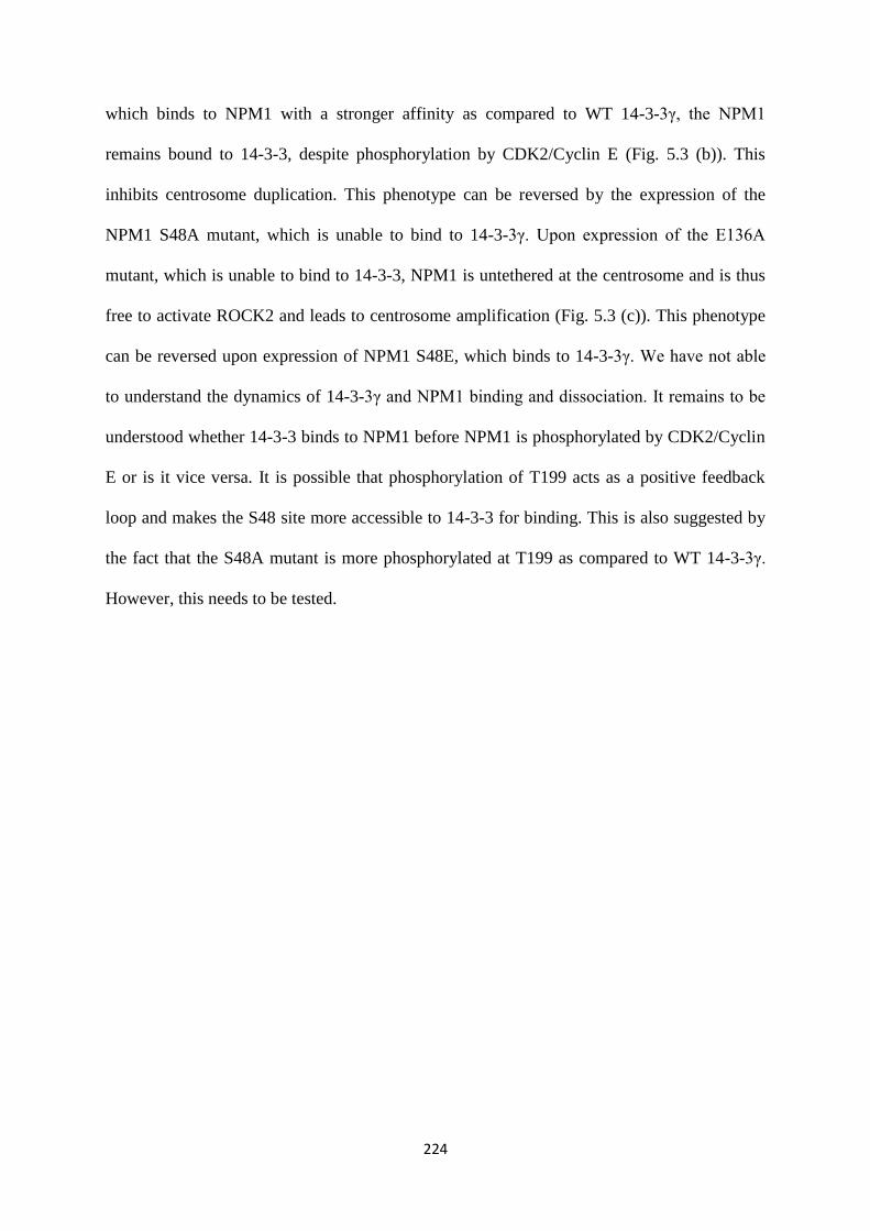
224
which binds to NPM1 with a stronger affinity as compared to WT 14-3-3γ, the NPM1
remains bound to 14-3-3, despite phosphorylation by CDK2/Cyclin E (Fig. 5.3 (b)). This
inhibits centrosome duplication. This phenotype can be reversed by the expression of the
NPM1 S48A mutant, which is unable to bind to 14-3-3γ. Upon expression of the E136A
mutant, which is unable to bind to 14-3-3, NPM1 is untethered at the centrosome and is thus
free to activate ROCK2 and leads to centrosome amplification (Fig. 5.3 (c)). This phenotype
can be reversed upon expression of NPM1 S48E, which binds to 14-3-3γ. We have not able
to understand the dynamics of 14-3-3γ and NPM1 binding and dissociation. It remains to be
understood whether 14-3-3 binds to NPM1 before NPM1 is phosphorylated by CDK2/Cyclin
E or is it vice versa. It is possible that phosphorylation of T199 acts as a positive feedback
loop and makes the S48 site more accessible to 14-3-3 for binding. This is also suggested by
the fact that the S48A mutant is more phosphorylated at T199 as compared to WT 14-3-3γ.
However, this needs to be tested.
225
Fig. 5.3 Model explaining the regulation of NPM1 function by 14-3-3γ.
(b)
(c)
(a)
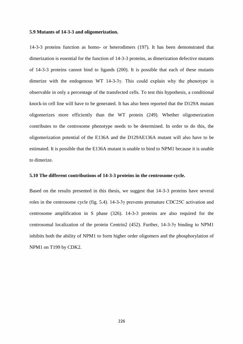
226
5.9 Mutants of 14-3-3 and oligomerization.
14-3-3 proteins function as homo- or heterodimers (197). It has been demonstrated that
dimerization is essential for the function of 14-3-3 proteins, as dimerization defective mutants
of 14-3-3 proteins cannot bind to ligands (200). It is possible that each of these mutants
dimerize with the endogenous WT 14-3-3γ. This could explain why the phenotype is
observable in only a percentage of the transfected cells. To test this hypothesis, a conditional
knock-in cell line will have to be generated. It has also been reported that the D129A mutant
oligomerizes more efficiently than the WT protein (249). Whether oligomerization
contributes to the centrosome phenotype needs to be determined. In order to do this, the
oligomerization potential of the E136A and the D129AE136A mutant will also have to be
estimated. It is possible that the E136A mutant is unable to bind to NPM1 because it is unable
to dimerize.
5.10 The different contributions of 14-3-3 proteins in the centrosome cycle.
Based on the results presented in this thesis, we suggest that 14-3-3 proteins have several
roles in the centrosome cycle (fig. 5.4). 14-3-3γ prevents premature CDC25C activation and
centrosome amplification in S phase (326). 14-3-3 proteins are also required for the
centrosomal localization of the protein Centrin2 (452). Further, 14-3-3γ binding to NPM1
inhibits both the ability of NPM1 to form higher order oligomers and the phosphorylation of
NPM1 on T199 by CDK2.
227
Fig. 5.4 Model representing the different contributions of 14-3-3 proteins to the
centrosome cycle.
5.11 Conclusion.
The results in this thesis demonstrate that 14-3-3 proteins perform different functions in the
centrosome cycle. These functions rely on the abilities of 14-3-3 proteins to act as adaptor
proteins, molecular switches and shuttling proteins. Most of the results in this thesis focus on
the γ isoform of 14-3-3 proteins, but as has been described before, it is possible that other
isoforms can also perform compensatory functions.

228
6. Bibliography
229
1. Piwnica-Worms, H. (1999) Cell cycle. Fools rush in. Nature 401, 535, 537
2. Nurse, P. (2000) A long twentieth century of the cell cycle and beyond. Cell 100, 71-
78
3. Mitchison, J. M. (1971) The biology of the cell cycle [by] J. M. Mitchison, University
Press, Cambridge [Eng.]
4. Morgan, D. (2007) The Cell Cycle : Principles of Control. (Lawrence, E. ed.), New
Science Press, UK. pp
5. Elledge, S. J. (1996) Cell cycle checkpoints: preventing an identity crisis. Science
274, 1664-1672
6. Pardee, A. B. (1974) A restriction point for control of normal animal cell
proliferation. Proceedings of the National Academy of Sciences of the United States of
America 71, 1286-1290
7. Coller, H. A. (2007) What's taking so long? S-phase entry from quiescence versus
proliferation. Nature reviews. Molecular cell biology 8, 667-670
8. Hirai, H., Roussel, M. F., Kato, J. Y., Ashmun, R. A., and Sherr, C. J. (1995) Novel
INK4 proteins, p19 and p18, are specific inhibitors of the cyclin D-dependent kinases
CDK4 and CDK6. Mol Cell Biol 15, 2672-2681
9. Chan, F. K., Zhang, J., Cheng, L., Shapiro, D. N., and Winoto, A. (1995)
Identification of human and mouse p19, a novel CDK4 and CDK6 inhibitor with
homology to p16ink4. Mol Cell Biol 15, 2682-2688
10. Reynisdottir, I., Polyak, K., Iavarone, A., and Massague, J. (1995) Kip/Cip and Ink4
Cdk inhibitors cooperate to induce cell cycle arrest in response to TGF-beta. Genes &
development 9, 1831-1845
11. Ohtani, K., DeGregori, J., and Nevins, J. R. (1995) Regulation of the cyclin E gene by
transcription factor E2F1. Proceedings of the National Academy of Sciences of the
United States of America 92, 12146-12150
12. Desai, D., Gu, Y., and Morgan, D. O. (1992) Activation of human cyclin-dependent
kinases in vitro. Molecular biology of the cell 3, 571-582
13. van den Heuvel, S., and Harlow, E. (1993) Distinct roles for cyclin-dependent kinases
in cell cycle control. Science 262, 2050-2054
14. Planas-Silva, M. D., and Weinberg, R. A. (1997) The restriction point and control of
cell proliferation. Current opinion in cell biology 9, 768-772
15. Vijayaraghavan, M. R., and Prabhakar, K. . (1984) The endosperm. in Embryology of
Angiosperms (Johri, B. M. ed.), Berlin: Springer-Verlag. pp 319-376
16. Alberts B, J. A., Lewis J, et al. . (2002) Drosophila and the Molecular Genetics of
Pattern Formation: Genesis of the Body Plan. in Molecular biology of the cell, 4th
Ed., Garland Science, New York. pp
17. Zhimulev, I. F., Belyaeva, E. S., Semeshin, V. F., Koryakov, D. E., Demakov, S. A.,
Demakova, O. V., Pokholkova, G. V., and Andreyeva, E. N. (2004) Polytene
chromosomes: 70 years of genetic research. International review of cytology 241,
203-275
18. Frade, J. M., and Ovejero-Benito, M. C. (2015) Neuronal cell cycle: the neuron itself
and its circumstances. Cell cycle (Georgetown, Tex.) 14, 712-720
19. Harper, J. W., and Adams, P. D. (2001) Cyclin-dependent kinases. Chemical reviews
101, 2511-2526
20. De Bondt, H. L., Rosenblatt, J., Jancarik, J., Jones, H. D., Morgan, D. O., and Kim, S.
H. (1993) Crystal structure of cyclin-dependent kinase 2. Nature 363, 595-602
21. Jeffrey, P. D., Russo, A. A., Polyak, K., Gibbs, E., Hurwitz, J., Massague, J., and
Pavletich, N. P. (1995) Mechanism of CDK activation revealed by the structure of a
cyclinA-CDK2 complex. Nature 376, 313-320
230
22. Frouin, I., Montecucco, A., Biamonti, G., Hubscher, U., Spadari, S., and Maga, G.
(2002) Cell cycle-dependent dynamic association of cyclin/Cdk complexes with
human DNA replication proteins. The EMBO journal 21, 2485-2495
23. John, P. C., Mews, M., and Moore, R. (2001) Cyclin/Cdk complexes: their
involvement in cell cycle progression and mitotic division. Protoplasma 216, 119-142
24. Viallard, J. F., Lacombe, F., Belloc, F., Pellegrin, J. L., and Reiffers, J. (2001)
Molecular mechanisms controlling the cell cycle: fundamental aspects and
implications for oncology. Cancer radiotherapie : journal de la Societe francaise de
radiotherapie oncologique 5, 109-129
25. Nigg, E. A. (1996) Cyclin-dependent kinase 7: at the cross-roads of transcription,
DNA repair and cell cycle control? Current opinion in cell biology 8, 312-317
26. Morgan, D. O. (1997) Cyclin-dependent kinases: engines, clocks, and
microprocessors. Annual review of cell and developmental biology 13, 261-291
27. Ekholm, S. V., and Reed, S. I. (2000) Regulation of G(1) cyclin-dependent kinases in
the mammalian cell cycle. Current opinion in cell biology 12, 676-684
28. Sherr, C. J., and Roberts, J. M. (1999) CDK inhibitors: positive and negative
regulators of G1-phase progression. Genes & development 13, 1501-1512
29. Serrano, M., Hannon, G. J., and Beach, D. (1993) A new regulatory motif in cell-
cycle control causing specific inhibition of cyclin D/CDK4. Nature 366, 704-707
30. Hannon, G. J., and Beach, D. (1994) p15INK4B is a potential effector of TGF-beta-
induced cell cycle arrest. Nature 371, 257-261
31. Guan, K. L., Jenkins, C. W., Li, Y., Nichols, M. A., Wu, X., O'Keefe, C. L., Matera,
A. G., and Xiong, Y. (1994) Growth suppression by p18, a p16INK4/MTS1- and
p14INK4B/MTS2-related CDK6 inhibitor, correlates with wild-type pRb function.
Genes & development 8, 2939-2952
32. Pavletich, N. P. (1999) Mechanisms of cyclin-dependent kinase regulation: structures
of Cdks, their cyclin activators, and Cip and INK4 inhibitors. Journal of molecular
biology 287, 821-828
33. Chen, J., Jackson, P. K., Kirschner, M. W., and Dutta, A. (1995) Separate domains of
p21 involved in the inhibition of Cdk kinase and PCNA. Nature 374, 386-388
34. Chen, J., Saha, P., Kornbluth, S., Dynlacht, B. D., and Dutta, A. (1996) Cyclin-
binding motifs are essential for the function of p21CIP1. Mol Cell Biol 16, 4673-4682
35. Nakanishi, M., Robetorye, R. S., Adami, G. R., Pereira-Smith, O. M., and Smith, J. R.
(1995) Identification of the active region of the DNA synthesis inhibitory gene
p21Sdi1/CIP1/WAF1. The EMBO journal 14, 555-563
36. Warbrick, E., Lane, D. P., Glover, D. M., and Cox, L. S. (1995) A small peptide
inhibitor of DNA replication defines the site of interaction between the cyclin-
dependent kinase inhibitor p21WAF1 and proliferating cell nuclear antigen. Current
biology : CB 5, 275-282
37. Lin, J., Reichner, C., Wu, X., and Levine, A. J. (1996) Analysis of wild-type and
mutant p21WAF-1 gene activities. Mol Cell Biol 16, 1786-1793
38. Besson, A., Dowdy, S. F., and Roberts, J. M. (2008) CDK inhibitors: cell cycle
regulators and beyond. Developmental cell 14, 159-169
39. Bakiri, L., Lallemand, D., Bossy-Wetzel, E., and Yaniv, M. (2000) Cell cycle-
dependent variations in c-Jun and JunB phosphorylation: a role in the control of
cyclin D1 expression. The EMBO journal 19, 2056-2068
40. Filmus, J., Robles, A. I., Shi, W., Wong, M. J., Colombo, L. L., and Conti, C. J.
(1994) Induction of cyclin D1 overexpression by activated ras. Oncogene 9, 3627-
3633
231
41. Malumbres, M., and Pellicer, A. (1998) RAS pathways to cell cycle control and cell
transformation. Frontiers in bioscience : a journal and virtual library 3, d887-912
42. Lavoie, J. N., L'Allemain, G., Brunet, A., Muller, R., and Pouyssegur, J. (1996)
Cyclin D1 expression is regulated positively by the p42/p44MAPK and negatively by
the p38/HOGMAPK pathway. The Journal of biological chemistry 271, 20608-20616
43. Brehm, A., Miska, E. A., McCance, D. J., Reid, J. L., Bannister, A. J., and
Kouzarides, T. (1998) Retinoblastoma protein recruits histone deacetylase to repress
transcription. Nature 391, 597-601
44. Magnaghi-Jaulin, L., Groisman, R., Naguibneva, I., Robin, P., Lorain, S., Le Villain,
J. P., Troalen, F., Trouche, D., and Harel-Bellan, A. (1998) Retinoblastoma protein
represses transcription by recruiting a histone deacetylase. Nature 391, 601-605
45. Kozar, K., Ciemerych, M. A., Rebel, V. I., Shigematsu, H., Zagozdzon, A., Sicinska,
E., Geng, Y., Yu, Q., Bhattacharya, S., Bronson, R. T., Akashi, K., and Sicinski, P.
(2004) Mouse development and cell proliferation in the absence of D-cyclins. Cell
118, 477-491
46. Ciemerych, M. A., Kenney, A. M., Sicinska, E., Kalaszczynska, I., Bronson, R. T.,
Rowitch, D. H., Gardner, H., and Sicinski, P. (2002) Development of mice expressing
a single D-type cyclin. Genes & development 16, 3277-3289
47. Tsutsui, T., Hesabi, B., Moons, D. S., Pandolfi, P. P., Hansel, K. S., Koff, A., and
Kiyokawa, H. (1999) Targeted disruption of CDK4 delays cell cycle entry with
enhanced p27(Kip1) activity. Mol Cell Biol 19, 7011-7019
48. Rane, S. G., Dubus, P., Mettus, R. V., Galbreath, E. J., Boden, G., Reddy, E. P., and
Barbacid, M. (1999) Loss of Cdk4 expression causes insulin-deficient diabetes and
Cdk4 activation results in beta-islet cell hyperplasia. Nature genetics 22, 44-52
49. Hu, M. G., Deshpande, A., Enos, M., Mao, D., Hinds, E. A., Hu, G. F., Chang, R.,
Guo, Z., Dose, M., Mao, C., Tsichlis, P. N., Gounari, F., and Hinds, P. W. (2009) A
requirement for cyclin-dependent kinase 6 in thymocyte development and
tumorigenesis. Cancer Res 69, 810-818
50. Botz, J., Zerfass-Thome, K., Spitkovsky, D., Delius, H., Vogt, B., Eilers, M.,
Hatzigeorgiou, A., and Jansen-Durr, P. (1996) Cell cycle regulation of the murine
cyclin E gene depends on an E2F binding site in the promoter. Mol Cell Biol 16,
3401-3409
51. Guichard, P., Hamel, V., Le Guennec, M., Banterle, N., Iacovache, I., Nemcikova, V.,
Fluckiger, I., Goldie, K. N., Stahlberg, H., Levy, D., Zuber, B., and Gonczy, P. (2017)
Cell-free reconstitution reveals centriole cartwheel assembly mechanisms. Nat
Commun 8, 14813
52. Zhang, H. S., Postigo, A. A., and Dean, D. C. (1999) Active transcriptional repression
by the Rb-E2F complex mediates G1 arrest triggered by p16INK4a, TGFbeta, and
contact inhibition. Cell 97, 53-61
53. Yu, Q., and Sicinski, P. (2004) Mammalian cell cycles without cyclin E-CDK2. Cell
cycle (Georgetown, Tex.) 3, 292-295
54. Teixeira, L. K., Wang, X., Li, Y., Ekholm-Reed, S., Wu, X., Wang, P., and Reed, S. I.
(2015) Cyclin E deregulation promotes loss of specific genomic regions. Current
biology : CB 25, 1327-1333
55. Hu, W., Nevzorova, Y. A., Haas, U., Moro, N., Sicinski, P., Geng, Y., Barbacid, M.,
Trautwein, C., and Liedtke, C. (2014) Concurrent deletion of cyclin E1 and cyclin-
dependent kinase 2 in hepatocytes inhibits DNA replication and liver regeneration in
mice. Hepatology (Baltimore, Md.) 59, 651-660
56. Okuda, M., Horn, H. F., Tarapore, P., Tokuyama, Y., Smulian, A. G., Chan, P. K.,
Knudsen, E. S., Hofmann, I. A., Snyder, J. D., Bove, K. E., and Fukasawa, K. (2000)
232
Nucleophosmin/B23 is a target of CDK2/cyclin E in centrosome duplication. Cell
103, 127-140
57. Sheaff, R. J., Groudine, M., Gordon, M., Roberts, J. M., and Clurman, B. E. (1997)
Cyclin E-CDK2 is a regulator of p27Kip1. Genes & development 11, 1464-1478
58. Dealy, M. J., Nguyen, K. V., Lo, J., Gstaiger, M., Krek, W., Elson, D., Arbeit, J.,
Kipreos, E. T., and Johnson, R. S. (1999) Loss of Cul1 results in early embryonic
lethality and dysregulation of cyclin E. Nature genetics 23, 245-248
59. Nakayama, K., Nagahama, H., Minamishima, Y. A., Matsumoto, M., Nakamichi, I.,
Kitagawa, K., Shirane, M., Tsunematsu, R., Tsukiyama, T., Ishida, N., Kitagawa, M.,
Nakayama, K., and Hatakeyama, S. (2000) Targeted disruption of Skp2 results in
accumulation of cyclin E and p27(Kip1), polyploidy and centrosome overduplication.
The EMBO journal 19, 2069-2081
60. Singer, J. D., Gurian-West, M., Clurman, B., and Roberts, J. M. (1999) Cullin-3
targets cyclin E for ubiquitination and controls S phase in mammalian cells. Genes &
development 13, 2375-2387
61. Wang, Y., Penfold, S., Tang, X., Hattori, N., Riley, P., Harper, J. W., Cross, J. C., and
Tyers, M. (1999) Deletion of the Cul1 gene in mice causes arrest in early
embryogenesis and accumulation of cyclin E. Current biology : CB 9, 1191-1194
62. Berthet, C., Aleem, E., Coppola, V., Tessarollo, L., and Kaldis, P. (2003) Cdk2
knockout mice are viable. Current biology : CB 13, 1775-1785
63. Barrière, C., Santamaría, D., Cerqueira, A., Galán, J., Martín, A., Ortega, S.,
Malumbres, M., Dubus, P., and Barbacid, M. (2007) Mice thrive without Cdk4 and
Cdk2. Molecular oncology 1, 72-83
64. Geng, Y., Yu, Q., Sicinska, E., Das, M., Schneider, J. E., Bhattacharya, S., Rideout,
W. M., Bronson, R. T., Gardner, H., and Sicinski, P. (2003) Cyclin E ablation in the
mouse. Cell 114, 431-443
65. Parisi, T., Beck, A. R., Rougier, N., McNeil, T., Lucian, L., Werb, Z., and Amati, B.
(2003) Cyclins E1 and E2 are required for endoreplication in placental trophoblast
giant cells. The EMBO journal 22, 4794-4803
66. Connell-Crowley, L., Solomon, M. J., Wei, N., and Harper, J. W. (1993)
Phosphorylation independent activation of human cyclin-dependent kinase 2 by cyclin
A in vitro. Molecular biology of the cell 4, 79-92
67. Russo, A. A., Jeffrey, P. D., and Pavletich, N. P. (1996) Structural basis of cyclin-
dependent kinase activation by phosphorylation. Nature structural biology 3, 696-700
68. Krude, T., Jackman, M., Pines, J., and Laskey, R. A. (1997) Cyclin/Cdk-dependent
initiation of DNA replication in a human cell-free system. Cell 88, 109-119
69. Hua, X. H., and Newport, J. (1998) Identification of a preinitiation step in DNA
replication that is independent of origin recognition complex and cdc6, but dependent
on cdk2. J Cell Biol 140, 271-281
70. Coverley, D., Laman, H., and Laskey, R. A. (2002) Distinct roles for cyclins E and A
during DNA replication complex assembly and activation. Nat Cell Biol 4, 523-528
71. Mitra, J., and Enders, G. H. (2004) Cyclin A/Cdk2 complexes regulate activation of
Cdk1 and Cdc25 phosphatases in human cells. Oncogene 23, 3361-3367
72. Vigneron, S., Sundermann, L., Labbe, J. C., Pintard, L., Radulescu, O., Castro, A.,
and Lorca, T. (2018) Cyclin A-cdk1-Dependent Phosphorylation of Bora Is the
Triggering Factor Promoting Mitotic Entry. Developmental cell 45, 637-650.e637
73. Dumitru, A. M. G., Rusin, S. F., Clark, A. E. M., Kettenbach, A. N., and Compton, D.
A. (2017) Cyclin A/Cdk1 modulates Plk1 activity in prometaphase to regulate
kinetochore-microtubule attachment stability. eLife 6

233
74. Luca, F. C., Shibuya, E. K., Dohrmann, C. E., and Ruderman, J. V. (1991) Both
cyclin A delta 60 and B delta 97 are stable and arrest cells in M-phase, but only cyclin
B delta 97 turns on cyclin destruction. The EMBO journal 10, 4311-4320
75. Kalaszczynska, I., Geng, Y., Iino, T., Mizuno, S., Choi, Y., Kondratiuk, I., Silver, D.
P., Wolgemuth, D. J., Akashi, K., and Sicinski, P. (2009) Cyclin A is redundant in
fibroblasts but essential in hematopoietic and embryonic stem cells. Cell 138, 352-365
76. Murphy, M., Stinnakre, M. G., Senamaud-Beaufort, C., Winston, N. J., Sweeney, C.,
Kubelka, M., Carrington, M., Brechot, C., and Sobczak-Thepot, J. (1997) Delayed
early embryonic lethality following disruption of the murine cyclin A2 gene. Nature
genetics 15, 83-86
77. Diril, M. K., Ratnacaram, C. K., Padmakumar, V. C., Du, T., Wasser, M., Coppola,
V., Tessarollo, L., and Kaldis, P. (2012) Cyclin-dependent kinase 1 (Cdk1) is
essential for cell division and suppression of DNA re-replication but not for liver
regeneration. Proceedings of the National Academy of Sciences 109, 3826-3831
78. Pines, J., and Hunter, T. (1989) Isolation of a human cyclin cDNA: evidence for
cyclin mRNA and protein regulation in the cell cycle and for interaction with
p34cdc2. Cell 58, 833-846
79. Brandeis, M., Rosewell, I., Carrington, M., Crompton, T., Jacobs, M. A., Kirk, J.,
Gannon, J., and Hunt, T. (1998) Cyclin B2-null mice develop normally and are fertile
whereas cyclin B1-null mice die in utero. Proceedings of the National Academy of
Sciences of the United States of America 95, 4344-4349
80. Ubersax, J. A., Woodbury, E. L., Quang, P. N., Paraz, M., Blethrow, J. D., Shah, K.,
Shokat, K. M., and Morgan, D. O. (2003) Targets of the cyclin-dependent kinase
Cdk1. Nature 425, 859-864
81. Crasta, K., Huang, P., Morgan, G., Winey, M., and Surana, U. (2006) Cdk1 regulates
centrosome separation by restraining proteolysis of microtubule-associated proteins.
The EMBO journal 25, 2551-2563
82. McHugh, B., and Heck, M. M. (2003) Regulation of chromosome condensation and
segregation. Current opinion in genetics & development 13, 185-190
83. Harper, J. W., Burton, J. L., and Solomon, M. J. (2002) The anaphase-promoting
complex: it's not just for mitosis any more. Genes & development 16, 2179-2206
84. Peters, J. M. (2002) The anaphase-promoting complex: proteolysis in mitosis and
beyond. Molecular cell 9, 931-943
85. Enserink, J. M., and Kolodner, R. D. (2010) An overview of Cdk1-controlled targets
and processes. Cell division 5, 11
86. Swaffer, M. P., Jones, A. W., Flynn, H. R., Snijders, A. P., and Nurse, P. (2016) CDK
Substrate Phosphorylation and Ordering the Cell Cycle. Cell 167, 1750-1761.e1716
87. Wiesmüller, L., Ford, J. M., and Schiestl, R. H. (2002) DNA Damage, Repair, and
Diseases. Journal of biomedicine & biotechnology 2, 45
88. Hartwell, L. H., and Weinert, T. A. (1989) Checkpoints: controls that ensure the order
of cell cycle events. Science 246, 629-634
89. Cortez, D., Wang, Y., Qin, J., and Elledge, S. J. (1999) Requirement of ATM-
dependent phosphorylation of brca1 in the DNA damage response to double-strand
breaks. Science 286, 1162-1166
90. Lim, D. S., Kim, S. T., Xu, B., Maser, R. S., Lin, J., Petrini, J. H., and Kastan, M. B.
(2000) ATM phosphorylates p95/nbs1 in an S-phase checkpoint pathway. Nature 404,
613-617
91. Gatei, M., Young, D., Cerosaletti, K. M., Desai-Mehta, A., Spring, K., Kozlov, S.,
Lavin, M. F., Gatti, R. A., Concannon, P., and Khanna, K. (2000) ATM-dependent
234
phosphorylation of nibrin in response to radiation exposure. Nature genetics 25, 115-
119
92. Bakhoum, S. F., Silkworth, W. T., Nardi, I. K., Nicholson, J. M., Compton, D. A., and
Cimini, D. (2014) The mitotic origin of chromosomal instability. Current biology :
CB 24, R148-149
93. LaBaer, J., Garrett, M. D., Stevenson, L. F., Slingerland, J. M., Sandhu, C., Chou, H.
S., Fattaey, A., and Harlow, E. (1997) New functional activities for the p21 family of
CDK inhibitors. Genes & development 11, 847-862
94. Ikeda, Y., Oda, K., Hiraike-Wada, O., Koso, T., Miyasaka, A., Kashiyama, T.,
Tanikawa, M., Sone, K., Nagasaka, K., Maeda, D., Kawana, K., Nakagawa, S.,
Fukayama, M., Tetsu, O., Fujii, T., Yano, T., and Kozuma, S. (2013) Cyclin D1
harboring the T286I mutation promotes oncogenic activation in endometrial cancer.
Oncology reports 30, 584-588
95. Jeong, S. B., Im, J. H., Yoon, J. H., Bui, Q. T., Lim, S. C., Song, J. M., Shim, Y.,
Yun, J., Hong, J., and Kang, K. W. (2018) Essential Role of Polo-like Kinase 1 (Plk1)
Oncogene in Tumor Growth and Metastasis of Tamoxifen-Resistant Breast Cancer.
Mol Cancer Ther 17, 825-837
96. Jackman, J., and O'Connor, P. M. (2001) Methods for synchronizing cells at specific
stages of the cell cycle. Current protocols in cell biology Chapter 8, Unit 8.3
97. Fox, M. H. (2004) Methods for synchronizing mammalian cells. Methods in
molecular biology (Clifton, N.J.) 241, 11-16
98. Javanmoghadam-Kamrani, S., and Keyomarsi, K. (2008) Synchronization of the cell
cycle using lovastatin. Cell cycle (Georgetown, Tex.) 7, 2434-2440
99. Maréchal, A., and Zou, L. (2013) DNA damage sensing by the ATM and ATR
kinases. Cold Spring Harbor perspectives in biology 5
100. Falck, J., Mailand, N., Syljuasen, R. G., Bartek, J., and Lukas, J. (2001) The ATM-
Chk2-Cdc25A checkpoint pathway guards against radioresistant DNA synthesis.
Nature 410, 842-847
101. Bartek, J., and Lukas, J. (2003) Chk1 and Chk2 kinases in checkpoint control and
cancer. Cancer Cell 3, 421-429
102. Donzelli, M., and Draetta, G. F. (2003) Regulating mammalian checkpoints through
Cdc25 inactivation. EMBO reports 4, 671-677
103. Morgan, S. E., and Kastan, M. B. (1997) p53 and ATM: cell cycle, cell death, and
cancer. Advances in cancer research 71, 1-25
104. Cheng, Q., and Chen, J. (2010) Mechanism of p53 stabilization by ATM after DNA
damage. Cell cycle (Georgetown, Tex.) 9, 472-478
105. Tibbetts, R. S., Brumbaugh, K. M., Williams, J. M., Sarkaria, J. N., Cliby, W. A.,
Shieh, S. Y., Taya, Y., Prives, C., and Abraham, R. T. (1999) A role for ATR in the
DNA damage-induced phosphorylation of p53. Genes & development 13, 152-157
106. He, G., Siddik, Z. H., Huang, Z., Wang, R., Koomen, J., Kobayashi, R., Khokhar, A.
R., and Kuang, J. (2005) Induction of p21 by p53 following DNA damage inhibits
both Cdk4 and Cdk2 activities. Oncogene 24, 2929-2943
107. Benson, E. K., Mungamuri, S. K., Attie, O., Kracikova, M., Sachidanandam, R.,
Manfredi, J. J., and Aaronson, S. A. (2014) p53-dependent gene repression through
p21 is mediated by recruitment of E2F4 repression complexes. Oncogene 33, 3959-
3969
108. Diehl, J. A., Zindy, F., and Sherr, C. J. (1997) Inhibition of cyclin D1 phosphorylation
on threonine-286 prevents its rapid degradation via the ubiquitin-proteasome pathway.
Genes & development 11, 957-972
235
109. Russo, A. A., Jeffrey, P. D., Patten, A. K., Massague, J., and Pavletich, N. P. (1996)
Crystal structure of the p27Kip1 cyclin-dependent-kinase inhibitor bound to the cyclin
A-Cdk2 complex. Nature 382, 325-331
110. Roussel, M. F. (1999) The INK4 family of cell cycle inhibitors in cancer. Oncogene
18, 5311-5317
111. Grallert, B., and Boye, E. (2008) The multiple facets of the intra-S checkpoint. Cell
cycle (Georgetown, Tex.) 7, 2315-2320
112. Frum, R. A., Chastain, P. D., 2nd, Qu, P., Cohen, S. M., and Kaufman, D. G. (2008)
DNA replication in early S phase pauses near newly activated origins. Cell cycle
(Georgetown, Tex.) 7, 1440-1448
113. Karnani, N., and Dutta, A. (2011) The effect of the intra-S-phase checkpoint on
origins of replication in human cells. Genes & development 25, 621-633
114. Hanks, S. K., and Rao, P. N. (1980) Initiation of DNA synthesis in the prematurely
condensed chromosomes of G1 cells. J Cell Biol 87, 285-291
115. Zhou, B. B., and Elledge, S. J. (2000) The DNA damage response: putting
checkpoints in perspective. Nature 408, 433-439
116. Friedberg, E. C., Walker, G. C. & Siede, W. . (1995) DNA Repair and Mutagenesis,
ASM Press, Washington DC
117. Chen, M. S., Ryan, C. E., and Piwnica-Worms, H. (2003) Chk1 kinase negatively
regulates mitotic function of Cdc25A phosphatase through 14-3-3 binding. Mol Cell
Biol 23, 7488-7497
118. Hassepass, I., Voit, R., and Hoffmann, I. (2003) Phosphorylation at serine 75 is
required for UV-mediated degradation of human Cdc25A phosphatase at the S-phase
checkpoint. The Journal of biological chemistry 278, 29824-29829
119. Mailand, N., Podtelejnikov, A. V., Groth, A., Mann, M., Bartek, J., and Lukas, J.
(2002) Regulation of G(2)/M events by Cdc25A through phosphorylation-dependent
modulation of its stability. The EMBO journal 21, 5911-5920
120. Matsuoka, S., Huang, M., and Elledge, S. J. (1998) Linkage of ATM to cell cycle
regulation by the Chk2 protein kinase. Science 282, 1893-1897
121. Peng, C. Y., Graves, P. R., Thoma, R. S., Wu, Z., Shaw, A. S., and Piwnica-Worms,
H. (1997) Mitotic and G2 checkpoint control: regulation of 14-3-3 protein binding by
phosphorylation of Cdc25C on serine-216. Science 277, 1501-1505
122. Schmitt, E., Boutros, R., Froment, C., Monsarrat, B., Ducommun, B., and Dozier, C.
(2006) CHK1 phosphorylates CDC25B during the cell cycle in the absence of DNA
damage. J Cell Sci 119, 4269-4275
123. Sorensen, C. S., Syljuasen, R. G., Falck, J., Schroeder, T., Ronnstrand, L., Khanna, K.
K., Zhou, B. B., Bartek, J., and Lukas, J. (2003) Chk1 regulates the S phase
checkpoint by coupling the physiological turnover and ionizing radiation-induced
accelerated proteolysis of Cdc25A. Cancer Cell 3, 247-258
124. Uto, K., Inoue, D., Shimuta, K., Nakajo, N., and Sagata, N. (2004) Chk1, but not
Chk2, inhibits Cdc25 phosphatases by a novel common mechanism. The EMBO
journal 23, 3386-3396
125. Xiao, Z., Chen, Z., Gunasekera, A. H., Sowin, T. J., Rosenberg, S. H., Fesik, S., and
Zhang, H. (2003) Chk1 mediates S and G2 arrests through Cdc25A degradation in
response to DNA-damaging agents. The Journal of biological chemistry 278, 21767-
21773
126. Xu, B., Kim, S. T., Lim, D. S., and Kastan, M. B. (2002) Two molecularly distinct
G(2)/M checkpoints are induced by ionizing irradiation. Mol Cell Biol 22, 1049-1059
236
127. Nyberg, K. A., Michelson, R. J., Putnam, C. W., and Weinert, T. A. (2002) Toward
maintaining the genome: DNA damage and replication checkpoints. Annual review of
genetics 36, 617-656
128. Russell, K. J., Wiens, L. W., Demers, G. W., Galloway, D. A., Plon, S. E., and
Groudine, M. (1995) Abrogation of the G2 checkpoint results in differential
radiosensitization of G1 checkpoint-deficient and G1 checkpoint-competent cells.
Cancer Res 55, 1639-1642
129. Lindsey-Boltz, L. A., Bermudez, V. P., Hurwitz, J., and Sancar, A. (2001)
Purification and characterization of human DNA damage checkpoint Rad complexes.
Proceedings of the National Academy of Sciences 98, 11236-11241
130. Mullee, L. I., and Morrison, C. G. (2016) Centrosomes in the DNA damage response-
-the hub outside the centre. Chromosome research : an international journal on the
molecular, supramolecular and evolutionary aspects of chromosome biology 24, 35-
51
131. Peng, C. Y., Graves, P. R., Ogg, S., Thoma, R. S., Byrnes, M. J., 3rd, Wu, Z.,
Stephenson, M. T., and Piwnica-Worms, H. (1998) C-TAK1 protein kinase
phosphorylates human Cdc25C on serine 216 and promotes 14-3-3 protein binding.
Cell growth & differentiation : the molecular biology journal of the American
Association for Cancer Research 9, 197-208
132. Molinari, M., Mercurio, C., Dominguez, J., Goubin, F., and Draetta, G. F. (2000)
Human Cdc25 A inactivation in response to S phase inhibition and its role in
preventing premature mitosis. EMBO reports 1, 71-79
133. Kumagai, A., and Dunphy, W. G. (1999) Binding of 14-3-3 proteins and nuclear
export control the intracellular localization of the mitotic inducer Cdc25. Genes &
development 13, 1067-1072
134. Lee, J., Kumagai, A., and Dunphy, W. G. (2001) Positive regulation of Wee1 by
Chk1 and 14-3-3 proteins. Molecular biology of the cell 12, 551-563
135. O'Connell, M. J., Raleigh, J. M., Verkade, H. M., and Nurse, P. (1997) Chk1 is a
wee1 kinase in the G2 DNA damage checkpoint inhibiting cdc2 by Y15
phosphorylation. The EMBO journal 16, 545-554
136. Lara-Gonzalez, P., Westhorpe, F. G., and Taylor, S. S. (2012) The spindle assembly
checkpoint. Current biology : CB 22, R966-980
137. Marcozzi, C., and Pines, J. (2018) Assays for the spindle assembly checkpoint in cell
culture. Methods in cell biology 144, 1-13
138. Moyle, M. W., Kim, T., Hattersley, N., Espeut, J., Cheerambathur, D. K., Oegema,
K., and Desai, A. (2014) A Bub1–Mad1 interaction targets the Mad1–Mad2 complex
to unattached kinetochores to initiate the spindle checkpoint. The Journal of Cell
Biology 204, 647-657
139. Zhang, G., Mendez, B. L., Sedgwick, G. G., and Nilsson, J. (2016) Two functionally
distinct kinetochore pools of BubR1 ensure accurate chromosome segregation. Nature
Communications 7, 12256
140. Overlack, K., Primorac, I., Vleugel, M., Krenn, V., Maffini, S., Hoffmann, I., Kops,
G. J., and Musacchio, A. (2015) A molecular basis for the differential roles of Bub1
and BubR1 in the spindle assembly checkpoint. eLife 4, e05269
141. Hwang, L. H., Lau, L. F., Smith, D. L., Mistrot, C. A., Hardwick, K. G., Hwang, E.
S., Amon, A., and Murray, A. W. (1998) Budding yeast Cdc20: a target of the spindle
checkpoint. Science 279, 1041-1044
142. Baker, D. J., Dawlaty, M. M., Galardy, P., and van Deursen, J. M. (2007) Mitotic
regulation of the anaphase-promoting complex. Cellular and molecular life sciences :
CMLS 64, 589-600
237
143. Vanoosthuyse, V., and Hardwick, K. G. (2005) Bub1 and the multilayered inhibition
of Cdc20-APC/C in mitosis. Trends in cell biology 15, 231-233
144. Tang, Z., Shu, H., Oncel, D., Chen, S., and Yu, H. (2004) Phosphorylation of Cdc20
by Bub1 provides a catalytic mechanism for APC/C inhibition by the spindle
checkpoint. Molecular cell 16, 387-397
145. Uhlmann, F., Lottspelch, F., and Nasmyth, K. (1999) Sister-chromatid separation at
anaphase onset is promoted by cleavage of the cohesin subunit Scc1. Nature 400, 37-
42
146. Yanagida, M. (2000) Cell cycle mechanisms of sister chromatid separation; roles of
Cut1/separin and Cut2/securin. Genes to cells : devoted to molecular & cellular
mechanisms 5, 1-8
147. Chestukhin, A., Pfeffer, C., Milligan, S., DeCaprio, J. A., and Pellman, D. (2003)
Processing, localization, and requirement of human separase for normal anaphase
progression. Proceedings of the National Academy of Sciences of the United States of
America 100, 4574-4579
148. Aitken, A. (2006) 14-3-3 proteins: a historic overview. Seminars in cancer biology
16, 162-172
149. Moore, B., Perez, Vj P. . (1967) Specific acidic proteins of the nervous system. ,
Prentice-Hall, Englewood Cliffs, NJ
150. Kleppe, R., Martinez, A., Doskeland, S. O., and Haavik, J. (2011) The 14-3-3 proteins
in regulation of cellular metabolism. Seminars in cell & developmental biology 22,
713-719
151. van Hemert, M. J., Steensma, H. Y., and van Heusden, G. P. (2001) 14-3-3 proteins:
key regulators of cell division, signalling and apoptosis. BioEssays : news and reviews
in molecular, cellular and developmental biology 23, 936-946
152. Dalal, S. N., Yaffe, M. B., and DeCaprio, J. A. (2004) 14-3-3 family members act
coordinately to regulate mitotic progression. Cell cycle (Georgetown, Tex.) 3, 672-677
153. Ling, C., Zuo, D., Xue, B., Muthuswamy, S., and Muller, W. J. (2010) A novel role
for 14-3-3sigma in regulating epithelial cell polarity. Genes & development 24, 947-
956
154. Sehgal, L., Mukhopadhyay, A., Rajan, A., Khapare, N., Sawant, M., Vishal, S. S.,
Bhatt, K., Ambatipudi, S., Antao, N., Alam, H., Gurjar, M., Basu, S., Mathur, R.,
Borde, L., Hosing, A. S., Vaidya, M. M., Thorat, R., Samaniego, F., Kolthur-
Seetharam, U., and Dalal, S. N. (2014) 14-3-3gamma-Mediated transport of
plakoglobin to the cell border is required for the initiation of desmosome assembly in
vitro and in vivo. J Cell Sci 127, 2174-2188
155. Tzivion, G., Gupta, V. S., Kaplun, L., and Balan, V. (2006) 14-3-3 proteins as
potential oncogenes. Seminars in cancer biology 16, 203-213
156. Freeman, A. K., and Morrison, D. K. (2011) 14-3-3 Proteins: diverse functions in cell
proliferation and cancer progression. Seminars in cell & developmental biology 22,
681-687
157. Muslin, A. J., Tanner, J. W., Allen, P. M., and Shaw, A. S. (1996) Interaction of 14-3-
3 with signaling proteins is mediated by the recognition of phosphoserine. Cell 84,
889-897
158. Morrison, D. K., Heidecker, G., Rapp, U. R., and Copeland, T. D. (1993)
Identification of the major phosphorylation sites of the Raf-1 kinase. The Journal of
biological chemistry 268, 17309-17316
159. Aitken, A., Howell, S., Jones, D., Madrazo, J., and Patel, Y. (1995) 14-3-3 alpha and
delta are the phosphorylated forms of raf-activating 14-3-3 beta and zeta. In vivo
238
stoichiometric phosphorylation in brain at a Ser-Pro-Glu-Lys MOTIF. The Journal of
biological chemistry 270, 5706-5709
160. Ichimura, T., Isobe, T., Okuyama, T., Takahashi, N., Araki, K., Kuwano, R., and
Takahashi, Y. (1988) Molecular cloning of cDNA coding for brain-specific 14-3-3
protein, a protein kinase-dependent activator of tyrosine and tryptophan hydroxylases.
Proceedings of the National Academy of Sciences of the United States of America 85,
7084-7088
161. Toker, A., Sellers, L. A., Amess, B., Patel, Y., Harris, A., and Aitken, A. (1992)
Multiple isoforms of a protein kinase C inhibitor (KCIP-1/14-3-3) from sheep brain.
Amino acid sequence of phosphorylated forms. European journal of biochemistry
206, 453-461
162. Rosenquist, M., Sehnke, P., Ferl, R. J., Sommarin, M., and Larsson, C. (2000)
Evolution of the 14-3-3 protein family: does the large number of isoforms in
multicellular organisms reflect functional specificity? Journal of molecular evolution
51, 446-458
163. van Heusden, G. P. H., Wenzel, T. J., Lagendijk, E. L., de Steensma, H. Y., and van
den Berg, J. A. (1992) Characterization of the yeast BMH1 gene encoding a putative
protein homologous to mammalian protein kinase II activators and protein kinase C
inhibitors. FEBS letters 302, 145-150
164. Rosenquist, M., Alsterfjord, M., Larsson, C., and Sommarin, M. (2001) Data mining
the Arabidopsis genome reveals fifteen 14-3-3 genes. Expression is demonstrated for
two out of five novel genes. Plant physiology 127, 142-149
165. van Heusden, G. P., Griffiths, D. J., Ford, J. C., Chin, A. W. T. F., Schrader, P. A.,
Carr, A. M., and Steensma, H. Y. (1995) The 14-3-3 proteins encoded by the BMH1
and BMH2 genes are essential in the yeast Saccharomyces cerevisiae and can be
replaced by a plant homologue. European journal of biochemistry 229, 45-53
166. Prasad, G. L., Valverius, E. M., McDuffie, E., and Cooper, H. L. (1992)
Complementary DNA cloning of a novel epithelial cell marker protein, HME1, that
may be down-regulated in neoplastic mammary cells. Cell growth & differentiation :
the molecular biology journal of the American Association for Cancer Research 3,
507-513
167. Bonnefoy-Bérard, N., Liu, Y. C., von Willebrand, M., Sung, A., Elly, C., Mustelin,
T., Yoshida, H., Ishizaka, K., and Altman, A. (1995) Inhibition of
phosphatidylinositol 3-kinase activity by association with 14-3-3 proteins in T cells.
Proceedings of the National Academy of Sciences 92, 10142-10146
168. Benzinger, A., Popowicz, G. M., Joy, J. K., Majumdar, S., Holak, T. A., and
Hermeking, H. (2005) The crystal structure of the non-liganded 14-3-3sigma protein:
insights into determinants of isoform specific ligand binding and dimerization. Cell
research 15, 219-227
169. Chaudhri, M., Scarabel, M., and Aitken, A. (2003) Mammalian and yeast 14-3-3
isoforms form distinct patterns of dimers in vivo. Biochemical and biophysical
research communications 300, 679-685
170. Yang, X., Lee, W. H., Sobott, F., Papagrigoriou, E., Robinson, C. V., Grossmann, J.
G., Sundstrom, M., Doyle, D. A., and Elkins, J. M. (2006) Structural basis for protein-
protein interactions in the 14-3-3 protein family. Proceedings of the National
Academy of Sciences of the United States of America 103, 17237-17242
171. Xiao, B., Smerdon, S. J., Jones, D. H., Dodson, G. G., Soneji, Y., Aitken, A., and
Gamblin, S. J. (1995) Structure of a 14-3-3 protein and implications for coordination
of multiple signalling pathways. Nature 376, 188-191
239
172. Liu, D., Bienkowska, J., Petosa, C., Collier, R. J., Fu, H., and Liddington, R. (1995)
Crystal structure of the zeta isoform of the 14-3-3 protein. Nature 376, 191-194
173. Rittinger, K., Budman, J., Xu, J., Volinia, S., Cantley, L. C., Smerdon, S. J., Gamblin,
S. J., and Yaffe, M. B. (1999) Structural analysis of 14-3-3 phosphopeptide
complexes identifies a dual role for the nuclear export signal of 14-3-3 in ligand
binding. Molecular cell 4, 153-166
174. Yaffe, M. B. (2002) How do 14-3-3 proteins work?-- Gatekeeper phosphorylation and
the molecular anvil hypothesis. FEBS letters 513, 53-57
175. Obsil, T., Ghirlando, R., Klein, D. C., Ganguly, S., and Dyda, F. (2001) Crystal
structure of the 14-3-3zeta:serotonin N-acetyltransferase complex. a role for
scaffolding in enzyme regulation. Cell 105, 257-267
176. Obsilova, V., Herman, P., Vecer, J., Sulc, M., Teisinger, J., and Obsil, T. (2004) 14-3-
3zeta C-terminal stretch changes its conformation upon ligand binding and
phosphorylation at Thr232. The Journal of biological chemistry 279, 4531-4540
177. Truong, A. B., Masters, S. C., Yang, H., and Fu, H. (2002) Role of the 14-3-3 C-
terminal loop in ligand interaction. Proteins 49, 321-325
178. Wang, W., and Shakes, D. C. (1996) Molecular evolution of the 14-3-3 protein
family. Journal of molecular evolution 43, 384-398
179. Bridges, D., and Moorhead, G. B. (2005) 14-3-3 proteins: a number of functions for a
numbered protein. Science's STKE : signal transduction knowledge environment
2005, re10
180. Yaffe, M. B., Rittinger, K., Volinia, S., Caron, P. R., Aitken, A., Leffers, H.,
Gamblin, S. J., Smerdon, S. J., and Cantley, L. C. (1997) The structural basis for 14-
3-3:phosphopeptide binding specificity. Cell 91, 961-971
181. Uhart, M., Iglesias, A. A., and Bustos, D. M. (2011) Structurally constrained residues
outside the binding motif are essential in the interaction of 14-3-3 and phosphorylated
partner. Journal of molecular biology 406, 552-557
182. Brunet, A., Bonni, A., Zigmond, M. J., Lin, M. Z., Juo, P., Hu, L. S., Anderson, M. J.,
Arden, K. C., Blenis, J., and Greenberg, M. E. (1999) Akt promotes cell survival by
phosphorylating and inhibiting a Forkhead transcription factor. Cell 96, 857-868
183. Cahill, C. M., Tzivion, G., Nasrin, N., Ogg, S., Dore, J., Ruvkun, G., and Alexander-
Bridges, M. (2001) Phosphatidylinositol 3-kinase signaling inhibits DAF-16 DNA
binding and function via 14-3-3-dependent and 14-3-3-independent pathways. The
Journal of biological chemistry 276, 13402-13410
184. Zhao, X., Gan, L., Pan, H., Kan, D., Majeski, M., Adam, S. A., and Unterman, T. G.
(2004) Multiple elements regulate nuclear/cytoplasmic shuttling of FOXO1:
characterization of phosphorylation- and 14-3-3-dependent and -independent
mechanisms. The Biochemical journal 378, 839-849
185. Conklin, D. S., Galaktionov, K., and Beach, D. (1995) 14-3-3 proteins associate with
cdc25 phosphatases. Proceedings of the National Academy of Sciences of the United
States of America 92, 7892-7896
186. Zha, J., Harada, H., Yang, E., Jockel, J., and Korsmeyer, S. J. (1996) Serine
phosphorylation of death agonist BAD in response to survival factor results in binding
to 14-3-3 not BCL-X(L). Cell 87, 619-628
187. Masters, S. C., Yang, H., Datta, S. R., Greenberg, M. E., and Fu, H. (2001) 14-3-3
inhibits Bad-induced cell death through interaction with serine-136. Molecular
pharmacology 60, 1325-1331
188. Coblitz, B., Shikano, S., Wu, M., Gabelli, S. B., Cockrell, L. M., Spieker, M., Hanyu,
Y., Fu, H., Amzel, L. M., and Li, M. (2005) C-terminal recognition by 14-3-3 proteins
240
for surface expression of membrane receptors. The Journal of biological chemistry
280, 36263-36272
189. Wang, B., Yang, H., Liu, Y. C., Jelinek, T., Zhang, L., Ruoslahti, E., and Fu, H.
(1999) Isolation of high-affinity peptide antagonists of 14-3-3 proteins by phage
display. Biochemistry 38, 12499-12504
190. Campbell, J. K., Gurung, R., Romero, S., Speed, C. J., Andrews, R. K., Berndt, M. C.,
and Mitchell, C. A. (1997) Activation of the 43 kDa inositol polyphosphate 5-
phosphatase by 14-3-3zeta. Biochemistry 36, 15363-15370
191. Baunsgaard, L., Fuglsang, A. T., Jahn, T., Korthout, H. A., de Boer, A. H., and
Palmgren, M. G. (1998) The 14-3-3 proteins associate with the plant plasma
membrane H(+)-ATPase to generate a fusicoccin binding complex and a fusicoccin
responsive system. The Plant journal : for cell and molecular biology 13, 661-671
192. Richter, A., Rose, R., Hedberg, C., Waldmann, H., and Ottmann, C. (2012) An
optimised small-molecule stabiliser of the 14-3-3-PMA2 protein-protein interaction.
Chemistry (Weinheim an der Bergstrasse, Germany) 18, 6520-6527
193. Rose, R., Erdmann, S., Bovens, S., Wolf, A., Rose, M., Hennig, S., Waldmann, H.,
and Ottmann, C. (2010) Identification and structure of small-molecule stabilizers of
14-3-3 protein-protein interactions. Angewandte Chemie (International ed. in English)
49, 4129-4132
194. Gu, M., and Du, X. (1998) A novel ligand-binding site in the zeta-form 14-3-3 protein
recognizing the platelet glycoprotein Ibalpha and distinct from the c-Raf-binding site.
The Journal of biological chemistry 273, 33465-33471
195. Ichimura, T., Uchiyama, J., Kunihiro, O., Ito, M., Horigome, T., Omata, S., Shinkai,
F., Kaji, H., and Isobe, T. (1995) Identification of the Site of Interaction of the 14-3-3
Protein with Phosphorylated Tryptophan Hydroxylase. Journal of Biological
Chemistry 270, 28515-28518
196. Luo, Z. J., Zhang, X. F., Rapp, U., and Avruch, J. (1995) Identification of the 14.3.3
zeta domains important for self-association and Raf binding. The Journal of
biological chemistry 270, 23681-23687
197. Tzivion, G., Luo, Z., and Avruch, J. (1998) A dimeric 14-3-3 protein is an essential
cofactor for Raf kinase activity. Nature 394, 88-92
198. Shen, Y. H., Godlewski, J., Bronisz, A., Zhu, J., Comb, M. J., Avruch, J., and
Tzivion, G. (2003) Significance of 14-3-3 self-dimerization for phosphorylation-
dependent target binding. Molecular biology of the cell 14, 4721-4733
199. Zhang, L., Wang, H., Liu, D., Liddington, R., and Fu, H. (1997) Raf-1 kinase and
exoenzyme S interact with 14-3-3zeta through a common site involving lysine 49. The
Journal of biological chemistry 272, 13717-13724
200. Brunet, A., Kanai, F., Stehn, J., Xu, J., Sarbassova, D., Frangioni, J. V., Dalal, S. N.,
DeCaprio, J. A., Greenberg, M. E., and Yaffe, M. B. (2002) 14-3-3 transits to the
nucleus and participates in dynamic nucleo-cytoplasmic transport. J. Cell Biol. 156,
817-828
201. Craparo, A., Freund, R., and Gustafson, T. A. (1997) 14-3-3 (ϵ) Interacts with the
Insulin-like Growth Factor I Receptor and Insulin Receptor Substrate I in a
Phosphoserine-dependent Manner. Journal of Biological Chemistry 272, 11663-
11669
202. Ogihara, T., Isobe, T., Ichimura, T., Taoka, M., Funaki, M., Sakoda, H., Onishi, Y.,
Inukai, K., Anai, M., Fukushima, Y., Kikuchi, M., Yazaki, Y., Oka, Y., and Asano, T.
(1997) 14-3-3 protein binds to insulin receptor substrate-1, one of the binding sites of
which is in the phosphotyrosine binding domain. The Journal of biological chemistry
272, 25267-25274
241
203. Liu, Y.-C., Liu, Y., Elly, C., Yoshida, H., Lipkowitz, S., and Altman, A. (1997)
Serine Phosphorylation of Cbl Induced by Phorbol Ester Enhances Its Association
with 14-3-3 Proteins in T Cells via a Novel Serine-rich 14-3-3-binding Motif. Journal
of Biological Chemistry 272, 9979-9985
204. Ku, N. O., Liao, J., and Omary, M. B. (1998) Phosphorylation of human keratin 18
serine 33 regulates binding to 14‐3‐3 proteins. The EMBO journal 17, 1892-1906
205. Honda, R., Ohba, Y., and Yasuda, H. (1997) 14-3-3 zeta protein binds to the carboxyl
half of mouse wee1 kinase. Biochemical and biophysical research communications
230, 262-265
206. Woodcock, J. M., Coolen, C., Goodwin, K. L., Baek, D. J., Bittman, R., Samuel, M.
S., Pitson, S. M., and Lopez, A. F. (2015) Destabilisation of dimeric 14-3-3 proteins
as a novel approach to anti-cancer therapeutics. Oncotarget 6, 14522-14536
207. Moorhead, G., Douglas, P., Morrice, N., Scarabel, M., Aitken, A., and MacKintosh,
C. (1996) Phosphorylated nitrate reductase from spinach leaves is inhibited by 14-3-3
proteins and activated by fusicoccin. Current biology : CB 6, 1104-1113
208. Lambeck, I. C., Fischer-Schrader, K., Niks, D., Roeper, J., Chi, J. C., Hille, R., and
Schwarz, G. (2012) Molecular mechanism of 14-3-3 protein-mediated inhibition of
plant nitrate reductase. The Journal of biological chemistry 287, 4562-4571
209. Bachmann, M., Huber, J. L., Liao, P. C., Gage, D. A., and Huber, S. C. (1996) The
inhibitor protein of phosphorylated nitrate reductase from spinach (Spinacia oleracea)
leaves is a 14-3-3 protein. FEBS letters 387, 127-131
210. Obsil, T., and Obsilova, V. (2011) Structural basis of 14-3-3 protein functions.
Seminars in cell & developmental biology 22, 663-672
211. Weiner, H., and Kaiser, W. M. (1999) 14-3-3 proteins control proteolysis of nitrate
reductase in spinach leaves. FEBS letters 455, 75-78
212. Zhang, L., Chen, J., and Fu, H. (1999) Suppression of apoptosis signal-regulating
kinase 1-induced cell death by 14-3-3 proteins. Proceedings of the National Academy
of Sciences 96, 8511-8515
213. Petrvalska, O., Kosek, D., Kukacka, Z., Tosner, Z., Man, P., Vecer, J., Herman, P.,
Obsilova, V., and Obsil, T. (2016) Structural Insight into the 14-3-3 Protein-
dependent Inhibition of Protein Kinase ASK1 (Apoptosis Signal-regulating kinase 1).
The Journal of biological chemistry 291, 20753-20765
214. Masone, D., Uhart, M., and Bustos, D. M. (2017) On the role of residue
phosphorylation in 14-3-3 partners: AANAT as a case study. Scientific Reports 7,
46114
215. Braselmann, S., and McCormick, F. (1995) Bcr and Raf form a complex in vivo via
14-3-3 proteins. The EMBO journal 14, 4839-4848
216. Datta, S. R., Katsov, A., Hu, L., Petros, A., Fesik, S. W., Yaffe, M. B., and
Greenberg, M. E. (2000) 14-3-3 proteins and survival kinases cooperate to inactivate
BAD by BH3 domain phosphorylation. Molecular cell 6, 41-51
217. Lizcano, J. M., Morrice, N., and Cohen, P. (2000) Regulation of BAD by cAMP-
dependent protein kinase is mediated via phosphorylation of a novel site, Ser155. The
Biochemical journal 349, 547-557
218. Tan, Y., Demeter, M. R., Ruan, H., and Comb, M. J. (2000) BAD Ser-155
phosphorylation regulates BAD/Bcl-XL interaction and cell survival. The Journal of
biological chemistry 275, 25865-25869
219. Li, F. Q., Mofunanya, A., Harris, K., and Takemaru, K. (2008) Chibby cooperates
with 14-3-3 to regulate beta-catenin subcellular distribution and signaling activity. J
Cell Biol 181, 1141-1154
242
220. Chene, P. (2001) The role of tetramerization in p53 function. Oncogene 20, 2611-
2617
221. Schumacher, B., Mondry, J., Thiel, P., Weyand, M., and Ottmann, C. (2010) Structure
of the p53 C-terminus bound to 14-3-3: implications for stabilization of the p53
tetramer. FEBS letters 584, 1443-1448
222. Waterman, M. J., Stavridi, E. S., Waterman, J. L., and Halazonetis, T. D. (1998)
ATM-dependent activation of p53 involves dephosphorylation and association with
14-3-3 proteins. Nature genetics 19, 175-178
223. Yang, H. Y., Wen, Y. Y., Chen, C. H., Lozano, G., and Lee, M. H. (2003) 14-3-3
sigma positively regulates p53 and suppresses tumor growth. Mol Cell Biol 23, 7096-
7107
224. Rajagopalan, S., Jaulent, A. M., Wells, M., Veprintsev, D. B., and Fersht, A. R.
(2008) 14-3-3 activation of DNA binding of p53 by enhancing its association into
tetramers. Nucleic Acids Res 36, 5983-5991
225. Graves, P. R., Lovly, C. M., Uy, G. L., and Piwnica-Worms, H. (2001) Localization
of human Cdc25C is regulated both by nuclear export and 14-3-3 protein binding.
Oncogene 20, 1839-1851
226. Dalal, S. N., Schweitzer, C. M., Gan, J., and DeCaprio, J. A. (1999) Cytoplasmic
localization of human cdc25C during interphase requires an intact 14-3-3 binding site.
Mol. Cell. Biol. 19, 4465-4479
227. Dansen, T. B., and Burgering, B. M. (2008) Unravelling the tumor-suppressive
functions of FOXO proteins. Trends in cell biology 18, 421-429
228. Obsil, T., Ghirlando, R., Anderson, D. E., Hickman, A. B., and Dyda, F. (2003) Two
14-3-3 binding motifs are required for stable association of Forkhead transcription
factor FOXO4 with 14-3-3 proteins and inhibition of DNA binding. Biochemistry 42,
15264-15272
229. Rena, G., Prescott, A. R., Guo, S., Cohen, P., and Unterman, T. G. (2001) Roles of the
forkhead in rhabdomyosarcoma (FKHR) phosphorylation sites in regulating 14-3-3
binding, transactivation and nuclear targetting. The Biochemical journal 354, 605-612
230. Brownawell, A. M., Kops, G. J., Macara, I. G., and Burgering, B. M. (2001)
Inhibition of nuclear import by protein kinase B (Akt) regulates the subcellular
distribution and activity of the forkhead transcription factor AFX. Mol Cell Biol 21,
3534-3546
231. Isobe, T., Ichimura, T., Sunaya, T., Okuyama, T., Takahashi, N., Kuwano, R., and
Takahashi, Y. (1991) Distinct forms of the protein kinase-dependent activator of
tyrosine and tryptophan hydroxylases. Journal of molecular biology 217, 125-132
232. Horie, M., Suzuki, M., Takahashi, E., and Tanigami, A. (1999) Cloning, expression,
and chromosomal mapping of the human 14-3-3gamma gene (YWHAG) to 7q11.23.
Genomics 60, 241-243
233. Margolis, S. S., Walsh, S., Weiser, D. C., Yoshida, M., Shenolikar, S., and Kornbluth,
S. (2003) PP1 control of M phase entry exerted through 14-3-3-regulated Cdc25
dephosphorylation. The EMBO journal 22, 5734-5745
234. Hosing, A. S., Kundu, S. T., and Dalal, S. N. (2008) 14-3-3 Gamma is required to
enforce both the incomplete S phase and G2 DNA damage checkpoints. Cell cycle
(Georgetown, Tex.) 7, 3171-3179
235. Dar, A., Wu, D., Lee, N., Shibata, E., and Dutta, A. (2014) 14-3-3 proteins play a role
in the cell cycle by shielding cdt2 from ubiquitin-mediated degradation. Mol Cell Biol
34, 4049-4061
236. Abbas, T., and Dutta, A. (2011) CRL4Cdt2: master coordinator of cell cycle
progression and genome stability. Cell cycle (Georgetown, Tex.) 10, 241-249
243
237. Furnari, B., Rhind, N., and Russell, P. (1997) Cdc25 mitotic inducer targeted by chk1
DNA damage checkpoint kinase. Science 277, 1495-1497
238. Sanchez, Y., Wong, C., Thoma, R. S., Richman, R., Wu, Z., Piwnica-Worms, H., and
Elledge, S. J. (1997) Conservation of the Chk1 checkpoint pathway in mammals:
linkage of DNA damage to Cdk regulation through Cdc25. Science 277, 1497-1501
239. Kasahara, K., Goto, H., Enomoto, M., Tomono, Y., Kiyono, T., and Inagaki, M.
(2010) 14-3-3gamma mediates Cdc25A proteolysis to block premature mitotic entry
after DNA damage. The EMBO journal 29, 2802-2812
240. Vishal, S. S., Tilwani, S., and Dalal, S. N. (2018) Plakoglobin localization to the cell
border restores desmosome function in cells lacking 14-3-3gamma. Biochemical and
biophysical research communications 495, 1998-2003
241. Han, S. I., Kawano, M. A., Ishizu, K., Watanabe, H., Hasegawa, M., Kanesashi, S. N.,
Kim, Y. S., Nakanishi, A., Kataoka, K., and Handa, H. (2004) Rep68 protein of
adeno-associated virus type 2 interacts with 14-3-3 proteins depending on
phosphorylation at serine 535. Virology 320, 144-155
242. Nash, K., Chen, W., Salganik, M., and Muzyczka, N. (2009) Identification of cellular
proteins that interact with the adeno-associated virus rep protein. Journal of virology
83, 454-469
243. Autieri, M. V., Haines, D. S., Romanic, A. M., and Ohlstein, E. H. (1996) Expression
of 14-3-3 gamma in injured arteries and growth factor- and cytokine-stimulated
human vascular smooth muscle cells. Cell growth & differentiation : the molecular
biology journal of the American Association for Cancer Research 7, 1453-1460
244. Autieri, M. V., and Carbone, C. J. (1999) 14-3-3Gamma interacts with and is
phosphorylated by multiple protein kinase C isoforms in PDGF-stimulated human
vascular smooth muscle cells. DNA and cell biology 18, 555-564
245. Stavridi, E. S., Chehab, N. H., Malikzay, A., and Halazonetis, T. D. (2001)
Substitutions That Compromise the Ionizing Radiation-induced Association of p53
with 14-3-3 Proteins Also Compromise the Ability of p53 to Induce Cell Cycle
Arrest. Cancer Research 61, 7030-7033
246. Rajagopalan, S., Sade, R. S., Townsley, F. M., and Fersht, A. R. (2010) Mechanistic
differences in the transcriptional activation of p53 by 14-3-3 isoforms. Nucleic Acids
Res 38, 893-906
247. Radhakrishnan, V. M., Putnam, C. W., Qi, W., and Martinez, J. D. (2011) P53
suppresses expression of the 14-3-3 gamma oncogene. BMC Cancer 11, 378
248. Chen, D. Y., Dai, D. F., Hua, Y., and Qi, W. Q. (2015) p53 suppresses 14-3-3gamma
by stimulating proteasome-mediated 14-3-3gamma protein degradation. International
journal of oncology 46, 818-824
249. Ramteke, M. P., Shelke, P., Ramamoorthy, V., Somavarapu, A. K., Gautam, A. K.,
Nanaware, P. P., Karanam, S., Mukhopadhyay, S., and Venkatraman, P. (2014)
Identification of a novel ATPase activity in 14-3-3 proteins--evidence from enzyme
kinetics, structure guided modeling and mutagenesis studies. FEBS letters 588, 71-78
250. Shirakashi, Y., Kawamoto, Y., Tomimoto, H., Takahashi, R., and Ihara, M. (2006)
alpha-Synuclein is colocalized with 14-3-3 and synphilin-1 in A53T transgenic mice.
Acta neuropathologica 112, 681-689
251. Yacoubian, T. A., Slone, S. R., Harrington, A. J., Hamamichi, S., Schieltz, J. M.,
Caldwell, K. A., Caldwell, G. A., and Standaert, D. G. (2010) Differential
neuroprotective effects of 14-3-3 proteins in models of Parkinson's disease. Cell death
& disease 1, e2
252. Muda, K., Bertinetti, D., Gesellchen, F., Hermann, J. S., von Zweydorf, F., Geerlof,
A., Jacob, A., Ueffing, M., Gloeckner, C. J., and Herberg, F. W. (2014) Parkinson-
244
related LRRK2 mutation R1441C/G/H impairs PKA phosphorylation of LRRK2 and
disrupts its interaction with 14-3-3. Proceedings of the National Academy of Sciences
of the United States of America 111, E34-43
253. Schapansky, J., Khasnavis, S., DeAndrade, M. P., Nardozzi, J. D., Falkson, S. R.,
Boyd, J. D., Sanderson, J. B., Bartels, T., Melrose, H. L., and LaVoie, M. J. (2018)
Familial knockin mutation of LRRK2 causes lysosomal dysfunction and accumulation
of endogenous insoluble alpha-synuclein in neurons. Neurobiology of disease 111, 26-
35
254. Takahashi, H., Iwata, T., Kitagawa, Y., Takahashi, R. H., Sato, Y., Wakabayashi, H.,
Takashima, M., Kido, H., Nagashima, K., Kenney, K., Gibbs, C. J., Jr., and Kurata, T.
(1999) Increased levels of epsilon and gamma isoforms of 14-3-3 proteins in
cerebrospinal fluid in patients with Creutzfeldt-Jakob disease. Clinical and diagnostic
laboratory immunology 6, 983-985
255. Van Everbroeck, B. R. J., Boons, J., and Cras, P. (2005) 14-3-3γ isoform detection
distinguishes sporadic Creutzfeldt–Jakob disease from other dementias. Journal of
Neurology, Neurosurgery & Psychiatry 76, 100-102
256. Zannis-Hadjopoulos, M., Yahyaoui, W., and Callejo, M. (2008) 14-3-3 cruciform-
binding proteins as regulators of eukaryotic DNA replication. Trends in biochemical
sciences 33, 44-50
257. Brázda, V., Čechová, J., Coufal, J., Rumpel, S., and Jagelská, E. B. (2012)
Superhelical DNA as a preferential binding target of 14-3-3γ protein. Journal of
Biomolecular Structure and Dynamics 30, 371-378
258. Xu, Z., Fulop, Z., Wu, G., Pone, E. J., Zhang, J., Mai, T., Thomas, L. M., Al-Qahtani,
A., White, C. A., Park, S. R., Steinacker, P., Li, Z., Yates, J., 3rd, Herron, B., Otto,
M., Zan, H., Fu, H., and Casali, P. (2010) 14-3-3 adaptor proteins recruit AID to 5'-
AGCT-3'-rich switch regions for class switch recombination. Nature structural &
molecular biology 17, 1124-1135
259. Lam, T., Thomas, L. M., White, C. A., Li, G., Pone, E. J., Xu, Z., and Casali, P.
(2013) Scaffold functions of 14-3-3 adaptors in B cell immunoglobulin class switch
DNA recombination. PLoS One 8, e80414
260. Raungrut, P., Wongkotsila, A., Lirdprapamongkol, K., Svasti, J., Geater, S. L.,
Phukaoloun, M., Suwiwat, S., and Thongsuksai, P. (2014) Prognostic significance of
14-3-3gamma overexpression in advanced non-small cell lung cancer. Asian Pacific
journal of cancer prevention : APJCP 15, 3513-3518
261. Song, Y., Yang, Z., Ke, Z., Yao, Y., Hu, X., Sun, Y., Li, H., Yin, J., and Zeng, C.
(2012) Expression of 14-3-3gamma in patients with breast cancer: correlation with
clinicopathological features and prognosis. Cancer epidemiology 36, 533-536
262. Hodgkinson, V. C., Agarwal, V., Elfadl, D., Fox, J. N., McManus, P. L., Mahapatra,
T. K., Kneeshaw, P. J., Drew, P. J., Lind, M. J., and Cawkwell, L. (2012) Pilot and
feasibility study: comparative proteomic analysis by 2-DE MALDI TOF/TOF MS
reveals 14-3-3 proteins as putative biomarkers of response to neoadjuvant
chemotherapy in ER-positive breast cancer. Journal of Proteomics 75, 2745-2752
263. Qi, W., Liu, X., Qiao, D., and Martinez, J. D. (2005) Isoform-specific expression of
14-3-3 proteins in human lung cancer tissues. International journal of cancer 113,
359-363
264. Wu, Q., Liu, C. Z., Tao, L. Y., Yu, L., Liu, W., Chen, S. S., and He, X. D. (2012) The
clinicopathological and prognostic impact of 14-3-3 protein isoforms expression in
human cholangiocarcinoma by immunohistochemistry. Asian Pacific journal of
cancer prevention : APJCP 13, 1253-1259
245
265. Ko, B. S., Lai, I. R., Chang, T. C., Liu, T. A., Chen, S. C., Wang, J., Jan, Y. J., and
Liou, J. Y. (2011) Involvement of 14-3-3gamma overexpression in extrahepatic
metastasis of hepatocellular carcinoma. Human pathology 42, 129-135
266. Lv, J., Zhu, X., Dong, K., Lin, Y., Hu, Y., and Zhu, C. (2008) Reduced expression of
14-3-3 gamma in uterine leiomyoma as identified by proteomics. Fertility and
Sterility 90, 1892-1898
267. Steinacker, P., Schwarz, P., Reim, K., Brechlin, P., Jahn, O., Kratzin, H., Aitken, A.,
Wiltfang, J., Aguzzi, A., Bahn, E., Baxter, H. C., Brose, N., and Otto, M. (2005)
Unchanged survival rates of 14-3-3gamma knockout mice after inoculation with
pathological prion protein. Mol Cell Biol 25, 1339-1346
268. Wachi, T., Cornell, B., Marshall, C., Zhukarev, V., Baas, P. W., and Toyo-oka, K.
(2016) Ablation of the 14-3-3gamma Protein Results in Neuronal Migration Delay
and Morphological Defects in the Developing Cerebral Cortex. Developmental
neurobiology 76, 600-614
269. Cornell, B., Wachi, T., Zhukarev, V., and Toyo-Oka, K. (2016) Overexpression of the
14-3-3gamma protein in embryonic mice results in neuronal migration delay in the
developing cerebral cortex. Neurosci Lett 628, 40-46
270. Scheer, U. (2014) Historical roots of centrosome research: discovery of Boveri's
microscope slides in Würzburg. Philosophical transactions of the Royal Society of
London. Series B, Biological sciences 369
271. Rios, R. M. (2014) The centrosome-Golgi apparatus nexus. Philosophical
transactions of the Royal Society of London. Series B, Biological sciences 369
272. Arquint, C., Gabryjonczyk, A. M., and Nigg, E. A. (2014) Centrosomes as signalling
centres. Philosophical transactions of the Royal Society of London. Series B,
Biological sciences 369
273. Stinchcombe, J. C., and Griffiths, G. M. (2014) Communication, the centrosome and
the immunological synapse. Philosophical transactions of the Royal Society of
London. Series B, Biological sciences 369
274. Reina, J., and Gonzalez, C. (2014) When fate follows age: unequal centrosomes in
asymmetric cell division. Philosophical transactions of the Royal Society of London.
Series B, Biological sciences 369
275. Nigg, E. A., and Holland, A. J. (2018) Once and only once: mechanisms of centriole
duplication and their deregulation in disease. Nature reviews. Molecular cell biology
19, 297-312
276. Tanos, B. E., Yang, H. J., Soni, R., Wang, W. J., Macaluso, F. P., Asara, J. M., and
Tsou, M. F. (2013) Centriole distal appendages promote membrane docking, leading
to cilia initiation. Genes & development 27, 163-168
277. Uzbekov, R., and Alieva, I. (2018) Who are you, subdistal appendages of centriole?
Open biology 8
278. Piel, M., Meyer, P., Khodjakov, A., Rieder, C. L., and Bornens, M. (2000) The
respective contributions of the mother and daughter centrioles to centrosome activity
and behavior in vertebrate cells. J Cell Biol 149, 317-330
279. Sonnen, K. F., Schermelleh, L., Leonhardt, H., and Nigg, E. A. (2012) 3D-structured
illumination microscopy provides novel insight into architecture of human
centrosomes. Biol Open 1, 965-976
280. Lawo, S., Hasegan, M., Gupta, G. D., and Pelletier, L. (2012) Subdiffraction imaging
of centrosomes reveals higher-order organizational features of pericentriolar material.
Nat Cell Biol 14, 1148-1158
281. Mennella, V., Keszthelyi, B., McDonald, K. L., Chhun, B., Kan, F., Rogers, G. C.,
Huang, B., and Agard, D. A. (2012) Subdiffraction-resolution fluorescence
246
microscopy reveals a domain of the centrosome critical for pericentriolar material
organization. Nat Cell Biol 14, 1159-1168
282. Fu, J., and Glover, D. M. (2012) Structured illumination of the interface between
centriole and peri-centriolar material. Open biology 2, 120104
283. Gonczy, P. (2012) Towards a molecular architecture of centriole assembly. Nature
reviews. Molecular cell biology 13, 425-435
284. Kitagawa, D., Vakonakis, I., Olieric, N., Hilbert, M., Keller, D., Olieric, V., Bortfeld,
M., Erat, M. C., Fluckiger, I., Gonczy, P., and Steinmetz, M. O. (2011) Structural
basis of the 9-fold symmetry of centrioles. Cell 144, 364-375
285. Guichard, P., Chretien, D., Marco, S., and Tassin, A. M. (2010) Procentriole assembly
revealed by cryo-electron tomography. The EMBO journal 29, 1565-1572
286. Aydogan, M. G., Wainman, A., Saurya, S., Steinacker, T. L., Caballe, A., Novak, Z.
A., Baumbach, J., Muschalik, N., and Raff, J. W. (2018) A homeostatic clock sets
daughter centriole size in flies. J Cell Biol 217, 1233-1248
287. Lin, Y. C., Chang, C. W., Hsu, W. B., Tang, C. J. C., Lin, Y. N., Chou, E. J., Wu, C.
T., and Tang, T. K. (2013) Human microcephaly protein CEP135 binds to hSAS-6
and CPAP, and is required for centriole assembly. The EMBO journal 32, 14
288. Inanç, B., Pütz, M., Lalor, P., Dockery, P., Kuriyama, R., Gergely, F., and Morrison,
C. G. (2013) Abnormal centrosomal structure and duplication in Cep135-deficient
vertebrate cells. Molecular biology of the cell 24, 2645-2654
289. Greenan, G. A., Keszthelyi, B., Vale, R. D., and Agard, D. A. (2018) Insights into
centriole geometry revealed by cryotomography of doublet and triplet centrioles. eLife
7, e36851
290. Winey, M., and O'Toole, E. (2014) Centriole structure. Philosophical transactions of
the Royal Society of London. Series B, Biological sciences 369
291. Kim, T. S., Park, J. E., Shukla, A., Choi, S., Murugan, R. N., Lee, J. H., Ahn, M.,
Rhee, K., Bang, J. K., Kim, B. Y., Loncarek, J., Erikson, R. L., and Lee, K. S. (2013)
Hierarchical recruitment of Plk4 and regulation of centriole biogenesis by two
centrosomal scaffolds, Cep192 and Cep152. Proceedings of the National Academy of
Sciences of the United States of America 110, E4849-4857
292. Dzhindzhev, N. S., Tzolovsky, G., Lipinszki, Z., Abdelaziz, M., Debski, J., Dadlez,
M., and Glover, D. M. (2017) Two-step phosphorylation of Ana2 by Plk4 is required
for the sequential loading of Ana2 and Sas6 to initiate procentriole formation. Open
biology 7
293. Ohta, M., Watanabe, K., Ashikawa, T., Nozaki, Y., Yoshiba, S., Kimura, A., and
Kitagawa, D. (2018) Bimodal Binding of STIL to Plk4 Controls Proper Centriole
Copy Number. Cell Reports 23, 3160-3169.e3164
294. Leda, M., Holland, A. J., and Goryachev, A. B. (2018) Autoamplification and
Competition Drive Symmetry Breaking: Initiation of Centriole Duplication by the
PLK4-STIL Network. iScience 8, 222-235
295. Takao, D., Yamamoto, S., and Kitagawa, D. (2018) A theory of centriole duplication
based on self-organized spatial pattern formation. bioRxiv, 424754
296. Yamamoto, S., and Kitagawa, D. (2018) Self-organization of Plk4 regulates
symmetry breaking in centriole duplication. bioRxiv, 313635
297. Lin, Y. C., Chang, C. W., Hsu, W. B., Tang, C. J., Lin, Y. N., Chou, E. J., Wu, C. T.,
and Tang, T. K. (2013) Human microcephaly protein CEP135 binds to hSAS-6 and
CPAP, and is required for centriole assembly. The EMBO journal 32, 1141-1154
298. Tang, C. J., Fu, R. H., Wu, K. S., Hsu, W. B., and Tang, T. K. (2009) CPAP is a cell-
cycle regulated protein that controls centriole length. Nat Cell Biol 11, 825-831
247
299. Mariappan, A., Soni, K., Schorpp, K., Zhao, F., Minakar, A., Zheng, X., Mandad, S.,
Macheleidt, I., Ramani, A., Kubelka, T., Dawidowski, M., Golfmann, K., Wason, A.,
Yang, C., Simons, J., Schmalz, H. G., Hyman, A. A., Aneja, R., Ullrich, R., Urlaub,
H., Odenthal, M., Buttner, R., Li, H., Sattler, M., Hadian, K., and Gopalakrishnan, J.
(2019) Inhibition of CPAP-tubulin interaction prevents proliferation of centrosome-
amplified cancer cells. The EMBO journal 38
300. Gönczy, P. (2012) Towards a molecular architecture of centriole assembly. Nature
Reviews Molecular Cell Biology 13, 11
301. Joo, K., Kim, C. G., Lee, M. S., Moon, H. Y., Lee, S. H., Kim, M. J., Kweon, H. S.,
Park, W. Y., Kim, C. H., Gleeson, J. G., and Kim, J. (2013) CCDC41 is required for
ciliary vesicle docking to the mother centriole. Proceedings of the National Academy
of Sciences of the United States of America 110, 5987-5992
302. Sillibourne, J. E., Hurbain, I., Grand-Perret, T., Goud, B., Tran, P., and Bornens, M.
(2013) Primary ciliogenesis requires the distal appendage component Cep123. Biol
Open 2, 535-545
303. Graser, S., Stierhof, Y. D., Lavoie, S. B., Gassner, O. S., Lamla, S., Le Clech, M., and
Nigg, E. A. (2007) Cep164, a novel centriole appendage protein required for primary
cilium formation. J Cell Biol 179, 321-330
304. Kodani, A., Salome Sirerol-Piquer, M., Seol, A., Garcia-Verdugo, J. M., and Reiter, J.
F. (2013) Kif3a interacts with Dynactin subunit p150 Glued to organize centriole
subdistal appendages. The EMBO journal 32, 597-607
305. Ou, Y. Y., Mack, G. J., Zhang, M., and Rattner, J. B. (2002) CEP110 and ninein are
located in a specific domain of the centrosome associated with centrosome
maturation. J Cell Sci 115, 1825-1835
306. Guarguaglini, G., Duncan, P. I., Stierhof, Y. D., Holmstrom, T., Duensing, S., and
Nigg, E. A. (2005) The forkhead-associated domain protein Cep170 interacts with
Polo-like kinase 1 and serves as a marker for mature centrioles. Molecular biology of
the cell 16, 1095-1107
307. Casenghi, M., Meraldi, P., Weinhart, U., Duncan, P. I., Korner, R., and Nigg, E. A.
(2003) Polo-like kinase 1 regulates Nlp, a centrosome protein involved in microtubule
nucleation. Developmental cell 5, 113-125
308. Schweizer, S., and Hoyer-Fender, S. (2009) Mouse Odf2 localizes to centrosomes and
basal bodies in adult tissues and to the photoreceptor primary cilium. Cell and tissue
research 338, 295-301
309. Mazo, G., Soplop, N., Wang, W. J., Uryu, K., and Tsou, M. F. (2016) Spatial Control
of Primary Ciliogenesis by Subdistal Appendages Alters Sensation-Associated
Properties of Cilia. Developmental cell 39, 424-437
310. Kanie, T., Abbott, K. L., Mooney, N. A., Plowey, E. D., Demeter, J., and Jackson, P.
K. (2017) The CEP19-RABL2 GTPase Complex Binds IFT-B to Initiate Intraflagellar
Transport at the Ciliary Base. Developmental cell 42, 22-36.e12
311. Schmidt, K. N., Kuhns, S., Neuner, A., Hub, B., Zentgraf, H., and Pereira, G. (2012)
Cep164 mediates vesicular docking to the mother centriole during early steps of
ciliogenesis. J Cell Biol 199, 1083-1101
312. Ishikawa, H., Kubo, A., Tsukita, S., and Tsukita, S. (2005) Odf2-deficient mother
centrioles lack distal/subdistal appendages and the ability to generate primary cilia.
Nat Cell Biol 7, 517-524
313. Delgehyr, N., Sillibourne, J., and Bornens, M. (2005) Microtubule nucleation and
anchoring at the centrosome are independent processes linked by ninein function. J
Cell Sci 118, 1565-1575
248
314. Bahe, S., Stierhof, Y. D., Wilkinson, C. J., Leiss, F., and Nigg, E. A. (2005) Rootletin
forms centriole-associated filaments and functions in centrosome cohesion. J Cell
Biol 171, 27-33
315. Vlijm, R., Li, X., Panic, M., Rüthnick, D., Hata, S., Herrmannsdörfer, F., Kuner, T.,
Heilemann, M., Engelhardt, J., Hell, S. W., and Schiebel, E. (2018) STED nanoscopy
of the centrosome linker reveals a CEP68-organized, periodic rootletin network
anchored to a C-Nap1 ring at centrioles. PNAS 115, 8
316. He, R., Huang, N., Bao, Y., Zhou, H., Teng, J., and Chen, J. (2013) LRRC45 is a
centrosome linker component required for centrosome cohesion. Cell Rep 4, 1100-
1107
317. Fry, A. M., Meraldi, P., and Nigg, E. A. (1998) A centrosomal function for the human
Nek2 protein kinase, a member of the NIMA family of cell cycle regulators. The
EMBO journal 17, 470-481
318. Nigg, E. A., and Stearns, T. (2011) The centrosome cycle: Centriole biogenesis,
duplication and inherent asymmetries. Nat Cell Biol 13, 1154-1160
319. Tsou, M. F., Wang, W. J., George, K. A., Uryu, K., Stearns, T., and Jallepalli, P. V.
(2009) Polo kinase and separase regulate the mitotic licensing of centriole duplication
in human cells. Developmental cell 17, 344-354
320. Matsuo, K., Ohsumi, K., Iwabuchi, M., Kawamata, T., Ono, Y., and Takahashi, M.
(2012) Kendrin is a novel substrate for separase involved in the licensing of centriole
duplication. Current biology : CB 22, 915-921
321. Lee, K., and Rhee, K. (2012) Separase-dependent cleavage of pericentrin B is
necessary and sufficient for centriole disengagement during mitosis. Cell cycle
(Georgetown, Tex.) 11, 2476-2485
322. Schockel, L., Mockel, M., Mayer, B., Boos, D., and Stemmann, O. (2011) Cleavage
of cohesin rings coordinates the separation of centrioles and chromatids. Nat Cell Biol
13, 966-972
323. Shinmura, K., Tarapore, P., Tokuyama, Y., George, K. R., and Fukasawa, K. (2005)
Characterization of centrosomal association of nucleophosmin/B23 linked to Crm1
activity. FEBS letters 579, 6621-6634
324. Peter, M., Nakagawa, J., Doree, M., Labbe, J. C., and Nigg, E. A. (1990)
Identification of major nucleolar proteins as candidate mitotic substrates of cdc2
kinase. Cell 60, 791-801
325. Tokuyama, Y., Horn, H. F., Kawamura, K., Tarapore, P., and Fukasawa, K. (2001)
Specific phosphorylation of nucleophosmin on Thr(199) by cyclin-dependent kinase
2-cyclin E and its role in centrosome duplication. The Journal of biological chemistry
276, 21529-21537
326. Mukhopadhyay, A., Sehgal, L., Bose, A., Gulvady, A., Senapati, P., Thorat, R., Basu,
S., Bhatt, K., Hosing, A. S., Balyan, R., Borde, L., Kundu, T. K., and Dalal, S. N.
(2016) 14-3-3 gamma Prevents Centrosome Amplification and Neoplastic
Progression. Sci Rep 6, 26580
327. Engle, K. M. M., T-S.; Wasa, M.; Yu, J-Q. (2008) Anomalous centriole
configurations are detected in Drosophila wing disc cells upon Cdk1 inactivation.
Accounts of Chemical Research 45, 5
328. Gottardo, M., Callaini, G., and Riparbelli, M. G. (2014) Procentriole assembly
without centriole disengagement - a paradox of male gametogenesis. J Cell Sci 127,
3434-3439
329. Shukla, A., Kong, D., Sharma, M., Magidson, V., and Loncarek, J. (2015) Plk1
relieves centriole block to reduplication by promoting daughter centriole maturation.
Nat Commun 6, 8077
249
330. Tsou, M. F., and Stearns, T. (2006) Mechanism limiting centrosome duplication to
once per cell cycle. Nature 442, 947-951
331. Arquint, C., and Nigg, E. A. (2014) STIL microcephaly mutations interfere with
APC/C-mediated degradation and cause centriole amplification. Current biology : CB
24, 351-360
332. Izquierdo, D., Wang, W. J., Uryu, K., and Tsou, M. F. (2014) Stabilization of
cartwheel-less centrioles for duplication requires CEP295-mediated centriole-to-
centrosome conversion. Cell Rep 8, 957-965
333. Hatch, E. M., Kulukian, A., Holland, A. J., Cleveland, D. W., and Stearns, T. (2010)
Cep152 interacts with Plk4 and is required for centriole duplication. J Cell Biol 191,
721-729
334. Wang, W. J., Soni, R. K., Uryu, K., and Tsou, M. F. (2011) The conversion of
centrioles to centrosomes: essential coupling of duplication with segregation. J Cell
Biol 193, 727-739
335. Bettencourt-Dias, M., Rodrigues-Martins, A., Carpenter, L., Riparbelli, M., Lehmann,
L., Gatt, M. K., Carmo, N., Balloux, F., Callaini, G., and Glover, D. M. (2005)
SAK/PLK4 Is Required for Centriole Duplication and Flagella Development. Current
Biology 15, 9
336. Zitouni, S., Francia, M. E., Leal, F., Montenegro Gouveia, S., Nabais, C., Duarte, P.,
Gilberto, S., Brito, D., Moyer, T., Kandels-Lewis, S., Ohta, M., Kitagawa, D.,
Holland, A. J., Karsenti, E., Lorca, T., Lince-Faria, M., and Bettencourt-Dias, M.
(2016) CDK1 Prevents Unscheduled PLK4-STIL Complex Assembly in Centriole
Biogenesis. Current biology : CB 26, 1127-1137
337. Nakazawa, Y., Hiraki, M., Kamiya, R., and Hirono, M. (2007) SAS-6 is a cartwheel
protein that establishes the 9-fold symmetry of the centriole. Current biology : CB 17,
2169-2174
338. Kleylein-Sohn, J., Westendorf, J., Le Clech, M., Habedanck, R., Stierhof, Y. D., and
Nigg, E. A. (2007) Plk4-induced centriole biogenesis in human cells. Developmental
cell 13, 190-202
339. Schmidt, T. I., Kleylein-Sohn, J., Westendorf, J., Le Clech, M., Lavoie, S. B.,
Stierhof, Y.-D., and Nigg, E. A. (2009) Control of Centriole Length by CPAP and
CP110. Current Biology 19, 1005-1011
340. Azimzadeh, J., Hergert, P., Delouvee, A., Euteneuer, U., Formstecher, E., Khodjakov,
A., and Bornens, M. (2009) hPOC5 is a centrin-binding protein required for assembly
of full-length centrioles. J Cell Biol 185, 101-114
341. Joukov, V., Walter, J. C., and De Nicolo, A. (2014) The Cep192-organized aurora A-
Plk1 cascade is essential for centrosome cycle and bipolar spindle assembly.
Molecular cell 55, 578-591
342. Haren, L., Stearns, T., and Luders, J. (2009) Plk1-dependent recruitment of gamma-
tubulin complexes to mitotic centrosomes involves multiple PCM components. PLoS
One 4, e5976
343. Zhang, X., Chen, Q., Feng, J., Hou, J., Yang, F., Liu, J., Jiang, Q., and Zhang, C.
(2009) Sequential phosphorylation of Nedd1 by Cdk1 and Plk1 is required for
targeting of the gammaTuRC to the centrosome. J Cell Sci 122, 2240-2251
344. Berdnik, D., and Knoblich, J. A. (2002) Drosophila Aurora-A is required for
centrosome maturation and actin-dependent asymmetric protein localization during
mitosis. Current biology : CB 12, 640-647
345. Hannak, E., Kirkham, M., Hyman, A. A., and Oegema, K. (2001) Aurora-A kinase is
required for centrosome maturation in Caenorhabditis elegans. J Cell Biol 155, 1109-
1116
250
346. Mogensen, M. M., Malik, A., Piel, M., Bouckson-Castaing, V., and Bornens, M.
(2000) Microtubule minus-end anchorage at centrosomal and non-centrosomal sites:
the role of ninein. J Cell Sci 113 ( Pt 17), 3013-3023
347. Fry, A. M., Mayor, T., Meraldi, P., Stierhof, Y. D., Tanaka, K., and Nigg, E. A.
(1998) C-Nap1, a novel centrosomal coiled-coil protein and candidate substrate of the
cell cycle-regulated protein kinase Nek2. J Cell Biol 141, 1563-1574
348. Faragher, A. J., and Fry, A. M. (2003) Nek2A kinase stimulates centrosome
disjunction and is required for formation of bipolar mitotic spindles. Molecular
biology of the cell 14, 2876-2889
349. Pagan, J. K., Marzio, A., Jones, M. J., Saraf, A., Jallepalli, P. V., Florens, L.,
Washburn, M. P., and Pagano, M. (2015) Degradation of Cep68 and PCNT cleavage
mediate Cep215 removal from the PCM to allow centriole separation, disengagement
and licensing. Nat Cell Biol 17, 31-43
350. Cole, D. G., Saxton, W. M., Sheehan, K. B., and Scholey, J. M. (1994) A "slow"
homotetrameric kinesin-related motor protein purified from Drosophila embryos. The
Journal of biological chemistry 269, 22913-22916
351. Sawin, K. E., LeGuellec, K., Philippe, M., and Mitchison, T. J. (1992) Mitotic spindle
organization by a plus-end-directed microtubule motor. Nature 359, 540-543
352. Gonczy, P., Pichler, S., Kirkham, M., and Hyman, A. A. (1999) Cytoplasmic dynein
is required for distinct aspects of MTOC positioning, including centrosome
separation, in the one cell stage Caenorhabditis elegans embryo. J Cell Biol 147, 135-
150
353. Tanenbaum, M. E., Macurek, L., Galjart, N., and Medema, R. H. (2008) Dynein, Lis1
and CLIP-170 counteract Eg5-dependent centrosome separation during bipolar
spindle assembly. The EMBO journal 27, 3235-3245
354. Wang, L., and Dynlacht, B. D. (2018) The regulation of cilium assembly and
disassembly in development and disease. Development 145
355. Ishikawa, T. (2017) Axoneme Structure from Motile Cilia. Cold Spring Harbor
perspectives in biology 9
356. Ishikawa, H., and Marshall, W. F. (2017) Intraflagellar Transport and Ciliary
Dynamics. Cold Spring Harbor perspectives in biology 9
357. Kollman, J. M., Merdes, A., Mourey, L., and Agard, D. A. (2011) Microtubule
nucleation by gamma-tubulin complexes. Nature reviews. Molecular cell biology 12,
709-721
358. Sir, J. H., Putz, M., Daly, O., Morrison, C. G., Dunning, M., Kilmartin, J. V., and
Gergely, F. (2013) Loss of centrioles causes chromosomal instability in vertebrate
somatic cells. J Cell Biol 203, 747-756
359. Ganem, N. J., Godinho, S. A., and Pellman, D. (2009) A mechanism linking extra
centrosomes to chromosomal instability. Nature 460, 278-282
360. Kwon, M., Godinho, S. A., Chandhok, N. S., Ganem, N. J., Azioune, A., Thery, M.,
and Pellman, D. (2008) Mechanisms to suppress multipolar divisions in cancer cells
with extra centrosomes. Genes & development 22, 2189-2203
361. Marthiens, V., Rujano, M. A., Pennetier, C., Tessier, S., Paul-Gilloteaux, P., and
Basto, R. (2013) Centrosome amplification causes microcephaly. Nature cell biology
15, 731-740
362. Matsumoto, Y., and Maller, J. L. (2004) A centrosomal localization signal in cyclin E
required for Cdk2-independent S phase entry. Science 306, 885-888
363. Ferguson, R. L., Pascreau, G., and Maller, J. L. (2010) The cyclin A centrosomal
localization sequence recruits MCM5 and Orc1 to regulate centrosome reduplication.
J Cell Sci 123, 2743-2749
251
364. Hachet, V., Canard, C., and Gönczy, P. (2007) Centrosomes promote timely mitotic
entry in C. elegans embryos. Developmental cell 12, 531-541
365. Portier, N., Audhya, A., Maddox, P. S., Green, R. A., Dammermann, A., Desai, A.,
and Oegema, K. (2007) A microtubule-independent role for centrosomes and aurora a
in nuclear envelope breakdown. Developmental cell 12, 515-529
366. Zhao, P., Teng, X., Tantirimudalige, S. N., Nishikawa, M., Wohland, T., Toyama, Y.,
and Motegi, F. (2019) Aurora-A Breaks Symmetry in Contractile Actomyosin
Networks Independently of Its Role in Centrosome Maturation. Developmental cell
48, 631-645.e636
367. Jackman, M., Lindon, C., Nigg, E. A., and Pines, J. (2003) Active cyclin B1-cdk1 first
appears on centrosomes in prophase. Nat. Cell Biol. 5, 143-148
368. Doxsey, S., McCollum, D., and Theurkauf, W. (2005) Centrosomes in cellular
regulation. Annual review of cell and developmental biology 21, 411-434
369. Araki, M., Masutani, C., Takemura, M., Uchida, A., Sugasawa, K., Kondoh, J.,
Ohkuma, Y., and Hanaoka, F. (2001) Centrosome protein centrin 2/caltractin 1 is part
of the xeroderma pigmentosum group C complex that initiates global genome
nucleotide excision repair. The Journal of biological chemistry 276, 18665-18672
370. Dantas, T. J., Wang, Y., Lalor, P., Dockery, P., and Morrison, C. G. (2011) Defective
nucleotide excision repair with normal centrosome structures and functions in the
absence of all vertebrate centrins. J Cell Biol 193, 307-318
371. Griffith, E., Walker, S., Martin, C. A., Vagnarelli, P., Stiff, T., Vernay, B., Al Sanna,
N., Saggar, A., Hamel, B., Earnshaw, W. C., Jeggo, P. A., Jackson, A. P., and
O'Driscoll, M. (2008) Mutations in pericentrin cause Seckel syndrome with defective
ATR-dependent DNA damage signaling. Nature genetics 40, 232-236
372. Tibelius, A., Marhold, J., Zentgraf, H., Heilig, C. E., Neitzel, H., Ducommun, B.,
Rauch, A., Ho, A. D., Bartek, J., and Kramer, A. (2009) Microcephalin and
pericentrin regulate mitotic entry via centrosome-associated Chk1. J Cell Biol 185,
1149-1157
373. Chaki, M., Airik, R., Ghosh, A. K., Giles, R. H., Chen, R., Slaats, G. G., Wang, H.,
Hurd, T. W., Zhou, W., Cluckey, A., Gee, H. Y., Ramaswami, G., Hong, C. J.,
Hamilton, B. A., Cervenka, I., Ganji, R. S., Bryja, V., Arts, H. H., van Reeuwijk, J.,
Oud, M. M., Letteboer, S. J., Roepman, R., Husson, H., Ibraghimov-Beskrovnaya, O.,
Yasunaga, T., Walz, G., Eley, L., Sayer, J. A., Schermer, B., Liebau, M. C., Benzing,
T., Le Corre, S., Drummond, I., Janssen, S., Allen, S. J., Natarajan, S., O'Toole, J. F.,
Attanasio, M., Saunier, S., Antignac, C., Koenekoop, R. K., Ren, H., Lopez, I., Nayir,
A., Stoetzel, C., Dollfus, H., Massoudi, R., Gleeson, J. G., Andreoli, S. P., Doherty,
D. G., Lindstrad, A., Golzio, C., Katsanis, N., Pape, L., Abboud, E. B., Al-Rajhi, A.
A., Lewis, R. A., Omran, H., Lee, E. Y., Wang, S., Sekiguchi, J. M., Saunders, R.,
Johnson, C. A., Garner, E., Vanselow, K., Andersen, J. S., Shlomai, J., Nurnberg, G.,
Nurnberg, P., Levy, S., Smogorzewska, A., Otto, E. A., and Hildebrandt, F. (2012)
Exome capture reveals ZNF423 and CEP164 mutations, linking renal ciliopathies to
DNA damage response signaling. Cell 150, 533-548
374. Farina, F., Gaillard, J., Guérin, C., Couté, Y., Sillibourne, J., Blanchoin, L., and
Théry, M. (2016) The centrosome is an actin-organizing centre. Nature cell biology
18, 65-75
375. Obino, D., Farina, F., Malbec, O., Saez, P. J., Maurin, M., Gaillard, J., Dingli, F.,
Loew, D., Gautreau, A., Yuseff, M. I., Blanchoin, L., Thery, M., and Lennon-
Dumenil, A. M. (2016) Actin nucleation at the centrosome controls lymphocyte
polarity. Nat Commun 7, 10969
252
376. Farina, F., Ramkumar, N., Brown, L., Samander-Eweis, D., Anstatt, J., Waring, T.,
Bithell, J., Scita, G., Thery, M., Blanchoin, L., Zech, T., and Baum, B. (2019) Local
actin nucleation tunes centrosomal microtubule nucleation during passage through
mitosis. The EMBO journal
377. Gotlieb, A. I., May, L. M., Subrahmanyan, L., and Kalnins, V. I. (1981) Distribution
of microtubule organizing centers in migrating sheets of endothelial cells. The Journal
of Cell Biology 91, 589-594
378. Burute, M., Prioux, M., Blin, G., Truchet, S., Letort, G., Tseng, Q., Bessy, T., Lowell,
S., Young, J., Filhol, O., and Thery, M. (2017) Polarity Reversal by Centrosome
Repositioning Primes Cell Scattering during Epithelial-to-Mesenchymal Transition.
Developmental cell 40, 168-184
379. Januschke, J., Llamazares, S., Reina, J., and Gonzalez, C. (2011) Drosophila
neuroblasts retain the daughter centrosome. Nat Commun 2, 243
380. Yamashita, Y. M., Mahowald, A. P., Perlin, J. R., and Fuller, M. T. (2007)
Asymmetric inheritance of mother versus daughter centrosome in stem cell division.
Science 315, 518-521
381. Schnerch, D., and Nigg, E. A. (2016) Structural centrosome aberrations favor
proliferation by abrogating microtubule-dependent tissue integrity of breast epithelial
mammospheres. Oncogene 35, 2711-2722
382. Ganier, O., Schnerch, D., Oertle, P., Lim, R. Y., Plodinec, M., and Nigg, E. A. (2018)
Structural centrosome aberrations promote non-cell-autonomous invasiveness. The
EMBO journal 37
383. Nigg, E. A. (2006) Origins and consequences of centrosome aberrations in human
cancers. International journal of cancer 119, 2717-2723
384. Godinho, S. A., and Pellman, D. (2014) Causes and consequences of centrosome
abnormalities in cancer. Philosophical transactions of the Royal Society of London.
Series B, Biological sciences 369
385. Basto, R., Brunk, K., Vinadogrova, T., Peel, N., Franz, A., Khodjakov, A., and Raff,
J. W. (2008) Centrosome amplification can initiate tumorigenesis in flies. Cell 133,
1032-1042
386. Castellanos, E., Dominguez, P., and Gonzalez, C. (2008) Centrosome dysfunction in
Drosophila neural stem cells causes tumors that are not due to genome instability.
Current biology : CB 18, 1209-1214
387. Vitre, B., Holland, A. J., Kulukian, A., Shoshani, O., Hirai, M., Wang, Y.,
Maldonado, M., Cho, T., Boubaker, J., Swing, D. A., Tessarollo, L., Evans, S. M.,
Fuchs, E., and Cleveland, D. W. (2015) Chronic centrosome amplification without
tumorigenesis. Proceedings of the National Academy of Sciences of the United States
of America 112, E6321-6330
388. Coelho, P. A., Bury, L., Shahbazi, M. N., Liakath-Ali, K., Tate, P. H., Wormald, S.,
Hindley, C. J., Huch, M., Archer, J., Skarnes, W. C., Zernicka-Goetz, M., and Glover,
D. M. (2015) Over-expression of Plk4 induces centrosome amplification, loss of
primary cilia and associated tissue hyperplasia in the mouse. Open biology 5, 150209
389. Sercin, O., Larsimont, J. C., Karambelas, A. E., Marthiens, V., Moers, V., Boeckx, B.,
Le Mercier, M., Lambrechts, D., Basto, R., and Blanpain, C. (2016) Transient PLK4
overexpression accelerates tumorigenesis in p53-deficient epidermis. Nat Cell Biol
18, 100-110
390. Levine, M. S., Bakker, B., Boeckx, B., Moyett, J., Lu, J., Vitre, B., Spierings, D. C.,
Lansdorp, P. M., Cleveland, D. W., Lambrechts, D., Foijer, F., and Holland, A. J.
(2017) Centrosome Amplification Is Sufficient to Promote Spontaneous
Tumorigenesis in Mammals. Developmental cell 40, 313-322.e315

253
391. Ring, D., Hubble, R., and Kirschner, M. (1982) Mitosis in a cell with multiple
centrioles. J Cell Biol 94, 549-556
392. Dzafic, E., Strzyz, P. J., Wilsch-Brauninger, M., and Norden, C. (2015) Centriole
Amplification in Zebrafish Affects Proliferation and Survival but Not Differentiation
of Neural Progenitor Cells. Cell Rep 13, 168-182
393. Marthiens, V., Rujano, M. A., Pennetier, C., Tessier, S., Paul-Gilloteaux, P., and
Basto, R. (2013) Centrosome amplification causes microcephaly. Nature Cell Biology
15, 731-740
394. Quintyne, N. J., Reing, J. E., Hoffelder, D. R., Gollin, S. M., and Saunders, W. S.
(2005) Spindle multipolarity is prevented by centrosomal clustering. Science (New
York, N.Y.) 307, 3
395. Cosenza, M. R., Cazzola, A., Rossberg, A., Schieber, N. L., Konotop, G., Bausch, E.,
Slynko, A., Holland-Letz, T., Raab, M. S., Dubash, T., Glimm, H., Poppelreuther, S.,
Herold-Mende, C., Schwab, Y., and Kramer, A. (2017) Asymmetric Centriole
Numbers at Spindle Poles Cause Chromosome Missegregation in Cancer. Cell Rep
20, 1906-1920
396. Leber, B., Maier, B., Fuchs, F., Chi, J., Riffel, P., Anderhub, S., Wagner, L., Ho, A.
D., Salisbury, J. L., Boutros, M., and Kramer, A. (2010) Proteins required for
centrosome clustering in cancer cells. Sci Transl Med 2, 33ra38
397. Rhys, A. D., Monteiro, P., Smith, C., Vaghela, M., Arnandis, T., Kato, T., Leitinger,
B., Sahai, E., McAinsh, A., Charras, G., and Godinho, S. A. (2018) Loss of E-
cadherin provides tolerance to centrosome amplification in epithelial cancer cells. The
Journal of cell biology 217, 195-209
398. Navarro-Serer, B., Childers, E. P., Hermance, N. M., Mercadante, D., and Manning,
A. L. (2018) Aurora A inhibition limits centrosome clustering and promotes mitotic
catastrophe in cells with supernumerary centrosomes. bioRxiv, 401661
399. Rebacz, B., Larsen, T. O., Clausen, M. H., Ronnest, M. H., Loffler, H., Ho, A. D., and
Kramer, A. (2007) Identification of griseofulvin as an inhibitor of centrosomal
clustering in a phenotype-based screen. Cancer Res 67, 6342-6350
400. Konotop, G., Bausch, E., Nagai, T., Turchinovich, A., Becker, N., Benner, A.,
Boutros, M., Mizuno, K., Kramer, A., and Raab, M. S. (2016) Pharmacological
Inhibition of Centrosome Clustering by Slingshot-Mediated Cofilin Activation and
Actin Cortex Destabilization. Cancer Res 76, 6690-6700
401. Telles, E., Hosing, A. S., Kundu, S. T., Venkatraman, P., and Dalal, S. N. (2009) A
novel pocket in 14-3-3epsilon is required to mediate specific complex formation with
cdc25C and to inhibit cell cycle progression upon activation of checkpoint pathways.
Exp Cell Res 315, 1448-1457
402. Habedanck, R., Stierhof, Y. D., Wilkinson, C. J., and Nigg, E. A. (2005) The Polo
kinase Plk4 functions in centriole duplication. Nat Cell Biol 7, 1140-1146
403. Madeira, F., Tinti, M., Murugesan, G., Berrett, E., Stafford, M., Toth, R., Cole, C.,
MacKintosh, C., and Barton, G. J. (2015) 14-3-3-Pred: improved methods to predict
14-3-3-binding phosphopeptides. Bioinformatics (Oxford, England) 31, 2276-2283
404. Vishal, S. (2018) Regulation of cell to cell adhesion and differentiation of
spermatogonial stem cells by 14-3-3γ. PhD, Homi Bhabha National Institute
(H.B.N.I.)
405. Neumann, B., Walter, T., Heriche, J. K., Bulkescher, J., Erfle, H., Conrad, C., Rogers,
P., Poser, I., Held, M., Liebel, U., Cetin, C., Sieckmann, F., Pau, G., Kabbe, R.,
Wunsche, A., Satagopam, V., Schmitz, M. H., Chapuis, C., Gerlich, D. W., Schneider,
R., Eils, R., Huber, W., Peters, J. M., Hyman, A. A., Durbin, R., Pepperkok, R., and
254
Ellenberg, J. (2010) Phenotypic profiling of the human genome by time-lapse
microscopy reveals cell division genes. Nature 464, 721-727
406. Boukamp, P., Petrussevska, R. T., Breitkreutz, D., Hornung, J., Markham, A., and
Fusenig, N. E. (1988) Normal keratinization in a spontaneously immortalized
aneuploid human keratinocyte cell line. J Cell Biol 106, 761-771
407. Kundu, S. T., Gosavi, P., Khapare, N., Patel, R., Hosing, A. S., Maru, G. B., Ingle, A.,
Decaprio, J. A., and Dalal, S. N. (2008) Plakophilin3 downregulation leads to a
decrease in cell adhesion and promotes metastasis. International journal of cancer
123, 2303-2314
408. Khapare, N., Kundu, S. T., Sehgal, L., Sawant, M., Priya, R., Gosavi, P., Gupta, N.,
Alam, H., Karkhanis, M., Naik, N., Vaidya, M. M., and Dalal, S. N. (2012)
Plakophilin3 loss leads to an increase in PRL3 levels promoting K8
dephosphorylation, which is required for transformation and metastasis. PLoS One 7,
e38561
409. Gosavi, P., Kundu, S. T., Khapare, N., Sehgal, L., Karkhanis, M. S., and Dalal, S. N.
(2011) E-cadherin and plakoglobin recruit plakophilin3 to the cell border to initiate
desmosome assembly. Cellular and molecular life sciences : CMLS 68, 1439-1454
410. Krek, W., and DeCaprio, J. A. (1995) Cell synchronization. Methods Enzymol 254,
114-124
411. Vassilev, L. T. (2006) Cell cycle synchronization at the G2/M phase border by
reversible inhibition of CDK1. Cell cycle (Georgetown, Tex.) 5, 2555-2556
412. Widengren, J., Chmyrov, A., Eggeling, C., Löfdahl, P.-Å., and Seidel, C. A. M.
(2007) Strategies to Improve Photostabilities in Ultrasensitive Fluorescence
Spectroscopy. The Journal of Physical Chemistry A 111, 429-440
413. Guo, L., and Gai, F. (2013) Simple method to enhance the photostability of the
fluorescence reporter R6G for prolonged single-molecule studies. The journal of
physical chemistry. A 117, 6164-6170
414. Peel, N., Stevens, N. R., Basto, R., and Raff, J. W. (2007) Overexpressing centriole-
replication proteins in vivo induces centriole overduplication and de novo formation.
Current biology : CB 17, 834-843
415. Meraldi, P., Honda, R., and Nigg, E. A. (2002) Aurora-A overexpression reveals
tetraploidization as a major route to centrosome amplification in p53-/- cells. The
EMBO journal 21, 483-492
416. Ohashi, S., Sakashita, G., Ban, R., Nagasawa, M., Matsuzaki, H., Murata, Y.,
Taniguchi, H., Shima, H., Furukawa, K., and Urano, T. (2006) Phospho-regulation of
human protein kinase Aurora-A: analysis using anti-phospho-Thr288 monoclonal
antibodies. Oncogene 25, 7691-7702
417. Box, J. K., Paquet, N., Adams, M. N., Boucher, D., Bolderson, E., O'Byrne, K. J., and
Richard, D. J. (2016) Nucleophosmin: from structure and function to disease
development. BMC molecular biology 17, 19
418. Mitrea, D. M., Grace, C. R., Buljan, M., Yun, M. K., Pytel, N. J., Satumba, J., Nourse,
A., Park, C. G., Madan Babu, M., White, S. W., and Kriwacki, R. W. (2014)
Structural polymorphism in the N-terminal oligomerization domain of NPM1.
Proceedings of the National Academy of Sciences of the United States of America
111, 4466-4471
419. Mukhopadhyay, A. (2012) REGULATION OF CELL CYCLE CHECKPOINT
PATHWAYS BY 14-3-3ε AND 14-3-3γ. PhD, University of Mumbai
420. Obenauer, J. C., Cantley, L. C., and Yaffe, M. B. (2003) Scansite 2.0: Proteome-wide
prediction of cell signaling interactions using short sequence motifs. Nucleic Acids
Res 31, 3635-3641
255
421. Dantas, T. J., Daly, O. M., and Morrison, C. G. (2012) Such small hands: the roles of
centrins/caltractins in the centriole and in genome maintenance. Cellular and
molecular life sciences : CMLS 69, 2979-2997
422. Salisbury, J. L., Suino, K. M., Busby, R., and Springett, M. (2002) Centrin-2 is
required for centriole duplication in mammalian cells. Current biology : CB 12, 1287-
1292
423. Paoletti, A., Moudjou, M., Paintrand, M., Salisbury, J. L., and Bornens, M. (1996)
Most of centrin in animal cells is not centrosome-associated and centrosomal centrin
is confined to the distal lumen of centrioles. J Cell Sci 109 ( Pt 13), 3089-3102
424. Nishi, R., Sakai, W., Tone, D., Hanaoka, F., and Sugasawa, K. (2013) Structure-
function analysis of the EF-hand protein centrin-2 for its intracellular localization and
nucleotide excision repair. Nucleic Acids Res 41, 6917-6929
425. Acu, I. D., Liu, T., Suino-Powell, K., Mooney, S. M., D'Assoro, A. B., Rowland, N.,
Muotri, A. R., Correa, R. G., Niu, Y., Kumar, R., and Salisbury, J. L. (2010)
Coordination of centrosome homeostasis and DNA repair is intact in MCF-7 and
disrupted in MDA-MB 231 breast cancer cells. Cancer Res 70, 3320-3328
426. Tourbez, M., Firanescu, C., Yang, A., Unipan, L., Duchambon, P., Blouquit, Y., and
Craescu, C. T. (2004) Calcium-dependent self-assembly of human centrin 2. The
Journal of biological chemistry 279, 47672-47680
427. Yang, A., Miron, S., Duchambon, P., Assairi, L., Blouquit, Y., and Craescu, C. T.
(2006) The N-terminal domain of human centrin 2 has a closed structure, binds
calcium with a very low affinity, and plays a role in the protein self-assembly.
Biochemistry 45, 880-889
428. Mukhopadhyay, A., Sehgal, L., Bose, A., Gulvady, A., Parijat, S., Rahul, T., Basu, S.,
Bhatt, K., Amol S., H., Renu, B., Lalit, B., Tapas K., K., and Dalal, S. N. (2016) 14-3-
3γ Prevents Centrosome Amplification and Neoplastic Progression. Scientific Reports
6, 20
429. Fischer, A., Baljuls, A., Reinders, J., Nekhoroshkova, E., Sibilski, C., Metz, R.,
Albert, S., Rajalingam, K., Hekman, M., and Rapp, U. R. (2009) Regulation of RAF
activity by 14-3-3 proteins: RAF kinases associate functionally with both homo- and
heterodimeric forms of 14-3-3 proteins. The Journal of biological chemistry 284,
3183-3194
430. Yang, C. H., Kasbek, C., Majumder, S., Yusof, A. M., and Fisk, H. A. (2010) Mps1
phosphorylation sites regulate the function of centrin 2 in centriole assembly.
Molecular biology of the cell 21, 4361-4372
431. Conduit, P. T., Wainman, A., and Raff, J. W. (2015) Centrosome function and
assembly in animal cells. Nature reviews. Molecular cell biology 16, 611-624
432. Farache, D., Emorine, L., Haren, L., and Merdes, A. (2018) Assembly and regulation
of gamma-tubulin complexes. Open biology 8
433. Yuba-Kubo, A., Kubo, A., Hata, M., and Tsukita, S. (2005) Gene knockout analysis
of two gamma-tubulin isoforms in mice. Developmental biology 282, 361-373
434. Murphy, S. M., Urbani, L., and Stearns, T. (1998) The mammalian gamma-tubulin
complex contains homologues of the yeast spindle pole body components spc97p and
spc98p. J Cell Biol 141, 663-674
435. Nakamura, M., and Hashimoto, T. (2009) A mutation in the Arabidopsis gamma-
tubulin-containing complex causes helical growth and abnormal microtubule
branching. J Cell Sci 122, 2208-2217
436. Draberova, E., D'Agostino, L., Caracciolo, V., Sladkova, V., Sulimenko, T.,
Sulimenko, V., Sobol, M., Maounis, N. F., Tzelepis, E., Mahera, E., Kren, L., Legido,
A., Giordano, A., Mork, S., Hozak, P., Draber, P., and Katsetos, C. D. (2015)
256
Overexpression and Nucleolar Localization of gamma-Tubulin Small Complex
Proteins GCP2 and GCP3 in Glioblastoma. Journal of neuropathology and
experimental neurology 74, 723-742
437. Huang, N., Xia, Y., Zhang, D., Wang, S., Bao, Y., He, R., Teng, J., and Chen, J.
(2017) Hierarchical assembly of centriole subdistal appendages via centrosome
binding proteins CCDC120 and CCDC68. Nat Commun 8, 15057
438. Pillai, S., Nguyen, J., Johnson, J., Haura, E., Coppola, D., and Chellappan, S. (2015)
Tank binding kinase 1 is a centrosome-associated kinase necessary for microtubule
dynamics and mitosis. Nat Commun 6, 10072
439. Nigg, E. A., and Raff, J. W. (2009) Centrioles, centrosomes, and cilia in health and
disease. Cell 139, 663-678
440. Graser, S., Stierhof, Y. D., and Nigg, E. A. (2007) Cep68 and Cep215 (Cdk5rap2) are
required for centrosome cohesion. J Cell Sci 120, 4321-4331
441. Vassilev, L. T., Tovar, C., Chen, S., Knezevic, D., Zhao, X., Sun, H., Heimbrook, D.
C., and Chen, L. (2006) Selective small-molecule inhibitor reveals critical mitotic
functions of human CDK1. Proceedings of the National Academy of Sciences of the
United States of America 103, 10660-10665
442. Chen, C. H., Howng, S. L., Cheng, T. S., Chou, M. H., Huang, C. Y., and Hong, Y. R.
(2003) Molecular characterization of human ninein protein: two distinct subdomains
required for centrosomal targeting and regulating signals in cell cycle. Biochemical
and biophysical research communications 308, 975-983
443. Kong, D., Farmer, V., Shukla, A., James, J., Gruskin, R., Kiriyama, S., and Loncarek,
J. (2014) Centriole maturation requires regulated Plk1 activity during two consecutive
cell cycles. J Cell Biol 206, 855-865
444. Sir, J. H., Pütz, M., Daly, O., Morrison, C. G., Dunning, M., Kilmartin, J. V., and
Gergely, F. (2013) Loss of centrioles causes chromosomal instability in vertebrate
somatic cells. Journal of Cell Biology 203, 10
445. Quintyne, N. J., Reing, J. E., Hoffelder, D. R., Gollin, S. M., and Saunders, W. S.
(2005) Spindle multipolarity is prevented by centrosomal clustering. Science 307,
127-129
446. Mantel, C., Braun, S. E., Reid, S., Henegariu, O., Liu, L., Hangoc, G., and
Broxmeyer, H. E. (1999) p21(cip-1/waf-1) deficiency causes deformed nuclear
architecture, centriole overduplication, polyploidy, and relaxed microtubule damage
checkpoints in human hematopoietic cells. Blood 93, 1390-1398
447. Adon, A. M., Zeng, X., Harrison, M. K., Sannem, S., Kiyokawa, H., Kaldis, P., and
Saavedra, H. I. (2010) Cdk2 and Cdk4 regulate the centrosome cycle and are critical
mediators of centrosome amplification in p53-null cells. Mol Cell Biol 30, 694-710
448. Matsumoto, Y., Hayashi, K., and Nishida, E. (1999) Cyclin-dependent kinase 2
(Cdk2) is required for centrosome duplication in mammalian cells. Current biology :
CB 9, 429-432
449. Shandilya, J., Senapati, P., Dhanasekaran, K., Bangalore, S. S., Kumar, M., Hari
Kishore, A., Bhat, A., Kodaganur, G. S., and Kundu, T. K. (2014) Phosphorylation of
multifunctional nucleolar protein nucleophosmin (NPM1) by aurora kinase B is
critical for mitotic progression. FEBS letters 588, 2198-2205
450. Okuwaki, M., Sumi, A., Hisaoka, M., Saotome-Nakamura, A., Akashi, S., Nishimura,
Y., and Nagata, K. (2012) Function of homo- and hetero-oligomers of human
nucleoplasmin/nucleophosmin family proteins NPM1, NPM2 and NPM3 during
sperm chromatin remodeling. Nucleic Acids Res 40, 4861-4878
257
451. Cuomo, M. E., Knebel, A., Morrice, N., Paterson, H., Cohen, P., and Mittnacht, S.
(2008) p53-Driven apoptosis limits centrosome amplification and genomic instability
downstream of NPM1 phosphorylation. Nat Cell Biol 10, 723-730
452. Bose, A., and Dalal, S. N. (2019) 14-3-3 proteins mediate the localization of Centrin2
to centrosome. Journal of Biosciences 44, 42
453. Hamilton, G., Abraham, A. G., Morton, J., Sampson, O., Pefani, D. E.,
Khoronenkova, S., Grawenda, A., Papaspyropoulos, A., Jamieson, N., McKay, C.,
Sansom, O., Dianov, G. L., and O'Neill, E. (2014) AKT regulates NPM dependent
ARF localization and p53mut stability in tumors. Oncotarget 5, 6142-6167
454. Ma, Z., Kanai, M., Kawamura, K., Kaibuchi, K., Ye, K., and Fukasawa, K. (2006)
Interaction between ROCK II and nucleophosmin/B23 in the regulation of centrosome
duplication. Mol Cell Biol 26, 9016-9034
455. Chan, J. Y. (2011) A clinical overview of centrosome amplification in human cancers.
International journal of biological sciences 7, 1122-1144
456. Marthiens, V., Piel, M., and Basto, R. (2012) Never tear us apart--the importance of
centrosome clustering. J Cell Sci 125, 3281-3292
457. Yang, Z., Lončarek, J., Khodjakov, A., and Rieder, C. L. (2008) Extra centrosomes
and/or chromosomes prolong mitosis in human cells. Nature Cell Biology 8, 4
458. Mizuguchi, H., Xu, Z., Ishii-Watabe, A., Uchida, E., and Hayakawa, T. (2000) IRES-
dependent second gene expression is significantly lower than cap-dependent first gene
expression in a bicistronic vector. Molecular therapy : the journal of the American
Society of Gene Therapy 1, 376-382
459. Ibrahimi, A., Vande Velde, G., Reumers, V., Toelen, J., Thiry, I., Vandeputte, C.,
Vets, S., Deroose, C., Bormans, G., Baekelandt, V., Debyser, Z., and Gijsbers, R.
(2009) Highly efficient multicistronic lentiviral vectors with peptide 2A sequences.
Human gene therapy 20, 845-860
460. Bury, L., Coelho, P. A., Simeone, A., Ferries, S., Eyers, C. E., Eyers, P. A., Zernicka-
Goetz, M., and Glover, D. M. (2017) Plk4 and Aurora A cooperate in the initiation of
acentriolar spindle assembly in mammalian oocytes. J Cell Biol 216, 3571-3590
461. Cowley, D. O., Rivera-Perez, J. A., Schliekelman, M., He, Y. J., Oliver, T. G., Lu, L.,
O'Quinn, R., Salmon, E. D., Magnuson, T., and Van Dyke, T. (2009) Aurora-A kinase
is essential for bipolar spindle formation and early development. Mol Cell Biol 29,
1059-1071
462. Marumoto, T., Honda, S., Hara, T., Nitta, M., Hirota, T., Kohmura, E., and Saya, H.
(2003) Aurora-A kinase maintains the fidelity of early and late mitotic events in HeLa
cells. The Journal of biological chemistry 278, 51786-51795
463. Hirota, T., Kunitoku, N., Sasayama, T., Marumoto, T., Zhang, D., Nitta, M.,
Hatakeyama, K., and Saya, H. (2003) Aurora-A and an interacting activator, the LIM
protein Ajuba, are required for mitotic commitment in human cells. Cell 114, 585-598
464. Anand, S., Penrhyn-Lowe, S., and Venkitaraman, A. R. (2003) AURORA-A
amplification overrides the mitotic spindle assembly checkpoint, inducing resistance
to Taxol. Cancer Cell 3, 51-62
465. Goepfert, T. M., Adigun, Y. E., Zhong, L., Gay, J., Medina, D., and Brinkley, W. R.
(2002) Centrosome amplification and overexpression of aurora A are early events in
rat mammary carcinogenesis. Cancer Res 62, 4115-4122
466. Kimura, M., Kotani, S., Hattori, T., Sumi, N., Yoshioka, T., Todokoro, K., and
Okano, Y. (1997) Cell cycle-dependent expression and spindle pole localization of a
novel human protein kinase, Aik, related to Aurora of Drosophila and yeast Ipl1. The
Journal of biological chemistry 272, 13766-13771
258
467. Littlepage, L. E., Wu, H., Andresson, T., Deanehan, J. K., Amundadottir, L. T., and
Ruderman, J. V. (2002) Identification of phosphorylated residues that affect the
activity of the mitotic kinase Aurora-A. Proceedings of the National Academy of
Sciences of the United States of America 99, 15440-15445
468. Dunaway, S., Liu, H. Y., and Walworth, N. C. (2005) Interaction of 14-3-3 protein
with Chk1 affects localization and checkpoint function. J Cell Sci 118, 39-50
469. Lutz, W., Lingle, W. L., McCormick, D., Greenwood, T. M., and Salisbury, J. L.
(2001) Phosphorylation of centrin during the cell cycle and its role in centriole
separation preceding centrosome duplication. The Journal of biological chemistry
276, 20774-20780
470. Dantas, T. J., Daly, O. M., Conroy, P. C., Tomas, M., Wang, Y., Lalor, P., Dockery,
P., Ferrando-May, E., and Morrison, C. G. (2013) Calcium-binding capacity of
centrin2 is required for linear POC5 assembly but not for nucleotide excision repair.
PLoS One 8, e68487
471. Kim, S. Y., Kim, D. S., Hong, J. E., and Park, J. H. (2017) Crystal Structure of Wild-
Type Centrin 1 from Mus musculus Occupied by Ca2. Biochemistry. Biokhimiia 82,
1129-1139
472. Klein, U. R., and Nigg, E. A. (2009) SUMO-dependent regulation of centrin-2. J Cell
Sci 122, 3312-3321
473. Petry, S., and Vale, R. D. (2015) Microtubule nucleation at the centrosome and
beyond. Nat Cell Biol 17, 1089-1093
474. Mayor, T., Stierhof, Y. D., Tanaka, K., Fry, A. M., and Nigg, E. A. (2000) The
centrosomal protein C-Nap1 is required for cell cycle-regulated centrosome cohesion.
J Cell Biol 151, 837-846
475. Zou, C., Li, J., Bai, Y., Gunning, W. T., Wazer, D. E., Band, V., and Gao, Q. (2005)
Centrobin: a novel daughter centriole-associated protein that is required for centriole
duplication. J Cell Biol 171, 437-445
476. Godek, K. M., Kabeche, L., and Compton, D. A. (2015) Regulation of kinetochore-
microtubule attachments through homeostatic control during mitosis. Nature reviews.
Molecular cell biology 16, 57-64
477. Szymiczek, A., Carbone, M., Pastorino, S., Napolitano, A., Tanji, M., Minaai, M.,
Pagano, I., Mason, J. M., Pass, H. I., Bray, M. R., Mak, T. W., and Yang, H. (2017)
Inhibition of the spindle assembly checkpoint kinase Mps-1 as a novel therapeutic
strategy in malignant mesothelioma. Oncogene 36, 6501-6507
478. Kwon, M., Godinho, S. A., Chandhok, N. S., Gergely, F., Basto, R., Ganem, N. J.,
Azioune, A., Thery, M., and Pellman, D. (2008) Mechanisms to suppress multipolar
divisions in cancer cells with extra centrosomes. Genes and Development 22, 16
479. Kanai, M., Crowe, M. S., Zheng, Y., Vande Woude, G. F., and Fukasawa, K. (2010)
RhoA and RhoC are both required for the ROCK II-dependent promotion of
centrosome duplication. Oncogene 29, 6040-6050
Brief communication
14-3-3 proteins mediate the localization of Centrin2to centrosome
ARUNABHA BOSE1,2 and SORAB N DALAL
1,2*1KS230, Advanced Centre for Treatment Research and Education in Cancer (ACTREC), Tata Memorial
Centre, Kharghar Node, Navi Mumbai 410210, India2Homi Bhabha National Institute, Training School complex, Anushakti Nagar, Mumbai 400085, India
*Corresponding author (Email, [email protected])
MS received 15 December 2018; accepted 7 March 2019
14-3-3e and 14-3-3c localize to the centrosome and regulate centrosome duplication, by inhibiting cdc25C function. As14-3-3c and 14-3-3e form a complex with centrosomal proteins, we asked if this ability was required to regulate centrosomeduplication. The results in this report demonstrate that 14-3-3e and 14-3-3c form a complex with Centrin2 and that thebinding site is located in the N-terminal EF hand in Centrin2, EF1. A Centrin2 mutant that does not form a complex with14-3-3 proteins displays a punctate cytoplasmic localization and does not localize to the centrosome. These results suggestthat in addition to negatively regulating centrosome duplication as previously reported, 14-3-3 proteins might also berequired for centriole biogenesis by regulating the localization of Centrin2 at the centrosome.
Keywords. Centrosome; centriole; Centrin2; 14-3-3
1. Introduction
The 14-3-3 protein family comprises a group of small,dimeric, acidic proteins that regulate multiple cellular path-ways (Aitken 2006; Tzivion et al. 2006; Gardino and Yaffe2011). 14-3-3 proteins bind to ligands such as cdc25C, Raf-1, IRS-1, via two consensus motifs (modes I and II) con-taining a phosphorylated serine/threonine residue (Muslinet al. 1996; Ogihara et al. 1997; Yaffe et al. 1997). Alter-natively, they also recognize ligands via a phosphorylationindependent mode III sequence (Coblitz et al. 2005). 14-3-3proteins can function as molecular scaffolds and associationof ligands with 14-3-3 proteins can affect ligand function(Tzivion et al. 2001). 14-3-3c and 14-3-3e have beendemonstrated to localize to centrosomes and the spindleapparatus (Pietromonaco et al. 1996) and loss of either 14-3-3e or 14-3-3c leads to centrosome amplification(Mukhopadhyay et al. 2016).
The centrosome is a membrane-less organelle, which isthe primary microtubule organizing centre in mammaliancells (Conduit et al. 2015). It consists of a pair of centrioles;the mother centriole and the daughter centriole and a
proteinaceous cloud, the pericentriolar matrix (PCM) (re-viewed in Azimzadeh and Marshall 2010). The centrosomeundergoes a duplication cycle that is coupled with DNAreplication. In G1, the mother and daughter centrioles dis-engage, procentriole synthesis is initiated in S-phase, thenew mother centriole acquires distal and sub-distal appen-dages and procentrioles elongate during G2, and prior tomitosis the two centrosomes migrate to the two polesresulting in formation of the spindle and chromosome seg-regation (reviewed in Nigg and Holland 2018).
Centrin2, which is the most ubiquitously expressed of thecentrin family of proteins (Wolfrum and Salisbury 1998),localizes to the distal end of the centriolar lumen (Paolettiet al. 1996) and is absolutely required for centriole dupli-cation (Salisbury et al. 2002). Centrin2 is a calcium bindingprotein and contains four EF hand domains that are essentialfor calcium binding (Nishi et al. 2013). Experiments indifferent model systems have shown that Centrin2 isessential for basal body development (Dantas et al. 2012).Centrin2 is phosphorylated differentially through the cellcycle, and this phosphorylation is said to play a key role inits ability to induce centrosome duplication (Lutz et al.
Electronic supplementary material: The online version of this article (https://doi.org/10.1007/s12038-019-9867-5) contains supplementarymaterial, which is available to authorized users.
http://www.ias.ac.in/jbiosci
J Biosci (2019) 44:42 � Indian Academy of SciencesDOI: 10.1007/s12038-019-9867-5 (0123456789().,-volV)(0123456789().,-volV)
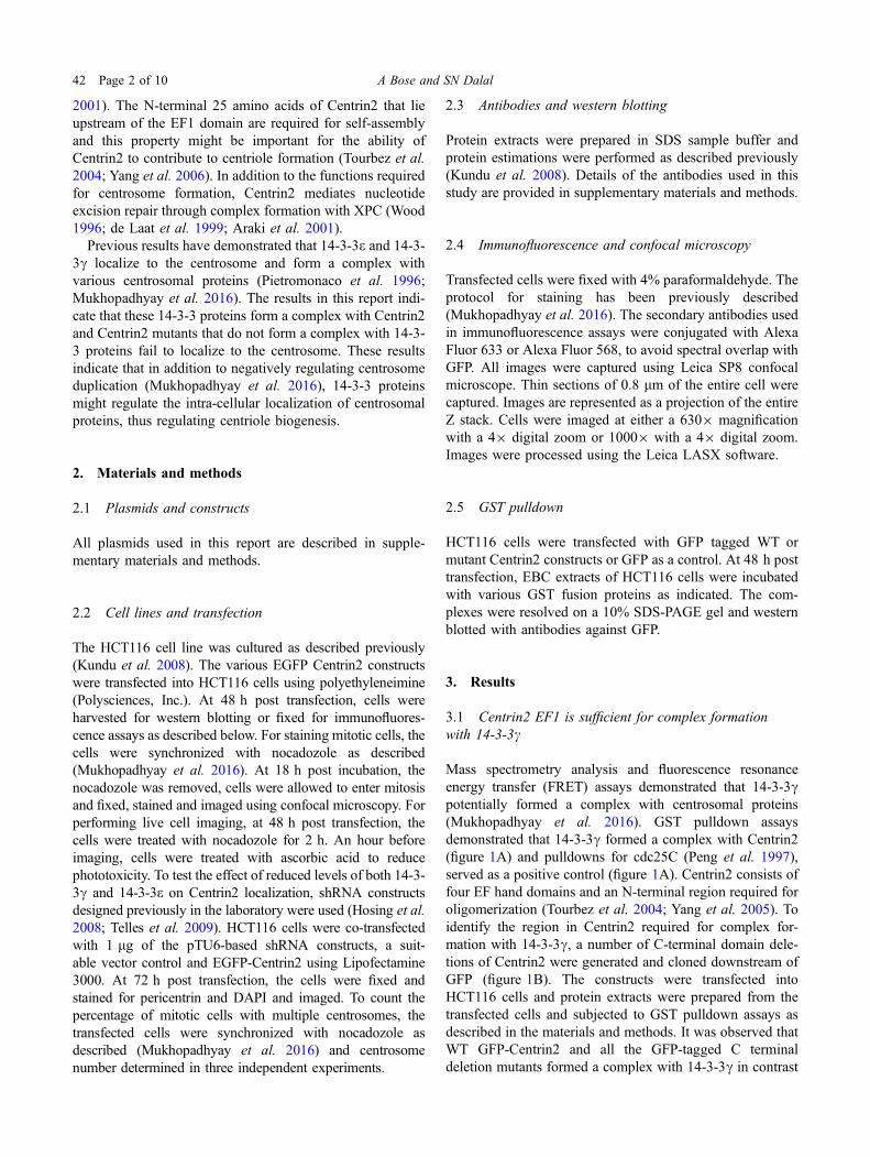
2001). The N-terminal 25 amino acids of Centrin2 that lieupstream of the EF1 domain are required for self-assemblyand this property might be important for the ability ofCentrin2 to contribute to centriole formation (Tourbez et al.2004; Yang et al. 2006). In addition to the functions requiredfor centrosome formation, Centrin2 mediates nucleotideexcision repair through complex formation with XPC (Wood1996; de Laat et al. 1999; Araki et al. 2001).
Previous results have demonstrated that 14-3-3e and 14-3-3c localize to the centrosome and form a complex withvarious centrosomal proteins (Pietromonaco et al. 1996;Mukhopadhyay et al. 2016). The results in this report indi-cate that these 14-3-3 proteins form a complex with Centrin2and Centrin2 mutants that do not form a complex with 14-3-3 proteins fail to localize to the centrosome. These resultsindicate that in addition to negatively regulating centrosomeduplication (Mukhopadhyay et al. 2016), 14-3-3 proteinsmight regulate the intra-cellular localization of centrosomalproteins, thus regulating centriole biogenesis.
2. Materials and methods
2.1 Plasmids and constructs
All plasmids used in this report are described in supple-mentary materials and methods.
2.2 Cell lines and transfection
The HCT116 cell line was cultured as described previously(Kundu et al. 2008). The various EGFP Centrin2 constructswere transfected into HCT116 cells using polyethyleneimine(Polysciences, Inc.). At 48 h post transfection, cells wereharvested for western blotting or fixed for immunofluores-cence assays as described below. For staining mitotic cells, thecells were synchronized with nocadozole as described(Mukhopadhyay et al. 2016). At 18 h post incubation, thenocadozole was removed, cells were allowed to enter mitosisand fixed, stained and imaged using confocal microscopy. Forperforming live cell imaging, at 48 h post transfection, thecells were treated with nocadozole for 2 h. An hour beforeimaging, cells were treated with ascorbic acid to reducephototoxicity. To test the effect of reduced levels of both 14-3-3c and 14-3-3e on Centrin2 localization, shRNA constructsdesigned previously in the laboratory were used (Hosing et al.2008; Telles et al. 2009). HCT116 cells were co-transfectedwith 1 lg of the pTU6-based shRNA constructs, a suit-able vector control and EGFP-Centrin2 using Lipofectamine3000. At 72 h post transfection, the cells were fixed andstained for pericentrin and DAPI and imaged. To count thepercentage of mitotic cells with multiple centrosomes, thetransfected cells were synchronized with nocadozole asdescribed (Mukhopadhyay et al. 2016) and centrosomenumber determined in three independent experiments.
2.3 Antibodies and western blotting
Protein extracts were prepared in SDS sample buffer andprotein estimations were performed as described previously(Kundu et al. 2008). Details of the antibodies used in thisstudy are provided in supplementary materials and methods.
2.4 Immunofluorescence and confocal microscopy
Transfected cells were fixed with 4% paraformaldehyde. Theprotocol for staining has been previously described(Mukhopadhyay et al. 2016). The secondary antibodies usedin immunofluorescence assays were conjugated with AlexaFluor 633 or Alexa Fluor 568, to avoid spectral overlap withGFP. All images were captured using Leica SP8 confocalmicroscope. Thin sections of 0.8 lm of the entire cell werecaptured. Images are represented as a projection of the entireZ stack. Cells were imaged at either a 6309 magnificationwith a 49 digital zoom or 10009 with a 49 digital zoom.Images were processed using the Leica LASX software.
2.5 GST pulldown
HCT116 cells were transfected with GFP tagged WT ormutant Centrin2 constructs or GFP as a control. At 48 h posttransfection, EBC extracts of HCT116 cells were incubatedwith various GST fusion proteins as indicated. The com-plexes were resolved on a 10% SDS-PAGE gel and westernblotted with antibodies against GFP.
3. Results
3.1 Centrin2 EF1 is sufficient for complex formationwith 14-3-3c
Mass spectrometry analysis and fluorescence resonanceenergy transfer (FRET) assays demonstrated that 14-3-3cpotentially formed a complex with centrosomal proteins(Mukhopadhyay et al. 2016). GST pulldown assaysdemonstrated that 14-3-3c formed a complex with Centrin2(figure 1A) and pulldowns for cdc25C (Peng et al. 1997),served as a positive control (figure 1A). Centrin2 consists offour EF hand domains and an N-terminal region required foroligomerization (Tourbez et al. 2004; Yang et al. 2005). Toidentify the region in Centrin2 required for complex for-mation with 14-3-3c, a number of C-terminal domain dele-tions of Centrin2 were generated and cloned downstream ofGFP (figure 1B). The constructs were transfected intoHCT116 cells and protein extracts were prepared from thetransfected cells and subjected to GST pulldown assays asdescribed in the materials and methods. It was observed thatWT GFP-Centrin2 and all the GFP-tagged C terminaldeletion mutants formed a complex with 14-3-3c in contrast
42 Page 2 of 10 A Bose and SN Dalal
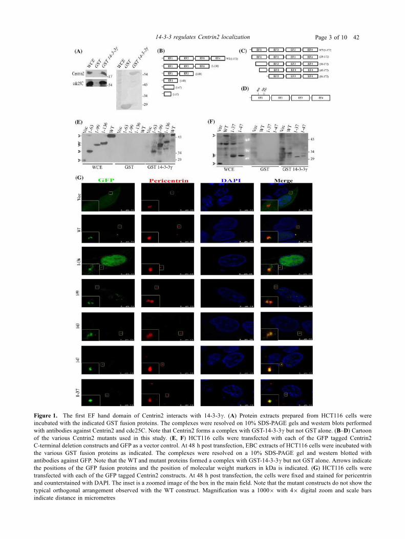
Figure 1. The first EF hand domain of Centrin2 interacts with 14-3-3c. (A) Protein extracts prepared from HCT116 cells wereincubated with the indicated GST fusion proteins. The complexes were resolved on 10% SDS-PAGE gels and western blots performedwith antibodies against Centrin2 and cdc25C. Note that Centrin2 forms a complex with GST-14-3-3c but not GST alone. (B–D) Cartoonof the various Centrin2 mutants used in this study. (E, F) HCT116 cells were transfected with each of the GFP tagged Centrin2C-terminal deletion constructs and GFP as a vector control. At 48 h post transfection, EBC extracts of HCT116 cells were incubated withthe various GST fusion proteins as indicated. The complexes were resolved on a 10% SDS-PAGE gel and western blotted withantibodies against GFP. Note that the WT and mutant proteins formed a complex with GST-14-3-3c but not GST alone. Arrows indicatethe positions of the GFP fusion proteins and the position of molecular weight markers in kDa is indicated. (G) HCT116 cells weretransfected with each of the GFP tagged Centrin2 constructs. At 48 h post transfection, the cells were fixed and stained for pericentrinand counterstained with DAPI. The inset is a zoomed image of the box in the main field. Note that the mutant constructs do not show thetypical orthogonal arrangement observed with the WT construct. Magnification was a 10009 with 49 digital zoom and scale barsindicate distance in micrometres
14-3-3 regulates Centrin2 localization Page 3 of 10 42
to GFP alone (figure 1E). Therefore, we concluded that theN-terminal 63 amino acids of Centrin2 are essential forcomplex formation with 14-3-3c. Shorter C-terminal dele-tions of Centrin2 expressing amino acids 1-37 and 1-47,were tested for their ability to form a complex with 14-3-3cand it was observed that both 1-37 and 1-47 formed acomplex with 14-3-3c (figure 1F).
In order to assess the localization of the C-terminalmutants of Centrin2, they were transfected in HCT116 cells.Post-transfection, the cells were stained with antibodies topericentrin and counter-stained with DAPI. It was observedthat all the C-terminal mutants localized to the centrosome asdemonstrated by their co-localization with pericentrin, in amanner similar to that observed for the WT Centrin2 protein(figure 1G). However, as opposed to what was observed inthe WT Centrin2 construct, none of the C-terminal mutantsappeared as two (G1 and S phase) or four (G2 and M phase)distinct Centrin2 dots (Paoletti et al. 1996). This could implythat the fourth EF hand domain might be involved in theaccurate self-assembly of Centrin2 at the centrosome. Inaddition, one mutant, 1-136, showed a significant enrich-ment in nuclear localization that was not observed witheither the other mutants or the WT protein (figure 1G). Thisis similar to the localization of only the C terminal region ofCentrin2 (aa. 95-172) observed previously (Nishi et al.2013). This region has been implicated in the binding toXPC and the nuclear localization of Centrin2. Nuclearsequestration of Centrin2 is suggested to play a role in theDNA damage response (Acu et al. 2010). It is possible thatthe third EF hand domain of Centrin2 alone is sufficient forits nuclear localization.
3.2 Deletion of EF1 in Centrin2 abrogates bindingwith 14-3-3c and leads to loss of the centrosomallocalization of Centrin2
As the construct with the longest C terminal deletion (1-37)of Centrin2 formed a complex with 14-3-3c, there were twopossibilities. One, that the 14-3-3c binding site in Centrin2lies in the first 37 amino acids and second, that there aremultiple 14-3-3 binding sites in Centrin2. To test thishypothesis, two types of N-terminal deletion mutants weregenerated (figure 1C). One lacked the first 28 amino acids(29-172) that are required for the self-assembly of Centrin2(Tourbez et al. 2004; Yang et al. 2006). The second typewere mutants that had sequential deletions in EF1 or deletedEF1 entirely. These mutants were transfected into HCT116cells and WT Centrin2 and GFP alone served as positive andnegative controls, respectively. GST pull-down assaysdemonstrated that deletion of the N-terminal region and EF1,abrogated binding to 14-3-3c (figure 2A). A deletion of thefirst 28 amino acids (29-172) formed a complex with 14-3-3c (figure 2A) suggesting that the 14-3-3c binding site waspresent in EF1. However, further N-terminal deletions (38-172 and 48-172) formed a complex with 14-3-3c
(figure 2A). These results were surprising as the C-terminaldeletion mutants, 1-47 and 1-37, also formed a complex with14-3-3c (figure 1F). These results suggest that there mightbe multiple 14-3-3c binding sites in the N-terminal 63 aminoacids of Centrin2.
The prediction web server (http://www.compbio.dundee.ac.uk/1433pred) was used to identify potential 14-3-3binding sites in Centrin2 (Madeira et al. 2015). Twopotential 14-3-3 binding sites were identified within EF1 inCentrin2; T45 and T47. The threonine residues that were theputative phosphate acceptors in these potential 14-3-3binding sites were altered to alanine (T45A and T47A). Inaddition, we hypothesized that an aspartic acid residue atposition 39 (D39) could potentially serve as a phospho-mimetic residue in a potential 14-3-3 binding site. Thisresidue was altered to alanine (D39A). All these pointmutants were tested for their ability to form a complex with14-3-3c in GST pull-down assays. It was observed that all ofthe point mutants were able to form a complex with 14-3-3c(figure 2B). A double mutant (T45/47A) also formed acomplex with 14-3-3c (figure 2B). These results suggestedthat none of these potential 14-3-3c binding sites in Centrin2was required for complex formation with 14-3-3c.
All the N-terminal Centrin2 mutants that formed a com-plex with 14-3-3c localized to the centrosome as demon-strated by the observation that the GFP signal co-localizedwith the pericentrin signal (figure 2C). Interestingly, theorganization of these mutants at the centrosome was similarto that of the C-terminal mutants, i.e. unable to form the 2 or4 dots typical of Centrin2 localization, but accumulatedstrongly at the centrosome. The single amino acid mutantsalso localized to the centrosome, in a pattern similar to thefull-length construct (supplementary figure 1A). However,the 64-172 mutant that failed to form a complex with 14-3-3c did not localize to the centrosome or the nucleus (fig-ure 2C) and showed a punctate pattern throughout thecytoplasm. This was observed in all cells transfected withthe mutant 64-172 and suggests that 14-3-3c binding toCentrin2 is required for the localization of Centrin2 to thecentrosome.
3.3 Centrin2 binds to both 14-3-3e and 14-3-3c
Our previous results had indicated that both 14-3-3e and14-3-3c can regulate centrosome duplication and that loss of14-3-3c did not lead to a disruption of centrin localization tothe centrosome (Mukhopadhyay et al. 2016). This raised thepossibility that multiple 14-3-3 proteins can bind to Centrin2and regulate its localization to the centrosome. To test thishypothesis, protein extracts from HCT116 cells were incu-bated with either GST alone or GST tagged 14-3-3e or 14-3-3c, the reactions resolved on SDS-PAGE gels followed bywestern blotting with antibodies to Centrin2 and cdc25C. Asshown in figure 3A, both 14-3-3e and 14-3-3c formed acomplex with Centrin2. To determine if 14-3-3e bound to the
42 Page 4 of 10 A Bose and SN Dalal

Figure 2. Deletion of the first EF hand domain of Centrin2 abrogates binding with 14-3-3c and leads to loss of the centrosomallocalization of Centrin2. (A, B) HCT116 cells were transfected with the indicated GFP tagged WT or Centrin2 mutant constructs and GFPas a vector control. At 48 h post transfection, EBC extracts of HCT116 cells were incubated with the various GST fusion proteins asindicated. The complexes were resolved on a 10% SDS-PAGE gel and western blots performed with antibodies against GFP. Note that64-172 does not form a complex with GST-14-3-3c in contrast to the WT or other Centrin2 mutants. * Indicates a non-specific band.Arrows indicate the positions of the GFP fusion proteins and the position of molecular weight markers in kDa is indicated. (C) HCT116cells seeded on coverslips were transfected with the GFP tagged WT or mutant Centrin2 constructs. At 48 h post transfection, the cells werefixed and stained for pericentrin and DAPI. The inset is a zoomed image of the box in the main field. Note that the 64-172 construct doesnot localize to the centrosome unlike the WT or other mutant constructs. The deletion mutants do not show the typical orthogonalarrangement observed with the WT construct. Magnification was a 10009 with 49 digital zoom and scale bars indicate distance inmicrometres
14-3-3 regulates Centrin2 localization Page 5 of 10 42
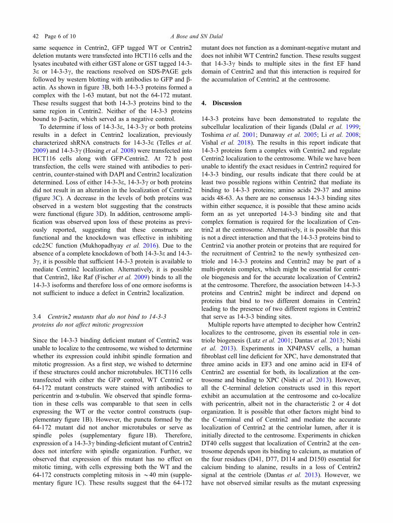
same sequence in Centrin2, GFP tagged WT or Centrin2deletion mutants were transfected into HCT116 cells and thelysates incubated with either GST alone or GST tagged 14-3-3e or 14-3-3c, the reactions resolved on SDS-PAGE gelsfollowed by western blotting with antibodies to GFP and b-actin. As shown in figure 3B, both 14-3-3 proteins formed acomplex with the 1-63 mutant, but not the 64-172 mutant.These results suggest that both 14-3-3 proteins bind to thesame region in Centrin2. Neither of the 14-3-3 proteinsbound to b-actin, which served as a negative control.
To determine if loss of 14-3-3e, 14-3-3c or both proteinsresults in a defect in Centrin2 localization, previouslycharacterized shRNA constructs for 14-3-3e (Telles et al.2009) and 14-3-3c (Hosing et al. 2008) were transfected intoHCT116 cells along with GFP-Centrin2. At 72 h posttransfection, the cells were stained with antibodies to peri-centrin, counter-stained with DAPI and Centrin2 localizationdetermined. Loss of either 14-3-3e, 14-3-3c or both proteinsdid not result in an alteration in the localization of Centrin2(figure 3C). A decrease in the levels of both proteins wasobserved in a western blot suggesting that the constructswere functional (figure 3D). In addition, centrosome ampli-fication was observed upon loss of these proteins as previ-ously reported, suggesting that these constructs arefunctional and the knockdown was effective in inhibitingcdc25C function (Mukhopadhyay et al. 2016). Due to theabsence of a complete knockdown of both 14-3-3e and 14-3-3c, it is possible that sufficient 14-3-3 protein is available tomediate Centrin2 localization. Alternatively, it is possiblethat Centrin2, like Raf (Fischer et al. 2009) binds to all the14-3-3 isoforms and therefore loss of one ormore isoforms isnot sufficient to induce a defect in Centrin2 localization.
3.4 Centrin2 mutants that do not bind to 14-3-3proteins do not affect mitotic progression
Since the 14-3-3 binding deficient mutant of Centrin2 wasunable to localize to the centrosome, we wished to determinewhether its expression could inhibit spindle formation andmitotic progression. As a first step, we wished to determineif these structures could anchor microtubules. HCT116 cellstransfected with either the GFP control, WT Centrin2 or64-172 mutant constructs were stained with antibodies topericentrin and a-tubulin. We observed that spindle forma-tion in these cells was comparable to that seen in cellsexpressing the WT or the vector control constructs (sup-plementary figure 1B). However, the puncta formed by the64-172 mutant did not anchor microtubules or serve asspindle poles (supplementary figure 1B). Therefore,expression of a 14-3-3c binding-deficient mutant of Centrin2does not interfere with spindle organization. Further, weobserved that expression of this mutant has no effect onmitotic timing, with cells expressing both the WT and the64-172 constructs completing mitosis in *40 min (supple-mentary figure 1C). These results suggest that the 64-172
mutant does not function as a dominant-negative mutant anddoes not inhibit WT Centrin2 function. These results suggestthat 14-3-3c binds to multiple sites in the first EF handdomain of Centrin2 and that this interaction is required forthe accumulation of Centrin2 at the centrosome.
4. Discussion
14-3-3 proteins have been demonstrated to regulate thesubcellular localization of their ligands (Dalal et al. 1999;Toshima et al. 2001; Dunaway et al. 2005; Li et al. 2008;Vishal et al. 2018). The results in this report indicate that14-3-3 proteins form a complex with Centrin2 and regulateCentrin2 localization to the centrosome. While we have beenunable to identify the exact residues in Centrin2 required for14-3-3 binding, our results indicate that there could be atleast two possible regions within Centrin2 that mediate itsbinding to 14-3-3 proteins; amino acids 29-37 and aminoacids 48-63. As there are no consensus 14-3-3 binding siteswithin either sequence, it is possible that these amino acidsform an as yet unreported 14-3-3 binding site and thatcomplex formation is required for the localization of Cen-trin2 at the centrosome. Alternatively, it is possible that thisis not a direct interaction and that the 14-3-3 proteins bind toCentrin2 via another protein or proteins that are required forthe recruitment of Centrin2 to the newly synthesized cen-triole and 14-3-3 proteins and Centrin2 may be part of amulti-protein complex, which might be essential for centri-ole biogenesis and for the accurate localization of Centrin2at the centrosome. Therefore, the association between 14-3-3proteins and Centrin2 might be indirect and depend onproteins that bind to two different domains in Centrin2leading to the presence of two different regions in Centrin2that serve as 14-3-3 binding sites.
Multiple reports have attempted to decipher how Centrin2localizes to the centrosome, given its essential role in cen-triole biogenesis (Lutz et al. 2001; Dantas et al. 2013; Nishiet al. 2013). Experiments in XP4PASV cells, a humanfibroblast cell line deficient for XPC, have demonstrated thatthree amino acids in EF3 and one amino acid in EF4 ofCentrin2 are essential for both, its localization at the cen-trosome and binding to XPC (Nishi et al. 2013). However,all the C-terminal deletion constructs used in this reportexhibit an accumulation at the centrosome and co-localizewith pericentrin, albeit not in the characteristic 2 or 4 dotorganization. It is possible that other factors might bind tothe C-terminal end of Centrin2 and mediate the accuratelocalization of Centrin2 at the centriolar lumen, after it isinitially directed to the centrosome. Experiments in chickenDT40 cells suggest that localization of Centrin2 at the cen-trosome depends upon its binding to calcium, as mutation ofthe four residues (D41, D77, D114 and D150) essential forcalcium binding to alanine, results in a loss of Centrin2signal at the centriole (Dantas et al. 2013). However, wehave not observed similar results as the mutant expressing
42 Page 6 of 10 A Bose and SN Dalal
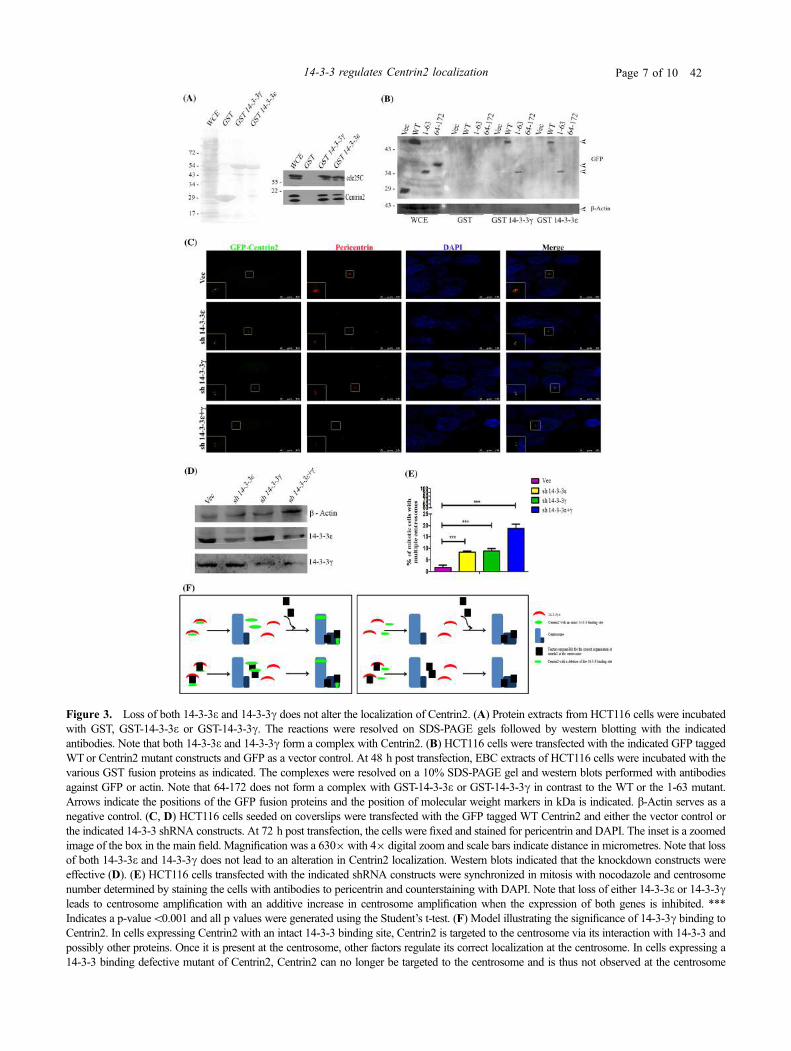
Figure 3. Loss of both 14-3-3e and 14-3-3c does not alter the localization of Centrin2. (A) Protein extracts from HCT116 cells were incubatedwith GST, GST-14-3-3e or GST-14-3-3c. The reactions were resolved on SDS-PAGE gels followed by western blotting with the indicatedantibodies. Note that both 14-3-3e and 14-3-3c form a complex with Centrin2. (B) HCT116 cells were transfected with the indicated GFP taggedWT or Centrin2 mutant constructs and GFP as a vector control. At 48 h post transfection, EBC extracts of HCT116 cells were incubated with thevarious GST fusion proteins as indicated. The complexes were resolved on a 10% SDS-PAGE gel and western blots performed with antibodiesagainst GFP or actin. Note that 64-172 does not form a complex with GST-14-3-3e or GST-14-3-3c in contrast to the WT or the 1-63 mutant.Arrows indicate the positions of the GFP fusion proteins and the position of molecular weight markers in kDa is indicated. b-Actin serves as anegative control. (C, D) HCT116 cells seeded on coverslips were transfected with the GFP tagged WT Centrin2 and either the vector control orthe indicated 14-3-3 shRNA constructs. At 72 h post transfection, the cells were fixed and stained for pericentrin and DAPI. The inset is a zoomedimage of the box in the main field. Magnification was a 6309with 49 digital zoom and scale bars indicate distance in micrometres. Note that lossof both 14-3-3e and 14-3-3c does not lead to an alteration in Centrin2 localization. Western blots indicated that the knockdown constructs wereeffective (D). (E) HCT116 cells transfected with the indicated shRNA constructs were synchronized in mitosis with nocodazole and centrosomenumber determined by staining the cells with antibodies to pericentrin and counterstaining with DAPI. Note that loss of either 14-3-3e or 14-3-3cleads to centrosome amplification with an additive increase in centrosome amplification when the expression of both genes is inhibited. ***Indicates a p-value\0.001 and all p values were generated using the Student’s t-test. (F) Model illustrating the significance of 14-3-3c binding toCentrin2. In cells expressing Centrin2 with an intact 14-3-3 binding site, Centrin2 is targeted to the centrosome via its interaction with 14-3-3 andpossibly other proteins. Once it is present at the centrosome, other factors regulate its correct localization at the centrosome. In cells expressing a14-3-3 binding defective mutant of Centrin2, Centrin2 can no longer be targeted to the centrosome and is thus not observed at the centrosome
14-3-3 regulates Centrin2 localization Page 7 of 10 42
the shortest Centrin2 fragment, 1-37, does not possess any ofthe four calcium binding residues, but still localizes to thecentrosome. These differences might reflect minor differ-ences in centriole biogenesis between species. It has beenreported that the N-terminus of Centrin1 might form acomplex with the C-terminus of Centrin1 (Kim et al. 2017),which suggests that the N-terminal mutants of Centrin2might form a complex with the endogenous Centrin2.However, the entire EF1 domain in Centrin1 is required fordimerization with the C-terminal EF hands in Centrin1 andthe 1-37 mutant does not have the entire EF hand. Therefore,it is unlikely that the 1-37 mutant localizes to the centrosomeby forming a dimer with the endogenous Centrin2. Further,the 14-3-3c binding defective mutant, 64-172, showed nonuclear localization. This could be due to the fact thatnuclear localization of Centrin2 has been linked to itsmodification by SUMOylation (Klein and Nigg 2009). Threeputative SUMOylation sites are present within the first 64amino acid region of Centrin2. It is possible that one of thesesites could be responsible for SUMOylation and that is whydeletion of this region results in loss of its nuclearlocalization.
Our previous results have demonstrated that 14-3-3e and14-3-3c can inhibit premature centrosome duplication byinhibiting cdc25C function (Mukhopadhyay et al. 2016).Cdc25C has a mode I 14-3-3 binding site (Muslin et al.1996; Yaffe et al. 1997) and association between cdc25Cand 14-3-3 proteins requires an intact serine residue atposition 216 (S216) in cdc25C (Peng et al. 1997; Dalalet al. 1999, 2004). However, while cdc25C binds to only14-3-3e and 14-3-3c (Dalal et al. 2004), Raf, which alsocontains a mode I 14-3-3 binding site forms a complex withall the 14-3-3 isoforms (Fischer et al. 2009). Therefore,some redundancy exists in the requirement for 14-3-3binding to ligands. In this report, we demonstrate that both14-3-3e and 14-3-3c form a complex with Centrin2, how-ever, a decrease in the levels of both proteins does notaffect Centrin2 localization but does result in centrosomeamplification (figure 3), possibly due to an increase incdc25C function as previously reported (Mukhopadhyayet al. 2016). 14-3-3 proteins bind to Centrin2 via a non-canonical 14-3-3 binding site while they form a complexwith cdc25C through a mode I 14-3-3 binding site.Therefore, it is possible that even though the levels of 14-3-3e and 14-3-3c are lowered upon the knockdown of theseproteins, sufficient protein is available to mediate local-ization to the centrosome. Alternatively, it is possible thatlike Raf (Fischer et al. 2009), Centrin2 can form a complexwith all the 14-3-3 isoforms suggesting that the role of14-3-3 proteins in regulating the localization of Centrin2could be shared by multiple 14-3-3 isoforms. This redun-dancy is consistent with the observation that Centrin2 isessential for centrosome duplication and cell viability(Salisbury et al. 2002). Therefore, multiple 14-3-3 proteinsmight be required to regulate the localization of Centrin2 tothe centrosome.
On the basis of our results, we propose the followingmodel (figure 3F). We have demonstrated previously, that14-3-3e and 14-3-3c localize to the centrosome(Mukhopadhyay et al. 2016). In cells expressing Centrin2with an intact 14-3-3 binding site, Centrin2 is targeted to thecentrosome via its binding to 14-3-3c. Once there, it selfassembles at the centriole permitting centriole biogenesis.Given that mutation of all potential phosphorylated residuesin EF1 that could serve as 14-3-3 binding sites does notabolish complex formation with 14-3-3 proteins, it is pos-sible that the interaction between 14-3-3 proteins and Cen-trin2 is either independent of phosphorylation or complexformation between 14-3-3 proteins and Centrin2 is depen-dent on other proteins required for centriole biogenesis.Once it is present at the centrosome, presumably, otherfactors which might bind to the C-terminal region of Cen-trin2, direct the organization of Centrin2 to form the cen-triolar lumen. In cells expressing a mutant of Centrin2 thateither fails to form a complex with 14-3-3 proteins or anadaptor protein that bridges the interaction with 14-3-3proteins, 14-3-3e and 14-3-3c are no longer able to targeteither Centrin2 or the Centrin2 protein complex to the cen-trosome resulting in the absence of Centrin2 at the centro-some. Thus, in addition to inhibiting premature centrosomeduplication in late S and G2 phase by inactivating cdc25Cfunction (Mukhopadhyay et al. 2016), 14-3-3e and 14-3-3cmight promote centriole biogenesis by mediating the local-ization of Centrin2 to the centrosome.
Acknowledgements
We thank Dr Erich Nigg, for his kind gift of the EGFP C2Centrin2 construct and the ACTREC microscopy facility forhelp with the imaging experiments in this report. The workwas funded by grants from the Department of Biotechnol-ogy, Govt. of India (BT/PR4181/BRB/10/1147/2012 andBT/PR8351/MED/30/995/2013) to SND.
References
Acu ID, Liu T, Suino-Powell K, Mooney SM, D’Assoro AB,Rowland N, Muotri AR, Correa RG, Niu Y, Kumar R andSalisbury JL 2010 Coordination of centrosome homeostasis andDNA repair is intact in MCF-7 and disrupted in MDA-MB 231breast cancer cells. Cancer Res. 70 3320–3328
Aitken A 2006 14-3-3 proteins: A historic overview. Semin. CancerBiol. 16 162–172
Araki M, Masutani C, Takemura M, Uchida A, Sugasawa K,Kondoh J, Ohkuma Y and Hanaoka F 2001 Centrosome proteincentrin 2/caltractin 1 is part of the xeroderma pigmentosumgroup C complex that initiates global genome nucleotideexcision repair. J. Biol. Chem. 276 18665–18672
Azimzadeh J and Marshall WF 2010 Building the centriole. Curr.Biol. 20 R816–R825
42 Page 8 of 10 A Bose and SN Dalal
Coblitz B, Shikano S, Wu M, Gabelli SB, Cockrell LM, Spieker M,Hanyu Y, Fu H, Amzel LM and Li M 2005 C-terminalrecognition by 14-3-3 proteins for surface expression ofmembrane receptors. J. Biol. Chem. 280 36263–36272
Conduit PT, Wainman A and Raff JW 2015 Centrosome functionand assembly in animal cells. Nat. Rev. Mol. Cell Biol. 16611–624
Dalal SN, Schweitzer CM, Gan J and DeCaprio JA 1999Cytoplasmic localization of human cdc25C during interphaserequires an intact 14-3-3 binding site. Mol. Cell. Biol. 194465–4479
Dalal SN, Yaffe MB and DeCaprio JA 2004 14-3-3 familymembers act coordinately to regulate mitotic progression. CellCycle 3 672–677
Dantas TJ, Daly OM and Morrison CG 2012 Such small hands: Theroles of centrins/caltractins in the centriole and in genomemaintenance. Cell. Mol. Life Sci. 69 2979–2997
Dantas TJ, Daly OM, Conroy PC, Tomas M, Wang Y, Lalor P,Dockery P, Ferrando-May E and Morrison CG 2013 Calcium-binding capacity of centrin2 is required for linear POC5assembly but not for nucleotide excision repair. PLoS One 8e68487
de Laat WL, Jaspers NG and Hoeijmakers JH 1999 Molecularmechanism of nucleotide excision repair. Genes Dev. 13768–785
Dunaway S, Liu HY and Walworth NC 2005 Interaction of 14-3-3protein with Chk1 affects localization and checkpoint function.J. Cell Sci. 118 39–50
Fischer A, Baljuls A, Reinders J, Nekhoroshkova E, Sibilski C,Metz R, Albert S, Rajalingam K, Hekman M and Rapp UR 2009Regulation of RAF activity by 14-3-3 proteins: RAF kinasesassociate functionally with both homo- and heterodimeric formsof 14-3-3 proteins. J. Biol. Chem. 284 3183–3194
Gardino AK and Yaffe MB 2011 14-3-3 proteins as signalingintegration points for cell cycle control and apoptosis. Semin.Cell Dev. Biol. 22 688–695
Hosing AS, Kundu ST and Dalal SN 2008 14-3-3 gamma isrequired to enforce both the incomplete S phase and G2 DNAdamage checkpoints. Cell Cycle 7 3171–3179
Kim SY, Kim DS, Hong JE and Park JH 2017 Crystal structure ofwild-type centrin 1 from Mus musculus occupied by Ca2.Biochemistry (Mosc) 82 1129–1139
Klein UR and Nigg EA 2009 SUMO-dependent regulation ofcentrin-2. J. Cell Sci. 122 3312–3321
Kundu ST, Gosavi P, Khapare N, Patel R, Hosing AS, Maru GB,Ingle A, Decaprio JA and Dalal SN 2008 Plakophilin3downregulation leads to a decrease in cell adhesion andpromotes metastasis. Int. J. Cancer 123 2303–2314
Li FQ, Mofunanya A, Harris K and Takemaru K 2008 Chibbycooperates with 14-3-3 to regulate beta-catenin subcellulardistribution and signaling activity. J. Cell Biol. 1811141–1154
Lutz W, Lingle WL, McCormick D, Greenwood TM and SalisburyJL 2001 Phosphorylation of centrin during the cell cycle and itsrole in centriole separation preceding centrosome duplication. J.Biol. Chem. 276 20774–20780
Madeira F, Tinti M, Murugesan G, Berrett E, Stafford M, Toth R,Cole C, MacKintosh C and Barton GJ 2015 14-3-3-Pred:Improved methods to predict 14-3-3-binding phosphopeptides.Bioinformatics 31 2276–2283
Mukhopadhyay A, Sehgal L, Bose A, Gulvady A, Senapati P,Thorat R, Basu S, Bhatt K, Hosing AS, Balyan R, Borde L,Kundu TK and Dalal SN 2016 14-3-3 gamma preventscentrosome amplification and neoplastic progression. Sci. Rep.6. https://doi.org/10.1038/srep26580
Muslin AJ, Tanner JW, Allen PM and Shaw AS 1996 Interaction of14-3-3 with signaling proteins is mediated by recognition ofphosphoserine. Cell 84 889–897
Nigg EA and Holland AJ 2018 Once and only once: Mechanismsof centriole duplication and their deregulation in disease. Nat.Rev. Mol. Cell Biol. 19 297–312
Nishi R, Sakai W, Tone D, Hanaoka F and Sugasawa K 2013Structure-function analysis of the EF-hand protein centrin-2 forits intracellular localization and nucleotide excision repair.Nucleic Acids Res. 41 6917–6929
Ogihara T, Isobe T, Ichimura T, Taoka M, Funaki M, Sakoda H,Onishi Y, Inukai K, Anai M, Fukushima Y, Kikuchi M, YazakiY, Oka Y and Asano T 1997 14-3-3 protein binds to insulinreceptor substrate-1, one of the binding sites of which is in thephosphotyrosine binding domain. J. Biol. Chem. 27225267–25274
Paoletti A, Moudjou M, Paintrand M, Salisbury JL and Bornens M1996 Most of centrin in animal cells is not centrosome-associated and centrosomal centrin is confined to the distallumen of centrioles. J. Cell Sci. 109 3089–3102
Peng CY, Graves PR, Thoma RS, Wu Z, Shaw AS and Piwnica-Worms H 1997 Mitotic and G2 checkpoint control: Regulationof 14-3-3 protein binding by phosphorylation of Cdc25C onserine-216. Science 277 1501–1505
Pietromonaco SF, Seluja GA, Aitken A and Elias L 1996Association of 14-3-3 proteins with centrosomes. Blood CellsMol. Dis. 22 225–237
Salisbury JL, Suino KM, Busby R and Springett M 2002 Centrin-2is required for centriole duplication in mammalian cells. Curr.Biol. 12 1287–1292
Telles E, Hosing AS, Kundu ST, Venkatraman P and Dalal SN 2009A novel pocket in 14-3-3e is required to mediate specificcomplex formation with cdc25C and to inhibit cell cycleprogression upon activation of checkpoint pathways. Exp. CellRes. 315 1448–1457
Toshima JY, Toshima J, Watanabe T and Mizuno K 2001 Bindingof 14-3-3beta regulates the kinase activity and subcellularlocalization of testicular protein kinase 1. J. Biol. Chem. 27643471–43481
Tourbez M, Firanescu C, Yang A, Unipan L, Duchambon P,Blouquit Y and Craescu CT 2004 Calcium-dependent self-assembly of human centrin 2. J. Biol. Chem. 27947672–47680
Tzivion G, Shen YH and Zhu J 2001 14-3-3 proteins; bringing newdefinitions to scaffolding. Oncogene 20 6331–6338
Tzivion G, Gupta VS, Kaplun L and Balan V 2006 14-3-3 proteinsas potential oncogenes. Semin. Cancer Biol. 16 203–213
Vishal SS, Tilwani S and Dalal SN 2018 Plakoglobin localization tothe cell border restores desmosome function in cells lacking14-3-3gamma. Biochem. Biophys. Res. Commun. 4951998–2003
Wolfrum U and Salisbury JL 1998 Expression of centrin isoformsin the mammalian retina. Exp. Cell Res. 242 10–17
Wood RD 1996 DNA repair in eukaryotes. Annu. Rev. Biochem. 65135–167
14-3-3 regulates Centrin2 localization Page 9 of 10 42

Yaffe MB, Rittinger K, Volinia S, Caron PR, Aitken A, Leffers H,Gamblin SJ, Smerdon SJ and Cantley LC 1997 The structuralbasis for 14-3-3: Phosphopeptide binding specificity. Cell 91961–971
Yang A, Miron S, Duchambon P, Assairi L, Blouquit Y and CraescuCT 2005 The N-terminal domain of human Centrin 2 has a
closed structure, binds calcium with a very low affinity, andplays a role in the protein self assembly. Biochemistry 45 10
Yang A, Miron S, Duchambon P, Assairi L, Blouquit Y and CraescuCT 2006 The N-terminal domain of human centrin 2 has a closedstructure, binds calcium with a very low affinity, and plays a role inthe protein self-assembly. Biochemistry 45 880–889
Corresponding editor: KUNDAN SENGUPTA
42 Page 10 of 10 A Bose and SN Dalal
Metadata of the chapter that will be visualized online
Chapter Title Centrosome Amplification and Tumorigenesis: Cause or Effect?
Copyright Year 2019
Copyright Holder Springer Nature Switzerland AG
Author Family Name Bose
Particle
Given Name Arunabha
Suffix
Organization KS215, Advanced Centre for Treatment Research andEducation in Cancer (ACTREC), Tata Memorial Centre
Address Navi Mumbai, Maharashtra, India
Organization Homi Bhabha National Institute
Address Mumbai, Maharashtra, India
Corresponding Author Family Name Dalal
Particle
Given Name Sorab N.
Suffix
Organization KS215, Advanced Centre for Treatment Research andEducation in Cancer (ACTREC), Tata Memorial Centre
Address Navi Mumbai, Maharashtra, India
Organization Homi Bhabha National Institute
Address Mumbai, Maharashtra, India
Email [email protected]
Abstract Centrosome amplification is a feature of multiple tumour types and has beenpostulated to contribute to both tumour initiation and tumour progression.This chapter focuses on the mechanisms by which an increase in centrosomenumber might lead to an increase or decrease in tumour progression and therole of proteins that regulate centrosome number in driving tumorigenesis.
Keywords(separated by ‘-’)
Centrosome - Centriole - Aneuploidy - Chromosomal instability
BookID 461942_1_En__ChapID 18_Proof# 1 - 21/6/19
1Chapter 182Centrosome Amplification3and Tumorigenesis: Cause or Effect?
4Arunabha Bose and Sorab N. Dalal
5Abstract Centrosome amplification is a feature of multiple tumour types and has6been postulated to contribute to both tumour initiation and tumour progression. This7chapter focuses on the mechanisms by which an increase in centrosome number8might lead to an increase or decrease in tumour progression and the role of proteins9that regulate centrosome number in driving tumorigenesis.10
11Abbreviations
AKAP450 12A-kinase-anchoring protein 450cdc25C 13Cell division cycle 25CCDK 14Cyclin-dependent kinaseCDK5RAP2 15CDK5 regulatory Subunit-associated protein 2CENP-E 16CENtrosome-associated Protein ECep 17Centrosomal proteinCLIP-70 18Cytoplasmic LInker Protein 170C-NAP1 19Centrosomal Nek2-associated protein 1CP110 20Centriolar coiled-coil protein of 110 kDaCPAP 21Centrosomal P4.1-associated proteinEGFR 22Epidermal growth factor receptorFBF1 23Fas-binding factor 1GCP 24γ-tubulin complex proteinhPOC5 25human Proteome of Centriole 5INCENP 26Inner CENtromere ProteinLRRC45 27Leucine-rich repeat containing 45NEDD1 28Neural precursor cell expressed developmentally down-regulated
protein 1
A. Bose · S. N. Dalal (*)KS215 AU1, Advanced Centre for Treatment Research and Education in Cancer (ACTREC), TataMemorial Centre, Navi Mumbai, Maharashtra, India
Homi Bhabha National Institute, Mumbai, Maharashtra, Indiae-mail: [email protected]
© Springer Nature Switzerland AG 2019M. Kloc (ed.), The Golgi apparatus and Centriole, Results and Problems in CellDifferentiation 67, https://doi.org/10.1007/978-3-030-23173-6_18
ODF229 Outer dense fiber protein 2SAS-630 Spindle assembly abnormal protein 6SCLT131 Sodium channel and clathrin linker 1SMC332 Structural maintenance of chromosomes protein 3STIL33 SCL interrupting locus proteinTACC234 Transforming acidic coiled-coil-containing protein 2TRF135 Telomeric repeat-binding factor 1
36 18.1 Introduction
37 The cell cycle is a carefully orchestrated process that leads to the accurate segrega-38 tion of chromosomes into two daughter cells (Malumbres and Barbacid 2007).39 Accurate genome segregation is accomplished by aligning the paired sister chroma-40 tids along the metaphase plate and attachment of the sister chromatids to the mitotic41 spindle (Reber and Hyman 2015; Walczak and Heald 2008). Following attachment42 to the spindle, the sister chromatids are ‘pulled’ apart by forces generated by spindle43 microtubules that originate from the two poles. Nucleation of the microtubules at the44 two poles, such that they are organized into a spindle, is mediated by a cellular45 organelle, called the centrosome (Fu et al. 2016).46 The centrosome is a membrane-less organelle, which is the primary microtubule47 organizing centre (MTOC) in most eukaryotic cells. Its ability to organize microtu-48 bules results in it being essential for multiple cellular functions including the49 generation of the mitotic spindle, regulation of cell cycle progression, the biogenesis50 of and signalling from cilia, the determination of cell fate, cellular trafficking and the51 generation of an effective immune response (Arquint et al. 2014; Reina and52 Gonzalez 2014; Rios 2014; Stinchcombe and Griffiths 2014). Each centrosome53 consists of a pair of centrioles surrounded by an amorphous structure called the54 pericentriolar matrix (Luders 2012) (Fig. 18.1). The two centrioles are arranged55 orthogonally, and each centriole is organized in a typical 9+3 structure, which56 alludes to the nine sets of triplet microtubules arranged around a central cartwheel57 (Gonczy 2012). This cartwheel consists of a hub and radial spokes which are made58 of nine homodimers of Sas-6 (Kitagawa et al. 2011). The spokes emanating from the59 hub bind to the first microtubule of each triplet through an interaction between60 Cep135 and the first microtubule of the triplet (Guichard et al. 2017).61 In human cells, a mature centriole is a cylinder, ~450 nm in length, with inner and62 outer diameters of ~130 nm and ~250 nm, respectively (Winey and O’Toole 2014).63 The centriole is said to have a polarized structure, with a proximal end (the base), and64 a distal end (the tip). The two centrioles in the centrosome are a mother centriole,65 which is inherited from the previous cell cycle, and a daughter centriole, whose66 synthesis is initiated in the current cell cycle during S phase (Fu et al. 2015). The67 mother centriole can be distinguished from the daughter centriole, by the presence of68 distal and sub-distal appendages (Fig. 18.1). The distal appendage proteins such as
A. Bose and S. N. Dalal
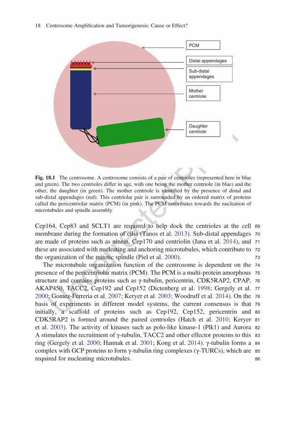
69Cep164, Cep83 and SCLT1 are required to help dock the centrioles at the cell70membrane during the formation of cilia (Tanos et al. 2013). Sub-distal appendages71are made of proteins such as ninein, Cep170 and centriolin (Jana et al. 2014), and72these are associated with nucleating and anchoring microtubules, which contribute to73the organization of the mitotic spindle (Piel et al. 2000).74The microtubule organization function of the centrosome is dependent on the75presence of the pericentriolar matrix (PCM). The PCM is a multi-protein amorphous76structure and contains proteins such as γ-tubulin, pericentrin, CDK5RAP2, CPAP,77AKAP450, TACC2, Cep192 and Cep152 (Dictenberg et al. 1998; Gergely et al.782000; Gomez-Ferreria et al. 2007; Keryer et al. 2003; Woodruff et al. 2014). On the79basis of experiments in different model systems, the current consensus is that80initially, a scaffold of proteins such as Cep192, Cep152, pericentrin and81CDK5RAP2 is formed around the paired centrioles (Hatch et al. 2010; Keryer82et al. 2003). The activity of kinases such as polo-like kinase-1 (Plk1) and Aurora83A stimulates the recruitment of γ-tubulin, TACC2 and other effector proteins to this84ring (Gergely et al. 2000; Hannak et al. 2001; Kong et al. 2014). γ-tubulin forms a85complex with GCP proteins to form γ-tubulin ring complexes (γ-TURCs), which are86required for nucleating microtubules.
PCM
Distal appendages
Sub-distalappendages
Mothercentriole
Daughtercentriole
Fig. 18.1 The centrosome. A centrosome consists of a pair of centrioles (represented here in blueand green). The two centrioles differ in age, with one being the mother centriole (in blue) and theother, the daughter (in green). The mother centriole is identified by the presence of distal andsub-distal appendages (red). This centriolar pair is surrounded by an ordered matrix of proteinscalled the pericentriolar matrix (PCM) (in pink). The PCM contributes towards the nucleation ofmicrotubules and spindle assembly
18 Centrosome Amplification and Tumorigenesis: Cause or Effect?
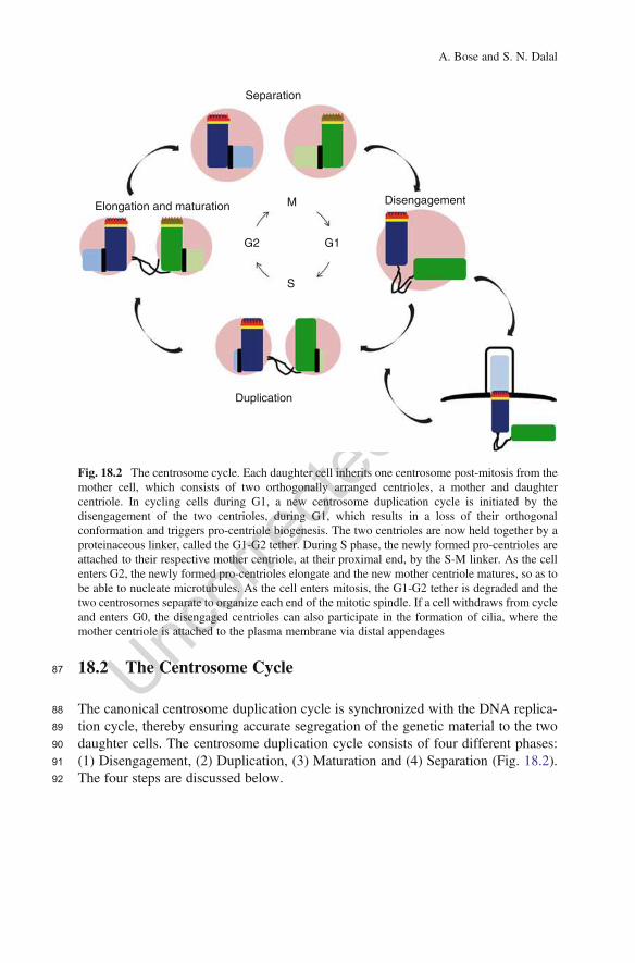
87 18.2 The Centrosome Cycle
88 The canonical centrosome duplication cycle is synchronized with the DNA replica-89 tion cycle, thereby ensuring accurate segregation of the genetic material to the two90 daughter cells. The centrosome duplication cycle consists of four different phases:91 (1) Disengagement, (2) Duplication, (3) Maturation and (4) Separation (Fig. 18.2).92 The four steps are discussed below.
Separation
Disengagement
Duplication
Elongation and maturation M
S
G1G2
Fig. 18.2 The centrosome cycle. Each daughter cell inherits one centrosome post-mitosis from themother cell, which consists of two orthogonally arranged centrioles, a mother and daughtercentriole. In cycling cells during G1, a new centrosome duplication cycle is initiated by thedisengagement of the two centrioles, during G1, which results in a loss of their orthogonalconformation and triggers pro-centriole biogenesis. The two centrioles are now held together by aproteinaceous linker, called the G1-G2 tether. During S phase, the newly formed pro-centrioles areattached to their respective mother centriole, at their proximal end, by the S-M linker. As the cellenters G2, the newly formed pro-centrioles elongate and the new mother centriole matures, so as tobe able to nucleate microtubules. As the cell enters mitosis, the G1-G2 tether is degraded and thetwo centrosomes separate to organize each end of the mitotic spindle. If a cell withdraws from cycleand enters G0, the disengaged centrioles can also participate in the formation of cilia, where themother centriole is attached to the plasma membrane via distal appendages
A. Bose and S. N. Dalal
9318.2.1 Disengagement
94From the beginning of the S phase to the onset of mitosis, each mother centriole and95its corresponding daughter centriole are orthogonally attached to each other via an96S-M linker (Nigg and Stearns 2011). This orthogonal arrangement of the two97centrioles is referred to as centriole engagement. The exact composition of the98S-M linker has not yet been elucidated; however, studies from Drosophila sper-99matocytes suggest that Sas proteins might be a part of this linker (Stevens et al.1002010). As a cell exits mitosis, the orthogonal arrangement of centrioles in each101mature centrosome in the daughter cells is disrupted by proteins such as separase and102Plk1, resulting in the degradation of the S-M linker (Tsou et al. 2009). Loss of the103S-M linker and establishment of a G1-G2 tether between the two centrioles serves as104a licensing event for the initiation of centrosome duplication. Inhibition of Plk1105activity using the small-molecule BI2536 inhibits centriole disengagement during106late G2 or early mitosis (Tsou et al. 2009). Knockdown experiments in HeLa cells107have demonstrated that cleavage of pericentrin by separase, during anaphase onset,108leads to its dissociation from the centrosome, and thus, disengagement (Matsuo et al.1092012). Separase is active only during anaphase onset, and this ensures that centriole110duplication occurs only once during the cell cycle (Tsou et al. 2009). Once the111centrioles are ‘disengaged’, they are licensed for the initiation of centriole112duplication.113An additional event required for disengagement is the phosphorylation of114nucleophosmin 1 (NPM1) by cyclin-dependent kinases. Phosphorylation of115NPM1 at a threonine residue at position 199 by CDK2 or CDK1 results in dissoci-116ation of NPM1 from the centrosome, thus licensing the centrosome for duplication117(Okuda et al. 2000; Peter et al. 1990). Expression of a phospho-deficient mutant of118NPM1 (T199A) has been demonstrated to inhibit centrosome duplication119(Tokuyama et al. 2001) while a phospho-mimetic mutant (T199D) promotes cen-120trosome amplification (Mukhopadhyay et al. 2016). Further, NPM1 has been dem-121onstrated to localize between the paired centrioles of unduplicated centrosomes122(Shinmura et al. 2005). Plk1 phosphorylates NPM1 at Ser-4, and inhibition of this123phosphorylation results in mitotic defects such as abnormal centrosome number and124the presence of fragmented nuclei in these cells (Zhang et al. 2004b).125Loss of the orthogonal configuration is not the only licensing event for centriolar126duplication (Engle et al. 2008 AU2; Gottardo et al. 2014; Shukla et al. 2015), and another127factor that governs competency for duplication is the distance between the mother128and the daughter centrioles. Plk1 can promote maturation and distancing of the129orthogonally arranged daughter centriole, leading to reduplication of the mother130centriole (Shukla et al. 2015). Further, another event that occurs during the disen-131gagement process is termed ‘centriole to centrosome conversion’. It is essential for132the newly formed daughter centriole to become duplication competent and for it to133able to function as an MTOC. In vertebrates, this begins with the initial loss of the134central cartwheel, which is mediated by CDK1 activity (Arquint and Nigg 2014). An135increase in Plk1 activity ‘modifies’ the daughter centriole inherited from the mother
18 Centrosome Amplification and Tumorigenesis: Cause or Effect?
136 cell and stabilization of the cartwheel-less centriole occurs via its recruitment of137 Cep295 (Izquierdo et al. 2014). Cep152 and Cep192 are further acquired by the138 daughter centriole, and the centriole is now competent for duplication and can139 mature to form the new mother centriole (Hatch et al. 2010; Kim et al. 2013;140 Wang et al. 2011).
141 18.2.2 Duplication
142 Following disengagement, in each cell, there is exactly one mother centriole linked143 to a daughter centriole via a proteinaceous linker called the G1-G2 tether. The144 disengaged centrioles are now licensed for duplication (Wang et al. 2011). When145 cells enter S phase, exactly one pro-centriole must form adjacent to each of the146 pre-existing centrioles. This process is regulated by proteins such as Plk4 (polo-like147 kinase 4), Cep192, Cep152, STIL, SAS6 and CDK2. Initially, Cep152 and Cep192148 act as scaffolding proteins that recruit Plk4 to the mother centriole (Kim et al. 2013).149 This results in the ring-like organization of Plk4 around the mother centriole as150 observed by super-resolution microscopy (Dzhindzhev et al. 2017; Ohta et al. 2018).151 Plk4 initiates centriole duplication by first marking the site of daughter centriole152 assembly which is dependent on the interaction between Plk4 and STIL (Ohta et al.153 2018; Sonnen et al. 2012). STIL is recruited to the centriole by Cep85 (Liu et al.154 2018) and upon recruitment to the mother centriole, STIL further activates and155 stabilizes Plk4 (Ohta et al. 2018). After recruitment of Plk4 and STIL marks the156 site of pro-centriole formation, SAS6 localizes to the same site and initiates157 pro-centriole assembly by assembling into higher order oligomers (Nakazawa158 et al. 2007). SAS6 oligomerization is the building block for the ninefold symmetrical159 centriole. Gorab, a trans-golgi protein, interacts with Sas6 and contributes to the160 establishment of the ninefold symmetry of the centriole and to centriole duplication161 in Drosophila (Kovacs et al. 2018).162 Plk4 is initially observed as a ring around the mother centriole, and this ring163 coalesces into a single spot (Dzhindzhev et al. 2017; Ohta et al. 2018). Several164 attempts have been made to understand how this transition might occur and how it165 might regulate centrosome duplication. Currently, there are two proposed models for166 how the transition occurs. According to a biophysical model proposed by Leda et al.167 (2018), the activity of Plk4 is dynamic and transiently peaks at several points around168 the mother centriole. It has been demonstrated previously that complex formation169 between Plk4 and its substrate, STIL, stabilizes active Plk4 (Moyer et al. 2015). The170 Plk4–STIL complex could be retained at the centriole due to the binding of STIL to171 other components of the centriole. This model predicts that there could be multiple172 points within the ring surrounding the mother centriole, at which this Plk4–STIL173 complex is stabilized. An exchange of the Plk4–STIL complex occurs between the174 mother centriole and the cytoplasm, which determines the concentration of the175 complex at any point. Initially, there is a concentration-based competition between176 all the Plk4–STIL complexes. The cluster with the highest concentration of Plk4–
A. Bose and S. N. Dalal
177STIL is predicted to mark the site of new centriole formation (Leda et al. 2018). The178second model suggests that first, Plk4 self-assembles into a ring around the mother179centriole. Both inactive and active forms of Plk4 coexist at the centriole. Once it is180recruited to the centriole, due to the ability of Plk4 to both laterally inhibit181neighbouring molecules of Plk4 and undergo auto-activation, there is a localized182increase in Plk4 activity at some points in the ring around the mother centriole. This183localized activity is enhanced by complex formation of Plk4 with STIL and SAS6,184thus leading to the formation of a single pro-centriole (Takao et al. 2018; Yamamoto185and Kitagawa 2018). The formation of the new pro-centriole adjacent to each mother186centriole results in the re-establishment of the S-M linker (Nigg and Stearns 2011).
18718.2.3 Elongation and Maturation
188During late S to G2, once Plk4 has marked the site of pro-centriole biogenesis,189centriole elongation is stimulated by proteins such as CPAP, CP110 and SAS6, all of190whom play important roles in this process (Kleylein-Sohn et al. 2007). SAS6, the191ninefold symmetrical cartwheel protein, acts as a scaffold around which microtu-192bules are assembled (Nakazawa et al. 2007). CPAP is a tubulin dimer-binding193capping protein that, along with CP110, promotes microtubule assembly and con-194trols centriolar length (Tang et al. 2009). It has been postulated that Cep135 links195SAS6 with CPAP and the microtubule triplet (Lin et al. 2013). Centrin and hPOC5196localize to the distal lumen of centrioles and are essential for elongation at the distal197end (Azimzadeh et al. 2009).198Elongation of the pro-centrioles is accompanied by expansion of the PCM.199According to one model, pericentrin recruits the Cep192-Plk1-Aurora A kinase200complex to the centrosome. Plk1 has been demonstrated to trigger the accumulation201of the scaffolding protein, Cep192 (Joukov et al. 2014). Plk1 phosphorylates202Cep192, which creates attachment sites for γ-TURCs (Joukov et al. 2014). Plk1203activity also recruits Cep215, a PCM protein, to the two centrosomes (Haren et al.2042009). It is postulated that Aurora A kinase, in conjunction with Plk1, phosphory-205lates components of the γ-TURCs, thus making them competent for the recruitment206of microtubules. Experiments in C. elegans and D. melanogaster have demonstrated207that loss of Aurora A leads to defects in centrosome maturation, with a reduction in208the accumulation of α-tubulin and γ-tubulin at the centrosome (Berdnik and209Knoblich 2002; Hannak et al. 2001). This step is essential to ensure that the four210centrioles are able to function as two competent MTOCs. Centriole maturation also211involves the acquisition of distal and sub-distal appendages by the newly formed212mother centriole. Centriole maturation begins with the recruitment of ODF2 as a213sub-distal appendage (Ishikawa et al. 2005). Ninein and Cep170 also function as214sub-distal appendages and bind microtubules (Mogensen et al. 2000). Distal append-215ages such as Cep164, Cep89, Cep83, FBF1 and SCLT1 are essential for membrane216docking and ciliogenesis (Tanos et al. 2013).
18 Centrosome Amplification and Tumorigenesis: Cause or Effect?
217 18.2.4 Separation
218 Before entry into mitosis, the G1-G2 tether that connects the two centrosomes is219 targeted for degradation. This tether is composed of proteins like C-NAP1 (Cep250),220 rootletin, Cep68 and LRRC45 (Bahe et al. 2005; Faragher and Fry 2003; Fry et al.221 1998a, b; Pagan et al. 2015). C-NAP1 is connected to the mother centriole via222 Cep135 (Kim et al. 2008). C-NAP1 is present at the proximal end of the mother223 centrioles and the two pools of C-NAP1 are connected by rootletin fibres (Bahe et al.224 2005; Vlijm et al. 2018). Super-resolution microscopy experiments have demon-225 strated that Cep68 binds to rootletin and increases the thickness of rootletin fibres226 (Vlijm et al. 2018). LRRC45 might link the rootletin fibres with C-NAP1 (He et al.227 2013). The degradation of the tether is initiated by the activity of the NIMA-related228 kinase, Nek2 (Fry et al. 1998b). Nek2 phosphorylates C-NAP1 and rootletin, leading229 to their displacement from the proximal end of the mother centrioles (Bahe et al.230 2005; Fry et al. 1998a). Plk1-mediated degradation of Cep68 further displaces231 Cep215, a PCM protein, from the centrosome, contributing to centrosome separation232 (Pagan et al. 2015).233 Once the G1-G2 tether has undergone dissolution, the two centrosomes have to234 be separated so as to form the mitotic spindle. This occurs primarily via the235 antiparallel sliding action of Eg5, a motor protein (Cole et al. 1994; Sawin et al.236 1992). Additionally, proteins such as dynein, Lis1 and CLIP-70 contribute to spindle237 formation by inducing microtubule sliding in the opposite direction to Eg5. The238 combined action of these motor proteins and their binding partners promotes migra-239 tion of the centrosomes to the two poles (Gonczy et al. 1999; Tanenbaum et al.240 2008). Each centrosome migrates towards a different pole in mitotic cells, leading to241 the formation of a mitotic spindle. The bipolar spindle further contributes towards242 the accurate segregation of DNA into two daughter cells, thus maintaining ploidy.
243 18.3 Centrosome Defects and Tumour Progression
244 Two rules govern the centrosome duplication cycle: cell cycle control and copy245 number control.
246 – Cell cycle control ensures that the centrosome replicates only once per cell cycle.247 – Copy number control ascertains that only one pro-centriole forms adjacent to248 each mother centriole.
249 Therefore, deregulation of any step of the centrosome cycle can give rise to250 centrosome abnormalities. These can be classified into two types: (a) structural and251 (b) numerical.
252 (a) Structural abnormalities. Structural abnormalities in centrosomes are errors in253 the organization of the centrosome. This leads to aberrations in centrosome size254 or shape and these structural alterations are present at high levels in tumour
A. Bose and S. N. Dalal
255tissues and have been postulated to contribute to tumour progression (Lingle256et al. 1998; Lingle and Salisbury 1999). Recently, it has been demonstrated that257overexpression of Ninein-like protein (Nlp) in MCF10A-derived 3D acini leads258to the formation of centrosome-related bodies (CRB) harbouring large patches of259Nlp, resulting in excessive cell proliferation. The cells in the lumen, which are260normally cleared by apoptosis, are not cleared, microtubule organization is261altered and this phenotype is associated with cellular transformation and neo-262plastic progression (Schnerch and Nigg 2016). Further, it has been demonstrated263that overexpression of Nlp weakens E-cadherin-based adherens junctions in the264epithelium. The weakening of adherens junctions results in increased mechan-265ical stress when a cell in the epithelial tissue enters mitosis. The mitotic266programme results in ‘budding’ of the new daughter cell from the original site267and could contribute to metastatic progression in tumour cells (Ganier et al.2682018). In addition to the changes in invasiveness, alterations in centrosome269structure could lead to aneuploidy which often contributes to tumour progression270(Nigg 2006). However, further studies are needed to investigate the extent of271aneuploidy that is induced by structural centrosome aberrations. Moreover,272given that the centrosome is the hub of several cellular signalling events, it273would be interesting to study how signalling pathways are affected by defects in274centrosome structure.275(b) Numerical abnormalities. Errors in cell cycle control of centrosome duplica-276tion contribute to numerical centrosome abnormalities. They can occur due to277centrosome over-duplication, defects in cytokinesis and the de novo formation278of centrosomes. These result in the presence of more than two centrosomes in a279mitotic cell which is also referred to as centrosome amplification. They are the280more extensively studied of the two types of centrosome abnormalities, espe-281cially in the context of tumour progression. Given that centrosomes organize282microtubules and form the mitotic spindle leading to DNA segregation, any283numerical errors in centrosome organization are strongly correlated with chro-284mosomal instability and aneuploidy, which is a hallmark of most tumours. Most285tumour cells show an increase in the number of centrosomes, suggesting that the286change in centrosome number could drive aneuploidy and thus, tumour progres-287sion (Godinho and Pellman 2014).
288Cells with multiple centrosomes form a multipolar spindle leading to a multipolar289mitosis resulting in aneuploidy (Ring et al. 1982). However, a multipolar mitosis in290most tumour cells leads to cell death, probably due to loss of genes that are291absolutely required for cell viability. Experiments in Zebrafish demonstrate that292neuroepithelial cells harbouring centrosome amplification undergo apoptosis (Dzafic293et al. 2015). Similarly, experiments in mice have shown that centrosome amplifica-294tion leads to microcephaly, due to increased apoptosis (Marthiens et al. 2013). These295data suggest that cells harbouring multiple centrosomes must avoid the massive296aneuploidy resulting from multipolar mitoses, which raises the interesting question297of how tumour cells manage to survive and thrive in the presence of multiple298centrosomes. In order to tolerate the burden of extra centrosomes, transformed
18 Centrosome Amplification and Tumorigenesis: Cause or Effect?
299 cells have developed a mechanism called clustering (Ring et al. 1982). In300 transformed cells with multiple centrosomes, at prophase, centrosomes are not301 present at only two distinct poles in tumour cells, thus forming a multipolar spindle.302 During prophase, the centrosomes migrate to and cluster at two different poles303 leading to the formation of a pseudo-bipolar spindle during metaphase (Basto304 et al. 2008; Ganem et al. 2009; Kwon et al. 2008; Quintyne et al. 2005). This305 formation of pseudo-bipolar spindles leads to errors in kinetochore–microtubule306 attachments, especially the formation of merotelic attachments, which contributes307 to the presence of lagging chromosomes and an increase in chromosome instability308 (CIN) (Cosenza et al. 2017; Ganem et al. 2009). The increase in CIN permits the309 generation of clones with a growth advantage leading to tumour progression.310 Multiple studies have attempted to identify gene products/molecular pathways311 that are required for centrosome clustering. This could lead to the identification of312 small molecules that inhibit clustering, which could lead to multipolar mitoses313 resulting in killing of tumour cells. A screen developed by Kwon et al. attempted314 to identify genes required for clustering in Drosophila S2 cells (Kwon et al. 2008).315 Depletion of HSET, a motor protein, has been demonstrated to induce the formation316 of multipolar spindles in human cancer cells harbouring multiple centrosomes317 (Kwon et al. 2008). Depletion of components of the spindle assembly checkpoint318 (SAC), Mad2, BubR1 (human Bub1) and CENP-E, also leads to the generation of319 multipolar spindles in S2 cells (Kwon et al. 2008). Several actin-binding proteins320 were also identified in these screens suggesting that disruption of actin dynamics321 could inhibit centrosome clustering (Kwon et al. 2008; Leber et al. 2010). This is322 consistent with recent data from the Godinho laboratory, which suggests that323 adherens junction functions that are required for maintaining cortical actin organi-324 zation and cell stiffness can inhibit centrosome clustering (Rhys et al. 2018).325 A genome-wide RNAi screen in UPCI:SCC114, a human oral squamous cell326 carcinoma (OSCC) cell line, has demonstrated that proteins that are a part of the327 chromosomal passenger complex (CPC), proteins of the Ndc80 complex, Cep164328 and Aurora B, which all contribute to spindle tension, are also required for centro-329 some clustering (Leber et al. 2010). Depletion of Aurora A in a panel of acute330 myeloid leukaemia (AML) cell lines also results in formation of a multipolar spindle331 and non-proliferation (Navarro-Serer et al. 2019). Drugs such as Griseofulvin,332 CP-673451 and Crenolanib that bind to cytoskeletal elements and inhibit clustering333 demonstrate the contribution of the cytoskeleton towards clustering (Konotop et al.334 2016; Rebacz et al. 2007).335 Previous results have suggested that loss of 14-3-3γ leads to premature cdc25C,336 and hence CDK1 activation, which leads to an increase in centrosome number. This337 is accompanied by an increase in the number of cells with pseudo-bipolar spindles338 with passage, an increase in aneuploidy and tumour formation (Mukhopadhyay et al.339 2016). However, expression of a 14-3-3γ binding defective mutant of cdc25C in340 14-3-3γ knockdown cells leads to a reversal of the clustering phenotype and a341 decrease in tumour growth and cell viability, presumably due to prematurely high342 levels of CDK1 activity in interphase and mitosis (Mukhopadhyay et al. 2016).343 These results are consistent with the hypothesis outlined above which suggests that
A. Bose and S. N. Dalal
344small molecules that might disrupt centrosome clustering in tumour cells could lead345to tumour cell killing and cell death. All of these results suggest that centrosome346clustering is a complex phenotype that requires multiple cellular pathways.347Given the strong correlation between centrosome amplification, CIN and cancer,348it has been a long-standing question of whether centrosome number dysregulation is349sufficient to promote tumorigenesis. One of the mechanisms leading to cells acquir-350ing extra centrosomes is a failure in cytokinesis. Therefore, it has been difficult to351assess the causal link between centrosome amplification and tumorigenesis. How-352ever, several recent studies have addressed this question and have offered multiple353solutions of how centrosome amplification might lead to tumour progression. In this354chapter, we aim to highlight several recent observations that shed some light on the355correlation between the presence of multiple centrosomes and transformation. We356have focused on proteins that are essential for different steps of the centrosome cycle357and how their aberrant expression has been demonstrated to contribute to centro-358some amplification and tumorigenesis.
35918.3.1 Polo-Like Kinase 1
360Polo-like kinase 1 (Plk1) is a member of the polo-like kinase family of proteins that361are serine/threonine kinases. It was initially identified in Drosophila embryos as a362kinase, active during the late anaphase–telophase transition (Llamazares et al. 1991).363Plk1 localization changes throughout the cell cycle, with it localizing at the centro-364some throughout interphase, and then moving to the kinetochore, the spindle and the365spindle mid-body during mitosis (Golsteyn et al. 1995; Kishi et al. 2009). Plk1366performs multiple functions that contribute to mitotic progression. Plk1 phosphor-367ylates multiple residues in the N-terminus of cdc25C resulting in an increase in368cdc25C activity and mitotic progression (Toyoshima-Morimoto et al. 2002). Plk1369phosphorylates BubR1 and INCENP, thus stimulating kinetochore assembly370(Arnaud et al. 1998; Elowe et al. 2007; Goto et al. 2006). Inhibition of Plk1 in371U-2OS cells decreases the robustness of the SAC, which decreases accurate chro-372mosome segregation and could, hence, contribute to CIN (O’Connor et al. 2015).373Plk1 contributes to centrosome maturation by phosphorylating NEDD1, which leads374to recruitment of γ-TURCs to centrosomes during centrosome maturation (Haren375et al. 2009; Zhang et al. 2009). Depletion of Plk1 using siRNA in HeLa cells leads to376a lack of phosphorylation of NPM1, which has been demonstrated to lead to defects377in nuclear size, cytokinesis and centrosome amplification (Zhang et al. 2004b). The378activity of Plk1 is required for centrosome disengagement, as inhibition of Plk1379using a small molecule prevents centrosome disengagement (Tsou et al. 2009).380Protein levels of Plk1 are elevated in a number of tumour types, including381gliomas, breast cancers, oesophageal squamous cell carcinomas, non-small cell382lung carcinomas, melanoma, renal cancer, prostate and colorectal cancer (Feng383et al. 2009; Liu et al. 2017; Ramani et al. 2015). Plk1 levels are high in384tamoxifen-resistant MCF-7 cells, and inhibition of Plk1 leads to a decrease in cell
18 Centrosome Amplification and Tumorigenesis: Cause or Effect?
385 proliferation (Jeong et al. 2018). A decrease in Plk1 levels confers sensitivity to386 gemcitabine in pancreatic cancer cell lines (Jimeno et al. 2010). Plk1 silencing can387 also enhance the sensitivity of rectal cancer and medulloblastoma cell lines to388 radiotherapy (Harris et al. 2012; Rodel et al. 2010). Interestingly, several missense389 and truncation mutations in the C-terminus of Plk1 have been identified in tumour390 cell lines such as HepG2, A431, MKN74 and A549 (Simizu and Osada 2000). Given391 that levels of Plk1 are found to be both overexpressed and reduced in tumour cell392 lines, it is difficult to actually assess the role of Plk1 in neoplastic progression or393 cellular transformation.394 Recent experiments performed in mouse models of Plk1 overexpression suggest395 that Plk1 may have different effects on neoplastic transformation and tumour396 progression depending on the presence of other genetic alterations or the tissue397 type in which Plk1 expression is elevated. Mouse embryonic fibroblasts (MEFs)398 isolated from mice carrying a doxycycline-inducible transgene for Plk1 exhibit a399 variety of mitotic defects such as multiple centrosomes, monopolar and multipolar400 spindles in pro-metaphase, as well as lagging chromosomes and anaphase bridges401 resulting in an increased duration of mitosis (de Carcer et al. 2018). Despite the402 defects in mitosis, when Plk1 overexpressing mice were crossed with mice403 containing a constitutively active K-Ras allele, K-RasG12D, which is only expressed404 in the mammary gland, a decrease in tumour progression was observed in the405 presence of doxycycline (de Carcer et al. 2018). Another study by Li et al. also406 found chromosomal instability, lagging and misaligned chromosomes and apoptosis407 in MEFs isolated from mice conditionally overexpressing Plk1 (Li et al. 2017). No408 malignant transformation was observed in these mice; however, hypersensitivity to409 ionizing radiation was observed in the liver. Treatment with ionizing radiation led to410 an increase in the number of liver tumours and the presence of lymphomas, which411 was accompanied by inhibition in the expression of genes required for DNA repair412 (Li et al. 2017). In both cases, despite defects in mitosis that lead to aneuploidy,413 overexpression of Plk1 alone did not lead to tumorigenesis in mice, suggesting that414 other mechanisms might contribute to tumour formation upon Plk1 overexpression.415 It is possible that Plk1 overexpression affects other proteins that influence transfor-416 mation, such as p53 (Smith et al. 2017).417 In contrast to its expression levels in most transformed cell lines, loss of Plk1 has418 been demonstrated to accelerate tumour formation in mice (Lu et al. 2008). Plk1 null419 mice show embryonic lethality due to the inability of the embryonic cells to divide.420 However, a significant proportion of Plk1 heterozygous mice develop tumours in421 various organs suggesting that haploinsufficiency of Plk1 could drive tumour pro-422 gression. Splenocytes isolated from these mice harboured a higher incidence of423 aneuploidy, which could contribute to CIN (Lu et al. 2008). Given all of the data424 on Plk1, it seems likely that Plk1 can act as either an oncogene or a tumour425 suppressor in a context-dependent manner.
A. Bose and S. N. Dalal
42618.3.2 Separase
427Separase or separin is a cysteine protease that promotes anaphase entry inmitotic cells428by cleaving Ssc1, which is part of the synaptonemal complex. This relaxes the tension429in the spindle and leads to anaphase progression (Uhlmann et al. 1999). Inhibiting430separase expression using RNAi in HeLa cells leads to genomic instability (Cucco431et al. 2018). Separase forms a complex with the MCM2-7 helicase and loss of432separase leads to an increase in replication fork speed, probably due to an increase433in the levels of acetylated SMC3 (Cucco et al. 2018). Antisense oligonuleotide-434mediated reduction of separase gives rise to aberrant mitoses with lagging chromo-435somes (Chestukhin et al. 2003). Apart from its function in relieving sister chromatid436cohesion, separase also plays a role in ensuring centrosome disengagement (Tsou and437Stearns 2006; Tsou et al. 2009). Knockout of separase in HCT116 and HeLa cell lines438inhibits centriole disengagement and subsequent duplication (Tsou et al. 2009).439Pericentrin is a potential substrate for separase and expression of a pericentrin mutant440that cannot be cleaved by separase suppresses centriole disengagement and duplica-441tion (Matsuo et al. 2012). Aki1 and cohesin have also been implicated as substrates of442separase at the centrosome (Nakamura et al. 2009). Separase also functions in double-443strand break repair, where it cleaves cohesin (Hellmuth et al. 2018). Activation of444separase in interphase has been demonstrated to aid in DNA double-strand break445repair in HEK293 cells (Hellmuth et al. 2018). All of these results suggest that446separase is required for maintaining genomic integrity either by regulating genome447organization and duplication or by regulating centrosome duplication.448Separase is significantly overexpressed in osteosarcoma, breast and prostate449tumour samples as per tissue immunofluorescence analysis (Meyer et al. 2009).450An increased nuclear localization of separase was also observed in these tissue451samples (Meyer et al. 2009). This unusual localization of separase shows a correla-452tion with tumour status, although the mechanism underlying the correlation between453separase levels and tumour progression remains to be elucidated (Meyer et al. 2009).454Increased separase activity with aberrant centrosome numbers has been observed in455bone marrow samples of patients with myelodysplastic syndrome (MDS)456(Ruppenthal et al. 2018). IHC staining of breast tumours samples from patients457has demonstrated a strong correlation between abnormal separase expression and458impaired survival (Gurvits et al. 2017). These results suggest that an increase in459separase levels can drive tumour progression.460A mutation in the zebrafish separase gene results in embryos with abnormal461mitotic spindles, aneuploidy, polyploidy and multiple centrosomes (Shepard et al.4622007). Adult zebrafish heterozygous for separase treated with the carcinogen N-463methyl-N-nitro-N-nitrosoguanidine (MNNG) were observed to be more susceptible464to developing tumours (Shepard et al. 2007). This implies that separase could act as a465tumour suppressor. A knockout of separase leads to embryonic lethality in mice466(Kumada et al. 2006). Experiments in mouse models suggest that separase467haploinsufficiency can lead to tumorigenesis in a p53 mutant background. MEFs468isolated from mice heterozygous for separase were found to exhibit a compromised
18 Centrosome Amplification and Tumorigenesis: Cause or Effect?
469 response to damage induced DNA repair (Hellmuth et al. 2018). Mice with a470 hypomorphic separase allele display an increased rate of tumorigenesis with a471 decrease in survival in a p53 mutant background (Mukherjee et al. 2011). Normal472 splenocytes isolated from these mice exhibited aneuploidy which might contribute to473 genomic instability (Mukherjee et al. 2011). In contrast to the results described474 above, conditional overexpression of separase in a mouse mammary epithelial cell475 line leads to increased aneuploidy and tumorigenesis in a p53 mutant background476 (Zhang et al. 2008). These data suggest that either an increase or a decrease in477 separase levels might not be enough to initiate tumour formation, though it might478 lead to aneuploidy followed by a second genetic event, which could drive tumori-479 genesis. It might be interesting to observe the effect of separase knockout or480 overexpression in tissue-specific, conditional mouse models.
481 18.3.3 Polo-Like Kinase 4
482 Polo-like kinase 4 (Plk4), a member of the polo family of kinases, is a master483 regulator of centriole biogenesis (Swallow et al. 2005). Plk4 was initially identified484 in mice, because of its sequence homology to polo in Drosophila (Fode et al. 1994).485 Further experiments in NIH3T3 cells ascertained that Plk4 is associated with the486 centrosome throughout the cell cycle (Hudson et al. 2001). Experiments in both487 Drosophila and mammalian cell lines using siRNA demonstrated that decrease in488 Plk4 levels leads to a loss of centrioles (Bettencourt-Dias et al. 2005; Habedanck489 et al. 2005). Overexpression of Plk4 increases centriole numbers, with multiple490 pro-centrioles forming from a single mother centriole (Habedanck et al. 2005;491 Kleylein-Sohn et al. 2007). These results indicate the importance of Plk4 levels in492 controlling centrosome numbers. Recent experiments in Drosophila have demon-493 strated that Plk4 also plays a role in controlling centriolar length (Aydogan et al.494 2018). Plk4 overexpression can also induce de novo centriole and MTOC formation495 in Drosophila embryos and Xenopus egg extracts, respectively (Eckerdt et al. 2011;496 Rodrigues-Martins et al. 2007). This occurs due to the ability of Plk4 condensates to497 recruit STIL, α-, β-tubulin and γ-tubulin, leading to the formation of acentriolar498 MTOCs (Gouveia et al. 2018).499 Plk4 levels are elevated in several cancers, such as breast, acute lymphoblastic500 leukaemia, prostate, pediatric medulloblastomas and embryonal brain tumours501 (Korzeniewski et al. 2012; Li et al. 2016, 2018; Pezuk et al. 2017). An increase in502 Plk4 levels has also been observed in human lung cancer and gastric cancer cell lines503 (Shinmura et al. 2014). Inhibition of Plk4 expression in neuroblastoma cell lines has504 been shown to suppress invasion and migration (Tian et al. 2018). Further, inhibition505 of Plk4 using CFI-400945 induces aneuploidy in lung cancer cell lines (Kawakami506 et al. 2018). In contrast to the results described above, Plk4 levels are decreased in507 colorectal cancer cell lines and hepatocellular cancers suggesting that a decrease in508 Plk4 levels might contribute to tumour progression (Kuriyama et al. 2009; Liu et al.509 2012; Rosario et al. 2010). Mutations in Plk4, including loss of function mutations,
A. Bose and S. N. Dalal
510cause microcephaly and growth failure with impaired centriole biogenesis (Martin511et al. 2014). These data suggest that changes in Plk4 expression lead to defects in512centrosome biogenesis leading to neoplastic progression.513As overexpression of Plk4 leads to an increase in centrosome number, it has been514the focus of multiple experiments to help understand the role of centrosome amplifi-515cation in tumour formation and progression. Basto et al. demonstrated for the first time516that centrosome amplification could drive tumorigenesis (Basto et al. 2008). Using an517assay wherein brain tissue from fruit flies overexpressing Sak (the Drosophila homo-518logue of Plk4) was transplanted into the abdomen ofWT hosts, they demonstrated that519a significant percentage of the hosts developed tumours with multiple centrosomes and520a number of the tumours formed metastatic colonies (Basto et al. 2008). Using the521same transplantation assay, Castellanos et al. screened for the ability of mutants of522different centrosomal proteins to affect tumour formation, in Drosophila (Castellanos523et al. 2008). They were able to demonstrate that 2% of the WT hosts with wing discs524from flies overexpressing Sak transplanted into their abdomen could develop tumours525(Castellanos et al. 2008). They were unable to assess the degree of CIN due to the526small size of the tumours.527Mice with a homozygous deletion of Plk4 displayed embryonic lethality at E7.5528(Hudson et al. 2001). Plk4+/– MEFs had increased centrosomal amplification,529multipolar spindle formation and aneuploidy when compared with WT cells530(Ko et al. 2005). Nestin-Cre-driven conditional overexpression of Plk4 in the531developing mouse brain led to centrosome amplification, aneuploidy and micro-532cephaly; however, no tumours were observed in the brains of these mice (Marthiens533et al. 2013). MEFs isolated from mice conditionally overexpressing Plk4, where a534lox stop cassette for Plk4 expression was driven by a chicken β-actin promoter, were535found to harbour multiple, functional centrosomes which contribute to mitotic errors536such as lagging chromosomes and multipolar mitosis (Vitre et al. 2015). Skin537fibroblasts derived from mice overexpressing Plk4 had multiple centrosomes, a538reduced proliferation rate and poor long-term survival (Vitre et al. 2015). Even539when mice overexpressing Plk4 were crossed with p53–/– mice, an increase in the540rate of tumour development was not observed, suggesting that Plk4 overexpression541in p53–/– mice did not accelerate tumour progression (Vitre et al. 2015). Similar542results were obtained in a skin carcinogenesis model, in which mice overexpressing543Plk4 were treated with 7,12-dimethylbenz(a)anthracene (DMBA), followed by544application of the tumour promoter 12-O-tetradecanoyl-phorbol-13-acetate (TPA).545There was no significant difference in tumour burden between the control and treated546mice (Vitre et al. 2015). However, mice carrying a similar Plk4 overexpression547allele, where expression was epidermis specific, showed differentiation and barrier548defects in the epidermis leading to death in a number of mice at postnatal day one549(P1) (Sercin et al. 2016). Mice overexpressing Plk4 that survived past P1 showed a550decrease in Plk4 levels suggesting a strong selection pressure against Plk4551overexpression. When these mice were crossed with p53 null mice, the barrier552defects were abrogated; however, cells overexpressing Plk4 were not detected553post-P1 as in the wild-type mice. Surprisingly, the transient overexpression of554Plk4 in the p53–/– mice led to the development of spontaneous skin tumours in
18 Centrosome Amplification and Tumorigenesis: Cause or Effect?
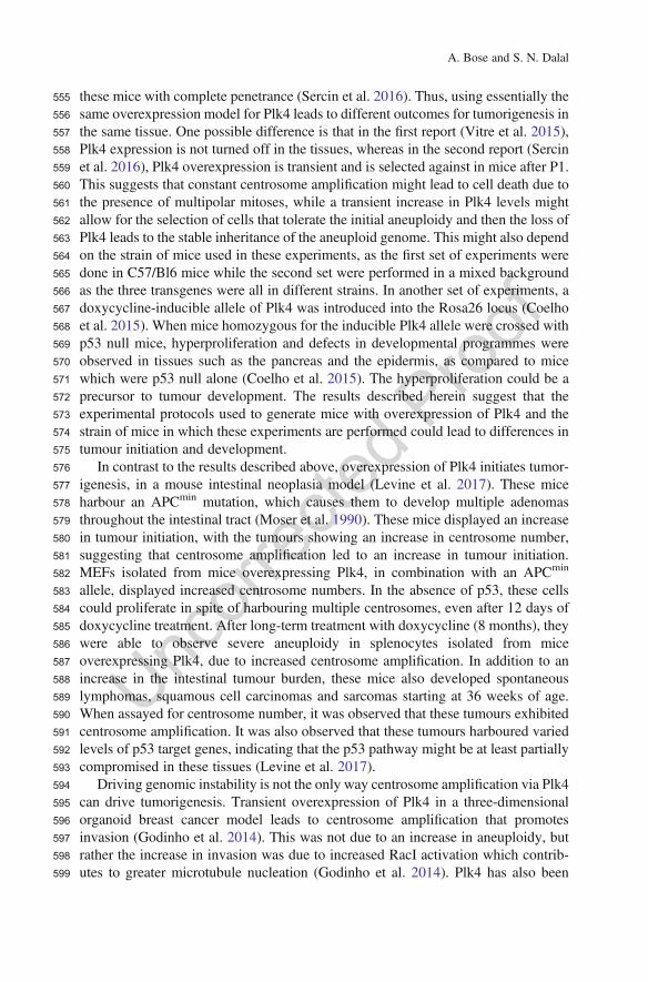
555 these mice with complete penetrance (Sercin et al. 2016). Thus, using essentially the556 same overexpression model for Plk4 leads to different outcomes for tumorigenesis in557 the same tissue. One possible difference is that in the first report (Vitre et al. 2015),558 Plk4 expression is not turned off in the tissues, whereas in the second report (Sercin559 et al. 2016), Plk4 overexpression is transient and is selected against in mice after P1.560 This suggests that constant centrosome amplification might lead to cell death due to561 the presence of multipolar mitoses, while a transient increase in Plk4 levels might562 allow for the selection of cells that tolerate the initial aneuploidy and then the loss of563 Plk4 leads to the stable inheritance of the aneuploid genome. This might also depend564 on the strain of mice used in these experiments, as the first set of experiments were565 done in C57/Bl6 mice while the second set were performed in a mixed background566 as the three transgenes were all in different strains. In another set of experiments, a567 doxycycline-inducible allele of Plk4 was introduced into the Rosa26 locus (Coelho568 et al. 2015). When mice homozygous for the inducible Plk4 allele were crossed with569 p53 null mice, hyperproliferation and defects in developmental programmes were570 observed in tissues such as the pancreas and the epidermis, as compared to mice571 which were p53 null alone (Coelho et al. 2015). The hyperproliferation could be a572 precursor to tumour development. The results described herein suggest that the573 experimental protocols used to generate mice with overexpression of Plk4 and the574 strain of mice in which these experiments are performed could lead to differences in575 tumour initiation and development.576 In contrast to the results described above, overexpression of Plk4 initiates tumor-577 igenesis, in a mouse intestinal neoplasia model (Levine et al. 2017). These mice578 harbour an APCmin mutation, which causes them to develop multiple adenomas579 throughout the intestinal tract (Moser et al. 1990). These mice displayed an increase580 in tumour initiation, with the tumours showing an increase in centrosome number,581 suggesting that centrosome amplification led to an increase in tumour initiation.582 MEFs isolated from mice overexpressing Plk4, in combination with an APCmin
583 allele, displayed increased centrosome numbers. In the absence of p53, these cells584 could proliferate in spite of harbouring multiple centrosomes, even after 12 days of585 doxycycline treatment. After long-term treatment with doxycycline (8 months), they586 were able to observe severe aneuploidy in splenocytes isolated from mice587 overexpressing Plk4, due to increased centrosome amplification. In addition to an588 increase in the intestinal tumour burden, these mice also developed spontaneous589 lymphomas, squamous cell carcinomas and sarcomas starting at 36 weeks of age.590 When assayed for centrosome number, it was observed that these tumours exhibited591 centrosome amplification. It was also observed that these tumours harboured varied592 levels of p53 target genes, indicating that the p53 pathway might be at least partially593 compromised in these tissues (Levine et al. 2017).594 Driving genomic instability is not the only way centrosome amplification via Plk4595 can drive tumorigenesis. Transient overexpression of Plk4 in a three-dimensional596 organoid breast cancer model leads to centrosome amplification that promotes597 invasion (Godinho et al. 2014). This was not due to an increase in aneuploidy, but598 rather the increase in invasion was due to increased RacI activation which contrib-599 utes to greater microtubule nucleation (Godinho et al. 2014). Plk4 has also been
A. Bose and S. N. Dalal
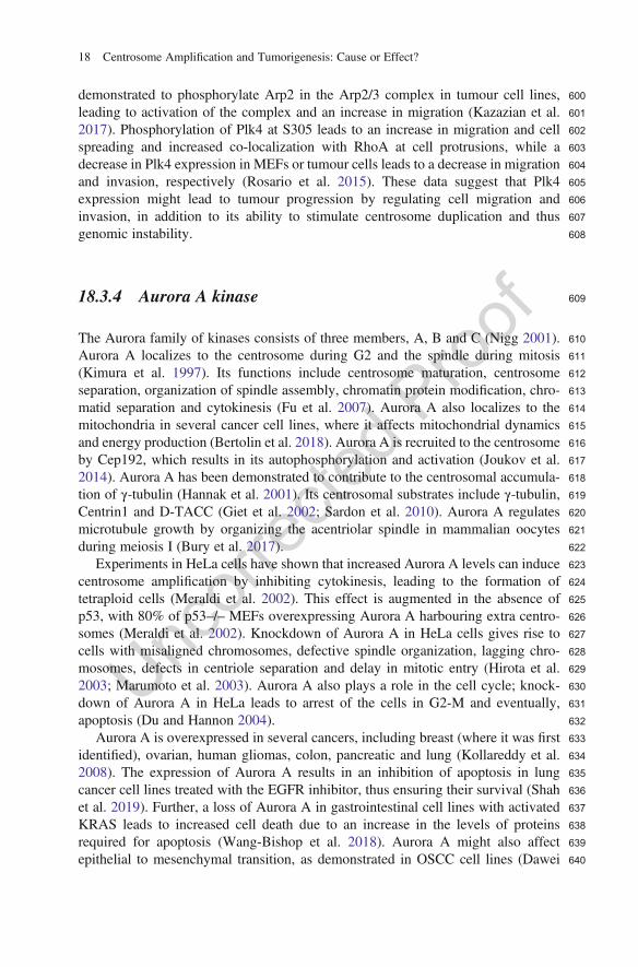
600demonstrated to phosphorylate Arp2 in the Arp2/3 complex in tumour cell lines,601leading to activation of the complex and an increase in migration (Kazazian et al.6022017). Phosphorylation of Plk4 at S305 leads to an increase in migration and cell603spreading and increased co-localization with RhoA at cell protrusions, while a604decrease in Plk4 expression in MEFs or tumour cells leads to a decrease in migration605and invasion, respectively (Rosario et al. 2015). These data suggest that Plk4606expression might lead to tumour progression by regulating cell migration and607invasion, in addition to its ability to stimulate centrosome duplication and thus608genomic instability.
60918.3.4 Aurora A kinase
610The Aurora family of kinases consists of three members, A, B and C (Nigg 2001).611Aurora A localizes to the centrosome during G2 and the spindle during mitosis612(Kimura et al. 1997). Its functions include centrosome maturation, centrosome613separation, organization of spindle assembly, chromatin protein modification, chro-614matid separation and cytokinesis (Fu et al. 2007). Aurora A also localizes to the615mitochondria in several cancer cell lines, where it affects mitochondrial dynamics616and energy production (Bertolin et al. 2018). Aurora A is recruited to the centrosome617by Cep192, which results in its autophosphorylation and activation (Joukov et al.6182014). Aurora A has been demonstrated to contribute to the centrosomal accumula-619tion of γ-tubulin (Hannak et al. 2001). Its centrosomal substrates include γ-tubulin,620Centrin1 and D-TACC (Giet et al. 2002; Sardon et al. 2010). Aurora A regulates621microtubule growth by organizing the acentriolar spindle in mammalian oocytes622during meiosis I (Bury et al. 2017).623Experiments in HeLa cells have shown that increased Aurora A levels can induce624centrosome amplification by inhibiting cytokinesis, leading to the formation of625tetraploid cells (Meraldi et al. 2002). This effect is augmented in the absence of626p53, with 80% of p53–/– MEFs overexpressing Aurora A harbouring extra centro-627somes (Meraldi et al. 2002). Knockdown of Aurora A in HeLa cells gives rise to628cells with misaligned chromosomes, defective spindle organization, lagging chro-629mosomes, defects in centriole separation and delay in mitotic entry (Hirota et al.6302003; Marumoto et al. 2003). Aurora A also plays a role in the cell cycle; knock-631down of Aurora A in HeLa leads to arrest of the cells in G2-M and eventually,632apoptosis (Du and Hannon 2004).633Aurora A is overexpressed in several cancers, including breast (where it was first634identified), ovarian, human gliomas, colon, pancreatic and lung (Kollareddy et al.6352008). The expression of Aurora A results in an inhibition of apoptosis in lung636cancer cell lines treated with the EGFR inhibitor, thus ensuring their survival (Shah637et al. 2019). Further, a loss of Aurora A in gastrointestinal cell lines with activated638KRAS leads to increased cell death due to an increase in the levels of proteins639required for apoptosis (Wang-Bishop et al. 2018). Aurora A might also affect640epithelial to mesenchymal transition, as demonstrated in OSCC cell lines (Dawei
18 Centrosome Amplification and Tumorigenesis: Cause or Effect?
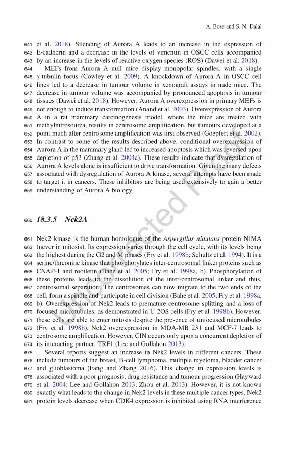
641 et al. 2018). Silencing of Aurora A leads to an increase in the expression of642 E-cadherin and a decrease in the levels of vimentin in OSCC cells accompanied643 by an increase in the levels of reactive oxygen species (ROS) (Dawei et al. 2018).644 MEFs from Aurora A null mice display monopolar spindles, with a single645 γ-tubulin focus (Cowley et al. 2009). A knockdown of Aurora A in OSCC cell646 lines led to a decrease in tumour volume in xenograft assays in nude mice. The647 decrease in tumour volume was accompanied by pronounced apoptosis in tumour648 tissues (Dawei et al. 2018). However, Aurora A overexpression in primary MEFs is649 not enough to induce transformation (Anand et al. 2003). Overexpression of Aurora650 A in a rat mammary carcinogenesis model, where the mice are treated with651 methylnitrosourea, results in centrosome amplification, but tumours developed at a652 point much after centrosome amplification was first observed (Goepfert et al. 2002).653 In contrast to some of the results described above, conditional overexpression of654 Aurora A in the mammary gland led to increased apoptosis which was reversed upon655 depletion of p53 (Zhang et al. 2004a). These results indicate that dysregulation of656 Aurora A levels alone is insufficient to drive transformation. Given the many defects657 associated with dysregulation of Aurora A kinase, several attempts have been made658 to target it in cancers. These inhibitors are being used extensively to gain a better659 understanding of Aurora A biology.
660 18.3.5 Nek2A
661 Nek2 kinase is the human homologue of the Aspergillus nidulans protein NIMA662 (never in mitosis). Its expression varies through the cell cycle, with its levels being663 the highest during the G2 and M phases (Fry et al. 1998b; Schultz et al. 1994). It is a664 serine/threonine kinase that phosphorylates inter-centrosomal linker proteins such as665 CNAP-1 and rootletin (Bahe et al. 2005; Fry et al. 1998a, b). Phosphorylation of666 these proteins leads to the dissolution of the inter-centrosomal linker and thus,667 centrosomal separation. The centrosomes can now migrate to the two ends of the668 cell, form a spindle and participate in cell division (Bahe et al. 2005; Fry et al. 1998a,669 b). Overexpression of Nek2 leads to premature centrosome splitting and a loss of670 focused microtubules, as demonstrated in U-2OS cells (Fry et al. 1998b). However,671 these cells are able to enter mitosis despite the presence of unfocused microtubules672 (Fry et al. 1998b). Nek2 overexpression in MDA-MB 231 and MCF-7 leads to673 centrosome amplification. However, CIN occurs only upon a concurrent depletion of674 its interacting partner, TRF1 (Lee and Gollahon 2013).675 Several reports suggest an increase in Nek2 levels in different cancers. These676 include tumours of the breast, B-cell lymphoma, multiple myeloma, bladder cancer677 and glioblastoma (Fang and Zhang 2016). This change in expression levels is678 associated with a poor prognosis, drug resistance and tumour progression (Hayward679 et al. 2004; Lee and Gollahon 2013; Zhou et al. 2013). However, it is not known680 exactly what leads to the change in Nek2 levels in these multiple cancer types. Nek2681 protein levels decrease when CDK4 expression is inhibited using RNA interference
A. Bose and S. N. Dalal
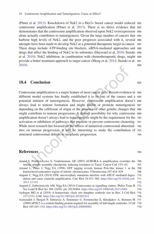
682(Pitner et al. 2013). Knockdown of Nek2 in a Her2+ breast cancer model reduced683centrosome amplification (Pitner et al. 2013). There is no direct evidence that684demonstrates that the centrosome amplification observed upon Nek2 overexpression685alone actually contributes to tumorigenesis. Given the large number of cancers that686harbour high levels of Nek2, and the poor prognosis associated with it, several687attempts have been made to develop Nek2 as a potential therapeutic target in cancer.688These drugs include ATP-binding site blockers, siRNA-mediated approaches and689drugs that affect the binding of Nek2 to its substrates (Hayward et al. 2010; Suzuki690et al. 2010). Nek2 inhibition, in combination with chemotherapeutic drugs, might691provide a better treatment approach to target cancer (Meng et al. 2014; Suzuki et al.6922010).
69318.4 Conclusion
694Centrosome amplification is a major feature of most cancer cells. Recent evidence in695different model systems has finally established it to be one of the causes and a696potential initiator of tumorigenesis. However, centrosome amplification doesn’t697always lead to tumour formation and might inhibit or promote tumorigenesis698depending on the cell/tissue of origin or the presence of other genetic changes that699might contribute to tumour progression. A distinct possibility for why centrosome700amplification doesn’t always lead to tumorigenesis might be the requirement for the701activation or inhibition of pathways that promote or prevent centrosome clustering.702While most research has focused on the effects of numerical centrosomal abnormal-703ities on tumour progression, it will be interesting to study the contribution of704structural centrosomal defects to neoplastic progression.
705References
706Anand S, Penrhyn-Lowe S, Venkitaraman AR (2003) AURORA-A amplification overrides the707mitotic spindle assembly checkpoint, inducing resistance to Taxol. Cancer Cell 3:51–62708Arnaud L, Pines J, Nigg EA (1998) GFP tagging reveals human Polo-like kinase 1 at the709kinetochore/centromere region of mitotic chromosomes. Chromosoma 107:424–429710Arquint C, Nigg EA (2014) STIL microcephaly mutations interfere with APC/C-mediated degra-711dation and cause centriole amplification. Curr Biol 24:351–360. https://doi.org/10.1016/j.cub.7122013.12.016713Arquint C, Gabryjonczyk AM, Nigg EA (2014) Centrosomes as signalling centres. Philos Trans R714Soc Lond B Biol Sci 369 (1650). pii: 20130464. https://doi.org/10.1098/rstb.2013.0464715Aydogan MG et al (2018) A homeostatic clock sets daughter centriole size in flies. J Cell Biol716217:1233–1248. https://doi.org/10.1083/jcb.201801014717Azimzadeh J, Hergert P, Delouvee A, Euteneuer U, Formstecher E, Khodjakov A, Bornens M718(2009) hPOC5 is a centrin-binding protein required for assembly of full-length centrioles. J Cell719Biol 185:101–114. https://doi.org/10.1083/jcb.200808082
18 Centrosome Amplification and Tumorigenesis: Cause or Effect?
720 Bahe S, Stierhof YD, Wilkinson CJ, Leiss F, Nigg EA (2005) Rootletin forms centriole-associated721 filaments and functions in centrosome cohesion. J Cell Biol 171:27–33. https://doi.org/10.1083/722 jcb.200504107723 Basto R, Brunk K, Vinadogrova T, Peel N, Franz A, Khodjakov A, Raff JW (2008) Centrosome724 amplification can initiate tumorigenesis in flies. Cell Rep 133:11725 Berdnik D, Knoblich JA (2002) Drosophila Aurora-A is required for centrosome maturation and726 actin-dependent asymmetric protein localization during mitosis. Curr Biol 12:640–647727 Bertolin G et al (2018) Aurora kinase A localises to mitochondria to control organelle dynamics and728 energy production. Elife 7. https://doi.org/10.7554/eLife.38111729 Bettencourt-Dias M et al (2005) SAK/PLK4 is required for centriole duplication and flagella730 development. Curr Biol 15:9731 Bury L et al (2017) Plk4 and Aurora A cooperate in the initiation of acentriolar spindle assembly in732 mammalian oocytes. J Cell Biol 216:3571–3590. https://doi.org/10.1083/jcb.201606077733 Castellanos E, Dominguez P, Gonzalez C (2008) Centrosome dysfunction in Drosophila neural734 stem cells causes tumors that are not due to genome instability. Curr Biol 18:1209–1214. https://735 doi.org/10.1016/j.cub.2008.07.029736 Chestukhin A, Pfeffer C, Milligan S, DeCaprio JA, Pellman D (2003) Processing, localization, and737 requirement of human separase for normal anaphase progression. Proc Natl Acad Sci USA738 100:4574–4579. https://doi.org/10.1073/pnas.0730733100739 Coelho PA et al (2015) Over-expression of Plk4 induces centrosome amplification, loss of primary740 cilia and associated tissue hyperplasia in the mouse. Open Biol 5:150209. https://doi.org/10.741 1098/rsob.150209742 Cole DG, Saxton WM, Sheehan KB, Scholey JM (1994) A “slow” homotetrameric kinesin-related743 motor protein purified from Drosophila embryos. J Biol Chem 269:22913–22916744 Cosenza MR et al (2017) Asymmetric centriole numbers at spindle poles cause chromosome745 missegregation in cancer. Cell Rep 20:1906–1920. https://doi.org/10.1016/j.celrep.2017.08.005746 Cowley DO et al (2009) Aurora-A kinase is essential for bipolar spindle formation and early747 development. Mol Cell Biol 29:1059–1071. https://doi.org/10.1128/mcb.01062-08748 Cucco F et al (2018) Separase prevents genomic instability by controlling replication fork speed.749 Nucleic Acids Res 46:267–278. https://doi.org/10.1093/nar/gkx1172750 Dawei H, Honggang D, Qian W (2018) AURKA contributes to the progression of oral squamous751 cell carcinoma (OSCC) through modulating epithelial-to-mesenchymal transition (EMT) and752 apoptosis via the regulation of ROS. Biochem Biophys Res Commun 507:83–90. https://doi.753 org/10.1016/j.bbrc.2018.10.170754 de Carcer G et al (2018) Plk1 overexpression induces chromosomal instability and suppresses755 tumor development. Nat Commun 9:3012. https://doi.org/10.1038/s41467-018-05429-5756 Dictenberg JB et al (1998) Pericentrin and gamma-tubulin form a protein complex and are757 organized into a novel lattice at the centrosome. J Cell Biol 141:163–174758 Du J, Hannon GJ (2004) Suppression of p160ROCK bypasses cell cycle arrest after Aurora-A/759 STK15 depletion. Proc Natl Acad Sci USA 101:8975–8980. https://doi.org/10.1073/pnas.760 0308484101761 Dzafic E, Strzyz PJ, Wilsch-Brauninger M, Norden C (2015) Centriole amplification in zebrafish762 affects proliferation and survival but not differentiation of neural progenitor cells. Cell Rep763 13:168–182. https://doi.org/10.1016/j.celrep.2015.08.062764 Dzhindzhev NS, Tzolovsky G, Lipinszki Z, Abdelaziz M, Debski J, Dadlez M, Glover DM (2017)765 Two-step phosphorylation of Ana2 by Plk4 is required for the sequential loading of Ana2 and766 Sas6 to initiate procentriole formation. Open Biol 7(12). pii: 170247. https://doi.org/10.1098/767 rsob.170247768 Eckerdt F, Yamamoto TM, Lewellyn AL, Maller JL (2011) Identification of a polo-like kinase769 4-dependent pathway for de novo centriole formation. Curr Biol 21:428–432. https://doi.org/10.770 1016/j.cub.2011.01.072
A. Bose and S. N. Dalal
771Elowe S, Hummer S, Uldschmid A, Li X, Nigg EA (2007) Tension-sensitive Plk1 phosphorylation772on BubR1 regulates the stability of kinetochore microtubule interactions. Genes Dev77321:2205–2219. https://doi.org/10.1101/gad.436007774Engle KMM, T-S; Wasa M, Yu J-Q (2008) Anomalous centriole configurations are detected in775Drosophila wing disc cells upon Cdk1 inactivation Acc Chem Res 45:5 AU3
776Fang Y, Zhang X (2016) Targeting NEK2 as a promising therapeutic approach for cancer treatment.777Cell Cycle 15:895–907. https://doi.org/10.1080/15384101.2016.1152430778Faragher AJ, Fry AM (2003) Nek2A kinase stimulates centrosome disjunction and is required for779formation of bipolar mitotic spindles. Mol Biol Cell 14:2876–2889. https://doi.org/10.1091/780mbc.e03-02-0108781Feng YB et al (2009) Overexpression of PLK1 is associated with poor survival by inhibiting782apoptosis via enhancement of survivin level in esophageal squamous cell carcinoma. Int783J Cancer 124:578–588. https://doi.org/10.1002/ijc.23990784Fode C, Motro B, Yousefi S, Heffernan M, Dennis JW (1994) Sak, a murine protein-serine/785threonine kinase that is related to the Drosophila polo kinase and involved in cell proliferation.786Proc Natl Acad Sci USA 91:6388–6392787Fry AM, Mayor T, Meraldi P, Stierhof YD, Tanaka K, Nigg EA (1998a) C-Nap1, a novel788centrosomal coiled-coil protein and candidate substrate of the cell cycle-regulated protein kinase789Nek2. J Cell Biol 141:1563–1574790Fry AM, Meraldi P, Nigg EA (1998b) A centrosomal function for the human Nek2 protein kinase, a791member of the NIMA family of cell cycle regulators. EMBO J 17:470–481. https://doi.org/10.7921093/emboj/17.2.470793Fu J, Bian M, Jiang Q, Zhang C (2007) Roles of Aurora kinases in mitosis and tumorigenesis. Mol794Cancer Res 5:1–10. https://doi.org/10.1158/1541-7786.mcr-06-0208795Fu J, Hagan IM, Glover DM (2015) The centrosome and its duplication cycle. Cold Spring Harb796Perspect Biol 7:a015800. https://doi.org/10.1101/cshperspect.a015800797Fu J et al (2016) Conserved molecular interactions in centriole-to-centrosome conversion. Nat Cell798Biol 18:87–99. https://doi.org/10.1038/ncb3274799Ganem NJ, Godinho SA, Pellman D (2009) A mechanism linking extra centrosomes to chromo-800somal instability. Nature 460:5801Ganier O, Schnerch D, Oertle P, Lim RY, Plodinec M, Nigg EA (2018) Structural centrosome802aberrations promote non-cell-autonomous invasiveness. EMBO J 37. https://doi.org/10.15252/803embj.201798576804Gergely F, Karlsson C, Still I, Cowell J, Kilmartin J, Raff JW (2000) The TACC domain identifies a805family of centrosomal proteins that can interact with microtubules. Proc Natl Acad Sci USA80697:14352–14357. https://doi.org/10.1073/pnas.97.26.14352807Giet R, McLean D, Descamps S, Lee MJ, Raff JW, Prigent C, Glover DM (2002) Drosophila808Aurora A kinase is required to localize D-TACC to centrosomes and to regulate astral micro-809tubules. J Cell Biol 156:437–451. https://doi.org/10.1083/jcb.200108135810Godinho SA, Pellman D (2014) Causes and consequences of centrosome abnormalities in cancer.811Philos Trans R Soc Lond B Biol Sci 369. pii: 20130467. https://doi.org/10.1098/rstb.2013.0467812Godinho SA et al (2014) Oncogene-like induction of cellular invasion from centrosome amplifica-813tion. Nature 510:18814Goepfert TM, Adigun YE, Zhong L, Gay J, Medina D, Brinkley WR (2002) Centrosome ampli-815fication and overexpression of aurora A are early events in rat mammary carcinogenesis. Cancer816Res 62:4115–4122817Golsteyn RM, Mundt KE, Fry AM, Nigg EA (1995) Cell cycle regulation of the activity and818subcellular localization of Plk1, a human protein kinase implicated in mitotic spindle function. J819Cell Biol 129:1617–1628820Gomez-Ferreria MA, Rath U, Buster DW, Chanda SK, Caldwell JS, Rines DR, Sharp DJ (2007)821Human Cep192 is required for mitotic centrosome and spindle assembly. Curr Biol82217:1960–1966. https://doi.org/10.1016/j.cub.2007.10.019
18 Centrosome Amplification and Tumorigenesis: Cause or Effect?
823 Gonczy P (2012) Towards a molecular architecture of centriole assembly. Nat Rev Mol Cell Biol824 13:425–435. https://doi.org/10.1038/nrm3373825 Gonczy P, Pichler S, Kirkham M, Hyman AA (1999) Cytoplasmic dynein is required for distinct826 aspects of MTOC positioning, including centrosome separation, in the one cell stage827 Caenorhabditis elegans embryo. J Cell Biol 147:135–150828 Goto H et al (2006) Complex formation of Plk1 and INCENP required for metaphase-anaphase829 transition. Nat Cell Biol 8:180–187. https://doi.org/10.1038/ncb1350830 Gottardo M, Callaini G, Riparbelli MG (2014) Procentriole assembly without centriole disengage-831 ment—a paradox of male gametogenesis. J Cell Sci 127:3434–3439. https://doi.org/10.1242/832 jcs.152843833 Gouveia SM et al (2018) PLK4 is a microtubule-associated protein that self assembles promoting de834 novo MTOC formation. J Cell Sci. https://doi.org/10.1242/jcs.219501835 Guichard P et al (2017) Cell-free reconstitution reveals centriole cartwheel assembly mechanisms.836 Nat Commun 8:14813. https://doi.org/10.1038/ncomms14813837 Gurvits N, Loyttyniemi E, Nykanen M, Kuopio T, Kronqvist P, Talvinen K (2017) Separase is a838 marker for prognosis and mitotic activity in breast cancer. Br J Cancer 117:1383–1391. https://839 doi.org/10.1038/bjc.2017.301840 Habedanck R, Stierhof YD, Wilkinson CJ, Nigg EA (2005) The Polo kinase Plk4 functions in841 centriole duplication. Nat Cell Biol 7:1140–1146. https://doi.org/10.1038/ncb1320842 Hannak E, Kirkham M, Hyman AA, Oegema K (2001) Aurora-A kinase is required for centrosome843 maturation in Caenorhabditis elegans. J Cell Biol 155:1109–1116. https://doi.org/10.1083/jcb.844 200108051845 Haren L, Stearns T, Luders J (2009) Plk1-dependent recruitment of gamma-tubulin complexes to846 mitotic centrosomes involves multiple PCM components. PLoS One 4:e5976. https://doi.org/847 10.1371/journal.pone.0005976848 Harris PS et al (2012) Polo-like kinase 1 (PLK1) inhibition suppresses cell growth and enhances849 radiation sensitivity in medulloblastoma cells. BMC Cancer 12:80. https://doi.org/10.1186/850 1471-2407-12-80851 Hatch EM, Kulukian A, Holland AJ, Cleveland DW, Stearns T (2010) Cep152 interacts with Plk4852 and is required for centriole duplication. J Cell Biol 191:721–729. https://doi.org/10.1083/jcb.853 201006049854 Hayward DG, Clarke RB, Faragher AJ, Pillai MR, Hagan IM, Fry AM (2004) The centrosomal855 kinase Nek2 displays elevated levels of protein expression in human breast cancer. Cancer Res856 64:7370–7376. https://doi.org/10.1158/0008-5472.can-04-0960857 Hayward DG et al (2010) Identification by high-throughput screening of viridin analogs as858 biochemical and cell-based inhibitors of the cell cycle-regulated nek2 kinase. J Biomol Screen859 15:918–927. https://doi.org/10.1177/1087057110376537860 He R, Huang N, Bao Y, Zhou H, Teng J, Chen J (2013) LRRC45 is a centrosome linker component861 required for centrosome cohesion. Cell Rep 4:1100–1107. https://doi.org/10.1016/j.celrep.862 2013.08.005863 Hellmuth S, Gutierrez-Caballero C, Llano E, Pendas AM, Stemmann O (2018) Local activation of864 mammalian separase in interphase promotes double-strand break repair and prevents oncogenic865 transformation. EMBO J 37. pii: e99184. https://doi.org/10.15252/embj.201899184866 Hirota T et al (2003) Aurora-A and an interacting activator, the LIM protein Ajuba, are required for867 mitotic commitment in human cells. Cell 114:585–598868 Hudson JW, Kozarova A, Cheung P, Macmillan JC, Swallow CJ, Cross JC, Dennis JW (2001) Late869 mitotic failure in mice lacking Sak, a polo-like kinase. Curr Biol 11:441–446870 Ishikawa H, Kubo A, Tsukita S, Tsukita S (2005) Odf2-deficient mother centrioles lack distal/871 subdistal appendages and the ability to generate primary cilia. Nat Cell Biol 7:517–524. https://872 doi.org/10.1038/ncb1251873 Izquierdo D, Wang WJ, Uryu K, Tsou MF (2014) Stabilization of cartwheel-less centrioles for874 duplication requires CEP295-mediated centriole-to-centrosome conversion. Cell Rep875 8:957–965. https://doi.org/10.1016/j.celrep.2014.07.022
A. Bose and S. N. Dalal
876Jana SC, Marteil G, Bettencourt-Dias M (2014) Mapping molecules to structure: unveiling secrets877of centriole and cilia assembly with near-atomic resolution. Curr Opin Cell Biol 26:96–106.878https://doi.org/10.1016/j.ceb.2013.12.001879Jeong SB et al (2018) Essential role of polo-like kinase 1 (Plk1) oncogene in tumor growth and880metastasis of tamoxifen-resistant breast cancer. Mol Cancer Ther 17:825–837. https://doi.org/88110.1158/1535-7163.mct-17-0545882Jimeno A, Rubio-Viqueira B, Rajeshkumar NV, Chan A, Solomon A, Hidalgo M (2010) A fine-883needle aspirate-based vulnerability assay identifies polo-like kinase 1 as a mediator of884gemcitabine resistance in pancreatic cancer. Mol Cancer Ther 9:311–318. https://doi.org/10.8851158/1535-7163.mct-09-0693886Joukov V, Walter JC, De Nicolo A (2014) The Cep192-organized aurora A-Plk1 cascade is887essential for centrosome cycle and bipolar spindle assembly. Mol Cell 55:578–591. https://888doi.org/10.1016/j.molcel.2014.06.016889Kawakami M et al (2018) Polo-like kinase 4 inhibition produces polyploidy and apoptotic death of890lung cancers. Proc Natl Acad Sci USA 115:1913–1918. https://doi.org/10.1073/pnas.8911719760115892Kazazian K et al (2017) Plk4 promotes cancer invasion and metastasis through Arp2/3 complex893regulation of the actin cytoskeleton. Cancer Res 77:434–447. https://doi.org/10.1158/0008-8945472.can-16-2060895Keryer G, Witczak O, Delouvée A, Kemmner WA, Rouillard D, Tasken K, Bornens M (2003)896Dissociating the centrosomal matrix protein AKAP450 from centrioles impairs centriole dupli-897cation and cell cycle progression. Mol Biol Cell 14:2436–2446. https://doi.org/10.1091/mbc.898e02-09-0614899Kim K, Lee S, Chang J, Rhee K (2008) A novel function of CEP135 as a platform protein of900C-NAP1 for its centriolar localization. Exp Cell Res 314:3692–3700. https://doi.org/10.1016/j.901yexcr.2008.09.016902Kim TS et al (2013) Hierarchical recruitment of Plk4 and regulation of centriole biogenesis by two903centrosomal scaffolds, Cep192 and Cep152. Proc Natl Acad Sci USA 110:E4849–E4857.904https://doi.org/10.1073/pnas.1319656110905Kimura M, Kotani S, Hattori T, Sumi N, Yoshioka T, Todokoro K, Okano Y (1997) Cell cycle-906dependent expression and spindle pole localization of a novel human protein kinase, Aik, related907to Aurora of Drosophila and yeast Ipl1. J Biol Chem 272:13766–13771908Kishi K, van Vugt MA, Okamoto K, Hayashi Y, Yaffe MB (2009) Functional dynamics of Polo-909like kinase 1 at the centrosome. Mol Cell Biol 29:3134–3150. https://doi.org/10.1128/mcb.91001663-08911Kitagawa D et al (2011) Structural basis of the 9-fold symmetry of centrioles. Cell 144:364–375.912https://doi.org/10.1016/j.cell.2011.01.008913Kleylein-Sohn J, Westendorf J, Le Clech M, Habedanck R, Stierhof YD, Nigg EA (2007) Plk4-914induced centriole biogenesis in human cells. Dev Cell 13:190–202. https://doi.org/10.1016/j.915devcel.2007.07.002916Ko MA, Rosario CO, Hudson JW, Kulkarni S, Pollett A, Dennis JW, Swallow CJ (2005) Plk4917haploinsufficiency causes mitotic infidelity and carcinogenesis. Nat Genet 37:883–888. https://918doi.org/10.1038/ng1605919Kollareddy M, Dzubak P, Zheleva D, Hajduch M (2008) Aurora kinases: structure, functions and920their association with cancer. Biomed Pap Med Fac Univ Palacky Olomouc Czech Repub921152:27–33922Kong D, Farmer V, Shukla A, James J, Gruskin R, Kiriyama S, Loncarek J (2014) Centriole923maturation requires regulated Plk1 activity during two consecutive cell cycles. J Cell Biol924206:855–865. https://doi.org/10.1083/jcb.201407087925Konotop G et al (2016) Pharmacological inhibition of centrosome clustering by slingshot-mediated926cofilin activation and actin cortex destabilization. Cancer Res 76:6690–6700. https://doi.org/10.9271158/0008-5472.can-16-1144
18 Centrosome Amplification and Tumorigenesis: Cause or Effect?
928 Korzeniewski N, Hohenfellner M, Duensing S (2012) CAND1 promotes PLK4-mediated centriole929 overduplication and is frequently disrupted in prostate cancer. Neoplasia (New York, NY)930 14:799–806931 Kovacs L et al (2018) Gorab is a Golgi protein required for structure and duplication of Drosophila932 centrioles. Nat Genet 50:1021–1031. https://doi.org/10.1038/s41588-018-0149-1933 Kumada K et al (2006) The selective continued linkage of centromeres from mitosis to interphase in934 the absence of mammalian separase. J Cell Biol 172:835–846. https://doi.org/10.1083/jcb.935 200511126936 Kuriyama R, Bettencourt-Dias M, Hoffmann I, Arnold M, Sandvig L (2009) Gamma-tubulin-937 containing abnormal centrioles are induced by insufficient Plk4 in human HCT116 colorectal938 cancer cells. J Cell Sci 122:2014–2023. https://doi.org/10.1242/jcs.036715939 Kwon M, Godinho SA, Chandhok NS, Ganem NJ, Azioune A, Thery M, Pellman D (2008)940 Mechanisms to suppress multipolar divisions in cancer cells with extra centrosomes. Genes941 Dev 22:2189–2203. https://doi.org/10.1101/gad.1700908942 Leber B et al (2010) Proteins required for centrosome clustering in cancer cells. Sci Transl Med943 2:33ra38. https://doi.org/10.1126/scitranslmed.3000915944 Leda M, Holland AJ, Goryachev AB (2018) Autoamplification and competition drive symmetry945 breaking: initiation of centriole duplication by the PLK4-STIL network. iScience 8:222–235.946 https://doi.org/10.1016/j.isci.2018.10.003947 Lee J, Gollahon L (2013) Mitotic perturbations induced by Nek2 overexpression require interaction948 with TRF1 in breast cancer cells. Cell Cycle 12:3599–3614. https://doi.org/10.4161/cc.26589949 Levine MS et al (2017) Centrosome amplification is sufficient to promote spontaneous tumorigen-950 esis in mammals. Dev Cell 40:313–322.e315. https://doi.org/10.1016/j.devcel.2016.12.022951 Li Z, Dai K, Wang C, Song Y, Gu F, Liu F, Fu L (2016) Expression of polo-like kinase 4(PLK4) in952 breast cancer and its response to taxane-based neoadjuvant chemotherapy. J Cancer953 7:1125–1132. https://doi.org/10.7150/jca.14307954 Li Z et al (2017) Polo-like kinase 1 (Plk1) overexpression enhances ionizing radiation-induced955 cancer formation in mice. J Biol Chem 292:17461–17472. https://doi.org/10.1074/jbc.M117.956 810960957 Li S, Wang C, Wang W, Liu W, Zhang G (2018) Abnormally high expression of POLD1, MCM2,958 and PLK4 promotes relapse of acute lymphoblastic leukemia. Medicine 97:e10734. https://doi.959 org/10.1097/md.0000000000010734960 Lin YC et al (2013) Human microcephaly protein CEP135 binds to hSAS-6 and CPAP, and is961 required for centriole assembly. EMBO J 32:1141–1154. https://doi.org/10.1038/emboj.2013.962 56963 Lingle WL, Salisbury JL (1999) Altered centrosome structure is associated with abnormal mitoses964 in human breast tumors. Am J Pathol 155:1941–1951. https://doi.org/10.1016/s0002-9440(10)965 65513-7966 Lingle WL, Lutz WH, Ingle JN, Maihle NJ, Salisbury JL (1998) Centrosome hypertrophy in human967 breast tumors: implications for genomic stability and cell polarity. Proc Natl Acad Sci USA968 95:2950–2955969 Liu L, Zhang CZ, Cai M, Fu J, Chen GG, Yun J (2012) Downregulation of polo-like kinase 4 in970 hepatocellular carcinoma associates with poor prognosis. PLoS One 7:e41293. https://doi.org/971 10.1371/journal.pone.0041293972 Liu Z, Sun Q, Wang X (2017) PLK1, a potential target for cancer therapy. Transl Oncol 10:22–32.973 https://doi.org/10.1016/j.tranon.2016.10.003974 Liu Y et al (2018) Direct binding of CEP85 to STIL ensures robust PLK4 activation and efficient975 centriole assembly. Nat Commun 9:15976 Llamazares S et al (1991) Polo encodes a protein kinase homolog required for mitosis in Drosoph-977 ila. Genes Dev 5:2153–2165978 Lu LY, Wood JL, Minter-Dykhouse K, Ye L, Saunders TL, Yu X, Chen J (2008) Polo-like kinase979 1 is essential for early embryonic development and tumor suppression. Mol Cell Biol980 28:6870–6876. https://doi.org/10.1128/mcb.00392-08
A. Bose and S. N. Dalal

981Luders J (2012) The amorphous pericentriolar cloud takes shape. Nat Cell Biol 14:1126–1128.982https://doi.org/10.1038/ncb2617983Malumbres M, Barbacid M (2007) Cell cycle kinases in cancer. Curr Opin Genet Dev 17:60–65.984https://doi.org/10.1016/j.gde.2006.12.008985Marthiens V, Rujano MA, Pennetier C, Tessier S, Paul-Gilloteaux P, Basto R (2013) Centrosome986amplification causes microcephaly. Nat Cell Biol 15:731–740. https://doi.org/10.1038/ncb2746987Martin CA et al (2014) Mutations in PLK4, encoding a master regulator of centriole biogenesis,988cause microcephaly, growth failure and retinopathy. Nat Genet 46:1283–1292. https://doi.org/98910.1038/ng.3122990Marumoto T, Honda S, Hara T, Nitta M, Hirota T, Kohmura E, Saya H (2003) Aurora-A kinase991maintains the fidelity of early and late mitotic events in HeLa cells. J Biol Chem992278:51786–51795. https://doi.org/10.1074/jbc.M306275200993Matsuo K, Ohsumi K, Iwabuchi M, Kawamata T, Ono Y, Takahashi M (2012) Kendrin is a novel994substrate for separase involved in the licensing of centriole duplication. Curr Biol 22:915–921.995https://doi.org/10.1016/j.cub.2012.03.048996Meng L et al (2014) Inhibition of Nek2 by small molecules affects proteasome activity. BioMed Res997Int 2014:13. https://doi.org/10.1155/2014/273180998Meraldi P, Honda R, Nigg EA (2002) Aurora-A overexpression reveals tetraploidization as a major999route to centrosome amplification in p53-/- cells. EMBO J 21:483–4921000Meyer R, Fofanov V, Panigrahi A, Merchant F, Zhang N, Pati D (2009) Overexpression and1001mislocalization of the chromosomal segregation protein separase in multiple human cancers.1002Clin Cancer Res 15:2703–2710. https://doi.org/10.1158/1078-0432.ccr-08-24541003Mogensen MM, Malik A, Piel M, Bouckson-Castaing V, Bornens M (2000) Microtubule minus-1004end anchorage at centrosomal and non-centrosomal sites: the role of ninein. J Cell Sci 113(Pt100517):3013–30231006Moser AR, Pitot HC, Dove WF (1990) A dominant mutation that predisposes to multiple intestinal1007neoplasia in the mouse. Science 247:322–3241008Moyer TC, Clutario KM, Lambrus BG, Daggubati V, Holland AJ (2015) Binding of STIL to Plk41009activates kinase activity to promote centriole assembly. J Cell Biol 209:863–878. https://doi.org/101010.1083/jcb.2015020881011Mukherjee M et al (2011) Separase loss of function cooperates with the loss of p53 in the initiation1012and progression of T- and B-cell lymphoma, leukemia and aneuploidy in mice. PLoS One 6:1013e22167. https://doi.org/10.1371/journal.pone.00221671014Mukhopadhyay A et al (2016) 14-3-3gamma prevents centrosome amplification and neoplastic1015progression. Sci Rep 6:26580. https://doi.org/10.1038/srep265801016Nakamura A, Arai H, Fujita N (2009) Centrosomal Aki1 and cohesin function in separase-regulated1017centriole disengagement. J Cell Biol 187:607–614. https://doi.org/10.1083/jcb.2009060191018Nakazawa Y, Hiraki M, Kamiya R, Hirono M (2007) SAS-6 is a cartwheel protein that establishes1019the 9-fold symmetry of the centriole. Curr Biol 17:2169–2174. https://doi.org/10.1016/j.cub.10202007.11.0461021Navarro-Serer B, Childers EP, Hermance NM, Mercadante D, Manning AL (2019) Aurora A1022inhibition limits centrosome clustering and promotes mitotic catastrophe in cells with supernu-1023merary centrosomes. Oncotarget 10(17):1649–16591024Nigg EA (2001) Mitotic kinases as regulators of cell division and its checkpoints. Nat Rev Mol Cell1025Biol 2:21–32. https://doi.org/10.1038/350480961026Nigg EA (2006) Origins and consequences of centrosome aberrations in human cancers. Int1027J Cancer 119:2717–2723. https://doi.org/10.1002/ijc.222451028Nigg EA, Stearns T (2011) The centrosome cycle: centriole biogenesis, duplication and inherent1029asymmetries. Nat Cell Biol 13:1154–1160. https://doi.org/10.1038/ncb23451030O’Connor A, Maffini S, Rainey MD, Kaczmarczyk A, Gaboriau D, Musacchio A, Santocanale C1031(2015) Requirement for PLK1 kinase activity in the maintenance of a robust spindle assembly1032checkpoint. Biol Open 5:11–19. https://doi.org/10.1242/bio.014969
18 Centrosome Amplification and Tumorigenesis: Cause or Effect?
1033 Ohta M, Watanabe K, Ashikawa T, Nozaki Y, Yoshiba S, Kimura A, Kitagawa D (2018) Bimodal1034 binding of STIL to Plk4 controls proper centriole copy number. Cell Rep 23:3160–3169.e3164.1035 https://doi.org/10.1016/j.celrep.2018.05.0301036 Okuda M et al (2000) Nucleophosmin/B23 is a target of CDK2/cyclin E in centrosome duplication.1037 Cell 103:127–1401038 Pagan JK et al (2015) Degradation of Cep68 and PCNT cleavage mediate Cep215 removal from the1039 PCM to allow centriole separation, disengagement and licensing. Nat Cell Biol 17:31–43.1040 https://doi.org/10.1038/ncb30761041 Peter M, Nakagawa J, Doree M, Labbe JC, Nigg EA (1990) Identification of major nucleolar1042 proteins as candidate mitotic substrates of cdc2 kinase. Cell 60:791–8011043 Pezuk JA, Brassesco MS, de Oliveira RS, Machado HR, Neder L, Scrideli CA, Tone LG (2017)1044 PLK1-associated microRNAs are correlated with pediatric medulloblastoma prognosis. Childs1045 Nerv Syst 33:609–615. https://doi.org/10.1007/s00381-017-3366-51046 Piel M, Meyer P, Khodjakov A, Rieder CL, Bornens M (2000) The respective contributions of the1047 mother and daughter centrioles to centrosome activity and behavior in vertebrate cells. J Cell1048 Biol 149:317–3301049 Pitner H, Kathryn M, Saavedra HI (2013) Cdk4 and Nek2 signal binucleation and centrosome1050 amplification in a Her2+ breast cancer model. PLoS One 8:41051 Quintyne NJ, Reing JE, Hoffelder DR, Gollin SM, Saunders WS (2005) Spindle multipolarity is1052 prevented by centrosomal clustering. Science 307:31053 Ramani P, Nash R, Sowa-Avugrah E, Rogers C (2015) High levels of polo-like kinase 1 and1054 phosphorylated translationally controlled tumor protein indicate poor prognosis in neuroblas-1055 tomas. J Neurooncol 125:103–111. https://doi.org/10.1007/s11060-015-1900-41056 Rebacz B, Larsen TO, Clausen MH, Ronnest MH, Loffler H, Ho AD, Kramer A (2007) Identifi-1057 cation of griseofulvin as an inhibitor of centrosomal clustering in a phenotype-based screen.1058 Cancer Res 67:6342–6350. https://doi.org/10.1158/0008-5472.can-07-06631059 Reber S, Hyman AA (2015) Emergent properties of the metaphase spindle. Cold Spring Harb1060 Perspect Biol 7:a015784. https://doi.org/10.1101/cshperspect.a0157841061 Reina J, Gonzalez C (2014) When fate follows age: unequal centrosomes in asymmetric cell1062 division. Philos Trans R Soc Lond B Biol Sci 369. https://doi.org/10.1098/rstb.2013.04661063 Rhys AD et al (2018) Loss of E-cadherin provides tolerance to centrosome amplification in1064 epithelial cancer cells. J Cell Biol 217:195–209. https://doi.org/10.1083/jcb.2017041021065 Ring D, Hubble R, Kirschner M (1982) Mitosis in a cell with multiple centrioles. J Cell Biol1066 94:549–5561067 Rios RM (2014) The centrosome-Golgi apparatus nexus. Philos Trans R Soc Lond B Biol Sci 369.1068 https://doi.org/10.1098/rstb.2013.04621069 Rodel F et al (2010) Polo-like kinase 1 as predictive marker and therapeutic target for radiotherapy1070 in rectal cancer. Am J Pathol 177:918–929. https://doi.org/10.2353/ajpath.2010.1000401071 Rodrigues-Martins A, Riparbelli M, Callaini G, Glover DM, Bettencourt-Dias M (2007) Revisiting1072 the role of the mother centriole in centriole biogenesis. Science 316:1046–1050. https://doi.org/1073 10.1126/science.11429501074 Rosario CO et al (2010) Plk4 is required for cytokinesis and maintenance of chromosomal stability.1075 Proc Natl Acad Sci USA 107:6888–6893. https://doi.org/10.1073/pnas.09109411071076 Rosario CO et al (2015) A novel role for Plk4 in regulating cell spreading and motility. Oncogene1077 34:3441–3451. https://doi.org/10.1038/onc.2014.2751078 Ruppenthal S, Kleiner H, Nolte F, Fabarius A, Hofmann WK, Nowak D, Seifarth W (2018)1079 Increased separase activity and occurrence of centrosome aberrations concur with transforma-1080 tion of MDS. PLoS One 13:e0191734. https://doi.org/10.1371/journal.pone.01917341081 Sardon T, Pache RA, Stein A, Molina H, Vernos I, Aloy P (2010) Uncovering new substrates for1082 Aurora A kinase. EMBO Rep 11:977–984. https://doi.org/10.1038/embor.2010.1711083 Sawin KE, LeGuellec K, Philippe M, Mitchison TJ (1992) Mitotic spindle organization by a plus-1084 end-directed microtubule motor. Nature 359:540–543. https://doi.org/10.1038/359540a0
A. Bose and S. N. Dalal
1085Schnerch D, Nigg EA (2016) Structural centrosome aberrations favor proliferation by abrogating1086microtubule-dependent tissue integrity of breast epithelial mammospheres. Oncogene108735:2711–2722. https://doi.org/10.1038/onc.2015.3321088Schultz SJ, Fry AM, Sutterlin C, Ried T, Nigg EA (1994) Cell cycle-dependent expression of Nek2,1089a novel human protein kinase related to the NIMA mitotic regulator of Aspergillus nidulans.1090Cell Growth Differ 5:625–6351091Sercin O et al (2016) Transient PLK4 overexpression accelerates tumorigenesis in p53-deficient1092epidermis. Nat Cell Biol 18:100–110. https://doi.org/10.1038/ncb32701093Shah KN et al (2019) Aurora kinase A drives the evolution of resistance to third-generation EGFR1094inhibitors in lung cancer. Nat Med 25:111–118. https://doi.org/10.1038/s41591-018-0264-71095Shepard JL et al (2007) A mutation in separase causes genome instability and increased suscepti-1096bility to epithelial cancer. Genes Dev 21:55–59. https://doi.org/10.1101/gad.14704071097Shinmura K, Tarapore P, Tokuyama Y, George KR, Fukasawa K (2005) Characterization of1098centrosomal association of nucleophosmin/B23 linked to Crm1 activity. FEBS Lett1099579:6621–6634. https://doi.org/10.1016/j.febslet.2005.10.0571100Shinmura K, Kurabe N, Goto M, Yamada H, Natsume H, Konno H, Sugimura H (2014) PLK41101overexpression and its effect on centrosome regulation and chromosome stability in human1102gastric cancer. Mol Biol Rep 41:6635–6644. https://doi.org/10.1007/s11033-014-3546-21103Shukla A, Kong D, Sharma M, Magidson V, Loncarek J (2015) Plk1 relieves centriole block to1104reduplication by promoting daughter centriole maturation. Nat Commun 6:8077. https://doi.org/110510.1038/ncomms90771106Simizu S, Osada H (2000) Mutations in the Plk gene lead to instability of Plk protein in human1107tumour cell lines. Nat Cell Biol 2:852–854. https://doi.org/10.1038/350411021108Smith L, Farzan R, Ali S, Buluwela L, Saurin AT, Meek DW (2017) The responses of cancer cells1109to PLK1 inhibitors reveal a novel protective role for p53 in maintaining centrosome separation.1110Sci Rep 7:16115. https://doi.org/10.1038/s41598-017-16394-21111Sonnen KF, Schermelleh L, Leonhardt H, Nigg EA (2012) 3D-structured illumination microscopy1112provides novel insight into architecture of human centrosomes. Biol Open 1:965–976. https://1113doi.org/10.1242/bio.201223371114Stevens NR, Roque H, Raff JW (2010) DSas-6 and Ana2 coassemble into tubules to promote1115centriole duplication and engagement. Dev Cell 19:913–919. https://doi.org/10.1016/j.devcel.11162010.11.0101117Stinchcombe JC, Griffiths GM (2014) Communication, the centrosome and the immunological1118synapse. Philos Trans R Soc Lond B Biol Sci 369. https://doi.org/10.1098/rstb.2013.04631119Suzuki K, Kokuryo T, Senga T, Yokoyama Y, NaginoM, Hamaguchi M (2010) Novel combination1120treatment for colorectal cancer using Nek2 siRNA and cisplatin. Cancer Sci 101:1163–1169.1121https://doi.org/10.1111/j.1349-7006.2010.01504.x1122Swallow CJ, Ko MA, Siddiqui NU, Hudson JW, Dennis JW (2005) Sak/Plk4 and mitotic fidelity.1123Oncogene 24:306–312. https://doi.org/10.1038/sj.onc.12082751124Takao D, Yamamoto S, Kitagawa D (2018) A theory of centriole duplication based on self-1125organized spatial pattern formation. bioRxiv 424754. https://doi.org/10.1101/4247541126TanenbaumME, Macurek L, Galjart N, Medema RH (2008) Dynein, Lis1 and CLIP-170 counteract1127Eg5-dependent centrosome separation during bipolar spindle assembly. EMBO J112827:3235–3245. https://doi.org/10.1038/emboj.2008.2421129Tang CJ, Fu RH, Wu KS, Hsu WB, Tang TK (2009) CPAP is a cell-cycle regulated protein that1130controls centriole length. Nat Cell Biol 11:825–831. https://doi.org/10.1038/ncb18891131Tanos BE, Yang HJ, Soni R, Wang WJ, Macaluso FP, Asara JM, Tsou MF (2013) Centriole distal1132appendages promote membrane docking, leading to cilia initiation. Genes Dev 27:163–168.1133https://doi.org/10.1101/gad.207043.1121134Tian X et al (2018) Polo-like kinase 4 mediates epithelial-mesenchymal transition in neuroblastoma1135via PI3K/Akt signaling pathway. Cell Death Dis 9:54. https://doi.org/10.1038/s41419-017-11360088-2
18 Centrosome Amplification and Tumorigenesis: Cause or Effect?
1137 Tokuyama Y, Horn HF, Kawamura K, Tarapore P, Fukasawa K (2001) Specific phosphorylation of1138 nucleophosmin on Thr(199) by cyclin-dependent kinase 2-cyclin E and its role in centrosome1139 duplication. J Biol Chem 276:21529–21537. https://doi.org/10.1074/jbc.M1000142001140 Toyoshima-Morimoto F, Taniguchi E, Nishida E (2002) Plk1 promotes nuclear translocation of1141 human Cdc25C during prophase. EMBO Rep 3:341–348. https://doi.org/10.1093/embo-reports/1142 kvf0691143 Tsou MF, Stearns T (2006) Mechanism limiting centrosome duplication to once per cell cycle.1144 Nature 442:947–951. https://doi.org/10.1038/nature049851145 Tsou MF, Wang WJ, George KA, Uryu K, Stearns T, Jallepalli PV (2009) Polo kinase and separase1146 regulate the mitotic licensing of centriole duplication in human cells. Dev Cell 17:344–354.1147 https://doi.org/10.1016/j.devcel.2009.07.0151148 Uhlmann F, Lottspelch F, Nasmyth K (1999) Sister-chromatid separation at anaphase onset is1149 promoted by cleavage of the cohesin subunit Scc1. Nature 400:37–42. https://doi.org/10.1038/1150 218311151 Vitre B et al (2015) Chronic centrosome amplification without tumorigenesis. Proc Natl Acad Sci1152 USA 112:111153 Vlijm R et al (2018) STED nanoscopy of the centrosome linker reveals a CEP68-organized,1154 periodic rootletin network anchored to a C-Nap1 ring at centrioles. Proc Natl Acad Sci USA1155 115:81156 Walczak CE, Heald R (2008) Mechanisms of mitotic spindle assembly and function. Int Rev Cytol1157 265:111–158. https://doi.org/10.1016/s0074-7696(07)65003-71158 Wang WJ, Soni RK, Uryu K, Tsou MF (2011) The conversion of centrioles to centrosomes:1159 essential coupling of duplication with segregation. J Cell Biol 193:727–739. https://doi.org/1160 10.1083/jcb.2011011091161 Wang-Bishop L et al (2018) Inhibition of AURKA reduces proliferation and survival of gastroin-1162 testinal cancer cells with activated KRAS by preventing activation of RPS6KB1. Gastroenter-1163 ology. https://doi.org/10.1053/j.gastro.2018.10.0301164 Winey M, O’Toole E (2014) Centriole structure. Philos Trans R Soc Lond B Biol Sci 369. https://1165 doi.org/10.1098/rstb.2013.04571166 Woodruff JB, Wueseke O, Hyman AA (2014) Pericentriolar material structure and dynamics.1167 Philos Trans R Soc Lond B Biol Sci 369. https://doi.org/10.1098/rstb.2013.04591168 Yamamoto S, Kitagawa D (2018) Self-organization of Plk4 regulates symmetry breaking in1169 centriole duplication. bioRxiv 313635. https://doi.org/10.1101/3136351170 Zhang D et al (2004a) Cre-loxP-controlled periodic Aurora-A overexpression induces mitotic1171 abnormalities and hyperplasia in mammary glands of mouse models. Oncogene1172 23:8720–8730. https://doi.org/10.1038/sj.onc.12081531173 Zhang H, Shi X, Paddon H, Hampong M, Dai W, Pelech S (2004b) B23/nucleophosmin serine1174 4 phosphorylation mediates mitotic functions of polo-like kinase 1. J Biol Chem1175 279:35726–35734. https://doi.org/10.1074/jbc.M4032642001176 Zhang N et al (2008) Overexpression of Separase induces aneuploidy and mammary tumorigenesis.1177 Proc Natl Acad Sci USA 105:13033–13038. https://doi.org/10.1073/pnas.08016101051178 Zhang X et al (2009) Sequential phosphorylation of Nedd1 by Cdk1 and Plk1 is required for1179 targeting of the gammaTuRC to the centrosome. J Cell Sci 122:2240–2251. https://doi.org/10.1180 1242/jcs.0427471181 Zhou W et al (2013) NEK2 induces drug resistance mainly through activation of efflux drug pumps1182 and is associated with poor prognosis in myeloma and other cancers. Cancer Cell 23:48–62.1183 https://doi.org/10.1016/j.ccr.2012.12.001
A. Bose and S. N. Dalal

Author QueriesChapter No.: 18 461942_1_En
Query Refs. Details Required Author’s response
AU1 Please check whether affiliations areok
AU2 Citation “Engle 2008” has beenchanged to “Engle et al. 2008” inorder to match with the reference list.Please check.
AU3 Please check the author names in ref.Engle et al. (2008)
1Scientific RepoRts | 6:26580 | DOI: 10.1038/srep26580
www.nature.com/scientificreports
14-3-3γ Prevents Centrosome Amplification and Neoplastic ProgressionAmitabha Mukhopadhyay1,†, Lalit Sehgal1,*,‡, Arunabha Bose1,*, Anushree Gulvady1,#, Parijat Senapati2, Rahul Thorat1, Srikanta Basu1, Khyati Bhatt1, Amol S. Hosing1, Renu Balyan3, Lalit Borde4, Tapas K. Kundu2 & Sorab N. Dalal1
More than 80% of malignant tumors show centrosome amplification and clustering. Centrosome amplification results from aberrations in the centrosome duplication cycle, which is strictly coordinated with DNA-replication-cycle. However, the relationship between cell-cycle regulators and centrosome duplicating factors is not well understood. This report demonstrates that 14-3-3γ localizes to the centrosome and 14-3-3γ loss leads to centrosome amplification. Loss of 14-3-3γ results in the phosphorylation of NPM1 at Thr-199, causing early centriole disjunction and centrosome hyper-duplication. The centrosome amplification led to aneuploidy and increased tumor formation in mice. Importantly, an increase in passage of the 14-3-3γ-knockdown cells led to an increase in the number of cells containing clustered centrosomes leading to the generation of pseudo-bipolar spindles. The increase in pseudo-bipolar spindles was reversed and an increase in the number of multi-polar spindles was observed upon expression of a constitutively active 14-3-3-binding-defective-mutant of cdc25C (S216A) in the 14-3-3γ knockdown cells. The increase in multi-polar spindle formation was associated with decreased cell viability and a decrease in tumor growth. Our findings uncover the molecular basis of regulation of centrosome duplication by 14-3-3γ and inhibition of tumor growth by premature activation of the mitotic program and the disruption of centrosome clustering.
The centrosome is the major microtubule nucleating and organizing center in mammalian cells, consisting of two cylindrical centrioles, surrounded by multi-layered toroid of pericentriolar matrix (PCM)1,2. Resting cells contain one centrosome which duplicates strictly once in a cell cycle, synchronized with DNA replication cycle, giving rise to two daughter centrosomes before the onset of mitosis [reviewed in3]. Deregulation of the centrosome duplica-tion cycle leads to centrosome amplification, which is commonly observed in multiple human tumors [reviewed in4]. Normal cells with supernumerary centrosomes generally die, due to the formation of multipolar spindles leading to severe aneuploidy and prolonged checkpoint arrest and mitotic catastrophe. In contrast, tumor cells with multiple centrosomes are able to cluster centrosomes at opposite poles thus generating pseudo-bipolar spin-dles. Generation of pseudo-bipolar spindles prevents mitotic catastrophe and promotes limited aneuploidy result-ing in an increase in cell survival and also resulting in the generation of invasive tumors5–7.
Two centrioles remain closely connected with each other through a proteinaceous linker, during G18. Biogenesis of the nascent daughter centriole (procentriole) begins with the relaxation of the inter-centriolar tether, resulting in separation of the mother centrioles, termed as centriole disjunction9,10. Centriole disjunction is regulated by orchestrated phosphorylation of various linker proteins including NPM1, β -catenin, Nek2, C-Nap1 (CEP250), rootletin, Cep68, causing their displacement from the linker11–16. After steric relaxation, procentriole biogenesis proceeds with step-wise assembly of the central “cart-wheel”17–19. Procentrioles mature, through the
1Advanced Centre for Treatment Research and Education in Cancer, Tata Memorial Center, Mumbai 410210, India. 2Transcription and Disease Laboratory, Molecular Biology and Genetics Unit, Jawaharlal Nehru Centre for Advanced Scientific Research, Bangalore 560064, India. 3National Institute of Immunology, New Delhi 110067, India. 4Department of Biological Sciences, Tata Institute of Fundamental Research, Mumbai 400005, India. †Present address: Institute of Biophysical Dynamics, University of Chicago, Chicago, IL 60637, USA. ‡Present address: Department of Lymphoma and Myeloma, MD Anderson Cancer Center, Texas, TX 77030, USA. #Present address: Department of Cell and Developmental Biology, State University of New York Upstate Medical University, Syracuse NY 13210, USA. *These authors contributed equally to the work. Correspondence and requests for materials should be addressed to S.N.D. (email: [email protected])
received: 08 October 2015
accepted: 04 May 2016
Published: 02 June 2016
OPEN
www.nature.com/scientificreports/
2Scientific RepoRts | 6:26580 | DOI: 10.1038/srep26580
S and G2 phase, from the proximal end of the mother centriole. If centriole disjunction is blocked, in spite of continued nuclear duplication, centriole duplication remains stalled due to inhibition in cartwheel-templating from mother centriole20.
Current studies indicate that, activation of the cdk1/cyclinB complex is involved in generation of altered cen-trosome number. However, the centrosomal targets of cdk1 and underlying mechanism of cdk1-mediated reg-ulation of centrosome duplication are largely unknown21–26. The cdk1/cyclinB1 complex is activated by cdc25C, whose activity is inhibited during interphase by complex formation with 14-3-3 proteins27,28. Here, we report a novel role of the 14-3-3-protein family29 in regulating centrosome number. We demonstrate that, 14-3-3γ and 14-3-3ε localize to the centrosome and control centrosome duplication by preventing premature activation of cdc25C, the cdk1/cyclinB1 complex and the centrosomal protein Nucleophosmin (NPM1)30. Loss of 14-3-3γ results in an increase in aneuploidy, cellular transformation and the formation of larger tumors in nude mice. Surprisingly, the expression of a 14-3-3-binding-deficient mutant of cdc25C (S216A) in 14-3-3γ -knockdown cells, at high passage, led to an extensive increase in spindle multi-polarity, a decrease in centrosome clus-tering, a decrease in cell survival and a reversal of tumor formation in nude mice. These results suggest that 14-3-3γ -mediated premature activation of the mitotic program during interphase results in an induction in spin-dle multi-polarity, a decrease in centrosome clustering and an inhibition of tumor formation.
ResultsLoss of 14-3-3γ leads to centrosome amplification. Loss of 14-3-3γ (Fig. 1a,b) results in an override of the S and G2 cell cycle check-points in HCT116 cells, leading to premature mitotic progression31. In addition to the loss of checkpoint control, an increase in mitotic index was also observed in 14-3-3γ -knockdown cells (Supplementary Fig. S1a,b), a phenotype often associated with centrosome amplification32,33. To determine if loss of 14-3-3γ lead to an increase in centrosome number, we determined centrosome number in cells that lack only the 14-3-3γ isoform (Supplementary Fig. S1c and Fig. 1c,d). An increase in number of cells containing supernu-merary centrosomes in mitotic phase was observed in the 14-3-3γ -knockdown cells as compared to the vector controls, using antibodies specific to γ -Tubulin [pericentriolar marker34], Ninein [mother centriole marker35] and Cep-170 [mother centriole marker36] (Fig. 1c,d). In addition, cells were also transfected with GFP-Centrin [centriole specific marker37] to confirm that loss of 14-3-3γ led to an increase in centrosome number (Fig. 1c,d). Similarly, loss of 14-3-3γ in HEK293 and U2OS cells also led to increased centrosome amplification (Fig. 1e,f). Expression of an shRNA-resistant 14-3-3γ construct resulted in a reversal of centrosome amplification in the 14-3-3γ -knockdown cells (Supplementary Fig. S1d–f), suggesting that centrosome amplification was solely due to loss of 14-3-3γ .
To determine whether the centrosome foci observed in 14-3-3γ -knockdown cells, are intact or fragmented centrosomes, the 14-3-3γ -knockdown and vector control cells were transfected with GFP-Centrin followed by immuno-staining with antibodies to Pericentrin38. All the amplified centrosome foci stained for both the centro-somal markers, thus ruling out the possibility that the structures we observe upon 14-3-3γ -loss are centrosome fragments (Supplementary Fig. S1g,h). To determine whether the additional centrosomes can anchor spindles, 14-3-3γ -knockdown cells were transfected with mCherry-α -Tubulin and GFP-Centrin or stained with antibodies to γ -Tubulin, followed by confocal microscopy. All the centrosomes present in 14-3-3γ -knockdown cells act as spindle generation centers (Fig. 1g–i). A minor increase in protein levels was observed for the centrosome protein required for cartwheel formation, h-SAS6 and an increase in the activation of the centrosome associated kinase Aurora A was observed upon loss of 14-3-3γ (Supplementary Fig. S5a,b).
To determine if the increase in centrosome number is observed in other cell cycle phases, centrosome number was estimated in interphase cells. An increase in centrosome number during interphase was also observed in 14-3-3γ -knockdown cells as compared to the vector control, using immunofluorescence with antibodies to Cep-170 and Centrin (Figs 1j–l and 2). To further confirm that we were observing intact centrioles, centrioles were examined using transmission electron microscopy. 14-3-3γ -knockdown cells showed the presence of more than two centrioles during interphase in comparison to the vector-control cells (Fig. 1m).
Centrosome over-duplication is the cause of centrosome amplification. Centrosome amplifica-tion can occur in two ways — (i) by de novo synthesis without using an existing centriole as template or, (ii) due to defects in template dependent centriole duplication process, which is termed as centriole over-duplication or hyper-duplication39,40. To determine the time-point in the cell cycle at which centrosome amplification takes place, cells were synchronized in late G1 with mimosine, prior to the initiation of centrosome duplication, released from the mimosine block and then followed over time. We have quantitated the number of cells with > 2 centrosomes in these figures so as to provide a comparison with the experiments shown in Fig. 1. We have not determined the number of cells with only one centrosome, which would be the standard centrosome number during G1 and early S phase. The quantitation shown in this figure does not take into account the number of cen-trosome amplification events that have occurred in the previous cycle and therefore, the increase in centrosome number upon loss of 14-3-3γ is not observed at early time points in the cell cycle. An alternative explanation is that cells with multiple centrosomes demonstrate an increased sensitivity to mimosine. However, it was observed that centrosome number increases during S-phase between 6–10 hours post release from mimosine and coincides with an increase in the expression of cyclinB1 (Fig. 2a–d). Thus, centrosome amplification, caused by the loss of 14-3-3γ occurs due to centriole over-duplication during interphase.
14-3-3γ-mediated centrosome over-duplication leads to increased aneuploidy and early tumor formation. Cells with multiple spindle poles either die due to a failure to complete mitosis, or survive after a multi-polar mitosis. On occasion, these cells cluster multiple centrosomes at two spindle poles leading to (i) an increase in aneuploidy, (ii) a favorable microtubule organization and (iii) often tumor progression39. To test
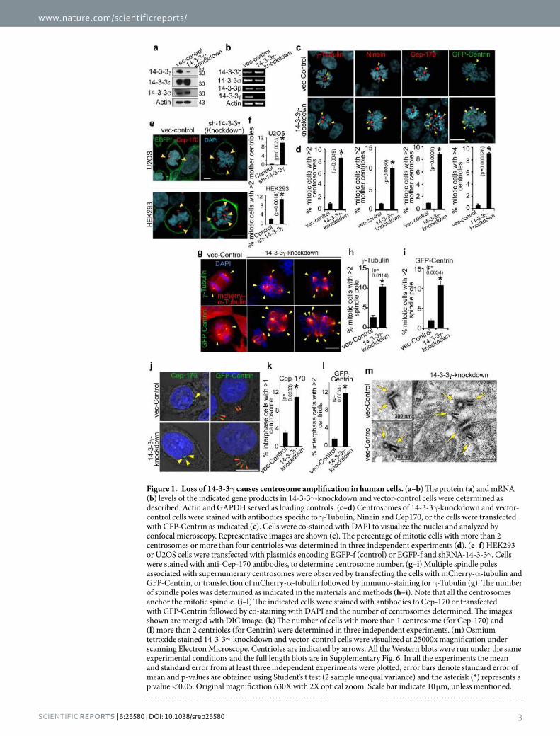
www.nature.com/scientificreports/
3Scientific RepoRts | 6:26580 | DOI: 10.1038/srep26580
Figure 1. Loss of 14-3-3γ causes centrosome amplification in human cells. (a–b) The protein (a) and mRNA (b) levels of the indicated gene products in 14-3-3γ -knockdown and vector-control cells were determined as described. Actin and GAPDH served as loading controls. (c–d) Centrosomes of 14-3-3γ -knockdown and vector-control cells were stained with antibodies specific to γ -Tubulin, Ninein and Cep170, or the cells were transfected with GFP-Centrin as indicated (c). Cells were co-stained with DAPI to visualize the nuclei and analyzed by confocal microscopy. Representative images are shown (c). The percentage of mitotic cells with more than 2 centrosomes or more than four centrioles was determined in three independent experiments (d). (e–f) HEK293 or U2OS cells were transfected with plasmids encoding EGFP-f (control) or EGFP-f and shRNA-14-3-3γ . Cells were stained with anti-Cep-170 antibodies, to determine centrosome number. (g–i) Multiple spindle poles associated with supernumerary centrosomes were observed by transfecting the cells with mCherry-α -tubulin and GFP-Centrin, or transfection of mCherry-α -tubulin followed by immuno-staining for γ -Tubulin (g). The number of spindle poles was determined as indicated in the materials and methods (h–i). Note that all the centrosomes anchor the mitotic spindle. (j–l) The indicated cells were stained with antibodies to Cep-170 or transfected with GFP-Centrin followed by co-staining with DAPI and the number of centrosomes determined. The images shown are merged with DIC image. (k) The number of cells with more than 1 centrosome (for Cep-170) and (l) more than 2 centrioles (for Centrin) were determined in three independent experiments. (m) Osmium tetroxide stained 14-3-3γ -knockdown and vector-control cells were visualized at 25000x magnification under scanning Electron Microscope. Centrioles are indicated by arrows. All the Western blots were run under the same experimental conditions and the full length blots are in Supplementary Fig. 6. In all the experiments the mean and standard error from at least three independent experiments were plotted, error bars denote standard error of mean and p-values are obtained using Student’s t test (2 sample unequal variance) and the asterisk (*) represents a p value < 0.05. Original magnification 630X with 2X optical zoom. Scale bar indicate 10 μ m, unless mentioned.

www.nature.com/scientificreports/
4Scientific RepoRts | 6:26580 | DOI: 10.1038/srep26580
if centrosome over-duplication leads to increased aneuploidy, chromosome number was determined in the 14-3-3γ -knockdown and vector-control cells by counting chromosomes in metaphase plates; HCT116 cells are near diploid and have a normal chromosomal complement (ATCC). Loss of 14-3-3γ -led to an increase in the number of cells with more or less than 46 chromosomes as compared to the vector control (Fig. 3a,b). Moreover, the extent of aneuploidy was increased with sub-culturing (“passage”) of the 14-3-3γ -knockdown cells (Fig. 3c,d); cells in passage-55 showing a greater increase in aneuploidy as compared to cell in passage-15. Aneuploidy leads to chromosome instability and chromosome lagging during anaphase, which often results in cells containing
Figure 2. Depletion of 14-3-3γ leads to centrosome over-duplication during S-phase. (a,b) 14-3-3γ - knockdown and vector-control cells were transfected with GFP-Centrin and synchronized with mimosine. Another similar set of un-transfected cells were used for staining with anti-Cep-170 antibody. At various time points post mimosine withdrawal (0 hours), cells were either processed for FACS analysis, or were stained with anti-Cep-170 antibody to visualize centrioles and co-stained with DAPI for nuclei. The cells transfected with GFP-Centrin were counter-stained with DAPI. The cell cycle histograms and associated representative confocal images are shown (a). The percentage of cells with centrosome amplification was determined at each time point in each experiment and the mean and standard error are plotted (b); Student’s t test (2 sample unequal variance) was used to determine p-value; p < 0.05 (*). (c) Protein extracts were resolved on SDS-PAGE gels followed by Western blotting for cyclinB1. Note that an increase in CyclinB1 level is coincident with an increase in centrosome number. Western blots for actin served as a loading control. (d) Table showing the percentages of cells in different cell cycle phases at each time point. All the Western blots were run under the same experimental conditions and the full length blots are in Supplementary Fig. 6.
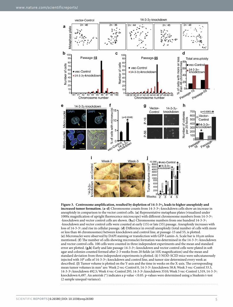
www.nature.com/scientificreports/
5Scientific RepoRts | 6:26580 | DOI: 10.1038/srep26580
Figure 3. Centrosome amplification, resulted by depletion of 14-3-3γ, leads to higher aneuploidy and increased tumor formation. (a–d) Chromosome counts from 14-3-3γ -knockdown cells show an increase in aneuploidy in comparison to the vector control cells. (a) Representative metaphase plates (visualized under 1000x magnification of upright fluorescence microscope) with different chromosome numbers from 14-3-3γ -knockdown and vector control cells are shown. (b,c) Chromosome numbers from one hundred 14-3-3γ -knockdown and vector control cells were counted at early (15) or late (55) passage. Aneuploidy increases with loss of 14-3-3γ and rise in cellular passage. (d) Difference in overall aneuploidy (total number of cells with more or less than 46 chromosomes) between knockdown and control line, at passage-15 and 55, is plotted. (e) Micronuclei were observed by DAPI staining or transfection with GFP-Lamin-A. Scale bar is 10 μ m unless mentioned. (f) The number of cells showing micronuclei formation was determined in the 14-3-3γ -knockdown and vector control cells. 100 cells were counted in three independent experiments and the mean and standard error are plotted. (g,h) Early and late passage 14-3-3γ -knockdown and vector control cells were plated in soft agar and colonies counted formed after 2-3 weeks from 20 fields (at 10X magnification) and the mean and standard deviation from three independent experiments is plotted. (i) 5 NOD-SCID mice were subcutaneously injected with 106 cells of 14-3-3γ -knockdown and control line, and tumor size determined every week as described. (J) Tumor volume is plotted on the Y-axis and the time in weeks on the X-axis. The corresponding mean tumor volumes in mm3 are: Week 2 vec-Control 0, 14-3-3γ knockdown 58.8; Week 3 vec-Control 33.5, 14-3-3γ knockdown 492.5; Week 4 vec-Control 293, 14-3-3γ knockdown 3510; Week 5 vec-Control 1,519, 14-3-3γ knockdown 6,497. An asterisk (*) indicates a p value < 0.05. p-values were determined using a Students t-test (2 sample unequal variance).
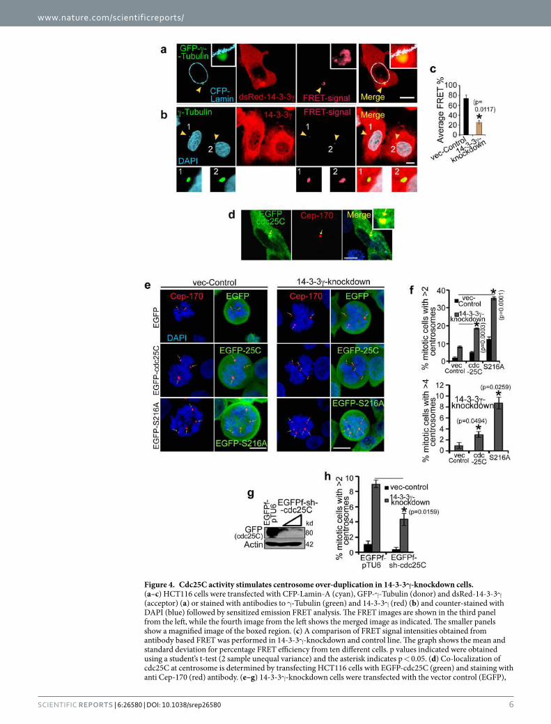
www.nature.com/scientificreports/
6Scientific RepoRts | 6:26580 | DOI: 10.1038/srep26580
Figure 4. Cdc25C activity stimulates centrosome over-duplication in 14-3-3γ-knockdown cells. (a–c) HCT116 cells were transfected with CFP-Lamin-A (cyan), GFP-γ -Tubulin (donor) and dsRed-14-3-3γ (acceptor) (a) or stained with antibodies to γ -Tubulin (green) and 14-3-3γ (red) (b) and counter-stained with DAPI (blue) followed by sensitized emission FRET analysis. The FRET images are shown in the third panel from the left, while the fourth image from the left shows the merged image as indicated. The smaller panels show a magnified image of the boxed region. (c) A comparison of FRET signal intensities obtained from antibody based FRET was performed in 14-3-3γ -knockdown and control line. The graph shows the mean and standard deviation for percentage FRET efficiency from ten different cells. p values indicated were obtained using a student’s t-test (2 sample unequal variance) and the asterisk indicates p < 0.05. (d) Co-localization of cdc25C at centrosome is determined by transfecting HCT116 cells with EGFP-cdc25C (green) and staining with anti Cep-170 (red) antibody. (e–g) 14-3-3γ -knockdown cells were transfected with the vector control (EGFP),
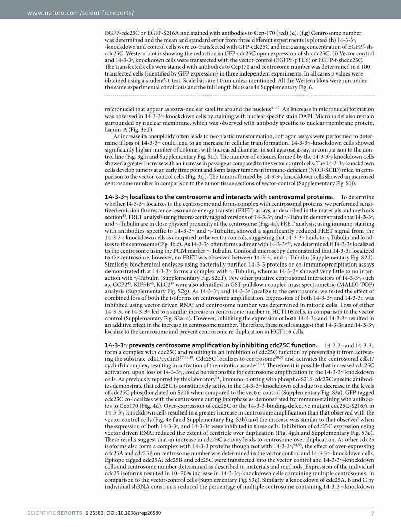
www.nature.com/scientificreports/
7Scientific RepoRts | 6:26580 | DOI: 10.1038/srep26580
micronuclei that appear as extra-nuclear satellite around the nucleus41,42. An increase in micronuclei formation was observed in 14-3-3γ -knockdown cells by staining with nuclear specific stain DAPI. Micronuclei also remain surrounded by nuclear membrane, which was observed with antibody specific to nuclear membrane protein, Lamin-A (Fig. 3e,f).
As increase in aneuploidy often leads to neoplastic transformation, soft agar assays were performed to deter-mine if loss of 14-3-3γ could lead to an increase in cellular transformation. 14-3-3γ -knockdown cells showed significantly higher number of colonies with increased diameter in soft agarose assay, in comparison to the con-trol line (Fig. 3g,h and Supplementary Fig. S1i). The number of colonies formed by the 14-3-3γ -knockdown cells showed a greater increase with an increase in passage as compared to the vector control cells. The 14-3-3γ -knockdown cells develop tumors at an early time point and form larger tumors in immune-deficient (NOD-SCID) mice, in com-parison to the vector-control cells (Fig. 3i,j). The tumors formed by 14-3-3γ -knockdown cells showed an increased centrosome number in comparison to the tumor tissue sections of vector-control (Supplementary Fig. S1j).
14-3-3γ localizes to the centrosome and interacts with centrosomal proteins. To determine whether 14-3-3γ localizes to the centrosome and forms complex with centrosomal proteins, we performed sensi-tized emission fluorescence resonance energy transfer (FRET) assays, as described in the materials and methods section43. FRET analysis using fluorescently tagged versions of 14-3-3γ and γ -Tubulin demonstrated that 14-3-3γ and γ -Tubulin are in close physical proximity at the centrosome (Fig. 4a). FRET analysis, using immuno-staining with antibodies specific to 14-3-3γ and γ -Tubulin, showed a significantly reduced FRET signal from the 14-3-3γ -knockdown cells as compared to the vector controls, suggesting that 14-3-3γ binds to γ -Tubulin and local-izes to the centrosome (Fig. 4b,c). As 14-3-3γ often forms a dimer with 14-3-3ε 44, we determined if 14-3-3ε localized to the centrosome using the PCM marker γ -Tubulin. Confocal microscopy demonstrated that 14-3-3ε localized to the centrosome, however, no FRET was observed between 14-3-3ε and γ -Tubulin (Supplementary Fig. S2d). Similarly, biochemical analyses using bacterially purified 14-3-3 proteins or co-immunoprecipitation assays demonstrated that 14-3-3γ forms a complex with γ -Tubulin, whereas 14-3-3ε showed very little to no inter-action with γ -Tubulin (Supplementary Fig. S2e,f). Few other putative centrosomal interactors of 14-3-3γ such as, GCP245, KIF5B46, KLC247 were also identified in GST-pulldown coupled mass spectrometric (MALDI-TOF) analysis (Supplementary Fig. S2g). As 14-3-3γ and 14-3-3ε localize to the centrosome, we tested the effect of combined loss of both the isoforms on centrosome amplification. Expression of both 14-3-3γ and 14-3-3ε was inhibited using vector driven RNAi and centrosome number was determined in mitotic cells. Loss of either 14-3-3ε or 14-3-3γ led to a similar increase in centrosome number in HCT116 cells, in comparison to the vector control (Supplementary Fig. S2a–c). However, inhibiting the expression of both 14-3-3γ and 14-3-3ε resulted in an additive effect in the increase in centrosome number. Therefore, these results suggest that 14-3-3ε and 14-3-3γ localize to the centrosome and prevent centrosome re-duplication in HCT116 cells.
14-3-3γ prevents centrosome amplification by inhibiting cdc25C function. 14-3-3γ and 14-3-3ε form a complex with cdc25C and resulting in an inhibition of cdc25C function by preventing it from activat-ing the substrate cdk1/cyclinB27,48,49. Cdc25C localizes to centrosome50,51 and activates the centrosomal cdk1/cyclinB1 complex, resulting in activation of the mitotic cascade52,53. Therefore it is possible that increased cdc25C activation, upon loss of 14-3-3γ , could be responsible for centrosome amplification in the 14-3-3γ knockdown cells. As previously reported by this laboratory31, immuno-blotting with phospho-S216-cdc25C specific antibod-ies demonstrate that cdc25C is constitutively active in the 14-3-3γ knockdown cells due to a decrease in the levels of cdc25C phosphorylated on S216 when compared to the vector control (Supplementary Fig. S3a). GFP-tagged cdc25C co-localizes with the centrosome during interphase as demonstrated by immuno-staining with antibod-ies to Cep170 (Fig. 4d). Over-expression of cdc25C or the 14-3-3-binding-defective mutant cdc25C-S216A in 14-3-3γ -knockdown cells resulted in a greater increase in centrosome amplification than that observed with the vector control cells (Fig. 4e,f and Supplementary Fig. S3b) and the increase was similar to that observed when the expression of both 14-3-3γ and 14-3-3ε were inhibited in these cells. Inhibition of cdc25C expression using vector driven RNAi reduced the extent of centriole over duplication (Fig. 4g,h and Supplementary Fig. S3c). These results suggest that an increase in cdc25C activity leads to centrosome over-duplication. As other cdc25 isoforms also form a complex with 14-3-3 proteins though not with 14-3-3γ 54,55, the effect of over-expressing cdc25A and cdc25B on centrosome number was determined in the vector control and 14-3-3γ -knockdown cells. Epitope tagged cdc25A, cdc25B and cdc25C were transfected into the vector control and 14-3-3γ -knockdown cells and centrosome number determined as described in materials and methods. Expression of the individual cdc25 isoforms resulted in 10–20% increase in 14-3-3γ -knockdown cells containing multiple centrosomes, in comparison to the vector-control cells (Supplementary Fig. S3e). Similarly, a knockdown of cdc25A, B and C by individual shRNA constructs reduced the percentage of multiple centrosome containing 14-3-3γ -knockdown
EGFP-cdc25C or EGFP-S216A and stained with antibodies to Cep-170 (red) (e). (f,g) Centrosome number was determined and the mean and standard error from three different experiments is plotted (h) 14-3-3γ -knockdown and control cells were co-transfected with GFP-cdc25C and increasing concentration of EGFPf-sh-cdc25C. Western blot is showing the reduction in GFP-cdc25C upon expression of sh-cdc25C. (i) Vector control and 14-3-3γ knockdown cells were transfected with the vector control (EGFPf-pTU6) or EGFP-f-shcdc25C. The transfected cells were stained with antibodies to Cep170 and centrosome number was determined in a 100 transfected cells (identified by GFP expression) in three independent experiments. In all cases p values were obtained using a student’s t-test. Scale bars are 10 μ m unless mentioned. All the Western blots were run under the same experimental conditions and the full length blots are in Supplementary Fig. 6.
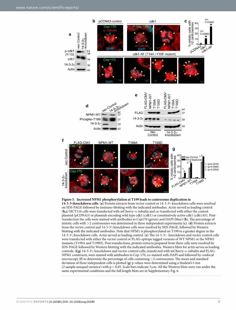
www.nature.com/scientificreports/
8Scientific RepoRts | 6:26580 | DOI: 10.1038/srep26580
Figure 5. Increased NPM1 phosphorylation at T199 leads to centrosome duplication in 14-3-3γknockdown cells. (a) Protein extracts from vector control or 14-3-3γ knockdown cells were resolved on SDS-PAGE followed by immuno-blotting with the indicated antibodies. Actin served as loading control. (b,c) HCT116 cells were transfected with mCherry-α -tubulin and co-transfected with either the control plasmid (pCDNA3) or plasmids encoding wild type cdk1 (cdk1) or constitutively active cdk1 (cdk1AF). Post-transfection the cells were stained with antibodies to Cep170 (green) and DAPI (blue) (b). The percentage of mitotic cells with > 2 centrosomes was determined in three independent experiments (c). (d) Protein extracts from the vector control and 14-3-3γ knockdown cells were resolved by SDS-PAGE, followed by Western blotting with the indicated antibodies. Note that NPM1 is phosphorylated on T199 to a greater degree in the 14-3-3γ knockdown cells. Actin served as loading control. (e) The 14-3-3γ -knockdown and vector control cells were transfected with either the vector control or FLAG-epitope tagged versions of WT NPM1 or the NPM1 mutants (T199A and T199D). Post transfections, protein extracts prepared from these cells were resolved by SDS-PAGE followed by Western blotting with the indicated antibodies. Western blots for actin serves as loading controls. (f,g) 14-3-3γ -knockdown and vector-control cells, transfected with mCherry-α -tubulin and FLAG-NPM1 constructs, were stained with antibodies to Cep-170, co-stained with DAPI and followed by confocal microscopy (f) to determine the percentage of cells containing > 2 centrosomes. The mean and standard deviation of three independent cells is plotted (g) p values were determined using a Student’s t-test (2 sample unequal variance) with p < 0.05. Scale bars indicate 5 μ m. All the Western blots were run under the same experimental conditions and the full length blots are in Supplementary Fig. 6.
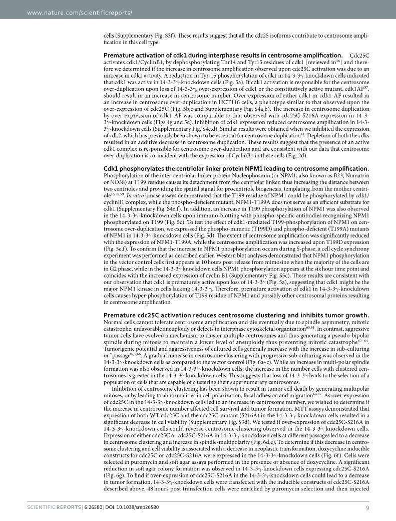
www.nature.com/scientificreports/
9Scientific RepoRts | 6:26580 | DOI: 10.1038/srep26580
cells (Supplementary Fig. S3f). These results suggest that all the cdc25 isoforms contribute to centrosome ampli-fication in this cell type.
Premature activation of cdk1 during interphase results in centrosome amplification. Cdc25C activates cdk1/CyclinB1, by dephosphorylating Thr14 and Tyr15 residues of cdk1 [reviewed in56] and there-fore we determined if the increase in centrosome amplification observed upon cdc25C activation was due to an increase in cdk1 activity. A reduction in Tyr-15 phosphorylation of cdk1 in 14-3-3γ -knockdown cells indicated that cdk1 was active in 14-3-3γ -knockdown cells (Fig. 5a). If cdk1 activation is responsible for the centrosome over-duplication upon loss of 14-3-3γ , over-expression of cdk1 or the constitutively active mutant, cdk1AF57, should result in an increase in centrosome number. Over-expression of either cdk1 or cdk1-AF resulted in an increase in centrosome over-duplication in HCT116 cells, a phenotype similar to that observed upon the over-expression of cdc25C (Fig. 5b,c and Supplementary Fig. S4a,b). The increase in centrosome duplication by over-expression of cdk1-AF was comparable to that observed with cdc25C-S216A expression in 14-3-3γ -knockdown cells (Figs 4g and 5c). Inhibition of cdk1 expression reduced centrosome amplification in 14-3-3γ -knockdown cells (Supplementary Fig. S4c,d). Similar results were obtained when we inhibited the expression of cdk2, which has previously been shown to be essential for centrosome duplication11. Depletion of both the cdks resulted in an additive decrease in centrosome duplication. These results suggest that the presence of an active cdk1 complex is responsible for centrosome over-duplication and are consistent with our data that centrosome over-duplication is co-incident with the expression of CyclinB1 in these cells (Fig. 2d).
Cdk1 phosphorylates the centriolar linker protein NPM1 leading to centrosome amplification. Phosphorylation of the inter-centriolar linker protein Nucleophosmin (or NPM1, also known as B23, Numatrin or NO38) at T199 residue causes its detachment from the centriolar linker, thus increasing the distance between two centrioles and providing the spatial signal for procentriole biogenesis, templating from the mother centri-ole16,58,59. In vitro kinase assays demonstrated that the T199 residue of NPM1 could be phosphorylated by cdk1/cyclinB1 complex, while the phospho-deficient mutant, NPM1-T199A does not serve as an efficient substrate for cdk1 (Supplementary Fig. S4e,f). In addition, an increase in T199 phosphorylation of NPM1 was also observed in the 14-3-3γ -knockdown cells upon immuno-blotting with phospho-specific antibodies recognizing NPM1 phosphorylated on T199 (Fig. 5c). To test the effect of cdk1-mediated T199-phosphorylation of NPM1 on cen-trosome over-duplication, we expressed the phospho-mimetic (T199D) and phospho-deficient (T199A) mutants of NPM1 in 14-3-3γ -knockdown cells (Fig. 5d). The extent of centrosome amplification was significantly reduced with the expression of NPM1-T199A, while the centrosome amplification was increased upon T199D expression (Fig. 5e,f). To confirm that the increase in NPM1 phosphorylation occurs during S-phase, a cell cycle synchrony experiment was performed as described earlier. Western blot analyses demonstrated that NPM1 phosphorylation in the vector control cells first appears at 10 hours post release from mimosine when the majority of the cells are in G2 phase, while in the 14-3-3γ knockdown cells NPM1 phosphorylation appears at the six hour time point and coincides with the increased expression of cyclin B1 (Supplementary Fig. S5c). These results are consistent with our observation that cdk1 is prematurely active upon loss of 14-3-3γ (Fig. 5a), suggesting that cdk1 might be the major NPM1 kinase in cells lacking 14-3-3 γ . Therefore, premature activation of cdk1 in 14-3-3γ -knockdown cells causes hyper-phosphorylation of T199 residue of NPM1 and possibly other centrosomal proteins resulting in centrosome amplification.
Premature cdc25C activation reduces centrosome clustering and inhibits tumor growth. Normal cells cannot tolerate centrosome amplification and die eventually due to spindle asymmetry, mitotic catastrophe, unfavorable aneuploidy or defects in interphase cytoskeletal organization60,61. In contrast, aggressive tumor cells have evolved a mechanism to cluster multiple centrosomes and thus generating a pseudo-bipolar spindle during mitosis to maintain a lower level of aneuploidy thus preventing mitotic catastrophe62–64. Tumorigenic potential and aggressiveness of cultured cells generally increase with the increase in sub-culturing or “passage”65,66. A gradual increase in centrosome clustering with progressive sub-culturing was observed in the 14-3-3γ -knockdown cells as compared to the vector control (Fig. 6a–c). While an increase in multi-polar spindle formation was also observed in 14-3-3γ -knockdown cells, the increase in the number cells with clustered cen-trosomes is greater in the 14-3-3γ knockdown cells. This suggests that loss of 14-3-3γ leads to the selection of a population of cells that are capable of clustering their supernumerary centrosomes.
Inhibition of centrosome clustering has been shown to result in tumor cell death by generating multipolar mitoses, or by leading to abnormalities in cell polarization, focal adhesion and migration64,67. As over-expression of cdc25C in the 14-3-3γ -knockdown cells led to an increase in centrosome number, we wished to determine if the increase in centrosome number affected cell survival and tumor formation. MTT assays demonstrated that expression of both WT cdc25C and the cdc25C-mutant (S216A) in the 14-3-3γ -knockdown cells resulted in a significant decrease in cell viability (Supplementary Fig. S3d). We tested if over-expression of cdc25C-S216A in 14-3-3γ -knockdown cells could reverse centrosome clustering observed in the 14-3-3γ knockdown cells. Expression of either cdc25C or cdc25C-S216A in 14-3-3γ -knockdown cells at different passages led to a decrease in centrosome clustering and increase in spindle-multipolarity (Fig. 6d,e). To determine if this decrease in centro-some clustering and cell viability is associated with a decrease in neoplastic transformation, doxycycline inducible constructs for cdc25C or cdc25C-S216A were expressed in the 14-3-3γ -knockdown cells (Fig. 6f). Cells were selected in puromycin and soft agar assays performed in the presence or absence of doxycycline. A significant reduction in soft agar colony formation was observed in 14-3-3γ -knockdown cells expressing cdc25C-S216A (Fig. 6g). To find if over-expression of cdc25C-S216A in the 14-3-3γ -knockdown cells could lead to a decrease in tumor formation, 14-3-3γ -knockdown cells were transfected with the inducible constructs of cdc25C-S216A described above. 48 hours post transfection cells were enriched by puromycin selection and then injected

www.nature.com/scientificreports/
1 0Scientific RepoRts | 6:26580 | DOI: 10.1038/srep26580
Figure 6. Over-expression of cdc25C-S216A leads to an increase in multipolar mitoses and a decrease in neoplastic progression. (a) 14-3-3γ -knockdown and vector control cells at different passages (P) were fixed and stained with antibodies to α -tubulin and Cep-170 and co-stained with DAPI. Arrows indicate centrosome poles. (b,c) The percentage of cells with multi-polar spindles and pseudo bi-polar spindles was determined in three independent experiments and the mean and standard error plotted. (d) 14-3-3γ -knockdown cells were transfected with EGFP, EGFP-cdc25C or EGFP-S216A constructs. Cells were fixed and stained with anti-α -Tubulin antibody and co-stained with DAPI. Arrows indicate spindle poles. (e) The percentage of cells with multi-polar spindles and pseudo bi-polar spindles was determined in three independent experiments and the mean and standard deviation plotted. A significant increase in the number of cells with multi-polar spindles and a corresponding decrease in the number of cells with pseudo-bipolar spindles was observed in cells expressing the two cdc25C constructs at all passages. (f,g) 14-3-3γ -knockdown cells were transfected with doxycycline (Dox) inducible constructs expressing either GFP or EGFP-cdc25C or EGFP-cdc25C-S216A. Post transfection the cells were selected in puromycin to select transfected cells. Transfected cells were grown in the presence or absence of doxycycline and protein extracts were resolved on SDS-PAGE gels followed by Western blotting with the indicated antibodies (f) or the cells were embedded in soft agar in the presence or absence of doxycycline and colony formation determined in three independent experiments. The mean and standard deviation are plotted (g). (h,i) 14-3-3γ -knockdown cells were transfected with doxycycline (Dox) inducible constructs expressing EGFP-cdc25C-S216A. Post selection, cells were injected subcutaneously in Nude mice. One set was given doxycycline in the drinking water (+ Dox) and the other set received no doxycycline (− Dox). Tumor volume was measured and the mean and standard error are plotted from 4 × 2 sets of mice at different times post injection. p values were obtained using Student’s t test (2 sample unequal variance). p values < 0.05 are indicated by asterisk. All the Western blots were run under the same experimental conditions and the full length blots are in Supplementary Fig. 6.
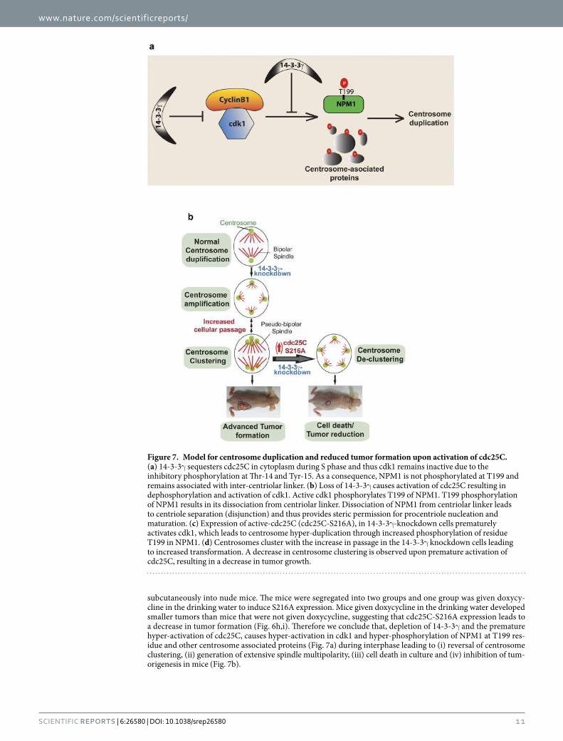
www.nature.com/scientificreports/
1 1Scientific RepoRts | 6:26580 | DOI: 10.1038/srep26580
subcutaneously into nude mice. The mice were segregated into two groups and one group was given doxycy-cline in the drinking water to induce S216A expression. Mice given doxycycline in the drinking water developed smaller tumors than mice that were not given doxycycline, suggesting that cdc25C-S216A expression leads to a decrease in tumor formation (Fig. 6h,i). Therefore we conclude that, depletion of 14-3-3γ and the premature hyper-activation of cdc25C, causes hyper-activation in cdk1 and hyper-phosphorylation of NPM1 at T199 res-idue and other centrosome associated proteins (Fig. 7a) during interphase leading to (i) reversal of centrosome clustering, (ii) generation of extensive spindle multipolarity, (iii) cell death in culture and (iv) inhibition of tum-origenesis in mice (Fig. 7b).
Figure 7. Model for centrosome duplication and reduced tumor formation upon activation of cdc25C. (a) 14-3-3γ sequesters cdc25C in cytoplasm during S phase and thus cdk1 remains inactive due to the inhibitory phosphorylation at Thr-14 and Tyr-15. As a consequence, NPM1 is not phosphorylated at T199 and remains associated with inter-centriolar linker. (b) Loss of 14-3-3γ causes activation of cdc25C resulting in dephosphorylation and activation of cdk1. Active cdk1 phosphorylates T199 of NPM1. T199 phosphorylation of NPM1 results in its dissociation from centriolar linker. Dissociation of NPM1 from centriolar linker leads to centriole separation (disjunction) and thus provides steric permission for procentriole nucleation and maturation. (c) Expression of active-cdc25C (cdc25C-S216A), in 14-3-3γ -knockdown cells prematurely activates cdk1, which leads to centrosome hyper-duplication through increased phosphorylation of residue T199 in NPM1. (d) Centrosomes cluster with the increase in passage in the 14-3-3γ knockdown cells leading to increased transformation. A decrease in centrosome clustering is observed upon premature activation of cdc25C, resulting in a decrease in tumor growth.
www.nature.com/scientificreports/
1 2Scientific RepoRts | 6:26580 | DOI: 10.1038/srep26580
DiscussionWe report for the first time that, loss of 14-3-3γ causes centrosome over-duplication, centrosome clustering and tumor formation in mice. Although 14-3-3 proteins have been isolated from centrosomal fractions68, the molec-ular basis and isoform specific role of 14-3-3 in centrosome duplication remained unknown. In this study, we have demonstrated that 14-3-3ε and 14-3-3γ localize to the centrosome and form complexes with centrosomal proteins. Our results demonstrate a mechanism by which 14-3-3γ restricts centrosome duplication to once per cell cycle, by inhibiting cdc25C function, thus preventing premature activation of cdk1 during interphase and resulting in a decrease in phosphorylation of T199 residue of the centriolar linker protein NPM1. Identification of the molecular basis of 14-3-3γ -mediated centrosome duplication helped to design a way to reduce centrosome clustering, leading subsequently to a decrease in tumor formation in nude mice by over-expression of constitu-tively active (14-3-3-binding-deficient) cdc25C in 14-3-3γ -knockdown cells. This also suggests that complete disruption of the cdc25C-14-3-3 complex during interphase might be a way to inhibit tumor growth and selective tumor cell killing.
In S. pombe, cdc25 activates the cdc2 (cdk1-ortholog)/cyclinB complex, which phosphorylates a spindle pole body protein Cut12 (Stf1). Gain of function mutation in Cut12 is sufficient to drive mitosis in the absence of cdc25C, and loss of Cut12 results in the failure in microtubule nucleation, failure to associate Spindle Pole Bodies to the nuclear membrane and failure in mitotic entry leading to mitotic arrest. The defects observed upon Cut12 deletion could be rescued by enhancing cdc25C or cdk1 function suggesting that cdc25C and cdk1 have a role to play in centrosome duplication24,69,70. However, experiments in other cell systems have suggested that loss of cdk1 leads to an increase in centrosome number due to the induction of multiple rounds of S-phase71,72. One of these studies71 reported that, while inhibition of cdk1 in rodent cells causes centrosome amplification due to endo-reduplication, the same phenotype was not observed in human cell lines including HCT116 and U-2OS, similar to the results reported here. The differences observed between experiments performed in Drosophila or rodent cell lines in culture and in human cell lines might be due to species specific differences in centrosome duplication. Altogether, these observations support the fact that increased cdk1-activation can lead to centrosome amplification in human cells. Cdk1 overexpression leads to genomic instability, centrosome separation22,23,73 and centrosome amplification74. Nam and van Deursen recently showed that cyclin B overexpression in the mouse leads to accelerated centrosome separation, chromosome mis-segregation and tumor formation through a Plk1− Nek2− C-NAP1/rootletin mediated pathway26,75. Therefore, it is evident that increased activation of the cyclin B/cdk1 complex leads to centrosome amplification, aneuploidy and tumor formation. Further, activation of cdk1 leads to the activation of Plk1 by the Aurora-A and Bora kinases76. Plk1 activation is responsible for Cep-152/Cep-192 mediated activation of Plk-4, which drives centriolar cartwheel formation through 9-fold assembly of Sas-6 dimers77–79. Thus, a positive feedback loop of cdk1 is required for the commitment of M phase progression80 and initiation of centriole biogenesis.
Although NPM1 is phosphorylated by the G1/S checkpoint regulator cdk216, it was not clear if NPM1 could also serve as a cdk1 substrate for centriole disjunction during S-phase12,59. In this study, we have demonstrated that T199 residue of NPM1 is a target of cdk1, and early phosphorylation of NPM1 by cdk1 leads to a premature increase in centrosome number. Further, our results suggest that in HCT116 cells cdk1 is the major NPM1 kinase and that the increase in NPM1 phosphorylation upon loss of 14-3-3γ is due to a premature increase in cdk1 activ-ity. Our data does not exclude the possibility that cdk1 phosphorylates other centrosomal proteins, in addition to NPM1, and that these substrates might also contribute to the increase in centrosome number observed upon loss of 14-3-3γ (Fig. 7a). Interestingly, loss of either cdk1 or cdk2 in the 14-3-3γ knockdown cells leads to a decrease in centrosome number, suggesting that cdk1 is not phosphorylating the same set of substrates as cdk2. Therefore, when cdk1 is prematurely activated during S-phase in the 14-3-3γ -knockdown cells, it stimulates centrosome over-duplication and this requires cdk2 activity suggesting that the activity of both proteins is required for cen-trosome amplification.
Our work also demonstrates that the mitotic phosphatase cdc25C is required for the increase in centrosome number observed upon loss of 14-3-3γ . However, over-expression of all the cdc25 isoforms in HCT116 cells results in an increase in centrosome number and a knockdown of the individual isoforms reduces centrosome number (Supplementary Fig. S3e,f). While both cdc25A and cdc25B have been reported to bind to 14-3-3 pro-teins, neither forms a complex with 14-3-3γ and have been shown to bind other 14-3-3 isoforms54,55. It is possible that all of the cdc25 isoforms can regulate the increase in centrosome duplication, indeed it has been reported that all of them are required for the complete activation of cdk1/cyclinB81 and that multiple isoforms need to be inac-tivated to generate a cell cycle arrest82. These are also consistent with our results that over-expression of cdk1 or an active form of cdk1 could lead to an increase in centrosome duplication, as the increased activity of the cdc25 family members would remove the inhibitory phosphates from the over-expressed cdk1. However, the increase in centrosome number upon loss of 14-3-3γ is dependent on the expression of cyclin B, as suggested by the data in Fig. 2, suggesting that the effects we observe are dependent on cdk1.
Our work also demonstrates that in addition to regulating cdc25C function, 14-3-3γ also binds to other cen-trosomal or centrosome associate proteins that regulate centrosome function. It is also possible that the interac-tion of 14-3-3γ with centrosomal proteins prevents their phosphorylation by cdks, thus preventing centrosome amplification (Fig. 7a). These results suggest the probable existence of other mechanisms by which 14-3-3γ reg-ulates centrosome duplication and biogenesis by regulating PLK4/Sas-6 mediated centriole cartwheel forma-tion83,84, or regulating the assembly of pericentriolar matrix85,86, or the formation of γ -Tubulin-ring complex to regulate microtubule nucleation19. Further work is required to clarify the mechanisms by which 14-3-3 proteins regulate centrosome biogenesis, as 14-3-3 proteins are broad-spectrum phospho-Ser/Thr-binding adaptor pro-teins that act as signal integration nodes for multiple biochemical pathways29.
Multipolar cell divisions are rare and multipolar spindles are often unstable short-lived intermediates. Tumor cells have evolved mechanisms to induce centrosome clustering leading to the formation of a pseudo-bipolar
www.nature.com/scientificreports/
13Scientific RepoRts | 6:26580 | DOI: 10.1038/srep26580
spindle during mitosis. Pseudo-bipolar mitoses result in the formation merotelic chromosome attachments leading to a favorable aneuploidy and acquisition of the neoplastic phenotype61. However, a hypothesis that is being currently promulgated in the literature suggests that promoting excessive genetic instability could be a potential target for anti-tumor therapeutics61,87, as tumor cells are programmed to be genetically unstable due to the inactivation of checkpoint pathways [reviewed in88–90]. Thus, exploiting the addiction to genetic instability could result in increased cell death even in tumors that are normally resistant to several cytotoxic agents used in cancer therapy. Our work demonstrates that rather than inhibiting cdk1 activity, a novel way of inhibiting tumor growth might be the premature activation of cdk1 in interphase cells. This is in contrast to the paradigm generally accepted in the literature where several studies have attempted to use inhibitors of the cell-cycle kinases to inhibit tumor growth, an approach that has not achieved significant success. While our observation is based on data generated in a xenograft mouse model that lacks an intact immune system, similar experiments could not be performed in mouse genetic models as mouse cdc25C lacks the 14-3-3 binding site and does not form a complex with 14-3-3 proteins (our unpublished data). However, given the data suggesting that over-expression of B cyclins in the mouse results in tumor progression26 and the number of papers suggesting that cyclin B is over-expressed in human tumors as compared to normal tissue91–96, we believe that our data suggesting that the premature activation of cdk1 is a potent way of killing tumor cells, certainly has broad significance for the field of tumor therapeutics (Fig. 7b).
Overall, this study provides a conceptual framework to understand the role of 14-3-3 proteins in centrosome duplication by regulating the activity of cdc25C, cdk1 and NPM1. Our work also indicates the future possibility of development of therapeutic methods to reduce tumor growth by targeting the disruption of 14-3-3-cdc25C complex in the interphase cells, without affecting normal cells. The novel molecular basis of 14-3-3-mediated cen-trosome duplication and harnessing this concept to inhibit centrosome-clustering and subsequent tumor reduc-tion opens the door towards understanding the regulation of centrosome biogenesis by myriad roles of 14-3-3 proteins, and creating novel avenues for preventing tumor growth by centrosome de-clustering.
Materials and MethodsEthics Statement. Maintenance of the animal facility is as per the national guidelines provided by the Committee for the Purpose of Control and Supervision of the Experiments on Animals (CPCSEA), Ministry of Environment and Forest, Government of India. All the experiments in this manuscript have been carried out according to the approved guidelines. The animals were housed in a controlled environment with the tempera-ture and relative humidity being maintained at 23 ± 2 °C and 40–70% respectively and a day night cycle of 12 hrs each (7:00 to 19:00 light; 19:00 to 7:00 dark). The animals were received an autoclaved balanced diet prepared in-house as per the standard formula and sterile water ad libitum. Mice were housed in the Individually Ventilated Cage (IVC) system (M/S Citizen, India) provided with autoclaved corn cob bedding material (Natgrit 406) pro-cured from Natural Organics, Satara, MS, India. Protocols for the experiments were approved by the Institutional Animal Ethics Committee (IAEC) of the Advanced Centre for Treatment Research and Education in Cancer (ACTREC). The animal study proposal number is 11/2008 dated August 19, 2008.
Cell culture and transfections. HCT116, U-2OS and HEK293 cells and the HCT116 derived vector con-trol and 14-3-3γ -knockdown cells were cultured as described31. Cells were transfected with lipofectamine-LTX (Invitrogen) according to the manufacturer’s instructions. HCT116 cells transfected with the pTRIPZ constructs expressing either WT cdc25C or S216A were maintained in media containing 1 μ g/ml of puromycin. Expression of cdc25C was induced by adding doxycycline to the medium at a concentration of 2 μ g/ml. To perform the rescue experiments GFP14-3-3γ -R (shRNA resistant 14-3-3γ cDNA) was transfected into 14-3-3γ -knockdown cells and subsequently the cells expressing GFP construct were sorted using flow cytometry as described97. Centrosome number was determined as described below.
Estimation of centrosome and spindle pole number, and determination of centrosome clus-tering and multi-polarity. To enrich cells in mitosis, the 14-3-3γ -knockdown and vector control cells were synchronized using 400 μ M mimosine for 20 hours as described98, released and fixed with 4% para-formaldehyde after 12–14 hours, to allow them to enter mitosis. To determine the percentage of cells containing more than two centrosomes or spindle poles, centrosomes or spindle poles of 100 mitotic cells were counted from three inde-pendent experiments. As over-expression of cdc25C or cdc25C-S216A in 14-3-3γ -knockdown cell causes death in culture gradually after 48 hours of expression, centrosome counts were performed immediately after 48 hours of transfection. The spindle poles from 100(x3) mitotic cells were counted in 3 independent experiments to deter-mine the number of cells with pseudo-bipolar, multi-polar or truly bipolar spindles.
Immunofluorescence, FRET analysis and confocal microscopy. To determine the localization of proteins, different cell types were grown on glass cover slips. Cells were transfected with combinations of fluores-cence proteins or labeled with fluorophore-conjugated antibodies. Cells were fixed with 4% para-formaldehyde and permiabilized with 0.3% tritonX-100. 0.05% DAPI was used to stain nuclei. Argon, Helium/Neon and diode lasers were used to capture images on a Carl Zeiss LSM 510 Meta confocal microscope. Images were captured under the oil immersion objectives of LSM510 meta (Carl Zeiss) confocal microscope, at 630X or 1000X mag-nification with 2X to 4X digital zoom. All the images were captured after background nullification with second-ary antibodies. Images were processed using the LSM510 software. FRET measurements were performed using the sensitized emission method in fixed cells using samples: Donor only (GFP or Alexa-Fluor-488), Acceptor only (dsRed or Alexa-Fluor-546) and FRET sample. Following images were acquired for FRET corrections and efficiency calculations: (1) Acceptor Only using Acceptor filter set. (2) Acceptor Only using FRET filter set. (3) Donor Only using Donor filter set. (4) Donor Only using FRET filter set. (5) FRET Specimen Only using FRET

www.nature.com/scientificreports/
1 4Scientific RepoRts | 6:26580 | DOI: 10.1038/srep26580
filter set. All the images were captured at X630 magnification in 12-bit format using Zeiss LSM 510 Meta confocal laser scanning microscope. The images were acquired using following lasers: Donor excitation using 488 nm Argon laser line while acceptor excitation using 543 nm Helium Neon laser line. Images acquired were further processed using LSM 510 image examiner software. The nomenclature and equations for FRET calculations are as previously described43 and the FRET protocol was obtained from the Centre for Optical Instrumentation laboratory, Wellcome Trust Centre, University of Edinburg99. FRET Corrections: (i) Acceptor in FRET channel (Co-efficient A) = Average intensity of Acceptor only using FRET set/Average intensity of Acceptor only using acceptor set. (ii) Donor in FRET channel (Co-efficient B) = Average intensity of Donor only using FRET filter set/Average intensity of Donor only using Donor filter set. (iii) Average FRET efficiency = FRET Specimen − (A * FRET Specimen using Acceptor filter set) − (B * FRET Specimen using Donor filter set) * 100.
Electron microscopy. To study centrosome amplification and organization in higher magnification 14-3-3γ -knockdown and vector-control cells were visualized under transmission electron microscope. Synchronized cells in S-phase were fixed with 3% glutaraldehyde, washed with 0.1 M of sodium cacodylate and post fixed with 1% osmium tetra oxide (Tedpella). Cultures were dehydrated and processed. Grids were con-trasted with alcoholic uranyl acetate for 1 minute and lead citrate for half a minute. The grids were observed under a Carl Zeiss LIBRA120 EFTEM transmission electron microscope, at an accelerating voltage of 120KV and at 25000X magnification. Images were captured using a Slow Scan CCD camera (TRS, Germany).
Preparation of metaphase plates. Cells were arrested in mitosis by growing them in Colcemid (0.1 μ g/ml) for 2 hours and were incubated in a hypotonic solution (0.075 M KCl) for 15–25 minutes at 37 °C. Metaphase spreads were generated by dropping the cells from a height on frosted glass slides and chromosomes were stained with Giemsa and imaged under 100X objective of the AxioImager Z1 upright microscope (Carl Zeiss).
Soft Agar Assays. Soft agar assays for the 14-3-3γ -knockdown and vector-control cells were performed as previously described100. To determine whether cdc25C over-expression led to a decrease in transformation, the 14-3-3γ -knockdown cells were transfected with doxycycline inducible constructs for EGFP, EGFP-cdc25C and EGFP-S216A. Transfected cells were selected in 0.5 μ g/ml puromycin. 72 hours post selection, the cells were har-vested by trypsinization and 10,000 cells plated in soft agar containing puromycin at a concentration of 0.5 μ g/ml in the presence or absence of 2 μ g/ml doxycycline in triplicate. The remaining cells were cultured in regular media containing puromycin at a concentration of 0.5 μ g/ml in the presence or absence of 2 μ g/ml doxycycline. The cells were harvested and protein extracts were prepared as described27 and resolved on SDS-PAGE gels for Western blot analysis with antibodies to GFP.
Tumour formation in immunocompromised mice. For the present study, we used NOD.CB17- Prkdcscid/NCrCrl (NOD-SCID mice) or BALB/c Nude mice (CAnN.Cg-Foxn1nu/Crl). The foundation stock of the immuno-compromised mice was procured from Charles River Laboratories, Willington, USA. All animal studies were approved by the Institutional Animal Ethics committee (IAEC) constituted under the guidelines of the CPCSEA, Government of India. 106 HCT116 derived 14-3-3γ -knockdown and vector-control cells were re-suspended in DMEM medium without serum and injected subcutaneously in the dorsal flank of 6–8 weeks
Oligonucleotide Sequence
cdk1a CCGGATGGGGATTCAGAAATTGATCAGTTCTCGATCAATTTCTGAATCCCCATTTTTTTC
cdk1b TCGAGAAAAAAATGGGGATTCAGAAATTGATCGAGAACTGATCAATTTCTGAATCCCCAT
cdk2a CCGGAGCTGTGGACATCTGGAGCCTAGTTCTCAGGCTCCAGATGTCCACAGCTTTTTTTC
cdk2b TCGAGAAAAAAAGCTGTGGACATCTGGAGCCTGAGAACTAGGCTCCAGATGTCCACAGCT
14-3-3β Fwd GGTATCTTTCTGAAGTGGC
14-3-3β Rev GCTACAGGCCTTTTC
14-3-3γ Fwd GAGCCACTGTCGAATG
14-3-3γ Rev CGCTGCAATTCTTGATC
14-3-3σ Fwd GCAGCCTTCATGAAAG
14-3-3σ Rev CCCTTCATCTTCAGGTAG
14-3-3ζ Fwd GTTCTTGATCCCCAATGC
14-3-3ζ Rev CTCTGGGGAGTTCAGAATC
GAPDH Fwd TGCATCCTGCACCACCAACT
GAPDH Rev CGCCTGCTTCACCACCTTC
Cdc25C-1a (shRNA) CCGGTGAAGAGAATAATCATCGTGTTTTCAAGAGAAACACGATGATTATTCTCTTCTTTTTC
Cdc25C-1b (shRNA) TCGAGAAAAAGAAGAGAATAATCATCGTGTTTCTCTTGAAAACACGATGATTATTCTCTTCA
Cdc25B-1a (shRNA) CCGGTAATCCTCCCTGTCGTCTGAATTTCAAGAGAATTCAGACGACAGGGAGGATTTTTTTC
Cdc25B-1b (shRNA) TCGAGAAAAAAATCCTCCCTGTCGTCTGAATTCTCTTGAAATTCAGACGACAGGGAGGATTA
Cdc25A-1a (shRNA) CCGGTAGCAACCACTGGAGGTGAAGTTCAAGAGACTTCACCTCCAGTGGTTGCTTTTTTC
Cdc25A-1b (shRNA) TCGAGAAAAAAGCAACCACTGGAGGTGAAGTCTCTTGAACTTCACCTCCAGTGGTTGCTA
Table 1. Sequences of oligonucleotide primers. Sequences of oligonucleotide primers used for designing shRNA constructs and performing RT-PCR assays.
www.nature.com/scientificreports/
1 5Scientific RepoRts | 6:26580 | DOI: 10.1038/srep26580
old NOD-SCID mice (obtained from ACTREC animal house facility). Five mice were injected for each clone. Tumor formation was monitored at intervals of 2–3 days and tumor size was measured by Vernier calipers. Tumor volume (mm3) was calculated by the formula ½ LV2 where L is the largest dimension and V its perpendicular dimension, as previously reported100. For the tumor reversal experiment, nude mice were injected with 106 cells and tumor volumes measure as mentioned above. One set of mice were given 2 mg/ml dox + 5% sucrose in drink-ing water (protected from light). The water was changed every 3 days.
Plasmids and constructs. The shRNA constructs targeting 14-3-3ε and 14-3-3γ and the shRNA resistant 14-3-3γ cDNA were described previously31,101. Published shRNA sequences for cdc25C102, cdc25B103, cdc25A104, cdk1105 and cdk2106 (Table 1) were cloned in pTU6IIA100 digested with AgeI and XhoI (New England Biolabs). The 5′ and 3′ oligonucleotides were annealed and phosphorylated at both the ends using T4 polynucleotide kinase (Fermentas). Oligos were designed in such a way that AgeI and XhoI restriction sites remained at the two termini. The annealed oligos were cloned into the pTU6 vector digested with AgeI and XhoI The shRNA cassettes was excised with EcoRI and XhoI and cloned into pEGFP-f (Clontech). The GFP-centrin construct107, the cdk1 expres-sion constructs108 and the CFP-lamin construct109 have been described previously. DsRed-14-3-3γ was generated by removing stop-codon from 14-3-3γ cDNA by PCR and cloned between Nhe1 and BamH1 of the pDsRedN1 vector (Clontech). Cdc25C and cdc25C-S216A mutant were cloned into pEGFPN1 (Clontech) and subsequently sub-cloned as EGFP fusions into pTRIPZ (Open Biosystems). WT NPM1 was cloned into the HindIII/XbaI sites of the pFLAG-CMV2 vector and the T199A and T199D mutants were generated by site-directed mutagenesis (Stratagene). Reverse transcriptase coupled polymerase chain reactions (RT-PCR) for the different 14-3-3 genes or GAPDH as a loading control were performed as described100.
Antibodies. Primary antibodies for 14-3-3γ (CG31; Abcam ab76525; dilution 1:2500), 14-3-3ε (T16; Santacruz sc1020; dilution 1:1000), 14-3-3σ (CS112 tissue culture supernatant 1:50), β -actin (Sigma A5316; dilution 1:5000), GFP (Clontech 632375; dilution 1:15,000), NPM1 (Invitrogen 325200; dilution 1:5000), phospho-T199 NPM1 (Abcam ab81551; dilution 1:2000), Aurora A (Invitrogen 458900, dilution 1:1000), p-T288 Aurora A (Cell signaling technology 3079, dilution 1:1000) and h-Sas6 (110 dilution 1:3000) were used for Western blot experiments. The secondary goat anti-mouse HRP (Pierce) and goat anti-rabbit HRP (Pierce) antibodies were used at a dilution of 1:2500 for Western blot analysis. Primary antibodies for Cep-170 (Invitrogen 41-3200; dilution 1:50), γ -Tubulin (Sigma T3559; dilution 1:200), α -tubulin (Abcam ab7291; dilution 1:500), centrin1 (Abcam ab11257; dilution 1:50), Ninein (Abcam ab4447; dilution 1:50), 14-3-3γ (CG31; Abcam ab76525; dilution 1:200) antibodies were used for immunofluorescence. Secondary antibodies (conjugated with Alexafluor-568, Alexafluor-546, Alexafluor-455 from Molecular probes, Invitrogen; dilution 1:100) were used for immunofluo-rescence studies.
Cell cycle analysis. To determine the time point of centrosome duplication, 14-3-3γ -knockdown and vector-control cells were arrested at G1/S boundary and released at different intervals afterwards. Cells were synchronized at G1/S phase by 400 μ MM mimosine for 20 hours98. Cells were washed twice with PBS and then fed with complete medium. Cells were harvested by trypsinization at 0, 2, 4, 6, 8 and 10 hours post release, fixed with 100% ethanol and stained with propidium iodide (Sigma) and the cell cycle profiles were acquired on a FACS Calibur (BD Biosciences) and analyzed using MODFIT software31. Protein extracts prepared from another aliquot of cells were used to determine the levels of CyclinB1, 14-3-3γ and actin by Western blot analysis. At each time point, cells were stained with antibodies to centrosome proteins and centrosome number determined as described above.
MALDI-TOF/TOF mass spectrometry. Lysates 14-3-3γ -knockdown and vector-control cells were used in GST-pulldown using GST-14-3-3γ (pGEX-3X, GE) as bait. Pulldown fractions were resolved in 6–12% gradient SDS-PAGE and gels were visualized by colloidal coomassie stain (PAGE blue, Fermentas). Bands of differential intensities were excised and treated with 30 mM potassium ferricyanide and 100 mM sodium thiosulfate solution. The gel pieces were reduced with 10 mM DTT. Rehydrated and reduced gel pieces were trypsinized in 20 μ g/ml Trypsin (proteomics grade, Sigma, 5266) in 25 mM ammonium bicarbonate at 37 °C overnight. Extraction of the in-gel digested peptides was performed with 5% v/v trifluoro acetic acid in 50% v/v acetonitrile. 1 μ l of recovered peptides and 1 μ l of peptide matrix solution (20 mg/ml HCCA in 0.1% v/v TFA in 50% v/v acetonitrile) were spotted onto sample target plate. External calibration was prepared by mixing peptide standard mixture and peptide matrix solution similarly. Mass spectra were acquired by MALDI-TOF/TOF mass spectrometer (Bruker Daltonics, Ultraflex II) on reflector ion positive mode. MASCOT database search engine (version 2.2.03) was used for comparing peptide masses with those in NCBInr protein database (database version: NCBInr_20080812.fasta) in Homo sapiens. Searches were carried out with trypsin digestion, one missed cleavage, fixed carbami-domethylation of cysteine residues and optional oxidation of methionine with 100-ppm mass tolerance for mono-isotopic peptide masses.
In vitro kinase assays. The cdk1/cyclinB1 enzyme was purchased from ProQinase. 1 μ g of bacterially expressed recombinant WT NPM1-his6 or T199A-NPM1-his6 was incubated along with about 4 ng of cdk1/cyclinB1 enzyme in a 20 μ l reaction mixture containing 50 mM Tris-HCl, 100 mM NaCl, 0.1 mM EGTA, 10 mM MgCl2, 0.2% β -mercaptoethanol, and [γ -32P] ATP. The reaction mixture was resolved on a 12% SDS-PAGE and autoradiography was performed.
MTT assays. To determine the viability of vector control and 14-3-3γ -knockdown cells expressing cdc25C or cdc25C-S216A, the colorimetric MTT metabolic activity assay was performed. The control and knockdown
www.nature.com/scientificreports/
1 6Scientific RepoRts | 6:26580 | DOI: 10.1038/srep26580
cells were transfected with doxycycline inducible (and Puromycin resistant) EGFP, EGFP-cdc25C WT and EGFP-cdc25C-S216A constructs. 24 hours post transfection, cells were washed with PBS and fed with fresh media containing selection antibiotic (DMEM + Puromycin). 60 hours post selection, 2000 cells of each lines were seeded in 96-well microtiter plate. After the cells had adhered (~24 hours), media was changed to DMEM + Puromycin + Doxycycline, in order to induce the expression of cdc25C. The day of addition of dox-ycycline was considered as day 0 and the MTT assay was performed across 6 days. For the MTT assay, 20 μ L of 5 μ g/mL MTT [3-(4,5-Dimethylthiazol-2-yl)-2,5-Diphenyltetrazolium Bromide)] reagent was added to each well. 4 hours post addition of MTT, 100 μ L of 10% SDS in HCl was added to the wells and incubated overnight. Absorbance of each well was measured at 540 nm/690 nm to assess viability. Percentage of cell viability is depicted as relative to that of day 0.
References1. Fu, J., Hagan, I. M. & Glover, D. M. The centrosome and its duplication cycle. Cold Spring Harb Perspect Med 5, a015800 (2015).2. Mennella, V. et al. Subdiffraction-resolution fluorescence microscopy reveals a domain of the centrosome critical for pericentriolar
material organization. Nat Cell Biol 14, 1159–1168, doi: 10.1038/ncb2597 (2012).3. Nigg, E. A. & Stearns, T. The centrosome cycle: Centriole biogenesis, duplication and inherent asymmetries. Nat Cell Biol 13,
1154–1160, doi: 10.1038/ncb2345 (2011).4. Cunha-Ferreira, I. et al. The SCF/Slimb ubiquitin ligase limits centrosome amplification through degradation of SAK/PLK4.
Current biology: CB 19, 43–49, doi: 10.1016/j.cub.2008.11.037 (2009).5. Godinho, S. A. et al. Oncogene-like induction of cellular invasion from centrosome amplification. Nature 510, 167–171, doi:
10.1038/nature13277 (2014).6. Ogden, A. et al. Quantitative multi-parametric evaluation of centrosome declustering drugs: centrosome amplification, mitotic
phenotype, cell cycle and death. Cell Death Dis 5, e1204, doi: 10.1038/cddis.2014.164 (2014).7. Vitale, I., Galluzzi, L., Castedo, M. & Kroemer, G. Mitotic catastrophe: a mechanism for avoiding genomic instability. Nat Rev Mol
Cell Biol 12, 385–392, doi: 10.1038/nrm3115 (2011).8. Mardin, B. R. & Schiebel, E. Breaking the ties that bind: new advances in centrosome biology. J Cell Biol 197, 11–18, doi: 10.1083/
jcb.201108006 (2012).9. Fang, G. et al. Centlein mediates an interaction between C-Nap1 and Cep68 to maintain centrosome cohesion. J Cell Sci 127,
1631–1639, doi: 10.1242/jcs.139451 (2014).10. Hardy, T. et al. Multisite phosphorylation of C-Nap1 releases it from Cep135 to trigger centrosome disjunction. J Cell Sci 127,
2493–2506, doi: 10.1242/jcs.142331 (2014).11. Adon, A. M. et al. Cdk2 and Cdk4 regulate the centrosome cycle and are critical mediators of centrosome amplification in p53-null
cells. Mol Cell Biol 30, 694–710, doi: 10.1128/MCB.00253-09 (2010).12. Colombo, E., Alcalay, M. & Pelicci, P. G. Nucleophosmin and its complex network: a possible therapeutic target in hematological
diseases. Oncogene 30, 2595–2609, doi: 10.1038/onc.2010.646 (2011).13. Cuomo, M. E. et al. p53-Driven apoptosis limits centrosome amplification and genomic instability downstream of NPM1
phosphorylation. Nat Cell Biol 10, 723–730, doi: 10.1038/ncb1735 (2008).14. Ling, H., Hanashiro, K., Luong, T. H., Benavides, L. & Fukasawa, K. Functional relationship among PLK2, PLK4 and ROCK2 to
induce centrosome amplification. Cell Cycle 14, 544–553, doi: 10.4161/15384101.2014.989121 (2015).15. Mbom, B. C., Nelson, W. J. & Barth, A. beta-catenin at the centrosome: discrete pools of beta-catenin communicate during mitosis
and may co-ordinate centrosome functions and cell cycle progression. Bioessays 35, 804–809, doi: 10.1002/bies.201300045 (2013).16. Tokuyama, Y., Horn, H. F., Kawamura, K., Tarapore, P. & Fukasawa, K. Specific phosphorylation of nucleophosmin on Thr(199) by
cyclin-dependent kinase 2-cyclin E and its role in centrosome duplication. J Biol Chem 276, 21529–21537, doi: 10.1074/jbc.M100014200 (2001).
17. Guichard, P. et al. Native architecture of the centriole proximal region reveals features underlying its 9-fold radial symmetry. Current biology: CB 23, 1620–1628, doi: 10.1016/j.cub.2013.06.061 (2013).
18. Guillet, V. et al. Crystal structure of gamma-tubulin complex protein GCP4 provides insight into microtubule nucleation. Nature structural & molecular biology 18, 915–919, doi: 10.1038/nsmb.2083 (2011).
19. Kollman, J. M., Merdes, A., Mourey, L. & Agard, D. A. Microtubule nucleation by gamma-tubulin complexes. Nat Rev Mol Cell Biol 12, 709–721, doi: 10.1038/nrm3209 (2011).
20. Fong, C. S., Kim, M., Yang, T. T., Liao, J. C. & Tsou, M. F. SAS-6 assembly templated by the lumen of cartwheel-less centrioles precedes centriole duplication. Dev cell 30, 238–245, doi: 10.1016/j.devcel.2014.05.008 (2014).
21. Blangy, A. et al. Phosphorylation by p34cdc2 regulates spindle association of human Eg5, a kinesin-related motor essential for bipolar spindle formation in vivo. Cell 83, 1159–1169 (1995).
22. Carter, S. L., Eklund, A. C., Kohane, I. S., Harris, L. N. & Szallasi, Z. A signature of chromosomal instability inferred from gene expression profiles predicts clinical outcome in multiple human cancers. Nat Genet 38, 1043–1048, doi: 10.1038/ng1861 (2006).
23. Crasta, K., Huang, P., Morgan, G., Winey, M. & Surana, U. Cdk1 regulates centrosome separation by restraining proteolysis of microtubule-associated proteins. EMBO J 25, 2551–2563, doi: 10.1038/sj.emboj.7601136 (2006).
24. Grallert, A. & Hagan, I. M. Schizosaccharomyces pombe NIMA-related kinase, Fin1, regulates spindle formation and an affinity of Polo for the SPB. EMBO J 21, 3096–3107, doi: 10.1093/emboj/cdf294 (2002).
25. Loffler, H. et al. Cep63 recruits Cdk1 to the centrosome: implications for regulation of mitotic entry, centrosome amplification, and genome maintenance. Cancer Res 71, 2129–2139, doi: 10.1158/0008-5472.CAN-10-2684 (2011).
26. Nam, H. J. & van Deursen, J. M. Cyclin B2 and p53 control proper timing of centrosome separation. Nat Cell Biol 16, 538–549, doi: 10.1038/ncb2952 (2014).
27. Dalal, S. N., Schweitzer, C. M., Gan, J. & DeCaprio, J. A. Cytoplasmic localization of human cdc25C during interphase requires an intact 14-3-3 binding site. Mol Cell Biol 19, 4465–4479 (1999).
28. Peng, C. Y. et al. Mitotic and G2 checkpoint control: regulation of 14-3-3 protein binding by phosphorylation of Cdc25C on serine-216. Science 277, 1501–1505 (1997).
29. Yaffe, M. B. How do 14-3-3 proteins work? - Gatekeeper phosphorylation and the molecular anvil hypothesis. FEBS Letters 513, 53–57 (2002).
30. Tsou, M. F. & Stearns, T. Controlling centrosome number: licenses and blocks. Curr Opin Cell Biol 18, 74–78, doi: 10.1016/j.ceb.2005.12.008 (2006).
31. Hosing, A. S., Kundu, S. T. & Dalal, S. N. 14-3-3 Gamma is required to enforce both the incomplete S phase and G2 DNA damage checkpoints. Cell Cycle 7, 3171–3179 (2008).
32. Chan, J. Y. A clinical overview of centrosome amplification in human cancers. Int J Biol Sci 7, 1122–1144 (2011).33. Castiel, A. et al. The Stil protein regulates centrosome integrity and mitosis through suppression of Chfr. J. Cell Sci 124, 532–539,
doi: 10.1242/jcs.079731 (2011).34. Moudjou, M., Bordes, N., Paintrand, M. & Bornens, M. gamma-Tubulin in mammalian cells: the centrosomal and the cytosolic
forms. J Cell Sci 109 (Pt 4), 875–887 (1996).
www.nature.com/scientificreports/
17Scientific RepoRts | 6:26580 | DOI: 10.1038/srep26580
35. Mogensen, M. M., Malik, A., Piel, M., Bouckson-Castaing, V. & Bornens, M. Microtubule minus-end anchorage at centrosomal and non-centrosomal sites: the role of ninein. J Cell Sci 113 (Pt 17), 3013–3023 (2000).
36. Guarguaglini, G. et al. The forkhead-associated domain protein Cep170 interacts with Polo-like kinase 1 and serves as a marker for mature centrioles. Mol Biol Cell 16, 1095–1107, doi: 10.1091/mbc.E04-10-0939 (2005).
37. Tollenaere, M. A., Mailand, N. & Bekker-Jensen, S. Centriolar satellites: key mediators of centrosome functions. Cell Mol Life Sci 72, 11–23, doi: 10.1007/s00018-014-1711-3 (2015).
38. Rosario, C. O. et al. Plk4 is required for cytokinesis and maintenance of chromosomal stability. Proc Natl Acad Sci USA 107, 6888–6893, doi: 10.1073/pnas.0910941107 (2010).
39. Godinho, S. A. & Pellman, D. Causes and consequences of centrosome abnormalities in cancer. Philosophical transactions of the Royal Society of London. Series B, Biological sciences 369, doi: 10.1098/rstb.2013.0467 (2014).
40. Hut, H. M. et al. Centrosomes split in the presence of impaired DNA integrity during mitosis. Mol Biol Cell 14, 1993–2004, doi: 10.1091/mbc.E02-08-0510 (2003).
41. Hatch, E. M., Fischer, A. H., Deerinck, T. J. & Hetzer, M. W. Catastrophic nuclear envelope collapse in cancer cell micronuclei. Cell 154, 47–60, doi: 10.1016/j.cell.2013.06.007 (2013).
42. Tucker, J. D. & Preston, R. J. Chromosome aberrations, micronuclei, aneuploidy, sister chromatid exchanges, and cancer risk assessment. Mutation Res 365, 147–159 (1996).
43. Gordon, G. W., Berry, G., Liang, X. H., Levine, B. & Herman, B. Quantitative fluorescence resonance energy transfer measurements using fluorescence microscopy. Biophysical J 74, 2702–2713, doi: 10.1016/S0006-3495(98)77976-7 (1998).
44. Yang, X. et al. Structural basis for protein-protein interactions in the 14-3-3 protein family. Proc Natl Acad Sci USA 103, 17237–17242, doi: 10.1073/pnas.0605779103 (2006).
45. Moritz, M. & Agard, D. A. Gamma-tubulin complexes and microtubule nucleation. Curr Opin Struct Biol 11, 174–181 (2001).46. McNally, K. L. et al. Kinesin-1 prevents capture of the oocyte meiotic spindle by the sperm aster. Dev Cell 22, 788–798, doi:
10.1016/j.devcel.2012.01.010 (2012).47. Splinter, D. et al. Bicaudal D2, dynein, and kinesin-1 associate with nuclear pore complexes and regulate centrosome and nuclear
positioning during mitotic entry. PLoS Biol 8, e1000350, doi: 10.1371/journal.pbio.1000350 (2010).48. Peng, C. Y. et al. C-TAK1 protein kinase phosphorylates human Cdc25C on serine 216 and promotes 14-3-3 protein binding. Cell
Growth Differ 9, 197–208 (1998).49. Dalal, S. N., Yaffe, M. B. & DeCaprio, J. A. 14-3-3 family members act coordinately to regulate mitotic progression. Cell Cycle 3,
672–677 (2004).50. Bonnet, J., Coopman, P. & Morris, M. C. Characterization of centrosomal localization and dynamics of Cdc25C phosphatase in
mitosis. Cell Cycle 7, 1991–1998, doi: 6095 (2008).51. Busch, C. et al. The G2/M checkpoint phosphatase cdc25C is located within centrosomes. The international journal of biochemistry
& cell biology 39, 1707–1713, doi: 10.1016/j.biocel.2007.04.022 (2007).52. Jackman, M., Lindon, C., Nigg, E. A. & Pines, J. Active cyclin B1-Cdk1 first appears on centrosomes in prophase. Nat Cell Biol 5,
143–148, doi: 10.1038/ncb918 (2003).53. Kramer, A., Lukas, J. & Bartek, J. Checking out the centrosome. Cell Cycle 3, 1390–1393 (2004).54. Conklin, D. S., Galaktionov, K. & Beach, D. 14-3-3 proteins associate with cdc25 phosphatases. Proc Natl Acad Sci USA 92,
7892–7896 (1995).55. Mils, V. et al. Specific interaction between 14-3-3 isoforms and the human CDC25B phosphatase. Oncogene 19, 1257–1265, doi:
10.1038/sj.onc.1203419 (2000).56. Deibler, R. W. & Kirschner, M. W. Quantitative reconstitution of mitotic CDK1 activation in somatic cell extracts. Mol Cell 37,
753–767, doi: 10.1016/j.molcel.2010.02.023 (2010).57. Krek, W. & Nigg, E. A. Mutations of p34cdc2 phosphorylation sites induce premature mitotic events in HeLa cells: evidence for a
double block to p34cdc2 kinase activation in vertebrates. EMBO J 10, 3331–3341 (1991).58. Okuda, M. et al. Nucleophosmin/B23 is a target of CDK2/cyclin E in centrosome duplication. Cell 103, 127–140 (2000).59. Peter, M., Nakagawa, J., Doree, M., Labbe, J. C. & Nigg, E. A. Identification of major nucleolar proteins as candidate mitotic
substrates of cdc2 kinase. Cell 60, 791–801 (1990).60. Zyss, D. & Gergely, F. Centrosome function in cancer: guilty or innocent? Trends Cell Biol 19, 334–346, doi: 10.1016/j.
tcb.2009.04.001 (2009).61. Ganem, N. J., Godinho, S. A. & Pellman, D. A mechanism linking extra centrosomes to chromosomal instability. Nature 460,
278–282, doi: 10.1038/nature08136 (2009).62. Lingle, W. L., Lukasiewicz, K. & Salisbury, J. L. Deregulation of the centrosome cycle and the origin of chromosomal instability in
cancer. Adv Exp Med Biol 570, 393–421, doi: 10.1007/1-4020-3764-3_14 (2005).63. Marthiens, V., Piel, M. & Basto, R. Never tear us apart--the importance of centrosome clustering. J Cell Sci 125, 3281–3292, doi:
10.1242/jcs.094797 (2012).64. Pannu, V. et al. Centrosome-declustering drugs mediate a two-pronged attack on interphase and mitosis in supercentrosomal
cancer cells. Cell Death Dis 5, e1538, doi: 10.1038/cddis.2014.505 (2014).65. Gregoire, L. et al. Spontaneous malignant transformation of human ovarian surface epithelial cells in vitro. Clin Cancer Res 7,
4280–4287 (2001).66. Pronsato, L. et al. High passage numbers induce resistance to apoptosis in C2C12 muscle cells. Biocell 37, 1–9 (2013).67. Pannu, V. et al. HSET overexpression fuels tumor progression via centrosome clustering-independent mechanisms in breast cancer
patients. Oncotarget 6, 6076–6091 (2015).68. Pietromonaco, S. F., Seluja, G. A., Aitken, A. & Elias, L. Association of 14-3-3 proteins with centrosomes. Blood Cells Mol Dis 22,
225–237, doi: S1079-9796(96)90103-5 10.1006/bcmd.1996.0103 (1996).69. Tallada, V. A., Bridge, A. J., Emery, P. A. & Hagan, I. M. Suppression of the Schizosaccharomyces pombe cut12.1 cell-cycle defect
by mutations in cdc25 and genes involved in transcriptional and translational control. Genetics 176, 73–83, doi: 10.1534/genetics.107.072090 (2007).
70. Tallada, V. A., Tanaka, K., Yanagida, M. & Hagan, I. M. The S. pombe mitotic regulator Cut12 promotes spindle pole body activation and integration into the nuclear envelope. J Cell Biol 185, 875–888, doi: 10.1083/jcb.200812108 (2009).
71. Steere, N. et al. Centrosome amplification in CHO and DT40 cells by inactivation of cyclin-dependent kinases. Cytoskeleton (Hoboken) 68, 446–458, doi: 10.1002/cm.20523 (2011).
72. Vidwans, S. J., Wong, M. L. & O’Farrell, P. H. Anomalous centriole configurations are detected in Drosophila wing disc cells upon Cdk1 inactivation. J Cell Sci 116, 137–143 (2003).
73. Blangy, A. et al. Phosphorylation by p34cdc2 regulates spindle association of human Eg5, a kinesin-related motor essential for bipolar spindle formation in vivo. Cell 83, 1159–1169 (1995).
74. Löffler, H. et al. Cep63 recruits Cdk1 to the centrosome: implications for regulation of mitotic entry, centrosome amplification, and genome maintenance. Cancer Res 71, 2129–2139, doi: 10.1158/0008-5472.can-10-2684 (2011).
75. Zhang, B. et al. Never in mitosis gene A-related kinase 6 promotes cell proliferation of hepatocellular carcinoma via cyclin B modulation. Oncology letters 8, 1163–1168, doi: 10.3892/ol.2014.2300 (2014).
76. Seki, A., Coppinger, J. A., Jang, C.-Y., Yates, J. R. & Fang, G. Bora and the kinase Aurora a cooperatively activate the kinase Plk1 and control mitotic entry. Science 320, 1655–1658, doi: 10.1126/science.1157425 (2008).
www.nature.com/scientificreports/
1 8Scientific RepoRts | 6:26580 | DOI: 10.1038/srep26580
77. Park, S.-Y. et al. Molecular basis for unidirectional scaffold switching of human Plk4 in centriole biogenesis. Nature structural & molecular biology 21, 696–703, doi: 10.1038/nsmb.2846 (2014).
78. Joukov, V., Walter, J. C. & De Nicolo, A. The Cep192-organized aurora A-Plk1 cascade is essential for centrosome cycle and bipolar spindle assembly. Mol Cell 55, 578–591, doi: 10.1016/j.molcel.2014.06.016 (2014).
79. Sonnen, K. F., Gabryjonczyk, A.-M., Anselm, E., Stierhof, Y.-D. & Nigg, E. A. Human Cep192 and Cep152 cooperate in Plk4 recruitment and centriole duplication. J Cell Sci 126, 3223–3233, doi: 10.1242/jcs.129502 (2013).
80. Santos, S. D. M., Wollman, R., Meyer, T. & Ferrell, J. E. Spatial positive feedback at the onset of mitosis. Cell 149, 1500–1513, doi: 10.1016/j.cell.2012.05.028 (2012).
81. Mailand, N. et al. Regulation of G(2)/M events by Cdc25A through phosphorylation-dependent modulation of its stability. EMBO J 21, 5911–5920 (2002).
82. Lindqvist, A., Kallstrom, H., Lundgren, A., Barsoum, E. & Rosenthal, C. K. Cdc25B cooperates with Cdc25A to induce mitosis but has a unique role in activating cyclin B1-Cdk1 at the centrosome. J Cell Biol 171, 35–45, doi: jcb.200503066 10.1083/jcb.200503066 (2005).
83. Avidor-Reiss, T. & Gopalakrishnan, J. Cell Cycle Regulation of the Centrosome and Cilium. Drug Discov Today Dis Mech 10, e119–e124, doi: 10.1016/j.ddmec.2013.03.002 (2013).
84. Matsuzawa, A. et al. The BRCA1/BARD1-interacting protein OLA1 functions in centrosome regulation. Mol cell 53, 101–114, doi: 10.1016/j.molcel.2013.10.028 (2014).
85. Woodruff, J. B., Wueseke, O. & Hyman, A. A. Pericentriolar material structure and dynamics. Philosophical transactions of the Royal Society of London. Series B, Biological sciences 369, doi: 10.1098/rstb.2013.0459 (2014).
86. Woodruff, J. B. et al. Centrosomes. Regulated assembly of a supramolecular centrosome scaffold in vitro. Science 348, 808–812, doi: 10.1126/science.aaa3923 (2015).
87. Janssen, A. & Medema, R. H. Genetic instability: tipping the balance. Oncogene 32, 4459–4470, doi: 10.1038/onc.2012.576 (2013).88. Nurse, P. Checkpoint pathways come of age. Cell 91, 865–867 (1997).89. Zhou, B. B. & Elledge, S. J. The DNA damage response: putting checkpoints in perspective. Nature 408, 433–439, doi:
10.1038/35044005 (2000).90. Nurse, P., Masui, Y. & Hartwell, L. Understanding the cell cycle. Nat Med 4, 1103–1106, doi: 10.1038/2594 (1998).91. Androic, I. et al. Targeting cyclin B1 inhibits proliferation and sensitizes breast cancer cells to taxol. BMC cancer 8, 391, doi:
10.1186/1471-2407-8-391 (2008).92. Gomez, L. A., de Las Pozas, A., Reiner, T., Burnstein, K. & Perez-Stable, C. Increased expression of cyclin B1 sensitizes prostate
cancer cells to apoptosis induced by chemotherapy. Mol cancer therapeutics 6, 1534–1543, doi: 10.1158/1535-7163.MCT-06-0727 (2007).
93. Gong, J., Ardelt, B., Traganos, F. & Darzynkiewicz, Z. Unscheduled expression of cyclin B1 and cyclin E in several leukemic and solid tumor cell lines. Cancer Res 54, 4285–4288 (1994).
94. Huang, V. et al. Upregulation of Cyclin B1 by miRNA and its implications in cancer. Nucleic acids research 40, 1695–1707, doi: 10.1093/nar/gkr934 (2012).
95. Shen, M. et al. Detection of cyclin b1 expression in g(1)-phase cancer cell lines and cancer tissues by postsorting Western blot analysis. Cancer Res 64, 1607–1610 (2004).
96. Soria, J. C. et al. Overexpression of cyclin B1 in early-stage non-small cell lung cancer and its clinical implication. Cancer Res 60, 4000–4004 (2000).
97. Khapare, N. et al. Plakophilin3 Loss Leads to an Increase in PRL3 Levels Promoting K8 Dephosphorylation, Which Is Required for Transformation and Metastasis. Plos One 7, e38561, doi: 10.1371/journal.pone.0038561PONE-D-11-23082 (2012).
98. Krek, W. & DeCaprio, J. A. Cell synchronization. Methods Enzymol 254, 114–124 (1995).99. Flory, M. R., Carson, A. R., Muller, E. G. & Aebersold, R. An SMC-domain protein in fission yeast links telomeres to the meiotic
centrosome. Mol cell 16, 619–630, doi: 10.1016/j.molcel.2004.10.027 (2004).100. Kundu, S. T. et al. Plakophilin3 downregulation leads to a decrease in cell adhesion and promotes metastasis. Int J Cancer 123,
2303–2314, doi: 10.1002/ijc.23797 (2008).101. Telles, E., Hosing, A. S., Kundu, S. T., Venkatraman, P. & Dalal, S. N. A novel pocket in 14-3-3epsilon is required to mediate specific
complex formation with cdc25C and to inhibit cell cycle progression upon activation of checkpoint pathways. Exp Cell Res 315, 1448–1457, doi: 10.1016/j.yexcr.2009.01.018 (2009).
102. Wei, S. et al. A critical role for phosphatase haplodeficiency in the selective suppression of deletion 5q MDS by lenalidomide. Proc Natl Acad Sci USA 106, 12974–12979, doi: 10.1073/pnas.0811267106 (2009).
103. Lindqvist, A., Kallstrom, H. & Karlsson Rosenthal, C. Characterisation of Cdc25B localisation and nuclear export during the cell cycle and in response to stress. J Cell Sci 117, 4979–4990, doi: 10.1242/jcs.01395 (2004).
104. Ray, D. et al. Hemizygous disruption of Cdc25A inhibits cellular transformation and mammary tumorigenesis in mice. Cancer Res 67, 6605–6611, doi: 10.1158/0008-5472.CAN-06-4815 (2007).
105. Lindqvist, A., van Zon, W., Karlsson Rosenthal, C. & Wolthuis, R. M. Cyclin B1-Cdk1 activation continues after centrosome separation to control mitotic progression. PLoS Biol 5, e123, doi: 10.1371/journal.pbio.0050123 (2007).
106. Molenaar, J. J. et al. Inactivation of CDK2 is synthetically lethal to MYCN over-expressing cancer cells. Proc Natl Acad Sci USA 106, 12968–12973, doi: 10.1073/pnas.0901418106 (2009).
107. Piel, M., Nordberg, J., Euteneuer, U. & Bornens, M. Centrosome-dependent exit of cytokinesis in animal cells. Science 291, 1550–1553, doi: 10.1126/science.291.5508.1550 (2001).
108. Hagting, A., Karlsson, C., Clute, P., Jackman, M. & Pines, J. MPF localization is controlled by nuclear export. EMBO J. 17, 4127–4138 (1998).
109. Ivorra, C. et al. A mechanism of AP-1 suppression through interaction of c-Fos with lamin A/C. Genes & Dev 20, 307–320, doi: 10.1101/gad.349506 (2006).
110. Tang, C. J., Fu, R. H., Wu, K. S., Hsu, W. B. & Tang, T. K. CPAP is a cell-cycle regulated protein that controls centriole length. Nat Cell Biol 11, 825–831, doi: 10.1038/ncb1889 (2009).
AcknowledgementsThe authors would like to thank M. Bornens for the gift of the GFP-Centrin construct, J. Pines for the cdk1 expression constructs, T. A. Wittmann for the mCherry-α -tubulin construct, J. Larner for the TripZ construct, V. A. Garcia for the GFP-LaminA and CFP-LaminA construct, Odile Mondesert for HA-cdc25B (pCDNA3-HA-cdc25B3-human) construct, Helen Piwnica-Worms for myc-cdc25A (pCDNA3-myc-cdc25A-human) construct, T. K. Tang for the h-Sas6 antibody and J. Joseph for the Aurora antibodies, V. Kailaje for help with microscopy and M. Mahimkar and A. Srikant for help with the metaphase plate analysis. We would also like to thank the ACTREC imaging faculty and the electron microscope facilities at ACTREC and TIFR. AM was supported by a fellowship from the University Grants Commission, Govt. of India and SB and ASH were supported by fellowships from the Council for Scientific and Industrial Research. The work was funded by grants from the Department of Biotechnology and ACTREC to SND.
www.nature.com/scientificreports/
1 9Scientific RepoRts | 6:26580 | DOI: 10.1038/srep26580
Author ContributionsA.M. designed and performed all centrosome amplification, aneuploidy and clustering related experiments and wrote the paper. L.S. performed cdk1 knockdown and mouse tumor reversal experiments. A.B. carried out dual staining of centrosomes, MTT and anchorage independent growth assays. A.G. performed time-lapse centrosome duplication experiments. A.G. and L.B. performed electron microscopy of centrosomes. R.T. and R.B. performed mouse maintenance, injections, and surgeries. S.B. tested expression levels of 14-3-3 isoforms. K.B. cloned ds-Red 14-3-3γ . A.S.H. generated 14-3-3γ knockdown clones, counted mitotic index. P.S. conducted cdk1/NPM1 kinase assay. T.K.K. supervised NPM1 collaboration. S.N.D. supervised the entire project.
Additional InformationSupplementary information accompanies this paper at http://www.nature.com/srepCompeting financial interests: The authors declare no competing financial interests.How to cite this article: Mukhopadhyay, A. et al. 14-3-3γ Prevents Centrosome Amplification and Neoplastic Progression. Sci. Rep. 6, 26580; doi: 10.1038/srep26580 (2016).
This work is licensed under a Creative Commons Attribution 4.0 International License. The images or other third party material in this article are included in the article’s Creative Commons license,
unless indicated otherwise in the credit line; if the material is not included under the Creative Commons license, users will need to obtain permission from the license holder to reproduce the material. To view a copy of this license, visit http://creativecommons.org/licenses/by/4.0/
Copyright © 2022 FDOKUMEN