On Exploiting Task Duplication in Parallel Program Scheduling
RESEARCH ARTICLE Open Access
Gene duplication and the origins ofmorphological complexity in pancrustaceaneyes, a genomic approachAjna S Rivera1, M Sabrina Pankey1, David C Plachetzki1, Carlos Villacorta1, Anna E Syme1, Jeanne M Serb1,3,Angela R Omilian2, Todd H Oakley1*
Abstract
Background: Duplication and divergence of genes and genetic networks is hypothesized to be a major driver ofthe evolution of complexity and novel features. Here, we examine the history of genes and genetic networks inthe context of eye evolution by using new approaches to understand patterns of gene duplication during theevolution of metazoan genomes. We hypothesize that 1) genes involved in eye development andphototransduction have duplicated and are retained at higher rates in animal clades that possess more distincttypes of optical design; and 2) genes with functional relationships were duplicated and lost together, therebypreserving genetic networks. To test these hypotheses, we examine the rates and patterns of gene duplication andloss evident in 19 metazoan genomes, including that of Daphnia pulex - the first completely sequenced crustaceangenome. This is of particular interest because the pancrustaceans (hexapods+crustaceans) have more opticaldesigns than any other major clade of animals, allowing us to test specifically whether the high amount ofdisparity in pancrustacean eyes is correlated with a higher rate of duplication and retention of vision genes.
Results: Using protein predictions from 19 metazoan whole-genome projects, we found all members of 23 genefamilies known to be involved in eye development or phototransduction and deduced their phylogeneticrelationships. This allowed us to estimate the number and timing of gene duplication and loss events in thesegene families during animal evolution. When comparing duplication/retention rates of these genes, we found thatthe rate was significantly higher in pancrustaceans than in either vertebrates or non-pancrustacean protostomes.Comparing patterns of co-duplication across Metazoa showed that while these eye-genes co-duplicate at asignificantly higher rate than those within a randomly shuffled matrix, many genes with known functionalrelationships in model organisms did not co-duplicate more often than expected by chance.
Conclusions: Overall, and when accounting for factors such as differential rates of whole-genome duplication indifferent groups, our results are broadly consistent with the hypothesis that genes involved in eye developmentand phototransduction duplicate at a higher rate in Pancrustacea, the group with the greatest variety of opticaldesigns. The result that these genes have a significantly high number of co-duplications and co-losses could beinfluenced by shared functions or other unstudied factors such as synteny. Since we did not observe co-duplication/co-loss of genes for all known functional modules (e.g. specific regulatory networks), the interactionsamong suites of known co-functioning genes (modules) may be plastic at the temporal scale of analysis performedhere. Other factors in addition to gene duplication - such as cis-regulation, heterotopy, and co-option - are alsolikely to be strong factors in the diversification of eye types.
* Correspondence: [email protected] Evolution and Marine Biology, University of California Santa Barbara,Santa Barbara, CA 93106 USA
Rivera et al. BMC Evolutionary Biology 2010, 10:123http://www.biomedcentral.com/1471-2148/10/123
Daphnia GenomicsConsortium
© 2010 Rivera et al; licensee BioMed Central Ltd. This is an Open Access article distributed under the terms of the Creative CommonsAttribution License (http://creativecommons.org/licenses/by/2.0), which permits unrestricted use, distribution, and reproduction inany medium, provided the original work is properly cited.
BackgroundGenomic complexity is driven, to a large extent, by geneduplication, retention, and divergence [1,2]. This ishypothesized to lead to both an increase in morphologi-cal complexity, via the evolution of novel features, andan increase in proteomic network complexity, throughthe establishment of new network interactions [3-5].Here, we examine the genetic histories of 23 genefamilies involved in eye development and phototrans-duction to test: 1) whether gene duplication rates arehigher in a taxon with greater eye disparity (we use theterm disparity as it is used in paleontology to describethe diversity of morphology [6]) and 2) if genes withknown functional relationships (genetic networks) tendto co-duplicate across taxa. We test these hypotheses byidentifying gene-family members involved in eye devel-opment and phototransduction from metazoan full gen-ome sequences. We use the term ‘eye-genes’ to describethe genes in our dataset with caution, because many ofthese genes have additional functions beyond vision oreye development and because it is not possible to ana-lyze all genes that influence vision or eye development.Next, we map duplication and loss events of these eye-genes on an assumed metazoan phylogeny. We then testfor an elevated rate of gene duplication/accumulation inthe group with the greatest diversity of optical designs,the Pancrustacea. Finally, we search for correlation induplication patterns among these gene families - a sig-nature of ‘co-duplication’ [7].We define Pancrustacea as disparate in eye morphology
because the group has the highest number of distinctoptical designs of any animal group. At the broadestlevel, there are eight recognized optical designs for eyesin all Metazoa [8]. Four of the broad optical types are sin-gle chambered eyes like those of vertebrates. The otherfour eye types are compound eyes with multiple focusing(dioptric) apparatuses, rather than the single one foundin single chambered eyes. The disparity of opticaldesigns in pancrustaceans (hexapods + crustaceans) isrelatively high [8]. Other diverse and “visually advanced”animal groups like chordates and mollusks have three orfour eye types, respectively, but pancrustaceans exhibitseven of the eight major optical designs found in ani-mals [8]. In is important to clarify that our use of ‘dis-parity’ in pancrustacean eyes does not have a directrelationship to evolutionary history (homology). Forexample, although related species often share opticaldesigns by homology, optical design can also changeduring evolution in homologous structures. Insect stem-mata share homology with compound eyes, but have asimplified optical design compared to compound eyes[9]. We argue that because of the range of eye designs,pancrustaceans are a key group for examining molecular
evolutionary history in the context of morphologicaldisparity.
Targeted gene families involved in eye developmentDespite visual disparity within insects and crustaceans,morphological and molecular data suggest that many ofthe developmental events that pattern eyes are sharedamong the Pancrustacea. For example, several key mor-phological events in compound eye development areconserved, suggesting that this process is homologousamong pancrustaceans [10-18]. While the genetics ofeye development are unknown for many pancrustaceans,we rely on comparisons between Drosophila and otherinsects. For instance, there are several genes commonlyexpressed in the Drosophila compound eye, stemmataand Bolwig’s organ patterning [rev. in [19]] that aresimilarly employed in eye development in other pan-crustaceans [e.g. [9,11,20-24]].In our analyses, we examine developmental gene families
falling into three classes: 1) Gene families used early invisual system specification: Decapentaplegic (Dpp),Engrailed (En), Hedgehog (Hh), Kruppel (Kr), Wingless/Wnt1 (Wnt1), and Zerknullt (Zen). 2) Gene families usedin retinal determination and patterning: Dachshund (Dac),Eyes absent (Eya), Eyegone/Twin of Eyegone (Eyg/Toe),Pax-6, and Sine Oculis/Six1/2 (Six1/2). 3) Gene familiesused in photoreceptor differentiation: Epidermal GrowthFactor Receptor (Egfr), Glass (Gl), Munster (Mu), Notch,Spam, Spitz (Spi), and CVC homeobox (Vsx). While mostof the gene families among these three classes have onlybeen examined extensively in Drosophila, studies in otherarthropods suggest at least some developmental conserva-tion [e.g. [11,20-25]]. Interestingly, several of these genesare also involved in vertebrate eye specification, suggestingpossible ancestral bilaterian eye-specification gene families[26]. However, most of these gene families are used inmultiple developmental contexts, making ancestral assign-ments impossible without more data. For this reason, wefocus on the evolutionary history of the genes themselves,rather than ancestral function. Although other genes areknown to be involved in these processes, we focus on genefamilies with known functional interactions to maintain anarrow scope that allows us to test our hypotheses. Othermore extensive datasets could be used in future similaranalyses as new information on eye-gene functionaccumulates.
Targeted gene families involved in phototransductionPhototransduction is the pathway for photosensitivityand visual processes as it converts a light signal fromthe environment to an electrical signal in photoreceptorcells. The pathway is regulated by photosensitive pro-teins in the opsin family. In animals, two major opsin
Rivera et al. BMC Evolutionary Biology 2010, 10:123http://www.biomedcentral.com/1471-2148/10/123
Page 2 of 16

clades (R- and C-opsins), and their associated pathwayproteins, tend to be segregated between rhabdomericand ciliary photoreceptor cell types [27-30]. R-opsins arethought to have originated prior to the bilaterian ances-tor [29]. Where the pathway is known, pancrustaceanvision is dependent on the rhabdomeric phototransduc-tion pathway [31,32]. This begins with R-opsin and pro-ceeds through a G-protein signaling cascade wherein theGq-alpha subunit interacts with Phospholipase C (PLC),causing Transient Receptor Potential (TRP) ion channelsto open for membrane depoloarization [33]. Arrestin(Arr) attenuates the signal by inhibiting further G-pro-tein activation [34,35].
Evolution of complexityOur first hypothesis, that genomic complexity facilitatesmorphological complexity, predicts a pattern of highgene duplication rate in an organismal clade with highoptical disparity. Our second hypothesis, that functionalrelationships constrain the histories of individual genes,predicts that gene families with direct functional interac-tions or common regulation (e.g. retinal determinationgenes) will show similar patterns of gene loss and dupli-cation. This second hypothesis addresses the question ofthe evolution of gene regulatory networks. In the con-text of gene duplication, there are two non-mutuallyexclusive ways that gene regulatory networks originate.The first is via co-option, where new regulatory interac-tions are formed between a duplicated gene and othergenes. The second is via co-duplication, wherein allgenes in a highly-conserved module (such as a
regulatory network) duplicate in the same phylogeneticinterval and continue to interact within divergingdaughter clades. When genes are involved in a highlyconserved module and used in various contexts, wemight expect that changes to specific genes in the mod-ule via duplication and divergence would be mirrored inchanges to the other components. That is, if two genesact in a conserved manner over evolutionary time, thenthe retention of a duplicate of one gene might result ina greater chance of retention for the duplicate of theother gene. One prominent example of co-duplicationof network genes preceding the evolution of greatervisual complexity is the origin of vertebrate rod andcone specific photoreceptor gene networks [7,36,37].Similar situations can also be envisaged for co-loss. Inthe current study, we look at duplication and loss pat-terns across a large genetic dataset to ask if genes in ourdataset tend to duplicate and be lost in tandem, showingpatterns of co-duplication/loss.
ResultsThe sequencing of the Daphnia pulex genome allows us,for the first time, to infer genomic-level arthropod evo-lution beyond the insect clade. Within the D. pulex gen-ome, we identified any homologs of 23 gene familiesinvolved in eye development and phototransduction(based on those in Drosophila). We then constructedgene-trees (Additional File 1) for each of these familiesbased on protein sequence from 19 taxa with completedgenomes (Tables 1 and 2, Additional File 1) and recon-ciled the gene trees to an assumed species tree to infer
Table 1 Genomes used in our analyses
Organism Reference Protein Database
Apis mellifera (bee) [85] protein (8/7/2006), NCBI
Bombyx mori (silkworm) [86] GLEAN merged consensus, silkworm genome consortium
Branchiostoma floridae (lancelet) [87] annotated proteins v1
Capitella spI (capitella) [JGI, unpublished data] filtered models v1, JGI
Caenorhabditis elegans (roundworm) [88] WS180.49 peptides, wormbase
Ciona intestinalis (tunicate) [89] filtered models v1, JGI
Danio rerio (zebrafish) NCBI protein (6/11/2008)
Daphnia pulex (waterflea) [JGI, unpublished data] filtered models v1.1, JGI
Drosophila melanogaster (fruit fly) NCBI BDGP5.4.49 peptides
Gallus gallus (chick) NCBI protein (11/28/2006)
Helobdella robusta (leech) [JGI, unpublished data] filtered models v3, JGI
Lottia gigantea (snail) [JGI, unpublished data] filtered models v1, JGI
Monosiga brevicollis (monosiga) [90] filtered models v1, JGI
Mus musculus (mouse) NCBI annotated proteins v3,
Nematostella vectensis (anemone) [91] filtered models v1, JGI
Takifugu rubripes (pufferfish) [JGI, unpublished data [92]] filtered models v4, JGI
Tribolium castaneum (beetle) NCBI protein (6/5/2008)
Trichoplax adhaerens (trichoplax) JGI filtered models v1, JGI
Xenopus tropicalis (frog) JGI filtered models v4, JGI
Rivera et al. BMC Evolutionary Biology 2010, 10:123http://www.biomedcentral.com/1471-2148/10/123
Page 3 of 16

gene duplication and loss events [38]. These dataallowed us to calculate the frequency of homolog lossand gain within each gene family across phylogeneticintervals on our assumed species tree (Figures 1 and 2).We found that for certain gene families there was a sig-nificantly higher rate of duplication in the pancrustaceanclade compared to other clades of animals. With theseinferred patterns of gene loss and gain, we performedcorrelative analyses to identify co-duplication of genefamilies. While we found that several gene families exhi-bit co-duplication/loss with at least one other gene, inmany cases these correlations are between genes that
are without a known functional relationship (Figure 3,Additional File 2).
Comparison to previously hypothesized gene treesAfter searching whole genome sequences (see Methods),we estimated gene trees for 22 different gene families(Additional File 1, Table 2). We were unable to estimatea tree for Munster due to ambiguous homology withother genes. For many of these gene families, this was thefirst phylogenetic analysis utilizing searches of whole-genome data. For several gene families this was also thefirst pan-Metazoa phylogenetic analysis (Table 2).
Table 2 Summary of gene-family phylogenies
Gene family D. pulex genes Protein model name(s) Scaffold #Location
previous trees expansion in pancrustaceans?
visual system specification gene families
Decapentaplegic (Dpp) 1 Dappu-347232 2:2889112-2890686 [93,94] No
Engrailed (En) 2 Dappu-290630 106:41493-47422 – Yes
Dappu-290638 106:25280-34404
Hedgehog (Hh) 2 Dappu-347555 207:81902-105568 [95] No
Wnt1 1 Dappu-44743 8:1160125-1175065 [96,97] No
Zerknullt (Zen) 0 [98] Yes, fly
retinal determination network gene families
Dachshund (Dac) 1 Dappu- 310049 1:4072756-4116888 [99] Yes, insects
Eyes-absent (Eya) 1 Dappu- 204955 1:62314-73955 [100] No
Eyegone (Eyg/Toe) 1 Dappu- 253988 74:25897-37400 – Yes
Pax-6 2 Dappu- 249978 51:368439-379409 [101,102] Yes
Dappu-249991 51:427984-441429
Six 1/2 1 Dappu-65962 275:49584-50586 [102] No
photoreceptor differentiation gene families
Epidermal Growth Factor 1 Dappu-324147 60:112587-119018 [103] Yes
Receptor (EGFR) Dappu- 321139 39:412588-415432
Kruppel (Kr) 1 Dappu-290527 2:2312128-2315494 – No
Glass (Gl) 1 Dappu-234903 4:2598047-2600296 [39] No
Munster (Mu)** 0 – ?
Notch 1 Dappu-328760 108:325324-337022 [90,104] No
Spam*** 0 — –
Spitz (Spi) 1 Dappu-271304 1714:5914-7575 [103] Yes
CVC Homeobox (Vsx) 1 Dappu-323346 53:603419-625383 [105] Yes, silkworm+fly
phototransduction gene families
Arrestin (Arr) 2 Dappu-216585 86:316599-318172 [106] Yes, beetle
Dappu-207575 5:2476074-2477692
Gq-alpha 2 Dappu-211929 25:514047-515490 [107] No
Dappu-188187 25:531825-536252
R-opsin 30 see Colbourne J et al: Genome Biology of the ModelCrustacean Daphnia pulex, submitted
[29] Yes
Phospholipase-C (PLC) 2 Dappu- 226357 53:369165-377304 [108] Yes, fly, bee
Dappu- 304714 3:1803843-1812297
Transient Receptor 2 Dappu-54362 41:27419-33467 – Yes
Potential Channel (TRPC) Dappu- 309057 9:569391-574613
Dappu- 309057 56:282882-311121
**No genome-scale tree.
***Only a single member with this domain architecture found.
Rivera et al. BMC Evolutionary Biology 2010, 10:123http://www.biomedcentral.com/1471-2148/10/123
Page 4 of 16
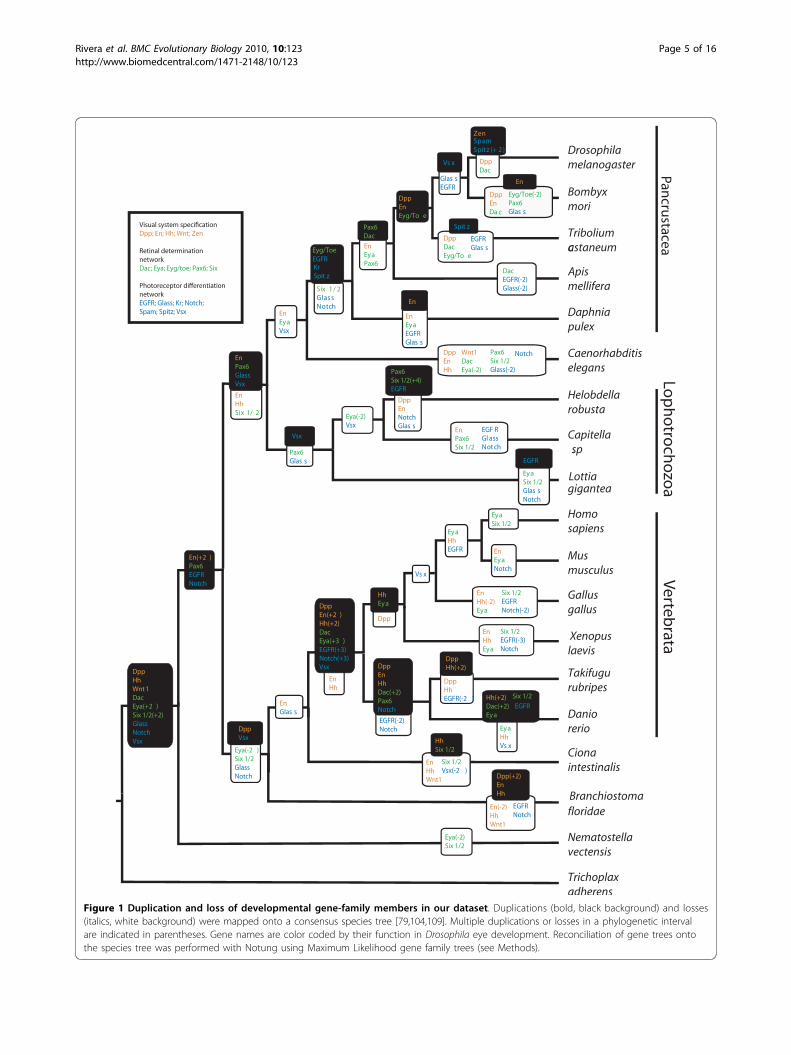
Figure 1 Duplication and loss of developmental gene-family members in our dataset. Duplications (bold, black background) and losses(italics, white background) were mapped onto a consensus species tree [79,104,109]. Multiple duplications or losses in a phylogenetic intervalare indicated in parentheses. Gene names are color coded by their function in Drosophila eye development. Reconciliation of gene trees ontothe species tree was performed with Notung using Maximum Likelihood gene family trees (see Methods).
Rivera et al. BMC Evolutionary Biology 2010, 10:123http://www.biomedcentral.com/1471-2148/10/123
Page 5 of 16
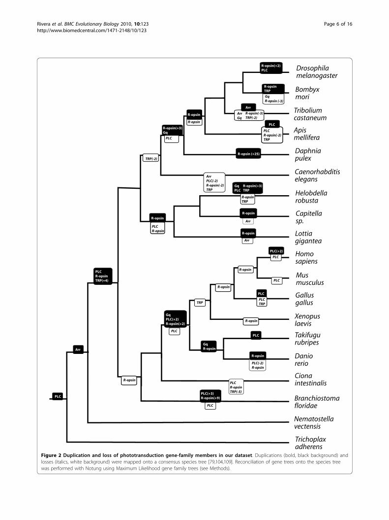
Figure 2 Duplication and loss of phototransduction gene-family members in our dataset. Duplications (bold, black background) andlosses (italics, white background) were mapped onto a consensus species tree [79,104,109]. Reconciliation of gene trees onto the species treewas performed with Notung using Maximum Likelihood gene family trees (see Methods).
Rivera et al. BMC Evolutionary Biology 2010, 10:123http://www.biomedcentral.com/1471-2148/10/123
Page 6 of 16
By searching whole-genome data, we were able to findnew members of several gene families. For example, wewere able to generate a more complete phylogenetichypothesis for the Glass (Gl) gene family with the discov-ery of a new chordate homolog. While a previous unpub-lished phylogeny identified an echinoderm Gl homolog[39], no homologs have been reported previously in chor-dates. Through the analysis of the chordate Branchistomafloridae genome, we discovered a putative homologwhich groups with the other Gl genes with very high sup-port (aLRT = 1) and has similar domain structure toother known Gl genes. This suggests a loss of the Glgene family of zinc-finger members early in chordateevolution (Additional File 1, Figure S13). In addition, ouranalyses uncovered one gene from zebrafish and onefrom fugu that together form a sister group to all otherDpp/BMP2/4 genes. This relationship implies a paralogpresent in fugu and zebrafish but absent in all othereumetazoans. These fish homologs have not, to ourknowledge, been studied, except in having been namedBMP2 based on similarity searches associated with zebra-fish and fugu genome projects, so confirming the pre-sence of new functional Dpp/BMP2/4 genes in these fishwould require experimental demonstration.We examined Pax-6 in greater detail than other gene
families by including sequences from additional arthro-pod species. This gene has a well-known and conservedrole in eye development, and previous authors haveindicated, counter to our conclusions, that the geneseyeless (ey) and twin-of-eyeless (toy) are insect- specificduplications [40]. We found strong phylogenetic evi-dence for a pre-arthropod duplication of ey and toy. We
found support for a family of genes that includes Droso-phila toy, a myriapod toy-like gene, plus a newlydescribed toy-like gene from an ostracod crustacean anda chelicerate. The toy-clade excludes Drosophila ey andthe ey-like genes of a crustacean and a myriapod. Weconclude it is very unlikely that toy and ey represent aninsect-specific duplication event, although the precisetiming of this duplication is difficult to determine withcurrently available data.
Pancrustaceans have high rates of gene-duplicationwithin our datasetWhile excluding arthropod-specific gene families (Spitz,Spam, and Zen), we analyzed and compared rates ofgain of gene-family members (duplications) across pan-crustaceans, across non-arthropod protostomes (Lopho-trochozoa and Caenorhabditis elegans), and acrossvertebrates. We used three denominators to calculaterates of gene duplication (ie rate equals distance/time,and we used three different metrics of evolutionary‘time’ to calculate gene duplications/time). Using totalgene duplications in the denominator normalizes byoverall rates of gene duplication in each clade, whichincludes any whole genome duplications that occurredin a particular group. A second denominator wasgenetic distance, utilizing average ortholog divergencebetween species in a clade [41]. Genetic distance nor-malizes by the overall molecular diversity in a clade.Our third denominator was a molecular clock estimateof divergence times [42,43]. Compared with other proto-stomes, we found that duplication rates of eye-geneswere significantly higher in pancrustaceans in all three
number of significant correlations
num
ber
of m
atric
es
010
020
030
040
050
060
0
0 2 4 6 8 10 12 14
CBA
spam
zen
spitz
pax6
en
eya
hh
notchdac opsingq
egfr
eyg
six1/2
dpp
spitz
spam
zen
hh
en
eya
pax6
dac
six
dpp
eyg
notchgq opsin
arr
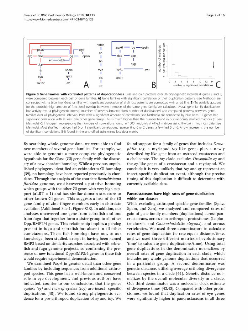
Figure 3 Gene families with correlated patterns of duplication/loss. Loss and gain patterns over 36 phylogenetic intervals (Figures 2 and 3)were compared between each pair of gene families. A) Gene families with significant correlation of their duplication patterns (see Methods) areconnected with a blue line. Gene families with significant correlation of their loss patterns are connected with a red line. B) To partially accountfor the probable high amount of functional overlap between members of the same gene family, we calculated overall gene family duplication/loss activity over a phylogenetic interval (number of losses subtracted from number of duplications) and compared patterns between genefamilies over all phylogenetic intervals. Pairs with a significant amount of correlation (see Methods) are connected by blue lines. 15 genes hadsignificant correlation with at least one other gene family. This is much higher than the number found in our randomly shuffled matrices (C, seeMethods). C) Histogram representing the numbers of correlations found in 1000 randomly shuffled matrices using the gain minus loss data (seeMethods). Most shuffled matrices had 0 or 1 significant correlations, representing 0 or 2 genes, a few had 5 or 6. Arrow represents the numberof significant correlations (14) found in the unshuffled gain minus loss data matrix.
Rivera et al. BMC Evolutionary Biology 2010, 10:123http://www.biomedcentral.com/1471-2148/10/123
Page 7 of 16
analyses (see Methods). Compared with vertebrates, eye-genes showed higher duplication rates in pancrustaceanswhen normalized by total gene duplications. However,comparing duplication over both molecular clock diver-gence times and genetic distance yielded similar rates ofeye-gene gain in vertebrates and pancrustaceans.In our first analytical measure of duplication rates, we
normalized the number of duplications observed in oureye-gene dataset by the total number of gene duplica-tions calculated from the genomes of the clade of inter-est. We inferred 50 duplications of eye-related genes inpancrustaceans compared to 33113 total duplications inthe pancrustacean genomes, resulting in a ratio (δ) of0.0015 (Table 3). This is significantly higher than the δvalue for non-arthropod protostomes (δ = 0.00026; Fish-er’s exact test, p = 1.5e-11) or vertebrates, (δ = 0.00058;p = 4.9e-6) (Tables 3 and 4). To further scrutinize dupli-cation rates, we examined developmental and photo-transduction genes separately. The difference betweenthe δ of non-arthropod invertebrates and pancrustaceanswas still significant for both developmental (p = 0.0102)and phototransduction (p = 1.47e-10) genes. Whencompared to vertebrates, only the δ for phototransduc-tion genes, and not developmental genes, was signifi-cantly higher in pancrustaceans (p = 2.52e-11) (Tables 3and 4).We also used genetic distance (average number of
amino acid substitutions between orthologs in a clade)as a second measure of evolutionary rate [41]. This mea-sure allows us to calculate gene duplications per aminoacid substitution (ι) to examine gene duplication in thecontext of overall lineage diversity (Table 3). For
pancrustaceans, we found that ι for eye genes was0.0478, significantly higher than ι for non-arthropodprotostomes (ι = 0.0193, p = 0.0010). However, ι washigher in vertebrates (ι = 0.0577) than pancrustaceans.We also calculated ι separately for developmental andphototransduction genes. Pancrustacean ι (0.0124) andnon-arthropod protostomes ι (0.0091) did not differ sig-nificantly for developmental genes, although vertebrateι was significantly greater (ι = 0.043, p = 8.79e-5). Forphototransduction genes, pancrustacean ι (0.0353) wassignificantly higher than ι for non-arthropod proto-stomes ι = 0.0102; p = 0.0004), and significantly higherthan ι for vertebrates ι = 0.0184, p = 0.0080) (Tables 3and 4).Finally, we used a calibrated molecular clock as a third
measure of evolutionary time. One critique of agesinferred by molecular clock studies is that they oftenoverestimate absolute clade ages [44-48]. Even so, theestimates could still be reliable estimators of relativeclade age, which is what we require for comparing ratesin different clades. Utilizing published molecular clock-based divergence time estimates [42,43], we found resultsvery similar to our analysis using genetic distance. Over-all, eye-gene duplication rates standardized using clockdivergence time estimates (μ) were found to be signifi-cantly higher in pancrustaceans (0.1604) than other pro-tostomes (0.0215, p = 1.9e-9) but were not significantlydifferent than μ for vertebrates (0.1044). Although devel-opmental genes analyzed alone were not significantly dif-ferent between pancrustaceans and vertebrates,phototransduction genes showed a significantly higher μin pancrustaceans compared to vertebrates (p = 0.0010).
Table 3 Gene duplication rates
clade(s) Dataset gene duplication rates
Eye duplications/total duplications (δ)
Eye duplications/genetic distance (ι)
Eye duplications/molecular clock (μ)
All Dev* PT* All Dev PT All Dev PT
pancrustacean .0015 3.9e-4 .0011 .0478 .0124 .0353 .1064 .0277 .0787
other protostomes 2.6e-4 1.2e-4 1.4e-4 .0193 .0091 .0102 .0215 .0101 .0114
vertebrate 5.8e-4 4.3e-4 1.5e-4 .0577 .0430 .0184 .1044 .0778 .0267
*Developmental genes (Dev) and Phototransduction genes (PT)
Table 4 Duplication rates in Pancrustacea compared to other clades
clade(s) compared to Pancrustacea p-values for significant difference in dataset gene duplication rates compared to Pancrustacea
Eye duplications/total duplications (δ) Eye duplications/genetic distance (ι)
Eye duplications/molecular clock (μ)
All Dev PT All Dev PT All Dev PT
Other protostomes 1.5e-11 .0102 1.47e-10 .0010 .5180 .0004 1.9e-9 .0381 8.2e-9
vertebrate 4.9e-6 .8741 2.52e-11 .4015 8.79e-5 .0080 1.00 .0016 .0010
Bold = significantly more duplications in pancrustaceans
Italics = significantly more duplications in non-arthropod clade
Rivera et al. BMC Evolutionary Biology 2010, 10:123http://www.biomedcentral.com/1471-2148/10/123
Page 8 of 16

Both sets of eye-genes showed a significantly higher μcompared to other protostomes (Tables 3 and 4).In all three analyses, eye genes showed a higher rate of
duplication in pancrustaceans than in non-arthropodprotostomes. In contrast, pancrustaceans only showhigher rates of duplication than vertebrates when photo-transduction genes are included in the analysis. That is,pancrustaceans do not show higher rates of develop-mental gene duplication compared to vertebrates underany analysis.
Co-duplication is significant in our datasetWe compared gene losses and gene duplications sepa-rately across Metazoan genomes and found that 15 of22 gene families had correlated patterns of loss or gainwith at least one other gene family (Figure 3a). In aseparate analysis, we compared patterns of gene lossand duplication simultaneously by taking the total num-ber of duplications minus losses for each gene familyover each branch of the species tree. Both analysesyielded similar results (Figure 3b).To test the statistical significance of our observed
number of correlations, we randomized our data matrixby shuffling to generate a null distribution of correla-tions. We found that the majority of shuffled matrices(1000 total) had only either 0 or 1 significant correla-tions between gene families and none had over 6 corre-lations, making our observed result of 15 significantlydifferent from the null distribution (Figure 3c). Thisconfirmed that the number of correlated gene familiesin the real dataset is greater than expected under bychance. However, the genes showing high levels of cor-relation in gain/loss patterns were not primarily geneswith known functional relationships (Figure 3, Addi-tional File 3).
DiscussionHere we have examined the evolutionary history ofgenes involved in eye development and phototransduc-tion by analysis of gene family trees, reconciled trees,and co-duplication data. Thus, we examined gene-familyevolution in the context of both morphological disparity(eye disparity vs. gene duplication rates) and proteinnetwork evolution (co-duplication/loss). In most of ouranalyses, we found that increased rates of duplicationwithin specific eye-gene families were correlated withthe increased optical disparity seen in pancrustaceans.At the protein network level, we found significant co-duplication of eye-genes, though the patterns of duplica-tion are more complex than we originally hypothesizedwith respect to previously known functional interactionsamong proteins.
Gene treesIn our examination of gene duplication and loss, wegenerated phylogenetic hypotheses for 22 gene families.Many of these are the first detailed analyses of the evo-lution of the gene family, which will be of use in futureresearch on these gene families in various contexts. Wefound that gene trees of orthologs often are incongruentwith assumed species-level relationships. Assuming ourinferred gene trees are accurate, our results imply thatthere is a more complex pattern of gain and loss thanwould have been expected by simply comparing numberof gene orthologs in each species. Because errors in ourtrees could lead to overestimations of complexity, werequired support values of 90% or higher in our reconci-liation analysis. That is, we allowed branch swapping tominimize duplications and losses in cases where thenode support was less than 0.9. However, this methodcan underestimate the numbers of gains and losses [49].Future inquiries could focus on these poorly supportednodes, by including additional species in the analyses asthey become available, or by including additional infor-mation (e.g. domain structure or intron presence/absence) in attempts to estimate phylogenies with highersupport. In addition, presumably more accurate recon-ciled trees could be generated in the future using morecomputationally expensive methods, such as fully Baye-sian estimation of gene and species reconciliation[50,51], which currently would be very difficult at thescale of analysis conducted here. The inclusion of addi-tional genomes in future studies will also be of help ingenerating accurate hypotheses, as taxon (in this casegene) sampling is an important determinant of phyloge-netic estimation [52].
Rates of gene duplicationWe found overall support for the hypothesis that geneduplication and/or retention rates are higher in pancrus-taceans, the group with the highest disparity of optical-types. We examined the sensitivity of this overall con-clusion in three different ways. First, we compared pan-crustaceans to both non-arthropod protostomes and tovertebrates. Second, for each of these comparisons, weestimated gene duplication rates using three differentdenominators: total gene duplications, overall geneticdistance, and divergence time estimates from molecularclock analyses. These different denominators are neces-sary to understand the influence of different modes ofgenome evolution on our conclusions, such as the mul-tiple genome duplications known in vertebrates. Third,we examined (both separately and together) duplicationrates of genes from different eye-gene categories (devel-opmental versus phototransduction genes), allowing usto test whether one category was the primary driver of
Rivera et al. BMC Evolutionary Biology 2010, 10:123http://www.biomedcentral.com/1471-2148/10/123
Page 9 of 16

the overall rates. For example, developmental genes areprobably involved in more non-visual phenotypes thanphototransduction genes since phototransduction genesoften have localized expression [e.g. [53]], and this dif-ference in pleiotropy could influence final results.Comparisons between eye-gene duplication rate in
pancrustaceans and non-arthropod protostomes clearlysupported our hypothesis, even when taking the conser-vative approach of not counting arthropod-specificgenes. The observed difference in gene duplication ratebetween these two clades does not depend on thedenominator used in rate calculations, and is signifi-cantly different for both developmental and phototrans-duction genes (Tables 3, 4). Despite the consistency ofthese results, it is important to consider that there aremultiple possible causes for our observed correlationbetween higher optical disparity and higher eye-geneduplication rate. One possible explanation is that geneduplications, perhaps retained by natural selection, are acausal factor in increasing optical disparity in pancrusta-ceans. In fact, gene duplications are known to haveincreased retinal complexity in vertebrates, leading toseparate rod and cone phototransduction pathways[7,36,37]. Whether these vertebrate duplications werefixed by natural selection or neutral processes isunknown. At present, however, too little is known aboutthe relationship between pancrustacean genes and opti-cal design phenotypes to claim that gene duplicationwas a causal factor leading to higher optical disparity.Another explanation is that the available full genomesequences do not allow for appropriate estimates ofduplication rates in these clades. For example C. elegansdoes not possess conventional eyes, even though manyother non-arthropod protostomes do. If, as a result oflosing eyes during evolution, the lineage of C. eleganshas a lower rate of eye-gene duplication, this couldresult in an underestimate of eye-gene duplication ratefor the entire clade. Similarly, the pancrustaceans usedhere could have more eye-genes than other arthropods.In fact, Daphnia pulex does have a large number ofgenes compared to other arthropods, perhaps because ofits asexual/sexual life history (Colbourne J et al: GenomeBiology of the Model Crustacean Daphnia pulex, sub-mitted). These hypotheses could be examined using theapproaches developed here, once additional genomesequences become available.Compared to rate differences between pancrustaceans
and non-arthropod protostomes, rate differencesbetween pancrustaceans and vertebrates were more vari-able. That is, using different denominators in our ratecalculations led to different results (total gene duplica-tions, genetic distance, or molecular clock). An impor-tant consideration in these comparisons is thatvertebrates are known to have undergone multiple
whole-genome duplications, which raised the overallestimated rate of gene duplication and accumulation forthe group. This is evident in total gene duplications thatwe counted (80673 in vertebrates vs. 33113 in pancrus-taceans) but is not reflected in our other distance mea-sures (denominators): both clades show similar geneticdistance (as measured by average ortholog distance -1047 and 814 respectively) as well as similar clade ages(as estimated by a molecular clock - 470 and 450 mya).The high overall rate of gene duplication and accumula-tion in vertebrates may therefore explain why, counterto our hypothesis, vertebrates show a significantly higherrate of eye development gene duplication thanpancrustaceans.The high rate of duplication and/or retention of genes
in vertebrates further suggest that the best rate compari-son might be that using total number of gene duplica-tions as the distance between species (denominator). Itis this rate calculation that corrects for vertebratewhole-genome duplications. Even here, we see a differ-ence between gene types, with only phototransductiongenes, and not developmental genes, supporting ourstarting hypothesis that pancrustaceans have a highereye-gene duplication rate. However, much of the signifi-cant difference in phototransduction genes is driven byextensive duplications of opsin in the D. pulex lineage(Colbourne J et al: Genome Biology of the Model Crus-tacean Daphnia pulex, submitted), a phenomenon alsoknown in other crustaceans [54,55]. Given the observeddifference between developmental and phototransduc-tion genes when comparing vertebrates and pancrusta-ceans, it is tempting to speculate on possible biologicalcauses for this result. For example, we expect develop-mental genes to be pleiotropic, and several of the genesstudied here are known to function in many contextsbesides eye development [e.g. [56]]. Phototransductiongenes have a more specific functional role and may beless pleiotropic [e.g. [53]]. The more pleiotropic devel-opmental genes could rely more heavily on modifica-tions in the protein and cis-regulatory sequences, ratherthan on gene duplication for diversifying function [57].If so, correlation between gene duplication rate andmorphological disparity could be low or nonexistent.The consideration of pleiotropy also highlights
another avenue for future research. If pleiotropy doesresult in a weaker correlation between eye disparity andgene duplication rate, gene choice must influence thefinal results. Therefore, future research might focus on abroader sampling of genes, especially to the extent thatanalyses conducted here could be fully automated toallow the analysis of very large datasets. For example, arecent study focusing on the insects found higher num-bers of gene duplications in dipterans than other insectsby examining 91 fly eye-genes [58]. Integrating this type
Rivera et al. BMC Evolutionary Biology 2010, 10:123http://www.biomedcentral.com/1471-2148/10/123
Page 10 of 16
of “retinome” scale analysis with the methods we showhere would give a more detailed and informed view ofgene evolution in the context of morphological disparityand innovation.The available genomic data allowed us to test the
hypothesis that pancrustaceans, a group with many dis-parate eye types, have more duplications of eye-genesthan less optically-diverse groups. This relies on anassumed species phylogeny, and our assumption that weare estimating rates of pancrustacean duplication for theentire clade. Complicating this assumption, the phyloge-netic position of branchiopods (including Daphniapulex) within Arthropoda remains somewhat uncertain[59-62]. We here consider the hexapod/D. pulex ances-tor to be the common ancestor of all pancrustaceans forsimplicity. This is justified by the wide variety of opticaldesigns found in this hypothesized hexapod-branchio-pod clade, regardless of whether it represents the ances-tral pancrustacean or whether crustaceans are in factparaphyletic [59-62]. Future research using genomesfrom more crustaceans and taxa with a wider range ofeye-type disparity could allow testing for a broader cor-relation between eye disparity and eye-related genenumber, a possibility supported by our results. Namely,if the ratio of eye-types to gene duplication rate is simi-lar in different clades, then a broader correlation mayexist.
Co-duplication of genesWe found that duplication and/or loss patterns in 15 of22 gene families correlated significantly with duplicationand/or loss patterns in at least one other gene family,significantly more than expected by chance (Figure 3C).Interestingly, many of the genes we found to co-dupli-cate are not known to have any functional relationshipwith each other. This suggests the possibility of novelfunctional relationships between genes, at least in ani-mals where the genetics are relatively unstudied (themajority of our samples). Co-duplications may also bethe result of undiscovered constraints at the genomiclevel (e.g. synteny), or an unknown systematic artifact ofour gene reconciliation analysis that infers that unre-lated genes duplicate or are lost at particular nodes.While new gene pairings were suggested by our co-duplication analysis, some pairings predicted by func-tional modules were not found.One functional module of particular interest is the
suite of phototransduction genes [31]. We found thateven though multiple ciliary phototransduction genesare known to have co-duplicated early in vertebrate his-tory [29,36,63], rhabdomeric phototransduction geneshave not co-duplicated as a unit when considering theentire history of Metazoa. A notable exception is that R-opsin and Gq-alpha (genes known to interact directly)
exhibit a significant pattern of co-duplication. This sug-gests that R-opsin and Gq-alpha have been a tightlylinked functional module throughout animal evolution,and if so, specific R-opsin paralogs may be expressedwith specific Gq-alpha paralogs.We also found that some phototransduction genes co-
duplicate with developmental genes (Figure 3). Some ofour data could represent novel genetic interactions, butthey could also stem from other unknown aspects ofthese genes including the number of protein interac-tions, the number of functions a protein is involved in,or genomic location. Although we tested the generalfalse-positive rate by generating randomized matrices ofour data, future studies might also compare the num-bers of co-duplicating eye-genes to that of a set of genesdrawn at random that are not necessarily involved inthe same organ system.Similarly, we found extensive co-duplication/loss
between only a few gene families known to be involvedin the same developmental pathway [19]. The retinaldetermination pathway, for example, includes Pax6, Dac,Eya and Six1/2, gene families known to have functionalinteractions in disparate taxa [56]. From this pathway,Pax6 and Dac had correlated loss patterns as did Eyaand Six1/2. Perhaps the functional relationship betweenthese gene pairs is more constrained than that of othergenes in the retinal determination network. Dac andPax6, for instance, are known to have a complex induc-tive relationship in both vertebrates and invertebrates[56,64]. Other gene families with known interactions inDrosophila compound eye development also showedcorrelations in either their loss or gain patterns. Theseinclude Hh and Eya [65], Dac and Dpp [66] and Six 1/2and Eya [67]. However, the majority of genes withknown regulatory interactions in eye development didnot tend to be duplicated/lost together more often thanexpected by chance. This finding - that the evolutionaryhistory of genes belonging to complete genetic modulesdo not share similar patterns of gain and loss - is con-sistent with a functional study that found networkdegeneration after genome duplication in yeast [68]. Inthat study, genes that function together before genomeduplication do not necessarily function together aftergenome duplication.
ConclusionOur research provides new methodology for examininggenomic complexity in the context of morphologicalcomplexity. In particular, we examined the evolutionaryhistories of genes acting in arthropod eye developmentand phototransduction to evaluate hypotheses of geneand protein module duplication. The phylogenetic treeswe created lay a foundation for research into the genehistories of several understudied, but developmentally
Rivera et al. BMC Evolutionary Biology 2010, 10:123http://www.biomedcentral.com/1471-2148/10/123
Page 11 of 16

important, gene families. Future research will likely leadto advances in understanding evolutionarily conservedprotein domains in these genes as well as the signifi-cance of the expansion of some families in particularlineages (e.g. the Six1/2 family in the lineage leading tothe Helobdella robusta). Our analyses of these gene his-tories revealed that, by one analysis, genes involved ineye development and phototransduction had higherrates of duplication in the taxon with the largest num-ber of eye types (pancrustaceans) (Table 3). Ourco-duplication analysis found higher than expectednumbers of co-duplicating genes, yet genes in knowngenetic modules were not always found to be gainedand lost together (Figure 3). Moreover, some genes thatare not known to have extensive interactions did showhigh correlation in loss and gain pattern. Futureresearch could clarify these findings, comparing thegenomic locations of co-duplicating genes in order toidentify synteny, identifying gene modules in the eyes ofnon-model organisms, confirming the function of thegene families in non-insect arthropods, and testing forpatterns consistent with positive selection acting on thegenes and modules of interest.
MethodsOverviewWe first found all homologs of genes of interest in theDaphnia pulex v1.0 genome. We next found all homo-logs in 18 other metazoan genomes. We constructedphylogenies for each gene family using maximum likeli-hood. Assuming species-level relationships to be known,we next reconciled each gene family tree with themetazoan tree to estimate timing of gene duplicationand loss events. We then estimated rates of gene dupli-cation within major metazoan clades. Finally, we testedfor significant correlation of gene duplication/loss pat-terns across gene families. Detailed methods for each ofthese general steps are detailed below.
Daphnia pulex genome searches and gene familyassignmentWith a protein sequence for each gene of interest fromFlyBase used as a “bait” sequence, Blastall searches wereperformed, using protein sequences for each gene ofinterest as a “bait” sequence, against all gene models ofDaphnia pulex v1.0 obtained from JGI [http://genome.jgi-psf.org/Daphnia; http://wfleabase.org/]. Searches firstretrieved the top 15 hits, this number was raised in sub-sequent searches until D. pulex models outside thegroup of interest were obtained. Redundant sequenceswere determined by examining the visual scaffold modelon JGI and then removed by hand. The gene family foreach D. pulex gene was assigned by inclusion in a maxi-mum likelihood tree using UniRef50 and UniRef90
sequences. These trees were estimated using an in-house pipeline of shell and perl scripts that merge exist-ing bioinformatic tools. The bait sequence from FlyBasewas used to perform a similarity search using blastp [69]of non-redundant protein databases curated by uniprothttp://www.uniprot.org/. In most cases, we used twoblast search strategies for each bait gene: 25/10 (wherethe top 25 blast hits of the Uniref90 and the top 10blast hits of the Uniref50 database were retained forfurther analysis). In cases when there was either notenough resolution or no outgroup hits obtained; morehits were taken from the Uniref90 or Uniref50 data-bases, respectively (See Additional file 1 for details).Identical sequences, such as those obtained from bothUniref90 and Uniref50 databases, were removed fromfurther analysis. Second, all retained sequences and baitwere aligned using MUSCLE [70]. Third, we estimatedmaximum likelihood phylogenetic trees using aLRT-PHYML [71,72] assuming a JTT [73] model of proteinevolution. We visualized resulting phylogenetic treeswith TreeView [74,75] or FigTree http://tree.bio.ed.ac.uk/software/figtree/. Where relevant, we tested whethergene trees were significantly different from previoustrees using the Shimodaira-Hasegawa (SH) test [76]implemented in PhyML [71,72] by comparing con-strained trees to the best trees.
Pax-6 sequencesIn phylogenetic analyses of Pax-6, we utilized previouslyunpublished sequence data from Daphnia pulex (con-firming the automated genome assemblies with cDNAsequencing) and the ostracod crustacean Euphilomedescarcharodonta. Euphilomedes carcharodonta were col-lected at the University of Southern California’s WrigleyMarine Lab on Catalina Island, California by free diving,collecting sediment with an aquarium net, and sortingwith a dissecting microscope. Daphnia pulex wereobtained from stock collections at Indiana University.We first isolated Pax-6 fragments using degenerate PCRprimers to highly conserved regions in the paired andhomeo domains of published Pax-6 sequences. Aftersequencing an initial Pax-6 fragment, we designed speci-fic primers for 5’ and 3’ RACE, often using nested pri-mers and the Gene Racer kit (Invitrogen). Primers andcycling conditions are given in Additional File 3. Addi-tional arthropod Pax-6 sequences were obtained fromGenBank.
Genome comparisonsWith protein sequence for each gene of interest fromFlyBase, initial blastall searches were executed against19 genomes obtained from JGI and NCBI (Table 1) withparameters set to return five best hits with e values lessthan 0.5. To ensure no paralogs were omitted, blastall
Rivera et al. BMC Evolutionary Biology 2010, 10:123http://www.biomedcentral.com/1471-2148/10/123
Page 12 of 16
searches were modified and repeated to allow morereturns in cases where all five best hits fell within thein-group for the gene of interest in initial phyogeneticanalyses. Sequence alignment and maximum likelihoodestimates of gene trees were performed as describedabove. Genes were identified phylogenetically as mono-phyletic ingroups sharing conserved domain architectureusing Pfam databases and NCBI’s Conserved DomainDatabase [77,78] (See Additional File 1 for details). Withoutgroups identified and pruned from the alignment,each gene tree was reconstructed and then reconciled tothe metazoan species tree [79] using NOTUNG [80].Rearrangements were allowed for node supports below0.9 in order to minimize the inferred number of geneduplication and loss events implied by poorly supportednodes.
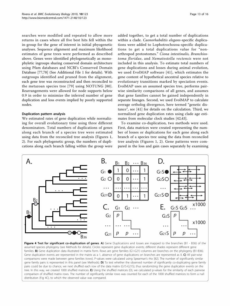
Duplication pattern analysisWe estimated rates of gene duplication while normaliz-ing for overall evolutionary time using three differentdenominators. Total numbers of duplications of genesalong each branch of a species tree were estimatedusing data from the reconciled tree analysis (Figures 1,2). For each phylogenetic group, the numbers of dupli-cations along each branch falling within the group were
added together, to get a total number of duplicationswithin a clade. Caenorhabditis elegans-specific duplica-tions were added to Lophotrochozoa-specific duplica-tions to get a total duplications value for “non-arthropod protostomes.” Ciona intestinalis, Branchios-toma floridae, and Nematostella vectensis were notincluded in this analysis. To estimate total numbers ofgene duplications and losses during animal evolution,we used EvolMAP software [41], which estimates thegene content of hypothetical ancestral species relative toevolutionary transitions marked by speciation events.EvolMAP uses an assumed species tree, performs pair-wise similarity comparisons of all genes, and assumesthat gene families cannot be gained independently inseparate lineages. Second, we used EvolMAP to calculateaverage ortholog divergence, here termed “genetic dis-tance”, see [41] for details on the calculation. Third, wenormalized gene duplication rates using clade age esti-mates from molecular clock studies [42,43].To examine co-duplication, two methods were used.
First, data matrices were created representing the num-ber of losses or duplications for each gene along eachbranch of a species tree using the data from reconciledtree analysis (Figures 1, 2). Gene patterns were com-pared in the loss and gain cases separately by examining
. . . A.
B1
B2
. . .
. . .
. . .
. . .
. . .
B.
B36
0
B1 B2 B36
G1
G2
G21
. . . 0
10
1
1
1
0
. . .
. . .
. . .
. . . . . . . . . . . .
1
D.G1S
G2S
G21S
. . .
x1000
G2
G21
. . .
. . . G1 G2
p
p p
. . . . . .
. . .
C.
x1000
G1S
G2S
G21S
G2S . . . p
p p
. . . . . .
. . .
E.
Figure 4 Test for significant co-duplication of genes. A) Gene Duplications and losses are mapped to the branches (B1 - B36) of theassumed species phylogeny (see Methods for details). Circles represent gene duplication events; different shades represent different genefamilies. B) Gene duplication data illustrated in matrix form. Rows are gene families (G1-G21) columns are branches on the phylogeny (B1-B36).Gene duplication events are represented in the matrix as a 1, absence of gene duplications on branches are represented as 0. C) All pair-wisecomparisons were made between gene families (rows). P-values were calculated using Spearman’s rho [82]. The number of significantly similargene family pairs is represented in this panel (see Methods). D) To test whether the observed number of significantly co-duplicating gene familypairs could be due to chance, we next shuffled each row of the data matrix (G1S-G21S), thus randomizing the gene duplication events on thetree. In this way, we created 1000 shuffled matrices. E) Using the shuffled matrices (D), we calculated p-values for the similarity of each pairwisecomparison of shuffled matrix rows. The number of significantly similar rows was counted for each of the 1000 shuffled matrices to form a nulldistribution (Fig 4C), to which the observed value was compared.
Rivera et al. BMC Evolutionary Biology 2010, 10:123http://www.biomedcentral.com/1471-2148/10/123
Page 13 of 16

correlation using Spearman’s rho implemented in R withp-values calculated using Algorithm AS 89, a test ofupper tail probability [81,82]. The second method usedthe same methodology comparing duplication and losspatterns together by looking at the number of duplica-tions minus the number of losses for each gene alongeach branch of the species tree. Significance of correla-tion was assessed using sequential Bonferroni to accom-modate multiple comparisons [83,84]. A nulldistribution for the expected number of co-duplicatinggene family pairs was created by randomizing the datamatrix 1000 times and analyzing each pseudoreplicate inR (Figure 4).
Additional file 1: Results of phylogenetic analyses on the 22 individualgene families used in this study.
Additional file 2: Pairwise correlation values of between duplication andloss of the 22 gene families examined.
Additional file 3: PCR conditions for the amplification of Pax6 homologsfrom Euphilomedes carcharodonta and Daphnia pulex.
AcknowledgementsThe Capitella sp. I, Helbodella robusta, Fugu rubripes, and Lottia giganteagenome data were produced by the US Department of Energy JointGenome Institute http://www.jgi.doe.gov/ in collaboration with the usercommunity. The Daphnia pulex sequencing and portions of the analyseswere performed at the DOE Joint Genome Institute under the auspices ofthe U.S. Department of Energy’s Office of Science, Biological andEnvironmental Research Program, and by the University of California,Lawrence Livermore National Laboratory under Contract No. W-7405-Eng-48,Lawrence Berkeley National Laboratory under Contract No. DE-AC02-05CH11231, Los Alamos National Laboratory under Contract No. W-7405-ENG-36 and in collaboration with the Daphnia Genomics Consortium (DGC)http://daphnia.cgb.indiana.edu. Additional analyses were performed bywFleaBase, developed at the Genome Informatics Lab of Indiana Universitywith support to Don Gilbert from the National Science Foundation and theNational Institutes of Health. Coordination infrastructure for the DGC isprovided by The Center for Genomics and Bioinformatics at IndianaUniversity, which is supported in part by the METACyt Initiative of IndianaUniversity, funded in part through a major grant from the Lilly Endowment,Inc. Our work benefits from, and contributes to the Daphnia GenomicsConsortium. Thanks to Gavin Wu of the UCSB Statlab for writing thecorrelation script in R. Thanks also to H. Robertson and others forperforming detailed annotation of opsin genes in Daphnia, which will bedescribed elsewhere.
Author details1Ecology Evolution and Marine Biology, University of California Santa Barbara,Santa Barbara, CA 93106 USA. 2Department of Biology, Jordan Hall 1001 E.Third Street, Indiana University, Bloomington, IN USA. 3Department ofEcology, Evolution, and Organismal Biology, 245 Bessey Hall, Iowa StateUniversity, Ames, IA 50011 USA.
Authors’ contributionsAuthor contributions: ASR and THO designed research; ASR, MSP, DCP, CV,AES, JMS, ARO, and THO performed research; ARO contributed newreagents/analytic tools; ASR, MSP, DCP, CV, AES, JMS, ARO, and THO analyzeddata; and ASR, MSP, DCP, JMS, and THO wrote the paper. All authors readand approved the final manuscript.
Received: 20 June 2009 Accepted: 30 April 2010Published: 30 April 2010
References1. Lynch M, Conery JS: The origins of genome complexity. Science 2003,
302(5649):1401-1404.2. Martin AP: Increasing genomic complexity by gene duplication and the
origin of vertebrates. American Naturalist 1999, 154(2):111-128.3. Ohno S: Evolution by gene duplication. New York: Springer-Verlag 1970.4. Freeling M, Thomas BC: Gene-balanced duplications, like tetraploidy,
provide predictable drive to increase morphological complexity. GenomeRes 2006, 16(7):805-814.
5. Oakley TH, Plachetzki DC, Rivera AS: Furcation, field-splitting, and theevolutionary origins of novelty in arthropod photoreceptors. ArthropodStruct Dev 2007, 36(4):386-400.
6. Foote M, Gould SJ: Cambrian and Recent Morphological Disparity. Science1992, 258(5089)-1816.
7. Plachetzki DC, Oakley TH: Key transitions during the evolution of animalphototransduction: novelty, “tree-thinking,” co-option, and co-duplication. Integrative and Comparative Biology 2007, 47(5):759-769.
8. Land MF, Nilsson D-E: Animal Eyes. Oxford: Oxford University Press 2002.9. Liu Z, Friedrich M: The Tribolium homologue of glass and the evolution
of insect larval eyes. Dev Biol 2004, 269(1):36-54.10. Harzsch S, Hafner G: Evolution of eye development in arthropods:
Phylogenetic aspects. Arthropod Structure & Development 2006,35(4):319-340.
11. Friedrich M, Benzer S: Divergent decapentaplegic expression patterns incompound eye development and the evolution of insectmetamorphosis. J Exp Zool 2000, 288(1):39-55.
12. Egelhaaf A, Altenfeld H, Hoffmann HU: Evidence for the Priming Role ofthe Central Retinula Cell in Ommatidium Differentiation of Ephestia-Kuehniella. Rouxs Archives of Developmental Biology 1988, 197(3):184-189.
13. Friedrich M, Rambold I, Melzer RR: The early stages of ommatidialdevelopment in the flour beetle Tribolium castaneum (Coleoptera;Tenebrionidae). Development Genes and Evolution 1996, 206(2):136-146.
14. Hafner GS, Tokarski TR: Retinal development in the lobster Homarusamericanus - Comparison with compound eyes of insects and othercrustaceans. Cell and tissue research 2001, 305(1):147-158.
15. Hafner GS, Tokarski TR: Morphogenesis and pattern formation in theretina of the crayfish Procambarus clarkii. Cell and tissue research 1998,293(3):535-550.
16. Melzer RR, Michalke C, Smola U: Walking on insect paths? Earlyommatidial development in the compound eye of the ancestralcrustacean, Triops cancriformis. Naturwissenschaften 2000, 87(7):308-311.
17. Harzsch S, Walossek D: Neurogenesis in the developing visual system ofthe branchiopod crustacean Triops longicaudatus (LeConte, 1846):corresponding patterns of compound-eye formation in Crustacea andInsecta? Development Genes and Evolution 2001, 211(1):37-43.
18. Wolff T, Ready DF: Pattern formation in the Drosophila retina. Thedevelopment of Drosophila melanogaster Cold Spring Harbor LaboratoryPressBate M, Martinez-Arias A 1993, II:1277-1325.
19. Friedrich M: Ancient mechanisms of visual sense organ developmentbased on comparison of the gene networks controlling larval eye,ocellus, and compound eye specification in Drosophila. ArthropodStructure and Development 2006, 35(4):357-378.
20. Angelini DR, Kaufman TC: Functional analyses in the hemipteranOncopeltus fasciatus reveal conserved and derived aspects ofappendage patterning in insects. Dev Biol 2004, 271(2):306-321.
21. Inoue Y, Miyawaki K, Terasawa T, Matsushima K, Shinmyo Y, Niwa N, Mito T,Ohuchi H, Noji S: Expression patterns of dachshund during headdevelopment of Gryllus bimaculatus (cricket). Gene Expr Patterns 2004,4(6):725-731.
22. Duman-Scheel M, Pirkl N, Patel NH: Analysis of the expression pattern ofMysidium columbiae wingless provides evidence for conservedmesodermal and retinal patterning processes among insects andcrustaceans. Dev Genes Evol 2002, 212(3):114-123.
23. Dong Y, Friedrich M: Comparative analysis of Wingless patterning in theembryonic grasshopper eye. Dev Genes Evol 2005, 215(4):177-197.
24. Hurley I, Fowler K, Pomiankowski A, Smith H: Conservation of theexpression of DII, en, and wg in the eye-antennal imaginal disc of stalk-eyed flies. Evol Dev 2001, 3(6):408-414.
25. Liu ZY, Yang XY, Dong Y, Friedrich M: Tracking down the “head blob”:Comparative analysis of wingless expression in the developing insect
Rivera et al. BMC Evolutionary Biology 2010, 10:123http://www.biomedcentral.com/1471-2148/10/123
Page 14 of 16
procephalon reveals progressive reduction of embryonic visual systempatterning in higher insects. 2006, 35(4):341-356.
26. Gehring WJ, Ikeo K: Pax 6: Mastering eye morphogenesis and eyeevolution. Trends in Genetics 1999, 15(9):371-377.
27. Terakita A: The opsins. Genome biology 2005, 6(3)-213.28. Arendt D: Evolution of eyes and photoreceptor cell types. The
International journal of developmental biology 2003, 47(7-8):563-571.29. Plachetzki DC, Degnan BM, Oakley TH: The Origins of Novel Protein
Interactions during Animal Opsin Evolution. PLoS ONE 2007, 2(10):e1054.30. Spudich JL, Yang CS, Jung KH, Spudich EN: Retinylidene proteins:
structures and functions from archaea to humans. Annu Rev Cell Dev Biol2000, 16:365-392.
31. Hardie RC, Raghu P: Visual transduction in Drosophila. Nature 2001,413(6852):186-193.
32. Arendt D, Wittbrodt J: Reconstructing the eyes of Urbilateria. PhilisophicalTransactions of the Royal Society of London B 2001, 356(1414):1545-1563.
33. Hardie RC, Minke B: The Trp Gene Is Essential for a Light-Activated Ca2+Channel in Drosophila Photoreceptors. Neuron 1992, 8(4):643-651.
34. Vinos J, K J, Hardy RW, Britt SG, Zuker CS: AG protein-coupled receptorphotphatase required for rhodopsin function. Science 1997, 277:687-690.
35. Byk T, BarYaacov M, Doza Y, Minke B, Selinger Z: Regulatory arrestin cyclesecures the fidelity of the fly photoreceptor cell. Proceedings of theNational Academies of Sciences USA 1993, 90:1907-1911.
36. Nordström K, Larsson TA, Larhammar D: Extensive duplications ofphototransduction genes in early vertebrate evolution correlate withblock (chromosome) duplications. Genomics 2004, 83(5):852-872.
37. Hisatomi O, Tokunaga F: Molecular evolution of proteins involved invertebrate phototransduction. Comp Biochem Phys B 2002, 133(4):509-522.
38. Durand D, Halldorsson BV, Vernot B: A hybrid micro-macroevolutionaryapproach to gene tree reconstruction. J Comput Biol 2006, 13(2):320-335.
39. Liu Z: The photoreceptor differentiation factor glass in the flour beetleTribolium castaneum: cloning, function and evolution. Detroit, MI: WayneState 2005.
40. Czerny T, Halder G, Kloter U, Souabni A, Gehring WJ, Busslinger M: twin ofeyeless, a second Pax-6 gene of Drosophila, acts upstream of eyeless inthe control of eye development. Mol Cell 1999, 3(3):297-307.
41. Sakarya O, Kosik KS, Oakley TH: Reconstructing ancestral genome contentbased on symmetrical best alignments and Dollo parsimony.Bioinformatics 2008, 24(5):606-612.
42. Blair J: Animals (Metazoa). The Timetree of Life Oxford: Oxford UniversityPressHedges S, Kumar S 2009, 223-230.
43. Pisani D: Arthropods (Arthropoda). The Timetree of Life Oxford: OxfordUniversity PressHedges S, Kumar S 2009, 251-254.
44. Pulquerio MJ, Nichols RA: Dates from the molecular clock: how wrongcan we be? Trends in Ecology and Evolution 2007, 22(4):180-184.
45. Swalla BJ, Smith AB: Deciphering deuterostome phylogeny: molecular,morphological and palaeontological perspectives. Philosophicaltransactions of the Royal Society of London 2008, 363(1496):1557-1568.
46. Roger AJ, Hug LA: The origin and diversification of eukaryotes: problemswith molecular phylogenetics and molecular clock estimation.Philosophical transactions of the Royal Society of London 2006,361(1470):1039-1054.
47. Peterson KJ, Cotton JA, Gehling JG, Pisani D: The Ediacaran emergence ofbilaterians: congruence between the genetic and the geological fossilrecords. Philosophical transactions of the Royal Society of London 2008,363(1496):1435-1443.
48. Gaidos E, Dubuc T, Dunford M, McAndrew P, Padilla-Gamino J, Studer B,Weersing K, Stanley S: The Precambrian emergence of animal life: ageobiological perspective. Geobiology 2007, 5(4):351-373.
49. Hahn MW: Bias in phylogenetic tree reconciliation methods: implicationsfor vertebrate genome evolution. Genome biology 2007, 8(7):R141.
50. Rasmussen MD, Kellis M: Accurate gene-tree reconstruction by learninggene- and species-specific substitution rates across multiple completegenomes. Genome Res 2007, 17(12):1932-1942.
51. Akerborg O, Sennblad B, Arvestad L, Lagergren J: Simultaneous Bayesiangene tree reconstruction and reconciliation analysis. Proc Natl Acad SciUSA 2009, 106(14):5714-5719.
52. Zwickl DJ, Hillis DM: Increased taxon sampling greatly reducesphylogenetic error. Systematic Biology 2002, 51(4):588-598.
53. Arendt D, Tessmar-Raible K, Snyman H, Dorresteijn AW, Wittbrodt J: Ciliaryphotoreceptors with a vertebrate-type opsin in an invertebrate brain.Science 2004, 306(5697):869-871.
54. Oakley TH, Huber DR: Differential expression of duplicated opsin genes intwo eye types of ostracod crustaceans. Journal of Molecular Evolution2004, 59(2):239-249.
55. Porter ML, Bok MJ, Robinson PR, Cronin TW: Molecular diversity of visualpigments in Stomatopoda (Crustacea). Visual neuroscience 2009,26(3):255-265.
56. Silver SJ, Rebay L: Signaling circuitries in development: insights from theretinal determination gene network. Development 2005, 132(1):3-13.
57. Wray GA: The evolutionary significance of cis-regulatory mutations. NatRev Genet 2007, 8(3):206-216.
58. Bao R, Friedrich M: Molecular evolution of the Drosophila retinome:exceptional gene gain in the higher Diptera. Mol Biol Evol 2009,26(6):1273-1287.
59. Giribet G, Edgecombe GD, Wheeler WC: Arthropod phylogeny basedon eight molecular loci and morphology. Nature 2001,413(6852):157-161.
60. Nardi F, Spinsanti G, Boore JL, Carapelli A, Dallai R, Frati F: Hexapod origins:Monophyletic or paraphyletic? Science 2003, 299(5614):1887-1889.
61. Mallatt JM, Garey JR, Shultz JW: Ecdysozoan phylogeny and Bayesianinference: first use of nearly complete 28S and 18S rRNA genesequences to classify the arthropods and their kin. MolecularPhylogenetics and Evolution 2003, 31:178-191.
62. Regier JC, Shultz JW, Kambic RE: Pancrustacean phylogeny: hexapods areterrestrial crustaceans and maxillopods are not monophyletic. 2005,272(1561):395-401.
63. Serb JM, Oakley TH: Hierarchical phylogenetics as a quantitativeanalytical framework for Evolutionary Developmental Biology. Bioessays2005, 27(11).
64. Pappu KS, Mardon G: Genetic control of retinal specification anddetermination in Drosophila. The International journal of developmentalbiology 2004, 48(8-9):913-924.
65. Pappu KS, Chen R, Middlebrooks BW, Woo C, Heberlein U, Mardon G:Mechanism of hedgehog signaling during Drosophila eye development.Development 2003, 130(13):3053-3062.
66. Chang T, Mazotta J, Dumstrei K, Dumitrescu A, Hartenstein V: Dpp and Hhsignaling in the Drosophila embryonic eye field. Development 2001,128(23):4691-4704.
67. Pignoni F, Hu B, Zavitz KH, Xiao J, Garrity PA, Zipursky SL: The eye-specification proteins So and Eya form a complex and regulate multiplesteps in Drosophila eye development [published erratum appears in Cell1998 Feb 20;92(4):following 585]. Cell 1997, 91(7):881-891.
68. Conant GC, Wolfe KH: Functional partitioning of yeast co-expressionnetworks after genome duplication. Plos Biol 2006, 4(4):545-554.
69. Altschul SF, Madden TL, Schaffer AA, Zhang J, Zhang Z, Miller W,Lipman DJ: Gapped BLAST and PSI-BLAST: a new generation of proteindatabase search programs. Nucleic Acids Research 1997, 25(17):3389-3402.
70. Edgar RC: MUSCLE: multiple sequence alignment with high accuracy andhigh throughput. Nucleic Acids Research 2004, 32(5):1792-1797.
71. Anisimova M, Gascuel O: Approximate likelihood-ratio test for branches:A fast, accurate, and powerful alternative. Syst Biol 2006, 55(4):539-552.
72. Guindon S, Gascuel O: A simple, fast, and accurate algorithm to estimatelarge phylogenies by maximum likelihood. Systematic Biology 2003,52(5):696-704.
73. Jones DT, Taylor WR, Thornton JM: The rapid generation of mutation datamatrices from protein sequences. Comput Appl Biosci 1992, 8(3):275-282.
74. Page RDM, Charleston MA: From gene to organismal phylogeny:reconciled trees and the gene tree/species tree problem. Mol PhylogenetEvol 1997, 7(2):231-240.
75. Page RDM, Charleston MA: Trees within trees: phylogeny and historicalassociations. Trends in Ecology and Evolution 1998, 13(9):356-359.
76. Shimodaira H, Hasegawa M: CONSEL: for assessing the confidence ofphylogenetic tree selection. Bioinformatics 2001, 17(12):1246-1247.
77. Marchler-Bauer A, Anderson JB, Derbyshire MK, DeWeese-Scott C,Gonzales NR, Gwadz M, Hao L, He S, Hurwitz DI, Jackson JD, et al: CDD: aconserved domain database for interactive domain family analysis.Nucleic Acids Res 2007, , 35 Database: D237-240.
Rivera et al. BMC Evolutionary Biology 2010, 10:123http://www.biomedcentral.com/1471-2148/10/123
Page 15 of 16

78. Finn RD, Tate J, Mistry J, Coggill PC, Sammut SJ, Hotz HR, Ceric G,Forslund K, Eddy SR, Sonnhammer ELL, et al: The Pfam protein familiesdatabase. Nucleic Acids Research 2008, 36:D281-D288.
79. Dunn CW, Hejnol A, Matus DQ, Pang K, Browne WE, Smith SA, Seaver E,Rouse GW, Obst M, Edgecombe GD, et al: Broad phylogenomic samplingimproves resolution of the animal tree of life. Nature 2008,452(7188):745-U745.
80. Chen K, Durand D, Farach-Colton M: NOTUNG: a program for dating geneduplications and optimizing gene family trees. J Comput Biol 2000, 7(3-4):429-447.
81. Best D, DE R: Algorithm AS 89: the upper tail probabilities of Spearman’srho. Applied Statistics 1975, 24:377-379.
82. Spearman C: The proof and measurement of association between twothings. American Journal of Phsychology 1904, 15:72-101.
83. Holm S: A simple sequentially rejective multiple test procedure.Scandanavian Journal of Statistics 1979, 6:65-70.
84. Rice WR: Analyzing Tables of Statistical Tests. Evolution 1989,43(1):223-225.
85. Consortium HGS: Insights into social insects from the genome of thehoneybee Apis mellifera. Nature 2006, 443(7114):931-949.
86. Xia Q, Zhou Z, Lu C, Cheng D, Dai F, Li B, Zhao P, Zha X, Cheng T, Chai C,et al: A draft sequence for the genome of the domesticated silkworm(Bombyx mori). Science 2004, 306(5703):1937-1940.
87. Putnam NH, Butts T, Ferrier DE, Furlong RF, Hellsten U, Kawashima T,Robinson-Rechavi M, Shoguchi E, Terry A, Yu JK, et al: The amphioxusgenome and the evolution of the chordate karyotype. Nature 2008,453(7198):1064-1071.
88. [http://www.wormbase.org], release 180 Oct. 30, 2007.89. Aparicio S, Chapman J, Stupka E, Putnam N, Chia JM, Dehal P, Christoffels A,
Rash S, Hoon S, Smit A, et al: Whole-genome shotgun assembly andanalysis of the genome of Fugu rubripes. Science 2002,297(5585):1301-1310.
90. King N, Westbrook MJ, Young SL, Kuo A, Abedin M, Chapman J,Fairclough S, Hellsten U, Isogai Y, Letunic I, et al: The genome of thechoanoflagellate Monosiga brevicollis and the origin of metazoans.Nature 2008, 451(7180):783-788.
91. Putnam NH, Srivastava M, Hellsten U, Dirks B, Chapman J, Salamov A,Terry A, Shapiro H, Lindquist E, Kapitonov VV, et al: Sea anemone genomereveals ancestral eumetazoan gene repertoire and genomicorganization. Science 2007, 317(5834):86-94.
92. Aparicio S, Chapman J, Stupka E, Putnam N, Chia J, Dehal P, Christoffels A,Rash S, Hoon S, Smit A, et al: Whole-genome shotgun assembly andanalysis of the genome of Fugu rubripes. Science 2002,297(5585):1301-1310.
93. Matus DQ, Thomsen GH, Martindale MQ: Dorso/ventral genes areasymmetrically expressed and involved in germ-layer demarcationduring cnidarian gastrulation. Curr Biol 2006, 16(5):499-505.
94. Technau U, Rudd S, Maxwell P, Gordon PM, Saina M, Grasso LC,Hayward DC, Sensen CW, Saint R, Holstein TW, et al: Maintenance ofancestral complexity and non-metazoan genes in two basal cnidarians.Trends Genet 2005, 21(12):633-639.
95. Matus DQ, Magie CR, Pang K, Martindale MQ, Thomsen GH: The Hedgehoggene family of the cnidarian, Nematostella vectensis, and implicationsfor understanding metazoan Hedgehog pathway evolution. Dev Biol2008, 313(2):501-518.
96. Schubert M, Holland LZ, Holland ND, Jacobs DK: A phylogenetic tree ofthe Wnt genes based on all available full-length sequences, includingfive from the cephalochordate amphioxus. Mol Biol Evol 2000,17(12):1896-1903.
97. Prud’homme B, Lartillot N, Balavoine G, Adoutte A, Vervoort M:Phylogenetic analysis of the Wnt gene family: Insights fromlophotrochozoan members. Current Biology 2002, 12(16):1395-1400.
98. Bastianello A, Ronco M, Burato PA, Minelli A: Hox gene sequences fromthe geophilomorph centipede Pachymerium ferrugineum (C.L. Koch,1835) (Chilopoda: Geophilomorpha: Geophilidae): Implications for theevolution of the Hox class genes of arthropods. Molecular Phylogeneticsand Evolution 2002, 22(1):155-161.
99. Sewell W, Williams T, Cooley J, Terry M, Ho R, Nagy L: Evidence for a novelrole for dachshund in patterning the proximal arthropod leg.Development Genes and Evolution 2008, 218(6):293-305.
100. Mazet F, Hutt JA, Milloz J, Millard J, Graham A, Shimeld SM: Molecularevidence from Ciona intestinalis for the evolutionary origin of vertebratesensory placodes. Dev Biol 2005, 282(2):494-508.
101. Aruga J, Odaka YS, Kamiya A, Furuya H: Dicyema Pax6 and Zic: tool-kitgenes in a highly simplified bilaterian. Bmc Evolutionary Biology 2007, 7.
102. Hoshiyama D, Iwabe N, Miyata T: Evolution of the gene families formingthe Pax/Six regulatory network: Isolation of genes from primitiveanimals and molecular phylogenetic analyses. Febs Lett 2007,581(8):1639-1643.
103. Stein RA, Staros JV: Insights into the evolution of the ErbB receptorfamily and their ligands from sequence analysis. Bmc Evolutionary Biology2006, 6.
104. Srivastava M, Begovic E, Chapman J, Putnam NH, Hellsten U, Kawashima T,Kuo A, Mitros T, Salamov A, Carpenter ML, et al: The Trichoplax genomeand the nature of placozoans. Nature 2008, 454(7207):955-U919.
105. Chow RL, Volgyi B, Szilard RK, Ng D, McKerlie C, Bloomfield SA, Birch DG,McInnes RR: Control of late off-center cone bipolar cell differentiationand visual signaling by the homeobox gene Vsx1. P Natl Acad Sci USA2004, 101(6):1754-1759.
106. Nakagawa M, Orii H, Yoshida N, Jojima E, Horie T, Yoshida R, Haga T,Tsuda M: Ascidian arrestin (Ci-arr), the origin of the visual and nonvisualarrestins of vertebrate. European Journal of Biochemistry 2002,269(21):5112-5118.
107. Suga H, Koyanagi M, Hoshiyama D, Ono K, Iwabe N, Kuma K, Miyata T:Extensive gene duplication in the early evolution of animals before theparazoan-eumetazoan split demonstrated by G proteins and proteintyrosine kinases from sponge and hydra. Journal of Molecular Evolution1999, 48(6):646-653.
108. Koyanagi M, Ono K, Suga H, Iwabe N, Miyata T: Phospholipase C cDNAsfrom sponge and hydra: antiquity of genes involved in the inositolphospholipid signaling pathway. Febs Lett 1998, 439(1-2):66-70.
109. Savard J, Tautz D, Richards S, Weinstock GM, Gibbs RA, Werren JH,Tettelin H, Lercher MJ: Phylogenomic analysis reveals bees and wasps(Hymenoptera) at the base of the radiation of Holometabolous insects.Genome Res 2006, 16(11):1334-1338.
doi:10.1186/1471-2148-10-123Cite this article as: Rivera et al.: Gene duplication and the origins ofmorphological complexity in pancrustacean eyes, a genomic approach.BMC Evolutionary Biology 2010 10:123.
Submit your next manuscript to BioMed Centraland take full advantage of:
• Convenient online submission
• Thorough peer review
• No space constraints or color figure charges
• Immediate publication on acceptance
• Inclusion in PubMed, CAS, Scopus and Google Scholar
• Research which is freely available for redistribution
Submit your manuscript at www.biomedcentral.com/submit
Rivera et al. BMC Evolutionary Biology 2010, 10:123http://www.biomedcentral.com/1471-2148/10/123
Page 16 of 16
Copyright © 2022 FDOKUMEN