Cardiovascular findings in duplication 17p11.2 syndrome
10
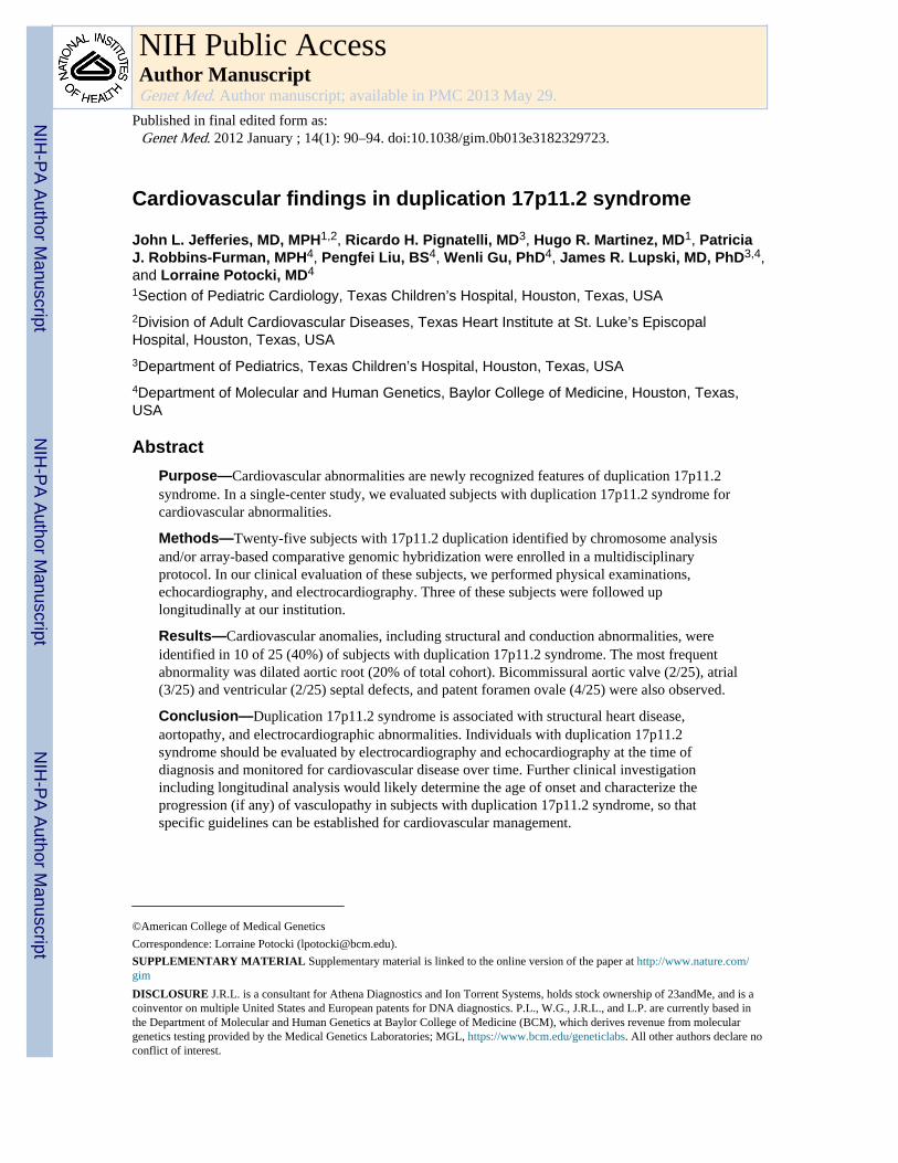
Cardiovascular findings in duplication 17p11.2 syndrome John L. Jefferies, MD, MPH 1,2 , Ricardo H. Pignatelli, MD 3 , Hugo R. Martinez, MD 1 , Patricia J. Robbins-Furman, MPH 4 , Pengfei Liu, BS 4 , Wenli Gu, PhD 4 , James R. Lupski, MD, PhD 3,4 , and Lorraine Potocki, MD 4 1 Section of Pediatric Cardiology, Texas Children’s Hospital, Houston, Texas, USA 2 Division of Adult Cardiovascular Diseases, Texas Heart Institute at St. Luke’s Episcopal Hospital, Houston, Texas, USA 3 Department of Pediatrics, Texas Children’s Hospital, Houston, Texas, USA 4 Department of Molecular and Human Genetics, Baylor College of Medicine, Houston, Texas, USA Abstract Purpose—Cardiovascular abnormalities are newly recognized features of duplication 17p11.2 syndrome. In a single-center study, we evaluated subjects with duplication 17p11.2 syndrome for cardiovascular abnormalities. Methods—Twenty-five subjects with 17p11.2 duplication identified by chromosome analysis and/or array-based comparative genomic hybridization were enrolled in a multidisciplinary protocol. In our clinical evaluation of these subjects, we performed physical examinations, echocardiography, and electrocardiography. Three of these subjects were followed up longitudinally at our institution. Results—Cardiovascular anomalies, including structural and conduction abnormalities, were identified in 10 of 25 (40%) of subjects with duplication 17p11.2 syndrome. The most frequent abnormality was dilated aortic root (20% of total cohort). Bicommissural aortic valve (2/25), atrial (3/25) and ventricular (2/25) septal defects, and patent foramen ovale (4/25) were also observed. Conclusion—Duplication 17p11.2 syndrome is associated with structural heart disease, aortopathy, and electrocardiographic abnormalities. Individuals with duplication 17p11.2 syndrome should be evaluated by electrocardiography and echocardiography at the time of diagnosis and monitored for cardiovascular disease over time. Further clinical investigation including longitudinal analysis would likely determine the age of onset and characterize the progression (if any) of vasculopathy in subjects with duplication 17p11.2 syndrome, so that specific guidelines can be established for cardiovascular management. ©American College of Medical Genetics Correspondence: Lorraine Potocki ([email protected]). SUPPLEMENTARY MATERIAL Supplementary material is linked to the online version of the paper at http://www.nature.com/ gim DISCLOSURE J.R.L. is a consultant for Athena Diagnostics and Ion Torrent Systems, holds stock ownership of 23andMe, and is a coinventor on multiple United States and European patents for DNA diagnostics. P.L., W.G., J.R.L., and L.P. are currently based in the Department of Molecular and Human Genetics at Baylor College of Medicine (BCM), which derives revenue from molecular genetics testing provided by the Medical Genetics Laboratories; MGL, https://www.bcm.edu/geneticlabs. All other authors declare no conflict of interest. NIH Public Access Author Manuscript Genet Med. Author manuscript; available in PMC 2013 May 29. Published in final edited form as: Genet Med. 2012 January ; 14(1): 90–94. doi:10.1038/gim.0b013e3182329723. NIH-PA Author Manuscript NIH-PA Author Manuscript NIH-PA Author Manuscript
-
Upload
independent -
Category
Documents
-
view
6 -
download
0
Transcript of Cardiovascular findings in duplication 17p11.2 syndrome
Cardiovascular findings in duplication 17p11.2 syndrome
John L. Jefferies, MD, MPH1,2, Ricardo H. Pignatelli, MD3, Hugo R. Martinez, MD1, PatriciaJ. Robbins-Furman, MPH4, Pengfei Liu, BS4, Wenli Gu, PhD4, James R. Lupski, MD, PhD3,4,and Lorraine Potocki, MD4
1Section of Pediatric Cardiology, Texas Children’s Hospital, Houston, Texas, USA2Division of Adult Cardiovascular Diseases, Texas Heart Institute at St. Luke’s EpiscopalHospital, Houston, Texas, USA3Department of Pediatrics, Texas Children’s Hospital, Houston, Texas, USA4Department of Molecular and Human Genetics, Baylor College of Medicine, Houston, Texas,USA
AbstractPurpose—Cardiovascular abnormalities are newly recognized features of duplication 17p11.2syndrome. In a single-center study, we evaluated subjects with duplication 17p11.2 syndrome forcardiovascular abnormalities.
Methods—Twenty-five subjects with 17p11.2 duplication identified by chromosome analysisand/or array-based comparative genomic hybridization were enrolled in a multidisciplinaryprotocol. In our clinical evaluation of these subjects, we performed physical examinations,echocardiography, and electrocardiography. Three of these subjects were followed uplongitudinally at our institution.
Results—Cardiovascular anomalies, including structural and conduction abnormalities, wereidentified in 10 of 25 (40%) of subjects with duplication 17p11.2 syndrome. The most frequentabnormality was dilated aortic root (20% of total cohort). Bicommissural aortic valve (2/25), atrial(3/25) and ventricular (2/25) septal defects, and patent foramen ovale (4/25) were also observed.
Conclusion—Duplication 17p11.2 syndrome is associated with structural heart disease,aortopathy, and electrocardiographic abnormalities. Individuals with duplication 17p11.2syndrome should be evaluated by electrocardiography and echocardiography at the time ofdiagnosis and monitored for cardiovascular disease over time. Further clinical investigationincluding longitudinal analysis would likely determine the age of onset and characterize theprogression (if any) of vasculopathy in subjects with duplication 17p11.2 syndrome, so thatspecific guidelines can be established for cardiovascular management.
©American College of Medical Genetics
Correspondence: Lorraine Potocki ([email protected]).
SUPPLEMENTARY MATERIAL Supplementary material is linked to the online version of the paper at http://www.nature.com/gim
DISCLOSURE J.R.L. is a consultant for Athena Diagnostics and Ion Torrent Systems, holds stock ownership of 23andMe, and is acoinventor on multiple United States and European patents for DNA diagnostics. P.L., W.G., J.R.L., and L.P. are currently based inthe Department of Molecular and Human Genetics at Baylor College of Medicine (BCM), which derives revenue from moleculargenetics testing provided by the Medical Genetics Laboratories; MGL, https://www.bcm.edu/geneticlabs. All other authors declare noconflict of interest.
NIH Public AccessAuthor ManuscriptGenet Med. Author manuscript; available in PMC 2013 May 29.
Published in final edited form as:Genet Med. 2012 January ; 14(1): 90–94. doi:10.1038/gim.0b013e3182329723.
NIH
-PA Author Manuscript
NIH
-PA Author Manuscript
NIH
-PA Author Manuscript
Keywordschromosome 17p duplication; congenital heart defects; dilated aortic root; Potocki-Lupskisyndrome; PTLS; vasculopathy
INTRODUCTIONDuplication 17p11.2 syndrome (Potocki-Lupski syndrome: OMIM# 610883) was the firstpredicted reciprocal microduplication syndrome described.1,2 Most individuals withduplication 17p11.2 syndrome (approximately 65%) harbor a common 3.7 Mb duplicationwithin 17p11.2, which is the homologous recombination reciprocal of the Smith-Magenissyndrome (SMS: OMIM# 182290) microdeletion.1,3 The common Potocki-Lupski syndromeduplication and common SMS deletion are caused by nonallelic homologous recombinationbetween the flanking distal and proximal low-copy repeats called SMS-REPs.4 Nonallelichomologous recombination can also use other low-copy repeats within this region and resultin the so-called uncommon recurrent duplications.3 Furthermore, DNA replication-basedmechanisms or nonhomologous end-joining are responsible for the majority of thenonrecurrent duplications.3,5
The clinical features of duplication 17p11.2 syndrome include hypotonia, failure to thrive,developmental delay, intellectual disability, sleep-disordered breathing, and structuralcardiovascular abnormalities.1,2,6–8 The behavioral phenotype of duplication 17p11.2syndrome includes autistic spectrum disorder, anxiety, and inattention.9 Although severalcases of 17p11.2 duplication were reported before its molecular mechanism was known,cardiovascular disease was not recognized as a feature of duplication 17p11.2 syndromeuntil individuals were systematically evaluated through a protocol-driven, multidisciplinaryclinical study.1 Herein, we detail the cardiovascular findings in 25 subjects with duplication17p11.2 syndrome, including 11 subjects who were described previously.1,10 Three of thesesubjects (527, 1106, and 2362) had more than one echocardiogram at our institution.
MATERIALS AND METHODSThe study was approved by the Baylor College of Medicine Institutional Review Board(Houston, TX). Individuals with 17p11.2 duplications identified by chromosome analysis,fluorescence in situ hybridization (FISH), and/or array-based comparative genomichybridization (array CGH) were referred to our institution and enrolled in amultidisciplinary clinical protocol in the General Clinical Research Center at TexasChildren’s Hospital. Informed consent was obtained from the subjects’ parent or legalguardian. Molecular characterization of almost all the subjects (except subject 3028) wasdescribed previously (Table, Supplementary Data online).1,3,5,10
Medical and family histories were recorded, and physical examination was performed on allsubjects on enrollment into the study. Two-dimensional transthoracic echocardiography andDoppler imaging were performed according to institutional guidelines. Aortic rootdimensions were measured according to guidelines of the American Society ofEchocardiography.11 Intracardiac structure and dimensions of the great vessels weremeasured. Using widely accepted criteria,12 Z scores (standard deviation units) werecalculated and compared with those of normal individuals with matched body surface area,gender, and age. A standard 15-lead electrocardiogram (ECG) was obtained at rest. Imagesand measurements were reviewed independently by a pediatric echocardiologist who wasunaware of the underlying diagnosis (R.H.P.). Subjects 527, 1006, and 2362 were followedlongitudinally to age 16 years, 27 years, and 3 years 10 months, respectively. Abnormal
Jefferies et al. Page 2
Genet Med. Author manuscript; available in PMC 2013 May 29.
NIH
-PA Author Manuscript
NIH
-PA Author Manuscript
NIH
-PA Author Manuscript
findings of Subjects 527 and 2362 are detailed within the “Results” section and in Table 1.Subject 1006 had normal cardiovascular evaluations.
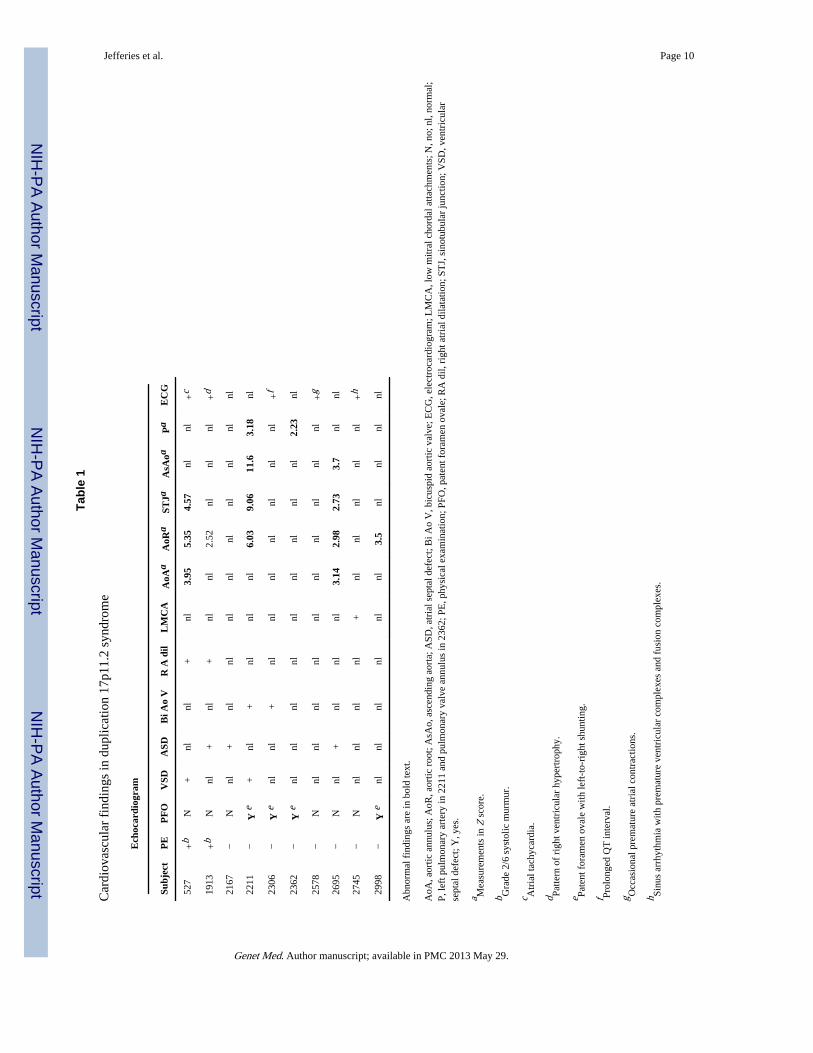
RESULTSTwenty-five subjects with 17p11.2 duplication (10 males, 15 females; age range at time ofmost recent cardiac evaluation from 2 years 1 month to 27 years) were included. Of the 25subjects, 10 (40%) showed cardiovascular involvement. No individual had a family historyof congenital heart defects (CHDs) or was exposed to teratogenic agents or maternaldiabetes. Abnormal cardiovascular findings are summarized in Table 1. A summary of allsubjects is provided in the Table (Supplementary Data online).
Clinical details of subjects with abnormal cardiovascular findingSubject 527 was diagnosed with duplication 17p11.2p12 soon after birth by chromosomeanalysis that was performed because of hypotonia, poor feeding, and CHDs. He had a large(10–12 mm) membranous ventricular septal defect (VSD) that extended into theconoventricular septum, with low-velocity bidirectional shunting through the defect. Inaddition, the right coronary cusp of the aortic valve prolapsed into the VSD during systole.At 2 months of age, he was noted to have increased pulmonary blood flow and congestiveheart failure by chest radiograph and underwent pulmonary artery banding that resulted incompensation of heart failure symptoms. He had routine echocardiograms performed at 1–2-year intervals; however, not all echocardiograms provided optimal visualization of the greatvessels. The first echocardiogram to reveal an abnormality of the aorta was performed at theage of 6 years and 4 months and showed a mildly dilated aortic annulus of 1.7 cm (Z score:3.63) and an aortic root of 1.76 cm (Z score: 0.37). An echocardiogram performed at age 11years 9 months revealed an aortic annulus Z score of 4.45, and aortic root Z score of 5.43,and a sinotubular junction Z score of 3.37. Echocardiogram at age 13 years 5 monthsrevealed an aortic annulus Z score of 3.52, and aortic root Z score of 4.73, and sinotubularjunction Z score of 2.98. At the age of 12 years, he was noted to have atrial ectopictachycardia and was started on an antiarrhythmic therapy (Sotalol, 50 mg twice a day); sincethat time, he has not had episodes of tachycardia. The most recent follow-upechocardiogram at age 16 years that was performed to evaluate cardiac anatomy andfunction revealed a large nonrestrictive perimembranous VSD with bidirectional shunting,mild tricuspid regurgitation with a peak velocity approximately 4.0 m/second by echoDoppler, and moderate right ventricular hypertrophy with a moderately dilated right atriumand right ventricle. There was a mildly dilated aortic annulus (Z score: 3.95), moderatelydilated aortic root (Z score: 5.35), and a sinotubular junction with a Z score of 4.57. Thepulmonary artery band appeared to be in good position with a peak Doppler velocity ofapproximately 4.3 m/second.
Subject 1913 was diagnosed at age 12 years by chromosome analysis and FISH that wasperformed because of dysmorphic features, poor growth, and developmental delay.Chromosome analyses at birth and earlier in childhood were normal. Cardiac evaluation at12 years and 9 months revealed a grade 2/6 systolic murmur. An echocardiogram showed alarge (19 mm) secundum atrial septal defect (ASD) with left-to-right shunting. The rightatrium and right ventricle were moderately enlarged with preserved right ventricular systolicfunction. The aortic root was mildly dilated at 2.31 cm (Z score: 2.52). The subject’s ECGrevealed right ventricular hypertrophy based on an R wave in V1 and an S wave in V6higher than the upper limits of normal range.
Subject 2167 was diagnosed at age 9 months by chromosome analysis and FISH that wasperformed because of hypotonia and poor feeding. At the age of 3 years 3 months, thepatient’s echocardiogram showed a small secundum ASD measuring 4–5 mm with left-to-
Jefferies et al. Page 3
Genet Med. Author manuscript; available in PMC 2013 May 29.
NIH
-PA Author Manuscript
NIH
-PA Author Manuscript
NIH
-PA Author Manuscript
right shunting. The right ventricle was normal in size and with systolic function. Theremainder of the echocardiogram was normal.
Subject 2211 was diagnosed at age 3 years 6 months by chromosome analysis and FISHperformed because of hypotonia, poor feeding, and developmental delay. Anechocardiogram performed at the age of 4 years 8 months (Figure 1) revealed a bicuspidaortic valve with raphe present between the right and left coronary commissures and severedilation of the aortic root (measuring 2.88 cm; Z score: 6.03), the sinotubular junctiondiameter (measuring 2.95 cm; Z score: 9.06), and the ascending aorta (measuring 3.38 cm; Zscore: 11.60). A small patent foramen ovale (PFO) with left-to-right shunting wasvisualized, as well as a small, restrictive muscular VSD with left-to-right shunting. Themitral valve was abnormal with redundant anterior and posterior leaflets and mild prolapseof the anterior mitral leaflet with resultant trivial mitral regurgitation. The left pulmonaryartery was dilated (measuring 1.33 cm; Z score: 3.18). He subsequently underwent surgicalrepair of the aorta at another institution, details of which are reported elsewhere.13
Subject 2306 was diagnosed at age 29 months by chromosome analysis and FISH that wasperformed because of hypotonia, failure to thrive, and developmental delay. Anechocardiogram at the age of 2 years 7 months revealed a PFO with left-to-right shuntingand bicommissural aortic valve with a partial fusion between the right and left commissures.The leaflets appeared mildly thickened without evidence of significant stenosis orregurgitation. The ECG revealed a prolonged QT interval of 488 milliseconds and a normalsinus rhythm.
Subject 2362 was diagnosed at age 8 months by chromosome analysis that was performedbecause of developmental delay. His initial echocardiogram at age 13 months revealed aPFO with left-to-right shunting and a dilated pulmonary valve annulus of 1.42 cm (Z score:2.23). No other significant structural abnormalities were found. An echocardiogramperformed at age 3 years 10 months was limited due to subject’s hyperactivity and agitation.
Subject 2578 was diagnosed at age 18 months by chromosome analysis and FISH that wasperformed because of developmental delay, hypotonia, and failure to thrive. Anechocardiogram at 4 years 4 months revealed no significant abnormalities. Two consecutiveECGs revealed premature atrial contractions that did not require medical intervention.
Subject 2695 was diagnosed at age 6 months by chromosome analysis and FISH that wasperformed because of hypotonia, failure to thrive, developmental delay, and congenitalcardiovascular disease. Echocardiogram performed before diagnosis revealed a large (10mm) ASD with left-to-right shunting. Follow-up echocardiogram at age 16 months revealeda smaller defect (by parental report). Echocardiogram performed at our institution, at age of4 years 1 month revealed a small secundum ASD with left-to-right shunting. In addition,there was aortic involvement including dilation of the aortic annulus (1.69 cm; Z score:3.14), the aortic root (1.96 cm; Z score: 2.98), the sinotubular junction (1.62 cm; Z score:2.73), and the ascending aorta (1.79 cm; Z score: 3.7). A small echodensity of unknownetiology or clinical significance was noted at the tip of the mitral valve leaflet, withoutresultant stenosis or insufficiency.
Subject 2745 was diagnosed at age 2 years 9 months by array comparative genomichybridization that was performed because of developmental delay and failure to thrive. Anechocardiogram performed by the age of 3 years and 9 months revealed low mitral chordalattachments on the interventricular septum without evidence of obstruction. Trivial aorticinsufficiency was also noted. The ECG revealed sinus arrhythmia with premature ventricularcomplexes and fusion complexes.
Jefferies et al. Page 4
Genet Med. Author manuscript; available in PMC 2013 May 29.
NIH
-PA Author Manuscript
NIH
-PA Author Manuscript
NIH
-PA Author Manuscript
Subject 2998 was diagnosed at age 2 years 7 months by array comparative genomichybridization that was performed because of hypotonia, failure to thrive, and developmentaldelay. At the age of 3 years 10 months, an echocardiogram revealed a PFO with left-to-rightshunting. In addition, the aortic root was mildly dilated with a diameter of 1.46 cm (Z score:3.5).
DISCUSSIONTo our knowledge, this is the largest reported study to assess cardiovascular involvement inpatients with duplication 17p11.2 syndrome. We identified cardiovascular abnormalities in10 of 25 (40%) patients with duplication 17p11.2 syndrome. This ratio is higher than in thegeneral population, even though ASD (0.66%),14 VSD (0.4%),15 and PFO (10–20%)14 arerelatively common CHDs. Nine of our 10 subjects had a structural abnormality; one hadonly an ECG abnormality. The most frequent abnormality was dilation of the aortic rootwith varying degrees of severity. Hypoplastic left heart was reported in two duplication17p11.2 syndrome patients,6,8 one of whom was evaluated at our institution after cardiactransplantation and was not included in this report because we did not perform his cardiacevaluation.6
Interestingly, only 6 of the 16 subjects (37.5%) with common duplications had acardiovascular abnormality, although they all had the same 3.7 Mb genomic duplication(Figure 2). On the other hand, the individual with the largest duplication (2711) had anormal cardiac evaluation. These findings demonstrate incomplete penetrance of CHDs inpatients with duplication 17p11.2 syndrome. Incomplete penetrance is observed for otherphenotypic aspects of SMS deletion 17p11.2 syndrome16,17 and other genomic disorders.18
Intriguingly, CHD is observed in 57% (4/7) of all nonrecurrent duplication subjects and66.7% (4/6) of the patients with nonrecurrent duplications larger than 3.7 Mb, whereas alower observation ratio of 37.5% is observed in patients with the 3.7 Mb commonduplication. This correlation indicates that there are potentially multiple genes or geneticfactors in 17p11.2 that contribute to the cardiovascular phenotype in duplication 17p11.2syndrome and that some of them may map outside of the 3.7 Mb common duplicationinterval.
We recognize that ASD, VSD, and PFO are relatively common congenital heart lesions.However, our findings of aortopathy are interesting and may be suggestive of connectivetissue disease involvement. This could have clinical implications for patients with aorticand/or mitral valve disease, such as the one patient we identified with an abnormal mitralvalve and prolapse of the anterior leaflet. Furthermore, aortic involvement was not confinedto the root, suggesting that a more diffuse vasculopathy should be considered in thesepatients. This would have important diagnostic implications, as a more advanced imagingtechnique such as computed tomography angiography or magnetic resonance angiographymay be indicated for comprehensive aortic assessment. In addition, evidence of aortopathyby noninvasive imaging analysis may necessitate appropriate medical therapy, and if severedilation of the aorta was found (as in 2211), surgical intervention may be discussedsecondary to the risk of possible dissection. Given the small number of patients in our cohortand limited clinical follow-up, the exact implications of aortic involvement, and medicaland/or surgical interventions cannot be determined. Our study is meant to characterize thespectrum of disease, which includes vascular involvement. Because these clinicaldelineation studies represent the initial characterizations of a relatively newly definedcondition, and our study is not longitudinal in nature, no clear clinical managementinferences can be made regarding the potential severity of this finding. However, given thefindings of aortopathy in multiple patients, future longitudinal studies are warranted.
Jefferies et al. Page 5
Genet Med. Author manuscript; available in PMC 2013 May 29.
NIH
-PA Author Manuscript
NIH
-PA Author Manuscript
NIH
-PA Author Manuscript
Our study provides important insight into the long-term management of patients withduplication 17p11.2 syndrome. First, given the potential need for medical and surgicalintervention, patients with duplication 17p11.2 syndrome should be referred to a qualifiedcardiovascular specialist. Given our findings of structural heart disease, a baselineechocardiogram and ECG should be obtained at the time of diagnosis. One subject hadisolated premature atrial complexes with no evidence of structural heart disease. The authorsrecognize that the importance of these ECG findings is unknown, given the lack of follow-up and ambulatory monitoring. Findings of premature atrial complexes on ECG are commonin the general healthy population, but the significance of these irregular beats in theduplication 17p11.2 population is unclear. However, in light of the important clinicalmanifestations of aortopathy documented herein, appropriate serial imaging studies arenecessary to delineate the vascular structures. Because transthoracic echocardiographyprovides limited visualization of the proximal aorta, more advanced imaging techniquessuch as computed tomography angiography or magnetic resonance angiography may beneeded to further define the thoracic aorta, as well as the abdominal aorta and primarybranch vessels.
There are limitations to our study. The patient population was small, and not all patients hadfollow-up echocardiograms to assess the progression of aortic or valvular disease. Limiteddata were available on the clinical course, including symptoms and medical therapies.Because no ambulatory electrocardiographic monitoring was performed in these patients, theoccurrence of arrhythmia may have been underestimated.
In conclusion, duplication 17p11.2 syndrome is associated with structural heart disease,aortopathy, and electrocardiographic abnormalities. Patients should have thorough cardiacevaluation at diagnosis and should be monitored over time for the development ofcardiovascular involvement. In addition, referral to a cardiologist for further management isindicated in patients with cardiovascular disease. Studies of the progression ofcardiovascular disease in these patients may lead to the development of guidelines forpatient care.
Supplementary MaterialRefer to Web version on PubMed Central for supplementary material.
AcknowledgmentsThis work was supported, in part, by Texas Children’s Hospital GCRC Grant M01RR00188. The authors thank thepatients and their families for their participation in our study. They also thank the nursing staff at the GeneralClinical Research Center (GCRC) who provided care for each patient admitted to Texas Children’s Hospital. Theyacknowledge the physicians and genetics professionals who generously made patient referrals and sharedinformation for these studies.
REFERENCES1. Potocki L, Bi W, Treadwell-Deering D, et al. Characterization of Potocki-Lupski syndrome
(dup(17)(p11.2p11.2)) and delineation of a dosagesensitive critical interval that can convey anautism phenotype. Am J Hum Genet. 2007; 80:633–649. [PubMed: 17357070]
2. Potocki L, Chen KS, Park SS, et al. Molecular mechanism for duplication 17p11.2- the homologousrecombination reciprocal of the Smith-Magenis microdeletion. Nat Genet. 2000; 24:84–87.[PubMed: 10615134]
3. Zhang F, Potocki L, Sampson JB, et al. Identification of uncommon recurrent Potocki-Lupskisyndrome-associated duplications and the distribution of rearrangement types and mechanisms inPTLS. Am J Hum Genet. 2010; 86:462–470. [PubMed: 20188345]
Jefferies et al. Page 6
Genet Med. Author manuscript; available in PMC 2013 May 29.
NIH
-PA Author Manuscript
NIH
-PA Author Manuscript
NIH
-PA Author Manuscript
4. Bi W, Park SS, Shaw CJ, Withers MA, Patel PI, Lupski JR. Reciprocal crossovers and a positionalpreference for strand exchange in recombination events resulting in deletion or duplication ofchromosome 17p11.2. Am J Hum Genet. 2003; 73:1302–1315. [PubMed: 14639526]
5. Zhang F, Khajavi M, Connolly AM, Towne CF, Batish SD, Lupski JR. The DNA replicationFoSTeS/MMBIR mechanism can generate genomic, genic and exonic complex rearrangements inhumans. Nat Genet. 2009; 41:849–853. [PubMed: 19543269]
6. Sanchez-Valle A, Pierpont ME, Potocki L. The severe end of the spectrum: Hypoplastic left heart inPotocki-Lupski syndrome. Am J Med Genet A. 2011; 155A:363–366. [PubMed: 21271655]
7. Soler-Alfonso C, Motil KJ, Turk CL, et al. Potocki-Lupski syndrome: a microduplication syndromeassociated with oropharyngeal dysphagia and failure to thrive. J Pediatr. 2011; 158:655–659. e2.[PubMed: 21168152]
8. Yusupov R, Roberts AE, Lacro RV, Sandstrom M, Ligon AH. Potocki-Lupski syndrome: aninherited dup(17)(p11.2p11.2) with hypoplastic left heart. Am J Med Genet A. 2011; 155A:367–371. [PubMed: 21271656]
9. Treadwell-Deering DE, Powell MP, Potocki L. Cognitive and behavioral characterization of thePotocki-Lupski syndrome (duplication 17p11.2). J Dev Behav Pediatr. 2010; 31:137–143.[PubMed: 20110824]
10. Yatsenko SA, Treadwell-Deering D, Krull K, et al. Trisomy 17p10-p12 due to mosaicsupernumerary marker chromosome: delineation of molecular breakpoints and clinical phenotype,and comparison to other proximal 17p segmental duplications. Am J Med Genet A. 2005; 138A:175–180. [PubMed: 16152635]
11. Lai WW, Geva T, Shirali GS, et al. Guidelines and standards for performance of a pediatricechocardiogram: a report from the Task Force of the Pediatric Council of the American Society ofEchocardiography. J Am Soc Echocardiogr. 2006; 19:1413–1430. [PubMed: 17138024]
12. Sluysmans T, Colan SD. Theoretical and empirical derivation of cardiovascular allometricrelationships in children. J Appl Physiol. 2005; 99:445–457. [PubMed: 15557009]
13. Girirajan S, Williams SR, Garbern JY, Nowak N, Hatchwell E, Elsea SH. 17p11.2p12 triplicationand del(17)q11.2q12 in a severely affected child with dup(17)p11.2p12 syndrome. Clin Genet.2007; 72:47–58. [PubMed: 17594399]
14. Kaplan S. Congenital heart disease in adolescents and adults. Natural and postoperative historyacross age groups. Cardiol Clin. 1993; 11:543–556. [PubMed: 8252558]
15. Wu MH, Chen HC, Lu CW, Wang JK, Huang SC, Huang SK. Prevalence of congenital heartdisease at live birth in Taiwan. J Pediatr. 2010; 156:782–785. [PubMed: 20138303]
16. Yan J, Bi W, Lupski JR. Penetrance of craniofacial anomalies in mouse models of Smith-Magenissyndrome is modified by genomic sequence surrounding Rai1: not all null alleles are alike. Am JHum Genet. 2007; 80:518–525. [PubMed: 17273973]
17. Yan J, Keener VW, Bi W, et al. Reduced penetrance of craniofacial anomalies as a function ofdeletion size and genetic background in a chromosome engineered partial mouse model for Smith-Magenis syndrome. Hum Mol Genet. 2004; 13:2613–2624. [PubMed: 15459175]
18. Lupski JR. Genomic disorders ten years on. Genome Med. 2009; 1:42. [PubMed: 19439022]
Jefferies et al. Page 7
Genet Med. Author manuscript; available in PMC 2013 May 29.
NIH
-PA Author Manuscript
NIH
-PA Author Manuscript
NIH
-PA Author Manuscript
Figure 1. Subject 2211 in a parasternal long-axis view, the dilatation of the aorta is observed indifferent segmentsThe aortic annulus diameter is the distance between 1 and 1 (Z score: 3.27); the aortic rootdiameter is the distance between 2 and 2 (Z score: 6.03); and the sinotubular junction is thedistance between 3 and 3 (Z score: 9.06). Ao, aorta; LA, left atrium; LV, left ventricle; RV,right ventricle.
Jefferies et al. Page 8
Genet Med. Author manuscript; available in PMC 2013 May 29.
NIH
-PA Author Manuscript
NIH
-PA Author Manuscript
NIH
-PA Author Manuscript
Figure 2. Summary of duplication sizesThe position and size of the genomic rearrangements (red (duplication), blue (triplication),and green (deletion) bars), as determined by array CGH are depicted for each subject. Anideogram of chromosome 17p, with a scale indicating the genomic coordinates (versionhg18), and the locations of PMP22 and RAI1 genes are also included. Subject 2343 (mosaicmarker) is not shown. Cardiac phenotype of each patient with CHD is briefly summarized inTable 1 and detailed in the text. Modified with permission from Am J Hum Genet.3 CGH,comparative genomic hybridization; CHD, congenital heart defect.
Jefferies et al. Page 9
Genet Med. Author manuscript; available in PMC 2013 May 29.
NIH
-PA Author Manuscript
NIH
-PA Author Manuscript
NIH
-PA Author Manuscript
NIH
-PA Author Manuscript
NIH
-PA Author Manuscript
NIH
-PA Author Manuscript
Jefferies et al. Page 10
Tabl
e 1
Car
diov
ascu
lar
find
ings
in d
uplic
atio
n 17
p11.
2 sy
ndro
me
Ech
ocar
diog
ram
Subj
ect
PE
PF
OV
SDA
SDB
i Ao
VR
A d
ilL
MC
AA
oAa
AoR
aST
JaA
sAoa
Pa
EC
G
527
+b
N+
nlnl
+nl
3.95
5.35
4.57
nlnl
+c
1913
+b
Nnl
+nl
+nl
nl2.
52nl
nlnl
+d
2167
−N
nl+
nlnl
nlnl
nlnl
nlnl
nl
2211
−Y
e+
nl+
nlnl
nl6.
039.
0611
.63.
18nl
2306
−Y
enl
nl+
nlnl
nlnl
nlnl
nl+
f
2362
−Y
enl
nlnl
nlnl
nlnl
nlnl
2.23
nl
2578
−N
nlnl
nlnl
nlnl
nlnl
nlnl
+g
2695
−N
nl+
nlnl
nl3.
142.
982.
733.
7nl
nl
2745
−N
nlnl
nlnl
+nl
nlnl
nlnl
+h
2998
−Y
enl
nlnl
nlnl
nl3.
5nl
nlnl
nl
Abn
orm
al f
indi
ngs
are
in b
old
text
.
AoA
, aor
tic a
nnul
us; A
oR, a
ortic
roo
t; A
sAo,
asc
endi
ng a
orta
; ASD
, atr
ial s
epta
l def
ect;
Bi A
o V
, bic
uspi
d ao
rtic
val
ve; E
CG
, ele
ctro
card
iogr
am; L
MC
A, l
ow m
itral
cho
rdal
atta
chm
ents
; N, n
o; n
l, no
rmal
;P,
left
pul
mon
ary
arte
ry in
221
1 an
d pu
lmon
ary
valv
e an
nulu
s in
236
2; P
E, p
hysi
cal e
xam
inat
ion;
PFO
, pat
ent f
oram
en o
vale
; RA
dil,
rig
ht a
tria
l dila
tatio
n; S
TJ,
sin
otub
ular
junc
tion;
VSD
, ven
tric
ular
sept
al d
efec
t; Y
, yes
.
a Mea
sure
men
ts in
Z s
core
.
b Gra
de 2
/6 s
ysto
lic m
urm
ur.
c Atr
ial t
achy
card
ia.
d Patte
rn o
f ri
ght v
entr
icul
ar h
yper
trop
hy.
e Pate
nt f
oram
en o
vale
with
left
-to-
righ
t shu
ntin
g.
f Prol
onge
d Q
T in
terv
al.
g Occ
asio
nal p
rem
atur
e at
rial
con
trac
tions
.
h Sinu
s ar
rhyt
hmia
with
pre
mat
ure
vent
ricu
lar
com
plex
es a
nd f
usio
n co
mpl
exes
.
Genet Med. Author manuscript; available in PMC 2013 May 29.