Acute CardioVascular Monitoring
12
Prof. Josep Masip MD, PhD, FESC Sample chapter on Intra-abdominal hypertension Handbook of Acute CardioVascular Monitoring Quick access to most relevant practical information on haemodynamic and general monitoring in acute cardiovascular care 2022 edition
-
Upload
khangminh22 -
Category
Documents
-
view
0 -
download
0
Transcript of Acute CardioVascular Monitoring

Prof. Josep Masip MD, PhD, FESC
Sample chapter on Intra-abdominal hypertension
Handbook of
Acute CardioVascularMonitoringQuick access to most relevant practical information on haemodynamic and general monitoring in acute cardiovascular care
2022 edition

8 - INTRA-ABDOMINAL HYPERTENSION
Dr. Élida Amestoy, MD, PhD Intensive Care Department, Consorci Sanitari Integral Hospital Sant Joan Desp Moisès Broggi, Barcelona, Spain
Prof. Dr. Manu L.N.G. Malbrain, MD, PhD Intensive Care Departement, Universitair Ziekenhuis Brussel (UZB), Jette / Faculty of Medicine and Pharmacy, Vrije Universiteit Brussels (VUB), Belgium
Key messages
IAP is the steady-state pressure concealed within the abdominal cavity expressed in mmHg
The gold standard IAP measurement technique is via the bladder
IAH is defined as a sustained pathological elevation in IAP ≥ 12 mmHg and it is graded in four categories
AbCS is a sustained IAP > 20 mmHg that is associated with new onset organ failure (assessed with sequential organ failure assessment, SOFA score)
Elevated IAP plays a major role in the development of multiple system organ dysfunction and failure as a result of significant hypoperfusion and the effects are not limited to the intra-abdominal organs alone
Primary IAH/AbCS is a condition associated with injury or disease in the abdominopelvic region
Secondary IAH or AbCS refers to conditions that do not originate from the abdominopelvic region, and are frequently associated with damage of the glycocalyx and/or tight junctions following massive fluid resuscitation
III.8
P.225

Basic monitoring includes an accurate IAP measurement every 4 - 6 h in patients with at least 2 or more risk factors for IAH or AbCS
Other important clinical parameters are gastric residual volume, urinary output, abdominal perfusion pressure calculated as MAP minus IAP, body weight, daily and cumulative fluid balance
Advanced haemodynamic monitoring is required in severe IAH
Medical management comes first and decompressive laparotomy should be used as last resort in cases other treatment options fail
Introduction
Intra-abdominal hypertension is a relatively frequent medical situation in intensive care units which can trigger important complications because it plays a major role in the deve-lopment of multiple system organ dysfunction. Therefore, intra-abdominal pressure should be considered another first line parameter to be monitored routinely.
III.8
P.226

8.1 - IAH and AbCS definitions
The 2013 the World Society of Abdominal Compartment Syndrome (WSACS, www.wsacs.org) changed its name to The Abdominal Compartment Society and updated consensus defini-tions and recommendations on this topic were published.
8.1.1 - Intra-abdominal pressure (IAP)
IAP is the steady-state pressure concealed within the abdominal cavity and the reference standard for intermittent IAP measurements is via bladder with a maximal instillation volume of 25 mL of sterile saline (Figure 1, 2). IAP should be expressed in mmHg and measured at end-expiration in the supine position, after ensuring that abdominal muscle contractions are absent and with transducer zeroed at the level of midaxillary line. Normal IAP ranges from sub-atmospheric to zero mmHg, however, IAP is commonly elevated to a range of 5–7 mmHg in critically ill adults.
Figure 1 - IAP measurement
- Supine position- End-expiration- Ensuring absent abdominal muscle contraction- Transducer zeroed at midaxillary line- Maximal instilation volume 25 mL sterile saline
- IAP expressed in mmHg
Manometer
UrimeterThree way stopcock
Instilation volume
Foley catheter
III.8
P.227

Figure 2 - IAP measurement with the Foley Manometer LV
- Open the Foley Manometer LV (Holtech Medical, Denmark) pouch and close the tube clamp
- Place the urine collection device under the patient's bladder
- Insert the Foley Manometer between catheter and drainage device
- Prime the Foley Manometer with 20ml of sterile saline through its needle-free injection/sampling port (only once i.e. at initial set-up)
- Urine sampling from the needle-free port is facilitated by temporarily opening the red clamp
- Replace the Foley Manometer whenever the Foley catheter or the urine collection device is replaced, or at least every 7 days
- Place the "0 mmHg" mark of the manometer tube at the midaxillary line at the level of the iliac crest and elevate the filter vertically above the patient
- Open the bio-filter clamp, and read IVP when the meniscus has stabilized after about 10 seconds
- Close clamp after IVP measurement and place the Foley Manometer in its drainage position
III.8
P.228

8.1.2 - Intra-abdominal hypertension
IAH is a sustained or repeated pathological elevation in IAP ≥ 12 mmHg and it’s graded in four categories (Table 1). AbCS is a sustained increased IAP > 20 mmHg that is associated with new onset organ failure (Figure 3). IAH is a graded phenomenon while AbCS is an all-or-nothing condition. AbCS is a syndrome and not a disease and as such it can have many causes or it can be associated with many disease processes. A polycompartment syndrome is a condition where two or more anatomical compartments have elevated com-partmental pressures.
Table 1 - WSACS grading of IAH
IAH grade IAP (mmHg)
Grade I 12-15
Grade II 16-20
Grade III 21-25
Grade IV 25
Figure 3 - Natural course of IAH/AbCS
12 20IAP (mmHg)
ORGAN DYSFUNCTION Normal IAP IAH AbCS
Death
Cardiovascular
Respiratory
Gastrointestinal
Renal
III.8
P.229
8.1.3 - Abdominal compliance
Abdominal compliance is a measure of the ease of abdominal expansion, which is determined by the elasticity of the abdominal wall and diaphragm. It should be expressed as the change in intra-abdominal volume per change in IAP.
8.2 - Primary and secondary IAH
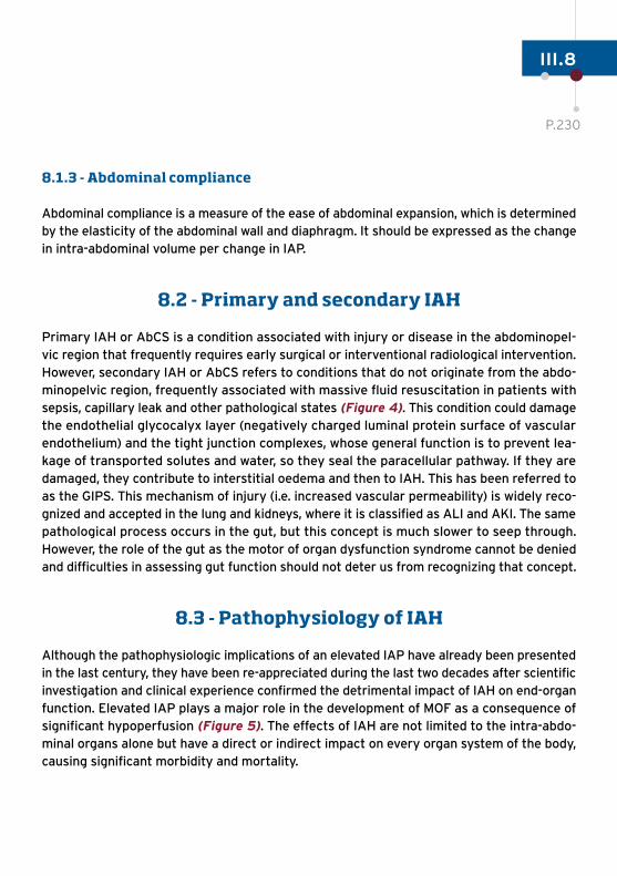
Primary IAH or AbCS is a condition associated with injury or disease in the abdominopel-vic region that frequently requires early surgical or interventional radiological intervention. However, secondary IAH or AbCS refers to conditions that do not originate from the abdo-minopelvic region, frequently associated with massive fluid resuscitation in patients with sepsis, capillary leak and other pathological states (Figure 4). This condition could damage the endothelial glycocalyx layer (negatively charged luminal protein surface of vascular endothelium) and the tight junction complexes, whose general function is to prevent lea-kage of transported solutes and water, so they seal the paracellular pathway. If they are damaged, they contribute to interstitial oedema and then to IAH. This has been referred to as the GIPS. This mechanism of injury (i.e. increased vascular permeability) is widely reco-gnized and accepted in the lung and kidneys, where it is classified as ALI and AKI. The same pathological process occurs in the gut, but this concept is much slower to seep through. However, the role of the gut as the motor of organ dysfunction syndrome cannot be denied and difficulties in assessing gut function should not deter us from recognizing that concept.
8.3 - Pathophysiology of IAH
Although the pathophysiologic implications of an elevated IAP have already been presented in the last century, they have been re-appreciated during the last two decades after scientific investigation and clinical experience confirmed the detrimental impact of IAH on end-organ function. Elevated IAP plays a major role in the development of MOF as a consequence of significant hypoperfusion (Figure 5). The effects of IAH are not limited to the intra-abdo-minal organs alone but have a direct or indirect impact on every organ system of the body, causing significant morbidity and mortality.
III.8
P.230

Figure 4 - Primary and secondary IAH/ACS
PRIMARY
- Abdominal surgery- Hemoperitoneum- Gastroparesis/ ileus- Colonic obstruction- Acute pancreatitis- Intra-abdominal tumors/ collections
SECONDARY
- Sepsis- Polytransfusion- Massive fluid
resuscitation
III.8
P.231

Figure 5 - Pathophysiologic implications of IAH
Central nervous system
- ICP- CPP- Idiopathic intracranial
hypertension in morbid obesity
Cardiovascular
- Venous return- Heart rate- Mean arterial pressure- CVP- CO- SVR- PAOP- Intra-thoracic blood
volume index- Global enddiastolic
blood volume index
Pulmonary
- Elevated diaphragm- Paw- Pleural Pressure- Auto-PEEP- Functional residual capacity- PaCO2 and PaO2- Chest wall compliance- Dinamic and static compliance- Dead-space ventilation- Atelectasis- Intrapulmonar shunt- Difficult weaning
Hepatic
- Hepatic and portal blow flow
- Lactate clearance- Glucose metabolism- Mitocondrial function
GASTROINTESTINAL
- Celiac and mucosal blow flow- APP- Mesenteric vein
compression- Intramucosal pH- CO2-gap- Intestinal permeability- Bacterial translocation- Success enteral
feeding
Renal
- Renal blow flow- GFR- Urinary output- Tubular dysfunction- Compression ureters- Anti-diuretic hormone
IAP
III.8
P.232

8.4 - Monitoring IAH and AbCS
Prevention of IAH is essential in patients with risk factors for IAH or AbCS. When two or more risk factors are pre-sent, baseline IAP monitoring is mandatory followed by sequential assessment in case of IAH grade I. The main monitoring parameter is the IAP, that can be obtained via trans-bladder measurement (Figure 1, 2) (Table 2). In order to perform a good measurement, pain and anxiety must be excluded, since voluntary contraction of the abdominal muscles can interfere with interpretation of the results. Other basic monitoring tools are gastric residual volume via nasogastric tube or antral cross-sec-tional area (ultrasound) and urinary output. Overzealous administration of fluids can cause secondary IAH/AbCS (endothelial dysfunction), so it is important to measure body weight and cumulative fluid balance. Abdominal per-fusion pressure is the difference between mean systemic arterial pressure and IAP. It more accurately reflects vis-ceral perfusion.
More advanced monitoring is needed in case of IAH grade II or higher. It the presence of an arterial line and a central venous catheter, the use of transpulmonary thermodilu-tion (PiCCO, Getinge or EV1000, Edwards) can provide additional information on preload, contractility, afterload and pulmonary edema (Table 2): oxygenation ratio (PaO
2/
FiO2), ScvO
2, CO (also through echocardiography), SVR, as
well as fluid management status in the different phases of shock (PPV, GEDVI and EVLWI). Sequential arterial or venous lactate levels may be helpful. Barometric preload parameters like CVP or PCWP are erroneously increased in case of IAH and cannot be used.
Table 2 - Main parameters to monitoring in IAH/ACS
Patients with risk factors for IAH and AbCS
- Diminished abdominal wall compliance
- Increased intra-abdominal contents
- Increased intra-luminal contents
- Capillary leak/fluid resuscitation
Monitoring Technique Normal values
Main parameters
IAP/4hTrans-bladder measurement
≤ 12 mmHg
APP AMP – IAP ≥ 60 mmHg
Gastric residual volume Nasogastric tube< 500cc/6h or < 1000cc/24h
Urinary output Foley catheter > 0,5–1 mL/Kg/h
Glomerular filtration rate
Body weight
Cumulative balance fluid
60-90 mL/min/1,73m2
Advanced parameters
Continuous AP A-line
Lactate Blood analysis < 2 mmol/L
PaO2/FiO
2Arterial gases > 300 mmHg
ScvO2
Central venous catheter > 70%
COTranspulmonary thermodilutionEchocardiography
4-8 L/min
SVR
Transpulmonary thermodilution
800-1200 din·s·cm-5
PPV <13%
GEDVI 600-800 mL/m2
EVLWI 3-7 mL/Kg
III.8
P.233

Patients with risk factors for IAH and AbCS
- Diminished abdominal wall compliance
- Increased intra-abdominal contents
- Increased intra-luminal contents
- Capillary leak/fluid resuscitation
Monitoring Technique Normal values
Main parameters
IAP/4hTrans-bladder measurement
≤ 12 mmHg
APP AMP – IAP ≥ 60 mmHg
Gastric residual volume Nasogastric tube< 500cc/6h or < 1000cc/24h
Urinary output Foley catheter > 0,5–1 mL/Kg/h
Glomerular filtration rate
Body weight
Cumulative balance fluid
60-90 mL/min/1,73m2
Advanced parameters
Continuous AP A-line
Lactate Blood analysis < 2 mmol/L
PaO2/FiO
2Arterial gases > 300 mmHg
ScvO2
Central venous catheter > 70%
COTranspulmonary thermodilutionEchocardiography
4-8 L/min
SVR
Transpulmonary thermodilution
800-1200 din·s·cm-5
PPV <13%
GEDVI 600-800 mL/m2
EVLWI 3-7 mL/Kg
III.8
P.234

Handbook of
Acute CardioVascular Monitoring
European Society of CardiologyAssociation for Acute CardioVascular Care (ACVC)2035, route des Colles - Les TempliersCS 80179 Biot - 06903 Sophia Antipolis - FRANCETel: +33 (0)4 92 94 76 00Fax: +33 (0)4 92 94 86 46Email: [email protected]
www.escardio.org/ACVC
2022 edition