Abstracts - Cardiovascular Medicine
153
Cardiovascular Medicine Kardiovaskuläre Medizin – Médecine cardiovasculaire Official journal of the Swiss Society of Cardiology, the Swiss Society of Hypertension, the Swiss Society of Angiology and the Swiss Society of Paediatric Cardiology www.cardiovascmed.ch Supplementum 27 ad Cardiovascular Medicine 2017;20: issue 5 17 May 2017 Abstracts Joint Annual Meeting of the Swiss Society of Cardiology and the Swiss Society of Cardiac and Thoracic Vascular Surgery Baden (Switzerland), June 7–9, 2017
-
Upload
khangminh22 -
Category
Documents
-
view
0 -
download
0
Transcript of Abstracts - Cardiovascular Medicine
Cardiovascular Medicine
Kardiovaskuläre Medizin – Médecine cardiovasculaire
Official journal of the Swiss Society of Cardiology,
the Swiss Society of Hypertension, the Swiss Society of Angiology
and the Swiss Society of Paediatric Cardiology
www.cardiovascmed.ch
Supplementum 27ad Cardiovascular Medicine2017;20: issue 517 May 2017
Abstracts
Joint Annual Meeting of the Swiss Society of Cardiology and the Swiss Society of Cardiac and Thoracic Vascular SurgeryBaden (Switzerland), June 7–9, 2017
S1
Table of contents
Rapid Fire Abstract Session - Periinterventional risks of TAVI ............................................................... 2
Abstract Session - Structural heart, clinical cases .................................................................................. 8
Rapid Fire Abstract Session - Prediction, Prognosis and Stratification................................................. 16
Abstract Session - New aspects in clinical and basic research in cardiology ....................................... 24
Rapid Fire Abstract Session - Electrical misbehaviour of the ventricles ............................................... 28
Rapid Fire Abstract Session - ACS treatment: Outcome trends and new technologies ....................... 35
Abstract Session - SSCS ....................................................................................................................... 41
Abstract Session - Unusual cardiac situation, clinical cases ................................................................. 45
Rapid Fire Abstract Session - This and that from rhythmology & The current role of Troponin and other risk factors in ACS ................................................................................................................. 50
Rapid Fire Abstract Session - This and that from heart failure ............................................................. 57
Rapid Fire Abstract Session - News from imaging, congenital heart disease and heart disease in pregnancy ............................................................................................................................. 63
Poster Walk I. - Electrophysiology and devices I .................................................................................. 73
Poster Walk II. - Electrophysiology and devices II ................................................................................ 79
Poster Walk I. - Basic research in cardiology ........................................................................................ 84
Poster walk I. - Innovation in congenital cardiac surgery and imaging ................................................. 91
Poster walk I. - Heart failure and beyond I ............................................................................................ 98
Poster Walk II. - Heart failure and beyond II ....................................................................................... 104
Poster walk II. - Epidemiology, risk factors, rehabilitation & thrombosis ............................................. 109
Poster walk I. - Clinical Cases I. .......................................................................................................... 116
Poster Walk II. - Clinical Cases II. ....................................................................................................... 123
Poster walk II. - ACS, PCI & CABG ..................................................................................................... 130
Author Index ........................................................................................................................................ 138
S2
Rapid Fire Abstract Session - Periinterventional risks of TAVI 31 Self-care in individuals with heart failure: results from a cross-sectional Swiss study P. Schäfer-Keller1, D. Graf2, G. Santos1, K. Denhaerynck1, K. Vasserot2, C. Augereau2, H. Villeneuve2, O. Raccanello2, N. Aubort2, M. Vona3, D. Richards4, A. Strömberg5 1University of Applied Sciences and Arts Western Switzerland - School of Health Sciences Fribourg - HEdS-FR / HES-SO, 2Cardiology, Cantonal Hospital Fribourg, Fribourg, 3Cardiac and Pulmonary Rehabilitation, Cantonal Hospital Fribourg, Billens, Switzerland, 4Mental Health Services Research, University of Exeter medical school, Exeter, United Kingdom, 5Department of Medical and Health Sciences, Linköpings University, Linköpings, Sweden Introduction: Self-care is a vital component of heart failure (HF) treatment, with confirmed influences on quality of life, morbidity and mortality. Although self-care is commonly sub-optimal in HF populations, and interventions targeting support and education improve it, Swiss studies are scarce. This study examined HF self-care in Switzerland and its relationship with hospitalizations. Method: A cross-sectional study of a convenience sample of adult HF patients from four campuses of one Swiss acute care hospital. We used the Self-Care of Heart Failure Index (SCHFI) to measure self-care maintenance, management and confidence, and the European Heart Failure Self-care Behavior Scale (EHFScBS) to measure medication regimen adherence, asking for help, and adapting daily activities. Results: We included 227 individuals with HF (40.1% female; mean age 77.8 years). When experiencing common symptoms, 53.8% reported not recognizing them as such, with the majority unlikely to take appropriate counter-measures. However, 62.7% were highly confident regarding symptom relief. Respectively, 43.7%, 72.0% and 94.8% of respondents reported sub-adequate levels of self-care confidence, maintenance, and management. In this sample, 73.6% had at least one recorded hospitalization during the previous year, with hospitalizations correlating positively with self-care (e.g., SCHFI-maintenance rho=0.29, p< 0.0001; EHFScBS-adherence to regimen rho=-0.26, p< 0.0001) Conclusion: Inadequate self-care levels were highly prevalent. Additionally, better self-care correlated with more hospitalizations. Ratings were highest for medication adherence and appointment keeping, and poorest for self-care management items, conflicting with high confidence regarding self-care capabilities. HF individuals need increased support, especially in symptom monitoring and recognition and symptom-related decision-making. These findings will translate into one component of a complex intervention, which we will then pilot and test.
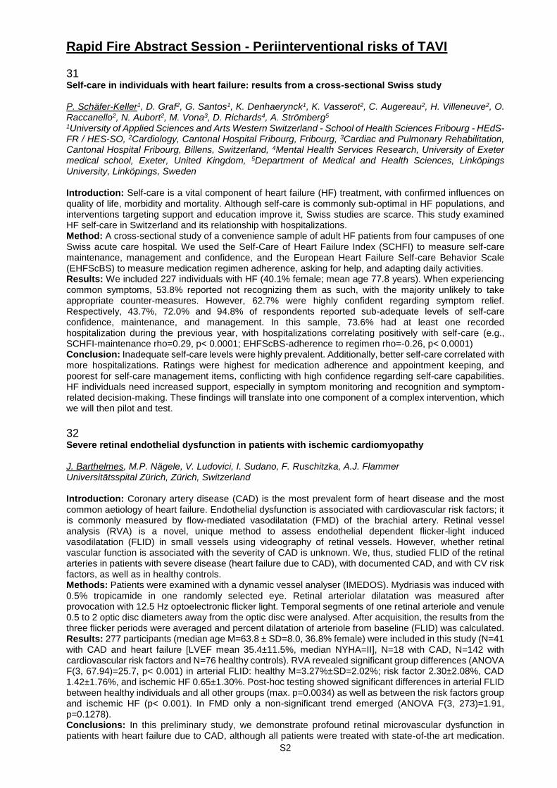
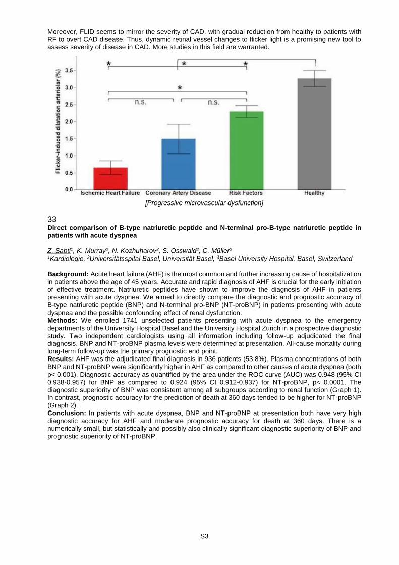
32 Severe retinal endothelial dysfunction in patients with ischemic cardiomyopathy J. Barthelmes, M.P. Nägele, V. Ludovici, I. Sudano, F. Ruschitzka, A.J. Flammer Universitätsspital Zürich, Zürich, Switzerland Introduction: Coronary artery disease (CAD) is the most prevalent form of heart disease and the most common aetiology of heart failure. Endothelial dysfunction is associated with cardiovascular risk factors; it is commonly measured by flow-mediated vasodilatation (FMD) of the brachial artery. Retinal vessel analysis (RVA) is a novel, unique method to assess endothelial dependent flicker-light induced vasodilatation (FLID) in small vessels using videography of retinal vessels. However, whether retinal vascular function is associated with the severity of CAD is unknown. We, thus, studied FLID of the retinal arteries in patients with severe disease (heart failure due to CAD), with documented CAD, and with CV risk factors, as well as in healthy controls. Methods: Patients were examined with a dynamic vessel analyser (IMEDOS). Mydriasis was induced with 0.5% tropicamide in one randomly selected eye. Retinal arteriolar dilatation was measured after provocation with 12.5 Hz optoelectronic flicker light. Temporal segments of one retinal arteriole and venule 0.5 to 2 optic disc diameters away from the optic disc were analysed. After acquisition, the results from the three flicker periods were averaged and percent dilatation of arteriole from baseline (FLID) was calculated. Results: 277 participants (median age M=63.8 ± SD=8.0, 36.8% female) were included in this study (N=41 with CAD and heart failure [LVEF mean 35.4±11.5%, median NYHA=II], N=18 with CAD, N=142 with cardiovascular risk factors and N=76 healthy controls). RVA revealed significant group differences (ANOVA F(3, 67.94)=25.7, p< 0.001) in arterial FLID: healthy M=3.27%±SD=2.02%; risk factor 2.30±2.08%, CAD 1.42±1.76%, and ischemic HF 0.65±1.30%. Post-hoc testing showed significant differences in arterial FLID between healthy individuals and all other groups (max. p=0.0034) as well as between the risk factors group and ischemic HF (p< 0.001). In FMD only a non-significant trend emerged (ANOVA F(3, 273)=1.91, p=0.1278). Conclusions: In this preliminary study, we demonstrate profound retinal microvascular dysfunction in patients with heart failure due to CAD, although all patients were treated with state-of-the art medication.
S3
Moreover, FLID seems to mirror the severity of CAD, with gradual reduction from healthy to patients with RF to overt CAD disease. Thus, dynamic retinal vessel changes to flicker light is a promising new tool to assess severity of disease in CAD. More studies in this field are warranted.
[Progressive microvascular dysfunction]
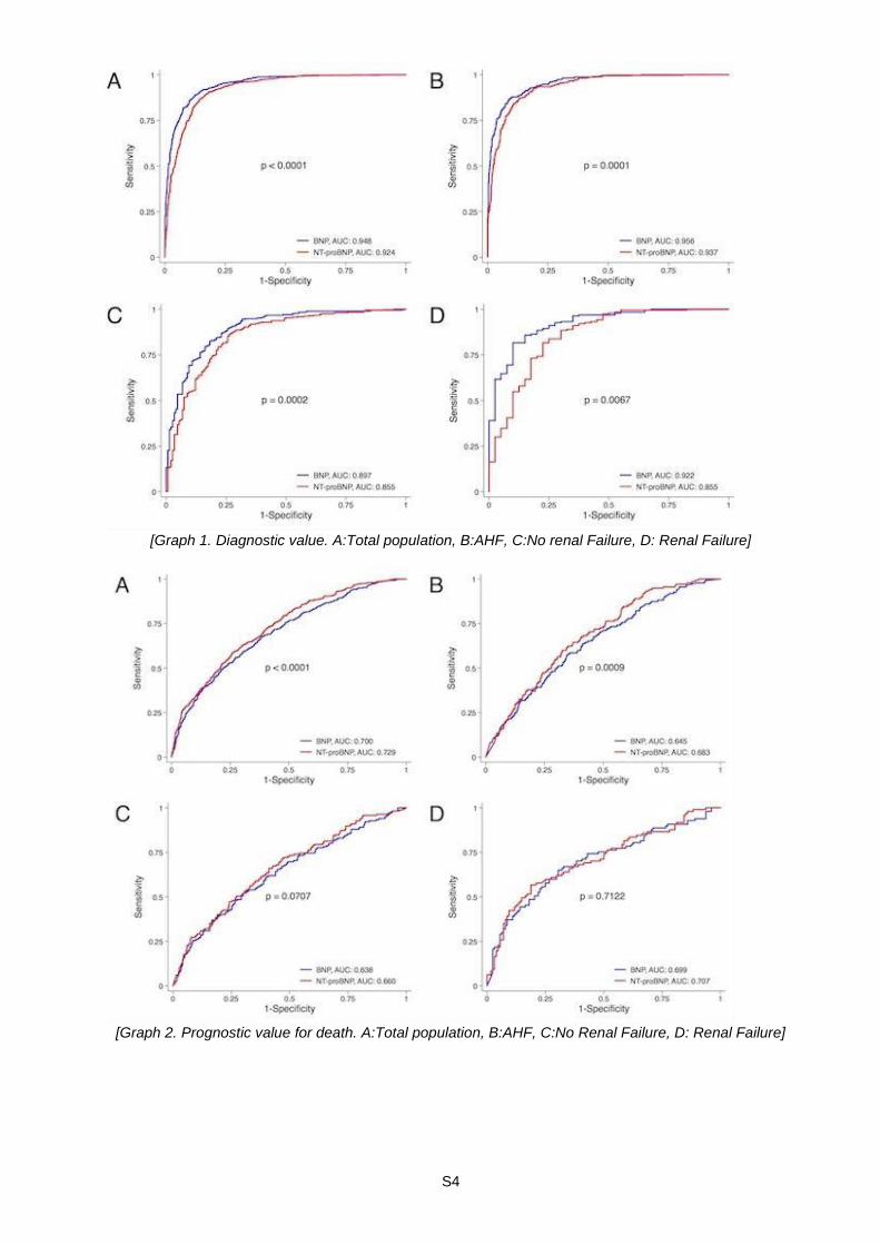
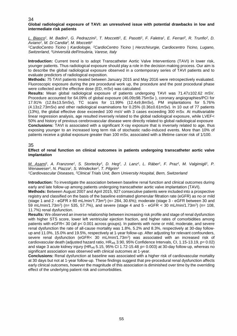
33 Direct comparison of B-type natriuretic peptide and N-terminal pro-B-type natriuretic peptide in patients with acute dyspnea Z. Sabti1, K. Murray2, N. Kozhuharov3, S. Osswald2, C. Müller2 1Kardiologie, 2Universitätsspital Basel, Universität Basel, 3Basel University Hospital, Basel, Switzerland Background: Acute heart failure (AHF) is the most common and further increasing cause of hospitalization in patients above the age of 45 years. Accurate and rapid diagnosis of AHF is crucial for the early initiation of effective treatment. Natriuretic peptides have shown to improve the diagnosis of AHF in patients presenting with acute dyspnea. We aimed to directly compare the diagnostic and prognostic accuracy of B-type natriuretic peptide (BNP) and N-terminal pro-BNP (NT-proBNP) in patients presenting with acute dyspnea and the possible confounding effect of renal dysfunction. Methods: We enrolled 1741 unselected patients presenting with acute dyspnea to the emergency departments of the University Hospital Basel and the University Hospital Zurich in a prospective diagnostic study. Two independent cardiologists using all information including follow-up adjudicated the final diagnosis. BNP and NT-proBNP plasma levels were determined at presentation. All-cause mortality during long-term follow-up was the primary prognostic end point. Results: AHF was the adjudicated final diagnosis in 936 patients (53.8%). Plasma concentrations of both BNP and NT-proBNP were significantly higher in AHF as compared to other causes of acute dyspnea (both p< 0.001). Diagnostic accuracy as quantified by the area under the ROC curve (AUC) was 0.948 (95% CI 0.938-0.957) for BNP as compared to 0.924 (95% CI 0.912-0.937) for NT-proBNP, p< 0.0001. The diagnostic superiority of BNP was consistent among all subgroups according to renal function (Graph 1). In contrast, prognostic accuracy for the prediction of death at 360 days tended to be higher for NT-proBNP (Graph 2). Conclusion: In patients with acute dyspnea, BNP and NT-proBNP at presentation both have very high diagnostic accuracy for AHF and moderate prognostic accuracy for death at 360 days. There is a numerically small, but statistically and possibly also clinically significant diagnostic superiority of BNP and prognostic superiority of NT-proBNP.
S4
[Graph 1. Diagnostic value. A:Total population, B:AHF, C:No renal Failure, D: Renal Failure]
[Graph 2. Prognostic value for death. A:Total population, B:AHF, C:No Renal Failure, D: Renal Failure]
S5
34 Global radiological exposure of TAVI: an unresolved issue with potential drawbacks in low and intermediate risk patients L. Biasco1, M. Badini1, G. Pedrazzini1, T. Moccetti1, E. Pasotti1, F. Faletra1, E. Ferrari2, R. Trunfio2, D. Aviano1, M. Di Candia3, M. Moccetti1 1CardioCentro Ticino | Kardiologie, 2CardioCentro Ticino | Herzchirurgie, Cardiocentro Ticino, Lugano, Switzerland, 3Università dell'Insubria, Varese, Italy Introduction: Current trend is to adopt Transcatheter Aortic Valve Interventions (TAVI) in lower risk, younger patients. Thus radiological exposure should play a role in the decision making process. Our aim is to describe the global radiological exposure observed in a contemporary series of TAVI patients and to evaluate predictors of radiological exposition. Methods: 75 TAVI patients treated between January 2015 and May 2016 were retrospectively evaluated. Fluoroscopic exposure during the pre procedural work up, the procedure and the post procedural phase were collected and the effective dose (ED, mSv) was calculated. Results: Mean global radiological exposure of patients undergoing TAVI was 71.47±102.62 mSV. Procedure accounted for 64.09% of global exposure (46.09±98.75mSv ), coronary angiographies/PCI for 17.91% (12.8±13.5mSv), TC scans for 11.99% (12.4±9,8mSv), PM implantations for 5.76% (4.13±2.73mSv) and other radiological examinations for 0.25% (0.36±0.61mSv). In 10 out of 77 patients (13%), the global effective dose exceeded 100 msV with 3 cases exceeding 300 mSv. At multivariable linear regression analysis, age resulted inversely related to the global radiological exposure, while LVEF< 50% and history of previous cerebrovascular disease were directly related to global radiological exposure Conclusions: TAVI is associated with a significant X-ray exposure that is inversely related to age, thus exposing younger to an increased long term risk of stochastic radio-induced events. More than 10% of patients receive a global exposure greater than 100 mSv, associated with a lifetime cancer risk of 1/100.
35 Effect of renal function on clinical outcomes in patients undergoing transcatheter aortic valve implantation M. Asami1, A. Franzone1, S. Stortecky1, D. Heg2, J. Lanz1, L. Räber1, F. Praz1, M. Valgimigli1, P. Wenaweser1, N. Piazza1, S. Windecker1, T. Pilgrim1 1Cardiovascular Diseases, 2Clinical Trials Unit, Bern University Hospital, Bern, Switzerland Introduction: To investigate the association between baseline renal function and clinical outcomes during early and late follow-up among patients undergoing transcatheter aortic valve implantation (TAVI). Methods: Between August 2007 and April 2015, 927 consecutive patients were included into a prospective registry and classified on the basis of the baseline estimated glomerular filtration rate (eGFR) as no or mild (stage 1 and 2 - eGFR ≥ 60 mL/min/1.73m2) (n= 284, 30.6%); moderate (stage 3 - eGFR between 30 and 59 mL/min/1.73m2) (n= 535, 57.7%), and severe (stage 4 and 5 - eGFR < 30 mL/min/1.73m2) (n= 108, 11.7%) renal dysfunction. Results: We observed an inverse relationship between increasing risk profile and stage of renal dysfunction with higher STS score, lower left ventricular ejection fraction, and higher rates of comorbidities among patients with eGFR< 30 (all p< 0.001 across groups). In patients with none or mild, moderate, and severe renal dysfunction the rate of all-cause mortality was 1.8%, 5.2% and 8.3%, respectively at 30-day follow-up and 11.0%, 15.0% and 19.5%, respectively at 1-year follow-up. After adjusting for relevant confounders, severe renal dysfunction (eGFR< 30 mL/min/1.73m2) was associated with an increased risk of cardiovascular death (adjusted hazard ratio, HRadj, 3.90, 95% Confidence Intervals, CI, 1.15-13.19, p= 0.02) and stage 3 acute kidney injury (HRadj 5.15, 95% CI 1.72-15.48, p= 0.003) at 30-day follow-up, whereas no significant association was observed with clinical outcomes at 1-year. Conclusions: Renal dysfunction at baseline was associated with a higher risk of cardiovascular mortality at 30 days but not at 1-year follow-up. These findings suggest that pre-procedural renal dysfunction affects early clinical outcomes, however the magnitude of this association is diminished over time by the overriding effect of the underlying patient risk and comorbidities.
S6
36 Baseline predictors of renal failure in TAVI: a sub analysis from the swiss TAVI Registry S. Obeid1, M. Melina Langfritz2, F. Nietlispach3, R. Binder4, A. Denegri3, M. Taramasso3, M. Moccetti5, G. Pedrazzini6, T. Moccetti6, F. Maisano3, T. Lüscher3 1Zurich Heart Center | Interventional Cardiology, University Hospital of Zürich, Zürich, 2Cardiology, 3Universitätsspital Zürich, 4Univerity of Zurich, Zurich, 5Cardiocentro Ticino, Lugano, 6Cardiocentro Ticino, Zurich, Switzerland Background: Acute Kidney Injury (AKI) post Trans Aortic Valve Implantation (TAVI) is associated with worse short and long term outcomes. We sought to identify significant baseline predictors of AKI and establish a high risk group within patients enrolled in the multicenter SWISS-TAVI cohort. Methods and results: After excluding patients on hemodialysis, a total of 526 patients were included in our analysis, of which 335 patients underwent TAVI at the university hospital of Zurich and 191 at Cardio Centro Ticino. Within the first week post valve implantation, 9.5% (n=50) of patients had developed AKI as defined by the KDIGO criteria. There was a significantly higher prevalence of Diabetes Mellitus (44.9% vs 27.9%, p=0.020) and Chronic Kidney Disease CKD≥4 (26% vs 13.9%, p=0.035) in patients who developed AKI as compared to those who did not, respectively. However there was no difference in age, gender, BMI, history of dyslipidemia and hypertension between the groups. In a multivariable binary regression analysis, diabetics were at a 1.9 fold increased risk of developing AKI [OR 1.902, 95% CI [1.018 - 3.553], p=0.044], as well as those with high creatinine levels, by a factor of 1.6 with every rise of 1 mg/dl at baseline [OR 1.605, 95% CI [1.111 - 2.319], p=0.012]. To further substantiate our findings we re-evaluated for predictors within the diabetic (n=155, 29.5%) and non-diabetic populations (n=370, 70.5%) where AKI developed in 14.2 %( n=22) and 7.3 % (n=27) respectively. Interestingly enough in non-diabetics, none of baseline Glomerular filtration rate, CKD Grade, STS, Euroscore, ACEF score or even procedural contrast usage were predictors of AKI .On the other hand within the diabetic population an elevation by 1mg/dl in baseline creatinine was an independent predictor of developing kidney injury [OR 2.061, 95% CI [1.154 - 3.683], p=0.015], where more than one third (41%) of the patients who developed AKI in this population had a CKD stage ≥4. Conclusion: Identifying patients at risk of developing AKI post TAVI is the first of many steps to help implement preventative measures. Diabetics with KDIGO CKD stage≥4 constitute a high risk group within the TAVI patients, where the renal guard system could play a major protective role especially when undergoing concomitant procedures. AKI in Non diabetics on the other hand seems to be more procedure related and less predictable by baseline characteristics.
37 Minimal use of contrast media in patients with severe chronic kidney disease undergoing transcatheter aortic valve implantation including non-enhanced magnetic resonance imaging for pre-procedural planning M. Brinkert1, J. Fornaro2, R. Buhmann2, V. Weberndörfer1, F. Cuculi1, R. Kobza1, S. Toggweiler1 1Heart Center Lucerne, 2Department of Radiology, Luzerner Kantonsspital, Lucerne, Switzerland Introduction: Patients with severe chronic kidney disease (CKD) are at high risk for contrast induced nephropathy and other complications following transcatheter aortic valve implantation (TAVI). This study investigated the feasibility and safety of an integrated approach aiming to minimize the need for contrast media for TAVI work-up and valve implantation including magnetic resonance imaging (MR) for pre-procedural planning. Methods: Between February 2016 and January 2017, a total of 122 patients underwent TAVI at the Luzerner Kantonsspital. Of those, 14 (11%) had severe chronic kidney disease (GFR < 30 ml/min/m2). In such patients, work-up was performed with non-enhanced MR for annular and iliofemoral assessment. In addition, use of contrast media was minimized for the pre-procedural coronary angiography and for TAVI. Results: Baseline GFR was 24 ± 6 and 58 ± 22 ml/min/m2 in patients with and without severe CKD, respectively (p < 0.01). Patients with severe CKD had a higher STS score (8.0 ± 2.7 % vs. 3.4 ± 1.9%, p < 0.01). Pre-procedural coronary angiography was performed with 42 ± 22 ml of contrast media in patients with severe CKD and with 81 ± 42 ml in all others (p < 0.01). Patients with severe CKD underwent native MR. All other patients were screened with an ECG gated CT using 64 ± 15 ml of contrast media. TAVI was performed with 25 ± 18 ml and 114 ± 53 ml of contrast media, respectively (p < 0.01). Valves implanted were the ACURATE neo (n = 76, 62%), the SAPIEN 3 (n = 39, 32%), and the Evolut R (n = 7, 6%). Following TAVI, paravalvular regurgitation was none/mild in 14/14 (100%) with severe CKD and in 106/108 (98%) without (p = 0.40). Median duration of hospitalization was longer in patients with severe CKD than in those without (11 days vs. 6 days, p < 0.01). At 30 days, there were no strokes, and survival was 100% and 98%, respectively. Stage 2 or 3 acute kidney injury had occurred in 1 (7%) vs. 3 (2.8%, p = 0.39). Early safety and clinical efficacy at 30 days were similar (86% vs 92%, p= 0.61 and 100% vs 95%, p= 1.00).
S7
Conclusion: In patients with severe CKD, we were able to reduce the total amount of contrast media required for pre-procedural planning and TAVI by 70% from 242 ± 80 ml to 73 ± 39 ml. This study, for the first time, also shows that non-enhanced MR may be effectively utilized for pre-procedural annular and iliofemoral assessment, and selection of prosthesis size.
38 Hospital length of stay after transcatheter aortic valve implantation: what are the baseline predictors and the peri-procedural determinants? D. Adamopoulos1, A. Frei1, M.-J. Licker2, C. Ellenberger2, H. Müller1, M. Roffi1, S. Noble1 1Cardiology, 2Anesthesiology, Geneva University Hospitals, Geneva, Switzerland Introduction: Early hospital discharge is generally associated with a better patient experience and higher quality of care. We sought to identify baseline features and peri-procedural variables affecting hospital length of stay (LoS) in patients undergoing transcatheter aortic valve replacement (TAVI). Methods: Data on patients undergoing TAVI procedure were collected prospectively in a single centre study from November 2009 to October 2016. All participants underwent a full clinical, laboratory and echocardiographic work-up as well as a systematic frailty assessment at baseline and post TAVI. The peri-procedural complications were also assessed. The study population was divided into 2 groups according to LoS (≤7 days and > 7 days: early and late discharge group, accordingly). Results: The final cohort comprised 222 consecutive patients (LoS 10.2±6.4 days, 42.3% [n=94] early vs 57.7% [n=128] late discharge group). Patients in the late discharge group exhibited longer gait speed time (0.84±0.15 log_sec vs 0.79±0.14 log_sec, p=0.03), poorer handgrip strength (18.5±7.6 kg vs 21.2±8.2 kg, p=0.04) and lower serum albumin levels (35±6 g/lt vs 38±5 g/lt, p=0.003) compared to the early discharge group pointing to a significant relation between baseline frailty status and LoS. Longer LoS was strongly associated with baseline history of peripheral artery disease [PAD] (18.8% vs 6.4%, p=0.008), urgent hospital admission (24.2% vs 6.4%, p< 0.001), impaired baseline renal function (glomerular filtration rate 51.6±16.8 ml/min vs 55.9±15.6 ml/min, p=0.05) and lower forced expiratory volume1sec (1.5±05 lt vs 1.7±0.5 lt, p=0.02). Definitive pacemaker implantation (O.R.:2.4 95%C.I.:1.2-4.9, p=0.02), acute renal failure (O.R.:4.0, 95%C.I.:1.1-14.3, p=0.02) and bleeding complications (O.R.:3.2 95%C.I.:1.2-8.2, p=0.02) were strongly associated with late discharge. In multivariate analysis, gait speed time [log_sec] (O.R: 11.8, 95%C.I.: 1.1-142, p=0.05), urgent hospital admission (O.R:14.8, 95%C.I.: 2.0-118, p=0.01) and positive history of PAD (O.R:8.1, 95%C.I.:1.7-38.1, p=0.009) were identified as independent baseline determinants of late hospital discharge for patients after TAVI. Conclusion: Slow gait speed test, urgent hospital admission and positive history of PAD appear to be independent predictors of hospital LoS. This provides novel insights for a more effective in-hospital management of patients undergoing TAVI procedure.
S8
Abstract Session - Structural heart, clinical cases 54 Leaflet thrombosis following transcatheter mitral valve replacement F. Franzeck1, M. Buser1, W. Koch2, M. Taramasso3, P.K. Haager1 1Department of Cardiology, Kantonsspital St. Gallen, 2Kardiologiepraxis Notkerstrasse, St. Gallen, 3Department of Cardiovascular Surgery, University Hospital Zurich, Zurich, Switzerland Introduction: After mitral valve repair, degeneration of the native mitral valve may cause stenosis or regurgitation. The feasibility of transcatheter mitral valve-in-ring implantation is suggested by several reports in prohibitive surgical risk. Transcatheter valves specifically designed for the mitral position are being developed but are not readily available and concerns about risk of thrombosis have been raised. Here, we present a case of a successful transcatheter mitral valve-in-ring replacement with a balloon-expandable transcatheter aortic valve (Edwards Sapien 3, 26mm) over a transvenous transseptal approach with subsequent symptomatic valve thrombosis on 20 mg rivaroxaban. Case presentation: A 73-year-old female with previous mitral valve annuloplasty (Physioring II 30mm) in 11/11 presented with congestive heart failure (NYHA III) in 12/15. TTE showed a LVEF of 65% and a severe mitral regurgitation due to flail leaflet of segment A2, the right ventricle was dilated and showed a reduced systolic function. Heart catheterization demonstrated mixed pulmonary hypertension (mean 58mmHg). The patient declined conventional open surgery (Euroscore II 14%). Multiplanar reconstruction from 3D TEE and cardiac CT showed a circumference of 74mm (435mm2) of the circular mitral ring. Therefore - a transcatheter mitral valve implantation with an Edwards Sapien 3 26mm valve was discussed in the heart team. Procedure was carried out via a transvenous transseptal approach under rapid ventricular pacing. TTE before discharge showed a transmitral mean gradient of 5mmHg. Rivaroxaban 20mg (already established for unprovoked pulmonary embolism and suspicion for thrombophilia) was continued and aspirin added for 3 months. After significant initial improvement in functional class (NYHA I), there was a progressive decline (NYHA II) from 08/16 on. TEE 11/16 showed a diffuse thickening with reduced mobility of 2 leaflets and a transmitral mean gradient of 11mmHg confirming valve thrombosis. Rivaroxaban was subsequently switched to phenprocoumon (target INR 3.0-3.5). Functional status improved again (NYHA I), TEE 01/17 showed a complete resolution of thrombosis and a decrease in mean gradient to 5mmHg. Conclusion: The duration and choice of anticoagulant after transcatheter mitral valve replacement remains unclear. Anticoagulation with rivaroxaban only may be insufficient. Transcatheter bioprosthesis in mitral position may have a higher thrombotic risk than in aortic position.
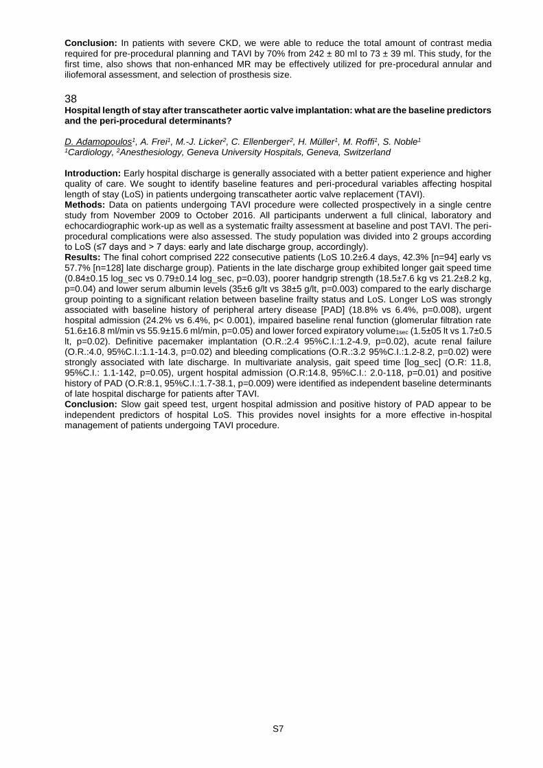
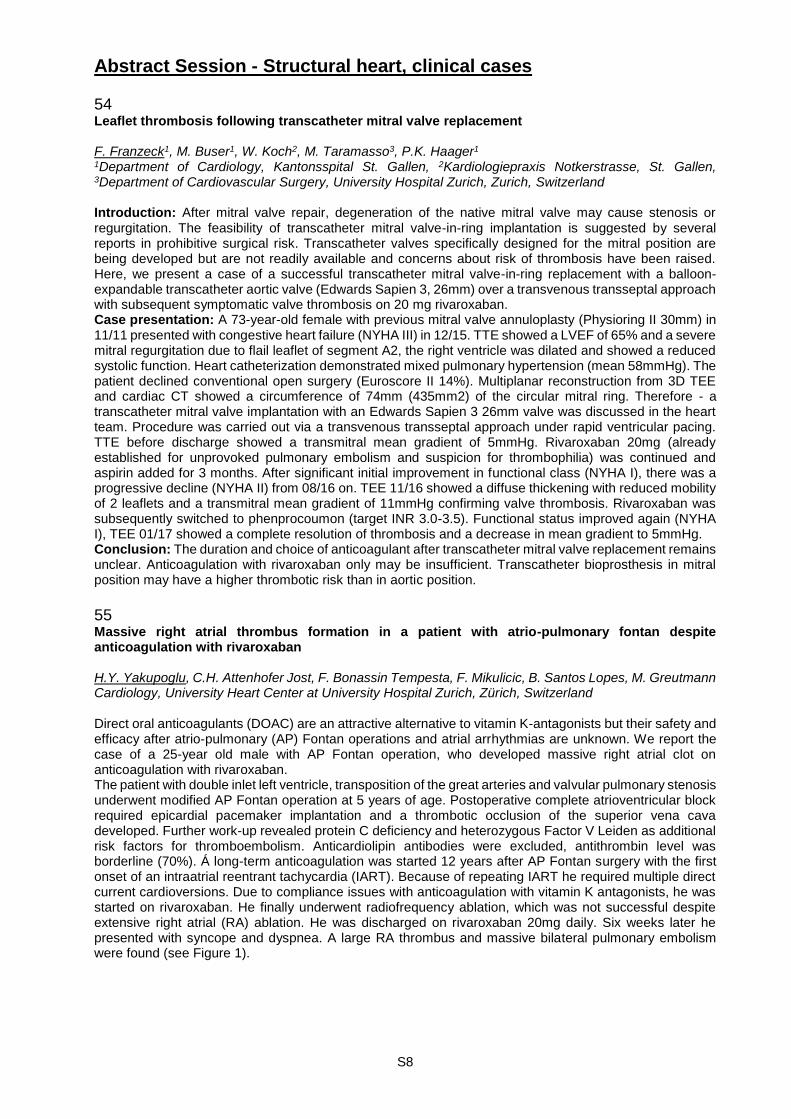
55 Massive right atrial thrombus formation in a patient with atrio-pulmonary fontan despite anticoagulation with rivaroxaban H.Y. Yakupoglu, C.H. Attenhofer Jost, F. Bonassin Tempesta, F. Mikulicic, B. Santos Lopes, M. Greutmann Cardiology, University Heart Center at University Hospital Zurich, Zürich, Switzerland Direct oral anticoagulants (DOAC) are an attractive alternative to vitamin K-antagonists but their safety and efficacy after atrio-pulmonary (AP) Fontan operations and atrial arrhythmias are unknown. We report the case of a 25-year old male with AP Fontan operation, who developed massive right atrial clot on anticoagulation with rivaroxaban. The patient with double inlet left ventricle, transposition of the great arteries and valvular pulmonary stenosis underwent modified AP Fontan operation at 5 years of age. Postoperative complete atrioventricular block required epicardial pacemaker implantation and a thrombotic occlusion of the superior vena cava developed. Further work-up revealed protein C deficiency and heterozygous Factor V Leiden as additional risk factors for thromboembolism. Anticardiolipin antibodies were excluded, antithrombin level was borderline (70%). Á long-term anticoagulation was started 12 years after AP Fontan surgery with the first onset of an intraatrial reentrant tachycardia (IART). Because of repeating IART he required multiple direct current cardioversions. Due to compliance issues with anticoagulation with vitamin K antagonists, he was started on rivaroxaban. He finally underwent radiofrequency ablation, which was not successful despite extensive right atrial (RA) ablation. He was discharged on rivaroxaban 20mg daily. Six weeks later he presented with syncope and dyspnea. A large RA thrombus and massive bilateral pulmonary embolism were found (see Figure 1).
S9
[Fig. 1 Computed tomography with thrombus in the right atrium (red) and pulmonary embolies (yellow)]
[Fig 2. Echocardiographic 4 chamber-view on the left (x-plane on the right) with thrombus (arrow)]
Anticoagulation was switched to unfractionated heparin, then changed to high-dose low molecular weight heparin twice daily aiming peak anti FXa-levels > 0.6 IU/ml. After two weeks he was started on phenprocoumon (target INR-levels 2.5-3.5). Despite optimal INR levels, the size of the RA clot decreased only slightly, although pulmonary emboli resolved completely after 4 months. One year later the patient underwent total cavo-pulmonary conversion operation with surgical removal of the residual clot. This case highlights the high propensity for atrial clot formation after the AP Fontan operation, particularly in the presence of additional risk factors, such as atrial arrhythmias, coagulation disorders and ablation procedures. It remains questionable whether DOACs are a safe alternative to vitamin K antagonists in this setting. In our own clinical practice we currently refrain from using DOACs after AP Fontan operations.
S10
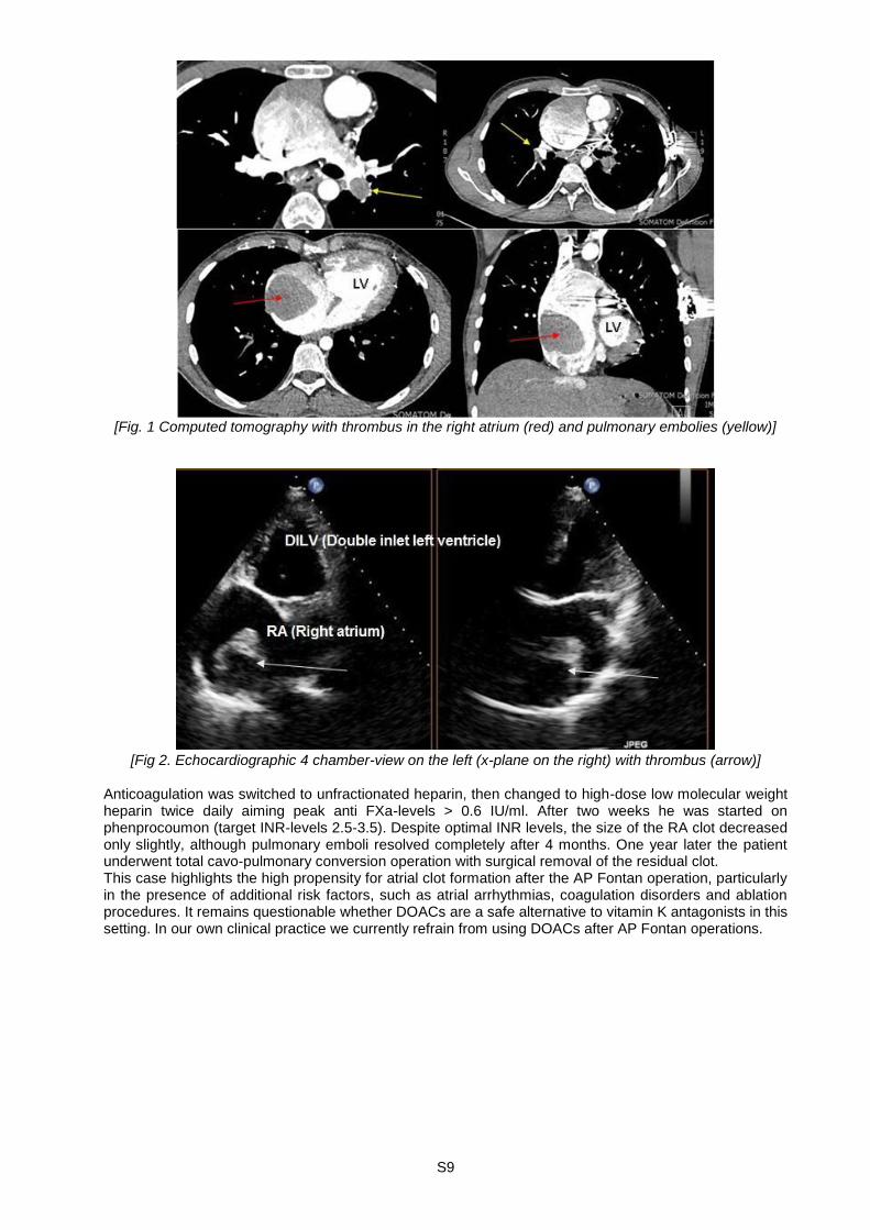
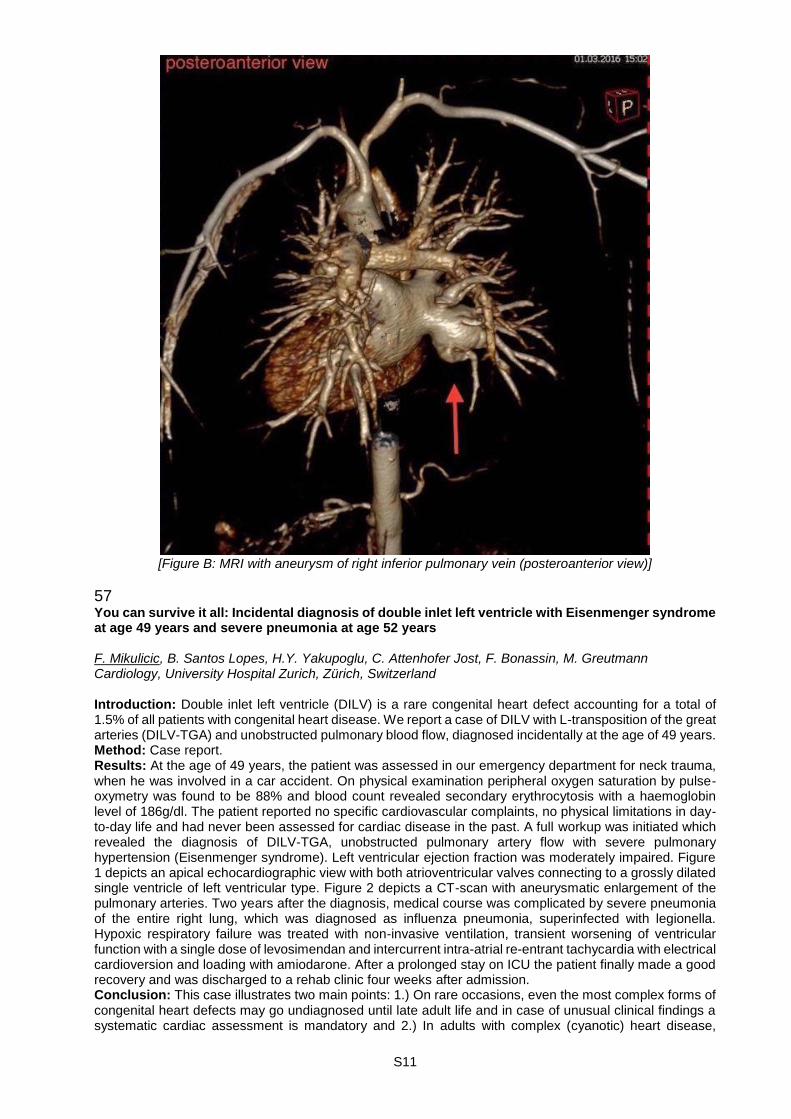
56 A case of pulmonary vein aneurysm K. Sotiropoulos, R. Kobza Herzzentrum, Luzerner Kantonsspital, Luzern, Switzerland Introduction: Ectopic atrial tachycardia (EAT) is common in older patients with structural heart disease, but rare in healthy young subjects. The triggering area is often near the crista terminalis in the right atrium or at the base of the pulmonary veins in the left atrium. Methods: A 30-year-old pregnant healthy woman presents with palpitations that have become more frequent and long-lasting since the beginning of the pregnancy. Except for being an ex-smoker, her health history is uneventful. The clinical examination is free of any pathological findings. The thyroid function is normal. The echocardiogram shows a normal LV function, pulmonary pressure and no valvulopathies. A bicycle ergometry triggers a supraventricular tachycardia up to 270 bpm. Cardioversion to sinus rhythm is achieved with Valsalva maneuver and a pill-in-the-pocket (metoprolol) as preventive therapy is prescribed. The electrophysiology study (EPS) is performed at a later time because of the pregnancy. Results: The first EPS detects an EAT treated with successful ablation of a focus at the opening of the right inferior pulmonary vein. 3 years later, the patient develops once again EATs and paroxysmal atrial fibrillation. A second EPS with pulmonary veins isolation is programmed. Before the EPS, an angio-CT for mapping is performed and discovers an aneurysm of the right inferior pulmonary vein with a diameter of 25 mm (figure A). Finally, one year later, because of a relapse of atrial tachycardia and atrial flutter, a third EPS with RF-ablation is performed. Since then, the patient is oligosymptomatic with atrial extrasystoles and short atrial “runs”. An oral anticoagulation is maintained. The last MRI control (figure B) shows a stability of the size of the aneurysm 4 years after the diagnosis. Conclusion: We may hypothesize that the origin of the EAT in our patient is the pulmonary vein aneurysm. There are only very few cases of pulmonary vein aneurysm reported in the literature, and no definite relation to EATs has been established. The case shows that in young patients, after exclusion of structural heart disease, a persistent atrial tachyarrhythmia should motivate further diagnostic investigations, which may reveal rare causes of the arrhythmia.
[Figure A: CT with pulmonary aneurysm of the right inferior pulmonary vein]
S11
[Figure B: MRI with aneurysm of right inferior pulmonary vein (posteroanterior view)]
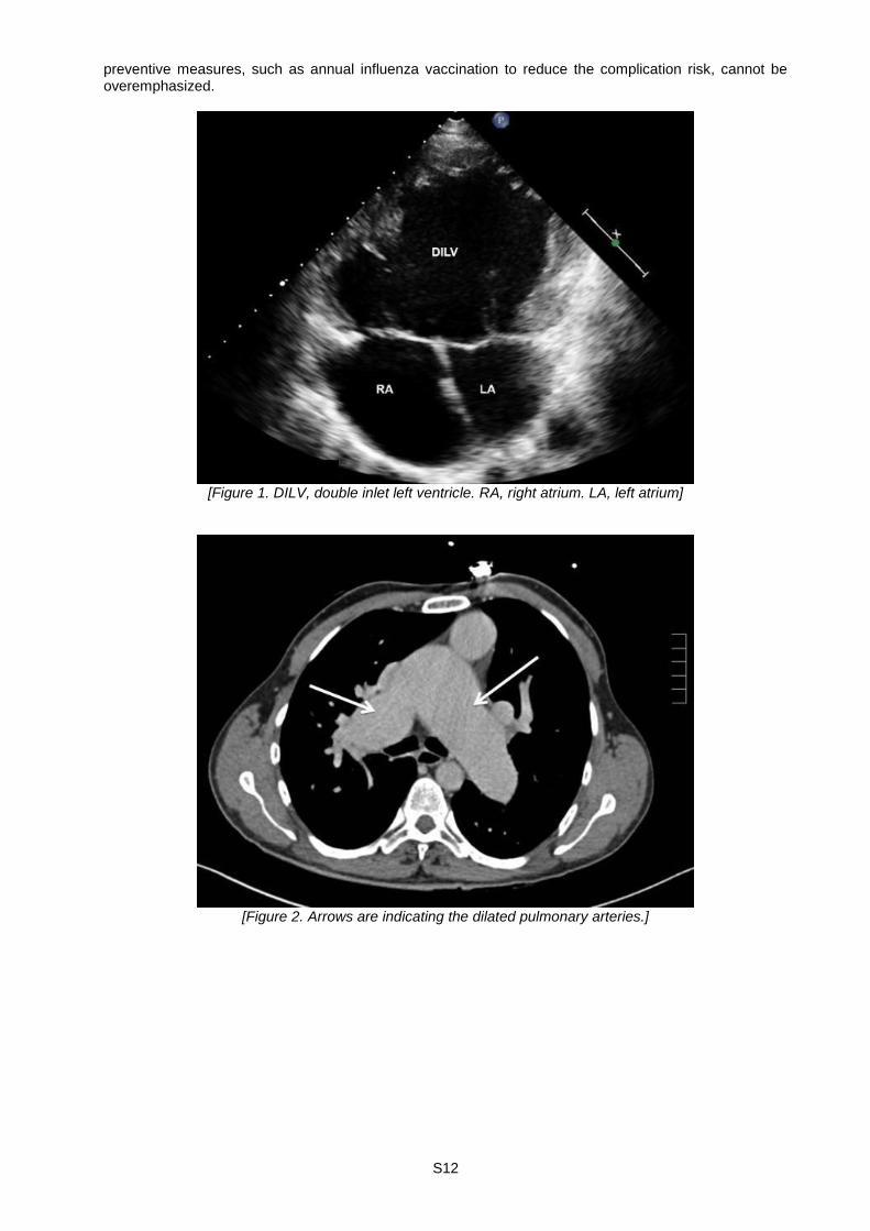
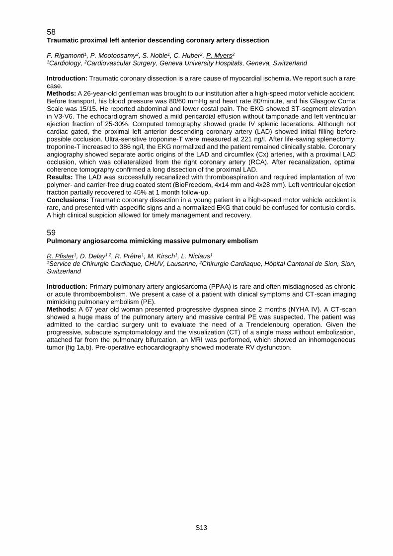
57 You can survive it all: Incidental diagnosis of double inlet left ventricle with Eisenmenger syndrome at age 49 years and severe pneumonia at age 52 years F. Mikulicic, B. Santos Lopes, H.Y. Yakupoglu, C. Attenhofer Jost, F. Bonassin, M. Greutmann Cardiology, University Hospital Zurich, Zürich, Switzerland Introduction: Double inlet left ventricle (DILV) is a rare congenital heart defect accounting for a total of 1.5% of all patients with congenital heart disease. We report a case of DILV with L-transposition of the great arteries (DILV-TGA) and unobstructed pulmonary blood flow, diagnosed incidentally at the age of 49 years. Method: Case report. Results: At the age of 49 years, the patient was assessed in our emergency department for neck trauma, when he was involved in a car accident. On physical examination peripheral oxygen saturation by pulse-oxymetry was found to be 88% and blood count revealed secondary erythrocytosis with a haemoglobin level of 186g/dl. The patient reported no specific cardiovascular complaints, no physical limitations in day-to-day life and had never been assessed for cardiac disease in the past. A full workup was initiated which revealed the diagnosis of DILV-TGA, unobstructed pulmonary artery flow with severe pulmonary hypertension (Eisenmenger syndrome). Left ventricular ejection fraction was moderately impaired. Figure 1 depicts an apical echocardiographic view with both atrioventricular valves connecting to a grossly dilated single ventricle of left ventricular type. Figure 2 depicts a CT-scan with aneurysmatic enlargement of the pulmonary arteries. Two years after the diagnosis, medical course was complicated by severe pneumonia of the entire right lung, which was diagnosed as influenza pneumonia, superinfected with legionella. Hypoxic respiratory failure was treated with non-invasive ventilation, transient worsening of ventricular function with a single dose of levosimendan and intercurrent intra-atrial re-entrant tachycardia with electrical cardioversion and loading with amiodarone. After a prolonged stay on ICU the patient finally made a good recovery and was discharged to a rehab clinic four weeks after admission. Conclusion: This case illustrates two main points: 1.) On rare occasions, even the most complex forms of congenital heart defects may go undiagnosed until late adult life and in case of unusual clinical findings a systematic cardiac assessment is mandatory and 2.) In adults with complex (cyanotic) heart disease,
S12
preventive measures, such as annual influenza vaccination to reduce the complication risk, cannot be overemphasized.
[Figure 1. DILV, double inlet left ventricle. RA, right atrium. LA, left atrium]
[Figure 2. Arrows are indicating the dilated pulmonary arteries.]
S13
58 Traumatic proximal left anterior descending coronary artery dissection F. Rigamonti1, P. Mootoosamy2, S. Noble1, C. Huber2, P. Myers2 1Cardiology, 2Cardiovascular Surgery, Geneva University Hospitals, Geneva, Switzerland Introduction: Traumatic coronary dissection is a rare cause of myocardial ischemia. We report such a rare case. Methods: A 26-year-old gentleman was brought to our institution after a high-speed motor vehicle accident. Before transport, his blood pressure was 80/60 mmHg and heart rate 80/minute, and his Glasgow Coma Scale was 15/15. He reported abdominal and lower costal pain. The EKG showed ST-segment elevation in V3-V6. The echocardiogram showed a mild pericardial effusion without tamponade and left ventricular ejection fraction of 25-30%. Computed tomography showed grade IV splenic lacerations. Although not cardiac gated, the proximal left anterior descending coronary artery (LAD) showed initial filling before possible occlusion. Ultra-sensitive troponine-T were measured at 221 ng/l. After life-saving splenectomy, troponine-T increased to 386 ng/l, the EKG normalized and the patient remained clinically stable. Coronary angiography showed separate aortic origins of the LAD and circumflex (Cx) arteries, with a proximal LAD occlusion, which was collateralized from the right coronary artery (RCA). After recanalization, optimal coherence tomography confirmed a long dissection of the proximal LAD. Results: The LAD was successfully recanalized with thromboaspiration and required implantation of two polymer- and carrier-free drug coated stent (BioFreedom, 4x14 mm and 4x28 mm). Left ventricular ejection fraction partially recovered to 45% at 1 month follow-up. Conclusions: Traumatic coronary dissection in a young patient in a high-speed motor vehicle accident is rare, and presented with aspecific signs and a normalized EKG that could be confused for contusio cordis. A high clinical suspicion allowed for timely management and recovery.
59 Pulmonary angiosarcoma mimicking massive pulmonary embolism R. Pfister1, D. Delay1,2, R. Prêtre1, M. Kirsch1, L. Niclaus1 1Service de Chirurgie Cardiaque, CHUV, Lausanne, 2Chirurgie Cardiaque, Hôpital Cantonal de Sion, Sion, Switzerland Introduction: Primary pulmonary artery angiosarcoma (PPAA) is rare and often misdiagnosed as chronic or acute thromboembolism. We present a case of a patient with clinical symptoms and CT-scan imaging mimicking pulmonary embolism (PE). Methods: A 67 year old woman presented progressive dyspnea since 2 months (NYHA IV). A CT-scan showed a huge mass of the pulmonary artery and massive central PE was suspected. The patient was admitted to the cardiac surgery unit to evaluate the need of a Trendelenburg operation. Given the progressive, subacute symptomatology and the visualization (CT) of a single mass without embolization, attached far from the pulmonary bifurcation, an MRI was performed, which showed an inhomogeneous tumor (fig 1a,b). Pre-operative echocardiography showed moderate RV dysfunction.

S14
[CT-scan (A) and MRI (B). Whites arrows show the rumor inside the pulmonary trunk]
Results: Surgery was performed through median sternotomy. Under extra-corporeal circulation (ECC) and cardiac arrest, the pulmonary trunk was opened. The tumor was attached to the pulmonary valve, inside the right ventricular outflow tract, infiltrating at least partially the pulmonary trunk (fig 2a,b). Complete macroscopic resection, including the distal right outflow tract and the pulmonary valve up to the bifurcation, and surgical reconstruction, by implanting a 21mm Contegra tube, was realized. Weaning of ECC was uneventful, apart from a transitory right ventricular dysfunction requiring 3 days inotropic support.
[Perioperative view. picture B shows the attachment to the pulmonary valve]
Conclusions: PPAA is a rare disorder, with relative poor prognosis and limited survival (post surgery survival of 6 months to 2 years has been reported) due to local tumor recurrence or metastasis. Surgical resection is the treatment of choice for at least tumor mass reduction. Adjuvant treatment strategies are limited, however should certainly be considered in cases of incomplete tumor resection. However symptomatic intra-cardiac tumor localization might require, as in the present case, an emergent surgical approach, despite uncertain diagnosis, due to the local expansion, responsible for obstructive of compressive cardiac symptoms and the acute exposure to cardiac death.
S15
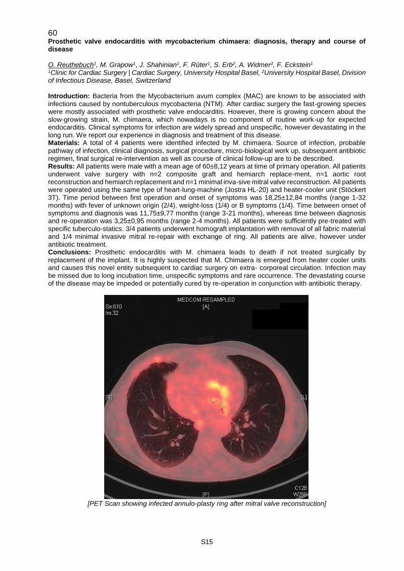
60 Prosthetic valve endocarditis with mycobacterium chimaera: diagnosis, therapy and course of disease O. Reuthebuch1, M. Grapow1, J. Shahinian1, F. Rüter1, S. Erb2, A. Widmer2, F. Eckstein1 1Clinic for Cardiac Surgery | Cardiac Surgery, University Hospital Basel, 2University Hospital Basel, Division of Infectious Disease, Basel, Switzerland Introduction: Bacteria from the Mycobacterium avum complex (MAC) are known to be associated with infections caused by nontuberculous mycobacteria (NTM). After cardiac surgery the fast-growing species were mostly associated with prosthetic valve endocarditis. However, there is growing concern about the slow-growing strain, M. chimaera, which nowadays is no component of routine work-up for expected endocarditis. Clinical symptoms for infection are widely spread and unspecific, however devastating in the long run. We report our experience in diagnosis and treatment of this disease. Materials: A total of 4 patients were identified infected by M. chimaera. Source of infection, probable pathway of infection, clinical diagnosis, surgical procedure, micro-biological work up, subsequent antibiotic regimen, final surgical re-intervention as well as course of clinical follow-up are to be described. Results: All patients were male with a mean age of 60±8,12 years at time of primary operation. All patients underwent valve surgery with n=2 composite graft and hemiarch replace-ment, n=1 aortic root reconstruction and hemiarch replacement and n=1 minimal inva-sive mitral valve reconstruction. All patients were operated using the same type of heart-lung-machine (Jostra HL-20) and heater-cooler unit (Stöckert 3T). Time period between first operation and onset of symptoms was 18,25±12,84 months (range 1-32 months) with fever of unknown origin (2/4), weight-loss (1/4) or B symptoms (1/4). Time between onset of symptoms and diagnosis was 11,75±9,77 months (range 3-21 months), whereas time between diagnosis and re-operation was 3,25±0,95 months (range 2-4 months). All patients were sufficiently pre-treated with specific tuberculo-statics. 3/4 patients underwent homograft implantation with removal of all fabric material and 1/4 minimal invasive mitral re-repair with exchange of ring. All patients are alive, however under antibiotic treatment. Conclusions: Prosthetic endocarditis with M. chimaera leads to death if not treated surgically by replacement of the implant. It is highly suspected that M. Chimaera is emerged from heater cooler units and causes this novel entity subsequent to cardiac surgery on extra- corporeal circulation. Infection may be missed due to long incubation time, unspecific symptoms and rare occurrence. The devastating course of the disease may be impeded or potentially cured by re-operation in conjunction with antibiotic therapy.
[PET Scan showing infected annulo-plasty ring after mitral valve reconstruction]
S16
Rapid Fire Abstract Session - Prediction, Prognosis and Stratification 113 Elevated blood pressure in children referred to a pediatric cardiology clinic: frequency and management S. Perrenoud1, A. Chiolero2, N. Sekarski3, Y. Mivelaz3 1Centre Hospitalier Universitaire Vaudois (CHUV), 2IUMSP, Centre Hospitalier Universitaire Vaudois (CHUV), 3Département Femme-Mère-Enfant, Centre Hospitalier Universitaire Vaudois, Lausanne, Switzerland Introduction: Elevated blood pressure (BP), a major risk factor for cardiovascular diseases, might begin in childhood and tracks overtime. However, frequency and management of elevated BP is not well described among pediatrics outpatients. Therefore, we planned to: 1) Establish the frequency of elevated BP in children referred to a cardiology clinic; 2) Determine the association with the diagnosis of an heart condition and the proportion of cases reported in the medical report and for which the cardiologist made a management proposal. Method: We performed a retrospective study of BP measurements of all outpatients having had an echocardiographic exam between 2005 to 2014 at the Cardiology Unit of the Lausanne university hospital (CHUV). BP values, demographic and anthropometric data from children 1 to 18 years old seen at the outpatient clinic were extracted. BP values were expressed in percentiles according to international references. Elevated BP was defined as a systolic or diastolic BP>95th percentile. Medical reports of an approximately 10% sample of children with elevated BP were reviewed to assess the diagnosis of a heart condition, if elevated BP was reported and if any management was proposed. Results: Among 10´779 outpatient visits (from 4'829 children; 57% of boys, mean age: 8.8 years, SD: 4.64), an elevated BP was found in 1799 (16.7%). In the sample of 222 children with elevated BP, 163 (73%) had a cardiac condition. An elevated BP was reported in the medical report in 15.3% of all cases (9.8% of cases with and 30.0% without a cardiac condition, respectively). When an elevated BP was reported, a management was proposed in 82.4% of cases. Conclusion: The frequency of elevated BP at a single visit in children referred to the cardiology clinic at the CHUV is close to the proportion found in the general population. Reporting of elevated BP in medical reports is relatively low, and three times lower in children with a cardiac condition. In case of elevated BP, cardiologists often make management recommendation. However, the clinical signification and the appropriate management of elevated BP at a single visit remain to be established.
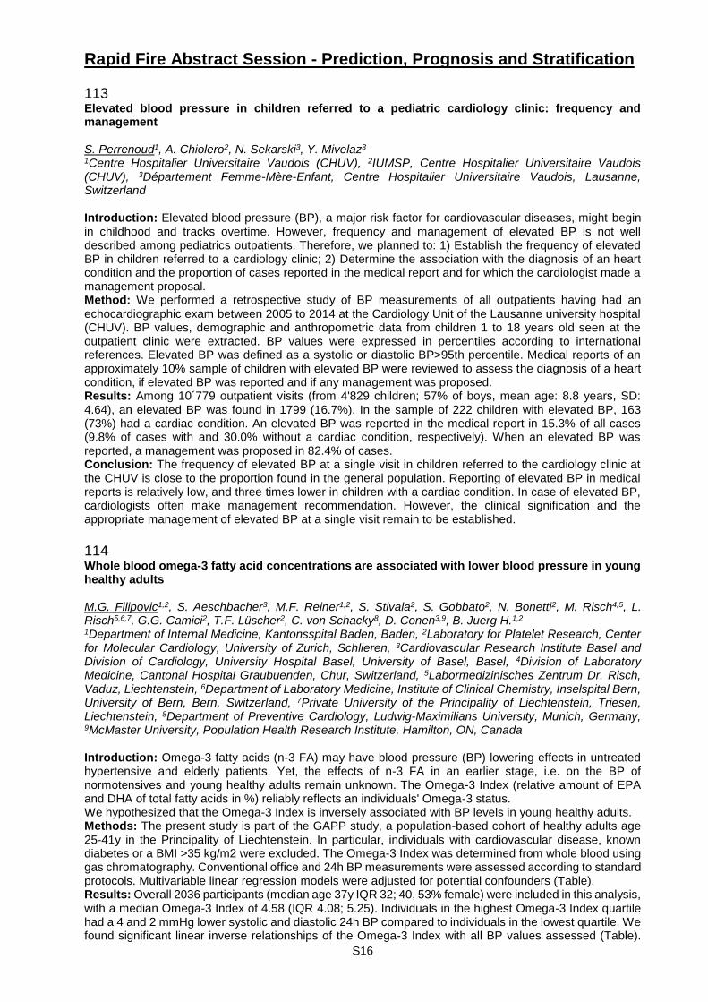
114 Whole blood omega-3 fatty acid concentrations are associated with lower blood pressure in young healthy adults M.G. Filipovic1,2, S. Aeschbacher3, M.F. Reiner1,2, S. Stivala2, S. Gobbato2, N. Bonetti2, M. Risch4,5, L. Risch5,6,7, G.G. Camici2, T.F. Lüscher2, C. von Schacky8, D. Conen3,9, B. Juerg H.1,2 1Department of Internal Medicine, Kantonsspital Baden, Baden, 2Laboratory for Platelet Research, Center for Molecular Cardiology, University of Zurich, Schlieren, 3Cardiovascular Research Institute Basel and Division of Cardiology, University Hospital Basel, University of Basel, Basel, 4Division of Laboratory Medicine, Cantonal Hospital Graubuenden, Chur, Switzerland, 5Labormedizinisches Zentrum Dr. Risch, Vaduz, Liechtenstein, 6Department of Laboratory Medicine, Institute of Clinical Chemistry, Inselspital Bern, University of Bern, Bern, Switzerland, 7Private University of the Principality of Liechtenstein, Triesen, Liechtenstein, 8Department of Preventive Cardiology, Ludwig-Maximilians University, Munich, Germany, 9McMaster University, Population Health Research Institute, Hamilton, ON, Canada Introduction: Omega-3 fatty acids (n-3 FA) may have blood pressure (BP) lowering effects in untreated hypertensive and elderly patients. Yet, the effects of n-3 FA in an earlier stage, i.e. on the BP of normotensives and young healthy adults remain unknown. The Omega-3 Index (relative amount of EPA and DHA of total fatty acids in %) reliably reflects an individuals' Omega-3 status. We hypothesized that the Omega-3 Index is inversely associated with BP levels in young healthy adults. Methods: The present study is part of the GAPP study, a population-based cohort of healthy adults age 25-41y in the Principality of Liechtenstein. In particular, individuals with cardiovascular disease, known diabetes or a BMI >35 kg/m2 were excluded. The Omega-3 Index was determined from whole blood using gas chromatography. Conventional office and 24h BP measurements were assessed according to standard protocols. Multivariable linear regression models were adjusted for potential confounders (Table). Results: Overall 2036 participants (median age 37y IQR 32; 40, 53% female) were included in this analysis, with a median Omega-3 Index of 4.58 (IQR 4.08; 5.25). Individuals in the highest Omega-3 Index quartile had a 4 and 2 mmHg lower systolic and diastolic 24h BP compared to individuals in the lowest quartile. We found significant linear inverse relationships of the Omega-3 Index with all BP values assessed (Table).
S17
Per 1-unit increase in log-transformed Omega-3 Index fully adjusted β coefficients (95% CI) were -2.67 (-4.83; -0.51; p=0.02) for 24h systolic, -2.30 (-3.92; -0.68; p=0.005) for 24h diastolic BP, -2.81 (-5.22; -0.40; p=0.02) for systolic office and -1.86 (-3.68; -0.04; p=0.05) for diastolic office BP. Conclusions: In conclusion, a higher Omega-3 Index is significantly associated with clinically relevant lower systolic and diastolic BP levels in young healthy individuals. Diets rich in n-3 FA (and potentially supplements) may be a strategy for primary prevention of hypertension.
S18
115 Eligibility for PCSK9 inhibitors according to ACC and ESC/EAS guidelines after acute coronary syndromes B. Gencer1, K.C. Koskinas2, L. Räber2, A. Karagiannis3, D. Nanchen4, R. Auer5, D. Carballo1, S. Carballo1, R. Klingenberg6, D. Heg7, C.M. Matter6, T.F. Lüscher6, N. Rodondi5,8, F. Mach1, S. Windecker2 1Division of Cardiology, Geneva University Hospital, Geneva, 2Department of Cardiology, University Hospital Bern, 3Clinical Trials Unit Bern, and Institute of Social and Preventive Medicine (ISPM), University of Bern, Bern, 4Department of Ambulatory Care and Community Medicine, University of Lausanne, Lausanne, 5Institute of Primary Health Care (BIHAM), University of Bern, Bern, 6Department of Cardiology, University Heart Center, University Hospital Zurich, Zürich, 7Clinical Trials Unit Bern, and Institute of Social and Preventive Medicine (ISPM), 8Department of General Internal Medicine, University Hospital of Bern, Bern, Switzerland Background: American College of Cardiology (ACC) and European Society of Cardiology/European Atherosclerosis Society (ESC/EAS) have recently published recommendations for the use of proprotein convertase subtilisin-9 (PCSK9) inhibitors in patients at very high cardiovascular risk. Objectives: To assess in a real-world dataset the eligibility of PCSK9 inhibitors after acute coronary syndromes (ACS). Methods: We analysed a prospective Swiss cohort of 2,023 patients hospitalized for ACS between 2009 and 2014 with available data for low-density lipoprotein cholesterol (LDL-C) and lipid-lowering therapy at one year. Clinical familial hypercholesterolemia (FH) was defined using Dutch Lipid Clinic Network algorithm as unlikely, possible, probable or definite. We estimated a fixed relative reduction of 24% in LDL-C levels at one year in all patients not treated with ezetimibe, irrespective of the LDL-C levels and statin regimen. Results: At one year, 94.3% of patients were treated with statin, 5.8% with ezetimibe, and 35.8% of patients had on-target LDL-C levels (< 1.8 mmol/l). 25.6% met criteria for possible or probable/definite FH. After simulating the lipid-lowering effect of ezetimibe in patients with statin, the proportion of patients who would be eligible for PCSK9 inhibitors at one year was 13.4% using ACC criteria and 2.7 % using ESC/EAS criteria. Patients with possible or probable/definite FH were significantly more eligible for PCSK9 inhibitors compared to their non-FH counterparts: 27.6% vs. 8.8% according to ACC criteria and 6.6% vs. 1.8% according to ESC/EAS criteria (P < 0.001). The percentages of patients with LDL-C levels on-target was 63.7% after adding ezetimibe effect and 66.2% vs. 78.5% after adding the effect of PCSK9 inhibitors in patients eligible according to ESC/EAS and ACC criteria respectively. Conclusions: Recommendations made by the ACC guidelines would lead to a four times higher eligibility rates for PCSK9 inhibitors compared to the ESC/EAS consensus statement in ACS patients.
116 Trimethylamine-N-oxide (TMAO) predicts mortality but not recurrent venous thromboembolism in elderly patients with acute venous thromboembolism M. Reiner1, D. Müller2, S. Gobbato1, O. Stalder3, A. Limacher3, N.R. Bonetti1, S. Stivala1, M. Méan4, N. Rodondi5,6, D. Aujesky5, T.F. Lüscher7,8, G.G. Camici8, A. vonEckardstein2, J.H. Beer1 1Cantonal Hospital Baden, Center for Molecular Cardiology, Baden, 2Institute of Clinical Chemistry, University Hospital Zurich, Zurich, 3Clinical Trials Unit Bern, Department of Clinical Research, and Institute of Social and Preventive Medicine, University of Bern, Bern, 4Division of General Internal Medicine, Lausanne University Hospital, Lausanne, 5Department of General Internal Medicine, Bern University Hospital, University of Bern, 6Institute of Primary Health Care (BIHAM), University of Bern, Bern, 7Department of Cardiology, University Heart Center, University Hospital Zurich, Zurich, 8Center for Molecular Cardiology, University of Zurich, Schlieren, Switzerland Background: Trimethylamine-N-oxide (TMAO) is a gut microbial metabolite of phosphatidylcholine and was shown to predict myocardial infarction, stroke and mortality. Experimental data found that TMAO augments atherosclerosis and platelet activation potentially explaining its prothrombotic potential. Yet, whether TMAO is associated with recurrent venous thromboembolism (VTE) remains unknown. Methods: Baseline plasma TMAO levels were measured by high performance liquid chromatography in 859 patient of The Swiss Cohort of Elderly Patients with VTE (SWITCO65+), a prospective multicenter cohort study of patients aged ≥65 years with acute VTE. We categorized TMAO into low, medium, and high levels based on the 25th and 75th percentile (low, < 2.28µmol/L; medium, 2.28 - 6.57µmol/L; high, >6.57µmol/L). Associations between TMAO and recurrent VTE and mortality at 3 years were assessed by competing risk regression and ordinary Cox-regression, respectively. Recurrent VTE was adjusted for age, gender, overt pulmonary embolism, prior VTE, provoked index VTE and anticoagulation; mortality was adjusted for age, gender, overt pulmonary embolism, active cancer, immobilization during the last 3 months,

S19
chronic or acute heart failure, chronic lung disease and anticoagulation. Relationship between TMAO and total mortality was further assessed by fractional polynomial Cox-proportional hazards modelling. Results: We found a trend for a higher risk of recurrent VTE in patients with higher TMAO levels. Compared with low TMAO levels, the adjusted subhazard ratio [SHR] of recurrent VTE was 1.38 (95% confidence intervall [CI], 0.81-2.36; p=0.232) in patients with medium and 1.44 (CI, 0.80-2.58; p=0.221) in patients with high TMAO levels. Interestingly, we found a significant U-shaped mortality curve associated with TMAO levels by fractional polynomial Cox-regression, indicating the lowest mortality rate in patients with 4 µmol/L of TMAO. The adjusted hazard ratio [HR] for total mortality was 0.68 (CI, 0.47-0.98, p=0.039) for medium and 1.02 (CI, 0.68-1.52; p=0.922) for high, as compared with low levels of TMAO. Conclusion: Total mortality occurs significantly less frequently in patients with medium TMAO levels and shows an U-shaped relationship. TMAO plasma concentrations have a non-significant tendency to predict recurrent VTE in elderly patients with previous VTE. A poor nutritional status due to comorbidities may explain the association of low TMAO levels with higher mortality rate.
117 Associations between circulating microRNAs and coronary artery disease in patients with type 2 diabetes A. Muendlein1,2, K. Geiger1, A. Leiherer1,2,3, C. Saely1,2,4, J. Ebner1, E.-M. Brandtner1, D. Zanolin1,2, A. Vonbank1,4, P. Schwerzler2,4, A. Mader2,4, P. Fraunberger2,3, H. Drexel1,2,5 1Vorarlberg Institute for Vascular Investigation and Treatment (VIVIT), Feldkirch, Austria, 2Private University of the Principality of Liechtenstein, Triesen, Liechtenstein, 3Medical Central Laboratories, 4Academic Teaching Hospital Feldkirch, Feldkirch, Austria, 5Drexel University, College of Medicine, Philadelphia, PA, United States Introduction: Type 2 diabetes (T2DM) is a major risk factor for coronary artery disease (CAD) and is commonly accompanied by other CAD risk factors, such as hypertension, obesity, and dyslipidemia. Although critically important, these traditional risk factors do not fully explain cardiovascular risk in people with diabetes. Recently, circulating microRNAs (miRNAs) have been proposed as new attractive biomarkers in both morbidities, CAD and T2DM. However, the influence of T2DM on the association between miRNAs and CAD is unclear. Method: In the present study we therefore investigated the association between a panel of 40 candidate-miRNAs, previously linked with cardiovascular disease, and angiographically determined CAD (defined as the presence of stenoses with a lumen narrowing ≥50%) in 120 coronary patients with (n=65) and without (n=55) T2DM, respectively. Results: In the total patient cohort, plasma levels of 15 out of 40 investigated candidate-miRNAs were significantly linked with the presence of CAD at a nominal level of significance: One miRNA (miR-15a-5p) was significantly increased and 14 miRNAs (miR-423-3p, miR-24-3p, miR-221-3p, miR-23a-3p, miR-223-3p, miR-197-3p, miR-17-5p, miR-30b-5p, miR-27a-3p, miR-320b, miR-26a-5p, miR-98-5p, miR-20a-5p, and miR-99b-5p) were significantly decreased in patients with CAD. Association between miR-423-3p, miR-24-3p, miR-221-3p, miR-23a-3p, and miR-223-3p and CAD still remained significant after correction for multiple testing. In the subgroup of patients with T2DM association between 10 miRNAs (miR-423-3p, miR-24-3p, miR-221-3p, miR-23a-3p, miR-223-3p, miR-197-3p, miR-17-5p, miR-30b-5p, miR-27a-3p, miR-15a-5p) and CAD was nominally significant. In patients without T2DM none of said miRNAs correlated significantly with CAD. Conclusion: We conclude that numerous circulating microRNAs are significantly associated with CAD, particularly in patients with T2DM.
S20
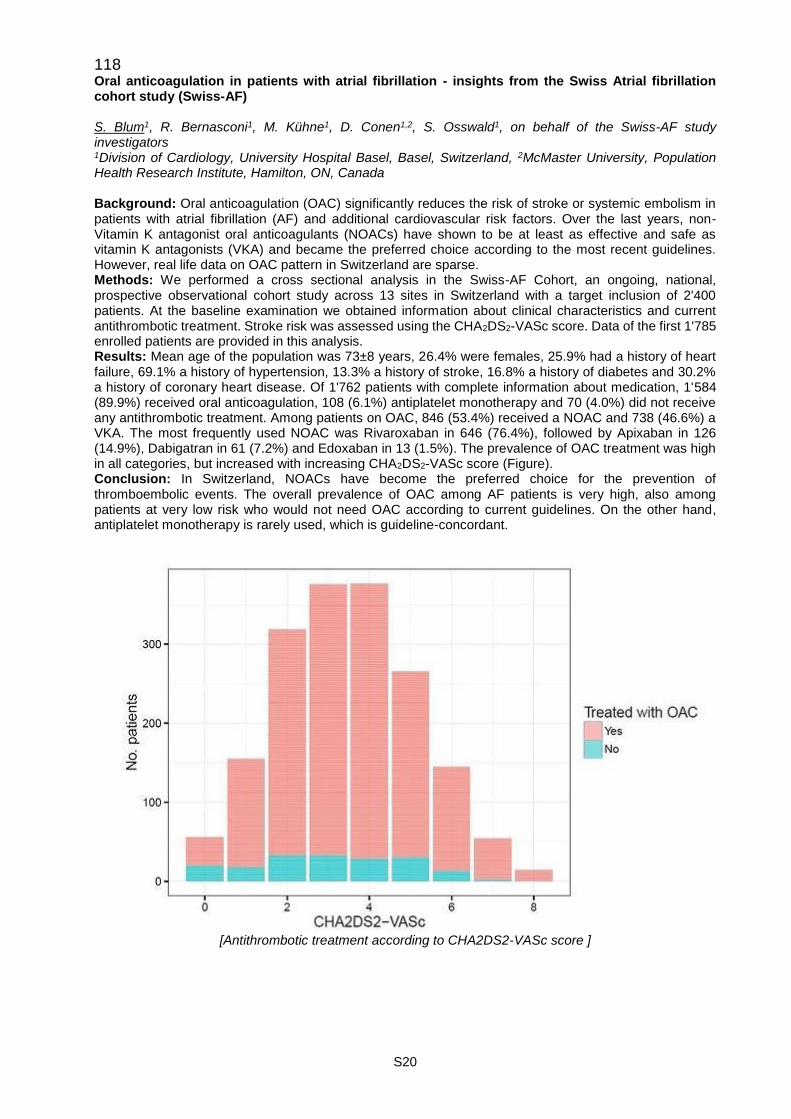
118 Oral anticoagulation in patients with atrial fibrillation - insights from the Swiss Atrial fibrillation cohort study (Swiss-AF) S. Blum1, R. Bernasconi1, M. Kühne1, D. Conen1,2, S. Osswald1, on behalf of the Swiss-AF study investigators 1Division of Cardiology, University Hospital Basel, Basel, Switzerland, 2McMaster University, Population Health Research Institute, Hamilton, ON, Canada Background: Oral anticoagulation (OAC) significantly reduces the risk of stroke or systemic embolism in patients with atrial fibrillation (AF) and additional cardiovascular risk factors. Over the last years, non-Vitamin K antagonist oral anticoagulants (NOACs) have shown to be at least as effective and safe as vitamin K antagonists (VKA) and became the preferred choice according to the most recent guidelines. However, real life data on OAC pattern in Switzerland are sparse. Methods: We performed a cross sectional analysis in the Swiss-AF Cohort, an ongoing, national, prospective observational cohort study across 13 sites in Switzerland with a target inclusion of 2'400 patients. At the baseline examination we obtained information about clinical characteristics and current antithrombotic treatment. Stroke risk was assessed using the CHA2DS2-VASc score. Data of the first 1'785 enrolled patients are provided in this analysis. Results: Mean age of the population was 73±8 years, 26.4% were females, 25.9% had a history of heart failure, 69.1% a history of hypertension, 13.3% a history of stroke, 16.8% a history of diabetes and 30.2% a history of coronary heart disease. Of 1'762 patients with complete information about medication, 1'584 (89.9%) received oral anticoagulation, 108 (6.1%) antiplatelet monotherapy and 70 (4.0%) did not receive any antithrombotic treatment. Among patients on OAC, 846 (53.4%) received a NOAC and 738 (46.6%) a VKA. The most frequently used NOAC was Rivaroxaban in 646 (76.4%), followed by Apixaban in 126 (14.9%), Dabigatran in 61 (7.2%) and Edoxaban in 13 (1.5%). The prevalence of OAC treatment was high in all categories, but increased with increasing CHA2DS2-VASc score (Figure). Conclusion: In Switzerland, NOACs have become the preferred choice for the prevention of thromboembolic events. The overall prevalence of OAC among AF patients is very high, also among patients at very low risk who would not need OAC according to current guidelines. On the other hand, antiplatelet monotherapy is rarely used, which is guideline-concordant.
[Antithrombotic treatment according to CHA2DS2-VASc score ]

S21
119 Cognitive function in patients with atrial fibrillation - the Swiss atrial fibrillation cohort study P. Meyre1,2, L. Eggimann1,2, M. Kühne1,2, S. Osswald1,2, D. Conen2,3, on behalf of the Swiss-AF Cohort Study Investigators 1Cardiovascular Research Institute Basel (CRIB), 2Division of Cardiology, University Hospital Basel, Basel, Switzerland, 3McMaster University, Population Health Research Institute, Hamilton, ON, Canada Introduction: There is emerging evidence suggesting a link between atrial fibrillation (AF) and the development of dementia, but only little is known about subclinical structural brain damage and its relation to functional cognitive decline in patients with AF. The Swiss AF Cohort Study (Swiss-AF) is designed to prospectively assess neurocognitive function over time in a large cohort of unselected AF patients. Methods: Swiss-AF is a prospective ongoing multicenter observational cohort study with a target enrollment of 2'400 patients across 13 sites in Switzerland. All patients underwent clinical and standardized neurocognitive assessment including the Montreal cognitive assessment (MOCA) at baseline and repeatedly thereafter. Results: In this cross-sectional analysis, data on the first 1'752 patients were included. The mean MOCA score was 25.2 ± 3.2 (normal ≥26/30) at study entry. History of hypertension was present in 1'234 (69.1%) patients, the mean CHA2DS2VASC score averaged 3.5 ± 1.7, oral anticoagulant treatment was given in 1'584 (88.7%) patients (VKA 41.3%, NOAC 47.4%), antiplatelet treatment in 108 (6.1%), both in 246 (13.8%), and none in 70 patients (3.9%). A history of a prior cerebrovascular event was observed in 238 (13.3%) participants. Patients with paroxysmal AF had a mean MOCA score of 25.4 ± 3.1, patients with persistent AF of 25.7 ± 3.0 and patients with permanent AF of 24.4 ± 3.4, as shown in the table. Of note, 25% of the population had a score of 23 or lower. Conclusions: In this large cohort of unselected AF patients, average MOCA score was below 26 at study entry suggesting substantial cognitive impairment. The availability of brain magnetic resonance imaging in all participants will shed further light into the underlying mechanisms.
MOCA score (corrected)
AF type n Mean Sd Median IQR Min Max
Paroxysmal 790 25.4 3.1 26.0 23.2 - 28.0 11 31
Persistent 485 25.7 3.0 26.0 24.0 - 28.0 13 31
Permanent 477 24.6 3.4 25.0 23.0 - 27.0 8 31
All 1752 25.2 3.2 26.0 23.0 - 28.0 8 31
[Distribution of MOCA score at baseline.]
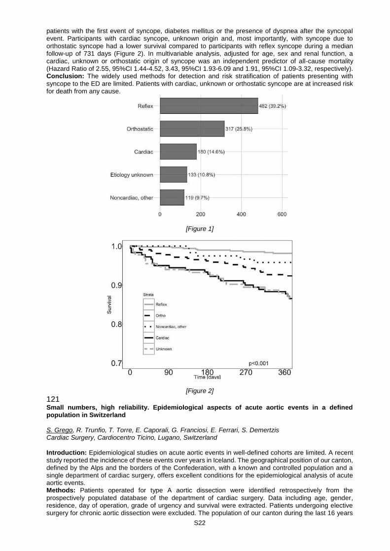
120 Natural history of syncope: insights from the BASEL IX syncope study P. Badertscher, T. Nestelberger, J. du Fay de Lavallaz, D. Flores Widmer, J. Lohrmann, M. Kühne, T. Reichlin, S. Osswald, C. Müller Universitätsspital Basel | Kardiologie, Cardiovascular Research Institute Basel (CRIB), Basel, Switzerland Background: The clinical presentation, resource utilization, diagnostic uncertainty and outcome of patients with syncope, are incompletely understood. Patients and methods: This ongoing observational, multicenter study is being carried out in seven different countries on three continents. In our first analysis we included 1409 patients > 40 years old presenting to the Emergency Department (ED) with syncope within the last 12 hours. Treating ED physicians were asked to quantify their clinical judgment regarding the presence of cardiac syncope using a visual analogue scale (VAS) to assess early diagnostic uncertainty. Patients were contacted at 12 and 24 months to determine major adverse events. Final diagnoses were adjudicated by two independent cardiologists after 12 months to investigate late diagnostic uncertainty. Results: Syncope was the final diagnosis in 1230 patients (87%). The distribution of causes in syncope was as follow (Figure 1): cardiac (15%), reflex-mediated (39%), orthostatic (26%), others - non-cardiac (10%) and unknown etiology (11%). 684 of all patients (56%) had at least two diagnostic tests at admission. 51% of all patients were hospitalized for a median length of 4 days (IQR 1-8). Early and late diagnostic uncertainties were assessed; the area under the curve (AUC) for ED-probability of cardiac syncope was 0.85 (CI 95%, 0.82-0.88). The final reviewed diagnosis by two independent cardiologists showed a mismatch rate of 32%. The following factors occurred significantly more often in mismatched patients:
S22
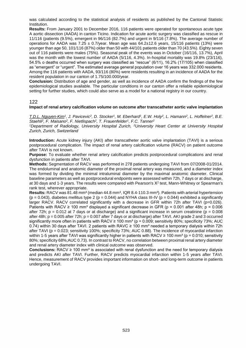
patients with the first event of syncope, diabetes mellitus or the presence of dyspnea after the syncopal event. Participants with cardiac syncope, unknown origin and, most importantly, with syncope due to orthostatic syncope had a lower survival compared to participants with reflex syncope during a median follow-up of 731 days (Figure 2). In multivariable analysis, adjusted for age, sex and renal function, a cardiac, unknown or orthostatic origin of syncope was an independent predictor of all-cause mortality (Hazard Ratio of 2.55, 95%CI 1.44-4.52, 3.43, 95%CI 1.93-6.09 and 1.91, 95%CI 1.09-3.32, respectively). Conclusion: The widely used methods for detection and risk stratification of patients presenting with syncope to the ED are limited. Patients with cardiac, unknown or orthostatic syncope are at increased risk for death from any cause.
[Figure 1]
[Figure 2]
121 Small numbers, high reliability. Epidemiological aspects of acute aortic events in a defined population in Switzerland S. Grego, R. Trunfio, T. Torre, E. Caporali, G. Franciosi, E. Ferrari, S. Demertzis Cardiac Surgery, Cardiocentro Ticino, Lugano, Switzerland Introduction: Epidemiological studies on acute aortic events in well-defined cohorts are limited. A recent study reported the incidence of these events over years in Iceland. The geographical position of our canton, defined by the Alps and the borders of the Confederation, with a known and controlled population and a single department of cardiac surgery, offers excellent conditions for the epidemiological analysis of acute aortic events. Methods: Patients operated for type A aortic dissection were identified retrospectively from the prospectively populated database of the department of cardiac surgery. Data including age, gender, residence, day of operation, grade of urgency and survival were extracted. Patients undergoing elective surgery for chronic aortic dissection were excluded. The population of our canton during the last 16 years
S23
was calculated according to the statistical analysis of residents as published by the Cantonal Statistic Institution. Results: From January 2001 to December 2016, 116 patients were operated for spontaneous acute type A aortic dissection (AADA) in canton Ticino. Indication for acute aortic surgery was classified as rescue in 11/116 (patients (9.5%), emergent in 96/116 (82.7%) and urgent in 9/116 (7.8%). The average number of operations for AADA was 7.25 ± 0.7/year. Mean age was 64.2±12.6 years, 15/116 patients (13%) were younger than age 50, 101/116 (87%) older than 50 with 44/101 patients older than 70 (43.5%). Eighty seven out of 116 patients were males (75%). Seasonal peak of the events was in October (16/116, 13.7%), April was the month with the lowest number of AADA (5/116, 4.3%). In-hospital mortality was 19.8% (23/116), 54.5% o deaths occurred when surgery was classified as “rescue” (6/11), 16.2% (17/105) when classified as “emergent” or “urgent”. The estimated average general population over 16 years was 332.000 residents. Among the 116 patients with AADA, 93/116 (80%) were residents resulting in an incidence of AADA for the resident population in our canton of 1.75/100.000/year. Conclusion: Distribution of age and gender, as well as incidence of AADA confirm the findings of the few epidemiological studies available. The particular conditions in our canton offer a reliable epidemiological setting for further studies, which could also serve as a model for a national registry in our country.
122 Impact of renal artery calcification volume on outcome after transcatheter aortic valve implantation T.D.L. Nguyen-Kim1, J. Pavicevic2, D. Stocker1, M. Eberhard1, E.W. Holy2, L. Hamann2, L. Hoffelner2, B.E. Staehli2, F. Maisano2, F. Nietlispach2, T. Frauenfelder1, F.C. Tanner2 1Department of Radiology, University Hospital Zurich, 2University Heart Center at University Hospital Zurich, Zurich, Switzerland Introduction: Acute kidney injury (AKI) after transcatheter aortic valve implantation (TAVI) is a serious postprocedural complication. The impact of renal artery calcification volume (RACV) on patient outcome after TAVI is not known. Purpose: To evaluate whether renal artery calcification predicts postprocedural complications and renal dysfunction in patients after TAVI. Methods: Segmentation of RACV was performed in 278 patients undergoing TAVI from 07/2008-01/2014. The endoluminal and anatomic diameter of the proximal renal artery was measured, and a diameter index was formed by dividing the minimal intraluminal diameter by the maximal anatomic diameter. Clinical baseline parameters as well as postprocedural endpoints were assessed within 72h, 7 days or at discharge, at 30 days and 1-3 years. The results were compared with Pearson's Χ2 test, Mann-Whitney or Spearman's rank test, wherever appropriate. Results: RACV was 81.48 mm³ (median 44.8 mm³, IQR 8.4-110.3 mm³). Patients with arterial hypertension (p = 0.043), diabetes mellitus type 2 (p = 0.044) and NYHA class III-IV (p = 0.044) exhibited a significantly larger RACV. RACV correlated significantly with a decrease in GFR within 72h after TAVI (p=0.026). Patients with RACV ≥ 100 mm³ displayed a significant decrease in GFR (p = 0.001 after 48h; p = 0.006 after 72h; p = 0.012 at 7 days or at discharge) and a significant increase in serum creatinine (p = 0.008 after 48h; p = 0.005 after 72h; p = 0.007 after 7 days or at discharge) after TAVI. AKI grade 2 and 3 occurred significantly more often in patients with RACV ≥ 100 mm³ (p = 0.009; sensitivity 80%; specificity 73%; AUC 0.74) within 30 days after TAVI. 2 patients with RAVC ≥ 100 mm³ needed a temporary dialysis within 72h after TAVI (p = 0.023; sensitivity 100%; specificity 73%; AUC 0.88). The incidence of myocardial infarction within 1-5 years after TAVI was significantly higher in patients with RACV ≥ 100 mm³ (p = 0.010; sensitivity 80%; specificity 68%;AUC 0.73). In contrast to RACV, no correlation between proximal renal artery diameter and renal artery diameter index with clinical outcome was observed. Conclusions: RACV ≥ 100 mm³ is associated with renal dysfunction and the need for temporary dialysis and predicts AKI after TAVI. Further, RACV predicts myocardial infarction within 1-5 years after TAVI. Hence, measurement of RACV provides important information on short- and long-term outcome in patients undergoing TAVI.
S24
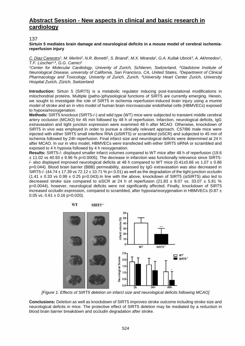
Abstract Session - New aspects in clinical and basic research in cardiology 137 Sirtuin 5 mediates brain damage and neurological deficits in a mouse model of cerebral ischemia-reperfusion injury C. Diaz Canestro1, M. Merlini2, N.R. Bonetti1, S. Briand1, M.X. Miranda1, G.A. Kullak Ubrick3, A. Akhmedov1, T.F. Lüscher1,4, G.G. Camici1 1Center for Molecular Cardiology, Univerity of Zurich, Schlieren, Switzerland, 2Gladstone Institute of Neurological Disease, university of California, San Francisco, CA, United States, 3Department of Clinical Pharmacology and Toxicology, Univerity of Zurich, Zurich, 4University Heart Center Zurich, University Hospital Zurich, Zürich, Switzerland Introduction: Sirtuin 5 (SIRT5) is a metabolic regulator inducing post-translational modifications in mitochondrial proteins. Multiple (patho-)physiological functions of SIRT5 are currently emerging. Herein, we sought to investigate the role of SIRT5 in ischemia reperfusion-induced brain injury using a murine model of stroke and an in vitro model of human brain microvascular endothelial cells (HBMVECs) exposed to hypoxia/reoxygenation. Methods: SIRT5 knockout (SIRT5-/-) and wild type (WT) mice were subjected to transient middle cerebral artery occlusion (MCAO) for 45 min followed by 48 h of reperfusion. Infarction, neurological deficits, IgG extravasation and tight junction expression were examined 48 h after MCAO. Otherwise, knockdown of SIRT5 in vivo was employed in order to pursue a clinically relevant approach. C57/B6 male mice were injected with either SIRT5 small interfere RNA (siSIRT5) or scrambled (siSCR) and subjected to 45 min of ischemia followed by 24h reperfusion. Final infarct size and neurological deficits were determined at 24 h after MCAO. In our in vitro model, HBMVECs were transfected with either SIRT5 siRNA or scrambled and exposed to 4 h hypoxia followed by 4 h reoxygenation. Results: SIRT5-/- displayed smaller infarct volumes compared to WT mice after 48 h of reperfusion (19.6 ± 11.02 vs 40.93 ± 9.96 % p=0.0005). The decrease in infarction was functionally relevance since SIRT5-/- also displayed improved neurological deficits at 48 h compared to WT mice (0.41±0.66 vs 1.07 ± 0.86 p=0.044). Blood brain barrier (BBB) permeability, assessed by IgG extravasation was also decreased in SIRT5-/- (44.74 ± 17.39 vs 72.12 ± 10.71 % p= 0.01) as well as the degradation of the tight junction occludin (1.41 ± 0.33 vs 0.99 ± 0.25 p=0.043).In line with the above, knockdown of SIRT5 (siSIRT5) also led to decreased stroke size compared to siSCR at 24 h of reperfusion (21.83 ± 8.07 vs. 33.07 ± 5.81 % p=0.0044), however, neurological deficits were not significantly affected. Finally, knockdown of SIRT5 increased occludin expression, compared to scrambled, after hypoxia/reoxygenation in HBMVECs (0.87 ± 0.05 vs. 0.61 ± 0.16 p=0.020).
[Figure 1. Effects of SIRT5 deletion on infarct size and neurological deficits following MCAO]
Conclusions: Deletion as well as knockdown of SIRT5 improves stroke outcome including stroke size and neurological deficits in mice. The protective effect of SIRT5 deletion may be mediated by a reduction in blood brain barrier breakdown and occludin degradation after stroke.
S25
138 Human adult progenitor cells as source of induced pluripotent stem cells: characterization and differentiation potential E. Pianezzi1,2, C. Altomare3, T. Torre4, S. Bolis3, V. Biemmi1,3, A. Ciullo1,2, E. Cervio1, G.G. Camici2, T. Moccetti4, L. Barile1,3, G. Vassalli1 1Fondazione Cardiocentro Ticino, Laboratory of Cellular and Molecular Cardiology, Lugano, 2Center for Molecular Cardiology, University of Zurich, Zurich, 3Foundation for Cardiovascular Research and Education, Taverne, 4Fondazione Cardiocentro Ticino, Lugano, Switzerland Introduction: It has been suggested that the somatic cell origin may influence the differentiation potential of Induced pluripotent stem cells (iPSCs) and the functional maturity of the re-differentiated cells, a phenomenon referred to as “somatic cell memory”. Here we compare different sources of human adult progenitor cells as former cells to obtain iPSCs and their differentiation capability to differentiate into iPSC-derived cardiomyocytes (iPSC-CMs). Methods: adult human cardiac-resident progenitor cells (CPC) and bone marrow mesenchymal stem cells (BM-MSC) obtained from the same donor were reprogrammed into iPSCs using the 4 Yamanaka's factors. Fibroblasts (Fib) were used as control cell line. iPSCs were subsequently re-differentiated into iPSC-CMs by modulating the Wnt pathway. iPSCs and iPSC-CMs were characterized by real-time PCR and immunofluorescence. Extracellular field potentials (FPs) from spontaneously beating iPSC-CMs were recorded by multi-electrode arrays (MEA). Results: real-time PCR revealed that iPSC derived from both CPC and BM-MSC showed comparable expression level of pluripotency markers, such as SSEA4, SOX2 and NANOG, when compared with Fib-derived iPSCs. After differentiation CPC-derived iPSC-CM showed higher expression of cardiac-specific markers compared to BM-MSC and fibroblast-derived iPSC-CM. Both CPC- and BM-MSC-iPSC-CM revealed Na+ and late L-type Ca2+ and functional rapid component of the delayed rectifier K+ current (IKr), as assessed using the IKr channel blocker E4031. IKs channel blocker JNJ303 and epinephrine affected the slow component of the delayed rectifier K+ current (IKs) only in CPC-iPSC-CM, thus suggesting the presence of functional channel. Conclusions: Adult human CPC and BM-MSC can be reprogrammed into iPSCs, from which CMs can be derived. CPC-iPSC-CMs express higher levels of sarcomeric proteins and display more mature electrophysiological features, such as functional IKs, compared to Fib-iPSC-CMs.
139 Characterization of endothelial function following various ischemic durations in an isolated rat heart model of donation after circulatory death N. Méndez Carmona, R.K. Wyss, M. Arnold, T.P. Carrel, H.T. Tevaearai Stahel, S. Longnus Clinic for Cardiovascular Surgery, University of Bern / Inselspital, Bern, Switzerland Background: Donation after circulatory death (DCD) could significantly improve the number of cardiac grafts available for transplantation; however, ischemia/reperfusion (I/R) injury remains a concern for post-transplant graft function. Endothelial damage is one key factor involved in cardiac I/R injury. Thus, we aimed to characterize hemodynamic and endothelial function following various ischemic durations in order to help improve the timing and choice of therapeutic targets for cardioprotection. Methods and Results: Isolated working rat hearts were perfused aerobically for a period of 20' and then underwent various periods of warm global ischemia (I) before reperfusion. At 60' reperfusion, recovery of the left ventricular work (heart rate-developed pressure product) was significantly lower after 27' I (76% ± 12), 30' I (67% ± 16) and 33' I (29% ± 20), but was not different for 21' I (91% ± 5) and 24' I (88% ± 13) compared with non-ischemic controls (100% ± 11; n=7-8/group; p-value < 0.05 for all). Coronary flow was measured as an indicator of endothelial function at 20' reperfusion. Compared to non-ischemic controls (25 ± 4 mL/min), coronary flow was not different after 21' I (24 ± 3 mL/min) and 24' I (19 ± 3 mL/min), but was significantly decreased after 27' I (19 ± 3 mL/min), 30' I (17 ± 6 mL/min) or 33'I (13 ± 2 mL/min; p-value < 0.05 for all). An additional series of hearts was perfused identically, but stopped at 30 min reperfusion. In this series, endothelial function was assessed by comparing vasodilatory responses between endothelium-dependent (bradykinin; 10-9, 10-8M) and endothelium-independent (sodium nitroprusside; 3x10⁻⁵M) vasodilators. Endothelial function appears to be impaired after 21' I, while smooth muscle function was impaired with ischemic durations >24'I (n=3-6/group). Conclusion: Post-ischemic hemodynamic function recovers almost completely up until 24'I but then rapidly decreases with longer ischemic durations. Evidence of endothelial dysfunction is presence after 21'I. Therefore, characterization of endothelial damage associated with cardiac I/R could help us to optimize therapeutic approaches to improve endothelial and myocardial recovery, ultimately to facilitate DCD heart transplantation.

S26
140 Structural characteristics of the human intracranial aneurysm wall prone to rupture S. Morel1,2, M. Diagbouga1, N. Dupuy1,2, E. Sutter1, M. Corniola1, R. Gondar2, M. Jaegersberg2, N. Isidor2, V. Braunersreuther3, M.-L. Bochaton-Piallat1, K. Schaller2, P. Bijlenga2, B. Kwak1 1Pathology and Immunology, University of Geneva, 2Clinical Neurosciences, Geneva University Hospitals, 3Clinical Pathology, University of Geneva, Genève, Switzerland Introduction: Intracranial aneurysm (IA) is a disease of the vascular wall resulting in abnormal enlargement of the vessel lumen. It is a common pathology with a prevalence of 2-5% in the adult population. IAs are mostly small, quiescent and asymptomatic; yet upon rupture severe brain damage or even death is frequently encountered. Little is known about the etiology of IAs, their evolution is hard to predict and intricate medical decision. Criteria nowadays used to recommend IAs treatment are based on IA location and size together with empirical patient characteristics such as age, ethnicity and hypertension. These criteria are subject to controversies as they remain unsatisfying to predict the risk of rupture accurately. The goal of our AneuX project is to use the IA 3D-shape to characterize disease status. Here, we report the first structural results on human IA domes. Methods: IA domes, ruptured (RIA, N=18) or not (UIA, N=31), have been collected following the clipping of the IA at the Geneva University Hospitals. Domes have been fixed in Formol, embedded in paraffin, sectioned and stained for smooth muscle cells (a-SM actin), macrophages (CD68), collagen (Masson's trichrome and Sirius Red) and elastin (Victoria Blue). Results are expressed in percentage. Comparison have been performed using non-parametric Mann-Whitney U test. Results: Patients characteristics (age, gender, hypertension, smoking, multiple IAs, positive familial history) are not different between RIAs and UIAs. Based on radiology images, IA location, aspect, collar size, maximal diameter and volume are also not different between the 2 groups. Histological analysis of aneurysmal wall composition revealed that RIAs have a higher content in macrophages (3.1% vs. 0.4%, p< 0.001) and a lower content of smooth muscle cells (16.2% vs. 24.0%, p< 0.05). This difference in cellular composition is associated with a lower content in collagen (total collagen: 15.2% vs. 37.4%, p< 0.05; collagen type I: 8.6% vs. 34.2%, p< 0.001; collagen type III: 10.4% vs. 16.5%, p< 0.05) in the vascular wall of ruptured IAs whereas the elastin content is not affected (33.9% vs. 41.6%, ns). Interestingly, the collagen content in the aneurysm wall of ruptured IAs was not affected by the thickness of the vascular wall. Conclusions: Our results show that the IA wall prone to rupture presents a lower smooth muscle cell and collagen content, independently of the wall thickness, and a higher content in macrophages.
141 Electrical and structural dysregulation of cardiomyocytes induced by doxorubicin treatment C. Altomare1, V. Biemmi1, N. Pernigoni1, A. Ciullo2, L. Barile1,2, G. Vassalli1,2, G. Milano1 1Foundation for Cardiovascular Research and Education, Taverne, 2Fondazione Cardiocentro Ticino, Lugano, Switzerland Introduction: Doxorubicin (DOXO)-induced cardiotoxicity has been a well-known phenomenon, however the modulation of electrical and structural properties of cardiomyocytes (CM) underlying DOXO cardiotoxicity is still being uncovered. We aimed to analyze the effect of in-vivo DOXO administration at single cell level, on isolated adult CM. Method: DOXO is administered in six equal intraperitoneal injections over a period of 2 weeks (cumulative dose, 15 mg/kg). Control group received only saline injection. Echocardiographic analysis was performed at different time points to monitor the progression of cardiotoxicity. Ventricular CM were isolated from left (LV) and right (RV) ventricle free walls according to Langendorff method. Ca2+-tolerant myocytes were used for patch-clamp measurements within 6-8 h from dissociation. Electrical activity was evaluated by action potential (AP) recordings during steady-state pacing at 1 Hz. To assess T-tubular (TT) disarray, sarcolemmal membranes were stained by incubating isolated CM with di-3-ANEPPDHQ (20 mmol/L). Eight-bit gray-scaled images were subjected to spatial Fast Fourier Transform analysis allowing quantification of periodic and aperiodic components of pixel variance. Whereas transverse TT generates the periodic component, the aperiodic one may reflect disarray of transverse TT, but can also be generated by longitudinal TT. Analysis was performed at the end of treatment (day 12) and one week after recovery. Results: As expected left ventricular end-systolic volume (LVESV) increases in the animals treated with DOXO at day 12, and the effect persisted during the week of recovery (Doxo 45.4 ± 6.4 vs CTRL 23.2 ± 6.1). RV and LV APD was prolonged in DOXO-CM group compared to CTRL-CM. APD50/APD90 ratio was increased by about 50% in DOXO-CM derived from both RV and LV at the end of treatment and rescued a week after the end of treatment. Sharp pattern of transverse TT striations was observed in CTRL-CM of both chambers; accordingly, in these CM, pixel variance was largely represented by the periodic component, whose period (0.5 mm-1) was consistent with transverse TT arrangement. The power under
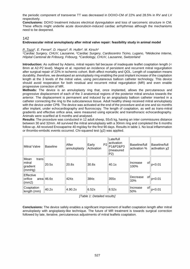
S27
the periodic component of transverse TT was decreased in DOXO-CM of 22% and 28.5% in RV and LV respectively. Conclusions: DOXO treatment induces electrical dysregulation and loss of sarcomeric structure in CM. These effects might underlie acute doxorubicin-induced cardiac arrhythmias although the mechanisms need to be deepened.
142 Endovascular mitral annuloplasty after mitral valve repair: feasibility study in animal model P. Tozzi1, E. Ferrari2, D. Hayoz3, R. Hullin4, M. Kirsch1 1Cardiac Surgery, CHUV, Lausanne, 2Cardiac Surgery, Cardiocentro Ticino, Lugano, 3Médecine Interne, Hôpital Cantonal de Fribourg, Fribourg, 4Cardiology, CHUV, Lausanne, Switzerland Introduction: As outlined by Adams, mitral repairs fail because of inadequate leaflet coaptation length (< 8mm at A2-P2 level). Magne et al. reported an incidence of persistent and recurrent mitral regurgitation after surgical repair of 22% in ischemic cohort. Both affect mortality and QOL. Length of coaptation means durability, therefore, we developed an annuloplasty ring enabling the post implant increase of the coaptation length at the 3 levels of the mitral valve, using percutaneous balloon catheter technology. This device should avoid reoperation for both residual and recurrent mitral regurgitation (MR) and even enable progressive correction of MR. Methods: The device is an annuloplasty ring that, once implanted, allows the percutaneous and progressive displacement of each of the 3 anatomical regions of the posterior mitral annulus towards the anterior. The displacement is permanent and induced by an angioplasty balloon catheter inserted in a catheter connecting the ring to the subcutaneous tissue. Adult healthy sheep received mitral annuloplasty with the device under CPB. The device was activated at the end of the procedure and at one and six months after implant, under echocardiography and fluoroscopy. The length of coaptation, as well as trans-mitral gradients and effective orifice area, were measured using epicardic and transthoracic echocardiography. Animals were scarified at 6 months and analysed. Results: The procedure was conducted in 12 adult sheep, 55±5 kg, having an inter commissures distance between 30 and 32mm. All survived the mitral annuloplasty with a 30mm ring and completed the 6 months follow up. All received Enoxaparine 40 mg/day for the first 90 days. Results in table 1. No local inflammation or thrombo-embolic events occurred. Chi-squared test (χ2) was applied.
Mitral Valve Baseline After annuloplasty
Early P2 Activation
Late/full activation P1&P2&P3 (measured P2)
Baseline/full activation %
Baseline/full activation χ2
Mean trans mitral gradient (mmHg)
20.5± 20.8± 30.8± 40.1± Increase of 100%
p<0.01
Effective orifice area (mm2)
46.6± 424± 384± 355± Decrease of 33%
p<0.01
Coaptation length (mm)
40.2± 4.90.2± 6.52± 8.52± Increase of 50%
p<0.01
[Table 1: Detailed results] Conclusions: The device safely enables a significant improvement of leaflet coaptation length after mitral annuloplasty with angioplasty-like technique. The future of MR treatment is towards surgical correction followed by late, iterative, percutaneous adjustments of mitral leaflets coaptation.

S28
Rapid Fire Abstract Session - Electrical misbehaviour of the ventricles 176 Association between genotype and ventricular involvement patterns in arrhythmogenic right ventricular cardiomyopathy/dysplasia D. Akdis1, A.M. Saguner1, A. Medeiros-Domingo2, S.H. Baldinger2, H. Burri3, T.F. Lüscher1, F.C. Tanner1, C. Brunckhorst1, F. Duru1 1Cardiology, University Hospital Zurich, Zürich, 2Bern University Hospital, Bern, 3Cardiology, Geneva University Hospitals, Geneva, Switzerland Introduction: Data on the impact of genotype on the long-term outcome regarding ventricular involvement patterns in arrhythmogenic right ventricular cardiomyopathy/dysplasia (ARVC/D) is scarce. Therefore, aim of this study was to investigate the presence and progression of right and left ventricular disease, and associated long-term outcome in patients with ARVC/D stratified by genotype. Methods: 54 patients fulfilling definite Task Force Criteria for ARVC/D were systematically evaluated at baseline and after follow-up. Right and left ventricular involvement, was determined by transthoracic echocardiography and cardiac magnetic resonance imaging. All patients received genetic testing of the clinical exome using next generation DNA sequencing, initially focusing on at least 96 known cardiomyopathy and channelopathy genes. Results: In total, 24 patients (44%) presented with biventricular (BiV) involvement during a median follow-up of 8 years. Of those, 13 patients did not present with BiV at first but instead developed new onset left ventricular involvement during this study period. The others (11 patients) showed a progression of the BiV involvement during the study period. Patients with BiV involvement had significantly more compound/multigenic or desmoplakin mutations as compared to those with only right ventricular involvement (p=0.03), and patients who developed new onset left ventricular involvement frequently harboured non-desmosomal mutations such as titin, ryanodine-receptor 2 and lamin A/C mutations. In 26 % of the patients with only right ventricular involvement, pathogenic mutations were absent. Conclusions: Our study shows that almost half of the patients with ARVC/D present with left ventricular involvement either at baseline or later during follow-up. Compund/digenic heterozygosity and desmoplakin mutations are common in this population. Non-desmosomal mutations are frequent in patients with new onset left ventricular involvement. Therefore, genetic testing using next generation DNA sequencing methods may have diagnostic and therapeutic implications in ARVC/D.
177 Sudden cardiac death and syncope of unknown origin: think about catecholaminergic polymorphic ventricular tachycardia! S. Aur1, F. Fellmann2, J.-M. Good2, H. Fodstad3, N. Sekarski4, Z.A. Bhuiyan3, J. Schläpfer1 1Service de Cardiologie, 2Service de Génétique Médicale, 3Laboratoire de Génétique Moléculaire, 4Unité de Cardiologie Pédiatrique, CHUV, Lausanne, Switzerland Sudden cardiac death (SCD) or syncope may remain unexplained after extensive cardiac investigations. However, genetic testing may help establish a clear diagnosis in a substantial proportion of these cases. We here report our experience in 4 patients referred for SCD or syncope of unknown origin in whom the diagnosis of Catecholaminergic Polymorphic Ventricular Tachycardia (CPVT) was given by genetic testing. The studied population encompasses 4 patients (pts) (2 female) mean age: 38±13y (20-53y) of 4 different families. Age at first symptoms was 13.7±10y (5-26); a family history of syncope or SCD was present in 3 out of 4 pts. Syncope was the first symptom in 3 pts and aborted SCD in 1 pt; it occurred mainly during stress or effort. Resting ECG was normal in all pts and no anatomical disease was demonstrated in any pt. Stress test triggered ventricular arrhythmias in 2 pts. When performed, Holter monitoring was irrelevant in 2/3 pts. Programmed ventricular stimulation was negative in 2/2 pts. The first medical diagnoses were: epilepsy, long QT syndrome, vaso-vagal syncope and SCD of unknown origin. Genetic testing performed 22.7±13y (11-35 y) after the initial event revealed mutation either in the RYR2 or CASQ2 genes in the 4 patients. All pts are asymptomatic under drug treatment ± an implantable cardioverter defibrillator. In all cases, extensive familial screening has been proposed and is ongoing. In patients with SCD or syncope of unknown origin and normal anatomical heart, CPVT has to be considered as a potential cause even in patients with a normal stress test. Genetic testing should be more often considered in these patients to avoid missing this pathology, which is usually controlled with beta-blocking agents.
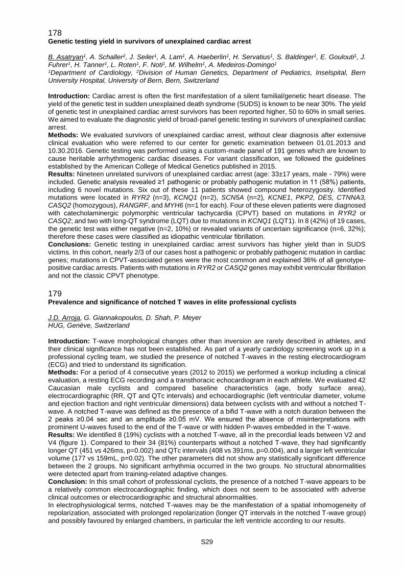
S29
178 Genetic testing yield in survivors of unexplained cardiac arrest B. Asatryan1, A. Schaller2, J. Seiler1, A. Lam1, A. Haeberlin1, H. Servatius1, S. Baldinger1, E. Goulouti1, J. Fuhrer1, H. Tanner1, L. Roten1, F. Noti1, M. Wilhelm1, A. Medeiros-Domingo1 1Department of Cardiology, 2Division of Human Genetics, Department of Pediatrics, Inselspital, Bern University Hospital, University of Bern, Bern, Switzerland Introduction: Cardiac arrest is often the first manifestation of a silent familial/genetic heart disease. The yield of the genetic test in sudden unexplained death syndrome (SUDS) is known to be near 30%. The yield of genetic test in unexplained cardiac arrest survivors has been reported higher, 50 to 60% in small series. We aimed to evaluate the diagnostic yield of broad-panel genetic testing in survivors of unexplained cardiac arrest. Methods: We evaluated survivors of unexplained cardiac arrest, without clear diagnosis after extensive clinical evaluation who were referred to our center for genetic examination between 01.01.2013 and 10.30.2016. Genetic testing was performed using a custom-made panel of 191 genes which are known to cause heritable arrhythmogenic cardiac diseases. For variant classification, we followed the guidelines established by the American College of Medical Genetics published in 2015. Results: Nineteen unrelated survivors of unexplained cardiac arrest (age: 33±17 years, male - 79%) were included. Genetic analysis revealed ≥1 pathogenic or probably pathogenic mutation in 11 (58%) patients, including 6 novel mutations. Six out of these 11 patients showed compound heterozygosity. Identified mutations were located in RYR2 (n=3), KCNQ1 (n=2), SCN5A (n=2), KCNE1, PKP2, DES, CTNNA3, CASQ2 (homozygous), RANGRF, and MYH6 (n=1 for each). Four of these eleven patients were diagnosed with catecholaminergic polymorphic ventricular tachycardia (CPVT) based on mutations in RYR2 or CASQ2; and two with long-QT syndrome (LQT) due to mutations in KCNQ1 (LQT1). In 8 (42%) of 19 cases, the genetic test was either negative (n=2, 10%) or revealed variants of uncertain significance (n=6, 32%); therefore these cases were classified as idiopathic ventricular fibrillation. Conclusions: Genetic testing in unexplained cardiac arrest survivors has higher yield than in SUDS victims. In this cohort, nearly 2/3 of our cases host a pathogenic or probably pathogenic mutation in cardiac genes; mutations in CPVT-associated genes were the most common and explained 36% of all genotype-positive cardiac arrests. Patients with mutations in RYR2 or CASQ2 genes may exhibit ventricular fibrillation and not the classic CPVT phenotype.
179 Prevalence and significance of notched T waves in elite professional cyclists J.D. Arroja, G. Giannakopoulos, D. Shah, P. Meyer HUG, Genève, Switzerland Introduction: T-wave morphological changes other than inversion are rarely described in athletes, and their clinical significance has not been established. As part of a yearly cardiology screening work up in a professional cycling team, we studied the presence of notched T-waves in the resting electrocardiogram (ECG) and tried to understand its signification. Methods: For a period of 4 consecutive years (2012 to 2015) we performed a workup including a clinical evaluation, a resting ECG recording and a transthoracic echocardiogram in each athlete. We evaluated 42 Caucasian male cyclists and compared baseline characteristics (age, body surface area), electrocardiographic (RR, QT and QTc intervals) and echocardiographic (left ventricular diameter, volume and ejection fraction and right ventricular dimensions) data between cyclists with and without a notched T-wave. A notched T-wave was defined as the presence of a bifid T-wave with a notch duration between the 2 peaks ≥0.04 sec and an amplitude ≥0.05 mV. We ensured the absence of misinterpretations with prominent U-waves fused to the end of the T-wave or with hidden P-waves embedded in the T-wave. Results: We identified 8 (19%) cyclists with a notched T-wave, all in the precordial leads between V2 and V4 (figure 1). Compared to their 34 (81%) counterparts without a notched T-wave, they had significantly longer QT (451 vs 426ms, p=0.002) and QTc intervals (408 vs 391ms, p=0.004), and a larger left ventricular volume (177 vs 159mL, p=0.02). The other parameters did not show any statistically significant difference between the 2 groups. No significant arrhythmia occurred in the two groups. No structural abnormalities were detected apart from training-related adaptive changes. Conclusion: In this small cohort of professional cyclists, the presence of a notched T-wave appears to be a relatively common electrocardiographic finding, which does not seem to be associated with adverse clinical outcomes or electrocardiographic and structural abnormalities. In electrophysiological terms, notched T-waves may be the manifestation of a spatial inhomogeneity of repolarization, associated with prolonged repolarization (longer QT intervals in the notched T-wave group) and possibly favoured by enlarged chambers, in particular the left ventricle according to our results.

S30
[Example of T wave notching recorded in one of the 8 cyclists ]
180 Impact of contact force sensing technology on catheter ablation success of idiopathic ventricular arrhythmias originating from the outflow tracts R. Ebrahimi, M. Kühne, S. Knecht, F. Spiess, B. Schär, S. Osswald, C. Sticherling, T. Reichlin Kardiologie, Universitätsspital Basel, Basel, Switzerland Introduction: Catheter ablation of frequent idiopathic premature ventricular contractions (PVC) and idiopathic ventricular tachycardia (VT) is used to eliminate symptoms and to prevent or treat PVC-induced cardiomyopathy. Recently, multiple studies have reported benefits of contact force (CF) sensing catheters in atrial fibrillation ablation. However, CF data on ventricular arrhythmia is lacking. We aimed to assess the impact of CF sensing on the success rates of catheter ablation of idiopathic ventricular arrhythmias originating from the outflow tracts. Methods: In this prospective observational cohort study, 60 consecutive patients undergoing a first catheter ablation for symptomatic idiopathic PVCs or idiopathic VTs were enrolled. All procedures were performed using the same electroanatomical mapping system (CARTO 3) with either a standard irrigated ablation catheter or an irrigated CF-sensing ablation catheter. Structural heart disease as the underlying cause for PVCs or VTs was ruled out using echocardiography or cardiac MRI at the discretion of the physicians. Patients in whom ablation was not performed due to insufficient or non-inducible arrhythmias were excluded from analysis. Ablation success was assessed after a median FU of 2.7 months (IQR 1.3-3.5 months) using 24h Holter ECG. Results: A total of 60 patients were enrolled. Median age was 48 years (IQR 41-60) and 52% of the patients were female. Median PVC burden before ablation was 20% (IQR 7-29%). The procedures were performed using standard ablation catheters in 21 patients (35%) and CF-sensing catheters in 39 patients (65%). Baseline characteristics of the patients are shown in Table 1. Overall, the success rate of catheter ablation was 83%. No significant difference was observed in the success rate between the standard group (91%) and the CF-sensing group (80%, p=0.28). In the subgroups according to an arrhythmia origin in the RVOT or the LVOT, the following success rates were found for the standard group and the CF-sensing group: RVOT 100% vs. 77% (p=0.07) and LVOT 78% vs. 89% (p=0.53). Complications occurred in 2/60 patients (3.3%): Pericardial tamponade in one patient in the CF-sensing catheter group, and pericarditis requiring medical treatment in one patient in the standard catheter group. Conclusion: The use of CF-sensing catheters does not increase the success rate of catheter ablation of idiopathic ventricular arrhythmias originating from the outflow tract.
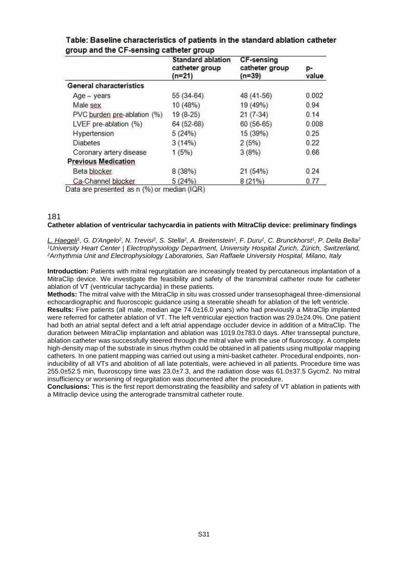
S31
181 Catheter ablation of ventricular tachycardia in patients with MitraClip device: preliminary findings L. Haegeli1, G. D'Angelo2, N. Trevisi2, S. Stella2, A. Breitenstein1, F. Duru1, C. Brunckhorst1, P. Della Bella2 1University Heart Center | Electrophysiology Department, University Hospital Zurich, Zürich, Switzerland, 2Arrhythmia Unit and Electrophysiology Laboratories, San Raffaele University Hospital, Milano, Italy Introduction: Patients with mitral regurgitation are increasingly treated by percutaneous implantation of a MitraClip device. We investigate the feasibility and safety of the transmitral catheter route for catheter ablation of VT (ventricular tachycardia) in these patients. Methods: The mitral valve with the MitraClip in situ was crossed under transesophageal three-dimensional echocardiographic and fluoroscopic guidance using a steerable sheath for ablation of the left ventricle. Results: Five patients (all male, median age 74.0±16.0 years) who had previously a MitraClip implanted were referred for catheter ablation of VT. The left ventricular ejection fraction was 29.0±24.0%. One patient had both an atrial septal defect and a left atrial appendage occluder device in addition of a MitraClip. The duration between MitraClip implantation and ablation was 1019.0±783.0 days. After transseptal puncture, ablation catheter was successfully steered through the mitral valve with the use of fluoroscopy. A complete high-density map of the substrate in sinus rhythm could be obtained in all patients using multipolar mapping catheters. In one patient mapping was carried out using a mini-basket catheter. Procedural endpoints, non-inducibility of all VTs and abolition of all late potentials, were achieved in all patients. Procedure time was 255.0±52.5 min, fluoroscopy time was 23.0±7.3, and the radiation dose was 61.0±37.5 Gycm2. No mitral insufficiency or worsening of regurgitation was documented after the procedure. Conclusions: This is the first report demonstrating the feasibility and safety of VT ablation in patients with a Mitraclip device using the anterograde transmitral catheter route.
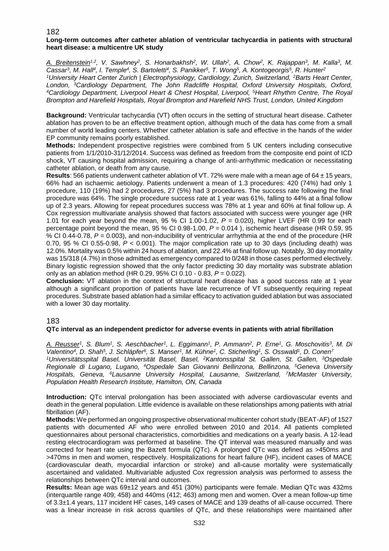
S32
182 Long-term outcomes after catheter ablation of ventricular tachycardia in patients with structural heart disease: a multicentre UK study A. Breitenstein1,2, V. Sawhney2, S. Honarbakhsh2, W. Ullah2, A. Chow2, K. Rajappan3, M. Kalla3, M. Cassar3, M. Hall4, I. Temple4, S. Bartoletti4, S. Panikker5, T. Wong5, A. Kontogeorgis5, R. Hunter2 1University Heart Center Zurich | Electrophysiology, Cardiology, Zurich, Switzerland, 2Barts Heart Center, London, 3Cardiology Department, The John Radcliffe Hospital, Oxford University Hospitals, Oxford, 4Cardiology Department, Liverpool Heart & Chest Hospital, Liverpool, 5Heart Rhythm Centre, The Royal Brompton and Harefield Hospitals, Royal Brompton and Harefield NHS Trust, London, United Kingdom Background: Ventricular tachycardia (VT) often occurs in the setting of structural heart disease. Catheter ablation has proven to be an effective treatment option, although much of the data has come from a small number of world leading centers. Whether catheter ablation is safe and effective in the hands of the wider EP community remains poorly established. Methods: Independent prospective registries were combined from 5 UK centers including consecutive patients from 1/1/2010-31/12/2014. Success was defined as freedom from the composite end point of ICD shock, VT causing hospital admission, requiring a change of anti-arrhythmic medication or necessitating catheter ablation, or death from any cause. Results: 566 patients underwent catheter ablation of VT. 72% were male with a mean age of 64 ± 15 years, 66% had an ischaemic aetiology. Patients underwent a mean of 1.3 procedures: 420 (74%) had only 1 procedure, 110 (19%) had 2 procedures, 27 (5%) had 3 procedures. The success rate following the final procedure was 64%. The single procedure success rate at 1 year was 61%, falling to 44% at a final follow up of 2.3 years. Allowing for repeat procedures success was 78% at 1 year and 60% at final follow up. A Cox regression multivariate analysis showed that factors associated with success were younger age (HR 1.01 for each year beyond the mean, 95 % CI 1.00-1.02, P = 0.020), higher LVEF (HR 0.99 for each percentage point beyond the mean, 95 % CI 0.98-1.00, P = 0.014 ), ischemic heart disease (HR 0.59, 95 % CI 0.44-0.78, P = 0.003), and non-inducibility of ventricular arrhythmia at the end of the procedure (HR 0.70, 95 % CI 0.55-0.98, P < 0.001). The major complication rate up to 30 days (including death) was 12.0%. Mortality was 0.5% within 24 hours of ablation, and 22.4% at final follow up. Notably, 30 day mortality was 15/318 (4.7%) in those admitted as emergency compared to 0/248 in those cases performed electively. Binary logistic regression showed that the only factor predicting 30 day mortality was substrate ablation only as an ablation method (HR 0.29, 95% CI 0.10 - 0.83, P = 0.022). Conclusion: VT ablation in the context of structural heart disease has a good success rate at 1 year although a significant proportion of patients have late recurrence of VT subsequently requiring repeat procedures. Substrate based ablation had a similar efficacy to activation guided ablation but was associated with a lower 30 day mortality.
183 QTc interval as an independent predictor for adverse events in patients with atrial fibrillation A. Reusser1, S. Blum1, S. Aeschbacher1, L. Eggimann1, P. Ammann2, P. Erne1, G. Moschovitis3, M. Di Valentino4, D. Shah5, J. Schläpfer6, S. Manser1, M. Kühne1, C. Sticherling1, S. Osswald1, D. Conen7 1Universitätsspital Basel, Universität Basel, Basel, 2Kantonsspital St. Gallen, St. Gallen, 3Ospedale Regionale di Lugano, Lugano, 4Ospedale San Giovanni Bellinzona, Bellinzona, 5Geneva University Hospitals, Geneva, 6Lausanne University Hospital, Lausanne, Switzerland, 7McMaster University, Population Health Research Institute, Hamilton, ON, Canada Introduction: QTc interval prolongation has been associated with adverse cardiovascular events and death in the general population. Little evidence is available on these relationships among patients with atrial fibrillation (AF). Methods: We performed an ongoing prospective observational multicenter cohort study (BEAT-AF) of 1527 patients with documented AF who were enrolled between 2010 and 2014. All patients completed questionnaires about personal characteristics, comorbidities and medications on a yearly basis. A 12-lead resting electrocardiogram was performed at baseline. The QT interval was measured manually and was corrected for heart rate using the Bazett formula (QTc). A prolonged QTc was defined as >450ms and >470ms in men and women, respectively. Hospitalizations for heart failure (HF), incident cases of MACE (cardiovascular death, myocardial infarction or stroke) and all-cause mortality were systematically ascertained and validated. Multivariable adjusted Cox regression analysis was performed to assess the relationships between QTc interval and outcomes. Results: Mean age was 69±12 years and 451 (30%) participants were female. Median QTc was 432ms (interquartile range 409; 458) and 440ms (412; 463) among men and women. Over a mean follow-up time of 3.3±1.4 years, 117 incident HF cases, 149 cases of MACE and 139 deaths of all-cause occurred. There was a linear increase in risk across quartiles of QTc, and these relationships were maintained after

S33
multivariable adjustment, as shown in the Table. Compared to AF patients with a normal QTc interval, AF patients with a prolonged QTc had a higher risk of HF (HR 1.8 [95%CI 1.2; 2.7], p=0.0047), MACE (HR 1.4 [1.0; 2.0], p=0.066) and all-cause mortality (HR 1.7 [1.2; 2.5], p=0.0039). Results were consistent for all three outcomes across subgroups for ECG rhythm at baseline (sinus rhythm, atrial fibrillation). Conclusions: In this well-characterized cohort of AF patients, QTc interval was independently associated with a higher risk of HF, MACE and all-cause mortality. Further studies are needed to evaluate the underlying mechanisms.
Continuous per SD (45.17ms)
Quartile 1 Quartile 2 Quartile 3 Quartile 4 p-value
Heart failure n: 117
Age/sex 1.45 (1.26; 1.67) p<0.001
Reference 1.25 (0.64; 2.42)
1.48 (0.79; 2.79)
3.20 (1.79; 5.71)
<.0001
Multivariate 1.30 (1.09; 1.57) p=0.0045
Reference 1.00 (0.51; 1.96)
1.03 (0.54; 1.97)
1.85 (0.99; 3.44)
0.022
MACE n: 149
Age/sex 1.29 (1.12; 1.49) p=0.0006
Reference 1.27 (0.74; 2.17)
1.39 (0.83; 2.34)
2.04 (1.25; 3.35)
0.0036
Multivariate 1.23 (1.05; 1.44), p=0.012
Reference 1.12 (0.65; 1.93)
1.21 (0.70; 2.08)
1.63 (0.95; 2.78)
0.057
All-cause mortality n: 138
Age/sex 1.41 (1.23; 1.61) p<0.001
Reference 1.48 (0.80; 2.75)
1.54 (0.84; 2.82)
3.34 (1.92; 5.81)
<.0001
Multivariate 1.26 (1.05; 1.50) p=0.011
Reference 1.16 (0.62; 2.17)
1.05 (0.57; 1.96)
1.99 (1.10 ; 3.60)
0.012
[Relationships between QTc interval and outcomes] Data are presented as hazard ratios with 95% confidence intervals. MACE was defined as cardiovascular death, stroke or myocardial infarction. Multivariable models were adjusted for age, gender, BMI, atrial fibrillation type, current smoking, heart rate in baseline ECG, QTc prolonging drugs, coronary heart disease, hypertension, heart failure, history of stroke, diabetes.
184 Prevalence of electrocardiogram abnormalities using the revised Seattle criteria in the longitudinal follow-up of elite professional cyclists P. Maziarski1, M.V. Julliard2, J.-L. Ziltener3, H. Burri1, P. Meyer1 1Service de Cardiologie, Hôpitaux Universitaires de Genève, 2Faculté de Médecine de Genève, 3Clinique Hirslanden La Colline, Genève, Switzerland Introduction: Most official sports associations recommend a resting electrocardiogram (ECG) as a screening tool for the prevention of sudden cardiac death (SCD) in young athletes. However, this strategy is debated especially because of the relatively high false-positive rate leading to unnecessary costs. Professional cyclists are known to exhibit the most prominent structural and electrical cardiac adaptations compared to other sports disciplines. The specificity of ECG changes to detect cardiac abnormalities that may lead to SCD may therefore be even lower in this population. Our aim was to evaluate the prevalence of ECG abnormalities in a professional cycling team according to the latest ECG screening criteria. Methods: As part of routine examinations required by the union cycliste internationale 12-lead resting ECGs of professional cyclists were recorded yearly at our institution between 2012 to 2015. The prevalence of ECG abnormalities was evaluated using the revised Seattle criteria. According to this classification, ECGs were categorized into normal variants, borderline variants, and abnormal ECG finding. In case of abnormal findings further diagnostic workup was performed. Results: A total of 104 ECGs in 43 male cyclists (median age = 25 years) were analyzed. Based on the revised Seattle criteria, 69 (66%) ECGs were classified as normal variants, 24 (23%) as borderline variants, and 11 (11%) as abnormal ECG findings. In the latter category the following abnormalities were observed: 9 ECG (9%) with pathological T-wave inversion, 1 (1%) with left and right atrial hypertrophy and 1 (1%) with

S34
a long QTc (>470ms). In all these athletes a transthoracic echocardiography was performed revealing mild dilatations of cardiac chambers and/or mild thickening of left ventricular walls that were all compatible with physiological training adaptations, without any other structural abnormalities. Conclusions: According to the latest screening criteria the prevalence of ECG abnormalities among professional cyclists is low but higher compared to cohorts from other sports disciplines. No significant cardiac abnormalities were observed on transthoracic echocardiography despite 11% of ECGs classified as abnormal.
185 Experience with a wearable defibrillator system Life Vest (®) in patients with potentially reversible high risk of malignant arrhythmias B. Schär, M. Kühne, T. Reichlin, S. Osswald, C. Sticherling Universitätsspital Basel, Basel, Switzerland Background: Since 2.5 years, the wearable defibrillator system Life Vest (WD) is reimbursed in Switzerland. Experience with this system regarding its effectiveness (pts receiving appropriate WD shock) or preventative of ICD implantation is limited in our country. Methods: We included all pts for which a WD was ordered in our tertiary care hospital. Qualifying cardiomyopathies, months of use and outcome (shocks, pts with subsequent ICD implantation) were studied prospectively. Indication for a WD was done by the treating physician after consultation with the first author. No fixed screening tool was used. Two pts for whom a WD was ordered were not included, as finally the WD could not be applied due to cognitive impairment and lack of understanding of these pts. Results: 27 pts were studied, mean age 54y (range 27-84y), 3 females. Qualifying cardiomyopathies were: valvular surgery (n=8), myocardial infarction (n=7), dilated cardiomyopathy (n=5), bypass surgery (n=5), miscellaneous (n=2). The reason for WD use was persistent infection after ICD lead extraction in 2 pts and a potentially transient cardiac condition in 25 pts. In all but 4 pts (myocarditis, sustained VT, presumed VF, non-sustained VT), LVEF was < 35%. Apart from an exanthema, the WD was well tolerated. Mean length of application was 3.6 months (range 10d-15mo). 14/27 pts were fitted for the “normal” period of 3 months. In 5 pts the WD is still in use (3 months observation time not yet reached) at the time of writing this abstract. 3/22 pts (14%) were shocked by the WD, all for fast VT with syncope. Improvement of LVEF during WD application was recorded in 11 pts (50%) so that no ICD needed to be implanted. An ICD was implanted in 11 pts (50%), including those 3 with shocks. Conclusion: In our population of WD patients with an LVEF mostly in the ICD indication range, ICD implantation could be prevented in 11 of 22 (50%) after a follow-up of mean 3.7 months. 3/22 were shocked by the WD. However, such rates depend much on individual risk assessment of pts fitted with a WD.
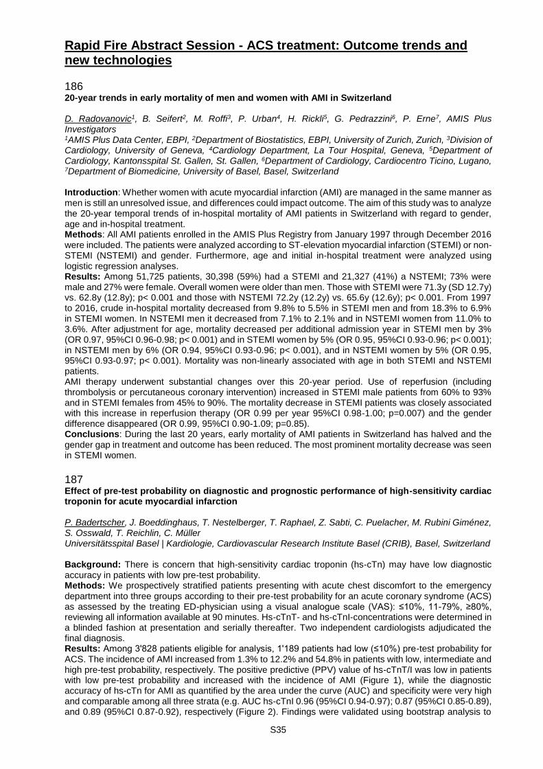
S35
Rapid Fire Abstract Session - ACS treatment: Outcome trends and new technologies 186 20-year trends in early mortality of men and women with AMI in Switzerland D. Radovanovic1, B. Seifert2, M. Roffi3, P. Urban4, H. Rickli5, G. Pedrazzini6, P. Erne7, AMIS Plus Investigators 1AMIS Plus Data Center, EBPI, 2Department of Biostatistics, EBPI, University of Zurich, Zurich, 3Division of Cardiology, University of Geneva, 4Cardiology Department, La Tour Hospital, Geneva, 5Department of Cardiology, Kantonsspital St. Gallen, St. Gallen, 6Department of Cardiology, Cardiocentro Ticino, Lugano, 7Department of Biomedicine, University of Basel, Basel, Switzerland Introduction: Whether women with acute myocardial infarction (AMI) are managed in the same manner as men is still an unresolved issue, and differences could impact outcome. The aim of this study was to analyze the 20-year temporal trends of in-hospital mortality of AMI patients in Switzerland with regard to gender, age and in-hospital treatment. Methods: All AMI patients enrolled in the AMIS Plus Registry from January 1997 through December 2016 were included. The patients were analyzed according to ST-elevation myocardial infarction (STEMI) or non-STEMI (NSTEMI) and gender. Furthermore, age and initial in-hospital treatment were analyzed using logistic regression analyses. Results: Among 51,725 patients, 30,398 (59%) had a STEMI and 21,327 (41%) a NSTEMI; 73% were male and 27% were female. Overall women were older than men. Those with STEMI were 71.3y (SD 12.7y) vs. 62.8y (12.8y); p< 0.001 and those with NSTEMI 72.2y (12.2y) vs. 65.6y (12.6y); p< 0.001. From 1997 to 2016, crude in-hospital mortality decreased from 9.8% to 5.5% in STEMI men and from 18.3% to 6.9% in STEMI women. In NSTEMI men it decreased from 7.1% to 2.1% and in NSTEMI women from 11.0% to 3.6%. After adjustment for age, mortality decreased per additional admission year in STEMI men by 3% (OR 0.97, 95%CI 0.96-0.98; p< 0.001) and in STEMI women by 5% (OR 0.95, 95%CI 0.93-0.96; p< 0.001); in NSTEMI men by 6% (OR 0.94, 95%CI 0.93-0.96; p< 0.001), and in NSTEMI women by 5% (OR 0.95, 95%CI 0.93-0.97; p< 0.001). Mortality was non-linearly associated with age in both STEMI and NSTEMI patients. AMI therapy underwent substantial changes over this 20-year period. Use of reperfusion (including thrombolysis or percutaneous coronary intervention) increased in STEMI male patients from 60% to 93% and in STEMI females from 45% to 90%. The mortality decrease in STEMI patients was closely associated with this increase in reperfusion therapy (OR 0.99 per year 95%CI 0.98-1.00; p=0.007) and the gender difference disappeared (OR 0.99, 95%CI 0.90-1.09; p=0.85). Conclusions: During the last 20 years, early mortality of AMI patients in Switzerland has halved and the gender gap in treatment and outcome has been reduced. The most prominent mortality decrease was seen in STEMI women.
187 Effect of pre-test probability on diagnostic and prognostic performance of high-sensitivity cardiac troponin for acute myocardial infarction P. Badertscher, J. Boeddinghaus, T. Nestelberger, T. Raphael, Z. Sabti, C. Puelacher, M. Rubini Giménez, S. Osswald, T. Reichlin, C. Müller Universitätsspital Basel | Kardiologie, Cardiovascular Research Institute Basel (CRIB), Basel, Switzerland Background: There is concern that high-sensitivity cardiac troponin (hs-cTn) may have low diagnostic accuracy in patients with low pre-test probability. Methods: We prospectively stratified patients presenting with acute chest discomfort to the emergency department into three groups according to their pre-test probability for an acute coronary syndrome (ACS) as assessed by the treating ED-physician using a visual analogue scale (VAS): ≤10%, 11-79%, ≥80%, reviewing all information available at 90 minutes. Hs-cTnT- and hs-cTnI-concentrations were determined in a blinded fashion at presentation and serially thereafter. Two independent cardiologists adjudicated the final diagnosis. Results: Among 3'828 patients eligible for analysis, 1'189 patients had low (≤10%) pre-test probability for ACS. The incidence of AMI increased from 1.3% to 12.2% and 54.8% in patients with low, intermediate and high pre-test probability, respectively. The positive predictive (PPV) value of hs-cTnT/I was low in patients with low pre-test probability and increased with the incidence of AMI (Figure 1), while the diagnostic accuracy of hs-cTn for AMI as quantified by the area under the curve (AUC) and specificity were very high and comparable among all three strata (e.g. AUC hs-cTnI 0.96 (95%CI 0.94-0.97); 0.87 (95%CI 0.85-0.89), and 0.89 (95%CI 0.87-0.92), respectively (Figure 2). Findings were validated using bootstrap analysis to
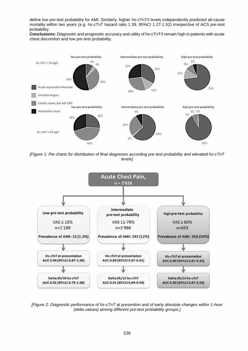
S36
define low pre-test probability for AMI. Similarly, higher hs-cTnT/I levels independently predicted all-cause mortality within two years (e.g. hs-cTnT hazard ratio 1.39, 95%CI 1.27-1.52) irrespective of ACS pre-test probability. Conclusions: Diagnostic and prognostic accuracy and utility of hs-cTnT/I remain high in patients with acute chest discomfort and low pre-test probability.
[Figure 1: Pie charts for distribution of final diagnoses according pre-test probability and elevated hs-cTnT
levels]
[Figure 2: Diagnostic performance of hs-cTnT at presention and of early absolute changes within 1-hour
(delta values) among different pre-test probability groups.]

S37
188 Bioresorbable vascular scaffolds (Absorb®) versus metallic stents in patients with ST-segment elevation myocardial infarction: a systematic review and meta-analysis M. Bossard1, T. Kofler2, F. Moccetti3, S. Toggweiler4, R. Jeger5, C. Kaiser5, R. Kobza4, S. Cook6, F. Cuculi4 1Hamilton General Hospital, McMaster University, Population Health Research Institute, Hamilton, ON, Canada, 2Innere Medizin, Universitätsspital Basel, Basel, Switzerland, 3Knight Cardiovascular Institute, Oregon Health Science University, Portland, OR, United States, 4Klinik für Kardiologie, Kantonsspital Luzern, Luzern, 5Klinik für Kardiologie, Universitätsspital Basel, Basel, 6Cardiologie, Université de Fribourg, Hôpital Cantonal, Fribourg, Switzerland Background: In patients with ST-segment elevation myocardial infarction (STEMI) patients, temporary vascular sealing with bioresorbable vascular scaffolds (BVS) might reflect a promising approach to overcome the not uncommon complications related to primary percutaneous coronary intervention with metallic stents (MS), including restenosis, stent thrombosis and neo-atherosclerosis. To evaluate the utility of BVS in STEMI patients, we performed a systematic review and meta-analysis. Methods: MEDLINE, EMBASE, and the Cochrane Register of Controlled Trials were searched from 1983 to December 2016. Studies comparing performance of BVS and MS in STEMI patients and providing data about clinical outcomes for more than 30days after index event were considered. Data were combined using random-effects models. Results: Of 8,393 citations, 6 studies (N=2,280) were included; 1 randomized trial (N=191). Procedure success was high with BVS and MS (96.78% versus 97.33%, p=0.79) Median follow up was 11.8 (8.5-16.5) months. Comparing rates of major adverse cardiovascular events (MACE) between STEMI patients treated with either BVS or MS, we found no significant differences, but there was a significant heterogeneity (BVS: 36/ 746 (4.8%) versus MS: 97/ 1,530 (6.3%) events; Odds ratio (OR) 0.81 [95% confidence interval (CI) 0.40-1.63], p=0.56; I2=48%; Figure A). Regarding target lesion revascularizations, there were no significant differences between BVS and MS (21/ 746 (2.8%) versus 42/ 1,530 (2.7%); OR 1.06 [95%CI 0.58-1.93], p=0.85; I2=0%). Overall, rates of definite stent thrombosis (ST) were higher among STEMI patients receiving BVS compared to MS (16/ 746 (2.1%) versus 13/ 1,530 (0.8%); OR 2.44 [95%CI 1.14-5.18], p=0.02; I2 0%, Figure B). Conclusions: To date, there is limited randomized data supporting the use of BVS in STEMI patients. According to mostly observational studies, usage of BVS in STEMIs might be safe with regards to MACE rates. But there is also a signal for higher rates of ST with BVS. However, an adequately powered trial is mandatory to finally define the role of BVS in the acute setting.
[Figure: Forest plots for the comparisons of bioresorbable vascular scaffolds (BVS) versus metallic stents
(MS) in STEMI patients: (A) Major adverse cardiovascular events (MACE); (B) Rates of definite stent thrombosis]
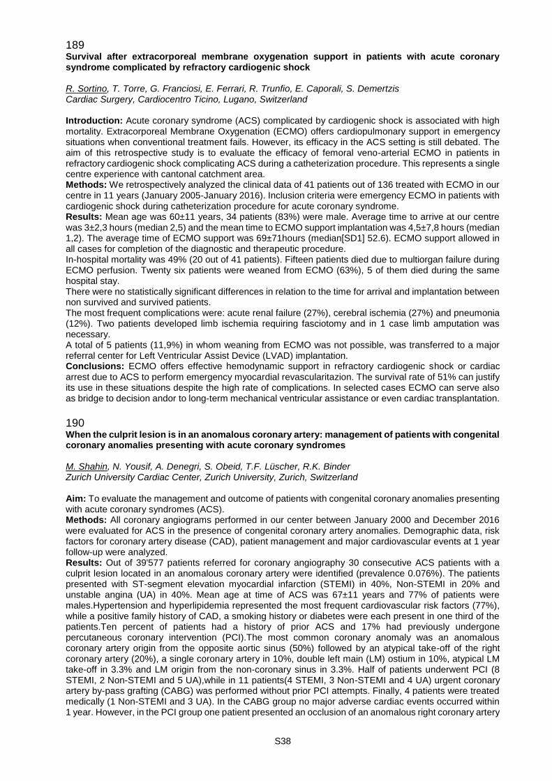
S38
189 Survival after extracorporeal membrane oxygenation support in patients with acute coronary syndrome complicated by refractory cardiogenic shock R. Sortino, T. Torre, G. Franciosi, E. Ferrari, R. Trunfio, E. Caporali, S. Demertzis Cardiac Surgery, Cardiocentro Ticino, Lugano, Switzerland Introduction: Acute coronary syndrome (ACS) complicated by cardiogenic shock is associated with high mortality. Extracorporeal Membrane Oxygenation (ECMO) offers cardiopulmonary support in emergency situations when conventional treatment fails. However, its efficacy in the ACS setting is still debated. The aim of this retrospective study is to evaluate the efficacy of femoral veno-arterial ECMO in patients in refractory cardiogenic shock complicating ACS during a catheterization procedure. This represents a single centre experience with cantonal catchment area. Methods: We retrospectively analyzed the clinical data of 41 patients out of 136 treated with ECMO in our centre in 11 years (January 2005-January 2016). Inclusion criteria were emergency ECMO in patients with cardiogenic shock during catheterization procedure for acute coronary syndrome. Results: Mean age was 60±11 years, 34 patients (83%) were male. Average time to arrive at our centre was 3±2,3 hours (median 2,5) and the mean time to ECMO support implantation was 4,5±7,8 hours (median 1,2). The average time of ECMO support was 69±71hours (median[SD1] 52.6). ECMO support allowed in all cases for completion of the diagnostic and therapeutic procedure. In-hospital mortality was 49% (20 out of 41 patients). Fifteen patients died due to multiorgan failure during ECMO perfusion. Twenty six patients were weaned from ECMO (63%), 5 of them died during the same hospital stay. There were no statistically significant differences in relation to the time for arrival and implantation between non survived and survived patients. The most frequent complications were: acute renal failure (27%), cerebral ischemia (27%) and pneumonia (12%). Two patients developed limb ischemia requiring fasciotomy and in 1 case limb amputation was necessary. A total of 5 patients (11,9%) in whom weaning from ECMO was not possible, was transferred to a major referral center for Left Ventricular Assist Device (LVAD) implantation. Conclusions: ECMO offers effective hemodynamic support in refractory cardiogenic shock or cardiac arrest due to ACS to perform emergency myocardial revascularitazion. The survival rate of 51% can justify its use in these situations despite the high rate of complications. In selected cases ECMO can serve also as bridge to decision andor to long-term mechanical ventricular assistance or even cardiac transplantation.
190 When the culprit lesion is in an anomalous coronary artery: management of patients with congenital coronary anomalies presenting with acute coronary syndromes M. Shahin, N. Yousif, A. Denegri, S. Obeid, T.F. Lüscher, R.K. Binder Zurich University Cardiac Center, Zurich University, Zurich, Switzerland Aim: To evaluate the management and outcome of patients with congenital coronary anomalies presenting with acute coronary syndromes (ACS). Methods: All coronary angiograms performed in our center between January 2000 and December 2016 were evaluated for ACS in the presence of congenital coronary artery anomalies. Demographic data, risk factors for coronary artery disease (CAD), patient management and major cardiovascular events at 1 year follow-up were analyzed. Results: Out of 39'577 patients referred for coronary angiography 30 consecutive ACS patients with a culprit lesion located in an anomalous coronary artery were identified (prevalence 0.076%). The patients presented with ST-segment elevation myocardial infarction (STEMI) in 40%, Non-STEMI in 20% and unstable angina (UA) in 40%. Mean age at time of ACS was 67±11 years and 77% of patients were males.Hypertension and hyperlipidemia represented the most frequent cardiovascular risk factors (77%), while a positive family history of CAD, a smoking history or diabetes were each present in one third of the patients.Ten percent of patients had a history of prior ACS and 17% had previously undergone percutaneous coronary intervention (PCI).The most common coronary anomaly was an anomalous coronary artery origin from the opposite aortic sinus (50%) followed by an atypical take-off of the right coronary artery (20%), a single coronary artery in 10%, double left main (LM) ostium in 10%, atypical LM take-off in 3.3% and LM origin from the non-coronary sinus in 3.3%. Half of patients underwent PCI (8 STEMI, 2 Non-STEMI and 5 UA),while in 11 patients(4 STEMI, 3 Non-STEMI and 4 UA) urgent coronary artery by-pass grafting (CABG) was performed without prior PCI attempts. Finally, 4 patients were treated medically (1 Non-STEMI and 3 UA). In the CABG group no major adverse cardiac events occurred within 1 year. However, in the PCI group one patient presented an occlusion of an anomalous right coronary artery
S39
during PCI of the circumflex artery and two bare-metal stent restenosis occurred within one-year follow-up (at 11 months and 8 months). Conclusion: Percutaneous revascularization of anomalous coronary arteries is challenging in the setting of acute myocardial infarction. If the origin or course of the anomalous coronary artery is not suitable for PCI or the extent of coronary disease precludes a percutaneous approach, prompt surgical revascularization is a valuable treatment strategy.
191 Ultrathin strut biodegradable polymer sirolimus-eluting stent versus durable polymer everolimus-eluting stent for coronary revascularization K. Yamaji, T. Zanchin, C. Zanchin, T. Pilgrim, S. Stortecky, K.C. Koskinas, L. Hunziker, F. Praz, S. Blöchlinger, C. Moro, M. Billinger, A. Moschovitis, M. Valgimigli, S. Windecker, L. Räber Bern University Hospital, Bern, Switzerland Aims: Biodegradable polymer, ultrathin strut sirolimus eluting stents (BP-SES) have been reported to be non-inferior in terms of the device-oriented composite endpoint (DOCE) compared with durable polymer, thin strut everolimus-eluting stents (DP-EES) in a randomized clinical trial (RCT). The purpose of the present study was to validate the efficacy and safety of BP-SES compared with DP-EES in an all-comers population. Methods and Results: Among 6001 consecutive patients (9978 lesions) who underwent PCI at Bern University Hospital between March 2011 to June 2014, 3602 patients (5961 lesions) were exclusively treated with BP-SES (N=1430, 2390 lesions) or DP-EES (N=2172, 3569 lesions). After propensity score (PS) matching, the final study population consisted of 1111 matched pairs (BP-SES 1844 lesions, DP-EES 1829 lesions). The primary device-oriented composite endpoint (DOCE) included cardiac death, target-vessel myocardial infarction (MI) and target lesion revascularization (TLR) at 1 year. Clinical indication to undergo PCI was well balanced between the two groups, including STEMI in 466 patients (21.0%), NSTEMI in 596 patients (26.8%), unstable angina pectoris (uAP) in 128 patients (5.8%) and stable coronary artery disease (CAD) in 1032 patients (46.4%). Clinical follow-up information was obtained in 94.9% of patients at 1 year. BP-SES as compared with DP-EES was associated with a similar cumulative incidence of DOCE (6.0% versus 7.9%, HR 0.76, 95%CI 0.55-1.05, P=0.09). There were no significant differences with regards to cardiac death (HR 0.68, 95%CI 0.41-1.14, P=0.15), target-vessel MI (HR 0.84, 95%CI 0.50-1.40, P=0.51), and TLR (HR 0.87, 95%CI 0.52-1.45, P=0.59). The rate of definite stent thrombosis was low within 1 year in both groups (BP-SES: 0.8% versus EES 0.9%, HR 0.90, 95%CI 0.36-2.21, P=0.82). A positive interaction suggesting a potential benefit of BP-SES was observed among female patients (pinteraction=0.018). Conclusions: In this consecutively enrolled all-comer population, BP-SES was associated with a similar efficacy and safety as compared with DP-EES.
192 Early experience with the magnesium bioresorbable vascular scaffold in an all-comers Population G. Hilfiker, S. Toggweiler, J. Chronis, M. Brinkert, P. Jamshidi, R. Kobza, F. Cuculi Herzzentrum | Kardiologie, Kantonsspital Luzern, Luzern, Switzerland Introduction: The Magmaris magnesium-based bioresorbable vascular scaffold (BVS) has recently been introduced into clinical practice promising temporary scaffolding and quick vessel restoration. We report our early experience with this device in all-comers presenting with coronary artery disease in our institution. Methods: All patients treated using Magmaris since the introduction of the device in June 2016 up to December 2016 are included in this analysis. Aggressive lesion preparation using non-compliant balloons was performed in all patients and in 81/82 patients post-dilatatation with non-compliant balloons was performed. Patients were followed-up during clinical visits or via telephone. We describe the rates of target lesion failure (TLF), which includes in-scaffold-restenosis (ISR) and probable/definitive scaffold thrombosis (ST). Interventional cardiologists were encouraged to perform optical coherence tomography (OCT) in patients presenting with TLF. All patients received double antiplatelet-therapy according to current guidelines. Results: A total of 82 patients with a mean age of 62 years were included. 45 patients (55 %) had acute myocardial infarction (MI): ST-elevation MI (n=20), non-ST AMI (n=25). Follow-up was performed in all patients and the mean follow-up duration was 143 +48 days. A total of 5 TLF (6.1 %) including 2 ISR (2.4 %) and 3 ST (3.7 %) occured. All ST were definitive with angiographic evidence of acute vessel closure and all occurred < 30 days post implantation. OCT was performed in 3/5 patients presenting with TLF and demonstrated scaffold collapse and scaffold fractures. Conclusion: Our initial experience with the Magmaris scaffold demonstrates high rates of early ST. OCT imaging demonstrated scaffold collapse as the main mechanism of TLF in these patients.
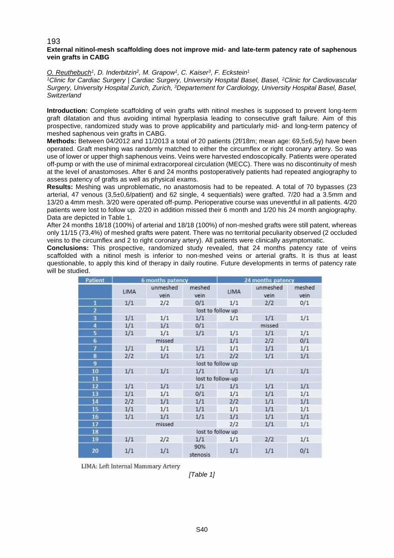
S40
193 External nitinol-mesh scaffolding does not improve mid- and late-term patency rate of saphenous vein grafts in CABG O. Reuthebuch1, D. Inderbitzin2, M. Grapow1, C. Kaiser3, F. Eckstein1 1Clinic for Cardiac Surgery | Cardiac Surgery, University Hospital Basel, Basel, 2Clinic for Cardiovascular Surgery, University Hospital Zurich, Zurich, 3Departement for Cardiology, University Hospital Basel, Basel, Switzerland Introduction: Complete scaffolding of vein grafts with nitinol meshes is supposed to prevent long-term graft dilatation and thus avoiding intimal hyperplasia leading to consecutive graft failure. Aim of this prospective, randomized study was to prove applicability and particularly mid- and long-term patency of meshed saphenous vein grafts in CABG. Methods: Between 04/2012 and 11/2013 a total of 20 patients (2f/18m; mean age: 69,5±6,5y) have been operated. Graft meshing was randomly matched to either the circumflex or right coronary artery. So was use of lower or upper thigh saphenous veins. Veins were harvested endoscopically. Patients were operated off-pump or with the use of minimal extracorporeal circulation (MECC). There was no discontinuity of mesh at the level of anastomoses. After 6 and 24 months postoperatively patients had repeated angiography to assess patency of grafts as well as physical exams. Results: Meshing was unproblematic, no anastomosis had to be repeated. A total of 70 bypasses (23 arterial, 47 venous (3,5±0,6/patient) and 62 single, 4 sequentials) were grafted. 7/20 had a 3.5mm and 13/20 a 4mm mesh. 3/20 were operated off-pump. Perioperative course was uneventful in all patients. 4/20 patients were lost to follow up. 2/20 in addition missed their 6 month and 1/20 his 24 month angiography. Data are depicted in Table 1. After 24 months 18/18 (100%) of arterial and 18/18 (100%) of non-meshed grafts were still patent, whereas only 11/15 (73,4%) of meshed grafts were patent. There was no territorial peculiarity observed (2 occluded veins to the circumflex and 2 to right coronary artery). All patients were clinically asymptomatic. Conclusions: This prospective, randomized study revealed, that 24 months patency rate of veins scaffolded with a nitinol mesh is inferior to non-meshed veins or arterial grafts. It is thus at least questionable, to apply this kind of therapy in daily routine. Future developments in terms of patency rate will be studied.
[Table 1]
S41
Abstract Session - SSCS 195 Recipient's blood group A is associated with longer survival of cardiac valvular bioprostheses O. Schussler1, N. Lila2, T. Perneger3, P. Mootoosamy4, A. François5, D. Smadja6, M. Cikirikcioglu7, M. Ruel8, A. Kalangos9, C. Huber10, A. Carpentier11 1HUG, Department of Cardiac Surgery/Cardiovascular Research Laboratory, Geneva, Switzerland, 2Laboratory of biosurgical Research, University Paris Descartes, Paris, France, 3Department of Clinical Epidemiology, 4Department of Cardiosvascular Surgery, HUG, Geneva, Switzerland, 5Etablissement Français du Sang, University Georges Pompidou Hospital, 6Immuno-Hematology Department, Georges Pompidou University Hospital, Paris, France, 7Department of Cardiovascular Surgery, HUG, Geneva, Switzerland, 8Cardiac Surgery, University of Ottawa / ottawa Heart Institute, Ottawa, ON, Canada, 9Cardiovascular Surgery, 10Department of Cardiovascular Surgery/Cardiovascular Research Laboratory, HUG, Geneva, Switzerland, 11Laboratory of Biosurgical Research, University Paris Descartes, Paris, France Background: Pigs/bovines share with humans some of the antigens present on cardiac valves. Two such antigens are the major xenogenic Ag, “a-Gal” which mimics the human B-antigen of ABO-blood group system and the minor Ag, pig A-antigen of AH-blood group which is similar to that of human AH-antigen. We hypothesize that these antigens may modify the immunogenicity of the bioprosthesis and also its longevity. ABO distribution may vary between patients with low (< 6 years) and high (≥15 years) bioprostheses longevity. Methods: Single-centre registry study (Paris, France) including all degenerative porcine bioprostheses (mostly Carpentier-Edwards 2nd/3rd generation heart valves) explanted between 1987-1998 and some bovine bioprostheses. For period 1998-2014 all porcine bioprostheses with longevity ≥13 years (follow-up >27 years). Predictive factors for bioprosthesis longevity: number, site of implantation, age were collected. Blood group and other variables were entered into an ordinal logistic regression analysis model predicting valve longevity, categorized as low (< 6 years), medium (6-14.9 years), and high (≥15 years). Results: Longevity and ABO-blood group were obtained for 548 explanted porcine bioprosthesis. Mean longevity was 10.2 years+3.9 [0-28] and significantly higher for A-patient than others (P=0.009). Using multivariate analysis, group A was a strong predictive factor of longevity (OR 2.09; P< 0.001). For the 64 explanted bovine bioprosthesis with low/medium longevity, the effect of group A was even more significant. Conclusions: Patients of A-group but not B have a higher longevity of their bioprostheses. Future graft-host phenotyping and matching may give rise to a new generation of long-lasting bioprosthesis for implantation in humans, especially for the younger population.
196 Ejection fraction improvement after aortic valve replacement in patients with reduced left ventricular systolic function V. Ntinopoulos, H. Löblein, O. Dzemali, A. Häussler, D. Odavic, A. Zientara, M. Gruszczynski, M. Genoni Cardiac Surgery Department, Triemli Hospital, Zurich, Switzerland Introduction: Reduced left ventricular systolic function is an important risk factor associated with adverse prognosis and prolonged postoperative hospital stay following cardiac surgery. Aim of this study is to examine the improvement of ejection fraction and contributing factors in such patients undergoing aortic valve replacement (AVR). Methods: We performed a retrospective observational study which included 70 patients with an ejection fraction lower than 40% that underwent AVR from April 2001 to August 2016. Ejection fraction was measured at discharge, 3 months and 1 year postoperatively. Student's t-test, Pearson's and Spearman's correlation were performed to identify factors leading to left ventricular systolic function improvement and a multiple linear regression was also performed to model their relationship. Results: The median age of the patients was 72 years (IQR 66-77). Severe aortic valve stenosis was diagnosed in 64 and severe aortic insufficiency was diagnosed in 10 patients. The median ejection fraction preoperatively was 30% (IQR 21.8-33%). The in-hospital postoperative mortality was 7.1%. There was a statistically significant improvement of ejection fraction 3 months (mean 21.3%, SD 14.3%, p< 0.0005) and 1 year (mean 27.7%, SD 13.8%, p< 0.0005) postoperatively. Factors statistically significantly related with an ejection fraction improvement at 3 months were female gender (p=0.001), absence of pulmonary disease (p=0.01), absence of renal failure (p=0.044), higher mean aortic valve gradient preoperatively (p< 0.0005) and presence of moderate or severe aortic valve regurgitation preoperatively (p=0.046) and at 1 year postoperatively female gender (p=0.001), no previous coronary interventions (p=0.017), no smoking (p=0.025), absence of pulmonary disease (p=0.013), higher mean aortic valve gradient preoperatively (p=0.049) and presence of moderate or severe aortic valve regurgitation preoperatively (p=0.039).
S42
Conclusions: A statistically significant improvement of ejection fraction was observed at 3 months and 1 year postoperatively. Factors statistically significantly related with left ventricular systolic function improvement postoperatively are female gender, absence of pulmonary disease, absence of renal failure, no smoking, no previous coronary interventions, higher mean aortic valve gradient preoperatively and presence of moderate or severe aortic valve regurgitation preoperatively.
197 Rapid-deployment intuity aortic valve replacement vs perimount magna ease bioprosthesis: clinical and hemodynamic results in a matched population E. Ferrari1,2,3, C. Roduit2, P. Salamin2, E. Caporali1, T. Torre1, R. Trunfio1, D. Berdajs4, L. von Segesser3, S. Demertzis1 1Cardiac Surgery, Cardiocentro Ticino, Lugano, 2Cardiac Surgery, 3Cardiovascular Research Unit, CHUV, Lausanne, 4Cardiac Surgery, Basel University Hospital, Basel, Switzerland Introduction: The rapid-deployment Intuity aortic valve is based on the well-known Perimount Magna Ease bioprostesis and features a new anchoring system with a balloon expandable stent placed below the annulus. The aim of the study is to compare the clinical outcome and the one-year hemodynamic results of the rapid-deployment Intuity aortic valve system with the Perimount Magna bioprosthesis in two cross-matched populations. Method: Between March 2014 and May 2015, 32 patients underwent aortic valve replacement with the Intuity valve system (Intuity-group). A matched population of patients with Perimount valves implanted during the same period of time was identified and used as a control group (Perimount-Group). Clinical data and outcome were compared. Valve hemodynamics were echocardiographically compared at baseline, discharge and after one-year. Results: There were more female in the Intuity-group (47% vs 22%, p=0.035), mean age was 78±6 and 73±5years in the Intuity and Perimount-group, respectively (p< 0.001) and the coronary disease was more common in the Intuity-group (65% vs 25%, p=0.005). Further preoperative characteristics were similar between the groups. Successful implantation rate was 100% at first attempt. Cross-clamp (mean:50±25 vs 53±22min, p=0.004), cardiopulmonary bypass (mean:68±27 vs 72±31min; p=0.007) and surgical times (mean:156±54 vs 165±40min; p=0.018) were shorter with Intuity despite the higher number of concomitant procedures (12 vs 7 patients). Mean valve size was 23.7mm (Intuity-group) and 24.1mm (Perimount-group) without annulus enlargement procedures performed; hospital mortality was zero (Intuity-group) and 3% (Perimount-group); new pacemakers were 2(6%) for Intuity and 1(3%) for Perimount (p=0.55). Postoperative peak and mean gradients were 18±6-31±7mmHg (p=0.026) and 9.8±3-13±4mmHg (p=0.022) for rapid-deployment and standard valves, respectively. Hospital stay was equivalent. At follow-up, mean gradients were 9±4mmHg for the Intuity and 14±4mmHg for the Perimount. Moderate paravalvular-leak was present in one Intuity (3%) at discharge and at follow-up. Conclusion: The Intuity valve system shows improved hemodynamic results compared to the standard Perimount valve with same mean implanted valve size. However, postoperative paravalvular leak should be strongly prevented during Intuity valve implantation.
198 Ticagrelor does not increase postoperative bleeding complications and the need for transfusions compared to clopidogrel or aspirin in off-pump coronary artery bypass surgery P. Matt1, F. Cuculi2, S. Toggweiler2, R. Kobza2, C. Müller1, P. Banerjee1, T. Syburra1, X. Müller1 1Herzzentrum | Klinik für Herzchirurgie, 2Herzzentrum | Klinik für Kardiologie, Luzerner Kantonsspital, Luzern, Switzerland Introduction: We hypothesized that patients treated with ticagrelor compared with those treated with clopidogrel or aspirin before off-pump coronary artery bypass surgery (OPCAB) are at increased risk of hemorrhagic complications needing transfusions and are more likely to need surgical reexploration. Method: Preoperative and postoperative clinical data were collected prospectively on 548 consecutive patients treated preoperatively with ticagrelor and aspirin (group T, n=34), clopidogrel and aspirin (group C, n=127) or only aspirin (group A, n=387) before undergoing isolated OPCAB at our institution from 2010 to 2016. Results: Patient demographics showed no significant differences among patient groups, however more patients in the ticagrelor and the clopidogrel group underwent urgent/emergent OPCAB compared with those under aspirin treatment, 20% vs. 16% vs 4% (p< 0.001). Chest tube drainage showed no significant differences among patient groups 12 hours after OPCAB, 416±187ml (group T) vs 491.7±246ml (group C) vs 453.9±225ml (group A), and a slight increase in chest tube drainage 24 hours after surgery in those with clopidogrel compared to those with ticagrelor, 778.1±355ml (group C) vs 622.1±258ml (group T, p=0.02), but not compared to aspirin 720.8±349ml (group A).The number of transfusions of red blood cells, platelets
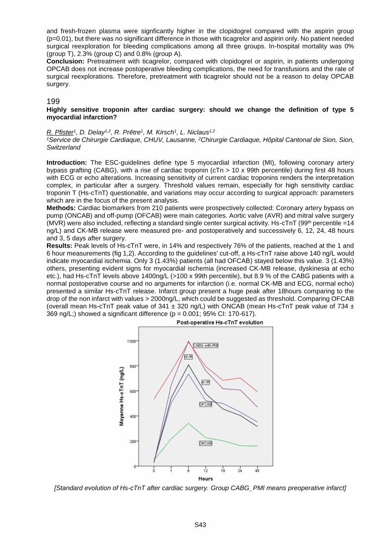
S43
and fresh-frozen plasma were signficantly higher in the clopidogrel compared with the aspirin group (p=0.01), but there was no significant difference in those with ticagrelor and aspirin only. No patient needed surgical reexploration for bleeding complications among all three groups. In-hospital mortality was 0% (group T), 2.3% (group C) and 0.8% (group A). Conclusion: Pretreatment with ticagrelor, compared with clopidogrel or aspirin, in patients undergoing OPCAB does not increase postoperative bleeding complications, the need for transfusions and the rate of surgical reexplorations. Therefore, pretreatment with ticagrelor should not be a reason to delay OPCAB surgery.
199 Highly sensitive troponin after cardiac surgery: should we change the definition of type 5 myocardial infarction? R. Pfister1, D. Delay1,2, R. Prêtre1, M. Kirsch1, L. Niclaus1,2 1Service de Chirurgie Cardiaque, CHUV, Lausanne, 2Chirurgie Cardiaque, Hôpital Cantonal de Sion, Sion, Switzerland Introduction: The ESC-guidelines define type 5 myocardial infarction (MI), following coronary artery bypass grafting (CABG), with a rise of cardiac troponin (cTn > 10 x 99th percentile) during first 48 hours with ECG or echo alterations. Increasing sensitivity of current cardiac troponins renders the interpretation complex, in particular after a surgery. Threshold values remain, especially for high sensitivity cardiac troponin T (Hs-cTnT) questionable, and variations may occur according to surgical approach: parameters which are in the focus of the present analysis. Methods: Cardiac biomarkers from 210 patients were prospectively collected: Coronary artery bypass on pump (ONCAB) and off-pump (OFCAB) were main categories. Aortic valve (AVR) and mitral valve surgery (MVR) were also included, reflecting a standard single center surgical activity. Hs-cTnT (99th percentile =14 ng/L) and CK-MB release were measured pre- and postoperatively and successively 6, 12, 24, 48 hours and 3, 5 days after surgery. Results: Peak levels of Hs-cTnT were, in 14% and respectively 76% of the patients, reached at the 1 and 6 hour measurements (fig 1,2). According to the guidelines' cut-off, a Hs-cTnT raise above 140 ng/L would indicate myocardial ischemia. Only 3 (1.43%) patients (all had OFCAB) stayed below this value. 3 (1.43%) others, presenting evident signs for myocardial ischemia (increased CK-MB release, dyskinesia at echo etc.), had Hs-cTnT levels above 1400ng/L (>100 x 99th percentile), but 8.9 % of the CABG patients with a normal postoperative course and no arguments for infarction (i.e. normal CK-MB and ECG, normal echo) presented a similar Hs-cTnT release. Infarct group present a huge peak after 18hours comparing to the drop of the non infarct with values > 2000ng/L, which could be suggested as threshold. Comparing OFCAB (overall mean Hs-cTnT peak value of 341 ± 320 ng/L) with ONCAB (mean Hs-cTnT peak value of 734 ± 369 ng/L;) showed a significant difference (p = 0.001; 95% CI: 170-617).
[Standard evolution of Hs-cTnT after cardiac surgery. Group CABG_PMI means preoperative infarct]

S44
[Standard evolution of CK-MB after cardiac surgery. Group CABG_PMI means preoperative infarct.]
Conclusions: The link of the definition for type 5 MI of the ESC guidelines to the threshold for Hs-cTnT remains highly questionable and was not reproducible. The actual cut-off is also uselessness, when considered as a coupled value to other criteria: according to the definition 98.6% of all surgical patients had pathologic postoperative Hs-cTnT raise. According to the present results per-operative infraction may be unlikely in CABG surgery if Hs-cTnT raise stays below.
200 Impact of clopidogrel and aspirin therapy after off pump coronary artery bypass surgery. Is postoperative double anti platelet therapy mandatory? A. Haeussler, L. Rings, S. Dushaj, D. Odavic, K. Graves, O. Dzemali, M. Genoni Herzchirurgie, Stadtspital Triemli Zürich, Zürich, Switzerland Introduction: Hypercoagolabity in off pump coronary artery bypass grafting (OPCAB) is well known problem in literature. Therefore, double-anti platelet therapy of clopidogrel and aspirin is maybe important for the impact of patency rate of bypass grafts in OPCAB surgery. Method: 143 patients were enrolled in this prospective randomized study. All pts underwent OPCAB surgery. Blood platelet function test was drawn 5 d before, 5d after surgery, at 3 month after surgery and at one year of follow up. Platelet function analyser (PFA-100TM) was used for evaluation of clotting time. Turbidimetric platelet aggregation and impedance platelet aggregation was induced by arachidonic acid, collagen and adenosine di- phosphate to monitor effect on the thrombocytes. To evaluate bypass graft patency rate a 64-slice cardiac computer tomogram was performed after one year. Endpoint were patency rate of bypass grafts, mortality, rate of reintervention, course of cardiac enzymes (CK, CK-MB and TropT), C reactive protein and bleeding complications. Results: Mean age was 68±4.65y, 120 were men and 23 women. Average risk rate (EuroScore logistic) was 4.04±1.4%. Use of bilateral mammarian artery was 54%. Rate of non-responders for Clopidogrel was significant elevated than for aspirin (13.3% vs. 4.2 %). 3.8 bypass grafts per patient were done. Patency rate does not differ in all pts. There was no significant difference in postoperative bleeding rates, CK and CK-MB rates, Trop T and CRP. Cardiac CT scan at on 1 year follow up showed no significant difference regarding graft patency rates. No mortality was recorded. Conclusion: Administration of a combined regime of clopidogrel and aspirin after OPCAB is not associated with increased postoperative bleeding rates. The pts, who were non-responders for clopidogrel showed the same in patency rate of the bypass grafts as the responders. Finally, we could not find any benefit for pts undergoing double antiplatelet therapy with aspirin and clopidogrel.

S45
Abstract Session - Unusual cardiac situation, clinical cases 201 Ticagrelor loading dose leading to prolonged asystole S. Rosset, E. Pruvot, O. Muller, P. Pascale Cardiology, CHUV, Lausanne, Switzerland Treatment with ticagrelor has been associated with an increased incidence in bradyarrhythmias, but these occurrences are considered as clinically silent. Herein we report the first case of a syncopal ventricular pause attributed to ticagrelor treatment in the absence of coronary artery disease. A 40 year old Caucasian female patient was addressed to our hospital for diagnostic coronary angiography for suspected unstable angina. Her baseline ECG (panel A) showed no conduction abnormalities. Her echocardiogram and laboratory work-up was unremarkable. She was admitted to our coronary care unit where a 180 mg loading dose of ticagrelor and 2.5 mg of fondaparinux were administred. 120 min after ticagrelor administration, she experienced syncope while supine. ECG monitoring revealed a 13 and 5.8 seconds asystole followed by a transient slow junctional escape as shown in panel B. Coronary angiogram showed no coronary artery disease. A cardiac MRI with late gadolinium enhancement showed a localized transmural scar in apical-lateral segment. A metabolic FDG18 PET-scan showed no focal activity. Pacemaker implantation was initially considered but subsequent monitoring for 1 week showed no recurrence of bradyarrhythmia. She was discharged with an implanted loop recorder (Reveal LINQ™, Medtronic). After 2 months of monitoring, no recurrence of abnormal pauses or bradycardic events were observed. While there have been two previous reports of symptomatic bradyarrhythmias related to ticagrelor requiring the insertion of a temporary transvenous pacemaker, these cases occurred in the setting of an acute coronary syndrome and shortly after coronary angioplasty. To the best of our knowledge, our case is the first to document the occurence of a life-threatening bradyarrhythmic event without associated confounding factors related to coronary artery disease. Moreover, the time sequence of events clearly supports the causal role of ticagrelor, as asystole occurred after the expected time to reach the peak plasma concentration (ie 1.3-2 hours). No recurrence was observed despite continuous monitoring with an implantable device in the following months. In conclusion, contrary to what might be assumed, our case demonstrates that a single loading dose of ticagrelor may lead to severe symptomatic bradyarrhythmic events.
[figure 1]
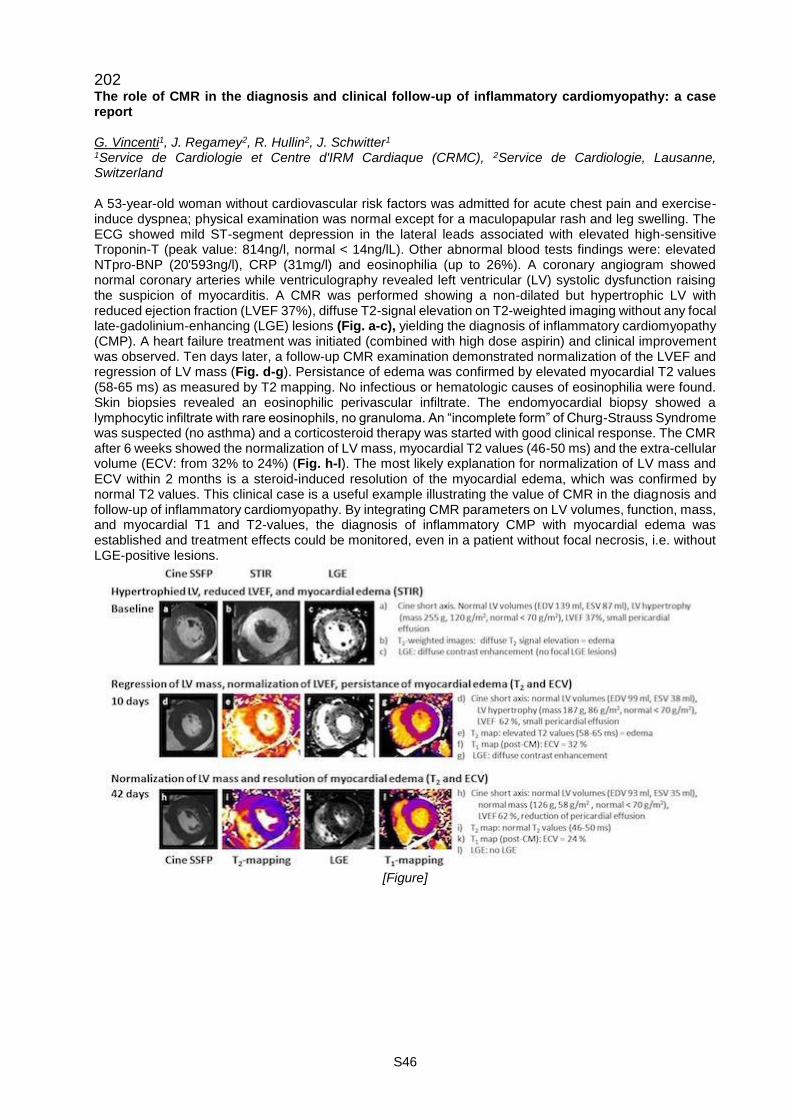
S46
202 The role of CMR in the diagnosis and clinical follow-up of inflammatory cardiomyopathy: a case report G. Vincenti1, J. Regamey2, R. Hullin2, J. Schwitter1 1Service de Cardiologie et Centre d'IRM Cardiaque (CRMC), 2Service de Cardiologie, Lausanne, Switzerland A 53-year-old woman without cardiovascular risk factors was admitted for acute chest pain and exercise-induce dyspnea; physical examination was normal except for a maculopapular rash and leg swelling. The ECG showed mild ST-segment depression in the lateral leads associated with elevated high-sensitive Troponin-T (peak value: 814ng/l, normal < 14ng/lL). Other abnormal blood tests findings were: elevated NTpro-BNP (20'593ng/l), CRP (31mg/l) and eosinophilia (up to 26%). A coronary angiogram showed normal coronary arteries while ventriculography revealed left ventricular (LV) systolic dysfunction raising the suspicion of myocarditis. A CMR was performed showing a non-dilated but hypertrophic LV with reduced ejection fraction (LVEF 37%), diffuse T2-signal elevation on T2-weighted imaging without any focal late-gadolinium-enhancing (LGE) lesions (Fig. a-c), yielding the diagnosis of inflammatory cardiomyopathy (CMP). A heart failure treatment was initiated (combined with high dose aspirin) and clinical improvement was observed. Ten days later, a follow-up CMR examination demonstrated normalization of the LVEF and regression of LV mass (Fig. d-g). Persistance of edema was confirmed by elevated myocardial T2 values (58-65 ms) as measured by T2 mapping. No infectious or hematologic causes of eosinophilia were found. Skin biopsies revealed an eosinophilic perivascular infiltrate. The endomyocardial biopsy showed a lymphocytic infiltrate with rare eosinophils, no granuloma. An “incomplete form” of Churg-Strauss Syndrome was suspected (no asthma) and a corticosteroid therapy was started with good clinical response. The CMR after 6 weeks showed the normalization of LV mass, myocardial T2 values (46-50 ms) and the extra-cellular volume (ECV: from 32% to 24%) (Fig. h-l). The most likely explanation for normalization of LV mass and ECV within 2 months is a steroid-induced resolution of the myocardial edema, which was confirmed by normal T2 values. This clinical case is a useful example illustrating the value of CMR in the diagnosis and follow-up of inflammatory cardiomyopathy. By integrating CMR parameters on LV volumes, function, mass, and myocardial T1 and T2-values, the diagnosis of inflammatory CMP with myocardial edema was established and treatment effects could be monitored, even in a patient without focal necrosis, i.e. without LGE-positive lesions.
[Figure]
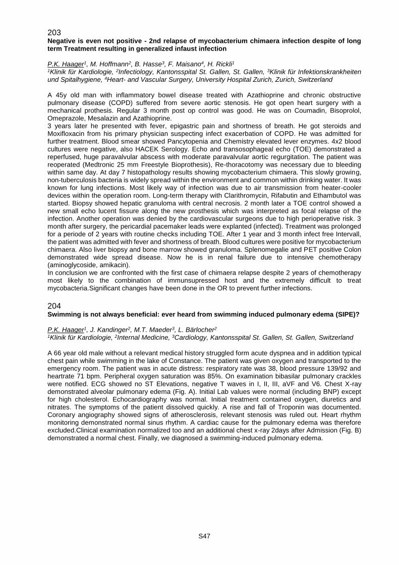
S47
203 Negative is even not positive - 2nd relapse of mycobacterium chimaera infection despite of long term Treatment resulting in generalized infaust infection P.K. Haager1, M. Hoffmann2, B. Hasse3, F. Maisano4, H. Rickli1 1Klinik für Kardiologie, 2Infectiology, Kantonsspital St. Gallen, St. Gallen, 3Klinik für Infektionskrankheiten und Spitalhygiene, 4Heart- and Vascular Surgery, University Hospital Zurich, Zurich, Switzerland A 45y old man with inflammatory bowel disease treated with Azathioprine and chronic obstructive pulmonary disease (COPD) suffered from severe aortic stenosis. He got open heart surgery with a mechanical prothesis. Regular 3 month post op control was good. He was on Coumadin, Bisoprolol, Omeprazole, Mesalazin and Azathioprine. 3 years later he presented with fever, epigastric pain and shortness of breath. He got steroids and Moxifloxacin from his primary physician suspecting infect exacerbation of COPD. He was admitted for further treatment. Blood smear showed Pancytopenia and Chemistry elevated lever enzymes. 4x2 blood cultures were negative, also HACEK Serology. Echo and transosophageal echo (TOE) demonstrated a reperfused, huge paravalvular abscess with moderate paravalvular aortic regurgitation. The patient was reoperated (Medtronic 25 mm Freestyle Bioprothesis), Re-thoracotomy was necessary due to bleeding within same day. At day 7 histopathology results showing mycobacterium chimaera. This slowly growing, non-tuberculosis bacteria is widely spread within the environment and common within drinking water. It was known for lung infections. Most likely way of infection was due to air transmission from heater-cooler devices within the operation room. Long-term therapy with Clarithromycin, Rifabutin and Ethambutol was started. Biopsy showed hepatic granuloma with central necrosis. 2 month later a TOE control showed a new small echo lucent fissure along the new prosthesis which was interpreted as focal relapse of the infection. Another operation was denied by the cardiovascular surgeons due to high perioperative risk. 3 month after surgery, the pericardial pacemaker leads were explanted (infected). Treatment was prolonged for a periode of 2 years with routine checks including TOE. After 1 year and 3 month infect free Intervall, the patient was admitted with fever and shortness of breath. Blood cultures were positive for mycobacterium chimaera. Also liver biopsy and bone marrow showed granuloma. Splenomegalie and PET positive Colon demonstrated wide spread disease. Now he is in renal failure due to intensive chemotherapy (aminoglycoside, amikacin). In conclusion we are confronted with the first case of chimaera relapse despite 2 years of chemotherapy most likely to the combination of immunsupressed host and the extremely difficult to treat mycobacteria.Significant changes have been done in the OR to prevent further infections.
204 Swimming is not always beneficial: ever heard from swimming induced pulmonary edema (SIPE)? P.K. Haager1, J. Kandinger2, M.T. Maeder3, L. Bärlocher2 1Klinik für Kardiologie, 2Internal Medicine, 3Cardiology, Kantonsspital St. Gallen, St. Gallen, Switzerland A 66 year old male without a relevant medical history struggled form acute dyspnea and in addition typical chest pain while swimming in the lake of Constance. The patient was given oxygen and transported to the emergency room. The patient was in acute distress: respiratory rate was 38, blood pressure 139/92 and heartrate 71 bpm. Peripheral oxygen saturation was 85%. On examination bibasilar pulmonary crackles were notified. ECG showed no ST Elevations, negative T waves in I, II, III, aVF and V6. Chest X-ray demonstrated alveolar pulmonary edema (Fig. A). Initial Lab values were normal (including BNP) except for high cholesterol. Echocardiography was normal. Initial treatment contained oxygen, diuretics and nitrates. The symptoms of the patient dissolved quickly. A rise and fall of Troponin was documented. Coronary angiography showed signs of atherosclerosis, relevant stenosis was ruled out. Heart rhythm monitoring demonstrated normal sinus rhythm. A cardiac cause for the pulmonary edema was therefore excluded.Clinical examination normalized too and an additional chest x-ray 2days after Admission (Fig. B) demonstrated a normal chest. Finally, we diagnosed a swimming-induced pulmonary edema.
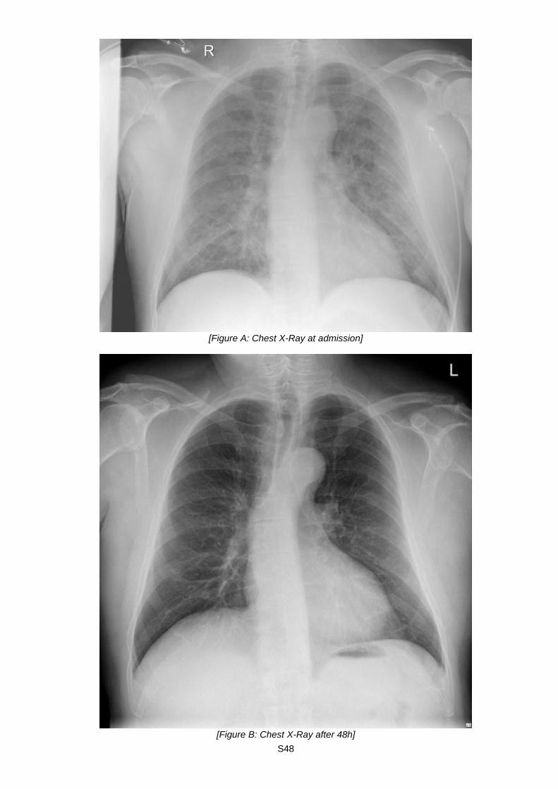
S48
[Figure A: Chest X-Ray at admission]
[Figure B: Chest X-Ray after 48h]
S49
Discussion: the pathophysiology of SIPE was initially not complete understood. Experiments point out, that probably an exercise-induced elevation of pulmonary capillary pressure is causing mechanical stress failure of the pulmonary capillaries. A leakage of erythrocytes, macromolecules and fluid across the damaged pulmonary capillary membranes into the alveoli occurs. This results in an acute pulmonary edema.Immersion, elevation of intrathoracic pressure, hypothermia, cold water, negative static lung load, exertion, fluid loading and low vital capacity are increasing the risk of SIPE.To diagnose SIPE with typical symptomas during or shortly after swimming other causes of pulmonary edema havet to be excluded (larngospasm, water aspiration). Typically, the pulmonary edema is cleared shortly (within 48h) after the acute situation.Recommended treatment consists of leaving water, stopping physical exercise, oxygen and diuretics. Prognosis is good without any structural or functional lung damage. Patients suffered from SIPE are on increased risk of a relapse in the same situation. In a small well conducted study, Sildenafil seems to be a reasonable prophylactic therapy to prevent a second episode if taken before exposure (Richard, Circulation 2016).
205 A very rare complication of treadmill testing J. Rigger, L. Jörg, D. Hack Kantonsspital St. Gallen, St. Gallen, Switzerland We present a case of a 64-year-old woman with Dyspnoea (NYHA II) was referred for further cardiological investigation. Medical history revealed closure of an atrial septal defect 2007 (Amplatzer ASD-Occluder 28 mm), AAIR- Pacemaker-Implantation (Medtronic Adapta SR) due to Sinus node dysfunctions and radiofrequency ablation of atrial flutter (both in 2008). The patient is a current smoker (62 pack years) and is treated for hypercholesterinaemia. Echocardiography before exercise testing showed no structural abnormalities and a normal systolic left ventricular ejection fraction (60%). Treadmill testing provoked exercise induced, asymptomatic ST-segment elevation in leads V4-6 (5.8 METS) suggesting a reversible perfusion defect due to a severely obstructed coronary artery. Echocardiography after 6 hours revealed apical ballooning of the left ventricular apex and a markedly impaired left ventricular ejection fraction of 35%. High sensitive Troponin I peaked at 4574 ng/l (normal < 30ngI), whereas creatinine kinase levels were normal and remained so throughout the hospital stay. Angiography revealed no obstructive coronary lesion. Ventriculography showed extensive apical akinesia involving two thirds of the left ventricle. Global systolic function was markedly reduced with an estimated LVEF of 30-35%. One month later and after treatment with a beta-blocker and ACE-inhibitor the patient had fully recovered with normalisation of the left ventricular motility and ejection fraction (60%), which finally confirmed the diagnosis of tako-tsubo cardiomyopathy. Conclusion: This case clearly underlines the importance of performing an exercise- stress-Test due to cardiac investigaton. TCM is a rare but potentially fatal condition, initially indistinguishable from ACS. Even some studies demonstrated just minor changes in ecg (1), impressive st-elevations are, like in our case, also possible. Clinicians should be aware of the existence and the typical clinical manifestations of this syndrome, in specially in postmenopausal women. There is an association with only 1-2% annual recurrence rate but a greater frequency of ongoing symptoms (2).
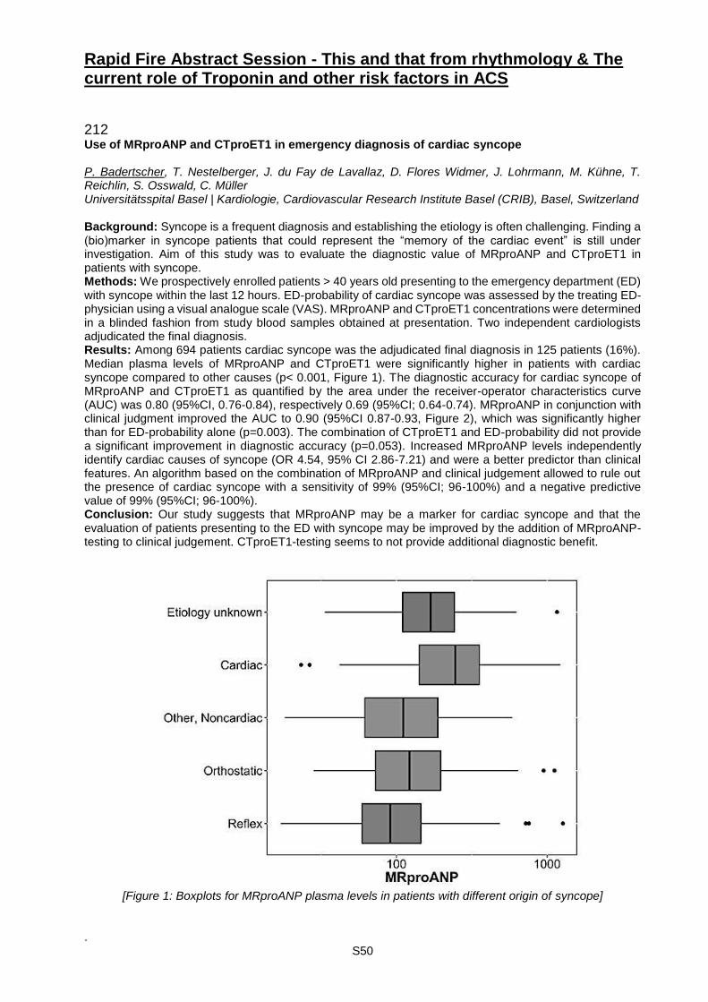
S50
Rapid Fire Abstract Session - This and that from rhythmology & The current role of Troponin and other risk factors in ACS
212 Use of MRproANP and CTproET1 in emergency diagnosis of cardiac syncope P. Badertscher, T. Nestelberger, J. du Fay de Lavallaz, D. Flores Widmer, J. Lohrmann, M. Kühne, T. Reichlin, S. Osswald, C. Müller Universitätsspital Basel | Kardiologie, Cardiovascular Research Institute Basel (CRIB), Basel, Switzerland Background: Syncope is a frequent diagnosis and establishing the etiology is often challenging. Finding a (bio)marker in syncope patients that could represent the “memory of the cardiac event” is still under investigation. Aim of this study was to evaluate the diagnostic value of MRproANP and CTproET1 in patients with syncope. Methods: We prospectively enrolled patients > 40 years old presenting to the emergency department (ED) with syncope within the last 12 hours. ED-probability of cardiac syncope was assessed by the treating ED-physician using a visual analogue scale (VAS). MRproANP and CTproET1 concentrations were determined in a blinded fashion from study blood samples obtained at presentation. Two independent cardiologists adjudicated the final diagnosis. Results: Among 694 patients cardiac syncope was the adjudicated final diagnosis in 125 patients (16%). Median plasma levels of MRproANP and CTproET1 were significantly higher in patients with cardiac syncope compared to other causes (p< 0.001, Figure 1). The diagnostic accuracy for cardiac syncope of MRproANP and CTproET1 as quantified by the area under the receiver-operator characteristics curve (AUC) was 0.80 (95%CI, 0.76-0.84), respectively 0.69 (95%CI; 0.64-0.74). MRproANP in conjunction with clinical judgment improved the AUC to 0.90 (95%CI 0.87-0.93, Figure 2), which was significantly higher than for ED-probability alone (p=0.003). The combination of CTproET1 and ED-probability did not provide a significant improvement in diagnostic accuracy (p=0.053). Increased MRproANP levels independently identify cardiac causes of syncope (OR 4.54, 95% CI 2.86-7.21) and were a better predictor than clinical features. An algorithm based on the combination of MRproANP and clinical judgement allowed to rule out the presence of cardiac syncope with a sensitivity of 99% (95%CI; 96-100%) and a negative predictive value of 99% (95%CI; 96-100%). Conclusion: Our study suggests that MRproANP may be a marker for cardiac syncope and that the evaluation of patients presenting to the ED with syncope may be improved by the addition of MRproANP-testing to clinical judgement. CTproET1-testing seems to not provide additional diagnostic benefit.
[Figure 1: Boxplots for MRproANP plasma levels in patients with different origin of syncope]
.
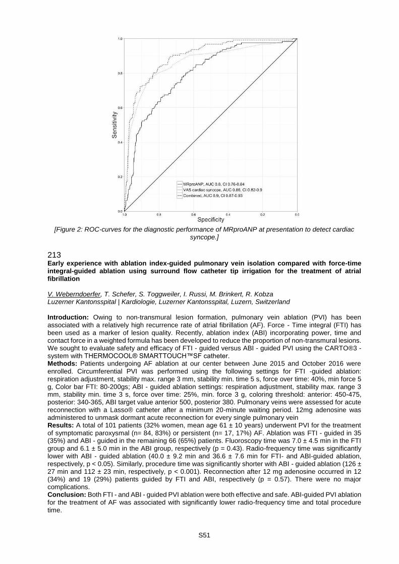
S51
[Figure 2: ROC-curves for the diagnostic performance of MRproANP at presentation to detect cardiac
syncope.]
213 Early experience with ablation index-guided pulmonary vein isolation compared with force-time integral-guided ablation using surround flow catheter tip irrigation for the treatment of atrial fibrillation V. Weberndoerfer, T. Schefer, S. Toggweiler, I. Russi, M. Brinkert, R. Kobza Luzerner Kantonsspital | Kardiologie, Luzerner Kantonsspital, Luzern, Switzerland Introduction: Owing to non-transmural lesion formation, pulmonary vein ablation (PVI) has been associated with a relatively high recurrence rate of atrial fibrillation (AF). Force - Time integral (FTI) has been used as a marker of lesion quality. Recently, ablation index (ABI) incorporating power, time and contact force in a weighted formula has been developed to reduce the proportion of non-transmural lesions. We sought to evaluate safety and efficacy of FTI - guided versus ABI - guided PVI using the CARTO®3 - system with THERMOCOOL® SMARTTOUCH™SF catheter. Methods: Patients undergoing AF ablation at our center between June 2015 and October 2016 were enrolled. Circumferential PVI was performed using the following settings for FTI -guided ablation: respiration adjustment, stability max. range 3 mm, stability min. time 5 s, force over time: 40%, min force 5 g, Color bar FTI: 80-200gs; ABI - guided ablation settings: respiration adjustment, stability max. range 3 mm, stability min. time 3 s, force over time: 25%, min. force 3 g, coloring threshold: anterior: 450-475, posterior: 340-365, ABI target value anterior 500, posterior 380. Pulmonary veins were assessed for acute reconnection with a Lasso® catheter after a minimum 20-minute waiting period. 12mg adenosine was administered to unmask dormant acute reconnection for every single pulmonary vein Results: A total of 101 patients (32% women, mean age 61 ± 10 years) underwent PVI for the treatment of symptomatic paroxysmal (n= 84, 83%) or persistent (n= 17, 17%) AF. Ablation was FTI - guided in 35 (35%) and ABI - guided in the remaining 66 (65%) patients. Fluoroscopy time was 7.0 ± 4.5 min in the FTI group and 6.1 ± 5.0 min in the ABI group, respectively (p = 0.43). Radio-frequency time was significantly lower with ABI - guided ablation (40.0 ± 9.2 min and 36.6 ± 7.6 min for FTI- and ABI-guided ablation, respectively, p < 0.05). Similarly, procedure time was significantly shorter with ABI - guided ablation (126 ± 27 min and 112 ± 23 min, respectively, p < 0.001). Reconnection after 12 mg adenosine occurred in 12 (34%) and 19 (29%) patients guided by FTI and ABI, respectively (p = 0.57). There were no major complications. Conclusion: Both FTI - and ABI - guided PVI ablation were both effective and safe. ABI-guided PVI ablation for the treatment of AF was associated with significantly lower radio-frequency time and total procedure time.

S52
214 Left atrial access in atrial fibrillation: patent foramen ovale versus transseptal puncture R. Sweda1,2, A. Haeberlin1,2, H. Servatius1, J. Seiler1, F. Noti1, A. Medeiros-Domingo1, S. Baldinger1, A. Lam1, E. Goulouti1, J. Fuhrer1, L. Roten1, H. Tanner1 1Department of Cardiology, Inselspital, Bern University Hospital, 2Cardiovascular Engineering, ARTORG Center for Biomedical Engineering Research, Bern, Switzerland Introduction: Left atrial access, as required for pulmonary vein isolation (PVI) in atrial fibrillation (AF) patients, is achieved through transseptal puncture (TSP) or via access through a patent foramen ovale (PFO). It has been discussed whether access through a PFO might have an impact on the outcome of AF ablation. This study sought to clarify the situation by studying periprocedural outcomes of AF ablation when performed through a PFO vs. TSP. Methods: Data from 1083 patients undergoing 1633 AF ablations were prospectively collected over 15 years (2001-2016). The presence of a PFO was determined ahead of the intervention by transesophageal echocardiography or during the intervention by wire probing. Outcomes included technical characteristics and procedural results such as success of the intervention. Additionally, details of re-ablations were assessed. Results: Ablation was performed through a PFO in 21.8% of cases. There were more men in the PFO group (82.9% vs. 74.2%, p=0.001) and patients with PFO had a higher incidence of pre-ablation strokes (11.4% vs. 6.0%, p=0.007). In both groups the majority of patients suffered from paroxysmal AF for a median duration of 36 months prior to the ablation. There was no difference between the groups regarding technical characteristics, however, there was a trend towards higher fluoroscopy time (p=0.076) and total procedural duration (p=0.079) in patients with TSP (table 1). Additionally, periprocedural complications occurred to a significantly higher rate in TSP patients (7.9% vs. 3.9%, p=0.010). Complete PVI was possible in >94% of cases (97% after 2006), irrespective of whether the ablation was performed through a TSP or a PFO. Repeat ablations were done in ~41% of individuals, most commonly because of AF recurrence. Compared to TSP patients, especially the right inferior PVs had reconnected more frequently in patients with PFO (59.5% vs. 49.2%, p=0.053, table 2). The overall success rate after a median follow up of 12 months was 69.5% and 67.6% in the PFO and the TSP group, respectively. Conclusion: In this to date largest analysis of PFO vs. TSP mediated left atrial access in AF ablation, long-term success rates were not different between the groups, although patients with PFO showed a higher number of right inferior PV reconnection. However, overall complications were more frequent in patients with TSP.
PFO (n=356)
TSP (n=1277)
p-value PFO (n=356)
TSP (n=1277)
p-value
RF duration, min (mean ± SD)
43.7 ± 23.3 43.1 ± 24.8 ns Isolation of all PV
94.3% 94.3% ns
RF power, watts*s (mean ± SD)
69 469 ± 39 396
68 319 ± 40 772
ns Isolation of all PV since 2006
97.6% 96.9% ns
Fluoroscopy duration, min (mean ± SD)
44.2 ± 31.9 48.1 ± 33.8 0.076 Complications 3.9% 8.1% 0.010
Radiation dose, Gy*cm2 (mean ± SD)
248.0 ± 271.4
298.2 ± 490.3
ns - Tamponade 0.8% 1.1% ns
Total duration, min (mean ± SD)
179.0 ± 77.1
189.0 ± 82.2
0.079 - Pericardial effusion
0.0% 0.9% ns
- Bleeding at puncture site
0.8% 2.3% ns
- Stroke/TIA 0.6% 0.7% ns
- Other 1.7% 3.1% ns
[Table 1: Procedural Outcomes]
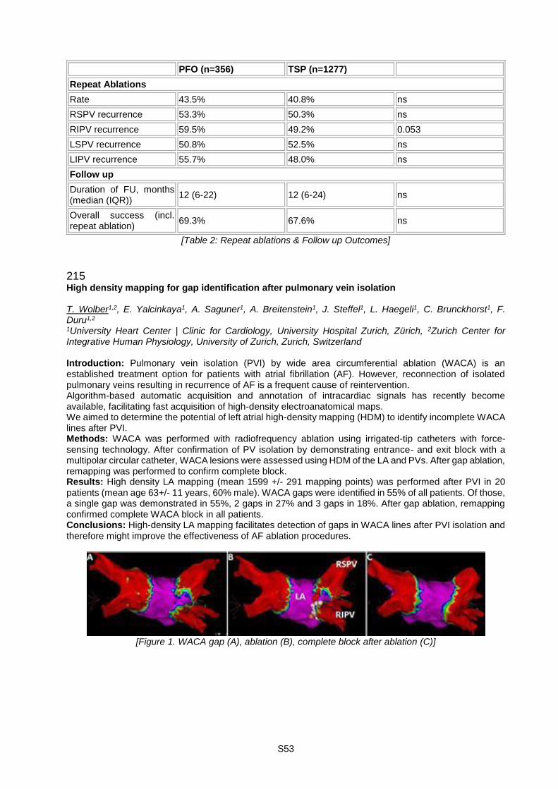
S53
PFO (n=356) TSP (n=1277)
Repeat Ablations
Rate 43.5% 40.8% ns
RSPV recurrence 53.3% 50.3% ns
RIPV recurrence 59.5% 49.2% 0.053
LSPV recurrence 50.8% 52.5% ns
LIPV recurrence 55.7% 48.0% ns
Follow up
Duration of FU, months (median (IQR))
12 (6-22) 12 (6-24) ns
Overall success (incl. repeat ablation)
69.3% 67.6% ns
[Table 2: Repeat ablations & Follow up Outcomes]
215 High density mapping for gap identification after pulmonary vein isolation T. Wolber1,2, E. Yalcinkaya1, A. Saguner1, A. Breitenstein1, J. Steffel1, L. Haegeli1, C. Brunckhorst1, F. Duru1,2 1University Heart Center | Clinic for Cardiology, University Hospital Zurich, Zürich, 2Zurich Center for Integrative Human Physiology, University of Zurich, Zurich, Switzerland Introduction: Pulmonary vein isolation (PVI) by wide area circumferential ablation (WACA) is an established treatment option for patients with atrial fibrillation (AF). However, reconnection of isolated pulmonary veins resulting in recurrence of AF is a frequent cause of reintervention. Algorithm-based automatic acquisition and annotation of intracardiac signals has recently become available, facilitating fast acquisition of high-density electroanatomical maps. We aimed to determine the potential of left atrial high-density mapping (HDM) to identify incomplete WACA lines after PVI. Methods: WACA was performed with radiofrequency ablation using irrigated-tip catheters with force-sensing technology. After confirmation of PV isolation by demonstrating entrance- and exit block with a multipolar circular catheter, WACA lesions were assessed using HDM of the LA and PVs. After gap ablation, remapping was performed to confirm complete block. Results: High density LA mapping (mean 1599 +/- 291 mapping points) was performed after PVI in 20 patients (mean age 63+/- 11 years, 60% male). WACA gaps were identified in 55% of all patients. Of those, a single gap was demonstrated in 55%, 2 gaps in 27% and 3 gaps in 18%. After gap ablation, remapping confirmed complete WACA block in all patients. Conclusions: High-density LA mapping facilitates detection of gaps in WACA lines after PVI isolation and therefore might improve the effectiveness of AF ablation procedures.
[Figure 1. WACA gap (A), ablation (B), complete block after ablation (C)]

S54
216 Performance of a leadless transcatheter pacemaker system compared to a conventional transvenous pacing system: Perioperative complications and shortterm follow-up N. Holm, T. Rizzo, C. Franzini, R. Dillier, A. Müller, R. Zbinden Klinik für Kardiologie, Stadtspital Triemli, Zürich, Switzerland Introduction: The Medtronic MICRA transcatheter pacing system (MICRA TPS) is a recently introduced leadless single-chamber pacing system. We compared the safety and performance of the MICRA TPS with conventional transvenous single chamber pacemakers (conventional VVI) implanted at our centre. Methods: This retrospective, observational single center study included 30 consecutive patients receiving a MICRA TPS and 33 historical control patients receiving a conventional VVI system at our centre. Results: Mean age was 83.3 ± 7.9 years in the MICRA TPS group and 82.3 ± 6.5 years in the conventional VVI group (p=NS). 53% in the MICRA TPS group were male compared to 55% in the conventional VVI group (p=NS). Indication for pacing was sick sinus syndrome in 73% and 57%, second and third degree heart block in 17% and 39% for the MICRA TPS and the conventional VVI group, respectively. Average procedure time was 41.2 ± 14 minutes for the MICRA TPS group and 34.3 ± 19.5 minutes for the conventional VVI group (p=NS). Radiation time was significantly longer in the MICRA TPS group compared to the conventional VVI group (10.3±6.8min vs 4.8±2.5min; p=0.01). There were significantly more periinterventional complications (including ventricular fibrillation, complete heart block, severe tricuspid regurgitation) in the MICRA TPS group compared to the conventional VVI group (20% vs 3%; p=0.03). Most of the complications in the MICRA TPS group happened in the first 15 patients. Mean follow up time was 82 ± 46 days in the MICRA TPS group and 95 ± 26 days in the conventional VVI group. The R wave increased significantly from 10.2±4.7mV to 13.6±5.6mV in the MICRA TPS group (p< 0.01) whereas it tended to decrease in the conventional VVI group (from 12.8±5.1 mV to 10.4±3.6mV; p=0.05) during follow-up. Thresholds remained stable in the MICRA TPS (from 0.57±0.24V @0.24msec to 0.73±[email protected]; p=NS) and in the conventional VVI group (0.76±0.3V @0.4msec to 0.84±[email protected]; p=NS). Impedance decreased significantly in the MICRA TPS group (from 713±175ohms to 601±75ohms; p< 0.01) and in the conventional VVI group (from 772±166ohms to 559±80ohms; p< 0.01). Conclusion: The new MICRA TPS is an alternative to conventional VVI pacing with comparable short term performance. There is a learning curve with more periprocedural complications compared to conventional VVI pacing especially in the first few cases and radiation times are significantly higher with the MICRA TPS system.
217 Trial examining the effects of music on anxiety of patients undergoing coronary angiogram S. Puricel, S. Lehmann, S. Benivady, H. Villeneuve, E. Boute, S. Cook Cardiologie, Université & Hôpital Fribourg, Fribourg, Switzerland Background: Although music is frequently used to promote a relaxing environment, the effect of its use in the cathlab remains unknown. Our study sought to determine the effects of music use on patients undergoing coronary angiogram, with the hypothesis that music use would result in lower anxiety, lower heart rate and blood pressure and greater patient satisfaction. Methods and results: All consecutive patient undergoing elective coronary angiogram at our institution were included in this randomized control trial. Patients included in the music group were asked to choose music that would best fit in the Spotify database. The music was broadcast through a dedicated musical cushion. Baseline patient and procedure characteristics were collected along with heart rate and blood pressure at 0, 5 minutes and time of procedure ended. After coronary angiograms, the patients were asked to quantify their wellness on an Edmonton scale and Cattle anxiety scale. Preliminary data based on the first 106 patients (mean age 66±11 years, 38% female) shows no significant difference between both groups in term of HR (Standard: pre-procedural:75±16 bpm; 5-minute: 78±16 bpm and post-procedural: 80±19 bpm versus Music: 74±11bpm, p=0.89; 76±10 bpm, p=0.62 and 75±9 bpm, p=0.30 respectively), blood pressure (Standard: pre-procedural: 136±25/77±14 mm Hg; 5-minute: 138±22/75±13 mm Hg and post-procedural: 133±25/73±12 mm Hg versus Music: 127±19/76±15 mm Hg, p=0.17/0.85; 127±23/73±13 mm Hg, p=0.15/0.58 and 135±23/71±16mm Hg, p=0.80/0.65 respectively) or sensation of well-being (Standard: 1 [1-2] versus 1 [1-1.5], p=0.18). Definitive data on 400 patients will be provided in June. Conclusion: Coronary angiogram causes anxiety in patients. Proper techniques to reduce this anxiety is necessary but to date music failed to demonstrate a substantial influence in all comer patients.
S55
218 Cysteine-rich angiogenic inducer 61 - a novel soluble biomarker of coronary thrombosis and myocardial ischemia improving risk stratification after acute coronary syndromes R. Klingenberg1, S. Aghlmandi2, C. Liebetrau1, L. Räber3, B. Gencer4, D. Nanchen5, D. Carballo4, A. Akhmedov6, F. Montecucco7, S. Zoller8, C. Brokopp9, D. Heg2, P. Jüni10, H. Marti Soler11, P.-M. Marques-Vidal11, P. Vollenweider11, O. Doerr12, N. Rodondi13, F. Mach4, S. Windecker3, U. Landmesser14, A. von Eckardstein15, C. Hamm1, C. Matter14, T.F. Lüscher14 1Cardiology, Kerckhoff-Klinik, Bad Nauheim, Germany, 2Clinical Trials Unit, Institute of Social and Preventive Medicine, University of Bern, 3Cardiology, Inselspital, Bern University Hospital, Bern, 4Cardiology, Geneva University Hospitals, Geneva, 5Ambulatory Care and Community Medicine, Unversity of Lausanne, Lausanne, 6Molecular Cardiology, University Hospital Zurich, Zurich, 7Experimental Cardiology, Geneva University Hospitals, Geneva, 8Bioinformatics, ETH Zurich, 9Regenerative Medicine Center, Department of Cardiothoracic Surgery, University Hospital Zurich, Zurich, Switzerland, 10Applied Health Research Centre (AHRC), Department of Medicine, University of Toronto, Toronto, ON, Canada, 11General Internal Medicine, Lausanne University Hospital, Lausanne, Switzerland, 12Cardiology, University Hospital Giessen, Giessen, Germany, 13General Internal Medicine, Inselspital, Bern University Hospital, Bern, 14Cardiology, 15Institute of Clinical Chemistry, University Hospital Zurich, Zurich, Switzerland Background: We aimed to characterize a novel biomarker involved in coronary thrombosis and myocardial ischemia and evaluate its role in risk stratification of patients with Acute Coronary Syndromes (ACS). Methods: Our identification strategy used gene expression profiling of coronary thrombi and peripheral blood mononuclear cells in ACS patients during primary percutaneous coronary intervention (pPCI). Transcoronary ablation of septal hypertrophy in patients with hypertrophic obstructive cardiomyopathy (HOCM) was used to characterize release kinetics of soluble cysteine-rich angiogenic inducer 61 (Cyr61, CCN1) in myocardial ischemia. An ischemia-reperfusion setting was used to study myocardial expression of Cyr61 in a mouse model. In ACS patients undergoing pPCI, the association of Cyr61 levels (dichotomized) with adjudicated all-cause mortality and the composite of all-cause mortality or myocardial infarction at 30 days and 1 year was compared with the GRACE risk score. A case-control cohort was used to evaluate the prognostic value of Cyr61 in primary prevention. Results: In coronary thrombi of ACS patients, Cyr61 gene transcripts were highly upregulated compared with peripheral blood mononuclear cells. Cyr61 was rapidly released from the myocardium after occlusion of the septal branch of the left anterior descending artery in patients with HOCM and returned to baseline within 105 minutes. Myocardial expression analysis in a mouse model of ischemia-reperfusion revealed de novo synthesis of Cyr61. In 1'094 ACS patients Cyr61 was elevated in those presenting within 24 hours after symptom onset, and highest in patients presenting early (≤ 6 hours). In 1`643 ACS patients, Cyr61 improved risk stratification for all-cause mortality beyond the GRACE risk score (c-statistic 0.74) and the GRACE risk score with hsTnT (c-statistic 0.79) to a c-statistic of 0.80 at 30 days and 1 year (c-statistic 0.67, 0.70, 0.72). Cyr61 was not associated with adverse cardiac outcome in primary prevention.. Conclusions: Cyr61 is an early soluble biomarker of coronary thrombosis and myocardial ischemia that improves early and long-term risk stratification in ACS patients. Clinical Trial Registration SPUM ACS NCT01000701
219 Near-linearity of release of cardiac troponin in early acute myocardial infarction M. Rubini1, R. Twerenbold2, S. Osswald2, C. Mueller2 1Cardiology, University of Basel, 2Kardiologie, Universitätsspital Basel, Basel, Switzerland Background: Release kinetics of cardiac troponin T and I in patients with acute myocardial infarction (AMI) are incompletely understood. Objectives: High-sensitivity cardiac troponin (hs-cTn) assays allow testing the hypothesis that cTnT/I release in early AMI is near-linear. Methods: In a prospective diagnostic multicenter study release of cTnT and cTnI was quantified using absolute changes in hs-cTnT/I within 1h, 2h, and 3h from presentation to the emergency department using four hs-cTn assays in patients with suspected AMI. Findings were validated using three additional sensitive (s-cTn) assays. The final diagnosis was adjudicated by two independent cardiologists. Results: Among 2437 patients with complete hs-cTnT data, AMI was the adjudicated diagnosis in 376 patients (15%). The correlation coefficient between 1h-change and 2h-change was 0.931 (p< 0.001). Similar findings were obtained with three hs-cTnI assays with correlation coefficients between 1h-change and 2h-change ranging from 0.851 to 0.946 (p< 0.001 for all) and validated with three additional s-cTnI assays (r=0.947 to 0.986, p< 0.001). Findings were consistent among AMI subtypes, in the subgroup of patients presenting very early after chest pain onset, as well as for the correlation between 1h-change and 3h-change. Changes in cTnT and cTnI in patients with diagnoses other than AMI in general were much
S56
smaller, and correlation coefficients between 1h-change and 2h-change ranged from 0.701 to 0.967 (p< 0.001 for all). Conclusions: Patients presenting with early AMI show a near-linear release of cTnT and cTnI. This linearity provides the pathophysiological basis for rapid diagnostic algorithms using 1h-changes as reliable surrogates for 2h/3h-changes.
220 Diagnostic accuracy of cardiovascular risk factors for an acute coronary syndrome M. Rubini, R. Twerenbold, J. Boeddinghaus, S. Osswald, C. Mueller Kardiologie, Universitätsspital Basel, Basel, Switzerland Introduction: We assessed the diagnostic performance of cardiovascular risk factors (CVRF) as cardiac risk burden for acute coronary syndrome (ACS) and acute myocardial infarction (AMI). Methods: In a prospective international multicenter study, we enrolled patients presenting to the Emergency Department (ED) with suspected AMI. The adjudication of the final diagnosis was performed by two independent cardiologists. Patients were stratified according to the presence or absence of known coronary artery disease (CAD). Diagnostic accuracy for ACS and AMI was the primary endpoint. Prognostic accuracy for death and/or future AMI was the secondary endpoint. Results: We included a total of 2830 patients. Among 1860 patients without known CAD, ACS and AMI were the adjudicated final diagnosis in 21% and 17% of patients, respectively. Overall cardiovascular risk profile quantified by the number of CVRF had modest diagnostic accuracy with an area under the receiver-operating-characteristics curve (AUC) of 0.68 (95% CI, 0.65-0.71) for ACS and 0.65 (95% CI, 0.62-0.69) for AMI, which both were inferior to high-sensitivity (hs) cardiac troponin (cTn) T at presentation (0.90; 95% CI 0.89-0.92 and 0.95; 95% CI 0.93-0.96, respectively, both p< 0.001), and did not provide diagnostic added value to hs-cTn (p=ns). Prognostic accuracy of the number of CVRF for death and/or future AMI also was modest (AUC 0.59, 95% CI 0.54-0.64). Diagnostic and prognostic accuracy of CVRF were poor in 970 patients with known CAD. These findings were confirmed in an external validation cohort from Australia and New Zealand. Conclusion: CVRF have only modest diagnostic and prognostic value in patients presenting with suspected ACS to the ED and their absence does not obviate the need for ECG and cTn testing.
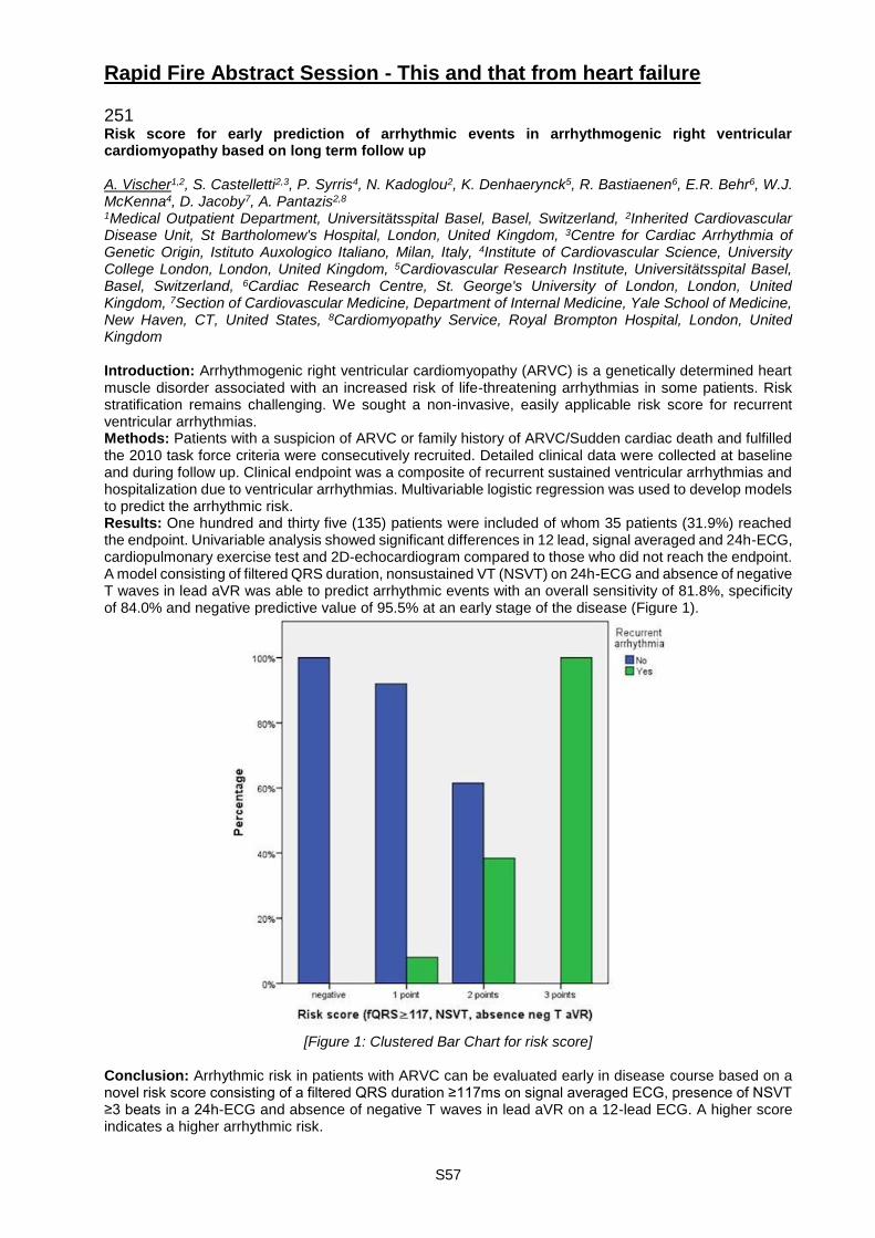
S57
Rapid Fire Abstract Session - This and that from heart failure 251 Risk score for early prediction of arrhythmic events in arrhythmogenic right ventricular cardiomyopathy based on long term follow up A. Vischer1,2, S. Castelletti2,3, P. Syrris4, N. Kadoglou2, K. Denhaerynck5, R. Bastiaenen6, E.R. Behr6, W.J. McKenna4, D. Jacoby7, A. Pantazis2,8 1Medical Outpatient Department, Universitätsspital Basel, Basel, Switzerland, 2Inherited Cardiovascular Disease Unit, St Bartholomew's Hospital, London, United Kingdom, 3Centre for Cardiac Arrhythmia of Genetic Origin, Istituto Auxologico Italiano, Milan, Italy, 4Institute of Cardiovascular Science, University College London, London, United Kingdom, 5Cardiovascular Research Institute, Universitätsspital Basel, Basel, Switzerland, 6Cardiac Research Centre, St. George's University of London, London, United Kingdom, 7Section of Cardiovascular Medicine, Department of Internal Medicine, Yale School of Medicine, New Haven, CT, United States, 8Cardiomyopathy Service, Royal Brompton Hospital, London, United Kingdom Introduction: Arrhythmogenic right ventricular cardiomyopathy (ARVC) is a genetically determined heart muscle disorder associated with an increased risk of life-threatening arrhythmias in some patients. Risk stratification remains challenging. We sought a non-invasive, easily applicable risk score for recurrent ventricular arrhythmias. Methods: Patients with a suspicion of ARVC or family history of ARVC/Sudden cardiac death and fulfilled the 2010 task force criteria were consecutively recruited. Detailed clinical data were collected at baseline and during follow up. Clinical endpoint was a composite of recurrent sustained ventricular arrhythmias and hospitalization due to ventricular arrhythmias. Multivariable logistic regression was used to develop models to predict the arrhythmic risk. Results: One hundred and thirty five (135) patients were included of whom 35 patients (31.9%) reached the endpoint. Univariable analysis showed significant differences in 12 lead, signal averaged and 24h-ECG, cardiopulmonary exercise test and 2D-echocardiogram compared to those who did not reach the endpoint. A model consisting of filtered QRS duration, nonsustained VT (NSVT) on 24h-ECG and absence of negative T waves in lead aVR was able to predict arrhythmic events with an overall sensitivity of 81.8%, specificity of 84.0% and negative predictive value of 95.5% at an early stage of the disease (Figure 1).
[Figure 1: Clustered Bar Chart for risk score]
Conclusion: Arrhythmic risk in patients with ARVC can be evaluated early in disease course based on a novel risk score consisting of a filtered QRS duration ≥117ms on signal averaged ECG, presence of NSVT ≥3 beats in a 24h-ECG and absence of negative T waves in lead aVR on a 12-lead ECG. A higher score indicates a higher arrhythmic risk.
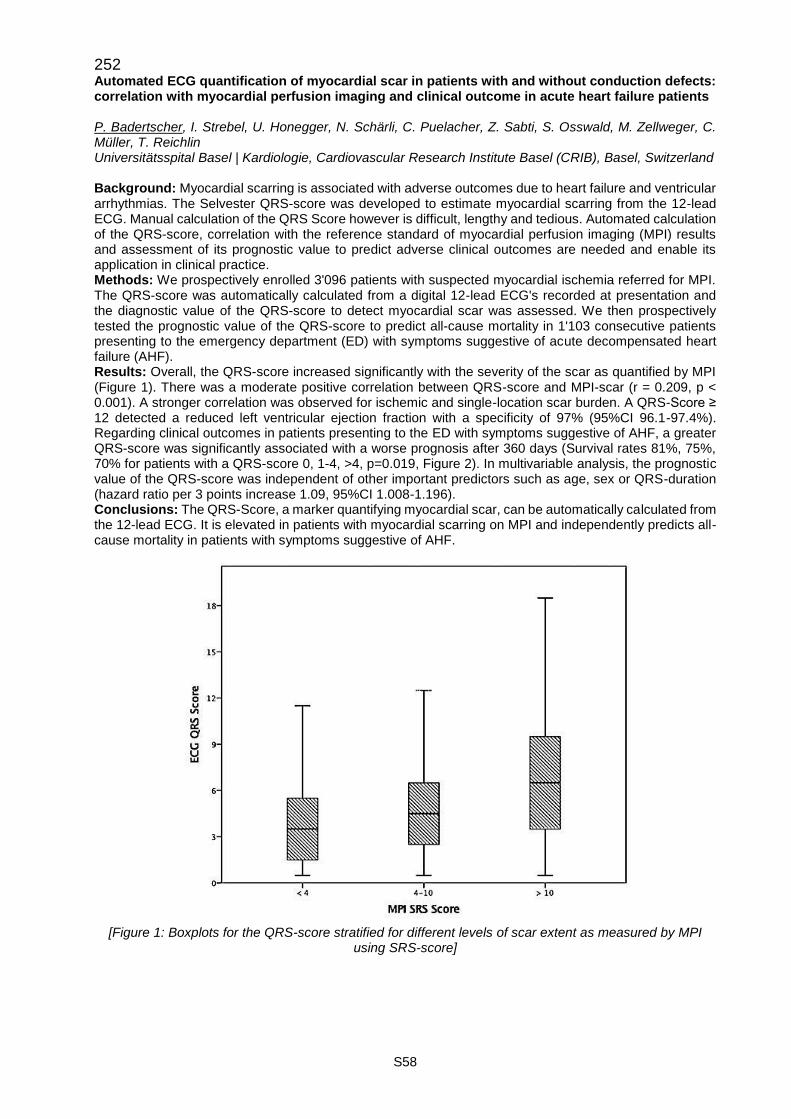
S58
252 Automated ECG quantification of myocardial scar in patients with and without conduction defects: correlation with myocardial perfusion imaging and clinical outcome in acute heart failure patients P. Badertscher, I. Strebel, U. Honegger, N. Schärli, C. Puelacher, Z. Sabti, S. Osswald, M. Zellweger, C. Müller, T. Reichlin Universitätsspital Basel | Kardiologie, Cardiovascular Research Institute Basel (CRIB), Basel, Switzerland Background: Myocardial scarring is associated with adverse outcomes due to heart failure and ventricular arrhythmias. The Selvester QRS-score was developed to estimate myocardial scarring from the 12-lead ECG. Manual calculation of the QRS Score however is difficult, lengthy and tedious. Automated calculation of the QRS-score, correlation with the reference standard of myocardial perfusion imaging (MPI) results and assessment of its prognostic value to predict adverse clinical outcomes are needed and enable its application in clinical practice. Methods: We prospectively enrolled 3'096 patients with suspected myocardial ischemia referred for MPI. The QRS-score was automatically calculated from a digital 12-lead ECG's recorded at presentation and the diagnostic value of the QRS-score to detect myocardial scar was assessed. We then prospectively tested the prognostic value of the QRS-score to predict all-cause mortality in 1'103 consecutive patients presenting to the emergency department (ED) with symptoms suggestive of acute decompensated heart failure (AHF). Results: Overall, the QRS-score increased significantly with the severity of the scar as quantified by MPI (Figure 1). There was a moderate positive correlation between QRS-score and MPI-scar (r = 0.209, p < 0.001). A stronger correlation was observed for ischemic and single-location scar burden. A QRS-Score ≥ 12 detected a reduced left ventricular ejection fraction with a specificity of 97% (95%CI 96.1-97.4%). Regarding clinical outcomes in patients presenting to the ED with symptoms suggestive of AHF, a greater QRS-score was significantly associated with a worse prognosis after 360 days (Survival rates 81%, 75%, 70% for patients with a QRS-score 0, 1-4, >4, p=0.019, Figure 2). In multivariable analysis, the prognostic value of the QRS-score was independent of other important predictors such as age, sex or QRS-duration (hazard ratio per 3 points increase 1.09, 95%CI 1.008-1.196). Conclusions: The QRS-Score, a marker quantifying myocardial scar, can be automatically calculated from the 12-lead ECG. It is elevated in patients with myocardial scarring on MPI and independently predicts all-cause mortality in patients with symptoms suggestive of AHF.
[Figure 1: Boxplots for the QRS-score stratified for different levels of scar extent as measured by MPI
using SRS-score]
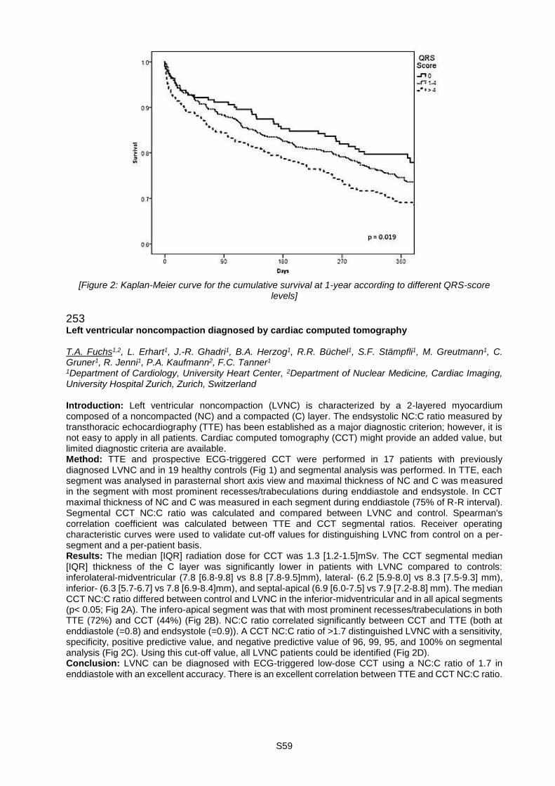
S59
[Figure 2: Kaplan-Meier curve for the cumulative survival at 1-year according to different QRS-score
levels]
253 Left ventricular noncompaction diagnosed by cardiac computed tomography T.A. Fuchs1,2, L. Erhart1, J.-R. Ghadri1, B.A. Herzog1, R.R. Büchel1, S.F. Stämpfli1, M. Greutmann1, C. Gruner1, R. Jenni1, P.A. Kaufmann2, F.C. Tanner1 1Department of Cardiology, University Heart Center, 2Department of Nuclear Medicine, Cardiac Imaging, University Hospital Zurich, Zurich, Switzerland Introduction: Left ventricular noncompaction (LVNC) is characterized by a 2-layered myocardium composed of a noncompacted (NC) and a compacted (C) layer. The endsystolic NC:C ratio measured by transthoracic echocardiography (TTE) has been established as a major diagnostic criterion; however, it is not easy to apply in all patients. Cardiac computed tomography (CCT) might provide an added value, but limited diagnostic criteria are available. Method: TTE and prospective ECG-triggered CCT were performed in 17 patients with previously diagnosed LVNC and in 19 healthy controls (Fig 1) and segmental analysis was performed. In TTE, each segment was analysed in parasternal short axis view and maximal thickness of NC and C was measured in the segment with most prominent recesses/trabeculations during enddiastole and endsystole. In CCT maximal thickness of NC and C was measured in each segment during enddiastole (75% of R-R interval). Segmental CCT NC:C ratio was calculated and compared between LVNC and control. Spearman's correlation coefficient was calculated between TTE and CCT segmental ratios. Receiver operating characteristic curves were used to validate cut-off values for distinguishing LVNC from control on a per-segment and a per-patient basis. Results: The median [IQR] radiation dose for CCT was 1.3 [1.2-1.5]mSv. The CCT segmental median [IQR] thickness of the C layer was significantly lower in patients with LVNC compared to controls: inferolateral-midventricular (7.8 [6.8-9.8] vs 8.8 [7.8-9.5]mm), lateral- (6.2 [5.9-8.0] vs 8.3 [7.5-9.3] mm), inferior- (6.3 [5.7-6.7] vs 7.8 [6.9-8.4]mm), and septal-apical (6.9 [6.0-7.5] vs 7.9 [7.2-8.8] mm). The median CCT NC:C ratio differed between control and LVNC in the inferior-midventricular and in all apical segments (p< 0.05; Fig 2A). The infero-apical segment was that with most prominent recesses/trabeculations in both TTE (72%) and CCT (44%) (Fig 2B). NC:C ratio correlated significantly between CCT and TTE (both at enddiastole (=0.8) and endsystole (=0.9)). A CCT NC:C ratio of >1.7 distinguished LVNC with a sensitivity, specificity, positive predictive value, and negative predictive value of 96, 99, 95, and 100% on segmental analysis (Fig 2C). Using this cut-off value, all LVNC patients could be identified (Fig 2D). Conclusion: LVNC can be diagnosed with ECG-triggered low-dose CCT using a NC:C ratio of 1.7 in enddiastole with an excellent accuracy. There is an excellent correlation between TTE and CCT NC:C ratio.
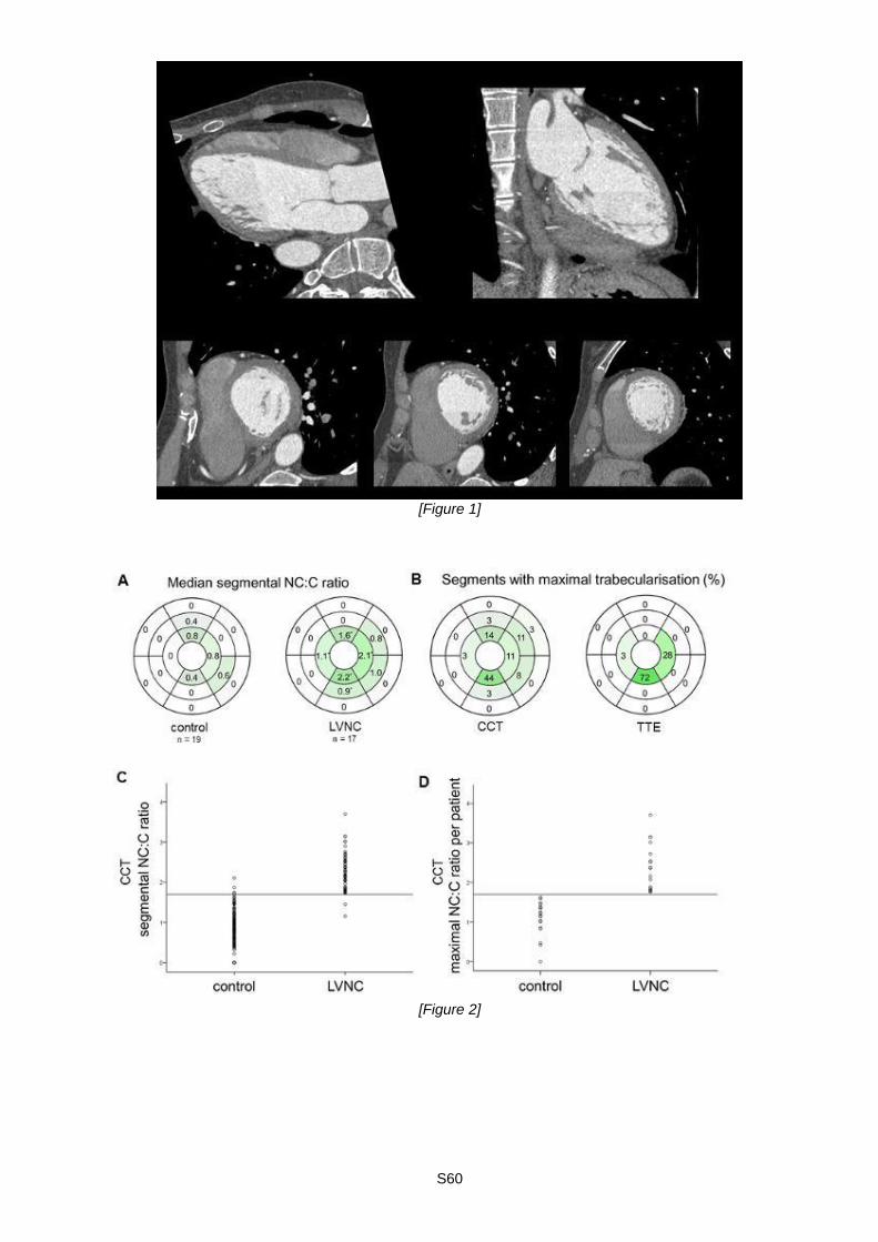
S60
[Figure 1]
[Figure 2]
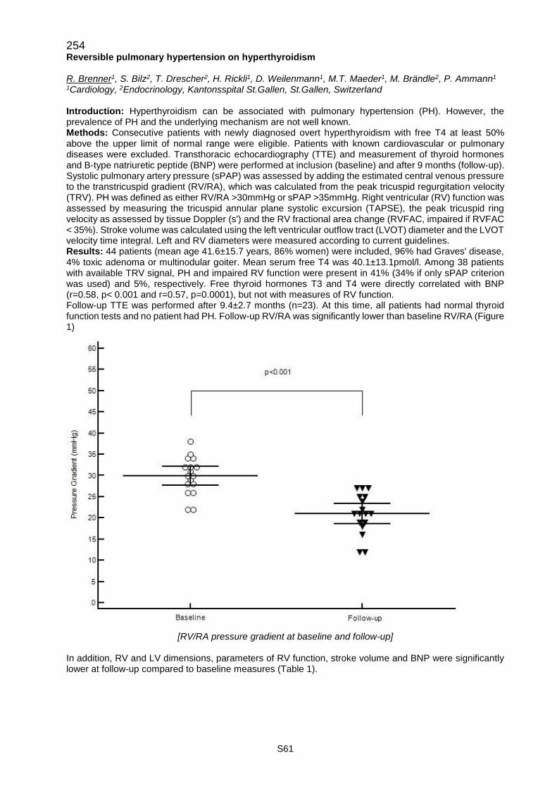
S61
254 Reversible pulmonary hypertension on hyperthyroidism R. Brenner1, S. Bilz2, T. Drescher2, H. Rickli1, D. Weilenmann1, M.T. Maeder1, M. Brändle2, P. Ammann1 1Cardiology, 2Endocrinology, Kantonsspital St.Gallen, St.Gallen, Switzerland Introduction: Hyperthyroidism can be associated with pulmonary hypertension (PH). However, the prevalence of PH and the underlying mechanism are not well known. Methods: Consecutive patients with newly diagnosed overt hyperthyroidism with free T4 at least 50% above the upper limit of normal range were eligible. Patients with known cardiovascular or pulmonary diseases were excluded. Transthoracic echocardiography (TTE) and measurement of thyroid hormones and B-type natriuretic peptide (BNP) were performed at inclusion (baseline) and after 9 months (follow-up). Systolic pulmonary artery pressure (sPAP) was assessed by adding the estimated central venous pressure to the transtricuspid gradient (RV/RA), which was calculated from the peak tricuspid regurgitation velocity (TRV). PH was defined as either RV/RA >30mmHg or sPAP >35mmHg. Right ventricular (RV) function was assessed by measuring the tricuspid annular plane systolic excursion (TAPSE), the peak tricuspid ring velocity as assessed by tissue Doppler (s') and the RV fractional area change (RVFAC, impaired if RVFAC < 35%). Stroke volume was calculated using the left ventricular outflow tract (LVOT) diameter and the LVOT velocity time integral. Left and RV diameters were measured according to current guidelines. Results: 44 patients (mean age 41.6±15.7 years, 86% women) were included, 96% had Graves' disease, 4% toxic adenoma or multinodular goiter. Mean serum free T4 was 40.1±13.1pmol/l. Among 38 patients with available TRV signal, PH and impaired RV function were present in 41% (34% if only sPAP criterion was used) and 5%, respectively. Free thyroid hormones T3 and T4 were directly correlated with BNP (r=0.58, p< 0.001 and r=0.57, p=0.0001), but not with measures of RV function. Follow-up TTE was performed after 9.4±2.7 months (n=23). At this time, all patients had normal thyroid function tests and no patient had PH. Follow-up RV/RA was significantly lower than baseline RV/RA (Figure 1)
[RV/RA pressure gradient at baseline and follow-up]
In addition, RV and LV dimensions, parameters of RV function, stroke volume and BNP were significantly lower at follow-up compared to baseline measures (Table 1).
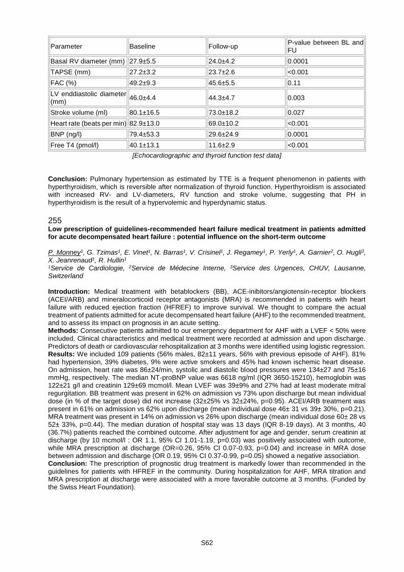
S62
Parameter Baseline Follow-up P-value between BL and FU
Basal RV diameter (mm) 27.9±5.5 24.0±4.2 0.0001
TAPSE (mm) 27.2±3.2 23.7±2.6 <0.001
FAC (%) 49.2±9.3 45.6±5.5 0.11
LV enddiastolic diameter (mm)
46.0±4.4 44.3±4.7 0.003
Stroke volume (ml) 80.1±16.5 73.0±18.2 0.027
Heart rate (beats per min) 82.9±13.0 69.0±10.2 <0.001
BNP (ng/l) 79.4±53.3 29.6±24.9 0.0001
Free T4 (pmol/l) 40.1±13.1 11.6±2.9 <0.001
[Echocardiographic and thyroid function test data] Conclusion: Pulmonary hypertension as estimated by TTE is a frequent phenomenon in patients with hyperthyroidism, which is reversible after normalization of thyroid function. Hyperthyroidism is associated with increased RV- and LV-diameters, RV function and stroke volume, suggesting that PH in hyperthyroidism is the result of a hypervolemic and hyperdynamic status.
255 Low prescription of guidelines-recommended heart failure medical treatment in patients admitted for acute decompensated heart failure : potential influence on the short-term outcome P. Monney1, G. Tzimas1, E. Vinet1, N. Barras1, V. Crisinel1, J. Regamey1, P. Yerly1, A. Garnier2, O. Hugli3, X. Jeanrenaud1, R. Hullin1 1Service de Cardiologie, 2Service de Médecine Interne, 3Service des Urgences, CHUV, Lausanne, Switzerland Introduction: Medical treatment with betablockers (BB), ACE-inibitors/angiotensin-receptor blockers (ACEI/ARB) and mineralocorticoid receptor antagonists (MRA) is recommended in patients with heart failure with reduced ejection fraction (HFREF) to improve survival. We thought to compare the actual treatment of patients admitted for acute decompensated heart failure (AHF) to the recommended treatment, and to assess its impact on prognosis in an acute setting. Methods: Consecutive patients admitted to our emergency department for AHF with a LVEF < 50% were included. Clinical characteristics and medical treatment were recorded at admission and upon discharge. Predictors of death or cardiovascular rehospitalization at 3 months were identified using logistic regression. Results: We included 109 patients (56% males, 82±11 years, 56% with previous episode of AHF). 81% had hypertension, 39% diabetes, 9% were active smokers and 45% had known ischemic heart disease. On admission, heart rate was 86±24/min, systolic and diastolic blood pressures were 134±27 and 75±16 mmHg, respectively. The median NT-proBNP value was 6618 ng/ml (IQR 3650-15210), hemoglobin was 122±21 g/l and creatinin 129±69 mcmol/l. Mean LVEF was 39±9% and 27% had at least moderate mitral regurgitation. BB treatment was present in 62% on admission vs 73% upon discharge but mean individual dose (in % of the target dose) did not increase (32±25% vs 32±24%, p=0.95). ACEI/ARB treatment was present in 61% on admission vs 62% upon discharge (mean individual dose 46± 31 vs 39± 30%, p=0.21). MRA treatment was present in 14% on admission vs 26% upon discharge (mean individual dose 60± 28 vs 52± 33%, p=0.44). The median duration of hospital stay was 13 days (IQR 8-19 days). At 3 months, 40 (36.7%) patients reached the combined outcome. After adjustment for age and gender, serum creatinin at discharge (by 10 mcmol/l : OR 1.1, 95% CI 1.01-1.19, p=0.03) was positively associated with outcome, while MRA prescription at discharge (OR=0.26, 95% CI 0.07-0.93, p=0.04) and increase in MRA dose between admission and discharge (OR 0.19, 95% CI 0.37-0.99, p=0.05) showed a negative association. Conclusion: The prescription of prognostic drug treatment is markedly lower than recommended in the guidelines for patients with HFREF in the community. During hospitalization for AHF, MRA titration and MRA prescription at discharge were associated with a more favorable outcome at 3 months. (Funded by the Swiss Heart Foundation).
S63
Rapid Fire Abstract Session - News from imaging, congenital heart disease and heart disease in pregnancy 281 CMR-derived metrics of interstitial myocardial fibrosis: which parameter is better associated with pathophysiological measures of diastolic dysfunction? P. Monney1,2, S. Hugelshofer1, P.G. Masci1,2, G. Vincenti1,2, T. Rutz1,2, J. Schwitter1,2 1Service de Cardiologie, 2Center for Cardiovascular Magnetic Resonance, CHUV, Lausanne, Switzerland Background: In patients with diastolic dysfunction (ie. at risk to develop heart failure (HF) with preserved ejection fraction), it is still disputable which CMR-verified parameters of interstitial myocardial fibrosis (IMF) best correlates with metrics of left ventricular (LV) longitudinal systolic and diastolic function as well as with left atrial (LA) performance. Methods: Patients with LV-EF≥40% in whom myocardial ischemia, infarction, myocarditis, or cardiomyopathies were excluded by CMR were recruited. LV volumes and mass were quantified and late-gadolinium-enhancement (LGE) was performed according to standard protocols. Pre and post-contrast T1-mapping and hematocrit were utilized for quantifying native (T1pre), post-contrast T1 (T1post), and extracellular volume fraction (ECV) of the basal interventricular septum. LV longitudinal systolic function (LV-FNlong) was calculated on 2-chamber and 4-chamber views as the mean systolic base-to-apex long axis shortening. Using cine 3-chamber, LA area changes during the cardiac cycle were measured to derive LA performance. LV peak-filling rate (PFR) was obtained from volume-time curves by semiautomatically contouring the endocardium of all phases of short-axis cine images. Results: In 85 patients (age 54±17y, 59% males), no correlation was found between T1pre and any LV function parameter. T1post correlated with age, gender, body mass index, and contrast dose whereas ECV was only associated with age. Neither ECV nor T1post correlated with renal function or the time interval between contrast injection and T1post measurement. After adjustment for age, gender, body mass index, and contrast dose, T1post and ECV correlated with LV-FNlong (β=0.28, p=0.02; β=-0.27, p=0.01, respectively), but not with LV-EF. T1post, but not ECV, correlated with LV-PFR (n=61, β=0.32, P=0.03). ECV (β=0.23, p=0.046), but not T1post, correlated with indexed LV mass. No parameter of LA performance correlated with any parameter of interstitial fibrosis. On multivariate linear regression analysis, T1post showed the strongest association with LV-PFR (β=0.38, p=0.017), while ECV showed the strongest association with LV-FNlong and indexed LV mass (β=0.29, p=0.017 and β=0.30, p=0.026, respectively). Conclusion: After adjustment for significant covariates, T1post and ECV were independently associated with different measures of LV longitudinal systolic and diastolic function, whereas T1pre failed to show any significant association.
282 Echocardiographic predictors of midterm outcome after MitraClip implantation I. Kapos1, L. Rebellius2, S. Obeid3, K. Shingo4, O. Gämperli5, F. Maisano4, F. Tanner5 1Kardiologie, UniversitätsSpital Zürich, Zurich, 2Univerity of Zurich, 3Kardiologie, UniversitätsSpital Zürich, 4Herzchirurgie, 5Kardiologie, Universitätsspital Zürich, Zürich, Switzerland Background: The MitraClip is a minimally invasive technique for treatment of patients with moderate-severe or severe mitral regurgitation (MR) at high surgical risk. Echocardiographic predictors of procedural and clinical outcome after MitraClip have been investigated in different cohort studies. We hypothesized that we could define an anatomic characteristic of the mitral valve (MV) obtained by 3-dimentional transesophageal echocardiography (3D-TEE) to predict clinical outcome after MitraClip in order to achieve an improved patient selection. Method and Results: 3D-TEE obtained at baseline in patients referred for MitraClip to our institution were retrospectively analyzed. In 97 of 150 patients (age 75.7 +/- 11.2) we were able to perform an off-line 3D reconstruction with a dedicated software for analysis of the mitral valve (TomTec Image Arena). Primary endpoint was all-cause mortality, heart transplantation, or re-intervention. All patients had moderate-to-severe (3+) or severe (4+) MR of functional (n=53; 55%) or degenerative (n=44; 45%) origin. Indication for treatment of MR and individual surgical risk were assessed by an interdisciplinary heart team according to current guidelines. Patients with functional versus degenerative MR had comparable procedural success (residual MR< = +2: 60% vs 70%, respectively; p=0.39). In contrast, among the patients meeting the primary endpoint, 83% had a functional MR primarily caused by ischemic heart disease (OR 5.0 for functional MR; p< 0.05). For patients meeting the primary end-point, left ventricular ejection fraction (LVEF) was significantly lower (p=0.04), MV tenting area was larger (p< 0.01), and MV posterior leaflet angle was higher (p< 0.02). Univariate analysis revealed that LVEF (OR 0.96 per %), MV tenting area (OR 1.80 per cm2), and MV posterior leaflet angle (OR 1.04 per degree) were significant predictors of the primary endpoint.

S64
Conclusions: Measurement of MV tenting area and posterior leaflet angle with 3D reconstruction at baseline predicts clinical outcome after MitraClip in a univariate analysis. Since both parameters depend on LV volume, this observation is consistent with the interpretation that the etiology of MR is an important predictor of clinical outcome.
283 Benchmark analysis of swiss centres participating in the ROPAC, a registry from the european society of cardiology (ESC) B. Santos Lopes1, F. Schwitz2, K. Wustmann2, M. Schwerzmann2, M. Greutmann1, F. Bonassin Tempesta1, J. Bouchardy3, T. Rutz4, D. Tobler5, ROPAC 1Universitäres Herzzentrum Zürich, Klinik für Kardiologie, Universitätsspital Zürich, Zurich, 2Universitätsklinik für Kardiologie, Inselspital, Bern University Hospital, Bern, 3Service de Cardiologie, Hôpitaux Universitaires de Genève, Geneva, 4Service de Cardiologie, CHUV, Lausanne, 5Klinik für Kardiologie, Universitätsspital Basel, Basel, Switzerland Introduction: The Registry Of Pregnancy And Cardiac disease (ROPAC) is a worldwide multicentre collaboration on pregnancy outcomes in women with structural heart disease from the EURObservational Research Programme of the ESC. Overall, 4 Swiss centres are participating in ROPAC. The aim of this study was to compare patient characteristics and pregnancy outcomes between specialist Swiss centres (CH) and the other participating centres in North West Europe (NWE). Methods: For the purpose of this study, data of women enrolled up to December 31, 2016 were analysed. The following data are reported and compared between CH-centres and the other participating centres in NWE (Austria, Belgium, France, Germany, Ireland, The Netherlands, Norway, Sweden, UK): age, main diagnosis, NYHA class, atrial fibrillation, pregnancy outcomes and follow-up data at 6 months. Results: Of a total of 1357 women enrolled in NWE, 185 (13.6%) were enrolled in Switzerland. Follow-up data at 6 months were available in 81%. There was no difference in age at pregnancy between women in CH- and NWE-centres (31.0y vs 30.4y, p=0.8). In both groups, the most women were asymptomatic (NYHA I: 85.4% vs 82.2%, p=0.3) and there were no differences in the proportion of main diagnosis between CH- and NWE-centres. The distribution of cardiac diagnoses of women followed in CH-centres is illustrated in figure 1. Regarding pregnancy outcomes, no difference was observed between CH- and NWE-centres regarding the incidence of atrial fibrillation (1.1% vs 0.5%, p=0.4), admission rates (17.3% vs 21.4%, p=0.2), heart failure (2.7% vs 4.3%, p=0.3) or maternal/foetal mortality (0%/0.5% vs 0.4%/0.4%, p=0.4/0.7). In CH-centres, a higher rate of miscarriages was reported (4.3% vs 1.8%, p = 0.029). Compared to NWE-centres, in CH-centres the total number of caesarean sections (31.5% vs 24.6%, p=0.047) and emergency caesarean sections (12.4% vs 7.6%, p=0.024) were higher, while there was no difference for caesarean section due to cardiac indication. At follow-up 6 months after delivery all patients followed at CH-centres were still alive and only 2 (1.3%) had experienced and episode of heart failure.
[Figure 1: HD = heart disease. CM = cardiomyopathy]
Conclusions: As compared to other centres in NWE, characteristics of cohorts of women with structural heart disease followed at dedicated specialist centres for high-risk pregnancies in Switzerland were similar. Pregnancy outcomes were comparable, except for a slightly higher rate of caesarean sections and miscarriages.
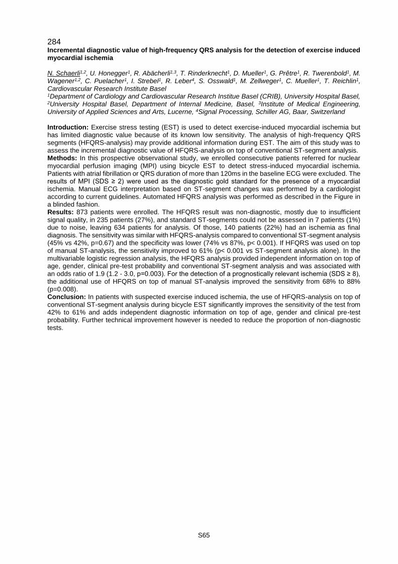
S65
284 Incremental diagnostic value of high-frequency QRS analysis for the detection of exercise induced myocardial ischemia N. Schaerli1,2, U. Honegger1, R. Abächerli1,3, T. Rinderknecht1, D. Mueller1, G. Prêtre1, R. Twerenbold1, M. Wagener1,2, C. Puelacher1, I. Strebel1, R. Leber4, S. Osswald1, M. Zellweger1, C. Mueller1, T. Reichlin1, Cardiovascular Research Institute Basel 1Department of Cardiology and Cardiovascular Research Institue Basel (CRIB), University Hospital Basel, 2University Hospital Basel, Department of Internal Medicine, Basel, 3Institute of Medical Engineering, University of Applied Sciences and Arts, Lucerne, 4Signal Processing, Schiller AG, Baar, Switzerland Introduction: Exercise stress testing (EST) is used to detect exercise-induced myocardial ischemia but has limited diagnostic value because of its known low sensitivity. The analysis of high-frequency QRS segments (HFQRS-analysis) may provide additional information during EST. The aim of this study was to assess the incremental diagnostic value of HFQRS-analysis on top of conventional ST-segment analysis. Methods: In this prospective observational study, we enrolled consecutive patients referred for nuclear myocardial perfusion imaging (MPI) using bicycle EST to detect stress-induced myocardial ischemia. Patients with atrial fibrillation or QRS duration of more than 120ms in the baseline ECG were excluded. The results of MPI (SDS ≥ 2) were used as the diagnostic gold standard for the presence of a myocardial ischemia. Manual ECG interpretation based on ST-segment changes was performed by a cardiologist according to current guidelines. Automated HFQRS analysis was performed as described in the Figure in a blinded fashion. Results: 873 patients were enrolled. The HFQRS result was non-diagnostic, mostly due to insufficient signal quality, in 235 patients (27%), and standard ST-segments could not be assessed in 7 patients (1%) due to noise, leaving 634 patients for analysis. Of those, 140 patients (22%) had an ischemia as final diagnosis. The sensitivity was similar with HFQRS-analysis compared to conventional ST-segment analysis (45% vs 42%, p=0.67) and the specificity was lower (74% vs 87%, p< 0.001). If HFQRS was used on top of manual ST-analysis, the sensitivity improved to 61% (p< 0.001 vs ST-segment analysis alone). In the multivariable logistic regression analysis, the HFQRS analysis provided independent information on top of age, gender, clinical pre-test probability and conventional ST-segment analysis and was associated with an odds ratio of 1.9 (1.2 - 3.0, p=0.003). For the detection of a prognostically relevant ischemia (SDS ≥ 8), the additional use of HFQRS on top of manual ST-analysis improved the sensitivity from 68% to 88% (p=0.008). Conclusion: In patients with suspected exercise induced ischemia, the use of HFQRS-analysis on top of conventional ST-segment analysis during bicycle EST significantly improves the sensitivity of the test from 42% to 61% and adds independent diagnostic information on top of age, gender and clinical pre-test probability. Further technical improvement however is needed to reduce the proportion of non-diagnostic tests.
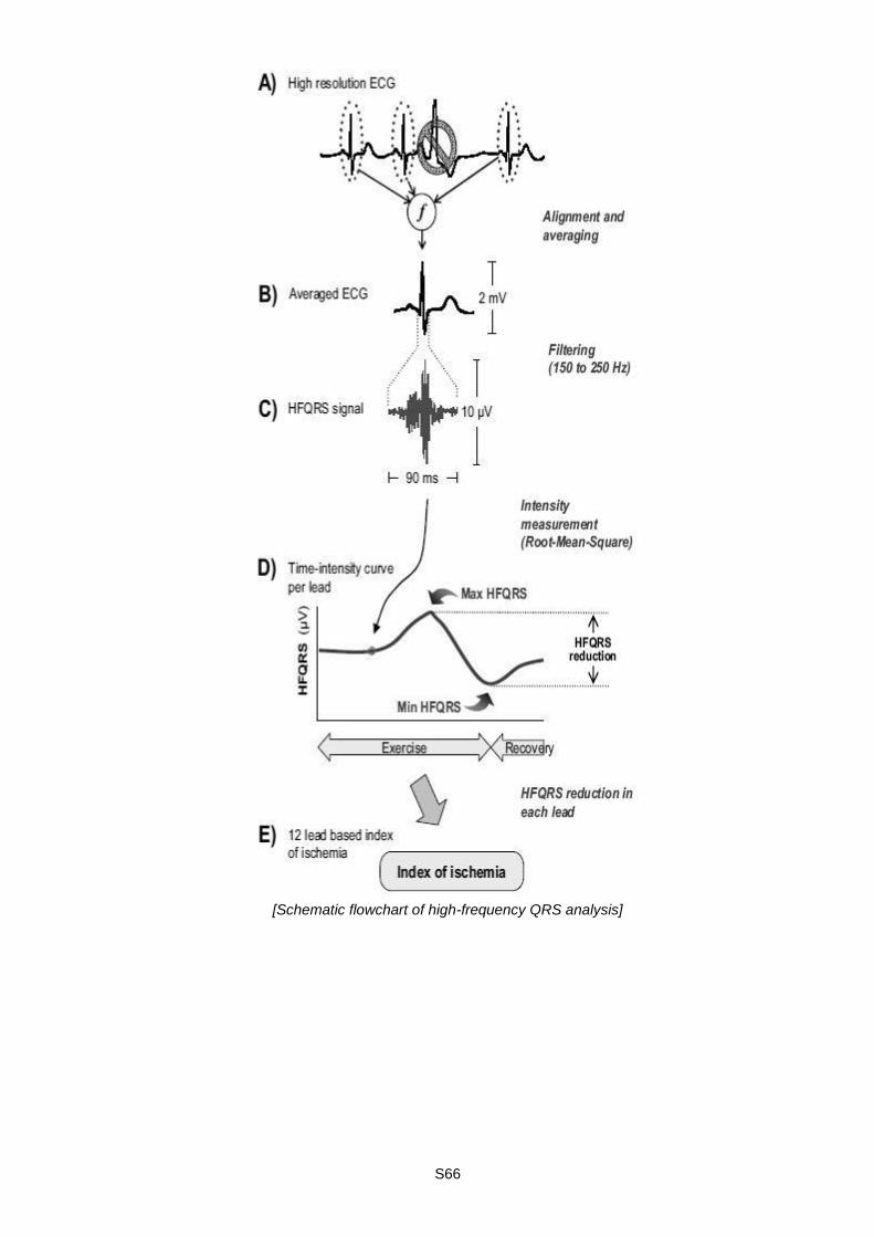
S66
[Schematic flowchart of high-frequency QRS analysis]
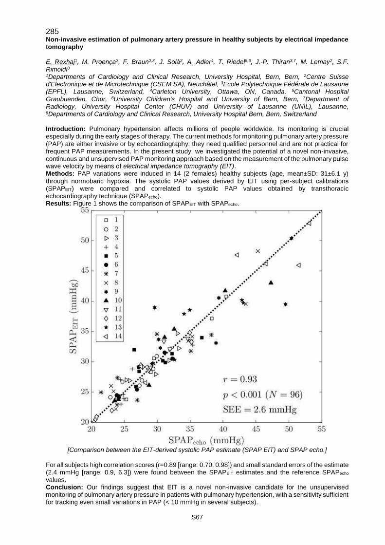
S67
285 Non-invasive estimation of pulmonary artery pressure in healthy subjects by electrical impedance tomography E. Rexhaj1, M. Proença2, F. Braun2,3, J. Solà2, A. Adler4, T. Riedel5,6, J.-P. Thiran3,7, M. Lemay2, S.F. Rimoldi8 1Departments of Cardiology and Clinical Research, University Hospital, Bern, Bern, 2Centre Suisse d'Electronique et de Microtechnique (CSEM SA), Neuchâtel, 3Ecole Polytechnique Fédérale de Lausanne (EPFL), Lausanne, Switzerland, 4Carleton University, Ottawa, ON, Canada, 5Cantonal Hospital Graubuenden, Chur, 6University Children's Hospital and University of Bern, Bern, 7Department of Radiology, University Hospital Center (CHUV) and University of Lausanne (UNIL), Lausanne, 8Departments of Cardiology and Clinical Research, University Hospital Bern, Bern, Switzerland Introduction: Pulmonary hypertension affects millions of people worldwide. Its monitoring is crucial especially during the early stages of therapy. The current methods for monitoring pulmonary artery pressure (PAP) are either invasive or by echocardiography: they need qualified personnel and are not practical for frequent PAP measurements. In the present study, we investigated the potential of a novel non-invasive, continuous and unsupervised PAP monitoring approach based on the measurement of the pulmonary pulse wave velocity by means of electrical impedance tomography (EIT). Methods: PAP variations were induced in 14 (2 females) healthy subjects (age, mean±SD: 31±6.1 y) through normobaric hypoxia. The systolic PAP values derived by EIT using per-subject calibrations (SPAPEIT) were compared and correlated to systolic PAP values obtained by transthoracic echocardiography technique (SPAPecho). Results: Figure 1 shows the comparison of SPAPEIT with SPAPecho.
[Comparison between the EIT-derived systolic PAP estimate (SPAP EIT) and SPAP echo.]
For all subjects high correlation scores (r=0.89 [range: 0.70, 0.98]) and small standard errors of the estimate (2.4 mmHg [range: 0.9, 6.3]) were found between the SPAPEIT estimates and the reference SPAPecho values. Conclusion: Our findings suggest that EIT is a novel non-invasive candidate for the unsupervised monitoring of pulmonary artery pressure in patients with pulmonary hypertension, with a sensitivity sufficient for tracking even small variations in PAP (< 10 mmHg in several subjects).
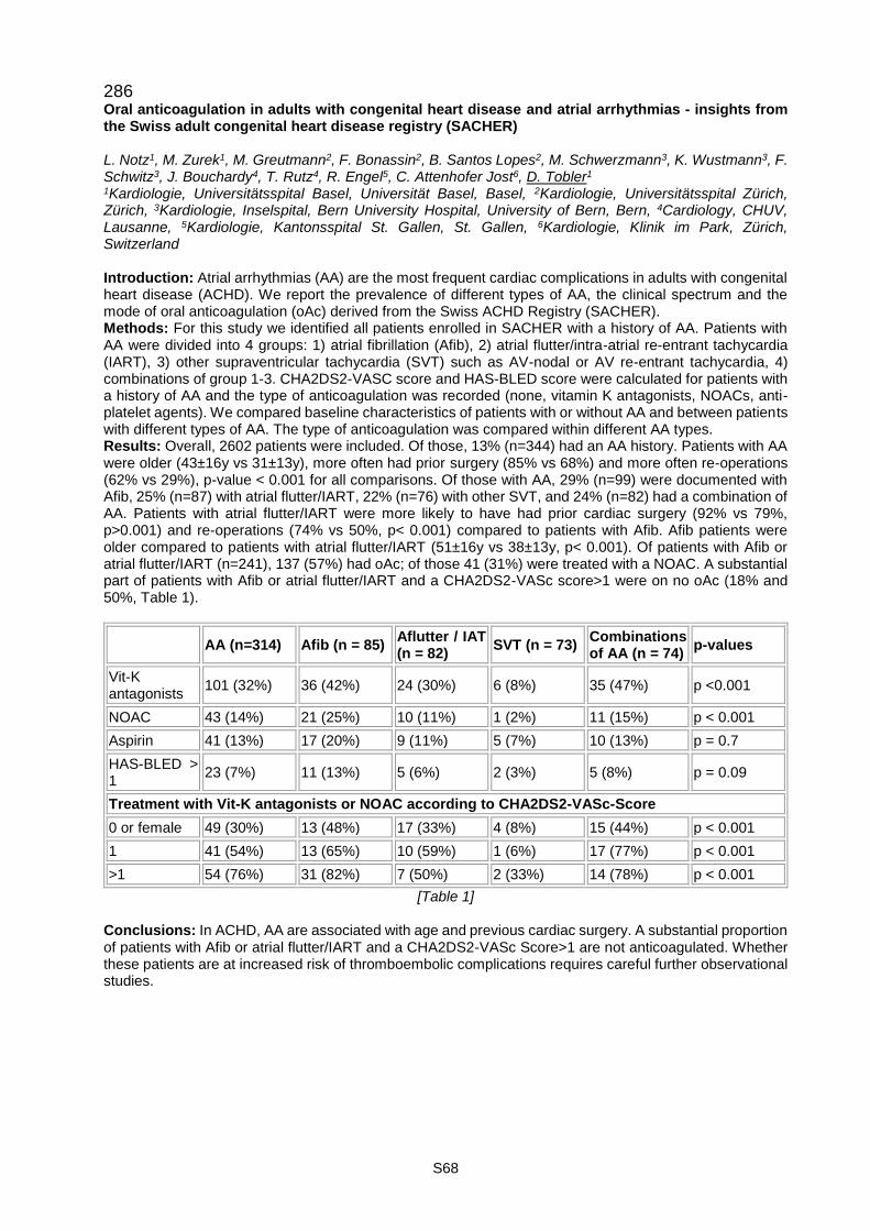
S68
286 Oral anticoagulation in adults with congenital heart disease and atrial arrhythmias - insights from the Swiss adult congenital heart disease registry (SACHER) L. Notz1, M. Zurek1, M. Greutmann2, F. Bonassin2, B. Santos Lopes2, M. Schwerzmann3, K. Wustmann3, F. Schwitz3, J. Bouchardy4, T. Rutz4, R. Engel5, C. Attenhofer Jost6, D. Tobler1 1Kardiologie, Universitätsspital Basel, Universität Basel, Basel, 2Kardiologie, Universitätsspital Zürich, Zürich, 3Kardiologie, Inselspital, Bern University Hospital, University of Bern, Bern, 4Cardiology, CHUV, Lausanne, 5Kardiologie, Kantonsspital St. Gallen, St. Gallen, 6Kardiologie, Klinik im Park, Zürich, Switzerland Introduction: Atrial arrhythmias (AA) are the most frequent cardiac complications in adults with congenital heart disease (ACHD). We report the prevalence of different types of AA, the clinical spectrum and the mode of oral anticoagulation (oAc) derived from the Swiss ACHD Registry (SACHER). Methods: For this study we identified all patients enrolled in SACHER with a history of AA. Patients with AA were divided into 4 groups: 1) atrial fibrillation (Afib), 2) atrial flutter/intra-atrial re-entrant tachycardia (IART), 3) other supraventricular tachycardia (SVT) such as AV-nodal or AV re-entrant tachycardia, 4) combinations of group 1-3. CHA2DS2-VASC score and HAS-BLED score were calculated for patients with a history of AA and the type of anticoagulation was recorded (none, vitamin K antagonists, NOACs, anti-platelet agents). We compared baseline characteristics of patients with or without AA and between patients with different types of AA. The type of anticoagulation was compared within different AA types. Results: Overall, 2602 patients were included. Of those, 13% (n=344) had an AA history. Patients with AA were older (43±16y vs 31±13y), more often had prior surgery (85% vs 68%) and more often re-operations (62% vs 29%), p-value < 0.001 for all comparisons. Of those with AA, 29% (n=99) were documented with Afib, 25% (n=87) with atrial flutter/IART, 22% (n=76) with other SVT, and 24% (n=82) had a combination of AA. Patients with atrial flutter/IART were more likely to have had prior cardiac surgery (92% vs 79%, p>0.001) and re-operations (74% vs 50%, p< 0.001) compared to patients with Afib. Afib patients were older compared to patients with atrial flutter/IART (51±16y vs 38±13y, p< 0.001). Of patients with Afib or atrial flutter/IART (n=241), 137 (57%) had oAc; of those 41 (31%) were treated with a NOAC. A substantial part of patients with Afib or atrial flutter/IART and a CHA2DS2-VASc score>1 were on no oAc (18% and 50%, Table 1).
AA (n=314) Afib (n = 85) Aflutter / IAT (n = 82)
SVT (n = 73) Combinations of AA (n = 74)
p-values
Vit-K antagonists
101 (32%) 36 (42%) 24 (30%) 6 (8%) 35 (47%) p <0.001
NOAC 43 (14%) 21 (25%) 10 (11%) 1 (2%) 11 (15%) p < 0.001
Aspirin 41 (13%) 17 (20%) 9 (11%) 5 (7%) 10 (13%) p = 0.7
HAS-BLED > 1
23 (7%) 11 (13%) 5 (6%) 2 (3%) 5 (8%) p = 0.09
Treatment with Vit-K antagonists or NOAC according to CHA2DS2-VASc-Score
0 or female 49 (30%) 13 (48%) 17 (33%) 4 (8%) 15 (44%) p < 0.001
1 41 (54%) 13 (65%) 10 (59%) 1 (6%) 17 (77%) p < 0.001
>1 54 (76%) 31 (82%) 7 (50%) 2 (33%) 14 (78%) p < 0.001
[Table 1] Conclusions: In ACHD, AA are associated with age and previous cardiac surgery. A substantial proportion of patients with Afib or atrial flutter/IART and a CHA2DS2-VASc Score>1 are not anticoagulated. Whether these patients are at increased risk of thromboembolic complications requires careful further observational studies.

S69
287 Quantification of aortic valve calcification on contrast-enhanced CT of patients prior to transcatheter aortic valve implantation M. Eberhard1, M. Mastalerz1, T. Frauenfelder1, F.C. Tanner2, F. Maisano2, F. Nietlispach2, B. Seifert3, H. Alkadhi1, T.D.L. Nguyen-Kim1 1Department of Radiology, University Hospital Zurich, 2University Heart Center at University Hospital Zurich, 3Department of Biostatistics, University Zurich, Zurich, Switzerland Purpose: To develop a formula enabling the quantification of aortic valve calcification (AVC) on contrast-enhanced CT of patients prior to transcatheter aortic valve implantation (TAVI). Methods: 218 consecutive patients (106 females, median age 82 years) with severe aortic valve stenosis undergoing non-enhanced and contrast-enhanced electrocardiography-gated CT angiography prior to TAVI were subdivided into a training (n=145) and validation cohort (n=72). AVC was semi-automatically segmented on contrast-enhanced CT using a threshold defined as average attenuation plus three standard deviations of attenuation in the aortic root. Linear regression model was applied to derive a formula enabling the quantification of the Agatston Score (AS) on contrast-enhanced CT in the training cohort. The formula was subsequently tested in the validation cohort. The AS quantified on non-enhanced CT served as reference standard. Results: On non-enhanced CT, patients in the training cohort had a medianAS of 2454 (interquartile range [IQR]: 1511-3636). The formula for computing the AS on contrast-enhanced CT in the training cohort was:
(2.27+1.04 ∗³√AgatsonCE)3. Testing the formula in contrast-enhanced CT of the validation cohort yielded a predicted median AS of 2339 (IQR: 1609-3643), being not significantly different to the reference standard AS based on non-enhanced CT (median 2455, IQR: 1503-3611, p=0.452). The intra-class correlation coefficient between predicted and reference standard AS in the validation cohort was 0.897; Bland Altman analyses showed narrow limits of agreement (average: -0.75). Conclusion: We introduced a formula enabling the accurate quantification of AVC on contrast-enhanced CT with similar results as compared to the standard Agatston method using non-enhanced CT. Thus, non-enhanced CT is no longer required for quantification of AVC.
288 Right ventricular morphology and function in left ventricular non-compaction cardiomyopathy S. Stämpfli1, A. Gotschy1, P. Kiarostami1, T. Özkartal1, C. Gruner1, M. Greutmann1, M. Niemann1,2, R. Manka1, F.C. Tanner1 1Klinik für Kardiologie | Kardiologie, Universitätsspital Zürich, Zürich, Switzerland, 2Furtwangen University, Faculty Mechanical and Medical Engineering, Furtwangen, Germany Introduction: Isolated left ventricular non-compaction cardiomyopathy (LVNC) is characterized by a two-layered left ventricular myocardium with extensive trabeculation. Involvement of the right ventricle (RV) has also been reported. Nevertheless, neither the optimal imaging modality nor standardized measurements for RV assessment have been defined. This study aims at describing RV morphology in LVNC and comparing cardiac magnetic resonance imaging (cMRI) with transthoracic echocardiography (TTE) in LVNC patients and controls. Method: Twenty patients with LVNC and 20 healthy controls (age and gender matched) underwent cMRI and TTE at the University Hospital Zurich. Measurements were performed in a blinded manner. Commonly measured dimensional and functional parameters were assessed according to current guidelines and recommendations. As novel cMRI parameters RV end-diastolic (ED) trabeculated area, RV ED trabeculated volume, and RV ED non-compacted to compacted ratio in short axis (SAX) were introduced.
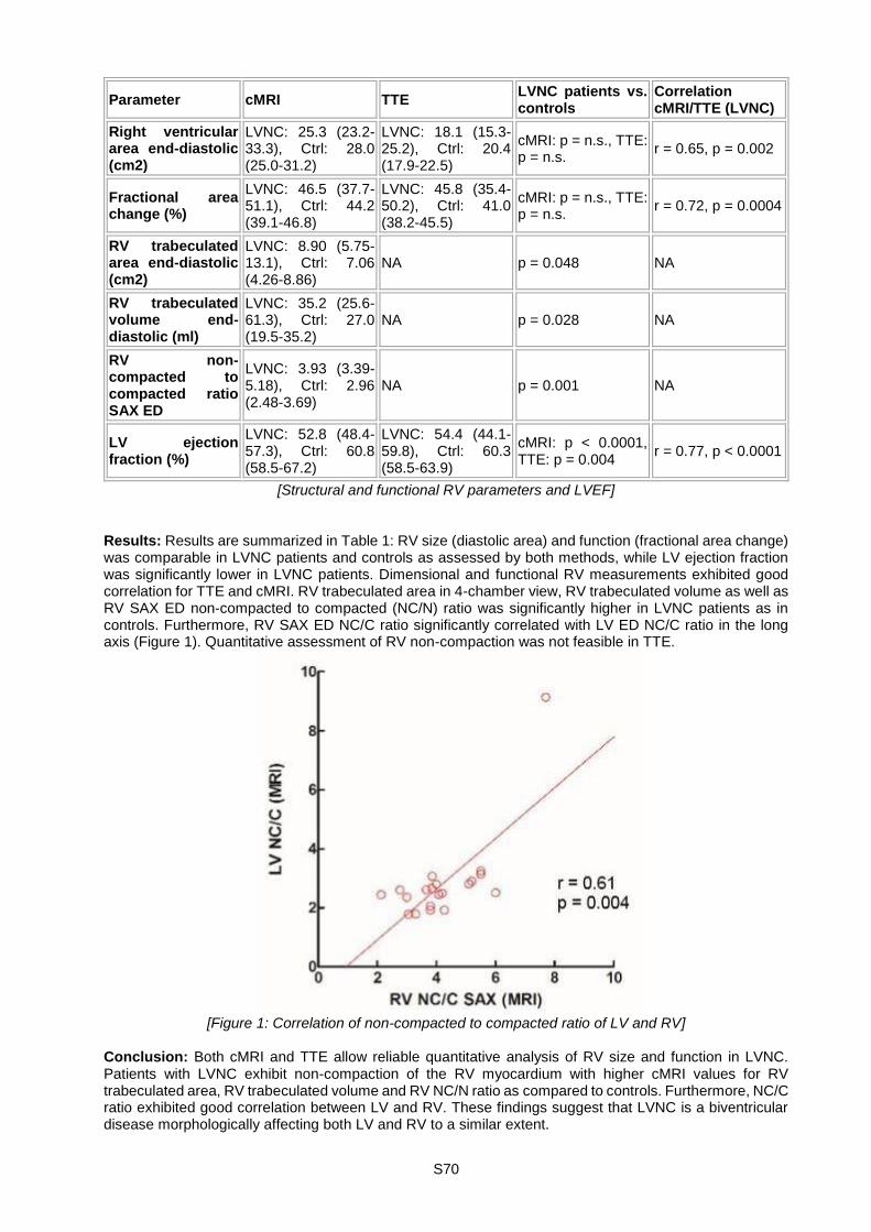
S70
Parameter cMRI TTE LVNC patients vs. controls
Correlation cMRI/TTE (LVNC)
Right ventricular area end-diastolic (cm2)
LVNC: 25.3 (23.2-33.3), Ctrl: 28.0 (25.0-31.2)
LVNC: 18.1 (15.3-25.2), Ctrl: 20.4 (17.9-22.5)
cMRI: p = n.s., TTE: p = n.s.
r = 0.65, p = 0.002
Fractional area change (%)
LVNC: 46.5 (37.7-51.1), Ctrl: 44.2 (39.1-46.8)
LVNC: 45.8 (35.4-50.2), Ctrl: 41.0 (38.2-45.5)
cMRI: p = n.s., TTE: p = n.s.
r = 0.72, p = 0.0004
RV trabeculated area end-diastolic (cm2)
LVNC: 8.90 (5.75-13.1), Ctrl: 7.06 (4.26-8.86)
NA p = 0.048 NA
RV trabeculated volume end-diastolic (ml)
LVNC: 35.2 (25.6-61.3), Ctrl: 27.0 (19.5-35.2)
NA p = 0.028 NA
RV non-compacted to compacted ratio SAX ED
LVNC: 3.93 (3.39-5.18), Ctrl: 2.96 (2.48-3.69)
NA p = 0.001 NA
LV ejection fraction (%)
LVNC: 52.8 (48.4-57.3), Ctrl: 60.8 (58.5-67.2)
LVNC: 54.4 (44.1-59.8), Ctrl: 60.3 (58.5-63.9)
cMRI: p < 0.0001, TTE: p = 0.004
r = 0.77, p < 0.0001
[Structural and functional RV parameters and LVEF] Results: Results are summarized in Table 1: RV size (diastolic area) and function (fractional area change) was comparable in LVNC patients and controls as assessed by both methods, while LV ejection fraction was significantly lower in LVNC patients. Dimensional and functional RV measurements exhibited good correlation for TTE and cMRI. RV trabeculated area in 4-chamber view, RV trabeculated volume as well as RV SAX ED non-compacted to compacted (NC/N) ratio was significantly higher in LVNC patients as in controls. Furthermore, RV SAX ED NC/C ratio significantly correlated with LV ED NC/C ratio in the long axis (Figure 1). Quantitative assessment of RV non-compaction was not feasible in TTE.
[Figure 1: Correlation of non-compacted to compacted ratio of LV and RV]
Conclusion: Both cMRI and TTE allow reliable quantitative analysis of RV size and function in LVNC. Patients with LVNC exhibit non-compaction of the RV myocardium with higher cMRI values for RV trabeculated area, RV trabeculated volume and RV NC/N ratio as compared to controls. Furthermore, NC/C ratio exhibited good correlation between LV and RV. These findings suggest that LVNC is a biventricular disease morphologically affecting both LV and RV to a similar extent.

S71
289 Short-term prognostic value of left ventricular global longitudinal strain following aortic valve replacement for severe aortic stenosis in the elderly: a prospective single-center study P. Antiochos1, A. Vella1, N. Barras1, O. Muller1, M. Gaxherri1, V. Crisinel1, M. Kirsch2, X. Jeanrenaud1, P. Monney1 1CHUV | Département Coeur-Vaisseaux | Service de Cardiologie, 2CHUV | Département Coeur-Vaisseaux | Service de Chirurgie Cardiaque, CHUV, Lausanne, Switzerland Introduction: Impaired left ventricular global longitudinal strain (GLS) has emerged as a valuable prognostic marker for all-cause mortality and poor cardiovascular outcomes in various clinical settings. We sought whether GLS predicts all-cause mortality, independently of baseline clinical, biological or other echocardiographic parameters, following aortic valve replacement for severe aortic stenosis (AS) in a population of elderly patients. Methods: We prospectively collected data for patients with severe AS, undergoing surgical (SAVR) or transcatheter aortic valve replacement (TAVR) from 01.01.2014 to date. Inclusion criteria were age>70 years old and availability of GLS measurement in baseline echocardiography. Exclusion criteria included concomitant aortic regurgitation grade >2, mitral valve disease grade >2 or previous cardiac surgery. The primary outcome was adjudicated all-cause mortality at 3 months. Results: Among n=84 patients (age 81.5 ± 6.5 years, 57.1% male, 67.9% TAVR) included in the analysis, 8 (9.5%) died within the first 3 months. All patients with a fatal outcome presented with a NYHA functional class of III/IV at admission and all of them suffered complications during their hospital stay. In univariate analysis, mortality was further associated with a baseline STS score [OR 1.11, 95 % confidence interval (1.0 - 1.23) for every 1% change, p=0.048], systolic blood pressure [1.98, (1.12 - 3.5) for every 10 mmHg, p=0.019] as well as haemoglobin [0.35, (0.18 -0.7) for every 10 g/L, p=0.003] and creatinine level at admission [1.31, (1.1 - 1.58) for every 10 µmol/L, p=0.003]. With regards to echocardiographic parameters, mortality was not related to GLS [1.66, (0.71 - 3.91) for every 5% change, p=0.244] or to aortic valve area [1.22, (0.82 - 1.8) for every 0.1 cm2 change, p=0.325], but was significantly associated with indexed LV mass [1.31, (1.04 - 1.66) for every 10 g/m2, p=0.024]. After adjusting for all of the above parameters in a multivariate model, only haemoglobin level at admission independently predicted mortality at 3 months [0.3, (0.1 - 0.95) for every 10 g/L, p=0.04]. Conclusion: Baseline echocardiographic parameters including GLS and AVA were not associated with all-cause death at 3 months, following aortic valve replacement for severe AS in the elderly. Short-term mortality in this specific population was mainly determined by baseline functional capacity, in-hospital complications and haemoglobin level at admission.
290 Right coronary artery motion analysis: a novel method to measure right ventricular systolic function by selective coronary angiography F. Baumann, S. Obeid, T. Gilhofer, P. Siegrist, R. Manka, J. Alibegovic, F. Tanner, T.F. Lüscher, R. Binder Department of Cardiology, University Heart Center at University Hospital Zurich, Zurich, Switzerland Aims: Right ventricular systolic function is not commonly assessed in the catheterization laboratory. We developed a novel method to assess right ventricular systolic function during selective coronary angiography. Methods: In consecutive patients who underwent transthoracic echocardiography and selective coronary angiography, the angiographic motion of the right coronary artery (RCA) in a thirty degree RAO view was analyzed by two independent operators. The motion distance and maximum speed of the mid-portion of the RCA during systole towards the apex was compared to the tricuspid annular plane systolic excursion by echocardiography as gold standard. Results: In 97 patients the mid-portion of the RCA moved 25.9 +/- 9.9 millimeter with a maximum speed of 11.3 +/- 4.9 centimeter per second during systole towards the apex. During independent repeat measurements the reliability of operator A was 94.7 % (95% CI 92.1 - 96.5, p < 0.001) and 95.6 % (95% CI 93.5 - 97.1, p < 0.001) and of operator B 98.1 % (95% CI 97.2 - 98.7, p < 0.001) and 95.6 % (95% CI 93.5 - 97.1, p < 0.001) for the RCA motion distance and RCA speed, respectively. Inter-observer variability was excellent for both measurements (Cronbachs's alpha for distance 0.976, 95% CI 0.964 - 0.984, p< 0.001; for speed 0.967, 95% CI 0.951 - 0.978, p < 0.001). There was a significant correlation of the RCA motion distance and RCA maximum speed with the tricuspid annular plane systolic excursion measured by echocardiography (RCA distance: r = 0.57, p < 0.001; RCA speed: r = 0.35, p < 0.001). In patients with a normal right ventricular systolic function measured by echocardiography the RCA motion distance was 27.8 +/- 9.2 millimeter and the RCA maximum speed was 12.0 +/- 4.6 centimeter per second. The area under the receiver operating curve for the RCA motion distance was 0.88 (95% CI 0.80 - 0.94) for discrimination of normal and abnormal right ventricular systolic function. A cut-off value of less than 22.2 millimeter systolic

S72
RCA motion had a specificity of 93.3 % and a sensitivity of 75.6 % for identifying an abnormal right ventricular systolic function. Conclusion: Analysis of the RCA motion is a novel reproducible and reliable method to indirectly measure right ventricular systolic function during selective coronary angiography. It is a simple and useful tool to asses right ventricular function in the catheterization laboratory and may serve for risk assessment of right ventricular failure.
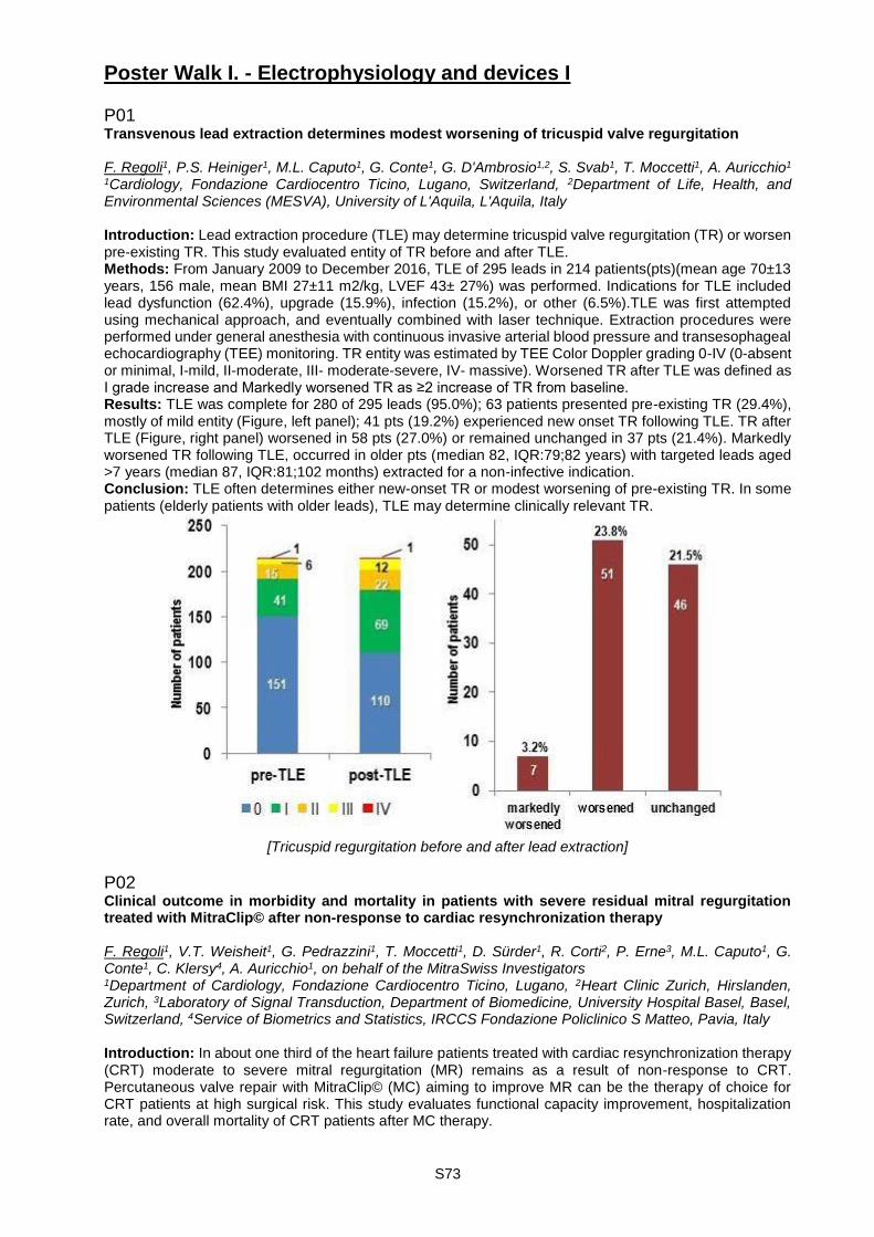
S73
Poster Walk I. - Electrophysiology and devices I P01 Transvenous lead extraction determines modest worsening of tricuspid valve regurgitation F. Regoli1, P.S. Heiniger1, M.L. Caputo1, G. Conte1, G. D'Ambrosio1,2, S. Svab1, T. Moccetti1, A. Auricchio1 1Cardiology, Fondazione Cardiocentro Ticino, Lugano, Switzerland, 2Department of Life, Health, and Environmental Sciences (MESVA), University of L'Aquila, L'Aquila, Italy Introduction: Lead extraction procedure (TLE) may determine tricuspid valve regurgitation (TR) or worsen pre-existing TR. This study evaluated entity of TR before and after TLE. Methods: From January 2009 to December 2016, TLE of 295 leads in 214 patients(pts)(mean age 70±13 years, 156 male, mean BMI 27±11 m2/kg, LVEF 43± 27%) was performed. Indications for TLE included lead dysfunction (62.4%), upgrade (15.9%), infection (15.2%), or other (6.5%).TLE was first attempted using mechanical approach, and eventually combined with laser technique. Extraction procedures were performed under general anesthesia with continuous invasive arterial blood pressure and transesophageal echocardiography (TEE) monitoring. TR entity was estimated by TEE Color Doppler grading 0-IV (0-absent or minimal, I-mild, II-moderate, III- moderate-severe, IV- massive). Worsened TR after TLE was defined as I grade increase and Markedly worsened TR as ≥2 increase of TR from baseline. Results: TLE was complete for 280 of 295 leads (95.0%); 63 patients presented pre-existing TR (29.4%), mostly of mild entity (Figure, left panel); 41 pts (19.2%) experienced new onset TR following TLE. TR after TLE (Figure, right panel) worsened in 58 pts (27.0%) or remained unchanged in 37 pts (21.4%). Markedly worsened TR following TLE, occurred in older pts (median 82, IQR:79;82 years) with targeted leads aged >7 years (median 87, IQR:81;102 months) extracted for a non-infective indication. Conclusion: TLE often determines either new-onset TR or modest worsening of pre-existing TR. In some patients (elderly patients with older leads), TLE may determine clinically relevant TR.
[Tricuspid regurgitation before and after lead extraction]
P02 Clinical outcome in morbidity and mortality in patients with severe residual mitral regurgitation treated with MitraClip© after non-response to cardiac resynchronization therapy F. Regoli1, V.T. Weisheit1, G. Pedrazzini1, T. Moccetti1, D. Sürder1, R. Corti2, P. Erne3, M.L. Caputo1, G. Conte1, C. Klersy4, A. Auricchio1, on behalf of the MitraSwiss Investigators 1Department of Cardiology, Fondazione Cardiocentro Ticino, Lugano, 2Heart Clinic Zurich, Hirslanden, Zurich, 3Laboratory of Signal Transduction, Department of Biomedicine, University Hospital Basel, Basel, Switzerland, 4Service of Biometrics and Statistics, IRCCS Fondazione Policlinico S Matteo, Pavia, Italy Introduction: In about one third of the heart failure patients treated with cardiac resynchronization therapy (CRT) moderate to severe mitral regurgitation (MR) remains as a result of non-response to CRT. Percutaneous valve repair with MitraClip© (MC) aiming to improve MR can be the therapy of choice for CRT patients at high surgical risk. This study evaluates functional capacity improvement, hospitalization rate, and overall mortality of CRT patients after MC therapy.

S74
Methods: Out of 832 patients included in the MitraSwiss registry, 94 patients were previously treated with CRT. Baseline clinical as well as follow-up data, including heart failure hospitalization and all-cause death, were extracted from multicenter registry database, MitraSwiss. Results: A significant reduction of MR was observed in 84 patients (95.35%) (≥1 grade MR reduction). However, significant improvement in NYHA functional class (≥1 NYHA class reduction) was observed in only 35 patients (51.47%); 24 patients (34.78%) remained in NYHA class III or IV six months after the intervention. During follow-up (median 18.8, IQR: 6.6; 33.5 months), 30 patients (32.26%) died (top panel). All-cause-mortality and hospitalization rates are presented in the figures Conclusion: Although MitraClip effectively reduces MR in almost all CRT patients, this effect does not appear to translate in significant reductions of both heart failure-related morbidity and mortality for any cause.
[Survival and HF hospitalization estimates]
P03 Pre-procedural short-lasting symptomatic atrial fibrillation is associated with multiple procedure success rate after pulmonary vein isolation S. Knecht1,2, M. Pradella3, T. Reichlin1,2, F. Spies1,2, B. Stieltjes3, J. Bremerich3, S. Osswald1,2, C. Sticherling1,2, M. Kühne1,2 1Cardiology, University Hospital Basel, University of Basel, 2Cardiovascular Research Institute Basel (CRIB), 3Radiology, University Hospital Basel, University of Basel, Basel, Switzerland Introduction: Pulmonary vein isolation (PVI) has become the cornerstone of the treatment of atrial fibrillation (AF). Outcomes after PVI are reported as single-procedure or multiple procedure success rates. The aim of the current study was to identify predictors for AF recurrence in patients after single and repeat PVI-only (no additional lesions). Method: Consecutive patients with symptomatic AF undergoing PVI-only as first and repeat ablation to reisolate reconnected veins were analysed. Established AF risk factors (hypertension, diabetes, heart failure, sleep apnea) and left atrial geometry from pre-procedural imaging (sphericity, volume, diameters, left atrial area and circumference) were assessed. AF burden was differentiated as paroxysmal and persistent and based on a patient reported symptomatic AF burden score including the frequency and duration of AF episodes and the number of cardioversions. Multivariable Cox-regression modelling
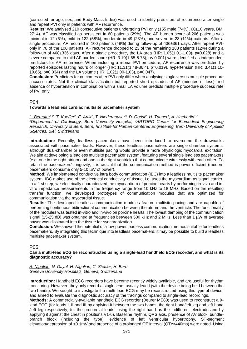
S75
(corrected for age, sex, and Body Mass Index) was used to identify predictors of recurrence after single and repeat PVI only in patients with AF recurrence. Results: We analysed 210 consecutive patients undergoing PVI only (155 male (74%), 60±10 years, BMI 27±4). AF was classified as persistent in 60 patients (29%). The AF burden score of 206 patients was minimal in 12 (6%), mild in 122 (58%), moderate in 49 (23%), and severe in 23 (11%) patients. After a single procedure, AF recurred in 100 patients (48%) during follow-up of 436±361 days. After repeat PVI-only in 78 of the 100 patients, AF recurrence dropped to 23 of the remaining 188 patients (12%) during a follow-up of 468±336 days. After a single procedure, the LA area (HR: 1.05(1.01-1.09), p=0.028) and a severe compared to mild AF burden score (HR: 3.10(1.65-5.78); p< 0.001) were identified as independent predictors for AF recurrence. When including a repeat PVI procedure, AF recurrence was predicted by reported episodes lasting hours or longer (HR: 11.31(1.48-86.4), p=0.019), hypertension (HR: 3.41(1.10-10.65), p=0.034) and the LA volume (HR: 1.02(1.00-1.03), p=0.047). Conclusion: Predictors for outcomes after PVI-only differ when analysing single versus multiple procedure success rates. Not the clinical classification but reported short episodes of AF (minutes or less) and absence of hypertension in combination with a small LA volume predicts multiple procedure success rate of PVI only.
P04 Towards a leadless cardiac multisite pacemaker system L. Bereuter1,2, T. Kueffer2, E. Artik2, T. Niederhauser3, D. Obrist2, H. Tanner1, A. Haeberlin1,2 1Department of Cardiology, Bern University Hospital, 2ARTORG Center for Biomedical Engineering Research, University of Bern, Bern, 3Institute for Human Centered Engineering, Bern University of Applied Sciences, Biel, Switzerland Introduction: Recently, leadless pacemakers have been introduced to overcome the drawbacks associated with pacemaker leads. However, these leadless pacemakers are single-chamber systems, although dual-chamber or even multisite pacing would provide a more physiologic myocardial excitation. We aim at developing a leadless multisite pacemaker system, featuring several single leadless pacemakers (e.g. one in the right atrium and one in the right ventricle) that communicate wirelessly with each other. To retain the pacemakers' longevity, it is crucial that the communication method is power efficient (modern pacemakers consume only 5-10 µW of power). Method: We implemented conductive intra body communication (IBC) into a leadless multisite pacemaker system. IBC makes use of the electrical conductivity of tissue, i.e. uses the myocardium as signal carrier. In a first step, we electrically characterized the myocardium of porcine hearts by performing in-vivo and in-vitro impedance measurements in the frequency range from 10 kHz to 18 MHz. Based on the resulting transfer function, we developed prototypes of communication modules that are optimized for communication via the myocardial tissue. Results: The developed leadless communication modules feature multisite pacing and are capable of performing continuous bidirectional communication between the atrium and the ventricle. The functionality of the modules was tested in-vitro and in-vivo on porcine hearts. The lowest damping of the communication signal (15-25 dB) was obtained at frequencies between 500 kHz and 2 MHz. Less than 1 µW of average power was dissipated into the tissue for synchronization. Conclusion: We showed the potential of a low-power leadless communication method suitable for leadless pacemakers. By integrating this technique into leadless pacemakers, it may be possible to build a leadless multisite pacemaker system.
P05 Can a multi-lead ECG be reconstructed using a single-lead handheld ECG recorder, and what is its diagnostic accuracy? A. Nigolian, N. Dayal, H. Nigolian, C. Stettler, H. Burri Geneva University Hospitals, Geneva, Switzerland Introduction: Handheld ECG recorders have become recently widely available, and are useful for rhythm monitoring. However, they only record a single lead, usually lead I (with the device being held between the two hands). We sought to investigate if a multi-lead ECG may be reconstructed using this type of device, and aimed to evaluate the diagnostic accuracy of the tracings compared to single-lead recordings. Methods: A commercially-available handheld ECG recorder (Beurer ME80) was used to reconstruct a 9-lead ECG (for leads I, II and III by applying it between the two hands, the right hand/left leg and left hand /left leg respectively; for the precordial leads, using the right hand as the indifferent electrode and by applying it against the chest in positions V1-6). Baseline rhythm, QRS axis, presence of AV block, bundle-branch block (including the type), evidence of left ventricular hypertrophy, ST-segment elevation/depression of >0.1mV and presence of a prolonged QT interval (QTc>440ms) were noted. Using
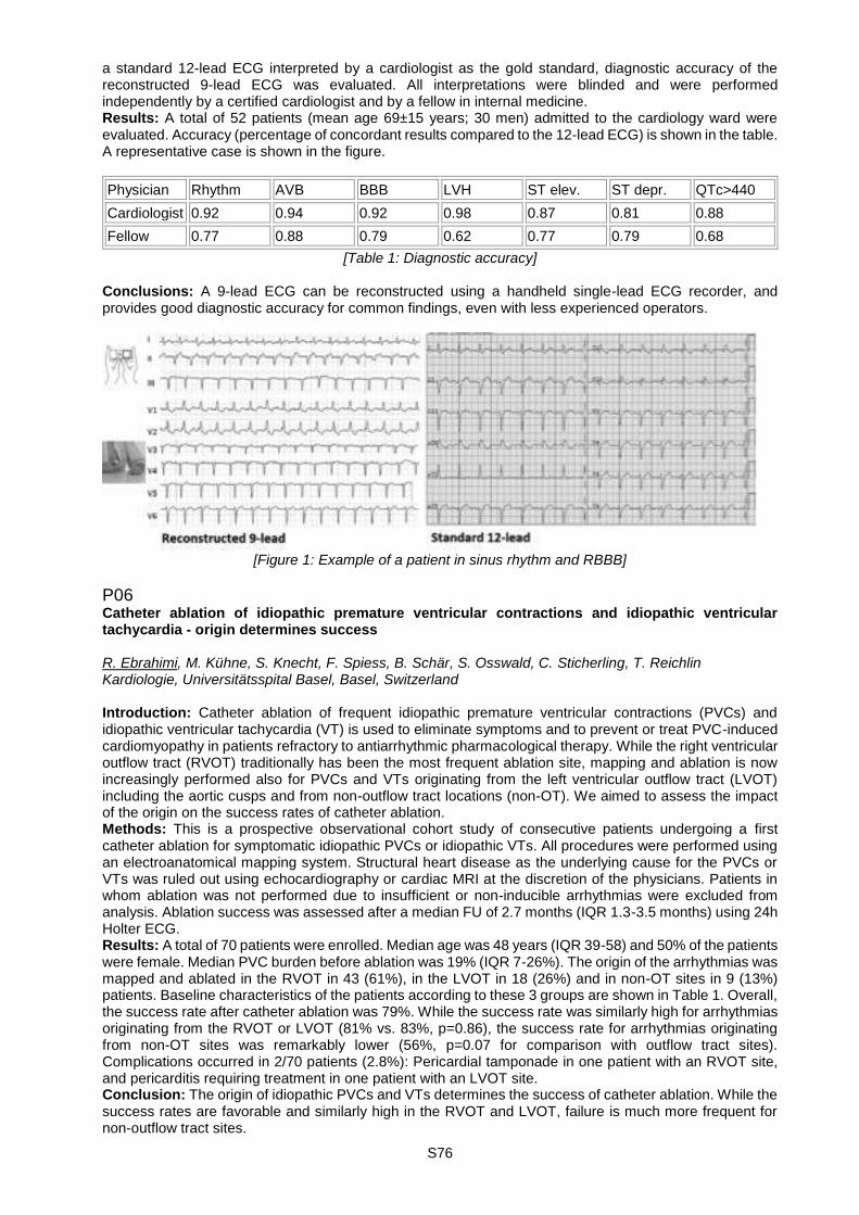
S76
a standard 12-lead ECG interpreted by a cardiologist as the gold standard, diagnostic accuracy of the reconstructed 9-lead ECG was evaluated. All interpretations were blinded and were performed independently by a certified cardiologist and by a fellow in internal medicine. Results: A total of 52 patients (mean age 69±15 years; 30 men) admitted to the cardiology ward were evaluated. Accuracy (percentage of concordant results compared to the 12-lead ECG) is shown in the table. A representative case is shown in the figure.
Physician Rhythm AVB BBB LVH ST elev. ST depr. QTc>440
Cardiologist 0.92 0.94 0.92 0.98 0.87 0.81 0.88
Fellow 0.77 0.88 0.79 0.62 0.77 0.79 0.68
[Table 1: Diagnostic accuracy] Conclusions: A 9-lead ECG can be reconstructed using a handheld single-lead ECG recorder, and provides good diagnostic accuracy for common findings, even with less experienced operators.
[Figure 1: Example of a patient in sinus rhythm and RBBB]
P06 Catheter ablation of idiopathic premature ventricular contractions and idiopathic ventricular tachycardia - origin determines success R. Ebrahimi, M. Kühne, S. Knecht, F. Spiess, B. Schär, S. Osswald, C. Sticherling, T. Reichlin Kardiologie, Universitätsspital Basel, Basel, Switzerland Introduction: Catheter ablation of frequent idiopathic premature ventricular contractions (PVCs) and idiopathic ventricular tachycardia (VT) is used to eliminate symptoms and to prevent or treat PVC-induced cardiomyopathy in patients refractory to antiarrhythmic pharmacological therapy. While the right ventricular outflow tract (RVOT) traditionally has been the most frequent ablation site, mapping and ablation is now increasingly performed also for PVCs and VTs originating from the left ventricular outflow tract (LVOT) including the aortic cusps and from non-outflow tract locations (non-OT). We aimed to assess the impact of the origin on the success rates of catheter ablation. Methods: This is a prospective observational cohort study of consecutive patients undergoing a first catheter ablation for symptomatic idiopathic PVCs or idiopathic VTs. All procedures were performed using an electroanatomical mapping system. Structural heart disease as the underlying cause for the PVCs or VTs was ruled out using echocardiography or cardiac MRI at the discretion of the physicians. Patients in whom ablation was not performed due to insufficient or non-inducible arrhythmias were excluded from analysis. Ablation success was assessed after a median FU of 2.7 months (IQR 1.3-3.5 months) using 24h Holter ECG. Results: A total of 70 patients were enrolled. Median age was 48 years (IQR 39-58) and 50% of the patients were female. Median PVC burden before ablation was 19% (IQR 7-26%). The origin of the arrhythmias was mapped and ablated in the RVOT in 43 (61%), in the LVOT in 18 (26%) and in non-OT sites in 9 (13%) patients. Baseline characteristics of the patients according to these 3 groups are shown in Table 1. Overall, the success rate after catheter ablation was 79%. While the success rate was similarly high for arrhythmias originating from the RVOT or LVOT (81% vs. 83%, p=0.86), the success rate for arrhythmias originating from non-OT sites was remarkably lower (56%, p=0.07 for comparison with outflow tract sites). Complications occurred in 2/70 patients (2.8%): Pericardial tamponade in one patient with an RVOT site, and pericarditis requiring treatment in one patient with an LVOT site. Conclusion: The origin of idiopathic PVCs and VTs determines the success of catheter ablation. While the success rates are favorable and similarly high in the RVOT and LVOT, failure is much more frequent for non-outflow tract sites.
S77
[Table: Baseline characteristics of the patients according to the origin of the idiopatic PVC/VT]
P07 Atrial dysplasia - a new mechanism for atrial fibrillation A. Saguner1, D. Akdis1, P. Bode2, C. Brunckhorst1, F. Duru1, G.H. Fontaine3 1Cardiology | Rhythmology, 2Pathology, USZ, Zurich, Switzerland, 3Cardiology | Rhythmology, Universite Pitie La Salpetriere, Paris, France Introduction: It was observed that atrial fibrillation/tachyarrhythmias (AF/AT) can be the first presentation of arrhythmogenic right ventricular dyplasia (ARVD). We therefore studied the atrial pathology of three ARVD patients. This abstract presents the atrial histology of ARVD patients with comparison to healthy controls considering the typical histologic changes known in the RV. Methods: Histology of the right atrium (RA) was available in 3 ARVD patients, in whom ARVD was confirmed by pathology. The observed anomalies were adipocytes in two cases, interstitial fibrosis in all, associated with replacement fibrosis in one case. This prompted us to study the RA structure in six subsequent controls without cardiovascular disease. Light microscopic examination with Leica digital image processing, and staining with HPS in ARVD to identify fibrosis, and HE in the control group, was performed. Results: The atrial pathology of these so-called normal controls presented anomalies, which can be interpreted as the background of an atrial arrhythmogenic substrate similar to the recently reported pathology of the RV in ARVD. As such, we found adipose tissue, interstitial and replacement fibrosis including one case of lymphocytic infiltration in the atria of these healthy controls, like histologic changes of the RV observed in ARVD (Figure1).
[Figure1]
Furthermore, we identified a perpendicular orientation of atrial myocardial fibres (Figure 2).

S78
[Figure 2]
Conclusions: The interface between the two perpendicular layers can be a zone of weakness leading to fat and fibrosis, particularly if increased loading conditions are present. Desmosomal variants may enhance this remodelling. However, since desmosomal mutations have not been observed in the normal heart, it is therefore possible to consider other genes or posttranslational modification to underly these changes. We propose new mechanisms for atrial tachy-arrhythmias including the role of active as well as healed myocarditis, which may precede the development of AF/AT and explain why these arrhythmias are the most frequent burden in the human species.
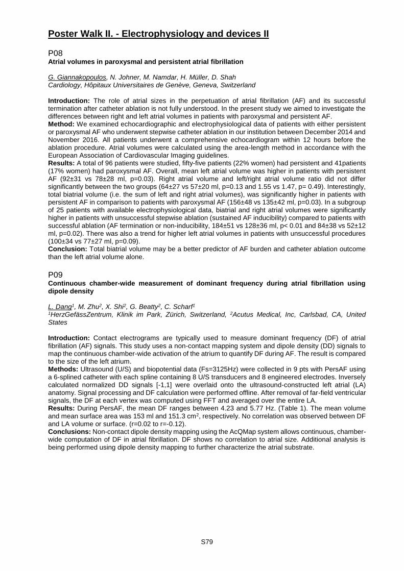
S79
Poster Walk II. - Electrophysiology and devices II P08 Atrial volumes in paroxysmal and persistent atrial fibrillation G. Giannakopoulos, N. Johner, M. Namdar, H. Müller, D. Shah Cardiology, Hôpitaux Universitaires de Genève, Geneva, Switzerland Introduction: The role of atrial sizes in the perpetuation of atrial fibrillation (AF) and its successful termination after catheter ablation is not fully understood. In the present study we aimed to investigate the differences between right and left atrial volumes in patients with paroxysmal and persistent AF. Method: We examined echocardiographic and electrophysiological data of patients with either persistent or paroxysmal AF who underwent stepwise catheter ablation in our institution between December 2014 and November 2016. All patients underwent a comprehensive echocardiogram within 12 hours before the ablation procedure. Atrial volumes were calculated using the area-length method in accordance with the European Association of Cardiovascular Imaging guidelines. Results: A total of 96 patients were studied, fifty-five patients (22% women) had persistent and 41patients (17% women) had paroxysmal AF. Overall, mean left atrial volume was higher in patients with persistent AF (92±31 vs 78±28 ml, p=0.03). Right atrial volume and left/right atrial volume ratio did not differ significantly between the two groups (64±27 vs 57±20 ml, p=0.13 and 1.55 vs 1.47, p= 0.49). Interestingly, total biatrial volume (i.e. the sum of left and right atrial volumes), was significantly higher in patients with persistent AF in comparison to patients with paroxysmal AF (156±48 vs 135±42 ml, p=0.03). In a subgroup of 25 patients with available electrophysiological data, biatrial and right atrial volumes were significantly higher in patients with unsuccessful stepwise ablation (sustained AF inducibility) compared to patients with successful ablation (AF termination or non-inducibility, 184±51 vs 128±36 ml, p< 0.01 and 84±38 vs 52±12 ml, p=0.02). There was also a trend for higher left atrial volumes in patients with unsuccessful procedures (100±34 vs 77±27 ml, p=0.09). Conclusion: Total biatrial volume may be a better predictor of AF burden and catheter ablation outcome than the left atrial volume alone.
P09 Continuous chamber-wide measurement of dominant frequency during atrial fibrillation using dipole density L. Dang1, M. Zhu2, X. Shi2, G. Beatty2, C. Scharf1 1HerzGefässZentrum, Klinik im Park, Zürich, Switzerland, 2Acutus Medical, Inc, Carlsbad, CA, United States Introduction: Contact electrograms are typically used to measure dominant frequency (DF) of atrial fibrillation (AF) signals. This study uses a non-contact mapping system and dipole density (DD) signals to map the continuous chamber-wide activation of the atrium to quantify DF during AF. The result is compared to the size of the left atrium. Methods: Ultrasound (U/S) and biopotential data (Fs=3125Hz) were collected in 9 pts with PersAF using a 6-splined catheter with each spline containing 8 U/S transducers and 8 engineered electrodes. Inversely calculated normalized DD signals [-1,1] were overlaid onto the ultrasound-constructed left atrial (LA) anatomy. Signal processing and DF calculation were performed offline. After removal of far-field ventricular signals, the DF at each vertex was computed using FFT and averaged over the entire LA. Results: During PersAF, the mean DF ranges between 4.23 and 5.77 Hz. (Table 1). The mean volume and mean surface area was 153 ml and 151.3 cm2, respectively. No correlation was observed between DF and LA volume or surface. (r=0.02 to r=-0.12). Conclusions: Non-contact dipole density mapping using the AcQMap system allows continuous, chamber-wide computation of DF in atrial fibrillation. DF shows no correlation to atrial size. Additional analysis is being performed using dipole density mapping to further characterize the atrial substrate.
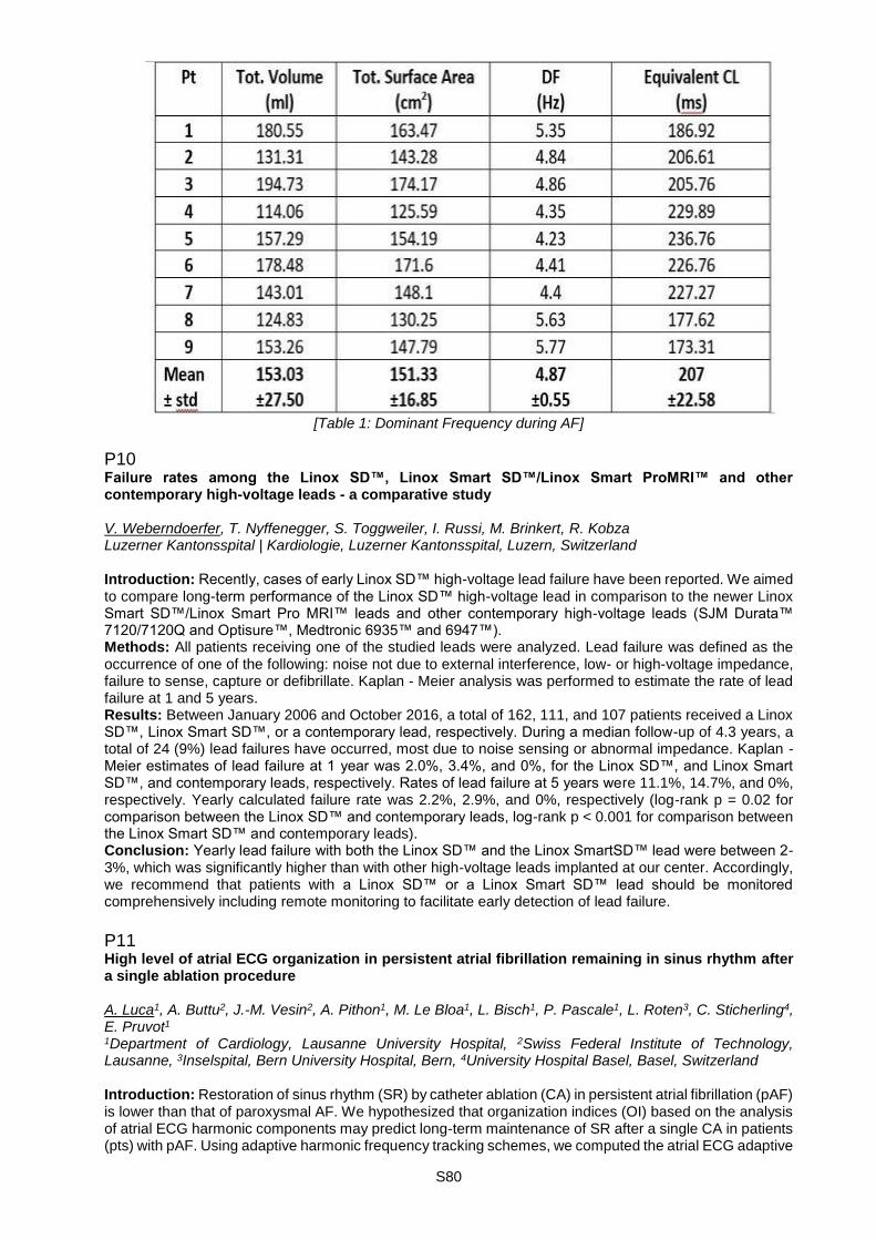
S80
[Table 1: Dominant Frequency during AF]
P10 Failure rates among the Linox SD™, Linox Smart SD™/Linox Smart ProMRI™ and other contemporary high-voltage leads - a comparative study V. Weberndoerfer, T. Nyffenegger, S. Toggweiler, I. Russi, M. Brinkert, R. Kobza Luzerner Kantonsspital | Kardiologie, Luzerner Kantonsspital, Luzern, Switzerland Introduction: Recently, cases of early Linox SD™ high-voltage lead failure have been reported. We aimed to compare long-term performance of the Linox SD™ high-voltage lead in comparison to the newer Linox Smart SD™/Linox Smart Pro MRI™ leads and other contemporary high-voltage leads (SJM Durata™ 7120/7120Q and Optisure™, Medtronic 6935™ and 6947™). Methods: All patients receiving one of the studied leads were analyzed. Lead failure was defined as the occurrence of one of the following: noise not due to external interference, low- or high-voltage impedance, failure to sense, capture or defibrillate. Kaplan - Meier analysis was performed to estimate the rate of lead failure at 1 and 5 years. Results: Between January 2006 and October 2016, a total of 162, 111, and 107 patients received a Linox SD™, Linox Smart SD™, or a contemporary lead, respectively. During a median follow-up of 4.3 years, a total of 24 (9%) lead failures have occurred, most due to noise sensing or abnormal impedance. Kaplan - Meier estimates of lead failure at 1 year was 2.0%, 3.4%, and 0%, for the Linox SD™, and Linox Smart SD™, and contemporary leads, respectively. Rates of lead failure at 5 years were 11.1%, 14.7%, and 0%, respectively. Yearly calculated failure rate was 2.2%, 2.9%, and 0%, respectively (log-rank p = 0.02 for comparison between the Linox SD™ and contemporary leads, log-rank p < 0.001 for comparison between the Linox Smart SD™ and contemporary leads). Conclusion: Yearly lead failure with both the Linox SD™ and the Linox SmartSD™ lead were between 2-3%, which was significantly higher than with other high-voltage leads implanted at our center. Accordingly, we recommend that patients with a Linox SD™ or a Linox Smart SD™ lead should be monitored comprehensively including remote monitoring to facilitate early detection of lead failure.
P11 High level of atrial ECG organization in persistent atrial fibrillation remaining in sinus rhythm after a single ablation procedure A. Luca1, A. Buttu2, J.-M. Vesin2, A. Pithon1, M. Le Bloa1, L. Bisch1, P. Pascale1, L. Roten3, C. Sticherling4, E. Pruvot1 1Department of Cardiology, Lausanne University Hospital, 2Swiss Federal Institute of Technology, Lausanne, 3Inselspital, Bern University Hospital, Bern, 4University Hospital Basel, Basel, Switzerland Introduction: Restoration of sinus rhythm (SR) by catheter ablation (CA) in persistent atrial fibrillation (pAF) is lower than that of paroxysmal AF. We hypothesized that organization indices (OI) based on the analysis of atrial ECG harmonic components may predict long-term maintenance of SR after a single CA in patients (pts) with pAF. Using adaptive harmonic frequency tracking schemes, we computed the atrial ECG adaptive
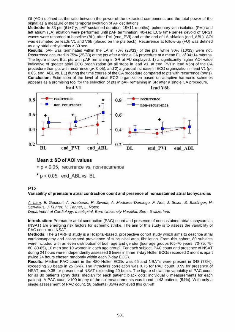
S81
OI (AOI) defined as the ratio between the power of the extracted components and the total power of the signal as a measure of the temporal evolution of AF oscillations. Methods: In 33 pts (61±7 y, pAF sustained duration: 19±11 months), pulmonary vein isolation (PVI) and left atrium (LA) ablation were performed until pAF termination. 40-sec ECG time series devoid of QRST waves were recorded at baseline (BL), after PVI (end_PVI) and at the end of LA ablation (end_ABL). AOI was estimated on leads V1 and V6b (placed on the pts back). Recurrence at follow-up (FU) was defined as any atrial arrhythmias > 30 sec. Results: pAF was terminated within the LA in 70% (23/33) of the pts, while 30% (10/33) were not. Recurrence occurred in 75% (25/33) of the pts after a single CA procedure at a mean FU of 34±14 months. The figure shows that pts with pAF remaining in SR at FU displayed: 1) a significantly higher AOI value indicative of greater atrial ECG organization (at all steps in lead V1, at end_PVI in lead V6b) of the CA procedure than pts with recurrence (p< 0.05), and 2) a gradual increase in ECG organization in lead V1 (p< 0.05, end_ABL vs. BL) during the time course of the CA procedure compared to pts with recurrence (p=ns). Conclusion: Estimation of the level of atrial ECG organization based on adaptive harmonic schemes appears as a promising tool for the selection of pts in pAF remaining in SR after a single CA procedure.
P12 Variability of premature atrial contraction count and presence of nonsustained atrial tachycardias A. Lam, E. Goulouti, A. Haeberlin, R. Sweda, A. Medeiros-Domingo, F. Noti, J. Seiler, S. Baldinger, H. Servatius, J. Fuhrer, H. Tanner, L. Roten Department of Cardiology, Inselspital, Bern University Hospital, Bern, Switzerland Introduction: Premature atrial contraction (PAC) count and presence of nonsustained atrial tachycardias (NSAT) are emerging risk factors for ischemic stroke. The aim of this study is to assess the variability of PAC count and NSAT. Methods: The STARFIB study is a Hospital-based, prospective cohort study which aims to describe atrial cardiomyopathy and associated prevalence of subclinical atrial fibrillation. From this cohort, 80 subjects were included with an even distribution of both age and gender [four age groups (65-70 years; 70-75; 75-80; 80-85), 10 men and 10 women in each age group]. For each subject, PAC count and presence of NSAT during 24 hours were independently assessed 6 times in three 7-day Holter ECGs recorded 2 months apart (twice 24 hours chosen randomly within each 7-day ECG). Results: Median PAC count in the 480 Holter ECGs was 65 and NSATs were present in 348 (73%), exceeding 20 beats in 25 (5%). The intraclass correlation was 0.75 for PAC count, 0.59 for presence of NSAT and 0.35 for presence of NSAT exceeding 20 beats. The figure shows the variability of PAC count for all 80 patients (gray dots: median for each patient; black dots: individual 6 measurements for each patient). A PAC count >100 in any of the six measurements was found in 43 patients (54%). With only a single assessment of PAC count, 28 patients (35%) achieved this cut-off.

S82
[Variability of premature atrial contraction count for all 80 patients]
Conclusions: Variability is low for PAC count but high for NSAT exceeding 20 beats. In 19% of patients we found only intermittent high PAC count. These patients fail to be recognized by a single assessment for high PAC count.
P13 Risk for adverse outcome events according to paroxysmal vs. non-paroxysmal atrial fibrillation S. Blum1, S. Aeschbacher1, P. Meyre1, P. Ammann2, P. Erne3, G. Moschovitis4, M. di Valentino5, D. Shah6, J. Schläpfer7, M. Kühne1, C. Sticherling1, S. Osswald1, D. Conen8 1Division of Cardiology, University Hospital Basel, Basel, 2Division of Cardiology, Kantonsspital St. Gallen, St. Gallen, 3Laboratory for Signal Transduction, University Hospital Basel, Basel, 4Division of Cardiology, Ospedale Regionale di Lugano, Lugano, 5Division of Cardiology, Ospedale San Giovanni Bellinzona, Bellinzona, 6Division of Cardiology, University Hospital Geneva, Geneva, 7Service of Cardiology, University Hospital Lausanne, Lausanne, Switzerland, 8McMaster University, Population Health Research Institute, Hamilton, ON, Canada Background: Increasing evidence suggests that sustained forms of atrial fibrillation (AF) are associated with worse outcomes, but long term follow-up data in unselected populations are lacking. In this study, we aimed to assess the risk for adverse events according to AF pattern in a large and unselected cohort of AF patients. Methods: We performed a prospective multicenter observational cohort study of 1540 AF patients. All patients completed questionnaires about personal characteristics and co-morbidities on a yearly basis. AF was classified into paroxysmal and non-paroxysmal AF. All outcomes were centrally validated and included incident hospitalization for heart failure (HF), all-cause mortality and a combined outcome of stroke, myocardial infarction or cardiovascular death (MACE). Multivariable adjusted Cox regression analysis was performed to assess hazard according to AF pattern. Results: Mean age of the population was 69±11 years, paroxysmal AF was observed among 863 (56%) patients and non-paroxysmal among 677 (44%). During a mean follow-up of 3.3±1.4 years, 117, 139 and 150 cases of HF, MACE, and overall deaths occurred, respectively. Compared to patients with paroxysmal
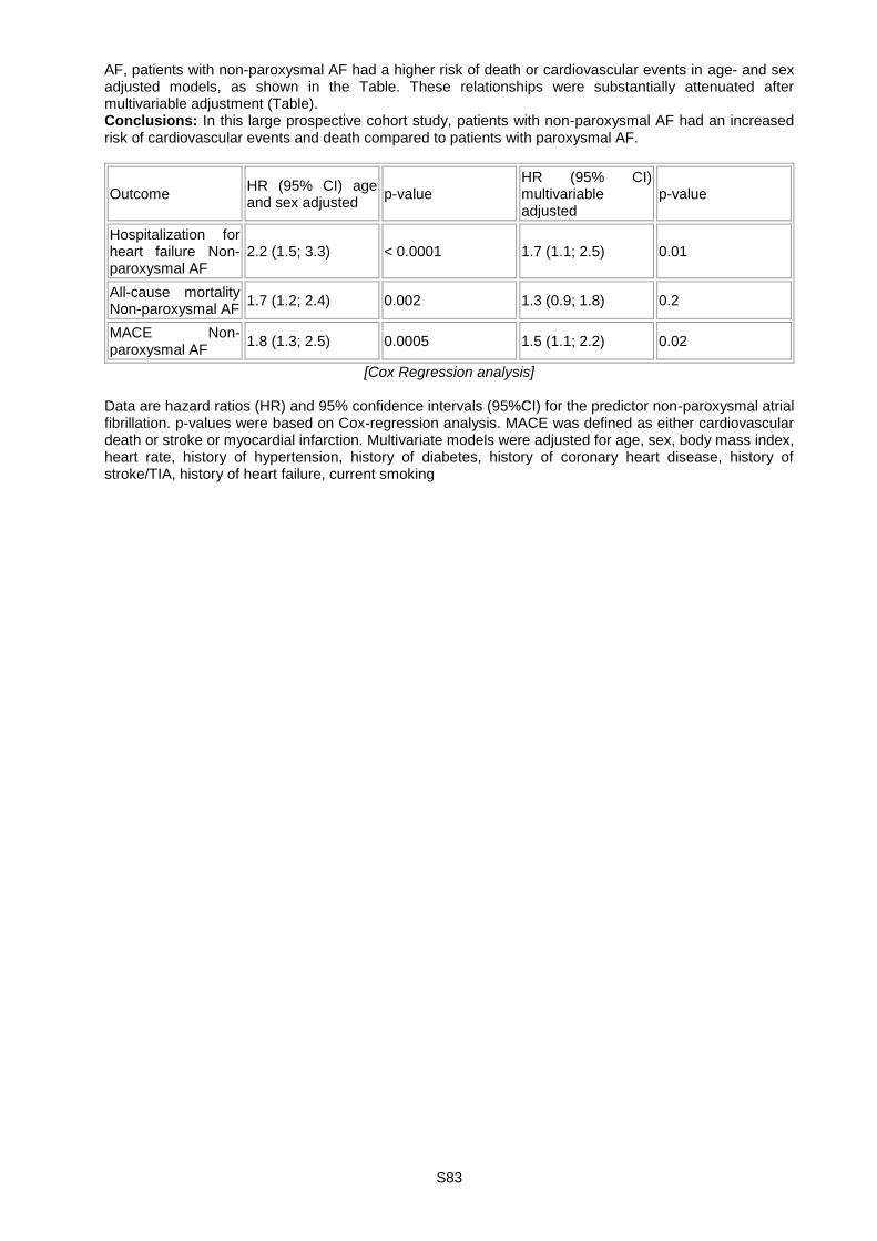
S83
AF, patients with non-paroxysmal AF had a higher risk of death or cardiovascular events in age- and sex adjusted models, as shown in the Table. These relationships were substantially attenuated after multivariable adjustment (Table). Conclusions: In this large prospective cohort study, patients with non-paroxysmal AF had an increased risk of cardiovascular events and death compared to patients with paroxysmal AF.
Outcome HR (95% CI) age and sex adjusted
p-value HR (95% CI) multivariable adjusted
p-value
Hospitalization for heart failure Non-paroxysmal AF
2.2 (1.5; 3.3) < 0.0001 1.7 (1.1; 2.5) 0.01
All-cause mortality Non-paroxysmal AF
1.7 (1.2; 2.4) 0.002 1.3 (0.9; 1.8) 0.2
MACE Non-paroxysmal AF
1.8 (1.3; 2.5) 0.0005 1.5 (1.1; 2.2) 0.02
[Cox Regression analysis] Data are hazard ratios (HR) and 95% confidence intervals (95%CI) for the predictor non-paroxysmal atrial fibrillation. p-values were based on Cox-regression analysis. MACE was defined as either cardiovascular death or stroke or myocardial infarction. Multivariate models were adjusted for age, sex, body mass index, heart rate, history of hypertension, history of diabetes, history of coronary heart disease, history of stroke/TIA, history of heart failure, current smoking

S84
Poster Walk I. - Basic research in cardiology P14 Rheumatoid arthritis and stroke - study of the role of chronic inflammation in ischemia/reperfusion brain injury N. Bonetti1, C. Diaz-Canestro1, M. Crucet1, S. Stivala1, A. Akhmedov1, T.F. Lüscher2, J.H. Beer1,3, G.G. Camici1 1Center for Molecular Cardiology | Cardiology Research, University of Zurich, Schlieren, 2Cardiology, University Heart Center Zurich, Zurich, 3Innere Medizin, Kantonsspital Baden, Baden, Switzerland Introduction: Rheumatoid arthritis (RA) affects up to 1 % of the population worldwide. Its detrimental systemic effects are underlined by a decrease of median survival by 17 years, with cardiovascular death accounting for 40 % of mortality. While the risk for coronary artery disease is at least 2-fold increased, the data concerning the incidence of stroke in this population are conflicting. Thus, we aimed to investigate Ischemia/Reperfusion (IR) brain injury in a mouse model of RA. Methods: We used a human Tumour Necrosis Factor α (TNFα) overexpressing mouse model that well reflects the peripheral and vascular phenotype of RA. Transgenic mice were treated intraperitoneally with the chimeric monoclonal TNFα antibody Infliximab or vehicle (Phosphate Buffered Saline (PBS)) from weeks 12-16. Wild type (WT) littermates were vehicle-treated within the same timeframe. 16-week-old mice were then subjected to transient middle cerebral artery occlusion (tMCAO) for 45 min. Stroke size was assessed by Triphenyltetrazoliumchloride (TTC) staining and neurological function by Bederson and RotaRod tests. Blood brain barrier (BBB) permeability after I/R brain injury was studied by immunohistochemical analysis of IgG extravasation and tight junction expression. Results: RA mice exhibited significantly larger stroke sizes and accordingly poorer neurological performance than their WT littermates. Treatment with Infliximab completely attenuated these findings. Immunohistochemical analyses revealed a significantly greater disruption of the BBB in untreated transgenic mice, as reflected by an increased IgG extravasation, at least partly mediated by a decreased occludin expression.
[Figure 1: (A) Untreated transgenic animals displayed significantly larger stroke sizes and significantly worse neurological function after tMCAO which was attenuated by treatment with Infliximab. (B) Untreated transgenic animals showed significantly more lgG extravasation after I/R brain injury than WT controls and Infliximab treated animals. In line with this observed a trend to decreased occluding expression in transgenic animals upon I/R brain injury]
S85
(Figure 1). Again, these observations were attenuated by treatment with Infliximab. Conclusions: In our RA model, we have seen significantly larger stroke sizes and worse neurological performance in transgenic mice. Treatment with Infliximab attenuated these findings. Increased IgG extravasation and decreased occludin expression after I/R brain injury in untreated transgenic mice suggests an aggravated BBB damage as an underlying mechanism. These findings call for further investigation of the role of inflammatory responses in I/R brain injury with special focus on the BBB integrity. Finally, findings from this study could help to delineate therapeutic targets specific to the subacute, inflammatory phase of stroke.
P15 GDF11 worsens myocardial infarction outcome in ischemia and reperfusion A. Akhmedov1, F. Montecucco2,3, D. Vdovenko1, N. Bonetti1, C. Diaz Canestro1, G.G. Camici1, A. Schaub Clerigue1, F. Mach2, T.F. Luescher1,4 1Center for Molecular Cardiology, Univerity of Zurich, Schlieren, 2Division of Cardiology, University of Geneva/Foundation for Medical Researches, Geneva, 3Department of Internal Medicine, University of Genoa School of Medicine/First Clinic of Internal Medicine, Genoa, 4Zurich Heart House, University Hospital Zurich, Zurich, Switzerland Introduction: Recent studies have implicated a role of TGFβ family members in aging and cardiovascular diseases. Growth Differentiation Factor 11 (GDF11) is a member of TGFβ superfamily with high homology to myostatin/GDF8. Interestingly, in mice its levels decline with age, whereas myostatin and TGFβ1 levels remain unchanged, suggesting involvement of GDF11 in aging. In addition, GDF11 has recently been shown to play a role in cardiac hypertrophy. However, not much is known about it's role in the myocardium. The goal of the present study was to examine the idea that restoring GDF11 levels by injecting recombinant GDF11 into blood stream of aged mice to the ones observed in young would rescue myocardial infarction and provide “youthful” characteristics to the old myocardium. Methods: 12-14-week-old and 22-24-month-old C57BL/6 male mice were injected daily with either recombinant human GDF11 or vehicle for 30 days. Afterwards mice were subjected to 30 min of ischemia (I) followed by 24h of reperfusion (R). Infarct size was assessed morphologically. Results: After I/R, both young and aged GDF11-injected mice developed markedly larger infarcts as compared to vehicle-treated group. This was further associated with increased post-ischemic levels of serum cardiac troponin I. In addition, both GDF11-injected groups showed accelerated cardiac cell death after I/R as has been assessed by TUNEL on heart cross sections. Of note, both GDF11-treated groups showed higher mortality during treatment. Finally, cardiac RISK and SAFE prosurvival pathways were less activated in both GDF11-treated groups. Conclusions: In summary, present study showed that daily injections of GDF11 promote increased sensitivity of the heart to myocardial infarction. Such GDF11-associated cardiac phenotype is likely to be driven by the increased cell death in the injured myocardium together with impaired function of prosurvival RISK and SAFE pathways. Thus, these results do not support proposed role of GDF11 as “rejuvenation” factor for the heart.
P16 Extracellular vescicles from pro-inflammatory cells contribute to cardiac dysfunction after myocardial infarction V. Biemmi1,2, S. Panella3, G. Milano2, A. Ciullo1,4, F. Muoio3, E. Cervio1, N. Pernigoni5, T. Moccetti5, T. Tallone3, G. Vassalli1, L. Barile1,2 1Laboratory of Cellular and Molecular Cardiology, Fondazione Cardiocentro Ticino, Lugano, 2Foundation for Cardiovascular Research and Education, 3Cell and Biomedical Technologies, Foundation for Cardiovascular Research and Education, Taverne, 4Center for Molecular Cardiology, Univerity of Zurich, Zurich, 5Fondazione Cardiocentro Ticino, Lugano, Switzerland Introduction: cardiac repair after myocardial infarction (MI) is a complex series of events initiated by an important inflammatory phase that serve to remove damaged cells followed by a reparative step. During MI macrophages (M0MΦ) infiltrate heart in a biphasic manner: the M1MΦ early population, associated with pro-inflammatory phenotype, picks 3-7 hrs after MI, whereas a later population, M2MΦ, associated with healing phenotype infiltrates the myocardium between days 4-7 after MI. Extracellular vesicles (EVs) are secreted nanoparticles by macrophages and other cells as mediator of cell-cell communication due to their ability to shuttle nucleic acids and proteins. Hypothesis: we asked whether EVs secreted by M1MΦ and M2MΦ differently act in inducing cytotoxic effect on cardiomyocytes (CM).
S86
Method: Human monocytes were isolated from buffy coats of healthy volunteers. M0MΦ were obtained after culturing monocyte for about 10 days in presence of M-CSF and polarized to M1MΦ and M2MΦ by using different combinations of cytokines. After polarization, M1MΦ and M2MΦ profiles were analyzed by FACS analysis and RT-PCR. To test cytotoxic effects of inflammatory EVs, complete or EVs-depleted conditioned medium as well as isolated EVs derived from M1MΦ and M2MΦ were added to the medium of neonatal CM and cell viability was assessed 12 hours after. To in-vivo confirm cytotoxic effect of pro-inflammatory EVs, GW4869 (inhibitor of EVs release) or vehicle were injected IP in rats 1 hour before the MI induction , echocardiography analysis was assessed 28 days after MI. Results: 24hrs after polarization M1MΦ cells overexpress specific genes such as IDOI, CXCL10 and GPD2 and M2MΦ overexpress TGFβ, CCL22 and PPARγ as assessed by Real-Time PCR. As shown by FACS analysis M1MΦ and M2MΦ expressed specific cell antigens which are not present in naïve monocyte like CD16, CD64 (M1MΦ profile) and CD206, CD163 (M2MΦ profile). M1MΦ-derived conditioned medium was able to induce cell death in CM and the cytotoxic effect decreased with EVs-depleted medium. In-vivo left ventricular ejection fraction (EF%) was comparably reduced at 24hrs post-MI in both, GW4869 and saline-treated group, but recovered to a greater extent in the first group than in control at 28 days post-MI. Conclusions: EVs secreted secreted by M1MΦ are able to induce cellular death in neonatal CM. Inhibition of EVs release during the acute phase of MI preserve heart function in an animal model of permanent LAD ligation.
P17 The functional relevance of HDL structure and composition in the improvement of cholesterol Efflux capacity after roux-en-Y gastric bypass P. Doytcheva1, J. Deuel2, D. Schaer2, T. Lutz3, T. Lüscher1, E. Osto1,4 1Center for Molecular Cardiology | Cardiology, Univerity of Zurich, Schlieren, 2Division of Internal Medicine- University Hospital Zurich, University of Zurich, 3Institute of Veterinary Physiology, Vetsuisse Faculty, Univerity of Zurich, Zurich, 4IFNH Laboratory of Translational Nutrition Biology, ETH Zurich, Schwerzenbach, Switzerland Background: Roux-en-Y gastric bypass (RYGB) reduces cardiovascular mortality. We showed that RYGB improves HDL cholesterol efflux capacity. The structure and molecular composition may be a crucial determinant of HDL functionality. Purpose: We studied whether and how changes in HDL subclasses profile and phospholipid (PL) composition after RYGB correlate with HDL cholesterol efflux capacity. Methods: HDL subclasses profile was assessed by fast protein liquid chromatography (FPLC) and by NMR from 15 morbidly obese patients before and 1 year after RYGB. After FPLC, cholesterol efflux was measured in J774 macrophages stimulated with HDL large, medium and small. Furthermore, the PL composition of different HDL subclasses was quantified by liquid chromatography-mass spectrometry (LC/MS). Results: FPLC showed increased smaller size HDL 1 year after RYGB (small HDL-1Y) compared to baseline. The size-function analysis revealed that among all HDL subclasses, small HDL-1Y were the most potent stimulator of cholesterol efflux from J774 macrophages. NMR analyses revealed also higher HDL particles number. Further, the composition of the small HDL-1Y showed specific elevation of certain phoshatidylcholines (PC), phosphatidylserine (PS) and phosphatidic acid (PA) content, as well as some corresponding lyso-forms (PA, PC, PE) compared to small HDL of the same patients before RYGB. Enrichment of HDL with these specific PL is correlated to an enhanced cholesterol efflux capacity and may underline the improved HDL functionality after RYGB. Reduced sphingosine-1-phosphate (S1P) content is a known mediator of HDL dysfunction. Indeed, S1P content was also increased in small HDL-1Y after RYGB. Total HDL cholesterol concentration was increased post RYGB. Conclusions: Small HDL-1Y after RYGB are increased and have enhanced cholesterol efflux capacity. Elevated content in PC, PS, PA and their lyso-forms as well as increased S1P levels may contribute to the improved athero-protective function of small HDL-1Y after RYGB. RYGB seems to achieve a dual benefit increasing the concentration and function of HDL.
S87
P18 Acquired intracoronary ADAMTS13 deficiency and VWF retention at sites of critical coronary stenosis in patients with STEMI G. Pedrazzini1, L. Biasco1, I. Sulzer2, A. Anesini1, T. Moccetti1, J. Kremer Hovinga2, L. Alberio3 1Fondazione Cardiocentro Ticino | Cardiology, Fondazione Cardiocentro Ticino, Lugano, 2Clinical Research, University of Bern, Bern, 3Division of Hematology and Central Hematology Laboratory, Centre Hospitalier Universitaire Vaudois (CHUV), Lausanne, Switzerland Background: Multimers of von Willebrand Factors (VWF) constitutes the bridge between platelet's glycoprotein Ib-α and collagen fibers allowing platelet's adhesion. ADAMTS13 by cleaving VWF multimers decreases their adhesiveness. Local imbalance between VWF and ADAMTS13 might be responsible for the presence of highly adhesive VWF multimers mediating adhesion and vessel occlusion. Aim: to evaluate systemic and intracoronary VWF and ADAMTS13 activity in patients with acute thrombotic coronary occlusion. Methods and results: 27 patients (24 men; median age, 65 years) undergoing emergency cardiac catheterization for STEMI with a TIMI 0 or I culprit lesion were evaluated. Blood samples were obtained from a peripheral vein, the coronary ostia and 1-3 cm distal to the site of occlusion with a thrombus aspiration catheter. VWF antigen (VWF:Ag), VWF ristocetin cofactor activity (VWF:RCo), ADAMTS13 activity were assessed. Both VWF:RCo and VWFAg and ADAMTS13 resulted significantly reduced in samples obtained distally to an occluded coronary as compared to those derived from the coronary ostia (VWF:RCo 190%,IQR124%-263% vs 146%,IQR106%-236% p=0.003; VWF:Ag 157%, IQR119%-207% vs 137% IQR 99%-192% p < 0.001; ADAMTS13 53% IQR 31%-75% vs ), while the ratio between ADAMTS13/VWF:RCo was significantly reduced in the coronary flow (ostium 0.31; distal occlusion 0.29) when compared to the systemic circulation (0.48; p< 0.001). Conclusions: a decreased ADAMTS13/VWF ratio in the coronary flow favors the presence of highly adhesiveVWFmultimers that would deposit at the site of a critical stenosis, mediating platelet adhesion and coronary occlusion.
P19 Overexpression of CXCR4 in cardiac progenitor cells improves exosome internalization by cardiomyocytes A. Ciullo1,2, V. Biemmi1,3, G. Milano3, E. Cervio1, T. Torre4, S. Demertzis4, T. Moccetti4, G.G. Camici2, G. Vassalli1, L. Barile1,3 1Laboratory of Cellular and Molecular Cardiology, Fondazione Cardiocentro Ticino, Lugano, 2Center for Molecular Cardiology, University of Zurich, Schlieren, 3Foundation for Cardiovascular Research and Education, Taverne-Torricella, 4Fondazione Cardiocentro Ticino, Lugano, Switzerland Introduction: exosomes (Exo) 40-150nm sized nanoparticles secreted from many cells like stem cells and tumor cells are important mediators of cell-cell communication due their ability to shuttle nucleic acids like microRNAs and proteins. Evidence indicates that Exo derived form cardiac progenitor cells (Exo-CPC) are enriched for several cardioprotective and proangiogenic factors thereby mediating cardiac protection during myocardial ischemia/reperfusion injury. CXCR4, a Gprotein-coupled 7-transmembrane receptor, together with its primary ligand stromal cell-derived factor- (SDF-) 1 alpha serves as a major regulator of stem/progenitor cell activities. Whether the axis CXCR4-SDF-1 alpha may also modulate the effectiveness of Exo or their uptake by target cells is unknown. We aimed to study the role of CXCR4 in modulating in-vitro and in-vivo the uptake of Exo-CPC by cardiomyocytes (CM). Methods: CPC were transfected with null vector (pCDNA3.1) or with vector to overexpress CXCR4 (pCDNA-CXCR4). Moreover to track Exo, c. elegans species specific Cel-miR-39 was overexpressed in human CPC or human dermal fibroblasts as control. Exo containing Cel-miR-39 and CXCR4 (ExoCR4-Cel39) or control Exo (ExoCTR-Cel39) were isolated and incubated with CM to in-vitro assess internalization by CM. In-vivo MI was induced in rats and 24 hrs later hearts were excided and retrogrady perfused in a Langendorff system. ExoCR4-Cel39 and ExoCTR-Cel39 were added to the perfusate. Hearts were perfused for 2 hrs before their enzymatic dissociation and CM isolation. Results: intracellular levels of Cel-miR-39 in CM exposed to ExoCTR-Cel39 from CPC were higher than those in cells exposed to ExoCTR-Cel39 derived from fibroblast suggesting a higher uptake efficiency for the former. By comparing ExoCTR-Cel39 and ExoCR4-Cel39 both derived from CPC the latest showed higher tropism for CM, demonstrating the implication of the transmembrane protein CXCR4 into Exo-uptake. Likewise, we assessed the cellular uptake of Exo by primary CM using isolated rat hearts perfused in a Langendorff system. Cel-miR-39 levels were higher in CM from hearts perfused with ExoCR4-Cel39 -containing perfusates, as compared with ExoCTR-Cel39 -containing perfusates.

S88
Conclusions: this study reveals a novel role of Exo derived from CPC overexpressing CXCR4 and highlights a new mechanism of intercellular mediation of progenitor cells for MI treatment.
P20 Bile acid-mediated vasodilatation in Takeda G-protein-coupled receptor 5 (TGR5)-deleted mice A. Taheri1,2,3, M. Engeli1,2,3, P. Doytcheva1,2,3, R. Urbanet1,2,3, M. Lannoo4, C. Wolfrum5, T.F. Luscher1,3, T.A. Lutz2,3, E. Osto1,3,5 1Center for Molecular Cardiology, University of Zurich and University Heart Center, Cardiology, University Hospital Zurich, 2Institute of Veterinary Physiology, Vetsuisse Faculty, University of Zurich, 3Zurich Center for Integrative Human Physiology, University of Zurich, Zurich, Switzerland, 4Visceral Surgery, University Hospital and Katholieke Universiteit Leuven, Leuven, Belgium, 5Laboratory of Translational Nutrition Biology, ETH Zurich, Schwerzenbach, Switzerland Introduction: Bile acids (BA) may mediate many beneficial cardiovascular effects after Roux en-Y gastric bypass (RYGB). BA can exert vasoactive actions via membrane (TGR5) and nuclear (farnesoid-X (FXR)) receptors at the aortic level. Here, the vasodilatatory action of taurolithocholic acid (TLCA), a potent TGR5 activator, and chenodeoxycholic acid (CDCA), a potent FXR activator, was investigated in TGR5-deleted mice (TGR5 -/-). Methods: Thoracic aortic rings of TGR5 -/- and control (WT) male mice aged 20 weeks and fed on normal chow were collected and suspended for isometric tension recording. Cumulative concentration-relaxation curves were obtained in response to TLCA (10-7 to 10-4 mol/L), CDCA (10-7 to 10-4 mol/L), glucagon-like peptide-1 (GLP-1 10-12 to 10-6 mol/L), norepinephrine, (NE, 10-11 to 10-5 mol/L), acetylcholine (ACh, 10-9 to 10-5 mol/L), and sodium nitroprusside, a nitric oxide donor (SNP, 10-10 to 10-6 mol/L), and after incubation with the H2O2 scavenger, catalase (1200 U/ml); indomethacin (10-4 mol/L), a COX1/2 inhibitor; propylargylglycine (PAG, 3 mM), a cystathionine y-lyase inhibitor; and L-NAME (10-4 mol/L), the endothelial nitric oxide synthase (eNOS) inhibitor, respectively. Removal of the endothelium with Triton X-100 0.05% was used to assess endothelium-independent responses. Results: Vasodilatation in response to TLCA and CDCA was significantly reduced by L-NAME and endothelium removal. This supports the presence of endothelial and NO-dependent vasodilatation in these mice. There were no differences in vasorelaxation induced by TLCA and CDCA, as well as by ACh, GLP-1 and SNP in TGR5 -/- vs WT. Preincubation with catalase only significantly reduced TLCA-induced vasorelaxation in WT but not in TGR5 -/- mice suggesting also a role for H2O2 in modulating the effect of TLCA. Preincubation with PAG reduced vasodilatation only in TGR5 -/- in response to CDCA, suggesting a role for H2S in modulating FXR-dependent vasorelaxation. Indomethacin had no effect on the vasoactive properties of TLCA and CDCA in TGR5 -/- vs WT, suggesting a COX-independent vasorelaxatory mechanism. Conclusion: TLCA and CDCA induce endothelium-dependent vasorelaxation, likely through the production of NO. TGR5 -/- mice may compensate for the absence of the receptor through a yet unclear pathway at the endothelial level. The molecular mechanisms involving the role of H2S and H2O2 in TGR5-induced vasorelaxation warrants further investigation.
P21 Facilitating the use of donation after circulatory death in heart transplantation: cardioprotective reperfusion strategies act through different mitochondrial sites M.-N. Sanz, E. Farine, P. Niederberger, N. Mendez Carmona, R.K. Wyss, M. Arnold, T.P. Carrel, H.T. Tevaearai Stahel, S.L. Longnus Clinic for Cardiovascular Surgery, University of Bern and Inselspital, Bern, Switzerland Background and aim: Donation after circulatory determination of death (DCD) is a promising solution for increasing donor organ availability, but implementation in heart transplantation is limited due to ischemia/reperfusion injury (IRI). Mitochondria play a central role in IRI; as they both undergo damage, but may confer tissue damage themselves. Using an isolated rat heart model of DCD, our laboratory has identified three cardioprotective reperfusion strategies: mild hypothermia (MH), mechanical post-conditioning (MPC) and hypoxia (HY)1. Considering that the strategies studied increased cardiac oxygen consumption, we hypothesize that mitochondrial alterations underlie their cardioprotective effects. Methods: Experiments were performed using an isolated, working rat heart model of DCD. Hearts of adult, male Wistar rats underwent 20 min aerobic perfusion, 27 min global ischemia and 60 min reperfusion. Five experimental conditions were compared: SHAM (no ischemia; n=8), ischemia non-intervention (CT; n=9), and ischemia plus reperfusion strategy (MH, MPC or HY; n=8 for each strategy). Ventricular tissue was snap-frozen for spectrophotometric analysis of mitochondrial parameters (mass and function) and energy shuttles.
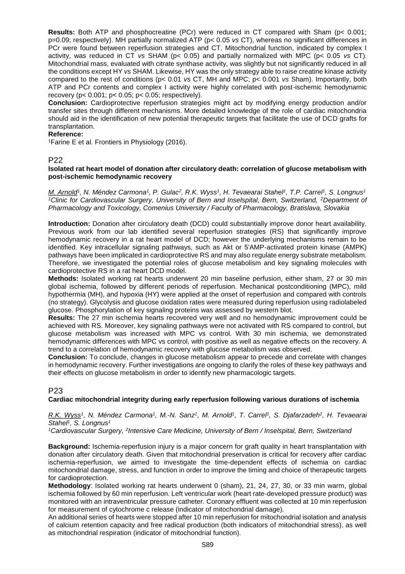
S89
Results: Both ATP and phosphocreatine (PCr) were reduced in CT compared with Sham (p< 0.001; p=0.09; respectively). MH partially normalized ATP (p< 0.05 vs CT), whereas no significant differences in PCr were found between reperfusion strategies and CT. Mitochondrial function, indicated by complex I activity, was reduced in CT vs SHAM (p< 0.05) and partially normalized with MPC (p< 0.05 vs CT). Mitochondrial mass, evaluated with citrate synthase activity, was slightly but not significantly reduced in all the conditions except HY vs SHAM. Likewise, HY was the only strategy able to raise creatine kinase activity compared to the rest of conditions (p< 0.01 vs CT, MH and MPC; p< 0.001 vs Sham). Importantly, both ATP and PCr contents and complex I activity were highly correlated with post-ischemic hemodynamic recovery (p< 0.001; p< 0.05; p< 0.05; respectively). Conclusion: Cardioprotective reperfusion strategies might act by modifying energy production and/or transfer sites through different mechanisms. More detailed knowledge of the role of cardiac mitochondria should aid in the identification of new potential therapeutic targets that facilitate the use of DCD grafts for transplantation. Reference: 1Farine E et al. Frontiers in Physiology (2016).
P22 Isolated rat heart model of donation after circulatory death: correlation of glucose metabolism with post-ischemic hemodynamic recovery M. Arnold1, N. Méndez Carmona1, P. Gulac2, R.K. Wyss1, H. Tevaearai Stahel1, T.P. Carrel1, S. Longnus1 1Clinic for Cardiovascular Surgery, University of Bern and Inselspital, Bern, Switzerland, 2Department of Pharmacology and Toxicology, Comenius University / Faculty of Pharmacology, Bratislava, Slovakia Introduction: Donation after circulatory death (DCD) could substantially improve donor heart availability. Previous work from our lab identified several reperfusion strategies (RS) that significantly improve hemodynamic recovery in a rat heart model of DCD; however the underlying mechanisms remain to be identified. Key intracellular signaling pathways, such as Akt or 5'AMP-activated protein kinase (AMPK) pathways have been implicated in cardioprotective RS and may also regulate energy substrate metabolism. Therefore, we investigated the potential roles of glucose metabolism and key signaling molecules with cardioprotective RS in a rat heart DCD model. Methods: Isolated working rat hearts underwent 20 min baseline perfusion, either sham, 27 or 30 min global ischemia, followed by different periods of reperfusion. Mechanical postconditioning (MPC), mild hypothermia (MH), and hypoxia (HY) were applied at the onset of reperfusion and compared with controls (no strategy). Glycolysis and glucose oxidation rates were measured during reperfusion using radiolabeled glucose. Phosphorylation of key signaling proteins was assessed by western blot. Results: The 27 min ischemia hearts recovered very well and no hemodynamic improvement could be achieved with RS. Moreover, key signaling pathways were not activated with RS compared to control, but glucose metabolism was increased with MPC vs control. With 30 min ischemia, we demonstrated hemodynamic differences with MPC vs control, with positive as well as negative effects on the recovery. A trend to a correlation of hemodynamic recovery with glucose metabolism was observed. Conclusion: To conclude, changes in glucose metabolism appear to precede and correlate with changes in hemodynamic recovery. Further investigations are ongoing to clarify the roles of these key pathways and their effects on glucose metabolism in order to identify new pharmacologic targets.
P23 Cardiac mitochondrial integrity during early reperfusion following various durations of ischemia R.K. Wyss1, N. Méndez Carmona1, M.-N. Sanz1, M. Arnold1, T. Carrel1, S. Djafarzadeh2, H. Tevaearai Stahel1, S. Longnus1 1Cardiovascular Surgery, 2Intensive Care Medicine, University of Bern / Inselspital, Bern, Switzerland Background: Ischemia-reperfusion injury is a major concern for graft quality in heart transplantation with donation after circulatory death. Given that mitochondrial preservation is critical for recovery after cardiac ischemia-reperfusion, we aimed to investigate the time-dependent effects of ischemia on cardiac mitochondrial damage, stress, and function in order to improve the timing and choice of therapeutic targets for cardioprotection. Methodology: Isolated working rat hearts underwent 0 (sham), 21, 24, 27, 30, or 33 min warm, global ischemia followed by 60 min reperfusion. Left ventricular work (heart rate-developed pressure product) was monitored with an intraventricular pressure catheter. Coronary effluent was collected at 10 min reperfusion for measurement of cytochrome c release (indicator of mitochondrial damage). An additional series of hearts were stopped after 10 min reperfusion for mitochondrial isolation and analysis of calcium retention capacity and free radical production (both indicators of mitochondrial stress), as well as mitochondrial respiration (indicator of mitochondrial function).

S90
Results: Post-ischemic left ventricular work at 60 min reperfusion significantly decreased after ischemia of 27 min or longer compared with sham (p< 0.05, n=7-8 per group). Mitochondrial cytochrome c release inversely correlated with ischemic duration (r=-0.833, p< 0.001, n=43). At 10 min reperfusion, ischemia, regardless of duration, decreased mitochondrial calcium retention capacity. Mitochondrial ROS production strongly increased with 21 and 27 min of ischemia compared with shams (p< 0.05, n=3-4 per group), whereas values after 33 min ischemia were not different. 27 min ischemia impaired mitochondrial complex II respiration (p< 0.05, n=5-6 per group) and 33 min ischemia impaired complex I and II respiration (p< 0.01, n=5-6 per group) compared with sham hearts. Conclusion: Following ischemia, mitochondrial damage and signs of stress precede cardiac hemodynamic and mitochondrial respiratory dysfunction. Our results indicate a functional resistance of cardiac mitochondria in early reperfusion to ischemic periods up to 21 min. In addition, cytochrome c is found to be a sensitive, early predictor of cardiac hemodynamic recovery that can be measured easily and rapidly. These findings may aid in the establishment of cardioprotective reperfusion strategies.

S91
Poster walk I. - Innovation in congenital cardiac surgery and imaging P24 Application of a novel, absorbable, knotless suture device provides excellent results for sternal closure and healing in paediatric cardiac surgery P.P. Heinisch1, K. Kleimeyer1, M. Bartkevics1, M. Veit1, D. Hutter2, T. Carrel1, A. Kadner1 1Centre of Congenital Heart Disease, Department of Cardiovascular Surgery, 2Centre of Congenital Heart Disease, Department of Cardiology, Inselspital, Bern University Hospital, University of Bern, Bern, Switzerland Background: Sternal closure by absorbable suture material is an established method for chest closure in paediatric cardiac surgery. However, the formation of granuloma around knotted suture material is rather frequently observed and is known to be the potential source of prolonged wound healing and infection, particularly in new-borns and infants. This study aims to determine the suitability and reliability of a novel absorbable, self-locking multi-anchor knotless suture with antibacterial technology for sternal closure in children. Methods: The applied material (STRATAFIX™ Symmetric PDS Plus, Ethicon) presents a poly-dioxanon PDS suture with a self-locking multi anchor design which enables a sternal closure in a continuous knotless suture technique. The absorption duration is reported of approximately 120-180 days. All patients undergoing knot-less closure after standard median sternotomy were examined for the occurrence of any sternal wound infection or instability by applying the screening criteria of the Centres for Disease Control and Prevention (CDC) at discharge, 30 and 60 days. Results: 65 patients underwent knot-less sternal closure by the novel technique. Mean age was 19.6 ±31.2 (0.03 to 108 months), mean bodyweight 8.0 ±7.36 (2.4 to 28.4 kg). The major cardiac pathologies were tetralogy of fallot (n=16, 24.6%), ventricular septal defect (n=11, 16.9%), transposition of the great arteries (n=10, 15.4%), HLHS (n=5, 7.7%), pulmonary stenosis (n=5, 7.7%) and other cardiac diagnosis. No case of sternal wound infection or instability were detected. Conclusion: The application of the absorbable, knot-less suture technique provides excellent results in regard to sternal wound infection and healing after median sternotomy in children.
P25 Left-sided reoperations after arterial switch operation for D-TGA and DORV TGA-type: a multicentre ECHSA study P. Myers1, V. Vida2, L. Zanotto2, L. Zanotto3, M. Padalino2, G. Sarris4, E. Protopapas4, C. Prospero5, C. Pizarro5, E. Woodford5, T. Tlaskal6, H. Berggren7, M. Kostolny8, I. Omeje8, V. Hraska9, A. Kadner10, T. Carrel10, P. Schoof11, M. Nosal12, J. Fragata13, M. Kozlowski14, B. Maruszewski14, L. Vricella15, D. Cameron15, V. Sojak16, M. Hazekamp16, J. Salminen17, I. Mattila17, C. Schreiber18, J. Cleuziou18, T. Sologashvili1, C. Huber1, R. Prêtre1, G. Stellin2 1Cardiovascular Surgery, Geneva University Hospitals, Geneva, Switzerland, 2Paediatric and Congenital Cardiac Surgery Unit, 3Statistical Sciences, University of Padua, Padua, Italy, 4Athens Heart Institute and Department of Pediatric and Congenital Cardiac Surgery, Iaso Children's Hospital, Athens, Greece, 5Nemours Cardiac Center, Alfred I. DuPont Hospital for Children, Wilmington, DE, United States, 6Children's Heart Center, University Hospital Motol, Prague, Czech Republic, 7Molecular and Clinical Medicine, Children's Heart Center, The Queen Silvia's Children's Hospital, Göteborg, Sweden, 8Cardiothoracic Unit, Great Ormond Street Hospital, London, United Kingdom, 9Herma Heart Center, Medical College of Wisconsin, Wisconsin, WI, United States, 10Cardiovascular Surgery, Inselspital, Bern University Hospital, University of Bern, Bern, Switzerland, 11University Medical Center Utrecht, Utrecht, The Netherlands, 12Children's Heart Centre Slovak Republic, Bratislava, Slovakia, 13Cardiothoracic Surgery, Hospital de Santa Marta and Nova Medical School, Lisbon, Portugal, 14Children's Memorial Health Institute, Varsavia, Poland, 15Cardiac Surgery, Johns Hopkins University, Baltimore, MD, United States, 16Cardiothoracic Surgery, Leiden University Medical Center, Leiden, The Netherlands, 17Division of Pediatric Surgery, Department of Children and Adolescents, Helsinki University Hospital, Helsinki, Finland, 18Cardiovascular Surgery, German Heart Center Munich, Technische Universität München, Munich, Germany Background: We sought to report frequency, types and outcomes of left-sided reoperations (LSRs) after arterial switch operation (ASO) for patients with D-transposition of the great arteries (D-TGA) and double outlet right ventricle (DORV-TGA type). Methods: Seventeen centers belonging to the European Congenital Heart Surgeons Association (ECHSA) contributed to data collection, 2 from Switzerland. We included 111 patients who underwent LSRs following 7951 ASO (1.4%) between January 1975 and December 2010. Original diagnoses included D-TGA (n=99)
S92
and DORV-TGA type (n=12). Main indications for LSR were aortic valve regurgitation (AVR, n=54, 49%) and coronary artery problems (CAP, n=21, 19%). Results: Median age at reoperation was 8.2 years (IQR 2.9-14 years). Seven patients died early after LSRs (6.3%); 4 patients with D-TGA (5.9%) and 3 patients with DORV TGA-type (25%)(p=0.02). Median age at last follow-up was 16.1 years (IQR 9.9-21.8 years). Seventeen patients (16%) required another reoperation, which were more frequent in patients with DORV TGA-type (4/9, 45%) than in patients with D-TGA (13/95, 14%)(p=0.08). Late death occurred in 4 patients (4/104, 3.8%). The majority of survivors were asymptomatic at last clinical examination (84/100, 84%). The remaining patients were in NYHA class II (13/100, 13%) and NYHA class III (3/100, 3%). Cconclusions: Reoperations for residual LSRs may become necessary late after ASO, predominantly for AVR and CAP the most frequent indications. Risk at reoperation is acceptable; DORV TGA-type and coronary procedures were significantly associated with a higher hospital morbidity and mortality at reintervention and with te need for recurrent LSRs.
P26 Comparison of vertical right axillary mini-thoracotomy versus median sternotomy for the correction of ventricular septal defects and complete AV-canal defects P.P. Heinisch1, M.J. Beck1, M. Wildbolz2, M. Bartkevics1, B. Winkler1, D. Hutter2, J.-P. Pfammatter2, T. Carrel1, A. Kadner1 1Centre of Congenital Heart Disease, Department of Cardiovascular Surgery, 2Centre of Congenital Heart Disease, Department of Cardiology, Inselspital, Bern University Hospital, University of Bern, Bern, Switzerland Objective: Vertical right axillary mini-thoracotomy (VRAMT) is the standard approach for correction of ASD and partial AV-canal defects at our institution. We report our experience with this approach for the repair of ventricular septal defects (VSD) and complete atrioventricular canal defects (CAVC) in infants and children in comparison to standard median sternotomy (MS). Methods: Patients undergoing correction of VSD and CAVC through either a VRAMT or a MS were reviewed retrospectively. Perioperative and postoperative clinical data were analysed. The surgical technique for the VRAMT involved a 3-5 cm vertical incision in the right axillary fold, central arterial and bi-caval cannulation for institution of mild hypothermic cardio-pulmonary-bypass. The study was approved by the Ethic Committee (Nr. 2016-01484) of the Canton of Bern. Results: Of a total of 94 patients, 33 patients underwent correction through VRAMT for VSD closure (n=23); CAVC correction (n=10) compared to 61 patients undergoing VSD closure (n=37); CAVC correction (n=24) through MS. VSD and CAVC groups were comparable for age and weight: VRAMT-VSD with 21.9±32.8months of age and weight of 9.5±7.8 vs. MS-VSD group with 7.1±8.3months of age and weight of 5.6±2.0kg. The VRAMT-CAVC with an age of 7.5±4.3 months and weight of 6.2±1.8 kg vs. MS-CAVC with an age of 13.1 ±34.6 and weight of 6.8±6.3. Furthermore, no significant differences were observed for X-clamp duration, as well as ICU stay and total hospital stay between the VRAMT and MS group. There was no need for any conversion from the minimal invasive approach in all cases. Echocardiographic follow-up revealed a residual (< 2mm) VSD in 2 patients and a moderate left AV valve insufficiency in 2 patients of both groups. One permanent pacemaker had to be implanted following CAVC-correction via MS. No wound infection or thoracic deformities were observed in both groups. Conclusion: Using standard techniques and equipment, and avoiding peripheral vessel cannulation, the VRAMT presents an attractive cosmetic access for safe and complete correction of VSD and CAVC-defects comparable to the standard approach, the median sternotomy.
P27 Use of a pulmonary valved conduit (cormatrix conduit) in a long term animal model - preliminary results M. Schweiger1, B. Krüger2, W. Knirsch3, N. Cesarovic4, T. Fleischmann4, M. Lipiski4, F. Lemme5, P. Hasenclever5, A. Konetzka5, M. Schmiady5, A. Podonyi5, H. Dave5, M. Hübler5 1Department for Heart and Vascualr Su, Children`s Hospital, Zurich, 2Department of Anesthesiology, University hospital Zurich, Zürich, Switzerland, 3Division of Pediatric Cardiology, University Children's Hospital of Zurich, Zürich, Sweden, 4Division of Surgical Research, University Hospital Zurich, Zürich, 5Department for Heart and Vascualr Surgery, Children`s Hospital, Zurich, Switzerland Objectives: There is a need for a right ventricular (RV) to pulmonary artery (PA) conduit with integrated valve in congenital heart surgery. The concept of regenerative medicine using a de-cellularized porcine small intestinal submucosa extracellular matrix biologic scaffold (CorMatrix) may overcome limitations of to date used conduits like homografts or xenografts.
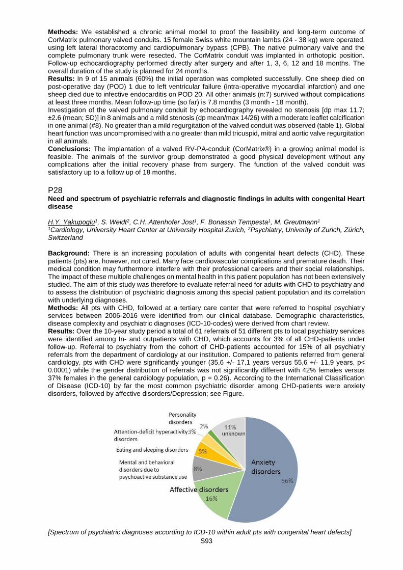
S93
Methods: We established a chronic animal model to proof the feasibility and long-term outcome of CorMatrix pulmonary valved conduits. 15 female Swiss white mountain lambs (24 - 38 kg) were operated, using left lateral thoracotomy and cardiopulmonary bypass (CPB). The native pulmonary valve and the complete pulmonary trunk were resected. The CorMatrix conduit was implanted in orthotopic position. Follow-up echocardiography performed directly after surgery and after 1, 3, 6, 12 and 18 months. The overall duration of the study is planned for 24 months. Results: In 9 of 15 animals (60%) the initial operation was completed successfully. One sheep died on post-operative day (POD) 1 due to left ventricular failure (intra-operative myocardial infarction) and one sheep died due to infective endocarditis on POD 20. All other animals (n:7) survived without complications at least three months. Mean follow-up time (so far) is 7.8 months (3 month - 18 month). Investigation of the valved pulmonary conduit by echocardiography revealed no stenosis [dp max 11.7; ±2.6 (mean; SD)] in 8 animals and a mild stenosis (dp mean/max 14/26) with a moderate leaflet calcification in one animal (#8). No greater than a mild regurgitation of the valved conduit was observed (table 1). Global heart function was uncompromised with a no greater than mild tricuspid, mitral and aortic valve regurgitation in all animals. Conclusions: The implantation of a valved RV-PA-conduit (CorMatrix®) in a growing animal model is feasible. The animals of the survivor group demonstrated a good physical development without any complications after the initial recovery phase from surgery. The function of the valved conduit was satisfactory up to a follow up of 18 months.
P28 Need and spectrum of psychiatric referrals and diagnostic findings in adults with congenital Heart disease H.Y. Yakupoglu1, S. Weidt2, C.H. Attenhofer Jost1, F. Bonassin Tempesta1, M. Greutmann1 1Cardiology, University Heart Center at University Hospital Zurich, 2Psychiatry, Univerity of Zurich, Zürich, Switzerland Background: There is an increasing population of adults with congenital heart defects (CHD). These patients (pts) are, however, not cured. Many face cardiovascular complications and premature death. Their medical condition may furthermore interfere with their professional careers and their social relationships. The impact of these multiple challenges on mental health in this patient population has not been extensively studied. The aim of this study was therefore to evaluate referral need for adults with CHD to psychiatry and to assess the distribution of psychiatric diagnosis among this special patient population and its correlation with underlying diagnoses. Methods: All pts with CHD, followed at a tertiary care center that were referred to hospital psychiatry services between 2006-2016 were identified from our clinical database. Demographic characteristics, disease complexity and psychiatric diagnoses (ICD-10-codes) were derived from chart review. Results: Over the 10-year study period a total of 61 referrals of 51 different pts to local psychiatry services were identified among In- and outpatients with CHD, which accounts for 3% of all CHD-patients under follow-up. Referral to psychiatry from the cohort of CHD-patients accounted for 15% of all psychiatry referrals from the department of cardiology at our institution. Compared to patients referred from general cardiology, pts with CHD were significantly younger (35,6 +/- 17,1 years versus 55,6 +/- 11,9 years, p< 0.0001) while the gender distribution of referrals was not significantly different with 42% females versus 37% females in the general cardiology population, p = 0.26). According to the International Classification of Disease (ICD-10) by far the most common psychiatric disorder among CHD-patients were anxiety disorders, followed by affective disorders/Depression; see Figure.
[Spectrum of psychiatric diagnoses according to ICD-10 within adult pts with congenital heart defects]
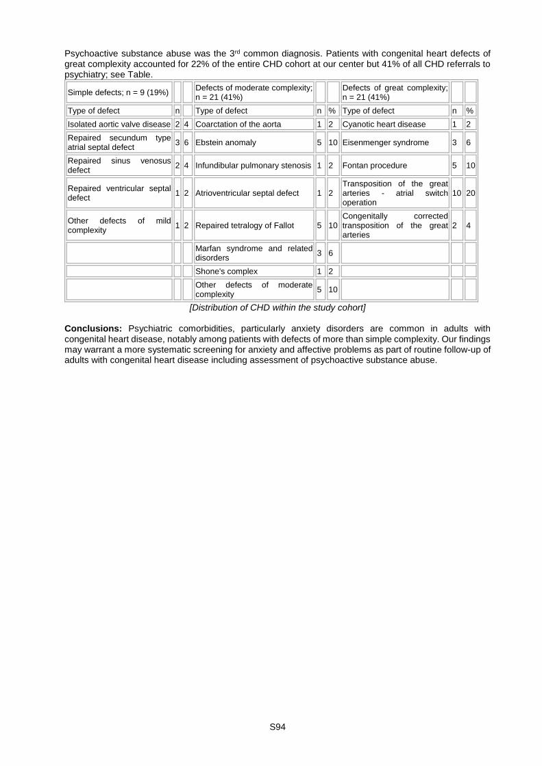
S94
Psychoactive substance abuse was the 3rd common diagnosis. Patients with congenital heart defects of great complexity accounted for 22% of the entire CHD cohort at our center but 41% of all CHD referrals to psychiatry; see Table.
Simple defects; n = 9 (19%) Defects of moderate complexity; n = 21 (41%)
Defects of great complexity; n = 21 (41%)
Type of defect n Type of defect n % Type of defect n %
Isolated aortic valve disease 2 4 Coarctation of the aorta 1 2 Cyanotic heart disease 1 2
Repaired secundum type atrial septal defect
3 6 Ebstein anomaly 5 10 Eisenmenger syndrome 3 6
Repaired sinus venosus defect
2 4 Infundibular pulmonary stenosis 1 2 Fontan procedure 5 10
Repaired ventricular septal defect
1 2 Atrioventricular septal defect 1 2 Transposition of the great arteries - atrial switch operation
10 20
Other defects of mild complexity
1 2 Repaired tetralogy of Fallot 5 10 Congenitally corrected transposition of the great arteries
2 4
Marfan syndrome and related disorders
3 6
Shone's complex 1 2
Other defects of moderate complexity
5 10
[Distribution of CHD within the study cohort] Conclusions: Psychiatric comorbidities, particularly anxiety disorders are common in adults with congenital heart disease, notably among patients with defects of more than simple complexity. Our findings may warrant a more systematic screening for anxiety and affective problems as part of routine follow-up of adults with congenital heart disease including assessment of psychoactive substance abuse.
S95
P29 Quantification of multiple mitral regurgitant jets: an in vitro validation study comparing two- and three-dimensional proximal isovelocity surface area methods F. Zürcher1, N. Brugger2, S. Jahren3, S. de Marchi1, C. Seiler1 1Cardiology, Inselspital, Bern University Hospital, University of Bern, Bern, 2Cardiology, Hôpital Fribourgeois, Fribourg, 3ARTORG Center for Biomedical Engineering Research, Bern, Switzerland Background: The accuracy of the proximal isovelocity surface area (PISA) method for the quantification of mitral regurgitation (MR), in the case of multiple jets, is unknown. The aim of this study was to evaluate different two-dimensional (2D) and three-dimensional (3D) PISA methods using 3D color Doppler data sets. Methods: Several regurgitant volumes (Rvols) were simulated using a pulsatile pump connected to a phantom equipped with single and double regurgitant orifices of different sizes and interspaces. A flowmeter served as the reference method. Transthoracic (TTE) and transoesophageal echocardiography (TEE) were used to acquire the 3D data sets. Offline, Rvols were calculated by 2D PISA methods based on hemispheric and hemicylindric assumptions and by 3D integrated PISA. Results: A fusion of the PISA was observed in the setting of narrow-spaced regurgitant orifices; compared with flowmeter, Rvol was underestimated using the single hemispheric PISA model (TTE: Bland-Altman bias ± limit of agreement, -17.5 ± 8.9 mL; TEE: -15.9 ± 7.3 mL) and overestimated using the double hemispheric PISA model (TTE: +7.1 ± 14.6 mL; TEE: +10.4 ± 11.9 mL). The combined approach (hemisphere for single orifice, hemicylinder with two bases for nonfused PISAs, and hemicylinder with one base for fused PISAs) was more precise (TTE: -3.4 ± 6.3 mL; TEE: -1.9 ± 5.6 mL). Three-dimensional integrated PISA was the most accurate method to quantify Rvol (TTE: -2.1 ± 6.5 mL; TEE -3.2 ± 4.8 mL). Conclusions: In the setting of double MR orifices, the 2D combined approach and integrated 3D PISA appear to be superior as compared with the conventional hemispheric method, thus providing tools for the challenging quantification of MR with multiple jets.
P30 Acute type A aortic dissection in non-dilated aortas: Should our guidelines be more aggressive? P. Myers, P. Mootoosamy, J. Jolou, M. Cikirikcioglu, T. Sologashvili, C. Huber Cardiovascular Surgery, Geneva University Hospitals, Geneva, Switzerland Introduction: Current guidelines recommend elective repair for ascending aortic aneurysms of ≥ 5.5 cm, or ≥ 5 cm in Marfan syndrome and related connective tissue disorders, or aneurysm growth ≥ 0.3 - 0.5 cm/year. The goal is to avoid the lethal complication of acute type A dissection. Dissection can occur below these recommended thresholds. The aim of this study was to review the aortic dimensions at presentation of all patients with acute type A aortic dissection. Methods: This was a single center retrospective review, including all patients with CT imaging before surgery for an acute aortic dissection. All imaging was analyzed using OsiriX MD software v. 7.5.1 and 3D MPR reconstructions to ensure orthogonal placement and measurements. Results: 11 patients were included from January to December 2016. Two patients were excluded, because they didn't have preoperative CT angiographic imaging (both with clinical and echocardiographic diagnosis in the setting of shock, immediately brought to the operating room for repair). Nine patients were included in the analysis, with a median age of 68 years (range 41.8-84.5), BSA 1.82 m2 (range 1.52-2.58), BMI 26.9 (range 21.5-48.2), and none had suspected or confirmed Marfan, Loeys-Dietz, Ehlers-Danlos or other connective tissue disorders. The median ascending aortic diameter was 44 mm (range 36-63) and only 1 patient had an aortic diameter within the dimensions that would warrant elective aneurysm repair (>55 mm). With a threshold decreased to 45 mm, 4 patients (44%) would have met criteria, while at threshold of 40 mm, 7 patients (78%) would have met criteria. When normalized to BSA, the median indexed ascending aortic diameter was 22.6 mm/m2 (range 17.8 - 32.5) and three patients had an indexed aortic diameter > 25 mm/m2. Five patients underwent Bentall and hemi-arch replacement, 4 patients ascending aortic and hemi-arch replacement (one followed by aortic valve replacement). There were no operative deaths, and two early deaths: one from secondary descending aortic rupture on post-operative day 1, and one from hemorrhage during veno-venous ECMO implantation for late respiratory failure. Conclusion: In our practice, aortic dissection frequently occurs in patients with aortic diameters below the current recommended thresholds for elective aneurysm repair. Screening in patients at risk and more aggressive surgical management may be of benefit to avoid this lethal complication.
S96
P31 A less invasive surgical approach to aortic coarctation leads to superior mid-term results S.G. Berset1, C. Balmer2, R. Prêtre1 1Service de Chirurgie Cardiaque | Cardiac Surgery, Centre Hospitalier Universitaire Vaudois (CHUV), Lausanne, 2Pädiatrie / Kinderkardiologie, Universitätskinderspital, Zürich, Switzerland Introduction: The objective was to analyze the mid-term results of a minimal invasive surgical approach to repair aortic coarctation with a special emphasis on the repair and on the stature changes linked to a posterior thoracotomy. Method: A transversal analysis at at least 6 year FU of all the children operated on with our new approach between 6/2002 and 10/2004 was made. The access included a muscle sparing thoracotomy and an extrapleural approach to the aorta. The coactation was repaired with an extended resection in all. In 6 patients, an additional enlargement plasty was performed on the aortic arch. The mid-term results were assessed clinically (by the measurement of staged blood pressures) and echocardiographically in all. In a few patients, it was further documented by angiography or a MRI study. The stature of the left chest and shoulder was assessed clinically and radiographically. Whenever an anomaly was suspected, a more complete examination was performed by an orthopedist. Results: Our cohort includes 30 children who were operated on at ages ranging from 1 to 450 days and weights from 980 gm to 10 kg. One patient died at 11 years of intractable pulmonary hypertension. There was no significant gradient (below 20 mmHg) across the repair in 25 patients. Five patients had higher values for which one underwent a percutaneous angioplasty and three a surgical reintervention. Except for one patient with borderline values, no patient had an arterial hypertension. One patient (who had a subsequent sternotomy) developed a scoliosis of moderate severity and another had a left winged scapula. No patient showed a rib fusion or an enlarged rib space. Conclusion: Our approach, respecting the muscles and bones of the chest and left shoulder showed the same good results on the aortic repair than a conventional posterior thoracotomy. A residual stenosis occurred only in a few patients and there was no case of arterial hypertension mid-term. The results regarding the chest and shoulder posture were better than those observed after a conventional thoracotomy with only one case of moderate scoliosis, one case of a winged scapula and no instance of rib fusion. Our minimal invasive surgical approach to aortic coarctation led to similarly good results on the aorta compared to a conventional approach but to a better outcome on the chest and shoulder posture and mobility.
P32 Added value of acceleration time to ejection time ratio in determining aortic stenosis severity M. Frank, J.-N. Yoon, E. Holy, M. Zuber, F. Tanner USZ | Cardiology, USZ, Zürich, Switzerland Introduction: Two-dimensional transthoracic echocardiography (TTE) is the routine diagnostic method for quantification of aortic stenosis (AS) severity. Accurate grading of aortic stenosis is still a challenge, especially under conditions of low-flow low-gradient aortic stenosis. In Doppler echocardiography, changes in the shape of the Doppler velocity spectrium can be seen depending on the severity in aortic stenosis. This study assessed whether the ratio of acceleration time (AT) to ejection time (ET) can be used as a diagnostic parameter for the evaluation of aortic stenosis severity, Methods: 138 patients with severe aortic stenosis (age, 84±7 years; 51% male) undergoing transcatheter aortic valve implantation (TAVI) were echocardiographically compared with a gender and age matched control group of 138 patients (age, 84±7 years; 51% male). For all patients AT, ET, and the ratio of AT/ET were measured using Doppler TTE. Results: The ratio of AT/ET in patients with severe AS was higher compared with the control group (37%, IQR 34-0.40% vs. 25%, IQR 21 - 29%, p < 0.001) (Figure 1). ROC analysis revealed that the AT/ET ratio differentiated the control group from patients with severe aortic stenosis with an area under the curve of 0.94; a cutoff of 33% had a sensivity of 80% and a specifity of 93% (Fig 2). Among the 138 patients, 45 were reclassified as having moderate aortic stenosis using multidetector computed tomography (MDCT) for determining left ventricular outflow tract area resulting in a corrected indexed aortic valve area (AVAi) >0.6 cm2/m2 using the continuity equation. These patients had a significantly shorter AT (0.10s, IQR 0.09 - 0.12s vs. 0.11s, IQR 0.10 - 0.13, p = 0.006) and ET (0.29s, IQR 0.26 - 0.32s vs. 0.31s, IQR 0.29 - 0.33s, p = 0.007); however, the AT/ET ratio did not differ significantly (36%, IQR 33 - 40% vs. 37%, IQR 35 - 40%, p = 0.331). Conclusion: The AT/ET ratio can be used as an additional parameter for differentiating severe aortic stenosis from normal individuals. However, this parameter is not sensitive enough for allowing a reliable differentiation of severe from moderate aortic stenosis. To discriminate between these subgroups, other
S97
more time consuming methods should be considered such as correction of left ventricular outflow tract area by 3D-TEE or MDCT.
P33 Utility of left ventricular global longitudinal strain for the differential diagnosis of left ventricular hypertrophy. Comparison with ECG and conventional 2D-echocardiographic parameters E. Vinet, X. Jeanrenaud, P. Monney Service de Cardiologie, CHUV, Lausanne, Switzerland Introduction: Left ventricular hypertrophy (LVH) is associated with a wide differential diagnosis including, after exclusion of aortic stenosis, hypertensive heart disease (HT), hypertrophic cardiomyopathy (HCM), cardiac amyloidosis (CA) and Fabry disease (FD). We assessed whether LV longitudinal strain analysis (LS) may improve the diagnostic prediction over ECG and standard transthoracic echocardiography (TTE). Methods: Consecutive patients presenting with LVH and having a definite diagnosis of HT, biopsy-proven CA, non-obstructive familial HCM or Fabry disease were included. An ECG tracing and a comprehensive TTE including LS was obtained in all patients. Each diagnostic group was successively compared with the three other groups and the ECG and TTE characteristics independently associated with each diagnostic group were identified by multivariate logistic regression. Results: 57 patients were included (17 HT, 13 CA, 15 HCM and 12 FD). Mean age was 56±13y, 65% males. After adjustment for age and gender, microvoltage on ECG [coef 0.36, 95%-CI (0.19-0.54), p< 0.001], a high relative LV wall thickness on TTE (optimal cut-off >0.6, [0.43, (0.05-0.82), p=0.03]) and a low LS in the basal LV segments (>-10%, [0.04, (0.02-0.06), p< 0.001]) were associated with CA. FD was associated with a short PR interval (< 150 ms, [-0.006, (-0.008 to -0.003), p< 0.001]), the number of ECG leads with T-wave (≥3, [0.05, (0.02-0.07), p=0.001], as well as with a symmetric pattern of LVH (septal to posterior wall thickness ratio < 1.2, [-0.22, (-0.33 to -0.10), p< 0.001]), but GLS parameters did not improve the prediction. HCM was independently associated with an asymmetric pattern of LVH (septal to posterior wall thickness ratio >1.4, [0.38, (0.28-0.47), p< 0.001]), left atrial dilatation (LA volume index ≥34 ml/m2, [0.008, (0.005-0.01), p< 0.001]) and preserved longitudinal strain values in the basal lateral and inferolateral segments (< -17%, [-0.03, (-0.05 to -0.005), p=0.02]). HT was independently associated with a higher ECG P-wave duration (>115 ms, [0.008, (0.004-0.01), p< 0.001]), a lower increase in septal wall thickness (< 15 mm, [-0.29, (-0.56 to -0.02), p=0.04]) and a preserved global longitudinal strain (< -13%, [-0.04, (-0.08 to -0.008), p=0.02]. Conclusion: In our selected cohort of patients with LVH, the addition of LV longitudinal strain to the ECG and standard TTE parameters allowed to improve the diagnostic prediction of CA, HCM and HT, but not FD.
S98
Poster walk I. - Heart failure and beyond I P34 Infective endocarditis at a tertiary care hospital over the past 15 years K. Amini1, H. Barbara2, A. Flammer1, F. Tanner1, M. Frank1 1USZ | Cardiology, 2Infectious Disease, USZ, Zürich, Switzerland Introduction: Infective endocarditis is a potentially lethal disease. Little is known about temporal trends in incidence, mortality, and microbiology in Switzerland. We included 315 patients with diagnosed infective endocarditis during the last 15 years based on the Duke Criteria and determined incidence, mortality, involved pathogens and echocardiographic complications. Methods: Patients with infective endocarditis diagnosed between January 2000 and June 2015 according to the Duke Criteria were identified. Clinical, echocardiographic and microbiological data were analyzed. Sensitivity of TTE was calculated and incremental value of TEE was estimated. Results: The number of cases increased substantially (+34.9%) after the year 2008/2009. The average number of cases was 20.5/year and the annual mortality remained stable (16%) over the observation period although the number of cases, age of patients, and use of prosthetic materials increased. Microbiologic analysis showed an increase of streptococcus viridans (+55%) and staphylococcus aureus (+47%) infections. TTE exhibited a sensitivity of 65%, specificity of 99%, negative predictive value of 93% and a positive predictive value 99%. TEE displayed an incremental value predominantly with prosthetic materials (53% false negative in TTE). Conclusion: In this retrospective study on infective endocarditis, an increasing number of cases was observed during the study period. This increase may be related to the aging population, increasing morbidity, and more frequent use of prosthetic materials. A contributing effect related to restriction of endocarditis prophylaxis can not be excluded, in particular because an increase in the viridans group, typical germs of the mouth flora, was observed. TTE exhibits an intermediate sensitivity for diagnosis of endocarditis, but its negative predictive value is rather high; hence, TTE should remain the standard imaging modality when endocarditis is suspected. Patients with a negative TTE and prosthetic heart valve or device should undergo TEE in addition to TTE.
P35 Role of echocardiographic left ventricular global longitudinal strain in patients admitted for acute decompensated heart failure P. Monney1, E. Vinet1, G. Tzimas1, V. Crisinel1, N. Barras1, J. Regamey1, P. Yerly1, A. Garnier2, O. Hugli3, X. Jeanrenaud1, R. Hullin1 1Service de Cardiologie, 2Service de Médecine Interne, 3Service des Urgences, CHUV, Lausanne, Switzerland Introduction: Acute decompensated heart failure (AHF) is associated with a poor prognosis and prognostic markers are still not well defined. We hypothesized that the global longitudinal strain (GLS) of the left ventricle (LV) assessed by transthoracic echocardiography (TTE) might be predictive of short-term outcome in AHF. Methods: Patients admitted for AHF at the emergency department of our hospital underwent a comprehensive TTE examination within the first 12 hours of admission and the same TTE examination was repeated upon discharge. Death or cardiovascular rehospitalization was assessed at 3 months. Results: One hundred and sixty-three patients were included (54% males, mean age 81±10 years, median NT-proBNP value 4917 ng/ml, IQR 2577-10536). 54% had had a previous episode of AHF and 42% were known for ischemic heart disease. Mean LVEF was 44±12% and 54 (33%) had preserved ejection fraction (LVEF≥50%, HFPEF). The median duration of hospital stay was 12 days (IQR 8-19 days). Between admission and discharge, no significant difference was observed in LVEF (44±12 vs 44±13%, p=0.42), LV end-diastolic volume (110±56 vs 109±57 ml, p=0.77), left atrial volume (81±37 vs 82±33 ml, p=0.29) and E/e' ratio (17.8±6.7 vs 16.8±8.0, p=0.20). There was however a trend toward an improvement in GLS (-11.0±5.6 vs -11.7±4.6%, p=0.08). GLS improved significantly in patients with reduced ejection fraction (LVEF< 50%, HFREF), from -10.0±5.2 to -11.5±4.7% (p=0.0002), but not in patients with HFPEF (-13.9±5.6 vs -12.4±4.3%, p=0.10). At 3 months, 49 (30.1%) patients reached the combined outcome of death or cardiovascular rehospitalization. After adjustment for age and gender, neither GLS nor other echocardiographic markers of left ventricular function were associated with the combined outcome. Conclusion: In patients admitted for acute decompensated heart failure, no significant change in the classical echocardiographic parameters of LV systolic or diastolic function could be detected during in-hospital treatment. GLS was able to detect an improvement in LV systolic function with treatment in patients with HFREF, but not with HFPEF. The significance of this finding remains unclear as none of the

S99
echocardiographic markers of LV function, including GLS, was able to predict the 3-months outcome after an episode of acute decompensated heart failure in our cohort. (Funded by the Swiss Heart Foundation).
P36 Heart failure in patients with arrhythmogenic right ventricular cardiomyopathy: genetic characteristics A. Vischer1,2, S. Castelletti2,3, P. Syrris4, W.J. McKenna4, A. Pantazis2,5 1Medical Outpatient Department, Universitätsspital Basel, Universität Basel, Basel, Switzerland, 2Inherited Cardiovascular Disease Unit, St Bartholomew's Hospital, London, United Kingdom, 3Centre for Cardiac Arrhythmia of Genetic Origin, Istituto Auxologico Italiano, Milan, Italy, 4Institute of Cardiovascular Science, University College London, 5Cardiomyopathy Service, Royal Brompton Hospital, London, United Kingdom Introduction: Arrhythmogenic right ventricular cardiomyopathy (ARVC) is a genetically determined heart muscle disorder. The incidence of heart failure (HF) in ARVC has been reported at 5-13%. We aimed to define the genotype and disease progression of ARVC patients with HF. Methods: Patients with a definite diagnosis of ARVC who underwent genetic testing were consecutively recruited. Detailed clinical data was collected at baseline and during follow up. Clinical endpoint was a composite of heart transplantation and death due to HF. Results: 135 patients were included. 8 (5.9%) patients reached the end point. A Plakophilin 2 mutation was present in most patients with HF, although 50% had multiple mutations
[Prevalence of Plakophilin mutation]
. Conclusion: HF is a rare but significant outcome of patients with a definite diagnosis of ARVC. Patients with HF predominantly carried Plakophilin 2 mutations and often had multiple mutations.

S100
P37 Endothelial dysfunction and autonomic imbalance in tako-tsubo cardiomyopathy and cardiac syndrome X M. Cattaneo1, A.P. Porretta1, J.M. Gallino1, M. Forni De Gottardi1, C. Limoni2, M. Moccetti3, A. Gallino1 1Cardiovascular Research, Hospital of San Giovanni, Bellinzona, 2University of Applied Sciences and Arts of Southern Switzerland, 3Cardiology, Fondazione Cardiocentro Ticino, Lugano, Switzerland Introduction: The underlying pathophysiology of Tako-Tsubo Cardiomyopathy (TTC) and Cardiac Syndrome X (CSX) suggests a few similarities. Endothelial dysfunction (ED) and autonomic imbalance (AI) have both been individually implied in the pathogenesis of both conditions. Aim of the present study was to non-invasively investigate AI and ED markers in TTC and CSX as compared to matched controls. Methods: We recruited female patients diagnosed with either CSX or TTC and a control group of matched post-menopausal women. The study protocol consisted of baseline measurements of catecholamines (CA), peripheral artery tonometry (PAT) and spontaneous baroreflex sensitivity (BRS), followed by measurements during controlled and deep breathing protocol and a mental stress protocol (MS) consisting of Mental arithmetic and Stroop-word-colour-conflict test. Mental stress-induced endothelial dysfunction was measured as PAT reactive hyperaemia index (RHI-MS) computed as the ratio between RHI-post-stress and baseline. Differences among groups were calculated by chi-square, ANOVA one way and Least Significant Difference Test. Results: Final analysis comprised 13 CSX, 11 TTC and 15 Controls. Cardiovascular risk factors and age were comparable among groups (chi-square and ANOVA). RH-MS was significantly lower in CSX as compared to TTC (p 0.007) and Controls (p. 0.022). A trend to percent reduction in RHI was present in CSX group (-0.11 ± 0.19, p 0.056). CA did not differ among groups, but significant norepinephrine stress-related rise was found in CSX (0.29 ± 0.28, p 0.003). We found no significant difference in BRS values or changes among groups, but there was a MS-related depression of BRS in CSX (absolute p 0.039; percent p 0.038) in TTC (percent p=0.002). Conclusion: In our study CSX suggest a more relevant MS-related endothelium dependant dysfunction and possible inappropriate vasoconstriction. Both CSX and TTC showed a MS-related cardiovagal outflow imbalance as compared to matched controls.
P38 Simultaneous transcatheter aortic valve implantation and left atrial appendage occlusion versus both interventions as stand-alone procedures T. Gilhofer, S. Obeid, T.F. Lüscher, R. Binder Department of Cardiology, University Heart Center at University Hospital Zurich, Zurich, Switzerland Aim: Patients in atrial fibrillation undergoing transcatheter aortic valve implantation (TAVI) have a worse prognosis than those in sinus rhythm. We investigated the feasibility and outcome of simultanous TAVI and left atrial appendage occlusion (LAAO) with Lotus and Watchman. Methods: Consecutive patients, who underwent either TAVI with Lotus, LAAO with Watchman or both procedures simultaneously were investigated. We compared the procedural characteristics, in-hospital- and 30-day-outcomes defined by VARC-2 criteria in these three groups. Results: A total of 52 consecutive patients were included in the study. An isolated TAVI with Lotus was performed in 19 patients, an isolated LAAO with Watchman in 23 patients and a combined procedure in 10 patients. Patients in the isolated LAAO cohort were significantly younger (TAVI: 85.1 +/- 4.8 years; LAAO: 75.7 +/- 6.0 years; TAVI + LAAO: 84.2 +/- 5.7 years; p< 0.001). There was no significant difference in the amount of contrast medium used between the three groups (TAVI: 89 +/- 29 ml; LAAO: 98 +/- 54 ml; TAVI + LAAO: 86 +/-29 ml; p=0.923). The total procedural time was significantly higher in the combined group than the isolated TAVI or LAAO group (TAVI: 57.1 +/- 18.0 minutes; LAAO: 42.7 +/- 16.0 minutes; TAVI + LAAO: 82.0 +/- 18.6 minutes; p=0.001). Procedural and device success was achieved in all patients. The length of hospital stay was significantly lower in the isolated LAAO group than the isolated TAVI or combined procedure group (TAVI: 9.7 +/- 4.3 days; LAAO: 2.0 +/- 2.1 days; TAVI + LAAO: 10 +/- 4 days; p< 0.001). While there was no 30-day mortality in the isolated TAVI or isolated LAAO group, one sudden cardiac death occurred in the combined group at home on day 28. There was one cerebrovascular event in the isolated TAVI group but none in the LAAO or the combined group. Major bleeding occurred in one patient in the isolated TAVI group, but none in the isolated LAAO or the combined group. Conclusion: Combining TAVI and LAAO with Lotus and Watchman is feasible and was comparable to both interventions as stand-alone procedures, although procedural time was higher when both interventions were performed simultaneously. Long-term follow-up will show, whether combining TAVI and LAAO improves prognosis in high-risk patients with atrial fibrillation and symptomatic severe aortic stenosis.

S101
P39 Open aortic arch anasthomosis wit antegrade cerebral perfusion and mild hypothermia - 10 years' experience of a single center M. Gruszczynski, O. Dzemali, D. Odavic, A. Häussler, M. Genoni Stadtspital Triemli Zürich | Klinik für Herzchirurgie, Stadtspital Triemli Zürich, Zürich, Switzerland Background: The elective aortic ascendant and arch surgery should consider the exclusion of the whole diseased tissue. The incidence of aortic complications and further dilatation regards especially the patients with bicuspid valves and genetically mediated disorders. Our collected operative data since 2005 till 2016 showed that among the patients treated with aortic replacement with clamped aorta there was a substantial proportion of patients who had to be re-operated due to uncured aortic pathology. Concerns about the progression of the aortic dilatation because of the tissue defect led us to reconsider our operative strategy. Methods: We present our series of 320 consecutive elective operations on ascending aorta performed in the period from 2005 to 2016. Among them in 169 patients we cannulated the ascending aorta or aortic arch and the distal anastomosis was performed with clamped aorta (group I), in 151 cases the right subclavian artery was cannulated and the distal anastomosis was done under unilateral SCP, moderate hypothermia and with open distal aorta (group II). Results: There were no statistically significant difference between the two groups in terms of preoperative data in both groups. The most frequent procedure was aortic valve replacement or reconstruction with replacement of the ascending aorta, followed by the complex procedures and alone ascending replacement with the vessel graft. There were no statistically relevant difference between the groups in term of CBP-time and cross-clamp. The mean cerebral perfusion time was 13.9±1.1 min at 28°C. We observed no difference in terms of mortality, new neurological deficits, postoperative delirium and further postoperative course except for the higher incidence of severe renal insufficiency in group I. We observed no occurrence of paraplegia in both groups. The mean blood loss was slightly higher in group I with also slightly higher number of transfused blood cell units and thrombocyte concentrate. Conclusion: The subclavian cannulation and unilateral selective cerebral perfusion allows aortic arch reconstruction and anastomosis under direct vision and exclusion of the entire pathology in the ascending aorta. Our results show that this procedure performed with moderate hypothermia and with relatively short circulatory arrest time with antegrade cerebral perfusion can be safe and effective method of organ protection with no higher mortality and morbidity.
P40 Beta-blockers uptitration between infusions predicts event-free survival in advanced heart failure treated with repeated cures of low-dose Levosimendan N. Barras, J. Regamey, P. Vogt, R. Hullin, P. Yerly Department of Cardiology, CHUV, Lausanne, Switzerland Introduction: The prognosis of advanced heart failure (AHF) is very poor. Because of low blood pressure (BP) and fluid overload, up-titration of Angiotensin converting enzyme inhibitors (ACEi), Angiotensin receptor antagonists (ARA), beta-blockers (BB) and mineralocorcticoid receptor antagonists (MRA) is often not tolerated. Intermittent Levosimendan infusions may then be of value to alleviate symptoms and possibly improve outcome. The goal of this study was to find out factors predictive of 1 year event-free survival in 42 AHF patients (LVEF< 40%, NYHA 3-4 despite optimal therapy>3 months) treated with repeated (4.37+/-1.9 infusions/ patient) low dose Levosimendan (0.05 µg/kg/min during 24h every 4 weeks). Methods: Medical files were reviewed for the composite endpoint of mortality, re-hospitalization or LVAD implantation during 1 year after first infusion. Patients´ baseline characteristics and status at inclusion and after 3 infusions were compared between the 14 who reached the endpoint (33%) and the 28 (67%) event-free survivors. Results: Nor patients´ characteristics (age 61.4+8.9y vs 59.7+7 y, P=0.62; female sex 17.85% vs 7.14%, P=0.64; ischemic heart disease 50% vs 64.3%, P=0.51; LVEF 0.21+0.05 vs. 0.22+0.06) nor patients´ baseline status predicted outcome (Table). Only BB therapy, percent of BB target dose and heart rate after 3 infusions predicted endpoint occurrence (Table). After adjustment for HR and BB, only BB dose >35% target dose remained significant (OR=0.64; 95%CI 0.07-0.87).
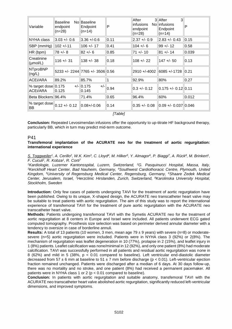
S102
Variable Baseline No endpoint (n=28)
Baseline Endpoint (n=14)
P
After 3 infusions No endpoint (n=28)
After 3 infusions Endpoint (n=14)
P
NYHA class 3.03 +/- 0.6 3.36 +/-0.6 0.11 2.37 +/- 0.9 2.83 +/- 0.43 0.15
SBP (mmHg) 102 +/-11 106 +/- 17 0.41 104 +/- 6 99 +/- 12 0.58
HR (bpm) 78 +/- 8 82 +/- 6 0.85 71 +/- 10 81 +/- 14 0.039
Creatinine (µmol/L)
116 +/- 31 138 +/- 38 0.18 108 +/- 22 147 +/- 50 0.13
NTproBNP (ng/L)
5233 +/- 2244 7765 +/- 3506 0.56 2910 +/-4002 6085 +/-1728 0.21
ACEi/ARA 89.2% 85.7% 1 92.9% 80% 0.27
% target dose ACEi/ARA
0.175 +/-0.125
0.175 +/-0.145
0.94 0.3 +/- 0.12 0.175 +/- 0.12 0.11
Beta Blockers 96.4% 71.4% 0.65 96.4% 60% 0.012
% target dose BB
0.12 +/- 0.12 0.08+/-0.06 0.14 0.35 +/- 0.08 0.09 +/- 0.037 0.046
[Table] Conclusion: Repeated Levosimendan infusions offer the opportunity to up-titrate HF background therapy, particularly BB, which in turn may predict mid-term outcome.
P41 Transfemoral implantation of the ACURATE neo for the treatment of aortic regurgitation: international experience S. Toggweiler1, A. Cerillo2, W.K. Kim3, C. Lloyd4, M. Hilker5, Y. Almagor6, P. Biaggi7, A. Rück8, M. Brinkert1, F. Cuculi1, R. Kobza1, R. Corti7 1Kardiologie, Luzerner Kantonsspital, Luzern, Switzerland, 2G. Pasquinucci Hospital, Massa, Italy, 3Kerckhoff Heart Center, Bad Nauheim, Germany, 4Southwest Cardiothoracic Centre, Plymouth, United Kingdom, 5University of Regensburg Medical Center, Regensburg, Germany, 6Shaare Zedek Medical Center, Jerusalem, Israel, 7Herzclinic Hirslanden, Zurich, Switzerland, 8Karolinska University Hospital, Stockholm, Sweden Introduction: Only few cases of patients undergoing TAVI for the treatment of aortic regurgitation have been published. Owing to its unique, X-shaped design, the ACURATE neo transcatheter heart valve may be suitable to treat patients with aortic regurgitation. The aim of this study was to report the international experience of transfemoral TAVI for the treatment of pure aortic regurgitation with the ACURATE neo transcatheter heart valve. Methods: Patients undergoing transfemoral TAVI with the Symetis ACURATE neo for the treatment of aortic regurgitation at 8 centers in Europe and Israel were included. All patients underwent ECG gated computed tomography. Prosthesis size selection was based on perimeter derived annular diameter with a tendency to oversize in case of borderline annuli. Results: A total of 13 patients (10 women, 3 men, mean age 79 ± 9 years) with severe (n=8) or moderate-severe (n=5) aortic regurgitation were included. Patients were in NYHA class 3 (92%) or 2(8%). The mechanism of regurgitation was leaflet degeneration in 10 (77%), prolapse in 2 (15%), and leaflet injury in 1 (8%) patients. Leaflet calcification was none/minimal in 12 (92%), and only one patient (8%) had moderate calcification. TAVI was successfully performed in all patients and residual aortic regurgitation was none in 8 (62%) and mild in 5 (38%, p < 0.01 compared to baseline). Left ventricular end-diastolic diameter decreased from 57 ± 6 mm at baseline to 51 ± 7 mm before discharge (p < 0.01). Left-ventricular ejection fraction remained unchanged. Patients were discharged after a median of 6 days. At 30 days follow-up, there was no mortality and no stroke, and one patient (8%) had received a permanent pacemaker. All patients were in NYHA class 1 or 2 (p < 0.01 compared to baseline). Conclusion: In patients with aortic regurgitation and suitable anatomy, transfemoral TAVI with the ACURATE neo transcatheter heart valve abolished aortic regurgitation, significantly reduced left-ventricular dimensions, and improved symptoms.
S103
P42 Very low pacemaker rate following ACURATE neo transcatheter heart valve replacement S. Toggweiler1, H. Nissen2, B. Mogensen3, F. Cuculi1, C. Fallesen2, K.T. Veien2, M. Brinkert1, P. Matt4, T. Syburra4, A. Rück3, X. Mueller4, R. Kobza1 1Kardiologie, Luzerner Kantonsspital, Luzern, Switzerland, 2Odense University Hospital, Odense, Denmark, 3Karolinska University Hospital, Stockholm, Sweden, 4Cardiac Surgery, Luzerner Kantonsspital, Luzern, Switzerland Introduction: Despite device improvements new conduction disorders after transcatheter aortic valve replacement (TAVR) remain an area of concern and values exceed those of surgical aortic valve replacement. We aimed to investigate if minimizing trauma to the aortic annulus and left ventricular outflow tract reduces the occurrence of new conduction disorders and need for permanent pacemakers. Methods: All patients undergoing transfemoral TAVR with the Symetis ACURATE neo at three centers in Europe were included. Prosthesis size selection was based on perimeter derived annular diameter. Valvuloplasty balloons were chosen to be smaller than the annular diameter to avoid oversizing in relation to the annulus and the left ventricular outflow tract. Results: A total of 175 patients (58% women, mean age 83 ± 6 years) were included. Fourteen patients (8.0%) had a permanent pacemaker at baseline. Predilatation was performed in all with a balloon 1.9 ± 0.9 mm smaller than the perimeter derived annular diameter. Postdilatation was performed in 46 (26.3%) with a balloon 1.2 ± 0.9 mm smaller than the perimeter derived annular diameter. Eighteen patients (10.3%) developed a new left bundle branch block, 13 (7%) a new first degree AV block, and 4 (2.3%) received a new permanent pacemaker. Moderate paravalvular regurgitation occurred in 8 patients (4.6%). At 30 days, rate of any stroke was 3/175 (1.7%), and one patient (0.6%) had died. Conclusion: Careful selection of the balloon and the ACURATE neo prosthesis size resulted in very low rates of new conduction disorders and permanent pacemakers without increasing the amount of paravalvular regurgitation.

S104
Poster Walk II. - Heart failure and beyond II P43 Syntax score ii in patients with coronary artery disease undergoing percutaneous mitral repair with the mitraclip S. Obeid1, A. Adrian2, M. Taramasso2, G. Pedrazzini2, G. Suetsch3, F. Landolt2, F. Karbeyaz2, H. Rodriguez2, D. Sürder4, T. Moccetti4, F. Maisano2, F. Nietlispach2 1Zurich Heart Center, Interventional Cardiology, University Hospital of Zürich, 2Universitätsspital Zürich, 3Hirslanden, Zurich, 4Cardiocentro Ticino, Lugano, Switzerland Background: Percutaneous mitral valve repair (PMVR) using the MitraClipTM system has become a valuable alternative in patients with severe mitral regurgitation (MR) and high surgical risk. We sought to evaluate the prognostic value of the SYNTAX II score (SSII) in patients with concomitant coronary artery disease (CAD) undergoing a Mitraclip procedure. Methods: In seventy-five consecutive patients who underwent PMVR at the University Heart Center Zürich and the Cardiocentro Ticino, the SSSII was calculated at baseline. Clinical endpoints comprised of all-cause mortality, mitral valve surgery due to failure of PMVR or reoperation, hospitalization for congestive heart failure, heart transplantation and the composite of all four endpoints. Results: Patients were followed for a median of 271 days. and were divided in tertiles of SSII: SSII low ≤ 46.5 (n=25), SSII mid 46.6 - 54.4 (n=25) and SSII high ≥ 54.5 (n=25). Patients in the highest SSII tertile had a lower left ventricular ejection fraction (33% vs. 40% vs. 53%) with a higher log-BNP (3.6 vs. 3.45 vs. 3.16) when compared to SSII mid and SSII low, respectively. However, the anatomical syntax score (SS) did not differ significantly within the tertiles (9.1±6.3 (SSII Low) vs 9.5±7.6 (SSII Mid) vs 10.2±6.7(SSII High), p=0.837). The primary endpoint occurred in 33% of patients (n=25). By multivariate analysis patients in the high SSII tertile (OR=6.12, 95% confidence interval, [CI] 1.45-25.86, p=0.014) and patients with a history of MI (OR=3.57, 95% confidence interval, [CI] 1.17-10.88, p=0.025) were at significantly higher risk of experiencing adverse events. Furthermore, in a combined outcome ROC curve analysis, the SSII showed good discrimination with an AUC of 0.73, p=0.001. A cutoff SSII >49 has been identified to have a sensitivity of 83% and specificity of 53% with approximately 45% of the patients experiencing an event during follow-up. Conclusion: Using SSII in CAD patients undergoing PMVR is feasible and of prognostic significance hence widening its clinical utility in valvular heart disease.
P44 Transcatheter aortic valve implantation in southern of Switzerland: Cardiocentro single center experience M. Boscolo Berto, L. Biasco, F. Faletra, M. Moccetti, E. Pasotti, T. Moccetti, G.B. Pedrazzini Cardiocentro Ticino, Lugano, Switzerland Background: Transcatheter Aortic Valve Replacement (TAVR) is the treatment of choice for severe aortic stenosis (AS) in patients at high surgical risk. Adoption in intermediate/low risk patients is clinically accepted, without data on long term prosthetic mechanical performances. Aim: to evaluate the acute and long term mechanical performances of TAVR and report freedom from reintervention and in-hospital mortality in a contemporary population. Results: From September-2011 to April-2016, 195 patients with severe AS with a mean age of 82±5.8years and a median STS score=7.56 (IQR 3.49-9.43), received TAVR. In-hospital mortality was 2.5%. Median follow-up was 410.6 days. Echocardiographic data at follow up showed a good mechanical performance of the aortic prosthesis (mean gradient at baseline: 48.1mmHg and at 30 days follow up: 9.59mmHg) persistent over time (at 1year: 10.3 mmHg; at 2 years: 7.8 mmHg; at 3 years: 10.2 mmHg; at 4 years: 5.75 mmHg). At follow up a mean gradient >20mmHg was evident in 9 (4.6%). Three (1.5%) of them required treatment: one patient underwent redo surgery, one declined a redo procedure while a reduction of transprosthetic gradients was observed after oral anticoagulation in a third patient. Overall, left ventricular ejection fraction showed a progressive increase over the first 2 years (at baseline: 51.8%; at 2 years follow up: 56.1%, p< 0.001). Conclusion: This single center experience with good long term mechanical performance of TAVR and low in hospital mortality suggest acceptable valve durability with a low overall incidence of valve deterioration requiring treatment, opening the possibility for the adoption in intermediate and low risk, younger, patients.
S105
P45 The use of bilateral internal mammary artery is an excellent predictor of improve left ventricular systolic function in patients with poor ejection fraction H. Loeblein, O. Dzemali, D. Odavic, A. Haeussler, A. Zientara, M. Genoni Herzchirurgie | Triemli Hospital, Triemli Hospital, Zürich, Switzerland Objectives: The preoperative identification of patients in whom ejection fraction improves after undergoing cardiopulmonary bypass (CABG) is crucial. Surgery using the bilateral internal mammary artery (BIMA) provides significantly better survival. However, to our knowledge there are no studies evaluating the use of BIMA on the improvement of systolic function in patients with poor ejection fraction. Methods: In this retrospective study all CABG patients (159) from 2007 to 2015 with an EF equal or less than 30% were retrieved from our data bank. We investigated the association between use of BIMA and EF at 3 months after CABG. Results: At 3 months echocardiography control was performed in 76/159 patients. The average values were: BNP 2675.9 pg/mL, EF 23.6%, age 63.5 years, creatinine 98.4 mmol/L, CKMB 12.7 U/L. 13% were female, 49% had diabetes and 29% underwent urgent or emergent surgery. The average intubation time, distal anastomosis, peak CKMB, packed red blood cell transfusion were 19 hours, 3.8, 26.7 U/L, 1.8 respectively. 41% received BIMA and 1.9% died in hospital. At 3 months there was a significant improvement in EF from an average of 23.6% to 38.2% (p = 0.000). There was no correlation between preoperative EF, age, gender, surgical timing, number of distal anastomosis, postoperative ischemia, blood transfusion, intubation time and EF improvement at 3 months. There was a significant correlation between high preoperative BNP level and the use of BIMA to the improvement of EF at 3 months (p = 0.021 and 0.014, respectively). Using multivariable logistic regression analysis the use BIMA was a significant independent predictor of improvement in the left ventricular EF at 3 months (p= 0.26) Conclusion: CABG is associated with low in hospital mortality in patients with poor EF. Poor EF improves significantly after CABG and the use of BIMA is an excellent predictor for this improvement.
P46 Assessment of shear stresses in a novel in-vitro healthy human right heart model U. Gulan1, A. Saguner2, D. Akdis2, C. Brunckhorst2, M. Holzner1, F. Duru2 1Institute of Environmental Engineering, ETH Zurich, 2University Heart Center Zurich, Zurich, Switzerland Introduction: Blood flow characteristics such as disturbed and turbulent flow and associated elevated shear stresses and energy dissipation play an important role in the pathophysiology of various cardiovascular diseases. Yet, the assessment of these parameters, particularly in the right ventricle (RV) is limited. Aim: Reynolds shear stresses and viscous stresses were studied in a novel in-vitro silicone right heart model under physiological flow conditions. Methods: As a consequence of limitations of current medical imaging tools, in-vitro flow measurement techniques have gained much attention as they allow studying complex flow patterns in the cardiovascular system. We performed a non-intrusive imaging method, 3D Particle Tracking Velocimetry (3D-PTV), in a silicone replica of the healthy human right heart. The investigation domain comprises the RV including the subtricuspid region, the right ventricular outflow tract (RVOT), and the RV apex. The flow conditions provided by the experimental setup are validated against the flow patterns obtained by in vivo MRI. A scalar shear stress value was calculated summing the Reynolds shear stresses and viscous stresses. Results: Our results show that the spatial distribution of shear stresses is qualitatively similar both at systole and diastole, i.e. higher stresses occur at the subtricuspid region (89±2.3Pa) and in the proximity of the RVOT (19±2.3Pa). Moreover, the high shear stress zone extends further towards the apex in the diastolic phase. It is also found that the temporal evolution of the shear stresses have two peaks, which correspond to the time instance when the pulmonary valve and the tricuspid valve open, respectively. Conclusions: The novel right heart model will help us to display the RV sites, which are mostly exposed to turbulent flow and hydromechanical stresses, and whether these correlate with diseased RV predilection sites in patients with arrhythmogenic right ventricular cardiomyopathy (ARVC).
S106
P47 Devices and methods for transcatheter retrieval of mechanical heart valve leaflets P.P. Heinisch1,2, R. Brecht3, A. Khoynezhad2, The Khoynezhad Laboratory 1Cardiovascular Surgery, Inselspital, Bern University Hospital, University of Bern, Bern, Switzerland, 2Cardiothoracic Surgery, Cedars-Sinai Medical Center, Los Angeles, CA, United States, 3University Hospital Regensburg, Regensburg, Germany Objective: Heart valve replacement either with a mechanical or a biological prosthesis is the primary treatment option for patients with severe aortic valve disease. Redo-surgery for patients with prosthetic heart valves is associated with an increased morbidity and mortality rate remaining currently the primary option. For patients with previous bioprosthetic heart valve, the valve-in-valve transcatheter implantation procedure has become a feasible and safe alternative. However, this procedure cannot be offered to patients with mechanical heart valves. Our group developed a novel device to expand the availability of transcatheter valve-in-valve replacement for patients with mechanical heart valves. Methods: In order to perform a valve-in-valve procedure in a MHV, the stiff pyrolytic carbon leaflets have to be removed in order to place a new transcatheter valve. Our laboratory has invented and patented a novel device and methodology for transcatheter retrieval of mechanical heart valve leaflets (Figure 1). The innovative transcatheter device consists of two parts: (1) a balloon for dislodging the MHV leaflets and (2) a retrieval device that will catch and transport the two valve leaflets safely (Patent WO2015031898 / EP3038569 - A2). Results: The feasibility of the novel device was evaluated in a pressurized in-vitro model using various sizes of mechanical heart valves. A critical issue of the disassembly is to avoid breaking the leaflets. Our preliminary data has shown that using a non-compliant balloon increases the force on the leaflet. This allowed for a faster disassembly, but there was a higher risk of fracturing at least one leaflet into multiple fragments. Therefore, we adapted the device and use now a semi-compliant balloon, but with a larger inflation volume. The safe retrieval of the valve fragments in their entirety from the body is another important step of the procedure. Therefore, the direction of blood flow, as well as the angulation and size of the blood vessels, have to be considered. Conclusion: This technology offers solutions to an unmet clinical need in the field of cardiovascular medicine. This novel device can obviate the need for risky open-heart surgery, thereby reducing morbidity and mortality, and providing an interventional option to patients with mechanical heart valve who are considered inoperable or high-risk for open-heart surgery.
P48 Contrast echocardiography in patients with left ventricular non-compaction cardiomyopathy L. Erhart1, M. Niemann1,2,3, S.F. Stämpfli1, H. Hammer1, I. Kapos1, K. Yonekawa1, A. Straub1, R. Said1, M. Greutmann1, C. Gruner1, F.C. Tanner1 1Department of Cardiology, University Heart Center, Zurich, Switzerland, 2Institute for Mechanical and Medical Engineering, University Furtwangen, Furtwangen, Germany, 3Institute for Biomedical Engineering, University and ETH Zurich, Zurich, Switzerland Introduction: The use of contrast agents in echocardiography enhances endocardial border detection; improved inter-observer agreement concerning left ventricular ejection fraction (LVEF) and end-diastolic volume (EDV) measurement has been observed in healthy individuals. It is not known whether this holds true in patients with left ventricular non-compaction cardiomyopathy (LVNC), a population where the measurement of LVEF is particularly challenging. Methods: 14 patients diagnosed with isolated left ventricular non-compaction cardiomyopathy (LVNC) and 16 healthy controls were investigated via echocardiography with and without contrast agents (Sonovue, Bracco Research, Geneva, Switzerland) using commercially available equipment (IE33 and Epic, Philips; Vivid S7 and E9, GE Healthcare). Biplane LVEF was evaluated using the Simpson's method by four level 1 and four level 3 echocardiographers. Paired t-tests were performed to analyze the differences in LVEF measurements between the two groups of investigators. Results: In controls, there was a small, but significant difference in native LVEF between level 1 and level 3 investigators (60 ± 6% vs. 58 ± 5%, p= 0.01). After contrast application, no difference in LVEF between the two investigator groups was observed (60 ± 4% vs. 59 ± 5%, p= 0.36).In contrast, LVEF measurements taken in LVNC patients exhibited a significant difference between the two investigator groups both under native conditions (56 ± 5% vs. 52 ± 5%, p=0.000) and with contrast (56 ± 7% vs. 52 ± 7%, p=0.000). The discrepancies between the two investigator groups remained almost equal with and without contrast (-4.0 ± 3% vs. -3.7 ± 2%, p=0.82). EDV measurements taken in controls by level 3 investigators were significantly higher after application of contrast (four chamber EDV: 115 ± 20% vs. 105 ± 16%, p=0.015; two chamber EDV: 117 ± 21% vs. 106 ± 15%, p=0.014). Similar results were observed in LVNC patients (four chamber
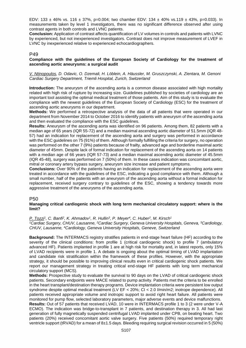
S107
EDV: 133 ± 46% vs. 116 ± 37%, p=0.004; two chamber EDV: 134 ± 40% vs.119 ± 43%, p=0.033). In measurements taken by level 1 investigators, there was no significant difference observed after using contrast agents in both controls and LVNC patients. Conclusion: Application of contrast affects quantification of LV volumes in controls and patients with LVNC by experienced, but not inexperienced investigators. Contrast does not improve measurement of LVEF in LVNC by inexperienced relative to experienced echocardiographers.
P49 Compliance with the guidelines of the European Society of Cardiology for the treatment of ascending aortic aneurysms: a surgical audit V. Ntinopoulos, D. Odavic, O. Dzemali, H. Löblein, A. Häussler, M. Gruszczynski, A. Zientara, M. Genoni Cardiac Surgery Department, Triemli Hospital, Zurich, Switzerland Introduction: The aneurysm of the ascending aorta is a common disease associated with high mortality related with high risk of rupture by increasing size. Guidelines published by societies of cardiology are an important tool assisting the optimal medical treatment of those patients. Aim of this study is to evaluate the compliance with the newest guidelines of the European Society of Cardiology (ESC) for the treatment of ascending aortic aneurysms in our department. Methods: We performed a retrospective analysis of the data of all patients that were operated in our department from November 2014 to October 2016 to identify patients with aneurysm of the ascending aorta and then evaluated the compliance with the ESC guidelines. Results: Aneurysm of the ascending aorta was identified on 96 patients. Among them, 82 patients with a median age of 65 years (IQR 55-72) and a median maximal ascending aortic diameter of 51.5mm (IQR 48-57) had an indication for replacement of the ascending aorta and surgery was performed in accordance with the ESC guidelines on 75 (91%) of them. Although formally fulfilling the criteria for surgery, no operation was performed on the other 7 (9%) patients because of frailty, advanced age and borderline maximal aortic diameter of 45mm. Despite lack of formal indication for replacement of the ascending aorta on 14 patients with a median age of 66 years (IQR 57-73) and a median maximal ascending aortic diameter of 45.5mm (IQR 45-48), surgery was performed on 7 (50%) of them. In these cases indication was concomitant aortic, mitral or coronary artery bypass surgery, aneurysm size increase and patient symptoms. Conclusions: Over 90% of the patients having an indication for replacement of the ascending aorta were treated in accordance with the guidelines of the ESC, indicating a good compliance with them. Although a small number, half of the patients with an aneurysm of the ascending aorta without a formal indication for replacement, received surgery contrary to guidelines of the ESC, showing a tendency towards more aggressive treatment of the aneurysms of the ascending aorta.
P50 Managing critical cardiogenic shock with long term mechanical circulatory support: where is the limit? P. Tozzi1, C. Banfi2, K. Ahmadov1, R. Hullin3, P. Meyer4, C. Huber2, M. Kirsch1 1Cardiac Surgery, CHUV, Lausanne, 2Cardiac Surgery, Geneva University Hospitals, Geneva, 3Cardiology, CHUV, Lausanne, 4Cardiology, Geneva University Hospitals, Geneve, Switzerland Background: The INTERMACS registry stratifies patients in end-stage heart failure (HF) according to the severity of the clinical conditions: from profile 1 (critical cardiogenic shock) to profile 7 (ambulatory advanced HF). Patients implanted in profile 1 are at high risk for mortality and, in latest reports, only 15% of LVAD recipients were in profile 1. A debate is ongoing about the optimal timing of LVAD implantation and candidate risk stratification within the framework of these profiles. However, with the appropriate strategy, it should be possible to improving clinical results even in critical cardiogenic shock patients. We report our management strategy in treating critical end-stage HF patients with long term mechanical circulatory support (MCS). Methods: Prospective study to evaluate the survival to 90 days on the LVAD of critical cardiogenic shock patients. Secondary endpoints were MACE related to pump activity. Patients met the criteria to be enrolled in the heart transplant/destination therapy programs. Device implantation criteria were persistent low output syndrome despite optimal medical treatment (LV EF < 20%; CI < 2.0 l/min/m2; inotrope dependence). All patients received appropriate volume and inotropic support to avoid right heart failure. All patients were monitored for pump flow, selected laboratory parameters, major adverse events and device malfunctions. Results: Out of 57 patients that received LVAD, 10 were in INTERMACS profile 1 to 3 (2 were under V-A ECMO). The indication was bridge-to-transplant in 7 patients, and destination therapy in 3. All had last generation of fully magnetically suspended centrifugal LVAD implanted under CPB, on beating heart. Two patients (20%) received concomitant aortic valve surgery. Five patients (50%) required temporary right ventricle support (tRVAD) for a mean of 8±1.5 days. Bleeding requiring surgical revision occurred in 5 (50%)

S108
patients, 3 during the tRVAD support. At the 90-day endpoint survival was 90%, one (10%) died due to respiratory failure. Three (30%) experienced critical illness polyneuropathy. Two (20%) had late driveline infection. Conclusions: Morbidity rate was high. However, the 90% survival rate at 90 days endorses the assumption that, with the appropriate strategy and last generation centrifugal LAVD, even the crash-and-burn patients have excellent possibility to benefit from long term MCS.
S109
Poster walk II. - Epidemiology, risk factors, rehabilitation & thrombosis P51 Effects of high-intensity interval training versus moderate-intensity continuous exercise on left ventricular function and remodeling in patients early after myocardial infarction: preliminary results from a randomized-controlled trial L. Trachsel1, L. Widmer1, T. Van Schouwenbourg Menalda1, D. Herzig1, J.-P. Schmid2, S. De Marchi1, P. Eser1, M. Wilhelm1 1Cardiology, Inselspital Bern, Bern, 2Cardiology, Clinic Barmelweid, Barmelweid, Switzerland Introduction: It is presently unknown how high-intensity interval training (HIIT) affects cardiac function and remodeling in patients early after myocardial infarction (MI). The aim of this study was to compare HIIT with the established moderate-intensity continuous exercise (MICE) over a 9-week training period early after MI. Methods: The present study analyses preliminary data of a prospective, randomized, controlled trial integrated in a 12-week cardiac rehabilitation program at the University Hospital Bern. Twenty-four consecutively enrolled patients (23 males, 1 female) with a first ST-segment elevation myocardial infarction (STEMI) within four weeks prior to study inclusion were subdivided in two groups: the HIIT group (n=11) and the MICE (n=13) group. All patients trained on cycle ergometers three times per week for 12 weeks for 38 to 40 minutes. MICE was performed at 50 to 80% of VO2peak. HIIT consisted of 4 intervals of 4 minutes at >80% of VO2peak, followed by 3 minutes at < 50% of VO2peak. During the first 3 weeks, all patients performed MICE training. Thereafter they were randomised to MICE training alone or to two sessions of HIIT and one session of MICE training per week. Advanced echocardiography and cardiopulmonary exercise testing and was performed after a 3-week run-in phase at week 3 and 12. The primary outcome measure was the difference in change of global longitudinal strain (GLS) between the two groups. Secondary outcome parameters were changes in left ventricular end diastolic volume index (LVEDVI) and peak oxygen consumption (VO2peak). Results: Mean age was 54.5±13.7 years for the HIIT and 49.9±12.9 years for the MICE group, respectively. There were no between group differences in any of the echocardiographic parameters nor VO2peak. GLS was -15.3±3.0 and -15.9±2.4% at baseline, and increased by -0.4±2.% and -1.7±2.9% (p=0.28) for HIIT and MICE, respectively. LVEDVI was 58.9±11.2 and 56.6±14.1 ml/m2 at baseline and changed by -2.5±12.0 and 4.5±13.3 ml/m2 (p=0.08) for HIIT and MICE, respectively. Peak oxygen consumption was 27.3±6.8 and 29.3±6.7 ml/kg/min at baseline and increased by 2.7±2.3 and 4.6±2.5 ml/kg/min (p=0.06) for HIIT and MICE, respectively. Conclusions: In patients early after MI, HIIT and MICE training were comparable with regard to effects on cardiac function and remodeling. There was a trend towards a superior increase in exercise capacity in the MICE group.
P52 Uptake of cardiac rehabilitation after percutaneous coronary interventions at a Swiss tertiary referral hospital S. Matter, J. Peterhans, L. Trachsel, D. Herzig, L. Räber, P. Eser, M. Wilhelm Cardiology, Inselspital Bern, Bern, Switzerland Introduction: Participation in a specialized secondary prevention and cardiac rehabilitation programme (CR) is a class I level A recommendation after percutaneous coronary interventions (PCI) with or without acute coronary syndromes. The aim of the present study was to assess the uptake of CR within 12 months after PCI at the University Hospital of Bern of patients residing in Bern and to elucidate socio-demographic differences in patients enrolling in comparison to those not enrolling in CR. Methods: From the university hospital accounting system (SAP, Germany), a database of all consecutive PCIs performed between January 2006 and December 2015 was composed. This database included demographic characteristics and information on CR uptake. CR uptake was assessed according to gender and age, place of residence and level of health insurance. Place of residence was classified according to established service areas: city of Bern, communities surrounding the city of Bern, and cantons other than Bern. Enrolment in CR was calculated for the service area of the city of Bern only, as patients from the other service areas may have enrolled in CRs offered by other providers. Results: In the 10-year period, 20111 PCIs were included in the analysis (excluding staged PCIs and PCIs performed in patients residing abroad). In the same time period, 1707 CRs were performed, of which 899 were performed by patients living in the service area of the city of Bern. Enrolment in CR amongst patients living within the service area of the City of Bern was 30%. CR uptake was greater for males (34%) than
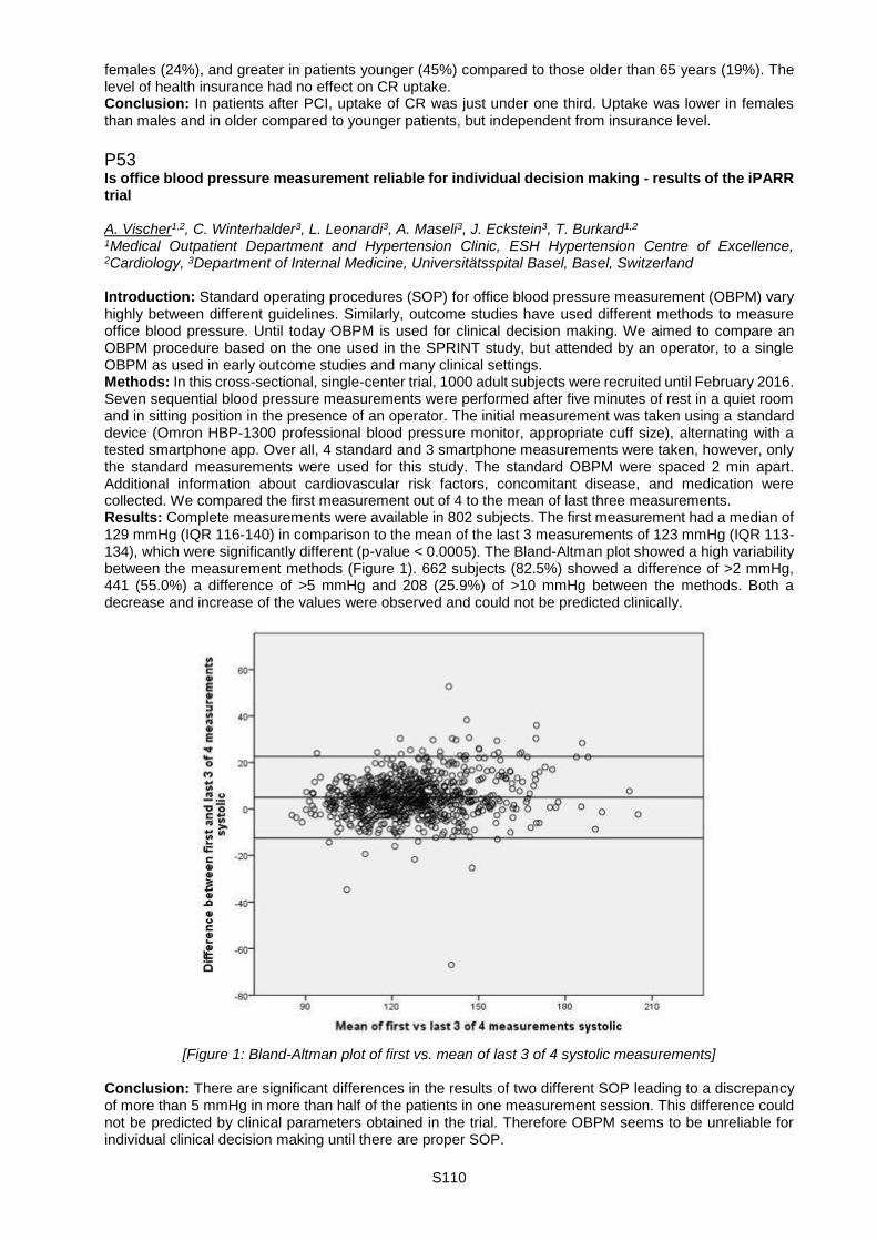
S110
females (24%), and greater in patients younger (45%) compared to those older than 65 years (19%). The level of health insurance had no effect on CR uptake. Conclusion: In patients after PCI, uptake of CR was just under one third. Uptake was lower in females than males and in older compared to younger patients, but independent from insurance level.
P53 Is office blood pressure measurement reliable for individual decision making - results of the iPARR trial A. Vischer1,2, C. Winterhalder3, L. Leonardi3, A. Maseli3, J. Eckstein3, T. Burkard1,2 1Medical Outpatient Department and Hypertension Clinic, ESH Hypertension Centre of Excellence, 2Cardiology, 3Department of Internal Medicine, Universitätsspital Basel, Basel, Switzerland Introduction: Standard operating procedures (SOP) for office blood pressure measurement (OBPM) vary highly between different guidelines. Similarly, outcome studies have used different methods to measure office blood pressure. Until today OBPM is used for clinical decision making. We aimed to compare an OBPM procedure based on the one used in the SPRINT study, but attended by an operator, to a single OBPM as used in early outcome studies and many clinical settings. Methods: In this cross-sectional, single-center trial, 1000 adult subjects were recruited until February 2016. Seven sequential blood pressure measurements were performed after five minutes of rest in a quiet room and in sitting position in the presence of an operator. The initial measurement was taken using a standard device (Omron HBP-1300 professional blood pressure monitor, appropriate cuff size), alternating with a tested smartphone app. Over all, 4 standard and 3 smartphone measurements were taken, however, only the standard measurements were used for this study. The standard OBPM were spaced 2 min apart. Additional information about cardiovascular risk factors, concomitant disease, and medication were collected. We compared the first measurement out of 4 to the mean of last three measurements. Results: Complete measurements were available in 802 subjects. The first measurement had a median of 129 mmHg (IQR 116-140) in comparison to the mean of the last 3 measurements of 123 mmHg (IQR 113-134), which were significantly different (p-value < 0.0005). The Bland-Altman plot showed a high variability between the measurement methods (Figure 1). 662 subjects (82.5%) showed a difference of >2 mmHg, 441 (55.0%) a difference of >5 mmHg and 208 (25.9%) of >10 mmHg between the methods. Both a decrease and increase of the values were observed and could not be predicted clinically.
[Figure 1: Bland-Altman plot of first vs. mean of last 3 of 4 systolic measurements]
Conclusion: There are significant differences in the results of two different SOP leading to a discrepancy of more than 5 mmHg in more than half of the patients in one measurement session. This difference could not be predicted by clinical parameters obtained in the trial. Therefore OBPM seems to be unreliable for individual clinical decision making until there are proper SOP.

S111
P54 The creatinine to uromodulin ratio in serum predicts major cardiovascular events independently from the presence of type 2 diabetes A. Leiherer1,2,3, A. Muendlein1,2, C. Saely1,2,4, J. Ebner1, E.-M. Brandtner1, A. Schuler1,4, P. Schwerzler2,4, A. Mader2,4, P. Fraunberger2,3, H. Drexel1,2,5 1Vorarlberg Institute for Vascular Investigation and Treatment (VIVIT), Feldkirch, Austria, 2Private University of the Principality of Liechtenstein, Triesen, Liechtenstein, 3Medical Central Laboratories, 4Academic Teaching Hospital Feldkirch, Feldkirch, Austria, 5Drexel University, College of Medicine, Philadelphia, PA, United States Introduction: Low concentrations of the kidney protein uromodulin are associated with type 2 diabetes (T2DM) and with chronic kidney disease (CKD). The serum creatinine to uromodulin ratio recently has attracted interest as a marker of CKD. Whether this ratio also is associated with the risk for major cardiovascular events is unknown and is addressed in the present study. Method: We measured uromodulin in 529 coronary patients and prospectively recorded major cardiovascular events (coronary death, fatal and non-fatal ischemic stroke, and non-fatal myocardial infarction) over up to 8 years. Results: During follow-up, a total of 91 major cardiovascular events occurred. The incidence of major cardiovascular events was significantly higher in patients with T2DM (n=141) than in those who did not have diabetes (25.4% vs. 14.6%; p=0.004). The creatinine to uromodulin ratio significantly predicted major cardiovascular events both univariately (HR 1.37 [95%CI 1.21-1.56], p< 0.001) and after multivariate adjustment including the presence of T2DM (HR 1.36 [CI 1.18-1.58], p< 0.001). Conclusion: In conclusion, this study for the first time shows that the serum creatinine to uromodulin ratio predicts major cardiovascular events independently from conventional risk factors including the presence of T2DM. Given that the biological role of uromodulin is still elusive this result appears important and may stimulate future research on uromodulin.
[Creatinin-uromodulin ratio as a predictor of cardiovascular events]
S112
P55 Low income predicts cardiovascular event risk independently from the presence of type 2 diabetes and pre-existing coronary artery disease R. Saely1, A. Vonbank2,3, C. Saely1,2,3, C. Lins2, D. Zanolin1,2, A. Leiherer1,2,4, A. Schuler2,3, P. Schwerzler1,3, A. Mader1,3, A. Muendlein1,2, H. Drexel1,2,5 1Private University of the Principality of Liechtenstein, Triesen, Liechtenstein, 2Vorarlberg Institute for Vascular Investigation and Treatment (VIVIT), 3Academic Teaching Hospital Feldkirch, 4Medical Central Laboratories, Feldkirch, Austria, 5Drexel University, College of Medicine, Philadelphia, PA, United States Introduction: A low socioeconomic status has been associated with an increased cardiovascular event risk. Whether low income predicts cardiovascular event risk independently from the presence of type 2 diabetes (T2DM) and pre-existing coronary artery disease (CAD) is not known and is addressed in the present study. Method: We assessed the annual net income through a standardized questionnaire in a consecutive series of 389 patients referred to coronary angiography for the evaluation of established or suspected stable coronary artery disease (CAD). Prospectively, we recorded cardiovascular events over a mean follow-up period of 8.0±3.7 years. Results: Annual net income was < €20.000 in 58%, €20.000-35.000 in 33.1% and >€35.000 in 8.9% of our patients. It was significantly lower in women (< €20.000 in 70.4%, €20.000-35.000 in 25.6%, >€35.000 in 4.0 %) than in men (< €20.000 in 53.0%, €20.000-35.000 in 36.1%, >€35.000 in 10.9%; p< 0.001) but did not differ significantly between patients with T2DM (n=116) and nondiabetic subjects (p=0.180) nor between patients with CAD (n=353) and those who did not have CAD at angiography (p=0.108). During follow-up, the incidence of cardiovascular events significantly increased with decreasing income: it was 62.4%, 32.4%, and 5.3 % in patients with net incomes of < €20.000, €20.000-35.000 and >€35.000, respectively; p=0.042). Annual net income significantly predicted the incidence of cardiovascular events both univariately (HR 0.77 [0.60-0.98]; p=0.037) and after adjustment for age, gender, smoking, LDL cholesterol, HDL cholesterol, hypertension, BMI, waist circumference, T2DM and angiographically determined baseline CAD (HR 0.68 [0.51-0,92]; p=0.011). Conclusion: We conclude that a low net income predicts cardiovascular event risk independently from the presence of T2DM and pre-existing coronary artery disease.
P56 Non-alcoholic fatty liver disease significantly predicts future decline in kidney function in angiographied coronary patients C. Lins1, A. Vonbank1,2, D. Zanolin1,3, A. Leiherer1,3,4, A. Schuler1,2, A. Muendlein1,3, P. Rein1,2, P. Schwerzler2,3, A. Mader2,3, H. Drexel1,3,5, C. Saely1,2,3 1Vorarlberg Institute for Vascular Investigation and Treatment (VIVIT), 2Academic Teaching Hospital Feldkirch, Feldkirch, Austria, 3Private University of the Principality of Liechtenstein, Triesen, Liechtenstein, 4Medical Central Laboratories, Feldkirch, Austria, 5Drexel University, College of Medicine, Philadelphia, PA, United States Introduction: Non-alcoholic fatty liver disease (NAFLD) is associated with insulin resistance, type 2 diabetes (T2DM), and an increased risk of cardiovascular events. A potential association of NAFLD with the decline of kidney function over time has not been investigated yet and is addressed in the present study. Method: We prospectively investigated a series of 981 consecutive patients referred to coronary angiography for the evaluation of established or suspected coronary artery disease (CAD). NAFLD was diagnosed using the validated fatty liver index, and the glomerular filtration rate (eGFR) was calculated both at baseline and after a follow-up period of 4 years. Results: At baseline, in patients with NAFLD (n=447; 45.6% of the study cohort) the prevalence of T2DM and of the metabolic syndrome was higher (32.7 vs. 18.8 %; p< 0.001 and 72.9 vs. 23.0%; p< 0.001, respectively), whereas eGFR was similar (73.2±20.0 vs. 72.9±21.3 ml/min/1.73 m2; p=0.812) when compared to subjects who did not have NAFLD. Prospectively, NAFLD significantly predicted a decline in eGFR after adjustment for age, gender, and eGFR at baseline (F=8.97; p=0.003) and after further adjustment for smoking, LDL cholesterol, T2DM and the MetS (F=7.35; p=0.007). Conclusion: We conclude that NAFLD significantly predicts a decline in kidney function over 4 years independently of baseline kidney function in angiographied coronary patients.

S113
P57 Sarcoma of the heart: survival after surgery L. Niclauss1, M. Montemurrro2, R. Prêtre1 1CHUV, 2Centre Hospitalier Universitaire Vaudois, Lausanne, Switzerland Introduction: Malignant cardiac tumors (TU) are rare and a general consensus concerning optimal therapeutic approach is missing. We present herein a case series of 9 sarcoma patients (pts) having had a follow-up (FU) after surgical TU resection. Method: Cardiac TU operated from 2000 to the end of 2015 were reviewed. Cardiac sarcomas were identified according to the histo-pathological analysis after surgical resection. Late mortality was determined during FU. General health conditions, tumor extension, surgical approaches (complete or partial TU resection) and adjuvant treatment at time of surgery were retrospectively analyzed. Results: During the observation period 55 pts underwent cardiac TU resection. Sarcomas were, with an incidence of 0.14% (9 pts) the most frequent malignant and the second most common TU (mxyomas/ fribro-elastomas occurred in 0.6%). Mean age was 69 ± 12 years (range 45 - 85 years). All pts presented cardiac symptoms ad admission, but were in an acceptable health condition, without nutritional deficiency. All survived surgery without major cardiac complications and a significant improvement of their symptoms. FU duration was 13 ± 15 months. One year mortality was 44%: four pts died after 6, 8, 12 and 12 months. The 5 survivors had a mean FU of 31 ± 16 months. TU mass extension varied from 4 to 10 cm (mean 6 ± 2.7 cm). Macroscopic complete intra-cardiac TU resection had a potential impact on prognosis, and could be realized in 4 pts (44%), having TU localizations either in the pulmonary artery (3 pts) or the left atrium (1 pt). These patients survived FU. Local relapse occurred in 3 pts (33%), all of them had an incomplete TU resection due to complex cardiac infiltration of the right ventricle in 2 pts, the septum and cardiac skeleton in another pt. Metastatic spread at time of surgery has probably an negative impact on prognosis, 2 of 3 pts. died 12 months after surgery.
[Sarcoma of the pulmonary artery (A) and the postoperative result at 26 months FU (B)] Conclusion: Primary malignant cardiac tumors are in the majority subtypes of sarcomas. Albeit rare, surgeons might be confronted with. A one year mortally of about 44% (in congruence with actual available literature) reflects a general poor prognosis. Nonetheless, surgery is a secure and reliable option to treat cardiac symptoms and may, by preventing direct cardiac compression, avoid early cardiac related deaths (nobody died from heart failure during FU). TU free survival probably depends from TU resection completeness and metastatic spreading at time of surgery.
S114
P58 Low coronary risk calculator based detection rates of old arteries in middle-aged women at recommended decision thresholds M. Romanens1, A. Adams2, I. Sudano3 1Cardiology, Vascular Risk Foundation, Olten, Switzerland, 2BAD Gesundheitsvorsorge und Sicherheitstechnik GmbH, Bonn, Germany, 3Cardiology, University Heart Center, Cardiology, University Hospital, Zürich, Switzerland Background: Intensity of primary care cardiovascular prevention is guided by risk thresholds, which are poorly validated. Methods: We assessed the prevalence of old arteries (70 years or more) in 3'248 healthy subjects in Switzerland and in Germany aged between 40 and 65 years. We compared sensitivity, specificity, and discriminatory performance of SCORE, PROCAM and AGLA coronary risk calculators to detect vascular age ≥ 70 years for various decision thresholds. Results: Old arteries were prevalent in 12% of Swiss women (11% in men), similar to Germany (11% and 17%). Sensitivity was 6% at the 10% AGLA-threshold in women and 30% in men in Switzerland, confirmed for German PROCAM (sensitivity: 8% in women, 56% in men). At the SCORE threshold of 5%, sensitivity ranged between 0% (Germany) to 5% (Switzerland) in women and between 16% and 18% in men. Subjects with old arteries showed higher rates of smoking, higher systolic blood pressure and higher LDL cholesterol. The discriminatory performance of SCORE was significantly better than AGLA / PROCAM in men (p=0.007), but not in women (p=NS). Conclusions: In women aged 40-65 years, the prevalence of old arteries is one out of seven and the detection rate of AGLA and SCORE is 6% and 5% respectively, independently reproduced by a large German group. Low risk factor based detection rates at current decision thresholds of old arteries was clearly evidenced in middle-aged women. Further research is needed to validate the prognostic impact of current risk assessment tools, especially in women.
P59 The A-Body-Shape-Index and type 2 diabetes are mutually independent predictors of cardiovascular events risk in angiographied coronary patients A. Vonbank1,2, C. Saely1,2,3, C. Lins1, D. Zanolin1,3, A. Leiherer1,3,4, A. Schuler1,2, P. Schwerzler2,3, A. Mader2,3, A. Muendlein1,3, H. Drexel1,3,5 1Vorarlberg Institute for Vascular Investigation and Treatment (VIVIT), 2Academic Teaching Hospital Feldkirch, Feldkirch, Austria, 3Private University of the Principality of Liechtenstein, Triesen, Liechtenstein, 4Medical Central Laboratories, Feldkirch, Austria, 5Drexel University, College of Medicine, Philadelphia, PA, United States Introduction: The A-Body-Shape-Index (ABSI) is calculated based on waist circumference, height and BMI and provides a measure of visceral adiposity. In the general population, this index has been associated with premature mortality. Its power to predict cardiovascular events in high-risk patients is not known and is addressed in the present study. Method: We prospectively investigated a large series of 1355 patients undergoing coronary angiography for the evaluation of established or suspected coronary artery disease over 5.0±2.4 years. Results: At baseline, ABSI scores were significantly higher in patients with T2DM (n=419) than in non-diabetic subjects (14.4±1.3 vs. 14.1±1.2; p< 0.001). During follow-up, a total of 421 cardiovascular events were recorded. Cardiovascular event risk was significantly higher in patients with T2DM than in non-diabetic subjects (44.6 % vs. 26.0 %; p< 0.001), and in univariate analysis the ABSI significantly predicted cardiovascular events (HR 1.14 [1.06-1.23]; p< 0.001). In multivariate analyses, both T2DM and ABSI proved independently predictive of cardiovascular events, with standardized adjusted HRs of 1.68 [1.37-2.06]; p< 0.001 and 1.12 [1.02-1.24]; p = 0.016, respectively. Conclusion: We conclude that ABSI and T2DM are mutually independent predictors of cardiovascular events in angiographied coronary patients.
S115
P60 The effect of optimizing glycemic control using insulin on the heart rate variability in type 2 diabetes mellitus patients C.B. Maadjhou1, C.N. Nganou-Gnindjio2, M. Azabji-Kenfack3, L. Mfeukeu Kuate4, A.M. Bimbai1, J.-C. N. Katte1, E. Sobngwi5 1University of Yaounde I, Yaounde, 2Cardiology, Université de Yaoundé I / Yaounde Central Hospital, Yaoundé, 3Physiology, University of Yaounde I, 4University of Yaounde I / Yaounde Central Hospital, 5Endocrinology and Diabetology, Université de Yaoundé I / Yaounde Central Hospital, Yaounde, Cameroon Cardiovascular autonomic neuropathy (CAN) is the most important autonomic neuropathy due to its potential life threatening outcome. Heart rate variability (HRV) provides a non-invasive tool for exploring the autonomic nervous system. Intensive glycemic control prevents the development of CAN in patients with type 1 diabetes. We aimed to determine whether the intensification of glycemic control using insulin will improve cardiovascular autonomic functions in patients with type 2 diabetes. Methods: We conducted a single arm clinical trial study at the National Obesity Centre of the Yaoundé Central Hospital. Participants were poorly controlled (HbA1c ≥ 7%) and well controlled (HbA1c < 7%) type 2 diabetic patients. The intervention lasted 60 days and consisted in the intensification of glycemic control using a basal plus insulin regimen. Target blood glucose were; fasting blood glucose: 70-130mg/dl and post prandial blood glucose < 180mg/dl. The primary outcome measure was a change in HRV parameters. Results: In total, 54 type 2 diabetes mellitus patients (29 poorly controlled vs 25 well controlled) without clinical signs of CAN were recruited (26 males and 28 females). The median age was 56[43-62] years, and duration of diabetes 3[1-7] years. Markers of sympathetic tone were lower in the poorly controlled group (SDNN: 102.01[90.45-111.05] ms vs 112.30[104.40-131.15] ms, p=0.014 and SDANN 88.01[72.95-99.70] ms vs 97.80[91.80-114.50] ms, p= 0.012). The intervention induced a change in HbA1c from 10.1[9.1-11.9] % to 6.7[5.9-6.9] % (p< 0.001). Concerning HRV analysis, there was a significant increase in markers of the parasympathetic (PNN50: 5.70[3.55-10.25] % to 8.12[3.05-16.90] %, p=0.008) and sympathetic activities (SDNN: 102.01[90.45-111.05] ms to 122.40[91.70-135.95] ms, p=0.01). Conclusion: Optimization of glycemic control using a basal plus insulin regimen while inducing a significant reduction in glycated hemoglobin, significantly improves HRV parameters. This suggests that improving glycemic control using insulin may improve cardiovascular autonomic functions in type 2 diabetes mellitus patients. Keywords: Type 2 diabetes mellitus, heart rate variability, glycemic control, insulin
P61 Further cardiovascular outcomes research with PCSK9 inhibition in subjects with eelevated risk, the FOURIER trial F. Mach1, M. Sabatine2, R. Giugliano2, A. Keech3, N. Honarpour4, S.M. Wasserman4, R. Scott4, P. Sever5, T. Pedersen6 1Cardiology, Geneva University Hospitals, Geneva, Switzerland, 2Cardiology, Brigham and Women's Hospital Boston, Boston, MA, United States, 3NHMRC Clinical Trials Centre, University of Sydney, Sydney, NSW, Australia, 4Amgen, Thousand Oaks, CA, United States, 5International Centre for Circulatory Health, Imperial College London, London, United Kingdom, 6Department of Cardiology, Oslo university Hospital, Oslo, Norway Background: Despite current therapies, patients with vascular disease remain at high risk for major adverse cardiovascular events. Low-density lipoprotein cholesterol is a well-established modifiable cardiovascular risk factor. Evolocumab is a fully human monoclonal antibody inhibitor of proprotein convertase subtilisin/kexin type 9 that reduces lowdensity lipoprotein cholesterol by approximately 60% across various populations.Study design: FOURIER is a randomized, placebo-controlled, double-blind, parallel-group, multinational trial testing the hypothesis that adding evolocumab to statin therapy will reduce the incidence of major adverse cardiovascular events in patients with clinically evident vascular disease. The study population consists of 27,564 patients who have had a myocardial infarction (MI), an ischemic stroke, or symptomatic peripheral artery disease and have a low-density lipoprotein ≥70 mg/dL or a non-high-density lipoprotein cholesterol ≥100 mg/dL on an optimized statin regimen. Patients were randomized in a 1:1 ratio to receive either evolocumab (either 140 mg subcutaneously every 2 weeks or 420 mg subcutaneously every month, according to patient preference) or matching placebo injections. The primary end point is major cardiovascular events defined as the composite of cardiovascular death, MI, stroke, hospitalization for unstable angina, or coronary revascularization. The key secondary end point is the composite of cardiovascular death, MI, or stroke. The trial is planned to continue until at least 1,630 patients experience the secondary end point, thereby providing 90% power to detect a relative reduction of ≥15% in this end point. Primary results will be ready for presentation at ACC 2017. Conclusions: FOURIER will determine whether the addition of evolocumab to statin therapy reduces cardiovascular morbidity and mortality in patients with vascular disease.
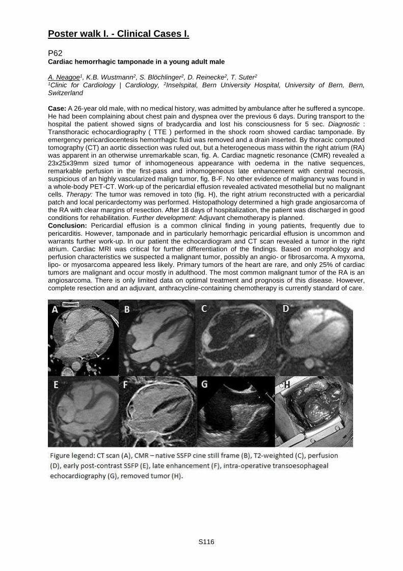
S116
Poster walk I. - Clinical Cases I. P62 Cardiac hemorrhagic tamponade in a young adult male A. Neagoe1, K.B. Wustmann2, S. Blöchlinger2, D. Reinecke2, T. Suter2 1Clinic for Cardiology | Cardiology, 2Inselspital, Bern University Hospital, University of Bern, Bern, Switzerland Case: A 26-year old male, with no medical history, was admitted by ambulance after he suffered a syncope. He had been complaining about chest pain and dyspnea over the previous 6 days. During transport to the hospital the patient showed signs of bradycardia and lost his consciousness for 5 sec. Diagnostic : Transthoracic echocardiography ( TTE ) performed in the shock room showed cardiac tamponade. By emergency pericardiocentesis hemorrhagic fluid was removed and a drain inserted. By thoracic computed tomography (CT) an aortic dissection was ruled out, but a heterogeneous mass within the right atrium (RA) was apparent in an otherwise unremarkable scan, fig. A. Cardiac magnetic resonance (CMR) revealed a 23x25x39mm sized tumor of inhomogeneous appearance with oedema in the native sequences, remarkable perfusion in the first-pass and inhomogeneous late enhancement with central necrosis, suspicious of an highly vascularized malign tumor, fig. B-F. No other evidence of malignancy was found in a whole-body PET-CT. Work-up of the pericardial effusion revealed activated mesothelial but no malignant cells. Therapy: The tumor was removed in toto (fig. H), the right atrium reconstructed with a pericardial patch and local pericardectomy was performed. Histopathology determined a high grade angiosarcoma of the RA with clear margins of resection. After 18 days of hospitalization, the patient was discharged in good conditions for rehabilitation. Further development: Adjuvant chemotherapy is planned. Conclusion: Pericardial effusion is a common clinical finding in young patients, frequently due to pericarditis. However, tamponade and in particularly hemorrhagic pericardial effusion is uncommon and warrants further work-up. In our patient the echocardiogram and CT scan revealed a tumor in the right atrium. Cardiac MRI was critical for further differentiation of the findings. Based on morphology and perfusion characteristics we suspected a malignant tumor, possibly an angio- or fibrosarcoma. A myxoma, lipo- or myosarcoma appeared less likely. Primary tumors of the heart are rare, and only 25% of cardiac tumors are malignant and occur mostly in adulthood. The most common malignant tumor of the RA is an angiosarcoma. There is only limited data on optimal treatment and prognosis of this disease. However, complete resection and an adjuvant, anthracycline-containing chemotherapy is currently standard of care.
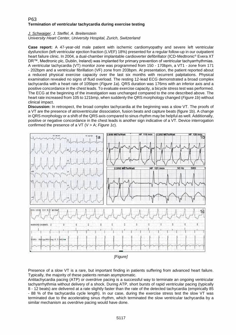
S117
P63 Termination of ventricular tachycardia during exercise testing J. Schwaiger, J. Steffel, A. Breitenstein University Heart Center, University Hospital, Zurich, Switzerland Case report: A 47-year-old male patient with ischemic cardiomyopathy and severe left ventricular dysfunction (left ventricular ejection fraction (LVEF) 18%) presented for a regular follow-up in our outpatient heart failure clinic. In 2004, a dual-chamber implantable cardioverter defibrillator (ICD-Medtronic® Evera XT DR™, Medtronic plc, Dublin, Ireland) was implanted for primary prevention of ventricular tachyarrhythmias. A ventricular tachycardia (VT) monitor zone was programmed from 150 - 170bpm, a VT1 - zone from 171 - 202bpm and a ventricular fibrillation (VF) zone from 203bpm. At presentation, the patient reported about a reduced physical exercise capacity over the last six months with recurrent palpitations. Physical examination revealed no signs of fluid overload. The resting 12-lead ECG demonstrated a broad complex tachycardia with a heart rate of 105bpm (Figure 1a). QRS duration was 176ms with an inferior axis and a positive concordance in the chest leads. To evaluate exercise capacity, a bicycle stress test was performed. The ECG at the beginning of the investigation was unchanged compared to the one described above. The heart rate increased from 105 to 121bmp, when suddenly the QRS morphology changed (Figure 1b) without clinical impact. Discussion: In retrospect, the broad complex tachycardia at the beginning was a slow VT. The proofs of a VT are the presence of atrioventricular dissociation, fusion beats and capture beats (figure 1b). A change in QRS morphology or a shift of the QRS axis compared to sinus rhythm may be helpful as well. Additionally, positive or negative concordance in the chest leads is another sign indicative of a VT. Device interrogation confirmed the presence of a VT (V > A; Figure 1c).
[Figure]
Presence of a slow VT is a rare, but important finding in patients suffering from advanced heart failure. Typically, the majority of these patients remain asymptomatic. Antitachycardia pacing (ATP) or overdrive pacing is a successful way to terminate an ongoing ventricular tachyarrhythmia without delivery of a shock. During ATP, short bursts of rapid ventricular pacing (typically 8 - 12 beats) are delivered at a rate slightly faster than the rate of the detected tachycardia (empirically 85 - 88 % of the tachycardia cycle length). In our case, during the exercise stress test the slow VT was terminated due to the accelerating sinus rhythm, which terminated the slow ventricular tachycardia by a similar mechanism as overdrive pacing would have done.
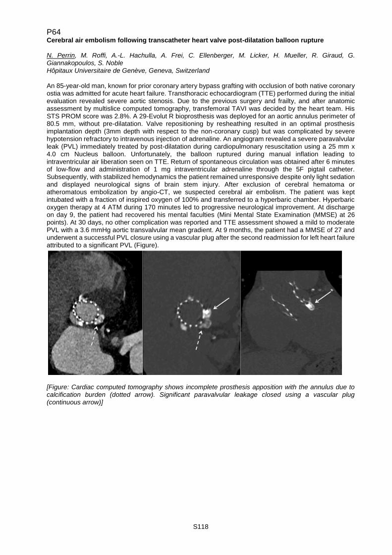
S118
P64 Cerebral air embolism following transcatheter heart valve post-dilatation balloon rupture N. Perrin, M. Roffi, A.-L. Hachulla, A. Frei, C. Ellenberger, M. Licker, H. Mueller, R. Giraud, G. Giannakopoulos, S. Noble Hôpitaux Universitaire de Genève, Geneva, Switzerland An 85-year-old man, known for prior coronary artery bypass grafting with occlusion of both native coronary ostia was admitted for acute heart failure. Transthoracic echocardiogram (TTE) performed during the initial evaluation revealed severe aortic stenosis. Due to the previous surgery and frailty, and after anatomic assessment by multislice computed tomography, transfemoral TAVI was decided by the heart team. His STS PROM score was 2.8%. A 29-Evolut R bioprosthesis was deployed for an aortic annulus perimeter of 80.5 mm, without pre-dilatation. Valve repositioning by resheathing resulted in an optimal prosthesis implantation depth (3mm depth with respect to the non-coronary cusp) but was complicated by severe hypotension refractory to intravenous injection of adrenaline. An angiogram revealed a severe paravalvular leak (PVL) immediately treated by post-dilatation during cardiopulmonary resuscitation using a 25 mm x 4.0 cm Nucleus balloon. Unfortunately, the balloon ruptured during manual inflation leading to intraventricular air liberation seen on TTE. Return of spontaneous circulation was obtained after 6 minutes of low-flow and administration of 1 mg intraventricular adrenaline through the 5F pigtail catheter. Subsequently, with stabilized hemodynamics the patient remained unresponsive despite only light sedation and displayed neurological signs of brain stem injury. After exclusion of cerebral hematoma or atheromatous embolization by angio-CT, we suspected cerebral air embolism. The patient was kept intubated with a fraction of inspired oxygen of 100% and transferred to a hyperbaric chamber. Hyperbaric oxygen therapy at 4 ATM during 170 minutes led to progressive neurological improvement. At discharge on day 9, the patient had recovered his mental faculties (Mini Mental State Examination (MMSE) at 26 points). At 30 days, no other complication was reported and TTE assessment showed a mild to moderate PVL with a 3.6 mmHg aortic transvalvular mean gradient. At 9 months, the patient had a MMSE of 27 and underwent a successful PVL closure using a vascular plug after the second readmission for left heart failure attributed to a significant PVL (Figure).
[Figure: Cardiac computed tomography shows incomplete prosthesis apposition with the annulus due to calcification burden (dotted arrow). Significant paravalvular leakage closed using a vascular plug (continuous arrow)]
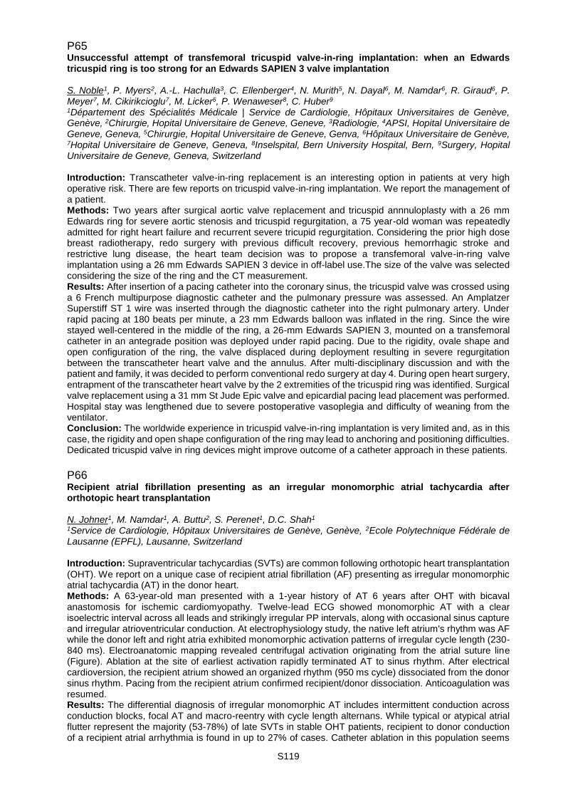
S119
P65 Unsuccessful attempt of transfemoral tricuspid valve-in-ring implantation: when an Edwards tricuspid ring is too strong for an Edwards SAPIEN 3 valve implantation S. Noble1, P. Myers2, A.-L. Hachulla3, C. Ellenberger4, N. Murith5, N. Dayal6, M. Namdar6, R. Giraud6, P. Meyer7, M. Cikirikcioglu7, M. Licker6, P. Wenaweser8, C. Huber9 1Département des Spécialités Médicale | Service de Cardiologie, Hôpitaux Universitaires de Genève, Genève, 2Chirurgie, Hopital Universitaire de Geneve, Geneve, 3Radiologie, 4APSI, Hopital Universitaire de Geneve, Geneva, 5Chirurgie, Hopital Universitaire de Geneve, Genva, 6Hôpitaux Universitaire de Genève, 7Hopital Universitaire de Geneve, Geneva, 8Inselspital, Bern University Hospital, Bern, 9Surgery, Hopital Universitaire de Geneve, Geneva, Switzerland Introduction: Transcatheter valve-in-ring replacement is an interesting option in patients at very high operative risk. There are few reports on tricuspid valve-in-ring implantation. We report the management of a patient. Methods: Two years after surgical aortic valve replacement and tricuspid annnuloplasty with a 26 mm Edwards ring for severe aortic stenosis and tricuspid regurgitation, a 75 year-old woman was repeatedly admitted for right heart failure and recurrent severe tricupid regurgitation. Considering the prior high dose breast radiotherapy, redo surgery with previous difficult recovery, previous hemorrhagic stroke and restrictive lung disease, the heart team decision was to propose a transfemoral valve-in-ring valve implantation using a 26 mm Edwards SAPIEN 3 device in off-label use.The size of the valve was selected considering the size of the ring and the CT measurement. Results: After insertion of a pacing catheter into the coronary sinus, the tricuspid valve was crossed using a 6 French multipurpose diagnostic catheter and the pulmonary pressure was assessed. An Amplatzer Superstiff ST 1 wire was inserted through the diagnostic catheter into the right pulmonary artery. Under rapid pacing at 180 beats per minute, a 23 mm Edwards balloon was inflated in the ring. Since the wire stayed well-centered in the middle of the ring, a 26-mm Edwards SAPIEN 3, mounted on a transfemoral catheter in an antegrade position was deployed under rapid pacing. Due to the rigidity, ovale shape and open configuration of the ring, the valve displaced during deployment resulting in severe regurgitation between the transcatheter heart valve and the annulus. After multi-disciplinary discussion and with the patient and family, it was decided to perform conventional redo surgery at day 4. During open heart surgery, entrapment of the transcatheter heart valve by the 2 extremities of the tricuspid ring was identified. Surgical valve replacement using a 31 mm St Jude Epic valve and epicardial pacing lead placement was performed. Hospital stay was lengthened due to severe postoperative vasoplegia and difficulty of weaning from the ventilator. Conclusion: The worldwide experience in tricuspid valve-in-ring implantation is very limited and, as in this case, the rigidity and open shape configuration of the ring may lead to anchoring and positioning difficulties. Dedicated tricuspid valve in ring devices might improve outcome of a catheter approach in these patients.
P66 Recipient atrial fibrillation presenting as an irregular monomorphic atrial tachycardia after orthotopic heart transplantation N. Johner1, M. Namdar1, A. Buttu2, S. Perenet1, D.C. Shah1 1Service de Cardiologie, Hôpitaux Universitaires de Genève, Genève, 2Ecole Polytechnique Fédérale de Lausanne (EPFL), Lausanne, Switzerland Introduction: Supraventricular tachycardias (SVTs) are common following orthotopic heart transplantation (OHT). We report on a unique case of recipient atrial fibrillation (AF) presenting as irregular monomorphic atrial tachycardia (AT) in the donor heart. Methods: A 63-year-old man presented with a 1-year history of AT 6 years after OHT with bicaval anastomosis for ischemic cardiomyopathy. Twelve-lead ECG showed monomorphic AT with a clear isoelectric interval across all leads and strikingly irregular PP intervals, along with occasional sinus capture and irregular atrioventricular conduction. At electrophysiology study, the native left atrium's rhythm was AF while the donor left and right atria exhibited monomorphic activation patterns of irregular cycle length (230-840 ms). Electroanatomic mapping revealed centrifugal activation originating from the atrial suture line (Figure). Ablation at the site of earliest activation rapidly terminated AT to sinus rhythm. After electrical cardioversion, the recipient atrium showed an organized rhythm (950 ms cycle) dissociated from the donor sinus rhythm. Pacing from the recipient atrium confirmed recipient/donor dissociation. Anticoagulation was resumed. Results: The differential diagnosis of irregular monomorphic AT includes intermittent conduction across conduction blocks, focal AT and macro-reentry with cycle length alternans. While typical or atypical atrial flutter represent the majority (53-78%) of late SVTs in stable OHT patients, recipient to donor conduction of a recipient atrial arrhythmia is found in up to 27% of cases. Catheter ablation in this population seems
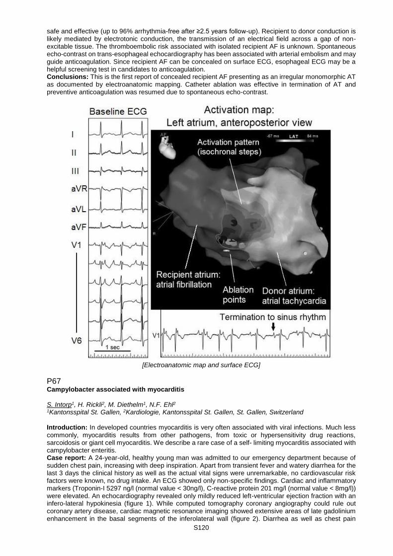
S120
safe and effective (up to 96% arrhythmia-free after ≥2.5 years follow-up). Recipient to donor conduction is likely mediated by electrotonic conduction, the transmission of an electrical field across a gap of non-excitable tissue. The thromboembolic risk associated with isolated recipient AF is unknown. Spontaneous echo-contrast on trans-esophageal echocardiography has been associated with arterial embolism and may guide anticoagulation. Since recipient AF can be concealed on surface ECG, esophageal ECG may be a helpful screening test in candidates to anticoagulation. Conclusions: This is the first report of concealed recipient AF presenting as an irregular monomorphic AT as documented by electroanatomic mapping. Catheter ablation was effective in termination of AT and preventive anticoagulation was resumed due to spontaneous echo-contrast.
[Electroanatomic map and surface ECG]
P67 Campylobacter associated with myocarditis S. Intorp1, H. Rickli2, M. Diethelm1, N.F. Ehl2 1Kantonsspital St. Gallen, 2Kardiologie, Kantonsspital St. Gallen, St. Gallen, Switzerland Introduction: In developed countries myocarditis is very often associated with viral infections. Much less commonly, myocarditis results from other pathogens, from toxic or hypersensitivity drug reactions, sarcoidosis or giant cell myocarditis. We describe a rare case of a self- limiting myocarditis associated with campylobacter enteritis. Case report: A 24-year-old, healthy young man was admitted to our emergency department because of sudden chest pain, increasing with deep inspiration. Apart from transient fever and watery diarrhea for the last 3 days the clinical history as well as the actual vital signs were unremarkable, no cardiovascular risk factors were known, no drug intake. An ECG showed only non-specific findings. Cardiac and inflammatory markers (Troponin-I 5297 ng/l (normal value < 30ng/l), C-reactive protein 201 mg/l (normal value < 8mg/l)) were elevated. An echocardiography revealed only mildly reduced left-ventricular ejection fraction with an infero-lateral hypokinesia (figure 1). While computed tomography coronary angiography could rule out coronary artery disease, cardiac magnetic resonance imaging showed extensive areas of late gadolinium enhancement in the basal segments of the inferolateral wall (figure 2). Diarrhea as well as chest pain
S121
ceased during the next days without specific treatment. Stool culture revealed campylobacter jejuni (retrospectively due to tartare ingestion 5 days before symptom onset). So we were able to make the diagnosis of campylobacter-associated myocarditis. In an outpatient control 4 weeks later the patient described an uneventful course. The echocardiographic findings as well as laboratory findings were normalized. Conclusion: Campylobacter jejuni-enteritis is a rare cause of myocarditis, whereas arthritis, erythema nodosum and Guillain-Barré-Syndrome are better known clinical entities occurring two to three weeks after intestinal infection. A special feature in the campylobacter associated myocarditis is the short time interval of one to five days between onset of enteritis and myocarditis. The pathomechanism is not fully understood, an autoimmune reaction from the gastrointestinal infection is discussed. As shown here, myocarditis can be self-limiting without any further therapy. It's not known, if antiobiotic therapy could reduce the risk of campylobacter associated myocardits and cardiomyopathy.
P68 Facing the dilemma: unstable acute type A aortic dissection - dissociative anesthesia & sternotomy & controlled drainage of pericardial tamponade M. Cikirikcioglu1, P. Mootoosamy1, G. Pavlovic2, M. Licker2, C. Huber1 1Departement de Chirurgie, Service de Chirurgie Cardio-Vasculaire, 2Service de Anesthesiologie, Hopitaux Universitaires de Genève, Genève, Switzerland Background and aim: Pericardial tamponade(PT) is a common complication of acute type A aortic dissection.Controlled drainage of PT with the close collaboration of the surgical and anesthetic teams is of paramount importance to prevent an abrupt blood pressure(BP) increase,advance the progression of the aortic dissection and even triggering an aortic rupture.We describe our controlled PT drainage technique for acute type A aortic dissection in hemodynamically unstable patients secondary to PT. Case report and technique: An 82-year-old lady presented with chest pain to our Emergency Department. BP difference between both arms raised the suspicion of acute aortic dissection.Acute type A aortic dissection and pericardial effusion was confirmed by TTE and CT.The patient was transferred to the OR.Before induction of anesthesia,the patient became hypotensive without response to i.v.fluid resuscitation(systolic/diastolic/mean BP 63/33/45mmHg).TEE showed severe PT due to increase of the pericardial effusion.Following immediate surgical draping,full median sternotomy performed under deep dissociative anesthesia by i.v.bolus of ketamine(2mg/kg)without endotracheal intubation.Controlled drainage of the pericardial effusion was done until stabilization of BP(110/65/82 mmHg) by one cm caudal pericardiotomy which is blocked temporarily by the surgeon's tip of the finger.Aortic dissection repair was done in a standard fashion:25°C hypothermia,ascending aortic-hemiarch replacement by open technique using antegrade cerebral perfusion. The patient left the hospital 8th postoperative day without having a major complication. Conclusion: PT is a dynamic pathology which may progress very quickly and worsen the hemodynamic status of the patient with acute aortic dissection.General anesthesia and positive pressure ventilation can even precipitate cardiac arrest in a patient with severe PT.Although controlled pericardial drainage by percutaneous puncture has been described to stabilize a patient with PT and aortic dissection before anesthesia,this approach is not well adopted because of potential delay transferring the patient to the operating room and repair.We believe our technique may give the surgical team in similar conditions as described above a safe approach without any delay to perform best surgical treatment on a critically ill acute type A dissection patients.
P69 Rare coronary arterial fistulas in a female patient with aortic valve stenosis. case report M. Mytsyk1, J.M. Erb2, M. Brunner2, F. Eckstein1, M. Grapow1 1Division of Cardiac Surgery, 2Department of Anaesthesiology, University Hospital of Basel, Basel, Switzerland Introduction: Coronary arteries (CA) normally arise from the sinus of valsalva on the ascending aorta. CA anomalies are rare among the general population and affect around 1% of the general population. However, cases of CA anomalies in elderly are not well described in literature. We report about very rare case of a patient with two fistulas arising from the left and the right CA into the sino-tubular junction of the pulmonary artery (PA). Documentation was performed with transesophageal echocardiography (TOE), epiaortic ultrasound, angiography and photography imaging during the operation. Methods: A 64-year-old female patient with severe aortic valve stenosis, suffering from dyspnoea NYHA II-III and presyncopal conditions, was admitted to our clinic for aortic valve replacement. Angina pectoris and oedema complaints were denied. The blood analysis showed normal findings. Coronary angiography

S122
suspected a coronary fistula arising from the LAD with uncertain course. Transthoracic echo showed a normal EF with 55%, severe stenosis of then aortic valve and mild insufficiency of the pulmonary valve. Results: During the routine intraoperative TOE scan for aortic valve procedures, we detected by chance a significant inflow into the sino-tubular junction of PA with a mild pulmonary regurgitation. After median sternotomy and opening of the pericardium we surprisingly observed two vessels: one arising from the right CA, the other one from left CA, ending into the anterior side of PA. Intraoperatively performed direct ultrasound (epiaortic probe) confirmed our finding of the fistula between pulmonary artery and coronary arteries. After the aortic valve replacement with biological prosthesis, both branches of the fistulas and the inflow side itself (anterior sinotubular junction of the pulmonary artery) were ligated. TOE confirmed the disappearance of the inflow, furthermore the pulmonary valve was competent without any sign of insufficiency. Conclusion: Fistulas arising from the left or the right CA are rare and in most cases not a dangerous anomaly of CA. We report about the very rare case of coronary fistulas arising from both main CA and ending together with an inflow into the sinotubular junction of PA. Extensive imaging material was collected for demonstration. Retrospectively both fistulas were seen in coronary angiography and retrospectively the mild pulmonary valve insufficiency was caused by them - after ligation regurgitation was completely resolved.
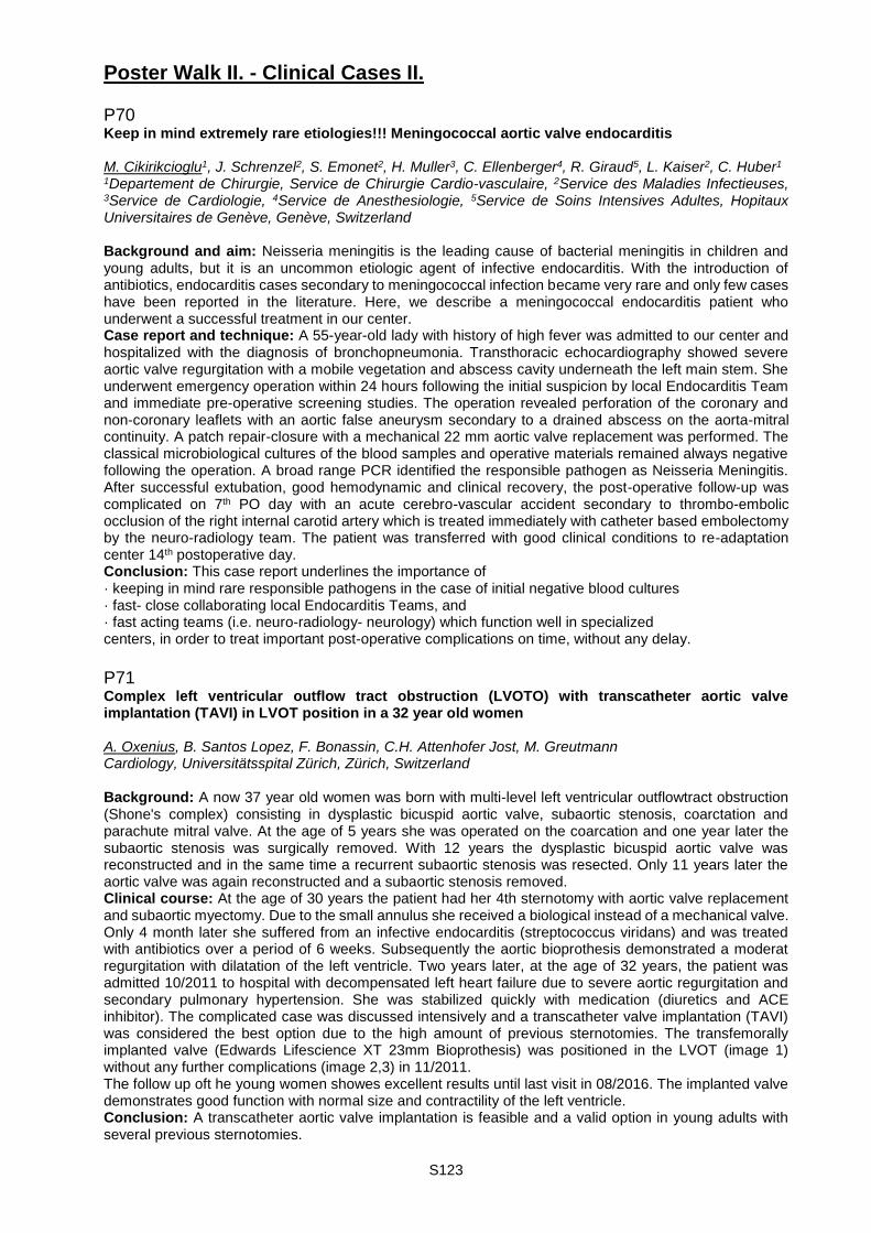
S123
Poster Walk II. - Clinical Cases II. P70 Keep in mind extremely rare etiologies!!! Meningococcal aortic valve endocarditis M. Cikirikcioglu1, J. Schrenzel2, S. Emonet2, H. Muller3, C. Ellenberger4, R. Giraud5, L. Kaiser2, C. Huber1 1Departement de Chirurgie, Service de Chirurgie Cardio-vasculaire, 2Service des Maladies Infectieuses, 3Service de Cardiologie, 4Service de Anesthesiologie, 5Service de Soins Intensives Adultes, Hopitaux Universitaires de Genève, Genève, Switzerland Background and aim: Neisseria meningitis is the leading cause of bacterial meningitis in children and young adults, but it is an uncommon etiologic agent of infective endocarditis. With the introduction of antibiotics, endocarditis cases secondary to meningococcal infection became very rare and only few cases have been reported in the literature. Here, we describe a meningococcal endocarditis patient who underwent a successful treatment in our center. Case report and technique: A 55-year-old lady with history of high fever was admitted to our center and hospitalized with the diagnosis of bronchopneumonia. Transthoracic echocardiography showed severe aortic valve regurgitation with a mobile vegetation and abscess cavity underneath the left main stem. She underwent emergency operation within 24 hours following the initial suspicion by local Endocarditis Team and immediate pre-operative screening studies. The operation revealed perforation of the coronary and non-coronary leaflets with an aortic false aneurysm secondary to a drained abscess on the aorta-mitral continuity. A patch repair-closure with a mechanical 22 mm aortic valve replacement was performed. The classical microbiological cultures of the blood samples and operative materials remained always negative following the operation. A broad range PCR identified the responsible pathogen as Neisseria Meningitis. After successful extubation, good hemodynamic and clinical recovery, the post-operative follow-up was complicated on 7th PO day with an acute cerebro-vascular accident secondary to thrombo-embolic occlusion of the right internal carotid artery which is treated immediately with catheter based embolectomy by the neuro-radiology team. The patient was transferred with good clinical conditions to re-adaptation center 14th postoperative day. Conclusion: This case report underlines the importance of · keeping in mind rare responsible pathogens in the case of initial negative blood cultures · fast- close collaborating local Endocarditis Teams, and · fast acting teams (i.e. neuro-radiology- neurology) which function well in specialized centers, in order to treat important post-operative complications on time, without any delay.
P71 Complex left ventricular outflow tract obstruction (LVOTO) with transcatheter aortic valve implantation (TAVI) in LVOT position in a 32 year old women A. Oxenius, B. Santos Lopez, F. Bonassin, C.H. Attenhofer Jost, M. Greutmann Cardiology, Universitätsspital Zürich, Zürich, Switzerland Background: A now 37 year old women was born with multi-level left ventricular outflowtract obstruction (Shone's complex) consisting in dysplastic bicuspid aortic valve, subaortic stenosis, coarctation and parachute mitral valve. At the age of 5 years she was operated on the coarcation and one year later the subaortic stenosis was surgically removed. With 12 years the dysplastic bicuspid aortic valve was reconstructed and in the same time a recurrent subaortic stenosis was resected. Only 11 years later the aortic valve was again reconstructed and a subaortic stenosis removed. Clinical course: At the age of 30 years the patient had her 4th sternotomy with aortic valve replacement and subaortic myectomy. Due to the small annulus she received a biological instead of a mechanical valve. Only 4 month later she suffered from an infective endocarditis (streptococcus viridans) and was treated with antibiotics over a period of 6 weeks. Subsequently the aortic bioprothesis demonstrated a moderat regurgitation with dilatation of the left ventricle. Two years later, at the age of 32 years, the patient was admitted 10/2011 to hospital with decompensated left heart failure due to severe aortic regurgitation and secondary pulmonary hypertension. She was stabilized quickly with medication (diuretics and ACE inhibitor). The complicated case was discussed intensively and a transcatheter valve implantation (TAVI) was considered the best option due to the high amount of previous sternotomies. The transfemorally implanted valve (Edwards Lifescience XT 23mm Bioprothesis) was positioned in the LVOT (image 1) without any further complications (image 2,3) in 11/2011. The follow up oft he young women showes excellent results until last visit in 08/2016. The implanted valve demonstrates good function with normal size and contractility of the left ventricle. Conclusion: A transcatheter aortic valve implantation is feasible and a valid option in young adults with several previous sternotomies.
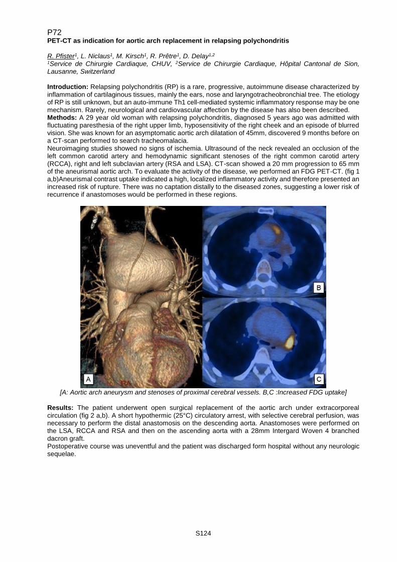
S124
P72 PET-CT as indication for aortic arch replacement in relapsing polychondritis R. Pfister1, L. Niclaus1, M. Kirsch1, R. Prêtre1, D. Delay1,2 1Service de Chirurgie Cardiaque, CHUV, 2Service de Chirurgie Cardiaque, Hôpital Cantonal de Sion, Lausanne, Switzerland Introduction: Relapsing polychondritis (RP) is a rare, progressive, autoimmune disease characterized by inflammation of cartilaginous tissues, mainly the ears, nose and laryngotracheobronchial tree. The etiology of RP is still unknown, but an auto-immune Th1 cell-mediated systemic inflammatory response may be one mechanism. Rarely, neurological and cardiovascular affection by the disease has also been described. Methods: A 29 year old woman with relapsing polychondritis, diagnosed 5 years ago was admitted with fluctuating paresthesia of the right upper limb, hyposensitivity of the right cheek and an episode of blurred vision. She was known for an asymptomatic aortic arch dilatation of 45mm, discovered 9 months before on a CT-scan performed to search tracheomalacia. Neuroimaging studies showed no signs of ischemia. Ultrasound of the neck revealed an occlusion of the left common carotid artery and hemodynamic significant stenoses of the right common carotid artery (RCCA), right and left subclavian artery (RSA and LSA). CT-scan showed a 20 mm progression to 65 mm of the aneurismal aortic arch. To evaluate the activity of the disease, we performed an FDG PET-CT. (fig 1 a,b)Aneurismal contrast uptake indicated a high, localized inflammatory activity and therefore presented an increased risk of rupture. There was no captation distally to the diseased zones, suggesting a lower risk of recurrence if anastomoses would be performed in these regions.
[A: Aortic arch aneurysm and stenoses of proximal cerebral vessels. B,C :Increased FDG uptake]
Results: The patient underwent open surgical replacement of the aortic arch under extracorporeal circulation (fig 2 a,b). A short hypothermic (25°C) circulatory arrest, with selective cerebral perfusion, was necessary to perform the distal anastomosis on the descending aorta. Anastomoses were performed on the LSA, RCCA and RSA and then on the ascending aorta with a 28mm Intergard Woven 4 branched dacron graft. Postoperative course was uneventful and the patient was discharged form hospital without any neurologic sequelae.

S125
[A, 1:Ascending Aorta, 2:Arch Aneurysm, 3:Brachiocephalic artery (dilated). B After Replacement ] Conclusions: Unusual localizations of an acute inflammatory process should be taken into consideration in patients suffering from RP: Large arteries may be involved, presenting a complex symptomatology. The PET CT may be useful to evaluate the activity of the disease, and to determine healthy zones for potential anastomotic sites. In these particular cases, surgery should be favored compared to endovascular surgery as it gives the opportunity to resect the diseased vessels to reduce recurrences.
P73 Hemodynamic changes may precede sleep apnea pattern shifts in a CRT patient with persistent atrial fibrillation episode F. Regoli, M.L. Caputo, G. Conte, T. Moccetti, A. Auricchio Cardiology, Fondazione Cardiocentro Ticino, Lugano, Switzerland Introduction: The present case shows how continuous device-based sleep apnea monitoring not only plays a role in patient management, but also may provide important pathophysiological insights into the complex interrelationship between cardiac arrhythmias, heart failure syndrome, and sleep apnea syndrome (SAS). Methods and results: A 76 year old patient, who underwent device upgrade to cardiac resynchronization therapy pacemaker (CRT-P, Sorin Reply) equipped with sleep apnea monitoring diagnostic feature, 3 months before, presented with worsening heart failure symptoms (NYHA III). The patient was known for purely obstructive SAS treated with continuous positive airway pressure (CPAP). ECG showed atrial fibrillation with spontaneous ventricular beats alternated with frequent ventricular ectopic beats; device control confirmed reduction of biventricular pacing percentage (BVP%) averaging 75%. After serial regular out-patient clinic visits, a sudden steep surge of the respiratory disturbance index (RDI) peaking to almost 60 could be appreciated following further BVP% reduction (Figure, phase 2). CPAP performance was verified once again with the pneumologist who confirmed normal device function and excellent patient adherence to the therapy.Increasing metoprolol to 150 mg/day and prescribing digoxin 0.125 mg daily effectively increased BVP% shortly after (Figure, phase 3), resulting in reversal of SAS pattern; ultimately spontaneous recovery to sinus rhythm and improvement of clinical symptoms was observed (Figure, phase 4). Conclusions: Integration of conventional PM diagnostics with continuous RDI pattern monitoring allowed to detect how hemodynamic worsening preceded a possible SAS shift with increases in central SAS; pharmacological rate control with subsequent increased CRT effect resulted in reversal of abnormal SAS pattern.
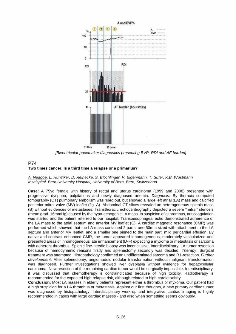
S126
[Biventricular pacemaker diagnostics presenting BVP, RDI and AF burden]
P74 Two times cancer. Is a third time a relapse or a primarius? A. Neagoe, L. Hunziker, D. Reinecke, S. Blöchlinger, V. Eigenmann, T. Suter, K.B. Wustmann Inselspital, Bern University Hospital, University of Bern, Bern, Switzerland Case: A 75yo female with history of rectal and uterus carcinoma (1999 and 2008) presented with progressive dyspnea, palpitations and newly diagnosed anemia. Diagnosis: By thoracic computed tomography (CT) pulmonary embolism was ruled out, but showed a large left atrial (LA) mass and calcified posterior mitral valve (MV) leaflet (fig. A). Abdominal CT slices revealed an heterogeneous splenic mass (B) without evidences of metastases. Transthoracic echocardiography depicted a severe “mitral” stenosis (mean grad. 16mmHg) caused by the hypo-echogenic LA mass. In suspicion of a thrombus, anticoagulation was started and the patient referred to our hospital. Transoesophageal echo demonstrated adherence of the LA mass to the atrial septum and anterior MV leaflet (C). A cardiac magnetic resonance (CMR) was performed which showed that the LA mass contained 2 parts: one 50mm sized with attachment to the LA septum and anterior MV leaflet, and a smaller one pinned to the main part, mild pericardial effusion. By native and contrast enhanced CMR, the tumor appeared inhomogeneous, moderately vascularized and presented areas of inhomogeneous late enhancement (D-F) expecting a myxoma or metastasis or sarcoma with adherent thrombus. Splenic fine-needle biopsy was inconclusive. Interdisciplinary, LA tumor resection because of hemodynamic reasons firstly and splenectomy secondly was decided. Therapy: Surgical treatment was attempted. Histopathology confirmed an undifferentiated sarcoma and R1-resection. Further development: After splenectomy, angiomadoid nodular transformation without malignant transformation was diagnosed. Further investigations showed liver dysplasia without evidence for hepatocellular carcinoma. New resection of the remaining cardiac tumor would be surgically impossible. Interdisciplinary, it was discussed that chemotherapy is contraindicated because of high toxicity. Radiotherapy is recommended for the expected high relapse risk, although related to high cardiotoxicity. Conclusion: Most LA masses in elderly patients represent either a thrombus or myxoma. Our patient had a high suspicion for a LA thrombus or metastasis. Against our first thoughts, a new primary cardiac tumor was diagnosed by histopathology. Interdisciplinary work-up and integrative cardiac imaging is highly recommended in cases with large cardiac masses - and also when something seems obviously.

S127
[Figure 1.]
P75 Left heart failure after double switch operation: the dilemma of preventing systemic right heart failure in congenitally corrected transposition of the great arteries F. Bonassin Tempesta, B. Santos Lopes, C. Attenhofer Jost, M. Greutmann Universitäres Herzzentrum, Universitätsspital Zürich, Zürich, Switzerland Background: In congenitally corrected transposition of the great arteries (ccTGA) severe systemic tricuspid regurgitation (TR) often heralds systemic right ventricular (RV) failure and is associated with increased mortality. Anatomical repair with a double switch operation (atrial switch and concomitant arterial switch operation) may be a treatment option for selected patients but long-term outcomes are not well defined. Case report: We report the case of a patient diagnosed with ccTGA at age 1y. He subsequently developed severe TR until the age of 6y when it was decided to perform a double switch operation to prevent systemic RV failure. As a preparation for the double switch procedure, in order to 'train' the subpulmonic left ventricle (LV), progressive pulmonary artery banding in two stages was performed within 3 months. At this point, systolic pressures in the subpulmonic LV had risen to 78% of systemic pressures and due to the shift of interventricular septum and improved tricuspid valve geometry, the degree of TR had improved from severe to mild. Although LV ejection fraction (EF) was noted to be mildly impaired it was decided to proceed to double switch operation. The postoperative course was complicated by rapidly progressive systemic LV failure, requiring escalation of medical therapy. Despite a short QRS-duration (< 120ms) an epicardial biventricular pacemaker (CRT-P) was placed at age 9y without LV function improvement. Between age 6 and 18y he had recurrent intraatrial reentrant tachycardias requiring multiple direct current cardioversions. Radiofrequency ablation was not successful. When the patient was transitioned to adult care at the age of 18y he remained in heart failure with NYHA functional class III. Transthoracic echocardiography showed a grossly dilated LV with severely impaired EF (< 20%, figure 1). With intensified medical heart failure therapy symptoms improved (increase in peak VO2 from 13.1 to 20.4ml/kg/m2), proBNP-levels decreased (from 1544ng/l to 356ng/l) but LV ejection fraction remained severely impaired (around 30%). Due to stenosis of the superior vena cava baffle (figure 2, arrow) it was decided not to implant an AICD for primary prevention at this time. This case highlights the challenges of the anatomical repair in ccTGA patients, even when performed in early childhood. In selected cases isolated banding of the pulmonary artery without subsequent double switch operation may be an alternative long-term strategy.
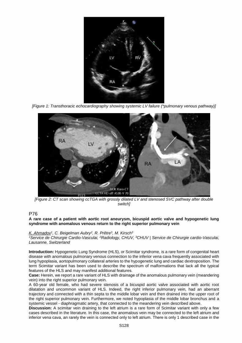
S128
[Figure 1: Transthoracic echocardiography showing systemic LV failure (*pulmonary venous pathway)]
[Figure 2: CT scan showing ccTGA with grossly dilated LV and stenosed SVC pathway after double
switch]
P76 A rare case of a patient with aortic root aneurysm, bicuspid aortic valve and hypogenetic lung syndrome with anomalous venous return to the right superior pulmonary vein K. Ahmadov1, C. Beigelman Aubry2, R. Prêtre3, M. Kirsch3 1Service de Chirurgie Cardio-Vasculai, 2Radiology, CHUV, 3CHUV | Service de Chirurgie cardio-Vasculai, Lausanne, Switzerland Introduction: Hypogenetic Lung Syndrome (HLS), or Scimitar syndrome, is a rare form of congenital heart disease with anomalous pulmonary venous connection to the inferior vena cava frequently associated with lung hypoplasia, aortopulmonary collateral arteries to the hypogenetic lung and cardiac dextroposition. The term Scimitar variant has been used to describe the spectrum of malformations that lack all the typical features of the HLS and may manifest additional features. Case: Herein, we report a rare variant of HLS with drainage of the anomalous pulmonary vein (meandering vein) into the right superior pulmonary vein. A 60-year old female, who had severe stenosis of a bicuspid aortic valve associated with aortic root dilatation and uncommon variant of HLS. Indeed, the right inferior pulmonary vein, had an aberrant trajectory and connected with a thin septa to the middle lobar vein and then drained into the upper root of the right superior pulmonary vein. Furthermore, we noted hypoplasia of the middle lobar bronchus and a systemic vessel - diaphragmatic artery, that connected to the meandering vein described above. Discussion: A scimitar vein draining to the left atrium is a rare form of Scimitar variant with only a few cases described in the literature. In this case, the anomalous vein may be connected to the left atrium and inferior vena cava, an rarely the vein is connected only to left atrium. There is only 1 described case in the
S129
literature (Furuya K et al) with anomalous venous return to the left inferior pulmonary vein. In all other cases of scimitar variant described in literature, the anomalous vein is connected to the left atrium. To our knowledge, the present case is the first report of a patient with HLS with an anomalous vein draining to the right superior pulmonary vein. This is also the first reported case of the patient with HLS associated with bicuspid aortic valve and aortic root aneurism. The patient underwent successful total aortic root replacement using a Freestyle stentless bioprothesis. The anomalous meandering vein and aorto-pulmonary collateral (systemic vessel) were left untouched during surgery since the meandering vein was draining into the left heart (to the superior lobar vein) and the aorto-pulmonary collateral was considered haemodinamically not significant. The patient was discharged at home in good general condition on postoperative day 7.
P77 Innovative management of acute stent recoil in the left main F. Rey1, M. Diakite1, F. Rigamonti2, M. Roffi1 1Cardiologie, Hôpitaux Universitaire de Genève, 2Cardiologie, Hôpitaux Universitaires de Genève, Genève, Switzerland A 71 years old woman with morbid obesity, dyslipidemia, diabetes and hypertension was admitted in emergency department with acute respiratory distress. She had high blood pressure and high rate atrial fibrillation with left bundle block branch requiring one defibrillation and orotracheal intubation. Serum level of high-sensitivity cardiac troponin and creatinin kinase were high. Echocardiography showed moderate decreased of left ventricle ejection fraction with severe antero-septo-apical hypokinesia. After intubation and stabilization of the heamodynamic situation, the patient was admitted to the catheterization laboratory for coronary angiography. Coronary angiography via right femoral artery showed two vessel diseases with severely calcified ostium of the left main (Image A) and 50-70% of the distal left anterior descending artery. During the intubation of left main artery, major electrocardiogram modification and blood pressure dumping happened (Image B), and then the patient fainted. She needed 2 mg intra-aortic adrenalin. Depending on heamodynamic compromise and the anatomy of coronary disease, percutaneous transluminal coronary angioplasty with implantation of drug eluting stent (DES) was decided. After the implantation of the DES (Synergy 4.5 x 12 mm at 18 atm) in the left main we proceeded post dilatation with non-compliant balloon (Pantera Leo 4.5x12 mm at 24 atm). The angiographic control with intravascular ultrasound (IVUS) (Image C) confirmed recoil of the stent. We decided to use the very high-pressure balloon (OPN 4.00x15mm at 35 atm) with persistence of the stent recoil in the ostial part. Then we decided to use the renal stent (Hippocampus 5.00 x 15mm at 10 atm.) overlapped with the first stent. It was post-dilatated with a compliant balloon (Pantera Leo 5.0 x 15 mm at 23 atm) and the second angiographic control with IVUS (Image D) showed successful deployment of the stent in the ostium of the left main artery with no recoil. This case demonstrates the management of stent recoil in a calcified left main and in an atherosclerotic disease of the aorta is very complex. Using a renal stent can be a kind solution when recoil persists after post dilatation with very high-pressure balloon.
S130
Poster walk II. - ACS, PCI & CABG P78 Using the antecubital vein as a safe approach for right heart catheterization in patient with suspected or confirmed pulmonary hypertension F. Rey1, M. Diakite2, F. Lador3, S. Noble4 1Cardiology, Hôpitaux Universitaire de Genève, Genève, 2Cardiology, 3Pneumology, Hôpitaux Universitaire de Genève, Geneva, 4Hôpitaux Universitaire de Genève, Genève, Switzerland Aims: Vascular access for right heart catheterization (RHC) procedures is usually performed using proximal venous access sites such as the common femoral vein or the internal jugular vein. While these routes are relatively low risk for patients, significant complications can occur such as large hematomas, pseudoaneurysms, and arterio-venous fistula formation, especially in patients treated with anticoagulants. Using the antecubital vein approach for RHC is safe and reduces procedural and fluoroscopy time as previously described. Methods: We retrospectively analyzed data on thirty-eight patients with suspected or confirmed pulmonary hypertension of varied origins, who underwent RHC between January 2012 and April 2016. Data was collected from procedures performed at rest and/or with effort using the antecubital vein approach. A Terumo Slender 7F in 6F introducer was inserted and over a Terumo 0.025” J-tipped wire a 7-F Swan ganz catheter was introduced. Procedural time and successful access, complications such as hematomas, pseudoaneurysms, arterio-venous fistula, neurological complications, and fluoroscopy time were assessed. Results: Patient mean age was 62 ± 14 years-old. 32 % had chronic thromboembolic hypertension, 16% had valvulopathy, 13% had severe chronic pulmonary disease, 13% had restrictive pulmonary syndrome, 13% had dilated cardiomyopathy, 8% had systemic disease and 5% had idiopathic pulmonary hypertension. 40% of patients had anticoagulation therapy and mean INR value was 1.3± 0.4. Once the introducer was inserted, access to the pulmonary artery was successful in all patients. The rate of complications was 0%. 84% of patients had left catheterization and when pulmonary pressures were normal at rest, 21% had RHC with effort (cycling). Mean fluoroscopy time for right catheterization only was 2.3 minutes. Mean fluoroscopy time for right and left catheterization +/- coronary angiography was 9.6 minutes. The mean of the mean pulmonary artery pressure at rest was 30.7 ± 16.5 mmHg, for the mean wedge pressure was 12.7 ± 8.2 mmHg and for the indexed cardiac output was 2.4± 0.7 L/min/m2. One patient had pulmonary hypertension with effort. Conclusion: Using the antecubital vein permits evaluation of pulmonary pressures at rest and during effort in patients with suspected or confirmed pulmonary hypertension of different origins. Procedures are short and safe.
P79 Impact of betablocker therapy status on clinical outcomes of patients with acute coronary syndrome M. Maeder1, D. Radovanovic2, J.F. Iglesias3, P. Erne4, G. Pedrazzini5, O. Bertel6, F. Eberli7, P. Urban8, M. Roffi9, H. Rickli10 1Kantonsspital St. Galen, St. Gallen, 2Univerity of Zurich, Zürich, 3CHUV, Lausanne, 4University of Basel, Basel, 5Cardiocentro Ticino, Lugano, 6Clinic im Park, 7Triemlispital, Zürich, 8La Tour Hospital, 9Hôpitaux Universitaire de Genève, Geneva, 10Kantonsspital St. Gallen, St. Gallen, Switzerland Introduction: While the beneficial effect of long-term betablocker (BB) therapy for post-myocardial infarction patients with moderate or severe left ventricular systolic dysfunction is well established, the benefits of BB therapy in patients with acute coronary syndrome (ACS) remains uncertain. We aimed to assess the influence of in-hospital BB therapy status on in-hospital mortality among patients with ACS. Methods: Data from consecutive patients included in the Acute Myocardial Infarction in Switzerland (AMIS) registry between 1997 and 2016 were retrospectively analyzed (n=47303). Baseline characteristics and clinical outcomes of patients with BB (n=34088) and without BB (BB-/-; n=13215) therapy during the hospital period were compared. In addition, the subgroups of patients with chronic and continued (BB+/+; n=11127), chronic but acutely discontinued (BB+/-; n=3298), and new onset (BB-/+; n=19663) BB therapy were compared to BB-/- patients. Results: Overall, patients with any in-hospital BB therapy (BB+/+, BB, -/+, or BB+/-) were younger (66±13 vs. 67±14 years), less likely to be female (27 vs. 28%) and to present with ST-segment elevation myocardial infarction (STEMI; 55 vs. 59%), and had lower Killip class (Killip >2: 5 vs. 11%) compared with BB-/- patients (p< 0.01 for all). BB-/+ patients represented the hemodynamically best patients (Killip >2: 2.9%), and BB+/- patients represented the worst group (Killip >2: 17.1%). Crude in-hospital mortality was lower in patients with any in-hospital BB therapy compared to those without (4.3 versus 9.8%). After adjustment for age, gender, Killip class and Charlson comorbitity index, any in-hospital BB therapy was associated with
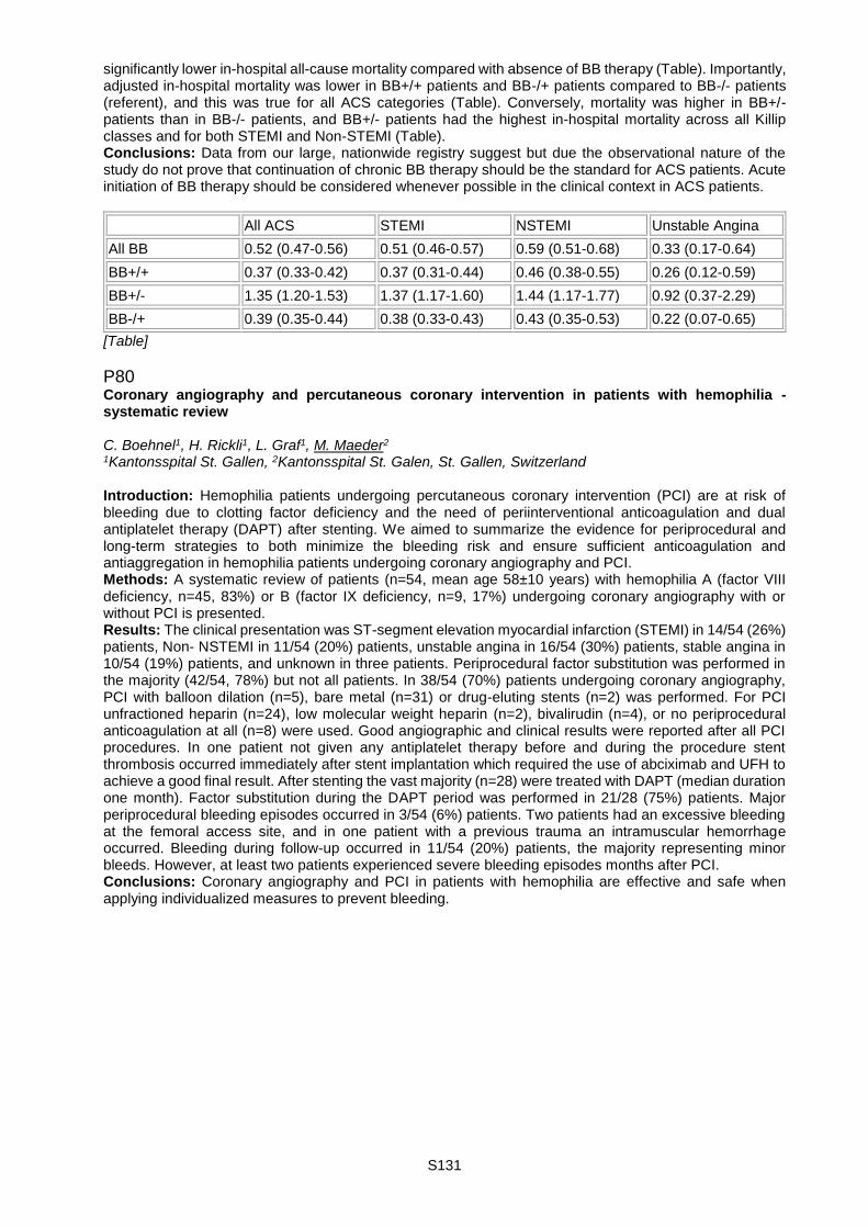
S131
significantly lower in-hospital all-cause mortality compared with absence of BB therapy (Table). Importantly, adjusted in-hospital mortality was lower in BB+/+ patients and BB-/+ patients compared to BB-/- patients (referent), and this was true for all ACS categories (Table). Conversely, mortality was higher in BB+/- patients than in BB-/- patients, and BB+/- patients had the highest in-hospital mortality across all Killip classes and for both STEMI and Non-STEMI (Table). Conclusions: Data from our large, nationwide registry suggest but due the observational nature of the study do not prove that continuation of chronic BB therapy should be the standard for ACS patients. Acute initiation of BB therapy should be considered whenever possible in the clinical context in ACS patients.
All ACS STEMI NSTEMI Unstable Angina
All BB 0.52 (0.47-0.56) 0.51 (0.46-0.57) 0.59 (0.51-0.68) 0.33 (0.17-0.64)
BB+/+ 0.37 (0.33-0.42) 0.37 (0.31-0.44) 0.46 (0.38-0.55) 0.26 (0.12-0.59)
BB+/- 1.35 (1.20-1.53) 1.37 (1.17-1.60) 1.44 (1.17-1.77) 0.92 (0.37-2.29)
BB-/+ 0.39 (0.35-0.44) 0.38 (0.33-0.43) 0.43 (0.35-0.53) 0.22 (0.07-0.65)
[Table]
P80 Coronary angiography and percutaneous coronary intervention in patients with hemophilia - systematic review C. Boehnel1, H. Rickli1, L. Graf1, M. Maeder2 1Kantonsspital St. Gallen, 2Kantonsspital St. Galen, St. Gallen, Switzerland Introduction: Hemophilia patients undergoing percutaneous coronary intervention (PCI) are at risk of bleeding due to clotting factor deficiency and the need of periinterventional anticoagulation and dual antiplatelet therapy (DAPT) after stenting. We aimed to summarize the evidence for periprocedural and long-term strategies to both minimize the bleeding risk and ensure sufficient anticoagulation and antiaggregation in hemophilia patients undergoing coronary angiography and PCI. Methods: A systematic review of patients (n=54, mean age 58±10 years) with hemophilia A (factor VIII deficiency, n=45, 83%) or B (factor IX deficiency, n=9, 17%) undergoing coronary angiography with or without PCI is presented. Results: The clinical presentation was ST-segment elevation myocardial infarction (STEMI) in 14/54 (26%) patients, Non- NSTEMI in 11/54 (20%) patients, unstable angina in 16/54 (30%) patients, stable angina in 10/54 (19%) patients, and unknown in three patients. Periprocedural factor substitution was performed in the majority (42/54, 78%) but not all patients. In 38/54 (70%) patients undergoing coronary angiography, PCI with balloon dilation (n=5), bare metal (n=31) or drug-eluting stents (n=2) was performed. For PCI unfractioned heparin (n=24), low molecular weight heparin (n=2), bivalirudin (n=4), or no periprocedural anticoagulation at all (n=8) were used. Good angiographic and clinical results were reported after all PCI procedures. In one patient not given any antiplatelet therapy before and during the procedure stent thrombosis occurred immediately after stent implantation which required the use of abciximab and UFH to achieve a good final result. After stenting the vast majority (n=28) were treated with DAPT (median duration one month). Factor substitution during the DAPT period was performed in 21/28 (75%) patients. Major periprocedural bleeding episodes occurred in 3/54 (6%) patients. Two patients had an excessive bleeding at the femoral access site, and in one patient with a previous trauma an intramuscular hemorrhage occurred. Bleeding during follow-up occurred in 11/54 (20%) patients, the majority representing minor bleeds. However, at least two patients experienced severe bleeding episodes months after PCI. Conclusions: Coronary angiography and PCI in patients with hemophilia are effective and safe when applying individualized measures to prevent bleeding.
S132
P81 Gaining valuable time: elective use of extracorporeal membrane oxygenation in the awake patient as bridge to surgery in acute post-infarction ventricular septal defect A. Zientara1, O. Dzemali1, D. Odavic1, A. Häussler1, P. Fodor2, R. Behr1, S. Matter-Ensner2, M. Gruszczynski1, M. Genoni1 1Cardiac Surgery, 2Intensive Care and Anesthesiology, Triemli Hospital, Zürich, Switzerland Objectives: The most important factor for survival after post-infarction ventricular septal defect (VSD) is timing of surgery. Reduction in mortality from over 50% to under 20% can be possible, if the clinical status allows a delay of surgery in the range of seven days. We present five patients, who were stabilized by extracorporeal membrane oxygenation (ECMO) after MI and subsequently successfully treated by surgery. Methods: After multidisciplinary decision making, patients with an acute post-infarction VSD receive a veno-arterial (v-a) ECMO for circulatory stabilization for approximately 7 days, preferably in an awake state, if tolerated. Patients stay extubated and are minimally mobilized until the operating day. The venous cannula is placed in the right femoral vein, the arterial cannula is implanted through a graft to the right subclavian artery. Anticoagulation is monitored by point-of-care Sonoclot Analyzer. Postoperatively, the ECMO-system is routinely changed. Results: From April 2013 to August 2016, five patients (three male, 54-80y) received a v-a ECMO after post-infarction VSD for 6-8 days (175±13 hours). Postoperatively, (4 x VSD-patch and aortocoronary bypass, 1x VSD-patch) ECMO was continued (48±23 hours). Before cardiac operation all patients were awake during mechanical circulatory support and could be weaned from the ECMO postoperatively. Three patients were extubated (first, third and fifth day) after v-a ECMO explantation. In two patients a change to v-v ECMO was necessary after 24 and 65 hours, respectively. One patient was successfully weaned from the respirator 14 days after v-v ECMO explantation. One patient died of sepsis on the eighth day after v-v ECMO explantation. Four patients were discharged to rehabilitation. As ECMO-related complications we had bleeding in two patients from the arterial cannulation site requiring surgical intervention. In two patients a tamponade occurred postoperatively needing re-thoracotomy. Four patients developed a delirium, all had signs of critical illness polyneuropathy. Conclusion: ECMO as a time gaining tool in acute post-infarction VSD is a safe technique regarding today's level of technology, which supports reorganization of the myocardial scar and might reduce perioperative mortality. V-a ECMO appears to be tolerable in awake patients allowing minimal mobilization. In the aforementioned setting, we demonstrate a standard treatment after interdisciplinary decision making.
P82 EasyLDI laser doppler imaging shows decrease in parasternal skin perfusion after mammary artery harvesting J. Shahinian1, D. Inderbitzin2, M. Grapow1, F. Eckstein1, O. Reuthebuch1 1University Hospital Basel, Cardiac Surgery, Clinic for Cardiac Surgery, Basel, 2Clinic for Cardiac Vascular Surgery, University Hospital Zurich, Zurich, Switzerland Introduction: Adequate sternal perfusion is crucial for the concomitant skin perfusion. We present a technique to assess the skin perfusion at the sternum level using EasyLDI, a medical laser Doppler imaging device that visualizes the perfusion of cutaneous microcirculation in real-time. We hypothesize that due to left mammary artery (LIMA) harvesting there is a shift of ipsilateral, parasternal skin perfusion in terms of reduced blood flow. Method: 50 patients admitted for CABG surgery underwent laser Doppler imaging prior to and after surgery. The measurements were conducted using EasyLDI medical imaging device (Aimago SA, Lausanne), according to standardized protocol. A reference point 2cm below the midclavicular line and at substantial distance from the perfusion area of the internal mammary artery (IMA) was measured prior to every test-series. The perfusion values at the measuring points were normalized with the concomitant reference point respectively. The left parasternal perfusion was compared with the right parasternal side with the right IMA being in place. For the statistical analysis we performed two-tailed student t-test. Results: Laser Doppler imaging was carried out prior and 5-7 days after the surgery. On the right parasternal side we observed post-operative non-significant (p > 0.07) minor decrease in skin perfusion. The pre-operative mean normalized perfusion (MNP) value was 1.09 ± 0.34 (mean ± standard deviation, SD) and the post-operative was 1.02 ± 0.30. On the left parasternal side we observed a highly significant reduction in perfusion (p < 0.0001). The pre-operative MNP value was 1.09 ± 0.32, this decreased to a post-operative MNP value of 0.93 ± 0.26; corresponding to a 15 % decline in perfusion. Further, we post-operatively observed decreasing perfusion gradient proximal (manubrium) to distal (xyphoid) with MNP value of 0.96 ± 0.26 (p-value = 0.01) proximal, 0.98 ± 0.28 (p-value = 0.02) and distal 0.85 ± 0.22 (p-value < 0.0001) respectively.
S133
Conclusion: We have investigated a laser Doppler imaging method to analyse parasternal skin perfusion as an indicator for sternal perfusion. With these preliminary data we clearly show that harvesting of IMA has an impact on the parasternal skin perfusion with a decrease of 15 % postoperatively. Thus, our data could confirm our hypothesis. Further studies will be initiated to develop an algorithm for preoperative assessment of individual risk for surgical site infection and wound healing.
P83 Frequency, reasons and predictors of premature ticagrelor discontinuation in patients undergoing coronary revascularization T. Zanchin1, F. Temperli1, A. Karagiannis2, M. Räsänen3, K. Koskinas1, C. Zanchin1, C. Moro1, T. Pilgrim1, S. Stortecky1, S. Windecker1, L. Räber1 1Bern University Hospital | Kardiologie | Invasive Kardiologie, Bern University Hospital, 2Clinical Trials Unit Bern, and Institute of Social and Preventive Medicine (ISPM), Bern, Switzerland, 3Wihuri Research Institute, Biomedicum Helsinki, Helsinki, Finland Introduction: Ticagrelor was superior compared to clopidogrel for the treatment of ACS patients, however, its discontinuation within one-year was high (24%) in PLATO. We aimed to assess the frequency, predictors and reasons for preterm ticagrelor discontinuation. Methods: Between November 2011 and June 2014, 4831 consecutive patients underwent PCI at a tertiary hospital of whom 1278 patients (26.5%) were treated with ticagrelor and aspirin. Compliance with and side effects related to ticagrelor treatment were assessed by a questionnaire and review of all medical reports obtained in the context of clinical events or changes in antiplatelet therapy. Ticagrelor discontinuation was defined as either change to a different P2Y12 inhibitor or a discontinuation prior to the prescribed duration of one year. Results: A total of 1073 patients (84%) continued ticagrelor treatment throughout one year. Out of 206 (16%) patients who prematurely discontinued ticagrelor, 10.8% (n=138) changed to another P2Y12 inhibitor (clopidogrel (n=106) or prasugrel (n=32)) and 5.2% of patients (n=82) stopped ticagrelor therapy. Reasons for ticagrelor discontinuation were: adverse effects (40%), initiation of oral anticoagulation (21%), preference of general practitioner (11%), bleeding events (8%), medical interventions (5%) and financial reasons (2%). Independent predictors of ticagrelor discontinuation were female gender (Odds ratio [OR] 1.8, 95% CI 1.19-2.64, p=0.01), peripheral arterial disease (PAD) (OR 2.0, 95% CI 1.02-3.90, p=0.04) and bleeding events (OR 3.92, 95% CI 1.68-9.13, p=< 0.01). Adverse effects leading to discontinuation (n=82) included dyspnea (38%), nausea (22%), epistaxis (20%) and echymoses (7%). While 132 patients (10.3%) reported at least one episode of ticagrelor-related dyspnoe within one year, only 31 patients (2.4%) discontinued ticagrelor permanently due to dyspnea. After cessation of ticagrelor, 77% of patients reported immediate relief while 23% patients reported no amelioration of symptoms. Conclusion: In this all-comers population, 16% of patients did not complete the recommended one-year treatment with ticagrelor. The principal reasons were adverse effects, initiation of oral anticoagulation, preference by general practitioner, and bleeding events. Dyspnea was the most frequent side effect and resulted in discontinuation of ticagrelor in 2.4% of patients. Independent predictors for premature ticagrelor discontinuation were female gender, PAD and bleeding.
P84 Multisite vascular disease increases in-hospital mortality in acute coronary syndromes M. Roffi1, D. Radovanovic2, F.R. Eberli3, J.F. Iglesias4, P. Urban5, G. Pedrazzini6, H. Rickli7, P. Erne8, AMIS Plus Investigators 1Cardiologie, HUG, Geneve, 2Biostatistics and Prevention Institute, UNI ZH, 3Triemli Hospital, Zurich, 4CHUV, Lausannne, 5La Tour Hospital, Gewneva, 6Cardiocentro Ticino, Lugano, 7Kantonsspital St. Galen, St. Galllen, 8AMIS Plus Data Center UNI ZH, Zurich, Switzerland Introduction: Little is known about the impact of multisite artery disease (MSAD) in patients with acute coronary syndromes (ACS). Therefore, we studied MSAD patients enrolled in the Swiss nationwide prospective cohort study on acute myocardial infarction AMIS-PLUS. Methods: All patients enrolled from January 1997 to October 2016 were stratified according to the presence of isolated coronary artery disease (CAD) or MSAD, defined as CAD with known concomitant vascular disease (i.e., cerebrovascular disease and/or peripheral artery disease). MSAD1 and MSAD2 defined patients with one and two additional vascular conditions, respectively. Primary outcome measures were in-hospital mortality and major adverse cardiac or cerebrovascular events (MACCE, defined as re-infarction, stroke or death). Results: Among a total of 44,157 patients, 39,613 (89.7%) had CAD only while 4,544 (10.3%) had MSAD [4,097 (9.3%) had MSAD1 and 447 (1.0%) had MSAD2]. Compared with patients with CAD only, MSAD patients were older, had a longer delay from symptom onset to hospital admission, had more frequently
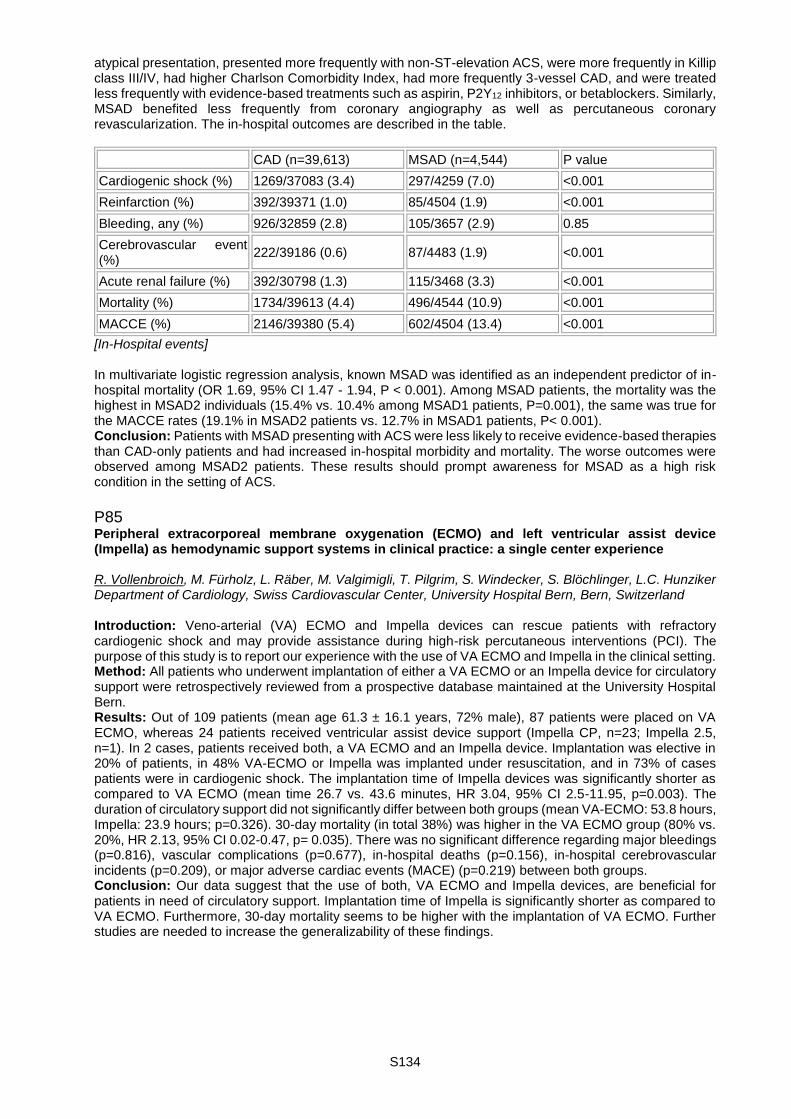
S134
atypical presentation, presented more frequently with non-ST-elevation ACS, were more frequently in Killip class III/IV, had higher Charlson Comorbidity Index, had more frequently 3-vessel CAD, and were treated less frequently with evidence-based treatments such as aspirin, P2Y12 inhibitors, or betablockers. Similarly, MSAD benefited less frequently from coronary angiography as well as percutaneous coronary revascularization. The in-hospital outcomes are described in the table.
CAD (n=39,613) MSAD (n=4,544) P value
Cardiogenic shock (%) 1269/37083 (3.4) 297/4259 (7.0) <0.001
Reinfarction (%) 392/39371 (1.0) 85/4504 (1.9) <0.001
Bleeding, any (%) 926/32859 (2.8) 105/3657 (2.9) 0.85
Cerebrovascular event (%)
222/39186 (0.6) 87/4483 (1.9) <0.001
Acute renal failure (%) 392/30798 (1.3) 115/3468 (3.3) <0.001
Mortality (%) 1734/39613 (4.4) 496/4544 (10.9) <0.001
MACCE (%) 2146/39380 (5.4) 602/4504 (13.4) <0.001
[In-Hospital events] In multivariate logistic regression analysis, known MSAD was identified as an independent predictor of in-hospital mortality (OR 1.69, 95% CI 1.47 - 1.94, P < 0.001). Among MSAD patients, the mortality was the highest in MSAD2 individuals (15.4% vs. 10.4% among MSAD1 patients, P=0.001), the same was true for the MACCE rates (19.1% in MSAD2 patients vs. 12.7% in MSAD1 patients, P< 0.001). Conclusion: Patients with MSAD presenting with ACS were less likely to receive evidence-based therapies than CAD-only patients and had increased in-hospital morbidity and mortality. The worse outcomes were observed among MSAD2 patients. These results should prompt awareness for MSAD as a high risk condition in the setting of ACS.
P85 Peripheral extracorporeal membrane oxygenation (ECMO) and left ventricular assist device (Impella) as hemodynamic support systems in clinical practice: a single center experience R. Vollenbroich, M. Fürholz, L. Räber, M. Valgimigli, T. Pilgrim, S. Windecker, S. Blöchlinger, L.C. Hunziker Department of Cardiology, Swiss Cardiovascular Center, University Hospital Bern, Bern, Switzerland Introduction: Veno-arterial (VA) ECMO and Impella devices can rescue patients with refractory cardiogenic shock and may provide assistance during high-risk percutaneous interventions (PCI). The purpose of this study is to report our experience with the use of VA ECMO and Impella in the clinical setting. Method: All patients who underwent implantation of either a VA ECMO or an Impella device for circulatory support were retrospectively reviewed from a prospective database maintained at the University Hospital Bern. Results: Out of 109 patients (mean age 61.3 ± 16.1 years, 72% male), 87 patients were placed on VA ECMO, whereas 24 patients received ventricular assist device support (Impella CP, n=23; Impella 2.5, n=1). In 2 cases, patients received both, a VA ECMO and an Impella device. Implantation was elective in 20% of patients, in 48% VA-ECMO or Impella was implanted under resuscitation, and in 73% of cases patients were in cardiogenic shock. The implantation time of Impella devices was significantly shorter as compared to VA ECMO (mean time 26.7 vs. 43.6 minutes, HR 3.04, 95% CI 2.5-11.95, p=0.003). The duration of circulatory support did not significantly differ between both groups (mean VA-ECMO: 53.8 hours, Impella: 23.9 hours; p=0.326). 30-day mortality (in total 38%) was higher in the VA ECMO group (80% vs. 20%, HR 2.13, 95% CI 0.02-0.47, p= 0.035). There was no significant difference regarding major bleedings (p=0.816), vascular complications (p=0.677), in-hospital deaths (p=0.156), in-hospital cerebrovascular incidents (p=0.209), or major adverse cardiac events (MACE) (p=0.219) between both groups. Conclusion: Our data suggest that the use of both, VA ECMO and Impella devices, are beneficial for patients in need of circulatory support. Implantation time of Impella is significantly shorter as compared to VA ECMO. Furthermore, 30-day mortality seems to be higher with the implantation of VA ECMO. Further studies are needed to increase the generalizability of these findings.
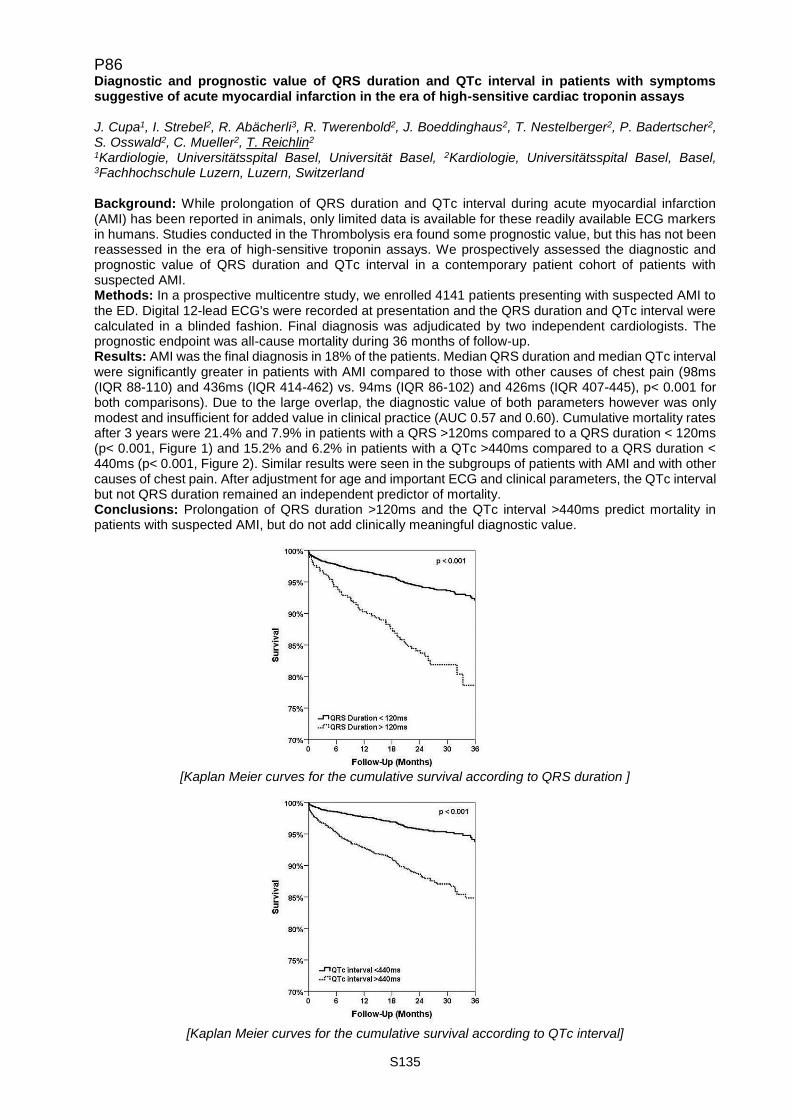
S135
P86 Diagnostic and prognostic value of QRS duration and QTc interval in patients with symptoms suggestive of acute myocardial infarction in the era of high-sensitive cardiac troponin assays J. Cupa1, I. Strebel2, R. Abächerli3, R. Twerenbold2, J. Boeddinghaus2, T. Nestelberger2, P. Badertscher2, S. Osswald2, C. Mueller2, T. Reichlin2 1Kardiologie, Universitätsspital Basel, Universität Basel, 2Kardiologie, Universitätsspital Basel, Basel, 3Fachhochschule Luzern, Luzern, Switzerland Background: While prolongation of QRS duration and QTc interval during acute myocardial infarction (AMI) has been reported in animals, only limited data is available for these readily available ECG markers in humans. Studies conducted in the Thrombolysis era found some prognostic value, but this has not been reassessed in the era of high-sensitive troponin assays. We prospectively assessed the diagnostic and prognostic value of QRS duration and QTc interval in a contemporary patient cohort of patients with suspected AMI. Methods: In a prospective multicentre study, we enrolled 4141 patients presenting with suspected AMI to the ED. Digital 12-lead ECG's were recorded at presentation and the QRS duration and QTc interval were calculated in a blinded fashion. Final diagnosis was adjudicated by two independent cardiologists. The prognostic endpoint was all-cause mortality during 36 months of follow-up. Results: AMI was the final diagnosis in 18% of the patients. Median QRS duration and median QTc interval were significantly greater in patients with AMI compared to those with other causes of chest pain (98ms (IQR 88-110) and 436ms (IQR 414-462) vs. 94ms (IQR 86-102) and 426ms (IQR 407-445), p< 0.001 for both comparisons). Due to the large overlap, the diagnostic value of both parameters however was only modest and insufficient for added value in clinical practice (AUC 0.57 and 0.60). Cumulative mortality rates after 3 years were 21.4% and 7.9% in patients with a QRS >120ms compared to a QRS duration < 120ms (p< 0.001, Figure 1) and 15.2% and 6.2% in patients with a QTc >440ms compared to a QRS duration < 440ms (p< 0.001, Figure 2). Similar results were seen in the subgroups of patients with AMI and with other causes of chest pain. After adjustment for age and important ECG and clinical parameters, the QTc interval but not QRS duration remained an independent predictor of mortality. Conclusions: Prolongation of QRS duration >120ms and the QTc interval >440ms predict mortality in patients with suspected AMI, but do not add clinically meaningful diagnostic value.
[Kaplan Meier curves for the cumulative survival according to QRS duration ]
[Kaplan Meier curves for the cumulative survival according to QTc interval]

S136
P87 Low rates of very late scaffold thrombosis with the Absorb bioresorbable vascular scaffold F. Cuculi, G. Hilfiker, S. Toggweiler, J. Chronis, M. Brinkert, P. Jamshidi, R. Kobza Herzzentrum | Kardiologie, Kantonsspital Luzern, Luzern, Switzerland Introduction: Recently published studies have raised serious concerns regarding the high rates of scaffold thrombosis (ScT) in patients treated with the Absorb bioresorbable vascular scaffold (BVS). Use of post-dilatation has been linked to reduced rates of ScT. We describe our long-term experience using aggressive pre-and post-dilatation when implanting Absorb BRS. Methods: Lesion preparation was performed using highly non-compliant balloons (Beo NC & OPN NC (Both SIS Medical, Frauenfeld, Switzerland). The OPN NC is a unique double-layer balloon with a rated burst pressure > 35 atm. After the publication of the GHOST-EU study, all scaffold were implanted using a new protocol, which included aggressive post-dilatation (> 30 atm with OPN NC). Probable and definitive rates of ScT are described according to current guidelines. The population is truly all-comers including patients with stable coronary artery disease (CAD), acute coronary syndrome (ACS), chronic total occlusions and calcified lesions. Results: Between 2013 and 2016 a total of 557 patients with coronary artery disease received Absorb at our institution. The mean age of the study population was 63+10 years. The indication for coronary revascularization was stable angina in 205 patients (37 %) and ACS in the rest of the patients (86 patients had STEMI). Mean follow-up was 496+256 days and 378 patients had a follow-up > 365 days. Scaffold thrombosis occurred in a total of 8 patients (1.4 %): 5 were early (< 30 days post PCI), 2 were late (between 30-365 days post PCI) and 1 patient had an acute (1 hour post PCI) and a very late ScT (603 days) post PCI. The incidence of ScT > 12 months post implantation was 0.3 % (n=1). Conclusions: Our results demonstrate that the Absorb BVS is a safe device when implanted using highly non-compliant balloons to pre- and post-dilate aggressively. ScT rates beyond 12 months were relatively low in our population and contradict the results of the Absorb-II trial.
P88 Ten Year follow-up after isolated Off-Pump CABG depending to syntax score S. Dushaj, H. Löblein, D. Odavic, A. Häussler, O. Dzemali, M. Genoni Cardiac Surgery, Stadtspital Triemli Zürich, Zürich, Switzerland Background and Purpose: Aim of this retrospective study was to evaluate association between the high preoperative SYNTAX score and mortality in isolated off-pump coronary bypass surgery (OPCAB). Methods: Consecutive 1805 patients undergoing primary isolated OPCAB from period of January 2005 to December 2014 were analyzed. SYNTAX Score was calculated and investigation groups were set as proposed by SYNTAX investigators. The influence of SYNTAX Score on in hospital and follow up mortality was evaluated. Results: Total 1805 patients were included, 16.1% were females (n=291) and mean age was 65.4±9.9% years. Preoperative risk factors were as follows; diabetes mellitus in 28.1% (n=507), peripheral vascular disease in 17.8% (n=322), smokers were 36.8% (n=665), COPD in 6.4% (n=116), previous MI in 33% (695), significant LMS in 43% (n=777). Preoperative mean SYNTAX Score was 31.4±9. According to the three groups were identified: in low SYNTAX score (0-22.5) were 17.1% (n=308) cases, in intermediate score (23-32.5) 40.7% (n=734) and high score (>33) were 42,3% (n=763) of cases. In-hospital mortality was 1.4% (n=26) and according to three groups it was: 0.16% in low (n=3), 0.5% (n=9) in intermediate and 0.77% (n=14) in high SYNTAX group. Average of distal anastomosis was 3,72. Perioperative infarction rate was at 1,66%. Mean follow up was 6.3±2.9 years. Overall survival at 5 years was 92.3±2.3% and at 10 years 84.3±3.3%. Comparing groups of patients according to Syntax Score groups, there were not a significantly difference on outcome. Conclusion: The Patients with higher SYNTAX Score was statistically not associated with a higher incidence of in-hospital or late mortality after primary off pump coronary artery bypass surgery. There was no significant higher incidence of re-PCI after primary off pump coronary artery bypass surgery in high Syntax-Score group.

S137
P89 Chest pain in an airplane R. Hedinger Institut für Notfallmedizin | Bildungsverantwortlicher | Experte in Notfallpflege HF and FCCM-Freelance Cabin Crew Member of Swiss International Air Lines, Universitätsspital Zürich, Zürich, Switzerland Introduction: When an in-flight medical emergency (IFME) during a commercial flight occurs, health care providers should be prepared to respond. Chest pain as an IFME is increasing as more passengers are up in the air, the bigger the aircrafts and especially elderly peassengers more often flying long distances. This session offers guidance to medical assistants how to respond to a IFME situation, for example chest pain on board, and to deal with the challenges caused by the in-flight environment. Methods: For this review, a comprehensive literature search was undertaken for "chest pain in an airplane" in MEDLINE and Google Scholar. Search terms were relevant to aviation IFME and flight physiology. Additionally, the review will include my personal experience as a cabin crew member with 2500 hours up in the air and over 800 landings. Results: At cruising altitude the cabin pressure is lower, causing an air expansion up to 30% with the result of less oxygen availability. This reduced partial pressure of oxygen should not affect the healthy passenger, but it may affect those with compromised cardiovascular, respiratory systems or blood disorders.Syncopes and pre-syncopes are relatively common medical events with 37-55%. Cardiac symptoms represent 8%, “respiratory symptoms” around 12% and cardiac arrest 0.3% of IFME on commercial airliners. Chest pain and cardiovascular events are frequently lead to flight diversions. Based on the review, I will give recommendation for the medical first response and will provide a decision guidance when a diversion should be considered. Assess vital signs, administer oxygen, nitroglycerin and aspirin. As well give a recommendation for a diversion in case of arrhythmia, abnormal vital signs or concern for myocardial infarction. In more than 80% of all flights, a physician or qualified medical professional was involved in providing therapy. The rolls and liabilities in offering medical assistance aboard an airplane are regulated over the US Aviation Medical Assistance Act of 1998 and by federal legislation of the airline. Conclusion: Inflight medical emergencies are a big challenge, given the specific enviroment of a aircraft cabin and the athmospheric condition. Nevertheless, chest pain as an IFME may be treated fast, safely and professionally. The legal aspects of volunteering to provide medical care is a well analyzed event and the airlines often offer additional protection to any medical assistance.
P90 Comparison of everolimus- and biolimus-eluting coronary stents with everolimus eluting bioresorbable vascular scaffolds - Two-year clinical outcomes of the EVERBIO II Trial D.A. Arroyo1, S. Schukraft1, Z. Kallinikou1, M. Togni1, O. Muller1, G. Baeriswyl1, J.-C. Stauffer1, J.-J. Goy2, S. Cook3, S. Puricel1 1Université & Hôpital Fribourg, Cardiologie, Fribourg, Switzerland, 2Université & Hôpital Fribourg, Cardiologie, Cairns, QLD, Australia, 3Cardiologie, Fribourg, Switzerland Background: Data from randomized controlled trials have shown that the ABSORB BVS is non-inferior to Cobalt Chromium everolimus-eluting stents at 2 years. We sought to provide the 2-year clinical outcomes of the randomized controlled EVERBIO-II trial. Methods and results: The EVERBIO II trial (Comparison of Everolimus- and Biolimus-Eluting Coronary Stents with Everolimus-Eluting Bioresorbable Vascular Scaffold) is a single-center, assessor-blind, randomized controlled trial enrolling 240 patients with an allocation ratio of 1:1:1 conducted at University and Hospital Fribourg, Switzerland. The studied devices were an everolimus-eluting persistent polymer stent (EES), a biolimus-eluting stent with bioabsorbable polymer (BES) and a fully bioresorbable vascular scaffold (BVS). Clinical end points collected at 9 months, 12 months, and 2 years, were academic research consortium defined composites, device thrombosis, and target-vessel revascularization. Clinical follow-up at 2 years was available in 96% (N=77) of patients in the EES group, in 100% (N=80) in the BES and 99% (N=77) in the BVS group. The device-oriented composite end point of cardiac death, target-vessel myocardial infarction and target-lesion revascularization occurred in 13 (16%) patients treated with EES, in 7 (9%) patients treated with BES and in 16 (21%) patients treated with BVS. There was no significant difference when the metallic stents were compared to the BVS (p=0.12). There was one late scaffold thrombosis throughout the trial in the BVS group, and no definite stent thrombosis in either EES or BES treated patients. Conclusions: The current analysis shows no differences with regard to clinical outcomes at 2 years between BVS and the best-in-class metallic DES. Event rates were numerically higher in BVS-treated patients.

S138
Author Index A Abächerli R. 284, P86 Adamopoulos D. 38 Adams A. P58 Adler A. 285 Adrian A. P43 Aeschbacher S. 114, 183, P13 Aghlmandi S. 218 Ahmadov K. P50, P76 Akdis D. 176, P07, P46 Akhmedov A. 137, 218, P14, P15 Alberio L. P18 Alibegovic J. 290 Alkadhi H. 287 Almagor Y. P41 Altomare C. 138, 141 Amini K. P34 Ammann P. 183, 254, P13 Anesini A. P18 Antiochos P. 289 Arnold M. 139, P21, P22, P23 Arroja J.D. 179 Arroyo D.A. P90 Artik E. P04 Asami M. 35 Asatryan B. 178 Attenhofer Jost C. 286, 57, P75 Attenhofer Jost C.H. 55, P28, P71 Aubort N. 31 Auer R. 115 Augereau C. 31 Aujesky D. 116 Aur S. 177 Auricchio A. P01, P02, P73 Aviano D. 34 Azabji-Kenfack M. P60 B Badertscher P. 120, 187, 212, 252, P86 Badini M. 34 Baeriswyl G. P90 Baldinger S. 178, 214, P12 Baldinger S.H. 176 Balmer C. P31 Banerjee P. 198 Banfi C. P50 Barbara H. P34 Barile L. 138, 141, P16, P19 Bärlocher L. 204 Barras N. 255, 289, P35, P40 Barthelmes J. 32 Bartkevics M. P24, P26 Bartoletti S. 182 Bastiaenen R. 251 Baumann F. 290 Beatty G. P09 Beck M.J. P26 Beer J.H. 116, P14 Behr E.R. 251 Behr R. P81 Beigelman Aubry C. P76 Benivady S. 217

S139
Berdajs D. 197 Bereuter L. P04 Berggren H. P25 Bernasconi R. 118 Berset S.G. P31 Bertel O. P79 Bhuiyan Z.A. 177 Biaggi P. P41 Biasco L. 34, P18, P44 Biemmi V. 138, 141, P16, P19 Bijlenga P. 140 Billinger M. 191 Bilz S. 254 Bimbai A.M. P60 Binder R. 290, 36, P38 Binder R.K. 190 Bisch L. P11 Blöchlinger S. 191, P62, P74, P85 Blum S. 118, 183, P13 Bochaton-Piallat M.-L. 140 Bode P. P07 Boeddinghaus J. 187, 220, P86 Boehnel C. P80 Bolis S. 138 Bonassin F. 286, 57, P71 Bonassin Tempesta F. 283, 55, P28, P75 Bonetti N. 114, P14, P15 Bonetti N.R. 116, 137 Boscolo Berto M. P44 Bossard M. 188 Bouchardy J. 283, 286 Boute E. 217 Brändle M. 254 Brandtner E.-M. 117, P54 Braun F. 285 Braunersreuther V. 140 Brecht R. P47 Breitenstein A. 181, 182, 215, P63 Bremerich J. P03 Brenner R. 254 Briand S. 137 Brinkert M. 192, 213, 37, P10, P41, P42, P87 Brokopp C. 218 Brugger N. P29 Brunckhorst C. 176, 181, 215, P07, P46 Brunner M. P69 Büchel R.R. 253 Buhmann R. 37 Burkard T. P53 Burri H. 176, 184, P05 Buser M. 54 Buttu A. P11, P66 C Cameron D. P25 Camici G.G. 114, 116, 137, 138, P14, P15, P19 Caporali E. 121, 189, 197 Caputo M.L. P01, P02, P73 Carballo D. 115, 218 Carballo S. 115 Carpentier A. 195 Carrel T. P23, P24, P25, P26 Carrel T.P. 139, P21, P22 Cassar M. 182 Castelletti S. 251, P36

S140
Cattaneo M. P37 Cerillo A. P41 Cervio E. 138, P16, P19 Cesarovic N. P27 Chiolero A. 113 Chow A. 182 Chronis J. 192, P87 Cikirikcioglu M. 195, P30, P65, P68, P70 Ciullo A. 138, 141, P16, P19 Cleuziou J. P25 Conen D. 114, 118, 119, 183, P13 Conte G. P01, P02, P73 Cook S. 188, 217, P90 Corniola M. 140 Corti R. P02, P41 Crisinel V. 255, 289, P35 Crucet M. P14 Cuculi F. 188, 192, 198, 37, P41, P42, P87 Cupa J. P86 D D'Ambrosio G. P01 D'Angelo G. 181 Dang L. P09 Dave H. P27 Dayal N. P05, P65 De Marchi S. P29, P51 Delay D. 199, 59, P72 Della Bella P. 181 Demertzis S. 121, 189, 197, P19 Denegri A. 190, 36 Denhaerynck K. 251, 31 Deuel J. P17 Di Candia M. 34 Di Valentino M. 183, P13 Diagbouga M. 140 Diakite M. P77, P78 Diaz Canestro C. 137, P15 Diaz-Canestro C. P14 Diethelm M. P67 Dillier R. 216 Djafarzadeh S. P23 Doerr O. 218 Doytcheva P. P17, P20 Drescher T. 254 Drexel H. 117, P54, P55, P56, P59 Du Fay de Lavallaz J. 120, 212 Dupuy N. 140 Duru F. 176, 181, 215, P07, P46 Dushaj S. 200, P88 Dzemali O. 196, 200, P39, P45, P49, P81, P88 E Eberhard M. 122, 287 Eberli F. P79 Eberli F.R. P84 Ebner J. 117, P54 Ebrahimi R. 180, P06 Eckstein F. 193, 60, P69, P82 Eckstein J. P53 Eggimann L. 119, 183 Ehl N.F. P67 Eigenmann V. P74 Ellenberger C. 38, P64, P65, P70 Emonet S. P70

S141
Engel R. 286 Engeli M. P20 Erb J.M. P69 Erb S. 60 Erhart L. 253, P48 Erne P. 183, 186, P02, P13, P79, P84 Eser P. P51, P52 F Faletra F. 34, P44 Fallesen C. P42 Farine E. P21 Fellmann F. 177 Ferrari E. 121, 142, 189, 197, 34 Filipovic M.G. 114 Flammer A. P34 Flammer A.J. 32 Fleischmann T. P27 Flores Widmer D. 120, 212 Fodor P. P81 Fodstad H. 177 Fontaine G.H. P07 Fornaro J. 37 Forni De Gottardi M. P37 Fragata J. P25 Franciosi G. 121, 189 François A. 195 Frank M. P32, P34 Franzeck F. 54 Franzini C. 216 Franzone A. 35 Frauenfelder T. 122, 287 Fraunberger P. 117, P54 Frei A. 38, P64 Fuchs T.A. 253 Fuhrer J. 178, 214, P12 Fürholz M. P85 G Gallino A. P37 Gallino J.M. P37 Gämperli O. 282 Garnier A. 255, P35 Gaxherri M. 289 Geiger K. 117 Gencer B. 115, 218 Genoni M. 196, 200, P39, P45, P49, P81, P88 Ghadri J.-R. 253 Giannakopoulos G. 179, P08, P64 Gilhofer T. 290, P38 Giraud R. P64, P65, P70 Giugliano R. P61 Gobbato S. 114, 116 Gondar R. 140 Good J.-M. 177 Gotschy A. 288 Goulouti E. 178, 214, P12 Goy J.-J. P90 Graf D. 31 Graf L. P80 Grapow M. 193, 60, P69, P82 Graves K. 200 Grego S. 121 Greutmann M. 253, 283, 286, 288, 55, 57, P28, P48, P71, P75 Gruner C. 253, 288, P48

S142
Gruszczynski M. 196, P39, P49, P81 Gulac P. P22 Gulan U. P46 H Haager P.K. 203, 204, 54 Hachulla A.-L. P64, P65 Hack D. 205 Haeberlin A. 178, 214, P04, P12 Haegeli L. 181, 215 Haeussler A. 200, P45 Hall M. 182 Hamann L. 122 Hamm C. 218 Hammer H. P48 Hasenclever P. P27 Hasse B. 203 Häussler A. 196, P39, P49, P81, P88 Hayoz D. 142 Hazekamp M. P25 Hedinger R. P89 Heg D. 115, 218, 35 Heiniger P.S. P01 Heinisch P.P. P24, P26, P47 Herzig D. P51, P52 Herzog B.A. 253 Hilfiker G. 192, P87 Hilker M. P41 Hoffelner L. 122 Hoffmann M. 203 Holm N. 216 Holy E. P32 Holy E.W. 122 Holzner M. P46 Honarbakhsh S. 182 Honarpour N. P61 Honegger U. 252, 284 Hraska V. P25 Huber C. 195, 58, P25, P30, P50, P65, P68, P70 Hübler M. P27 Hugelshofer S. 281 Hugli O. 255, P35 Hullin R. 142, 202, 255, P35, P40, P50 Hunter R. 182 Hunziker L. 191, P74 Hunziker L.C. P85 Hutter D. P24, P26 I Iglesias J.F. P79, P84 Inderbitzin D. 193, P82 Intorp S. P67 Isidor N. 140 J Jacoby D. 251 Jaegersberg M. 140 Jahren S. P29 Jamshidi P. 192, P87 Jeanrenaud X. 255, 289, P33, P35 Jeger R. 188 Jenni R. 253 Johner N. P08, P66 Jolou J. P30 Jörg L. 205

S143
Juerg H. B. 114 Julliard M.V. 184 Jüni P. 218 K Kadner A. P24, P25, P26 Kadoglou N. 251 Kaiser C. 188, 193 Kaiser L. P70 Kalangos A. 195 Kalla M. 182 Kallinikou Z. P90 Kandinger J. 204 Kapos I. 282, P48 Karagiannis A. 115, P83 Karbeyaz F. P43 Katte J.-C. N. P60 Kaufmann P.A. 253 Keech A. P61 Khoynezhad A. P47 Kiarostami P. 288 Kim W.K. P41 Kirsch M. 142, 199, 289, 59, P50, P72, P76 Kleimeyer K. P24 Klersy C. P02 Klingenberg R. 115, 218 Knecht S. 180, P03, P06 Knirsch W. P27 Kobza R. 188, 192, 198, 213, 37, 56, P10, P41, P42, P87 Koch W. 54 Kofler T. 188 Konetzka A. P27 Kontogeorgis A. 182 Koskinas K. P83 Koskinas K.C. 115, 191 Kostolny M. P25 Kozhuharov N. 33 Kozlowski M. P25 Kremer Hovinga J. P18 Krüger B. P27 Kueffer T. P04 Kühne M. 118, 119, 120, 180, 183, 185, 212, P03, P06, P13 Kullak Ubrick G.A. 137 Kwak B. 140 L Lador F. P78 Lam A. 178, 214, P12 Landmesser U. 218 Landolt F. P43 Lannoo M. P20 Lanz J. 35 Le Bloa M. P11 Leber R. 284 Lehmann S. 217 Leiherer A. 117, P54, P55, P56, P59 Lemay M. 285 Lemme F. P27 Leonardi L. P53 Licker M. P64, P65, P68 Licker M.-J. 38 Liebetrau C. 218 Lila N. 195 Limacher A. 116 Limoni C. P37

S144
Lins C. P55, P56, P59 Lipiski M. P27 Lloyd C. P41 Löblein H. 196, P49, P88 Loeblein H. P45 Lohrmann J. 120, 212 Longnus S. 139, P22, P23 Longnus S.L. P21 Luca A. P11 Ludovici V. 32 Luescher T.F. P15 Luscher T.F. P20 Lüscher T. 36, P17 Lüscher T.F. 114, 115, 116, 137, 176, 190, 218, 290, P14, P38 Lutz T. P17 Lutz T.A. P20 M Maadjhou C.B. P60 Mach F. 115, 218, P15, P61 Mader A. 117, P54, P55, P56, P59 Maeder M. P79, P80 Maeder M.T. 204, 254 Maisano F. 122, 203, 282, 287, 36, P43 Manka R. 288, 290 Manser S. 183 Marques-Vidal P.-M. 218 Marti Soler H. 218 Maruszewski B. P25 Masci P.G. 281 Maseli A. P53 Mastalerz M. 287 Matt P. 198, P42 Matter C. 218 Matter C.M. 115 Matter S. P52 Matter-Ensner S. P81 Mattila I. P25 Maziarski P. 184 McKenna W.J. 251, P36 Méan M. 116 Medeiros-Domingo A. 176, 178, 214, P12 Melina Langfritz M. 36 Mendez Carmona N. P21 Méndez Carmona N. 139, P22, P23 Merlini M. 137 Meyer P. 179, 184, P50, P65 Meyre P. 119, P13 Mfeukeu Kuate L. P60 Mikulicic F. 55, 57 Milano G. 141, P16, P19 Miranda M.X. 137 Mivelaz Y. 113 Moccetti F. 188 Moccetti M. 34, 36, P37, P44 Moccetti T. 138, 34, 36, P01, P02, P16, P18, P19, P43, P44, P73 Mogensen B. P42 Monney P. 255, 281, 289, P33, P35 Montecucco F. 218, P15 Montemurrro M. P57 Mootoosamy P. 195, 58, P30, P68 Morel S. 140 Moro C. 191, P83 Moschovitis A. 191 Moschovitis G. 183, P13

S145
Mueller C. 219, 220, 284, P86 Mueller D. 284 Mueller H. P64 Mueller X. P42 Muendlein A. 117, P54, P55, P56, P59 Muller H. P70 Muller O. 201, 289, P90 Müller A. 216 Müller C. 120, 187, 198, 212, 252, 33 Müller D. 116 Müller H. 38, P08 Müller X. 198 Muoio F. P16 Murith N. P65 Murray K. 33 Myers P. 58, P25, P30, P65 Mytsyk M. P69 N Nägele M.P. 32 Namdar M. P08, P65, P66 Nanchen D. 115, 218 Neagoe A. P62, P74 Nestelberger T. 120, 187, 212, P86 Nganou-Gnindjio C.N. P60 Nguyen-Kim T.D.L. 122, 287 Niclaus L. 199, 59, P72 Niclauss L. P57 Niederberger P. P21 Niederhauser T. P04 Niemann M. 288, P48 Nietlispach F. 122, 287, 36, P43 Nigolian A. P05 Nigolian H. P05 Nissen H. P42 Noble S. 38, 58, P64, P65, P78 Nosal M. P25 Noti F. 178, 214, P12 Notz L. 286 Ntinopoulos V. 196, P49 Nyffenegger T. P10 O Obeid S. 190, 282, 290, 36, P38, P43 Obrist D. P04 Odavic D. 196, 200, P39, P45, P49, P81, P88 Omeje I. P25 Osswald S. 118, 119, 120, 180, 183, 185, 187, 212, 219, 220, 252, 284, 33, P03, P06,
P13, P86 Osto E. P17, P20 Oxenius A. P71 Özkartal T. 288 P Padalino M. P25 Panella S. P 16 Panikker S. 182 Pantazis A. 251, P36 Pascale P. 201, P11 Pasotti E. 34, P44 Pavicevic J. 122 Pavlovic G. P68 Pedersen T. P61 Pedrazzini G. 186, 34, 36, P02, P18, P43, P79, P84 Pedrazzini G.B. P44

S146
Perenet S. P66 Perneger T. 195 Pernigoni N. 141, P16 Perrenoud S. 113 Perrin N. P64 Peterhans J. P52 Pfammatter J.-P. P26 Pfister R. 199, 59, P72 Pianezzi E. 138 Piazza N. 35 Pilgrim T. 191, 35, P83, P85 Pithon A. P11 Pizarro C. P25 Podonyi A. P27 Porretta A.P. P37 Pradella M. P03 Praz F. 191, 35 Prêtre G. 284 Prêtre R. 199, 59, P25, P31, P57, P72, P76 Proença M. 285 Prospero C. P25 Protopapas E. P25 Pruvot E. 201, P11 Puelacher C. 187, 252, 284 Puricel S. 217, P90 R Raccanello O. 31 Radovanovic D. 186, P79, P84 Räber L. 115, 191, 218, 35, P52, P83, P85 Rajappan K. 182 Raphael T. 187 Räsänen M. P83 Rebellius L. 282 Regamey J. 202, 255, P35, P40 Regoli F. P01, P02, P73 Reichlin T. 120, 180, 185, 187, 212, 252, 284, P03, P06, P86 Rein P. P56 Reinecke D. P62, P74 Reiner M. 116 Reiner M.F. 114 Reusser A. 183 Reuthebuch O. 193, 60, P82 Rexhaj E. 285 Rey F. P77, P78 Richards D. 31 Rickli H. 186, 203, 254, P67, P79, P80, P84 Riedel T. 285 Rigamonti F. 58, P77 Rigger J. 205 Rimoldi S.F. 285 Rinderknecht T. 284 Rings L. 200 Risch L. 114 Risch M. 114 Rizzo T. 216 Rodondi N. 115, 116, 218 Rodriguez H. P43 Roduit C. 197 Roffi M. 186, 38, P64, P77, P79, P84 Romanens M. P58 Rosset S. 201 Roten L. 178, 214, P11, P12 Rubini M. 219, 220 Rubini Giménez M. 187

S147
Rück A. P41, P42 Ruel M. 195 Ruschitzka F. 32 Russi I. 213, P10 Rüter F. 60 Rutz T. 281, 283, 286 S Sabatine M. P61 Sabti Z. 187, 252, 33 Saely C. 117, P54, P55, P56, P59 Saely R. P55 Saguner A. 215, P07, P46 Saguner A.M. 176 Said R. P48 Salamin P. 197 Salminen J. P25 Santos G. 31 Santos Lopes B. 283, 286, 55, 57, P71, P75 Sanz M.-N. P21, P23 Sarris G. P25 Sawhney V. 182 Schaer D. P17 Schaerli N. 284 Schäfer-Keller P. 31 Schaller A. 178 Schaller K. 140 Schär B. 180, 185, P06 Scharf C. P09 Schärli N. 252 Schaub Clerigue A. P15 Schefer T. 213 Schläpfer J. 177, 183, P13 Schmiady M. P27 Schmid J.-P. P51 Schoof P. P25 Schreiber C. P25 Schrenzel J. P70 Schukraft S. P90 Schuler A. P54, P55, P56, P59 Schussler O. 195 Schwaiger J. P63 Schweiger M. P27 Schwerzler P. 117, P54, P55, P56, P59 Schwerzmann M. 283, 286 Schwitter J. 202, 281 Schwitz F. 283, 286 Scott R. P61 Seifert B. 186, 287 Seiler C. P29 Seiler J. 178, 214, P12 Sekarski N. 113, 177 Servatius H. 178, 214, P12 Sever P. P61 Shah D. 179, 183, P08, P13 Shah D.C. P66 Shahin M. 190 Shahinian J. 60, P82 Shi X. P09 Shingo K. 282 Siegrist P. 290 Smadja D. 195 Sobngwi E. P60 Sojak V. P25 Solà J. 285

S148
Sologashvili T. P25, P30 Sortino R. 189 Sotiropoulos K. 56 Spies F. P03 Spiess F. 180, P06 Staehli B.E. 122 Stalder O. 116 Stämpfli S. 288 Stämpfli S.F. 253, P48 Stauffer J.-C. P90 Steffel J. 215, P63 Stella S. 181 Stellin G. P25 Stettler C. P05 Sticherling C. 180, 183, 185, P03, P06, P11, P13 Stieltjes B. P03 Stivala S. 114, 116, P14 Stocker D. 122 Stortecky S. 191, 35, P83 Straub A. P48 Strebel I. 252, 284, P86 Strömberg A. 31 Sudano I. 32, P58 Suetsch G. P43 Sulzer I. P18 Sürder D. P02, P43 Suter T. P62, P74 Sutter E. 140 Svab S. P01 Sweda R. 214, P12 Syburra T. 198, P42 Syrris P. 251, P36 T Taheri A. P20 Tallone T. P16 Tanner F. 282, 290, P32, P34 Tanner F.C. 122, 176, 253, 287, 288, P48 Tanner H. 178, 214, P04, P12 Taramasso M. 36, 54, P43 Temperli F. P83 Temple I. 182 Tevaearai Stahel H. P22, P23 Tevaearai Stahel H.T. 139, P21 Thiran J.-P. 285 Tlaskal T. P25 Tobler D. 283, 286 Toggweiler S. 188, 192, 198, 213, 37, P10, P41, P42, P87 Togni M. P90 Torre T. 121, 138, 189, 197, P19 Tozzi P. 142, P50 Trachsel L. P51, P52 Trevisi N. 181 Trunfio R. 121, 189, 197, 34 Twerenbold R. 219, 220, 284, P86 Tzimas G. 255, P35 U Ullah W. 182 Urban P. 186, P79, P84 Urbanet R. P20 V Valgimigli M. 191, 35, P85 Van Schouwenbourg Menalda T.P51

S149
Vassalli G. 138, 141, P16, P19 Vasserot K. 31 Vdovenko D. P15 Veien K.T. P42 Veit M. P24 Vella A. 289 Vesin J.-M. P11 Vida V. P25 Villeneuve H. 217, 31 Vincenti G. 202, 281 Vinet E. 255, P33, P35 Vischer A. 251, P36, P53 Vogt P. P40 Vollenbroich R. P85 Vollenweider P. 218 Von Eckardstein A. 116, 218 Von Schacky C. 114 Von Segesser L. 197 Vona M. 31 Vonbank A. 117, P55, P56, P59 Vricella L. P25 W Wagener M. 284 Wasserman S.M. P61 Weberndoerfer V. 213, P10 Weberndörfer V. 37 Weidt S. P28 Weilenmann D. 254 Weisheit V.T. P02 Wenaweser P. 35, P65 Widmer A. 60 Widmer L. P51 Wildbolz M. P26 Wilhelm M. 178, P51, P52 Windecker S. 115, 191, 218, 35, P83, P85 Winkler B. P26 Winterhalder C. P53 Wolber T. 215 Wolfrum C. P20 Wong T. 182 Woodford E. P25 Wustmann K. 283, 286 Wustmann K.B. P62, P74 Wyss R.K. 139, P21, P22, P23 Y Yakupoglu H.Y. 55, 57, P28 Yalcinkaya E. 215 Yamaji K. 191 Yerly P. 255, P35, P40 Yonekawa K. P48 Yoon J.-N. P32 Yousif N. 190 Z Zanchin C. 191, P83 Zanchin T. 191, P83 Zanolin D. 117, P55, P56, P59 Zanotto L. P25, P25 Zbinden R. 216 Zellweger M. 252, 284 Zhu M. P09 Zientara A. 196, P45, P49, P81 Ziltener J.-L. 184

S150
Zoller S. 218 Zuber M. P32 Zürcher F. P29 Zurek M. 286

ImpressumCardiovascular MedicineOfficial publication of the Swiss Society of Cardiology, the Swiss Society for Angiology, the Swiss Society of Hypertension and the Swiss Paediatric Cardiology Society.Contact: Gisela Wagner, CVM Editorial office, EMH Medical Publishers Ltd., Farnsburgerstrasse 8, 4132 Muttenz, Phone +41 (0)61 467 85 52, Fax +41 (0)61 467 85 56, [email protected], www.cardiovascmed.chOnline manuscript submission: http://www.edmgr.com/cvm
Publishing company: EMH Medical Publishers Ltd., Farnsburgerstrasse 8,4132 Muttenz, Phone +41 (0)61 467 85 55,Fax +41 (0)61 467 85 56, www.emh.ch
Marketing / Advertising: Dr. phil. II Karin Würz, Head of Marketing and Communication, Phone +41 (0)61 467 85 49, Fax +41 (0)61 467 85 56, [email protected]
Subscription: EMH Medical Publishers Ltd., Subscriptions, Farnsburgerstrasse 8, 4132 Muttenz, Tel. +41 (0)61 467 85 75, Fax +41 (0)61 467 85 76, [email protected] price (excl. postage): CHF 125.–, students CHF 63.–. Postage prices and single issues see www.sanp.ch
ISSN: print version: 1664–2031 / online version: 1662629X
Mode of publication: 11 issues per year.
© EMH Swiss Medical Publishers Ltd. (EMH), 2017. «Cardiovascular Medicine» is an open access publication of EMH. EMH Swiss Medical Publishers Ltd. grants to all users on the basis of the Creative Commons license «AttributionNonCommercialNoDerivatives 4.0 International» for an unlimited period the right to copy, distribute, display, and perform the work as well as to make it publicly available on condition that: (1) the work is clearly attributed to the author or licensor; (2) the work is not used for commercial purposes and (3) the work is not altered, transformed, or built upon. Any use of the work for commercial purposes needs the explicit prior authorisation of EMH on the basis of a written agreement.
Hinweis: Alle in dieser Zeitschrift publizierten Angaben wurden mit der grössten Sorgfalt überprüft. Die mit Verfassernamen gezeichneten Veröffentlichungen geben in erster Linie die Auffassung der Autoren und nicht zwangsläufig die Meinung der Redaktion von «Cardiovascular Medicine» wieder. Die angegebenen Dosierungen, Indikationen und Applikationsformen, vor allem von Neuzulassungen, sollten in jedem Fall mit den Fachinformationen der verwendeten Medikamente verglichen werden.
Production: Schwabe AG, Muttenz, www.schwabe.ch
Cover photo: © Rosshelen | Dreamstime.com