
ABSTRACTS - Carver College of Medicine - The University of ...
138
ABSTRACTS Medical Student Research Conference September 18-20, 2019 University of Iowa Roy J. & Lucille A. Carver College of Medicine Sponsored by: Medical Student Research Council Free to University of Iowa Students
-
Upload
khangminh22 -
Category
Documents
-
view
0 -
download
0
Transcript of ABSTRACTS - Carver College of Medicine - The University of ...
ABSTRACTS
Medical Student Research Conference
September 18-20, 2019
University of Iowa Roy J. & Lucille A. Carver College of Medicine
Sponsored by: Medical Student Research Council
Free to University of Iowa Students

Title: Lesion Localization of Post-Stroke Depression Student: Ossama Abu-Halawa, M2G Mentor: Aaron Boes, MD, PhD
Abstract: The neuroanatomical basis of depression remains poorly understood, yet this topic is of critical importance for understanding depression as a disorder and developing better treatments, such as identifying new targets for brain stimulation. Here, we investigate this question using the lesion method, a technique which associates the anatomical location of brain lesions with the functional consequences of the lesion. We leveraged lesion and behavioral data from stroke patients in the Iowa Neurological Patient Registry, the largest registry in the world of patients with focal acquired brain lesions and extensive neuropsychological testing. Using this dataset, we conducted multivariate lesion-symptom mapping using sparse canonical correlation analysis on 540 stroke patients in the Iowa Neurological Patient Registry, ranging from those with absent to mild, moderate and severe depression. We found no association between lesion location and depression severity. Based on the potential of clinical ambiguity introduced from mild depression cases, we conducted a second analysis that removed mild cases and compared subjects with no depression to those with moderate and severe levels. This showed that right insular lesions are associated with depression and right ventromedial prefrontal cortex lesions appear to reduce one’s risk of depression. While the preliminary results of the analysis are promising due to its concordance with multi-modal imaging studies on depression, important next steps in the analysis include cross-validation of our findings with an external dataset of post-stroke depression patients.
The Effects of Chronic Sleep Deprivation on Hippocampal Gene Expression
Hassan A. Ahamed1,2,3, Lin-Chun Li1, and Ted Abel1
Department of Molecular Physiology and Biophysics1, Department of Neuroscience2, University of Iowa
MSTP3
Introduction
Sleep deprivation and sleep loss are common issues effecting a multitude of people world-wide. It is
estimated that more than half of the world is not getting appropriate amounts of sleep on a day-to-day
basis. Fortunately, for some populations, sleep disturbances are only an acute issue; someone may lose
sleep for a certain amount of days but recovers with a good night’s rest eventually. In previous studies it
was shown, in mice, that these acute bouts of sleep deprivation can cause a transient change in
hippocampal gene expression, especially in the genes ARC and NR4A1 (these genes are early-immediate
genes associated with long-term potentiation and memory) and others. In the present study, we were
interested in the gene expression of ARC and NR4A1 in mice following a bout of sleep deprivation.
Methods
3-month-old adult mice were subjected to sleep deprivation for one week. This involved a device that
forced mice to stand on a platform that was submerged in water. When the mice lost muscle tone, they
would fall into the water and be forced to climb back up on the platform. This means that the mice were
not able to fall into REM sleep (when they would lose muscle tone and subsequently fall into the water).
After sleep deprivation, mice brains were then extracted and sectioned on cryostat. Following sectioning,
the mice brains were sectioned further for regions of interest within the hippocampus using LCM (all
three layers – oriens, pyramidal and radiatum of CA1, for this project). We were then able to run PCR on
our extracted RNA after stabilizing it as cDNA. We ran both dPCR and qPCR in order to confirm our
findings and fine-tune our methods.
Results
As well investigating gene expression, we were interested in tracking the mice’s weight over the week of
sleep deprivation as well as establish a protocol for cell-counting in the hippocampus. We discovered that
mice tend to lose weight as they are sleep deprived, an observation we did not expect. The sleep deprived
mice lost an average of 9.9% of their body weight and the control group gained an average of .5% of their
body weight, over the span of the 7 days (N = 10 for both groups). We also have started to finalize our
cell counting protocol in order to better understand how sleep deprivation effects cell numbers in the
different layers of the hippocampus. As for gene expression of ARC and NR4A1, we discovered that
these genes are just not expressed highly enough for us to be detecting them with qPCR and dPCR,
especially when we are looking at just one sub-section of the hippocampus.
Discussion
Although the genes of interest were not expressed in a way that we could detect them, we have gained
valuable insight as to what we need to do in the future in order to investigate the role of these genes in
sleep deprivation. This could mean potential gene amplification or looking at various parts within the
hippocampus where these genes may be expressed in a way that is detectable. We also want to further
investigate why it is that mice lose weight during sleep deprivation, where one would think the opposite
would happen. Finally, further looking into whether cell numbers in the hippocampus are influenced by
sleep deprivation is a direction we want to take in the future.
Title of presentation: Nut Consumption and Renal Function Among Women at High Risk Presenter name: Aparna S. Ajjarapu Mentor’s name: Cuilin Zhang, MD, PhD, MPH Collaborators Names: Stefanie N. Hinkle, PhD, Jing Wu, MD, Mengying Li, PhD, Shristi Rawal, PhD, Ellen C. Francis, MS, Liwei Chen, PhD, Georgia Pitsava, MD, Anne A. Bjerregaard, PhD, Louise G. Grunnet, PhD, Allan Vaag, MD, DMSc, Yeyi Zhu, PhD, Ronald CW Ma, FRCP, Peter Damm, MD, DMSc, James L. Mills, MD, MS, Sjurdur F. Olsen, PhD
Abstract: Background: Nut intake has been associated with reduced cardiometabolic risk, but few studies have examined its association with renal function. Objective: We examined associations between nut intake and renal function among women with previous gestational diabetes mellitus (GDM), a population with an increased risk for renal dysfunction. Design and Methods: This study included 607 women with a history of GDM who participated in the Diabetes & Women’s Health Study (2012-2014) follow-up clinical exam in Denmark. At the clinic, biospecimens were collected and habitual intake of nuts (9 types) in the past year was assessed using a food frequency questionnaire. A total of 330 women free of major chronic diseases were included in the analysis. Total nut intake was classified as none (≤1 serving/month), monthly (2-3 servings/month), weekly (1-6 servings/week), and daily (≥1 serving/day). One serving was defined as 28g. Renal function markers included estimated glomerular rate (eGFR) and urinary albumin-to-creatinine ratio (UACR), calculated based on plasma creatinine (mg/dL), and urinary albumin (mg/L), and creatinine (mg/dL) measurements, respectively. We estimated percent differences with 95% confidence intervals (CI) for each outcome by nut intake, adjusted for current body mass index, age, physical activity, energy intake, alcohol consumption, and vegetables intake. Results: We observed a non-linear association between total nut intake and UACR with lowest UACR values among women with weekly intake. Compared to women with weekly intake (n=222), the adjusted UACR values were higher by 86% [95% CI: 15%, 202%], 24% [-1%, 54%], and 117% [22%, 288%] among women with no (n=13), monthly (n=86), and daily (n=9) intake, respectively. Compared to weekly consumers, daily nut consumers also had 9% [0%, 19%] significantly higher eGFR values but eGFR values were similar among women with no and monthly intake. Conclusion: Moderate nut consumption may be beneficial to kidney health among women with prior GDM. Funding Sources: Intramural Research Program of the NICHD, March of Dimes Birth Defects Foundation, Innovation Fund Denmark, Health Foundation, Heart Foundation, EU, Danish Diabetes Academy supported by Novo Nordisk Foundation, and NIH Building Interdisciplinary Research Careers in Women's Health Program.
The Model Development, Validation and Refinement to Identify Anaphylaxis in Pediatric Patients Presenting to the Emergency Department
Authors: Kelsey Anderson, B.S.; Karisa K Harland, PhD, MPH; Sangil Lee, MD, MS
Background: The number and severity of anaphylaxis cases presenting to the Emergency
Department (ED) is increasing and can create a potentially difficult diagnosis. The objective of this study was to validate the known combinations of food allergy, angioedema, hoarseness, dyspnea and nausea with clinical information to estimate a concise model for anaphylaxis diagnosis among pediatric patients.
Purpose: The purpose of our study was to develop and validate a concise algorithm
meeting the diagnostic criteria for anaphylaxis in children presenting to the emergency department.
Methods: This study was a retrospective chart review of pediatric patients (0-18 years)
presenting to the ED at a large rural tertiary care center with the chief complaints of allergic reactions, food allergies, insect stings, medication reactions or anaphylaxis. Anaphylaxis was defined as a patient meeting any of the three National Institute of Allergy and Infectious Diseases/Food Allergy and Anaphylaxis Network (NIAID/FAAN) criteria. Data collected included past medical history (PMH), demographics, suspected allergen, symptoms, medications, ED management and follow-up. Differences among variables across anaphylaxis diagnosis were tested with the Pearson Chi-Square test for categorical variables and the Student’s t-test for continuous variables. The c-statistic for the predictive ability of the model was calculated using multivariable logistic regression.
Results: A total of 475 patients were included with 54% of the sample being male (n=259).
Almost one-third (n=139) of patients had a confirmed diagnosis of anaphylaxis in the ED. Of those, 15 (10.8%) had a PMH of angioedema (p <.01), 28 (20.1%) had a PMH of anaphylaxis (p < .0001), 27 (19.4%) had a PMH of asthma (p = .05) and 32 (23.0%) had a PMH of hives (p <.0001). Each of the variables for inclusion in the regression model, food allergy, angioedema (p <.01), dyspnea (p <.01), nausea (p <.0001) and hoarseness (p <.0001), were highly associated with anaphylaxis diagnosis. When combined in a regression model, these variables were highly predictive of an anaphylaxis diagnosis (c=0.87).
Conclusion: Our study indicated that the combination of food allergy,
angioedema, hoarseness, dyspnea and nausea are associated with anaphylaxis diagnosis. These findings can improve accuracy of diagnosis and improve outcomes.
Qualitative Investigation of Factors Associated with Successes and Challenges of Implementing a Therapeutic Lifestyle Intervention in Secondary Progressive Multiple Sclerosis. Andrea Arthofer, BA, Tyler Titcomb, PhD, RD, Nicole Grogan, MD, Paul Meirick, MD, Sandra Daack-Hirsch, PhD, RN, Terry Wahls, MD
Introduction. In recent years, there has been emerging emphasis on lifestyle modifications for multiple sclerosis (MS). A previous multimodal intervention involving diet; massage and meditation; and a fitness program of exercise, stretching, and electrical stimulation was associated with improved fatigue, anxiety, depression, and cognitive function. However, participants were not completely adherent to the study
protocol. This is characterized by self-reported group average adherence to diet and supplements 90%
and 75% adherence to exercise or electrical stimulation.
Purpose. This study aimed to identify factors preventing or promoting compliance to study components.
Methods. Study participants and family support persons were interviewed with open-ended questions about adopting the intervention. Interviews were recorded and transcribed by research team members. A thematic codebook was created and responses were, coded in NVivo. Response coding was verified by another research team member; disagreements were discussed and resolved. Remaining disagreements were brought to the primary investigator for final decision. After verification, themes mentioned as favorable or unfavorable to the implementation of each intervention of this therapeutic lifestyle were identified.
Results. 7 participants and 7 family members were interviewed after a median of 7 months (range 2-8 months) on the protocol. All participants were Caucasian females. Mean age was 52.9 ± 3.6 years with MS duration was 13.4 ± 7.2 years. Expanded Disability Status Score (EDSS) was 6.1 ± 0.2. Baseline mobility varied from no walking aid to walker. Education levels varied from High School Diploma to Master’s degree.
Barriers to adherence to the diet most mentioned by participants included time constraint, social settings, large portions, and taste preferences. Time constraint and social settings were also mentioned as barriers to the meditation and fitness program, along with a lack of understanding of how to implement the program. Barriers to massage included time constraint, social settings, lack of understanding, cost, and guilt, mentioned once each. Similarly, support persons also stated taste preferences, large portions, and social settings as barriers to the diet. However, family members also mentioned lack of understanding and cost as barriers to the diet. In addition, family members stated lack of understanding and time constraint as barriers to the fitness program, but not social settings. Time constraint was mentioned once as a barrier to massage and meditation. Lack of discipline was also mentioned once as a barrier to meditation. On the other hand, participants attributed successful diet adherence to family support, fitness program and meditation adherence to established routine, and massage adherence to hired support. Family members also mentioned family support as the most common reason for diet adherence, followed by taste preference and organization. Family support and knowledge of the program were common factors for success at following the program. Self-discipline, family support, hired support, and knowledge of the program were also mentioned once each as promoters of massage adherence. Likewise, understanding of the program and self-discipline were mentioned as promoting meditation adherence.
Hope for the future on a scale of 1-10 (1 = no hope; 10 = extremely hopeful) increased from 4.6 ± 1.8 prior to hearing of the study to 7.4 ± 2.0 at the time of interview (P = 0.067) for participants. Family member hope change was similar, increasing from 5.0 ± 1.7 to 7.5 ± 1.9 (P = 0.033).
Conclusion. This was a well-accepted intervention, exemplified by the largely effective adherence by highly fatigued individuals. However, this or a similar program could be enhanced in the future by implementing the following; increased interaction, defined by longer duration, increased phone calls from the study team, and communication with other participants; providing materials such as recipes, sample diets, pictures for e-stim placement, and a simplified guidelines chart; increased interactive as opposed to passive training incomponents of the fitness protocol and measuring serving sizes; and a simplified logging process consistingof less days of logging and inclusion of symptom changes. Additionally, increased family and social supportcould be achieved by educating designated support members on ways to provide meaningful support andaddressing ways to participate in social settings while being in compliance with the program.

Abstract-Sahaana ArumugamAn Analysis of A Water, Sanitation & Hygiene Intervention and Enteric Pathogen Detection in Kisumu, Kenya Mentor: Dr. Kelly Baker-Occupational and Environmental Health
Pediatric diarrheal disease is the second leading cause of infant mortality worldwide. This study aims to contribute to the understanding of the role of food in pathogen transmission in low & middle income countries to inform development of an infant food weaning hygiene intervention. Previous studies detected nucleic acids of enteric pathogens in 62% of infant weaning foods collected in Kisumu, Kenya, with cow’s milk being significantly more likely than other foods to both contain a pathogen and have a higher diversity of pathogens. The Safe Start Project utilized stool samples from infants collected prior to and after a standard WASH intervention over the course of months to ascertain the intensity of infections following episodes of diarrhea. Over 24 enteric pathogens of interest were part of the study. The Market to Mouth Project utilized food samples of cow’s milk from the same households as well as from vendors. The aim was to determine levels of food contamination differences between households and vendors. DNA and RNA were co-extracted from all samples before RT-qPCR. The data will be analyzed to determine levels of pathogen contamination and diversity in samples. Preamplification of food samples using TaqMan probes did not have the desired effect of lowering the threshold of detection, emphasizing the difficulties in detecting low concentrations of infectious agents in environmental samples.

Title: From Surviving to Thriving: How burn survivors succeed
Kimberly Dukes PhD, Stephanie Baldwin, BA, Evangelia Assimacopoulos, BS, Brian Grieve, BS, Lucy Wibbenmeyer MD
Introduction:Surviving a burn injury involves a complex healing process. Unfortunately, there is not a ‘one size fits all’ method for supporting survivors through their recovery, and survivors often have difficulty getting the support they need. In this study, we sought to identify factors that were influential in the recovery process for burn survivors, especially relating to barriers in obtaining support.
Methods:We conducted thematic analysis on transcripts of in-depth, semi-structured interviews with 11 burn survivors who had been treated at a Midwest tertiary facility. Survivors were purposefully selected for variability in age, gender, injury size, injury mechanism and quality of life responses. Interviews were recorded and transcribed verbatim. All interviews were coded by at least two authors. Coded results were entered into MAXQda, a qualitative data management software program.
Results:The mean age of the survivors interviewed was 51 years (35-63 years) and time from the injury was 5.4 years (2 months to 26 years). Their burn sizes ranged from<10% in 4 to 70-79% in one. Survivors acknowledged profound ongoing physical, emotional, and practical barriers to the “long process” of recovery, sometimes exacerbated by rural contexts. However, we found that complex processes of active coping, finding meaning and acceptance, and caring for others contributed to their resilience. During this process, survivors sought and benefited from many kinds of support (e.g. family, friends, providers, formal structured peer programs like BurnCamp or support groups, and informal or formal online networks), and from providing support to others (informally or formally, often burn-injury-related), including telling their stories. However, not all interviewees used the same support systems or used them at the same stage of recovery. Some interviewees indicated that support systems need to vary throughout the recovery period.
Conclusions:Survivors could benefit from a flexible set of options for participating in peer support networks as both beneficiaries and providers of support. These options should ideally be accessible in different locations, through different mechanisms (e.g. camp, face-to-face, web-facilitated), and at different stages of recovery, even years after the injury. This is important especially for inpatients who may not be ready to benefit from structured peer support opportunities that could become difficult to identify or access once they leave the hospital.
Applicability of Research to Practice:Providers can develop and communicate diverse support options and ensure that they are easily accessible to survivors, especially those in remote areas that may be years post-discharge.
External Funding:Summer Research Fellowship and University Research Office.
Impact of Surgeon's Choice of 1 vs. 2 Staged Revision ACL Reconstruction Based on Radiograph
Assessment of Preoperative Bone Tunnel Characteristics.
Brandon Bates, John Albright, MD (Mentor), Brian Wolf, MD, MS
Roy J. and Lucille A. Carver College of Medicine, University of Iowa
Department of Orthopaedics and Sports Medicine
Background: As the number of primary ACL reconstructions rises so too will the number of revision
ACL reconstructions (rACLR). A subset of patients will present for revision ACLR with pathologically
dilated bone tunnels. These dilated tunnel cases are managed by a single or two staged approach. The
question is which technique is superior in avoiding yet another re-revision ACLR. A recent systematic
review concluded there is an insufficiency of high-quality studies with enough cases to examine staged
rACLR. The Multi-center ACL Revision Study (MARS) consists of 83 surgeons performing rACLR on
1205 patients. This prospectively collected cohort provides 630 cases to attempt to provide clinically
significant comparisons between these groups at 6 year follow up.
Hypothesis: We hypothesize that the MARS cohort patients who underwent two staged rACLR with
bone grafting will have a lower incidence of subsequent rACLR operations compared to patients with
bone tunnel widths in excess of 14mm who underwent single staged rACLR with or without bone
grafting within the first six years.
Methods: Within the MARS cohort, 630 patients submitted radiographs of their knees. Tunnel width,
tunnel mal-position and graft impingement were measured using these radiographs. Revision graft failure
was defined as need for re-rACLR within 6 years of entering into the MARS study.
Results: Currently, 197 of the 630 radiographs have been measured. Unfortunately, we haven’t received
demographic and outcome information on these 197 patients from the MARS headquarters at Vanderbilt
University. Therefore, comparisons within this group is impossible. We could analyze the percent of
patients who possessed pathologically dilated bone tunnels (≥ 14 mm.). Of 29 patients who received bone
grafting at time of index rACLR, 17 patients (58%) had dilated bone tunnels. Out of 17 patients who
received a subsequent re-rACLR, 7 patients (41%) had dilated bone tunnels. Out of the remaining 151
patients’ radiographs examined, 64 patients (42%) had dilated bone tunnels. Additionally, we utilized
three medical students to calculate the inter- and intra-observer reliability of our bone tunnel width
measurements. The inter-observer, average measures intraclass correlational coefficient (ICC) ranged
from 0.813 to .949 while the intra-observer average measures ICC ranged from 0.831 to 0.978. We must
note that the inter-observer ICC could not be calculated for the femoral tunnel anteroposterior and sagittal
views due to too few of cases in which all three raters recognized a tunnel. For these measurements, the
kappa statistic was 0.533 and 0.246 respectively.
Conclusion: We have determined that measuring bone tunnel widths with radiographs is reliable. Of the
197 patients reviewed, 88 patients (44.7%) had bone tunnels measure in excess of 14 mm. 53 of these 88
patients had a bone tunnel measure in excess of 16 mm, 21 of these 88 had a bone tunnel measure in
excess of 18 mm, and 8 out of these 88 had a bone tunnel measure in excess of 20 mm. Once we receive
the remaining radiographs, demographic information and outcome measure we will be able to determine
if patients with dilated bone tunnels who receive single staged or two staged rACLR are less likely to
endure a subsequent re-revision ACL reconstruction.
EricBertrocheMalyndaWynn, MDCassimIgram, MD
AreComputedTomographyAngiogramsNecessaryforallBluntCervicalSpineInjuries?
IntroductionComputedtomographyangiograms(CTA)oftheneckareroutinelyorderedintheinitialwork-upforbluntcervicalspineinjuries,regardlessofmechanismofinjury,pastmedicalhistoryofthepatient,orinjurypatterninordertoassessforvertebralarteryinjury(VAI).Currentopinionsregardingtraumawork-upprotocolsdifferbetweengeneraltraumaandorthopedicspineliterature.Ononehand,CTA’sperformedonallbluntcervicalspineinjuriesensuresaVAIisnotmissed.Ifdetected,secondarystrokeriskisdecreasedwithinitiationofanti-platelettherapy.However,notallinjuriesarecreatedequal,andoftenpatientsarealreadyreceivinganti-platelettherapy,orhavealowenergymechanismorfracturethatwouldhavealowprobabilityofVAI.GiventhepotentialcostsoutweighingthebenefitsofaCTAforsomepatients,amoreindividualizedapproachtobluntcervicalspineinjuriesiswarranted.
PurposeofthestudyWesoughttoidentifythosepatientsatmostriskforVAIandwouldbenefitmostfromCTAduringtheirinitialwork-up.BasedonobservationofpriorpatientswehypothesizethatpatientswhosustainbluntcervicalspineinjuriesfromlowenergymechanismswillhavealowerincidenceofVAIthanhighenergymechanisms.WealsoanticipatethatasignificantnumberofpatientswhoreceiveaCTAandhaveanidentifiedVAIwillhaveacontraindicationtoanti-platelettherapy,oralreadyreceivinganti-platelettherapyforapre-existingcondition.MethodsThisisanIRBapprovedretrospectivecohortreviewofpatientswhosustainedbluntcervicalspineinjuriesfromJanuary2014toDecember2018.PatientwereincludediftheypresentedtotheemergencydepartmentduringthespecifiedtimeframeandhadacervicalspineCT.Patientswereexcludediftheywerelessthan18yearsofage,werefoundtohaveonlychronicinjuriesoneimaging,andifnoCTwasavailableorreview.Patientdemographicsincludedpatient’sage,dateofinjury,mechanismofinjury.Detailssurroundingpatient’sinjuryincludedtypeofcervicalspineinjury,locationincervicalspine,associatedthoracicandlumbarspineinjury,ifpatientreceivedCTA,ifpatientwastakinganti-platelettherapypriortoinjury,associatedkidneydiseaseandfracturecharacteristics.LogisticregressionwasthenperformedtodetermineriskfactorsassociatedwithVAI.
ResultsTherewasatotalof300patientsthatmetinclusioncriteria,with64.2%male,and79.2%werepatientstransferredfromoutsidehospitals.Associatedthoracicandlumbarspineinjuriesoccurredin25.7%and9%ofpatients,respectively.MotorvehicleaccidentsandfallfromheightshadthehighestassociatedVAI.ContrastloadfromtheCTAresultedinacutekidneyinjuryin10.7%ofpatients.Atotalof21.3%ofpatientswhounderwentCTAhadacontra-indicationtoanti-platelettherapy,and19.5%ofpatientswhounderwentCTAwerealreadyreceivinganti-platelettherapypriortoadmissionforapreviousmedicalcondition. There were greater odds of having a VAI in multi-level cervical spine injuries (OR 3.25, p=0.03) and fractures through the transverse foramen (OR 4.37, p=0.0035).
Conclusion
Those patients with multi-level cervical spine injuries and involvement of transverse foramen have the highest associated with VAI. These patients would likely benefit most from additional CTA imaging. When possible, patient history of anti-platelet therapy could present un-necessary ordering of CTA studies in those isolated cervical spine injuries as a result of low energy mechanisms. This further raises the discussion of treating blunt cervical injuries with prophylactic anti-platelet therapy to save patients cost and contrast load if existing kidney disease exists.
Background
Mood disorders have been associated with a variety of cardiovascular disease risk factors, including
inflammation and large artery stiffness, particularly while depressed although longitudinal studies have
been limited.
Methods
With measurements at baseline and 8 weeks, we prospectively assessed mood, levels of inflammatory
markers (hsCRP and TNF-α), serum lipids, and large artery stiffness in a cohort of 26 participants with a
diagnosis of a mood disorder, enriched for current depression. Depressive symptoms were measured
using the Montgomery Åsberg Depression Rating Scale (MADRS) at baseline and 8 weeks. Associations
between depressive symptoms and other measures were assessed using linear mixed models, unadjusted
and adjusted for age and BMI.
Results
Participants (n=26) were a mean (SD) age of 41.6 (12.8) years old and 81% female. During the study,
there was a mean (SD) MADRS score improvement of 9.5 (9.4) from baseline to eight weeks. Reductions
in our primary outcome TNF-α with improvement in depression fell short of significance (P=0.08). In
secondary analyses, there was a statistically significant association between improved cholesterol ratio
(P=0.04) and triglycerides (P=0.04) with depression improvement. There was no statistically significant
change in large artery stiffness during the study.
Conclusion
Improved depressive symptoms were associated with improved cholesterol ratios even after adjustment,
suggesting possible mechanism by which acute mood states may influence cardiovascular disease risk.
Future longitudinal studies with extended and intensive follow-up investigating cardiovascular disease
risk related to acute changes and persistence of mood symptoms is warranted.
Jonathan Birdsall
Jess Fiedorowicz
Background
Mood disorders have been associated with a variety of cardiovascular disease risk factors, including
inflammation and large artery stiffness, particularly while depressed although longitudinal studies have
been limited.
Methods
With measurements at baseline and 8 weeks, we prospectively assessed mood, levels of inflammatory
markers (hsCRP and TNF-α), serum lipids, and large artery stiffness in a cohort of 26 participants with a
diagnosis of a mood disorder, enriched for current depression. Depressive symptoms were measured
using the Montgomery Åsberg Depression Rating Scale (MADRS) at baseline and 8 weeks. Associations
between depressive symptoms and other measures were assessed using linear mixed models, unadjusted
and adjusted for age and BMI.
Results
Participants (n=26) were a mean (SD) age of 41.6 (12.8) years old and 81% female. During the study,
there was a mean (SD) MADRS score improvement of 9.5 (9.4) from baseline to eight weeks.
Reductions in our primary outcome TNF-α with improvement in depression fell short of significance
(P=0.08). In secondary analyses, there was a statistically significant association between improved
cholesterol ratio
(P=0.04) and triglycerides (P=0.04) with depression improvement. There was no statistically significant
change in large artery stiffness during the study.
Conclusion
Improved depressive symptoms were associated with improved cholesterol ratios even after adjustment,
suggesting possible mechanism by which acute mood states may influence cardiovascular disease risk.
Future longitudinal studies with extended and intensive follow-up investigating cardiovascular disease
risk related to acute changes and persistence of mood symptoms is warranted.
Immunosuppression: Associations with Cervical Cancer Demographic Characteristics
Hannah Botkin, BS, Alison Hefel, BS, Colette Gnade, MD, Abbey Hardy-Fairbanks, MD, Colleen Stockdale, MS, MD Department of OB/GYN, UIHC
Background: Immune suppression, in its many forms, increases the risk of a myriad of other medical conditions. Immunosuppressed patients may be at an increased risk for some types of cancer, and current United States Preventative Service Task Force (UPSTF) screening guidelines for cervical cancer do not apply to these individuals. Despite these recommendations, it has been well recognized that immune suppressive conditions increase the risks of cervical dysplasia and cancer, including but not limited to HIV, AIDS, autoimmune conditions, and chronic steroid use.
Aims/Hypothesis: This study aims to understand the association of immunosuppression and cervical cancer prevalence and presentation. We predict that immunosuppressed women are at greater risk for cervical cancer, and that the cancer is more advanced when detected in this population when compared with immunocompetent patients.
Methods: This retrospective cohort study consisted of cervical cancer patients treated at the University of Iowa Hospitals and Clinics from 1986 through 2018. This data set consists of 1788 total patients, with 227 classified as immunosuppressed. The results are further divided into types of immunosuppression, including: various autoimmune diagnoses, HIV, genetic immunosuppression, hepatitis, or other. Demographics, as well as information about pathology, treatment, and recurrence was also collected about each patient and included in the data analysis. Methods of cervical cancer diagnosis were divided into those who were diagnosed based on screening pap smear, those who presented with symptoms, and those whose cancer was found on physical exam.
Results: 18.2% (n=227) of subjects were classified as immunosuppressed. The most common forms of immunosuppression were diabetes and hypothyroidism, which comprised 6.7% (n=84) and 5.3% (n=66) of our immunosuppressed patients, respectively. Immune suppressed individuals were significantly older at the time of their cervical cancer diagnosis (52.6 vs 45.9, p=0.000). Immune status did not have a significant correlation with method of diagnosis, specifically diagnosis via pap smear screening. There was also no association with more advanced stage at diagnosis or smoking.
Discussion: Because immune suppression increases with age, the association found may be due to that association. This may mean that screening in immune suppressed individuals may need to extend beyond the current recommendation of age 65. More data is needed on the diagnosis of cervical cancer in older age groups to inform future guidelines for screening.
Triggering receptor expressed on myeloid cells-1 regulates many aspects of neutrophil function
Jayden Bowen, Kathy Keck, Sankar Baruah, Shubha Murthy, Julia Klesney-Tait
Introduction: Triggering receptor expressed on myeloid cells-1 (TREM-1) is a cell-surface receptor that amplifies pro-inflammatory signaling in myeloid cells. Previously, our lab has found that TREM-/- neutrophils undergo decreased migration in the lung due to a deficiency in reactive oxygen species production. Microvesicles are small signaling bodies derived from cells that include exosomes, microparticles, and apoptotic bodies. A role for neutrophil-derived microparticles has been described in scenarios such as neutrophil migration, inflammatory and anti-inflammatory signaling, asthma, and cancer metastasis. Purpose: We set forth to further investigate the role of TREM-1 in the basic biologic function of neutrophils, particularly in microvesicle release. We hypothesized that TREM-1 deficiency would lead to decreased micro vesicle production when stimulated, and that TREM-1 deficient microvesicles would be less pro-inflammatory than wild-type microvesicles. Methods: Neutrophils were isolated from bone marrow of WT or TREM-1-/- C57Bl/6 mice by negative magnetic selection. Microparticle isolation: Neutrophils were stimulated for 30 minutes at 37C with a pro-inflammatory stimulant such as fMLP or LPS. The cells were then centrifuged for 5 minutes at 600g. The supernatant was removed and centrifuged at 13,000g for 10 minutes. The supernatant was once more removed and centrifuged at 100,000g for 1 hour. The pelleted microvesicles were then frozen at -80C until analyzed. Western Blot: Isolated microvesicles were lysed in 1% SDS and boiled for 5 minutes before the addition of beta-mercaptoethanol. Protein quantification was performed by Bradford assay. Samples were then analyzed by standard SDS-PAGE and immunoblotting techniques with the relevant antibodies. Flow Cytometry: Cells were stained per manufacturer’s recommendations with fluorophore conjugated antibodies before and after stimulation as above and then analyzed with a BD LSRII. Results: Microvesicles derived from TREM-1-/- neutrophils expressed matrix metalloproteinases, markers of inflammation, and cell adhesion proteins at varying levels compared to WT-derived microvesicles. Specifically, the metalloprotease MMP9 was found to be consistently upregulated in microvesicles after neutrophil stimulation, with increased levels in KO vs WT microvesicles. Similarly, the integrin CD11b was found to be upregulated in KO microvesicles compared to WT after stimulation. As has been previously reported, CD11b was upregulated on stimulated neutrophils of both genotypes, but contrary to our previous findings, CD11b appeared to expressed at higher levels on KO neutrophils. Discussion: A role for TREM-1 in microvesicle shedding has not been previously described. The observed changes in microvesicle loading suggest that downstream signaling of TREM-1 is involved in protein sorting to microvesicles. Contrary to our hypothesis, the microvesicle fraction from TREM-1-/- neutrophils displayed higher levels of MMP-9 and CD11b, suggesting that while TREM deficiency may be overall anti-inflammatory, disordered microvesicle production may skew towards an inflammatory profile. Future directions include quantification and sizing of microvesicles, as well as further phenotypic analysis of microvesicles by flow cytometry.
Anterior Inferior Iliac Spine Deformities: Incidence and Associations
Student: Nathan Cao, M1
Mentor: Kyle Duchman, MD – Department of Orthopedics and Rehabilitation
Co-Mentors: Robert Westermann, MD – Department of Orthopedics and Rehabilitation
Abstract:
Background/Purpose: Recent studies have suggested that subtle morphological differences of the femoral head and
acetabulum may lead to hip and groin pain and possibly hip osteoarthritis (OA). Femoroacetabular impingement
(FAI) syndrome is a condition that is characterized by abnormal contact between the acetabulum and femoral head,
which arises from subtle morphological differences of one or both of these structures. FAI syndrome has been
accepted as a frequent cause of hip pain and dysfunction in patients over a broad age range. In this study, we
investigated the relationship between a specific variant of extra-articular FAI syndrome, namely anterior inferior
iliac spine (AIIS) morphology, and how AIIS morphology relates to acetabular volume and version.
Methods: The study was conducted using cadaveric skeletons from the UI-Stanford Bone collection with full
documentation of common demographic characteristics, including sex, age at death, race, and in most cases,
occupation. AIIS morphology was categorized as Type I, II, or III as previously described. Acetabular volume was
approximated by the equation for a half-ellipsoid as previously described. To measure acetabular version, the
pelvises were reconstructed using rubber bands and 2.5cm thick foam to represent the pubic symphysis. The pelvises
were then placed on a flat surface with the anterior superior iliac spines (ASIS) and pubic symphysis as the points of
contact, allowing for the establishment of the anatomic frontal plane. Three separate axial measurements of version
were taken using a goniometer; the cranial measurement taken 5mm distal to the acetabular roof, central
measurement taken at the diameter of the acetabulum, and the caudal measurement 5mm proximal to the inferior
edge of the acetabular rim. Global version was calculated as the mean of these three measurements. Descriptive
statistics were performed, and demographic and morphological characteristics compared using Student’s t-test and
chi-square analyses for continuous and categorical variables, respectively. Findings were considered statistically
significant with p-values <0.05.
Results: Of the 72 hips reviewed, 9 (12.5%) had Type 1 AIIS morphology, 44 (61.1%) Type 2, and 19 (26.4%)
Type 3. Global acetabular version measurements taken from right hip bones were on average more anteverted than
left hip bones (13.9°±6.7° vs. 18.9°±11.0°; p <0.001). Cranial acetabular version measurements taken from right hip
bones were on average more anteverted than left hip bones (8°±8.83° vs. 21.58°±13.75°; p-value <0.001).
Acetabular volume on the left hip bones was on average smaller than right hip bones (45.69 cm3±11.33 cm
3 vs.
47.49 cm3±11.28 cm
3; p = 0.016). When comparing acetabular volume and version between the three AIIS
morphological subtypes, there were no significant differences noted.
Conclusion/Discussion: Only 9 of 72 specimens exhibited Type 1 morphology, which has previously been
described as the most common AIIS subtype. These differences may be the result of variable activities that may
influence AIIS morphology, as the majority of specimens were from persons classified as manual laborers, or
increasing age. Although global version measurements are often used to predict or evaluate FAI, the cranial version
measurement can also provide valuable insight. This is due to the fact that cam-type FAI typically occur in the
anterosuperior aspect of the acetabulum, with cranial retroversion being thought of as a good predictor of developing
cam-type FAI. Conversely, anteversion has been associated with hip dysplasia. Acetabular retroversion reflects
overcoverage of the femoral head anteriorly and undercoverage posteriorly. This may have the effect of decreasing
the right hip’s susceptibility to cam-type FAI, while increasing the chances of hip dysplasia. The data would also
suggest that left hips may be more susceptible to cam-type FAI, but less susceptible to hip dysplasia. However,
previous literature has not found that FAI is more common on one side than the other. Acetabular volume was
shown to be larger on right hips than left, pointing at a possible relationship between larger acetabuli and increases
in acetabular anteversion. Although information on handedness was not documented in these specimens, acetabular
versions measurements being more anteverted on right hips may also be the result of preferential use of the right leg
over the left. Future studies may aim to repeat this study using a more diverse cohort of skeletal cadavers from
individuals from the 21st century to see if data on prevalence and associations differ drastically from those found in
this study. An ideal cohort would include more females, more individuals from ethnic populations, extensive
documentation of occupation, and handedness of patients.
Clinical Factors that Affect the Cumulative Live Birth Rate from an IVF Cycle Emily Capper, M2 Karen Summers, MPH, Patrick Ten Eyck, Ph.D., Rachel Mejia, DO, Brad Van Voorhis, MD DepartmentofObstetricsandGynecology, Reproductive Endocrinology and Inferility Division
Background and Introduction IVF is the most effective treatment for infertility but is complicated by a high rate of multiple births due to the common practice of transferring multiple embryos back to the uterus. Elective single embryo transfer (eSET), defined as the transfer of a single embryo to the uterus with cryopreservation of all other high-quality embryos from the cycle for future use as needed, has been shown to be effective in dramatically reducing the multiple birth rate when compared to multiple embryo transfer. eSET is gaining favor among IVF physicians as we strive to reduce medical complications and high healthcare costs associated with multiple gestation pregnancies. However, previous studies have demonstrated a lower live birth rate per embryo transfer which can make this option less appealing to patients. There have been no prior studies evaluating the effect of eSET on the cumulative live birth rate, which incorporates the outcome from all fresh and frozen-thawed embryos transfers from a single IVF egg retrieval. We have access to a national database that links outcomes for all embryos from a given IVF egg retrieval and can utilize this data to determine the effect of eSET on the cumulative live birth rate which we believe is the best measure of the effectiveness of eSET.
Hypothesis eSET will improve the cumulative live birth rate from a single IVF retrieval and will lower the multifetal pregnancy rate significantly.
Purpose This study investigates if there are significant differences in cumulative live birth rate, multifetal pregnancy rate and time to live birth when eSET versus double embryo transfer (DET) are used. This study also investigates factors that affect the cumulative live birth rate and multifetal pregnancy rate from a single IVF retrieval.
Methods We analyzed primary IVF clinic data collected by the National Society for Assisted Reproductive Technology (SART) from patients aged 21 to 45 who had their first IVF egg retrieval between January 2014 and December 2015. We excluded patients who had oocyte or embryo banking, preimplantation genetic testing of embryos, or previous fresh or frozen IVF cycles. We linked all fresh and frozen cycles that resulted from a single egg retrieval to determine the cumulative live birth rate and multiple gestation rate. Patients were censored following a live birth and embryo transfers occurring after December 2016. Cumulative live birth rates were calculated using linked subsequent fresh-frozen transfers. Generalized linear mixed models were used to assess the impact of clinical and demographic factors on cumulative live birth rate and multifetal pregnancy rate. For each outcome variable, models of all possible predictor subsets were fit, and the top model was selected using the Bayesian information criterion (BIC). Statistical significance of any differences found was tested using a multivariable regression model accounting for differences in important clinical and demographic variables between populations studied. The study was approved by the institutional review board at the University of Iowa.
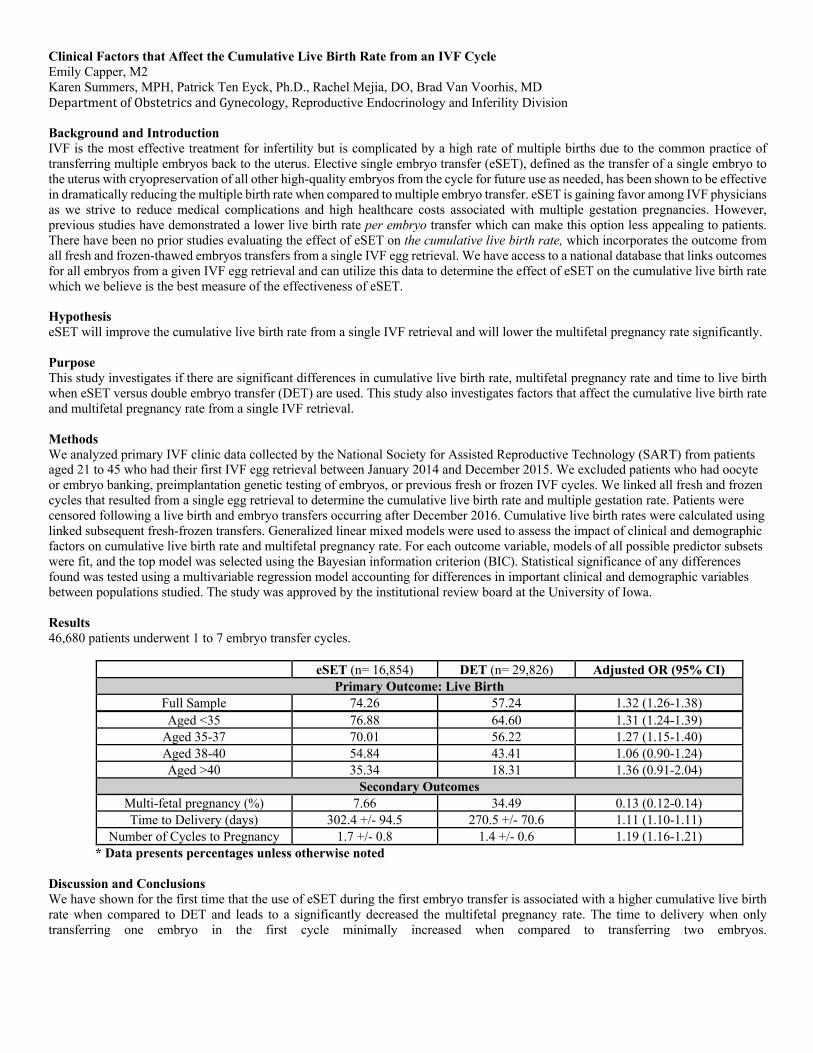
Results 46,680 patients underwent 1 to 7 embryo transfer cycles.
eSET (n= 16,854) DET (n= 29,826) Adjusted OR (95% CI) Primary Outcome: Live Birth
Full Sample 74.26 57.24 1.32 (1.26-1.38) Aged <35 76.88 64.60 1.31 (1.24-1.39)
Aged 35-37 70.01 56.22 1.27 (1.15-1.40) Aged 38-40 54.84 43.41 1.06 (0.90-1.24) Aged >40 35.34 18.31 1.36 (0.91-2.04)
Secondary Outcomes Multi-fetal pregnancy (%) 7.66 34.49 0.13 (0.12-0.14) Time to Delivery (days) 302.4 +/- 94.5 270.5 +/- 70.6 1.11 (1.10-1.11)
Number of Cycles to Pregnancy 1.7 +/- 0.8 1.4 +/- 0.6 1.19 (1.16-1.21) * Data presents percentages unless otherwise noted
Discussion and Conclusions We have shown for the first time that the use of eSET during the first embryo transfer is associated with a higher cumulative live birth rate when compared to DET and leads to a significantly decreased the multifetal pregnancy rate. The time to delivery when only transferring one embryo in the first cycle minimally increased when compared to transferring two embryos.
Evaluationofsecondarybonecontusionpatternsbypre-operativeMRIintheACLcompetentpatientthatmaypredictthepresenceofanMRIoccultmeniscaltear.
JosephCarmody,Dr.DarusLeeBennett,Dr.KenjirouOhashi,Dr.MikhaelSebaaly
Background:MRIisoftenusedtodiagnosemeniscaltearsinthesettingofkneeinjury.Despitetechnologicalimprovements,detectionofmeniscaltearsremainssuboptimal,especiallyfortearsthataresmallorassociatedwiththeposteriorhorn(DeSmet2008).Inabout10%ofcasesinvolvingmeniscaltears,MRIfindingsareambiguous(Justice1995,DeSmet1994).Oneapproachtoimprovingdetectionofoccultmeniscaltearsisthroughtheidentificationofsecondarysignsofmeniscalinjuries.Forexample,alterationsofsignalintensityinadjacenttissueorbonemarrowcanincreasediagnosticconfidence(Bergin2008).Previousstudieshaveidentifiedindirectsignsofmeniscaltears,includingMCLbowingandedema,jointeffusion,meniscalextrusion,parameniscalcysts,cartilagelossaswellaslinearandnonlinearsubchondraledema(Tasker1995,Bergin2008).However,mostpreviousstudieshaveexploredcharacteristicsofmeniscaltearsandMRIoccultmeniscaltearsaccompanyingdamagetotheACL(DeSmet1994,Laundre2009).CharacteristicsofmeniscaltearsandMRIoccultmeniscaltearsthatoccurindependentofACLinjuriescouldbeexploredfurthertoaiddiagnosticconfidenceformeniscaltearsinACLcompetentknees.
Objective:CharacterizethetypesofMRIoccultmeniscaltears,locationofMRIoccultmeniscaltears,andbonecontusionpatternsassociatedwithMRIoccultmeniscaltearsinACLcompetentkneeinjuries.MaterialsandMethods:8,863kneeMRIreportswerereviewedforpostMRIsurgicaldiagnosisofmeniscaltear.961patientsunderwentarthroscopicsurgerywithin3monthsofMRIandhadsurgicallydiagnosedmeniscaltearswithcompetentACLs.TheMRIrecordswerethenreviewedforteartype,location,discoidmeniscus,bonecontusionpatternsandcorrelatedwitharthroscopicfindings.TheoccurrenceofMRIoccultmeniscaltearsandreaddetectionrateforthreeboardcertifiedmusculoskeletalradiologistswasthencalculated.
Results:112surgicallyreportedtearswerenotdiagnosedonpre-surgicalMRIreports.60ofthe112casesweresubsequentlyexcludedduetopriorsurgery,frayedmarginsofthemeniscusreportedasatearintheoperativereport,orincompleterecords.TheMRIscansoftheremaining52tearswerethenreviewedbythreeboardcertifiedmusculoskeletalradiologist.Reader1identified24of52previouslymissedtears,Reader2identified5oftheremaining28missedtears,andreader3identified5oftheremaining23missedtears.18tearsweredeemedoccultafterthethreereviews.
Conclusion/Discussion:Flapandradialtearswerethemostcommontypeofocculttearforbothlateralandmedialmenisci.Tearswerelocatedonthehornsofthemeniscus,butwerenotfoundinthebody.Edemaofthemedialfemoralcondylewasthemostcommonbonecontusionpatternaccompanyingoccultmeniscaltears.
References:1. Bergin D, Hochberg H, Zoga AC, Qazi N, Parker L, Morrison WB. Indirect soft-tissue and osseous signs on knee MRI ofsurgically proven meniscal tears. AJR Am J Roentgenol. 2008;191(1):86–92.2. De Smet AA, Graf BK. Meniscal tear missed on MR imaging: Relationship to meniscal tear patterns and anterior cruciateligament tears. AJR 1994;162:905-9113. De Smet AA, Tuite MJ, Norris MA, Swan JS. MR diagnosis of meniscal tears: analysis of causes of errors. AJR Am JRoentgenol 1994; 163:1419-1423.4. De Smet AA, Mukherjee R. Clinical, MRI, and arthroscopic findings associated with failure to diagnose a lateral meniscal tearon knee MRI. ARJ Am J Roentgenol 2008; 190:22-26.5. Justice WW, Quinn SF. Error patterns in the MR imaging evaluation of menisci of the knee. Radiology 1995; 196:617–621 4.Rubin DA, Kettering JM6. Laundre BJ, Collins MS, Bond JR, Dahm DL, Stuart MJ, Mandrekar JN. MRI accuracy for tears of the posterior horn of thelateral meniscus in patients with acute anterior cruciate ligament injury and the clinical relevance of missed tears. AJR Am JRoentgenol 2009; 193:515-523.7. .Tasker AD, Ostlere SJ. Relative incidence and morphology of lateral and medial meniscal cysts detected by MRI. Clin Radiol1995; 50:778–es:
Human exposure to A. phagocytophilum and correlation with L. infantum seropositivity in Natal, Brazil
Olivia Chase1, Breanna Scorza2, Selma Jerônimo3, and Christine Petersen2
1 University of Iowa Carver College of Medicine 2 Department of Epidemiology, University of Iowa 3 Institute of Tropical Medicine, University of Rio Grande do Norte, Natal, RN, Brazil
Background
Visceral leishmaniasis (VL) caused by zoonotic L. infantum is endemic in 65 countries worldwide. In Brazil alone,
there are over 3000 new cases of VL reported annually. L. infantum is transmitted by phlebotomine sand flies, and
dogs are the primary reservoir in endemic areas. Disease prevalence in Northeast Brazil directly mirrors the
seropositivity of dogs in the area with infected dog ownership representing a significant factor predisposing humans
to infection. Because of the overlapping immune cellular tropisms of tick-borne pathogens and L. infantum, it has been
hypothesized that tick-borne co-infections would alter the immune response needed to curtail L. infantum causing
increased progression to clinical leishmaniosis. Recent work has demonstrated abundant (>50%) canine exposure to
the tick-borne pathogen Anaplasma phagocytophilum in household dogs around Natal with dogs exposed to A.
phagocytophilum significantly more likely to be seropositive for Leishmania. Comorbid tick-borne diseases
dramatically increased the likelihood for progression to clinical CanL and its correlated transmission of L. infantum.
As A. phagocytophilum is zoonotic and associated with development of clinical CanL, determining the prevalence of
human exposure to A. phagocytophilum and identifying if risk of co-infection increases in known L. infantum
seropositive patients will better elucidate the role dogs play in vector transmission to ultimately improve VL
intervention efforts in Brazil.
Purpose
The purpose of this study is to determine if there is human exposure to A. phagocytophilum in periurban neighborhoods
of Natal, Brazil and identify if there is a correlation between exposure to A. phagocytophilum and L. infantum
seropositivity. Due to abundant canine exposure, we hypothesize there is a high prevalence of human exposure to A.
phagocytophilum in addition to increased co-exposure in known L. infantum seropositive patients.
Methods
The Institute of Tropical Medicine in Natal, Brazil has an archive of cast-away sera samples from both VL patients
and healthy endemic controls. Exposure to A. phagocytophilum in both populations was determined via indirect
enzyme-linked immunosorbent assays (ELISA) using soluble A. phagocytophilum total protein.
Results
Sera from 237 individuals were analyzed (67 VL-positive patients, 140 endemic controls, and 30 VL patients post-
treatment). As predicted, there was high exposure to Anaplasma in the endemic control group (15%, 21/140). There
was no significant correlation between Anaplasma ELISA and soluble Leishmania antigen (SLA) ELISA, indicating
minimal serologic cross-reaction. However, exposure in the VL cohort could not accurately be determined due to non-
specific binding likely caused by hypergammaglobulinemia, which is a known clinical manifestation of VL. Samples
of VL patients post-treatment, and therefore likely resolved hypergammaglobulinemia, were tested to examine this
effect. Exposure rate was similar to the endemic control cohort (16.7%, 5/30).
Conclusions
This work shows there is a high exposure to tick-borne infections in periurban areas of Natal, Brazil and highlights
some of the difficulties faced in serologic testing in VL patients and similar patient populations with
hypergammaglobulinemia. Serologic testing with a more specific Anaplasma antigen or total immunoglobulin levels
could help differentiate between cross-reactivity and non-specific binding due to hypergammaglobulinemia in order
to better elucidate the role tick-borne coinfections play in the progression of visceral leishmaniasis in humans.
Title: Outcomes and Management of Peripheral Neuropathies in Gynecologic Surgery Summer Research Fellow: Edison Chen Mentor: Joseph T. Kowalski, MD
Background Gynecologic surgery is associated with a risk of postoperative neuropathy. Prior studies primarily seek to describe the mechanism underlying the development of neuropathies, characterize the frequency of neuropathies and offer expert opinion on prevention. However, the literature is limited in information regarding postoperative care when neuropathies inevitably happen and give limited details on the timeline, outcomes and treatments. We sought to identify and describe the characteristics, treatments and outcomes of postoperative neuropathies following benign gynecologic surgery.
Methods Patients 18 years or older undergoing benign gynecologic surgery of at least 60 minutes duration in lithotomy position with clear documentation of stirrup type from June 24, 2008 - November 27, 2013 were eligible. Pre-existing neuropathy, coagulopathy and personal history of venous thromboembolism were exclusion criteria. Presence of neuropathy was determined by direct chart review. Demographics, treatment characteristics and details of neuropathy were abstracted from the chart. Neuropathies were characterized by anatomic location and suspected nerve/dermatome distribution. Neuropathies were classified as sensory, motor or both. Sensory neuropathies were further characterized as including loss of
sensation, paresthesia and/or dysesthesia. Duration of symptoms were classified as 1 week, >1 week
but 3 months or >3 months. Appropriate descriptive statistics and Pearson’s correlation were used.
Results A total of 1877 patients met inclusion criteria. Fifty-three (2.8%) had symptoms consistent with a peripheral neuropathy. Subjects had a mean (SD) age of 49.0 (11.8) years and BMI of 32.0 (8.8) kg/m2. Mean (SD) surgical time was 224 (98) minutes. The most commonly performed procedures were any prolapse repair (39.6%), vaginal hysterectomy (37.7%), mid-urethral sling (28.3%) and laparoscopic hysterectomy (26.4%). Thirty-two (60.4%) had lower extremity, 16 (30.2%) upper extremity, 2 (3.8%) upper and lower extremity and 3 (5.7%) abdominal wall neuropathies. The most common nerve involved was the femoral in 14 (26.4%). Forty-two (79.2%) were unilateral, and 11 (20.8%) bilateral. Forty (75.5%) were sensory, 1 (1.9%) motor and 12 (22.6%) sensory and motor. Of those with a sensory component, 46 (88.5%) were further characterized by loss of sensation, 20 (38.5%) paresthesia and 14 (26.9%) dysesthesia. Thirty-seven (69.8%) experienced complete resolution. Of those who experienced complete resolution, that occurred in 1 week or less in 11 (29.7%), less than 3 months in 18 (48.6%) and more than 3 months in 8 (15.1%). Additionally, 11 (20.8%) were noted to have significant improvement in symptoms at the time of last follow up. Only 5 (9.4%) subjects had no improvement in symptoms at last follow up.
Expectant management without any further evaluation was recommended in 30 (56.6%) subjects, and no specific intervention was noted in 34 (64.2%). 6 (11.3%) underwent duplex ultrasound screening for venous thromboembolism. These were all negative. Thirteen (24.5%) had physical therapy (PT) consultation and 7 (13.2%) had neurology consultation. PT consultation was significantly correlated with the presence of a motor neuropathy (r=0.490, p=0.01). Medications were only specifically used in 4 (7.5%) patients. These included gabapentin, pregabalin, amitriptyline, topical lidocaine, topical capsaicin, St. John’s Wort oil, hydrocodone-acetaminophen and an unspecified anti-inflammatory medication. Notably, one patient complained of bilateral paresthesia of the hands and feet post-operatively. She was found to have severe hyperkalemia and acute kidney injury. She was subsequently admitted to the intensive care unit and recovered well.
Conclusions The risk of peripheral neuropathy following benign gynecologic surgery is low. About 70% of those who experience a neuropathy may expect complete resolution, usually in less than 3 months. PT is more commonly consulted in the setting of motor neuropathies.
1
Effect of Late Surfactant Administration on Survival without Bronchopulmonary Dysplasia Among Extremely
Preterm Infants
Phani T. Chevuru, M1, Carver College of Medicine
Mentors: Edward Bell, MD, Professor of Pediatrics; and Matthew Rysavy, MD, PhD, Neonatology Fellow, Stead Family
Department of Pediatrics, University of Iowa Hospitals and Clinics
1. Background
Bronchopulmonary dysplasia (BPD) is a common respiratory morbidity among extremely preterm infants, affecting
10,000-15,000 infants born each year and resulting in high medical expenditures, illness, and decreased quality of life.
Deficient or dysfunctional surfactant may contribute to the development of BPD in this population. The Trial of Late
Surfactant for Prevention of Bronchopulmonary Dysplasia (TOLSURF) was a multicenter randomized controlled trial that
tested whether late administration of surfactant to intubated preterm infants (n=511) starting at 1-2 weeks of life and
continuing until they had received a maximum of 5 doses or were no longer intubated would improve survival without BPD.
The trial found no statistically significant benefit in the primary outcome between treatment and control groups.
We hypothesized that despite the overall result of no efficacy in the trial population, a potential effect of late
surfactant on the primary outcome may be observed in select infants who demonstrated significant short-term reductions in
ventilatory support after receipt of surfactant. Theoretically, long-term benefit (prevention of BPD) might be mediated by
short-term reductions in ventilator-induced lung injury. This summer, we performed the first half of this project, which is on-
going.
2. Methods
This was a re-analysis of existing data from a clinical trial performed between 2010-2015. The clinical trial cohort
consisted of infants born before the gestational age of 28 weeks and with no known birth defects. Infants were assigned to
either treatment with late surfactant (porfactant alpha) or a sham procedure (bagged breaths with instilled air in the same
volume as the surfactant). We described baseline values and changes in Respiratory Severity Score (RSS), defined as the
fraction of inspired oxygen (FiO2) multiplied by the mean airway pressure (MAP), of patients in the trial. RSS values were
measured every 8 hours for the duration of hospitalization of all trial infants. We averaged three 8-hour values to determine
values for the day prior to the first surfactant or sham treatment (i.e., baseline) and 8-hour values for the day following the
first surfactant or sham treatment. We described characteristics of infants in the TOLSURF treatment cohort who
demonstrated favorable changes in RSS following receipt of surfactant. We compared these to characteristics of infants in the
TOLSURF sham cohort and the relationship of these characteristics to changes in RSS during the same time period. Future
work will focus on determining, among the infants in the treatment and control groups predicted to show favorable changes
in short-term respiratory parameters from late surfactant, whether repeat late surfactant improves the primary trial outcome of
survival without BPD in the treatment group.
3. Results
On average, for the 4-10 days preceding late surfactant administration, day-to-day variation in RSS was +/- 0.65.
Infants in both control and treatment groups with higher baseline RSS values showed greater variability in short-term RSS
change. Among infants in the surfactant group with baseline RSS > 4, 26% (10/38) had a worsening (increase) of RSS after
receiving surfactant and 74% (28/38) had an improvement. In the control group, 59% (30/51) had a worsening and 42%
(21/51) had an improvement following sham administration. In the surfactant group, maternal diabetes, gastrointestinal
perforation, culture-proven sepsis, and prior treatment of the patent ductus arteriosus were associated with improvements in
RSS. In contrast, preceding pulmonary hemorrhage and pulmonary interstitial emphysema were associated with worsening
RSS following treatment. In the control group, Infasurf (surfactant) therapy before randomization, Hispanic race and
pulmonary hemorrhage were associated with improvements in RSS, while postnatal vitamin A and improving RSS before
randomization were associated with worsening RSS after sham treatment. Additionally, we observed that baseline RSS was
correlated with the primary outcome: infants with higher RSS values tended to have lower rates of survival without BPD.
4. Discussion
We found that RSS, a marker of the need for respiratory support for preterm infants, varied day-to-day among trial
participants regardless of treatment assignment and that infants with higher baseline RSS values showed greater variability in
short-term RSS change. Several factors were associated with improvements in RSS following late surfactant administration
that were not associated with improvement in the control group. These factors serve as candidates to inform future research to
determine whether some infants receiving late surfactant may benefit from late surfactant in lower mortality or rates of
bronchopulmonary dysplasia.
A Safe and Effective Approach to Clinical Scrotal Orchiectomy for Transgender Women Using Local Anesthesia
M2 Student: Katherine N. Christel Mentor: Bradley A. Erickson, MD
Introduction: While general public acceptance of transgender people has increased in recent years, patients identifying as part of the LGBTQIA+ community still report stigma and limitations in health insurance coverage as barriers to accessing proper medical care. The University of Iowa Hospital established a transgender clinic in response to these concerns and in order to provide more access to this population of patients. Since few health insurance policies cover many of the medications and surgical procedures associated with gender affirming transition, we have implemented a fixed cost for scrotal orchiectomy under local anesthesia that makes this procedure more accessible.
Purpose: Using and institutional cohort, we sought to evaluate and describe the clinical outcomes of our transgender patients who underwent clinical scrotal orchiectomy under local anesthesia as part of their gender affirmation process.
Hypothesis: Scrotal orchiectomy under local anesthesia is a safe and effective procedure with acceptable cosmetic results.
Methods: 40 transgender patients underwent bilateral orchiectomy at our institution between October 2014 and January 2019. All patients received local anesthesia with 1 patient additionally receiving MAC. Pertinent surgical details include a 2 cm single vertical incision in the median raphe that doesn’t disrupt local lymphatics, intra-tunical mobilization and high cord ligation (see video). Safety of the procedure was assessed based on post-operative complications which were graded using Clavien-Dindo Classification (https://www.assessurgery.com/clavien-dindo-classification/ ). Specific post-op complications noted included hematoma, pain for more than seven days, wound infection, cosmetic complications, and whether the complication(s) required a second operation. Effectiveness of the procedure was defined as whether the surgery reduced the amount of medicine required for transition purposes (decreasing medications is a common goal of patients seeking to reduce the amount of high-risk medications they take and limit unwanted side effects that often accompany many of the drugs utilized during the transition process). Effectiveness was determined by comparing patients’ pre- and post-op medication lists in their electronic medical records. Other variables of interest included demographics, body mass index, insurance type (commercial, veteran, Medicare, Medicaid) and smoking status. These variables were then assessed for their ability to predict complications.
Results: A low rate of minor complications was observed with two (5%) patients that had pain lasting more than seven days (Clavien-Dindo 1), two (5%) patients developed superficial site infection that were treated with antibiotics and drainage (Clavien-Dindo 2), and one (2.5%) patient had a post-op hematoma that required surgical scrotal exploration with evacuation (Clavien-Dindo 3a). No patient was unable to tolerate the procedure requiring cessation or had post-operative regret. Reduction in medicine used for the purpose of gender transition was observed, with all but one patient (98%) completely discontinuing use of spironolactone post-operatively. Insurance status (Public – 67.5%; Private – 30%), smoking (15%) and BMI (29.7(18.4 – 50.3)) did not affect complication rate. We calculated that performing the orchiectomy under local anesthesia in clinic reduced the overall cost by 90% and significantly reduced the out-of-pocket costs that most patients in Iowa paid.
Conclusions/Discussion: Clinic orchiectomy under local anesthesia is a safe, effective, and cost-saving procedure performed in the multidisciplinary care of transwomen patients. Patients undergoing our procedure were able to reduce costs for medications to transition and reduce some of the undesirable side-effects of those drugs. By providing orchiectomies under local anesthesia at a fixed cost we believe that we are broadening access to care while decreasing some of the previous risks associated with general anesthesia.
Abstract Jian ChuMentor: Aditya Badheka MBBS, MS
Introduction: Sudden unexpected oxygenator failure during extracorporeal membrane oxygenation
(ECMO) caused by thrombosis is associated with significant mortality. Emergent circuit change is
resource-intensive and has been shown to be detrimental to patients. Therefore, it is imperative to be
able to measure clot formation to anticipate the need for circuit changes. In contemporary clinical
practice, pressure gradients, visual inspection, and lab markers are used to predict clot burden. However,
each of these have key limitations. Non-invasive flow monitoring of the shunt line has been previously
proposed as a method of predicting clot burden. We hypothesize that shunt flow is a reliable marker of
oxygenator clot burden that correlates with pressure gradients, which is the current gold standard.
Objective: To demonstrate a proof-of-concept experimental model to simulate physiologic oxygenator
obstruction using blood analog fluid.
Methods: Ex vivo pediatric ECMO circuits were configured with a shunt that bifurcates from the main
line distal to the pump and returns to the main line proximal to the pump. Over-the-tube ultrasound flow
monitors were clipped into place immediately proximal and distal to this shunt bifurcation to measure
shunt flow. A microsphere access port was spliced into the main line proximal to the oxygenator. Blood
analog fluid was prepared as a 35% aqueous glycerol solution by volume. Three experiments were
performed at a flow rate of 1000 mL/min using normal saline and one experiment was repeated using
blood analog fluid.
Results: In 3 experiments using normal saline, we found a microsphere dose-dependent increase in
pressure across the oxygenator. We also found that shunt flow was linearly associated with pressure with
excellent correlation (R2 > 0.98).
Conclusions: Shunt flow monitoring is a reliable method of assessing microsphere-induced oxygenator
obstruction. Future experiments using this robust methodology will help to confirm the correlation
between shunt flow and pressure gradient.
TheEffectofLightonEyelidContraction:PhysiologyandPotentialUses
Name:CyrusColahMentor:RandyKardon, MD, PhDOtherCollaborators:Dr.PieterPoolman,Dr.NitsanDuvdevan-Strier
Background:Blinkingoftheeyelidsfunctionstowashtearsacrossthecornea,protectthecorneainresponsetotactileandchemicalstimuli,andtoalsofunctionasanaccessorypupil,contractingtolimittheamountofbrightlightreachingtheretina.Reflexmovementsoftheeyelidsinresponsetolighthasnotbeenstudiedingreatdetailandmaybeauseful,objectiveindicatoroflightsensitivityoftheeyeandrecipientregionsofthebrain,especiallyformonitoringpatientswithmigraineandotherformsoflightsensitivityandtheirresponsetotreatment.Purpose:Thepurposeofthisstudywastoinvestigatethephysiologyoftheeyelid’sresponsetolightstimulus,determininghowtheresponsevariesbetweenindividuals,whethertheresponseisconsistentandrepeatablewithinindividuals,andwhethertheresponseiswell-behavedwithrespecttocommonresponsevsagonistmodels.Thisincludedoptimizingconditionssuchasstimulusfrequency,intensity,andcolorinordertoelicitthemostwell-behavedresponse,andtheseconditionscanbeusedinfuturetrials.Anotherpartofthisstudywascomparingtheresponseinnormalsubjectstopatientswithahistoryofmigrainebutwhowerenothavingaheadacheatthetimeoftestingtodetermineiftherewasanyobservabledifferenceinthebaselineresponseinpotentiallylight-sensitiveindividuals.Methods:Physiologyoftheorbicularisoculieyelidmusclecontractioninresponsetolightstimuliwasinvestigatedin9normalsubjectsandin5subjectswithahistoryofmigraine,butwhowerenothavingaheadacheatthetimeoftesting.Lightstimuliwereprovidedbyahandhelddevicethatdeliveredunilateral1Hzwide-fieldflashesatenergiesrangingfrom0.01cd*s/m^2to316cd*s/m^2.Resultingcontractionsoftheorbicularisoculiwerevideorecordedandthepercentchangeinpalpebralfissureopeningwasmeasured.Thiseyelidmeasurementwascorrelatedwithotherimportantreflexbiometricsoftheeye:pupillarycontractionsandtheelectroretinogram(ERG).Repeattestingondifferentdayswasalsoperformedtoassesstherepeatmeasurementvariabilityforfuturelongitudinalstudies.Results:Increasingcontractionoftheorbicularisoculiwasfoundtocorrelatewithincreasinglightstimulusin12/14subjects(Spearmancorrelationcoefficientr>0.75,p<0.05).Thestimulus-responsecurvefororbicularisoculicontractionasafunctionofincreasinglightintensitywasfoundtobewellbehavedin9/14subjects,followingasigmoidallog(stimulus)vsnormalizedresponsecurve(r^2>0.8).Theresponsecurvewasshowntobeconsistentandrepeatablewhensubjectswereretestedonthesamedayandwhensubjectswereretestedweekslater.Magnitudeofreflexcontractionsoftheeyelidsvariedwidelybetweensubjects(maximumpercentcontractionmean=26.9,sd=16.1).Therewasnosignificantdifferenceinresponsecurvesbetweennormalsubjectsandmigrainersduringtheirheadache-freeperiod.Conclusion:Thewell-behavednatureoftheorbicularisoculiresponse,itsintra-subjectrepeatability,anditswidevariabilitybetweensubjectsindicatessignificantpotentialforusingthiseyelidreflexasameasurefordiagnosisandformonitoringtherapyinpatientswithincreasesinlightsensitivityduringamigraineandinpatientswithdecreaseinlightsensitivityduetoretinalandopticnervediseases.
Title: Investigating Healthcare Provider Bias Toward Patients Who Use Drugs Using a Survey-Based Implicit Association Test
Authors: Rachel A. Dahl, MS1,2,3; J. Priyanka Vakkalanka, ScM1,2,4; Karisa K. Harland, MPH, PhD1,2,4; Joshua Radke, MD1,2
Affiliations: University of Iowa Roy J. and Lucille A. Carver College of Medicine1; Department of Emergency Medicine, University of Iowa Hospitals and Clinics2; University of California, Berkeley School of Public Health3; Department of Epidemiology, University of Iowa College of Public Health4.
Background
When healthcare providers have implicit bias against patients who use drugs (PWUD), it may result in worse outcomes. We investigated whether implicit bias is associated with explicit bias toward PWUD at a large midwestern hospital using an online implicit association test (IAT).
Methods
We sent emails to five departments at our institution in order to recruit healthcare providers to complete an IAT via a Qualtrics® platform. We created the IAT using previously validated methods. Participants were presented with a series of on-screen stimuli or characteristics that they were instructed to match to targets (drug user or non-user) or to categories (good words or bad words) as fast as possible without making errors. A summary measure (D-score) for each participant was generated using iatgen software. A D-score [-2,+2] measures incompatibility in the timing of matching bad associations (“disgusting” withdrug user/bad words) or good associations (“empathy” with drug user/good words) with the target. Ascore of 0 indicated no bias. A positive score indicated bias against drug users, where +2 is most biased.Participants then completed a survey about their explicit beliefs toward PWUD, including nine questionsadapted from a previously validated study and five new questions. Surveys were scored on a 5-pointLikert scale (1=low, 5=high). Scores were compared by demographic characteristics using univariateanalyses. Explicit and implicit bias scores were measured through linear regression.
Results
Of the 44 providers who completed the study, 73% were female, 23% were from the ED, and 37% were staff physicians. About 60% of participants saw 1-10 patients with substance use disorder weekly. Total mean D-score was 0.562 (SD=0.37, p<0.001). Mean D-scores did not vary across demographic characteristics. Providers from the ED had higher explicit bias scores overall (2.27, p=0.047) and among questions regarding whether PWUD deserve healthcare (2.36, p=0.020). With each unit increase in overall explicit bias score, there was a 0.2 increase in D-scores (p=0.025).
Conclusion
We observed a positive association between implicit and explicit bias overall. Compared to other departments at our institution, ED providers may have higher explicit bias, but not implicit bias, toward PWUD. However, this study is underpowered, with potential bias due to recruitment method.

Prediction of Fluid Intelligence Scores from Retinal Images Using Deep Learning
Gavin Davis-Ramos, Jacob Michaelson, Leo Brueggeman
Introduction & Purpose
Several studies have drawn connections between cerebral and retinal anatomy(1) (2). Given the precedent backing this research question, the aim of our study was to predict fluid intelligence from retina scans within the UK Biobank (UKBB) using deep learning.
Methods
There were 84,809 original images of variable quality in the UKBB that had corresponding IQ scores. Many of the images were unusable and were filtered out based on frequency of uniform pixel brightness. Next histogram normalization was performed on each image to assure that the image intensities were standardized. After filtering out the unusable images, we were left with 24,290 images with associated IQ scores.
The fluid intelligence was measured on a scale of 0-13 and is calculated by number of questions correct on an unweighted 13-question quiz which participants are given 2 minutes to complete. The mean result of the original dataset was 6.2522 (σ=2.12, on an integer scale).
Next, we used a convolutional neural network (CNN) with 58,662,393 total parameters. The train/validation split used was 16000/4000 samples, and 4,290 images were set aside for prediction. The final output layer included a linear activation function with a bias initializer of 6.5 (the mean value of the IQ scores).
Results
After 17 epochs the best model performance was achieved (at epoch 9) with a validation loss of 4.329 and validation mean absolute error of 1.6773. This model was then used to predict on the saved test data. The predictions were compared to the true values using a Pearson correlation test which yielded a Pearson coefficient of 0.129 and p-value of 2.65e-17. These results suggest the model was significantly able to predict fluid intelligence using the retina images. After this a tool called keras-vis was used to visualize which parts of the image the machine learning model is paying attention to when making its prediction. This showed that the optic nerve and fovea seemed to play a large part in its predictive ability(3).
Discussion
Using the largest retina image database with associated cognitive performance scores available, we show that retina structure is strongly correlated with a measure of intelligence. As the eye is considered the window into the brain, these results suggest that other cognitive traits may have observable patterns within the eye as well.
Works cited
1. Schlosshauer B, Steuer H. Comparative anatomy, physiology and in vitro models of the blood-brain and blood-retina barrier. Current Medicinal Chemistry-Central Nervous System Agents.2002;2(3):175-86.2. Patton N, Aslam T, MacGillivray T, Pattie A, Deary IJ, Dhillon B. Retinal vascular image analysis asa potential screening tool for cerebrovascular disease: a rationale based on homology between cerebraland retinal microvasculatures. Journal of Anatomy. 2005;206(4):319-48.3. Kotikalapudi R. keras-vis: GitHub; 2017 [Available from: https://github.com/raghakot/keras-vis.
SPINK1 Variants in Pediatric Patients with Acute Recurrent or Chronic Pancreatitis
Katelin Durham and Aliye Uc, MD
Background: Mutations in SPINK1 have been associated with acute recurrent pancreatitis
(ARP) and chronic pancreatitis (CP) in children. We investigated SPINK1 variants found in a
large, international, multi-center cohort of children with ARP or CP.
Methods: Next Generation Sequencing was performed on biosamples collected from 153
children with ARP or CP in the INSPPIRE (INternational Study group of Pediatric Pancreatitis:
In search for a cuRE) cohort. Variants of SPINK1 gene were catalogued and evaluated for
pathogenicity using ACMG (American College of Medical Genetics and Genomics) guidelines.
Mutations were grouped for clinical significance (benign, likely benign, uncertain significance,
likely pathogenic, pathogenic) and molecular consequence (frameshift, missense, synonymous,
noncoding).
Results: Among 153 children with ARP or CP, a total of 31 (20%) were identified to have a
variant in the SPINK1 gene. Seven patients had a missense mutation, of which 2 patients each
had variant of unknown significance c.198A>C (p.K66N) and benign/likely benign variant
c.36G>C (p.L12F), and 3 patients had pathogenic variant c.101A>G (p.N34S). Two patients had
a frameshift mutation with pathogenic variant c.27delC (p.S10VfsX5). Three patients had a
synonymous mutation with variant of uncertain significance c.174C>T (p.C58CC). An additional
19 patients had non-coding region mutations
Conclusion: SPINK1 mutations are common in pediatric ARP and CP with lower than expected
pathogenic variants. Most variants are benign, of uncertain significance, or in non-coding
regions. The contribution of nonsense variants and variants of uncertain significance on disease
process is unknown.
Potential Association Between Comorbid States of Immune Alteration and
Prognosis and Course of Cutaneous T-cell Lymphoma
Alexandra Eckburg, BA
Vincent Liu, MD
September 4, 2019
Cutaneous T-cell lymphoma (CTCL) encompasses a morphologic spectrum of skin infiltrations
by T-lymphocytes with potential disseminated systemic involvement. As a malignancy of
lymphocytes, CTCL is uniquely positioned at the nexus between neoplasia and immunity.
Currently, however, Little is understood about the specific influence of immunosuppression on
the progression and course of CTCL. This investigation interrogated the University of Iowa
Cutaneous Lymphoma Registry in an effort to assess the potential impact upon the course of
CTCL by comorbid conditions associated with immunologic alteration, including chronic
lymphocytic lymphoma (CLL), rheumatoid arthritis (RA), hypothyroidism, psoriasis, and
eczematous dermatitis. Endpoints reflective of progressive disease, including therapeutic
succession,, stage progression, and death, were utilized for the analysis of the CTCL-
immunoalteration cohort, and compared with case matched CTCL controls. This analysis
suggests a possible relationship between comorbid conditions of immune alteration and the
behavior of CTCL, and identifies a subpopulation of CTCL patients whose CTCL may benefit
from adequate treatment of the comorbid condition in an effort to restore immunologic
homeostasis.
CCOM SRF 2019 Abstract Submission
Key High Efficiency Practices of Residents in an Academic Emergency Department: A
Mixed Methods Study
Haley M. Egan, Morgan B. Swanson, Steven A. Ilko, Karisa K. Harland, Nicholas M. Mohr,
Azeemuddin Ahmed
Background: Emergency department (ED) utilization and overcrowding are on the rise.
Emergency medicine residency programs are under increased pressure to train efficient and
productive residents in order to meet these demands. Specific practices associated with resident
efficiency have not yet been scientifically characterized.
Purpose: The purpose of this study was to identify key practices associated with enhanced
efficiency in emergency medicine residents.
Methods: A mixed-methods study design was utilized to identify discrete behaviors associated
with resident efficiency. In Stage 1, eight emergency medicine faculty provided 61 behaviors
associated with resident efficiency during semi-structured interviews, which were then distilled
into eight behaviors by independent ranking. Seven behaviors from the study team were added,
as well as 16 behaviors identified in a previous study on community ED provider efficiency by
Bobb et al., to create a final list of 31 efficiency behaviors. Stage 2 was an observational study of
27 emergency medicine residents who were each observed for two 4-hour periods during
separate clinical shifts. The timing and frequency of each of the study behaviors was recorded in
minute-by-minute observation logs. In Stage 3, the association between provider efficiency and
each of the 31 behaviors was tested using univariable generalized estimating equations with an
identity link and clustered on resident year of training. Efficiency was measured using residents’
relative value units per hour.
Results: Eight resident practices were found to be positively associated with efficiency: average
patient load, taking history with nurse, checking the board, running the board, conversations with
consultants, use of dictation, text communication, and non-work tasks. Four practices were found
to be negatively associated with efficiency: time in patient room, visits to patient room,
reviewing electronic medical record, and utilizing ED clerks.
Conclusion: Several discrete behaviors were found to be associated with enhanced resident
efficiency. By identifying these efficiency behaviors, residency programs can counsel residents
on specific practices that can be implemented or developed in order to improve upon their
personal efficiency throughout training.
Mechanisms of Leptin-Angiotensin Cross-Talk for the Control of Resting Metabolism Kendra L. Frey, Sarah A. Sapouckey, Guorui Deng, Justin L. Grobe
Background: Obesity is a primary risk factor for the development of cardiovascular, metabolic, and cancerous diseases, and it is a major burden on our healthcare system. Recent studies at the NIH indicate that the primary mechanism preventing sustainable weight loss in obese humans involves the adaptation (suppression) of resting metabolic rate (RMR). Recent studies from our group have implicated the renin-angiotensin system (RAS) within the arcuate nucleus (ARC) of the hypothalamus in the control of sympathetic nerve activity (SNA) and thereby RMR. Specifically, the angiotensin II type 1A (AT1A) receptor on neurons which express agouti-related peptide (AgRP) is necessary for metabolic control by leptin. Specifically, genetic disruption of AT1A in leptin receptor (LepR)-expressing cells (AT1A
LepR-KO mice) or AgRP-expressing cells (AT1AAgRP-KO mice) abolishes SNA and RMR
responses to leptin, while leaving proximal LepR signaling (ie – phosphorylation of STAT3) intact. Despite these insights, it remains unclear how leptin, acting through its LepR, activates AT1A signaling in AgRP neurons. Objective: The goal of the present project is to understand the neurocircuitry and biochemistry which mediates the cross-talk between leptin and AT1A receptors located on AgRP neurons. Hypothesis: We hypothesize that neurons of the arcuate nucleus respond to LepR signaling by increasing synthesis of angiotensinogen (AGT), the precursor for angiotensin II. This would result in increased local paracrine activation of AT1A on AgRP neurons, driving increased SNA and RMR. Methods: To determine whether LepR may act within individual cells to regulate AGT expression, fluorescent in situ hybridization (FISH / RNAscope) methods were used to co-localize AGT with LepR, and with markers of glia (glial fibrillary acidic protein, GFAP), or relevant neuronal subtypes (AgRP; or those expressing proopiomelanocortin, POMC). To determine whether leptin drives AGT gene expression in neurons, immortalized mouse hypothalamic neuronal cell lines that express the gene profile of AgRP neurons (N47 cells) were treated with varying doses of leptin. cDNA was then synthesized from extracted RNA by reverse transcription polymerase chain reaction (rtPCR). The amount of AGT mRNA in treated cells was then determined using quantitative polymerase chain reaction (qPCR). To examine the physiological significance of AGT induction by LepR activation, AGT was selectively ablated in LepR-expressing cells by cre-lox technology in mice on a C57BL/6J background (LepR-Cre x AGTflox/flox to yield AGTLepR-KO mice). These AGTLepR-KO mice and littermate controls underwent comprehensive metabolic phenotyping, including assessments of body mass and composition, food intake, and digestive efficiency. Results: AGT mRNA co-localized with mRNA for LepR, GFAP, AgRP and POMC within the ARC, supporting a possible role or LepR in the regulation of AGT in both glia and neuronal cell types. Analyses of leptin-treated N47 cell cultures (n=10) indicated that leptin treatment did not significantly increase AGT mRNA. AGTLepR-KO mice exhibited normal growth rates, body composition, food intake, and digestive efficiency when maintained on a standard chow diet (Teklad 7913) or when shifted to a 45% high fat diet for five weeks. Interestingly, previous studies indicate that AT1A colocalizes with LepR and AgRP within the ARC, and preliminary experiments demonstrate that leptin treatment of cultured cells increased mRNA for the AT1A receptor, and acute injection of leptin into wildtype C57BL/6J mice caused an increase in AT1A mRNA within the arcuate nucleus. Conclusions & Discussion: Here we document that AGT co-localizes with LepR within the ARC, but that leptin fails to stimulate AGT expression in vitro and in vivo. Further, genetic disruption of AGT in LepR-expressing cells has no obvious major consequence upon energy homeostasis. In contrast, we previously demonstrated that AT1A also co-localizes with LepR, and here demonstrate that leptin stimulates AT1A mRNA both in vitro and in vivo. We conclude that leptin signaling does not appear to modulate availability of the AGT precursor, but rather it activates AT1A signaling within AgRP neurons through increasing the expression of the AT1A receptor, which should result in increased cellular sensitivity to angiotensin. Importantly, these data do not rule out additional possible roles for LepR modulation of the expression or activity of other critical components of the RAS within the ARC, including renin, the pro-renin receptor, angiotensin converting enzymes 1 or 2, neprilysin, aminopeptidases A or N, or other angiotensin receptors (AT1B, AT2, Mas, or LNPEP).

Assessing the Role of Routine Knee Aspiration in the Diagnosis of Prosthetic Joint Infections Jocquil Givens BS, Sarah Oest BS, Qiang An MBBS, Timothy Brown MD, and Jesse Otero MD, PhD
Background Pain after total knee arthroplasty may be the presenting clinical symptom of a prosthetic joint infection (PJI), which is estimated to occur in almost 1% of primary total knee arthroplasties and causes significant morbidity in affected patients. Common clinical practice for diagnosis of PJI includes clinical exam and inflammatory markers, followed by synovial fluid analysis at a later visit if indicated. Some clinical providers, including the principal investigators of this study, perform a full infection workup including synovial fluid analysis at the initial clinical encounter for patients presenting with painful TKA in contrast to the more restrictive standard protocol.
Objectives The objective of this study is to examine the utility of universal knee aspiration in painful TKA and the potential impact on clinical practice by determining the number of PJIs recognized with universal knee aspiration that would be undiagnosed by standard screening protocol.
Methods We performed a retrospective case control study on 60 patients of Jesse Otero, MD, PhD, and Timothy Brown, MD who underwent routine knee aspiration due to painful total knee arthroplasty between the years of 2011-2019 at the University of Iowa. We determined infection status based on the Musculoskeletal Infection Society (MSIS) diagnostic criteria and collected laboratory values including ESR/CRP and synovial fluid markers including TNC, alpha-defensin, % PMN, synovial CRP, gram stain, and cultures. We also collected clinical exam findings including pain, swelling, erythema, effusion, and systemic signs of infection to determine whether aspiration would have been performed under standard diagnostic protocol. Fisher exact test was used for statistical analysis.
Results Of the 60 patients aspirated, 8 (13.33%) had an infected prosthesis. Of these 8 patients, 1 (12.5%) would likely have been missed with a more restrictive aspiration protocol. Sensitivity and specificity of knee aspiration for PJI diagnosis was 87.5% and 96.1 % (p<0.0001), which was higher than diagnosis by ESR (50% sensitivity, 75% specificity, p=0.206), CRP (87.5% sensitivity, 59.6% specificity, p=0.0201), or clinical exam findings (75% sensitivity, 73% specificity; p=0.0131) alone. The positive predictive value of knee aspiration in PJI diagnosis was found to be 77.7%, while other diagnostic modalities tested in this study ranged from 23-30%. Knee aspiration was associated with a Number needed to screen of 2.
Conclusion Use of knee aspiration in painful total knee arthroplasties more reliably identifies prosthetic joint infections than clinical symptom and laboratory values alone, supporting the use of routine knee aspiration during initial diagnostic workup. More research should be done in larger sample sizes to further assess clinical value and evaluate whether a change in protocol is warranted for diagnostic workup of prosthetic joint infections.
Viral infection of the skin as a mechanism of Ebola transmission
Jennifer Good1,2, Samuel Connell2, Kelly Messingham2, Aloysius Klingelhutz3, Olena Shtanko4, Robert Davey4, Wendy Maury3
1Carver College of Medicine, University of Iowa, Iowa City, IA 52242; Departments of 2Dermatology and 3Microbiology, University of Iowa, Iowa City, IA 52242; 4Texas Biomedical Research Institute, San Antonio, TX 78245
The recent Ebola virus (EBOV) outbreaks have provided evidence for skin being a route of viral transmission in humans but the skin cell populations that are susceptible to EBOV infection are inconclusive. To study which skin cell types are being infected, we utilized a human organ culture model where skin explants are cultured on nylon inserts at the air-liquid interface and were challenged by both a BSL2 replication competent recombinant virus, EBOV-GP-rVSV-GFP, and a BSL4 wild type EBOV-GFP virus. Cryosections of the skin organ cultures revealed GFP+ cells in both the dermis and epidermis as early as day 2 post-infection and costaining with cytokeratin 5 at later time points. Two additional models were utilized to examine EBOV infection of the skin in vivo. In the first, C57BL/6J mice were given 5x10² i.u. EBOV-GP-rVSV-GFP (BSL2) virus i.p. Mice were euthanized every six hours over a 48-hour time period, with tissue (proximal skin, distal skin, and spleen) collected at every time point. GFP positivity was used to track viral replication and tissue was co-stained with markers of epithelia (cytokeratin 5), macrophages (CD68), and hematopoietic cells (CD45) and images were captured with an epifluorescent microscope and CCD camera. GFP+ CD68+ colocalization was observed in the mouse spleens between 36-48 hours post infection; however, no GFP expression was observed in the proximal and distal skin of those mice. In the second in vivo model, Rhesus macaques were infected with 1000 PFU of WT EBOV (lethal dose) under BSL4 conditions. Animals were sacrificed shortly before death and tissues were collected. Immunostaining of macaque skin revealed EBOV GP staining of cells within hair follicles, one potential path by which EBOV is observable on the skin surface. Ongoing studies will use costaining with cell lineage markers to identify which follicular cell types are infected by EBOV. Overall, these studies suggest that the skin serves as a location of active EBOV infection, which would facilitate person-to-person transmission.
Title: Evaluation of Osmolal Gap to Estimate Concentrations of Toxic Alcohols and Glycols in Patients at an
Academic Medical Center
Student: Heather R. Greene Mentor: Matthew D. Krasowski
Background: The ingestion of toxic alcohols including methanol, ethylene glycol, and isopropanol remains a
significant public health problem. These compounds are found in common household and industrial products
which are easy to obtain. Ethylene glycol is the primary component of automobile antifreeze, methanol is present
in windshield washing fluid, and isopropanol is found in ‘rubbing alcohol’. The presence of these compounds in
serum/plasma can often be determined and monitored by measuring the osmolal gap (OG). However, other
compounds such as propylene glycol (found in some intravenous medications) and acetone can also increase the
OG and complicate clinical diagnosis. Conversion factors can be used to estimate specific concentrations of
acetone and toxic alcohols from the OG. There is relatively little published data evaluating these conversion
factors in a patient population, especially for isopropanol and acetone.
Purpose: The purpose of the study was to ascertain how well measured plasma/serum concentrations of ethylene
glycol, isopropanol, methanol, acetone, and propylene glycol compared to estimated concentrations of these
compounds derived from the OG and compound-specific conversion factors.
Methods: In this retrospective study approved by the Institutional Review Board, we analyzed data from 260
samples originating from 158 unique patients that had determination of both OG and concentrations for toxic
alcohols at the University of Iowa Hospitals and Clinics central laboratory. Specific analysis included gas
chromatography (acetone, isopropanol, methanol, ethylene glycol, propylene glycol) and/or enzymatic assay
(ethylene glycol). Many samples also contained ethanol. The data was grouped by type of ingestion. We analyzed
the relationship between the measured concentrations of the compounds and the estimated concentrations
determined by the OG and compound-specific conversion factors.
Results: The correlations tend to be linear and vary by compound, with methanol and ethylene glycol having the
highest R2
values of 0.93 and 0.95, respectively. This is consistent with other published studies. Higher variability
was seen for the data for isopropanol, acetone, and propylene glycol. For each of the data subsets, the estimated
toxic alcohol concentration calculated using conversion factors and the OG tends to overestimate the actual
concentration of the compound.
Discussion/Conclusion: Overall, our study demonstrates the generally linear relationship between measured
concentrations of ethylene glycol, isopropanol, methanol, acetone, and propylene glycol and estimated
concentrations of the compounds determined by the OG. The relationship is less linear for the acetone and
propylene glycol data because the patients in these groups tend to have more complicated cases including
ketoacidoses and multi-organ failure. In contrast, many of the ethylene glycol, methanol, and isopropanol cases
represent simple ingestions. These data suggest that, in combination with an adequate history, the OG and
compound-specific conversion factors are helpful for estimating the concentration of a compound in a patient’s
blood to guide clinical management.
Title: Differential effects of gram-negative bacteria in Paneth cell disruption-induced necrotizing enterocolitis
Authors: Anna Greenwood1, Shiloh Lueschow2, Steven McElroy, MD2,3 1Carver College of Medicine Student, 2Department of Microbiology and Immunology, 3Stead Family Department of Pediatrics
Background: Necrotizing enterocolitis (NEC) is a leading cause of gastrointestinal morbidity and mortality in premature infants. Clinical NEC in human infants is believed to require bacteria and specifically a disruption or dybiosis of the normal intestinal flora. However, no specific species has been linked as causative of NEC. The McElroy Lab’s model of NEC-like injury is dependent on bacteria to induce NEC-like pathology, but it is unknown if different organisms will have differential effects on injury susceptibility. Klebsiella pneumoniae was originally chosen to induce NEC-like injury in animal models because it has been commonly isolated from infants with the disease.
Objective: The objective of this project is to study other similar bacterial members of the protobacteria phyla head to head against K. pneumoniae to determine bacterial characteristics that alter the susceptibility to intestinal injury, the magnitude of the inflammatory response, the alterations to the microbiome composition and the subsequent effects on the microbiome’s gene regulation.
Methods: 14-16 day old C57Bl/6J mice were treated with dithizone, a heavy metal chelator, to ablate Paneth cells via intraperitoneal injection. Six hours later, mice were enterally infected with either Klebsiella pneumoniae, Klebsiella oxytoca, Enterobacter cloacae, or a mixture of bacteria isolated from an infant with NEC (“NECteria”). The mice were euthanized nine hours after infection, and serum, small intestine, and cecal samples were harvested for examination. Serum samples were quantified for analysis of common markers of inflammation. Small intestinal samples were fixed and examined by a single blinded investigator for intestinal injury on a standardized 4-point injury scale. Cecal samples were collected and will be examined for intestinal microbiome composition. Statistical differences were determined using ANOVA tests and Holm-Sidak Multiple comparison analysis using Prism 8.
Results: Treatment with dithizone alone (n=16) was similar to sham (n=6). However, treatment with dithizone followed by K. pneumoniae (n=22) caused significantly higher injury compared to shams (p=0.002). The animals treated with K. pneuomiae had NEC-like injury scores 60% of the time compared to 0% of the time in sham animals and 13% of the time in dithizone alone. Treatment with dithizone followed by Klebsiella oxytoca (n=6) while not significantly different, caused NEC-like injury in only 17% of the animals. Similarly, mice treated with dithizone followed by Enterobacter cloacae (n=8) showed injury scores similar to sham controls. Lastly, animals treated with dithizone followed by NECteria (n=8) were similar to animals treated with dithizone followed by K. pneumoniae and had NEC-like injury 50% of the time (p=0.02).
Discussion: It is still unclear the exact mechanism of which NEC induces intestinal injury. It is believed that NEC occurs when there is a dysbiosis in the normal gut flora and oftentimes certain gram-negative bacteria are allowed to proliferate during NEC causing pathogenesis. K. oxytoca, while closely related to K. pneumoniae, does not show a significant difference in injury from the sham group or group treated with dithizone alone. Therefore, there is some unclear aspect regarding this type of bacteria that results in an inability to cause as severe of injury as K. pneumoniae. E. cloacae does not show a significant difference from sham controls. However, treatment with NECteria, which is a combination of E. cloacae, Cronobacter sakazakii, Enterococcus faecalis, and K. pneumoniae, generates injury similar to those treated with K. pneumoniae alone. The results of this study indicate that bacterial members of the protobacteria phyla affect the susceptibility to intestinal injury in varying degrees and their differential effects may aid in the discovery of the pathogenesis of necrotizing enterocolitis.
Trenton Greif Mentor: Dr. Jennifer PowersImmunosuppression in Cutaneous Scar Formation
Purpose: To determine the effects of immunosuppressive therapies on cutaneous wound healing and scarring (time to healing, tensile wound strength, cosmetic outcome).
Background: Despite their prominent immune modulating effects, little investigation has been done regarding the extent to which immunosuppressive therapies for anti-rejection post organ transplantation affect cutaneous wound healing and scarring. We seek to determine the extent to which these therapies affect wound healing and scarring (time to healing, cosmetic outcome and immune markers) in the skin and to examine the effect of the cutaneous microbiome(quantity, variety, particular species) including how it may further contribute to inflammation, and thus scarring.
Methods: This is a single center pilot study which aims to recruit up to 50 subjects at University of Iowa Hospitals and Clinics Department of Dermatology to explore clinical factors and to assess microbiota and scarring in the acute wound setting after excision of non-melanoma skin cancers (squamous cell carcinoma and basal cell carcinoma).We plan to select approximately 50% of our study population from individuals undergoing anti-inflammatory biologic/immunosuppressive therapies. We evaluate post-excision scar formation and microbiota patterns beginning on the day of excision (day 0). Subsequent evaluation is performed during suture removal (day 14), as well as during the subject’s proceeding follow-up (3 to 6 months). Swabs are obtained at each timepoint and are taken around the lesion in question, contralateral site and nares as follows: single skin swabs will be taken for traditional culture techniques, Staph toxin analysis and bacterial genetic sequencing. Subjects will also undergo pre- and post-excisional high-resolution photography of the incision site. At day 14, we obtain a scar tissue sample via standard 4mm skin punch biopsy techniques. Measurements of erythema at each timepoint are taken using spectrophotometry to determine the reflected and transmitted color wavelengths. A cutometer device is used to measure skin stiffness and energy absorption. Cutometer measurements will be gathered from all patients on visit days 14 and follow-up in 3-6 months.
Results: This study is currently in progress and recruitment is ongoing. Five total patients have been enrolled. Three subjects are on tacrolimus for anti-transplant rejection therapy while Two are not taking immunosuppressive medications. Ten skin samples (Five dog-ear and Five punch biopsies) have been taken total. 90 total swabs have been acquired. These samples and swabs are currently being processed.

Stress Response Biomarker Assessment in Women with Overactive Bladder
Emma Greimann1; Allen Mehr, DO1; Susan Lutgendorf, PhD1,2,3; Karl Kreder, MD, MBA2; and Catherine Bradley, MD, MSCE1, 2. From the Departments of Obstetrics and Gynecology1 and Urology2, Carver College of Medicine and the Department of Psychology3, College of Liberal Arts and Sciences, University of Iowa.
Background Epidemiologic and clinical studies have demonstrated strong associations between overactive bladder (OAB) and psychological conditions, but the physiological basis for these associations is unclear. Psychosocial stress is widely known to induce adaptational responses of physiologic systems, including the hypothalamus-pituitary-adrenocortical (HPA) axis and the sympatho-adrenomedullary (SAM) system, and animal studies have suggested acute and chronic stress may alter bladder function. Small human studies have identified HPA axis dysregulation and autonomic/sympathetic dysfunction in women with frequency and urgency caused by painful bladder conditions, but it is not known if similar dysfunction may be found in women with frequency and urgency related to OAB.
Hypothesis & Aims We hypothesized that women with OAB would demonstrate diurnal cortisol patterns that differ from healthy control subjects. Our primary aim was to compare stress response biomarkers (cortisol and alpha amylase) in women with OAB and in healthy women without urinary symptoms.
Methods We conducted a prospective cross-sectional study, in female OAB patients and in women without urinary symptoms or diagnoses. OAB patients were recruited from the UIHC Urogynecology clinic. Healthy control subjects were recruited via advertisements and were loosely matched to OAB patients by age. Data collection for both groups included demographics, medical histories, other health information and medications. Baseline psychosocial symptoms were assessed with validated questionnaires: Urogenital Distress Inventory (OAB group only), General Anxiety Disorder short form (GAD-7), Personal Health Questionnaire Depression Scale (PHQ-8), Perceived Stress Scale (PSS) and Childhood Traumatic Events Scale (CTES). Subjects also kept a 3-day diary of urinary symptoms and daily depression, anxiety and stress ratings while collecting diurnal saliva samples. Saliva was collected using oral swabs twice daily for 3 consecutive days and analyzed by chemiluminescence immunoassay at the Technical University of Dresden. Subject characteristics and questionnaire scores were compared between groups using Wilcoxon rank-sum and chi squared tests. Cortisol and amylase results were log transformed, diurnal slopes calculated by regressing results on time, and group differences tested in a time x group interaction term. Enrollment of at least 16 control and 32 OAB subjects would enable us to identify group differences of ≥ 1 SD in biomarker assessments; we planned to slightly over-enroll to allow for missing data and
non-return of saliva samples.
Results 19 controls and 39 OAB subjects completed basic data collection, with biomarker results complete (to date) in 15 controls and 28 OAB subjects. Mean ages were 63 and 67 yrs (p=0.71) for control and OAB groups, respectively. OAB subjects had higher BMI, more medications, and lower self-rated health compared with controls (p<0.05 for all). OAB patients reported more childhood trauma (CTE-S median (interquartile range (IQR)) score 7 (2-12) vs. 3 (0-6), p=0.039) and depression symptoms (PHQ-8 median (IQR) score 4 (2-7) vs. 2 (0-5), p=0.049). Daily ratings of anxiety, depression and stress did not differ between groups. OAB participants had a “steeper” diurnal amylase slope compared to controls (1.03 vs. 1.00, p=0.034) indicating an average 3% greater increase in amylase per hour during the day. No differences were seen in average AM or PM cortisol or amylase levels or in the cortisol slope between the groups.
Conclusion Results of this novel pilot study include a “steeper” amylase daytime slope for OAB patients compared to healthy controls, which may suggest increased sympathetic “tone” in these patients. Similar to past studies, we found the OAB group reported more mental health symptoms and higher trauma burden than women with no urinary symptoms. Our future analyses will adjust outcomes for group differences and evaluate stress biomarker differences among the OAB patients by varying urinary and psychological symptom severity.

Retrospective Review of Wound Healing Outcomes for Patients Who Undergo Surgery for Sarcoma Removal
Jacob Gylten; Jane Persons; Stephanie Chen, MD; Munir Tanas, MD; Benjamin J. Miller, MD, MS. Department of Pathology; Department of Orthopaedics and Rehabilitation.
Introduction: Postoperative wound healing complications (WHC) occur at higher rates following sarcoma resections versus other orthopedic procedures. Additionally, pre-operative radiation has been associated increased healing complications in these patients. This study aimed to determine if patient demographics, tumor characteristics, treatment details, or histologic markers in biopsy tracts taken prior to tumor resection predicted wound healing complications.
Methods: Patients who underwent sarcoma resection at the UIHC from 2011 to 2019 were identified and patient (age, sex), tumor (size, histology, stage, location), and treatment (radiation, chemotherapy), and outcome (recurrence, surgical margins) characteristics were recorded through an extensive chart review. We then performed simple bivariate analyses (Fisher’s exact testing, chi square) to investigate associations with wound healing complications and various factors. Of these patients, those with a pre-surgical tumor biopsy tract that included identifiable hypodermis were stained for many relevant histologic markers and were similarly analyzed.
Results: Our results showed that wound healing complications demonstrated a significant correlation with tumor size (WHC = 15.54cm +/- 5.38cm, Non-WHC 11.75 +/- 6.17, P<0.0396). Furthermore, pre-operative radiation was associated with lower rates of local recurrence (P=0.0233) and improved surgical margins (P=0.0029). The data also suggested a trend that pre-operative radiation leading to increased incidence of wound healing complications (OR=2.8 [0.6804-11.52], P=0.1434).
Conclusions: The study supports prior reports of increasing tumor size leading to healing complications. Our data also supports the significance of pre-operative radiation in local recurrence and margins while suggesting a trend with wound healing complication incidence. Histological skin analysis still in progress and we hope that through a better understanding of the dermal characteristics we may be able to identify patients at high risk of wound healing complications preemptively through diagnostic biopsy tracts and improve their care.
A novel MYRF mutation linked to Nanophthalmos Joshua Hagedorn, Benjamin Roos, Dr. John Fingert The University of Iowa Institute of Vision Research
Introduction: There is a wide range of rare genetic disorders that lead to reduced ocular size. Nanophthalmos patients have typical ocular anatomy, but their eye lengths are reduced by 30% or more, and often have thickened choroid and sclera. Clinical features of nanophthalmos also include axial lengths of less than 20 mm and extremely high hyperopia with refractive indexes equal to or higher than +8D.
Several genes have been associated with nanophthalmos. Recently, the gene Myelin regulatory factor (MYRF) was shown to be linked to nanophthalmos, and also as a regulator gene for eye development. Myelin regulatory factor (MYRF) is a membrane-associated homo-trimeric protein that self-cleaves to release an N-terminal domain that activates transcription (Garnai, 2019). It has a significant role in both forming and developing myelination in the central nervous system.
Purpose: My goal was to establish a process that would allow researchers to quickly sequence nanophthalmos patients to screen them for the newly established disease-causing gene. Also, we retroactively tested patients in the Institute of Vision Research with unknown genetic causes of nanophthalmos.
Method: The MYRF gene is 27 exons long and is found on chromosome 11. We designed forward and reverse primers for each exon. We then preformed a standard PCR which was verified using gel electrophoresis. Our amplified DNA product was sequenced using sanger sequencing techniques.
Results: We sequenced 4 confirmed nanophthalmos patients. Two of the four patients had no mutations at all. The third patient had a homologous mutation on exon 6 that is very common and presumed to be not disease causing. The fourth patient shared the same homologous mutation on exon 6, however, also had a Thr518MET missense mutation on exon 11. This mutation is very rare and has only been seen in 10 out of 160,000 genomes. We used PolyPhen 2 to determine if the mutation would be damaging to the protein and Polyphen 2 confirmed that it was “probably damaging.” The CADD score for this variant is 21.5, which leads us to believe that this mutation is pathogenic.
Conclusion: The MYRF gene is strongly conserved in human genomes. The original paper that identified MYRF variants to cause nanophthalmos determined that significant alterations of the C-terminus of MYRF would be a disease-causing mutation. When we combine the rareness of the mutation we observed and the results from PolyPhen 2 and CADD both pointing towards the mutation being pathogenic we can confidently conclude that the mutation does in fact cause this patients nanophthalmos. To confirm this, a follow up study using a mouse model could be done to show that this mutation is pathogenic.
References: Garnai SJ, Brinkmeier ML, Emery B, Aleman TS, Pyle LC, et al. (2019) Variants in myelin regulatoryfactor (MYRF) cause autosomal dominant and syndromic nanophthalmos in humans and retinal degeneration in mice. PLOS Genetics 15(5): e1008130. https://doi.org/10.1371/journal.pgen.1008130
A retrospective cohort study of the association of maternal obesity and fetal cord blood gases in parturients receiving phenylephrine during cesarean delivery
Student: Nancy Ha, M4 Mentor: Dr. Unyime Ituk
Introduction Previous studies have shown that there is an inverse relationship between maternal obesity (BMI ≥ 30 kg/m2) and fetal cord pH in women having cesarean delivery under spinal anesthesia.(1,2) However, in these studies, the management of spinal anesthesia-induced hypotension was not standardized and the majority of the cases of hypotension in the cohort was managed with ephedrine. Ephedrine use is associated with fetal acidemia because of stimulation of the fetal adrenal glands. In the cohort of patients for our study, parturients received a prophylactic phenylephrine infusion intra-operatively for the management of hypotension during cesarean delivery. Prophylactic phenylephrine infusion has been shown to be associated with less fetal acidemia as well as better blood pressure control compared to ephedrine. The purpose of this study was to examine the relationship between maternal BMI and fetal cord pH in a cohort controlled for only phenylephrine use for spinal anesthesia-induced hypotension. Our hypothesis is that obese women having cesarean section under spinal anesthesia and receiving a prophylactic phenylephrine infusion are not at an increased risk of fetal acidemia. Methods The design of this study was a retrospective cohort study using data retrieved from electronic medical records of parturients delivering via cesarean delivery at a tertiary care center between January 2012 and March 2019. Data collected included maternal BMI, age, race, time from anesthesia induction to delivery, Apgar scores, fetal cord arterial pH, and fetal cord base deficit. Inclusion criteria were gestational age ≥ 37 weeks, live non-anomalous singleton neonate births, scheduled elective cesarean delivery. Exclusion criteria were: major birth defects, birth weight < 2,500 g, hypertension, preeclampsia, gestational hypertension, maternal renal, cardiac, or connective tissue disease, placental abruption, chorioamnionitis, ruptured membranes prior to cesarean delivery, non-reassuring fetal status, and the use of ephedrine. Results Review of electronic medical records yielded a total of 2,902 cesarean deliveries within the proposed time frame. Of these, 2067 (71.2%) were excluded for the aforementioned criteria above. In addition, 101 patients (3.5%) who met inclusion criteria but did not have fetal arterial blood gases recorded were also excluded. A total of 691 patients were subsequently included in the data analysis. The mean age was 31.0 (± 5.3) Within the cohort, 133 (19.2%) had a BMI ≥ 40 [category 5], 119 (17.2%) between 35.0-39.9 [category 4], 210 (30.4%) between 30.0-34.9 [category 3], 188 (27.2%) between 25.0-29.9 [category 2], and 41 (5.9%) between 18.5-24.9 [category 1]. The mean fetal cord arterial pH for the cohort was 7.28 (± 0.05). Within each BMI category, the mean arterial cord pH was 7.29 for categories 1 and 2, 7.28 for category 3, and 7.27 for categories 4 and 5. Unadjusted analysis of the data did show a significant inverse relationship between maternal BMI and fetal cord pH (p = 0.005). In addition, mean time from anesthesia induction to delivery was 27.0 minutes (± 7.98). The time from induction of anesthesia to delivery was also shown to have a significant inverse relationship with fetal cord pH (p = 0.003), with higher BMI’s taking longer time for delivery (median time for category 5 = 27.6 minutes, median time for category 1 = 24.0 minutes). Discussion Unadjusted analysis of the data showed that there was a significant inverse relationship between maternal BMI and fetal cord pH, contrary to our initial hypothesis. However, time from anesthesia induction to delivery also appeared to be significantly associated with fetal cord pH. Further analysis with adjusted data is needed to elucidate possible confounding factors and provide a better interpretation of the relationship between maternal BMI and fetal cord pH.

References
1. Edwards RK, Cantu J, Cliver S, Biggio JR, Owen J, Tita AT. The association of maternal obesity with fetal pH and base deficit at cesarean delivery. Obstetrics and gynecology 2013;122:262-7.
2. Powell MF, Morgan CJ, Cantu JA, Sakawi Y, Biggio JR, Tita ATNTN, Szychowski JM, Edwards RK. Obesity and Neonatal Cord Blood Gas Results at Cesarean: Effect of Intraoperative Blood Pressure. American journal of perinatology 2017;34:716-21.
2019 Medical Student Research Conference Student: Mikako Harata Mentor: Dr. Ling Yang Oral Presentation
Title: Elucidating the Immuno-metabolic Role of Lysosomes in Atherosclerosis Disease Progression
Atherosclerosis is a major cause of cardiovascular disease (CVD), resulting in 610,000 deaths annually in the United States. Atherosclerosis is characterized by plaque formation that consequently occludes vasculature required for arterial supply to the heart and other vital organs. Plaque formation is promoted by cholesterol-rich diet and chronic inflammation. Macrophages are key mediators of inflammatory signaling and their dysfunction exacerbates atherosclerotic lesions. However, the molecular mechanisms underlying the role of macrophages in the progression of the disease remain poorly understood.
The lysosome is an organelle that has emerged as an important component for modulating metabolic and inflammatory signaling in macrophages. However, it is unclear whether the failure of lysosomes to mediate immuno-metabolic signaling impairs macrophages and to what extent it contributes to atherosclerosis. Therefore, the overall objective of this study was to determine the pathophysiological role of macrophagic lysosomal function in the progression of atherosclerosis. The gamma-interferon-inducible lysosomal thiol-reductase (GILT) is a key lysosomal enzyme that is required for normal lysosomal enzymic activity. Our preliminary data showed that deletion of GILT in bone marrow macrophage favor M1 (pro-inflammatory) polarization of the macrophage. Therefore, we hypothesize that malfunction of macrophagic GILT impairs lysosomal function, leading to atherosclerosis-associated chronic inflammation and plaque formation.
Aim 1: Establish the impact of GILT deletion on the progression of atherosclerosis in a mouse model of atherosclerosis.
Mouse atherogenesis was induced in GILT knock-out (KO) mice and WT mice by feeding the mice a cholesterol-rich diet and overexpressing AAV-PCSK9, which accelerates accumulation of cholesterol in the blood. We first assessed the overall metabolic status by performing glucose tolerance test, which revelaed glucose intolerance in the GILT KO mice, compared to the WT control group. These mice also exhibited increased heart mass. We next examined the role of GILT in the formation atherosclerotic lesions and observed reduced plaque formation along the aortic arch in the GILT KO mice. This finding was in contrary to our initial hypothesis. Finally, we evaluated systemic levels of lipids in the serum and found that the GILT KO mice had reduced levels of cholesterol. Together, these data implicate that while deletion of GILT alters the metabolic state in mice, it may serve a protective role against atherosclerosis.
Aim 2: Determine the regulatory role of GILT on macrophage function and polarization. To examine whether the deletion of GILT impairs lysosomal function in macrophages, we analyzed the lysosomal
acidity and cholesterol influx in cultured bone marrow-dervied macrophages (BDMDs) from WT and GILT KO mice fed a regular chow diet. We found that the GILT KO macrophages exhibited decreased lysosomal acidity and cholesterol influx, indicating the dysregulation of lysosomal function.
Polarization is a term used to describe the differentiation of macrophages into pro-inflammatory (M1) or anti-inflammatory states (M2). Macrophage polarization capacities were quantitatively measured using flow cytometry (FC). The conditions were formed by harvesting BMDMs from WT and GILT KO mice. The two sets of BMDMs were incubated with or without oxidized LDL (oxLDL), the specific lipid inducer of macrophages within atherosclerotic plaques. FC was gated for viable cells and macrophages (F4/80), while excluding lymphocytes (CD45). We found that the WT oxLDL BMDMs had the highest percentage of M1 polarized macrophages, compared to the other conditions. GILT KO oxLDL had the smallest percentage of M2 macrophages amongst its viable cells compared to the other conditions. Overall, these findings indicate that GILT deletion alters macrophage polarization in response to accumulated lipid.
This project establishes the role of macrophagic lysosomal function in the setting of atherosclerosis. As well as providing novel and valuable insight into the pathophysiological role of lysosomes in the context of macrophagic atherosclerosis generation.

Are Risk Factors the Same for All Cervical Cancers?
Alison Hefel, BS, Hannah Botkin, BS, Colette Gnade, MD, Abbey Hardy-Fairbanks, MD, Colleen K Stockdale MD, MS Department of OB/GYN, University of Iowa Hospitals and Clinics
Abstract Background: Cervical cancer currently ranks as the 14th leading cause of death in American women. Even with the implementation of several effective screening methods such as Pap smears, knowledge of risk factors remains limited especially for rare forms of cervical cancer (sarcoma, malignant melanoma, lymphoma). These rare forms of cervical cancer are suggested to be detected randomly at an earlier age while squamous cell are detected with routine screening methods.
Aims/Hypothesis: The aims of this study are to (1) determine whether routine screening (with cervical cytology) affords the same screening effectiveness for different types of cervical cancer (squamous, adenocarcinoma, neuroendocrine and other rare forms), (2) determine the differences and similarities between demographics as well as presenting signs and symptoms that can be used for earlier detection among different cervical cancer etiologies. We predict that women are more likely to be diagnosed with squamous or adenocarcinoma types rather than neuroendocrine or other rare forms using current screening methods.
Methods: This retrospective cohort study consisted of 1,774 cervical cancer patients treated at the University of Iowa Hospitals and Clinics from 1986 through 2018. Diagnoses were categorized as squamous, adenocarcinoma and other (neuroendocrine and rare forms). Risk factors affecting cervical cancer were included in the data analysis: age, race, smoking status, parity, BMI, a history of immunosuppression.
Results: Mean age of diagnosis for squamous, adenocarcinoma and other were 47.8 years, 48.3 years and 43.6years respectively, p value 0.001. 63.1% of other were diagnosed at or before the age of 45 years compared to 52.0% of adenocarcinoma and 49.3% of squamous cell cancers, p value 0.007. 38.8% of other were diagnosed by screening compared to 32.3% of adenocarcinoma and 27.0% of squamous, p value 0.022. 69.1% of squamous were diagnosed by symptoms compared to 65.5% of adenocarcinoma and 58.9% of other, p value 0.022. Smoking (current or prior) is linked to all forms of cervical cancer, p value 0.00.
Discussion: As previously reported, the rarer types of cervical cancer were detected at a younger age compared to squamous and adenocarcinoma. Smoking was associated with all types of cervical cancer. All types of cervical cancer were more likely to be diagnosed by symptoms rather than screening, which is concerning since screening has been shown to be effective for squamous cell cervical cancer. Further analysis will be performed to see if women were adequately screened (and cancer not detected), or not screened per guidelines. In addition, there should be more emphasis placed on collecting a thorough patient history, review of systems and pelvic exam in those women with subtle symptoms.
Ulnar nerve neuropathy recovery is common after operative fixation of distal humerus fractures
Authors: Grant Henning, Michael Willey, Erin Wilson, Joseph Buckwalter, John Davis, Ignacio Garcia
Fleury
Purpose:
Ulnar nerve neuropathy is a common finding in the immediate post-operative period after operative
fixation of distal humerus fractures. Reported rates of neuropathy range from 0-33%. Neuropathy is
typically managed with observation with expected improvement in symptoms. The purpose of this study
is to assess ulnar nerve neuropathy recovery rates after operative fixation of a distal humerus fracture.
Methods:
Subjects 15 years or older that underwent operative fixation of a distal humerus fracture between
September 2007 and July 2017 at a single Level 1 trauma center with a minimum 6-month follow-up
were identified in our fracture database. Ulnar nerve neuropathy was defined as any motor or sensory
neuropathy recorded in the medical documentation. Ulnar nerve symptoms were classified as either pre-
operative and/or post-operative. Recovery was classified as 1) no improvement in ulnar neuropathy
noted, 2) partial improvement of neuropathy, 3) complete resolution of neuropathy. Statistical analysis
was performed by an epidemiologist in the University of Iowa Department of Orthopedics &
Rehabilitation. Continuous variables were described using mean and standard deviation with t-tests being
used to evaluate differences between groups. Categorical variables were described as frequency and
percentages, with difference between group differences tested with the Chi-Square test. Statistical
significance was defined as p-value < 0.05.
Results:
A total of 118 subjects met inclusion criteria. The median age was 51.5 years (15-87) and 50 were female.
A Charlson Comorbidity Index (CCI) ≥2 was seen in 17 subjects. Fifty subjects were identified as having
either pre-operative neuropathy (17 subjects) or post-operative with no documentation of neuropathy
before fixation (33 subjects), with the mean age of 43 +/- 17.9 (15-81). The remaining 68 subjects that did
not have symptoms had a mean age of 51 +/-19.9 (15-87). Between these two groups, there was a
significant difference in age (p=0.040), but not sex or CCI. Of the 33 subjects that developed the
neuropathy post-op, 28 (84.9%) showed recovery, 13 (39.4%) of which had partial improvement, and 15
(45.5%) showed complete resolution. For these 33 subjects the mean age was 48.7 +/- 18.8 (15-81). The
pre-op neuropathy group had 14 (82.4%) subjects with recovery, 5 (29.4%) with partial, and 9 (52.9%)
with complete. For these 17 subjects the mean age was 35.2 +/-12.4 (17-61). There was a significant
difference in age (p=0.001), but not sex, CCI, or fracture classification, between the pre-op and post-op
groups. 18/21 (85.7%) of the subjects with open fractures had recovery, 8 (38.1%) partial and 10 (47.6%)
complete. Open fractures had a significantly increased association with pre-op neuropathies (12/17) as
compared to post-op (9/33) (p=0.003). There was no statistical difference in recovery of open vs. closed
fractures.
Conclusion:
Symptoms often improve when ulnar nerve neuropathy is present after operative fixation of distal
humerus fractures. Open fractures did not result in reduced likelihood of nerve recovery. Pre-operative,
injury-related and iatrogenic, surgery-related neuropathies demonstrated similar rates of recovery. This
data may help surgeons when discussing neuropathy concerns with patients.

Oxidative Stress from Pharmacological Ascorbate Induces Radio-Chemo-Sensitization in Lung Cancer Cells John Henrich, Charles Searby, Melissa Fath, TaherAbuHejleh,MuhammadFurqan,KristinPlichta,JohnKeech,KalpajParekh,JoeCullen,DanBerg,EmyleighOpat,KellieBodeker,NancyHollenbech,BryanAllen,DouglasR.Spitz
Free Radical and Radiation Biology Program, Departments of Radiation Oncology, Holden Comprehensive Cancer Center, Carver College of Medicine, The University of Iowa, Iowa City, IA 52242
Background: Lung cancer it the most lethal and second most prevalent cancer in the United States with more than 221,000 patients diagnosed per year and an estimated 158,000 deaths in 2015 alone [1]. Over the past 40 years, the five-year survival rate for lung cancer patients has remained unchanged at 11-17% [1]. Pharmacological ascorbate (P-AscH-; high dose intravenous vitamin C) has demonstrated radio-chemo-sensitization both in cancer cells and mouse xenograft models while acting as an antioxidant to normal tissue [1-3]. Clinical trials, conducted at the University of Iowa, further demonstrate that P-AscH- is both safe and tolerable when used concomitantly with the standard of care therapy in NSCLC, pancreatic adenocarcinoma, and glioblastoma multiforme. Ascorbate undergoes auto-oxidation leading to excess H2O2 synthesis. It has been hypothesized that the selective toxicity in cancer cells compared to normal cells can been attributed to the increased levels of redox-active labile iron (LIP) in cancer cells, which amplifies the oxidation of P-AscH- leading to critical levels of reactive oxygen species. The corresponding increase in oxidative damage to lipids, proteins and DNA would thus explain ascorbates ability to selectively cause radio-chemo-sensitization. Ferritin heavy chain is the functional portion of ferritin, which is the major intracellular iron storage protein in eukaryotes. The purpose of the current study was twofold: (1) to determine if conditional over expression of ferritin heavy chain (Ft-H) in cancer cell lines can inhibitthe radio-chemo-sensitization induced by P-AscH- in H1299 cell lines and; (2) to look at markers ofoxidative stress in plasma from patients in the ongoing NSCLC phase 2 clinical trial.
Method: Subjects in the phase 2 clinical trial are being treated with an infusion of P-AscH- 4 hours prior to and 24 hours following treatment with standard of care carboplatin and paclitaxel once every three weeks. Blood is being drawn from the subjects prior to the start of treatment and after radiation fractions 15 and 30. Plasma from 5 patients receiving ascorbate with the standard of care and 5 comparators receiving only standard of care were assessed for markers of oxidative stress. Dot blots were performed using antibodies directed against the Michael adducts of 4-hydroxynonenal (4HNE) after reacting blots with sodium borohydride; and with antibodies directed against dinitrophenylhydrazine (DNPH) after reacting blots with DNPH to assess for protein carbonyls. A H1299 (NSCLC) cell line was transfected with a TRIPZ shRNA lentivirus that conditionally overexpressed either Ft-H or ferritin light chain (Ft-L) in the presence of doxycycline. Antibiotic selection was done using a puromycin dose curve to eliminate non-transfected cells, and the remaining cells were isolated to form colonies from individual clones. Overexpression of Ft-H and Ft-L chains was then confirmed using Western blotting. Measurement of LIP was assessed using Calcien-AM flow cytometry. Finally a clonogenic survival assay was performed with ascorbate (2.5, 7.5, 15 pmol/cell) in the presence and absence of doxycycline.
Results: Patients in the ascorbate group had significantly higher (P = 0.0373) plasma levels of protein carbonyls at 30 Gy than the patients receiving only the standard of care. There was also a similar trend seen in the 4HNE dot blot. Western blot analysis confirmed that the transfected and doxycycline induced H1299 clones overproduced Ft-H or Ft-L when compared to non-transfected controls, as well as when compared to the non-induced clones. Treatment with doxycycline resulted in a decrease of labile iron in 3 H1299 clones as demonstrated by the Calcien assay. Preliminary data suggests a small protective effect from ascorbate in H1299 clones that over-express Ft-H.
Conclusions: These results suggest that there is an increase in systemic markers of oxidative stress in the plasma from patients undergoing ascorbate therapy when compared to patients receiving the standard of care alone. A possible explanation for this result could be that the additional oxidative stress caused by ascorbate lead to the death and lysis of cancer cells, releasing increased amounts of oxidized proteins into the circulatory system.
The Foreign Body Response to Cochlear Implant Biomaterials
Timon Higgins, M1, Megan Foggia, MD, Department of Otolaryngology, Marlan Hansen, MD, Department of Otolaryngology
Introduction: Cochlear implants (CIs) dramatically alter the lives of patients with hearing deficiency, and although their technology is constantly improving, improvements continue to be limited by the immunologic/fibrotic foreign body response (FBR) that occurs within the cochlea following CI. Although the FBR is a natural protective mammalian mechanism, this reaction can have detrimental consequences for CI users, including increased impedances leading to decreased CI battery life, delayed loss of residual acoustic hearing after hearing preservation CI, granulomatous reactions, or in rare cases device extrusion or failure. As such, this research focused on further understanding the mechanisms of the FBR, especially regarding platinum (Pt) and polydimethylsiloxane (PDMS)—the two main biomaterials of a cochlear implant. Additionally, this study investigated the role of ultra-low fouling zwitterionic polymers in decreasing the FBR in vivo. These polymers, which are net neutral and hydrophilic, recruit a layer of hydration to their surface making adsorption of any other molecules energetically unfavorable. These zwitterionic polymers show promise in eliminating the inflammatory FBR in the cochlea.
Hypotheses: We hypothesized that macrophage count and proliferation would be greater on platinum than PDMS. We hypothesized that the presence of macrophages, when co-cultured with fibroblasts, would alter fibroblast proliferation. We hypothesized that the addition of a noxious stimulus such as lipopolysaccharide (LPS) would prime macrophages and increase macrophage cytokine expression on PDMS and platinum. Finally, we hypothesized that platinum would create a more robust FBR in vivo relative to PDMS, and that zwitterionic coated materials would decrease the inflammatory FBR relative to uncoated substrates.
Methods: To assess macrophage growth on PDMS versus Pt, bone marrow derived macrophages (BMDMs) were grown on PDMS and Pt substrates for 7 days, followed by immunostaining with F4/80 antibody. Cell count was scored using fluorescent microscopy. To assess fibroblast proliferation on PDMS versus Pt, BMDMs were cultured on PDMS and Pt substrates for 8 days. On day 8, 25ng/ml LPS was added to +LPS conditions to prime the macrophages; -LPS conditions did not receive LPS. After 4 hours, the LPS was removed and cochlear fibrocytes were plated on the substrates and Cocultures were allowed to incubate for 2 days. Cochlear fibroblasts were also plated alone on PDMS and Pt substrates without macrophages as controls. The macrophage-fibroblast co-cultures and fibroblast controls were then incubated for 2 hours with 5-Ethynyl-2´-deoxyuridine (EdU) followed by immunostaining with F4/80 antibody, vimentin and DAPI. The percentage of EdU positive fibroblasts was scored using fluorescent microscopy. To assess the FBR in vivo, PDMS and platinum implants were placed subcutaneously for 6 weeks and then explanted. Histological analysis of the surrounding tissue was performed to compare size of the fibrotic tissue capsule.
Results: Macrophage growth was greater on Pt substrates relative to PDMS. EdU staining of macrophages and fibroblasts on PDMS consistently revealed greater EdU expression in fibroblasts co-cultured with macrophages than fibroblast only controls, and this difference was much greater when the macrophages were pre-treated with LPS. For the Pt cultures, the fibroblasts only cultures had a higher level of EdU expression than did the cocultures. Corresponding cytokine expression levels revealed similar levels in cytokine expression on PDMS and Pt. The only significant changes in cytokine expression were in the Pt with LPS treatment group. The cytokines that had significantly higher expression were CXCL2, CXCL10, CCL3, TNF-alpha, and TIMP-1. Tissue histology from the in-vivo studies revealed a more robust fibrous capsule surrounding the PDMS relative to the platinum. Additionally, zwitterionic coated PDMS substrates demonstrated less fibrosis relative to uncoated substrates.
Conclusion: This work demonstrated that cells respond differently to the different biomaterials that make up the CI. While cell growth and proliferation are greater on Pt relative to PDMS in in vitro models, PDMS seems to induce a greater inflammatory response in vivo. The in vivo experiments importantly demonstrated that the application of ultra-low fouling zwitterionic polymers to the implanted PDMS substantially decreases the inflammatory response to the implant. As such, continuing to improve our understanding of the response of macrophages to CI biomaterials may help further mitigate the inflammatory FBR within the cochlea.
Efferent Projections of Vglut2, Foxp2, and Pdyn expressing Parabrachial Neurons
Dake Huang, Joel Geerling
The parabrachial nucleus (PB) is a complex structure located at the junction of the midbrain and
pons, lateral to the locus coeruleus. PB neurons participate in a variety of homeostatic functions. While
PB cytoarchitecture and overall efferent projections have been studied, we lack comprehensive
information on the axonal projection patterns of specific PB neuron subtypes. In this study, we
compared the projection pattern of glutamatergic PB neurons overall with their subpopulation that
expresses the transcription factor Foxp2 and a further subpopulation that expresses the neuropeptide
gene Pdyn. We injected Cre-dependent Synaptophysin-mCherry into the PB of three different Cre-driver
mouse strains to label axonal projections. The Vglut2-expressing population, which encompasses the
entire PB, projects densely to a wide variety of brain regions, including the insular cortex, basal
forebrain, amygdaloid complex, hypothalamus, thalamus, and several brainstem nuclei. Foxp2- and
Pdyn-expressing subpopulations project to a subset of these regions in the preoptic area, hypothalamus,
thalamus, and upper brainstem, with virtually no projections to the cortex and amygdaloid complex. Our
results provide a cell-type-specific efferent projection map of the PB that is based on genetic identity,
rather than cytoarchitectural features used to organize previous efferent projection maps. This
information adds evidence that genetic identity determines connectivity and function, so classifying PB
neuronal populations using genetic markers, rather than cytoarchitectural features, will enhance the
translation of neuroscientific findings from experimental animals to the human brain.

Respiratory Rate Variability in Medically Intractable Epilepsy Patients
Tyler C Hughes, Ariane E Rhone and Brian J Dlouhy
In recent years, human studies and animal models have shown seizure-induced apnea is a critical
component in the pathophysiology of sudden unexpected death in epilepsy, the leading cause of mortality
in patients with medically intractable epilepsy. Previous studies of these patients revealed seizure-induced
apnea coincides with spread of seizures to the amygdala, and direct electrical stimulation of the amygdala
reproduces the apnea observed with spontaneous seizures. In some patients, apnea persists after amygdala
stimulation ends and respiratory depression continues for several minutes. Therefore, we hypothesized
epilepsy patients may have variability in their respiratory rate beyond seizure events, and this respiratory
rate variability may be used as a biomarker. The respirations of medically intractable epilepsy patients
(n=6) and healthy controls (n=6) was recorded with a nasal airflow cannula, oral thermistor, and chest
belt at resting conditions. Epilepsy patients, in addition, had intracranial electrodes monitoring seizure
activity. Respiratory rate variability was determined using the coefficient of variation (i.e. the standard
deviation of the mean) of inter-breath intervals. Examination of breathing over 20 minute inter-ictal
periods revealed the average coefficient of variation for epilepsy patients was 37.26% compared to
18.76% for controls. These results suggest epilepsy patients may have increased respiratory variability at
baseline which may reflect respiratory control center instability as a result of altered neural networks from
persistent forebrain to brainstem seizure propagation through the amygdala. Given these findings,
respiratory rate variability is a promising biomarker for brain health in epilepsy. Future studies should
explore additional uses of respiratory rate variability in epilepsy such as predicting epilepsy mortality as
well as the physiological mechanisms of respiratory variability in epilepsy.
Judging a Muscle by its Cover: Validation of Thenar Muscles via Surface Anatomy Roberto Infante, BS; Mitchell Carlson; Ignacio Garcia, MD; Jessica Goetz, PhD; Joseph Buckwalter, MD PhD
Background: Carpal Tunnel Syndrome (CTS) is the compression of the median nerve, which innervates the thenar muscles, due to inflammation in the wrist. With chronic CTS, thenar musculature atrophy occurs. Currently, MRI is the only technology that can track musculature atrophy. We seek to use the less expensive, and similarly non-invasive technology, 3D scanning, to provide an alternative CTS assessment tool. While the cost may be lower, 3D scanning can only evaluate and analyze surface structures. In order to utilize 3D scanning in the assessment of deep anatomical structures, specifically in terms of the carpal tunnel, we seek to validate the relationship between deep structures (thenar musculature volume) and superficial structures (outer tissue envelope volume).
Purpose of the Study: Validate the relationship between thenar musculature volume (TMV) and the outer tissue envelope volume (OTEV).
Methods: Twenty MRI’s (T1&T1 TSE) of UIHC patients with normal thenar anatomy were identified based on patient history and quality of scan. Geometry of TMVs were manually segmented and converted to 3D triangulated surface models. Geometry of OTEVs were automatically segmented based on thresholding and converted to 3D triangulated surface models. OTEV was further defined via elevation differences to mark creases on the hand. The newly defined volume was then extracted from the 3D triangulated surface model. To account for differences in thumb positioning between scans, two different OTEVs were defined, one that included and one that excluded the thumb. The TMV was correlated with the two different OTEVs.
Results: There was a strong positive relationship between the thenar musculature volume and the soft tissue envelope volume (TMV/OTEV) using Spearman Correlation Coefficients (0.88, with thumb, 0.85 without thumb, ). The TMV/OTEV relationship was 19% of the with-thumb volume and 26% of the without thumb volume. Similar relationship agreements occur with and without the inclusion of the thumb in the volume, therefore changes in thumb position were not producing major artifacts. There was no significant difference in TMV/OTEV between men and women, so one equation may be used for both sexes.
Conclusion: The relationship between thenar musculature volume and the outer tissue envelope volume has been identified. Future work will include using the same surface anatomy identification procedure with 3D scans of the hand of CTS patients to track CTS muscular atrophy and recovery after carpal tunnel release surgery.
Finding the brain’s internal clock: the role of dopamine in interval-timing behavior Casey Inman, R. Austin Bruce, Matthew Weber, Nandakumar Narayanan Department of Neurology, Carver College of Medicine
Background: Parkinson’s disease (PD) involves motor and cognitive symptoms. While there are many treatments for motor symptoms of PD, there are few treatments for the cognitive symptoms. One of the cognitive symptoms of PD is a deficit in interval timing, or the estimation of time over several seconds. Interval timing requires dopamine in the striatum.
PD involves a loss of dopaminergic input to the striatum, a group of subcortical nuclei in the brain that plays a role in movement and cognition. Medium spiny neurons (MSNs) are the principal neuronal cell type in the striatum. Of these, there are two broad classes: MSNs that express D1-type dopamine receptors and those that express D2-type dopamine receptors. The circuitry for movement has been well defined with D1 MSNs facilitating movement and D2 MSNs inhibiting movement. However, the circuitry for cognition in the striatum is less clear. Prior studies have implicated striatal D2 MSNs in timing, but their specific role is unknown. Here, we studied the role of D1- and D2 MSNs in interval timing in mice.
Purpose: We used pharmacologic and optogenetic techniques to modulate activity of both D1- and D2 positive neurons in vivo and measured the effect on interval timing performance in mice. We tested the hypothesis that decreasing activity of D2 MSNs would slow down timing, causing animals to respond later than their target interval and that modulating activity of D1 MSNs would have no effect on timing.
Methods: Interval-timing task: Mice were trained on an interval-timing switch task. In this task, mice initiated trials at a center nose-poke. Mice then moved to a nose-poke on the right or left to receive a reward after a short interval of 6 s. However, on 50% of trials there was no reward after 6 s, so the mice had to “switch” to the other nose-poke to receive a reward after a long interval of 18 s. These “switch times” served as the dependent variable for both pharmacologic and optogenetic experiments.
Pharmacology: Intraperitoneal injections of drugs that manipulate D1- or D2 receptors (D1 agonist: SKF82958, D1 antagonist: SCH23390, D2 agonist: quinpirole, D2 antagonist: sulpiride) were administered to 12 C57BL/6J mice 20-40 minutes prior to the start of the interval-timing task. Control saline injections were completed one day prior.
Optogenetics: In order to produce light-mediated inhibition of D1- or D2 MSNs in the striatum, we used two D1- and two D2-Cre mice in combination with Cre-dependent expression of Halorhodopsin. The virus was injected bilaterally into the dorsomedial striatum (DMS), and then optic fibers were implanted targeting the same location. During the interval-timing task, 10 mW of 561 nm laser light were transmitted via the optic fibers to inhibit firing of either D1- or D2 MSNs in the DMS.
Results: Our pharmacology experiments revealed 1) SCH23390 both delayed and increased the variability of switch timing and 2) sulpiride delayed switch timing. Studies with SKF82958 and quinpirole are ongoing. Our preliminary optogenetic results indicate a variable effect for MSN inhibition regardless of D1- or D2 receptor specificity. Further experiments are currently in progress.
Conclusions: Our pharmacological data suggests that D1 dopamine receptors are involved in both accuracy and precision of interval timing, while D2 dopamine receptors are primarily involved in accuracy of interval timing. Because D1- and D2 dopamine receptors exist in many different regions of the brain, our optogenetic data may help elucidate the specific neural circuits mediating these effects. We are collecting more optogenetic data inactivating D1- or D2 MSNs.
The Impact and Age-Dependent Effects of Traumatic Brain Injury on Astrocyte Activation State
Tasnia Iqbal, M2 Mentor: Elizabeth Newell, MD
Introduction Traumatic brain injury (TBI) is currently a leading cause of death and disabilities in the United States. Following trauma, secondary injury cascades are initiated, including neuroinflammation and ischemia, which can compound the primary neurologic injury. While astrocytes are normally supportive to neurons within the CNS, neuroinflammation may induce neurotoxic reactive astrocytosis. The exact signals that drive the development of protective vs neurotoxic astrocytes are unknown. Currently, the impact of TBI on astrocyte activation state and whether this state varies with age at injury is not well understood. Because there are currently no direct treatments for TBI other than supportive care and post-treatment neurorehabilitation, improved understanding of secondary injury mechanisms is necessary. Improved understanding of the mechanism of astrocyte activation post-TBI may ultimately assist with the development of targeted neuroimmune therapies to prevent progressive neuroinflammation and secondary brain injury.
Problem Statement Astrocyte responses to TBI are complex and highly dependent on not only age, but also specific signaling molecules and microenvironments, ischemia, inflammation, and degree of axonal injury. By conducting these studies and understanding the role and onset of reactive astrocytosis in secondary inflammatory responses following TBI, we can potentially determine potential targets for development of therapies to treat secondary injury following TBI and to potentially exploit age-specific differences in these mechanisms to tailor these treatments to specific age groups. We hypothesize that following TBI, a combination of both neurotoxic A1 and neuroprotective A2-like astrocytes will be present in brain tissue. In addition, young age at time of TBI will result in greater development of A1-like astrocyte recruitment due to greater hyperresponsiveness of glia, whereas an increased proportion of A2-like astrocytes will be seen in older, average-aged adults.
Methods We evaluated the impact of age using mice with and without an inhibitor of the inflammatory marker IL-1 (IL-1RI) on evolution of astrocytosis at 3 and 21 days post-TBI in adult and juvenile mice, respectively. Mice in the TBI group were injured using murine lateral fluid percussion model. Brain tissue from wild type, IL-RI knockout, and wild type sham mice from both 21 day juvenile and 3 day adult groups were immunostained with glial fibrillary acidic protein (GFAP) for immunohistochemical detection of astrocyte activation. The regional proportional area method of the hypothalamus region was used to evaluate the evolution of astrocytosis in these groups. Additionally, tissue samples were collected from parietal cortex, hippocampus, and brain stem and processed for RNA isolation using qPCR (Quantitative Polymerase Chain Reaction) to evaluate expression of markers indicative of A1 vs A2 astrocyte activation. This was done at different time-points post-TBI, including 1, 3, and 7 days post-injury to evaluate the temporal course of astrocyte activation.
Conclusion Analysis of results to evaluate any significant changes in astrocyte activation and proportion of reactive versus protective astrocytosis overtime, as well as differences between age groups are currently underway. In further studies, brain regions such as the corpus callosum will be analyzed using the proportional area method to evaluate astrocytosis. Future studies will include staining brain tissue from 3 and 21 day post-TBI groups with inflammatory factor-1 (Iba-1) to study the evolution of microglial accumulation post-TBI.
Effect of terazosin on locomotion and aggregation in a C. elegans model of Parkinson’s disease
Akansha Jain1, Sarah Ernst1, Veena Prahlad2, and Michael Welsh1, 3
1Department of Internal Medicine, University of Iowa Hospitals and Clinics2Department of Biology, University of Iowa
3Howard Hughes Medical Institute
Parkinson’s disease (PD) is a common neurodegenerative disease that lacks therapies to prevent progressive neurodegeneration. Impaired energy metabolism, reduced mitochondrial biogenesis, and reduced ATP levels are all common features of PD. In addition, alpha-‐synuclein aggregates (Lewy bodies) and destruction of substantia nigra dopamine neurons are characteristic features of PD. Previous studies revealed that terazosin enhances the activity of phosphoglycerate kinase 1 (PGK1), thereby stimulating glycolysis and increasing cellular ATP levels. Therefore, we asked if enhancing PGK1 activity would attenuate the neurodenegeration seen in a C. elegans model of PD. We used a transgenic C. elegans Parkinson’s disease model, where alpha-‐synuclein is being overexpressed in muscle cells and assessed terazosin’s effect on neurodegeneration by performing motility assays to look at worm motility and immunofluorescence imaging to look at alpha-‐synuclein aggregates. Worm motility was measured by recording 30s videos and measured using the wrMTrck plugin in ImageJ. Both 1 µM and 100 µM terazosin have no effect on the motility of wild-‐type (WT) control worms which suggests the drug is not toxic nor increasing motility in the control worms. Interestingly, we see that the alpha-‐synculein transgenic worms have a significantly lower motility compared to the control worms as day 1 adults. In addition, the alpha-‐synuclein worms have a more severe defect in motility as they age. From day 3 to day 5, the rate of deterioration of the alpha-‐synuclein worms is higher compared to that of the control worms (slope of -‐1.0 versus -‐0.71). Lastly, both 1 µM and 100 µM terazosin rescued the motility of day 5 adult alpha-‐synuclein worms back to their original baseline motility as day 1 adults (no drug versus 1 µM terazosin, p= 0.0016 and no drug versus 100 µM terazosin, p=0.0091; unpaired t-‐test). These findings suggest that enhancing PGK1 activity and increasing glycolysis may slow neurodegeneration in a PD C. elegan’s model. Within a C. elegans model, this study identifies a pathway that might be targeted to slow or prevent neurodegeneration in PD and potentially other neurodegenerative diseases with altered energy balance and protein aggregation.
Investigation of the Rac-Driven Drug Resistance Mechanism in Melanoma M2 Student: Brooke Jennings Research Mentor: Christopher Stipp, PhD
BACKGROUND: Mutations in the B-RAF gene account for approximately 50% of genetic driver mutations in skin melanoma. Patients with B-RAF mutant melanoma are typically treated with one of the BRAF inhibitors (BRAFi), vemurafenib, dabrafenib, or encorafenib. However, patients can stop responding to this treatment in as little as six months. JNK, or Jun kinase, is a possible contributor to BRAFi-resistance due to its ability to activate c-jun. C-jun has been implicated in cell proliferation, cell migration, and survival through its role in transcription regulation. Previous work in the Stipp and Dupuy labs has also demonstrated the importance of Src family tyrosine kinases and the small GTPase Rac1 in BRAFi resistance. Overexpression of the Src-dependent Rac1 guanine exchange factor, Vav1, promotes BRAFi resistance. Intriguingly, Rac1 is well known to signal towards JNK and promote c-jun activation.
PURPOSE: Previous research in the Stipp lab has identified Src, Rac1, and JNK as possible key regulators in the emergence of BRAF inhibitor drug resistance in melanoma. The objective of this study was to establish the generality of the JNK inhibitor across cell lines and determine whether or not JNK is necessary for Src-driven drug resistance. We also aimed to identify the cell death mechanism blocked by Src and JNK signaling in drug resistant melanoma. We hypothesize that JNK will be a key downstream regulator required for drug resistance across cell lines, and that inhibition of Src and JNK signaling prevents BRAFi resistance and promotes cell death by reactivating a blocked cell death mechanism. We used a combination of pharmacological, genetic and biochemical approaches to test our working hypothesis.
METHODS: A375, 451 LU, PDX#15, WM-266-4, and SKMEL-28 are BRAF-mutant melanoma cell lines that can be used to model BRAFi resistance in vitro. All were grown in standard DMEM media, with the exception of the PDX#15 which used standard RPMI media. For the dose response curves, cells were treated with increasing concentrations of drug. Cell viability was assessed after 72 hours. The effects of drug treatments were evaluated using the AlamarBlue reagent, which yields relative tumor cell number as a read out.
RESULTS: AlamarBlue experiments with the JNK inhibitor JNK-IN-8 in combination with BRAF inhibitor showed that the BRAF inhibitor/JNK inhibitor combination worked better in killing the melanoma cells than the standard treatment of BRAF inhibitor/MEK inhibitor combination in A375, 451 LU, and SKMEL-28 cell lines. The WM-266-4 results were inconclusive and may or may not support the JNK inhibitor combination. The PDX#15 did not follow the trend, and the BRAF/inhibitor JNK inhibitor combination treated cells actually performed better than the standard treatment. A dose response curve for the JNK-IN-8 compound in PDX#15 indicated an IC20 value of approximately 4 μM. The Src inhibitor saracatinib was used to create a dose response curve under constant vemurafenib treatment in A375, with and without liproxstatin (a ferroptosis inhibitor) and necrostatin-1 (a necroptosis inhibitor). Preliminary data suggest that liproxstatin may be able to partially rescue cells from vemurafenib/saracatinib induced death.
CONCLUSION: It appears that JNK may be a key downstream regulator in the drug resistance mechanism seen in melanoma. For this reason, JNK inhibition may be a broadly applicable treatment option for patients who stop responding to BRAF inhibitor treatment due to drug resistance. In the in vitro models the BRAF inhibitor with JNK inhibitor performed better than the standard treatments in most cell lines. In future experiments it will be important to check the long-term effects of JNK inhibition and evaluate the efficacy in an in vivo model. It will also be important to identify the role of specific JNK isoforms and downstream targets in the emergence of drug resistance in melanoma.
Liproxstatin was able to rescue the cells from vemurafenib/saracatinib induced death indicating co-inhibition of BRAF and Src may promote melanoma cell death via ferroptosis. However, necrostatin-1 was unable to rescue the cells. It will be important to also look at markers of the ferroptosis cell death by utilizing western blot analysis of drug treated lysates. These markers may be used to identify patients responding well to treatment and those who are non-responders could seek other treatment.
Effect of Timing of Gynecologic Oncology Rotation on NBME Shelf Examination Performance in the Obstetrics and Gynecology Core Clerkship
Presenter:Jenna JohnsonMentor: Dr. Sarah Shaffer
Background: Medical students in the Carver College of Medicine complete a six-week clerkship in obstetrics and gynecology at the University of Iowa Hospitals and Clinics during either the spring of their MS2 or the fall of their MS3 year. Grades for this clerkship are determined by a number of components, including student performance on the NBME Shelf examination taken at the end of the sixth week.
Students spend one week of their six-week clerkship on the gynecologic oncology service, which is often regarded as the most demanding week of the rotation both in terms of time and workload requirements, leaving little time for shelf exam preparation. To our knowledge, few other similar studies have evaluated how the structure of the core clerkship rotation effects examination performance or grading. A 2017 study evaluated whether there was a difference in student performance on the NBME shelf examination if they had the “night float” component of their clerkship the week before the exam and found no difference in scores.
Purpose: The aim of this study is to objectively determine if the timing of the student’s week on the gynecologic oncology service affects their NBME shelf examination performance. We hypothesize that having the gynecologic oncology rotation closer to the end of the clerkship will result in lower shelf examination scores.
Methods: Data including students’ NBME shelf examination scores, week they were on the gynecologic oncology service (1-6), and clerkship grade were collected from January 2017 through December 2018. Mean scores were grouped according to week on the gynecologic oncology service and semester in which the clerkship was completed. Aggregate mean scores over the entire data collection period were also determined. Statistical analysis was performed using a two-sample unpaired t-test.
Results: Mean shelf examination scores were lowest for students on the gynecologic oncology service during week 4 (75.34). Mean shelf examination scores for students on the gynecologic oncology service during any other week were significantly higher (79.03, p value: 0.0048).
Conclusion: The position of a student’s gynecologic oncology rotation may impact their shelf examination performance, although the critical time may not be directly preceding the examination.
Change in Spleen Size and Its Relation to Clinical Outcomes in Cirrhotic Veterans Cured of Hepatitis C
Kyle Kinder and Kyle Brown, MD
Background: Hepatitis C virus (HCV) infection is one of the leading causes of chronic liver disease and cirrhosis. The United States veteran population has a prevalence of HCV infection that is 2-3 times higher than the general US population, and veterans tend to have more comorbidities and complications that predispose to cirrhosis, hepatocellular carcinoma (HCC), and liver failure. Today, direct-acting antiviral (DAA) therapies can achieve sustained virological response (SVR; synonymous with cure of the infection) in over 90% with people with HCV infection. However, those with established cirrhosis prior to SVR remain at an elevated risk for liver cancer and other liver-related morbidities. As a consequence, even after successful treatment, cirrhotic patients are followed biannually in Hepatology for clinical monitoring and to obtain surveillance ultrasounds. These studies routinely include measurements of the spleen, as splenic enlargement is a common finding in patients with cirrhosis and portal hypertension.
Purpose of Study: The aim of this study was to determine whether changes in spleen size following SVR correlate with liver-specific clinical outcomes in veterans with HCV-cirrhosis who have been cured of the infection.
Methods: The medical records of 486 HCV patients treated with DAAs between January 2014 and May 2019 at the Iowa City VA (ICVA) who achieved SVR were reviewed. We identified 112 subjects with a diagnosis of cirrhosis prior to treatment who had a minimum of 2 imaging reports documenting spleen size, of which at least one was done prior to achieving SVR and one after. Patients were categorized as having no change in spleen size between pretreatment and the last recorded measurement if the difference in the length of the spleen at these timepoints was <1 cm, a reduction if the spleen size diminished by >1 cm, and an increase if the spleen size enlarged by >1cm. New onset of any of the following liver-related morbidities – esophageal varices (EV), hepatic encephalopathy, ascites, and HCC - were defined as occurring after the start of treatment, and were identified via discharge summaries, imaging reports (ultrasound, MRI, or CT), procedural reports (esophagogastroduodenoscopy -EGD), or clinician notes. Patients with new onset of more than 1 liver-related morbidity were counted only once. Patients who experienced a post-SVR resolution of EV were also recorded.
Results: (needs confidence intervals, I believe) In the group overall, there was a reduction in median spleen length of 0.5 cm (range -6.1 cm to +4.8 cm). The median interval between first and last imaging was 31 months (range 5-68 mo). Spleen size decreased in 41 patients following SVR, increased in 19 patients and was unchanged in 52. In the group whose spleen size did not change, 7 patients had new-onset liver-related morbidity after SVR (13%). There were 8 cases of new onset liver-related morbidity (20%) among those whose spleen size decreased post-SVR and 7 cases in the group whose spleen size increased (37%). The differences between these groups is not statistically significant. 10 patients in the group with increased spleen size had known EV, none of which resolved following SVR. 9 patients in the group whose spleens became smaller had a history of EV; of these, 5 resolved after SVR. Varices resolved in 2/13 patients (15%) with stable spleen size.
Conclusion: 1) Mild-moderate reductions in spleen size were observed in approximately 1/3 of patients with HCV cirrhosisfollowed for up to 5 years following SVR; 2) The risk of new-onset liver-related morbidity appears to be similaramong pts whose spleen sizes decrease and those in whom it remains stable; 3) It is possible that the clinical coursesof the 3 groups will diverge more sharply with longer follow up; 4) We speculate that increases in spleen size afterSVR are the result of concomitant liver diseases such as non-alcoholic steatohepatitis (NASH) or alcoholic liverdisease.
Characterization of Neonatal Peritoneal Dialysis
M1 Student: Mitch Kinkor
Primary Mentor: John Dagle, MD, PhD.
Background
Acute kidney injury is associated with negative outcomes for neonates, including increased
morbidity and mortality. The goal of peritoneal dialysis (PD) is to utilize osmotic diffusion to
eliminate urea and other toxins from anuric/oliguric patients, or patients who produce very dilute
urine; but it is commonly viewed as a last resort treatment in neonates due to high rates of
mortality and complication. Dialysate leakage through the catheter exit site, along with
peritonitis, have both been identified as common complications of neonatal PD. PD is
particularly difficult in neonates because of their lack of subcutaneous fat, and it can be further
complicated by a sense of urgency that does not allow adequate time for the catheter exit site to
heal before dialysis is started. Our primary hypothesis is that the number of days between PD
catheter placement and PD initiation (“wait time”) is associated with catheter leak within 30 days
of catheter placement. Further, we investigated subject records to determine clinical variables
associated with catheter leak, peritonitis, and mortality prior to discharge. Finally, we set out to
describe rates of mortality, catheter leak, and peritonitis amongst neonates receiving PD.
Methods
This IRB-approved, retrospective chart review included 40 UIHC subjects under 5 months of age
who have received peritoneal dialysis through a peritoneal dialysis catheter since 2010. Subjects
received PD in either the PICU or the NICU. Variables abstracted from the medical record
included birth weight, gender, gestational age, “wait time”, catheter type, day of life at catheter
placement, omentectomy at time of catheter placement, and weight at PD as a percentage of dry
weight (proxy for fluid overload). Bivariate analyses were conducted investigating the
relationship between the variables collected and three key, patient-centered outcomes: mortality
before discharge, catheter leak within 30 days of PD, and peritonitis before discharge.
Results
We found no association between “wait time” and PD catheter leakage. 17/40 patients had
observed leak of PD catheter within 30 days of catheter placement, 14/40 patients were clinically
diagnosed with peritonitis before discharge, and the overall mortality rate of patients before
discharge was 50%. We identified several variables that had potential association with mortality
prior to discharge including: day of life at catheter placement (p = .099), gender (p = .004), and
birth weight (p = .02). Weight at PD as a percentage of dry weight may have a weak association
with peritonitis (p = .06). The association between leak and peritonitis was not significant (p =
.12). We did not observe any strong associations describing catheter leak within 30 days of PD.
Discussion
We were unable to identify factors related to PD catheter leakage in our population. Our
statistically significant findings were limited, but we did observe clinical trends that indicated
that our primary hypothesis is worthy of future investigation, particularly in light of our small
sample size. There is a very limited amount of published literature describing peritoneal dialysis
treatment in neonates, especially relating to factors associated with catheter leak and peritonitis,
and the area remains an important target for future research.
Defining and Quantifying Malnutrition in Post-operative Orthopedic Trauma Presenter: Brandon Koch, Mentor: Michael Willey, MD, Collaborator: Ruth Grossman, PhD, RN
Introduction: Inadequate nutrition is widespread among hospitalized and post-surgical patients. High energy, polytrauma patients are at particularly increased risk of nutritional deficiencies in the acute phase after injury. Malnutrition rates in orthopedic populations have shown to be upwards of 22%, leading to more complicated and prolonged hospitalization. Nutrition is still largely underappreciated and understudied in the musculoskeletal trauma literature.
Purpose: The objective of this study was to accurately quantify the prevalence of malnutrition in post-surgical orthopedic trauma patients and to define specific nutritional deficiencies. We expect rates of malnutrition to be high in this particular population, leading to potentially avoidable complications.
Methods: All patients aged 18-55 presenting to our institution with a trauma-related fracture of the extremities, sacrum, or pelvis and who were indicated for surgical fixation, were prospectively enrolled. Nutritional intake was measured using the Automated Self- Administered 24-hour (ASA24®) Dietary Assessment Tool. At time of enrollment, a baseline ASA24 survey was completed, if feasible, along with dominant hand grip strength testing. We then asked participants to complete three ASA24 surveys per week, including two weekdays and one weekend day, during weeks one, two, and four from the date of surgery. All subjects completing at least nine surveys – 5 total - were included in data analysis.
Results: Regarding average protein intake per day over the 4-week study period, 3 out of 5 subjects (60%) met the daily recommended intake (DRI) of 65 grams of protein. The same 3 subjects (60%) also met the DRI for total fat intake of 62 grams per day. However, 0 of the 5 subjects met the daily recommended intake of 312 grams of carbohydrates. Because carbohydrate intake makes up a large portion of daily caloric intake, all 5 subjects also did not meet the recommended 2800 kilocalories per day. Looking at the trends of macronutrient intake over time, in general, both fat and carbohydrate intake increased from week 1 to week 2 following surgery, followed by a decrease from week 2 to week 4. Protein intake was steady from week 1 to week 2, followed by a slight decrease from week 2 to week 4. There was significantly less variability in micronutrient intake, so these values were averaged over the 5 subjects. Intake of all the micronutrients was below the DRI for that nutrient, with the exception of Vitamin K. Specifically, subjects consumed, on average, 65.8% of the DRI for Vitamin C, 49.2% of the DRI for Vitamin A, and 108.7% of the DRI for Vitamin K. With special interest on bone healing, both calcium and Vitamin D intake were below the DRI, at 60% and 32% respectively.
Discussion: Although this represents very preliminary data and any strong conclusions would be premature, we believe some interesting trends do emerge. According to the definition of malnutrition, an imbalance in the intake of energy and nutrients, this data suggests that patients are malnourished in the first 4 weeks following orthopedic trauma. We hypothesized that protein intake, in particular, would be significantly below the DRI, and so far, this is not true in all cases. While all subjects were below DRI on carbohydrates, the subjects that met the DRI for protein intake also met the DRI for fat intake. The ultimate goal of this line of research is to identify common nutritional deficiencies in musculoskeletal trauma patients, to guide targeted nutritional interventions after injury.

Dissecting the immune responses to in situ immunization designed to modify the tumor
microenvironment
Dhruv Kothari, Sue Blackwell, Chaobo Yin, George Weiner
Background: The tumor micro-environment is complex and varied. Induction of an IFN-α signature in
that microenvironment is one factor that can predict the ability of a cancer immunotherapy to induce an
effective anti-cancer immune response. CMP-001 is a novel virus-like-particle composed of the Qβ
bacteriophage capsid protein encapsulating an immunostimulatory CpG-A oligodeoxynucleotide TLR9
agonist. In situ vaccination with CMP-001 is believed to induce a strong IFN-α response by activating the
plasmacytoid dendritic cells (pDCs) in the tumor microenvironment and stimulating an anti-tumor
response. Recent results from the Weiner laboratory show anti-Qβ is required for the CMP-001 to induce
the IFN-α response. Combination immunotherapies with CMP-001 have been more effective than CMP-
001 alone in treatment of melanoma; a lymphoma trial is expected to begin soon in part based on these
results. This study was designed to investigate the cellular effects and expression patterns of CMP-001
and anti-Qβ and combination immunotherapies on immune cells in the tumor microenvironment.
Purpose: To determine the effect of CMP-001 and anti-Qβ on PD-L1 expression on pDCs and other
immune cells. To investigate how CMP-001 and anti-Qβ with or without anti-PD-L1 affects pDC
survival, IFN-a and IP-10 expression. To determine which combination immunotherapy with CMP-001
and anti-Qβ with or without anti-PD-1 and anti-PD-L1 produces the highest amount of T-cell
proliferation.
Methods: In vitro studies were conducted by isolating Peripheral Blood Mononuclear Cells (PBMC) from
healthy donors and treating with CMP-001 and anti-Qβ in addition to treatment groups with or without
anti-PD-1 and anti-PD-L1. After 2 days of incubation, immune cells were evaluated for PD-L1
expression. In addition, pDCs treated with and without anti-PD-L1 were checked for survival, IFN-α
expression and IP-10 expression after 1 day of incubation. To assess the effects of combination treatments
on T-cell proliferation, allogenic T-cells were isolated from PBMCs and introduced to PBMCs from
another donor. In another model, Tetanus Toxoid was added to T-cells isolated from PBMCs.
Proliferation for both models was evaluated on day 3 and 6.
Results: CMP-001 and anti-Qβ causes significant upregulation of PD-L1 on pDCs and Monocytes;
however, this treatment does not significantly upregulate PD-L1 on NK or B cells. CMP-001 and anti-Qβ
with or without anti-PD-L1 does not significantly affect pDC survival, IFN-α or IP-10 production.
Finally, CMP-001 and anti-Qβ regardless of combination therapy with anti-PD-1 or anti-PD-L1 results in
reduced proliferation of CD4+ and CD8+ T cells in a novel and memory antigen specific response on Day
6. Day 3, however, shows increased proliferation of T-cells in both CD4+ and CD8+ T cells.
Conclusions: Although CMP-001 and anti-Qβ upregulates PD-L1 on pDCs and monocytes, combination
treatment with anti-PD-L1 does not decrease in pDC survival, IFN-α production or IP-10 production. This
suggests that combination therapy of CMP-001 + anti-Qβ + anti-PD-L1 does not impair the pDCs ability
to have a robust IFN response and is one worth exploring further in mouse models for tumor reduction.
Treatment with CMP-001 and anti-Qβ results in a reduction of T-cell proliferation in both CD4+ and
CD8+ T cells in both the allogenic T cell model and Tetanus Toxoid models by day 6. This finding was
unexpected and requires further evaluation. Single cell RNA sequencing studies are ongoing to further
understand the specific changes seen in various immune cell populations including subsets of T cells in
response to CMP-001 and anti-Qβ.
Identification of parabrachial complex-specific immunohistochemical markers in the human pons tegmentum.
Benjamin L Kreitlow1, Marco M Hefti2, & Joel C Geerling3
1 Medical Student, University of Iowa - Carver College of Medicine, Iowa City, Iowa. 2 Assistant Professor of Pathology, University of Iowa, Iowa City, Iowa. 3 Assistant Professor of Neurology, University of Iowa, Iowa City, Iowa.
Background The parabrachial complex (PB) is a collection of nuclei that surrounds the superior cerebellar peduncle within the pontine tegmentum. These nuclei project to the other brainstem structures and to the hypothalamus, thalamus, amygdala, and insular and infralimbic cortices. Within the PB, individual subnuclei are associated with complex physiological regulation, including cardiovascular control and electrolyte balance. Previous work has shown that the PB displays unique tau pathology in patients with early-onset Alzheimer’s disease. Tau pathology is anatomically localized to a distinct region of the PB. Unfortunately, the PB contains many neuron subtypes, and it is unclear which subpopulations are targeted in Alzheimer’s neuropathology. To elucidate this neuron-specific pathology, cell-type specific markers that are compatible with human tissue need to be tested in the pontine tegmentum.
Aims The aim of this experiment was to establish cell-type-specific immunohistochemical (IHC) markers that could be used to identify the distributions of neuron subpopulations of the human PB.
Methods Human brainstem tissue from donor autopsy material was supplied through the Iowa Brain Bank. After collection, tissue was formalin-fixed, cryo-protected, and cut into 40 µm thick sections using a freezing stage microtome into 1-in-12 tissue series. Select tissues were then washed, pre-treated with H2O2 to reduce endogenous peroxidase activity, and then labeled (or double-labeled) with one of nine primary antibodies. Chromogenic staining was accomplished using 3,3’-diaminobenzadine (DAB, with or without nickel chloride color modification) deposition, catalyzed by peroxidase enzyme conjugated to a secondary antibody, for visualization under brightfield microscopy. After staining and mounting, images were captured using high-resolution, whole-slide scanning digital imaging.
Results Two well-known, PB-adjacent landmarks of the pons tegmentum were observed. Serotoninergic neurons in the dorsal raphe nucleus were stained using an anti-tryptophan/tyrosine/phenylalanine hydroxylase (PH8) antibody, and the locus coeruleus, a collection of catecholaminergic neurons, was stained with a tyrosine hydroxylase (TH) antibody. Three probes displayed staining within the PB. Calcitonin gene-related peptide (CGRP) and estrogen receptor alpha (ERα) antibodies outlined a population of axons and cell nuclei, respectively, within the caudal medial and ventrolateral PB that moved ventrally as more caudal sections were examined, in the PB subregion that suffers from distinct tau neuropathology in Alzheimer’s disease. The agouti-related peptide (AgRP) antibody also exhibited cross-reactivity within this caudal aspect of the PB.
Conclusion and Future Directions Five molecular probes successfully labeled neuronal elements in the pontine tegmentum; of these markers, CGRP and ERα labeled a specific PB subregion that may be targeted by Alzheimer’s disease pathology. Identifying distinct subnuclei of the human PB is necessary to translate the pathological findings observed in early Alzheimer’s patients into mouse-model experiments. Future experiments may expand and clarify these results using additional molecular labeling techniques, such as RNA in situ hybridization.
Loss of Peripheral Tolerance in Bullous Pemphigoid is Associated with Alterations in Skin Dendritic Cell Subsets
Leah Laageide, Jennifer Ong, Tyler Crowe, Janet Fairley, MD, Kelly Messingham, PhD.
Background: Bullous pemphigoid (BP) is an autoimmune disease characterized by blisters and erosions that correspond histologically to loss of epidermal adhesion. The pathogenesis of BP is attributed to the downstream effects of autoantibodies targeting a hemidesmosomal protein, BP180 (collagen XVII); however, the restriction of lesions to the skin, despite expression of BP180 in other tissues, suggests that other skin-specific mechanisms are involved. Due to their ability to stimulate and inhibit immune responses, dendritic cells (DCs) are key regulators of autoimmunity. One of the ways in which tissue DCs suppress autoimmunity is through production of TGF-β and IL-10. The role of DCs in the tissue-specific loss of tolerance in BP has not been explored.
Purpose: To evaluate the relative distribution and suppressive function of 4 main skin DC subpopulations (epidermal Langerhans cells (langerin+), CD1c, CD14 and CD141+ dermal DCs) in perilesional biopsies from BP patients compared to healthy controls.
Methods: Biopsies were obtained from individuals who met the clinical and histologic criteria of BP and from age- and sex-matched controls with no history of autoimmune disease. Skin cryosections (5-7 μM) were immunostained with fluorescently labeled antibodies specific for skin DC subpopulations, and cytokines TGF-β and IL-10. Images were captured with a Nikon photomicroscope equipped with epifluorescence and positive staining was analyzed with NIH ImageJ. A Bioplex Immunoassay was also utilized to compare IL-10 and TGF- β in blister fluid and sera of BP and control patients.
Results: No differences in HLA-DR staining were observed when BP and control skin were compared, suggesting similar total numbers of DCs. However, some striking differences in the distribution of DC subpopulations were observed. In particular, there was a significant decrease in langerin staining, indicating a decrease in the number of Langerhans cells, and an increase in CD1a+ DC, while no difference CD14+ staining was observed. Interestingly, DC production of TGF-β and IL-10 was not different in BP vs. controls; Langerin+ DCs produce both cytokines, CD1a and CD141+ DCs primarily produced TGF-β, while CD14+ DCs produce only IL-10. Evaluation of total IL-10 levels in the epidermis, dermis, blister fluid and sera also showed no difference between BP patients and controls, while total TGF- β for these parameters showed a significantly difference in dermal levels between both populations.
Conclusion: These studies identify an unrecognized difference in skin DC populations in BP that may contribute to autoimmunity, based their putative roles in peripheral tolerance. Future studies aim to compare these DC sub-populations in samples of unaffected skin from BP patients and from patients before and after disease remission. We are currently developing new methods that will permit functional analysis of live cells isolated from BP lesions.
Antibody responses to Staphylococcus aureus superantigens in patients with cystic fibrosis
Student: Mason M. LaMarche, M2
Mentor: Anthony J. Fischer MD, PhD, Assistant Professor of Pediatrics
Additional collaborators: Patrick M. Schlievert PhD, Sachinkumar Singh MBBS MPH PhD, Samuel Kilgore
Background
Staphylococcus aureus is the most prevalent respiratory pathogen in cystic fibrosis (CF). Approximately 75%
of patients with CF have chronic infection with S. aureus, and many infections begin in early childhood. S. aureus infections persist despite intensive antibiotic therapy. Moreover, although some patients receive CFTR
modulator drugs to correct the basic defect in CF, there is little evidence that these patients successfully
eradicate S. aureus. Because it is unclear how S. aureus infections persist in the CF airway, we aim to identify
bacterial factors that interfere with adaptive immune response by the infected host. We recently found that
~80% of S. aureus isolates from patients with CF encode superantigens (SAgs) belonging to the enterotoxin
gene cluster (EGC). Earlier investigations suggest EGC toxins can disrupt immune responses and stimulate
inflammation.
Purpose
We hypothesized that children with CF infected by S. aureus fail to produce IgG antibody specific to the S.
aureus EGC. The lack of protective immune response may enable S. aureus to evade the immune response and
establish persistent infection.
Methods
De-identified serum specimens were obtained from the University of Iowa CF Biobank. Sera were assayed for
an IgG response to S. aureus SAgs (TSST-1 and EGC toxins) by an enzyme-linked immunosorbent assay
(ELISA). EGC toxins tested include SEI, SEO, SEU, SEN, SEM, and SEG. Positive IgG antibody detection
for each antigen was defined as an ELISA signal equal to or exceeding that of 1:80 dilution of mixed human
donor IgG compared to purified TSST-1. The detection range for this assay was between 1:10 and 1:10240.
Results
Serum from 17 patients with CF was analyzed using the ELISA method for antibody response to EGC toxins
and TSST-1. 14 patients (82.4%) had a history of at least 1 S. aureus infection. Of those 14 patients, 11
(78.6%) had a geometric mean EGC antibody titer below 1:80. For the three patients with no known S. aureus
exposure, IgG response was identified for SEG (n=2) and SEU (n=1) only. In the 3 patients who had an
antibody titer predictive of immunity, SEO, SEG, and SEU elicited the highest response with geometric means
of 127, 202, and 127 respectively. SEN (geometric mean= 21) and SEM (geometric mean=22) had the lowest
detected antibody response across patients with a known S. aureus infection.
Discussion
Although 80% of S. aureus isolates encode EGC toxins, only 3 of 14 patients with S. aureus infections had
detectable immune response to EGC. Among the patients with detectable immune response, there was unequal
antibody response across all toxins in the EGC. These observations suggest either poor antibody response of
the host to this secreted toxin, or minimal expression of some EGC toxins in vivo.
Study Advantages. This study is the first to report serologic response to the highly prevalent EGC SAgs in
patients with CF who are chronically infected with S. aureus.
Study Limitations. Due to de-identification, we cannot determine the EGC status of bacteria from each patient or whether infected patients with immune responses subsequently cleared their infections. Additionally, S.
aureus infection is prevalent, limiting availability of uninfected controls.
Further work to link antibody status, infection history, and lung functioning is underway. Additionally,
confirming the presence of toxins in the sputum of patients with CF and S. aureus infection is important to
establishing the role of the EGC in persistent airway infection.
Regional Changes in Acetabular Coverage after PAO Are Not Responsible for Changes in Contact Stress in
Dysplastic Hips
Tyler J. Larson, Holly D. Thomas-Aitken, Joshua Holt, Robert W. Westermann, Jessica E. Goetz
Introduction: Acetabular dysplasia is a skeletal abnormality characterized by decreased coverage of the femoral head by the
acetabulum. Untreated dysplasia causes a pathologic mechanical environment in the hip joint that leads to pain and early-
onset osteoarthritis. Surgical correction of dysplasia through periacetabular osteotomy (PAO) aims to reorient the acetabulum
to increase acetabular coverage, but the specifics of the relationship between regional acetabular coverage and the resulting
joint contact mechanics is still unknown. The purpose of the present study was to determine the association between joint
contact stresses and regional levels of acetabular coverage using computational modeling techniques.
Methods: Under IRB approval, patient-specific hip models were generated from preoperative and postoperative CT scans of
20 hip dysplasia patients. The models were aligned based on anatomic landmarks1 and loaded with kinematics and
bodyweight-scaled joint reaction forces for normal walking gait of hip dysplasia patients.2 Discrete element analysis (DEA)
was used to compute joint contact stresses.3 Stresses were summed over all modeling time-points to determine the cumulative
stresses experienced by the hip joint during walking gait. Peak contact stress, peak cumulative stress, mean cumulative stress,
and mean contact area were determined for each of six acetabular regions: anterior, superior anterior, superior, superior
posterior, posterior, and inferior posterior. Custom MATLAB software4 was used to automatically determine acetabular
spread, version, and tilt, as well as the degree of acetabular coverage in each of the six acetabular regions. The degree of
acetabular coverage was correlated with each contact stress measure that was computed for each individual acetabular region.
Results: Acetabular tilt decreased significantly postoperatively (12°; p<0.001). A significant increase was observed for
weight bearing area (179 mm2; p=0.038) and total coverage area (176 mm2; p=0.012). PAO significantly increased total
coverage in the superior (12°; p=0.008), superior anterior (12°; p=0.034), and superior posterior (9°; p=0.014) regions while
decreased coverage in the inferior posterior region (-18°; p=0.003) (Figure 1). Mean cumulative stress-time exposure and
cumulative contact stress area increased in the anterior (0.9 MPa-s: p=0.001; 58.7 mm2: p<0.001) and inferior posterior (0.3
MPa-s: p=0.006; 44.6 mm2: p=0.008) coverage regions. Although not significant, the mean of all other contact stress
measures increased in the anterior and inferior posterior regions. Max stress significantly decreased (-3.2 MPa p=0.031) in
the superior anterior coverage region. Although not significant, the mean of all other contact stress measures decreased in the
superior, superior anterior, and superior posterior regions. Correlations between acetabular coverage and contact stress were
slightly greater for postoperative data but extremely modest with correlation coefficients ranging from 0.00 - 0.423.
Discussion: The significant decrease in acetabular tilt and increase of total coverage area, especially in the superior regions,
suggests that these are clinically acceptable PAO corrections. Most of the increased coverage occurred on the superior parts
of the acetabulum, which is intended with PAO correction. While PAO increased global acetabular coverage, this came at the
expense of decreased posterior and inferior coverage. Modest correlations with postoperative data indicate correlation rather
than a causative relationship. Inclusion of more patients stratified by type of dysplasia may help strengthen moderate
relationships. Regional changes in acetabular coverage after PAO for hip dysplasia are not likely responsible for changes in
contact stress. Factors related to altering global coverage are more likely to influence joint contact stress.
Significance: Overall changes in acetabular orientation after PAO may decrease contact stress in the superior regions but
may also increase contact stress in the anterior and posterior inferior regions. However, the specific regional changes appear
to contribute extremely modestly to the regional differences in joint mechanics.
References: 1. Wu G, et al. J Biomech 2002. 2. Harris MD, et al. J Biomech 2017. 3. Thomas-Aitken HD, et al. J Biomech
2018. 4. Peterson JB, et al. J Orthop Res 2018.
-0.5
1.5
3.5
5.5
A SA S SP P IP
MP
a-s
Mean Cumulative Stress-Time Exposure
Preop Mean Postop Mean
0
50
100
150
A SA S SP P IP
Deg
rees
Total Acetabular Coverage
Preop Mean Postop Mean
Figure 1: Preoperative and
postoperative mean cumulative
stress-time exposure and mean
total coverage by region:
A=anterior, SA=superior anterior,
S=superior, SP=superior posterior,
P=posterior, IP=inferior posterior.
Error bars represent standard
deviation. Statistical significance
was determined by two tailed t-
test (p<0.05) and is denoted by *
* * *
* *
*
Investigating the Relationship Between the Labile Iron Pool and Proliferation in Glioma Cells
RM Lavering, KM Mapuskar, DR Spitz, BG Allen, MS Petronek
Background/Intro: Iron plays many roles essential to cellular function including proliferation, electron transport, DNA damage repair, and transporting and storing oxygen. Because of iron’s involvement in these processes crucial for life, varieties of regulatory measures are in place and employed to ensure tight regulation of said processes. These measures include gene expression and strict control of numerous proteins. While most iron found within a cell is protein-bound, approximately 2% of intracellular iron is unbound this percentage is known as the labile iron pool (LIP). Iron metabolism is often dysregulated in neoplastic cells leading to an iron addiction phenotype. It has been seen in cancer cells that there is an increased LIP and recent literature suggests a primary role may be to enhance cellular proliferation by allowing more iron for utilization.
Hypothesis/Aims: We hypothesize that glioma cells have more intracellular labile iron because this free iron is being utilized to enhance the cancer’s proliferative capacity.
Aim 1: Determine the growth rates of the glioma cells as compared to normal astrocytes.
Aim 2: Determine the relative LIP between glioma cells and normal astrocytes.
Methods: Normal human astrocytes and three different glioma cell lines, U87; U118; and U251 were cultured in 1X DMEM F12 media with 15% FBS, 1% P/S, 1% Na-pyruvate, 1.5% HEPES, 0.5% insulin, and 0.1% Fibroblast Growth Factor. Each cell line was plated in equal amount and counted each day for 5 days using a Coulter Cell Counter to measure cellular growth rates. Intracellular LIP measures were done by staining cells with a fluorescent intracellular probe, calcein, that is sensitive to chelatable iron. Fluorescence measures were done using an LSR-UV flow cytometer and results were analyzed using FlowJO.
Results: It can be seen that NHA, U87, U118 cell lines had comparable growth rates with average doubling times of 1.280, 1.321, and 1.100 days respectively, while U251 cells had an increased proliferative rate and an average doubling time of 0.8448 days. All glioma lines showed increased LIP measures relative to the NHAs. U87 cells had a LIP approximately 8 times larger, U118 cells approximately 10 times, and U251 cells 4 times larger.
Conclusion/Discussion: We see a relationship between proliferation and the LIP. Overall, the GBM cell lines had more LIP than the NHA control, but the most proliferative of the GBMs, the U251s, had the lowest LIP among the GBMs. This evokes the notion that while cancer cells have an increased LIP, it is the utilization of the LIP rather than accumulation of labile iron that aids in the increased proliferative capacity of tumor cells.
Authors: Gage T Liddiard, Emily Walsh, Dr. Ted Abel Title: Sleep deprivation, cAMP, and hippocampal learning and memory. Introduction: Sleep is incredibly important for learning, memory, and almost every physiologically restorative function for the body. Sleep deprivation and insufficient sleep have many detrimental impacts on our health, including specific learning and memory deficits, as well as being a risk factor for many cognitive diseases such as Alzheimer’s or schizophrenia. We hope to elucidate the ways in which we can either eliminate or abate the negative effects of sleep deprivation for those affected. Because the hippocampus is so tightly involved in learning and memory, this was our area of interest. O One longstanding theory behind the role of the hippocampus in learning and memory is that memories are consolidated within the hippocampus via modulation of synaptic strength, specifically through strengthening particular synapses through the process of long term potentiation (LTP). In the long term, LTP leads to an increase in dendritic spine number and density, theoretically increasing the signal strength of the dendrites and neurons involved. The mechanism behind these processes are both cyclic AMP (cAMP) and protein kinase A (PKA) dependent. The concentration of cAMP, and thus PKA activity falls in response to sleep deprivation, and there is thus a resulting decrease in dendritic spine density and signaling within the hippocampus. This is thought to be the phenotype underlying many of the negative cognitive effects associated with sleep deprivation. Methods/Future Directions: In order to rescue the concentrations of cAMP back to basal levels, we used a pharmacogenetics approach with the viral-mediated expression of G-alpha-S coupled Drosophila Octopamine receptor within mouse hippocampal pyramidal neurons. This was performed via stereotaxic surgery and the virus was infused bilaterally into mouse hippocampi. In order to test the result of the receptor we plan to use a cAMP ELISA kit in which we will process the hippocampi and measure concentrations of cAMP in both sleep deprived and non sleep deprived mice. In order to visualize if rescuing cAMP concentrations could in turn rescue the dendritic spine number and density, we plan to use a highly fluorescent farnesylated Green Fluorescent Protein injected retro-orbitally for sparse labelling. We should then be able to use these membrane bound GFP to visualize and analyze dendritic morphology. To verify that receptor expression is localized to our region of interest we plan to use Immunohistochemistry with an HA tag to visualize both cell type and region specificity. Finally, in order to test if this rescue of cAMP concentrations abates the cognitive deficits we plan to use a behavioral assay that specifically tests for spatial object recognition (SOR). Discussion: Our ultimate goal with our research is to elucidate the ways in which sleep deprivation and sleep insufficiency are harmful, and to use our understanding of this to develop new potential treatment targets and methods for the prevention of these negative effects.
�1
Retrospective review of blunt cerebrovascular injury screening in patients sustaining cervical spine fractures through low energy mechanisms
Michael Lung, BA; Colette Galet, PhD; Michele Lilienthal, RN; Dionne Skeete, MD
Background. Blunt Cerebrovascular Injury (BCVI) refers to blunt injury to the carotid and vertebral arteries and has been estimated to account for 2.99% of trauma admissions. One of the most common screening criteria for BCVI is the presence of cervical spine fractures. While this injury pattern is mostly associated with high energy traumas such as motor vehicle accidents, it can also occur in low energy traumas such as ground level falls. Ground level falls disparately affect geriatric patients (>65 years old), a demographic which is predisposed to baseline renal insufficiency that could potentially be exacerbated by the contrast used in BCVI screening methods such as computed tomography angiography and magnetic resonance angiography. The questions are whether the incidence and outcomes of BCVI in patients suffering from cervical spine fractures due to a low energy mechanisms of injury are similar to those due to high energy mechanisms and, thus, whether screening is warranted in the low energy mechanism population.
Hypothesis. We hypothesize that patients who suffered cervical spine fractures from low energy mechanisms of injury will be less likely to present with BCVI compared to those who suffered high energy mechanisms of injury.
Methods. The University of Iowa Trauma Registry was retrospectively queried for all adult patients admitted with cervical spine fracture between July 2015 and June 2018. Patients who were pregnant, incarcerated, or diagnosed with single spinous process fracture, osteophyte fractures, or chronic fractures were excluded. Demographics, comorbidities, injury related data, admission data, BCVI screening information, BCVI treatment-related data, complications (stroke), and mortality data were collected. Descriptive statistics were obtained. Univariate and multivariate analyses were used to assess differences in BCVI rates and associated outcomes between the low and high energy mechanism of trauma groups.
Results. A total of 475 patients were included in this study; 60.6% were males and the average age was 58.6 (18–99) years old. BCVI screening was performed in 408 (85.9%) patients, out of which 56 (11.8%) had a BCVI. Of those who screened positive for BCVI, only 11 (19.6%) incurred their injury from a low energy mechanism; 8 (72.7%) were 65 or older and 7 (63.6%) were on anticoagulant/antiplatelet prior to their injury. No significant difference was observed in CSI fracture level (p = 0.722), BCVI screening method with most patients screened by CTA (90.9% vs. 95.6%; p = 0.540), or BCVI injury grades (p = 0.092) between the high and low energy mechanism groups. In the low energy mechanism group, four (36%) and seven (64%) cases suffered grade I and grade IV BCVI, respectively. BCVI was mostly observed on the vertebral artery in both groups (100% vs. 91.1%, p = 0.305). No difference in treatment modalities was observed between the low and high energy mechanism groups. None of the study patients had complications associated with BCVI screening such as acute kidney injury, infiltration, or allergic reaction. None of the patients in the low energy mechanism group presented with BCVI complications such as stroke, neurological deficit or death from BCVI, while 2 and 3 cases in the high energy mechanism group developed a stroke or died because of BCVI, respectively. Mortality rates were not significantly different between the low and high groups (18.2% vs. 11.1%, p = 0.525).
Conclusion. Overall, out of the 475 patients admitted for cervical spine fracture, a total of 56 (11.8%) patients presented with BCVI; 11 (2.3%) patients from a low energy mechanism and 45 (9.5%) patients from a high energy mechanism. Of the patients with BCVI caused by low energy mechanisms, none had any complications caused by their BCVI. Additionally, there was no difference in treatment modalities between the high energy and low energy groups. These results suggest that empiric antithrombotic therapy, when not contraindicated, may be advisable in lieu of BCVI screening in low energy cases.

Chemical characterization and in vitro effects of indoor air particulate matter from kitchens using wood and LPG cookstoves in Tamil Nadu, IndiaStudent: Robert Manges Mentor: Dr. Alejandro Comellas, MD Collaborator: Emma Stapleton, PhD
Background: Biomass fuels are used by half of the world’s population for home heating and cooking. In women who have never smoked, the use of biomass fuels for indoor cooking is associated with a 41% greater risk of developing chronic obstructive pulmonary disease (COPD).1 Indoor air pollution (IAP) causes approximately 0.5 million deaths in India every year, most occurring in people younger than 70.2 Sixty-six percent of the population of India rely on solid fuels for cooking, such as firewood, dung cakes, crop residues, coal and kerosene.2 Combustion of solid fuels in open fires is inefficient and results in significant exposure to respirable particulate matter (PM) of a diameter of 2.5 µm or smaller (PM2.5).3 The high PM2.5 exposures in India due to cooking indoors pose significant pulmonary health effects, such as increased risk of developing COPD, lung cancer, and acute respiratory infections.1,3,4 However, the mechanism of those harms is poorly understood.3,4 To this end, our lab sought to quantify exposures of Indian cooks to PM2.5 and better understand biological mechanisms that may explain pulmonary health effects from cook-stove exposures. We sampled PM2.5, assessed real-time (peak) PM2.5 concentrations, whole particles, and endotoxin from kitchens of 34 primary female cooks in Thanjavur, India, who used either biomass (wood) or liquefied petroleum gas (LPG) for their cooking. Additionally, we performed pulmonary function tests (PFTs) and chest computed tomography scans (n=24 and 25, respectively) on the study population.
Purpose of the study: This study aimed to quantify exposures and physiological effects of IAP from cooking with biomass fuel in a cohort of women in India. Preliminary evaluation of PFTs from the biomass fuel participants demonstrated that 1/3 had an obstructive phenotype (post-bronchodilation FEV1/FVC <80). None of the participants in the control population (cooks who used LPG, a cleaner burning fuel) had abnormal PFTs. We sought to determine if the PM from wood biomass exposure explains this effect. Aim 1. To characterize the elemental (metal) content of the IAP samples. Aim 2. To determine the cytotoxicity of IAP samples on lung epithelia.
Methods: The elemental (individual metal) content of the PM2.5 filters and whole particle samples was determined using X-ray fluorescence (XRF). The cytotoxicity of the whole particle samples was assessed using A549 cells (a type II pulmonary epithelia cell model) by quantifying LDH release following an 18-hour exposure to whole particles from all homes. A549 cells were plated at an optimized cell-density of 4,000/cells per well in a 96-well tissue culture plate with PM doses of 4.5, 7.8, 38, 75, 150, 300 µg/cm2. After the exposure, LDH release was quantified using a standard protocol (CyQuant’s LDH Release AssayTM).
Results: XRF demonstrated that PM from biomass and LPG burning homes had significantly different concentrations of a number of elements (p<0.05), in both PM2.5 and whole particle samples. PM2.5 samples indicated that combustion particles from biomass homes were higher in Potassium, and LPG homes were higher in Iron. Biomass whole particles (>40 µm) had higher: Zirconium, Iron, Arsenic, and Vanadium content. LPG whole particles had higher: Zinc, Chromium, and Calcium content. Additionally, both biomass and LPG whole particles were equivalently (p=0.65) cytotoxic to A549 cells (at 300 µg/cm2, a biologically relevant dose), and cytotoxicity was low (1.5%).
Discussion: This pilot study has contributed valuable new particle characterization and mechanisms regarding health effects of indoor air pollution, a globally significant cause of morbidity and mortality. Our chemical characterization found that the IAP is different between fuel-types. Many of the identified metals are known lung irritants, such as Arsenic, Vanadium, and Chromium. Interestingly, arsenic exposure has been reported to recapitulate the Cystic Fibrosis phenotype at doses similar to those we found in our PM samples.5,6 These heavy metals may help explain the obstructive pulmonary phenotype associated with IAP. The results of our cytotoxicity testing indicate that, in acute (18-hour) exposures, IAP from two fuel-types is equally cytotoxic. One major limitation of the LDH assay is that we were only able to assess acute exposures. Participants in this study have been exposed to biomass PM for 2-8 decades, which we were unable to capture in our high throughput assay. It is likely that both the chemical composition, and the total concentration of chronic exposure to PM2.5, impacts lung health.

1. Siddharthan T, Grigsby
MR, Goodman
D, Chowdhury
M, Rubinstein
A, Irazola V, Gutierrez
L, and Miranda J. Association between Household Air Pollution Exposure and Chronic Obstructive Pulmonary Disease Outcomes in 13 Low- and Middle-Income Country Settings. Am J Respir Crit Care Med. 2018; 197(5)611–620.
2. India State-Level Disease Burden Initiative Air Pollution Collaborators. The impact of air pollution on deaths, disease burden, and life expectancy across the states of India: the Burden of Disease Study 2017. Lancet Planet Health. 2018;S2542-5196(18)30261-4.
3. Torres-Duque C, Maldonado D, Perez-Padilla R, Ezzati M, and Viegi G. Biomass fuels and respiratory diseases: A review of the evidence. Proc Am Thorac Soc. 2008;5:577-590
4. Eisner MD, Anthonisen N, Coultas D, Kuenzli N, Perez-Padilla R, Postma D, Romieu I, Silverman EK, and Balmes JR. An official american thoracic society public policy statement: Novel risk factors and the global burden of chronic obstructive pulmonary disease. Am J Respir Crit Care Med. 2010;182:693-718
5. Jennifer M. Bomberger, Coutermarsh BA, Barnaby RL, and Stanton BL. Arsenic Promotes Ubiquitinylation and Lysosomal Degradation of Cystic Fibrosis Transmembrane Conductance Regulator (CFTR) Chloride Channels in Human Airway Epithelial Cells. J. Biol. Chem. 2012; 287: 17130
6. Mazumdar M, Hasan OS, Hamid R, Valeri L, Paul L, Selhub J, Rodrigues EG, Silva F, Mia S, Mostofa MdG, Quamruzzaman Q, Rahman M and Christiani DC. Arsenic is associated with reduced effect of folic acid in myelomeningocele prevention: a case control study in Bangladesh. Environmental Health. 2015; 14.34.

Visually evoked potentials as a surrogate for the electroretinogram in children with inherited retinal disease
Rose McGlauflin, BS2, Wanda Pfeifer, O(C), C, COMT, CO1, 3, Sajag Bhattarai1, 3, MS; Kai Wang, PhD4, Arlene Drack, MD1, 2, 3
[1] University of Iowa Department of Ophthalmology & Visual Sciences, [2] University of Iowa Carver College ofMedicine, [3] Institute for Vision Research, [4] University of Iowa Department of Biostatistics
Introduction: Children with decreased vision often have an electroretinogram (ERG) as part of their diagnostic workup. The ERG is a measure of retinal function highly sensitive to detecting genetic retinal disorders; however, for babies and toddlers this test is often impossible to perform in clinic because it requires placing a filament across the surface of the eye. These children are required to have the test under anesthesia with the attendant risks of morbidity and mortality.
Visual evoked potentials (VEP) are easier to obtain in children. VEPs are electrophysiologic signals measured at the occipital cortex, with an electrode on the back of the head. Though traditionally not considered a quantitative measure of vision, VEP is preferentially driven by the center of the retina, the region most important for visual acuity. VEP may also be more robust than ERG (when the retinal signal is low) due to neurologic amplification of the visual signal.
Hypothesis: In children with retinal degenerations and dystrophies, VEP amplitude will correlate with visual acuity and ERG amplitude, making it a surrogate for ERG testing in young children.
Methods: A retrospective chart review was performed following IRB approval (IRB# 201905775). Charts for review included all UIHC Eye Clinic patients who have had both an ERG and a VEP from 3/1/2010 to 5/13/2019. Eighty-eight patient charts were identified. Data collected included clinical and molecular diagnoses, amplitudes and latencies obtained from all ERGs and VEPs performed, best corrected visual acuity (BCVA), and Goldmann visual fields (GVF).
Results: A strong correlation was found between VEP amplitude and logMAR visual acuity (r =-0.39, p=0.0001), with a pronounced drop in VEP amplitude when logMAR visual acuity is worse than 1.0 (20/200 by Snellen) (p=0.001). The strongest correlations in VEP and ERG amplitudes were found between VEP amplitudes and 3.0 light-adapted A wave (r=-0.315, p=0.003), 3.0 light-adapted B wave (r=0.355, p=0.0007), and light-adapted flicker amplitudes (r=0.281, p=0.009). Analysis of VEP amplitude and GVF showed VEP amplitudes were significantly larger in patients who were able to perceive the smallest (I1e) target compared to those only able to perceive larger targets (III4e,V4e) (p=0.0004).
Discussion and Conclusions: The macula, the center portion of the retina, contains the largest concentration of cone cells in the retina, crucial for fine-detail vision in daylight conditions. VEP amplitudes correlated with better visual acuity, a smaller perceivable GVF target, and the light-adapted (cone cell) components of the ERG. Fine-detail vision, which is indicated by a smaller target size and a lower logMAR visual acuity, also requires a functioning macula. Based on these findings, our data confirm that VEP is largely measuring a response from the macula and is quantitative for visual acuity.
Our data demonstrates a correlation between objective VEP testing and subjective measures of vision, such as GVF and visual acuity. VEP can therefore be used in young children who cannot tolerate ERG as a means of monitoring vision and retinal degenerative changes, and potentially as an endpoint in gene therapy trials for patients with IRD.
Improving efficiency of hip fracture care by simplifying wound management and eliminating unnecessary clinical follow-upp
Presenter: Liana MeffertMentor: Michael Willey, MD
The incidence of geriatric hip fracture is steadily increasing and poses a significant
burden on our healthcare system. There is a need to improve efficiency of managing these
injuries. This study is a retrospective review of geriatric hip fractures at University of Iowa
Hospitals & Clinics (UIHC) evaluating how a change in practice to 2-octyl cyanoacrylate
adhesive (Dermabond) and polyester mesh (Prineo) closure with patient or nurse removal and
elimination of the 2-week follow up visit impacts quality and efficiency of care. The study
population includes geriatric hip fracture patients (age ≥65 years) 12 months before and 12
months after the practice was changed from a 2-week follow up for a wound check and suture
removal to polyester mesh and adhesive closure removed at home, or in the skilled facility, with
a first follow up at 6 weeks. The authors hypothesize that the use of the Dermabond Prineo skin closure system will
reduce the rates of both superficial and deep surgical site infections (SSI) while increasing the
number of nursing calls within the first 6 weeks following surgery due to the elimination the 2-
week follow-up for suture removal.
This study reviewed patients that underwent hip fracture fixation from 12/31/2016 to
12/30/2017 (prior to implementation of our efficient hip fracture management protocol) and
12/31/2017 to 12/31/2018 (after implementation of the protocol). Patients were identified from
NSQIP (National Surgical Quality Improvement Program) database that has been collected
prospectively on these patients and includes 30- day complication rates after the surgery.
Medical records were reviewed to document demographic data, comorbidities, confirm
adherence to the follow-up protocol, identify complications, and mortality. Demographic data
was used to control for variables such as gender, age, smoking, diabetes, comorbid diseases and
drug use.
Preoperative demographics were compared between groups receiving suture removal
versus polyester mesh and adhesive closure using chi-square or exact tests for categorical
variables, as appropriate, and Wilcoxon Rank Sum tests for continuous variables. The
relationships between demographics, wound closure, fracture characteristics, and postoperative
SSI was modeled with logistic regression. Analyses were completed using SAS statistical
software version 9.4 (SAS Institute, Inc., Cary, NC) and a p-value <0.05 considered statistically
significant.
210 patients met study criteria and were evaluated following hip fracture repair during
the two-year time period. Four patients had more than one surgery during this time, and thus
were included twice in the dataset. There were no significant differences in demographics and
comorbidities between the groups being compared. Four superficial SSIs and three deep SSIs
were reported during the selected time period, with no significant difference between groups
(p= p=0.9234; p=0.5171).
A total of 210 calls were reported in the first 6 weeks following surgery, including
multiple calls from individual patients. At a maximum, three patients were recorded as calling
five times within the first 6 weeks following their surgery. There was no significant difference in
number of calls between groups (p=0.9486). Additionally, the average duration from time of
surgery to nursing call was approx. 20 days.
While the Dermabond Prineo closure system has previously been evaluated for a variety
of procedures, its efficacy in terms of wound closure and patient impact has not been explicitly
evaluated for in hip fractures. Importantly, the use of this closure system eliminates the need for
a 2-week follow up, reducing burden of care and cost on patients, without increasing number of
nursing calls or infection rates.

Elevations in Joint Contact Stress Associated with Abnormal Femoral Version
Alex M. Meyer, Holly D. Thomas-Aitken, Robert Westermann, Jessica Goetz
University of Iowa, Iowa City, IA
Introduction
Abnormal femoral version is an anatomic deformity in which the angle the femoral neck makes with the femoral
shaft femur is oriented anteriorly (increased anteversion) or posteriorly (increased retroversion) relative to the
normative value of approximately 10°.1-3 Femoral retroversion decreases internal rotation of the hip4 and is often
associated with cam-type impingement. Surgical resection of cam deformities has been shown to decrease in
contract stress in discrete element analysis (DEA) modeling studies7 and increase range of motion in cadaveric
studies.8,9 However, little is known about the effect of abnormal femoral version on contact stresses in the hip. The
objective of this work was to calculate hip joint contact stresses using DEA and compare the effects of differing
femoral version with contact stresses measured directly in cadaveric hips with corresponding femoral version.
Methods
Baseline CT and MRI scans were acquired on five cadaveric pelvis specimens. All specimens were dissected down
to the joint capsule and potted into PMMA for mechanical testing. A capsulotomy was performed, the hip was
dislocated, and a Tekscan sensor was placed in the joint space. Specimens were oriented in a heel-strike position10
and loaded with a 1000N force using an MTS testing system. Pressure measurements were recorded by the Tekscan
sensor with the femur at 0°, 15°, and 30° of version. The specimens were locked into place at 0° and post-test CT
scans were obtained to register the pressure sensor measurements to the joint anatomy. To create 3D hip models for
DEA, the femoral and acetabular bone and cartilage geometry were segmented from the baseline and post-test scans
using Mimics and ITK-Snap software. The bone and cartilage surfaces were smoothed using Geomagic Studio 2014
and oriented in Geomagic Design X to match the MTS loading position. DEA of these models was performed using
custom MATLAB software and the resulting computed contact stresses were compared to physical measurements.
Results
In all five of the specimens, increased femoral anteversion resulted in decreased contact area measured by the
Tekscan sensor (Figure 1). Increased femoral anteversion resulted in decreased peak stress in two specimens,
increased peak stress in one specimen, and relatively unchanged stress in the other two specimens. The DEA
computations had similar trends in contact stress peaks and distributions to the mechanical testing results.
Conclusion/Discussion
Given that femoral version minimally altered the magnitude and only moderately changed the location of the peak
contact forces, we conclude that femoral version alone is not the biggest factor in causing hip pain. Performing a
rotational osteotomy to correct femoral version in a patient with hip pain would not alieviate their symptoms. It is
probable that the differences in gait associated with abnormal femoral version would have a greater effect on hip
contact stress than anatomic abnormality alone.
References
1. Litrenta et al. J Hip Pres Surg 2018. 2. Archibald HD et al. J of Ped Ortho 2019. 3. Weinberg DS et al. J of Ped
Ortho 2017. 4. Kraeutler MJ et al J Bone Joint Surg 2018. 7. Scott EJ et al. J Bone Joint Surg 2018. 8. Röling MA et
al. BMC Muscluloskeletal Disorders 2015. 9. Ng KG et al.Am Jour of Sports Med 2018. 10. Harris MD, et al. J
Biomech 2017.
Figure 1.
Tekscan
sensor
pressure
distributions
for all five
specimens at
0°, 15°, and
30° of femoral
anteversion.
Specimen 1 Specimen 2 Specimen 3 Specimen 4 Specimen 5
Lumbosacral Morphology, Mobility, and the Relationship with Instability Following Total Hip Arthroplasty
Matthew Meyer, Jacob Elkins MD PhD, Nicholas Bedard MD, Jesse Otero MD PhD, Timothy Brown MD
Introduction: Dislocation following total hip arthroplasty (THA) is a common and morbid complication. Lumbar
spine pathology, and abnormal spine-pelvis axis are frequent causes of instability following THA. The purpose of
this study was to evaluate preoperative spinopelvic morphology and mobility in patients undergoing primary THA,
to describe short term clinical outcomes (<2 years), and identify risk factors for dislocation following THA.
Methods and patients: We retrospectively identified 299 hips (272 patients) that had undergone THA at a single
institution from October 2017 through December 2018. After exclusion for incomplete radiographic record, 290 hips
were available for evaluation. Pelvic tilt and sacral slope were measured in seated and standing radiographs, while
pelvic incidence and lumbar lordosis were measured standing. Postoperative cup anteversion and abduction
measurements were also made. The measurements were made by a single observer (MM) blinded to the outcome.
Mean patient age was 61.1 years. Mean BMI was 31.5 kg/m2. Patient diagnoses were OA 246, AVN 33, and DDH
11. Ten patients had prior PSF including 7 with PSF to sacrum. Of the 10 patients, there was 1 single level fusion, 6
two-level, and 3 multilevel (3,3,6). Surgical approaches were as follows: 147 posterior, 142 anterior, and 1
anterolateral. Surgical head sizes were 36mm or bigger in 174 hips and 32mm or less in 116 hips. Patient follow-up
was a mean 13.8 months (1 – 22 months).
Results: We identified 4 dislocations (2 anterior and 2 posterior). These resulted in 1 reoperation and 3 revisions.
Mean PI-LL Mismatch was 2.84 (-34.4 - 47.5), with 41 cases of flat back deformity. Mean SSstand-SSsit was 19.0
(-13.4-53.5). Mean cup anteversion was 30.1 (3.50-57.9) and mean cup abduction was 44.1 (7.8-72.3). 127 of the
cups were within the safe zone for acetabular positioning (30-50 abduction, 5-25 anteversion). Risk regression
analysis was unable to identify factors that placed patient at significant risk of dislocation.
Discussion: The outcome of this study showed that lumbosacral pathology and prior lumbar surgery is prevalent in
our primary THA population. Although we were unable to identify specific risk factors for dislocation in the present
cohort, the number of dislocations was small (4 cases, 1.4%) and the study was underpowered to find association
with spine and pelvis measurements. Future studies will continue to prospectively add to our database in the hope
that significant power will be obtained with additional cases.
Description of a novel population of dynorphinergic neurons in the human brainstem that
suppress pain and itch
Maddie Mix1, LJ Agostinelli1,2, Marco Hefti3, Joel Geerling4, Alexander Bassuk4,5
1Medical Scientist Training Program, University of Iowa, Iowa City, IA 2Interdisciplinary Neuroscience Graduate Program, University of Iowa, Iowa City, IA 3Department of Pathology, University of Iowa, Iowa City, IA 4Department of Neurology, University of Iowa, Iowa City, IA 5Department of Pediatrics, University of Iowa, Iowa City, IA
Background: Chronic refractory pain and pruritus have an immense healthcare burden with
largely ineffective treatments. Investigations of the neural control of pain and itch have largely
focused on bottom-up signaling from the spinal cord to the brain. However, no top-down
inhibitory pathway for pain and itch has been reported despite the enormous potential for
therapeutic intervention. We recently discovered a GABAergic population of neurons in the
ventrolateral brainstem of mice that express the inhibitory neuropeptide dynorphin. These
neurons are named LJA5 to describe their location in the lateral pons, juxta A5 (an adrenergic
cell group). Importantly, LJA5 neurons are the only known inhibitory projections that selectively
target lamina I of the spinal cord where nociceptive signals from the periphery first enter the
central nervous system. Correspondingly, chemogenetic activation of LJA5 neurons in mice
robustly reduced models of capsaicin-induced pain and histamine-induced itch. Due to the
translational potential of this work to ameliorate pain and itch in patients, this project aimed to
define whether LJA5 neurons could be identified in human brainstem specimens.
Methods: Human paraffin-embedded brainstem specimens were provided from the Iowa Brain
Bank and sectioned at 10 µm. Cytoarchitecture was assessed by Nissl stain to determine
section level on the rostral-caudal axis. Chemoarchitecture was appreciated through the
Advanced Cell Diagnostics RNAscope multiplex v2 fluorescent assay using human-specific
RNA probes for prodynorphin (Pdyn) and glutamate decarboxylase 1 (GAD1). To differentiate
LJA5 neurons from the adjacent A5 adrenergic cell group, immunohistochemistry for tyrosine
hydroxylase was performed.
Results: We report for the first time the existence of LJA5 neurons in the human brainstem.
These Pdyn-expressing neurons reside dorsal to the superior olive, ventral to the CN VI
nucleus, lateral to the CN VI nerve, and medial to the CN VII nerve spanning throughout the
pons. These landmarks also define the neuroanatomical location of LJA5 neurons in the mouse
brainstem. Furthermore, GAD1 colocalization with Pdyn established that LJA5 neurons are
GABAergic, while immunohistochemistry for tyrosine hydroxylase confirmed that this neuronal
nucleus is distinct from the A5 adrenergic cell group.
Conclusions: This work is the first to describe a top-down neural pathway to control pain and
itch in the human nervous system. Future studies will aim to better characterize the expression
profile of LJA5 neurons through RNAseq technology to delineate which neuropeptides mediate
antinociceptive outcomes. In summary, the presence of LJA5 neurons in the human brainstem
and their capacity to suppress pain and itch sensations highlights the translational relevance of
this work in developing novel analgesic and anti-pruritic treatments.
The Design and Validation of a Wire Navigation Simulator for Pediatric Supracondylar Humerus Fractures
Background: Orthopedics residency training is moving toward greater sophistication in the training and assessment of resident surgical skills. In addition to traditional cadaver and bare Sawbones training, programs are beginning to investigate the benefit of simulators in resident training. However, pediatric orthopedics do not yet have the benefits of dedicated training simulators for their skills. Currently, there are almost no reported studies to develop simulators specific to the needs of pediatric orthopedic surgeons. This project addresses that gap by developing a simulator for pediatric supracondylar humerus fractures.
Hypothesis: The central hypothesis of this study is that modifying an existing adult hip fracture wire navigation simulator to address pediatric supracondylar humerus fractures will produce and effective, validated training experience for orthopedic residents.
Goals of research: Demonstrate the content validity of the pediatric supracondylar humerus fracture simulator. Features the simulator is designed to reproduce will be defined and tested to demonstrate that the simulator can successfully reproduced those features. Part of this goal that is being completed currently is validating that our metrics of measuring for determining the quality of the surgery correlate with expert faculty clinical judgement. Our method of determining quality of the reduction is to measure the spread of the three pins that are typically used in this surgery. Several papers have found that a wider pin spread correlates with better patient outcomes and reduced likelihood of a failed reduction. We have analyzed and ranked 20 fluoroscopy images of pediatric supracondylar fracture reductions performed at the University of Iowa based on pin spread. We are in the process of having faculty orthopedic surgeons from multiple institutions independently rank the same 20 images based on their own clinical judgement. We then will compare the results of the two groups to determine if pin spread correlates with faculty clinical judgment.
In the future, we hope to demonstrate the simulator’s construct validity by comparing the performance of the staff surgeons and residents. Residents and faculty from both the University of Iowa and a fracture course at the Orthopedic Trauma Association meeting will be recruited to participate in a test of the supracondylar humerus fracture simulator. Time to complete the task, number of fluoroscopic images, and placement of wires will be measured. It is expected that the staff surgeons with more experience will perform better than the residents. We also hope to demonstrate the simulator training transfers to operating room performance. We expect that the residents who practiced before surgery will have a faster operating time, require fewer fluoroscopic images, and will less frequently need to remove and rest wires during the procedure.
Challenges: The engineers working on developing the simulator have had some difficulty modifying the adult hip simulator into one for pediatric elbows. The hip simulator only measures the trajectory of one pin, while the pediatric elbow simulator measures the path of three. This has made it more difficult as the device must make measurements in three planes instead of one. We have also had difficulties in preventing movement and twisting of the smaller pediatric sawbone and wires. Movement of the simulator has caused significant error in where the simulator registers the pin location compared to where it actually is in space.
Presenter: Abby MooreMentors: Emily Connor, MD and Heather Kowalski, MD

Title: The role of complement factor H in the choroidal endothelium
Name: Nathaniel K. Mullin
Mentor: Robert F. Mullins, PhD, Department of Ophthalmology and Visual Sciences
Background: Age-related macular degeneration is the most common cause of irreversible vision loss in the elderly and will continue to increase in prevalence in the near future. Polymorphisms in the complement factor H gene (CFH) are highly associated with the development of age-related macular degeneration (AMD). This member of the complement cascade normally has an inhibitory role, preventing upstream events that result in the assembly and subsequent cell lysis function of the membrane attack complex (MAC). While the association been CFH and AMD has been established, most CFH is believed to be synthesized by liver and the exact role of systemic vs locally produced CFH in preservation of the choriocapillaris and retinal pigment epithelium (RPE) is not known. Previously, our group has shown that more MAC is deposited in the eyes of AMD patients than in controls. Additionally, studies of human donor eyes have shown that choroid endothelial cell death is associated with the formation of drusen, the characteristic lesions seen in early AMD. These data indicate that complement-mediated endothelial cell death may be the initiating event in AMD pathogenesis, and the function of CFH in these cells could play an essential role in protection from disease.
Purpose of current study: Demonstrate the utility of an immortalized choroidal cell line model in genetic studies and investigate the endothelial-intrinsic role of complement factor H.
Methods: An immortalized cell line expressing temperature-sensitive immortalization factors under an endothelial-specific promoter (CDH5) was developed previously in the lab. Briefly, choroidal endothelial cells were isolated via anti-CD31 magnetic bead sorting from dissected human donor eyes. Immortalization factors (hTERT and temperature sensitive SV40 T antigen) were delivered by integrating lentiviruses and transduced cells were selected using antibiotic resistance. In the current study, complement factor H mRNA was targeted using three siRNAs delivered to cells by lipofectamine reagent. Knockdown of CFH transcript was confirmed by quantitative RT-PCR in three biological replicates. Next, knockdown of CFH protein was confirmed by enzyme linked immunosorbent assay (ELISA). CFH protein knockdown was qualitatively assessed by immunohistochemistry on fixed samples. CFH-deficient endothelial cell response to complement was assessed by treating cells with human serum containing all complement factors (compared to heat inactivated, complement depleted serum). Membrane attack complex (MAC, C5b-9) deposition on the cell surface was assayed using immunohistochemical staining and quantified with computerized image analysis.
Results: CFH mRNA was reduced by 66% (p value <0.001) at 48 hours post siRNA transfection. CFH protein level was assessed both quantitatively by ELISA and qualitatively through immunohistochemical staining. In both assays, intracellular protein level was reduced in siRNA-treated cells (41% of control at 48 hours following transfection). Cells treated with normal human serum displayed some accumulation of the membrane attack complex 18 hours after treatment. Cells treated with siRNA against CFH displayed significantly increased MAC accumulation (1.46-fold increase over control, p<0.01), as measured through immunohistochemistry and automated image analysis.
Discussion: In this study, we aimed to investigate the potential protective role of CFH in the choroidal endothelium specifically. Utilizing a donor-derived cell line, we successfully knocked down CFH transcript expression using siRNA transfection. Further, we confirmed that protein levels of CFH are decreased in cells following transfection. To investigate the potential protective function of CFH in these cells, we challenged them with complement proteins in human serum following CFH knockdown. Preliminary results of this assay indicate the cells lacking normal CFH levels are more susceptible to MAC accumulation. These data imply that CFH produced locally by choroidal endothelial cells may protect these cells from complement-mediated lysis. Future experimentation is needed to confirm the protective effect of CFH in these cells, and to further elucidate the role of endothelium derived CFH in the complex tissues of the RPE and choroid.

Title: Role of Staphylococcus aureus in Immunity Across Aging
Authors: Ananya Munjal, Patrick Schlievert PhD, Janet A. Fairley MD
Abstract: Staphylococcus aureus is a Gram-positive, round-shaped bacterium that is found predominantly in the upper respiratory tract and on the skin. This bacteria colonizes in blisters caused by bullous pemphigoid (BP), an autoimmune blistering disease mediated by collagen XVII autoantibodies. The colonization of Staphylococcus aureus is a source of infection and toxin-induced disorder. The aim of this study was to look at the role of Staphylococcus aureus in autoimmunity and compare the rates of antibodies against a Staphylococcus toxin across an age spectrum. The hypothesis was that the rates of Staphylococcus antibodies would decline with age. Skin, nares, and blood samples were collected from 167 adults with no prevalence of skin disease between the ages of 40 and 90 and stratified by decade. Serum was created from the acquired blood samples and then used to perform ELISA assays using TSST1 to determine rates of anti-TSST1 antibodies. The results of the study showed a significant overall decrease in anti-TSST antibodies when comparing average antibody values for adults aged 40 to 50 to antibody values for older populations. The results of this study have several medical applications for use in respiratory and skin infections, and can be used as a standard for how baseline antibody values decrease with age. When considering applications specific to skin, the Staphylococcus aureus antibody values can be utilized as controls to determine instances of colonization in patients with bullous pemphigoid.
The role of the voltage-gated calcium channel CaV1.2 in cerebellar behavior in mice Bryn Myers, Aislinn Williams, MD, PhD, Dept of Psychiatry and Iowa Neuroscience Institute Background and Significance
Bipolar disorder (BD) and schizophrenia (SZ) are severe, chronic mental illnesses affecting millions of people each year. Genetic studies have identified several intriguing risk genes for these disorders, but as of now we do not have a mechanistic understanding of how most of these genetic variants contribute to disease. One of the most frequently replicated psychiatric risk genes for BD and SZ is CACNA1C, which encodes the pore-forming subunit of the L-type voltage-gated calcium channel CaV1.2. This channel is expressed throughout the mammalian brain including thecerebellum [1]. Mice lacking CaV1.2 in the cortex and hippocampus exhibit a wide variety of abnormal affective andcognitive behaviors, depending on the specific site or cell type in which CaV1.2 is deleted [2-3], yet no studies havelooked at the role of CaV1.2 in cerebellum-dependent behaviors. BD and SZ patients have deficits in cerebellar-dependent behaviors, suggesting cerebellar dysfunction may be common to both disorders. Therefore, there is acritical need to investigate whether dysregulated CaV1.2 expression in the cerebellum can cause cerebellar deficits asobserved in BD and SZ patients.
Research Question and Hypothesis
As part of the larger study, my specific research will be nested within efforts to better understand how alterations of CaV1.2 expression change cerebellar development and behavioral phenotypes. By measuring the functional consequences of altered CaV1.2 expression on cerebellar structure and behavior, we can determine whether CaV1.2 is important for cerebellar development and function, and whether this system might be a therapeutic target in psychiatric diseases. We hypothesize that cerebellar Cav1.2 dysregulation will alter cerebellar development and behavioral output. We will test our hypothesis by determining the effect of Cav1.2 expression level on behaviors that require proper cerebellar function.
Methods
I performed behavioral analyses of mice with either deficient or normal expression of CaV1.2 in cerebellar granule neurons. Adult mice underwent a battery of tests to assess cerebellar-dependent and cerebellar-associated behaviors. We tested animals for non-canonical cerebellum-associated behaviors such as open field test for exploratory behavior, three-chamber sociability test for social approach and recognition, forced swim test, and sucrose preference test. They were also assessed on the accelerating rotarod to evaluate gross motor learning and on the ErasmusLadder (Noldus), which assays basic locomotion, locomotion adaptation, and interlimb coordination. Male and female mice were used in approximately equal numbers in all behavioral experiments (n≈12 for each genotype). For analysis, mice were grouped by genotype and differences assessed by ANOVA and post-hoc t-test.
Results and Conclusions
No significant differences (p<0.05) between genotype are seen in open field test, forced swim test, or sucrose
preference tests, while notable behavioral differences are apparent in social preference and motor learning in mice
where Cav1.2 is reduced or deleted in the cerebellum. Further, observed differences did not support the original
hypothesis that Cav1.2 cerebellar knockout mice would generally perform poorly, wild-type mice would perform well,
and haploinsufficient mice would perform intermediately. Rather, we observed Cav1.2 haploinsufficient mice achieving
significantly longer times on the accelerating rotarod on Day 4, but inability to distinguish between a familiar and a
novel mouse on the social recognition task. Cav1.2 knockout mice showed enhanced preference for a novel mouse in
the social recognition task, with motor function similar to WT mice. Taken together, these results suggest Cav1.2 in
cerebellar granule neurons is important for cerebellar function and support the idea that social recognition is mediated
in part by the cerebellum. These data do not support the original hypothesis that cerebellar Cav1.2 is important in
affective function, insofar as that is tested by the tasks we chose (forced swim, sucrose preference).
References
1. Schlick, B., B.E. Flucher, and G.J. Obermair, Voltage-activated calcium channel expression profiles in mouse brainand cultured hippocampal neurons. Neuroscience, 2010. 167(3): p. 786-98.2. Kabir, Z.D., et al., Cacna1c in the Prefrontal Cortex Regulates Depression-Related Behaviors via REDD1.Neuropsychopharmacology, 2017. 42(10): p. 2032-2042.3. Koppe, G., et al., CACNA1C gene regulates behavioral strategies in operant rule learning. PLoS Biol, 2017. 15(6): p.e2000936.
Title: Psychosocial Determinants of Health in Young Adult Hip Patients Student: Momin Nasir Mentor: Robert Westermann, MD and Elizabeth Scott, MD
Abstract:
The influence of psychosocial factors on surgical and rehabilitative outcomes has been recognized in multiple populations but has largely been ignored in individuals with hip pathology despite growing recognition by clinicians as to how psychosocial factors may contribute to patient outcomes. Further investigation into the incidence of psychosocial distress and specific maladaptive psychosocial traits in these patients needs to be performed to ultimately understand their contributions to treatment outcomes. We aim to identify the incidence and prevalence of maladaptive psychosocial features and their association with hp pain/dysfunction and opioid use in young adults. We hypothesize that maladaptive psychosocial features (low resiliency and grit, and high kinesophobia, pain catastrophizing, hazardous alcohol use, PROMIS Pain interference & PROMIS Pain Behavior), are associated with increased pain (VAS), lower reported physical function (HOOS, PROMIS PF-CAT), and self-reported use of opioid medication in young adults undergoing treatment for nonarthritic hp pain.
Consenting young adults (ages 18-40) presenting with nonarthritic hip pain (femoroacetabular impingement, hip dysplasia) are currently being enrolled in a cross-sectional study and are prompted to complete a set of psychometric and opioid use questionnaires electronically prior to beginning physical therapy or receiving treatment. Currently, 14 of 100 patients have been enrolled and upon completion of enrollment, the prevalence and severity of psychological distress and maladaptive traits in young adults with hip pain will be assessed and calculated from the data. Scoring will be compared to normative data for each survey. From our results, we hope to understand the psychological burden of hip pain and dysfunction present in young adults. Better understanding of the interplay of psychosocial features and opioid use may be useful in helping develop better treatment plans and other nonsurgical care plans. We seek to ultimately improve outcomes and potentially allow patients to more fully participate in their care.
The Association between Utilization of a Mobile Crisis Outreach Program and Future Emergency
Department Utilization: A Propensity-Matched Case Control Study
Ryan Neuhaus, MS1; J. Priyanka Vakkalanka, ScM2,3; Karisa K. Harland, MPH, PhD2,3; Lance Clemsen MS,
Elaine Himadi, MD1,4, Sangil Lee, MD, MS2.
Affiliations: University of Iowa Roy J. and Lucille A. Carver College of Medicine1; Department of Emergency
Medicine, University of Iowa Hospitals and Clinics2; Department of Epidemiology, University of Iowa College of
Public Health3. Department of Psychiatry, University of Iowa Hospitals and Clinics4
Background: Mental Health and Substance Abuse (MHSA) patients in the emergency department (ED)
are facing increasing lengths of stay. Research indicates Mobile Crisis Outreach (MCO) has the potential
to reduce ED stays for MHSA patients by providing de-escalation, mental health evaluation, and
connection to community resources for stabilization. Our objective was to assess the impact of MCO
contact on future healthcare utilization.
Methods: We performed a retrospective chart review of 302 patients presenting to the UIHC ED with a
MHSA chief complaint from 2015-2018. Exposed individuals (n=106) were patients who had MCO
contact and a UIHC ED visit +/-30 days of MCO contact. Control patients (n=196) were 2:1 propensity-
score matched to the MCO patients on demographic and clinical mental health diagnoses. Data
collected included demographics, social support and situational characteristics, healthcare access and
utilization, and clinical characteristics (e.g. diagnoses, medications, presentation). We evaluated
associations between covariates and MCO contact [odds ratios (OR) and 95%CI]. The outcome was time
to return to ED within one year of the index visit. Data were analyzed through Cox Regression modeling
[Hazard Ratio (HR), 95%CI], with purposeful selection of confounders.
Results: At the index visit, MCO patients were significantly more likely to be homeless
(OR:14.8;95%CI:1.87-117) and less likely to have adequate family or social support (OR:0.51;95%CI:0.31-
0.84) or be accompanied to the ED by family or friends (OR:0.32;95%CI:0.16-0.64). MCO patients were
significantly less likely to have an overdose chief complaint (uOR:0.33;95%CI:0.11-0.97) but were more
likely to present with suicide attempt (uHR:3.09,95%CI:1.47-6.51). MCO patients were less likely to have
a hospital bed requested for them (OR: 0.50;95%CI:0.29-0.88), but they were more likely to have return
visits to the ED (aHR:1.95;95%CI:1.38-2.74).
Conclusion: Patients with MCO contact presented to the ED more frequently than controls; however,
they were less likely to require hospitalization for each visit. MCO patients had less family and social
support, a role that may be filled by MCO and subsequent referral to community health services. More
research is needed to track longer-term health outcomes for patients who receive MCO care and see if
MCO usage affects patient risk for adverse effects such as suicide attempts.

Trends in NIH Funding to US Allopathic Medical Schools Paige Noble, BA, Patrick Ten Eyck, PhD, Robert Roskoski, MD/PhD, and Jay Brooks Jackson MD/MBA.
Over the last 10 years the number of medical schools has expanded, and the National Institutes of Health (NIH) funding for biomedical research has increased. However, there is a perception that a disproportionate share of this funding has been concentrated in fewer medical schools on the coasts. Therefore, we compared the trend in NIH grant funding from 2009 to 2018 for United States allopathic medical schools among top funded schools, private and public schools, and by region of the country. We hypothesized that increases in NIH funding would be proportionally distributed throughout the various groups.
For comparisons, we obtained data from the Blue Ridge Institute for Medical Research database, which lists the NIH grant funding (not including research and development contracts or American Recovery and Reinvestment Act funding) for United States allopathic medical schools. We compared the NIH grant funding of the top 10, 20, and 50 funded US allopathic medical schools from 2009 to 2018. Regional NIH funding was compared using the four regional groups as defined by the Association of American Medical Colleges (Southern, Northeastern, Central, and Western). Public and private medical school funding were also compared. Comparisons of means were performed using generalized linear models with a log link function due to the right-skewed nature of the data.
Total NIH funding for the top 20 funded medical schools increased 28.8% (p = 0.0006). NIH funding for the top 10 and 50 medical schools increased disproportionately as well (25.8% (p < 0.0001) and 27.1% (p = 0.0119), respectively). NIH grant funding for public and private medical schools increased 19.8% and 28.0% (p = 0.0079), respectively. Of the 4 geographic regions, NIH funding increased the most in the Western region (33.9%, p =0.0032) followed by the Northeast (24.3%, p = 0.0153), Central (21.3%, p = 0.0135), and Southern (17.9%, p =0.8653) regions.
Thus, the proportional increase in NIH funding was significantly greater for the top funded medical schools, for private medical schools, and in the Western and Northeastern regions of the country from 2009 to 2018. Conversely, NIH research support for public medical schools and schools in the Central and Southern regions shows a trend of receiving proportionately less funding. Long term, these trends raise the issue of whether there will only be a few dozen, mostly private, well-funded medical schools for research, while the rest primarily focus on education. This scenario is unlikely to be in the best interests of the country, training of medical students, and patient care.
Title: Characterization of Helicobacter pylori phagosomes in human neutrophils
Name: Theadora Ordog
Mentor: Lee-Ann Allen, PhD
Abstract: Helicobacter pylori is a human pathogen that resides in the gastric mucosa and plays a causative role in the development of peptic ulcer disease and gastric cancer. As a part of the normal innate immune response immune response to bacterial pathogens, neutrophils are recruited to the site of infection and engulf these organisms via phagocytosis. Ingested bacteria are destroyed by the creation of a phagolysosome through fusion of primary, secondary, and tertiary granules with the phagosome. H. pylori elicits a strong neutrophil response in the human stomach but is able to evade killing once phagocytosed. This is in part due to diversion of toxic NADPH oxidase-derived reactive oxygen species away from the phagosome. As the integral membrane subunits of the NADPH oxidase complex are enriched in the membranes of secondary granules, we hypothesized that phagosome maturation may be impaired. The goal of this project was to determine whether primary, secondary, and tertiary neutrophil granules fused with the H. pylori phagosome or were redirected to the plasma membrane, accompanied by release of granule content into the extracellular space. To this end we used confocal immunofluorescence microscopy and ELISA assays at various time points after infection to assess whether bacterial phagosomes were enriched with granule surface markers (CD66b and CD63) and whether the contents of the granules (MMP-9, lactoferrin and elastase) were released into the extracellular space. Our data indicate that both primary and secondary granules showed limited fusion with H. pylori phagosomes, suggesting a significant defect in phagosome maturation that contrasted markedly with data obtained for opsonized yeast zymosan, used as a positive control. At the same time, H. pylori infection increased granule mobilization and fusion with the plasma membrane, but this was not statistically significant. Taken together, these results extend prior studies to show that H. pylori inhibits phagosome fusion with both primary and secondary granules and thereby disrupts phagosome maturation as one aspect of its virulence strategy that likely contributes to pathogen persistence in vivo.
Evaluation of Femoral Anteversion and Femoroacetabular Impingement in Human Cadaveric Femora
Student: Abioye Oshodi
Mentor: Dr. Kyle Duchman, Department of Orthopaedics and Rehabilitation, University of Iowa
Hospitals and Clinics
Background:
Femoroacetabular impingement (FAI) syndrome is an increasingly recognized clinical entity resulting in activity-
related hip and groin pain. The pain is proposed to be secondary to subtle morphologic hip abnormalities, resulting
in soft tissue and osseous impingement within what would normally be considered physiologic range of motion.
Femoral retroversion is one particular morphologic hip variant that may result in FAI syndrome due to premature
contact between the femur and the acetabulum with resultant interposition of the hip capsule and acetabular labrum.
Hypothesis:
We hypothesize that femoral retroversion (decreased femoral anteversion) will be associated with cam-type FAI
morphology.
Aims:
1. Evaluate the normative values of femoral version in a collection of human femora
2. Correlate femoral retroversion with proximal femur morphology, specifically cam-type FAI
morphology
Methods:
An osteological collection of cadavers collected from the early 1900s which previously belonged to Stanford
University but given to the University of Iowa’s Anthropology Department was used to evaluate the research aims.
To measure the alpha angle and femoral version, axial photographs of the proximal femur perpendicular to the axis
of the femoral neck were obtained by elevating the femora on foam blocks while ensuring that the posterior aspects
of the distal medial and lateral femoral condyles were in contact with the distal foam block to maintain native
femoral version. The setup allowed the femoral head and neck to be freely suspended allowing accurate assessment
of proximal femoral anteversion or retroversion. Image Capture, a software with capability to determine angle
measurements on digital photographs, was used to determine alpha angles and femoral version
Descriptive statistics were obtained and alpha angle and version measurements compared between male and female
specimens using a student’s independent sample t-test. The relationship between femoral version and alpha angle
was assessed using a linear regression analysis. A p-value of <0.05 was considered statistically significant for all
portions of the study.
Results
A total of 191 cadaveric femora belonging to 100 cadavers were examined. Of the 100 cadavers, 95% were male.
The average alpha angle for all femora was 54.6 ± 12.0. There was no statistical difference in the alpha angle
between left and right femora (53.9 ± 11.4 vs. 55.3 ± 12.6 degrees; p=0.41) between the left and right femora. The
average femoral version for all specimens was 9.1 ± 8.6 degrees. Again, there was no statistical difference in version
between left and right femora (9.3 ± 8.4 vs. 8.9 ±8.9 degrees; p=0.71). There was a side to side version discrepancy
of 10 degrees or greater for 17% of the cadavers. There was significant correlation between alpha angle and femoral
version, with alpha angle increasing as femoral version increased when evauating all specimen (<0.001). This
relationship was also true when examining alpha angle and femoral version of only the left side (p=0.003), as well
as the right side (p<0.001).
Discussion
Normal clinical values for femoral version range from 8-14 degrees. Our results were consistent with these previous
findings, with mean femoral of 9.1 ± 8.6 degrees in a predominantly male collection of human cadaveric femora.
There was a statistically significant correlation between femoral version and alpha angle, which has not been
previously described. As the femoral version becomes increasingly recognized as a potential contributor to FAI
syndrome, it will be important to consider how femoral version may affect classically described FAI syndrome
characteristics including cam deformity.
Continuous IV tirofiban in Acute Aneurysmal Subarachnoid Hemorrhage is Associated with a Reduced Risk of Clinical Vasospasm.
Carlos Osorno-Cruz, BS, Lauren Allan, DO, David Hasan, MD
BACKGROUND Cerebral vasospasm is a major concern for patients who suffer from aneurysmal subarachnoid hemorrhage (aSAH). Previous retrospective studies have shown a significant decrease in vasospasm in patients on dual antiplatelet therapy, Aspirin and Plavix, following stent placement for treatment of the aneurysm. Some of the downsides of Aspirin and clopidigrel is the time needed to reach therapeutic levels and the prolonged half-life. Tirofiban a glycoprotein IIb/IIIa inhibitor has a much faster time of onset and shorter half-life than Aspirin and clopidigrel making it a compelling alternative.
AIM To assess the risk of clinical vasospasm in patients who are given a continuous infusion of tirofiban as a mono-antiplatelet therapy in the management of ruptured aneurysms in the setting of either stent-assisted coiling (SAC) or flow diversion devices (FDD) and requiring an external ventricular drain (EVD) and/or ventriculoperitoneal shunt (VPS).
METHODS Aneurysmal subarachnoid hemorrhage (aSAH) patients between December 2010 and March 2019 who were treated with SAC or FDD were started on a continuous tirofiban infusion protocol (0.10u/kg/min) with no preceding loading dose as a mono-antiplatelet therapy were compared with patients who underwent coil embolization without antiplatelet therapy. Safety analysis was performed retrospectively to assess the vasospasm rate, hemorrhagic rate, and rate of ischemic events.
RESULTS Twenty-one patients were included in the experimental series and 81 in the control group. Patients with a Hunt and Hess score less than five were included in the analysis. Patients on tirofiban received a total of 26 procedures that included 20 EVDs (95%) and six VPSs’ (29%). All patients in the control group had an EVD placed (100%) and 23 had a VPS (28%). There was no significant difference in the incidence of hemorrhage in the tirofiban vs the control group due to EVD and/or VPS placement (4.7% vs 13.4% respectively p=.27). After multivariate analysis tirofiban independently reduced the risk of vasospasm by 72 percent (OR .28, p=.03). There was a higher incidence of infarcts in the tirofiban group vs the control group (4, 19.04% vs 21, 25.6% p=.514) of those in the tirofiban group only one patient had a symptomatic infarct and 18 in the control group (p=.031).
CONCLUSION Our study suggests that prolonged use of intravenous tirofiban monotherapy in aSAH patients for endovascular SAC or FDD reduces the risk of clinical vasospasm in the perioperative setting and support our previous findings that antiplatelet therapy decrease the incidence of clinical vasospasm in this cohort.
Endotyping Mucous Membrane Pemphigoid
Authors: Samuel Palmer
Mentors: Kelly Messingham, Janet Fairley
Research Assistants: Tyler Crowe, Samuel Connell
Background: Mucous membrane pemphigoid (MMP) is a member of the pemphigoid group of autoimmune blistering diseases characterized by autoantibodies targeting epithelial-substrate attachment proteins. MMP patients have blistering lesions that primarily affect the various mucous membranes of the body, with the mouth and eyes most commonly affected. The presentation of MMP is heterogeneous in terms of sites involved, autoantigens targeted and disease course. This heterogeneity has hindered development of targeted therapies, so treatment remains broad-based immunosuppression, which is associated with significant morbidity. We hypothesize that MMP represents several disease endotypes, disease subtypes associated with distinct clinical features and underlying molecular mechanisms that influence individual treatment responses.
Study Purpose: To define endotypes of MMP, which will improve our understanding of disease pathogenesis and aid in identification of targeted therapies, based on distinct molecular mechanisms of pathogenesis.
Methods: Patients meeting the clinical and histologic criteria for MMP, positive indirect or direct immunofluorescence and lesions primarily affecting the mucous membranes, or age- and sex-matched controls, were identified in the Blistering Skin Disease RedCap database in the Department of Dermatology at the University of Iowa (IRB# 200701758). Demographic and clinical information was collected and sites of involvement were confirmed by patient chart review. The molecular targets of autoantibodies in patient sera were evaluated via ELISA (BP180, BP230), indirect immunofluorescence on salt split skin to identify reactivity to the roof or the base of the split and immunoblotting against a panel of known autoantigens, including Collagen XVII, Collagen VII, α6β4 integrin and Laminin 332.
Results: The database contained 73 patients meeting inclusion criteria. 39 patients (53%) were female and 35 patients (47%) were male. Oral mucosa was the most common site of involvement (97%) followed by ocular (37%) and genital involvement (23%). Anal involvement was the least common (5%). The average age of diagnosis for our database was 68 years. Ongoing autoantibody target analysis and statistical analysis will determine if disease endotypes can be identified for MMP.
Conclusion/Future Directions: Preliminary analysis shows our cohort has a similar age of MMP onset as has been reported by others. However, the frequency of sites involved in our patients differs from some previous reports. Once experiments aimed at dentification of target antigens are completed, analysis of demographic information, areas affected, autoantibody levels and target antigens will be used to develop MMP disease endotypes. Going forward, studies with larger and more diverse patient populations will be needed to understand how any identified endotypes can be used clinically.
Modifiable Factors that Influence Quadriceps Recovery after ACL Reconstruction: A Systematic Review
Parker E. A., Baron J. E., Duchman K. R., Westermann R. W.
Background:
Quadriceps atrophy following anterior cruciate ligament (ACL) reconstruction is a common sequela which
impairs the recovery of knee function and can delay return to sport. A number of modifiable factors may
impact the severity of quadriceps atrophy, including intraoperative tourniquet use and duration, perioperative
nerve block administration, postoperative blood flow restriction training, and postoperative supplement use.
Purpose:
To perform a systematic review of the literature to evaluate the impact of the four target interventions on
postoperative quadriceps atrophy following ACL reconstruction.
Study Design:
Systematic review.
Methods:
A systematic review was performed in accordance with PRISMA guidelines to evaluate randomized controlled
trials and prospective cohort studies (level I and II studies) which employed perioperative or postoperative
interventions in ACL reconstruction that influenced quadriceps volume. The included studies had to have
quantifiable postoperative quadriceps measurements such as thigh circumference, quadriceps cross-sectional
area, isokinetic quadriceps strength, or quadriceps electromyographic (EMG) testing.
Results:
Our review identified 15 studies which met the inclusion and exclusion criteria: 5 utilizing intraoperative
tourniquets, 4 utilizing perioperative nerve blocks, 3 utilizing postoperative blood flow restriction training, and
3 utilizing postoperative supplements. Intraoperative tourniquet use led to detrimental quadriceps changes in
4 out of 5 studies. Perioperative femoral nerve blocks caused isokinetic weakness of the quadriceps in 2 out of
4 studies, but this weakness resolved during the follow-up period for both studies. Blood flow restriction
training resulted in less quadriceps cross-sectional area loss in 2 out of 3 studies. Of the 3 postoperative
supplement studies, 1 study on leucine supplementation during return to sport training showed increased
thigh circumference, but leucine use was not implemented until 6 months postoperatively.
Conclusion:
These studies show that, among the modifiable ACL reconstruction factors, intraoperative tourniquet use and
postoperative blood flow restriction training currently have the most consistent evidence regarding their
impact on quadriceps recovery. Intraoperative tourniquet use appears to have a detrimental effect on
postoperative quadriceps recovery, while postoperative blood flow restriction training appears to improve
postoperative quadriceps recovery. Regional anesthesia/nerve block administration and postoperative
supplement use require further investigation.
Mafb is not required for palatogenesis in the mouse
Brian Paul1, Kristina Palmer2, Lindsey Rhea1, Melissa Carlson1, Jocelyn C. Sharp2, C. Herbert Pratt2, Steve Murray2, Martine Dunnwald1
1Carver College of Medicine, Department of Anatomy and Cell Biology
2The Jackson Lab, Bar Harbor, ME
BACKGROUND: Non-syndromic cleft lip with or without cleft palate (NSCL/P) is a common birth defect with complex etiology. Recent genome-wide association studies (GWAS) have identified significantly deleterious and protective single-nucleotide polymorphisms (SNPs) in and near MAFB (MAF BZIP Transcription Factor B). The single-exon, 3.3kb gene MAFB encodes the protein MAFB, a transcription factor involved in structural development of the skin, brain, digits, and kidneys. Mafb is also expressed in the murine craniofacial region at time points critical for palatal development. Previous sequencing of MAFB in cases revealed a rare missense variant (H131Q) which was significantly associated with an increased risk for NSCL/P. We incorporated this patient-derived allele, as well as a novel Mafb null, into the murine genome and analyzed the craniofacial development of the resulting mice. We hypothesized that wild-type Mafb is required for craniofacial development in the mouse, therefore knocking in the patient mutation or eliminating Mafb will result in abnormal craniofacial development.
METHODS: The patient-derived H131Q variant was knocked into the murine genome (MafbH131Q) in a construct that allowed for subsequent deletion of Mafb through FLP-FRP recombination (Mafbdel). The resulting mice were intercrossed and their embryos harvested for histological characterization at time points critical for craniofacial development.
RESULTS: Mafbdel/del animals died shortly after birth, while MafbH131Q mice survived into adulthood at Mendelian ratios and were fertile. Histological analysis at e13.5-e14.5 revealed no difference in palatal shelf elevation and no increase in epithelial contact. Immunofluorescence for periderm markers did not indicate altered expression of keratin 6 and keratin 17, including at sites of epithelial contact.
CONCLUSIONS: These data describe the lack of effect that these novel Mafb null and patient-derived MafbH131Q alleles have on murine palatal development. The P0 lethality of Mafbdel/del replicates the findings of other Mafb null studies. Future studies of functional compensation are ongoing.

Factors Predicting Spinal Cord Stimulator Explantation--Eli PerezMentor: Matthew A. Howard, MD
Study Design: A retrospective cohort study of 81 patients who underwent surgery related to spinal cord stimulator therapy at our institution.
Objective: To determine the factors correlating with an increased risk of spinal cord stimulator explantation in patients using spinal cord stimulation therapy for the treatment of refractory pain.
Background: First utilized almost 50 years ago for the treatment of chronic pain, spinal cord stimulation (SCS) has become an effective treatment option for chronic pain refractory to non-invasive therapies. However, the failure rate for SCS therapy, generally defined as a failure to achieve a 50% reduction in pain scores, is outstandingly high with literature reporting failure rates between 30-50%. Furthermore, as many as 10% of SCS patients have their stimulators explanted withthe most prevalent reason being therapy failure. Despite the association between explant ratesand therapy failure, few studies have reported on factors associated with an increased risk ofexplant.
Methods: This study identified 81 spinal cord stimulator therapy patients that met inclusion criteria. Patients were categorized into two groups based on their surgical indication: SCS hardware replacement as part of routine follow-up (n=50) and SCS explant due to therapy failure (n=31). Therapy failure was defined as the patient desiring SCS explant because they did not find the therapy beneficial. Data was collected by performing a retrospective review of the patient’s charts. The collected data included: basic demographics, prescription drug use, pain location, pain etiology, comorbidities, and sagittal spinal alignment parameters.
Results: Diabetes rates were found to be significantly (p=0.028) higher in the SCS replacement group (26%) compared to the SCS explant group (6.5%). The SCS explant group also had a significantly higher (p=0.015) proportion of patients using opioids (90%) compared to the SCS replacement group (68%). Of those with opioid prescriptions, the daily MME did not differ significantly between the two groups. The sagittal spinal alignment parameters PI, LL, SS, PT, and PI-LL mismatch also did not differ significantly between the two groups.
Conclusion: This study demonstrates an association between diabetes and a reduced risk of SCS explant. This could suggest SCS is efficacious when diabetes is a contributing factor to the pathogenesis of a patient’s refractory pain. Furthermore, it was found that the use of opioids for pain control correlated with an increased risk of SCS explantation. This is consistent with findings in previous studies examining the use of opioids in SCS patients. It was also determined that sagittal spinal alignment was nearly identical in both groups. This indicates that pain resulting from sagittal spinal misalignment is not associated with an increased risk of SCS explantation.

Improving Disease Self-Management Through Food Choice: Piloting interdisciplinary student engagement with community food pantries to impact health outcomes for food insecure populations
Jared Peterson; Bethany Muyskens; Craig Syrop, MD, Natasha Peterson; Michael Haugsdal, MD
Background The Coralville Community Food Pantry (Coralville, Iowa) serves 3,000 unique individuals per year, and many patrons are affected by medical conditions (such as hypertension or diabetes) for which treatment requires diet modification. Food insecurity makes difficult lifestyle changes even more challenging for this population.
Study Aim The aim of this project is to better understand the health of pantry clients and develop strategies for the pantry to promote self-management through food choice.
Methods Validated surveys were administered to pantry clients. Data from the surveys were used to identify the prevalence of diet-modifiable chronic disease, gauge interest in diet modification as part of treatment, and assess health care use and accessibility in this population.
Results 71 individuals have completed the survey, and 46.4% (n=33) identified as having ³1 of 5 diet-modifiable chronic diseases. 57.6% (n=19) of these patrons used emergency room services in the last year due to that condition. Hypertension was the most common disease (36.6%, n=26). 64.8% (n=46) of respondents indicated interest in learning how food choices affect their health. Additionally, 28.1% (n=20) of individuals reported that cost prevented them from seeing a doctor in the last year.
Conclusion These results indicate that a significant fraction of this population suffers from diet-modifiable chronic diseases and that successful management will depend upon elimination of barriers such as health care cost and knowledge.
Patients with BMI<18.5 in Arthroplasty Clinic: An Examination of Co-Morbidities and
Post-Surgical Outcomes compared to Controls
PI: Jesse E. Otero, MD, PhD, Adult Hip and Knee Reconstruction
Clinical Collaborators: James A. Pingenot, MS1 CCOM, Cameron W. Foreman, MS4 CCOM
Title: Patients with BMI<18.5 in Arthroplasty Clinic: An Examination of Co-Morbidities and
Post-Surgical Outcomes compared to Controls
Study Design: Retrospective Case Study
Aim 1: Hypothesis - patients who are underweight have a higher rate of co-morbidities than
patients who are normal weight.
-Define the comorbidity profiles (Charlson Comorbidity Index, (CCI) Elixhauser score,
Mcpherson Host Type) of the patients in the study cohort compared to the control population
(Patients with BMI 18.5-30).
Aim 2: Hypothesis - patients who are underweight have a higher rate of postoperative
complications than the control population.
-Will use the National Surgical Quality Improvement Project (NSQIP) database to
identify 30day complications in these two populations
Methods:
This study utilized a retrospective chart review using EPIC data. Data was collected by a
combination of examination of the patient’s medical record and use of the National Surgical
Quality Improvement Project (NSQIP) database. Our inclusion criteria was as follows; new
patients seen in the adult hip and knee arthroplasty clinical with chief complaint of hip or knee
pain, grade 3-4 Osteoarthritis (determined radiographically) of the hip or knee joint, BMI<18.5.
Results:
A statistically significant difference was found between the co-morbidity profiles of BMI <18.5
and control groups based on EPIC data obtained from the University of Iowa Hospitals and
Clinics, as well as with utilized NSQIP data looking for post-surgical outcomes. Additionally,
we calculated the Elixhauser co-morbidity score for both populations along with CCI and
Mcpherson host type as Elixhauser has many more potential diagnoses for co-morbidity than
CCI allows for, improving our data. Of a population with 128 patients matched for controls by
sex/race/joint the mean Elixhauser score for those in the <18.5 BMI group was 20.796875 while
in the control group was 3.3125000 using a Variable analysis. The Mcpherson Host Type for
BMI <18.5 had 42.58% type C compared to the control with 12.89% type C. After running odds
ratios, we found that BMI <18.5 is 8.3445 times more likely to Get Type C (95%CI 4.3185-
16.1236). For CCI the average in BMI <18.5 was 7.56 compared to 3.34 in the control.
This data shows that patients with BMI <18.5 have higher scores of comorbidities compared to
controls and may not be the best surgical candidates when compared to larger BMI patients.

A Scoping Review Examining the Validity of Medical School Grades
Nicole Platti, Clare Kreiter, Heather Healy
Department of Family Medicine
Background / Problem: While multi-tiered grades continue to be employed within most medical schools, many are
questioning the validity of medical school grades and a number of U.S. medical schools have eliminated multi-tiered
grading. As the literature does not currently provide a comprehensive and scientific review of the validity evidence
for grading, a decision to eliminate or retain grades is usually based upon philosophic, qualitative, and/or anecdotal
evidence.
Objectives: This study seeks to explore four validity-related questions related to grading within medical school.
1. Are medical school grades reliable?
2. Do grades accurately reflect academic achievement in medical school?
3. Do grades impact learning in the medical school?
4. Are grades related to educational and professional outcomes after medical school?
Methods: To address the four questions, we first conduct a systematic computerized literature search using Prisma
guidelines. Next, we retrieved each potentially relevant publication for review to assess whether the article provided
quantitative evidence addressing the validity questions of interest. For the initial literature search, we limited the
inclusion criteria to include any article related to grades for health professional students (medical, nursing, dental,
PT, OT, etc.). There were no limits placed on publication date or location of study. After reviewing the results, we
determined that the data related to medical students was sufficient to exclude other health professions in the final
analysis. Additionally, the selected articles needed to have quantitative data (e.g. reliability coefficients, correlation
coefficients, etc.) that could be extracted and used in the analysis.
Results: To date, we have completed the systematic literature search for potential studies that address Objective 1
and have begun the subsequent data extraction and analysis. The initial search yielded 6528 total articles: PubMed =
1450, Embase = 2034, CINAHL = 1132, ERIC = 1834, and Cochrane CENTRAL = 78. After eliminating
duplicates, 4475 articles remained. The titles and abstracts were screened independently by both NP and CK and
another 4409 were excluded. The remaining 66 full-text articles were then assessed for eligibility. At this stage we
excluded any article where medical students were not the subject, or a reliability coefficient was not described. One
article was included in the final summary table as having reported a direct measure of grade reliability. This study
was conducted here at the University of Iowa. The directly estimated reliability for GPA for one-year of didactic
grades was 0.88 and for clinical grades was 0.77 [n=1101]. Due to the small quantity of studies that reported grade
reliability coefficients, we decided to examine studies for which grade reliability could be derived from its
relationship with other variables. For this portion of the analysis, 8 studies were included that correlated GPA with a
variable of known reliability, USMLE Step 1. One-year grade reliabilities were calculated using the attenuation for
reliability equation, conservatively setting the true-score correlation to 1.0. The average derived one-year didactic
GPA reliability was 0.73 [mean n=655] and 0.40 for clinical GPA [mean n=653].
Discussion/Future Plans: After reviewing the literature search results, our scoping review suggested that the grade
validity data obtained at the University of Iowa was generalizable to other programs. Comparing the average derived
grade reliability with that directly estimated, the results for didactic grading are comparable, particularly if the
conservative methods used to derive reliability are considered. However, the results for clinical grades are less
comparable. This may reflect the lack of consistency and objectivity of grading within this portion of medical school
training. Future directions include using the literature search results we obtained to explore and address Objectives
2-4.

Role of DHHC1 in insulin secretion and mitochondrial function in pancreatic beta cells
Student: Ashley Radig, M2
Mentor: Yumi Imai, MD
Co-authors: Joseph Promes, Mishra Akansha, Siming Liu, Stefan Strack
Background
The DHHC family of enzymes dynamically regulates protein s-palmitoylation that regulates stabilization and function of over 10% of proteins. However, little is known for a role of DHHC in beta cells. Preliminary data revealed that among 9 DHHCs, the down-regulation of DHHC1, 2, 6, 20, and 24 reduced glucose-stimulated insulin secretion (GSIS) significantly in INS1 cells. DHHC1 was chosen for further study as it reduced GSIS to less than 50% of control treated with scramble siRNA. INS1 cells after DHHC1 reduction showed decreased oxygen consumption rate (OCR) and appeared to change mitochondrial morphology, collectively implicating the impairment in mitochondrial integrity.
Hypothesis/Aims
In this study, we aim to understand a mechanism by which DHHC1 regulates mitochondrial function and supports insulin secretion. We hypothesize that DHHC1 is critical for proper insulin secretion due to its role in the maintenance of mitochondrial health in beta cells.
Methods
Aim 1: DHHC1 was down-regulated by siRNA in INS1 cells. RNA was harvested to confirm successful down-regulation of DHHC1. Mitochondrial DNA was harvested and then quantified using real-time PCR. Protein was harvested to determine the level of key mitochondrial proteins by Western blot. Mitochondrial shape was visualized using mitotracker. Aim 2: DHHC1 was down-regulated by lentiviral-mediated transduction of shRNA in pseudoislets made from human islet cells. Insulin secretion was measured by perifusion and mitochondrial function was assessed using Seahorse metabolic analyzer. For all experiments, cells treated with scramble sequence RNA were used as the control. Student’s t test was used to compare the difference between the two groups and p<0.05 was considered statistically significant difference.
Results
Down-regulation of DHHC1 in INS1 cells decreased mRNA expression of DHHC1 significantly (p<0.05, 16.86% to 23.06% of control by qPCR in three separate experiments). Although there was no reduction in mtDNA, Western blot revealed reductions in Complex I by OxPhos antibody in two experiments. Unexpectedly, DRP1 phosphorylation was reduced in one experiment. INS1 cells stained with mitotracker showed that the mitochondria in DHHC1 down-regulated cells has lower aspect ratios on repeat analyses (Scr = 1.999 and 2.136, SiDHHC1 = 1.909 and 1.994; p = 0.0266 and 0.0448, respectively), indicating shorter, blunted mitochondria. Insulin secretion in shDHHC1 human pseudoislets were decreased compared to scramble (p<0.05, n=4). There also was blunting of OCR in one of the shDHHC1 pseudoislets we tested.
Conclusion
Decreased DHHC1 impairs insulin secretion in both INS1 cells and human islets indicating that DHHC1 is critical for insulin secretion. The impairment of OCR, the reduction of OXPHOS protein, and fragmentation of mitochondria in DHHC1 deficient beta cells indicate that DHHC1 is a key factor to maintain the integrity of mitochondria within pancreatic beta cells.
Upstream Obstetrics: Assessing the Prevalence of Food Insecurity in a High-risk Obstetrics Population Deepika Raghavan, BS; Katherine Merritt, MS; Michael Haugsdal, MD; Craig Syrop, MD, MHCDS
Introduction Food insecurity, a social determinant of health defined as limited availability of and access to adequate food, may heighten risks for poor pregnancy outcomes particularly if compounded by existing co-morbidities in high-risk pregnancies. The Upstream Obstetrical Clinic is a quality improvement project that seeks to address food insecurity in high-risk obstetrics patients. This study aims to identify the prevalence and impact of food insecurity among pregnant patients in the clinic population.
Methods This project was deemed IRB-exempt by the HSRD. Validated survey questions regarding food insecurity were administered to high-risk obstetrics patients. Screen positive individuals were offered counseling by a team member to identify nutrition resources in the patient's residential area. Data collected were used to assess prevalence of social needs, correlation of social needs with co-morbidities, and effects on pregnancy outcomes.
Results One hundred eighty-five women completed the survey, with 24% (44/185) screening positive. Diabetes was the most prevalent co-morbidity for screen positive patients (25/44, 57%). Twenty-two percent of screen positive, delivered patients were affected by diabetes (9/40). The prevalence of abnormal HgbA1C was 66% (6/9) among these patients. There was no difference in perinatal outcomes between patient subgroups.
Conclusion/Implications Twenty-four percent of all patients and 22% of diabetic patients identified as food insecure. Statistical differences could not be assessed due to limited sample size. Nevertheless, these results suggest that food insecurity may be associated with adverse health outcomes. As the Upstream Clinic population increases, the impact of food insecurity on obstetric and neonatal outcomes will continue to be assessed.

PEG-Asparaginase Induced Liver Injury: Case Report And Systematic Review Of The
Literature
Madalyn Rasor, Kais Zakharia, Arvind R. Murali, Alan Gunderson
Introduction: L-asparaginase is antineoplastic medication that has been used for decades in the treatment
of acute leukemias. Asparaginase hydrolyzes asparagine to ammonia and aspartic acid resulting in
asparagine depletion in plasma, inhibiting protein synthesis and apoptosis. PEG-asp is an E. coli-derived,
pegylated form of L-asparaginase with reduced immunogenicity and longer half-life. Drug induced liver
injury can develop with use of several medications via a variety of mechanisms. High index of suspicion
is frequently needed to establish the diagnosis. Although most cases are benign and improve with
discontinuation of offending agent, DILI remains the most common cause of acute liver failure in the US.
We reviewed the available literature and present an additional asparaginase-induced liver injury case.
Methods: We searched Pubmed using keywords “asparaginase and liver” and detected 343 abstracts. Of
those, only 21 cases were relevant and were all case reports. Upon full text review, 3 cases were further
excluded. The remaining 18 cases describing 18 patients with “asparaginase induced liver injury” were
included. We then analyzed these cases along with our data, using descriptive statistics.
Case: A 45-year-old female with a newly diagnosed B-cell ALL was admitted with abnormal liver
numbers and jaundice. She started on daunorubicin, vincristine, PEG-asp, and prednisone. Liver enzymes
were normal except for mildly elevated ALT at 65. Liver numbers started to increase substantially in a
mixed cholestatic-hepatocellular pattern. Patient never developed acute liver failure. Patient has no known
liver disease. Viral hepatitis was ruled out. RUQ US showed fatty liver otherwise normal. Autoimmune
and metabolic hepatitis panel came back normal. Other than BMI of 40, patient had no risk factors for
liver disease. Liver biopsy showed severe macrovesicular steatosis and mild cholestasis. Despite
discontinuing chemotherapy on the day of admission and a course of steroids and L-carnitine infusion,
liver numbers continued to increase. After those peak values, liver numbers started to decrease and patient
was discharged. Liver numbers continued to improve except for ALT of 44.
Results: Including our patient, our review includes 19 patients with asparaginase induced liver injury. 13
patients were females (68%) and 6 patients were males (32%). 5 patients were under 18 years old (26%),
6 patients between 18-50 years old (32%) and 8 patients > 50 years old (42%). All patients received L-asp
(74%) or PEG-asp (26%) for ALL (except for one). 21% of patients had hepatocellular pattern, 26% had
cholestatic pattern, 21% had mixed pattern and pattern of elevation was not reported in 32% of patients.
Biopsy was performed in 11 patients and showed marked macrovesicular steatosis in all cases. 13 patient
(68%) had complete normalization of liver numbers. Time of normalization after discontinuing
asparaginase ranged from 14 days to 6 months. No patients developed acute liver failure.
Discussion: Hepatotoxicity is a potential side effect of PEG-asp and requires discontinuation of
treatment. Pattern of toxicity can be hepatocellular, cholestatic or mixed. Liver biopsy typically shows
severe steatosis. Although abnormal liver tests return to normal without clinical manifestations of liver
disease, time to normalization can be prolonged up to 6 months which may impact patient’s self-
assessment of disease progression and may increase anxiety therefore patient and primary care team
should be warned that time to recovery may be prolonged. Although not reported in those 19 cases, acute
liver failure and death remain potential complications. Mitochondrial toxicity resulting from PEG-
asparaginase use can be treated using mitochondrial cofactors including L-carnitine and vitamin B
complex as reported previously however this regimen failed in our patient. The underlying mechanism of
treatment with L-carnitine and vitamin B complex is due to their roles as mitochondrial cofactors. With
supplementation, these cofactors allow for mitochondrial toxicity from the PEG-asp to be corrected.
Conclusion: PEG-asp can potentially cause DILI with severe steatosis. Although complete recovery is
the rule, time to full recovery can be prolonged.
Aortic Stiffness is Associated with Cerebral Perfusion via Arterial Spin Labeling (ASL) MRI in Heavy Smokers
Contributors / Affiliations: Benjamin D. Reasoner MS1, Rachel E. Luehrs MS2, Andrew J. Metzger MSBE3, Vincent, A. Magnotta PhD3,4,5 Gary L. Pierce PhD2, Alejandro P. Comellas MD6, Peg Nopoulos MD3,5, Karin F. Hoth PhD3,5
1Carver College of Medicine, U of Iowa, 2Health and Human Physiology, U of Iowa, 3Psychiatry, U of Iowa, 4Radiology, U of Iowa, 5Iowa Neuroscience Institute, U of Iowa, 6Internal Medicine, U of Iowa
Background: Early chronic obstructive pulmonary disease (COPD) in heavy smokers is driven by an abnormal inflammatory response that is associated with changes in vascular function.[1] Aortic stiffness is an independent predictor of cardiovascular mortality in adults.[2] ASL can be used to examine altered cerebral perfusion, yet no studies to date have utilized ASL in COPD.
The study’s primary objective was to examine the hypothesis that greater aortic stiffness would be associated with reduced global cerebral perfusion in smokers with and without COPD.
Methods and Data Analysis: 45 current and former heavy smokers (>10 pack-years) completed 2 study visits including spirometry, arterial blood gases, brain MRI including ASL, and vascular assessment consisting of applanation tonometry to derive aortic stiffness via carotid-femoral pulse-wave velocity (cfPWV). Participants were classified into COPD severity groups using GOLD criteria (GOLD 0 and GOLD 1-2) based on spirometry.
Potential group differences in average cerebral perfusion were examined using an independent samples t-test. The primary analysis strategy used bivariate and partial correlations to examine the association between aortic stiffness and average cerebral perfusion among all heavy smoker participants. Associations between aortic stiffness and regional cerebral perfusion (i.e. frontal, parietal, occipital and temporal lobes) were examined in exploratory analyses. Partial correlations were adjusted for mean arterial pressure, pack-year history, and age.
Results: No statistically significant differences in cerebral perfusion were observed comparing heavy smokers meeting spirometry criteria for COPD (GOLD 1-2 group) vs. heavy smokers not meeting criteria (GOLD 0 group). Among all participants, the bivariate correlation between aortic stiffness (cfPWV) and total cerebral perfusion revealed a statistically significant inverse association (r=-0.37, p=0.02). The correlations between cfPWV and regional perfusion were significant for all lobes (occipital r=-0.38, p=0.01; temporal r=-0.34, p=0.03, frontal r=-0.36, p=0.02; and parietal r=-0.37, p=0.01). Partial correlations showed that the association between cfPWV and mean global cerebral perfusion remained significant after adjusting for pack-years, age, and mean arterial pressure (r=-0.37, p=0.02). The trend was similar for the four lobes of the brain (r from -0.35 to -0.40, p from 0.01 to 0.02).
Conclusion: Airflow limitation measured by spirometry does not fully capture the underlying mechanisms linking COPD and changes in brain structure and function. Our data suggest that systemic vascular function, namely large elastic central artery stiffness (here measured via aortic stiffness) may play an important role in modulating cerebral perfusion among heavy smokers.
References:
1. Barr RG. The epidemiology of vascular dysfunction relating to chronic obstructive pulmonary disease andemphysema. Proc Am Thoracic Soc. 2011;8(6):522-7. PubMed PMID: 22052931; PMCID: 3359073.
2. Sabit et al. American journal of respiratory and critical care medicine. 01/13/2007. 175. 1259-1265.10.1164/rccm.200701-067OC.
Acquired Neuropathological Lesions in Patients Dying from Congenital Heart Disease from 2000-2017.
Leigh A. Rettenmaier, B.S.1, Patricia A. Kirby, MBBCh,2 Benjamin E. Reinking, M.D.,3 Angela N. Viaene, M.D., Ph.D.,4 Marco M. Hefti, M.D.2
1University of Iowa Carver College of Medicine, 375 Newton Road, Iowa City, IA 52242, USA 2University of Iowa Department of Pathology, 200 Hawkins Drive, Iowa City, IA 52242 USA 3University of Iowa Department of Pediatrics, 200 Hawkins Drive, Iowa City, IA 52242 USA 4Department of Pathology and Laboratory Medicine, Children's Hospital of Philadelphia, University of Pennsylvania Perelman School of Medicine, Philadelphia, PA, USA., 3401 Civic Center Boulevard, Philadelphia, PA 19104 USA
Background As increasing numbers of infants with congenital heart disease (CHD) survive into adulthood, neurologic deficits have become a major source of long-term morbidity. Previous studies have reported high rates of white matter injury and intracranial hemorrhage, but do not reflect improvements in surgical and medical management of CHD in the last two decades. The purpose of this study is therefore to characterize the neuropathological lesions identified in subjects dying from CHD in a more recent cohort from two institutions.
Methods and Results We searched the autopsy archives at two major children’s hospitals for patients with cyanotic congenital cardiac malformations who underwent autopsy. We identified 50 cases ranging in age from 20 gestational weeks to 46 years. Acquired neuropathologic lesions were identified in 60% (30/50) of subjects upon postmortem examination. The most common lesions were intracranial hemorrhage, most commonly subarachnoid (12/50, 24%) or germinal matrix (10/50, 20%), acute neuronal necrosis in the hippocampus (10/50, 20%), and diffuse white matter gliosis (8/50, 16%). Periventricular leukomalacia (PVL) was rare (3/50, 6%). Twenty-six subjects underwent surgical repair or palliation. Of the 50 subjects, 60% (30/50) had isolated CHD, while 24% (12/50) were diagnosed with chromosomal abnormalities (trisomy 13, 18, chromosomal deletions and duplications) and 16% (8/50) had multiple congenital anomalies.
Conclusions In the modern era of pediatric cardiology and cardiac surgery, intracranial hemorrhage and acute hypoxic-ischemic lesions are the dominant neuropathologic lesions identified in patients coming to autopsy. Rates of more severe focal lesions, particularly PVL, have decreased compared to historical controls.
Analysis of Shoulder Stability in Reverse Total Shoulder Arthroplasty Patients Using a Finite Element Model
Alex Rier, Dr. Brendan Patterson, Dr. Trevor R. Gulbrandsen, and Joshua E. Johnson
Background Introduced in the US in 2005 was the reverse total shoulder arthroplasty (RSA) procedure. This procedure has grown in popularity in the past decade, and the negative outcomes from the procedure are known.1 These include instability, scapular notching, and impingement, and models have been made in the past to display the range of motion that is allowed before impingement takes place.2,3
Purpose/Hypothesis Finding the range of motion before impingement is demonstrated in simple models.4 The finite element model allows study of instability after impingement, role of various cuff soft tissue structures in providing stability, liner-‐scapula contact, and stresses within bone and soft tissue. Necessary for this model is the incorporation of accurate tendon-‐muscle geometries that map out the rotator cuff using elements with the potential to simulate muscle tissue contraction. This model can be used for structural analysis to estimate the mechanical behavior by dividing the system into many elements. The dynamic aspect of the model can be used to study shoulder stability when impingement and scapular notching are occurring as well as display risk factors for acromion stress fractures – all of which could not be done with a simple model. The model can also account for differences in the muscle tissue of the patients (age, injury, disease state, etc.).
Method De-‐identified MRI scans from healthy shoulders will be used to acquire the muscle geometries. The scans are analyzed for clarity in healthy shoulders of all ages for the best representation of muscle geometries. In this process, a software is used to segment the muscles and trace out boundaries to construct a three-‐dimensional surface that can be implemented into the finite element model. This model will be used to study stability of RSA throughout various ranges of motion.
Results The model is not complete as MRIs are still being analyzed and rotator cuff muscles are still being added.
Conclusion/Discussion Understanding more about how the negative outcomes arise in RSA will lead to better decisions on future procedures in patients that have undergone RSA.
References 1. Palsis JA, Simpson KN, Matthews JH, Traven S, Eichinger JK, Friedman RJ. Current trends in the
use of shoulder arthroplasty in the united states. Orthopedics. 2018;41(3):e416-‐ 23.2. Kohan E.M. Et al. Dislocation following reverse total shoulder arthroplasty. J Shoulder Elbow
Surg. 2017;26(7):e1238-‐1245.3. Barco R. Et al. Complications in reverse total shoulder arthroplasty. EFORT Open Rev.
2017;1(3):e72-‐80.4. Roche CP. Et al. Scapular notching in reverse shoulder arthroplasty: validation of a computer
impingement model. Bull Hosp Jt Dis. 2013;71(4):e278-‐83.
Elevated levels of the autophagy chaperone p62 in muscle laminopathy
Jose L. Rios1, Gary H. Coombs2, and Lori L. Wallrath3
1Carver College of Medicine, University of Iowa , Iowa City, IA, 2Biology, Waldorf University, Forrest
City, IA; 3Department of Biochemistry, University of Iowa, Iowa City, IA
Introduction: Laminopathies are a group of diseases caused by mutations in genes that code for nuclear
envelope proteins. One of these diseases, Emery-Dreifuss Muscular Dystrophy (EDMD), is caused by
mutations in the human LMNA gene. This gene codes for the proteins lamin A/C ; intermediate filaments
that form a network lining the inner nuclear membrane . Individuals with EDMD display muscle weakness,
muscle wasting, and dilated cardiomyopathy. Thus far, over 400 mutations in the LNMA gene have been
discovered; however, the underlying mechanisms of muscle disease has yet to be discovered.
To understand the pathomechanisms of EDMD, fruit fly models have been generated. By modeling
human LMNA mutations in the fly Lamin C gene, cellular defects within muscle have been identified. For
example, muscle-specific expression of mutant Lamin C altered the redox state of muscle relative to
muscle expressing wild-type Lamin C. A cause of altered redox status can be caused by loss of protein
homeostasis. In addition, a striking phenotype seen in EDMD is the accumulation of Lamin C in the
cytoplasm.
Hypothesis: We hypothesized that mutant lamins causes a loss of protein homeostasis in muscle.
Methods: To test this hypothesis, I performed immunohistochemistry on muscle expressing mutant and
wild-type Lamin C. p62 binds ubiquitinated proteins and transports them to autophagosomes for
destruction. Additionally, we looked at the co-localization of p62 and Lamin C as well as the level of
mono/polyubiquitinated proteins in the Lamin C mutants. This was performed by dissecting 3rd instar
larvae expressing the mutant Lamin C. These dissections were made in such a way that the muscular
body wall was preserved. Then, immunohistochemistry was performed to visualize p62, Lamin C, DNA
(with DAPI), and actin (with Phalloidin). The Lamin C mutants tested were S37L, L74R, R237P, and
G489V. These mutants were selected because they are each located in different domains of the Lamin C
protein.
Results: As expected, muscles expressing wild-type Lamin C showed little to no p62 staining. In contrast,
all of the mutant Lamin C proteins showed an increase in cytoplasmic p62. In several cases the p62
staining was perinuclear. Co-staining for p62 and Lamin C revealed that the two proteins show very little
co-localization. In addition, there was no increase in mono/polyubiquitination of proteins in muscle
expressing mutant Lamin C compared to wild type.
Conclusions: Collectively, these results show that mutant lamins cause elevated cytoplasmic p62 that is
not associated with mutant Lamin C cytoplasmic aggregation, suggesting a defect in the autophagy
process. Furthermore, elevated levels of . Thus, these data strongly indicate that protein homeostasis in
is indeed impaired by mutant lamins.
Future Directions: These findings open several new avenues that are worth exploring to better
understand the disease mechanisms of EDMD. The next step will be to determine which step in the
autophagy pathway is not working properly. This will be done by quantifying lysosomal activity, exploring
the effects of increasing ubiquitination, and testing how knocking-down p62 influences muscle phenotype.
Saul Rodriguez
MD/PhD Candidate: Lindsay Agostinelli
PI: Dr. Alex Bassuk
Abstract: Investigating collateral projections of LJA5 neurons to the periaqueductal grey (PAG),
parabrachial nucleus (PB), and lamina I of the spinal cord
Relevant Background/Introduction: LJA5 is a novel brain nucleus of GABAergic inhibitory
neurons in the ventrolateral pons that were recently discovered by MD/PhD candidate Lindsay
Agostinelli. These neurons are distinguishable by their expression of inhibitory neuropeptide
dynorphin (pdyn) and their activation has been shown to suppress itch and pain. Dorsal root
ganglion (DRG) sensory fibers conveying pain/temperature/itch sensation have been shown to
travel through the lamina I of the spinal cord as they make their way up to the PB and cortex.
LJA5 neurons have recently been found to project to all lamina I levels of the spinal cord where
they inhibit sensory fibers. Further studies have shown that the sensory fibers conveying
pain/temperature/itch also project to the PB. Lesions to the PB and PAG have inhibited itch
behavior. LJA5 has also been shown to target the PB and the PAG.
Purpose of the Study: Establish that LJA5 neurons send collateral projections to the PAG, PB
and lamina I of the spinal cord through the use of retrograde tracers.
Methods: We injected variously colored retrograde tracers, cholera toxin subunit B (CTb) and
fluorescent microspheres (beads), into the brains of mice. The tracers entered the terminal axons
of neurons and traveled down the axon back to the neuron cell body. Two types of CTb were
used, each tagged to a different fluorescent fluorophore (red and far red) so that we could see the
retrogradely labeled cells in the brain. With stereotaxic surgery, we microinjected two different
fluorescently tagged CTb tracers into two different LJA5 targets (PAG, PB and lamina I of the
spinal cord) in three combinations yielding 6 mice total. The mice were deeply anesthetized with
ketamine/xylazine during surgery and perfused one week later to remove their brains. The brains
were cut into 30um sections and mounted onto slides. Using a slide scanning microscope to
image the brains, we confirmed whether the CTb tracer was injected into the correct target. Since
we put two differently fluorescent colored tracers, we were able to analyze the LJA5 neurons for
the retrograde tracer. If the neurons had both red and green tracer, we knew that the neurons
projected to both targets.
Results: From the images obtained by using the slide scanning microscope, we determined that
we did in fact hit each target site (PB, PAG, and lamina I of the spinal cord). We were able to see
retrogradely labeled cells and more importantly, they were clustered in the LJA5 nucleus.
Conclusion/Discussion: From these results, we have started to establish a link between the
different target areas and the LJA5 cluster of neurons. Since sensory fibers conveying
itch/pain/temperature travel to these locations, it is reasonable that the nucleus involved in their
suppression projects to those areas as well. Further experiments are necessary to perfect the
injection site as well as finding a means of injecting into the lamina I region with minimal
damage to the tissue.
Neuropsychological Profiles of Young Participants in Flag vs. Tackle Football
Jasmine Roghair, Patricia Espe-Pfeifer, Ph.D., Andrew Peterson, MD, MSPH
Background: 2.8 million children between grades 2-7 participate in youth football each year. Previous studies have found that reported injury rates are slightly higher in flag (non-contact) football than in tackle. However, intrinsic differences may exist between children and families that choose to play flag versus tackle football.
Hypothesis: Children who play flag football will score differently than those who play tackle football on validated neuropsychological tests.
Methods: The following validated neuropsychological tests/questionnaires were administered to the athlete:
1. Wechsler Abbreviated Scale of Intelligence-Second Edition, providing a 2 subtest IQ estimate (FSIQ-2)2. Trail Making Test, Children’s version, Parts A & B, measuring mental set-shifting, attention, and cognitive
processing speed3. WISC-IV Integrated Digit Span and Spatial Span Subtests, measuring working memory and spatial processing4. Beck Self-Concept Inventory for Youth (BSCI-Y), measuring the child’s perception of self-concept
The following questionnaires were administered to the parent/guardian of the athlete:
1. Achenbach Child Behavior Checklist-Parent Report Form, measuring internalizing and externalizing behaviors andsymptoms
2. Behavior Rating Inventory of Executive Function-Parent Form (BRIEF), measuring aspects of executive functioningand yielding a behavioral regulation index, metacognition index, and global executive composite
3. Standard survey determining the reasons for enrollment in each type and concussion risk perceptions
Standardized scores were compared between those that participate in tackle football and those in flag football using a two-sample t-test or the Wilcoxon rank-sum test. The non-parametric test (Wilcoxon) was used for variables that were not normally distributed, with normality assessed by the Shapiro-Wilk test.
Results: 41 tackle and 23 flag football players (grades 4-6) were enrolled from youth football leagues. The parent’s socioeconomic status and perceived concussion risks were not significantly different. Most parents indicated on the standard survey that they chose to put their child in tackle instead of flag football to prepare them for junior high football, while flag football parents expressed concerns for tackle safety.
FSIQ-2 Trails A Trails B Digit Span
Spatial Span Forward
Spatial Span Backward
BSCI-Y
Tackle Football (n=41)
107.7 (12.2)
0.79 [0.47 - 0.95]
0.51 [0.04-0.84]
10.3 (2.7) 10.9 (2.5) 10.6 (2.7) 53.7 (6.2)
Flag Football (n=23)
107.4 (10.1)
0.92 [0.67-1.20]
0.64 [0.03-0.93]
10.4 (3.3) 11.6 (2.8) 12.0 (2.4) 51.3 (7.2)
p-value 0.916 0.133 0.458 0.924 0.298 0.046 0.168 Table 1. Standardized results are summarized with mean (standard deviation) or median [interquartile-range] values.
Among the results, flag football players scored higher on Spatial Span Backward (p=0.046). Tackle players were either the same as or trended higher scores on the BRIEF, with only the Inhibit sub-score being significantly higher (p=0.026). None of the other tests were significantly different.
Discussion: In general, children who play tackle football did not score significantly differently on standardized neuropsychological testing than children who play flag football. Therefore, while a larger sample size would be informative, concerns that injury epidemiology studies comparing flag to tackle football could be confounded by intrinsic differences in the children who choose to play each type seem to be unfounded.
Assessment of Social Environment: A Pilot Study using Smart Phone Technology Student: Edvin Rosic Mentor: Lane Strathearn, MBBS FRACP PhD
Background & Introduction ‘Autism Spectrum Disorder’ (ASD) describes an assortment of behavioral patterns characterized by difficulties with communication, social reciprocity, and social relationships, as well as repetitive movements, obsessive interests, inflexibility to change, and hyper- or hypo- sensitivity to sensory input.
BabySteps is a smart phone application that has been developed to investigate environmental risk factors of ASD through videos of free play interactions, ecological momentary assessments (EMAs), and screening questionnaires. The eventual goal is to integrate this data with epigenetic marker data, hopefully contributing to the identification of ASD traits at early intervention windows.
Purpose The purpose of this month-long pilot study was to test BabySteps’ utility in collecting data, and to receive constructive feedback from mothers for app improvement.
Methods Mothers were contacted from the UIHC Maternal Fetal Tissue Bank (MFTB), screened via email survey, and invited to download the BabySteps app. Participants belonged to one of four groups: expecting mothers in the third trimester of pregnancy, and mothers with 6-, 15-, and 21-month old infants. At the end of the month-long testing period, the participants were invited to complete a final experience survey and attend an online focus group meeting.
Results 25 eligible mothers consented and downloaded the app, and 12 answered the final, 6-point Likert-style questionnaire about their experience. The majority of mothers were moderately or extremely satisfied with the style of the application (8/12) and the notifications (7/12), agreed or strongly agreed that the app was intuitive to use (7/12), and that they would likely recommend the app to friends (8/12). However, most mothers responded either ‘maybe’ or ‘no’ about their likelihood of using the app over a 2-year period (8/12).
Focus group meetings were instrumental in providing key qualitative information. First, knowledge that the enrolment process was not prohibitive to joining the study. Mothers we spoke to were comfortable with the in-app consent process, the genetic disclosures, and the level of contact they received from other studies recruiting from the MFTB. Second, mothers provided elaboration on user-unfriendly aspects of the app. Mothers revealed confusion over which in-app tasks were critical for the research project and which tasks were for their own personal benefit or amusement, and they provided some insight into the effectiveness of EMA pop-up notifications. Finally, mothers shared their own creative methods of incentivizing use of the app. These included rewards for completing tasks spanning from prize draws, to informational e-books, to motivational tips for working with their child to achieve developmental milestones.
Discussion This pilot study was particularly helpful in identifying bugs/glitches in the BabySteps app and obtaining qualitative feedback from users. Survey and focus group feedback have highlighted incentivization as a major point of focus for future development of the app.
Title: Implementing a Crisis Stabilization Unit - Impacts on Emergency Behavioral Health Patients Mentee: Natalie Ross Mentor: Elaine Himadi MD Collaborators: Levi Kannedy MHA. Allison Kim MD, Dr. Lee Sangil MD MS, Christopher Stamy MHA
Background: Emergency departments across the country are increasingly suffering from over-crowding, leading to extensive wait-times, lowered quality of care, increased hospital costs, and decreased patient satisfaction. More than 50% of emergency departments (ED) found in urban and teaching hospitals, including UIHC, are reporting dangerously high NEDOCS scores, a scale assessing the number of patients relative to available health care resources [1]. This issue has been exacerbated by the rising prevalence of mental health visits to the ED, which have increased 44% from 2006 to 2014, while access to psychiatric resources have steadily declined [2]. The high influx of patients has resulted in extensive wait times for an inpatient bed, known as “ED boarding”, which have disproportionately affected the psychiatric patient population, due to limited inpatient beds. Longer stays significantly diminish quality of care for both mental and non-mental health patients, leading to poorer outcomes. In order to alleviate this burden, several psychiatric care models have been organized at hospitals to provide care to mental health patients presenting to the ED. In October of 2018, UIHC established a Crisis Stabilization Unit (CSU), which focuses on providing timely, specialized care, provided continuously by mental health experts.
Purpose: The purpose of this study is to evaluate the impact of a short-stay emergency mental health facility on the psychiatric patient population. We propose that the establishment of a CSU at UHIC will lower the need for inpatient hospitalization, as well as improve patients’ health post-discharge, thus reducing the ED burden.
Method: We conducted a retrospective quasi-experimental study, examining patient dispositions from the ED, before (Oct 15, 2017-May 15, 2018) and after (Oct 15, 2018- May 15,2019) the implementation of the CSU. Adult patients (≥18 years) were included in the study if they presented to the ED with suicidal ideation or attempt, as identified from diagnostic codes using the International Classification of Diseases, 10th revision. The outcomes of patients from these sets were assessed, to understand the potential mental health benefits that the CSU has on stabilizing urgent care patients. The primary outcome measured was psychiatric inpatient admission rates at UIHC, while secondary outcomes included ED boarding time, left without being seen rates, bounce back rates, code greens placed, physical restraint use, and rates of suicide within 90 days of discharge. In addition, we reviewed the rate of follow-up appointment scheduling and resource counseling upon discharge, as well as patient attendance at these follow-ups. The data was stored in a secure online REDcap database, with MRNs used to ensure quality control.
Results: Data were collected from 532 patients (2.3% of all ED patients) and 758 patients (3.3% of all ED patients) presenting to the ED with a psychiatric crisis in the pre- and post-CSU time periods, respectively. Upon review, there was no difference in overall inpatient psychiatric admission rates of patients that sought care in the UIHC ED, before and after CSU-implementation. However, selecting for the patients who presented with suicidality, inpatient admit rates declined from 29.4% before the CSU, to 23.1% requiring admission after establishment of the new unit. Inpatient psychiatric admission was 1.37 (95%CI: 1.06-1.76) times higher in the pre-CSU period versus the post-CSU group. Additionally, data from unpublished prior UIHC survey performed by Levi Kannedy and colleagues, indicates average ED boarding time for psychiatric patients declined from 28.9 hours to 5.91 hours after it was established. The average number of emergency psychiatric patients transferred to outside facilities, significantly decreased from 9.73 patients/month, to 1.5 patients/month.
Discussion: The primary focus of this study was to assess changes in inpatient psychiatric admission rates of patients seeking care in the ED. Those patients who were suicidal benefitted from stabilization in the CSU in that admission rates to a psychiatric inpatient bed significantly decreased. Results also demonstrate that establishment of the CSU has significantly impacted the wait times and triage encounters of emergency mental health patients. Preliminary data suggest that these changes indirectly improves the secondary outcomes mentioned above, resulting in better overall care for this overlooked and continuously growing population of mental health patients.
1. Barish, R, Mcgauly, P., Arnold, T. Emergency Room Crowding: A Marker of Hospital Health.Transactions of the American Clinical and Climatological Association. 123: 304-311. 2012.
2. Moore, BJ, Stocks C, Owens PL. Trends in Emergency Department Visits, 2006-2014. HCUP StatisticalBrief #227. 2017 Sept.
Family Encouragement of Healthy Eating Predicts Child Dietary Intake and Weight Loss in Family-Based Behavioral Weight Loss Treatment
Sophia Rotman1,2, Lauren Fowler1, Mary Katherine Ray1, Richard I. Stein1, Jacqueline F. Hayes1, Katherine N. Balantekin3, Rachel P. Kolko Conlon4, Brian E. Saelens5, Alexis Engel1, R. Robinson Welch1, Leonard H. Epstein3, &
Denise E. Wilfley1
1Washington University in St. Louis, St. Louis, MO, USA 2Roy J. and Lucille A. Carver College of Medicine, Iowa City, IA, USA
3University at Buffalo School of Medicine and Biomedical Sciences, Buffalo, NY, USA 4University of Pittsburgh, Pittsburgh, PA, USA
5Seattle Children’s Research Institute and the University of Washington, Seattle, WA, USA
Background: Social support for healthy eating can influence child eating behaviors. Family support for healthy eating has been linked to long-term weight outcomes for children in family-based behavioral weight loss treatment (FBT); however, it remains unknown how family support influences child eating behaviors during FBT. Characterizing the influence of family support could highlight ways to improve social support and enhance treatment response.
Purpose: This study aimed to determine the impacts of both baseline and change in family support on change in child diet and weight during FBT. We hypothesized that family support would increase during treatment, and the increase in family support would be associated with increases in healthy child eating behaviors and decreases in child weight. We also hypothesized that children with high pre-FBT family social support would have greater improvements in dietary intake and reductions in weight compared to children with low pre-FBT family social support.
Methods: Children (n=175; BMI percentile≥85th; ages 7-11; 61.1% female; 70.9% white) and a participating parent completed 4 months of FBT. Parents were active participants and learned social support-related strategies (i.e., praise and modeling of healthy eating). Child perceived family encouragement and discouragement for healthy eating, child diet quality (via 24-hour recalls), and child weight were assessed pre- and post-FBT. Multivariate repeated-measures general linear models tested the relation between change in dietary and weight variables and change in social support. Repeated-measures general linear models evaluated how pre-FBT support was related to the change in dietary intake and weight from pre- to post-FBT. In all models, child age, sex, race, and ethnicity; parent marital status and education; and family annual household income category were tested as potential covariates, and significant covariates were controlled for. Child percent overweight was also tested as a potential covariate in models relating to dietary outcomes.
Results: Family encouragement for healthy eating increased during FBT, and this increase was related to increases in child healthy vegetable intake and overall diet quality, as well as decreases in refined grains consumed. Low pre-FBT family encouragement predicted greater increases in healthy vegetable intake, greater weight reduction, and greater increases in family encouragement for healthy eating. Family discouragement for healthy eating did not change during treatment nor did it predict dietary or weight outcomes.
Conclusions: FBT successfully improves family encouragement, which is associated with improvements in child dietary intake. Furthermore, even children who began treatment with low family encouragement for healthy eating show great improvements in dietary intake and weight during treatment. Results suggest that changes in child eating behavior during treatment is influenced by active, positive parenting techniques such as praise of healthy eating rather than negative family support.
Building a Better Consent Form: Lessons from SUPPORT Emily Ruba, BS, Laura Shinkunas, MS, Erica Carlisle, MD
BACKGROUND: The Surfactant, Positive Pressure, and Oxygenation Randomized Trial (SUPPORT)
randomized premature infants to restricted oxygen saturation ranges at the high end (91-95%) and low
end (85-89%) of the standard of care, identifying a tradeoff between a lower incidence of retinopathy of
prematurity (ROP) and higher frequency of death in the infants assigned to the lower oxygen saturation
range. In 2013, study investigators SUPPORT were criticized for failing to obtain adequate informed
consent from research participants. This triggered discussions across the biomedical community regarding
the ethical requirements for the disclosure of risk during research on standard-of-care treatments.
PURPOSE: Though the nature of the study design and risks imposed by the SUPPORT have been
discussed extensively in the literature, the consent forms used in this multi-institutional study have not
been systematically analyzed. The purpose of this study was to qualitatively examine the publicly
available consent forms to elucidate how the conceptualization of oxygen ranges at the high and low end
of normal in premature infants as standard-of-care research was represented in the SUPPORT consent
forms, as well as how this may have impacted participants understanding of the study’s risks.
METHODS: We conducted content analysis on the most recent consent forms used by each of the
twenty-two institutions that participated in SUPPORT. Both inductive (derived from the consent forms
themselves) and deductive (informed by existing literature) coding was used. Two investigators
independently coded the consent forms and discussed to reach consensus. A third investigator was
consulted in instances of uncertainty. Data was managed and analyzed in Microsoft Excel.
RESULTS: The participating institutions varied in the expected adverse outcomes they chose to discuss
on their consent forms. Half of the consents (50%) placed a particular emphasis on describing the use of
modified pulse oximeters. Specifically, the forms described in detail how the pulse oximeters had been
modified to mask assignment to oxygen saturation group, the difference between pulse oximeter use in
the research and clinical settings, the risk of injury posed by pulse oximeter skin tape, and how infants
would be randomized to pulse oximeters (when in fact they were being randomized to oxygen saturation
levels, which the pulse oximeters only tracked). Almost half of the forms (45%) failed to discuss the
masking of the pulse oximeters. In many cases (59%), wording did not clearly specify whether pulse
oximeters would be used on non-participating infants, despite pulse oximeter use being the standard of
care. None of the forms discussed the additional risk that masking may have posed to infants.
Additionally, only four forms (18%) discussed expected adverse outcomes related to receiving oxygen
saturation at the high end (retinopathy of prematurity) and low end (developmental delay and death) of
normal in the section of the consents devoted to the potential risks of participation in the study.
DISCUSSION: An emphasis on the use of modified pulse oximeters coupled with a failure to fully
describe the expected adverse outcomes of retinopathy of prematurity and developmental delay or death
served to misrepresent the purpose of the study to parents consenting on their infants’ behalf. Some argue
that investigators’ conceptualizations of SUPPORT as standard of care research informed their hypothesis
that adverse outcomes would not differ between the two oxygen saturation ranges, thus justifying the
inconsistent disclosure of factors that were thought to provide no additional risk. We argue that this
rationalization is inconsistent with the decision of investigators at these sites to include extensive
descriptions of the role of the modified pulse oximeters, despite arguing that use of the modified
oximeters would not provide additional risk to participants. This analysis can provide insight as to the
impact that the inclusion or exclusion of appropriate information in consent forms can have on a
participant’s understanding of risk. This work may also guide the construction of more meaningful
consent forms for future research on standard of care treatments.
Development of Chimeric Helper-Dependent Adenovirus Vectors for Inner
Ear Gene Delivery Ryan Sabotin
Mentors: Stacia L Phillips, PhD, Samuel M. Young, PhD, Marlan R. Hansen, MD
Background and Significance
Hearing loss is the most common neurological disorder, having a severe negative impact on quality of life and
correlating with the development of psychosis disorders and dementia. Although much is known about the etiology
of this deficit, the ability to translate this knowledge into effective therapeutic strategies to treat hearing loss is
severely lacking. While hearing aids are the typical means to rehabilitate hearing loss, they are unable to restore
function in cochleae that have lost significant numbers of sensory cells and cannot stop the further progression of
hearing loss. Therefore, to address this growing health crisis and its limited treatment strategies, there is an urgent
need to develop novel therapeutics.
Given the recent clinical successes of viral vector-based gene therapy, viral vectors are a promising approach to treat
sensorineural hearing loss (SNHL), having the potential to treat diseases by stably expressing a transgene that can
correct the underlying cause. Recombinant adeno-associated virus (AAV) and adenovirus (Ad) vectors are well
characterized and have had success in treating a host of diseases in animal models. Helper-dependent adenoviral
vectors (HdAd), the latest generation of recombinant Ad vectors, are completely devoid of viral genes and have a
large capacity to deliver foreign DNA (37 kb). This lack of cytotoxicity and removal of all viral genes has enabled
long-term correction of genetic administration of HdAd in mammalian disease models. Common HdAd vectors
utilize the capsid proteins of Ad Serotype 5 (Ad5), infecting cells via the coxsackievirus and adenovirus receptor
(CAR). However, the expression of CAR abundantly throughout the inner ear appears to trap vector particles,
preventing them from reaching the desired cells of interest. Therefore, there is a need to characterized vectors based
on alternate surface receptors, allowing for the development of vectors with modified tropism and enhanced
transduction of target cells for gene therapy.
Objective
To characterize tropisms of novel HdAd vectors for inner ear gene delivery, including the characterization of
chimeric HdAd vectors with the primary attachment protein from 3 different Ad serotypes that utilize cellular
receptors other than CAR for viral entry.
Methods
The coding region for the Ad5 fiber knob protein in the current Ad5 helper virus genomic plasmid was replaced with
the fiber knob sequence form Ad50, Ad28, or Ad37, using CRIPSR Cas9 nuclease protocols to insert donor fiber
knob sequence into parental viral genomes. Insert sequences were constructed via In-Fusion HD Cloning with
genomic sequencing to confirm proper sequence identity. The subsequent production of chimeric HdAd viral
sequence was propagated on 293 HEK cells, and the transfected cells were monitored for cytopathic effect (CPE).
This process is currently ongoing. Each HdAd vector will express a fluorescent reporter to permit detection of
transduced cells. To assess the different tropism of these novel HdAd vectors, they will be delivered to 4-6 month
old CBA/J mice through the round window and the mice will be monitored for impact on hearing function. Cochlear
imaging will be used to analyze cell-type specificity within the inner ear for each chimeric vector.
Results
CRISPR Cas9 nuclease demonstrates a novel method for producing chimeric HdAd viral vectors. Fiber knob
proteins were successfully removed from Ad5 sequences and Ad50, Ad28, and Ad38 fiber sequences could be
inserted at the correct location within a parental plasmid used to alter the original viral sequence. CRISPR Cas9
nuclease methods were also found to be more effective than previously employed recombineering methods to alter
viral sequences. Future data will be collected to analyze the tropisms involved with these chimeric vectors.
Conclusion
Chimeric HdAd viral vectors remain a promising route for targeted gene therapy in the inner ear, with CRISPR Cas9
methods allowing for rapid and accurate production of novel vectors. Future work will involve characterization of
surface receptors involved with these vectors in a mouse model, allowing for a more comprehensive understanding
of cell-specificity and the therapeutic benefits of gene therapy in chimeric HdAd viral vectors.
Adeno-associated Viral Targeting of Purkinje Cells
Student: Stephanie Saey, M1, Carver College of Medicine
Mentors: Hildegard Janouschek, MD, and John Wemmie, MD PhD, Department of Psychiatry, Iowa Neuroscience Institute
Introduction: Bipolar disorder (BD) is one of the most debilitating psychiatric illnesses1, and afflicts approximately
3.5% of the population. BD is characterized by episodes of elevated or decreased mood, accompanied by enhanced or
decreased energy, sleep disturbances, and cognitive impairment. Much remains unknown about the pathophysiology of
this disease, though recent research from the University of Iowa suggests a role of the cerebellum. Traditionally regarded
as a brain structure vital to motor control, the cerebellum also plays an important role in timed memory acquisition and
formation2. Functional imaging studies show cerebellar activations during timing tasks,2, and patients with cerebellar
lesions have problems with perceptual tasks that involve timing2,3. In regards to BD specifically, changes in cerebellar
volume have been documented, along with altered cerebellar connectivity4. Moreover, BD patients show deficits in
cerebellar-dependent eye blinking conditioning5 and recent work suggests local reductions in cerebellar pH in BD
patients6. Therefore, endogenous pH differences in the cerebellum could underlie timing abnormalities in BD.
Purkinje neurons are the primary output of the cerebellar cortex and are essential for cerebellar timing functions as noted
above. For example, long-term depression of the parallel fiber synapse onto Purkinje neurons is required for cerebellar-
dependent eye-blink conditioning7. Acid-sensing ion channels (ASICs), along with the essential subunit ASIC1A, are
abundant in the molecular layer of the cerebellar cortex and located in Purkinje neuron somata and dendrites. In other
brain regions, such as the amygdala and hippocampus, ASIC1A-containing channels are involved in plasticity and play a
role in cued and contextual fear conditioning8, 9. The role of ASIC1A in the cerebellum has not yet been investigated.
Hence, disrupting ASIC1A in Purkinje cells of mice is a promising method to test effect of pH on behavior and brain
function.
Aim 1: Verify viral expression of Cre in Purkinje cells by using a Cre-reporter mouse (Rosa 26 mouse).
Aim 2: Inject Cre- and GFP-expressing virus into the cerebellum of floxed ASIC1A mice and confirm that it disrupts
ASIC1A in Purkinje cells.
Methodology: Rosa mice were injected with a Cre- and GFP-expressing adeno-associated virus (AAV) under the control
of the Purkinje cell specific L7-6 promoter and under the control of a non-specific CMV promoter. Nine microliters of the
virus were injected directly into the cerebellar cortex. Additionally, as a control, 9 microliters of GFP-expressing AAV
under CMV control were injected into mice. After 3-4 weeks, the time the virus needs to fully express, all mice were
euthanized. The cerebellum was extracted, fixed, and expression of GFP and Cre was visualized via confocal microscopy.
Results/Conclusions: A robust GFP signal was visible in the cerebellum of mice injected with GFP-expressing AAV
under the control of CMV. Cre-expression could not be verified in the mice injected with the virus under the control of
the L7-6 promoter. However, Cre expression in Purkinje cells was visible in the mice injected with the virus under the
control of the non-specific CMV promoter. These results indicate that the viral construct with the L7-6 promoter is not
suitable for Purkinje-cell specific knockdown of ASIC1A. Since the CMV promoter is non-specific it cannot be used to
knock down ASIC1A specifically in Purkinje cells. We are currently creating a viral construct with a different capsid and
Cre under the control of the L7-6 promoter.
References
1. Kessler RC, Chiu WT, Demler O, Merikangas KR, Walters EE. Prevalence, severity, and comorbidity of 12-month DSM-IV disorders in the National Comorbidity Survey Replication. Arch Gen
Psychiatry. 2005; 62(6):623-27.
2. Dreher JC, Grafman J. The roles of the cerebellum and basal ganglia in timing and error prediction. Eur J Neurosci. 2002; 16(8):1609-19.
3. Baumann O, Borra RJ, Bower JM, Cullen KE, Habas C, Ivry RB, Leggio M, Mattingley JB, Molinari M, Moulton EA, Paulin MG, Pavlova MA, Schmahmann JD, Sokolov AA. Consensus paper:
the role of the cerebellum in perceptual processes. Cerebellum. 2015; 14(2):197-220.
4. He Y, Wang Y, Chang TT, Jia Y, Wang J, Zhong S, Huang H, Sun Y, Deng F, Wu X, Niu C, Huang L, Ma G, Huang R. Abnormal intrinsic cerebro-cerebellar functional connectivity in un-
medicated patients with bipolar disorder and major depressive disorder. Psychopharmacology (Berl). 2018 Sep 11. doi: 10.1007/s00213-018-5021-6.
5. Bolbecker AR, Mehta C, Johannesen JK, Edwards CR, O'Donnell BF, Shekhar A, Nurnberger JI, Steinmetz JE, Hetrick WP. Eyeblink conditioning anomalies in bipolar disorder suggest cerebellar
dysfunction. Bipolar Disord. 2009; 11(1):19-32.
6. Johnson CP, Christensen GE, Fiedorowicz JG, Mani M, Shaffer JJ Jr, Magnotta VA, Wemmie JA. Alterations of the cerebellum and basal ganglia in bipolar disorder mood states detected by
quantitative T1ρ mapping. Bipolar Disord. 2018; 20(4):381-390.
7. Sweatt JD. Neural plasticity and behavior - sixty years of conceptual advances. J Neurochem. 2016 Oct; 139 Suppl 2:179-199.
8. Wemmie JA, Askwith CC, Lamani E, Cassell MD, Freeman JH Jr, Welsh MJ. Acid-sensing ion channel 1 is localized in brain regions with high synaptic density and contributes to fear conditioning.
J Neurosci. 2003 Jul 2; 23(13):5496-502.
9. Wemmie JA, Chen J, Askwith CC, Hruska-Hageman AM, Price MP, Nolan BC, Yoder PG, Lamani E, Hoshi T, Freeman JH Jr, Welsh MJ. The acid-activated ion channel ASIC contributes to
synaptic plasticity, learning, and memory. Neuron. 2002; 34(3):463-77.
Assessment of central venous catheter repair procedure success in pediatric intestinal failure Velarchana Santhana, M1 Riad Rahhal, MD, MS – Department of Pediatrics
Background: Children who suffer from intestinal failure (IF) frequently caused by short bowel syndrome often require parenteral nutritional through central venous catheters (CVCs). Although CVCs are widely used for this purpose, there is risk for central line-associated bloodstream infections (CLABSIs) and catheter dysfunction including mechanical damage or breakage. When damaged, management includes either catheter replacement or repair. Despite availability of catheter repair kits, the literature is limited on their use in the pediatric intestinal failure where long term dependence on venous access is crucial.
Purpose of the study: The primary aims of this study were to assess the efficacy and safety of repairs of silicone type CVCs in IF patients. Rates of successful CVC repair and CLABSIs 7-days post repair were used as outcome metrics.
Methods: The study is a retrospective cohort study of pediatric IF patients followed through the Intestinal Rehabilitation service at the University of Iowa from 2009 to 2019. Collected data included patient demographics (sex, age, underlying disease leading to IF, remaining small bowel length), CVC characteristics, other factors (prophylactic antibiotic use, service performing, unit location) and CVC repair outcomes.
Results: Thirty-seven patients with IF were included in this study. The most common underlying diagnoses were necrotizing enterocolitis and gastroschisis with a median remaining small bowel length of 22.5% of what was expected for age. The cohort had 45 CVC replacements and 138 CVC repair procedures corresponding to CVC replacement and repair rates of 1.5 and 4.7 per 1000 catheter days respectively. The median number of repairs per patient was 1 (range 0-5). Most CVC repairs were performed by the pediatric gastroenterology (GI) service (77%), followed by pediatric surgery. Temporary repairs were performed prior to permanent repairs in about a third of cases. Successful CVC repair rate was very high (96%) without significant differences based on service performing repairs (pediatric surgery 93% vs pediatric GI 97%, p-value=0.3). Differences in success rate were noted based on hospital location with lowest success noted in the emergency department (81%) compared to outpatient and inpatient units at 100% and 96% respectively (p=0.007). The 7-day post repair CVC failure rate (defined as CVC requiring another repair) was 7%. Use of prophylactic antibiotics was noted in 21% of cases and was more frequently used by pediatric surgery compared to pediatric GI (65% vs 12%, p < 0.0001). The 7-day CLABSI rate post repair was 2.2% with no differences based on hospital location of repair procedure (p=1.00), service performing repair (p=1.00), use of prophylactic antibiotics (p=1.00), or performance of a temporary repair prior to a permanent repair (p=0.6).
Conclusions: The high success rate for CVC repair is consistent with the limited literature and suggests that experienced non-surgical service can provide similar positive outcomes in CVC repair as compared to surgical service. The study findings do not support routine use of prophylactic antibiotics prior to CVC repair. Differences in repair outcomes based on hospital location may be related to the availability of trained nursing staff in CVC care. Limitations of this study include its retrospective and single-site design. Future studies on cost effective analysis of CVC repair versus replacement should be conducted.

Immunological Response in Aneurysmal Subarachnoid Hemorrhage (IRASH)
Student: Deepon Sarkar, M2 Mentors: Dr. Edgar Samaniego and Dr. Sterling Ortega Collaborators: Dr. Mario Zanaty, Dr. David Hasan, Dr. Jorge Roa, and Dr. Nitin Karandikar
Background Subarachnoid hemorrhage (SAH) is an acute cerebrovascular disease with a global incidence of approximately 9 cases per 100,000 people each year. One cause of SAH is aneurysmal rupture. Even though aneurysmal SAH (aSAH) accounts only for 5% of all strokes, it affects a younger population and is associated with a high mortality rate resulting in a significant loss of productive life years, comparable to that caused by ischemic stroke. Mortality after aSAH can occur at various time points with 10-15% of patients dying before receiving medical attention and 25% within the first 24-72 hours in the hospital.
Of the patients who survive the initial 24-72 hours after aneurysmal rupture, 70% are at risk for delayed cerebral vasospasm. A vasospasm event narrows the lumen of medium and large intracranial arteries, usually occurring 3-14 days after SAH. Vasospasm is associated with long-term neurocognitive impairment and is a contributor to delayed cerebral ischemia (DCI), a major cause of morbidity and mortality in SAH. The immune system likely plays a role in vasospasm development because peripheral immune cells get recruited into the brain after SAH. The inflammatory response following SAH leads to increased permeability of the blood-brain barrier, which allows for infiltration of peripheral leukocytes that phagocytose red blood cells and debris after an aneurysmal rupture. In addition, studies have shown a correlation between the incidence of vasospasm and inflammation. Since vasospasm is associated aSAH along with its negative clinical outcomes, it is important to further study the causes of vasospasm in relation to inflammation and immune pathways.
Purpose In this study, we aim to contribute to the growing evidence on the connection between human immune response and vasospasm. By analyzing CSF from SAH patients at different time points in relation to vasospasm occurrence, we will have a better understanding on the role of specific inflammatory pathways in aneurysmal SAH and vasospasm. Our hypothesis is that vasospasm is an immune-mediated mechanism, and specific immunological mediators trigger vasospasm after aSAH. We hope to establish potential targets for future immunomodulatory therapies that can treat or prevent vasospasm and related, deleterious neurological outcomes.
Methods 14 patients who had an external ventricular drain (EVD) placement for aneurysmal SAH were enrolled into the study starting in January 2019. CSF from the EVD was collected at different timepoints based on when the EVD was placed: Days 0-1 for baseline values, Days 3-5 for first vascular changes, Days 7-10 for vasospasm peak, and before EVD removal for post-vasospasm. The CSF was then processed using Ficoll to extract peripheral blood mononuclear cells in order to measure changes in CSF count of innate and adaptive immune cells. The immune cells were first stained with antibody-fluorophore markers for specific immune cells and were then quantified using flow cytometry. The data was statistically analyzed to compare any potential changes between the different time points of vasospasm development and also amongst patients who did and did not have clinical evidence of vasospasm within each time point.
Results Using nested aNOVA 1-way statistical tests, no significant differences were found during the development of vasospasm in aSAH patients. In addition, there were no significant differences within each time point of CSF collection between patients who did and did not have clinical evidence of vasospasm.
Conclusion There were no significant changes in innate and adaptive immune cell counts during the development of vasospasm, which was not expected based on our hypothesis. Since cellular changes did not occur as the SAH evolved, there is a need to test the functional differences with the immune cells. Further studies should involve testing the cytokine production of immune cells in order to see what specific role immune cells have during vasospasm.
Buprenorphine Initiation and Treatment in Rural Iowa Communities: Emergency Departments
as a Link to Sustained Therapy
Chris Schanbacher, Medical Student; Maresi Berry-Stoelzle, MD
Introduction: In 2017, over 47,000 opiate involved overdose deaths were reported and that
same year the U.S. Department of Health and Human Services declared a nationwide public
health emergency in response to America’s opioid crisis (hhs.gov). Medication assisted
treatment, specifically buprenorphine, has been shown to be an effective treatment modality
for opiate use disorder (OUD) but access remains limited, especially in rural areas. Some
primary care physicians are utilizing MAT as one way to increase access. Emergency
department (ED) initiation of MAT is also being explored as a viable option, being that the ED is
a critical access point for patients with OUD. Effective MAT initiation in the ED must be
supported by available local MAT providers for continuation of treatment, which in rural areas
are often primary care providers. The purpose of this exploratory study is to explore ED and
family medicine (FM) provider’s perceptions on ED initiation of MAT and their willingness to
work together.
Methods: Surveys were mailed to ED and FM providers with questions regarding OUD
education, current MAT prescription status, reasons for not prescribing MAT (if applicable), and
demographic information. Also, both forms included a 6-point Likert scale that gauged
physician attitudes towards community need for MAT, ED initiation of MAT, satisfaction with
MAT prescription, scope of practice, and institutional support for MAT. Physicians were invited
to take part in a focused interview involving open-ended questions about the topics in the
survey. Focused interviews were transcribed and coded for themes.
Results: 36 ED physicians and 49 FM physicians responded to the survey and 8 ED and 4 FM
physicians took part in focused interviews. The themes identified from the interview and
survey data include: expanding access by increasing MAT providers, eliminating the waiver
requirement, and reducing prescription limitations; physician support for ED initiated MAT; the
need for a robust referral system with providers that are able to follow up over time; the need
for increased outreach, education, awareness of MAT in the community the patient, and the
physician; time, space, insurance limitations and untrained staff as barriers; and physician
perceived patient barriers to MAT.
Conclusion: ED initiated MAT is physician supported and represents an important opportunity
to expand MAT access to areas that are underserved. Establishing a strong referral system,
increasing physician MAT education, community MAT awareness, and addressing physician and
patient barriers were identified as facilitators to increased ED utilization of MAT.
Title: AMPK Isoform Response to Increased Cellular Shear Stress
Mentor: Dr. Kris DeMali
Presenter: Miranda Schene, M2G
Introduction: All cells in the human body experience force. For many cells, this force comes in the form
of shear stress that results from fluid flow. Examples include endothelial cells in the blood vessels,
respiratory epithelial cells in the bronchi, and even glomerular cells in the kidneys. Cells must have a
mechanism to adapt to this stress and deformation in order to withstand the tension. The mechanism
for this adaptation involves cell-cell adherens junctions. E-cadherin is a cell surface protein that binds
two adjacent cells together. When cells are under shear stress, the cells are pulled apart putting strain
on the E-cadherin and forcing it into a conformational change. This change sets off an intracellular
signaling cascade, which culminates in reinforcement of the actin cytoskeleton, referred to as “cell
stiffening.” Cell stiffening allows a cell to survive shear stress. The intracellular signaling cascade involves
the enzyme AMP Kinase (AMPK), a metabolic regulator for the cell. AMPK is present in the cell as two
different isoforms: AMPKα1 and AMPKα2. Previous work in the lab has shown that AMPKα1 and
AMPKα2 respond differently to different magnitudes of shear stress: AMPKα1 is activated when the cell
is under low or normal shear stress, and AMPKα2 is activated when the cell is under high stress. The goal
of this project was to silence expression of AMPKα2 and observe the reaction of AMPKα1 under these
conditions.
Methods: For these studies, we employed MCF10A cells, a human breast epithelial cell line, as a model
for these experiments. We employed two different lentiviruses encoding shRNAs against AMPKα2: one
which we produced in the laboratory from a vector we cloned, and another purchased from Santa Cruz
Biotechnology. To mimic cellular shear stress, we applied low or high amplitudes of orbital shear stress
to cells using a table shaker. We then measured activation of the different AMPK isoforms. For this, we
used immunoprecipitation to selectively collect either AMPKα1 or AMPKα2 and immunoblotting the
resulting samples with antibodies that recognize AMPK phosphorylated in its activation loop.
Results: AMPKα2 was successfully inhibited to various extents: the lab-cloned virus achieved 27%
knockdown from basal levels, and the commercial virus achieved 57% inhibition. We employed the cell
line with the 57% inhibition for additional experiments. We then subjected this cell line to both low and
high shear stress and analyzed cellular responses via immunoprecipitation and western blotting. In
wildtype MCF10a cells, AMPKα1 showed no increase in activation above baseline levels under high
stress conditions and was only activated under low stress. When the knockdown cell line was subjected
to low and high stress, we observed activation of AMPKα1 under high stress conditions, as well as
normal activation under low stress conditions. The activation level under high stress was approximately
50% of that seen under low stress conditions, but still represented a change in activation response from
the wild-type.
Conclusion: AMPKα1 is differently activated under low and high shear stress conditions when AMPKα2
levels are low in the cell. Further work should be done in this area to investigate how these changes
occur, and their implications for cellular response to stress.
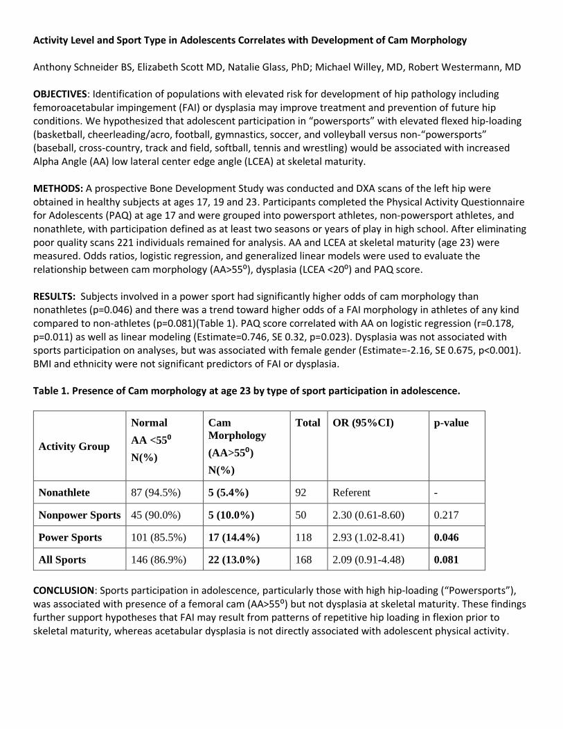
Activity Level and Sport Type in Adolescents Correlates with Development of Cam Morphology
Anthony Schneider BS, Elizabeth Scott MD, Natalie Glass, PhD; Michael Willey, MD, Robert Westermann, MD
OBJECTIVES: Identification of populations with elevated risk for development of hip pathology including femoroacetabular impingement (FAI) or dysplasia may improve treatment and prevention of future hip conditions. We hypothesized that adolescent participation in “powersports” with elevated flexed hip-loading (basketball, cheerleading/acro, football, gymnastics, soccer, and volleyball versus non-“powersports” (baseball, cross-country, track and field, softball, tennis and wrestling) would be associated with increased Alpha Angle (AA) low lateral center edge angle (LCEA) at skeletal maturity.
METHODS: A prospective Bone Development Study was conducted and DXA scans of the left hip were obtained in healthy subjects at ages 17, 19 and 23. Participants completed the Physical Activity Questionnaire for Adolescents (PAQ) at age 17 and were grouped into powersport athletes, non-powersport athletes, and nonathlete, with participation defined as at least two seasons or years of play in high school. After eliminating poor quality scans 221 individuals remained for analysis. AA and LCEA at skeletal maturity (age 23) were measured. Odds ratios, logistic regression, and generalized linear models were used to evaluate the relationship between cam morphology (AA>55⁰), dysplasia (LCEA <20⁰) and PAQ score.
RESULTS: Subjects involved in a power sport had significantly higher odds of cam morphology than nonathletes (p=0.046) and there was a trend toward higher odds of a FAI morphology in athletes of any kind compared to non-athletes (p=0.081)(Table 1). PAQ score correlated with AA on logistic regression (r=0.178, p=0.011) as well as linear modeling (Estimate=0.746, SE 0.32, p=0.023). Dysplasia was not associated with sports participation on analyses, but was associated with female gender (Estimate=-2.16, SE 0.675, p<0.001). BMI and ethnicity were not significant predictors of FAI or dysplasia.
Table 1. Presence of Cam morphology at age 23 by type of sport participation in adolescence.
Activity Group
Normal
AA <55⁰
N(%)
Cam
Morphology
(AA>55⁰)
N(%)
Total OR (95%CI) p-value
Nonathlete 87 (94.5%) 5 (5.4%) 92 Referent -
Nonpower Sports 45 (90.0%) 5 (10.0%) 50 2.30 (0.61-8.60) 0.217
Power Sports 101 (85.5%) 17 (14.4%) 118 2.93 (1.02-8.41) 0.046
All Sports 146 (86.9%) 22 (13.0%) 168 2.09 (0.91-4.48) 0.081
CONCLUSION: Sports participation in adolescence, particularly those with high hip-loading (“Powersports”), was associated with presence of a femoral cam (AA>55⁰) but not dysplasia at skeletal maturity. These findings further support hypotheses that FAI may result from patterns of repetitive hip loading in flexion prior to skeletal maturity, whereas acetabular dysplasia is not directly associated with adolescent physical activity.

Exploring the Involvement of Novel C-Type Lectin Receptors in Ebola Virus Entry
Kristina M. Sevcik, Kai Rogers, Wendy Maury
Introduction and Hypothesis: Zaire Ebola virus (EBOV) is a negative-sense RNA virus of the family Filoviridae. It causes hemorrhagic fever in humans and infection occurs in sporadic outbreaks. Although EBOV infection can quickly turn deadly with mortality rates as high as 90%, there are currently only investigational vaccines for the virus and no available therapeutic drugs. To approach the problem of preventing and treating EBOV infection, the cellular components that mediate viral binding and entry are of interest. Previous studies have found various phosphatidylserine receptors and C-type lectin receptors (CLRs) to help mediate uptake of EBOV, as opposed to the virus utilizing one particular cell surface receptor. We hypothesized that additional CLRs may also serve as filovirus receptors. Here, four novel CLRs identified by the Maury lab as potential EBOV entry factors are explored for their impact on EBOV infection: CLEC3a, CLEC4d, CLEC5a, and CLEC12b.
Methods: To investigate the role of the four CLRs in EBOV infection, we utilized a variety of methods. Expression of each receptor in cells relevant to infection was assessed via qRT-PCR on cDNA from macrophages and keratinocytes as well as antibody staining and qRT-PCR on HEK 293T transfected cells. Virus binding assays and internalization assays were performed on HEK 293T transfected cells to assess whether the expression of the CLRs enhanced binding and entry into cells. Transfected cells were also infected with a BSL2 model virus, rVSV/EBOV GP, to determine whether infection itself was enhanced. Additionally, sugars known to inhibit control receptors or found in literature to inhibit the CLRs were explored for competitive inhibition in either infection or entry assays. Finally, we sought to increase expression of CLEC4d by co-expressing it with an Fc plasmid and assessing the effects of co-expression via antibody staining, binding assays, and infection.
Results: In this study, we found that primary macrophages express CLEC4d, CLEC5a, and CLEC12b, while keratinocytes express CLEC3a, CLEC5a, and minor amounts of CLEC12b. Both of these cell types are important in EBOV infection. HEK 293T cells, that do not endogenously express these CLRs, transfected with expression plasmids for the CLRs seemed to express only small amounts of CLEC4d and CLEC12b and relatively large amounts of CLEC3a and CLEC5a, as seen by antibody staining and qRT-PCR. All CLRs significantly enhanced EBOV binding, particularly CLEC4d and CLEC12b. Both CLEC4d and CLEC12b also significantly increased EBOV internalization. Interestingly, wild type VSV, that was used as a control, showed similar trends in binding and significant increase in entry with CLEC4d and CLEC12b. Results varied between infection repeats, but no CLR consistently enhanced EBOV infection. The sugars explored did not inhibit the CLRs, and Fc expression did not enhance CLEC4d expression or function.
Conclusions and Discussion: The results indicate that these CLRs may serve as EBOV entry factors, but further exploration is merited. It is unclear how CLEC4d and CLEC12b consistently enhanced both binding and entry, but not infection with EBOV. The similar results shown using WT VSV may indicate that binding may be relatively non-specific. Future studies with these CLRs require increased expression and our CLEC3a and CLEC5a studies suggested that these CLRs do not play an important role in EBOV infection.

Title: Nutrition and Exercise Patterns Among Toddlers
Timothy Sevcik, M2 with mentors: David Bedell, MD, and Barcey Levy, PhD, MD
Introduction:
Among youth, 18.5% of those between the age of age 2 and 19 are obese, with Hispanic children (25.8%) disproportionately affected. Pediatric obesity is associated with adulthood obesity and but can also be a
marker for future problems such as cardiovascular disease. Pediatric obesity has been associated with
factors such as genetics, consumption of sugary beverages and general diet, screen time, physical activity, and neighborhood safety. The goal of this study was to investigate common factors associated with
obesity and identify differences between overweight, healthy-weight, Hispanic, and non-Hispanic
preschool-aged children.
Methods:
Children ages 3-6 were recruited from the patient population of the University of Iowa River Crossing
Clinic in Riverside, Iowa. Parents completed three surveys: one survey on family demographics and parental perceptions of the child, and two one-day food recalls through the National Cancer Institute’s
Automated Self-Administered 24-Hour (ASA24®) Dietary Assessment Tool which is available in
English and Spanish. Child physical activity was assessed through 5 days of tracking with Garmin Vívofit Jr. 2 fitness trackers. Additional information was collected from the clinic EMR including age, ethnicity,
height, and weight.
For comparisons of BMI, children were classified into groups by age/sex-based BMI percentiles where healthy-weight children were below the 85th percentile while overweight/obese children were at or above
the 85th percentile. For categorical and continuous variable pairings independent sample t-tests were used.
For ordinal and categorical variable pairings Spearman’s correlation was used. For pairings with two
categorical variables a Fischer’s exact test was used due to low sample size. Study data was analyzed using SPSS 25 software. The study was approved by the University of Iowa IRB.
Results: A total of 37 children were consented to the study with a mean age of 4.6 ± 1.2 years. The study
population was 8% Black (n=3), 30% Hispanic (n=11), and 62% White (n=23); while 3% were
underweight (n=1), 65% of a healthy weight (n=24), 14% overweight (n=5), and 19% obese (n=7).
However, in the parental perceptions survey, 36 of 37 parents classified their child as healthy and only one parent classified their child as overweight. Out of the children, 34 returned the fitness trackers with 4
or more days of step data. For comparisons between healthy and overweight/obese groups no significant
difference was found in mean daily calories adjusted for height (12.82 vs 12.81 Kcal/day/cm, p=0.996), or for daily hours of screen time for healthy (1.7±0.78 vs. 1.5±1.1, p=0.442), while a significant
difference was found for mean daily steps (10,092 ±2,631 vs. 7,913 ± 2,776, p=0.038). No significant
association was found between average steps per day and parental perception of the child’s activity level (r=0.254, p=0.154).
Comparisons between Hispanics and non-Hispanics showed no significant difference in the proportion of
healthy and overweight/obese children (df=1, p=0.122) and in mean daily steps (9,948±2,833 vs.
9,272±2,849, p=0.545). However, Hispanics did consume fewer daily calories adjusted for height than non-Hispanics (10.24±2.73 vs. 13.95±3.96 Kcal/day/cm, p=0.008).
Conclusions: Children at a healthy weight took more steps/day than overweight/obese children, which demonstrates the
importance of physical activity for a healthy weight. Parents were unwilling or unable to correctly
perceive aspects of their child’s health such as their weight status and physical activity level, which has ramifications for their child’s health.

Title: AFP as a Reliable Marker for HCC in Patients with Cirrhosis and Cured HCV Infection Student: Mahek Shahid, M1 Mentor: Dr. Kyle Brown, MD, MSc
Background: Alpha-fetoprotein (AFP) is polypeptide that is used as a screening test for hepatocellular carcinoma (HCC) in patients with cirrhosis. There are concerns about the usefulness of AFP as a screening tool in patients with cirrhosis due to chronic HCV because chronic liver disease and HCV-induced cirrhosis can be associated with increased levels in AFP in the absence of HCC.2 Since 2014, highly effective antiviral therapy has become available for the treatment of chronic HCV infection. Research has shown that sustained virological response (SVR). i.e., cure of HCV infection, is associated with decreased AFP levels in HCV patients.8 The observation that AFP levels normalize after SVR suggests there is less nonspecific “noise” in AFP levels following HCV cure, which may improve its usefulness as a screening test.
Purpose (Hypothesis/Aims): The purpose of this study was to identify patients with HCV cirrhosis who were successfully treated (achieved SVR) at UIHC and were subsequently diagnosed with HCC. We hypothesized the specificity of AFP for identification of HCC is improved following SVR in patients with HCV-induced cirrhosis.
Methods: This study utilized retrospective data collection by chart review with charts dated 2014-2018 from University of Iowa Hospitals and Clinics (UIHC). An IRB was written to obtain diagnosis ICD codes that were sent to the General Healthcare Compliance Office to gather a list of patients who had been diagnosed with neoplasms of the liver. From this list, only primary neoplasms were looked at in further detail. Sex, race, age (at time of initiation of HCV treatment), treatment interval time, pre- and post-treatment AFP levels, date of HCC diagnosis, AFP level at time of HCC diagnosis, what led to HCC diagnosis, and comorbidities were collected for each patient. Patients were filtered to those who had successfully received antiviral treatment, those who had not received any treatment to serve as controls, and those who were grouped into other who did not meet our inclusion criteria of patients who had HCV, had been successfully treated with an antiviral therapy, subsequently diagnosed with HCC, with AFP levels in their charts from UIHC (to ensure consistent reference ranges for lab values).
Results: Median of AFP levels at SVR achievement in treatment group was 7.00, Median of AFP levels at HCC diagnosis in treatment group was 8.10. Median of HCC diagnosis in untreated group (control) was 25.00. 30 (n=37) patients had decreased AFP levels from pre-treatment to post-treatment (SVR achievement). 18 (n=37) patients of the treatment group had increased AFP levels from SVR achievement to time of diagnosis of HCC. A Mann Whitney test was performed on the AFP levels of the treatment group at SVR and at HCC diagnosis. For this, p value was .39. Since p value >.05, this data is statistically insignificant. A Mann Whitney test was performed on the AFP levels at HCC diagnosis between the treatment group and the control group. For this, p value was .01. Since p value <.05, the difference in AFP levels between the two groups is statistically significant.
Discussion: Previous studies have suggested SVR achievement is associated with decreased AFP levels; our study confirms this. AFP levels tended to decrease from pre-treatment to post-treatment (SVR achievement). From our sample, 81.1% of patients showed this decreased in AFP levels. A percentage of patients (48.6%) who underwent antiviral treatment, but subsequently developed HCC, had an increase in their AFP levels at time of diagnosis following normalization of AFP post-treatment. However, there was not a statistically significant increase in AFP levels from SVR achievement to diagnosis of HCC. Despite this, AFP levels were lower at the diagnosis of HCC in the treatment group versus the control group. The persistently high AFP levels in the control group likely reflect the ongoing inflammatory activity in that group of patients since they were untreated. Overall, the sample size was smaller than initially anticipated. In further studies, more patients will be included to increase the sample size and thus, power, in statistical analysis of the data.
Title: Endoscopic Dilation is a Safe and Effective Therapeutic Procedure for Patients with Eosinophilic Esophagitis
Medical Student: Kuangda Shan, M4 Faculty Mentor: Yehudith Assouline-Dayan, MD
Background: Eosinophilic esophagitis (EoE) is a chronic antigen and immune-mediated condition that is commonly associated with other atopic diseases such as asthma, allergic rhinitis and atopic dermatitis. The most common symptoms of EoE are feeding problems including dysphagia and food impaction due to the chronic eosinophil-induced inflammation, esophageal dysfunction and stricture formation. The mainstays of therapy for EoE are swallowed steroids and progressive diet elimination as it is thought that EoE pathogenesis involves Type I hypersensitivity due to specific food antigens. However, there is a subgroup of patients who remain resistant to first line therapy. Esophageal dilation via endoscopy is often utilized in this patient subgroup for symptomatic improvement.
Purpose: This study examines the safety and efficacy of esophageal dilation in both adults and pediatric patients with EoE.
Method: A retrospective chart review was performed for this study. All esophageal biopsy reports from 2006-2016 were obtained from the Department of Pathology. Patients with biopsy-proven EoE were identified by having over 15 eosinophils/hpf on esophageal biopsy. We reviewed the electronic medical records of the EoE patients and identified those who have received endoscopic dilations since 2006. Data on endoscopic dilation methods, complications, improvements, and pre-procedure medications was collected. A descriptive statistical analysis was then performed.
Results: A total of 1050 patients with EoE (265 pediatric and 785 adult patients) were identified. Out of the 1050 patients with EoE, 160 patients received a total of 295 endoscopic dilations. Of the 160 patients, 58.2% (n=93) of patients only had one dilation while 41.8% (n=67) of patients received >1 endoscopic dilation. 81% of endoscopic dilations were done with the Savary-Gillard dilator while 18% were done with balloon dilators. With regards to complications, the overall complication rate was 6.1%, primarily due to post-procedural chest pain (4.8%) and post-procedural admissions (1.3%). The post-procedural admissions were mainly due to sedation complications. Clinical improvement was documented in 87% of dilations on follow-up.
Conclusion: Overall, endoscopic dilation is a safe and effective therapeutic procedure for patients with EoE and should be considered as a therapeutic management option alongside diet restriction and pharmacological therapy.

A Look into the Neuropsychological Functioning of Breast Cancer Survivors 10 or More Years into Survivorship Student: Ashten Sherman, M2 Mentor: Natalie Denburg, PhD
Introduction Breast cancer is a disease that will affect one in every eight women in the US across a lifetime (ACS, 2019). Even once a woman has braved the worst by undergoing cancer treatment, she is still not free from the effects that the disease and its treatment can have on the human body. One such effect is cancer treatments’ negative impact on the brain (colloquially known as “chemobrain”). Research on chemobrain has revealed mixed results, with some studies showing detriment to cognition following cancer and adjuvant treatment, but a recent study suggesting neuroprotection (e.g., Ospino-Romero et al., 2019). The mixed findings may be the result of an overreliance on cross-sectional studies. By contrast, there are a limited number of longitudinal studies examining the phenomenon of chemobrain.
Purpose In order to better understand the neuropsychological effects that cancer treatment can have on the brain, our study aimed to measure longitudinally the cognitive and psychological functioning of breast cancer survivors. Furthermore, we divided our survivors based on type of adjuvant treatment: local therapy (e.g., radiation) vs. chemotherapy. We hypothesized that the greatest longitudinal change (i.e., decline) would occur in the chemotherapy-receiving group, with the next greatest longitudinal change being displayed by the survivors who received local treatment. We predicted that normal comparisons would show significantly less change/decline than both cancer groups.
Methods Our study followed 36 breast cancer survivors: 16 who received local treatment and 20 who received chemotherapy. The 36 survivors were matched with a group of healthy women of similar age and educational level, but without a history of cancer. Time 1 testing occurred when the cancer survivors were 65 year and older and at least 10 years post-treatment, and the Time 2 testing occurred approximately two years following Time 1. All subjects continued to be cancer free. At each time point, all participants (i.e., local treatment, chemotherapy, and normal comparison groups) underwent a comprehensive neuropsychological battery to assess mental status, attention/working memory, psychomotor speed, anterograde memory, language, visuospatial skills, executive functioning, and mood. For just the two cancer groups, we also collected medical comorbidity, as well as several psychosocial variables involving well-being, post-traumatic growth, and locus of control.
Results Our hypotheses were not supported. On the cognitive and mood measures, all three groups changed over time to the same degree on virtually all measures. There were two exceptions, and these ran counter to our predictions: on measures of psychomotor speed and visuospatial skills, the cancer survivors showed less decline from Time 1 to Time 2 than did the normal comparisons. Similarly, among the two cancer groups, the rate of psychosocial change was comparable.
Conclusion The results indicate that longitudinal change in cognition and mood was comparable across three groups of older adults: breast cancer survivors treated with chemotherapy, breast cancer survivors treated with local therapy, and non-cancer normal comparisons. Overall, a history of cancer and adjuvant treatment does not appear to increase one’s risk for exaggerated cognitive aging.
Discovery of p21-interacting Proteins in Skeletal Muscle Fibers Presenter: ZZach SkopecMentor: Chris Adams, MD, PhD
Skeletal muscle atrophy diminishes the health and quality of life of tens of millions of people. Causes of
muscle atrophy include aging, muscle disuse, malnutrition, critical illness, certain medications and a
wide range of chronic illnesses including cancer, heart failure, COPD, diabetes, renal failure, cirrhosis,
rheumatoid arthritis, and HIV/AIDS. Effects of muscle atrophy include weakness, impaired activity, falls,
prolonged hospitalization, delayed rehabilitation, loss of independent living, and increased mortality.
Despite its broad clinical impact, skeletal muscle atrophy lacks a specific and effective pharmacologic
therapy and remains poorly understood at the molecular level. In preliminary studies, we identified
p21 as a skeletal muscle fiber protein that is necessary and sufficient for skeletal muscle atrophy.
However, the mechanism by which p21 promotes muscle atrophy is not yet known. Here we describe
an unbiased biochemical approach that we used to isolate and identify 13 proteins that specifically
interact with p21 in mouse skeletal muscle fibers. These 13 proteins represent novel potential
mediators of skeletal muscle atrophy. Investigations of their roles in skeletal muscle atrophy are now
underway.

Effect of Labor & Delivery Unit Closures on Access to Obstetrical Care in Rural Iowa Student: Laurel Smeins, M2 Mentor: Stephanie Radke, MD – Department of Obstetrics and Gynecology Other Collaborators: Thomas Gruca, PhD; Debra Kane, PhD; Greg Nelson, MA, RN
Background: The state of Iowa has a large rural population and was ranked 49th in the country in obstetrician-gynecologists per capita in 2017.1 Furthermore, the state has seen a large number of closures of labor and delivery units in rural level one hospitals. 28 of these hospitals have discontinued obstetrical services since 2001, with 12 of these closures occurring in the past two years, leaving 48 of 99 counties without any inpatient obstetrical services. With the decreased number of rural labor and delivery units and obstetrical providers, rural Iowa women could be facing significantly diminished access to care, both delivery and prenatal, which could have numerous consequences for maternal and fetal health outcomes. Additionally, it has been shown that there are significantly higher rates of severe maternal morbidity among non-white women and Medicaid recipients in Iowa,2 suggesting that these women could face even greater compounded risk for negative outcomes as a result of service discontinuation. With the availability of obstetrical services decreasing, it is important to understand the effect of changes in access, and if certain populations, such as non-white women or Medicaid recipients, are more vulnerable to these changes, so that interventions can be initiated if necessary.
Methods: Birth certificate data for Iowa moms delivering in Iowa was provided by the Iowa Department of Public Health for years 2013 through 2018, and each birth was assigned to one of 6 categories based on the services available in the mother’s county of residence and population density: Urban with Labor & Delivery (L&D) and Prenatal care (PNC), Urban with no L&D but PNC, Urban with neither L&D nor PNC, Rural with L&D and PNC, Rural with no L&D but PNC, and Rural with neither L&D or PNC. Urban and rural status was determined from 2013 Rural Urban Continuum Codes with additional zip-code level categorization utilizing the Federal Office of Rural Health Policy funding classifications for counties where only a portion is classified as urban. Data from the Iowa Department of Public Health (IDPH) was used to identify the location of open L&D units and the closure date for hospitals with service discontinuation during our study period. Prenatal care availability was determined through an obstetrician-gynecologist database and outreach clinic list provided by the Office of Statewide Clinical Education (OSCE) at the Carver College of Medicine. For the counties that did not have a practicing obstetrician-gynecologist or an outreach clinic, other types of providers (family medicine physicians, nurse practitioners) that could possibly provide PNC were identified from a database of all Iowa physicians and advanced practice providers provided by the OSCE and surveyed via telephone to identify whether or not they provided PNC. Adequacy of prenatal care received by mothers in each county classification was assessed by trimester of initiation of prenatal care as well as adequacy of prenatal care utilization index (APNCU) derived from the birth certificate data. This was further stratified by delivery payor (Medicaid or other) and race and ethnicity. Travel times from zip code of residence to zip code of delivery were computed using Google Maps average travel times based on zip-code population center. Unplanned birth outside a L&D unit (home, en route to facility, or at non-delivery facility) was examined for rural counties without L&D units and further stratified by driving time to facility, delivery payor, and race.
Results: 14 hospitals closed their Labor and Delivery units during our study time period. Of these, we found 12 (85.7%) continued to have prenatal care available in their area. Overall, it was observed that a high number of Iowa women received adequate prenatal care (85.8%) during our study time period. Urban women were more likely to receive adequate care than rural-residing women (87.3% vs 83.4%, p<0.001) and women insured under Medicaid for their pregnancy were less likely to receive adequate care than those with other payors (80.3% vs 89.6%, p<0.001). Examining proximity to delivery services, urban women residing in a county with Labor and Delivery services had the lowest distance and travel time (95% <30 miles and 92% <30 minutes) as compared to rural women residing in counties with Labor and Delivery services (74% < 30 miles, 66% < 30 minutes) and rural woman residing in counties without services (42% < 30 miles, 24% < 30 minutes, all p<0.001). Correspondingly, it was observed that rural women had significantly greater likelihood of delivering outside of a Labor and Delivery unit (1.4% vs 0.6%, p<0.001) with rural women from counties without Labor and Delivery services experiencing the greatest risk (2.7%, aOR = 5.1). Infants born to mothers residing in rural counties with and without Labor and Delivery units were not more likely to be transferred after birth, indicating women generally delivered in units with the appropriate level of neonatal care. However, women residing > 60 miles from a delivery hospital were significantly more likely to experience a maternal transfer than those 30-59 miles or <30 miles (15.3% vs 1.9% vs 0.2%, p<0.001), indicating women had a greater challenge to timely access to appropriate levels of maternal care.
Discussion: Overall our results indicate that despite the concerning trend in rural Labor and Delivery unit closure, prenatal care access is generally preserved and a high number of women are receiving adequate care. However, our results revealed concerning trends regarding appropriate access to hospital-based Labor and Delivery services with rural-residing women facing a greater risk for delivery outside of a Labor and Delivery unit and high rates of maternal transfers, both of which increase with increasing travel time to the hospital. Effective emergency transportation services are vital to ensure the safety of birth for rural-residing women.
1. Rayburn W. The Obstetrician-Gynecologist Workforce in the United States: Facts,Figures, and Implications, 2017. 2017.2. Frederiksen BN, Lillehoj CJ, Kane DJ, Goodman D, Rankin K. Evaluating Iowa Severe Maternal Morbidity Trends and Maternal Risk Factors: 2009-2014. Matern Child
Health J. 2017;21(9):1834-1844.
Hip Pathology in Patients Presenting to the Urologist for Chronic Orchialgia
Emily Solsrud, Alyssa Conrad, Robert Westermann MD, Amy Pearlman MD, Timothy Brown MD
Introduction: Chronic orchialgia can be a difficult clinical condition to diagnose and treat. Hip
pathology is a rare cause of chronic orchialgia. This study aimed to identify patients with chronic
orchialgia and evaluate them for the presence of concomitant hip pathology.
Patients and Methods: We prospectively identified 44 hips (22 patients) at the UIHC Men’s Health
Urology Clinic with a mean age of 35 years (range, 18 – 62 years), 100% male, mean BMI of 29 kg/m2
(range, 19 – 53 kg/m2), and duration of orchialgia for greater than 3 months. These patients were given
hip-specific PROs (HOOS Jr, VAS, PROMIS, UCLA Activity Score), had a hip specific physical exam
performed by a non-orthopedic provider (ES, AP), and had hip specific radiographs taken and evaluated
for markers of femoroacetabular impingement (FAI), developmental hip dysplasia (DDH), and
osteoarthritis (OA). Medical records were reviewed for follow-up to determine the outcome of referrals
for hip treatment to physical therapy (PT), or orthopedic surgery clinic.
Results: Testing for range of motion identified decreased flexion in 3 hips (7%), decreased internal
rotation in 11 hips (25%), decreased external rotation in 8 hips (18%), and decreased abduction in 1 hip
(2%). Flexion contracture was present in 8 hips (18%). The impingement test was positive in eliciting
pain in 18 hips (41%). There were 16 hips (36%) with positive Patrick’s tests. There were 13 hips (30%)
with positive Stinchfield tests. There was 1 hip (2%) with positive straight leg raise.
HOOS, Jr. Hip Survey had a mean of 4 (range, 0-11). The Visual Analogue Scale had a mean of 8.5
(range, 1.8-15.2). The UCLA Activity Score had a mean of 7 (range, 2 – 10). The PROMIS Scale v1.2
Global Health had a mean physical score of 16 (range, 11 – 18) and a mean mental score of 14 (range, 7-
19).
Radiographic analysis found Tonnis 0 in 28 hips (64%), Tonnis 1 in 15 hips (34%), Tonnis 2 in 2 hips
(5%), and Tonnis 3 in 1 hip (2%). No hips showed global impingement with LCEA > 50 degrees. 25 hips
(57%) had LCEA < 25 degrees and mild dysplasia. Tonnis angle showed 9 abnormal hips (20%), with 1
hip < 0 degrees and 8 hips > 10 degrees. Crossover sign found in 13 hips (30%). Abnormal alpha angle >
60 degrees found in 12 hips (27%).
16 patients (73%) have had a referral to our orthopedic clinic. Of these referrals, 4 patients (18%) have
had a clear orthopedic diagnosis or intervention which has relieved orchialgia symptoms.
Conclusion/Discussion: Patients presenting to the urology clinic with chronic orchialgia can have hip
pathology on radiograph and clinical examination. In our small cohort of 22 patients, we found enough
evidence for referral to orthopedic surgery in 16 patients (73%), although evaluation by the specialist has
led to hip diagnosis and treatment in only a small portion of the original cohort (4 patients, 18%). The
study lacks statistical power to conduct a multivariate analysis about outcomes or to identify predictors
that can help identify those at risk for an underlying hip cause to the testicular pain. We would
recommend the urology practitioner treating chronic orchialgia conduct a short hip physical examination
with internal rotation, impingement testing, Stinchfield, and obtain an AP pelvis radiograph to help
identify those patients that would benefit from referral to the orthopedic surgeon.
Investigating cortico-limbic-striatal circuit interactions during risky decision-making Nathen Spitz, Nicole Schwab, Julia Schaffer, Lucas R. Glover, Patrick P. Piantadosi, Andrew Holmes
Introduction: There are numerous neuropsychiatric conditions including addictions and anxiety disorders that
are associated with phenotypes that display atypical decision-making in the presence of risk. Previous data implicates
a set of cortical, limbic, and striatal nuclei in certain processes that are related to risky decision-making, yet how
these structures interact and function in a circuit to mediate these decisions remains unclear.
Aims: In order to better elucidate these circuit-level interactions, in this current study, we adapted a risky decision-
making assay for mice, in which we utilized techniques to examine neuronal population activity in vivo using fiber
photometry and employed optogenetics to silence basolateral amygdala (BLA) terminals in the nucleus accumbens
shell (NAcS) to delineate distinct roles for this circuit in risky decision-making.
Methods: Male C57BL/6J mice were acclimated, habituated, and shaped to enter the reward port of an operant
conditioning box to initiate a trial, from which they were required to press a touchscreen to receive a liquid
milkshake reward. During the Reward Magnitude Discrimination task, mice were biased to choose one screen that
delivered a larger volume milkshake reward (20 µl), while the other screen produced a smaller volume milkshake
reward (5 µl). Once the mice reached an established responding criteria of > 90% on the large rewards for three
consecutive days, they advanced to the risky decision-making test days. In the risky decision-making task, mice
progressed through three blocks, with each block containing an increasing probability of foot shock. The first block
started with a 0% probability of shock associated with the large reward, while blocks 2 and 3 featured an increasing
probability of foot shock concomitant with choice of large reward at 50% and 75% respectively.
To record in vivo neuronal population activity via fiber photometry, in the NAcS, rAAV2-retro-Ef1a-Cre was
unilaterally infused. Then within the ventromedial prefrontal cortex (vmPFC) and the BLA, mice were unilaterally
infused with AAVjd/Ef1a-DIO-GCaMP5m. Lastly, a multimodal fiber probe for recording was placed in the vmPFC
and BLA.
To silence terminals in the BLANAcS pathway, rAAV5-CAMK11a-eArchT3.0 e-YFP or rAAV5-CAMK11a-
eYFP for control mice, were bilaterally infused in the BLA. Then in the NACS, fiber optic cables were implanted.
Results: During the deliberation phase of the reward magnitude task—with no possibility of foot shock—
ArchT-mediated optogenetic silencing of the BLANAcS pathway did not affect reward magnitude
discrimination. When the ArchT infected mice (n=10) had their pathway silenced, they chose the large
reward almost 100% of the time, as did the control mice (n=6) who were not virally transplanted with an
inhibitory opsin.
In the risky decision-making phase of the experiment, which included three blocks of increasing foot shock
possibility, silencing the BLANAcS pathway via ArchT in experimental mice (n=10) significantly
reduced the shift away from the large, risky reward that was found in control mice (n=6), showcasing that
silencing this pathway maintained risky decision-making and implicates the BLANAcS pathway in this
behavior, *p < 0.05 ArchT vs. eYFp.
Conclusion: A previously validated risky decision-making task was successfully adapted for mice, and
through this task and testing, we found that glutamatergic afferents from the BLA to the NAcS encode
distinct and relevant events during the risky decision-making task. In the presence of risk (the increasing
probability of foot shock), optogenetic silencing of the BLA NAcS pathway during deliberation
eliminated the shift from a large, risky reward to a small, safe reward, and maintained risky reward seeking.
These data are all consistent with the hypothesis that the BLA signals punished outcomes during reward-
seeking and contributes to adaptively shifting behavior away from risky behaviors.
Predicting pain after cesarean section using a 3-question questionnaire, local anesthesia infiltration and
observer rating
Joseph Stearns, Unyime Ituk
Introduction
Cesarean delivery is one of the commonest surgical procedures in the USA, about 1.3 million procedures per year
and accounts for 32 % of all live births. Acute postoperative pain associated with cesarean delivery is a clinical
concern, and inadequate control of acute postoperative pain can limit early ambulation, self-care and ability to care
for newborn as well as contribute to postpartum depression [1]. The current standard for postcesarean pain
management is a “one-size-fits-all” approach and does not account for interindividual variability in acute
postoperative pain. Identification of women at a higher risk for severe postoperative pain would allow for a more
targeted analgesic approach to postoperative pain management.
The aim of this study is to determine if using a combination of simple pain screening questions, patient’s rating of
pain during local anesthetic infiltration and physician’s predicted rating of patient’s postoperative pain can
adequately predict severe acute postcesarean pain. We hypothesized that the combination of these 3 tools will
predict the average severity of acute postoperative pain with movement within 48 hours after cesarean delivery.
Methods
This prospective observational study was approved on August 27th
, 2018 by the University of Iowa Institutional
Review Board (Human Subjects office IRB #201808806). Healthy pregnant women age ≥ 18 years, singleton term
(≥37 weeks gestation), scheduled for elective cesarean delivery were approached to participate in the study.
Prior to cesarean delivery, recruited subjects complete a 3-question questionnaire. In the operating room, the patient
will be asked to rate the pain from the local anesthetic injection using a numerical rating scale (NRS) of 0 – 10 (0 =
no pain, 10 = worst pain imaginable). The physician will be asked to predict the severity of patient’s post-operative
pain based upon their reaction to the local anesthetic injection. Postoperative pain scores at rest and with movement
will be collected at 6, 24, 48 hours using NRS. Analgesic consumption data at 6, 24 & 48 hours was retrieved from
the medical record.
The primary outcome is the average postoperative pain with movement 48 hours after cesarean delivery. Secondary
outcomes include total amount of opioid analgesia used (oral and IV) for breakthrough pain within 48 hours
calculated in morphine equivalents and time to first request for opioid analgesia (defined as minutes from end of
surgery).
Results
To date, 40 women have been enrolled in the study. The mean (SD) postoperative NRS pain anticipated by patients
is 7 (±2) and the median [IQR] NRS pain score on local anesthesia infiltration is 3 [2 – 5]. The 48 hour mean NRS
pain score with movement is 2 (±2). Values are expressed as mean ± SD, median [IQR], and number (percentage) as
appropriate
Discussion
At the time that this abstract is due the current data has not been analyzed for a regression model to test the
relationship between the predictive tools and pain outcomes.
References
Pan PH, Coghill R, Houle TT, et al. Multifactorial preoperative predictors for postcesarean section pain and
analgesic requirement. Anesthesiology 2006; 104: 417-25.
Ultrasound Guided Tenotomy Improves Physical Function and Decreases Pain for Tendinopathies of the Elbow, A Retrospective Review
Daniel Stover, BA; Benjamin Fick, BS; Ruth L. Chimenti, DPT, PhD; Mederic M. Hall, MD
Abstract
Background: Tendinopathy is a common cause of elbow pain in the active population. The three most common locations of elbow tendinopathy are the common extensor tendon, common flexor tendon, and triceps tendon. Initial treatment for all locations includes conservative measures such as rest, bracing, and physical therapy. However, there are no accepted standards of care for the cases recalcitrant to these conservative measures. One minimally invasive option is Ultrasound Guided Tenotomy (USGT). Small case studies have shown promising preliminary results of USGT for common extensor tendinopathy and common flexor tendinopathy, but none have included USGT for triceps tendinopathy. This is the largest study on the safety and effectiveness of USGT for elbow tendinopathy, and the first to include complication rate for the procedure in patients with common extensor, common flexor, and triceps tendinopathy.
Hypothesis: USGT for elbow tendinopathy will improve pain and physical function with low complication rates and high patient satisfaction.
Methods: Retrospective chart review identified 131 patients [144 procedures] (mean age ± SD, 48.1 ± 9.8 years; mean body mass index ± SD, 32.2 ± 7.7 kg/m2; 59% male) with elbow tendinopathy (104 common extensor tendinopathy; 19 common flexor tendinopathy; 8 triceps tendinopathy) treated with USGT over a 6-year period by a single physician. Pain and quality of life measures were collected at baseline. Pain, quality of life, satisfaction with outcome, and complications were collected at short-term (2, 6, 12 week) via regularly scheduled clinic visits and long-term (median 2.7 years IQR= 2.0 to 4.0 years) via email or phone surveys.
Results: USGT significantly decreased pain and improved the physical function subscores of quality of life assessments for both the elbow tendinopathy group overall and the common extensor subgroup (p< 0.01 for both comparisons). The majority (70%) of patients were satisfied with the procedure. There was a 0% complication rate.
Conclusion: Benefits of USGT for elbow tendinopathy include pain relief, improved physical function, and high patient satisfaction. USGT is a safe minimally invasive treatment for refractory elbow tendinopathy.
Identification of phase amplitude coupling in the human mesial temporal lobe during
episodic memory processing
Rodent models of episodic memory posit that phase-amplitude coupling (PAC) is necessary for
the spatial organization of neuronal ensembles critical for associative encoding. Here, we used a
bispectral analysis to document PAC occurrence in brain regions associated with memory
encoding/retrieval pathways. Local field potentials recorded from intracranial electrodes placed
for seizure localization offer a unique opportunity to record high fidelity signals from the brain to
study memory (1,2,3). Memory encoding is strongly correlated with PAC interactions,
specifically theta/high gamma activity. Conventional measures of PAC are windowed bispectral
estimators, amounting to narrow smoothings of the bispectrum (4). They are “blind” in that a
statistic (such as a modulation index) is calculated using pre-established frequency bands. PAC
that matches the standardized bands will be detected, but a broad representation of PAC
incidence in the signal is not obtained; a direct analysis of the bispectrum can be used to observe
the full picture. We used a visual inspection of the bispectrum to document PAC instances in the
hippocampus, parahippocampal gyrus, lateral temporal gyrus, and entorhinal cortex. Coherence
and correlation with successful memory encoding were also investigated for each band. These
findings were aggregated to identify the distribution of PAC frequency bands for each
region.
Presenter: Danial SyedMentor: Dr. Matthew Howard, Neurosurgery

1. Rizzuto, Daniel S., et al. "Human neocortical oscillations exhibit theta phase differences
between encoding and retrieval." Neuroimage 31.3 (2006): 1352-1358.
2. Place, Ryan, et al. "Bidirectional prefrontal-hippocampal interactions support context-
guided memory." Nature neuroscience 19.8 (2016): 992.
3. Yaffe, Robert B., et al. "Reinstatement of distributed
cortical oscillations occurs with precise spatiotemporal
dynamics during successful memory retrieval." Proceedings
of the National Academy of Sciences 111.52 (2014): 18727-
18732.
4. Kovach, Christopher K., Hiroyuki Oya, and Hiroto
Kawasaki. "The bispectrum and its relationship to phase-
amplitude coupling." Neuroimage 173 (2018): 518-539.
Staphylococcus Aureus Clinical Isolates Induce Pseudomonas Aeruginosa Exploratory
Motility
Xavier Tijerina, Dominique Limioli, PhD
Staphylococcus aureus and Pseudomonas aeruginosa are two of the most common infections in
Cystic Fibrosis patients. Patients who are co-infected have higher rates of morbidity and
mortality than patients who are infected with one or the other only. Pseudomonas shows
chemotaxis activity towards Staphylococcus secreted factors which causes the formation of
mixed communities. These mixed communities enhance the survivability and virulence of both
organisms through increased factor secretion and increased resistance to antibiotics.
Understanding how these polymicrobial communities form can provide information and possibly
new methods to improve patient outcome. Using a twitch motility assay of 30 clinical MRSA
isolates, the motility of Pseudomonas was measured with a wide range of results. Further
microscopic and macroscopic assays were performed including hemolysis and proteolysis.
Two unique phenotypes were observed, one that did not induce motility and one that
hyperinduced. Genome analysis was used to try to identify possible mutations in Staph secreted
factors and regulators of these factors. Mutations were found in the global regulator Agr in the
deficient strains but variations in the hyperinducers have yet to be identified. Deeper genome
analysis and the use of additional clinical isolates will be used in future experiments to gain more
information on the interactions between the two organisms.
RAPID ASTROCYTE ACTIVATION FOLLOWING JUVENILE TBI IS AN IMPORTANT SOURCE OF INFLAMMATORY MEDIATORS
Brittany Todd, Polly Ferguson, Alex Bassuk, Elizabeth Newell
Compared to microglia, astrocyte activation has been reported to occur in a more delayed fashion following brain trauma. Past studies have focused on the reparative processes driven by astrocyte activation such as glial scar formation and its role in limiting secondary injury. Recently, the duality of neurotoxic (A1) vs. neuroprotective (A2) astrocyte activation has been recognized; neurotoxic astrocyte gene signatures were identified and demonstrated to contribute to secondary neuronal injury following neurologic insult. Characterization of astrocyte phenotype (pan-reactive, A1, A2) following pediatric TBI and its impact on inflammatory mediator production have not been previously reported and are important in understanding the glial specific contributions to neuroinflammation. We used lateral fluid percussion injury adapted to PND 18-24 C57BL/6J mice to model moderate-severe pediatric TBI. Tissue expression of cytokines and astrocyte activation markers was evaluated by qPCR. Additionally, glial populations were isolated by FACS sorting, and glial-specific gene expression was evaluated by qPCR. At 24 hours, pediatric FPI resulted in increased inflammatory cytokine expression (IL-1b, TNF, and IL-6) in both focal (ipsilateral parietal cortex, hippocampus) and remote regions (brainstem). Pediatric FPI also resulted in astrocyte activation 24hrs post-injury: pan-reactive astrocyte markers (Lcn2, GFAP, Vimentin), A1 markers (Serping1, C3), and A2 markers (S100a10, Ptx3, Stat3) were increased at the tissue level in ipsilateral hippocampus. Glial-specific gene expression revealed increased inflammatory cytokine expression (IL-1b, TNF, IL-6, Ccl2) in FACS sorted astrocytes 24 hours post-juvenile FPI. Conclusions: Juvenile FPI results in rapid astrocyte activation which contributes importantly to increased inflammatory cytokine expression. Key differences be- tween microglia and astrocytes in inflammatory cytokine expression were noted 24 hours post-injury. Ongoing studies will compare the relative contribution of astrocytes and microglia to inflammatory mediator production and elucidate how astrocyte activation changes over time.
Acknowledgements: K12HD27748, K08NS110829
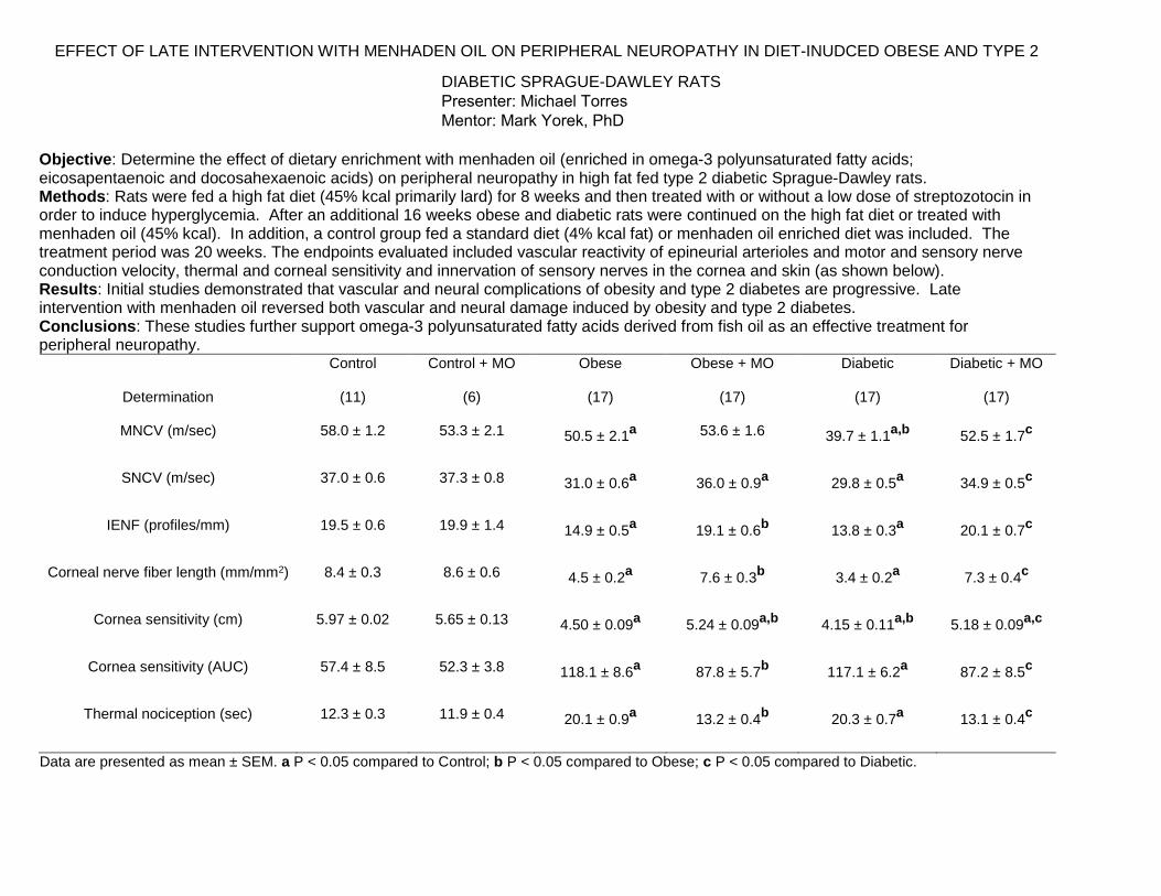
EFFECT OF LATE INTERVENTION WITH MENHADEN OIL ON PERIPHERAL NEUROPATHY IN DIET-INUDCED OBESE AND TYPE 2
Objective: Determine the effect of dietary enrichment with menhaden oil (enriched in omega-3 polyunsaturated fatty acids; eicosapentaenoic and docosahexaenoic acids) on peripheral neuropathy in high fat fed type 2 diabetic Sprague-Dawley rats. Methods: Rats were fed a high fat diet (45% kcal primarily lard) for 8 weeks and then treated with or without a low dose of streptozotocin in order to induce hyperglycemia. After an additional 16 weeks obese and diabetic rats were continued on the high fat diet or treated with menhaden oil (45% kcal). In addition, a control group fed a standard diet (4% kcal fat) or menhaden oil enriched diet was included. The treatment period was 20 weeks. The endpoints evaluated included vascular reactivity of epineurial arterioles and motor and sensory nerve conduction velocity, thermal and corneal sensitivity and innervation of sensory nerves in the cornea and skin (as shown below). Results: Initial studies demonstrated that vascular and neural complications of obesity and type 2 diabetes are progressive. Late intervention with menhaden oil reversed both vascular and neural damage induced by obesity and type 2 diabetes. Conclusions: These studies further support omega-3 polyunsaturated fatty acids derived from fish oil as an effective treatment for peripheral neuropathy.
Determination
Control
(11)
Control + MO
(6)
Obese
(17)
Obese + MO
(17)
Diabetic
(17)
Diabetic + MO
(17)
MNCV (m/sec) 58.0 ± 1.2 53.3 ± 2.1 50.5 ± 2.1a 53.6 ± 1.6 39.7 ± 1.1
a,b 52.5 ± 1.7
c
SNCV (m/sec) 37.0 ± 0.6 37.3 ± 0.8 31.0 ± 0.6a
36.0 ± 0.9a 29.8 ± 0.5
a 34.9 ± 0.5
c
IENF (profiles/mm) 19.5 ± 0.6 19.9 ± 1.4 14.9 ± 0.5a
19.1 ± 0.6b
13.8 ± 0.3a
20.1 ± 0.7c
Corneal nerve fiber length (mm/mm2) 8.4 ± 0.3 8.6 ± 0.6 4.5 ± 0.2a 7.6 ± 0.3
b 3.4 ± 0.2a
7.3 ± 0.4c
Cornea sensitivity (cm) 5.97 ± 0.02 5.65 ± 0.13 4.50 ± 0.09a
5.24 ± 0.09a,b
4.15 ± 0.11a,b
5.18 ± 0.09a,c
Cornea sensitivity (AUC) 57.4 ± 8.5 52.3 ± 3.8 118.1 ± 8.6a
87.8 ± 5.7b
117.1 ± 6.2a
87.2 ± 8.5c
Thermal nociception (sec) 12.3 ± 0.3 11.9 ± 0.4 20.1 ± 0.9a
13.2 ± 0.4b
20.3 ± 0.7a
13.1 ± 0.4c
Data are presented as mean ± SEM. a P < 0.05 compared to Control; b P < 0.05 compared to Obese; c P < 0.05 compared to Diabetic.
DIABETIC SPRAGUE-DAWLEY RATS
Presenter: Michael Torres Mentor: Mark Yorek, PhD
Calvin Tran
Mentor: Jess G. Fiedorowicz, MD, PhD
Departments of Psychiatry, Epidemiology, and Internal Medicine
Systematic Review and Meta-Analyses of Weight Gain with FDA-approved Mood Stabilizers in Bipolar
Disorder
Background: Bipolar disorder (BD) is a lifelong mental disorder characterized by alternating episodes of mania or
hypomania and episodes of depression. Mood stabilizers have antimanic and antidepressant effects and are one of
the main types of pharmacological interventions for BD. Although long-term adherence to mood stabilizers is
necessary for the management of BD, these medications also carry a significant risk for weight gain. Unfortunately,
it has been demonstrated that many psychiatrists do not select for medications based on metabolic risk and many
patients with BD are at elevated cardiometabolic risk. One barrier to appropriate prescribing involves limited
information that summarizes the adipogenic potential of medications.
Aims: We sought to assess risk with the FDA approved mood stabilizers lithium, valproic acid derivatives,
lamotrigine, and carbamazepine. This systematic review and meta-analysis seeks to compile and analyze data from
randomized controlled trials that include weight change data of the listed mood stabilizers to provide up-to-date
information quantifying and comparing each medication’s propensity to induce weight gain
Methods: Our literature search involved two separate computerized searches of Pubmed and Embase for
randomized controlled trials involving at least one of the listed mood stabilizers above performed on patients with
bipolar and related disorders, including other mood disorders and schizoaffective disorder. Search results were then
combined with duplicates removed.
In order to be included, mean weight change data including standard deviation (in kg or lb) had to be reported (or at
least calculatable) for the treatment groups and placebo group in each study. Studies that reported weight change as
an occurrence with no way to obtain the mean weight change data were excluded. Corresponding authors were
contacted if they reported the mean change data without a standard deviation. For each medication in which two or
more studies including weight change data is available, we will conduct a meta-analysis using Review Manager 5
(RevMan 5) to generate forest and funnel plots. If needed to account for heterogeneity, results may be stratified by
age (e.g., adolescent vs. adult samples) or any other variable that appears to explain the heterogeneity. Multiple
doses of a single medication within a trial will be treated as a separate study for the analyses to allow any estimation
of dose effects.
Results: From an initial pool of 613 studies, only 47 studies were relevant to the selected mood stabilizers. From
those 47, only four lithium trials and four valproate trials. Lamotrigine results are not currently available. No
carbamazepine studies were found during our literature search. The lithium results were stratified by Adult (2
studies) vs. Pediatric patients (2 studies). Adult patients demonstrated no significant differences relative to placebo
for weight change (-0.05 kg, [-1.22, 1.13] 95% CI, I2=94%). Pediatric patients also demonstrated no significant
differences relative to placebo for weight change (-0.27 kg, [-1.01, 0.46] 95% CI, I2=0%). The valproate results
demonstrated no significant difference for weight change (0.92 kg, [-0.02, 1.87] 95% CI, I2=84%).
Conclusions/Discussion: While we have several more articles to review and extract data from for this systematic
review and meta-analysis, preliminary results for the data so far seem to suggest that mood stabilizers do not
demonstrate significantly greater weight gain than placebo in short-term trials. Since the duration of most of these
studies is between 4-12 weeks, especially for our Valproate studies, weight differences are not notable over this
length of time. Because weight change data for many of these studies is obtained as a secondary outcome, there are
inconsistencies across studies in how the data is reported (e.g., using LOCF analysis to report end-of-study mean
change data instead of those who completed the entire study).
Trends in Horizontal Periocular Asymmetry
Charlene Tran, BS1
Dabin Choi, BS1 Keith D. Carter, MD, FACS2
A. Tim Johnson, MD2
Thomas A. Oetting, MD2
Alison Bozung, OD, FAAO2
Audrey C. Ko, MD2
Erin M. Shriver, MD, FACS 2
1 Carver College of Medicine, University of Iowa, Iowa City, IA 52242
2 Department of Ophthalmology and Visual Sciences, University of Iowa, 200 Hawkins Drive, Iowa City, IA 52242
Purpose: To determine whether there is laterality predominance in the horizontal width of the periocular region.
Background: Facial symmetry is considered a standard of beauty, but slight lateral asymmetry of the eyelid and orbit are common. There are conflicting studies regarding right or left-sided facial predominance and little research has been performed on laterality of periocular structures. Recognizing horizontal asymmetry in the periocular region is essential for evaluating patients’ baseline periocular structures, providing proper patient counseling, pre-operative planning, and maximizing post-operative success for both the patients and surgeons.
Methods: A prospective study of consecutive adult patients in an academic ophthalmology department was conducted in compliance with the IRB to evaluate for laterality dominance of periocular structures. Exclusion criteria included patients who had worn contact lenses; undergone laser skin surfacing or injections; or sustained periocular-altering trauma, surgery, or disease processes. Standardized digital photographs were obtained, periocular structures were measured with NIH Image J software, and dimensions were calculated based on a standard corneal limbus to limbus distance. The midline was defined as the midpoint between the medial canthi and the distances calculated for both the right and left sides were midline to medial canthus, pupil centroid, lateral canthus, and lateral zygoma.
Results: Periocular structures were measured in 86 patients. There was no significant difference between midline to medial canthus between the right (16.53 mm) and left (16.57 mm) (p=0.25). Right-sided predominance was found to be increasingly significant for the following variables in millimeters: midline to pupil center (31.36 vs 30.03, p=.004), midline to lateral canthus (42.50 vs 42.04, p<.001), and midline to lateral zygoma (65.91 vs 63.77, p<.001).
Conclusion: Photographic analysis of adults with no periocular-altering history demonstrates that there is a right-sided predominance in the horizontal dimension of the midline to the pupil, lateral canthus, and zygoma with increasing significance.
Title
Evaluation of maternal prenatal stress and its effects on placental redox processes – a neurodevelopmental perspective
Authors
Hanora Van Ert (M2G), Rachel Schroeder, Banu Gumusoglu, Hanna Stevens MD PhD (mentor)
Background
There is an increased risk for developing neuropsychiatric disorders such as schizophrenia, autism spectrum disorder,
and attention deficit hyperactivity disorder in offspring of mothers exposed to stress during pregnancy. Pregnant mouse
dams that undergo restraint stress have offspring with delayed GABAergic progenitor migration. These developing
neurons are critical in forming inhibitory pathways, and disruption can lead to development of neuropsychiatric disorders.
Our lab identified reactive oxygen species (ROS) to be mediators in GABAergic progenitor migration. Maternal major
depressive disorder (MDD) has been shown to increase ROS damage in mothers and has been hypothesized to affect
GABAergic migration. Our lab is currently investigating the neuroprotective compound P7C3-A20 (P7C3), which has been
shown to decrease ROS production, as a potential therapeutic to treat maternal depression and to protect offspring
neurodevelopment. Because MDD has been shown to increase oxidative damage, and ROS have been shown to be
increased in fetal brains exposed to chronic stress, our lab has been curious about what role the placenta plays in
mediating oxidative stress, specifically, the production of ROS, and ROS scavenging capabilities.
Purpose of this study
• Does maternal prenatal stress affect the placenta and its redox capabilities?
• What effect does P7C3-A20 have on maternal behavior and placental redox properties?
Methods
For this pilot study, female CD1 mice were randomized into four groups: prenatal stress with vehicle (PS Veh, n=2),
prenatal stress with P7C3-A20 (PS P7C3, n=5), non-stressed with vehicle (NS Veh, n=2), or non-stressed with P7C3-A20
(NS P7C3, n=2). Females were mated with CD1 males, and detection of a vaginal plug indicated embryonic day 0 (E0).
Stress and compound administration was given E5-E16. P7C3 groups received 20mg/kg P7C3-A20 via oral gavage twice
daily. PS groups received 45 minutes of restraint stress 3x daily; a well-documented method shown to induce both
maternal depression-like behavior and increase ROS production in fetal brains. Open-field (OFT), elevated plus maze
(EPM) and tail suspension tests (TST) were conducted on E17 to examine maternal anxiety-like and depressive-like
behaviors. On E18, maternal blood and brain tissue as well as placentas and embryonic brains were collected. We
compared gene expression of 19 redox-related genes in placental tissue using validated Sybr-Green primers with a qPCR
approach. Due to the small sample size in three groups, statistical comparisons were not performed.
Results
Behavioral analysis of pregnant dams showed that PS Veh dams displayed increased first-five-minute center time in the
open field compared to NS Veh/P7C3 dams (37% and 27% respectively). PS Veh mice also spent more time in the closed
arms of EPM compared to NS Veh/P7C3 mice (20% and 14% respectively). PS Veh dams exhibited less struggle time in
the TST compared with NS Veh/P7C3 mice (160% and 34% respectively). These altered behaviors of PS mothers on
OFT, EPM and TST were corrected to approximately the level of NS Veh treated mice with PC73-A20 treatment. Of the
19 genes evaluated, 7 were associated with ROS production and 12 were antioxidant genes. Of note, there was no
increase, or a slight decrease in pro-oxidant gene expression due to PS in male placentas. Across antioxidant genes,
expression was generally increased by PS in male placentas. P7C3 compound exposure altered expression of some
antioxidant genes in male placentas including a decrease to levels in NS Veh placentas of ~30%. Female placenta
analyses are ongoing.
Conclusions/Future directions
Pilot data show that P7C3-A20 may rescue maternal depressive- and anxiety-like behavior in PS dams for EPM and TST.
Transcriptional analyses showed increased placental expression of antioxidant coding genes some of which were
returned to control levels by P7C3-A20, suggesting that the placenta may be responding to an increase in oxidative stress
only in the absence of P7C3-A20. However, ROS-producing genes were either unchanged or decreased by PS,
suggesting ROS may not be generated by the placenta, but rather, other sources. These findings add to the ongoing
investigation of P7C3-A20 as a therapeutic for maternal MDD and as a protective agent for neurodevelopment in PS
offspring. Future investigations will directly measure the functional capacities of redox processes in placental tissues. To
identify the source of increased ROS in fetal brains after maternal stress, specific components of the maternal stress
response such as hormones and other physiological diffusible molecules and their interaction with the placenta will be
tested as mediators.

Exploring the role of Ca2+ in neurite growth, alignment, and pathfinding
Joseph Vecchi: Mentored by Amy Lee and Marlan Hansen, Collaborating with Allan Guymon
Background: Dorsal Root Ganglia (DRGs) are where the soma of primary sensory neurons reside.
Studying the factors involved in these neuron’s growth and pathfinding is necessary to understand how
to repair somatosensory loss following peripheral nerve injury. Previous research has shown that Ca2+
plays a large role in this process as an excess of Ca2+ inhibits neurite growth and that Ca2+ influx is
involved in growth cones changing direction. Based on this we chose to study the role of CaBP1, which
slows inactivation of L-type Ca2+ channels and thereby mediates activity-dependent repression of
neurite growth. Knocking out CaBP1 has been shown to increase the initiation of neurite growth in
sensory neurons. We also chose to utilize micropatterned substrates to study the regulation of neurite
growth and guidance as micropatterned ridges activate similar signaling cascades within growth cones
as chemo-repulsive stimuli.
Hypothesis: The hypothesis is that CaBP1 serves to regulate Ca2+ signals in the growth cone and
therefore WT CaBP1 neurons will follow physical guidance cues better than CaBP1 KO neurons.
Methods: Micropatterned materials were made from a 40% HMA, 59% HDDMA, and 1% photo-initiator
solution, covered with a photomask, and exposed to UV light in order to create micropatterns of 2um
amplitude and 8um amplitude as well as a flat photopolymerized control. DRG neurons were dissected
from CaBP1 WT and KO mice and plated onto micropatterned and control materials. For alignment data,
DRGNs grew for 24 hours prior to immunostaining with NF200 and measuring the alignment index (total
neurite length / aligned length). For live Ca2+ signaling, Pirt-GCaMP3 CABP1 WT and KO DRGNs were
plated the same way and grown for 6 hours prior to recording.
Results: Alignment data indicated that the CABP1 KO neurites did not align as strongly to the 8um
amplitude micropattern condition by having an increased alignment index. More experimental
replicates are needed to solidify effect size and for better analysis of the control and 2um amplitude
conditions. For live cell imaging, the system proved to be viable for approximately 2 hours. Changes in
fluorescence, a proxy for Ca2+ signaling in this mouse line were able to be detected and measured via
imageJ as well as growth cone decisions (align, cross, stop, and branch) at boundaries appear able to be
assessed.
Conclusion: Initial experiments indicate that CABP1 does play a role in neurite alignment in the presence
of topographical features though the dynamics of neurite growth and the decisions the growth cone
makes to create this alignment remain uncertain. The live cell imaging system being developed appears
able to address the growth dynamics, growth cone properties, and correlate Ca2+ signaling involved in
the differential alignment observed.

Perceptions of Minority and White Women Regarding Their Physicians’ Care: A Qualitative
Analysis
Student: Megan Vree, M2
Mentors: David Bedell, MD and Barcey Levy, MD, PhD
Collaborators: Kimberly Dukes, PhD; Jeannette Daly, RN, PhD; Megan Schmidt, MEd, MPH
Introduction: Health and healthcare disparities have been noted among racial and ethnic minorities
(Flaskerud & Kim, 1999; Edward & Hines-Martin, 2016). And while prior research has illuminated many
barriers that contribute to this, little research has been done to investigate barriers that may arise from
minority women’s perceptions of their physicians and the care they provide (Edward & Hines-Martin,
2016).
Purpose: We aimed to investigate the perceptions of white, Congolese, and Latina women regarding their
physicians and how this impacts their care and regarding barriers to care generally. We hypothesized that
the women would report perceptions that impact care and that there would be differences between the
minority women and the white women.
Methods: We obtained IRB approval to interview women who were either pregnant or recently gave
birth and who were receiving medical care at the Free Medical Clinic, UI River Crossing, or a UIHC post-
partum unit. The interviews were semi-structured. Interpreters were used when needed. The interviews
were audio-recorded, transcribed (and translated as needed), and then analyzed for major themes using the
qualitative analysis software MAXQDA 2018.
Results: Two Congolese, four white, and eleven Latina women were interviewed. Physician
characteristics patients considered important, desirable, and helpful were being attentive, competent,
accommodating, diligent, respectful, compassionate, personable, engaged, humble, honest, and cheerful.
Common themes identified with regard to patient-provider interactions were answering patients’
questions and concerns; explaining and discussing care plans; listening to the patient; asking questions;
and nonverbal communication indicating interest and sincere attention. We found no appreciable
differences between the minority and white women with regard to these themes. General barriers to care
were language, finances/insurance, availability, accessibility, complexity of the system, patient-derived,
lack of social support system, discrimination, continuity of care, and cultural differences.
Conclusion/Discussion: Our study suggests that all patients regardless of race/ethnicity and whether U.S.
or foreign-born value what physicians say and do and just as importantly how they say or do things (e.g.,
in a manner that conveys they truly care about them and their concerns).
References: Edward, J. & Hines-Martin, V. (2016). Examining perceived barriers to healthcare access for
Hispanics in a southern urban community. Journal of Hospital Administration, 5. Retrieved from:
http://dx.doi.org/10.5430/jha.v5n2p102.
Flaskerud, J. H. & Kim, S. (1999). Health problems of Asian and Latino immigrants. The Nursing Clinics
of North America, 34. Retrieved from https://uiowa-illiad-oclc-
org.proxy.lib.uiowa.edu/illiad/illiad.dll?Action=10&Form=75&Value=1380382.
Abstract-Introduction of Ibrutinib and Venetoclax Associated with Improved Conditional Net Survival in Mantle Cell Lymphoma and Chronic Lymphocytic LeukemiaPresenter: Madison WahlenMentor: Dr. Amy Lee, Molecular Physiology & Biophysics
Mantle cell lymphoma (MCL) is a rare subtype of non-Hodgkin lymphoma (NHL) with a heterogeneous disease course, presenting in patients as both aggressive and indolent disease. There is not a standard treatment approach, as some patients experience favorable outcomes without treatment, while others experience poor outcomes despite aggressive treatments. However, in 2014, novel targeted agents ibrutinib and venetoclax were introduced and believed to be of substantial clinical benefit for the treatment of MCL. This study aims to explore the effect of novel treatment agents ibrutinib and venetoclax on conditional net survival in MCL patients.
Data from the Surveillance, Epidemiology, and End Results Program was used to conduct a conditional survival analysis. This analysis included patients with MCL and three other NHL subtypes diagnosed between 2000-2016. Patients were stratified into groups based on diagnosis year according to defined treatment eras for MCL to determine the effect of the recently introduced novel agents ibrutinib and venetoclax. The introduction of these therapies was associated with improved conditional net survival outcomes for MCL, supporting the public health benefit of ibrutinib and venetoclax. Future research should examine the effects of these therapies on long-term survival outcomes in MCL.
Presentation Title: The Effect of a Video Game Warm-Up on Surgical Simulator Performance Presenter: Ben Walker Mentor: Erin Shriver, MD, FACS
Abstract: With the advent of work-hour restrictions, residency programs have had to find new ways to supplement surgical training. Virtual surgical simulators have become an increasingly common tool used to achieve this goal. However, the use of surgical simulators is somewhat limited due to their prohibitive cost, immobility, and relatively low enjoyment level. Video game play has been suggested as a potential augmentation for surgical simulator training, as previous and current action video game play has been demonstrated to improve surgical simulator performance and video games offset some of the inherent disadvantages to simulator training. Investigations into laparoscopic surgical simulation have demonstrated that playing video games as a warm-up prior to simulator use can have a positive impact on performance, suggesting that a video game warm-up may also be useful for surgical simulator training and may benefit actual surgical performance. The relationship between a video-game warm-up and microsurgical simulator performance has yet to be explored. In this study we recruited residents, fellows, and faculty from the University of Iowa Department of Ophthalmology and Visual Sciences. Baseline surgical simulator performance was evaluated using the Eyesi cataract surgical simulator, followed by baseline performance on the video game Cuphead (StudioMDHR) for the Xbox One. On separate days, participants had their performances reevaluated both with and without a Cuphead warm-up. The recruitment and data collection for this study are currently in progress.
Regionalization in Emergency Time-Critical Care: Classifications, Comparisons, and Considerations Nathan Walton and Nicholas Mohr, MD
Department of Emergency Medicine – University of Iowa
Background: Regionalization is the concept of organizing hospitals and providers into a system to optimize care by matching patient needs with the appropriate resources within the system. Regionalized care has been shown to improve outcomes in trauma, burn, stroke, STEMI, cardiac arrest, and NICU/OB care. The objective of this study was to conduct a comparative analysis of regionalization systems to identify common factors that can be used to refine existing systems and develop new disease-specific networks.
Methods: This study was a comparative literature review and interviews with informed organizational representatives of existing regionalization systems. We conducted a comprehensive literature review to develop a classification-based, comparative review of the existing regionalized systems of care by their components and characteristics. Then we performed a text-based analysis of the writing of the involved organizations (professional, regulatory, etc.) and interviews with the organizational leaders directly involved with regionalized systems of care. This information was incorporated into the comparison and evaluated for trends and best practices.
Results: Regionalization in the US has followed a predictable pattern of development. Systems center on the delivery of time- or volume-sensitive care that is limited due to resource, facility, or expertise scarcity. In response to lapses in care delivery and inefficient resource use, professional organizations (American College of Surgeons, Brain Attack Coalition, etc.) have published clinical guidelines and suggested regionalized tiered systems of facilities by resources and expectations of participation. These guidelines are used by government or third party-designating and certifying organizations to be established and verified in participating facilities. These efforts have been effective in establishing regionalized networks characterized by triage/transfer protocols, data registries, research, education, and performance improvement measures.
Conclusions: Regionalization in emergency care has been found to improve outcomes for several conditions despite continuing barriers in personnel, quality and processes, technology, finances, and jurisdictional politics. The best practices learned in the process hold promise to improve the existing systems and establish new ones.

Closing the gap: Developing a photopolymerized resorbable biomaterial for cleft palate surgery mucosal defectQi Wang2, Yassine Filali1, Dr. Deborah Kacmarynski3, Dr. Kristan Worthington1 1. Department of Biomedical Engineering, 2. Medical Scientist Training Program, 3. Department of Otolaryngology
Introduction: With an incidence of 1 in 690 life births, oral clefts occur frequently worldwide. For these patients, primary lip repairs can be undertaken at three months of age with palatal repairs around six months. However, these surgeries are often limited by tissue availability, resulting in de-mucosalized bone at the periphery of the hard palate. These full thickness mucoperiosteal defects are a considerable source of pain and increases risk of infection and risk of procedure failure. Unassisted healing also results in extensive scarring which disrupts dento-maxillary development and require additional surgical corrections. In developing countries where resources and healthcare access are limited, cleft palate management necessitate good surgical outcomes to reduce follow up surgeries and therapies. Numerous biologically-derived materials, including autologous cells and tissue, allogeneic decellularized dermis, and combinations thereof, have shown some promise for other oral soft tissue engineering applications, but these are not globally accessible nor affordable options for mitigating the negative effects of pediatric mucoperiosteal defects. Thus, there is a need to develop an affordable and accessible biomaterial that is also easy to incorporate into standard surgical procedures and will improve cleft palate repair surgery outcomes.
Methods: The material’s formulation will affect its biocompatibility and mechanical integrity. In this study, we chose gelatin as a base material and added PLGA (poly-lactic-co-glycolic acid) nanoparticles to improve its mechanical properties. The base material is methacrylated to allow for photopolymerization. Photo initiators were chosen based on wavelength of polymerization. Effects of light wavelengths (UV versus visible light) used during photopolymerization on cell survival were accessed through viability assays and ImageJ analysis. Biocompatibility were assessed through co-culture with palatal mucosal fibroblasts. Mechanical properties of biomaterials were obtained using a rheometer and data analysis were performed using MATLAB. Pig palatal tissue were also isolated, and mechanical properties were measured to serve as a biological correlate.
Results: UV light treatment resulted in cell death after 30s of exposure and there was significant reduction in the number of viable cells as UV treatment time increased. Visible light did not result in significant cell death for up to 15 mins of exposure. This prompts the use of visible light for future material development. Co-culturing of palatal mucosal fibroblasts with gelatin did not affect cell attachment after 24hrs and survival over 5 days. Over time, palatal cells grew into gelatin occupied areas. The modulus of polymerized gelatin did not meet required mechanical integrity. However, adding PLGA nanoparticles increased modulus of the biomaterial and matches values obtained with pig palatal tissue.
Conclusion: Our data demonstrated that gelatin plus nanoparticles showed improved mechanical properties and biocompatibility assays showed promising results. However, many properties of this material need to be improved. Photopolymerization wavelength, viscosity before polymerization, degradation kinetics and interaction with immune system all needs to be studied further to produce an ideal biomaterial for this application. Observations during handling of biomaterial indicated that a material blend might be required to achieve desired viscosity before polymerization and improved degradation kinetics.
A synonymous mutation in the COL4A6 gene causes congenital hearing loss due to exon
skipping
Student: Yixi Wang
Mentor: Richard Smith, MD
Introduction
Hereditary hearing loss is one of the most common diseases and highly heterogenous. COL4A6
is one of 5 genes associated with X-linked non-syndromic hearing loss. To date, in the only
COL4A6 family that has been reported, a pathogenic G591S missense variant was identified. In
current bioinformatics pipelines, synonymous variants are removed due to the underlying
assumption that they are not disease causing. Recent literature, however, has shown there are
several ways that synonymous variants can cause phenotypic change, though few have been
reported in relation to non-syndromic hearing loss.
Purpose of study
To confirm that a novel synonymous single nucleotide polymorphism in the COL4A6 gene is the
causative mutation in a patient with congenital hearing loss via mis-splicing of exon 41.
Method
Comprehensive genetic testing was completed on the proband from a family segregating
presumed X-linked inheritance using a deafness-specific panel (OtoSCOPE). Although no
pathogenic or likely pathogenic missense or truncating variants were found in the X-linked and
autosomal genes, an ultra-rare synonymous variant was identified in COL4A6, which was
predicted to cause mis-splicing and subsequent skipping of exon 41. To confirm this effect,
COL4A6 exon 41 was PCR-amplified with flanking intronic sequences and analyzed via exon
trapping.
Results
Comprehensive genetic testing identified a novel synonymous mutation (c.4218A>G) that was
predicted to alter splicing at exon 41. In vitro splicing assays using the c.4218A>G-carrying exon
(mutant exon 41) generated a 245bp product as resolved by gel electrophoresis band, identical to
the empty vector backbone. Wild type controls, in contrast, produced a band at 392bp.
Sequencing confirmed that the products of the mutant and empty vector consisted of only Exon
A-Exon B, while wild type controls consisted of Exon A-Exon 41-Exon B. The mutant also
produced a secondary band at approximately 350bp, which represented heteroduplexes of both
correctly spliced and exon-skipped products.
Conclusion/Discussion
Herein we report a novel synonymous variant (c.4218A>G) that results in skipping of exon 41 in
COL4A6 thereby leading to hearing loss. This case represents the first pathogenic synonymous
variant in a gene associated with DFNX-related hearing loss, and highlights the importance of
retaining ultra-rare synonymous variants in bioinformatics pipelines for further analysis as
warranted.

Cognitive Function in Relation to Hypertension and Education
Student: Ryan C. Ward, MS
Mentor: Gary L. Pierce, PhD
Collaborators: Lyndsey E. DuBose, Emily Harlynn, David J. Moser, PhD.
Introduction: Hypertension is associated with decreased cognition during aging. Oppositely, education is
linked to greater cognition in the presence of brain pathology. This study’s aim was to examine the
relationship between hypertension and education on longitudinal changes in cognition. We hypothesized
that greater education would protect individuals from cognitive decline in the presence of hypertension.
Methods: Middle aged and older adults (n = 173) came in for baseline clinical measures and cognitive
testing. Approximately 3 years later, 105 subjects returned for follow up cognitive tests. Regression
models used interaction terms between education and hypertension status or systolic blood pressure (SBP)
to predict cognition (global, immediate and delayed memory, processing speed, executive function) at
baseline and change in cognition over 3 years. Additionally, hypertensive subjects were dichotomized
into high education (≥ 12 years of education) or low education (< 12 years of education). A mixed effects
model compared cognitive change between education groups.
Results: At baseline, the education*hypertension interaction was significant for immediate memory (β =
1.42, p < .05) and delayed memory (β = 1.16, p < .05). At follow up, the education*hypertension
interaction predicted change in processing speed (β = -2.28, p < .05). There were no significant
education*SBP interactions. The high education group showed a greater change in delayed recall (p <
.05).
Conclusion: Education moderated the effects of hypertension on memory at baseline, but not after a 3-
year follow-up. Higher educated hypertensive individuals may experience greater rates of change in
memory.

Biologic use for treatment of pediatric rheumatic diseases in patients less than four years of Age Student: Cassie Wassink, M2 Mentor: Sandy Hong, MD Collaborator: Jessica Lynton, PharmD, BCPS
Background Currently, biologic therapy is approved for pediatric patients with rheumatic disease; however, for very young pediatric patients, biologic therapy is not FDA approved. Biologic therapy is used off-label for the treatment of severe and early onset JIA in order to prevent long-term damage. Little has been written documenting the safety and efficacy of biologic therapy in the very young pediatric population suffering from rheumatic disease.
Purpose (hypothesis, aims): The purpose of this study is to describe the efficacy and safety of biologic medication in treating very young pediatric patients with JIA.
Method All patients less than four who received biologic therapy for rheumatic disease were identified. Patients were identified only at The University of Iowa Hospitals and Clinic and within the past decade. All data was extracted through retrospective chart review. Serologies and diagnoses were collected for all patients. Efficacy was measured through disease activity at baseline, six months, and at one year. Disease activity parameters were active joint count, joints with limited range of motion count, and physician global assessment, as well as determination of clinical remission. Safety was determined through the reporting of all adverse events during the study period. Baseline was determined as initiation of biologic therapy and medication information was collected throughout study period, including co-occurring methotrexate therapy. Patient data was deidentified. Data analysis was descriptive and included means and ranges.
Results All patients remained on biologic medication for the duration of the study period. However, four of eight patients were not in clinical remission at the end of the study period. There were no serious adverse events. Side effects of biologic therapy included mood changes, diarrhea, and eczema.
Conclusion/Discussion The efficacy and tolerability of biologic therapy for treating rheumatic disease was comparable between the very young pediatric population and the general JIA population. This very young pediatric population is a difficult to treat group as 50 percent of study population was not in remission at study endpoint. Biologic therapy should not be withheld.

Weeks, KristinMentor: Mary Charlton, PhDOral Presentation
Risk Factors for Venous Thromboembolism Events in Ovarian Cancer Patients: A Meta-Analysis of Observational Studies
Background and objective:
Studies have reported on risk factors for venous thromboembolism (VTE) events in ovarian
cancer patients. The objective of this meta-analysis is to quantify the association between VTE
and the most commonly reported patient and tumor characteristics in ovarian cancer patients.
Methods:
Pubmed, Embase, and Cumulative Index to Nursing and Allied Health Literature (CINAHL)
were used to identify observational studies. Two reviewers independently abstracted and
assessed quality via the Newcastle Ottawa tool. Meta-analyses were performed for exposures,
including advanced cancer stage, clear cell histology, serous histology, ascites at diagnosis, and
complete cytoreduction, and the outcome of VTE using the random effects model. The I-squared
and Cochrane Q test were used to evaluate heterogeneity.
Results:
Twenty cohort studies were included with 6324 total ovarian cancer patients, 769 of which
experienced a VTE. The odds of VTE in ovarian cancer patients was higher among patients with
cancer stage III/IV (vs. cancer stage I/II, pooled odds ratio (OR)= 2.73; 95% CI= 1.84-4.06;
I2=64%), clear cell histology (vs. non-clear cell, OR=2.11; 95% CI=1.55-2.89; I2=6%), and
ascites at diagnosis (vs. no ascites at diagnosis, OR=2.12; 95% CI=1.51-2.96; I2 =32%). Results
were non-significant for serous histology (vs. non-serous, OR=1.26; 95% CI= 0.91-1.75;
I2=42%) and complete cytoreduction (vs. incomplete cytoreduction, OR=1.05; 95% CI= 0.27-
4.11; I2=88%).
Conclusions:
Cancer stage III/IV, clear cell histology and ascites at diagnosis significantly increased the odds
of VTE in ovarian cancer patients. Further research is needed into the best ways to assess risk of
VTE in ovarian cancer patients, as well as research into best prophylactic regimens.
Weeks, KristinMentor: Mary Charlton, PhDPoster Presentation
Title: Impact of Rurality on Stage IV Ovarian Cancer at Diagnosis:
A Midwest Cancer Registry Cohort Study
Purpose
We aim to understand if rurality impacts ovarian cancer patients’ odds of presenting with stage
IV ovarian cancer at diagnosis independent of distance to primary care provider and the
socioeconomic status of patient’s residence at diagnosis.
Methods
A cohort of 1,000 women in Iowa, Kansas and Missouri were sampled and analyzed from the
registries’ statewide population data that had a histologically confirmed primary ovarian cancer
diagnosis in 2011-2012. All variables, including census tract median income and education as
well as travel distance to primary care physician, were captured through an extension of standard
registry protocol using strict definitions/abstraction manuals. Chi-square tests and a multivariable
logistic regression model were used.
Findings
At diagnosis, 111 women had stage IV cancer and 889 had stage I-III. Compared to patients with
stage I-III, patients with stage IV disease had a higher average age, more comorbidities, and were
more often rural. Multivariate analysis showed that rural women (vs. metropolitan) had a greater
odds of having stage IV ovarian cancer at diagnosis (odds ratio (OR) 2.53, 95% confidence
interval (CI) 1.40-4.56) as did women with Charlson scores of ≥2 (vs. 0) (OR 1.96, 95% CI 1.03-
3.72).
Conclusion
Rural ovarian cancer patients have greater odds of having stage IV cancer at diagnosis in
Midwestern states independent of the distance they lived from their primary care physician and
the socioeconomic status of their residence at diagnosis. Rural women’s greater odds of
metastatic ovarian cancer at diagnosis could affect treatment options and morbidity/mortality.
Further investigation is needed into reasons for these findings.
Evaluation of a Communication Skills Curriculum for Surgical Residents
Anna White, Marcy E Rosenbaum, PhD, Lauren Peters, Kelsey E Koch, MD and Muneera R Kapadia, MD MME
Background
Effective provider-patient communication is associated with improved health outcomes, improved patient
understanding and compliance, and higher patient and provider satisfaction. In contradistinction, poor
communication has been associated with patient dissatisfaction, increased litigation, and adverse medical outcomes.
Surgeons, in particular, require effective communication skills as surgical practice involves high complexity and
acuity situations, commonly eliciting strong emotional responses from patients and families.
There is no standard national communication curriculum for surgical residents and only a few individual
training programs have implemented such communication curricula. Surgical residents have many clinical demands
and have limited time for communication skills education. Therefore, we developed a communication curriculum for
surgical residents with limited didactics and an emphasis on participant practice. Our hypothesis was that
implementation of this new communication curriculum for surgical residents will improve resident performance and
confidence in communication skills. The three aims of this project were (1) to evaluate the surgical residents’
experiences throughout the communication curriculum (2) to determine the effect of the communication curriculum
on surgical residents’ confidence levels regarding their communication skills (3) to determine the effect of the
communication curriculum on residents’ communication with simulated patients.
Methods
We designed and implemented a four-module communication curriculum for clinical PGY 2, 3, and 4
surgical residents. Each 30-minute module focused on different communication skills: demonstrating empathy,
exploring patient’s expectations and concerns, avoiding jargon and chunking information, and using teach-back. The
modules included a brief introduction of the targeted communication skill(s), role play in small groups with
simulated patients (SP) with feedback and debriefing.
Prior to the curriculum, residents went through a 2-station objective structured clinical examination (OSCE
and completed a survey focusing on confidence in communication skills. After each communication module,
residents evaluated the individual session regarding usefulness and how likely they were to use the skill. Following
the communication curriculum, residents underwent another 2-station OSCE. A final survey focusing on confidence
in communication skills as well as evaluation of the curriculum was administered to the participating residents.
Using validated communication rating scales, each resident’s OSCE performance was scored by 2 independent
raters. The OSCE scores and the survey/evaluation data underwent statistical analysis using SAS.
Results
Seventeen residents completed the pre- and post-curriculum OSCEs and surveys. Fourteen residents
attended 3 or more of the communication modules. The average rating for usefulness of each module on a scale of 0
(not useful) to 4 (very useful) was modest: demonstrating empathy, 2.5; asking about expectations and concerns,
2.9; avoiding jargon/chunking information, 2.8; and using teach-back, 3.1. Interestingly, the average likelihood
(scale 0-4) of using the skill(s) was higher: 3.2, 3.5, 3.6 and 3.5, respectively. When asked specifically about session
length, 14 residents reported the sessions were appropriate; 3 residents reported that the sessions were too short.
Residents reported that prior to the communication curriculum they intermittently used each of the targeted skills.
Following the curriculum, residents reported an increased level of usage of each of the targeted skills. When asked
to evaluate their confidence (scale 0=not confident, 4=confident) about general communication skills at the end of
the curriculum versus prior to the curriculum, residents reported increased confidence in responding to emotions (3.4
versus 3.0, p<0.004), information sharing (3.2 versus 2.8, p<0.002), and bad news telling (3.1 versus 2.6, p<0.0006),
but there was no change in history-taking skills.
The overall post-curriculum OSCE scores increased when compared to the pre-curriculum OSCE scores
(p<0.001). When looking at individual communication skill performance, the post curriculum scores were increased
for demonstrating empathy, asking about expectations and concerns, and using teach-back, but there was no change
in avoiding jargon and chunking information. The interrater reliability was found to be a=0.80.
Conclusions
The brief education modules focusing on individual communication skills led to increased self-reported use
of the targeted skills and were effective in improving resident communication in simulated patient encounters. Based
on positive resident feedback on the brevity of sessions, this may be a useful model that could be used in both
surgical and non-surgical residency programs with limited curricular time availability.