Program and Abstracts - UNC School of Medicine
201
Program and Abstracts Organizers: Rolf Renne Dirk P. Dittmer
-
Upload
khangminh22 -
Category
Documents
-
view
0 -
download
0
Transcript of Program and Abstracts - UNC School of Medicine
Program and Abstracts
Organizers:
Rolf Renne Dirk P. Dittmer
2
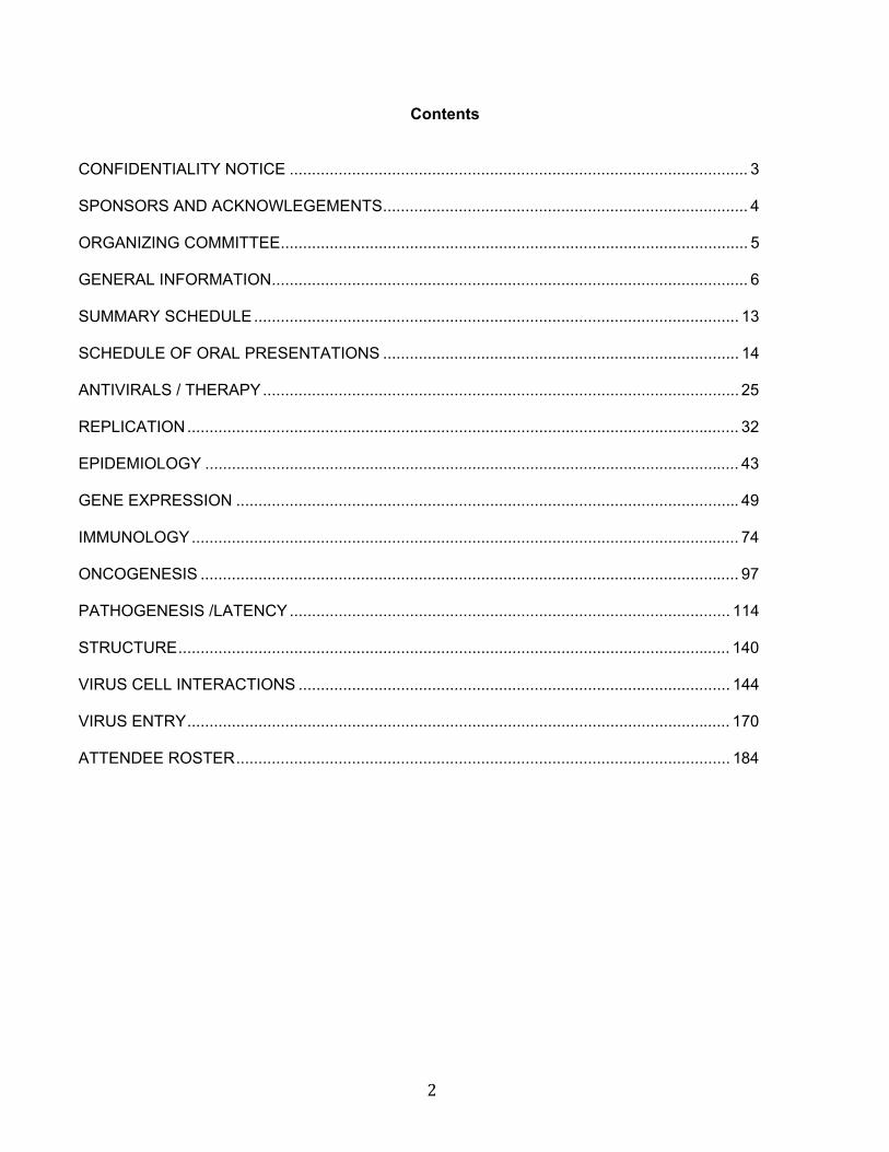
Contents CONFIDENTIALITY NOTICE ....................................................................................................... 3
SPONSORS AND ACKNOWLEGEMENTS.................................................................................. 4
ORGANIZING COMMITTEE......................................................................................................... 5
GENERAL INFORMATION........................................................................................................... 6
SUMMARY SCHEDULE............................................................................................................. 13
SCHEDULE OF ORAL PRESENTATIONS ................................................................................ 14
ANTIVIRALS / THERAPY........................................................................................................... 25
REPLICATION............................................................................................................................ 32
EPIDEMIOLOGY ........................................................................................................................ 43
GENE EXPRESSION ................................................................................................................. 49
IMMUNOLOGY........................................................................................................................... 74
ONCOGENESIS ......................................................................................................................... 97
PATHOGENESIS /LATENCY................................................................................................... 114
STRUCTURE............................................................................................................................ 140
VIRUS CELL INTERACTIONS ................................................................................................. 144
VIRUS ENTRY.......................................................................................................................... 170
ATTENDEE ROSTER............................................................................................................... 184
3
CONFIDENTIALITY OF RESULTS PRESENTED AT THIS MEETING
To continue in the spirit of the highly successful Workshops in the past, we hope that this 12th
International Workshop on KSHV and Related Agents will provide ample opportunity for informal discussions and exchange of ideas, data and reagents. To promote such interactions, and to encourage participants to present unpublished results, please treat all scientific presentations and abstracts in this book as confidential unpublished research. Abstracts may not be cited. If you want to cite results, please do so as a “personal communication”, only with agreement from the respective authors. Results presented at this meeting may not be used as the basis of research without the explicit permission of the corresponding author. The KSHV field has already benefited from many collaborative ventures and a relatively free flow of reagents. Let’s encourage presentation of recent data and establish the KSHV workshops as an exciting series of meetings that are worth attending and cannot be replaced by scientific journals.
4
SPONSORS
National Cancer Institute
University of Florida Shands Cancer Center
Genetics Institute Emerging Pathogenes Institute
Dept. of Genetics and Microbiology
University of North Carolina at Chapel Hill Center for AIDS Research
Lineberger Comprehensive Cancer Center Dept. of Microbiology and Immunology
ACKNOWLEDGEMENTS
Conference Solutions (www.ConferenceSolutionsInc.com) for overall organization
Z. Hong Zhou for cover art
Taum Hanlon for Webpage design and managements
5
Organizing Committee
David J. Blackbourn Cancer Research UK Cancer Centre, School of Cancer Sciences, Vincent Drive, College of Medical and Dental Sciences, University of Birmingham Ethel Cesarman Department of Pathology and Laboratory Medicine, Weill Cornell Medical College Bala Chandran Department of Microbiology and Immunology, H. M. Bligh Cancer Research Laboratories, Chicago Medical School, Rosalind Franklin University of Medicine and Science Yuan Chang Molecular Virology Program, University of Pittsburgh Cancer Institute Blossom Damania Department of Microbiology & Immunology, Lineberger Comprehensive Cancer Center, Center for AIDS Research (CfAR), University of North Carolina at Chapel Hill Dirk P. Dittmer Department of Microbiology & Immunology, Lineberger Comprehensive Cancer Center, Center for AIDS Research (CfAR), University of North Carolina at Chapel Hill Donald Ganem Howard Hughes Medical Institute and GW Hooper Foundation, Department of Microbiology, University of California Dean Kedes Myles H. Thaler Center for AIDS and Human Retrovirus Research, University of Virginia Health Sciences Enrique A. Mesri University of Miami Miller School of Medicine Ashlee V. Moses Vaccine and Gene Therapy Institute, Oregon Health & Science University John Nicholas Department of Oncology, Johns Hopkins University School of Medicine Chris H. Parsons Hollings Cancer Center, Medical University of South Carolina Rolf Renne Department of Molecular Genetics and Microbiology and UF Shands Cancer Center, University of Florida Thomas F. Schulz Institute of Virology, Hannover Medical School Sankar Swaminathan UF Shands Cancer Center, University of Florida Denise Whitby Viral Oncology Section, AIDS and Cancer Virus Program, SAIC-Frederick, NCI-Frederick, Yan Yuan Department of Microbiology, School of Dental Medicine, University of Pennsylvania
6
Welcome to the “12th International Workshop on KSHV and Related Agents” at the Doubletree Guest Suites in Charleston, South Carolina, 13-16 September 2009. WORKSHOP VENUE Host Hotel Doubletree Guest Suites Historic Charleston 181 Church Street Charleston, SC 29401 USA www.charlestondoubletree.com Phone: 843.577.2466 Fax: 843.577.9099 Check-in: 4:00 pm Check-out: 12:00 pm Overflow Hotel Andrew Pinckney Inn 40 Pinckney Street Charleston, SC 29401 USA www.andrewpinckneyinn.com Phone: 843.937.8800 Fax: 843.937.8810 Check-in: 3:00 pm Check-out: 11:00 am Business Center The Doubletree features complimentary 24-hour internet access, photocopier, fax machine and printer. It is located behind the Front Desk. There are additional computers and a printer available for guest use near the lobby elevators. Internet Access Wireless internet access is complimentary in the lobby and in The Lighthouse Café. In guest rooms, internet access is available for $9.95 per day or $8.00 per day for three or more days. Fitness Center A Fitness Center is available and open from 7:00am-10:00pm. It’s located on the 3rd floor. Use of the Fitness Center is provided complimentary for all hotel guests.

7
Hotel Food & Beverage Outlets For meals not provided by the Workshop, below is a listing of the hotel’s options for breakfast and for dinner. There is not an option for lunch at the hotel. However, there are several restaurants in close proximity to the hotel. Please see map and suggestions. The Lighthouse Café The Lighthouse Cafe offers a comfortable and relaxing atmosphere for your early morning breakfast. Awake each morning to your choice of a hot Southern Breakfast Buffet or a traditional Continental selection. Attire: Casual Breakfast Hours:
7:00am-10:00am, Monday-Friday 7:30am-10am, Saturday and Sunday
Room Service Room Service is available each morning. We offer three diverse selections to start your morning off right. Room Service Hours:
7:00am-10:00am, Monday-Friday 7:30am-10am, Saturday and Sunday
Hank’s Seafood
Step out the front door of the hotel and travel left just one half block to Hank's Seafood Restaurant. Hank's has been voted Charleston's best seafood restaurant for 9 years running. This restaurant recreates a Classic Charleston Fish House with an old fashioned saloon-style bar and exhibition raw bar. The menu features Southern specialties such as Fried Seafood Platters, Low Country Bouillabaisse and She Crab Soup. Friendly service and a unique wine list earn this new restaurant top honors from locals and visitors alike. Attire: Casual
Dinner Hours:
5:00pm-10:00pm, Sunday-Thursday
5:00pm-11:30pm, Friday and Saturday
NOTE: Hank’s will deliver to guest rooms 5:00pm-10:00pm nightly.
CONFERENCE REGISTRATION Location: Hayne Street Gallery (Charlestonian Ballroom Foyer) Desk Hours: Sunday, 13 September: 4:00pm-8:00pm
Monday, 14 September: 7:00am-5:00pm Tuesday, 15 September: 7:00am-5:00pm Wednesday, 16 September: 8:00am-12:00pm
8
PRESENTER CHECK-IN Location: Hayne Street Gallery (Charlestonian Ballroom Foyer) Limited Desk Hours: Sunday, 13 September: 4:00pm-8:00pm
Monday, 14 September: 7:00am-2:00pm Tuesday, 15 September: 10:00am-2:00pm
POSTER VIEWING SESSION AND SET-UP Posters can be set-up Sunday, 4:00pm-7:15 pm and Monday, 7:00am-12:00pm in the Stono/Ashley/Cooper rooms on the second floor. All posters have been pre-assigned a poster board designated by your abstract number.
A Poster Viewing Session with Beer and Wine Reception will be held on Monday at 8:00pm-10:30pm. Posters must be removed by 6:00pm on Tuesday.
ORAL PRESENTATIONS Your oral presentation is scheduled to last 15 minutes (12 minute talk plus 3 minutes for questions and answers). The organizers have set up the following schedule to receive your presentation in time to ensure that all sessions flow smoothly. Please note that presentations not submitted to the Presenter Check-in Desk on time may result in your talk not being accompanied by graphics. Presentations may be brought in on CD or USB drives. Please refer to the following schedule and bring your presentation to the Presenter Check-in Desk during designated times.
IMPORTANT NOTE: All speakers must use the computers provided by the workshop for their oral presentations. Personal laptops may not be used. MAC and PC computers will be provided by the conference and equipped with the latest software. PowerPoint is the accepted format. Please be sure to bring your presentation to the Hayne Street Gallery (Charlestonian Ballroom Foyer) as directed by the following schedule:
Presentation Date Presentation DUE to Check-in Desk_____
Sunday, 13 September Sunday, 4:00pm-6:00pm Monday, 14 September Sunday, 4:00pm-8:00pm Tuesday, 15 September Monday, 7:00am-2:00pm Wednesday, 16 September Tuesday, 10:00am-2:00pm NAME BADGES Please wear your name badge at all times during the conference and to the banquet. It serves as your entry ticket to conference meetings and meals.
9
MEALS The following meals are included in your registration: Sunday, 13 September Welcome Reception 6:00pm-7:30pm Palmetto Courtyard Monday, 14 September Breakfast 7:00am-8:15am Palmetto Courtyard Morning Break 10:15am-10:45am Palmetto Courtyard Lunch 12:30pm-2:00pm Palmetto Courtyard Afternoon Break 4:00pm-4:30pm Palmetto Courtyard Poster Session/Reception 8:00pm-10:30pm Stono/Ashley/Cooper Tuesday, 15 September Breakfast 7:00am-8:15am Palmetto Courtyard Morning Break 10:15am-10:45am Palmetto Courtyard Lunch 12:30pm-2:00pm Palmetto Courtyard Afternoon Break 4:00pm-4:30pm Palmetto Courtyard Depart for Banquet 7:00pm Cypress Lowcountry Grille Wednesday, 16 September Breakfast 7:00am-8:15am Palmetto Courtyard Morning Break 10:15am-10:45am Hayne Street Gallery/Tent Tuesday Night Banquet at Cypress Lowcountry Grille The location of the Banquet on Tuesday night is a very short walk from the hotel at 167 East Bay Street in Charleston’s Historic District. (843.937.4012) The restaurant will be closed to the public and serving only our private group this evening. We will enjoy a very nice plated dinner on both levels of the restaurant – one overlooking the other. We will walk together as a group and depart from the Doubletree Hotel at 7:00pm. Please dress with the warm South Carolina temperatures in mind.
10
TRANSPORTATION TO CHARLESTON INTERNATIONAL AIRPORT (CHS)
Taxi / Shuttle
Taxis and shuttle services are available to the airport from the Doubletree Guest Suites. The cost is approximately $35.00. Please call the Bell Stand at extension 7629 for assistance with your transportation arrangements.
Drivers
From the front door of the hotel, turn right onto Market Street and turn right again onto Meeting Street. Follow Meeting Street to I-26 West. Take I-26 West to 526 West. Take the first exit – International Boulevard. Turn right at the end of the exit ramp onto International Boulevard and follow the signs to the airport.
11
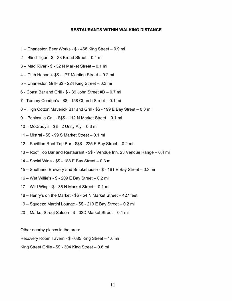
RESTAURANTS WITHIN WALKING DISTANCE
1 – Charleston Beer Works - $ - 468 King Street – 0.9 mi
2 – Blind Tiger - $ - 38 Broad Street – 0.4 mi
3 – Mad River - $ - 32 N Market Street – 0.1 mi
4 – Club Habana- $$ - 177 Meeting Street – 0.2 mi
5 – Charleston Grill- $$ - 224 King Street – 0.3 mi
6 - Coast Bar and Grill - $ - 39 John Street #D – 0.7 mi
7– Tommy Condon’s - $$ - 158 Church Street – 0.1 mi
8 – High Cotton Maverick Bar and Grill - $$ - 199 E Bay Street – 0.3 mi
9 – Peninsula Grill - $$$ - 112 N Market Street – 0.1 mi
10 – McCrady’s - $$ - 2 Unity Aly – 0.3 mi
11 – Mistral - $$ - 99 S Market Street – 0.1 mi
12 – Pavillion Roof Top Bar - $$$ - 225 E Bay Street – 0.2 mi
13 – Roof Top Bar and Restaurant - $$ - Vendue Inn, 23 Vendue Range – 0.4 mi
14 – Social Wine - $$ - 188 E Bay Street – 0.3 mi
15 – Southend Brewery and Smokehouse - $ - 161 E Bay Street – 0.3 mi
16 – Wet Willie’s - $ - 209 E Bay Street – 0.2 mi
17 – Wild Wing - $ - 36 N Market Street – 0.1 mi
18 – Henry’s on the Market - $$ - 54 N Market Street – 427 feet
19 – Squeeze Martini Lounge - $$ - 213 E Bay Street – 0.2 mi
20 – Market Street Saloon - $ - 32D Market Street – 0.1 mi
Other nearby places in the area:
Recovery Room Tavern - $ - 685 King Street – 1.6 mi
King Street Grille - $$ - 304 King Street – 0.6 mi
12
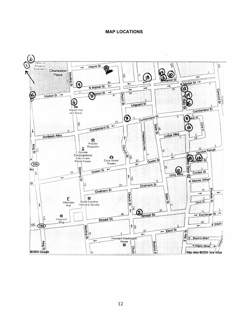
MAP LOCATIONS
13
Schedule of Scientific Sessions: (75 talks)
Sunday, September 13, 2009: Opening remarks: 7:15 pm - 7:30 pm
Replication: 7:30 pm - 9:00 pm 6 talks
Monday, September 14, 2009: Antivirals/Therapy: 8:15 am - 10:15 am 8 talks
Coffee break
Structure/Morphogenesis: 10:45 am - 12:30 pm 7 talks
Lunch break/Poster Viewing
Gene Expression – IE: 2:00 pm - 3:30 pm 6 talks
Coffee break
Immunology: 4:00 pm - 5:30 pm 6 talks
Dinner on your own in Charleston
Poster Session: 8:00 pm – 10:30 pm
Tuesday, September 15, 2009: miRNA: 8:15 am - 10:15 am 8 talks
Coffee break
Pathogenesis (I): 10:45 am - 12:30 pm 7 talks
Lunch break/ Poster Viewing
Latency (I): 2:00 pm - 4:00 pm 8 talks
Coffee break
Tropism/Epidemiology: 4:30 pm - 6:00 pm 6 talks
Banquet: 7:00 pm - 9:00 pm
Wednesday, September 16, 2009 Pathogenesis (II): 8:15 am - 10:00 am 7 talks
Coffee break
Latency (II): 10:30 am - 12:00 pm 6 talks
Concluding remarks/Meeting adjourned
14
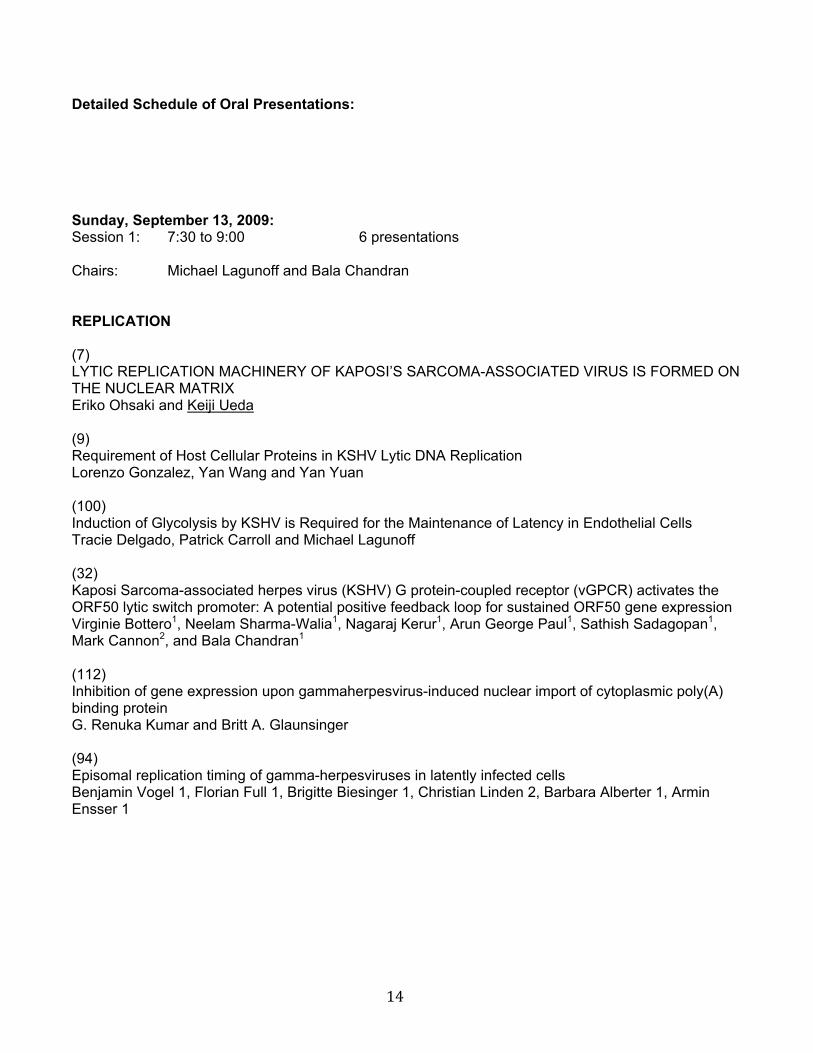
Detailed Schedule of Oral Presentations: Sunday, September 13, 2009: Session 1: 7:30 to 9:00 6 presentations Chairs: Michael Lagunoff and Bala Chandran REPLICATION (7) LYTIC REPLICATION MACHINERY OF KAPOSI’S SARCOMA-ASSOCIATED VIRUS IS FORMED ON THE NUCLEAR MATRIX Eriko Ohsaki and Keiji Ueda (9) Requirement of Host Cellular Proteins in KSHV Lytic DNA Replication Lorenzo Gonzalez, Yan Wang and Yan Yuan (100) Induction of Glycolysis by KSHV is Required for the Maintenance of Latency in Endothelial Cells Tracie Delgado, Patrick Carroll and Michael Lagunoff (32) Kaposi Sarcoma-associated herpes virus (KSHV) G protein-coupled receptor (vGPCR) activates the ORF50 lytic switch promoter: A potential positive feedback loop for sustained ORF50 gene expression Virginie Bottero1, Neelam Sharma-Walia1, Nagaraj Kerur1, Arun George Paul1, Sathish Sadagopan1, Mark Cannon2, and Bala Chandran1 (112) Inhibition of gene expression upon gammaherpesvirus-induced nuclear import of cytoplasmic poly(A) binding protein G. Renuka Kumar and Britt A. Glaunsinger (94) Episomal replication timing of gamma-herpesviruses in latently infected cells Benjamin Vogel 1, Florian Full 1, Brigitte Biesinger 1, Christian Linden 2, Barbara Alberter 1, Armin Ensser 1
15
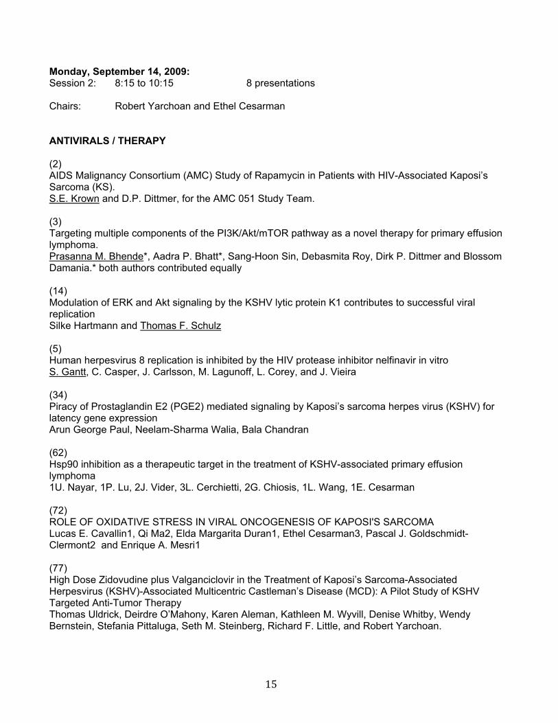
Monday, September 14, 2009: Session 2: 8:15 to 10:15 8 presentations Chairs: Robert Yarchoan and Ethel Cesarman ANTIVIRALS / THERAPY (2) AIDS Malignancy Consortium (AMC) Study of Rapamycin in Patients with HIV-Associated Kaposi’s Sarcoma (KS). S.E. Krown and D.P. Dittmer, for the AMC 051 Study Team. (3) Targeting multiple components of the PI3K/Akt/mTOR pathway as a novel therapy for primary effusion lymphoma. Prasanna M. Bhende*, Aadra P. Bhatt*, Sang-Hoon Sin, Debasmita Roy, Dirk P. Dittmer and Blossom Damania.* both authors contributed equally (14) Modulation of ERK and Akt signaling by the KSHV lytic protein K1 contributes to successful viral replication Silke Hartmann and Thomas F. Schulz (5) Human herpesvirus 8 replication is inhibited by the HIV protease inhibitor nelfinavir in vitro S. Gantt, C. Casper, J. Carlsson, M. Lagunoff, L. Corey, and J. Vieira (34) Piracy of Prostaglandin E2 (PGE2) mediated signaling by Kaposi’s sarcoma herpes virus (KSHV) for latency gene expression Arun George Paul, Neelam-Sharma Walia, Bala Chandran (62) Hsp90 inhibition as a therapeutic target in the treatment of KSHV-associated primary effusion lymphoma 1U. Nayar, 1P. Lu, 2J. Vider, 3L. Cerchietti, 2G. Chiosis, 1L. Wang, 1E. Cesarman (72) ROLE OF OXIDATIVE STRESS IN VIRAL ONCOGENESIS OF KAPOSI'S SARCOMA Lucas E. Cavallin1, Qi Ma2, Elda Margarita Duran1, Ethel Cesarman3, Pascal J. Goldschmidt-Clermont2 and Enrique A. Mesri1 (77) High Dose Zidovudine plus Valganciclovir in the Treatment of Kaposi’s Sarcoma-Associated Herpesvirus (KSHV)-Associated Multicentric Castleman’s Disease (MCD): A Pilot Study of KSHV Targeted Anti-Tumor Therapy Thomas Uldrick, Deirdre O’Mahony, Karen Aleman, Kathleen M. Wyvill, Denise Whitby, Wendy Bernstein, Stefania Pittaluga, Seth M. Steinberg, Richard F. Little, and Robert Yarchoan.
16
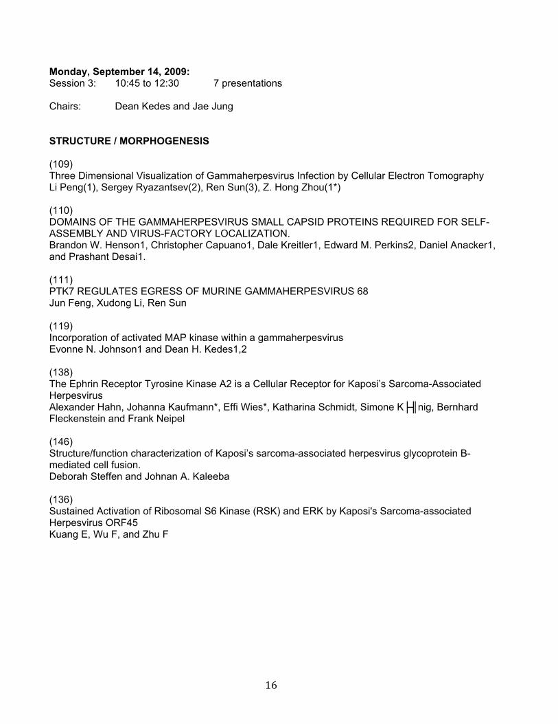
Monday, September 14, 2009: Session 3: 10:45 to 12:30 7 presentations Chairs: Dean Kedes and Jae Jung STRUCTURE / MORPHOGENESIS (109) Three Dimensional Visualization of Gammaherpesvirus Infection by Cellular Electron Tomography Li Peng(1), Sergey Ryazantsev(2), Ren Sun(3), Z. Hong Zhou(1*) (110) DOMAINS OF THE GAMMAHERPESVIRUS SMALL CAPSID PROTEINS REQUIRED FOR SELF-ASSEMBLY AND VIRUS-FACTORY LOCALIZATION. Brandon W. Henson1, Christopher Capuano1, Dale Kreitler1, Edward M. Perkins2, Daniel Anacker1, and Prashant Desai1. (111) PTK7 REGULATES EGRESS OF MURINE GAMMAHERPESVIRUS 68 Jun Feng, Xudong Li, Ren Sun (119) Incorporation of activated MAP kinase within a gammaherpesvirus Evonne N. Johnson1 and Dean H. Kedes1,2 (138) The Ephrin Receptor Tyrosine Kinase A2 is a Cellular Receptor for Kaposi’s Sarcoma-Associated Herpesvirus Alexander Hahn, Johanna Kaufmann*, Effi Wies*, Katharina Schmidt, Simone K├╢nig, Bernhard Fleckenstein and Frank Neipel (146) Structure/function characterization of Kaposi’s sarcoma-associated herpesvirus glycoprotein B-mediated cell fusion. Deborah Steffen and Johnan A. Kaleeba (136) Sustained Activation of Ribosomal S6 Kinase (RSK) and ERK by Kaposi's Sarcoma-associated Herpesvirus ORF45 Kuang E, Wu F, and Zhu F
17
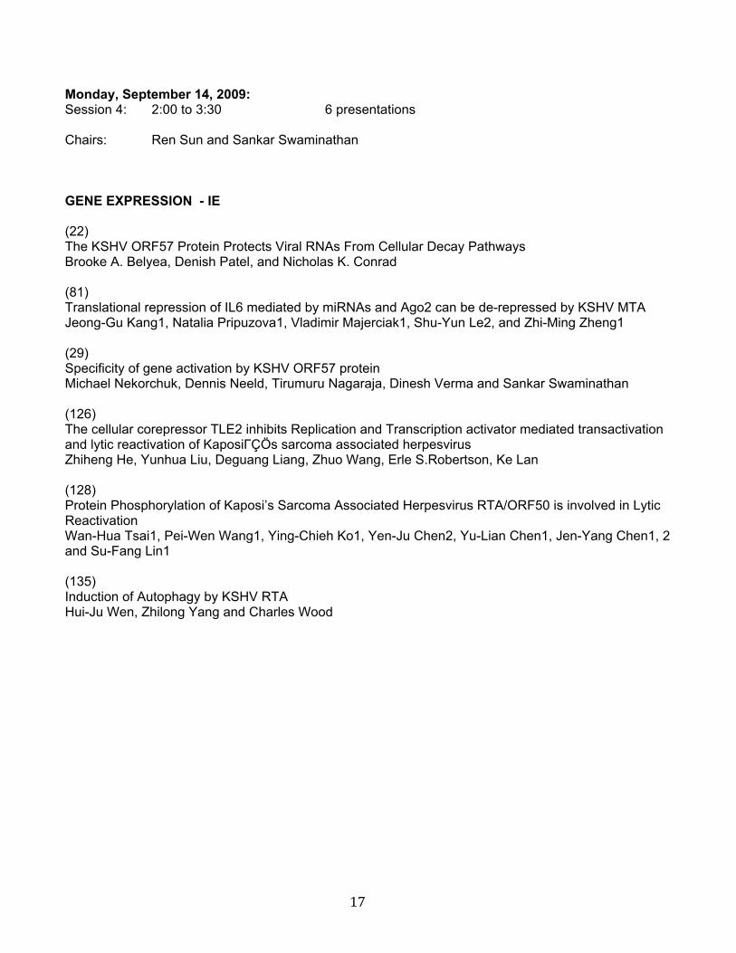
Monday, September 14, 2009: Session 4: 2:00 to 3:30 6 presentations Chairs: Ren Sun and Sankar Swaminathan GENE EXPRESSION - IE (22) The KSHV ORF57 Protein Protects Viral RNAs From Cellular Decay Pathways Brooke A. Belyea, Denish Patel, and Nicholas K. Conrad (81) Translational repression of IL6 mediated by miRNAs and Ago2 can be de-repressed by KSHV MTA Jeong-Gu Kang1, Natalia Pripuzova1, Vladimir Majerciak1, Shu-Yun Le2, and Zhi-Ming Zheng1 (29) Specificity of gene activation by KSHV ORF57 protein Michael Nekorchuk, Dennis Neeld, Tirumuru Nagaraja, Dinesh Verma and Sankar Swaminathan (126) The cellular corepressor TLE2 inhibits Replication and Transcription activator mediated transactivation and lytic reactivation of KaposiΓÇÖs sarcoma associated herpesvirus Zhiheng He, Yunhua Liu, Deguang Liang, Zhuo Wang, Erle S.Robertson, Ke Lan (128) Protein Phosphorylation of Kaposi’s Sarcoma Associated Herpesvirus RTA/ORF50 is involved in Lytic Reactivation Wan-Hua Tsai1, Pei-Wen Wang1, Ying-Chieh Ko1, Yen-Ju Chen2, Yu-Lian Chen1, Jen-Yang Chen1, 2 and Su-Fang Lin1 (135) Induction of Autophagy by KSHV RTA Hui-Ju Wen, Zhilong Yang and Charles Wood
18
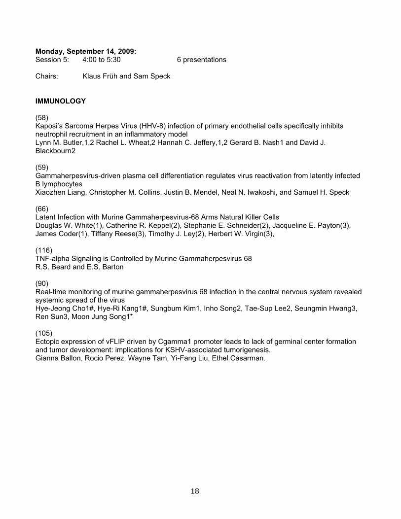
Monday, September 14, 2009: Session 5: 4:00 to 5:30 6 presentations Chairs: Klaus Früh and Sam Speck IMMUNOLOGY (58) Kaposi’s Sarcoma Herpes Virus (HHV-8) infection of primary endothelial cells specifically inhibits neutrophil recruitment in an inflammatory model Lynn M. Butler,1,2 Rachel L. Wheat,2 Hannah C. Jeffery,1,2 Gerard B. Nash1 and David J. Blackbourn2 (59) Gammaherpesvirus-driven plasma cell differentiation regulates virus reactivation from latently infected B lymphocytes Xiaozhen Liang, Christopher M. Collins, Justin B. Mendel, Neal N. Iwakoshi, and Samuel H. Speck (66) Latent Infection with Murine Gammaherpesvirus-68 Arms Natural Killer Cells Douglas W. White(1), Catherine R. Keppel(2), Stephanie E. Schneider(2), Jacqueline E. Payton(3), James Coder(1), Tiffany Reese(3), Timothy J. Ley(2), Herbert W. Virgin(3), (116) TNF-alpha Signaling is Controlled by Murine Gammaherpesvirus 68 R.S. Beard and E.S. Barton (90) Real-time monitoring of murine gammaherpesvirus 68 infection in the central nervous system revealed systemic spread of the virus Hye-Jeong Cho1#, Hye-Ri Kang1#, Sungbum Kim1, Inho Song2, Tae-Sup Lee2, Seungmin Hwang3, Ren Sun3, Moon Jung Song1* (105) Ectopic expression of vFLIP driven by Cgamma1 promoter leads to lack of germinal center formation and tumor development: implications for KSHV-associated tumorigenesis. Gianna Ballon, Rocio Perez, Wayne Tam, Yi-Fang Liu, Ethel Casarman.
19
Tuesday, September 15, 2009: Session 6: 8:15 to 10:15 8 presentations Chairs: Don Ganem and Paul Kellam miRNA (23) Regulation of RTA and lytic reactivation by a KSHV-encoded microRNA Priya Bellare and Don Ganem, HHMI and GW Hooper Foundation, University of California, San Francisco, CA 94143 (45) A Global Analysis of Conserved and Non-Conserved Herpesvirus miRNAs Nicole Walz, Thomas Christalla and Adam Grundhoff (76) KSHV miR-K12-11 expression in human progenitors during in-vivo hematopoiesis induces B-cell expansion in NOD/LtSz-scid IL-2R gamma null mice. Isaac W. Boss (1), Peter E. Nadeau (2), Jeffrey R. Abbott (2), Ayalew Mergia (2), Rolf Renne (1) (97) KSHV-encoded miRNAs target MAF to induce endothelial cell reprogramming Amy Hansen1, Stephen Henderson1*, Dimitris Lagos1*, Eve Coulter2, Leonid Nikitenko1, Fiona Gratrix1, Karlie Plaisance3, Rolf Renne3, Mark Bower4, Richard Jenner2, Paul Kellam2,5 and Chris Boshoff1** (104) A KSHV microRNA attenuates p53-induced cell cycle arrest through down-regulation of the p21 tumor suppressor Eva Gottwein and Bryan Cullen (118) Regulation of apoptosis by Kaposi’s sarcoma associated herpesvirus microRNAs Guillaume Suffert 1*, Georg Malterer 2*, Jean Hausser 3, Liisa Lappalainen 4, Tomi Ivacevic 5, Vladimir Benes 5, Olivier Voinnet 6, Mihaela Zavolan 3, Juergen Haas 2,7*, Paivi Ojala 4*, Sebastien Pfeffer 1* (31) Regulation of delayed-early transcripts by Kaposi’s sarcoma-associated herpesvirus -encoded microRNAs Horng-Ru Lin and Don Ganem (148) KSHV encoding microRNAs regulation of xCT: implications for de novo infection and prolonged survival of infected cells in an environment of oxidative stress Zhiqiang Qin1,2, Eduardo Freitas1, Ben Kalivas1, Roger Sullivan1, Karlie Plaisance3, Rocky Bacelieri4, Patricia Kearney1,2, Johnan Kaleeba5, and Chris Parsons1,2*
20
Tuesday, September 15, 2009: Session 7: 10:45 to 12:30 7 presentations Chairs: SJ Gao and Blossom Damania PATHOGENESIS (I) (89) Sulfotyrosines of the Kaposi’s sarcoma-associated herpesvirus G protein-coupled receptor promotes tumorigenesis through autocrine activation Hao Feng 1, Michael R. Farzan 2, and Pinghui Feng 1 (96) Viral Bcl-2-mediated Evasion of Autophagy Aids Chronic Infection of γHerpesvirus 68 Xiaofei E, Seungmin Hwang, Soohwan Oh, Yousang Gwack, Timothy F. Kowalik, Ren Sun, Jae U Jung, and Chengyu Liang (74) HSP90 & HSP40 ARE REQUIRED FOR THE EXPRESSION AND ANTI-APOPTOTIC FUNCTION OF KSHV K1 Kwun Wah Wen and Blossom Damania (142) KSHV induces transdifferentiation of lymphatic endothelial cells in a 3-D endothelial cell model Simonas Laurinavicius1, Fang Cheng1, Nami Sugiyama2, Kari Alitalo2, Kaisa Lehti2, and Päivi M. Ojala1
(122) Tetherin restricts Kaposi’s Sarcoma-associated Herpesvirus and is antagonized by the viral RING-CH ubiquitin ligase K5 Claire Pardieu (1), Sam J Wilson (1,3), Alessandra Calvi (2), Trinity Zang (3), Paul Bieniasz (3), Paul Kellam (1), Greg J Towers (1,4) and Stuart J D Neil (2,4)1. (124) Bim Nuclear Translocation and Inactivation by HHV-8 Interferon Regulatory Factor 1 Young Bong Choi and John Nicholas (133) E3 ubiquitin ligase activity of KSHV K5 is required to counteract the cellular restriction factor tetherin (BST2) Mandana Mansouri, Kasinath Viswanathan, Janet L.Douglas, Jean Gustin, Jennie Hines, Ashlee V.Moses and Klaus Fruh
21
Tuesday, September 15, 2009: Session 8: 2:00 to 4:00 8 presentations Chairs: Ken Kaye and Erle Robertson LATENCY (I) (40) Single Cell Analysis of K1 Expression During Latent KSHV Infection Sanjay Chandriani and Don Ganem (42) Remodeling of Viral Chromatin by LANA and K-Rta via Sumoylation Yoshihiro Izumiya, Chie Izumiya, Mamata Pochampalli, Paul A. Luciw, Hsing-Jien Kung (47) FLIP-mediated autophagy in cell death control Jong-Soo Lee1,2, Qinglin Li2*, June-Yong Lee1, Sun-Hwa Lee1, Hyeongnam Jeong1, Hye-Ra Lee1,2, Heesoon Chang1,2, Fu-Chun Zhou3, Shou-Jiang Gao3, Chengyu Liang1,2, and Jae U. Jung1,2,# (130) A systems biology approach to identify combination effects of KSHV genes on NF-kappaB activation A. KONRAD, E. WIES, M. THURAU, G. MARQUARDT, E. NASCHBERGER, R. JOCHMANN, T.F. SCHULZ, H. ERFLE, B. BRORS, B. LAUSEN, F. NEIPEL and M. STUERZL, (51) KSHV viral Interferon Regulatory Factor 4 (K10.1) interacts with the cellular repressor protein CSL/CBF1, an effector protein of the Notch signalling pathway Katharina Heinzelmann(1), Barbara Scholz(1), Elisabeth Kremmer(2), Ronald Frank(3), J├╝rgen Haas(4), Even Fossum(4), Bettina Kempkes(1) (60) Regulation of the Myc-Max-Mad transcription network by the cellular centromeric protein, KSHV LANA-interacting protein 1 (KLIP1). Kurt Kuhne1,2,3, Tiffany Jones1,4, Moraima Guadalupe1,2, Shou-Jiang Gao1,2,3,4 (68) The Kaposin B Protein of Kaposi’s Sarcoma-Associated Herpesvirus (KSHV) Promotes Actin Cytoskeleton Rearrangements in Endothelial Cells J. A. Corcoran and C. McCormick (75) KSHV Manipulates Canonical Notch Signaling through Direct Upregulation of DLL4 and JAG1 to Alter Cell Cycle Gene Expression in Lymphatic Endothelial Cells Victoria Emuss, Dimitris Lagos, Arnold Pizzey, Fiona Gratrix, Stephen Henderson, Chris Boshoff
22
Tuesday, September 15, 2009: Session 9: 4:30 to 6:00 6 presentations Chairs: Denise Whitby and Thomas Schulz TROPISM / EPIDEMIOLOGY (15) KSHV interaction with Langerhans and dermal dendritic cells through C-type lectins Giovanna Rappocciolo, Mariel Jais, Paolo Piazza, Frank Jenkins and Charles Rinaldo (17) Seroprevalence of Kaposi’s Sarcoma-Associated Herpesvirus and Risk Factors in Xinjiang, China Bishi Fu1, Feng Sun2, Baolin Li1, Lei Yang3, Yan Zeng3, Xiulian Sun1, Fanhong Xu4, Simon Rayner1, Moraima Guadalupe5, Shou-Jiang Gao1,5 and Linding Wang1 (19) Prevalence of KSHV and Incidence of KS in the Multicenter AIDS Cohort Study (MACS) in the pre-HAART and HAART Eras W. Miley1, M. Kesler2, J. Mullen2, M.N. Labo1, D. Whitby1 and L.P. Jacobson2 (69) The search for HHV9, the member of the rhadinovirus-2 (RV2) lineage of Old World primate rhadinoviruses predicted to infect humans. Timothy M. Rose, Courtney Gravett, Jonathan T. Ryan, and A. Gregory Bruce (114) Activated CD4+ T cells regulate KSHV reactivation in primary B cells in culture. Jinjong Myoung and Don Ganem (120) Reconstructing a Time Varying Regulatory Network for KSHV Infection of Human Primary Endothelial Cells Jia Meng1, Shou-Jiang Gao2,3, Yufei Huang1,3
23
Wednesday, September 16, 2009: Session 10: 8:15 to 10:15 7 presentations Chairs: Enrique Mesri and John Nicholas PATHOGENESIS (II) (91) Myc Represses KSHV RTA Expression and is Essential for the Maintenance of KSHV Latency Xudong Li, Jun Feng, Shijia Chen, Hongyu Deng, Ren Sun (137) GLTSCR2/PICT-1, a Putative Tumor Suppressor Gene Product, Induces the Nucleolar Targeting of KS-Bcl-2 Protein Inna Kalt, Adi Schachor, Tatyana Shteinberg, and Ronit Sarid (125) Intracellular Localization and Function of Human Herpesvirus 8 Interleukin-6 Daming Chen, Young Bong Choi, Gordon Sandford, and John Nicholas (26) Epigenetic regulation of latency and reactivation of Kaposi’s sarcoma-associated herpesvirus Zsolt Toth and Jae U. Jung (52) TOLL-LIKE RECEPTOR SIGNALING CONTROLS REACTIVATION OF KSHV FROM LATENCY Sean M. Gregory, John A. West, Patrick J. Dillon, Chelsey Hilscher, Dirk P. Dittmer and Blossom Damania. (55) KAPOSI’S SARCOMA-ASSOCIATED HERPESVIRUS LATENCY-ASSOCIATED NUCLEAR ANTIGEN INHIBITS INTERFERON (IFN)- EXPRESSION BY COMPETING WITH IFN REGULATORY FACTOR-3 FOR BINDING TO IFN- PROMOTER. Nathalie Cloutier and Louis Flamand. (57) Suppression of human T cell clone activity by KSHV vOX2 and its cellular counterpart CD200 Karen Misstear1, Rachel Colman1, Simon Chanas1, Janet Lord2, Andrew Hislop1, David J. Blackbourn1;
24
Wednesday, September 16, 2009: Session 11: 10:45 to 12:00 6 presentations Chairs: Diane Hayward and Hsing-Jien Kung LATENCY (II) (86) KSHV LANA Mediated Episome Maintenance in Nonhuman Cell Lines Vishi Srinivasan, Katherine Slain, Erika De Leon-Vazquez, Brenna Kelley-Clarke, Kenneth M. Kaye (92) DNMT3b and DNMT3a associates with neddylated proteins and KSHV LANA enhances this interaction Melanie Scocco, Gangling Liao, Richard F. Ambinder, and S. Diane Hayward, Meir Shamay (99) SUMO is a Molecular Sensor for Epigenetic Regulation of KSHV Latency Hsing-Jien Kung, Amy Chang, Latricia Fitzgerald, Chie Izumiya, Paul Luciw and Yoshihiro Izumiya (102) Determining host kinase-KSHV LANA interactions utilizing protein chip technology Crystal L Woodard1, Jian Zhu1, Hee-sool Rho1, Rob Newman1, Zhi Xie4, Meir Shamay3, Gangling Liao3, Jin Zhang1,3, Jiang Qian4, Diane Hayward1,3,5, Heng Zhu1,2,5 (95) How Does the DNA-Binding and Dimerization Domain of EBNA1 Inhibit Colony Formation of Primary Effusion Lymphoma Cell Lines, Even in the Absence of EBV? Lindsay Dresang and Bill Sugden (10) Involvement of SSRP1 in latent replication of Kaposi’s sarcoma-associated herpesvirus Jianhong Hu, Eugene Liu and Rolf Renne

25
ANTIVIRALS / THERAPY
26
1. Vickie Marshall ([email protected]) HIF-1 Active Natural Product Extracts with Therapeutic Potential for KSHV Related Disease Vickie Marshall1, Rachel Bagni1, Nazzarena Labo1, Andrea Carras1, Alex Ray1, Betty Conde2, David J. Newman3, Robert H. Shoemaker4, John H. Cardellina II5, Tamara L. Meragelman5 ,Thomas G. McCloud5, Denise Whitby1 1Viral Oncology Section, AIDS and Cancer Virus Program, SAIC-Frederick; 2Viral Technology Laboratory, Advanced Technology Program, SAIC-Frederick, NCI-Frederick; 3Natural Products Branch, and 4Screening Technologies Branch, Developmental Therapeutics Program, NCI; 5Natural Products Support Group, SAIC-Frederick, NCI-Frederick; We recently proposed an “Oncoweed Hypothesis” to partly explain the geographical variation in KSHV prevalence and disease. We developed a KSHV reactivation screen to identify natural products from KSHV endemic regions that may reactivate KSHV from latency. Reactivation of KSHV has also been reported in hypoxic conditions. One of the hallmarks of the response to hypoxia is activation of the transcription factor HIF-1. We sought to ascertain if natural product extracts which had shown activity in a HIF-1 screen affected KSHV reactivation. Natural products extracts from the DTP repository were assayed in two distinct in vitro models of KSHV infection. In parallel, KSHV chemical reactivators such as phorbol esters (TPA) and histone deacetylase inhibitors (sodium butyrate) were used as positive controls, while unstimulated BCBL-1 cells served as negative controls. Initial screening had identified several extracts from Rubiaceae as containing the known HDAC inhibitor FK228, as possibly having KSHV-reactivating activity. These extracts were incubated with the KSHV latently-infected cell line, BCBL-1, at concentrations of 6.25 to 50 μg/ml for 4 days. KSHV viral loads were determined using quantitative PCR. Four KSHV reactivating extracts were selected for further study at their most potent concentration in a time course study. RNA was obtained from BCBL-1 cells collected at 6, 24, 36, 48, 72, and 96 hours and assayed using a KSHV whole genome array to profile changes in viral gene expression. An expanded dilution series was made to determine the minimum reactive dose. The extracts were incubated with Vero cells stably infected with a recombinant KSHV (rKSHV219), which constitutively expresses GFP, but is induced upon reactivation to express RFP from the RTA-responsive KSHV lytic promoter for polyadenylated nuclear RNA. Reactivation activity was determined by counting the proportion of red fluorescent Vero cells. Extracts containing FK228 induced potent reactivation of KSHV from latency. The most potent extracts induced viral loads equivalent to or greater than sodium butyrate positive controls in BCBL-1 cells. In Vero cells, reactivation was validated by an increase of several fold more RFP positive cells. Viral gene expression analysis showed an ordered upregulation of viral genes in a manner consistent with viral reactivation and an induction of viral genes with HIF-1α responsive elements including ORF 50 and the ORF 34-37 cluster. Those extracts displaying the highest level of KSHV reactivation all were obtained from members of the plant family Rubiaceae which came from east Africa and Madagascar, corresponding to regions of endemic KS. A sampling of Rubiaceae extracts which fail to show the HIF response, and do not contain FK228 by HPLC/MS analysis did not activate KSHV lytic replication in BCBL-1 or Vero cells. Our results indicate that screening of natural product extracts against anticancer targets, such as HIF-1α, can be informative in related fields like viral biology. The identification of naturally occurring compounds capable of reactivation of KSHV may explain the worldwide disparities in KS incidence and disease and potentially identify novel therapeutics or influence treatment strategies.

27
2. Susan Krown ([email protected]) AIDS Malignancy Consortium (AMC) Study of Rapamycin in Patients with HIV-Associated Kaposi’s Sarcoma (KS). S.E. Krown and D.P. Dittmer, for the AMC 051 Study Team. Department of Medicine, Memorial Sloan-Kettering Cancer Center, New York, NY 10065 and University of North Carolina at Chapel Hill, Chapel Hill, NC 27599. The PI3K/Akt/mTOR pathway is activated in KS. Inhibitors of this pathway inhibit the growth of PEL cells in murine xenotransplant models and KS in immunosuppressed recipients of kidney allografts. The AMC performed a pilot study of rapamycin, an mTOR inhibitor, in HIV-infected patients with KS to evaluate its safety, antitumor activity, pharmacologic interactions with antiretroviral therapy (ART), and effects on mTOR-dependent signaling in tumors. Seven KS patients, 4 on HIV protease inhibitor (PI)-based ART and 3 on non-nucleoside reverse transcriptase inhibitor (NNRTI)-based ART were treated with varying doses of rapamycin to achieve target blood trough levels of rapamycin between 5 and 10 ng/ml. Adverse events were generally mild, and no dose-limiting toxicities were observed. There was no increase in HIV RNA plasma levels. Three patients showed partial tumor regression, three showed stable disease, and one had progressive KS. The dose of rapamycin required to achieve target drug trough levels varied markedly depending on the type of ART administered. PIs, and in particular ritonavir, are inhibitors of both CYP3A and P-glycoprotein, and co-administration with rapamycin resulted in marked reduction in the doses of rapamycin required. In the patients on PI-based ART, the maintenance rapamycin dose ranged from 0.1 mg twice a week to 0.3 mg three times a week. By contrast, NNRTIs may induce CYP3A, and among patients on NNRTI-based ART, the maintenance rapamycin doses ranged from 2.3 to 6.7 mg daily (i.e., a >200-fold difference in cumulative weekly dose between the lowest and highest maintenance doses). Immunohistochemical studies of baseline and on-treatment KS biopsies showed a reduction in phospho-S6 (pS6) expression. This demonstrates that rapamycin inhibited its target in KS tumors in patients. Additional studies on other members in the pathway and KSHV viral load will be presented. The patients showing a decrease in pS6 were those who were biopsied at later time points in the study, but were also those who had the highest rapamycin trough levels. These data suggest that mTOR inhibition may be a safe and effective strategy for treatment of HIV-associated Kaposi’s sarcoma, and that additional trials are warranted to address questions regarding appropriate target trough ranges for optimal efficacy. Supported by UO1 CA121947.
28
3. Prasanna M. Bhende ([email protected]) Targeting multiple components of the PI3K/Akt/mTOR pathway as a novel therapy for primary effusion lymphoma. Prasanna M. Bhende*, Aadra P. Bhatt*, Sang-Hoon Sin, Debasmita Roy, Dirk P. Dittmer and Blossom Damania.* both authors contributed equally Lineberger Comprehensive Cancer Center and the Department of Microbiology & Immunology, University of North Carolina at Chapel Hill, Chapel Hill, NC 27599, USA Kaposi’s Sarcoma-associated Herpesvirus (KSHV) is linked with lymphoproliferative disorders of B cells including Primary Effusion Lymphoma (PEL) and Multicentric Castleman’s Disease (MCD). We have previously reported that the KSHV transforming protein, K1, induces the activation and phosphorylation of Akt kinase via the activation of phosphotidylinositol-3-OH kinase (PI3K). We have also shown that K1-expressing B lymphocytes and endothelial cells display increased phosphorylation and activation of PI3K, Akt, and the mammalian Target Of Rapamycin (mTOR). Activated Akt can directly and indirectly induce the phosphorylation and activation of mTOR, and also phosphorylate and inactivate pro-apoptotic factors such as GSK-3β and FKHR. Additionally, activated Akt inhibits the tumor suppressor activity of AMP-activated protein kinase (AMPK), a negative regulator of mTOR. Upon activation, mTOR phosphorylates its downstream effector proteins, p70 S6 kinase and S6 ribosomal protein, thereby stimulating protein synthesis. Thus, activation of the PI3K/Akt/mTOR pathway stimulates protein synthesis and cell survival. This signaling pathway is highly activated in PEL and hence inhibition of this pathway serves as a promising therapeutic target for PEL. In this study, we tested several different compounds that target multiple kinases in this pathway including PI3K, Akt, mTOR, and AMPK, for therapeutic efficacy against PEL. We found that compounds that modulated activity of all four kinases, either singly or in combination, were efficacious in inhibiting PEL proliferation in vitro and in vivo, using PEL xenograft mouse models. These compounds include the thiazolidinediones, which are activators of AMPK, and the Akt inhibitor, perifosine.
29
4. Kristen M. Tamburro ([email protected]) NextGen Sequencing for Diagnosis of AIDS-defining Cancers: A case study Kristen M. Tamburro, Jessica Poisson, Charles van der Horst, Debasmita Roy, Nadia Malouf, Dirk P. Dittmer Lineberger Comprehensive Cancer Center and the Department of Microbiology & Immunology, University of North Carolina at Chapel Hill, Chapel Hill, NC 27599, USA Kaposi sarcoma associated-herpesvirus (KSHV, HHV8) is the causative agent of Kaposi Sarcoma (KS), Primary Effusion Lymphoma (PEL), and Multicentric Castleman Disease (MCD), all of which can often be diagnosed in terminal AIDS patients. Metastatic KS can be found lining the pleural cavity, the intestines, and lung. We studied a rare presentation of non-neoplastic KSHV-disease in an AIDS patient. The patient had minimal skin KS and no evidence of PEL or MCD. Using real-time QPCR, we nevertheless found KSHV viral loads > 1 million copies / ml. Sequence-based microbiome profiling in PBMCs uncovered concurrent acute HHV-6a infection, leading us to conclude that the cause of death was acute KSHV/HHV6a-viremia rather than KSHV-associated neoplasia. -- POSTER
30
5. Soren Gantt, MD PhD ([email protected]) Human herpesvirus 8 replication is inhibited by the HIV protease inhibitor nelfinavir in vitro S. Gantt, C. Casper, J. Carlsson, M. Lagunoff, L. Corey, and J. Vieira Background: Antiretroviral drug (ARV) combinations, especially those containing HIV protease inhibitors (PIs), are beneficial in preventing and treating HIV-associated Kaposi sarcoma (KS). Suppression of HIV infection with resulting improvement in immune responses against HHV-8 and/or KS tumor cells appears only partly to explain the effects of ARVs on KS. Several PIs have been shown to have anti-tumor activities, which may also play a role in controlling KS. In addition, ARVs may have direct effects on HHV-8 replication, although this has not been extensively studied. Methods: We screened a large panel of ARVs (nelfinavir, indinavir, saquinavir, ritonavir, lopinavir, amprenavir, atazanavir, zidovudine, stavudine, lamivudine, didanosine, abacavir, tenofovir, nevirapine, and efavirenz) for their ability to inhibit HHV-8 replication in vero cells using a recombinant virus that expresses an inducible secretory alkaline phosphatase (SeAP). Cell viability was determined by trypan blue staining, as well as measurement of ATP levels (CellTiter Glo, Promega) and MTT assays. Ganciclovir was used as a positive control for inhibition of HHV-8 replication. Results: Nelfinavir inhibited HHV-8 replication by 50% at a concentration (IC-50) of 7.5 uM (+/3.0), which is within the range of therapeutic plasma levels seen in the course of treatment of HIV infection. Minimal activity was observed with zidovudine, stavudine, lopinavir and ritonavir, but the IC-50 was not achieved for these drugs. None of the other ARVs tested showed inhibitory activity against HHV-8. No toxicity was observed at nelfinavir concentrations <20 uM. The IC-50 of ganciclovir against HHV-8 in this assay was 20 uM (+/8). Conclusions: Concentrations of nelfinavir that are achievable in plasma are able to inhibit HHV-8 replication in vitro. As HHV-8 is not known to encode an aspartyl protease, nelfinavir likely acts through a novel mechanism. Although a viral target is possible, we speculate that inhibition of HHV-8 replication occurs due to one or more of nelfinavir’s multiple effects on cellular functions, some of which impart its anti-tumor activities. Nelfinavir or related compounds may be of particular clinical benefit for the prevention of KS due to the ability to suppress HIV infection, inhibit tumor growth and metastasis, as well as inhibit replication of HHV-8. As nelfinavir is approved for the treatment of HIV infection and has a favorable safety profile, it is an attractive agent for use against KS. Further studies are needed to determine if nelfinavir is effective in suppressing HHV-8 replication in vivo.
31
6. Sarumathi Mohan ([email protected]) Defining a role for sphingolipids in PEL cell growth and survival Sarumathi Mohan1, Charles D. Smith2,4, and Chris Parsons1,3* Departments of Medicine1, Pharmaceutical Sciences2, and Microbiology/Immunology3 Medical University of South Carolina Charleston, SC *Corresponding author: [email protected] 4Apogee Biotechnology Corporation Hershey Center for Applied Research Hummelstown, PA KSHV is the causative agent of primary effusion lymphoma (PEL), a tumor located within body cavities of patients infected with HIV. Conventional chemotherapy induces unwanted toxicity and carries substantial failure rates for this tumor, and the appearance of PEL portends an ominous prognosis. Intermediates in lipid metabolic pathways activate a number of signaling transduction events previously implicated in supporting KSHV gene expression and PEL pathogenesis. These include sphingosine kinase (SK) and its bioactive sphingolipid by-product, sphingosine-1-phosphate (S1P). SK and S1P are upregulated in many cancers and are associated with tumor progression. A novel inhibitor of SK, ABC294640, has demonstrated activity against a number of tumors using in vivo models and has proven safe in phase I clinical trials. In preliminary experiments using flow cytometry, we found that ABC294640 reduces growth and survival of PEL cells (BCBL-1) and KSHV-infected endothelial cells, but not uninfected control cells. Parallel mass spectrophotometry experiments revealed that for BCBL-1, ABC294640 increases cell concentrations of ceramides—sphingolipid moieties implicated in the induction of cancer cell apoptosis. Finally, RT-PCR revealed that ABC294640 induces alterations in KSHV transcript expression within BCBL-1, including upregulation of RTA and downregulation of v-FLIP. Since both sphingolipid signaling and maintenance of latent KSHV gene expression in PEL cells is dependent upon NF-kB activation (the latter, in part, through v-FLIP activation of NF-kB), we hypothesized that ABC294640 reduces NF-kB activation in PEL cells. Subsequent experiments verified that ABC294640 reduces constitutive NF-kB expression by BCBL-1 cells within 2 hours. Finally, preliminary experiments using a well-characterized murine model for PEL demonstrated that ABC294640 significantly reduces intraperitoneal tumor burden. These data support the potential importance of sphingolipid-mediated signaling for PEL cell growth and survival and the potential utility of ABC294640 as a therapy for PEL. Additional data are required to discern whether this effect is specific for KSHV-infected cells and whether sphingolipids play a role in maintaining a KSHV transcriptome supportive of PEL cell growth and survival.

32
REPLICATION
33
7. Eriko Ohsaki ([email protected]) LYTIC REPLICATION MACHINERY OF KAPOSI’S SARCOMA-ASSOCIATED VIRUS IS FORMED ON THE NUCLEAR MATRIX Eriko Ohsaki and Keiji Ueda Department of Infectious Diseases, Hamamatsu University School of Medicine 1-20-1 Handayama,Higashi-ku, Hamamatsu, Shizuoka 431-3192, Japan. We previously showed that the ori-P of latent replication in KSHV genome, LANA and cellular replication machinery colocalized in the nuclear matrix fraction during G1 phase by using the cell fractionation assay. In contrast to the latent DNA replication, the lytic DNA replication is dependent on viral itself components. The expression of RTA switches from latency to reactivation, and this switch is induced by chemical reagents such as tetradecanoyl phorbol-13 acetate (TPA) and/or sodium butyrate in KSHV-infected cells. In this study, we investigated the dynamic localization of lytic replication proteins and ori-Lyt region in TPA-induced PEL cell lines. Interestingly, the lytic replication components such as RTA, K8, ORF57 colocalized in the nuclear matrix fraction around 24 hours after TPA treatment, though the half of these proteins was also detected in the nucleo-cytoplasmic fraction. ORF59, which is a DNA polymerase processivity factor preferentially accumulated in the nuclear matrix fraction around 30 hours after TPA treatment whereas it appeared in the nucleo-cytoplasmic and chromatin fractions later on around 36 hours after TPA treatment. The ori-Lyt region was present in the nucleo-cytoplasmic fraction at 0 hour after TPA treatment. We found that the ori-Lyt region moved to the nuclear matrix and histone/DNA fraction around 24 hours after TPA treatment, and BrdU-incorporated (replicated) ori-Lyt region appeared in the histone/DNA fraction around 30 hours after TPA treatment. The lytic replication should be very active around 30~36 hours after TPA treatment according to the production of ORF 59, but we never detected the replicated viral DNA incorporated BrdU in the nucleo-cytoplasmic fraction where PML was present. These results suggested that lytic DNA replication machinery was formed on the nuclear matrix but not on the PML body and the replicated DNA moved apart from the nuclear matrix immediately after the lytic DNA replication.
34
8. Barbara Scholz ([email protected]) KSHV lytic vIRF4 (K10.1), a novel target gene of Rta and potential modulator of Rta mediated transactivation via the host CSL/CBF1 protein Barbara Scholz(1), Katharina Heinzelmann(1), Elisabeth Kremmer(2), Ronald Frank(3), Jürgen Haas(4), Even Fossum(4), Bettina Kempkes(1) Helmholtz Zentrum München, Department of Gene Vectors(1), Institute of Molecular Immunology(2), Munich, Germany; Helmholtz Zentrum für Infektionsforschung, Department of Chemical Biology(3), Braunschweig, Germany; Max-von-Pettenkofer-Institute(4), Ludwig-Maximilians-University, Munich, Germany The replication and transcription activator Rta is the main KSHV protein controlling the switch from the latent to the lytic viral life cycle. Rta activates the expression of several lytic genes by interaction with the host DNA binding protein CSL/CBF1, also called RBP-Jκ, a sequence specific transcription factor which is ubiquitously expressed and well known as the immediate down stream effector of the Notch signal transduction pathway. Hence, CSL/CBF1 plays a critical role in Rta mediated reactivation of lytic gene expression, viral DNA replication and release of progeny virus. We identified the KSHV protein viral interferon regulatory factor 4 (vIRF4) as a novel Rta target gene. vIRF4 belongs to the class of early lytic proteins but its role in the lytic cycle is poorly described up to now. In reporter gene assays we mapped a region of 1000 nucleotides upstream of the vIRF4 transcription start site, which is activated by Rta. In this region we found three candidate transcription factor binding sites for C/EBP, AML and CSL/CBF1. These transcription factors have been described previously to contribute to Rta mediated promoter activation. In electrophoretic mobility shift assays we could show that CSL/CBF1 is directly recruited to the vIRF4 promoter region 290-319 upstream of the transcription start site. Using CSL/CBF1 knock out cells for reporter gene studies we show, that transactivation of the vIRF4 promoter ist strongly enhanced if CSL/CBF1 is co-expressed. In conclusion, CSL/CBF1 is not essential for vIRF4 expression, but it strongly promotes transactivation of the vIRF4 promoter via Rta.We also found that vIRF4 tightly binds to the host CSL/CBF1 protein. Thereby vIRF4 is recruited directly to DNA via CSL/CBF1 binding. In a GAL4 based reporter gene system vIRF4 acts as transcriptional repressor, but does not repress the activity of a CSL/CBF1 dependent reporter gene.By testing CSL/CBF1 mutants, which carry several point mutations and are deficient for Notch binding, we were able to show that vIRF4 uses similar CSL/CBF1 binding sites as Notch and importantly also as Rta. After transient co-expression of vIRF4 and Rta in a CSL/CBF1 dependent reporter gene system we found that increasing amounts of vIRF4 exert an inhibitory effect on Rta and Notch transactivation. We hypothesize that vIRF4 antagonizes the Rta mediated transactivation mechanism by competing for CSL/CBF1 binding. Hence, KSHV vIRF4 might be an additional critical factor controlling the progression of the lytic life cycle of KSHV.
35
9. Yan Yuan ([email protected]) Requirement of Host Cellular Proteins in KSHV Lytic DNA Replication Lorenzo Gonzalez, Yan Wang and Yan Yuan Department of Microbiology, School of Dental Medicine, University of Pennsylvania Philadelphia, Pennsylvania 19104 Kaposi’s sarcoma-associated herpesvirus (KSHV) has been proven to be an etiologic agent of Kaposi’s sarcoma (KS), a leading neoplasm of AIDS patients and a major cause of death in these patients. KSHV is also associated with primary effusion lymphoma (PEL) and multicentric Castleman’s disease (MCD). There is currently no cure for KS. Although the use of classic tumor chemotherapies have shown to reduce the development of KS, tumor response to such treatment is only transient. In KS lesions, constant lytic replication plays a role in sustaining the population of latently infected cells that otherwise are quickly lost by segregation of latent viral episomes as spindle cells divide (Grundhoff and Ganem, 2004, J. Clin. Invest. 113:124-136). Therefore, inhibition of KSHV lytic replication may halt the KS development or maintenance and become an important strategy for treatment of KS and other KSHV-associated diseases.Lytic DNA replication initiates at an origin (ori-Lyt) and requires trans-acting elements, both viral and cellular. Recently, we demonstrated that several host cellular proteins are involved in KSHV lytic DNA replication such as topoisomerases (Topo) I and II, RecQL, MSH2/6 and SAF-A. To assess the importance of these cellular proteins in viral lytic DNA replication, shRNA-mediated specific gene silencing was used. The results showed that depletion of RecQL1, Topo I and II, and SAF-A severely inhibited lytic DNA replication as well as virion production, suggesting essential roles of these cellular proteins in viral DNA replication. Discovery of these cellular proteins’ involvement in viral DNA replication raises a possibility that these proteins could be new targets of therapeutic approaches to block KSHV replication and treat KSHV-associated human diseases. Given that viruses have tendencies to mutate their genome and therefore develop drug resistance, targeting host cellular proteins that viruses rely on for their replication offers the advantage of minimizing drug resistance, and hence constitutes an important, novel therapeutic strategy.
36
10. Involvement of SSRP1 in latent replication of Kaposi’s sarcoma-associated herpesvirus Jianhong Hu, Eugene Liu and *Rolf Renne Department of Molecular Genetics and Microbiology and UF Shands Cancer Center, University of Florida, Gainesville, Florida 32610 Kaposi’s sarcoma-associated herpesvirus (KSHV/HHV-8) is a gamma-herpesvirus that undergoes both lytic and latent infection. During latent infection, two viral elements are required: LANA, which functions as an origin binding protein (OBP), and the latent origin, which resides within the terminal repeats (TRs) of the viral genome. Previously, we identified two cis-elements within TR which are required for latent DNA replication: two LANA binding sites (LBS1 and LBS2) and a GC-rich replication element (RE) upstream of LBS1/2. To further characterize RE, we constructed a 71 bp minimal replicon (MR) and performed a detailed mutational analysis. Our data indicate that the first 8 nucleotides (nts) within RE are critical for replication. Moreover, both the position and the distance between RE and LBS1/2 can affect origin replication activity, suggesting that RE may function as a loading pad for cellular proteins involved in replication. Using biotinylated DNA fragments of wt or mutant MR as probes, we identified 30 proteins that preferentially bind to the origin. Among these proteins, structure-specific recognition protein 1 (SSRP1), a subunit of the FACT complex, and telomeric repeat binding factor 2 (TRF2) formed complexes with LANA at the MR region. Furthermore, siRNA-based knock-down of SSRP1, but not dominant negative-based knock-down of TRF2, significantly decreased the efficiency of LANA-dependent DNA replication. These results indicate that SSRP1 is a novel cellular protein involved in LANA-dependent DNA replication. Jianhong Hu ([email protected])
37
11. Moon Jung Song ([email protected]) Age-dependent pathogenesis of murine gammaherpesvirus 68 infection of the central nervous system. Hye-Jeong Cho1, Sungbum Kim1,2, Sung-Eun Kwak3, Tae-Cheon Kang3, and Moon Jung Song1* 1Division of Biotechnology, College of Life Sciences and Biotechnology, Korea University, Seoul, 136-713, Republic of Korea. 2Department of Microbiology, 3Department of Anatomy and Neurobiology, College of Medicine, Hallym University, Chuncheon, 200-702, Republic of Korea.*Corresponding author; [email protected] Gammaherpesvirus infection of the central nervous system (CNS) has been linked to various neurological diseases, including meningitis, encephalitis, and multiple sclerosis. However, little is known about the interactions between the virus and the CNS in vitro or in vivo. Murine gammaherpesvirus 68 (MHV-68) is genetically related and biologically similar to human gammaherpesviruses, thereby providing a tractable animal model system in which to study both viral pathogenesis and replication. In the present study, we show the successful infection of cultured neuronal cells, microglia, and astrocytes with MHV-68 to various extents. Upon intracerebroventricular injection of a recombinant virus (MHV-68/LacZ) into 4-week-old and 9-10-week-old mice, the 4-week-old mice displayed high mortality within 5-7 days, while the majority of the 9-10-week-old mice survived until the end of the experimental period. Until a peak at 3-4 days post-infection, viral DNA replication and gene expression were similar in the brains of both mouse groups, but only the 9-10-week-old mice were able to subdue viral DNA replication and gene expression after 5 days post-infection. Pro-inflammatory cytokine mRNAs of tumor necrosis factor-α, interleukin 1β, and interleukin 6 were highly induced in the brains of the 4-5-week-old mice, suggesting their possible contributions as neurotoxic factors in the age-dependent control of MHV-68 replication of the CNS.
38
12. Scott Tibbetts ([email protected]) A lytic replication-associated process is required for the efficient establishment of MHV68 latency in B cells. Haiyan Li and Scott A. Tibbetts Department of Microbiology and Immunology, Center for Molecular and Tumor Virology, Feist-Weiller Cancer Center, Louisiana State University Health Sciences Center-Shreveport, Shreveport, LA 71130. Gammaherpesviruses establish stable latent infection in B cells, providing a lifelong reservoir of virus that can eventually contribute to the development of malignant disease. While numerous human and murine studies have analyzed latent gammaherpesvirus infection of B cells, the study of the establishment phase of latency in vivo has proven to be extremely difficult due to the confounding and temporally overlapping process of acute replication. To segregate these two processes, we have generated recombinant MHV68 viruses that are incapable of undergoing lytic replication due to specific mutations in essential lytic gene products. While a mutant virus deficient in expression of the single-stranded DNA binding protein (encoded by the early gene orf6) established normal latency in macrophages, its ability to establish latency in B cells was severely attenuated, implicating a critical lytic replication-associated process in B cell latency establishment. In contrast to these results, a mutant virus lacking expression of the late gene orf31 (encoding a protein of unknown function) established latent infection in B cells at a frequency equivalent to wild-type virus. Therefore, generation of new infectious virus particles is not requisite for the establishment of B cell latency. Interestingly, co-administration of the viral DNA polymerase inhibitor cidofovir significantly reduced the frequency of B cells harboring the orf31 mutant virus, demonstrating a critical role for viral DNA replication or subsequent late gene expression in efficient establishment of B cell latency. Thus, in contrast to the most widely held paradigms that attribute latent infection of B cells solely to the expression of latency-associated gene products, our studies point to a surprising role for a lytic replication-associated process in the establishment of B cell latency.
39
13. Laura DeMaster ([email protected]) The ORF59 DNA polymerase processivity factor homologs of Old World primate RV2 rhadinoviruses are highly conserved nuclear antigens expressed in differentiated epithelium in infected macaques Laura K. DeMaster, A. Gregory Bruce, Angela M. Bakke, Courtney Gravett, Helle B-Bielefeldt-Ohmann, Kellie L. Burnside, and Timothy M. Rose The ORF59 DNA polymerase processivity factor of the human RV1 rhadinovirus, Kaposi,s sarcoma-associated herpesvirus (KSHV), is required for efficient copying of the genome during virus replication in the nucleus and production of infectious virions. The ORF59 protein is antigenic in the infected host and has been widely used as a marker for virus replication in infected cells. Characterization of the mechanism for activation of the virus from latency to active viral replication is important for understanding the biology and transmission of rhadinoviruses, such as KSHV. We have cloned and sequenced the genes encoding the related ORF59 proteins from the RV1 rhadinovirus homologs of KSHV from chimpanzee and three species of macaque (PtrRV1, RFHVMm, RFHVMn and RFHVMf, respectively) and have compared them with the ORF59 proteins obtained from the more distantly related KSHV-like RV2 rhadinoviruses infecting the same non-human primate species (PtrRV2, RRV, MneRV2, and MfaRV2, respectively). Our studies show that the ORF59 homologs of the RV1 and RV2 Old World primate rhadinoviruses are highly conserved with obvious phylogenetic clustering within the two rhadinovirus lineages. The RV1 and RV2 ORF59 C-terminal domains exhibited a strong lineage-specific conservation. A polyclonal rabbit antiserum was developed against a C-terminal polypeptide that is highly conserved between the macaque RV2 ORF59 sequences. The rabbit anti-RV2 ORF59 serum showed strong reactivity and specificity towards the ORF59 proteins encoded by the macaque RV2 rhadinoviruses, RRV (rhesus) and MneRV2 (pig-tail), with no cross reaction to RV1 ORF59 proteins. Using this antiserum and RT-qPCR assays developed against macaque RV2 ORF59 sequences, we determined that the RRV ORF59 protein is expressed early after permissive infection of both rhesus primary fetal fibroblast cultures and the African green monkey kidney Vero epithelial cell line in vitro. RRVand MneRV2-infected foci showed strong nuclear expression of the ORF59 protein that correlated with production of infectious progeny virus and suggested cell-cell transmission of the virus. Immunohistochemical studies of an MneRV2-infected pig-tailed macaque revealed strong nuclear expression of the ORF59 protein in infected cells within the differentiating layer of skin epithelium corroborating previous suggestions that differentiated epithelial cells are permissive for replication of KSHV-like rhadinoviruses.
40
14. Modulation of ERK and Akt signaling by the KSHV lytic protein K1 contributes to successful viral replication Silke Hartmann and Thomas F. Schulz Department of Virology, Hanover Medical School, Hannover, Germany Previous studies have shown that activation of the KSHV lytic pathway by TPA is mediated via MAPK signaling cascades (MEK/ERK, JNK and p38) (Cohen et al. 2006; Ford et al., 2006; Xie et al., 2006, 2008). These pathways induce the expression of RTA, the key activator of the lytic replication cascade. We now analysed which signaling pathways are important for the progression of the KSHV lytic cycle downstream of RTA. We observed that ERK2 and Akt activation increased during the viral lytic cycle following RTA expression. Chemical inhibition of the ERK pathway reduced virus production in infected cells whereas Akt inhibition showed no consistent effect. In order to identify viral lytic cycle proteins required for the activation of ERK2 and Akt, and/ or the progression of the lytic cycle, we used RNA interference to down-regulate the expression of individual candidate proteins. Knockdown of the KSHV K1 protein significantly reduced the production of infectious virus particles in the supernatant of reactivated Vero rKSHV.219 and EAhy rKSHV.219 cells. In order to confirm these results, we deleted the K1 gene frame from a recombinant KSHV genome (BAC36). In HEK293 cells transfected with BAC36-K1, the increase in activation of the kinases ERK2 and Akt after induction of the KSHV lytic cycle was strongly reduced, if compared to BAC36 wt, and less viral progeny was detected in supernatant of reactivated cells. Furthermore, expression of the early lytic protein K8 was highly diminished in K1-deficient cells. Our results suggest that the activation of intracellular signaling cascades driven by the K1 protein of KSHV is important at a relatively early phase of the KSHV lytic cycle and promotes viral productive lytic replication. Silke Hartmann ([email protected])
41
15. KSHV interaction with Langerhans and dermal dendritic cells through C-type lectins Giovanna Rappocciolo, Mariel Jais, Paolo Piazza, Frank Jenkins and Charles Rinaldo University of Pittsburgh, Pittsburgh, Pennsylvania USA The skin contains two types of dendritic cells (DC), Langerhans cells (LC) which reside in the epidermis in close contact with keratinocytes, and dermal dendritic cells (DDC) resident in the dermis. LC and DDC process cutaneous antigens and migrate out of the skin into the draining lymph nodes to present antigens to T and B cells. Recent reports showed that LC and DDC play an important role in certain virus infections, such as HIV-1 and HSV. Because of the strategic position of LC and DDC at mucosal sites of infection and the ability of these cells to capture pathogens, we hypothesized that these cells could be infected with KSHV and have an important role in the development of Kaposi’s sarcoma. We have previously shown that KSHV enters monocyte-derived dendritic cells (MoDC) through DC-SIGN, resulting in a nonproductive infection. We have now generated LC and DDC from pluripotent cord blood CD34+ precursors by culture with GM-CSF, TNF and TGF-β to obtain LC, and GM-CSF, TNF and IL4 to generate DDC. These expressed the typical phenotype of LC ,i.e., CD207 pos, CD14pos, CD11b neg, CD1apos, HLA-DRpos, DC-SIGN neg and dermal DC, i.e., DC-SIGNpos, CD14 neg, CD11bpos, CD1apos, HLA-DRpos, langerinneg. We found that both LC and DDC supported productive infection with KSHV. Strikingly, while the level of viral DNA replication increased only 4-fold in infected DDC by 24h, we observed a >1 log10 increase in levels of viral DNA in LC. Anti-DC-SIGN mAB inhibited viral infection of DDC as detected by expression of viral proteins and viral DNA, while blocking of langerin on LC did not interfere with viral entry and replication. Infection with KSHV did not alter cell surface expression of langerin on LC, but downregulated expression of DC-SIGN on DDC, as we previously reported for MoDC. Cytokine production in infected LC and DDC was also altered compared to uninfected cells, with an increase in the levels of IL-8, IL-6 and IL-10 in the infected cells. These results indicate that KSHV can target both LC and DDC for productive infection and alter their function, supporting a role for these dermal DC in KSHV infection and pathogenesis. Giovanna Rappocciolo ([email protected])
42
16. Fuchun Zhou ([email protected]) Characterization of rhesus rhadinovirus Orf49 Fuchun Zhou, Qiuhua Li, Wei Zhang, Ye Fengchun, Shou-Jiang Gao Tumor Virology Program, Greehey Children’s Cancer Research Institute, and Department of Pediatrics, University of Texas Health Science Center at San Antonio Rhesus rhadinovirus (RRV) is an excellent animal model for KSHV infection because of their high sequence homology and similar pathogenesis caused in their respective hosts. Previous studies have shown that KSHV Orf49 activates the JNK and p38 pathways while MHV68 Orf49 cooperates with RTA to regulate virus replication. Orf49 proteins encoded by KSHV and MHV68 have both cytoplasmic and nuclear localization while EBV Orf49 protein is only located in the nuclei. In this study, we have characterized the RRV Orf49 protein. Expression of HA or Flag-tagged Orf49 in rhesus fibroblasts showed that it had nuclear localization. Using a recombinant RRV replacing Orf49 with a Flag-tagged Orf49, we confirmed that Orf49 protein had nuclear localization during RRV natural infection. In acute infection, Orf49 protein started to express at 24 h post-infection and maintained through out the infection course. Cycloheximide and phosphonoacetic acid inhibited the expression of Orf49 mRNA. Further characterization showed that Orf49 is virion-associated protein. Replacement of Orf49 with a Gaussia luciferase cassette in RRV Bacmid reduced virus replication titer by 10-fold while overexpression of Orf49 with an adenovirus rescued this defective phenotype. Together, these results indicate that RRV Orf49 protein is a virions-associated viral late lytic protein, and is required for optimal virus replication.

43
EPIDEMIOLOGY
44
17. Shou-Jiang Gao ([email protected]) Seroprevalence of Kaposi’s Sarcoma-Associated Herpesvirus and Risk Factors in Xinjiang, China Bishi Fu1, Feng Sun2, Baolin Li1, Lei Yang3, Yan Zeng3, Xiulian Sun1, Fanhong Xu4, Simon Rayner1, Moraima Guadalupe5, Shou-Jiang Gao1,5 and Linding Wang1 1Wuhan Institute of Virology, Chinese Academy of Sciences, Wuhan, China; 2Center for Disease Control and Prevention of Yili Hazakh Autonomous Prefecture in Xinjiang, Yining, China; 3Laboratory of Xinjiang Endemic and Ethnic Diseases, Shihezi University, Shihezi, China; 4Shanghai Institute of Biological Products, Shanghai, China 5Tumor Virology Program, Greehey Children’s Cancer Research Institute and Department of Pediatrics, The University of Texas Health Science Center at San Antonio, San Antonio, Texas, USA Xinjiang, China is an endemic area for Kaposi’s sarcoma (KS) but the seroprevalence of Kaposi’s sarcoma-associated herpesvirus (KSHV) and risk factors remain undefined. In this study, antibodies to one KSHV latent protein (ORF73) and two KSHV lytic proteins (ORF65 and ORF-K8.1) were examined in 2,228 subjects from the general population and 37 subjects infected with HIV-1 in Xinjiang, and 560 subjects from the general population in Hubei, a low KS incidence region. The serostatus of a serum sample was defined based on positive results in any one of the three serologic assays. The seroprevalence of KSHV in the general population was higher in Xinjiang than in Hubei (19.2% vs 9.5%; odds ratios [OR], 2.28; 95% confidence interval [CI], 1.68-3.08; P < 0.001). Among the ethnic groups in Xinjiang, 68 (15.8%) Han, 182 (20.7%) Uygur, 140 (19.9%) Hazakh, 9 (33.3%) Xibo, and 29 (16.8%) Hui were KSHV-seropositive, respectively. Compared to the Han, the latter groups had an increasein the risk of KSHV of 62.2%, 63.8%, 180.1% and 30.2% (P = 0.003, 0.004, 0.018, and 0.286, respectively). Subjects aged < 20, 20-50, and > 50 had a seroprevalence of KSHV of 11.8%, 17.9% and 24.6%, respectively. Compared to subjects aged < 20, the latter groups had an increase in the risk of KSHV of 63.3% and 144.5% (P = 0.009 and < 0.001, respectively). Subjects infected with HIV-1 in Xinjiang had a seroprevalence of KSHV of 43.2%, and a 220% increase in the risk of KSHV compared to the general population (P < 0.001). Similar results were obtained when the seroprevalence of KSHV was analyzed with any single or two of the three serologic assays alone. Genotyping identified 3 unique sequences clustered in the A clade. This study indicates that Xinjiang has a high seroprevalence of KSHV. Geographic location, ethnicity, age and HIV-1 infection are risk factors. Serologic and genotyping results suggest the introduction of KSHV into Xinjiang by specific ethnic groups.
45
18. Moraima Guadalupe ([email protected]) Prevalence and Risk Factors for KSHV infection in HIV-infected Subjects from South Texas Guadalupe, M.1,2, Westbrook, S.3, Redding S.3,5, Bullock, D.6, Agan, B.8,9, Marconi, V. 8,9, Infante A.2, Anstead G.6, Sankar, V.3, Barbieri, S.3, Wang L. D.10, Lei X. F.2, Flahive, Y.1, Rebeles J.1,2, Dow D. 2, Yeh, C-K.3, Dang, H.4, Pollock B.7 and Gao, S.-J.1,2 Greehey Children’s Cancer Research Institute1, Departments of Pediatrics2, Dental and Diagnostic Science3, Community Dentistry4, Dental Hygiene5, Medicine6, Epidemiology and Biostatistics7, University of Texas Health Science Center at San Antonio, Infectious Disease Clinical Research Program (IDCRP) 8, Uniformed Services University of the Health Sciences, Bethesda, MD., and San Antonio Military Medical Center (SAMMC)9, Wuhan Institute of Virology, CAS10. Kaposi’s sarcoma (KS) is the most common neoplasm in HIV-infected subjects caused by infection of KS-associated herpesvirus (KSHV). In AIDS-KS subjects, HIV infection affects the clinical manifestation and outcome of the disease by regulating the host immune response and KSHV viral load. While HAART has significantly reduced the incidence of KS, a subset of HIV-infected subjects continues to develop KS even under effective HAART. The risk factors that contribute to the development of KS in these subjects, in this new HIV era, remain unclear and require investigation, particularly in subjects with prolonged HAART. In this cross-sectional study, we determined the risk factors for KSHV infection and viral lytic replication in a cohort of 383 HIV-infected subjects from South Texas. KSHV infection and lytic antibody titers reflecting the status of viral replication were determined by evaluating specific antibodies to the viral latent nuclear antigen (LANA) with an immunofluorescence antibody assay (LANA-IFA) and to the viral lytic antigen ORF65 by ELISA. We found a 36% overall KSHV prevalence among the HIV-infected subjects. Multivariate logistic regression analyses adjusted for gender, age and ethnicity showed that the rate of KSHV seroprevalence was higher in males (40%) compared to females (13%) (OR 4.94, 95% CI: 2.14-11.44, p<0.000) and was significantly increased in subjects with <200 CD4 T cells/mm3 (OR 2.82, 95% CI: 1.57-5.07, p=0.001) and >400 HIV copies/mL (OR, 1.84, 95% CI: 1.17-2.90, p=0.008), and in individuals who were infected with HIV for more than 15 years (OR 3.39, 95% CI: 1.57-7.29, p=0.002. The KSHV seroprevalence was also increased in HIV infected subjects co-infected with Syphilis (Prev. 56%, OR 2.39, 95% CI: 1.22-4.69, p=0.011) and Hepatitis (Prev. 47%, OR 1.73, 95% CI: 1.04-2.88, p=0.036) but did not correlate with other co-infections or other inflammation-related risk factors. High ORF65 antibody titers were observed in subjects with <200 CD4 T cells/mm3 (p=0.002) and longer HIV disease course (>15 yrs.) (p=0.033) and KSHV seropositive subjects infected with HIV between the ages of 35-45 (p=0.033) however no significant associations were observed between the HIV viral copy numbers, CD8 T cell levels (p=0.137) or age (p=0.141). In conclusion, our findings demonstrate that the impaired immune function associated with the loss of CD4 T cells, increased HIV viral burden, and prolonged HIV disease course in HIV infected subjects directly influence the risk for KSHV infection and lytic replication in HIV-infected subjects receiving HAART. This information should be useful for long-term management of HIV infection and KSHV-related malignancies in the era of HAART.
46
19. Wendell Miley ([email protected]) Prevalence of KSHV and Incidence of KS in the Multicenter AIDS Cohort Study (MACS) in the pre-HAART and HAART Eras W. Miley1, M. Kesler2, J. Mullen2, M.N. Labo1, D. Whitby1 and L.P. Jacobson2 1. Viral Oncology Section, AIDS and Cancer Virus Program, SAIC-Frederick, NCI-Frederick, Frederick, MD 21702 2Department of Epidemiology, Bloomberg School of Public Health, The Johns Hopkins University, Baltimore, Maryland 21205. Background: The introduction of HAART therapy has resulted in a dramatic reduction in the incidence of Kaposi’s Sarcoma in HIV infected subjects. The risk remains high compared to the general population, however, and incidence remains stable during the HAART era. Recently, KS has been reported in HIV infected subjects with CD4 counts >200 and well controlled HIV disease. In the HAART era, little is known about prevalence of KSHV in HIV infected subjects or about correlates of KS development in KSHV and HIV co-infected subjects. Objective: To compare the KSHV prevalence in the pre-HAART era, transition period and the HAART era. To characterize and contrast the development of Kaposi’s sarcoma (KS) in the pre-HAART era, transition era and HAART era. Methods: Antibodies to the lytic antigen k8.1 and to the latently associated nuclear antigen (LANA) encoded by Orf73 were determined by ELISA. The pre-HAART era was defined as 1987-mid 1995, the transition period mid 1995-1998, and the HAART era as 1998-2008. Poisson methods were used to obtain the incidence of KS. Cox regression methods were used to identify relative hazards of KS between the eras. Results: 1620 individuals were at risk for KS in the pre-HAART era, 805 in the transition period and 1218 in the HAART era. There were 396 incident KS cases: 355 in the pre-HAART era, 22 in the transition period, and 19 in the HAART era. KSHV prevalence was 77.1% (95% CI: 75.3, 78.9) in the pre-HAART era, 79.3% (95% CI: 76.8, 81.8) in the transition era and 78.8% (95% CI: 76.7, 80.8) in the HAART era. The unadjusted incidence rates of KS in the pre-HAART era was 41.19 per 1000 PY, 12.2 per 1000 PY in the transition era, and 2.42 per 1000 PY in the HAART era. There was no effect of calendar period on the effect of time-varying CD4+ cell counts for the development of KS. Median CD4+ cell count at the time of KS diagnosis increased over calendar period. Fifty four percent of KS cases in the HAART era had a CD4 cell count above 200 cells/uL and 31% had a viral load below 400 copies/ml. Conclusions: The overall KSHV prevalence in HIV-infected, MSM, participants in the MACS, did not significantly differ from 1987 through 2008, indicating that other factors must be responsible for the decline in incidence of KS seen in the same period. Increased CD4+ cell counts were protective against KS in each calendar period although the CD4 level at the time of KS diagnosis increased with calendar time.
47
20. Sheila Dollard ([email protected]) HHV-8 seroprevalence and childhood transmission inside and outside the KS Belt SC Dollard, LM Butler, G Dorsey, PJ Rosenthal, C Brander, TB Neilands, D Whitby, CH Shiboski, W Hladik, A Mosam, JN Martin Background: Equatorial Africa has among the highest incidences of Kaposi’s sarcoma, thus earning the name “KS Belt”. A prevailing view is that HHV-8/KSHV seroprevalence is high throughout the continent of Africa, not necessarily higher in the KS belt, and that childhood transmission is primarily responsible for the high adult KSHV seroprevalence in Africa. To date, comparisons of HHV-8 seroprevalence across African regions have been difficult due to differences in populations sampled and the serologic assays used. Methods: Participants were adults and children in Uganda (in the KS Belt), adults and children South Africa (outside the KS belt), and adults in Zimbabwe (outside the KS belt). All sera were tested by the same laboratory for HHV-8 antibodies using two enzyme immunoassays (against K8.1 and ORF65) and a lytic whole cell immunofluorescence assay. Results: A total of 2375 adult participants and 1416 child participants were examined. In the Ugandan population, HHV-8 seroprevalence increased markedly with age among children from 10% at age 2 years to 30.6% by age 8 (ptrend<0.001). Among adults in Uganda, HHV-8 seroprevalence was high early in adulthood (36% by age 21) without significant change thereafter. In contrast, HHV-8 seroprevalence among children in South Africa was 7.5%-9.0% and did not increase with age. HHV-8 seroprevalence among young adults in South Africa was 11% and increased with age. HHV-8 seroprevalence among young adults in Zimbabwe was 14% and increased with age. After age adjustment, Ugandan adults had 3.24-fold greater odds of being HHV-8-infected than South African adults (p<0.001) and 2.22-fold greater odds than Zimbabwean adults (p<0.001). Conclusion: HHV-8 seroprevalence and transmission patterns are distinct inside the KS belt compared to outside the KS belt. Inside the KS belt there is substantial childhood transmission and adult seroprevalence is higher. These findings help explain high KS incidence in the KS Belt and underscore the importance of a uniform approach to HHV-8 serology.
48
21. Kay L. Crabtree ([email protected]) Risk Factors for KSHV Infection of Children in Zambian Households K. Crabtree, D. Smith, C. Wood Department of Biological Sciences Department of Veterinary Biological Sciences University of Nebraska-Lincoln Lincoln, NE 68583 Background: Kaposi’s sarcoma associated herpes virus or Human herpesvirus 8 (HHV-8) is the causative agent for Kaposi’s Sarcoma (KS). KSHV prevalence is high in Africa with over 40% of the population infected in countries such as Zambia. Infection by KSHV occurs in early childhood and HIV infection is known to be a strong risk factor for development of KS. KSHV infection risk factors and the mode of transmission is poorly delineated in children, although previous studies have suggested that the transmission route may involve salivary contact. Our objective is to delineate the risk factors associated with early childhood KSHV infection in Zambia. Methods: Between 2004 and 2007 we enrolled 331 households including 555 children age five and under in Lusaka, Zambia to examine the risk factors that could contribute to KSHV infection in children. Plasma was screened for both KSHV and HIV-1 antibodies. Health histories were obtained and detailed questionnaires were completed from each household describing behaviors involving salivary exposure. We used multi-level logistic regression to model the factors associated with KSHV seropositive children including household HIV-1 and KSHV serology and sociodemographic and behavioral factors, taking into account clustering within families. Results: In the multivariate model, there were five factors that were associated with childhood KSHV infection. These included number of household members (OR 1.5, p = 0.0001), KSHV positive youth in the household (OR 3.8, p = 0.006), household water source on their property, or shared with the community (OR 3.4, p value 0.02), an KSHV positive primary caregiver (OR 2.5, p = 0.03), and if at least one child in the household was breastfed, a protective measure was noted (OR 0.38, p = 0.04). Conclusions: KSHV seropositive status in children was associated with the presence of other positive KSHV members, most strongly with KSHV positive youth in the household. This may indicate transmission between siblings, or that the youth in the household were also exposed to the same risk factors as the children. Increasing number of household members was strongly associated, suggesting that household density could play a role, or increasing numbers may be influencing chance. The type of water source, community versus private, was noted to be a significant risk factor, which may be an indicator of other household practices, such as sharing drinking utensils, as risk factors.

49
GENE EXPRESSION
50
22. Nicholas Conrad ([email protected]) The KSHV ORF57 Protein Protects Viral RNAs From Cellular Decay Pathways Brooke A. Belyea, Denish Patel, and Nicholas K. Conrad Department of Microbiology, University of Texas Southwestern Medical Center, Dallas, TX 75063 The Kaposi's sarcoma-associated herpesvirus (KSHV) is a dsDNA virus that causes Kaposi's sarcoma and two lymphoproliferative disorders. While transcriptional control of gene expression in KSHV is essential for viral replication and pathogenesis, the contributions of post-transcriptional regulation to the biology of KSHV should not be overlooked. The KSHV ORF57 protein is essential for viral replication and has been reported to enhance multiple nuclear events in RNA biogenesis. Several groups have shown that ORF57 enhances the levels of KSHV PAN RNA, a polyadenylated nuclear transcript. Because poorly exported mRNAs may be subject to degradation by RNA quality control systems, we tested whether ORF57 increases PAN RNA levels by protecting it from cellular RNA decay factors. Chromatin immunoprecipitation (ChIP) analyses and promoter-swap experiments demonstrate that ORF57 increases PAN RNA accumulation by a post-transcriptional mechanism. Using a transcription pulse assay, we show that ORF57 protects an unstable mutant of PAN RNA from a rapid cellular RNA decay pathway. Interestingly, in the presence of ORF57, both hyperand hypo-adenylated forms of PAN RNA appear several hours after transcription shut-off. Moreover, ORF57 binds PAN RNA in cultured cells and this binding correlates with function, suggesting a model in which ORF57 binds nuclear transcripts and enhances their stability in vivo. We conclude that ORF57 is an RNA stability factor and speculate that ORF57 helps inefficiently exported intronless viral transcripts evade nuclear quality control pathways.
51
23. Priya Bellare ([email protected]) Regulation of RTA and lytic reactivation by a KSHV-encoded microRNA Priya Bellare and Don Ganem, HHMI and GW Hooper Foundation, University of California, San Francisco, CA 94143 Herpesviruses encode numerous microRNAs (miRNAs), most of whose functions are unknown. The Kaposi’s sarcoma-associated herpesvirus (KSHV) encodes 17 known miRNAs as part of its latency program, leading to speculation that these RNAs might function, in part, to regulate the latent state. The major regulator of latency is RTA, which mediates the switch to lytic replication. For this reason, we screened a LUC gene bearing the 3’UTR of RTA for regulation by each of the 17 viral miRNAs. LUC expression from this chimera was specifically downregulated by miRK9*. This regulation was mapped to a sequence with strong seed complementarity located in the distal region of the UTR; mutational inactivation of this sequence ablated miRK9* regulation, and restoration of complementarity in a mutant miRK9* restored regulation. Ectopic expression of miRK9* also impairs RTA expression from a genomic RTA expression vector, as judged by Western blotting. Moreover, antagonism of miRK9* with a 2’O-methyl antisense oligonucleotide in cells latently infected with rKSHV.219, which bears an RFP gene under lytic promoter control, triggers enhanced spontaneous lytic reactivation (as judged by increased RFP expression). Given its expression in latency, we propose that miRK9* acts to stabilize the latent phenotype, by ablating the ability of low-level RTA transcripts to prematurely trigger lytic growth.
52
24. Meghan Holdorf ([email protected]) FUNCTIONAL ANALYSIS OF THE DNA INSULATOR PROTEIN CTCF WITHIN THE KSHV GENOME Meghan Holdorf, Don Ganem, JJ Miranda, Departments of Cellular & Molecular Pharmacology, Microbiology and Medicine, University of California, San Francisco, CA 94143 DNA insulators play an essential role in gene transcriptional control, providing a barrier to external factors that may improperly activate or silence a necessary gene. The mechanism by which DNA insulation occurs is currently unknown, but in vertebrates the activity of DNA insulation is controlled by the CTCF protein. CTCF also binds to the episomal genomes of herpesviruses. In order to understand the function of CTCF in insulation, we have identified 33 CTCF-binding sites in the latent KSHV genome using chromatin immunoprecipitation followed by high-resolution sequencing (ChIP-seq) on an Illumina platform. Interestingly, CTCF sites are not restricted to the latency control region but are widely distributed on the viral genome, including regions in or around many lytic transcription units. They can be found near transcription start sites and within coding regions. Additionally, ChIP-seq data from cells undergoing lytic KSHV replication reveal that CTCF occupancy is lost at many but not all sites under these conditions. Reduction of CTCF levels by siRNA-mediated silencing produces a dramatic decrease in lytic viral replication and the production of infectious virions, despite successful induction of functional RTA protein. These data indicate that, in addition to its proposed roles in latent transcription, CTCF function is required for lytic growth of KSHV. Given the widespread distribution of CTCF-binding sites and the changes in occupancy of these sites during lytic growth, we propose that CTCF binding is an important force shaping the transcriptional program of KSHV.
53
25. Ying Shao ([email protected]) Cellular microRNA Expression and Types of Epstein-Barr Virus Latency Ying Shao, Yan Wang and Yan Yuan Department of Microbiology, School of Dental Medicine, University of Pennsylvania Philadelphia, Pennsylvania 19104 Epstein-Barr virus (EBV) transforms primary B cells in vitro, giving rise to a lymphoblastois cell phenotype (LCL). In contrast, EBV-positive Burkitt’s lymphoma (BL) cells do not express a lymphoblastoid phenotype. The two different phenotypes are paralleled by two distinct EBV latent gene expression patterns termed type I latency in BL, where the expression of viral latent proteins is restricted to EBNA1, and type III latency in LCL, where the full panel of viral latent genes are expressed. The questions as to what factors implement the differential expression of viral latent genes in different types of latency, and what factors induced the switch between EBV type I and III latencies are intriguing but still elusive. MicroRNAs (miRNAs) are a class of small single stranded RNA of ~22 nucleotides in length, which have been shown to be regulators of cell growth, differentiation and apoptosis via inhibiting post-transcriptional translation from target mRNAs. miRNAs have been implicated in the development of lymphomas and leukemia. In order to explore the effects of EBV latent infection of different types on cellular miRNA expression profile and the influences of miRNAs on EBV latency establishment, we combined expression profiling of both cellular miRNA and mRNA in cells harboring type I and type III latent EBV. Microarray profiling of miRNA gene expression revealed significantly different patterns in type I and type III EBV latently infected cells. A class of miRNAs, including mir-146a, mir-155, mir-21 and mir-23-24 cluster are highly expressed in type III latently infected cells and down-regulated in type I cells. On the other hand, another class of miRNAs such as mir-17-92 cluster and mir-106-92 cluster are up-regulated in type I cells and down-regulated in type III latency. Real-time RT-PCR experiment using specific primers and probes confirmed the changes observed in microarray measurement. Further investigation demonstrated that miR-146a, miR-155 and miR-23-24 cluster were up-regulated in BJAB cells stably expressing LMP1 in comparison to parental BJAB cells, but were significantly down-regulated in BJAB cells stably expressing EBNA1. These observations are consistent with the results of miRNA promoter-Luciferase assays in 293T cells cotransfected with EBNA1, LMP1 and I╬║B vectors, respectively. Bay11-7085, a NF-κB phosphorylation inhibitor, was also used to determine if LMP1 regulates miRNA genes via a NF-κB signaling and the result showed that the treatment with Bay11-7085 efficiently blocked the NF-κB signaling pathway and remarkably inhibited the expression of mir-146a, mir-155 and mir-23-24 cluster in type III cells and BJAB cells stably expressing LMP1. To understand biological consequences of the microRNAexpression changes in type I and type III cells, the same RNAs used for miRNA profiling studies were also subjected to a messenger RNA profiling analysis. Association of some mRNA expression with certain miRNA upand down-regulation in types I and III cells were identified and investigated. The results will be discussed.

54
26. Zsolt Toth ([email protected]) Epigenetic regulation of latency and reactivation of Kaposi’s sarcoma-associated herpesvirus Zsolt Toth and Jae U. Jung Department of Molecular Microbiology and Immunology, Keck School of Medicine, University of Southern California, Los Angeles, California
Kaposi’s sarcoma-associated herpesvirus (KSHV) infection of individuals leads to a life-long persistence in B-lymphocytes, where the viral genome is maintained as an episomal DNA in the nuclei of infected cells. KSHV genome is packed into a nucleosomal structure in latency and histone modifications of the KSHV genome are considered to be primarily responsible for the regulation of both latency and reactivation. However, it is still elusive which histone modifications contribute to the viral latency and reactivation. Here, using chromatin immunoprecipitation assays coupled with KSHV specific tilling microarray (ChIP-on-CHIP), we attempted to determine the pattern of repressive and active histone modifications along the entire KSHV genome during latency and reactivation. Our results show that both H3K27me3 and H3K9me3, the characteristics of repressive heterochromatin, are present throughout the KSHV genome during latency. While the H3K27me3 is widely distributed along the entire viral genome, the H3K9me3 is primarily found in the two specific regions of viral genome. In addition, the histone H3 modifications, H3K4me3 and acetylation (AcH3), that are characteristic for active gene expression, are enriched in the genomic regions where H3K27me3 and H3K9me3 levels are significantly low. Interestingly, a few viral genomic areas including the promoter region of RTA contain both the repressive (H3K27me3) and active (H3K4me3) histone marks, indicating that the regulatory region of RTA may contain the bivalent genomic structure to allow rapid reactivation upon various stimuli. Furthermore, ChIP assay indicates that the EZH2 histone methyltransferase, a subunit of Polycomb group (PcG) complex, is likely responsible for the H3K27me3-mediated repressive heterochromatin of RTA promoter region. During latency, the extent of EZH2 association with the promoter region of RTA correlates with the high level of H3K27me3. Upon reactivation, however, the H3K27me3 level decreases concomitantly with the dissociation of EZH2 from the promoter region of RTA. These results demonstrate that the histone modifications are directly involved in the regulation of KSHV latency and reactivation, and that the PcG complex appears to participate in the repressive heterochromatin formation of RTA promoter region to establish/maintain viral latency.
55
27. Genome-wide viral and host RNA targets of KSHV ORF57 in KSHV-infected B cells Jeong-Gu Kang1, Natalia Pripuzova1, Vladimir Majerciak1, Shu-Yun Le2, Calvin Chan1, and Zhi-Ming Zheng1 1HIV and AIDS Malignancy Branch, 2Nanobiology Program, Center for Cancer Research, National Cancer Institute, National Institutes of Health, Bethesda, MD 20852 Kaposi sarcoma-associated herpesvirus (KSHV) is associated with several forms of cancer. We recently demonstrated that KSHV ORF57 functions as a viral splicing factor and promotes viral RNA splicing in the absence of other viral proteins (Majerciak, et al. JVI 82: 2792-2801, 2008); this is in contrast to its homolog herpes simplex virus ICP27, which inhibits cellular RNA splicing. In activated KSHV+ B cells, KSHV ORF57 partially co-localizes with cellular splicing factors in nuclear speckles and assembles into spliceosomal complexes in association with low-abundance viral pre-mRNAs and other essential splicing components. In the spliceosomal complexes, ORF57 specifically interacts with the intron regions of viral pre-mRNAs, supporting its novel role in splicing. However, ORF57 also promotes the expression of viral intronless genes, indicating that ORF57 may interact with RNAs bearing various structures. To address these questions, we have begun to screen genome-wide viral and cellular RNA targets of KSHV ORF57. To facilitate this screen, we have recently developed a CLIP (crosslinking immunoprecipitation) assay to profile ORF57-RNA interactions in living cells. When an ORF57 binds to an RNA in close contact, an intermolecular crosslink can be generated without chemically modifying either the RNA or the protein. This is achieved by irradiating cells containing native RNA-protein complexes with short-wavelength ultraviolet light; covalent crosslinks then form when the nucleotides or amino acids in the RNA and protein are photochemically converted to reactive species. By using CLIP in combination with cDNA cloning and screening, we are able to specifically pull down with anti-ORF57 antibody those crosslinked RNA-protein complexes from which specific RNA motifs can be determined in interaction with ORF57 protein. We have screened approximately 6,000 colonies from CLIP pull-downs and identified 575 sequences that match with 243 cellular RNA transcripts and 192 sequence that match with 32 viral RNA transcripts. Interestingly, a majority of the RNA sequences targeted by ORF57 are located in viral or cellular RNA introns and share a similar secondary structure. This approach has provided enormous information on potential ORF57 RNA targets in KSHV-infected B cells. We are now characterizing the most promising RNA targets and expecting that this will lead to the discovery of many additional viral and cellular targets of ORF57 and to a better understanding KSHV pathogenesis/oncogenesis. Jeong-Gu Kang ([email protected])
56
28. Sankar Swaminathan ([email protected]) Mechanism of gene activation by the ORF57 protein of Kaposi’s sarcoma-associated herpesvirus Tirumuru Nagaraja, Dorit Mueller, and Sankar Swaminathan Division of Infectious Diseases, Department of Medicine & UF Shands Cancer Center, University of Florida, Gainesville, FL ORF57 is a post transcriptional regulatory protein essential for KSHV lytic replication. ORF57 may enhance expression of other KSHV genes by enabling export of their mRNAs from the nucleus. One reason that ORF57 may be essential for KSHV replication is that most KSHV genes are intronless, and mRNAs that do not undergo splicing are poorly exported. ORF57 binds to a cellular protein Ref/Aly, that is involved in nuclear mRNA export. It has been hypothesized that ORF57 acts to recruit REF/Aly to KSHV mRNAs, thereby facilitating their export. However, this model does not fully explain the mechanism of action of ORF57. For enhanced export to lead to increased accumulation of mRNAs, one would also have to postulate that ORF57 target mRNAs are extremely unstable and that they are turned over rapidly in the nucleus, and that cytoplasmic export leads to their stabilization. In addition, the ratio of cytoplasmic to nuclear mRNAs would be expected to change due to the export effects of ORF57. In this study we have assessed the relative roles of export versus effects on RNA stability and/or transcription in the mechanism by which ORF57 enhances KSHV gene expression. Several KSHV mRNAs appear to be unable to be expressed efficiently in the absence of ORF57 protein whereas others are not so dependent on ORF57. The effect of adding a constitutive transport element to KSHV genes which are either highly dependent or relatively independent of ORF57 was measured. The effect of blocking nuclear export on ORF57 effects on target mRNA levels was also assessed. Based on our data, the deficiency that prevents efficient accumulation of some transcripts in the absence of ORF57 is likely to be poor transcript stability. Contrary to current models, ORF57 activity is not likely to be mediated solely by enhancement of nuclear export for several reasons. First, ORF57 does not routinely alter C:N RNA ratios. Second, ORF57 enhances RNA accumulation beyond that achieved by the addition of heterologous RNA transport elements. Finally, blockade of nuclear export results in increased nuclear mRNA accumulation in the presence of ORF57. Since ORF57 does not appear to enhance transcription, ORF57 is likely to increase mRNA levels by enhancing mRNA stability in addition to effects on export.
57
29. Sankar Swaminathan ([email protected]) Specificity of gene activation by KSHV ORF57 protein Michael Nekorchuk, Dennis Neeld, Tirumuru Nagaraja, Dinesh Verma and Sankar Swaminathan Division of Infectious Diseases, Department of Medicine and UF Shands Cancer Center, University of Florida,Gainesville, Florida KSHV ORF57 is a post transcriptional regulatory protein essential for KSHV lytic replication. ORF57 may enhance expression of other KSHV genes by enabling export of their mRNA transcripts from the nucleus. One reason that ORF57 may be essential for KSHV replication is that most KSHV genes are intronless, and mRNAs that do not undergo splicing are poorly exported. It has been shown that ORF57 binds to a cellular protein Ref/Aly, that is involved in nuclear mRNA export. It has been hypothesized that ORF57 acts to recruit REF/Aly and other members of the TREX (transcription and export complex) to KSHV mRNAs, thereby facilitating their export. ORF57 also demonstrates a high degree of target gene specificity. For example, ORF57 upregulates the expression of ORF59, which encodes the KSHV DNA polymerase processivity factor, and is poorly expressed in the absence of ORF57. However, ORF57 shows little effect on the expression of another KSHV gene, GPCR, which is expressed efficiently independent of ORF57. The mechanism by which ORF57 discriminates between its target mRNAs is unknown. Various chimeras were constructed between ORF57 and GPCR genes and their expression and responsiveness to ORF57 was measured. Inefficient expression but also responsiveness to ORF57 mapped to a specific region of the ORF59 mRNA. Further, placement of this sequence at the 5' terminus of the gene appeared to be required for its effect. These results indicate that there is an inherent deficiency in some KSHV transcripts that prevents efficient accumulation and that ORF57 relieves this block by as yet undetermined mechanisms. In the ORF59 transcript, this property maps to 150 nucleotides which may need to be present at the beginning of the transcript to exert its effect.
58
30. Dong Yue ([email protected]) A Novel Approach for Identifying Viral and Cellular targets of KSHV MicroRNAs Dong Yue(1), Hui Liu(4), Shou-Jiang Gao(2,3), Yufei Huang(1,3) 1)Department of ECE, University of Texas at San Antonio, 2)Department of Pediatrics, 3)Greehey Children’s Cancer Research Institute, University of Texas Health Science Center at San Antonio, 4)SIEE, China University of Mining and Technology, Xuzhou, Jiangsu, CHINA. microRNAs (miRs) are short non-coding RNAs that induce cleavage or inhibit the translation of target mRNAs by binding to the specific sites in the 3’UTR of mRNAs. KSHV encodes 17 miRNAs derived from 12 pre-miRNAs. Most of the KSHV miRNAs are expressed in latent infection but can be further induced in lytic replication, suggesting they might regulate the viral lifecycle. Indeed, we have recently discovered that KSHV miRNAs promote viral latency by inhibiting viral lytic replication program. While other studies have also shown that KSHV miRNAs target cellular genes, their roles in KSHV infection and replication have not been completely defined so far. Identification of genome-wide viral and cellular targets of KSHV miRs could provide insights into their regulatory role in viral infection and replication. Because of the large number of potential targets, a systematic experimental based examination would be extremely laborious and infeasible. Current existing miRNA target prediction programs also have low specificity and sensitivity. In this study, we first developed a novel target prediction algorithm, SVMicrO. SVMicrO can predict both miR binding sites and target UTRs. SVMicrO was developed based on a popular statistical classification algorithm called support vector machine and makes the prediction by examining diverse miR-mRNA sequence pairing features (113 and 30 for binding site and UTR, respectively) including 6-mer seed pairing, binding energy, etc. In particular, the relative importance of each feature in making prediction is learned from a large collection of experimentally verified miR-mRNA pairs. Systematic evaluation of the SVMicro showed that it consistently outperforms all the existing algorithms.We then carried out prediction of viral targets of KSHV miRs after extracting 1000 nucleotides upstream of 3’ end of all the viral ORFs in both strands. We identified a list of putative target sites for the KSHV miRs. For example, among the prediction with highest scores, miR-K8 and miR-K10 were predicted to target K7 and K13, respectively. Similarly, we identified a list of cellular targets of KSHV miRs. To further improve the specificity and sensitivity of the predicted targets, we developed a Bayesian approach to integrate microarray expression data. A conventional approach would overlap the top predictions from sequence data with the genes with highest down expression. However, such approach proved unsuccessful since miRNA targets might not be downregulated at expression level. Systematic evaluation showed that our novel approach achieves significantly better performance. This approach was further used to refine the cellular targets of miR-k12-1, miR-k12-3, and miR-k12-11. In addition to cellular targets, we also obtained pathway information. Overall, these predictions generate useful hypothesis for subsequent experimental design for further examining the functions of KSHV miRNAs.
59
31. Horng-Ru Lin ([email protected]) Regulation of delayed-early transcripts by Kaposi’s sarcoma-associated herpesvirus -encoded microRNAs Horng-Ru Lin and Don Ganem Howard Hughes Medical Institute and G.W. Hooper Research Foundation, University of California, San Francisco, CA, USA 94143 MicroRNAs are noncoding RNAs of 21-23 nucleotides that can regulate gene expression by binding to complementary target sequences within the target mRNAs. Kaposi’s sarcoma-associated herpesvirus (KSHV) expresses 12 pre-miRNAs during latency and two of them show substantially enhanced expression after induction of lytic replication. Our recent work shows that at least one miRNA encoded by KSHV regulates the expression of the lytic switch protein RTA, a viral immediate-early gene, suggesting these viral miRNAs are important for regulating the transition between latency and lytic replication. In addition to RTA, several of the delayed-early (DE) genes are required for the initiation of lytic replication of viral genome. In this study, we conducted an unbiased screen for KSHV-encoded miRNAs that regulate DE genes involved in lytic DNA replication. First, we systematically examined the repression of luciferase reporters containing 3’ untranslated regions (UTRs) of DE transcripts under individual KSHV miRNA expression. The identified 3’UTRs were then scanned against the corresponding miRNAs to search potential miRNA target sequences using a combination of bioinformatic algorithms. The predicted miRNA target sites were further validated by mutating miRNAs and the target sites in 3’UTRs. Using this strategy, we identified several DE transcripts, including those spanning ORFs 56 and 57, as direct targets for several KSHV-encoded miRNAs, suggesting that downregulation of DE transcripts by KSHV-encoded miRNAs may play a role in stabilizing viral latency or aborting inappropriate lytic cycle entry. Functional assays that explore the biological consequences of the interactions between viral miRNAs and their targets are being developed and will be presented.
60
32. Virginie Bottero ([email protected]) Kaposi Sarcoma-associated herpes virus (KSHV) G protein-coupled receptor (vGPCR) activates the ORF50 lytic switch promoter: A potential positive feedback loop for sustained ORF50 gene expression Virginie Bottero1, Neelam Sharma-Walia1, Nagaraj Kerur1, Arun George Paul1, Sathish Sadagopan1, Mark Cannon2, and Bala Chandran1 H. M. Bligh Cancer Research Laboratories, Department of Microbiology and Immunology, Chicago Medical School, Rosalind Franklin University of Medicine and Science, North Chicago, IL 1 and The Department of Medicine, The University of Minnesota, Minneapolis, MN 2. KSHV vGPCR, a lytic cycle associated protein, induces several signaling pathways leading to the activation of various transcription factors and consequently the expression of cellular and viral genes. Though the role of vGPCR in KSHV tumorigenicity has been well studied, its function related to the viral life cycle is poorly understood. Reduction in vGPCR by RNA interference also resulted in the reduction in KSHV lytic switch ORF50 gene expression. When this was explored, induction of the ORF50 promoter by vGPCR expression was observed. Further examination of the molecular mechanisms by which vGPCR regulates the ORF50 promoter, using various ORF50 promoter constructs, revealed that induction of ORF50 promoter by vGPCR did not involve AP1 but was dependent on Sp1 and Sp3 transcription factors. vGPCR signaling led to an increase in Sp1 and Sp3 DNA binding activity and a decrease in histone deacetylase (HDAC) activity. These activities were pertussis toxin independent, did not involve Rho and Rac-GTPases and involved the heterotrimeric G protein subunits Gα12 and G╬▒q. Studies using pharmacologic inhibitors and dominant negative proteins identified phospholipase C, the novel protein kinase C (novel PKC) family and protein kinase D (PKD) as part of the signaling initiated by vGPCR leading to ORF50 promoter activation. Taken together, this study suggests a role for vGPCR in the sustained expression of ORF50 which could lead to a continued activation of lytic cycle genes and ultimately to successful viral progeny formation.
61
33. Sathish Sadagopan ([email protected]) Neomycin treatment inhibits LANA expression and induces lytic gene expression in BCBL-1 cells Sathish Sadagopan, Mohanan Valiya Veettil and Bala Chandran H.M.Bligh Cancer Research Laboratories, Department of Microbiology and Immunology, Chicago Medical School, Rosalind Franklin University of Medicine and Science, North Chicago, IL De novo KSHV infection of HMVEC-d cells resulted in an up regulation of several cytokines, growth and angiogenic factors in an NFkB dependent manner, and angiogenin was one of the angiogenic factors that was highly induced. We recently demonstrated that angiogenin translocates from the cytoplasm to nucleus and increased the infected endothelial cell 45S rRNA synthesis, proliferation, migration, and angiogenesis (J Virol. 2009 Apr;83(7):3342-64). The amino glycoside antibiotics Neomycin inhibited the nuclear translocation of angiogenin in endothelial cells and blocked the proliferation and angiogenesis induced by KSHV. KS tissue sections were positive for angiogenin suggesting that KSHV-induced angiogenin could be playing a pivotal role in the pathogenesis of KSHV infection, including a contribution to the angioproliferative nature of KS lesions. Angiogenin induction was also observed in TIVE-LTC and BCBL-1 cells latently infected with KSHV. Neomycin treatment inhibited BCBL-1 cell proliferation in a time and dose dependent manner. Interestingly, neomycin treatment inhibited latent gene ORF 73 expression and increased the lytic gene expression, both during de novo KSHV infection and in latently infected BCBL-1 cells. Immunofluorescence analysis further demonstrated the increase in early lytic ORF59 gene expression as well as late lytic gpK8.1A gene expression by neomycin treatment. Supernatants from neomycin treated BCBL-1 cells contained more infectious viral particles than untreated cell supernatant as demonstrated by an increase in HFF cell infection. Studies are underway to determine the signaling mechanisms and transcription factors involved in the up regulation of lytic gene expression and the inhibition in ORF73 expression.
62
34. Arun George Paul ([email protected]) Piracy of Prostaglandin E2 (PGE2) mediated signaling by Kaposi’s sarcoma herpes virus (KSHV) for latency gene expression Arun George Paul, Neelam-Sharma Walia, Bala Chandran H. M. Bligh Cancer Research Laboratories, Department of Microbiology and Immunology, Rosalind Franklin University of Medicine and Science, North Chicago, IL. KSHV is etiologically associated with Kaposi’s sarcoma, primary effusion lymphoma, and multicentric castleman’s disease. Pathologically, KS is a multi-focal angioproliferative lesion, characterized by transformed spindle shaped endothelial cells with latent KSHV infection and a conglomerate of inflammatory cells with a sub-population (<1%) of cells entering lytic cycle creating a pro-inflammatory microenvironment that constantly nourishes a condition of reactive inflammatory hyperplasia. Our earlier studies had shown the significance of the pro-inflammatory COX-2/PGE2 pathway in maintaining the expression of latency associated nuclear antigen-1 (LANA-1). Based on this, we hypothesized that KSHV utilizes PGE2 mediated signaling to maintain LANA-1 expression. We used a 293-based cell culture system for our study. De novo KSHV infection of 293 cells induced COX-2, mPGES-2, and PGE2 secretion. In addition, exogenous PGE2 was able to recover the down regulation of LANA-1 expression caused by COX-2 inhibitor NS398 in de novo infected 293 cells. When we examined the effect of exogenous PGE2 on LANA-1 promoter using a luciferase-based assay, we observed a significant induction of LANA-1 promoter activity 4h post treatment. We next investigated the role of signal transduction pathways underlying PGE2 mediated LANA-1 promoter activity using pharmacological inhibitors at non-cytotoxic concentrations for well-characterized signal molecules associated with PGE2. Our data strongly indicated the significance of Src, PI3K, Akt 1/2, NF-κB, Ca2+, cAMP, PKC╬╢/╬╗, ERK 1/2, and JNK signaling in maintaining LANA-1 promoter activity through PGE2. Exogenous PGE2 treatment activated these signal molecules in 293-system. Overall, the study is opening up a new paradigm that suggests that KSHV utilize the signal cascades induced by COX-2/PGE2 pathway upon de novo infection to maintain latency gene expression and suggests the potential of COX inhibitors as chemotherapeutic agents specific for KSHV related diseases.
63
35. Yen-Ju Chen ([email protected]) Permissive Replication of Kaposi’s Sarcoma Associated Herpesvirus Mediated by Epstein-Barr Virus Immediate-Early Protein Rta in HEK293 Cell Yen-Ju Chen1, Wan-Hua Tsai2, Ying-Chieh Ko2, Yu-Lian Chen2, Jen-Yang Chen1,2 and Su-Fang Lin2 1Graduate Institute of Microbiology, College of Medicine, National Taiwan University, Taipei 100, Taiwan; 2National Institute of Cancer Research, National Health Research Institutes, Miaoli County 350, Taiwan One of the viral latent-lytic molecular switches, functionally termed replication and transcription activator (RTA), is evolutionarily conserved in the genomes of gamma-herpesviridae. Accordingly, KSHV RTA (ORF50/K-RTA) and EBV RTA (Rta/BRLF1) potently convert their cognate latent episomes into robust lytic replications in a variety of cell background when provided ectopically. By far, the identified genetic responsive elements of K-RTA and EBV Rta share little homologies, and, cross-reactivations of KSHV by EBV Rta or EBV by K-RTA are previously undocumented. Interestingly, selective inductions of KSHV by sodium butyrate and EBV by TPA observed in a dually infected primary effusion lymphoma line (BC1) indicated different sets of cellular signaling pathways are required for KSHV and EBV reactivations. We have recently established an HEK293-Tet-EBV Rta cell line harboring latent EBV genome whose lytic replication can be induced by conditional expression of EBV Rta upon doxycycline treatment. Unexpectedly, as a control experiment we found in the same cell background EBV Rta was capable of initiating and completing the lytic cycle of a residing latent rKSHV.219 genome (kindly provided by Dr. Jeffrey Vieira). Specifically, doxycycline treated HEK293-Tet-EBV Rta harboring rKSHV.219, designated as ERKV, exhibited dramatic growth arrest, metabolic retardation, morphologic changes and cell death in a course of 168 h. Under an induction rate close to 90%, the time-points of the highest expression levels for the following 4 viral proteins in ERKV were determined: namely EBV Rta (24 h), K-RTA (48 h), K-bZIP (48 h) and K8.1 (72 h). Infectious KSHV particles released to the culture media were observed to peak at 96 h after induction and reached a titer of 2x10⁵ I.U. per ml. To elucidate the molecular mechanism of EBV Rta mediated KSHV reactivation, luciferase reporting assays were carried out to examine the transactivation capabilities of EBV Rta on a number of KSHV viral promoters. Preliminary results indicated that EBV Rta was able to transactivate promoters of K-RTA and PAN, but not to that of K-bZIP and ORF57. Based on these results, we hypothesized that in a dually infected cell, over-expression of EBV Rta may force the expression of K-RTA that in turn invokes the lytic cycle of KSHV replication.

64
36. Khaled Alkharsah ([email protected]) A role for the Kaposi’s sarcoma herpesvirus FLICE inhibitory protein (vFLIP) in viral persistence and the formation and survival of endothelial spindle cells Khaled R. Alkharsah(1), Rafaella Bosco(3), Vivek V. Singh(1), Antje B├╝rger(1), Oliver Dittrich-Breiholz(2), Michael Kracht(2), Thomas F Schulz(1). (1) Institut für Virologie, (2) Institut für Pharmakologie, Medizinische Hochschule Hannover, Hannover. (3) Department of Oncology and Hematology, University of Modena and Reggio Emilia, Modena, Italy. The KSHV vFLIP belongs to the viral FLIPs family of proteins and is encoded by the orf K13 (orf71) of Kaposi’s sarcoma herpesvirus (KSHV). KSHV vFLIP is unique among other vFLIPs by its ability to induce the NFκB pathway, which has been shown to be essential for the survival of KSHV-infected primary effusion lymphoma cell lines. Here we used a reverse genetic approach to study the role for KSHV vFLIP following de novo infection of primary endothelial cells. The gene encoding vFLIP was deleted from a KSHV genome cloned in a bacterial artificial chromosome (BAC) to generate BAC KSHV ╬öFLIP. To generate a revertant (KSHV FLIP R), vFLIP was reinserted in its authentic location. Stable producer cell lines containing the recombinant KSHV ╬öFLIP showed a reduced constitutive NFκB activity and higher percentage of lytic cells after induction compared to KSHV wt or KSHV FLIP R producer cell lines. Following infection of cultured primary human umbilical vein endothelial cells (HUVEC), the number of KSHV ╬öFLIP infected cells decreased rapidly and showed increased PARP cleavage, in contrast to KSHV wild type or FLIP revertant infected cells. Whereas KSHV wt and the FLIP R induced the formation of spindle cells, the hallmark of Kaposi’s sarcoma lesion, KSHV ╬öFLIP did not. Gene expression microarray experiments indicated a role of vFLIP in the virus-induced expression of several inflammatory and interferon induced genes. These results demonstrate the importance of vFLIP, expressed in the context of the viral genome, for endothelial spindle cell formation and the survival of KSHV-infected endothelial cells. The availability of the KSHV ╬öFLIP will allow the identification of cellular pathways involved in the atypical differentiation of the endothelial cells into spindle cells.
65
37. Yu-Hsuan Wu ([email protected]) Kaposi’s sarcoma herpesvirus induces endothelial cell motility via up-regulating miR-31 while downregulating the miR-221/222 cluster Yu-Hsuan Wu1, Tzu-Fang Hu1, Yuan-Hao Tsai1, Hsei-Wei Wang1,2,3,4 1Institute of Microbiology and Immunology, 2Institute of Clinical Medicine,3VGH-YM Genome Research Center, National Yang-Ming University; 4Department of Education and Research, Taipei City Hospital, Taipei, Taiwan Kaposi’s sarcoma (KS)-associated herpesvirus (KSHV) is the etiological agent of KS. In vivo, KS is capable of spreading throughout the body, and pulmonary metastasis is observed clinically. In vitro, KSHV induces the invasiveness of endothelial cells. However, the underlying mechanism is still unclear. MicroRNAs (miRNAs) are endogenous ~22nt non-coding RNA molecules which post-transcriptionally regulate gene expression by targeting mRNAs for cleavage or translational repression. Several cellular miRNAs have been demonstrated to regulate cell migration and invasion, hence contributing in tumor metastasis and angiogenesis. We wonder how KSHV may influence cellular miRNome expression for regulating cellular motility. By applying miRNA microarray analysis, we found that miR-31 is up-regulated while miR-221/222 cluster is downregulated in both KSHV-infected primary blood endothelial cells (BECs) and lymphatic endothelial cells (LECs). MiR-31, which is induced by K15, could increase endothelial cell migration, but miR-221/222 cluster, which is repressed by LANA, negatively regulated cell motility. We further identified candidate miR-31 and miR-221/222 downstream targets related to cell motility by computational prediction. KSHV therefore may induce tumor metastasis and lymphangiogenesis and angiogenesis through altering cellular miRNA expression. Targeting KSHV-regulated cellular miRNAs or their downstream target genes may represent a novel approach of treatment for KSHV-associated neoplasia or anti-angiogenesis.
66
38. Eve Coulter ([email protected]) Absence of miR-221 expression leads to p27 expression in Primary Effusion Lymphoma Eve M Coulter[1], Dan Frampton[1], Amy Buck[2], Ed Tsao[1] and Paul Kellam[1,3] [1] MRC Centre for Medical Molecular Virology, Department of Infection, University College London, 46 Cleveland Street, London, England W1T 4JF[2] Division of Pathway Medicine, The Chancellor's Building, University of Edinburgh, 49 Little France Crescent, Edinburgh EH16 4SB [3] Wellcome Trust Sanger Institute, Wellcome Trust Genome Campus, Hinxton, Cambridge, UK, CB10 1SA. MicroRNAs (miRNAs) are small non-coding, single stranded RNAs that negatively regulate gene expression. Several hundred miRNAs have been predicted within the human genome, with a subset of these human miRNAs shown to be atypically expressed in many tumours. KaposiΓÇÖs sarcoma-associated herpesvirus (KSHV) is required for development of KaposiΓÇÖs Sarcoma, a tumour of endothelial origin and is associated with the B-cell tumour, Primary Effusion Lymphoma (PEL) and the B-cell polyclonal expansion Multicentric Castleman’s Disease (MCD). KSHV encodes 17 mature viral miRNA genes, 15 of which are located within the latency-associated locus of the KSHV genome, clustered within a 2.8-kbp-long intragenic region between v-Flip (ORF71) and the kaposin gene. Using microarray technology, we determined the expression of these mature viral and host cellular miRNAs in a range of PEL cell lines, as well as a series of B cell tumour cell lines associated with early and late stages of B cell development. Thirty-three host miRNAs were constitutively expressed in all B cell lymphomas. Importantly, two human miRNAs, miR-221 and miR-222, are not expressed in the PEL cell lines but are expressed in all other B-cell lymphomas. Over-expression of miR-221 by lentiviral gene transduction was associated with the decreased expression of miR-221 gene targets, such as p27 in PEL. Sequence analysis of the miR-221/-222 genomic cluster encompassing the primary (pri)-miRNA showed no evidence of mutational inactivation in JSC-1. Over expression of miR-221 in PEL reveals target genes in with the potential to contribute to the PEL tumour phenotype.
67
39. Sanjay Chandriani ([email protected]) Genome-wide analysis of the KSHV transcriptome Sanjay Chandriani and Don Ganem HHMI and Departments of Microbiology and Medicine University of California, San FranciscoSan Francisco, CA 94143 Previous studies of KSHV transcription have been largely driven by the mapping of transcripts for individual open reading frames (ORFs), or by genome-wide analyses using RT-PCR or microarrays designed to detect transcription of annotated viral ORFs. Here, we have pursued an analysis of the KSHV transcriptome that is unbiased by the disposition of ORFs, employing a custom KSHV array composed of overlapping 60-mer synthetic oligonucleotide probes tiled across both strands of the genome. This array format permits a strand-specific measurement of viral transcripts generated in the course of an infection, and has the capacity to detect noncoding and antisense transcripts as well as traditional mRNAs. We have conducted this analysis in both latent and lytic KSHV infections, in a variety of cell lines of endothelial and non-endothelial origin. Our study reveals the following themes. (i) The KSHV transcriptome contains numerous RNAs (other than PAN) that appear to be noncoding; several of these are very large (>10kb), while others are quite small (<800 nt). (ii) Many viral RNAs appear to be antisense to known ORFs, including some antisense to pathogenetically important genes like RTA and v-IL6. (iii) Overall, during lytic infection over 70% of each strand is represented in stable RNA – a much higher figure than would be estimated by the extent of known ORFs. (iv) Different cell lines differ in the complement of genes transcribed in the absence of lytic induction, with some lines (e.g. BCBL-1 cells and de novo-infected TIME cells) displaying quite permissive expression of numerous classical lytic genes, while others (e.g. HFF and SLK) demonstrating more dramatic restriction of gene expression in latency. These findings reveal that the overall architecture of the viral transcriptome strikingly resembles that of its mammalian host, in which recent studies have identified vast numbers of noncoding and antisense RNAs of as yet unknown function. Herpesviral genomes may prove useful experimental systems for unraveling the function(s) and regulation of such novel RNAs.
68
40. Sanjay Chandriani ([email protected]) Single Cell Analysis of K1 Expression During Latent KSHV Infection Sanjay Chandriani and Don Ganem HHMI and Departments of Microbiology and Medicine University of California, San Francisco San Francisco, CA 94143 Structurally similar to the B-cell receptor, K1 is a transmembrane glycoprotein that is constitutively active and can signal to several important molecules, which include NF-kB, PI3K, and Syk. Forced expression of K1, in a plethora of cell types and animal models, leads to profound cell physiologic changes including enhanced cell survival, immortalization and oncogenic transformation. These phenotypes are a result of cell-autonomous and paracrine/autocrine effects orchestrated by K1. Rational hypotheses about the role of K1 in contributing to the spindle cell phenotype during authentic infection depend strongly on whether K1 is expressed during latency. It has been previously demonstrated that K1 is strongly induced during lytic replication. RT-PCR and northern blot analyses also show the presence of K1 transcripts in uninduced, latently infected cell populations. However, these populations almost always include cells that have spontaneously entered lytic cycle. This fact has led to lingering doubts about whether K1 is actually expressed during latency or if the Northern/RT-PCR signals seen are due to high expression in the contaminating lytic cells.Given the technical hurdles of detecting low expression of gene in a heterogenous cell population, we sought to develop a sensitive assay that could detect K1 transcripts in single cells and could unequivocally answer the question of whether K1 is expressed in latently infected cells. In this assay, we make serial dilutions of infected cells such that the number of cells at the largest dilution has 0-3 cells. RNA is extracted from all dilutions and RT-qPCR is performed for several lytic and latent transcripts. We applied this assay to several cell lines in which spontaneous lytic reactivation was very infrequent, as judged by expression profiling on custom KSHV arrays. In at least one such cell line (SLK), at cell dilutions in which transcripts from lytic genes such as PAN, ORF59, and RAP are absent – an indication that no lytic cells are present we continue to detect latent transcripts such as vCyc, and the K1 gene appears to be expressed in those same cell dilutions. We conclude that K1 can be expressed at a low level during latency, at least in some cell lines in culture. In those lines, this conclusion is supported by expression profiling on a KSHV tiling array, which reveals persistent K1 expression in the absence of most lytic transcripts.
69
41. Xiaonan Dong ([email protected]) Murine gamma-herpesvirus 68 hijacks the MAVS-IKKε pathway to initiate lytic infection. Xiaonan Dong1, Hao Feng1, Haiyan Li2, Ren Sun3, Zhijian (James) Chen4, Scott Tibbetts2, and Pinghui Feng1 1Department of Microbiology and 4Department of Molecular Biology, UT Southwestern Medical Center, Dallas, TX 75019; 2Department of Microbiology and Immunology, Louisiana State University Health Science Center, Shreveport, LA 71130; 3Department of Molecular and Medical Pharmacology, UCLA, Los Angeles, CA 90095 Mitochondrion antiviral signaling molecule (MAVS, also known as IPS-1, CARDIF, and VISA) is an antiviral adaptor that promotes the synthesis and secretion of pro-inflammatory cytokines and interferons. Here we report that murine gamma herpesvirus 68 (γHV68) hijacks the MAVS-IKKε pathway to efficiently initiate lytic infection. γHV68 shows reduced acute infection in the lung and normal latent infection in the spleen of MAVS-deficient (MAVS-/-) mice compared to those of wild-type (MAVS+/+) mice. During early stage of acute infection, γHV68 induces a transient expression of MAVS in the lung and spleen after intranasal and intraperitoneal infection, respectively. Furthermore, MAVS expression levels in individual mouse correlate with levels of viral lytic transcripts, suggesting that MAVS is necessary for efficient lytic replication of γHV68. Indeed, γHV68 forms fewer plaques and demonstrates delayed replication kinetics in MAVS-/MEF than those in MAVS+/+ MEF. Genetic and biochemical data support that γHV68 infection activates IKKε in a MAVS-dependent manner. Real time PCR analyses reveal lower levels of viral mRNA in MAVS-/MEF than those in MAVS+/+ MEF. In fact, IKKε phosphorylates and potentiates the transcription activity of the viral replication and transcription activator (RTA). Mutational analyses identify IKKε phosphorylation sites within RTA transactivation domain and recombinant γHV68 carrying mutations within these IKKε phosphorylation sites are greatly impaired. Furthermore, MAVS-dependent IKKε activation is necessary for RTA to induce the ubiquitin/proteasome-dependent degradation of RelA, an inhibitor of γHV68 lytic replication. Thus, our findings uncovered two roles of the MAVS-IKKε pathway in γHV68 de novo infection, promoting the transcription activation of RTA and inducing the degradation of an inhibitory RelA by RTA.
70
42. Yoshihiro Izumiya ([email protected]) Remodeling of Viral Chromatin by LANA and K-Rta via Sumoylation Yoshihiro Izumiya, Chie Izumiya, Mamata Pochampalli, Paul A. Luciw, Hsing-Jien Kung UC Davis Cancer Center, Sacramento, CA 95817 The small ubiquitin-like modifier (SUMO) is a protein that regulates a wide variety of cellular processes by covalent attachment (i.e., sumoylation) to a diverse array of target proteins. Significance of the SUMO pathway for heterochromatin formation has been demonstrated through studies of sumoylation of chromatin components and recruitment of histone methyltransferases via SUMO-mediated protein interactions. In latently infected cells, the KSHV genome is tethered to host chromosomes by the viral LANA protein; thus, LANA influences the local environment of the viral genome during latency. Here we show that LANA (via region 191 to 251) strongly associates with SUMO-modified protein and is highly modified by SUMO in vitro and in vivo. Notably, LANA catalyzes the sumoylation of histones H2A and H2B; this result indicates that LANA can create a heterochromatic environment via sumoylation. On the other hand, the KSHV transcriptional transactivator, K-Rta, degrades SUMO by conjugating ubiquitin to SUMO. Mutation analysis showed that both the ring-finger like domain and SUMO-interacting motif (SIM) of K-Rta are essential for the degradation function. In addition, the K-Rta SIM domain is necessary for disruption of the promyelocytic (PML) body, which is a nuclear structure enriched for SUMO-modified proteins. In BCBL-1 cells harboring the latent KSHV genome, K-Rta expression drastically reduced levels of sumoylated proteins, disrupted chromosomal foci containing HP-1 (heterchormatin protein 1) as well as foci containing LANA, and changed the KSHV chromosomal environment to euchromatin. These results demonstrate that K-Rta is a SUMO-targeting E3 ubiquitin ligase, which targets proteins that are heavily SUMO-modified for degradation. Importantly, the K-Rta mutant, possessing diminished SUMO-degradation function, significantly impaired the ability to both transactivate viral promoters and reactivate KSHV from latently infected cells. These results establish a model for remodeling of KSHV chromatin by regulating the cellular SUMO-pathway through a balance of LANA and K-Rta activities.
71
43. Berenice Aguilar ([email protected]) KSHV induces Prox1 expression to modulate host cellular responses B. Aguilar, J.J. Kang, S. Lee, J. Yoo, S.K. Ganesan, S. Ramu, Y.K. Hong Department of Biochemistry, University of Southern California Keck School of Medicine, Los Angeles, CA 90033 Kaposi’s sarcoma (KS) is a multifocal, highly proliferative soft-tissue cancer that is most prevalent within the HIV-infected community. The human herpesvirus-8 (HHV-8) or Kaposi’s sarcoma-associated herpesvirus (KSHV) has been identified as the infectious causative agent for the disease. We and others have previously found that KSHV reprograms the transcriptional profile of infected endothelial cells such that they adopt a mixed phenotype between blood and lymphatic endothelial cells. This has been attributed to the fact that the virus reprograms the transcriptional profile of infected cells by inducing the expression of cellular Prox1. Prox1 is a transcription factor known to play an essential role in the genetic reprogramming of endothelial cells during lymphangiogenesis. Prox1 induces the expression of lymphatic endothelial cell (LEC)-specific genes, while repressing the expression of blood endothelial cell (BEC)-specific markers, thus promoting the acquisition of lymphatic identity by endothelial cells. KSHV-induced cell-fate reprogramming is a novel concept in KS tumorigenesis and the mechanism and pathological benefit of this reprogramming event remains to be understood. The aim of our research is to define the molecular mechanism underlying lymphatic reprogramming of KSHV-infected BECs and to determine the role of Prox1 in modulating host cellular responses. We hypothesize that KSHV-induced Prox1 activates the expression of LEC-specific genes to promote lymphatic differentiation of KSHV-infected BECs, which may create a suitable microenvironment for KSHV pathogenesis and disease progression. In addition, KSHV encodes a viral G-protein coupled receptor (vGPCR) that has been found to be not only necessary but sufficient to induce cell proliferation, survival and cell transformation. Our preliminary studies show that Prox1 represses the Regulator of G-Protein Signaling-4 (RGS-4), a GTPase that we have found can inhibit vGPCR-mediated activation of AKT. We propose that KSHV induces Prox1 expression in infected cells to repress RGS4, facilitating unperturbed vGPCR function in promoting host cell transformation via AKT activation. Taken together, these studies suggest that Prox1 can modulate gene expression to enhance KS development and progression. Our study provides an important insight towards defining the mechanism and pathological benefits of KSHV-mediated endothelial cell reprogramming in KS pathogenesis.
72
44. Dan Frampton ([email protected]) Meta Analysis of Gene Expression in Primary Effusion Lymphomas Dan Frampton [1], Eve Coulter [1] and Paul Kellam [1,2] [1] MRC Centre for Medical Molecular Virology, Department of Infection, University College London. 46 Cleveland Street, London, UK W1T 4JF.[2] Wellcome Trust Sanger Institute, Wellcome Trust Genome Campus, Hinxton, Cambridge, UK, CB10 1SA. [1] Tel: +44 (0) 20 7679 9216. Email: [email protected] Gene expression profiling using microarrays has become an important technique in discovering the underlying factors behind transcriptional regulation in the development and differentiation of B-cells. However, individual studies are often limited in number and scope which hinders the ability to identify genuinely differentially expressed genes, especially in instances where the fold change or expression levels may be relatively small. A number of groups have reported using meta-analysis approaches to combine similar microarray datasets, successfully identifying differentially expressed genes that were otherwise classed as statistically insignificant in the original studies. We and others have shown PEL has a gene expression profile similar to that of plasmablasts, blocked before the differentiation into plasma. We have combined our own and several other published microarray datasets from a variety of B-cell lines and identified a number of consistently differentially expressed genes specific to PEL. This was achieved by the creation of a local database to map probes from different array platforms and the use of a Bayesian approach to weight observed differences in mRNA expression by prior of variation within each dataset. Although selecting a set of probes common to each array platform reduces the number of potentially significant genes, this approach increases the reliability of prediction. We used a number of resources to examine the gene lists obtained from this procedure. We performed gene-set enrichment analysis using the DAVID web-resource to identify over-represented groups of genes. In addition, we mapped the differentially expressed gene onto the B-Cell Interactome, a network of known and strongly predicted interactions specific to B-cells. In this way we were able to identify known pathways which appear to be either up or downregulated in PEL compared to other B-cells. By means of contrast, we used the Gene-3D resource to map the same gene lists onto a broader range of pathways. Together these approaches provide a multi-level view of the pathways and networks underlying transcriptional regulation in primary effusion lymphomas.
73
45. Adam Grundhoff ([email protected]) A Global Analysis of Conserved and Non-Conserved Herpesvirus miRNAs Nicole Walz, Thomas Christalla and Adam Grundhoff Heinrich-Pette-Institute for experimental Virology and Immunology, D-20251 Hamburg, Germany microRNAs (miRNAs) are small non-coding RNAs which play an important role in gene regulation. The current release of the miRNA registry (miRBase) lists 16 viruses which together encode a total of 160 miRNAs. Strikingly, all but 5 of these are encoded by members of the herpesvirus family, pointing towards an important role for miRNAs in the herpesvirus lifecycle. We have recently developed VMir, a computational algorithm for the ab initio prediction of miRNAs, which has been successfully applied to identify miRNAs in several viruses of the herpesand polyomavirus families. Here, we present a global analysis of miRNA conservation of known as well as predicted miRNAs across the herpesvirus family. For this purpose, we have performed VMir predictions of all 48 fully sequenced herpesvirus reference sequence genomes currently deposited in GenBank, followed by combined sequence/structure alignments of all 1128 pairwise genome combinations in order to detect putative conserved miRNAs. While our analysis identified large numbers of hitherto unknown herpesvirus miRNAs, conserved miRNAs were only detected in a few cases of closely related viruses. Most prominent among these were Epstein-Barr Virus (EBV) and Rhesus Lymphocryptovirus (rLCV), which share ~65% sequence identity and have previously been found to share 8 miRNAs. Our analysis predicted an additional 9 conserved as well as 8 novel non-conserved rLCV miRNAs. To verify the validity of our approach, we have performed northern blotting experiments to confirm the expression and cloning of small RNAs to determine the seed sequences of above candidates, and have found all of the 17 predictions to represent bona fide miRNAs. Our analysis also predicts the presence of hitherto unknown miRNAs in Rhesus Rhadinovirus (RRV), a relative of KSHV. While none of these miRNAs are conserved in KSHV, a total of 10 miRNAs are predicted to be shared by the closely related Japanese Monkey Rhadinovirus (JMRV). With the exception of above examples, we did not detect additional cases of conserved gammaherpesvirus miRNAs. However, several viruses are predicted to encode large clusters of unique miRNAs at similar genomic locations as other Lymphocryptoor Rhadinoviruses. We will present details about our analysis and also report on approaches designed to exploit the knowledge of conserved and non-conserved miRNAs to identify relevant targets of herpesvirus encoded miRNAs.

74
IMMUNOLOGY
75
46. Hye-Ra Lee ([email protected]) Kaposi’s Sarcoma-Associated Herpesvirus Viral Interferon Regulatory Factor 4 Targets MDM2 to Deregulate the p53 Tumor Suppressor Pathway Hye-Ra Lee, Zsolt Toth, Young C. Shin, Jong-Soo Lee, Heesoon Chang, Wei Gu, Tae-Kwang Oh, Myung Hee Kim, and Jae U. Jung. Department of Molecular Microbiology and Immunology, Keck School of Medicine, University of Southern California, Harlyne J. Norris Cancer Research Tower, 1450 Biggy Street, Los Angeles, California 90033, USA. Cells infected by viruses utilize interferon (IFN) and p53-mediated irreversible cell cycle arrest and apoptosis as parts of the overall host surveillance mechanism to ultimately block viral replication and dissemination. Viruses, in turn, have evolved elaborate mechanisms to subvert IFNand p53-mediated host innate immune responses. Kaposi’s sarcoma-associated herpesvirus (KSHV) encodes several viral interferon regulatory factors (vIRF1-4) within a cluster of loci, their functions primarily being to inhibit host IFN-mediated innate immunity and deregulate p53-mediated cell growth control. Despite its significant homology to and similar genomic location with other vIRFs, vIRF4 is distinctive, as it does not target and antagonize host IFN-mediated signal transduction. Here, we show that KSHV vIRF4 interacts with the murine double minute 2 (MDM2) E3 ubiquitin ligase, leading to the reduction of p53, a tumor suppressor, via proteasome-mediated degradation. The central region of vIRF4 is required for its interaction with MDM2, which led to the suppression of MDM2 auto-ubiquitination and thereby, a dramatic increase of MDM2 stability. Consequently, vIRF4 expression markedly enhanced p53 ubiquitination and degradation, effectively suppressing p53-mediated apoptosis. These results indicate that KSHV vIRF4 targets and stabilizes the MDM2 E3 ubiquitin ligase to facilitate the proteasome-mediated degradation of p53, perhaps to circumvent host growth surveillance and facilitate viral replication in infected cells. Taken together, the indications are that the downregulation of p53-mediated cell growth control is a common characteristic of the four KSHV vIRFs, and that p53 is indeed a key factor in the host’s immune surveillance program against viral infections.
76
47. Jae Jung ([email protected]) FLIP-mediated autophagy in cell death control Jong-Soo Lee1,2, Qinglin Li2*, June-Yong Lee1, Sun-Hwa Lee1, Hyeongnam Jeong1, Hye-Ra Lee1,2, Heesoon Chang1,2, Fu-Chun Zhou3, Shou-Jiang Gao3, Chengyu Liang1,2, and Jae U. Jung1,2,# 1Department of Molecular Microbiology and Immunology, University of Southern California, Keck School of Medicine, Harlyne J. Norris Cancer Research Tower, 1450 Biggy Street, Los Angeles, California 90033, USA.2Department of Microbiology and Molecular Genetics and Tumor Virology Division, New England Primate Research Center, Harvard Medical School, 1 Pine Hill Drive, Southborough, Massachusetts 01772, USA.3Department of Pediatrics, The University of Texas Health Science Center at San Antonio, 78229, USA. Autophagy is an active homeostatic degradation process for the removal or turnover of cytoplasmic components wherein the LC3 ubiquitin-like protein undergoes an Atg7 E1-like enzyme/Atg3 E2-like enzyme-mediated conjugation process to induce autophagosome biogenesis. Besides its cytoprotecive role, autophagy acts upon cell death when it is abnormally upregulated. Thusly, the autophagy pathway requires tight regulation to ensure that this degradative process is well balanced. Two death effector domains (DED1/2)-containing cellular FLICE-like inhibitor protein (cFLIP) and viral FLIP (vFLIP) of Kaposi’s sarcoma-associated herpesvirus (KSHV), Herpesvirus saimiri (HVS), and Molluscum contagiosum virus (MCV) protect cells against death receptor-mediated apoptosis. Here, we report that cellular and viral FLIPs robustly suppress autophagy by preventing Atg3 from binding and processing LC3. Consequently, FLIP expression effectively represses cell death with autophagy as induced by rapamycin, an mTor inhibitor and an effective anti-tumor drug against KSHV-induced Kaposi’s sarcoma (KS) and primary effusion lymphoma (PEL). Remarkably, either a DED1 α2-helix ten amino-acid (α) peptide or a DED2 α4-helix twelve amino-acid (α4) peptide of FLIP is individually sufficient for binding FLIP itself and Atg3, with the peptide interactions effectively suppressing Atg3-FLIP interaction without affecting Atg3-LC3 interaction, resulting in robust cell death with autophagy. Our study thus identifies a novel checkpoint of the autophagy pathway where cellular and viral FLIPs limit the Atg3-mediated step of LC3 conjugation to regulate autophagosome biogenesis. Furthermore, the FLIP-derived short peptides induce growth suppression and cell death with autophagy, representing biologically active molecules for potential anti-cancer therapies.
77
48. Rachel Bagni ([email protected]) Plasmacytoid and myeloid dendritic cells efficiently mediate Kaposi’s sarcoma-associated herpesvirus (KSHV) infection of primary B cells in vitro Rachel K. Bagni1, Nazzarena Labo2, Andrea N. Carras1, Eugene Barsov3, Cari Petrow-Sadowski4, Vickie Marshall2, Dave Ott3, Pamela L. Tuma5, Francis W. Ruscetti4, Denise Whitby2.1. Protein Expression Laboratory, Advanced Technology Program, Science Applications International Corporation-Frederick, National Cancer Institute-Frederick, Frederick, Maryland 21702, USA.2.Viral Oncology Section, AIDS and Cancer Virus Program, Science Applications International Corporation Frederick, National Cancer Institute Frederick, Frederick, Maryland 21702, USA.3.Retroviral Assembly Section, AIDS and Cancer Virus Program, Science Applications International CorporationΓÇôFrederick, National Cancer Institute Frederick, Frederick, Maryland 21702, USA.4.Laboratory of Experimental Immunology, Cancer and Inflammation Program, Center for Cancer Research, National Cancer Institute-Frederick, Frederick, Maryland 21702, USA.5.Department of Biology, Catholic University of America, Washington, District of Columbia 20064, USA. Circulating B lymphocytes are a major reservoir of KSHV in infected subjects. However, B cell lines and primary B cells are resistant to direct KSHV infection in vitro. In addition, primary B cells are difficult to propagate for more than a few days. In this study, we combined a novel primary B cell propagation method with efficient infection mediated by dendritic cells to study KSHV de novo infection of primary B cells in vitro.Primary monocyte-derived dendritic cells (MDDCs) or plasmacytoid dendritic cells (pDCs) were pulsed with KSHV for 4 hours at which point KSHV DNA was readily detectable. Uptake of KSHV was significantly reduced by pre-incubating cells with antibodies to integrins α3, 1 and DC-SIGN. Autologous B cells were grown on a feeder layer of irradiated NIH3T3 cells transduced with a human CD40L retroviral vector. KSHV+ DCs were co-cultivated with primary B cells for 4-8 hours and then separated by CD19+ immunomagnetic isolation. B cell cultures were maintained on feeder cells for >30 days and monitored for KSHV infection. Efficient KSHV infection of primary B cells was mediated by both MDDCs and pDCs. KSHV LANA protein (ORF73) was detected by IFA in 2-15% of B cells through day 14. Viral gene expression analysis using a KSHV whole genome virus array showed establishment of latent KSHV infection followed by spontaneous reactivation of lytic viral replication in the primary B cell cultures. An analogous series of experiments utilizing rKSHV219 as virus source is ongoing. The expression of GFP or RFP encoded by the recombinant virus (constitutively, or upon reactivation, respectively) in the resulting B cell cultures allows for the identification and sorting of infected cells while discriminating between latent and lytic phases of viral expression. Using the rKSHV291 system, the immunophenotype of B cells infected with KSHV through DC transfer is being characterized.These studies suggest that dendritic cells play an important role in the transmission of KSHV and the pathogenesis of KHSV related diseases and demonstrate a powerful in vitro model for studying KSHV infection of B cells. Funded by NCI Contract N01-CO-12400.
78
49. Carrie B. Coleman ([email protected]) MHV68 infection of immature B cells during latency. Carrie B. Coleman, Michael S. Nealy, and Scott A. Tibbetts Department of Microbiology and Immunology, Center for Molecular and Tumor Virology, Feist-Weiller Cancer Center, Louisiana State University Health Sciences Center-Shreveport, Shreveport, LA 71130. Viruses in the gammaherpesvirus family, including Kaposi’s sarcoma-associated herpesvirus (KSHV or HHV8) and Epstein-Barr virus (EBV), establish lifelong latent infection in B cells, allowing them to persist without being detected by the host immune system. The most prominent paradigm for latency establishment by gammaherpesviruses suggest that these viruses have developed mechanisms to take advantage of natural B cell signaling pathways, with infection of naive B cells resulting in their activation and subsequent differentiation into long-lived memory B cells. However, no studies have carefully examined whether B cells are infected prior to their maturation, or the role that such an infection would play in the establishment and maintenance of gammaherpesvirus latency. Murine gammaherpesvirus 68 (MHV68) is genetically related to EBV and KSHV, establishes latency in B cells, and is associated with B cell lymphomagenesis, thus providing a useful small animal model for studies of gammaherpesvirus latency and pathogenesis. To determine whether B cells are infected prior to maturation, we used a single copy-sensitive limiting dilution PCR assay to quantify the frequency of developing B cells that harbor viral genome during chronic infection. During both early and long-term latency, MHV68 genome was present in immature B cells in the bone marrow and spleen. Because these cells normally exhibit a short life span and a high rate of turnover, these findings suggest a potential scenario in which the continuous infection and subsequent differentiation of developing B cells contributes to the maintenance of lifelong latency in the memory B cell reservoir.

79
50. Michael Nealy ([email protected]) Use of a novel enzymatic marker to identify B cells expressing the MHV68 latency-associated nuclear antigen (LANA) in vivo. Michael S. Nealy, Carrie B. Coleman, Haiyan Li, and Scott A. Tibbetts Department of Microbiology and Immunology, Center for Molecular and Tumor Virology, Feist-Weiller Cancer Center, Louisiana State University Health Sciences Center-Shreveport, Shreveport, LA 71130. The infection of mice with murine gammaherpesvirus 68 (gammaHV68, MHV68) provides a useful small animal model to determine the specific cellular and molecular events that occur in vivo during the establishment of lifelong gammaherpesvirus latency. A significant obstacle to such studies has been the identification and isolation of latently infected cells from in vivo reservoirs. The latency-associated nuclear antigen (LANA) is essential for episome segregation and partitioning in cells infected with KSHV in vitro and the corresponding MHV68 LANA homolog is essential for the establishment of MHV68 latency in vivo. Thus LANA is an essential protein that is likely to be expressed in most latently infected cells, and in particular in those cells undergoing division. To determine which cells express LANA during the establishment and maintenance of latency in vivo, we have generated a recombinant MHV68 expressing a unique enzymatic marker fused to the C-terminus of LANA. The MHV68.orf73mk virus lytically replicated similar to wild-type MHV68, and established infection at a normal frequency during long-term latency. During latency, splenocytes expressing LANA were readily detectable at frequencies comparable to those determined by PCR for viral genome. Furthermore, during the early stage of latency establishment LANA expression was detected in naïve, germinal center, and memory B cells. Thus this novel marker virus provides a powerful means to isolate LANA-expressing cells during in vivo infection and thereby address fundamental questions about gammaherpesvirus latency and pathogenesis.
80
51. Katharina Heinzelmann ([email protected]) KSHV viral Interferon Regulatory Factor 4 (K10.1) interacts with the cellular repressor protein CSL/CBF1, an effector protein of the Notch signalling pathway Katharina Heinzelmann(1), Barbara Scholz(1), Elisabeth Kremmer(2), Ronald Frank(3), J├╝rgen Haas(4), Even Fossum(4), Bettina Kempkes(1) Helmholtz Zentrum Munchen, Department of Gene Vectors(1), Institute of Molecular Immunology(2), Munich, Germany, Helmholtz Zentrum Infektionsforschung(3), Department of Chemical Biology, Braunschweig, Germany, Max-von-Pettenkofer-Institute(4), Ludwig-Maximilians-University, Munich, Germany Notch signalling is an evolutionary conserved signalling pathway that is involved in a wide variety of developmental processes, including adult homeostasis and stem cell maintenance. A dysregulation or loss of Notch signalling underlies a wide range of genetic disorders and cancer. Activated Notch signalling promotes the viability of KSHV infected primary effusion lymphoma (PEL) cell lines in culture. Activation of the Notch membrane receptor induces proteolytic cleavage and releases an intracellular Notch (Notch IC) protein which can bind to the CSL protein, also called RBP-Jκ or CBF1. CSL/CBF1 as a sequence specific DNA binding protein can recruit corepressor complexes to target genes and thereby repress transcription. Notch binding to CSL/CBF1 results in corepressor release and assembly of a coactivator complex. The latent EBV proteins EBNA-2, -3A, -3C and the lytic KSHV proteins RTA and LANA1 have been recently shown to bind to CSL/CBF1 and thus possibly cross talk to the Notch pathway. We could identify another lytic KSHV protein as a novel interacting partner of CSL/CBF1, the viral interferon regulatory factor 4 (vIRF4/K10.1). In contrast to RTA or Notch, vIRF4 does not activate CSL/CBF1 dependent reporter genes. In Co-immunoprecipitation studies we could demonstrate this interaction for endogenous protein levels expressed in PEL cell lines. Furthermore, using peptide arrays, alanine scans, deletion analysis and site directed mutagenesis, we could identify a stretch of three aminoacids within vIRF4 which confer the binding of vIRF4 to CSL/CBF1. We confirmed our result by generating vIRF4 mutants lacking these aminoacids which in consequence lost the ability to interact with CSL/CBF1. Interestingly these aminoacids are part of a motif which shares similarity to a highly conserved motif in Notch IC used for CSL/CBF1 binding. Therefore we hypothesize that vIRF4 mimics the binding of Notch IC but interferes via CSL/CBF1 with the Notch signalling pathway.
81
52. Sean Gregory ([email protected]) TOLL-LIKE RECEPTOR SIGNALING CONTROLS REACTIVATION OF KSHV FROM LATENCY Sean M. Gregory, John A. West, Patrick J. Dillon, Chelsey Hilscher, Dirk P. Dittmer and Blossom Damania. Department of Microbiology & Immunology and Lineberger Comprehensive Cancer Center, University of North Carolina at Chapel Hill, Chapel Hill, NC 27599, USA Kaposi’s sarcoma-associated herpesvirus (KSHV) is the etiological agent of Kaposi’s sarcoma, primary effusion lymphoma (PEL) and multicentric Castleman’s disease. Like other herpesviruses, KSHV establishes life-long latency in the human host with intermittent periods of reactivation. Physiological triggers of herpesviral reactivation are poorly defined. Toll-like receptors (TLRs) recognize pathogens and are vital for the host innate immune response. We screened multiple TLR agonists for their ability to initiate KSHV replication in latently infected PEL. Agonists specific for TLR7/8 reactivated latent KSHV and induced viral lytic gene transcription and replication. Furthermore, vesicular stomatitis virus (VSV), a bonafide physiological activator of TLR7/8, also reactivated KSHV from latency. This demonstrates that secondary pathogen infection of latently-infected cells can reactivate KSHV. Human herpesviruses establish life-long latency in the host and it is plausible that a latently-infected cell will encounter multiple pathogens during its lifetime, and that these encounters lead to episodic reactivation. Our findings have broad implications for physiological triggers of latent viral infections, such as herpesviral reactivation and persistence in the host.
82
53. Neelam Sharma-Walia ([email protected]) Neelam Sharma-Walia, Arun George-Paul#, Virginie Bottero#, Sathish Sadagopan, Mohanan Valiya Veettil, Nagaraj Kerur and Bala Chandran. # Authors contributed equally H. M. Bligh Cancer Research Laboratories, Department of Microbiology and Immunology, Chicago Medical School, Rosalind Franklin University of Medicine and Science, North Chicago, IL, U.S.A. Kaposi’s sarcoma (KS), an enigmatic endothelial cell vascular neoplasm, is characterized by the proliferation of spindle shaped endothelial cells, inflammatory cytokines (ICs), growth factors (GFs) and angiogenic factors. Kaposi sarcoma herpesvirus (KSHV) is etiologically linked to KS and expresses its latent genes in KS lesion endothelial cells. Primary infection of human micro vascular endothelial cells (HMVEC-d) results in the establishment of latent infection and reprogramming of host genes, and cyclooxygenase-2 (COX-2) is one of the highly up-regulated genes. Our previous study suggested a role for COX-2 in the establishment and maintenance of KSHV latency (Sharma-Walia et al; 2006). Here we examined the role of COX-2 in ICs, GFs, angiogenesis and invasive events occurring during KSHV de novo infection of endothelial cells. A significant amount of COX-2 was detected in KS tissue sections. Telomerase-immortalized human umbilical vein endothelial cells supporting KSHV stable latency (TIVE-LTC) expressed significant amounts of functional COX-2 and microsomal PGE2 synthase (m-PGES), and secreted the predominant eicosanoid inflammatory metabolite PGE2. KSHV latent ORF73 gene expression was reduced considerably by inhibition of COX-2. Infected HMVEC-d and TIVE-LTC cells secreted a variety of ICs, GFs, angiogenic factors and matrix metalloproteinases (MMPs), which were significantly abrogated by COX-2 inhibition by chemical inhibitors or by siRNA. PGE2 profoundly increased the adhesion of endothelial cells to fibronectin by activating the small G protein Rac1, an important event in KS progression. Furthermore, COX-2 inhibition significantly reduced tube formation of infected HMVEC-d and TIVE-LTC cells as well as cell survival. Collectively, these studies demonstrate that in vitro KSHV infection of endothelial cells recapitulate the KS lesion microenvironments and KSHV induced COX-2/PGE2 plays pivotal roles in the creation of this environment. Effective inhibition of COX-2 via well-characterized clinically approved COX-2 inhibitors could potentially be used in treatment to control latent KSHV infection and ameliorate KS.
83
54. Nagaraj Kerur ([email protected] ) Characterization of KSHV de novo infection of human monocytic THP1 cells Nagaraj Kerur, Mohanan Valiya Veettil, Neelam Sharma-Walia, Sathish Sadagopan and Bala Chandran H. M. Bligh Cancer Research Laboratories, Department of Microbiology and Immunology, Chicago Medical School, Rosalind Franklin University of Medicine and Science, North Chicago, Illinois 60064, USA. Kaposi sarcoma associated herpesvirus (KSHV) etiologically associated with Kaposi sarcoma and primary effusion lymphoma. Much of what we know of KSHV infection and pathogenesis has been learned from de novo infection of endothelial cells and from latently infected PEL cells. KSHV has been frequently detected in monocytes of KS patients however there has been no study addressing the primary infection of monocytic cells. Here we report the systematic characterization of de novo infection of human monocytic THP1 cells. We have examined the various stages of KSHV infection including binding, entry, gene expression and persistence of KSHV in THP1 cells. Treatment of KSHV with heparin sulfate reduced viral binding and entry in THP1 cells. KSHV internalization was significantly blocked by soluble integrins alpha3beta1 and alpha5beta1 suggesting a role for these integrins in KSHV entry in THP1 cells. While anti-xCT antibodies did not significantly inhibit KSHV internalization but blocking DC-SIGN with mannan, a DC-SIGN natural ligand decreased KSHV internalization at high concentration of mannan suggesting DC-SIGN is possibly not a preferred KSHV receptor in THP1 cells. Both latent (ORF73) and lytic (ORF50) gene expression was observed in the infected cells with levels of ORF50 expression increasing with time post infection. Further studies showed that KSHV established latency in these cells as evidenced by persistence of viral genome and expression of LANA protein in infected THP1 cells. The persistence was also accompanied by production of viral protein gpK8.1A suggesting lytic reactivation of KSHV. KSHV infection also induced pre-existing host cell signaling molecules PI3K, Src and FAK. We observed diminished KSHV gene expression upon blocking of viral interaction with four integrins (alpha3beta1, alphavbeta3, alphavbeta5 and alpha5beta1) by incubating KSHV with soluble integrins. While all the four soluble integrins (alpha3beta1, alphavbeta3, alphavbeta5 and alpha5beta1) blocked viral gene expression but KSH V internalization was inhibited by only two soluble integrins alpha3beta1 and alpha5beta1. Our studies suggest that signaling events induced upon KSHV interaction with alphavbeta3 and alphavbeta5 are crucial for post-internalization events (intracellular trafficking or viral gene expression) during KSHV infection; further experiments are in progress to decipher the stage of block in the viral infection. Taken together our studies indicate that KSHV binding in THP1 cells involves initial interaction with heparan sulfate proteoglycans. Integrins alpha3beta1 and alpha5beta1 play role in viral entry where as interins alpha3beta1, alphavbeta3, alphavbeta5 and alpha5beta1 play role in KSHV gene expression. Our studies also show induction of pre-existing signaling molecules early during de novo infection of THP1 cells.
84
55. Nathalie Cloutier ([email protected]) KAPOSI’S SARCOMA-ASSOCIATED HERPESVIRUS LATENCY-ASSOCIATED NUCLEAR ANTIGEN INHIBITS INTERFERON (IFN) EXPRESSION BY COMPETING WITH IFN REGULATORY FACTOR-3 FOR BINDING TO IFN- PROMOTER. Nathalie Cloutier and Louis Flamand. Laboratory of Virology, Rheumatology and Immunology Research Center, CHUQ Research Center and Faculty of Medicine, Laval University, Quebec, Canada, G1V 4G2. [email protected], (418) 654-2772 Host cells respond to viral infections by synthesizing and producing antiviral molecules such as type I interferons (IFN). To minimize the antiviral responses triggered following viral entry several viruses, including members of the Herpesvirus family, have evolved various mechanisms to counteract this cellular reaction. The Kaposi’s sarcoma-associated herpesvirus (KSHV) is an oncogenic virus associated with KS, primary effusion lymphoma and multicentric Castleman’s disease. KSHV encodes multiple proteins expressed during the lytic replication cycle that alter the host’s antiviral response and microarray analysis performed during early KSHV infection revealed that several IFN-responsive genes were rapidly upregulated. Considering that in KS lesions and primary effusion lymphoma cells KSHV is latent in the vast majority of cells, we were interested in determining whether latently-expressed viral proteins have the ability to modulate IFN synthesis. Our objective was to determine the impact of latency-associated nuclear antigen (LANA) expression on interferon- (IFN- ) synthesis. LANA is a large nuclear protein that plays a role in the establishment and maintenance of latent KSHV episome in the nucleus of infected cells. LANA is also described to modulate the cellular and viral transcription by altering the functions of various transcription factors. We have observed that LANA repressed IFNB gene transcription and IFN- protein synthesis triggered by well-characterized IFN inducers. The activation of the IFN- promoter requires the formation of an enhanceosome achieved by the fixation of p50/p65 to the NF-κB binding site (Positive Regulatory Domain (PRD)-II), the fixation of interferon regulatory factor-3 (IRF3) to the PRD-I/III, the fixation of ATF-2/c-Jun to the PRD-IV and the interaction with coactivators such as CREB-Binding Protein (CBP)/p300 that enables the RNA polymerase II machinery access to the promoter. We have used a DNA affinity beads assay to determine if LANA could affect the fixation of these transcription factors to the IFN- promoter. We observed that LANA binds the PRD-I/III domain of the IFN- promoter and interferes with the binding of IRF3 to this region but does not obstruct the binding of NF-κB and ATF-2/c-Jun. We also confirmed that CBP transcription is inhibited by LANA resulting in reduced binding of CBP to the enhanceosome complex. Using mutants of LANA we identified the central acidic repeated region as an essential domain interfering with the binding of IRF3 to the PRD-I/III region of the IFN- promoter. In addition, the nuclear localization of LANA proved essential for IFNB inhibition. In all, our results indicate that LANA disturbs the formation of IFN- enhanceosome by competing with the fixation of IRF3 to the promoter as well as by inhibiting the expression of CBP. The ability of LANA to inhibit IFNB gene expression highlights a new role for this protein in cellular gene modulation and immune evasion strategies.
85
56. Myung-Shin Lee ([email protected]) KSHV infects human endothelial precursor cells and affects the differentiation of infected cells Myung-Shin Lee, Jeong-Hee So, Hae-Wol Cho, Seung-Min Yoo Department of Microbiology & Immunology, Eulji University School of Medicine, Daejeon, South Korea Kaposi’s sarcoma-associated herpesvirus (KSHV) has been known as the etiologic agent of Kaposi's sarcoma (KS) which is a vascular tumor characterized by populating spindle cells, inflammatory cell infiltrations, and angiogeneic figures. However, the exact origin of spindle cells in KS is still unknown. Recently, an endothelial precursor “Endothelial Colony Forming Cell, ECFC” has been isolated from circulating or umbilical cord blood. ECFCs are showing a high proliferating potential and can be re-plated to form secondary or tertiary colonies. Moreover, they are responsible for vasculogenesis in vivo. The “Mesenchymal Stem Cell, MSCΓ” also has been reported as another precursor of endothelial cells, though exact processes of differentiation were not yet understood. Endothelial progenitor cells are continuously circulating blood vessels and differentiate into endothelial cells to repair damaged vascular well. As endothelial progenitor cells have been proposed as a candidate for the spindle cells of KS, the study on KSHV infection to endothelial progenitor cells became an interesting research topic. In this study, we isolated ECFCs and MSCs from human umbilical cord blood and infected with KSHV-BAC36. Characteristic features like infectivity, proliferation rate, viral gene expressions, and angiogenic activities of KSHV-infected endothelial precursor cells were observed and campared with those of KSHV-infected HUVECs. The KSHV infectivity on ECFCs and MSCs were significantly higher than HUVECs. Most of KSHV infected ECFC underwent lytic replication, whereas most KSHV infected MSCs underwent latent replication and differentiated into long lasting cells having some characteristic features of endothelial cells. Conclusively, we postulate that endothelial precursor cells are important target of KSHV infection, and KSHV infection affects endothelial precursor cells to pursue different fates.
86
57. Karen Misstear ([email protected]) Suppression of human T cell clone activity by KSHV vOX2 and its cellular counterpart CD200 Karen Misstear1, Rachel Colman1, Simon Chanas1, Janet Lord2, Andrew Hislop1, David J. Blackbourn1; CRUK Institute for Cancer Studies1 and Department of Immunity and Infection2, College of Medical and Dental Sciences, University of Birmingham, Birmingham B15 2TT, UK. Background: The KSHV ORFK14 lytic cycle gene product (vOX2) shares homology with cellular CD200, a transmembrane protein with known immunoregulatory activities exerted via its receptor, CD200R. vOX2 shares 36% protein identity with CD200, and both vOX2 and CD200 ligate CD200R with similar affinity.Aim: To determine whether vOX2 and CD200 regulate antigen-specific human T cell function.Methods: BJAB cells lacking endogenous CD200 were engineered to express full-length vOX2 or CD200 and utilized as antigen-presenting cells (APC) in a model system to study the activity of previously characterized Epstein Barr Virus (EBV)-specific human T cell clones. These engineered BJAB were pulsed with antigen epitope peptides and incubated with cognate T cell clones. T cell activity was measured by ELISA quantification of soluble interferon- (IFN ), and by flow cytometric assay of intracellular IFN and IL-2. Intracellular signalling events downstream of T cell receptor (TCR) ligation and expression levels of cell surface costimulatory molecules were measured by flow cytometry. Results and conclusions: vOX2 and CD200 suppressed IFN production by up to 50% in seven CD8+ CD200R+ T cell clones and one CD4+ CD200R+ clone isolated from three EBV-infected donors. Endogenous levels of CD80, CD86 and HLA-DR were not altered by vOX2 or CD200 on engineered APCs, but HLA-ABC expression was reduced by 30%. Mechanistically, there was a trend towards inhibition of ERK1/2 and p38 MAP kinases, and Akt phosphorylation, in T cells stimulated by either vOX2 or CD200-expressing APCs. We hypothesise that vOX2 and CD200 on the surface of an APC suppress TCR-mediated MAP kinase phosphorylation, and consequently suppress T cell activity. This functional inhibition is amplified by reduced surface HLA-ABC on the APC. Thus, we have provided the first evidence of a role for both cellular CD200 and KSHV vOX2 in negatively modulating antigen-specific T cell activity. In vivo, CD200 most likely contributes to T cell homeostasis, negatively regulating their responses in a similar fashion to CTLA-4. We anticipate that the negative regulation of T cells by vOX2 contributes to KSHV evasion of antigen-specific T cell responses during lytic replication. Travel fellowship information: the presenting author is a PhD student within the University of Birmingham (UK)
87
58. Lynn Butler ([email protected]) Kaposi’s Sarcoma Herpes Virus (HHV-8) infection of primary endothelial cells specifically inhibits neutrophil recruitment in an inflammatory model Lynn M. Butler,1,2 Rachel L. Wheat,2 Hannah C. Jeffery,1,2 Gerard B. Nash1 and David J. Blackbourn2 School of Clinical and Experimental Medicine1 & CRUK Institute for Cancer Studies2 College of Medical and Dental Sciences, University of Birmingham, B15 2TT, UK Background: Kaposi’s sarcoma (KS) is an endothelial cell (EC) tumour with pronounced inflammatory leukocyte infiltration and extravasation. EC play a major role in the regulation of leukocyte recruitment during inflammation, through expression of specific adhesion receptors, cytokines and chemokines. Kaposi’s sarcoma-associated herpesvirus (KSHV) infection of EC changes the expression of a number of these factors, such as ICAM-1, CD31, VCAM-1 and IL-6, although until now nothing has been reported regarding the functional impact on leukocyte traffic. Hypothesis: We hypothesized that KSHV infection of EC would influence their ability to recruit leukocytes in an inflammatory model. Methods: To test this hypothesis, human umbilical vein EC were infected with KSHV and maintained in culture for between 4h and 10 days, prior to stimulation with the cytokines tumour necrosis factor (TNF) or interleukin-1 (IL-1). Neutrophils or peripheral blood lymphocytes (PBL) were then perfused over the EC in an in vitro model of vascular flow, and directly observed and quantified by phase-contrast video-microscopy. Results and conclusions: KSHV-infection of EC did not alter the number of neutrophils binding from flow but significantly decreased neutrophil transendothelial migration (~70% inhibition) in response to TNF-stimulation. These effects were observed from 24h post-infection, up to 10 days. Interestingly, this inhibition was leukocyte specific, as PBL transendothelial recruitment was not affected. IL-1 stimulated neutrophil transmigration was also inhibited by KSHV-infection of the EC, but to a lesser extent (~25% inhibition). These observations are the first to ascribe a physiologically-significant functional change to KSHV-infected EC. They imply a selective ability of the virus to modulate leukocyte infiltration in KS, which could provide it with a survival advantage.
88
59. Sam Speck ([email protected]) Gammaherpesvirus-driven plasma cell differentiation regulates virus reactivation from latently infected B lymphocytes Xiaozhen Liang, Christopher M. Collins, Justin B. Mendel, Neal N. Iwakoshi, and Samuel H. Speck Dept of Microbiology & Immunology & Emory Vaccine Center, Emory University School of Medicine, Atlanta, GA 30322 Mechanisms involved in maintaining chronic gamma-herpesvirus infections are poorly understood and, in particular, little is known about the mechanisms involved in controlling gammaherpesvirus reactivation from latently infected B cells in vivo. Recent evidence has linked plasma cell differentiation with reactivation of the human gammaherpesviruses EBV and KSHV through induction of the immediate-early viral transcriptional activators by the plasma cell-specific transcription factor XBP-1s. We now extend those findings to document a role for a gamma-herpesvirus gene product in regulating plasma cell differentiation and thus virus reactivation. We have previously shown that the murine gammaherpesvirus 68 (MHV68) gene product M2 is dispensable for virus replication in permissive cells, but plays a critical role in virus reactivation from latently infected B cells. Here we show that in mice infected with wild type MHV68, virus infected plasma cells (ca. 8% of virus infected splenocytes at the peak of viral latency) account for the majority of reactivation observed upon explant of splenocytes. In contrast, there is an absence of virus infected plasma cells at the peak of latency in mice infected with a M2 null MHV68. Furthermore, we show that the M2 protein can drive plasma cell differentiation in a B lymphoma cell line in the absence of any other MHV68 gene products. Thus, the role of M2 in MHV68 reactivation can be attributed to its ability to manipulate plasma cell differentiation, providing a novel viral strategy to regulate gammaherpesvirus reactivation from latently infected B cells. We postulate that M2 represents a new class of herpesvirus gene products (reactivation conditioners) that do not directly participate in virus replication, but rather facilitate virus reactivation by manipulating the cellular milieu to provide a reactivation competent environment.
89
60. Kurt Kuhne ([email protected]) Regulation of the Myc-Max-Mad transcription network by the cellular centromeric protein, KSHV LANA-interacting protein 1 (KLIP1). Kurt Kuhne1,2,3, Tiffany Jones1,4, Moraima Guadalupe1,2, Shou-Jiang Gao1,2,3,4 Tumor Virology Program, Greehey Children’s Cancer Research Institute1, Department of Pediatrics2, Department of Microbiology & Immunology3, and Department of Molecular Medicine4, The University of Texas Health Science Center at San Antonio KLIP1 (Kaposi’s sarcoma-associated herpesvirus (KSHV) LANA-interacting protein 1) is a cellular protein discovered in our laboratory while screening for potential interaction proteins of the predominant KSHV latency associated protein, latent nuclear antigen (LNA) or latency-associated nuclear antigen (LANA). Our prior studies demonstrated that KLIP1 is a potent transcriptional repressor regulated by LANA. Additional studies indicate that KLIP1 is localized to centromeres through interphase and mitosis, has a role in microtubule attachment, and is a mitotic checkpoint protein. The Myc-Max-Mad transcription regulatory network is a critical network that influences cell fate decisions by regulating cell growth, proliferation, differentiation, and apoptosis. Deregulation of this vital transcription network is implicated in over half of all cancers, including Kaposi’s sarcoma. Here, we show that KLIP1 is associated with members of the Myc-Max-Mad transcription regulatory network, and reorganize and recruit them to centromeres. As a result, KLIP1 represses the transcription of c-Myc target genes, and negatively affect cell proliferation in multiple cell lines including thsoe whose growth depend on Myc activity. Thus, we have identified a novel regulator of the Myc-Max-Mad transcription network that regulates gene expression by targeting them to centromeric heterochromatin.

90
61. Michael DeFee ([email protected]) Targeting membrane-associated hsp90 inhibits the activation of nuclear factor kappa B by Kaposi's sarcoma associated herpesvirus Michael DeFee1,3, Zhiqiang Qin1, Jennifer Issacs4 and Chris Parsons1,2,3 Departments of Medicine1, Microbiology/Immunology2, Dental Medicine3, Pharmacology4 Medical University of South Carolina, Charleston, SC Multiple studies have implicated nuclear factor kappa B (NF-kB) as an important signaling intermediate in KSHV pathogenesis, including viral gene expression and the secretion of soluble mediators of angiogenesis. We have previously presented data suggesting that Heat Shock Protein 90 associated with the cell surface (csHSP90) mediates KSHV gene expression during de novo infection. To determine whether csHSP90 is associated with NF-kB activation and downstream signaling effectors, we used an NF-kB reporter plasmid and immunoblot analyses to quantify NF-kB activation in the presence of KSHV and a specific inhibitor of csHSP90 (dimethylaminoethylamino-17-demethoxygeldanamycin-N-oxide or DNo). We found that targeting csHSP90 reduces KSHV-mediated activation of NF-kB in a manner similar to more direct inhibition of NF-kB activation. Parallel studies revealed that targeting csHSP90 also specifically inhibited canonical activation of NF-kB mediated by exogenous tumor necrosis factor-alpha (TNF-alpha) and reduced KSHV-mediated secretion of IL-8 and vascular endothelial growth factor (VEGF). Functional studies are underway to determine the relative importance of these effects for cell migration and angiogenesis associated with KSHV. These preliminary results suggest an important role for csHSP90 in KSHV-mediated NF-kB activation and the potential utility of targeting csHSP90 for the reduction of cytokine-mediated angiogenesis and KS progression.
91
62. Utthara Nayar ([email protected]) Hsp90 inhibition as a therapeutic target in the treatment of KSHV-associated primary effusion lymphoma 1U. Nayar, 1P. Lu, 2J. Vider, 3L. Cerchietti, 2G. Chiosis, 1L. Wang, 1E. Cesarman 1Department of Pathology and Laboratory Medicine and 3Department of Medicine, Weill Cornell Medical College, and the 2Department of Molecular Pharmacology and Chemistry, Memorial Sloan-Kettering Cancer Center, New York, NY 10065. Hsp90 is a chaperone protein that binds client proteins involved in the regulation of cell survival and apoptosis signal transduction, including Akt, IKK complex, Apaf-1, survivin, CDKs, and KSHV vFLIP. This binding is necessary to maintain proper protein folding, assembly, transport, and function. A lack of Hsp90 results in protein misfolding, ubiquitination and degradation. We evaluated the sensitivity of six PEL cell lines to treatment with the Hsp90 inhibitor 17-AAG, a derivative of geldanamycin. We found all PEL cell lines to be exquisitely sensitive, with growth inhibition at IC50s in the nanomolar range, which is lower than that of virus-negative lymphoma cell lines (usually over 1μM). Geldanamycin has previously been shown to inhibit the activity of the IKK complex containing vFLIP, which signals to NF-kB for enhanced cell survival. We found that it also inhibits Akt in PEL cells. Since 17-AAG has undesirable pharmacophysiology, we tested a new purine-scaffold Hsp90 inhibitor with high selectivity for tumor versus normal cell Hsp90, which is water-soluble with high oral bioavailability. This inhibitor, called PU-H71, was tested in vitro and shown to have a similar effect as 17-AAG, with IC50s of .6-1μM in PEL cell lines. PU-H71 was further tested in a mouse xenograft model of PEL, and shown to inhibit progression of tumor spread in these mice. Results suggest that Hsp90 inhibition with PU-H71 is a promising targeted approach for the treatment of PEL and warrants further preclinical and clinical investigation.
92
63. Judith Fontana ([email protected]) Pathogenetic influences of Kaposi’s sarcoma-associated herpesvirus on the inflammatory axis in human skin-derived cells. J. M. Fontana and J. A. Kaleeba Department of Microbiology and Immunology Uniformed Services University of the Health Sciences, Bethesda, MD 20814 Kaposi’s sarcoma-associated herpesvirus (KSHV) is etiologically linked to Kaposi’s sarcoma (KS), an angioproliferative lesion commonly seen in patients with prolonged immune deficiency. A unifying feature in all epidemiologically-recognized forms of KS is chronic inflammation, which is believed to promote lesional histogenesis by supplying trophic support for the aberrant growth of KSHV-infected and uninfected cells within the paracrine vicinity. Since KS usually manifests cutaneously in the form of pigmented patches, plaques or nodules, we hypothesized that human skin is an important target organ for KSHV infection and that pathologic interactions between KSHV and skin cells perturb the inflammatory axis in the basic epidermal unit and lead to virus-associated disease. It is unknown whether human melanocytes, which are key regulators of inflammation in the skin, are susceptible to infection with KSHV. We investigated the ability of KSHV to infect human melanocytes and studied the influence of infection on the inflammatory axis in these cells. We utilized a recombinant KSHV that expresses GFP from a constitutively active cellular promoter and RFP from a strictly lytic KSHV promoter. KSHV-infected melanoma-derived cell lines expressed GFP and acquired morphologies similar to those seen in proliferating spindle cells found in KS lesions. Viral gene-specific PCR confirmed the presence of viral genomes, RT-PCR detected actively transcribed viral genes, and immunofluorescence and Western blot verified the expression of viral proteins in infected cells. Upon the induction of lytic replication, KSHV-infected melanoma-derived cell lines expressed RFP and transcribed viral lytic genes. Furthermore, virus could be harvested from these cells and used to successfully infect another cell line, indicating a productive infection. KSHV infection also resulted in the modulation of cellular proteins that are known to regulate inflammation in the skin. These data suggest that KSHV can infect human melanocytes and disrupt normal inflammatory dynamics. These studies will improve our understanding of how KSHV infection affects inflammation in skin-derived cells and could lead to the identification of therapeutic targets for management of virus-associated disease in the target organ.
93
64. Qiming Liang ([email protected]) Negative regulation of IRF-7 by ATF-4 and its modulation by KSHV ORF45 Qiming Liang1, Joe Sun2, Tim Townes2, Fanxiu Zhu1 1Department of Biological Science, Florida State University, Tallahassee, FL 32306-4370; 2Department of Biochemistry, University of Alabama at Birmingham, Birmingham, AL 35294 Cells react to viral infection by eliciting type 1 interferon-based innate immune responses and integrated stress responses but little is known about the interrelationships between them. In the process of elucidating the mechanism by which KSHV ORF45 suppresses IRF-7 activation, we discovered an unsuspected linkage between these two protective cellular mechanisms. We found that IRF-7 interacts with ATF-4, a key component of the integrated stress responses. Under various stress conditions, global cellular protein translation is suppressed as a result of eIF2a phosphorylation. However, translation of ATF-4 escapes from the global suppression and is actually increased under stressed conditions. Consequently, as a transcriptional factor, ATF-4 induces expression of other cellular stress response genes and helps cells recover from stresses. We found that IRF-7 upregulates ATF-4 activities but ATF-4 downregulates IRF-7 activation. Such a dead end negative regulation loop could explain why induction of interferon lasts for only several hours usually. We also provide evidence the negative regulation of IRF-7 by ATF-4 is modulated by KSHV ORF45.
94
65. Laurie T. Krug ([email protected]) Inhibition of NF-kappa B signaling reduces virus persistence and identifies a role for the SDF-1/CXCR4 axis in a murine model of gammaherpesvirus-associated pulmonary fibrosis. Laurie T. Krug1,2, Edilson M. Torres-Gonzales3, Arlene Stecenko3, Kenneth L. Brigham3, Samuel H. Speck1,2, and Ana L. Mora3 Emory Vaccine Center1, Department of Microbiology and Immunology2,Division of Pulmonary, Allergy, and Critical Care Medicine3, Emory University School of Medicine, Atlanta, GA 30322, USA. Idiopathic pulmonary fibrosis (IPF) is a chronic progressive lung disorder of unknown etiology. Based on studies that suggest an association of gammaherpesvirus lung infection with IPF, we are investigating a mouse model in which IFN R-/mice infected intranasally with murine gammaherpesvirus 68 (MHV68) develop lung fibrosis. In this model, MHV68 persistence and reactivation from latency is critical for the development of fibrosis. Since NF-κB signaling is a key determinant of gammaherpesvirus latency, we hypothesized that interference with this host pathway would alter the MHV-68-induced lung pathology. Here we report that IFN R-/mice infected with a recombinant MHV68 virus that expresses a dominant inhibitor of NF-κB activation (MHV68-IκBαM) have mild lung pathology lacking vasculitis and fibrosis. We performed a comparative analysis between IFN R-/mice infected with MHV68-IκBαM and marker rescue virus (MHV68-MR) during both the acute and chronic stages of infection to identify critical events that might provide mechanistic insight into pulmonary fibrosis. While there were no differences in levels of acute replication or lung pathology between 4 and 12 dpi, there was a striking reduction in inflammation at the onset of chronic infection that correlated with a decreased virus load in mice infected with MHV68-IκBαM compared to mice infected with MHV68-MR. During the acute phase, MCP-1 production was more prolonged and preceded an increased mononuclear infiltrate in the bronchoalveolar lavage of MHV68-MR-infected mice. A pro-fibrotic role for alternatively activated (M2) macrophages was supported by the detection of increased levels of TGF-β, MMP-9, YM1/2, and arginase 1 in IFN R-/mice infected with MHV68-MR versus MHV68-IκBαM at 15 and 90 dpi. A difference in SDF-1 levels led us to investigate the efficacy of treatment with the CXCR4-antagonist TN14003 in the reduction of inflammation and collagen deposition at 15 dpi. We conclude that NF-κB signaling and the SDF-1/CXCR4 axis are two critical components in a mouse model of gammaherpesvirus-associated pulmonary fibrosis.
95
66. Douglas White ([email protected]) Latent Infection with Murine Gammaherpesvirus-68 Arms Natural Killer Cells Douglas W. White(1), Catherine R. Keppel(2), Stephanie E. Schneider(2), Jacqueline E. Payton(3), James Coder(1), Tiffany Reese(3), Timothy J. Ley(2), Herbert W. Virgin(3), Todd A. Fehniger(2) (1)Division of Rheumatology and (2)Division of Oncology, Department of Internal Medicine; (3)Department of Pathology and Immunology; Washington University School of Medicine, St. Louis, Missouri 63110 USA Nearly all humans become latently infected with multiple herpesviruses during childhood, and emerging evidence indicates that such latency stimulates the immune system. Natural killer (NK) cells are innate immune lymphocytes that require arming to maximize their effector potential, but how this is achieved in vivo remains unclear. We demonstrate that latent infection with murine gammaherpesvirus-68 armed NK cells as evidenced by increased granzyme B protein expression, cytotoxicity, and interferon-gamma production. This NK cell modulation occurred rapidly in the latently infected host and did not require acute infection. Furthermore, NK cells armed by herpesvirus latency protected the host against a lethal lymphoma challenge. Thus, the immune environment created by a latent herpesvirus is sufficient to enhance NK cell function in vivo. We speculate that latent herpesviruses, a highly prevalent family of viruses, might modulate NK cell function in healthy humans.
96
67. Thomas Guenther ([email protected]) Monitoring the Epigenetic Fate of Kaposi's Sarcoma-asscoiated Herpesvirus Genomes Thomas Guenther and Adam Grundhoff Heinrich-Pette-Institute for experimental Virology and Immunology, D-20251 Hamburg, Germany Methylation of DNA is thought to be one of the major factors which govern the establishment and maintenance of latent infection by KSHV and other herpesviruses. However, while the methylation status of a few short segments of KSHV genomes from primary effusion lymphoma (PEL) cells have been investigated by bisulfite sequencing, very little is known about global methylation patterns of viral episomes during latency, and even less about where and when such modifications are established during a de novo infection. We have developed a system to globally monitor the epigenetic status of KSHV genomes at high resolution, using immunoprecipitation of methylated DNA (MeDIP) in conjunction with a custom-designed tiling microarray. We have also designed software to analyze, refine and normalize MeDIP data, as well as to provide graphical visualization of modified genomes. Our analysis reveals highly distinct global methylation patterns of KSHV episomes in PEL cells. These patterns were non-random, as they were observed in several different PEL lines, and did furthermore not merely reflect fluctuations of local CpG motif frequencies. Moreover, viral episomes from de novo infected endothelial cells adopted global methylation patterns which were strikingly similar, but not identical, to those seen in PEL cells. Our data furthermore show that, following a de novo infection, CpG methylation patterns evolve in the course of weeks rather than days, suggesting that DNA methylation only plays a minor (if any) role in the early phase of latency establishment. Instead, DNA methylation may be triggered by early histone modifications and serve to reinforce latency patterns at late timepoints of infection. Using ChIP-on-chip analysis in combination with our analysis software, we have additionally generated high resolution maps of various histone modifications patterns of KSHV episomes from PEL cells as well as de novo infected endothelial cells. The implications of our findings for the understanding of the establishment and maintenance of KSHV latency will be discussed.

97
ONCOGENESIS
98
68. Jennifer Corcoran ([email protected]) The Kaposin B Protein of Kaposi’s Sarcoma-Associated Herpesvirus (KSHV) Promotes Actin Cytoskeleton Rearrangements in Endothelial Cells J. A. Corcoran and C. McCormick Department of Microbiology and Immunology, Dalhousie University, Halifax, NS, Canada, B3H 1X5. Reorganization of the actin cytoskeleton is essential for cell migration in both normal and cancer cells. Kaposi’s sarcoma (KS), the most common cancer of untreated AIDS patients, consists of lesions of proliferating latently-infected endothelial cells (ECs) that display marked rearrangement of the actin cytoskeleton resulting in ‘spindle-shaped cells’. The etiologic agent of KS is the human herpesvirus, Kaposi’s sarcoma-associated herpesvirus (KSHV). De novo KSHV infections of primary ECs in culture also promote these actin rearrangements. In this study, we investigate the contribution of the latent viral gene product kaposin B to the actin rearrangements and endothelial cell migration observed in KS. Kaposin B binds and activates the host cell kinase MK2, a downstream target of p38 MAPK, while activation of MK2 influences cytoskeletal dynamics and cell migration. Active MK2 phosphorylates the actin-capping protein hsp27, resulting in actin fiber growth. We now show that when kaposin B is expressed in both primary human umbilical vein endothelial cells (HUVECs) and transformed ECs (SLK cells), the protein alters cytoskeletal dynamics causing marked stress fiber formation. Stress fiber formation could also be induced by phosphomimicking versions of MK2 and hsp27. Kaposin B-induced stress fiber formation was eliminated when cells were treated with chemical inhibitors of MK2 or Rho kinase, but not by an inhibitor of p38 MAPK. These findings show that kaposin B expression results in profound changes in cytoskeletal dynamics. Moreover, preliminary experiments suggest that Kaposin B promotes the migration of primary endothelial cells in a wound healing assay, further supporting our theory that kaposin B plays a key role in the virus-induced changes in endothelial cells that drive KS tumourigenesis.
99
69. The search for HHV9, the member of the rhadinovirus-2 (RV2) lineage of Old World primate rhadinoviruses predicted to infect humans. Timothy M. Rose, Courtney Gravett, Jonathan T. Ryan, and A. Gregory Bruce. Department of Pediatrics, University of Washington and Center for Childhood Infections and Prematurity Research, Seattle Children's Research Institute, Seattle, WA Kaposi’s sarcoma-associated herpesvirus (KSHV/HHV8), the etiological cause of Kaposi’s sarcoma, primary effusion lymphoma and multicentric Castleman’s disease, belongs to the Rhadinovirus genus within the gammaherpesvirus family of tumor-inducing herpesviruses. Two distinct lineages of KSHV-like rhadinoviruses have been identified in Old World primates. KSHV and closely related homologs in macaques, gorillas, chimpanzees and other Old World primates constitute the RV1 rhadinovirus lineage. While little is known regarding the closely related RV1 rhadinoviruses in chimpanzees and gorillas, studies on the macaque RV1 rhadinovirus, called retroperitoneal fibromatosis herpesvirus (RFHV), have revealed very close structural, functional and pathological similarities with KSHV, its homologous counterpart in humans. We originally identified three variants of RFHV infecting different species of macaques using PCR with consensus-degenerate hybrid oligonucleotide primers (CODE HOP) targeting the conserved herpesvirus DNA polymerase gene. A second RV2 lineage of Old World primate rhadinoviruses has been identified in the same non-human primate species, using both virus culture and CODEHOP PCR. The complete genome of rhesus rhadinovirus (RRV), the prototype RV2 rhadinovirus of macaques, has been determined. RRV contains almost all of the genetic complement present in the RV1 rhadinovirus lineage that is believed to function in tumor induction. Sequence comparisons suggest that the RV1 and RV2 rhadinovirus lineages evolved from an ancestral primate rhadinovirus with a non-speciative divergence close to the timeframe of the split of New and Old World primates, approximately 50 millon years ago. The presence of an RV1 and RV2 rhadinovirus species in every Old World non-human primate studied supports this evolutionary history. However, only a single rhadinovirus has been identified in humans, the RV1 rhadinovirus, KSHV. A long term goal of our laboratory has been to identify the putative human RV2 rhadinovirus using CODEHOP PCR and other approaches. We have cloned and sequenced a variety of genes from RV1 and RV2 rhadinoviruses infecting non-human primates closely related to humans, including chimpanzee, baboon and macaques. We developed molecular assays and serological reagents that are designed to react with closely related genes and proteins of the putative human RV2 rhadinovirus. In this presentation, we will describe our approaches and assays and present the current state of our search for this putative virus that could be an etiological agent for a number of human malignancies. Timothy M. Rose ([email protected])
100
70. K. G. Kousoulas ([email protected]) siRNA-mediated inhibition of the KSHV K8.1 and gB glycoprotein synthesis inhibits VEGF and vIL-6 production and tumorigenesis in mice R. Subramanian 1,2, O. D’Auvergne 4, I. Sehgal 3, H. X. Kong 1,2,and K. G. Kousoulas 1,2 1 Department of Pathobiological Sciences, 2 Division of Biotechnology and Molecular Medicine, 3 Department of Comparative Medical Sciences, School of Veterinary Medicine, Louisiana State University, Baton Rouge, LA. 4 Department of Biological Sciences, Southern University, Baton Rouge, LA Kaposi’s sarcoma-associated herpesvirus (KSHV), or human herpesvirus 8, is a lymphotropic oncogenic virus that has been implicated in the pathogenesis of Kaposi’s sarcoma; body cavity-based B-cell lymphoma (BCBL), or primary effusion lymphoma (PEL); and some forms of multicentric Castleman’s disease. KSHV viral glycoproteins play important roles in the infectious lifecycle and have been implicated in KSHV-associated endothelial cell transformation, angiogenesis and KS-induced malignancies. KSHV-infected cells secrete VEGF and vIL-6 (a viral homolog of the cellular IL-6) and induce angiogenesis in vitro and angiogenesis and tumorigenesis in mice. Similarly, KSHV-associated PELs secrete high levels of VEGF and vIL-6 in vitro and VEGF, vIL-6 and b-FGF in mouse xenografts. KSHV-encoded glycoproteins B (gB) and K8.1 are known to stimulate VEGF secretion, most likely mediated by direct or indirect binding to cell-surface receptors including the gB-specific aVb3 integrins. Previously, we reported that conditional transcriptional silencing of KSHV gB in BCBL-1 cells using siRNAs caused inhibition of virion egress, which was efficiently rescued by codon-modified versions of gB. In this study, siRNA-mediated inhibition of either gB or K8.1 transcription by anti-gB or K8.1 siRNAs caused substantial decrease in both vIL-6 and VEGF transcription and translation, as quantified by semi-quantitative PCR and western immunoblots. Codon-optimized versions of either gB or K8.1 efficiently rescued VEGF and vIL-6 transcription. To address the issue whether gB expressed on BCBL-1 surfaces, or on extracellular virions was involved in the VEGF and vIL-6 regulation via direct binding of the RGD motif of gB to the aVb3 integrin receptor, the RGD motif was mutated to RAA. The RAA-gB expressed via a codon-optimized gB gene was expressed on cell surfaces and efficiently rescued virion egress inhibited by anti-gB siRNAs against the BCBL-1 endogenous wild-type gB. Furthermore, transient expression of the RAA-gB rescued VEGF and vIL-6 synthesis indicating that binding of gB via its RGD motif to the aVb3 integrin receptor was not responsible for the observed gB-associated regulation of VEGF and vIL-6 transcription. Conditioned media collected from BCBL-1cells transfected with anti-gB and anti K8.1 siRNAs exhibited significantly reduced ability to induce the formation of capillary network of endothelial cells when compared to conditioned media from mock-infected BCBl-1 cells. Furthermore, media obtained from BCBL-1 cells expressing lower amounts of gB and K8.1 after transfection with anti-gB and K8.1 sRNAs produced substantial reduction in endothelial cell migration in a vertical migration assay in comparison to control media expressing wild-type levels of gB and K8.1. BCBL-1 cells transfected with either anti-gB or K8.1 siRNAs or non-specific siRNA were mixed with Matrigel and subcutaneously implanted in nude mice. Tumors developed by BCBL-1 cells expressing lower amounts of gB and K8.1 failed to grow, while BCBL-1 cells transfected with non-specific siRNAs appeared to grow at similar rates to mock-transfected BCBL-1 cell tumors. These results suggest a functional linkage between gB / K8.1 synthesis and VEGF / vIL-6 transcriptional regulation via unknown signaling pathways.
101
71. Fengchun Ye ([email protected]) P53 re-activation as a treatment modality for Kaposi’s sarcoma Fengchun Ye, Tao Kang, JiangPing Xie, Liwu Qian, and ShouJiang Gao. Dept. Pediatrics & Greehey Children’s Cancer Research Institute, Univ. Texas Health Science Center at San Antonio, 8403 Floyd Curl Dr., TX 78229 Kaposi’s sarcoma (KS) is a vascular neoplasia that is etiologically associated with Kaposi’s sarcoma-associated herpesvirus (KSHV) infection. KSHV not only promotes cell proliferation and malignancy transformation but also induces angiogenesis and chronic inflammation to support the growth of KS tumors. This oncogenic virus suppresses the tumor suppressor p53 to deregulate cell cycle and induce genetic instabilities to cause ultimate tumor growth. Since p53 suppression plays a key role in KS tumor development, functional restoration of this protein might provide an effective treatment of the disease. To test this hypothesis, we used the recently developed MDM2 inhibitor, Nutlin-3, which inhibits MDM2-p53 interaction to prevent p53 proteomic degradation, to treat two KSHV-induced “KS-like” tumor models. The first model was a telomerase-immortalized human umbilical vascular endothelial cell line (TIVE). The second model was a rat embryonic cell line (Thy1.1). Both cell lines became malignantly transformed upon KSHV infection and grew “KS-like” tumors in nude mice. Treatment with Nutlin-3 definitely led to the re-activation of p53 in both TIVE-KSHV and Thy1.1-KSHV cell lines, which was evidenced by up-regulation of several p53 direct targets such as p21 and MDM2. Consequently, Nutlin-3 induced a G0/G1 cell cycle arrest and significantly inhibited cell proliferation in vitro. Interestingly, Nutlin-3 also inhibited KSHV-induced expression of the angiogenic and inflammatory cytokine Angiopoietin-2, suggesting that p53 re-activation by Nutlin-3 might affect KSHV-induced angiogenesis and inflammation as well. To further assess the anti-tumor activity of Nutlin-3, we xeno-grafted the TIVE-KSHV cells into SCID nude mice and started to treat the mice with IP injection of Nutlin-3 when tumor size reached 0.5 cm3. Following a two-week period of treatment, all tumors in Nutlin-3 treated mice regressed while tumors in placebo-treated mice continued to grow. These results strongly suggested that re-activ ation of p53 is an effective approach against KSHV-induced tumors and that Nutlin-3 is a very promising treatment for KS tumors with no observed toxicity.

102
72. Enrique A. Mesri ([email protected]) ROLE OF OXIDATIVE STRESS IN VIRAL ONCOGENESIS OF KAPOSI'S SARCOMA Lucas E. Cavallin1, Qi Ma2, Elda Margarita Duran1, Ethel Cesarman3, Pascal J. Goldschmidt-Clermont2 and Enrique A. Mesri1 1Viral Oncology Program, Sylvester Comprehensive Cancer Center and Department of Microbiology & Immunology and 2Vascular Biology Institute and Department of Medicine, University of Miami Miller School of Medicine, Miami, FL 33136. 3Department of Pathology and Laboratory Medicine, Weill Medical College of Cornell University, New York, NY. E-mail: [email protected]. Kaposi's sarcoma (KS), caused by the Kaposi's sarcoma-associated herpes virus (KSHV), is a major cancer associated with AIDS and a global health challenge. The tumor is characterized by intense angiogenesis and the proliferation of spindle cells that can affect the skin, mucosa and viscera, causing significant morbidity. Rac1 GTPase is a signaling mediator that triggers production of reactive oxygen species (ROS) by non-phagocytic NADPH-oxidase (NOX). We have found that expression of a constitutively-active Rac1 mutant (RacCA) driven by α-smooth muscle actin (α-SMA) promoter in transgenic mice led to the formation of lesions that strongly resemble those of Kaposi’s sarcoma. Significantly, RacCA-α-SMA tumors revealed major transcriptome overlap with KS tumor biopsies. RacCA tumorigenesis, was linked to male gender, and involved ROS activation of angiogenesis and cell proliferation (Ma et al. PNAS 2009, 106:8683). In consistence with a role in KS pathogenesis, we found that AIDS-KS lesions and KSHV-infected tumors from our KS mouse model (mouse endothelial cell KSHV Bac36: mECK36) over-express Rac1 in all KSHV-infected (LANA+ve) cells. We now report that mECK36 tumors over-express key members of the NOX family and that mECK36 tumors upregulate NOX members in a KSHV dependent fashion. This led us to test the ability of N-acetyl cysteine (NAC), a well characterized antioxidant, to suppress mECK36 tumors in mice. We found that NAC prevented KSHV-induced tumor formation. Interestingly, we also found that NAC inhibited VEGF, c-myc and latent viral gene expression in the mECK36 tumors through a mechanism involving platelet derived growth factor (PDGF) receptor and ligand downregulation. These data indicate that Rac1, NOX, ROS, and their downstream effectors are molecules actively involved in KS viral oncogenesis, and suggest that Rac1 signaling and oxidative stress could be attractive KS chemopreventive and therapeutic targets.
103
73. Wuguo Chen ([email protected]) Distinct p53:p53, p53:LANA and LANA:LANA complexes in Kaposi sarcoma-associated herpesvirus (KSHV) lymphomas Wuguo Chen, Michelle Staudt, Christin Burd, Isaac Hilton and Dirk P. Dittmer University of North Carolina at Chapel Hill The role of p53 in primary effusion lymphoma (PEL) is complicated. The latency-associated nuclear antigen (LANA) of Kaposi sarcoma-associated herpesvirus (KSHV) binds p53. Despite this interaction, we had found that p53 was functional in PEL, i.e. able to induce apoptosis in response to DNA damage (J. Virol. 81: 1912-22 (2007)), and that hdm2 was overexpressed. To further elucidate the relationship between LANA and p53 we purified authentic LANA complexes from PEL by column chromatography. (i) This confirmed that LANA bound p53. However, the LANA:p53 complexes were a minority compared to hdm2:p53 and p53:p53 complexes. (ii) The half-life of p53 was not extended, which is in contrast to SV40 Tag transformed cells. (iii) p53:p53, LANA:p53, and LANA:LANA complexes co-existed in PEL and each protein was able to bind to its cognate DNA element. (iv) PELs with the highest level of hdm2 also exhibited the highest level of hdmX (hdm4) and (v) in these cells hdmX is a novel binding partner of LANA. These data suggest that under normal conditions p53 is inactive in PEL, thus allowing for exponential growth and tumorformation in murine xenograft models, but that this inactivation is driven by the relative stoechiometry of LANA, hdm2, hdmX and p53. If p53 is activated this complex falls apart easily, and p53 exercises its role as guardian of the genome.
104
74. Kwun Wah Wen ([email protected]) HSP90 & HSP40 ARE REQUIRED FOR THE EXPRESSION AND ANTI-APOPTOTIC FUNCTION OF KSHV K1 Kwun Wah Wen and Blossom Damania Department of Microbiology & Immunology and Lineberger Comprehensive Cancer Center, University of North Carolina at Chapel Hill, Chapel Hill, NC 27599, USA The K1 protein of Kaposi’s sarcoma-associated herpesvirus (KSHV) is a transmembrane glycoprotein encoded by the first open reading frame located at the far left-end of the KSHV genome. K1 is a transforming protein as it has previously been shown to transform rodent fibroblasts, and K1 transgenic mice also develop multiple lymphomas and sarcomas. We, and others, have previously demonstrated that K1 can inhibit Fas-mediated apoptosis. Furthermore, our lab has previously shown that K1 can activate the PI3K/Akt/mTOR signaling pathway in several different cell types, including B lymphocytes and endothelial cells. This pathway is also activated in KS and PEL tumors. We used tandem affinity purification (TAP) to identify cellular proteins that interact with K1. We identified the molecular chaperones, heat shock protein 90 (Hsp90) and heat shock protein 40 (Hsp40), as cellular binding partners of K1. Interactions of K1 with both Hsp40 and Hsp90 were confirmed by co-immunoprecipitation assays. We found that the N-terminal domain of K1 is necessary for interaction with both Hsp90 and Hsp40. To test our hypothesis that K1 is a client protein of Hsp90 and the Hsp40-Hsp70 chaperone complex, we examined K1 protein levels in the presence or absence of pharmacological inhibitors, as well as in the presence of siRNAs directed against Hsp90, Hsp40, or a non-targeting control siRNA. We observed that K1 expression was decreased in the presence of Hsp90 inhibitors such as geldanamycin. Furthermore, siRNA-mediated knockdown of Hsp90 and Hsp40 expression also reduced K1 protein levels. Additionally, we examined whether Hsp90 and Hsp40 played a role in the ability of K1 to inhibit Fas-mediated apoptosis. We found that K1 required the presence of both Hsp90 and Hsp40 to exert its anti-apoptotic function. Our study suggests that pharmacological inhibitors of Hsp90, including geldanamycin and its derivatives, may be efficacious for the treatment of KSHV-related malignancies.
105
75. Vicky Emuss ([email protected]) KSHV Manipulates Canonical Notch Signaling through Direct Upregulation of DLL4 and JAG1 to Alter Cell Cycle Gene Expression in Lymphatic Endothelial Cells Victoria Emuss, Dimitris Lagos, Arnold Pizzey, Fiona Gratrix, Stephen Henderson, Chris Boshoff Cancer Research UK Viral Oncology Group, UCL Cancer Institute, Paul O'Gorman Building, Huntley Street, University College London, London WC1E 6BT, UK Increased expression of Notch signaling pathway components is observed in Kaposi’s sarcoma but the mechanism underlying the manipulation of the canonical Notch pathway by KSHV has not been fully elucidated. Here, we present that KSHV directly modulates the expression of the Notch ligands JAG1 and DLL4 in lymphatic endothelial cells. The KSHV-encoded vFLIP induces JAG1 expression through an NFkB-dependent mechanism, while vGPCR upregulates DLL4 through a mechanism dependent on ERK. Gene expression profiling of lymphatic endothelial cells following JAG1and DLL4-mediated stimulation of Notch signaling results in the suppression of genes associated with the cell cycle, indicating a role for Notch signaling in inducing cellular quiescence. Thus, upregulation of JAG1 and DLL4 by KSHV could alter the expression of cell cycle components in neighbouring uninfected cells during latent and lytic phases of viral infection, influencing cellular quiescence and plasticity.
106
76. Isaac W Boss ([email protected]) KSHV miR-K12-11 expression in human progenitors during in-vivo hematopoiesis induces B-cell expansion in NOD/LtSz-scid IL-2R gamma null mice. Isaac W. Boss (1), Peter E. Nadeau (2), Jeffrey R. Abbott (2), Ayalew Mergia (2), Rolf Renne (1) (1) Department of Molecular Genetics and Microbiology and Shands Cancer Center, University of Florida, Gainesville, Florida, USA 32611.(2) Department of Infectious Diseases and Pathology, College of Veterinary Medicine, University of Florida, Gainesville, Florida 32611. MicroRNAs are small non-coding RNA molecules, which are important regulators of gene expression. Kaposi’s sarcoma-associated herpesvirus (KSHV), a lymphotropic virus which establishes latency in B-cells, expresses 12 miRNAs during latent infection. To date, little is known about the roles that these miRNAs play in KSHV infection and pathogenesis. Previous work by our lab and others found that one of these latently expressed miRNAs, miR-K12-11, shares 100% seed sequence homology with hsa-miR-155, an oncogenic miRNA that plays important roles in B-cell activation and differentiation (1,2). Analysis of cellular targets for these miRNAs found that both are capable of regulating an overlapping set of gene targets. Our work also uncovered that KSHV infected primary effusion lymphoma (PEL) cell lines do not express miR-155. These data suggested that during B-cell infection KSHV miR-K12-11 may mimic the function of miR-155, possibly leading to dysregulated B-cell differentiation, a characteristic of PEL cells. In this study, we explored the physiological effects of miR-K12-11 expression and its ability to mimic miR-155 in human cord blood progenitors during hematopoiesis in the NOD/LtSz-scid IL2R gamma null mouse. Using a foamy virus gene delivery system we forced expression of either miR-K12-11 or miR-155 in human cord blood progenitors that were transplanted into sublethally irradiated mice. Following reconstitution at 14 weeks, we analyzed human cell lineage differentiation in the bone marrow and spleen using FACS and histological analysis. Our results show that forced expression of either miRNA, during in-vivo hematopoiesis, leads to increased B-cell expansion in the spleen of these animals. This represents the first in-vivo study examining the role of a KSHV miRNA in human cells and further promotes the hypothesis that miR-K12-11 is a functional mimic of miR-155. In summary, these data suggest a role for miR-K12-11 in KSHV lymphomagenesis.1. Skalsky, R.L., Samols, M.A., Plaisance, K.B., Boss, I.W. et al., Journal of Virology. 2007. 81(23): 12836-12845.2. Gottwein, E., Mukherjee, N., Sachse, C., Frenzel C. et al., Nature. 2007. 450(7172): 1096-1099.
107
77. High Dose Zidovudine plus Valganciclovir in the Treatment of Kaposi’s Sarcoma-Associated Herpesvirus (KSHV)-Associated Multicentric Castleman’s Disease (MCD): A Pilot Study of KSHV Targeted Anti-Tumor Therapy Thomas Uldrick, Deirdre O’Mahony, Karen Aleman, Kathleen M. Wyvill, Denise Whitby, Wendy Bernstein, Stefania Pittaluga, Seth M. Steinberg, Richard F. Little, and Robert Yarchoan. HIV and AIDS Malignancy Branch, Laboratory of Pathology, and Biostatistics and Data Management Section, Center for Cancer Research (CCR), NCI; AIDS and Virus Cancer Program, NCI-Frederick; and Clinical Investigations Branch, CTEP, NCI. Bethesda, MD, USA Background: MCD is a B-cell lymphoproliferative disorder that in HIV-infected patients is almost always caused by KSHV. KSHV-MCD is characterized by fatigue, fevers, lymphadenopathy, and edema as well as cytopenias, elevated serum C-reactive protein (CRP), and hypoalbuminemia. MCD plasmablasts often have KSHV lytic gene expression, and overexpression of vIL6 is believed to cause many of the symptoms. Previously, we demonstrated that 2 lytic KSHV kinases, encoded by ORF21 and ORF36, can phosphorylate zidovudine (AZT) and ganciclovir to toxic triphosphate moieties (Davis DA et al., Cancer Res. 2007; 67:7003-10). Furthermore, AZT and ganciclovir have at least additive cytotoxicity in KSHV-infected, lytically activated PEL cell lines. We have translated these observations in a novel pilot clinical study of virally targeted anti-tumor therapy in patients with symptomatic KSHV-MCD. Methods: Fourteen patients (pt) with symptomatic KSHV-MCD received AZT 600mg every 6 hr, and valganciclovir 900 mg every 12 hr. First-cycle treatment length ranged from 7-21 days. In subsequent 21-day cycles, treatment was generally on days 1-7. Treatment continued until clinical and biochemical complete response, plateau in response, or progressive disease. Objective measures of biochemical response included changes in CRP, albumin, and platelets.Results: Pt characteristics: 13 men, one woman, median age 43 yr (27-56); median ECOG-PS 2 (1-3); median CD4 270 cells/┬μL (67-1319); 9 patients (64%) had undectable HIV viral load (range <50, 64,100 copies/ml3). All received combination antiretroviral therapy, 9 also had KS, 1 also had KSHV-associated lymphoma. Baseline median CRP 8.8 mg/dl (0.97-35.9), median albumin 2.8 g/dl (1.5-3.5) and median platelets 115 K/┬μl (12-347) were all abnormal. A median of 7 (1-29) cycles per pt were administered. 12 of 14 pts had clinical improvement associated with decreased CRP (med -7.6 mg/dl, range +21.5, -33.5), increased albumin (med +0.5 g/dl, range -0.7, +1) and increased platelets (med +33 k/┬μl, range -125, +255). With median potential follow-up 27.6 months (3+, 44+), median progression-free survival was 5 months (1-39+). 12-month overall survival is estimated at 79%. Five pts (3-39 months follow-up) needed no additional MCD therapy, while 9 went on to receive additional therapy for increased symptoms or lack of complete symptom resolution. Over 128 total cycles, per patient Grade 3 or 4 toxicities probably or definitely attributed to AZT + valganciclovir: nausea (n=1), infection (1), anemia (2), neutropenia (3), thrombocytopenia (1). Conclusions: These preliminary data suggest that high-dose AZT combined with valganciclovir, a regimen targeted at the causative agent, has clinical activity against KSHV-MCD. Further evaluation of these agents in KSHV-MCD is warranted, and accrual continues. Thomas Uldrick ([email protected])
108
78. Calabro' Maria Luisa ([email protected]) Microenvironment and primary effusion lymphoma. Calabr ML, Di Gangi IM, Persano L,* Chiapolino G, Indraccolo S, Amadori A, Chieco-Bianchi L.* Immunology and Diagnostic Molecular Oncology, Istituto Oncologico Veneto, IRCCS, Padova; *Department of Oncology and Surgical Sciences, Oncology Section, University of Padova, Italy. The intracavitary localization of primary effusion lymphoma (PEL) renders this tumor suitable for gene therapy. We used this approach to study the specific contribution of the host microenvironment in severe combined immunodeficiency (SCID) mice intraperitoneally injected with PEL-derived cells. The peritoneal host microenvironment was specifically targeted by using a murine IFN-alpha-expressing lentiviral vector (mIFN-alpha-LV). Treatment of PEL-injected SCID mice with mIFN-alpha-LV significantly prolonged mice survival and reduced ascites development. As mIFN-alpha retained species-specific activity in vitro, it likely acted in vivo on the intracavitary murine milieu. PEL cells survive and proliferate within large serous body cavities lined by mesothelia, which may contribute both positive and negative signals to lymphoma cells. Mesothelial cells were found to express tumor necrosis factor-related apoptosis-inducing ligand (TRAIL) in response to mIFN-alpha in vitro and in vivo, raising the possibility that the murine microenvironment might trigger TRAIL-mediated apoptosis in PEL cells, and thus exert a direct tumoricidal activity. In vitro experiments showed that mIFN-alpha-treated mesothelial cells significantly induced apoptosis in co-cultured PEL cells in a TRAIL-dependent manner, as showed by the inhibitory effect of a blocking anti-TRAIL monoclonal antibody. These data suggest that the interaction between lymphomatous and mesothelial cells may be central to PEL pathogenesis, and also indicate that the specific targeting of microenvironment may impair PEL development.POSTER
109
79. Marie-Eve Janelle ([email protected]) LYSOPHOSPHATIDIC ACID AND AUTOTAXIN IN KSHV-ASSOCIATED CANCERS. Marie-Eve Janelle , Nathalie Cloutier, Sylvain Lefort and Louis Flamand. Rheumatology and Immunology Research Center, CHUQ Research Center and Faculty of Medicine, Laval University, Quebec, Canada. Human Herpes Virus-8 is associated with the development of several types of cancer such as Kaposi’s sarcoma (KS), primary effusion lymphoma (PEL) and Multicentric Castleman’s disease (MCD). To date, mechanisms underlying cellular transformation and tumor progression in KSHV-associated cancer are not clearly understood. Cancer cells, regardless of the transformation stimuli, have abnormal growth patterns often regulated by the over production and consumption (autocrine) of growth factors. Initially, polypeptides such as cytokines were considered as prime tumor growth factors. It is now becoming increasingly clear that bioactive lipid mediators play equally important roles in tumor cell growth. Bioactive lipids originate from the hydrolysis and transformation of membrane phospholipids by a variety of cellular enzymes. Autotaxin (ATX) was first identified as an autocrine factor promoting cancer cells motility but it is now well established that ATX possess a lysophospholipase D activity and is responsible for the formation of an important bioactive mediator, lysophosphatidic acid (LPA). Several studies are showing a correlation between ATX, LPA overexpression and tumor progression. The aim of our study is to determine the role of ATX and LPA in KSHV-dependent tumorigenesic processes. First, we report that ATX is expressed in various cancers associated with KSHV. Our results show that KS lesions express higher levels of ATX than normal tissues and that HHV-8-infected PEL cells over express ATX relative to other non-HHV-8-related B cell lymphomas suggesting that bioactive lipid mediators might serve as autocrine growth factors for HHV-8-infected tumor cells. We also observed lysophospholipase D activity in the supernatants of infected cells suggesting that LPA is produced. We have determined that PEL cells express at least one of the seven identified LPA receptors, EDG-4 (LPA2), functionally responsive to LPA. Finally, we studied the effect of LPA and ATX inhibitors on cell proliferation, survival and HHV -8 viral gene expression. Our results suggest that ATX and LPA may contribute to the development and progression of KSHV-associated cancers.
110
80. Pirita Pekkonen ([email protected]) Deciphering mechanisms leading to T-cell lymphomagenesis in Eμ-v-cyclin transgenic mice Pirita Pekkonen1, Annika JΣrviluoma1, Emmy W. Verschuren3, Gerard I. Evan2 and PΣivi M. Ojala1 1Genome-Scale Biology Program, Biomedicum Helsinki & Institute of Biomedicine, University of Helsinki; 2University of California-San Francisco Cancer Research Institute, San Francisco; 3 FIMM, Institute for Molecular Medicine Finland KSHV latent protein v-cyclin is a cellular cyclin D2 homolog reported to promote G1-S transition in the cell cycle, induce DNA damage and have oncogenic potential. When v-cyclin expression is targeted to the B and T lymphocyte compartments in mice via the Eμ-promoter/enhancer, this results in lymphomagenesis only in 17% of the mice with a relatively late onset. Inactivation of at least one p53 allele is required for earlier onset and higher penetrance of the lymphomas in the mixed CBA/CaJ x C57BL6 background (Verschuren et al., 2002 and 2004). We have bred these v-cyclin transgenic mice into the outbred CD1/ICR genetic background, which interestingly led to markedly accelerated tumorigenesis, as the mice developed undifferentiated T-cell lymphomas even as early as 1 month of age. Moreover, the penetrance of lymphomas increased significantly to over 90%. The mice retained wildtype TP53 and INK4a tumor suppressor genes in the background. Intriguingly, the pre-tumorigenic thymi and spleens had a decreased cellularity in the transgenic v-cyclin mice, although the number of proliferating cells was similar or even higher as in the non-transgenic littermates and apoptosis was not increased. Currently, we are addressing the molecular mechanism leading to tumorigenesis in the v-cyclin transgenic mice by Affymetrix gene expression analysis of the pre-tumorigenic thymi of the Eμ-v-cyclin mice and their non-transgenic littermates. References: Verschuren EW, Hodgson JG, Gray JW, Kogan S, Jones N, Evan GI. The role of p53 in suppression of KSHV cyclin-induced lymphomagenesis. Cancer Res. 2004 Jan 15;64(2):581-9. Verschuren EW, Klefstrom J, Evan GI, Jones N. The oncogenic potential of Kaposi's sarcoma-associated herpesvirus cyclin is exposed by p53 loss in vitro and in vivo. Cancer Cell. 2002 Sep;2(3):229-41
111
81. Jeong-Gu Kang ([email protected]) Translational repression of IL6 mediated by miRNAs and Ago2 can be de-repressed by KSHV MTA Jeong-Gu Kang1, Natalia Pripuzova1, Vladimir Majerciak1, Shu-Yun Le2, and Zhi-Ming Zheng1 1HIV and AIDS Malignancy Branch, 2Nanobiology Program, Center for Cancer Research, National Cancer Institute, National Institutes of Health, Bethesda, MD 20852 Cancerous cells transformed by KSHV infection manifest an increased expression of both viral and human IL6 as a hallmark for clinical diagnosis and therapeutic target. An increased IL6 appears important to maintain cancer cell proliferation. However, what causes IL6 increase in the tumor cells by KSHV infection remains largely unknown. KSHV mRNA transport and accumulation protein (MTA or ORF57) is a viral early protein produced at lytic virus infection and has been characterized as a multifunctional viral protein in regulation of viral gene expression. Mechanistically, little is known about how MTA functions in promotion of its target gene expression. In seeking for genome-wide RNA targets of MTA by UV crosslinking immunoprecipitation, we identified viral IL6 (vIL6) as a MTA target which contains a MTA response element (MRE). Cells with a MTA-null KSHV genome display a deficiency in vIL6 expression during virus lytic induction. In human cell cotransfection assays, MTA promotes the expression of both vIL6 and human IL6 (hIL6). Mutations in the vIL6 MRE identified its role in translational enhancement of vIL6, suggesting that vIL6 is translationally regulated in a sequence-specific manner. Unexpectedly, Ago2 which is a major component in RISC was found in association with MTA in RNA-protein pulldown assays by using MRE RNA oligos. Bioinformatics analysis showed that the identified vIL6 MRE contains a functional seed match to miR-1293 which could be verified by ectopic expression of miR-1293 or Ago2 to prevent vIL6 expression. Mutation of the miR-1293 seed match in the MRE or by ectopic expression of MTA could diminish the translational repression of vIL6 mediated by Ago2 or miR-1293 in vitro and in vivo, resulting in enhancement of vIL6 expression. Furthermore, we demonstrated that hIL6 expression is also under control by the same strategy, but using miR-608 binding to a similar MRE in the corresponding region. Together, our results indicate the existence of a highly conserved miRNA pathway in cells in prevention of cytokine-induced cell proliferation and how an oncogenic virus invades this pathway to induce cell proliferation and tumorigenesis during virus infection.
112
82. Elizabeth Abboud([email protected])
Kaposi’s Sarcoma-Associated Herpesvirus (KSHV) G-Protein Coupled Receptor (vGPCR) Enhances Endothelial Cell Survival E. Abboud*, M. Angelova*, M. Ferris*, B. Shelby**, A. Nelson*, J. Lasky***, C. Morris* and D. Sullivan*
*Tulane University School of Medicine, Department of Microbiology and Immunology, New Orleans, LA. **Centers for Disease Control and Prevention, NCIRD/DVD/MMRHLB/Herpesvirus Team, Atlanta, GA. ***Tulane University School of Medicine, Department of Medicine, New Orleans, LA.
The Kaposi’s Sarcoma-Associated Herpesvirus (KSHV) virally encoded G-protein coupled receptor (vGPCR) is a constitutively active lytic phase protein with significant homology to the human interleukin-8 (IL-8) receptor. vGPCR has angiogenic and tumorigenic properties and has been shown to be sufficient to induce an angiogenic phenotype and KS lesions. It has been proposed that vGPCR induction of the angiogenic phenotype in KS lesions ultimately leads to tumorigenesis, and is, therefore, essential to the proliferation of the disease; however, the mechanisms involved are not well understood. vGPCR signaling has been shown to induce expression of vascular endothelial growth factor (VEGF) and promote cell survival via the Akt pathway; however, the downstream effectors of Akt signaling have not been fully elucidated. We have previously shown that Bcl-2, an anti-apoptotic protein, is upregulated in spindle-shaped endothelial cells, the main component of KS lesions. We hypothesized that vGPCR was conferring a survival advantage to endothelial cells and that this advantage was dependent on increased Bcl-2 expression. We used an MMLV retroviral expression vector encoding the KSHV ORF74 (vGPCR) and a GFP cassette (pBABE-vGPCR) to investigate this hypothesis. In the study presented here, human umbilical vein endothelial cells (HUVEC) and human dermal microvascular endothelial cells (HDMVEC) expressing vGPCR showed increased survival after serum-starvation compared to vector-only controls. Expression of vGPCR in endothelial cells also resulted in increased Bcl-2 mRNA and protein levels. We also found that the vGPCR-induced Bcl-2 mRNA and protein levels was dependent on PI3K/Akt; however, inhibition of mTOR with rapamycin did not affect vGPCR-induced Bcl-2 protein levels. We are currently investigating whether inhibition of Bcl-2 with siRNA leads to a loss of the vGPCR-induced survival advantage and angiogenic potential. Taken together, the results of this study a) implicate Bcl-2 as a mediator of vGPCR-induced endothelial cell survival, and b) show that Bcl-2 is a downstream effector of PI3K/Akt in this process.
113
83. Magdalena Angelova ([email protected])
Flavopiridol inhibits angiogenic properties of Kaposi’s sarcoma-associated herpesvirus encoded G-protein coupled receptor
M. Angelova*, H. McFerrin*, E. Abboud*, A. Nelson*, B. Shelby**, M. Ferris*, C. Morris* and D. Sullivan*
*Tulane University School of Medicine, Department of Microbiology and Immunology, New Orleans, LA. **Centers for Disease Control and Prevention, NCIRD/DVD/MMRHLB/Herpesvirus Team, Atlanta, GA. Kaposi’s sarcoma-associated herpesvirus (KSHV/HHV8) has been identified as the etiologic agent of Kaposi’s sarcoma (KS). In its most aggressive form, KS is a multifocal highly vascularized neoplasm that is the most common malignancy associated with acquired immunodeficiency syndrome (AIDS). Although highly active anti-retroviral therapy has decreased the incidence of KS, it remains an incurable tumor for which there is no established treatment. Due to the vascular nature of KS, an anti-angiogenic therapeutic approach is attractive. Recent evidence suggests that inhibition of P-TEFb, a transcriptional elongation factor composed of cyclin dependent kinase 9 (CDK9) and its regulatory partner cyclin T, is anti-angiogenic. Experimental evidence suggests that the KSHV-encoded G-protein-coupled receptor (vGPCR) is required and sufficient to initiate angiogenesis and tumorigenesis. Expression of vGPCR in endothelial cells has been shown to induce the secretion of an array of growth factors and cytokines including vascular endothelial growth factor (VEGF) and basic fibroblast growth factor (bFGF), both potent pro-angiogenic factors that enhance endothelial cell survival. We have also shown that expression of vGPCR in endothelial cells upregulates expression of the anti-apoptotic gene Bcl-2 and enhances their survival upon serum starvation. We hypothesized that flavopiridol, a novel inhibitor of CDK9, would inhibit vGPCR-induced angiogenesis by downregulating expression of angiogenic growth factors and/or Bcl-2. Using primary human umbilical vein endothelial cells (HUVEC) transduced with either a control or a vGPCR-expressing retroviral vector, we showed a significantly enhanced ability of vGPCR-expressing cells to form capillary-like tubules on growth factor-reduced Matrigel. Treatment of the cells with 50nM flavopiridol prior to and during the assay inhibited the vGPCR-enhanced formation of tubules. These results correlated with a significant decrease in expression of VEGF-A, VEGF-C and Bcl-2 mRNA. Together these results suggest that P-TEFb plays a role in mediating transcriptional regulation of vGPCR responsive genes and implicate CDK9 as a potential target to reduce vGPCR-enhanced endothelial cell survival, angiogenesis and tumorigenesis. Experiments are currently underway to determine if CDK9 is directly activated upon vGPCR expression and whether inhibition of P-TEFb activity suppresses KSHV-enhanced angiogenesis and tumorigenesis in vivo.

114
PATHOGENESIS LATENCY
115
84. Erle S Robertson ([email protected])
Transcriptomic analysis of KSHV latency-associated nuclear antigen target genes and its relationship to global immune modulation
Qiliang Cai, Subhash C Verma, Michelle Ma, Tathagata Choudhuri, Erle S Robertson Department of Microbiology and the Tumor Virology Program, AbramsonComprehensive Cancer Center, University of Pennsylvania Medical School, Philadelphia, PA, United States of America Kaposi's sarcoma-associated herpesvirus (KSHV) is the etiologic agent associated with Kaposi's sarcoma, primary effusion lymphoma, and multicentric Castleman's disease. The latency-associated nuclear antigen (LANA) encoded by ORF73 is highly expressed in these malignancies and plays an important role in viral DNA replication, episome maintenance, and transcriptional regulation. To explore the potential role of LANA on suppressing host cell immune responses to virus infection, we compared 567 gene expression profiles linked to hematology/ immunology and the related signal transduction pathways in KSHV-negative B lymphoma cells with LANA stable expression. There were 58 gene probes that showed at least a 2-fold increase in expression in the LANA stably expressed cell; 143 gene probes showed at least a 2-fold decrease in expression in response to LANA. The largest category of genes down-regulated by LANA encoded proteins were involved in lymphocyte activation/surface molecule, and cellular apoptosis. Moreover, other genes up-regulated by LANA encoded cytokine/growth factors, receptors, and other phospholipase/kinase signaling proteins. Transcription factors accounted for the largest group of LANA-regulated genes, indicating that LANA controls a network of transcriptional responses to immune regulation in B lymphoma cells and that LANA was sufficient to induce increased basement membrane invasion and cell growth/proliferation similar to responses induced by viral infection. Our data suggest that LANA may engage in JAK-STAT regulatory pathways to suppress host cell immune response and promote tumor growth.
116
85. Lisa Nichols ([email protected]) Rapamycin control of KSHV lytic replication via post-transcriptional downregulation of viral gene expression Lisa A. Nichols, Laura A. Adang, and Dean H. Kedes University of Virginia, Charlottesville, VA Kaposi’s sarcoma-associated herpesvirus (KSHV) is the agent responsible for the development of both Kaposi’s sarcoma (KS) as well as two B cell lymphomas. As with all herpesviruses, infection with KSHV consists of both a lytic phase, marked by a coordinated cascade of gene expression involving most if not all the viral genome, and a latent phase, in which only a few genes remain transcriptionally active. Infections with KSHV are generally asymptomatic, with disease manifesting in the setting of severe immune deficiency, such as through co-infection with HIV, or during iatrogenic post-transplant immunosuppression. For transplant patients presenting with KS, relief of immunosuppression is rarely an option due to the risk of organ rejection. However, clinical observations have shown that switching the immunosuppressive drug regimen to one containing the mTOR inhibitor, rapamycin, can prevent and even reverse progression of post-transplant KS. Consistent with this clinical observation, recent investigations have found that rapamycin can inhibit the growth of KSHV-associated tumor cell lines. To gain additional understanding of the anti-KS effects of rapamycin, we asked whether the drug might also directly inhibit the KSHV life cycle. Since the viral protein RTA is the primary regulator of the switch from viral latency to lytic replication, we first asked whether rapamycin changed intracellular levels of RTA. Using KSHV+ lymphoma lines, we found that rapamycin downregulates expression of RTA. This effect does not reflect a global shutdown in protein production, however, as cellular as well as latent viral gene expression remained unchanged by rapamycin treatment. Further, our studies indicate an absence of RTA regulation at the mRNA level and suggest that rapamycin mediates its anti-viral effects mainly via post-transcriptional inhibition leading to lower levels of RTA. Strikingly, in KSHV+ lymphoma cell lines containing only the endogenous viral genome, rapamycin treatment led to significant reductions in RTA levels and, subsequently, viral production. These effects were evident in cultures irrespective of chemical induction of the lytic cycle. Thus, in addition to the rapamycin-mediated growth inhibition evident in a number of KSHV tumor lines, this drug treatment can simultaneously decrease viral production, a likely prerequisite for tumor formation and maintenance in infected patients. We will present our most recent data utilizing a rapamycin-resistant mTOR variant to directly assess the role of this cellular kinase’s activity in the regulation of RTA levels. Finally, while rapamycin treatment can inhibit growth in subsets of both KSHV tumors and other cancer models via inhibition of the mTOR-raptor complex (mTORC1), recent studies have further shown that long-term treatment with rapamycin can also modulate Akt phosphorylation via sequestration of the mTOR-rictor containing mTORC2 in some cell types. We will additionally present data addressing the sensitivity of the Akt pathway in KSHV lymphomas to long-term rapamycin treatment, testing the hypothesis that the relative sensitivity or resistance to this mTORC2 effect may predict a tumor’s differential sensitivity to rapamycin.
117
86. Kenneth M. Kaye ([email protected]) KSHV LANA Mediated Episome Maintenance in Nonhuman Cell Lines Vishi Srinivasan, Katherine Slain, Erika De Leon-Vazquez, Brenna Kelley-Clarke, Kenneth M. Kaye Brigham and Women’s Hospital, Harvard Medical School, Boston, MA Kaposi’s sarcoma-associated herpesvirus (KSHV) latency-associated nuclear antigen (LANA) mediates replication and persistence of viral genomes in proliferating cells. LANA tethers KSHV DNA to mitotic chromosomes, thereby using the cell’s innate system of genome partitioning. Episome segregation requires simultaneous binding of LANA to viral DNA and to mitotic chromosomes. C-terminal LANA specifically binds KSHV terminal repeat (TR) DNA. LANA has two independent chromosome targeting regions. N-terminal LANA attaches to the surface of the nucleosome by binding histones H2A/H2B. C-terminal LANA concentrates to a subset of mitotic chromosomes in pericentromeric and peri-telomeric regions. N-terminal LANA chromosome binding is essential for episome persistence and C-terminal LANA chromosome binding is critical when N-terminal binding is diminished. We have generated a plasmid containing both LANA and TR DNA, the two essential components for KSHV persistence, and this plasmid persists as an episome. Different promoters driving LANA expression resulted in different efficiencies of episome persistence. The number of TR elements also affected the efficiency of episome maintenance. In addition to human cells, LANA maintained episomes in multiple nonhuman cell lines, including murine cells. The finding that LANA can maintain TR containing episomes in murine cells is significant, as murine knockout cell lines can now be used to characterize the contribution of specific cellular genes to KSHV episome maintenance.
118
87. Kathryn Norby ([email protected]) Synthesis and Partitioning of the Genomes of Kaposi’s Sarcoma-associated Herpesvirus Kathryn Norby (1), Arthur Sugden (2), Bill Sugden (1) (1) University of Wisconsin Madison, Department of Oncology; (2) Brown University, Department of Molecular Biology, Cell Biology and Biochemistry Kaposi’s Sarcoma-associated herpesvirus (KSHV) maintains its genome as a plasmid in tumor cells. We aim to understand the mechanisms by which the viral genomes are synthesized and distributed to daughter cells. We expect that this understanding will allow us to develop the means to inhibit replication of the viral plasmids to treat KSHV tumors. We have demonstrated that viral genomes replicate in a licensed manner and can be lost from dividing cells in the absence of selection. Currently, we are using fluorescently tagged viral DNA molecules to study in real time the synthesis of the viral genome and the partitioning of those genomes to daughter cells during cell division. These experiments allow us to address the following questions: (1) What is the efficiency of synthesis of KSHV’s genome? and (2) Does KSHV encode a mechanism for non-randomly partitioning its genomes to daughter cells? Our experiments have been motivated by our earlier, parallel studies with Epstein-Barr Virus (EBV), a human tumor virus which also maintains its genomes as plasmids in tumor cells. Additionally, we are investigating the maintenance of hybrid plasmids containing the partitioning elements of EBV with the synthesis elements of KSHV. We have drawn one important conclusion from our initial findings. KSHV plasmids are lost from cells in the absence of selection. Those KSHV-associated tumors that retain KSHV plasmids, such as Primary Effusion Lymphomas, must be provided selective advantages by their KSHV genomes.
119
88. Lynn M. Hassman ([email protected]) KSHV Targets a Subset of Tonsilar B Cells Hassman LM, Ellison TJ and Kedes DH Departments of Microbiology1 and Medicine2, University of Virginia Charlottesville, VA 22908 Background: KSHV (HHV8) latently infects B cells and encodes proteins capable of modulating B cell signaling and survival. In immunocompromised persons, KSHV infection can result in three distinct tumors, two of which have B cell origins: multicentric Castleman’s disease (MCD) and primary effusion lymphoma (PEL). To date, the only clues to the identity of the type(s) of B cells that are susceptible to KSHV infection, other than extrapolation from the phenotype of PEL or MCD, come indirectly from histological data demonstrating KSHV infection in areas adjacent to tonsilar crypts (Chagas, et al., 2006) regions with a high proportion of IgM+ B cells. Evidence supporting salivary transmission of KSHV along with data showing that KSHV infected B cells in MCD are also IgM+ are consistent with the hypothesis that KSHV may preferentially infect IgM+ tonsilar B cells in vivo. To test this notion, we exposed cultures of purified tonsilar B cells to cell-free KSHV and then analyzed the cells by multispectral imaging fluorescence cytometry (MIFC) 48 to 72h later. Results: The proportion of B cells susceptible to infection (LANA+) was reproducible for any one tonsil but varied widely (1% to 16%) among individual tonsils. Nevertheless, in all cases, LANA+ B cells were almost exclusively IgM+, a phenotype comprising up to 70% of the total tonsilar B cells. More remarkably, these LANA+ cells were nearly all lambda light chain positive, a subset representing only 40% of the total B cell population. To assess whether this selectivity occurred at the level of viral entry, we first sorted total B cells into either IgM+ and IgMsubsets, or lambda+ and lambdasubsets prior to the addition of KSHV to the cultures, and then measured intracellular viral genome copies by qPCR 12 later. Despite differences in LANA expression, approximately equivalent numbers of viral genomes were evident in all subsets. In parallel experiments, we also analyzed the baseline level of expression of B cell activation markers among the different tonsils immediately prior to exposure to KSHV and found that increased expression of these markers predicted greater susceptibility to LANA+ infection. Conclusions: While multiple subsets of human tonsilar B cells were permissive to KSHV entry, only IgM+lambda+ B cells displayed consistent LANA+ infection. This subset represents <30% of tonsilar B cells. However, since KSHV infection was not evident throughout this subset, other factors may promote permissiveness to infection, including increased levels of B cell activation. Finally, the phenotype of infected B cells we identified corresponds to the phenotype of B cells in MCD, suggesting the possibility that IgM+ lambda+ tonsilar B cells infected after exposure to salivary KSHV may serve as the cells of origin for this disease.
120
89. Pinghui Feng ([email protected]) Sulfotyrosines of the Kaposi’s sarcoma-associated herpesvirus G protein-coupled receptor promotes tumorigenesis through autocrine activation Hao Feng 1, Michael R. Farzan 2, and Pinghui Feng 1 1. Department of Microbiology, University of Texas southwestern Medical Center, 5323 Harry Hines Blvd., Dallas TX, 75390-90482. Department of Microbiology and Molecular Genetics, Harvard Medical School, New England Primate Research Center, One Pine Hill Dr. Southborough, MA 01772 The Kaposi’s sarcoma-associated herpesvirus (KSHV) G protein-coupled receptor (vGPCR) is a bona fide signaling molecule that is implicated in KSHV-associated malignancies. While vGPCR activates specific cellular signaling pathways in a chemokine-independent fashion, vGPCR binds a broad spectrum of CC and CXC chemokines. However, the role of chemokines in vGPCR tumorigenesis remain poorly understood. We report here that vGPCR is post-translationally modified by sulfate groups at tyrosine residues within its N-terminal extracellular domain. A chemokine-binding assay demonstrates that the tyrosine sulfate moieties are critical for vGPCR association with GRO-α (an agonist), but not with IP-10 (an inverse agonist). A sulfated peptide corresponding to residues 12 through 33 of vGPCR, but not the unsulfated equivalent, partially inhibits vGPCR association with GRO-α . Although the vGPCR variant lacking sulfated tyrosines is capable of activating downstream signaling pathways, the ability of the unsulfated vGPCR variant to induce tumor growth in nude mice is significantly diminished. Furthermore, the unsulfated vGPCR variant failed to induce the expression of chemokines that serve as vGPCR agonists. This implies that autocrine activation by chemokine agonists is critical for vGPCR tumorigenesis. Indeed, GRO-α increases vGPCR-mediated AKT phosphorylation and vGPCR tumorigenesis in a tyrosine sulfate-dependent manner. Our findings support the conclusion that autocrine activation triggered by chemokine agonists via tyrosine sulfate moieties is necessary for vGPCR tumorigenesis, thereby providing a rationale for future therapeutic design targeting the tumorigenic vGPCR.
121
90. Moon Jung Song ([email protected]) Real-time monitoring of murine gammaherpesvirus 68 infection in the central nervous system revealed systemic spread of the virus Hye-Jeong Cho1#, Hye-Ri Kang1#, Sungbum Kim1, Inho Song2, Tae-Sup Lee2, Seungmin Hwang3, Ren Sun3, Moon Jung Song1* 1Division of Biotechnology, College of Life Sciences and Biotechnology, Korea University. Seoul 136-713; Laboratory of Nuclear Medicine Research, Korea Institute of Radiological and Medical Sciences, Seoul 139-706, Republic of Korea; 4Department of Molecular and Medical Pharmacology, School of Medicine, UCLA, CA 90095, U.S.A. #these authors equally contributed *Corresponding author: [email protected] Murine gammaherpesvirus 68 (MHV-68) has been used as a model system to study pathogenesis and virus-host interactions of human gammaherpesviruses such as Epstein-Barr virus (EBV) and Kaposi’s sarcoma-associated herpesvirus (KSHV). Although gammaherpesviruses are not considered to be neurotropic, both EBV and KSHV viral DNAs have been detected in the brain tissues and MHV-68 is able to infect and persist in the central nervous system under a certain condition. However, it is not clear whether and where the virus initially replicated in the brain establishes persistent infection in the host. We took advantage of a recombinant murine gammaherpesvirus expressing the firefly luciferase driven by a viral promoter gene (M3FL) to non-invasively and continuously monitor MHV-68 infection in the central nervous system (CNS) in vivo. Upon intracerebroventricular injection of M3FL into 9ΓÇô10-week-old mice, virus replication was monitored in the whole body using a bioluminescence optical imaging system. Virus lytic replication was primarily detected in the injected site as early as at 1 day post-infection (p.i.), increased until 3-5 days p.i. and subdued at 7-9 days p.i., consistent with our previous results with LacZ/MHV-68. Notably, during the later stage of MHV-68 infection, the bioluminescence signal was found in the abdominal area of the infected mice. Realtime PCR and RT-PCR analysis of the spleen tissues revealed the presence of viral genome as well as viral gene expression including RTA and ORF57. In contrast, none of the mice intraperitoneally-infected with M3FL showed the bioluminescence signal in the brain. To our knowledge, this is the first to report the systemic spread of MHV-68 from the brain. Our results suggest that the virus may gain an access to outside of the brain through circulating lymphocytes in the brain, thereby establishing persistent infection in spleen. **This work was supported by a grant from the National R&D Program for Cancer Control, Ministry of Health and Welfare, Republic of Korea [0920170 to M.J.S].
122
91. Sun, Ren ([email protected]) Myc Represses KSHV RTA Expression and is Essential for the Maintenance of KSHV Latency Xudong Li, Jun Feng, Shijia Chen, Hongyu Deng, Ren Sun Department of Molecular and Medical Pharmacology, University of California Los Angeles, Los Angeles, California, United States of America.
KSHV reactivation plays important roles in KSHV pathogenesis. KSHV RTA is the essential viral factor controlling the switch between latency and reactivation. However, our understanding of which and how cellular genes regulate KSHV latency and reactivation is limited. As an important proto-oncogene, Myc is upregulated and/or deregulated in many types of cancers. Previous reports show that KSHV Latency-Associated Nuclear Antigen (LANA) deregulates Myc by stabilizing it and enhancing its activity, suggesting that KSHV actively regulates Myc to effect changes to virus life cycle and/or cell physiology. Here we report that Myc (or c-Myc) plays an important role in suppressing reactivation and is required for the maintenance of KSHV latency. We show that depletion of Myc with two different short hairpin RNA (shRNA) vectors induces KSHV reactivation. Myc knockdown increased KSHV RTA mRNA levels, suggesting that Myc inhibits KSHV reactivation by repressing KSHV RTA expression. Mycoverexpression inhibits KSHV RTA promoter activities, suggesting that Myc can inhibit KSHV RTA transcription independent of other viral genes. In addition, we mapped the region mediating the repression by Myc to the proximal region of RTA promoter. Mutation of the cis-element in this region reduced the repression of RTA promoter activity by Myc, suggesting that this cis-element contributes to the repression of RTA promoter by Myc. Results from deletion analysis indicate that the transactivation domain of Myc is not required for Myc to repress KSHV RTA promoter activity and to suppress KSHV reactivation, suggesting that transcriptional activation of Myc target genes is not essential for inhibition of KSHV reactivation. In contrast, the c-terminal region of Myc encompassing the Helix-loop-helix-leucine zipper domain is required for Myc to suppress KSHV reactivation, suggesting that its interaction with Myc-associated factor x (Max) through this region is important for Myc to inhibit RTA promoter activity and suppress KSHV reactivation, consistent with previous reports showing that Max is essential for transcriptional repression of Myc target genes. In summary, we show that Myc inhibits KSHV reactivation through repressing KSHV RTA transcription, and plays an important role in the maintenance of KSHV latency. Thus Myc plays dual roles in tumorigenesis through the interaction with KSHV. KSHV latent product increase Myc activity, which in turn enhance viral latency and stimulate cell proliferation.
123
92. Meir Shamay ([email protected]) DNMT3b and DNMT3a associates with neddylated proteins and KSHV LANA enhances this interaction Melanie Scocco, Gangling Liao, Richard F. Ambinder, and S. Diane Hayward, Meir Shamay Viral Oncology Program, Sidney Kimmel Cancer Center, Johns Hopkins School of Medicine, Baltimore, Maryland Previously we have shown that the de-novo DNA methyltransferases can be recruited to certain promoters by the Kaposi’s sarcoma associated herpes virus LANA protein. LANA interacts with DNMT3a, DNMT3b and DNMT1 in-vitro, and preferentially associates with DNMT3a in-vivo. The recruitment of DNMT3a and DNMT3b to repressed promoters is a fundamental question in carcinogenesis, and we thought to use KSHV LANA to understand this mechanism. Cullin4A (CUL4A) is a nuclear protein, and like other Cullins is regulated by conjugation with the ubiquitin like protein NEDD8. The ubiquitin ligase complex CUL4-DDB1-ROC1 interacts with EED-EZH2-SUZ12 methyltransferase that directs histone H3 methylation at lysine 9 and lysine 27, and inactivation of CUL4 or DDB1 significantly abolishes tri-methylation at H3K9 and H3K27. These histone modifications are associated with transcription repression and in cancer cells are associated with DNA methylation. We performed co-immunoprecipitation and found that DNMT3b co-immunoprecipitates with endogenous CUL4A. DNMT3a and DNMT3b associate preferentially with the neddylated form of CUL4A. Further, DNMT3a and DNMT3b co-immunoprecipitated with NEDD8 but not a mutant NEDD8 unable to conjugate, suggesting that the interaction is conjugation dependent. Interestingly, LANA enhanced this interaction, and modified it to be neddylation independent. In agreement with this observation, a mutant CUL4A that lacked the C-terminal neddylation site was able to interact with DNMT3a and DNMT3b only in the presence of LANA. This work may shed light on the mechanism of LANA induced transcription repression and DNA methylation in KSHV associated cancers.
124
93. Armin Ensser ([email protected]) The status of rhadinoviral (epi)genomes in latently infected T cells Katrin Zielke, Barbara Alberter, Benjamin Vogel, Florian Full and Armin Ensser Institute for Clinical and Molecular Virology, University Hospital Erlangen, Germany Herpesviruses establish latency in suitable host cells, in which most viral genes are silenced and the viral genome persists as a non-integrated, circular histone-associated episome in the nucleus. This study addresses the regulation of viral gene silencing and the transition from latency to lytic replication by epigenetic mechanisms in Herpesvirus saimiri (HVS). The localization of HVS episomes in the host cell euor heterochromatin may be specified by the histone modification and transcription status of the viral genome. The distribution of euand heterochromatic regions was analyzed by Methyl-DNA-IP (MeDIP) or chromatin-IP (ChIP) followed by qPCR. Selected loci of the viral genome were investigated in terms of DNA methylation (MeDIP) and six different histone modifications (ChIP). Those loci reflected promoters of latently expressed viral genes, of immediate early, early and late genes transcribed during lytic replication and the non-coding repeats (H-DNA). Furthermore, the whole viral genome was probed for histone acetylation in high resolution by custom ChIP-on-Chip. In human T cells, the lytic promoters were consistently hypoacetylated, while the stably expressed oncogenes stpC and tip were associated with euchromatic histone marks. Incubation with the histone deacetylase inhibitor TSA caused de novo acetylation at immediate early and some early gene loci along with an enhanced transcription of specific viral genes.Binding sites for the multifunctional protein CTCF play a major role in defining cellular and presumably also viral chromatin regions. CTCF insulated regions are thus expected to be highly relevant in regulating the dynamics of viral chromatin and thereby influence viral latency and reactivation. Moreover, they might contribute to viral persistence via their interactions with cohesins. The distribution of CTCF-binding-sites (CBS) within the HVS genome was detected by bioinformatic analysis and ChIP-on-Chip. ChIP and CTCF-pulldown confirmed CTCF binding within the intergenic region of HVS ORFs 73/74. Notably, three CBS have been reported in the homologous region of KSHV/HHV8, indicating a conserved, important function in rhadinoviruses. Two CBS-deleted recombinant HVS isolates were generated by the two-step en passent red-mediated homologous recombination of HVS bacmids (Osterrieder et al., 2006). The HVS bacmids were validated by PCR, restriction-enzyme digestion followed by pulse-field-gel-electrophoresis, and by direct DNA sequencing. The recombinant HVS clones are replication competent and are currently tested in experiments addressing the functional significance of CBS in the HVS genome, which include the proliferation abilities and differences between mutant viruses, the possible colocalization of cohesins with CTCF at CBS, and the general effect of CBS deletions on viral latency. Overall, these studies on the chromatin modification status and CTCF will shed more light on the regulation of transcription, latency and lytic reactivation of rhadinoviruses.
125
94. Armin Ensser ([email protected]) Episomal replication timing of gamma-herpesviruses in latently infected cells Benjamin Vogel 1, Florian Full 1, Brigitte Biesinger 1, Christian Linden 2, Barbara Alberter 1, Armin Ensser 1 (1)Institut fur Klinische und Molekulare Virologie der Friedrich-Alexander Universitat Erlangen-Nurnberg, D-91054 Erlangen, and (2)Institut fur Virologie und Immunbiologie der Universitat Wurzburg, 97078 Wurzburg, Germany The gammaherpesvirinae Epstein-Barr virus and Herpesvirus saimiri establish lifelong, persistent infection in their respective host, and also cause tumors under specific conditions. In latently infected cells, the viral DNA persists as an episome and viral gene expression is mostly limited to genes that are responsible for the transformed phenotype and for the segregation of the viral genome. Our study addresses the timing of episomal replication of two gammaherpesviruses with respect to the cell cycle, and whether there are differences between the viruses or between different loci within the viral genomes. Newly synthesized DNA of latently infected Bor T-cells was BrdU-labeled; then we sorted the cells corresponding to cell cycle phases G0/1, G2/M and S (4 fractions S1-S4) and performed anti-BrdU chromatin immunoprecipitation. Following this DNA of different viral gene loci was quantitatively detected together with cellular genes of known replication time as controls. Both gammaherpesviruses replicated very early in S-phase, together with cellular euchromatin. However, small but noteworthy differences were detected between the euchromatic latently expressed genes and the heterochromatic silent genes, which seem to be replicated later in cell cycle. This work expands the knowledge of episomal herpesviral genome replication during the cell cycle of latently infected lymphocytes.
126
95. Lindsay Dresang ([email protected]) How Does the DNA-Binding and Dimerization Domain of EBNA1 Inhibit Colony Formation of Primary Effusion Lymphoma Cell Lines, Even in the Absence of EBV? Lindsay Dresang and Bill Sugden From the Department of Oncology, McArdle Laboratory for Cancer Research at the University of Wisconsin-Madison Kaposi’s Sarcoma-associated Herpes Virus (KSHV) is causally-associated with Primary Effusion Lymphomas (PELs), a non-Hodgkins lymphoma. Approximately 80% of PELs are dually infected with both KSHV and Epstein-Barr virus (EBV), another human tumor virus of the same Herpes Virus sub-family as KSHV. It has been shown previously that Epstein-Barr Nuclear Antigen 1 (EBNA1), a viral protein encoded by EBV required for its latent replication and maintenance, is down-regulated by a KSHV latent transcript (Krithivas et al, 2000). Additionally, DNA-binding derivatives of EBNA1 inhibit the colony formation of PELs under limiting dilution, regardless of their EBV status (Mack and Sugden, 2008). Colonies that did grow out all contained mutations in the EBNA1 derivatives abrogating their ability to bind DNA, indicating that EBNA1’s binding site-specifically to DNA is needed for it to inhibit the colony-forming ability of PELs (ibid). We hypothesize that derivatives of EBNA1 bind sites in either KSHV or the cellular genome to alter the expression of genes in PELs and thereby inhibit colony formation. We are assessing cellular and viral gene changes in the presence or absence of DNA-binding derivatives of EBNA1 in PEL cell lines made inducible for these derivatives. We have thus far identified 32 cellular genes whose expression changes 1.5 fold or more upon induction of EBNA1’s DNA-binding derivative in the PEL cell line JSC-1, based on the results of microarray experiments performed in triplicate. We will be able to identify likely candidates for regulation by EBNA1 based on the gene’s proximity to EBNA1 DNA-binding sites using the newly developed position-weighted matrix for EBNA1 (Dresang, et al, 2009). References: Krithivas, A, DB Young, G Liao, D Greene, and SD Hayward. 2000. J Virol. 74(20):9637-45. Mack, AA and B Sugden. 2008. Cancer Res 68(17):6963-6968. Dresang, LR, DT Vereide, and B Sugden. 2009. J Virol. 83(7):2930ΓÇô2940.
127
96. Chengyu Liang ([email protected]) Viral Bcl-2-mediated Evasion of Autophagy Aids Chronic Infection of γHerpesvirus 68 Xiaofei E, Seungmin Hwang, Soohwan Oh, Yousang Gwack, Timothy F. Kowalik, Ren Sun, Jae U Jung, and Chengyu Liang γ-Herpesviruses (γHVs) have developed a unique mode of interaction with their hosts wherein they establish a life-long persistent infection and are frequently associated with the onset of various malignancies. One critical virulence factor involved in the persistency of murine γ-herpesvirus 68 (γHV68) is the viral homolog of the Bcl-2 protein (vBcl-2), which has been implicated to counteract both host apoptototic responses and autophagy pathway. However, the relative significance of the two activities of vBcl-2 in viral persistent infection has yet to be elucidated. Here, by characterizing a series of loss-of-function mutants of vBcl-2, we have successfully distinguished the vBcl-2-mediated antagonism of autophagy from the vBcl-2-mediated inhibition of apoptosis in vitro and in vivo. A mutant γHV68 virus lacking the anti-autophagic activity of vBcl-2 demonstrates an impaired ability to maintain chronic infections in mice, whereas a mutant virus lacking the anti-apoptotic activity of vBcl-2 establishes chronic infections as efficiently as the wild-type virus but displays a compromised ability for ex vivo reactivation. Thus, the vBcl-2-mediated antagonism of host autophagy constitutes a novel mechanism by which γHVs evade host immunity and confer persistent infections and/or pathogenesis, further underscoring the importance of autophagy as a critical host determinant in controlling the in vivo latency of γ-herpesviruses.
128
97. Amy Hansen ([email protected]) KSHV-encoded miRNAs target MAF to induce endothelial cell reprogramming Amy Hansen1, Stephen Henderson1*, Dimitris Lagos1*, Eve Coulter2, Leonid Nikitenko1, Fiona Gratrix1, Karlie Plaisance3, Rolf Renne3, Mark Bower4, Richard Jenner2, Paul Kellam2,5 and Chris Boshoff1** 1 Cancer Research UK Viral Oncology Group, UCL Cancer Institute, UCL, London, WC1E 6BT2 University College London, Division of Infection and Immunity, London, UK 3 Department of Molecular Genetics and Microbiology, University of Florida College of Medicine, Gainesville, Florida, United States of America 4 Imperial College School of Medicine, Chelsea and Westminster Hospital, Imperial College London, London SW10 9NH, UK 5 Wellcome Trust Sanger Institute, Hinxton, Cambridge, CB10 1SA, UK. Kaposi sarcoma herpesvirus (KSHV) induces transcriptional reprogramming of endothelial cells. In particular, KSHV-infected lymphatic endothelial cells (LEC) show an upregulation of genes associated with blood vessel endothelial cells (BEC). Therefore Kaposi sarcoma spindle cells express markers of both LEC and BEC, and do not faithfully represent either cell type. The mechanism of this virus-induced phenotypic convergence is unknown.To address this question we investigated whether the KSHV miRNAs play a role in virus-induced transcriptional reprogramming. Performing gene expression microarrays in LEC expressing the KSHV miRNA cluster we identified the transcription factor MAF as a viral miRNA target. MAF is differentially expressed between LEC and BEC and is commonly involved in cell fate determination and embryological development; however its precise function in LEC is unknown. We confirmed MAF down-regulation during KSHV infection by way of the KSHV miRNAs. Furthermore, silencing of MAF by siRNA and the KSHV miRNAs in LEC led to increased expression of BEC-specific genes. This indicated that KSHV miRNA-mediated MAF silencing contributes to the transcriptional reprogramming of LEC, leading to increased expression of BEC-specific markers. This work provides evidence that MAF functions as a transcriptional repressor involved in endothelial cell differentiation during KSHV infection.
129
98. Pratyusha Mandal ([email protected]) Interferon mediated regulation of latent herpesvirus gene expression and reactivation Mandal, P., Andry, K., Krueger, B., Jacques, R., and E.S. Barton Purdue University Department of Biological Sciences, West Lafayette, IN-47906. Gammaherpesviruses including KSHV and EBV infect human hosts and go through an initial phase of acute replication followed by lifelong latency and occasional reactivation. Latency and reactivation of these viruses have been implicated in several human cancers. The innate immune system of vertebrates utilizes interferons (IFNs) as a first line of defense to control viral replication. However, in the absence of interferons, murine gammaherpesvirus 68 (MHV68), a close relative of EBV and KSHV, reactivates from long-term latency more efficiently, a phenotype that correlates with the up-regulation of a viral latency-associated gene M2. The 5’ region of M2 gene encodes a consensus interferon regulated response element (ISRE), suggesting a direct role for IFN in regulating this viral gene expression. To determine how IFN regulates latent viral gene expression and viral reactivation, we infected mice with a MHV68 mutant (MHV68-ISREΔ), lacking the M2 ISRE. MHV68-ISREΔ displayed increased lytic replication and reactivated from latency with an increased efficiency compared to wild type virus, effects which required host Interferon Receptor 1 (IFNAR1). Using electromobility shift assay (EMSA), we showed that interferon regulatory factor 2 (IRF2), bound specifically to the M2 promoter and not to the mutated viral promoter region lacking the ISRE (ISREΔ). Previous in vitro data has shown IRF2 to be a negative regulator of cellular interferon stimulated genes (ISGs) and an EBV latent gene (EBNA-1). This strongly suggests IRF2-mediated transcriptional regulation of the viral gene M2 in vivo. These results together indicate the existence of a novel innate immune mechanism to control not only initial acute replication, but also long-term latency of gammaherpesviruses. Long term interferon-mediated transcriptional regulation of specific viral promoters might be one of the many mechanisms which controls latency, reactivation, and pathogenesis of gammaherpesviruses.
130
99. Hsing-Jien Kung ([email protected]) SUMO is a Molecular Sensor for Epigenetic Regulation of KSHV Latency
Hsing-Jien Kung, Amy Chang, Latricia Fitzgerald, Chie Izumiya, Paul Luciw and Yoshihiro Izumiya
UC Davis Cancer Center, Sacramento, CA 95817.
Like phosphorylation, SUMO modification of proteins is reversible, transient and widely used in different cellular processes. With the recognition of SIM (SUMO-interacting motif) that is present in many proteins and in analogy to the SH2/tyroine-phopshorylation connection, it has been suggested that SUMO plays an important part in cellular signaling. SUMO signal is known to be involved in modulating histone codes and heterochromatin formation, principally by recruitment of SIM containing histone methylase and HP-1 (heterochromatin protein -1), as well as the repressive complex, which includes KAP-1 and HDAC. We have been interested in the role of SUMO as a molecular sensor for KSHV latency and reactivation. We found, strikingly, that KSHV encodes multiple genes that regulate the SUMO pathway. First, K-Rta is a SIM-containing ubiquitin ligase, specifically targeting SUMO or SUMO-modified proteins for degradation. Second, K-bZIP is a SIM-containing SUMO ligase, which provides a SUMO rich environment, counteracting the action of K-Rta. Third, LANA is a heavily sumoylated protein that utilizes SUMO to maintain repressive chromatin. Fourth, ORF36/vPK prevents sumoylation by phosphorylation of the protein targets. The presence of multiple KSHV proteins which counteract one another reflects the dynamic process of viral replication and the switch between latent and lytic state. We will present recent data showing that cellular proteins, such as PML, KAP-1, p53 and JmjC containing histone demethylase, are targets of viral mediated SUMO modification, and their roles in epigenetic regulation of KSHV latency.

131
100. Tracie Delgado ([email protected]) Induction of Glycolysis by KSHV is Required for the Maintenance of Latency in Endothelial Cells Tracie Delgado, Patrick Carroll and Michael Lagunoff Department of Microbiology, University of Washington Kaposi’s Sarcoma (KS) is an endothelial cell derived tumor caused by Kaposi’s Sarcoma-associated Herpesvirus (KSHV). We are interested in determining how KSHV infection of endothelial cells alters host cell metabolism and facilitates KSHV infected cell survival. We previously published that KSHV infection induces Hypoxia Induced Factor (HIF) expression. HIF mediates various responses in the cell during low oxygen, including the induction of glycolysis and the Warburg effect. The Warburg effect, a common aspect of all tumor cells, is an increase in aerobic glycolysis as a source of energy and a decrease in oxidative phosphorylation. To determine if KSHV induces glycolysis in vitro, we measured glucose uptake, the levels of key glycolytic intermediates and lactic acid production. Our results show there is an increase in glucose transporter 3 expression, glucose uptake and lactic acid production in latently infected cells, showing glycolysis is upregulated by latent infection. In addition, Hexokinase II, the key rate limiting enzyme which converts glucose to glucose-6-phosphate to fuel glycolysis, is induced during infection. To determine if the induction of glycolysis is important for maintenance of latent infection in endothelial cells we used specific inhibitors to block glucose metabolism. 2-deoxy-D-glucose (2-DG) is a competitive inhibitor of glucose metabolism and cancer cells are extremely sensitive to this drug, while Oxamate is a specific inhibitor of glycolysis that prevents LDH conversion of pyruvate to lactate. Endothelial cells latently infected with KSHV are much more sensitive to 2-DG leading to greatly increased cell death over their mock counterparts. In addition, latently infected cells are exquisitely sensitive to Oxamate, which induces death in 50% of the infected cells through induction of apoptotic pathways but no increase in cell death is seen in mock infected cells. Taken together, this data indicates that KSHV infection induces and requires increased glycolysis for the maintenance of latency. This data provides insights as to how KSHV infection of endothelial cells alters host cell metabolism and the role of aerobic glycolysis or the Warburg effect in viral oncogenesis.
132
101. Imogen Yi-Chun Lai ([email protected]) X-box binding protein 1 alone does not induce EBV lytic replication in Primary Effusion lymphoma and Burkitt’s Lymphoma Imogen Yi-Chun Lai1 and Paul Kellam 1&2 1 MRC Centre for Medical Molecular Virology, Department of Infection, University College London. 46 Cleveland Street, London, UK W1T 4JF. 2 Wellcome Trust Sanger Institute, Wellcome Trust Genome Campus, Hinxton, Cambridge, UK, CB10 1SA.1Tel: 020 7679 9215 Email: [email protected] The plasma cell differentiation factor XBP-1 transactivates the KSHV ORF 50 promoter in primary effusion lymphoma (PEL) cells, leading to reactivation of KSHV from latency. PEL is a plasmablast lymphoma, arrested immediately before terminal differentiation into antibody secreting plasma cells. When XBP-1 induces KSHV lytic reactivation in PELs, XBP-1 also causes PELs to display a more plasma cell like phenotype. PELs are negative for the spliced transcriptional form of active XBP-1, namely XBP-1s. All PEL cells are infected with Karposi’s Sarcoma-associated Herpesvirus (KSHV), with about 75% also co-infected with Epstein Barr virus (EBV). However little is known about the reactivation of EBV from latency in PEL cells. XBP-1s has been suggested to transactivate the immediate early gene BZLF-1 of EBV. Here we investigate the effect of different inducers of EBV lytic reactivation in PEL cells. By using Western blot, we show that XBP-1s does not induce the lytic reactivation of EBV defined by lack of expression of BZLF-1, despite inducing lytic reactivation of KSHV. We also examined the effect of XBP-1s in inducing EBV lytic replication, in the absence of KSHV using Burkitt’s lymphoma (BL) cells. BL is a germinal centre lymphoma and is also negative for active XBP-1s. XBP-1s does not induce EBV lytic reactivation in BL as determined by lack of BZLF-1 expression. Finally, we show that XBP-1s does not transactivate the EBV BZLF-1 promoter by luciferase assay. This leads us to believe that XBP-1s alone, is not sufficient to induce EBV lytic reactivation in B-cell lymphomas.
133
102. Crystal L Woodard ([email protected]) Determining host kinase-KSHV LANA interactions utilizing protein chip technology Crystal L Woodard1, Jian Zhu1, Hee-sool Rho1, Rob Newman1, Zhi Xie4, Meir Shamay3, Gangling Liao3, Jin Zhang1,3, Jiang Qian4, Diane Hayward1,3,5, Heng Zhu1,2,5 Department of Pharmacology and Molecular Sciences,1 High Throughput Biology Center,2 Department of Oncology, Johns Hopkins School of Medicine,3 The Wilmer Eye Center, Johns Hopkins University School of Medicine, Baltimore, MD 21205, USA,4 Kimmel Cancer Center, Baltimore, Maryland 21231, 5 Latency associated nuclear antigen, LANA, has multiple properties that could contribute to tumorigenesis. LANA is essential for the replication and maintenance of KSHV episomal DNA during latency and is also responsible for some of the reprogramming of cell gene expression that occurs upon KSHV infection. Phosphorylation is a major post translational modification that controls the function of host and viral proteins. As an innovative platform to explore the relationships between host kinase signaling pathways and KSHV LANA, we used protein array technology. We created a human herpesvirus microarray containing a subset of KSHV, EBV, HSV-1, and HCMV proteins. Next we purified from yeast 345 of 518 known and predicted human kinases in their active form and performed kinase reactions on the herpesvirus protein chips using γ-32P-ATP as the labeling reagent. As a negative control, two chips were incubated with kinase buffer without added protein kinase. The phosphorylation signals were detected by exposing the chips to X-ray film. The data from these in vitro kinase assays was analyzed based on signal intensity from γ-32P-ATP phosphorylation, as quantified using GenePix software and statistical analysis. Proteins with paired signals, showing 3 standard deviations (SD) above control were considered as potential substrates. The validity of our assays was confirmed by our identification of a number of previously reported viral substrates of specific human kinases. The N-terminus of LANA was more frequently detected as a kinase substrate in these assays than any of the other viral proteins on the array. Known interactions that were detected in our assay with the N-terminus of KSHV LANA include BRD2 and PIM1. The kinases shown to phosphorylate the N-terminus of LANA were subjected to bioinformatic analysis in order to select the subset of nuclear kinases. Since LANA is a nuclear protein, these kinases would be the most biologically relevant. This analysis led us to focus on 47 nuclear kinases, most of which have yet to be associated with LANA phosphorylation. The N-terminus of LANA contains regions necessary for chromosomal binding and transcriptional repression. We are in the process of mapping the individual kinase phosphorylation sites to determine sites that may play a role in these functions mediated by LANA.
134
103. Robert E. Means ([email protected]) Sabine M. Lang1*, PhD, Ingrid Cardenas2*, MD, Paulomi Aldo2 MS, Gil Mor2, MD, PhD, and Robert E. Means1, PhD 1Department of Pathology and 2Department of Obstetrics and Gynecology, Yale School of Medicine, New Haven, CT USA The rate of adverse pregnancy events has been climbing steadily in the past two decades. In particular, pre-term labor defined as occurring before 37 weeks of pregnancy, has increased to approximately 12% of all deliveries in the United States. While some lifestyle and medical factors have been identified as contributing to early parturition, the majority of premature births are due to unknown causes. A successful pregnancy is highly dependent on control of both the maternal and developing fetal immune responses. Indeed, the mother’s inflammatory responses are harnessed both to insure proper implantation, as well as to coordinate delivery. Therefore, mismanagement of these responses has the potential to contribute directly to adverse pregnancy outcomes. We hypothesize that reactivation of endogenous herpesviruses during pregnancy could have a significant effect on inflammatory responses and therefore generate adverse pregnancy events. To begin building a model to test this hypothesis we have utilized MHV-68 infection of mice in their second trimester of pregnancy, followed by assessment for fetal growth and development, maternal viral load and fetal viral load. Following MHV-68 infection in C57B/6 mice during the second trimester (d8.5) we saw a significant increase in fetal resorptions, and limited fetal weight gain, mimicking human abortions and intrauterine growth retardation, respectively. We saw a significantly greater impact on resorptions and weight gain following infection of TLR3 knock-out mice. While the dams (both wild-type and TLR KO) had detectable viral loads in spleen, decidua, and placenta, no virus was detectable in live fetuses. Histological examination of the placental/decidual border showed gross abnormalities, while the fetuses displayed hemorrhage and edema in the lungs, thoracic cavity, pericardium and hydrocephalus in the brain. Examination of systemic versus fetal cytokine and chemokine levels demonstrated a more pro-inflammatory fetal response that is likely contributing to the observed pathology. Further differences between wild-type and TLR3 KO mice will be discussed. Overall, our data suggest that subclinical viral infection at the placenta is able to affect the fetus with potential implication to post-partum development. Further, these results suggest that TLR3 may play a crucial role in modulating immune responses at the placental/decidual border.
135
104. Eva Gottwein ([email protected]) A KSHV microRNA attenuates p53-induced cell cycle arrest through down-regulation of the p21 tumor suppressor Eva Gottwein and Bryan Cullen Department of Molecular Genetics and Microbiology, Duke University Medical Center, Durham, North Carolina, 27710, USA MicroRNAs (miRNAs) are small (~21-23 nucleotide long) non-coding RNAs that typically repress cellular gene expression at the posttranscriptional level through direct interaction with complementary regions in their target mRNAs. KSHV expresses 12 viral miRNAs during viral latency, suggesting a role in viral replication and pathogenesis. While KSHV miR-K11 was shown to be an ortholog of cellular miR-155, specific functions of the KSHV miRNAs remain mostly unknown. We used microarray analysis of B-cells expressing physiological levels of individual KSHV miRNAs to identify cellular target mRNAs for each KSHV miRNA. This analysis identified the cellular cyclin dependent kinase inhibitor p21 as a candidate target of KSHV miR-K1. We confirmed p21 as a direct target of KSHV miR-K1 using luciferase indicator assays. Upon ectopic expression, KSHV miR-K1 specifically inhibited endogenous p21 expression. Consequently, miR-K1 reduced the p21-mediated cell cycle arrest that occurs following p53-activation, while the induction of other p53 target genes proceeded unaltered. Stable functional knock-down of miR-K1 in latently KSHV-infected B-cells revealed that KSHV miR-K1 is non-essential in PEL cells. Upon activation of p53, however, the loss of miR-K1 resulted in de-repression of p21 and enhanced cell cycle arrest. Taken together, our data point to a functional role of miR-K1 in blocking cell cycle arrest mediated by the p21 tumor suppressor. This viral miRNA may therefore contribute to cellular transformation by KSHV.
136
105. Gianna Ballon ([email protected]) Ectopic expression of vFLIP driven by Cgamma1 promoter leads to lack of germinal center formation and tumor development: implications for KSHV-associated tumorigenesis. Gianna Ballon, Rocio Perez, Wayne Tam, Yi-Fang Liu, Ethel Casarman. Department of Pathology and Laboratory Medicine, Weill Cornell Medical College, New York, NY 10021, USA. Several in vitro observations suggest that vFLIP, a protein expressed during latency, is an important viral oncogene. It is sufficient to confer KS-like type of growth in primary endothelial cells and it is essential for the survival of KSHV-infected PEL cells, mainly by constitutively activating the NF-kB pathway. While it is well established that both PEL and MCD derive from B-cells, the precise normal B-cell counterpart that is first targeted by the initial transforming event remains uncertain. Therefore, in the attempt to investigate a possible role for vFLIP in these diseases we systematically expressed vFLIP at different stages of B-cell differentiation, by using a conditional recombinant activation approach. We knocked a cDNA encoding vFLIP, preceded by a loxP-flanked neoR-Stop cassette and followed by Frt-flanked IRES-eGFP sequences, into the ubiquitously expressed ROSA26 locus. A specifically restricted expression of the transgene in B-cells has been achieved by crossing the ROSA26.vFLIP knock-in mice with other mice expressing cre recombinase either under the control of the CD19 or Cgamma1 promoter, resulting in expression of vFLIP in all B-cells or germinal center B-cells, respectively. Here we report that the expression of vFLIP under the control of Cgamma1 promoter is sufficient to result in splenomegaly, follicles enlargement, lack of germinal center formation and partially impaired class-switch recombination. These results are similar to those observed when the expression of vFLIP is driven by CD19 promoter. Therefore, these findings seem to be due to a specific effect vFLIP exerts in mature B-cells rather than a consequence of a broad NF-kB activation in B-cell progenitors. Moreover, the mice develop tumors with a nearly 90% incidence. The tumors express the transgene indicating a B-cell derivation, but they lack B-cell specific markers. Full phenotypic characterization of these tumors is ongoing. These results indicate that by constitutively activating the NF-kB pathway even at the germinal center B-cell stage, the terminal normal B-cell differentiation is impaired, and provide insights into the role of vFLIP in KSHV-associated tumorigenesis. In addition, these mice provide a model to test potential inhibitors of vFLIP.
137
106. Jessica A White ([email protected]) Distribution of retroperitoneal fibromatosis herpesvirus and rhesus rhadinovirus in selected macaque tissue samples. Rhesus rhadinovirus (RRV) and retroperitoneal fibromatosis herpesvirus (RFHV), two closely related gamma-2-herpesviruses, are endemic in captive breeding populations of rhesus macaques, although at significantly different prevalence. Our previous studies have demonstrated that both viruses are shed in oral secretions, and that co-infection with both viruses is common. To date, little is known about the tissue distribution of either of these viruses in persistently infected macaques. The goal of this study was to determine the distribution of RRV and RFHV in selected macaque tissues using in situ hybridization (ISH). Lymph node, tonsil, submandibular salivary gland, spleen and oropharynx tissues from 14 rhesus macaques were analyzed by specific ISH assays for both RRV and RFHV. Determining the tissue distribution of RRV and RFHV, particularly as it relates to viral shedding, will facilitate a better understanding of host-viral interactions and epidemiologic patterns observed for these two viruses.
138
107. Sang-Hoon Sin ([email protected]) Latency-associated nuclear antigen (LANA) augments the B cell response to antigen Sang-Hoon Sin, Farnaz D. Fakhari and Dirk P. Dittmer Department of Microbiology and Immunology, University of North Carolina at Chapel Hill, Chapel Hill, NC 27599 Many gamma herpesviruses, including Kaposi sarcoma-associated herpesvirus (KSHV), establish latency in CD19 positive B cells. Only a minor subset of viral latent genes is expressed. We hypothesized that the KSHV latency-associated nuclear antigen (LANA) provides a selective advantage by driving preferential proliferation of infected B cell in response to antigen encounter. To test this hypothesis we used LANA B cell transgenic mice (Fakhari et al. J. Clin. Invest. 2006 116(3): 735-42). In the C57BL/6 background 100% of the mice present with mature (CD19+IgM+IgD+) and marginal (CD23-CD21+CD24+) B cell activation. 10 days after single immunization of the NP-KLH model antigen without adjuvant LANA transgenic mice exhibited significantly more activated GC B cells (CD19+PNA+CD71+) than isogenic controls. This phenotype was dependent upon B cell receptor signaling since LANA was not able to restore the follicular B cell defect in CD19ko mice. This data supports a model in which LANA lowers the activation threshold in a latently infected B cell, but in itself is not sufficient to drive cell proliferation or lymphomagenesis.
139
108. Grzegorz Sarek ([email protected]) KSHV lytic replication compromises apoptosis by p53 reactivation in primary effusion lymphomas Grzegorz Sarek1, Juulia Enback2, Annika Jarviluoma1, Jurgen Haas3, Antoine Gessain4, Pirjo Laakkonen2, and Paivi M. Ojala 1 1Genome-Scale Biology Program, and 2Molecular and Cancer Biology Program, Biomedicum Helsinki, Institute of Biomedicine, and University of Helsinki, P.O. Box 63, 00014-Univ. of Helsinki, Finland, 3Divison of Pathway Medicine, School of Biomedical Sciences, University of Edinburgh, UK, and Max-von-Pettenkofer Institute, University of Munich, Germany, 4Unite d’Epidemiologie et Physiopathologie des Virus Oncogenes, Departement d’Virologie, B timent Lwoff, Institut Pasteur, 75015, Paris, France. Primary effusion lymphomas (PELs) are aggressive Kaposi’s sarcoma herpesvirus (KSHV) induced malignancies with median survival less than six months post-diagnosis. Mutations of p53 seldom occur in PELs, suggesting that genetic alterations in the p53 gene are not selected during PEL progression. We have reported that p53 restoration by an inhibitor of the p53-MDM2 interaction, Nutlin-3, induces selective and massive apoptosis in PEL cells leading to anti-tumor activity in a subcutaneous PEL xenograft model. To better recapitulate the human disease, we have investigated anti-tumor potential of Nutlin-3 both in an intraperitoneal PEL xenograft model and in primary PEL tumor samples ex vivo. Treatment with Nutlin-3 led to tumor regression in about 70% of the intraperitoneal PEL xenografts. Analysis of tumor cells from the ascitic fluid of treated animals revealed p53 dependent apoptosis, which was restricted to the lymphoma cells, while no increased cell death was observed in other tissues including the radiosensitive organs. Tumor cells in mice not responding to Nutlin-3 treatment failed to undergo apoptosis, and PEL xenografts showed no signs of regression. Elevated viral loads have been linked to increased appearance of new KS lesions, and the severity of KS staging, suggesting that the extent of viral replication and reactivation correlate with disease progression. Interestingly, increased levels of the ORF50 and K8.1 lytic transcripts were observed in ascites isolated from mice refractory to the Nutlin-3 treatment, while mRNA levels for LANA remained unaffected. This suggests that viral reactivation may compromise the apoptotic potential of MDM2 inhibitor, which would correlate with the poor response to Nutlin-3. Intriguingly, induction of the lytic cycle by TPA in BC-3 and JSC-1 cells in culture also led to inhibition of Nutlin-3-induced apoptosis, and we also observed downregulation of p53 expression virtually in all reactivated cells. These results indicate that increased rate of viral reactivation may complicate the response of PEL tumors to p53 reactivation. Notably, PEL tumors grow in a hypoxic environment, and previous studies in PEL cell lines have demonstrated an increase in viral reactivation under hypoxia. Currently, experiments are underway to address hypoxia-mediated induction of lytic replication in relation to p53 reactivation.

140
STRUCTURE
141
109. Li Peng ([email protected]) Three Dimensional Visualization of Gammaherpesvirus Infection by Cellular Electron Tomography Li Peng(1), Sergey Ryazantsev(2), Ren Sun(3), Z. Hong Zhou(1*) (1)The Department of Microbiology, Immunology and Molecular Genetics, (2)The Department of Biological Chemistry, (3)The Department of Molecular and Medical Pharmacology, University of California, Los Angeles, 90095 Two dimensional transmission electron microscopy has been employed widely to study the replications of herpesviruses, especially alphaherpesviruses. To overcome the ambiguity in interpreting projection data in traditional 2D imaging and to understand the replication cycle of gammaherpesvirus, we applied both thin section and cryo cellular electron tomography to study 3D ultrastructures of gammaherpesvirus infection by using a model system of NIH3T3 cells infected with murine gammaherpesvirus 68 (MHV-68). Unique features induced by viral infection (such as nuclear inclusions and cytoplasmic assembly sites) and characteristic events in entry, assembly and egress processes were captured in three dimensions for the first time: entry via endocytosis; incoming capsids docking at the nuclear pore complex and the injection of viral DNA into the nucleus; complete DNA encapsidation process in the nucleus; envelopment and de-envelopment during nuclear egress; tegumentation and secondary envelopment in the cytoplasm; egress via exocytosis. Based on over 500 cellular tomograms, an unprecedented comprehensive 3D model of gammaherpesvirus replication was proposed.
142
110. Prashant Desai ([email protected]) DOMAINS OF THE GAMMAHERPESVIRUS SMALL CAPSID PROTEINS REQUIRED FOR SELF-ASSEMBLY AND VIRUS-FACTORY LOCALIZATION. Brandon W. Henson1, Christopher Capuano1, Dale Kreitler1, Edward M. Perkins2, Daniel Anacker1, and Prashant Desai1. Viral Oncology Program, The Sidney Kimmel Comprehensive Cancer Center1, Department of Biology and Integrated Imaging Center2, Johns Hopkins University, Baltimore, MD. USA. The gammaherpesvirus capsids are icosahedral structures composed of six proteins. The capsid shell is made up of the major capsid protein (MCP), the triplex proteins (TRI 1/2), and the small capsid protein (SCP). The scaffold protein and the protease occupy the internal space. The assembly of KSHV and EBV capsids is thought to occur in a manner similar to that determined for HSV-1. Recently we have used the baculovirus expression system to self-assemble KSHV and EBV capsids by the co-expression of just the six-capsid proteins. A novel outcome of this study was the discovery that the small capsid protein of KSHV (pORF65) and that of EBV (BFRF3), unlike the HSV-1 SCP (VP26), are essential for assembly of a stable structure. Using a GFP tag we have discovered that localization of the small capsid protein to nuclear assembly sites required both the major capsid and scaffold proteins but not the triplex proteins. Similar to HSV-1 VP26 we were able to assemble capsids using a SCP-GFP fusion polypeptide, this finding raises the possibility of visualizing capsid/virion translocation in KSHV or EBV infected cells. Mutational analysis of the SCPs showed the N-terminal half of pORF65 and BFRF3 were required and sufficient for capsid assembly. The N-terminal 65 amino acids of BFRF3 contain the sequences required for interaction with major capsid protein (BcLF1). Similarly a domain between amino acids 50-65 of pORF65 is required for binding to MCP (pORF25). Site-directed mutagenesis of conserved amino acids in the N-terminal domain of pORF65 has identified residues essential for KSHV capsid assembly. These studies have revealed the multi-functional domains of the gammaherpesvirus small capsid proteins for self-assembly.
143
111. Jun Feng ([email protected]) PTK7 REGULATES EGRESS OF MURINE GAMMAHERPESVIRUS 68 Jun Feng, Xudong Li, Ren Sun Department of Molecular and Medical Pharmacology, University of California, Los Angeles, Los Angeles, CA90095, USA Like all other viruses, herpesvirus utilizes various cellular machineries for successful infection and replication. To identify these cellular proteins that regulate virus replication, we screened a kinase cDNA library for enhancers or inhibitors of the de novo infection and replication of murine gammaherpesvirus 68 (MHV-68) in 293T cells. Protein tyrosine kinase 7 (PTK7), was identified as a positive regulator of MHV-68 replication. PTK7 is a highly conserved protein which plays a role in planar cell polarity, ranging from frog to human. Mice with PTK7 mutation die perinatally, and have defects in neural tube closure and stereociliary bundle orientation. Ectopic expression of PTK7 was found to enhanced virus production without affecting intracellular viral transcript levels of early and late genes (ORF50, ORF6, and ORF65). Viral lytic protein expression level (ORF26 and ORF65) was not affected by PTK7 overexpression. These results suggest the involvement of PTK7 in the late stage of MHV-68 life cycle, possibly the release of virus. We found that PTK7 overexpression increases extracellular virus production but not cell-associated virus. In contrast, knocking down endogenous PTK7 using shRNA increased cell-associated virus production. Furthermore, mutation study of PTK7 showed that both extracellular and intracellular are required for its role in promoting MHV-68 egress. Taken together, we have identified PTK7 as a novel regulator of MHV-68 replication. PTK7 plays an important role in MHV-68 virus egress, an area that is understudied in gammaherpesvirus field.

144
VIRUS CELL INTERACTIONS
145
112. Britt Glaunsinger ([email protected]) Gammaherpesviruses target translating cellular mRNAs for Degradation Sergio Covarrubias1, G. Renuka Kumar2, and Britt A. Glaunsinger2 1Division of Infectious Diseases and Immunity, School of Public Health, and 2Department of Plant and Microbial Biology, University of California Berkeley Lytic infection with KSHV, EBV, and MHV68 leads to a severe restriction in cellular gene expression. Host shutoff is mediated by the viral SOX protein, which induces mRNA turnover in both the nucleus and cytoplasm. To explore the mechanism(s) by which RNAs are targeted for turnover by SOX in the cytoplasm, we compared the stability of cytoplasmic reporter RNAs that vary in their translational competence. Nonpolyadenylated transcripts lacking a 3’ UTR were neither translated nor targeted by SOX for turnover. Addition of templated poly(A) sequences partially restored translation and SOX-mediated turnover, whereas maximal turnover was observed for RNAs polyadenylated by cellular machinery and which were efficiently translated. Additionally, we observed that treatment of cells with translation inhibitors blocked SOX-induced mRNA turnover. We previously observed that cells expressing SOX exhibit nuclear relocalization of cytoplasmic poly(A) binding protein (PABPC), a protein with key roles in mRNA stability and translation. Co-immunoprecipitation experiments showed that PABPC binding to the translation factors eIF4E, eRF3, and PAIP1 is disrupted in SOX expressing cells. In contrast, SOX does not disrupt the association of PABPC with its negative regulator PAIP2, nor does it cause PAIP2 relocalization. These data suggest that only PABPC engaged in translation complexes is relocalized by SOX. Thus, we hypothesize that nuclear relocalization of PABPC by SOX during KSHV infection could target translating cellular mRNAs for destruction.
146
113. Britt Glaunsinger ([email protected]) Inhibition of gene expression upon gammaherpesvirus-induced nuclear import of cytoplasmic poly(A) binding protein G. Renuka Kumar and Britt A. Glaunsinger Department of Plant & Microbial Biology, University of California Berkeley Lytic infection with gammaherpesviruses induces widespread destruction of cellular mRNAs. This phenotype is caused by the viral protein SOX, which lacks ribonuclease activity and therefore presumably activates cellular RNA degradation pathways. SOX induces hyperadenylation and nuclear retention of nascent mRNAs, and nuclear relocalization of cytoplasmic poly(A) binding protein (PABPC), which is coincident with cytoplasmic mRNA turnover. Within the cytoplasm, PABPC plays key roles in mRNA stability and translation. However, roles for this protein within the nucleus have not been elucidated. We therefore sought to explore whether the nuclear relocalization of PABPC had functional consequences for cellular gene expression. Expression of PABPC fused to a nuclear retention signal (NRS) caused a robust accumulation of endogenous poly(A) RNA within the nucleus. PABPC-NRS also induced hyperadenylation and a dose-dependent depletion of GFP reporter mRNA levels in cells. Simultaneous RNAi-mediated knockdown of multiple PABPC isoforms inhibited SOX-induced hyperadenylation in cells, indicating that nuclear relocalization of PABPC is a major driver of SOX-induced hyperadenylation. PABPC contains 4 RNA recognition motifs (RRMs), a central linker domain, and a conserved helical C-terminus. Mutational analyses demonstrated that the nuclear activities of PABPC described above require RRM1 and RRM2, the major participants in poly(A) binding. As PABPC nuclear relocalization is induced by multiple viruses and also occurs during other non-viral cellular stresses, we propose that PABPC may serve as a cellular sensor of stress that globally down-regulates gene expression upon nuclear translocation. Gammaherpesviruses may exploit this phenotype to redirect resources towards viral gene expression.
147
114. Jinjong Myoung ([email protected]) Activated CD4+ T cells regulate KSHV reactivation in primary B cells in culture. Jinjong Myoung and Don Ganem, HHMI and University of California, San Francisco, CA 94143 USA Kaposi’s Sarcoma associated herpesvirus (KSHV) is tropic to B cells in vivo and is linked to two B cell diseases, PEL and MCD. However, KSHV infects few established B cell lines, and only recently has been shown to infect primary B cells in culture. When unfractionated primary tonsillar lymphoid cells (a mixture of T and B cells) are infected with KSHV, the cells displayed low rates of spontaneous lytic reactivation, compared to cells treated with valproic acid (600 mM), a known KSHV lytic replication inducer. However, when infected B cells were separated from T cells, they supported much higher levels of spontaneous lytic replication. Notably, cyclosporine A treatment of infected tonsillar homogenates greatly increased KSHV reactivation to levels similar to those seen in purified B cells. Since CsA inhibits T cell activation, these data imply that activated T cells may play a suppressive role in viral reactivation. To test this hypothesis, uninfected T or B cells were added to infected B cells. When purified, activated tonsillar T cells were added to infected B cells, KSHV reactivation was reduced to basal levels, while addition of purified B cells had no effect. To explore the possibility that direct cell-cell contact is required for T cells to inhibit KSHV viral reactivation from B cells, activated T cells and infected B cells were separated by 0.2 microm pore-sized membrane in TransWell dishes, and the effect on lytic reactivation compared to that observed when the cells were directly co-cultivated. These experiments clearly demonstrated that direct T and B cell interaction is required for the inhibition of viral reactivation. Finally, T cell activation is required for T cells to mediate inhibition of viral reactivation, as only PHAor anti-CD3 antibody-stimulated T cells could confer the inhibition, while unstimulated T cells had no effect. Fractionation experiments revealed that CD4+ T cells, but not CD8+ T cells, inhibited viral reactivation from B cells. These data indicate that cytotoxic killing mediated by CD8+ T cells is not the primary reason for the inhibition. Rather, membrane-associated molecule(s) on CD4+ T cells are presumably responsible; since inhibition is observed with T and B cells from many unrelated donors, it is likely that this process is not MHC-restricted. To our knowledge, this is the first demonstration that KSHV reactivation in B cells is regulated by activated T cells. We have observed this phenomenon in over 60% of tonsils we have procured to date. Studies to identify the determinant(s) of T cell-mediated inhibition on KSHV lytic replication in B cells are currently underway.
148
115. Edward Tsao ([email protected]) Chemical Genetics of KSHV Reactivation Edward H. Tsao [1], Paul D. Dobson [2], Katarzyna A. Wisniewska [1], Sally Watterson [1], Lucy Dalton-Griffin [1], Sam J. Wilson [3], and Paul Kellam [1,4]. [1] MRC Centre for Medical Molecular Virology, Department of Infection, University College London, 46 Cleveland St, London W1T 4JF, UK.[2] School of Chemistry and The Manchester Interdisciplinary Biocentre, The University of Manchester, 131 Princess St, Manchester M1 7DN, UK.[3] The Aaron Diamond Aids Research Center, Affiliate of the Rockefeller University, 455 First Avenue, New York NY 10016, USA.[4] Virus Genomics Team, Wellcome Trust Sanger Institute, Cambridge, UK. Small molecules that trigger Kaposi’s sarcoma-associated herpesvirus (KSHV) reactivation allow the investigation of cellular pathways which interact with the control of the viral latent-to-lytic switch. In addition, as KSHV is associated with tumours of endothelial cells and B-cell origin, these tumours could potentially be targeted via the small molecule-induced reactivation of virus in latently infected tumour cells leading to virus induced cell death, immune response meditated cell death or susceptibility to antiviral therapy. To examine these ideas, we have screened the NCI DTP Diversity set (1990 compounds) using a primary effusion lymphoma (PEL) cell-line (JSC-1 r219) containing a recombinant KSHV that expresses green fluorescent protein (GFP) under the control of host EF1-promoter and red fluorescent protein (RFP) under the control of the KSHV PAN promoter. Without reactivation stimuli all cells express GFP and no RFP. Following reactivation KSHV expresses the viral transactivator RTA that activates RFP expression. We have identified 70 compounds that induce RFP expression in JSC-1 r219. Since the induction of KSHV RTA alone is sufficient to drive the full lytic cycle, we verified these primary hits by assaying for RTA protein in JSC-1, and confirmed that 25 compounds can induce RTA expression. The cellular pathways targeted by these compounds that lead to the induction of the lytic cycle are under investigation.
149
116. R. Suzanne Beard ([email protected]) TNF-alpha Signaling is Controlled by Murine Gammaherpesvirus 68 R.S. Beard and E.S. Barton Purdue University Department of Biological Sciences, West Lafayette, IN Cell death is important in the innate immune response to viral infection and may limit viral replication and subsequent spread. The herpesviruses encode multiple genes which are proposed to block cell death, but the precise host pro-death pathways relevant during infection are not clear. Our recent work indicates that tumor necrosis factor alpha (TNF-alpha), a inflammatory cytokine upstream of pro-death signaling pathways, is upregulated during latent infection with murine gammaherpesvirus 68 (MHV68), a virus genetically related to Kaposi’s Sarcoma (KSHV). This suggests that TNF-alpha may have exerted selective pressure during MHV68 evolution. To test the hypothesis that MHV68 regulates TNF-alpha-induced cell death, we treated infected 3T3 cells with TNF-alpha either alone or in combination with cycloheximide (CHX). In uninfected cells, TNF-alpha simultaneously activates pro-survival and apoptotic signaling pathways, resulting in cell activation but not cell death. CHX blocks NF-kB-dependent synthesis of pro-survival proteins, causing apoptosis when administered with TNF-alpha. We found that cells infected with MHV68 for 8 hours underwent rapid TNF-alpha-induced cell death even in the absence of CHX, suggesting that pro-survival pathways are blocked by early steps in MHV68 infection. To define the mechanism by which infection blocks the TNF-alpha-induced pro-survival pathway, infected cells were treated with TNF-alpha in combination with butylated hydroxyanisole (BHA), an antioxidant that blocks reactive oxygen species. MHV68-induced sensitization to cell death was blocked by BHA, suggesting that reactive oxygen species produced early in MHV68 infection alter TNF-alpha signaling and predispose the cell to apoptosis. However, at 24hrs post infection (hpi), cells infected with MHV68 were resistant to apoptosis even in the presence of CHX, suggesting that MHV68 antagonizes TNF-alpha-dependent pro-apoptotic signaling as viral infection advances. Surprisingly, a mutated virus that does not express the viral bcl-2 was still resistant to apoptosis at 24hpi. These findings suggest that TNF-alpha acts in an anti-viral manner at early stages of MHV68 infection, but that the virus subsequently inhibits this cell death pathway using a mechanism that does not require the viral bcl-2.
150
118. Regulation of apoptosis by Kaposi’s sarcoma associated herpesvirus microRNAs Guillaume Suffert 1*, Georg Malterer 2*, Jean Hausser 3, Liisa Lappalainen 4, Tomi Ivacevic 5, Vladimir Benes 5, Olivier Voinnet 6, Mihaela Zavolan 3, Juergen Haas 2,7*, Paivi Ojala 4*, S├⌐bastien Pfeffer 1* 1 Architecture and Reactivity of RNA, IBMC-CNRS, Strasbourg, France, 2 Max von Pettenkoffer Institute, Munich, Germany, 3 Biozentrum, University of Basel, Switzerland, 4 Genome-Scale Biology Program, Biomedicum Helsinki & Institute of Biomedicine, Helsinki, Finland, 5 EMBL Genomic Core Facility, Heidelberg, Germany, 6 IBMP-CNRS, Strasbourg, France, 7 Division of Pathway Medicine, University of Edinburgh, U.K. * Equal contributions DNA viruses infecting mammals, in particular herpesviruses, are known to encode micro (mi) RNAs and to use them to regulate the expression of both viral and cellular genes. Kaposi’s sarcoma associated herpesvirus (KSHV) expresses a cluster of twelve miRNAs, which are abundantly expressed during latency and lytic infection. Some targets of KSHV miRNAs have previously been identified such as thrombospondin, Fos, or BACH1. Here, we used an microarray expression profiling approach to analyze the transcriptome of both B lymphocytes and endothelial cells stably expressing either all or the intronic KSHV miRNAs and monitor the changes induced by the presence of these miRNAs. A global survey of regulated genes showed that there was enrichment in down-regulated genes falling in pathways such as cell cycle, focal adhesion, TGF-β signaling, and cell death. We generated a list of potential KSHV miRNAs targets by looking for seed matches containing transcripts among the ones that were significantly down regulated upon KSHV miRNAs expression. Interestingly, the overlap of putative targets shared between B lymphocytes and endothelial cells was minimal, indicating that there could be a tissue specific target-regulation by viral miRNAs. The potential miRNA targets were validated using a luciferase reporter assay and western blotting. Among the validated targets, we identified a critical factor for the control of apoptosis. In functional assays we received further evidence that KSHV miRNAs might indeed protect cells from apoptosis induction, which unravels a novel function for viral miRNAs. Sebastien Pfeffer ([email protected])
151
119. Evonne N. Johnson ([email protected]) Incorporation of activated MAP kinase within a gammaherpesvirus Evonne N. Johnson1 and Dean H. Kedes1,2 Departments of Microbiology1 and Medicine2, University of Virginia Charlottesville, VA 22908 For herpesviruses, both initial infection and efficient lytic cycle progression require the activation of specific intracellular signaling pathways. We examined the activation of the mitogen activated protein kinase (MAPK)/ extracellular signal-regulated kinase (ERK) signaling pathway during de novo infection with the gammaherpesvirus Rhesus monkey rhadinovirus (RRV). In light of the similarities between RRV and its homolog in humans, KSHV, we were not particularly surprised to find that de novo RRV infection of fibroblast cells led to persistent intracellular accumulation of phospho-ERK (pERK). Levels of pERK continued to rise for days after the initiation of lytic replication a pattern reminiscent of that in primary effusion lymphoma lines following KSHV reactivation. We noted that the pERK levels increased in parallel with the assembly and maturation of virions. We hypothesized that these nascent particles may contain cellular proteins and, in particular, signaling molecules that may be in high concentrations in the vicinity of viral replication and assembly. Earlier mass spectrometry data from our laboratory demonstrated that mature RRV contains at least 33 virally encoded proteins. Our most recent analyses now reveal that infectious RRV indeed contains a discrete number of cellular proteins, including pERK, within the tegument layer. The association of host proteins within viral particles is not a novel phenomenon; however, our work represents the first study to document the incorporation of these signaling molecules within a gammaherpesvirus. We propose that the incorporation of these enzymes may represent a snapshot of the cellular environment during viral infection. To determine if the incorporation of pERK is a selective process, we have also examined the activation of other MAPK pathways, including p38 and SAP/JNK, during de novo RRV infection. We will present our results addressing the pattern of incorporation of these molecules within both RRV and KSHV particles relative to their respective levels of activation during periods of viral lytic replication.

152
120. Jia Meng ([email protected]) Reconstructing a Time Varying Regulatory Network for KSHV Infection of Human Primary Endothelial Cells Jia Meng1, Shou-Jiang Gao2,3, Yufei Huang1,3 1Department of ECE, University of Texas at San Antonio, 2Department of Pediatrics, 3Greehey Children;s Cancer Research Institute, University of Texas Health Science Center at San Antonio Virus infection is a dynamic process involving complex virus-cell interactions. We and others have shown that Kaposi’s sarcoma-associated herpesvirus (KSHV) activates diverse cellular pathways including MEK/ERK, p38, JNK, STAT3, NF-kB, AKT and PI3K pathways following acute infection of human primary endothelial cells. However, the complete regulatory networks during KSHV acute infection remain undefined. In this study, we apply a novel computational approach to reveal a time-varying regulatory network during KSHV infection of primary human umbilical vein endothelial cells (HUVEC) by integrating time series microarray data and existing knowledge of pathway and transcription regulation. The uniqueness of our network model lies in two aspects. First, the nodes of this time varying network represent pathways and transcription factors (TFs), and thus, the network directly reveals the biological information of relevant pathways and TFs. Specifically, we adopted prior knowledge of pathways and TFs from MSigDB, PID and TRANSFAC databases, and applied our newly developed enrichment based clustering algorithm to the time series expression data to identify temporal transcription modules that are most biologically enriched by the nodes of the network. Secondly, the time-variation of regulatory impact is estimated based on a linear regulatory model. As a result, time-variation of the regulatory network in terms of its structure and dynamic regulatory impact can be obtained. Overall, since our network is constructed base on pathways and TFs, it is expected to be computationally more robust than gene-centric network and easier to interpret than module-based network. The constructed KSHV network consists of 109 nodes, which correspond to 20 signaling pathways, 28 transcription factors and 61 metabolic pathways, many of which have been shown closely related to the KSHV infection process in previous works, including p38, MAPK, cell cycle pathway, c-Jun, c-FOS, etc. These nodes coupled with the directed edges and their r egulatory timing in the network paint temporal and spacious landscape of regulatory cascades at both transcription and protein levels that are informative of distinct mechanism of viral infections.
153
121. Gordon Sandford ([email protected]) Viral G Protein-Coupled Receptor Functions as a Positive Regulator of HHV-8 Productive Replication Gordon Sandford, Young Bong Choi, and John Nicholas Sidney Kimmel Comprehensive Cancer Center at Johns Hopkins, Baltimore, MD, U.S.A. The HHV-8 viral G protein-coupled receptor (vGPCR) encoded by ORF74 is structurally related to chemokine receptors and has been implicated in disease pathogenesis, either as a direct transforming protein or as a mediator of angioproliferation via the induction of cellular cytokines. However, little is known about the natural function of the heptahelical receptor in normal virus biology. We have addressed this issue by ORF74 disruption and vGPCR depletion, via bacmid mutagenesis and shRNA transduction into HHV-8 infected cells, respectively, followed by analysis of virus replication competence. In HHV-8 bacmid transfected HEK293T cells and in HHV-8 infected primary effusion lymphoma (PEL) and telomerase immortalized endothelial (TIME) cells, ORF74/vGPCR disruption or depletion led to markedly reduced titers of encapsidated viral genomes released into culture media following lytic induction. Repaired bacmids (revertants) and transfected shRNA-resistant vGPCR expression vector were able to restore wild-type levels of virus production, demonstrating that effects observed were indeed due to targeting of vGPCR and not to inadvertent phenotypically significant alterations outside the ORF74 locus or to shRNA off-target effects. As K14/vOX2 and ORF74/vGPCR are encoded in a bicistronic mRNA, we also verified, by complementation assays, that vOX2 was not functionally relevant in our assays. Previously characterized variants of vGPCR with altered Galpha coupling and signaling properties were utilized to identify activated pathways contributing to the pro-replication functions of the receptor. We found that a bacmid genome encoding vGPCR.8 (R322W, Galphai-specific) in place of wild-type vGPCR was replication-defective in transfected and reactivated HEK293T cells, whereas vGPCR.15 (M325S, predominantly Galphaq-coupled) was able to replicate equivalently to wild-type. Similarly, gain-of-function studies in PEL and HHV-8+ TIME cells transduced with expression vectors for vGPCR, vGPCR.8 or vGPCR.15 revealed that overexpressed vGPCR and vGPCR.15 were able to boost virus production above normal levels, whereas vGPCR.8 was inactive. Combined, our data provide evidence that vGPCR is a positive regulator of HHV-8 productive replication and that signaling via Galphaq activation and targeted mitogen activated protein kinase (MAPK) pathways are of particular relevance in respect of this activity.
154
122. Claire Pardieu ([email protected]) Tetherin restricts Kaposi’s Sarcoma-associated Herpesvirus and is antagonized by the viral RING-CH ubiquitin ligase K5 Claire Pardieu (1), Sam J Wilson (1,3), Alessandra Calvi (2), Trinity Zang (3), Paul Bieniasz (3), Paul Kellam (1), Greg J Towers (1,4) and Stuart J D Neil (2,4)1. MRC Centre for Virology, University College London, 46 Cleveland St, London W1P 4JF, UK 2. Department of Infectious Disease, King’s College London School of Medicine, Guy’s Hospital, London SE1 9RT, UK 3. Howard Hughes Medical Institute, Aaron Diamond AIDS Research Center and Laboratory of Retrovirology, The Rockefeller University, 448 1st Ave, New York NY10016 4. To whom correspondence should be addressed: [email protected], [email protected] Tetherin (CD317/BST2) has been recently identified as a potent antiviral factor able to restrict the release of a diverse array of enveloped viruses and virus-like particles including human immunodeficiency virus (HIV)-1. Using a new cell line named KSHV HeLa, we show that Kaposi’s Sarcoma’s Associated Herpes Virus (KSHV) too is sensitive to human tetherin’s restriction and we propose that tetherin’s activity is counteracted by the KSHV encoded CH E3 ubiquitin ligase K5. KSHV HeLa cells were generated after infecting HeLa cells with recombinant green/red virus rKSHV.219 followed by puromycin selection. Upon transient expression of the “Replication and Transcription Activator” (RTA) protein, rKSHV.219 undergoes lytic replication and KSHV HeLa cells produce virus to a high titre of 10exp4 to 10exp5 infectious unit/ml, as seen by titration onto 293T, and of 10exp6 to 10exp7 genome copies/ml, as seen by quantitative PCR (qPCR) for KSHV ORF37. In those cells, expression of human tetherin inhibits viral release, as does depletion of K5 using RNAi. In HeLa cells, K5 causes a profound loss of tetherin’s expression from the cell surface followed by its degradation. We also show that K5-induced tetherin’s degradation is sensitive to proteasomal inhibition and is dependent on a membrane-proximal lysine residue in the tetherin cytoplasmic tail. Furthermore, degradation of tetherin by K5 is inhibited by a dominant-negative mutant of the AAA-ATPase VPS4 suggesting that K5 induces ubiquitin-dependent sorting of tetherin for endosomal processing. The HIV-1 encoded countermeasure for tetherin is the Vpu protein. Importantly, we show here that K5 expression is able to substitute for Vpu for the release of HIV-1 particles from tetherin-expressing cells. Taken together our results demonstrate that like Vpu, K5 is a viral countermeasure to tetherin-mediated restriction and that herpesvirus particle release is sensitive to this mode of antiviral inhibition.
155
123. Young Bong Choi ([email protected]) Induction of Angiogenic Chemokine CCL2 by Human Herpesvirus 8 Chemokine Receptor Young Bong Choi and John Nicholas Sidney Kimmel Comprehensive Cancer Center at Johns Hopkins, Baltimore, MD, U.S.A. Kaposi’s sarcoma is believed to be initiated and driven primarily by cytokine dysregulation, and several HHV-8 encoded proteins have the potential to participate in this process. Among the viral proteins suspected of contributing to viral pathogenesis is the ORF74-encoded viral G protein-coupled receptor (vGPCR), which can induce various cellular cytokines. CC ligand-2 (CCL2/MCP-1) is an angiogenic chemokine found at elevated levels in KS lesions and induced by HHV-8 infection of endothelial cells. In our investigations of vGPCR function and activities, we identified CCL2 as a major target of vGPCR signaling. CCL2 levels were induced robustly in endothelial (TIME) cells transduced with a lentiviral-vGPCR expression vector, and ChIP and reporter-based experiments identified transcriptional induction and promoter-proximal C/EBPbeta sites as likely mediators of this effect. This was confirmed by shRNA-mediated depletion of C/EBPbeta, which inhibited CCL2 induction by vGPCR. Consistent with previous reports, we found that CCL2 was induced upon de novo infection of endothelial cells. The role of vGPCR, specifically, in this process was demonstrated in shRNA-mediated vGPCR depletion studies, in which each of two viral vector-transduced shRNAs directed to ORF74 sequences (but not non-silencing control shRNA) was able to block CCL2 induction by HHV-8 infection. As for vGPCR-transduced cells, the relevance of C/EBPbeta for CCL2 induction in the context of virus infection was demonstrated via C/EBPbeta depletion, which led to marked inhibition of induced CCL2 expression. Our data show that CCL2 is an angiogenic target of vGPCR regulation, that vGPCR is required for CCL2 induction during HHV-8 de novo infection of endothelial cells, and that C/EBPbeta is of major significance in this process. These results identify a mechanism by which vGPCR can contribute, in a host cell shutoff-independent manner, to viral pathogenesis.
156
124. Young Bong Choi ([email protected]) Bim Nuclear Translocation and Inactivation by HHV-8 Interferon Regulatory Factor 1 Young Bong Choi and John Nicholas Sidney Kimmel Comprehensive Cancer Center at Johns Hopkins, Baltimore, MD, U.S.A. Viral replication efficiency is in large part governed by the ability of viruses to counteract pro-apoptotic signals induced by infection of host cells. There are several known mechanisms by which HHV-8 can block the host’s innate antiviral defenses via interference with interferon and apoptotic signaling. One relevant group of proteins is the set of four viral interferon regulatory factors (vIRFs 1-4), which act essentially as dominant negative proteins to block cellular IRF activities in addition to targeting IRF signaling-induced proteins such as p53 and inhibiting other inducers of apoptosis such as TGFb receptor-activated Smad transcription factors. Previous studies from this laboratory have identified the importance of HHV-8 chemokine signaling for prolonged survival of and efficient productive replication in endothelial cells and the relevance of v-chemokine mediated negative regulation of Bim expression in this process. In the course of these studies, we observed that in a large proportion of cells supporting lytic reactivation, the normally cytoplasmic pro-apoptotic BH3-only protein was localized in the nucleus. Several HHV-8 nuclear proteins were tested for their abilities to effect nuclear translocation of Bim in transfected cells; vIRF-1 was found to induce nuclear localization of Bim. Consistent with this, confocal microscopy revealed the co-localization of vIRF-1 and Bim in the nuclei of lytically reactivated cells, and physical association of vIRF-1 and Bim was identified in co-precipitation experiments using both transfected cell lysates and purified recombinant vIRF-1 and Bim. In vitro binding studies with recombinant Bim and a series of truncation variants of bacterially-derived vIRF-1 enabled precise mapping of the Bim-interacting residues (Bim-binding domain, BBD) of vIRF-1. Wild-type, but not mutated, BBD fused to a nuclear localization signal was sufficient to induce Bim nuclear translocation in transfected cells, and vIRF-1 proteins containing point mutations or a small deletion in the BBD were unable to effect nuclear translocation of Bim or to protect cells from Bim-induced apoptosis. These vIRF-1 variants, specifically abrogated in Bim binding, were utilized in subsequent analyses of the functional significance of vIRF-1:Bim interaction in the context of virus infection. Depletion of endogenous vIRF-1 led to reductions in virus production in lytically reactivated endothelial cultures, and transduced expression of wild-type vIRF-1 promoted virus production, considerably above levels achieved with the Bim-refractory variants. Consistent with this, virus production in reactivated TIME cells was inhibited by wild-type BBD-peptide but not by a peptide mutated in Bim-binding core residues. To our knowledge, this is the first report of interactions between IRF and Bim proteins and also of Bim translocation as a means of functional inactivation of this or any other Bcl-2-family protein. Our data indicate that disruption of vIRF-1:Bim interaction may provide a therapeutically useful antiviral strategy.
157
125. Daming Chen ([email protected]) Intracellular Localization and Function of Human Herpesvirus 8 Interleukin-6 Daming Chen, Young Bong Choi, Gordon Sandford, and John Nicholas Sidney Kimmel Comprehensive Cancer Center at Johns Hopkins, Baltimore, MD, U.S.A. Human herpesvirus 8-encoded viral interleukin-6 (vIL-6) has been implicated as a key factor in HHV-8 associated neoplasia because of its pro-proliferative and survival effects and also in view of its angiogenic properties. Unlike human IL-6 (hIL-6), vIL-6 is secreted very inefficiently and can signal intracellularly. While vIL-6 is generally considered to be a lytic gene, its expression has been detected, above other lytic gene expression, in latently infected primary effusion lymphoma (PEL) cultures. Therefore, intracellular autocrine signal transduction may be of particular relevance to growth and survival of latently infected cells and to pathogenesis. Using lentiviral-delivered vIL-6-directed shRNA, we found that intracrine, strictly autocrine, signaling by vIL-6 in PEL cells was important for cell growth, impacting both survival and proliferation of cells depleted of vIL-6 but not of co-cultured untransduced cells. Effects of vIL-6 depletion could be overcome completely by complementation with shRNA-resistant, ER-directed (KDEL-tagged) vIL-6. These findings provided evidence of true latent expression of vIL-6 and its function via intracrine signaling, leading us to investigate the mechanisms of vIL-6 intracellular signal transduction and retention. In transfected HEK293T cells, as in PEL cells, vIL-6 localized in the ER and here it was found to signal exclusively via gp130 signaling complexes lacking the gp80 alpha-subunit of the IL-6 receptor. Thus, vIL-6 signaling in the ER compartment is not subject to the previously reported functional influence of gp80, which can be incorporated into vIL-6-induced signaling complexes at the cell surface. Results from vIL-6 mutational studies found that while the C-helix of vIL-6 was important for intracellular retention, it was context-dependent and could not confer this property to hIL-6. Co-precipitation experiments revealed that vIL-6 but not hIL-6 associated with ER-resident chaperone protein calnexin. However, the duration of vIL-6:calnexin association was significantly shorter than the half-life of intracellular retention, suggesting additional mechanisms preventing vIL-6 secretion. While calnexin depletion had little influence on absolute levels of secreted vIL-6, it led to markedly reduced levels of intracellular cytokine. This was reversed by gp130 transduction, which, while having no detectable effect on vIL-6 secretion, relocalized vIL-6 into ER-distinct compartments in calnexin-depleted cells, specifically. These results indicate the importance of vIL-6:calnexin interactions for stability and ER localization of vIL-6 and the potential role of gp130 in promotion of ER exit, but not secretion, of the viral cytokine. Further studies are required to elucidate the precise mechanisms of ER/intracellular retention of vIL-6 and the biological and pathogenic activities of vIL-6 intracrine signaling during HHV-8 latency.
158
126. The cellular corepressor TLE2 inhibits Replication and Transcription activator mediated transactivation and lytic reactivation of KaposiΓÇÖs sarcoma associated herpesvirus Zhiheng He, Yunhua Liu, Deguang Liang, Zhuo Wang, Erle S.Robertson, Ke Lan Institut Pasteur of Shanghai, Shanghai Institutes for Biological Sciences, Chinese Academy of Sciences, 225 South Chongqing Road, Shanghai 200025, The People’s Republic of China. Replication and transcription activator (RTA) encoded by ORF50 of Kaposi’s sarcoma associated herpesvirus (KSHV) is essential and sufficient to initiate lytic reactivation. RTA activates its target genes through either direct binding with high affinity to its responsive elements or interaction with cellular factors, such as RBP-Jk, Ap-1, C/EBP-α and Oct-1. In this study, we identified transducin like enhancer of split 2 (TLE2) as a novel RTA binding protein by using a yeast two hybrid screen a human spleen cDNA library. The interaction between TLE2 and RTA was confirmed by GST binding and co-immunoprecipitation assays. Immunofluorescence analysis showed that TLE2 and RTA were colocalized in the same nuclear compartment in KSHV infected cells. This interaction recruits TLE2 to RTA band to its recognition sites on DNA, represses its autoactivation and transactivation of the down-stream genes. Moreover, TLE2 also inhibited the induction of lytic replication and virion production driven by RTA. We further showed that Q, SP, WDR domains of TLE2 and the proline-rich domain of RTA are essential for this interaction. Interestingly, RBP-Jk was shown previously to bind to the same Proline-rich domain of RTA but that this binding can be competed by TLE2. In addition, TLE2 can form a complex with RTA to access the cognate DNA sequence of RRE at different promoters. Intriguingly, the transcription level of TLE2 could be upregulated by RTA during the lytic reactivation process. We have identified a new RTA binding protein TLE2 and demonstrated that TLE2 can inhibit RTA mediated replication and transactivation. This provides another potentially important mechanism for maintenance of KSHV viral latency through interaction with a host protein. Ke Lan ([email protected])
159
127. Pei-Ching Chang ([email protected]) Cellular chromatin remodeling protein KAP-1 as a latency regulator for Kaposi’s sarcoma-associated herpesvirus and its modulation by the viral protein kinase Pei-Ching Chang,1 Yoshihiro Izumiya,1 Albert Van Geelen, 2 Thomas J. Ellison, 1 Don-Hong Wang, 1 Latricia D. Fitzgerald, 1 David K Ann3, Paul A. Luciw,2 and Hsing-Jien Kung1** 1Department of Biological Chemistry and Molecular Medicine and UC Davis Cancer Center, 2 Center for Comparative Medicine and Department of Pathology, University of California, Davis, CA 95616, 3 Department of Clinical and Molecular Pharmacology, City of Hope National Medical Center, Duarte, CA 91010 Considerable evidence links KSHV (Kaposi’s sarcoma-associated herpesvirus) to the development of Kaposi’s sarcoma, a major AIDS-associated malignancy. KSHV also causes primary effusion lymphoma and Castleman;s disease. The development of these neoplasias often requires the transformation of target cells by latent viral genome, and paracrines released by surrounding lytically infected cells. Understanding the molecular details associated with the transition from latency to lytic replication holds keys to the control of viral spread and can impact the development of intervention strategies. Here, we report the involvement of KAP-1/TIF1beta, a cellular transcriptional repressor that controls chromosomal remodeling, in latency-switch. Knockdown of KAP-1 by siRNA enhanced KSHV re-activation mediated by K-Rta. In cells harboring latent KSHV genomes, KAP-1 was associated with the great majority of viral promoters that function during lytic replication. K-Rta overexpression induced the viral lytic cycle with concomitant reduction of KAP-1 binding to viral promoters. Association of KAP-1 with heterochromatin is modulated by both sumoylation and phoshorylation. Phosphorylation of KAP-1 at Ser824 was induced during lytic replication, with kinetics consistent with the induction of viral protein kinase (vPK). Overexpression of vPK resulted in increased phosphorylation of KAP-1 Ser824. Additional studies demonstrated that KAP-1 is a major interacting partner and substrate of vPK. Phosphorylation of KAP-1 leads to a decrease in the extent of KAP-1 sumoylation, with consequent decrease of the ability of KAP-1 to condense chromatin. In this report, we uncover a cellular factor, KAP-1, that regulates KSHV latency, and demonstrates that vPK modulates the chromatin remodeling function of KAP-1.
160
128. Wan-Hua Tsai ([email protected]) Protein Phosphorylation of Kaposi’s Sarcoma Associated Herpesvirus RTA/ORF50 is Involved in Lytic Reactivation Wan-Hua Tsai1, Pei-Wen Wang1, Ying-Chieh Ko1, Yen-Ju Chen2, Yu-Lian Chen1, Jen-Yang Chen1, 2 and Su-Fang Lin1 1National Institute of Cancer Research, National Health Research Institutes, Miaoli County 350, Taiwan; 2Graduate Institute of Microbiology, College of Medicine, National Taiwan University; Taipei 100, Taiwan The replication and transcription activator (RTA) of Kaposi's sarcoma-associated herpesvirus (KSHV) is a lytic switch protein for viral reactivation from latency. In this study, the potential regions of K-RTA required for its biological activities were examined. By using transient transfection or doxycycline inducible expression systems in HEK293 cells, Flag-K-RTA protein was exclusively found to be located in the nucleus and was estimated to be 114-kDa in size. After doxycycline removal, the half-life of K-RTA was determined to be about 8 h. Furthermore, in vitro study indicated that the size of K-RTA was converted from 114to 98-kDa in the presence of alkaline phosphatase, suggesting that K-RTA was extensively phosphorylated in the nucleus and the 114-kDa form of K-RTA was fully phosphorylated. To investigate the impact of phosphorylation on functional properties of K-RTA, the affinity purified K-RTA was analyzed by mass spectrometry and Thr-513 and Thr-514 were identified to be the potential phosphorylated residues in vivo. However, substitutions of Thr-513 or Thr-514 to Ala did not significantly affect K-RTA transactivation activity nor protein stability. By sequence blast search, we noticed a Ser-rich region located in the C-terminus (amino acid 633~652) of K-RTA shared significant homology with that of an in vivo phosphorylated tryptic peptide of NELF-B. NELF-B is a member of the negative elongation factors required for RNA Pol II transcription process. To determine the role of these Ser residues in K-RTA activity, each Ser in this region was mutated to Ala followed by transactivation and latency disruption assays. Interestingly, while these mutants showed similar nuclear localizations in the cells, the protein migration mobilities were varied. In addition, double mutations in Ser-634 and Ser-636 impaired the capability of K-RTA in transactivating PAN-promoter and in disrupting viral latency. Finally, we observed that both the transactivation and latency disruption activities of K-RTA were modestly inhibited by CDK9 inhibitors DRB and Roscovitine, implicating that CDK9 may be one of the cellular kinases that regulate K-RTA biological functions.
161
129. Yu-Lian Chen ([email protected]) Suppressive Regulation of Kaposi’s Sarcoma Associated Herpesvirus RTA/ORF50 with O-GlcNAcylation Yu-Lian Chen1* , Ying-Chieh Ko1*, Yi-Ling Wu2, Pei-Wen Wang1, Wan-Hua Tsai1, Kay-Hooi Khoo2 and Su-Fang Lin1 1National Institute of Cancer Research, National Health Research Institutes, Miaoli County 350, Taiwan; 2 NRPGM Core Facilities for Proteomcis and Glycomics, Institute of Biological Chemistry, Academia Sinica, Taipei 115, Taiwan Replication and Transcription Activator (RTA) of Kaposi’s sarcoma associated herpesvirus (KSHV) is a molecular switch that initiates the lytic transcription and replication of a latent KSHV genome. KSHV RTA (K-RTA) has been shown to interact with a number of viral and host proteins by which K-RTA executes its multi-functions to complete the KSHV productive cycle. O-linked N-acetyl-glucosamine modification (O-GlcNAcylation) on serine or threonine residues has been recognized in numerous nucleocytoplasmic proteins. We have previously identified two potential O-GlcNAcylated threonine residues located at amino acids 366 and 367 in K-RTA by using MALDI-TOF and LC MS/MS mass spectrometry. In the present study, we embarked to elucidate the impacts of host O-GlcNAcylation on the biological activity of K-RTA. Firstly, site specific changes of TT366/367 to AA366/367 resulted in a modest enhancement of K-RTA transactivation on KSHV early promoters including PAN and ORF57. Second, the enhancement of transactivation capability upon potential O-GlcNAc site mutation was maintained in the C-terminal 400 amino acids of K-RTA, indicating that the N-terminal DNA binding domain may not be involved in O-GlcNAcylation mediated regulation. Third, O-GlcNAcylation site mutant exhibited significant increment in KSHV latency disruption compared to that of the wild type. Finally, we observed a global suppression of cellular O-GlcNAcylation preceding the onset of KSHV lytic reactivation. Together, these results suggest that similar to the cellular protein Sp1, O-GlcNAcylation may denote a suppressive effect on K-RTA mediated transcriptional activation. In contrast, physiological stimuli that lead to the decrement of host O-GlcNAcylation will de-repress the inhibitory effects on K-RTA and thus facilitate KSHV lytic cycle to proceed.*: contribute equally to the present communication. Abbreviations: RTA, replication and transcription activator; O-GlcNAc, O-linked b-N-acetylglucosamine
162
130. Michael Stuerzl ([email protected]) ORAL A systems biology approach to identify combination effects of KSHV genes on NF-kappaB activation A. KONRAD, E. WIES, M. THURAU, G. MARQUARDT, E. NASCHBERGER, R. JOCHMANN, T.F. SCHULZ, H. ERFLE, B. BRORS, B. LAUSEN, F. NEIPEL and M. STUERZL, Division of Molecular and Experimental Surgery, Department of Surgery, University of Erlangen-Nuremberg, Schwabachanlage 10, D-91054 Erlangen, Germany Kaposi's sarcoma-associated herpesvirus (KSHV) is the etiologic agent of Kaposi’s sarcoma and primary effusion lymphoma. Activation of the cellular transcription factor nuclear factor-kappaB (NF-kappaB) is essential for latent persistence of HHV-8, survival of KSHV-infected cells and disease progression. We used reversely transfected cell microarrays (RTCM, syn.: cell chip analysis) as an unbiased systems biology approach to analyze systematically the effects of KSHV genes on the NF-kappaB signaling pathway. All HHV-8-encoded genes individually (n=86) and additionally all Kand latent genes in pairwise combinations (n=231) were investigated. Statistical analyses of more than 14,000 transfections identified ORF75 as a novel and confirmed K13 as a known KSHV-encoded activator of NF-kappaB. K13 and ORF75 showed cooperative NF-kappaB activation. siRNA-mediated knockdown of ORF75 expression demonstrated that this gene significantly contributes to NF-kappaB activation in KSHV-infected cells. Furthermore, our approach confirmed K10.5 as an NF-kappaB inhibitor and newly identified K1 as an inhibitor of both K13and ORF75-mediated NF-kappaB activation. All results obtained with RTCM were confirmed with classical transfection experiments. Our work describes the first successful application of RTCM for the systematic analysis of pathofunctions of genes encoded by an infectious agent. With this approach ORF75 and K1 were identified as novel KSHV-encoded regulatory molecules on the NF-kappaB signal transduction pathway. The identified genes may be involved in fine-tuning of the balance between latency and lytic replication since this depends critically on the state of NF-kappaB activity.
163
131. Andreas Konrad ([email protected]) Viral Inhibitor of Apoptosis vFLIP/K13 Protects Endothelial Cells against Superoxide-induced Cell Death A. KONRAD, P. CHUDASAMA, M. THURAU, G. MARQUARDT, N. GONIN-LAURENT, K. WEINL├äNDER, E. NASCHBERGER, R. JOCHMANN, T. F. SCHULZ, M. THOME, F. NEIPEL, and M. STUERZL Division of Molecular and Experimental Surgery, Department of Surgery, University of Erlangen-Nuremberg, Schwabachanlage 10, D-91054 Erlangen, Germany Human herpesvirus 8 (HHV-8) is the etiological agent of Kaposi’s sarcoma (KS). HHV-8 encodes an antiapoptotic viral Fas-associated death domain-like interleukin-1beta-converting enzyme-inhibitory protein (vFLIP/K13). The antiapoptotic activity of vFLIP/K13 has been attributed to an inhibition of caspase 8 activation and more recently to its capability to induce the expression of antiapoptotic proteins via activation of NF-kappaB. Our study provides the first proteome-wide analysis of the effect of vFLIP/K13 on cellular-protein expression. Using comparative proteome analysis, we identified manganese superoxide dismutase (MnSOD), a mitochondrial antioxidant and an important antiapoptotic enzyme, as the protein most strongly upregulated by vFLIP/K13 in endothelial cells. MnSOD expression was also upregulated in endothelial cells upon infection with HHV-8. Microarray analysis confirmed that MnSOD is also upregulated at the RNA level, though the differential expression at the NA level was much lower (5.6-fold) than at the protein level (25.1-fold). The induction of MnSOD expression was dependent on vFLIP/K13-mediated activation of NF-kappaB, occurred in a cell-intrinsic manner, and was correlated with decreased intracellular superoxide accumulation and increased resistance of endothelial cells to superoxide-induced death. The molecular mechanisms of vFLIP/K13-mediated activation of NF-kappaB will be discussed. The up-regulation of MnSOD expression by vFLIP/K13 may support the survival of HHV-8-infected cells in the inflammatory microenvironment in KS.
164
132. Li-Wu Qian ([email protected]) Phosphorylation mediates KSHV-induced degradation of VE-cadherin Li-Wu Qian, Shou-Jiang Gao Tumor virology program, Greehey Children’s Cancer Research Institute, University of Texas Health Science Center at San Antonio, 78229 Kaposi’s sarcoma-associated herpes virus (KSHV)-induced Kaposi’s sarcoma is a highly disseminated tumor with aberrant vascular permeability. In a previous study, we have shown that KSHV infection increases the permeability of endothelial monolayers by disrupting adherens junctions (Qian et al., J Virol. 2008, 82: 11902-12). While the entry of KSHV virions into cells is sufficient and necessary for inducing the protein degradation of VE-cadherin, the main component of adherens junctions, the mechanism underlying this process remains unclear. In the present study, we have found that KSHV infection increases the phosphorylation of the intracelluar tail of VE-cadherin at tyrosine residuals 658 and 731. Specific inhibitor of Src kinase PP2 but not less specific tyrosine kinase inhibitors completely blocks KSHV-mediated VE-cadherin phosphorylation at tyrosine 731 as well as the degradation of VE-cadherin. Phosphorylation of VE-cadherin also correlates with the dissociation of beta-catenin but not other catenins from the VE-cadherin complex. Furthermore, KSHV-induced VE-cadherin degradation is mediated through the ubiquitin/proteasome pathway but not the endosome/lysosome pathway. Together, these results indicate that KSHV infection induces VE-cadherin phosphorylation through Src kinase, which triggers its degradation through the ubiquitin/proteasome pathway.
165
133. Mandana Mansouri ([email protected]) E3 ubiquitin ligase activity of KSHV K5 is required to counteract the cellular restriction factor tetherin (BST2) Mandana Mansouri, Kasinath Viswanathan, Janet L.Douglas, Jean Gustin, Jennie Hines, Ashlee V.Moses and Klaus Fruh Vaccine and Gene herapy Institute, oregon Health and Science University, 505, NW 185th Ave., Beaverton, OR, 97006 K3/MIR1 and K5/MIR2 of Kaposi sarcoma herpesvirus (KSHV) are viral members of the membrane associated RING-CH (MARCH) ubiquitin ligase family and contribute to viral immune evasion by directing the conjugation of ubiquitin to immunomodulatery transmembrane proteins. In quantitative proteomics screens for novel host cell proteins downregulated by viral immunomodulators, we demonstrated that K5, as well as the HIV-1 immunomodulator Vpu, reduced steady state levels of bone marrow stromal cell antigen 2 (BST2, CD317, Tetherin) (Bartee 2006, Douglas 2009). Recent work revealed that in the absence of Vpu, HIV-1 virions are tethered to the plasma-membrane in a BST2/Tetherin-dependent manner. Moreover, BST2/Tetherin, an interferon-induced protein, also restricted the egress of retroviruses and filoviruses. Thus, it seems conceivable that BST2 similarly interferes with KSHV egress and that this is counteracted by K5. Therefore, we studied the fate of IFN-induced BST-2 in the presence of K5 and established whether endogenous BST2 is downregulated during primary infection by, and reactivation of, KSHV. We demonstrate that K5 targets BST2 at the protein level and does not alter the transcription of BST2. We further show K5 ubiquitinates one of the two cytoplasmic lysines of BST2 upon ER-exit. The ensuing degradation of BST2 can be inhibited by ubiquitin depletion or by eliminating the cytosolic lysines. Although inhibitors of endo/lysosomal acidification only moderately restored the surface expression and half-life of BST2 it seems that BST2 is targeted to multivesicular bodies since BST2 downregulation by K5 is blocked by dominant negative Vacuolar Protein Sorting 4(VPS4) protein. Taken together the data clearly suggests that K5 ubiquitinates BST2 in a post ER compartment and facilitates sorting into lysosomes for degradation. A role of BST2 in controlling KSHV-egress is suggested by reduced recovery of KSHV from BST2-expressing cells in the absence of K5. This is currently further explored by examining whether K5-resistant i.e. lysine-deleted BST2 can prevent KSHV egress.
166
134. Sun, Ren ([email protected]) Tpl2/AP1 Signaling Pathway Enhances Murine Gammaherpesvirus 68 Lytic Replication Xudong Li, Jun Feng, Shijia Chen, Li Peng, Wei-Wu He, Jing Qi, Hongyu Deng, Ren Sun Department of Molecular and Medical Pharmacology, UCLA David Geffen School of Medicine, Los Angeles, California 90095 How cellular factors regulate gammaherpesvirus lytic replication is not well understood. Here, through functional screening of a cellular kinase expression library, we identified mitogen-activated protein kinase kinase kinase (MAP3K) Tpl2 as a positive regulator of MHV-68 lytic gene expression and replication. Tpl2 enhances MHV-68 lytic replication by upregulating MHV-68 lytic gene expression and the promoter activities of viral lytic genes including RTA and ORF57. The ERK pathway is activated and contributes to the enhancement of MHV-68 lytic replication downstream of Tpl2 expression. Moreover, by screening a cellular transcription factor expression library, we identified Fos/Jun AP-1 transcription factors as downstream factors that are both necessary and sufficient for mediating the enhancement of MHV-68 lytic replication by Tpl2. Tpl2 upregulates Fos expression. Overexpression of Fos or Jun enhances MHV-68 lytic replication, indicating that upregulation of Fos is sufficient for Tpl2 to enhance MHV-68 lytic replication. Conversely, inhibition of the AP-1 activity by Fos knockdown or overexpression of a dominant negative version of Jun abolished the enhancement of virus replication by Tpl2, indicating that Fos/Jun AP-1 activity is required for the enhancement of MHV-68 lytic replication by Tpl2. In addition, Tpl2 stimulates the promoter activities of key viral lytic genes including replication and transcription activator RTA and ORF57 in an AP-1-dependent manner, in accordance with the essential role of AP-1 transcription factors in Tpl2-induced enhancement of virus replication. We identified an AP-1 responsive element on the MHV-68 RTA promoter as the cis element mediating the upregulation of RTA promoter activity by Tpl2. We constructed a mutant MHV-68 virus that has two nucleotides mutated within this AP-1 responsive element. This virus exhibits attenuated lytic replication kinetics, indicative of a critical role of this AP-1 responsive element during lytic replication. Finally, Tpl2 knockdown or inhibition of Tpl2 kinase activity by a chemical inhibitor of Tpl2 inhibits the lytic replication of MHV-68-WT, but not MHV mutant virus, indicating that endogenous Tpl2 promotes efficient virus lytic replication through upregulating RTA expression in an AP-1 dependent manner. MHV-68 lytic infection upregulates Fos expression, AP-1 activity and RTA promoter activity in a Tpl2-dependent manner. In summary, through tandem functional screens, we identified the Tpl2/AP-1 signaling transduction pathway as a positive regulator of MHV-68 lytic replication.
167
135. Hui-Ju Wen ([email protected]) Induction of Autophagy by KSHV RTA Hui-Ju Wen, Zhilong Yang and Charles Wood Nebraska Center for Virology and School of Biological Sciences, University of Nebraska-Lincoln, 4240 Fair St., Lincoln, NE 68583, USA Autophagy is an intracellular catabolic mechanism responsible for the degradation and recycling of long-lived cellular proteins and damaged organelles. It plays an important role in multiple biological processes, including cellular survival during starvation, development, differentiation, tissue homeostasis, aging and cell death and has been reported to prevent the development of some cancers. Autophagy has also been implicated in an antiviral defense pathway that degrades virus or prevents viral pathogenesis. More recently, autophagy has been found to play a role in viral replication, exit from host cells and facilitating viral pathogenesis. In the current literature, studies on autophagy and herpesviruses are still limited. It has been reported that human cytomegalovirus (HCMV) and herpes simplex virus-1 (HSV-1) develop a number of strategies to escape the host defense from autophagy. Epstein-Barr virus (EBV) latent membrane protein 1 (LMP 1) which is required for the proliferation of infected B cells, utilizes autophagic degradation to limit its own accumulation in EBV-infected B cells. There has been no report on the role autophagy on KSHV replication. Recently, we found that inhibition of autophagy can reduce KSHV lytic reactivation from latency, suggesting that autophagy is induced during early viral lytic replication and plays a role in reactivation of KSHV from latency. The induction of autophagy involves KSHV RTA. The expression of RTA is capable of inducing autophagosome formation, and this induction is cell type independent. In addition, RTA facilitates the fusion of autophagosomes with lysosomes to generate autolysosomes. Our results suggest that RTA triggers canonical autophagy pathway that plays a role in regulation of KSHV life cycles.
168
136. Fanxiu Zhu ([email protected]) Sustained Activation of Ribosomal S6 Kinase (RSK) and ERK by Kaposi's Sarcoma-associated Herpesvirus ORF45 Kuang E, Wu F, and Zhu F Department of Biological Science, Florida State University, Tallahassee, FL 32306-4370 Kaposi's sarcoma-associated herpesvirus (KSHV) is a human DNA tumor virus etiologically linked to Kaposi’s sarcoma, primary effusion lymphoma, and a subset of multicentric Castleman's disease. Infection and reactivation of KSHV activates multiple MAPK pathways. Noticeably, the ERK/RSK activation is sustained late during KSHV primary infection and reactivation from latency, but the responsible viral factors and underlying mechanism was unknown. Open reading frame 45 (ORF45) of KSHV is an immediate early, phosphorylated, and tegument protein. Its unique temporal and spatial expression put it in the forefront of coping with host cellular environment. We recently reported that ORF45 interacts with p90 ribosomal S6 kinases (RSKs), a family of serine/threonine kinases that lie at the terminus of the ERK pathway, and strongly stimulates their kinase activities. We found that binding of ORF45 to RSK increases the association of ERK with RSK, such that ORF45, RSK, and ERK form complexes. The complexes shield active pERK and pRSK from dephosphorylation. As a result, the complex-associated RSK and ERK are activated and sustained at high levels. We also demonstrated that RSK and ERK are activated biphasically during KSHV primary infection and lytic replication cycle. We provided evidence that the reciprocal activation of ERK and RSK by ORF45 contributes to the sustained activation of ERK/RSK in KSHV lytic replication. We further demonstrated that ablation of RSK expression by siRNA or inhibition of kinase activity by specific RSK inhibitors lead to lower KSHV lytic gene expression, reactivation, and virus production, suggesting an essential role of the RSK in KSHV lytic replication. Therefore, inhibition of RSK is likely to disrupt KSHV infection and be a potent target for therapy of KSHV associated diseases.
169
137. Ronit Sarid ([email protected]) GLTSCR2/PICT-1, a Putative Tumor Suppressor Gene Product, Induces the Nucleolar Targeting of KS-Bcl-2 Protein Inna Kalt, Adi Schachor, Tatyana Shteinberg, and Ronit Sarid The Mina and Everard Goodman Faculty of Life Sciences, Bar Ilan University, Ramat-Gan, Israel KS-Bcl-2, encoded by KSHV, is a structural and functional homologue of the Bcl-2 family of apoptosis regulators. Like several other Bcl-2 family members, KS-Bcl-2 protects cells from apoptosis and autophagy. Using a yeast two-hybrid screen and co-immunoprecipitation assays, we identified a novel KS-Bcl-2 interacting protein, referred to as “protein interacting with carboxyl terminus 1” (PICT-1), encoded by a candidate tumor suppressor gene, GLTSCR2. Confocal laser scanning microscopy revealed nucleolar localization of PICT-1, whereas KS-Bcl-2 was located mostly at the mitochondrial membranes with a small fraction in the nucleoli. Ectopic expression of PICT-1 resulted in a large increase in the nucleolar fraction of KS-Bcl-2, and only a minor fraction remained in the cytoplasm. Furthermore, knock-down of endogenous PICT-1 abolished the nucleolar localization of KS-Bcl-2. However, ectopically expressed PICT-1 did not alter the cellular distribution of human Bcl-2. Subsequent analysis mapped the crucial amino acid sequences of both KS-Bcl-2 and PICT-1 required for their interaction and for KS-Bcl-2 targeting to the nucleolus. Functional studies suggest a correlation between nucleolar targeting of KS-Bcl-2 by PICT-1 and reduction of the anti-apoptotic activity of KS-Bcl-2. Thus, these studies demonstrate a cellular mechanism to sequester KS-Bcl-2 from the mitochondria and to down-regulate its virally-encoded anti-apoptotic activity. Additional characterization of the interaction of KS-Bcl-2 and PICT-1 is likely to shed light on the functions of both proteins.

170
VIRUS ENTRY
171
138. Frank Neipel ([email protected]) The Ephrin Receptor Tyrosine Kinase A2 is a Cellular Receptor for Kaposi’s Sarcoma-Associated Herpesvirus Alexander Hahn, Johanna Kaufmann*, Effi Wies*, Katharina Schmidt, Simone K├╢nig, Bernhard Fleckenstein and Frank Neipel Virologisches Institut, Universitatsklinikum Erlangen, Schlobgarten 4, D-91054 Erlangen, Germany Immediately following entry of the host cell, KSHV induces profound changes in cellular signal transduction pathways regulating angiogenesis, innate immunity or cell division, all of which play an important role in KSHV pathogenesis. The mechanisms that trigger entry of KSHV and induce early signaling events are still poorly understood. We now show that the Ephrin receptor tyrosine kinase A2 (EphA2) is a cellular receptor for KSHV glycoproteins H and L (gH/gL). KSHV infection rates were increased by over-expression of EphA2. In contrast, soluble EphA2 was found to block both gH/gL binding and KSHV infection at nanomolar concentrations. In addition, KSHV induced EphA2 phosphorylation within the first minutes of infection. Although Ephrin receptors constitute the largest family of tyrosine kinase receptors they have not been linked to viral entry before. EphA2 is a broadly expressed tyrosine kinase which has been strongly implicated in contributing to neo-vascularisation and oncogenesis . Activation of EphA2 is known to trigger endocytosis, a major pathway of KSHV entry. Soluble EphA2 is already under investigation as an anti-tumour agent due to its pronounced anti-angiogenic activi-ties. We now show that soluble EphA2 is also a potent antiviral agent that inhibits KSHV infection. Taken together, EphA2 is a promising new target to combat KS as blocking of EphA2 would interfere with both KSHV infection and mechanisms of KSHV-induced oncogenesis and angiogenesis.
172
139. Lucy Dalton-Griffin ([email protected]) X-Box Binding Protein-1 contributes to the induction of the KSHV lytic cycle under hypoxia Lucy Dalton-Griffin1, Sam J. Wilson2 and Paul Kellam1,3, Department of Infection, UCL, Cleveland Street, London, W1T 4JF1, The Aaron Diamond AIDS Research Center, Rockefeller University, 455 First Avenue, New York, NY 100162 and The Wellcome Trust Sanger Institute, The Wellcome Trust Genome Campus, Hinxton, Cambridge CB10 1SA3. Tel: 00442076799559, [email protected] Kaposi’s Sarcoma-Associated Herpesvirus (KSHV) like other herpesviruses has two stages to its life cycle; latency and lytic replication. KSHV is required for development of Kaposi’s sarcoma, a tumour of endothelial origin and is associated with the B-cell tumour, Primary Effusion Lymphoma (PEL) and the plasmablastic variant of Multicentric Castleman’s Disease (MCD) all of which are characterised by predominantly latent KSHV infection. We have previously shown that PEL are blocked in differentiation by the absence of active X-box binding protein-1 (XBP-1). XBP-1 is responsible for the terminal differentiation of B-cells into plasma cells (PCs) as it is a major regulator of the unfolded protein response (UPR) and therefore facilitates production of large quantities of immunoglobulin in these cells. When XBP-1 is supplied in its active spliced form (XBP-1s) into PEL cells they differentiate towards a PC and induce KSHV lytic replication. We and others were therefore able to link B-cell terminal differentiation to KSHV reactivation a physiological trigger of KSHV lytic reactivation. Here we show that XBP-1s transactivates the ORF50/RTA promoter though a novel “ACGT” core containing XBP-1s response element (XRE), an element previously identified as a weakly active hypoxia response element (HRE). Hypoxia has been shown to induce KSHV lytic cycle and active HREs that respond to hypoxia inducible factor-1α (HIF-1α) are present in the ORF50/RTA promoter. Hypoxia however also leads to ER stress and activates XBP-1s. Here we show that both transcription factors contribute to the induction of RTA expression, leading to the production of infectious KSHV under hypoxia. This demonstrates one of the many complex interactions between KSHV and the host where the virus responds to two different stress inducible factors that have important roles in normal cell biology.
173
140. Mohanan Valiya Veettil ([email protected]) c-Cbl regulates internalization and intracellular trafficking of KSHV in human microvascular dermal endothelial cells Mohanan Valiya Veettil, Sathish Sadagopan and Bala Chandran H.M. Bligh Cancer Research Laboratories, Department of Microbiology and Immunology, Chicago Medical School, Rosalind Franklin University of Medicine and Science, North Chicago, IL, USA. Previous studies from our laboratory demonstrated that KSHV interacts with functionally related CD98/xCT and integrin (alpha3beta1, alphaVbeta3 and alphaVbeta5) molecules to form a multi-molecular complex during the early stages of endothelial cell infection to mediate entry and signal induction. Further studies showed that xCT antibodies did not block virus entry and nuclear delivery but viral gene expression was significantly inhibited. Additionally, the PI3-K and NF-kB signaling pathway which are necessary for viral gene expression were also significantly inhibited. xCT antibody treatment inhibited the interaction of PI3-K with c-Cbl, an adaptor molecule which recruits a wide range of signaling molecules to the activated complex. c-Cbl is a 120 kDa adaptor protein containing multiple proline-rich motifs that are potential binding sites for several proteins. To understand the role of c-Cbl in KSHV infection, we investigated the phosphorylation of c-Cbl and the associated molecules following KSHV infection in HMVEC-d cells. KSHV infection results in the increased tyrosine phosphorylation of Cbl at early time points. Confocal immunofluorescence analysis showed that phopsho Cbl associate with actin and the macropinosome marker dextran in KSHV infected cells. Since KSHV enters by macropinocytosis in HMVEC-d cells, the association of Cbl with actin lamellae and macropinosome suggest a role for Cbl in the regulated entry of KSHV. Cbl phopshorylation was inhibited by Src and PI3-K inhibitors suggesting that Cbl is down stream to Src and PI3-K in the infected cells. Cbl ShRNA transduced HMVEC-d cells increased the entry of KSHV; in contrast, it significantly decreased the nuclear delivery. The role of Cbl in KSHV entry and transport provide the evidence that a signaling complex containing Cbl play a significant role in KSHV infection.
174
141. Wei Zhang ([email protected]) Entry and Trafficking pathway of Rhesus Rhadinovirus: a primate model for Kaposi’s Sarcoma-associated Herpesvirus Wei Zhang, Fuchun Zhou, Whitney Greene and Shoujiang Gao Greehey Children’s Cancer Research Institute and Department of Molecular Medicine, The University of Texas Health Science Center at San Antonio, San Antonio, TX 78229 Rhesus monkey Rhadinovirus (RRV) is a gamma-2 herpesvirus highly related to the human Kaposi’s Sarcoma-associated Herpesvirus (KSHV or human herpesvirus 8). RRV naturally infects Rhesus Macaques (RM) and is capable of growth to high titers in vitro, making it a good model for KSHV research. The mechanism by which RRV infects target cells remains unclear. This study characterized RRV entry in Rhesus Fibroblast (RF). We used a recombination RRV bearing luciferase reporter gene to identify the pathway RRV uses to enter RF. Luciferase activities were strongly inhibited by the endosomal acidification inhibitors as well as the inhibitors for clathrin-mediated endocytosis, but not the inhibitors for caveolae-mediated endocytosis. At early time of infection, co-localization of RRV capsid with transferrin (a marker for clathrin-mediated endocytosis) was observed by immunofluorescence analysis. These results suggest the entry of RRV to cells is mediated by clathrin-dependent endocytosis.
175
142. Päivi M. Ojala ([email protected]) KSHV induces transdifferentiation of lymphatic endothelial cells in a 3-D endothelial cell model Simonas Laurinavicius1, Fang Cheng1, Nami Sugiyama2, Kari Alitalo2, Kaisa Lehti2, and Päivi M. Ojala1
1Genome-Scale Biology Program, Biomedicum Helsinki & Institute of Biomedicine, 2Molecular Cancer Biology Program, Biomedicum Helsinki & Haartman Institute; University of Helsinki, P.O. Box 63, 00014-Univ. of Helsinki, Finland Kaposi’s sarcoma (KS) tumors are characterized by extensive neoangiogenesis, formation of irregular-shaped capillaries and slit-like vasculature. The tumors consist of spindle-shaped endothelial cells (ECs) which express markers of lymphatic endothelium, smooth muscle cells, macrophages and dendritic cells. Upon infection of blood ECs with KSHV the gene-expression pattern is reported to shift towards that of lymphatic ECs (LECs) suggesting that KSHV infection induces transcriptional reprogramming. Infection of LECs with KSHV in culture induces spindle formation (spindling) of these cells which is a hallmark of KS tumors.
To study transformation of ECs upon KSHV infection in detail, we have developed a 3-D spheroid assay for KSHV-infected LECs. This is a very useful model system to study differentiation of endothelial cells in vitro. Upon stimulation with a variety of factors, blood EC spheroids can develop sprouts that extend outwards from the spheroid body making it also an attractive model for studies of angiogenesis.
We have produced spheroids from KSHV-infected and mock-infected LECs and studied the sprouting and morphology of the spheroids. Our results show significant differences in the morphology and sprouting of KSHV-LEC spheroids compared to those formed by mock-LECs. The KSHV-LEC spheroids sprout more extensively and the sprouts are longer and regress slower than in the mock-LEC spheroids.
In addition to morphological differences, we have addressed the structure of the spheroid bodies, as well as analyzed their cellular differentiation status by confocal microscopy using markers for cell differentiation. This has revealed surprising findings in the differentiation of KSHV-infected LECs, and we are currently validating these findings in KS biopsies as well as deciphering which cellular pathways are involved. Our results indicate that the 3-D cell culture approach better mimics the tumor microenvironment than traditional 2-D cultures, and can therefore allow identification of novel oncogenic processes involved in the KSHV-induced EC transformation.
176
143. ([email protected])
Human herpesvirus 8 in Australian HIV-positive subjects: subtype A in an HIV-negative patient
David Speicher1, Alfred Lam2, Newell Johnson1 1 School of Dentistry and Oral Health, Griffith University, Southport, Australia.2 School of Medicine, Griffith University, Southport, Australia
Background and Objectives: HHV-8, regarded as the aetiological agent of Kaposi’s sarcoma (KS), multicentric Castleman’s disease (MCD) and primary effusion lymphoma (PEL) has been extensively characterized worldwide but overlooked in Australia as HAART has greatly reduced the incidence of KS as an AIDS-defining condition. Globally, there is marked variability in the prevalence of KS. In Australia the prevalences of both HIV (0.1%) and of HHV-8 are low, but there are no population data for the latter. Our overall objective is to fully characterize HHV-8 in Australia, as part of a multinational study which includes high-prevalence parts of the world. This study describes not only HHV-8 in KS and MCD biopsies from both HIV-positive and –negative patients in Queensland, but also the second HHV-8 isolate subtyped in Australia. Methods: 22 biopsies from 16 patients (males:females, 15:1), mean age of 55.7 years (28.2-88.6 years) with KS or MCD diagnosed between 2004 and 2008 were examined by haematoxylin and eosin (H&E) staining and immunohistochemistry (IHC) targeting the HHV-8 LANA-1 protein (NCL-HHV8-LNA; Novacastra). Confirmation of HHV-8 IHC staining was sought by real-time polymerase chain reaction (rtPCR) targeting ORF73. Amplicon of the full-length HHV-8 ORFK1 were purified and subjected to nucleotide sequencing. Results: HHV-8 was detected in two HIV-negative elderly patients (1 male, 1 female aged 78 and 88 years respectively) with classic KS nodules on lower abdomen and left thumb. Biopsies of AIDS-KS lesions positive for HHV-8 were visible at all KS stages (patch to nodule) and taken from the epidermis [right leg (n = 8), left leg (n = 3), left forearm and glans penis (n =1)], duodenum and buccal mucosa]. HHV-8 was also detected in a cervical lymph node from an HIV-positive male with MCD. rtPCR ORF73 confirmed the presence of HHV-8 in all cases where IHC stained positive for LANA-1 but failed to confirm 1/5 weakly stained IHC positives. Sequence analysis of ORFK1 revealed HHV-8 subtype A in a KS nodule of an HIV-negative Italian elderly male. Conclusion: HHV-8 was detected in KS and MCD biopsies in Australia by IHC and confirmed by PCR. The presence of subtype A in an HIV-negative elderly Italian male from Qld is the second reported HHV-8 subtype from Australia, the first being subtype D (isolate Au1; Victoria). Subtype A may reflect the patient’s ethnicity or simply be common in Australia. Ongoing studies will characterize Australian HHV-8 subtype(s) and determine how these correlate with geographical location and ethnicity in this multicultural nation.
177
144. Michael DeFee ([email protected]) Targeting membrane-associated hsp90 inhibits the activation of nuclear factor kappa B by Kaposi's sarcoma associated herpesvirus Michael DeFee1,3, Zhiqiang Qin1, Jennifer Issacs4 and Chris Parsons1,2,3 Departments of Medicine1, Microbiology/Immunology2, Dental Medicine3, Pharmacology4, Medical University of South Carolina, Charleston, SC Multiple studies have implicated nuclear factor kappa B (NF-kB) as an important signaling intermediate in KSHV pathogenesis, including viral gene expression and the secretion of soluble mediators of angiogenesis. We have previously presented data suggesting that Heat Shock Protein 90 associated with the cell surface (csHSP90) mediates KSHV gene expression during de novo infection. To determine whether csHSP90 is associated with NF-kB activation and downstream signaling effectors, we used an NF-kB reporter plasmid and immunoblot analyses to quantify NF-kB activation in the presence of KSHV and a specific inhibitor of csHSP90 (dimethylaminoethylamino-17-demethoxygeldanamycin-N-oxide or DNo). We found that targeting csHSP90 reduces KSHV-mediated activation of NF-kB in a manner similar to more direct inhibition of NF-kB activation. Parallel studies revealed that targeting csHSP90 also specifically inhibited canonical activation of NF-kB mediated by exogenous tumor necrosis factor-alpha (TNF-alpha) and reduced KSHV-mediated secretion of IL-8 and vascular endothelial growth factor (VEGF). Functional studies are underway to determine the relative importance of these effects for cell migration and angiogenesis associated with KSHV. These preliminary results suggest an important role for csHSP90 in KSHV-mediated NF-kB activation and the potential utility of targeting csHSP90 for the reduction of cytokine-mediated angiogenesis and KS progression.
178
145. Anne M. Pumfery ([email protected]) Characterization of KSHV ORF11 as a possible inhibitor of ORF50/Rta Christina N. Ramirez1, Stacie-Ann Creighton1, David M. Lukac2, and Anne M. Pumfery1
1Department of Biological Sciences, Seton Hall University, South Orange, NJ 07109 2Department of Microbiology and Molecular Genetics, UMDNJ-Newark, Newark, NJ 07101
The Kaposi’s sarcoma-associated herpesvirus (KSHV) encodes genes for proteins homologous to a number of human proteins, such as IL-6, bcl2, v-cyclin, and proteins involved in nucleotide metabolism, including dUTPases. There are three KSHV genes that have homology to dUTPases, ORFs10, 11, and 54. ORF54 has been shown to function as a dUTPase (E. Kremmer et al., 1999), and ORF10 has been shown to inhibit IFN-mediated signal transduction (S.A. Bisson et al., 2009). However, whether ORF11 functions as a dUTPase has not been formally demonstrated. The entire open reading frame of ORF11 was cloned and expressed as a 72 kDa GST fusion protein in E. coli. Although ORF11 contains dUTPase motifs in the C-terminus, our data indicates that KSHV ORF11 does not code for a functional dUTPase. Based on sequence analysis of ORF11, the N-terminus does not have high homology to dUTPases (Davison & Stow, 2005) which may explain why we weren’t able to demonstrate dUTPase activity for ORF11. The lytic cycle was induced in BC-1 cells with sodium butyrate and the expression of ORF11 was inhibited by treatment with cyclohexamide but not with phosphonoacetic acid demonstrating that ORF11 is expressed as an early gene. Based on its significant homology to the Epstein-Barr virus LF2 protein (28% identity/46% similarity), we hypothesize that ORF11 may have functions similar to LF2. LF2 has recently been shown to bind to EBV Rta and suppress its ability to transactivate promoters of Rta-responsive early genes (Calderwood et al., 2007, 2008). We have successfully cloned ORF11 into a 3X-FLAG expression vector for expression in mammalian cells. We are in the process of determining if ORF11 binds to Rta and/or inhibits Rta transactivation of the ORF57 and K8-K-bZIP promoters.
179
146. Deborah Steffen ([email protected]) Structure/function characterization of Kaposi’s sarcoma-associated herpesvirus glycoprotein B-mediated cell fusion. Deborah Steffen and Johnan A. Kaleeba Department of Microbiology and Immunology, Uniformed Services University of the Health Sciences School of Medicine, Bethesda, Maryland 20814, U. S. A. We recently showed that the viral determinants for KSHV-induced cell fusion at neutral pH map to virion envelope glycoproteins gB, gH and gL, which constitute the minimal requirements for receptor binding and fusion with permissive cells expressing the xCT/CD98 receptor complex. However, the individual viral glycoproteins that perform these unique functions remain to be characterized. Since KSHV gB can mediate cell fusion in absence of other viral glycoproteins, the goal of this study is to elucidate the structure/function relationships underlying the intrinsic fusion activities of gB. We report that KSHV gB shares many attributes of the known type I viral fusion proteins (exemplified by HIV-1 gp160, paramyxovirus F, influenza HA, and Ebola virus GP): it is synthesized as a 110-120 kDa precursor that is proteolytically cleaved by a Furin-like protease at the canonical RKRR/S site (aa 440/441), producing two (N-terminal and C-terminal) subunits that remain exoplasmically associated via covalent interactions. To determine if furin is required for gB cleavage and/or function, we expressed the protein in Furin-expressing HeLa cells, in the poorly fusiogenic 3T3 and F-515 murine fibroblasts, or in LoVo cells which do not express functional furin, all in the absence or presence of increasing concentrations of furin inhibitors dec-RVKR-cmk and hexa-D-Arg. Both inhibitors reduced fusion between gB-expressing effector cells and a variety of target cell lines, suggesting that cleaved gB essentially anchors a conserved fusion machinery. Western blot analysis confirmed that LoVo and inhibitor-treated cell lysates contained predominantly full length gB, while untreated lysates contained the two gB cleavage products. Cells expressing full-length gB were not able to fuse as efficiently as cells expressing cleaved gB, supporting the role of furin-mediated maturation of gB into a fusion-competent protein. KSHV gB is also predicted to possess two heptad repeat (HR) sequences (1 and 2) predicted to form the six-helix bundle (6-HB) structure that is classically recognized as the critical intermediate step during type I viral fusion. Pre-incubation of effector and target cells with synthetic KSHV gB HR1 and HR2 peptides blocked cell fusion in a dose-dependent manner; in this context, HR2 was more potent than HR1, suggesting that it might prevent formation of the 6-HB structure in a manner strikingly analogous to that observed in other type I viral fusion systems. Using epitope-tag ELISA coupled with Native/Fluorescent PAGE and western-blot analysis, we have also analyzed formation of stable 6-HB complexes in solution as a basis for predicting whether this fusion committal intermediate forms during gB-mediated fusion. These studies, which are the first to reveal the structural determinants of the intrinsic fusiogenic properties of KSHV gB, clearly establish a relationship between Furin-mediated cleavage and acquisition of competence for fusion by KSHV gB. They also provide a platform for detailed analysis of the kinetics as well the temporal regulation of gB-mediated fusion, which may unveil opportunities for rational design of agents that may block KSHV entry via direct fusion
180
147. Whitney A. Greene ([email protected]) Actin Dynamics Regulate Multiple Endosomal Steps during Kaposi’s Sarcoma-Associated Herpesvirus Entry and Trafficking in Endothelial Cells Whitney Greene and Shou-Jiang Gao Tumor Virology Program, Greehey Children’s Cancer Research Institute; Departments of Pediatrics and Microbiology and Immunology, The University of Texas Health Science Center at San Antonio, San Antonio, TX, USA The role of actin during clathrin-mediated endocytosis in mammalian cells remains unclear. We have previously reported that Kaposi’s sarcoma-associated herpesvirus (KSHV) entry of human primary endothelial cells is mediated by the clathrin-mediated endocytosis pathway. In this study, we define the role of the actin cytoskeleton during the internalization of both KSHV and transferrin by endothelial cells. We have developed an immunofluorescence-based assay to visualize viral capsids and the associated cellular components. In contrast to infectivity or reporter assays, this method does not rely on the expression of any viral genes, but instead directly visualizes the accumulation of individual viral capsids at the nuclear membrane as an indicator of successful viral entry and trafficking in the cells. We found that KSHV viral particles were associated with actin filaments immediately following infection, and KSHV infection induced dynamic actin cytoskeleton rearrangements. KSHV viral particles were observed to colocalize with markers of early, recycling and late endosomes and also with actin fibers at early time points following viral infection. Disruption of the actin cytoskeleton and inhibition of regulators of actin nucleation such as RhoGTPases and Arp2/3 complex profoundly blocked KSHV entry as determined by viral particle accumulation at the nucleus. Actin disruption also reduced the total number of LANA positive cells at later time points post infection. Consistent with these observations, transferrin, which enters cells by clathrin-mediated endocytosis, was found to be associated with actin filaments together with early and recycling endosomes, and to a lesser degree, with late endosomes and lysosomes. Internalization of transferrin was also reduced by actin disruption. The results of these studies demonstrate the utility of a virus such as KSHV for elucidating important cellular events. Our results indicate an important role for actin dynamics during several steps along the endocytic pathway, from the initiation of internalization at the cell membrane through endosomal sorting and maturation in endothelial cells.
181
148. Zhiqiang Qin ([email protected]) KSHV encoding microRNAs regulation of xCT: implications for de novo infection and prolonged survival of infected cells in an environment of oxidative stress Zhiqiang Qin1,2, Eduardo Freitas1, Ben Kalivas1, Roger Sullivan1, Karlie Plaisance3, Rocky Bacelieri4, Patricia Kearney1,2, Johnan Kaleeba5, and Chris Parsons1,2* 1Departments of Medicine and 2Craniofacial Biology Hollings Cancer Center Medical University of South Carolina, Charleston, SC *Corresponding author: [email protected] 3Department of Molecular Genetics and Microbiology Shands Cancer Center University of Florida, Gainesville, FL 4Department of Dermatology Medical University of South Carolina, Charleston, SC 5Departments of Microbiology/Immunology and Molecular/Cell Biology F. Edward Hebert School of Medicine Uniformed Services University of the Health Sciences, Bethesda, MD xCT, a fusion/entry receptor for KSHV, also restores intracellular glutathione stores to maintain cell viability in an environment of oxidative stress. Although some mechanisms of xCT regulation have been elucidated, it is unknown whether KSHV itself regulates xCT to facilitate de novo infection and/or survival of KSHV-infected cells. Since the negative transcription regulator, BACH-1, has been previously identified as a target of a KSHV-encoded microRNA (Skalsky et al., 2007, JV), and since BACH-1 putatively binds to an antioxidant response element (ARE) located in the xCT promoter, we hypothesized that KSHV-encoded miRNAs (KSHV miRNAs) regulate xCT expression. To address this, we sought to determine whether KSHV miRNA expression impacts macrophage permissiveness to infection and survival in an environment of oxidative stress through their regulation of xCT. BALBc-derived murine macrophages (264.7 or “RAW” cells) were transiently transfected using a pcDNA3.1-miRNA construct encoding 10 of the 12 KSHV miRNAs, then incubated with purified KSHV. Immunofluorescence and qRT-PCR assays indicated that KSHV miRNA increase macrophage susceptibility to de novo KSHV infection, largely through the upregulation of xCT. We found that KSHV miRNAs, and specifically miR-K12-11, also reduce BACH-1 expression in these cells, and that BACH-1 siRNA increased xCT expression and susceptibility of macrophages to KSHV infection. Interestingly, we also found that KSHV or KSHV miRNAs increase macrophage secretion of reactive nitrogen species (RNS) and rescue macrophages from RNS-induced cell death, in part through the upregulation of xCT. Moreover, inhibition of enzymes ultimately responsible for production and secretion of RNS reduced KSHV-induced secretion of RNS by macrophages and the susceptibility of KSHV miRNA-transfected cells to KSHV infection. For clinical correlation, we performed immunohistochemistry to quantify xCT expression within archived KS skin tumors and found that xCT expression was significantly increased within more advanced stage lesions previously associated with higher intratumoral KSHV viral loads. In summary, these data support a role for KSHV itself, and more specifically KSHV miRNAs, in the regulation of xCT, RNS production, cell permissiveness to KSHV infection, and the maintenance of KSHV-infected cells in an environment of oxidative stress. These data have implications for the role of KSHV-infected macrophages in KS pathogenesis and the development of interventions targeting xCT and/or RNS production for reducing KS formation or progression.
182
149.
Generation and Characterization of monoclonal antibodies directed against KSHV glycoprotein gH Yingyun Cai and Edward A. Berger Laboratory of Viral Diseases, National Institute of Allergy and Infectious Diseases, National Institutes of Health, Bethesda, MD 20892 USA Kaposi’s Sarcoma associated herpesvirus (KSHV) is the etiologic reagent responsible for Kaposi’s Sarcoma (KS), Primary Effusion Lymphoma (PEL) and Multicentric Castleman’s disease (MCD). KSHV gH protein encoded by ORF22 plays important roles in viral entry steps. However, the information about the structure and function of KSHV gH protein is very limited. In this study, the soluble form of ectodomain of gH protein with C9 tag at the C-terminus was expressed in the mammalian system. The mice were immunized with immunoaffinity purified gH protein. 10 monoclonal Abs direct against KSHV gH protein were generated and characterized. 6 of them can recognize the surface express form of gH protein, while the others can not. We also found that three of them recognized the lineal epitopes, while others may recognize the conformational epitopes. Among them, only mAb15 can significantly inhibit the viral entry at the post binding steps. These mAbs will facilitate the KSHV gH protein structure and functional analysis and maybe potentially be used to develop the therapeutic reagents for lytic KSHV-related diseases such as MCD.
Yingyun Cai ([email protected])
183
150.
Autophagy contributes to v-cyclin-induced senescence
Andrew M. Leidal, David P. Cyr, Richard J. Hill, Craig McCormick and Patrick W.K. Lee
Department of Microbiology and Immunology, Dalhousie University, Halifax, Nova Scotia, Canada
Kaposi’s sarcoma associated herpesvirus (KSHV), the etiologic agent of Kaposi’s sarcoma (KS), encodes a cyclin (v-cyclin) that deregulates cell proliferation to facilitate the growth and division of latently infected cells. It has been shown that v-cyclin deregulation of cell growth control triggers host DNA damage responses and the induction of cellular senescence, a process commonly known as oncogene-induced senescence (OIS). However the precise mechanisms of v-cyclin-induced senescence remain to be elucidated. It has recently been shown that senescence requires prior activation of the cellular autophagic machinery. Therefore, we hypothesized that v-cyclin may promote senescence by triggering autophagy. To examine the early events in v-cyclin-induced senescence we employed a novel v-cyclin inducible system in human human foreskin fibroblasts (HFF) and telomerase-immortalized endothelial (TIME) cells. We find induction of v-cyclin expression rapidly leads to activation of host DNA damage responses including H2AX phosphorylation, accretion of 53BP1 foci and stabilization of p53. Concomitant with the activation of host DNA damage responses we observe pronounced increases in phosphatidylethanolamine-modified LC3 (PE-LC3/LC3 II) and punctate cytoplasmic fluorescence staining pattern for an eGFP-LC3B fusion protein, suggesting an increase in the cellular autophagic flux. Remarkably, treatment of cells expressing v-cyclin with 3-methyladenine, an inhibitor of autophagy, delays the onset of senescence. Together, these data suggest activation of autophagy is an important component of v-cyclin-induced senescence and that an impaired autophagic response may facilitate bypass of oncogene-induced senescence.
Craig McCormick [[email protected]]

184
ATTENDEE ROSTER
Attendee Roster
Tulane University School of Medicine
Phone: 504-988-8678
Elizabeth Abboud
Email: [email protected]
New Orleans, LA 70112 USA1430 Tulane Ave., SL-38
NIH / NCI
Phone: 301-402-3624
Johanna Abend
Email: [email protected]
Bethesda, MD 20892-1868 USA
Building 10 - Magnuson Clinical Center, 6N11010 Center Dr.
University of Virginia
Phone: 434-249-6883
Melissa Anderson
Email: [email protected]
Charlottesville, VA 22901 USA
1300 Jefferson Park AvenueDept. of Microbiology, Box 800734
Tulane University School of Medicine
Phone: 504-988-8678
Magdalena Angelova
Email: [email protected]
New Orleans, LA 70112 USA1430 Tulane Ave., SL-38
SAIC-Frederick, Inc.
Phone: 301-846-5469
Rachel Bagni
Email: [email protected]
Frederick, MD 21702 USA
Fax: 301-846-5588
1050 Boyles Street, Bldg 535, Rm. 428
Cornell University
Phone: 917-825-8749
Gianna Ballon
Email: [email protected]
Astoria, NY 11105 USA21-59 45th Street
Seattle Children's
Phone: 206-527-9433
Serge Barcy
Email: [email protected]
Seattle, WA 98115 USA3836 NE 97th Street
Purdue University
Phone: 765-496-9688
Erik Barton
Email: [email protected]
West Lafayette, IN 47907 USA915 W. State St., B413 Lilly
University of Florida MGM
Phone: 352-273-8214
Tyler Beals
Email: [email protected]
Gainesville, FL 32610 USA1376 Mowry Rd., #375E
Purdue University
Phone: 765-496-1984
Rachel Suzanne Beard
Email: [email protected]
Lafayette, IN 47905 USA
Fax: 765-496-1189
438 South 3rd St.
Brigham and Women's Hospital
Phone: 617-525-4236
Chantal Beauchemin
Email: [email protected]
Boston, MA 02115 USA
181 Longwood AvenueChanning Laboratory, 8th Floor
University of California, San Francisco
Phone: 415-476-4301
Priya Bellare
Email: [email protected]
San Francisco, CA 94143 USA
GW Hooper Foundation513 Parnassus Avenue, HSW1501
University of North Carolina at Chapel Hill
Phone: 919-843-6013
Aadra Bhatt
Email: [email protected]
Chapel Hill, NC 27599 USA
Fax: 919-966-9673
LCCC 31-304, CB#7295, 450 West Drive
University of North Carolina at Chapel Hill
Phone: 919-843-6013
Prasana Bhende
Email: [email protected]
Chapel Hill, NC 27599 USA
Fax: 919-966-9673
31-303 Lineberger Cancer Center, 450 West Drive
University of Birmingham, School of CancerSciences
Phone: 441214158804
David Blackbourn
Email: [email protected]
Birmingham, B15 2TT United Kingdom
Fax: 441214143271
Edgbaston
University of Florida
Phone: 352-273-8214
Isaac Boss
Email: [email protected]
Gainesville, FL 32610 USAUS Cancer Center, 1376 Mowry Rd., Rm. 375E
Rosalind Franklin University of Medicine andScience
Phone: 847-578-8700 x7710
Virginie Bottero
Email: [email protected]
North Chicago, IL 60064 USA
Fax: 847-578-3349
3333 Green Bay Road
Seattle Children's Research Institute
Phone: 206-818-9024
Greg Bruce
Email: [email protected]
Seattle, WA 98101-1304 USA1900 9th Avenue, C9S-8
University of Birmingham
Phone: 441214144043
Lynn Butler
Email: [email protected]
Birmingham, B15 2TT United Kingdom
Physiology Department, The Medical SchoolEdgbaston
NIAID/NIH
Phone: 301-496-7643
Yingyun Cai
Email: [email protected]
Bethesda, MD 20892 USA33 North Dr., Bldg. 33, Rm 3W20B
Weill Cornell Medical College
Phone: 212-746-8838
Ethel Cesarman
Email: [email protected]
New York, NYNY 10065 USA
Fax: 212-746-8816
1300 York Ave.
Rosalind Franklin University
Phone: 847-578-8700 x7710
Sayan Chakraborty
Email: [email protected]
North Chicago, IL 60064 USA3333 Green Bay Road
Rosalind Franklin University of Medicine andScience
Phone: 847-578-8822
Bala Chandran
Email: [email protected]
North Chicago, IL 60064 USA
Fax: 847-578-3230
3333 Green Bay Road
University of California, San Francisco
Phone: 415-476-4627
Sanjay Chandriani
Email: [email protected]
San Francisco, CA 94143 USA
513 Parnassus AvenueHSW 1501, Box 0552
University of California, San Francisco
Phone: 510-541-5149
Henry Chang
Email: [email protected]
San Francisco, CA 94143-0052 USA513 Parnassus Ave, Room HSW 1501
UC Davis Medical Center
Phone: 916-734-7687
Pei-Ching (Amy) Chang
Email: [email protected]
Sacramento, CA 95817 USARes3 Rm 2400. 4645 2nd Ave.
NIAID, NIH
Phone: 301-496-7727
Deboeeta Chatterjee
Email: [email protected]
Bethesda, MD 20892 USA
Fax: 301-480-2569
Bldg. 33, Rm. 3W20, 33 North Drive, MSC 3210
Johns Hopkins University
Phone: 410-502-6800
Daming Chen
Email: [email protected]
Baltimore, MD 21287 USA
Fax: 410-502-6802
1650 Orleans Street, CRBI 314
Brigham and Women's Hospital
Phone: 617-525-4232
Horng-shen Chen
Email: [email protected]
Boston, MA 02115 USAMCP 8th Floor, 181 Longwood Ave.
University of North Carolina at Chapel Hill
Phone: 919-966-7962
Wuguo Chen
Email: [email protected]
Chapel Hill, NC 27599-7290 USA
715 Mary Ellen Jones Bldg.116 Manning Drive
National Health Research Institutes (NHRI)
Phone: 8863724616635122
Yen-Ju Chen
Email: [email protected]
Miaoli County, 350 TaiwanNo.35, Keyan Road, Zhunan Town
National Health Research Institutes (NHRI)
Phone: 8863724616635122
Yu-Lian Chen
Email: [email protected]
Miaoli County, 350 TaiwanNo.35, Keyan Road, Zhunan Town
University of Helsinki
Phone: 358919125557
Fang Cheng
Email: [email protected]
Helsinki, 00014 FinlandPO Box 63, Haartmaninkatu 8
Johns Hopkins University
Phone: 410-614-0593
Chuang-Jiun Chiou
Email: [email protected]
Columbia, MD 21046 USA
Fax: 410-502-6802
9020 Moving Water Ln.
University of Florida
Phone: 352-871-6760
Hong Seok Choi
Email: [email protected]
Gainesville, FL 32610 USA1376 Mowry Rd., Rm. 375E
Johns Hopkins University
Phone: 410-502-6800
Young Bong Choi
Email: [email protected]
Baltimore, MD 21287 USA1650 Orleans Street, CRBI 314
University of North Carolina at Chapel Hill
Phone: 919-966-7962
Pauline Chugh
Email: [email protected]
Chapel Hill, NC 27599 USA
Fax: 919-962-8103
804 Mary Ellen Jones Bldg., CB#7290, 98 Manning
Rheumatology and Immunology Research Center
Phone: 418-656-4141 x46101
Nathalie Cloutier
Email: [email protected]
Quebec, Quebec G1V 4G2 Canada
Fax: 418-654-2765
2705 Laurier Blvd., Room T1-49
LSU Health Sciences Center Shreveport
Phone: 318-675-8149
Carrie Coleman
Email: [email protected]
Shreveport, LA 71130 USA1501 Kings Hwy.
UT Southwestern Medical Center
Phone: 214-648-7437
Nicholas Conrad
Email: [email protected]
Dallas, TX 75390 USA6000 Harry Hines Blvd., NA6.138
Dalhousie University
Phone: 902-494-6431
Jennifer Corcoran
Email: [email protected]
Halifax, NS B3H 1X5 Canada
Dept. of Microbiology and ImmunologyTupper Building, 5850 College St., 7-F
University of California, Berkeley
Phone: 510-643-4128
Laurent Coscoy
Email: [email protected]
Berkeley, CA 94720 USA142 Life Science Addition #3200
University College London
Phone: 442076799216
Eve Coulter
Email: [email protected]
London, W1T 4JF United Kingdom
46 Cleveland StreetWindeyer Building
University of Nebraska-Lincoln
Phone: 402-472-4559
Kay Crabtree
Email: [email protected]
Lincoln, NE 68583-0900 USA4240 Fair St.
Dalhousie University
Phone: 902-494-6431
David Cyr
Email: [email protected]
Halifax, NS B3H 1X5 Canada
Dept. of Microbiology and ImmunologyTupper Building, 5850 College St., 7-F
Southern University
Phone: 2252050534
Oswald D'Auvergne
Email: [email protected]
Baton Rouge, LA 70813 USA
Fax: 2257715350
244 William James Hall
University College London
Phone: 442076799216
Lucy Dalton-Griffin
Email: [email protected]
London, W1T 4JF United Kingdom
46 Cleveland StreetWindeyer Building
University of North Carolina at Chapel Hill
Phone: 919-843-6011
Blossom Damania
Email: [email protected]
Chapel Hill, NC 27599 USALineberger Cancer Center, CB#7295
NCI
Phone: 301-402-3630
David Davis
Email: [email protected]
Bethesda, MD 20892 USA
Fax: 301-402-3645
9000 Rockville PikeBuilding 10, Room 6N106
Medical University of South Carolina
Phone: 843-792-3385
Michael Defee
Email: [email protected]
Charleston, SC 29425 USA
Hollings Cancer Center, Room HO50686 Jonathan Lucas St.
University of Washington
Phone: 206-221-5693
Tracie Delgado
Email: [email protected]
Seattle, WA 98195 USA
1705 NE Pacific StreetBox 357242
Seattle Children's Research Institute
Phone: 206-884-8233
Laura DeMaster
Email: [email protected]
Seattle, WA 98101 USA1900 Ninth Ave., 8th Floor, C9S-8
Johns Hopkins University
Phone: 410-614-1581
Prashant Desai
Email: [email protected]
Baltimore, MD 21231 USARm. 353 CRB1, Viral Oncology, SKCCC
UNC at Chapel Hill / Lineberger ComprehensiveCancer Center
Phone: 919-843-6013
Patrick Dillon
Email: [email protected]
Chapel Hill, NC 27599 USA
Fax: 919-966-9673
Lineberger 31-303, 450 West Drive
UNC Chapel Hill
Phone: 919-966-7962
Dirk Dittmer
Email: [email protected]
Chapel Hill, NCNC 27599-7290 USA
Fax: 919-962-8103
715 Mary Ellen Jones, CB#7290
University of Florida
Phone: 352-273-8214
Nonhlanhla Dlamini
Email: [email protected]
Gainesville, FL 32610 USA
Fax: 352-273-8299
UF Cancer Center, Cancer/Genetics Research1376 Mowry Rd., R375E
Centers for Disease Control
Phone: 404-639-1516
Sheila Dollard
Email: [email protected]
Atlanta, GA 30333 USA1600 Clifton Rd NE, MS G18
NIH/NCI/AMP/OHAM
Phone: 301-496-4995
Geraldina Dominguez
Email: [email protected]
Bethesda, MD 20892 USA
Fax: 301-480-4137
31 Center Drive, Room 3A33
UT Southwestern Medical Center
Phone: 214-648-1256
Xiaonan Dong
Email: [email protected]
Dallas, TX 75390-9048 USA6000 Harry Hines Blvd., NA6.300
University of Wisconsin - Madison
Phone: 608-262-6697
Lindsay Dresang
Email: [email protected]
Madison, WI 53706 USA
814 McArdle Laboratory1400 University Avenue
Johns Hopkins University
Phone: 410-614-0594
Elana Ehrlich
Email: [email protected]
Baltimore, MD 21287 USA
Fax: 410-502-6802
1650 Orleans Street, CRBI 316
University of Virginia
Phone: 434-982-3545
Thomas Ellison
Email: [email protected]
Charlottesville, VA 22908 USA1300 Jefferson Park Ave., Jordan Hall, room 7069
UCL Cancer Institute
Phone: 442076796859
Vicky Emuss
Email: [email protected]
London, London WC1E 6BT United KingdomPaul O'Gorman Building, 72 Huntley Street
Virologisches Institut des Universitätsklinikums
Phone: 4991318522104
Armin Ensser
Email: [email protected]
Erlangen, 91054 Germany
Fax: 4991318526493
Schlossgarten 4
University of Texas Southwestern Medical Cernter
Phone: 214-648-1256
Hao Feng
Email: [email protected]
Dallas, TX 75390 USA
5323 Harry Hines Blvd. NA 6.300Microbiology Dept., Mail Code 9048
UCLA
Phone: 310-447-4440
Jun Feng
Email: [email protected]
Los Angeles, CA 90034 USA11130 Rose Ave., Apt. 307
UT Southwestern Medical Center
Phone: 214-648-1255
Pinghui Feng
Email: [email protected]
Dallas, TX 75039 USA
6000 Harry Hines Blvd.Department of Microbiology
University College London
Phone: 442076799216
Dan Frampton
Email: [email protected]
London, W1T 4JF United Kingdom
46 Cleveland StreetWindeyer Building
Oregon Health and Science University
Phone: 503-418-2700
Klaus Frueh
Email: [email protected]
Beaverton, OR 97006 USA
Vaccine and Gene Therapy Institute505 NW 185th Ave.
UCSF
Phone: 415-476-2826
Don Ganem
Email: [email protected]
San Francisco, CA 94131 USA
Fax: 415-476-0939
413 Gold Mine Drive
Seattle Children's Hospital
Phone: 206-987-1160
Soren Gantt
Email: [email protected]
Seattle, WA 98101 USA1900 Ninth Ave. NE, 8th Floor
University of Texas Health Science Center at SA
Phone: 210-562-9030
Shou-Jiang Gao
Email: [email protected]
San Antonio, TX 78229 USA
Fax: 210-562-9014
GCCRI, 8403 Floyd Curl
Florida State University
Phone: 850-645-7695
Joseph Gillen
Email: [email protected]
Tallahassee, FL 32306 USA89 Chieftan Way, Bio Unit 1
University of California, Berkeley
Phone: 510-642-5427
Britt Glaunsinger
Email: [email protected]
Berkeley, CA 94720-3102 USA
Department of Plant & Microbial Biology111 Koshland Hall
Dept. Microbiology, Dental School
Phone: 215-573-4221
Lorenzo Gonzalez Molleda
Email: [email protected]
Philadelphia, PA 19104 USA
Fax: 215-898-8385
248 Levy Building, 240 S. 40th Street
Duke University Medical Center
Phone: 919-684-5656
Eva Gottwein
Email: [email protected]
Durham, NC 27710 USA
CARL Bldg., Room 427, Box 3025Research Drive
University of Texas HSC at San Antonio
Phone: 210-562-9017
Whitney Greene
Email: [email protected]
San Antonio, TX 78229 USA8403 Floyd Curl
University of North Carolina at Chapel Hill
Phone: 919-843-6013
Sean Gregory
Email: [email protected]
Chapel Hill, NC 27599 USA
Fax: 919-966-9673
31-303 Lineberger Cancer Center, 450 West Drive
Heinrich-Pette-Institute
Phone: 494048051275
Adam Grundhoff
Email: [email protected]
Hamburg, 20251 GermanyMartinistrasse 52
UTHSCSA
Phone: 210-913-3092
Moraima Guadalupe
Email: [email protected]
San Antonio, TX 78229 USA8403 Floyd Curl
Heinrich-Pette-Institute for Experimental Virology
Phone: 494048051276
Thomas Guenther
Email: [email protected]
Hamburg, 20251 GermanyMartinistrasse 52
University of Washington
Phone: 206-221-5693
Kim Gutierrez
Email: [email protected]
Seattle, WA 98195 USA
Fax: 206-543-8297
1705 NE Pacific StreetHSB, Room J285
University of Florida
Phone: 352-273-8214
Irina Haecker
Email: [email protected]
Gainesville, FL 32610 USA
Fax: 352-273-8299
1376 Mowry RoadPO Box 103633
University of Florida
Phone: 352-273-8215
Soo-Jin Han
Email: [email protected]
Gainesville, FL 32610 USA
Fax: 352-273-8299
US Cancer Center, Cancer/Genetics Research1376 Mowry Rd., Rm. 375E
UCL Cancer Institute
Phone: 442076796859
Amy Hansen
Email: [email protected]
London, London United KingdomPaul O'Gorman Building, 72 Huntley Street
Louisiana State University
Phone: 225-578-9429
Muzammel Haque
Email: [email protected]
Baton Rouge, LA 70803 USAHD Wilson Hall, Rm. 226
University of Virginia
Phone: 434-982-3545
Lynn Hassman
Email: [email protected]
Charlottesville, VA 22908 USA1300 Jefferson Park Ave., Box 800734
Johns Hopkins School of Medicine
Phone: 410-955-8686
Gary Hayward
Email: [email protected]
Baltimore, MD 21231 USA1650 Orleans Street, CRB-I 3M09
Johns Hopkins University
Phone: 410-955-2548
S. Diane Hayward
Email: [email protected]
Baltimore, MD 21287 USA
Fax: 410-502-6802
1650 Orleans Street, CRBI 308
Helmholtz Center Munich, Dept. of Gene Vectors
Phone: 49897099352
Katharina Heinzelmann
Email: [email protected]
Munich, 81377 GermanyMarchioninistr. 25
University of North Carolina at Chapel Hill
Phone: 573-289-3888
Isaac Hilton
Email: [email protected]
Chapel Hill, NC 27599 USA804 Mary Ellen Jones Bldg., CB 7290
UCSF
Phone: 415-265-0506
Meghan Holdorf
Email: [email protected]
San Francisco, CA 94158 USA
Genentech Hall600 16th St., MC 2280 S576
University of Southern Californa
Phone: 323-442-7825
Young Hong
Email: [email protected]
Los Angeles, CA 90033 USA1450 Biggy St., NRT6501
University of Florida / MGM
Phone: 352-273-8204
Jianhong Hu
Email: [email protected]
Gainesville, FL 32610 USABox 100266
University of Texas at San Antonio
Phone: 210-458-6270
Yufei Huang
Email: [email protected]
San Antonio, TX 78249-0669 USAOne UTSA Circle
UC Davis Cancer Center
Phone: 916-734-7842
Yoshihiro Izumiya
Email: [email protected]
Sacramento, CA 95817 USAUCDMC Res. III, 4645 2nd Ave.
CRRI, CHUQ Research Center, Laval University
Phone: 418-834-8446
Marie-Eve Janelle
Email: [email protected]
Quebec, Quebec G1V 4G2 Canada
Fax: 418-654-2705
2705 Blvd. Laurier
University of Virginia
Phone: 434-982-3545
Evonne Johnson
Email: [email protected]
Charlottesville, VA 22908 USA
Fax: 434-982-1071
1300 Jefferson Park AvenueDept. of Microbiology, Box 800734
University of Southern California
Phone: 323-442-1713
Jae Jung
Email: [email protected]
Los Angeles, CA 90033 USA
Fax: 323-442-1721
2011 Zonal Avenue, HMR401
Uniformed Services University of the HealthSciences
Phone: 301-295-9621
Johnan Kaleeba
Email: [email protected]
Bethesda, MD 20814 USA
Fax: 301-295-1545
4301 Jones Bridge RoadMicrobiology and Immunology
NIH / NCI
Phone: 301-594-1383
Jeong-Gu Kang
Email: [email protected]
Bethesda, MD 20892-1868 USA
Fax: 301-480-8250
10 Center Drive, 6N106
Brigham and Women's Hospital, Harvard MedicalSchool
Phone: 617-525-4256
Kenneth Kaye
Email: [email protected]
Boston, MA 02115 USA
Channing Laboratory181 Longwood Ave.
University of Virginia
Phone: 434-243-2758
Dean Kedes
Email: [email protected]
Charlottesville, VA 22901 USA
Department of Microbiology1300 Jefferson Park Ave.
Osaka University Graduate School of Medicine
Phone: 8116687938780
Ueda Keiji
Email: [email protected]
Suita, Osaka 565-0871 Japan
Fax: 811668793789
Virology, Department of Microbiology and2-2 Yamada-oka
Wellcome Trust Sanger Institute
Phone: 441223494940
Paul Kellam
Email: [email protected]
Cambridge, CB10 1SA United KingdomWellcome Trust Genome Campus, Hinxton
Helmholtz Center Munich
Phone: 49897099354
Bettina Kempkes
Email: [email protected]
Munich, 81377 GermanyMarchioninistr. 25
Rosalind Franklin University of Medicine andScience
Phone: 847-578-8700
Nagaraj Kerur
Email: [email protected]
North Chicago, IL 60064 USA3333 Green Bay Rd.
NIH / NCI
Phone: 301-301-4354 x627
Philippe Kieffer-Kwon
Email: [email protected]
Rockville, MD 20852 USA229 Rollins Avenue
Dalhousie University
Phone: 902-494-6431
Christine King
Email: [email protected]
Halifax, Nova Scotia B3H 1X5 Canada
5850 College StreetTupper Medical Bldg., Rm. 7F-1
University Erlangen - Nuremberg
Phone: 4991318532756
Andreas Konrad
Email: [email protected]
Erlangen, 91054 Germany
Fax: 4991318532077
Div. of Molecular and Experimental SurgerySchwabachanlage 10
Louisiana State University
Phone: 225-578-9683
Konstantin Kousoulas
Email: [email protected]
Baton Rouge, LA 70803 USA
Fax: 225-578-9655
Biomed, Vet. Med. Bldg.Skip Bertman Road
Memorial Sloan-Kettering Cancer Center
Phone: 212-639-7426
Susan Krown
Email: [email protected]
New York, NY 10065 USA
Fax: 212-717-3342
1275 York Avenue
SUNY- Stony Brook University
Phone: 631-632-9055
Laurie Krug
Email: [email protected]
Stony Brook, NY 11794-5222 USA
Fax: 631-632-9797
130 Life Sciences BuildingDept. Molecular Genetics and Microbiology
University of Texas Health Science Center at SA
Phone: 210-562-9044
Kurt Kuhne
Email: [email protected]
San Antonio, TX 78229 USA
Children's Cancer Research Institute8403 Floyd Curl Drive
UC Davis Cancer Center
Phone: 916-734-1538
Hsing-Jien Kung
Email: [email protected]
Sacramento, CA 95817 USA
Fax: 916-734-2589
UCDMC Research III, #95/rm 2400B4645 2nd Avenue
SAIC-Frederick, Inc., NCI-Frederick, Inc.
Phone: 301-846-5939
Maria Labo
Email: [email protected]
Frederick, MD 21702 USA1050 Boyles Street, Bldg 535, Room 428
University of Washington
Phone: 206-616-4285
Michael Lagunoff
Email: [email protected]
Seattle, WA 98195 USA
1959 N.E. Pacific St.J-279, Box 357242
University College London
Phone: 442076799215
Imogen Yi-Chun Lai
Email: [email protected]
London, W1T 4JF United Kingdom
46 Cleveland StreetWindeyer Building
Institute Pasteur of Shanghai, Chinese Academy ofSciences
Phone: 86-21-6385-1323
Ke Lan
Email: [email protected]
Shanghai, 200025 China
Fax: 86-21-6384-3571
225 South Chongquing Road
University of Helsinki
Phone: 358415287627
Simonas Laurinavicius
Email: [email protected]
Helsinki, FI-00014 Finland
B530b1, PO Box 63Haartmaninkatu 8, University of Helsinki
University of Southern California
Phone: 323-442-7859
Hyera Lee
Email: [email protected]
Los Angeles, CA 90033 USA
Fax: 323-442-1721
2011 Zonal Avenue, HMR401
Eulji University School of Medicine
Phone: 821088232223
Myung-Shin Lee
Email: [email protected]
Daejeon, 301-832 Korea
Dept. of Microbiology, Eulji University School of#143-5, Yongdu-dong, Jung-gu
Dalhousie University
Phone: 902-423-6553
Andrew Leidal
Email: [email protected]
Halifax, NS B3H 1X5 Canada
Fax: 902-494-3292
2-5845 College St.
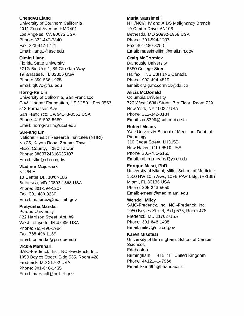
University of Southern California
Phone: 323-442-7840
Chengyu Liang
Email: [email protected]
Los Angeles, CA 90033 USA
Fax: 323-442-1721
2011 Zonal Avenue, HMR401
Florida State University
Phone: 850-566-1965
Qimig Liang
Email: [email protected]
Tallahassee, FL 32306 USA221G Bio Unit 1, 89 Chieftan Way
University of California, San Francisco
Phone: 415-502-5669
Horng-Ru Lin
Email: [email protected]
San Francisco, CA 94143-0552 USA
G.W. Hooper Foundation, HSW1501, Box 0552513 Parnassus Ave.
National Health Research Institutes (NHRI)
Phone: 8863724616635107
Su-Fang Lin
Email: [email protected]
Miaoli County, 350 TaiwanNo.35, Keyan Road, Zhunan Town
NCI/NIH
Phone: 301-594-1207
Vladimir Majerciak
Email: [email protected]
Bethesda, MD 20892-1868 USA
Fax: 301-480-8250
10 Center Dr., 10/6N106
Purdue University
Phone: 765-496-1984
Pratyusha Mandal
Email: [email protected]
West Lafayette, IN 47906 USA
Fax: 765-496-1189
422 Harrison Street, Apt. #9
SAIC-Frederick, Inc., NCI-Frederick, Inc.
Phone: 301-846-1435
Vickie Marshall
Email: [email protected]
Frederick, MD 21702 USA1050 Boyles Street, Bldg 535, Room 428
NIH/NCI/HIV and AIDS Malignancy Branch
Phone: 301-594-1207
Maria Massimelli
Email: [email protected]
Bethesda, MD 20892-1868 USA
Fax: 301-480-8250
10 Center Drive, 6N106
Dalhousie University
Phone: 902-494-4519
Craig McCormick
Email: [email protected]
Halifax, NS B3H 1X5 Canada5850 College Street
Columbia University
Phone: 212-342-0184
Alicia McDonald
Email: [email protected]
New York, NY 10032 USA722 West 168th Street, 7th Floor, Room 729
Yale University School of Medicine, Dept. ofPathology
Phone: 203-785-6160
Robert Means
Email: [email protected]
New Haven, CT 06510 USA310 Cedar Street, LH315B
University of Miami, Miller School of Medicine
Phone: 305-243-5659
Enrique Mesri, PhD
Email: [email protected]
Miami, FL 33136 USA1550 NW 10th Ave., 109B PAP Bldg. (R-138)
SAIC-Frederick, Inc., NCI-Frederick, Inc.
Phone: 301-846-1408
Wendell Miley
Email: [email protected]
Frederick, MD 21702 USA1050 Boyles Street, Bldg 535, Room 428
University of Birmingham, School of CancerSciences
Phone: 441214147966
Karen Misstear
Email: [email protected]
Birmingham, B15 2TT United KingdomEdgbaston
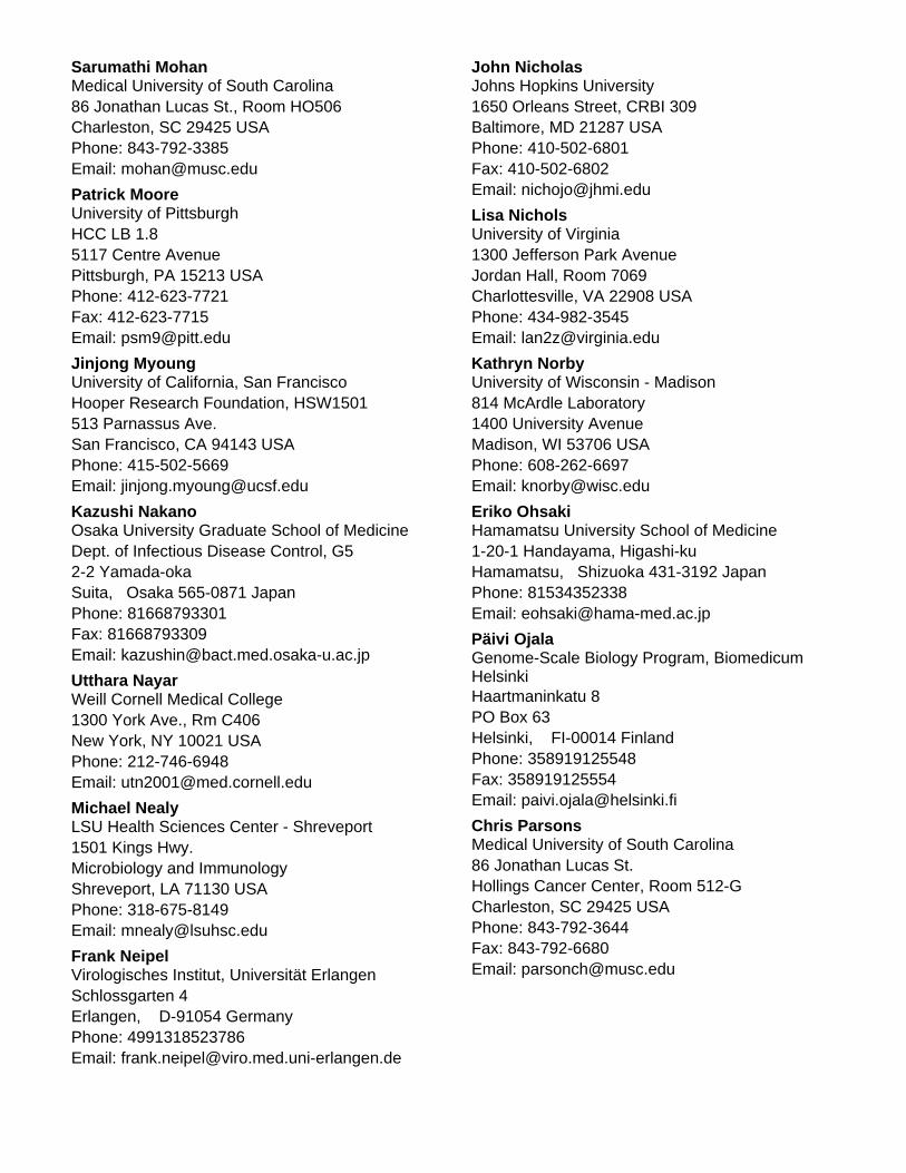
Medical University of South Carolina
Phone: 843-792-3385
Sarumathi Mohan
Email: [email protected]
Charleston, SC 29425 USA86 Jonathan Lucas St., Room HO506
University of Pittsburgh
Phone: 412-623-7721
Patrick Moore
Email: [email protected]
Pittsburgh, PA 15213 USA
Fax: 412-623-7715
HCC LB 1.85117 Centre Avenue
University of California, San Francisco
Phone: 415-502-5669
Jinjong Myoung
Email: [email protected]
San Francisco, CA 94143 USA
Hooper Research Foundation, HSW1501513 Parnassus Ave.
Osaka University Graduate School of Medicine
Phone: 81668793301
Kazushi Nakano
Email: [email protected]
Suita, Osaka 565-0871 Japan
Fax: 81668793309
Dept. of Infectious Disease Control, G52-2 Yamada-oka
Weill Cornell Medical College
Phone: 212-746-6948
Utthara Nayar
Email: [email protected]
New York, NY 10021 USA1300 York Ave., Rm C406
LSU Health Sciences Center - Shreveport
Phone: 318-675-8149
Michael Nealy
Email: [email protected]
Shreveport, LA 71130 USA
1501 Kings Hwy.Microbiology and Immunology
Virologisches Institut, Universität Erlangen
Phone: 4991318523786
Frank Neipel
Email: [email protected]
Erlangen, D-91054 GermanySchlossgarten 4
Johns Hopkins University
Phone: 410-502-6801
John Nicholas
Email: [email protected]
Baltimore, MD 21287 USA
Fax: 410-502-6802
1650 Orleans Street, CRBI 309
University of Virginia
Phone: 434-982-3545
Lisa Nichols
Email: [email protected]
Charlottesville, VA 22908 USA
1300 Jefferson Park AvenueJordan Hall, Room 7069
University of Wisconsin - Madison
Phone: 608-262-6697
Kathryn Norby
Email: [email protected]
Madison, WI 53706 USA
814 McArdle Laboratory1400 University Avenue
Hamamatsu University School of Medicine
Phone: 81534352338
Eriko Ohsaki
Email: [email protected]
Hamamatsu, Shizuoka 431-3192 Japan1-20-1 Handayama, Higashi-ku
Genome-Scale Biology Program, BiomedicumHelsinki
Phone: 358919125548
Päivi Ojala
Email: [email protected]
Helsinki, FI-00014 Finland
Fax: 358919125554
Haartmaninkatu 8PO Box 63
Medical University of South Carolina
Phone: 843-792-3644
Chris Parsons
Email: [email protected]
Charleston, SC 29425 USA
Fax: 843-792-6680
86 Jonathan Lucas St.Hollings Cancer Center, Room 512-G
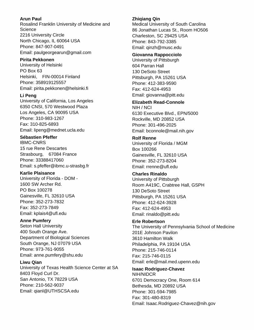
Rosalind Franklin University of Medicine andScience
Phone: 847-907-0491
Arun Paul
Email: [email protected]
North Chicago, IL 60064 USA2216 University Circle
University of Helsinki
Phone: 358919125557
Pirita Pekkonen
Email: [email protected]
Helsinki, FIN-00014 FinlandPO Box 63
University of California, Los Angeles
Phone: 310-983-1267
Li Peng
Email: [email protected]
Los Angeles, CA 90095 USA
Fax: 310-825-6893
6350 CNSI, 570 Westwood Plaza
IBMC-CNRS
Phone: 33388417060
Sébastien Pfeffer
Email: [email protected]
Strasbourg, 67084 France15 rue Rene Descartes
University of Florida - DOM -
Phone: 352-273-7832
Karlie Plaisance
Email: [email protected]
Gainesville, FL 32610 USA
Fax: 352-273-7849
1600 SW Archer Rd.PO Box 100278
Seton Hall University
Phone: 973-761-9055
Anne Pumfery
Email: [email protected]
South Orange, NJ 07079 USA
400 South Orange Ave.Department of Biological Sciences
University of Texas Health Science Center at SA
Phone: 210-562-9037
Liwu Qian
Email: [email protected]
San Antonio, TX 78229 USA8403 Floyd Curl Dr.
Medical University of South Carolina
Phone: 843-792-3385
Zhiqiang Qin
Email: [email protected]
Charleston, SC 29425 USA86 Jonathan Lucas St., Room HO506
University of Pittsburgh
Phone: 412-383-9590
Giovanna Rappocciolo
Email: [email protected]
Pittsburgh, PA 15261 USA
Fax: 412-624-4953
604 Parran Hall130 DeSoto Street
NIH / NCI
Phone: 301-496-2025
Elizabeth Read-Connole
Email: [email protected]
Rockville, MD 20852 USA6130 Executive Blvd., EPN/5000
University of Florida / MGM
Phone: 352-273-8204
Rolf Renne
Email: [email protected]
Gainesville, FL 32610 USABox 100266
University of Pittsburgh
Phone: 412-624-3928
Charles Rinaldo
Email: [email protected]
Pittsburgh, PA 15261 USA
Fax: 412-624-4953
Room A419C, Crabtree Hall, GSPH130 DeSoto Street
The University of Pennsylvania School of Medicine
Phone: 215-746-0114
Erle Robertson
Email: [email protected]
Philadelphia, PA 19104 USA
Fax: 215-746-0115
201E Johnson Pavilon3610 Hamilton Walk
NIH/NIDCR
Phone: 301-594-7985
Isaac Rodriguez-Chavez
Email: [email protected]
Bethesda, MD 20892 USA
Fax: 301-480-8319
6701 Democracy One, Room 614
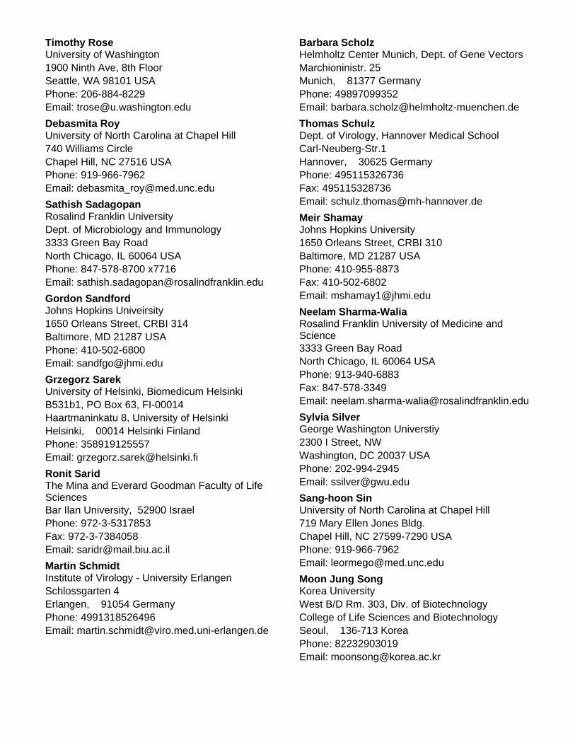
University of Washington
Phone: 206-884-8229
Timothy Rose
Email: [email protected]
Seattle, WA 98101 USA1900 Ninth Ave, 8th Floor
University of North Carolina at Chapel Hill
Phone: 919-966-7962
Debasmita Roy
Email: [email protected]
Chapel Hill, NC 27516 USA740 Williams Circle
Rosalind Franklin University
Phone: 847-578-8700 x7716
Sathish Sadagopan
Email: [email protected]
North Chicago, IL 60064 USA
Dept. of Microbiology and Immunology3333 Green Bay Road
Johns Hopkins Univeirsity
Phone: 410-502-6800
Gordon Sandford
Email: [email protected]
Baltimore, MD 21287 USA1650 Orleans Street, CRBI 314
University of Helsinki, Biomedicum Helsinki
Phone: 358919125557
Grzegorz Sarek
Email: [email protected]
Helsinki, 00014 Helsinki Finland
B531b1, PO Box 63, FI-00014Haartmaninkatu 8, University of Helsinki
The Mina and Everard Goodman Faculty of LifeSciences
Phone: 972-3-5317853
Ronit Sarid
Email: [email protected]
Bar Ilan University, 52900 Israel
Fax: 972-3-7384058
Institute of Virology - University Erlangen
Phone: 4991318526496
Martin Schmidt
Email: [email protected]
Erlangen, 91054 GermanySchlossgarten 4
Helmholtz Center Munich, Dept. of Gene Vectors
Phone: 49897099352
Barbara Scholz
Email: [email protected]
Munich, 81377 GermanyMarchioninistr. 25
Dept. of Virology, Hannover Medical School
Phone: 495115326736
Thomas Schulz
Email: [email protected]
Hannover, 30625 Germany
Fax: 495115328736
Carl-Neuberg-Str.1
Johns Hopkins University
Phone: 410-955-8873
Meir Shamay
Email: [email protected]
Baltimore, MD 21287 USA
Fax: 410-502-6802
1650 Orleans Street, CRBI 310
Rosalind Franklin University of Medicine andScience
Phone: 913-940-6883
Neelam Sharma-Walia
Email: [email protected]
North Chicago, IL 60064 USA
Fax: 847-578-3349
3333 Green Bay Road
George Washington Universtiy
Phone: 202-994-2945
Sylvia Silver
Email: [email protected]
Washington, DC 20037 USA2300 I Street, NW
University of North Carolina at Chapel Hill
Phone: 919-966-7962
Sang-hoon Sin
Email: [email protected]
Chapel Hill, NC 27599-7290 USA719 Mary Ellen Jones Bldg.
Korea University
Phone: 82232903019
Moon Jung Song
Email: [email protected]
Seoul, 136-713 Korea
West B/D Rm. 303, Div. of BiotechnologyCollege of Life Sciences and Biotechnology
NCI
Phone: 301-402-3630
Erin Soule
Email: [email protected]
Bethesda, MD 20814 USA
9000 Rockville PikeBuilding 10, Room 6N106
Emory University School of Medicine
Phone: 404-727-7665
Sam Speck
Email: [email protected]
Atlanta, GA 30322 USA
Dept. Micro. & Immunonology1462 Clifton Rd, DSB 429
Griffith University
Phone: 610423320251
David Speicher
Email: [email protected]
Griffith University, Queensland 4222 Australia
Fax: 61756780708
School of Dentistry and Oral HealthGold Coast Campus
Uniformed Services University
Phone: 301-295-9620
Deborah Steffen
Email: [email protected]
Bethesda, MD 20814 USA4301 Jones Bridge Road, Rm. B4121
University of Erlangen
Phone: 4991318536331
Michael Stürzl
Email: [email protected]
Erlangen, 91054 Germany
Div. of Molecular and Experimental SurgerySchwabachanlage 10
Louisiana State University
Phone: 225-578-9665
Ramesh Subramanian
Email: [email protected]
Baton Rouge, LA 70803 USA
Fax: 225-578-9655
Div. of Biotechnology and Molecular Medicine,Skip Bertman Drive
The University of Texas at Austin
Phone: 512-471-5391
Chris Sullivan
Email: [email protected]
Austin, TX 78712-0162 USA
Molecular Genetics & Microbiology1 University Station, A5000
Tulane University School of Medicine
Phone: 504-988-6690
Deborah Sullivan
Email: [email protected]
New Orleans, LA 70112 USA1430 Tulane Ave., SL-38
University of California, Los Angeles
Phone: 310-794-5557
Ren Sun
Email: [email protected]
Los Angeles, CA 90095-9517 USA
UCLA Mol & Med. PharmacologyBox 959517, 10-155 Factor
University of Florida
Phone: 352-246-4263
Sankar Swaminathan Swaminathan
Email: [email protected]
Gainesville, FL 32653 USA5125 NW 62nd Terrace
University of North Carolina at Chapel Hill
Phone: 919-966-7962
Kristen Tamburro
Email: [email protected]
Chapel Hill, NC 27599 USA804 Mary Ellen Jones Bldg., 98 Manning Dr., CB
LSU Health Sciences Center
Phone: 318-675-8148
Scott Tibbetts
Email: [email protected]
Shreveport, LA 71106 USA1501 Kings Highway
University of Southern California
Phone: 323-442-7863
Zsolth Toth
Email: [email protected]
Los Angeles, CA 90033 USA
Fax: 323-442-1721
2011 Zonal Avenue, HMR401
National Health Research Institutes (NHRI)
Phone: 8863724616635122
Wan-Hua Tsai
Email: [email protected]
Miaoli County, 350 TaiwanNo.35, Keyan Road, Zhunan Town
University College London
Phone: 44207679216
Edward Tsao
Email: [email protected]
London, W1T 4JF United Kingdom
46 Cleveland StreetWindeyer Building
NIH/NCI/HIV and AIDS Malignancy Branch
Phone: 301-402-6296
Thomas Uldrick
Email: [email protected]
Bethesda, MD 20892-1868 USA
Fax: 301-480-5955
10 Center Dr., Room 6N106
University of Colorado School of Medicine
Phone: 303-724-4207
Linda van Dyk
Email: [email protected]
Aurora, CO 80238 USA
Fax: 303-724-4226
Microbiology Department, MS 833312800 E. 19th Ave.
Rosalind Franklin University
Phone: 847-578-8700 x7716
Mohanan Veettil
Email: [email protected]
North Chicago, IL 60064 USA3333 Green Bay Road
Vaccine and Gene Therapy Institute, OHSU
Phone: 503-418-2740
Kasinath Viswanathan
Email: [email protected]
Beaverton, OR 97006 USA
Fax: 503-418-2701
505, NW 185th Ave.
Johns Hopkins University
Phone: 410-614-0594
Leyao Wang
Email: [email protected]
Baltimore, MD 21287 USA1650 Orleans Street, CRBI 316
University of North Carolina at Chapel Hill
Phone: 919-843-6013
Ling Wang
Email: [email protected]
Chapel Hill, NC 27599 USA
Fax: 919-966-9673
450 West Drive
University of Nebraska-Lincoln
Phone: 402-472-4559
Hui-Ju Wen
Email: [email protected]
Lincoln, NE 68583-0900 USARm. 330, Morrison Center, 4240 Fair St.
University of North Carolina at Chapel Hill
Phone: 919-843-6013
Kumn Wah Wen
Email: [email protected]
Chapel Hill, NC 27599 USA
Fax: 919-966-9673
Lineberger 31-303, 450 West Drive
UNC at Chapel Hill / Lineberger ComprehensiveCancer Center
Phone: 919-843-6013
John West
Email: [email protected]
Chapel Hill, NC 27599 USA
Fax: 919-966-9673
450 West Drive
SAIC-Frederick, Inc., NCI-Frederick, Inc.
Phone: 301-846-1714
Denise Whitby
Email: [email protected]
Frederick, MD 21702 USA1050 Boyles Street, Bldg 535, Room 412
Washington University in St. Louis
Phone: 314-362-9224
Douglas White
Email: [email protected]
St. Louis, MO 63110 USA
Fax: 314-362-4096
660 S. Euclid Ave., Box 8118/8045
UC Davis Primate Center
Phone: 530-752-5696
Jessica White
Email: [email protected]
Davis, CA 95819 USA
Fax: 530-752-4816
County Rd 98 and Hutchinson Drive
Oregon Health and Science University
Phone: 503-690-5285
Scott Wong
Email: [email protected]
Beaverton, OR 97006 USA
Fax: 503-418-2719
Vaccine and Gene Therapy Institute505 NW 185th Ave.
Nebraska Center for Virology, UNL
Phone: 402-472-4550
Charles Wood
Email: [email protected]
Lincoln, NE 68583-0900 USA
Fax: 402-472-3323
102 C Morrison Center, 4240 Fair St.
The Johns Hopkins University
Phone: 410-502-2807
Crystal Woodard
Email: [email protected]
Baltimore, MD 21205 USA
Fax: 410-502-1872
733 N Broadway, BRB 340
Institute of Microbiology and Immunology, NYMU
Phone: 886911254509
Yu-Hsuan Wu
Email: [email protected]
Taipei, 112 TaiwanNo.155, Sec.2, Linong Street
NIH / NCI
Phone: 301-402-3630
Min Yang
Email: [email protected]
Bethesda, MD 20892 USARoom 5A25, Building 10, 9000 Rockville Pike
University of Florida
Phone: 352-871-6858
Yigie Yang
Email: [email protected]
Gainesville, FL 32610 USA1376 Mowry Rd., #121
National Cancer Institute
Phone: 301-496-0328
Robert Yarchoan
Email: [email protected]
Bethesda, MD 20892-1868 USA
Fax: 301-480-5955
Bldg. 10, Rm. 6N106, MSC 186810 Center Drive, NIH
University of Pennsylvania
Phone: 215-573-7556
Yan Yuan
Email: [email protected]
Philadelphia, PA 19104 USA
Fax: 215-898-8385
Department of MicrobiologySchool of Dental Medicine
National Cancer Institute
Phone: 301-594-1382
Zhi-Ming (Thomas) Zheng
Email: [email protected]
Bethesda, MD 20892 USA10 Center Dr., Rm. 6N106
Florida State University
Phone: 850-644-6273
Fanxiu Zhu
Email: [email protected]
Tallahassee, FL 32306-4370 USA
Fax: 850-644-0481
Dept. of Biological Science89 Chieftan Way
National Cancer Institute
Phone: 443-015-9466 x34
Joseph Ziegelbauer
Email: [email protected]
Bethesda, MD 20892 USA10 Center Dr., Bldg. 10, Room 6N106
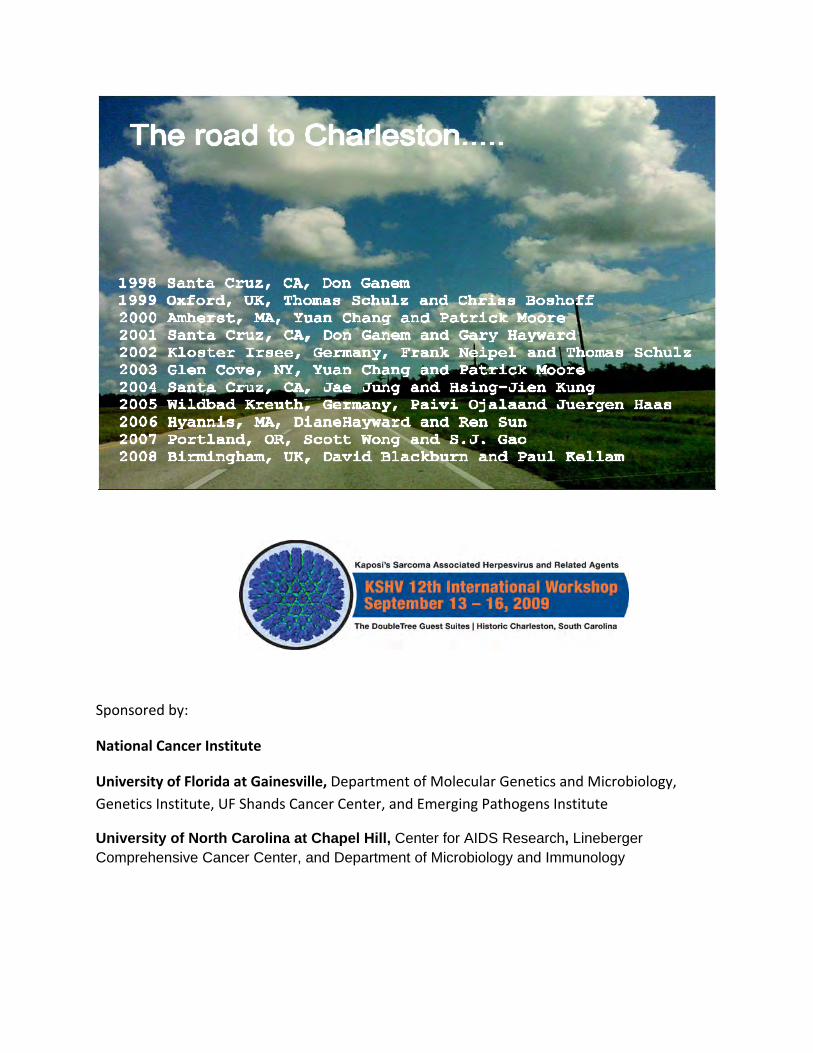
Sponsored by:
National Cancer Institute
University of Florida at Gainesville, Department of Molecular Genetics and Microbiology, Genetics Institute, UF Shands Cancer Center, and Emerging Pathogens Institute
University of North Carolina at Chapel Hill, Center for AIDS Research, Lineberger Comprehensive Cancer Center, and Department of Microbiology and Immunology