Endocrine Abstracts
179
March 2013 Volume 31 ISSN 1479-6848 (online) Online version available at www.endocrine-abstracts.org Society for Endocrinology BES 2013 18 –21 March 2013, Harrogate, UK Endocrine Abstracts published by bioscientifica
-
Upload
khangminh22 -
Category
Documents
-
view
1 -
download
0
Transcript of Endocrine Abstracts
1470-3947(201104)26;1-Y
March 2013 Volume 31ISSN 1479-6848 (online)
Online version available at www.endocrine-abstracts.org
Society for Endocrinology BES 201318 –21 March 2013, Harrogate, UK
Endocrine Abstracts
Society for Endocrinology BES 2012
19 –22 March 2012, Harrogate, UK
En
do
crine A
bstracts
Volum
e 31 March 2013
published by bioscientifi ca
EJEA_31-1_cover.indd 1EJEA_31-1_cover.indd 1 3/12/13 8:46:01 PM3/12/13 8:46:01 PM
Endocrine AbstractsVolume 31
March 2013
SOCIETY FOR ENDOCRINOLOGYBES 201318-21 March 2013, Harrogate International Centre
Harrogate, UK
Abstract Book
Abstract Marking Panel
J Ahlquist SouthendF Ahmed Glasgow
A Allahbadia Sheffield
B Allolio Germany
R Amin London
R Andrew Edinburgh
R Andrews Bristol
S Atkin Hull
S Aylwin London
J Ayuk Birmingham
L Bailey Liverpool
J Barth
A Bates Birmingham
P Belchetz
M Besser London
K Boelaert Birmingham
P Bouloux London
M Brada
N Bridges London
J Burrin London
P Carroll London
K Chapman Edinburgh
P Chapple London
K Chatterjee Cambridge
T CheethamNewcastle-upon-Tyne
P Clark Birmingham
P Clayton Manchester
J Compston Cambridge
M Cooper Birmingham
S Cox Torquay
A Crown Brighton
M Dattani London
M DeBono Sheffield
W Dhillo London
K Docherty Abderdeen
W Drake London
C Duncan Edinburgh
R Eastell Sheffield
S Ellard
B Evans Cardiff
W Farrell Birmingham
D Flynt Glasgow
A Fowdon Cambridge
R Fowkes London
J Franklyn Birmingham
W Fraser Liverepool
M Freel Glasgow
N Gittoes Birmingham
H Gleeson Manchester
C Gregson Bristol
A Grossman London
M Gurnell Cambridge
J Ham Cardiff
P Hanson london
L Heisler Cambridge
T Howlett Leicester
S Hunter Belfast
A James Newcastle
C Johnston Watford
N Karavitaki Oxford
N Kieffer Leicester
P King London
J Kopchick USA
S Kumar Warwick
J Lau
G Lavery Birmingham
V Lawrence London
G Leese Dundee
M Levy Leicester
A Lopez Bernal Bristol
S MacKenzie Glasgow
C A McArdle Bristol
C McCabe Birmingham
A McNeilly Edinburgh
P McTernan Warwick
J Monson London
D Morris Ipswich
N Morton Edinburgh
J Munday Portsmouth
R Murray Leeds
J Newell-Price Sheffield
S Orme Leeds
K Owen Oxford
S Pearce Newcastle
P Pickett Shrewsbury
R Quinton Newcastle
S Razvi Gateshead
M Read Birmingham
A Rees Cardiff
P Saunders Edinburgh
P Selby Manchester
D Shapiro Glasgow
L Shepherd Birmingham
H Simpson Cambridge
V Smith Birmingham
P Squires Warwick
C Stewart Manchester
R Stimson Edinburgh
F Swords Norwich
G Tan Nottingham
T Tan London
N Taylor London
C Thirlwell London
T Toft Edinburgh
J Tomlinson Birmingham
A Toogood Birmingham
P Trainer Manchester
B Vaidya Exeter
J Valle Manchester
B Walker Edinburgh
E Ward Leeds
G Wark
J Wass Oxford
S Webb
J Webster Sheffield
T Weetman Sheffield
M Westwood Machester London
Members
A Allahabadia
WArlt
K Chapman
E Crowne
E Davies
C Duncan
W Farrell
S Orme
R Semple
A Toogood
J Wass
EDITORS
The abstracts submitted were marked by the Abstract Marking panel selected by the Programme Committee
Programme Committee
C McCabe (Programme Secretary)
M Druce (Programme Co-ordinator)
C McArdle (Programme Co-ordinator)
CORPORATE SUPPORTERSThe Society for Endocrinology would like to thank its Corporate Supporters for their generousfinancial assistance.
Platinum Supporters:BioscientificaIpsenPfizer
Gold Supporters:Bayer HealthcareNovartis
Silver Supporters:Sandoz Ltd
Conference SecretariatBioScientifica Ltd Contact: Claire Arrigoni/Harriet EdwardsEuro House, 22 Apex Court Tel: +44 (0)1454 642210Woodlands Fax: +44 (0)1454 642222Bradley Stoke E-mail: [email protected] BS32 4JT, UK Website: http://www.bioscientifica.com
Society for Endocrinology BES 2013, Harrogate, UK
Endocrine Abstracts (2013) Vol 31
Society for Endocrinology22 Apex Court Tel: +44 (0) 1454 642200Woodlands Fax: +44 (0) 1454 642220Bradley Stoke E-mail: [email protected] BS32 4JT, UK Website: http://www.endocrinology.org
Society for Endocrinology BES 2013, Harrogate, UK
Endocrine Abstracts (2013) Vol 31
CONTENTS
Society for Endocrinology BES 2013
PLENARY LECTURERS’ BIOGRAPHICAL NOTES
PLENARY LECTURES
Society for Endocrinology Dale Medal Lecture . . . . . . . . . . . . . . . . . . . . . . . . . . . . . . . . . . . . . . . . . PL1Society for Endocrinology Hoffenberg International Medal Lecture . . . . . . . . . . . . . . . . . . . . . . . . . . . . . . PL2Society for Endocrinology European Medal Lecture . . . . . . . . . . . . . . . . . . . . . . . . . . . . . . . . . . . . . . PL3Society for Endocrinology Tranatlantic Medal Lecture . . . . . . . . . . . . . . . . . . . . . . . . . . . . . . . . . . . . . PL4British Thyroid Association Pitt-Rivers lecture . . . . . . . . . . . . . . . . . . . . . . . . . . . . . . . . . . . . . . . . . PL5Society for Endocrinology Medal Lecture . . . . . . . . . . . . . . . . . . . . . . . . . . . . . . . . . . . . . . . . . . . . PL6Clinical Endocrinology Trust Lecture . . . . . . . . . . . . . . . . . . . . . . . . . . . . . . . . . . . . . . . . . . . . . . PL7Clinical Endocrinology Trust Visiting Professor Lecture . . . . . . . . . . . . . . . . . . . . . . . . . . . . . . . . . . . . PL8
SYMPOSIA
Irn bru, to drink or not to drink: endocrinology and iron . . . . . . . . . . . . . . . . . . . . . . . . . . . . . . . S1.1–S1.3Hormones maketh Man . . . . . . . . . . . . . . . . . . . . . . . . . . . . . . . . . . . . . . . . . . . . . . . . . S2.1–S2.3Nurture not nature: epigenetics and disease susceptibility . . . . . . . . . . . . . . . . . . . . . . . . . . . . . . S3.1–S3.3New Bone Biology - Is there life after RANK ligand? . . . . . . . . . . . . . . . . . . . . . . . . . . . . . . . . . S4.1–S4.4Sex in the brain (Supported by Endocrine Connections) . . . . . . . . . . . . . . . . . . . . . . . . . . . . . . . . S5.1–S5.4Making the glucocorticoid clock run smoothly (Supported by Addison’s Disease self-help group) . . . . . . . . . S6.1–S6.4Thyroid hormone receptors - mutations and implications (Supported byJournal of Molecular Endocrinology) . . . . . . . . . . . . . . . . . . . . . . . . . . . . . . . . . . . . . . . . . . . S7.1–S7.4Non functioning pituitary tumours (Supported by Endocrine-Related Cancer and the Pituitary foundation) . . . . S8.1–S8.4Novel aspects of GPCR signalling (Supported by the Journal of Endocrinology) . . . . . . . . . . . . . . . . . . . S9.1–S9.4Lipodystrophy - The perils of being thin . . . . . . . . . . . . . . . . . . . . . . . . . . . . . . . . . . . . . . . S10.1–S10.4Guts, brains and bariatric surgery . . . . . . . . . . . . . . . . . . . . . . . . . . . . . . . . . . . . . . . . . . . S11.1–S11.4Thymic function and autoimmune endocrine disease . . . . . . . . . . . . . . . . . . . . . . . . . . . . . . . . S12.1–S12.4
CLINICAL MANAGEMENT WORKSHOPS
Management controversies in parathyroid disease . . . . . . . . . . . . . . . . . . . . . . . . . . . . . . . CMW1.1–CMW1.4How Do I Do It? . . . . . . . . . . . . . . . . . . . . . . . . . . . . . . . . . . . . . . . . . . . . . . . . . . CMW2.1–CMW2.6PCOS - why, how and what . . . . . . . . . . . . . . . . . . . . . . . . . . . . . . . . . . . . . . . . . . . CMW3.1–CMW3.4Managing Hypoglycaemia . . . . . . . . . . . . . . . . . . . . . . . . . . . . . . . . . . . . . . . . . . . . CMW4.1–CMW4.4
APPLIED PHYSIOLOGY WORKSHOP
Digital copies: exploiting numerical models of biological systems . . . . . . . . . . . . . . . . . . . . . . . APW1.1–APW1.4
DEBATE . . . . . . . . . . . . . . . . . . . . . . . . . . . . . . . . . . . . . . . . . . . . . . . . . . . . . . . . . . . D1–D2
MEET THE EXPERT SESSIONS . . . . . . . . . . . . . . . . . . . . . . . . . . . . . . . . . . . . . . . . . . . MTE1–MTE9
NURSE SESSION
The Patient Pathway for Pituitary Care . . . . . . . . . . . . . . . . . . . . . . . . . . . . . . . . . . . . . . . . . N1.1–N1.5Late Effects . . . . . . . . . . . . . . . . . . . . . . . . . . . . . . . . . . . . . . . . . . . . . . . . . . . . . . . . N2.1–N2.3
YOUNG ENDOCRINOLOGISTS SESSION
Young endocrinologists’ prize lectures . . . . . . . . . . . . . . . . . . . . . . . . . . . . . . . . . . . . . . YEP1.1–YEP1.2Maintaining your endocrine career despite what life throws at you - Keeping everything up in the air . . . . . YE1.1–YE1.6
Society for Endocrinology BES 2013, Harrogate, UK
SENIOR ENDOCRINOLOGISTS SESSION . . . . . . . . . . . . . . . . . . . . . . . . . . . . . . . . . . . . . SE1.1–SE1.6
ORAL COMMUNICATIONS
Young Endocrinologists prize session . . . . . . . . . . . . . . . . . . . . . . . . . . . . . . . . . . . . . . . . . OC1.1–OC1.8Steroids and thyroid . . . . . . . . . . . . . . . . . . . . . . . . . . . . . . . . . . . . . . . . . . . . . . . . . . OC2.1–OC2.8Reproduction, growth and Development . . . . . . . . . . . . . . . . . . . . . . . . . . . . . . . . . . . . . . . OC3.1–OC3.8Obesity, metabolism and bone . . . . . . . . . . . . . . . . . . . . . . . . . . . . . . . . . . . . . . . . . . . . . OC4.1–OC4.8Pituitary and Neoplasia . . . . . . . . . . . . . . . . . . . . . . . . . . . . . . . . . . . . . . . . . . . . . . . . OC5.1–OC5.8
POSTER PRESENTATIONS
Bone . . . . . . . . . . . . . . . . . . . . . . . . . . . . . . . . . . . . . . . . . . . . . . . . . . . . . . . . . . . . . P1–P34Clinical biochemistry . . . . . . . . . . . . . . . . . . . . . . . . . . . . . . . . . . . . . . . . . . . . . . . . . . . . P35–P56Clinical practice/governance and case reports . . . . . . . . . . . . . . . . . . . . . . . . . . . . . . . . . . . . . P57–P135Cytokines and growth factors . . . . . . . . . . . . . . . . . . . . . . . . . . . . . . . . . . . . . . . . . . . . . P136–P139Growth and development . . . . . . . . . . . . . . . . . . . . . . . . . . . . . . . . . . . . . . . . . . . . . . . P140–P148Neoplasia, cancer and late effects . . . . . . . . . . . . . . . . . . . . . . . . . . . . . . . . . . . . . . . . . . . P149–P174Nursing practise . . . . . . . . . . . . . . . . . . . . . . . . . . . . . . . . . . . . . . . . . . . . . . . . . . . . P175–P176Obesity, diabetes, metabolism and cardiovascular . . . . . . . . . . . . . . . . . . . . . . . . . . . . . . . . . . P177–P243Pituitary . . . . . . . . . . . . . . . . . . . . . . . . . . . . . . . . . . . . . . . . . . . . . . . . . . . . . . . . . P244–P287Reproduction . . . . . . . . . . . . . . . . . . . . . . . . . . . . . . . . . . . . . . . . . . . . . . . . . . . . . . P288–P314Steroids . . . . . . . . . . . . . . . . . . . . . . . . . . . . . . . . . . . . . . . . . . . . . . . . . . . . . . . . . P315–P351Thyroid . . . . . . . . . . . . . . . . . . . . . . . . . . . . . . . . . . . . . . . . . . . . . . . . . . . . . . . . . P352–P388
INDEX OF AUTHORS
Endocrine Abstracts (2013) Vol 31

Society for Endocrinology BES 2013, Harrogate, UK
Plenary Lecturers’
Biographical Notes
Endocrine Abstracts (2013) Vol 31
Society for Endocrinology Dale Medal Lecture
Ronald M Evans, The Salk Institute for Biological Studies, LaJolla California, USA
Dr. Ronald M Evans is known for his discoveries and characterization of nuclear hormone receptors, theestablishment of the nuclear receptor super family and the elucidation of their universal mechanism ofaction, this revealed how receptor activation by lipophilic hormones and drugs are transformed intophysiology and the treatment of disease.
Dr R M Evans obtained his BA and PhD from the University of California, Los Angeles, School ofMedicine in 1970 and 1974 respectively, was a postdoctoral fellow with James Darnell at theRockefeller University in New York and in 1977 joined the faculty of The Salk Institute for BiologicalStudies where he is now an Investigator of the Howard Hughes Medical Institute and Professor in theGene Expression Laboratory. He holds the March of Dimes Chair in Molecular and DevelopmentalBiology and is Adjunct Professorships at the University of California, San Diego in the Departments ofBiology, Biomedical Sciences, and Neuroscience.
At Salk, Dr Evans isolated the GH gene to study its transcriptional regulation by steroid and thyroidhormones. In 1985 his group cloned and characterized the first nuclear hormone receptor, the humanglucocorticoid receptor. His subsequent isolation of the thyroid, mineralocorticoid and retinoic acid(vitamin A) established the existence of the nuclear receptor superfamily. This work led to theprinciples of DNA recognition, receptor heterodimer formation, and the discovery of the DNA codingmechanism for hormone response elements. He isolated the first orphan receptors (ERR1and 2) as wellas the unexpectedly important retinoid X receptor (RXR). He pioneered biochemical and moleculartechniques (termed reverse endocrinology) that led to the identification of the RXR ligand 9-cis RA.RXR proved to be a Rosetta stone for puzzling out the identity of a series of unknown receptors, whichhave profound implications for normal physiology, disease pathogenesis and drug discovery. He alsoisolated and characterized the xenobiotic sensor SXR.
More recently, Dr Evans has focused on PPARg and d as major regulators of whole body lipidmetabolism. As part of this work he created genetically thin mice and the first animal (termed the‘Marathon Mouse’) genetically engineered for increased running endurance. This led to the recentdiscovery that transcription of a nuclear gene network by a PPARd synthetic agonist and the AMPkinase (AMPK) activator ‘AICAR’ can enhance running endurance in absence of mechanical exercise.More recently, he has extended this concept by demonstrating that AICAR can act like ‘pharmacologiclight’ to entrain the rhythm of the hepatic circadian clock.
Selected awards since 2000 include: 1st Bristol – Myers Squibb Award for Metabolic Research(2000), City of Medicine Award, Duke (2002), the March of Dimes Prize in Developmental Biology(2003), General Motors Cancer Research Foundation Alfred P Sloan Medal (2003), Keio MedicalScience Prize (2003), Albert Lasker Basic Medical Research Award (2004), Glen T Seaborg Medal,UCLA (2005), ‘Grande Medaille d’Or’ of the French Academy of Sciences (2005), Gairdner Award,Canada (2006), Harvey Prize of the Technion Institute, Israel (2006), the Albany Prize in Medicine(2007) and the Endocrine Regulation Prize, IPSEN Foundation (2008), Ernst Knobil Award, U TexasHlth Sci Cntr (2009), Wolf Prize, Wolf Foundation Israel (2012), Dale Medal, British Society forEndocrinology, UK (2013). He is a member of the National Academy of Sciences, the Institute ofMedicine, the American Academy of Arts and Sciences, the American Philosophical Society and wasnamed the1994 California Scientist of the Year.
Society for Endocrinology BES 2013, Harrogate, UK
Endocrine Abstracts (2013) Vol 31
Society for Endocrinology Hoffenberg International Medal Lecture
Fernand Labrie (Professor), Department of Anatomy and Physiology, Laval university, QuebecCity Quebec, Canada
After obtaining his MD and PhD (Endocrinology) degrees with Honours at Laval University, QuebecCity, Canada, F Labrie pursued his postdoctoral training at the University of Cambridge, UK, first in theLaboratory of Professor Asher Korner and then, in the Laboratory of Molecular Biology of ProfessorFrederick Sanger, twice Nobel laureate in medicine. Dr Labrie then isolated the first mammalianmessenger RNA before returning to Laval University in 1969 where he founded the Laboratory ofMolecular Endocrinology, one of the largest research groups in endocrinology worldwide with a totalpersonnel of up to 350 members including 32 senior scientists. Between 1982 and 2008, he has beenscientific director of the CHUL Research Center (1200 employees), one of the largest medical researchInstitutes in Canada. From 1990 to 2002, Dr Labrie has been head of the Department of Anatomy andPhysiology of the Faculty of Medicine at Laval University while between 1992 and 1995, he has beenpresident of the Fonds de la Recherche en Sante du Quebec.The most important contribution of Dr Labrie to clinical medicine has been the discovery and
development of medical castration with GnRH agonists as well as combined androgen blockade, thefirst treatment shown to prolong life in prostate cancer and at the basis of the recent developments usingblockade of androgens made locally in the prostate in castration – resistant prostate cancer. GnRHagonists and combined androgen blockade have become the standard hormonal therapy of prostatecancer worldwide. He also discovered that a large proportion of androgens and estrogens in women(100% after menopause) and men are made in peripheral tissues from dehydroepiandrosterone by themechanism of intracrinology. Dr Labrie and his group then discovered the most potent antiestrogen,namely Acolbifene, and performed all related toxicology, phases I and II clinical studies.Dr Labrie’s discoveries are described in more than 1250 scientific publications and have been cited
more than 40 000 times. Dr Labrie is the most cited Canadian scientist among all disciplines in theinternational literature. He recently won the King Faisal International Prize in medicine. He receivednumerous other awards, including the Friesen Award of the Canadian Society of Clinical Investigationand is Doctor Honoris Causa at the Universities of Caen and Athens.
Society for Endocrinology BES 2013, Harrogate, UK
Endocrine Abstracts (2013) Vol 31
Society for Endocrinology European Medal Lecture
Anna Spada, Full Professor of Endocrinology, School of Medicine, Universityof Milan, Milan, Italy
Anna Spada is currently Full Professor at the School of Medice, University of Milan. Her main researchinterests are on signal transduction in pituitary cells, pathogenesis of pituitary tumors, genotype/phenotype relationships in acromegalic patients with gsp mutations, tissue specific GNAS1 geneimprinting, molecular mechanisms of resistance to hormone action, activating and inactivatingmutations of GNAS1 in endocrine disorders, polymorphic variants of somatostatin receptor genes inacromegalic patients, polymorphic variants of D2 receptor gene and resistance to cabergoline,pharmacogenomics.
Professor A Spada has been on the Editorial Board of numerous, prestigious peer-revied journalincluding Endocrinology, Journal of Endocrinological Investigation, Endocrine-Related Cancer and ispast Editor-in-Chief of the Journal of Molecular Endocrinology.
She has chared 98 national and 42 International Meetings and has lectured world-wide. ProfessorA Spada has authored over 133 articles in peer-reviewed journals.
Society for Endocrinology BES 2013, Harrogate, UK
Endocrine Abstracts (2013) Vol 31
Society for Endocrinology Medal Lecture
Marta Korbonits, Department of Endocrinology, St Barts and the LondonSchool of Medicine, London, UK
Professor Korbonits is a clinical academic endocrinologist with special interest in pituitarytumorigenesis and as well as metabolic effects of hormones. She graduated in medicine at SemmelweisMedical School in Budapest and works in the Department of Endocrinology at Barts and the LondonSchool of Medicine at St. Bartholomew’s Hospital in London since 1991, where currently she isCo-Centre Head. She received and MD and a PhD from the University of London and was a recipient ofan MRC Clinician Scientist Fellowship to study ghrelin physiology and genetics. Her current interestinclude hormonal regulation of the metabolic enzyme AMP-activated protein kinase, the physiologyand pathophysiology of ghrelin and endocannabinoids and pituitary tumours including familial cases.She has a large collection of familial isolated pituitary adenoma families and works on both the clinicalcharacterisation as well as molecular aspects of this disease.She has published over 150 papers, numerous book chapters, and edited books in the filed of
Endocrinology and serves on the editorial board of several prestigious endocrine journals. She washeading the Program Organising Committee of the Society for Endocrinology for 3 years, and currentlyserves on the Executive Committee of the Pituitary Society and the European Society of ClinicalInvestigation and is an elected member of the Association of Physicians of Great Britain and Ireland.She shares her time between clinical patient care, clinical research and laboratory based research as
well as teaching at undergraduate and postgraduate level.
Society for Endocrinology BES 2013, Harrogate, UK
Endocrine Abstracts (2013) Vol 31
Clinical Endocrinology Trust Lecture
Stafford Lightman, Professor of Medicine, University of Bristol, Bristol, UK
Stafford Lightman is Professor of Medicine at the University of Bristol and is Director of the HenryWellcome Laboratories for Integrative Neuroscience and Endocrinology. He started his scientific careerworking on catecholamines and opioid peptides with Leslie Iversen at the University of Cambridge andprovided some of the first data linking opioid peptides with the regulation of neurohypophysialfunction. At this time he also performed some of the first studies demonstrating the importance of brainstem catecholamine pathways in the regulation of hypothalamic activity. On moving to what is nowImperial College in London, he started to develop his studies on the role of the brain in the regulation ofstress response. He demonstrated the shift from CRH to arginine vasopressin in the control of thehypothalamic-pituitary-adrenal axis during chronic stress, demonstrated and characterised thedevelopment of stress hyporesponsiveness during lactation in both rats and man and developed modelsof immunological activation of the stress response. More recently he has developed the concept ofemergent pulsatility of hormone secretion as a result of inherent delays in the feedforward or feedbackrelationships regulating endocrine activity. This has also led to a new emphasis on the importance ofdigital signalling at the level of glucocorticoid receptors and GR chromatin interactions.
S Lightman was the founder Editor-in-Chief of the Journal of Neuroendocrinology, a founder Fellowof the Faculty of Medical Sciences, the founder Chairman of the Pituitary Foundation and a CouncilMember of the Physiological Society. He sits on several Research Councils, Wellcome Trust andEuropean Research Committees and has Chaired the European Union Committee Review of TertiaryEducation in East Africa. Professor Lightman also has a major interest in inter-relationships between artand neuroscience.
Society for Endocrinology BES 2013, Harrogate, UK
Endocrine Abstracts (2013) Vol 31
Clinical Endocrinology Trust Visiting Professor Lecture
Lynnette Nieman, National Institutes of Health, Bethesda, Maryland, USA
Dr Lynnette Nieman is a Senior Investigator and Chief of the Endocrinology Consultation Service at theNational Institutes of Health (NIH) Clinical Research Center. She has been at the NIH since herfellowship. From 1991 to 2001 she served as the Clinical Director of intramural NICHD, overseeingclinical care of the institute’s patients and ensuring compliance with human subjects researchregulations.Dr Nieman is an active clinician, having seen more than 1100 patients with Cushing’s syndrome and
is the Principal Investigator for six active protocols. She has authored more than 250 publications andsponsored three investigational new drug applications to the FDA, one of which was licensed in the USand Europe. She provided Congressional testimony on one of these agents. She is a co-editor of theAdrenal Section of UpToDate and an associate editor for the Journal of Clinical Endocrinology andMetabolism.She is a member of the subcommittee that creates the US Endocrinology and Metabolism
certification examination. Dr Nieman has received the NIH Director’s Award, Clinical Teacher of theYear award and the Endocrine Society’s Distinguished Physician award. She is a past Vice President forClinical Science of the Endocrine Society and Chaired its 2012 annual meeting.
Society for Endocrinology BES 2013, Harrogate, UK
Endocrine Abstracts (2013) Vol 31

Society for Endocrinology BES 2013, Harrogate, UK
Plenary Lectures
Endocrine Abstracts (2013) Vol 31
Society of Endocrinology Dale Medal Lecture
PL1Nuclear receptors and AMPK: can exercise mimetics cure diabetesRonald EvansSalk Institute for Biological Studies, La Jolla, CA, USA.
Nuclear hormone receptors (NHRs) are a large family of ligand-activatedtranscription factors that regulate programs of cellular growth, differentiation andhomeostasis. The structurally conserved ligand binding domains (LBDs) of NHRsbind to hydrophobic small molecules including steroid hormones, fat solublevitamins and bile acids, thereby interpreting small molecule cues to affecttranscriptional readouts.The temporal correspondence between metabolic and circadian rhythms suggeststhe inherent coupling of these two key physiologic processes. Sleep, inactivityand fasting are opposed by wakefulness, motivated behavior and the fed state.Thus we are interested whether there may be common mechanism for ‘entraining’both the clock and key metabolic pathways. We provide evidence that the energysensor AMPK, via actions as an atypical transcriptional regulator, may functionas one such dual entrainment trigger.In regards to the clock, we provide genetic, mechanistic and pharmacologicevidence that AMPK-dependent phosphorylation enables cryptochrome (e.g.Cry1) to act as energy sensor for metabolic entrainment of the circadian clock. Weshow that Cry1 acts as a glucocorticoid receptor (GR) repressor and thus controlscyclic glucose production from the liver. In addition, we find that in muscle,usually active PPARd agonists (such as GW1516) are able to promote increasedrunning endurance, suggesting the potential of drugs that can promote the benefitsof exercise even in sedentary mice. Finally, we show that the AMPK agonistAICAR is a prototypic “exercise mimetic,’ enhancing endurance by stimulatingmitochondrial function in muscle. Pharmacologic exercise from AMPK hasimportant therapeutic implications in metabolic disease, atherosclerosis andfrailty, as well as an already realized potential for athletic abuse.Declaration of fundingThis work was supported by the National Institute of Health (grant DK057978-32), the Glenn Foundation, the Ellison Medical Foundation and the HelmsleyCharitable Trust. Dr Evans is an investigator of the Howard Hughes MedicalInstitute and March of Dimes Chair in Molecular and Developmental Biology atthe Salk Institute.
DOI: 10.1530/endoabs.31.PL1
Society of Endocrinology Hoffenberg International MedalLecture
PL2Multiple applications of intracrinology in clinical medicineFernand Labrie1,2
1EndoCeutics Inc., Quebec City, Quebec, Canada; 2Emeritus Professor,Laval University, Quebec City, Quebec, Canada.
Man is unique, with some other primates, in having adrenals that secretelarge amounts of dehydroepiandrosterone (DHEA). The problem with DHEA,however, especially for women, is that its secretion from the adrenals startsdecreasing at the age of 30 years and has already declined, on average, by 60% atmenopause. Since there is no other source of sex steroids after menopause thanthose made locally in peripheral tissues by the mechanisms of intracrinology, it islogical to believe that low DHEA is responsible for the series of medical problemsclassically associated with the hormone deficiency of postmenopause. As strongsupport for the mechanism of intracrinology, recent randomized and placebo-controlled studies have shown that all the signs and symptoms of vulvovaginalatrophy can be rapidly improved or corrected by local administration of DHEAwithout systemic exposure to estrogens. In men, the combination of a pureantiandrogen with a GnRH agonist was the first treatment shown to prolong life inprostate cancer and can cure the disease in most cases if treatment is started at thelocalized stage. As a follow-up to our initial observations on the dual source ofandrogens and role of intracrinology in men, positive clinical data have beenobtained with the new antiandrogen MDV3100 as well as with abiraterone, aninhibitor of 17a-hydroxylase (CYP17A1) in patients with prostate cancerprogressing after castration, a benefit necessarily due to blockade ofextratesticular androgens made in the prostate by intracrinology. On the otherhand, the benefits of aromatase inhibitors and antiestrogens in breast cancer inpostmenopausal women are necessarily secondary to the inhibition of theformation and action, respectively, of the estrogens made locally in the breast bythe process of intracrinology.In men, the combination of a pure antiandrogen with a GnRH agonist was the firsttreatment shown to prolong life in patients with prostate cancer and can cure the
disease in most cases if treatment is started at the localized stage. As a follow-upto our initial observations on the dual source of androgens in men, positiveclinical data have recently been obtained in studies with the new antiandrogenMDV3100 as well as with abiraterone, an inhibitor of 17a-hydroxylase(CYP17A1) in patients with prostate cancer progressing after castration, a benefitnecessarily due to blockade of extratesticular androgens made in the prostate byintracrinology. On the other hand, the benefits of aromatase inhibitors andantiestrogens in breast cancer in postmenopausal women are necessarilysecondary to the inhibition of the formation and action, respectively, of theestrogens made locally in the breast by the process of intracrinology.Declaration of interestPresident of EndoCeutics, Inc. Developping new DHEA medical indications.Declaration of fundingThis work was supported by EndoCeutics Inc.
DOI: 10.1530/endoabs.31.PL2
Society of Endocrinology European Medal Lecture
PL3cAMP in the pituitary: an old messenger for multiple signalsAnna SpadaUniversity of Milan, Milan, Italy.
cAMP is implicated in the regulation of a variety of cell functions that are relatedto activation of multiple intracellular pathways. In addition to the control ofdifferentiated functions, such as hormone secretion, cAMP inhibits or stimulatescell proliferation depending on the cell type. In particular, consistent with thefrequent expression of somatic mutations constitutively activating Gs alphasubunit (gsp oncogene) in GH-secreting adenomas, the activation of cAMPdependent pathway generates proliferative signals in somatotrophs. Conversely,this stimulatory effect is not present, or even reverted in an inhibitory one, inpituitary cells of the lactotroph and gonadotroph lineages, such as humanprolactinomas and non-functioning adenomas, as well as the corresponding celllines (MMQ and HP75). The discrepant responsiveness to cAMP increase isrestricted to cell proliferation and cell cycle protein induction, since thestimulatory effects on hormone secretion are maintained in all cell types.Although cAMP effects were initially attributed to protein phosphorylationthrough the activation of protein kinase A (PKA), other factors, such as the twocAMP-activated guanine nucleotide exchange factors (Epac one and two), haverecently been identified as allosteric modulators of cAMP action. While the roleof Epac induced activation of Rap1 has been investigated in other endocrine cellsystems, such as the thyroid and the adrenal, the impact of this pathway onpituitary cells is still undefined. Recent data in the lab indicate that the stimulatoryeffect of cAMP on somatotrophs proliferation and the inhibitory effect onlactotrophs and gonadotrophs growth are mimicked by the PKA- and Epac-selective cAMP analogs. Moreover, these agents act synergistically in regulatingcell proliferation and hormone secretion. These data rule out the involvement ofEpac-generated signals on the divergent effects of cAMP in pituitary cells ofdifferent types, suggesting possible different expression and/or function of thecAMP and Ras-Raf-ERK cross-signalling components.
DOI: 10.1530/endoabs.31.PL3
Society of Endocrinology Tranatlantic Medal Lecture
PL4
Abstract unavailable.
DOI: 10.1530/endoabs.31.PL4
Society for Endocrinology BES 2013, Harrogate, UK
Endocrine Abstracts (2013) Vol 31
British Thyroid Association Pitt-Rivers lecture
PL5BTA_Pitt Rivers LectureAntonio C BiancoProfessor of Medicine, University of Miami, Miami, Florida, USA.
A C Bianco is a professor of medicine and Chief of the Division of Endocrinology,Diabetes and Metabolism at the University of Miami Miller School of Medicine. DrA C Bianco obtained his MD, PhD and clinical training in internal medicine andendocrinology in Sao Paulo, Brazil. His work has established the importance of thelocal control of thyroid hormone activation/inactivation via deiodination, as well asfundamental cellular and molecular properties of the deiodinases (D1, D2 and D3).He has also helped to elucidate the three-dimensional structure of the deiodinase-ubiquitination complex, demonstrating that ubiquidation-deubiquidation controlslocal T3 production by affecting D2 dimerization. This constitutes a posttranslationalon/off switch controlling thyroid hormone action in the settings of development,health and disease.
Partly supported by the Clinical Endocrinology Trust
DOI: 10.1530/endoabs.31.PL5
Society of Endocrinology Medal Lecture
PL6Genes and giantsMarta KorbonitsBarts and the London School of Medicine, London, UK.
The number of diseases associated with genetic abnormalities has grownexponentially in the last decade. Pituitary tumours are no exception, as now atleast nine genes are known to predispose to pituitary tumour development: MEN1,PRKAR1A, AIP, CDKN1B, SDH (A, B, C and D) and DICER1. On the other hand,only a small minority of the pituitary-related gene carriers develop pituitarydisease, suggesting that other interfering genes or factors are also important.Based on our recent assessment in a tertiary referral centre, up to 7% of patientswith pituitary adenomas have a family history.About 20% of familial isolated pituitary adenoma patients have a germlinemutation in the AIP gene. These patients have a characteristic phenotype withyoung-onset, usually somatotroph adenoma which is difficult to control withsurgery or medical therapy. We have identified a novel pathway involvingsomatostatin analogues and AIP: somatostatin analogues increase AIP expressionand this, in turn, upregulates the transcription factor ZAC1, known to harbourtumour suppressor activity. This mechanism may explain the poor effect ofsomatostatin analogues in AIP mutation-positive patients. AIP is a well-conservedgene and its importance is supported by our recent data involving CG1847, thefruitfly orthologue of AIP. Complete CG1847 knockdown leads to lethality butorgan-specific knockdown can reveal novel AIP interacting partners.A seemingly far-fetched link between historical patients suffering from gigantismand current families with childhood-onset acromegaly led to the identification ofan AIP mutation which now ties together 17 kindreds with over 80 carriers.Prospective identification of pituitary disease is emerging as a real possibility,which could potentially eradicate the development of gigantism in these families.Thus, the analysis of genetic syndromes associated with pituitary tumours mayshed important light on tumour pathogenesis, and can have a significant impact onpatient care.
Declaration of fundingThis work was supported by MRC, Wellcome Trust, Barts and the LondonCharity and Unrestricted educational grant from Pfizer.
DOI: 10.1530/endoabs.31.PL6
Clinical Endocrinology Trust Lecture
PL7The dynamics of hypothalamo-pituitary-adrenal activityStafford LightmanUniversity of Bristol, Bristol, UK.
The circadian variation of hypothalamo-pituitary-adrenal (HPA) activity is wellrecognised, with levels of glucocorticoid rising in anticipation of the activity ofthe coming day (in humans) or night (in rodents). Less well recognised however,is that in common with many other hormones, both ACTH and corticosteroids arereleased in a pulsatile pattern – with the largest pulses occurring in the morning inman – explaining the large range of ‘normal’ morning cortisol levels. Althoughthis pulsatility was always assumed to be a result of an undefined pacemaker inthe hypothalamus, there is no good experimental data to support this. We havemathematically modelled this system and find that the feedforward:feedbackrelationship between pituitary corticotrophs and adrenal fasciculata cells obviatesthe need for an external pacemaker and that the system should oscillateirrespective of the pattern of CRH input. We have now tested this in vivo in the ratand have shown that a constant infusion of CRH is sufficient to activate normalultradian rhythmicity of both ACTH and corticosterone, occurring with the samefrequency as that found under normal physiological conditions.Since pulsatile release of glucocorticoids emerges as fundamental property ofthe pituitary adrenal system, it would be expected that tissue responses toglucocorticoids have also adapted to make use of this oscillating signal. We haveshown that glucocorticoid receptor signalling is indeed able to respond to pulsesof corticosterone with pulses of gene transcription (gene pulsing) and that thisprovides scope for a system that is very responsive to rapid changes in hormonelevels – a very important factor for a stress-responding homeostatic system likethe HPA axis.Declaration of fundingThe Wellcome Trust, MRC and BBSRC
Generously supported by Clinical Endocrinology Trust
DOI: 10.1530/endoabs.31.PL7
Clinical Endocrinology Trust Visiting Professor Lecture
PL8
Abstract unavailable.
DOI: 10.1530/endoabs.31.PL8
Society for Endocrinology BES 2013, Harrogate, UK
Endocrine Abstracts (2013) Vol 31

Society for Endocrinology BES 2013, Harrogate, UK
Symposia
Endocrine Abstracts (2013) Vol 31
Irn bru, to drink or not to drink: endocrinology and iron
S1.1Iron homeostasis: who are the major players?Robert EvansBrunel University, Uxbridge, Middlesex, UK.
Iron, an essential trace element for almost all organisms, has a structural orfunctional role in a number of proteins and enzymes. Total body iron amounts tow35 and 45 mg/kg of body weight in healthy adult women and men, respectively.This iron is highly conserved and daily iron losses, normally only 0.5 to 2 mg vianon-specific processes, are compensated for by absorption of an equivalentamount of iron from the diet. The precise regulation of cellular iron uptake andstorage is very important if individuals are to avoid conditions of iron deficiency,as a result of a failure to absorb sufficient dietary iron, or iron overload, as a resultof increased absorption of dietary iron or repeated blood transfusions, as in thecase of patients with b-thalassaemia. Dietary iron and iron released from sites ofhaemoglobin breakdown is sequestered by transferrin, the iron-transport proteinin plasma, however, it is unclear whether iron is passed directly or indirectly totransferrin. The iron saturation of transferrin is normally in the range 25–40% butin conditions of iron overload, transferrin can become fully iron-saturated andpotentially toxic non-transferrin-bound iron (NTBI) can then be detected inplasma at levels of up to 10 mM.Until about 15 years ago our understanding of the processes of dietary ironuptake, transport of iron in the blood, cellular uptake of iron and intracellularstorage of iron was the result of extensive studies on the structure and function ofa relatively small number of proteins, namely transferrin, ferritin, the classicaltransferrin receptor and iron-regulatory proteins. With the application ofmolecular biological and genetic techniques to the processes of mammalianiron metabolism, many novel proteins and enzymes, including HFE, hepcidin,ferroportin, DMT1, Dcytb, hephaestin, HCP1 and transferrin receptor two, havebeen identified and shown to play a crucial role in normal iron metabolism.
DOI: 10.1530/endoabs.31.S1.1
S1.2The iron-regulatory hormone hepcidinElizabeta NemethUCLA, Los Angeles, CA, USA.
The hepatic peptide hormone hepcidin is the principal regulator of iron absorptionand tissue iron distribution. Hepcidin circulates in blood plasma and acts atnanomolar concentrations by inducing degradation of its receptor, the cellulariron exporter ferroportin. Ferroportin exports iron into plasma from absorptiveenterocytes, from macrophages that recycle the iron of senescent erythrocytes,and from hepatocytes that store iron. Therefore, hepcidin-mediated degradation offerroportin results in decreased iron absorption in the duodenum, regulating totalbody iron, as iron losses from the body are normally very small. Hepcidin effecton macrophage ferroportin inhibits the large flux of recycled iron into plasma anddecreases plasma iron concentration, as iron is consumed for erythropoiesis andother processes. Hepcidin therefore acts as an endocrine regulator of total bodyiron stores and plasma iron concentration.The synthesis of hepcidin is transcriptionally regulated by iron, erythropoiesis andinflammation. Extracellular and intracellular iron concentrations increasehepcidin transcription through a mechanism dependent on the bone morpho-genetic protein pathway. Increased iron requirements of erythroid precursors forhemoglobin synthesis cause hepcidin suppression by an unknown pathway.Hepcidin production is also increased by inflammation, primarily through IL6.Dysregulation of these mechanisms leads to aberrant hepcidin production and thedevelopment of iron disorders.Increased hepcidin concentrations in plasma cause or contribute to thepathogenesis of iron-restricted anemias including anemias associated withinflammation (rheumatoid arthritis, inflammatory bowel disease, obesity),chronic kidney disease, some cancers and iron-refractory iron deficiency anemia.Hepcidin deficiency causes iron overload in hereditary hemochromatosis as wellas iron-loading anemias such as beta-thalassemia. The hepcidin–ferroportin axisis the principal regulator of extracellular iron homeostasis in health and disease,and is a promising target for the diagnosis and treatment of iron disorders.Declaration of interestI am a co-founder and Chief Scientific Officer of Intrinsic LifeSciences, a biotechcompany developing hepcidin diagnostics. I am also a co-founder of MerganserBiotech, a biotech company developing hepcidin-targeted therapeutics.Declaration of fundingThis work was supported by grants from the National Institute of Health.
DOI: 10.1530/endoabs.31.S1.2
S1.3The celtic disease – haemochromatosis: a disease of iron overloadHeinz Zoller & Melanie SchranzMedical University of Innsbruck, Innsbruck, Austria.
In contrast to the conventional view of haemochromatosis as a monogenic diseasewith autosomal recessive inheritance, more recent evidence form genetic,epidemiological, cell biological and clinical studies, challenges this view. Theconcept that haemochromatosis is an endocrine disorder of mixed etiologyembraces the poylgenic nature of the disease, the low penetrance and thesimilarities in phenotype of genetic and acquired forms of iron overload. Key tounderstanding haemochromatosis as an endocrine disorder is that iron overload iscaused by a deficiency of the iron hormone ‘hepcidin’.Accordingly, haemochromatosis can be viewed the ‘diabetes of iron metabolism’,where hepcidin and iron have a functional relationship comparable to insulin andglucose. Studies in mice defective for the genes encoding the hemochromatosisprotein HFE, transferrin receptor two, and bone morphogenic protein six providefirst insight into the ‘iron sensor’, which signals directly to the hepcidin promoterand thus control its transcription rate. Studies on the iron export proteinferroportin and its direct negative regulator hepcidin have shown that plasma ironis regulated via controlled release of iron from recycling macrophages, duodenalenterocytes and hepatocytes. This closes a feedback-loop, where hepcidin tightlycontrols plasma iron for sufficient delivery to cells utilizing the metal, whileavoiding oxidative stress though uncontrolled release of iron into the circulation.In patients with haemochromatosis, hepcidin production is inappropriately lowwhich causes uncontrolled release of iron into the circulation that results inincreased transferrin saturation, which is the principal clinical biochemical defectin hemochromatosis. Genetically hemochromatosis is associated with homo-zygosity for the common C282Y polymorphism of the HFE gene. Although theexact function of this protein is still unknown, studies in cell and animal models ofthe disease suggest that HFE is part of the ‘iron sensor’ expressed in hepatocytes.Distribution of iron overload in HFE associated hemochromatosis is similar toother genetic iron overload disorders such as haemochromatosos associated withmutations in the transferrin receptor two gene, hemojuvelin gene or the hepcidingene, which supports the role of these gene products in the iron sensingmechanism, that is being unravelled.
DOI: 10.1530/endoabs.31.S1.3
Hormone maketh Man
S2.1Anti-Mullerian hormone: a Sertoli cell hormone that can be used as apredictor of male hypogonadismRodolfo ReyCentro de Investigaciones Endocrinologicas (CEDIE-CONICET), Hospitalde Ninos Ricardo Gutierrez, Buenos Aires, Argentina.
Sertoli cells are the most active cell population in the prepubertal testis. Duringinfancy and childhood, male hypogonadism can be evidenced by assessing Sertolicell function without the need for stimulation tests. Anti-Mullerian hormone(AMH) is a distinctive serum marker of the prepubertal Sertoli cell, which is highfrom foetal life until puberty. AMH production is stimulated by FSH and potentlyinhibited by androgens. Initially used only to distinguish between patients withPersistent Mullerian duct syndrome (PMDS) due to AMH gene mutations andthose with AMH receptor mutations, AMH diagnostic usefulness has extended topatients with other forms of disorders of sex development (DSD) and prepubertalmale hypogonadism more generally. In boys with nonpalpable gonads, AMH isundetectable in anorchid patients, but detectable in those with abdominal testes.In prepubertal males with foetal- or childhood-onset primary or centralhypogonadism affecting the whole testis (Sertoli C Leydig cells), serum AMHis low. Conversely, when hypogonadism only affects Leydig cells, serum AMH isnormal/high. AMH is also normal/high in patients with androgen insensitivity. Inpatients of pubertal age with central hypogonadism, AMH is low for Tanner stage– reflecting lack of FSH stimulus –, but high for age – reflecting lack oftestosterone inhibitory effect. FSH treatment results in serum AMH rise, whereashCG treatment increases testosterone levels which inhibit AMH production. Insummary, serum AMH determination is helpful in assessing gonadal function,without need for stimulation tests, and orientates the aetiological diagnosis ofpaediatric male hypogonadism. Furthermore, serum AMH is an excellent markerof FSH and androgen action in the testis.Declaration of interestHonoraria received from CONICET and royalties received from Beckman-Coulter and INSERM related to the AMH assay development and use.Declaration of fundingThis work was supported by the Consejo Nacional de Investigaciones Cientıficas
Society for Endocrinology BES 2013, Harrogate, UK
Endocrine Abstracts (2013) Vol 31
y Tecnicas (CONICET, Argentina) and Agencia Nacional de PromocionCientıfica y Tecnologica (ANPCYT, Argentina).
DOI: 10.1530/endoabs.31.S2.1
S2.2The role of IGFs and the Sertoli cell in driving ‘maleness’Serge NefUniversity of Geneva, Geneva, Switzerland.
The way to maleness is a long process starting with fertilization when spermdelivers the testis-determining Y chromosome to the oocyte and ending withpuberty and the action of testicular hormones. Since Sertoli cells are at thecrossroads of the entire process, the analysis of the factors driving theirdifferentiation and function is essential to the global understanding of male sexualdevelopment. By using mouse functional genetics, we will show that growthfactors of the insulin/IGF family are required to mediate different aspects ofgonadal development including Sertoli cell differentiation and function.Constitutive ablation of insulin/IGF signaling pathway led to defects in sexdetermination including absence Sertoli cell commitment and testiculardifferentiation as well as a delay in ovarian differentiation. In addition, we alsoshow that the growth factors of the insulin family are the major drivers regulatingthe final number of Sertoli cells, testis size and daily sperm output in mice. Thesefindings shed light on a crucial – but so far underestimated – signaling pathwayunderlying male sexual development in mice and potentially disorders of sexdevelopment (DSD) in humans.Declaration of fundingThis work was supported by the Swiss National Science Foundation, The GerbertRuf Stiftung and NCCR frontiers in genetics.
DOI: 10.1530/endoabs.31.S2.2
S2.3Androgens and male fertility: a long way from the black box theoryLee SmithUniversity of Edinburgh, Edinburgh, UK.
In males androgens are primarily made by testicular Leydig cells and act asessential regulators of both fetal masculinization and adult reproductive function.The impact of androgens on gene transcription is largely mediated by theandrogen receptor (AR), a member of the steroid hormone super-family of ligandactivated transcription factors. AR is expressed widely throughout the body,including several key somatic cell-types in the testis. Although we have knownfor many years that androgens are important regulators of testicular developmentand function, until recently it has been impossible to determine the specific rolesandrogens play in each cell-type, and how these cells respond to androgens toensure correct male development and fertility. We have exploited conditionalgene-targeting of AR using the Cre/lox system to ablate AR function in severalkey cell-types of the testis, including the Sertoli cells (SC), peritubular myoidcells (PTM), vascular smooth muscle cells (VSM), vascular endothelial cells(VE), and Leydig cells (LC); with a view to elucidating the cell-specific roles ofandrogen-signalling within the testis. These studies have identified novel roles foreach cell-type in the promotion of male reproductive function. AR-signalling inSCs controls post-meiotic germ cell development and LC number. AR-signallingin PTM cells controls all stages of GC development, SC function and LCdifferentiation. Whilst AR-signalling in VE cells appears dispensable fortesticular function, AR-signalling in VSM cells controls testicular blood-flowand LC function. Recent unpublished data suggests AR-signalling in LCs is alsoimportant for testicular function, acting via a novel mechanism. Takentogether, these studies provide increasing evidence for the presence of a complexandrogen-dependent paracrine signalling pathway within the testis, with eachAR-expressing cell-type influencing others to ensure their correct developmentand function.Declaration of fundingThis work was supported by the Medical Research Council (grant numbersMC_U127685841, G1100354) and Tenovus Scotland (grant number E08/6).
DOI: 10.1530/endoabs.31.S2.3
Nurture not nature: epigenetics and disease susceptibility
S3.1A mouse model of non-genetic inter-generational effectsVardhman RakyanQueen Mary, University of London, London, UK.
Non-genetic inheritance allows the environmental history of an individual toinfluence the next generation. This form of inheritance is well documented inworms, fruit-flies and plants. Observational evidence in mammals suggests twoforms of non-genetic transgenerational effects; ‘developmental programming’ inresponse to early life exposures and germ-line transmission of an environmentallyinduced change i.e. ‘epigenetic inheritance’. However, at present the mechanisticbasis of these phenomena in mammals remains mysterious.We have been studying a mammalian model encompassing both of these forms ofinheritance. Inbred, C57BL/6J female mice are fed either a protein restricted orstandard diet through gestation/lactation. The F1 offspring represent ‘develop-mentally programmed’ individuals. F1 males are maintained on control diet post-weaning and then mated to produce F2 offspring; our model for germ-line‘epigenetic inheritance’.Previously we found that both F1 and F2 adults show both phenotypic andmolecular changes. However, it is likely that many of these observations relate todownstream effects. What then, is the primary cause of this altered developmentaltrajectory? Our current work aims to define the primary phenotype in our model ofmammalian epigenetic inheritance and correlate this with epigenetic pertur-bations in the male germ-line. To this end, transcriptomic analyses are beingperformed on F2 animals at multiple stages of development; prior to the firstembryonic cellular differentiation, late gestation and as adults. Our recent findingswill be discussed.Declaration of fundingThis work was supported by Biotechnology and Biological Sciences ResearchCouncil, UK (G00711/X/1).
DOI: 10.1530/endoabs.31.S3.1
S3.2Nutritional programming of epigenetics in metabolic syndromeAnne WhiteUniversity of Manchester, Manchester, UK.
There is clear evidence from epidemiological studies that adverse conditionsduring pregnancy can programme changes in offspring which lead to increasedrisk of developing type two diabetes and obesity, two diseases associated withmetabolic syndrome. Whilst earlier investigations focused on undernutrition inrelation to famine, it is also relevant in the developed world to consider howdieting may programme lasting changes in the offspring.In models of maternal undernutrition, the offspring have increased glucoseintolerance and obesity in later life. Therefore our aims were to determine howhypothalamic genes critically involved in energy balance are affected by maternalundernutrition, both in the fetus and in adult offspring. We have utilised a sheepmodel, where hypothalamic maturation occurs prior to birth, on a similartrajectory to humans. In this model, ewes were moderately undernourished aroundthe time of conception.We found that maternal undernutrition is associated with epigenetic changes inthe glucocorticoid receptor (GR) and concomitant increases in GR mRNA andprotein in the fetal hypothalamus. These changes persisted in adult offspringstudied up to five years after the maternal insult. The increases in GR expressionwere associated with an increased NPY mRNA and would predict the obesephenotype seen in adult male sheep. In contrast to the hypothalamus, differentepigenetic changes in the GR were identified in the hippocampus and pituitary,but only in the adult offspring.Therefore, adverse nutritional environments during pregnancy can programmeepigenetic changes in the glucocorticoid receptor, resulting in changes inexpression in areas of the brain responsible for feeding behaviour, energyexpenditure and glucose homeostasis. That these epigenetic changes are identifiedin the fetus but persist in adult offspring provides a mechanism for the increasedpropensity for metabolic disease.Declaration of fundingThe study was supported by the Barbara Mawer Endowment fund, UK NationalInstitute of Health Research Manchester Biomedical Research Center, HealthResearch Council of New Zealand, New Zealand National Research Centre forGrowth and Development (to Frank Bloomfield), the Canadian Institutes forHealth Research (to John Challis).
DOI: 10.1530/endoabs.31.S3.2
Society for Endocrinology BES 2013, Harrogate, UK
Endocrine Abstracts (2013) Vol 31
S3.3Epigenetic changes associated with prenatal exposure to famine inhumansL.H. LumeyColumbia University, New York, New York, USA.
Epidemiologic studies suggest that adult disease risk may be associated withadverse environmental conditions early in development but the biologicalmechanisms behind these relations are unclear.Our group used the circumstances of the Dutch famine of 1944–1945 to evaluateepigenetic changes in men and women with prenatal famine exposure during theSecond World War. Study subjects were followed from birth to age w58 years.To minimize potential confounding by maternal genes, early family environmentor gender, all study subjects were paired to one unexposed same-sex siblingcontrol. We examined 60 probands with exposure early in gestation and 62 withexposure late in gestation together with their unexposed sibling for a total of 122pairs. We first established that DNA methylation at the IGF2 locus is associatedwith exposure in early gestation and that there may be sex-specific associationswith DNA methylation at other loci.In a randomly selected subgroup with exposure early in gestation (nZ24 pairs),we used next generation sequencing to systematically evaluate 28 classes ofgenomic regions within the genome for DNA methylation changes in relation tofamine exposure. We found no associations with classes related to cancerprevalence such as CpG islands. We did however find associations with someclasses defined by an open chromatin structure such as enhancers, especially thoseactive during early blastocyst development. These associations were confirmedby mass spectrometry for four loci in this subgroup and also among the other 36pairs with exposure early in gestation. The observed DNA methylation changesare in the order of 2–4%.The identified loci map to genes involved in insulin signalling, regulation ofdevelopmental growth and lipid metabolism as will be presented in more detail.We are further exploring associations of the loci with measures of obesity and ofglucose, insulin, and lipid metabolism.Declaration of fundingThis work was supported by NIH grants RO1 HL-67914 and R01 AG-042190(PI:LHL).
DOI: 10.1530/endoabs.31.S3.3
New Bone Biology – Is there life after RANK ligand?
S4.1
Abstract unavailable.
DOI: 10.1530/endoabs.31.S4.1
S4.2Inhibition of sclerostin in the treatment of osteoporosisSocrates PapapoulosLeiden University Medical center, Leiden, The Netherlands.
During the past few years there have been important developments in thepharmacotherapy of osteoporosis. These developments were paralleled bysignificant progress in our understanding of the local regulation of bonemetabolism. Studies of human and animal genetics led to identification of novelsignaling pathways in bone cells, such as the Wnt signaling pathway, that providetargets for new bone building therapeutics for patients with osteoporosis.Fundamental for these developments have been studies of two rare bonesclerosing dysplasias, sclerosteosis and van Buchem disease, with closely relatedphenotypes characterized by bone overgrowth, which are due to defectiveproduction of sclerostin, a negative regulator of bone formation. The expressionof sclerostin is restricted to osteocytes and is modified, among other, bymechanical loading and PTH. In addition, sclerostin stimulates bone resorption bya RANKL-mediated mechanism in osteocytes. An antibody to sclerostin given toOVX rats or intact monkeys increased bone formation at all bone envelopeswithout affecting, or even decreasing, bone resorption and improved bonestrength. A single injection of an antibody to sclerostin to healthy postmenopausalwomen increased serum P1NP transiently decreased serum CTX and increased
BMD after only 3 months. A recently reported phase II clinical study of monthlyadministration of a sclerostin antibody to postmenopausal women with low bonemass showed that this treatment increased BMD at all skeletal sites to levelshigher than those attained with teriparatide and was well tolerated. Phase IIIstudies are currently under way. The kinetics of bone remodeling in response torepeated administration of sclerostin inhibitors to humans need, however, to beclarified, particularly the nature and the magnitude of the response (transient orsustained). Apart from establishing the efficacy of these new molecules a criticalissue for their introduction into clinical practice will be their tolerability andsafety profile.
DOI: 10.1530/endoabs.31.S4.2
S4.3Parathyroid hormone-related protein as a potential treatment forosteoporosisPedro EsbritInstituto de Investigacion Sanitaria-Fundacion Jimenez Dıaz, Madrid,Spain.
Osteoporosis is defined as low bone mineral density and/or poor bonemicroarchitecture associated with increased risk of fractures. This chronic diseasemainly affects postmenopausal women, but also older men, being increasinglyconsidered as an age-related morbidity. Chronic glucocorticoid therapy anddiabetes mellitus are also concomittant causes of osteopenia in aging subjects.The fact that osteoporosis-related fractures accompany the increased life spanimposes a major challenge to our health systems. Skeletal alterations inosteoporosis are a consequence of an altered bone remodelling related to adecreased bone formation to bone resorption balance. Anticatabolic agentscommonly used as osteoporosis therapies (namely, bisphosphonates) usuallydecrease bone formation due to their bone-remodelling inhibitory action. The firstproven bone anabolic is parathyroid hormone (PTH) when administeredintermittently, and as such is currently available for osteoporosis treatment. Itspotential use to promote fracture healing and in tissue engineering applications isalso being evaluated. PTH-related protein (PTHrP) is considered as a local PTHcounterpart in bone. PTHrP within Its N-terminal region shows homology to PTH,justifying its interaction with the common PTH/PTHrP type 1 receptor (PPR) inosteoblasts, thereby modulating bone formation and bone turnover. Intermittentadministration of N-terminal PTHrP peptides increases bone accrual to a similarextent to PTH in osteoporotic rodent models and in postmenopausal women. Incontrast to PTH, however, bone anabolism of N-terminal PTHrP seems to occurwithout concomitant activation of bone resorption (thus avoiding the risk ofhypercalcaemia). This interesting feature might be due to different interactionkinetics for PTH and PTHrP with the PPR. Furthermore, the PTH-unrelatedC-terminal PTHrP domain exerts osteogenic actions, apparently associated withits anti-resorptive and anabolic properties, both in vitro and in vivo. Variouspromising options regarding the use of PTHrP-derived peptides to enhance boneaccrual and bone repair will be discussed.Declaration of fundingThis study was supported by grants from Spanish Instituto de Salud Carlos III(PI050117, RD06/0013/1002, PI11/00449), Ministerio de Educacion y Ciencia(SAF2005-05254), Comunidad Autonoma de Madrid (CAM; S2009/MAT-1472)and Fundacion de Investigacion Medica Mutua Madrilena.
DOI: 10.1530/endoabs.31.S4.3
S4.4
Abstract unavailable.
DOI: 10.1530/endoabs.31.S4.4
Society for Endocrinology BES 2013, Harrogate, UK
Endocrine Abstracts (2013) Vol 31
Sex in the brain (Supported by Endocrine Connections)
S5.1
Abstract unavailable.
DOI: 10.1530/endoabs.31.S5.1
S5.2
Abstract unavailable.
DOI: 10.1530/endoabs.31.S5.2
S5.3
Abstract unavailable.
DOI: 10.1530/endoabs.31.S5.3
S5.4Hormone-dependent chromatin modifications regulating sexuallydifferentiated animal behaviourDonald PfaffProfessor of Neurobiology and Behaviour, The Rockefeller University,New York, NY.
Among all brain functions, the most strongly sexually differentiated are thosewhich are directly related to reproduction. In addition to neuroendocrine controlsof pituitary hormone release, we consider reproductive behaviors whoseexpression depends on steroid hormones. The hormone-dependent transcriptionalactivations in hypothalamic neurones long known to be required for female-specific reproductive behaviour, lordosis (Drive, MIT Press, 1999) involvebinding to specific DNA response elements by the ligand-activated transcriptionfactor Estrogen Receptor-alpha. Access to these DNA response elements iscontrolled by structural modifications of the N-termini of histones. I willsummarize new data showing estrogen effects on histone chemistry inhypothalamic neurones that regulate lordosis behaviour. With chromatinimmunoprecipitation (ChIP) we analyze sites on the promoters of theprogesterone receptor and the oxytocin receptor genes, whose associatedchromatin is modified by estrogen. As a side point, I will describe surprisingrelations between estrogenic and thyroid hormone effects on these promoters. Insum, we try to reason from detailed histone modifications in hypothalamicneurons to envision alterations of the defined lordosis behavior circuit, thus toexplain the production of a biologically crucial behaviour.
DOI: 10.1530/endoabs.31.S5.4
Making the glucocorticoid clock run smoothly (Supportedby Addison’s Disease self-help group)
S6.1Monitoring glucocorticoid signaling and circadian clock function withtransgenic zebrafish reporter linesBenjamin D Weger1, Meltem Weger1, Nicolas Diotel1, Michael Nusser2,Sepand Rastegar1, Tsuyoshi Hirota3, Steve A. Kay3,4, Uwe Strahle1,Gerald Brenner-Weiss2 & Thomas Dickmeis1
1Institute of Toxicology and Genetics, Karlsruhe Institute of Technology,Karlsruhe, Germany; 2Institute of Functional Interfaces, Karlsruhe Instituteof Technology, Karlsruhe, Germany; 3Division of Biological Sciences,University of California San Diego, La Jolla, California, USA; 4Dana andDavid Dornsife College of Letters, Arts and Sciences, University ofSouthern California, Los Angeles, California, USA.
Due to its rapid development and high fecundity, the zebrafish is a standard modelin developmental biology and genetics. Furthermore, key hormone systemspresent in mammals are already active in zebrafish larvae. The endocrine systemand the circadian clock are closely linked, and also the circadian system ofzebrafish matures early in development. The small size of zebrafish larvae makesthem particularly suitable for chemical screens in vivo: Larvae can be kept fordays in 96 well plates and be exposed to different chemicals under varyingenvironmental conditions. To harness these in vivo chemical screeningpossibilities of zebrafish larvae for chronobiology and endocrinology, we havegenerated transgenic zebrafish lines based on simple enhancer elements for themonitoring of circadian clock function and glucocorticoid signaling. Thus, azebrafish line carrying a luciferase reporter construct regulated by 4 circadianE-box elements indicates core clock feedback loop activity early duringdevelopment, and allows the detection of compound effects on period lengthover a broad range (1–12 h) in vivo under conditions suitable for high-throughputscreening. Another luciferase reporter line carrying four glucocorticoid responseelements (GREs) detects stress induced cortisol release in single larvae and canmonitor the maturation of the stress response during development. This assay canalso detect effects of environmental pollutants on endocrine signaling that are notdetectable with cell culture assays: we observed a disruption of glucocorticoidsignaling with environmentally relevant concentrations of an organotincompound that requires metabolization within the organism. A pilot screenwith an FDA approved drug library of 640 compounds detected bona fideglucocorticoids present in the library with high specificity, as well as oneadditional compound stimulating cortisol production in vivo. Our lines provideversatile tools for chronobiology, stress research, environmental monitoring ofendocrine disruptors and pharmaceutical screens targeting glucocorticoidsignaling and circadian clock function.Declaration of fundingThis work was supported by the Deutsche Forschungsgemeinschaft (DFG, grantnumber DI913/4-1), by the Studienstiftung des deutschen Volkes (to M Weger),the European Commission’s 7th Framework Programme (ZF-HEALTH project,EC Grant Agreement HEALTH-F4-2010-242048), and the BioInterfaces (BIF)Programme of the Karlsruhe Institute of Technology (KIT) in the HelmholtzAssociation.
DOI: 10.1530/endoabs.31.S6.1
S6.2
Abstract unavailable.
DOI: 10.1530/endoabs.31.S6.2
S6.3Diurnal cortisol delivery: a novel tool for adrenal insufficiencyRichard RossUniversity of Sheffield, Sheffield, UK.
Cortisol is an essential stress hormone and replacement with oral hydrocortisoneis lifesaving in patients with adrenal insufficiency. Cortisol has a diurnal rhythmregulated by the central body clock and this rhythm is a metabolic signal forperipheral tissue clocks. Loss of cortisol rhythmicity is associated with fatigue,depression and insulin resistance. A general principle in endocrinology isto replace hormones to replicate physiological concentrations; however thepharmacokinetics of oral immediate release hydrocortisone make it impossible tofully mimic the cortisol rhythm and patients still have an increased morbidity andmortality despite replacement. Traditionally physicians have replaced hydro-cortisone with a total daily dose based on the diurnal 24 h cortisol production ratewith hydrocortisone given twice or thrice daily with the highest dose first thing inthe morning. Monitoring treatment and dose titration has been much debated withsome clinicians using cortisol day curves and others relying on clinical symptoms.
Society for Endocrinology BES 2013, Harrogate, UK
Endocrine Abstracts (2013) Vol 31
The main challenge being there is no established biomarker of cortisol activity. Wehave taken the view that an understanding of the cortisol circadian rhythm andhydrocortisone pharmacokinetics is essential when tailoring hydrocortisone dose.Using this approach we have developed a thrice daily, weight-related, dosingregimen and a pharmacokinetic and clinical method to monitoring treatment.However, this regimen still does not replicate the earlymorning rise in cortisol levels.To address this we have undertaken hydrocortisone infusion studies and developed amodified release formulation of hydrocortisone, Chronocort, that delivers the earlymorning rise in cortisol levels. We have undertaken pilot studies in patients withcongenital adrenal hyperplasia to investigate the benefits of circadian cortisoltherapy. Our argument for replicating the cortisol circadian rhythm is based on theobservation that disruption of the rhythm is associatedwith ill health andpreliminarydata that circadian cortisol delivery improves disease control.Declaration of interestI am a Director of and hold equity in Diurnal Ltd.
DOI: 10.1530/endoabs.31.S6.3
S6.4
Abstract unavailable.
DOI: 10.1530/endoabs.31.S6.4
Thyroid hormone receptors – mutations and implications(Supported by Journal of Molecular Endocrinology)
S7.1Physiologically distinct roles for thyroid hormone receptor isoformsGraham R WilliamsImperial College London, London, UK.
The majority of T3 actions are mediated by nuclear thyroid hormone receptors (TRaand TRb), which act as hormone-inducible transcription factors. TRs areconstitutively localised to the nucleus and, in the absence of hormone, bind to T3-response elements (TREs) located in the promoter regions of T3 target genes tomediate transcriptional repression. Entry of T3 to the nucleus and high affinitybinding toTRs results in de-repression of gene transcription andhormone-dependentactivation of target gene expression. Several TRa and TRb isoforms are transcribedfrom separate THRA and THRB genes. TRa1, TRb1 and TRb2 contain DNA andligand-binding domains and act as fully functional T3 receptors,whereas TRa2 lackshormone-binding activity and acts as a weak antagonist in vitro, although itsphysiological function is unknown. TRa1 and TRb1 are expressed widely but theirrelative levels differ during development and in adulthood due to tissue-specific andtemporo-spatial regulation.ExpressionofTRb2 is restricted to thehypothalamus andpituitary,where itmediates negative feedback regulation ofTRHandTSH secretion,and to the cochlea and retina where it regulates timing of the onset of hearing andcolour vision. Studies ofmice with deletion ormutations of the Thra and Thrb geneshave identified tissue-specific functions for TRa and TRb. Thus, T3 actions aremediated predominantly by TRa1 in the brain, heart, skeleton and gastro-intestinaltract and by TRb1 in the hypothalamus, pituitary, liver and lung, whereas T3
responses in other tissues such as skeletal muscle and adipose tissue are mediated byboth TR isoforms. These studies have revealed the physiological complexity ofthyroid hormone action, whilst characterisation of patients with resistance to thyroidhormone due toTRmutations has emphasised the translational importance of studiesin genetically modified mice.
DOI: 10.1530/endoabs.31.S7.1
S7.2Human thyroid hormone receptor alpha mutations – a novel syndromeemergesVKK ChatterjeeUniversity of Cambridge Institute of Metabolic Science, Cambridge, UK.
Thyroid hormones act via receptor subtypes (TRa1, TRb1, TRb2) with differing,tissue-specific expression.We describe two unrelated cases ofResistance to Thyroid
Hormonemediated by defective TRa1. Proband one (P1 female, age 6yrs) presentedwith lower segmental growth retardation (height! 10th centile), skeletal dysplasia(delayed bone age, femoral epiphyseal dysgenesis, delayed fusion of cranial sutures)and severe constipation. Proband two (P2, female, age 46yrs) also hasdisproportionate short stature (height !0.4th centile) and is dysmorphic(macrocephaly, coarse facies, multiple skin tags) with chronic constipation.Childhood cognitive impairment and epilepsy persist into adult life.Many biochemical and physiological parameters were similar in P1 and P2: bothexhibited low/low-normal FT4, high/high-normal FT3, low reverse T3 (rT3) andnormal TSH levels; their resting heart and metabolic rates (BMR) were subnormal;circulating IGF1 levels were reduced; red blood cell mass was reduced withmacrocytosis, but with normal haematinic and haemolytic indices.Heterozygous mutations in THRA, resulting in carboxyterminally truncated TRa1mutant proteins were identified in each subject ((P1: E403X; P2: fs388X). BothE403X and fs388X mutant TRa1 are transcriptionally inactive, fail to dissociatefromcorepressors, unable to recruit coactivators and inhibit wild type receptor actionin a dominant-negative manner. T3-dependent target gene (KLF9) induction inperipheral blood mononuclear cells from patients is markedly attenuated.Thyroxine treatment readily suppressed TSH and raised BMR and circulating IGF1levels in both patients; serum SHBG levels rose and raised muscle CK normalised(P2); resting heart rates remained subnormal. General alertness, constipation andgrowth (P1) improved. These treatment responses reflect preserved hormonesensitivity in TRb-expressing tissues (eg hypothalamus, pituitary, liver) andresistance in TRa-expressing tissues (e.g. myocardium).This disorder exemplifies the existence of tissue-selective hypothyroidism,uncoupled from a dysregulated pituitary-thyroid axis, in humans.Declaration of fundingThis work was supported by the Wellcome Trust (grant number 095564) and theNIHR Cambridge Biomedical Research Centre.
DOI: 10.1530/endoabs.31.S7.2
S7.3Human thyroid hormone receptor b mutations-syndrome of resistanceto thyroid hormonePaolo Beck-PeccozUniversity of Milan, Milan, Italy.
The classical form of thyroid hormone resistance (RTH) is characterized byelevated levels of circulating T4 and T3 in the presence of measurable serum TSHconcentrations as a consequence of mutations of thyroid hormone b receptor(TRb). RTH is a rare disorder, inherited in an autosomal dominant fashion. In themajority of the subjects, RTH is associated with heterozygous mutations in theTRb gene. The mutant receptors display either reduced affinity for T3 or impairedinteraction with the cofactors, thus losing its ability to modulate target geneexpression in different tissues. In contrast to what observed for other nuclearreceptors, no mutations have been identified in the DNA-binding domain or inother regions of the receptor. To explain the presence of resistance in individualsheterozygous for the mutation, it was discovered that the mutant receptor exerts adominant negative effect, which occurs because the mutant protein inhibits theactivity of the wild type b- and a-receptors. In order to exert this effect, mutantreceptors must retain normal dimerization and DNA binding properties. In about10–15% of the cases with clinical and biochemical phenotype of RTH, nomutation could be found in the TRb gene and this situation is defined as ‘non-TRbRTH’. It is speculated that these patients may have an abnormality of one of thecofactors or TH transporters into the cells. Heterozygous mutations in regionsother than the three ‘hot spots’ may be clinically silent because lacking ofdominant negative properties. RTH subjects are clinically defined as GRTH whendisplay compensated hypothyroidism, or PRTH which exhibit variable symptomsof hyperthyroidism. There is a significant overlap between these two forms, beingthe symptoms variable. Differences in the degree of hormonal resistance arelinked to the different levels of TRb and TRa expression, in different tissues.
DOI: 10.1530/endoabs.31.S7.3
S7.4Nuclear receptor corepressors confer the actions of mutant thyroidhormone receptor aSheue-yann ChengNational Cancer Institute, Bethesda, Maryland, USA.
Patients with mutations of the thyroid hormone receptor a (THRA) gene displayclassic features of hypothyroidism with growth and developmental retardation,
Society for Endocrinology BES 2013, Harrogate, UK
Endocrine Abstracts (2013) Vol 31
skeletal dysplasia, and severe constipation, but with only borderline-abnormalthyroid hormone levels. These patients are heterozygotes, indicating that TRa1mutants act in a dominant negative manner to mediate the clinical manifestationsin these patients. However, the molecular mechanisms by which these TRa1mutants act in vivo in a dominant negative fashion are not known. We tested thehypothesis that the severe hypothyroidism in patients with THRA mutationsresults from an inability of TRa1 mutants to properly release the nuclearcorepressors (NCORs), thereby inhibiting thyroid hormone (T3)-mediatedtranscription activity. We crossed Thra1PV mice expressing a dominant negativeTRa1 mutant (TRa1PV) with mice expressing a mutant Ncor1 allele (Ncor1DID
mice). TRa1PV has the same mutated C-terminal sequence (-TLPRGL) withtruncated termination at amino acid L406 as did two patients with frameshiftmutations of the THRA gene. The NCOR1DID protein globally expressed bythe mutant Ncor1 allele cannot recruit the TR or PV mutant. Remarkably, theexpression of NCOR1DID ameliorated abnormalities in the pituitary-thyroid axisof Thra1PV/C mice. The severe retarded growth, infertility, and delayed bonedevelopment were partially reverted in Thra1PV/C mice expressing NCOR1DID.The impaired adipogenesis was partially corrected by de-repression ofperoxisome-proliferator activated receptor g and CCAAT/enhancer-bindingprotein a genes, due to the inability of TRa1PV to recruit NCOR1DID. Thesein vivo data suggest that the aberrant recruitment of NCOR1 by TRa1 mutantscould lead to clinical hypothyroidism in humans. Therefore, therapies aimed atthe TRa1-NCOR1 interaction or its downstream actions could be tested aspotential targets in treating TRa1 mutant-mediated hypothyroidism in patients.Declaration of fundingThe present research was supported by the Intramural Research Program at theCenter for Cancer Research, National Cancer Institute, National Institutes of Health.
DOI: 10.1530/endoabs.31.S7.4
Non functioning pituitary tumours (Supported byEndocrine-Related Cancer and the Pituitary foundation
S8.1Epidemiology and natural history of pituitary tumoursNiki KaravitakiOxford Centre for Diabetes, Endocrinology and Metabolism, Oxford, UK.
Non-functioning pituitary adenomas (NFAs) are benign pituitary neoplasmsarising from the adenohypophyseal cells. They are not associated with clinicalevidence of hormonal hypersecretion and have a prevalence of 22 cases/100 000people. They are diagnosed more commonly in males (2/3 of the NFA cases) andbased on a recent UK community-based cross-sectional study, the median age attheir diagnosis is 52 years (males 51 and females 43). At presentation, themajority is macroadenomas and their clinical manifestations are the result ofpressure effects to surrounding structures.Studies assessing the natural history of presumed, non-operated NFAs have shownprobability of enlargement 19% formicroadenomas and 44% formacroadenomas at48months follow-up. It has also been proposed that the event rate for growth per 100patient-years is 12.53 (95% CI: 7.86–17.20) for macroadenomas and 3.32 (95% CI:2.13–4.50) for microadenomas, whereas the risk of apoplexy is low. Factorspredicting the behaviour of this group of NFAs are not clear.Surgery remains the main management option for patients with macroadenomasexerting pressure effects to vital structures. Relapse rates in those treated only bysurgery range between 6–46% (the risk is higher if there is extrasellar tumourremnant) and in those managed by surgery and adjuvant radiotherapy between0–36%. Up to 20% of the relapses have been detected 10 years post-operativelynecessitating long-term surveillance. Careful monitoring is also required for patientswho had surgery following apoplexy of their NFA, as the risk of regrowth is notminimal (13% at 60 months). Reliable markers of tumour relapse at a pathologicaland/or molecular level are currently lacking.
DOI: 10.1530/endoabs.31.S8.1
S8.2
Abstract unavailable.
DOI: 10.1530/endoabs.31.S8.2
S8.3Pathological markers of aggressive pituitary tumour behaviourMaria Chiara ZatelliUniversity of Ferrara, Ferrara, Italy.
Recent advances in molecular pathology have improved our knowledge on thepathogenesis of pituitary tumors, as well as on their growth potential, likelihoodof recurrence, and prognosis. The development of reliable and prognosticallyinformative methods of assessing tumor behavior is particularly important inpituitary tumors, where no precise correlation exists between morphology andclinical aggressiveness. Specific morphologic features (macroscopic invasion ofthe perisellar tissues, number of mitoses, Ki-67 labelling index, p53 expression)may serve as predictive markers of tumor behavior.Apoptosis andmitoses represent two adverse and asynchronous events, maintainingthe optimal cell numbers, and, as well as cytogenetic analysis, may be useful indefining the biological aggressiveness of pituitary tumors. From the genetic point ofview, MEN1 tumors seem more aggressive, invasive and resistant to treatmentrequiring a very careful long-life follow-up. Recently, several studies attempted toidentify new molecular markers (i.e. cyclooxygenase-2, galectin-3, angiogenesismolecules, pituitary tumor-transforming gene), that need to be validated. Amongthese, several are represented by therapeutic targets of new (and old) molecularlytargeted therapies. Immunohistochemical detection of somatostatin receptors isimportant, being their density directly related to the effectiveness of somatostatinanalogues. Similarly, the outcome of treatment with temozolomide, an orallyadministered alkylating agent, has been related to the expression of O(6)-methylguanine-DNA methyltransferase (MGMT), a DNA repair enzyme. Studiesinvolving cDNA microarrays, stem cells and microenvironment may revealadditional important information to identify predictive markers in the near future.
DOI: 10.1530/endoabs.31.S8.3
S8.4Aggressive pituitary tumours and temozolomide treatmentThierry BrueAix-Marseille University, Marseille, France.
Aggressive pituitary tumours are particularly challenging to clinicians in terms ofdiagnosis and treatment. Theymay first present as typical pituitary adenomas, with adelayedappearance of aggressive signs, or initially as aggressive tumours. Predictingpituitary tumour behaviour remains difficult: increased mitotic, Ki-67, and P53indexes may be associated with tumour aggressiveness. True pituitary carcinomasare rare, representing about 0.2% of all pituitary tumours. The treatment of pituitarycarcinomas and aggressive pituitary tumours includes surgery, adjuvant medicaltreatment, external beam radiotherapy, and chemotherapy. Until recently, thetreatment of pituitary carcinomas was mainly palliative and did not seem to increaseoverall survival. Recent case reports detailed the successful use of temozolomide, anorally administered alkylating agent used to treat malignant gliomas, in themanagement of carcinomas and aggressive pituitary tumours. The outcome oftreatment might depend on the expression of O6-methylguanine-DNA methyl-transferase (MGMT), a DNA repair enzyme that potentially interferes with drugefficacy. However, on an individual basis, the prognostic value of determining pre-treatment MGMT status does not seem to preclude a therapeutic attempt. Overallabout 50–60% of such aggressive tumours respond to temozolomide.
DOI: 10.1530/endoabs.31.S8.4
Society for Endocrinology BES 2013, Harrogate, UK
Endocrine Abstracts (2013) Vol 31
Society for Endocrinology BES 2013, Harrogate, UK
Novel aspects of GPCR signalling (Supported bythe Journal of Endocrinology)
S9.1GPCR mutations and reproductionUrsula KaiserBrigham and Women’s Hospital and Harvard Medical School, Boston,Massachusetts, USA.
The mechanisms controlling the timing of puberty remain largely unknown. Recentinsights into genetic causes of pubertal disorders have provided important advancesin our understanding of the physiology underlying this developmental process.Mutations in genes important in neuroendocrine pathways controlling GnRH releaseand LH and FSH secretion have been identified in patients with isolatedhypogonadotropic hypogonadism, Kallmann syndrome, and central precociouspuberty. Many of the genes implicated encode G protein-coupled receptors (GPCRs)and their cognate ligands, including: i) KISS1/KISS1R, encoding kisspeptin and itsreceptor (KISS1R), ii) TAC3/TACR3, encoding neurokinin B and the neurokinin 3receptor (NK3R), iii) PROK2/PROKR2, encoding prokineticin 2 and prokineticinreceptor 2 (PROKR2), and iv) GNRH1/GNRHR, encoding GnRH itself and itsreceptor, GnRHR. Mutations in these GPCRs have been described in bothheterozygous and homozygous states in patients with varying degrees of GnRHdysregulation. These mutations have been identified in diverse functional domains ofthe receptors. Elucidation of structure-function relationships for these GPCRs and ofthe key mechanisms by which their activation mediates cellular and biologicalresponses have become increasingly important for our understanding of thereproductive abnormalities resulting from mutations in these genes. Theidentification of the critical domains of these receptors important for their activityand of their downstream pathways of signaling will advance our understanding of thefunction of these receptors in the control of GnRH release.Declaration of fundingThis work was supported by NIH (grant numbers U54 HD28138, R01 HD19938,R01 HD61577, and R21 HD66495).
DOI: 10.1530/endoabs.31.S9.1
S9.2Persistent signaling by TSH receptorsMarvin GershengornNIDDK, NIH, Bethesda, Maryland, USA.
Signaling by TSH receptor (TSHR) was thought to terminate after withdrawal ofTSH. Recently, however, TSHR was found to signal persistently even afterTSH withdrawal via both the cAMP and inositol-1,4,5-trisphosphate pathways.Similar persistent signaling was found for other G protein-coupled receptors, suchas the parathyroid hormone receptor, which stimulates the cAMP pathway,and the S1P1 receptor, which inhibits the cAMP pathway. For TSHR, acontroversy has developed as to whether persistent signaling is cell type-specific.We reported that persistent TSHR signaling occurs in a model cell system ofHEK293 cells stably overexpressing human TSHRs whereas another groupreported that persistent signaling was found in mouse thyroid follicles but not inHEK293 cells. If thyroid cells were different from other cells regarding persistentsignaling, this would have important implications for TSHR biology. Specifically,as TSH is secreted in a pulsatile fashion, thyroid cells would respond persistentlybut other cells only transiently. I will discuss our new findings, which confirm ourprevious observations, that TSHR signals persistently in HEK293 cells (and inhuman thyrocytes).Declaration of fundingThis work was supported by the Intramural Research Program of the NIDDK,NIH (DK Z01 011006).
DOI: 10.1530/endoabs.31.S9.2
S9.3In vivo dimerization of LH receptorsAdolfo Rivero-Muller2, Kim Jonas1, Ashutosh Trehan2, Aylin Hanyaloglu1
& Ilpo Huhtaniemi1,2
1Imperial College London, London, UK; 2University of Turku, Turku,Finland.
The classical model of GPCR activation entails the binding of a single ligand to asingle receptor molecule, followed by transmembrane signal transduction to the
Endocrine Abstracts (2013) Vol 31
appropriate G protein(s). The possibility of GPCRs functioning as dimers oroligomers still remains controversial, and is largely based on in vitro studies ontransfected cells. The glycoprotein hormone receptors, including that of LH(LHCGR), differ from most GPCRs by their large extracellular ligand-bindingdomain distinct from the transmembrane signaling domain. Therefore, numerousLHCGR mutants are specifically devoid of either ligand binding or intracellularsignaling. If such mutants are coexpressed in transfected cells, they can partiallyrescue ligand-evoked signaling, providing compelling evidence for functionalcomplementation (or intermolecular co-operation) possibly through dimerization.We tested the physiological significance of this mode of receptor activation byco-expressing in BAC transgenic mice either a binding- or signaling deficientmutant of LHCGR, crossed into the LHCGR null background. Either of themutant LHCGRs singularly expressed did not alter the hypogonadal phenotype ofLHCGR knockout (KO) mice. However, when both mutants were coexpressed inthe KO background, the eugonadal and fertile wild-type phenotype of male micewas restored. Interestingly, female mice co-expressing the two mutant receptorsin the KO background remained hypogonadal and infertile. These findingsindicate that binding- and signaling-deficient LHCGRs are able to partiallyrecover the signaling that is sufficient for Leydig cell activation and fertility inmale mice. Functional complementation in females is not sufficient to revertthe hypogonadal KO phenotype, probably due to the higher receptoractivation required (ovulatory LH peak), but it is also possible that only selectedsignaling pathways (biased agonism) are activated upon LHCGR functionalcomplementation.Declaration of interestThe authors declare that there is no conflict of interest that could be perceived asprejudicing the impartiality of the review.Declaration of fundingThis work was supported by the Wellcome Trust Programme Grant082101/Z/07/Z, the BBSCR Project Grant BB/1008004/1 and a grant from TheAcademy of Finland.
DOI: 10.1530/endoabs.31.S9.3
S9.4Allosteric LH, TSH and FSH receptor signalingChris J van KoppenMSD, Oss, The Netherlands.
The LH receptor (LHR) together with the FSH receptor (FSHR) and TSH receptor(TSHR) constitute a highly conserved subgroup of G protein-coupled receptors.Activation of these receptors requires the binding of the glycoprotein hormones tothe long and divergent N-terminus of the receptor and the intramolecular signaltransduction from the hormone-receptor complex to the transmembrane domainof the receptor. The main signaling pathway of the LHR, TSHR and FSHRis stimulation of adenylyl cyclase via Gs proteins but they may couple tophospholipase C via Gq proteins as well. Interaction of activated receptors withb-arrestin has also been demonstrated but this coupling mechanism has beenstudied in much less detail.Research and development within various pharmaceutical companies have beenfocused on developing low molecular weight (LMW) agonists and antagonists forthese receptors. Such LMW allosteric receptor modulators offer increasedhomogeneity and consistency, better compound stability compared to glyco-protein hormones and, preferably can be administered orally to improve patientconvenience and compliance. At MSD (formerly Organon), allosteric nanomolarpotent and orally active LMW agonists and antagonists of the LHR, TSHR andFSHR have been developed. These compounds have been shown to interact withthe transmembrane domains instead of the N-terminus of the receptor.In this presentation, the allosteric profile of two nanomolar potent, orally activeLHR and FSHR agonists (Org 42599 and Org 214444-0 respectively), and oneTSHR antagonist (Org 274179-0) will be presented. Highlighted will be i) biasedsignaling and rescue of expression and signaling of intracellularly retained mutantreceptors identified in human patients with impaired reproductive function (Org42599), ii) full inhibition of receptor signaling without affecting hormone binding(Org 274179-0) and iii) enhancement of binding and signaling of the glycoproteinhormone (Org 214444-0).Declaration of interestThe presenting author (CVK) has been employee of MSD (Oss, The Netherlands).Declaration of fundingThis work was supported by MSD (Organon, The Netherlands).
DOI: 10.1530/endoabs.31.S9.4
Society for Endocrinology BES 2013, Harrogate, UK
Lipodystrophy – The perils of being thin
S10.1
Abstract unavailable.
DOI: 10.1530/endoabs.31.S10.1
S10.2
Abstract unavailable.
DOI: 10.1530/endoabs.31.S10.2
S10.3HIV lipodystrophyFrancesc Villarroya1,2, Pere Domingo3 & Marta Giralt1,2
1Institute of Biomedicine (IBUB), University of Barcelona, Barcelona,Catalonia, Spain; 2CIBER Fisiopatologia de la Obesidad y Nutricion,Barcelona, Catalonia, Spain; 3Hosptial de la Santa Creu i Sant Pau,Barcelona, Catalonia, Spain.
HIV-1-infected patients under antirretroviral treatment develop the so-called‘lipodystrophy syndrome’. It is characterized by peripheral lipoatrophy (face,limbs), visceral lipohypertrophy, and, sometimes abnormal accumulation ofsubcutaneous fat in specific sites, especially the dorso-cervical area. Theabnormal fat distribution is associated with systemic alterations characteristicof the metabolic syndrome (dyslipidemia, insulin resistance). Drugs areimportantly involved in the development of the syndrome, but studies of fatbiopsies from HIV-1-infected patients that had not yet started treatment revealedthat alterations in adipose tissue are already present in association with theinfection. A major alteration in the patients is mitochondrial toxicity, evidencedby mitochondrial DNA depletion in fat. This is mainly due to some of theantiretroviral drugs. However, the mitochondrial DNA depletion foundcommonly in atrophic and hypertrophic areas makes unlikely that this alterationwas responsible for the opposite behavior of fat at distinct anatomical sites.Enhanced expression of pro-inflammatory cytokines (TNFa, MCP-1) takes placein lipoatrophic areas, whereas, in hypertrophic sites, this alteration is either non-present (dorso-cervical lipomatosis) or it has a minor intensity (visceral fat). Thepro-inflammatory status in subcutaneous areas prone to atrophy is associated witha repression of the expression of adipogenic genes, such as PPARgamma, and itsmetabolic target genes (GLUT4, lipoprotein lipase). This alteration inadipogenesis is absent in hypertrophic areas. It is likely that the pro-inflammatoryinduction was a first event in eliciting the anti-adipogenic profile in atrophic fat.Finally, the dorso-cervical hypertrophic areas have features reminiscent oflipomatosis (enhanced proliferation, lowered telomere length) and a distortedbrown-to-white adipose tissue phenotype (expression of the brown fat markergene UCP1). HIV-1 lipodsytrophy appears as a multifaceted alteration in whichinflammatory and adipogenic alterations act locally in a differential manner, withoverall systemic alterations being associated with the complex distribution of fatdisturbances.Declaration of fundingMinisterio de Ciencia e Innovacion, Instituto de Salud Carlos III (GrantPI11/00376) and Red de SIDA).
DOI: 10.1530/endoabs.31.S10.3
S10.4Approach to lipodystrophy management: a new national serviceAnna StearsAddenbrooke’s Hospital, Cambridge, UK.
Lipodystrophy is a group of rare disorders characterized by complete or partialloss of adipose tissue. Lipodystrophy can be inherited or acquired and maypresent in childhood or in adults. Metabolic sequelae of lipodystrophy includesevere insulin resistance (SIR), type 2 diabetes, hypertriglyceridaemia, hepaticsteatosis and hyperandrogenism. Research in our centre has produced a databaseof several hundred patients with lipodystrophy and/or SIR and has contributed toidentification of several novel genetic syndromes of SIR. We have also recentlyestablished integrated biochemical and genetic diagnostic algorithms for patientswith SIR. In April 2011 this was recognised through commissioning of a nationalmultidisciplinary NHS service for patients from England with lipodystrophyand/or SIR by the National Specialist Commissioning Team (NSCT). TheNational SIR Service aims to provide diagnostic, therapeutic and educationalsupport for patients with lipodystrophy and/or SIR, and to establish anddisseminate evidence-based recommendations for their clinical management.The service is based at Addenbrooke’s Hospital, Cambridge. We review patientsat a weekly multidisciplinary clinic with a Consultant Physician, ConsultantPaediatrician, Specialist Diabetes Nurse and Dietitian. Individualised dietarymanagement is a key component. For selected patients treatment with agents suchas leptin, GLP-1 agonists, Humulin R U500 insulin and rhIGF1 is superviseddirectly. Close contact is maintained with local specialists and primary carers.The service accepts referral of patients from England with one) Clinicallydiagnosed lipodystrophy (generalised or partial), two) Clinical suspicion ofinsulin receptor defects, from neonatal Donohue Syndrome to adult type A insulinresistance and three) Unexplained SIR with BMI !30 kg/m2 and acanthosisnigricans and/or severe hyperinsulinaemia. In conclusion, the National SevereInsulin Resistance Service is a new NSCT-funded multidisciplinary serviceaiming to improve management and clinical outcomes for patients withlipodystrophy and/or SIR. Current management strategies for patients withlipodystrophy will be discussed.Declaration of fundingThe National Severe Insulin Resistance service is centrally funded through theNHS National Specialised Services Team (NSCT).
DOI: 10.1530/endoabs.31.S10.4
Guts, brains and bariatric surgery
S11.1
Abstract unavailable.
DOI: 10.1530/endoabs.31.S11.1
S11.2Ghrelin, a gut-brain signal of importance for food rewardKarolina Skibicka, Rozita Shirazi, Mayte Alvarez-Crespo, Corinna Neuber& Suzanne DicksonThe Sahlgrenska Academy, University of Gothenburg, Gothenburg,Sweden.
Accumulating evidence suggests that ghrelin’s physiological role extends beyondappetite and energy balance to include reward-seeking behaviour both for food(a natural reward) and chemical drugs. The neurochemical circuitry that linksghrelin to reward behavior and the level of the mesolimbic reward system remainsunclear. Ghrelin receptors can be found on the ventral tegmental area (VTA)dopamine neurons. It is not known, however, which dopaminergic projections arerelevant for ghrelin’s effects on reward, since VTA dopamine neurons sendprojections to several brain areas relevant for reward behavior including thenucleus accumbens (NAc), amygdala and prefrontal cortex. We found that food-motivated behaviour assessed in the progressive ratio lever-pressing for sucroseparadigm could be driven from the VTA but not the NAc. Interestingly,pretreatment with either a D1-like or D2 receptor antagonist into the NAccompletely blocked the food reward effect of VTA ghrelin, leaving chow intakeintact. This suggests a role for the VTA-NAc dopaminergic signaling in food
Endocrine Abstracts (2013) Vol 31
Society for Endocrinology BES 2013, Harrogate, UK
reward behavior but not for food intake. The idea that ghrelin’s effects on foodintake and food reward behavior engage different reward circuitry is alsosupported by studies in which we were able to parse ghrelin’s effects thesebehaviors pharmacologically using mu-preferring opioid receptor antagonist oran NPY Y1 receptor antagonist. Supported by EC: FP7-HEALTH-2009-241592;FP7-KBBE-2009-3-245009.Declaration of fundingThis work was supported by EC: FP7-HEALTH-2009-241592; FP7-KBBE-2009-3-245009.
DOI: 10.1530/endoabs.31.S11.2
S11.3
Abstract unavailable.
DOI: 10.1530/endoabs.31.S11.3
S11.4
Abstract unavailable.
DOI: 10.1530/endoabs.31.S11.4
Thymic function and autoimmune endocrine disease
S12.1The thymus medulla, aire and autoimmunityGraham AndersonUniversity of Birmingham, Birmingham, UK.
A key role of the thymic medulla is to negatively select CD4C and CD8C
thymocytes expressing potentially autoreactive abT-cell receptors (abTCR), aprocess important for T-cell tolerance induction. It is known that toleranceinduction in the thymus involves multiple processes, and the thymus medulla isalso known to contribute through guiding the generation and selection of naturalFoxP3C regulatory T-cells (nT-Reg). Of the cells contained within the medulla,thymic medullary thymic epithelial cells (mTEC) have been implicated as a keyregulator of the major aspects of tolerance induction. This is at least in partthrough their expression of the autoimmune regulator (AIRE) gene, the onlyknown regulator of intrathymic peripheral tissue antigen (PTA) expression, andthe gene mutated in autoimmune polyendocrinopathy (APECED). Importantly,the role of the AireC mTEC subset in T-cell tolerance mechanisms is poorlyunderstood. Here we have investigated the cellular and molecular processes thatlead to the formation of AireC mTEC, and investigated their role in T-celltolerance induction. We provide evidence that the establishment of tolerogenicmTEC in the fetal and neonatal periods is under the control of cellularcomponents of the innate immune system, including invariant gamma deltaT-cells, and lymphoid tissue inducer cells. Thus, during a time window that isknown to be essential for tolerance induction, the innate immune system acts toestablish Aire-expressing tolerogenic thymic microenvironments that ultimatelyshape the nascent abTCR repertoire. Moreover, by performing in vivo thymustransplantation experiments involving thymic tissue genetically deficient inmTEC, we provide evidence that the thymus medulla controls FoxP3CT-Regdevelopment through the generation of their FoxP3KCD25C precursors.Collectively, our data shows that medullary thymic microenvironments that areshaped by the innate immune system play an essential role in imposing toleranceon the emerging adaptive immune system through both central and peripheraltolerance mechanisms.Declaration of fundingMRC Programme Grant.
DOI: 10.1530/endoabs.31.S12.1
Endocrine Abstracts (2013) Vol 31
S12.2
Abstract unavailable.
DOI: 10.1530/endoabs.31.S12.2
S12.3Thymic microenvironments for T cell repertoire formationYousuke TakahamaUniversity of Tokushima, Tokushima, Japan.
During the development in the thymus, a virgin repertoire of diverse TCR-abrecognition specificities in immature T cells is selected through positive andnegative selection to form a functionally competent and self-tolerant repertoireof mature T cells. Positive selection supports the survival of self-MHC-restrictedthymocytes that receive low-affinity TCR engagement, whereas negativeselection deletes potentially harmful self-reactive thymocytes upon high-affinityTCR engagement. Recent advances in the biology of thymic stromal cells haveindicated that proximal interplays among developing T cells, dendritic cells, andthymic medullary epithelial cells that promiscuously express tissue-specific self-antigens is essential for the establishment of a self-tolerant TCR repertoire. It hasalso been indicated that the formation of an immunocompetent TCR repertoirerequires positive selection by thymic cortical epithelial cells that express uniqueprotein degradation machineries, including the b5t-containing thymoproteasome.These results suggest an essential role of self-peptide repertoires specificallyexpressed by multiple thymic microenvironments in the development of animmunocompetent and self-tolerant T cell repertoire.Declaration of fundingThis work was supported by grants-in-aid for Scientific Research from MEXT andJSPS (grant numbers 23249025 and 24111004).
DOI: 10.1530/endoabs.31.S12.3
S12.4Regulatory T cells, CTLA-4 and autoimmune diseaseDavid Sansom1,2
1University College London, London, UK; 2University of Birmingham,Birmingham, UK.
The T cell immune system exists in a state of balance, poised to react to invadingpathogens but at the same time constantly being restrained from attacking ourown tissues. Several strategies are employed in order to minimise our own self-reactivity. First amongst these processes is the deletion of T cells in the thymus,however this process is incomplete and self-reactive T cells still populate ourimmune systems. A second layer of control is exerted by regulatory T cells (Treg)which act to restrain self-reactivity by dominantly suppressing T cell responses.As expected, deficiency in Treg results in profound auto- immune dysregulationpolyendocrinopathy and enteropathy X-linked syndrome(IPEX). How Tregfunction to prevent autoimmunity is therefore of considerable interest. Theprotein CTLA-4 is highly expressed on Treg and is also associated with a numberof autoimmune diseases in genome wide studies. We have recently identified anovel molecular basis for CTLA-4 function where CTLA-4 acts as a molecular‘hoover’ removing stimulatory ligands from antigen presenting cells (1). This talkwill discuss the impact of CTLA-4 on regulatory T cell function along withstrategies for enhancing regulatory T cell control of responses relevant toautoimmune disease.Reference1. Qureshi OS, et al. Trans-endocytosis of CD80 and CD86: a molecular basis forthe cell-extrinsic function of CTLA-4. Science 2011; 332 (6029): 600–3.Declaration of fundingThis work was supported by the BBSRC grant H013598/1.
DOI: 10.1530/endoabs.31.S12.4
Society for Endocrinology BES 2013, Harrogate, UK
Clinical Management
Workshops
Generously supported by Clinical Endocrinology
Endocrine Abstracts (2013) Vol 31
Management controversies in parathyroid disease
CMW1.1
Abstract unavailable.
DOI: 10.1530/endoabs.31.CMW1.1
CMW1.2
Abstract unavailable.
DOI: 10.1530/endoabs.31.CMW1.2
CMW1.3
Abstract unavailable.
DOI: 10.1530/endoabs.31.CMW1.3
CMW1.4Medical management of primary hyperparathyroidismDavid HoskingCity Hospital, Nottingham, UK.
Surgical parathyroidectomy remains the most cost effective treatment for primaryhyperparathyroidism but where surgery is inappropriate because of significantco-morbidity there is a place for medical therapy. Calcimimetics are type IIagonists (requiring calcium for activity) of the calcium sensing receptor (CaSR).The first compound of this class is cinacalcet which reduces PTH by about 50% at4 h with recovery to within 20% of baseline over the next 8 h. Despite thesefluctuations in PTH it is possible to achieve stable normocalcaemia over a 24 hperiod without loss of effect in studies extending out to 4 years. Most patients withmodest HPTH (!2.85 mmol/l) will require 60–90 mg/day although those withhypercalcaemia O 3 mmol/l will require 90 mg twice daily or more to achievenormocalcaemia.Despite the achievement of normocalcaemia calcium excretion does not fallbecause the reduced filtered load of calcium (CaCC!GFR) is balanced by loss ofdistal renal tubular calcium reabsorption consequent on the fall in PTH. Serumconcentrations of 1,25 (OH)2 D are also maintained because a reduction due to thefall in PTH is balanced by direct stimulation of theproximal convoluted tubularCaSR which activates the 1 a hydroxylase. A disappointing finding is that boneturnover and BMD are notimproved by cinacalcet. This may be due to thetransientnature of the control of PTH since BMD increases when PTH is reducedby parathyroidectomy. It is also likely that the changes in PTH produced bycinacalcet have a more prolonged time course than, for example, the short spike ofconcentration produced by teriparatide which is anabolic to bone. Howeverseveral studies have shown that bisphosphonates protect the skeleton from HPTHand in patients with osteoporosis due to HPTH co-prescription with cinacalcetwill be needed.
DOI: 10.1530/endoabs.31.CMW1.4
How Do I Do It?
CMW2.1
Abstract unavailable.
DOI: 10.1530/endoabs.31.CMW2.1
CMW2.2How do I investigate and manage hypomagnesaemia?John AyukUniversity Hospital Birmingham, Birmingham, UK.
Serum magnesium concentration is regulated by the balance between intestinalabsorption and renal excretion. Hypomagnesaemia is relatively common, with anestimated prevalence in the general population ranging from 2.5 to 15%. It mayresult from inadequate magnesium intake, increased gastrointestinal or renal loss,or redistribution from extracellular to intracellular space. Drug-inducedhypomagnesaemia, particularly related to proton-pump inhibitor (PPI) therapy,is being increasingly recognised. Although most patients with hypomagnesaemiaare asymptomatic, manifestations may include neuromuscular, cardiovascularand metabolic features.Measurement of total serum magnesium is the method of choice for determiningclinical magnesium status. However serum magnesium may not alwaysaccurately reflect the intracellular magnesium status and a subject with normalserum magnesium levels may have total body magnesium depletion. Although30% of serum magnesium is bound to albumin and is therefore inactive,conventionally serum magnesium concentrations are not ‘adjusted’ for albuminconcentrations, as there is generally a high correlation between serum total andionised magnesium concentrations.Once hypomagnesaemia is confirmed, in many cases the cause can be obtainedfrom the history. If no cause is apparent, the distinction between gastrointestinaland renal losses can be made by measuring 24-h urinary magnesium excretion orfractional excretion of magnesium.Patients with symptomatic hypomagnesaemia should be treated with intravenousmagnesium, reserving oral replacement for asymptomatic patients. Consensusstatements suggest administration of 8–12 g of magnesium sulphate in the first24 h followed by 4–6 g/day for 3 or 4 days to replete body stores. Oral magnesiumsalts can be used to supplement body magnesium, but they are generally not wellabsorbed from the gastro-intestinal tract.
DOI: 10.1530/endoabs.31.CMW2.2
CMW2.3How do I monitor and follow up transgender patients using hormonaltherapies?Leighton Seal1,21St George’s Hospital, London, UK; 2Gender Identity Clinic, West LondonMental Health Trust, London, UK.
Gender identity disorder is not a rare condition occurring in 1:7440 born male and1:31 153 born female individuals. Although gender transition is supervised inspecialist clinics, post transition the patients are discharged back to primary orsecondary care follow up. In my session I will discuss the process of gendertransition, common hormonal regimens used and their monitoring. I will alsodiscuss safety studies and the monitoring of long term follow up of individualstaking cross sex hormone therapy.For transmen standard hormone replacement doses of androgen appear to beadequate for general long term health. Although cardiovascular risk in transmen isincreased compared to the female population the myocardial infarction risk is stillonly one third the general male population.In transwomen oestrogen doses of up to 5-times standard hormone replacingdoses are used. In transwomen the risk of DVT is 20-fold the general population,however it appears that they oestrogen type used is important in this risk andmodern regimens using oestradiol are much safer than older regimens usingpremarin. Similarly recent studies suggest that the use of ethinyl oestradiol isassociated with and increased risk of cardiovascular events compared with otherforms of oestrogen and has implications for the long term management ofoestrogen replacement. The breast cancer risk of transwomen is however
Society for Endocrinology BES 2013, Harrogate, UK
Endocrine Abstracts (2013) Vol 31
minimal. The studies support the use of oestradiol as the safest form of oestrogenin transwomen.Overall the long term safety of hormone replacement in trans-patients is safe witha standard mortality ratio of 1 compared with the general population.Declaration of interestI fully declare a conflict of interest. I have performed paid lectures for Bayer andPfiser.
DOI: 10.1530/endoabs.31.CMW2.3
CMW2.4How do I manage the pregnant patient with a prolactinoma?John S BevanAberdeen Royal Infirmary, Aberdeen, UK.
There are two issues: i) dopamine agonist (DA) safety for mother and baby, and ii)risk of oestrogen-induced prolactinoma enlargement. Bromocriptine (BC) andCabergoline (CAB) are both safe for ovulation induction but the safety database islarger for BC (6239 pregnancies) than for CAB (789). Neither drug causesincreases in miscarriage, premature delivery, multiple births or congenitalmalformations, compared to data for normal pregnancy. Risk of symptomatictumour enlargement during pregnancy depends on prolactinoma size and itsresponsiveness to DA therapy before pregnancy. For micro prolactinoma the riskis only 2%. For untreated macro prolactinoma, the risk is w20% but the figure isprobably w10% for DA-responsive tumours treated medically for at least 6months before conception. DA-induced tumour fibrosis may limit earlyreexpansion.ManagementFor patients with micro prolactinoma, DA can be safely stopped when pregnancyis confirmed. Formal visual fields and serum PRL measurements are unnecessary.Breast feeding can be encouraged and up to one-third of tumours remit afterpregnancy. For patients with macro prolactinoma, DA should be used for 6–12months before conception is attempted. If post-treatment MRI shows intrasellartumour, DA can be stopped when pregnancy is confirmed; fewer than 10%develop symptomatic enlargement (for which DA therapy can be restarted,usually with BC). If significant extrasellar tumour persists, either debulkingsurgery before pregnancy or continued DA therapy throughout pregnancy can beconsidered. The safety data for BC and CAB usage throughout pregnancy arebroadly reassuring but experience remains limited. Visual fields should bemonitored every 1–3 m and MRI reserved for those with symptoms of tumourenlargement.Recommended readingLebbe M et al, Clin Endocrinol (2010), 73:236 (outcome of 100 cabergoline-initiated pregnancies). Molitch ME. Prolactinoma in pregnancy. Best Pract ResClin Endocrinol Metab (2011), 25:885 (comprehensive review).
DOI: 10.1530/endoabs.31.CMW2.4
CMW2.5How and when do I induce puberty in males?Indi Banerjee1,21Royal Manchester Children’s Hospital, Manchester, UK; 2University ofManchester, Manchester, UK.
Induction of puberty may be required in boys, if puberty is either delayed orarrested. Boys with delayed puberty are often significantly concerned about theirphysical immaturity, short stature and perceived difference in appearance frompeers. The commonest cause of delayed puberty is constitutional delay of growthand puberty (CDGP), a relatively benign condition. However, hypergonado-trophic hypogonadism due to Klinefelter syndrome and hypogonadotrophichypogonadism (HH) due to brain tumours and Kallmann syndrome, should alsobe considered in the differential diagnoses. In boys with CDGP, induction ofpuberty may be beneficial to improve height velocity and pubertal progress, andhence allay anxiety. In children with chronic illnesses causing delayed puberty,induction of puberty may be beneficial to optimise bone mineralisation. Severalandrogen preparations, administered by varying routes and regimens have beenused for the induction of puberty; a short course of injectable testosterone remainsthe commonest method, with satisfactory puberty and height outcomes achieved
in most boys with CDGP. Long acting depot preparations of testosterone may bebeneficial in young adults with persistently delayed puberty with likely HH,although injectable gonadotrophin induction of puberty may also be considered.
DOI: 10.1530/endoabs.31.CMW2.5
CMW2.6
Abstract unavailable.
DOI: 10.1530/endoabs.31.CMW2.6
PCOS – why, how and what
CMW3.1Consensus on women’s health aspects of PCOSAdam BalenLeeds Teaching Hospitals, Leeds, UK.
Polycystic ovary syndrome (PCOS) is a common endocrine and metabolicdisorder present in 5–15% of women in the reproductive age group. There havebeen a number of international consensus meetings that have focussed ondiagnosis and management, with the third ESHRE/ASRM sponsored PCOSconsensus workshop in 2010 looking at the various health aspects of PCOS, apartfrom the well known effects on reproduction. These include problems duringadolescence, hirsutism and acne, menstrual cycle abnormalities, quality of life,ethnicity, long-term metabolic and cardiovascular health and cancer risk. Theendocrine features normally draw attention due to symptoms of irregularmenstrual cycles, oligo or anovulation leading to delayed conception andhyperandrogenaemic features such as hirsutism and acne. Insulin resistance (IR)and resultant hyperinsulinaemia are considered to be the underlying pathophy-siological features for many with PCOS. It is not easy to analyze the possible roleof PCOS, independent from overweight, metabolic syndrome, insulin resistanceand diabetes, on long-term health. In addition to the long-term metabolic andcardiovascular risks there is also an increased risk of endometrial cancer.
DOI: 10.1530/endoabs.31.CMW3.1
CMW3.2
Abstract unavailable.
DOI: 10.1530/endoabs.31.CMW3.2
CMW3.3Is there a place for metformin in pcosRichard LegroThe Pennsylvania State University, Hershey, Pennsylvania, USA.
Metformin has been used extensively in multiple reproductive settings includingto ameliorate hyperandrogenism and chronic anovulation, to treat infertility, toprevent miscarriage and to prevent later pregnancy complications. Metformindoes result in modest improvements in the PCOS phenotype with reductions incirculating insulin and testosterone levels, weight loss, and improvedmenstrual/ovulatory frequency. It is relatively ineffective as a solo agent totreat infertility, and further has a relative anti-fecundity compared to clomiphenealone, though it likely has a lower multiple pregnancy rate. Clomiphene remainsthe first choice for infertility therapy and the gold standard for women with PCOS.Metformin may have benefit as an adjuvant agent with clomiphene in selectpopulations, such as obese women. Metformin may be useful to prevent OHSS
Society for Endocrinology BES 2013, Harrogate, UK
Endocrine Abstracts (2013) Vol 31
when used in conjunction with gonadotropins. The benefit of metformin in IVFper se is uncertain. The use of metformin to prevent pregnancy loss or to preventpregnancy complications is still experimental and better trials are needed to guidetherapy. Current level of evidence recommends caution in the extended use ofmetformin during pregnancy. Metformin has been shown to delay the progressionto diabetes in high risk female populations, such as those with IGT. It may alsohave some benefit for the treatment of premature pubarche. Metformin isineffective as a weight loss agent in women with PCOS. Criteria for stoppingMetformin (other then pregnancy or unacceptable side effects) are uncertain, andthe long-term risk benefits are unknown in women with PCOS.Declaration of interestI fully declare a conflict of interest.
DOI: 10.1530/endoabs.31.CMW3.3
CMW3.4Diagnosis and management of hirsutismHector F Escobar-MorrealeUniversity of Alcala & Hospital Ramon y Cajal, Madrid, Spain.
Hirsutism is the presence of excessive terminal hair in androgen dependent areasof the female body. A frequent medical complaint that affects w 12% ofCaucasian premenopausal women, hirsutism usually results from relativelybenign functional disorders. Among them, the polycystic ovary syndrome is themost frequent etiology.The most important tool for the diagnosis of hirsutism is a complete clinicalhistory and physical examination. Functional causes begin peripubertally andprogress slowly, whereas the very rare androgen-secreting neoplasms have asudden onset and a rapid progression of hirsutism, and usually associate clinicalsigns of virilization and defeminization. The correct diagnostic approach to thehirsute patient requires, in all cases, quantification of hirsutism using the modifiedFerriman–Gallwey score, measurement of circulating androgen concentrations, adetailed study of ovulatory function and, possibly, ultrasound assessment ofpolycystic ovarian morphology. Chronic management must consider not onlyamelioration of hirsutism but also treatment of the underlying etiology and of anymetabolic associations. When caused by a functional disorder, treatment ofhirsutism should be chronic and should include cosmetic as well aspharmacological interventions including oral contraceptives and/or antiandro-gens. For non-functional disorders treatment should focus on solving theunderlying etiology as hirsutism is usually responsive to the elimination of thesource of androgen excess.Declaration of fundingThis work was supported by Instituto de Salud Carlos III, Ministerio de Economıay Competitividad (grants PI080944 and PI1100357) and Direccion General deFarmacia, Ministerio de Sanidad, Servicios Sociales e Igualdad (grant EC10-096).
DOI: 10.1530/endoabs.31.CMW3.4
Managing Hypoglycaemia
CMW4.1Hypoglycaemia in diabetes: effects on cerebral and autonomic functionStephanie AmielKing’s College London, London, UK.
Hypoglycaemia (low blood glucose) is the most important acute side effect ofinsulin and insulin secretagogue therapies for diabetes mellitus. The initialresponse to a threat to the circulating glucose concentration is cessation ofendogenous insulin and stimulation of pancreatic glucagon release – neither ofwhich happen in insulin deficient diabetes. Patients with diabetes depend on otherautonomic and most importantly symptomatic responses to defend against falls inblood glucose low enough seriously to impair cognitive function. Up to 40% ofpeople with established type 1 diabetes and an as-yet undetermined proportion ofpeople with type 2 develop defects in these further responses which means thatcortical impairment is the first clinical correlate of a falling glucose. This stateincreases risk of severe hypoglycaemia six fold. The defects in glucosecounterregulation and hypoglycaemia perception are induced by antecedentexposure to hypoglycaemia and may be associated with other elements ofautonomic dysfunction specific to the state of unawareness (i.e. not necessarily toclassical diabetic autonomic neuropathy). Hypoglycaemia also affects brainfunction in the medium term, in that memory formation and consolidation areboth adversely affected by low glucose, although in adults there is no strongevidence for permanent brain dysfunction from hypoglycaemic episodes from
which apparently full recovery is made at the time.The regional brain responses to acute symptomatic hypoglycaemia includestimulation of glucose-responsive neurones in the hypothalamus and brain stemnuclei and activation of brain regions involved in stress (the HPA axis); andinteroception (the anterior cingulate cortex), as well as changes in activation inreward and appetite control pathways. Many of these central responses are alteredin hypoglycaemia unaware patients. The differences in cortical responses mayunderlie clinical observations of low concern about hypoglycaemia expressed bymany diabetic patients with hypoglycaemia unawareness and of reducedcompliance with regimen changes intended to prevent hypoglycaemia. On-goingresearch is using education and technology to help prevent recurrenthypoglycaemia in these patients and restore their protection from severehypoglycaemia.Declaration of fundingThe work to be presented is a review but the author’s own work has received grantfunding from Diabetes UK, Juvenile Diabetes Research Foundation International,Wellcome Trust, Medtronic Inc, The King’s College Hospital Charity.
DOI: 10.1530/endoabs.31.CMW4.1
CMW4.2
Abstract unavailable.
DOI: 10.1530/endoabs.31.CMW4.2
CMW4.3Hypoglycaemia in neonates and ChildrenKhalid HussainInstitute of Child Health, London, UK.
Hypoglycaemia is one of the most common biochemical abnormalities observedin the neonatal, infancy and childhood periods. Despite the commonality there isstill confusion about the definition and management of hypoglycaemia.Hypoglycaemia can be due to many causes (including endocrine and metabolic)in the neonatal, infancy and childhood period. For example hyperinsulinaemichypoglycaemia is the most severe form of hypoglycaemia in the neonatal periodwhereas ‘ketotic’ hypoglycaemia presents in the childhood only during anintercurrent illness. Thus having an understanding of normal glucose physiologywill not only help the clinician to understand the biochemical basis ofhypoglycaemia but will also allow the clinician to organise appropriateinvestigations and institute the correct management. The early recognition andprompt management of hypoglycaemia is the cornerstone in preventing braininjury. During this talk I will give an overview of glucose physiology, review thecauses of hypoglycaemia in the neonatal and childhood periods and discuss thedifferent management approaches.
DOI: 10.1530/endoabs.31.CMW4.3
CMW4.4Autoimmune hypoglycaemia – when and how to look for anti-insulinand anti-insulin receptor antibodiesRobert SempleUniversity of Cambridge, Cambridge, UK.
After secretion from the pancreatic b cells, insulin exerts its pleiotropic effects bybinding to its widely expressed cell surface receptor and triggering a cascade ofintracellular signalling events, suppressing hepatic glucose production andinducing glucose uptake into fat and muscle among many other effects. Insulin isalso cleared rapidly from the circulation, with a half-life of around 5 min, aprocess which is partly mediated by insulin receptor binding. This rapid clearanceis critical to normal glucose homeostasis. Autoantibodies may perturb the highlydynamic glucose-insulin negative feedback loop in two major ways, both ofwhich may lead to severe hypoglycaemia and/or hyperglycaemia. First,antibodies against the insulin receptor often have the ability to activate thereceptor inappropriately irrespective of circulating insulin levels. This may
Society for Endocrinology BES 2013, Harrogate, UK
Endocrine Abstracts (2013) Vol 31

produce severe hypoglycaemia, although the chronic presence of these antibodiesmore commonly desensitizes the receptors, producing severe ‘type B’ insulinresistance. Second, high affinity, high capacity antibodies against insulin itselfmay perturb insulin kinetics sufficiently to produce severe hypoglycaemiaassociated with the presence of ‘macroinsulin’ complexes. Either pathologicalanti insulin receptor or pathological anti-insulin antibodies may arise eitherspontaneously or in the context of pre-existing diabetes, which may complicateinterpretation of diagnostic tests. Rapid diagnosis is important, and in some casesmay lead to use of potent multimodal immunosuppression to correct the severe
metabolic disorder. How to select appropriate patients for antibody testing, andhow best to utilise laboratory investigation to generate clinically meaningfulresults will be discussed.Declaration of fundingThis work was supported. Details are included below. Dr Semple is supported bya Fellowship from the Wellcome Trust (098498/Z/12/Z).
DOI: 10.1530/endoabs.31.CMW4.4
Society for Endocrinology BES 2013, Harrogate, UK
Endocrine Abstracts (2013) Vol 31
Society for Endocrinology BES 2013, Harrogate, UK
Applied Physiology
Workshop
Endocrine Abstracts (2013) Vol 31
Digital copies: exploiting numerical models ofbiological systems
APW1.1Modelling neuroendocrine systemsGareth Leng & Duncan MacGregorUniversity of Edinburgh, Edinburgh, UK.
In recent years, the increasing availability of massive computational capacity hasreached what may be seen as a ‘tipping point’, bringing once unimaginablecomputational power into the lab. This is enabling models to be built, fit andrefined during experiments, making predictive models that are powerful tools forhypothesis generation and testing. Neuroendocrine systems are at the forefront ofthese advances. Because of the exceptional opportunities that they offer forexperimental intervention, they have long been prominent ‘model systems’ inneuroscience, now these model systems are the source of powerful computationalmodels. The electrical activity of oxytocin and vasopressin cells of thehypothalamus can now be closely matched by computational models whoseparameters are fit to the data by evolutionary algorithms as the data are collected. Iwill show how the synchronized bursting of oxytocin cells during suckling, whichunderlies pulsatile oxytocin secretion, can be understood through a networkmodel of oxytocin cells, and how models can be used to explore the functionalsignificance of the phasic firing patterns of vasopressin cells.ReferencesRossoni E, Feng J, Tirozzi B, Brown D, Leng G & Moos F 2008 Emergentsynchronous bursting of oxytocin neuronal network. PLoS Computl Biol 18;4(7):e1000123.1. Macgregor DJ & Leng G. 2012 Phasic firing in vasopressin cells: understandingits functional significance through computational models. PLoS Comput Biol.8(10):e1002740. doi: 10.1371/journal.pcbi.1002740.2. Tomaiuolo M, Bertram R, Leng G & Tabak J 2012 Models of electricalactivity: calibration and prediction testing on the same cell. Biophysical Journal103: 2021–20323. Leng G & Macgregor D 2008 Mathematical modelling in neuroendocrinology.J Neuroendocrinol 20: 713–718Declaration of fundingThis work was supported by The Wellcome Trust (grant number 083286/Z/07/A).
DOI: 10.1530/endoabs.31.APW1.1
APW1.2Distinguishing normal, depressive and PTSD cortisol dynamics inhumans through mathematical modellingMaria Rodriguez-Fernandez1, K. Sriram2 & Francis J. Doyle III1
1Institute of Collaborative Biotechnologies, University of California SantaBarbara, Santa Barbara, California, USA; 2Indraprastha Institute ofInformation Technology (IIIT), Delhi, India.
PTSD is an anxiety disorder that occurs among persons exposed to a traumaticevent involving life threat and injury. This is a co-morbid psychiatric disorder thatoccurs along with depression. Cortisol, secreted in the adrenal cortex in responseto stress, is an informative biomarker that distinguishes anxiety disorders suchas major depression and post-traumatic stress disorder (PTSD) from normalsubjects. In comparison to normal subjects, hypocortisolemia was observedduring the night in PTSD, while hypercortisolemia was observed in depressedsubjects. Yehuda et al. proposed a hypothesis that, in humans, the hypersensitivehypothalamus–pituitary–adrenal (HPA) axis is responsible for the occurrence ofdiffering levels of cortisol in anxiety disorders. Specifically, PTSD subjects havelower cortisol levels during the late subjective night in comparison to normalsubjects, and this was assumed to occur due to strong negative feedback loops inthe HPA axis. We complemented this hypothesis by constructing a mathematicalmodel for cortisol dynamics in HPA axis using nonlinear ordinary differentialequations and estimated the kinetic parameters that fitted the cortisol time seriesobtained from the clinical data of normal, depressed and PTSD patients. Theparameters obtained from the simulated phenotypes strongly support thehypothesis that, due to disruptive negative feedback loops, cortisol levels aredifferent in normal, PTSD and depressed subjects during the night. Bifurcationanalysis carried out with the optimized parameters exhibited two supercriticalHopf points and, for the choice of parameters, the oscillations were found to becircadian in nature. Importantly, the model predicted the transitions from normalto various diseased states, and these transitions were shown to occur due to
changes in the strength of the negative feedback loop and the stress intensity in theneuro-endocrine axis.Declaration of fundingThis work was financially supported by the Institute for CollaborativeBiotechnologies through the U.S. Army Research Office (Grants W911NF-10-2-0111 and W911NF-09-D-0001). The funders had no role in study design, datacollection and analysis, decision to publish, or preparation of the manuscript.
DOI: 10.1530/endoabs.31.APW1.2
APW1.3
Abstract unavailable.
DOI: 10.1530/endoabs.31.APW1.3
APW1.4Modelling the circulating renin–angiotensin system and its effects onblood pressurePatrick Hannaert, Vivien Aubert & Francois GuillaudINSERM U1082, Poitiers, France.
In cardiovascular-renal (CVR) pathology, the renin (REN)-angiotensin (A2)system (RAS) is central as key regulator of blood pressure BP (major CVR riskfactor), and it is widely targeted by therapeutical agents. CVR regulations areutterly complex: realistic and integrated models are needed. However RAS isabsent or crude in existing models: in the most integrative one (Guyton’s modelGM), it is restricted to a simple factor acting on resistances and aldosterone: keyelements are absent (e.g. REN). We integrated into GM realistic endocrine RASmodules: Plasma describing RAS actors; REN-producing JGA (juxtaglom.appar.) controlled by perfusion pressure and A2. We present such development(and late improvements).MethodsSimulink used for modules; M2SL (simulation libr., Hernandez 2009) used forfinal, integrated GMC. JGA: controllers modulate REN secretion and REN-cellsrecruitment. Plasma: REN (Plasma REN Activ., PRA) & ACE kinetics,clearances are integrated to yield REN(t), A1(t) & A2(t). Parameters wereoptimized with Matlab tools: long-term for recruitment (5d, RENZf(Na intake)),and short-term for secretion (!24 h, REN or ACE inhib.; Nussberger2002). CostfunctionZsimul.Kexp. root mean-squared error, normalized to data range(nRMSE); final validation against independant data.ResultsStand-alone JGA and Plasma were optimized, then GMC: 9 & 23% final error forlong & short-term data, resp. Simulated baseline values for PRA, REN, A1 & A2fell within ‘normal’ values; so was the case when driving the model with variablesodium intake (3 mmHg per 100 mEq/d). Short term simulations of A2 infusion(1 h, 3.0 ng/kg per min) reproduced (G40%), the BP rise (6–12 mmHg) and PRAdecrease (40%) (Visser2008; Gordon2000). Simulating renal artery stenosis(PPZBP-30 mmHg) showed that PRA increased strongly (four-fold) and rapidly(minutes); the subsequent BP increase brought PRA down, and was associatedwith a (partial, 66%) compensatory return of PP toward its initial level, allowingfor a partial restoration of filtration.ConclusionWe developed and validated a realistic endocrine RAS, featuring plasmabiochemistry & regulated renin production; the resulting, integrated CVR-RASmodel exhibits proper physiological and pharmacological behavior, but stillrequires refinement, at all levels, to capture RAS physiological complexity(e.g. tissue RAS) and approach clinical needs.Declaration of fundingThis work was supported by the ANR (ANR-06-BYOS-7-03), by the INSERM,by the CNRS and by the CHU and the Faculty of Medicine of Poitiers. It was alsopartly supported by the GEMMS (Poitiers).
DOI: 10.1530/endoabs.31.APW1.4
Society for Endocrinology BES 2013, Harrogate, UK
Endocrine Abstracts (2013) Vol 31

Society for Endocrinology BES 2013, Harrogate, UK
Debate
Endocrine Abstracts (2013) Vol 31
D1Clinical Debate: this house believes that radio iodine should be the firstline treatment for all patients with Graves’ disease: FORJayne FranklynUniversity of Birmingham, Birmingham, UK.
Radioiodine is a highly effective and safe treatment for hyperthyroidism,including Graves’ disease. Remission rates for Graves’ hyperthyroidismfollowing medical therapy alone, even with a full course of antithyroid drugs,are poor (w40% overall), especially in certain groups such as those with severehyperthyroidism, those with large goitres and probably in males. Furthermore,antithyroid drugs can themselves cause significant morbidity and occasional lifethreatening side effects; obtaining good biochemical control can also bechallenging. With increasing evidence of long term complications ofhyperthyroidism, especially cardiovascular, and increasing evidence forsignificantly increased long term mortality from vascular diseases, prompt andeffective treatment of Graves’ hyperthyroidism becomes crucial. Given theexcellent safety profile of radio iodine and its efficacy in curing hyperthyroidismthere is a strong case for using this therapeutic option in all cases of Graves’disease, albeit with important caveats regarding timing in those with activethyroid eye disease and those desiring pregnancy or with young children in thehome.
DOI: 10.1530/endoabs.31.D1
D2AgainstAnthony WeetmanUniversity of Sheffield, Sheffield, UK.
Radioiodine is indeed the best first line treatment in all patients with Graves’disease. Except in those who want to try for pregnancy in the next few months,those with significant child care or work responsibilities that will not allow themto take the necessary radioprotection precautions, those who are breast feeding orwho smoke and have ophthalmopathy, and those who have been exposed to stableiodine. Oh, and those who are not very happy to accept the risk of permanenthypothyroidism rather than have an initial trial of a treatment which will promptlyand reliably reverse their hyperthyroid symptoms, while giving them an almostevens chance of a cure without hypothyroidism (a condition which we now knowis not quite as appealingly straightforward to treat as it once was). In cost benefitterms there is not much in it, and in side effect terms the risks of antithyroid drugtreatment need to be set against the rather large fraction of iatrogenichypothyroidism which is not properly controlled. ‘All patients’ of course includechildren, a further Graves’ subgroup in which eminence-based rather thanevidence-based medicine is practised. Despite a recent call for wider use ofradioiodine in childhood, the younger the child the less enthusiastic mostpaediatricians are to use this. In truth there is not an ideal treatment for Graves’disease and a nuanced approach which takes into full account the patient’scircumstances is the only way to decide which treatment is best employed firsttime around.
DOI: 10.1530/endoabs.31.D2
Society for Endocrinology BES 2013, Harrogate, UK
Endocrine Abstracts (2013) Vol 31

Society for Endocrinology BES 2013, Harrogate, UK
Meet The Expert
Sessions
Endocrine Abstracts (2013) Vol 31
MTE1
Abstract unavailable.
DOI: 10.1530/endoabs.31.MTE1
MTE2
Abstract unavailable.
DOI: 10.1530/endoabs.31.MTE2
MTE3
Abstract unavailable.
DOI: 10.1530/endoabs.31.MTE3
MTE4
Abstract unavailable.
DOI: 10.1530/endoabs.31.MTE4
MTE5
Abstract unavailable.
DOI: 10.1530/endoabs.31.MTE5
MTE6Malignant phaeochromocytomasAshley GrossmanUniversity of Oxford, Oxford, UK.
The great majority of phaeochromocytomas are benign, but some 10-15% arefound to be malignant, the proportion being higher when they are extra-adrenal,paragangliomas. Malignant behaviour is hard to predict, and there are few
histopathological features that are consistently of use. There are novelbiochemical and molecular markers, but none has proven to be as yet especiallyreliable although elevated urinary methoxytyramine seems to be useful. Atpresent, size is probably as useful as any marker in predicting malignancy. I use123I-MIBG radionuclide scanning in all patients diagnosed with a phaeochromo-cytomas in order to identify any possible metastases pre-operatively, and as amarker of possible therapy for recurrent tumours. Where tumours are indeedmalignant, it has been found that up to half show germline mutations of theSDH-B gene, and then family counselling and assessment is important. Thestandard chemotherapy for such tumours is with the combination of cyclopho-sphamide, vincristine and doxorubicin (CVD), but a long-term effect on survivalhas been difficult to demonstrate. Recent data have suggested that temozolomidemay be useful a second-line chemotherapy, but responses are rarely maintainedlong-term. Where the tumour shows radiolabelled MIBG uptake then treatmentwith 131I-mIBG is effective in controlling symptoms and abnormal biochemistryin many patients, and stabilization of tumour progression is often seen. Very highdoses of 131I-mIBG may cause tumour regression, but at the expense ofconsiderable marrow toxicity. Our recent data also suggest an increase inmyeloproliferative disorders in long-terms survivors from this treatment.Knowledge of the molecular pathogenesis of these tumours had indicated thattyrosine kinase inhibitors such as sunitinib, and mTOR inhibitors such aseverolimus, may be beneficial: sunitinib may have some temporary inhibitingeffect on tumour progression, but everolimus does not appear to show muchtherapeutic benefit. Recent in vitro and in vivo data suggest that combinationtherapy with broad-action mTOR and ERK inhibitors may be highly effective, butwe await proper clinical trials. At present, these tumours are invariably eventuallylethal, although survival can be prolonged for a considerable time by the judicialuse of sequential or combination therapies.
DOI: 10.1530/endoabs.31.MTE6
MTE7Hormone misuse in sport and leisureRichard HoltUniversity of Southampton, Southampton, UK.
When humans are placed in a competitive setting, particularly in the sportingarena, they will attempt to gain an advantage over their opponent in order to win.When all legitimate methods have been exhausted and the athlete has reachedtheir peak performance, there is a temptation for some to seek outpharmacological methods to improve performance yet further.The earliest records of doping in sport come from ancient times but with theadvent of modern pharmacology and the birth of the field of endocrinology in the19th century, the number and quantity of drugs used to improve strength andovercome fatigue increased dramatically.Doping not only damages the integrity of sport but may cause significant harm toathletes who use performance enhancing drugs.This workshop will describe most commonly abused performance enhancingdrugs, including insulin. The potential beneficial and adverse effects will bediscussed. The workshop will finally discuss the therapeutic use exemption(TUE), which is required for all elite competitors who use banned substances forclinical reasons.Declaration of fundingThe GH-2004 project has received funding from the World Anti-Doping Agency,US Anti-Doping Agency and the Partnership for Clean Competition.
DOI: 10.1530/endoabs.31.MTE7
MTE8Nanometre-resolution imaging of hormonal secretion in living cells inreal timeRory DuncanHeriot-Watt University, Edinburgh, UK.
Biochemists have defined a probably complete catalogue of proteins involved ininsulin secretion. Similarly, over the last two decades, biochemists andphysiologists have defined the physical characteristics of different types of ion
Society for Endocrinology BES 2013, Harrogate, UK
Endocrine Abstracts (2013) Vol 31

channels that underlie normal pancreatic beta cell physiology. What is missing isinformation describing the ‘wheres and whens’: where are these proteins (i.e. notjust for example on the surface of a cell, but how are the single protein moleculesarranged) and when do they act? Recently, it has become possible to examine thenano-scale locations, movements and interactions of 1000 s of single moleculesinside living cells. We will provide high quality training to the student, to definewith the highest possible resolution where the relevant ion channel molecules arein cells, when they interact with other proteins, how they move around, when theyare active and address the important question; where and when do these moleculescontribute to secretion in neuroendocrine cells?Declaration of fundingThis work was supported by the MRC (grant numbers:G0901607 andMR/K01563X/1) and the Wellcome Trust Charity (grant number:WT092478MA).
DOI: 10.1530/endoabs.31.MTE8
MTE9
Abstract unavailable.
DOI: 10.1530/endoabs.31.MTE9
Society for Endocrinology BES 2013, Harrogate, UK
Endocrine Abstracts (2013) Vol 31

Society for Endocrinology BES 2013, Harrogate, UK
Nurse Session
Endocrine Abstracts (2013) Vol 31
The Patient Pathway for Pituitary Care
N1.1
Abstract unavailable.
DOI: 10.1530/endoabs.31.N1.1
N1.2
Abstract unavailable.
DOI: 10.1530/endoabs.31.N1.2
N1.3
Abstract unavailable.
DOI: 10.1530/endoabs.31.N1.3
N1.4
Abstract unavailable.
DOI: 10.1530/endoabs.31.N1.4
N1.5
Abstract unavailable.
DOI: 10.1530/endoabs.31.N1.5
Late Effects
N2.1
Abstract unavailable.
DOI: 10.1530/endoabs.31.N2.1
N2.2
Abstract unavailable.
DOI: 10.1530/endoabs.31.N2.2
N2.3Gonadal late effectsMaralyn DruceBarts and the London Medical School, London, UK.
Over recent decades, survival rates have improved for cancers in both childrenand adults. In 2009 it was estimated that approximately two million people wereliving with or beyond cancer in the UK, rising by 3% a year. Patients are generallyaware of the short-term side effects of cancer therapies but ‘late effects’ of cancertreatment, occurring months to years after therapy is completed, are less wellappreciated. Detailed and large-scale longitudinal studies have shown that theendocrine system is a frequent casualty of cancer therapies and an understandingof these outcomes may help us to optimise treatments. This may help us tomaintain benefits while reducing long-term risks. This knowledge also helps us tosupport our patients in preparation and planning for their future.We will consider several areas for discussion including:† The effects of cancer and its treatment in childhood on the later gonadal
function and reproductive capacity of male and female adult survivors.† How the treatment of various cancers in adulthood affects gonadal function and
reproductive capacity of both male and female cancer survivors.† Who is at particular risk, whether the risk can be predicted and whether there
are strategies for damage limitation and fertility preservation in both men andwomen who are being treated for cancer.
† The management of hormone replacement therapy in adult men and womenwho have hypothalamo-pituitary-gonadal axis dysfunction after treatment forcancer. Are there particular risks of such therapy in these individuals?
† How we can counsel and help men and women to make decisions about theirmanagement based on the evidence that we have available.We will also consider whether published guidance is available to help us andwhat further information might be useful to gather in the future.
DOI: 10.1530/endoabs.31.N2.3
Society for Endocrinology BES 2013, Harrogate, UK
Endocrine Abstracts (2013) Vol 31
Society for Endocrinology BES 2013, Harrogate, UK
Young Endocrinologists’
Session
Endocrine Abstracts (2013) Vol 31
Young endocrinologists’ prize lectures
YEP1.1Clinical and pre-clinical studies of neuroendocrine tumours (NETs) inmultiple endocrine neoplasia type 1 (MEN1), and evaluation of MEN1gene replacement therapy for MEN1-associated NETs.Gerard Walls1,2, Paul Newey1, Manuel Lemos1, Mahsa Javid1, Sian Piret1,Anita Reed1 & Rajesh Thakkera1Academic Endocrine Unit, Oxford Centre for Diabetes, Endocrinology andMetabolism (OCDEM), Radcliffe Department of Medicine, University ofOxford, Churchill Hospital, Old Road, Headington, Oxford OX3 7LJ, UK;2Nuffield Department of Surgical Sciences, University of Oxford, JohnRadcliffe Hospital, Headley Way, Headington, Oxford OX3 9DU, UK.
We have studied clinical and pre-clinical models to investigate neuroendocrinetumour (NET) development and efficacy of novel therapy for NETs. We focusedon multiple endocrine neoplasia type 1 (MEN1), an autosomal dominantlyinherited condition characterised by the combined occurrence of pancreatic isletand anterior pituitary NETs with parathyroid and adrenocortical tumours. MEN1is due to MEN1 gene mutations that inactivate Menin, a tumour suppressor. Ourclinical studies revealed the unexpected occurrence of O2 cm non-functioningpancreatic NETs in O15% of asymptomatic children with MEN1, leading toupdated international guidelines for pancreatic NET screening and detection by10 years of age in MEN1 patients. Treatment of MEN1-associated foregutcarcinoids, pancreatic and pituitary NETs is more difficult than for equivalenttumours in non-MEN1 patients as they are larger, multiple, more aggressive, havea higher prevalence of metastases, and resist chemotherapy or radiotherapy due tolow proliferation rates. Therefore, to facilitate development of better andalternative therapies for MEN1-associated NETs, we established a knockoutmouse model for MEN1 and novel in vivo methodologies to: evaluate NETproliferation using long-term five-bromo-two-deoxyuridine administration indrinking water; mathematically model NET growth kinetics; and MRI for NETimaging. Our pre-clinical studies revealed that homozygous loss of Men1wasembryologically lethal and influenced by genetic modifiers. However, hetero-zygous (Men1C/K) mice developed pancreatic, pituitary, parathyroid, andadrenocortical tumours with hypercalcaemia, hypophosphataemia and hypercor-ticosteronaemia. Furthermore, NETs had loss of heterozygosity for Men1 and lossof menin expression, whilst proliferation rates were !2% and followed second-order kinetics. Therefore, we undertook a blinded, randomised-controlled trial ofMen1 gene therapy for treating Men1C/K pituitary NETs using a recombinantreplication-deficient adenoviral vector expressing wild-type Men1. Treated NETsdemonstrated in vivo expression of menin and inhibition of proliferation, withoutsignificant adverse effects or increased mortality, and established pre-clinicalproof-of-concept for gene replacement therapy in pituitary NETs.Declaration of fundingThis work was supported by the United Kingdom Medical Research Council(grants numbers G9825289, G0501780/76451 and G1000467), the WellcomeTrust (grant number 087332/Z/08/Z), Portuguese Foundation for Science andTechnology (grant number BD/12415/2003) and the United Kingdom NationalInstitute of Health Research.
DOI: 10.1530/endoabs.31.YEP1.1
YEP1.2The Wnt/b-catenin effector Tcf3/TCF7L1 is required for normalhypothalamic–pituitary developmentCarles Gaston-Massuet1,2, Mark McCabe2, Chun-I Wu3, S. NedaMousavy Gharavy1, Markella Koniordou1, Bradley J Merrill3,Mehul Dattani2 & Juan Pedro Martinez-Barbera11Neural Development, ICH-UCL, London, UK; 2DevelopmentalEndocrinology, ICH-UCL, London, UK; 3Department of Biochemistry andMolecular Genetics, University of Illinois at Chicago, Chicago, USA.
The pituitary gland, is a small midline organ situated at the base of the brain, thatacts as a master regulator of multiple physiological functions: such as growth,puberty, metabolism, stress response, reproduction and lactation. The pituitarygland is composed of three lobes: the anterior and intermediate, which form theanterior pituitary (AP and contains hormone producing cells), and the posteriorlobe which constitutes the posterior pituitary (PP contains axonal inputs). Manymolecules that govern the development of the AP have been identified, andmutations within a number of these molecules have been shown to cause varyingpituitary phenotypes, from congenital hypopituitarism to pituitary tumours.Congenital hypopituitarism encompasses a range of disorders that can bemanifested as an isolated hormone deficiency, or loss of multiple hormones(combined hypopituitarism), in which two or more hormones are lacking. Severeendocrine dysfunction can also result from pituitary tumours, such as
adamantinomatous craniopharyngiomas (ACPs). ACPs are slow-growingtumours that arise from the RP, affect mainly children and often cause lesionsin the nearby structures – the hypothalamus and the optic nerves – with life-threatening consequences and high morbidity for the patients. Previously, wehave shown that the Wnt/b-catenin pathway needs to be antagonised during earlyAP development to maintain the appropriate numbers of progenitor cells. Hence,absence of two Wnt/b-catenin antagonising genes, Six3 and Hesx1, in the APresults in a higher proliferation of undifferentiated precursors, severe APhyperplasia and dwarfism (Gaston-Massuet, Dev Biol, 2008). With this in mind,we studied the effect of the absence of the Wnt/b-catenin downstream effectorTcf3 by conditionally ablating Tcf3 from the AP (Hesx1Cre/C; Tcf3flox/flox). Weshow for the first time a novel role for Tcf3 in the AP development. Absence ofTcf3 results in AP hyperplasia a phenotype that closely resembles that of Hesx1mutants. Moreover, morphological analyses of second murine model that carries aTcf3 allele that lacks b-catenin binding domain, Tcf3DN/DN, shows normalpituitary development indicating that the function Tcf3, is to repress downstreamtargets. In contrast, over-activation of the canonical Wnt pathway, by conditionalexpression of a degradation-resistant form of b-catenin (Ctnnb1flox(ex3)) in theundifferentiated precursors of the pituitary gland (Hesx1Cre/C; Ctnnb1flox(ex3)/C)results in hypopituitarism, severe hyperplasia and adamantinomatous craniophar-yngioma ACP- like tumours. This finding, demonstrated for the first time, a causaleffect of mutations in b-catenin in ACP, and provided with and novel animalmodel to further study the ACP pathogenesis (Gaston-Massuet PNAS, 2011).Importantly, only pituitary undifferentiated precursors/stem cells are responsiveto mutated b-catenin, which identifies the cell origin of ACP tumours to bepituitary undifferentiated precursor cells that are Sox2Cve, Sox9-ve, p27Kip2Cve.In order to identify a possible therapeutic effects of Wnt/b-catenin inhibitionin vivo, we have generated a mouse that antagonises Wnt by expressing Hesx1from the Rosa26 locus (Hesx1Cre/C; Ctnnb1flox(ex3)/C; R26Hesx1/C). Interestingly,this triple compound mice show a median survival of 29 weeks compared to 12weeks for the double compound mutants (Hesx1Cre/C; Ctnnb1flox(ex3)/C)indicating that in vivo inhibition of Wnt results in amelioration of adamantino-matous craniopharyngioma tumours. Over-expression of Hesx1 in these tumoursleads to restoration of wild-type levels of Lef1 and Axin2 (downstream targets ofb-catenin) and proliferation, indicating that this inhibition is sufficient to exert andameliorating effect on ACPs. We have generated an in vitro murine ACP-cellculture assay to identify Wnt b-catenin inhibitory compounds with potentialtherapeutic effect on ACPs. We have found, that indomethacin and sulindacsulfone have a strong effect on cell colony assay growth, suggesting a positivetherapeutic effect of these compounds. Our research indicates that Wnt/b-cateninpathway is crucial for pituitary development. Initially, this pathway needs to benegatively regulated to maintain the progenitor pool. When Wnt/b-catenin isaberrantly expressed by activating mutations, this leads to pituitary tumours suchas ACPs.Declaration of funding.NIHR-BRC, Wellcome trust and Endocrinology Society.
DOI: 10.1530/endoabs.31.YEP1.2
Maintaining your endocrine career despite what life throwsat you – Keeping everything up in the air
YE1.1Maintaining an active basic research career whilst juggling an academicand personal lifeGareth LaveryUniversity of Birmingham, Birmingham, UK.
We all want to be earnest and successful basic researchers; usually this meansrunning a group and this can be difficult when as an academic you will also haveadministrative duties, committees to attend and teaching to give. And as arounded and fulfilled human being you will have a personal life that rewards inmany other ways, such as a passion for sport, family or other activities. Life is andshould be fluid, so it is unrealistic and unrewarding to schedule an equal numberof hours for each of your various work and personal activities, you have to usecommon sense, get support from friends and family, involve mentors andmanagers and offer it in return, to ultimately balance all the important thingsin life.For some, working long hours creates value and balance in their lives. For others,it is not a routine they can productively or enjoyable maintain. A good work-lifebalance for someone who has no children may be different than that of someonewith children. The best work-life balance for you may be different than that ofyour co-workers or your manager.We will explore some ways in which we can balance our academic and personallives so that we are fulfilled at all levels and therefore better equipped to continuebeing contented and successful scientists. We will also discuss how building
Society for Endocrinology BES 2013, Harrogate, UK
Endocrine Abstracts (2013) Vol 31
around you a team of trusted and dependable colleagues, supportive family,friends, mentors and managers will provide balance, harmony and productivity. Itis important that you, as an individual, find ways to create the right work-lifebalance for yourself.
DOI: 10.1530/endoabs.31.YE1.1
YE1.2Juggling a clinical workload: and academic researchPaul NeweyAcademic Endocrine Unit, OCDEM, University of Oxford, Oxford, UK.
A number of challenges are faced by those wishing to pursue a career as anacademic clinician. Perhaps the most obvious hurdle for those at a junior stage ofresearch career, is achieving sufficient academic success to attract ongoingresearch funding, whilst achieving the clinical standards required for thecompletion of training and in due course, revalidation. The introduction of theintegrated academic clinical training program has helped address this balance,improving opportunities for research at junior stages of clinical training, andraising the profile of academic careers within the modern NHS. The potentialrewards of an academic career are significant including ongoing intellectualstimulation, freedom to pursue ones own ideas and interests, travel andopportunities for collaboration. However, the journey may not be straightforwardand enthusiasm, persistence and willpower are required to deflect worries over jobinsecurity, time-pressures, and a feeling of being ‘behind’ ones peer-group.Furthermore, a clear focus on ones goals is required, including frequentreassessment of the ‘where am I going?’ and ‘what do I need to do to getthere?’ questions. Other skills may be developed during this period includingworking efficiently, prioritizing and planning, negotiating and to some extent,managing uncertainty. Identifying independent mentors may be hugely beneficialand may help overcome many of the challenges faced. However, the idealoutcome is a synergistic balance between clinical and academic work; usingpatients to inspire important research questions, and applying researchmethodology and an inquisitive mind to the provision of clinical care.
DOI: 10.1530/endoabs.31.YE1.2
YE1.3
Abstract unavailable.
DOI: 10.1530/endoabs.31.YE1.3
YE1.4Non-traditional career paths towards an academic careerPaul FosterUniversity of Birmingham, Birmingham, UK.
Many post-doctoral scientists still believe that the road to academic successfollows the traditional university career trajectory. Although some lectureship andfellowship positions will inevitably be filled by those who have chosen this path,forward-thinking universities now seek principal investigators who haveinternational collaborations and industry links, supported by unique expertiseand knowledge on many divergent aspects of scientific research. But as theeconomic downturn bites, leading to a drop in research funding, how does theaspirational scientist differentiate their academic credentials in this highly-competitive career route?One option sometimes misunderstood by post-doctoral researchers is the
academic-related openings found within pharmaceutical industries. As ‘BigPharma’ continues its withdrawal from the early stages of research anddevelopment, there has been a noticeable increase in the growth of contractresearch organisations (CROs) and academic spin-out companies. Taking theseemployment avenues can provide ample opportunities for driven scientificresearchers to gain significant experience working at the academic-industry-commercial interface. This knowledge is of growing interest to universities. Theyincreasingly expect the ‘bench-to-bedside’ approach to research projects,something traditionally associated with pharmaceutical companies, as this lendsa strong translational aspect to the work, and this, in turn, attracts grant funding.However, there are many pitfalls to avoid with such a career path. For example,how do budding academics continuously publish high-quality research articlesthat may compromise the commercial interests of the company? Without aprevious track record, how do they demonstrate an ability to attract researchfunding?This session will answer these questions and provide insights into how a career inindustry, rather than being the death of your academic career, can be thespringboard into those difficult to get lectureship or fellowship posts.
DOI: 10.1530/endoabs.31.YE1.4
YE1.5Daphne Jackson Fellowships offer returners the chance to re-establish aresearch profile after a career breakKatie PerryThe Daphne Jackson Trust, Guildford,Surrey, UK.
This presentation will describe the barriers, the business case and the benefits ofemploying returners to science, engineering and technology (SET) careers by theonly organisation in the UK solely dedicated to returning scientists to careers. TheDaphne Jackson Trust is an organisation that offers Fellowships to men andwomen who have taken a career break from science, engineering or technology.With falling numbers of graduates entering SET careers, employers in bothacademia and industry can no longer afford to ignore the fact that scientists whotake a career break often do not return to their old jobs. This has seriousimplications for the cost of recruitment and training of staff and, overall, isaffecting the competitiveness and productivity of many Universities andcompanies in the UK and Europe. Scientists often feel unable to return due to awide variety of reasons: lack of part-time or flexible posts, difficulties withchildcare, unpleasant and outdated working environments, lack of careerprogression, secretive and unfair recruitment and promotion procedures, toname but a few.In fact, it can be almost impossible for many to return without the help of aDaphne Jackson Fellowship, as it offers the opportunity to re-establish scientificcredentials and obtain a recent research record whilst retraining and renewingskills that are essential for a future career. The Trust has a 96% success rate inreturning Fellows to SET based careers and these returners have much to offertheir employers. Not only are they fully qualified for the role in the first place buttheir career breaks have often heightened the skills required by top classemployers: time management, flexibility and adaptability, conflict resolution andworking under pressure. It is time that all Universities and Companies becameengaged in schemes such as that offered by the Daphne Jackson Trust.
DOI: 10.1530/endoabs.31.YE1.5
YE1.6
Abstract unavailable.
DOI: 10.1530/endoabs.31.YE1.6
Society for Endocrinology BES 2013, Harrogate, UK
Endocrine Abstracts (2013) Vol 31
Society for Endocrinology BES 2013, Harrogate, UK
Senior Endocrinologists
Session
Endocrine Abstracts (2013) Vol 31
SE1.1
Abstract unavailable.
DOI: 10.1530/endoabs.31.SE1.1
SE1.2Robert Graves’ and his remarkable colleaguesT Joseph McKennaSt Vincent’s University Hospital, Dublin 4, Ireland.
The 19th century saw the emergence of an extraordinarily gifted group of Dublindoctors who enjoy eponymous recognition. Immediately on graduating RobertGraves’ travelled for over 2 years to many of the foremost medical centres inEurope. On his return he introduced formal bedside teaching and daily lectureswhich has become the model for clinical training in these islands. Graves’ mainother interest was the treatment of febrile illness recognizing the importance ofnutrition, hydration and contagion. Graves’ was a prolific author but his onlycontribution to the disorder which bears his name was a single paper and this wasnot the first description of hyperthyroidism. William Wilde and William Stokeswere colleagues, friends and the earliest biographers of Graves’. Wilde was themost eminent ear specialist of his day, author, editor, antiquarian, medical advisorfor first Irish censuses and father of Oscar. Wilde was nominated by Graves’ towrite his ‘portrait’ but confined him to medical matters. Ten years after Graves’death Stokes wrote a biography which described the young Graves’ as dashing,decisive, a charismatic leader and a teller of tales. Stokes is rememberedeponymously in association with contemporaries Adams and Cheyne. Towardsthe end of Graves’ career he was involved in a fractious debate with DominicCorrigan concerning remuneration of dispensary doctors who treated faminevictims (which Graves’ considered to be inadequate) and also the management ofcoincident typhus. Corrigan was recognized internationally for his description ofaortic insufficiency. However his nomination for Honorary Fellowship of theRoyal College of Physicians of Ireland was blackballed, probably by Graves’.Graves’ retired from his posts as professor and physician before he was 50 years.As Graves’ withdrew Corrigan rose to prominence. After Graves’ death at 53years, Corrigan became one of the College’s most influential presidents.
DOI: 10.1530/endoabs.31.SE1.2
SE1.3Should the aging male become a father?Eberhard NieschlagCentre of Reproductive Medicine and Andrology, Munster, Germany.
Couples in developed countries are increasingly delaying child bearing to later inlife. While it is well known that female reproductive functions decrease andgenetic risks for the offspring increase beyond the age of 35 and seize completelyaround the age of 50, the influence of risks of paternal age on fertility andoffspring are less well known. Indeed, until recently life long fertility wasassumed for the male. However, testicular function slowly declines with age andfertility decreases significantly after age 40. Moreover, advancing paternal age isassociated with increased rates of early abortion. Children of older fathers alsohave a higher risk of autosomal dominant genetic disorders such asarchondroplasia, Apert syndrome and neurofibromatosis, as well as complexdiseases such as schizophrenia, depressions and autism spectrum disorders(ASD). Although these risks remain small couples should not only be informedabout maternal age-related changes in reproductive functions, but also about thepaternal age-related decline in fertility and paternally-mediated risks for theoffspring. These facts should also serve to encourage couples to procreate at ayounger age.
DOI: 10.1530/endoabs.31.SE1.3
SE1.4Hypovitaminosis-D and the RAS in type 2 diabetes riskBarbara J BoucherBart’s and The London School of Medicine and Dentistry, Queen MaryUniversity of London, London, UK.
BackgroundHypovitaminosis D is associated with T2DM risk, cross-sectionally andprospectively; supplementation can reduce insulin resistance and increaseglucose-induced insulin secretion in humans. Increased pancreatic islet RASactivity is induced by hyperglycemia, enhancing b cell damage (Leung PS et al.).Vitamin D is now known to suppress renin secretion, thus we have examinedeffects of calcitriol on increased islet RAS activity in vitroGhyperglycemia andthe effects of RAS blockade on islet function during continuing vitamin Ddeficiency in vivo.Methodsi) RAS component expression and secretion in islets from WT mice wereexamined under normal and hyperglycemic conditions and in VDR-KO mice. ii)RAS activity and b cell function were examined in islets from mice with dietaryvitamin D deficiencyGtreatment with aliskrenin.Resultsi) Adding calcitriol to islets before or during exposure to hyperglycemiaprevented increases in RAS activity and related disorders, and restored insulinsecretion (effects that were optimal @10K9 molar). ii) RAS blockade in vivoduring vitamin D deficiency restored glucose tolerance, reduced overall andpostprandial insulin resistance whilst also reducing islet RAS over-activity andrelated disordersCommentsUpregulation of RAS activity contributes to islet dysfunction, probablyexplaining T2DM risk-reduction in RCTs of RAs blockers. Hypovitaminosis Dcontributes to T2DM risk through avoidable effects on islet RAS activity.Vitamin D and RAS blockade may be additive or synergistic for T2DM riskreduction.Preliminary studies suggest vitamin D suppression of hepatic RAS may reduceinsulin resistance.RAS suppression may benefit RAS-increasing diabetic complications, likeproliferative retinopathy.Declaration of fundingThis work was supported by the General Research Fund from Research GrantsCouncil of Hong Kong (Ref. No: CUHK 470709 and CUHK 468912) awarded toP S Leung. Aliskiren was from Novartis (MTA #33405).
DOI: 10.1530/endoabs.31.SE1.4
SE1.5Type 1 and type 2 diabetes are the same disorder of insulin resistance,but with different genetic backgroundsTerence WilkinUniversity of Exeter Medical School, Exeter, Devon, UK.
Some 40 years ago, diabetes was re-classified from a single disorder intoautoimmune (T1D) and metabolic (T2D) on the interpretation of observationrather than the outcome of experiment. The 20 or so experiments carried out sinceto test the autoimmune hypothesis (largely randomised trials of immunotherapy)have proved disappointing, and none has translated into patient benefit. There isarguably reason to question the autoimmunity paradigm. The acceleratorhypothesis argues that T1D and T2D are the same disorder of insulin resistance,set against different genetic backgrounds. Humans are born with a substantialreserve of b cells, which is gradually lost throughout life. For most, the rate of lossis slow and inconsequential. When accelerated, however, the loss becomes criticalwithin a lifetime, and diabetes results at an age (adulthood or childhood)determined by the tempo of loss. The accelerator hypothesis makes nofundamental distinction between ‘no diabetes’ (the majority), ‘slow diabetes’(adult onset) and ‘fast diabetes’ (childhood onset). What matters to prevention isproper identification of the accelerator responsible. Insulin demand (insulinresistance) is the primary accelerator, and autoimmunity the response to stressedbeta cells in the few with reactive HLA genes which further accelerates their loss.Evidence will be presented that pre-type one diabetic children are insulinresistant, that autoantibodies (the hallmark of T1D) are common in T2D and thatthe age at presentation of T1D in children is inversely related to their BMI – trueacceleration. A series of trials to test the accelerator hypothesis are planned.
DOI: 10.1530/endoabs.31.SE1.5
Society for Endocrinology BES 2013, Harrogate, UK
Endocrine Abstracts (2013) Vol 31

SE1.6Suspicious hypoglycaemia; was it insulin?Vincent MarksUniversity of Surrey, Guildford, Surrey, UK.
Hypoglycaemia – especially in elderly hospital in-patients – is far less uncommonin non-diabetic patients than was previously thought but is only very rarely due toaccidental or malicious ‘insulin’ (including insulin analogues and sulphonylurea)administration. The question of when to suspect that this might be the case andhow to confirm or refute it is one that crops up from time to time in everycommunity. Sometimes it is relatively simple and easy to confirm as, for example,
when the patient is alive when first seen, relevant blood samples have beencollected and the appropriate analyses ie for plasma glucose, insulin, C-peptide,proinsulin, b-hydroxybutyrate, insulin-antibodies and sulphonylurea concen-trations, have been carried out on them. More often it is extremely difficult orimpossible, especially in retrospect or if the patient is dead when first seen andreliance is placed on retrospective analysis of the clinical case notes. Equallydifficult is identifying the culprit in cases of established misfeasance whether dueto accident or negligence or to malicious (including misguided mercy killing)insulin administration. In such cases conviction or exoneration depends heavilyupon the quality of advocacy and the persuasiveness of expert opinion in court.
DOI: 10.1530/endoabs.31.SE1.6
Society for Endocrinology BES 2013, Harrogate, UK
Endocrine Abstracts (2013) Vol 31

Society for Endocrinology BES 2013, Harrogate, UK
Oral Communications
Endocrine Abstracts (2013) Vol 31
Society for Endocrinology BES 2013, Harrogate, UK
Young Endocrinologists prize session
OC1.1TNFa directly regulates in vivo corticosteroid metabolism in inflam-matory arthritisDominika E Nanus1,2, Andrew Filer1,4, Benjamin A Fisher1,4, PeterC Taylor5, Paul Stewart2, Christopher D Buckley1,3, Iain McInnes6, MarkS Cooper1,2 & Karim Raza1,31Rheumatology Research Group, University of Birmingham, Birmingham,UK; 2Centre for Endocrinology, Diabetes and Metabolism, University ofBirmingham, Birmingham, UK; 3Rheumatology, Sandwell and WestBirmingham Hospitals NHS Trust, Birmingham, UK; 4Rheumatology,University Hospitals Birmingham NHS Foundation Trust, Birmingham,UK; 5Kennedy Institute of Rheumatology, University of Oxford, Oxford,UK; 6Institute of Infection, Immunity and Inflammation, University ofGlasgow, Glasgow, UK.
Within the synovium of patients with rheumatoid arthritis (RA), synovialfibroblasts generate active corticosteroids through expression of 11b-hydroxy-steroid dehydrogenase type 1 (11b-HSD1). In vitro, this enzyme is stronglyup-regulated by pro-inflammatory cytokines such as tumour necrosis factor a(TNFa) and IL1b. In this study, we determined the relationship betweeninflammation and global 11b-HSD1 activity in vivo, in a clinical study of patientswith inflammatory arthritis treated with anti-TNFa therapy. Urine samples werecollected from RA (nZ20) and psoriatic arthritis (PsA) (nZ20) patients as part ofa multicentre study assessing responses to infliximab and etanercept and fromhealthy controls (HC, nZ51). Systemic measures of glucocorticoid metabolismwere assessed by gas chromatography/mass spectrometry at week 0, 4 and 12 ofanti-TNFa therapy and calculated as the tetrahydrocortisolCallotetrahydrocorti-sol/tetrahydrocortisone ((THFCalloTHF)/THE) and the cortols/cortolones ratios.Clinical data including DAS28 and CRP were also collected. Urinary (THFCalloTHF)/THE (1) and cortols/cortolones (2) ratios were significantly higher inRA and PsA patients prior to treatment compared to HC (1: RA, 1.22 (0.93–1.3),PZ0.005; PsA, 1.05 (0.87–1.41), PZ0.02; HC, 0.91 (0.75–1.05); 2: RA, 0.66(0.51–0.75), PZ0.0001; PsA, 0.60 (0.51–0.66), PZ0.0005; HC, 0.48 (0.41–0.54)). The elevated (THFCalloTHF)/THE ratio fell following anti-TNFatherapy at 4 weeks for RA (1.22 (0.93–1.30) vs 0.94 (0.81–1.23), PZ0.017) andat 12 weeks for PsA (1.05 (0.87–1.41) vs 0.96 (0.72–1.23), PZ0.018). A similarobservation was made for the cortols/cortolones ratio at 4 and 12 weeks for RA(0.66 (0.51–0.75) vs 0.55 (0.51–0.62), PZ0.004 and 0.66 (0.51–0.75) vs 0.56(0.50–0.62), PZ0.03). In patients with RA there was a positive correlationbetween the 12 week change in DAS28 score (1), CRP (2) and the 12 week changein the cortols/cortolones ratio (1: rZ0.64, PZ0.003; 2: rZ0.45, PZ0.048). Thisstudy demonstrates for the first time that TNFa plays a central role in regulating11b-HSD1 activity in vivo in patients with inflammatory arthritis.Declaration of fundingThe study from which patients were recruited was funded by Merck. DominikaNanus is supported by a PhD studentship from the Sir Jules Thorn CharitableTrust.
DOI: 10.1530/endoabs.31.OC1.1
OC1.2Macrophage-specific 11b-hydroxysteroid dehydrogenase type 1 defici-ency promotes angiogenesis but impairs resolution of K/BxN seruminduced arthritisZhenguang Zhang1, Agnes Coutinho1, Patrick Hadoke1, Donald Salter2,Jonathan Seckl1 & Karen Chapman11Endocrinology Unit, The Queen’s Medical Research, BHF/University ofEdinburgh Centre for Cardiovascular Science, Edinburgh, UK; 2Division ofPathology, The Queen’s Medical Research, Edinburgh, UK.
Chronic inflammatory disease is often accompanied by angiogenesis and fibrosis.Glucocorticoids (GCs) exert anti-inflammatory and anti-angiogenic effects, inwhich macrophages are a major target. Local endogenous GC action is controlledby 11b-hydroxysteroid dehydrogenase (11b-HSD), with the type 1 isozyme, 11b-HSD1 converting inactive GCs into active forms. Mice deficient in 11b-HSD1have a phenotype consistent with reduced glucocorticoid action, includingincreased angiogenesis and more severe acute inflammation.To elucidate the role of 11b-HSD1 in macrophages, MKO mice with conditionaldisruption of 11b-HSD1 in macrophages were generated by crossing LysM-Crewith Hsd11b1flox/flox mice. Cre-negative littermates were controls. 11b-HSD1reductase activity was reduced by 82% in resident peritoneal macrophages ofMKO mice.To investigate angiogenesis, sponge implants were inserted subcutaneously intoeach flank of adult male mice and harvested after 21 d. Chalkley counting on
Endocrine Abstracts (2013) Vol 31
H&E stained sponge sections showed significantly increased angiogenesis inMKO mice (score: 5.2G1.0 vs 4.3G0.7; P!0.05, nZ9–11). Cdh5 expression(encoding VE-cadherin) was higher in sponges from MKO mice (relativeexpression: 1.5G0.9 vs 0.8G0.6; –P!0.05), as was Il1b (relative expression:6.5G6.4 vs 1.5G0.9; P!0.05). Vegfa mRNA was unchanged.Inflammation was investigated following i.p. injection of 125 ml K/BxN serum toinduce arthritis. Onset of inflammation (d1-8) was similar to controls (nZ6–7).MKO mice exhibited greater clinical inflammation scores in the resolution phaseof arthritis (d13–21; area under the curve: 86.6G14.7 vs 60.1G13.4; P!0.005),indistinguishable from global 11b-HSD1-deficient mice. H&E staining revealedpronounced fibroplasia predominantly in the supporting mesenchyme associatedwith the tenosynovium.These data suggest that intracellular regeneration of active glucocorticoids by11b-HSD1 in macrophages is crucial to promote resolution of inflammation andlimit fibro-proliferation. Moreover, macrophage 11b-HSD1 contributes to theanti-angiogenic effects of endogenous glucocorticoids. Targeted delivery ofinactive glucocorticoid precursors to macrophages may be of benefit in chronicjoint inflammatory disease.Declaration of fundingThis work was supported by the Medical Research Council project grant(G0800235) and Welcome Trust Programme (WT083184) grant Z Zhang is astudent sponsored by China Scholarships Council/University of EdinburghScholarships.
DOI: 10.1530/endoabs.31.OC1.2
OC1.311b-HSD1KO mice are protected from glucocorticoid dependent age-associated muscle atrophyZaki Hassan-Smith, Stuart Morgan, Iwona Bujalska, Lianne Abrahams,Mark Cooper, Gareth Lavery & Paul StewartUniversity of Birmingham, Birmingham, UK.
Glucocorticoids (GCs) are prescribed for their anti-inflammatory and immuno-suppressive properties. However, they have significant side-effects including adecline in muscle mass and function which has similarities to age relatedsacropaenia. Within skeletal muscle 11b-hydroxysteroid dehydrogenase type 1(11b-HSD1) converts 11-dehydrocorticosterone (11DHC) to active corticoster-one (CORT) amplifying local GC action. We hypothesise that 11b-HSD1mediated intramyocellular GC generation may contribute to sarcopenia. Toinvestigate this we assessed 6-week-old male wild-type (WT) mice treated withCORT (100 mg/ml), 11DHC (100 mg/ml) or vehicle via the drinking water for 5weeks, and young (26 weeks) and aged (112 weeks) WT and 11b-HSD1KO miceand assessed grip strength as a marker of muscle function and assessed musclegene expression profiles using fluidigm expression arrays.In WT mice, both CORT and 11DHC increased the expression of the key muscleatrophy genes including the FOXO1&3 transcription factors, MuRF1, atrogin-1,myostatin, GSK3b and GADD45a in quadriceps muscles. This was paralleled bydecreased quadriceps weight and grip strength compared to vehicle treated andyoung mice. WT mice at 112 weeks of age also demonstrated increasedexpression of the same atrophy gene expression profile in quadriceps muscles asseen in CORT treated mice, paralleled by an age-dependent decrease in gripstrength. However, aged 11b-HSD1KO mice were protected from the atrophyassociated gene expression profile of increased FOXO1&3, MuRF1, atrogin-1,myostatin, and GSK3b expression, and crucially these mice retained a musclemass and grip strength reminiscent of a younger mouse.In summary, we have identified a muscle gene expression profile common to bothGC and age associated myopathy, which 11b-HSD1KO mice do not displayassociated with increased muscle mass and function. These data suggest thatmuscle expression of 11b-HSD1 could offer a novel therapeutic target preventingGC-related myopathy and sarcopaenia, ultimately improving healthy lifespan.Declaration of fundingThis work was supported by the European Research Council (grant number20092506).
DOI: 10.1530/endoabs.31.OC1.3
OC1.4A serum microRNA profile potentially associated with glucocorticoidmediated insulin resistanceLaura Gathercole, Craig Doig, Jonathan Hazlehurst, Sarah Borrows,Paul Stewart, Gareth Lavery & Jeremy TomlinsonUniversity of Birmingham, Birmingham, UK.

Society for Endocrinology BES 2013, Harrogate, UK
Patients with glucocorticoid (GC) excess develop insulin resistance and centralobesity. We have demonstrated that GCs have tissue-specific effects on insulinsensitivity in humans, causing resistance in skeletal muscle but sensitivity insubcutaneous adipose tissue. The molecular mechanisms that underpin thesedifferences remain poorly understood. Over the last decade small non-codingRNAs (microRNAs–miRNAs) controlling protein expression have beenidentified, representing an additional regulatory layer to the control of metabolismthrough the regulated expression of enzymes, transcription factors and signallingcomponents. miRNAs are readily detected in human serum and altered miRNAprofiles have been linked to metabolic disease.In order to identify GC regulated miRNAs blood was extracted from 10 healthyvolunteers under four treatment conditions. Volunteers were fasted for 12 h andinfused with either saline or hydrocortisone (0.2 mg/kg per h) this was followedby 4 h of insulin infusion (100 mU/m2 per min). Samples were taken after fasting(C/K hydrocortisone) and after insulin infusion (C/K hydrocortisone). RNAwas extracted and used in miRNA array analysis, providing full coverage ofmirBASE17, including 1750 known human miRNAs.In the fasting state, hydrocortisone treatment significantly altered serum levels ofseven miRNAs, including some with predicted metabolic targets. Compared tofasting saline, the combination of hydrocortisone and insulin regulated 16miRNAs, interestingly increasing miR-195 (associated with hypertension) andmiR-144 (inhibition of insulin receptor substrate 1 (IRS1)). Compared to insulinalone, hydrocortisone regulated 25 miRNAs, interestingly increasing miR-637(involved in adipocyte differentiation) and miR-145 (inhibition of IRS1 and 2).This study has identified novel profiles of GC regulated miRNAs in human serumassociated with insulin sensitivity, a number of which have predicted anddemonstrated metabolic targets. These data will allow us to investigate theendocrine regulation of miRNAs and their role in metabolic homeostasis andhighlights potential miRNA targets that may underpin the tissue-specific effectsof GCs on insulin action.
DOI: 10.1530/endoabs.31.OC1.4
OC1.5Inhibition of 5a-reductase type 1 with dutasteride impairs insulinsensitivityRita Upreti1, Katherine Hughes1, Calum Gray2, Fiona Minns3,Ian Marshall4, Laurence Stewart5, Brian Walker1 & Ruth Andrew1
1Queen’s Medical Research Institute, Endocrinology, University/BritishHeart Foundation Centre for Cardiovascular Science, University ofEdinburgh, Edinburgh, UK; 2Clinical Research Imaging Centre, Queen’sMedical Research Institute, University of Edinburgh, Edinburgh, UK;3Radiology, Western General Hospital, Edinburgh, Edinburgh, UK; 4Centrefor Clinical Brain Sciences, University of Edinburgh, Edinburgh, UK;5Urology, Western General Hospital, Edinburgh, Edinburgh, UK.
5a-Reductase (5aR) inhibitors decrease prostatic dihydrotestosterone in benignprostatic hyperplasia (BPH) treatment; finasteride inhibits 5aR type 2, whiledutasteride inhibits 5aR1 and 2. 5aRs, especially 5aR1, are also expressed inmetabolic tissues regulating actions of androgens and other substrates, includingglucocorticoids.Hypothesis5aR1 inhibition with dutasteride induces metabolic dyshomeostasis.StudyWith ethical approval, in a double-blind RCT, we studied metabolism in 47 men(20–85 years) before and after 3 months of either dutasteride (0.5 mg daily; nZ17; D), finasteride (5 mg daily; nZ 16; F) or control (tamsulosin; 0.4 mg daily;nZ 14). The primary outcome was insulin sensitivity, measured during a two-step(10; 40 mU/m2 per min) hyperinsulinaemic euglycaemic clamp, with d2-glucoseand d5-glycerol tracers. Data are mean (95% CI; P value) difference in changefrom baseline, compared by one-way ANOVA with LSD post-hoc tests whereappropriate.ResultsD, but not F, impaired insulin sensitivity. During high-dose insulin, the M value(mean steady state glucose infusion rate) decreased with D vs both control and F,by 10.1 mg/kg fat-free mass/min (K16.3;K3.9; PZ0.002); signifying impairedskeletal muscle insulin sensitivity. Glucose and glycerol rates of appearanceduring low-dose insulin were unchanged. Tracer infusion alone inducedhyperinsulinaemia only with D by 11 pmol/l (3; 20; PZ0.009). D increasedHOMA-IR and fasting C-peptide by 15% (3; 27; PZ0.015), and 114.5 pmol/l(31.5; 197.6; PZ0.007) respectively. Fasting glucose, cholesterol, body massindex, waist:hip ratio and blood pressure were unaltered. Body fat (bioimpe-dance) increased 2.6% (0.9; 4.2; PZ0.003) with D. Post-treatment visceral andsubcutaneous adipose volumes (magnetic resonance imaging; L4/5), and hepaticfat (1H spectroscopy), were unchanged. Serum adiponectin, resistin, IL8, and
MCP1 were unchanged, however leptin increased 44% (16; 73; PZ0.03) with D.In all indices F was not different to control.Conclusion5aR inhibition with dutasteride, but not finasteride, impairs peripheral(principally muscle) insulin sensitivity, and increases body fat and leptin. 5aR1inhibition is potentially detrimental to metabolic health; this may have importantimplications for BPH treatment.Declaration of fundingThis work was supported by the Chief Scientist Office (grant number CZB/4/462,2009).DOI: 10.1530/endoabs.31.OC1.5
OC1.6Improving the vitamin D status of vitamin D deficient adults isassociated with improved mitochondrial oxidative function in skeletalmuscleAkash Sinha1,2, Kieren Hollingsworth3, Steve Ball2,4 & Tim Cheetham1,2
1Endocrinology, GNCH, Newcastle upon Tyne, UK; 2Institute of GeneticMedicine, Newcastle University, Newcastle upon Tyne, UK; 3Institute ofCellular Medicine, Magnetic Resonance Centre, Newcastle University,Newcastle upon Tyne, UK; 4Endocrinology, Royal Victoria Infirmary,Newcastle upon Tyne, UK.
ObjectiveSuboptimal mitochondrial function has been implicated in several disorderswhere fatigue is a prominent feature. Vitamin D deficiency is a well-recognisedcause of fatigue and myopathy. The aim of this study was to examine the effects ofcholecalciferol therapy on skeletal mitochondrial oxidative function insymptomatic, vitamin D deficient individuals.DesignThis longitudinal study assessed mitochondrial oxidative phosphorylation in thegastro-soleus compartment using phosphorus-31 magnetic resonance spec-troscopy measurements of phosphocreatine recovery kinetics in 12 symptomatic,severely vitamin D deficient subjects before and after treatment withcholecalciferol (10–12 weeks later). All subjects had serum assays before andafter cholecalciferol therapy to document serum 25OHD and bone profiles. 15healthy controls also underwent 31P-MRS and serum 25OHD assessment.ResultsThe phosphocreatine recovery half-time (t1/2PCr, t1/2ADP) was significantlyreduced following cholecalciferol therapy in the subjects indicating animprovement in maximal oxidative phosphorylation (P!0.001, PZ0.003).This was associated with an improvement in mean serum 25OHD levels (8.8G4.2to 113.8G51.5 nmol/l, P!0.001). There was no difference in phosphatemetabolites at rest. A linear regression model showed that decreasing serum25OHD levels are associated with increasing t1/2PCr (rZK0.41, PZ0.009). Allpatients reported an improvement in fatigue following cholecalciferol therapy.ConclusionsCholecalciferol therapy augments muscle mitochondrial maximal oxidativephosphorylation following exercise in symptomatic, vitamin D deficientindividuals. This finding suggests that changes in mitochondrial oxidativephosphorylation in skeletal muscle could at least be partly responsible for thefatigue experienced by these patients. For the first time, we demonstrate a linkbetween vitamin D and the mitochondria in human skeletal muscle.Declaration of fundingThis research was supported by the British Society of Paediatric Endocrinologyand Diabetes (BSPED) prize to Dr Akash Sinha (2011).DOI: 10.1530/endoabs.31.OC1.6
OC1.7Autosomal dominant hypocalcemia type 2 is caused by germline GNA11gain-of-function mutationsSarah Howles1, Andrew Nesbit1, Fadil Hannan1, Valerie Babinsky1,Rosie Head1, Treena Cranston2, Nigel Rust3 & Rajesh Thakker11Academic Endocrine Unit, Nuffield Department of Clinical Medicine,University of Oxford, Oxford, UK; 2Oxford Molecular Genetics Laboratory,Churchill Hospital, Oxford, UK; 3Sir William Dunn School of Pathology,University of Oxford, Oxford, UK.
The calcium-sensing receptor (CaSR) is a guanine-nucleotide-binding protein (G-protein)-coupled receptor that has a central role in calcium homeostasis. Loss-of-function mutations of the CaSR result in familial hypocalciuric hypercalcemia
Endocrine Abstracts (2013) Vol 31
Society for Endocrinology BES 2013, Harrogate, UK
type 1 (FHH1) and gain-of-function mutations in autosomal dominanthypocalcemia (ADH). Recently, loss-of-function Ga11 mutations have beenidentified to cause FHH2 and we hypothesised that gain-of-function Ga11mutations may be a cause of ADH in thez60% of ADH patients who do not haveCaSR mutations. DNA sequence analysis of the 1077 bp coding region and 12exon-intron boundaries of GNA11 in eight patients with hypocalcaemia butwithout CaSR mutations identified two missense mutations, Arg181Gln andPhe341Leu. These missense mutations were absent from z5400 exomes andboth Arg181 and Phe341 residues are highly conserved in vertebrate Ga11 subunitorthologues and human paralogues indicating that these were likely mutations ofGNA11. Wild-type (WT) and ADH2-associated mutants Gln181 and Leu341Ga11 proteins were expressed by transient transfection in HEK293 cells, stablytransfected with CaSR, and assessed by measuring their intracellular calciumresponses to changes in extracellular calcium ((Ca2C)o). This revealed thatexpression of the mutant Ga11 proteins resulted in a leftward shift in theconcentration-response curves to alterations in (Ca2C)o and a significant(P!0.0001) reduction in EC50 when compared to transfection with WT Ga11(WT EC50Z2.25 mM (95% confidence interval (CI) 2.21–2.29 mM), Gln181EC50Z1.92 mM (95% CI 1.81–2.02 mM), Leu341 EC50Z1.99 mM (95% CI1.83–2.10 mM). This indicates that these mutations are associated with Ga11gain-of-function. Three-dimensional modelling of these mutations predictedconformational changes resulting in reduced stabilisation of the GTP hydrolysistransition state and a reduction in the intrinsic Ga11 GTPase activity leading toprolonged lifetime of the active GTP-bound Ga11 subunit. Thus, our resultsestablish a new disorder, ADH type 2 (ADH2) which is due to the first reportedgermline gain-of-function mutations in Ga11.Declaration of fundingThis work was supported by the Medical Research Council programme grants –G9825289 and G1000467 (M A N, F M H, and R V T), and National Institute forHealth Research (NIHR) Oxford Biomedical Research Centre Programme (M A Nand R V T); European Commission Seventh Framework Programme (FP7-264663)(V N B); S A H is a Wellcome Trust Clinical Research Training Fellow.
DOI: 10.1530/endoabs.31.OC1.7
OC1.8Kisspeptin advances ovulation in healthy womenAlexander Comninos1, Channa Jayasena1, Monica Nijher1, Ali Abbara1,Akila De Silva1, Johannes Veldhuis2, Risheka Ratnasabapathy1,Chioma Izzi-Engbeaya1, Adrian Lim3, Daksha Patel3, Mohammad Ghatei1,Steve Bloom1 & Waljit Dhillo11College London, London, UK; 2Mayo Clinic, Rochester, Minnesota, USA;3Imperial College NHS Trust, London, UK.
BackgroundThe KISS1 gene, is a critical regulator of normal reproductive function. Inhumans, KISS1 deletion results in a failure to go through puberty while activatingmutations result in central precocious puberty. Administration of kisspeptininduces ovulation in rodents and sheep. However chronic exposure to exogenouskisspeptin-54 leads to profound tachyphylaxis in women with hypothalamicamenorrhoea. It is not known whether exogenous kisspeptin can alter themenstrual cycle in healthy women.AimTo determine the effects of acute and chronic kisspeptin administration on themenstrual cycle in healthy women.MethodsWe performed a prospective, single-blinded, one-way crossover study. Fivehealthy female volunteers received twice-daily s.c. injections of kisspeptin-54 orsaline for 7 days during days 7–14 of their menstrual cycle. All subjectsunderwent serial assessments of basal reproductive hormones, ultrasoundparameters, and LH pulsatility, as well assessment of acute sensitivity to GnRHand kisspeptin injection.ResultsKisspeptin-54 treatment shortened the menstrual cycle (mean length of menstrualcycle in days: saline 28.6G1.4 vs kisspeptin 26.8G3.1, P!0.01), advanced theonset of highest recorded serum LH (mean menstrual day of highest recorded LH:saline 15.2G1.3 vs kisspeptin 13.0G1.9, P!0.05), and advanced the onset of theluteal phase of menstrual cycle (mean menstrual day of progesterone increase:saline 18.0G2.1 vs kisspeptin 15.8G0.9, P!0.05). On menstrual day 15, thelargest ovarian follicle had a significantly higher diameter following kisspeptin-54when compared with saline (mean diameter of largest follicle (mm): saline 10.0G2.2 vs kisspeptin 15.5G1.2, P!0.05). LH pulsatility was maintained duringkisspeptin-54 treatment. Sensitivity to exogenous kisspeptin-54 and exogenousGnRH was maintained during twice-daily kisspeptin-54 administration.ConclusionWe demonstrate for the first time that exogenous kisspeptin-54 advances the
Endocrine Abstracts (2013) Vol 31
menstrual cycle in healthy women. This data has therapeutic implications for theuse of kisspeptin in the treatment of women with reproductive disorders.Declaration of fundingWellcome/GSK/NIHR Fellowships, Clinical Lectureships, and Starter grants.SfE Early Career Grant.MRC Experimental Medicine project grant.Integrative Mammalian Biology (IMB) Capacity Building Award and the NIHRBiomedical Research Centre Funding Scheme.
DOI: 10.1530/endoabs.31.OC1.8
Steroids and thyroid
OC2.1Loss-of-function mutations in IGSF1 cause a novel, X-linked syndromeof central hypothyroidism and testicular enlargementNadia Schoenmakers1, Yu Sun2, Beata Bak3, Paul van Trotsenburg4,Wilma Oostdyk5, Peter Voshol1, Luca Persani6, Timothy Davis7, Paul leTissier8, Neda Gharavy9, Natasha Appelman-Dijkstra10, Alberto Pereira10,Johan den Dunnen2, Martijn Breuning2, Raoul Hennekam4, VKrishna Chatterjee1, Mehul Dattani11, Daniel Bernard3 & Jan-Maarten Wit21Institute of Metabolic Science, University of Cambridge, Cambridge, UK;2Centre for Human and Clinical Genetics, Leiden University MedicalCentre, Leiden, The Netherlands; 3Department of Pharmacology andTherapeutics, McGill University, Montreal, Quebec, Canada; 4Departmentof Paediatric Endocrinology, Emma Children’s Hospital, Academic MedicalCentre, Amsterdam, The Netherlands; 5Department of Paediatrics, LeidenUniversity Medical Centre, Leiden, The Netherlands; 6Department ofClinical Sciences and Community Health, Universita degli Studi di Milano,Milan, Italy; 7Freemantle Hospital Unit, School of Medicine andPharmacology, The University of Western Australia, Crawley, WesternAustralia, Australia; 8Division of Molecular Neuroendocrinology, NationalInstitute for Medical Research, Mill Hill, UK; 9Neural Development Unit,UCL Institute of Child Health, London, UK; 10Department of Endo-crinology and Metabolic Disorders, Leiden University Medical Centre,Leiden, The Netherlands; 11Developmental Endocrinology Research Group,Clinical and Molecular Genetics Unit, UCL Institute of Child Health andGreat Ormond Street Hospital for Children, London, UK.
IntroductionCongenital central hypothyroidism occurs either as isolated TSH deficiency or inconjunction with other pituitary hormone deficits. Undetected central hypothyr-oidism is associated with developmental delay in children and adversecardiometabolic sequelae in adults. Hitherto, mutations in the TRH receptor(TRHR) or TSHb subunit (TSHB) genes are the only known causes of isolatedTSH deficiency.MethodsUsing whole exome and candidate gene sequencing, we have studied 11 unrelatedfamilies with males exhibiting isolated TSH deficiency, testicular enlargementand variably low serum prolactin levels.ResultsWe have identified eight distinct mutations and two whole gene deletions in theX-linked immunoglobulin superfamily member 1 (IGSF1) gene in affected males.IGSF1 encodes a pituitary-enriched plasma membrane glycoprotein; disease-associated mutations block trafficking of IGSF1 from the endoplasmic reticulumto the membrane, consistent with loss-of-protein function. We have alsocharacterised IGSF1-deficient mice. Adult male IGSF1 null mice show decreasedpituitary TSH content and circulating T3 levels, and increased body weight,recapitulating features of the human disorder. Elevated hypothalamic TRH levelsin null mice, in association with decreased pituitary TRHR mRNA levels andblunted serum TSH responses to TRH testing suggests that impaired TRHsignalling may be the basis for hypothyroidism.ConclusionsCollectively, our observations delineate a novel X-linked syndrome in which loss-of-function mutations in IGSF1 cause central hypothyroidism, testicularenlargement and variable prolactin deficiency, and identify a previouslyunsuspected role for IGSF1 in hypothalamic-pituitary control of thyroid andtesticular function.Declaration of fundingThis work was supported by the Wellcome Trust (grants 095564, 077157/Z/05/Z,084361, 078432, 086545), the MRC (U117570590) and Great Ormond StreetChildrens Hospital Charity.
DOI: 10.1530/endoabs.31.OC2.1
Society for Endocrinology BES 2013, Harrogate, UK
OC2.2Abnormal cardiac bio-energetics in subclinical hypothyroidism;cardiac magnetic resonance spectroscopic studyAsgar Madathil, Kieren Hollingsworth, Salman Razvi, Andrew Blamire,Roy Taylor, Julia Newton & Jolanta WeaverNewcastle University, Newcastle upon Tyne, UK.
BackgroundIt is well established that subclinical hypothyroidism (SCH) is associatedwith mild ventricular dysfunction and early cardiovascular disease (CVD),but it is unknown if there is an underlying defect in cardiac bio-energeticfunction.ObjectiveTo quantify the cardiac phosphocreatine/ATP (PCr/ATP) ratio in SCH, comparewith healthy controls (HC) and to measure the effect of 6 months of thyroxinetreatment.MethodCardiac energetic function (PCr/ATP ratio) was measured using phosphorus-31magnetic resonance spectroscopy in subjects with SCH (TSH 4.0–10.0 mIU/l,normal free T4) at baseline and after thyroxine therapy (1.6 cg/kg per day) andcompared with age and gender matched HC. All subjects were free of any overtheart disease. 21 subjects with SCH and 17 HC were well matched for age (meanage 39.1 vs 43.3 years), sex (females 80 vs 80%), and mean free T4 (13.4 vs14.4 pmol/l) but differed in mean TSH (6.6 vs 2.1 mIU/l, P!0.001). A meanserum TSH of 2.0 mIU/l was achieved on treatment with thyroxine.ResultsAt baseline PCr/ATP ratio in 21 SCH was lower than in HC (1.8G0.3 vs 2.1G0.2, PZ0.001). After treatment (data analysed in 16 SCH) PCr/ATP ratiosignificantly improved (1.9G0.3 vs 1.7G0.2, PZ0.004) to the level similar toHC (PZNS). Serum TSH was similar in HC and SCH post-treatment group (PZNS). The PCr/ATP ratio negatively correlated with serum TSH in all subjects(rZK0.37, PZ0.026).DiscussionTo our knowledge, this is the first report demonstrating subnormal cardiacPCr/ATP ratio in SCH subjects and correction by thyroxine treatment. Bycomparison with studies of overt heart disease and PCr/ATP, our findings in SCHprovide further confirmation for the presence of early cardiac impairment in oursubjects, which is reversible with thyroxine therapy and provides a rationale foractive management of SCH.Declaration of fundingBupa Foundation. The Medical Research Charity (Ref:22094167).
DOI: 10.1530/endoabs.31.OC2.2
OC2.3A bi-transgenic murine model of PTTG and PBF overexpression in thethyroid glandJim Fong, Martin Read, Gavin Ryan, Greg Lewy, Vicki Smith,Kristien Boelaert, Jayne Franklyn & Chris McCabeUniversity of Birmingham, Birmingham, UK.
Whilst the majority of differentiated thyroid cancers (DTC) have oncogenicmutations, a significant minority may be driven by the overexpression of proto-oncogenes. PTTG and PBF are proto-oncogenes which are induced in DTC, elicittumours in xenograft models and interact in vitro, where PBF shuttles PTTG intothe nucleus. However, the relative contributions of each gene to DTC has not beendelineated. Here, we constructed a bi-transgenic murine model over-expressingboth PBF and PTTG specifically in the thyroid gland (PTTG/PBF-Tg), andcharacterised it in comparison to age and sex matched single transgenic (PTTG-Tg, PBF-Tg) and wild-type mice. A total of 68 wild-type, 98 PBF-Tg, 25 PTTG-Tg and 23 PTTG/PBF-Tg mice were assessed. No significant difference in thyroidweight was observed between male and female mice within each of the fourgenotypes. However, there was a significant 2.7-fold increase in thyroid weight,adjusted for total body weight, in bi-transgenic mice compared to wild-type(P!0.001) at 6 weeks of age. PBF-Tg thyroid weight was 1.7-fold higher thanwild-type mice (P!0.001), whereas PTTG thyroid weights were similar to wild-type (0.95-fold; PZ0.18). Interestingly, bi-transgenic thyroids were 1.6-foldheavier than PBF-Tg thyroids (P!0.001). Enlarged thyroid growth inbitransgenic mice was accompanied by significant hyperplasia and macro-follicular lesions. As oncogenic expression of PTTG is known to induce geneticinstability (GI), we determined GI levels through FISSR-PCR in primary thyroidcultures of each genotype. Compared with WT mice (arbitrary GI indexZ0%),PBF-Tg mice had a GI index of 19.8G1.8%; PTTG-Tg mice of 7.6G1.6%; andPTTG-Tg/PBF-Tg mice of 37.9G2.7%. Together, our data reveal a complexinterplay between PTTG and its binding partner PBF in vivo; the bi-transgenic
thyroid phenotype is closer to that of PBF-Tg mice than PTTG-Tg mice, butreveals increased goitre size and heightened genetic instability than either singletransgenic model alone.Declaration of fundingWellcome TRust; RCHX13926.
DOI: 10.1530/endoabs.31.OC2.3
OC2.4THRA or DIO2mutations are not a common cause of high bone mass inhumansA I Gogakos1, J H D Bassett1, C C Gluer2, D M Reid3, D Felsenberg4,C Roux5, R Eastell6 & G R Williams11Endocrinology Group, Imperial College London, London, UK; 2Biomedi-zinische Bildgebung, Diagnostische Radiologie, UniversitatsklinikumSchleswig-Holstein, Kiel, Germany; 3School of Medicine and Dentistry,University of Aberdeen, Aberdeen, UK; 4Campus Benjamin Franklin,Center of Muscle and Bone Research, Charite-University Medicine Berlin,Free University and Humboldt University of Berlin, Berlin, Germany;5Department of Rheumatology, Paris Descartes University, CochinHospital, Paris, France; 6Bone Biomedical Research Unit, Centre forBiomedical Research, University of Sheffield, Northern General Hospital,Sheffield, UK.
Mice with dominant-negative mutations of thyroid hormone receptor a1 (TRa1)are euthyroid but display growth retardation and delayed bone age as juvenilesand increased bone mass during adulthood, indicating impaired skeletal thyroidhormone responsiveness. The first autosomal dominant mutations affecting TRa1in humans were recently described in two unrelated children and one parent whowere euthyroid apart from a low T4:T3 ratio. Consistent with the mouse phenotypeboth children exhibited skeletal dysplasia and growth retardation but informationabout the affected parent was limited. The type 2 deiodinase (D2) converts theprohormone T4 to the active hormone T3, thus controlling the intracellular supplyof T3 to target tissues. Detailed analysis of D2 knockout mice demonstrated highbone mass and mineralisation resulting from impaired T3 action in osteoblasts.However, no individuals with mutations affecting D2 have been described.These data demonstrate that impaired T3 action in bone results in high bonemineral density (BMD) despite normal circulating thyroid status. Thus, wehypothesised that euthyroid adults with mutations affecting TRa1 or D2 wouldexhibit high BMD.We defined a subgroup of 1278 healthy euthyroid postmenopausal women (hipstandardised BMD: 863G145; Lumbar spine standardised BMD: 1022G138;meanGS.D., mg/cm2; fT4:fT3 ratio: 3.58G0.79) from the osteoporosis andultrasound study (OPUS) population. The THRA gene (exons 2-10) wassequenced in 100 subjects with the highest BMD in whom DNA was available(nZ200 alleles; hip BMD: 1105G112; LS BMD: 1325G156; fT4:fT3 ratio:3.45G0.57) and the DIO2 gene (exons 1, 2, a, b, SECIS) was sequenced in 48 ofthese individuals (nZ96 alleles; hip BMD: 1118G123; LS BMD: 1321G159;fT4:fT3 ratio: 3.52G0.67).The T4:T3 ratio did not correlate with hip or lumbar spine BMD and no THRA orDIO2 mutations were identified. These data demonstrate that mutations affectingTRa1 or D2 are not a common cause of high bone mass in humans.Declaration of fundingThis work was supported by the Medical Research Council (grant numberG800261).
DOI: 10.1530/endoabs.31.OC2.4
OC2.5A mutation in thioredoxin reductase 2 (TXNRD2) is associated with apredominantly adrenal phenotype in humansRathi Prasad1, Claire Hughes1, Li Chan1, Catherine Peters2,Nisha Nathwani3, Adrian Clark1, Helen Storr1 & Louise Metherell11Barts and the London School of Medicine and Dentistry, QMUL, WilliamHarvey Research Institute, Centre for Endocrinology, London, UK; 2GreatOrmond Street Hospital for Children NHS Foundation Trust, London, UK;3Luton and Dunstable University Hospital, Luton, UK.
Familial glucocorticoid deficiency (FGD, OMIM#202200) is a rare autosomalrecessive disorder characterised by adrenal resistance to the action of ACTH, withisolated glucocorticoid deficiency. Recently, mutations in NNT, encoding themitochondrial anti-oxidant nicotinamide nucleotide transhydrogenase have beenreported to cause FGD.
Endocrine Abstracts (2013) Vol 31
Society for Endocrinology BES 2013, Harrogate, UK
Our index case, from a highly consanguineous Kashmiri family, was diagnosedwith adrenal insufficiency during a septic episode at the age of 12 years. Her sisterwas subsequently diagnosed at 4.5 years with a 2 years history ofhyperpigmentation. Three of the index case’s children were diagnosed withFGD between the ages of 0.3–2.9 years on screening and their first cousin wasdiagnosed aged 0.1 year after presenting with poor feeding and heart failuresecondary to a truncus arteriosis cardiac malformation.Whole exome sequencing of three affected family members identified a novelhomozygous mutation, Y447X in TXNRD2, encoding thioredoxin reductase 2 thatsegregated with the disease in this extended kindred. TXNRD2 is a predominantlymitochondrial selenoprotein, dependent upon a c-terminal selenocysteine residuefor reduction of the active site disulphide in anti-oxidant thioredoxins and integralin maintaining thioredoxin activity. TXNRD2 knockout is embryonic lethal inmice due to cardiac malformation, cardiac specific ablation leads to dilatedcardiomyopathy (DCM) and heterozygous mutations have also been described inhumans with DCM. The mutation was predicted to lead to premature truncationand removal of the selenocysteine residue, however RT-PCR and western blottingrevealed complete absence of TXNRD2 in patients homozygous for the mutationpresumably as a result of nonsense-mediated decay of mRNA.Previous studies describe a delicate balance of mitochondrial redox regulationcontrolling steroidogenesis at the level of the adrenal gland. We report the firstmutation in TXNRD2 associated with a predominantly adrenal phenotype,indicating the importance of the thioredoxin system in maintaining redoxhomeostasis in the adrenocortical environment.Declaration of fundingThis work was supported by the Medical Research Council and the WellcomeTrust.
DOI: 10.1530/endoabs.31.OC2.5
OC2.6An N-ethyl-N-nitrosourea induced Corticotrophin releasing hormonepromoter mutation provides a mouse model of Cushing’s syndromeLiz Bentley1, Christopher T Esapa1,2, M Andrew Nesbit1,2, Rosie A Head1,2,Holly Evans1,3, Darren Lath1,3, Tertius A Hough1,4, Christine Podrini1,5,William D Fraser1,6, Peter I Croucher1,7, Matthew A Brown1,8, Steve DM Brown1, Roger D Cox1 & Rajesh V Thakker1,21MGU, MRC Harwell, Oxford, UK; 2OCDEM, University of Oxford,Oxford, UK; 3Mellanby Centre for Bone Research, University of Sheffield,UK; 4MLC, MRC Harwell, Oxford, UK; 5Sanger Institute, Cambridge, UK;6University of East Anglia, Norwich, UK; 7Garvan Institute, University ofNew South Wales, Australia; 8Diamantina Institute, University of Queens-land, Australia.
Cushing’s syndrome, which is characterised by excessive circulating glucocorticoid(GC) concentrations, may be due to ACTH-dependent or -independent causes thatinclude anterior pituitary and adrenal cortical tumours, respectively. In the course ofour phenotype-driven screens ofmousemutants induced by the chemicalmutagenN-ethyl-N-nitrosourea (ENU), we observed a mutant mouse with obesity, hypergly-caemia and low bone mineral density, features that are consistent with Cushing’ssyndrome. This phenotype was inherited as an autosomal dominant trait and thedisease locuswasmapped to chromosome 3 and to a 6.6 Mbp interval that containedthe gene encoding corticotrophin releasing hormone (Crh). DNA sequence analysisof the Crh gene did not identify any coding region mutations, in affected mice, butinstead aT toC transition atK120 bp relative to the transcription start site,within theCrh promoter, was identified. Luciferase reporter assays demonstrated that this T toC transition resulted in a greater than twofold increase in transcription activity inNeuro2a cells (P!0.001) and was thus a gain-of-function mutation. CrhK120/C
mice, when compared to wild-type littermates (CrhC/Cmice), had obesity, musclewasting, thin skin, hair loss, elevated plasma concentrations of corticosterone(meanGSEM: CrhK120/CZ721.8G88.8 ng/ml; CrhC/CZ414.3G59.5 ng/ml,PZ0.01) and 24 h urinary concentrations of corticosterone (CrhK120/CZ504.3G14.9 ng/ml;CrhC/CZ141.9G30.5 ng/ml, P!0.0001). In vivo assessment ofCrhK120/C mice revealed them to have elevated plasma concentrations of glucose(PZ0.01), insulin (P!0.0001), leptin (P!0.01), cholesterol (P!0.001) andtriglycerides (P!0.01). CrhK120/C mice also had low bone mineral density(P!0.0001), hypercalcaemia (P!0.001), hypercalciuria (P!0.0001) anddecreased concentrations of plasma parathyroid hormone (P!0.05) and osteocalcin(P!0.0001). Thus, amousemodel forCushing’s syndromehasbeen established andthis will help in further elucidating the pathophysiological effects of GC excess.Declaration of fundingThis work was supported by the Medical Research Council UK (including grantnumbers G0600702/1, G9825289 andG1000467); and European Union Framework7 (grant number FP7-200800, TREAT-OA).
DOI: 10.1530/endoabs.31.OC2.6
Endocrine Abstracts (2013) Vol 31
OC2.711b-hydroxysteroid dehydrogenase type 1: a role in skin wound healingAna Tiganescu, Yoshikazu Uchida, Peter Elias & Walter HolleranDermatology, UCSF, San Francisco, California, USA.
Glucocorticoid (GC) excess inhibits wound healing (WH) causing increasedpatient discomfort and infection risk. The GC-activating enzyme 11b-hydroxysteroid dehydrogenase type 1 (11b-HSD1) regulates local GC availabilityin tissues including liver, adipose, and muscle. 11b-HSD1 is also expressed inskin, where studies recently demonstrated increased levels in older donors and areversal of age-induced dermal atrophy in 11b-HSD1-null mice. However, therole of 11b-HSD1 during WH remains to be elucidated.Following ethical approval, two 5 mm full-thickness dorsal wounds weregenerated in female SKH1-HR mice and collected on day 0 (d0), d2, d4, d8(nZ4). 11b-HSD1, cofactor-supplying hexose-6-phosphate dehydrogenase(H6PDH), glucocorticoid receptor (GR) and the differentiation marker filaggrinwere analyzed by qPCR (normalized to 18S rRNA). 11b-HSD1 protein wasanalyzed by Western blot in dispase-separated epidermis/dermis (nZ4,normalized with b-actin). Confluent primary mouse keratinocytes weredifferentiated for 48 h with 1.5 mM calciumG200 nM corticosteroneGthe GRinhibitor RU486 (nZ3 each group).11b-HSD1 protein increased substantially in full-thickness wounds at d2 relativeto d0 and d2 unwounded skin (negligible), was reduced at d4 (detectableexclusively in wounded dermis), and was negligible by d8; qPCR revealed a14-fold mRNA increase at d2 (vs unwounded, P!0.05), fourfold increase at d4(P!0.05) decreasing to baseline levels by d8. H6PDHmRNA increased in d4 andd8 wounds (twofold, P!0.05) whilst filaggrin mRNA increased in d8 wounds(1.6-fold, P!0.05). GR mRNA levels were unaffected by wounding.Increased 11b-HSD1 during early WH suggests GC activation could potentiatesubsequent keratinocyte differentiation. Indeed, calcium-treated keratinocytesdisplayed increased filaggrin and H6PDH mRNA only during GC co-incubation(6- and 12-fold respectively P!0.01), effects blocked by RU486. GR expressionwas unaffected by differentiation.Therefore, 11b-HSD1 blockade may accelerate WH by limiting keratinocytedifferentiation, promoting proliferation/migration and re-epithelialization, ofpotential importance in older patients exhibiting elevated basal 11b-HSD1 levelsand impaired WH.Declaration of fundingThis work was supported by a US Department of Defence grant W81XWH-11-2-0189.
DOI: 10.1530/endoabs.31.OC2.7
OC2.8Whenmath meets biology: systems approach to drug resistance analysisDaphne Chen, Malak Qattan, Vaskar Saha, Ji Zhong Liu, Jean-Marc Schwartz, Constantinos Demonacos & Marija Krstic-DemonacosUniversity of Manchester, Manchester, UK.
Glucocorticoids (GCs) have an important role in inflammation, apoptosis andimmunosuppression and are among the most widely prescribed medications inclinical practice. GCs exert their effect by binding to the transcription factor,glucocorticoid receptor (GR). GCs are used in the treatment of acutelymphoblastic leukaemia (ALL) as they induce apoptosis in lymphoid cells,however resistance and side effects still occur frequently. Computationalmodeling has enormous potential in the understanding of biological processessuch as apoptosis and the discovery of novel regulatory mechanisms.With theadvances in high-throughput technology, vast amount of ‘omics’ type of datamake the study of drug resistance challenging. Here we use systems biology withthe ultimate goal of increasing understanding of GR function and predictingfuture experimental approaches. As Bcl-2 family of genes that control apoptosis isa key determinant of GC function in ALL, we built kinetic models based onordinary differential equations that facilitated investigation of the molecularmechanisms of GCs mediated Bim and Bmf induction. To gain a global view onGR resistance in ALL and to extend the previously established models, weperformed integrated timecourse microarray analysis in ALL cell lines andclinical samples. This approach identified c-Jun and Erg as crucial determinantsof GC resistance and demonstrated that using Erg inhibitors increased apoptosisof ALL cells. Finally, adopting variety of genomewide experimental studydesigns coupled with specific clustering analysis, we demonstrate that stem cellsand bone marrow microenvironment alter expression profiles of genes that controlsignalling, apoptosis, autophagy and inflammation and increase ALL chemore-sistance.In conclusion, our findings represent a successful example of utilising systemsbiology to study causes of drug resistance. These approaches aid discovery of
Society for Endocrinology BES 2013, Harrogate, UK
biomarkers of GC resistance, advance our understanding of drug sensitivity, linkhost-tumour interactions to chemoresistance and can be used to improve therapyof leukemia.Declaration of fundingChildren with Cancer UK.Government of Saudi Arabia.
DOI: 10.1530/endoabs.31.OC2.8
Reproduction, growth and Development
OC3.1Identification of very early sorting endosomes that spatially programgonadotrophin hormone receptor signallingFrederic Jean-Alphonse1, Shanna Bowersox2, Stanford Chen1,Gemma Beard1, Manojkumar Puthenveedu2 & Aylin Hanyaloglu11Imperial College London, London, UK; 2Carnegie Mellon University,Pittsburg, USA.
G protein-coupled receptors (GPCRs) are one of the largest families of themammalian genome and represent the single most common therapeutic target.The glycoprotein hormones; LH and human chorionic gonadotrophin, act at asingle GPCR the LH/hCGR, whose roles in reproductive function and pregnancyare well known. Endocytic trafficking of GPCRs represents a key mechanism indefining cellular responses in complex signaling pathways by controlling both thetemporal and spatial parameters of cellular signalling. Studies on LH/hCGRsignalling have unveiled unexpected and diverse facets to its regulation viaendocytic trafficking. The early endosome (EE) has traditionally been thought ofas the earliest primary site of protein sorting in the endocytic pathway. Using theLH/hCGR as a model receptor, we identify a biochemically distinct compartment,preceding the EE that mediates the post-endocytic sorting of the LH/hCGR. Weshow that these very early sorting endosomes (VESEs) contain the adaptor proteinAPPL1 (adapter protein containing PH domain, PTB domain and leucine zippermotif) yet do not contain the EE markers Rab5 and EEA1. Receptors are directedto and sorted from this compartment to the regulated recycling pathway, throughinteractions with the PDZ protein GAIP-interacting protein (GIPC, C terminus).The C-terminal tail of the LH/hCGR was both necessary and sufficient forinteracting with GIPC and targeting to the VESE. Loss of cellular GIPC, viasiRNA-mediated knockdown, reroutes receptors to EEs and prevents sorting tothe recycling pathway. Total-internal reflection microscopy revealed that GIPC isrecruited to LH/hCGR in clathrin-coated pits. Importantly, altering the traffickingof receptors from the VESEs to the EEs, or enriching cargo in the VESEreprograms LH/hCGR signalling. These findings reveal an unprecedentedheterogeneity in the endocytic platforms involved in cargo sorting. Further, thatendosomal signalling from VESEs provides acute spatial compartmentalizationfor potentially diverse receptor signalling systems.Declaration of fundingThis work was supported by the Wellcome Trust (WT085099MA) and GenesisResearch Trust (P15844).
DOI: 10.1530/endoabs.31.OC3.1
OC3.2Heterodimerisation of GNRH receptors modifies the LH-inducedcalcium signalling profileStanford Chen1, Kim Jonas1, Ilpo Huhtaniemi1,2 & Aylin Hanyaloglu11Imperial College London, London, UK; 2University of Turku, Turku,Finland.
The gonadotrophin receptors, LH receptor (LHR) and FSHR are G-proteincoupled receptors, vital in mediating reproductive functions. During the follicularphase of the ovarian cycle, FSHR and LHR are separately localised to discretecellular compartments, granulosa and theca cells respectively, where they controlsteroidogenesis and follicle maturation. However, as the follicle develops, LHRexpression is induced in granulosa cells, resulting in co-expression of FSHR andLHR in a single cellular compartment. Remarkably, little is known about thefunctional significance of this co-expression. While both FSHR and LHR areknown to homodimerise, the question of whether FSHR and LHR can formfunctional heterodimers remains to be explored. Therefore, this study aims todetermine if FSHR and LHR can form heterodimers and assess the functionalimpact of such heterodimer formation. The ability of FSHR and LHR toheterodimerise in live, intact cells was observed through the use ofbioluminescence resonance energy transfer. The ability of the heterodimer to
impact receptor cell surface expression showed no significant effects on cellsurface trafficking of either receptor. Further, Gas/cAMP signalling was notaltered in the LHR/FSHR heterodimer compared to cells expressing each receptoralone. Interestingly, the pattern of LHR-induced Gaq/calcium signalling wassignificantly altered in the presence of FSHR, from an acute and rapid signal to amore sustained calcium response. The prolonged calcium signal from LHactivated LHR/FSHR expressing cells appears to be mediated through activationof L-type calcium channels. Use of a Gai inhibitor, pertussis toxin, had no effecton calcium signalling indicating there may be no alteration in G protein-couplingof the heterodimer. The mechanisms underlying this change in calcium signallingpatterns will be further assessed. Overall this study indicates that LHR/FSHRheterodimers may represent a key mechanism for generating sustained calciumresponses in preovulatory follicles.Declaration of fundingBBSRC grant number BB/1008004/1.
DOI: 10.1530/endoabs.31.OC3.2
OC3.3Regulation of G protein-coupling specificity via cis and trans activationof the LH/chorionic gonadotrophin receptor (LHCGR)Kim Jonas1, Adolfo Rivero Muller2, Yen Yin Chou1, Tae Ji3 &Aylin Hanyaloglu11Imperial College London, London, UK; 2University of Turku, Turku,Finland; 3University of Kentucky, Lexington, Kentucky, USA.
Accepted dogma once stated that G protein-coupled receptors (GPCRs) functionas monomers, however, over the last decade in vitro experiments have shownGPCRs to function as dimers and higher order oligomers. We have recentlyreported the first in vivo evidence for the physiological importance of Class AGPCR homodimerisation using the LHCGR as a model receptor. Transgenicco-expression of binding deficient LHCGR (LHCGR-LH) and signalling deficientLHCGR (LHCGR-cAMP) reversed the hypogonadism and infertility of maleLHCGR null mice. Utilising the LHCGR-LH and LHCGR-cAMP as tools forstudying cis (same receptor binding hormone and propagating signal) and trans(one receptor partner binding hormone, and one propagating signal) activationthrough receptor dimerisation, we aimed to interrogate whether these distinctmodes of receptor signalling result in ligand bias to a receptor that can couple tomultiple G proteins, using the endogenous ligands, LH and hCG. In cellsexpressing either WT LHCGR or LHCGR-LH/LHCGR-cAMP, hCG and LHshowed equivalent Gas- cAMP responses indicating that trans-activation wassufficient to mediate the Gas response to both ligands. hCG-dependent Gaqsignalling was comparable in WT LHCGR and LHCGR-LH/LHCGR-cAMP
expressing cells. However, LH-dependent Gaq signalling was severelydiminished in the trans-activation mode of signalling, revealing a requirementof cis-activation for full LH-dependent Gaq signalling. Assessment of ligandinduced receptor-G protein interactions by bioluminescence energy transfer(BRET) also showed equivalent hCG and LH-dependent Gas coupling, howeverLH-dependent Gaq receptor-association in the transactivation model wasdiminished. Interestingly, varying the surface expression of the LHCGR-LH tothe LHCGR-cAMP indicated that trans-activation to Gas signalling favours anoligomeric complex containing an excess of LHCGR-cAMP. This study indicatesthat cis- and trans- modes of GPCR activation can result in ligand-induceddiversification of signalling, and may provide mechanistic insight into how GPCRdimers/oligomers, via trans-activation, can regulate the degree of activity ofG-protein signalling.Declaration of fundingBBSRC grant number BB/1008004/1 and Wellcome Trust grant number082101/Z/07/Z.
DOI: 10.1530/endoabs.31.OC3.3
OC3.4Characterising changes in the in vivo male rodent brain using magneticresonance spectroscopyMartina Rodie1, Michelle Welsh2, William Holmes3, Lindsay Gallagher3,James Mullin3, Martin McMillan1, I Mhairi Macrae4,3 & S Faisal Ahmed11Department of Child Health, University of Glasgow, Glasgow, UK;2School of Life Sciences, University of Glasgow, Glasgow, UK; 3GlasgowExperimental MRI Centre, University of Glasgow, Glasgow, UK; 4Instituteof Neuroscience and Psychology, University of Glasgow, Glasgow, UK.
BackgroundBy providing a non-invasive, functional insight into brain chemistry, MRS has the
Endocrine Abstracts (2013) Vol 31
Society for Endocrinology BES 2013, Harrogate, UK
potential to provide objective longitudinal data on mammalian brain develop-ment.AimsTo assess the sexual dimorphism in rodent brain chemistry and developmentusing in vivo MRS.Methods26 (19 males) Sprague–Dawley rats were scanned at 6 weeks and 12 male rats at10 weeks using a 7TMRI scanner. Testosterone concentrations were measured byELISA and reproductive organs and brain tissue collected. Metabolites wereexpressed as a ratio to creatine and full width at half-maximum (FWHM) of thewater peak was used as a guide to the reliability of the ratios.ResultsMedian weight in 6 wk males, 6 wk females and 10 wk males was 197 g (142–230), 131 g (121–135) and 316 g (274–365) respectively. Median anogenitaldistance (AGD) in 6 wk males, 6 wk females and 10 wk males was 2.46 cm (1.89–2.9), 1.17 cm (1.04–1.19) and 3.25 cm (2.8–3.6). Median male serum testosteroneat 6 and 10 weeks were 1.33 ng/ml (0.23–5.45) and 3.36 ng/ml (1.75–8.26).Median brain weight at 6 weeks in three male and three female rats was 1.84 g(1.8–1.84) and 1.78 g (1.75–1.8). Median prostate weight at 6 and 10 weeks in 3and 12 male rats was 0.22 g (0.20–0.47) and 0.56 g (0.44–0.76). Median testesweight at 6 weeks was 0.83 g (0.8–0.94). Median phallus weight at 6 weeks was0.12 g (0.11–0.14). 14 metabolites were identified in the occipitofrontal cortex.FWHM range was 12–38 Hz. In the 6 wk male rats, myo-inositol ratios showed apositive association with testosterone levels (PZ0.04), and AGD was correlatedwith phosphocreatine (PZ0.033) and glutamate (PZ0.045). In addition, therewas an increase from 6 to 10 weeks in three metabolite ratios: taurine (PZ0.025),myo-inositol (PZ0.012) and phosphocholine (PZ0.005).ConclusionsMRS is a reliable tool for studying the brain in maturing rats and may be a usefultool for studying the link between longitudinal changes in sex steroids and braindevelopment.Declaration of fundingYorkhill Children’s Foundation Charity.
DOI: 10.1530/endoabs.31.OC3.4
OC3.5Effects of in utero exposure to acetaminophen (paracetamol) onsteroidogenesis by the rat and human fetal testisSander van den Driesche, Afshan Dean, Richard Anderson, Richard Sharpe& Rod MitchellMRC Centre for Reproductive Health, Edinburgh, UK.
Reproductive disorders in males that manifest at birth (cryptorchidism, hypospa-dias), or young adulthood (low sperm count, lower testosterone levels, testicularcancer) may share a common origin and constitute a ‘testicular dysgenesissyndrome’ (TDS). Normal reproductive tract development is programmed byandrogens within the ‘masculinisation programming window’ (MPW, e15.5–e18.5-rats;w8–14 weeks’ gestation-humans). In rats, TDS disorders can arise because ofdeficiencies in this programming. Epidemiological studies have shown thatacetaminophen (paracetamol) use by women during pregnancy during thepresumptive MPW is associated with increased risk of cryptorchidism in boys atbirth. Furthermore, exposure of rats to acetaminophen has been shown to reducetestosterone production by the fetal testis and to cause a small significant reduction inanogenital distance (AGD, which provides a read-out of testosterone production/ac-tion during the MPW). We investigated the effects of acetaminophen exposure(350 mg/kg per day) on fetal rat masculinisation and on human fetal testistestosterone production using an ex-vivo xenograft system. In utero exposure ofpregnant rats to acetaminophen increased bodyweight postnatally, but not in fetallife. Intratesticular testosterone was significantly reduced at embryonic day (e)17.5after acetaminophen exposure, which accounted for a small but significant reductionin AGD at e21.5; AGD was unaffected in pubertal and adult rats. No effects onpostnatal testis weight, penis length, and occurrence of hypospadias orcryptorchidism were observed in rats exposed in utero to acetaminophen (althoughoffspring number studied was low). Similar acetaminophen exposure (1 week) ofcastrate nude mice bearing human fetal testis xenografts non-significantly loweredblood testosterone levels, but significantly reduced host seminal vesicle weight. Inconclusion, gestational exposure to acetaminophen causes a mild reduction in fetaltestosterone production in the rat and human fetal testis, which may explain theincreased risk of cryptorchidism reported aftermaternal use of acetaminophen in late1st/early 2nd trimester of pregnancy.Declaration of fundingThis work was supported by the UK Medical Research Council (grant numberG33253).
DOI: 10.1530/endoabs.31.OC3.5
Endocrine Abstracts (2013) Vol 31
OC3.6Follistatin-like 3 (FSTL3), a transforming growth factor b ligandinhibitor, is essential for placental development in miceRachel Robertson, Waheed Mahmood, Imelda McGonnell &Abir MukherjeeRoyal Veterinary College, London, UK.
Follistatin-like 3 (FSTL3) is an endogenous glycoprotein inhibitor of transforminggrowth factor-b (TGFb) ligands such as activin. TGFb ligands are integral to keycellular processes such as proliferation, development and differentiation, includingangiogenesis. It is not clear, however, howFSTL3 and activin action affect tissue andorgan functions in health and disease. We have identified a group of activin-responsivegenes that have anexpression pattern closely aligned to that ofFSTL3andour expression analyses support the likelihood that FSTL3 action is important incardiovascular tissues and potentially angiogenesis. To test the hypothesis thatFSTL3 is indeed a regulator of angiogenesis we studied the placenta in FSTL3 genedeleted mice (FSTL3 KO). Our findings reveal significant defects in the FSTL3 KOplacentawhen compared toWT.Gross overall analysis showeda significant increasein size at 16.5 and 18.5 dpc compared to WT. Concomitantly, placental efficiencywas significantly reduced at 18.5 dpc.While grossmorphology is altered from flat todomed in shape, histology and immunohistochemistry reveal morphologicaldifferences in placental junctional zones in FSTL3 KO placenta compared to WT.Most strikingly, the FSTL3 KO placenta has significantly reduced red blood cellamount strongly supportive of a role for FSTL3 in angiogenesis. Finally, activinresponsive FSTL3-synexpression genes are upregulated in FSTL3 KO placenta.Thus we conclude that FSTL3 function is crucial for normal placental developmentand function and that the FSTL3 synexpression genes identified might contribute toan activin-responsive effector network important in normal placental development.
DOI: 10.1530/endoabs.31.OC3.6
OC3.7Disruption of mesenchymal glucocorticoid signaling attenuatesembryonic lung development and results in post natal lethality in miceRowan Hardy1,2, Aiqing Li2, Shihani Stoner2, Jan Tuckermann3,Markus Seibel2 & Hong Zhou21ANZAC Research Institute, University of Sydney, Sydney, New SouthWales, Australia; 2Institute of Biomedical Research, University ofBirmingham, Birmingham, West Midlands, UK; 3Leibniz Institute for AgeResearch, Fritz Lipmann Institute, Jena, Thuringen, Germany.
Glucocorticoid signalling is essential during embryonic lung development, withboth the global and epithelial glucocorticoid receptor (GR) null mice presentingwith lung atelectasis and post natal lethality. In this study, we examined the role ofglucocorticoid signalling within mesenchymal tissues. To study the role of the GRin mesenchymal tissues during embryogenesis we crossed GRflox mice withDermo1-Cre mice to generate GRDermo1 mice, where the GR gene wasconditionally deleted within mesenchymal cells. Organ development betweenE14.5 through to birth was determined by histological staining and MRIperformed at E18.5. Specific mesenchymal cell populations were assessed byimmunohistochemistry and quantitative RT-PCR. GRDermo1 mice displayedsevere pulmonary atelectasis, defective abdominal wall formation and postnatallethality. GRDermo1 mice failed to progress from the canalicular to saccular stageof lung development, evidenced by the presence of immature air sacs, thickenedinterstitial mesenchyme and an underdeveloped vascular network between E14.5and E18.5 (lung tissue to alveolar space; GRDermo1, 90.3% vs WT, 79.6%,P!0.001). Interstitial fibroblast numbers were expanded within GRDermo1 micecompared to WT littermates (50.1 vs 21.1 cells/ 50 mm2; P!0.005). However,myofibroblasts, vascular smooth muscle and endothelial cells were shown to bepresent in normal numbers. Analysis of their functionality revealed thatmyofibroblasts from GRDermo1 mice possessed significantly reduced elastinsynthesis. In contrast epithelial lining cells of immature saccules were poorlydifferentiated. Reduced elastin and collagen deposits were also noted inconnective tissues adjacent to the umbilical hernia. This study demonstratesthat eliminating the GR in cells of the mesenchymal lineage results in markedeffects on interstitial fibroblast function, including a significant decrease in elastinsynthesis. This results in lung atelectasis and postnatal lethality, as well asadditional and hitherto unrecognized developmental defects in abdominal wallformation. In addition, altered glucocorticoid signalling in the mesenchymeindirectly attenuates normal lung epithelial differentiation.Declaration of fundingNational Health & Medical Research Council, Australia, Project Grant 632819 toMJS and HZ. RSH received support from Arthritis Research UK (ref no 19552).
DOI: 10.1530/endoabs.31.OC3.7
Society for Endocrinology BES 2013, Harrogate, UK
OC3.8Adiponectin induces GSK3 kinase-mediated cross-tolerance to endo-toxin in macrophagesLaura Hand1, David Ray1, Andrew Loudon1, David Bechtold1 &Garth Cooper21University of Manchester, Manchester, UK; 2The University of Auckland,New Zealand, New Zealand.
Adiponectin, an exclusive adipose-derived hormone, circulates at highconcentration, and exerts metabolic effects. Adiponectin levels in the circulationcorrelate negatively with BMI, and low adiponectin concentrations are associatedwith the low grade inflammation and metabolic dysfunction that accompaniesobesity. Adiponectin has been reported to have potent anti-inflammatoryactivities, and to exert these effects by regulating macrophage function. Themechanism of adiponectin action remains unclear, but a broad effect onexpression of pro-inflammatory cytokines has been suggested. Here, we showthat prior exposure of primary murine macrophages to adiponectin for as little as3 h was sufficient to render them tolerant to subsequent exposure tolipopolysaccharide (LPS), with grossly reduced proinflammatory cytokine output.Continued exposure to adiponectin was required to maintain tolerance, withmacrophages regaining sensitivity to LPS one day following wash out ofadiponectin. Adiponectin-induced cross-tolerisation was mediated throughsuppression of Toll-like receptor signalling, specifically via induction of negativefeedback by the signalling inhibitor A20. Further, adiponectin-induced cross-tolerance was dependent on the kinase GSK3, which was required for induction ofA20. Pretreatment with the GSK3 inhibitor SB216763 attenuated adiponectin-induced A20 expression and blocked adiponectin-induced cross-tolerance. Ourdata suggest that adiponectin’s constant presence in the circulation at high levels(in lean subjects) renders macrophages resistant to pro-inflammatory stimuli forthe prevention of prolonged and excessive inflammation.Declaration of fundingThis work was supported by BRC.
DOI: 10.1530/endoabs.31.OC3.8
Obesity, metabolism and bone
OC4.1Glucocorticoid receptor deficiency in cardiomyocytes causes patho-logical cardiac remodelling in miceRachel Richardson, Ewa Rog-Zielinska, Adrian Thomson, Carmel Moran,Christopher Kenyon, Gillian Gray & Karen ChapmanThe University of Edinburgh, Edinburgh, UK.
Variation in the glucocorticoid receptor (GR) gene associates with relativeglucocorticoid resistance, hypertension and increased cardiovascular disease riskin humans. To investigate the contribution of cardiac GR to this phenotype wehave characterised adult male mice with cardiomyocyte and vascular smoothmuscle deletion of GR (SMGRKO) and have found left ventricular function to beimpaired.SMGRKO mice, generated by crossing GR ‘floxed’ mice (congenic onC57BL/6J) with SM22a-Cre mice, have reduced cardiac GR protein andmRNA levels (by 52 and 57%, respectively), compared to Cre-negative littermatecontrols.The Visualsonics Vevo 770 High-Resolution Ultrasound In Vivo Micro-ImagingSystem was used to assess cardiac function at 10 weeks of age. Mitral valveDoppler showed a detrimental increase in the myocardial performance index(MPI), a marker of combined systolic and diastolic function, in SMGRKO mice.This was primarily due to greater isovolumetric contraction time (control: 12.9G0.8 ms, SMGRKO: 15.5G0.6, P!0.05) indicating impairment of the initial leftventricular contractile phase.Heart weight (% body weight) is increased in SMGRKO mice (control: 0.5G0.02%, SMGRKO: 0.55G0.01%, P!0.05) and they have elevated levels ofcardiac mRNA encoding myosin heavy chain-b (control: 100G9%, SMGRKO:151G16%, P!0.05), mineralocorticoid receptor (MR (control: 100G14%,SMGRKO: 151G19%, P!0.05)) and pro-fibrotic factors (collagen, TGFb?,CTGF). Cardiac expression of Ca2C handling genes was unaltered. Histopathol-ogy shows fibrosis and a trend for increased cardiomyocyte cross sectional area inthe left ventricle of SMGRKO mice, suggestive of cardiomyocyte hypertrophyand pathological remodelling despite normal blood pressure.These data demonstrate that cardiomyocyte/smooth muscle GR deficiency causespathological changes in the left ventricle resulting in impairment of isovolumetriccontraction in 10 week old mice. The findings support a role for cardiomyocyteGR in determination of cardiovascular disease risk.Declaration of fundingR.V. Richardson is supported by an MRC PhD Studentship.
DOI: 10.1530/endoabs.31.OC4.1
OC4.211b-HSD1 knockout mice are protected from the adverse metaboliceffects of exogenous glucocorticoid excessStuart Morgan, Iwona Bujalska, Laura Gathercole, Zaki Hassan-Smith,Phil Guest, Lianne Abrahams, Paul Stewart, Gareth Lavery &Jeremy TomlinsonUniversity of Birmingham, Birmingham, UK.
Glucocorticoids (GC), such as prednisolone, are widely prescribed for their anti-inflammatory and immunosuppressive properties. However, they have significantside-effects including insulin resistance and hepatic steatosis. 11b-Hydroxy-steroid dehydrogenase type 1 (11b-HSD1) converts 11-dehydrocorticosterone(11DHC) to active corticosterone (CORT) and thus amplifies local GC action. Wehypothesise that enhanced local GC regeneration of exogenously administeredGCs by 11b-HSD1 may contribute to the adverse side-effect profile. To test thishypothesis, 6-week-old male 11b-HSD1K/K and wildtype (WT) mice weretreated with CORT (100 mg/ml), 11DHC (100 mg/ml) or vehicle via the drinkingwater. After 5 weeks, animals underwent glucose tolerance testing and weresacrificed for assessment of metabolic parameters. As anticipated, 11DHC treated11b-HSD1K/K mice were indistinguishable from vehicle treated mice. CORTand 11DHC treated WT mice displayed impaired glucose tolerance, hepaticsteatosis and increased hepatic expression of the fatty acid transporter CD36.However, 11b-HSD1K/K CORT treated mice were protected from glucoseintolerance, hepatic steatosis and had lower hepatic CD36 expression. In CORTtreated WT adipose tissue, 11b-HSD1 and hormone sensitive lipase (HSL)expression were elevated, associated with increased circulating free fatty acid(FFA) levels. Conversely, CORT treated 11b-HSD1K/K mice were protectedfrom elevated HSL expression and increased circulating FFAs. Finally,intramyocellular diacylglyceride (DAG) content was elevated in CORT and11DHC treated WT mice, whereas CORT treated 11b-HSD1K/K mice wereprotected from increased DAG levels, a possible factor in improved glucosetolerance. Importantly, both WT and 11b-HSD1K/K CORT treated mice had asimilar increased circulating CORT (500 nmol/ml) compared to vehicle controls(50 nmol/ml). These data demonstrate that 11b-HSD1K/K mice are protectedfrom the adverse effects of exogenous GC excess, and suggest that local GCregeneration may contribute significantly to the adverse effect profile oftherapeutic GC use. This raises the possibility of using selective 11b-HSD1inhibitors as an adjunctive therapy to limit the side-effects of GC treatment.Declaration of fundingMedical Research Council (senior clinical fellowship ref. G0802765, JeremyTomlinson), the Wellcome Trust (program grant ref. 082809, Paul Stewart) andthe Biotechnology and Biomedical Sciences Research Council (ref.BB/G023468/1, Gareth Lavery; ref. BBB/S/M/2006/13045, Stuart Morgan).
DOI: 10.1530/endoabs.31.OC4.2
OC4.3Adult offspring of undernourished sheep exhibit epigenetic alterationsin HPA axis glucocorticoid receptorGhazala Begum1, Adam Stevens1, Mark Oliver2,3, Anne Jaquiery2,4,Jane Harding4, John Challis5, Frank Bloomfield3,4 & Anne White11University of Manchester, Manchester, UK; 2Liggins Institute, Auckland,New Zealand; 3National Research Centre for Growth and Development,Auckland, New Zealand; 4Department of Paediatrics: Child and YouthHealth, Auckland, New Zealand; 5Department of Physiology, Toronto,Canada.
Maternal programming increases the risk of alterations in the offspring’s HPAaxis. Previously we showed that maternal undernutrition in sheep inducesepigenetic changes in the glucocorticoid receptors (GR) within hypothalamicenergy balance pathways, without affecting HPA axis GR. However, these studiesfocussed on fetal tissues1. Here, we investigated whether GR is epigeneticallyaltered in the HPA axis of adult offspring to determine the status of the pathwaysin adult life.Ewes were maternally undernourished from 60 days before until 30 days afterconception (UN) or fed ad libitum (N). TermZ148 days. Brain tissue from adultoffspring (mean age 4.4 years) were collected for analysis.Hippocampal GR mRNA expression was decreased in female and male UNoffspring (27% (P!0.03) and 28% (P!0.02) respectively) compared withcontrols. Similarly, GR protein levels were decreased in UN offspring (UN
Endocrine Abstracts (2013) Vol 31
Society for Endocrinology BES 2013, Harrogate, UK
females; 64%, (P!0.02): UN males 40% (P!0.004)). Epigenetic analysis founda 50% increase in GR promoter methylation in UN females (P!0.02) but not inmales. However, changes in epigenetic histone markers (H3K9 acetylation andH3K27 trimethylation) associated with the GR promoter were found in bothmales and females, consistent with GR expression data.Within the pituitary, epigenetic changes in GR were gender specific. These wereassociated with decreased GR mRNA and protein in UN females (mRNA: 39%(P!0.01); protein: 43% (P!0.04)) and increased GR mRNA and protein in UNmales (mRNA: 94% (P!0.05); protein: 118% (P!0.004)) compared withcontrols.These studies in adult offspring find changes in the epigenetic and expressionstatus of GR in the HPA axis not found at the fetal stage that were both tissue andgender specific. Therefore, maternal undernourishment during pregnancy resultsin alterations in the HPA axis which are manifest in adult sheep, suggestingplasticity in the effects of programming.1. Stevens et al. Endocrinology 2010 8 3652–3664.Declaration of fundingThe work was supported by the UK National Institute of Health ResearchManchester Biomedical Research Center, the Health Research Council of NewZealand, the New Zealand National Research Centre for Growth andDevelopment, and the Canadian Institutes for Health Research.
DOI: 10.1530/endoabs.31.OC4.3
OC4.4Cholestatic pregnancy programmes metabolic disease in the offspringGeorgia Papacleovoulou1, Shadi Abu-Hayyeh1, Evanthia Nikolopoulou1,Caroline Ovadia1, Vanya Nikolova1, Marjo-Riitta Jarvelin1,Eugene Jansen2, Christiane Albrecht3, Jose J G Marin4, Alex S Knisely5 &Catherine Williasmon11Imperial College London, London, UK; 2National Institute for PublicHealth and the Environment, Bilthoven, The Netherlands; 3University ofBern, Bern, Switzerland; 4University of Salamanca, Salamanca, Spain;5King’s College Hospital, London, UK.
Epidemiological studies have identified the intrauterine environment as a majorcontributor to increased rates of metabolic disease in adults, but the underlyingmechanisms are poorly understood. Intrahepatic cholestasis of pregnancy (ICP) isa common liver disease of pregnancy that affects 0.5–2% pregnant women and ischaracterised by increased bile acid (BA) levels in the maternal serum. Theinfluence of ICP on the metabolic health of offspring is unknown.We analysed the North Finland birth cohort (NFBC) 1985/86 database and foundthat 16-year-old children of mothers with ICP had altered lipid profiles andincreased BMI compared to the offspring of uncomplicated pregnancies. Weinvestigated the effect of maternal cholestasis on the metabolism of adultoffspring by using a mouse model of gestational cholestasis. The 18-week-oldfemales from cholestatic mothers developed a severe obese, diabetic phenotypewith hepatosteatosis following western diet (WD) feeding for 6 weeks comparedto mice not exposed to cholestasis in utero. Female littermates were susceptible tometabolic disease prior to dietary challenge, as indicated by a pro-inflammatoryprofile, mild hepatosteatosis and elevated serum adipocytokines. In human andmouse placentas, we demonstrated that gestational cholestasis causes accumu-lation of lipids. We showed increased transplacental cholesterol transport and denovo fetal hepatic lipid synthesis in cholestatic pregnancy. Furthermore, maternalcholestasis in the Agouti viable yellow (Avy) mouse model altered the epigenomeof the offspring.This is the first report showing that maternal cholestasis in the absence of alteredmaternal BMI or diabetes can cause metabolic disease in the offspring. We havedemonstrated that the offspring phenotype is programmed by epigeneticalterations and also impaired lipid transport as a consequence of maternalhypercholanaemia.Declaration of fundingGenesis Research Trust, Imperial College London, P21764.Biomedical Research Centre, Imperial College Healthcare NHS TrustWellcome Trust, P30874.
DOI: 10.1530/endoabs.31.OC4.4
OC4.5Energy intake following infusion of glucagon and GLP-1: a double-blind crossover studyJaimini Cegla, Rachel Troke, Ben Jones, George Tharakan,Katherine McCullough, Julia Wilde, Chung Thong Lim, Naseem Parvizi,
Endocrine Abstracts (2013) Vol 31
Mohamed Hussein, James Minnion, Joyceline Cuenco, Edward Chambers,Mohammad Ghatei, Tricia Tan & Stephen BloomImperial College, London, UK.
Obesity is a growing global epidemic and current medical therapies have proveninadequate. Endogenous satiety hormones provide an attractive target for thedevelopment of drugs which aim to cause effective weight loss with minimal sideeffects. Two related peptide hormones, glucagon and glucagon-like peptide 1(GLP-1), are the subject of this investigation. Both have been found to reduceappetite and cause weight loss. Additionally, glucagon increases energyexpenditure. It is proposed that co-administration of both peptides will have anadditive effect on appetite reduction, while GLP-1 will protect against thehyperglycaemic effect of glucagon.In this double-blind crossover study, a weight-adjusted dose of each peptide,alone or in combination, or placebo, was infused into 12 human volunteers for 120min. An ad libitummeal was provided after 90 min and calorie intake determined.Resting energy expenditure was measured by indirect calorimetry at baseline andduring the infusion. At regular time points blood samples were taken for assay ofglucose, insulin, GLP-1 and glucagon. Pulse, blood pressure and self-perceivednausea levels were also recorded at each time point.Co-infusion of glucagon with GLP-1 led to a reduction in food intake of 17.9%.Furthermore, the addition of GLP-1 protected against glucagon-inducedhyperglycaemia and a trend of increased energy expenditure was seen onco-infusion of glucagon with GLP-1. This was achieved in the absence ofnegative effects on cardiovascular parameters.This study therefore supports the concept of GLP-1 and glucagon dual agonism asa possible treatment for obesity.Declaration of fundingThis work was supported by the Wellcome Trust.
DOI: 10.1530/endoabs.31.OC4.5
OC4.6Transgenic disruption of 5a-reductase 1 increases susceptibility to liverfibrosisDawn Livingstone1, Georgina Rees1, Benjamin Weldin1,David MacFarlane1,2, Brian Walker1 & Ruth Andrew1
1University of Edinburgh, Edinburgh, UK; 2University of Dundee, Dundee,UK.
5a-Reductase 1 (5aR1) catalyses A-ring reduction of glucocorticoids andandrogens, predominantly in liver and modulates steroid hormone action. Wepreviously demonstrated transgenic disruption of 5aR1 predisposes mice todeveloping fatty liver. Here we tested whether 5aR1 disruption increasessusceptibility to the development of liver injury, using the carbon tetrachlorideinduced liver fibrosis model.Male 5aR1K/K (KO) mice and wild-type controls (WT) were studied aged 4–5months following 6 weeks of twice-weekly injection of carbon tetrachloride(CClK4; 0.3 ml/g) or vehicle (olive oil); nZ4K8/group. Liver fibrosis wasassessed by picrosirius red staining (PSR) of collagen in fixed liver sections andquantified by pixel counting. Plasma and liver triglycerides were measuredcolourimetrically and liver RNA transcripts quantified by real-time PCR. Data aremeanGS.E.M.; *P!0.05.Body weight was not different between WT and KO mice, and was not altered by6 weeks CClK4 treatment. However, collagen deposition stimulated by CClK4
treatment was more marked in KO than WT (6575G873 vs 4776G398 pixels*).Liver weight was not different between WT and KO, but triglyceride content washigher in KO (24.5G2.3 vs 16.1G1.6 nmol/mg*). CClK4 did not alter liverweight or triglyceride content in WT, but reduced liver weight (19%*) anddepleted liver triglyceride (60%*) in KO. Plasma triglyceride concentration wascorrespondingly increased in CClK4 treated KO (80% vs KO-vehicle* and 43%vs WT- CCl4*). CClK4 increased RNA for pro-fibrotic a-SMA and TIMP-1(12-* and 3-fold* respectively), but there was no differential effect between WTand KO.In summary, disruption of 5aR1 is associated with fatty liver and increasedsusceptibility to liver injury in the CCl4 induced liver fibrosis model. This moresevere injury is associated with depletion of liver triglycerides coupled with a risein circulating triglycerides. The mechanism underpinning the increased injury isnot clear, but may have important implications for patients being treated with 5aRinhibitors.Declaration of fundingThis work was supported by the British Heart Foundation.
DOI: 10.1530/endoabs.31.OC4.6
Society for Endocrinology BES 2013, Harrogate, UK
OC4.7Familial hypocalciuric hypercalcaemia type 3 is caused by mutations inadaptor protein 2 sigma 1M Andrew Nesbit1, Fadil M Hannan1, Sarah A Howles1, Anita A C Reed1,Treena Cranston2, Clare E Thakker1, Lorna Gregory3, Andrew J. Rimmer3,Nigel Rust4, Una Graham5, Patrick J Morrison6, Steven J Hunter5, MichaelP Whyte7 & Rajesh V Thakker11Nuffield Department of Clinical Medicine, University of Oxford, Oxford,UK; 2Oxford Medical Genetics Laboratories, Oxford University HospitalsNHS Trust, Oxford, UK; 3Wellcome Trust Centre for Human Genetics,University of Oxford, Oxford, UK; 4Sir William Dunn School of Pathology,University of Oxford, Oxford, UK; 5Regional Centre for Endocrinology andDiabetes, Royal Victoria Hospital, Belfast, UK; 6Department of MedicalGenetics, Queen’s University Belfast, Belfast City Hospital, Belfast, UK;7Shriners Hospital for Children, Saint Louis, USA.
Familial hypocalciuric hypercalcaemia (FHH) is an autosomal dominant disordercharacterized by lifelong elevation of serum calcium concentrations withinappropriately low urinary calcium excretion. Three types referred to asFHH1, FHH2 and FHH3 and located on chromosomes 3q21.1, 19p and 19q13.3,respectively, have been reported. FHH1, caused by loss-of-function mutations ofthe calcium-sensing receptor (CaSR), accounts for O65% of FHH patients. Toidentify the genetic defect in FHH3, we performed exome sequencing in patientsfrom two unrelated FHH3 kindreds. This revealed a C to T transition, thatpredicted occurrence of an Arg15Cys missense mutation, in adaptor protein 2sigma 1 (AP2S1), encoding AP2s2, which was demonstrated to co-segregate withFHH3 in 32 affected members from five generations of the two kindreds. Thismutation is predicted to alter an evolutionary conserved arginine residue. Todetermine the frequency of AP2S1 mutations in the w35% of FHH patientswithout CaSR mutations, we undertook DNA sequence analysis of AP2S1 in 50additional unrelated patients. This revealed occurrence of 11 missenseheterozygous mutations, consistent with autosomal dominant inheritance ofFHH3, that all affected Arg15 and consisted of four Arg15Cys, three Arg15His,and four Arg15Leu mutations. Wild-type and mutant AP2s2 were transientlyexpressed in HEK293 cells, stably transfected with CaSR, and assessed for theirresponse to changes in extracellular calcium levels. This demonstrated thatmutant AP2s2 decreased the sensitivity of these cells to extracellular calcium andreduced CaSR endocytosis. AP2s2 forms part of the AP2 complex that has a rolein G protein-coupled receptor recycling, and examination of the crystal structureof AP2 revealed that replacement of the Arg15 residue of AP2s2 compromises akey contact with acidic dileucine motifs of cargo proteins. Thus, our studies haveidentified the genetic defect underlying FHH3, and give important insights intocalcium homeostasis.Declaration of fundingThis work was supported by the Medical Research Council (MRC) (Grantsnumbers G9825289 and G1000467, G0900747/91070), National Institute forHealth Research Oxford Biomedical Research Centre Programme, WellcomeTrust (Grant reference 090532/Z/09/Z), Research and Development Office,Northern Ireland, and the Shriners Hospitals for Children (Grant number 15958).
DOI: 10.1530/endoabs.31.OC4.7
OC4.8Peptide YY regulates bone mineral content and strengthM J Brassill1, S A Rahman2, A Boyde3, R L Batterham2, G R Williams1 & JH D B Assett11Molecular Endocrinology Group, Imperial College, London, UK; 2Centrefor Obesity research, University College London, London, UK; 3QueenMary University of London, Oral Growth and Development, London, UK.
Bone loss in anorexia nervosa and following bariatric surgery is associated withan elevated circulating concentration of the gastrointestinal anorexigenichormone peptide YY (PYY), which acts principally via the Y1R and Y2Rreceptors. Selective deletion of Y1R in osteoblasts or Y2R in the hypothalamusresults in high bone mass, but deletion of PYY has resulted in conflicting skeletalphenotypes leading to uncertainty regarding its role in the regulation of bonemass. We hypothesised that PYY is a negative regulator of bone turnover andstrength and determined the consequences of PYY deletion in knockout mice.We investigated the skeletal phenotype of PYYKO mice during growth (postnatalday P14) and adulthood (P70 and P186) using X-ray microradiography, micro-CT, backscattered electron scanning electron microscopy (BSE-SEM), histology,histomorphometry and mechanical testing.Long bones from adult PYYKO mice were stronger (maximum load: WT 9.59G5.3N, PYYKO 12.67G3.1N, P!0.001, nZ7–8), more rigid (Stiffness: WT28.98G1.65 N/mm, PYYKO 36.06G1.51 N/mm, P!0.01, nZ7–8) and tougher
(energy dissipated prior to fracture: WT 54.57G4.73%, PYYKO 82G4%,P!0.05, nZ7–8;) than WT controls and displayed increased cortical thickness(WT 0.19G0.007 mm, PYYKO 0.22G0.004 mm, P!0.01, nZ7–8) and mineralcontent (P!0.001, nZ7–8). Investigation of the mechanisms responsiblerevealed the phenotype did not result from altered skeletal development orreduced osteoclastic bone resorption, thus suggesting enhanced osteoblastic boneformation as the underlying cause.These data are consistent with a role for PYY as a negative regulator of boneformation that mediates the detrimental skeletal consequences of anorecticconditions such as starvation, malignancy and cardiac failure.Declaration of fundingThis work was supported by the Medical Research Council (grant numbersG800261) and a Royal College of Physicians of Ireland Travelling Fellowship inEndocrinology to M J B.
DOI: 10.1530/endoabs.31.OC4.8
Pituitary and Neoplasia
OC5.1Genetic background influences tumour phenotype in heterozygousMen1 knockout miceKate E Lines, Mahsa Javid, Anita A C Reed, Sian E Piret, Gerard V Walls,Mark Stevenson, Paul T Christie & Rajesh V ThakkerAcademic Endocrine Unit, OCDEM, University of Oxford, Oxford, UK.
Multiple endocrine neoplasia type 1 (MEN1), an autosomal dominant disordercharacterised by the occurrence of parathyroid, pancreatic islet and anteriorpituitary tumours, is due to mutations of a tumour suppressor gene,MEN1.MEN1mutations have also been reported to cause familial isolated primaryhyperparathyroidism (FIHP). Moreover, 15 identical MEN1 mutations havebeen reported to cause MEN1 or FIHP in unrelated families; thereby implicating arole for genetic modifiers in the expression of the MEN1 mutation. To elucidatethe role of genetic modifiers, we utilised a mouse knockout model for MEN1 inwhich!90% of heterozygous mice, lacking oneMen1 allele that has exons 1 and2 deleted (Men1C/K), develop parathyroid, pancreatic islet and anterior pituitarytumours, by the age of 18 months. Men1C/K mice were backcrossed for 10-18generations to produce two congenic strains that had R99.9% genetic identity toeither the C57BL/6 or 129S6/SvEv strain, and the pituitaries and pancreata andwere examined for tumour development in 207 Men1C/K mice (81 males; 126females), aged 18-26 months. This revealed that the frequency of pituitarytumours was influenced by the background strain. Thus, in femaleMen1C/Kmiceaged over 18 months, anterior pituitary tumours (expressing prolactin and growthhormone) were significantly more frequent in C57BL/6 than in 129S6/SvEv mice(78.8 vs 38.3% respectively; P!0.005); whereas, the frequency of pancreaticislet cell tumours in the two strains was similar (males 90.2 vs 97.5% and females95.0 vs 87.9%; PO0.2). However, immunohistochemical examination ofpancreatic islet tumours (nZ25) revealed that glucagon-immunostaining tumoursdeveloped significantly more frequently in the 129S6/SvEv than the C57BL/6strain (70.0 vs 6.7% respectively; P!0.002). Thus, our results demonstrate thatgenetic modifiers in the two mouse strains, 129S6/SvEv and C57BL/6, are able toalter the phenotypic expression of pituitary and pancreatic neuroendocrinetumours due to Men1 mutations.Declaration of fundingThis work was supported by the Medical Research Council (grant numbersG1000467, 2010-2015 and G0501780, 2006-2009) and the Wellcome Trust (grantnumber 087332/Z/08/Z, 2008-2011).
DOI: 10.1530/endoabs.31.OC5.1
OC5.2The role of microRNA miR-34a in the regulation of aryl hydrocarbonreceptor interacting proteinJudit Denes1, Leandro Kasuki1,2, Giampaolo Trivellin1, Monica Gadelha1,2
& Marta Korbonits11Department of Endocrinology, William Harvey Research Institute, Bartsand the London School of Medicine, Queen Mary University of London,London, UK; 2Endocrinology Unit, Clemetnino Fraga Filho UniversityHospital, Federal University of Rio de Janeiro, Rio de Janeiro, Brazil.
Germline mutations in the aryl hydrocarbon receptor interacting protein (AIP)gene predispose to early onset pituitary adenoma, with a preponderance ofsomatotrophinomas. Patients harbouring an AIP mutation respond poorly to
Endocrine Abstracts (2013) Vol 31
Society for Endocrinology BES 2013, Harrogate, UK
somatostatin analogue (SSA) treatment. On the other hand, a subset of sporadicsomatotrophinomas with no AIP mutations show low AIP protein expression andexhibit a decreased response to SSA treatment as well. microRNAs are smallinterfering RNAs which can regulate protein expression. miR-34a is predicted tointeract with specific target sequences located within the 3 0 untranslated region(3 0UTR) of AIP.RNA expression levels of AIP and miR-34a were evaluated in 31 sporadicsomatotrophinoma tissues (17 invasive, 14 non-invasive) by RT-qPCR andimmunohistochemistry was used to assess AIP protein expression. A luciferasereporter assay was used to examine the in silico predicted target sites of miR-34ain the 3 0UTR of AIP. Deletion constructs of AIP 3 0UTR were utilised to confirmthe binding and regulatory function of miR-34a.Low AIP protein expression was seen in invasive sporadic somatotrophinomas,whereas the corresponding mRNA levels were not statistically different comparedto non-invasive tumours. This suggests that the expression of AIP might beregulated post-transcriptionally by miRNAs, which repress gene expressionmainly by inhibiting protein translation. We observed that the expression levels ofmiR-34a is higher in somototrophinomas with low AIP expression. (PZ0.02)Co-transfection of reporter cells with a miR-34a precursor and the luciferase-WT-AIP-3 0UTR plasmid construct shows a negative effect on the luciferaseexpression, suggesting that miR-34a binds to the AIP-3 00UTR. By using deletionmutants we have validated miR-34a predicted target sites at AIP-3 00UTR.ConclusionsWe have identified and proved that miR-34a is a negative regulator of AIP proteinexpression and increased miR-34a expression could explain the low AIPexpression and resulting tumour invasiveness and decreased SSA-responsivenessof sporadic somatotrophinomas.
DOI: 10.1530/endoabs.31.OC5.2
OC5.3Whole-exome sequencing studies of non-functioning pituitaryadenomasPaul Newey1, M Andrew Nesbit1, Andrew Rimmer2, Rosie Head1,Caroline Gorvin1, Moustafa Attar3, Lorna Gregory3, John Wass4,David Buck3, Niki Karavitaki4, Ashley Grossman4, Gilean McVean2,Olaf Ansorge5 & Rajesh Thakker11Academic Endocrine Unit, OCDEM, University of Oxford, Oxford, UK;2Bioinformatics and Statistical Genetics Group, Wellcome Trust Centre forHuman Genetics, University of Oxford, Oxford, UK; 3High-ThroughputGenomics Group, Wellcome Trust Centre for Human Genetics, Universityof Oxford, Oxford, UK; 4Department of Endocrinology, OCDEM, OxfordUniversity Hospitals Trust, Oxford, UK; 5Department of Neuropathology,John Radcliffe Hospital, Oxford University Hospitals Trust, Oxford, UK.
Pituitary non-functioning adenomas (NFAs), arising mostly from gonadotrophcells, represent the second most common type of pituitary tumour, afterprolactinomas. NFAs are monoclonal in origin, but mutations of genes associatedwith hereditary pituitary syndromes (e.g. MEN1, AIP, CDKN1B, andPRKAR1A), classic oncogenes or tumour suppressor genes are rarely found.We therefore performed whole-exome sequence analysis to determine thetumourigenic events in pituitary NFAs using DNA extracted from seven pituitaryNFAs and matched leucocyte samples. Informed consent was obtained fromindividuals using protocols approved by local and national ethics committees. Theseven patients (four males, three females) had a mean age of 55 years (range 39–82 years). Histologically, all tumours were confirmed as pituitary gonadotrophadenomas with no atypia and a Ki-67 index of %3%. Exome capture wasperformed using the SureSelect Human All Exon 50Mb Kit and sequencingundertaken using the Illumina HiSeq2000 platform. Somatic variants wereidentified and validated. A high degree of coverage was achieved such thatw97%of targeted bases were represented by O10 bp reads. Twenty-four somaticvariants were identified in the seven NFAs (meanZ3.5 variants/tumour; range 1–7). The majority of variants occurred as missense single nucleotide variants (80%)with the remainder constituting synonymous changes or small frameshiftingdeletions. Each of the 24 mutations occurred in different genes, none of which hadbeen previously reported to be associated with pituitary tumourigenesis. Three ofthe somatic mutations, occurring in independent tumours, represented putativedriver genes involved in proliferation, apoptosis, and the cell-cycle check-point.However, analysis of these 3 genes in a validation set of 24 pituitary NFAs did notidentify additional mutations. Thus, pituitary NFAs harbour few somaticmutations, consistent with their low proliferation rates and benign nature, butthere appears to be no frequently mutated gene involved in the aetiology ofpituitary NFAs.Declaration of fundingThis work was supported by funding from the Medical Research Council (grant
Endocrine Abstracts (2013) Vol 31
numbers G9825289/2004, G1000467/2010 and 91070), the National Institute forHealth Research (NIHR) and the Wellcome Trust (grant number 090532/Z/09/Z).
DOI: 10.1530/endoabs.31.OC5.3
OC5.4Clinical, metabolic, biochemical and radiological characterisation ofpatients with thyrotropinomas reveals a highly variable phenotypeOlympia Koulouri1,2, Carla Moran1,2, Narayanan Kandasamy1,2,David Halsall1,2, Krish Chatterjee1,2 & Mark Gurnell1,21Addenbrooke’s Hospital, Cambridge, UK; 2University of Cambridge,Cambridge, UK.
BackgroundThyrotropinomas (TSHomas) are traditionally considered a rare albeit importantcause of thyrotoxicosis, accounting forw1% of all pituitary adenomas. Althoughearly case series reported a preponderance of macroadenomas, emerging evidencesuggests microadenomas are being increasingly diagnosed. In addition, theclinical/biochemical phenotype appears to be more variable than previouslysuspected. We therefore examined the clinical, metabolic, biochemical andradiological features of patients referred to our centre with a diagnosis ofTSHoma over a 24-month period.Methods20 patients with hyperthyroxinaemia and non-suppressed TSH were identified, inwhom laboratory assay artefact, confounding intercurrent illness/drug therapyand THRB mutations were excluded. Further investigations included: hyperthyr-oid symptom score, measurement of resting energy expenditure (REE), sleepingheart rate (SHR), bone mineral density (BMD), sex hormone-binding globulin(SHBG), a-subunit (ASU):TSH molar ratio, TRH test, OGTT, octreotidesuppression test and volume MRI. 14 patients proceeded to a formal trial ofsomatostatin receptor ligand (SRL) therapy.ResultsClinical/metabolic features varied markedly, ranging from euthyroid to overtlyhyperthyroid, and were not clearly correlated with the degree of hyperthyrox-inaemia; w70% had evidence of cardiac (arrhythmias) and/or bone (osteope-nia/osteoporosis) complications. Basal TSH levels were normal in 14 (70%)patients, and most exhibited a blunted response to TRH stimulation, but the fold-rise varied from 1.1 to 8. In one third of patients, SHBG levels were not raised(associated with evidence of co-existing GH hypersecretion in two (33%) cases).Similarly, the ASU:TSH molar ratio was not uniformly elevated. In w40% ofcases, volumeMRI revealed a microadenoma; no demonstrable lesion was seen intwo patients. SRL therapy normalised TFTs in 85% of patients, typically within 1week of starting treatment. To date, eight patients have proceeded to surgery withhistological confirmation of the diagnosis of thyrotropinoma.ConclusionThe clinical, biochemical and radiological phenotype of thyrotropinomas ishighly variable, with many cases exhibiting one or more atypical features oftenleading to diagnostic confusion.Declaration of fundingThis research did not receive any specific grant from any funding agency in thepublic, commercial or not-for-profit sector.
DOI: 10.1530/endoabs.31.OC5.4
OC5.5Densely and sparsely granulated somatotroph adenomas: clinical,genetic and histological differencesSarah Larkin1, Raghava Reddy1, Niki Karavitaki1, Simon Cudlip2,John Wass1 & Olaf Ansorge11University of Oxford, Oxford, UK; 2Oxford University Hospitals NHSTrust, Oxford, UK.
Somatotroph adenomas causing acromegaly are histologically classified intodensely and sparsely granulated subtypes and an intermediate, mixed type.Although the different subtypes are not currently taken into account when makingdecisions about the management of acromegaly, there is growing evidence thatthe subtypes represent clinically different entities.In a cohort (nZ52) of somatostatin-naıve patients with acromegaly, sparselygranulated adenomas were larger (PZ0.038), found predominantly in younger(PZ0.029), female patients (PZ0.026) and exhibited higher proliferation indicesand invasion of surrounding structures (P!0.0001 and PZ0.001). Sparselygranulated adenomas also showed diminished responses to the octreotidesuppression test compared to the densely granulated subtype (PZ0.007). Codons
Society for Endocrinology BES 2013, Harrogate, UK
201 and 227 of Gsa and codon 49 of GHR were sequenced and the mutation statuscompared to clinical characteristics. There were no mutations at codon 49 of GHRin this cohort and mutation in Gsa did not co-segregate with granulation pattern.However, Gsa mutation was associated with smaller tumours (PZ0.027) with agreater production of growth hormone (PZ0.048) and more common satisfactoryresponse to the octreotide suppression test (PZ0.022). Immunohistochemicalcharacterisation of the adherens junction complex in a subset of this cohortrevealed an intact complex at the cell membranes of densely granulated adenomasthat was disrupted in the sparsely granulated subtype. Disruption of cell–celladhesion may underlie the poorly cohesive and more invasive features of sparselygranulated adenomas.The granulation pattern of somatotroph adenomas is associated with differingclinical, histological and biochemical characteristics in this cohort. The sparselygranulated subtype represents a larger, more invasive tumour with disrupted cell–cell adhesion. Determination of the adenoma subtype may become an importantconsideration in the management of acromegaly.Declaration of fundingThis work was supported by the NIHR Oxford Biomedical Research Centre.
DOI: 10.1530/endoabs.31.OC5.5
OC5.6Manipulating PBF/PTTG1IP phosphorylation status to improveradioiodine uptake in thyroid and other tumoursVicki Smith, Neil Sharma, Martin Read, Gavin Ryan, Perkin Kwan,Andrew Turnell, Ashley Martin, Kristien Boelaert, Jayne Franklyn &Christopher McCabeUniversity of Birmingham, Birmingham, UK.
The clinical effectiveness of ablative radioiodine treatment is limited by theability of the sodium iodide symporter (NIS) to uptake 131I. A significantproportion of well-differentiated thyroid tumours are unable to concentratesufficient radioiodine for effective therapy, and in other tumour models such asbreast, where radioiodine uptake would be an attractive therapeutic option, uptakeis insufficient. Pituitary tumor-transforming gene-binding factor (PBF/ PTTG1IP)is over-expressed in multiple cancers including thyroid and breast, and potentlyrepresses NIS function in thyroid cancer. We now demonstrate that the post-translational mechanism by which PBF represses NIS is applicable to non-thyroidtumour cells including breast and prostate. Crucially, we describe a method bywhich PBF repression of NIS may be overcome in human tumours. We identifyPBF as a tyrosine phospho-protein which specifically binds the proto-oncogenetyrosine-protein kinase Src in mass spectrometry, GST-pulldown and co-immu-noprecipitation assays. Src induction leads to phosphorylation at PBF residueY174. Abrogation of this residue results in plasma membrane retention and aninability to bind NIS. The Src inhibitor PP1 inhibits PBF phosphorylation inmultiple cell lines in vitro, including human primary thyroid cells. Of directclinical importance, PP1 overcomes PBF repression of iodide uptake in humanprimary thyroid cells. Thus, we propose that targeting PBF phosphorylation atresidue Y174 via tyrosine kinase inhibitors may be a novel therapeutic strategy toenhance the efficacy of ablative radioiodine treatment in thyroid and otherendocrine and endocrine-related tumours.Declaration of fundingMRC grant numbers RRAK16296 and RRAK14998.
DOI: 10.1530/endoabs.31.OC5.6
OC5.7Uterine tumours with loss of progesterone receptor expression developin mice deleted for a cell division cycle 73 alleleGerard Walls1, Sanjiv Manek2 & Rajesh Thakker11Academic Endocrine Unit, Oxford Centre for Diabetes, Endocrinology andMetabolism, Churchill Hospital, Oxford OX3 7LJ, UK; 2Department ofPathology, John Radcliffe Hospital, Oxford OX3 9DU, UK.
Mutations of the cell division cycle 73 (CDC73) gene, which encodes the 531amino acid protein parafibromin, are associated with the Hyperparathyroidism-Jaw Tumour (HPT-JT) syndrome, an autosomal dominant disorder characterisedby parathyroid tumours and ossifying jaw fibromas. In addition,w75% of womenwith HPT-JT develop benign and malignant uterine tumours, which includeendometrial hyperplasia, adenosarcomas, adenofibromas, and leiomyomas. Toexplore the role of CDC73 in uterine tumourigenesis, we developed a mousemodel deleted for one Cdc73 allele (Cdc73C/K). Mice were kept in accordancewith welfare guidelines and project licence restrictions. Female Cdc73C/K and
wild-type (Cdc73C/C) mice were studied atS18 months of age, and proliferationwas assessed by administration of five-bromo-two-deoxyuridine in drinking waterfor two weeks. Uterine tumours, which included adenofibromas and adenomyo-mas developed in 33% of Cdc73C/K mice (8 of 24 mice), but in none of 23 wild-type littermates. These Cdc73C/K mice also had several other endometrialhistological abnormalities which included: large wall cysts; hyperplasia withsquamous metaplasia; and transluminal bridging. Furthermore, immunohisto-chemistry demonstrated reduced parafibromin expression and loss of pro-gesterone receptor expression in the uterine hyperplasia and tumours of theseCdc73C/K mice, when compared to that in normal uteri from Cdc73C/C mice. Inaddition, proliferation rates in uterine myometria from Cdc73C/K mice weresignificantly increased compared to myometria from Cdc73C/C littermates(0.900G0.168% vs 0.526G0.063%, respectively, P!0.05). Thus, one third offemale Cdc73C/K mice developed uterine hyperplasia and tumours with reducedparafibromin expression, loss of progesterone receptor expression, and increasedcellular proliferation rates. These female Cdc73C/Kmice may therefore provide auseful model for the study of aetiological molecular mechanisms and treatmentsfor uterine tumours.Declaration of fundingThis work was supported by the United Kingdom Medical Research Council(grant numbers G9825289, 2004 and G1000467, 2010) and the United KingdomNational Institute of Health Research.
DOI: 10.1530/endoabs.31.OC5.7
OC5.8Pituitary adenoma and phaeochromocytoma/paraganglioma – a novelsyndrome with a heterogeneous genetic backgroundJudit Denes1, Francesca Swords2, Eleanor Rattenberry3,Paraskevi Xekouki4, Ajith Kumar5, Christopher Wassif6, Naomi Fersht7,Stephanie Baldeweg8, Damian Morris9, Stafford Lightman10, ChrisJ Thompson11, Amar Agha11, Aled Rees12, Maralyn Druce1, Joan Grieve13,Michael Powell13, Cesar Luiz Boguszewski14, Claire Higham15,Julian Davis15, Cristina Preda16, Jacqueline Trouillas17,Nadezhda Dalantaeva18, Antonio Ribeiro-Oliveira Jr19, Pinaki Dutta20,Federico Roncaroli21, Rajesh V Thakker22, Mark Stevenson22,Brendan O’Sullivan23, Phillipe Taniere23, Kassiani Skordilis23,Plamena Gabrovska1, Anne Barlier24, Sian Ellard25, Karen Stals25,Constantine A. Stratakis4, Ashley B. Grossman26, Eamonn Maher3 &Marta Korbonits11Department of Endocrinology, Barts and the London School of Medicine,Queen Mary University of London, London, UK; 2Diabetes, Endocrinologyand General Medicine, Norfolk and Norwich University Hospital, Norwich,UK; 3Medical and Molecular Genetics, University of Birmingham,Birmingham, UK; 4Section Endocrinology and Genetics, Institute of ChildHealth and Human Development, National Institutes of Health, Bethesda,United States; 5Clinical Genetics Department, Great Ormond StreetHospital, London, UK; 6Section on Molecular Dysmorphology, Institute ofChild Health and Human Development, National Institutes of Health,Bethesda, United States; 7Department of Oncology, University CollegeLondon Hospitals, London, UK; 8Department of Endocrinology, UniversityCollege London Hospitals, London, UK; 9Department of Diabetes andEndocrinology, The Ipswich Hospital NHS Trust, Ipswich, UK; 10HenryWellcome Laboratories for Integrative Neuroscience and Endocrinology,University of Bristol, Bristol, UK; 11Department of Endocrinology,Beaumont Hospital, Dublin, Ireland; 12Department of Endocrinolgy andDiabetes, Cardiff University School of Medicine, Cardiff, UK;13Department of Neurosurgery, National Hospital for Neurology andNeurosurgery, London, UK; 14Servico de Endocrinologia e MetabologiaHospital de Clinicas, Universidade Federal do Parana, Curitiba Brazil;15Department of Endocrinology, Central Manchester University Hospitals,Manchester, UK; 16University of Medecine and Pharmacy “Gr.T.Popa” Iasi,Romania; 17Centre de Pathologie Est, Hospices Civils de Lyon,Lyon, France; 18Endocrinolgy Research Centre, Lomonosov MoscowState University, Moscow, Russia; 19Hospital das Clinicas da UniversidadeFederal de Minas Gerais, Belo Horizonte, Brazil; 20Department ofEndocrinology, Post Graduate Institute of Medical Education andResearch, Chandigarh, India; 21Neuropathology Unit, ImperialCollege, London, UK; 22Academic Endocrine Unit, University of Oxford,Oxford, UK; 23Histopathology - University Hospitals BirminghamNHS Foundation Trust, Birmingham, UK; 24Laboratoire de BiologieMoleculaire, Hopital de la conception, Marseille, France; 25Department ofMolecular Genetics, Royal Devon and Exeter NHS Hospital, Exeter, UK;26Oxford Centre for Diabetes, Endocrinology and Metabolism, Universityof Oxford, Oxford, UK.
Endocrine Abstracts (2013) Vol 31

Society for Endocrinology BES 2013, Harrogate, UK
Pituitary adenomas and phaeochromocytoma/paragangliomas (PHAEO/PGL) canvery rarely occur in the same patient or in the same family. Together, they are notknown to be part of any classical endocrine neoplasia syndromes. In some caesthe pathogenetic mechanism may be secondary to a PHAEO secreting GHRHleading to somatotroph hyperplasia and clinical acromegaly. However, wesuggest several other mechanisms which could lead to the development ofpituitary and PHAEO/PGL together: a known PHAEO/PGL gene which alsocauses pituitary adenoma formation, a known pituitary tumour gene which alsocauses PHAEO/PGL, digenic disease, a new gene(s) causing both diseases, andone must not exclude the possibility that the development of the two tumourstogether might be coincidence. We found 52 cases in the literature with thiscombination of diseases, although only 15 of them had a confirmed diagnosis.We studied 25 patients with the combination of pituitary adenoma and aPHAEO/PGL. Recognised PHAEO/PGL causing genes (SDH A-D, SDHAF2,RET, VHL, TMEM127, MAX) and pituitary adenoma genes (MEN1, AIP,
Endocrine Abstracts (2013) Vol 31
CDKN1B) were sequenced using next generation or Sanger sequencing, and lossof heterozygosity was studied in the tumours, where available.We identified mutations in SDHB, SDHC, SDHD, MEN1, RET and VHL in somepatients and families with PHAEO/PGL and pituitary adenomas (1 SDHA variantof unknown significance, 1 MEN1 variant with unknown pathogenicity andseveral mutations; 4 SDHB, 1 VHL, 1 SDHC, 1 SDHD and 1 MEN1). Loss ofheterozygosity for the relevant gene was shown in all the cases where pituitarytissue was available. In addition, we noted that pituitary adenomas in patientsaffected by SDH mutations have unique histology.These data suggest that mutations in some PHAEO/PGL and pituitary genes canaffect both these tissue types.
DOI: 10.1530/endoabs.31.OC5.8

Society for Endocrinology BES 2013, Harrogate, UK
Poster Presentations
Endocrine Abstracts (2013) Vol 31
Bone
P1GNA11 loss-of-function mutations cause familial hypocalciurichypercalcaemia type 2 (FHH2)Fadil Hannan1, M A Nesbit1, Sarah Howles1, Valerie Babinsky1,Treena Cranston2, Nigel Rust1, Maurine Hobbs3, Hunter Heath III4 &Rajesh Thakker11University of Oxford, Oxford, UK; 2Oxford University Hospitals NHSTrust, Oxford, UK; 3University of Utah, Utah, USA; 4Indiana UniversitySchool of Medicine, Indiana, USA.
Loss-of-function mutations of the calcium-sensing receptor (CaSR), a G-protein-coupled receptor (GPCR), result in familial hypocalciuric hypercalcaemia (FHH),a disorder of extracellular calcium homeostasis affecting the parathyroids andkidneys. However, around 35% of FHH patients do not have CaSR mutations. Aform of FHH, designated FHH2, has been mapped to chromosome 19p. TheGNA11 gene, encoding G-protein a11 (Ga11), a component of the CaSRsignalling pathway, resides on 19p and is therefore a candidate gene for FHH2.We investigated the FHH2 kindred for GNA11 mutations and identified anin-frame isoleucine deletion (Ile199del). GNA11 mutational analysis was alsoundertaken in nine previously reported FHH patients who did not have CaSRmutations, and this revealed a Leu135Gln missense mutation in one of thesepatients. To assess the functional consequences of these mutations, wild-type andmutant Ga11 proteins were expressed in HEK293 cells stably transfected withCaSR, and the intracellular calcium response to changes in extracellular calciumwas measured. The Ile199del and Leu135Gln mutations both led to a rightwardshift of the concentration-response curves with significantly (P!0.0001)increased mean EC50 values of 2.52 mM (95% confidence interval (CI)Z2.49–2.56) and 3.46 mM (95%CI Z3.40–3.51), respectively, when compared to thewild-type Ga11 EC50 of 2.29 mM (95%CIZ2.24–2.34). These findings indicatedthat the Ile199del and Leu135Gln mutations were associated with a loss of Ga11function. An examination of the crystal structure of Gaq, which has 90% aminoacid identity to Ga11, indicated that both of these mutations were located in Gastructural motifs that are critical for GPCR signalling. In particular, the Ile199delmutation disrupted hydrogen bonding within a peptide loop that comprises part ofthe Ga-GPCR interface. This study has identified the genetic abnormality causingFHH2, and further increased our understanding of extracellular calciumhomeostasis and the structure–function relationships of G-protein a subunits.Declaration of fundingThis work was supported by Medical Research Council programme grants –G9825289 and G1000467, National Institute for Health Research OxfordBiomedical Research Centre Programme, and European Commission SeventhFramework Programme – FP7-264663. One of the co-authors is a Wellcome TrustClinical Research Training Fellow.
DOI: 10.1530/endoabs.31.P1
P2Increased linear bone growth in SOCS2 knockout mice in response toGH is independent of systemic or local IGF-1Ross Dobie1, Vicky MacRae1, Chloe Pass1, Seema Jasim1, Faisal Ahmed2 &Colin Farquharson11The Roslin Institute, University of Edinburgh, Edinburgh, UK; 2Section ofChild Health, School of Medicine, University of Glasgow, Glasgow, UK.
IntroductionGH signalling is essential for post-natal linear bone growth. The systemic/localmechanisms responsible for GH action remain unclear as the importance of liverderived IGF1 on linear growth has recently been challenged.AimTo unravel the underlying mechanisms of linear bone growth we exploited thesuppressor of cytokine signalling-2 (SOCS2) KO mice which have enhancedlinear growth despite normal systemic IGF1 and GH levels.MethodsGrowth plates were micro-dissected from WT and SOCS2 KO bone and IGF1levels assessed by RT-qPCR. Embryonic day 17 metatarsals were cultured fromboth WT and SOCS2 KO mice in the presence of GH in order to assessdownstream signalling and IGF1 expression.ResultsOur present in vivo data revealed no downstream increase in Igf1 expression ingrowth cartilage of WT and SOCS2 KO mice. These data were extended byex-vivo embryonic metatarsal experiments. In response to GH, wild-type (WT)bones expressed increased SOCS2 (but not SOCS1 or 3) transcript levels butSTAT5 phosphorylation was profoundly less than that noted in similarly treatedSOCS2 null metatarsals. This confirms and extends our previous in vitro
chondrocyte data. Increased STAT5 activation of SOCS2 metatarsals followingGH challenge was associated with increased linear growth over an 8-day-periodwhereas the growth of GH treated WT bones remained unchanged. This increasedgrowth of SOCS2 null bones in response to GH was not, however, accompaniedby greater IGF1 and IGFBP3 transcript levels suggestive of IGF1 independentmechanisms. Moreover, GH remained stimulatory to SOCS2 bone growth in thepresence of an IGF1 receptor inhibitor (NVP-AEW541).DiscussionThese studies emphasise the importance of SOCS2 in the regulation of GHstimulation of linear bone growth and indicate that GH can enhance linear growthby initiating molecular pathways intrinsic to the growth plate that are independentof local IGF1 production.
DOI: 10.1530/endoabs.31.P2
P3Mutations in CLC-5 cause disturbances in cytoskeletal dynamics andsolute transport in Dent’s disease renal proximal tubule cell-linesCaroline Gorvin1, Sian Piret1, Dilair Baban2, Martijn Wilmer3,Lambertus van den Heuvel4,5, Elena Levtchenko4,5 & Rajesh Thakker11Academic Endocrine Unit, Oxford Centre for Diabetes, Endocrinology andMetabolism (OCDEM), University of Oxford, Oxford, UK, 2GenomicsGroup, The Wellcome Trust Centre for Human Genetics, University ofOxford, Oxford, UK, 3Department of Pharmacology and Toxicology,Nijmegen Centre for Molecular Life Sciences, Radboud UniversityNijmegen Medical Sciences, Nijmegen, The Netherlands, 4Laboratory ofGenetic, Endocrine and Metabolic Disorders, Department of PaediatricNephrology, Radboud University Nijmegen Medical Centre, Nijmegen, TheNetherlands, 5Department of Development and Regeneration, CatholicUniversity Leuven, Leuven, Belgium.
Dent’s disease is a renal proximal tubular Fanconi disorder characterised bygeneralised loss of solutes incuding insulin, glucose, PTH, amino acids andvitamin-D binding protein and is associated with rickets in 25% and phosphaturiainw40% of patients. Dent’s disease is caused by mutations in the chloride/protonantiporter CLC-5, which, with megalin and cubilin has a role in receptor-mediated endocytosis and vesicle trafficking. To further elucidate the role ofCLC-5 in endosomal trafficking, we performed gene expression profiling usingIllumina’s Human-HT2v4 BeadChip utilising three human conditionally-immortalised proximal tubular epithelial cell-lines (ciPTECs) that harbouredone of three CLC-5 mutations: an in-frame histidine insertion at codon 30(30:insHis), a deletion of codons 132 to 241 (del132–241) and a nonsensemutation (Arg637Stop) (nZ5 for each ciPTEC). Differentially expressed geneswith a minimal fold-change of 1.5 and a P!0.05 were further analysed. Sixty-seven genes were differentially expressed in all three Dent’s disease ciPTECs, andindividually, the ciPTECs had 107 (30:insHis), 272 (del132-241) and 375(Arg637Stop) differentially expressed genes. Pathway analysis revealed that twofunctional pathways, which involved endocytosis/actin dynamics and solutetransport, were commonly affected. Quantitative PCR and Western blot analysisconfirmed that promoters of actin polymerization were upregulated and inhibitorsof actin polymerisation were downregulated in Dent’s disease ciPTECs; thesewould lead to increased plasma membrane directed vesicular movement, andreduced trafficking from membrane to early endosome. In addition, several smallGTPases that regulate early endosomal trafficking were downregulated.Furthermore, Dent’s disease ciPTECs had decreased expression of a number ofsolute transport genes including SLC38A5, an amino acid transporter, and glucosetransporters, which may contribute to the observed aminoaciduria and glycosuriain patients. Investigation of these dysregulated pathways may help to elucidate themechanisms by which CLC-5 mutations cause Dent’s disease.Declaration of fundingThis work was supported by the Medical Research Council (grants G9825289 andG1000467), Kidney Research UK (KRUK), EuFP6-EuReGene, (05085) and Fundfor Scientific Research, Flanders (Fundamental Clinical Investigatorship,1801110N).
DOI: 10.1530/endoabs.31.P3
P4Contribution to bone mass and strength of osteoblast GH actions thatare independent of local IGF-1 production: lessons from the SOCS2knockout mouseRoss Dobie1, Vicky MacRae1, Carmen Huesa1, Rob van’t Hof2,Faisal Ahmed3 & Colin Farquharson1
Society for Endocrinology BES 2013, Harrogate, UK
Endocrine Abstracts (2013) Vol 31
1The Roslin Institute, University of Edinburgh, Edinburgh, UK; 2Centre forMolecular Medicine, University of Edinburgh, Edinburgh, UK; 3Section ofChild Health, School of Medicine, University of Glasgow, Glasgow, UK.
GH is anabolic to the skeleton but its mode of action is unclear. Clues areavailable from the study of the suppressor of cytokine signalling-2 (SOCS2) KOmouse which has increased bone mass despite normal systemic IGF1 levels.Whilst suggesting direct GH effects on bone forming osteoblasts the precisesignalling mechanisms remain unclear. The aims of this study were, therefore, tofurther detail the bone phenotype of SOCS2 KO mice and determine if GHpromotes bone mass by mechanisms that are dependent or independent ofosteoblast IGF1 production.All SOCS2 KO mice had increased trabecular BV/TV (P!0.05) and cortical BV(P!0.05) as assessed by mCT. The cortex was thicker only in male SOCS2 KOmice and this was associated with increased breaking strength (P!0.05) asdetermined by three-point bending. IGF1 and IGFBP3 mRNA expression levelswere similar between bone and liver samples of SOCS2 KO and WT micesupporting a local, IGF1 independent, GH mechanism responsible for the notedincreased bone mass. In vitro mechanistic studies (immunoblotting andimmunocytochemistry) indicated that in WT osteoblasts, SOCS2 (but notSOCS1 or 3) transcript and protein expression increased in response to GH.Also, GH promoted greater STATs one, three and five phosphorylation in SOCS2KO calvarial osteoblasts than in WT cells. Overexpression of SOCS2 in MC3T3osteoblast-like cells resulted in decreased STAT 5 signalling following GHchallenge. This GH mediated increased STAT signalling in SOCS2 KO cellsresulted in, as expected, elevated IGF1 mRNA expression (P!0.05) but themagnitude of the increase was similar to that noted in WT cells. These dataconfirm our in vivo observations.These studies emphasise the critical role for SOCS2 in controlling GH anabolicbone effects. They also confirm the functional contribution to bone mass ofosteoblast GH actions that are independent of local IGF1 production.
DOI: 10.1530/endoabs.31.P4
P5Mimicking osteocytes in vivo using 3D collagen gels: development of anovel tool to study osteocyte biologyNicole Scully1,4, Sam L Evans2,4, Deborah J Mason3,4 & Bronwen AJ Evans1,41Institute of Molecular and Experimental Medicine, School of Medicine,Cardiff University, Cardiff, UK; 2School of Engineering, Cardiff University,Cardiff, UK; 3Division of Pathophysiology and Repair, School ofBiosciences, Cardiff University, Cardiff, UK; 4Arthritis Research Biome-chanics and Bioengineering Centre, Cardiff University, Cardiff, UK.
Osteocytes make up O90% of bone cells, are embedded in mineralised matrixwhere they form a communication network. Osteocytes differentiate fromosteoblasts, and are thought to be mechano-sensitive. They are very difficult toisolate leading to a dependence on cell lines for in vitro studies of osteocytebiology. There is thus a need to develop new methods to study these cells. Recentpublications indicate that osteoblasts maintained in in vitro 3D collagen gels maydifferentiate to osteocytes.We maintained osteoblasts (MC-3T3; human primary) in 3D type I collagen gels(250 ml; 48-well plates) for 15 days in either a-MEM (basal medium), ormineralising medium (basal medium, dexamethasone, b-glycerophosphate). Cellnumber, viability and phenotype (IHC, qRTPCR, confocal microscopy), gelstiffness (Losenhausen), and VEGF and IL6 secretion (ELISA) were quantified.Cells appeared more dendritic over time and formed connecting cellular networks(H&E, phalloidin). Cell viability was similar in both media (O85%MC-3T3s;O95% human primary), but cell numbers were significantly higher (P!0.001) inmineralising conditions. Calcein and alizarin red staining confirmed the presenceof mineralisation from day 7. DMP-1 (osteoblast/osteocyte differentiationmarker) was not expressed (IHC) at day 3 but then gradually increased inexpression (days 7–14). E11 (osteocyte marker localised to dendrites) was low atday 3 (IHC, qRTPCR), peaked at day 10, and returned to lower levels by day 14.Gel stiffness significantly increased over 11 days (P!0.01) and the mineralisedgels were stiffer than those in basal medium (P!0.01). VEGF and IL6 secretionalso changed significantly with time and culture conditions.Osteoblasts maintained in 3D gels appear to differentiate along the osteocyticpathway. It is possible to mineralise these cultures thus mimicking further theirin vivo environment. This methodology provides a novel model to study osteocytebiology, and will enable studies relating to bone loading, repair and regeneration.Declaration of fundingThis work was supported by Arthritis Research UK.
DOI: 10.1530/endoabs.31.P5
P6Retinoic acid and IGF-1 stimulate the differentiation of human primaryosteoblasts to osteocytes in 3D collagen gelsSarah Goring1, Nicole Scully1,3, Deborah Mason2,3 & Bronwen Evans1,31School of Medicine, Cardiff University, Cardiff, UK, 2School ofBiosciences, Cardiff University, Cardiff, UK, 3Centre for Biomechanics andBioengineering, Cardiff, UK.
Osteocytes differentiate from osteoblasts, are embedded in mineralised matrixand are critical regulators of bone remodelling. In vitro osteocyte models arecurrently limited to cell lines in monolayer, but these do not represent their 3Denvironment in vivo. We have recently shown that osteoblasts in 3D gelsdifferentiate along the osteocytic pathway. Since retinoic acid (RA) has beenshown to stimulate monolayer osteoblast/osteocyte differentiation, we haveinvestigated the effects of RA on cells in 3D. We have also hypothesised thatIGF1 modulates osteocyte differentiation and function.We maintained osteoblasts in 3D type I collagen gels (up to 15 days) C/K RA(5!10K6M) or IGF1 (5 mg/ml). We measured cell number and viability (trypanblue), VEGF and IL6 secretion (ELISA), and expression and secretion ofosteocyte related proteins (e.g. DMP1, RANKL, FGF-23; qRTPCR, ELISA).RA significantly (P!0.001) reduced cell numbers (HOBS, MC-3T3s), whereasIGF1 had no effect on either cell type. Cell viability was high throughout. InHOBS, IL6 secretion was decreased at day 7 (P!0.001), whereas IGF1 had noeffect. In MC-3T3s both compounds decreased IL6 secretion. Interestingly, bothRA and IGF1 significantly increased RANKL expression and stimulated FGF-23expression and secretion in MC3T3 cells, confirming that the cells haddifferentiated to osteocytes. Both compounds also increased VEGF secretion inthese cells.RA and IGF1 modulate mouse and human osteoblast/osteocyte number andfunction in 3D gels, with broadly similar results obtained with the two cell typestested. The production of FGF-23 in the presence of IGF1 highlights the possiblerole of IGF1 in osteocyte differentiation and function. Since we have also recentlydeveloped a method of applying load to these gels, this study provides a novel 3Din vitro system to further study the role of IGF1 in osteocyte differentiation andfunction, especially those related to mechano-sensing signalling pathways.Declaration of fundingSociety for Endocrinology, Arthritis Research UK and Cardiff University.
DOI: 10.1530/endoabs.31.P6
P7Alterations of CLC-5 expression, function and trafficking in Dent’sdiseaseCaroline Gorvin1, Martijn Wilmer2, Sian Piret1, Brian Harding1,Lambertus van den Heuvel3,4, Parmjit Jat5, Jonathan Lippiat6,Elena Levtchenko3,4 & Rajesh Thakker11Academic Endocrine Unit, Oxford Centre for Diabetes, Endocrinology andMetabolism (OCDEM), University of Oxford, Oxford, UK; 2Department ofPharmacology and Toxicology, Nijmegen Centre for Molecular LifeSciences, Radboud University Nijmegen Medical Sciences, Nijmegen, TheNetherlands; 3Laboratory of Genetic, Endocrine and Metabolic Disorders,Department of Paediatric Nephrology, Radboud University NijmegenMedical Centre, Nijmegen, The Netherlands; 4Department of Developmentand Regeneration, Catholic University Leuven, Leuven, Belgium;5Department of Neurodegenerative Disease, UCL Institute of Neurology,London, UK; 6Institute of Membrane and Systems Biology, Faculty ofBiological Sciences, University of Leeds, Leeds, UK.
Dent’s disease, due to mutations in the chloride/proton antiporter, CLC-5,represents one form of familial hypophosphataemic rickets. Dent’s diseasepatients also have: low-molecular-weight-proteinuria; hypercalciuria withnephrolithiasis and renal failure; and urinary loss of parathyroid hormone andvitamin D-binding protein, due to defective receptor-mediated endocytosis withinthe renal proximal tubule. However, there is variability in these clinicalphenotypes such that only 25% of patients have rickets, while w40% havephosphaturia, and we have previously demonstrated that this is also affected in thecellular phenotypes of human conditionally-immortalised proximal tubularepithelial cell-lines (ciPTECs) established from the urines of patients harbouringCLC-5 mutations. To further elucidate the mechanisms underlying the differencesin the cell phenotypes and their structural-functional relationships we studied twociPTECs harbouring CLC-5 mutations in different intracellular domains: anin-frame insertion (30:insHis) in the N-terminus that lies in a highly chargedregion; and a nonsense mutation (Arg637Stop) in the C-terminus, that causes lossof a region of CLC-5 known to interact with endocytic accessory proteins.Heterologous expression and whole-cell patch-clamp recordings within HEK293
Society for Endocrinology BES 2013, Harrogate, UK
Endocrine Abstracts (2013) Vol 31
cells revealed CLC-5 mutations to cause a reduction in chloride currents, tow70% of wild-type for 30:insHis-CLC-5 (nZ10), and w30% of wild-type forArg637Stop-CLC-5 (nZ6) (P!0.05 in both). Confocal microscopy of ciPTECsdemonstrated that 30:insHis-CLC-5 had reduced cell surface expression, whilethe Arg637Stop-CLC-5 was predominantly intracellular. Thus, the Arg637Stop-CLC-5 mutation may disturb interactions with endocytic proteins, therebydisrupting the trafficking of CLC-5 to the cell surface, and reducing the overallchloride conductance of the cell; whilst the 30:insHis-CLC-5 mutation whichalters the amino acid charge balance of the N-terminus, likely affects chlorideconductance, thereby impairing endosomal acidification and receptor-mediatedendocytosis. These studies further demonstrate that different intracellularmechanisms give rise to Dent’s disease and these may account for the differencesin patient phenotypes.Declaration of fundingThis work was supported by the Medical Research Council (Grants G9825289and G1000467), Kidney Research UK, EuFP6-EuReGene, (05085) and the Fundfor Scientific Research Flanders (Fundamental Clinical Investigatorship,1801110N).
DOI: 10.1530/endoabs.31.P7
P8Excessive GH expression in bGH transgenic mice adversely alters bonearchitecture and qualitySu-Vern Lim1, Massimo Marenzana1,3, Edward List1,4, John Kopchick1,4,Marta Korbonits1,2 & Chantal Chenu11Royal Veterinary College, London, UK; 2Queen Mary University ofLondon, London, UK; 3Imperial College London, London, UK; 4OhioUniversity, Athens, USA.
GH is an important anabolic hormone involved in the regulation of longitudinalbone growth. However, acromegaly patients have a higher prevalence of vertebralfractures despite normal bone mineral density (BMD), suggesting that over-expression of GH has adverse effects on skeletal architecture and strength. Weused giant bovine GH (bGH) transgenic mice to analyse the effects of high serumGH levels on bone architecture and mechanical strength. Five month-oldhemizygous male bGH mice were compared with age- and sex-matched wild type(WT) controls (nZ7 in each group). Tibia and lumbar vertebrae were harvestedfrom these mice and BMD and bone architecture analysed using micro-computedtomography. Femora were tested to failure using three-point-bending. Asexpected, bGH transgenic mice displayed significant increases in body weightand lengths of tibiae and vertebrae. Both cortical and trabecular bonecompartments were altered in bGH tibia compared to WT ones. bGH miceshowed decreases in trabecular bone volume fraction (BV/TV) (K49%) andtrabecular number (K48%), while trabecular pattern factor (C797%) andstructure model index (C68.9%) were significantly increased indicatingdeterioration in trabecular bone structure and connectivity. Although corticaltissue perimeter was drastically increased in transgenic mice (C53.2%), corticalthickness was reduced by 25%. bGH mice showed similar trabecular BMD inlumbar vertebra (L4) relative to controls, while cortical BMD was significantlyreduced in bGH vertebra compared to controls. Mechanical testing of femoraconfirmed that bGH femora have decreased intrinsic mechanical propertiescompared to WT, including ultimate stress (K27.6%) and Young’s modulus(K54.1%). Preliminary histomorphometry results also indicate that boneresorption is increased in bGH tibia compared to controls. These data collectivelysuggest that high serum GH levels negatively affects bone architecture and qualityand that bGH transgenic mice are a useful model to understand the mechanismsinvolved in the skeletal changes observed in acromegaly patients.Declaration of fundingThis work was supported by the Society For Endocrinology.
DOI: 10.1530/endoabs.31.P8
P9Bone health in type 1 diabetes patients with celiac diseaseSunil Kumar Kota1, Lalit Kumar Meher2, Sruti Jammula3 & KirtikumarD Modi11Medwin Hospital, Hyderabad, Andhrapradesh, India; 2MKCG MedicalCollege, Berhampur, Orissa, India; 3Roland Institute of PharmaceuticalSciences, Berhampur, Orissa, India.
ObjectivesType 1 diabetes mellitus (T1DM) is associated with various autoimmune
conditions including celiac disease. Both these conditions are independently andvariably associated with risk of osteoporosis. The current study intended to studybone health parameters and factors affecting it in patients with T1DM withserological evidence of celiac disease (CD).MethodsA cross sectional study including 100 type one diabetes patients following up inour hospital was screened for CD bye IgA tissue transglutaminase (TTG) levels.Twelve patients (12%) patients tested positive. Twenty age and sex matchedT1DM (IgA TTG negative) patients served as controls. After history and physicalexamination, biochemical parameters including serum levels of ionized calcium,inorganic phosphorus, alkaline phosphatase, parathyroid hormone and 25hydroxy vitamin D were measured. Bone mineral density (BMD) were measuredat total body (TB), lumbar spine (LS) and left femoral neck (FN) using dualenergy X ray absorptiometry (Lunar DRX DPO). Similarly DXA scan was donefor measurement of total body bone mineral content (TBBMC), bone area(TBBA) and body composition. All the parameters were expressed as meanGS.D.Data were analyzed using online graphpad quickcalc software and P!0.05 wasconsidered statistically significant.ResultsTBBMD (0.77G0.04 vs 0.81G0.05 g/cm2) and TBBMC (801G143 vs 982G196) were lower in type one diabetic subjects with IgA TTG positivity (P! 0.05).Similarly the total body Z score (K1.64G0.56 vs K0.46G0.67), lumbar spine Zscore (K1.42G0.61 vs K0.22G0.83) and femoral neck Z score (K1.48G0.52vs K0.34G0.79) and TBBMC for age Z score (K1.3G0.8 vs K1.0G0.9) werelower in type 1 diabetic subjects with IgA TTG positivity (P! 0.05). However,TBBA (1038G149 vs 1134G156 cm2) and TBBA for age Z score (K0.9G0.9vs K0.8G0.9) did not significantly differ between the two groups.DiscussionCeliac autoimmunity is associated with reduced bone mineralization in T1DMpatients. Celiac disease should be considered as a possible secondary cause ofosteopenia in type 1 diabetic patients found to have a reduced BMD.ConclusionImportant impact of early identification of CD in T1DM could be to prevent thisimportant complication.
DOI: 10.1530/endoabs.31.P9
P10High throughput detection of early joint pathology in mouse models ofosteoarthritisA E Draghici, J A Waung, J H D Bassett & G R WilliamsMolecular Endocrinology Group, Imperial College, London, UK.
Articular cartilage maintenance and repair is regulated by numerous endocrineand paracrine factors. Investigation of molecular mechanisms underlyingosteoarthritis (OA) is limited by inability to identify early stage disease andindividuals at risk of progression. Susceptibility to OA is genetically determinedand the availability of mice from the International Knockout Mouse Consortiumwith deletions of every known gene provides a unique opportunity to investigateits pathophysiology. Nevertheless, screening for joint abnormalities usingstandardized Osteoarthritis Research Society International (OARSI) histologyprotocols is labour intensive and costly, thus limiting progress. We hypothesizethat the triiodide ion in Lugol’s iodine solution penetrates the intact joint toprovide excellent contrast resolution and permit simultaneous imaging ofunmineralized and mineralized tissues. The aim of this study was to investigatetriiodide staining in knee and hip joints from WT mice to determinemorphological and structural parameters of articular cartilage and subchondralbone.Limbs from WT mice were fixed and stored in isotonic 70% ethanol and imagesof knee and hip joints obtained using digital x-ray microradiography. Todetermine the optimal osmolality and triiodide concentration, samples wereplaced in varying dilutions of Lugol’s iodine solution. To investigate the optimalduration of staining and X-ray intensity required, samples were imaged between24 and 144 h using accelerating voltages of 16–35 kV. Staining for 48 h in a 25%saturated solution of Lugol’s iodine (308 mOsmol/l) achieved excellent softtissue contrast at 31 kV and 19 s exposure and resulted in no demonstrable tissueshrinkage.These studies demonstrated that triiodide staining results in excellentvisualization of the joint capsule, menisci, articular cartilage and mineralizedtissue establishing that this method can be used for high resolution three-dimensional imaging using micro-CT and electron microscopy.This strategy provides a novel approach for high throughput detection of earlyjoint disease in genetically modified mice.Declaration of fundingThis work was supported by the Wellcome Trust (grant number 94134/Z/10/Z).
DOI: 10.1530/endoabs.31.P10
Society for Endocrinology BES 2013, Harrogate, UK
Endocrine Abstracts (2013) Vol 31
P11Bone health in children with GH deficiencySunil Kumar Kota1, Lalit Kumar Meher2, Sruti Jammula3 & KirtikumarD Modi11Medwin Hospital, Hyderabad, Andhrapradesh, India; 2MKCG MedicalCollege, Berhampur, Orissa, India; 3Roland Institute of PharmaceuticalSciences, Berhampur, Orissa, India.
ObjectivesThe current study intended to assess the impact of GH deficiency (GHD) on bonehealth after using various size corrections.MethodsThirty prepubescent children with GHD (male:femaleZ20:10, mean age– 9.4G3.5 years) were included in the study. Data on anthropometry and total body bonemineral content (TBBMC), bone area (TBBA) and lean body mass (TBLBM) bydual energy X ray absorptiometry were collected. Anthropometric Z scores andbone parameter Z scores were computed using ethnic normative referencedatabase.ResultsMean height for age Z score (HAZ) was K5.1G1.7. Mean TBBMC for age Zscore was K9.2G6.3 and mean TBBA for age Z score was K7.1G4.3. All thestudy children had ‘short bones’ with HAZ !K2. Twenty-four (80%) childrenhad ‘narrow bones’ (TBBA for height Z score!K2). Twenty one (70%) childrenhad ‘light bones’ (TBBMC for TBBA Z score !K2). Mean TBBMC for age Zscores were significantly lower than the mean HAZ (P!0.05), indicating lowerBMC after adjusting for height. Mean TBBMC for TBLBM Z score was – 3.3G4.2, indicating bone mineral deficit even after adjusting for TBLBM. There wasno significant gender difference in any of the parameters.DiscussionGHD in children causes low bone mineral density (BMD). Height and muscleforce drive bone mineralization. International society of clinical densitiometryhas made it obligatory to applying size corrections. Analysis of different bonehealth parameters lead to the demonstration that Indian children with GHD have‘short bones’ (100% cases), ‘narrow bones’ (80% cases) and ‘light bones’ (70%cases).ConclusionIndian prepubertal GHD children had low bone mass even after applying sizecorrections implying need for corrective measures for their bone health.
DOI: 10.1530/endoabs.31.P11
P12Vitamin D receptor polymorphisms and bone mass indices in postmenarcheal Indian adolescent girlsSunil Kumar Kota1, Lalit Kumar Meher2, Sruti Jammula3 & KirtikumarD Modi11Medwin Hospital, Hyderabad, Andhrapradesh, India; 2MKCG MedicalCollege, Berhampur, Orissa, India; 3Roland Institute of PharmaceuticalSciences, Berhampur, Orissa, India.
ObjectiveThe aim of the present study was to assess the association between vitamin Dreceptor (VDR) gene polymorphism and bone mass indices in Indian adolescentgirls.MethodsThe current study was a cross sectional one including 100 post menarcheal girlsaged 15–18 years. Serum levels of ionized calcium, inorganic phosphorus,alkaline phosphatase, parathyroid hormone and 25 hydroxy vitamin D weremeasured. bone mineral content (BMC), bone area (BA) and bone mineral density(BMD) were measured at total body (TB), lumbar spine (LS) and left femoralneck (FN) using dual energy X ray absorptiometry (Lunar DRX DPO).Polymorphisms of VDR gene at the Fok1 and Bsm 1 loci were detected usingSYBR green quantitative PCR.ResultsVitamin D deficiency (serum 25-OH D3 ! 30 ng/ml) was observed in 43%patients. The overall prevalence of genotype for Bsm1 in this study was 33.3%Bb, 29.2% bb and 37.5% BB. For Fok1 genotype, the prevalence was 44.2% Ff,7.5% ff and 48.3% FF. There were no significant differences in the bloodparameters when classified according to Bsm1 and Fok1 genotypes. Subjects withBB genotype have significantly higher mean TBBMC (1012G178 g), TBBA(1264G186 cm2), TBBMD (0.89G0.06 g/cm2) and LSBMD (0.81G0.04 g/cm2)than Bb and bb (P!0.05). They showed tendency for association with LSBMCand LSBA (P!0.1). Bsm 1 genotype did not show an association with FN boneindices whereas Fok1 genotype did not show an association with TB, LS or FNbone indices.
DiscussionVitamin D is important for bone health. Vitamin D deficiency is common amongchildren and adolescents in India, in spite of abundant sunshine. With respect tothe Bsm1 genotype, the Bb and bb subgroups were more prevalent (62.5%) thanBB (37.5%) and were associated with worse bone health parameters. Whereaswith respect to the Fok1 polymorphism, FF genotype was most common (48.3%).But there was no difference in the bone health parameters among differentsubgroups.ConclusionThe present study demonstrates VDR gene polymorphism; defined by Bsm 1genotype has an influence on total body and lumbar spine bone mass indices inpost menarcheal Indian girls.
DOI: 10.1530/endoabs.31.P12
P13Identification of a novel heterozygous mutation in exon 50 of theCOL1A1 gene manifesting clinically as osteogenesis imperfectaNina Owen, Narendra Reddy, Saboor AS Aftab, Alison L Harte, PhilipG McTernan, Gyanendra Tripathi & Thomas M BarberDivision of Metabolic and Vascular Health, University of Warwick,Coventry, UK.
IntroductionOsteogenesis imperfecta (OI) is a rare, heterogeneous, genetic connective tissuedisorder that manifests clinically as bone fragility, brittleness and growthdisorder. Effective diagnosis is important (although often challenging) to enableinstitution of early and effective multidisciplinary management.The caseA 19-year-old woman was referred to the Endocrine clinic at the WarwickshireInstitute for the Study of Diabetes, Endocrinology and Metabolism (WISDEM,UHCW) following a low-impact fall resulting in fractures of her pelvic rami. Herpast medical history revealed numerous childhood low-impact fractures, a prioreating disorder and poor hearing. There was no family history of note. Her onlymedication was the combined oral contraceptive pill (microgynon), and she was asmoker. On examination, her BMI was 17.9 kg/m2. She had blue discolouration toher sclerae bilaterally, bilateral clinodactyly and increased joint laxity. Weber’stest localised to the right-side. Serum calcium, magnesium, phosphate, alkalinephosphatase and 25-hydroxycholecalciferol levels were all normal. Pelvic X-raysconfirmed fractures to the superior and inferior rami. DEXA bone scan revealedlumbar osteopenia (T-score K1.8).GeneticsSequencing genomic DNA revealed that she is heterozygous for thec.3880_3883dup mutation in exon 50 of the COL1A1 gene, confirming ourclinical suspicion of OI Type 1. This mutation is predicted to result in a frameshiftat p.Thr1295, and truncating stop codon 3 amino acids downstream. Thismutation has not previously been reported in OI. She has ongoing genetics andaudiology follow-up, has been commenced on a bisphosphonate and has beenadvised regarding modifications to her lifestyle.ConclusionsWe report a novel frameshift mutation within COL1A1 resulting in OI in a youngwoman who had sustained numerous childhood low-trauma fractures. Despite herclassical blue sclerae, the diagnosis of OI had not previously been entertained.This case illustrates the importance (and challenges) of early diagnosis andeffective management of OI.
DOI: 10.1530/endoabs.31.P13
P14Bone mass accrual following supplementation of vitamin D alone versusvitamin DC calcium in underprivileged Indian premenarcheal girlsSunil Kumar Kota1, Lalit Kumar Meher2, Sruti Jammula3 & KirtikumarD Modi11Medwin Hospital, Hyderabad, Andhrapradesh, India; 2MKCG MedicalCollege, Berhampur, Orissa, India; 3Roland Institute of PharmaceuticalSciences, Berhampur, Orissa, India.
ObjectiveTo determine effectiveness of supplementing vitamin D alone vs vitamin DCcalcium on bone mass accrual in underprivileged Indian premenarcheal girls.MethodsA double blind, matched pair, cluster randomization study was carried out in 200premenarcheal girls (8–12 years) from three public schools. The participants were
Society for Endocrinology BES 2013, Harrogate, UK
Endocrine Abstracts (2013) Vol 31
randomized into two clusters and were allocated to receive either vitamin D(Group A): 30 000 IU oral cholecalciferol every 3 months or vitamin DC calcium(Group B): 500 mg/day calcium and vitamin D 30 000 IU oral cholecalciferolevery 3 months. The supplementation trial was done for the duration of 1 year.Anthropometry, biochemical parameters, total body bone area (TBBA), mineralcontent (TBBMC) and bone mineral density (TBBMD) by dual energy X rayabsorptiometry were assessed at baseline and at the end of one year.ResultsAt baseline vitamin D deficiency was observed in 84 (42%) girls. Postsupplementation TBBMC, TBBMD and TBBA were significantly increased inboth the groups in comparison to baseline. But the corresponding Z scores showedsignificant improvement only in group B. Mean percent increase in TBBMC wassignificantly higher in group B (from 841G174 to 1018G226 g, 22.3%)compared to group A (from 793G138 to 935G185 g, 17.6%, PZ0.02).Improvement in TBBMC-for-age Z score was higher in the group B (fromK1.1G0.9 to K0.9G0.9, 22%) vs group A (from K1.1G0.7 to K1.1G0.8,13.6%, PZ0.03). Similarly increments in TBBMD was significantly higher ingroup B (from 0.78G0.05 to 0.82G0.06 g/cm2, 5.5%) vs group A (from 0.77G0.05 to 0.80G0.05 g/cm2, 3.3%, PZ0.03). However increase in TBBA was notsignificantly different between the two groups (14.4% in group B vs 13.8% ingroup A, PO0.1). No significant difference in mean percent increase in TBBMCwere observed across vitamin D categories (!20, 20–30,O30 ng/ml) in both thegroups. The increase in height was similar in the two supplemented groups (7.3G1.5 cm in group A vs 7.4G1.4 cm in group B).DiscussionLow adult bone mass is linked to osteoporosis and fractures and is dependent onthe extent of childhood and adolescent bone mineralization. Indices of bonehealth improved significantly following calcium and vitamin D supplementation.ConclusionCalcium along with vitamin D supplementation was more effective in improvingbone mass accrual in underprivileged premenarcheal girls than vitamin D alone.
DOI: 10.1530/endoabs.31.P14
P15Primary hyperparathyroidism in patients with urolithiasis: prevalenceand predictorsSunil Kumar Kota1, Lalit Kumar Meher2, Sruti Jammula3 & KirtikumarD Modi11Medwin Hospital, Hyderabad, Andhrapradesh, India; 2MKCG MedicalCollege, Berhampur, Orissa, India; 3Roland Institute of PharmaceuticalSciences, Berhampur, Orissa, India.
ObjectiveTo know the prevalence of primary hyperparathyroidism (PHPT) in urolithiasisand predictors of PHPT in urolithiasis.MethodsThis cross sectional study was conducted between July 2005 and July 2012 inDepartment of Endocrinology and Urology at our hospital. In this study a total of165 consecutive patients with urolithiasis with radioopaque stones were evaluatedfor clinical and biochemical profile. Stones retrieved were analyzed for type ofstone using infrared spectroscopy.ResultsOut of these 165 patients, 123 were males, 42 females, with a mean age of 34.5G12.1 years. Eleven patients (7%) had histopathologically proven PHPT. Mean ageof these patients was 45.6G12.4 years with male:female ratio of 4:7. Prevalenceof bone pains, backache, fracture, weakness, fatigability, joint pain, andmyopathy were more common in PHPT group. Three (35%) patients in PHPTand 3 (2%) in non PHPT group had nephrocalcinosis (P!0.0001). Simultaneousrenal and ureteric stones were significantly more common in PHPT patients(P!0.0001). Calcific pancreatitis was found in 2 (18.1%) patients with PHPT ascompared to 1 (0.6%) in non PHPT group (P value- 0.0001).Mean serum calcium in PHPT group was 12.3G0.3 g/dl, 9.2G0.03 g/dl in nonPHPT group (P!0.0001), Alkaline phosphatase was 312.5G73.9 vs 114.3G2.6 IU/l (P!0.0001) and phosphate was 3.3G0.3 vs 3.7G0.5 g/dl (P valueZ0.4),iPTH levels were 398.9G132.4 vs 49.3G3.1 pg/ml (P!0.0001) respectively.Calciumoxalateswere themost common type of stones in either of the groups (85.3vs 69.1%) respectively.DiscussionThe prevalence of PHPT in our patient with urolithiasis is 7%. Urolithiasis ismore common in males but prevalence of PHPT is more common in females withurolithiasis. Urolithiasis patients with PTPH are older with additionalsymptomatology. Serum calcium, alkaline phosphate, parathyroid hormonelevels were predictors of PHPT. Nephrocalcinosis, concomitant ureteric and renalstones and calcific pancreatitis were predictors of PHPT in urolithiasis patients.
ConclusionPHPT should be considered as an etiologic factor in urolithiasis.
DOI: 10.1530/endoabs.31.P15
P16Impact of hyponatraemia in patients with fracture neck of femurJayadave ShakherBirmingham Heartlands Hospital, Heart of England NHS Trust, Birming-ham, Westmidlands, UK.
IntroductionHyponatraemia, defined as serum sodium!135 mmol/l is commonest electrolyteabnormality and is frequently encountered in elderly population. It is associatedwith osteoporosis and falls and an independent risk factor for fractures. Thereported 1-year mortality for fracture neck of femur is between 20 and 35%.AimTo evaluate the impact of hyponatraemia on patients with fracture neck of femurcompared to normonatraemia admitted to the hospital.MethodsThis is an observational retrospective audit to look at the incidence ofhyponatraemia and outcomes such as length of stay, time to operation andmortality in patients admitted to acute hospital between October 2009 and March2011. The admission sodium was used for statistical comparison.Results1050 patients were admitted with fracture neck of femur during this period. Therewere 23.1% of subjects with Na !135 mmol/l and 73.14% with normal sodium,defined as 135 to 144 mmol/l. Among the hyponatraemic group, 16.48% had Na130–134 (mild), 4.95% Na 125–129 (moderate) and 1.72% Na !120 (severe).Patients with hyponatraemia Na !135 mmol/l on admission to hospital hadsignificantly increased length of stay and delayed time to operation comparedwith normonatraemic group. They were also older than the patients with normalsodium. There was no difference with regards to within 30 days and over 30 daysmortality in both groups. This may be due to the higher mortality associated withfracture neck of femur and in both groups the mortality was around 25% inkeeping with other studies.
ConclusionThere was high prevalence of hyponatraemia in patients with fracture neck offemur. The length of stay was significantly longer by 4 days and the time tosurgery was delayed by 1 day in hyponatraemic subjects. Early identification andmanagement of hyponatraemia and associated medical conditions may help toimprove the clinical outcomes.Declaration of interestInvited speaker for Otsuka company.Declaration of fundingReceived educational grant from Otsuka company towards study of hypona-traemia in fracture neck of femur.
DOI: 10.1530/endoabs.31.P16
Table 1 Hyponatremia vs normonatremia (!135 vs 135–144)
VariableHyponatremia(nZ243)
Normonatremia(nZ768) P
Age – mean (S.D.) 81.53 (10.85) 79.43 (15.19) 0.046Gender – men (n %) 63 (25.93) 229 (29.82) 0.24Length of stay – median(IQR)
25 (11, 49) 21 (11, 40) 0.046
Time to operation –median (IQR)
2 (1, 5) 1 (1, 3) 0.048
Overall mortality 62 (25.51) 203 (26.43) 0.78Mortality with 30 days 29 (11.93) 99 (12.89) 0.70Mortality over 30 days 33 (13.58) 104 (13.54) 0.99
Society for Endocrinology BES 2013, Harrogate, UK
Endocrine Abstracts (2013) Vol 31
P17Risperidone associated changes in prolactin and bone mineral density: astudy from South IndiaThomas Paul, Jayaprakash Russell Ravan, Naveen Thomas, Nihal Thomas,Prasanna Samuel & Deepa BraganzaChristian Medical College, Vellore, Tamil Nadu, India.
BackgroundRisperidone is a widely used antipsychotic, known to cause secondaryhyperprolactinaemia. Hyperprolactinaemia is associated with erectile dysfunc-tion, amenorrhea and reduced bone mineral density (BMD). However, there isinsufficient information about the extent, severity and association between theseside effects, particularly among the south Indian population.Aim and objectivesTo estimate the prevalence of erectile dysfunction and amenorrhea, hyperpro-lactinaemia, subnormal BMD (osteopenia and osteoporosis), and vitamin Ddeficiency in patients taking Risperidone for more than 1 year. Also, to investigatewhether erectile dysfunction (ED) or menstrual irregularity can be used as a proxyindicator of BMD loss in such patients, replacing dual energy X-rayabsorptiometry (DXA) scan.MethodSixty-five patients (32 men and 33 women) (mean age (S.D.)Z29.6 (6.5) years)receiving Risperidone as the only prolactin raising medication for minimumperiod of one year were studied. The history of erectile dysfunction and menstrualirregularities, BMD measurement of their lumbar spine and hip, serum prolactinand serum 25-hydroxy vitamin D levels were assessed.ResultsErectile dysfunction was reported by 44% men (nZ14) and amenorrhea by 24%women (nZ8). The prevalence of Hyperprolactinaemia (O25 ng/ml) in womenand men (O20 ng/ml) were 84.4 and 78.8% respectively. Subnormal BMD wasfound in 50% of the subjects. Furthermore, 30% subjects had vitamin Ddeficiency (!20 ng/ml) and 61% had vitamin D insufficiency (!30 ng/ml). Astatistically significant association was observed between subjects having eitherED or MD with subnormal BMD as compared to those not having them (OR 3.71;95% CI: 1.23–11.24. PZ0.02).ConclusionThese results suggest that patients on long term Risperidone are at a greater risk ofdeveloping hyperprolactinaemia, reduced BMD and vitamin D deficiencyalthough multiple contributory factors or mechanisms may be responsible.However, these findings have to be corroborated in a larger study population.
DOI: 10.1530/endoabs.31.P17
P18Predictors of low bone mineral density in an Irish cystic fibrosis (CF)cohortTom McEnery1,3, Nigel Glynn1,3, Cedric Gunaratnam2,3,Noel McElvaney2,3, Diarmuid Smith1,3 & Claire McHenry1,31Department of Endocrinology, Beaumont Hospital, Dublin, Ireland;2Department of Respiratory Medicine, Beaumont Hospital, Dublin, Ireland;3Royal College of Surgeons in Ireland, Dublin, Ireland.
Increased life expectancy in patients with CF has brought about novel challengesin their care. Osteoporosis in CF is associated with significant morbidity and is anexclusion criterion for lung transplantation. Recent evidence suggests anassociation between dysglycaemia and low bone mineral density (BMD).We aimed to determine predictors of bone loss in a cohort of CF patients attendinga tertiary referral centre and, in particular, if dysglycaemia is linked with lowBMD.We performed a retrospective review of patients included in our hospital CFdatabase. Data recorded included patient demographics and anthropometriccharacteristics, biochemistry, BMD as measured by dual-energy X-rayabsorbimetry (DXA), lung function, medication prescribed and number ofhospitalisations. The impact of patient characteristics on BMD was analysed byc2 test for discrete variables and Student’s t-test for continuous variables.Spearman correlation between patient variables and Z scores was calculated.Complete data was available for 92 patients. Median age was 25G5 years andBMI 20.9G4 kg/m2. Sixty-three patients (68%) had a Z-score%K1 of whom 17had a Z-score %K2.5. Fifty-five had normal glucose tolerance, 12 had impairedglucose tolerance and 29 CF-related diabetes with HbA1c of 5.4G0.5, 5.9G0.4and 7.7G1.8% and Z-scores of K1.4G1.2, K1.4G1.0 and K1.4G1.5,respectively. Lower Z-scores were associated with poor lung function, lowbody weight and higher rates of hospitalisation and antibiotic use but not withdysglycaemia.Despite recent evidence suggesting an association between dysglycaemia and
bone loss in CF, we found no such association in our Irish cohort. Global diseaseseverity and not dysglycaemia is the better predictor of bone loss in CF.
DOI: 10.1530/endoabs.31.P18
P19Comparison of different measures of urinary calcium excretion inprimary hyperparathyroidismChristopher Smith1, Andrew Gallagher1, Stephen Gallacher1,2,Fergus MacLean1,2, Paul Johnson1,3 & John Hinnie11Victoria Infirmary, Glasgow, UK; 2Southern General Hospital, Glasgow,UK; 3University of Glasgow, Glasgow, UK.
Patients with primary hyperparathyroidism (PHP) should have assessment ofurinary calcium excretion as part of routine work up. This helps in the differentialdiagnosis of PHP in that urine calcium is low in familial benign hypocalciurichypercalcaemia. Possible measures of calcium excretion include 24 h urinecollection, spot sample for urine for urine calcium concentration andcalcium/creatinine ratio (UCa/creat), and fractional excretion of calcium(FrExCaZurine calcium!serum creatinine/serum calcium!urine creatinine).24 h urine collections, although perceived as gold-standard, are notoriouslydifficult to carry out reliably while FrExCa requires simultaneous blood and urinetesting and a further calculation based these results. Spot urine for urine calciumconcentration and UCa/creat on the other hand is easy to collect and requires nofurther calculation. It is not established whether any of the three measures ofurinary calcium excretion (urine calcium concentration, UCa/creat and FrExCa)correlate with one another. Close correlation between any two would suggest thatthere was little point in measuring both.We used a database of 51 confirmed cases of PHP to collect simultaneous urineand blood samples and measured urine calcium concentration, UCa/creat andFrExCa for the 51 datasets. Using Excel software we estimated correlationcoefficients between the three.The following correlation coefficients were obtained. Urine calcium concen-tration and UCa/creat rZ0.431, Urine calcium concentration and FrExCa rZ0.385, and UCa/creat and FrExCa rZ0.961. UCa/creat and FrExCa were veryclosely correlated with each other, but both measures were weakly correlated withurine calcium concentration. Our long term objective will be to test whether anyof these three measurements can be used to reliably differentiate between PHPpatients and the normal population. With this in mind, since UCa/creat andFrExCa are so closely correlated there would seem little point in measuring both,and because FrExCa requires both a blood sample and a further calculation thenUCa/creat would be the preferred option. We intend therefore to test whetherurine calcium concentration and/or UCa/creat can be used to differentiate betweenPHP patients and the normal population.
DOI: 10.1530/endoabs.31.P19
P20Metabolic sydrome in women with osteoporosis on bisphosphonatetherapyMilica Marjanovic Petkovic1 & Teodora Beljic Zivkovic1,21Zvezdara Univeristy Medical Center, Belgrade, Serbia; 2Medical Faculty,University of Belgrade, Belgrade, Serbia.
Metabolic syndrome (MetS) is known to be associated with low levels of vitaminD. The association of vitamin D in postmenopausal women with MetS andosteoporosis has not been investigated. The aim of our investigation was to assesspresence of MetS in postmenopausal women treated for osteoporosis.MetodsFifty-nive women treated with weekly alendronate, vitamin D (Fosavance 5600,MSD) and calcium for 1 year were evaluated. The following parameters wereassessed: DEXA at the level of lumbar spine and hip, parathyroid hormone levels(PTH), 25 hydroxycholecalciferol (25OHD3), lipids and glucose levels, bloodpressure, waist circumfrence and BMI.ResultsMean age of subjects was 65.6G8.1 years, indicating late start of osteoporosistreatment. After 1 year of therapy, osteoporosis was still present in 37, osteopeniain 17, while five women attained normal bone mineral density. Metabolicsyndrome was found in 38 women. The mean concentration of 25OHD3 inwomen with metabolic syndrome was 59.1G23.3 nmol/l, while it was 114.25G35.6 nmol/l (P!0.05) in those with no MetS. Levels of PTH did not differbetween the groups. Metabolic syndrome was present in 45.2% of women withosteopenia, 54.8% of those with osteoporosis and in three women with normal
Society for Endocrinology BES 2013, Harrogate, UK
Endocrine Abstracts (2013) Vol 31
bone mineral density, after 1 year of treatment of osteoporosis. Metabolicsyndrome was not present in 71.4% of women with osteoporosis, 28.6% of themwith osteopenia and in two women with normal bone mineral density while ofweekly alendronate therapy.ConclusionLow levels of vitamin D in postmenopausal women with MetS may compromisetreatment of osteoporosis. Evaluation of vitamin D levels prior to treatment andit’s adequate supplementation in women with MetS, may improve outcome oftreatment with alendronate.
DOI: 10.1530/endoabs.31.P20
P21The utility of neck MRI in the localisation of parathyroid adenomas inprimary hyperparathyroidismNigel Glynn1, Brian Pierce1, Arnold DK Hill2, Claire McHenry1,Diarmuid Smith1, Chris Thompson1, Frank Keeling3 & Amar Agha11Department of Endocrinology, Beaumont Hospital and RCSI MedicalSchool, Dublin, Ireland; 2Department of Surgery, Beaumont Hospital andRCSI Medical School, Dublin, Ireland; 3Department of Radiology,Beaumont Hospital and RCSI Medical School, Dublin, Ireland.
Neck ultrasound (US) can facilitate minimally invasive parathyroidectomy byproviding fine anatomical detail of enlarged parathyroid adenomas in patientswith primary hyperparathyroidism (PHPT). The role of neck MRI in USnegative/equivocal cases remains unclear. We aimed to evaluate the performanceof neck MRI in parathyroid tumour localisation in the setting of negative orequivocal neck US.We performed a retrospective review of a consecutive series of 35 patients (29women) with a biochemical diagnosis of PHPT who had neck MRI performedover the last 5 years. All patients had either negative neck US or discordant resultsbetween the US and the 99mTc-sestamibi scintigraphy. Data recorded includedbiochemical, radiological, surgical and histological variables.The median serum calcium 2.86 mmol/l (range 2.66–3.55) and median PTH was93 ng/ml (range 48–564). Seventeen patients underwent parathyroidectomy. Twopatients did not achieve post-operative cure - one had confirmed multi-glanddisease and the other had a failed neck exploration. Both patients had a negativeMRI neck. The sensitivity of MRI was 47% in the whole group. However apositive MRI scan was 94% accurate in localising an enlarged parathyroid gland.Patient age, severity of hyperparathyroidism and adenoma weight were notpredictive of a positive MRI scan.In patients with primary hyperparathyroidism and non-diagnostic neck US ordiscordant imaging results, neck MRI can be useful with a high specificityalthough moderate sensitivity in adenoma localisation.
DOI: 10.1530/endoabs.31.P21
P22Renal replacement therapy to treat severe hypercalcaemic crisis: caseseriesJulia Prague1, Omar Mustafa1, Ben Whitelaw1, Rebeka Jenkins1,Anna Crown2, Nick Vaughan2, Klaus-Martin Schulte3, Salvador Diaz-Cano4 & Alan McGregor11Department of Endocrinology, King’s College Hospital, London, UK;2Department of Endocrinology, Royal Sussex County Hospital, Brighton,UK; 3Department of Endocrine Surgery, King’s College Hospital, London,UK; 4Department of Histopathology, King’s College Hospital, London, UK.
BackgroundThe initial management of hypercalcaemia is well described: aggressiveintravenous rehydration and subsequent intravenous bisphosphonates if required.Isolated case reports document the use of haemo/peritoneal dialysis andhaemofiltration in the management of severe hypercalcaemia. We report three
cases that required renal replacement therapy to treat severe hypercalcaemia.Case seriesA 21-year-old male presented with abdominal pain and vomiting. He was found tobe hypercalcaemic (corrected calcium 5.00 mmol/l). A CT scan revealedpancreatitis. Parathyroid hormone (PTH) was 900 ng/l. He was promptly treatedwith intravenous fluids, pamidronate, calcitonin and haemofiltration to rapidlyreduce his calcium level to excellent effect. He proceeded to urgent surgery;histology confirmed a 5!3 cm adenoma.A 73-year-old female presented with a swollen leg secondary to deep veinthrombosis. Routine bloods revealed corrected calcium of 4.76 mmol/l(previously normal), and acute kidney injury (creatinine 478 mmol/l). Aggressiverehydration was delayed and after 24 h she required urgent haemodialysis toreduce her calcium level and control her fluid balance. PTH was 1644 ng/l. Urgentparathyroidectomy was performed; histology confirmed a dominant nodule on thebackground of hyperplasia. Renal function recovered after 16 days.A 33-year-old male with known X-linked hypophosphataemia on long-termcalcitriol and oral phosphate presented with a short history of nausea, vomitingand constipation. Corrected calcium was 4.61 mmol/l. He was treated withintravenous fluids, pamidronate, and the calcitriol was stopped. He represented 3weeks later with similar symptoms and hypercalcaemia (3.8 mmol/l). PTH was676 ng/l. Cinacalcet was started but seven days later he required haemodialysisfor recurrent hypercalcaemia. He underwent urgent parathyroid surgery;histology confirmed four gland hyperplasia.ConclusionsThis series highlights the important role of renal replacement therapy in themanagement of severe hypercalcaemia: in medical optimisation for urgentparathyroid surgery, in the presence of direct complications of the profoundhypercalcaemia, or in treatment resistant cases.
DOI: 10.1530/endoabs.31.P22
P23Audit of cost-saving following introduction of investigation protocol forprimary hyperparathyroidismAdam Skelton1, Christopher Smith1, Laura McLaren1, David Stobo1,Stephen Gallacher1,2, Andrew Gallagher1, Fergus MacLean1,2 &John Hinnie11Victoria Infirmary, Glasgow, UK; 2Southern General Hospital, Glasgow,UK.
This group has previously audited the effect of a protocol for investigation ofprimary hyperparathyroidism (PHP); the protocol stating that only patientsmeeting criteria for parathyroidectomy should have parathyroid imaging carriedout. This showed a reduction in the number of radiological investigations (USS,CT, MRI and sestamibi scans) done in patients with primary hyperparathyroidismfollowing introduction of this protocol.The aim of this audit was to compare the cost of investigating those prior toimplementing the protocol, to the cost afterward.47 cases of confirmed primary hyperparathyroidism were reviewed; 22 of thosebefore the protocol introduction (December 2008), 25 after. We liaised with ourlocal radiology department, and they were able to give us costings (includingpersonnel, equipment use and ancillary costs i.e. contrast media) for each of theinvestigations utilised in the pre-operative assessment of these patients. An USSneck was found to cost £54.93, CT neck - £160, MR neck £368.73 and sestamibiscan £400.00. By calculating the number of investigations eliminated via theprotocol, we used this and the costings to derive a figure for the savings.Prior to the introduction of the protocol, five patients who were not considered forparathyroidectomy had imaging studies, comprising three ultrasound scans, foursestamibi scans, one CT and one MRI scan; a total cost of £2294.34. Since theprotocol has been in use, two patients who have not been referred for surgery havehad localisation scans – two ultrasounds and one sestamibi scan – total cost of£509.86.Introduction of a protocol to rationalise radiological investigation of PHP resultedin a saving of £1784.48 by reducing the number of inappropriate investigations.
DOI: 10.1530/endoabs.31.P23
Society for Endocrinology BES 2013, Harrogate, UK
Endocrine Abstracts (2013) Vol 31

P24Effect of bone mineral density in patients with non-functioning pituitaryadenomaAye Aye Min & Robert MurrayLeeds Teaching Hospitals NHS Trust, Leeds, UK.
We look into total 67 patients with non-functioning pituitary adenoma and reviewtheir bone mineral density in association with age and gender, effect ofradiotherapy and hormonal replacement.Age range5.97% (4 patients) are between 18 and 40 years, 43.28% (29 patient) between 41and 60 years and 50.74% (34 patients) are between 61 and 86 years.Gender61.19% (41 patients) are male and 38.8% (26 patients) are female.Radiotherapy59.7% (40 patients) received Radiotherapy. Among them 70% (28 patients) hadnormal BMD, 22.5% (9 patients) had osteopenia and 7.5% (3 patients) hadosteoporosis.16.41% (11 patients) developed osteopenia and 1.49% (1 patients) hadosteoporosis even without radiotherapy.Treatment29.85% (20 patients) received growth hormones. Among them 19.4% (13patients) had normal BMD, 8.95% (6 patients) had osteopenia and 1.49% (1patients) had osteoporosis 50.74% (34 out of 67) received sex hormonereplacement. Among them, 32.83% (22 patients) had normal BMD, 16.41%(11 patients) had osteopenia and 1.49% (1 patients) had osteoporosis.8.95% (6 patients) needed vitamin D supplement. Among them 5.97% (4 patients)had normal BMD and only 2.98% (2 patients) had osteopenia.61.19% (41 patients) were taking hydrocortisone. Among them, 29.85% (20patients) had normal BMD, 23.88% (16 patients) developed osteopenia and5.97% (4 patients) had osteoporosis.ConclusionMajority of the patients are in the age range of 61–86 years and most of them aremale. Radiotherapy, hormonal deficiencies or hormonal replacement did notinfluence outcome of BMD.8.95% (6 patients) needed vitamin D supplement. Among them 5.97% (4 patients)had normal BMD and only 2.98% (2 patients) had osteopenia.61.19% (41 patients) were taking hydrocortisone. Among them, 29.85% (20patients) had normal BMD, 23.88% (16 patients) developed osteopenia and5.97% (4 patients) had osteoporosis.ConclusionMajority of the patients are in the age range of 61–86 years and most of them aremale. Radiotherapy, hormonal deficiencies or hormonal replacement did notinfluence outcome of BMD.
DOI: 10.1530/endoabs.31.P24
P25Proximal myopathy: a diagnostic dilemmaAkshatha Taranath Kamath, Rajesh Karalumangala Nagarajaiah,Moni Abraham Kuriakose & Sampath Satish KumarNarayana Hrudayalaya, Bangalore, Karnataka, India.
Vitamin D deficiency with secondary hyperparathyroidism is common in South-east Asia. In contrast, primary hyperparathyroidism is relatively rare. We presenta case of severe proximal myopathy with significant diagnostic delay.A 23-year-old lady presented with a 2 years history of lower back pain, radiatingto both groins and upper thighs associated with recurrent falls. Her pain andweakness progressed insidiously leading to difficulty standing or walkingindependently. She had been admitted under orthopedics and neurology atdifferent hospitals where MRI spine/thigh and nerve conduction studies werenormal, but serum calcium was low. She was diagnosed with ‘lumbago’ andsciatica. Management was four glasses of milk/day, ultrasonic massage andphysiotherapy. Examination revealed proximal muscle weakness of the limbs,waddling gait, generalized bony tenderness, and bilateral genu valgus.Investigations showed 25OH-vitamin D !4 ng/ml, PTH 898 (15–65) pg/ml,phosphorus 1.5 (2.5–4.9) mg/dl, magnesium 1.9 (1.8–2.4) mg/dl, calcium 10.0(8.5–10.1) mg/dl, CK 20 (21–15) IU/l, ALP 1959 (50–136) IU/l, 24 h urinarycalcium 264 mg/dl, and 24 h urinary phosphorus 0.4 (0.4–1.3) g/24 h. She wascommenced on vitamin D and phosphate. At follow-up, there was significantimprovement in symptoms, particularly bony pain, ALP improved (1251 IU/l) butserum calcium and PTH increased to 10.9 mg/dl and 1161 pg/ml respectively.She was diagnosed with myopathy secondary to osteomalacia, primaryhyperparathyroidism and hypophosphatemia. Neck USS and Sestamibi scanelucidated a 2.0!1.3!1.1 c right inferior parathyroid adenoma. BMD revealed
severe osteoporosis (Z-score L2–4: K4.7, femoral neck K3.9, forearm K5.7).She underwent Rt inferior parathyroidectomy. Recent biochemistry shows PTH38.4 pg/ml and calcium 8.8 mg/dl on ergocalciferol 2000 IU/day and Calcium1000 mg/day. Her symptoms subsided, except genu valgus, and she isindependent and working.DiscussionMyopathy has a wide spectrum of aetiological factors. Our patient had severeosteomalacia, hypophosphatemia and primary hyperparathyroidism. All of theseconditions cause myopathy of variable severity. In our patient, it’s difficult todetermine the predominant aetiological factor. Myopathy due to metabolic causesis treatable and requires prompt diagnosis.
DOI: 10.1530/endoabs.31.P25
P26Audit of primary hyperparathyroidism management: do we adhere tothe guidelines?Prashanth Vas, Adnan Tariq & Muhammad ButtPeterborough and Stamford Hospitals NHS Foundation Trust, Peterborough,UK.
IntroductionPrimary hyperparathyroidism (PPTH) is a common referral to endocrine clinicswith a clinical spectrum ranging from an asymptomatic state to a symptomaticdisorder with or without end organ damage. We audited our management againstthe NIH guidelines which are endorsed by the Endocrine Society.MethodsThere is no outpatient coding system to correctly identify all patients with primaryhyperparathyroidism. We included patient on radiology database who underwentimaging for the adenoma localization between April 2011 and March 2012.ResultsA total of 60 patients were identified, of which 68% were female. Mean age of thetotal cohort was 65 years. 12/60 (20%) were under 50 years of age and 48/60(80%) were older than 50 years.Only 23/60 (40%) had a 24 h urinary calcium measurement done and only 16/60(27%) had a baseline DEXA assessment. Only 8/13 (62%) of those with a historyof renal stones had a renal ultrasound (USG) with 8/45 (18%) with no history ofrenal stones had ‘routine USG’. 52/60 (87%) of the patients underwent bothparathyroid ultrasound and Sestamibi scan for adenoma localization.9/12 (75%) of patients age 50 years or less had surgery. Of the remaining 3/12(25%), one was waiting for surgery, one opted for conservative approach and onemoved out of the country. In those O50 years, 37/48 (77%) met the criteria forsurgery; 11/48 (23%) did not meet surgical criteria. In the former group, 22/48(42%) had surgery, 12/48 (25%) were deemed surgical high risk or declined aftertests, 3/48 were awaiting surgical review. All 31/31 (100%) patients undergoingsurgery were cured.ConclusionsOur audit highlights local variance in management and the need to adhere to theguidelines. We plan to discuss this in clinical governance meeting and formulate aperforma for appropriate investigations for primary hyperparathyroidism.
DOI: 10.1530/endoabs.31.P26
P27The diagnosis of osteoporosis among subjects of Southern Indian originabove 50 years of age: the impact of the indian council of medicalresearch vs caucasian bone mineral density reference standardsThomas Paul, Mahesh Mruthyunjaya, Asha Shyamasunder,Dukhabandu Naik, Simon Rajaratnam, Nihal Thomas &Mandalam SeshadriChristian Medical College, Vellore, Tamil Nadu, India.
IntroductionIn the year 2010, the Indian Council of Medical Research (ICMR) has published anormative data for mone mineral density (BMD) measured by dual energy X-rayabsorptiometry (DXA) scanning. However, its impact on the diagnosis ofosteoporosis when compared to currently used Caucasian database has not beenanalysed.ObjectivesTo study the effect of the newly generated ICMR database (ICMRD) on thediagnosis of osteoporosis compared with the Hologic DXA-4500 series database(HD) in subjects above the age of 50 in a tertiary care centre from South India.
Society for Endocrinology BES 2013, Harrogate, UK
Endocrine Abstracts (2013) Vol 31
MethodsA cross-sectional study of DXA scans performed between January 2009 andDecember 2011 was done. The reference standards of BMD obtained in the ICMRstudy for the hip and spine were used to recalculate the T-scores, and theiragreement with HD in the diagnosis of osteoporosis was ascertained.ResultsA DXA scan of the lumbar spine in 4427 subjects (M:FZ544:3883) and hip in3677 subjects (M:FZ467:3210) were analysed. The mean age (S.D.) of thesubjects was 61.3G8.4 and 59.7G7.5 years in males and females respectively.Osteoporosis at the spine and hip were diagnosed in 1859 (42.7%) and 404(11.4%) subjects by HD and in 1186 (27.7%) and 296 (8.3%) subjects by ICMRDrespectively. A significant agreement existed between the two databases for thediagnosis of osteoporosis at the spine (kZ0.657; P!0.001) and hip (kZ0.808;P!0.001). A greater proportion was diagnosed as having osteoporosis with HDover ICMRD (at lumbar spine by 35.1% and hip by 27.1%).ConclusionThough a larger proportion of subjects were diagnosed with osteoporosis usingHD over ICMRD at both sites, there was a significant agreement between the twomethods for the diagnosis. However, further studies are required to denote as towhether a similar degree of agreement exists for the diagnosis of osteoporosis inthose subjects with fractures.
DOI: 10.1530/endoabs.31.P27
P28Not the end of brown tumours: three cases within 12 monthsFred McElwaine, Hamish Courtney & Karen MullanRoyal Victoria Hospital, Belfast, UK.
A 37-year-old woman presented with a short history of left arm pain. X-rayindicated a lytic lesion of the scapula. Calcium was elevated at 3.25 mmol/l(normal 2.2–2.6) with parathyroid hormone (PTH) 936 pg/ml (5–70). Uponquestioning she reported nocturia, polydipsia and dyspepsia. Magnetic resonanceimaging revealed brown tumours in the scapula, clavicle and hand. A superiorparathyroid adenoma was excised with normalisation of calcium. Bone biopsy ofthe scapular lesion revealed giant cells in keeping with a brown tumour.A 52-year-old man presented with left hip pain, and mild nocturia and polydipsia.Radiographs demonstrated a pathological fracture of the femoral neck due to alytic lesion, which upon biopsy revealed a brown tumour. He had primaryhyperparathyroidism (calcium 3.44 mmol/l, PTH 835 pg/ml) due to a rightinferior parathyroid adenoma.A 66-year-old man presented with nausea, headache and a lump on his shin. Hehad primary hyperparathyroidism (calcium 3.9 mmol/l, PTH O1500 pg/ml).X-rays were in keeping with a brown tumour of the left tibia. A left inferiorparathyroidectomy was performed and the tibial mass has decreased in size postoperatively.Brown tumours result from excessive osteoclastic resorption of trabecular bonefollowed by reparative bone deposition causing expansion beyond the usual shapeof the bone. They are a feature of advanced hyperparathyroidism and are seldomseen today. Despite significant hypercalcaemia however, all of these threepatients had minimal symptoms, thus perhaps permitting development of thisunusual complication.
DOI: 10.1530/endoabs.31.P28
P29A case of severe immobilization hypercacemia in a young patient onneurorehabilitation unitAhmad Shiraz, Roger Clark, Simon Howell & Kalpana KaushalLancashire Teaching Hospital Foundation Trust, Preston, UK.
IntroductionImmobilization hypercalcemia (IH) is more common in children and adolescentdue to increased rate of bone turnover and it usually develop 4–6 weeks posttrauma but it can begin as early as 2 weeks and as late as six months. We report apatient who developed severer hypercalcemia after sustaining multiple fracturesand immobilization.CaseA 24-year-old man was admitted for neurorehabilitation after road trafficaccident, sustaining a subarachanoid haemorrhage and multiple fractures. Fourmonths after admission, he developed severe hypercalcemia of (3.63 mmol/l) andPTH was suppressed at 0.9 pmol/l. He had mild renal impairment initially.Thyroid and adrenal function, serum ACE, vitamin D and PTHrp were normal.
Serum protein electrophoresis and skeletal survey was normal which excludedmultiple myeloma. CT Thorax, abdomen and pelvis and bone scan wereunremarkable except hypertrophic calcification around the hips. Normocalcaemiawas achieved following intravenous fluids and zoledronic acid. After extensiveinvestigation a diagnosis of immobilization hypercalcemia (IH) was made. Serumcalcium was normal when last checked nine weeks after bisphosphonate therapy.DiscussionImmobilization hypercalcemia (IH) is an under-recognized cause of hypercal-caemia. Risk factors include more severe immobilisation, pre-existing renaldisease, and childhood/adolescence. Although our patient presented later after histrauma than average, the diagnosis should be considered in appropriate clinicalsetting after excluding other common causes of hypercalcaemia. Clinicalsuspicion of IH may reduce the need for unnecessary invasive procedures.
DOI: 10.1530/endoabs.31.P29
P30Cinacalcet treatment for hypercalcaemia in primaryhyperparathyroidismIan Seetho1, Shah Qazi2, Pesh Amin2 & Rustam Rea2
1University of Liverpool, Liverpool, UK; 2Royal Derby Hospital, Derby,UK.
IntroductionCinacalcet acts at the calcium-sensing receptors on parathyroid cells to increasethe sensitivity to circulating calcium concentrations. Studies have shown that thistreatment is an effective means of managing hypercalcaemia in primaryhyperparathyroidism.AimsThe aim of this study was to determine the outcomes of patients who had receivedcinacalcet for at least 3 months for primary hyperparathyroidism.MethodsWe identified patients who had a diagnosis of primary hyperparathyroidism andwho had been treated with cinacalcet for at least 3 months. Patients with renaldisease were excluded from the study.Results12 patients were identified with a mean age 75.8 years, 10 males, 2 females. Twopatients were admitted because of their hypercalcaemia. Two patients underwentparathyroid surgery after commencing cinacalcet which was subsequentlydiscontinued.Calcium and PTH dataPretreatment PTH range was 43–838 ng/l, median 150 ng/l. Pretreatment calciumrange was 2.66–3.77 mmol/l, median 3.01 mmol/l. All 12 patients had animprovement in calcium levels with 6 patients achieving normocalcaemia. Threeadditional patients achieved calcium levels between 2.6 and 2.8 mmol/l. Medianfall in calcium levels was 0.455 mmol/l (0.27–0.71 mmol/l). There was nocorrelation between initial PTH level and percentage fall in calcium levels.(correlation coefficient rZ0.378, PZ0.225) nor between initial calcium levelsand percentage fall in calcium levels (correlation coefficient rZ0.007, PZ0.9).DiscussionOur results are consistent with evidence in the literature that shows that cinacalcetresults in biochemical improvement in calcium levels, with all subjects showingsignificant falls in calcium levels (between 9 and 20%) (PZ0.002). The absence ofan association between initial PTH and calcium levels with the subsequent reductionin calcium levels suggests that improvements in calcium levels cannot be predictedbased on these two measurements alone. We recommend this treatment as analternative to surgery for primary hyperparathyroidism in patients with significantsymptomatic hypercalcaemia unresponsive to conventional interventions.
DOI: 10.1530/endoabs.31.P30
P31AUDIT of patients referred for DXA scanning in a south IndianTertiary care centreThomas Paul, Mahesh Mruthyunjaya, Asha Shyamasunder,Dukhabandu Naik, Simon Rajaratnam & Nihal ThomasChristian Medical College, Vellore, Tamil Nadu, India.
IntroductionOsteoporosis is a silent disease and fractures pose enormous medical and financialburden on ageing individuals. Also subjects of younger age are at risk for subnormalbone mineral density when they have systemic disorders like rheumatoid arthritis oron long-term medications like corticosteroids. Preventive measures can be initiated
Society for Endocrinology BES 2013, Harrogate, UK
Endocrine Abstracts (2013) Vol 31
if osteoporosis is diagnosed and treated before any fracture occurs. Even after thefirst fracture has occurred, there are effective treatments to decrease the risk offurther fractures and associated morbidity and mortality.ObjectiveTo evaluate the current use of bone densitometry, referral pattern, and prevalence ofosteoporosis in patients referred for DXA in a tertiary care centre.MethodsBone densitometry data of all the patients (nZ1480) referred for DXA from 1 Augto 31 Oct 2012 was collected. Their demography and biochemical data wererecorded from Computerized Hospital Information Processing System network.ResultsThe mean age (GS.D.) was 49.9 (G13.1) and 47% of subjects (nZ695) was below50 years. Steroid use was documented in 25% (nZ370) of the patients, and amongthem, 12.5% had osteoporosis and 60% had osteopenia. In those above 50 years ofage (nZ785), 42% had osteoporosis and 44% had osteopenia. Calcium profile wasevaluated in only 65.6% and vitamin D in 75.6% of the patients. Vitamin Ddeficiency (!20 ng/ml) was present in 61%. The most common referrals were fromRheumatology (28%), followed by Internal Medicine (25%), Endocrinology (21%)and Orthopedics (16%). The most common reasons for referral were forpostmenopausal state and inflammatory arthritis.ConclusionUtilization of DXA scanning as a tool for diagnosis of osteoporosis is restricted toonly few specialties. A large section of women and men with osteoporosis who needpreventive measures, vitamin D and calcium supplements still remain undiagnosed.
DOI: 10.1530/endoabs.31.P31
P32Audit of management of patients with primary hyperparathyroidism indistrict general hospitalJana Bujanova, Funke Akiboye, David Coppini & Ruth PooleEndocrinology and Diabetes, Poole, UK.
AimThe aim of this audit was to evaluate our adherence to current recommendationsby Third International Workshop on the Management of Asymptomatic PrimaryHyperthyroidism.MethodWe audited 50 cases from our database of patients with PHPT diagnosed between2007 and 2012.Results21/50 (42%) were referred for surgery with 19/21 operated. All referred patientshad SESTA MIBI preoperative localisation scan. 19/21 had USS in addition.28/50 (56%) patients were managed medically. 3 patients, who were notconsidered for surgery had localisation imaging done. 10/50 (20%) had calciumlevel O 2.85 mmol/l. 8/10 were referred for surgery, 1/10 was not a surgicalcandidate and in 1/10 ‘watch and wait’ strategy was adopted. 6/50 (12%) patientswere !50 years, 4/6 were referred for surgery, 1/6 was thought to have noindications and 1/6 had severe mental disability.Renal imaging was not routinely done with 58% patients having renal US done aspart of work up. 8/29 (28%) patients had stones/nephrocalcinosis. 5/8 werereferred for surgery. DEXA scan was requested in 35/50 (70%) patients. 5/35(14%) patients had osteoporosis, 4/5 had parathyroid surgery. The remainingpatient not referred for surgery was thought to have no surgical indication. 15/50(30%) patients had no 24 h urinary calcium levels done as part of initial work up.ConclusionThe majority of patients sampled were managed medically however a smallnumber of patients who were not considered suitable for surgery had unnecessarylocalisation imaging. Imaging for complications of osteoporosis and renal stoneswas sub-optimal, occurring in 70 and 58% of patients respectively, suggesting thatpatients eligible for surgery may have been missed. In addition a small number ofpatients fulfilling criteria for surgical treatment were managed medically.
DOI: 10.1530/endoabs.31.P32
P33Osteogenic malignancy and severe vitamin D deficiency (osteogenicosteomalacia)Carol Postlethwaite, Amy Thomas, Peter Goulden & Jesse KumarMaidstone and Tunbridge Wells NHS Trust, Kent, UK.
IntroductionVitamin D deficiency is increasingly being recognized as a metabolic disorder in
temperate climates with various bone, cardiovascular and systemic mani-festations. However topical, it is important to exclude tumour induced osteogenicosteomalacia as a possible aetiology in severe vitamin D deficiency when otherrisk factors (vegetarian diet, ethnicity, etc.) are absent. Early diagnosis ofmalignant tumors could be life saving and their resection may make this vitaminD resistant syndrome respond to treatment. We would like to introduce anddiscuss the importance of fibroblast growth factor-23 (FGF-23) as a tumor markerand a possible paraneoplastic substance.Case reportA 58-year-old Caucasian man presented to his general practitioner with severe backpain and very low vitamin D3 levels (!10 nmol/l). Alkaline phosphatase waselevated at 160 U/l but calcium, phosphate and parathormone levels were normal.Over the next few monthshis calcium levels decreased further to a low of 2.06 mmol/land phosphate to 0.76 mmol/l. He was prescribed vitamin D supplements andsubsequently a Tc-99m bone scan revealed marked increased activity in the rightproximal femur indicating an aggressive osteoblastic process, in-keeping withosteosarcoma. The femur was resected and a titanium implant inserted.DiscussionOsteogenic osteomalacia is a very rare diagnosis with subtle manifestations whichare considered a consequence of an underlying neoplasm, which can be veryindolent. Undetected, the associated malignant tumors could metastasize. A highdegree of clinical awareness and suspicion is required, particularly if the tumor issmall along with the use of radiological investigations such as PET and Octreotidescans to detect occult tumours. FGF-23 is a member of the FGF family which isinvolved in phosphate homeostasis and skeletogenesis and may prove useful as atumour marker to help exclude a malignant tumour in severe vitamin D deficiency.
DOI: 10.1530/endoabs.31.P33
P34Osteogenic malignancy and severe vitamin D deficiency (Osteogenicosteomalacia)Carol Postlethwaite, Amy Thomas, Thomas Ulahannan & Jesse KumarMaidstone Hospital, Maidstone, Kent, UK.
IntroductionVitamin D deficiency is increasingly being recognized as a widely prevalentmetabolic disorder in temperate climates with various systemic manifestations.However topical, it is important to exclude tumour induced osteogenicosteomalacia as a possible aetiology in severe vitamin D deficiency when otherrisk factors (vegetarian diet, ethnicity, etc.) are absent. Early diagnosis ofmalignant tumours could be life saving and their resection may make this vitaminD resistant syndrome respond to treatment. We would like to introduce anddiscuss the importance of fibroblast growth factor-23 (FGF-23) as a tumourmarker and a possible paraneoplastic substance.Case reportA 58-year-old Caucasian man presented to his general practitioner with severeback pain and very low vitamin D3 levels (!10 nmol/l). Alkaline phosphatasewas elevated at 160 U/l and over the next few months his calcium levelsdecreased to a low of 2.06 mmol/l and phosphate to 0.76 mmol/l. He wasprescribed vitamin D supplements and subsequently a Tc-99m bone scan revealedmarked increased activity in the right proximal femur indicating an aggressiveosteoblastic process, in-keeping with osteosarcoma. The femur was resected and atitanium implant inserted.DiscussionOsteogenic osteomalacia is a very rare diagnosis with subtle manifestations,which can be a consequence of an underlying indolent neoplasm. Undetected, theassociated malignant tumours could metastasize. A high degree of clinicalawareness and suspicion is required, particularly if the tumour is small along withthe use of radiological investigations such as PET and Octreotide scans to detectoccult tumours. FGF-23 is a member of the FGF family, which is involved inphosphate homeostasis and skeletogenesis and may prove useful as a tumourmarker to help exclude a malignant tumour in severe vitamin D deficiency.
DOI: 10.1530/endoabs.31.P34
Clinical biochemistry
P35A rapid and sensitive LC–MS/MS assay for the routine analysis ofestradiol and estroneLaura Owen & Brian KeevilUniversity Hospital of South Manchester, Manchester, UK.
Society for Endocrinology BES 2013, Harrogate, UK
Endocrine Abstracts (2013) Vol 31

IntroductionMost clinical laboratories use immunoassays to measure estradiol despitelimitations such as poor specificity, poor sensitivity and wide variability betweendifferent manufacturers’ assays. LC–MS/MS assays overcome the issues ofsensitivity and specificity however the methods reported in the literature ofteninvolve complex sample preparation and lengthy run times. We describe a simple,rapid assay for the simultaneous measurement of serum estradiol and estrone.MethodsSample (250 ml) was diluted with water after the addition of internal standards.After mixing, the diluted samples are transferred to the wells of a Biotage SLECplate. After extraction with MTBE, the ether is dried then extract is reconstitutedwith 100 ml of 40% methanol. Extract was extracted further using on-line solidphase extraction on a C18 cartridge by a Waters Acquity/OSM followed by aWaters TQS tandem mass spectrometer.ResultsThe lower limits of quantitation for estradiol and estrone were 10 and 6 pmol/lrespectively. The CV of the assay for estradiol and estrone concentrations of125 pmol/l was ! 7%. Further the estradiol assay demonstrated a CV of 10% at22 pmol/l and the estrone assay had a CV of 5% at 16 pmol/l. The averagerecovery for estradiol was 102% and estrone was 106%. The comparison with acommercial immunoassay gave the following equation: Immunoassay Z0.94!LC–MS/MS C21 pmol/l. The run time was 4.5 min per sample.DiscussionWe have developed a rapid assay for the LC-MS/MS measurement of estradioland estrone which does not require derivatisation in the sample preparation. Theassay is suitable for routine clinical use or for clinical trials. The assaydemonstrated superior performance compared to immunoassays at lowerconcentrations making it more suitable for use in males and patients on aromataseinhibitors.
DOI: 10.1530/endoabs.31.P35
P36The search for 3-Epi-25-hydroxy vitamin DJonathan C.Y. TangUniversity of East Anglia, Norwich, UK.
BackgroundThe C-3 epimer of 25 Hydroxyvitamin D3 (3-Epi-25OHD3) is produced in theliver by the epimerisation pathway of 25-hydroxy vitamin D3. It differs from25OHD3 in configuration of the hydroxyl group at the third carbon (C-3) position.Despite the fact that little is known regarding its clinical significance, concernshave been raised that isobaric interference may result in over-estimation of total25OHD when measured by liquid chromatography tandem mass spectrometry(LC–MS/MS).ObjectiveThe aim of the study was to develop a chromatographic technique to resolve3-Epi-25OHD3 from 25OHD3 and quantify by LC–MS/MS. The method was usedto identify the presence of 3-Epi-25OHD3 in samples received for 25OHDmeasurement at the Norfolk and Norwich University Hospital.MethodSerum was precipitated with zinc sulphate and acetonitrile containing hexa-deuterated 25OHD3. The extract was chromatographed using a 2.6 mm 100!2.1 mm I.D. pentofluorophenyl solid core particle column. Mass detection andquantification were performed by positive electrospray ionization with MS/MS inmultiple reaction monitoring mode.ResultsThe method was able to fully resolved 3-Epi-25OHD3 from 25OHD3. Theintraassay CV for the epimer were 6.3 and 4.1% at 25.4 and 62.1 nmol/lrespectively; and interassay CV were 8.3 and 6.5% at 27.6 and 63.2 nmol/l,respectively. In the patient samples tested, we found an average of 4.2 nmol/l of3-Epi-25OHD3 present. One patient sample had a 25OHD3 of 187 nmol/l that wasshown to contain 141 nmol/l of 25OHD3 and 44 nmol/l of 3-Epi-25OHD3.Thispatient was receiving a high dose of vitamin D supplementation.ConclusionWe have developed a method for 3-Epi-25OHD3 that is quick and robust forroutine use. The method can be easily adapted into any existing LC–MS/MSsystem set up for 25OHD measurement. Although the prevalence of 3-Epi-25OHD3 was found to be low, 3-Epi-25OHD3 can readily be separated to ensureaccurate measurement of total 25OHD3. This will facilitate further research intothe clinical importance of the epimer.
DOI: 10.1530/endoabs.31.P36
P37Plasma metanephrine analysis by online solid phase extractionLC–MS/MSJoanne Adaway1,2 & Brian Keevil1,2
1University Hospital South Manchester NHS Foundation Trust, Manchester,UK; 2Manchester Academic Health Science Centre, Manchester, UK.
BackgroundPlasma metanephrine analysis is widely accepted as the test of choice forphaeochromocytomas and sympathetic extra-adrenal paragangliomas. It isimportant to analyse 3-methoxytyramine along with metanephrine and normeta-nephrine as 3-MT measurement has been shown to be useful in tumour localisationand also in determining whether metastasis has taken place. 3-MT analysis ischallenging as the concentrations of 3MT of interest are very low, and thesensitivity of many assays is not sufficient to distinguish between normal and raisedconcentrations. We have developed a sensitive method for measuring metanephr-ine, normetanephrine and 3-methoxytyramine using an online solid phaseextraction system coupled to a Waters Xevo TQS mass spectrometer.After sample dilution with internal standard, deproteinisation is carried out using10 K centrifugal filters. 75 ml of deproteinised sample is loaded onto a WatersAcquity OSM system, using weak cation exchange cartridges for further on-linesample clean-up. Chromatography is carried out on a Waters Hilic 3 mm 2.1!50 mm column, and mass spectrometry is performed on a Waters Xevo TQS massspectrometer.The recovery of samples from the centrifugal filters wasO95% for all 3 analytes ata concentration of 3 nmol/l. The LLOQ was 0.0375 nmol/l for metanephrine, and0.075 nmol/l for normetanephrine and 3-MT. The assay was linear up to 30 nmol/lfor all analytes, and a good correlation was shown between this assay and the assaycurrently in use in our laboratory for metanephrine and normetanephrine, with an r2
value O0.99.We have developed a sensitive assay for plasma metanephrine analysis, whichcorrelates well with our current assay for metanephrine and normetanephrine. TheLLOQ of 3MT is 0.075 nmol/l, which will enable us to distinguish between normaland raised levels, something which has not been possible with previous assays.
DOI: 10.1530/endoabs.31.P37
P38Development of an inductively coupled plasma-mass spectrometrymethod for measurement of urine iodine and assessment of iodine statusin subclinical hypothyroidismKatie Jones1, Joanne Rogers1, Anna De Lloyd2, Aled Rees3,Marian Ludgate2 & Carol Evans1
1Medical Biochemistry and Immunology, University Hospital of Wales,Cardiff, UK; 2Thyroid Research Group, Institute for Molecular andExperimental Medicine, School of Medicine, Cardiff University, Cardiff,UK; 3Cardiovascular and Metabolism Research Group, Institute ofMolecular and Experimental Medicine, School of Medicine, CardiffUniversity, Cardiff, UK.
Iodine deficiency may lead to reduced thyroid hormone production and ultimatelyhypothyroidism. The UK has previously been considered to be iodine sufficient,however recent evidence suggests the UK may be iodine deficient. Iodine statuscan be assessed in several ways, including measurement of urinary iodineexcretion, for which inductively coupled plasma-mass spectrometry (ICP-MS) isconsidered the gold standard method.An ICP-MS method for determination of urine iodine was developed using anAgilent 7700! instrument with auto-sampler and integrated sample introductionsystem. Published methods showed discrepancies in the choice of diluenttherefore this was optimised. The final diluent was alkali based (tetramethylam-monium hydroxide) including tellurium 125 as internal standard. Followingmethod validation, iodine concentration was measured in spot urine samples from203 individuals (18–70 years) enrolled in a local study recruiting patients withsubclinical hypothyroidism (TSH O5 mU/l). These individuals had knownthyroid function tests results at the time of urine collection.Method validation studies demonstrated that the assay allowed accurate, precise,and sensitive quantification of iodine. Intra- and inter-assay coefficients ofvariation were 3.6 and 3.0% respectively. The limit of detection was 2 mg/l andthe limit of quantitation was 1 mg/l. Minimal carryover was observed, andlinearity of dilution was demonstrated. Measurement of urine iodine in 203individuals revealed a median concentration 93 mg/l (range 8–3340 mg/l),consistent with mild iodine deficiency according to World Health Organisationclassification. No correlation was found between urine iodine concentration andTSH or free T4 in this cohort.An ICP-MS assay for analysis of urine iodine has been successfully developedand validated. Use of this assay demonstrated the presence of mild iodine
Society for Endocrinology BES 2013, Harrogate, UK
Endocrine Abstracts (2013) Vol 31
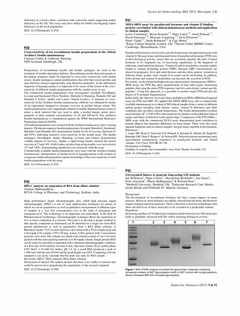
deficiency in a local cohort, consistent with a previous report suggesting iodinedeficiency in the UK. This assay may have utility for further investigating iodinedeficiency in the local population.
DOI: 10.1530/endoabs.31.P38
P39Cross-reactivity of ten recombinant insulin preparations in the AbbottArchitect Insulin immunoassayCatriona Clarke & Catherine ShearingNHS Scotland, Edinburgh, UK.
Preparations of recombinant insulin and insulin analogues are used in thetreatment of insulin-dependent diabetes. Recombinant insulin that corresponds tothe human sequence might be expected to cross-react extensively with insulinassays. Insulin analogues contain modifications that alter their action profiles andmay therefore interact unpredictably with detection antibodies. In the differentialdiagnosis of hypoglycaemia it is important to be aware of the extent of the cross-reactivity of different insulin preparations with the insulin assay in use.Ten commercial insulin preparations were investigated: Actrapid, NovoRapid,Levemir and Insulatard (Novo Nordisk); Humulin S, Humalog, Humulin M3 andHumulin I (Lilly); Apidra and Lantus (Sanofi-Aventis). A measure of cross-reactivity in the Architect Insulin immunoassay (Abbott) was obtained by meansof an experiment designed to measure recovery in pooled human serum. Theinsulin preparations were sequentially diluted in insulin-depleted human serum toattain a stock material that was used to spike a pooled human serum (post-prandial) to final nominal concentrations of 20 and 100 mU/l. The ArchitectInsulin immunoassay is standardised against the WHO International ReferencePreparation material 66/304.All insulin preparations cross-reacted with the Architect insulin assay, but to varyingdegrees. The human sequence insulin preparations (Actrapid, Humulin S, Insulatard,Humulin I and Humulin M3) demonstrated similar levels of recovery (between 81and 89%), indicating extensive cross-reactivity in the insulin assay. The insulinanalogues (NovoRapid, Apidra, Humalog, Levemir and Lantus) demonstratedvariable degrees of recovery. Apidra, a rapid-acting insulin, showed the lowestrecovery at 12 and 14%, while Lantus, an ultra-long acting insulin, over-recovered at127 and 140%, demonstrating significant cross-reactivity with the assay.Commercially available insulin preparations cross-react with the Architect Insulinimmunoassay. Interpretation of insulin levels in hypoglycaemia in the context ofexogenous insulin administration requires knowledge of the cross-reactivity of suchinsulin preparations with the assay.
DOI: 10.1530/endoabs.31.P39
P40HPLC analysis on separation of BSA from dilute solutionGoutam MukhopadhyayBCDA College of Pharmacy and Technology, Kolkata, India.
High performance liquid chromatography also called high pressure liquidchromatography (HPLC) is one of such sophisticated techniques by means ofwhich we can do quantitative as well as qualitative measurement of different typesof samples at a very low concentration even in the order of pictogram andnanogram level. The technology is an important one particularly in the field ofPharmaceutical Technology. Chromatography technique effects the separation oftwo or more component in a mixture. Our goal is to develop a unique method sothat specific compound as mentioned can be identified in a single run from BSAprotein qualitatively as well as quantitative from a BSA dilute solution. ARheodyne model 7125 six port injection valve fitted with a 20 ml sample loop anda Novapak C18 column (150!39 mm, waters, USA) packed with 5 micrometerparticles were used. The column was fitted with a Guard column (5 cm!4.6 mm)packed with the same packing material as in Novapak column. Single protein BSAcan be easily be enriched or separated with a optimum chromatographic condition,at a flow rate of 0.8 ml/min, run time 8 min, injection volume 20 ml, mobile phase0.9% NaCl C10 mM Tris buffer, pH 7.4. As a result BSA produced a peak at1.406 min with the area 66 944 and the peak height was 6291. Comparing with thestandard it can easily conclude that the peak was only for BSA sample.Keywords: HPLC, BSA standard, BSA dilute solution.Declaration of interest The authors declare that there is no conflict of interest thatcould be perceived as prejudicing the impartiality of the research reported.
DOI: 10.1530/endoabs.31.P40
P41MSIA-SRM assay for parathyroid hormone and vitamin D bindingprotein: correlation with clinical immunoassay methods and applicationto clinical samplesLewis Couchman1, Bryan Krastins1,2, Mary Lopez1,2, Amol Prakash1,2,David Sarracino1,2, Maryann Vogelsang1,2, Scott Peterman1,2,Gouri Vadali1,2, Sarah Robinson1,2 & Caje Moniz1
1Kings College Hospital, London, UK; 2Thermo Fisher BRIMS Centre,Cambridge, Massachusetts, USA.
Parathyroid hormone is involved in calcium homeostasis through interactions withvitamin D. Because intact and truncated forms of parathyroid hormone (PTH) varyin their biological activity, assays that can accurately quantify the ratio of intacthormone to its fragments are of increasing significance in the diagnosis ofendocrine, renal and bone diseases. Vitamin D and its metabolites circulate tightlybound to vitamin D-binding protein (DBP). Because DBP concentrations arealtered in pregnancy, liver and renal diseases and also show genetic variations indifferent ethnic groups, total vitamin D in serum can be misleading. In addition,both calcium and vitamin D metabolites can decrease the secretion of PTH.Previously, we developed multiplexed mass spectrometric immunoassay (MSIA)–SRM assays for PTH that allow quantification of four fully-tryptic monitoringpeptides (that span the entire PTH sequence) and two semi-tryptic variant specificpeptides1. Using this approach, it is possible to monitor intact PTH and also thedegree of N-terminal fragmentation.In this study, the objective was to develop a multiplexed, MSIA-SRM-based targetedassay for PTH and DBP. We applied this MSIA-SRM assay and a commerciallyavailable immunoassay to a cohort of 500 clinical samples from a variety of differentpatient groups including renal disease, cancer, vitamin D deficiency and otherconditions that can affect calcium homeostasis. The results demonstrated excellentassay linearity (R2Z0.90–0.99) with sensitivity for analytes in the published clinicalranges and limits of detection in the pg/ml range. Comparison of the PTH MSIA –SRM assay with the commercial ELSA assay demonstrated good correlation innormal subjects but important differences in renal failure. There were also someunusual fragments seen in clinical samples, not previously reported in the literature.References1. Lopez MF, Rezai T, Sarracino DA, Prakash A, Krastins B, Athanas M, Singh RJ,Barnidge DR, Oran P, Borges C & Nelson RW. Selected reaction monitoring-massspectrometric immunoassay responsive to parathyroid hormone and relatedvariants. Clin Chem 2010 56 281–90.Declaration of fundingFunding of reagents and consumables were from Thermo Scientific US.
DOI: 10.1530/endoabs.31.P41
P42Glycosylated linkers to generate long-acting GH tandemsIan Wilkinson1, Pippa Cawley1, Maximilian Bielohuby2, Jon Sayers1,Peter Artymiuk1, Martin Bidlingmaier2 & Richard Ross1
1Sheffield University, Sheffield, UK; 2Endocrine Research Unit, Medizi-nische Klinik und Poliklinik IV, Munich, Germany.
BackgroundThe development of recombinant biologics has had a major impact on manydiseases. However, most biologics are rapidly cleared from the body and thereforerequire frequent injection regimens. There is therefore a need for technologies thatallow the half-lives of these molecules to be extended in a predictable manner.HypothesisIncreasing numbers of N-linked glycosylation motifs between two GH moleculesleads to gradually increased half-life whilst retaining biological activity.
1 2 3 4 5 6 7
Figure 1 SDS–PAGE analysis of purified GH glycol-linker molecules containingincreasing numbers of NAT (glycosylation motif) of QAT (control with no glycosylation).Shows increased MW with increased glycosylation.
Society for Endocrinology BES 2013, Harrogate, UK
Endocrine Abstracts (2013) Vol 31
MethodsA number of GH tandem molecules with linkers containing 2–8 NATglycosylation motifs and their respective controls (in which N is replaced by Qin the sequence motif NAT) were cloned, sequenced and expressed in a CHO cellline. SDS–PAGE was used to verify increases in molecular weight and an inhouse dual luciferase reporter assay used to test bioactivity. Protein was purifiedusing immobilised metal affinity chromatography (IMAC). Pharmaco kinetics(PK) were assessed using a rat model system.ResultsOn increasing numbers of glycosylation motifs a concomitant increase inmolecular weight (MW) as observed by SDS–PAGE (see Fig. 1) from 42 to75 kDa (nZ8 glycosylations). All GH tandems were purified using IMAC fromsuspension-adapted serum free cultures to O95% purity. PK was tested in tandemGH molecules with and without two glycosylation sites. The tandem withoutglycosylation was cleared rapidly and with an identical profile to monomeric GHwhilst the tandem with glycosylation showed a fourfold slower clearance.ConclusionIt is possible to increase the apparent MW of hormone tandems usingglycosylated-linkers whilst maintaining bioactivity and that this also delaysclearance of the molecule in rat model system providing a potential technologyfor generating long acting biologicals.
DOI: 10.1530/endoabs.31.P42
P43High pulmonary artery pressure is associated with BNP andNT-proBNP in lowlanders acclimatising to high altitudeA Mellor1,2, N E Hill1, C Boos3, D Holdsworth3, J Begley4, M Stacey1,D Hall7, A Lumley7, A Hawkins4, S Foxen1, J O’ Hara5, C Smith1, S Ball6 &D Woods1,6
1Defence Medical Services, UK; 2Academic Department of EmergencyMedicine, James Cook University Hospital, Middlesborough, UK;3Department of Cardiology, Poole Hospital NHS Foundation Trust, Dorset,UK; 4Department of Biochemistry, Poole Hospital NHS Foundation Trust,Dorset, UK; 5Carnegie Institute, Leeds Metropolitan University, Leeds, UK;6University of Newcastle, Newcastle upon Tyne, UK; 7Ministry of DefenceHospital, Northallerton, UK.
BackgroundWe have previously demonstrated that the natriuretic peptides BNP andNT-proBNP rise with ascent to high altitude (HA). Both peptides are classicallymarkers of congestive cardiac failure but have also recently been found to beassociated with pulmonary hypertension at sea-level (SL). As pulmonaryhypertension is central to the risk of high altitude pulmonary oedema we aimedto establish if there was any association between high pulmonary artery systolicpressure (PASP) and BNP/NT-proBNP at HA.Methods20 subjects from an expedition to Nepal (study 1) and 48 subjects from anexpedition to Bolivia (study 2) were recruited and had BNP and NT-proBNPassayed and non-invasive assessment of PASP performed with ascent to 5150 m.ResultsBNP and NT-proBNP generally increased at altitude compared to baseline.PASP increased progressively with ascent. Generally, a PASP R40 mmHg (vs!40 mmHg)at various altitudes was associated with a higher BNP and NT-proBNP.For example, in study one at 5150 m those with a PASPR40 mmHg (nZ8) vs those!40 mmHg had BNP of 54.5G36 vs 13.4G17 pg/ml (PZ0.012). In study twothose with a high PASP at 3600 m at rest (nZ6) had higher BNP (37.5G38 vs 10G6.7, PZ0.003) and NT-proBNP (370G413 vs 101G72, PZ0.003).ConclusionBNP and NT-proBNP may serve as markers for elevated PASP, a central feature ofhigh altitude pulmonary oedema, at HA. In this respect they offer the potential tofacilitate early diagnosis and management, particularly with the availability ofpoint-of-care testing.
DOI: 10.1530/endoabs.31.P43
P44Danazol cross-reacts in the Roche E170 testosterone assayFiona Riddoch1 & Les Perry2
1Barts Health NHS Trust, London, UK; 2Croydon Health Services NHSTrust, London, UK.
The duty biochemist at Barts Health noted a testosterone result of 17 nmol/l on a20-year-old female, measured by Roche E170 electrochemiluminescence
immunoassay. Laboratory policy is that all female testosterone resultsO2.5 nmol/l by immunoassay (upper reference limit 1.92 nmol/l) are checkedby LCMS to exclude analytical interferences. The LCMS result on this samplewas below the limit of quantification (!0.5 nmol/l).Clinical details were ‘aplastic anaemia’. There was no previous testosteroneresult. LH and FSH were 8.6 and 6.3 IU/l respectively. Patient’s notes revealedthat she was on danazol to treat anaemia secondary to Dyskeratosis Congenita, ata dose of 200 mg alternating daily with 100 mg.The sample was diluted in steroid-free serum, and measured neat (confirmingprevious result) and at three dilutions. The dilution studies gave a linearregression of yZ1.01x – 0.25, with a correlation coefficient of r2 Z0.999.The sample was checked on an alternative platform; testosterone was 2.3 nmol/l(!4.5) on the Abbott Architect (one-step chemiluminescent microparticleimmunoassay).Danazol (200 mg capsule) obtained from Pharmacy was dissolved in methanol(10 ml), spiked into steroid-free serum to a final concentration of 988 nmol/l, andassayed for testosterone. On the Roche platform the result was 10.2 nmol/l(recovery of 1.04%). By LCMS, testosterone was reported as undetectable.These data confirm that danazol cross-reacts with the reagent antibodies in theRoche assay. The manufacturer states that cross-reactivity of danazol is %0.5% ata concentration of 1000 ng/ml. Abbott state that danazol does not cross-react intheir assay. Danazol (17a-ethinyl testosterone) is a derivative of ethisterone, andhas significant structural homology with ethisterone and norethisterone.UKNEQAS have demonstrated that norethisterone (at pharmacological concen-trations) interferes in the Roche testosterone assay. Users of immunoassay shouldbe aware of interferences due to steroid drugs.
DOI: 10.1530/endoabs.31.P44
P45Persistent hyperparathyroidism following parathyroidectomy: canroutine vitamin D replacement prior to surgery alter post-operativesecondary hyperparathyroidism?Natalie Chand1, Gina Weston-Petrides2, Abigail Evans2, Anthony Skene1,Joe Begley1,2, Philipp Antonas1 & Tristan Richardson1
1Royal Bournemouth and Christchurch Hospitals, Bournemouth, UK;2Poole Hospital, Poole, UK.
IntroductionA persistently elevated parathyroid hormone (PTH) following parathyroidectomyis usually associated with vitamin D deficiency. We have previouslydemonstrated this to occur in w60% of post-operative patients. We haveexamined the effect of routine vitamin D replacement pre-operatively on theproportion of patients with persistently elevated PTH post-operatively.MethodsData was collated retrospectively from our parathyroid database, including:patient demographics; pre- and post-operative biochemical results; operativedata; and vitamin D treatment.Results126 consecutive patients undergoing parathyroidectomy for sporadic primaryhyperparathyroidism were examined retrospectively (88 (70%) female). Pre-operative ultrasound resulted accurately localised 72% patients and nuclearmedicine localised 64% allowing 82 patients (65%) to undergo targeted(minimally invasive) parathyroidectomy.Median pre-operative serum calcium was 2.9 mmol/l (range 2.6–4.0), and medianpre-operative PTH was 13.1 (range 4.5–849 pmol/l). 116 patients (92%) had asingle gland excised. 70 of 88 (79.5%) patients who had vitamin D levelsmeasured were found to be vitamin D deficient pre-operatively (!50 nmol/l) –median pre-operative serum vitamin D 35.1 nmol/l (range 7–127).All patients received vitamin D (cholecalciferol 1000–2000 IU daily) at least 3months prior to parathyroidectomy. There were no episodes of severehypercalcaemia requiring more urgent surgical intervention. 97.6% patientswere rendered normocalcaemic following parathyroidectomy (failure rate 2.4%).Of these, 45 post-operative patients (36%) were found to be vitamin D deficient.There were no episodes of prolonged hypocalcaemia post-operatively (HungryBone Syndrome).ConclusionsA series previously presented by this unit reported 57% patients with persistentlyelevated PTH following parathyroidectomy. We now routinely replace vitamin Dthree months prior to parathyroidectomy, resulting in a reduction in thisproportion. There were no adverse consequences of vitamin D replacement andreplacement was successful in reducing post-op vitamin D deficiency. Thisreduction in secondary hyperparathyroidism is likely to relate to better bonehealth, which we are exploring.
DOI: 10.1530/endoabs.31.P45
Society for Endocrinology BES 2013, Harrogate, UK
Endocrine Abstracts (2013) Vol 31
P46Service review and demand management following clinical audit ofurine free cortisol requesting at Barts Health NHS TrustFiona Riddoch1, William Drake1 & Les Perry2
1Barts Health NHS Trust, London, UK; 2Croydon Health Services NHSTrust, London, UK.
Urine free cortisol (UFC) is analysed in 24 h urine collections in suspectedCushing’s syndrome, and provides an integrated measure of cortisol secretionover the whole day. The aim of this audit was to review how clinically useful UFCresults were, and whether this analytical service was still justified. The currentautomated immunoassay with manual sample preparation was time-consuming,expensive (disproportionate quality control / external quality assessment (EQA)sample analysis, and unused reagent frequently discarded), and prone to cross-reactant interference.Requests from April 2010 to March 2011 were examined. Of 202 samples, 102were requests from within the Trust, mainly from Adult Endocrinology (40%),22% were EQA samples, and 28% were GP or referred samples.Of internal requests, 34% were clinically appropriate (e.g. ‘Cushing’s syndrome’,‘hypertension’, ‘relapse, known Cushing’s’), and 17% were inappropriate (e.g.‘adrenal insufficiency’, ‘tall stature’ (paediatric), ‘renal stones’). Analysis washampered, as 48% of requests had no clinical details. For 20% of requests a randomurine sample was sent, the results of which are not clinically interpretable.Clinical details, sample type, and whether the UFC result was raised or normal(!340 nmol/24 h) were considered in conjunction with the results of otherbiochemical and dynamic function tests to judge whether the test had ‘added value’in the patient pathway. In only 10% of requests was the UFC test considered to addvalue. In a further 12% of samples UFC may have added value, although dynamicfunction testing was preferable (e.g. 48 h low dose dexamethasone suppressiontest).Discussion with the clinical colleagues informed a consensus regarding when UFCis clinically required, and it was agreed to switch the assay to an LCMS method,which is more specific. Measures introduced following the audit have reducedrequests by 30%, and prevented inappropriate samples and requests being assayed,reducing analyses by 80%.
DOI: 10.1530/endoabs.31.P46
P47Hyponatremia in patients with fractured neck of femur: short andmedium term outcomesThenmalar VadivelooUniversity of Dundee, Dundee, UK.
Hyponatremia is an electrolye disorder which is relatively common inhospitalised patients. Usually it is asymptomatic and mild but in severe caseshas been associated with confusion and increased risk of morbidity and death. Theaim of this study was to compare outcomes following fractured neck of femur(FNOF) in patients with and without hyponatremia and was facilitated by thepopulation health datasets in Tayside obtained from the Health InformaticsCentre, University of Dundee. Length of stay, readmissions and deaths were themain variables of interest. Also examined were the time from admission tosurgery, the length of hospitalisation for recuperation/convalescence and theproportion of patients for whom normal serum was attained at discharge.Patients were included in this study if between 1/1/2000 and 28/6/2011, they wereadmitted to hospital in Tayside, Scotland with FNOF. Individuals were diagnosedas having hyponatremia if, at the time of admission, there was a specific diagnosisof the condition or the first serum sodium recorded following admission was lessthan 125 mEq/l. Electronic databases containing primary and secondary careinformation were linked using a unique identifier for each patient. c2 methods,independent t-tests and non-parametric methods were used to compare baselinecharacteristics. Cox regression was used to estimate the unadjusted and adjustedhazard ratio (HR) of readmission and death associated with hyponatremia. Binarylogistic regression was used to determine the effect size (odds ratio) ofhyponatremia and other independent variables on binary outcomes.Hyponatremia was associated with longer hospitalisation at index admission (30vs 17 days, PZ0.003) and increased risk of readmission for any reason (adjustedHR 1.60 (1.22, 2.11), PZ0.001). There was some evidence of an increased risk ofreadmission for FNOF (adjusted HR 1.68 (0.99, 2.83), PZ0.052) but no increasedrisk of death (adjusted HR 0.95 (0.75, 1.21), PZ0.661).Hyponatremia in patients with FNOF is a marker of longer hospitalisation andincreased risk of readmission.Declaration of fundingThis work was supported by funding from Otsuka Pharmaceuticals UK Ltd.
DOI: 10.1530/endoabs.31.P47
P48A comparison of serum chromogranin A measurement with 24 h urineand serum 5-hydroxyindole acetic acid measurement in patients withNETsPhillip Monaghan1, Joanne Adaway2, Juan Valle1, Richard Hubner1,Peter Trainer1, Denise Darby1 & Brian Keevil21The Christie Hospital, Manchester, UK; 2The University Hospital of SouthManchester, Manchester, UK.
IntroductionChromogranin-A (CgA) is a 49 kDa protein of the granin/secretogranin familyoriginating from dense-core secretory granules within cells of the diffuseendocrine system. CgA is currently the best available diagnostic biomarker forneuroendocrine tumours (NETs) with recent clinical guidelines advocating themeasurement of CgA as part of the baseline biochemical profile in patientspresenting with symptoms suspicious of a gastroenteropancreatic NET.5-Hydroxyindole acetic acid (5-HIAA) is also utilised as a marker for patientswith serotonin-secreting NETs. Most laboratories currently measure 24 h 5-HIAAexcretion in urine samples. However, urine collections are cumbersome and mayoften lead to inaccurate assessment of 24 h 5-HIAA excretion. More recently,LC–MS/MS measurement of 5-HIAA in plasma and serum matrices has becomeavailable.MethodThe aim of this study was to compare serum CgA measurement to both serum and24 h urine 5-HIAA measurement. We measured serum CgA, 24 h urine 5-HIAAexcretion and serum 5-HIAA in paired samples from 20 patients with known5-HIAA secreting neuroendocrine tumours. Patients receiving PPI therapy wereexcluded from the study.ResultsAll results were expressed as a percentage of the reference range. Linearregression analysis of CgA and 24 h urine 5-HIAA gave a correlation coefficientof 0.92 with a corresponding Passing-Bablok regression equation of (urine5HIAA)Z1.41!(CgA)K0.31. Linear regression analysis of CgA and serum5-HIAA gave a correlation coefficient of 0.63 with a corresponding Passing-Bablok regression equation of (plasma 5HIAA)Z1.74!(CgA)K0.49.ConclusionWe have demonstrated a strong correlation between 5-HIAA measurement inboth 24 h urine samples and serum samples when compared to the current bestavailable general NET marker CgA. Furthermore, in contrast to CgAmeasurement by immunoassay, 5HIAA measurement by LC–MS/MS is notsusceptible to anti-reagent antibody interference, or the high dose hook effect andtherefore offers greater analytical robustness.
DOI: 10.1530/endoabs.31.P48
P49An unusual case of chronic liquorice ingestion presenting ashypokalemic paralysisPallavi Hegde, Arun Jeenahalli Ramappa & David Bowen-JonesWirral University Teaching Hospital NHS Foundation Trust, Wirral, UK.
Long-term liquorice ingestion is a well-known cause of secondary hypertensionand hypokalemia. However, its initial presentation with a very severehypokalemia and rhabdomyolysis is exceedingly rare. We report a 46-year-oldgentleman who presented with acute onset bilateral leg weakness. Medicationincluded allopurinol, aminophylline, fluoxetine, gabapentin, omeprazole, andinhalers. He had a 40 pack year smoking history and drank alcohol heavily inthe past.On examination he had power of 2/5 in his legs and 5/5 in upper limbs. Severehypokalemia of 2.6 mmol/l and raised creatinine kinase of 42 660 IU/l was noted.He had raised urinary potassium excretion at 104 mmol/24 h, suppressedaldosterone (61 pmol/l) and renin (0.3 pmol/ml per h)concentration and normallevels of cortisol and thyroid function.Muscular weakness resolved with potassium replacement. Renal potassiumexcretion and hypokalemia normalised after several weeks. He represented withvery similar symptoms and with potassium of 1.7 mmol/l. A detailed dietaryhistory revealed that he consumed 1 bag (140 g) of liquorice daily for many years.He was diagnosed as suffering from Liquorice induced hypokalemia. 24 h urinarysteroid chromatography profile was consistent with 11b-hydroxysteroid dehydro-genase two deficiency. He was discharged home with advice that he should stopconsuming liquorice.Liquorice induced hypokalemia and rhabdomyolysis is very rare. Liquorices’active ingredient, glycyrrhizic acid inhibits 11b-hydroxysteroid dehydrogenase,the renal enzyme which is responsible for conversion of cortisol to cortisone. Asa result, renal mineralocorticoid receptors are activated by excessive cortisol,
Society for Endocrinology BES 2013, Harrogate, UK
Endocrine Abstracts (2013) Vol 31
which causes sodium and water retention and hypokalemia. In this patient a clearhistory of excessive liquorices ingestion, elevated renal potassium excretion,suppressed aldosterone and renin and biochemical confirmation of suppressed11b-hydroxysteroid dehydrogenase deficiency confirmed the diagnosis. Thisemphasizes the importance of a detailed dietary history in unexplainedhypokalaemia.
DOI: 10.1530/endoabs.31.P49
P50Role of urinary dopamine in investigation of phaeochromocytoma/paragangliomaChristopher Smith1, Marie Freel1, Colin Perry1, Fraser Davidson1,2,Karen Smith1,3 & Yannick Tautphaus1,4
1Western Infirmary, Glasgow, UK; 2Crosshouse Hospital, Ayrshire, UK;3Glasgow Royal Infirmary, Glasgow, UK; 4University of Lubeck,Schleswig-Holstein, Germany.
IntroductionThere is controversy over which form of biochemical testing is best for screeningfor phaeochromocytoma/paraganglioma (PHAEO/PGL). Measurement of plasmametanephrines is the most sensitive test but is less specific and lacks widespreadavailability, and so many centres opt to measure 24-h urinary excretion ofcatecholamines and metanephrines. However, the standard 24-h urinarycatecholamine profile also includes dopamine (DA), vanillylmandelic acid(VMA) and homovanillic acid (HVA).The aim of the current study was to determine the additional value of measuringurinary DA in the diagnosis of PHAEO/PGL by reviewing patients who hadelevated DA in isolation.MethodsWe reviewed 2161 (1118 cases) 24-h urinary catecholamine profiles analysed byHPLC and electrochemical detection at Crosshouse Hospital, Ayrshire between2007 and 2010. We then reviewed the cases where an isolated DA elevation wasseen.ResultsNinety one samples (4.2%) demonstrated an elevation in 24-h urinary DA whichcorresponded to 51 cases (4.6%). 18 cases (1.6%) demonstrated elevated urinaryDA in isolation. None of these had current evidence of PHAEO/PGL.ConclusionElevated 24-h urinary DA excretion in isolation is unusual and of the cases weidentified, there was no situation where this was of clinical relevance. These datawould support the removal of urinary dopamine from the conventional urinarycatecholamine profile (unless there is high suspicion of a DA secreting PGL) asin our series it had no impact on diagnostic yield and may lead to unnecessaryinvestigations.
DOI: 10.1530/endoabs.31.P50
P51Liquorice induced hypertension and hypokalaemiaHamza Khan, Giridhar Tarigopula, Praveen Partha & Paul PeterDarlington Memorial Hospital, Darlington, UK.
Liquorice (scientific name Glycyrrhiza glabra) is historically used forgastrointestinal complaints. Now it is primarily used as a flavoring agent in thetobacco, confectionery and to some extent in the pharmaceutical and beverageindustries. Excessive intake of liquorice may cause a primary hyperaldosteron-ism-like syndrome characterized by sodium and water retention, hypertension,hypokalaemia, metabolic alkalosis, low-renin activity, and hypoaldosteronism.We describe a 69 years old lady who presented through a General Practitionerwith hypertension, dependent oedema and hypokalaemia. There was no history ofdiarrhoea or vomiting and diuretics or laxative use. She was an ex smoker andconsumed a bottle of wine per week Her bloods showed severe hypokalaemiawith potassium of 2.0 mmol/l. Her arterial pH was 7.50 with bicarbonate42 mmol/l (metabolic alkalosis). Her serum renin and aldosterone were both low.Her Serum TSH, overnight dexamethasone suppression, ultrasound scan of thekidneys and 24 h urinary calcium were normal. Her history was reviewed onceagain and at that time it revealed that she had been taking liquorice in sweets foras long as she could remember. She stopped liquorice after that admission andwas discharged home on oral potassium supplements Sando K, two tablets threetimes a day which she gradually reduced and was not on any potassiumsupplements for about two months when seen in out patients, She wasnormotesive, had no dependent oedema and her serum potassium was normal.
The case emphasizes the importance of considering a detailed patient’s history,which often may lead the treating physician to the correct clinical diagnosis.
DOI: 10.1530/endoabs.31.P51
P52Calcification of basal ganglia in chronic hypoparathyroidismHamza Khan, Anurag Dhingra, Giridhar Tarigopula, Praveen Partha& Paul PeterDarlington Memorial Hospital, Darlington, UK.
Hypoparathyridsm and pseudohypoparathyroidsm are the common causes ofpathological calcification in the brain though 0.3–1.5% cases are physiological.The clinical presentation of hypoparathyroidsm can vary with the calcium levelsand chronicity of hypocalcaemia. We describe a 39-year-old female who had typeone diabetes for the last 23 years. She was repeatedly hospitalised with collapseepisodes thought to be hypoglycaemic though never proved. She also had primaryhypoparathyroidism for the last 10 years and was on calcium and vitamin Dsupplements. Her adjusted calcium was 2.2–2.5 mmol/l. She had a cataractremoved from the right eye and also had an early cataract in the left eye. Herfundal examination did not show any diabetic retinopathy. She also started to havetonic clonic seizures which initially were thought to be due to hypoglycaemicevents but the capillary glucose was never documented in these episodes. She hadslow speech, serpentine gaze and shuffling gait on examination. Her Chvostek’sand Trousseau’s signs were negative. She was sent for neuropsychologicalassessment to ascertain the cognitive impairment. There she had CT brain donewhich showed calcification in basal ganglia, centrum semi ovale bilaterallyand also subcortical dense linear calcification bilaterally. She was seen by aneurologist and started on anticonvulsant treatment. Her seizures improved but noimprovement in her cognition and gait abnormalities. This case emphasizes theimportance of thinking about the whole spectrum of the disease even if thebiochemical markers are stable on treatment.DOI: 10.1530/endoabs.31.P52
P53A review of causes of hypomagnesaemia in hospital patients and itsmanagementJana Bujanova1,2, Tristan Richardson1 & Joe Begley1,2
1Royal Bournemouth Hospital, Bournemouth, UK; 2Poole General Hospital,Poole, UK.
An association between the use of proton pump inhibitors (PPIs) and profoundhypomagnesaemia has been highlighted in a number of case reports. As theprevalence of this occurrence or cause remain unknown, we undertook a review ofpatients with significant hypomagnesaemia in our hospital with a particular focuson use of PPIs and management.35 patients (21f; mean age (S.D.): 71.3 (14.6); median: 74 years) with significanthypomagnesaemia (defined as serum Mg !0.5 mmol/l, adjusted for albumin),for whom medical records were available, were identified from the laboratorycomputer. Information was extracted on serum Mg level, presentation, identifiedcause and treatment.Serum Mg levels were 0.17–0.49 mmol/l (mean (S.D.): 0.30 (0.08); median 0.32);calcium levels were 1.35–2.61 mmol/l (mean (S.D.): 1.91 (0.33); median1.93 mmol/l). 26/35 patients were prescribed PPI’s – there was no differencebetween Mg or Ca levels for those on PPIs compared to the group as a whole.Use of PPIs was deemed causative or contributory in 12 of 26, 7 of whom wereswitched to an alternative antacid, three were stopped and two continued. Othercauses documented included GI loss in eight, chemotherapy in five, renal losses inthree, alcohol abuse four, with no cause documented in 10.There was lack of standardisation of treatment - for the group, 17, including ninein whom PPIs were considered causative, received IV replacement (duration:30 min-4d; dose 20–96 mmol, mean (S.D.): 43.2 (27.5); median 40 mmol); twoalso received oral treatment (magnesium glycerophosphate 8 mmol tds). Of theremaining 19, four received oral replacement (three magnesium glycerophosphate8 mmol tds, one unknown).This short review shows PPI use to be common among patients with significanthypomagnesaemia, though it was considered as a contributory or causative factorin less than 50%. It also highlights a lack of standardisation in Magnesiumreplacement.
DOI: 10.1530/endoabs.31.P53
Society for Endocrinology BES 2013, Harrogate, UK
Endocrine Abstracts (2013) Vol 31
P54An audit on management of hyponatraemia in hospitalised patientsV M R Katreddy, A U Nayak, G I Varughese & R N ClaytonUniversity Hospital North Staffordshire NHS trust, Stoke on trent, UK.
BackgroundAmongst hospitalised patients, hyponatraemia is the commonest electrolyteabnormality with reported prevalence of about 25%. Its association withmortality, morbidity and increased length of stay is well recognised, includingadverse fracture risk and falls with mild hyponatraemia in elderly. We audited themanagement of hyponatraemia in inpatients in a University hospital setting.MethodsOver a 3-month period, amongst all in-patients, we identified those who hadhyponatraemia (serum Na%130 mmol/l) from the in-house biochemistrydatabase. Data on their demographics, management of hyponatraemia, lengthof stay and mortality was obtained and analysed.ResultOf the 109 patients identified with hyponatraemia – their gender: 46% male; age:75G14 years (meanGS.D.), with 60% being over 75 years age. The proportionwho had serum Na !120, 121–125 and 126-130 mmol/l were 22, 24 and 54%respectively. Majority were managed in medical wards (91%) – acute medicine19%, care of the elderly 27%, endocrine 19%, gastroenterology 10%, generalmedicine 8% and other medical specialities including respiratory medicine 8%.Only 21% had Endocrine specialist input. SIADH, fluid overload and drugs wereattributed as cause for the hyponatraemia in 16%, 14% and 10% respectively andremaining 60% was due to other causes including vomiting and/or diarrhoea orwhere no clear cause was identified. The in-hospital mortality was 9.2% in thecohort, amongst whom 40% had serum Na !125 mmol/l. Of the 88.8%discharged, 36% and 35% had serum Na%130 mmol/l and %125 mmol/l atdischarge. The average length of stay in those discharged was 12.4 days.ConclusionsThis audit demonstrates the potential deficiencies in the optimum managementof hyponatraemia in inpatients setting. Large proportion had Na !130 mmol/l ondischarge which is potentially associated with adverse outcomes especially in thehigh risk elderly population.
DOI: 10.1530/endoabs.31.P54
P55Impact of hyponatraemia in critically ill patientsJayadave Shakher1, Nirav Gandhi2 & Govindan Raghuraman1
1Birmingham Heartlands Hospital, Heart of England NHS Trust, Birming-ham, Westmidlands, B95SS, UK, 2College of Medical and Dental Sciences,University of Birmingham, Birmingham, Westmidlands, UK.
IntroductionHyponatraemia, defined as serum sodium !135 mmol/l is one of the commonestelectrolyte abnormalities seen in patients admitted to acute hospitals and isassociated with increased morbidity and mortality. Impact of this condition is notadequately measured in critically ill patients admitted to intensive care unit. Theaim of this observational study was to assess the incidence and outcomes ofpatients admitted to intensive care unit (ICU) in a UK based setting.MethodsThis was a retrospective observational study that looked into the incidence of
hyponatraemia and outcomes such as mortality, length of stay, ventilator days,renal days in patients admitted to ICU between January 2011 and March 2012.Sodium levels were evaluated at four distinct time frames that included admissionto hospital, admission to ICU, discharge from ICU and discharge from Hospital.Appropriate statistical tests were applied for comparisons with hospital and ICUadmission Na.Results1289 patients were admitted during this time. Incidence of hyponatraemia athospital admission as 27.5% and out of this 7.5% were moderately and severelyhyponatraemic (!130 mmol/l). Incidence at ICU admission was 22.3% of which3.3% were in moderate or severely hyponatraemic. Patients with hyponatraemia(!135) at presentation to hospital had increased APACHE II and ICNARCscores(PZ!0.0001). Patients with hyponatraemia at admission to ICU also hadincreased APACHE II and ICNARC physiological scores. Overall there wasincreased mortality (35.9 vs 21.2%, PZ!0.0001) and ICU length of stay, andincreased ventilator days.
DiscussionThis study confirms findings available in literature on the increased morbidity andmortality in patients presenting with hyponatraemia to hospital and ICU.However, whether this caused excess mortality and morbidity in these patients isdifficult to ascertain and prospective studies are required to evaluate the effect ofcorrection of sodium levels on mortality and morbidity.Declaration of interestInvited speaker for Otsuka company.
DOI: 10.1530/endoabs.31.P55
P56Audit of inpatient management of hyponatraemiaBhavin Patel1, Gillian Coyle1, Vidya Srinivas1, Javier Gomez1,2
& Khin Swe Myint11Department of Endocrinology, Norfolk and Norwich University Hospital,Norwich, UK; 2Department of Clinical Biochemistry, Norfolk and NorwichUniversity Hospital, Norwich, UK.
IntroductionHyponatraemia is the commonest electrolyte disturbance occurring in 15–20% ofinpatients (1), with significant clinical implications if mismanaged. We conducteda retrospective audit of our current management of hyponatraemia in our 1000bedded trust.MethodData of patients with severe hyponatraemia (Na!125 mmol/l) admitted tohospital over 4 weeks (Aug 2011) was collected. Twenty randomly selected caseswere reviewed focusing on initial assessment, management plan and associatedmorbidity.Results218 cases were identified during the study period. Among the 20 selected cases,the lowest admission sodium was 112 mmol/l, the mean age was 73 years (29–90)with 30% female, the cohort was also noted to have multiple co-morbidities (MZ4.56). Assessment of hyponatraemia was only mentioned in 10 (50%) cases on theconsultant post take ward round. Fluid balance was recorded only in eight (44%)cases and no patient was assessed for postural hypotension. Aetiologies identified
Table 1 Hyponatraemia vs normonatraemia (Hospital admission sodium)(!135 vs 135-144)
Hyponatrae-mia (nZ353)
Normonatraemia(nZ874) P value
Age: mean (S.D.) 64.85 (16.35) 62.12 (17.65) 0.013Gender: male (n %) 210 (59.49%) 520 (59.50%) 0.998APACHE II score: mean (S.D.) 18.26 (6.75) 15.02 (6.42) !0.0001ICNARC model physiology score 18.24 (9.02) 14.73 (8.19) !0.0001ITU length of stay: median (IQR) 3 (2, 6) 3 (2, 5) 0.27Status at discharge from ITU: alive 274 (77.6%) 756 (86.5%) !0.0001Hospital Length of Stay: median (IQR) 12 (6, 29) 11 (6, 23) 0.32Status at discharge from Hospital: alive 231/273
(84.6%)694/756 (91.8%) 0.001
Overall mortality % 122/340(35.9%)
182/857 (21.2%) !0.0001
Advanced respiratory days 0 (0, 2) 0 (0, 2)Advanced cardiovascular days 0 (0, 0) 0 (0,0)Renal days 0 (0, 0) 0 (0, 0)Advanced respiratory days O0 131 (37.11%) 302 (34.55%) 0.40Advanced CV days O0 79 (22.38%) 156 (17.85) 0.068Renal days O0 66 (18.70%) 79 (9.04) !0.0001
Table 2 Hyponatraemia vs normonatraemia (ICU admission sodium)(!135 vs 135-144)
Hyponatraemia(nZ287)
Normonatraemia(nZ894) P value
Age: mean (S.D.) 65.98 (15.82) 61.76 (17.71) 0.0003Gender: male (n %) 177 (61.67%) 525 (58.72) 0.38APACHE II score: mean (S.D.) 17.40 (6.87) 15.32 (6.57) !0.0001ICNARC model physiology score 16.28 (8.98) 15.24 (8.44) 0.075ITU length of stay: median (IQR) 3 (2, 4) 3 (2, 5) 0.032Status at discharge from ITU: alive 236 (82.23%) 777 (86.91%) 0.048Hospital Length of Stay: median (IQR) 13 (6, 28) 11 (6, 23) 0.46Status at discharge from Hospital: alive 215/235 (91.49%) 703/777 (90.48%) 0.64Overall mortality % 72/277 (25.99%) 193/873 (22.11%) 0.18Advanced respiratory days 0 (0,1) 0 (0,2)Advanced cardiovascular days 0 (0, 0) 0 (0, 0)Renal days 0 (0, 0) 0 (0, 0)Advanced respiratory days O0 74 (25.78%) 323 (36.13%) 0.001Advanced CV days O0 45 (15.68%) 160 (17.90%) 0.39Renal days O0 33 (11.50%) 104 (11.63%) 0.95
Society for Endocrinology BES 2013, Harrogate, UK
Endocrine Abstracts (2013) Vol 31
were as follows; idiopathic (42%), drug induced (26%), SIADH (26%) andhypervolaemic causes (6%). Of the SIADH group the following investigationswere performed; urinary sodium (60%), urine osmolality (60%) and serumosmolality (80%), thyroid function tests (20%) and 0900 h cortisol (14%). Inrelation to outcomes, fall in serum sodium was seen in 25%, with a rise evident in65%, one case having O10 mmol rise in 24 h. The mean length of hospital stay(LOS) was 16 days (4–56 days) in comparison with a trust mean of 5 days, with15% of prolonged LOS documented to be attributable to hyponatraemia. Meansodium on discharge was 130 mmol/l (121–137 mmol/l), one death occurred postdischarge (Na 122 mmol/l).ConclusionHyponatraemia is commonly seen in patients with multiple co-morbidities. Manage-ment remains challenging and attributes to prolonged LOS. A clear local guideline isneeded and is currently under development, to improve the standard of care.ReferenceHuda MSB, Boyd A, Skagen K, Wile D, Van Heyningen C, Watson I, Wong S & GillG. Investigation and management of severe hyponatraemia in a hospital setting.Postgrad Med J 2006 82 (965) 216–219.
DOI: 10.1530/endoabs.31.P56
Clinical practice/governance and case reports
P57Initiation and maintenance of mitotane as adjuvant therapy foradrenocortical cancer: a single centre experienceBenjamin Whitelaw1, Omar Mustafa1, Patsy Coskeran1, Julia Prague1,Tiana Kordbacheh1, Dylan Lewis2, Salvador Diaz-Cano3, Roy Sherwood4,Jackie Gilbert1, Alan McGregor1 & Simon Aylwin1
1Department of Endocrinology, King’s College Hospital, London, UK;2Department of Radiology, King’s College Hospital, London, UK;3Department of Histopathology, King’s College Hospital, London, UK;4Department of Biochemistry, King’s College Hospital, London, UK.
BackgroundMitotane is an adrenolytic chemotherapy, currently accepted as first line adjuvanttherapy in adrenocortical carcinoma. Mitotane has a narrow therapeutic window.Serum levels of O14 mg/l are required to achieve a cytotoxic effect and levels ofO20 mg/l are potentially toxic. There are two strategies for mitotane initiation: alow-dose regimen (3 g) and a high-dose regimen (increase to 6g/day over 4 daysand reduce to 4.5 g/day after 10 days).MethodsWe conducted a retrospective review of consecutive mitotane use foradrenocortical carcinoma in a UK tertiary centre from 2006 to 2012. Mitotaneinitiation was completed using a nurse-led protocol.ResultsInitiation: Twenty patients were identified: 15 were prescribed the high-doseregimen and five the low-dose regimen. Eighty-five percent (17/20) achieved thetherapeutic threshold (O14 mg/l). Mean time to reach therapeutic level was 2.2months. Twenty percent (4/20) achieved therapeutic threshold in the first month.In total, seventy percent (14/20) achieved therapeutic levels within 3 months ofinitiation. Mitotane levels were performed 2-weekly for the first 3 months. Themaximum dose used ranged from 3 to 8 g/day (mean 5.5 g/day).Maintenance: Of the 20 patients initiated on mitotane, twenty-five percent (5/20)discontinued treatment because of side effects and/or intolerance (neurologicaland gastrointestinal symptoms). A further twenty-five percent (5/20) discontinuedfollowing disease progression with subsequent mortality. In total, seventy-fivepercent (15/20) of patients continued to maintenance phase (O3 months). Ofthese, seventy-seven percent of serum mitotane levels were above therapeuticthreshold and fifty-three percent were within the range 14–20 mg/l. The meanmaintenance dose of mitotane for this group was 4.8 g/day (range 2.5–8.0 g/day).Ten patients currently remain on mitotane.ConclusionInitiation of mitotane can be safely performed on an outpatient basis, using anurse-led protocol. Seventy percent of patients in our series achieved mitotanelevels above the therapeutic threshold within 3 months, comparing favourably toother published series.
DOI: 10.1530/endoabs.31.P57
P58De Novo HNF1b mutation as a cause for chronic treatment-resistanthypomagnesaemiaCraig Stiles1, Ajith Kumar2, Detlef Bockenhauer2 & Marta Korbonits1
1Queen Mary University, Barts and the London School of Medicine,London, UK; 2Great Ormond Street Hospital, London, UK.
A 29y female presented with an 8y history of hypomagnesaemia. It was notedincidentally when hospitalised with mumps-related pancreatitis. Subsequentlysymptomatic hypomagnesaemia, with headaches and lethargy, was treated withmagnesium glycerol phosphate 4 mg TDS, but she remained symptomatic withoccasional need of IV Mg2C. It was thought that she was poorly compliantwith her oral Mg2C supplements. At presentation to our department for follow-upof her hypomagnesaemia, SeMg2C was low (0.51 mmol/l) despite Mg2C glycerolphosphate 4 mg TDS. Other biochemistry was normal including creatinine,vitamin D, total protein, PTH and cCa2C. Urinary Na&K were normal andglucose negative. Urinary Ca2C was low at 1 mmol/24 h (3–5 mmol/24 h).Urinary Mg2C was (inappropriately) normal at 4 mmol/24 h (3–5 mmol/24 h). Asthe patient appeared to be losing Mg2C from the renal tract, renal imaging withUS and CT was performed, showing one large 2.8 cm and four 1.5 cm cysts in theleft kidney, while the right was normal. The patient had a bicornate uterus. Therewas no significant family history. Referral to a geneticist lead to identification ofa heterozygous whole gene deletion of HNF1-Beta (renal-cysts-and-diabetessyndrome: RCAD). Neither parent shared this mutation. HNF1b loss-of-functionmutations are associated with MODY5, pancreatic insufficiency, renal cysts,hyperuricaemic nephropathy, single functioning kidney, gout, pancreatic atrophyand urogenital deformities (including bicornate uterus). In a paediatric populationabnormal antenatal renal US is frequent with Mg2C-wasting. HNF1B is importantfor the expression of FXYD2, which regulates ion transport in the distalconvoluted tubule and inactivating mutations in FXYD2 also lead tohypocalciuria and hypomagnesaemia. The differential diagnosis of hypomagne-saemia includes diabetes mellitus, diuretics, platinum-containing chemotherapy,PPIs, EGF-antibodies, alcohol, hypercalcaemia and mutations in paracellin-1(Mg2C-wasting with hypercalciuria due to loss of tight junction, hinders Mg2C-reabsorption in ascending loop of Henle), KCNA1 (KC-channel whichpotentiates Mg2C-reabsorption via transmembrane electrical potential) andEGF (stimulates Mg2C-reabsorption of).
DOI: 10.1530/endoabs.31.P58
P59Outcomes of transsphenoidal surgery (TSS) for acromegalyIrfanulla Baig1, Kashif Hafeez1, Anand Velusamy2 & Anna Crown1
1Brighton and Sussex University Hospitals NHS Trust, Brighton, UK;2Eastbourne District General Hospital, Eastbourne, UK.
BackgroundPituitary surgery is the initial treatment for the majority of patients withacromegaly. The UK acromegaly register data (UK-AR-2) suggests that surgicalremission rates vary widely, with a marked improvement since 2000. The aim ofthis study was to assess the outcomes of first TSS for acromegaly in our centreover the past 5 years.MethodsWe retrospectively analysed data for all acromegaly patients who underwent firstTSS between 2007-2011. Biochemical remission was defined as normalisation ofIGF1 and GH nadir !1 mcg/l post-GTT, or !2 mcg/l (random GH or seriesmean), at 3 months after surgery. Post-operative imaging was reviewed and dataregarding pre and post-operative pituitary hormone deficiencies was collected.ResultsTwo surgeons performed 22 first TSS operations for patients with acromegalyover the 5 year period (2.2 acromegaly operations /year per surgeon), includingeight microadenomas, six intrasellar (IS) macroadenomas and eight extrasellar(ES) macroadenomas.Post-operative remission rates for GH were achieved in 88% of microadenomas,67% of IS macroadenomas and 25% of ES macroadenomas. The correspondingpercentages for IGF1 were 75, 50 and 12.5%, and for both IGF1 and GH 62.5, 50and 12.5%.Post-operatively, six patients with macroadenomas developed new pituitary axisdeficiencies, whilst five patients with pre-operative pituitary axis deficienciesrecovered function post-operatively.Comparative dataMean biochemical remission rates following TSS for acromegaly in eightpublished series since 2000 are 78% for microadenomas, 59% for intrasellarmacroadenomas and 25% for extrasellar macroadenomas. Most published seriesare from larger centres. Some studies only report outcomes for multi-modaltherapy for acromegaly.ConclusionsHow many pituitary centres the UK should have for optimal outcomes, andwhether centres should have one or two pituitary surgeons, remains an active
Society for Endocrinology BES 2013, Harrogate, UK
Endocrine Abstracts (2013) Vol 31
debate. Regular collection and reporting of surgical outcome data is essential toinform pituitary service provision.
DOI: 10.1530/endoabs.31.P59
P60How do medical students and doctors learn clinical endocrinology?Mehul Patel & Maralyn DruceDepartment of Endocrinology, St Bartholomew’s Hospital, London, UK.
BackgroundThere has been little research into methods of learning clinical endocrinology, aspeciality encompassing rare conditions and pattern recognition. Problemsinclude limited patient exposure outside specialist centres and increasing pressureon doctors to manage their own educational needs. An understanding of learningmethods in use across the spectrum of students and doctors will help to identifytrusted resources and untapped techniques.AimPilot study to identify the usefulness of different learning resources employed bystudents and doctors currently engaged in endocrine practice.MethodParticipants completed a paper/online questionnaire indicating perceivedusefulness of educational resources they had encountered. This was a prospectivestudy using convenience sampling.Results77 responses were obtained: 57 third-year and 4 final-year students on endocrineplacements, three foundation/CMT doctors, five registrars and 8 consultants.Lectures and textbooks were useful across all groups; but patient encounters wereperceived as most helpful (100.0% of final year students and above). Withincreasing seniority, perceived usefulness of journals increased (25% of 3rd-yearstudents, 50.0% of final-year students and 100% of registrars/consultants).Wikipedia was commonly-used by students (86.7% of 3rd-years found it helpful;62.5% of consultants had never used it). Except for registrars (60% of whomfound them useful), most participants had never used e-learning resources. 6.5%of participants had used podcasts and 26.0% had used apps for endocrine learning.Students are willing to use e-learning/podcasts if directed to these resources.DiscussionTraditional resources and patient encounters remain important for learning.E-learning, apps and podcasts are relatively unexploited tools. Perceivedusefulness is a helpful starting point for design and evaluation of grade-specificresources. Developing ‘virtual patients’ may benefit students and trainees, whilstsenior physicians may value podcasts as a bite-sized method of delivering clinicaldevelopments. These will require evaluation for quality of learning as well as easeof use and popularity.
DOI: 10.1530/endoabs.31.P60
P61An unusual cause of Cushing’s syndrome with secondary adrenalinsufficiencyAmalia Iliopoulou & Emma WardSt James University Hospital, Leeds, UK.
A 20-year-old girl presented to the endocrine clinic with a history of three stoneweight gain, and development of numerous purple striae over her lower abdomen,inner thighs and upper arms, gradually progressing over a 12-month period. Theonset of symptoms had coincided with the initiation of contraceptive depotmedroxyprogesterone acetate, which was discontinued two months prior to herpresentation. Her 9 am cortisol was !50 nmol/l.She had a background history of well controlled asthma, not requiring steroidinhalers for past two years. She was seeing the rheumatologists because of chronicthoracic pain and was on fentanyl patches. She had received two intramuscularsteroid injections for this, last of which was two years previously. There was noother history of exogenous steroid use.She had a short synacthen test that showed an undetectable baseline cortisol,rising to 263 nmol/l after stimulation. ACTH was 11 ng/l, with normal renin,aldosterone and baseline pituitary function. She was commenced on hydrocorti-sone 10 mg am and 5 mg pm and a glucagon stimulation test organised a monthlater showed a peak cortisol of 262 nmol/l. She was reviewed 4 months later, herCushingoid features had significantly improved and a 0900 h cortisol was265 nmol/l. Subsequently, a repeat glucagon stimulation test showed a peakcortisol of 604 nmol/l, consistent with a complete recovery of the HPA axis. Shehas been off hydrocortisone for last 2 months and remains asymptomatic.
Medroxyprogesterone acetate is a widely used potent progestational agent. It haslow affinity for the glucocorticoid receptor resulting in low level glucocorticoidactivity and may cause a combination of cushingoid appearance with secondaryadrenal insufficiency when used in large doses, as in the treatment of cancer. Toour knowledge this is the first case of such syndrome occurring with very smalldoses of medroxyprogesterone used for contraception purposes.
DOI: 10.1530/endoabs.31.P61
P62Management of intercurrent illness in adrenal insufficiencyRachel McLatchie1, Mark W.J. Strachan1,2 & Fraser W. Gibb1,2
1University of Edinburgh, Edinburgh, UK; 2Edinburgh Centre forEndocrinology, Edinburgh, UK.
Several recent publications have advocated that patients with primary andsecondary adrenal insufficiency (PAI and SAI) be given emergency injectablehydrocortisone to reduce the risk of adrenal crisis. There is a limited evidence-base for this recommendation and consequently the Edinburgh Centre forEndocrinology (ECE) does not routinely issue kits to its patients. This studyassessed the frequency of hospital presentations in ECE’s AI patients, and theseverity of their condition on presentation. It also evaluated patients’ knowledgeof crisis-preventing behaviours, proportions with emergency hydrocortisone kits,and the influence of these factors on presentation rates.Details on 81 PAI and 66 SAI patients and their presentations were obtained frompatient records. Data such as blood pressure and biochemistry was collected, aswell as reason for admission. 81 patients participated in a telephone questionnaire,part of which assessed their knowledge of ‘sick day rules,’ as defined by PituitaryFoundation literature. Patients were also questioned about emergency kits.54.3% of PAI and 34.8% of SAI patients presented to A&E over a 6-year period.The trends suggested that PAI patients were more likely to be hypotensive,hyponatraemic, hyperkalaemic and have low bicarbonate, but these results werenot statistically significant. PAI patients were, however, more likely to behypoglycaemic (PAI 15.4%, SAI 0.0%, PZ0.048). The commonest cause ofpresentation for PAI patients was gastrointestinal infection, and respiratoryinfection for SAI. Only 16% of patients knew to go straight to A&E whenvomiting. 32% possessed emergency kits, but their frequency of presentation didnot differ significantly from those without (PZ0.53).Improved education strategies may be required. Further studies should establishwhether or not patients with emergency kits are less unwell on presentation thanthose without, and it may be useful to run a comparative study between ECE andanother centre which does issue emergency kits.
DOI: 10.1530/endoabs.31.P62
P63A case of Hashimoto’s thyroiditis induced coagulopathyAnthony Lewis1, Gary Benson1,2 & Hamish Courtney1
1Regional Centre for Endocrinology and Diabetes, Royal Victoria Hospital,Belfast, UK; 2Departmentof Haematology, Belfast City Hospital, Belfast,UK.
A 26yr old man presented to haematology with a short history of easy bruising.There was no spontaneous bleeding. Past medical history was unremarkable.Family history of clotting disorders was negative. On examination there wassignificant lower limb bruising.Initial investigations revealed platelets of 238 (150–450), prothrombin time (PT)of 12.0 (12.0–17.0 s) and elevated activated partial thromboplastin time (APTT)of 41.0 (24.0–38.0 s). The elevation in APTT alluded to an intrinsic pathwayabnormality which was investigated through further clotting factor analysis. Thisrevealed reduced levels of chromogenic factor VIII 0.46 (0.6–1.3 U/dl), factorVIII clotting assay 0.4 (0.6–1.3 IU/ml), von Willebrand Factor antigen 0.31 (0.7–2.0 IU/ml) and von Willebrand Factor activity 0.39 (0.7–2.0 IU/ml). Factor IXassay was normal at 0.63 (0.6–1.3 U/ml). This confirmed the diagnosis of vonWillebrand disease.On further questioning he complained of cold intolerance, dry skin and lethargyand there was a positive family history of hypothyroidism in an aunt. Clinicalexamination was unremarkable. Thyroid function was significantly deranged withunmeasurable free T4 !5.5 (9.0–19.0 pml/l) and grossly elevated TSH of 711.1(0.4–4.5 mU/l). Anti-thyroid peroxidase antibodies were elevated at 3000(0–135 U/ml) confirming the diagnosis of Hashimoto’s thyroiditis. He wascommenced on levothyroxine and the coagulopathy reversed (APTT 36.3 s,Factor VIII clotting assay 0.71 IU/ml, von Willebrand Factor Activity
Society for Endocrinology BES 2013, Harrogate, UK
Endocrine Abstracts (2013) Vol 31

0.78 IU/ml) as his thyroid function normalised.This is a case of acquired von Willebrand disease due to Hashimoto’s thyroiditis.It is the most common coagulopathy in hypothyroidism and is characterisedclinically by easy bruising, epistaxis and mucosal bleeding and biochemicallythrough elevated APTT, reduced Factor VIII and von Willebrand Factor.Normalisation of the thyroid axis usually reverses the coagulopathy negating theneed for further treatment. Although a relatively uncommon complication ofhypothyroidism it should be considered at diagnosis and coagulation statusmeasured if clinically indicated.
DOI: 10.1530/endoabs.31.P63
P64Conn’s syndrome with normal plasma renin aldosterone ratioMyat Thida, Julie Andrews, Julian Barth & Steve OrmeLeeds Teaching Hospitals NHS Trust, Leeds, UK.
BackgroundConn’s syndrome accounts for 35% of primary hyperaldosteronism. Elevatedplasma aldosterone concentration to renin activity is widely used as a screeningdiagnostic tool. However, we report an unusual presentation of Conn’s syndromewith normal plasma renin aldosterone ratio.A 48-year-old man was seen in endocrine clinic with uncontrolled hypertensionand severe hypokalemia. Primary hyperaldosteronism was suspected with bloodpressure 170/110 mmHg, serum sodium 145 mmol/l, serum potassium 2.4 mmol/land metabolic alkalosis with serum bicarbonate 29 mmol/l. Initial plasma reninaldosterone ratio was 290 with aldosterone 320 pmol/l and renin 1.1 nmol/l per h.Subsequently blood pressure was controlled and serum potassium was corrected.Repeated plasma renin aldosterone ratio while on doxazosin and normalpotassium was again not consistent with Conn’s, having aldosterone365 pmol/l, renin activity 0.8 nmol/l per h with ratio of 450. MRI adrenalconfirmed 1 cm nodule in the right adrenal gland.Despite normal plasma renin aldosterone ratio, clinical suspicion of Conn’sdisease led to further investigations. Saline infusion test revealed failure ofaldosterone suppression at 225 pmol/l with relatively normal renin aldosteroneratio of 450 post saline infusion.Subsequently, he underwent adrenal veins sampling which showed a significantgradient of aldosterone to the right adrenal gland. (right adrenal vein aldosterone21 940 pmol/l, cortisol 1630 nmol/l, ratio 13.5; left adrenal vein aldosterone445 pmol/l, cortisol 1491, ratio 0.3). A laparoscopic right adrenalectomy wasdone. Histology confirmed benign adenoma consistent with Conn’s syndrome.Two months after surgery, blood pressure was 110/60 mmHg withoutantihypertensive, serum electrolytes remained normal, normal 24 h urinarypotassium at 85 mmol/day, plasma aldosterone 230 pmol/l, renin activity2.6 nmol/ l per h with aldosterone/renin ratio 88. He was discharged fromendocrine clinic.ConclusionPrimary hyperaldosteronism can be a diagnostic dilemma for clinicians. Furtherinvestigations should be considered if there is strong clinical evidence despitenormal plasma renin aldosterone ratio.
DOI: 10.1530/endoabs.31.P64
P65Peri-operative a-blockade: efficacy of intravenous phenoxybenzaminevs oral phenoxybenzamine in patients with phaeochromocytoma andparagangliomaShazia Hussain, Kirun Gunganah, Michael Ashby, Robert Carpenter,Mona Waterhouse, Maralyn Druce, William Drake & Scott AkkerDepartment of Endocrinology, St Bartholonew’s hospital, London, UK.
IntroductionRegimens for pre-operative a and b-blockade for patients with secretoryphaeochromocytomas/paragangliomas vary widely between centres. The world-wide lack of availability of intravenous phenoxybenzamine (Goldshield) hasremoved a useful tool in the management of phaeochromocytoma crisis and has
necessitated a change in our institution’s routine pre-operative strategy. Wecompare pre, peri and post-operative surrogate measures of blockade in a cohortof patients receiving iv phenoxybenzamine with an oral regimen.MethodsOf 41 patients with phaeochromocytoma/paraganglioma seen between 2009 and2012, 19 patients were included in this retrospective audit. Patients were onlyincluded if the same surgeon (RAC) and anaesthetist (MA) were present and wereexcluded if a transfusion was required. All patients had a blockade with oralphenoxybenzamine for at least 3 weeks prior to surgery. In the immediate 3-daypre-operative period five patients had accelerated oral phenoxybenzamine therapyG intravenous fluids and 14 patients had intravenous phenoxybenzamine Gintravenous fluid. We assessed intraoperative parameters of a blockade efficacyincluding requirement for sodium nitroprusside (SNP) and intravenous fluids. Weassessed postoperative fluid requirement, use of and response to adrenaline, bloodpressure and heart rate variability.ResultsPatients treated with intravenous phenoxybenzamine pre-operatively requiredless SNP (6.4 vs 12.3 mg) and less intra-operative intravenous fluids (2.9 l vs5.2 l) compared to patients treated with oral phenoxybenzamine. Mean systolicBP in the 3-day pre-operative period was lower in the i.v. group (123 vs130 mmHg) and the immediate post-operative systolic BP was higher in the ivgroup (108 mmHg vs 93 mmHg). Data including tumour type and size,catecholamine/metanephrine levels, postural BPs, and dose of a andb-blockade will be presented.ConclusionAlthough a small cohort, the data suggest that patients treated with ivphenoxybenzamine have better pre-operative BP control, require less intraopera-tive intervention and have less post-operative hypotension than patients treatedwith oral phenoxybenzamine. We invite other centres to report their experience.
DOI: 10.1530/endoabs.31.P65
P66A very interesting presentation: VIP co-secretion by aphaeochromocytomaJessica Triay & Karin BradleyUniversity Hospitals Bristol NHS Trust, Bristol, Avon, UK.
A 62-year-old lady was assessed following discovery of a retroperitoneal tumouron a CT scan. Fifteen years previously she was diagnosed with irritable bowelsyndrome with alternating constipation and diarrhoea, however, within the last 5years, diarrhoea was the dominant feature with bowel opening every 20 min daily.Investigations for inflammatory bowel disease, hyperthyroidism and coeliacdisease were negative. Her anal sphincter was found to be non-functioning and acolostomy was performed privately to improve her symptoms, but large volumesof stool continued to be passed daily. An episode of right upper quadrantabdominal pain led to imaging and subsequent referral. She had never been awareof any symptoms such as skin rashes or hypoglycaemia, and her weight wasstable. Two years previously she had experienced episodes of palpitations andsweating, although these had resolved and appeared to coincide with menopause.There was no significant family history and clinically, she was not hypertensive.CT demonstrated a 7 cm multi-cystic retroperitoneal tumour adjacent to the tail ofthe pancreas but uncertainty about pancreatic or adrenal origin. Octreotide uptakewas mildly avid. Laboratory investigations showed a markedly elevatedvasoactive intestinal peptide (VIP) at 100 pmol/l and urinary dopamine(12 768 mmol/24 h) with normal urinary adrenaline and noradrenaline. Plasmametanephrines confirmed elevated normetanephrine (18 562 pmol/l)Given these findings, phenoxybenzamine was commenced to optimise bloodpressure and a left adrenalectomy was performed. Histology confirmed completeexcision and immunohistochemistry was consistent with a phaeochromocytoma.VIP immunoassay was inconclusive due to high background staining, however, afasting gut hormone profile post operatively confirmed complete normalisation ofVIP levels (6 pmol/l) as were plasma normetanephrine (532 pmol/l), suggestinghormone co-secretion. Furthermore, stoma output slowed considerably to normalvolumes. We present an unusual case with complex biochemistry that highlightsthe importance of paying heed to the patient history.
DOI: 10.1530/endoabs.31.P66
Society for Endocrinology BES 2013, Harrogate, UK
Endocrine Abstracts (2013) Vol 31
Society for Endocrinology BES 2013, Harrogate, UK
P67A single pathology specialty service for hyperthyroid patients improvescare and outcomes compared to general endocrine clinics: results andimplications of an audit: re-audit cycle of clinical outcomes for differinghyperthyroid care modelsMo Lee Wong1, Tolulope Olateju2, Jean Munday3, Darryl Meeking3,Michael Cummings3 & Iain Cranston31Royal Bournemouth and Christchurch Hospitals NHS Trust Hospital,Bournemouth, UK; 2University Southampton Hospital NHS Trust, South-ampton, UK; 3Queen Alexandra Hospital, Portsmouth Hospitals Trust,Portsmouth, UK.
Prior to May 2011, we ran four separate consultant-led endocrine services withsix-eight new patient referrals identifiable per week with hyperthyroidism. Thesewere seen ‘ad hoc’ in general endocrine clinics, where their needs were notprioritised compared to other endocrine referrals, resulting in concern around thetimeliness of their care.We identified 203 patients under active follow-up (FU) (activeZon anti-thyroidmedications or within 6 months of radioactive iodine). The first audit cycle wascarried out to determine care received by patients with thyrotoxicosis in thegeneral endocrine clinics and the time interval between outpatient clinicappointments (OPAs).Using the July 2006 UK guidelines for use of TFTs, our standards were:i) All patients with abnormal thyroid function under treatment should be seenwithin 6 weeks (G7 days)ii) All euthyroid patients under treatment should be seen within 3 months (G7 days).Intervention: Following this review of the ‘ad hoc’ service, we introduced adedicated ‘weekly multi-disciplinary clinic’ hyperthyroid service to streamlinecare and allow for appropriate follow-up as well as opportunity for liaison withother specialists in those patients who require it.ResultsStandard 166% of thyrotoxic/hypothyroid patients were seen within target compared to 32%prior to the new service. Mean FU was 7 weeks compared to 20 weeks.Standard 2100% of all euthyroid patients were seen within target compared to 56% prior tothe new service. Mean FU was 8 weeks compared to 30 weeks.More patients achieved euthyroidism in the new hyperthyroid service at both 2ndand 3rd visits.ConclusionThese data highlight that dedicated hyperthyroid services led to improvements incare quality and outcomes for patients, as well as better use of services andresources. More patients were seen within specified target standards witheuthyroid status achieved earlier. There was a significant reduction inre-appointment and non-attendance rates.
DOI: 10.1530/endoabs.31.P67
P68Novel use of subcutaneous octreotide via an insulin pump for posturalorthostatic tachycardia syndromeMuhammad Khan, Karen Perkins & Franklin JosephCountess of Chester Hospital, Chester, UK.
Postural orthostatic tachycardia syndrome (PoTS) reflects a disturbance ofautonomic function leading to a myriad of clinical features. Subcutaneousoctreotide injections and intramuscular long acting preparations of octreotidehave been used in the treatment of PoTS. However, inconvenience of frequentinjections, side effects and theoretical overexposure, as well as cost of theintramuscular preparation, makes them far from ideal.We present a case of uncontrolled severe PoTS rectified through the novel use ofoctreotide via an insulin pump.A 22-year-old woman presented in April 2002 with episodic palpitationsassociated with chest discomfort, dyspnoea, and pre-syncope. After 3 years ofinconclusive investigations, deteriorating symptomatic control leaving her offwork, wheelchair bound, recovering from multiple syncopal related long bonefractures, depression and with a poor quality of life, a diagnosis of PoTS wasconsidered. Diagnostic tilt table testing identified pre-syncope associated withsinus tachycardia, maximum heart rate rise of 42 bpm, and decreases in bloodpressure confirming PoTS.Several therapies including fludrocortisone, slow sodium, midodrine, ephedrineand ivabradine were either ineffective or caused intolerable side effects. In May2010, SC octreotide, 50 mg every 90 min six times a day (300 mg/24 h), was
trialled. Despite her PoTS symptoms improving, she reported excruciatingabdominal cramps and diarrhoea following each injection. The severity andfrequency of side effects prompted the use of subcutaneous octreotide via anAnimas insulin pump in 2011. The patient received 10 mg/h of octreotide for 12 h(120 mg/24 h). The patient reported significant improvements in physical functionand ability to maintain upright posture with no side effects. She returned to workand her quality of life improved.ConclusionDelivery of octreotide via an insulin pump provides a novel therapeutic strategy inthe management of PoTS using lower doses and achieving symptom control withfewer side effects compared to subcutaneous octreotide.
DOI: 10.1530/endoabs.31.P68
P69Management of primary hyperparathyroidism during pregnancy: acase series of the lessons learntKatherine McCullough1, Niamh Martin1, Fausto Palazzo1,Catherine Williamson1,2 & Karim Meeran11Imperial Centre for Endocrinology, Hammersmith Hospital, ImperialCollege Healthcare NHS Trust, London, UK; 2Maternal and Fetal DiseaseGroup, Queen Charlotte’s Hospital, Imperial College, London, UK.
Primary hyperparathyroidism (PHPT) is a common condition, affectingapproximately 1% of the general population. In women of childbearing age, thecorrect diagnosis and management is particularly important since PHPT isassociated with miscarriage, pre-eclampsia, intrauterine growth restriction,preterm delivery and postpartum neonatal hypocalcaemia. We describe a caseseries of six women diagnosed with PHPT and their management duringpregnancy. In four cases, the PHPT was secondary to a parathyroid adenomawhilst in two patients, parathyroid hyperplasia was diagnosed. Four patientsunderwent parathyroidectomy during the second trimester of pregnancy. Onepatient was diagnosed during her third trimester and was managed conservativelyuntil delivery. On further follow up, she declined surgery, her calcium levelsreturned to normal despite raised PTH and she remained asymptomatic. Anotherpatient unfortunately miscarried during her first trimester and underwent aparathyroidectomy thereafter. We discuss the various diagnostic challengesincluding interpretation of urine calcium to creatinine clearance ratio duringpregnancy, the importance of vitamin D replacement during diagnostic work upand the pros and cons of performing surgery during the 2nd trimester ofpregnancy. Importantly, in three of these women, hypercalcaemia had beendetected prior to conception. This highlights the importance of prompt diagnosisand if PHPT confirmed, appropriate counselling about maternal and fetal risksand the need to expedite surgery. These cases illustrate the difficulties faced indiagnosing and managing women with PHPT during pregnancy, who requireclose monitoring and definitive management of PHPT. In addition, fetal calciummonitoring immediately postpartum is advocated. In some cases it may also beappropriate to undertake genetic testing for multiple endocrine neoplasia, giventhe age of onset and presentation.
DOI: 10.1530/endoabs.31.P69
P70Normal plasma and urine catecholamines in a patient with symptomsand radiological findings of a phaeochromocytoma cured by laparo-scopic adrenalectomyYasir Mohamed Elhassan1, Richard Ross1,2 &Sabapathy Balasubramanian1,21Sheffield Teaching Hospitals NHS Trust, Sheffield, UK; 2University ofSheffield, Sheffield, UK.
A 60-year-old woman was referred with a 14 mm right adrenal mass on a contrastCT abdomen whilst being investigated for left iliac fossa pain and increasedbowel frequency. She reported a 2-year history of anxiety attacks, poor sleep,excessive sweating and weight loss. She had hypertension, asthma and recurrentvasovagal syncope and had previously undergone an open cholecystectomy. Hermedications included Lansoprazole, Salbutamol, Losartan, Citalopram andDiltiazem. Systemic and abdominal examination was unremarkable. Her bloodpressure was normal.Investigations showed normal FBC, U&Es, LFTs, TSH and glucose. Furtherbiochemical tests showed normal renin and aldosterone levels, DHEAS, and 1 mgovernight dexamethasone suppression test (cortisol 12 nmol/l). She had four 24 hurinary measurements for catecholamines and metanephrines all of which were
Endocrine Abstracts (2013) Vol 31
Society for Endocrinology BES 2013, Harrogate, UK
normal. Plasma metanephrines were mildly elevated (735 pmol/l; normal range:80–510) on Citalopram and Diltiazem but repeat measurements followingdiscontinuation of medications were normal (133 pmol/l).A further non-contrast CT and MRI of the abdomen showed a low attenuationlesion and high signal on T2 weighted images respectively in the right adrenalnodule. MIBG showed significantly elevated tracer activity in the right adrenalnodule.The patient was counselled about the uncertainty of the diagnosis, risks of surgeryand the likelihood that her symptoms may not be cured. After preoperativepreparation with maximally tolerated doses of phenoxybenzamine, she underwentretroperitoneoscopic right adrenalectomy. During the procedure, there was clearevidence of blood pressure lability (fluctuations) during tumour manipulation,consistent with a phaeochromocytoma. Her recovery was uneventful. Histologyconfirmed phaeochromocytoma. Her symptoms resolved and there was asignificant drop in plasma metanephrines post-operatively (!40 pmol/l).This is a rare case of undetectable catecholamine and metabolites in asymptomatic patient with radiological and histological evidence of a smallphaeochromocytoma and highlights the importance of symptoms and imaging inthe diagnosis of phaeochromocytoma.
DOI: 10.1530/endoabs.31.P70
P71Bariatric surgery in a patient with melanocortin 4 receptor mutationHanaa Elkhenini1,2, John New1,2 & Akheel Syed1,21Obesity Medicine and Endocrinology, Salford Royal NHS FoundationTrust and Uiversity Teaching Hospital, Salford, UK; 2The University ofManchester, Manchester, UK.
Whilst bariatric surgery is the most effective therapy for idiopathic morbidobesity in adults, little is known about its effectiveness in patients with monogenicobesity syndromes. We report 5-year outcome of gastric bypass surgery in ayoung man with severe super-obesity associated with melanocortin four receptor(MC4R) mutation.A 22-year-old male with a weight of 221.6 kg and BMI 76.7 kg/m2 was referred toour centre for bariatric surgery. Previous attempts at lifestyle measures, dietingand treatment with orlistat had been ineffective. His biochemical andhaematological profiles and sleep studies were normal. Following multi-disciplinary assessment he underwent Roux-en-Y gastric bypass (RYGB) surgeryin July 2007 and achieved weight loss of 60 kg in the first postoperative year. Hewas treated with sibutramine 15 mg daily from 2008 to 2010 to facilitate furtherweight reduction. He continues to report good post-surgical appetite suppressionand to-date has achieved weight reduction of 75.8% of excess weight.
85
75
65
55
45
35
250 9 18 24 30 45 58 64
Months
BM
I (kg
/m2 )
Figure Reduction in body mass index (BMI) following gastricbypass surgery.
Heterozygous MC4R mutations have been associated with dominantly inheritedobesity in various ethnic groups and non-surgical interventions are rarelyeffective in the long-term. One previous report of bariatric surgery in a patientwith complete MC4R deficiency reported poor weight loss after gastric banding.We speculate that patients with MC4R mutations achieve superior weight lossoutcomes from procedures such as RYGB that produce neurohormonal changesrather than gastric restriction alone.
DOI: 10.1530/endoabs.31.P71
Endocrine Abstracts (2013) Vol 31
P72Tolvaptan treatment in hyponatraemia due to chronic heart failureJayadave ShakherBirmingham Heartlands Hospital, Heart of England NHS Trust, Birming-ham, West Midlands, UK.
Hyponatraemia is the commonest electrolyte disorder in hospitals and frequentlyencountered in patients with heart failure (HF). Elevated circulating levels ofarginine vasopressin AVP correlate with disease severity with higher levels indecompensated HF. The activation of AVP from posterior pituitary is mediatedthrough pressure sensitive baroreceptors by impaired cardiac output resulting inincreased passive water reasorption in the kidneys with resultant hyponatraemia.This case illustrates decline in sodium level to!120 mmol/l in HF patient despitefluid management and discontinuation of diuretic. A short-term use of Tolvaptannormalises the serum sodium (SNa) and subsequent reintroduction of diureticwithout adverse outcome.Tolvaptan, a V2 receptor antagonist is licensed for treatment of SIADH in Europebased on two RCTs, SALT 1 and SALT 2. In both trials, heart failure withhyponatraemia accounted for 33 and 29% respectively. Tolvaptan significantlyincreased the average daily AUC for the SNa concentration from baseline to studyday 4 through day 30 compared to placebo.Case: 85-year-old lady with known LVSD with ejection fraction of 50% wasadmitted with symptoms of heart failure and treated with intravenous diuretic.Though her symptoms improved, her SNa gradually declined despite stopping thediuretic. Biochemistry showed SNa 114 with S osmolality 243 mOsm/kg, Urineosmolality 678 mOsm/kg and urine sodium of !20. She had ‘appropriate’ ADHelevation due to HF as evidenced by raised UrOsm in the setting of low SOsm.Tolvaptan 15 mg was started and SNa was 131 mmol/l on day 4 with reduction inweight. Tolvaptan was discontinued. Her diuretic was restarted on day 8 and herSNa remained in normal range.DiscussionTolvaptan offers additional spectrum in the management of HF to improvesymptoms and correction of moderate to severe hyponatraemia in selected cases.(Note: not licensed for HF in Europe)Declaration of interestInvited speaker for Otsuka company.
DOI: 10.1530/endoabs.31.P72
P73Hypercalcaemia secondary to colecalcifrol administration in undiag-nosed sarcoidosisNaveen Aggarwal & K R NarayananQueen Elizabeth Hospital, Gateshead, UK.
A 32-year-old gentleman, of South-Asian origin was admitted with a 4-weekhistory of abdominal pain, nausea and vomiting. He also had history of polyuria,polydipsia and weight loss over 6 weeks. Just prior to these symptoms he had beenstarted on colecalciferol 20 000 units weekly by his GP for Vitamin D deficiency(25(OH) Vitamin D – 10.3 nmol/l (48–145)). On admission he had adjustedcalcium of 4.52 mmol/l and acute kidney injury with his eGFR being 38 ml/minper 1.73 m2. His bone profile and renal functions were normal earlier, beforestarting Dekristol. His PTH was suppressed at 0.53 pmol/l (1.1–5.5) and 25(OH)Vitamin D was still low at 37.1 nmol/l. Intravenous fluids and pamidronateresulted in only mild improvement. His chest X-ray showed bihilar lymphadeno-pathy which prompted us to start him on Prednisolone 30 mg daily. The serumcalcium started to improve within 24 hours and the renal functions also improvedgradually. His serum ACE level was 229 U/l (8–52) and 1,25(OH)2 Vitamin Dwas increased atO250 pmol/l (20–120). CT chest confirmed chest X-ray findingsand showed multiple cervical lymph nodes. A subsequent lymph node biopsyshowed well formed non-caseating granulomata with multinucleated macro-phages, confirming the diagnosis of sarcoidosis.DiscussionThe incidence of hypercalcaemia in sarcoidosis is 10–20%. Increased intestinalcalcium absorption induced by high serum calcitriol concentrations is the primaryabnormality, although a calcitriol-induced increase in bone resorption may alsocontribute. In sarcoidosis, conversion from calcidiol to calcitriol becomesindependent of PTH and occurs in activated mononuclear cells (particularlymacrophages). Parathyroid-hormone-related protein may also contribute to thehypercalcaemia in some patients with sarcoidosis. This case illustrates thatreplacement of oral colecalciferol to treat vitamin D deficiency can precipitate adangerous elevation in serum calcium levels in previously un-recognisedsarcoidosis.
DOI: 10.1530/endoabs.31.P73
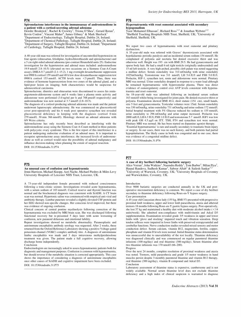
Society for Endocrinology BES 2013, Harrogate, UK
P74Spironolactone interference in the immunoassay of androstenedione ina patient with a cortisol-secreting adrenal adenomaDeirdre Broderick1, Rachel K Crowley1, Triona O’Shea1, Gerard Boran2,Kevin Conlon3, Vincent Maher4, James Gibney1 & Mark Sherlock11Department of Endocrinology, Tallaght Hospital, Dublin 24, Ireland;2Department of Chemical Pathology, Tallaght Hospital, Dublin 24, Ireland;3Department of Surgery, Tallaght Hospital, Dublin 24, Ireland; 4Departmentof Cardiology, Tallaght Hospital, Dublin 24, Ireland.
A 48-year-old man was referred for investigation of uncontrolled hypertension onfour agents (olmesartan, felodipine, hydrochlorothiazide and spironolactone) anda 3 cm right-sided adrenal adenoma (pre-contrast Hounsfield units 25). Endocrineinvestigation for the hypertension and adrenal mass included: androstenedione19.9 nmol/l 2.8–10.5) (elevated on two occasions on a Siemens Coat-A-Countassay), DHEA 0.7 mmol/l (2.1–15.2), 1 mg overnight dexamethasone suppressiontest 0900 h cortisol 159 nmol/l and 48 h low dose dexamethasone suppression test0900 h cortisol 153 nmol/l. ACTH levels were !5 pmol/l. Thus, there wasevidence of hormone hypersecretion from two zones of the adrenal gland, and alipid-poor lesion on imaging; both characteristics would be suspicious foradrenocortical carcinoma.Spironolactone, diuretics and olmesartan were discontinued to assess his renin-angiotensin-aldosterone system and 6 weeks later a plasma renin activity andaldosterone were performed (3.0 ng/ml per h and 708 pmol/l respectively) andandrostenedione was now normal at 3.7 nmol/l (2.8–10.5).The diagnosis of a cortisol-producing adrenal adenoma was made and the patientunderwent laparoscopic adrenalectomy. He was commenced on glucocorticoidcover post-operatively and his 6 week post operative synacthen test revealedglucocorticoid deficiency in keeping with the diagnosis (cortisol 0 min279 nmol/l, 30 min 366 nmol/l). Histology showed an adrenal adenoma with0/9 Weiss criteria.Spironolactone has only recently been described as interfering with theandrostenedione assay using the Siemens Coat-A-Count method in three patientswith polycystic ovary syndrome. This is the first report of this interference in apatient undergoing endocrine evaluation of an adrenal mass. It is important torecognise spironolactone assay interference; the increased levels of androstene-dione as well as cortisol would raise the possibility of adrenal malignancy andinfluence decision-making when planning the extent of surgical resection.
DOI: 10.1530/endoabs.31.P74
P75An unusual case of confusion and hyponatraemiaJenni Harrison, Michael Knopp, Azri Nache, Michael Piedres & Miles LevyUniversity Hospitals of Leicester NHS Trust, Leicester, UK.
A 73-year-old independent female presented with reduced consciousnessfollowing a tonic-clonic seizure. Investigations revealed acute hyponatraemia,with a serum sodium of 103 mmol/l. Cortisol reserve and thyroid function wasnormal and the biochemical diagnosis was consistent with SiADH. A CT brainscan was normal. Hypertonic saline was commenced with empirical anti-viral andantibiotic therapy. Lumbar puncture revealed a slightly elevated CSF protein andher EEG showed non-specific changes. Her conscious level improved, but therewas evidence of ongoing confusion.Clinical concern of central pontine myelinolysis following correction of thehyponatraemia was excluded by MRI brain scan. She was discharged followingfunctional recovery but re-presented 5 days later with acute worsening ofconfusion, new paranoid delusions and emotional lability.Repeat investigations showed no metabolic abnormality. Paraneoplastic andautoimmune encephalitis antibody serology was requested. After 2 weeks, thesereturned from the Oxford Reference Laboratory showing a positive Voltage-gatedpotassium-channel (VGKC) complex antibody titre. A diagnosis of autoimmunelimbic encephalitis was made and 3 days intravenous methylprednisolonetreatment was given. The patient made a full cognitive recovery, allowingdischarge home independently.ConclusionEndocrinologists are increasingly asked to assess hyponatraemic patients both fordiagnostic and management purposes. Confusion is common with hyponatraemia,but should reverse if the metabolic situation is corrected appropriately. This caseshows the importance of considering a diagnosis of autoimmune encephalitisonce other causes of SiADH and cognitive impairment have been excluded.
DOI: 10.1530/endoabs.31.P75
P76Hypernatraemia with reset osmostat associated with secondaryhypogonadismYasir Mohamed Elhassan1, Richard Ross1,2 & Jonathan Webster1,21Sheffield Teaching Hospitals NHS Trust, Sheffield, UK; 2University ofSheffield, Sheffield, UK.
We report two cases of hypernatraemia with reset osmostat and pituitarydysfunction.A 35-year-old male was referred with Graves’ thyrotoxicosis associated withhypokalaemic periodic paralysis and an incidental serum sodium 154 mmol/l. Hecomplained of polyuria and nocturia but denied excessive thirst and wasotherwise well. Height was 193 cm with BMI 29.5. He had gynaecomastia andsparse body hair. He had a small 6 ml right testicle (originally undescended) and15 ml left testicle. A very high arched, possibly cleft palate was noted suggestinga midline defect. Serum osmolality was 314 mOsm/kg with urine osmolality1025mOsm/kg. Testosterone was 5.0 nmol/l, LH 5.6 IU/l and FSH 5.0 IU/l.Prolactin, IGF-1, synacthen test, renin and aldosterone were normal. PituitaryMRI was normal. Urine osmolality dropped in response to a water load althoughhe remained hypernatraemic with hyperosmolar plasma. There was directevidence of osmoregulatory control over AVP levels consistent with hyperna-traemia and reset osmostat.An 18-year-old male was admitted following an incidental serum sodium163 mmol/l whilst being investigated for joints pain. He denied excessive thirst orpolyuria. Examination showed BMI 40.4, short stature (154 cm), small hands,size 5 feet and gynaecomastia. Testicular volumes were 15ml. Serum osmolalitywas 330 mOsm/kg, urine osmolality 731 mOsm/kg and urine sodium 172 mmol/l.Initial hospital treatment with 5% Glucose reduced his sodium to 155 mmol/l,urine osmolality to 296 mOsm/kg and urine output increased. Prolactin was2000 mIU/l, LH 6.3 IU/l, FSH 1.0 IU/l and testosterone 5.7 nmol/l. IGF1 was lowwith peak GH 4.5 mg/l on ITT. TSH, FT4 and synacthen test were normal.Pituitary MRI was normal. He has been started on dopamine agonist therapy.‘Essential hypernatraemia’ is rare and usually secondary to traumatic brain injuryor surgery. In our cases, there was no such history, and both patients had partialhypopituitarism. The likely cause in both was congenital and in one case, therewas evidence of a congenital midline defect.
DOI: 10.1530/endoabs.31.P76
P77A case of dry beriberi following bariatric surgeryAlice Verran1, John Watkins1, Narendra Reddy1, Tom Barber1, Milan Piya1,Harpal Randeva, Sudhesh Kumar1, Saboor Aftab1 & Sailesh Sankar1,21University of Warwick, Coventry, UK; 2University Hospitals of Coventryand Warwickshire, Coventry, UK.
IntroductionOver 9000 bariatric surgeries are conducted annually in the UK and post-operative micronutrient deficiency is common. We report a case of dry beriberisecondary to thiamine deficiency following Roux-en-Y bypass surgery.Case presentationA 45-year-old Caucasian obese lady (155 kg, BMI 57) presented with progressiveproximal limb weakness, upper and lower limb paraesthesia, ataxia and athetoidtremors 18 months following Roux-en-Y gastric bypass surgery. Post-operatively,she lost 57 kg and maintained a healthy diet with moderate alcohol intake (!14units/week). She admitted non-compliance with multivitamin and Adcal D3supplementation. Examination revealed grade 3/5 weakness in upper and lowerlimbs with ‘glove and stocking’ impaired touch and vibration sensation. Deeptendon reflexes were impaired in lower limbs with preserved extrapyramidal andcerebellar functions. Nerve conduction studies revealed mixed sensory and motorconduction defect. Serum calcium, vitamin B12, magnesium, ferritin, copper,phosphate and vitamin D levels were normal. Initial thiamine status determinationwas unsuccessful due to unavailability of the test locally. Thiamine deficiencywas diagnosed clinically and was commenced on regular parenteral thiamineinfusions (100 mg/day) and oral thiamine (300 mg/day). Serum thiamine aftertwo thiamine infusions was 170 nmol/l (66–200).ProgressOver the next 24 months, complete resolution of proximal weakness and ataxiawas noted. Tremors, mild paraesthesia and grade 1/5 motor weakness in handmuscles persist despite 3-monthly parenteral thiamine and vitamin B12 therapy,oral thiamine (300 mg/day), vitamin B compound and Adcal D3.ConclusionLaboratory assessment of thiamine status is expensive, cumbersome and is notwidely available. Normal serum thiamine level does not exclude thiaminedeficiency and a high index of clinical suspicion is warranted to diagnose
Endocrine Abstracts (2013) Vol 31
Society for Endocrinology BES 2013, Harrogate, UK
thiamine deficiency states (dry beriberi, wet beriberi and Wernicke Korsakoffsyndrome). Biochemical and clinical monitoring of micronutrient deficienciesshould be mandatory for all peri-operative bariatric surgery patients, preferablyby adopting a locally agreed protocol.
DOI: 10.1530/endoabs.31.P77
P78Giant parathyroid adenoma extending into the mediastinum: a casereportSally Thrower1, Adam Denley1, Mimi Chen2 & Andrew Johnson21University Hospitals Bristol NHS Foundation Trust, Bristol, UK; 2NorthBristol NHS Trust, Bristol, UK.
Introduction85% of cases of primary hyperparathyroidism are caused by a parathyroidadenoma. These are usually small in size, in the range of 70 mg to 1 g. Thoseweighingmore than 2 g are classified as ‘giant adenomas’. Fewadenomasweighingmore than 30 g have been described in the literature. Herewe present one such case.Case reportA 52-year-old female presenting with lethargy was found to have a correctedcalciumof 3.15 mmol/lwith a parathyroid hormoneof 122.5 pmol/l. Imagingof theparathyroids was performed. Sestamibi scanning demonstrated a large area of highuptake, not typical of a parathyroid adenoma. Computed tomography revealed amass, 9 cm in maximum dimension, extending from the inferior pole of the rightlobe of the thyroid to the right side of themediastinum, and into the subcarinal area.Endobronchial ultrasound-guided transbronchial needle aspiration (EBUS–TBNA) of this mass showed acinar structures, morphology and phenotypefavouring parathyroid adenoma. This was confirmed histologically after surgicalremoval of a large single parathyroid adenoma, weighing 92.7 g, under the jointcare of maxillofacial/cardiothoracic surgical teams. Bisphosphonate treatment wasrequired to control calcium levels (which rose to 3.8 mmol/l) pre-operatively. Thepatient experienced post-operative hypocalcaemia, requiring calcium infusion,high dose oral calcium supplementation and alphacalcidol treatment.DiscussionThis case is one of the biggest giant parathyroid adenomas described, the largestdescribed weighed 116 g. In general adenoma weight correlates with functionalstatus of the gland, only one case of a giant non-functioning adenoma has beenreported. Primary hyperparathyroidism is more common in women but there isrelative male overrepresentation in cases of large adenoma. Whilst malignancydoes not appear to be more common in cases of giant parathyroid adenoma, suchcases may represent a subset of parathyroid lesion with pronounced features ofprimary hyperparathyroidism and specific genomic aberrations.
DOI: 10.1530/endoabs.31.P78
P79Tertiary hyperparathyroidism due to chronic severe vitamin Ddeficiency in ethnic minority patients: a case seriesMahmud Ahmed, Harni Bharaj, Simon Hargreaves, George Yeung,Adam Robinson & Ambar BasuRoyal Bolton Hospital, Bolton, UK.
IntroductionVitamin D deficiency is common in ethnic minority population living in UK. It is arelatively easily treatable condition. However, if left untreated it may lead tochronic secondary hyperparathyroidismandonto tertiary hyperparathyroidism.Wereport three cases of severe vitamin D deficiency who presented withhypercalcaemia.CasesWe report three patients. 60 and 46 years old ladies, both of south-asian origin, a 46years oldman of Somalian origin referred to the endocrinology serviceswith severehypercalcaemia. Referred calcium levels were 3.20, 3.79 and 2.98 mmol/lrespectively. Parathormone (PTH) levels were 625, 890 and 639 pg/ml. VitaminD levels 11, !10 and 7 nmol/l (normal 70–250). PTH levels were 484, 532 and234 pg/ml respectively after high dose colecalciferol replacement. Calcium levelsdid not change significantly.ImagingSingle left ectopic inferior parathyroid adenoma in the first lady on NMparathyroid MIBI scan. Left superior, left inferior and the ectopic left inferiorparathyroid glands were removed at surgery. Postoperative calcium was2.47 mmol/l and PTH was 32 pg/ml.A right single inferior parathyroid adenoma was identified on the NM ParathyroidMIBI scan in the second lady. A right inferior parathyroid adenoma was removed.
Endocrine Abstracts (2013) Vol 31
Postoperative calcium was 2.44 mmol/l and a parathormone level 33 pg/ml.The gentleman had a left superior and a right inferior ectopic parathyroid detectedon USS and NM parathyroid MIBI scan. He is awaiting surgery.DiscussionTertiary hyperparathyroidism (TPHT) has been commonly associated with renaltransplant and end stage renal failure patients. Here we present three cases ofTHPT which we believe were secondary to untreated chronic severe vitamin Ddeficiency. Vitamin D deficiency is a fairly common condition particularly in theethnic minority population in the UK, both men and women. Early recognition andtreatment of this condition could prevent the progression to THPT and savesurgical intervention and thus reducing significant healthcare cost.
DOI: 10.1530/endoabs.31.P79
P80Hypercalcaemia due to simultaneous presentation of primary hyper-parathyroidism and metastatic oesophageal cancerHiang Leng Tan1, Najeeb Waheed1 & Muhammad Butt21Hereford County Hospital, Hereford, UK; 2Peterborough City Hospital,Peterborough, UK.
IntroductionWe report a patient with hypercalcaemia secondary to parathyroid hormonerelated peptide (PTHrp) from metastatic oesophageal cancer and co-existingprimary hyperparathyroidism.Case reportA 52-year-old lady was admitted to the hospital with a 2-week history of rightscapula pain, reduced appetite and weight loss.Blood test revealed an adjusted calcium of 3.99 nmol/l (NR 2.1–2.55 nmol/l),PTH of 147 ng/l (NR 15–65 ng/l), PTHrp of 4.3 pmol/l (NR 0.0–1.8 pmol/l) andnormal myeloma screen. Normal 25-OH vitamin D levels and renal functionsexcluded the possible secondary PTH elevation.CT scan showed evidence of lung and liver metastasis but the site of primarycarcinoma could not be identified. Bone scan did not reveal any bony lesion. Liverbiopsy confirmed metastatic carcinoma with possible lung or gastrointestinal tract(GI) as a primary site. She underwent an upper GI endoscopy which confirmedsquamous cell cancer on oesophageal biopsy.Hypercalcaemia initially responded well to intravenous fluid and bisphosphonatetherapy and she was discharged home after noticeable clinical and biochemicalimprovement. She was readmitted with symptomatic hypercalcaemia resistant tofurther fluids and bisphosphonates. A trial of chemotherapy did not improve herhypercalcaemia and 2 months after her initial diagnosis, she passed away.ConclusionHypercalcaemia is a common occurrence in malignancy and usually confer a poorprognosis. In our patient, hypercalcaemia was secondary to both metastaticoesophageal cancer and primary hyperparathyroidism. Primary hyperparathyr-oidism in this setting is mere an interesting observation and does not influence thecourse of patients’ treatment or outcome.There have been case reports linking an association between metastatic breastcancer and primary hyperparathyroidism and their simultaneous presentation, butnone have yet reported with metastatic oesophageal cancer.
DOI: 10.1530/endoabs.31.P80
P81‘There is nothing more deceptive than an obvious fact’, SherlockHolmes: a case report of thyroid sarcoidosisClaudia EscalanteEndocrinology and Diabetes Department, Salford Royal Foundation TrustHospital, Salford, UK.
A 53-year-old caucasian female presented with tremor, palpitations, sweating,breathlessness and chest discomfort. Examination revealed a non-tender, largediffuse goitre and TFT showed an elevated T4 and undetectable TSH. Thyroidantibodies were elevated; 115 IU/ml (NR!35) and thyroid ultrasound confirmeddiffuse vascular goitre. Patient was diagnosed with thyrotoxicosis with a likelyaetiology of Grave’s disease.Carbimazole was commenced which normalised the thyroid function, but wasstopped when the patient developed agranulocytosis. Neutrophil count recovered,however symptoms returned with the addition of thyroid eye disease andderanged liver function (contraindicating treatment with propythiourocil).Abdominal ultrasound revealed no hepatobiliary tract abnormality. Virology,immunoglobulins, ferritin, PT, autoantibody and infection screens were negative.
Society for Endocrinology BES 2013, Harrogate, UK
Echocardiogram demonstrated moderate tricuspid regurgitation, dilated rightatrium and elevated pulmonary artery pressure. CXR showed bulky hilar, reportedas not clinically significant. The patient was pre-treated with B-blockers andlugol’s iodine. She underwent uncomplicated total thyroidectomy once TFT’s hadnormalised. Histology showed diffuse hyperplasia, areas of fibrosis and severalnon-caseating granulomas. Post-operatively, liver function normalised spon-taneously. Despite normal thyroid function and thyroxine replacement, breath-lessness persisted. Serum ACE level was elevated at 114 U/l (NR11–55).Ultimately, the patient was referred to the respiratory team for management ofpulmonary sarcoidosis.This case raises the following points: A rare but well recognised case of thyroidgland sarcoidosis coexisting with Grave’s disease. Liver dysfunction can beexplained by thyrotoxicosis, thionamide treatment and sarcoidosis. Although thesymptoms aligned with Grave’s hyperthyroidism, this was not the sole cause of thepatient’s symptoms, particularly the breathlessness. Post-thyroidectomy, patientremained breathless and disabled. The initial hilar adenopathy was discounted,however was one of the missing pieces of the puzzle in the underlying diagnosis.Lesson; even though a symptom appears to be explained by one diagnosis theremay be more than one condition contributing to symptom complex!
DOI: 10.1530/endoabs.31.P81
P82A case of severe hypoaldosteronism following unilateral adrenalectomyfor Conn’s syndromeAlistair Connell & Mark CohenBarnet and Chase Farm Hospitals NHS Trust, London, UK.
A 58-year-old female presented with a 20-year history of resistant hypertensionand hypokalaemia, with normal renal function. Investigations confirmed primaryhyperaldosteronism that was not suppressed following a standard saline infusiontest. CT scanning revealed a right-sided adrenal mass of 1.3 cm, with a signalintensity of K1 HU. The left adrenal was normal in appearance. A LDDSTexcluded ACTH-independent Cushing’s syndrome. Adrenal vein samplingconfirmed right-sided unilateral hypersecretion of aldosterone; she underwentan uneventful right adrenalectomy in October 2011, following which her BP wascontrolled on atenolol alone.In November 2011 she presented to other hospitals with acute kidney injury,postural hypotension and hyperkalaemia. In July 2012 she was admitted to ourunit and was found to have a normal anion-gap metabolic acidosis, consistent withtype IV renal tubular acidosis. A SST confirmed a normal adrenal–glucocorticoidaxis (peak cortisol 630 nmol/l). Her aldosterone level was inappropriately low,given hypovolaemia and hyperkalaemia, suggesting hypoaldosteronism (plasmaaldosterone 100 pmol/l; plasma renin activity 4.4 pmol/ml per h). In keeping withthis, her biochemical abnormalities normalized following mineralocorticoidreplacement. An attempt to reduce the dose of fludrocortisone resulted in a furtherreversible recurrence of both hyperkalaemia and acute kidney injury. She is nowtaking 100 mg fludrocortisone TDS, and remains well. Her atenolol has beenchanged to amlodipine, as ß-blockade may have prevented adequate recovery ofher renin-aldosterone axis.Post-operative hypoaldosteronism is well documented in cases of unilateraladrenalectomy for aldosterone-producing adenomas. This may relate to adecrease in adrenal mass, or a transient suppression of the contralateral gland(1). However, it is rare for this to be prolonged, or severe. This may occur in up to5% of cases, and responds to mineralocorticoid treatment (2). Our case illustratesthe importance of follow-up in the post-operative period.References1. Gadallah MF, Kayyas Y & Boules F. Reversible suppression of the renin-aldosterone axis after unilateral adrenalectomy for adrenal adenoma. Am J KidneyDis 1998 32 (1) 160–163.2. Fischer E, Hanslik G, Pallauf A, Degenhart C, Linsenmaier U, Beuschlein F,Bidlingmaier M, Mussack T, Ladurner R, Hallfeldt K, Quinkler M & Reincke M.Prolonged zona glomerulosa insufficiency causing hyperkalemia in primaryaldosteronism after adrenalectomy. J Clin Endocrinol Metab 2012 97 (11)3965–3973 (doi: 10.1210/jc.2012-2234).
DOI: 10.1530/endoabs.31.P82
P83SDHB mutation and a large asymptomatic paraganglioma in a youngwoman: the importance of taking a good family historyStuart Foster1, Julian Barwell1,2, David Lloyd1,2 & Miles Levy1,21University of Leicester, Leicester, UK; 2University of Leicester Hospitals,Leicester, UK.
Succinate dehydrogenase subunit B (SDHB) mutations are associated with a highrisk of developing pheochromocytomas, paragangliomas and renal cell tumours.The risk of malignancy is also higher than that of other SDH mutations.A 23-year-old woman was referred to endocrine clinic following confirmation ofan SDHB mutation. Her family was screened when a relative underwent amedical, prior to starting a new job, and a significant family history of renaltumours was discovered. The patient’s grandmother had two siblings with renalcancer and a further sibling with bone metastases of unknown origin. Thepatient’s uncle, mother and brother were found to have SDHB mutations. Theuncle was discovered to have a renal tumour whereas the mother had normalimaging and biochemistry; the brother is currently under investigation.The patient had no symptoms of a catecholamine-producing tumour, did notcomplain of back pain or abdominal pain and had normal urinary catecholamines.A routine screening CT scan of the abdomen, however, revealed a large,heretogeneously enhancing mass in the right para-aortic region, arising from theorgan of Zuckerkandl. Pre-operative clinical examination confirmed the presenceof an abdominal mass. Surgical resection of the mass was difficult as it wasadherent to all surrounding tissue. The mass measured 10!6.3!11.2 cm andimmunohistochemistry confirmed it to be a benign paraganglioma.This case demonstrates the importance of genetic screening when consideringSDH mutations. Due to non-penetrance, a three-generation family history isnecessary as the relevant diseases may appear to ‘skip’ a generation. This youngwoman had a very large tumour which was detected in the absence of symptomsand would not have been diagnosed if the physician conducting the medical hadmissed the strong family history of renal cancer. Early detection is especiallyimportant due to the malignant potential of these tumours.
DOI: 10.1530/endoabs.31.P83
P84The challenges of a dopamine secreting paragangliomaAnthony Lewis1, Roy Harper1,2, Steven Hunter1 & Karen Mullan11Regional Centre for Endocrinology and Diabetes, Royal Victoria Hospital,Belfast, UK; 2Ulster Hospital Dundonald, Belfast, UK.
A 39-year-old female presented with an 18-month history of borderlinehypertension, headaches, palpitations and some anxiety symptoms. Both parentsalso had hypertension. Blood pressure was 160/102 on no medications. She had alarge single cafe au lait spot but no neurofibromata. Urinary dopamine levels wererepeatedly elevated (5398–8653 nmol/24 h (n!3900)) with normal nor-adrenaline and adrenaline levels. Serum calcium was also elevated at2.83 mmol/l (nZ2.2–2.6). Meta-iodobenzylguanidine (MIBG I-123) scanningindicated a single focus of activity further defined on SPECT–CT as a 12 mmmass in the lower para-aortic region. Parathormone was elevated at 274 pg/ml(nZ15–70) and a nuclear uptake scan indicated a right lower parathyroid focus.Calcitonin was unmeasurable. Following control of blood pressure withamlodipine and lisinopril she proceeded to surgery where a black lobulatedparaganglioma was removed at the organ of Zuckerkandl. A 30 s asystolicepisode occurred on first manipulation of the tumour. Urinary dopamine levelsnormalised, antihypertensives were reduced and 5 months later a parathyroidadenoma was removed uneventfully. Antihypertensives have recently beenwithdrawn. Genetic testing to date has been negative including MEN2a, succinatedehydrogenase B, C and D mutations. Dopamine secreting paragangliomas areextremely rare and are usually metastatic at diagnosis. They are usually associatedwith nonspecific symptoms, normotension and present with mass effects. Alphablockade is contraindicated because of its association with cardiovascularcollapse which is due to the unopposed hypotensive action of dopamine when thepressor catecholamines are blocked. This is the first reported case presenting witha coexistent parathyroid adenoma.
DOI: 10.1530/endoabs.31.P84
P85Lithium-induced hyperparathyroidism successfully treated with cina-calcet: two case reportsMohammad Hassan Bholah, Khyatisha Seejore & Afroze AbbasSt James University hospital, Leeds, UK.
BackgroundLithium-associated hyperparathyroidism (HPT) is an under-recognised side effectof chronic lithium therapy. Cessation of lithium may precipitate relapse ofpsychiatric illness. Potential treatment with cinacalcet has been described in twocase series recently.
Endocrine Abstracts (2013) Vol 31
Society for Endocrinology BES 2013, Harrogate, UK
AimWe present two cases of HPT secondary to lithium therapy used for bipolaraffective disorder, which were successfully managed with cinacalcet.ResultsPatient 1 is a 64-year-old gentleman with a history of lethargy, nocturia andpolyuria who was found to have hypercalcaemia and HPT. Sestamibi/SPECTscans did not detect any parathyroid adenomas but surgical exploration revealedan enlarged right superior parathyroid gland which was excised. Histopathologyconfirmed a parathyroid adenoma. However, the calcium and parathyroidhormone (PTH) remained consistently elevated after parathyroidectomy andcinacalcet was introduced at a dose of 30 mg once daily. Calcium and PTH levelsimproved over 15 months. Patient 2 is a 60-year-old gentleman who wasinvestigated for tiredness, polyuria and pathological fractures and found to havehypercalcaemia and HPT as well as nephrocalcinosis and osteoporosis. Nodefinite parathyroid adenomas were identified on Sestamibi/SPECT scans and atrial of cinacalcet (30 mg once daily titrated with biochemical response) wasstarted. This resulted in normalisation of both calcium and PTH levels within 2years. In both cases, cinacalcet was well tolerated and the patients improvedsymptomatically with no further complications from their underlying conditions.
Table 1
Beforecinacalcet On cinacalceta Reference range
Patient 1 Calcium (adj.) 3.02 2.65 2.20–2.60 (mmol/l)PTH 43.0 22.2 1.5–7.6 (pmol/l)
Patient 2 Calcium (adj.) 2.64 2.54PTH 22.5 7.0
aMost recent results on cinacalcet therapy.
ConclusionCinacalcet is known to be effective in primary hyperparathyroidism but ourobservations also support the use of this calcimimetic agent in lithium-inducedhyperparathyroidism as a potential alternative to surgery.
DOI: 10.1530/endoabs.31.P85
P86Recurrence of a brown tumour in a patient with secondaryhyperparathyroidism due to severe vitamin D deficiencyMarie-France Kong1,2, Kaustubh Nisal1,2 & Christopher Avery1,21Centre Hospitalier Universitaire Brugmann, Brussels, Belgium;2University Hospitals of Leicester NHS Trust, Leicester, UK.
In May 2011 a 55-year-old South East Asian man had a tumour removed from thefloor of his mouth. The histology was consistent with a brown tumour ofhyperparathyroidism. His PTH was raised at 12.3 pmol/l (NR 1.6–7.5) and he wasreferred to the endocrinology clinic. His serum calcium was 2.22 mmol/l(NR 2.2–2.6 mmol/l) and his serum 25 (OH) vitamin D level was !15 (NRO50 nmol/l) with a normal alkaline phosphatase. He had no symptoms ofosteomalacia. He is a vegetarian. His past medical history included hypertension,hyperlipidaemia and a pulmonary embolism in 2005. His results were consistentwith secondary hyperparathyroidism due to severe vitamin D deficiency. He wasgiven replacement therapy and when discharged from the clinic in November2011 his PTH was normal at 3.8 pmol/l, his serum calcium was 2.35 mmo/l andhis vitamin D level was 165 nmol/l. He was advised to take 25 mg (1000 IU) ofcolecalciferol lifelong. In March/April 2012 the patient noticed swelling in theright maxilla which progressively got bigger. A CT scan of the lesion showed anexpansile lesion in the right maxillary alveolus. Biopsy again showed featuresconsistent with a brown tumour of hyperparathyroidism. His repeat PTH wasnormal at 3.9 pmol/l with a normal serum calcium of 2.34 mmol/l and his serum25 (OH) vitamin D level was 48 nmol/l. His urinary calcium excretion wasnormal (5.9 mmol/24 h – NR 2.5–7.5). He was advised to increase hiscolecalciferol to 50 mg (2000 IU) per day. An ultrasound scan of his parathyroidglands was essentially normal.Brown tumours are non-neoplastic lesions resulting from abnormal bonemetabolism and are known to be associated with primary hyperparathyroidism.There have been case reports of peripheral brown tumour in the maxilla asmanifestation of secondary hyperparathyroidism associated with chronic renalfailure but to our knowledge there have been no reports of a brown tumour due tosecondary hyperparathyroidism as a result of severe vitamin D deficiency.Furthermore it is unclear why the patient developed a further brown tumour afterhis secondary hyperparathyroidism had been corrected.
DOI: 10.1530/endoabs.31.P86
Endocrine Abstracts (2013) Vol 31
P87An uncommon presentation of a common endocrine conditionAvinash Jain1,2,3, Saurabh Bansal1,2,3, S Krishnasamy1,2,3,Nayan Chaudhari1,2,3 & Harit Buch1,2,31B J Medical College, Ahmedabad, Gujarat, India; 2Government DentalHospital, Ahmedabad, Gujarat, India; 3Royal College of Physician, London,UK.
A 55-year female from low economic background was referred from the localdental hospital where she had presented with a swelling near her left lower 3rdmolar which caused her significant difficulty in eating. On further questioning sheconceded to having progressive difficulty in walking and bony pains over the past6 months with increasing dependency for daily activities. She was frail withproximal muscle weakness and widespread bony tenderness but no othersignificant neurological abnormalities. She also had two firm non-tenderswellings over the lower half of her right forearm and over the right hand.Total calcium 2.3 mmol/l (2.2–2.6), phosphorus 0.63 mmol/l (0.81–1.4), albumin33 g/l (35–50), alkaline phosphatase 124 U/l (42–98), parathyroid hormone2842 ng/l (8–51), Serum creatinine 61.9 mmol/l (44–100) and vitamin-D14.6 ng/ml (30–100). Skeletal X-rays showed radiolucent oval lesions over thelower 1/3rd of radius and the 4th metacarpal base with appearances suggestive ofBrown tumours. This diagnosis was confirmed by biopsy of the oral lesion withthe overall picture suggestive of long-standing vitamin-D deficiency andsecondary hyperparathyroidism. She was commenced on 60 000 units ofcholecalciferol weekly. By 6 weeks there was a significant improvement inmobility, bony pains and sense of well-being. At 3 months despite continuedoverall improvement she re-consulted oral surgeons to request surgical excisionof the persistently symptomatic oral lesion. She is awaiting this procedure andbiochemical re-assessment.A Brown tumour is recognised sequelae of long-standing primary hyperpar-athyroidism but the presence of multiple tumours has not been reported in apatient with vitamin-D deficiency. The persistence of symptomatic oral tumourrequiring excision is also unusual.In conclusion, long standing vitamin D deficiency presents in a variety of waysand a high index of suspicion needs to be maintained in high-risk populations.
DOI: 10.1530/endoabs.31.P87
P88Rare case of pheochromocytoma presenting in pregnancyJehangir Abbas, Sviatlana Zhyzhneuskaya, Murali Ganguri &Vijayaraman ArutchelvamDepartment of Endocrinology, James Cook University Hospital, Middles-brough, UK.
IntroductionWe submit a rare presentation of pheaochromocytoma in pregnanacy, diagnosedjust before delivery, posing complex management difficulties.Case presentationA 24-year-old primipara had headache, hypertension and visual disturbance butwithout any palpitations or diaphoresis. Her headache was persistent duringpregnancy. Investigations at 39 weeks of gestation showed normal plasmaNormetadrenaline at 885.0 (120–1180) but raised Metadrenaline raised at 1849(80–510) leading onto a diagnosis of pheochromocytoma. There was an initialdiagnostic dilemma, if the raised catacholamines are due to stress withpregnanacy. However ultrasound abdomen showed left adrenal enlargement.She was started on phenoxybenzamine and the blood pressure was controlled.Labour was successfully managed with adequate a blockade and a healthy babywas delivered by Caessarean section at 40 weeks of gestation. After delivery shewas started on Bisoprolol along with increasing dose of phenoxybenzamine. Leftadrenal pheochromocytoma was confirmed by CT Abdomen showing a 9!6 mmleft adrenal adenoma which was confirmed by MIBG Scan. Laparoscopic leftadrenalectomy was planned with controlled a and b blockade.DiscussionHypertension is a common problem in pregnancy that can result in significantmaternal and foetal morbidity and mortality. The common causes include pre -eclampsia, gestational hypertension and essential hypertension. Althoughpheochromocytoma is a rare cause of hypertension in pregnancy (0.007% of allpregnancies), it can lead to potentially life-threatening cardiovascular compli-cations for the mother and increased foetal mortality if left undiagnosed anduntreated. In pregnancy, depending on the gestation at which diagnosis is made,the optimal timing for surgery is during the late first or early second trimester.When the pregnancy is more advanced, medical management followed bycombined caesarean section and tumour resection closer to term is preferred.
DOI: 10.1530/endoabs.31.P88
P89Non islet cell tumour hypoglycaemia resistant to medical treatmentMohammad Rahman1, Simon Wordsworth1, Gail Curtis2 & Stephen Wong11Deptartment of Endocrinology and Diabetes, Glan Clwyd Hospital, NorthWales, UK; 2Deptartment of Biochemistry, Glan Clwyd Hospital, NorthWales, UK.
A 73 years old gentleman with a diagnosis of mesothelioma presented withsymptoms typical of hypoglycaemia. Other than the expected abnormal chestsigns there were no significant examination findings.Capillary glucose was unrecordable; lab testing confirmed serum glucose of0.9 mmol/l. He had no history of diabetes mellitus or any medication that mayinduce hypoglycaemia. There was a slight rise in CRP and white cell count waselevated. There was no clinical evidence of infection. Routine biochemical testswere normal. Chest X-ray showed extensive left chest shadowing.Intramuscular glucagon and intravenous dextrose initially improved bloodglucose but episodic symptomatic hypoglycaemia continued. Blood glucosesfluctuated between 0.9 and 2 mmol/l. Despite aggressive treatment with dextrose10% and additional dextrose 50% boluses he continued to have frequenthypoglycaemia.Having previous treatment with steroids a short synacthen test was requested,which was normal. A recent CT scan showed no adrenal, hepatic or pancreaticlesions.With no other apparent cause a presumptive diagnosis of non islet cell tumourhypoglycaemia was made. This was confirmed by suppressed insulin(!10 pmol/l) and c-peptide (!94 pmol/l) levelswith a high IGF2:IGF1 ratio (19.3).Chest ultrasound confirmed significant mesothelial tumour with little pleural fluid.He was treated with high dose corticosteroid, diazoxide and octreotide infusion.Severe hypoglycaemia remained problematic necessitating continued glucoseinfusions. He was unfit for chemotherapy or debulking surgery and deterioratedrapidly before dying 7 days after admission.Non islet cell tumour hypoglycaemia is a rare paraneoplastic phenomenon.Treatment involves reduction in tumour size by chemotherapy or surgery.Hypoglycaemia can remain significant and resistant to medical managementstrategies.
DOI: 10.1530/endoabs.31.P89
P90An unusual case of pancreatitis: a case reportVictoria Goodall & Emma BinghamFrimley Park Hospital, Frimley, Surrey, UK.
A previously fit andwell 49-year-old gentleman presented to hospital with vomitingand myalgia. Three weeks previously he had been admitted with acute pancreatitis,but no causewas found.An incidental hypercalcaemiawasalso noted at that time.Hehad lost approximately two stone in weight in the previous three months and hadbecome more short of breath on exertion. He had a strong family history for type Idiabetes mellitus, but otherwise there was no significant history.On this admission he was found to have a high corrected calcium of 3.14, with a lowparathyroid hormone (!0.3). Further tests revealed that he had a low vitamin D, anormal myeloma screen, PSA and serum ACE. Imaging performed wasunremarkable, with a normal chest X-ray, CT thorax and NM whole body bonescan. He was treated with IV fluid resuscitation and pamidronate.Additional investigations came back and exposed that he was thyrotoxic with T4O75and supressedTSH!0.03.Hehad a positiveTSH receptor antibodyof 6.2.ANMthyroid scan with uptake technetium showed enlargement and marked increaseduptake in both thyroid lobes consistentwithGrave’s disease. Hewas commenced oncarbimazole and monitored in an outpatient endocrinology clinic with a normalfollow up calcium level of 2.20.Hypercalcaemia is most commonly caused by primary hyperparathyroidism1.Malignancy is another common cause and together they account for the majority ofcases of hypercalcaemia (1).Thyrotoxicosis has been found as a sole cause of hypercalcaemia, however,significant symptomatic hypercalcaemia is rare (2). There have been only a fewothercase reports of similar symptomatic hypercalcaemia with hyperthyroidism (3).However, in this case his thyrotoxicosis also lead to his pancreatitis through havinghypercalcaemia. This case highlights that thyroid function tests should always bedone when presented with a high calcium level.References1. McCance KL & Huether SE. Pathophysiology: the biologic basis for disease inadults and children. 4th edn, 2002.2. Daly JG, Greenwood RM & Himsworth RL. Serum calcium concentration inhyperparathyroidism at diagnosis and after treatment. Clin Endocrinol 1983397–404.
3. Harper PS & Hughes RO. Severe hypercalcaemia from hyperthyroidism withunusual features. BMJ 1970 213–214.
DOI: 10.1530/endoabs.31.P90
P91A painful neck in a young well looking man presenting to A&EChioma Izzi-Engbeaya1, Sagen Zac-Varghese1, Fausto Palazzo1,2,Karim Meeran1 & Waljit S Dhillo11Department of Endocrinology, Imperial College Healthcare NHS Trust,London, UK; 2Department of Endocrine Surgery, mperial CollegeHealthcare NHS Trust, London, UK.
A 38-year-old man presented on the acute medical take with a week’s history ofsore throat, dysphagia, neck swelling, and fever. One month prior he had suffereda respiratory tract infection, which resolved without antibiotics. He had no notablepast medical and family history. He was a non-smoker and drank 15 units ofalcohol per week. On examination he looked well, was afebrile, sweaty andflushed; chest was clear, heart sounds were normal, regular pulse (110 bpm) andBP 148/88 mmHg. His anterior neck was erythematous, his right thyroid lobe wassmoothly enlarged with no bruits heard over his thyroid. There was no palpablecervical lymphadenopathy and no evidence of tonsillitis. An initial clinicaldiagnosis of de Quervain’s thyroiditis was made. Blood tests revealed WCC 20,CRP 515 and normal thyroid function. Due to the neck erythema and high CRP anultrasound was requested.Ultrasound revealed a very large complex pus collection extending to the rightsternocleidomastoid muscle. Samples were obtained but percutaneous drainagewas not possible. HIV test was negative, Group A beta haemolytic streptococcuswas cultured from the pus but blood cultures were sterile. Empirical intravenousco-amoxiclav was changed to intravenous ceftriaxone and oral clindamycin. Hewas discharged after 5 days and continued outpatient treatment. After 10 days ofceftriaxone, 20 days of clindamycin and 2 weeks of co-amoxiclav with additionalamoxicillin, there was complete radiological resolution of the abscess. Videofluoroscopy and direct phyarngoscopy excluded a piriform sinus fistula. All bloodtests normalised and no mycobacteria were cultured from extended culture of theabscess fluid.Viral thyroiditis remains the most likely diagnosis in a patient with a painfulthyroid, but thyroid abscess must be considered as although it is rare, itsmanagement is different from viral thyroiditis and it is associated with a mortalityof up to 12%.
DOI: 10.1530/endoabs.31.P91
P92Life threatening airway obstruction secondary to a large probableparathyroid cystKatherine McCullough, Vasilis Constantinidis, Michael Badman,James Jackson & Fausto PalazzoImperial Centre for Endocrinology, Hammersmith Hospital, ImperialCollege Healthcare NHS Trust, London, UK.
Parathyroid cysts are rare, usually asymptomatic and typically present as a necklump. They are most commonly detected in middle-aged women and canoccasionally present with compressive symptoms. True parathyroid cysts arenon-functional and benign in nature, allowing a more conservative approach totheir management in many patients. We present the case of an 82-year-old ladywith a past medical history of a presumed parathyroid cyst which was drained bythe ENT surgeons. Three years later, she presented to hospital with sudden onsetshortness of breath, throat pain and three months of increasing dysphagia.Examination and initial investigations were consistent with upper airwayobstruction and a chest radiograph showed a large retrosternal mass. Computedtomography revealed a large 8!5 cm cyst causing tracheal compression anddeviation. The patient underwent drainage of the cyst via ultrasound andfluoroscopic guidance with full resolution of symptoms. Histology andintra-cystic PTH levels were consistent with a large parathyroid cyst howeveran alternative diagnosis of a 3rd branchial arch cyst could not be excluded. Wediscuss the prevalence and presentation of such cysts and different managementoptions. This case illustrates a rare, yet important cause of a neck lump that canpresent with life-threatening upper airway obstruction.
DOI: 10.1530/endoabs.31.P92
Society for Endocrinology BES 2013, Harrogate, UK
Endocrine Abstracts (2013) Vol 31
P93Tuberous sclerosis: an uncommon cause of hyperprolactinemiaSunil Kumar Kota1, Lalit Kumar Meher2, Sruti Jammula3 & KirtikumarD Modi11Medwin Hospital, Hyderabad, Andhrapradesh, India; 2MKCG MedicalCollege, Berhampur, Orissa, India; 3Roland Institute Of PharmaceuticalSciences, Berhampur, Orissa, India.
ObjectiveTo report a case of tuberous sclerosis presenting with hyperprolactinemia.MethodsClinical, laboratory and radiographic data are reported on a 26-year-old femalepresenting with galactorrhea and menstrual irregularities.Case reportA 26-year-old female with no premorbidities presented with complaints ofgalactorrhea for the past 10 days and menstrual irregularities over the past sixmonths. Galactorrhea was spontaneous. Her last childbirth four years ago wasuneventful. She had no head ache, vomiting, and visual impairment. She deniedany history of substance abuse, drug intake hypothyroidism, chronic liver orkidney diseases, and epilepsy. She was a well nourished female with mild pallor,tiny nodule on face, subungual fibroma in hands. There was spontaneousgalactorrhea and mildly tender breasts without any signs of inflammation.Systemic examination was entirely normal with normal IQ. Ophthalmologicevaluation revealed white disk shaped retinal hamartoma. Routine laboratoryinvestigations including renal and liver function tests, thyroid profile werenormal. Serum prolactin was 85 ng/ml with FSH K4.66 and LH K4.21 mIU/ml.Tests for evaluation of other anterior pituitary hormones were normal. Abdominaland pelvic ultrasound revealed no abnormality. Chest X ray showed bilateralinterstitial infiltrates. Echocardiogram of heart was normal. Computedtomography (CT) scan revealed multiple intracerebral calcifications. Thesecalcified lesions/ subependymal hamartomas are seen along the lateral surface ofthe lateral ventricles giving rise to characteristic candle dripping appearance.Magnetic resonance imaging (MRI) of the brain ruled out the presence of anypituitary mass. The combined clinical scenario along with the radiologic findingsleads to the diagnosis of TSC with hyperprolactinemia. Patient was prescribedcabergoline 0.5 mg twice daily which resulted in amelioration of galactorrhea andregularization of menses.DiscussionTuberous sclerosis (TSC) is a multi system genetic disorder which infrequentlyaffects the endocrine system. Cushing’s disease, hypoglycemia secondary toinsulinomas, precocious puberty, thyrotoxicosis, hypercalcemia secondary toparathyroid adenomas, hyperprolactinemia and acromegaly have all beenreported in TSC patients. The circulating prolactin of our patient may be ofpituitary origin or may possibly be secreted ectopically by a hamartoma.ConclusionTSC patients develop hormone secreting tumors involving the neuroendocrinesystem.
DOI: 10.1530/endoabs.31.P93
P94Acute diabetic autonomic neuropathy as phaeochromocytoma mimicDeirdre Maguire, Berenice Lopez & Peter HammondHarrogate Hospital, Yorkshire, UK.
A 20-year-old man with a 5-year history of poorly controlled type 1 diabetespresented with epigastric pain, bloating and weight loss. He had attended DAFNErecently and had been commenced on an insulin Pump resulting in improvementof HbA1C from 114 to 76 mmol/mol over a 4-month period. Blood pressure waselevated (157/108 mmHg) with a resting tachycardia of 110. Haemoglobin waselevated at 18.7 g/dl. 24 h blood pressure monitoring revealed an averagediastolic blood pressure of 120 mmHg and he was admitted for furtherinvestigation and management. Doxazosin caused his blood pressure to drop to68/40 mmHg and was discontinued. MRI adrenals and MRA of the renal arteriesdid not reveal any significant abnormality. The patient did not want to remain inhospital, and was discharged with close follow-up in an ambulatory care setting.Twenty-four hour urinary noradrenaline levels were elevated at 904 nmol/24 h(70–550). Subsequently, the result of plasma metanephrines taken during hishospital admission became available and were within normal range. Followingthis result, he was commenced on metoprolol. Midodrine, an alpha-1 receptoragonist, was subsequently commenced for severe symptomatic orthostatichypertension. Average 24 h blood pressure on this combination was136/87 mmHg. Due to on-going weight loss with BMI of 15.7, he requiredinput from dieticians.
Four months later, his weight has increased by 3 kg. His symptoms of posturalhypotension are only occasional and his midodrine dose has been reduced.Acute insulin neuritis can be precipitated by rapid improvements in blood glucosecontrol. This resulted in acute autonomic neuropathy with marked autonomicover-activity and some features suggestive of phaeochromocytoma. Clinicalhistory and normal plasma metanephrines helped secure the diagnosis and tailormanagement.
DOI: 10.1530/endoabs.31.P94
P95Hypervitaminosis D, an uncommon reality!Z H Mansuri1, S Dumra2, B C Kaji1, S Krishnasamy2 & H N Buch1,21B J Medical College and Civil Hospital Ahmedabad, Ahmedabad, Gujarat,India; 2Sterling Hospital, Ahmedabad, Ahmedabad, Gujarat, India.
An 89-year-old female family physician presented to an orthopaedic surgeon witha short history of aches and pains. She was suspected to have vitamin D deficiencyand was empirically prescribed three intramuscular injections of 6 million units ofcholecalciferol at monthly intervals. A few days after the third dose she presentedwith nausea, generalised weakness, confusion and ataxia. She appeared drowsyand dehydrated. Vital parameters were normal and there were no focalneurological signs. Her calcium was 4.53 mmol/l (2.2–2.6), serum creatinine396 mmol/l (44–100), serum vitamin D3 O160 ng/dl (30–100). She wasdiagnosed to have hypervitaminosis D manifesting with severe hypercalcaemiaand acute renal failure.Management included normal saline infusion, intranasal calcitonin and supportivetreatment. In view of severe hypercalcemia and worsening oliguria, she underwenttwo cycles of haemodialysis which were complicated by disequilibrium syndrome.Serum calcium normalised and she gradually recovered and she was discharged. 3months later calciumwas 2.43 mmol/l, serum creatinine 114 mmol/l although serumvitamin D3 remainedO160 ng/dl. Clinically she recovered completely and has nowreturned to active professional life.With the rising prevalence of vitamin D deficiency and high cost of its biochemicalconfirmation there is an increasing trend amongst medical practitioners in India toprescribe vitamin D supplements empirically. This approach is generally safe inview of the wide gap between the therapeutic and toxic doses of vitaminD.Howeverelderly patients or those with renal failure or primary hyperparathyroidism are pre-disposed to toxicity and may develop hypervitaminosis D especially with use ofhigher doses.Our case here highlights the need to use modest doses of vitamin D therapy and tomaintain clinical and biochemical vigilance for side-effects in high-risk individuals.
DOI: 10.1530/endoabs.31.P95
P96Simultaneous presentation of Graves’ thyrotoxicosis and Addison’sdisease presenting as incipient adrenal crisisMurali Ganguri, Jahangir Abbas, S Zhyzhneuskaya & Sath NagJames Cook University Hospital, Middlesbrough, Cleveland, UK.
IntroductionGraves’ thyrotoxicosis and Addison’s disease are disorders with a strong auto-immune basis. Primary hypothyroidism and Addison’s disease are recognisedcomponents of polyglandular autoimmune syndrome type II (PGA-II). Despite itsautoimmune etiology, Graves disease is not commonly associated with PGA-II.We present a case of a patient with newly diagnosed Graves’ disease presenting inincipient adrenal crisis due to unrecognized Addison’s disease.Case reportA 35-year-old gentleman presented with headache, paraesthesia, heat intolerance,and weight loss associated with severe fatigue, nausea and vomiting. Graves’thyrotoxicosis was suspected and confirmed biochemically (TSH !0.01 mU/l,FT4 30.7 pmol/l, FT3 8.6 pmol/l). He was treated with Carbimazole 40 mg oncedaily for a few weeks without any symptomatic improvement. He was referredto our unit with progressive weight loss, dizziness and fatigue. On re-assessmenthe looked unwell and was deeply tanned and hypotensive. Addison’s diseaseco-presenting with Graves’ disease was suspected. ACTH stimulation withtetracosactide (Syncathen) showed no incremental cortisol response (baselinecortisol 48 nmol/l) and elevated serum ACTH (679 ng/l) confirmed primaryadrenal insufficiency. Graves’ thyrotoxicosis was treated with Carbimazole.Catabolic symptoms resolved completely with glucocorticoid and mineralocorti-coid replacement therapy.
Society for Endocrinology BES 2013, Harrogate, UK
Endocrine Abstracts (2013) Vol 31
DiscussionThyroid dysfunction and Addison’s disease are recognised components ofpolyglandular autoimmune syndrome type II (Schmidt syndrome). Primaryhypothyroidism is the norm and Graves’ thyrotoxicosis is very rarely recognizedas part of the syndrome. It is well recognized that occult Addison’s disease shouldbe suspected in patients who fail to improve symptomatically after commencinglevothyroxine for primary hypothyroidism. This case highlights the fact thatpatients presenting with Graves’ disease and Addison’s simultaneously are at riskof incipient adrenal crisis. A high index of suspicion remains the cornerstone ofdiagnosis.
DOI: 10.1530/endoabs.31.P96
P97Severe refractory non-islet cell tumour hypoglycaemia due to metastaticcolorectal carcinomaMurali Ganguri, J Abbas, A Ramdas, S Zhyzhneuskaya & Sath NagJames Cook University Hospital, Middlesbrough, Cleveland, UK.
IntroductionNon-islet cell tumour hypoglycaemia (NICTH) is an uncommon but seriouscomplication of disseminated malignancy. The underlying aetiology ofhypoglycaemia is tumoral overproduction of IGF2, which results in stimulationof insulin receptors and increased glucose utilization. Extensive tumour burdeninvolving the liver and adrenal glands can also cause severe hypoglycaemia.Case reportAn 80-year-old man presented acutely in an unresponsive state. Capillary glucosewas recorded as 1.1 mmol/l. He had no history of diabetes and had no access tooral hypoglycaemic agents. Marked hepatomegaly, deranged liver functions(ALT 48 U/l, ALP 894 U/l, GGT 1262 U/l) and coagulopathy (prothrombin time15.8 s) were noted. Abdominal ultrasound showed hepatic metastases andsubsequent staging CT scan confirmed a primary colorectal malignancy withextensive hepatic metastases.Severe hypoglycaemia was initially managed with a continuous 20% dextroseinfusion. Diazoxide 200 mg BD was initiated but despite this and concurrentdextrose, capillary glucose remained low (!4 mmol/l). Prednisolone 60 mg oncedaily and subcutaneous octreotide 50 mg three times a day were subsequentlyinitiated but despite this hypoglycaemia proved difficult to control. SerumC-peptide (!0.10 nmol/l) and Insulin (!1.0) were appropriately suppressed inkeeping with NICTH. Adrenal insufficiency was excluded as a potential cause ofhypoglycaemia. Despite maximal combined therapy, hypoglycaemia provedrefractory and the patient succumbed to his illness.ConclusionNICTH is a major complication of malignancy particularly if associated withhepatic metastases. The aetiology is multi-factorial and includes reduced hepaticglycogen reserves, nutritional deficiency due to tumour induced cachexia andectopic production of IGF2 which activates insulin receptors and promotesglucose utilization. Refractory hypoglycaemia in this context necessitatescombination drug therapy but is a significant therapeutic challenge with a poorprognosis.
DOI: 10.1530/endoabs.31.P97
P98Unusual presentation of central pontine myelinolysisJayadave ShakherBirmingham Heartlands Hospital, Heart of England NHS Trust,Birmingham, Westmidlands, UK.
IntroductionCentral pontine myelinolysis, CPM classically occurs in alcoholics, malnourishedand elderly, few days following rapid correction of hyponatraemia resulting inpermanent neurological squeal. We described two cases of CPM occurring inalcoholics after 3 weeks of hospital admission with complete recovery of grossneurological signs.Case 1: 24-year-old male, known alcoholic, was admitted with left hemiplegiaand features of bulbar and pseudobulbar palsy with dysphagia, dysarthria andemotional lability. Admission biochemistry including serum sodium was normalapart from mildly elevated liver enzymes. No clinical evidence of infection orvasculitis with normal FBC, CRP and ESR. MRI brain showed evidence ofpontine and extrapontine myelinolysis. Previous clinical records showed patientwas admitted 4 weeks ago with alcohol intoxication with rapid correction ofhyponatraemia with intravenous 0.9% NaCl and did not manifest any
neurological deficits on discharge. He was transferred to rehabilitation unitrequiring all assistance with activities of daily living including nasogatric feed.He made a complete recovery after 6 months leading an independent life.Case 2: 55-year-old alcoholic admitted with intoxication and receivedintravenous 0.9% NaCl with rapid correction of sodium and developed delayedpresentation of CPM with swallowing and speech difficulties and became wheelchair bound. MRI brain demonstrated typical picture of CPM. He made acomplete recovery after 3 months.DiscussionCPM is a non-inflammtory demyelinating condition affecting central pons and in10% extrapontine sites typically involving myelin with sparing of neurons andaxons. The postulated mechanism is due to rapid osmotic fluctuating fromoverzealous correction of sodium resulting in damage to endothelial cells withrelease of somemyelinotoxic factors.The prognosis is poorwithdeath or permanentneurological damage but both the cases described made complete recovery.RecommendationGradual correction of SNa not more than 8 mmol/l in 24 h in high risk group.
DOI: 10.1530/endoabs.31.P98
P99SIADH and bilateral adrenal infarction in a patient with theantiphospholipid syndromeWinston W S Fung, Amar S Bhat & Adrian M JenningsQueen Elizabeth Hospital Kings Lynn NHS Foundation Trust, Kings Lynn,UK.
A 75-year-old female presented with increasing drowsiness and no other specificfeatures. Past medical history included the antiphospholipid syndrome, threeepisodes of myocardial infarction, left ischaemic leg, epilepsy, hypothyroidismand splenectomy. Medications included warfarin. Examination showed pulse70/min, blood pressure 120/60 mmHg, crepitations at the left lung base, aGlasgow Coma Scale 14/15. Investigations revealed serum sodium 117 mmol/l,urea 8.8 mmol/l, serum osmolality 269 mOsmol/kg, urine osmolality493 mOsmol/kg, urine sodium 49 mmol/l. Haemoglobin was 13.2 g/dl, initialINR 7.5. Chest X-ray showed consolidation at the left lung base. CT brain scanwas unremarkable. Short synacthen test showed serum cortisol 585 nmol/l (basal)and 482 nmol/l (stimulated).The patient was treated with intravenous antibiotics and fluid restriction withimprovement in her serum sodium (128 mmol/l). The short synacthen test repeatedafter 1 week showed hypoadrenalism (peak cortisol 149 nmol/l). Serum ACTHwas65 ng/l (0–50), FT4 19.8 pmol/l (11.5–22.7), TSH 0.51 mIU/l (0.35–5.50). CTshowed bilateral bulky adrenals compatible with adrenal infarction with somehaemorrhage. A repeat scan after three months showed significant resolution.Investigations for antiphospholipid syndrome were repeated and showedAnti-cardiolipin IgG 1902.0 GPL U/ml (0–9.9), Anti-cardiolipin IgM 119.0 MPLU/ml (0–9.9), lupus anticoagulant positive. The patient was treated withhydrocortisone and fludrocortisone and her condition improved. Her electrolytesreturned to normal (serum sodium 133 mmol/l, potassium 4.7 mmol/l).This lady therefore had evidence of SIADH which was probably secondary torespiratory infection but also had acute adrenal failure due to infarction associatedwith the antiphospholipid syndrome. This case illustrated the importance ofconsidering adrenal failure due to infarction in patients with antiphospholipidsyndrome who present non-specifically unwell, even when there is evidence ofinfection.
DOI: 10.1530/endoabs.31.P99
P100Incidental papillary thyroid carcinoma with primaryhyperparathyroidism: two casesKhyatisha Seejore, Mohammad Hassan Bholah & Afroze AbbasCentre for Diabetes and Endocrinology, St James’s University Hospital,Leeds, West Yorkshire, UK.
BackgroundPrimary hyperparathyroidism (PHPT) and concomitant medullary thyroid disease iswell described in literature as part of multiple endocrine neoplasia. However,coexistence of PHPT and papillary thyroid cancer (PTC) has only been scarcelydocumented in sporadic case reports and some surgical series.Wepresent twounusual cases of PHPTassociatedwith synchronousmultifocal PTC.CasesA 62-year-old woman with sporadic PHPT was noted to have a parathyroidadenoma by 99Tc-Sestamibi. She underwent parathyroidectomy with removal of
Society for Endocrinology BES 2013, Harrogate, UK
Endocrine Abstracts (2013) Vol 31
three parathyroid glands at neck exploration, two of which were hyperplastic.Incidentally, she was found to have metastatic follicular variant of papillary thyroidcarcinoma on histology in a cervical lymph node. Further imaging showed no othermetastases. She then proceeded with completion thyroidectomy with lymph nodeclearance followed by radioactive iodine treatment.A 68-year-old man was referred with incidental hypercalcaemia and biochemicalPHPT. 99Tc-Sestamibi scan revealed a solitary parathyroid adenoma as wellas incidental left-sided cervical adenopathy. Neck ultrasound also identified a3.6!2.1 cm enlarged left cervical lymph node and noted a contralateral 2!1.7 cmirregular thyroid nodule. Fine needle aspiration (FNA) of the thyroid lesion wasinsignificant. This prompted FNAof the neck nodewhich showedmetastatic papillarythyroid carcinoma. He underwent a total thyroidectomy and cervical dissection withexcision of the right parathyroid adenoma. Histology confirmed PTC with capsularinfiltration.DiscussionThe mechanisms underlying the association between PHPT and PTC are notestablished. Recent studies favour the combined use of CT–SPECT and cervicalultrasound in the first instance, coupled with FNA of suspicious lesions. These casesunderline the need for a high index of suspicion for synchronous hyperparathyroidismand non-medullary thyroid cancer. Co-existence of both disease processes maycomplicate patient management via unrecognised thyroid cancer and the necessity forre-operative neck surgery.
DOI: 10.1530/endoabs.31.P100
P101Hypercalcaemia as first presentation of sarcoidosisNaveen Aggarwal & K R NarayananQueen Elizabeth Hospital, Gateshead, UK.
A 37-year-old gentleman was admitted following a GP referral for asymptomatichypercalcaemia. His adjusted calcium was raised at 3.27 mmol/l while phosphatelevel was normal. The only past medical history was borderline hypertensionwhich was being monitored in primary care. His PTH level was suppressed at0.43 pmol/l l (1.1–5.5) while 25(OH) Vitamin D was normal at 71.6 nmol/l(48–145). Twenty-four hours urinary metadrenalines were normal. His full bloodcounts were also normal. He was treated with i.v. fluids and i.v. Pamidronate andthe hypercalcaemia responded well and his calcium levels have remained normalsince then. The suppressed PTH level prompted us to look for other causes ofhypercalcaemia. Familial hypocalciuric hypercalcaemia and multiple myelomawere excluded. His chest X-ray showed diffuse nodular opacities but no otherspecific abnormalities were present. His serum ACE level was 266 U/l (8–52) and1,25(OH)2 Vitamin D was increased at O250 pmol/l (20–120). A CT scan ofneck, chest and abdomen was arranged which showed subcentimeter lymphnodesat the level of carotid bifurcation and paratracheal region. There were alsonumerous ill defined foci throughout the lungs. It also showed an enlarged spleen(15 cm) with numerous hypo-attenuated foci in its parenchyma.In view of splenomegaly a bone marrow biopsy was arranged which showed areactive bone marrow with non-caseating granulomata and other changes inkeeping with sarcoidosis.His calcium levels currently remain in normal range and he is awaiting review bychest physicians.DiscussionThe incidence of bone marrow involvement in sarcoidosis is about 10%. Inpatients with bone marrow involvement, there is usually higher incidence ofextrapulmonary manifestations, leucopaenia/lymphopaenia and anaemia.In patients with hypercalcaemia, a normal screen for usual causes should lead toinvestigations for unusual causes of hypercalcaemia.
DOI: 10.1530/endoabs.31.P101
P102Tumours metastatic to the pituitary gland presenting with atypicalsymptomsManeesh Udiawar1, HS Santhosh2, Hemanth Bolusani1, Stephen Davies1 &Buchi Okosieme31University Hospital of Wales, Cardiff, Wales, UK; 2Singleton Hospital,Swansea, Wales, UK; 3Prince Charles Hospital, Merthyr Tydfil, Wales, UK.
We report two cases with initial presentation of sudden onset ophthalmoplegiain i) a patient recently diagnosed with breast carcinoma and ii) a patientsubsequently diagnosed with carcinoma lung. The first patient (68 years) wasreferred to the tertiary endocrine unit with a 2 weeks history of visual lossassociated with 3rd cranial nerve palsy in her right eye and with a temporal
hemianopia in her left eye. MRI showed an enhancing sellar and suprasellar mass.Initial biochemistry was consistent with anterior hypopituitarism. She underwenttrans-sphenoidal debulking surgery. Histology confirmed a metastatic lesionconsistent with a breast primary.The second patient (49 years) presented with a one day history of left sided ptosisand retroorbital pain and symptoms of anorexia, weight loss and lethargy over aperiod of 1 month. He was a heavy smoker with no past medical history.Examination revealed left sided ptosis with complete ophthalmoplegia.Biochemistry was consistent with anterior hypopituitarism. MRI scan revealedan enhancing sellar and suprasellar mass. Chest X-ray showed an opacity in theleft upper lobe which was confirmed by CT. He was treated with steroid andthyroxine replacement with subsequent development of polyuria and polydipsiaconsistent with diabetes insipidus. He underwent tras-sphenoidal debulkingsurgery with histology consistent with metastasis from a lung primary.Tumours metastatic to the pituitary gland are an unusual complication of systemicmalignancy. Breast and lung are the common sites of primary tumour. The mostcommon presentation is usually diabetes insipidus which was observed in thesecond patient but only after steroid replacement. Sudden onset of symptoms suchas headaches, ophthalmoplegia and diabetes insipidus in a patient above 45 yearsof age should always raise the suspicion of metastasis to the pituitary regardless ofa history of malignancy.
DOI: 10.1530/endoabs.31.P102
P103Acute adrenal insufficiency due to bilateral adrenal haemorrhagicinfarction associated with sepsis secondary to an open fracture of theankleBen Brookes, Mahmoud Ahmad, Saf Adam & Adam RobinsonRoyal Bolton Hospital, Bolton, UK.
BackgroundAcute adrenal insufficiency is a life threatening condition. Signs and symptoms areoften non-specific. The adrenal glandhas the richest arterial supply of any tissuewithlimited venous drainage and this mismatch predisposes to haemorrhagic infarction.We present a case report of acute adrenal insufficiency due to bilateral adrenalhaemorrhagic infarcts associated with an open ankle fracture.Case historyA 52-year man fell from a ladder sustaining an open fracture of his right ankle. Onpresentation to A&E the wound was dressed and the fracture stabilised. X-rayrevealed comminuted fractures of the distal shaft of the fibula and the distal tibia.Intravenous antibiotics and prophylactic LMW heparin were administered andbaseline U&Es normal. Seventy-two hours into the admission he was taken totheatre, thewound debrided and the fibula # pinned.Anaesthetic records record somehypotension during the procedure with SBP dipping to 60 mmHg. During thesubsequent days recurrent pyrexia and hypotension were noted and antibioticscontinued. His Na gradually fell with a nadir at day 9 of 122 mmol/l. This respondedto intravenous therapyand at a 2ndoperationonday13 the tibial #was pinned. SerumNa was again noted to fall over the next few days with a nadir of 124 mmol/l on day23. The patient complained of intermittent abdominal pain and began hallucinatingon day 20. Enterobacter cloacae was cultured from the wound. Serum cortisol wasmeasured on days 23 and 24 being undetectable on both occasions. His platelets haddropped to 56. A diagnosis of acute adrenal insufficiency was made and high doseintravenous hydrocortisone administered. Clinical improvement was evident within48 h. Baseline pituitary tests were unremarkable. CT scans showed enlargedheterogeneous adrenal glands consistent with bilateral haemorrhagic infarction.ConclusionAcute adrenal insufficiency due to bilateral adrenal haemorrhagic infarction is arare event that can have catastrophic consequences if not diagnosed and treated in atimely manner. Our patient presented with a common orthopaedic injury and aninpatient course characterised by pyrexia, low BP and hyponatraemia, inthemselves unremarkable and frequently associated with trauma/sepsis. A highindex of suspicion in a context of slow deterioration lead to further investigationand the additional diagnosis being made.
DOI: 10.1530/endoabs.31.P103
P104Ectopic thyroid tissue presenting as metastatic follicular cancerSyed H Ahmed, Dhanya Kalathil, Aftab Ahmad & Tejpal PurewalRoyal Liverpool Hospital, Liverpool, UK.
We present the case of a 73-year-old woman, who presented with ascites and ahistory of left radical hemithyroidectomy for localized follicular thyroid
Society for Endocrinology BES 2013, Harrogate, UK
Endocrine Abstracts (2013) Vol 31
carcinoma (FTC) 28 years ago. Computed tomography (CT) scanning of her bodyrevealed extensive metastatic lesions. An omental biopsy showed featuressuggestive of thyroid follicular epithelial cancer. Serum thyroglobulin was raisedat 127 mg/l. She died before the biopsy result was received. Two years beforepresentation, a bilobed nodule was noted in the superior mediastinum measuring3.5!1.5 cm, on a CT chest organized by her primary care physician to investigatebreathlessness. A thyroid uptake scan that followed had shown normal uptake inthe right thyroid remnant and a high uptake in this structure, suggestive of ectopicthyroid tissue. No further action was taken at the time. A retrospective review ofher thyroid function tests showed that serum thyrotropin (TSH) had never beensuppressed and she had untreated hypothyroidism 3 years before her presentation.We suspect that she may have harboured FTC cells from the primary lesion in theectopic gland or developed a de-novo lesion within it that spread due toinadequate TSH suppression. A timely diagnosis by a positron emissiontomography (PET) or radio-iodine scan would have provided grounds forconsideration of curative or palliative radio-iodine ablation.Thyroid cancer, the commonest endocrine cancer, is relatively rare in comparisonto other cancers (!1% of all cancers). It is therefore least suspected in anindividual presenting with metastatic disease and an unknown primary. Follicularthyroid cancer is the second most common differentiated thyroid cancer. Currentpractice involves total thyroidectomy (older patients, high-risk cases), followedby radio-iodine ablation and long-term TSH suppression. This case emphasizesthe importance of long-term follow up of this condition, in order to monitor forrecurrence and to maintain TSH suppression.
DOI: 10.1530/endoabs.31.P104
P105A peculiar case of a dog biteVilashini Arul Devah, Ana Pokrajac, Mark Savage & Isha MalikDepartment of Endocrinology, North Manchester General Hospital, PennineAcute Hospitals NHS Trust, Manchester M8 5RB, UK.
A 59-year-old gentleman presented to the Medical Admission Unit with facialand ankle oedema following a dog bite. He did not have any significantpast medical history. Initially, he was treated for angioedema. His oedemaworsened to anasarca, blood pressure rose and was found to be hypokalaemic.Echocardiogram showed a normal left ventricular ejection fraction. Urine proteincreatinine ratio was !0.5 g/24 h. Vasculitic and autoimmune screen werenegative. Eight weeks later, he was noted to have a Cushingoid appearance,ongoing persistent hypertension with hypokalaemia, as well as a new diagnosis ofdiabetes mellitus. He had pulmonary oedema and ascites on clinical examination.He was treated with furosemide, spironolactone and insulin.Subsequent biochemical investigations showed high random cortisol(2202 nmol/l) unsuppressed with low dose dexamethosone test, low ACTH(!5 ng/l) and high ACTH precursor (POMC) (610 pmol/l). He also had elevatedfasting gut hormone levels – CART (469 pmol/l), chromogranin A (247 pmol/l),chromogranin B (216 pmol/l).Imaging with MR and CT scanning showed liver metastases and bilateral adrenalnodules (largest measuring 27 mm). Further investigations showed these weresomatostatin receptor negative. Liver biopsy demonstrated grade 3 poorly differentiatedneuroendocrine cancer. PET/CT scan revealed high metabolic activity within the tail ofpancreas, bilateral adrenal lesions and suspicious malignant liver lesions.The patient underwent bilateral adrenalectomy, distal pancreatectomy and splenectomy.Pancreatic histology confirmed grade 3 poorly differentiated neuroendocrine cancer.Post operatively, his blood pressure, serum potassium and blood glucose havenormalised.At the local neuroendocrine multidisciplinary team meeting, chemotherapy has beenrecommended, and is awaiting assessment for the same.ConclusionThis case denotes rapidly deteriorating ectopic Cushing’s syndrome following amisleading history of a dog bite which delayed diagnosis. It demonstrates theimportanceof correlatingendocrineandbiochemicalfindingswithaclinical diagnosis.
DOI: 10.1530/endoabs.31.P105
P106Management of diabetic ketoacidosis in a district general hospital:a 3 years retrospective auditMohamed Ahmed, Juaidy Zakaria, Caitriona Doyle, Ciana McCarthy& Cathrine McHughDiabetes and Endocrinology Department, Sligo General Hospital, The Mall,Sligo Co., Sligo, Ireland.
ObjectivesDiabetic ketoacidosis (DKA) remains a life-threatening emergency. An updatedprotocol was introduced in Sligo general hospital in 2009 to standardise DKAmanagement. The aim of this study was to audit current practice against theprotocol standards.MethodsClinical notes of all adult patients admitted with DKA between July 2009 andJune 2012 were analysed and retrospectively audited against the protocol.ResultsFifty-six episodes ofDKAwere identified in 36 patients. 57.1% (nZ32) weremales,and the median age was 28.5 years. 87.5% of episodes occurred in patients with type1 diabetes (nZ49). Infection was the main precipitating factor in 33 episodes(58.9%) followed by non compliance 19.6%, new onset diabetes 14.3%, and alcoholexcess 7.1%. The majority of patients were managed in a general medical ward(58.4%, nZ33), 25% (nZ14) were managed in ICU, and nine patients (16.1%) inHDU. There were no deaths and the median length of stay was 4 days. Fluids werecommencedwithin 60 min of arrival to ED in 91% (nZ51) of episodes and themeantime to commence fluids was 30.3G3.6 min. Insulin was started within 60 min in 46episodes (82.1%), with amean time of 48.5G5.65 min. Potassiumwas inadequatelyreplaced in 30.4% (nZ17), and mild hypokalaemia (KC3.0–3.5) occurred in 10episodes (17.9%). Long acting insulin was inappropriately stopped in 74.3% ofadmissions. The frequency of biochemistry and blood gas measurements followedthe protocol in only 37.5 and 51.8% respectively. 93% (nZ52) of admissions carewere taken over by the diabetes team, and 89.3% (nZ50) received education by thediabetes nurse pre discharge.ConclusionAlthough insulin and fluid replacements were generally in line with therecommendations, potassium replacement, continuing basal insulin, biochemistryand blood gas monitoring were inadequate. Emphasizing the importance of theseparameters, especially to the junior doctors, is essential to improve care standards.
DOI: 10.1530/endoabs.31.P106
P107Unusual presentations of adrenocortical tumoursOmar Mustafa, Ben Whitelaw, Rebeka Jenkins, Tiana Kordbacheh,Paola Salaris, Chris Manu, Norman Taylor, Roy Sherwood, Gill Vivian,Dylan Lewis, Klaus-Martin Schulte, Salvador Diaz-Cano, Jackie Gilbert,Alan McGregor & Simon AylwinThe Adrenal Multidisciplinary Team, King’s College Hospital,London, UK.
BackgroundAdrenocortical tumours (adenoma or carcinoma) present in well-recognisedways: hormones excess (Cushing’s, Conn’s, virilisation) or hormonally silentwith symptoms of mass effect, or found incidentally on imaging. We present3 cases of adrenal tumours, referred to our regional adrenal multidisciplinarymeeting with unusual presenting features.Case 1: post-menopausal bleedingA previously well 57-year-old female presented with vaginal bleeding 5 yearsafter completing menopause. She was not using exogenous oestrogen.Hysteroscopy demonstrated endometrial hyperplasia. Subsequently, shedeveloped right-sided abdominal pain. Imaging revealed 6 cm invasive rightadrenal mass. Serum oestradiol and 17b-oestradiol were elevated. Oncologicalright adrenalectomy was performed. Histology confirmed invasive adrenocorticalcarcinoma. Serum oestradiol fell to post-menopausal levels. No further vaginalbleeding was noticed.Case 2: polycythaemiaA previously well 49-year-old female was noted to have hypertension andpolycythemia (haemoglobin 17 g/dl). Venesection (following a haematologyopinion) was unsuccessful in controlling polycythaemia. Subsequently,significant weight gain and increased body hair over the preceding year werenoted. She had features of Cushing’s syndrome, marked hirsutism and increasedmuscle tone. Investigations showed high serum testosterone (23 nmol/l) andelevated non-suppressible serum cortisol. Imaging showed 3.5 cm left-sided well-circumscribed adrenal lesion. It was treated with laparoscopic left adrenalectomy.Histology demonstrated oncocytic adrenocortical adenoma. Post-operatively,cortisol and testosterone levels normalised and polycythaemia resolved.Case 3: hypertension and low serum aldosteroneA 27-year-old female presented with 6-month history of hypertension,hypokalaemia (2.8 mmol/l) and low serum aldosterone (86 pmol/l, range100–450 pmol/l). She subsequently developed abdominal pain. Imaging revealed10 cm left adrenal lesion. Serum cortisol was suppressed (!30 nmol/l) afterovernight dexamethasone (1 mg). Urinary and plasma catecholamines/metane-phrines were normal. Urinary steroid profile demonstrated marked increases ofmetabolites of 11-deoxycortisol(S) and 11-deoxycorticosterone. Histology from
Society for Endocrinology BES 2013, Harrogate, UK
Endocrine Abstracts (2013) Vol 31
the oncological resection of the mass revealed adrenocortical adenoma.Hypertension resolved post-operatively.DiscussionThese three cases represent rare presenting features of adrenal tumours.
DOI: 10.1530/endoabs.31.P107
P108An audit of the diagnosis and management of hypogonadism inadult menP W James Russell1, Javier Gomez1,2 & Khin Swe Myint11Department of Diabetes and Endocrinology, Norfolk and NorwichUniversity Hospitals NHS Foundation Trust, Norwich, Norfolk, UK;2Clinical Biochemistry Department, Norfolk and Norwich UniversityHospitals NHS Foundation Trust, Norwich, Norfolk, UK.
IntroductionSymptomatic hypogonadism affects 5–6% of men aged 30–79 years and isassociated with increased morbidity and mortality. There are currently no trustguidelines for the diagnostic workup of these patients.MethodsRetrospective analysis was performed of diagnosis and assessment of adult menwith low serum total testosterone (TT) and compared with clinical practiceguidelines of The Endocrine Society. We identified all patients with TT belownormal range (!9.9 nmol/l) carried out at the trust between July and November2010 and excluded patients with known hypogonadism or prostate cancer. First50 cases were evaluated in detail. Investigation results were retrieved usingelectronic records and clinic letters.ResultsIn total, 247 cases with low TT were identified. Among 50 patients (mean age 63,range 34–94 years) 84% (42/50) had a valid indication for measurement. Meantime (GS.D.) of measurement was 11:18amG142 min. Fifty-eight per cent(29/50) had a confirmatory repeat measurement, 42% (21/50) had sex hormone-binding globulin measured and 52% (26/50) had gonadotropins measured, ofwhich 69.2% (18/26) were low/normal and 26.9% (7/26) were high. Of those withlow/normal gonadotropins, 50% (9/18) had complete pituitary function testing(cortisol, prolactin, free thyroxine). Testosterone replacement therapy wascommenced in 44% (22/50). Pre-treatment, 45.5% (10/22) had PSA, 27.3%(6/22) had liver function, and 27.3% (6/22) had haematocrit measured. Within sixmonths after starting treatment, 86.4% (19/22) were reviewed in clinic, 63.6%(14/22) had repeat TT and 50% (11/22) had repeat haematocrit.ConclusionThere are pitfalls in our current practice in both diagnosis and assessment ofhypogonadism. Morning TT is measured later than recommended and often notrepeated, resulting in potential over-diagnosis. Lack of gonadotropin measure-ment has serious implications for missing potential aetiology. Pre-treatmentassessment and treatment monitoring must be improved to ensure patient safety.We are currently writing trust guidelines to address these issues.
DOI: 10.1530/endoabs.31.P108
P109Pituitary tuberculosisHS Santhosh1, Maneesh Udiawar2 & Hemanth Bolusani21Singleton hospital, Swansea, UK; 2University Hospital of Wales,Cardiff, UK.
A 28-year-old man of Asian origin presented with fever, cough and weight lossover 4 weeks. He had symptoms of worsening headaches and vomiting. The chestX-ray was normal and due to persistent headache and vomiting a lumbar puncturewas performed following a normal CT head scan. The CSF findings confirmed thediagnosis of tubercular meningitis and the patient received a complete course ofanti tubercular treatment. Six months later he was readmitted with visual blurring,a diagnosis of optic neuritis was made and he was treated with steroids. Thepatient re-presented in a few months with symptoms of polyuria, polydipsia andexcessive tiredness. The biochemistry demonstrated evidence of secondaryhypothyroidism and subsequent testing confirmed anterior hypopituitarism. Thepatient was commenced and hydrocortisone, thyroxine and testosterone.Symptoms of diabetes insipidus resolved following hormone replacement and awater deprivation test undertaken at a later date was completely normal.Pituitary tuberculosis is extremely rare and a reported incidence of 4% in patientswith tuberculosis has been quoted in the literature. It accounts for w0.15% of allintracranial tumours and 0.6% of all intracranial TB. Endocrine dysfunction hasbeen reported to occur in patients with tubercular meningitis and the most
commonly reported endocrinopathies include adrenal insufficiency, centralhypothyroidism and hyperprolactinemia. Hypopituitarism has been reported tooccur years after recovery from the illness in some patients who had sufferedfrom tubercular meningitis in their childhood. Trans-sphenoidal biopsy hasbeen considered to be essential to establish the diagnosis. Anti tuberculartreatment regimes used in CNS tuberculosis have been used to effectivelytreat pituitary tuberculosis. It is important for the endocrine fraternity to recognisepituitary tuberculosis and tubercular meningitis as rare causes of hypothalamicpituitary insufficiency as anti tubercular treatments are curative.
DOI: 10.1530/endoabs.31.P109
P110The difficulties in diagnosing and treating phaeochromocytoma in apatient with multiple co-morbiditiesDearbhla McKenna, S J Hunter & K MullanRegional Centre for Endocrinology and Diabetes, Belfast, UK.
A 33-year-old lady reported a 6 months history of sweating and worseningpalpitations especially after taking sotalol. She had a history of congenital heartdisease (double inlet left ventricle, pulmonary valvular stenosis, ventricular septaldefect, and Fontan connection surgery at 18 years); Blue Bleb Syndrome withchronic gastrointestinal blood loss, and recurrent pulmonary emboli. She requiredlong-term warfarin treatment and regular blood transfusions. She was found to havenew hypertension. Her cardiac performance and right ventricular ejection fraction(44%) had reduced which serves her systemic circulation. Her symptoms andmodestly elevated urinary noradrenaline levels were initially felt to be in keepingwith worsening intrinsic cardiac function (596–892 nmol/24 h; nZ50–560).A clonidine suppression test and genetic screening were negative. However plasmanormetanephrineswere significantlyelevated at 5415and5661 pg/ml (n!1180) andaMIBGwith spectCTscan (meta-iodobenzylguanidinewith single-photonemissioncomputed tomography)demonstrated a hot spot at the left para-aortic region. She hada prolonged admission for withdrawal of sotalol and frusemide and introduction ofalpha-blockadewhich she toleratedwell. Inputswere sought fromcardiac, endocrineand vascular surgery, anaesthetics, cardiology, haematology and gastroenterology.She proceeded to elective surgery which was uncomplicated apart from modestpostoperative bleeding. Follow-up urinary catecholamines normalised and thepatient’s presenting symptoms partially abated. This case illustrates the difficulty ofdiagnosing primary catecholamine excess in the setting of severe cardiac disease andalso the success of the multidisciplinary approach for patients with multiplethreatening co potentially life-morbidities.
DOI: 10.1530/endoabs.31.P110
P111Adrenal incidentalomas: who requires further testing?Fiona Paterson, Aikaterini Theodoraki, Adaugo Amajuoyi,Jody MacLachlan, Pierre Bouloux & Bernard KhooRoyal Free Hospital, London, UK.
Adrenal incidentalomas are common and guidelines recommend testing to excludefunctioning lesions andmalignancy. Their increasing prevalence results in a numberof investigations usually conducted in the Endocrinology clinic.In 2011 we audited the prevalence and management of adrenal incidentalomasidentified on abdominal CT imaging over one calendar month in our centre.Consequently, a decision pathway for new adrenal lesions was introduced in theRadiology department. This pathway is based on lesion imaging characteristics andabrief clinical assessment. A year later we re-audited the local practice.690 CT scans were reviewed in 2011 and 1264 in 2012. In 2011, 32/690 (4.64%)scans with adrenal lesions were identified and in 2012, 85/1264 (6.7%). In both the2011 and the 2012 cohorts, the majority of patients with adrenal lesions found onimaging, had a knownmalignancyor adrenalmetastasis andwere underOncologicalcare (13/32, 40.6% in 2011; 42/85, 49.4% in 2012). Excluding patients withmalignancy and other radiological diagnosis, 17 (2.46%) patients in 2011 and 26(2.01%) in 2012 with adrenal incidentalomas were identified. Of those, 1.01% in2011 and 0.95% in 2012 had newly identified incidentalomas. Only a minority ofpatients with incidentalomas had testing to exclude a functional lesion (5/17, 29.4%in2011; 4/26, 15.4% in2012).Hounsfield unitswere reported in 9/17, 52.9% in 2011and in 8/26, 30.8% patients with incidentalomas in 2012. There was inconsistentterminology in reporting adrenal lesions.We support comprehensive reporting of incidentalomas and a selective testing strategy.
DOI: 10.1530/endoabs.31.P111
Society for Endocrinology BES 2013, Harrogate, UK
Endocrine Abstracts (2013) Vol 31
P112GH therapy in adults with Prader–Willi syndromeLisa Wynn & Gul BanoSt Georges Healthcare NHS Trust, London, UK.
Prader–Willi syndrome (PWS) is a genetic syndrome caused by a disorder ofchromosome 15 (q11–13) are deleted or unexpressed on the paternalchromosome. It affects both sexes equally.Characteristics of PWS include hypotonia, hyperphagia with excessive weightgain, short stature delayed puberty and hypogonadism. Individuals with PWS areat risk of learning and attention difficulties.Most adults with PWS have deficiencies of GH or the GH/IGF axis and suffersimilar body composition abnormalities compared with both children with PWSand adults with GH deficiency.GH replacement in children with PWS is well accepted and extensively used.Studies suggest that GH therapy may be beneficial in adults with PWS GH use inPWS adults remains controversial due to effects of GH on glucose homeostasis,the metabolic syndrome (MS), and other obesity- and age-related comorbidities inoverweight and obese PWS adults. We describe a series of adult patients treatedwith GH at St George’s Hospital. Theses patients showed initial weight loss withstabilisation of weight. There was improvement in their behaviour with increasein energy levels and better quality of life. The GH was well tolerated with increasein IGF1, and without deterioration in glucose metabolism. One patient hadsignificant GH-mediated water retention during dosage optimization. Thissuggests judicious GH initiation, dosage adjustment, and careful monitoring.
DOI: 10.1530/endoabs.31.P112
P113Primary hyperparathyroidism in pregnancy: a conservative approachHJ Wallace1, F Eatock2, D R McCance1 & S J Hunter1
1Regional Centre for Endocrinology and Diabetes, Royal Victoria Hospital,Belfast Health and Social Care Trust, Belfast BT12 6BA, UK; 2Departmentof Endocrine Surgery, Royal Victoria Hospital, Belfast Health and SocialCare Trust, Belfast BT12 6BA, UK.
Primary hyperparathyroidism (PHP) during pregnancy is associated with high riskof maternal, foetal and neonatal mortality. Maternal and foetal complications havebeen reported in 67 and 80% of cases respectively. Guidelines for the managementof PHP in adults exist, but there is no clear consensus regarding optimalmanagement of PHP during pregnancy. We describe a case of PHP managedconservatively during pregnancy, resulting in the delivery of a healthy baby.A 32-year-old lady with a known history of gallstones was admitted withcholecystitis. She was noted to have an elevated serum corrected calcium of2.64 mmol/l (2.1–2.6 mmol/l). Further investigation revealed an elevated plasmaparathyroid hormone concentration of 140 pg/ml (10–85 pg/ml), vitamin D levelof 73 nmol/l (O50 nmol/l) and 24 h urinary calcium level of 4.51 mmol/l(3–9 mmol/l), consistent with a diagnosis of PHP. She had no past history offractures, renal calculi or chronic kidney disease. DEXA scan confirmedosteopenia. TcMIBI scan localised a parathyroid adenoma and she was referredfor consideration of surgery.When she attended the surgical clinic, she was 9 weeks pregnant. After discussionwith the patient, a conservative approach was favoured, delaying surgical resectionuntil after delivery. Surgery in the second trimester was considered as a possibleoption should she become symptomatic.She adhered to a eucalcaemic diet and maintained adequate hydration. She wasclosely monitored with monthly bone profile measurement. She remainedasymptomatic throughout pregnancy, foetal growth was satisfactory and serumcalcium ranged from 2.52–2.87 mmol/l. A healthy baby, weighing 3.1 kg wasdelivered at 38C1 weeks gestation. Maternal and neonatal calcium levels weremonitored and were satisfactory following delivery. There were no signs ofneonatal hypocalcaemic tetany. A left parathyroid adenoma was excised at6 months post-partum.This case demonstrates the importance of an individualised management plan,based on severity of disease, symptoms and gestational age. Increased awarenessand careful management are key to decreasing complications associated with thecondition.
DOI: 10.1530/endoabs.31.P113
P114Audit of the management of primary hyperparathyroidism at Watfordgeneral hospitalMatthew Allum1,2 & Michael Clements1
1West Hertfordshire Hospitals NHS Trust, Watford, UK; 2Imperial CollegeHealthcare NHS Trust, London, UK.
IntroductionPrimary hyperparathyroidism (PHPT) can only be cured by parathyroidectomy(PTX) but mild and asymptomatic cases can often be managed conservatively.We carried out a retrospective audit of patients attending the Endocrine clinic atWatford to determine how our practice compared with the different guidelines forsurgical referral published in the USA and the UK.MethodA search was carried out of clinic letters from Jan 2010 to Oct 2011. Additionaldata were gathered from the hospital biochemistry results system. We determinedthe number of patients who fulfilled either of two sets of criteria for surgery andwhether this had taken place. UK indications for PTX were: serum calciumO3.0 mmol/l, renal calculi, acute pancreatitis, age !50 years, osteoporosis at hipor spine, and 24-h urine calcium excretion O10 mmol/day. USA surgical criteriafor asymptomatic PHPT were serum calcium O2.8 mmol/l, GFR !60 ml/min,osteoporosis and age as above.ResultsNinety-seven patients were identified with PHPT. Thirty-two of these patientswere referred for PTX. Fifty-five patients had indications for surgery according toUK guidelines and 29 of these (53%) had so far been referred. Of those notreferred the indication for surgery in 16 was osteoporosis only. Seventy-onepatients fulfilled USA criteria for PTX and 31 of them (44%) had been referred.Twelve patients not yet submitted to PTX had a GFR !60 ml/min. Mean age ofall patients not referred was 72.6 years.ConclusionOur practice in this cohort was relatively conservative. Approximately half ofpatients who fulfilled some criterion for surgery (USA or UK) had not beenreferred. Reasons for this included age, stable disease, co-morbidities and patientchoice. Further explanation from retrospective analysis will be available and aprospective audit is also underway.
DOI: 10.1530/endoabs.31.P114
P115Potential role of antituberculosis treatment as cause of severehypocalcaemia immediately following total thyroidectomyChenchi Reddy Kankara1, George Barrett2, David Derry3, Daniel Flanagan1
& Tass Malik2
1Department of Endocrinology, Derriford Hospital, Plymouth HospitalsNHS Trust, Plymouth, UK; 2ENT Department, Derriford Hospital,Plymouth, UK; 3Department of Respiratory Medicine, Derriford Hospital,Plymouth, UK.
We report potential effects of antituberculosis (TB) treatment on vitamin Dmetabolism in the immediate postoperative period following total thyroidectomy.A 50-year woman of Chinese origin was incidentally found to have papillarythyroid carcinoma with local nodal metastasis following FDG–PET imaging doneto assess nature of lung nodules on chest X-ray and CT chest. The nature of lunglesion was uncertain and commenced on empirical TB treatment (four drugregimen). At this stage 25-hydroxy vitamin D (25-OHD) level was 71 nmol/l andcalcium 2.20 (ref. range 2.1–2.55 mmol/l). One month later she had totalthyroidectomy and level six neck dissection. She developed severe symptomatichypocalcaemia in the immediate postoperative period with calcium 1.70.Symptoms improved with emergency treatment but remained symptomatic for4 days requiring continuous calcium infusion. During this time she wascommenced on alphacalcidol and oral calcium, and her calcium level stabilisedby day 5. PTH level was 0.1 (ref. range 1.6–6.9 pmol/l) at discharge and improvedto 0.4 within 2 weeks, while her calcium remained stable and was able to reducedose of alfacalcidol.TB treatment, especially rifampicin is reported to cause significant reduction in25-OHD within few weeks after starting treatment. Several reports in theliterature suggest adverse effects of TB treatment on vitamin D metabolism. It isreported that rifampicin binds to steroid and xenobiotic receptor (SXR) whichmediates 24-hydroxylase activity leading to increased clearance of vitamin D (1).We suggest that treating clinicians are aware of the need for closer monitoring ofpatients on TB treatment undergoing thyroid surgery and perhaps considermonitoring and correction of vitamin D insufficiency preoperatively to avoidcomplications of severe hypocalcaemia in the immediate postoperative period.
Society for Endocrinology BES 2013, Harrogate, UK
Endocrine Abstracts (2013) Vol 31
Reference1. Zhou C et al. Steroid and xenobiotic receptor and vitamin D receptor crosstalkmediates CYP24 expression and drug-induced osteomalacia. JCI 2006 116 (6)1703–1712.
DOI: 10.1530/endoabs.31.P115
P116Pelvic pain in a type 2 diabetes patientSunil Kumar Kota1, Lalit Kumar Meher2, Sruti Jammula3 & KirtikumarD Modi11Medwin Hospital, Hyderabad, Andhrapradesh, India; 2Mkcg MedicalCollege, Berhampur, Orissa, India; 3Roland Institute of PharmaceuticalSciences, Berhampur, Orissa, India.
ObjectiveTo present a patient with long standing type 2 Diabetes complaining of chronicpelvic pain, due to an uncommon cause of bilateral vas calcification.MethodsClinical, laboratory and radiographic data are reported on a 62-year-old diabeticpresenting with chronic pelvic pain.ResultsA 62-year-old man with a history of 17 years of diabetes presented with chronicdull aching, non radiating pain in the pelvis and in the region of sacal sulcus below5th lumbar vertebra. There was no history of fever with chills and sweats, dysuria,urgency, frequency of urination. Pain was not aggravated during intercourse.There was no past history of sexually transmitted disease, frequent andextramarital sexual encounters, chronic kidney disease. Complete blood pictureand routine urine examination did not reveal any evidence of infection orproteinuria. Fasting and post prandial blood sugars were 104 and 136 mg/dl withHbA1c at 6.7%. Other blood parameters including lipid profile, renal and liverfunction tests, serum calcium, phosphorous were all within normal limits. X rayshowing anteroposterior view of pelvis revealed bilateral serpentine structureswith symmetric and regular vas deferens calcification involving vas calcification.DiscussionThe causes of bilateral vas calcification include degenerative changes due toageing, diabetes mellitus, end stage renal disease with secondary hyperparathyr-oidism. They give rise to regular calcifications within the muscular components ofthe vas with preservation of luminal patency. Causes of unilateral vas calcificationinclude inflammatory conditions like tuberculosis, gonorrhea, syphilis, schisto-somiasis, and chronic non-specific urinary tract infections. The calcifications areintraluminal and irregular leading to partial or complete occlusion of the lumen.Vasa differentia may calcify after relatively short duration of diabetes if thedisease starts after the age of 40, whereas if the disease occurs before the age of40, it has usually been present for at least 15 years before calcification is noted.Diabetes accelerates the process of senescent calcification of the vas deferens byaugmented expression of several bone-associated proteins (e.g. osteopontin, bonesialoprotein, alkaline phosphatase, type one collagen, osteocalcin) that facilitateor regulate the calcification process. In addition uremic serum upregulatesosteoblast transcription factor Cbfa 1 and osteopontin expression. Diabeticpatients with vasal wall calcification may also develop failure of emission, whereno sperm reach the posterior urethra due to aperistalsis of the vas deferens.ConclusionType 2 diabetic subjects with long standing pelvic pain and without any elicitablecause should be evaluated for this uncommon etiology of vas calcification.
DOI: 10.1530/endoabs.31.P116
P117Clinical practice hyperaldosteronism: a misleading adrenal tumorPankaj Verlekar, Andrea Norris & Jimmy ChongRoyal Hampshire County Hospital, Winchester, Hampshire, UK.
BackgroundA 58-year-old male known to have hypertension since 2002 was initially treated withFurosemide and Bendroflumethiazide but was stopped due to hypokalemia. Bloodpressure was controlled with Ramipril, Atenolol, Doxazocin and Amlodepine. Hewas found to be persistently hypokalemia ranging from 2.7 to 3.3 mmol/l.InvestigationsAldosterone: 250 ng/l, renin:!2.3 mU/l, aldosterone:renin ratio:O108, 24 h urinecatecholamines!2: normal CT scan (Oct 2010) – 8 mm right adrenal adenomaMRI (Jan 2011) – right adrenal gland was not clearly seen due to movementartefact.
DiscussionThis man had biochemical evidence of primary hyperaldosteronism. Initial CT scanshowed evidence of right adrenal adenoma but adrenal vein sampling showed thatthe hyperaldosteronism originated from the left adrenal gland. This casedemonstrates the value of using adrenal vein sampling to localise thehyperfunctioning gland in the absence of definitive and potentially misleadingadrenal imaging.
DOI: 10.1530/endoabs.31.P117
P118Bilateral adrenal calcification caused by a previous Streptococus mitissepticaemiaMohamed Ahmed, Probal Moulik & Andrew MacleodThe Royal Shrewsbury Hospital, Shrewsbury, UK.
This 23 years old man was noted incidentally to have bilateral adrenalcalcification on CT scan of his abdomen for chronic abdominal pain. He hadnormal growth and milestones with no neonatal events. Aged 10 years he wasadmitted briefly to a high dependency unit with circulatory shock andhyperthermia, with isolation of Streptococcus mitis from blood culture. Oneyear later he was noted to have bilateral adrenal calcification on abdominal XR,and a short Synacthen (250 mg) test was considered normal (baseline cortisol of303 nmol/l rising to a peak of 543 nmol/l). Current investigations reveal normaladrenal function including a Short synacthen test; which revealed normalresponse with base line cortisol of 440 nmol/l rising to a peak of 620 nmol/l.Renin was 20 mU/l and ACTH was 25.0 ng/l.This case illustrates bilateral adrenal calcification without enlargement of theglands, possibly after an episode of adrenal hemorrhage at the age of 10 years,presumably due to an episode of septicaemia caused by Streptococcusmitis.Normal adrenal function has been maintained for 13 years. Waterhouse–Friderichsen syndrome is classically due to meningococcal septicaemia,but maybe precipitated by S. pneumoniae and b haemolytic streptococcus groupA infection Hemorrhage is the most common cause of adrenal calcification.In 20% of the cases it can be bilateral, Most (90%) of the adrenal tissue must bedestroyed for adrenal insufficiency.DOI: 10.1530/endoabs.31.P118
P119A question of GH deficiency or notSondra Gorick, Katherine Powell & Rosemary TempleNorfolk and Norwich University Hospital NHS Trust, Norwich, Norfolk,UK.
We present a 45-year-old lady who initially presented with neurologicalsymptoms and MRI was noted to show asymmetry of the pituitary gland. Therewere no endocrine symptoms. In 2011 she developed some visual symptoms (nottypical of pituitary disease) and was referred to the ophthalmic department. MRIscan showed an 11 mm right-sided pituitary lesion and she was referred to theendocrine department. She gave a 2-year history of feeling increasingly unwellwith back pain, sensory disturbance in her limbs, nausea and left-sided frontalheadache. There were no symptoms or signs of pituitary dysfunction. Initial testsshowed an elevated IGF1 38.9 nmol/l (13.0–35.0) but subsequent GH suppressiontest was normal (nadir GH 0.12 mg/l). Repeat IGF1 was normal (29.3 nmol/l).An insulin stress test (IST) with nadir glucose of 1.1 mmol/l revealed peakcortisol of 477 (NR O550) and peak GH of 1.28 mg/l (O6.6, 3.3-6.6 equivocal)demonstrating partial hypopituitarism. The patient began hydrocortisonereplacement therapy. The patient underwent hypophysectomy May 2011.Histology showed a plurihormonal secreting tumour. Operation was difficultdue to the tough nature of the adenoma and later MRI revealed residual adenoma.She subsequently had pituitary irradiation early 2012. The patient continued tofeel unwell complaining of lack of concentration and tiredness. Adult GHdeficiency assessment (AGHDA) was 23/25. Repeat IST in September 2012
Table 1 Adrenal vein sampling
Aldosterone Cortisol Androstenedione
Right adrenalvein
411 559 0.74
Left adrenalvein
O34 340 8809 O70
Society for Endocrinology BES 2013, Harrogate, UK
Endocrine Abstracts (2013) Vol 31
demonstrated cortisol and GH deficiency (peak cortisol 396 nmol/l and peak GH0.97 mg/l). She was therefore started on hydrocortisone and 0.1 mg GH treatment.However IGF1 level taken at time of IST was noted to be above the normal range(35.1 nmol/l). In conclusion we present a patient with conflicting results on GHstatus with repeatedly elevated IGF1 levels with GH deficiency on IST.
DOI: 10.1530/endoabs.31.P119
P120Ectopic ACTH syndrome as a presenting symptom of bronchogeniccarcinomaSafwaan Adam1, Ronald Kato1,2, Sarah Rose1, Harni Bharaj1
& Ambar Basu1
1Royal Bolton Hospital, Bolton, UK; 2Blackpool Victoria Hospital,Blackpool, UK.
IntroductionEctopic ACTH syndrome (EAS) is associated with small cell carcinoma of thelung. It is reported as a rare condition. Here we report three cases of undiagnosedbronchogenic carcinoma who presented with EAS within a period of 12 months.Case 167-year-old lady, smoker, presented with severe proximal myopathy of 4 weekduration. Clinically she appeared cushingoid. Newly diagnosed Type twodiabetes. Lab tests – potassium 2.3 mmol/l, bicarbonate 37 mmol/l. CXR showedabnormal shadow at left hilum. 0900 h cortisol 1406, ACTH 80 (normal 0–40).CT scan of thorax/abdomen/pelvis revealed large left hilar mass, infiltrating leftmain bronchus, multiple hepatic and bilateral adrenal metastatic lesion.Case 2A 53-year-old female smoker presented with shortness of breath and weight loss.Her blood pressure had become uncontrolled despite being on severalantihypertensives. Newly diagnosed diabetes.She was cushingoid and had severeproximal myopathy. Lab test – potassium was 2.5 mmol/l, bicarbonate 35 mmol/l.Glucose – 30 mmol/l. Chest radiograph – right lower lobe consolidation. An earlymorning cortisol O1600l/l (normal range 450–700 nmol/l). ACTH 96 ng/l(normal range !80 ng/l) in presence of high cortisol. Bronchoscopy-broncho-genic carcinoma. Histology – small cell lung carcinoma (SCLC).Case 3A 70-year-old male, non-smoker presented with shortness of breath for 6 months.He had dysphagia, weight loss and hoarseness of voice. Lab tests potassium of2.5 mmol/l, with normal sodium, urea, creatinine. Bicarbonate 35 mmol/l. Chestradiograph – left hilar mass, left basal consolidation. 0900 h cortisol sample1595 nmol/l (normal 450–700 nmol/l).CT staging-multiple masses within the mediastinum and left hilum. There weremultiple liver metastasis. Bronchoscopy-tumours in the left upper lobe.Histology-small cell lung carcinoma.DiscussionEAS is reported as a rare diagnosis in SCLC (5% of all cases). Here we report threecases who presented with EAS in previously undiagnosed bronchogenic carcinoma.The clue to diagnosis was unexplained hypokalaemia and alkalosis in all cases whichwas confirmed by biochemical testing. Early recognition of the condition allowssymptom control to improve quality of life but does not affect prognosis.DOI: 10.1530/endoabs.31.P120
P121Secondary diabetes due to phaeochromocytomaAshish Patel & Jayadave ShakherBirmingham Heartlands Hospital, Heart of England NHS Trust,Birmingham, West Midlands, UK.
Case: A 73-year-old Pakistani gentleman with known hypertension and type twodiabetes mellitus initially presented to hospital hypotensive, with epigastric painand vomiting. An abdominal CT confirmed no abdominal aortic aneurysm, butdiscovered incidental findings of a 5.5!3!3.7 cm left-sided adrenal mass and a1.5 cm gallbladder stone. He was treated for suspected cholecystitis and 24-hurinary free catecholamines requested. Unfortunately he was lost to follow-up.He was admitted few years later with symptoms of vomiting and bilateral flankpain. On this occasion he was hypertensive (211/123). Urine dipstick was positivefor leucocytes, protein and blood with his biochemistry mimicking an acutekidney injury pattern. Given the raised WCC (24.1) and renal angle tenderness, hewas treated for pyelonephritis. A repeat CT abdomen confirmed the previousadrenal mass with no change in size. Repeat urinary free catecholamines wereelevated. The urinary free adrenaline level was 1470 nmol/coll (8–101) and
noradrenaline level 748 nmol/coll (82–650). Both noradrenaline and adrenalinecreatinine ratios were also raised. An MIBG scan demonstrated increased uptakein the left adrenal mass. A diagnosis of phaeochromocytoma was confirmed.Following adequate adrenergic block with phenoxybenzamine, he underwentan elective laparoscopic left-sided adrenalectomy. He recovered well post-operatively with his urinary catecholamines both within normal range. Histologyconfirmed the mass was a phaeochromocytoma. Following surgery both his bloodpressure and capillary blood sugars normalised post-operatively and therefore nolonger required insulin or anti-hypertensives.DiscussionPhaeochromocytomas are commonly associated with impaired glucose tolerancethrough excessive production of catecholamines, hormonal antagonists of insulin.If a phaeochromocytoma is diagnosed and managed appropriately, it can lead to asignificant reduction in the patient’s insulin requirement and even resolution oftheir diabetes. Clinicians should maintain a high level of suspicion of secondaryreversible causes when managing diabetic patients.
DOI: 10.1530/endoabs.31.P121
P122Absent thyroid with GH deficiencyFlaminia Bruno, Devesh Sennik & Gul BanoSt Georges University Hospital, London, UK.
BackgroundThyroid dysgenesis (TD) represents a heterogeneous group of conditions andaccounts for 85% of cases of congenital hypothyroidism (CH). This can be due toabnormal gland organogenesis which results in thyroid agenesis (35–40% of thecases), thyroid hypoplasia (5%) and thyroid ectopy (30–45%).Causes of CH can be classified into: one. dyshormonogenesis usually associatedwith goiter, caused by mutations in the genes coding for the proteins responsiblefor thyroid hormone synthesis; two. dysembryogenesis or dysgenesis, that may bedue to: i) inactivating TSHR mutations; ii) genetic mutation affecting the thyroidtranscription factors.We report a case of 54 years old lady who presented to our endocrine clinic withhypothyroidism on thyroxine replacement, sensorineural hearing loss, shortstature (98centile) and obesity (BMI 40.3). She did not have any learningdifficulties. She was diagnosed hypothyroid at 14 years of age. She had nosignificant past medical or family history. Her thyroxine was stopped for 3 weeksand investigations were carried out. She tolerated discontiuation of thyroxinewell. Her TSH was 84.6 mU/l (0.4–5.0), FT4 was 4.9 pmol/l (10–23) and FT3 was1.6 pmol/l (3.5–6.5) suggesting primary hypothyroidism. She was also found tohave GH deficiency on provocative test. Thyroid uptake scan revealed absentthyroid gland. MRI of the pituitary showed normal gland. In view of deafness CTscan of the inner ear was done and it was normal. Genetic test for Pendredsyndrome was negative. She has been started on thyroxine and GH replacement.InterpretationWe report a case of primary hypothyroidism due to hypoplastic/absent thyroidtissue associated with GH deficiency, sensorineural deafness and normal mentaldevelopment. Familial occurrence of GH deficiency and primary hypothyroidismhas been reported in three male siblings in the literature but thyroid absence withGHD has not been reported to our knowledge.
DOI: 10.1530/endoabs.31.P122
P123Degree of bone mineral density improvement in patients withosteoporosis on current NICE recommended treatments: an auditParijat De1 & Rachel Reed2
1Department of Diabetes and Endocrinology, City Hospital, Birmingham,UK; 2Birmingham Medical School, Birmingham, UK.
AimsTo identify any improvements in BMD on NICE recommended treatments inpatients with osteoporosis. Also to identify how many patients were takingcalcium and vitamin D.MethodsRetrospective audit of 31 patients who had a DEXA scan between June 2011 andOctober 2012 at Birmingham City Hospital. Patient information was gatheredfrom electronic data base (Clinical Data Archive). Inclusion criteria was at leasttwo DEXA scan T-scores results, one before and after at least 1 year of treatment.Results63% were females, mean age 54 years, 42% were White British and mean
Society for Endocrinology BES 2013, Harrogate, UK
Endocrine Abstracts (2013) Vol 31
duration of osteoporosis 4 years. Cause of osteoporosis was varied. 24/31 patientsmet audit standards. 70% of patients were taking Alendronic acid. 100% of thepatients with osteoporosis were taking calcium and vitamin D supplements. 58%had an improvement in spine and femur T-score (mean improvement 1.2 S.D.).three patients had a deterioration DEXA T-score and treatment was changedaccording to NICE guidelines to Raloxifene (2) and to Strontium Ranelate (1).three patients had a deterioration in femur T-score only and four had adeterioration in the spine T-score.ConclusionsNearly 2/3rd of patients (58%) with osteoporosis on NICE recommendedtreatments experienced significant improvements in spine and femur BMD. 100%of the patients were taking calcium and vitamin D according to recommendedguidelines. This audit shows that current treatment for osteoporosis is effectiveand most patients adhere to their drugs although compliance was difficult toascertain from the data base. In future we plan to audit compliance and patientsatisfaction prospectively.
DOI: 10.1530/endoabs.31.P123
P124Accelerated renal impairment in a patient with type 2 diabetes with aninadequately investigated incidental adrenal adenomaMarie-France Kong1,2, Zin Zin Htike1,2, Michael Pieridies1,2
& Nigel Brunskill1,2
1Centre Hospitalier Universitaire Brugmann, Brussels, Belgium;2University Hospitals of Leicester NHS Trust, Leicester, UK.
A 54-year-old Asian man with type two diabetes diagnosed 6 years ago wasadmitted to hospital in May 2011 because of deteriorating renal function. HiseGFR had been stable until April 2011 when his eGFR dropped from 37 inJanuary 2011–2021. Repeat eGFR 2 weeks later was 17 and his family doctor felthe needed to be admitted to hospital. He was on metformin alone which wasstopped. His HbA1c was 6.4% (48 mmol/mol). He had hypertension and was onfour antihypertensive agents. He had no retinopathy and no peripheralneuropathy. As he was well he was discharged and was seen in our jointrenal/diabetes clinic. His renal immunology was negative and he proceeded to arenal biopsy. This showed features of diabetic nephropathy and moderate chronicdamage with associated arterio- and arteriolosclerosis. The number of viableglomeruli present was small and it was not possible to perform immunohis-tochemistry or electron microscopy. It was noted that he had an adrenal adenomaapproximately one cm in diameter which was found incidentally in 1999. Reninand aldosterone levels were suggestive of primary hyperaldosteronism but thishad not been followed up. He had a repeat scan in 2005 and the adrenal lesion hadnot increased in size after 6 years and was not followed up again. A repeat scan inJuly 2011 showed that there was minimal increase in size of the adrenal mass to1.2 cm. Twenty-four hours urine collections excluded phaechromocytoma andCushing’s syndrome. Renin aldosterone ratio confirmed primary hyperaldoster-onism. His eGFR had dropped further and he started haemodialysis. As it wasplanned for him to have a renal transplant he was referred for adrenalectomywhich was carried out laparoscopically. Two cortical adrenal adenomas wereexcised. Appearances were consistent with Conn’s syndrome. His blood pressuredropped and he came off all his antihypertensives.Inadequate follow up/delay in investigating his incidental adrenal adenoma mayhave resulted in accelerated deterioration of his renal function.
DOI: 10.1530/endoabs.31.P124
P125Nephrogenic diabetes insipidus caused by lithium toxicityChristopher Kyriacou, Akila De Silva, Alice Walker, Preeshila Behary,Sagen Zac-Varghese, Nicholas Massie, Temoor Naeem, Karim Meeran& Nicola NearyImperial Centre for Endocrinology, London, UK.
A 59-year-old gentleman with schizoaffective disorder, treated with long-termlithium therapy and depot fluphenazine, underwent elective cystectomy and ilealconduit formation for transitional cell bladder carcinoma.Post operatively, he developed acute renal impairment, evidenced by a fall ineGFR from 68 to 26 ml/min per 1.73 m2. This resulted in accumulation of lithiumto a toxic level of 1.82 mmol/l (0.4–1.0); despite stopping lithium, serum sodiumand urine output increased progressively and his fluid balance became negative.He became aggressive and his Glasgow coma scale (GCS) score fell to 12/15.By post-operative day 12, urine output was 12 l/24 h with a serum sodium of
168 mmol/l, plasma osmolality of 358 mOsm/kg and inappropriately dilute pairedurine osmolality of 211 mOsm/kg.The endocrinology team identified lithium toxicity as the cause of new onsetnephrogenic diabetes insipidus. Intravenous fluid was administered to match urineoutput. Over the following week, as his lithium level fell to a sub-therapeutic levelof 0.23 mmol/l, the patient’s polyuria improved to 4.5 l daily and serum sodiumfell to 160 mmol/l. Intravenous fluid was then reduced to match half his dailyurine output, supplemented with oral fluids as his consciousness level normalised.Four weeks post-operatively, his serum sodium and eGFR were completelynormal (137 mmol/l and 63 ml/min per 1.73 m2 respectively). Urine outputremained high at w3.5 l/day, but he was able to match this with oral intake andmaintain normal serum sodium. The psychiatry team advised that he no longerrequired lithium and he was discharged on depot fluphenazine alone for hismental health. A formal water deprivation test has been arranged, given that hecontinues to experience thirst and polyuria.In summary, we present a case of acute nephrogenic diabetes insipidus, secondaryto lithium toxicity that arose from early post-operative renal injury.
DOI: 10.1530/endoabs.31.P125
P126Title: A case of primary hypoadrenalism secondary to amyloidosisJohn Watkins, Alice Verran, Saboor Aftab, Harpal Randeva, Tom Barber& Narendra ReddyUniversity of Warwick, Coventry, UK.
IntroductionEndocrinopathy is frequently seen in systemic amyloidosis and commonlyinvolves thyroid and gonads. We report a case of primary adrenal failuresecondary to systemic light chain amyloidosis (AL), involving kidney, liver,spleen, gut, nerves and tongue.CaseA 42-year-old Somalian lady presented with 2-year history of lethargy, febrileepisodes and 21 kg weight loss. Investigations showed increase in serum lambdalight chain 103 mg/l (5.7–26.3), paraproteinaemia (7 g/l), 8% plasma cells onbone marrow aspirate and a diagnosis of lambda light chain secreting plasma celldyscrasia was made. She subsequently developed renal failure and renal biopsyconfirmed systemic amyloid light chain (AL) amyloidosis. CTDa chemotherapy(attenuated cyclophosphamide, thalidomide, dexamethasone) was initiated. Shefurther developed macroglossia, peripheral and autonomic neuropathy and alteredbowel habits. Amyloidosis was further confirmed on duodenal biopsy and serumamyloid protien (SAP) scintigraphy, demonstrating moderate total body amyloidload including liver and spleen.Two years later, she was admitted following cardiac arrest from neutropaenicsepsis and renal failure requiring external cardio-pulmonary support. Hypoten-sion did not resolve (mean BP: 76/44 mmHg) despite overall recovery throughresolution of sepsis and renal failure (haemodialysis). Hypoadrenalism wasclinically suspected and was confirmed through short synacthen test (60 mincortisol response: 128 nmol/l). Hypotension resolved (Mean BP:126/80 mmHg)on parenteral hydrocortisone (400 mg/day) and oral fludrocortisone (50 mg/day).ProgressA normal pituitary scan and an appropriate pituitary response (IGF1 16.3(13–37 nmol/l), LH !1 (2–12 IU), FSH !1 (2–10 IU), TSH 1.05 (0.35–6 mU/l),T4 13.5 (9–26 pmol/l), ACTH 17.4 (!46.1 ng/l), prolactin 881 (!600 mu/l))ruled out secondary hypoadrenalism. She is haemodialysis dependent and iscurrently stable on oral hydrocortisone (20 mg/day) and fludrocortisone(50 mg/day).ConclusionDiagnosis of adrenal insufficiency can be challenging in critically ill patientsdiagnosed with conditions involving multiple organs, such as systemicamyloidosis. High degree of clinical suspicion is needed to prevent thispotentially fatal condition, as endocrine dysfunction in systemic amyloidosis ismore frequent than it was once thought.
DOI: 10.1530/endoabs.31.P126
P127A challenging case of recurrent disabling severe hypoglycemic episodesKirthika Jeyaraman & Kevin ShotliffChelsea and Westminster Hospital, London, UK.
Nesidioblastosis is a well recognized cause of persistent hyperinsulineamichypoglycemia of infancy. Regardless of the pancreatectomy procedure used,
Society for Endocrinology BES 2013, Harrogate, UK
Endocrine Abstracts (2013) Vol 31
hypoglycaemia may recur during long term follow up. We describe a challengingcase of recurrent hypoglycaemic episodes in a young adult.We describe a 33-year-old landscape gardening laborer with history of insulintreated diabetes diagnosed at the age of 3 weeks old following subtotalpancreatectomy for nesidioblastosis. He presented with hypoglycemia andrequired partial pancreatectomy when he was 5 days old followed by subtotalpancreatectomy when 3 weeks old for nesidioblastosis. He was referred by his GPwith 6 months of worsening hypoglycemia in spite of making appropriate insulinand dietary adjustments. He had four-five episodes of severe hypoglycemia needing to go to emergency department over this 6 month period and had loss ofhypoglycemic awareness. On initial screening he had detectable C-peptide0.12 mg/l (NR 1.1–4.4), insulin levels !0.5 mIU/l (NR 2.6–24.9). MRI and CTscan of abdomen showed a possible small area of residual pancreatic tissue in theregion of head of pancreas. Octreotide scan did not show any increased uptake inthis area.Imaging has failed to show anything that was felt to surgically treatable.Octreotide scan showed no significant uptake reducing the potential for othertherapeutic options at this stage. He is now taking Creon and a basal bolus insulinregimen with Novorapid and Detemir. He is currently adjusting his insulin andmaking life style changes in order to reduce hypoglycemic episodes. Hishypoglycaemia is continuing to improve and he has returned to work.† Should we do anything else at this stage to address his unexplained
hypoglycaemic episodes?† How do we control his hypoglycaemia if this gets worse in the future?
Should total pancreatectomy be considered as an option to prevent disablinghypoglycaemia in future?
DOI: 10.1530/endoabs.31.P127
P128‘Gastroparesis’ in a patient with uncontrolled diabetes: NOT alwaysautonomic neuropathy!Manikya Kuriti1 & Vinay Nidadavolu2
1St Elizabeth Medical Centre, Tufts University, Boston, Massachusetts,USA; 2University of Connecticut, Farmington, Connecticut, USA.
Case: 81-year-old male with history of insulin-dependent diabetes mellitus(diagnosed in 1984, HbA1c of 8.9%), hypertension presents to the emergencydepartment with 6 days of worsening nausea, vomiting and abdominal discomfortto the point where he couldn’t tolerate any oral diet. He was admitted thrice in thelast year with similar complaints. A routine evaluation was always done includingX-ray, routine endoscopy which showed gastroparesis and no abnormal cytologyin brushings. He responded to motility agents after a few days and was dischargedhome with pro-kinetic agents. He was scheduled to receive a gastroenterologyevaluation for pacemaker placement in few months. Now patient also complainedof a 20 lb weight loss, heart burn and early satiety, which he thinks are new for him.An abdominal X-ray showed gastric outlet obstruction. The gastroenterologistrepeated an endoscopy which showed narrowed gastric outlet, mild erythema ofthe gastric and esophageal mucosa. A high index of suspicion lead to a endoscopicultrasound-guided biopsies showing a diffusely thickened wall of the gastricantrum that came back positive for adenocarcinoma. The anatomic patternconfirmed the diagnosis of Linitis plastica. This was the reason for the patientsgastroparesis. A PET scan confirmed FDG uptake diffusely in the stomach wall,small lesion in his liver and a few nodes.DiscussionDiabetes-induced gastroparesis is seen in 5–12% of all diabetics. It is postulatedto be secondary to the autonomic neuropathy involving the vagus nerve. Itprogressively gets worse with no definitive solution other than the novel treatmentof using Gastric Pacemaker which has been shown to have limited success. Thecomplacency and questionable benefits of the pacemaker placement makes manyof us not do a thorough work up for such a patient, which might lead to majorunder diagnosis of these life-threatening conditions.
DOI: 10.1530/endoabs.31.P128
P129Audit on continuous subcutaneous insulin infusionNyi Htwe & Robert SkellyColchester General Hospital, Colchester, UK.
Insulin pump is one of the treatment options for patients with type 1 diabetesmellitus. In July 2008, NICE has issued Technological Appraisal (TA 151)regarding insulin pump therapy guidance.
Objective of the auditTo measure current practice in Colchester General Hospital in continuoussubcutaneous insulin infusion – CSII (insulin pump) for the treatment of diabetesmellitus against the recommendations in the technological appraisal (TA 151).MethodologyReview of patients’ notes.Summary of findings† Mean age of the patients is 43.1 (range 18–73) years.† 27 females and 14 males patients are on CSII.† The average duration of diabetes in these 41 patients is 25.7 (range 5–47) years.† The average duration on CSII is 3.15 (range 1–7) years.† Reason for starting on CSII – 25 patients with hyperglycaemia, 12 patients with
hypoglycaemia and four patients with both reasons.† CSII was initiated in all patients (100%) by a trained specialist team.† The specialist team provided structured education programme and advice on
diet, lifestyle and exercise appropriate for people using CSII in all patients(100%).
† 27 patients (66%) have improved glycaemic control (O0.5% HbA1c reduction)1 year after CSII.
† Six patients (43%) out of 14 patients assessed have reduced number ofhypoglycaemia or improved in hypoglycaemia awareness.
† Twenty patients (49%) have lost weight 1 year after CSII.ConclusionOverall, the current practice in Colchester General Hospital in CSII therapy(insulin pump) for the treatment of diabetes mellitus is in line with therecommendations set by NICE’s TA 151.
DOI: 10.1530/endoabs.31.P129
P130Radioactive iodine-induced hyperparathyroidismVani Shankaran & Robert MurraySt James University Hospital, Leeds, UK.
Case historyA 27 years old lady having presented with a neck mass, underwent a totalthyroidectomy for thyroid malignancy in 1967. Histology confirmed papillarycarcinoma of the thyroid. In 1968, she complained of tiredness and mild neckswelling despite TSH suppressive therapy. Her thyroid uptake study showed aresiduum of thyroid tissue within the neck. She went on to have radioiodineablation therapy on two separate occasions. She responded well with no evidenceof residual tumour. She was maintained on TSH suppressive therapy thereafterwith levothyroxine.During the follow up, at the age of 72 years she began to complain of low energylevel, constipation and tiredness which warranted further investigations.Investigations:Blood results normal FBC, U&E, LFT, vitamin D and elevated calcium of2.74 mmol/l and PTH of 10.1 (normal 2.5–7.6 pmol/l) pmol/l. DXA showed spineL1–L4, T-score K1.1, and left hip total T-score K1.7. Renal ultrasound scanshowed no evidence of nephrocalcinosis.Results and treatmentBiochemical profile was consistent with primary hyperparathyroidism.Following discussion, the patient opted for conservative management.Conclusion and point of discussion:External beam radiation (5–30 Gy) is associated with a dose-dependentoccurrence of primary hyperparathyroidism, following a latency period of20–45 years. In contrast, radioiodine therapy delivers a local dose of radioactivityto the thyroid and adjacent parathyroid glands in the region of 50–100 Gy forbenign disease. Radioiodine has been associated with an increased risk ofhypoparathyroidism.We hypothesise that our patient developed hyperparathyroidism as a consequenceof her previous radioiodine therapy, the delivered dose to the parathyroids beingrelatively lower due to only a small amount of residual thyroid tissue at time ofablation. This case highlights the importance of being aware of to the long-termsequelea of radioiodine therapy and the need for continued observation.
DOI: 10.1530/endoabs.31.P130
P131A case of phaeochromocytoma presenting as incidentlomaShahbaz Ahmed1 & Bashir K El-Mahmoudi1,2
1Tameside General Hospital, Greater Manchester, UK; 2The University ofManchester, Greater Manchester, UK.
Society for Endocrinology BES 2013, Harrogate, UK
Endocrine Abstracts (2013) Vol 31
A 83-year-old gentleman admitted with nausea and feeling generally unwell. Pastmedical history of hypertension, duodenal ulcer, previous gastric surgery,polymylagia rheumatica and type two diabetes mellitus. Chest XR showed rightbasal pneumonia which was treated with antibiotics. Patient admitted tosignificant weight loss therefore he had thoracic-abdominal CT scan performedwhich showed 4 cm by 4 cm solid cystic lesion in the left adrenal gland possiblymalignant. Subsequently magnetic resonance imaging scan revealed left adrenalheterogeneous lesion which could be either primary or metastatic. 24 h urinarymetadrenaline 7.2 mmol/24 h, normetadrenaline 4.1 mmol/24 h. These findingswere consistent with diagnosis of phaeochromocytoma. He had experiencedhypotensive episodes associated with dizziness these were treated withintravenous fluids to expand his intravascular volume. He did not toleratephenoxybenzamine because of dizziness and low blood pressure. He was assessedfor surgical treatment to his phaeochromocytoma but in view of his multipleco-morbidity he declined surgery therefore he was managed conservatively. Fornearly 2 years he remains under outpatient follow-up and his repeated imagingshowed slight enlargement of his original lesion but no new lesion.Most common symptoms of phaeochromocytoma are headaches, palpitations,sweating and tachycardia. Up to 8% of patients may be asymptomatic and arediscovered incidentally on imaging done for unrelated symptoms as in our case. Inthe elderly decrease in baroreceptor function with age as well as concomitantdisease, signs and symptoms of which can confound the paeochromocytomadiagnosis. This case clearly highlights management challenges of phaeochromo-cytoma in the elderly with complex co-morbidity.DOI: 10.1530/endoabs.31.P131
P132Propylthiouracil-induced severe agranulocytosis!Manikya Kuriti1 & Vinay Nidadavolu2
1St Elizabeth Medical Centre, Tufts University, Boston, Massachusetts,USA; 2University of Connecticut, Farmington, Connecticut, USA.
CaseA 43 years old female with a PMHx of Graves disease initially treated withmethimazole (discontinued due to arthralgias) and started on propylthiouracil(PTU). Three years ago she went into remission and the PTU was stopped. About6 weeks ago, the patient started experiencing typical symptoms of hyperthyroidismincluding palpitations and hot sweats so she was restarted her on PTU. Patientstarted noticing fever, chills, myalgias and dry cough. In the hospital herTemperature was 102.4 F, pulse was 137 beats/min with sinus tachycardia.Labs showed Wbc – 1.2 K/cm with neutrophils of 7%, TSH – 0.2 mlU/ml,T4 – 3.2 ng/dl and T3 uptake – 42%. Peripheral smear showed decreased whiteblood cell count with otherwise mature lymphocytes and RBC. Physicalexamination showed hyperreflexia of the extremities with tremors. She wasstarted on antibiotics for neutropenic fever and propranalol for the sinustachycardia. An extensive work up for neutropenia was negative includingvitamin B12, folic acid, serology for viral infections (hepatitis, HIV, CMV, EBV,parvovirus), lupus antigen and pan-cultures. Bone marrow aspirate revealedmaturation arrest in granulocytic lineages at the myelocyte/metamyelocyte stage.Flow cytometry showed 4% myeloblasts and very few normal maturing myeloidcells. PTU was thought to be the culprit as a diagnosis of exclusion. Patient wasgiven granulocyte stimulating factor and this improved her WBC count.DiscussionAgranulocytosis is a rare but serious complication of thionamide therapy, with aprevalence of 0.1–0.5%. It can occur as early as 10 days after starting therapy ormay be delayed for up to 3 months. The immune reactions occur via IgE-mediatedhypersensitivity, drug-induced IgG/IgM response and neutrophil–drugcomplexes. Antigranulocyte antibodies have been reported in some patientswhich could explain the reaction for the second time initiation of the medication.
DOI: 10.1530/endoabs.31.P132
P133Rare onset of polyendocrinopathies in a pediatric patientNithi Fernandes, Shahnawaz Amdani & Swati Dave-SharmaLincoln Medical Center, Bronx, New York, USA.
We present this case of a 7-year-old female who had been well, without fever,polyuria or polydipsia, nor temperature intolerance. There was no known familyhistory of auto-immune disorders. Physical exam was unremarkable but furtherlabs revealed hyperglycemia (329 mg/dl), venous pH 7.37, glycosuria (noketonuria). Her hyperglycemia resolved with intravenous and then subcutaneousinsulin. Further labs revealed glutamic acid decarboxylase antibodies to be high(O30.0), and HbA1c 8.9%. Interestingly, she was also noted to have a decreasedTSH (!0.008 mIU/ml) and elevated thyroid peroxidase antibodies (O600 U/ml).She was diagnosed as a new onset type 1 diabetes and Grave’s disease and startedon an insulin regimen with methimazole daily.Grave’s disease is the most common cause of hyperthyroidism in children andadults. While type 1 diabetes also presents in childhood, both conditionspresenting coincidentally is extremely rare and uniquely categorized as apolyglandular autoimmune syndrome (PAS). There are three types of PAS; type 3is when autoimmune thyroiditis occurs with another organ-specific autoimmunedisease, 3A being with diabetes mellitus. This condition, associated with HLAtype 2, has been noted to have an autosomal dominant pattern of inheritance withincomplete penetrance, usually occurring after the age of 30. Patients with PASIII must undergo lifelong monitoring of hormones and/or vitamin replacementtherapy to avoid the development of new glandular failures.Management of this condition in the pediatric population is challenging primarilybecause of the impact on families who have to learn to manage multipleconditions. Moreover, the pediatric age group may be asymptomatic, or havesymptoms but remain unnoticed for a long time, putting them at risk for chroniclymphocyte infiltration. This case proves an important teaching point to routinelyscreen pediatric patients for autoimmune abnormalities in multiple organ systemsbecause autoimmune endocrinopathy can be insidious in onset and necessitatesadequate surveillance.
DOI: 10.1530/endoabs.31.P133
P134Diarrhoea and an adrenal incidentalomaSamuel Bruno1, Marie-France Kong1,2, Felicia Baleanu1 & Rafik Karmali11Centre Hospitalier Universitaire Brugmann, Brussels, Belgium;2University Hospitals of Leicester NHS Trust, Leicester, UK.
A 53-year-old lady of African origin presented to the emergency department witha 2 months history of watery diarrhoea associated with anorexia, generaldeterioration and weakness. She had lost 10 kg over 2 months. There was nohistory of recent travel abroad. Her past medical history included a cardiac arrestin 2000 (hypertrophic cardiomyopathy) and she had an internal defibrillatorimplanted for Brugada-like syndrome. She had hypertension since 1999 and itwas noted that she was taking five different antihypertensives (olmesartanCamlodipine, spironolactoneCaltizide, moxonidine) and she was also on sotalol.On further questioning she also admitted to having hot flushes which shehad attributed to the menopause. She had had an admission under thegastroenterologists 2 weeks previously and had a 24 h urine collection whichhad shown normal 5HIAA excretion however her chromogranin A was found tobe elevated at 294 ng/ml (NR !100) and an octreotide scan had been requestedwhich subsequently came back showing a chain of nodules overexpressingsomatostatin receptors (subtype two and five) in the left para-aortic retro-peritoneal region (in front of the left renal hilum). VIP and gastrin levels werenormal. CT scan of the thorax was unremarkable. CT scan of the abdomen/pelvisshowed no focal abnormality in the liver. Multiple mesenteric infra-centrimetricganglions were seen. A 3.2 cm left adrenal mass was noted and there was also aright ovarian cyst. Three further urine collections were requested and in onecollection the 5HIAA levels were elevated at 22.4 ng/24 h (NR !8).Catecholamine levels were normal.It was planned for her to have a laparoscopic biopsy of one of the nodules notedon the octretide scan. However, at laparoscopy the surgeon could not visualize thenodules and removed the left adrenal mass. Subsequently the aldosterone levelcame back at 8.9 ng/dl (NR 4–20) and her renin level was !0.5 mUI/ml (NR2.8–39.9), results compatible with a diagnosis of primary hyperaldosteronism.She came off all her anti-hypertensives. Unfortunately following her operationshe developed a deep vein thrombosis and further investigations to look for thesource of her carcinoid syndrome are on hold.
DOI: 10.1530/endoabs.31.P134
Society for Endocrinology BES 2013, Harrogate, UK
Endocrine Abstracts (2013) Vol 31
P135Multiple autoimmune diseases: is it rare?Olubukola Ojo1, Rosemary Ikem1,2, Babatope Kolawole1,2
& Fatiu Arogundade1,2
1Obafemi Awolowo University Teaching Hospital, Osun State, Nigeria,2Obafemi Awolowo University, Osun State, Nigeria.
A 56 year old female who was referred to the Endocrine clinic with a history ofprogressive anterior neck swelling, weight gain, memory impairment andsluggishness. On examination she was found to be pale, had an anterior neckmass which moved with swallowing but not with tongue protrusion, is diffuse,firm non tender, measuring 8 ! 6 cm. There was associated submandibularlymph node enlargement.Her thyroid function test revealed primary hypothyroidism (FT3 2.5 pmol/l, FT4
2.7 pmol/l and sTSH 8.01 mIU/l) and thyroid autoantibodies were markedlyelevated (TPOAb 55.5 IU/ml, TgAb 197.3 IU/ml). Her ESR was also markedlyelevated (131 mm/h westergren method). A diagnosis of Autoimmunehypothyroidism was made and patient was commenced on L-thyroxine.6 weeks later she complained of dry mouth and mouth ulcers and furtherquestioning revealed that she had recurrent joint pain and swelling especially ofthe small joints of the hands, hypopigmentation of the skin and the need to drinkwater to swallow food. On examination she had bilateral parotid fullness, drytongue, hypopigmented patches on the hands and swelling of both proximal anddistal interphalangeal joints. Spit test was about 1 ml.LE cells was positive and Antinuclear antibody was positive with homogenouspattern.Protein excretion in 24 h was 1.2 g end patient was referred to the Nephrologist.The following diagnoses were considered; autoimmune hypothyroidism, systemiclupus erythematosus with nephritis and Sjogren syndrome. She was subsequentlytreated with L-thyroxine, prednisolone and cyclophosphamide and is presently onfollow up.ConclusionAutoimmune diseases are not uncommon in older women. The presence of onewarrants an intense search for others.
DOI: 10.1530/endoabs.31.P135
Cytokines and growth factors
P136Effect of acute hypoxia upon myostatin expression in healthyindividualsBradley Elliott1, Derek Renshaw1, Stephen Getting1, Peter Watt2
& Richard Mackenzie1
1University of Westminster, London, UK; 2University of Brighton,Eastbourne, UK.
We previously showed acute hypoxic conditions result in atrophy of myotubesin vitro. Chronic hypoxic exposure in vivo induces muscular atrophy in healthymountaineering individuals and patients with COPD. Myotubes in vitro increasemyostatin expression in response to hypoxic exposure. Further, hypoxic COPDpatients show cachexia and increased serum myostatin expression. However,in vivo results are cofounded by disease factors in COPD patients orenvironmental factors in mountaineers. We therefore hypothesize that healthynormal individuals exposed to hypoxia will increase expression of serum andintramuscular myostatin protein. We exposed eight healthy males to 2 h ofhypoxia (12% O2) or control condition (21.93% O2) in a cross-over design,separated by 14 days. Muscle biopsies and arterialised blood samples were takenpre-hypoxia (tZ0), immediately post-hypoxia (tZ120), and 3.3 h post-hypoxia(tZ320). Plasma was analysed for total myostatin using a commercial ELISA.Bioactive myostatin peptide levels from biopsies was visualised by western blotin a standard manner.Hypoxia successfully perturbed homeostasis, with saturation of capillary O2
decreased across all time-points, increased heart rate during hypoxia, and trendstowards increased LLAMS scores under hypoxic conditions. Analysed biopsiessuggest a decrease in intramuscular myostatin at tZ320 coupled with an increasein plasma myostatin concentration at tZ320 as measured by plasma ELISA.Hypoxic exposure is associated with myotube atrophy in vitro and muscle atrophyin vivo. Myostatin is an anti-anabolic protein that may underlie atrophy underhypoxic conditions. We suggest hypoxia stimulates muscle release of myostatininto the circulation where it has systemic effects. Further, if myostatin signallingis altered in healthy individuals during acute hypoxic exposure, this may explainthe muscle atrophy seen in mountaineering and in diseases such as COPD.
DOI: 10.1530/endoabs.31.P136
P137Genes for IGF2 and related IGF binding proteins are associated withlongitudinal trends in BMIRam Prakash Narayanan1, Bo Fu2, Antony Payton3, Rachelle Donn4,Adrian Heald1, William ER Ollier3 & Martin Gibson1
1Vascular Research Group, The University of Manchester, Salford, UK;2School of Community-based Medicine, The University of Manchester,Manchester, UK; 3Centre for Integrated Genomic Medical Research, TheUniversity of Manchester, Manchester, UK; 4Musculoskeletal ResearchGroup, School of Translational Medicine, The University of Manchester,Manchester, UK.
High IGF2 has been associated with longitudinal weight loss. We wished to studyassociations of genes coding for IGF2 and for binding proteins that havepreferential IGF2 affinity (IGFBP2, IGFBP5 and IGFBP6) in 991 Caucasiansubjects from Salford with type 2 diabetes.Fifteen IGF2, four IGFBP2, eight IGFBP5 and one IGFBP6 SNPs weresuccessfully genotyped. Longitudinal BMI data for the years 2002 to 2009 wasobtained from integrated primary care and hospital electronic medical records.Mixed effects regression analyses were used to study SNPs as predictors oflongitudinal BMI in models adjusted for age, gender and prescription of commondiabetes medications that could affect weight (metformin, sulphonylureas,thiazolidinediones and insulin).IGF2 rs12417332 and IGFBP5 rs4674107 was associated with weight gain, whileIGFBP2 rs7603372, IGFBP2 rs9341105, IGFBP5 rs741384 and IGFBP5rs7426116 were associated with longitudinal weight loss. When all thesignificantly associated proteins were taken together in a stepwise regressionmodel along with age, gender and the earlier medications as covariates,associations for three SNPs – IGFBP2 rs9341105 (bZK0.12, 95% CI K0.20to K0.04, PZ0.001), IGFBP5 rs741384 (bZ0.12, 95% CI 0.06 to 0.18,P!0.001) and IGF2 rs12417332 (bZK0.12, 95% CI K0.22 to K0.023,PZ0.016) remained.IGF2 has been previously reported to have longitudinal associations with weightchange, and the biding proteins IGFBP2 and IGFBP5 can potentially modify IGFbioavailability. This study suggest that gene variations for all these proteins canpartly determine longitudinal weight trends in diabetes.
DOI: 10.1530/endoabs.31.P137
P138Comparing the effect of rimonabant and metformin on vascularendothelial growth factor levels in women with PCOSJudit Konya1, Thozhukat Sathyapalan1, Li Wei Cho1, Anne Marie Coady2,Eric S Kilpatrick3 & Stephen L Atkin1
1Department of Diabetes and Endocrinology, University of Hull, Hull, UK;2Department of Obstetric Ultrasound, Hull and East Yorkshire Women’sand Children’s Hospital, Hull and East Yorkshire Hospitals NHS Trust,Hull, UK; 3Department of Clinical Biochemistry, Hull and East YorkshireHospitals NHS Trust, Hull, UK.
BackgroundPCOS is associated with a clustering of cardiovascular risk factors. Increasedserum levels of vascular endothelial growth factor (VEGF) are thought to beproatherogenic, and have also been found to be elevated in PCOS. This study wasundertaken to determine the changes of VEGF in patients with PCOS afterrimonabant and/or metformin intervention.MethodsA 6-month randomised open labelled parallel study of rimonabant or metforminin 20 patients with PCOS with a body mass index R30 kg/m2. Subsequently,patients who were on 3 months of rimonabant were changed over to metformin for3 months, whereas those on 3 months of metformin were continued on metforminfor another 3 months.ResultsAfter 3 months of rimonabant (V1/2) there was a significant increase in VEGF(99.24C17.61 vs 116.16C15.82 pg/ml, P! 0.01) whilst there was no significantchange in VEGF in the metformin group (110.29C25.17 vs 111.47C24.84 pg/ml, PZ0.75). However there was a significant decrease in VEGF inthe metformin group between month 3 and month 6 (V2/3) (111.47C24.84vs 91.21C16.44 pg/ml, PZ0.04). There was also no significant change in VEGFduring metformin treatment in the rimonabant group (V2/V3) (116.16C15.82vs 106.45C36.43 pg/ml, PZ0.18).Weight change in the rimonabant group was V1/V2 104.6C4.6 vs 98.4C4.7 kg,P!0.01; V2/V3 98.4C4.7 vs 98.6C4.8 kg; PZ0.96; in the metformin groupV1/V2 103.8C3.9 vs 102.2C4.1 kg, PZ0.08; V2/V3 102.2C4.1 vs 100.9C4.2 kg, PZ0.02. There was no correlation between changes in weight andVEGF levels.
Society for Endocrinology BES 2013, Harrogate, UK
Endocrine Abstracts (2013) Vol 31
ConclusionMetformin decreased VEGF levels after 6 months independent of weight change,however rimonabant increased VEGF levels despite significant weight losssuggesting a proatherogenic pharmacological effect.
DOI: 10.1530/endoabs.31.P138
P139IGF binding protein-2 is associated with all-cause and cardiovascularmortality in type 2 diabetesRam Prakash Narayanan1, Robert L Oliver1, Kirk W Siddals1, AdrianH Heald1, William E R Ollier2 & J Martin Gibson1
1Vascular Research Group, The University of Manchester, Salford, UK;2Centre for Integrated Genomic Medical Research, Manchester, UK.
Low IGFBP2 is associated with the metabolic syndrome, but high IGFBP2 islongitudinally associated with worsening renal function. IGFBP2 binds IGF2 (andIGF1) and modified IGF bioavailablilty. The aim of our study was to identifywhether baseline IGFBP2 concentrations were associated with all-cause andcardiovascular mortality in type 2 diabetes.554 subjects (59.3% male, mean age 63.7 (S.D. 10.7)) with type 2 diabetes fromthe Salford Diabetes cohort were studied. IGFBP2 as well as IGF1, IGF2, IGFBP-one and IGFBP-three were measured once-in samples withdrawn at baseline in2002-2003. Clinical data regarding these 554 subjects were then followed up fordeath until August 2011.Clinical data was obtained from electronic records and death data from Office ofNational Statistics. 132 deaths were recorded in the study population, with causeof death available in 124 of them. Main causes of death were – 51 cardiovasculardeaths (including myocardial infarction (19), stroke (7), heart failure (6), others(9); cancer (35); Sepsis (15), COPD (6), renal failure (2).Cox regression analyses done separately for each IGF protein, age, baselinecardiovascular variables and diabetes duration, log rank assessments done forgender, previous history of MI or stroke. Positively associated variables studied inCox-proportional hazard regression model after satisfying proportionalityassessments.60% of deceased were male. High baseline IGFBP2 wasignificantly associatedwith higher all-cause mortality (hazard ratio 1.001, 95% CI 1.000–1.003,PZ0.005). IGF1 (HR 1.003, 95% CI 1.00001–1.007, PZ0.044) and IGFBP2(HR 1.004, 95% CI 1.002–1.007, PZ0.001) were significantly associated withcardiovascular mortality.IGFBP2 is predictor of all-cause and cardiovascular mortality. The nature of theassociation remains to be clarified, it may be related to decreased IGF2bioavailability and increased frailty.
DOI: 10.1530/endoabs.31.P139
Growth and development
P140Comprehensive spatio-temporal expression profiling reveals a completenatriuretic peptide system in the developing Zebrafish (Danio rerio)Andrew Lessey, Samantha Mirczuk, Imelda McGonnell & Robert FowkesThe Royal Veterinary College, London, UK.
C-type natriuretic peptide (CNP) is the ancestral and most highly conservedmember of the mammalian natriuretic peptide family. Although broadlyexpressed throughout the periphery (bone, gonads, kidneys), CNP is the majornatriuretic peptide of the brain and CNS and is suspected to influenceneuroendocrine function. Our recent studies using fetal human pituitary sampleshas shown that the gene encoding CNP, NPPC, is expressed early on in pituitarydevelopment but a comprehensive understanding of CNP expression in uteroremains to be established. Employing the versatile Danio rerio (Zebrafish) as anexcellent vertebrate model of development, we sought to establish the spatio-temporal expression of components of the natriuretic peptide system throughoutdevelopment. Zebrafish embryos were captured at time points ranging fromthree hpf up to 120 hpf prior to extraction of total RNA. Additionally, adultZebrafish were euthanized and total RNA was extracted from both head and bodyseparately. RT-PCR enabled us to screen for the expression of nppa (ANP), nppb(BNP), nppc2, cnp3, nppc4, nppcl (CNPs), npr1a, nprA (GC-A), nprB (GC-B)and nprC (clearance receptor gene). All these genes were expressed, but cleartemporal differences between the four distinct CNP transcripts were observed.Digoxigenin-labelled RNA probes were synthesised from all cloned PCRproducts prior to in situ hybridization using embryos captured at 24, 48, 72, 96
and 120 hpf. The four CNP genes each demonstrated individual spatialexpression, yet were all concentrated in the head and notochord duringdevelopment with the GC-B receptor nprb, more restricted in its expression.In silico phylogenetic analysis of the four CNP genes demonstrated evolutionaryconservation was maintained between these transcripts in Zebrafish andconserved across vertebrate species. These data reveal the presence of an intactnatriuretic peptide system in developing Zebrafish, and will facilitate a reversegenetics approach to identify their functional roles within neuroendocrinedevelopment.Declaration of fundingWellcome Trust Project Grant (WT093257MA)RVC PhD Studentship.
DOI: 10.1530/endoabs.31.P140
P141Impaired heart function and cardiac maturation in fetal mice withdisrupted GR signalling in vascular smooth muscle and cardiomyocytesEva A Rog-Zielinska, Adrian Thompson, Carmel Moran, ChristopherJ Kenyon, Megan C Holmes & Karen E ChapmanUniversity of Edinburgh, Edinburgh, UK.
Glucocorticoid signalling is essential for cardiac maturation late gestation. Inmice, global glucocorticoid receptor deficiency (GRK/K) severely impairscardiac function and ultrastructure at embryonic day (E) 17.5. To dissect directeffects of GR deficiency in the heart from effects on other systems, Sm22a-Cremice were crossed with ‘floxed’ GR mice to generate SMGRKO mice withdisrupted GR signalling in cardiomyocytes and vascular smooth muscle cells. GRmRNA and protein levels in hearts of fetal SMGRKO mice are reduced byw65%, without any change in other organs (liver, kidney, lung) or in plasmacorticosterone levels.In contrast to global GR-deficiency, late gestation SMGRKO mice are viable butstill show oedema. In vivo ultrasound analysis showed impaired heart functionwith elevated myocardial performance index (MPI) in SMGRKO comparedto floxed littermate controls (SMGRKO 0.43G0.01 vs control 0.49G0.2,nZ20–26, P!0.05). Histopatholgy showed cardiomyocytes in SMGRKO fetusesare irregularily shaped and smaller than in controls, and fail to align in the outermostlayer of the compact myocardium. Electron microscopy revealed immatureand disorganised myofibrils in SMGRKO cardiomyocytes, similar to globalGR-deficiency. SMGRKO hearts showed mRNA changes indicating reducedcontraction (Myosin Heavy Chain-a), energy metabolism (hexokinase-1, PGC-1a)and calcium handling (Cav1.2, RyR2, SERCA2a) in SMGRKO at E17.5.Thus, the cardiac phenotype of SMGRKO fetuses is strikingly similar to that ofglobal GR-deficienct, albeit of lesser magnitude owing perhaps to incomplete GRdeletion in the heart. However, in contrast to the smaller hearts of globallyGR-deficient fetuses, hearts of E17.5 SMGRKO mice are normally sized. Thissuggests that smaller hearts in global GR-deficiency is secondary to lack of GRelsewhere.These data support a role for GR within cardiomyocytes in promotion of cardiacmaturation.Declaration of fundingThis work was supported by the BHF scholarship to E A R-Z, FS/08 per 065.
DOI: 10.1530/endoabs.31.P141
P142Impact of a low protein diet during pregnancy in sheep on insulin andvascular endothelial growth factor signallingLouise Lloyd & David GardnerUniversity of Nottingham, Sutton Bonington Campus, Loughborough, UK.
IntroductionEpidemiological evidence suggests that a poor diet during pregnancy leads tooffspring with higher risk of chronic diseases in adulthood. Our previous work insheep showed that mild protein-energy malnutrition in early gestation results indecreased insulin signalling in the liver, and changes in vascular endothelialgrowth factor (VEGF) in the kidney. The current study explored whether thesechanges are generalised phenomena, or organ-specific.MethodsPregnant ewes were fed either a control diet providing adequate dietary protein(control, C; nZ15), or a low protein diet (50% protein restriction) during earlypregnancy (0–65 days of a 147 day gestation; low protein, LP, nZ16). Fetuseswere killed at day 65 gestation, or carried to term and killed in adulthood at two
Society for Endocrinology BES 2013, Harrogate, UK
Endocrine Abstracts (2013) Vol 31
years of age. Organs were snap frozen in liquid nitrogen, or preserved in 4%paraformaldehyde. The mRNA was measured using quantitative PCR, andprotein levels assessed using immunohistochemical techniques.ResultsIn the fetal kidney VEGF mRNA and protein was reduced by 30–40% in LP vscontrols, but as adults the reverse was true. In contrast, in the fetal liver, VEGFmRNA was increased threefold in LP fetuses vs controls, and was not differentbetween groups in the adults. However, as adults, the LP exposed animals hadhepatic insulin resistance, and down-regulated insulin-sensitive genes. In theadult kidneys, at this stage there were no significant reductions in the insulinpathway genes in the LP group.ConclusionsA low protein diet leads to hepatic insulin resistance and effects on VEGF in thekidney. However, these effects are organ-specific and do not translate to otherorgans.Declaration of fundingThis work was supported by a Society for Endocrinology Early Career Grant.
DOI: 10.1530/endoabs.31.P142
P143Reduced glucocorticoid action in obese pregnancy associates withincreased birth weight and macrosomiaJames O’Reilly, Simon Riley, Hilary Critchley, Jonathan Seckl& Rebecca ReynoldsUniversity of Edinburgh, Edinburgh, UK.
BackgroundOne in five women is obese at antenatal booking. Maternal obesity increases riskof offspring complications including higher birthweight. We hypothesised thatthis is mediated by altered action of maternal glucocorticoids, key regulators offetal growth and development. We compared cortisol levels during pregnancy andplacental glucocorticoid sensitivity in obese and lean women.MethodsWith ethical approval serum cortisol levels were measured at 16, 28 and 36 weeksgestation in nZ173 class III obese (BMI 44.0G4.5 kg/m2) and nZ107 lean (BMI22.8G1.6 kg/m2) pregnant women. Serial corticosteroid binding globulin (CBG)concentrations were measured in a subset (nZ39 lean, 26 obese) and free cortisollevels calculated using Coolen’s equation. Salivary cortisol was measured atbed-time, waking and 30 mins after waking. 11b-hydroxysteroid dehydrogenasetype 2 (11bHSD2), which inactivates cortisol, and glucocorticoid receptor (GR)mRNAs were measured in first trimester (nZ34) and term (nZ56) placentalsamples.ResultsCortisol levels rose similarly during pregnancy in obese and lean, but weresignificantly lower throughout pregnancy in obese women (P!0.05). The diurnalrhythm was maintained. CBG levels also increased, though change was lower inobese (1.21-fold (G0.32) vs 1.56-fold (G0.38), P!0.01). In obese, lowercalculated free cortisol at 16 weeks gestation was associated with higherbirth-weight after adjustment for confounders (rZK0.46, P!0.05). Placentalexpression of 11bHSD2 increased in association with increasing obesity in earlypregnancy (rZ0.46, P!0.01) and was highest in term placentas in obese womenwith macrosomic (O4000 g) offspring (P!0.05). Placental expression of GRalso increased in association with increasing obesity in early pregnancy (rZ0.45,P!0.01), but was lowest in term placenta from obese women with macrosomicoffspring (P!0.05).ConclusionsLower circulating and bioavailable cortisol levels in early obese pregnancy,together with a greater placental barrier to maternal glucocorticoids maycontribute to higher birth weight and macrosomia in offspring of obese women.Declaration of fundingThis work was supported by the Sir Jules Thorn Charitable Trust.
DOI: 10.1530/endoabs.31.P143
P144Identification of twenty-two novel GATA3 mutations in hypopar-athyroidism-deafness-renal dysplasia syndromeKatherine Gaynor1, Irina Grigorieva1, Treena Cranston1,2,M Andrew Nesbit1 & Rajesh Thakker1
1Endocrine Unit, OCDEM, University of Oxford, Oxford, UK; 2OxfordMedical Genetics Laboratories, Churchill Hospital, Oxford, UK.
The hypoparathyroidism-deafness-renal dysplasia (HDR) syndrome is anautosomal dominant disorder caused by germline mutations of the dualzinc-finger (ZnF) transcription factor, GATA3. To date, 51 GATA3 mutationshave been reported, which can be divided broadly into three structural-functionalclasses: i) mutations that lead to a loss of DNA binding and involve ZnF2;ii) mutations, usually of ZnF1, that bind DNA but result in reduced DNA bindingaffinity; and iii) mutations, mostly of ZnF1, that do not alter DNA binding oraffinity but lose protein interaction. To gain further insights into the structural-functional consequences of GATA3 mutations, we investigated 31 HDR patientsfor GATA3 mutations. Venous blood was obtained after informed consent, asapproved by the local ethical committee, and leukocyte DNA extracted. GATA3specific primers were used for PCR amplification and the DNA sequencedetermined. Twenty-two germline heterozygous GATA3 mutations wereidentified, consisting of: nine missense mutations, four nonsense mutations,four frameshifting deletions, two frameshifting insertions and three splice sitemutations. To further elucidate the molecular mechanisms altered by the mutantswe functionally characterised six novel missense mutations: Arg299Gln, locatedin the linker region between ZnF1 and ZnF2; Thr326Ile, Arg330Trp, Ala341Asp,and Cys342Tyr, within ZnF2; and Tyr345Cys, located C-terminal to ZnF2.Investigation of nuclear localisation, DNA binding and gene transactivationrevealed that: Thr326Ile resulted in loss of nuclear localisation; all the othermutations resulted in complete loss of DNA binding and reduction oftransactivational activity by O90%, with the exception of Arg299Gln whichled to decreased DNA binding and a O30% reduction of transactivationalactivity. Thus, our studies have identified an additional 22 germline GATA3mutations, associated with HDR, including one that defines a fourth class ofGATA3 mutation, which is located in the ZnF1-ZnF2 linker region, and has anintermediate effect on DNA binding and gene transactivation.Declaration of fundingThis work was supported by the Medical Research Council (MRC), UK (grantnumber G1000467/2010).
DOI: 10.1530/endoabs.31.P144
P145Familial constitutional delay in growth and puberty is a condition withsignificant genetic heterogeneity and limited overlap with the timing ofpuberty in the general populationSasha Howard1, Michael Barnes1, Helen Storr1, Karoliina Wehkalampi2,Lou Metherell1 & Leo Dunkel11Barts and the London School of Medicine and Dentistry, Queen MaryUniversity of London, London, UK; 2Children’s Hospital, HelsinkiUniversity Central Hospital and University of Helsinki, Helsinki, Finland.
BackgroundPubertal timing has importance both for the individual, but also for public health.Previous studies estimate that 60–80% of variation in pubertal onset is geneticallydetermined. Recently, a large genome-wide association study (GWAS) meta-analysis identified 42 loci for age-at-menarche (AAM), which explained3.6–6.1% of the variation in the general population, but causal genes have notbeen identified.CDGP is defined as pubertal onset at more than 2.0 standard deviations later thanmean population age. CDGP therefore represents an extreme variant of normalpubertal timing and has repeatedly been shown to cluster in families, but thegenetic factors behind CDGP remain elusive.AimsWe hypothesise that in CDGP only a few genetic variants have a strong impact onthe timing of puberty, and we aim to identify these using whole exome sequencing(WES).Methods/ResultsWe selected seven very informative CDGP families who have been accuratelyphenotyped through long-term growth data. Filtering and annotation of over2 million variants returned from WES of 52 individuals produced an extensive listof potential causative variants that segregate with the trait. No common genemutations were apparent across the seven families, and pathway analysis did notidentify common pathways across the families.To examine for genetic overlap between causal genes in CDGP, and AAM in thenormal population, we identified all genes in linkage disequilibrium with theGWAS loci with inclusive limits (D 0O0.8, r2 no limit, altogether 297 genes).Nine variants were seen in these 297 genes in our CDGP patients, all known non-synonymous single nucleotide polymorphisms. Sequencing of the nine genes in afurther 288 CDGP individuals is currently underway for validation.DiscussionOur results highlight the significant genetic heterogeneity in CDGP, and suggestlimited overlap between the genetic factors regulating pubertal timing in thenormal population and CDGP.
Society for Endocrinology BES 2013, Harrogate, UK
Endocrine Abstracts (2013) Vol 31
Declaration of fundingThis work was supported by the Barts and the London Charity (grant number417/1551) and the Academy of Finland (grant number 14135).
DOI: 10.1530/endoabs.31.P145
P146Pre-clinical investigation of therapy for segmental overgrowth causedby constitutive activation of phosphoinositide-3 kinas: lessons for cancertherapyVictoria Parker1, Matthijis Groeneveld1, Qifeng Zhang3, Simon Rudge3,Marjorie Lindhurst5, Susan Huson4, Steven O’Rahilly1, Leslie Biesecker5,Ines Barroso2, Michael Wakelam3 & Robert Semple1
1Metabolic Research Laboratories, Institute of Metabolic Science,Cambridge Univeristy Hospitals NHS Trust, Cambridge, UK; 2WellcomeTrust Sanger Institute, Hinxton, UK; 3The Babraham Institute, Cambridge,UK; 4Genetics Unit, Manchester Academic Health Science Centre,Manchester, UK; 5The National Human Genome Research Institute, USNational Institutes of Health, Bethesda, Maryland, USA.
IntroductionWe recently reported cases of segmental overgrowth due to mosaic heterozygousactivating mutations in the p110a catalytic subunit of PI3K. The index casepresented with life-long, massive overgrowth of both legs with a lean upper body.Mobility was threatened by continued growth. mTORC1 inhibition has beeneffective at slowing excess growth due to loss of PTEN function, a negativeregulator of PI3K. We hypothesised that mTORC1 inhibition would also beeffective in this setting.Methods/ResultsFibroblasts grown from a leg biopsy were confirmed to have a 50% burden of thep.PIK3CA.His1047Leu mutation, with higher basal and stimulated PIP3 levelsand consequent hyperactivation of downstream PI3K-AKT signalling. Treatmentof cells with everolimus 5 nmol/l for 120 h reduced basal and insulin-stimulatedphosphorylation of AKTser473 and p70S6K, however baseline PIP3 levels weretwofold higher with everolimus treatment, this increase being amplified by insulinor EGF.DiscussionThis study highlights that chronic mTORC1 blockade leads to increased PIP3levels with potential to enhance cytokinesis despite reduced AKT activation. Weconclude that sirolimus, an mTORC1 inhibitor, may be effective in this patient,but careful monitoring for malignancy will be required. Our findings furtherunderline concerns that cancers, especially those possessing activating PI3Kmutations, could behave adversely with mTORC1-inhibitor monotherapy.Declaration of fundingThis work was supported by the Wellcome Trust grant number 097721/Z/11/Z,2012 and the Sackler Fund 2012.
DOI: 10.1530/endoabs.31.P146
P147Effects of the endocrine disrupting herbicide, Atrazine, on pituitarydevelopment, gene expression and signalling pathways in Zebrafish(Danio rerio) and mouse pituitary cell linesJoshua Swain, Andrew Lessey, Samantha Mirczuk, Julien Lambertucci-Bonnet, Lisa Tucker, Imelda McGonnell & Robert FowkesRoyal Veterinary College, London, UK.
Atrazine (ATR) is a widely used herbicide, with known effects as an endocrinedisrupting chemical. Several studies have implicated ATR in causing disorders ofsex development in reptiles, and chronic exposure can cause an increase incidenceof intersex in fish, suggesting that the hypothalamo-pituitary–gonadal axis is amajor site of ATR action. In this study, we investigated whether acute exposure(hours to days) to ATR could cause abnormalities in the development of Zebrafishlarvae. Wild-type AB zebrafish larvae were exposed to ATR (1–10 mM) within2 h post-fertilisation (hpf), and examined at 24, 48 and 72 hpf. Morphometricanalyses revealed that ATR caused a concentration-dependent increase inpericardial oedema and hatching rates (***P!0.001) compared with DMSO-treated controls, but significantly reduced eye diameter (**P!0.01). In addition,ATR exposure caused a modest, but highly significant reduction in body length(***P!0.001). Subsequent in situ hybridisation analyses of these larvae at eachof the indicated time-points revealed altered expression of pomc, prl and l h inATR-treated Zebrafish. To establish whether the growth alterations observed inZebrafish were also observed in pituitary cell lines, crystal violet assays were
performed on aT3-1, LbT2 (gonadotroph) and GH3 (somatotroph) cells. ATRexposure for up to 72 h failed to significantly alter cell proliferation in any of thecell lines examined. RT-PCR analyses of pituitary gene expression in aT3-1 andLbT2 cells revealed that ATR enhanced expression of Egr1. However, theexpression of Nr5a1 was inhibited in aT3-1 cells, but enhanced in LbT2. Finally,as ATR is thought to interact with phosphodiesterase enzymes, we examined theeffect of ATR on cAMP accumulation in aT3-1 and LbT2 cells. Surprisingly,ATR failed to significantly alter cAMP in either cell line, whereas the diterpine,Forskolin, dramatically enhanced cAMP in both cell types (***P!0.001). Thesedata reveal that ATR may have rapidly acting effects during development inZebrafish, and that disruption to pituitary gene expression in particular couldcontribute to endocrine disorders in later life.Declaration of fundingThis work was supported by a Wellcome Trust CVRT award (to J S).
DOI: 10.1530/endoabs.31.P147
P148The spectrum of associated congenital anomalies in disorders of sexdevelopment: a review of the I-DSD RegistryKathryn Cox1, Jillian Bryce1, Jipu Jiang1, Martina Rodie1, Richard Sinnott2,Mona Alkhawari3, Wiebke Arlt4, Laura Audi5, Antonio Balsamo6,Silvano Bertelloni7, Martine Cools8, Feyza Darendeliler9, Stenvert Drop10,Mona Ellaithi11, Olaf Hiort12,13, Ieuan Hughes14, Lidka Lisa15,Yves Morel16, Olle Soder17 & S Faisal Ahmed1
1University of Glasgow, Glasgow, UK; 2University of Melbourne,Melbourne, Victoria, Australia; 3Amiri Hospital, Kuwait, Kuwait;4University of Birmingham, Birmingham, UK; 5Vall d’Hebron ResearchInstitute, Barcelona, Spain; 6University of Bologna, Bologna, Italy;7University Hospital Pisa, Pisa, Italy; 8University Hospital Ghent, Ghent,Belgium; 9Istanbul University, Istanbul, Turkey; 10Sophia Children’sHospital/Erasmus MC, Rotterdam, The Netherlands; 11University ofKhartoum, Khartoum, Sudan; 12University of Lubeck, Lubeck, Germany;13University of Cambridge, Cambridge, UK; 14Institute of Endocrinology,Prague, Czech Republic; 15Hospices Civils de Lyon, Lyon, France;16Karolinska Institutet, Stockholm, Sweden; 17Westfalian-WilhelmsUniversity, Munster, Germany.
BackgroundImproved knowledge of the range of anomalies encountered in DSD may improveour understanding of the underlying aetiology. However, given the rarity of theseconditions, thorough analysis of congenital anomalies in DSD has not previouslybeen possible.AimsTo discover the frequency of congenital anomalies in DSD, and to identifypatterns of anomalies within specific conditions.Methods1050 registered cases on The I-DSD Registry (UKCRN#12729), currently usedby 20 clinical centres in 14 countries, were examined. 649 (62%) had consentlevel to allow sharing suitable information. Case details were obtained from theRegistry and where information was unclear the reporting clinician was contactedto obtain further information.ResultsOf 649 cases, congenital anomalies occurred in 173 (27%); 107 (62%) cases hadone anomaly and 66 (38%) had two or more anomalies. Commonest anomaliesincluded renal 35 (20%), heart 32 (18%), skeletal 32 (18%), short stature30 (17%), small for gestational age (SGA) 28 (16%) and CNS 27 (15%). Of the46XY, 46XX and 45X/46XY cases, anomalies were encountered in 113 (25%),31 (26%), 19 (45%), respectively. In complete androgen insensitivity syndrome(AIS), congenital anomalies were reported in eight cases reported to have amutation in the androgen receptor (AR) gene (ARmutCve) (range of anomalies:renal, GI tract, heart, skeletal, skin) and in one case which was ARmutKve(renal). Corresponding data for partial AIS: total 10 cases, 2 ARmutCve, 3ARmutKve, 5 unknown. Of 89 cases of non-specific 46XY DSD, associatedanomalies were encountered in 43 (48%). The range of anomalies included SGA17 (40%), heart 10 (23%), CNS 8 (19%), renal 7 (16%), GI tract 6 (14%), ENT 5(12%), skeletal 5 (12%), craniofacial 4 (9%), short stature 4 (9%), eyes 3 (7%),respiratory 3 (7%), skin 3 (7%), adrenal 1 (2%), haematological 1 (2%), andunidentified syndrome 1 (2%).ConclusionsAssociated congenital anomalies occur frequently in DSD, including inmonogenic conditions such as AIS which are generally thought to solely affectsex development. These findings provide a direction for further study of geneticand environmental causes of DSD.
Society for Endocrinology BES 2013, Harrogate, UK
Endocrine Abstracts (2013) Vol 31
Declaration of fundingThe I-DSD Registry is supported by the Medical Research Council (Grant numberG1100236).
DOI: 10.1530/endoabs.31.P148
Neoplasia, cancer and late effects
P149Epigenetic modifiers reduce proliferation of human neuroendocrinetumour cell linesKate E Lines1, Katherine U Gaynor1, Mark Stevenson1, Paul J Newey1,Sian E Piret1, Panagis Filippakopoulos2, Susanne Muller2,Simona Grozinsky-Glasberg3, Ashley B Grossman1, Stefan Knapp2,Christopher Schofield2, Chas Bountra2 & Rajesh V Thakker1
1Academic Endocrine Unit, OCDEM, University of Oxford, Oxford, UK;2Structural Genomics Consortium, University of Oxford, Oxford, UK;3Department of Endocrinology, Queen Mary University of London,London, UK.
Neuroendocrine tumours (NETs), occurring at multiple sites including thepancreas, gastrointestinal tract, lung, thymus and pituitary, usually present at anadvanced metastatic stage, and are increasing in incidence and prevalence asawareness and diagnostic techniques have improved. Treatments for NETsincluding surgery, drugs (e.g. somatostatin analogues), chemotherapy, radio-therapy and radionuclide therapy, are often not effective and as such additionaltherapeutic agents are required. We have assessed the efficacy of eight differentcompounds known to perturb functions of epigenetic related proteins, on theproliferation, over five days, of three NET cell lines (Bon-1 derived from apancreatic NET, and H727 and H720 derived from lung NETs). We chose tostudy epigenetic modifiers as O40% of sporadic NETs are due to mutations of themultiple endocrine neoplasia type 1 (MEN1) gene, which encodes the histonemethyltransferase MLL1 interacting protein, menin. Moreover, pancreatic NETsalso have frequent mutations of chromatin remodelling genes, and pituitary NETshave alterations in histone modification. Two of the compounds, JQ1 and PFI-1,which are inhibitors of bromodomains found on bromo and extra terminal (BET)proteins, that selectively recognise 3-N-acetylated lysine residues, including thosepresent on histone tails, and modulate the transcription of growth stimulatinggenes, were found to significantly reduce the proliferation of the three NET celllines. JQ1 reduced proliferation by up to 95% (P!0.0001) and PFI-1 reducedproliferation by up to 40% (P%0.0002). The potency of these two compoundswas subsequently assessed by a dose-titration study. To significantly reduceproliferation (i.e. to 50–60% of wild type) in the Bon-1, H727 and H720 NET celllines, JQ1 and PFI-1 were required at low concentrations of 20–50 nM, and100 nM–1 mM respectively. Thus, our data demonstrate that direct inhibitors ofhistone interacting enzymes are promising potential therapeutic targets forneuroendocrine tumours.Declaration of fundingThis work was supported by the Medical Research Council (grant numberG1000467, 2010–2015).
DOI: 10.1530/endoabs.31.P149
P150POMC correlates with viable tumour cell mass in lung cancerxenograftsJennifer BryantUniversity of Manchester, Manchester, UK.
Small cell lung cancer (SCLC) is the most common cause of the ectopic ACTHsyndrome and is characterised by rapid growth and poor prognosis. In the ectopicACTH syndrome, tumours secrete highly elevated levels of the ACTH precursor,proopiomelanocortin (POMC), compared to ACTH itself (Oliver 2003). Thisstudy aimed to develop a mouse model of SCLC capable of secreting high levelsof POMC and correlate this with viable tumour mass to assess POMC as apotential biomarker.We have previously identified a panel of SCLC cell lines that express POMC.These tumour cells secrete POMC, but not ACTH, and express otherneuroendocrine markers in vitro. When the human SCLC cell line, DMS 79, isgrown in vivo, tumours stain positive for POMC and negative for ACTH.Tumours are also positive for additional neuroendocrine markers including neuralcell adhesion molecule (N-CAM) and neuron-specific enolase (NSE). Tumourcells found invading into the muscle surrounding the tumour also stained positivefor POMC.
Mice bearing DMS79 xenograft tumours exhibit high levels of POMC in thecirculation. Over the 4 weeks tumours took to reach 1000 mm3 in size, circulatingPOMC increased 11-fold. ACTH and corticosterone, although not correlatingwith tumour growth, both increased in relation to the time tumours weremaintained in vivo.Analysis of viable tumour mass in irradiated versus non-irradiated tumoursrevealed a strong correlation with POMC levels. Differences in viable cell numberwere a result of extensive necrosis in irradiated tumours.These results indicate that POMC, but not ACTH, can act as a reliable indicator ofviable tumour mass in patients with SCLC tumours capable of secreting POMC.The neuroendocrine phenotype may also play a role in SCLC tumour cellinvasion.Declaration of fundingBarbara Mawer Studentship, Biotechnology and Biological Sciences ResearchCouncil (BBSRC), Manchester Biomedical Research Centre.
DOI: 10.1530/endoabs.31.P150
P151Phenotype–genotype analysis in a cohort of patients with multipleendocrine neoplasia type 1 identifies a novel nonsense mutation atcodon 554Snigdha Reddy1, Calum Goudie1, Victoria Parker2, Soo-Mi Park3,Becky Treacy3 & Helen Simpson2
1School of Clinical Medicine, Cambridge University Hospitals NHS Trust,Cambridge, UK; 2Institute of Metabolic Science, Cambridge UniversityHospitals NHS Trust, Cambridge, UK; 3Clinical Genetics, CambridgeUniversity Hospitals NHS Trust, Cambridge, UK.
AimsMEN1 is characterised by parathyroid, pituitary and pancreatic tumours inassociation with neoplasia of intra-thoracic endocrine tissue, adrenal glands andcutaneous manifestations. Mutations of the tumour suppressor Menin arecausative and affected patients possess heterozygous germline mutations inMEN1, with acquisition of a second hit in the wild-type allele initiatingtumourigenesis. Phenotype–genotype correlations can provide insights into themolecular function of Menin and help guide management and surveillance. Wetherefore sought to analyse patients with MEN1 presenting to a tertiary centre.MethodsCase notes and electronic records were reviewed and those with a confirmedMEN1 phenotype were selected. Mutation loci and functional consequences werededuced using Ensembl and mutations were cross referenced with COSMIC andthe Universal Mutation Database for MEN1.ResultsOf 48 patients, 41 patients had confirmed germline mutations in MEN1 identifiedby sequencing exons 2–10 (62%) or targeted screening (38%). One patientpossessed wild-type MEN1 confirmed by multiplex ligation-dependent probeamplification. Results were pending or unavailable for six patients. Twenty-twodifferent mutations were identified, with 10 in multiple family members.Mutations were present in exons 2, 3, 6, 7, 9 and 10 and introns 4, 6 and 9.Twenty-seven percent were missense, 27% nonsense and 36% frame-shiftmutations. One mutation, Q554X in exon 10 was novel. The majority of patientsdeveloped PHPT and pituitary and pancreatic involvement were associated withmutations distributed throughout MEN1. Age of onset was variable.DiscussionOur results reflect previous studies suggesting there are no obvious MEN1phenotype-genotype correlations. A novel mutation, Q554X, was found in apatient with PHPT and a gastrinoma. The mutation is predicted to causetruncation at codon 554 resulting in loss of a nuclear localisation signal and Cterminus which is thought to be critical for DNA binding and emphasises theimportance of this region in Menin function.
DOI: 10.1530/endoabs.31.P151
P152Parathyroid gland studies in mouse models for endocrine tumoursdefines anatomical locations and ultrastructural differences betweennormal and tumour cellsGerard Walls1, Anne Clark2 & Rajesh Thakker1
1Academic Endocrine Unit, Oxford Centre for Diabetes, Endocrinology andMetabolism, Churchill Hospital, Oxford OX3 7LJ, UK; 2Diabetes ResearchLaboratories, Oxford Centre for Diabetes, Endocrinology and Metabolism,Churchill Hospital, Oxford OX3 7LJ, UK.
Society for Endocrinology BES 2013, Harrogate, UK
Endocrine Abstracts (2013) Vol 31
Investigation of parathyroids in mouse models is hampered by difficulties inidentifying the small glands. We developed a microsurgical technique to identifymurine parathyroids by dissecting from the distal thyrothymic ligament to thelower thyroid pole (LTP). Parathyroids were identified in 100 mice whichcomprised: 48 mice deleted for a cell-division-cycle 73 gene allele (Cdc73C/K),involved in the hyperparathyroidism-jaw tumour syndrome; 10 mice deleted for amultiple endocrine neoplasia type 1 gene allele (Men1C/K); and 42 wild-typecontrols (Cdc73C/C; Men1C/C). Parathyroid gland length and position relative tothe LTP was assessed by histology. Parathyroid tumours from Men1C/K orCdc73C/K mice were significantly larger than normal wild-type glands (meanlength (GS.E.M.) was 762.8 mm (G62.0) in Men1C/K mice; 819.7 mm (G30.4) inCdc73C/K mice; and 316.0 mm (G33.6) in Cdc73C/C;Men1C/C mice,P!0.0001). The anatomical locations of the parathyroids relative to the LTPwere as follows: w40% were within 66.5 mm (G35.3) of the LTP; w25% were769.9 mm (G72.3) above the LTP in the posterior thyroid capsule; w21%were 995.0 mm (G153.4) below the LTP in the thyrothymic ligament; w7% werewithin the thyroid, 1275.3 mm (G125.4) above the LTP; and w7% were withinthe thymus, 2233.3 mm (G68.8) below the LTP. Thus, w35% of mouseparathyroids, as opposed to !10% of human parathyroids, are in the thyroid,thyrothymic ligament, or thymus. Electron microscopy revealed ultrastructuraldifferences and demonstrated that Men1C/K parathyroid tumour cells comparedto wild-type parathyroid cells: were smaller with a reduced cytoplasmic:nuclearratio (2.57 (G0.30) vs 40.77 (G15.48), respectively, P!0.05); had moremitochondria per unit cytoplasmic area (13.35 (G1.12) vs 3.78 (G0.43),P!0.0001); and fewer secretory granules (4.75 (G0.67) vs 26.82 (G4.34),P!0.0001). This study, which represents the first anatomical location andultrastructural characterisation of murine parathyroids, reveals that parathyroidtumours are more frequently located at ectopic sites, contain significantly moremitochondria, but have fewer secretory granules.Declaration of fundingThis work was supported by the United Kingdom Medical Research Council(Grant numbers G9825289, 2004 and G1000467, 2010) and the United KingdomNational Institute of Health Research.
DOI: 10.1530/endoabs.31.P152
P153Functional induction of the oestrogen-regulated PTTG1-binding factorin colorectal cancerPerkin Kwan1, Martin Read1, Robert Seed1, Gavin Ryan1, Vicki Smith1,Rachel Watkins1, Wenli Lu1, Stephen Ward1,2, John Watkinson1,2,Jayne Franklyn1, Kristien Boelaert1 & Christopher McCabe1
1University of Birmingham, Birmingham, UK; 2University HospitalsBirmingham NHS Foundation Trust, Queen Elizabeth Hospital, Birming-ham, UK.
PTTG1-binding factor (PBF) is an oestrogen-regulated proto-oncogene that isoverexpressed in thyroid, breast and pituitary tumours. The precise role of PBF intumourigenesis, however, has not been established, nor whether it is also anaetiological factor in non-endocrine cancer. In this study, we investigated PBFfunction in established colorectal cells and human tumours. Specific binding wasevident between the tumour suppressor p53 and both endogenous and exogenousPBF in HCT116 cells. Half-life studies also showed that PBF overexpressionsignificantly decreased p53 stability in HCT116 cells (P!0.01). In keeping withthis, greater ubiquitination of p53 was detected in PBF-transfected HCT116 cells,with no effect on p53 transcription. To gain further insight we examined p53expression and mutational status in 15 matched normal and cancer humancolorectal specimens. Western blotting showed that the majority of colorectaltumours (14/15) had increased PBF, with a mean w6-fold induction compared tonormal tissue (P!0.0001). p53 mutations were identified in eight colorectaltumours, of which seven had highly stabilized p53. Tumours with mutated p53were associated with high PBF expression, whereas those with wild-type p53 andhigh PBF expression had lower p53 (P!0.05). In addition, PBF expression wassignificantly higher (P!0.05) in colorectal tumours with extramural vascularinvasion (EMVI), an independent predictor of recurrence and poorer overallsurvival. Despite being oestrogen-regulated, PBF expression was not significantlydifferent between males and females (PZ0.15). Our findings suggest that PBF’srole in cell transformation most likely reflects its interaction with p53. This is thefirst study to demonstrate PBF overexpression in colorectal cancer, implying arole for PBF as a novel etiological marker in colorectal tumourigenesis, andrevealing that PBF may also be involved in non-endocrine tumours.Declaration of fundingThis work was supported by the Wellcome Trust (grant number RCHX13926) andthe Cancer Research UK Charity (grant number C12493/A12107).
DOI: 10.1530/endoabs.31.P153
P154PBF overexpression causes increased p53 ubiquitination anddegradation via MDM2Gavin Ryan, Martin Read, Robert Seed, Vicki Smith, Jim Fong,Andrew Turnell, Jayne Franklyn, Christopher McCabe & Kristien BoelaertUniversity of Birmingham, Birmingham, UK.
The pituitary tumor-transforming gene-binding factor (PBF) is a relativelyuncharacterised proto-oncogene, which is overexpressed in thyroid tumours. PBFelicits tumor growth in nude mice, whilst thyroid targeted transgenic over-expression in the PBF-Tg mouse induces hyperplasia and macrofollicular lesions,accompanied by induction of the E2 ubiquitin ligase Rad6. Our previousunpublished data showed that PBF binds to p53, and reduces stimulation ofdownstream target genes by competitive binding. Further, half-life studies of p53showed reduced p53 stability when PBF was overexpressed in K1 and TPC-1thyroid papillary cancer cell lines, and ubiquitination assays confirmed this wasdue to increased ubiquitination and subsequent degradation by the proteasome.Now, GST pull-down assays demonstrate direct binding between PBF andMDM2, the principal negative regulator of p53. The competitive inhibitor ofp53-MDM2 binding, Nutlin-3, revealed that the increased degradation of p53observed when PBF was overexpressed was mediated by MDM2. No change inp53-MDM2 binding stringency was detected when PBF expression wasameliorated by siRNA treatment, and MDM2 subcellular localisation wasunchanged by PBF overexpression in K1 cells. However, co-immunoprecipitationassays (TPC and K1 thyroid cells) revealed that PBF specifically interacts withRad6, which has previously been shown to regulate p53 ubiquitination. Further,PBF-Tg mice demonstrated significantly induced genetic instability at 6 weeks ofage, as determined by FISSR-PCR. Thus we propose that aberrantly expressedPBF functionally inactivates p53 via a complex interplay between Rad6 andMDM2, thus promoting genetic instability and tumorigenesis in thyroid cells andleading to thyroid cancer.Declaration of fundingMedical Research Council - Grant number GBT1321.DOI: 10.1530/endoabs.31.P154
P155Mechanisms of estrogen receptor function in breast cancerJason Carroll1,2
1Cancer Research UK, Cambridge, UK, 2University of Cambridge,Cambridge, UK.
Estrogen receptor (ER) is the defining feature of luminal breast cancers, where isfunctions as a transcription factor in response to the ligand estrogen. The traditionalview of ER getting recruited to promoters of target genes is too simplistic. Therecent discovery of ER–DNA interaction regions from ERC breast cancer celllines has revealed that ER rarely associates with promoter regions of target genesand instead associates with enhancer elements significant distances from the targetgenes. The genomic mapping of ER binding events also revealed the enrichment ofDNA motifs for Forkhead factors. The Forkhead protein FOXA1 (HNF3a) wassubsequently shown to bind to approximately half of the ER binding events in thegenome and was required for ER to maintain interaction with DNA. We haveextended on these findings to map ER binding events in primary breast cancers anddistant metastases. We find context dependent ER cis-regulatory elements(cistromes) that give insight into underlying transcriptional networks. Thesedifferential ER binding profiles correlate with clinical response in ERC breastcancers. We experimentally explore the binding dynamics between drug sensitiveand resistant contexts and identify properties that govern ER binding differences.These data suggest that ER–DNA interactions are dynamic and can be modulatedby changes in FOXA1. We are currently exploring mechanisms that mediateFOXA1–DNA interactions, in order to better understand ER transcriptionalactivity in breast cancer biology. This work provides insight into how estrogenmediates its effects in cancer and how hormone dependent cancers function afteracquiring resistance to current endocrine therapies.Declaration of fundingThis work was funded by Cancer Research UK and an ERC starting grant.
DOI: 10.1530/endoabs.31.P155
Society for Endocrinology BES 2013, Harrogate, UK
Endocrine Abstracts (2013) Vol 31
P156Investigation of the antiproliferative effect of natural sesquiterpenelactones on human cancer cell linesJudit Molnar1, Ildiko Lajter2, Zsuzsanna Hajdu2, Thomas Szekeres3,Philipp Saiko3, Judit Hohmann2 & Istvan Zupko1
1Department of Pharmacodynamics and Biopharmacy, University ofSzeged, Szeged, Hungary; 2Department of Pharmacognosy, University ofSzeged, Szeged, Hungary; 3Clinical Institute of Medical and ChemicalLaboratory Diagnostics, Medical University of Vienna, Vienna, Austria.
Plants and plant extracts play a crucial role in the research of novel antineoplasticagents. Five sesquiterpene lactones, 4b,15-dihydro-3-dehydrozaluzanin C(ddZC), zaluzanin C, artecanin, 3b-chloro-4a,10a-dihydroxy-1a,2a-epoxy-5a,7aH-guaia-11(13)-en-12,6a-olide and iso-secotanapartholide methyl etherwere isolated from two traditionally used Asteraceae species (Onopordumacanthium and Artemisia asiatica). MTT-assay was used to determine theantiproliferative effect on human adherent cancer cell lines such as gynecologicalcell lines (HeLa, MCF7) and skin adenocarcinoma cell line (A431). The mosteffective compounds were further tested on HL-60 leukemia cell line by cellcounting and reasonable IC50 values were obtained (3.6–13.5 mM). The two mosteffective natural products were subjected to additional tests in order to describetheir apoptosis inducing capacity. Treatment with ddZC resulted in disturbance ofcell cycle which was detected by means of flow cytometry. Concentration-dependent chromatin condensation and disruption of membrane integrity weredetected after 24 h of incubation with 5 and 10 mM ddZC. Activation of caspase-3by ddZC was additionally investigated. These experimental results indicate thatsesquiterpene lactones and especially ddZC may be regarded as potential startingstructures for development of novel anticancer agents.
DOI: 10.1530/endoabs.31.P156
P157Predicted NES in PBF appears to be functional in vitroVikki Poole, Vicki Smith, Gavin Ryan, Lorna Gilligan, Robert Seed,Neil Sharma, Martin Read, Kristien Boelaert & Christopher McCabeUniversity of Birmingham, Birmingham, UK.
Pituitary tumor transforming gene (PTTG) binding factor (PBF) is a proto-oncogene which is frequently upregulated in endocrine cancers. PBF haspreviously been determined to contain several putative signal sequences within its180 amino acids. Previous studies have shown the nuclear localisation signal(NLS) to be functional and prediction software now suggests the presence of aputative leucine-rich nuclear export signal (NES) between residues 17 and 27.PBF is known to shuttle in and out of the nucleus, although as a small protein ofw25 kDa, this may be a passive process. Here, we hypothesised that the putativeNES is functional in vitro and mediates active nuclear exit. Predicted NESresidues were conserved in the PBF sequences of six out of seven analysedspecies. The most resounding NES prediction was present in human PBF, with anaverage NES score of 0.69 over the 11 consensus NES residues of sequenceLXXXLXXLXL, where ‘L’ is a hydrophobic residue (often leucine) and ‘X’ isany other amino acid. Exportin-1 (CRM1), the central protein involved in nuclearexport, was chemically inhibited and knocked down via siRNA in COS-7 and K1cells, and the location of PBF assessed using immunofluorescence. When CRM1was knocked down, cytoplasmic PBF levels were reduced as, more unexpectedly,were nucleolar levels. Fibrillarin, used as a marker of cellular nucleoli,co-localised with PBF in the presence of scrambled siRNA. However, whenCRM1 was specifically knocked down, this co-localisation was no longer present,and there was a marked reduction in PBF staining corresponding with nucleolarlocation. Consistent findings were apparent with the CRM1 inhibitor, LeptomycinB. Thus, although the precise nuclear, nucleolar and cytoplasmic functions ofPBF remain to be delineated, these data suggest that PBF has a conserved andfunctional nuclear export sequence, revealing that PBF exit from the nucleus isgoverned by an active process.Declaration of fundingThis work was supported by the Medical Research Council (DLAA GBT1471).
DOI: 10.1530/endoabs.31.P157
P158Pituitary metasases: patients presenting with cranial nerve palsies anddiabetes insipidus: a single centre experiencePaola Salaris, Tiana Kordbacheh, Ben Whitelaw, Omar Mustafa,Anna Visca, Nick Thomas, Peter Bullock, Sinan Barazi, David Landau,Andrew King, Timothy Hampton, Jackie Gilbert, Alan McGregor &Simon AylwinKing’s College Hospital, London, UK.
BackgroundPituitary metastases are a rare complication of systemic malignancy. The mostcommon presentations of pituitary masses include visual field defects, headaches,and hypopituitarism, but cranial nerve palsies and diabetes insipidus are alsorecognised although unusual. We aimed to determine if these were morefrequently associated with pituitary metastasis.MethodsWe conducted a review of 944 patients undergoing pituitary surgery from ateaching hospital neuropathology database 1997–2012, identifying histologicallyconfirmed pituitary metastasis or deposit from haematological malignancy. Wereviewed symptoms at presentation, including diabetes insipidus, cranial nervepalsies and anterior pituitary hormonal deficiencies. In addition, we recorded thesite of primary tumour.ResultsWe identified 11 cases of metastatic pituitary lesions, representing 1.16% of allsurgical biopsies performed. Breast and lung cancer were the most commonprimary neoplasms metastasising to the pituitary (36.3 and 27.3% respectively).The remaining neoplasms metastasising to the pituitary were liver and kidneycancer, and haematological malignancies including myeloma and B-celllymphoma. The most common presentation was headache and visual fielddefects. However, 63.6% of patients presented with anterior pituitary deficiencies,54.4% with cranial nerve palsies, and 27.2% with diabetes insipidus. Primarypituitary presentations occurred in 45.4% of cases prior to a diagnosis ofmalignancy. In cases where there was a known history of malignancy, the meantime between diagnosis of primary tumour and the onset of pituitary symptomswas 26 months (SEZ12.3, nZ6) with a median of 15 months.ConclusionCranial nerve palsies and diabetes insipidus are highly unusual in patients withpituitary adenoma and should alert clinicians to consider and investigate forpituitary metastasis in patients with or without a previous diagnosis of neoplasticdisease.
DOI: 10.1530/endoabs.31.P158
P159The relationship between anatomical location of phaeochromocytomaand paraganglioma and their secretory propertiesChung Thong Lim1, Jasdeep Gill1, Radha Ramachandran1, Susan E Brook1,Niamh M Martin1, Waljit Dhillo1, Jeannie Todd1, Fausto Palazzo1,Martyn Caplin2, Pierre Bouloux3, Karim Meeran1, Tricia Tan1 &Bernard Khoo2,3
1Imperial Centre for Endocrinology, Imperial College Healthcare NHSTrust, London, UK; 2Neuroendocrine Tumour Unit, Royal Free LondonNHS Foundation Trust, London, UK; 3Endocrinology, Royal Free LondonNHS Foundation Trust, London, UK.
BackgroundPhaeochromocytomas (PCC) and paragangliomas (PGL) are neuroendocrinetumours that can secrete catecholamines. PCC are found in the adrenals, whereasPGL are found at any level between the skull base to the pelvis. More than 25% ofcases are associated with mutations in susceptibility genes such as the SDHsubunits. Screening for PCC/PGL crucially depends on detection of biochemicalmarkers such as catecholamines and metanephrines (catecholamine metabolites).Study designThis retrospective study looked at the influence of the site of PGL/PCC on theirsecretory properties. The relationship between the type of genetic mutations andthe secretory properties of PGL/PCC was also investigated. 103 patients withPGL/PCC were identified. Secretory tumours were those with pre-operative levelsof catecholamine or metanephrines above normal reference ranges.ResultsPCC are almost exclusively secretory (97%, P!0.0001) while PGL can besecretory (52%) or non-secretory (48%). The majority of the secretory tumourswere located in the thorax/abdomen (97%, P!0.0001). In contrast, most H&NPGL are non-secretory (80%). Interestingly, both patients with secretory H&Ntumours in this study carried germline mutations in SDH-B, which may lead tometachronous tumours or undetected metastases. In addition, as shown in this
Society for Endocrinology BES 2013, Harrogate, UK
Endocrine Abstracts (2013) Vol 31
study, PGL/PCC that occur in patients with SDH-B mutation tend to be secretory(77%), whereas the tumours could be either secretory (50%) or non-secretory(50%) in patients with SDH-D mutations. Although no additional tumours havebeen detected in these two patients on follow-up, further and close monitoring isnecessary.ConclusionsPCC are usually secretory whereas PGL can be either secretory or non-secretory.Secretory PGL/PCC tend to be sited in the abdomen. H&N PGL are generallynon-secreting tumours. Most importantly, normal catecholamine or metanephrineresults do not exclude the diagnosis of PGL/PCC.
DOI: 10.1530/endoabs.31.P159
P160Adjuvant use of 131I-MIBG in phaeochromocytoma and paragangliomaat high risk of malignancyMatthew A Rutherford1, Alastair Rankin1, Michael Yates1, Nicholas Reed2
& E Marie Freel11Department of Endocrinology, Western Infirmary, Glasgow, UK; 2BeatsonWest of Scotland Cancer Centre, Glasgow, UK.
Phaeochromocytomas (Phaeo) and paragangliomas (PGL) are rare catecholamineproducing tumours. It can be difficult to predict their malignant potential andpatients can sometimes present with metastatic disease many years after theiroriginal diagnosis (1). Therefore, we offer a single dose of adjuvant131I-Metaiodobenzylguanidine (131I-MIBG) to subjects felt to be at higher riskof local recurrence (demonstration of capsular or vascular invasion on pathology).However, there is a lack of evidence supporting this practice and concernregarding the safety and tolerability of such therapy. We therefore aimed toreview outcomes of cases of adjuvant 131I-MIBG use in such ‘high risk’ patients.We reviewed the casenotes of patients who received adjuvant 131I-MIBG forphaeo or PGL from 1985 to 2010. Patients who had metastatic disease at the timeof treatment were excluded. Recurrence of disease was assessed by 131I-MIBGscintigraphy, computed tomography/magnetic resonance imaging, biochemicalmeasurement of catecholamines and symptom recurrence.Fifteen patients (eight males) were reviewed. Mean age was 36 years (range9–79). One patient was receiving an a blocker and another was on b and ablockers during treatment. All had undergone surgical resection prior to131I-MIBG therapy. Indications for therapy were capsule breach, local nodalspread/vascular invasion or local recurrence. One patient received 131I-MIBG dueto a strong family history of malignant disease. Each patient received one dose of131I-MIBG; mean dose was 9166 MBq (range 5180–10 353 MBq). The treatmentwas well tolerated with no haematological sequelae. One patient developedrecurrent disease and one patient died due to disease progression. Follow-up isongoing for the rest of the cohort with no evidence of recurrence.Adjuvant 131I-MIBG treatment of phaeo/PGL is well tolerated and routine use ofa/b blockade during therapy is not required. These data support its use in anadjuvant basis to prevent progression to metastatic disease in patients thought tobe at risk of malignant disease however lack of a control group prevents firmconclusions.Reference1. Chrisoulidou A et al. The diagnosis and management of malignantphaeochromocytoma and paraganglioma. Endocrine-Related Cancer 2007 14569–585.
DOI: 10.1530/endoabs.31.P160
P161Multi-modal approach to treatment in advanced adrenocorticalcarcinomaLesley Hall1, Nick Reed2, Edward Leen3, Harpreet Wasan4, Colin Perry1 &Marie Freel11Endocrine Unit, Western Infirmary, Glasgow, UK; 2Beatson West ofScotland Cancer Centre, Glasgow, UK; 3Department of InterventionalRadiology, Hammersmith Hospital, London, UK; 4The Cancer Centre,Hammersmith Hospital, London, UK.
A 38-year-old lady, who was 9 months post-partum, presented in 2008 withhirsutism, acne and abdominal discomfort. She was virilised and had an easilypalpable right upper quadrant mass. Biochemistry revealed mild hypokalaemia(K 3.3 mmol/l), low albumin and gross elevation of serum androgens(androstenedione 93.9 nmol/l, DHAS 37.7 mmol/l and testosterone 13.7 nmol/l).UFC was 380 nmol/24 h but following 1 mg dexamethasone, cortisol suppressed
fully. Urine steroid profile demonstrated increased excretion of androgen andprogestogen metabolites and grossly elevated THS excretion. Renin andaldosterone were normal. On CT scanning a large right adrenal tumour wasseen with liver metastases encompassing the IVC. Initial treatment comprisedmitotane and operative de-bulking. Histology confirmed adrenocortical carci-noma (ACC), so she underwent four cycles of chemotherapy with mitotane Cdoxorubicin/ cisplatin/ etoposide, followed by radical radiotherapy to the adrenalbed (50 Gy in 25 fractions over 5 weeks). She was maintained on mitotane but in2010 relapsed with liver metastases which were not amenable to further surgery.Radiofrequency ablation (RFA) of the liver metastases was then performed. In2011 she developed lung and progressive liver metastases. Further RFA to liverand lung was administered, followed by repeated chemotherapy (with the sameregimen), but disease progressed further. Sunitinib was initiated and the doseincreased, which resulted in a partial response, although an area of apparentselective resistance in part of the liver was treated by nanoknife ablation. RecentCT suggests some disease progression, although androgens have normalised overthe years (testosterone !0.5 nmol/l), a’dione 4.7 nmol/l, DHAS 2.9 mmol/l).There are little published data on RFA in metastatic ACC, but two small studies(both nZ8) have demonstrated safety and short-term efficacy in terms of tumourshrinkage (Wood et al.), and median survival of 1.9 years post RFA (Ripely et al.)respectively. Pre-clinical studies exploring the use of sunitinib in ACC arepromising (Lin et al., Kroiss et al.) and there are a few published cases and a smallclinical trial of use of sunitinib in metastatic ACC reporting variable outcomes(Lee et al., Kroiss et al.). This is a highly unusual case of prolonged survival inaggressive ACC with the use of multiple therapeutic modalities, many of whichhave a paucity of clinical evidence.
DOI: 10.1530/endoabs.31.P161
P162Prevalence of multiple endocrine neoplasia type 1 syndrome in primaryhyperparathyroidismSunil Kumar Kota1, Lalit Kumar Meher2, Sruti Jammula3 &Kirtikumar D Modi11Medwin Hospital, Hyderabad, Andhrapradesh, India; 2MKCG MedicalCollege, Berhampur, Orissa, India; 3Roland Institute of PharmaceuticalSciences, Berhampur, Orissa, India.
ObjectiveTo assess the prevalence of multiple endocrine neoplasia type 1 (MEN1) inpatients with symptomatic primary hyperparathyroidism (PHPT).MethodsA retrospective analysis of 75 consecutive patients with symptomatic PHPT fromJanuary 1994 to July 2012 was done, who underwent parathyroid surgery at ourcentre. Five patients had MEN1 syndrome. Among them one was familial MEN1.The patients with MEN1 were analyzed based on clinical presentation,biochemical and hormonal profile, imaging modalities and treatment outcome.ResultsMean age of the study patients was 28.6G12.9 years (male:femaleZ4:1). Meanage of the rest all patients was 43.5C11.5 years. Four were symptomatic atpresentation and one was diagnosed on family screening. Mean duration ofsymptoms was 23.8G12.1 months. Bone pains and painful proximal myopathywere the commonest presentation (4/4), followed by pathological fractures in onecase. Distal renal tubular acidosis was diagnosed in one case, which normalizedafter surgery. The most common presenting manifestation was PHPT in fourpatients (80%), followed by hyperprolactinemia due to pituitary tumor in onepatient (20%). PHPT was a universal feature (100%) in all MEN1 syndromefollowed by pituitary tumors in three cases (60%) and enteropancreaticneuroendocrine tumors in two cases (40%), with both being insulinoma.Among the pituitary tumors, prolactinoma and nonfunctioning pituitary adenomawere present in two each cases demonstrating equal prevalence.All PHPT patients underwent parathyroidectomy and the ones with MEN1 hadmean parathyroid gland weight was 1235.6G684.5 mg, which was larger than therest (Mean parathyroid gland weight was 835.4G178.5 mg, PZ0.04). ThreePHPT patients with MEN1 syndrome had double adenoma and two patients hadmultiglandular parathyroid involvement.DiscussionPHPT Patients with MEN1 tend to be younger with multifocal involvement andlarger glands. Eighty % of MEN cases had PHPT as initial manifestation followedby hyperprolactinemia in 20% cases. Our series demonstrated higher incidence ofsymptomatic PHPT, higher prevalence of pituitary tumors (80%) and insulinoma(40%). Additionally prevalence of pituitary involvement (80%) outscores theprevalence of enteropancreatic neuroendocrine tumors (40%). These deviationsfrom classic involvement depicted in literature could be due to small sample sizeof the study population.ConclusionAll young patients with double adenoma or multiglandular parathyroid
Society for Endocrinology BES 2013, Harrogate, UK
Endocrine Abstracts (2013) Vol 31
involvement should be screened for MEN1 syndrome irrespective of thesymptoms. To avoid the recurrent surgical procedure, high index of suspicionis needed for diagnosis.
DOI: 10.1530/endoabs.31.P162
P163Primary thyroid cancer as late effects of childhood cancer therapy: acase series of five patientsManohara Kenchaiah, Aye Naing, Devesh Sennik & Gul BanoSt Georges University Hospital, London, UK.
IntroductionAdvancement in modalities of treatment in childhood malignancies has improvedthe survival. Though Hypothyroidism is the first recognised and commonestthyroid disease in these set of patients, there is also an increased risk ofsubsequent primary thyroid cancer among the Survivors childhood malignancieswho have had radiotherapy to the head, neck, or upper thorax. We hereby presentcase series of five patients who developed thyroid cancer after childhood cancertherapy.The average age group of the patients is 27.4 (24–39). Three of these had ALL,two other patients had Hodgkin’s disease and AML each. Average age atdiagnosis of primary malignancy was 11 (1–25 years) and average durationbetween treatment of primary malignancy and diagnosis of thyroid cancer was13.2 (7–21 years). Of the three patients with ALL, one had cranial irradiation withinitial chemotherapy and two others had Total body radiation. Patient with AMLhad total body radiation before allogeneic bone marrow transplant and one withHodgkin’s disease had chemotherapy only. Three of these patients presented withthyroid lump and two others were diagnosed on ultrasound scan. All five patientshad papillary thyroid cancer. Only one of these had local metastasis to lymphnodes. All patients had total thyroidectomy with level six neck dissectionfollowed by Radio iodine ablation.ConclusionPapillary thyroid cancer is commonest type of thyroid cancer in survivors fromchildhood cancer therapy. Radiation increases the risk for development of thyroidcancer. Standard long-term follow-up of patients treated for childhoodmalignancies particularly requiring radiation to head and neck region shouldinclude thyroid ultrasound for early detection of the cancer.
DOI: 10.1530/endoabs.31.P163
P164Diagnosis, localisation and management of insulinoma; a single-centreexperienceAndrew S Powlson1, Benjamin Challis1, Suzanne Curran1, Asif Jah2,Raj Praseedom2, Emmanuel Huguet2, Neville Jamieson2, Andrew Metz3,Nicholas Carroll3, Ashley Shaw4, David Halsall5, Mark Gurnell1, V KrishnaChatterjee1 & Helen L Simpson1
1Wolfson Diabetes and Endocrine Clinic, Institute of Metabolic Science,Addenbrooke’s Hospital, Cambridge, UK; 2Department of Surgery,Addenbrooke’s Hospital, Cambridge, UK; 3Department of Gastroenterol-ogy, Addenbrooke’s Hospital, Cambridge, UK; 4Department of Radiology,Addenbrooke’s Hospital, Cambridge, UK; 5Department of ClinicalBiochemistry, Cambridge, UK.
Nineteen cases of insulinoma were treated in this centre between 2003 and 2012(12 female, 7 male, 5 associated with MEN-1). Of the cohort, 14 had their primaryinvestigation and management here. All presented with symptoms consistentwith hypoglycaemia and had a supervised fast demonstrating serum glucose!2.2 mmol/l with inappropriately normal/elevated insulin.11 of 14 patients proceeded to surgery. Two elderly patients declined furtherinvestigation after their fast and CT scan, which identified a lesion in both cases.Conservative management with somatostatin analogues/dietary interventionswere successful in managing hypoglycaemia. A third case had radiologicalevidence of extensive liver metastases and underwent hepatic arterialembolisation along with palliative chemotherapy but died 12 months afterdiagnosis.Of the 11 patients who proceeded to surgery, the lesion was localised pre-operatively in each case, all lesions being confirmed as insulinoma on histology.All of these patients were cured of their symptomatic hypoglycaemia with norecurrence to date (follow-up range 3 months–9 years). Ten patients had CT asfirst line imaging, with the lesion identified in eight cases (sensitivity 80%). Theselesions were subsequently seen on EUS. Eleven patients underwent EUS and all
11 procedures identified a lesion (100% sensitivity). FNA was carried out inaddition to EUS in two cases, cytology demonstrating grade 1 neuroendocrinetumour. Five patients had MRI, with successful localisation in all cases. Coeliacaxis angiography yielded a sensitivity of 57% over seven cases. Where this failedto demonstrate a lesion, selective calcium arterial stimulation and venoussampling was successful with 100% sensitivity (four cases, one of which wasconcordant with the angiography result). Specificity was 100% for all modalities.Our experience suggests, in our centre, a combination of MRI/CT imaging andEUS following a biochemical diagnosis of insulinoma accurately identifieslesions, facilitating targeted curative surgery.
DOI: 10.1530/endoabs.31.P164
P165Audit of patients with multiple endocrine neoplasia type 1 in a tertiaryreferral centreCalum Goudie1, Snigdha Reddy1, Victoria Parker2, Suzanne Curran2,Pippa Corrie1, Ashley Shaw1, Neville Jamieson1, Raaj Praseedom1,Emmanuel Huget1, Asif Jah1, Nicolas Carroll1, John Buscombe1, Soo-Mi Park3 & Helen Simpson2
1School of Clinical Medicine, Cambridge University Hospitals NHS Trust,Cambridge, UK; 2Institute of Metabolic Science, Cambridge UniversityHospitals NHS Trust, Cambridge, UK; 3Clinical Genetics, CambridgeUniversity Hospitals NHS Trust, Cambridge, UK.
AimTo review the presentation, management and outcomes in adult patients withMEN1 attending a multidisciplinary clinic.MethodsCase notes and electronic records were reviewed in patients attending a tertiarycentre clinic for care of MEN1.ResultsForty-eight patients were analysed; 46% were male and 54% female. Mean agewas 49 years (range 14–89) and 4% were deceased. Eighty-five percent hadconfirmed MEN1 mutations and 23% appeared to have sporadic mutations.Eighty eight percent had developed PHPT (mean age 42 years, range 14–82).Thirty three percent had developed renal stones and 17% osteoporosis. Seventy-nine percent were treated surgically and were histologically determined to havehyperplasia (27%), adenomas (23%), mixed hyperplasia-adenoma (14%) andotherwise unknown (26%). Cinacalcet was used in 4% patients for resistantdisease post-surgery. Thirty-two patients (67%) had pancreatic NETs (mean age43 years, range 16–72). Of the 16 patients with gastrinomas, eight had metastaticdisease. Six gastrinomas were surgically resected (including four metastatictumours). One patient died as a result of a metastatic gastrinoma. Four patientshad insulinomas (all of which were treated surgically), and two hadglucagonomas. Thirteen patients (27%) developed pituitary adenomas (meanage 48 years, range 23–60). Of these, six were prolactinomas (five macro, onemicro), two were corticotroph adenomas and five were non-functioning.Hypopituitarism was found in five cases (two hypothyroidism, three hypogonad-ism). Three patients underwent curative transsphenoidal surgery; five patientswere successfully managed with dopamine agonist therapy alone.Additionally, two patients had thymic masses, one patient had multiple gastriccarcinoid tumours, and there was one case of DIPNEC.DiscussionThe characteristics of MEN1 patients presenting to our service is consistent withprevious reports (Pieterman et al. 2011), except presentation with PHPT wasolder; this may explain the higher frequency of renal stones observed. Optimalmanagement of gastrinomas in MEN-1 remains unclear. Multicentre studies areneeded to help guide treatment.
DOI: 10.1530/endoabs.31.P165
P166Difficulties in management of malignant insulinomaA Garg1, A M Rathore1, D C Patel2, B Khoo1, M Caplin1 & T Meyer1
1Royal Free London NHS Foundation Trust, London, UK; 2UniversityCollege London Hospitals NHS Foundation Trust, London, UK.
IntroductionInsulinomas are the most common, functioning, pancreatic neuro-endocrinetumours. The minority of patients !10% who present with metastatic diseasehave a median survival of !2 years.We present a case of a gentleman with a 30 years history of Multiple Endocrine
Society for Endocrinology BES 2013, Harrogate, UK
Endocrine Abstracts (2013) Vol 31
neoplasia type 1 (MEN1), which highlights the various modalities of treatmentand the challenges from his progressive disease and marked symptomatichypoglycaemia.Case HistoryMr R W, a 75-year-old patient was first diagnosed with primary hyperparathyr-oidism in 1972. He underwent parathyroidectomy and had a recurrence in 2002requiring repeat surgery. Genetic testing confirmed MEN-1. His son also wasfound to have MEN-1. In 2005, he was diagnosed with Insulinoma withsymptomatic hypoglycaemic episodes. At the time of diagnosis, he had metastaticlesions in the liver and spleen in addition to the pancreatic lesion.In 2006 he underwent surgery including distal pancreatectomy, splenectomy andresection of liver lesions. In addition radiofrequency ablation of liver lesions wasalso performed. Due to his clinical and radiologically progressive disease, heunderwent three further radiofrequency ablations in 2007. An Octreotide trial in2008 failed to give symptomatic relief. Progressive disease necessitated anothersurgery in 2009, followed by further radiofrequency ablation.Diazoxide provided limited symptomatic relief but with significant side effects.In view of his on-going hypoglycaemic episodes and inability to tolerate higherdoses of diazoxide, he was initiated on Everolimus in June 2012. We show, withcontinuous glucose monitoring, that the frequency of his hypoglycaemic episodeshas reduced significantly. It is too early to comment on disease resolution.ConclusionsEverolimus is a highly promising treatment for metastatic insulinoma thatcombines direct anti-tumour effects with hyperglycaemic effects that serve torelieve the morbidity from hypoglycaemia.
DOI: 10.1530/endoabs.31.P166
P167Crizotinib induced hypogonadism: a novel complication of lung cancertreatmentShir Kong Lu2, Thein Htay1, Sanjay Popat2 & Daniel Morganstein1
1Chelsea and Westminster Hospital, London, UK; 2Royal Marsden Hospital,London, UK.
Tyrosine kinase inhibitors and other targeted treatments are revolutionizing thetreatment of cancer. However multiple endocrine side effects of these treatmentsare emerging.Crizotinib, a multi-targeted small molecule tyrosine kinase inhibitor of ALK andc-met, has been approved by the FDA for the treatment of non small cell lungcancer (NSCLC) patients with a novel oncogenic gene fusion, EML4-ALK and itsvariants. Crizotinib is generally well tolerated. Interestingly, hypogonadism hasbeen observed in patients treated with crizotinib. Weickhardt et al. reducedtestosterone level in all NSCLC patients treated with crizotinib compared to only32% of those not receiving the drug. This occurred as early as 2–3 weeks aftertreatment initiation with rapid improvement after treatment interruption. Themechanism of crizotinib-induced hypogonadism is unclear with features of bothprimary and secondary hypogonadism, with some, but not all patients having lowgonadotrophins.We present the case of a 35 years old gentleman with metastatic NSCLCreceiving treatment with crizotinib. He described progressive lethargy and loss oflibido. Investigations revealed a low testosterone of 5.6 nmol/l and very lowSHBG of 8 nmol/l. LH was normal at 4.6 IU/l but FSH was high at 12 IU/l.Prolactin, whilst taking metoclopramide was also elevated but an MRI of hispituitary did not show any evidence of metastases or other pituitary mass.He therefore appears to have isolated hypogonadism most likely secondary tocrizotinib therapy, although the low SHBG has not previously been describedwith this drug. He has now commenced a trial of testosterone replacement.This illustrates the importance of endocrinologists being aware of the side effectsof novel anti-cancer drugs and of joint working between oncologists andendocrinologists.
DOI: 10.1530/endoabs.31.P167
P168Review of patients with adrenocortical carcinoma at a tertiary referralcentreS Deshpande, V Parker, U Ahmed, D Moore, N V Jamieson, M G Gurnell,V K Chatterjee, B Basu, P Corrie & H L SimpsonCambridge University Hospital Foundation Trust, Cambridge, UK.
IntroductionAdrenocortical carcinoma (ACC) is a rare but devastating malignancy. We
performed a review to determine outcome in our patient cohort.Of 20 patients 14 were females, six males. Median overall survival was 27.5months (range 1–168), nine being deceased. Mean age at presentation 52.3 years(range 18–71). The majority of cancers were large; 5% %5 cm, 40% 5–10 cm, 55O10 cm. 25% had stage IV disease, 10% stage III, 55% stage II, 10% stage 1.75% of 16 with histology had Weiss score O2. The functionality of tumours wasdetermined using blood tests and urinary steroid profiles. 70% were functioning.67% tumours in males were secretory (glucocortiocoids only). 93% tumours infemales secreted a range of hormones; glucocorticoids 41%, androgens 41%,mineralocorticoids 11%, other precursors 11%.Of 16 adrenalectomies, five underwent laparoscopic adrenalectomy, eightlaparotomy, three procedures unknown. Surgical procedure did not affectsurvival.Fifteen patients were treated with mitotane; seven as adjuvant therapy aftersurgery, six combined with chemotherapy, two as monotherapy for recurrencewhen unfit for chemotherapy. Mean duration of Mitotane was 18.3 months (range1–70). Therapeutic level (14–20 mg/dl) was achieved in only five due to sideeffects.One patient underwent post-operative radiotherapy to the adrenal bed. Sevenpatients had palliative chemotherapy with EDP (Etoposide, Doxorubicin,Cisplatin) and Mitone 1st line. Two required 2nd line Streptozocin regimes.Median overall survival was 18 months (range 2–30). Eleven of all patientssuffered adverse effects to systemic therapies.Of 11 patients under surveillance 6 are in remission, 3 having completedO2 years mitotane at therapeutic levels. Five patients are undergoing treatmentfor recurrence or disease progression, three undergoing palliative chemotherapy.ConclusionsIn our small cohort, ACC has a poor prognosis. Further multicentre studies areneeded to answer outstanding questions regarding adjuvant mitotane therapy.New treatments are needed to improve the dismal prognosis.
DOI: 10.1530/endoabs.31.P168
P169Between genetics, guidelines and treatment in MEN2A: a family affairRuxandra Dobrescu1, Dan Hortopan1, Bogdan Stanescu1, Serban Radian1,2
& Corin Badiu1,2
1CI Parhon National Institute of Endocrinology, Bucharest, Romania; 2CarolDavila University of Medicine and Pharmacy, Bucharest, Romania.
The multiple endocrine neoplasia syndrome type 2A (MEN 2A) is theconsequence of a heritable mutation in the RET proto-oncogene, leading to avery high predisposition to develop medullary thyroid carcinoma (MTC),pheochromocytoma and primary parathyroid hyperplasia. Screening of ‘at-risk’family members and prophylactic thyroidectomy is recommended – but notalways easily accomplished.Our proband was diagnosed with MTC in 1977 (at age 21), had a subtotalthyroidectomy and was lost to follow up until 1998 when he was discovered tohave a calcitonin of O1000 pg/ml, but no therapy was pursued. In 2000, whileadmitted for acute pancreatitis, a 3 cm left adrenal mass was discovered on CT;the patient reported being hypertensive since age 18 (TAsZ160 mmHg), withoutthe spells suggestive of pheochromocytoma, but had high urinary metanephrines(1200 mg/24 h). MEN2A was first considered after he underwent left adrena-lectomy, and pathology confirmed pheochromocytoma. By then age 46, hebrought his two sons to the endocrine clinic – aged 22 and 14 years. Genetictesting revealed a mutation in codon 634 of the RET proto-oncogene. Both boyshad marginally elevated calcitonin levels, therefore thyroidectomy wasrecommended, but both refused and signed. The grandfather, 75 years old andknown to be diabetic, hypertensive and with coronary artery disease, carried theRET mutation and was diagnosed with pheochromocytoma (bilateral adrenalmasses of 40/17 and 42/26 mm) and MTC (calcitonin 1200 pg/ml), as an exampleof the natural history of the disease. However, all cases were reluctant to pursuesurgery until pheochromocytoma triggered admission into the hospital. Currenttreatment (unfortunately delayed too long) according to guidelines in this familyis now in progress.Our case series suggests that although the biology of MEN2A is currently betterunderstood, with clear clinical guidelines certified by costly investigations, therecommended therapy is not always easily followed, with non-compliance beingprobably multifactorial.
DOI: 10.1530/endoabs.31.P169
Society for Endocrinology BES 2013, Harrogate, UK
Endocrine Abstracts (2013) Vol 31
P170Cowden syndromeAye Naing1, Manohar Kenchaiah1, Gul Bano1 & Shirley Hodgson2
1Thomas Addison Unit, Department of Endocrinology and Diabetes,St George’s University Hospital, London, UK; 2Medical Genetics Unit,St George’s University Hospital, London, UK.
A 27-year-old lady presented with headache, bilateral papilloedema and longstanding thyroid enlargement. Her MRI brain showed a cerebellar lesion. She hadsurgery and histology was consistent with Lhermitte–Duclos, a benign braintumour. She also had retinal changes. Her fluroscein angiogram and opticalcoherence tomography showed multiple retinal harmatomas and pigmentedretinal epithelium. She had retrosternal thyroid extension and had totalthyroidectomy because of tracheal compression. In view of brain histology andgoitre, Cowden syndrome was suspected. Genetic analysis confirmed PTEN genemutation and clinical diagnosis.Cowden syndrome is a rare autosomal dominant condition characterised bymultiple harmatomas affecting multiple organs including endocrine glands, skinand mucous membrane. The condition develops due to a mutation in tumoursuppressor gene PTEN on chromosome 10q23. PTEN is responsible for cellgrowth, apoptosis and cell migration. It has near complete penetrance whichbecomes evident by age 20 in most affected individuals. There is increasedincidence of certain type of malignancies, the commonest being breast (25–50%),non medullary thyroid cancer (10%) and endometrium carcinomas (5–10%).Surveillance is vital in patients with confirmed PTEN mutation. The difficultyarises as there are variable phenotypes and age-related penetrance. Based onNCCN recommendation (2009), a protocol for screening and surveillance ofCowden syndrome has now been developed by St George’s Genetic Department.
DOI: 10.1530/endoabs.31.P170
P171Recurrent bronchial carcinoid tumour presenting as a thyroid noduleLesley Hall1, Chris Smith1, David Carty1, Frances McManus1, Nick Reed2,Marie Freel1 & Colin Perry1
1Endocrine Unit, Western Inrirmary, Glasgow, UK; 2Beatson West ofScotland cancer Centre, Glasgow, UK.
A 38-year-old lady presented in 2003 with a 2 years history of recurrentpneumonia, with CXR having demonstrated intermittent right lower zoneconsolidation. CT of chest revealed dense right lower lobe consolidation and a2.5 cm tumour occluding the right lower lobe orifice was found on bronchoscopy.Strongly positive immunohistochemical staining for chromogranin, NCAM, PGP9.5 and synaptophysin suggested carcinoid so right lower and middle lobectomywas performed. Histology confirmed a classical carcinoid tumour with completeresection margins. She was discharged from follow up and remained well until2011 when she was referred to our clinic with a right sided neck swellingassociated with weight loss and back pain. The thyroid was enlarged with a firmnodular lump on the right side, and palpable cervical lymph nodes. BP was normaland she was clinically and biochemically euthyroid. Ultrasound revealed multiplesuspicious lesions within the thyroid which appeared highly vascular. Enlargedlymph nodes were seen within the neck. Core biopsy suggested recurrentneuroendocrine tumour (NET), and subsequent imaging showed metastaticdisease in the mediastinum, abdomen and skeleton. Biochemistry revealedelevated chromogranin A O300 pmol/l, calcitonin !14 ng/l, and normal urinary5HIAA, catecholamines and metanephrines. Further imaging comprisedoctreotide scan which showed normal uptake, MIBG scan which found bothlobes of the thyroid and some of liver lesions to be MIBG avid, although most ofthe metastases were not, raising the possibility of two synchronous cancers. FDG-PET scan, however, showed equally FDG avid disease in the skeleton, liver,subcarinal nodes and thyroid. The patient’s main symptom is back pain, so shewas treated with radiotherapy to T11 (20 Gy in five fractions) and is receivingopiates and monthly pamidronate. She is eligible for a clinical trial examining theefficacy of everolimus vs. placebo. Octreotide has not been used therapeuticallybecause she has neither symptoms of carcinoid syndrome nor octreotide aviddisease on scanning. Carcinoid tumour presenting or recurring as a thyroid noduleis extremely rare, with very few cases previously reported. Although bronchialcarcinoid tumours are typically indolent, this case highlights the importance oflong term follow up of all patients with these tumours, an approach which is nowrecommended in a number of consensus guidelines.
DOI: 10.1530/endoabs.31.P171
P172Gut carcinoid in a patient with horseshoe kidney and family history ofcarcinoid syndrome: a case reportKonstantinos Lois, Andy James & Petros PerrosRegional Neuroendocrine Tumour Clinic, Newcastle upon Tyne HospitalsNHS Foundation Trust, Newcastle upon Tyne, UK.
IntroductionThe familial risks of carcinoids are not clear. There has never been a report ofgastrointestinal carcinoid coexisting with horseshoe kidney.Case presentationWe present the case of a 15 mm well differentiated metastatic small bowel NETwith vascular and perineural invasion and three of four positive lymph nodes(Ki67: 1.9%, ENETS stage: pT4 pN1 pMX R1) in a 75-year-old British male withepisodes of diarrhea and 24 h urinary 5HIAA: 48 (!40), pancreatic polypeptide:955 ng/l (0–200), CgA: 50 U/l (0–30), N-terminal glucagon: 295 ng/l (0–250),C-terminal glucagon: 165 ng/l (0–150); gastrin, neurokinin A, VIP, insulin,somatostatin and serum calcium were normal and features of MEN1 absent.Interestingly, the patient had horseshoe kidney and family history of a sister withlung carcinoid (age 53), and 11 years later metastatic small bowel carcinoid andcarcinoid syndrome (Ki67: 1%, CgA and urinary 5HIAA slightly elevated). Thereview of the literature shows that the development of primary carcinoid tumorwithin horseshoe kidney might be 60–85-fold higher compared to normalkidneys, while coexistence of gastrointestinal carcinoid with horseshoe kidneyhas never been reported so far. Previous studies using the Swedish Family-CancerDatabase estimated standardized incidence ratios for offspring when their parentshad a carcinoid of 4.35 (nZ8, 95% CI 1.86–7.89) for small intestinal and of 4.65(nZ4, 95% CI 1.21–10.32) for colon carcinoids.ConclusionsThe case presented is the first to report coexistence of gastrointestinal carcinoidwith horseshoe kidney. The familial aggregation of gastrointestinal carcinoidmight underlie the need of screening programmes and gene finding studies.
DOI: 10.1530/endoabs.31.P172
P173Insulinoma in postprandial hypoglycaemia and aggressive behaviourShwe Zin Chit Pan1,2 & Anitha Mathews1
1Health Care NHS Trust, Cambridgeshire, UK; 2Queen Mary, University ofLondon, London, UK.
BackgroundFasting hypoglycaemia is a common presenting symptom of insulinoma.However, insulinoma should be considered as a potential cause in thosepresenting with symptoms of hypoglycaemia after meal (1, 2, 3). Here we report acase who initially presented with postprandial symptoms though there wasevidence of fasting hypoglycaemia subsequently.Case reportA 57-year-old lady initially presented with a 2 years history of palpitation, feelinghot, sweating and dizzy episodes which occurred within 2 h of meals. She alsogained some weight. There was no history of nocturnal or fasting hypoglycaemia.Past medical history included CREST syndrome. There was no family history ofnote. On examination she has a BMI of 34.5.Thyroid function tests, plasma metanephrines, short synacthen test, urea andelectrolytes, renal and liver function tests were normal. Further monitoring ofcapillary glucose revealed fasting hypoglycaemia.Subsequently, a supervised 48-h fast test was performed. Patient developedhypoglycaemia within first 12 h. The lowest blood glucose level was 2.2 mmol/l.There were elevated insulin level 49 pmol/l and pro-insulin level 22.2 pmol/l.C-peptide level was 863 pmol/l. Sulphonylurea screen was negative. Hypogly-cemia was associated with neuroglycopenic symptoms particularly veryaggressive behaviour.CT scan of the abdomen and endoscopic ultrasound (EUS) have confirmed1.2 mm solitary insulinoma in the tail of pancreas.ConclusionThis case has highlighted the importance of awareness of post prandialhypogycemic symptoms as presenting feature of insulinoma.References1. Placzkowski KA, Vella A, Thompson GB, Grant CS, Reading CC, CharboneauJW, et al. Secular trends in the presentation and management of functioninginsulinoma at the Mayo Clinic, 1987–2007. J Clin Endocrinol Metab. 2009 94 (4)1069–73.2. Madathil A, Weaver J. Insulinoma presenting as postprandial hypoglycaemia.BMJ Case Rep. 2011 2011.
Society for Endocrinology BES 2013, Harrogate, UK
Endocrine Abstracts (2013) Vol 31
3. Connor H, Scarpello JH. An insulinoma presenting with reactive hypoglycae-mia. Postgrad Med J. 1979 55 (648) 735–8.
DOI: 10.1530/endoabs.31.P173
P174Hyperparathyroid jaw tumour syndromePranav Kumar & Keston JonesSingleton Hospital, Swansea, UK.
Hyperparathyroid Jaw tumour syndrome is a familial form of primaryhyperparathyroidism. Individuals are predisposed to develop parathyroidcarcinomas (15%), ossifying fibromas of mandible and maxilla (30%), renalabnormalities including cystic lesions and hamartomas, and uterine tumours(1, 2). The pathogenic mutation is in CDC73 gene (previously known as HRPT2and C1orf28) inherited in an autosomal dominant manner.Our patient was the first person in the UK to have the diagnosis confirmed ongenetic testing. Two sisters had primary hyperparathyroidism. Father hadhyperparathyroidism and an ossifying fibroma. During subsequent follow up,she was found to have a small neuroendocrine tumour of her pancreas and is underregular review. The proposed screening protocol is discussed.References1. Szabo J et al. Hereditary hyperparathyroid Jaw tumour syndrome. Am J HumGenet. 1995 56 (56) 944–950.2. Oxford Molecular Genetics Laboratory; R Thakker; 16/09/2011.
DOI: 10.1530/endoabs.31.P174
Nursing practice
P175Using a nursing model in the management of a patient withMcCune–Albright syndromeCaroline JaggerManchester Royal Infirmary, Manchester, UK.
McCune–Albright syndrome and fibrous dysplasia are a rare sporadic skeletaldisorder in which normal bone structures and marrow are replaced by a benignfibrous tissue. The disease can be limited to one or many bones and is moreprevalent in females. One of the clinical consequences experienced by patients issevere bone pain for which bisphosphonates are an effective treatment.By the use of a nursing model for the treatment of the patient with this rare diseaseit can enable the holistic care of the patient. Roper, Logan and Tierney (1980) isone of the many nursing models it uses 12 components and is based on theactivities of daily living, which can be utilised to ensure all the components of apatients care can be encompassed. The patient assessed in the outpatient setting isa female who has a severe form of the disease she has scoliosis, is wheelchairbound, suffered many fractures, had a hysterectomy and breast cancer. Whenreviewing the patient it is important that the holistic care of the patient is takeninto account, not just the treatment of the medical condition.The nurses role is pivotal to the positive experience of the patient, and builds ameaningful relationship in the management of this rare disorder. As a specialistnurse it is essential that the patient has confidence in the ability of the nurse toprovide expert advice and effective communication so that the patient feelscomfortable contacting the helpline when she is suffering with pain and that thenurse has systems in place and so can expedite admission to the Programmedinvestigation unit, taking into account the complexities of the disease and thepracticalities of attending the hospital such as transportation, dietary needs, andeliminating, and building a rapport with the patient and showing compassion.
DOI: 10.1530/endoabs.31.P175
P176Development of adult endocrine specialist nurse competenciesLisa Shepherd1,2, Kate Davies2,3, Christine Gibson2,4, Morag Middleton2,5,Jean Munday2,6, Shashana Shalet2,7, Phillip Yeoh2,8, Julie Cragg9 &Veronica Kieffer2,10
1Heart of England NHS Foundation Trust, Birmingham, UK; 2Society forEndocrinology Nurse Competency working group, Bristol, UK; 3KingsCollege Hospital, London, UK; 4Manchester Royal Infirmary, Manchester,
UK; 5Aberdeen Royal Infirmary, Aberdeen, UK; 6Queen AlexandraHospital, Portsmouth, UK; 7Salford Royal Hospitals Foundation Trust,Manchester, UK; 8The London Clinic, London, UK; 9Society forEndocrinology, Bristol, UK; 10Leicester Royal Infirmary, Leicester, UK.
The Society for Endocrinology Nurse Committee provides national, internationaland local guidance, support and networking for nurses working in Endocrinology.Following review and revalidation of the endocrine nurse certificate, the NurseCommittee looked to explicate adult endocrine nurse competencies. The need forcore competencies to standardise role expectations was a concern voiced fromnurses and committee members. Up to the introduction of paediatric endocrinenurse competencies (RCN 2008) there had been no formal framework forendocrine nurses. Many specialist nurses work in isolation and the developmentof competencies could; assist those new to post, facilitate continuing professionaldevelopment, assist managers with identifying strengths and gaps in knowledge,skills and services, aid performance appraisal and thereby improve and supportendocrine nurses locally by providing a path of career progression. A workinggroup was formed consisting of eight experienced endocrine nurses working inthe NHS and private sector. Eight improve patient care. The competencies are tobe launched in 2013 when they will be introduced and made available to allendocrine nurses and endocrine centres in the UK. The document will also beaccessible via the Society for Endocrinology website reaching a wider populationand international audience.Declaration of fundingSupport, organisation of meetings and reimbursement of travel expenses wasprovided by the Society for Endocrinology.
DOI: 10.1530/endoabs.31.P176
Obesity, diabetes, metabolism and cardiovascular
P177Association of calcium-sensing receptor polymorphisms with vascularcalcification and glucose homeostasis in renal transplant recipientsValerie N Babinsky1, Fadil M Hannan1, Sonia Youhanna1,2,Olivier Devuyst1,2 & Rajesh V Thakker1
1Academic Endocrine Unit, Nuffield Department of Clinical Medicine,University of Oxford, Oxford, UK; 2Institute of Physiology, University ofZurich, Zurich, Switzerland.
The calcium-sensing receptor (CaSR) is a G-protein coupled receptor thatregulates extracellular calcium concentration. The CaSR is also implicated in thepathogenesis of non-calcium disorders such as vascular calcification and diabetes,which are leading causes of cardiovascular disease. Common CaSR singlenucleotide polymorphisms (SNPs) have been demonstrated to be determinants ofcalcium metabolism. The aim of this study was to investigate whether CaSR SNPsmay influence vascular calcification in renal transplant recipients, a patient groupin which cardiovascular disease is the major cause of death. Three coding regionpolymorphisms (Ala986Ser, Arg990Gly, Gln1011Glu) and three CaSR promoterpolymorphism (rs115759455, rs7652589, rs150189) were analyzed in 285 renaltransplant recipients that had undergone radiological assessment of aortic medialand coronary artery intimal calcification, vessel wall stiffness, and measurementof calcification risk factors including blood pressure, renal function, and indicesof calcium and glucose homeostasis. Univariate and multivariate analysis forassociations was performed by regression analysis. The frequencies of theAla986Ser, Arg990Gly, Gln1011Glu, rs115759455, rs7652589 and rs1501899alleles in the transplant recipients were 0.88/0.12, 0.94/0.06, 0.97/0.03, 0.95/0.05,0.63/0.37 and 0.64/0.36 respectively. The CaSR SNP allelic frequencies obeyedthe Hardy-Weinberg equilibrium and did not significantly differ from previouslyreported population cohorts. A significant negative association was revealedbetween rs7652589 and levels of coronary artery calcifications (odds ratio (OR)Z0.83, P!0.05). Moreover, significant positive associations were demonstratedbetween rs115759455 and serum phosphate concentrations (ORZ1.14, P!0.01),and Ala986Ser and plasma glucose concentrations (ORZ1.17, P!0.05). Thus,the rs7652589 CaSR SNP was associated with a beneficial effect on intimalcalcification within the coronary vessels. Whereas, the rs115759455 andAla986Ser SNPs were associated with increased levels of serum phosphate andglucose, which are promoters of arterial medial calcification. Our findingsindicate novel associations between common CaSR polymorphisms and vascularcalcification and glucose homeostasis.Declaration of fundingThis work was supported by a Mary Curie Early stage researcher fellowship.
DOI: 10.1530/endoabs.31.P177
Society for Endocrinology BES 2013, Harrogate, UK
Endocrine Abstracts (2013) Vol 31
P178Testosterone stimulates cholesterol efflux and metabolism in humanmacrophages via liver X receptorElizabeth Kilby1,2 & Hugh Jones1,21University of Sheffield, Sheffield, UK; 2Barnsley Hospital NHS FoundationTrust, Barnsley, UK.
Low testosterone is associated with an increased prevalence of cardiovascular(CV) diseases such as atherosclerosis. Testosterone replacement improves severalCV risk factors including lowering cholesterol. Liver X receptor a (LXRa) ispresent in various cell types such as macrophages where it stimulates cholesterolefflux and this ability means LXRa agonists are a potential therapy foratherosclerosis. It was therefore proposed that testosterone acts to reduce thefeatures of atherosclerosis by acting through LXRa. THP-1 macrophages wereused, as they express the androgen receptor (AR) and are therefore responsive totestosterone. Cells were treated with 10K8 M testosterone (24, 48 and 72 h) eitheralone or in combination with Flutamide (an AR inhibitor) or LXRa antagonist andgene expression between control and treated cells was assessed by qPCR. Thefluorescent cholesterol analogue dehydroergosterol (DHE) was used to directlyobserve the effect of testosterone on cholesterol efflux. Testosterone significantlyincreased LXRa expression in macrophages. In addition, testosterone increasedthe expression of genes downstream of LXRa which encode proteins involved incholesterol efflux and metabolism, including ABCA1 (ATP-binding cassettetransporter A1), APOE (apolipoprotein E), FAS (fatty acid synthase) andSREBP1c (sterol regulatory element-binding protein 1c). Blocking LXRaactivity inhibited the effect of testosterone, demonstrating testosterone increasesABCA1, APOE, FAS and SREBP1c expression by activating LXRa. Testosteronewas shown to act via its AR, as blocking the AR inhibited the effect oftestosterone on LXRa and LXR-target genes. Testosterone increased the rate ofcholesterol efflux from macrophages. We provide evidence that testosteroneactivates LXRa and acts through this nuclear receptor to control the expression ofLXR-target genes to stimulate cholesterol efflux and metabolism. We thereforehypothesize that testosterone exerts its anti-atherogenic effects in part through theactivation of LXRa and LXR-target genes.
DOI: 10.1530/endoabs.31.P178
P179FGF21 action on human adipose tissue compromised by reducedbKlotho and FGFR1 expression in type 2 diabetes mellitusMilan K Piya1,2, Alison L Harte1, Madhu V Chittari1, Gyanendra Tripathi1,Sudhesh Kumar1,2 & Philip G McTernan11Division of Metabolic and Vascular Health, Warwick Medical School,University of Warwick, Coventry, UK; 2Warwickshire Institute for theStudy of Diabetes Endocrinology and Metabolism (WISDEM), UniversityHospitals Coventry and Warwickshire NHS Trust, Coventry, UK.
Background/objectivesFibroblast growth factor 21 (FGF21) is a potent hormone known to reduceglycaemia and improve insulin resistance with anti-obesity effects. Althoughmainly secreted in the liver, adipose tissue is considered an important target for itsfunction. Whilst FGF21 has been shown to be expressed in murine adipose tissue,studies in human adipose tissue are lacking. Therefore our aim was to examine theexpression of FGF21, FGF21 receptor-1 (FGFR1), bKlotho (co-factor essentialfor FGF21 function) in human adipose tissue depots and circulating FGF21 indifferent metabolic states.MethodsFGF21, FGFR1, and bKlotho mRNA expression was determined in abdominalsubcutaneous (AbdSc) and omental (Om) adipose tissue (nZ37: lean, over-weight, obese, type 2 diabetes mellitus (T2DM)). Plasma FGF21 (BMI: 31.5G7.9 kg/m2; age: 47.1G10.1 years; nZ115) was measured across metabolic states.ResultsbKlotho mRNA expression was significantly increased in AbdSc adipose tissuefrom obese (11.2 (meanGS.E.M.)0.25DCT) subjects compared with leanindividuals (12.2G0.3DCT; P!0.05). In contrast, bKlotho mRNA expressionwas significantly reduced in T2DM subjects (13.5G0.3DCT) compared with lean(P!0.01), overweight (P!0.001) and obese (P!0.001) individuals. FGFR1mRNA expression was also reduced in T2DM subjects compared with overweight(T2DM: 12.6G0.2DCT vs overweight: 11.8G0.2DCT; P!0.01) and obese(obesity: 11.5G0.4DCT; P!0.05) individuals. FGF21 was not expressed in anyadipose tissue depot or metabolic state. Circulating FGF21 was significantlyraised in T2DM subjects compared with lean (P!0.001), overweight (P!0.001)and obese (P!0.05) subjects.
Conclusions/summaryThere was no apparent FGF21 expression in human adipose tissue, whilstbKlotho and FGFR1 were expressed and altered by metabolic state. The raisedcirculating FGF21 in T2DM together with the reduced expression of bKlotho andFGFR1 in AbdSc adipose tissue suggest that although FGF21 acts on adiposetissue, its action may be compromised by metabolic state. Therefore, the reducedbKlotho and FGFR1 adipose tissue expression, despite increased circulatingFGF21, could be a reason for the observed ‘FGF21 resistance’ in T2DM.
DOI: 10.1530/endoabs.31.P179
P180A component of transcriptional PRC2 complex, enhancer of zesthomology, modulates endothelial cell responses to hypoxia andpost-ischemic angiogenesis in a mouse model of limb ischemiaTijana Mitic, Orchi Anannya & Costanza EmanueliBristol Heart Institute, University of Bristol, Bristol, UK.
Endothelial cells (ECs) have major role in post-ischemic angiogenesis.Trimethylation of lysine 27 of histone3 (H3K27me3) is a repressive epigeneticmark carried by EZH2 enzyme, the catalytic part of Polycomb complex2 (PRC2,also comprising the suppressor of zeste 12 homolog (Suz12)). We investigated ifin vitro hypoxia and in vivo limb ischemia affect the expression of PRC2 in ECsand mouse limb muscles, respectively. Then, modulation of angiogenic genes byEZH2 and angiogenic responses were assessed using EZH2 inhibitor,3-deazaneplanocin (DZNep).Human umbilical vein ECs (HUVECs) were cultured under normoxia or hypoxia(1.2 %O2, 6–48 h), to mimic ischemia, and treated with DZNep (control: 1%DMSO) or transfected with siRNA against EZH2 (control: scramble oligos).Migration was assessed by scratch assay. The expressions of PRC2 andproangiogenic genes, eNOS, VEGF and VEGF-receptor2, were measured byqPCR. Chromatin-immunoprecipitation coupled with qPCR was performed forEZH2, Suz12 or H3K27me3 at promoters of aforementioned genes. Further, limbischemia (LI) was used as a mouse model of in vivo angiogenesis. CD1 male mice(aged 15 weeks) received DZNep (1.5 mg/kg, i.p. every 2 days) or vehicle 1 daypre-LI. Post-ischemic blood flow (BF) recovery was assessed by colour laserdoppler at 30 min and weekly thereafter for 3 weeks. Mice were culled and tissuewas snap-frozen or formalin-fixed for analyses.Hypoxia amplified levels of EZH2 and H3K27me3 in HUVECs. DZNep andEZH2 knock-down reduced expression of EZH2 but not Suz12, and improvedmigration of HUVECs. Same treatments reversed enrichment of EZH2 orH3K27me3, thus unlocking the expression of above proangiogenic genes. LI hasincreased the expression of EZH2 and levels of H3K27me3; which were reversedby DZNep. Post-ischaemic BF recovery (weeks 1–3) was increased (P!0.01)due to DZNep in line with increased capillary density in the LI-muscles.Therefore, inhibition of EZH2 promotes angiogenesis in both ECs and LI-musclesby enhancing the expression of pro-angiogenic genes.Declaration of fundingThis work was supported by the Society for Endocrinology Early Career Grant2012, the European Federation for the Study of Diabetes and the FP7 RESOLVEproject grant.
DOI: 10.1530/endoabs.31.P180
P181The role of hepatic 11b-hydroxysteroid dehydrogenase type 1 incholesterol homeostasisKajal Manwani, Tak Y Man, Christopher J Kenyon, Ruth Andrew,Karen E Chapman & Jonathan R SecklUniversity of Edinburgh, Edinburgh, UK.
11b-Hydroxysteroid dehydrogenase type one (11b-HSD1) converts inertglucocorticoids to active forms, amplifying intracellular glucocorticoid action.11b-HSD1 also catalyses the reduction of seven-ketocholesterol (7KC) to7b-hydroxycholesterol (7bHC). 7KC may inhibit cholesterol biosynthesis(Brown et al. 2002). Alteration of cholesterol homeostasis is a majoratherosclerotic risk factor. 11b-HSD1 deficiency/inhibition is atheroprotectivein animal models, despite only modest changes in plasma cholesterol levels. Here,we have investigated whether 11b-HSD1 influences hepatic cholesterolhomeostasis in mice fed fat– or cholesterol-rich diets.Male mice (5–6-week-old, nZ6–10/group): 11b-HSD1-deficient (Hsd11b1K/K),transgenically overexpressing 11b-HSD1 in liver (LOE) or wild-type (WT) werefed chow (C; 11% fat), high fat (HF; 58% fat) or western diet (WD; 38% fatC0.2% cholesterol) for 12 weeks. Liver and fat depots were collected, RNA
Society for Endocrinology BES 2013, Harrogate, UK
Endocrine Abstracts (2013) Vol 31
extracted and analysed by qPCR. Data (corrected for housekeeping genes) aremean GS.E.M..Significantly decreased liver weight was observed for WD-fed Hsd11b1K/Kmice(P!0.05), and reduced mesenteric fat was observed in HF-fed LOE micecompared to WT mice on the same diet (P!0.05). Compared to chow, WDdecreased hepatic levels of mRNAs encoding SREBP2, HMG-CoA-reductase andHMG-CoA-synthase in WT mice as predicted, and in LOE mice (P!0.001).Hepatic LXRa mRNA was unaffected by diet in WT and Hsd11b1K/K mice (anddid not differ in chow-fed mice between genotypes), but was increased in WD-fedLOE mice (WT, 100G3.46 vs LOE, 178.48G6.21, P!0.05), as were the LXRatargets, Abgc5/8 (Abcg5: WT, 100G4.65 vs LOE, 242.13G9.91, P!0.001;Abcg8: WT, 100G3.23 vs LOE, 167.77G5.49, P!0.01).These data do not support a role for hepatic 11b-HSD1 in de novo cholesterolsynthesis. However, increased hepatic Abcg5/8 expression in WD-fed LOE micesuggests hepatic 11b-HSD1 promotes sterol efflux into the intestinal and biliarylumen, possibly meditated through higher Lxra expression. This suggests a rolefor hepatic 11b-HSD1 in promoting biliary cholesterol secretion.Declaration of fundingThis work was supported by the Wellcome Trust Programme (grant numberWT083184). K Manwani is a PhD student sponsored by University ofEdinburgh/SORSAS scholarships.
DOI: 10.1530/endoabs.31.P181
P182Vitamin B12 and folate imbalance induces cholesterol synthesis andendoplasmic reticulum stress in human adipocytesAntonysunil Adaikalakoteswari1, Jonathan Moore2, Mccarthy Ciara1,Philip Voyais1, Philip Mcternan1, Ponnusamy Saravanan1 &Gyanendra Tripathi11Division of Metabolic and Vascular Health, University of Warwick,Warwick, UK; 2Systems Biology Centre, University of Warwick,Warwick, UK.
Adipose tissue (AT) plays a central role in integrating energy metabolism andglucose homeostasis. It is the major site of fatty acid storage as triglycerides and isbody’s largest cholesterol pool. In AT cholesterol is mostly found in its free, non-esterified form. There is accumulating evidence that cholesterol imbalance in ATis closely associated with adipocyte dysfunction and obesity-mediated metaboliccomplications, including low levels of high-density lipoprotein cholesterol andinsulin resistance. Low levels of methyl donor S-adenosylmethionine (SAMe) hasbeen shown to activate SREBP-1 and lipogenesis. Similarly, vitamins B12 andfolate regulate the levels of SAMe and homocysteine. As homocysteine has beenshown to induce ER stress, we investigated the effect of B12/folate on cholesterolsynthesis and ER stress in adipocytes.Human pre-adipocyte cell line CHUB-S7 was differentiated in various B12/folateconcentrations: i) control: (B12 500 nM, folate-6 mM); ii) normal B12/highfolate: (B12 500 nM, folate-15, 30, 60 mM). iii) Low B12/high folate: (B120.15 nM, folate-6, 15, 30, 60 mM) and iv) No B12/high folate: (B12 0 nM, folate-0, 30, 60 mM). DNA, RNA and protein was extracted from the differentiatedadipocytes and used for investigation of cholesterol biosynthesis pathway usingmicroarray, real-time PCR and western blotting. Conditioned media was used forhomocysteine analysis by HPLC.Microarray analysis led to identification of cholesterol biosynthesis and ER stresspathways, altered due to B12/folate imbalance. Validation by real-time-PCRconfirmed that compared to normal B12/folate levels, the genes involved incholesterol biosynthesis, regulation and ER stress were up-regulated in both B12deficient conditions (P!0.05). Protein expression of SREBP-1 and SREBP-2were also increased in the same conditions including levels of total cholesteroland homocysteine (all P!0.05).Our data provides a novel mechanism that adipocytes subjected to inappropriatelevels of B12/folate exhibit increased cholesterol biosynthesis and ER stress, thuspotentially predisposing adipocyte dysfunction.Declaration of fundingGeorge Eliot NHS Trust Diabetes Research Fund.
DOI: 10.1530/endoabs.31.P182
P183The effects of Syzygium aromaticum-derived oleanolic acid on reactiveoxygen species in the heart, liver and kidney of STZ-induced diabeticratsBlessing Mkhwanazi, Ntethelelo Sibiya, Metse Serumala, Rene Myburg& Cephas T MusabayaneUniversity of KwaZulu-Natal, KwaZulu-Natal, South Africa.
The onset of diabetic complications is attributed to sustained hyperglycemiawhich triggers the generation of free radicals and oxidative-related damage in theretina, renal glomerulus and peripheral nerves. Recent studies report that intenseglycaemic control by the subcutaneous administration of insulin cannotcompletely restore the balance between reactive oxygen species and antioxidants.Preliminary studies in our laboratory indicate that transdermally deliveredSyzygium aromaticum-derived oleanolic acid (OA) has the ability to lower bloodglucose in experimental diabetes mellitus due to the sustained release of thetriterpene. However, no work has been done to determine the effects of OA onreactive oxygen species (ROS). Research has indicated that some bioactivecompounds such as flavonoids and tannins have antioxidant properties.Accordingly, this study was designed to investigate and evaluate the effects ofS. aromaticum-derived OA on ROS levels. The acute effects of OA wereevaluated on malondialdehyde (MDA) and glutathione (GSH) concentrations inSTZ-induced diabetic rats following a glucose load after an 18-h fast. Ratsadministered pectin-free OA or transdermally delivered insulin acted as untreatedand treated positive controls, respectively, while non-diabetic rats served asabsolute controls. The transdermal patches were applied for 6 h, thereafter theanimals were sacrificed. The heart, liver and kidney were collected for ROSbiomarkers (MDA) and antioxidants (GSH) analysis. MDA levels weresignificantly reduced in the heart (233.0G0.1 vs 47.0G0.1 nmol/l) and liver(233.0G0.1 vs 130.0G0.1 nmol/l). Interestingly, GSH levels were alsosignificantly increased in the heart (196.0G0.4 vs 313.0G0.5 nmol/l). Theseresults suggest that S. aromaticum-derived OA is potentially effective inameliorating the oxidative stress observed in diabetes mellitus in the heart. Itcan therefore, be concluded that OA is a potential drug for diabetes mellitus thatwould not only lower blood glucose but also can avert complications that arisedue to oxidative stress.Declaration of fundingThis study was partly funded by the NRF South Africa and the University ofKwaZulu-Natal, Research Division.
DOI: 10.1530/endoabs.31.P183
P184Effects of vitamin D supplementation on blood pressure, glucocorticoidsand cardiovascular risk markers in healthy subjectsEmad Al-dujaili, Veronica Giudice, Lorna Fyfe & Joanna KitaQueen Margaret University, Edinburgh, Scotland, UK.
BackgroundRecently, vitamin D has received an enormous attention, primarily at the publichealth level due to its numerous biological effects. It has been suggested thatvitamin D deficiency may adversely affect blood pressure and the cardiovascularsystem. The aim of this project was to determine the possible effects andassociation between vitamin D intake, blood pressure, glucocorticoids and CVDrisk factors.MethodsTwo pilot studies have been carried out; one using short-duration repeatedmeasure (nZ20) and the second was a randomised parallel controlled design(nZ38). Healthy volunteers from Queen Margaret University were recruited.Each participant was asked to consume 20 mg (800 IU/day) of vitamin D3supplement per day for 14 days (1st study), and vitamin D or placebo for 28 days(2nd study). Three readings of systolic blood pressure (SBP), diastolic bloodpressure (DBP) and pulse wave velocity (PWV) were recorded at baseline and atthe end of the intervention. Blood (glucose and lipids), saliva and 24h urine(glucocorticoids) samples were obtained. Diet dairies and lifestyle questionnaireswere also monitored through out the study.ResultsVitamin D intake increased significantly in both studies (P!0.001) and thusindicating compliance. Mean PWV showed a significant decrease of 0.74 m/s(PZ0.017), with a negative correlation with vitamin D intake (rZK0.43). Therewas also a significant decrease in mean SBP (115.3G13.1–110.9G10.8,PZ0.035) and DBP (73.6G10.6–69.8G9.1, PZ0.04). There was no significantchange in BMI between baseline and final measurements (PZ0.527). Nosignificant differences were found between the groups in total, LDL cholesterol,triglycerides and glucose except HDL increased following 4 weeks of D intake;
Society for Endocrinology BES 2013, Harrogate, UK
Endocrine Abstracts (2013) Vol 31
from 0.92G0.12–1.24G0.35 mmole/l (PZ0.025). There was no effect onsalivary cortisol but cortisone increased (0900 h: 7.33G2.6–9.98G5.3 nM,PZ0.04). Urinary free cortisol/cortisone ratio was reduced (1.91G0.75–1.22G0.53, PZ0.015).ConclusionThe results suggest that moderate intake of vitamin D can influence salivary andurinary glucocorticoids, attenuate BP, improve cardiovascular markers and mightbe beneficial to prevent contemporary diseases. Further studies to elucidate theeffects of the ‘sunshine’ vitamin, particularly in relation to CVD risk factorswould be justified.
DOI: 10.1530/endoabs.31.P184
P185Knockdown of brain 11b-HSD1 does not lower body weight or improveinsulin sensitivityErika Harno1, Elizabeth C Cottrell1, Joanne DeSchoolmeester2,Andrew V Turnbull2, Brendan Leighton2 & Anne White11University of Manchester, Manchester, UK; 2AstraZeneca, Alderley Park,UK.
Glucocorticoids act on several major neuropeptide networks in the hypothalamuswhich are important for regulation of energy balance and insulin sensitivity.Active glucocorticoids (cortisol/corticosterone in humans/rodents) can beregenerated from their inactive forms by the enzyme 11b-hydroxysteroiddehydrogenase type one (11b-HSD1) which is expressed in the hypothalamus.Therefore we decreased 11b-HSD1 expression in the brain to investigate the roleof regenerated corticosterone on metabolic parameters.Knock-down of 11b-HSD1 in the CNS (BKO) using a Nestin-Cre promoter led toO90% inhibition of 11b-HSD1 activity and expression in the brain. Twelve-week-old BKO, 11b-HSD1 conditional (floxed) and Nestin-Cremice were placedon high fat diet (HFD) for 12 weeks. The BKOs were lighter than the floxed mice,but similar to the Nestin-Cre mice. There was no difference in % body fatmeasured by dual energy X-ray absorptiometry (DXA; Nestin-Cre: 41.5G1.7 vsBKO: 39.4G2.6 vs floxed: 38.7G6.2) or fat distribution. BKO had a reducedfood intake compared to floxed animals, but higher food intake than Nestin-Cre.Although decreased glucocorticoids in the hypothalamus would be expected toimprove insulin sensitivity, BKO and Nestin-Cre mice had similar insulinsensitivity measured during an oral glucose tolerance test (OGTT) with the floxedmice tending to be less insulin sensitive. Glucose responses were notdifferent between any of the three strains. Reduction of glucocorticoids in thebrain, may be expected to upregulate the hypothalamic-pituitary-adrenal axisthrough reduction in negative feedback, however, there was no difference inadrenal gland weight between the three strains (BKO: 2.3G0.6 vs nestin-Cre:2.2G0.4 vs floxed: 2.6G0.7 mg).In summary, 11b-HSD1 knockdown in the brain does not improve the metaboliceffects of HFD. This suggests that 11ßHSD1 inhibition in brain is unlikely to be aprimary driver of efficacy in the search for11b-HSD1 inhibitors to treat type twodiabetes and obesity.Declaration of interestJDS and AVT are employees of Astrazeneca. JDS, AVT and BL are shareholdersof AstraZeneca. EH and AW recieve grant funsing from AstraZenecaDeclaration of fundingThis work is supported by funding from AstraZeneca (E H and A W).
DOI: 10.1530/endoabs.31.P185
P186C-type natriuretic peptide down regulates interferon g mediatedpro-inflammatory gene expression in human endotheliumAmy Day, Robert Fowkes & Charlotte LawsonRoyal Veterinary College, London, UK.
Cardiovascular diseases account for more deaths in the Western world than fromany other cause. Atherosclerosis has a chronic inflammatory component involvingTh1 pro-inflammatory cytokines such as IFN-g, which is known to induceendothelial cell inflammatory responses. CNP, acting via its receptors to elevateintracellular cGMP, is produced by endothelium and endocardium and isupregulated in atherosclerosis. It is believed to be protective yet its role invascular inflammation is poorly understood. The aim of this study was toinvestigate effects of CNP on human endothelial cell inflammatory responsesfollowing IFN-g stimulation. HUVECs were treated with either IFN-g (10 ng/ml)or CNP (100 nm), or both in combination, followed by analysis by flow cytometry
for expression of MHC class 1 and ICAM-1. In experiments, CNP was substitutedby the cGMP donor 8-Bromoguanosine 3 0, 5 0-cyclic monophosphate. Todetermine whether CNP also modulates the anti-microbial effects of IFN-g inHUVEC, expression of indolamine deoxygenase (IDO) was measured byRT-PCR. To determine whether CNP directly affects the signalling pathwaysactivated by IFN-g the phosphorylation of STAT-1b was analysed by westernblotting and immunodetection. IFN-g significantly increased expression of bothMHC class 1 and ICAM-1, which was significantly inhibited by CNP or8-Br-cGMP. CNP also reduced IFN-gmediated phosphorylation of STAT-1b andtotal levels of STAT-1b and enhanced IFN-g mediated expression of mRNA forIDO. CNP downmodulated IFN-g induced pro-inflammatory gene expression inhuman endothelial cells via a cGMP-mediated pathway, as well as upregulatingIFN-g mediated expression of anti-microbial genes, which are considered to beprotective in a chronic inflammatory response. Thus, CNP has a protective role invascular inflammation and novel therapeutic strategies for CVD based onupregulation of endothelial CNP expression could reduce chronic ECinflammation.
DOI: 10.1530/endoabs.31.P186
P187Acute effects of co-infusion of peptide YY (3–36) and glucagon-likepeptide-1 on insulin secretion and insulin sensitivityTricia Tan1, Victoria Salem1, Rachel Troke1, Akila De Silva1, Ali Alsafi1,Shivani Misra1, Kevin Baynes1, Mohammed Ghatei1, James Minnion1,Ben Field1, Ian Godsland2 & Stephen R Bloom1
1Division of Diabetes, Endocrinology and Metabolism, Imperial CollegeLondon, Hammersmith Hospital, London, UK; 2Division of Endocrinologyand Metabolic Medicine, Department of Medicine, Imperial CollegeLondon, St Mary’s Hospital, London, UK.
The amelioration of type 2 diabetes and sustained weight loss after bariatricsurgery are thought to be due to elevated circulating levels of the gut hormonespeptide YY3–36 (PYY3–36) and glucagon-like peptide-1 (GLP-1). GLP-1augments the insulin response to an oral glucose load. PYY3–36 has appetite-inhibitory effects and contributes to longer-term weight loss. Rodent studiesprovide conflicting data regarding the effects of PYY on glucose homeostasis:PYY may enhance insulin-mediated glucose disposal, but in vitro evidencesuggests that the activation of Y1-receptors (which mediate the effects of PYY onpancreatic islet cells) may inhibit insulin release. No study to date has examinedthe effects of PYY on glucose homeostasis, either alone or in combination withGLP-1, in humans.HypothesisGLP-1 will exert a beneficial effect on glucose homeostasis in healthy,overweight human volunteers, and co-administration with PYY3–36 will notattenuate this response.MethodsFourteen overweight healthy volunteers were studied in a single-blinded crossoverfashion. They were randomised to receive four infusions: i) vehicle; ii) GLP-1 at0.2 pmol/kg per min; iii) PYY3–36 at 0.15 pmol/kg per min; and iv) co-infusion ofGLP-1 plus PYY3–36 at the above doses. During each intervention, a frequentlysampled intravenous glucose tolerance test was performed to assess acute insulinresponse to glucose (AIRg), and insulin sensitivity (Si), using minimal modeling.ResultsAIRg was significantly increased by infusion of GLP-1 alone, compared to all otherinterventions (P!0.05). No acute effect on Si was noted with any of theinterventions. The disposition index (AIRg!Si) was significantly greater followingadministration of GLP-1 alone than with any other intervention (P!0.05).ConclusionGLP-1 infusion increases the AIRg in healthy, overweight humans. In contrast,PYY infusion had no effect on AIRg and neither hormone, alone or in combination,was shown to enhance Si in the acute setting.Declaration of fundingR Troke was supported by the Medical Research Council. The Department isfunded by an Integrative Mammalian Biology (IMB) Capacity Building Award, anFP7-HEALTH-2009-241592 EurOCHIP grant and funding from the NIHRBiomedical Research Centre Funding Scheme.
DOI: 10.1530/endoabs.31.P187
Society for Endocrinology BES 2013, Harrogate, UK
Endocrine Abstracts (2013) Vol 31
P188Human abdominal subcutaneous adipocytes as an active source ofLpPLA2, influenced by fat depot and metabolic state, with LpPLA2converting LDL into more potent atherogenic Ox-LDL, in vitroWarunee Kumsaiyai1, Alison Harte1, Fadi Al-Naji1, Nasser Al-Daghri1,Ioannis Kyrou1, Thomas Barber1,3, Shaun Sabico2, Gyanendra Tripathi1 &Philip McTernan11Division of Metabolic and Vascular Health, Warwick Medical School,University of Warwick, Coventry, UK; 2Biomarkers Research Program,Biochemistry Department, College of Science, King Saud University,Riyadh, Saudi Arabia; 3Human Metabolism Research Unit, WISDEM,UHCW, Coventry, UK.
Lipoprotein-associated phospholipase A2 (LpPLA2) is a member of thephospholipaseA2 super family of enzymes, and is upregulated in arterialinflammation, obesity and cardiovascular disease. The other isoforms, iPLA2and cPLA2, appear to contribute to inflammation through production of lipidmediators. The role of PLA2 in human adipose tissue (AT) is unclear, thereforewe sought to i) characterise PLA2 isoforms in lean, obese, T2DM abdominalsubcutaneous (AbdSc) and omental (Om) AT ii) evaluate the role of lipids andinflammatory markers on circulating LpPLA2 levels and iii) determine thein vitro regulation of LpPLA2 in human adipocytes.AT and sera from lean, overweight, obese and T2DM subjects were taken. PLA2gene expression was determined by microarray, RT-qPCR and Western Blot.Association between circulating LpPLA2 and metabolic parameters wereinvestigated. The human adipocyte cell line, Chub-S7, was used to assess theeffects of oxidized LDL on PLA2 expression.LpPLA2 mRNA levels were higher in AbdSc AT than Om AT in obesity bytwofold (P!0.05). The cPLA2 protein expression was increased with obesity inAbdSc AT (P!0.01). T2DM showed increased LpPLA2 mRNA levels in AbdSc(P!0.001) and OmAT (P!0.01). Serum LpPLA2 showed positive correlationswith cholesterol, TG, LDL, endotoxin and oxidized LDL (Ox-LDL) (P!0.001)in non-diabetic subjects and with Ox-LDL (P!0.001), LDL (P!0.01) andcholesterol (P!0.05) in T2DM. In differentiated pre-adipocytes, activation ofLpPLA2 protein expression was noted in response to LDL and Ox-LDL(P!0.001).The adipocyte appears to be an active source of LpPLA2, altered by fat depot andmetabolic state, with LpPLA2 protein expression being induced by LDL andOx-LDL in vitro. As such, increased LpPLA2 protein from the adipocyte inobesity and/or T2DM may contribute to raise circulating Ox-LDL, whichpromotes further inflammation and atherosclerotic risk. Taken together, LpPLA2in the adipocyte and AT represents an important therapeutic target to reduceinflammation, atherosclerotic risk and development of metabolic complications.Declaration of fundingBirminggham Science City and Thai Government.
DOI: 10.1530/endoabs.31.P188
P189The role of glucocorticoid metabolism in bile acid homeostasisMonica Naomi Opiyo, Christopher Kenyon, Kajal Manwani, Tak Man,Jonathan Seckl, Ruth Andrew & Karen ChapmanEndocrinology Unit, Centre for Cardiovascular Science, EdinburghUniversity, QMRI, Edinburgh, UK.
Background and aimsBile acids regulate cholesterol metabolism and the digestive system. They areconserved through enterohepatic circulation, a glucocorticoid-modulatedprocess. We investigated whether the regeneration of active glucocorticoid by11b-hydroxysteroid dehydrogenase type one (11b-HSD1) affects bile acid releaseand enterohepatic transport after re-feeding.MethodsBile acid turnover was assessed in global (Hsd11b11K/K), liver-specific knockout(Hsd11b1LKO/LKO) mice as well as mice with liver-specific over-expression of11b-HSD1 (Hsd11b1LOE/LOE), compared with their respective genetic controls(C57Bl/6 and ‘floxed’ Hsd11b1F/F littermates). Adult (12 week) male, chow-fedmice (nZ8/group), were either fasted for 4 h or fasted for 4 h then re-fed 4 h.Serum bile acids were measured spectrophotometrically and corticosterone byELISA. Body and tissue weights, food and fluid intake were recorded.ResultsSerum bile acids remained high in re-fed Hsd11b1K/Kmice (fasted, 30C9 nM vsrefed, 24C12 nM) in contrast to C57BL/6 mice (fasted, 3.8C1.3 nM vs refed,62.5C7.6 nM, P!0.0001). Hsd11b1LKO/LKO mice replicated all the effects seenin Hsd111b1K/K mice whilst Hsd11b1LOE/LOE was identical to C57BL/6.Hsd11b1K/K mice failed to show the normal gall bladder weight change upon
re-feeding compared to C57BL/6 (C57BL/6: fasted 100C6% vs re-fed, 49.6%C8, P!0.0001; Hsd11b1K/K: fasted, 96C6.5% vs refed 92C7.8%). Hsd11b1K/K
mice showed reduced food and water intake following re-feeding (Food:C57BL/6 100C7%, vs Hsd11b1K/K 72.3C14% P!0.05. Water: C57BL/6100C15% vs Hsd11b1K/K 63C4%, P!0.001). Basal, noon and eveningcorticosterone levels showed a normal circadian rhythm but did not differ betweengenotypes.ConclusionsThese data suggest that hepatic 11b-HSD1 deficiency reduces enterohepaticcirculation of bile acids by preventing gall bladder emptying. Thus inhibition ofhepatic11b-HSD1 may lead to cholestasis. Whether acute changes in food andwater intake are directly caused by 11b-HSD1 activity or are secondary tochanges in bile acid homeostasis is not clear.Declaration of fundingThis research did not receive any specific grant from any funding agency in thepublic, commercial or not-for-profit sector.
DOI: 10.1530/endoabs.31.P189
P190Testosterone differentially regulates lipid and glucose metabolism invisceral and subcutaneous fat in the testicular feminised mouseDaniel Kelly1, Vakkat Muraleedharan1,2, Samia Akhtar1, Kevin Channer3 &T Hugh Jones1,21University of Sheffield, Sheffield, UK; 2Barnsley Hospital NHS FoundationTrust, Barnsley, UK; 3Sheffield Teaching Hospitals NHS Foundation Trust,Sheffield, UK.
ObjectivesTestosterone deficiency is common in obese men with type 2 diabetes.Testosterone replacement therapy (TRT) improves insulin resistance, glycaemiccontrol and cholesterol in hypogonadal men, and TRT reduces body fat mass.Adipose tissue plays a major role in glucose homeostasis and insulin sensitivitythrough the regulation of lipid and glucose metabolism. There are functionaldifferences between subcutaneous and visceral adipose tissue. This studyinvestigates the expression of key targets involved in lipid and glucosehomeostasis in visceral and subcutaneous fat depots of the testicular feminised(Tfm) mouse, which exhibit non-functional androgen receptors and lowcirculating levels of testosterone.MethodsTfm mice were fed a high-cholesterol diet ad libitum for 28 weeks and receivedeither TRT (intramuscular mixed testosterone esters) or placebo (saline) and werecompared to wild-type littermates. Visceral and subcutaneous abdominal adiposetissue was collected and relative concentrations of mRNA was analysed by qPCRfor the expression of gene targets involved in lipid metabolism (acetyl coAcarboxylase, ACC; fatty acid synthase, FAS; hormone sensitive lipase, Lipe;lipoprotein lipase, LPL), cholesterol efflux (apolipoprotein E, ApoE; ATP-binding cassette transporter A1, ABC-A1), glucose control (glucose transporter 4,Glut4; hexokinase 2, HK2; phosphofructokinase, PFK) and master regulators oflipid and glucose metabolism (peroxisome proliferator-activated receptor-a,PPARa; PPARg; liver X receptor, LXR; sterol regulatory element bindingprotein-1, Srebp-1; Srepb-2).ResultsCompared to littermates, Tfm mice had significantly lower subcutaneous mRNAexpression of LPL, ApoE, Glut4 HK2, PFK, PPARa, PPARg, LXR, Srebp-1 andSrebp-2 (P%0.05, nZ9) and increased Lipe. In visceral fat, only PPARg wassignificantly reduced (P!0.001, nZ9). TRT increased the expression of LXR,ApoE and Srebp-1 in subcutaneous fat (P%0.05, nZ9), showed a trend towardsincreased srebp-2 (PZ0.07) and HK2 (PZ0.09) and decreased Lipe. However,TRT had no significant effect on the expression of any targets in visceral fat.DiscussionSubcutaneous fat is intrinsically different from visceral fat and testosterone actsdifferentially on targets of glucose and lipid metabolism in these depots topotentially influence whole-body metabolism.
DOI: 10.1530/endoabs.31.P190
Society for Endocrinology BES 2013, Harrogate, UK
Endocrine Abstracts (2013) Vol 31
P191Novel inositol pyrophosphate and insulin sensitivity in response tomuscle contraction in glucose intolerant humansJane Naufahu1, Peter Watt2, Louise Mellish3, Bradley Elliott1,Anna Kerekgyarto1 & Richard Mackenzie11University of Westminster, London, UK; 2Brighton University, Brighton,UK; 3King’s College Hospital, London, UK.
Peripheral insulin resistance is a major defect associated with glucose intoleranceand type two diabetes. Yet the mechanism(s) responsible for this defect remain tobe determined. Inositol pyrophosphate (IP7) is formed via the enzyme IP6K1from IP6. The inhibition of Akt, and the potential decrease in insulinsignalling, through the binding of IP7 to the Akt PH domain represents anexciting research area.Firstly, we will characterise this mechanism in humans for the first time.Secondly, we will test for correlations of IP6K1 activity with in vivo measures ofinsulin sensitivity (SI2*) and glucose effectiveness (SG2*). Thirdly, using astimulus that is known to improve insulin sensitivity (i.e. muscle contraction), wewill test if this pathway can be altered following exercise in glucose intolerantindividuals (mean (S.D.) age 52.2 (5.3) years; BMI 30.7 (6.4); HOMA-IR 3.4(1.2); fasting blood glucose 5.0 (0.5) mmol/l) (nZ4). Analysis of our data isnearing completion but preliminary results show that area under the curve forglucose (AUCGlu) is lower immediately post high-intensity intermittent exercise(IE) (resting control; 1276 (60) vs IE; 1197 (115) mmol/l per min) while acuteinsulin response to glucose (AIRglu) is elevated following IE at 745 (98)compared to 391 (104) mU/ml per min for the control trial. These data suggest thatthis form of exercise improves glucose tolerance. We now wish to determine ifthese changes are reflected in activity levels and protein content of IP6K1 andIP6/IP7, respectively in our complete data set (nZ15). These samples have beencollected using standard muscle biopsy methodologies and will be fully analysedand presented in our final abstract.Declaration of fundingSupported by The Society of Endocrinology and The University of Westminster.
DOI: 10.1530/endoabs.31.P191
P192Augurin stimulates food intake in male Wistar ratsMichael Patterson1,2, John Tadross1, Keisuke Suzuki1, Kylie Beale1,Charoltte Boughton1, Mohammad Ghatei1 & Stephen Bloom1
1Imperial College, London, UK; 2University of Roehampton, London, UK.
Augurin is a putative neuropeptide product of the esophageal cancer-relatedgene-4. We have previously demonstrated intra-cerebroventricular (ICV) andintra-paraventricular nucleus (iPVN) administration of augurin (71–148)stimulates the hypothalamo-pituitary-adrenal axis in male Wistar rats. This effectwas dependent on the release of CRH. Since CRH is known to inhibit feeding, wehypothesised augurin (71–148) would have similar actions. We thereforeinvestigated the effects of augurin (71–148) on light and dark phase feeding inmale Wistar rats. I.c.v. administration of augurin had no significant effects onfeeding. In contrast, iPVN administration of augurin (1 and 2 nmol) to ad libitumfed rats during early dark phase (1900–2000 h) significantly increased food intake0–1 h after injection (0–1 hour food intake/g: vehicle 4.8G0.3; augurin 1 nmol6.8G1.0, P!0.05; 2 nmol 7.3G0.4, P!0.01). Similarly, iPVN administration ofaugurin (0.5 nmol, 1 and 2 nmol) to ad libitum fed rats during early light phase(0900–1000 h) significantly increased food intake 0–1 hours after injection (0–1 hour food intake/g: vehicle 0.7G0.1; augurin 0.5 nmol 1.8G0.4, P!0.05;1 nmol 2.1G0.4, P!0.05; 2 nmol 2.1G0.3, P!0.01).These studies are the first to suggest a role for augurin in the stimulation offeeding. The effect may be mediated via the hypothalamic paraventricularnucleus. Further work is now needed to determine the precise mechanisms behindthese effects and whether augurin has an important role in feeding behaviour.Declaration of fundingThis research was funded by programme grants from the MRC (G7811974) andWellcome Trust (072643/Z/03/Z) and by an EU FP6 Integrated Project GrantLSHM-CT-2003-503041. We are also grateful for support from grants forequipment from the Society for Endocrinology. K Beale was supported by a JeanShanks Foundation PhD Studentship.
DOI: 10.1530/endoabs.31.P192
P193
Abstract withdrawn
DOI: 10.1530/endoabs.31.P193
P194Intracellular delivery of therapeutic siRNA via an antennapedia-doublestranded RNA binding domain fusion protein as a novel strategy forPTP1B translation attenuation in type 2 diabetesMyrsini Tsimon, John Murphy & Alastair BarrUniversity of Westminster, London, UK.
Type 2 diabetes is a metabolic disorder characterised by insulin resistance andis associated with diminished signalling through the insulin receptor. Proteintyrosine phosphatase 1B (PTP1B), encoded by the PTPN1 gene, has been shownto attenuate insulin’s actions by de-phosphorylating the activated insulin receptorand downstream signalling components such as IRS1. Several studies havehighlighted PTP1B as a potential therapeutic target for type 2 diabetes andobesity; however, small molecule drug discovery efforts have proved to bechallenging due to issues with achieving selectivity and bioavailability. Inhibitionof PTP1B expression via the RNAi pathway represents an alternative therapeuticapproach; however, due to their large size and charge these molecules cannotenter cells unaided. Here we aim to characterize a novel siRNA delivery methodusing a fusion protein containing the Drosophila melanogaster Antennapediahomeoprotein (AntpHD) transduction domain, which has the innate ability tocross cell membranes, and the double-stranded RNA binding domain (dsRBM1)from human protein kinase R, which binds dsRNA with high avidity. Fusionproteins consisting of either the AntpHD, or its third helix, Penetratin, fused todsRBM1 will be tested as a means to deliver PTP1B siRNA into HepG2 cells withthe aim of knocking down PTP1B and consequently potentiating insulin-mediatedeffects. Multiple constructs have been successfully cloned, expressed and purifiedusing an E. coli expression system and characterized by SDS–PAGE and massspectrometry. Indirect immunofluorescence studies using a myc-tagged constructhave demonstrated the ability of the proteins to cross the cell membraneand localize within cytoplasmic vesicles after 2 and 24 h treatment, at 1 mMconcentrations, while higher concentrations (10 mM), resulted in time-dependentperi-membrane aggregation with some intracellular localization in R50% ofcells. Ongoing experiments are underway to assess the effectiveness of thismethod as an approach to knockdown PTP1B and potentiate insulin signalling.Declaration of fundingWork was supported by Diabetes UK (grant number 11/0004331), the LeventisFoundation Scholars Association (2011-2013) and a fee waiver was provided bythe University of Westminster, School of Life Sciences.Leventis Foundation 2011–2013.
DOI: 10.1530/endoabs.31.P194
P195Replication of genome wide association-validated loci for type 2diabetes mellitus in the Saudi Arabian populationNasser Al-Daghri1, Khalid Alkharfy1, Majed Alokail1, Amal Alenad2,Omar Al-Attas1, Abdul Khader Mohammad1, Shaun Sabico1,3 &Omar Albagha41King Saud University, Riyadh, Saudi Arabia; 2University of SouthHampton, South Hampton, UK; 3Warwick Medical School, University ofWarwick, Coventry, UK; 4University of Edinburgh, Edinburgh, UK.
BackgroundPrevious genome wide association studies in Caucasian and South Asianpopulations have identified over 35 loci for type 2 diabetes mellitus (T2DM) risk.However, little is known about the contribution of these loci in T2DM from aSaudi Arabian population. In this study we investigated for the first time, theassociation of 38 previously identified T2DM risk loci (32 loci from Caucasianand six loci from South Asian populations) in 1166 T2DM patients and 1235healthy controls from Saudi Arabia.
Society for Endocrinology BES 2013, Harrogate, UK
Endocrine Abstracts (2013) Vol 31
MethodsAll DNA samples from cases and controls were genotyped for 38 SNPs using theKASPar method (KBioscience, Hoddesdon UK).ResultsCommon genetic variants (in or near WFS1, JAZF1, CDKN2A/B, TCF7L2,KCNQ1, HNF4A, and DUSP9) showed significant (P!0.05) associations withT2DM in our study population. The effect sizes of these loci were comparable tothose previously identified with the exception of HNF4A which showed evidenceof heterogeneity with a trend for larger effect size in our study population (OR,95% CI; 1.27, 1.07–1.51) compared to that reported in South Asian populations(1.09, 1.06-1.12, I2Z65.9). Analysis of risk allele scores (RAS) defined by theT2DM-associated loci showed that subjects in the top 20% of the RASdistribution (nZ480) had 2.5 fold increase in disease risk as compared to those inthe lower 20% (nZ480; PZ9.5!10K12). RAS were also associated with fastingglucose level (bZ0.12; PZ2.2!10K9) but not with BMI (PZ0.19).ConclusionIn conclusion we have shown for the first time that variants at WFS1, JAZF1,CDKN2A/B, TCF7L2, KCNQ1, HNF4A, and DUSP9 are associated with T2DMin the Saudi population but further larger studies will be required to confirm thesefindings in other Middle Eastern populations with high T2DM prevalence and toidentify other T2DM-susceptibility loci.
DOI: 10.1530/endoabs.31.P195
P196Cellular consequences for insulin signal transduction of the naturallyoccurring AKT2 p.Glu17Lys mutationMarina Minic, Nuno Rocha, Ben Challis, Matthijs Groeneveld,Stephen O’ Rahilly & Robert SempleMetabolic Research Laboratories, Institute of Metabolic Science, Universityof Cambridge, Cambridge, Cambridgeshire, UK.
IntroductionWe have reported a syndrome of hypoketotic hypoglycaemia due to the de novop.Glu17Lys mutation in AKT2, a serine threonine kinase central to insulinsignalling. We aimed to evaluate further the physiological and cellularconsequences of the p.Glu17Lys mutation.ResultsTwo previously reported 17-year-old men heterozygous for the AKT2p.Glu17Lys mutation, each of whom had experienced severe fasting hypogly-caemia in early life, were re-evaluated. Both had 37% body fat and normal levelsof hepatic triglyceride and fasting de novo lipogenesis. In patient one, who has afamily history of obesity-related insulin resistance (IR), blood glucose remainednormal overnight with concomitantly low plasma insulin levels, and a normalincrease in free fatty acids (FFA). In patient two hypoglycaemia withundetectable insulin occurred after 2 h, and recurred after oral glucose. TheFFA profile in this patient was nearly flat. Oral glucose tolerance testing showedno evidence of diabetes in either patient. In primary fibroblasts from patient onelow level basal phosphorylation of AKT and its substrates p70 S6 kinase andGSK3a/b was seen, but, surprisingly, their peak phosphorylation after insulin orEGF stimulation was attenuated. In 3T3-L1 cells conditionally overexpressingmutant but not wildtype AKT2 nuclear exclusion of FoxO1a was seen, but nodifference in phosphorylation of AKT2 nor in basal or insulin-stimulateddeoxyglucose uptake was observed.ConclusionThe AKT2 p.Glu17Lys mutation produces mild constitutive activation ofsignalling in primary fibroblasts. In the 3T3-L1 model this is sufficient tosuppress FoxO1a activity but not to activate glucose transport, consistent with thelow glucose required to maintain normoglycaemia. The clinical importance of theattenuated peak cellular response to ligand is unclear, while the ‘autocorrection’of one patient with a family history of obesity-related IR may offer insights intothe level at which insulin action is impaired in prevalent disease.Declaration of fundingThis work was supported by the Wellcome Trust, The Cambridge Gates Trust,NIHR Cambridge BRC, and MRC-CORD.
DOI: 10.1530/endoabs.31.P196
P197DPPIV and macronutrients regulate the expression of PYY3–36 inhuman gut epithelial cellsAnna Kosicka, Derek Renshaw & Mohammed Gulrez ZariwalaUniversity of Westminster, London, UK.
The enzyme dipeptidyl peptidase-IV (DPP-IV) is expressed by gut epithelialcells. DPP-IV cleaves X-proline dipeptides from the N-terminus of polypeptidehormones including GLP-1, GIP and PYY. DPP-IV causes a deactivation of theincretin hormones GLP-1 and GIP, whereas conversely, by converting PYY1–36to PYY3–36 it allows specific activation of the Y2 receptors in the arcuate nucleusof the hypothalamus leading to hypophagia. Infused PYY3–36 has been shown toreduce food intake in healthy and obese humans by 30%. Furthermore, highprotein meals have been shown to significantly increase plasma PYY3–36 levelscompared to carbohydrate or fat. The role of DPPIV enzyme expression inintestinal cells or the role of individual amino acids on the up regulation of PYYhas not been investigated.Using the Caco-2 human intestinal epithelial cells we investigated whether theeffect of specific macronutrients on PYY and DPPIV gene expression. Cells werestimulated for 24 h with 1.59 mM L-leucine, 2.39 mM L-arginine, L-LeuCL-Arg,non essential amino acids (NEAA)!10 concentration and 20 nM glucose. Theexpression of DPP-IV and PYY1–36 genes was determined by SYBR-green realtime PCR.Our results showed no change to the DPP-IV and PYY1–36 gene expression onstimulation with either L-Leu or L-Arg in isolation. However, addition of bothL-LeuCL-Arg in combination resulted in a 1.5-fold induction of DPP-IV(PZ0.017) gene and 1.8-fold up-regulation of PYY (PZ0.019) gene expression.Glucose treatment also caused a 1.4 up-regulation of DPPIV (PZ0.041) and a 3.3up-regulation of PYY1 (P!0.0001) genes. These data show for the first time thatPYY gene is expressed in the human gut epithelial cell line – Caco-2 and that theincrease in plasma PYY3–36 may therefore be a result of increased expression ofboth PYY and DPPIV genes in human intestinal cells.Declaration of fundingDr M G Zariwala was awarded the Society for Endocrinology Early Career Grant.
DOI: 10.1530/endoabs.31.P197
P198Inflammatory markers in diabetic foot and impact of vitamin DdeficiencySunil Kumar Kota1, Lalit Kumar Meher2, Sruti Jammula3
& Kirtikumar D Modi11Medwin Hospital, Hyderabad, Andhrapradesh, India; 2MKCG MedicalCollege, Berhampur, Orissa, India; 3Roland Institute of PharmaceuticalSciences, Berhampur, Orissa, India.
Objectivei) To evaluate plasma levels of IL6, adiponectin and resistin in subjects withdiabetic foot in comparison with subjects without foot complications. ii) To assessthe impact of vitamin D status on the levels of above inflammatory markers.MethodsA total 100 diabetic foot cases and 100 diabetic controls were recruited in thestudy. Serum level of 25OH vitamin D was estimated from the cases & controlsby RIA. Serum IL6, adiponectin and resistin were assayed by ELISA. Data wereanalyzed using online graphpad quickcalc software and P!0.05 was consideredstatistically significant.ResultsMean age of the study population was 54.3G12.4 years (male:femaleZ68:32).Mean age of the controls was 52.5G13.6 (male:femaleZ60:40). HbA1c wascomparable (10.3 vs 10.9%). Diabetic foot cases were having lower vitamin Dstatus (16.1G16.0 ng/ml) than the diabetic controls (19.8G14.1 ng/ml).Prevalance of vitamin D deficiency was higher in cases than controls (62 vs57%). Females outnumbered males in terms of prevalence of vitamin Ddeficiency (22/32 females (68%) vs 40/68 (58%) males in cases and 25/ 40females (62%) vs 32/60 (53%) males in control group). Severity of vitamin Ddeficiency (!10 ng/ml) was higher in cases (48.2%) than controls (26.2%).IL6 level was higher in cases (128.3 pg/ml) than the controls (63.8 pg/ml) (P-0.01). Similarly lower median plasma levels of adiponectin (7.7 vs 8.4 mg/ml) andhigher median plasma levels of resistin (3.8 vs 3.6 ng/ml) were observed in cases(P!0.05). No significant difference was observed in the levels of these markersbetween male and female study participants in both the groups. Patients undervitamin D deficient group (!30 ng/ml) demonstrated higher IL6 (130.8 vs100.0 pg/ml), higher resistin (3.9 vs 3.6 ng/ml) and lower adiponectin (7.6 vs8.3 mg/ml) levels compared to vitamin D sufficient (R 30 ng/ml) group indiabetic foot (P!0.05).
Society for Endocrinology BES 2013, Harrogate, UK
Endocrine Abstracts (2013) Vol 31
DiscussionImmuno-regulatory role of vitamin D is well established. Diabetic foot infectionsreflect the immune-compromised state of the patients and therefore it isspeculative that vitamin D deficiency is more common and severe in diabeticfoot. Our study demonstrated that diabetic subjects with diabetic foot showed incomparison with diabetics without diabetic foot higher IL6 and resistin plasmalevels and lower adiponectin plasma levels. Hypovitaminosis D is more prevalentin patients with diabetic foot and Vitamin D deficiency is more severe in patientswith diabetic foot infections. The levels of the above markers are more in diabeticfoot patients with vitamin D deficiency.ConclusionAssumption is made that Vitamin D deficiency enhances inflammatory responsein addition to hyperglycemia, in diabetic foot.
DOI: 10.1530/endoabs.31.P198
P199The possible involvement of the receptor for advanced glycation endproducts in vascular senescence in diabetesNalinie Joharatnam & Takanobu Yoshimoto1Tokyo Medical and Dental University, Tokyo, Japan; 2Imperial CollegeLondon, London, UK.
IntroductionRAGE activation plays a pivotal role in the pathogenesis of diabeticvasculopathy, however the signal transduction mechanism downstream ofRAGE is not fully understood. Yoshimoto et al. previously established theRAGE-overexpressed vascular smooth muscle cell line, RAGE-A10, and foundthat its slow growth rate and increased monocyte chemotactic protein-one(MCP-1) mRNA expression were analogous to the phenomenon of senescence.Senescent cells display characteristic morphology, including increased senes-cence-associated ß-galactosidase (SA-ßG) activity, increased proinflammatorystatus and a slow growth rate.AimThis study was undertaken to investigate the possible cellular senescence ofRAGE-A10 and determine its potential signal transduction mechanism(s).MethodFour rat VSMC cell lines (A10) expressing human RAGE and control cell linespMX-A10 were cultured. SAßG staining cells were counted by light microscopy.Enzyme-linked immunosorbent assay measured MCP-1 pro-inflammatory proteinlevels. RAGE cells were treated for 72 h with ligand S100B; NAD(P)H oxidaseinhibitor, apocynin; inhibitor of IkB phosphorylation, Bay11074; MEK1inhibitor, U0126; and Src inhibitor, PP2, all known to be involved in diabeticvasculopathy, Flow cytometry examined cell cycle distribution.ResultsRAGE-A10 cells demonstrated increased SA-ßG activity, significantly increasedprotein expression of MCP-1 (P!0.05) with ligand stimulation and a slowergrowth rate, due to delayed G1/S phase cell cycle progression. NF-kB, MAPKand non-receptor tyrosine kinase Src involvement in ligand-induced MCP-1expression in RAGE-A10 was clearly shown. However increased SA-ßG activityand reduced growth rate appear to be mediated by a ligand-independent signalingpathway.
ConclusionOur findings demonstrate that RAGE-overexpressed A10 cell lines exhibitcharacteristic hallmarks of senescence, represented by increased SA-ßG activity,increased proinflammatory status and a slower rate of cell proliferation, via an asyet unknown signal transduction pathway(s). Further investigation intomechanisms downstream of RAGE may provide clearer insight into potentialtherapeutic targets against RAGE and senescence in the treatment of diabeticvasculopathy.
DOI: 10.1530/endoabs.31.P199
P200Effects of Syzygium aromaticum-derived oleanolic acid administrationon postprandial glucose concentration and key intestinal carbohydratehydrolyzing enzymes of streptozotocin-induced diabetic ratsSilindile Hadebe, Sinenkosi Dube, Andile Khathi, Metse Serumula,Rene Myburg & Cephas T MusabayaneUniversity of KwaZulu-Natal, KwaZulu-Natal, South Africa.
The magnitude and duration of postprandial blood glucose elevations due tohydrolysis of carbohydrates are major risk factors of diabetes and coronary heartdiseases. Inhibition of the key carbohydrate hydrolyzing enzymes in the smallintestine suppresses postprandial blood glucose peaks and reduces chronicvascular complications in diabetic subjects. Recent reports indicate that Syzygiumspp-derived oleanolic acid (OA) inhibits glucose transport in the small intestine,but its effects on postprandial hyperglycaemia and key carbohydrate hydrolyzingenzymes remain unanswered. Accordingly, postprandial blood glucose variationwas evaluated in non-diabetic and STZ-induced diabetic rats after loading withdisaccharides (maltose and sucrose) and the polysaccharide, starch after 18-h fastwith and without co-administration of OA. The inhibitory hydrolysis effects ofOA against a-amylase, sucrase and a-glucosidase were also investigated in vivoand in-vitro. Rats administered with deionized water or acarbose acted asuntreated and treated positive controls, respectively. By comparison with animalspre-loaded with carbohydrates alone, co-administration of OA with maltose,sucrose and starch significantly reduced the peak blood glucose spikes of separategroups of non-diabetic and STZ-induced diabetic rats. The standard drug,acarbose similarly suppressed the postprandial glucose spikes. The suppression ofthe postprandial glucose spikes response by OA to carbohydrate loads wasassociated with the reduction of the area under the blood glucose-time curve(AUC0.5–1 h) of non-diabetic and diabetic animals. By comparison with untreatedanimals, OA significantly reduced the AUC0.5–1 h of STZ-induced diabetic ratsafter loading with maltose (48.61G1.42 vs 36.87G0.91 mmol/l), sucrose(45.87G1.37vs 36.38G0.86 mmol/l) and starch (52.81G1.56 vs 40.95G1.33 mmol/l). The in vitro half-maximal inhibitory concentrations (IC50) of OAon a-amylase, sucrase and a-glucosidase were 56.45G1.78, 59.88G1.35 and62.11G1.79 mg/ml respectively. These results suggest that OA inhibitscarbohydrate-hydrolyzing enzymes leading to suppression of postprandialhyperglycaemia in STZ-induced diabetic rats loaded with maltose, sucrose andstarch.Declaration of fundingThis study was partly funded by the NRF South Africa and the University ofKwaZulu-Natal, Research Division.
DOI: 10.1530/endoabs.31.P200
Society for Endocrinology BES 2013, Harrogate, UK
Endocrine Abstracts (2013) Vol 31

Society for Endocrinology BES 2013, Harrogate, UK
P201Unfolded protein response pathway, IRE1a-XBP1 is altered duringadipogenesis in obese human adipocytesPhilip Voyias, Ciara McCarthy, Adaikala Antonysunil,Warunee Kumsaiyai, Alison Harte, Philip McTernan & Gyanendra TripathiWarwick Medical School, University of Warwick, Coventry, UK.
BackgroundAdipose tissue (AT) plays a central role in obesity-related complications such ascardiovascular diseases, insulin resistance, and type two diabetes. A comprehen-sive understanding of the molecular mechanisms underlying adipocyte formationis of both fundamental and clinical relevance. It has been reported that UPRpathway, IRE1a-XBP1 regulates hepatic lipogenesis and the role of this pathwayin adipogenesis in murine adipocytes has also been confirmed. Our team has alsoshown that this pathway is upregulated in obese human AT. Therefore, we furtherinvestigated the role of IRE1a-XBP1 pathway in the process of adipogenesis inlean and obese human preadipocytes.MethodsA human preadipocyte cell line Chub-S7 (nZ3) and abdominal subcutaneous(AbdSc) preadipocytes (Ad) from lean (nZ3, BMI: 22.47(GS.D. 2.55) kg/m2,age: 35.3(GS.D. 8.50) years) and obese (nZ3, BMI: 31.97 (GS.D. 1.26) kg/m2,age: 31.0 (GS.D. 7.55) years) individuals were differentiated over 14 days andmarkers of adipogenesis and IRE1a, XBP1 and spliced XBP1 (XBP1s) wereassessed using qRT-PCR and western blotting.ResultsIn Chub-S7 adipocytes IRE1a mRNA expression oscillated through differen-tiation peaking at days 4 and 12, with an eight- and sixfold increased expressionrelative to day 0, respectively (P!0.01, P!0.01). A similar pattern of mRNAexpression of IRE1a during differentiation of all AbdSc Ad was observed. XBP1smRNA expression in Chub-S7 cells peaked on day 4 only (90-fold expressionrelative to day 0; P!0.01). XBP1s mRNA expression in obese AbdSc Admatched that of IRE1a with a 12-fold peak rise on days 4 and 12, whilst in AbdScAd from lean subjects XBP1s remained unchanged.Conclusionsi) The IRE1a-XBP1 pathway is induced during adipogenesis in an oscillatorymanner and may have an important role in this process. ii) IRE1a and XBP1sexpression was enhanced during differentiation of obese AbdSc Ad compared tolean. These data highlight with weight gain subtle changes in UPR pathway,IRE1a-XBP1 may lead to downstream metabolic consequences.Declaration of fundingThis work was supported by the Biotechnology and Biological Sciences ResearchCouncil, Birmingham Science City and AstraZeneca.
DOI: 10.1530/endoabs.31.P201
P202Vitamin D and insulin resistance: no association in healthy overweightpeople at high risk of cardiovascular diseaseIan Wallace1,2, Claire McEvoy2, Lesley Hamill2, Cieran Ennis1,2,Patrick Bell1, Steven Hunter1, Jayne Woodside2, Ian Young2 &Michelle McKinley2
1Regional Centre For Endocrinology and Diabetes, Royal Victoria Hospital,Belfast, UK; 2Nutrition and Metabolism Group, Centre for Public Health,Queen’s University Belfast, Belfast, UK.
Observational studies suggest reduced vitamin D levels are associated with anincreased incidence of type 2 diabetes mellitus. We examined the relationshipwith insulin resistance (assessed using a two-step euglycaemic hyperinsulinaemicclamp technique) in 92 overweight, non-diabetic individuals with no history ofcardiovascular disease - mean age 56 years (range 40–77 years), 64% males, 36%females, BMI 30.9 kg/m2 (range 26.4–36.9 kg/m2), fasting plasma glucose5.8 mmol/l (range 4.9–7.0 mg/dl).Vitamin D was measured using an ultra performance liquid chromatographytechnique (UPLC) with tandem mass spectrometry. Statistical analysis wasperformed using Pearson’s correlation coefficients and partial correlation.Geometric mean total vitamin D for the whole group was 32.2 pg/ml. Thirty-threeper-cent were deficient (!25 pg/ml), 47% insufficient (26–50 pg/ml), 20%adequate (O50 pg/ml). Geometric mean assessments of insulin resistance wereStep one GIR 6.64 mmol/kg per min, Step two GIR 34.70 mmol/kg per min andHOMA-IR 1.83. Pearson’s correlation coefficients for vitamin D and GIR stepone are K0.003 (PZ0.98), GIR step two K0.036 (PZ0.73) and HOMA-IRK0.163 (PZ0.13). Partial correlation analysis did not detect any significantcorrelations after correction for potential anthropometric, seasonal or genderconfounders. Further subgroup analysis of the deficient group did not detect anysignificant correlations.
Endocrine Abstracts (2013) Vol 31
Previous studies have assessed insulin resistance using HOMA-IR and haveproduced inconsistent results. Using gold standard techniques we did not detectany relationship between vitamin D and measures of whole-body, peripheral orhepatic insulin resistance in this group of overweight healthy individuals at highrisk of cardiovascular disease.Declaration of fundingFood Standards Agency (UK Department of Health) N02042, Irish EndocrineSociety (Lilly Bursary, 2010).
DOI: 10.1530/endoabs.31.P202
P203Evaluation of efficacy of transdermal delivery of chloroquine onPlasmodium berghei-infected male Sprague–Dawley rats and effects onblood glucose and renal electrolyte handlingPretty Murambiwa, Mark Tufts, Samson Mukaratirwa, Fanie R VanHeerden & Cephas T MurabayaneUniversity of KwaZulu-Natal, Durban, South Africa.
Chloroquine (CHQ) the most frequently used drug for falciparum malaria in sub-Saharan Africa countries evokes adverse effects on glucose homeostasis andkidney function in African children. The complications can partly be ascribed totransiently high plasma CHQ concentrations following oral administration and/ormalaria parasites. We have, however, reported that topical application of thepectin CHQ matrix patch releases CHQ into the bloodstream. Accordingly, thecurrent study determined whether CHQ delivered via the transdermal route canreduce malaria parasites and ameliorate the side effects associated with oral CHQ.The method of CHQ patch production is similar to that previously reported. Oralglucose tolerance responses (OGT) to CHQ delivered orally or transdermallywere monitored in groups of non-infected and Plasmodium berghei-infected maleSprague–Dawley rats following glucose load. Blood glucose concentrations weremeasured at 15-min intervals for the first hour and hourly thereafter for 3 h.Parasitaemia, plasma insulin, blood glucose and renal function were monitoredover a 21-day period divided into pre-treatment (days 0–7), treatment (days 8–12)and post-treatment (days 13–21) in separate groups following a once offapplication of the CHQ patch (53 mg/kg) twice daily administration of CHQ(60 mg/kg, p.o.) during the treatment period. Transdermally delivered CHQsustained plasma concentrations of CHQ and equally reduced P. berghei parasitesby comparison with twice daily oral chloroquine. Compared with respectivecontrol groups, OGT responses of animals administered oral and transdermalCHQ were lower at all the time points that blood was sampled after the glucoseload. Oral CHQ administration increased plasma insulin concentration whilsttopical CHQ patch did not have any significant effect. Oral CHQ treatment wasassociated with increased urinary NaC outputs and hyperkalaemia. The CHQmatrix patch did not influence these parameters. We conclude that the CHQ patchhas the potential circumvent the adverse effects of oral CHQ.Declaration of fundingThis study was partly funded by the NRF South Africa and the University ofKwaZulu-Natal, Research Division.
DOI: 10.1530/endoabs.31.P203
P204Altered mitochondrial dynamics in obesity: redressing the balancethrough bariatric surgery?Ciara McCarthy1, Philip Voyias1, Adaikala Antonysunil1, Alison Harte1,Ponnusamy Saravanan1,2, Ioannis Kyrou1, Gyanendra Tripathi1 &Philip McTernan1
1Division of Metabolic and Vascular Health, Warwick Medical School,University of Warwick, Coventry, UK; 2University of Warwick and GeorgeEliot Hospital, Warwick Medical School, Coventry, UK.
Mitochondria are essential for synthesising ATP required for cellular metabolism.Mitochondria are able to alter their morphology and abundance through thebalance of fission and fusion genes. T2DM is often accompanied by a metabolicsyndrome within the individual. The aim of my PhD is to investigate whethermetabolic dysregulation in T2DM can be linked to dysfunction within theirmitochondria.Declaration of fundingEPSRC.
DOI: 10.1530/endoabs.31.P204
Society for Endocrinology BES 2013, Harrogate, UK
P205Irisin as a central regulator in energy homeostasis?Milan K Piya1,2, Alison L Harte1, Kavitha Sukumar3, Sean James4,Gyanendra Tripathi1, Sudhesh Kumar1,2, Manu Vatish3 & PhilipG McTernan1
1Division of Metabolic and Vascular Health, Warwick Medical School,University of Warwick, Coventry, UK; 2Warwickshire Insitute for the Studyof Diabetes, Endocrinology and Metabolsim, University Hospitals Coventryand Warwickshire NHS Trust, Coventry, UK; 3Division of ReproductiveHealth, Warwick Medical School, University of Warwick, Coventry, UK;4Department of Pathology, University Hospitals Coventry and Warwick-shire NHS Trust, Coventry, UK.
Background/objectivesIrisin is a novel myokine, released predominantly by skeletal muscle. Peripheralaction of irisin improves glucose homeostasis, increases energy expenditure andinduces browning of adipocytes, with therapeutic potential for use in weight loss.However, there are no human studies on the central role of irisin in the regulationof appetite or energy expenditure. This study sought to examine the potentialcentral role of irisin, by demonstrating irisin in human cerebrospinal fluid (CSF)and hypothalamic sections. We also studied how different metabolic states mayalter the effect of irisin, and their relationship with maternal and neonatal serumirisin levels.MethodsPatients attending for elective Caesarean sections in a tertiary care teachinghospital were recruited. CSF, serum and neonatal cord blood were collected from91 pregnant women; non-obese (nZ34) obese (nZ39) and those with gestationaldiabetes mellitus (GDM) (nZ18). Irisin was assessed by an enzyme-linkedimmunosorbent assay which was validated in-house, using recombinant humanirisin.ResultsIrisin was present in human CSF 32.0 (meanGS.E.M.) 1.5 ng/ml at a 20–30-foldlower concentration compared with serum irisin (799G34.5 ng/ml). Serum irisindid not differ between groups but CSF irisin correlated positively with serumirisin levels from non-obese and obese pregnant women (P!0.01), with CSFirisin significantly raised in GDM subjects (P!0.05). Independent of BMI, serumirisin correlated with HOMA IR (P!0.01), cholesterol, TG and HDL (P!0.01).Neonatal cord irisin levels (237G8.1 ng/ml) correlated with maternal serum irisin(rZ0.29, P!0.05). Immunohistochemistry staining of human hypothalamictissue showed the presence of irisin in the neuronal cells of the paraventricularnucleus, co-localised with neuropeptide Y.ConclusionThese data suggest a new central role of irisin to potentially influence appetite aswell as peripheral energy expenditure which may become desensitised inmetabolic disease.
DOI: 10.1530/endoabs.31.P205
P206Ileal interposition with diverted sleeve gastrectomy for treatment oftype 2 diabetesSunil Kumar Kota1, Surendra Ugale2, Neeraj Gupta2 & Kirtikumar D Modi11Medwin Hospital, Hyderabad, Andhrapradesh, India; 2Kirloskar Hospital,Hyderabad, Andhrapradesh, India.
ObjectiveTo prospectively evaluate the results of laparoscopic ileal interposition (II) withdiverted sleeve gastrectomy (DSG) for control of type 2 diabetes mellitus(T2DM) and related metabolic abnormalities.MethodsAll patients underwent IICDSG. They had T2DM R5 years with poor glycemiccontrol despite adequate dosage of oral hypoglycemic agents (OHAs) and/ orinsulin. The primary outcome was remission of diabetes (HbA1C !6.5% withoutOHAs/insulin). Secondary outcomes were reduction in antidiabetic agentrequirement and components of metabolic syndrome.ResultsWe report the postoperative follow-up data of 13.1G5.3 months (range:3–26months). There were 32 patients (Male: femaleZ21:11) with mean age of 48.7G7.8 (range, 34–66 years), duration of diabetes of 13.1G5.8 years (range, 5–30years), and preoperative body mass index of 29.1G6.9 kg/ m2 (range: 22.4–39.5 kg/m2). They had poorly controlled diabetes with mean FBS: 236.52G88.4 mg/dl, PLBS: 305. 1G124.3 mg/dl and HbA1C: 9.8G1.8%. Sixteenpatients (50%) had hypertension, while dyslipidemia and microalbuminuria waspresent in 12 patients (39%) each.The mean operative time was 387.7G84.3 minutes and the mean postoperative
hospital stay was 8.8G5.4 days. Intraoperative complications were noted in fourpatients (12.5%). Nausea and loss of appetite was observed in three patients(10%), which improved over a period of 2 weeks. At 3 months postoperativefollow up, none of these patients had any complications with regards to theintraoperative and immediate postoperative eventsTwenty two patients (70.5%) had diabetes remission. Fifteen/ sixteen (93%)patients had remission in hypertension. All participants had weight loss rangingbetween 15% and 25%. Postoperatively statistically significant decline wasobserved in the glycemic and lipid parameters, microalbuminuria at all intervals(P! 0.05). Patients with duration of follow up more than 6 months demonstratedto have better improvement in terms of reduction in glycemic, lipid parametersand microalbuminuria. Three patients had vitamin B12 deficiency 1 year aftersurgery.DiscussionThe surgery addresses the foregut and hindgut mechanisms for DM control. TheDSG component restricts calorie intake and induces ghrelin (orexin) loss. It alsoexcludes the duodenal loop, thereby negating the effect of insulin resistancepromoting Rubino’s factor. II leads to earlier and rapid stimulation of interposedileal segment by ingested food resulting in augmented GLP-1 secretion.ConclusionIICDSG seem to be promising procedures for control of type two DM andassociated metabolic abnormalities.
DOI: 10.1530/endoabs.31.P206
P207Polycystic ovary syndrome has no independent effect on vascular,inflammatory or thrombotic markers when matched for obesityHassan Kahal1, Ahmed Aburima1, Tamas Ungvari2, Alan Rigby1,Alison Dawson1, Anne-Marie Coady2, Rebecca Vince3, Ramzi Ajjan4,Eric Kilpatrick2, Khalid Naseem1 & Stephen Atkin1
1Hull York Medical School, Hull, UK; 2Hull and East Yorkshire NHS Trust,Hull, UK; 3University of Hull, Hull, UK; 4Leeds Institute for Genetics,Health and Therapeutics, University of Leeds, Leeds, UK.
IntroductionPrevious studies investigating cardiovascular (CV) risk in obese women withpolycystic ovary syndrome (PCOS) have been potentially confounded by notadequately accounting for body weight.ObjectiveTo assess if PCOS increases CV risk independently in young obese women byexamining carotid intima-media wall thickness (cIMT) and platelet function.DesignA cross-sectional study comparing women with PCOS (nZ21) to age (32.8G7.2vs 33.5G6.7 years), and weight (100.9G16.7 vs 99.3G14.7 kg) matchedcontrols (nZ19). Platelet function was examined by flow cytometry, clotstructure and fibrinolysis by turbidimetric assays and endothelial function byELISA and post-ischaemic reactive hyperemia.ResultsThe PCOS group had higher testosterone 1.2G0.3 vs 0.9G0.3 nmol/l (PZ0.01),HOMA-IR 2.5G1.7 vs 1.7G1.0 (PZ0.08), impaired glucose tolerance 33.3% vs5.3% (PZ0.02), and urinary isoprostane 16.0G4.4 vs 11.8G7.1 ng/ml (PZ0.04)compared to controls. Mean cIMT 0.5G0.05 vs 0.48G0.06 mm (PZ0.36), andbasal platelet surface expression (percentage of positive cells) of P-selectin0.52G0.3 vs 0.43G0.23 (PZ0.40) and fibrinogen binding 0.97G0.4 vs 0.83G0.3 (PZ0.48) did not significantly differ between the PCOS and control groups,respectively. Furthermore, platelets sensitivity to stimulation with adenosine-five’-diphosphate or inhibition with prostacyclin, clot structure and fibrinolyticefficiency ex vivo, endothelial reactive hyperemic index (RHI), inflammation(hsCRP) and adhesion markers (sE-selectin, sP-selectin, sVCAM-1 and sICAM-1) were not significantly different between the two groups.ConclusionsPCOS appeared not to independently increase atherothrombotic risk whenmatched for obesity. It is likely that any excess CV risk in young obese womenwith PCOS can either be attributed to obesity or is not yet apparent at this earlystage of the condition.
DOI: 10.1530/endoabs.31.P207
Endocrine Abstracts (2013) Vol 31
Society for Endocrinology BES 2013, Harrogate, UK
P208Leptin in serum and vitreous humor, and retinal ob mRNA, in humandiabetesSyed A Imran1,2, Diane A Wilkinson1, Alex Y Tan3, John D Dickinson3,Dan M O’Brien3, Arif Samad3 & Michael Wilkinson1,2
1Obs Gyn, IWK Health Centre, Dalhousie University, Halifax, Canada;2Division of Endocrinology, VG Hospital, Halifax, Canada; 3Ophthal-mology and Visual Sciences, Dalhousie University, Halifax, Canada.
IntroductionElevated intraocular (vitreous) leptin is implicated in proliferative diabeticretinopathy. The source of vitreous leptin is uncertain and it could be derived fromsystemic leptin. However we showed that leptin (ob) mRNA is present in ratretina, suggesting that retinal tissue could also be a source of intraocular leptin.The present investigation quantified ob mRNA in human retinal tissue obtainedfrom diabetic and control cadavers. In a parallel study, non-diabetic and diabeticpatients provided serum and vitreous samples for determination of leptinconcentrations.MethodsHuman retinal tissue (CON, nZ8; DIAB, nZ8) was obtained from the Eye Bankof Canada (Toronto). Ob mRNA was quantified by real-time RT-PCR. Serum andvitreous samples were obtained from diabetic (nZ35) and non-diabetic (nZ25)patients. Leptin concentrations were determined by radioimmunoassay (LincoResearch, Missouri; sensitivity, 0.05 ng/ml).ResultsSerum leptin was not significantly different in DIAB vs CON groups. Howeverdifferences were seen when male (M) and female (F) groups were compared: FDIAB had higher serum leptin than both CON and M DIAB (P! 0.001), whereasM DIAB vs CON values were not different. In contrast, vitreous levels of leptinwere increased in all diabetics (P!0.01) and vitreous leptin levels were alsoaffected by sex; eg. F DIAB values OM DIAB (P!0.001). Ob mRNA waspresent in all retinal samples. Levels were higher in DIAB vs CON (C60%;PZ0.01) and a sex difference was also seen (FOM; P!0.01).ConclusionsWe observed a significant positive effect of diabetes on vitreous leptin levels,especially in females. Since serum leptin was also higher in females compared tomales, it suggests that elevated systemic leptin concentrations could beresponsible, at least in part, for the high levels observed in vitreous samplesfrom diabetic patients. Nonetheless, we confirmed that human retina might be asource of intraocular leptin, and ob mRNA levels were increased by diabetes infemales.Declaration of fundingThis work was supported by UIMRF/Capital Health Research Fund.
DOI: 10.1530/endoabs.31.P208
P209The endocrine and metabolic characteristics of a large Bardet–Biedlsyndrome clinic populationSafa Mujahid1, Mohammed Huda1, Elizabeth Forsythe1,Jonathan Hazlehurst2, Jeremy Tomlinson2, Philip Beales1, Paul Carroll1 &Barbara McGowan1
1Guy’s and St Thomas’ NHS Foundation Trust, London, UK; 2QueenElizabeth Hospital, Birmingham, UK.
BackgroundBardet Biedl syndrome (BBS) is a rare autosomal recessive disorder caused byciliary dysfunction. It is characterised by rod cone dystrophy, polydactyly, renaldysfunction, cognitive impairment. Endocrine consequences are thought toinclude hypogonadism, obesity and polyuria. However little is known about theendocrine and metabolic abnormalities in adult patients.MethodsOne hundred and fifty-four patients with BBS were identified through twonational BBS clinics; anthropometric measurements and fasting blood sampleswere taken in 130 patients. Date are reported as meanGS.E.M.ResultsEighty-five patients (55.2%) were male. The age was 33.0G1.0 years (range 13–58 years) and BMI was 35.7G0.7; 102 (76.7%) were obese (BMI O30 kg/m2).The prevalence of metabolic syndrome was high (59.4%) and 24(15.6%) BBSpatients had type 2 diabetes; one patient had type 1 diabetes. Polycystic ovarysyndrome was present in 12/69 (17.4%) females and 35 (27.3%) patients hadbiochemical evidence of NAFLD.Most BBS patients were euthyroid (77.2%), 10(6.5%) had hypothyroidism andone had hyperthyroidism. Twenty-four (19%) had subclinical hypothyroidism.One hundred and two BBS patients (78.4%) were eupituitary. Of the remaining
Endocrine Abstracts (2013) Vol 31
patients, 15 (11.5%) had an isolated low IGF1, five had mild hyperprolactinaemia(prolactin !1000 mIU/l) and seven patients had isolated low prolactin. Onepatient had significant hyperprolactinemia (prolactinZ6391 IU/ml) and sub-sequent MRI showed pituitary hypoplasia.Twenty-six (40%) males were hypogonadal (primary in four patients andsecondary in 22 patients). Two patients had nephrogenic diabetes insipidus.Four (3.9%) patients had stage 5 CKD, 4(3.9%) stage 4 CKD and 14 (13.7%)stage 3 CKD. Four patients had functioning renal transplants.ConclusionsThis is the first study to investigate endocrinopathies in a large BBS population.Despite previous reports, generalised pituitary hormone dysfunction is notprevalent but subclinical hypothyroidism and hypogonadism are common. Themajority of patients are obese and the prevalence of metabolic syndrome and type2 diabetes is high.
DOI: 10.1530/endoabs.31.P209
P210Maternal B12 insufficiency predicts neonate’s metabolic risk factorsAntonysunil Adaikalakoteswari1, Lawson Alexander2, Craig Webster2,Catherine Wood3, Neil Anderson3, Kavitha Sivakumar1, Philip McTernan1,Gyanendra Tripathi1, Manu Vatish1 & Ponnusamy Saravanan1,3
1University of Warwick, Warwick, UK; 2Heartlands Hospital, Birmingham,UK; 3George Eliot Hospital, Nuneaton, UK.
Studies in India (vegetarian population) show that vitamin B12 insufficiency iscommon in pregnancy and independently predict adiposity and insulin resistancein the offspring. Epigenetic programming is postulated as, along with folic acid,B12 is crucial for DNA methylation. Therefore, we investigated whether maternalB12 levels in a non-vegetarian UK population predict metabolic risk of theoffspring.Paired maternal venous and cord blood samples (nZ91) were collected at thetime of elective caesarean section. Serum vitamin-B12 and folate weredetermined by electro-chemiluminescent immunoassay. Serum homocysteinewas determined by liquid-chromatography with tandem-mass spectrometricdetection (LC–MS/MS).B12 insufficiency (!150 pmol/l) was common (mothers 40%; neonates 29%) butnot folate insufficiency (!7 nmol/l) (11% only in mothers). Low B12 mothershad significantly higher BMI, triglycerides, cholesterol, LDL-cholesterol andhomocysteine. The neonates of low B12 mothers had higher triglycerides, leptin,homocysteine and lower HDL-cholesterol. Correlation analysis showed thatmaternal B12 (rZ0.648; P!0.001), folate (rZ0.706; P!0.001) and homo-cysteine (rZ0.756; P!0.001) are associated with the respective neonatal indices.Furthermore, maternal B12 was inversely associated with neonatal triglycerides,HOMA-IR, leptin, homocysteine and directly with HDL-cholesterol. In multipleregression analysis, adjusted for all likely confounders, maternal B12independently predicted neonatal HDL-cholesterol (bZ0.169; PZ0.044; R2Z8.6%), leptin (bZK0.662; PZ0.002; R2Z12.7%), homocysteine (bZK0.302;PZ0.001; R2Z14.7%) but not triglycerides or HOMA-IR.Our study replicates the Indian observations in a non-vegetarian population thatmaternal B12 insufficiency predicts metabolic risk of the offspring, albeit in aselective group at delivery. Carefully designed studies with appropriate controlgroup, looking at early pregnancy B12 are warranted to assess the causality of ourobservation. If replicated, this has the potential to reduce the burden of metabolicdisorders in the offspring.Declaration of fundingGeorge Eliot NHS trust Diabetes research fund.
DOI: 10.1530/endoabs.31.P210
P211The use of GLP-1 receptor agonists to manage unwanted weight gain inpatients with ‘hypothalamic’ obesity secondary to structural pituitarypathologyYe Kyaw, Julianne Mogford, Victoria Hordern & David Russell-JonesCEDAR Centre, Royal Surrey County Hospital, UK.
Introduction:The hypothalamus is intimately involved in weight homeostasis. Pituitarytumours and treatment for pituitary tumours has been well described to induceobesity in certain subjects presumably due hypothalamic irritation or dysfunction.This is often very challenging to treat.
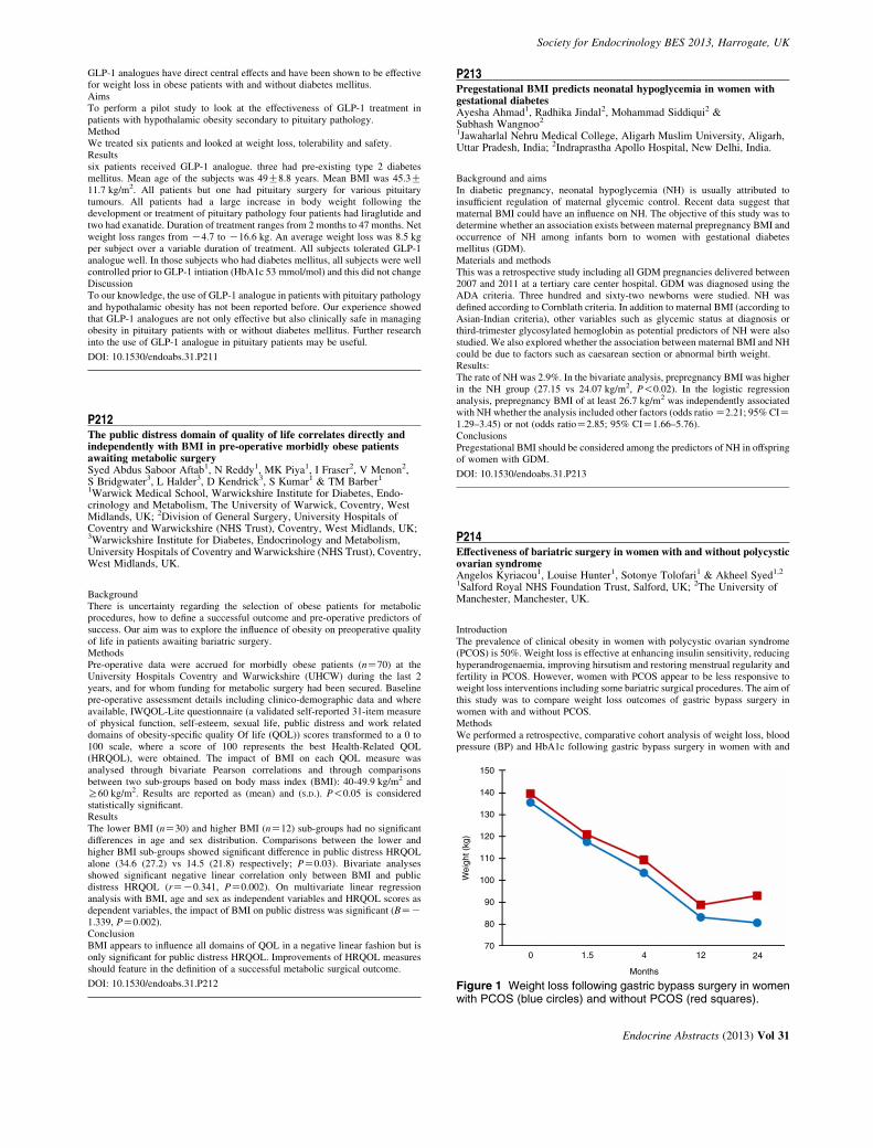
Society for Endocrinology BES 2013, Harrogate, UK
GLP-1 analogues have direct central effects and have been shown to be effectivefor weight loss in obese patients with and without diabetes mellitus.AimsTo perform a pilot study to look at the effectiveness of GLP-1 treatment inpatients with hypothalamic obesity secondary to pituitary pathology.MethodWe treated six patients and looked at weight loss, tolerability and safety.Resultssix patients received GLP-1 analogue. three had pre-existing type 2 diabetesmellitus. Mean age of the subjects was 49G8.8 years. Mean BMI was 45.3G11.7 kg/m2. All patients but one had pituitary surgery for various pituitarytumours. All patients had a large increase in body weight following thedevelopment or treatment of pituitary pathology four patients had liraglutide andtwo had exanatide. Duration of treatment ranges from 2 months to 47 months. Netweight loss ranges from K4.7 to K16.6 kg. An average weight loss was 8.5 kgper subject over a variable duration of treatment. All subjects tolerated GLP-1analogue well. In those subjects who had diabetes mellitus, all subjects were wellcontrolled prior to GLP-1 intiation (HbA1c 53 mmol/mol) and this did not changeDiscussionTo our knowledge, the use of GLP-1 analogue in patients with pituitary pathologyand hypothalamic obesity has not been reported before. Our experience showedthat GLP-1 analogues are not only effective but also clinically safe in managingobesity in pituitary patients with or without diabetes mellitus. Further researchinto the use of GLP-1 analogue in pituitary patients may be useful.
DOI: 10.1530/endoabs.31.P211
150
140
130
120
110
100
90
80
700 1.5 4 12 24
Months
Wei
ght (
kg)
Figure 1 Weight loss following gastric bypass surgery in women
P212The public distress domain of quality of life correlates directly andindependently with BMI in pre-operative morbidly obese patientsawaiting metabolic surgerySyed Abdus Saboor Aftab1, N Reddy1, MK Piya1, I Fraser2, V Menon2,S Bridgwater3, L Halder3, D Kendrick3, S Kumar1 & TM Barber1
1Warwick Medical School, Warwickshire Institute for Diabetes, Endo-crinology and Metabolism, The University of Warwick, Coventry, WestMidlands, UK; 2Division of General Surgery, University Hospitals ofCoventry and Warwickshire (NHS Trust), Coventry, West Midlands, UK;3Warwickshire Institute for Diabetes, Endocrinology and Metabolism,University Hospitals of Coventry and Warwickshire (NHS Trust), Coventry,West Midlands, UK.
BackgroundThere is uncertainty regarding the selection of obese patients for metabolicprocedures, how to define a successful outcome and pre-operative predictors ofsuccess. Our aim was to explore the influence of obesity on preoperative qualityof life in patients awaiting bariatric surgery.MethodsPre-operative data were accrued for morbidly obese patients (nZ70) at theUniversity Hospitals Coventry and Warwickshire (UHCW) during the last 2years, and for whom funding for metabolic surgery had been secured. Baselinepre-operative assessment details including clinico-demographic data and whereavailable, IWQOL-Lite questionnaire (a validated self-reported 31-item measureof physical function, self-esteem, sexual life, public distress and work relateddomains of obesity-specific quality Of life (QOL)) scores transformed to a 0 to100 scale, where a score of 100 represents the best Health-Related QOL(HRQOL), were obtained. The impact of BMI on each QOL measure wasanalysed through bivariate Pearson correlations and through comparisonsbetween two sub-groups based on body mass index (BMI): 40-49.9 kg/m2 andR60 kg/m2. Results are reported as (mean) and (S.D.). P!0.05 is consideredstatistically significant.ResultsThe lower BMI (nZ30) and higher BMI (nZ12) sub-groups had no significantdifferences in age and sex distribution. Comparisons between the lower andhigher BMI sub-groups showed significant difference in public distress HRQOLalone (34.6 (27.2) vs 14.5 (21.8) respectively; PZ0.03). Bivariate analysesshowed significant negative linear correlation only between BMI and publicdistress HRQOL (rZK0.341, PZ0.002). On multivariate linear regressionanalysis with BMI, age and sex as independent variables and HRQOL scores asdependent variables, the impact of BMI on public distress was significant (BZK1.339, PZ0.002).ConclusionBMI appears to influence all domains of QOL in a negative linear fashion but isonly significant for public distress HRQOL. Improvements of HRQOL measuresshould feature in the definition of a successful metabolic surgical outcome.
DOI: 10.1530/endoabs.31.P212
P213Pregestational BMI predicts neonatal hypoglycemia in women withgestational diabetesAyesha Ahmad1, Radhika Jindal2, Mohammad Siddiqui2 &Subhash Wangnoo2
1Jawaharlal Nehru Medical College, Aligarh Muslim University, Aligarh,Uttar Pradesh, India; 2Indraprastha Apollo Hospital, New Delhi, India.
Background and aimsIn diabetic pregnancy, neonatal hypoglycemia (NH) is usually attributed toinsufficient regulation of maternal glycemic control. Recent data suggest thatmaternal BMI could have an influence on NH. The objective of this study was todetermine whether an association exists between maternal prepregnancy BMI andoccurrence of NH among infants born to women with gestational diabetesmellitus (GDM).Materials and methodsThis was a retrospective study including all GDM pregnancies delivered between2007 and 2011 at a tertiary care center hospital. GDM was diagnosed using theADA criteria. Three hundred and sixty-two newborns were studied. NH wasdefined according to Cornblath criteria. In addition to maternal BMI (according toAsian-Indian criteria), other variables such as glycemic status at diagnosis orthird-trimester glycosylated hemoglobin as potential predictors of NH were alsostudied. We also explored whether the association between maternal BMI and NHcould be due to factors such as caesarean section or abnormal birth weight.Results:The rate of NH was 2.9%. In the bivariate analysis, prepregnancy BMI was higherin the NH group (27.15 vs 24.07 kg/m2, P!0.02). In the logistic regressionanalysis, prepregnancy BMI of at least 26.7 kg/m2 was independently associatedwith NH whether the analysis included other factors (odds ratio Z2.21; 95% CIZ1.29–3.45) or not (odds ratioZ2.85; 95% CIZ1.66–5.76).ConclusionsPregestational BMI should be considered among the predictors of NH in offspringof women with GDM.
DOI: 10.1530/endoabs.31.P213
P214Effectiveness of bariatric surgery in women with and without polycysticovarian syndromeAngelos Kyriacou1, Louise Hunter1, Sotonye Tolofari1 & Akheel Syed1,2
1Salford Royal NHS Foundation Trust, Salford, UK; 2The University ofManchester, Manchester, UK.
IntroductionThe prevalence of clinical obesity in women with polycystic ovarian syndrome(PCOS) is 50%. Weight loss is effective at enhancing insulin sensitivity, reducinghyperandrogenaemia, improving hirsutism and restoring menstrual regularity andfertility in PCOS. However, women with PCOS appear to be less responsive toweight loss interventions including some bariatric surgical procedures. The aim ofthis study was to compare weight loss outcomes of gastric bypass surgery inwomen with and without PCOS.MethodsWe performed a retrospective, comparative cohort analysis of weight loss, bloodpressure (BP) and HbA1c following gastric bypass surgery in women with and
with PCOS (blue circles) and without PCOS (red squares).
Endocrine Abstracts (2013) Vol 31
Society for Endocrinology BES 2013, Harrogate, UK
without PCOS aged 18–50 years matched for age (G5 years) and preoperativeBMI (G5 kg/m2). We report results from 56 women, 28 with and 28 withoutPCOS.ResultsMean preoperative age was 28.5 years, weight 137.5 kg, BMI 50.0 kg/m2, systolicand diastolic BP 135 and 85 mm Hg respectively, and HbA1c 37 mmol/mol; therewere no significant differences between the two groups. There was significantweight loss following bariatric surgery with no statistical difference betweengroups (see Fig. 1). Likewise, there was significant reduction in BMI, BP andHbA1c with no significant difference between groups.ConclusionGastric bypass surgery in obese women with PCOS results in significantreductions in weight, BMI, BP and HbA1c, similar to women without PCOS.
DOI: 10.1530/endoabs.31.P214
P215Octreotide therapy of chronic urticaria and angioedema after gastricbypass procedureJeanny Varghese, Mohamed Malik & Carrock SewellScunthorpe General Hospital, Scunthorpe, UK.
BackgroundThe exact changes of hormones and the relative importance of these to themetabolic improvement after bariatric surgery remain to be explored. Wehighlight the unusual case of a patient who developed episodes of angioedemapost roux-en-y surgery for weight reduction.CaseA 38-year-old man with a BMI of 50 kg/m2, had laparoscopic Roux-en-y bypasssurgery, resulting in 89 kg loss over 12–18 months. Six to twelve 12 months post-op he developed recurrent facial angioedema and mild urticaria, lasting for 6–12 h, relieved with oral steroids and antihistamines. Two of these episodes wereassociated after taking ibuprofen. A low salicylate diet was ineffective as weremultiple antihistamines at up to triple standard doses at preventing the reactions.Tests for autoimmunity, food allergy, C1 inhibitor deficiency, mast cell tryptase,pheochromocytoma, and carcinoid including octreotide scintigraphy werenegative. His plasma gut peptide profile was within the normal range. As severalurinary 5-HIAA levels were borderline elevated (52.8, 54.1 and 36.5 mmol/day,normal 0–50) he was commenced on octreotide and has since remained symptomfree. Attempts to reduce the octretide dose result in return of symptoms,indicating that he does not just have chronic urticaria that has now resolved.DiscussionThere is progressive rise in peptideYY, enteroglucagon, pancreatic polypeptideand GLP-1 after gastric bypass surgery. Somatostatin and its analogue octreotideinhibit the release of peptide hormones through stimulation of somatostainreceptors and inhibition of L-type calcium channels. Octreotide inducedmodulation of post bypass satiety gut hormone release is proven in animalmodels. We hypothesise the response seen in this case is due to modulation ofcertain gut peptide(s) with angioneurotic properties.ConclusionTrials of octreotide therapy may be useful in similar patients.
DOI: 10.1530/endoabs.31.P215
P216Prevalence of vitamin D insufficiency in severely obese patients seekingbariatric surgeryRachel Smith1, Rachel Batterham1,3 & Nick Finer2,3
1Department of Medicine, University College London, London, UK; 2UCLInstitute of Cardiovascular Science, London, UK; 3UCLH Centre for WeightLoss, Metabolic and Endocrine Surgery, London, UK.
Background and objectivesVitamin D deficiency and insufficiency is common in obese individuals. Weaimed to determine the prevalence of vitamin D insufficiency within a cohort ofseverely obese individuals, exploring potential underlying associations.MethodsIn a retrospective analysis of 703 patients sequentially presenting for bariatricsurgery assessment, 663 had complete demographic, anthropometric, andhaematological measures (vitamin D, folate, vitamin B1, vitamin B12, whitecell count, and C-reactive protein). Vitamin D levels were defined as deficient (!25 nmol/l), insufficient (25–50 nmol/l), or normal (O50 nmol/l). Adiposity was
Endocrine Abstracts (2013) Vol 31
estimated using indirect measures; BMI, and body fat percentage (BF%)estimated via the CUN-BAE equation. Statistical analyses: groups formedaccording to vitamin D status were compared using ANOVA. Stepwise regressionwas performed using age, gender, BMI, BF%, index of multiple deprivation,multivitamin use, number of comorbidities, white cell count, C-reactive protein,folate and vitamin B12 as independent variables for vitamin D concentration.ResultsMean serum vitamin D concentration was 38G23 nmol/l. 75% of patients hadabnormal serum vitamin D concentrations; 33.2% were deficient, and 41.8%insufficient. Individuals were divided according to vitamin D status (deficient vsinsufficient vs normal). Those with vitamin D deficiency had an increased BMI(48.6 vs 46.9 vs 45.4 kg/m2; P!0.001), decreased folate concentrations (5.8 vs7.7 vs 8.7 ng/ml; P!0.001), and decreased vitamin B12 concentrations (393 vs445 vs 464 pg/ml; PZ0.003). Stepwise regression revealed only a weakassociation with folate concentration (R2Z0.081; PZ0.046).ConclusionVitamin D insufficiency is common among severely obese bariatric surgerycandidates, observed in approximately 75% of patients. The underlying cause isstill unknown and the hypotheses of sequestration within adipose tissue, orinadequate nutritional intake are not supported by these data. Questions remainregarding the functional significance of low vitamin D concentrations amongobese individuals, and whether routine supplementation is required.
DOI: 10.1530/endoabs.31.P216
P217Appetite regulation during a 6-month military tour to AfghanistanN E Hill1,3, J L Fallowfield2, S K Delves2, S J Brett1, D R Wilson3, G Frost1,W Dhillo1, S R Bloom1 & K G Murphy1
1Imperial College London, London, UK; 2Institute of Naval Medicine,Alverstoke, UK; 3Royal Centre for Defence Medicine, Birmingham, UK.
BackgroundMilitary personnel on operational deployment commonly lose weight despite theadequate supply of rations. Moderate weight loss (w5% body mass) occurredduring the initial phase of a 6-month deployment to Afghanistan without affectingphysical fitness. Reasons for this weight loss are presently unknown. We sought toestablish whether changes in appetite regulatory hormones contribute to theobserved weight loss.MethodsBody mass and body composition were measured in a cohort of Royal Marinestwice before (January and March) and during a 6-month summer (March–October) deployment to Afghanistan (Afgh) in 2010. Circulating total and activeghrelin, peptide YY, pancreatic polypeptide, glucagon-like peptide-one, insulinand leptin were measured in samples drawn at the same time points. Data wereanalysed by repeated measures ANOVA.ResultsPercentage body fat increased between the January and March time-points takenin the UK, but was reduced after (mean) 3 months in Afgh (17.5 G0.5% (Jan) vs18.0 G0.5% (Mar) vs 16.5 G0.5% (Afgh); nZ105; P!0.0001); changes inserum leptin concentrations in this lean population mirrored those observed inpercentage body fat (2.15 G0.20 ng/ml (Jan) vs 2.76 G0.20 ng/ml (Mar) vs 1.44G0.12 ng/ml (Afgh); nZ51; P!0.0001). Only leptin was significantly correlatedwith percentage body fat (and body mass) at each time-point (rZ0.59; nZ122(Jan), rZ0.58, nZ137 (Mar), rZ0.51, nZ98 (Afgh); all P!0.0001). There wereno changes in the concentrations of the other hormones measured in response toalterations in body mass or body fat.DiscussionLeptin has previously been shown to be reduced after weight loss in obesepopulations also after short-term (72 h) starvation in lean volunteers. The currentstudy suggests that fasting leptin concentration also reflects small changes inpercentage body fat in a lean population of military personnel. Future work willinvestigate whether post-prandial changes in gut hormones may drive thealterations in energy homeostasis observed on deployment.Declaration of fundingThis work was funded by the Ministry of Defence.
DOI: 10.1530/endoabs.31.P217
Society for Endocrinology BES 2013, Harrogate, UK
P218Weight loss after bariatric surgery in women of childbearing ageAderinsola Alatishe, Basil J Ammori, John P New & Akheel A Syed1Salford Royal NHS Foundation Trust and University Teaching Hospital,Salford, UK; 2The University of Manchester, Manchester, UK.
Obesity increases the risk of pregnancy-related complications such asmiscarriage, foetal abnormalities, hypertension, diabetes, thrombosis, caesareansection and infection. Although bariatric surgery addresses some of these risks,women should defer pregnancy for 12-24 months postoperatively until weightloss has plateaued due to concerns regarding limited maternal weight loss andfoetal nutritional deficiency. The aim of this study was to evaluate the impact ofpregnancy on weight loss after bariatric surgery and to assess pregnancyoutcomes.Amongst 730 obese people who underwent bariatric surgery, 232 women ofchildbearing age (18–45 years) with a mean G S.D. age 34.0G5.9 years,preoperative weight 137.7G21.3 kg and BMI 50.6G7.2 kg/m2 were identified.One-hundred and ninety-seven women (84.9%) had undergone Roux-en-Y gastricbypass, 19 (8.2%) adjustable gastric banding, 8 (3.4%) sleeve gastrectomy and 8(3.4%) other procedures.Women who became pregnant following bariatric surgery (nZ21) were youngerat the time of surgery compared to women in the non-pregnancy group (28.0G5.4vs 34.6G5.6 years, P!0.001) but were otherwise well matched for preoperativeweight (136.5G18.5 vs 137.8G21.6 kg), BMI (49.2G7.4 vs 50.7G7.2 kg/m2)and type of bariatric procedure.Eighteen women (86%) completed pregnancy successfully; 12 (57%) had livebirths by vaginal route, 6 (29%) had caesarean section; 2 (9%) undertook medicaltermination of pregnancy and 1 (5%) suffered a spontaneous miscarriage.At a median follow-up of 30 months, the pregnancy and non-pregnancy groupsachieved significant but comparable excess weight loss after bariatric surgery(70.4 vs 70.0%).In conclusion, pregnancy after bariatric surgery is safe and does not adverselyinfluence weight loss outcomes. However, close surveillance of maternal weightand nutritional status is advisable, especially with conception within the first 12months after surgery.
DOI: 10.1530/endoabs.31.P218
P219Eating more quickly heightens overall systemic exposure to glucose andNEFA in the post-prandial phase, irrespective of energy expenditure inobese womenNarendra Reddy, Chen Peng, Milan K Piya, Saboor A S Aftab,Alison Campbell, John Hattersley, Louise Halder, Alison L Harte,Harpal Randeva, Gyanendra Tripathi, Philip G McTernan, Sudhesh Kumar& Thomas M BarberDivision of Metabolic and Vascular Health, University of Warwick,Coventry, UK.
Background/aimThe global obesity epidemic has promoted a search for novel solutions. Oneapproach is through modification of eating-related behaviours. Our aim was toexplore the effects of meal duration on energy expenditure, appetite andexcursions of molecules associated with insulin sensitivity in the post-prandialphase.MethodsNormoglycaemic, pre-menopausal, Caucasian obese women (nZ8) wererecruited from the Obesity clinic at Warwickshire Institute for the Study ofDiabetes, Endocrinology and Metabolism (WISDEM, UHCW). Whole-bodyindirect calorimetry (6-h) was performed on each subject on two separateoccasions, with standard lunch (763 kcal: 50% carbohydrate) ingested over 10- vs40-min. Blood tests were taken at baseline (fasting) and at 30, 60, 90, 120, 180
and 240 min following mid-meal, and analysed for glucose, insulin and non-esterified fatty acids (NEFA). Number/composition of calories ingested during astandard buffet meal (240-min) was assessed. Paired-sample t-tests were used.P!0.05 was considered statistically significant.ResultsOverall, following the 10- vs 40-min meals, there were significant differences inpost-prandial plasma glucose (mean 6.4 mmol/l (S.D. 1.4) vs 6.0 mmol/l (S.D. 1.3)respectively, PZ0.01) and serum NEFA concentrations (mean 243 mmol/l (S.D.224) vs 191 mmol/l (S.D. 171) respectively, PZ0.005). There was a trendtowards greater post-prandial insulin resistance following the 10- vs 40-minmeals (HOMA2 IR 2.1 (S.D. 2.0) vs 1.9 (S.D. 1.7) respectively, PZNS). Post-prandial energy expenditures were equivalent between the two meal durations(range 80–120 kcal/h). Regarding buffet selection (tZ240-min), total number/composition of calories ingested were equivalent between the meal durations.ConclusionOur novel data highlight that meal duration impacts on systemic glucose andNEFA in obese women. Eating a meal more quickly increases overall exposure toglucose and NEFA, with a trend towards increased insulin resistance during thepost-prandial phase; whilst no impact was noted on energy expenditure orappetite. Longer meal durations may therefore facilitate better maintenance ofmetabolic health.
DOI: 10.1530/endoabs.31.P219
P220Hepatic 11b-hydroxysteroid dehydrogenase type 1 is elevated followingweight loss secondary to bariatric surgeryConor Woods1, Angela Taylor2, Beverly Hughes2, Michelle Corrigan1,Paul Stewart2, Jeremy Tomlinson2, Donal O Shea1 & Mark Sherlock2,3
1Education and Research Centre, St Vincent’s University Hospital and StColumcille’s Hospital, Dublin, Ireland; 2Centre for Endocrinology, Diabetesand Metabolism, Institute of Biomedical Research, University ofBirmingham, Birmingham, UK; 3Department of Endocrinology, Adelaideand Meath Hospital, Tallaght l, Dublin, Ireland.
In the pathogenesis of obesity, dysregulated tissue cortisol metabolism(controlled by 11b-HSD1), is postulated to be involved. Fifteen patients (sevenmens, mean BMI 50.8G7 kg/m2) awaiting Roux En Y gastric Bypass (RYGB)surgery underwent assessment of 11b-HSD1 activity using cortisol generationprofile. Corticosteroids in serum and subcutaneous adipose tissue microdialysisfluid and urinary corticosteroid metabolites were analysed by liquid and gaschromatography mass spectrometry. Results were compared to six post oppatients and non-obese controls. Mean area under the curve (AUC) for serumcortisol (F) to cortisone (E) ratio (marker of 11b-HSD1 activity) was significantlyreduced in pre-op patients compared to post-op patients: 1037G448.9 vs 1656G505.1 units/min (PZ0.0045). Subcutaneous adipose tissue Microdialysisfluid showed a threefold reduction in F/E AUC ratio following RYGB surgery:303.2 vs 103.2 units/min. Total urinary cortisol metabolites (Fm) weresignificantly raised in severely obese patients compared to non-obese controls;6602G742 vs 4396G2362 mg/24 h (PZ0.026). The ratios of (THFC5aTHF)/THE (marker of 11b-HSD1 activity) and Fm/Em were notsignificantly different. The ratios of androsterone/etiocholanolone and 5 0
tetrahydrocortisol/5a tetrahydrocortisol (markers of 5a reductase activity) werenot significantly different. In this obese cohort, there is evidence of hypothalamic–pituitary–adrenal axis activation as reflected by increased total cortisolmetabolites, driven by decreased hepatic 11b-HSD1 generation of cortisol.Global urine metabolites are not different reflecting possible up regulation of11b-HSD1 in adipose tissue and muscle, and down regulation in liver. This mayreflect a protective mechanism to reduce hepatic cortisol exposure.Declaration of fundingNewman fellowship, UCD, Dublin.
DOI: 10.1530/endoabs.31.P220
Endocrine Abstracts (2013) Vol 31
P221Brown adipose tissue identification in an adult human using IDEALMRINarendra Reddy1, Terence Jones1, Sarah Wayte2, Oludolapo Adesanya2,Yen Yeo2, Harpal Randeva2, Sudhesh Kumar1,2, Charles Hutchinson1,2 &Tom Barber11University of Warwick, Coventry, UK; 2University Hospitals of Coventryand Warwickshire, Coventry, UK.
AimManipulation of human brown adipose tissue (BAT) represents a noveltherapeutic option for diabesity. The aim of our study was to develop and test anovel magnetic resonance imaging (MRI) based method to identify human BATand delineate it from white adipose tissue (WAT), and validate it by providingimmunohistochemical confirmation.MethodsInitial scanning with 18F-FDG PET–CT radiotracer uptake on a 25-year-oldCaucasian female with primary hyperparathyroidism, showed avid uptake withinthe mediastinum, neck, supraclavicular fossae and axillae, consistent with BAT.Subsequently, serial MR scans were performed using three-echo IDEAL (iterativedecomposition of water and fat with echo asymmetry and least-squaresestimation) sequence. Retrospectively, regions of interest (ROIs) were identifiedon MR corresponding to PET–CT images. Prospectively, ROIs were identified onMR images based on signal intensity and appearance, and compared with PET–CT. Immunohistochemical staining using uncoupling protein-1 antibody wasperformed on fat samples corresponding to low MR-signal, obtained duringparathyroidectomy.ResultsOf the 111 retrospectively identified ROIs from PET–CT scans, 88 (79%) showedcorresponding low signal on the MR images: 100% in mediastinum, 29/31(93.5%) in neck, 31/41 (75.6%) supraclavicular, and 8/14 (57%) in axillae.Prospectively, 87% of ROIs identified on MR scans corresponded to increasedareas of uptake on PET–CT scans. Histology and immunohistochemistryconfirmed BAT.ConclusionWe provide the first ever report that MR can be used reliably to identify BAT in ahuman adult, with histological and immunohistochemical confirmation. Our datademonstrate proof of concept to support the development of MR, a safe andreproducible imaging modality, as a biomarker for human BAT.
DOI: 10.1530/endoabs.31.P221
P222Vitamin D supplementation as influenced by diabetic therapiesKhalid Alkharfy1, Nasser Al-Daghri1, Abdulaziz Al-Othman1,Osama Moharram1, Majed Alokail1, Yousef Al-Saleh1, Shaun Sabico1,3 &Philip McTernan31King Saud University, Riyadh, Saudi Arabia; 2King Abdulaziz UniversityHospital, Riyadh, Saudi Arabia; 3Warwick Medical School, WarwickUniversity, Coventry, UK.
ObjectiveVitamin D deficiency is associated with an increased risk of type two diabetesmellitus (T2DM) and cardiovascular disease reduced through Vitamin D3supplementation. However to the best of our knowledge, no research hasdetermined the effect of vitamin D3 supplementation in conjunction with existingpharmacological and non-pharmacological approaches to the DMT2 population.Hence, the aim of this study was to determine the effect of vitamin D3supplementation in a cohort of Saudi DMT2 population on different oralhypoglycemic agents and compare them with a non-DMT2 control.MethodsA total of 499 randomly selected DMT2 subjects divided into eight groups basedon their existing diabetes management (non-DMT2 control Z151; rosiglitazone(Avandia) Z49; diet Z15; insulin alone Z55; insulin in combination with otheroral hypoglycemic (OHG) agents Z12; metformin alone Z121; OHGcombination Z37; sulphonylurea Z59) were included in this 12-monthinterventional study. All subjects were given 2000 IU vitamin D3 daily exceptthe control group. Anthropometrics, glucose, lipid profile and 25-OH Vitamin Dwere measured at baseline, 6 and 12 months.ResultsT2DM subjects treated with Metformin raised vitamin D2 levels at 6 and 12months compared with baseline (P!0.001). No significant changes wereobserved between Vitamin D2 levels and BMI, glucose, total- and HDL-cholesterol in any DMT2 groups. However significant decreases in triglycerideswere observed in subjects treated with both rosiglitazone and insulinCOHG
group at 6 and 12 month Vitamin D3 supplementation (both P values !0.001).ConclusionWhilst all groups increased circulating vitamin D2 following supplementation, itappears that DMT2 patients on insulin in combination with other OHG drugsheighten their Vitamin D3 absorption. As such Vitamin D3 taken with insulin plusOHG can offer benefits to reduce Vitamin D deficiency whilst improvingcardiometabolic risk factors.Declaration of fundingThe study was generously funded by King Abdulaziz City of Science andTechnology (KACST Grant no: AT-29-38), Riyadh, KSA for funding the study.
DOI: 10.1530/endoabs.31.P222
P223Association of the incidence of type 1 diabetes with markers of infectionand antibiotic susceptibility at country levelAlexia-Giovanna Abela1,2 & Stephen Fava1,21Diabetes and Endocrine Centre, Mater Dei Hospital, Malta; 2University ofMalta Medical School, Msida, Malta.
AimTo investigate the association between country incidence of type one diabetes(T1DM) and mortality from infectious disease as well as to antibioticsusceptibilityMaterials and methodsAn ecological study correlating data from the WHO DiaMond Project for theincidence of T1DM, the WHO estimates of mortality (2004) from communicablediseases and the Alexander Project for bacterial susceptibility to antimicrobialagents.ResultsThere were statistically significant negative correlations between the incidence ofT1DM and mortality from: infections and parasitic diseases (rZK0.34, PZ0.01), respiratory infections (rZK0.29, PZ0.03), tuberculosis (rZK0.36, PZ0.007), diarrhoeal diseases (rZK0.32, PZ0.02) and total infectious disease mortality(rZK0.35, PZ0.008). There was a positive correlation between T1DMincidence and susceptibility of Streptococcus pneumoniae to penicillin (rZ0.47, PZ0.03), erythromycin (rZ0.52, PZ0.014), doxycycline (rZ0.65,P%0.002) and co-trimoxazole (rZ0.58, PZ0.007). We also found a positivecorrelation between T1DM incidence and the mean susceptibility (rZ0.62,PZ0.004), and lowest antibiotic susceptibility (rZ0.73, P%0.0001) of S.pneumonia.ConclusionWe found a negative correlation between country incidence of T1DM and itsmortality from infectious diseases. Mortality from infectious diseases is a strongmarker of the total infective burden. Incidence of T1DM was found to bepositively correlated with the susceptibility of S. pneumoniae to all antibioticsstudied. Increased antibiotic susceptibility of a given organism may be an indirectmarker of a low degree of exposure of the community to it. Our results providesupport for the hygiene hypothesis, namely that diminished bacterial exposure inearly post-natal life results in increased risk of developing T1DM.
DOI: 10.1530/endoabs.31.P223
P224A pilot study of 25-hydroxy vitamin D level in type 2 diabetes mellituswith diabetic retinopathyMona AbdelsalamEndocrine, 7 Metabolism Unit, Ain Shams University, Cairo, Egypt.
BackgroundMany cellular, preclinical and observational studies support a role for vitamin D(VD) in the pathogenesis of type 2 diabetes. VD is suggested to be an inhibitor ofangiogenesis. A growing body of evidence suggests an association between VDinadequacy and diabetic retinopathy.ObjectiveTo study the relation between 25 (OH) vitamin D level and diabetic retinopathy(DR) in patients with type 2 diabetes mellitus and to evaluate for any relationbetween 25 (OH) vitamin D level and different stages of diabetic retinopathy.Subjects and methods50 Type 2 diabetic patients and 50 healthy volunteers (control group) matched byage and sex participated in the study. Based on their ophthalmic findings, the type2 diabetic patients were divided into two groups: group (I) 25 patients with DRand group (II) 25 patients without DR. Fluorescein angiography was done for
Society for Endocrinology BES 2013, Harrogate, UK
Endocrine Abstracts (2013) Vol 31
group (I) and accordingly the patients were further classified into three subgroups:moderate NPDR, severe NPDR and PDR. Each subgroup is then dividedaccording to presence or absence of clinically significant macular edema (CSME).Fasting blood sugar, HbA1c, renal functions, liver functions, lipid profile, serumcalcium, serum phosphorous, intact parathyroid hormone (iPTH) and serum 25hydroxyvitamin D3levels were done to all participants in the study.ResultsMean 25(OH) VD level was lower in type 2 diabetic cases than in control group(P!0.01). Mean 25(OH) VD level was lower in type 2 diabetic cases with DRthan type 2 diabetic cases without DR (P!0.05). Patients with PDR have thelowest mean 25(OH) vitamin D level compared to patients with moderate NPDRand severe NPDR (P!0.05). 25(OH). VD level was inversely correlated withage, duration of type 2 diabetes mellitus, stages of diabetic nephropathy, fundusfindings, BMI, SBP, DBP, glycemic parameters, urinary ACR, total cholesterol,triglycerides, LDL-C (P!0.01) and iPTH level (P!0.05). 25(OH) VD level waspositively correlated with GFR, HDL-C and total calcium level.ConclusionsThere is an association between 25 (OH) vitamin D insufficiency and DR amongpatients with type 2 diabetes mellitus. Low serum 25 (OH) vitamin D might be arisk marker of development or progression of DR. Measurement of serum 25(OH) vitamin D concentrations could become a useful biochemical marker relatedto the severity of DR. The effect of vitamin D replacement in patient with DRshould be evaluated.
DOI: 10.1530/endoabs.31.P224
P225Non-alcoholic fatty liver disease in patients attending the NationalSevere Insulin Resistance ServiceSarah M Leiter1, Alison Sleigh2, Claire Adams1,3, Julie Harris1,3, DavidJ Lomas4,5, Michael Allison6, Robert Semple1,3, David Savage1,3 &Anna Stears1,31Metabolic Research Laboratories, University of Cambridge, Cambridge,UK; 2Wolfson Brain Imaging Centre, University of Cambridge, Cambridge,UK; 3Wolfson Department of Diabetes and Endocrinology, AddenbrookesHospital, Cambridge, UK; 4Department of Radiology, University ofCambridge, Cambridge, UK; 5Department of Radiology, AddenbrookesHospital, Cambridge, UK; 6Department of Medicine, AddenbrookeesHospital, Cambridge, UK.
IntroductionThe prevalence of non-alcoholic fatty liver disease (NAFLD) is greatly increasedin patients with lipodystrophy and some other forms of severe insulin resistance.Liver biopsy remains the definitive technique for diagnosis and staging ofNAFLD. However, non-invasive techniques, such as magnetic resonancespectroscopy (MRS) and magnetic resonance imaging (MRI) are increasinglyused to assess response to therapeutic interventions. The National Severe InsulinResistance (SIR) Service, commissioned by the National Specialist Commission-ing Team in 2011, aims to optimise outcomes of patients with lipodystrophyand/or SIR. Selected patients, including all those receiving leptin therapy, areoffered annual measurement of liver fat using MRS or MRI.Patient populationLiver fat was measured in 28 patients (23 female), median (range) age 28 (13–58)years, BMI 25.8 (15.8–34.0) kg/m2. Twenty-four have lipodystrophy and fourhave SIR of currently unknown cause. None have an insulin receptor mutation. 11were taking leptin. 13/28 patients were scanned using proton single voxel MRSand 15/28 patients were scanned using in-phase and out of phase gradient-echoMRI with dual flip angles.Results23/28 (82.0%) patients had O5% liver fat. Median (range) liver fat was 22.6%(1.4–102.0) in patients scanned by MRS and 9.0% (2.0–29.0) in those scanned byMRI. Biopsies were available in two patients. Interestingly these patients bothhave biopsy proven fibrotic liver disease, but relatively low liver fat by MRI of 4.0and 6.0% respectively.ConclusionAs expected, there is a very high prevalence of NAFLD in patients withlipodystrophy and/or SIR attending the National SIR Service. MRI/S basedassessment of hepatic steatosis may be useful in assessing response to therapy, buta normal result does not preclude the presence of significant NAFLD and mayeven signify the presence of fibrosis in some cases, though this is yet to beformally proven.Declaration of fundingThe National Severe Insulin Resistance service is funded by the NationalSpecialist Commissioning Team.
DOI: 10.1530/endoabs.31.P225
P226A study of serum osteocalcin level in women with gestational diabetesmellitusMona AbdelsalamEndocrine, 7 Metabolism Unit, Ain Shams University, Cairo, Egypt.
IntroductionGestational diabetes mellitus complicates up to 14% of pregnancies worldwide.Gestational diabetes mellitus women are more frequently affected by changes inbone turnover during pregnancy. Osteocalcin, ostoblast derived protein issuspected to be involved in the regulation of glucose and fat metabolism.Aim of the workis to study serum osteocalcin levels in women with gestational diabetes mellitus.Subjects and methodsThis study included sixty pregnant women at 22nd to 28 weeks of pregnancy.They are subdivided into 30 women diagnosed with gestational diabetes mellitusand 30 healthy pregnant women. All subjects included subjected to full historytaking, clinical examination and laboratory investigations which includes 100 goral glucose tolerance test, HbA1c, lipid profile, fasting serum insulin, HOMA-IRand fasting serum osteocalcin level.ResultsSerum osteocalcin was lower in gestational diabetes mellitus women (3.70G1.55 ng/dl) than healthy pregnant women (16.61G5.30) (P!0.001). There was ahighly significant negative correlation between serum osteocalcin level andfasting blood glucose, 2 h postprandial blood glucose, HbA1c, fasting seruminsulin, HOMA-IR, total cholesterol, serum triglycerides, LDL-cholesterol andhighly significant positive correlation with HDL-cholesterol (P!0.001).ConclusionSerum osteocalcin level may have a role in glucose homeostasis in gestationaldiabetes mellitus.
DOI: 10.1530/endoabs.31.P226
P227Short term calorie restriction: effects on endocrine markers ofnutritional statusSwati Jain & Som Nath SinghDefence Institute of Physiology and Allied Sciences, Delhi, India.
Calorie restriction (CR), a form of dietary regime which reduces caloric intakebelow the habitual unrestricted food intake is a strategy to control weight. Thebenefits associated with CR are numerous but the associated constant feeling ofhunger makes adherence to the CR regime difficult. In the present study certainappetite regulatory hormones and endocrine markers during CR were studied.Male Sprague–Dawley rats were randomly divided into control and test groups(nZ12 rats/group) where the control rats were ad libitum fed and experimentalrats were kept on a 25% CR diet for a period of 5 days. The regulatory hormonesand peptides i.e. ghrelin, leptin, CCK, NPY, insulin, IGF1, corticosterone,adiponectin and thyroid hormones were estimated in plasma at the end of theexperiment. Decrease in body weight was 6.8% in the test group while there was again in body weight by 5.3% in the control rats over the 5-day period. Plasmalevels of ghrelin and NPY were found to be significantly higher (P!0.05) incomparison to control while leptin was below the limit of detection. Insulin wasnot affected significantly in the CR diet in the test group. A small decline inadiponectin was found in the CR group. Partial food restriction caused a slightreduction in plasma CCK and significantly decreased IGF1 levels. Low levels ofcirculating T3, as a result of energy deficit with elevated blood T4 levels werefound. The findings of the present study could be used for formulation of highsatiety low calorie diets for control of metabolic disorders due to over nutrition.Declaration of fundingDefence Institute of Physiology and Allied Sciences, DRDO, Min. of Defence,Delhi, India.
DOI: 10.1530/endoabs.31.P227
P228The development of a structured education programme to improvecardiovascular risk in women with polycystic ovary syndrome(SUCCESS Study)Hamidreza Mani1,2, Heather Daly1, Janette Barnett1,2, Miles Levy2,Kamlesh Khunti1, Trevor Howlett2 & Melanie Davies1,2
Society for Endocrinology BES 2013, Harrogate, UK
Endocrine Abstracts (2013) Vol 31
1University of Leicester, Leicester, UK; 2University Hospitals of Leicester,Leicester, UK.
IntroductionPolycystic ovary syndrome (PCOS) has a prevalence of 10–15% and is associatedwith metabolic and mental health consequences. There is evidence that structurededucation programmes improve illness perception, quality of life and themetabolic profile in other chronic conditions however evidence for structuredpatient education in PCOS is lacking.We aimed to use an iterative cycle of research to develop a pragmatic educationalintervention tailored for the needs of women with PCOS.MethodsIn line with the Medical Research Council’s framework for developing andevaluating complex interventions and using Bartholomew’s intervention mappingprotocol we developed an education programme for women with PCOS.Research question was explored by literature review, consultation with peers aswell as qualitative patient interviews. A semi-structured topic guide was used inthe interviews to assess their need and views on the education programme and itsdesign and content. The programme was also informed by the related literature,phenotypic and outcome analysis of a large local database of women with PCOSand the evidence from education programmes in other chronic conditions such asdiabetes.ResultsThe developed programme was piloted in a group of women with PCOS. Theirfeedback was sought through a semi-structured focus group interview. Refinedintervention was piloted again in a fresh patient group and their feedback wassought.After final refinement of the programme a group of skilled health careprofessional with the knowledge of the underpinning philosophies and learningtheories were trained to deliver this education programme.This pilot work has resulted in a randomised Controlled trial to test its feasibilityand efficacy.DiscussionWomen with PCOS are at increased risk of diabetes, cardiovascular and mentalhealth issues and need an early lifestyle intervention. A successful outcome of ourresearch programme will be a milestone in treatment of this condition andimproving patient care especially in primary care.Declaration of fundingPartly funded by ‘Early Career Grant’ from Society for EndocrinologyIt is part of a PhD project supported by Leicester Diabetes Centre.It is supported by the NIHR “Leicester and Loughborough Diet, Lifestyle andPhysical activity Biomedical Research Unit” and Collaboration for Leadership inApplied Health Research and Care (CLAHRC).
DOI: 10.1530/endoabs.31.P228
P229Hypogonadism in males with type 2 diabetes mellitusAdewole AdesanyaFederal Medical Centre, Lokoja, Kogi State, Nigeria.
Background and objectiveThere have been reported increase prevalence of hypogonadism in diabetic mencompared to age matched non-diabetic men. The objective of this study was toassess the level of androgens in T2DM patients and non-diabetics and correlate itwith erectile function and visceral adiposity.MethodsThis was a cross-sectional comparative study of 160 male patients with T2DM(study subjects) and 80 age matched non-diabetics (control subjects). Level offree testosterone and LH was assayed, erectile dysfunction was assessed withIIEF-5 questionnaire and clinical data of both the study and control subjects wereanalysed.ResultsThe study and control subjects were well matched for age, with meanGS.D. ofstudy subjects being 58.2G10.1 years and control subjects was 56.6G11.3 years.The study subjects had a significantly lower meanGS.D. free testosterone levelthan the control subjects (16.9G6.7 pg/ml compared with 21.6G8.9 pg/ml,PZ0.00). The meanGS.D. LH level of the study subjects was significant lower,compared to that of the control subjects (5.3G3.1 mIU/l compared to 7.8G5.5 mIU/l, P!0.05). Among the study subjects, ED was present in 118 (73.8%)persons compared to 32 (40.0%) persons in the control subjects. The difference inthe prevalence of ED in the two groups was statistically significant. (c2Z25.9,dfZ1, PZ0.00). The meanGS.D. BMI, WC and WHR was also statisticallysignificantly higher in the study subjects compared to control subjects.BMI(26.2G4.0 kg/m2 compared to 24.5G3.0 kg/m2; P!0.001). WC (98.7G13.1 cm
compared to 90.5G10.5 cm; P!0.001). WHR (1.0G0.0 compared to 0.9G0.1;PZ0.02).ConclusionOur study showed that patients with T2DM had hypogonadotrophic hypogonad-ism compared to the non-diabetics. Erectile dysfunction and visceral adipositywas more prevalent in men with T2DM compared to age-matched non-diabetics.
DOI: 10.1530/endoabs.31.P229
P230Obesity-related hypogonadotrophic hypogonadism: recovery of normalpituitary–gonadal axis function following bariatric surgeryAnjali Santhakumar1, Shaz Wahid2 & Richard Quinton11Royal Victoria Infirmary,Newcastle Hospitals NHS Foundation Trust,Newcastle upon Tyne, UK; 2South Tyneside Hospital, South Shields, UK.
BackgroundFunctional hypogonadotrophic hypogonadism (FHH) occurs in the context of anychronic disease including obese patients with type 2 diabetes (T2DM) and/ormetabolic syndrome. FHH is reversible with resolution of the underlying diseaseprocess. Reported benefits of bariatric surgery include improvements in lipidprofile, blood pressure and resolution of T2DM. Here we report reversal of FHHand T2DM with bariatric surgery-associated weight loss.Case historyA 47-year-old man was referred with sexual dysfunction. He underwent pubertyaged 14. His mood was low, he drank alcohol to excess, was obese (BMI 43), withsparse body hair; testes 15 ml. Baseline biochemistry confirmed hypogonado-trophic hypogonadism: LH 1.7 and FSH 4.4 IU/l; testosterone 7.2 nmol/l (NR: 9–25 nmol/l) and DEXA showed L1-4 spine osteoporosis, but serum ferritin andMRI pituitary were normal.He was started on testosterone undecanoate 1 g i.m. (3-monthly) withsymptomatic and biochemical improvement. However, he eventually becamepolycythaemic (peak Hb and haematocrit 18.1 g/dl and 55%, respectively) andtreatment thus had to be discontinued. Shortly afterwards, he developed T2DMand, in view of his co-morbidities, was referred for lifestyle change (includingcounselling) and went on to undergo bariatric surgery. Post-operatively, his BMIfell to 30.3, T2DM resolved and hypertension improved. Significantly, FHH hadalso resolved (LH 3.1 and FSH 4.4 IU/l; testosterone 12.1 nmol/l).ConclusionFHH represents a physiologic response to ill health and thus may serve as a usefulbiological function. Several short-term interventional studies have demonstratedimprovements in various surrogate endpoints with testosterone therapy in patientswith FHH in relation to obesity and/or metabolic syndrome. Nevertheless, giventhe paucity of longitudinal safety/efficacy data, it may be more appropriate totarget the underlying problem of obesity, rather than opting for testosteronereplacement. As demonstrated in our patient, weight loss resulting from bariatricsurgery can lead to recovery of pituitary–gonadal axis obviating the requirementfor testosterone replacement therapy.
DOI: 10.1530/endoabs.31.P230
P231Risk assessment of adult residents in ile-ife, South-Western Nigeria fortype 2 diabetes mellitusGbadebo Ajani, Rosemary Ikem, Adenike Enikuomehin, David Soyoye &Babatope KolawoleObafemi Awolowo University Teaching Hospitals’ Complex, Ile-Ife, OsunState, Nigeria.
BackgroundType 2 diabetes mellitus (T2DM) is a potentially preventable disease that ispresently increasing in epidemic proportion worldwide. Its onset can also bedelayed with an appropriate and timely intervention if people at risk are identifiedearly. This study determined risk levels of adults in Nigeria for type 2 DM byusing Finnish Diabetes Risk Score (FINDRISC).MethodDuring our local activities for world diabetes day in 2011, adults who were notpreviously known to be diabetic voluntarily underwent screening examinationafter an overnight fast at OAUTHC, Ile-Ife. Each participant had theiranthropometry, blood pressure, fasting plasma glucose (FPG) measured andFINDRISC assessed.
Society for Endocrinology BES 2013, Harrogate, UK
Endocrine Abstracts (2013) Vol 31
ResultsThere were 158 participants, 85 (53.8%) males and 73 (46.2%) females. Majority,80 (50.7%) were in age group 45–64 years. 10 (6.3%) subjects had high risk with atotal risk score of 15–20 which estimated that one in three subjects will developdiabetes within 10 years. None of the subjects had very high risk. Only 142(89.9%) subjects had FPG done. High risk subjects had 4 (40.0%), 1 (10.0%) and5 (50.0%) of them with normal FPG, impaired FPG and diabetic FPGrespectively. There was a significant association between the type 2 diabetesrisk levels and FPG (PZ0.014) and SBP (PZ0.008).ConclusionThe Finnish Diabetes Risk Score is a useful noninvasive method of screening inour environment. We recommend its use as a self-administered questionnaire inour population so that individuals will know their risk levels early and adoptappropriate life style modifications.
DOI: 10.1530/endoabs.31.P231
P232Adrenal insufficiency post bariatric surgeryVani Shankaran, Amanda Barclay, Rajeshwaran Cinnadorai, Myat Thida &Balasubramaniyan SrinivasanSt James University Hospital, Leeds, UK.
IntroductionNICE recommends weight loss surgery as a treatment option for people withobesity. However, long term data on outcomes and complications on surgery arelimited. We report unexplained adrenal insufficiency post bariatric surgery.Case report:Patient one27 years old lady underwent Roux-en-Y gastric bypass (RYGB). Weight loss (kg)was from 130.2 to 73.2 over a 2-year period. Patient complained of general unwelland dizziness. No orthostatic hypotension was noted. Random cortisol (nmol/l)was low at 98 and Short synacthen test (SST) showed 0 and 30 min Cortisol of 158and 386 respectively with a low ACTH.Patient two46 years old man underwent RYGB. Weight loss was from 151.8 to 56.8 over aperiod of 18 months. He complained of episodes of vomiting, sweating and blackouts with no postural hypotension. Random cortisol was 50 and SSTdemonstrated cortisol of 108 and 281 at 0 and 30 min respectively. Steroidreplacements were commenced and he improved symptomatically.DiscussionThe cause of adrenal insufficiency in the above cases remains unexplained.Adrenal insufficiency has been reported after major surgery as result of stress orblood loss affecting the pituitary gland. However only one case has been reportedpost bariatric surgery. Possible mechanisms are malabsorption of bile affectingcholesterol leading to reduced precursor for steroid synthesis, malabsorption oftrace elements and vitamins (especially selenium and vitamin B5) that are steroidbiosynthesis cofactors, re-setting of hypothalamo-pituitary–adrenal axis due toweight loss as in anorexia nervosa and perioperative complications such as bloodloss causing pituitary/adrenal infarct or apoplexy.ConclusionThese two cases suggest the importance of patient selection, pre surgicalcounselling and long term follow-up post bariatric surgery.
DOI: 10.1530/endoabs.31.P232
P233Pregnancy outcome in a patient with lipodystrophy and type 2 diabetesTolulope Shonibare & Muhammad ButtHuddersfield Royal Infirmary, Huddersfield, UK.
BackgroundA 23-year-old woman diagnosed with Dunnigan-type familial partial lipodystro-phy (FPLD) attended the joint Antenatal/Endocrine Clinic at 13 weeks ofgestation. She was diagnosed at age 7 and subsequently developed type 2 diabetesat age 11 years. She was managed initially with metformin followed by additionof insulin. It was an unplanned pregnancy and her booking HbA1c was 8.4%(IFCC 68 mmol/mol).Prior to pregnancy, she was on a basal bolus regime of Levemir and Novorapidand her total daily insulin dose wasw180 units. At booking, we added Metformin500 mg twice daily, which was later increased to three times daily.Her insulin requirements escalated rapidly to a total daily dose of 250 units by 25weeks gestation. At this stage, the use of Humulin R (U500) was considered and
discussed with the patient. However, she presented with vaginal spotting and wentinto spontaneous labour at 26 weeks. She delivered a live female infant who wasadmitted briefly to the special care baby unit. During labour she was managedwith intravenous sliding scale insulin.ConclusionFamilial Lipodystrophy is a group of rare disorders associated with numerousmetabolic complications. Diabetes, familial lipodystrophy and pregnancy incombination all compound and confer a severe insulin resistant state which ifpoorly controlled, can have an adverse effect on pregnancy outcome. Data on theuse of U500 insulin during pregnancy is limited however there are a handful ofcase reports of its use in pregnancy with successful management of glycaemic andobstetric outcome.This case is a reminder of the challenges in the glycaemic management of patientswith lipodystrophy particularly in pregnancy. It also adds to the limited literatureavailable on pregnancy outcome of patients with lipodystrophy.
DOI: 10.1530/endoabs.31.P233
P234Impaired iron status in severely obese bariatric surgery candidates ismultifactorialRachel Smith1 & Nick Finer21Department of Medicine, University College London, London, UK; 2UCLInstitute of Cardiovascular Science, London, UK; 3UCLH Centre of WeightLoss, Metabolic and Endocrine Surgery, London, UK.
Background and objectivesObesity is associated with a relatively high prevalence of anaemia and irondeficiency compared to normal weight individuals; the cause is unknown. Weaimed to determine the prevalence of iron deficiency and anaemia in a severelyobese cohort of bariatric surgery candidates, and to explore underlyingassociations with markers of nutrition and inflammation.MethodsIn a retrospective review of 703 consecutive patients presenting for bariatricsurgery assessment, 656 were evaluated after exclusions for haemoglobinopathyand missing data. Assessment included; clinical history, basic anthropometry, andblood tests (haemoglobin (Hb), serum iron, total iron binding capacity, ironbinding saturation, ferritin, vitamin B1, vitamin B12, folate, white cell count(WCC), and C-reactive protein (CRP)). Anaemia was defined as; men: Hb!13 g/dl, or women: Hb!12 g/dl; and iron deficiency as an abnormality in two ormore measures of iron status. Indirect measures of adiposity included BMI andpercentage body fat (BF%) estimated from the CUN-BAE equation. Statisticalanalyses: stepwise regression was performed with independent predictors ofserum iron concentration: age, gender, BMI, BF%, index of multiple deprivation,diabetes status, number of comorbidities, WCC, CRP, folate, vitamin B12, and useof multivitamins, metformin, insulin, non-steroidal anti-inflammatory drugs(NSAIDs) or proton pump inhibitors.ResultsAnaemia and iron deficiency were present in 9.9 and 11.4% of patients,respectively. Stepwise regression revealed an association between serum ironconcentration and CRP (R2Z0.227; PZ0.001), however CRP concentrationswere only available for 156 patients (24%). Excluding CRP from analyses, WCChad the largest effect on serum iron concentration, with NSAIDs, gender, anddiabetes status also contributing (R2Z0.076; P!0.001), albeit leading to a muchweaker association than observed with CRP.ConclusionAnaemia and iron deficiency are relatively common in severely obese individuals,potentially due to inflammation causing deranged iron homeostasis, although amultifactorial aetiology is likely.
DOI: 10.1530/endoabs.31.P234
P235What lies beneath: a case of spontaneous hypoglycaemia or glucosetransporter type 1 defect disguised as chronic fatigue?Mohit Kumar, Annice Mukherjee & Chris HendrikszSalford Royal Foundation Trust, Salford, UK.
A 42-year-old female had extensive neurological investigations (normal MRIbrain, EEG, NCS). A low CSF glucose triggered endocrine referral. She had ahistory of ill health/fatigue since 19 years when she had a viral illness withseizure-like episode and transient wheelchair dependance, with no formaldiagnosis made. She had recurrent symptoms including fatigue, myalgia and
Society for Endocrinology BES 2013, Harrogate, UK
Endocrine Abstracts (2013) Vol 31
weakness, with some relation to hunger and fasting.Physical examination revealed macrocephaly, mild generalised reduction inpower and ataxia, with brisk bilateral lower limb reflexes.Afull fatiguescreenwasnormal.Prolongedfasting ledtosymptomsat24 hoffatigue,left calf pain andmultiple episodes of rhythmic left leg jerking lasting approximately20 s, during which she was fully conscious. Clinical examination demonstrated lefthemiparesis (4/5) and reduced sensation. Labglucosewas4.3 mmol/l, plasma lactate0.9 mmol/l (0.5–2.2) during the episode. She declined further fasting.Shewas referred to the regional adult inheritedmetabolic disorders team.Thehistoryand examination is characteristic of the paroxysmal late-onset form of glucosetransporter type one (GLUT 1) deficiency syndrome. This is usually caused bymutations in the SLC2A1 gene and results in impaired glucose transport into thebrain. Patients develop classical features of hypoglycaemia with normal peripheralblood glucose. Some marginal changes may occur in serum lactate (i.e.inappropriately low !1 mmol/l) when symptomatic. Lower limb pain and jerkingis characteristic. Ketogenic diet is effective in 60% of cases. This is an autosomaldominant condition and is likely to be under-diagnosed. Cases may appear inendocrine clinics because of hypoglycaemic symptoms and hunger. Symptomsmaybe assumed to be functional or related to chronic fatigue syndrome.
DOI: 10.1530/endoabs.31.P235
P236Improvement in testosterone post bariatric surgeryVani Shankaran, Amanda Barclay, Rajeswaran Cinnadorai, Myat Thida &Srinivasan BalasubramanianSt James University Hospital, Leeds, UK.
IntroductionObesity is known to be associated with hypogonadotropic hypogonadism.Hypogonadism is an established risk factor for cardio vascular disease and type 2diabetes mellitus (T2DM). However there is little evidence on improvement intestosterone with bariatric surgery.Aim and methodsTo asses changes in testosterone levels after bariatric surgery. Retrospective studyon men undergoing bariatric surgery, and data collected pre and post surgery.Data was analysed for normality followed by paired Wilcoxon signed rank tests.State version ten was used for analysis.Results:22 patients underwent bariatric surgery andwere followed up over amedian intervalof 11months. Themean (S.D.) agewas 49.49 (8.55) years.Mean pre operativeweight(kg)was 151.09 (23.88).Meanweight losswas35.5 (12.1–74).Therewas significantrise in total testosterone levels, mean Pre op and post op testosterone (nmol/l) being9.02 (3.66) and 14.5 (9.04) respectively (PZ0.009). 50% of patients in study groupshowed improvement in their testosterone level. FSH and LH levels improve aftersurgery though these were not significant. Linear regression models showed that therise in testosterone was not determined by weight loss.DiscussionTestosterone levels are inversely proportional to the BMI. There are severalmechanisms by which obesity can cause hypogonadism.In the above study, as onlyhalf of them showed improvement in testosterone and not that all the patients cameoff testosterone after surgery. This indicates that there may be uncertainmechanisms apart from weight loss that improves the level.ConclusionThis is one of the studies on improvement in testosterone level after weight loss.Further large scale data is needed to look into the long term effect of weight loss insex hormones and also to identify other uncertain mechanisms which improves thetestosterone.
DOI: 10.1530/endoabs.31.P236
P237Prevalence and associated risks for metabolic syndrome in nigerianswith type 2 diabetes mellitusDavid Soyoye1, Olorunfemi Adebayo2, Babatope Kolawole2,3 &Rosemary Ikem2,3
1Ladoke Akintola University of Technology Teaching Hospital, Osogbo,Osun, Nigeria; 2Obafemi Awolowo University Teaching Hospital, Ile-Ife,Osun, Nigeria; 3Obafemi Awolowo University, Ile-Ife, Osun, Nigeria.
IntroductionDiabetes mellitus is considered a cardiovascular risk equivalent. The presence ofother cardiovascular risk factors in patients with diabetes may imply synergy for
the occurrence and severity of cardiovascular morbidities and mortalities.Metabolic syndrome is the presence of these clusters of cardiovascular risks in anindividual. Metabolic syndrome has been shown to be associated with sometraditional risk factors and emerging risk factors.AimThis study determined the prevalence of metabolic syndrome defined by theInternational Diabetes Federation (IDF) in people with type 2 diabetes mellitus,and also determined its associations.MethodA case–control study involving 150 Nigerians with Diabetes and 150 age and sexmatched controls. Relevant clinical and anthropometric measurements were takenand laboratory investigations were done. Statistical analysis was done using SPSS14. Regression analysis was done to determine the factors associated withMetabolic Syndrome and regression results were interpreted using odds ratio andconfidence intervals.ResultsThe meanGS.D. age of the diabetic and control groups were 56.12 (G7.65) yearsand 55.76 (G7.49) years respectively (PZ0.681). Prevalence of metabolicsyndrome among diabetics and control were 68.7 and 14.7% respectively. Femalegender, hypertension, and abdominal obesity were associated with increased oddsfor metabolic syndrome.ConclusionMetabolic syndrome is common among our diabetic patients. Detection andmanagement of its cluster of risks may be an essential component in reducing itsoccurrence.
DOI: 10.1530/endoabs.31.P237
P238Ethnic specific anthropometric values have been used by theInternational Diabetes Federation to aid in the diagnosis of themetabolic syndrome: no such values are available for Sub-SaharanAfrica including NigeriaOfem Enang1, Okon Essien1, Olufemi Fasanmade2 &Augustine Ohwovoriole21University of Calabar Teaching Hospital, Calabar, Cross River State,Nigeria; 2Lagos University Teaching Hospital, Idi Araba/Lagos State,Nigeria.
ObjectivesTo determine the mean and normative values of anthropometry among theinhabitants of a coastal Nigerian city.MethodsA cross sectional survey comprising 1134 subjects (645 males and 489 females)representative of the entire population of Calabar metropolis aged 15–79 wasstudied. A multistage sampling method was applied to select the subjects. Using amodification of WHO STEPS instrument the information obtained includedanthropometric indices. Anthropometric indices were expressed as mean (S.D.).The comparison of means between groups was done using independent student t-test. The normative values of indices of nutriture were determined using CI, andthe level of significance was taken as P!0.05.ResultsThe mean (95% CI) values of BMI for males and females were 27.0 kg/m2 (95%CI 26.5–27.2) and 28.5 kg/m2 (95% CI 28.0–29.0) respectively. The mean (95%CI) value of waist circumference (WC) for males was 91.0 cm (95% CI 90.2–91.8), of females was 89.8 cm (95% CI 88.8–90.8). The mean (95% CI) values ofwaist hip ratio (WHR) for males was 0.90 (95% CI 0.88–0.89) while that offemales was 0.85 (95% CI 0.87–0.89). The mean (95% CI) values of height formales was 1.70 m (1.70–1.72) and for females was 1.60 m (1.64–1.65), P!0.01.The mean (95% CI) values of hip circumference (HC) for males was 101.1 cm(101.5–103.3) and for females was 105.6 cm (102.8–104.4), P!0.01.ConclusionThe mean values of anthropometry in the study population are different fromthose from other parts of Nigeria and other parts of the world.
DOI: 10.1530/endoabs.31.P238
P239Preoperative characteristics of morbidly obese patients who achieved atleast 50% excess weight loss post-metabolic SurgerySyed Abdus Saboor Aftab1, N Reddy1, MK Piya1, I Fraser2, V Menon2,S Bridgwater3, L Halder3, D Kendrick3, S Kumar1 & TM Barber11Warwickshire Institute for Diabetes, Endocrinology and Metabolism,Warwick Medical School, The University of Warwick, Coventry, West
Society for Endocrinology BES 2013, Harrogate, UK
Endocrine Abstracts (2013) Vol 31
Midlands, UK; 2Division of General Surgery, University Hospitals ofCoventry and Warwickshire (Nhs Trust), Coventry, West Midlands, UK;3Warwickshire Institute for Diabetes, Endocrinology and Metabolism,University Hospitals of Coventry andWarwickshire (NHS Trust), Coventry,West Midlands, UK.
BackgroundPre-operative predictive measures of post-metabolic surgery success wouldfacilitate a more refined evidence-based selection of patients. This study aimed atidentifying those clinical and psycho-social predictors of success (defined asweight-loss) following metabolic surgery.MethodsPre-operative data including IWQOL-Lite questionnaire (a validated self-reported 31-item measure of physical function, self-esteem, sexual life, publicdistress and work related domains of obesity-specific quality of life (QOL)) scorestransformed to a 0–100 scale, where a score of 100 represents the best health-related QOL (HRQOL), were obtained for 26 patients who had completed 1 yearfollow-up post-metabolic surgery at University Hospitals Coventry andWarwickshire. Comparisons were made between pre-operative characteristicsfrom two subgroups: patients with !30, and O50% excess weight lost 1 yearpost-operatively (towards an ideal BMI 25 kg/m2 (EWL-1 year)). Statisticalanalyses were done for pre-operative variables and EWL-1 year. Results arereported as (mean) and (S.D.). P!0.05 is considered statistically significant.ResultsComparisons between the subgroups with lower and higher EWL-1 year values(nZ12 and 9 respectively) showed no significant differences in pre-operativeHRQOL scores for all QOL domains. Only pre-operative BMI and EWL-1 yearhad significant correlation (rZ0.499, PZ0.009). Pre-operatively, BMI showed asignificant negative correlation with public distress HRQOL (rZK0.54,PZ0.005) and self-esteem related HRQOL (rZK0.409 PZ0.041). Multivariatelinear regression analysis with age, sex, BMI and HRQOL scores as independentvariables and EWL-1 year as a dependent variable, revealed that pre-operativeBMI had a significant independent association with EWL-1 year (bZ0.70,PZ0.01).ConclusionWe show a significant, independent and direct relationship between pre-operativeBMI and excess weight-loss one year following metabolic surgery, although pre-operative QOL measures were not predictive of EWL-1 year. It remains possiblethat pre-op QOL may predict improved psycho-social function post metabolicsurgery.
DOI: 10.1530/endoabs.31.P239
P240Glycaemic variability: does it make a difference in prediabetes?Thomas Paul, Dinesh Garg, Asha Shyamasunder, Nihal Thomas,Kanakamani Jeyaraman, Nithya Devanithi & Samuel PrasannaChristian Medical College, Vellore, Tamil Nadu, India.
IntroductionGlycaemic variability has been proposed as a contributing factor for developmentof diabetes related complications. This concept originated from Epidemiology ofDiabetes Interventions and Complications (EDIC) study, in which, although theHbA1C was similar in conventional and intensively treated groups, the incidenceof retinopathy was higher in the conventional group. This was attributed toglycaemic variability. Studying glycaemic variability in prediabetes wouldprovide a better understanding of prediabetes and its complications.Aims and objectives
To study glycaemic variability in prediabetic Indian women.Materials and methodsAfter informed consent, prediabetic women (based on OGTT) were connected toa continuous glucose monitoring (CGM) device for 72 h. Various indices ofglycaemic variability including mean amplitude of glycaemic excursions(MAGE) were calculated using the easy GV softwareq.ResultsFifteen eligible women were enrolled over a period of 6 months. The mean agewas 36.13 years (S.D. 9.1) and mean BMI was 31.54 kg/m2 (S.D. 7.86). All subjectshad interstitial glucose measurement !200 mg/dl during the 72 h of CGMrecording. The mean MAGE was 2.74 (S.D. 0.72). MAGE was elevated (O1.3) in47% of the subjects. Individuals with prediabetes had different glycaemicvariability irrespective of their subgroup (IFG/IGT/both). There was nosignificant correlation between MAGE and BMI, HbA1c or triglycerides. Therewas a trend towards positive correlation of MAGE with waist circumference (RZ0.527, PZ0.053).ConclusionsWomen with prediabetes showed a range of glycaemic variability across theboard irrespective of their subgroup (IFG/IGT/both), thus providing anopportunity to categorize prediabetics in a distinct way. A prospective studywould be required to see whether the categorization based on glycaemicvariability has the ability to stratify risk and has treatment implications.
DOI: 10.1530/endoabs.31.P240
P241Unusual case of hypoglycaemia in diabetic patientAye NyuntGlan Clwyd Hospital, Rhyl, UK.
A 73-year-old lady with a history of type two diabetes since 1993, mastectomy forcarcinoma left breast in 2000, primary hypothyroidism, and hypertension wasreferred to the local hospital in view of poor glycaemic control in July 2001. Hermedications included Thyroxine, Atenolol, and Tamoxifen, Frusemide, Glicla-zide 160 mg BD and Pioglitazone 30 mg. At the time of referral her weight was68.5 kg, (BMI 23.7), HbA1c 10.2%. She was started on Humalog mix 25 BD. In2003 her weight went up to 83.3 kg with HbA1c of 7.8%. In 2004 she was starteda basal bolus regime with Humalog TDS and Glargine. In 2010 her weightincreased to 93.9 kg (BMI 32.5) with HbA1c of 8.5%. She became depressed with25 kg weight gain over a period of 9 years and therefore started on anantidepressant in September 2011. When reviewed in January 2012 her weightwas down to 89 kg with a better HbA1c of 7.0% without changes in her diet,physical activities and renal function. She was having hypoglycaemias. Shesensibly cut down the insulin doses. Now she is only on Glargine 40 units at night.The cause of hypoglycaemia was investigated including the short synacthen testwhich was normal. Apparently she was commenced on Citalopram by the GP for12 months.ConclusionPatients with type two diabetes are at a higher risk for depression. Selectiveserotonin reuptake inhibitors (SSRIs) are effective anti-depressant. Hypoglycae-mia in insulin treated diabetic patient is common. Taking a thorough drug historyis essential especially Citalopram when presented with hypoglycaemia with noapparent reason. About 1% of people taking Citalopram developed hypoglycae-mia. SSRIs do not seem to influence plasma insulin levels or augmenthypoglycaemic action of injected insulin.
DOI: 10.1530/endoabs.31.P241
Society for Endocrinology BES 2013, Harrogate, UK
Endocrine Abstracts (2013) Vol 31
P242withdrawing insulin in a young personMithun Bhartia1 & Sudarshan Ramachandran1,21Sandwell, West Bromwich, UK; 2Good Hope, Suttoncoldfield, UK.
On reviewing patients who are on Mixtard 30, We came across a 17-year-oldAsian boy who was diagnosed to have type 1 DM and learning difficulties. Wenoted that his HbA1c was 5.5. According to notes he was on 16 and 8 units ofMixtard 30. We discovered that he has not been taking his evening insulin (otherthan a short period after diagnosis) because he was having low BM. His dietconsist of burgers, orange juice, takeaways and some coke.We have been gradually working on his diet and reducing his morning insulin.On 29/10, We have stopped his insulin completely. He is under close follow upand remains Insulin free to date.His story goesSince the 2nd day of his life, he was found to be unduly sleepy with low bloodsugars despite treatment with dextrose, hourly feeds and glucagon injection 6 h.Blood taken during a hypo episode: insulin levels 305 C-peptide 1565.U/S scan of the pancreas– cystic mass in the tail of pancreas.– Subtotal pancreatectomy (Tail, body, head of pancreas removed).Pathology report – Features consistent with nesidoblastosisJuly 2005 – HbA1c – 6.0. Feb 06 – School nurse did some BM incluiding ones upto 20. In the clinic, HBAIC – 8.6.Patient had no symptoms of thirst, polyuria ornocturnal enuresis. Ketone testing was negative. Mix 30 – 22-morning/11-evening was started.April 06 – HbA1c – 7.6 Nov 06 – BM ranged between 5–9.Since then Hbaic – Aug 08 – 5.2, Aug 09 – 5.4, Jan 10 – 5.5, Sept 10 – 5.5.
DOI: 10.1530/endoabs.31.P242
P243Re-occurrence of pancreatic insulinoma: an usual cause ofhypoglycaemiaCharles Anwuzia-Iwegbu, Ali Mian, Shazia Hussain, Eleftheria Panteliou,Harvey Chahal & Wiliam DrakeSt Bartholomews Hospital, London, UK.
A 42-year-old woman presented to her GP with episodes of feeling ‘shaky’exacerbated by physical exercise and prolonged fast. She was previouslydiagnosed with an insulinoma in 2006 (serum glucose 1.6 mmol/l, serum insulin3.1 mIU/l and serum C-peptide !165 pmol/l). CT abdomen /transabdominalultrasound revealed a 1 cm insulinoma in the uncinate process of the pancreas andthe patient later underwent pancreatic enucleation in 2006. Post pancreaticenucleation, 72 h fast was negative. The patient remained asymptomatic post opand re-presented to a locum GP 6 years later with initial symptoms. She wasreviewed during her annual follow-up and due to concerns relating to herbackground she was admitted to the specialist endocrine department for furtherinvestigations. A 72 h fast was positive for hypoglycaemia with serum glucoselevel 1.8 mmol/l, serum insulin 8.6 mIU/l and serum C-peptide 443 pmol/l.
DOI: 10.1530/endoabs.31.P243
Pituitary
P244Secretory granule accumulation in anterior pituitary somatotrophs ofTPC1 null miceSarah Gannon & Helen ChristianUniversity of Oxford, Oxford, UK.
Calcium mobilization from intracellular stores represents an important cellsignalling process that is regulated, in mammalian cells, by inositol-1,4,5-triphosphate (IP3), cyclic ADP ribose (cADPR) and nicotinic acid adeninedinucleotide phosphate (NAADP). Intracellular calcium is important formobilization of secretory granules to the plasma membrane in preparation forexocytosis. NAADP mobilizes calcium from lysosome-related acidic compart-ments and it has been shown that two-pore channels (TPCs) comprise a family ofNAADP receptors, with TPC1 expressed on endosomal membranes. Western blotanalysis and RT-PCR have revealed TPC1 expression in anterior pituitary anddouble-labelling immunofluorescence labelling for growth hormoneGHandTPC1demonstrated colocalisation in somatotrophs. Here we test the hypothesis that GHsecretion in somatotrophs from TPC1 null mice would be impaired and excess
storage of secretory granules would result. Anterior pituitary sections from maleand female wild-type (WT) and TPC1 null mice (nZ4 of each) were immunogoldlabelled for GH and examined by quantitative electron microscopy to determinesomatotroph size, secretory granule characteristics and distribution. In femaleTPC1 null mice there was a significant (P!0.01 vs WT) increase in cell andcytoplasmic area, and a significant increase (P!0.01) in granule densitysuggesting increased synthesis and storage of GH. Furthermore, there was adecrease in the percentage of secretory granules within a 300 nm margin of theplasma membrane indicating that fewer granules were distributed adjacent to sitesof secretion (P!0.01). In male TPC1 null mice there was no significant differencemeasured in cell and cytoplasmic area, granule distribution or size but a significant(P!0.05 vsWT) increase in granule density wasmeasured. In conclusion, the dataare consistent with increased GH storagel in the absence of TPC1.
DOI: 10.1530/endoabs.31.P244
P245Transport features of pituitary folliculostellate cells increase inpregnancyTyphanie Maurer, John Morris & Helen ChristianUniversity of Oxford, Oxford, UK.
Folliculo-stellate (FS) cells exert a paracrine regulation on their neighbouringendocrine cells in the anterior pituitary gland. FS cells are non-granular cellscharacterized by long cytoplasmic processes and form follicles with microvilli ontheir luminal cavity, suggesting a transport function. Moreover, FS cells formmonolayers in primary culture and develop domes after reaching confluence,characteristics of polarized transport epithelia. However little is known abouttransporter proteins in FS cells. We investigated the expression of threetransporters: the peptide transporter PepT2, the glucose transporter 2 (Glut2) andthe cholesterol and annexin one transporter ATP-binding cassette transporter A1(ABCA1) in two FS cell lines (TPit and TtT/GF cells), CaCo2 cells (a positivecontrol epithelial colorectal cell line) and anterior pituitary by immunogoldelectron microscopy and western blotting. Immunogold particles for each of thetransporters investigated were detected in Caco-2 cells. TPit, TtT/GF and anteriorpituitary cells were found to express ABCA1, Glut2 but not PepT2 and the samefindings were obtained in FS cells in rat anterior pituitary. In response to changingphysiological demands during pregnancy, the pituitary has the ability to expand itscell number several fold and FS cell transport systems may be important forsupporting these changes. Therefore FS cell size and ultrastructure were studied byuse of electron microscopy in virgin and pregnant (1 and 3 weeks) rats. FS cell sizewas significantly increased in 3weeks pregnant rats (P!0.01)withmore numerousmicrovilli compared to virgin rats and an increased number (P!0.01) of FS cellswere located adjacent to blood. In weaned rats FS size was not significantlydifferent to virgin. An increase in Glut2 immunogold labelling of FS cell microvilliwas measured in 3 weeks pregnant pituitary consistent with a role for FS cells insupporting increased nutrient requirements for pituitary growth in pregnancy.
DOI: 10.1530/endoabs.31.P245
P246Chronic glucocorticoid exposure causes de-novo methylation of geneskey to the regulation of the hypothalamic–pituitary–adrenal axisGeorgia Bakirtzi & John Newell-PriceAcademic Unit of Diabetes, Endorinology and Metabolism, University ofSheffield, Beech Hill Road, Sheffield S10 2RX, UK.
Introduction:The HPA axis is essential for mammalian life. Proopiomelanocortin (POMC),expressed in corticotroph cells of the pituitary, is the master activator of the axis,and a classical negative feedback loop exists whereby glucocorticoids from theadrenals repress its expression. Glucocorticoids are commonly prescribedmedicines (10 million prescriptions in UK per year), but when used long-term,suppression of the HPA axis is a major side effect, with risk of life-threateningadrenal failure even after withdrawal of treatment.Hypothesis:Glucocorticoids induce de novo DNAmethylation of POMC or POMC-activatingtranscription factors, accounting for long-term inhibition even after withdrawal oftreatment.Methods:Methylation patterns were studied by bisulphite sequencing in murine ACTH-expressing (AtT20) and non-expressing (3T3-L1) cell lines, before and after long-term culture in dexamethasone or vehicle. Gene expression was assessed by
Society for Endocrinology BES 2013, Harrogate, UK
Endocrine Abstracts (2013) Vol 31
qPCR.Results:Expression of POMC and Tpit was dramatically reduced in AtT20 cells, whereasthat of NeuroD1 and Pitx1, two POMC-activating transcription factors, was not.Dexamethasone treatment was associated with de-novo methylation of the Tpitpromoter region, which was methylated in 3T3-L1 cells under normal cultureconditions. Tpit and POMC expression was rescued by the histone deacetylaseinhibition.Conclusion:To our knowledge this is the first example of de novo DNA methylation inducedby glucocorticoids. These data may have important implications for use ofepigenetic modifying agents after chronic glucocorticoid therapy to allowrecovery of the HPA axis.
DOI: 10.1530/endoabs.31.P246
P247Polymersomes-mediated siRNA delivery for states of hormone excessGeorgia Bakirtzi1,4, Giuseppe Battaglia2,3, Giuseppe Battaglia4 &John Newell-Price11Academic Unit of Diabetes, Endocrinology and Metabolism, University ofSheffield, Beech Hill Road, Sheffield S10 2RX, UK; 2The Krebs Institute,University of Sheffield, Western Bank, Sheffield S10 2TN, UK; 3The Centrefor Membrane Interactions and Dynamics, University of Sheffield, WesternBank, Sheffield S10 2TN, UK; 4Department of Biomedical Science,University of Sheffield, Western Bank, Sheffield S10 2TN, UK.
IntroductionCushing’s disease is a devastating condition associated with a fivefold excessmortality. It is usually due to a small (few mm) benign corticotroph tumour in thepituitary expressing excess pro-opiomelanocortin (POMC), the peptide product ofwhich, ACTH, drives excess secretion of cortisol from the adrenal. There is aclear clinical need for better treatment options.BackgroundWe have designed, optimized and validated unique siRNAs to POMC and shownhighly effective and durable knockdown in vitro and in vivo. Here, we haveextended these data to assess the effectiveness of polymersomes, which arebiomimetic and polymer-based vesicles, for enhanced delivery of anti-pomcsiRNA.MethodsPolymersomes were formed using the amphiphilic, pH sensitive, PMPC (poly(2-(methacryloyloxy) ethyl phosphorylcholine) – PDPA poly (2--(diisopropylamino)ethyl methacrylate) copolymers. Effectiveness of polymer-somes-mediated siRNA delivery was studied in the AtT20 cell line.Results:Polymersomes are effective for the delivery of siRNA, supporting theirapplication to deliver anti-pomc siRNA as therapy.ConclusionThese data further support the potential of a novel epigenetic therapeuticapproach for Cushing’s disease.
DOI: 10.1530/endoabs.31.P247
P248Transcriptional regulation of C-type natriuretic peptide (CNP/Nppc)and its receptor guanylyl cyclase-B (GC-B/Npr2) in gonadotroph andsomatolactotroph cell linesSamantha Mirczuk, Alexander Jones, Imelda McGonnell & Robert FowkesRoyal Veterinary College, London, UK.
C-type natriuretic peptide (CNP) has recently been implicated as a key meioticarrest factor in oocytes, and mechanistic studies suggest that the transcriptionalregulation of the CNP gene (Nppc) and of its receptor, GC-B (Npr2) is sensitive togonadotrophin-dependent cAMP accumulation. We have shown CNP to be amajor regulator of gonadotrophs in the pituitary, but have yet to establish howeither Nppc or Npr2 are transcriptionally controlled locally. In the current study,the Nppc promoter, spanning -1209 toC56 relative to the transcriptional start sitewas cloned into pGL3LUC from a BAC containing mouse Ch1. aT3-1 and GH3cells were transiently transfected with Nppc-LUC plasmid, treated with theadenylyl cyclase activator, Forskolin (FSK, 10K9–10K5 M) for 24 h, after whichwhole cell lysates were collected and luciferase reporter assays performed. A dosedependent increase in Nppc promoter activity was observed with increasingconcentrations of FSK in aT3-1 cells (1.5G0.5–8.0G3.5-fold increase compared
with baseline), with a similar pattern observed in GH3 cells (1.4G0.1–3.4G1.6-fold increase), although maximal activity was seen at 10K6 M (3.9G0.7-foldincrease). To establish whether cAMP signalling could also affect the expressionof the CNP receptor, GC-B, similar experiments were performed using a reporterconstruct containing the human NPR2 promoter, spanning K2129 from thetranscriptional start site (NPR2-LUC). Interestingly, FSK failed to stimulate theNPR2-LUC promoter in aT3-1 and GH3 cells, and surprisingly caused anapparent inhibition at lower concentrations (33.9G10.0–26.7G7.5%, 18.4G3.8–23.2G4.6% respectively). In summary, these data suggest that cAMP signallingcan differentially alter transcriptional regulation of Nppc and Npr2 in pituitarycell lines, providing some parallels with the regulation of these genes in ovariantissues. However, the specific mechanisms involved in these transcriptionaleffects remain to be established.Declaration of fundingWellcome Trust Project Grant (WT093257MA).
DOI: 10.1530/endoabs.31.P248
P249‘Invasion signature’ revealed by the analysis of AIP positive and AIPmutation negative human pituitary adenomasSayka Barry, Emanuela Gadaleta, Claude Chelala & Marta KorbonitsBarts Cancer Institute, London, UK.
BackgroundFamilial isolated pituitary adenoma (FIPA) is an autosomal dominant conditionwith incomplete penetrance. Heterozygote mutations have been identified in thearyl-hydrocarbon receptor interacting protein (AIP) gene in 20% of FIPA familiescausing young-onset aggressive tumours.AimsThe aim of this study was to perform comparative gene expression microarrayanalysis of familial AIP positive and AIP negative adenomas and compare them tosporadic tumours and normal pituitary to discover novel genes and pathwaysresponsible for familial pituitary tumorigenesis.MethodsWe have performed gene expression analysis on normal pituitary, sporadicGH-secreting adenomas, AIP positive and AIP negative familial somatotrophadenomas (five samples of each category) using the Affymetrix human Gene ChipHG-U133 Plus 2.0 array. Data analysis was carried out in the statistical ‘R’environment. Ingenuity Pathway Analysis (IPA) tool was used for pathwayanalysis. Expression of the ten selected genes from microarray analysis wasvalidated by quantitative reverse transcriptase PCR. Functional assays wereperformed using BioCoat-Matrigel invasion chambers.ResultsWe have identified differentially expressed genes in AIPpos (451 up; 1249 down)and AIPneg (234 up; 1609 down) pituitary adenomas compared to normalpituitary. A smaller number of genes differ in their expression levels betweenfamilial AIP positive and sporadic adenomas (10 up; 22 down) and 45 genes (20up; 25 down) in AIPpos compared to AIPneg tumours. IPA of these genesrevealed one of the significantly altered functional modules: ‘cellular invasionsignature’. Several genes of the invasion signature have been validated byRT-qPCR. Functional studies on invasion characteristic with AIP knockdowncells support these data.ConclusionThe observed transcriptional changes probably reflect the more aggressiveclinical phenotype in AIP positive patients. The identified genes may predict theinvasive potential of these tumours.Declaration of funding
DOI: 10.1530/endoabs.31.P249
P250Maternal vocalisation as an effective priming method for oxytocin inyoung adultsKatie Daughters1, Keith Jensen1,2 & Joy Hinson11Cardiff University, Cardiff, UK; 2Queen Mary, University of London,London, UK; 3Barts and The London School of Medicine and Dentistry,London, UK.
The neuropeptide oxytocin is the most recent peptide to have emerged from a newfield of research investigating the physiological underpinnings of human socialbehaviour. Whilst previous studies have focused on the role of touch as aneffective method of priming, this study investigated the role of social vocalisation
Society for Endocrinology BES 2013, Harrogate, UK
Endocrine Abstracts (2013) Vol 31
as a potential priming method of endogenous oxytocin release. 40 femaleparticipants, aged between 18 and 21 years of age, were randomly allocated intoeither a primed or unprimed condition. Primed participants engaged in a phonecall with their mothers, whilst unprimed participants rang a cinema hotline. Thestudy illustrated that primed participants had significantly higher oxytocinconcentrations compared to unprimed participants. Interestingly, primedparticipants’ baseline oxytocin concentrations were also higher than unprimedparticipants, thus there appears to be an anticipation response in primedparticipants. The results provide initial evidence for maternal vocalisation as aneffective priming method for endogenous oxytocin in young adults.
DOI: 10.1530/endoabs.31.P250
P251Altitude acclimatization: plasma AVP response and physiologicalchangesMeenakshi Sachidhanandam, Ashok Kumar Salthan, Som Nath Singh &UdaySankar RayDefence Institute of Physiology and Allied Sciences, Delhi, India.
BackgroundArginine vasopressin (AVP) changes during altitude acclimatization is of clinicalinterest as increases in their plasma levels (with reference to sea-level (SL)) havebeen associated with fluid retention accompanied by elevated plasma cortisollevels. Studies have reported no change/ decrease in plasma AVP during ‘normal’acclimatization. This study was conducted to evaluate plasma AVP changes andthe associated physiological changes during chronic exposure to high-altitude(HA).MethodsHealthy, male volunteers (nZ36) between 20–50 years of age were recruited forthe study. Subjects were evaluated both at SL and HA (4500 m, 3–4 weeks ofstay) for the following parameters: plasma cortisol (CORT) and AVP (ELISA),haemoglobin(Hb), hematocrit(Hct), plasma sodium (Na); total protein (PROT),body weight (BW), arterial oxygen saturation (SaO2), heart rate (HR), respiratoryrate (RR), mean arterial pressure (MAP). Values are meanGS.D.ResultsAt HA, all subjects were asymptomatic and exhibited physiological charac-teristics of altitude acclimatization (Y: BW, SaO2; [: Hb, Hct, MAP, HR; 4:RR). Plasma AVP and CORT level during HA exposure was within ‘SL normalrange’. Subjects were categorized as follows:
ConclusionsAltitude acclimatization is characterised by physiological variation in bothplasma AVP and CORT levels. However, the significance of ‘subtle’ changes inAVP with regard to body fluid regulation (re-setting of osmotic threshold) needsfurther evaluation.
DOI: 10.1530/endoabs.31.P251
P252Craniopharyngiomas and Wnt signalling pathwaysVeronica Preda1,3, Sarah Larkin2, Niki Karavitaki1, Bruce Robinson3,Roderick Clifton-Bligh3, Ashley Grossman1 & Olaf Ansorge21Oxford Centre for Diabetes Endocrinology and Metabolism, ChurchillHospital, Oxford, UK; 2Department of Neuropathology, Radcliffe Hospital,Oxford University, Oxford, UK; 3Kolling Institute, University of Sydney,Sydney, Australia.
Craniopharyngiomas are tumours which grow in the region of the sella, withadamantinomatous (ACP) and papillary (PCP) subtypes. While usually ‘benign’,They can have devastating long term sequelae, both from the mass effects of the
tumour itself on the visual, pituitary or hypothalamic pathways, but also from theneurosurgical challenge to achieve tumour control with preservation of thesurrounding pituitary and hypothalamic pathways. To date there is no satisfactorymedical therapy for these tumours. The ACP subtype accounts for 10% ofpaediatric intracranial tumours. Potential therapies may depend on establishingand exploiting the molecular pathogenesis of these tumours.Key components of the Wnt signaling pathway have previously shown to playimportant roles in colorectal, breast, stomach and prostate cancer. Mutations inthe b-catenin gene, CTNNB1, have been implicated in the tumorigenesis pathwayof ACPs. b-Catenin plays a role in cadherin mediated cell-cell adhesion, and alsoacts as a signal mediator, functioning as a downstream mediator. It is thought tostabilise and accumulate in the cytosol, translocating to the nucleus and inducingthe transcription of the Wnt target genes. Upstream it is linked to membranecadherins such as E-cadherin, a-catenin, and plakoglobin. These interactions todate not been explored to our knowledge in craniopharyngiomas.We have examined in a large cohort of 98 craniopharyngiomas, comprising 80ACPs and 18 PCPs modifications in the Wnt signalling pathway, particularly inb-catenin, helping to separate them as distinct entities. b-catenin was found to betranslocated into the nucleus in discrete clusters of tumour cells in all (100%)ACPs, but the gene mutation rate of the b-catenin CTNNB1 gene was only 50%.Further to this, we are currently exploring changes in other parts of the E-cadherincomplex of proteins, which may account for the aberrant b-catenin localisation.
DOI: 10.1530/endoabs.31.P252
P253In vivo characterisation of skeletal muscle metabolism in GH deficientadults using phosphorus-31 magnetic resonance spectroscopyAkash Sinha1,2, Kieren Hollingsworth3, Steve Ball2,4 & Tim Cheetham1,2
1Paediatric Endocrinology, GNCH, Newcastle upon Tyne, UK; 2Institute ofGenetic Medicine, Newcastle University, Newcastle upon Tyne, UK;3Institute of Cellular Medicine, Magnetic Resonance Centre, NewcastleUniversity, Newcastle upon Tyne, UK; 4Endocrinology, Royal VictoriaInfirmary, Newcastle upon Tyne, UK.
ContextGH deficient (GHD) adults can experience fatigue which resolves with GHreplacement. The precise basis of this is unclear. Suboptimal mitochondrialfunction has been demonstrated in several conditions in which fatigue is aprominent symptom. Phosphorus-31 magnetic resonance spectroscopy (31P-MRS) can measure maximal mitochondrial oxidative phosphorylation, animportant parameter of mitochondrial function. We have adapted this techniqueto enable non-invasive measurement of muscle mitochondrial oxidativephosphorylation in vivo during dynamic muscle activity.ObjectiveTo characterise and compare in vivo skeletal muscle metabolism in age, genderand physical activity matched untreated GHD adults, treated GHD adults andhealthy volunteers. We also compared the perception of fatigue using specificdomains within Qol-AGHDA across the three groups.DesignTwenty two untreated GHD adults, 23 treated GHD adults and 20 healthyvolunteers were recruited at a tertiary University centre. All patients underwentassessment of muscle mitochondrial function (t1/2 PCr) using
31P-MRS. Fastingbiochemical analyses and anthropometric measurements were obtained. Allpatients completed questionnaires on quality of life (QoL-AGHDA) and physicalactivity assessment (IPAQ).ResultsThere was no difference in maximal mitochondrial function (PZ0.53) and protonhandling (PZ0.30) of skeletal muscle between untreated GHD, treated GHD andhealthy volunteers. There was no association between t1/2 PCr and serum IGF1(rZK0.13, PZ0.32). Untreated GHD adults complained of significantlyincreased fatigue and impaired QoL when compared to treated GHD adults andhealthy controls (PZ0.009, PZ0.002). Untreated GHD patients had significantlylower IGF1 than both treated GHD and healthy volunteers (P!0.001).ConclusionsWhilst untreated GHD adults experience fatigue compared to treated GHD adultsand normal volunteers, they do not demonstrate persistent abnormalities inmaximal mitochondrial oxidative function, anaerobic glycolysis nor protonclearance as assessed by 31P-MRS. This suggests a likely central component inthe pathophysiology of fatigue in GH deficiency.Declaration of fundingThe 1st year of the study was funded by Pfizer Inc and the 2nd year by MercSerono Inc. However, they were not involved in the design, conduct or analysis ofthe study.
DOI: 10.1530/endoabs.31.P253
Table 1
AVP CORT
CategoryAVPSLvsHA
n SL HA P value SL HA P value PROT/Na
I (4) 8 0.7G0.2 0.7G0.2 O.05 10.0G3.0 8.6G3.6 O.05 4II (Y) 16 0.9G0.2 0.6G0.2 !.05 10.7G3.1 11.7G4.6 O.05 [III ([) 12 0.6G0.1 0.9G0.2 !.05 9.9G1.9 12.3G2.8 !.05 [/4
Society for Endocrinology BES 2013, Harrogate, UK
Endocrine Abstracts (2013) Vol 31
P254Creation of a locus-specific database for AIP mutationsFauzia Begum1,2, Giampaolo Trivellin2, Plamena Gabrovska2,Katarzyna Wertheim-Tysarowska3, Michael Jones4, Karen Stals5,Sian Ellard5, Serban Radian2 & Marta Korbonits21King’s College London, London, UK; 2Department of Endocrinology,Queen Mary University of London, London, UK; 3Department of MedicalGenetics, Institute of Mother and Child, Warsaw, Poland; 4University ofBristol, Bristol, UK; 5Department of Molecular Genetics, Royal Devon andExeter Foundation Trust, Exeter, UK.
Locus-specific databases (LSDBs) have been recently developed in response tothe increasing number of genetic changes reported in the human genome. LSDBshave been created for several genes implicated in endocrine syndromes, forexample MEN1, VHL, RET, GNAS, PRKAR1A and the SDH subunits. Mutationsin AIP are found in about 20% of familial isolated pituitary adenoma (FIPA)patients.The aim of this project is to collect all the variants found worldwide in FIPAcohorts accompanied by their clinical information in order to improve ourknowledge about this syndrome. We have developed a curated, free-to-use AIP-LSDB, available at http://aip.fipapatients.org/.The database is registered in Orphanet, the reference portal for rare diseases. AIPvariants are named according to the locus reference genomic (LRG) 460, astandard reference sequence generated in collaboration with the NCBI and EBI,following Human Genome Variation Society recommendations. Variantssubmission by other centres is allowed after free registration via a providedclinical description form.The database is displayed as a graphic view of AIP. The number and type ofvariants in each region (i.e. exons and introns) are reported in a pop-up windowwhile passing with the mouse over the corresponding fragment. By clicking on theregion of interest the nucleotide and amino acid sequence appear and all thereported variants are showed underneath as dots. Clicking on each dot enables tosee the genetic and clinical details reported in the patients harbouring that variant.A flexible data selection tool is implemented for statistical analysis, but data canalso be exported to perform further analyses.This database will assist clinicians and researchers in the interpretation of AIPvariants, thus improving genetic counselling and reducing unnecessary testing,and will help to examine the structure–function and the genotype-phenotypecorrelations, if any, in AIP mutated patients.
DOI: 10.1530/endoabs.31.P254
P255High prevalence of pituitary dysfunction following blast traumaticbrain injury: results from the UK Blast Injury Outcome Study ofArmed Forces Personnel (BIOSAP)Claire Feeney1,2, David Baxter1,3, David Sharp1, Debbie Peters2,Timothy Ham1, Mark Midwinter3, Alex Bennett5,6, Alan Mistlin5 &Anthony Goldstone2,41Imperial College, London, UK; 2Imperial Centre for Endocrinology,London, UK; 3Royal Centre for Defence Medicine, Birmingham, UK;4MRC Clinical Sciences Centre, London, UK; 5Defence MedicalRehabilitation Centre, Surrey, UK; 6Academic Centre for musculoskeletaland Rehabilitation Medicine, Leeds, UK.
BackgroundPituitary dysfunction is a recognised consequence of traumatic brain injury (TBI)causing significant cognitive, psychological and metabolic impairment. Hormonereplacement offers an important therapeutic opportunity. Blast traumatic braininjury (bTBI) from improvised explosive devices (IEDs) is commonly seen insoldiers returning from recent conflicts. We investigated: i) the prevalence andconsequences of pituitary dysfunction following moderate-severe bTBI, and ii)whether it is associated with particular patterns of brain injury.MethodsNineteen soldiers with moderate-severe bTBI (all male, age: 28.3 years (26.8–32.2), median (interquartile range)), and 39 controls with moderate-severe non-blast TBI (nbTBI) (all male, age: 32.3 (23.1–36.7)), O2 months since injury,underwent full dynamic endocrine assessment. In addition, soldiers had structuralbrain magnetic resonance imaging (MRI) including diffusion tensor imaging(DTI) and cognitive assessment.ResultsSix of 19 (32.0%) soldiers with bTBI, but only 1 of 39 (2.6%) nbTBI controls, hadevidence of pituitary dysfunction (PZ0.038). Standard MRI failed to showdifferences between the bTBI subjects with and without pituitary dysfunction.However, DTI measures of white matter structure showed evidence of greater
traumatic axonal injury in those bTBI subjects with than without pituitarydysfunction (PZ0.023) Pituitary dysfunction negatively impacted symptoms,quality of life and cognitive function in soldiers with bTBI. Four out of 19 (21%)soldiers commenced hormone replacement(s) for hypopituitarism.ConclusionsWe reveal a high prevalence of pituitary dysfunction in soldiers sufferingmoderate-severe bTBI, which was more frequent than after moderate-severenbTBI. We recommend that all patients with moderate-severe bTBI shouldroutinely have comprehensive assessment of endocrine function.Declaration of fundingMedical Research Council, Imperial College Healthcare Charity.
DOI: 10.1530/endoabs.31.P255
P256Correlation of clinical smell test and magnetic resonance imaging ofolfactory system in idiopathig hypogonadotropic hypogonadismSunil Kumar Kota1, Lalit Kumar Meher2, Sruti Jammula3 & KirtikumarD Modi11Medwin Hospital, Hyderabad, Andhrapradesh, India; 2MKCG MedicalCollege, Berhmpur, Orissa, India; 3Roland Institute of PharmaceuticalSciences, Berhampur, Orissa, India.
Objectivesi) To measure olfactory bulbs and sulci using dedicated magnetic resonanceimaging (MRI) sequences in idiopathic isolated hypogonadotropic hypogonadism(IHH) patients with a well detailed phenotype characterization and ii) to correlateMRI findings with a clinical smell test.MethodsMRI was performed in 20 patients (all male, aged between 11 and 45 years, meanage of 26) with IHH and olfactory dysfunction was assessed using the smellidentification test (UPSIT), a qualitative suprathreshold olfaction test obtainedfrom the Universityof Pennsylvania. Coronal spin echo T2-weighted andvolumetric T1-weighted gradient echo sequences were acquired in a 1.5Tsystem. ImageJ software was used to obtain olfactory bulb volumes and olfactorysulcus depths and lengths. Data were analyzed with SPSS 15.0 and the Kappaindex was used to evaluate the agreement between the UPSIT and MRI.ResultsThe UPSIT revealed normosmia, hyposmia and anosmia in 10 (50%), 4 (20%)and 6 (30%) patients respectively. Fourteen patients (70%) had olfactoryabnormalities in the MRI. Commonest abnormality was hypoplasia seen in eightpatients (40%). Five patients (25%) had olfactory bulb. One patient had unilateralhypoplasia with normal sense of smell. There was moderate agreement betweenthe MRI quantitative evaluation and the UPSIT (overall kZ0.55).DiscussionOlfactory bulb and sulcus aplasia were the most common findings in IHH patients(70%). We objectively demonstrated agreement between MRI findings and thesmell test, especially the presence of bulb aplasia and anosmia, confirming thehigh specificity of MRI findings.ConclusionTherefore, our findings help ascertain MRI accuracy in the diagnosis of IHH,differentiating patients with hypogonadotropic hypogonadism with an apparentlynormal or difficult to evaluate sense of smell.
DOI: 10.1530/endoabs.31.P256
P257Endocrine remission of Cushing’s disease after endoscopic trans-sphenoidal surgery: Retrospective review of a single centre experienceDevon Kennard, Ben Whitelaw, Dorota Dworakowska, Nick Thomas,Sinan Barazi, Peter Bullock, Andrew King, Tim Hampton, Roy Sherwood,Charles Buchanan, Jackie Gilbert, Alan McGregor & Simon AylwinKing’s College Hospital, London, UK.
BackgroundCushing’s disease is caused by corticotroph tumours of the pituitary gland and thestandard first-line treatment is trans-sphenoidal surgery. Published data fromother centres describes post-operative endocrine remission achieved in 50–90%of cases.MethodWe conducted a retrospective audit of patients who had endoscopic pituitarysurgery for suspected or proven Cushing’s disease. Data was collected from Jan2007, when the department commenced endoscopic surgery, until Nov 2012. We
Society for Endocrinology BES 2013, Harrogate, UK
Endocrine Abstracts (2013) Vol 31
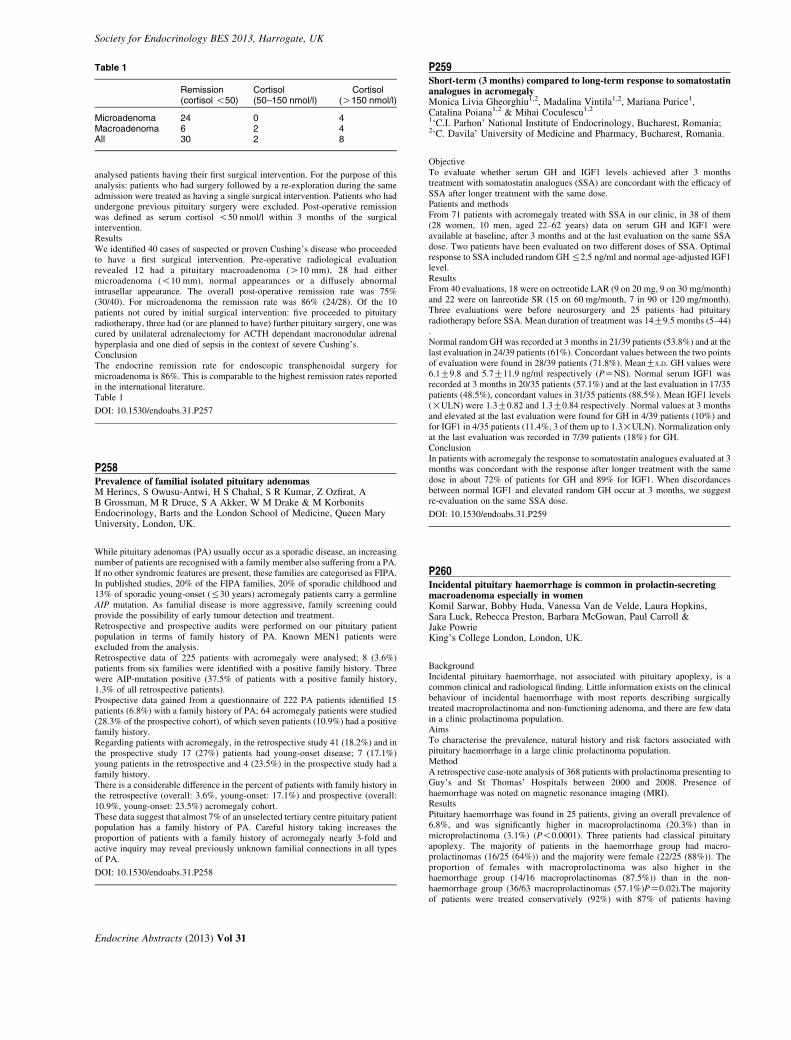
analysed patients having their first surgical intervention. For the purpose of thisanalysis: patients who had surgery followed by a re-exploration during the sameadmission were treated as having a single surgical intervention. Patients who hadundergone previous pituitary surgery were excluded. Post-operative remissionwas defined as serum cortisol !50 nmol/l within 3 months of the surgicalintervention.ResultsWe identified 40 cases of suspected or proven Cushing’s disease who proceededto have a first surgical intervention. Pre-operative radiological evaluationrevealed 12 had a pituitary macroadenoma (O10 mm), 28 had eithermicroadenoma (!10 mm), normal appearances or a diffusely abnormalintrasellar appearance. The overall post-operative remission rate was 75%(30/40). For microadenoma the remission rate was 86% (24/28). Of the 10patients not cured by initial surgical intervention: five proceeded to pituitaryradiotherapy, three had (or are planned to have) further pituitary surgery, one wascured by unilateral adrenalectomy for ACTH dependant macronodular adrenalhyperplasia and one died of sepsis in the context of severe Cushing’s.ConclusionThe endocrine remission rate for endoscopic transphenoidal surgery formicroadenoma is 86%. This is comparable to the highest remission rates reportedin the international literature.Table 1
DOI: 10.1530/endoabs.31.P257
P258Prevalence of familial isolated pituitary adenomasM Herincs, S Owusu-Antwi, H S Chahal, S R Kumar, Z Ozfirat, AB Grossman, M R Druce, S A Akker, W M Drake & M KorbonitsEndocrinology, Barts and the London School of Medicine, Queen MaryUniversity, London, UK.
While pituitary adenomas (PA) usually occur as a sporadic disease, an increasingnumber of patients are recognised with a family member also suffering from a PA.If no other syndromic features are present, these families are categorised as FIPA.In published studies, 20% of the FIPA families, 20% of sporadic childhood and13% of sporadic young-onset (%30 years) acromegaly patients carry a germlineAIP mutation. As familial disease is more aggressive, family screening couldprovide the possibility of early tumour detection and treatment.Retrospective and prospective audits were performed on our pituitary patientpopulation in terms of family history of PA. Known MEN1 patients wereexcluded from the analysis.Retrospective data of 225 patients with acromegaly were analysed; 8 (3.6%)patients from six families were identified with a positive family history. Threewere AIP-mutation positive (37.5% of patients with a positive family history,1.3% of all retrospective patients).Prospective data gained from a questionnaire of 222 PA patients identified 15patients (6.8%) with a family history of PA; 64 acromegaly patients were studied(28.3% of the prospective cohort), of which seven patients (10.9%) had a positivefamily history.Regarding patients with acromegaly, in the retrospective study 41 (18.2%) and inthe prospective study 17 (27%) patients had young-onset disease; 7 (17.1%)young patients in the retrospective and 4 (23.5%) in the prospective study had afamily history.There is a considerable difference in the percent of patients with family history inthe retrospective (overall: 3.6%, young-onset: 17.1%) and prospective (overall:10.9%, young-onset: 23.5%) acromegaly cohort.These data suggest that almost 7% of an unselected tertiary centre pituitary patientpopulation has a family history of PA. Careful history taking increases theproportion of patients with a family history of acromegaly nearly 3-fold andactive inquiry may reveal previously unknown familial connections in all typesof PA.
DOI: 10.1530/endoabs.31.P258
P259Short-term (3 months) compared to long-term response to somatostatinanalogues in acromegalyMonica Livia Gheorghiu1,2, Madalina Vintila1,2, Mariana Purice1,Catalina Poiana1,2 & Mihai Coculescu1,21‘C.I. Parhon’ National Institute of Endocrinology, Bucharest, Romania;2‘C. Davila’ University of Medicine and Pharmacy, Bucharest, Romania.
ObjectiveTo evaluate whether serum GH and IGF1 levels achieved after 3 monthstreatment with somatostatin analogues (SSA) are concordant with the efficacy ofSSA after longer treatment with the same dose.Patients and methodsFrom 71 patients with acromegaly treated with SSA in our clinic, in 38 of them(28 women, 10 men, aged 22–62 years) data on serum GH and IGF1 wereavailable at baseline, after 3 months and at the last evaluation on the same SSAdose. Two patients have been evaluated on two different doses of SSA. Optimalresponse to SSA included random GH%2.5 ng/ml and normal age-adjusted IGF1level.ResultsFrom 40 evaluations, 18 were on octreotide LAR (9 on 20 mg, 9 on 30 mg/month)and 22 were on lanreotide SR (15 on 60 mg/month, 7 in 90 or 120 mg/month).Three evaluations were before neurosurgery and 25 patients had pituitaryradiotherapy before SSA. Mean duration of treatment was 14G9.5 months (5–44).Normal randomGHwas recorded at 3 months in 21/39 patients (53.8%) and at thelast evaluation in 24/39 patients (61%). Concordant values between the two pointsof evaluation were found in 28/39 patients (71.8%). MeanGS.D. GH values were6.1G9.8 and 5.7G11.9 ng/ml respectively (PZNS). Normal serum IGF1 wasrecorded at 3 months in 20/35 patients (57.1%) and at the last evaluation in 17/35patients (48.5%), concordant values in 31/35 patients (88.5%). Mean IGF1 levels(!ULN) were 1.3G0.82 and 1.3G0.84 respectively. Normal values at 3 monthsand elevated at the last evaluation were found for GH in 4/39 patients (10%) andfor IGF1 in 4/35 patients (11.4%, 3 of them up to 1.3!ULN). Normalization onlyat the last evaluation was recorded in 7/39 patients (18%) for GH.ConclusionIn patients with acromegaly the response to somatostatin analogues evaluated at 3months was concordant with the response after longer treatment with the samedose in about 72% of patients for GH and 89% for IGF1. When discordancesbetween normal IGF1 and elevated random GH occur at 3 months, we suggestre-evaluation on the same SSA dose.
DOI: 10.1530/endoabs.31.P259
P260Incidental pituitary haemorrhage is common in prolactin-secretingmacroadenoma especially in womenKomil Sarwar, Bobby Huda, Vanessa Van de Velde, Laura Hopkins,Sara Luck, Rebecca Preston, Barbara McGowan, Paul Carroll &Jake PowrieKing’s College London, London, UK.
BackgroundIncidental pituitary haemorrhage, not associated with pituitary apoplexy, is acommon clinical and radiological finding. Little information exists on the clinicalbehaviour of incidental haemorrhage with most reports describing surgicallytreated macroprolactinoma and non-functioning adenoma, and there are few datain a clinic prolactinoma population.AimsTo characterise the prevalence, natural history and risk factors associated withpituitary haemorrhage in a large clinic prolactinoma population.MethodA retrospective case-note analysis of 368 patients with prolactinoma presenting toGuy’s and St Thomas’ Hospitals between 2000 and 2008. Presence ofhaemorrhage was noted on magnetic resonance imaging (MRI).ResultsPituitary haemorrhage was found in 25 patients, giving an overall prevalence of6.8%, and was significantly higher in macroprolactinoma (20.3%) than inmicroprolactinoma (3.1%) (P!0.0001). Three patients had classical pituitaryapoplexy. The majority of patients in the haemorrhage group had macro-prolactinomas (16/25 (64%)) and the majority were female (22/25 (88%)). Theproportion of females with macroprolactinoma was also higher in thehaemorrhage group (14/16 macroprolactinomas (87.5%)) than in the non-haemorrhage group (36/63 macroprolactinomas (57.1%)PZ0.02).The majorityof patients were treated conservatively (92%) with 87% of patients having
Table 1
Remission(cortisol !50)
Cortisol(50–150 nmol/l)
Cortisol(O150 nmol/l)
Microadenoma 24 0 4Macroadenoma 6 2 4All 30 2 8
Society for Endocrinology BES 2013, Harrogate, UK
Endocrine Abstracts (2013) Vol 31
complete resolution of their haemorrhage within 26.6G5.2 (meanGS.E.M.)months. Anticoagulant therapy, diabetes, hypertension and different types ofdopamine agonist therapy were not associated with pituitary haemorrhage. Afteradjustment for confounders, the presence of macroprolactinoma (odds ratio 9.0095% CI 3.79–23.88 P!0.001) and being female (odds ratio 8.03 (95% CI 1.22–52.95) were independently associated with haemorrhage.ConclusionThese data suggest that haemorrhage is common in macroprolactinoma where onein five develop haemorrhage, but is also present in microprolactinoma. The vastmajority resolved spontaneously with medical treatment. We also present noveldata showing a strong female preponderance, suggesting that women, particularlywith macroprolactinoma, were more likely to develop haemorrhage.
DOI: 10.1530/endoabs.31.P260
P261Ophtalmological, endocrine, and neurological complications in giantmale prolactinomas.Farida Chentli, Katia Daffeur, Lina Akkache, Meriem Haddad &Nadia kalafateBab El Oued Hospital, Algiers, Algeria.
IntroductionProlactinomas are more invasive in males. Giant ones (height R4 cm) arerelatively rare in literature.Our aim is to analyze their frequency, their radiological aspects, and theirneurological, endocrine and ophthalmological complications.MethodsAll of them had clinical exam, hormonal, ophthalmological, and radiologicalassessment based on cerebral MRI. Mixed adenomas were excluded. Positivediagnosis was based on clinical presentation, high prolactin concentration,positive response to dopamine agonistsGimmunohistochemy study.ResultsAmong 154 male prolactinomas seen in our department between 1984–2012 wehave observed 44 giant tumors (28.5%).Mean ageZ36 years, 38.3%were under 30.Mean tumor heightZ53.95 (40–97) mm andmean volumeZ66.2 mm3 (15.5–184).Mean prolactin Z15 715 ng/ml (n!15). Solid and kystic aspect, with or withoutcalcifications, mimicking craniopharyngiomas was observed in 28.5%. Cavernoussinuses were invaded in all except two. Other invasions were: posteriorZ69.4%,anterior Z58.3%, and frontalZ5.5%. For endocrine complications we observedgonadic deficit in 98.4%. Thyreotro and corticotrop insufficiencies were seen inrespectively 34%, and32%. 47%hadmultiple deficits. Posterior pituitary deficitwasobserved in 2.2%. Ophthalmological complications were: Optic atrophy in 46%,ptosisZ8%, diplopiaGstrabismus Z5.4%. For neurological abnormalities weobserved memory loss and/or unconsciousnessZ24%, epilepsyZ15.5%, compres-sive hydrocephalyZ8% and frontal syndromeZ5%.Conclusion
In this study concerning giant male prolactinomas, optic atrophy is the mostcommon abnormality. Severe and life threatening neurological troubles are veryfrequent too as they were observed in nearly 40%. But, multi pituitary deficits andcompressive hydrocephaly are relatively rare, which argues for a low progression.
DOI: 10.1530/endoabs.31.P261
P262Endocrine and radiological abnormalities in empty sella syndromeSophie Westland, Helen Mason, Gul Bano & Philip RichSt George’s University, London, UK.
BackgroundPrimary or idiopathic empty sella syndrome (ESS) is the herniation of themeninges through an incompetent diaphragma sellae into the sella turcica whichpushes the pituitary gland aside so giving the appearance of an empty sella.Secondary ESS is caused by damage to pituitary tissue which results in an emptysella turcica. There is significant lack of agreement in the literature regarding thenumber of patients with empty sella syndrome (ESS) who suffer from pituitarydysfunction; some papers report that pituitary function is usually preserved whileothers state that it is not.AimsThe aim of this study was to determine the proportion of patients with ESS whohad endocrine abnormalities on presentation and to define radiological features todifferentiate between primary and secondary ESS.MethodsIn this retrospective study, records of all patients diagnosed with an ‘empty sellaturcica’ at since 1990 were reviewed. 26 patients satisfied these criteria and theirMRI scans were re-evaluated blind by a neuroradiologist to confirm a diagnosis ofprimary or secondary ESS. Results of baseline endocrine testing and MRI scanswere recorded. Information on demographics, presenting complaint and bloodpressure were also collected.Results65% (nZ17) of patients diagnosed with ESS showed evidence of pituitarydysfunction. The most common endocrine abnormality was GH deficiency whichaffected 54% (nZ14) followed by secondary hypothyroidism in 9 patients withlow levels of TSH and T4. Headache (39%) and fatigue (27%) were the mostcommon presenting complaints.ConclusionESS is a heterogeneous condition with varied, nonspecific symptoms and highrate of endocrine dysfunction. Endocrine function should be assessed in allpatients with this diagnosis.
DOI: 10.1530/endoabs.31.P262
Society for Endocrinology BES 2013, Harrogate, UK
Endocrine Abstracts (2013) Vol 31
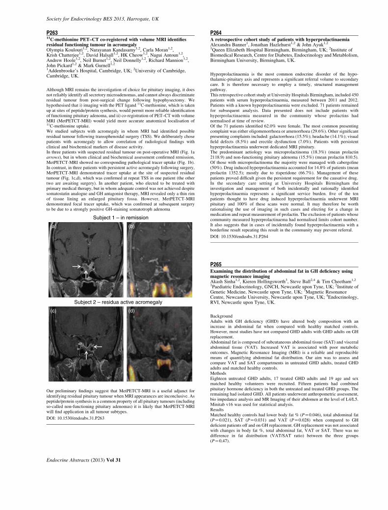
P26311C-methionine PET–CT co-registered with volume MRI identifiesresidual functioning tumour in acromegalyOlympia Koulouri1,2, Narayanan Kandasamy1,2, Carla Moran1,2,Krish Chatterjee1,2, David Halsall1,2, HK Cheow1,2, Nagui Antoun1,2,Andrew Hoole1,2, Neil Burnet1,2, Neil Donnelly1,2, Richard Mannion1,2,John Pickard1,2 & Mark Gurnell1,2
1Addenbrooke’s Hospital, Cambridge, UK; 2University of Cambridge,Cambridge, UK.
Although MRI remains the investigation of choice for pituitary imaging, it doesnot reliably identify all secretory microadenomas, and cannot always discriminateresidual tumour from post-surgical change following hypophysectomy. Wehypothesised that i) imaging with the PET ligand 11C-methionine, which is takenup at sites of peptide/protein synthesis, would permit more reliable identificationof functioning pituitary adenoma, and ii) co-registration of PET–CT with volumeMRI (MetPETCT-MRI) would yield more accurate anatomical localisation of11C-methionine uptake.We studied subjects with acromegaly in whom MRI had identified possibleresidual tumour following transsphenoidal surgery (TSS). We deliberately chosepatients with acromegaly to allow correlation of radiological findings withclinical and biochemical markers of disease activity.In three patients with suspected residual tumour on post-operative MRI (Fig. 1aarrows), but in whom clinical and biochemical assessment confirmed remission,MetPETCT-MRI showed no corresponding pathological tracer uptake (Fig. 1b).In contrast, in three patients with persistent active acromegaly following surgery,MetPETCT-MRI demonstrated tracer uptake at the site of suspected residualtumour (Fig. 1c,d), which was confirmed at repeat TSS in one patient (the othertwo are awaiting surgery). In another patient, who elected to be treated withprimary medical therapy, but in whom adequate control was not achieved despitesomatostatin analogue and GH antagonist therapy, MRI revealed only a thin rimof tissue lining an enlarged pituitary fossa. However, MetPETCT-MRIdemonstrated focal tracer uptake, which was confirmed at subsequent surgeryto be due to a strongly positive GH-staining somatotroph adenoma
Our preliminary findings suggest that MetPETCT-MRI is a useful adjunct foridentifying residual pituitary tumour when MRI appearances are inconclusive. Aspeptide/protein synthesis is a common property of all pituitary tumours (includingso-called non-functioning pituitary adenomas) it is likely that MetPETCT-MRIwill find application in all tumour subtypes.
DOI: 10.1530/endoabs.31.P263
P264A retrospective cohort study of patients with hyperprolactinaemiaAlexandra Banner1, Jonathan Hazlehurst1,2 & John Ayuk1,2
1Queen Elizabeth Hospital Birmingham, Birmingham, UK; 2Institute ofBiomedical Research, Centre for Diabetes, Endocrinology and Metablolism,Birmingham University, Birmingham, UK.
Hyperprolactinaemia is the most common endocrine disorder of the hypo-thalamic-pituitary axis and represents a significant referral volume to secondarycare. It is therefore necessary to employ a timely, structured managementpathway.This retrospective cohort study at University Hospitals Birmingham, included 450patients with serum hyperprolactinaemia, measured between 2011 and 2012.Patients with a known hyperprolactinaemia were excluded. 71 patients remainedfor subsequent analysis. Data presented does not include patients withhyperprolactinaemia measured in the community whose prolactins hadnormalised at time of review.Of the 71 patients identified 62.0% were female. The most common presentingcomplaint was either oligomenorrhoea or amenorrhoea (29.6%). Other significantpresenting complaints included: galactorrhoea (15.5%); headache (14.1%); visualfield defects (8.5%) and erectile dysfunction (7.0%). Patients with persistenthyperprolactinaemia underwent dedicated MRI pituitary.The predominant aetiology was microprolactinoma (18.3%) (mean prolactin2118.9) and non-functioning pituitary adenoma (15.5%) (mean prolactin 810.5).Of those with microprolactinoma the majority were managed with cabergoline(50%). Drug induced hyperprolactinaemia accounted for 14.8% of patients (meanprolactin 1352.5); mostly due to risperidone (66.7%). Management of thesepatients proved difficult given the persistent requirement for the causative drug.In the secondary care setting at University Hospitals Birmingham theinvestigation and management of both incidentally and rationally identifiedhyperprolactinaemia represents a significant service burden. five of the tenpatients thought to have drug induced hyperprolactinaemia underwent MRIpituitary and 100% of these scans were normal. It may therefore be worthrationalising the use of imaging in such cases and electing for a change inmedication and repeat measurement of prolactin. The exclusion of patients whosecommunity measured hyperprolactinaema had normalised limits cohort number.It also suggests that in cases of incidentally found hyperprolactinaemia with aborderline result repeating this result in the community may prevent referral.
DOI: 10.1530/endoabs.31.P264
P265Examining the distribution of abdominal fat in GH deficiency usingmagnetic resonance imagingAkash Sinha1,2, Kieren Hollingsworth3, Steve Ball2,4 & Tim Cheetham1,2
1Paediatric Endocrinology, GNCH, Newcastle upon Tyne, UK; 2Institute ofGenetic Medicine, Newcastle upon Tyne, UK; 3Magnetic ResonanceCentre, Newcastle University, Newcastle upon Tyne, UK; 4Endocrinology,RVI, Newcastle upon Tyne, UK.
BackgroundAdults with GH deficiency (GHD) have altered body composition with anincrease in abdominal fat when compared with healthy matched controls.However, most studies have not compared GHD adults with GHD adults on GHreplacement.Abdominal fat is composed of subcutaneous abdominal tissue (SAT) and visceralabdominal tissue (VAT). Increased VAT is associated with poor metabolicoutcomes. Magnetic Resonance Imaging (MRI) is a reliable and reproduciblemeans of quantifying abdominal fat distribution. Our aim was to assess andcompare VAT and SAT compartments in untreated GHD adults, treated GHDadults and matched healthy controls.MethodsEighteen untreated GHD adults, 17 treated GHD adults and 19 age and sexmatched healthy volunteers were recruited. Fifteen patients had combinedpituitary hormone deficiency in both the untreated and treated GHD groups. Theremaining had isolated GHD. All patients underwent anthropometric assessment,bio impedance analysis and MR Imaging of their abdomen at the level of L4/L5.Minitab v16 was used for statistical analysis.ResultsMatched healthy controls had lower body fat % (PZ0.046), total abdominal fat(PZ0.021), SAT (PZ0.031) and VAT (PZ0.028) when compared to GHdeficient patients off and on GH replacement. GH replacement was not associatedwith changes in body fat %, total abdominal fat, VAT or SAT. There was nodifference in fat distribution (VAT/SAT ratio) between the three groups(PZ0.47).
Subject 1 – in remission
Subject 2 – residua active acromegaly
(a) (b)
(c) (d)
Society for Endocrinology BES 2013, Harrogate, UK
Endocrine Abstracts (2013) Vol 31
ConclusionsThere were no major alterations in body fat distribution in untreated GHD adultswhen compared with treated GHD adults. However, both treated and untreatedGHD adults have increased body fat % and abdominal fat when compared tohealthy controls. This is likely to reflect the causes and consequences ofhypopituitarism rather than the effects of GH deficiency in isolation. We concludethat both treated and untreated GHD patients have increased fat mass but thedistribution of this fat is no different to control subjects.Declaration of fundingThis work was supported by funds from Pfizer inc and Merc Serono, Inc.
DOI: 10.1530/endoabs.31.P265
P266Long-term results after treatment of craniopharyngioma: experiencewith 46 adult patientsCristina Capatina1,2, Maria Preda1, Anda Dumitrascu2, Dan Hortopan2,Andra Caragheorgheopol2, Daniela Alexandrescu2, Vasile Ciubotaru3,Mihail Coculescu1,2 & Catalina Poiana1,2
1‘Carol Davila’ University of Medicine and Pharmacy, Bucharest, Romania;2‘C.I.Parhon’ National Institute of Endocrinology, Bucharest, Romania;3‘Bagdasar-Arseni’ Emergency Hospital, Bucharest, Romania.
Background:Craniopharyngioma is a rare, mostly benign tumor of the central nervous system,generally associated with important morbidity.Aim:To study the clinical characteristics and treatment outcome in adult patients.Methods:Adult patients diagnosed with craniopharyngioma between 1980 and 2012,followed-up in the Pituitary and Neuroendocrine Department of the ‘C.I.Parhon’National Institute of Endocrinology in Bucharest were retrospectively evaluated.We studied the presenting symptoms, complications at diagnosis, type oftreatment, surgical complications, rate of tumor resection, endocrinological andvisual outcome.Results:A total of 46 patients (18 females, 28 males aged between 18 and 72 years,median 35.4 years) with a mean follow-up of 7.33 years (1–41 years) wereincluded. The presenting symptoms were mostly headache (86.95%), visualimpairment (78.26%), symptoms of hypopituitarism (30.42%), diabetes insipidus(DI) (8.69%), hydrocephalus (15.21%); the median time to diagnosis was 12months. All tumors had sellar and suprasellar component, the mean craniocaudaldiameter was 2.58 cm (range 1 – 5.6 cm). All patients were operated (69.56% –transfrontal approach, 26.08% trassphenoidal, 4.34% – frontotemporal); in mostcases repeated surgery was necessary due to recurrence or remnant growth. sixcases received adjuvant radiotherapy. In only 13 cases (28.26%) gross-totalremoval (GTR) was achieved (3 subsequently recurred), in 71.73% of cases atumor remnant (diameter between 0.4 and 5 cm) was present. 32.6% developedpermanent DI. Visual field improved in 15.21% (normalized in four cases),worsened in 36.95% and remained stable in 45.65%. Two hypopituitary casesnormalised, in all others (89.13%) hypopituitarism remained or worsened withtreatment. Only 56.52% of cases could return to their usual activity. Weight gainwas significant: 38% of normal or overweight cases became obese.Conclusions:Craniopharyngiomas are often associated with an ominous prognosis andsignificant morbidity. Surgery remains the treatment of choice but total resectioncan be achieved in a minority of patients in our center.
DOI: 10.1530/endoabs.31.P266
P267Syndrome of inappropriate antidiuretic hormone secretion andtreatment with tolvaptan: a case seriesEdward Jude & Joanne VereTameside Hospital NHS Foundation Trust, Ashton Under Lyne, UK.
BackgroundHyponatraemia is the most common electrolyte abnormality, encountered in up to30% of inpatients. Plasma sodium levels !125 mmol/l are classed as severe andcan be associated with drowsiness, confusion, ataxia and personality change. Inmild hyponatraemia, the patient is usually asymptomatic, although studies haveshown chronic low sodium of any level may be associated with increasedmortality and longer hospital stays. Vasopressin receptor antagonist (tolvaptan)
are indicated to treat hyponatraemia in patients with SIADH not responding tofluid restriction. In this case series we present our experience with the use ofTolvaptan. Methods:A retrospective study of all patients treated for SIADH with Tolvaptan. Patientdemographics and biochemistry were obtained from the case records. Results:Nine patients were identified who were treated with tolvaptan; and included fivemales, mean age 74.4G8.5 years. Cause of SIADH were: pituitary tumor (1),Lung cancer (3), Drug induced (1), stroke (2), idiopathic (2). Mean admissionserum sodium (S Na) was 120G9 mmol/l. After fluid restriction mean S Na was122.9G4.2 mmol/l. Following commencement of tolvaptan (15 mg daily) serumsodium at 24, 48–72 h and 1, 2 and 4 weeks were: 128.8G4.3, 133.1G2.8,133.2G3.8, 133.1G5.5, 133.2G4.7 mmol/l respectively. Mean (range) durationof tolvaptan therapy was 81 (5–180) days. Five patients had S Na O130 in 24 h,and all patients had S Na O130 within 72 h. None of the patients had side effectsto tolvaptan. One patient had a greater than recommended increase in serum Na(O12 mmol/l) in 24 h. Conclusion: Patients treated with tolvaptan had rapidimprovement in serum sodium levels. Use of tolvaptan was without any adverseevents to the treatment.
DOI: 10.1530/endoabs.31.P267
P268Hyponatraemia assessment and outcomes in acute medically ill patientsEdward Jude, Anna Hughes, Omer Taha & Tony TetlowTameside Hospital NHS Foundation Trust, Ashton Under Lyne, UK.
BackgroundHyponatraemia is the most common electrolyte abnormality, encountered in up to30% of inpatients. Inappropriate management can have serious implications forpatients; including demyelinating disease, coma, and death.MethodsPatients (nZ122) admitted to the medical admission unit of a district generalhospital with a serum sodium (Na) !130 were selected for the study. All detailsincluding patient demographics, blood biochemistry, date of admission and dateof death were taken from the case notes and hospital computerised system. Detailson assessment of hyponatraemia including thyroid, adrenal and renal functionwere also recorded.ResultsMean age was 70.4C18.1 years; 48 males. Mean serum Na on admission was125.8C4.1 mmol/l. Of the 122 patients, 38 died (31.1%) in hospital. Patients whodied were older (66.7G18.9 vs 72.3G15.3; PZ0.054) Admission serum Na andplasma glucose in survivors vs died was 127.2G3.9 vs 124.2G4.7 mmol/l(P!0.001); and 7.0G3.0 vs 5.4G2.2 mmol/l (PZ0.02) respectively. Patientswith admission serum Na !125 in the survivors vs died was 32.1 vs 66.7%.Patients investigated for hyponatraemia were as follows: serum cortisol (nZ6),plasma osmolality (nZ9), urine osmolality (nZ9), short synacthen test (nZ0),urine sodium (nZ3), thyroid function tests (nZ19). Conclusions: Patientsadmitted with acute medical conditions with severe hyponatraemia have a highmortality and those with lower serum sodium have greater risk of death. Patientswere also inadequately worked up for assessment of cause of the hyponatraemiaand further education of medical specialists is urgently required to improvemanagement and outcome. Also lower admission plasma glucose was associatedwith higher mortality.
DOI: 10.1530/endoabs.31.P268
P269Inside acromegaly: a pilot study for recruiting focus groups using socialmediaOluwasomidotun Idowu & Andy LevyUniversity of Bristol, Bristol, UK.
BackgroundThe exponential growth of social media over the last decade has provided new,diverse and accessible ways in which people can share and receive information.Its rapid rise has attracted researchers and professionals of various disciplines,aiming to capitalise on this relatively new phenomenon. In this qualitative studywe utilised social media to conduct research on psychosocial aspect ofacromegaly. Facebook currently has 845 million users. Acromegaly, with aprevalence of approximately 60/million, was used as an exemplar of a relatively
Society for Endocrinology BES 2013, Harrogate, UK
Endocrine Abstracts (2013) Vol 31

rare disease associated with adverse physical and psychological effects.MethodA Facebook account was created and 10 international acromegaly Facebookgroups contacted. In-depth semi-structured interviews were conducted usingFacebook chat, messenger or Skype, and a focus group established on Facebook.Four groups responded and in addition to nine individuals who participated in afocus group, eigth agreed to an in-depth semi-structured interview of which fivewere completed.ResultsAnalysis of the data revealed various emerging themes each classed under one ofthree super-ordinate themes: i) psychological impact ii) social impact (3)implications for practise. Changes in physical appearance and low moodassociated with the acquisition of disfigurement later in life were the predominantfactors responsible for depression, social withdrawal and adverse effects onoccupation and relationships.ConclusionUsing social media to recruit focus groups is a very rapid and economical way togain potentially important insights into the social impact of rare diseases such asacromegaly.
DOI: 10.1530/endoabs.31.P269
P270Pituitary apoplexy: a case seriesIoannis Dimitropoulos1, Louis Pobereskin2 & Daniel Flanagan1
1Derriford Hospital Endocrinology Department, Plymouth, Devon, UK;2Derriford Hospital Neurosurgery Department, Plymouth, Devon, UK.
Pituitary apoplexy whether due to haemorrhage or infarction remains a rareendocrine diagnosis. Recent UK guidelines have emphasised the lack ofpublished evidence in the management of this condition. We present ourexperience of 12 current cases (nine males, three females).Eleven cases were managed conservatively (91.6%), one patient required urgentpituitary surgery. None of the above cases required pituitary radiotherapy.Presenting symptoms were headache and meningism (75%) with ocular palsy andvisual field defects in three (25%) of patients. Interestingly, 25% of patientspresented with non-specific symptoms of fatigue and no headache. Clinicalsuspicion of Apoplexy was high in 58% of cases.In terms of initial endocrine deficit, nine (75%) patients required steroidreplacement ab initio (2 patients were successfully weaned-off, but two more hadto be started on steroid replacement), consequently 75% continue on steroidreplacement. 25% of patients required Levothyroxine from diagnosis with 58%currently on thyroid hormone replacement. One patient was hypogonadal atdiagnosis and six more (58%) are currently on Testosterone. One patientdeveloped partial Diabetes Insipidus and is on Desmopressin and three patientsare on growth hormone replacement. Three patients so far are currently on nohormone replacement therapy. Two of these patients are on Dopamine agonisttherapy for their macroprolactinomas. All other tumours are currently believed tobe non-functional.In all cases there was no evidence of tumour re-growth. Tumour shrinkage was onaverage 34.8% (tumour width) and 25.1% (tumour height) over an average of 3.4years.In conclusion, although the majority of cases have presented with the classicalpicture of sudden onset of headache and meningism, this was not universal.Endocrinologists do need to be aware that apoplexy can present with non-specific,non-classical symptoms.A conservative management approach in certain clinical circumstances seems toprovide satisfactory outcomes.
DOI: 10.1530/endoabs.31.P270
P271An unusual cause of testosterone deficiencyCecil Eboh1, Paul Price2 & Louise Moorhouse2
1Royal United Hospital, Bath, UK; 2Great Western Hospital, Swindon, UK.
36-year-old gentleman referred by his GP with poor libido and erectiledysfunction associated with hypogonadotrophic hypogonadism. 0900 h testoster-one 0.3 nmol/l (10–35), LH !0.2 IU/l, FSH 0.1 IU/l, prolactin 71 mIU/l (50–500).Symptoms started at time of break up of his marriage in 2011. No other symptomsof hypogonadism or of pituitary disease. Previously fit and well. Teetotal. On nomedication. Patient an avid fitness fanatic. He had never knowingly used anabolic
steroids. However, he had taken a ‘nutritional supplement’ called T-Bullets(purchased from a sports nutrition shop to improve his gym performance) for20 days until just before GP’s blood test. Examination revealed a well virilisedgentleman with no signs of hypogonadism.Further investigations0900 h cortisol 457 nmol/l (138–690); IGF1 331 mg/l (75–344); FT4 11 pmol/l(5.6–21); Normal U&E, FBC and LFT. MRI pituitary and hypothalamus withinnormal limits6 months after stopping T-Bullet, testosterone level gradually returning to normal.04/10/12 – 6.9 nmol/l, 29/10/12 – 10.2 nmol/lDiscussionT-Bullets are marketed as a ‘nutritional supplement’ by the makers. They areeasily available online and can be purchased from some sports nutrition shops.The active ingredient is: ‘2a, 17a-dimethy-5a-androst-3-one-17b-ol 13-ethyl-3-methoxy-gona-2,5,(10)-dien-17-one’ which Martindale: the complete drugreference lists as an anabolic steroid (related to testosterone)Conclusion:Anabolic steroids were first artificially synthesized in the 1930s. The misuse ofanabolic steroid drugs to enhance physique, body strength and athleticperformance is well-known. Use of anabolic steroids can result in hypogonado-trophic hypogonadism as a result of suppression of the hypothalamo-pituitary–gonadal axis (HPG).The unwitting consumption of an anabolic steroid should be considered in anypatient presenting with hypogonadotrophic hypogonadism who has been taking a‘nutritional supplement’.The unwitting consumption of an anabolic steroid should be considered in anypatient presenting with hypogonadotrophic hypogonadism who has been taking a‘nutritional supplement’.
DOI: 10.1530/endoabs.31.P271
P272Rare case of round blue cell pituitary tumour with probablehypothalamic involvementSviatlana Zhyzhneuskaya1, Anna Mitchell1,2, Jehangir Abbas1,Murali Ganguri1, Swethajit Biswas1,2, Petros Perros1,2, Philip Kane1 &Vijayaraman Arutchelvam1
1Department of Endocrinology, James Cook University Hospital, Mid-dlesbrough, UK; 2Department of Endocrinology, Newcastle UniversityHospitals Trust, Newcastle, UK.
IntroductionWe submit a rare presentation of round blue cell pituitary tumour complicated bycranial diabetes insipidus following transphenoidal surgery.Case history47-year-old lady with severe headache was diagnosed with a 10 mm nonfunctioning pituitary macro adenoma. Initial plan for conservative managementwas revised as she developed sixth cranial nerve palsy, bi temporal hemianopiaand rapidly enlarging pituitary tumour to 20!18!19 mm. Urgent Transphenoi-dal pituitary decompression was performed. However the headache got worse andthe cranial nerve palsy did not resolve. As the vision significantly deteriorated,she underwent a repeat surgery in 2 weeks. Pituitary tumour was found to be offibrous consistency and the histology confirmed poorly differentiated round bluecell tumour with mitotic index 50%, positive for CD99 and CD56, cytogeneticallyunclassified.Management and discussionOn 1st post-operative day, she developed polyuria and polydipsia, diagnosed withcranial DI and commenced on DDAVP. Next day she developed generalisedseizure caused by rapidly developing hyponatraemia, managed with strict fluidbalance and a fluid restriction of 1.5l daily with dynamic management DDAVPdose. Chemotherapy with Vincristine, Doxorubicine, Cyclophosphomide andhigh dose of Dexamethasone started. She developed steroid-induced diabetesmellitus, managed with Glargine. She became very thirsty despite normal serumsodium and urine osmolality raising the possibility of hypothalamic thirst centrebeing affected by the aggressive pituitary tumour. Patient had some improvementin her vision after two cycles of chemotherapy and it was planned to complete 4cycles followed by high dose radical adjuvant radiotherapy with an optic chiasmsparing regime. Overall prognosis remains poor.ConclusionWe presented this case to illustrate a rare, aggressive pituitary malignancy whichpossibly has invaded the hypothalamus causing disruption to the thirst mechanismin addition to cranial diabetes insipidus.
DOI: 10.1530/endoabs.31.P272
Society for Endocrinology BES 2013, Harrogate, UK
Endocrine Abstracts (2013) Vol 31
P273Pituitary abscess: a rare cause of pituitary mass lesionSrinivasa Kummaraganti1, Ravi Bachuwar2 & Vikram Hundia2
1Bradford Royal Infirmary, Bradford, UK; 2Airedale General Hospital,Airedale, UK.
Pituitary abscess is very rare accounting for 0.2 – 0.6% of all pituitary lesions.Only around 210 case reports have been described so far. We report a case ofpituitary abscess presented as pituitary mass lesion with hypopituitarism.A 51 year old man was admitted with headache and found to have severehyponatraemia. Past medical history included paranoid schizophrenia. Clinicalexamination was normal.Relevant investigations: sodium 110 mmol/l, 0900 h cortisol 21 nmol/l, free T4
5.1 pmol/l, TSH 0.34 mIU/l, LH 0.8 IU/l, FSH 3.4 IU/l, testosterone 0.6 nmol/l,prolactin 139 mU/l.Pituitary MR revealed a macroadenoma measuring 20 mm and abutting the opticchiasm. Glucagon stimulation test confirmed secondary hypoadrenalism. Visualfield tests showed bilateral superior upper quadrantanopia.He was started on hydrocortisone, thyroxine and testosterone replacement for thehypoptuitarism. He was referred to the neurosurgical team. He underwent transsphenoidal surgery. Creamy soft material drained. Histology revealed fluid withacute and chronic inflammatory cells consistent with abscess. He has receivedantibiotics. Post operatively he recovered well and headaches improved.Conclusions:Pituitary abscess usually occurs in pre-existing pituitary lesion. Predisposingfactors usually include focus of parasellar infection. Usual presenting featuresinclude headache, visual defects, hypopituitarism, pyrexia and meningitis. MRIwith contrast may show peripheral enhancement. Treatment is surgical drainage,antibiotics and hormone replacement.Pituitary abscess is very rare, a potential cause of pituitary mass. Clinicaldiagnosis should be suspected with symptoms and signs of pituitary mass andinfection. It’s often radilogically indistinguishable from other pituitary lesions.Correct diagnosis is difficult before the surgery.
DOI: 10.1530/endoabs.31.P273
P274Hypopituitarism presenting with features of stiff person syndromeR N Mudaliar, S Wilson, S J Howell, S Shaunak & K KaushalLancashire Teaching Hospitals NHS Foundation Trust, Preston, Lancashire,UK.
IntroductionStiff person syndrome (SPS) is a progressive neurological disorder characterizedby fluctuating stiffness and rigidity in both axial and limb muscles. Stiff legsyndrome, an SPS variant mainly affecting limb muscles, is emerging as a distinctidentity. The cause of SPS is unknown but an autoimmune pathogenesis issuspected. There are a few reported cases of hypopituitarism presenting withfeatures suggestive of SPS.CaseA 66-year-old woman presented to the neurologists with a 12-month history ofgradually worsening back pain, leg spasms resulting in muscle stiffness anddifficulty mobilising. Physical examination showed lower limb rigidity butnormal sensorimotor function and reflexes.MR brain scan showed minimal pituitary enlargement without any focalabnormality. Endocrine assessment revealed GH, TSH, gonadotropin and partialACTH deficiency. Electromyography and anti-GAD antibodies were negative,but her presentation was felt to be consistent with SPS. Despite a favourableinitial response to GABA-enhancing drugs, a lasting clinical remission was onlyachieved with hydrocortisone replacement therapy.DiscussionWe report this unusual case of hypopituitarism presenting in a patient withclinical features suggestive of SPS. Appropriate endocrine evaluation andpituitary hormone replacement may alleviate the significant morbidity associatedwith this condition.
DOI: 10.1530/endoabs.31.P274
P275Snake bite and hypopituitarism: ignorance or incompetence?Akshatha Taranath Kamath & Sampath Satish KumarNarayana Hrudayalaya, Bangalore, Karnataka, India.
Hypopituitarism as a consequence of snake bite is rare. However, there are a fewcase reports from India and adjacent South-East Asian countries. We present acase of hypopituitarism secondary to snake bite where the diagnosis wassignificantly delayed. A 42-year-old gentleman presented as out-patient with a 10years history of generalized weakness, lethargy, coarse facial features, reducedlibido, reduced socialization and depression. He had an acute illness following asnake bite 15 years ago. He was also diagnosed with hypothyroidism 11 yearsago. The snake bite was associated with altered sensorium and acute renal failurerequiring dialysis for several days. He was managed by general physicians whodiagnosed hypothyroidism and commenced levothyroxine. His symptomspersisted, for which his levothyroxine dosage was reorganized many times andmultivitamins prescribed. Review of the original TFTs were consistent withsecondary hypothyroidism, which had been overlooked by his treating physicians.Further investigations revealed fT4 8.6 (9.0–20.0) pmol/l, TT3 1.32 (0.6–1.80) ng/ml, TSH 0.00 (0.35–5.5) mIU/ml, 8am cortisol 0.00 (4.30–22.40) mg/dl,ACTH 6.10 (7.2–63.3) pg/ml, LH !0.10 (1.1–7.0) mIU/ml, FSH !0.10 (1.7–12) mIU/ml, GH !0.05 (0.00–4.00) ng/ml, IGF1 !25 (101.00–267.00) ng/ml,IGFBP3 1.01 (3.30–6.60) mg/ml, and testosterone !0.10 (3.0–10.6) ng/ml.Pituitary MRI Scan revealed an ‘Empty Sella’. Hypopituitarism was diagnosedand he was immediately commenced on Hydrocortisone 10-5-5 mgs. He wassubsequently started on Testosterone gel, later changing to testosterone depotinjections 10 weekly. His symptoms improved significantly over a few weeks.DiscussionThe type of snake bitten determines the symptoms and signs of envenomation.Viper bites are venomous and cause altered sensorium, coagulopathy, internalbleeding, hypotension, tachycardia, renal and respiratory failure. Our patient’ssymptoms were consistent with a viper bite. His hypopituitarism and secondaryhypothyroidism remained undiagnosed leading to physical and mental suffering formore than 10 years. We suspect that the patient developed pituitary apoplexy andchronic hypopituitarism resulting from the snake bite. Endocrinologists should beaware of the possibility of hypopituitarism in patients who suffer a snake bite.
DOI: 10.1530/endoabs.31.P275
P276TSH-secreting pituitary adenoma identified in pregnancy: managementof an unusual caseJessica Triay, David Wynick, Natasha Thorogood & Karin BradleyUniversity Hospitals Bristol NHS Trust, Bristol, Avon, UK.
A 35-year-old woman was referred with biochemical hyperthyroidism (T4
30 pmol/l; T3 7 pmol/l) without TSH suppression (5.4 mIU/l). She was 7 weekspregnant following natural conception, and reported no symptoms ofhyperthyroidism. Migraines had been a feature in very early pregnancy, but hermedical history was otherwise unremarkable and there was no significant familyhistory. Examination findings were entirely normal.Investigations confirmed negative thyroid heterophile antibodies. Interpretationof usual investigations for a TSH-secreting pituitary adenoma was complicated bypregnancy (elevated a-subunit O24 mg/l, prolactin 722 mIU/l and SHBG209 nmol/l; LH and FSH fully suppressed). Pituitary MRI showed asymmetricalenlargement (16!11 mm) considered to be disproportionate to the gestationalperiod. Mutational analysis of the thyroid hormone receptor gene was negative(although 15% of cases have no detectable mutations).As thyroid hormones were only mildly elevated, surveillance was commencedwith regular thyroid function tests and trimesteral visual field assessment. Therewere no fetal or maternal complications encountered and she had a normaldelivery at term. Post-partum investigations show persistent a-subunit elevation(4 mg/l), but the rest of her hormone profile including prolactin has normalised.We are currently planning future management.There are only three reports of management of TSH-secreting pituitary adenomain pregnancy, all identified before conception. Issues raised here are i) thedifficulty distinguishing thyroid resistance syndrome from pituitary driven diseasein pregnancy given the potential for false negative results and changes in hormoneprofiles; ii) the challenges identifying the appropriate time to commencetreatment given the elevated risk of miscarriage and fetal morbidity in associationwith maternal hyperthyroidism; iii) concerns for potential complications ofaccelerated hyperthyroidism (due to the bHCG surge) or visual disturbance (dueto pituitary enlargement); and iv) identifying the appropriate treatment tocommence should it be required.
DOI: 10.1530/endoabs.31.P276
Society for Endocrinology BES 2013, Harrogate, UK
Endocrine Abstracts (2013) Vol 31
P277A rising TSH in a patient with known TSHoma does not necessarilyindicate recurrenceDhruvkumar Laheru, Mary Armitage & Tristan RichardsonRoyal Bournemouth Hospital, Bournemouth, UK.
In 2003, a 60-year-old man presented to our unit non-specifically unwell. Thyroidfunction tests (TFTs) demonstrated an elevated fT4 of 50 pmol/l (reference range10–22 pmol/l)), T3 8.8 pmol/l (reference range 3.1–6.8 pmol/l) and TSH of10.3 mU/l (reference range 0.5–4.5 mU/l)). Following appropriate investigations,a TSHoma was confirmed. MRI of the Pituitary confirmed the finding of amacroadenoma and the patient underwent pituitary decompression withsubsequent stereotactic radio-surgery in 2004. Interval MRI Scans remainedstable with no suggestion of change in residual pituitary tissue.Last year, after having felt well for a number of years, he gave a 1-year history offeeling generally tired and lethargic, with weight gain, and a new diagnosis ofdepression. Thyroid function showed a rising TSH. Repeat free T4 levels werewithin the reference range. Repeat MRI did not identify any change in size of theremaining pituitary tissue.Repeat testing of his TFTs demonstrated a trending increase in TSH with a fallingT4 level (albeit still within the reference range). Thyroid peroxidase antibodieswere requested which were strongly positive (2827 Iu/ml, reference range!100 Iu/ml), suggesting likely, co-existent thyroiditis.Treatment with levothyroxine has been considered but withheld currently as thefree T4 remains in the normal range. He is under active monitoring and there hasbeen a return to his pre-existing pattern of TFTs indicating a likely diagnosis of atransient thyroiditis.This case highlights the importance of maintaining vigilance and an open mindfor co-existent common pathologies alongside rare ones in the same organ systemand that not all rising TSH values indicate relapse of TSHoma.
DOI: 10.1530/endoabs.31.P277
P278Unusual presentation of acromegaly and functioning pituitary gona-dotrophinoma (FSHoma)Shwe Zin Chit Pan1,2, Richard Bevan-Jones1 & Anitha Mathews1
1Hinchingbrooke Health Care NHS Trust, Cambridgeshire, UK; 2QueenMary, University of London, London, UK.
BackgroundGonadotroph adenomas are the most common non-functioning pituitaryadenomas in adult1, 2. However, functioning gonadotrophinomas are rare. Here,we describe an unusual presentation of concurrent acromegaly and functioningFSHoma (FSH-secreting pituitary adenoma).A 39-year-old man presented with a vague visual disturbance to the optometristand a bitemporal hemianopia was detected.Further questioning elicited increase in shoe size with no significant history ofsweating, headache and change in hand size.Physical examination was notable for features of acromegaly (prominenteyebrows, prognathism, large hands and feet) and bilateral testicular enlargement.Endocrine profiling confirmed acromegaly on OGTT- oral glucose tolerance test(non-suppressed nadir GH level of 1.2 mg/l with basal GH level of 1.5 mg/l). IGF 1level was elevated at 64 (9.5–45) nmol/l. There was significantly elevated FSHlevel of 107.2 (1–10.1) U/l with LH 1.2 (1.5–6.3) U/l and testosterone 9.3 (8–29)nmol/l. The rest of pituitary profiling was normal: Prolactin 370 (45–375) mU/l,TSH 0.65 (0.35–5.5) mU/l, FT4 12.3 (10–19.8) pmol/l, normal short synacthentest.US testes confirmed bilateral testicular enlargement: left testis measuring 46 ccand right testis measuring 50 c with no neoplastic changes.MRI pituitary demonstrated a large pituitary macroadenoma 3.5!2.8 cm, withthe expansion of pituitary fossa, compressing optic chaism and extending into leftcavernous sinus.Subsequently transsphenoidal hypophysectomy was performed. Histologyconfirmed chromophob pituitary adenoma. Immunohistochemistry showedFSH-immunoreactive adenoma with negative GH staining.Literature analysis revealed very rare synchronous presentation of acromegalyand FSH-secreting pituitary adenoma3. Concurrent GH and prolactin secretion ismore common.References1. Chanson P & Brochier S. Non-functioning pituitary adenomas. J EndocrinolInvest. 2005 28 (11 Suppl International) 93–9.2. Tamiya H, Fukuhara N, Yoshida N, Suzuki H, Takeshita A, Inoshita N, et al. Asilent follicle-stimulating hormone-producing pituitary adenoma in a teenagemale. Endocr Pathol. 2011 22 (4) 212–7.
3. Kontogeorgos G, Scheithauer BW, Horvath E, ovacs K, Lloyd RV, Smyth HS,et al. Double adenomas of the pituitary: a clinicopathological study of 11 tumors.Neurosurgery. 1992 31 (5) 840–9 (discussion 9).
DOI: 10.1530/endoabs.31.P278
P279Pituitary apoplexy and aortic dissectionRN Mudaliar, A Golash, K Kaushal & SJ HowellLancashire Teaching Hospitals NHS Foundation Trust, Preston, Lancashire,UK.
IntroductionPituitary apoplexy is a rare but life threatening clinical syndrome characterised byacute neuro-ophthalmological features caused by haemorrhage and/or infarctionof the pituitary gland. Although many precipitating factors are known, mostapoplectic episodes occur spontaneously.CaseA 59-year-old gentleman presented in 2005 with a 12-month history of bitemporalhemianopia. An MR scan revealed a pituitary macroadenoma. When he wasreviewed in the pituitary clinic repeat assessment showed improvement of thevisual field defect. His baseline pituitary hormonal profile was normal. In theabsence of any signs of optic chiasmal compression and given that he hadsignificant co-morbidity by way of CCF, a watch and wait policy was adopted. Heremained clinically and radiologically stable for the next 5 years.In 2010, he was admitted to a neighbouring hospital with acute aortic dissection.This was managed conservatively and was followed by a protracted period ofrehabilitation. During his recovery he was found to have a bitemporal hemianopiaand was therefore assessed in the pituitary clinic. His visual field defect hadalmost resolved with only a left infero-temporal defect persisting. A pituitaryMRI scan showed marked reduction in size of pituitary adenoma and drooping ofthe optic chiasm. Endocrine assessment revealed TSH, gonadotropin and partialACTH deficiency. It was felt likely that he had developed pituitary infarctionsecondary to aortic dissection. He was established on appropriate hormonalreplacement. He subsequently underwent aortic valve replacement and aorticrepair under steroid cover. He remains stable although there has been intermittentfluctuation in visual symptoms.DiscussionThe partial hypopituitarism and shrinkage of the pituitary tumour in this case islikely to be explained by pituitary infarction caused by sudden alterations incritical perfusion pressure as a result of acute aortic dissection on a background ofcardiac failure.
DOI: 10.1530/endoabs.31.P279
P280Isolated central hypoadrenalism as the sole manifestation of presumedneurosarcoidosisSviatlana Zhyzhneuskaya, Murali Ganguri, Jehangir Abbas & Sath NagDepartment of Endocrinology, James Cook University Hospital, Middles-brough, UK.
IntroductionHypothalamic–pituitary sarcoidosis is uncommon and affects !10% of patientswith neurosarcoidosis. It presents a diagnostic challenge. We present a case ofisolated central hypoadrenalism presenting as the sole manifestation ofneurosarcoidosis.Case historyA 76-year-old man with recently diagnosed primary hypothyroidism presentedwith weight loss, increasing lethargy and fatigue. Physical exam revealed inguinallymhadenopathy. Staging CT thorax showed bilateral mediastinal lymphadeno-pathy, interstitial lung parenchymal changes, and splenomegaly. Mildhypercalcaemia (calcium 2.65 mmol/l) and hyponatremia were noted.Initialdifferential diagnoses included lymphoma, malignancy, or granulomatousdisease. Diagnostic excision biopsies of left inguinal and mediastinal lymphnodes were inconclusive. An empirical trial of steroid therapy for presumedsarcoidosis was commenced with symptomatic improvement in general wellbeing. Withdrawal of steroids was attempted in view of the inclusive lymph nodebiopsies. This resulted in a marked deterioration in the patient with hypotension,listlessness and obtundation. Adrenal insufficiency was suspected. ACTHstimulation with tetracosactide (Synacthen) showed a sub-optimal cortisolincrement with peak cortisol of 224 nmol/l. Adrenal antibodies were negativeand serum ACTH level !5 mg/l suggesting secondary adrenal insufficiency This
Society for Endocrinology BES 2013, Harrogate, UK
Endocrine Abstracts (2013) Vol 31
was confirmed by a subsequent long ACTH stimulation test. CT imaging showedno gross pituitary abnormality. The patient improved symptomatically onglucocorticoid replacement therapy.DiscussionThe exact mechanism of injury in hypothalamic–pituitary sarcoidosis is unclear.Endocrine dysfunction was attributed to granulomatous infiltration of thehypothalamus and pituitary though hypothalamic insufficiency and pituitaryresponsiveness to hypothalamic releasing factors are now thought to be the maincause of hypopituitarism. Central hypoadrenalism can present with protracted andnon-specific symptoms and pose a diagnostic challenge in the absence of anygross structural pituitary abnormality. A high index of suspicion remains thecornerstone of diagnosis.
DOI: 10.1530/endoabs.31.P280
P281Etiology and outcome of hyponatremia due to pituitary insufficiency ina tertiary endocrine centerRaluca-Alexandra Trifanescu1,2, Corin Badiu1,2,Andra Caragheorgheopol1,2, Mihail Coculescu1,2 & Catalina Poiana1,2
1Carol Davila University of Medicine and Pharmacy, Bucharest, Romania;2C.I. Parhon National Institute of Endocrinology, Bucharest, Romania.
BackgroundHyponatremia is a common electrolyte abnormality, especially in elderly,hospitalized patients, with a prevalence of severe hyponatremia (!125 mmol/l)up to 6–8%. Pituitary insufficiency (TSHCACTH) may be difficult to diagnose,but it is important to differentiate it from SIADH and cerebral salt wastingsyndrome.AimTo describe the aetiology and outcome of patients with hyponatremia due topituitary insufficiency in a tertiary endocrine center.Patients and methodsThe records of 40 patients presented with/referred for hyponatremia (!130 mmol/l) in the Department of Pituitary Pathology between 2005 and 2012,were retrospectively reviewed. There were identified 30 patients (16M/14F, aged61.9G14.3 years) with hyponatremia due to pituitary insufficiency, three patientswith severe primary hypothyroidism and seven patients with primary adrenalfailure.ResultsIn 13 patients, hyponatremia was the event revealing pituitary insufficiency; meanserum sodium at diagnosis was 113.7G8.6 mmol/l (range: 97–128). Severehyponatremia (!125 mmol/l) was recorded in 26/30 patients (86.7%).Panhypopituitarism or multiple pituitary deficiencies were present in 28 out of30 patients (93.3%); two patients (6.7%) showed isolated ACTH deficiency.Median 0800 h serum cortisol at the moment of diagnosis of secondary adrenalfailure was 2.3 mg/dl (range: nd-12.34 mg/dl). The etiology of panhypopituitarismwas: pituitary tumors (nZ18), empty sella (nZ5), Sheehan’s syndrome (nZ4cases), possible autoimmune hypophysitis (nZ3). There were 14 nonfunctioningpituitary adenomas, 1 acromegaly, 2 prolactinomas and 1 ACTH secretingadenoma. On first hospital admission, 5/30 patients were comatose and twopatients had seizures. All patients recovered after saline infusion and steroidtherapy; three patients were cautiously treated with infusions of 5.85% salinediluted in 0.9% saline. There were no fatalities or osmotic demyelizationsyndrome.ConclusionsHypopituitarism with TSH and ACTH insufficiency seems to be a frequentendocrine cause of severe hyponatremia. Correct diagnosis is important, asglucocorticoids are very effective.
DOI: 10.1530/endoabs.31.P281
P282Spontaneous resolution of pituitary Cushing’sMalik HumayunRoyal Hampshire County Hospital, Winchester, UK.
A 32-year-old male was referred by his GP with a 6-month history of lethargy,erectile dysfunction, weight gain, acne and hypertension. He denied exogenoussteroid usage and had no other past medical history. On examination, he hadclassical features of Cushing’s including moon face, central adiposity, proximalmuscle weakness and purple striae.Investigations confirmed Cushing’s from a pituitary source as shown in the tablebelow. MRI pituitary showed a probable pituitary microadenoma on the left side.His case was discussed at the pituitary MDT and was offered surgery. About 2months before surgery, he developed sudden severe headache for which heattended A&E department where he was discharged after pain control. Followingthis episode his symptoms of Cushing’s improved, started to lose weight. Heunderwent surgery but histology revealed normal pituitary tissue.Three months after surgery, he had lost 10 kg in weight with a normalising bodyhabitus. Blood pressure was also back to normal. A repeat low dosedexamethasone suppression test was normal with 0900 h serum cortisol of12 nmol/l and his full pituitary profile was also normal. Repeat MRI pituitaryshowed a normal pituitary gland.ConclusionSpontaneous remission in pituitary Cushing’s disease has been documented invery few cases. The possible etiology is considered to be possible infarction orhaemorrhage into the adenoma. We suspect that he might have infarcted hispituitary shortly before his surgery when he attended the A&E with severeheadache.
DOI: 10.1530/endoabs.31.P282
P283A case of complex neurodevelopmental abnormality causing asympto-matic SIADHAnjali Gondhalekar, Anthony Grubb, Angharad Herbert, Wing Hong Tong,Ranganatha Rao & David JenkinsWorcester Royal Hospital, Worcester, UK.
CaseA 25-year-old lady was incidentally found to have hyponatremia while she wasinvestigated for painful left ankle and hand swelling. At the time of review inendocrine clinic, she was completely asymptomatic. Her menstrual period wasnormal. On examination she was found to be hypertensive with consistent bloodpressure of 188/110. Rest of the systemic examination was unremarkable.Investigations revealed biochemical findings consistent with SIADH. Her serumsodium was 121 mmol/l, potassium 4.4 mmol/l, urea 1.7 mmol/l and creatinine72 mmol/l. Her serum osmolality was 254 mosmol/kg, urine osmolality455 mmol/kg and urine sodium 44 mmol/l. Her serum cortisol was 862 nmol/land TSH was normal. SIADH was confirmed by serum AVP level of 0.68 pmol/lwhich should be undetectable at serum osmolality of 254 mosmol/kg.Her chest X-ray was unremarkable. MRI of the brain showed complexdevelopmental anomalies with absence of corpus callosum, dilatation of occipitalhorn of right lateral ventricle and anteriorly situated inter hemispheric cysts whichwas thought to be the cause of SIADH.She was treated with 1.5 l of fluid restriction to maintain her serum sodiumbetween 125 and 131 mmol/l. Her blood pressure was treated with doxazosin2 mg daily after ruling out phaeochromocytoma and primary hyperaldosteronism.ConclusionThis rare case highlights the importance performing thorough neurologicalassessment of patients presenting with asymptomatic hyponatremia.
DOI: 10.1530/endoabs.31.P283
Society for Endocrinology BES 2013, Harrogate, UK
Endocrine Abstracts (2013) Vol 31
P284Hypopituitarism with visual field loss is not always an adenomaUmar Farooq1, Umar Raja1,2 & Ansu Basu1,21Heart of England Trust, Birmingham, West Midlands, UK; 2City andSandwell Trust, Birmingham, West Midlands, UK.
IntroductionA biochemical profile of an underactive pituitary gland allied to visual field loss iscommonly due to an underlying adenoma. We report a case where such a clinicalpicture was found but imaging/biopsy revealed a different cause.Case reportA 52-year-old Indian male was referred by his GP to an endocrine outpatientdepartment with reduced visual acuity, balance problems and a biochemicalprofile of an underactive pituitary gland. Past history included hyperlipidaemia,allergic rhinitis and depression. Examination revealed a bitemporal hemianopiabut no long tract neurological signs. Blood tests showed free T4 of 6 pmol/l, TSH0.26 ml/l, testosterone nul!0.1 ng/ml, prolactin 885 m/l and IGF1 6.0 nmol/l. AnMRI head showed a large ill-defined mass of the optic chiasm. This extended intothe hypothalamus and was suspicious of an optic chiasm glioma. The patient wasstarted on thyroxine, testosterone and hydrocortisone therapy. He was referred forneurosurgical opinion and a hypothalamus biopsy revealed the presence ofgranuloma. Cerebrospinal fluid culture is currently negative for TB but showed araised protein level of 0.98 g/l. The patient was started on anti-TB medication (hehad reported contact with a family member who had TB) and steroids (for thepossible differential of sarcoid.) This patient continues to be cared for by theendocrinology, neurology, ophthalmology and respiratory teams.DiscussionVisual field loss alongside a picture of an underactive pituitary gland is not alwayscaused by an adenoma. In such cases where other causes are apparent, medicalreplacement therapy is merited alongside involvement of different specialists inthe long-term management plan.
DOI: 10.1530/endoabs.31.P284
P285Rapidly progressive pituitary carcinoma in a young femaleSumithra Giritharan, Tara Kearney & Kanna GnanalinghamSalford Royal NHS Foundation Trust, Salford, UK.
A 32-year-old female presented with a 6-week history of worsening headachesassociated with bitemporal visual field defects. An urgent MR scan demonstrateda sizeable pituitary lesion with chiasmal compression and suprasellar extension.Endocrine tests revealed hyperprolactinaemia (2550 mU/l) with hypocortisolae-mia (!37 nmol/l). Cabergoline and hydrocortisone replacement were initiated.Unfortunately, 24 h later her visual fields deteriorated further and the patientunderwent a subtotal transsphenoidal, followed by transcranial resection of thetumour. Post-operatively, the visual fields improved slightly. The histology was inkeeping with carcinoma – either of metastatic origin or primary pituitary cancer.A CT chest, abdomen and pelvis did not suggest a possible primary.Three weeks post-operatively, the patient re-presented with worsening headachesand a deterioration in visual fields. Tumour recurrence was confirmed on CT andthe patient proceeded to have urgent radiotherapy. Unfortunately, 2 days aftercompleting the course of radiotherapy (3 months post-surgery), she wasreadmitted to hospital with progressive headache, nausea and deterioration ofvision in the left eye. An MRI demonstrated widespread meningeal thickeningwith evidence of multiple durally-based metastases. Upon review by the oncologyteam it was advised that the patient have a lumbar puncture for CSF cytology anda repeat CT chest, abdomen and pelvis to aid future management decision. Sadlyhowever, this patient passed away days after this review.This case not only highlights the difficulty in diagnosis but also the therapeuticchallenge in managing what has been in this case a rapidly fatal disease.
DOI: 10.1530/endoabs.31.P285
P286Growth of a meningioma in a female patient with uncontrolledcongenital adrenal hyperplasiaTriona O’Shea1, Rachel Crowley1, Michael Farrell2, Steven Hunter2,James Gibney1 & Mark Sherlock11Tallaght Hospital, Dublin, Ireland; 2Beaumont Hospital, Dublin, Ireland.
ContextGrowth of meningiomas has been previously described in patients receivingoestrogen/progestogen therapy.MethodsCase history, laboratory findings, imaging and histology are discussed.Case historyA 45-year-old woman with a known history of 21-hydroxyase deficiency (of thenon-salt wasting variety) and long-standing non-adherence with corticosteroidtherapy presented to the Endocrine Clinic for follow-up care. She complained ofsevere headache. On examination she was of short stature, virilised and hadmarked right sided proptosis.Laboratory findingsTestosterone 19 nmol/l (0–1.5), 17-hydroxyprogesterone O180 nmol/l (!6.5).Imaging revealed a large left sphenoid wing meningioma with anteriordisplacement of the right eye.HistologyMeningioma, Ki index?, MIB-1 index? stained positive for progesteronereceptorsDiscussionGrowth of meningiomas has been described in patients receiving menopausalhormone therapy, long active reversible contraceptives1 and transsexual patients2.To our knowledge this is the first reported case of aggressive meningiomaputatively related to stimulation of the progesterone receptor in response to highlevels of 17-hydroxyprogesterone in a patient with uncontrolled congenitaladrenal hyperplasia.
DOI: 10.1530/endoabs.31.P286
P287Finally we know! ‘It comes from your pituitary’Nazia Rashid & Stephanie E BaldewegUniversity College Hospital, London, UK.
IntroductionCushing’s syndrome is broadly categorized into ACTH dependent (pituitary andectopic source) and ACTH independent (adrenal source). Localizing source ofCushing’s can sometimes be a cumbersome diagnostic process.Case historyA 25-year-old male patient presented with sudden rapid onset weight gain,muscle weakness and occasional headaches.as well as severe dyspnoea,orthopnea and PND. There was no significant past medical or family history.He was not on any regular medication and denied exogenous steroids intake. Hehad clinical features consistent with florid Cushing’s syndrome and congestivecardiac failure. Cardiac MRI suggested severe dilated Cardiomyopathy with EF23% which was treated medically. His screening investigations for Cushing’sshowed discordance with clinical picture. He had high 02400 h urine free cortisolon two occasions but suppressed !28 on low dose dexamethasone suppressiontest. Two early mornings ACTH levels were undetectable and promptedinvestigations to find an adrenal source. CT as well as MRI adrenals failed tolocalize an adenoma. Alternative sources were then explored. Pituitary MRI andsubsequent dynamic pituitary MRI were entirely normal apart from stalkdeviation to left side. No ectopic source of disease was found on Galliumoctreotide PET–CT. Rest of the Pituitary function tests were satisfactory. He hadInferior petrosal sinus sampling which showed strong lateralization to left side ofpituitary. He is currently awaiting pituitary surgery for Cushing’s disease and hasbeen started on blockade therapy with Metyrapone in the interim. Repeat cardiacMRI shows improvement in cardiac function (EF 41%).ConclusionsDiagnosing Cushing’s syndrome and identifying the source can sometimes bechallenging and require more invasive investigations. We highlight importanceof taking clinical picture into account whenever dealing with complicatedCushing’s patients and their discrepant investigations.
DOI: 10.1530/endoabs.31.P287
Reproduction
P288Inflammatory markers in polycystic ovarian syndrome and theirassociation with cardiovascular risk factorsSunil Kumar Kota1, Lalit Kumar Meher2, Sruti Jammula3 & KirtikumarD Modi1
Society for Endocrinology BES 2013, Harrogate, UK
Endocrine Abstracts (2013) Vol 31
1Medwin Hospital, Hyderabad, Andhrapradesh, India; 2MKCG MedicalCollege, Berhampur, Orissa, India; 3Roland Institute of PharmaceuticalSciences, Berhampur, Orissa, India.
ObjectivesTo determine and compare inflammatory markers including adiponectin, visfatinand IL18 in patients with polycystic ovarian syndrome (PCOS) 2. To find outwhether adiponectin and interleukin 18 (IL18) is associated with markers ofinsulin resistance, hyperandrogenism and carotid intima-media wall thickness(CIMT) as a cardiovascular risk factor.MethodsThis is a prospective controlled study involving 60 consecutive euglycemicpatients with PCOS (Rotterdam criteria) and 50 age and BMI matched controlswere included in the study. After detailed clinical evaluation includinganthropometry, besides oral glucose tolerance test, fasting venous samples wereanalysed for IL18, visfatin, adiponectin, highly sensitive C-reactive protein (hsCRP) and complete lipid profile. We estimated body composition (total body fatand visceral adipocity index, VAI by dual energy X-ray absorptiometry), CIMT(by Doppler ultrasonography), indices of insulin sensitivity (QUICKI) andresistance (homeostasis model assessment for insulin resistance, HOMA-IR) andfree androgen index (FAI). Data were analyzed using online graphpad quickcalcsoftware and P!0.05 was considered statistically significant.ResultsPCOS patients had greater FAI (1.42G0.83 vs 0.64G0.4), higher HOMA-IR(2.13G1.05 vs 1.91G1.8) and lesser QUICKI (0.156G0.025 vs 0.163G0.015)than the control groups. Patients with PCOS have significantly increased serumIL18 and visfatin levels than that of the control group (IL18: 213.48G76.84 vs170.4G41.11 pg/ml, visfatin: 73.35G11.54 vs 55.56G9.27 ng/ml, P!0.05) andhsCRP (2.56G0.64 vs 1.62G0.78 mg/l, PZ0.004). Similarly the PCOS grouphad significantly lower level of adiponectin (0.8G0.6 vs 1.04G0.49 ng/ml, P!0.001). Correlation coefficients of IL18 were as follows: with CIMT (0.355), FAI(0.328), HOMA-IR (0.345) and waist circumference (0.367), each with P!0.05.Similarly the correlation coefficients of adiponectin were with CIMT (K0.312),FAI (K0.343), HOMA-IR (K0.352) and waist circumference (K0.359), eachwith P!0.05.DiscussionThere is alteration of adipokines and other inflammatory markers in PCOS withincrease in visfatin, IL18 and hs CRP and reduction in of adiponectin levels.Increased IL18 and decreased adiponectin levels correlated with insulinresistance, obesity and hyperandrogenism.ConclusionThese altered adipokine profile is associated with increased CVD risk in PCOSpatients, leading to the suggestion that one of these markers like IL18 can serve aspotential therapeutic target in future for decreasing their CV risk.
DOI: 10.1530/endoabs.31.P288
P289Maternal corticosterone regulates amino acid allocation to fetal growthin miceOwen Vaughan, Amanda Sferruzzi-Perri & Abigail FowdenUniversity of Cambridge, Cambridge, UK.
Synthetic glucocorticoid administration during pregnancy reduces birth weightand increases disease risk in the offspring. This study investigated whetherincreasing maternal natural corticosterone levels, by endogenous stress orexogenous administration, alters fetal amino acid supply in the mouse.C57BL6/J dams (nZ82) were exposed to daily restraint and subcutaneous salineinjection (stress) or given corticosterone (cort, 82G11 mg/g per day in drinkingwater) for 5 days in mid/late pregnancy (either D11–16 or D14–19, term is D21).Plasma corticosterone was measured by RIA. On D19, placental amino acidtransport was determined as the materno-fetal clearance of non-metabolisable14C-methylaminoisobutyric acid (MeAIB). Gene expression was determined insnap-frozen placentae using qPCR. Significant effect (P!0.05) of treatment wasdetermined by one way ANOVA with Bonferroni’s post-hoc test.Compared with untreated controls (ut), stress or cort from D11–16 increasedplasma corticosterone on D16 (ut, 571G58 ng/ml; stress, 1018G109 ng/ml; cort,1142G134 ng/ml; P!0.05) but not on D19 (ut, 714G41 ng/ml; stress, 739G53 ng ml; cort, 524G53 ng/ml; PO0.05). Corticosterone remained was high onD19 in both groups of dams treated from D14–19 (stress, 949G134 ng/ml; cort,1143G176 ng/ml. PZ0.003). On D19, MeAIB clearance was affected bytreatment (PZ0.007), and was highest in D11–14 cort dams and lowest in D14–19 cort dams. When all groups were combined, MeAIB clearance inverselycorrelated with maternal corticosterone (Pearson’s rZK0.50, P!0.001). On
D19, placental Redd1/Ddit4, an inhibitor of mammalian target of rapamycin(mTOR) signalling, was upregulated when maternal corticosterone was high inD14–19 cort treated dams (142% vs ut, P!0.05) but did not differ from controlvalues in D11–16 cort dams.The results show that physiological increases in corticosterone during mousepregnancy regulate fetal amino acid supply, in part, through inhibition ofplacental mTOR signalling. This effect may defend amino acids for maternalmetabolism, at the expense of fetal growth.Declaration of fundingThis work was supported by a graduate studentship from the Centre forTrophoblast Research.
DOI: 10.1530/endoabs.31.P289
P290BMP4 induces terminal differentiation of primary trophoblast cells andincreases chorionic gonadotrophin secretionVictoria Cabrera-Shaarp1, Stephanie Richardson1, Alycia Kowalski1,Doug Antczak2, Abir Mukherjee1 & Amanda de Mestre11Royal Veterinary College, University of London, London, UK; 2BakerInstitute, Cornell Universtiy, Ithaca, New York, USA.
ObjectiveChorionic gonadotrophin (CG) is a hormone induced during terminaldifferentiation of trophoblast cells that significantly influences pregnancyoutcome. The TGFb superfamily SMAD2/3 pathway regulates placental functionbut the activity of the alternative pathway through SMAD1/5/8 in the placenta isunknown. This study investigated the role of BMP4 signalling through SMAD1/5in terminal differentiation of primary chorionic gonadotrophin-secretingtrophoblast cells.MethodsA novel equine animal model of trophoblast differentiation was used to gain purepopulations of primary CG-secreting trophoblast cells and placental tissue.Chorionic girdle was isolated from days 27 to 34 equine conceptuses. A 44 Kgene probe equine expression array and RT-PCR was used to compare Type I andType II serine/threonine kinase and accessory receptor expression. Culturedchorionic girdle trophoblast cells were supplemented with 1–100 ng/ml humanBMP4. Differentiation was determined following dual-labelling of the cells withCellTrace BODIPY TR methyl ester and Hoeschst. eCG concentration wasdetermined using ELISA. Total SMAD1/5, pSMAD1/5, total SMAD2 andpSMAD2 expression in the placenta was determined using western blotting.ResultsChorionic girdle tissue and cultured CG-secreting trophoblast cells preferentiallyexpressed receptors ALK3, BMPR-II, Dragon and Bambi that bind the ligandBMP4. Stimulation of chorionic girdle trophoblast cells with 1–100 ng/ml BMP4resulted in a dose dependent increase in total number and proportion of terminallydifferentiated binucleate cells (P!0.001) and induced eCG secretion (P!0.01)in a developmental dependent manner. Phospho-SMAD1/5 expression, but notpSMAD2 was tightly regulated during CG-secreting trophoblast differentiationin vivo, with peak expression of pSMAD1/5 noted at gestation day 31corresponding to maximal trophoblast differentiation.ConclusionOur findings support a role for TGFb signalling in regulation of differentiation ofprimary trophoblast cells via BMP4 dependent binding to BMPR-II and ALK3and activation of SMAD1/5. The observation of BMP4 signalling in primarytrophoblast provides a previously unreported mechanism of TGFb signalling inthe placenta.Declaration of fundingWellcome Trust (grant number 091581).
DOI: 10.1530/endoabs.31.P290
P291Is diethylstilboestrol an endocrine disruptor in the developing humanfetal testis? Effects of DES exposure using a xenograft approachR T Mitchell1,2, R A Anderson1, S van den Driesche1, C McKinnell1,S MacPherson1, W H B Wallace1,2, C J H Kelnar1,2 & R M Sharpe11Edinburgh University, Edinburgh, UK; 2Royal Hospital for Sick Children,Edinburgh, UK.
ContextIn rodents, in-utero exposure to the exogenous oestrogen diethylstilboestrol(DES) results in reproductive abnormalities in male offspring. It has been
Society for Endocrinology BES 2013, Harrogate, UK
Endocrine Abstracts (2013) Vol 31
proposed that similar anti-androgenic effects also occur in the human fetal testisfollowing oestrogen exposure.ObjectiveDetermine effects of DES exposure on testosterone production by normallygrowing human fetal testis xenografts.DesignHuman fetal testes (15–19 weeks gestation, nZ6) were xenografted into castratemale nude mice. Mice were treated for 35 days with vehicle or 100 mg/kg DESthree times a week; all mice were treated with hCG to mimic normal humanpregnancy. For comparison, fetal male rats were exposed in-utero to vehicle or100 mg/kg DES on e13.5, e15.5, e17.5, e19.5 and e20.5.Main outcome measuresTestosterone production from xenografts was assessed by measuring host serumtestosterone and seminal vesicle (SV) weights at termination. Insl3 productionwas determined in serum, whilst immunohistochemistry was performed for Insl3and ERa.ResultsHuman fetal testis xenografts showed similar survival and total graft weight inDES and vehicle-exposed hosts. SV weight was significantly increased in DEScompared to vehicle-exposed hosts (44.3 vs 26.6 mg, PZ0.01) and testosteronewas also increased (0.33 vs 0.45 ng/ml, PO0.05). There was no difference inInsl3 expression. In contrast, in-utero exposure of fetal rats to DES resulted insignificantly lower intra-testicular testosterone levels associated with a reductionin Insl3 expression. ERa was expressed in fetal rat testis but was not expressed inthe human fetal testis.ConclusionsExposure of human fetal testes to DES does not impair testosterone production asit does in rats. This can be explained by a lack of ERa signalling in the humanfetal testis. This highlights an important difference in the effect of proposed‘endocrine disruptors’ in humans compared with rodents which has importantimplications for determining the risk posed to human health by environmentaloestrogens.Declaration of fundingThis work was supported by a BSPED Research award and an Academy ofMedical Sciences Starter Grant for Clinical Lecturers.
DOI: 10.1530/endoabs.31.P291
P292Perinatal origins of adult Leydig cells and function: role ofdevelopmental androgensKaren Kilcoyne1, Nina Atanossova2, Luiz Renato de Franca3,Nathalia Lara3, Karel De Gendt4, Guido Verhoeven4, Chris McKinnell1,Sheila Macpherson1, Sander van den Driesche1, Lee Smith1 & RichardM Sharpe11University of Edinburgh, Edinburgh, UK; 2Institute of ExperimentalMorphology, Sofia, Bulgaria; 3Institute of Biological Sciences, MinasGerais, Brazil; 4Catholic University of Leuven, Leuven, Belgium.
Fetal events can affect adult testosterone levels but how this occurs is unknown, asadult Leydig cells (ALC) do not differentiate until puberty. Qin et al. 2008 (PLosONE) identified that chicken ovalbumin upstream promoter-transcription factor II(COUP-TFII) is essential postnatally for ALC development. We hypothesizedthat: i) COUP-TFIIC non-Leydig interstitial cells are progenitors for ALC and arepresent in the fetal testis, ii) these ‘progenitor ALC’ are regulated by fetalandrogens, and this might affect testosterone levels in adulthood.Three approaches were used: i) complete androgen receptor (AR) knockout(ARKO) mice, ii) pregnant rats treated with dibutyl phthalate (DBP; 500 mg/kgper day; e13.5–e21.5) which reduces fetal intratesticular testosterone O70%, iii)iNOSK/K mice in which intratesticular testosterone is increased. Numbers ofprogenitor ALC were quantified from fetal life to adulthood, and related to ALCnumber and function, (testosterone and LH levels, ALC-specific steroidogenicgene expression).Presumptive progenitor ALC expressing AR (COUP-TFIIC/ARC) are abundantin the fetal testis of rats, mice, marmosets and humans. In ARKOs, progenitorALC number was reducedw40% from birth to adulthood, paralleled by a similarshortfall in ALC; there was also compensated ALC failure. In adults progenitorand ALC numbers were correlated (P!0.0001). Fetal DBP exposure reducedprogenitor ALC numbers by w40% in fetal and postnatal life and inducedcompensated ALC failure and reduced steroidogenic gene expression. TheiNOSK/K mice are currently under investigation.This study suggests that COUP-TFII identifies a population of cells in the fetaltestis from which ALC develop from puberty onwards, and these cells are partlyregulated by androgens. Reduced fetal androgen exposure altered ALC function(mechanism unclear), presumably by altering fetal androgen action on the ALCprogenitors. This adds a new dimension to growing evidence that fetal androgen
exposure may determine functional competence of the adult testis and overallmale reproductive health.
DOI: 10.1530/endoabs.31.P292
P293Pulsatile GnRH signaling to ERK: relevance of pulse duration andfrequencyRebecca Perrett1, Stephen Armstrong2, Rob Fowkes3 & Craig McArdle11University of Bristol, Bristol, UK; 2University of Western Australia, Perth,Australia; 3Royal Veterinary College, London, UK.
GnRH is secreted in pulses and its effects on pituitary gonadotropes depend onpulse frequency. This is crucial for physiological control and therapeuticmanipulation of the system (in IVF and treatment of hormone-dependent cancers)but GnRH pulse frequency decoding mechanisms are unknown. The simplestform of frequency dependence is a linear relationship between integrated inputsand outputs but such ‘integrative tracking’ cannot explain the bell-shapedfrequency-response relationships seen for many GnRH effects. GnRH acts viaGq/11 coupled GPCRs to activate effectors including ERKs, which mediate manytranscriptional effects of GnRH but little is known about ERK signaling withpulsatile stimulation so we have explored this with automated fluorescencemicroscopy in HeLa cells transduced with adenovirus expressing ERK2-GFP (1).Five minute GnRH pulses caused rapid, transient and reproducible ERK2-GFPactivation (nuclear translocation) at varied pulse concentrations (0.01–100 nM)and frequencies (0.25–2/h). Using an Egr1 luciferase reporter, increasing pulsefrequency increased the transcriptional response (to ERK activation) butincreasing pulse duration had a less pronounced effect (i.e. Egr-1 luciferasewas approximately doubled by doubling frequency from 0.5 to 1 pulse/h, but asimilar effect required a 10! increase in pulse duration from 1 to 10 min).Exploring activation of endogenous ERKs revealed that ppERK1/2 levelscontinue to rise for at least 3 min after a 1 min GnRH pulse, which may explainthe unexpectedly high transcriptional response to very brief stimulation. Thus,varying pulse frequency implies that the ERK pathway is a simple integrativetracker of GnRH pulse frequency, but varying pulse duration reveals it is not.ERK activation is more sensitive to pulse frequency than it is to pulse duration (inthis short time-frame) and this may have adaptive advantages for use of the ERKpathway in frequency-encoded signaling system.Declaration of fundingThe work was funded by the Medical Research Council (Grant 93477) andWellcome Trust (Project and Equipment Grants 076557 and 078407).
DOI: 10.1530/endoabs.31.P293
P294Intrahepatic cholestasis of pregnancy levels of sulfated progesteronemetabolites downregulate hepatic LXRaVanya Nikolova, Shadi Abu-Hayyeh, Georgia Papacleovoulou,Malcolm Parker & Catherine WilliamsonImperial College London, London, UK.
IntroductionIntrahepatic cholestasis of pregnancy (ICP) is a pregnancy-specific liver disorderwhich is associated with higher incidence of gallstone disease. ICP symptoms areusually presented in the third trimester of gestation and their severity advances inparallel with the increase in serum sulphated progesterone metabolites (P4-S) inthe mother. Liver X receptor a (LXRa) actively participates in the regulation oflipid metabolism functioning as a cholesterol sensor. We aimed to investigate ifICP levels of P4-S could modulate the LXRa transcriptome and thus contribute togallstone formation by increasing biliary cholesterol sectretion.MethodsThe influence of ICP levels of P4-S on LXRa as well as its target genes wasassessed in human hepatoma cells using RT-PCR and western blotting. LXRareporter assays were employed to determine which domain of the nuclear receptormediates the impact of the sulphated progesterone metabolites.ResultsLucifease reporter assays demonstrated that the P4-S epiallopregnanolonesul-phate (PM5S), epiallo-pregnanediol 3-sodium sulphate (EPAS) and epipregna-nolonesulphate (EPS) were able to attenuate the basal as well as the agonist-induced transactivity of LXRa in a dose-dependent manner. Also, PM5S, EPASand EPS were able to specifically modulate the activity of the LXRa ligandbinding domainGthe hinge region as shown using recombinant GAL4-LXRavectors. Quantitative RT-PCT showed that PM5S, EPAS and EPS could decrease
Society for Endocrinology BES 2013, Harrogate, UK
Endocrine Abstracts (2013) Vol 31
the mRNA levels of the LXRa target ABCG1in a dose-dependent manner whilethe gene expression of of LXRa itself is reduced by PM5S only. Protein analysisalso showed that PM5S, EPAS and EPS diminish the levels of LXRa in thenucleus.ConclusionMurine studies have shown that the lack of ABCG1 causes increased biliarycholesterol secretion which is a key pathophysiological event in the developmentof gallstones. Supraphysiological levels of P4-S contribute to the formation ofgallstones in ICP by dowregulating LXRa and its target ABCG1.Declaration of fundingThis work was supported by the Welcome Trust Grant (P30874, 2010).
DOI: 10.1530/endoabs.31.P294
P295DNA methyltransferase 3a, 3b and 3L expression in fetal germ cells andits modulationThomas Chambers1, Afshan Dean1, Sander van den Driesche1,Rod Mitchell1, Sheila MacPherson1, Richard Anderson1, Mandy Drake2 &Richard Sharpe11Centre for Reproductive Health, University of Edinburgh, Edinburgh, UK;2Centre for Cardiovascular Science, University of Edinburgh, Edinburgh,UK.
Background5-Cytosine methylation of DNA is a means of encoding epigenetic information.In the testis, the generation of de novo methylation is conducted by the enzymesDNMT3a and 3b and the co-enzyme DNMT3L. Epigenetic marks made to theDNA of germ cells are important as a potential means of trans-generationalcarriage of environmental information. In fetal life, germ cell demethylation andremethylation are important physiological events and these overlap with keychanges in germ cell differentiation (loss of pluripotency), but whether this iscoincidental or not is unknown. This study characterised expression of theDNMT3s.MethodsDNMT 3a, 3b and 3L were co-localised with the germ cell markers Oct4 andVASA using immunofluorescence in fetal testes from the rat (17.5 and 21.5 dayspost conception (dpc)), marmoset (98 and 110 dpc) and human (gestation weeks14 and 19) to determine changes in expression related to age and to germ celldifferentiation status.ResultsDNMT3a and 3b are expressed in some but not all germ cells of the fetal testisacross all the species examined. DNMT3a is expressed in fewer pluripotentOct4C cells than DNMT3b. The proportion of Oct4C germ cells expressingDNMT3b increased in the rat, marmoset and human from dpc 17.5 to 21.5, 98 to110 and gestational weeks 14 to 19 respectively. Ongoing studies arecharacterising DNMT3L expression and identifying if there is a relationshipbetween DNMT3 expression and germ cell differentiation.DiscussionWe show that DNA methytransferase enzymes are present in fetal germ cellsacross multiple species, including primates, at the protein level. The potentialmanipulation of DNA methylation by environmental stimuli is a mechanism bywhich life style and pathogen exposure could impact upon the health ofsubsequent generations. The presence of DNMTs in fetal germ cells demonstratesa means by which de novo cytosine methylation can be induced.Declaration of fundingThis work was supported by the UK Medical Research Council (grant numberG33253).
DOI: 10.1530/endoabs.31.P295
P296Dissecting the prokineticin receptor dimerization interface: a role inkallmann sindrome?Silvia Sposini1,2, Aylin Hanyaloglu1,2 & Rossella Miele1,21Imperial College, London, UK; 2Universita ‘Sapienza’, Rome, Italy.
Prokineticin receptors (PKR1 and PKR2) are GPCRs that belong to neuropeptideY receptor class. They exert their biological functions binding two structurallyrelated peptides (Bv8 or PK2 and EG-VEGF orr PK1). Intensive research over thepast few years has shown that PKs/PKRs signalling modulates neuronal survivaland neurogenesis, hypothalamic hormone secretion, nociception, circadianrhythm and complex behaviours, such as feeding and drinking. It also promotes
angiogenesis in steroidogenic tissues and reproductive organs, hematopoiesis andimmune response.A growing body of evidence points to the fact that GPCRs exist as homo- orheterodimers but the functional impact for many of these dimers still remainsunclear.The study’s aim was to confirm PKR2 homodimerization, to indentify thehomodimerization interface and to assess PKR1-PKR2 heterodimerization.Techniques namely bioluminescence resonance energy transfer (BRET) wereused to study receptor-receptor interactions in live cells in real time. Theinteraction site of PKR2 homodimer was assessed by use of receptor fragmentscorresponding to TM1-5, TM1-4, TM1-7 and TM6-7. These were also employedto determine the role of PKR2 homodimerization in receptor targeting to themembrane.Two missense mutations (P290S and L173R) within transmembrane domains ofPKR2 sequence were also studied. It has been demonstrated that these PKR2mutants can cause the Kallmann syndrome phenotype, which combineshypogonadism, due to gonadotropin-releasing hormone deficiency, and anosmiaor hyposmia, due to defective olfactory bulb morphogenesis. We investigated theeffects of these mutations on cell surface targeting and dimerization of bothmutated and wild-type receptors.Given the importance of dimerization in receptor synthesis and cell surfacetargeting, the work obtained can provide a framework for interpretation of resultsconcerning the PKR2 mutants associated with Kallmann syndrome.
DOI: 10.1530/endoabs.31.P296
P297Tocopherol interacts with b-mimetic effect in reproductive andrespiratory tracts in the ratAgnes Hodi, Judit Hajagos-Toth, Anna Klukovits, Adrienn Seres &Robert GasparDepartment of Pharmacodynamics and Biopharmacy, University of Szeged,Szeged, Hungary.
BackgroundIt is known that reactive oxygen species (ROS) production is necessary for thesignal transduction mechanism of b-adrenergic receptors.AimOur aim was to investigate whether antioxidant agent (a)-tocopherol-acid-succinate may reduce the effect of b-mimetic action on b(2)-adrenergic receptorregulated tissues.MethodsTissue samples were collected from non-pregnant (trachea) and 22-day pregnant(myometrium, cervix) rats. In separated groups of animals, the tracheal anduterine b-adrenoceptor activities were upregulated by 17-b-oestradiol-valerateand progesterone treatment, respectively. Cervical resistance, myometrial andtracheal contractions were investigated in isolated organ bath. The cyclic AMPaccumulations in the tissues were measured with competitive cyclic AMPEnzyme Immunoassay (EIA) Kit.ResultsTerbutaline (10K6 M) increased the cervical resistance both in non-treated andprogesterone-treated samples. Tocopherol (10K7 M) reduced this action inprogesterone-treated samples, but had no effect on non-treated cervices.Terbutaline (10K10 to 10K5 M) decreased the spontaneous myometrialcontraction both in non-treated and progesterone-treated tissues. The presenceof tocopherol did not alter this action in any cases. Terbutaline (10K9 to 10K4 M)reduced the tracheal tones both in non-treated and oestrogen-treated tissues. Thiseffect was reduced by tocopherol in both cases (by 52% in non-treated and 68% inoestrogen-treated tissues). The changes in tissues cAMP levels showed a strongcorrelation with the isolated organ results.ConclusionsTocopherol as a free radical scavenger may interact with the b-mimetic action.This effect is pronounced in trachea, slight in the cervix and non-significant in themyometrium. These results suggest that the ROS may have different significancein the signalling process of b(2)-adrenergic receptors in different tissues.
DOI: 10.1530/endoabs.31.P297
P298Steroid regulation of gene and protein expression of osteopontin andavb3 integrin in ovine endometriumTina Tremaine, Ali Fouladi-Nashta, Mohammed Khalid & Claire WathesThe Royal Veterinary College, Hertfordshire, UK.
Society for Endocrinology BES 2013, Harrogate, UK
Endocrine Abstracts (2013) Vol 31
At implantation, the ability of an embryo to successfully attach to the luminalepithelium is dependent on the receptive phenotype the endometrium mustacquire. This spatially and temporally restricted period of uterine receptivity isdefined by endometrial molecular adaptations, highly regulated by embryonic-derived signals and ovarian steroids. Critical to this isthe expression ofadhesivemoleculesintegrins and osteopontin (OPN) at the foetal-maternalinterface which facilitate initial embryo-endometrial interactions. Osteopontinisthe primary ligand for avb3 integrinwhich show coordinateincreased mid-lutealexpression. Aims of this study were to characterise spatio-temporal expressionand steroidal regulation of osteopontin and avb3 in ovine endometrium.Sheep uteri obtained from intact mid-luteal and follicular phase and fromovariectomised ewes subjected to intramuscularinjections of 12.5 mg/mlprogesteroneG3 mg/ml oestradiol for 10 days, commencing 6 days followingovariectomy. Molecular analysis of osteopontin and avb3 and its av and b3subunits was performed by RT-PCR and immunohistochemistry.Cycle-dependent changes in osteopontin and avb3 were confirmed, with anincrease in OPN and b3 subunit in mid-luteal phase endometrium.Peak levels ofOPN transcript notable in mid-luteal inter-carunculartissue, indicates abundanceof glands in these regions are likely the primary source of OPN endometrialproduction. Progesterone treatment induced an increase in OPN mRNA andprotein, most significantly in glandular epithelium. The cellular distribution of avsubunit was comparable to osteopontin, although levels of transcript and proteinwere shown to peak in follicular phase and were induced most significantly inresponse to oestradiol. The spatial and temporal distribution pattern of b3 subunitdiffered to av with no detectable staining in either luminal or glandularepithelium, but strong immunoreactivity was consistent in both phases invasculature and increasedin subepithelialstroma in mid-luteal phase compared tofollicular. In conclusion, mid-luteal up-regulation of OPN and avb3 mayfacilitate early embryo attachment.Declaration of fundingThis work was supported and is part of an ongoing Phd. project, by the BBSRC.
DOI: 10.1530/endoabs.31.P298
P299The effect of mTOR blockers on japanese quail ovarian granulosa cellfunctionsAttila Kadasi1, Nora Maruniakova1, Adriana Kolesarova1, Andrej Balazi2,Emilia Hanusova3, Jan Kotwica4, Antonello Mai5 & Alexander V Sirotkin21Slovak University of Agriculture, Nitra, Slovakia; 2Institute for Geneticsand Reproduction of Farm Animals, Animal Production Research Centre,Nitra-Luzianky, Slovakia; 3Institute of Animals Breedingand ProductQuality, Animal Production Research Centre Nitra, Nitra-Luzianky,Slovakia; 4Institute of Animal Reproduction and Food Research, Olsztyn-Kortowo, Poland; 5Instituto Pasteur, Fondazione Cenci Bolognezzi,Universita degli Studi di Roma, La Sapienza, Roma, Italy.
The aim of our study was to elucidated the role of mTOR-dependent intracellularsignalling pathway in control of ovarian functions. For this purpose, we haveexamine the effect of three mTOR inhibitors (resveratrol, curcumin and syntheticmTOR blocker MC 2183 – Mai et al. 20051, at the doses 0, 1, 10, 100 mg/ml) onapoptosis and steroidogenesis by cultured Japanese quail ovarian granulosa cells.The release of steroid hormones (progesterone and testosterone) and accumu-lation of bax (marker of apoptosis) was analysed by RIA and immunocytocx-hemistry respectively.It was observed, that resveratrol adition decreased progesterone release (at 1 and10 mg/ml but not at 100 mg/ml) and stimulated testosterone release (at 10 and100 mg/ml but not at 1 mg/ml), as well as increased the percentage of apoptotic(bax-positive) cells at dose-dependent manner at all doses (1, 10 and 100 mg/ml)added. Curcumin treatment diminished progesterone release (at 10 and 100 mg/mlbut not at 1 mg/ml), activated both testosterone release (at 10 and 100 mg/ml but notat 1 mg/ml) and apoptosis at all doses (at 1, 10 and 100 mg/ml). MC 2183 additionsignificantly down-regulated progesterone secretion (at 1 and 100 mg/ml but not at10 mg/ml), did not affected testosterone release at any dose (at 1, 10 and 100 mg/ml)and increased accumulation of bax (at 10 and 100 mg/ml but not at 1 mg/ml).These observations suggest the involvement of mTOR-dependent intracellularpathway in control of ovarian steroidogenesis and apoptosis. It can be involved inpromotion of progestogen, inhibition of androgen and suppression of apoptosis inavian ovarian cells.References:1. Mai et al. 2005 Design, syntesis, and biological evaluation of sirtinol analougesas class III histone/protein deacetylase (sirtuin) inhibitors. Int. J. Med. Chem.2005 48 7789–7795.
DOI: 10.1530/endoabs.31.P299
P300KATP channels are involved in the tocolytic effect of b2 agonists inpregnant ratNorbert Lovasz, Andrea Koncz, Eszter Ducza & George FalkayDepartment of Pharmacodynamics and Biopharmacy, Faculty of Pharmacy,University of Szeged, Szeged/Csongrad, Hungary.
Preterm birth defined is a major determinant of neonatal mortality and morbidity.The incidence of preterm birth has not decreased over the years despite majorimprovements in medical research. In the view to decreasing the potentiallymaternal and foetal adverse events it is a pharmacological challenge to find newtherapeutic strategies. In the clinical practice the most frequently used tocolyticagents are the b2-adrenoceptor agonist (terbutaline, fenoterol, ritodrine).Present study unravels the functional presence of ATP-sensitive potassiumchannel (KATP channel) and its involvement in mediating b2-adrenoceptors-induced myometrial relaxation in rat myometrium at 6 and 22 days of gestation.The tissues pretreated with 10K6 M glibenclamide (KATP channel blocker), therelaxant effect of b2 agonists were significantly lower compared with alone andthe dose-response curves were shifted to right at day 6 of pregnancy, while theglibenclamide was ineffective at day 22. The combination of b2 agonists withpinacidil (KATP channel opener) the uterus-relaxant effect dose-dependentlyincreased at day 6 of pregnancy. At term the synergism between b2 agonists andpinacidil was not observed. The ontogeny of KATP channel subunit (SUR1) wasinvestigated by RT-PCR and western blot techniques; high level was found at6 day of pregnancy and low level was detected at term. From these results it canbe concluded: there is a close correlation between KATP channel density and b2-adrenoceptor induced KATP channel activation in the rat myometrium.Results of our study evidently suggest the functional presence of KATP channel inthe pregnant rat myometrium and the role of b2-adrenoceptor agonist inducedmyometrial relaxation at early stage of pregnancy. Based on these findings, it maynot be suggest that the therapeutic application of both the b2-adrenoceptor agonistand specific KATP channel opener as promising tocolytic agent. However, it maybe used for treatment of habitual abortion.Declaration of fundingThis work was supported by the New Hungary Development Plan (TAMOP-4.2.1/B-09/1/KONV-2010-0005 – Creating the Center of Excellence at theUniversity of Szeged).
DOI: 10.1530/endoabs.31.P300
P301Possible role of fetuin-B in the preterm delivery in the ratEszter Ducza, Adrienn Seres & Robert GasparDepartment of Pharmacodynamics and Biopharmacy, University of Szeged,Szeged, Hungary.
Fetuin-B is an inhibitor of basic calcium phosphate, preventing unwantedcalcification. The low level of fetuin could be associated with an increased risk ofatherosclerosis and ectopic microcalcifications in soft tissues and the rupture ofthe membranes leading to preterm delivery. Our aims were i) to examine thealterations of mRNA expression of the fetuin-B in the late pregnant rat uterus,furthermore ii) to investigate the fetuin-B expression in hormonally- andlipopolysaccharide (LPS)-induced preterm labour. iii) Moreover we examined theeffect of farnesoid X receptor (FXR) agonist ursodeoxycholic acid (UDCA) onfetuin-B expression.Changes in fetuin-B mRNA expression were measured by real-time PCR onpregnancy days 18, 20, 21 and 22 in rat uterus and after induction of pretermdelivery. From pregnancy day 18 the animals were treated with UDCA(100 mg/kg per day).The fetuin-B mRNA expression was the highest on days 20 and significantlydecreased by last day of pregnancy (day 22). In hormonally-induced pretermlabour, the mRNA expression of fetuin-B was markedly reduced similarly to thelast day of normal pregnancy. On the other hand, the fetuin-B expressionincreased in the inflammatory preterm birth induced by LPS.The pre-treatment with UDCA significantly increased the fetuin-B mRNA levelon day 22 and in LPS-induced preterm birth. In contrast, UDCA did not alter themRNA level in hormonally-induced preterm model.We suppose that the altered expression of fetuin-B may have importance in theinitiation of delivery in rat and may serve as a marker to indicate the possiblereason of the preterm birth. Further studies are required to clarify the putative roleof FXR agonists in the control of delivery or preterm birth.
DOI: 10.1530/endoabs.31.P301
Society for Endocrinology BES 2013, Harrogate, UK
Endocrine Abstracts (2013) Vol 31
P302Effect of t-2 toxin and combination of t-2 toxin with resveratrol andmc2183 on ovarian cells of japanese quailsNora Maruniakova1, Attila Kadasi1, Marina Medvedova1, Marek Halenar1,Jozef Bulla1, Alexander V Sirotkin2, Roland Grossmann3 &Adriana Kolesarova11Slovak University of Agriculture, Nitra, Slovakia; 2Animal ProductionResearch Centre Nitra, Luzianky-Nitra, Slovakia; 3Institute of AnimalScience, Mariensse, Germany.
Occurence of mycotoxins in various food and feed commodities is a worldwideproblem. T-2 toxin is one of the most toxic trichothecene mycotoxin, producedmainly by Fusarium species. Poultry belongs to very sensitive species which arevery often explored to toxic effects of mycotoxins. The aim of our in vitro studieswas to examine secretion activity of ovarian granulossa cells to produceprogesterone P4 after T-2 toxin and its combinations with resveratrol (RSV) andsynthetic mTOR blocker MC2183 addition. Ovarian granulosa cells wereincubated without (control group)/with treatments of natural substances atvarious doses for 24 h: T-2 toxin (10 100 1000 ng/ml), T-2 toxin(10 100 1000 ng/ml) plus resveratrol (60 mg/ml) and T-2 toxin(10 100 1000 ng/ml) plus MC2183 (60 mg/ml). Secretion of progesterone afteraddition of T-2 toxin and combinations of T-2 toxin with resveratrol or MC2183was determined by RIA. Progesterone release by GC was stimulated by T-2 toxinat the doses 1000 ng/ml but P4 was inhibited by addition at 100, 10 ng/ml.Stimulation of P4 output at all used doses was after addition of combination of T-2toxin (10 100 1000 ng/ml) plus RSV (60 mg/ml). On the other hand, inhibitoryeffect was after addition of T-2 toxin (10 100 ng/ml) in combination withMC2183 (60 mg/ml). However at highest used doses T-2 toxin (1000 ng/ml) plusMC2183 (60 mg/ml) we observed stimulation in P4 release by ovarian granulosacells. All our observed results were not significant (PS0.05). In conclusion, ourresults suggest possible effect of T-2 toxin and its combination with RSV orMC2183 on ovarian function.Declaration of fundingThis work was financially supported by the Ministry of Education of the SlovakRepublic project no. 1/0790/11 and 1/0084/12.
DOI: 10.1530/endoabs.31.P302
P303In vitro effect of 4-nonylphenol and 17b-estradiol on bovinespermatozoaJana Lukacova, Zuzana Knazicka, Eva Tvrda & Norbert LukacSlovak University of Agriculture, Nitra, Slovak.
Nonylphenol (NP) is an endocrine disruptor that is capable of interfering with thehormonal system of numerous organisms. Estrogens play a central role in femalereproduction, but also affect the male reproductive system. In males, theystimulate sperm capacitation, acrosome reaction and fertilizing ability. The aim ofthe present study was to investigate the effect of 4-nonylphenol (4-NP) and theeffect of 17b-estradiol (E2) on bovine spermatozoa motility. We examined thedose- and time-dependent effect of 4-NP (1, 10, 100 and 200 mg/ml) with andwithout addition 1 mg/ml E2 on the spermatozoa motility during several timeperiods (0, 2, 4 and 6 h). The spermatozoa motility was determined by ComputerAssisted Semen Analyzer (CASA) system using the Sperm Vision program andthe percentage of motile spermatozoa (motility O5 mm/s) was evaluated. Thisstudy was performed in 10 replicates at each concentration. At least 1000spermatozoa were analyzed in each sample. The control group (medium without
NP) was compared to the experimental groups (exposed to differentconcentrations of NP), to the positive control group (medium with E2) and tothe experimental groups (exposed to different concentrations of NP with additionE2). The obtained data showed a decreased motility of bovine spermatozoa in allexperimental groups with the addition of 4-NP. NP significantly (P!0.001 andP!0.05) decreased spermatozoa motility in all experimental groups. The lowestspermatozoa motility (P!0.001) was found at doses O100 mg/ml of NP incomparison with the control group during all time periods. The addition of 17b-estradiol significantly (P!0.05) increased the motility in the experimental groupswith 10, 100 and 200 mg/ml of NP during 4 and 6 h of cultivation. In conclusion,our results confirm that the high doses of 4-NP have the negative effect onspermatozoa motility, but E2 can have the stimulating effect on spermatozoamotility.4-nonylphenol, 17b-estradiol, bovine spermatozoa, motility, CASA system.Declaration of fundingThis work was supported by the Scientific Agency of the Slovak Republic VEGANo. 1/0532/11.
DOI: 10.1530/endoabs.31.P303
P304Effects of iron on the steroidogenesis of human adrenocarcinoma (nci-h295r) cell line in vitroZuzana Knazicka1, Zsolt Forgacs2, Jana Lukacova1, Agnieszka Gren3 &Norbert Lukac11University of Agriculture, Nitra, Slovakia; 2National Institute of ChemicalSafety, Budapest, Hungary; 3Pedagogical University, Cracow, Poland.
Currently, there is increasing evidence that various chemicals introduced in theenvironment have the potential to cause damage to endocrine system, whichregulates reproductive processes. Iron has various effects on reproductiveendocrinology and it can also cause or contribute to hormonal disruption and tointerfere with the key enzymes involved in steroid synthesis. The target of thisin vitro study was to determine the effects of iron (FeSO4$7H2O) on thesteroidogenesis in the human adrenocortical carcinoma (NCl-H295R) cell line,which serves as a model system for screening endocrine-disruptive chemicals.The NCl-H295R cell line was obtained from the American Type CultureCollection (ATCC, Manassas, VA, USA). The cells were cultured in the presenceof iron (3.9; 62.5; 250; 500; 1000 mMFeSO4$7H2O) or without FeSO4$7H2O (thecontrol group) for 48 h. ELISA was used for the steroid hormones – testosterone(T) and progesterone (P) quantification directly from the culture medium. Aconcentration-dependent depression in the testosterone production was observedat the highest concentrations (R250 mM) of FeSO4$7H2O. The groups with thelowest doses (3.9–62.5 mM) stimulated the release of testosterone by the NCl-H295R cell line. The progesterone production was also decreased at the highestconcentrations, but this decline was less evident in comparison to the testosteronedecrease. The highest concentration of progesterone was significantly (P!0.001)detected at lowest dose (3.9 mM) of FeSO4$7H2O. Results of this study showed adose-dependent decrease of the steroid producing cells at very high concen-trations of iron and subsequent changes in the concentration of testosterone andprogesterone by adrenocortical carcinoma cells. Iron at low concentrationsstimulated the steroid hormones synthesis, which presumably can affect also theirmetabolites or enzymatic pathways.Declaration of fundingThis work has been supported by the Scientific Agency of the Slovak RepublicVEGA No. 1/0532/11 and the SPP Foundation 2011/2012 (Slovak Republic).
DOI: 10.1530/endoabs.31.P304
Society for Endocrinology BES 2013, Harrogate, UK
Endocrine Abstracts (2013) Vol 31
Society for Endocrinology BES 2013, Harrogate, UK
P305Gaining a better understanding of individual experiences of weightregulation in polycystic ovary syndromeGill CooperWolfson Research Institute, Durham University, Stockton-on-Tees, UK.
Polycystic ovary syndrome (PCOS) is an endocrinopathy affecting 5–10% ofwomen. The PCOS symptomatology spectrum includes compromised fertility,dermatological conditions and metabolic dysregulation, the predominant cause ofwhich is excess androgen production. PCOS is associated with increased risk ofdeveloping features of the metabolic syndrome which is exacerbated by the factbetween 40 and 80% of PCOS diagnosed women are estimated to be obese.Achieving modest weight loss can improve fertility and dermatologicalconditions including acne and hirsutism, however for many PCOS diagnosedwomen losing weight and/or sustaining weight loss is challenging. Individualexperience of PCOS symptomatology and its milieu, which has consequences forquality of life and emotional wellbeing, may itself be a promoter of weight gainand a barrier to weight loss. In a recent study examining female reproductive andmetabolic health, women of reproductive age were invited to answer questionsabout their general, reproductive and metabolic health. Following the initial datacollection phase, follow up interviews were carried out with women diagnosedwith PCOS to examine in depth their experiences managing their weight andinvestigate common predictors of weight gain. Themes reported include controlover body weight, lifestyle practices adopted to maintain weight, knowledge ofenergy balance regulation, sources of support and impact on emotional wellbeing.This on-going work will contribute to the knowledge and understanding of thehealth service needs of obese women diagnosed with PCOS and identify ifspecific factors should be considered when designing future strategies forintervention.Declaration of funding2010 Society of Endocrinology Early Career Grant Award.
DOI: 10.1530/endoabs.31.P305
P306Regulation of LH/CG receptor signaling in human endometrium andperturbations in recurrent pregnancy lossCamilla West, Aylin Hanyaloglu & Jan BrosensImperial College London, London, UK.
G-protein coupled receptors (GPCRs) represent the largest family of signalingreceptors in nature. Their diversity means they play key physiological roles andtheir dysfunction underlies many pathological conditions, thus they are the focusof many drug design programs due to their primary biological and clinicalimportance. Our objective is to understand the fundamental mechanismsregulating hormone signaling via GPCRs. Disruption of GPCRs regulation inhumans underlies many diseases including reproductive disorders and pregnancycomplications, such as polycystic ovarian syndrome, ovarian cancer, infertility,recurrent miscarriage and pre-term births, so a crucial goal is to understand theconsequences of these molecular processes on both normal physiological functionand in disease. My project aims to elucidate the cellular machinery involved inLH and human chorionic gonadotrophin (hCG) re-ceptor (LH/CG-R) signalregulation and its potential implications in recurrent pregnancy loss. This GPCRis essential in maintaining normal human reproduction by regulating ovulation,spermatogenesis and maintenance of early pregnancy (via actions of thepregnancy hormone hCG). hCG and the LH/CG-R may also play additionalroles during pregnancy including regula-tion of embryo implantation and immuneresponses of the mother, and it is known to be ex-pressed in human endometrialstromal cells (HESCs) which forms part of the lining of the uterus. Preliminarydata in the laboratory has indicated that hCG-induced receptor signaling inHESCs is not coupled to the Gas- pathway (which is the classical pathway of thisreceptor), but to the Gai-pathway. Additionally, there may be a change in theconstitutive activity of this recep-tor between differentiated and non-differ-entiated HESCs. To our knowledge, no other previous reports have shown aGPCR to change its constitutive activity in the same cell type and this is anexciting novel aspect that will be investigated further in this project. Thus, theoverall aim of this project is to unravel the mechanisms underlying the uniquesignalling and regulatory properties of the LH/CG-R in human endometrium andhow such mechanisms could be altered in women with recurrent pregnancy loss.Declaration of fundingGenesis Research Trust.
DOI: 10.1530/endoabs.31.P306
Endocrine Abstracts (2013) Vol 31
P307Kisspeptin-54 administration stimulates LH pulsatility in women withhypothalamic amenorrhoeaAli Abbara1, Channa Jayasena1, Risheka Ratnasabapathy1,Alexander Comninos1, Monica Nijher1, Zainab Ganiyu-Dada1,Amrish Mehta2, Catriona Todd2, Mohammad Ghatei1, Stephen Bloom1
& Waljit Dhillo1
1Imperial College London, London, UK; 2Imperial College NHS Trust,London, UK.
IntroductionKisspeptin-54 is a recently identified hormone, which potently stimulates GnRHsecretion within the hypothalamus. Women with hypothalamic amenorrhoea(HA, hypogonadotrophic hypogonadism associated with low body weight) havereduced LH pulsatility causing amenorrhea and infertility. We have previouslydemonstrated that exogenous administration of kisspeptin-54 acutely stimulatesgonadotrophin secretion in women with HA. However, it is not known whetherexogenous kisspeptin-54 administration can stimulate LH pulsatility.MethodsA single-blinded, placebo-controlled study was performed. Six participants withHA due to low body weight or exercise (mean BMI 18.3) each attended six studyvisits. Blood was sampled at 10 min intervals for measurement of LH.Participants received a continuous intravenous infusion of saline (placebo) orkisspeptin-54 (doses 0.01, 0.03, 0.1 or 0.3 nmol/kg per h) for 8 h. LH pulsatilitywas determined by modified Santen and Bardin analysis.ResultsAs expected, LH pulsatility was virtually absent in all participants with HA duringsaline administration. Administration of kisspeptin-54 significantly increasedmean serum LH and the number of LH pulses in a dose-dependent manner.Maximal effects were observed during 0.3 nmol/kg per h kisspeptin-54 infusion,which induced a 12-fold increase in basal LH secretion, and sixfold increase innumber of LH pulses (mean LH in IU/l: 1.1, saline; 12.9, 0.3 nmol/kg per h,PZ0.011; mean number of pulses/8 h: 0.67, saline; 4.2, 0.3 nmol/kg per h,PZ0.003).DiscussionWe demonstrate for the first time that exogenous kisspeptin-54 temporarilyrestores LH pulsatility in women with HA, which has important therapeuticimplications. Further work will determine if repeated administration ofkisspeptin-54 is able to restore fertility in women with deficient endogenousLH pulsatility.
DOI: 10.1530/endoabs.31.P307
P308Effect of ethnicity on the clinical presentations of women with polycysticovary syndrome: a 20-year retrospective cohort studyHamidreza Mani1,2, Miles Levy1, Melanie Davies1,2, Danielle Morris2,Laura Gray2, Kamlesh Khunti2 & Trevor Howlett11University Hospitals of Leicester, Leicester, UK, 2University of Leicester,Leicester, UK, 3NHS Leicester City, Leicester, UK.
MethodologyPolycystic ovary syndrome (PCOS) has a variety of signs and symptomscompromising different phenotypic presentations. Insulin resistance is a knownassociation of PCOS. Despite the documented effect of ethnicity on insulinresistance, there is little known about the effect of ethnicity on the clinicalpresentations of PCOS. We compared the clinical presentations of white andSouth Asian (SA) women with PCOS.MethodologyRetrospective analysis of a clinical database of all PCOS women attending oneUK endocrine unit (1988–2009). Androgen criteria are defined as one of;hirsutism, acne, androgenic alopecia or increased free androgen index.Anovulation criteria are defined as oligo-amenorrhoea or infertility. Ethnicitywas self-registered by patients. Only white and SA data have been presented.ResultsOf 2207 patients, 684 (30%) were SA. White women had a higher metabolic riskprofile (hypertension, smoking and BMI) and less diabetes, while SA had moreandrogenic and less anovulatory criteria, were younger at presentation and hadmore deprived background (Table 1).
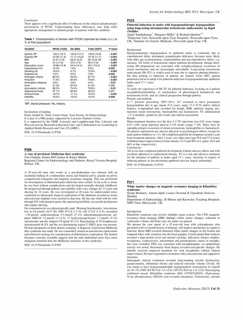
Society for Endocrinology BES 2013, Harrogate, UK
ConclusionThere appears to be a significant affect of ethnicity on the clinical and phenotypicpresentation of PCOS. Understanding these differences may help tailorappropriate management in defined groups of patients with this condition.
Table 1 Characteristics of women with PCOS (reported as mean (S.D.) or% of that population)
Variable* White (1523) SA (684) Total (2207) P value
Systolic BP 133.2 (15.7) 125.8 (14.7) 130.8 (15.8) !0.001Diastolic BP 75.06 (11.3) 71.17 (10.2) 73.79 (11.1) !0.001BMI 31.01 (7.9) 28.27 (6.5) 30.16 (0.19) !0.001Age 27.3 (7.6) 24.5 (7.3) 26.4 (7.6) !0.001Deprivation score 18.86 (14.9) 24.9 (14.4) 20.7 (15.0) !0.001Hypertension-Hx 10.4% 7.6% 9.5% 0.023Smoking-Hx 18.5% 3.7% 13.9% !0.001Diabetes-Hx 7.2% 9.5% 7.9% 0.042Androgen criteria 85.4% 93.0% 87.7% !0.001Hirsutism 71.5% 84.6% 75.6% !0.001Androgenic-alopecia 3.9% 5.6% 4.4% !0.01Acne 21.5% 20.5% 21.2% 0.306Anovulation criteria 80.5% 74.4% 78.6% !0.01Oligomenorrhoea 57.1% 60.8% 58.3% 0.057Amenorrhoea 18.7% 11.1% 16.4% !0.001Infertility 16.5% 13.5% 15.5% 0.039
*BP, blood pressure; Hx, history.
Declaration of fundingPartly funded by ‘Early Career Grant’ from Society for Endocrinology.It is part of a PhD project supported by Leicester Diabetes Centre.It is supported by the NIHR ‘Leicester and Loughborough Diet, Lifestyle andPhysical activity Biomedical Research Unit’ and Collaboration for Leadership inApplied Health Research and Care (CLAHRC).
DOI: 10.1530/endoabs.31.P308
P309A case of persistent Mullerian duct syndromeUna Graham, Emma McCracken & Karen MullanRegional Centre for Endocrinology and Diabetes, Royal Victoria Hospital,Belfast, UK.
A 45-year-old man who works as a psychotherapist was referred with anincidental finding of a rudimentary uterus and bilateral pelvic gonads on pelviccomputerized tomogram and magnetic resonance imaging. This was performedfor investigation of abdominal pain which has since settled. As far as he is awarehe was born without complications and developed normally through childhood.He progressed through puberty uneventfully with voice changes by 13 years andshaving by 16 years. He was investigated at 20 years for undescended testesconfirmed on ultrasound. Surgical exploration of the abdomen revealed no testesand testicular implants were inserted at that time. He has one child with his wifethrough IVF with donated sperm. He reported good libido, no erectile dysfunctionand regular shaving.On examination he was phenotypically male. Morning biochemistry: testosteronelow at 8.6 nmol/l (10.5–30), FSH 39 U/l (1.5–9), LH 12 U/l (1.5–8), oestradiol!50 pmol/l, androstenedione 9.3 nmol/l (3–15), dehydroepiandrosterone sul-phate DHEAS 3.5 mmol/l (2–14.5), 17 hydroxyprogesterone 7.3 nmol/l (2–15)and prostate specific antigen 0.8 ng/ml (0–2.5). Karyotyping of 30 lymphocytesdemonstrated 46 XY and the sex-determining region Y (SRY) gene was present.He had osteopaenia on bone density scanning. A diagnosis of persistent Mullerianduct syndrome was made. He was counselled, started on testosterone replacementand referred to urology for consideration of abdominal re-exploration. The limitedliterature currently available suggests that the intra abdominal testes have moremalignant potential than the Mullerian structures in this syndrome.
DOI: 10.1530/endoabs.31.P309
P310Pubertal induction in males with hypogonadotropic hypogonadismusing long-acting intramuscular testosterone undecanoate 1g depot(Nebido)Anjali Santhakumar1, Margaret Miller1 & Richard Quinton1,2
1Endocrine Unit, Newcastle-upon-Tyne Hospitals, Newcastle-upon-Tyne,UK; 2Institute for Genetic Medicine, Newcastle-upon-Tyne, UK.
BackgroundHypogonadotropic hypogonadism in apubertal males is commonly due toconstitutional delay; permanent gonadotropin deficiency becomes more likelywith older age at presentation, cryptorchidism and non-reproductive defect, e.g.anosmia. All forms of testosterone induce pubertal development, though short-acting IM preparations are associated with extraphysiological excursions ofserum testosterone and are increasingly unavailable. Long-acting testosteroneundecanoate IM (TU) is widely-used in men due to superior pharmacokinetics,but data relating to induction of puberty are limited. From 2007, patientpreference led us to adopt it for pubertal-induction in hypogonadotropic apubertalmales aged 17C years.AimsTo audit our experience of IM TU for pubertal induction, focusing on i) patientacceptability/tolerability, ii) maintenance of physiological haematocrit andtestosterone levels, and iii) clinical progression through puberty.Patients and methodsnZ7 patients presenting 2007–2011; 6/7 assumed to have permanenthypogonadism due to age (mean 37.6 years; range 17.3–57.8) and/or clinicalfeatures. Longitudinal data recorded for height, BMI, pubertal staging, pre-injection serum testosterone, haemoglobin and haematocrit. TU administeredw3–4-monthly, guided by lab results and clinical assessment.ResultsMean treatment duration over the first 3–5 TU injections was 0.91 years (range0.51–1.04); mean injection interval 13.24 weeks (range 7–18). There were nosupraphysiologic excursions of serum testosterone, haemoglobin or, haematocrit.No patient experienced any adverse physical or psychological effects, except formale-pattern baldness (nZ1). All completed pubertal development around a yearfrom treatment-initiation. After 3 years, two older men (age 50.8 and 57.8 years)exhibited major improvement in bone density (23.5 and 40% at L-spine; 26.6 and46% at hip, respectively).ConclusionsAll seven men completed pubertal development without adverse effects and withexcellent adherence to replacement therapy. TU is a safe and effective treatmentfor the initiation of puberty in males aged 17C years. Anxieties in respect ofinducing puberty in late-presenting apubertal men are largely unfounded.
DOI: 10.1530/endoabs.31.P310
P311White matter changes on magnetic resonance imaging in KlinefeltersyndromeDeepa Beeharry, Amena Iqbal, Louise Overend & Upendram Srinivas-ShankarDepartment of Endocrinolgy, St Helens and Knowsley Teaching HospitalsNHS Trust, Merseyside, UK.
IntroductionKlinefelter syndrome may involve multiple organ systems. The CNS, magneticresonance brain imaging (MRI) findings (white matter changes, reduction inventricular volume and brain size) are under recognised.We present the case report of a 47-year-old man with schizophrenia whopresented with a 4-month history of lethargy, self-neglect and decline in cognitivefunction. Brain MRI revealed abnormal white matter changes in left frontal andtemporal lobes with extension into the basal ganglia. Cerebrospinal fluid analysisrevealed a high protein level and normal cytology. Infectious (herpes simplex,toxoplasma, cryptococcus), autoimmune and paraneoplastic causes of encepha-litis were excluded. EEG was consistent with encephalopathy; no epileptiformactivity was noted. Stereotactic brain biopsy revealed non-specific changes. Heinitially received empirical treatment for viral encephalitis without clinicalimprovement. He later responded to treatment with corticosteroids and supportivemeasures.Subsequent clinical evaluation revealed long-standing erectile dysfunction,gynaecomastia, abdominal obesity and reduced testicular volume (10 ml). Hewas found to have hypergonadotrophic hypogonadism (testosterone 6.3 nmol/l(nr 10–32), FSH 18.9 IU/l (nr 1.0–12.0), LH 25.6 IU/l (nr 1.0–12.0). Karyotypingconfirmed mosaic Klinefelter syndrome (KS) (47XXY/46XX). Dual-energyX-ray absorptiometry (DEXA) scan revealed osteopenia. Testosterone, calcium
Endocrine Abstracts (2013) Vol 31
Society for Endocrinology BES 2013, Harrogate, UK
and vitamin D were initiated. Repeat brain MRI scan revealed normal pituitary.The abnormal white matter changes in the frontal lobes persisted with someregression of the temporal lobe changes.ConclusionsKlinefelter syndrome is under diagnosed and has a higher prevalence in peoplewith schizophrenia. In the presence of unexplained CNS, white matter changes onMRI and clinical features of hypogonadism, karyotyping to exclude Klinefeltersyndrome should be considered.
DOI: 10.1530/endoabs.31.P311
P312Metformin treatment of PCOS: St George’s Hospital Endocrine UnitClinical ExperienceHannah Walton, Helen Mason & Gul BanoSt George’s Hospital, London, UK.
Polycystic ovary syndrome (PCOS) is the most common endocrine conditionaffecting women and is associated with hyperinsulinaemia and hyperandrogen-ism. Obesity is present in at least 30% of cases and plays a vital role in thedevelopment and maintenance of PCOS as well as affecting the severity of theclinical and endocrine features. Significant improvements in symptoms ofandrogen excess and ovulatory function are seen with even a modest weight lossof 5% in women with PCOS. Metformin is used in PCOS to improve insulinresistance and to achieve weight loss, but there is still controversy as to whether ornot metformin aids the latter.The aim of this study was to determine the impact of metformin treatment onoverweight patients with PCOS over the period they attended St George’sHospital Endocrine Unit.Data was obtained retrospectively on 43 patients with PCOS attending theendocrine clinic at St George’s Hospital and prescribed metformin. Patientsattended clinic for an average of 5 years. Changes in weight that occurred whilston metformin treatment and weight changes while not taking the treatment weredetermined and compared. The average weight of patients when they firstattended clinic was 86.5 kg.The average weight change whilst on metformin was a loss of 2.17 kg. When thepatients were not taking metformin, the average weight change was a gain of3.8 kg. Side effects due to metformin were a major cause of lack of complianceand 50% of the patients took metformin for !6 months.In conclusion, patients lost significantly more weight whilst taking metforminthan when not on the drug, despite its limitations. Non-compliance was high withgastro-intestinal side effects being the main cause.
DOI: 10.1530/endoabs.31.P312
P313Myotonic dystrophy: a rare cause of primary hypogonadismAlison Thorne, C M Iqbal, Deepa Beeharry, Tom Mayes &Upendram Srinivas-ShankarDepartment of Endocrinolgy, St Helens and Knowsley Teaching HospitalsNHS Trust, Merseyside, UK.
Male hypogonadism is usually considered in the presence of classical symptomslike reduced libido, erectile dysfunction and reduced bone mineral density.We present the case history of a 43-year-old man with learning difficulties whopresented with long-standing lethargy. Clinical examination revealed bilateralptosis, muscle weakness and slow relaxation of handgrip. He had abdominalobesity, pseudo-gynaecomastia, frontal balding, reduced facial, chest, axillaryand pubic hair. Testes measured 15mls bilaterally. Investigations revealedhypergonadotrophic hypogonadism ((testosterone 3.0 nmol/l (nr 10–32); FSH11.3 IU/l (nr 1.0–12.0), LH 11.1 IU/l (nr 1.0–11.0)). Other biochemicalinvestigations: estradiol 193 (!172 pmol/l), 0900 h cortisol 640 nmol/l. Thyroidfunction tests, prolactin and IGF1 were normal.Genetic test confirmed the presence of CTG repeat expansion of dystrophiamyotonica protein kinase (DMPK) gene consistent with myotonic dystrophy type1. Electromyography (EMG) revealed frequent myotonic discharges in the distaland proximal limb muscles, consistent with myotonic dystrophy.Our case report highlights the importance of excluding hypogonadism in thepresence of myopathy and specific phenotypic features of congenital muscledisease.
DOI: 10.1530/endoabs.31.P313
Endocrine Abstracts (2013) Vol 31
P314
Abstract withdrawn.
DOI: 10.1530/endoabs.31.P314
Steroids
P31511bHSD1 deficiency increases susceptibility to liver fibrosis byactivating hepatic stellate cellsXiantong Zou1,2, Antonella Pellicoro2, Rebecca Aucott2,Prakash Ramachandran2, Michelle Clarkson1, Scott P Webster1,John P Iredale2, Brian R Walker1 & Zoi Michailidou2
1Center for Cardiovascular Science, Queens Medical Research Institute,Edinburgh, UK; 2the MRC Centre for Inflammation Research, QueensMedical Research Institute, Edinburgh, UK.
BackgroundLiver fibrosis in cirrhosis is characterized by accumulation of extracellular matrixfrom activated hepatic stellate cells (HSCs). Glucocorticoids (GCs) limit HSCactivation in vitro. Local GC levels are regulated by 11b-hydroxysteroiddehydrogenase-1 (11bHSD1) which converts inactive GCs (11-dehydrocorticos-terone) into active GCs (corticosterone). In this study we hypothesized that11bHSD1 could potentially inhibit liver fibrosis.Method11bHSD1 levels in mouse models of liver injury were investigated. We studiedliver fibrotic responses to carbon tetrachloride in mice with global 11bHSD1deletion (KO) or with administration of a selective murine 11bHSD1 inhibitor,UE2316. Immunohistochemistry, qPCR, western blot and flow cytometry wereused to analyse the liver response. Primary mouse HSCs were cultured in vitro toinvestigate the effect of 11bHSD1.Results11bHSD1 mRNA and protein levels were decreased concurrently with peakfibrosis and later recovered in liver injury models. Despite lower indices ofhepatocyte injury, 11bHSD1 KO mice had exaggerated fibrosis with increasedcollagen deposition (Col I) and HSC activation (aSMAC). The fibrotic responsepersisted after injury. An array of profibrotic genes (Col1, aSMA, Tgfb) and genesinvolved in ECM remodelling (MMP2, MMP9, Timp1) were highly up-regulatedin the 11bHSD1 KO mice. In the resolution phase 11bHSD1 KO mice showed animpairment in ‘resolving macrophages’ populations (decreased F4/80int cd11bhi
ly6Clo macrophage ratio). Findings were similar after UE2316 administrationduring injury. In vitro studies showed 11bHSD1 deficient HSCs were moreactivated than wild type after 8 days in culture and this activation was inhibited by11-dehydrocorticosterone and corticosterone.ConclusionLoss of GCs regenerated within the liver by 11bHSD1 may contribute tounrestrained activation of HSCs following chemical injury and promote liverfibrosis. This contrasts with anti-fibrotic effects of 11bHSD1 deficiency inadipose. Context-specific effects of 11bHSD1 inhibitors on inflammation andrepair deserve careful further scrutiny.Declaration of fundingBritish Heart Foundation(BHF); Translational Medicine Research Initiative(TMRI); Medical Research Council (MRC); China Scholarship Council (CSC).
DOI: 10.1530/endoabs.31.P315
P316Validation of CYP11B1 and CYP11B2 regulation by microRNA-24Louise Diver1, Samantha Alvarez-Madrazo1, Junjun Lin1, Stacy Wood1,Scott MacKenzie1, John Connell2 & Eleanor Davies1
1University of Glasgow, Glasgow, UK; 2Univeristy of Dundee, Dundee,UK.
Society for Endocrinology BES 2013, Harrogate, UK
The CYP11B1 and CYP11B2 genes encode the enzymes responsible, respectively,for the terminal stages of cortisol and aldosterone biosynthesis, and have beenimplicated in the development of essential hypertension. Previously, weinvestigated the role of microRNAs in the regulation of these genes and showedin vitro that levels of the adrenally-expressed microRNA-24 (miR-24) inverselycorrelate with those of CYP11B1 and CYP11B2 mRNA, as well as cortisol andaldosterone production. Bioinformatic analysis predicts two putative bindingsites for miR-24 in the 3 0UTR of CYP11B1 mRNA and one in CYP11B2.The purpose of this study was to ascertain whether observed changes in CYP11B1and CYP11B2 mRNA levels in vitro were due to the direct action of miR-24 atthese sites.Luciferase reporter constructs containing full-length CYP11B1 and CYP11B23 0UTR sequences were specifically mutated by a single base at the predicted miR-24 binding sites using site-directed mutagenesis. These constructs were thentransfected into HeLa cells, either alone or alongside miR-24 inhibitor; luciferaseluminescence was measured 48 h post-transfection.Cells transfected with mutated plasmids yielded significantly higher lumines-cence compared to non-mutated plasmids (P!0.01). Co-transfection of non-mutated plasmids with miR-24 inhibitor also significantly increased lumines-cence (P!0.05), although this effect was eliminated when inhibitor wasco-transfected with mutated plasmids (PZ0.24). Furthermore, combinedmutation of both the predicted CYP11B1 miR-24 sites resulted in greater effectthan single mutations at either site.These results are consistent with canonical miRNA binding and repression, andconfirm that miR-24 is capable of regulating CYP11B1 and CYP11B2 expressionthrough direct binding of 3 0UTR sites on their mRNA. This is the first study todemonstrate directly such regulation of these genes at specific sites, and may haveimportant implications for corticosteroid biosynthesis and its role in thedevelopment of hypertension.Declaration of fundingThis work was supported by the British Heart Foundation (grant numberPG/09/092).
DOI: 10.1530/endoabs.31.P316
P317Molecular mechanisms underlying the anti-inflammatory properties of5a-tetrahydrocorticosteroneAnnalisa Gastaldello, Mark Nixon, Chenjing Yang, Philippa T K Saunders,Karen E Chapman, Brian R Walker & Ruth AndrewQueen’s Medical Research Institute, University of Edinburgh, Edinburgh,UK.
Glucocorticoids (GCs) are highly effective anti-inflammatory drugs, howevertheir use is limited by serious side effects. We have previously shown that 5aTHBbinds GC receptor (GR) and suppresses inflammation in vitro and in vivo, withoutaffecting metabolism. Here the underlying molecular mechanisms were exploredin cell models of ligand-induced GR phosphorylation, nuclear localisation andgene transcription. Data are meanGS.E.M. (three experiments); *P!0.05 vsvehicle.Phosphorylation of Ser211GR, influencing nuclear localisation and genetranscription, was assessed by western blot in A549 cells treated (1h) withvehicle, corticosterone (B) (1 mM) or 5aTHB (1–30 mM). Phosphorylation wasnot induced by 5aTHB alone, in contrast to B (fold induction: 1.4G0.4 (5aTHB,1 mM), 6.6G1.6* (B).To determine mobility of ligand-bound GR, localisation of green fluorescentprotein tagged-rat GR (GR-GFP) transfected in HEK293 cells was monitored byfluorescence microscopy. Nuclear translocation of GR-GFP by 5aTHB (1 mM)was incomplete (82.7G1.5% reaching the nucleus within 5 h). Conversely,translocation with B (1 mM) was complete within 30 min. A greater proportion(w3!) of GR-GFP translocated to the nucleus following a sub-maximal dose ofB (3 nM; 45 min)) when 5aTHB (1 mM) was present. Nuclear export after steroidwashout was observed only with 5aTHB (32.1G6.1% remaining after 24 h). Therate of recovery from nuclear photobleaching (FRAP) suggested that 5aTHB-bound GR-GFP was more mobile in the nucleus than with B (half-life: 5a-THB3.12G0.38 vs B 4.40G0.42 s; P!0.05).Ligand ability to induce transcription of GR dimer- and multimer-dependentreporter genes (MMTV and PNMT respectively) was tested. Compared to B,5aTHB (0.1–3 mM; 24 h) was unable to activate either reporter plasmid (foldinduction, 5aTHB and B: MMTV-Luc, 1.2G0.04; 16.9G1.1*; PNMT-Luc,1.6G0.6; 3.1G0.5*).GR nuclear translocation is slower in presence of 5aTHB, and activation ofclassical homodimer- and multimer-dependent gene transcription is absent. Thismay be linked, to an inability to 5aTHB-bound GR to be phosphorylated on keyresidue Ser211.Declaration of interest
The co-authors hold a relevant patent for the use of 5aTHB as an anti-inflammatory drug.Declaration of fundingThis work was supported by the British Heart Foundation.
DOI: 10.1530/endoabs.31.P317
P318Novel loci for familial autoimmune Addison’s disease detected bylinkage analysisAnna L Mitchell1, Anette Boe Wolff2, Earn H Gan1, Katie MacArthur1,Martina M Erichsen2, Jolanta U Weaver3, Bijay Vaidya4, Sophie Bensing5,Eystein Husebye2, Heather J Cordell1 & Simon H S Pearce1
1Institute of Genetic Medicine, Newcastle upon Tyne, UK; 2Section ofEndocrinology, Haukeland University Hospital, Bergen, Norway; 3Instituteof Cellular Medicine, Newcastle upon Tyne, UK; 4Royal Devon and ExeterHospital, Exeter, UK; 5Karolinska Institutet, Stockholm, Sweden.
Due to the rarity of autoimmune Addison’s disease (AAD), it has proved difficultto gather large case cohorts for genetic studies. Linkage analysis offers a powerfulmeans of identifying genetic susceptibility loci but has never been applied toAAD because of the scarcity of families containing R2 affected individuals. Wecollected DNA from 23 such families to perform the first linkage study in AAD.We genotyped 117 samples (50 cases, 67 controls) from 23 families with R2affected individuals from the UK (nZ12) and Norway (nZ11), on the AffymetrixSNP-6.0 array. Data was formatted and quality controlled in PLINK and Merlinwas used for linkage analysis. Results were validated by genotyping 65 SNPs(Sequenom) under two of the linkage peaks in 1097 unrelated AAD (69321-hydroxylase autoantibody positive) and 1117 controls from the UK, Norwayand Sweden.Applying a rare dominant model, three loci on chromosomes 18, 9 and 7 had LODscores O2.0. The maximum LOD score of 3.0 was observed within a linkage peakon chromosome 18 (75241668–77950543 kb). Non-parametric analysis revealedone locus on chromosome 6 with maximum LOD score 3.01, in a linkage peakspanning 22375648 – 35968100 kb, which contains the HLA complex. Meta-analysis of the validation study data in the whole cohort revealed association at 3SNPs underlying the linkage peak on chromosome 18, with maximal associationwith an intergenic SNP rs7236339 (P!0.004). When those without 21-hydroxyl-ase autoantibodies were excluded, 4 SNPs were associated. 3 of these wereintergenic SNPs on chromosome 18, with maximal association at rs7231100(P!0.004) and one was on chromosome seven, in the AUTS2 gene (rs12698902,P!0.01).This is the first linkage study in AAD and the finding of linkage to the HLA regionvalidates this approach. This study has generated some novel loci, which may castlight on the pathogenesis of AAD.Declaration of fundingThis work was supported by the Medical Research Council (G0900390) and aEuropean Union Framework 7 grant (201167) to the Euradrenal Consortium.
DOI: 10.1530/endoabs.31.P318
P319Identification of a duplicated P450 side-chain cleavage enzyme(zCyp11a2) defines initiation and maintenance of steroidogenesis inzebrafishSilvia Parajes, Aliesha Griffin, Angela Taylor, Cedric Shackleton,Irene Miguel-Escalada, Wiebke Arlt, Ferenc Mueller & Nils KroneUniversity of Birmingham, Birmingham, UK.
Zebrafish has emerged as an important vertebrate in vivo model to study humandisease. Steroidogenesis in zebrafish is not well characterised. Human CYP11A1(hCYP11A1) catalyses the first step of steroidogenesis, the conversion ofcholesterol to pregnenolone. Zebrafish Cyp11a1 (zCyp11a1) is essential duringembryogenesis. Published data suggest that zCyp11a1 facilitates steroidogenesisin the interrenal (equivalent to mammalian adrenal), gonad and brain. Weidentified a duplicated gene, designated as zCyp11a2, sharing 85% sequenceidentity with zCyp11a1.The aim of this study was to characterise the zCyp11a paralogs gene expressionpattern and function.Our RT-PCR data shows that zCyp11a1 is expressed during early development,from 0 to 22 h post-fertilisation (hpf). Conversely, zCyp11a2 was only detectedafter the interrenal is formed (from 32 hpf). Adult gonads expressed both
Endocrine Abstracts (2013) Vol 31
Society for Endocrinology BES 2013, Harrogate, UK
paralogs. The interrenal and brain expressed only zCyp11a2. The in vivo zCyp11afunction was assessed by knockdown studies using zCyp11a-morpholinos.Pregnenolone and cortisol were measured in the developing zCyp11a morphantsby liquid chromatography/tandem mass spectrometry (LC/MS/MS). zCyp11a1morphants showed an abnormal early development and reduced pregnenolonelevels. Transient zCyp11a2 knockdown impaired de novo pregnenolone andcortisol synthesis and resulted in a late phenotype indicative of metabolicabnormalities. zCyp11a enzyme activity was assessed in COS7 cells transientlyco-overexpressing adrenodoxin and zCyp11a or hCYP11A1. Cells wereincubated with 22R-hydroxycholesterol and pregnenolone was measured byLC/MS/MS. zCyp11a2 activity was similar to hCYP11A1, whilst zCyp11a1activity was significantly reduced.Our data completely revises our understanding of zebrafish steroidogenesis bydefining the roles of the previously described zCyp11a1 and the newly discoveredzCyp11a2 enzyme. zCyp11a2 rather than zCyp11a1 is the hCYP11A1 ortholog inzebrafish steroidogenic tissues. Furthermore, we prove that zCyp11a1 activity issignificantly reduced but essential during embryogenesis. Importantly, this studyproves the value of zebrafish as a comprehensive in vivo model in translationalresearch of adrenal and gonadal disease.Declaration of fundingThe project was supported by a Wellcome Trust ISSF grant (to N K), a Society forEndocrinology early career grant (to S P) and a Marie Curie Intra-EuropeanFellowship (grant number IEF-GA-2009-255424, 2009 to S P)
DOI: 10.1530/endoabs.31.P319
P320Glucocorticoids enhance insulin sensitivity in human hepatocytesMaryam Nasiri, Iwona Bujalska, Paul Stewart, Laura Gathercole &Jeremy TomlinsonUniversity of Birmingham, Birmingham, UK.
Patients with glucocorticoids (GC) excess develop central obesity, insulinresistance and hepatic steatosis in up to 20% of cases. Current dogma suggeststhat GCs cause insulin resistance in all tissues. However, we have previouslydemonstrated that GCs induce insulin sensitisation in adipose tissue in vitro,whilst causing insulin resistance in skeletal muscle. In rodent hepatocytes, GCsenhance insulin stimulated lipogenesis but studies in human hepatocytes have notbeen performed and the cellular mechanisms underpinning these observationshave not been determined.Cryopreserved human hepatocytes were purchased from Celsis in vitroTechnologies (Baltimore, USA) and incubated with variable doses of cortisol(0–1000 nM) for 24 h in the presence and absence of insulin (5 nM). Insulinsignalling gene expression levels were quantified by real-time PCR and westernblotting was performed to determine total and phospho PKB/akt proteinexpression levels. De novo lipogenesis (DNL) was measured by 1-(14C) acetateincorporation in triglyceride.GC receptor, IRS1/2, insulin receptor and AKT1/2 were all expressed in primarycultures. Incubation with cortisol alone or in combination with insulin did notsignificantly alter gene expression levels. However, whilst cortisol treatment didnot alter total PKB/akt levels, insulin stimulated phosphorylation of PKB/akt atserine 473 increased following cortisol pre-treatment in a dose dependant manner(1.23-fold (100 nM), 1.68-fold (250 nM), 2.44-fold (1000 nM) vs control nZ4P!0.05). Increasing doses of cortisol increased insulin stimulated lipogenesis(43.9G12.7% (250 nM), 66.13G9.8% (1000 nM) vs control (23.61G10.7%),P!0.05).We have demonstrated that in primary human hepatocytes GC treatment enhancesinsulin signalling through increased serine phosphorylation of PKB/akt and thatGCs and insulin can act synergistically to promote lipogenesis. Whilst translationto the clinical setting is crucially important, this mechanism may be fundamentalin explaining the interaction between GCs and insulin to drive lipogenesis.Furthermore, this may contribute to the pathogenesis of non-alcoholic fatty liverdisease with GC excess.Declaration of fundingMedical research council (senior clinical fellowship Ref.g0802765,J W T).
DOI: 10.1530/endoabs.31.P320
P321The zebrafish ferredoxin orthologue Fdx1b is essential for the redoxregulation of interrenal steroidogenesis in larvae and adult fishAliesha Griffin, Silvia Parajes, Angela Taylor, Cedric Shackleton,Ferenc Mueller & Nils Krone
Endocrine Abstracts (2013) Vol 31
University of Birmingham, Birmingham, UK.
Mitochondrial steroidogenic cytochrome P450 (CYP) enzymes, such as P450side-chain cleavage, rely on electron transfer from the redox partner ferredoxin(FDX1) for catalytic activity. Previous in vitro data suggest these cofactors arekey regulators of CYP enzyme activity. This study aims to establish the role ofredox regulation on steroidogenesis using zebrafish as a vertebrate in vivo model.In contrast to humans, zebrafish have two FDX1 genes, zFdx1 and zFdx1b. OurRT-PCR data shows that zFdx1 is maternally expressed and maintainedthroughout zygotic development. Its paralog, zFdx1b begins expression between24 and 36 h post-fertilisation (hpf), which mirrors the expression of the mainzebrafish P450 side-chain cleavage enzyme, zCyp11a2. Importantly, this timepoint coincides with the development of the zebrafish interrenal – the counterpartof the mammalian adrenal gland. While zFdx1 is ubiquitously expressed in adultzebrafish, zFdx1b expression is restricted to the main steroidogenic tissues:interrenal, gonads and brain. This suggests zFdx1b is the redox partner ofsteroidogenic mitochondrial CYP enzymes in the adult zebrafish. The in vivofunction of the two zFdx paralogs was characterised using a transient morpholinosknockdown approach. Consistent with the expression data, knockdown of zFdx1shows early morphological abnormalities during zebrafish development.Alternatively, zFdx1b morphants develop late onset metabolic abnormalities,consisting of an enlarged yolk and a delay in the inflation of their swim bladder at120 hpf. Cortisol was extracted from zFdx1b deficient larvae and measured byliquid chromatography/tandem mass spectrometry. zFdx1b morphants failed tosynthesise cortisol, compared to the injected controls.This study gives in vivo insights into the molecular mechanism of mitochondrialredox regulation of steroidogenesis. We demonstrated that zFdx1b is essential forde novo steroidogenesis in zebrafish larvae, and that zFdx1 cannot compensate forFdx1b function. Overall, our data establishes zebrafish as a model to study inbornerrors of adrenal steroidogenesis.Declaration of fundingThis work was supported by the Elite Interntation Student Scholarship from theUniversity of Birmingham (awarded to AG) and an Wellcome Trust ISSF grant(awarded to N K).
DOI: 10.1530/endoabs.31.P321
P322Steroid profile response to angiotensin II and ACTH in normalvolunteer under high and low salt conditionsFrances McManus1,2
1University of Glasgow, Glasgow, UK; 2University of Dundee, Dundee,UK.
IntroductionSteroid profiling using liquid chromatography with tandem mass spectroscopy(LC:MS) has a low cost per sample and has the potential for high through-putprocessing. However, although this technology is becoming more widely used,little is known of the normal ranges of many less well studied steroid compoundsas well as their response to a variety of physiological stimulants.MethodsVolunteers were recruited to a randomised, double blind cross-over study andadhered to a standard salt diet for 3 days with salt loading (slow sodium tabs) orplacebo. After 30 min recumbent rest, samples were obtained pre and post ACTHand angiotensin II infusions. Samples were extracted from plasma using ChemElute cartridges (Varian, CA, USA) and injected into a C-18-A reversed phaseHPLC column. Identification and quantification were accomplished by tandemmass spectrometry using Varian 1200L mass spectrometer.ResultsHigh and standard salt phases were confirmed by 24 h urinary sodium excretionand plasma renin concentrations (PRC). Mean (24 h Na, standard salt 97.1G39.5 mmol, high salt 199.8G64.6 mmol, P!0.001; PRC, standard salt phase19.1G13.9 mIU/l, high salt phase 9.7G5.9 mIU/l, P!0.001). Aldosterone andits immediate precursor, 18-hydroxycorticosterone were stimulated by saltrestriction and angiotensin II infusion. There was no difference in deoxycorti-costerone or 18-hydroxydeoxycorticosterone following angiotensin II infusion.Cortisol, 11-deoxycortisol, corticosterone and cortisone concentrations werereduced following angiotensin II infusion. As expected, ACTH stimulated allmeasured corticosteroids compounds.ConclusionsThese data suggest that angiotensin II infusion is associated with a reduction incortisol and its precursor steroid hormones. In addition, aldosterone synthase islikely to catalyse the 18-hydroxylation of corticosterone but not 18-hydroxylationof deoxycorticosterone. Steroid profiling by LC:MS can reveal a morecomprehensive picture of the ‘steroid-ome’ leading to a greater understanding
Society for Endocrinology BES 2013, Harrogate, UK
of the factors controlling the steroid pathway.Declaration of fundingThis work was supported by the Medical Research Council (F M, ClinicalTraining Fellowship G0601322; E D, J M C, R F, Programme Grant G0400874).
DOI: 10.1530/endoabs.31.P322
P323Mechanisms of estrogen receptor transcription in breast cancerJason Carroll1,2
1Cancer Research UK, Cambridge, UK; 2University of Cambridge,Cambridge, UK.
Estrogen receptor (ER) is the defining feature of luminal breast cancers, where isfunctions as a transcription factor in response to the ligand estrogen. Thetraditional view of ER getting recruited to promoters of target genes is toosimplistic. The recent discovery of ER-DNA interaction regions from ERCbreast cancer cell lines has revealed that ER rarely associates with promoterregions of target genes and instead associates with enhancer elements significantdistances from the target genes. The genomic mapping of ER binding events alsorevealed the enrichment of DNA motifs for Forkhead factors. The Forkheadprotein FOXA1 (HNF3a) was subsequently shown to bind to approximately halfof the ER binding events in the genome and was required for ER to maintaininteraction with DNA. We have extended on these findings to map ER bindingevents in primary breast cancers and distant metastases. We find contextdependent ER cis-regulatory elements (cistromes) that give insight intounderlying transcriptional networks. These differential ER binding profilescorrelate with clinical response in ERC breast cancers. We experimentallyexplore the binding dynamics between drug sensitive and resistant contexts andidentify properties that govern ER binding differences. These data suggest thatER–DNA interactions are dynamic and can be modulated by changes in FOXA1.We are currently exploring mechanisms that mediate FOXA1–DNA interactions,in order to better understand ER transcriptional activity in breast cancer biology.This work provides insight into how estrogen mediates its effects in cancer andhow hormone dependent cancers function after acquiring resistance to currentendocrine therapies.Declaration of fundingThis work was supported by funding from Cancer Research UK.
DOI: 10.1530/endoabs.31.P323
P324Continuous measurement of free cortisol profilesRagini C Bhake, Jack A Leendertz, Astrid C E Linthorst& Stafford L LightmanUniversity of Bristol, Bristol, UK.
In order to understand the significance of circadian and ultradian hormonalrhythms in man, both in health and disease, it is essential to be able to obtainmultiple samples over extended periods, especially during the hours of sleep. Thehuman automated blood sampling (HABS) system reported by Henley andcolleagues can achieve this but is recommended for use in the setting of a clinicalinvestigation unit which is its major drawback. For many diagnostic and scientificquestions, the most meaningful physiological setting to look at homeostaticallyimportant hormones is a subject’s home setting. We have now developed analternative approach to measure glucocorticoid hormones using the technique ofsubcutaneous microdialysis. This has the additional advantage that it measures thelevel of active free cortisol as opposed to the total cortisol levels measured inwhole blood of which approximately 90% is bound to carrier proteins. Since onlythe free unbound cortisol has access to tissues and their receptors these are thelevels that are physiologically important for glucocorticoid signalling. Thetechnique of microdialysis combined with a novel miniaturised sampling systemprovides the ability to collect multiple samples automatically without the need forvenous access. As part of the validation of this technique, serum samples andcorresponding microdialysate samples from either the subcutaneous tissuecompartment alone or both subcutaneous tissue and intravenous compartmentwere collected every 10 min from 1000 to 1400 h in healthy male volunteers aged19–28 years. Preliminary results confirm that free cortisol is detectable using thistechnique in both the body compartments and that free cortisol levels reflectspontaneous changes in serum total cortisol measured during the study period.
Our early results indicate the potential of this system as a unique tool in researchand in clinical diagnosis.Declaration of fundingThis work was carried out with financial support from the European Union – FP7grant to OPTIMI.
DOI: 10.1530/endoabs.31.P324
P325Dual role of TTC5 cofactor in GR-mediated gene expressionMalihah Sadeq, Constantinos Demonacos & Marija Krstic-DemonacosUniversity of Manchester, Manchester, UK.
Glucocorticoid receptor (GR) is a ligand-dependent nuclear receptor whichregulates the transcription of a wide spectrum of genes that are responsible forvital immunological, metabolic, developmental, and anti-inflammatory functions.GR transcriptional regulatory effects are modulated by co-regulators including thetetratricopeptide 5 (TTC5) which has been shown to stabilize GR and alter itsaction in response to cellular stress. TTC5 is a stress-responsive activator of p300and its activities are controlled by the ataxia telangiectasia mutated (ATM) andChk2 kinases. TTC5 is comprised of six TPRs in addition to four probable nuclearreceptor (NR) LXXLL boxes. Here we provide evidence to suggest that in A549lung cancer cells different LXXLL motifs in TTC5 are required to differentiallyinhibit GR mediated transcriptional function on the TAT-3 promoter in ahormone-dependent manner, whereas association of GR with TTC5 through theco-regulator’s TPR sequences increased GR mediated TAT-3 gene expression.Furthermore, TTC5 and GR sub cellular co-localization occurred in a modedependent on the presence of hormone as well as the integrity of the LXXLLdomains located in the TTC5 N terminus. We conclude that TTC5 plays a dualfunction in the control of GR mediated gene expression and its sub cellularlocation depending on the surface of TTC5 utilized to interact with the receptor.
DOI: 10.1530/endoabs.31.P325
P326Identification of a novel CYP11B1 isoform in human adrenocorticalcellsSamantha Alvarez-Madrazo1, Scott MacKenzie1, Alette Brinth1,Niall Fraser1, Rita Bernhardt1,2, John Connell1,3 & Eleanor Davies1
1BHF Glasgow Cardiovascular Research Centre, Institute of Cardiovascularand Medical Sciences, College of Medical, Veterinary and Life Sciences,University of Glasgow, Glasgow, UK; 2Department of Biochemistry,Saarland University, Saarbrucken, Germany; 3Medical Research Institute,College of Medicine, Dentistry and Nursing, University of Dundee, Dundee,UK.
The final reaction for cortisol production in the adrenal gland is catalysed by the11b-hydroxylase enzyme, encoded by the CYP11B1 gene. Variants in this genehave been associated with alterations in cortisol levels, which increase bloodpressure. This gene is traditionally thought to consist of 9 exons. However, recentevidence has predicted the existence of at least one alternatively spliced form.The presence of novel CYP11B1 mRNA species in the H295R humanadrenocortical cell line and non-diseased human adrenal tissue was investigatedusing RT-PCR, sequencing and western blotting.Following RT-PCR, a larger band corresponding in size to an alternative form(ALT1) of CYP11B1 mRNA was observed on agarose gels, in addition to thewild-type (WT) form. Sequencing of the ALT1 band confirmed the presence of anadditional exon between exons 2 and 3. In silico analysis of the 26 in-frame aminoacids encoded by this exon predicts an insertion between alpha helices B’ and C ofthe enzyme. Western blotting using a custom antibody targeted at this insertionproduced a band of the predicted size in total H295R cell protein. Control cellsexpressing only the WT form of CYP11B1 did not yield this band.Further in vitro studies are required to investigate the effect of this alternativetranscript on protein structure and cortisol production. The identification of anovel CYP11B1 isoform will broaden our understanding of adrenal physiologyand its contribution to cortisol synthesis.Declaration of fundingThis work was supported by the TENOVUS Scotland Grant no. S11/11.
DOI: 10.1530/endoabs.31.P326
Endocrine Abstracts (2013) Vol 31
P327The role of anti-ACTH1–24 antibodies in synacthen-related adverseeventsEarn H Gan1,2, Katie MacArthur1, Anna L Mitchell1,2, Patricia Crock3,Sophie Bensing3 & Simon H S Pearce1,21Institute of Genetic Medicine, Newcastle University, Newcastle UponTyne, UK; 2Endocrine Unit, Royal Victoria Infirmary, Newcastle UponTyne, UK; 3Karolinska Institutet, Stockholm, Sweden.
BackgroundImmune responses to self-peptides should not generally occur. However, four of12 autoimmune Addison’s disease (AAD) patients developed adverse reactionsimmediately after synacthen injections, following repeated subcutaneoussynacthen injections during a clinical trial (RoSA study). We wondered if theseadverse effects were due to the production of anti-synacthen (ACTH1–24)antibodies.MethodsWe evaluated the presence of serum anti-ACTH binding activity usingimmunoblotting and ELISA on sera from participants in the RoSA study(nZ12; baseline and after synacthen exposure), 131 unrelated patients with AAD,92 patients with Graves’ disease (GD), 15 patients with isolated ACTH deficiencyand 102 controls without known autoimmune disease. Immunoblotting wasperformed on polyacrylamide/tricine gels using commercial synacthen and full-length ACTH peptide (both 10 mg/well). ELISA was performed using ACTH1–24
(1 mg/ml) immobilised on solid phase.ResultsBands atw4 andw6 kDa, corresponding to ACTH1–24 and full-length ACTH1–39
peptide respectively, were found in 10/12 (83%) RoSA study immunoblots,including all those who had an adverse reaction to synacthen. This is in contrastwith healthy control sera, which showed no binding. The same 10 subjects fromthe RoSA study also showed high levels of binding to synacthen by ELISA, alongwith 28 patients with AAD (21% of 131), 13 patients with GD (14% of 92) andone isolated ACTH deficiency patient (7% of 15). All positive patient sera in theELISA were tested against the synacthen peptide on immunoblotting, and all(nZ41) showed specific 4kDa binding.ConclusionOur study demonstrates that repeated administration of depot synacthen can leadto anti-ACTH1–24 autoreactivity. In addition, a significant number of AAD andGD patients also had similar autoreactivity (P!0.001). The presence of theseantibodies could mediate some of the adverse effects seen in the RoSA study andexplain the well-described phenomenon of resistance to chronic ACTH therapy.Declaration of fundingThis work was supported by the Medical Research Council (G0900001).
DOI: 10.1530/endoabs.31.P327
P32819F-magnetic resonance spectroscopy as a tool to quantify11b-hydroxysteroid dehydrogenase activity in vivoGregorio Naredo-Gonzalez1,7, Maurits Jansen2, Rita Upreti1, Scott Semple3,Gavin Merrifield2, Oliver Sutcliffe4, Michael Hansen5, Ian Marshall6,Ruth Andrew1,7 & Brian Walker1,71Endocrinology, University/BHF Centre for Cardiovascular Sciences,University of Edinburgh, Edinburgh, UK; 2Edinburgh Preclinical Imaging,University/BHF Centre for Cardiovascular Sciences, University ofEdinburgh, Edinburgh, UK; 3Clinical Research Imaging Centre, Universityof Edinburgh, Edinburgh, UK; 4Division of Chemistry and EnvironmentalScience, School of Science and the Environment, Manchester MetropolitanUniversity, Manchester, UK; 5Johnson & Johnson Pharmaceutical Researchand Development, New Jersey, USA; 6Centre for Clinical Brain Sciences,University of Edinburgh, Edinburgh, UK; 7Mass Spectrometry Core,Wellcome Trust Clinical Research Facility, University of Edinburgh,Edinburgh, UK.
Non-invasive methods to measure enzyme activity in vivo can provide a usefultool for the development of selective inhibitors. Tissue-specific dysregulation of11b-hydroxysteroid dehydrogenase 1 (11b-HSD1), a reductase enzyme thatamplifies active intracellular glucocorticoid levels, has been shown in obesepatients using invasive tools (biopsy, microdialysis and arteriovenous samplingwith stable isotope tracers). 11b-HSD1 inhibitors are efficacious in pre-clinicalmodels of obesity, diabetes, atherogenesis and cognitive dysfunction, butunpredictable pharmacodynamics may explain disappointing results in phase 2trials. We have explored the use of 19F-magnetic resonance spectroscopy (MRS)and identified suitable fluorinated keto tracer substrates for the in vivo monitoringof hepatic 11b-HSD1 both in rat and human. The effect of tracer structure
(equivalent fluorine atoms per molecule and distance to the keto/hydroxy group),tracer abundance, scanning time and biological matrix was studied using sevenTesla (small animal) and three Tesla (human) MRI scanners. 19F-MRS responseswere linearly related to the total amount of equivalent fluorine atoms. Signalsfrom keto and hydroxy forms differing as little as 0.6 ppm could be resolved andmeasured simultaneously. We have determined in vitro LODF (limit of detectionas absolute fluorine content) of 0.250 mmol in chloroform and 0.625 mmol inblood, using 400 s/spectrum. In vivo detection of tracer 2-(phenylsulfonyl)-1-(4-(trifluoromethyl)phenyl)ethanone and its hydroxy metabolite was achieved inrat liver (7T scanner) after very low oral doses of tracer (5-8 mg). However, thistri-fluorinated tracer is not a licensed pharmaceutical, so studies in humans wereprogressed with monofluorinated dexamethasone. Oral doses of 10–14 mg wereused and under these conditions neither substrate nor product could be detected inhuman liver. We conclude that MRS monitoring of 11b-HSD1 is feasible, butrequires novel multi-fluorinated tracers.Declaration of fundingThis work was supported by an award from the Translational Medicine ResearchCollaboration (a consortium of the Universities of Aberdeen, Dundee, Edinburghand Glasgow, the associated NHS Health Boards), Scottish Enterprise and Wyeth(now Pfizer) Pharmaceuticals, grant reference CVMD/EU/016 and British HeartFoundation (BHF) Programme Grant RG/200016.
DOI: 10.1530/endoabs.31.P328
P329P450 side-chain cleavage enzyme autoantibodies in canine Addison’sdiseaseAlisdair Boag1, Kerry McLaughlin2, Mike Christie2, Peter Graham3,Harriet Syme2 & Brian Catchpole21Royal Veterinary College, North Mymms, Hertfordshire, UK; 2DiabetesResearch Group, King’s College London, London, UK; 3Dechra LaboratoryServices, Poulton-Le-Fylde, Lancashire, UK.
BackgroundAddison’s disease (AD) in both humans and dogs is characterised bycorticosteroid deficiency requiring lifelong hormone therapy. In humansautoimmune pathogenesis is established; at diagnosis w90% of patients are21-hydroxylase (21-OH) autoantibody positive, with antibodies to other adrenalantigens also detected. The pathogenesis of canine AD is less well characterised;autoimmune mechanisms are suspected, with anti-adrenal autoantibodiesdemonstrated by indirect immunofluorescence.HypothesisSpecific adrenal autoantibodies are detectable in canine AD patients.Methods: Canine orthologs of 21-OH, p450 side-chain cleavage enzyme(P450scc), 3-b-hydroxysteroid dehydrogenase (3-b-HSD) and 17-a-hydroxylase(17-a-OH) were identified, cloned and S35 radiolabelled recombinant proteinsexpressed. A RIA was validated using radiolabelled human 21-OH and sera ofknown autoantibody status. Sera from AD dogs and controls with no history of anendocrinopathy or immune-mediated disease were then tested to investigate thepresence of autoantibodies; cross-reactivity across species was also assessed.ResultsRadiolabelled proteins were of expected size. Human 21-OH autoantibodies didnot cross-react with the canine radiolabelled protein. A proportion, 5/20, of canineAD samples showed reactivity to P450scc, these were a German Shepherd Dog,Beagle, Lurcher, Great Dane and Polish Lowland Sheepdog, comprising threefemales and two males. Human P450scc autoantibody positive sera from a patientwith autoimmune polyglandular syndrome type one (APS1) and a patient withAPS2 with associated premature ovarian failure (POF) cross-reacted with canineP450scc.ConclusionsThis is the first study showing P450scc autoantibodies in canine AD. This offersinteresting comparisons with human disease, in particular with APS1 and POF,both associated with P450scc autoantibodies. These findings help confirm animmune-mediated component to canine AD, a potential model for the humandisease, and could offer a new approach for diagnostic testing in dogs.Declaration of fundingThis work was supported by the BBSRC with Dechra (grant numberBB/G016992/1) and the PetPlan Charitable Trust (grant number 09-04).
DOI: 10.1530/endoabs.31.P329
Society for Endocrinology BES 2013, Harrogate, UK
Endocrine Abstracts (2013) Vol 31
P330Quantitative analysis of canrenone in plasma by triple quadrupole massspectrometryNatalie Homer, Jill Harrison, Javaid Iqbal, Brian Walker & Ruth AndrewUniversity of Edinburgh, Edinburgh, UK.
Canrenone is a mineralocorticoid receptor antagonist used as a diuretic agent totreat hypertension. It is the major active metabolite of spironolactone and may bequantified in clinical studies either to ensure compliance or to gain informationabout pharmacokinetic-pharmacodynamic interactions.The aim of this study was to develop and validate a sensitive, quantitative assayfor the analysis of canrenone in plasma.HPLC mass spectrometric method development was carried out on a TQ4Quantum Discovery triple quadrupole mass spectrometer with an Aria CTCHPLC autosampler system. Under positive electrospray ionisation the major iondetected was the protonated molecular ion (MCH)Cm/z 341 for canrenone.Alfaxalone (precursor ion m/z 333; 1 mg) was used as the internal standard. Undercollisional activation, the major fragmentation ions were m/z 107.1 and 91.1 forcanrenone and m/z 297.3 and 315.3 for alfaxalone. The fragmentation ions wereused as quantifier and qualifier ions respectively to add specificity to the assay.Optimal separation of the steroids was achieved using ammonium acetate(5 mM)/methanol at (60:40, 0.3 ml/min) on a Waters C18 T3 Atlantis HPLCcolumn (3 mm; 100!2.1 mm) at 25 8C. A gradient rising from 40 to 90%methanol was applied, with a total run time of 8 minutes.Validation of quantitative parameters was performed using six intra- and inter-assay replicates. Satisfactory recoveries of canrenone and alfaxalone (106.2%(relative standard deviation (RSD) 9.5%) and 102.2% (RSD 4.7%)) wereachieved following liquid-liquid extraction of only 25 ml plasma with ethylacetate (1:10). The limit of detection was 5 ng/ml and the lower limit ofquantitation was 15 ng/ml. The assay was linear (rZ0.9936) over a range ofconcentrations (5 ng/ml to 5 mg/ml). The assay proved suitable for quantitation ofcanrenone in a group of patients who had received a 400 mg dose, in whomconcentrations in plasma were found to be in the range 5–45 mmol/l.
DOI: 10.1530/endoabs.31.P330
P331Quality of life relates to glucocorticoid treatment regimen, adiposityand insulin resistance in adults with congenital adrenal hyperplasia: UKCongenital adrenal Hyperplasia Adult Study Executive (CaHASE)Thang S Han1, Nils Krone2, Debbie S Willis3, Gerard S Conway1, D AledRees4, Roland H Stimson5, Brian R Walker5, Wiebke Arlt2 & RichardJ Ross61University College London, London, UK; 2University of Birmingham,Birmingham, UK; 3Society for Endocrinology, Bristol, UK; 4CardiffUniversity, Cardiff, UK; 5University of Edinburgh, Edinburgh, UK,6University of Sheffield, Sheffield, UK.
BackgroundQuality of life (QoL) has been variously reported as normal or impaired incongenital adrenal hyperplasia (CAH) adults. We found impaired QoL in UKCAH adults and now report the relationship between QoL, glucocorticoidtreatment and health outcomes in these patients.MethodsCross-sectional analysis of 151 CAH adults with 21-hydroxylase deficiency aged18–69 years in whom QoL (SF-36), glucocorticoid regimen, anthropometric, andmetabolic measures were recorded. Relationships were examined between QoL,type of glucocorticoid (hydrocortisone, prednisolone, hydrocortisone plusprednisolone and any regimen with dexamethasone), and dose of glucocorticoidexpressed as prednisolone dose equivalent, PreDDefault (!5, 5–7.4,R7.5 mg/day). Principal components analysis (PCA) was undertaken to identifyclusters of associated clinical and biochemical features and the principalcomponent (PC) scores used in regression analysis as predictor of QoL.ResultsThere was a difference in QoL according to glucocorticoid treatment regimen forvitality (ANOVA: F3,144Z5.1, PZ0.002) and mental health (F3,144Z3.9,PZ0.011). In post hoc analysis, vitality and mental health z-scores were higherin patients on hydrocortisone monotherapy compared with the other treatmentgroups (P!0.05). QoL did not relate to PreDEq or mutation severity. In PCA,three PCs were identified that explained 61% of the total variance (r2) in observedvariables. Regression analysis demonstrated that PC2, reflecting adiposity andinsulin resistance (waist circumference, serum triglycerides, HOMA-IR andHDL-cholesterol) related to QoL scores, specifically impaired physical function(bZK0.72, 95% CI: K1.11 to K0.34, r2Z19.9%, P!0.001), bodily pain(bZK0.51, 95% CI: K0.77 to K0.23, r2Z21.6%, P!0.001), general health
(bZK0.50, 95% CI:K0.80 toK0.20, r2Z16.0%, PZ0.001) and vitality (bZK0.44, 95% CI: K0.65 to K0.16 r2Z15.5%, PZ0.002).ConclusionsIncreased adiposity and insulin resistance, and use of prednisolone ordexamethasone, are associated with impaired QoL in adults with CAH. Furtherstudies are justified to establish whether optimising the choice of glucocorticoidtreatment and/or weight loss can improve QoL in this disadvantaged patientgroup.AcknowledgementsCaHASE are grateful to The Clinical Endocrinology Trust for their financialsupport and the Society for Endocrinology for management of the project.Declaration of fundingThe Clinical Endocrinology Trust (UK Registered Charity Number 288679) andthe Society for Endocrinology.
DOI: 10.1530/endoabs.31.P331
P332Truncal fat distribution is associated with enhanced glucocorticoidexcretion, increased 5a-reductase activity and higher insulin resistanceindependent of BMI in women with polycystic ovary syndromeMichael O’Reilly, James Hodson, Nicola Crabtree, Jon Hazlehurst,Paul Stewart, Jeremy Tomlinson & Wiebke ArltUniversity of Birmingham, Birmingham, UK.
Polycystic ovary syndrome (PCOS) is a clinical triad of anovulation,hyperandrogenism and insulin resistance. Patterns of fat distribution in PCOSmay be associated with androgen activation, glucocorticoid metabolism andinsulin resistance. Here we analysed the relationship between fat distribution,steroid metabolism and insulin resistance in women with PCOS.We compared results from 100 PCOS patients (Rotterdam criteria) with 80 sex-and BMI-matched controls. All patients underwent BMI measurement and bodycomposition assessment by dual-energy X-ray absorptiometry (DEXA), fastingglucose and insulin measurement for homeostatic model assessment of insulinresistance (HOMA-IR) and 24-h urine analysis by gas chromatography/massspectrometry. The latter included calculation of total glucocorticoid excretion(mg/24 h) and markers of 5a-reductase activity (androsterone/etiocholanoloneand 5a-THF/THF ratios). Linear regression analysis was used to measure theimpact of fat distribution on glucocorticoid excretion, 5a-reductase activity andinsulin resistance.PCOS and control patients were matched for BMI (32.1G7.1 and 32.2G6.2 kg/m2 respectively). Compared to controls, PCOS women had higher urinarysteroid ratios indicative of 5a-reductase activity (An/Et 1.3G0.6 vs 1.0G0.5,PZ0.005; 5a-THF/THF 0.9G0.5 vs 0.7G0.4, PZ0.004) and higher totalglucocorticoid excretion (9624G4214 vs 8067G4165, PZ0.013). Afteradjustment for age and BMI, increased truncal fat distribution on DEXA washighly predictive of HOMA-IR, glucocorticoid excretion and 5a-reductaseactivity. For each percentage increase in truncal fat, HOMA-IR values increasedby 7.1% (95% CI, 4.6–9.6, P!0.001) and total glucocorticoid metabolites by2.9% (95% CI, 1.3–4.9, P!0.001). Total leg fat was a negative predictor ofinsulin resistance, with each percentage increase in leg fat associated with a 3.6%reduction in HOMA-IR (95% CI 0.11–6%, PZ0.005).Body fat distribution in PCOS is closely associated with steroid metabolism andinsulin resistance. Truncal obesity is highly predictive of insulin resistance,glucocorticoid excretion and 5a-reductase activity. Increased leg fat may conferbeneficial effects on metabolism in patients with PCOS.
DOI: 10.1530/endoabs.31.P332
P333Revival of adrenal function in established autoimmune Addison’sdiseaseEarn H Gan1,2, Anna L Mitchell1,2, Petros Perros1, Andy James1,Steve Ball1,2, Jadwiga Furmaniak3, Shu Chen3, Richard Quinton1,2 & SimonHS Pearce1,21Institute of Genetic Medicine, Newcastle University, Newcastle UponTyne, UK; 2Endocrine Unit, Royal Victoria Infirmary, Newcastle UponTyne, UK; 3FIRS Laboratories, RSR Ltd, Cardiff, UK.
Despite lifelong glucocorticoid and mineralocorticoid replacement, there isexcess morbidity and mortality associated with autoimmune Addison’s disease(AAD). Adrenal cortical cells undergo continuous self-renewal from a populationof subcapsular progenitor or stem cells, under the influence of ACTH. We aimed
Society for Endocrinology BES 2013, Harrogate, UK
Endocrine Abstracts (2013) Vol 31
to determine if synthetic ACTH analogue could revive adrenal steroidogenicfunction and ameliorate AAD.We performed an open-label trial of synthetic ACTH1–24 analogue (synacthen) inadults with established AAD for more than 1 year (NCT 01371526). In phase I,depot synacthen 1 mg was administered s.c. alternate days for 10 weeks. In phaseII, participants were then randomised to a further 10 weeks of either a continuous24 h infusion, or overnight 12 h pulsatile synacthen (both administered at10 mg/h). Dynamic testing of adrenal function was performed every 5 weeksfollowing medication withdrawal. Twelve subjects (aged 16–65; 11 females),were treated for either 10 (nZ2) or 20 weeks (nZ10). One participant withdrewafter 5 weeks.Serum cortisol and aldosterone levels remained under 100 nmol/l in 10 of 12participants throughout the study. However, two participants both with detectablebaseline serum cortisol (219 and 179 nmol/l) achieved peak serum cortisolconcentrations O400 nmol/l, after 10 and 29 weeks of synacthen therapy,respectively; allowing withdrawal of replacement medication. These patients(both female, with positive 21-hydroxylase antibodies) had AAD for 8 and 4 yearsrespectively. One of them remains well with improving serum cortisol levels 72weeks after stopping all treatments. The other participant had a gradual reductionin both serum cortisol and aldosterone concentration, hence steroid therapy wasrecommenced at week-64.This is the first study to demonstrate that established AAD may be amenable to aregenerative medicine therapy. We have also shown that AAD is a heterogeneouscondition in terms of residual adrenal function, and that adrenal progenitor/stemcells may remain dormant for many years.Declaration of fundingThis work was supported by the Medical Research Council (grant numberG0900001).
DOI: 10.1530/endoabs.31.P333
P334Range of urinary steroid metabolite ratios in children undergoinginvestigation for suspected disorder of steroid synthesisAngela Lucas-Herald1, Martina Rodie1, Norrice Liu1, Karen Rankin2,Neil Watson2, Mohammed Guftar Shaikh1, Malcolm Donaldson1,Jane McNeilly3, David Shapiro2 & Syed Faisal Ahmed11Department of Child Health, Royal Hospital for Sick Children, Glasgow,UK, 2Department of Biochemistry, Glasgow Royal Infirmary, Glasgow, UK,3Department of Biochemistry, Southern General Hospital, Glasgow, UK.
BackgroundCalculation of a urinary steroid metabolite ratio (uSMR) may be a useful methodof improving diagnostic yield when investigating disorders of steroid hormonesynthesis.Objective & HypothesisTo investigate the range of uSMR in children with suspected disorders of steroidhormone synthesis.Population / MethodsTen ratios were calculated on steroid metabolite data analysed by GC-MS in urinesamples collected between 2008-2010 from 219 children who were undergoinginvestigations. To obtain reference data, urine samples were also analysed in 89children with no background of endocrine concerns and who had a urine samplecollected at presentation to the hospital with an acute illness.ResultsOf the 89 reference children, 36(40%) were male and median age at time of thetest was 3 yrs(range,1 month-11 yrs). Of the 219 endocrine patients, 64(29%)were boys. In 129(59%) cases, a urine sample was collected to investigate early orexaggerated signs of adrenarche. Median age at test was 7.4 yrs(1 day-18 yrs).Median and ranges of two steroid ratios used in the diagnosis of 21-hydroxylasedeficiency are demonstrated in the Table.
Abbreviations17HP: 17-hydroxypregnanolone, PT: pregnanetriolone, THE: tetrahydrocorti-sone, THF: tetrahydrocortisol.ConclusionsThese novel data show that reference ranges for urinary steroid metabolite dataneed to be age matched. Most children with suspected disorders of steroidsynthesis have a ratio which is within the reference range and the identification ofoutliers will lead to better targeting of genetic analyses.
DOI: 10.1530/endoabs.31.P334
P335Gonadotrophic response to operational deployment in AfghanistanN E Hill1,3, S K Delves2, M Stacey3, A Davison5, R Quinton6, S Turner5,G Frost1, D R Wilson3, K G Murphy1, J L Fallowfield2 & D R Woods41Imperial College London, London, UK; 2Institute of Naval Medicine,Alverstoke, UK; 3Royal Centre for Defence Medicine, Birmingham, UK;4Northumbria and Newcastle NHS Trusts, Newcastle Upon Tyne, UK;5Newcastle Hospitals NHS Trust, Newcastle Upon Tyne, UK; 6RoyalVictoria Infirmary, Newcastle Upon Tyne, UK.
BackgroundMilitary training has been associated with changes in the hypothalamic–pituitary–testicular axis that are consistent with central hypogonadism (fall in testosterone,LH and FSH concentrations). The effects on the hypothalamic–pituitary–testicular axis of deployment to a combat zone are not known. The aim of thisstudy was to clarify this situation.MethodsMilitary personnel were investigated pre-deployment (Pre-) and following 3months in Afghanistan (Mid-deployment). Body mass, body composition andstrength were measured, and androgen, thyroid hormone and leptin concen-trations were analysed. Data were evaluated by students T test.ResultsBody mass (kg) decreased between Pre- (83.2G9.2 kg) and Mid-Deployment(79.2G8.2 kg) (P!0.001). During this period total testosterone concentration didnot change but sex hormone binding globulin (SHBG) increased (30.7G9.7 vs42.3G14.1 nmol/l; P!0.001) contributing to a decrease (P!0.001) in calculatedfree testosterone concentration of between 14.3% (measured by RIA) and 23.3%(by liquid chromatography–mass spectrometry). LH and FSH concentrationincreased by 14.3% (P!0.001) and 4.9% (P!0.003), respectively. Androstene-dione concentration decreased by 14.5% (PZ0.024) and leptin and free T3
decreased by 44% (P!0.001) and 5.6% (PZ0.033) respectively. Physicalstrength was maintained despite the change in body mass or testosteroneconcentration over this 3-month period.ConclusionFree testosterone concentration decreased significantly during the first half of anoperational deployment. There was no evidence to suggest that this is due tostress-induced central hypogonadism. Although the mechanisms for increasedSHBG levels are not clear, it may be that a fall in body mass and a reduction inleptin concentration conspire to elevate SHBG and contribute to the decrease infree testosterone concentration.Declaration of fundingThis work was funded by the Ministry of Defence.
DOI: 10.1530/endoabs.31.P335
P336Reversal of dilated cardiomyopathy in a patient with Cushing’ssyndrome after a successful adrenalectomyNadeem Abbas, John Chambers & J K PowrieDepartment of Endocrinology, Guy’s and St Thomas’ NHS FoundationTrust, London, UK.
Cushing’s syndrome (CS) associated with dilated cardiomyopathy without LVHis rare but important to recognise as treatment of CS can lead to total recovery ofheart function.A 30-year-old previously fit and well Turkish man presented with chest pain andwas diagnosed with NSTEMI and CCF. An MRI of the heart and CT coronaryangiogram showed normal coronary arteries but a large right adrenal tumour of11.5 cm with extension into IVC. The transthoracic echocardiogram showed aglobally dilated left ventricle with an estimated ejection fraction 20%. There wasmoderate to severe functional mitral regurgitation. Clinically the patient had grossclinical CS and was hypertensive (BP-170/90).
!6months 6months – !10yrs 10yrs-18yrs
17HP/(THECTHFC5alphaTHF) Referenceboys Affected boysReference girls Affectedgirls
0.014 (0.005-0.08)0.014 (0.005-0.102)0.11 (0.025-0.005)0.026 (0.006-0.089)
0.005 (0.00-0.25)0.008 (0.001-0.37)0.006 (0.00-0.033)0.008 (0.001-0.08)
0.025 (0.003-0.08)0.017 (0.001-0.183)0.011 (0.003-0.069)0.02 (0.003-0.089)
PT/(THECTHFC5alphaTHF) Referenceboys Affected boysReference girls Affectedgirls
0.024 (0.008-0.031)0.046 (0.003-0.186)0.012 (0.009-0.035)0.039 (0.013-0.187)
0.021 (0.00-0.283)0.027 (0.003-0.103)0.019 (0.006-0.128)0.039 (0.005-0.351)
0.108 (0.025-0.482)0.049 (0.019-0.693)0.065 (0.029-0.223)0.085 (0.044-0.299)
Society for Endocrinology BES 2013, Harrogate, UK
Endocrine Abstracts (2013) Vol 31
Further investigations revealed Na-140, K-3.3, cortisol after a 1 mg ODST;509 nmol/l, 24 h urinary cortisol; 4026 nmol/day (normal range 100–379 nmol/day),ACTH !5 ng/l. Aldosterone, renin, urinary metanephrines, DHEA and andros-tendione all normal. His heart failure was managed with optimal medical treatmentand metyrapone was started. Right adrenalectomy with extraction of intracavaltumour thrombus was performed with evidence of complete excision both onhistological assessment and a post operative CT scan. Histology confirmed adrenalcortical carcinoma. Weiss score 5.1 year post surgery, clinical features of Cushing’s have almost resolved.Echocardiogram 7 months after adrenalectomy showed a reduction in LV size andan EF of 40-45%. A recent echocardiogram in Turkey showed EF 56%.Studies examining the relationship between hypercortisolism and cardiacdysfunction suggest that excess cortisol is contributory to cardiac re-modellingand dilated cardiomyopathy, independent of hypertension. The pathophysiology ofcardiac remodeling involves complex mechanisms including activation ofneurohormonal factors, alpha adrenergic and renin–angiotensin–aldosteronesystems. Experimental models have found that the effects of noradrenaline,angiotensin II, and aldosterone can be heightened by hypercortisolism. Thesaturation of 11b-HSD2 enzyme resulting in mineralocorticoid receptor activationby cortisol has also been suggested as a possible reason for cardiomyopathy in CS.
DOI: 10.1530/endoabs.31.P336
P337Osteoporosis prophylaxis in medical patients taking corticosteroidsThomas Carter, Angeline Simons, James Nutt, Beng Smith & Maduri RajaHeart of England Foundation Trust, Birmingham, UK.
BackgroundOral corticosteroids are a known risk factor for developing osteoporosis andsubsequent fracture at higher bone mineral density than post menopausalosteoporosis. Bone loss is thought to be most pronounced in the first 12 weeks ofsteroid use, and existing guidelines recommend a fracture risk assessment andappropriate osteoporosis prophylaxis with calcium and vitamin D supplementsand bisphosphonates. There is also a recognized role for FRAX scoring to helpstratify osteoporotic fracture risk in these patients.MethodsFifty medical inpatients prescribed oral corticosteroids had their 10-year fracturerisk calculated using the fracture risk assessment tool (FRAX). The managementof these patients was then compared to the current recommendations.ResultsTwenty patients were categorized as low risk, 17 as intermediate risk and 13 ashigh risk for fragility fractures. Of those patients at low risk, 55% (nZ11) hadappropriate management, this fell to 12% (nZ2) in the intermediate group and15% (nZ2) in the high risk group. Of the remaining patients, 16 low andintermediate risk patients were receiving treatment unnecessarily or beforeadequate investigation, seven intermediate risk patients were under-investigatedand untreated, and 11 high risk patients were not on adequate treatment.ConclusionsThe risk assessment and management of adult patients taking oral corticosteroidsis important to minimize the risk of fragility fractures. Patients are currently notbeing adequately investigated and treated for osteoporosis. We recommend thatpatients prescribed oral corticosteroids receive a fracture risk assessment ondischarge and are managed appropriately in the community.
DOI: 10.1530/endoabs.31.P337
P338A ‘Heavy’ price of beauty therapyNazia Rashid & Teng Teng ChungUniversity College Hospital, London, UK.
IntroductionIatrogenic Cushing’s syndrome from potent topical steroid use, resulting insuppression of the hypothalamic-pituitary-adrenal axis is well recognised.However, this may not be well acknowledged in amongst a general medicaltake. We report a case of Cushing’s syndrome from long term use of skinwhitening cream and topical steroids, highlighting the importance of detailedhistory taking.Case historyA 49-year-old Nigerian female was admitted on an acute medical take withabdominal pain and osmotic symptoms. She was found to have hyperglycaemiawith ketoacidosis which was treated with insulin. The medical team noted her to
have persistent hypertension and hypokalaemia requiring treatment. Herpresentation prompted further endocrine investigations but she was not referredto an endocrinologist at that stage. She denied using exogenous steroid in anyform in the following months of outpatient review. A 0900 h cortisol was120 nmol/l with two random cortisol of 11 nmol/l. But patient was asymptomaticof adrenal insufficiency. CT scanning of the adrenals performed on the suspicionof Conn’s disease, showed no focal adenoma. Because of the discordance in herclinical features and biochemistry, she was eventually referred to anendocrinologist.Clinical examination was consistent with Cushing’s syndrome with markedfeatures of facial plethora, central obesity, proximal myopathy and striking purplestriae over the chest and abdomen. Her new onset diabetes mellitus andhypertension were associated complications of steroid excess. The clinical pictureof Cushing’s syndrome with low early morning cortisol raised the likelihood ofexogenous steroid use. Her history was re-visited by endocrinology. Long termtopical steroid exposure from skin whitening moisturiser and 0.05% Clobetasolcream was identified and stopped.ConclusionThis case highlights importance of a focussed history, physical examination,correct interpretation of biochemistry and early referral. A detailed drug historyincluding over the counter medications and skin whitening products containingsteroids needs to be elicited.
DOI: 10.1530/endoabs.31.P338
P339Spot urine cortisol: creatinine ratio: a useful screening test for patientswith Cushing’s syndromeThomas Paul, Nitin Kapoor, Victoria Job, Jeyaseelan Lakshmanan& Simon RajaratnamChristian Medical College, Vellore, Tamil Nadu, India.
IntroductionCushing’s syndrome(CS) is associated with high morbidity and mortality whichwarrants a good screening test that is less laborious. We explored the possibilityof using urine spot cortisol:creatinine ratio (UCCR) as a new screening test forCushing’s syndrome.Aims and objectivesTo study the efficacy of UCCR as a screening test for patients with Cushing’ssyndrome.To compare UCCR in patients with CS, obese and normal subjects.Material and Methods:This was a prospective study conducted over a period of 1 year (February2011–January 2012). All patients with CS (nZ15) were recruited. We alsoincluded a subset of obese (nZ15) and normal weight (nZ5) subjects. All CSsubjects underwent measurement of 24 h urinary free cortisol, midnight serumcortisol and plasma ACTH. An UCCR was measured in an early morning spotsample. Using 12.3 nmol/mmol (meanC2S.D.), based on an earlier study innormal subjects, as the cutoff for UCCR, the sensitivity, specificity, positive andnegative predictive values were calculated.ResultsForty per cent of the patients had Cushing’s disease, 33% had adrenal adenomas,20% had ectopic ACTH producing tumours and 7% had adrenal carcinomas. Themean (S.D.) of UCCR (nmol/mmol) in the CS, obese and normal subjects were 7.0(C2.7) and 3.5 (C2.7) respectively. There was a significant difference in themean UCCR of patients with and without CS (obese and normal subjects), 36.0(C24.7) vs 6.13 (C3.0) nmol/mmol (P!0.001).Using 12.3 nmol/mmol as the cutoff for UCCR, the sensitivity, specificity, positiveand negative predictive values were 93.7, 100, 100 and 93.3% respectively.Conclusion:With a cut off of 12.3 nmol/mmol, UCCR was found to have high sensitivity andspecificity. This inexpensive, rapid, non-invasive test can easily be performed.However, it has to be validated in a larger population with Cushing’s syndrome.
DOI: 10.1530/endoabs.31.P339
P340Successful use of subcutaneous infusion of cortisol in an adult case ofcongentital adrenal hyperplasiaYahya Mahgoub, Dhanya Kalathil, Gary Cuthbert, Chan Hemantha& Tejpal PurewalRoyal Liverpool University Hospital, Liverpool, UK.
Society for Endocrinology BES 2013, Harrogate, UK
Endocrine Abstracts (2013) Vol 31
Congenital adrenal hyperplasia (CAH) is a group a rare autosomal recessivedisorders characterised by a deficiency on one of the enzymes necessary forcortisol biosynthesis. More than 90% of CAH is caused by mutations or deletionsin cytochrome P450 21-hydroxylase gene. Impaired glucocorticoid synthesisresults in chronic elevation of ACTH causing adrenal hyperplasia andaccumulation of steroid precursors such as 17-hydroxyprogesterone (17-HOP).The main goal in CAH managemengt is to replace deficient steroids in order toprevent adrenal crises and to suppress the abnormal secretion of androgens. Inaddition to Mineralocorticoid (fludrocortisone), different glucocorticoids can beused i.e. prednisolone, dexamethazone but more commonly hydrocotisome twiceor thrice a day is used. However, the adequate and balanced replacement therapywith glucocorticoid is sometimes difficult to obtain. This is because of number offactors such as patient tolerance, adverse effects and drugs pharmacokinetics.In this case report, we present a 40 years old lady with a long standing history ofcongenital adrenal hyperplasia, which failed to be controlled with conventionalvarious modalities and doses of oral glucocorticoid. With good compliance onhydrocortisone 15 mg (morning) and 5 mg (late afternoon) her average 17 HOPwas high in the moning at 21 nmol/l and during the day ranged between 2.9 and4.9 nmol/l. Adjusting hydrocortisone doses and timings could not be toleratedbecause of significant weight gain and anxiety and difficulty sleeping after theeveing dose. Instead, dexamethasone was tried twice with different doses(0.5–4 mg a day), but caused depression and intolerenace. The 17 HOP profile ondexamethasone was again significatly high in the morining at 47–56 nmol/l.Using a continuous and variable subcutaneous hydrocortisone infusion via aninsulin pump, achieved rapid control of her CAH, attained a normal cortisolcircadian and 17 HOP profiles and significantly improved her quality of life.Average daily hydrocortisone dose was 12-17.5 mg/day, which produced onaverage 24-h serum cortisol and 17-hydroxyprogesterone concentrations of302.08 and !2.3 nmol/ml, respectively.
DOI: 10.1530/endoabs.31.P340
P341An audit of adrenal venous sampling at University College Hospital,LondonAli Rathore, Anukul Garg & Gerard ConwayUniversity College Hospital, London, UK.
IntroductionAdrenal venous sampling (AVS) is the reference standard test to differentiatebetween unilateral and bilateral adrenal diseases in patients with primaryhyperaldosteronism. Current Endocrine society guidelines recommend AVS in allcases of primary hyperaldosteronism where surgery is desirable and practical.However, this procedure is technically challenging and failure rate is high.AimsThe aim of this audit was to evaluate success rate of adrenal venous sampling atUniversity College Hospital, London in a retrospective analysis.ResultsWe audited all 16 AVS procedures performed on 14 patients in the last 3 years. Aprocedure was considered successful when adequate cannulation of both adrenalveins was demonstrated. We used cortisol gradient across adrenal vein andperipheral vein to establish success of venous cannulation and applied a cut offvalue of O2. Right adrenal vein cannulation was successful in 8 (50%)procedures. Left adrenal vein cannulation was successful in 12 (75%) procedures.Both adrenal veins were adequately cannulised in 6 (37%) procedures which weredeemed successful. No significant procedure related complications were noted.We reviewed outcomes of the six successful cases. Two patients, who hadidiopathic hyperaldosteronism were treated medically. Three patients underwentlaparoscopic unilateral adrenalectomy. One (33%) of these three patients hadcomplete cure of hypertension while the other two (66%) had significantimprovement in blood pressure control. One patient awaits surgery.ConclusionThis audit shows that success rate for AVS was nearly 40% at University CollegeHospital in a 3-year period. This is comparable to many centres across Europealthough some centres have much higher success rate. Our success rate is likely toimprove as the experience of the radiologists grows. In addition, intraproceduralcortisol measurement is being introduced at the centre which will facilitate furtherimprovement.
DOI: 10.1530/endoabs.31.P341
P342An online survey on awareness regarding steroid therapy and sick dayrulesSurya Rajeev, Nidhi Choudhary & Niru GoenkaCountess of Chester Hospital NHS Foundation Trust, Chester, UK.
IntroductionLifelong corticosteroid replacement is the treatment for patients with Addison’sdisease. Steroid therapy need to be altered in the event of illness such as infection,accident or any other major stress. Patients need to be advised on sick day rulesand this information should be reiterated at every time of patient contact.We conducted an online survey among doctors across all specialties in theCountess of Chester hospital to assess the knowledge of doctors of varying gradeson the management of patients on long term steroids.Results47.1% respondents were consultants. 94% said they usually enquire how longpatients have been on steroids. 100% said they usually enquire the reason fortaking steroids. When asked about changes to be made to steroid therapy duringtimes of illness, 53% answered they will double the dose. 44% answered that themanagement is not different for patients taking steroids for COPD or rheumatoidarthritis compared to Addison’s disease.88% answered they will give steroids intravenously if patients present withgastrointestinal illness and 94% answered they will give steroids intravenously ifpatients are nil by mouth. When asked about information which should be given topatientswho are taking long steroids, only 50%answered correctly. 82.4%answeredthat this information should be reiterated at every time of patient contact. 94%answered that they would identify a steroid user by a medic alert bracelet.When asked to identify oral steroid therapy from a list, 100% answered thatprednisolone and dexamethasone are oral steroids. 68.8% identified hydrocortisoneas an oral steroid and 47% identified fludrocortisone.ConclusionsAwareness regarding steroid therapy needs to be improved among doctors of alltraining grades. Focused education need to be given to all trainees to improveawareness so that patients get appropriate and timely advice.
DOI: 10.1530/endoabs.31.P342
P343Antiphospholipid antibody syndrome: adrenal insufficiencyAkshatha Taranath Kamath, Sandeep Mysore Seetharamu,Gayathri Gopalakrishnan, Sharat Damodar & Sampath Satish KumarNarayana Hrudayalaya, Bangalore, Karnataka, India.
Antiphospholipid antibody (APLA) syndrome is a rare autoimmune systemicdisorder which can occur as primary condition or secondary to connective tissuediseases, most frequently systemic lupus erythematosis. We are presenting a rarecase of a lady who developed adrenal insufficiency secondary to adrenal veinthrombosis, as a late sequel to APLA Syndrome.A 50-year-old lady admitted to the medical ICU with one day’s history of severeabdominal pain and vomiting. PMH includes APLA syndrome and recurrentintrauterine deaths. She was not on any regular medications except OCP whichwas started a month prior to her hospital admission. Investigations revealed serumsodium 134 (135–150) mEq/l, potassium 3.7 (3.5–5.0) mEq/l, cortisol 20.14(0.49–58.60) mg/dl, APTT 30.6 (26.1–33.3) s, PT 11.7 (11.0–15.0) s, D-dimer 0.2(0.0–0.3) mg/l. CT scan showed bilateral adrenal gland and retroperitonealinflammation. In keeping with past history of APLA syndrome, bilateral adrenalvein thrombosis was considered and she was anticoagulated. Three weeks later,she visited the OPD with tiredness, dizziness and an episode of collapse. BP was110/70 and 120/80 mmHg on sitting and standing respectively. Morning serumcortisol was 5.38 mg/dl. 250 mg ACTH stimulation test revealed baseline and30 min cortisol 2.98 and 5.49 mg/dl respectively. She was diagnosed with adrenalinsufficiency as a delayed complication of adrenal vein thrombosis. She wasstarted on tablet hydrocortisone 10–5–5mg. Recent ACTH Stimulation test didnot show improvement in adrenal response. She has been on maintenance dose ofhydrocortisone.DiscussionThe characteristic feature of APLA Syndrome is recurrent arterial and venousthrombosis. However, adrenal vein thrombosis and adrenal insufficiency is rarelyreported. OCP along with lack of anticoagulation therapy might have precipitatedadrenal vein thrombosis. Even though patient did not have adrenal insufficiencyacutely, she developed the condition as a delayed complication. Treatingphysicians should have high index of suspicion regarding rare complications andmonitor them regularly so that life threatening consequences can be avoided.
DOI: 10.1530/endoabs.31.P343
Society for Endocrinology BES 2013, Harrogate, UK
Endocrine Abstracts (2013) Vol 31
P344Feature of acute mineralocorticoid excess from ACTH secretingbronchial tumourMansour Seidahmad, Firas Haddadin & Khin Swe MyintNorfolk and Norwich University Hospital, Norwich, UK.
IntroductionA 77-year-old man with glipizide treated type 2 diabetes presented acutelyunwell, with weakness, intermittent breathlessness, and poor glycaemic control(previously well control with HBA1c of 42 mmol/mol). Initial assessmentshowed body weight of 74.5 kg, mild leg oedema, blood pressure of190/100 mmHg, expiratory wheeze, oxygen saturation 91% and PO2 7.8 mmHgon air. Chest XR showed chronic right lower lobe shadowing and a bulky hilum.His plasma glucose was 22.4 mmol/l, potassium 2.4 mmol/l, and sodium of145 mmol/l. He was treated for heart failure, started on insulin therapy and triagedto endocrinology for hyperglycaemia. Hypokalaemia was refractory to potassiuminfusion. Further investigations and management: echocardiogram was normal.CT confirmed 5 cm hilar mass with mediastinal lymphadenopathy, small bilateralpleural effusion, consistent with bronchial carcinoma and potential metastasis toboth adrenals. Despite lack of Cushing’s feature, Ectopic ACTH secretion wassuspected. Urgent random serum Cortisol at 1700 h was very high at 5294 nmol/lwith high plasma ACTH of 249 ng/l (normal !50), 24 h Urinary Free Cortisollevel was massive at 29 211 nmol/24 h (normal 50–300). He deteriorated within48 h with significant fluid retention, 5.5 kg weight gain and pleural effusionrequiring chest drain. Subsequent confirmatory test for Cushing’s syndrome wasdeemed unnecessary. He was started onMetyrapone with dose actively up-titrated(1 g tds in 12 days) against the mean serum cortisol day series achieving meancortisol of 980 nmol/l. spironolactone therapy initiated. He responded well,features of mineralocorticoid excess largely resolved (6 kg weight loss) and ableto undergo bronchoscopy confirming small cell bronchial carcinoma.DiscussionRapidly progressing ACTH secreting bronchial tumour will not present withtypical feature of Cushing. High index of suspicion of such a potential diagnosis iscrucial. Bulky adrenals in this case were likely ACTH driven not metastasis(altering the staging). Metyrapone remained the first line of therapy improvingmetabolic changes.
DOI: 10.1530/endoabs.31.P344
P345Cranial diabetes insipidus in a patient with previously cured pregnancyassociated adrenal Cushing’s syndromeFJS Haddadin, K Powell, J Saada & F SwordsNorfolk and Norwich University Hospitals NHS Foundation Trust,Norwich, Norfolk, UK.
Cushing’s syndrome in pregnancy is rare and is associated with increased fetaland maternal morbidity. It has previously been described in the presence ofectopic LH receptor expression, and has been associated with gestational diabetesand preeclampsia but to our knowledge has never been associated with any otherendocrine pathology.We here report a 34-year-old woman, who presented with hypertension, weightgain, fluid retention and easy bruising at 11 weeks gestation. On assessment shehad marked proximal myopathy, thin skin with pale striae, hypertension:170/110 mmhg and proteinuria.Pregnancy associated Cushing’s Syndrome was confirmed biochemically. Two24 h urinary cortisol were 11 344 and 9552 nmol/24 h on successive days.Midnight cortisol was elevated at 755 nmol/l with undetectable ACTH level, andlow dose dexamethasone suppression test showed no suppression: 853 to811 nmol/l. Ultrasound scan revealed a viable 12 week foetus and non-contrastMRI scan confirmed the suspicion of a unilateral adrenal adenoma. Sheunderwent uneventful laparoscopic adrenalectomy at 13 weeks, then receivedhydrocortisone replacement throughout the rest of the pregnancy which wassubsequently withdrawn, and delivered a healthy male fetus at 39 weeks.Two years later the patient has represented with symptoms suggestive of DiabetesInsipidus. Water deprivation testing has confirmed this diagnosis, with normalrepeat urinary free cortisols and low dose dexamethasone testing. MRI pituitary isapparently normal, with no evidence of recurrence on the adrenal CT scan. Auto-immune screen, ferritin, hCG, a-fetoprotein, ESR, and angiotensin convertingenzyme as well as anterior pituitary profile are all normal.The patient has responded well to intranasal desmopressin. However, no unifyingdiagnosis has yet been confirmed and we believe this to be the first case presentingwith this combination of diagnoses.
DOI: 10.1530/endoabs.31.P345
P346ACTH independent bilateral macronodular adrenal hyperplasiapresenting as subclinical Cushing’s syndromeMyat Thida, Vani Shankaran, Simon Holmes, C Rajeswaran &Bala SrinivasanMidyorks NHS Trust, Dewsbury, UK.
BackgroundHypercortisol states present a diagnostic conundrum. Other conditions such ascyclical and subclinical Cushings pose additional challenges. We report acomplex clinical presentation of thymoma with coexisting subclinical Cushing’s.Case reportA 65-year-old man presented to chest clinic with breathlessness and anteriormediastinal mass on CXR. CT scan demonstrated a 7.5 cm probable thymomaand bilateral adrenal masses of varying sizes up to 5.3 cm. Patient has wellcontrolled hypertension on four antihypertensive. No other features ofhypercortisolism.24 h urinary free cortisol were normal on three occasions as were 24 h urinarymetanephrines and 5HIAA. MRI of adrenals showed atypical appearance foradenomas. PET scan demonstrated positive uptake in anterior mediastinal massand adrenal glands raising suspicion of hormonal correlation between mediastinaland adrenal masses despite initial normal Endocrine results. CT guided biopsy ofmediastinal mass confirmed thymoma.Repeated hormonal assessment showed non suppressible cortisol after overnightdexamethasone and subsequent LDDST and HDDST also resulted in a failure ofcortisol suppression with cortisol (nmol/l) 425 and 497 respectively. ACTH wasundetectable (!5 ng/l). Patient underwent thymectomy with perioperativesteroid cover.Repeated interval adrenal MRI remained unchanged. Repeat LDDST andHDDST cortisone non-suppression (403 and 350 nmol/l) respectively. ACTHwas !5 ng/l. Iodo cholesterol scan demonstrated ACTH independent macro-nodular adrenal hyperplasia. Then patient will be treated medically for subclinicalCushing’s with interval scans.ConclusionBilateral macronodular hyperplasia is a rare cause and it accounts for 1% ofadrenal Cushing’s. Patient with subclinical Cushing’s may have normal urinaryfree cortisol level. But there is a failure of cortisol supression by low dosedexamethasone.
DOI: 10.1530/endoabs.31.P346
P347A case of hypocalcaemia in ectopic ACTH productionMyat Thida, Sarah Drake & Afroze AbbasLeeds Teaching Hospitals, Leeds, UK.
A 70-year-old female presented with general lethargy and a two day history ofpainfulmuscle twitching and paraesthesia inher right hand.Noother symptomswerereported. Past medical history included hypertension, hypothyroidism and she hadrecently been diagnosed with Type 2 diabetes. Examination was unremarkable.The overall biochemical picture was that of a hypokalaemic alkalosis withhypocalcaemia and hyperglycaemia: NaC140 mmol/l, KC2.9 mmol/l, urea8.9 mmol/l, creatinine 51 mmol/l, corrected Ca2C2.05 mmol/l, albumin 31 g/l,phosphate 0.64 mmol/l, Mg2C0.89 mmol/l, ALT 61 (!40) IU/l, ALP 252 IU/l,random glucose 21.4 mmol/l, HbAlc 66 mmol/mol, PTH 53.2 (1.5–7.6) pmol/l and2400 h urinary calcium 3.97 mmol/d (2.5–7.50). CXR revealed a mass at thesuperior aspect of the right hilum.Venousgas showed ametabolic alkalosis (pH7.51,HCO3 32.4).A subsequent CT confirmed a right upper lobe lung tumour with mediastinal nodesand liver metastases (T3, N2, M1b). During her admission serum calciumspontaneously dropped to 1.67 mmol/l and potassium levels to 2.0 mmol/l. Calciumand potassium levels were normalised with aggressive electrolyte replacement. Theassociation of a primary lung tumour, new-onset diabetes, hypertension andhypokalaemia suggested the possibility of ectopic ACTH. A random cortisol wasO4140 nmol/l, and a follow-up 1mg overnight dexamethasone test showed failure ofsuppression of cortisolO4140 nmol/l with plasma ACTH 1197 (!47) ng/l.A diagnosis of Cushing’s syndrome secondary to ectopic ACTH production wasmade. Liver biopsy established a diagnosis of metastatic poorly differentiatedneuroendocrine carcinoma of small cell type from a lung primary. Unfortunately thepatient’s condition deteriorated and she was offered palliative care.This case describes atypical presentation of ectopic Cushing’s syndromecomplicated by profound hypocalaemia.
DOI: 10.1530/endoabs.31.P347
Society for Endocrinology BES 2013, Harrogate, UK
Endocrine Abstracts (2013) Vol 31
P348Bilateral enlarging adrenal masses: when can we wait in indeterminatelesions?Prashanth Vas & Muhammad ButtPeterborough and Stamford Hospitals NHS Foundation Trust, Peterborough,UK.
Case historyA 60-year-old male underwent left sided orchidectomy for a Seminoma inNovember 2005. As a part of work up for the Seminoma, he was noted to havebilateral adrenal masses which raised the possibility of metastasis.InvestigationsCT scan of the abdomen showed 22 mm mass on the right with a Hounsfield unitsof K31 and a 12 mm mass on the left with Hounsfield units of K1. Theradiological phenotype of these masses along with density measurements wereconsistent with benign adenomas rather then metastasis. Biochemical assessmentconfirmed endocrinologically inactive masses with normal overnight 1 mgdexamethasone suppression test and two normal 2400 h urine estimations forcatecholamines.TreatmentPost operatively, he has responded well to chemotherapy and there is no evidenceof tumour recurrence. Radiological surveillance for adrenal adenomas was carriedout by the oncologists together with surveillance for tumour recurrence with planto refer to endocrine team if there is evidence of enlargement of adrenal masses.Endocrine follow was lost and hence no endocrine surveillance was carried outfor 6 years.CT assessments in 2008 and 2009 showed no change, however in 2012 it wasnoted that the right adrenal lesion had increased to 3.2 cm and a further endocrineassessment was sought.He was seen in endocrine clinic recently and remains asymptomatic and nowawaits further biochemical assessment given the increasing size of adenoma andreview of his serial radiology after the images have been repatriated to ourdepartment.Conclusions and points for discussion.Biopsy of adrenal mass in the context of previous malignancy, current increasingsize and normal endocrine investigations.Acceptable length of hormonal and radiological surveillance of solitary adrenallump, discussion of guidelines which are mostly American and their impact onUK healthcare resources if these are followed.
DOI: 10.1530/endoabs.31.P348
P349Hypokalaemia: a happy outcomeMithun Bhartia1 & John Milles21Sandwell Hospital, West Bromwich, UK; 2Goodhope Hospital,Suttoncoldfield, UK.
A 55-year-old lady was referred by her GP to the acute medical unit with a 4-weekhistory of fatigue, generalised swelling and weight gain of a stone, together with apotassium 2.8 mmol/l and sodium 146 mmol/l. Her blood pressure was211/85 mmHg and she was suspected of having Conn’s syndrome. Treatmentwas started with Amlodipine 5 mg daily and Spironolactone 25 mg daily whichwas increased to 100 mg daily on subsequent follow-up in the acute medical unit.She remained unwell despite correction of her hypokalaemia and blood pressureand an aldosterone of 66 pmol/l ruled out Conn’s. She was discussed with theendocrine team who noticed she had some difficulty getting up from her chair andthat she looked cushingoid. Although she had never smoked and had no chestsymptoms a chest X-ray was requested which showed a 35 mm opacity in theright middle lobe which was confirmed with a CT scan. 24-h urine cortisol was11208 nmol/l, random cortisol was O1750 nmol/l and ACTH 262 ng/l,compatible with ectopic ACTH syndrome. She was started on Metyrapone250 mg qds, increasing to 500 mg qds. At bronchoscopy a vascular lesion wasseen suggesting a possible bronchial carcinoid. Bronchial washings wereunhelpful. A PET scan showed a metabolically active 3.7 cm mass with SUVmax of 4.9. There was also mild metabolic activity in both adrenal glands. A rightmiddle lobectomy was performed and histology confirmed a typical carcinoidtumour. Subsequently after a 1 mg overnight dexamethasone suppression test hercortisol was 24 nmol/l and she made a complete recovery no longer requiringantihypertensive medication.
DOI: 10.1530/endoabs.31.P349
P350An interesting adrenal adenoma: is it just androgen producing orcoproducing both androgen and cortisolShankar Dhandapani & Tara KearneySalford Royal NHS Foundation Trust, Salford, Manchester, UK.
IntroductionWe describe a case of young girl, who initially presented with androgenproducing adrenal adenoma, and post-operatively, the other adrenal glandprofoundly cortisol suppressed, raising suspicion, if it was originally co-producingboth androgen and cortisol.Case reportA 29-year-old girl initially presented, with 8 months history of hirsutism andcranial hair loss.She had a normal menarche and menstrual cycles and conceived two childrenwithout difficulty and no suggestion of early virilisation.Examination showed facial and abdominal hirsutism, but no clinical features ofCushing’s syndrome.Blood tests showed raised testosterone 5.3 nmol/l (normal !1.5 nmol/l), raisedandrostenedione 18.7 nmol/l (normal !6 nmol/l) and normal DHEAs of4.9 mmol/l.Her aldosterone renin ratio was normal at 284 and 1000 h cortisol was 466. CTscan demonstrated a well circumscribed 5 cm right adrenal mass with noconcerning features.She had a laparoscopic left adrenalectomy and the tumour stain positively withcalretinin, Melan-A and inhibin, confirming adreno-cortical origin.However 6 weeks post-operatively she presented with ongoing tiredness and ashort synacthen test showed sub optimal cortisol increase from 150 to 339 nmol/l.She was commenced on hydrocortisone and test repeated 6 weeks later, whichre-confirmed that the other adrenal gland is still suppressed with cortisol valuesincreasing from 24 to just 70 nmol/l.Due to profound suppression of the contra lateral adrenal gland, it is possible thatthe adenoma was co secreting both androgen and cortisol. Interestingly she had anuneventful surgery and immediate post-op period without any steroid cover.ConclusionAlthough clinically she was not Cushingoid, she could have had screening forCushing’s pre-operatively, as she might have developed acute adrenal crisisduring the surgery, due to the suppression of the other adrenal and luckily it didnot happen in our case.
DOI: 10.1530/endoabs.31.P350
P351An interesting case of adrenal adenomaShankar Dhandapani & Tara KearneySalford Royal NHS Foundation Trust, Salford, Manchester, UK.
IntroductionWe describe a case of young girl, who initially presented with androgenproducing adrenal adenoma, and post-operatively, the other adrenal glandprofoundly cortisol suppressed, raising suspicion, if it was originally co-producingboth androgen and cortisol.Case reportA 29-year-old girl initially presented, with 8 months history of hirsutism andcranial hair loss.She had a normal menarche and menstrual cycles and conceived two childrenwithout difficulty and no suggestion of early virilisation.Examination showed facial and abdominal hirsutism, but no clinical features ofCushing’s syndrome.Blood tests showed raised testosterone 5.3 nmol/l (normal!1.5 nmol/l), raised androstenedione 18.7 nmol/l (normal !6 nmol/l) andnormal DHEAs of 4.9 mmol/l.Her aldosterone renin ratio was normal at 284 and 1000 h cortisol was 466.CT scan demonstrated a well circumscribed 5 cm right adrenal mass with noconcerning features.She had a laparoscopic left adrenalectomy and the tumourstain positively with calretinin, melan-A and inhibin, confirming adreno-corticalorigin.However 6 weeks post-operatively she presented with ongoing tiredness and ashort synacthen test showed sub optimal cortisol increase from 150 to 339 nmol/l.She was commenced on hydrocortisone and test repeated 6 weeks later, whichre-confirmed that the other adrenal gland is still suppressed with cortisol valuesincreasing from 24 to just 70 nmol/l. Due to profound suppression of the contralateral adrenal gland, it is possible that the adenoma was co secreting bothandrogen and cortisol. Interestingly she had an uneventful surgery and immediatepost-op period without any steroid cover.
Society for Endocrinology BES 2013, Harrogate, UK
Endocrine Abstracts (2013) Vol 31
ConclusionAlthough clinically she was not Cushingoid, she could have had screening forCushing’s pre-operatively, as she might have developed acute adrenal crisisduring the surgery, due to the suppression of the other adrenal and luckily it didnot happen in our case.
DOI: 10.1530/endoabs.31.P351
Thyroid
P352Local regulation of T3 availability in susceptibility to osteoarthritisJ A Waung1, A Sandison2, J H D Bassett1 & G R Williams11Molecular Endocrinology Group, Imperial College, London, UK;2Department of Histopathology, Imperial College Healthcare NHS Trust,London, UK.
Local regulation of T3 action in bone and cartilage is a novel mechanismunderlying the pathogenesis of osteoarthritis (OA). Accelerated chondrocytedifferentiation is a hallmark of OA and T3 regulates this process. The type 1 and 2deiodinases (D1, D2) convert the pro-hormone T4 to the active hormone T3 whilstD3 inactivates both T3 and T4. D1 contributes to circulating T3 levels and local T3
availability is determined by the relative activities of D2 and D3 in target cells.Population studies have recently identified DIO2 and DIO3 as OA susceptibilityloci, whilst phase three trials of the T3 analogue Eprotirome were terminated aftertoxicology studies revealed cartilage destruction. Thus, increased T3 action inchondrocytes may increase susceptibility to OA. We hypothesised that deletion ofthe T4 activating enzymes would confer protection against OA and thus studiedknee joints obtained from 16-week-old adult D2KO and D1D2KO mice 5 mmcoronal and sagittal sections at 80 mm intervals were stained with Safranin-OFast-Green and scored according to the Osteoarthritis Research SocietyInternational (OARSI) grading scale (0Z normal articular cartilage and6Z clefts or erosions extending to the mineralizing front and covering O75%of the articular cartilage). Maximum and standardized OA scores weredetermined. A maximum score of two was recorded in D2KO, D1D2KO andWT mice (Kruskal–Wallis, NS, nZ3–5), indicating loss of articular cartilagesurface lamina and the presence of superficial clefts. Standardized OA scoresalso did not differ among genotypes (WT 7.84 (0–13.5); D2KO 2.07 (0–12.8);D1D2KO 8.26 (0–15.2); median (range) total osteoarthritis score/articularsurface, Kruskal–Wallis, ns, nZ3–5).These data demonstrate that deletion of thyroid hormone activating enzymes doesnot affect the onset of spontaneous OA and that disease provocation models willbe required to determine the role of local T3 availability in OA pathogenesis.Declaration of fundingThis work was supported by an ARUK Clinical Research Fellowship to J A W(grant number 19744).
DOI: 10.1530/endoabs.31.P352
P353Low frequency of pendrin autoantibodies detected using a radioligandbinding assay in patients with autoimmune thyroid diseaseElizabeth Kemp, Harpreet Sandhu & Anthony WeetmanUniversity of Sheffield, Sheffield, UK.
ContextPendrin is a transmembrane protein located at the apical end of the thyrocytewhere it mediates the efflux of iodide through the thyroid follicular cell. Recently,pendrin was described as a significant antibody target in Japanese patients withGraves’ disease or autoimmune hypothyroidism using an immunoblotting assay.However, a subsequent study failed to verify this in autoimmune thyroid diseasepatients of Tunisian origin.ObjectiveThe aim of the current study was to evaluate a UK population of patients withautoimmune thyroid disease for the presence of pendrin autoantibodies using anovel radioligand binding assay.ResultsSera from 71 Graves’ disease and 66 autoimmune hypothyroidism patients and 28healthy controls were evaluated for pendrin autoantibody reactivity in radioligandbinding assays. The results indicated that 7/71 (9.9%) Graves’ disease and 5/66(7.6%) autoimmune hypothyroidism patient sera, respectively, were positive forpendrin autoantibodies. Overall, the frequency of pendrin autoantibodies didnot differ significantly between the autoimmune thyroid disease patient cohorts
and the healthy control group: PZ0.186 and 0.317 for Graves’ disease andautoimmune hypothyroidism patients, respectively.ConclusionPendrin autoantibodies, detected using a novel radioligand binding assay, are notwidely prevalent in UK patients with autoimmune thyroid disease, nor do theydiffer in frequency between Graves’ disease and autoimmune hypothyroidism.These autoantibodies are therefore unlikely to be a useful marker for diseasediagnosis, although the role that pendrin may play as an autoantigen in theinitiation or maintenance of thyroid autoimmunity remains to be established.
DOI: 10.1530/endoabs.31.P353
P354Interferon induced thyrotoxicosisVeneranda Lorelei Salazar, Sarah Whomersley & Komal ImtiazLancashire Teaching Hospitals NHS Trust, Chorley, Lancashire, UK.
A 44-year-old gentleman with history of intravenous drug abuse, was referred fornew onset hyperthyroidism. He was diagnosed with hepatitis C a year prior topresentation. Treatment included pegylated-interferon (IFN-a) 100 mg weeklyand Ribavirin 1 g daily. He had early viral response at week 4 of treatment withviral load of !30 IU/ml from 24 089 IU/ml. At week 8, patient complained oflethargy. He was tachycardic, but had no goitre or thyroid eye disease. Thyroidfunction tests (TFTs) revealed a suppressed TSH!0.02 mU/l (NR 0.35–5.0), FT4
47.4 pmol/l, (NR 11–23), FT3 12.6 pmol/l (NR 3.9–6.8). He was started onCarbimazole 20 mg OD. Anti TPO were positive at 1606. TSH receptorantibodies were negative and Isotope thyroid scan showed homogenous uptake.His Interferon treatment was stopped at week 18 and Ribavirin was continued.Successful clearance of virus was achieved 6 months post treatment. His thyroidfunction tests normalized after starting Carbimazole. Then he becamehypothyroid, hence carbimazole was stopped and subsequently, thyroxine wascommenced. Unfortunately, he was lost to follow up.An overall mean prevalence of incident thyroid dysfunction of 6.2% on IFN-atreatment has been reported; hypothyroidism occurring more frequently (3.9%)than hyperthyroidism (2.3%). Thyroid dysfunction was subclinical, andspontaneous resolution occurred in almost 60% of patients with or withoutwithdrawal of interferon. Risk factors for developing thyroid dysfunction werefemale sex and pre-existing autoimmune thyroiditis. IFN-a can lead to inductionof thyroid autoantibodies. In one study, ten patients developed thyrotoxicosis; sixof them had clinical manifestations consistent with Graves’ disease, and three hadtransient thyrotoxicosis, with progression to hypothyroidism after resolution ofthyrotoxicosis.Thyroid dysfunction, especially thyrotoxicosis, is not infrequently observed inpatients receiving interferon therapy for chronic active hepatitis. It isrecommended to measure TFTs before starting IFN, during and after it hasbeen discontinued.
DOI: 10.1530/endoabs.31.P354
P355Levothyroxine therapy affects cerebral blood flow and fatigue insubclinical hypothyroidismNasar Aslam, Jiabao He, Asgar Madathil, Salman Razvi, Andrew Blamire& Jolanta WeaverInstitute of Cellular Medicine, Newcastle University, Newcastle, UK.
BackgroundOvert and subclinical hypothyroidisms are associated with altered cerebral bloodflow (CBF) which may be reversed with levothyroxine treatment (T4T).Subclinical hypothyroidism (SCH) is associated with fatigue but it is unclearwhether fatigue is related to abnormal CBF and whether T4T has a beneficialimpact. We therefore studied fatigued SCH patients before and after T4T andeuthyroid non-fatigued healthy controls (HC).MethodsCBF was measured by arterial spin labelling magnetic resonance imaging on a 3Tscanner in 20 SCH subjects (age 40.2G12.1, serum thyroid stimulating hormone(TSH) between 4-10 mIU/l and normal serum free thyroxine) at baseline and after6 months of T4T 1.6 mg/kg per day (nZ17 due to dropouts), as well as in 20 ageand gender matched HCs. Fatigue was measured by fatigue index score (FIS).ResultsIn HC, SCH at baseline and post treatment, the TSH (meanGS.D.) was 2.1G0.9,6.7G1.8 and1.9G1.0 mIU/l respectively; FISwas4.3G5.0, 76.6G23.7 and 34.2G35.7 respectively; and the whole grey matter CBF was 46.9G5.8, 48.8G6.9 and
Society for Endocrinology BES 2013, Harrogate, UK
Endocrine Abstracts (2013) Vol 31
46.7G8.5 ml/100 g per min respectively. CBF in SCHwas non-significantly higherthan inHC (PZ0.3), and showed a significant decrease after T4T (PZ0.013). FIS inSCH was significantly higher than in HC (P!0.001), and showed a significantdecrease after T4T (P!0.001). At baseline FIS were not correlated with CBF inSCH.ConclusionsWe found in SCH a non-significant increase in CBF, which was significantlyreduced by T4T to the level seen in HC. This suggests that increased CBF wassecondary to SCH state. We postulate that slight increase in CBF in SCHmay be anover-compensatory response to tissue hypothyroidism. Normalisation of increasedblood flow velocities in overt hypothyroidism was found by one group before. Theobserved fatigue was not associated with CBF in SCH, suggesting that CBF is not amarker of fatigue in SCH.Declaration of fundingCharity foundation ref no. 22094167.
DOI: 10.1530/endoabs.31.P355
P356Thyroid incidentaloma incidence and malignant prevalence inF-18-FDG-PET/CT imagingRebecca Gorrigan, Ian Goddard & Maralyn DruceBarts Health NHS Trust, London, UK.
BackgroundThyroid incidentaloma (TI) is an unsuspected, asymptomatic thyroid lesiondiscovered on an imaging study performed for unrelated purposes. Reportedincidence during 2-(18)(F) fluoro-2-deoxy-D-glucose positron emissiontomography (18F-FDG-PET/CT) varies from 0.2 to 8.9%, with a quoted thyroidmalignancy prevalence of 8-64%.MethodWe reviewed all 18F-FDG-PET/CT scans performed in our institution over 52months (May 2008-August 2012).ResultsOf 7438 patients scanned, there were localised areas of thyroid activity in 94(1.3%). Thirty-three patients (35.1%) underwent further investigation, of those, 6(18.2%) had thyroid cancer– four papillary and two follicular carcinomas; plusone metastatic adenocarcinoma (3.0%). 19 patients (29.5%) had benign disease,14 determined by ultrasound scan (USS) alone, four by USS and fine-needleaspiration and one histologically. Two patients’ (6.1%) USS showed the lesion tobe extrathyroidal. Three patients (9.1%) had inconclusive investigations while 2(6.0%) still await evaluation.Diffuse thyroid uptake was observed in 46 patients (0.01%). Thirteen (28.3%) ofthese were previously diagnosed with Hashimoto’s thyroiditis. Of the 12 patients(26.1%) with thyroid function tests (TFTs) recorded, seven were abnormal (fourhypothyroid, two subclinical hypothyroid, one thyrotoxic).ConclusionThe incidence of thyroid malignancy in our cohort was lower than that shown inrecent studies and systematic reviews. 35.1% of TIs in our cohort were furtherinvestigated, consistent with other studies.Whilst prospective studies are required to accurately identify the prevalence ofmalignancy in TIs, for many of these patients, a diagnosis of thyroid cancer willnot alter management or survival, due to the natural history of the condition forwhich the 18F-FDG-PET/CT was performed. Clear protocols should be developedto help clinicians manage such TI’s. Malignancy rates for TI’s cannot be inferredaccurately from studies with patient selection bias in the investigation of TI’s. Allpatients with diffuse thyroid FDG uptake should have TFTs.
DOI: 10.1530/endoabs.31.P356
P357Vitamin D status in autoimmune hypothyroidismSunil Kumar Kota1, Lalit Kumar Meher2, Sruti Jammula3 & KirtikumarD Modi11Medwin Hospital, Hyderabad, Andhrapradesh, India; 2MKCG MedicalCollege, Berhampur, Orissa, India; 3Roland Institute of PharmaceuticalSciences, Berhampur, Orissa, India.
ObjectiveTo investigate vitamin D status in patients with autoimmune hypothyroidism.MethodsThe study group consisted of 100 patients with newly diagnosed Hashimato’sthyroiditis and 100 subjects as the control group. Parameters of calcium
metabolism, thyroid function tests and 25(OH) vitamin D levels were measured.Results or Case PresentationMean age of the study study groups was 33.4G4.8 years with female:maleZ72:28. Vitamin D insufficiency/deficiency (25(OH)D !30 ng/ml) rate wassignificantly higher in the Hashimoto’s group compared with the control subjects(75 vs 20%, P!0.0001). In the Hashimoto group, mean 25(OH) vitamin D levelswere significantly lower compared with the control group (12.5G7.0 vs 22.3G7.9 ng/ml, P!0.001). The study group revealed higher Anti TPO levels inpatients vitamin D deficiency 25(OH)D !20 ng/ml than patients with vitamin Dinsufficiency group (25(OH)D !30 ng/ml) (650.4G35.4 vs 340.3G65.4 IU/ml,P 0.001). Serum vitamin D level was inversely correlated with the Anti TPOlevels (rZK0.30, PZ0.007).DiscussionVitamin D is involved in immune system and, in particular, on T cell-medicatedimmunity. Vitamin D receptor is profoundly present in the immature immunecells of thymus and the CD8. Low vitamin D level gives rise to a variety ofautoimmune disorders including type 1 diabetes, hypothyroidism.ConclusionThe higher vitamin D deficiency rates besides lower vitamin D levels in theHashimoto group together with the inverse correlation between vitamin D andAntiTPO suggest that vitamin D deficiency may have a role in the autoimmuneprocess in Hashimato’s thyroiditis.
DOI: 10.1530/endoabs.31.P357
P358Prevalence of anti-thyroglobulin antibodies, their prognosticsignificance and impact on patient care in a cohort of patientswith differentiated thyroid cancerSam O’Toole, James Pittaway, Omair Raja, Matthew Buckland,Nick Plowman, Carmel Brennan, Mona Waterhouse, Scott Akker,Will Drake & Maralyn DruceBarts and the London School of Medicine and Dentistry/Barts Health NHSTrust, London, UK.
BackgroundThe measurement of thyroglobulin (Tg) is important in the follow-up of patientswith differentiated thyroid cancer (DTC), but interpretation is subject tointerference by anti-thyroglobulin antibodies (TgAb). National guidelinesrecommend measurement of TgAb status but no consensus exists on howsurveillance should be adapted in cases of TgAb positivity.AimsTo evaluate the impact of TgAbs on clinical management, in a single-centrecohort of DTC patients.MethodsRetrospective analysis of patients receiving radio-iodine ablation at StBartholomew’s Hospital, London, 1/12/05–31/7/11.Results236 consecutive patients met inclusion criteria, of whom 161 were followed-uplocally (median duration 1023 days). Forty-three patients (27%) required furthertreatment and eight (5%) died of DTC during the follow-up period.96 patients (53.1%) had their TgAb status assessed. Six patients (6%) were TgAbpositive. All were female and they were more likely to be younger (median age32.5 vs 44.1years), and have larger tumours (median size 30.8 vs 22.8 mm), withvascular invasion (66.7 vs 24.4%) and lymph node involvement (66.7 vs 24.4%)at diagnosis. All were alive at most recent follow-up; four had evidence of locallyrecurrent (three) or metastatic disease (one). Three patients required additionalradio-iodine therapy and one had a selective neck dissection. Four patients neverhad a detectable Tg.DiscussionEven over a short follow-up period, TgAb positive patients had a high prevalenceof recurrent or metastatic disease. The decision to embark on further treatmentwas based upon the presence of radio-iodine avid or palpable disease as Tg levelswere falsely reassuring. The presence of TgAb renders Tg an unreliablerecurrence marker. Beyond the usual practices of clinical examination and chestradiography, optimal surveillance strategies in this ‘high risk’ group are unclear.In our cohort we did not observe that sequential measurement of TgAb resulted inany overt amendments to decision-making.
DOI: 10.1530/endoabs.31.P358
Society for Endocrinology BES 2013, Harrogate, UK
Endocrine Abstracts (2013) Vol 31
P359Factors prompting thyroid function testing in hospitalised patients withthyroid dysfunction: analysis of a large hospital databaseBarbara Torlinska1, Jamie Coleman1, Mariam Afzal2, Jayne Franklyn1 &Kristien Boelaert11University of Birmingham, Birmingham, UK; 2University HospitalsBirmingham NHS Foundation Trust, Birmingham, UK.
Thyroid dysfunction is common and the clinical presentation of subjects withabnormal thyroid hormone concentrations varies widely. Whilst acute illness mayaffect the interpretation of thyroid function tests, delaying diagnosis and treatmentof thyroid dysfunction may have significant consequences. We set out todetermine the likelihood of thyroid function testing and the factors influencing theprobability of serum TSH measurement in hospitalised patients with a recordeddiagnosis of thyroid dysfunction. Out of 280 000 admissions between January2007 and December 2011 to our centre we identified 9912 admissions with adiagnosis of hypothyroidism (91.6%) or hyperthyroidism (8.4%) at discharge.78.3% were female and the mean age was 66.1G0.7 years. 67.5% were admittedas emergency and mean length of stay was 8.5G0.13 days. The main reason foradmission was coded according to the ICD10 classification. The primary reasonfor admission was circulatory diseases in 22.4%, digestive disorders in 14.5%,neoplasms in 13.6% and respiratory conditions in 7.8%. Serum TSHconcentrations were measured in 1852 (18.6%). Longer hospital stay (2–4 days:AORZ2.11 (1.74–2.57), P!0.001; 5–10 days: AORZ3.56 (2.95–4.30),P!0.001; O10 days: AORZ10.33 (8.65–21.36), P!0.001 vs 1 day) andemergency admission (AORZ2.62 (2.25–3.06), P!0.001 vs elective admission)were associated with increased probability of testing. Diagnosis of hyperthyroid-ism (AORZ2.53 (2.11–3.03), P!0.001 vs hypothyroidism) and older age(AORZ1.01 (1.00–1.01) per annual increment, PZ0.05) had higher likelihood ofserum TSH measurement. Primary diagnoses of neoplasms (AORZ0.56 (0.45–0.70), P!0.001) or digestive disorders (AORZ0.67 (0.55–0.81), P!0.001) wereassociated with reduced odds of thyroid function testing compared withcirculatory diseases.ConclusionsOnly one in five subjects with a recorded diagnosis of thyroid dysfunctionunderwent thyroid function testing. Admission for circulatory causes andemergency reasons were associated with an increased likelihood of testing. Adiagnosis of hyperthyroidism, older age and longer hospital stay wereindependent factors predicting increased probability of serum TSH measurement.Further analysis may identify patient groups who may benefit from thyroidfunction testing during hospitalisation.
DOI: 10.1530/endoabs.31.P359
P360Homozygous resistance to thyroid hormone: can cardiac complicationsbe prevented?Carla Moran1, Amal Al-Johani2, Odelia Rajanayagam1, David Halsall3,Abdelhadi Habeb2 & V K K Chatterjee11University of Cambridge, Cambridge, UK; 2Amternity and ChildrensHospital and Taibah University, Al-Madinah, Saudi Arabia; 3Department ofBiochemistry, Addenbrookes Hospital, Cambridge, UK.
Resistance to thyroid hormone (RTH) is usually due to heterozygous mutations inTHRB gene with rare cases being homozygous for receptor defects. We describean RTH case due to a homozygous TRb mutation (R243Q).The Proband (male, 8.4 years), was born at term with low birth weight (1.9 kg) toconsanguineous parents. He has a prominent nasal bridge, goitre, low body weight(10th centile), recurrent tonsillitis, hyperactivity and has mild hearingimpairment. He has a sinus tachycardia of 125 bpm, mitral and tricuspidregurgitation, and reduced ejection fraction for age (EF 55%). NT-proBNP, amarker of cardiac dysfunction, is elevated (298 pg/ml; rr 10–157).His circulating thyroid hormones (FT4 Ofourfold; FT3 Oeightfold raised) arevery elevated, with normal TSH levels. THRB gene sequencing showedhomozygosity for a nucleotide substitution, corresponding to an arginine toglutamine change at codon 243 (R243Q) in TRb. Less abnormal thyroid function(FT4 1.2–1.8-fold, FT3 1.2–1.9-fold raised) in each parent and three siblings wasassociated with heterozygosity for the R243Q TRb mutation.Goitre, hyperactivity and recurrent infections had also been noted in anothersibling. He developed mitral regurgitation and cardiomegaly and died of heartfailure at 13 years, despite diuretic therapy. Although his THRBmutation status isunknown, elevation of his thyroid hormones (FT4 Othreefold, FT3 O13-foldraised, comparable to the Proband suggest homozygous RTH, with cardiachyperthyroidism contributing to his mortality.Previous functional studies indicated unique properties of R243Q TRb, with
significantly impaired ligand-dependent dissociation of mutant receptor homo-dimers bound to DNA and delayed corepressor release. This might account forunexpectedly significant dominant negative inhibition by R243Q mutant TRbin vitro, correlating with our observed severe homozygous RTH phenotype.Controlling cardiac hyperthyroidism with either medical therapy or thyroidablation to prevent life threatening decompensation will be a therapeuticchallenge.
DOI: 10.1530/endoabs.31.P360
P361Factors predicting the development of hypothyroidism after radioactiveiodine treatmentJeanny Varghese, Mo Aye, Graham Wright, A Rigby, James England,Thozhukat Sathyapalan & Stephen AtkinHull Royal Infirmary, Hull, UK.
BackgroundThe use of radioactive iodine treatment (RAI) for the definitive treatment ofbenign hyperthyroid disorders has been well established. This study wasconducted to determine the factors predicting the development of hypothyroidismfollowing RAI therapy.MethodsAll patients (nZ104, 25 men, 79 women) who had RAI for hyperthyroidismbetween January 2008 and December 2009 were included. In 82.6% of patientsantithyroid medications were used prior to RAI treatment.ResultsAll patients were administered fixed dose of RAI (meanGS.D.; 402G25.4) MBq.The median (IQR) age of patients was 58 years (54–62). Ninety patients had onlyone dose of RAI where as fourteen patients received the second RAI treatmentafter at least 6 months after the first dose which rendered them euthroid orhypothyroid. The success rate of RAI treatment (percentage of patients renderedeither hypothyroid (64.4%) or euthyroid (22.1%) after 1 year was 86.5% which iscomparable to other studies.Patients became hypothyroid 138G132 (IQR 32–560) days post RAI. Theaverage dose of thyroxine replacement was 116G39 (range 50–200) mg. Themedian values for T3 were 8.3, 5 and 4.4 pmol/;; medians for T4 were 20, 14 and13 pmol/l; medians for TSH were 0.05, 0.5 and 0.23 mIU/l at diagnosis, beforeand after treatment respectively.When Cox regression analysis was used younger people, lower BMI, higher levelsof T3 and T4 at diagnosis and prior treatment of antithyroid medications increasedthe chance of developing hypothyroidism subsequently. When Kaplan–Meircurve was plotted the risk of development of hypothyroidism was lower after 18months of RAI treatment.ConclusionsYounger age, lower BMI, higher levels of T3 and T4 at diagnosis and priortreatment with antithyroid medications were associated with subsequentdevelopment of hypothyroidism. The risk of developing hypothyroidismdiminishes 18 months after RAI.
DOI: 10.1530/endoabs.31.P361
P362Does closer monitoring of thyroid function post radioiodine reduce theseverity of hypothyroidism when first detected?Myat Thida, N R Ellis, D Wright & S R PeaceyBradford Teaching Hospitals NHS Foundation Trust, Bradford, UK.
We have previously shown that the current guidelines for monitoring thyroidfunction post radioiodine (RI), may not detect hypothyroidism until it has becomesevere, in a significant proportion of cases (J Endocrinol Invest 2012 35 82–86).An alternative more intense follow-up strategy was used whereby patients hadTSH and FT4 measured at 4-week intervals post RI for 6 months. Endocrinespecialist nurse-led telephone and nurse-led out-patient clinics were utilized. Datawas collected prospectively in 104 patients who received RI for hyperthyroidism.101 patients were treated with single dose of RI (mean 466 MBq) and threepatients received as second dose (mean 550 MBq). Results from this study werecompared to our previously published data which used traditional follow-up. Wefound 75 patients (72%) developed hypothyroidism during the first 6 months offollow-up. Hypothyroidism was detected in 16% of the patients at !4 weeks,36% at 4–!8 weeks, 28% at 8–!12 weeks, 12% at 12–!16 weeks, 3% at 16–!20 weeks and 5% at 20–!24 weeks. At first detection of hypothyroidism, duringintense follow-up, 8/75 patients (11%) had TSH O50 mU/l compared to 44/124
Society for Endocrinology BES 2013, Harrogate, UK
Endocrine Abstracts (2013) Vol 31
(36%) using our previous data during traditional follow-up (P!0.001) and 11/75patients (15%) had FT4 !5 pmol/l compared to 34/124 (27%) using our previousdata during traditional follow-up (P !0.03). Comparing intense follow-up withtraditional follow-up; median TSH was 11 mU/l vs 32 mU/l (P!0.005) andmedian free T4 was 8 vs 7 pmol/l (P!0.02) when hypothyroidism was firstdetected. We conclude that using a more intense follow-up protocol post RI, wecan detect hypothyroidism at an earlier and less severe stage.
DOI: 10.1530/endoabs.31.P362
P363Interferon-induced thyroid dysfunction: a case seriesDevesh Sennik, Daniel Forton & Leighton SealSt George’s Healthcare NHS Trust, Tooting, UK.
Interferon use for the treatment of chronic hepatitis infection, is associated withthe side effect of thyroid dysfunction. This is frequent and can be severe,particularly if not recognised. We performed a retrospective analysis of cases ofinterferon related thyroid dysfunction referred to our tertiary endocrinologycentre. Fourteen cases were identified over the last 8 years. An analysis wascarried out of demographic features, presentation, treatment and outcomes.The mean age was 42.5 years (range 26–52). 57% were female and 43% male.21% were smokers and 14% had positive family history of thyroid disease. 57%of patients developed hypothyroidism, 21% developed thyroiditis, 14%hyperthyroidism and one patient developed sick euthyroidism. The mean speedof onset of thyroid dysfunction was 12.3 weeks (range 7.7–21 weeks). The mostprevalent symptom in patients diagnosed with hyperthyroidism or thyroiditis wassweating (100% of patients), followed by palpitations (80%), increased stoolfrequency and weight loss (60%). 40% of patients were asymptomatic.Hypothyroid patients presented with weight gain (63%), fatigue (50%) andcold intolerance/poor concentration (25%). Interestingly, 86% of cases had noabnormal physical signs. There were no patients with eye signs.TPO antibody tests were found to be positive in 36% of patients (mean620 IU/ml). 64% of patients required treatment with levothyroxine and 29% weremanaged conservatively. One patient each required treatment with radioactiveiodine and carbimazole.This series demonstrates the breadth of thyroid dysfunction associated withinterferon treatment. A female preponderance and a lack of thyroid eye signs wasseen, as in previous studies. The mean speed of onset is 12.3 weeks whichsupports current recommendations to test thyroid function at the start of treatmentand every three months. However, the shortest speed of onset was 7.7 weeks,suggesting that earlier testing is advisable if clinical suspicion is high.
DOI: 10.1530/endoabs.31.P363
P364Clinical performance of fine-needle aspiration biopsy of thyroid nodulesin a tertiary referral centreNigel Glynn1, Mark Hannon1, Sarah Lewis1, Patrick Hillary1, Arnold DK Hill2, Frank Keeling3, Martina Morrin3, Claire McHenry1,Diarmuid Smith1, Chris Thompson1, Mary Leader4 & Amar Agha11Department of Endocrinology, Beaumont Hospital and RCSI MedicalSchool, Dublin, Ireland; 2Department of Surgery, Beaumont Hospital andRCSI Medical School, Dublin, Ireland; 3Department of Radiology,Beaumont Hospital and RCSI Medical School, Dublin, Ireland; 4Depart-ment of Pathology, Beaumont Hospital and RCSI Medical School, Dublin,Ireland.
Fine-needle aspiration biopsy (FNAB) is the tool of choice for evaluating thyroidnodules but there is a significant percentage of insufficient or indeterminateaspirates and falsely reassuring results have been reported in up to 6% of cases.We aimed to examine our experience with FNAB among a large cohort ofunselected patients with thyroid nodules.239 consecutive patients (211 women) underwent FNA of a thyroid nodulebetween July 2008 and June 2010. Median follow-up 40 months. Data recordedinclude demographic and biochemical variables as well as Thy grading.18% were initially diagnosed as Thy 1 (insufficient), 58% as Thy 2 (benign), 19%as Thy 3 (follicular neoplasm), 2% as Thy 4 (suspicious for malignancy) and 3%as Thy 5 (malignant). All patients classified as Thy 4 and 5 had malignancydiagnosed following thyroidectomy. 10 of 45 nodules (22%) classified as Thy3,were ultimately diagnosed as malignant following surgery. Four Thy 2 noduleschanged classification following routine repeat FNAB; one patient wasreclassified as Thy 5 and was diagnosed with papillary thyroid cancer and 3
were reclassified as Thy 3 – two were ultimately diagnosed with benign diseasewhile one declined lobectomy.Younger euthyroid patients were more likely to have adverse cytological features.There was no association between gender and histological outcome.The rate of malignancy among Thy three nodules was high but comparable withreported data. All such nodules should be fully excised. Thy two nodules are verylikely to be benign but repeat sampling after 6 months is recommended.
DOI: 10.1530/endoabs.31.P364
P365The challenge of managing refractory amiodarone-induced Graves’disease in resistance to thyroid hormoneCarla Moran1, VKK Chatterjee1, MD Page2 & Penny Owen21University of Cambridge, Cambridge, UK; 2Department of Endocrinology,Royal Glamorgan Hospital, Llantrisant, UK.
A 42-year-old man with resistance to thyroid hormone (RTH) and a recognisedthyroid hormone receptorb mutation (R383C) mutation, presented with atrialfibrillation (AF) which was resistant to DC cardioversion until initiation ofamiodarone therapy.As expected in RTH, his baseline TFTs were abnormal (FT4 34.6 pmol/l, TSH2.27 mU/l), but rose further (FT4 45 pmol/l, TSH 0.93 mU/l) followingcommencement of amiodarone. However, shortly thereafter, his thyroidhormones (FT4 O100 pmol/l, FT3 22 pmol/l) became very elevated withsuppressed TSH (!0.01), positive anti-TSH receptor antibodies (10.4, NR!1)and proptosis, consistent with Graves’ disease. Amiodarone was discontinued andhigh dose carbimazole (60 mg) restored euthyroidism. However, followingdiscontinuation of titrated thionamide therapy at 6 months, his thyrotoxicosis hasrecurred and he is now controlled with carbimazole. We are considering definitivetreatment.Due to predominance of normal TRa in myocardium, cardiac sensitivity toelevated thyroid hormones is retained in RTH, manifesting as tachycardia (26%)or atrial fibrillation (6%) (1); known association of the R383C TRbmutation withgreater pituitary than peripheral resistance, may have predisposed to AF in thiscase. As in conventional thyrotoxicosis, AF in RTH is resistant to cardioversionand controlling heart rate with b blockade is usually advised. Although effective,amiodarone therapy can result in thyrotoxicosis (AIT), which can be especiallychallenging to manage in the context of RTH. Surgical or radioiodine ablation ofthe thyroid may be required, but the appropriate dose of subsequent hormonereplacement is difficult to determine. Thyroxine replacement in conventionaldosage is associated with chronically elevated TSH levels, with attendant risk ofpituitary thyrotroph hyperplasia or even adenoma formation (2); conversely,supraphysiological T4 treatment risks cardiac hyperthyroidism and recurrence ofAF.References1. Kahaly G. Cardiac involvement in thyroid hormone resistance JCEM 2002 87(1) 204–212.2. Gurnell M. Reversible pituitary enlargement in the syndrome of resistance tothyroid hormone Thyroid 1998 8 679–682.
DOI: 10.1530/endoabs.31.P365
P366A rapidly enlarging neck lump and Horner’s sign: lessons from a novelcaseJames Fergus Donaldson, Frank Booth, Rajeev Parameswaran & IriaAdriana Rodriguez GomezSt Mary’s Hospital, Newport, Isle of Wight, UK.
BackgroundAnaplastic carcinoma and primary lymphoma (TL) each constitute !2% ofthyroid malignancies and are difficult to distinguish clinically. Both typicallypresent with rapidly enlarging anterior neck masses in the elderly. Both may causepressure symptoms (e.g. dysphagia, stridor and hoarseness). Differentiation isimperative as their treatment and prognoses differ.Case reportA 68-year-old man presented with a rapidly enlarging thyroid mass, pressuresymptoms and an ipsilateral Horner’s syndrome (HS). His past history includedprimary biliary cirrhosis. Clinical examination revealed a fixed 7 cm thyroid masswith no lymphadenopathy. Bloods revealed thyroid stimulating hormone15 mIU/l, thyroxine 7.6 mIU/l and lactate dehydrogenase 636 m/l. Full bloodcount, smear and serum thyroid antibodies were normal. Indirect laryngoscopy
Society for Endocrinology BES 2013, Harrogate, UK
Endocrine Abstracts (2013) Vol 31
demonstrated a right cord paresis. Fine needle aspiration (FNA) demonstratedmononuclear and atypical cells.Ultrasound-guided core biopsy revealed diffuse large B-cell lymphoma.Whole body CT demonstrated a large thyroid mass, tracheal deviation with noevidence of lymphadenopathy. Within 5 days of commencing chemotherapy(rituxumab/cyclophosphamide/doxorubicin/vincristine/ prednisolone; R-CHOP)his neck swelling was impalpable. He received external beam radiotherapy. Hishoarse voice resolved within a month. The patient is now almost disease free withno clinical sign’s of HS at 1-year follow-up.DiscussionHS in association with thyroid lymphoma has not been reported in the Englishliterature. TL is associated with Hashimoto’s thyroiditis, and other auto-immunedisorders such as in our case. TL is almost exclusively B-cell in origin: non-Hodgkins type; 71% (aggressive); or mucosa associate lymphoid tissue (MALT);28% (indolent). Even though FNA is the accepted first line histologicalinvestigation for thyroid masses, core or incisional biopsy may be necessarywhen FNA is inconclusive. Treatment regimes (typically chemotherapyGradiotherapy) differ for histological sub-types of lymphomas. 5-year failure-freesurvival is up to 90% in TL compared with a mean survival of 6 months inanaplastic carcinoma.
DOI: 10.1530/endoabs.31.P366
P367The presence of thyroid peroxidase antibodies in Graves’ disease ispredictive of disease duration and relapse ratesFurat Wahab, Edward Kearney & Stonny JosephEast Kent Hospital University NHS Trust, Kent, UK.
TSH receptor antibodies (TRAB) are now routinely measured in patients withGraves’ disease (GD) to aid diagnosis. Thyroid peroxidase antibodies (TPO)sometimes co-exist in these patients but not always. Some studies have suggesteda functional and prognostic role for these antibodies. However, the phenotypiccharacteristics of the patient with positive TPO (with or without TRAB) and theinfluence of TPO on the clinical course of GD are not known.A retrospective analysis of the health records from 14 patients with diagnosed GDwho had both TRAB and TPO measured were identified from an endocrine clinicin East Kent. Data was collected on demographics, family history, duration oftherapy, relapse rate and the need for early use of definitive therapy. Patients weredivided into TPOCve and TPOKve groups. Data is expressed as meanGS.D. andgroups compared using un-paired t-testing. A P value of !0.05 was consideredsignificant.Nine patients were TPOCve. They were predominantly female (89%) andyounger (42.2G19.2 years) compared 40% and 50.0G15.8 years respectively inthe TPOKve group. A positive family history was present in 60% TPOCvepatients but in none of the TPOKve patients. TPOCve patients were treated forlonger (17.9G2.5 vs 13.2G5.0 months, PZ0.046), had 30% relapse rate within12months of discontinuing therapy and 20% needed radioactive iodine therapy
(RAI) early. None of the TPOKve relapsed nor required RAI. TPOCve patientshad higher TRAB levels although not significantly so (17.3G16.14 vs 2.6G1.9 U/l, PZ0.19).This study has demonstrated that the presence of TPO in Graves’ disease results ina phenotype of patients with a more aggressive disease pattern that takes longer totreat, has a higher relapse rate, mainly females and younger age group comparedto those patients without TPO. This finding has practical implications for themanagement of GD but larger studies will be required to confirm the findings.
DOI: 10.1530/endoabs.31.P367
P368Managing Graves’ disease: management involving endocrine nurse ledservice: experience from DGHAftab Aziz, Sue Cox & Rob G. DyerTorbay Hospital, Torquay, UK.
Graves’ disease is an autoimmune condition predominantly affecting middle agedwomen. It can be difficult to manage and consumes a lot of medical time andresources.In Torbay Hospital, we introduced endocrine nurse specialist (ENS) follow upservice to reduce medical burden. This has shifted the work-load of patient carebut on the other hand, has resulted in identification of great need for support andincreased nurse time.We reviewed clinical notes of patients with Graves’ disease seen by doctors andlater by ENS after definitive treatment.We identified (nZ54) patients using ENS database. We observed theirmanagement during 2011–12, final outcomes, duration of intervention,consultation sessions by doctors and ENS and compared them. Mean age was17–82 years (median 51). There were 45 females (83%) and 9 males (16%). 48(88%) presented for the first time while only 6 (11%) had relapsed. There were398 consultations in total. 219 (55%) sessions were with medical team, while 169(43%) were with ENS. Furthermore, ENS also provided support and services viaphone calls (56), letters (116) and emails or texts (24). 33 (61%) patients receivedI131 therapy, 5 (9%) underwent thyroidectomy, 10 (18%) patients came offtreatment and remained in remission and 6 (11%) are still actively treated.Duration of treatment lasted between 11 and 108 months (median 24 months).30% patients required monitoring for more than 36 months after definitivetreatment.In summary, introducing endocrine nurse proved efficient service and identifiedunrecognised need for patient support. It can result in longer follow-up in service.Telephone and email contact is a good thing but presents challenges in recordingof information and clinical governance. We believe and there is potential for aprotocol driven IT solution to improve quality and effectiveness in themanagement of Graves’ disease.
DOI: 10.1530/endoabs.31.P368
Society for Endocrinology BES 2013, Harrogate, UK
Endocrine Abstracts (2013) Vol 31

P369Thyroid nodules, FNA cytology and thyroid cancer in MaltaMark Gruppetta1,2, Alexia-Giovanna Abela1,2, Mario J Cachia1,2,Stephen Fava1,2 & Josanne Vassallo1,21Diabetes and Endocrine Centre, Mater Dei Hospital, Msida, Malta;2Department of Medicine, University of Malta Medical School, Msida,Malta.
IntroductionThyroid nodules are very common and elucidating the nature of these thyroidnodules is an important task.MethodologyPatients who had an ultrasound guided fine needle aspiration (FNA) of a thyroidnodule between January 2008 and June 2012 were retrospectively audited andtheir ultrasonographic and biochemical characteristics where analysed. For thosepatients who were operated nodule characteristics were correlated with thyroidhistology.Results397 thyroid aspirates were identified. Using The Betsheda System for ReportingThyroid Cytopathology (TBSRTC) 59.5% were classified as category II (benign),15.4% category IV (follicular) 4.8% category V (suspicious for malignancy) and8.4% category VI (malignant).Statistical analysis of operated patients (nZ97) yielded a positive predictivevalue for malignancy (for those who were classified according to TBSRTCcategories V and VI) of 89.5%, a negative predictive value of 86.4%, sensitivityof 81.0% and specificity of 92.7%.42 patients who were operated had thyroid malignancy, of whom 41 had apapillary carcinoma and 1 patient had a medullary thyroid carcinoma. The meanage at presentation was 48.0 years (S.D.G12.6 years), the mean largest diameter ofthe papillary carcinomas was 13.8 mm (S.D.G8.6 mm) and 48.8% had lymphnode involvement. 58.5% of patients with malignant histology had more than 1focus of malignancy in the thyroid. The mean size of thyroid nodule on ultrasoundof these patients was 17.5 mm (S.D.G9.4 mm), 53.7% had a hypoechoic noduleand 48.8% had microcalcifications. These findings differed from those who had afollicular adenoma on histology, where 13.0% had a hypoechoic nodule onultrasound and 16.1% had microcalcifications.ConclusionsThese findings further establish that FNA of thyroid nodules is a very importantand helpful tool in the management of thyroid nodules. Important characteristicsof thyroid cancer are shown including the high rate of multifocality seen in ourpatient cohort.
DOI: 10.1530/endoabs.31.P369
P370False positive pentagastrin stimulation test in a family with medullarythyroid cancerLaurence Fulford, Anthony Skene, Joe Begley & Tristan RichardsonRoyal Bournemouth Hospital, Bournemouth, UK.
We report a case of a false positive pentagastrin stimulation test in a patient with apositive family history for medullary thyroid cancer (MTC), but normal histologyfollowing total thyroidectomy.An asymptomatic 50-year-old man was referred urgently with an elevatedcalcitonin level of 19.9 ng/l (reference range!11.8), taken following thediagnosis of his brother with MTC which had been discovered on a routineprivate medical examination. There was no family history of multiple endocrineneoplasia. A pentagastrin stimulation test was performed, which demonstrated anelevated calcitonin at baseline (29.8 ng/l), a peak at 1 min (195 ng/l) and a slowreturn to baseline, consistent with a positive test for underlying MTC.Further investigations for urinary catecholamines, chromogranin A and B, PTH,calcium thyroid ultrasound and an MRI neck were all normal. Genetic testing wasconsented for and the results were pending whilst he was under investigation.The patient proceeded to a total thyroidectomy despite not having the results ofgenetic testing. The thyroid and cervical lymph node biopsies demonstratednormal histology, with no evidence of C-cell hyperplasia or malignant change.Genetic analysis confirmed carriage of the RET proto-oncogene mutation in thepatient’s brother and sister, but our patient did not carry the abnormality. Post-operative repeat calcitonin is awaited.This case questions of the role of the pentagastrin stimulation test as a diagnostictest for MTC. Our patient had a strong family history of MTC and a raisedcalcitonin level on screening and was adamant that he wanted to proceed to totalthyroidectomy. He does not regret having total thyroidectomy and affirms hewould make a similar decision again. A second opinion is awaited on the thyroidhistology.
DOI: 10.1530/endoabs.31.P370
P371Factors affecting choice of definitive therapy in patients with relapsedthyrotoxicosisHend Moussa, Elena Macias-Fernandez & Stonny JosephEast Kent Hospitals University NHS Trust, Kent, UK.
The use of anti-thyroid medication is favoured first line therapy in Graves’ disease(GD). However, relapse rates are high (up to 50%) and definitive therapies ofeither surgery or radioactive Iodine therapy (RAI) are often considered followingan informed decision. The definitive choice taken depends on several factorsconsidered during the patient-doctor interaction. The aim of this study was thus todetermine the influence of such factors.A cross-sectional, qualitative study using a questionnaire based interviewapproach was used. 14 participants with relapsed GD were recruited at anendocrinology clinic in East Kent. This involved ranking a series of statementsfrom ‘strongly agree’ to ‘strongly disagree’ exploring the effect of relatives,doctors, patient knowledge, co-morbidities and misconceptions around RAI. Theresults are expressed in percentages.RAI was favoured by 71% of our subjects, surgery by 7%, with others uncertain oftheir choice. Only 50% of patients could correctly recall the advantages anddisadvantages of surgery vs RAI. 14% of patients confessed being frightened byRAI and expressed concerns over continued radioactivity. All (100%) patientsdisclosed that their relatives would play no part in their decision making processwith 78% willing for their doctor to make that final decision for them. Thepatients’ age (above 65 years – 50%) and presence of co-morbidities (14%) weremost likely to influence a patient to choose RAI while surgical patients werelikely to be younger (50%) and have no other medical conditions (78%).This study highlights the need to fully clarify to relapsed GD patients the pros andcons of each definitive therapy. Clinicians can be reassured that misconceptionssurrounding RAI are low and that in the majority of cases it is the favouredtreatment choice. We conclude that the most influential factors considered byrelapsed patients were their age and the presence of co-morbidities.
DOI: 10.1530/endoabs.31.P371
P372Levothyroxine absorption testing: a 5-day (usual dose) test as analternative to the 1-day (1000 mg) testE Elmahi, P Vas & S OyiboPeterborough City Hospital, Peterborough, UK.
IntroductionDespite being on adequate amounts of levothyroxine (O1.6 mg/kg) some patientsstill exhibit biochemical evidence of inadequate replacement (serum TSH O4.2 mIU/lGfree-thyroxine (FT4) !12 pmol/l). We report the use of a 5-dayabsorption test for assessing levothyroxine absorption in such a patient.CaseA 35-year-old female with hypothyroidism since 2007 had a serum TSH rangingfrom 4.48 to 54.9 mIU/l and FT4 ranging from 5.5 to 8.9 pmol/l, despite being onlevothyroxine (150–400 mg/day) for several months. She had iron deficiencyanaemia but normal serum vitamin B12, folate, calcium and anti-TTG antibodylevels, and normal gastro-endoscopic examination. After an unsuccessful six-week trial of the patient self-administering her levothyroxine tablets in themorning on an empty stomach, we therefore performed a supervised 5-daylevothyroxine absorption test using her current dose (150 mg/day).MethodsThe patient attended the endocrine unit every morning (Monday to Friday) forsupervised administration of her levothyroxine tablets on an empty stomach.Blood samples for thyroid hormones were taken pre-dose on day-1 and 2 h post-dose on day-5.ResultsThe 5 day absorption test improved the thyroid hormone profile in this patient bythe fifth day. On day-1 the pre-dose serum TSH was 27.9 mIU/l and the FT4 was11.3 pmol/l, while on day-5 the 2-h post-dose TSH was 1.9 mIU/l and the FT4 was34.6 pmol/l. These results indicate that continued daily supervised administrationof levothyroxine (150 mg/day in this case) would have resulted in significant drug-induced thyrotoxicosis.ConclusionThe supervised 5-day (usual dose) levothyroxine test is as useful as the 1-day(1000 mg) test for assessing levothyroxine absorption in patients with apparent
Society for Endocrinology BES 2013, Harrogate, UK
Endocrine Abstracts (2013) Vol 31
malabsorption or pseudomalabsorption of levothyroxine, and may be moreappropriate for patients who take less than 500 mg/day of levothyroxine.Furthermore, this can be achieved in an outpatient setting. Further studies arerequired to validate and standardise the 5-day absorption test.
DOI: 10.1530/endoabs.31.P372
P373Four cases of thyroid carcinoma presenting in childhood: 15 yearsexperience in a National Tertiary Referral CentreEvelyn Ervine1, Ian Wallace2, Kiarash Taghavi1 & Philip Morreau11Department of Paediatric Surgery, Starship Children’s Hospital, Auckland,New Zealand; 2Department of Endocrinolgy and Diabetes, GreenlaneClinical Centre, Auckland, New Zealand.
Thyroid cancer has an annual incidence of 0.2–5 per million children representing3% of all childhood tumours. We describe the presentation, histopathology andtreatment of four patients in a regional paediatric surgical and endocrine unit overthe past 15 years.Three of four cases are female with 1 male. Age of diagnosis ranged from 6 to 15years. All had an elevated thyroglobulin at presentation with normal thyroidfunction tests. Three underwent FNA and progressed to total thyroidectomy. Twopresented with a simple thyroid lump. One had an incidental finding of a neckmass during work-up for renal transplant. The remaining case presented with aneck swelling treated as an inflammatory mass. She later presented with stridorand hoarseness, necessitating urgent operative intervention. One patient hadmetastases (pulmonary) at presentation. Histology showed one follicularcarcinoma and three papillary carcinoma tumours (one follicular variant). Allhad post-operative radio-iodine (range 1–10 doses) and are disease free at present.We describe follow-up over a range of 18 months to 14 years.Thyroid carcinoma is a rare diagnosis in children, with limited studies to guideoptimum management. We recommend a high index of suspicion when a childpresents with a neck lump. Regular follow-up is important especially throughteenage years as compliance with thyroxine treatment can be varied. We reportremission in all 4 of our cases, highlighting the potential good clinical outcome inthis age group.
DOI: 10.1530/endoabs.31.P373
P374Acute transient thyrotoxicosis following intensity-modulatedradiotherapy to the neckRavi Menon & James AhlquistSouthend Hospital, Westcliff on Sea, UK.
Radiation to the neck is known to be associated with the later development ofhypothyroidism. The possibility of acute radiation-induced thyrotoxicosis is notgenerally recognised. We report here a case of acute hyperthyroidism shortly afterradiotherapy.A 57-year-old man with poorly differentiated adenocarcinoma of the left parotidunderwent parotidectomy with radical neck dissection followed by radiotherapy.He received 65 Gy by intensity modulated radiotherapy in 30 fractions. 16 daysafter completing radiotherapy he developed a sore neck and palpitations. Thyroidfunction test showed TSH 0.02 mU/l, fT4 30.6 pmol/l, fT3 8.2 pmol/l, indicatingthyrotoxicosis. He was treated with carbimazole and propranolol by his GP, andreferred for specialist care. There was no past or family history of thyroid disease,and there were no symptoms or signs to suggest Graves’ disease. TPO wasnegative. After 11 days of treatment the fT4 had fallen to 23.9 pmol/l, and fT3 wasnormal at 5.8 pmol/l. Radiation-induced thyroiditis was suspected, and after18 days of treatment carbimazole was stopped. 27 days after diagnosis a 99mTcthyroid uptake scan showed almost no uptake, indicating acute thyroiditis. After11 weeks thyroid function returned to normal (TSH 3.38 mU/l); 4 weeks later hedeveloped hypothyroidism (TSH 9.18 mU/l, fT4 11.5 pmol/l, TSH later17.84 mU/l) and was treated with thyroxine.Therapeutic radiation to the neck is known to increase the risk of thyroiddysfunction, with late hypothyroidism the most common abnormality (20–30%incidence with long-term follow-up). Thyrotoxicosis is also reported, but mostlyin people with Hodgkin’s disease, where there is a 7–20-fold higher risk ofdeveloping Graves’ thyrotoxicosis after radiotherapy. Thyrotoxicosis due toradiation-induced thyroiditis is uncommon, with only isolated case reports in theliterature. These mostly mention asymptomatic thyroiditis; clinically evidentthyrotoxicosis is not generally recognised. In thyrotoxicosis due to acutethyroiditis, thionamide therapy should be avoided. Although screening such
patients for late hypothyroidism is widely advocated, the value of assessing forhyperthyroidism early after radiotherapy is not known. Thyrotoxicosis from acutethyroiditis after neck irradiation may occur more commonly than is recognised; aprospective study would clarify this.
DOI: 10.1530/endoabs.31.P374
P375Evaluation of fine needle aspiration and ultrasound in diagnosticassessment of thyroid nodulesSV Sagi, L Berman, VKK Chatterjee & HL SimpsonAddenbrookes Hospital, Cambridge, East of England, UK.
ObjectiveTo evaluate the outcome of fine needle aspiration biopsy (FNAB) of solid thyroidnodules, and the utility of thyroid ultrasound in determining the nature of solidnodules in our practice.MethodsWe reviewed the diagnostic outcome of FNAB of solid thyroid nodules in 93patients from our dedicated thyroid biopsy clinic. In addition we compared thepredictive value of sonographic assessment with FNAB in a subset of patientswho had undergone both procedures. Samples were assessed for adequacy in theclinic by a cytology technician.ResultsIn 24 male and 69 female patients, median age 53 (range 18–84 years), 91% ofFNAB were adequate, yielding Thy 2 (64%), Thy 3 (21%), Thy 4 (2%) and Thy 5(2%) outcomes respectively. 51 benign colloid nodules, 14 follicular lesions, 2Hurthle cell, 3 papillary, 1 medullary, and 1 anaplastic carcinoma were identified.8.6% FNAB were classified as insufficient (Thy1), requiring reaspiration. 20biopsies were Thy3 and 15 patients underwent surgery with histological outcomesof colloid nodule (10), hurthle cell carcinoma (1), follicular carcinoma (3),Hashimoto’s thyroiditis (1). 3 nodules yielded benign cytology on subsequentFNAB, 1 patient is awaiting surgery and 1 patient had lymphoma requiringchemotherapy. 3 of 4 Thy 4/5 FNAB proved to be malignant on histology and 1was consistent with benign colloid nodule.Fifty patients (53.7%) underwent thyroid ultrasound together with FNAB,enabling comparison of sonographic characteristics with cytological outcome.
ConclusionThyroid nodule FNAB was highly successful (91%) in a dedicated clinic withcytological and radiological support. However, 65% of Thy 3 lesions had a benignhistological outcome, suggesting that further diagnostic modalities to evaluateindeterminate nodules and prevent unnecessary surgery are required. Ultrasoundhad a positive predictive value of 66% and negative predictive value of 86%confirming its utility in deciding which nodules to biopsy.Table 1
DOI: 10.1530/endoabs.31.P375
P376Thyroid dermopathy: an extreme variantKalpita Majumdar, Natalia Barry & Sophie HollingtonWhittington Hospital, London, UK.
IntroductionThyroid dermopathy usually takes the form of pre-tibial myxedema, which mayrarely be caused by Hashimoto’s thyroiditis. We present an extreme variant ofpre-tibial myxedema, called Elephantiasis nostras verruca (ENV), in ahypothyroid patient.CaseA 51-year-old woman presented with a progressively worsening growth on herleft leg and reduced mobility, constipation, cold intolerance and severe self-
Table 1
Thy 1 Thy 2 Thy 3 Thy 4
Benign USS appearance nZ34 5 25 3 1Suspicious USS appearance nZ16 1 6 7 1
Society for Endocrinology BES 2013, Harrogate, UK
Endocrine Abstracts (2013) Vol 31
neglect. She was slow to respond to queries. She was obese and bradycardicwith dry skin, patchy scalp alopecia, husky voice and no palpable goitre. Therewas a large ichthyotic mass affecting the left leg. The lower limbs werelymphedematous, hyperkeratotic and hyperpigmented. The patient was severelyhypothyroid (free T4 !2 pmol/l, TSH 93.9 mU/l) with positive anti-thyroidantibodies. HIV, hepatitis and syphilis screens were negative. A skin biopsy wasundertaken. Viable epidermal tissue showed spongiosis and pseudoepithelioma-tous hyperplasia. The dermis was oedematous with granulation tissue infiltration,and no neoplasm or infection. The patient was commenced on thyroid hormonereplacement, with topical emollients and dressings to prevent super-infection. Ourworking diagnosis is severe thyroid dermopathy secondary to autoimmunehypothyroidism. She will be followed up jointly in the endocrine and dermatologyclinics.DiscussionOur patient has a rare and severe form of dermopathy known as Elephantiasisnostras verruca, which is the result of progressive lymphedema with acobblestone-like appearance deforming the skin. There are multiple causesincluding tumours, obesity, scleroderma, and autoimmune thyroid disease. It isusually associated with Graves thyrotoxicosis, but has been reported withHashimoto’s thyroiditis. Biopsy reveals pseudoepitheliomatous hyperplasia,dilated lymphatic spaces, and chronic inflammation. Prolonged accumulation ofprotein-rich interstitial fluid induces fibroblast proliferation and increasessusceptibility to infection and inflammation. Treatments for ENV are challengingand include conservative measures to reduce stasis, diuretics, and prevention ofrecurrent infection. Surgical debridement or amputation is needed in recalcitrantcases.
DOI: 10.1530/endoabs.31.P376
P377Too low, too high: is it the Roux-en-Y? Fluctuating thyroid functionpost obesity surgeryJames Crane & Ian ScobieMedway Maritime Hospital, Gillingham, UK.
Hypothyroidism is common condition with a strong female preponderance and aUK prevalence of w2%. It is normally treated with replacement orallevothyroxine.Morbid obesity is a costly public health issue with a prevalence in England ofw3% with two-thirds of sufferers being female. Weight loss surgery isincreasingly employed as a successful and cost effective intervention for super-morbidly obese patients (BMI O40 kg/m2) in accordance with NICE guidelines(CG43, 2010).Our patient is a young woman with primary autoimmune hypothyroidism andco-existent super-morbid obesity (peak BMIZ54.9 kg/m2) who underwentlaparoscopic proximal Roux-en-Y gastric bypass surgery, subsequently achievingmassive weight loss to reach a new stable BMI of 32 kg/m2. Following surgery,her previously stable dose of levothyroxine of 175 mg (w0.8–0.9 mg/kg per day)was increased in a stepwise fashion in response to TSH levels indicating under-replacement. Subsequently her dose has fluctuated between 250 and 400 mg daily(w2.9–4.5 mg/kg per day) with no single dose achieving a stable biochemicaleuthyroidism. Adherence to treatment was self-reported to be good. Theimportance of temporally separating levothyroxine and other interactingmedications (including iron containing micronutrient supplements givenroutinely after malabsorptive weight loss surgical procedures) was impressedupon the patient. Heterophile antibodies against those used in the TSH assay weretested for and excluded.Previous studies of absorption of levothyroxine before and after gastric bypasshave not shown there to be any significant worsening of the absorption profilefollowing surgery. Our experience with this patient would suggest that this maynot always be the case.With increasing use of obesity surgery including gastric bypass procedures tocombat obesity, problems with dose adjustments to thyroid hormone replacementand other pharmaceuticals with a narrow therapeutic range is likely to become amore common and potentially challenging problem in our routine clinicalpractice.
DOI: 10.1530/endoabs.31.P377
P378Not all raised T4 needed treatment!U Y Raja, Tauseef Kapadi & Ansu BasuBirmingham City Hospital, Birmingham, UK.
IntroductionThyrotoxicosis is a common condition referred to endocrine clinics. However, notall of them needed treatment with antithyroid medications. We report a case ofthyroid hormone resistance due to a novel TSHRb mutation who has been treatedwith subtotal thyroidectomy and antithyroid medications due to raised T4 levels.Case reportA 38-year-old south Asian woman was seen in orthopaedic clinic for electiveright foot scarf osteotomy for hallux valgus deformity. She has a past medicalhistory of asthma and subtotal thyroidectomy at the age of 11. Incidental bloodtests have shown raised FT4 levels of 38 pmol/l, FT3 9.8 pmol/l and TSH6.62 mU/l. She was referred to endocrine clinic and carbimazole 20 mg OD wasstarted in view of symptoms of palpitations, hot flushes and headache. At the sametime, bloods were send to check for heterophile antibodies for TSH, anteriorpituitary profile including alpha sub unit and MRI pituitary was requested.Anterior pituitary profile came back as normal with alpha sub unit!0.3 and MRIpituitary was normal as well. Despite being on carbimazole and someimprovement in symptoms, Her FT4 was still high at 35 pmol/l with TSH raisedat 5.34 mU/l. Her bloods were send to check mutations regarding thyroidhormone resistance and she was found to have a novel heterozygous thyroidreceptor b mutation at 1009 AOC (p. Thr337Pro). Carbimazole was stopped andshe underwent a successful surgery for right hallux valgus deformity.DiscussionThyroid hormone resistance syndrome is rare and often treated inappropriatelywith antithyroid medications or thyroidectomy. Raised TSH with raised FT4
should alert the clinician about possibility of either TSHoma or thyroid hormoneresistance syndrome and should be thoroughly investigated before a decision ismade to treat the abnormal thyroid function tests.
DOI: 10.1530/endoabs.31.P378
P379A case of metastatic papillary thyroid carcinoma presenting withpleural, pulmonary and bone metastasesSathish Babu Parthasarathy1, Sarah Alshahwan1, Aswathiah Srinath1,Klaus-Martin Schulte1, Mark Terry2, Gill Vivian1 & Jackie Gilbert11Kings College Hospital, London, UK, 2Princess Royal University Hospital,London, UK.
BackgroundDifferentiated thyroid cancers are reported to present with synchronous distantmetastases in 1–9% of cases. The most common single sites of synchronousmetastases are lung (45%) and bone (39%) with dual site involvement (12%).Other single sites of metastases are rare (4%). Pleural metastases are veryunusual, accounting for ! 0.6% of cases.CaseA 55-year-old male smoker presented with cough, weight loss and thoracic backpain. Examination demonstrated a firm 2 cm right thyroid mass with no palpablelymphadenopathy. CT imaging revealed a right sided, 6 cm pulmonary mass,multiple pulmonary nodules, a pleural effusion and likely bone metastases. Bothcore needle biopsy of the pulmonary mass and a pleural biopsy stained positivefor thyroglobulin and TTF1 and negative for CEA and PSA suggesting metastaticpapillary thyroid carcinoma. Ultrasound revealed a nodular thyroid, cytology Thy3a. The patient underwent a total thyroidectomy with right sided level II–IVlymph node dissection, left sided level VI dissection, resection of the lung rightlower lobe and a hilar lymph node clearance. Histology revealed conventionaltype papillary thyroid carcinoma pT4 N1 (6/53) M1. The resected lung lesiondemonstrated metastatic papillary thyroid carcinoma with involvement of sub-pleural and septal lymphatics, infiltration of the visceral pleura and metastasticinvolvement of local lymph nodes. Post-operatively the patient underwent I131ablation therapy (8000 MBq). Therapeutic uptake was seen in the pleural andskeletal metastases with radiological progression of relatively iodine poorpulmonary metastases. FDG-PET eight weeks post-therapy showed diseaseresponse in the pleura with residual active disease at other sites.ConclusionWe report a patient presenting with metastatic papillary thyroid carcinoma withpleural, pulmonary and bone metastases. Pleural metastases are a rare site ofmetastatic spread and are associated with a poor prognosis (median 27 months).
DOI: 10.1530/endoabs.31.P379
Society for Endocrinology BES 2013, Harrogate, UK
Endocrine Abstracts (2013) Vol 31
P380Raised TSH: a diagnostic conundrum!Misbah Mohammad, Myat Thida, Simon Holmes & Bala T SrinivasanDewsbury District General Hospital, NHS Trust, Yorkshire, UK.
BackgroundElevated TSH with raised free T4 (fT4) presents a diagnostic challenge.Symptomatically they vary across the spectrum of thyroid status. We reportcases with these dilemmas.Case 1Seventy four year old was referred with 5 years of TSH ranging between 4.9–7.9 mIU/l (0.2–4.0), fT4 18.5–27 pmol/l (9.0–19) and free T3 (fT3) 4.5–12 pmol/l(2.5–5.7). Thyroid peroxidase antibodies (TPOA) were negative. He reportedtremor, weight loss and exertional shortness of breath. He had small goitre, nothyroid ophthalmopathy, normal visual fields, BMI (kg/m2) 22 and atrialfibrillation.Results were confirmed with a different assay. FSH was 17.4 IU/l (2–12),Prolactin 516 mIU/l (0–500), SHBG 104 nmol/l (4–71) and alpha subunit0.95 IU/l (0–1). Thyrotropinoma was suspected and thyroid axis was assessed.Partial suppression of TSH to 4.1 with Liothyronine and blunted response to TRH(Baseline TSH 7.4 peaking at 7.67) was seen. MRI pituitary confirmed right sidedmicroadenoma. TSH fell progressively by 42.8% from 8.4 to 4.8 on Octreotideday curve confirming somatostatin analogue sensitivity.Case 2A 59-year-old was incidentally found to have deranged TFT in 2008.Though asymptomatic Levothyroxine was commenced. Subsequently TSH wasconsistently elevated (16.8–32.6) despite normal or raised fT4 (13.2–23.3) andThyroxine was steadily increased to 150 mg until referral in 2012.She was euthyroid, no goitre, BMI 28.8, pulse rate 106 and reported compliance.Pituitary profile and TPOA were unremarkable. Dilution and blocking studies forheterophile antibodies demonstrated interfering antibodies. After removing these,TSH was !0.02. Thyroid replacements were discontinued.ConclusionThese two cases show contrasting outcomes. Tests need to be interpreted inconjunction with patients’ symptoms. High index of suspicion is necessary toinvestigate normal or raised Thyroid hormones with elevated TSH.
DOI: 10.1530/endoabs.31.P380
P381Bilateral thyroid cysts: an important association not to forget?Mo Lee Wong, Anthony Skene & Tristan RichardsonRoyal Bournemouth Hospital, Bournemouth, UK.
A 64-year-old man was referred with an incidental finding of multiple bilateralthyroid cysts following CT scanning for abdominal pain. He had originallypresented with an acute episode of left upper quadrant pain. CT scan of theabdomen demonstrated multiple lesions in the liver compatible with simple cysts.There were also multiple bilateral renal cysts, of which the largest was 10 cm. Asmall amount of retroperitoneal fluid was seen, probably as a result of a rupturedcyst.He reviewed by the Urology MDT and it was felt that as his symptoms hadresolved and all the cysts appeared simple, no further intervention was necessary.He was referred to our Thyroid Clinic in view of the incidentally discoveredbilateral thyroid cysts. He described a family history where his uncle died hadsuddenly with a history that could be consistent with a ruptured Berry aneurysm.Thyroid function tests were normal. On fine needle aspiration 2 ml fluid wasobtained which revealed pigmented macrophages consistent with the diagnosis ofa simple thyroid cyst (THY 2).We considered a diagnosis of adult polycystic kidney disease (APKD) which wasconfirmed on genetic analysis and he now has regular follow-up with the renalphysicians.This case illustrates the unusual association between multiple bilateral thyroidcystic disease and APKD. APKD is characterised by formation of multiple renaland hepatic cysts. Pancreatic cysts can occur and can rarely cause recurrentpancreatitis. Cystic disease can affect the intracranial blood vessels (Berryaneurysms) and systemic circulation where aortic insufficiency due to aortic rootdilatation can occur. Thyroid cysts have been described to be rarely associatedwith APKD – the only review suggested an incidence of 7%. One should considerthis especially in the light of increasing referrals of patients with incidentallyfound thyroid pathology.
DOI: 10.1530/endoabs.31.P381
P382An unusual association with autoimmune hypothyroidismLouise Overend1, Niall Furlong2 & Steven McNulty21Wirral University Hospitals NHS Trust, Merseyside, UK, 2St Helens andKnowsley Teaching Hospitals NHS Trust, Merseyside, UK.
BackgroundUntreated hypothyroidism may be associated with cutaneous signs includingcoarse, dry skin and hair loss. Myxoedema (also known as thyroid dermopathy) isusually associated with Graves’ thyrotoxicosis but has been reported in patientswith hypothyroidism. We describe an unusual skin disorder in a patient withautoimmune hypothyroidism, initially misdiagnosed as myxoedema.CaseA 41-year-old female with an extensive medical history including autoimmunehypothyroidism, acute myeloid leukaemia, carpal tunnel syndrome and previouspatellectomy was referred to clinic due to persistent tiredness and weight gaindespite increasing doses of levothyroxine therapy. She was maintained onlevothyroxine 125 mg od and her thyroid function tests were consistently withinthe normal range. Despite this she reported gain in weight of 20 kg over 7 months.She also reported the appearance of multiple, rapidly enlarging, painful, fattydeposits on her legs, which she had been advised were due to hypothyroidism.Clinical examination revealed multiple, large (O10 cm) lipomatous lesionswhich did not resemble the non-pitting oedema and skin discolouration usuallyassociated with myxoedema. Further investigation resulted in a diagnosis ofadiposis dolorosa, known as Dercum’s disease. This rare, progressive syndromeof unknown aetiology is characterised by multiple, painful lipomas, obesity,fatigue and mental disturbance. It may also be associated with thyroid dysfunction(usually hypothyroidism), musculoskeletal pain, irritable bowel syndrome andchondromalacia patellae.ConclusionsIt is unusual to see cutaneous signs in patients with hypothyroidism, particularlywhen receiving adequate levothyroxine replacement. Thyroid dermopathy isusually associated with Graves’ disease, thus patients with hypothyroidism whoreport skin changes as a prominent feature may warrant further examination.Adiposis dolorosa is a rare, progressive skin disorder which usually develops inmiddle age and is five times more common in women. It may be associated withthyroid dysfunction and may present as persistent fatigue and weight gain despitebiochemical euthyroidism.
DOI: 10.1530/endoabs.31.P382
P383Case report: when measured free T4 and free T3 may be misleading.Interference with free thyroid hormones measurements on Roche andSiemens platformsKrzysztof Lewandowski1,2, Katarzyna Dabrowska1,2 & Andrzej Lewinski1,21The Medical University of Lodz, Lodz, Poland; 2Research Institute, PolishMother’s Memorial Hospital, Lodz, Poland.
A 59-year-old female presented with apathy and 6 kg weight gain. Investigationsrevealed primary hypothyroidism (TSH O100 mIU/ml). Thyroxine (L-T4) wasstarted and titrated up to 75 mg, o.d., with clinical improvement. Otherinvestigations revealed high titres of anti-thyroid peroxidase and anti-thyroglobulin antibodies. After three months, there was a fall in TSH to12.74 mIU/ml, however, with unexpectedly high free T4 (FT4) - 6.8 ng/ml and freeT3 (FT3) - 6.7 pg/ml concentrations (reference range (rr): 0.8–1.9 ng/ml and 1.5–4.1 pg/ml (Siemens) respectively). At this stage L-T4 was stopped, and this wasfollowed by a rapid increase in TSH (to 77.76 mIU/ml), however, FT4
concentration remained elevated (2.1 ng/ml). On admission to our Department,she was clinically euthyroid on L-T4, 88 mg, once daily. Investigations on Rocheplatform confirmed mildly elevated TSH – 5.14 (rr: 0.27–4.2 mIU/ml) with highFT4 (4.59 (rr: 0.93–1.7 ng/ml)) and FT3 (4.98 (rr: 2.6–4.4 pg/ml)). Other testsrevealed hypoechogenic ultrasound pattern typical for Hashimoto thyroiditis.There was no discrepancy in calculated TSH value following TSH dilution (101%recovery). Concentrations of FT4 and FT3 were assessed on the day ofdiscontinuation of L-T4 and after four days by the means of Abbott Architect I1000SR platform. These revealed FT4 and FT3 concentrations within thereference range (e.g. FT4 - 1.08 ng/ml (rr: 0.7–1.48) vs 4.59 ng/ml (rr: 0.93–1.7,Roche), FT3 – 3.70 pg/ml (rr: 1.71–3.71) vs 4.98 (rr: 2.6–4.4, Roche)), confirmingassay interference. Concentrations of ferritin and SHBG were normal.ConclusionsClinicians must be aware of possible assay interference, including themeasurements of FT4 and FT3 in the differential diagnosis of abnormal resultsof thyroid function tests that do not fit the patient clinical presentation.
Society for Endocrinology BES 2013, Harrogate, UK
Endocrine Abstracts (2013) Vol 31
Declaration of fundingThe study was supported by the grant of the Polish Ministry of Science AndHigher Education nr NN402 476637 (2009).
DOI: 10.1530/endoabs.31.P383
P384Management of thyrotoxic crisis in a brittle asthmaticGeorge Barrett, Chenchi Kankara, Phillipa Squires & Tass MalikDerriford Hospital, Plymouth, UK.
A 35-year lady with Grave’s disease and brittle asthma presented to theEndocrinology team with symptoms and signs of thyrotoxic crisis. She was not onany anti-thyroid medication as she had previous suffered a severe rash in responseto carbimazole, and profound gastrointestinal disturbance with propylthiouracil.It was suggested that she consider radioiodine to control her condition, but as asingle parent with young children she was not prepared to undergo the isolationthis would require.The decision was made to commence Lugol’s iodine, and over 6 days her free T4
improved from 66.2 to 31.7 pmol/l (normal range 9.1–23.8 pmol/l). There wassome symptomatic improvement over this time, but due to concerns of reboundhyperthyroidism (Jod - Basedow phenomenon) a total thyroidectomy wasperformed on day 7.This procedure was challenging for the surgical team who encountered adhesivetissue planes, but despite this, the parathyroid glands and recurrent laryngealnerves were identified and preserved, and the operation was uneventful.The anaesthetic challenge was to overcome the hypermetabolic state and controlany catecholamine surge in the perioperative period in a patent with asthma whomay experience profound respiratory compromise with beta blocker adminis-tration. A midazolam, propofol, vecuronium and remifentanil induction was usedwith sevoflurane and remifentanil maintenance anaesthesia. In addition,magnesium sulphate was infused to control arrhythmias as a consequence ofhyperthyroidism, and hydrocortisone to inhibit further TSH release. It wasdecided that beta blockers should be trialled in the relatively controlled context ofthe intubated, ventilated patient in case required postoperatively. A short actingesmolol (6.1 mg/kg per min) infusion was administered without adverse effects.This case demonstrates how with careful multidisciplinary input and endo-crinology work up, a patient experiencing a complicated thyrotoxic storm can besafely managed surgically.
DOI: 10.1530/endoabs.31.P384
P385Audit of use of radioactive iodine in the treatment of thyrotoxicosis atthe Bristol royal infirmary (2008–2009)John Jitan2, Jessica Triay1 & Karin Bradley11University Hospitals Bristol NHS Trust, Bristol, Avon, UK, 2WarwickMedical School, Coventry, UK.
Audit was performed to identify outcomes of patients from our departmentreferred for RAI for thyrotoxicosis over 14 month period. Follow-up data ofatleast 6 months was required for inclusion. Forty-eight patients were identified;four were excluded due to insufficient follow-up. Diagnoses were: Graves’disease (nZ35; 79.5%), multinodular disease (MND) (nZ8; 18.2%) andAmiodarone induced thyrotoxicosis (AIT) (nZ1; 0.02%).Graves’ diseaseAnti-TPO antibodies positive in 15 patients and negative or unmeasured in 21patients. All antibody unknown or negative patients had clinical features ofGraves’. Antithyroid drug therapy (ATDT) of choice was block and replace (BCR) (nZ19; 56%), followed by Carbimazole titration (nZ12; 32%). Two patientsreceived Propylthiouracil (PTU). Despite some variation, mean treatmentduration was 18 months. One patient required no ATDT. Our RAI standarddose (400 MBq) and given to 27 patients, with higher doses in selected cases(600 MBq nZ6; 800 MBq nZ2). Cure (hypothyroidism or euthyroidism) wasachieved in 77.8% (nZ21) of the standard dose group, with 15 requiring thyroidreplacement therapy (68%). Two patients required thyroidectomy and 3 hadrepeat RAI.MNDEight cases identified. Carbimazole titration was preferred ATDT (nZ7) to BCR(nZ1) and no treatment (nZ1). Standard dose RAI was received by sevenpatients and cure achieved in 57.1% (nZ4; 2Zeuthyroid, 2Zhypothyroid).
AITThis patient commenced Carbimazole titration and RAI 800 MBq wasadministered after 15 months. The patient was rendered euthyroid.Despite some variation, our practices followed standard recommendations andalthough this audit precedes propylthiouracil drug warnings, use was very limited.Overall cure was achieved in 73.5% using standard dose RAI, and is comparablewith other centres. Formulating a diagnosis is useful as optimal ATDTmanagement differs between causes.
DOI: 10.1530/endoabs.31.P385
P386Successful pregnancy outcomes with thyroxine treatment in euthyroidwomen with positive thyroid autoantibodies and recurrent miscarriageTolulope Shonibare1, Najeeb Waheed2 & Muhammad Butt11Huddersfield Royal Infirmary, Huddersfield, UK; 2Hereford CountyHospital, Hereford, UK.
We present two thyroid antibody positive euthyroid women with history ofrecurrent miscarriage who had successful pregnancy outcome when treated withlevothyroxine.A 31-year-old Caucasian lady was referred to our endocrine services with ahistory of three previous miscarriages. She had strongly positive thyroidperoxidise antibodies (TPO) with normal thyroid function tests.We commenced her on 25 mg of levothyroxine. Within two months, sheconceived and the dose was increased to 50 mg daily. She remained on this dosethroughout her pregnancy. Thyroid functions were monitored every 6–8 weeksand had remained normal.Another 28-year-old Caucasian lady was referred pre-conception with a history oftwo previous miscarriages. She had normal thyroid function with stronglypositive TPO antibodies and family history of primary hypothyroidism.She commenced 50 mg of levothyroxine and conceived 6 weeks later. Sheremained on the same dose throughout her pregnancy and her thyroid functionswere also monitored every 6–8 weeks.Both ladies had uneventful pregnancies and delivered successfully at 40 weeksgestation. We aimed for thyroid stimulating hormone (TSH) of 1 mU/l.Levothyroxine was stopped after delivery and thyroid functions remained normal6 weeks later.There is a strong association between thyroid antibodies and pregnancy loss.Intervention trials with levothyroxine in thyroid antibody positive euthyroid womenwith recurrent miscarriage are quite limited but have shown a decrease in themiscarriage rate. The data however is insufficient to recommend for or againstroutine levothyroxine therapy in thyroid antibody positive euthyroid women duringpregnancy.Such patients are at an increased life time risk of developing primary hypothyroidismand require at least annual monitoring of thyroid function tests and again if they planto conceive to ensure euthyroidism. Management of these patient and the successfuloutcomes with levothyroxine treatment adds to the limited evidence in this area.
DOI: 10.1530/endoabs.31.P386
P387Carbimazole induced cholestatic hepatitisHamza Khan, Giridhar Tarigopula, Praveen Partha & Paul PeterDarlington Memorial Hospital, Darlington, UK.
Thyrotoxicosis is a common disorder especially in women. Most of the patientstolerate antithyroid medications very well with very few developing life threateningside effects. We describe a 64 years old gentleman who was diagnosed withhyperthyroidism secondary to Grave’s disease (autoimmune). He was treated withCarbimazole 20 mg daily. With in a month, he presented with malaise and reducedoral intake. Laboratory investigations showed acute cholestatic hepatitis with raisedalkaline phosphatise (ALP) and alanine transaminase (ALT). His Carbimazole wasstopped and was given beta blockers to control his symptoms. His serumCeruloplasmin, autoimmune screen and iron studies were normal. His viral hepatitisscreen for A, B and C was negative and ultrasound scan of his liver was normal Thepatient’s symptoms and laboratory abnormalities resolved with in ten days afterwithdrawing the offending drug and he was started on Propylthiouracil. His liverfunctions were normal three months after starting Propylthiouracil. BothCarbimazole and Propylthiouracil can cause liver dysfunction. But since themechanism of liver damage is different, antithyroidmedications can be interchangedwithout increasing the risk of further liver damage.
DOI: 10.1530/endoabs.31.P387
Society for Endocrinology BES 2013, Harrogate, UK
Endocrine Abstracts (2013) Vol 31

P388Myxedema Coma: an uncommon presentation of a common thyroidproblemAtif Munir, Caroline Hutchison, Balakrishna Kumar & Sony AnthonyUniversity Hospital of Hartlepool, Hartlepool, UK.
Myxedema coma has very high mortality and should be suspected in an acutelyunwell patient presenting with depressed mental status who is hypothermic,bradycardic and or hypotensive. Myxedema coma may be the first presentation of
people with undiagnosed hypothyroidism. Definitive management is with thyroidhormone but supportive measures, identification and treatment of precipitatingfactors in an appropriately safe environment are vital. There is no consensus aboutpreferred thyroid hormone regimen. Corticosteroid therapy is given until adrenalinsufficiency has been excluded. We present a case series in this contexthighlighting the fact that this condition can still be seen outside textbooks andneeds to be considered as a possible differential by general physicians on take.
DOI: 10.1530/endoabs.31.P388
Society for Endocrinology BES 2013, Harrogate, UK
Endocrine Abstracts (2013) Vol 31

Society for Endocrinology BES 2013, Harrogate, UK
Author Index
Abbara, A OC1.8 & P307Abbas, A P100, P347
& P85Abbas, J P272, P280, P88,
P96 & P97Abbas, N P336Abdelsalam, M P224
& P226Abela, A P223 & P369Abrahams, L OC1.3
& OC4.2Abu-Hayyeh, S OC4.4
& P294Aburima, A P207Adaikalakoteswari, A
P182 & P210Adam, S P103 & P120Adams, C P225Adaway, J P37 & P48Adebayo, O P237Adesanya, A P229Adesanya, O P221Aftab, S P126 & P77Aftab, SA P13Aftab, SAS P212, P219
& P239Afzal, M P359Aggarwal, N P101 & P73Agha, A OC5.8, P21 &
P364Ahlquist, J P374Ahmad, A P104 & P213Ahmad, M P103Ahmed, F P2 & P4Ahmed, M P106, P118
& P79Ahmed, S P131Ahmed, SF OC3.4 & P148Ahmed, SH P104Ahmed, U P168Ajani, G P231Ajjan, R P207Akhtar, S P190Akiboye, F P32Akkache, L P261Akker, S P358 & P65Akker, SA P258Al-Attas, O P195Al-Daghri, N P188, P195
& P222Al-dujaili, E P184Al-Johani, A P360Al-Naji, F P188Al-Othman, A P222Al-Saleh, Y P222
Alatishe, A P218Albagha, O P195Albrecht, C OC4.4Aled Rees, D P331Alenad, A P195Alexander, L P210Alexandrescu, D P266Alkharfy, K P195 & P222Alkhawari, M P148Allison, M P225Allum, M P114Alokail, M P195 & P222Alsafi, A P187Alshahwan, S P379Alvarez-Crespo, M S11.2Alvarez-Madrazo, S P316
& P326Amajuoyi, A P111Amdani, S P133Amiel, S CMW4.1Amin, P P30Ammori, BJ P218Anannya, O P180Anderson, G S12.1Anderson, N P210Anderson, R OC3.5
& P295Anderson, RA P291Andrew, R OC1.5, OC4.6,
P181, P189, P317,P328 & P330
Andrews, J P64Ansorge, O OC5.3, OC5.5
& P252Antczak, D P290Anthony, S P388Antonas, P P45Antonysunil, A P201
& P204Antoun, N P263Anwuzia-Iwegbu, C P243Appelman-Dijkstra, N
OC2.1Arlt, W P148, P319, P331
& P332Armitage, M P277Armstrong, S P293Arogundade, F P135Artymiuk, P P42Arul Devah, V P105Arutchelvam, V P272
& P88Ashby, M P65Aslam, N P355Assett, JHDB OC4.8
Atanossova, N P292Atkin, S P207 & P361Atkin, SL P138Attar, M OC5.3Aubert, VAPW1.4Aucott, R P315Audi, L P148Avery, C P86Aye, M P361Aylwin, S P107, P158,
P257 & P57Ayuk, J CMW2.2 & P264Aziz, A P368
Baban, D P3Babinsky, V OC1.7 & P1Babinsky, VN P177Bachuwar, R P273Badiu, C P169 & P281Badman, M P92Baig, I P59Bak, B OC2.1Bakirtzi, G P246 & P247Balasubramanian, S P236
& P70Balazi, A P299Baldeweg, S OC5.8Baldeweg, SE P287Baleanu, F P134Balen, A CMW3.1Ball, S OC1.6, P253, P265,
P333 & P43Balsamo, A P148Banerjee, I CMW2.5Banner, A P264Bano, G P112, P122,
P163, P170, P262 &P312
Bansal, S P87Barazi, S P158 & P257Barber, T P126, P188,
P212, P221, P239 &P77
Barber, TM P13 & P219Barclay, A P232 & P236Barlier, A OC5.8Barnes, M P145Barnett, J P228Barr, A P194Barrett, G P115 & P384Barroso, I P146Barry, N P376Barry, S P249Barth, J P64Barwell, J P83
Bassett, JHD OC2.4, P10& P352
Basu, A P120, P284,P378 & P79
Basu, B P168Battaglia, G P247Batterham, R P216Batterham, RL OC4.8Baxter, D P255Baynes, K P187Beale, K P192Beales, P P209Beard, G OC3.1Bechtold, D OC3.8Beck-Peccoz, P S7.3Beeharry, D P311 & P313Begley, J P370, P43, P45
& P53Begum, F P254Begum, G OC4.3Behary, P P125Bell, P P202Bennett, A P255Bensing, S P318 & P327Benson, G P63Bentley, L OC2.6Berman, L P375Bernard, D OC2.1Bernhardt, R P326Bertelloni, S P148Bevan, JS CMW2.4Bevan-Jones, R P278Bhake, RC P324Bharaj, H P120 & P79Bhartia, M P242 & P349Bhat, AS P99Bholah, MH P100 & P85Bianco, AC PL5Bidlingmaier, M P42Bielohuby, M P42Biesecker, L P146Bingham, E P90Biswas, S P272Blamire, A OC2.2 & P355Bloom, S OC1.8, OC4.5,
P192 & P307Bloom, SR P187 & P217Bloomfield, F OC4.3Boag, A P329Bockenhauer, D P58Boe Wolff, A P318Boelaert, K OC2.3, OC5.6,
P153, P154, P157& P359
Boguszewski, CL OC5.8

Society for Endocrinology BES 2013, Harrogate, UK
Bolusani, H P102 & P109Boos, C P43Booth, F P366Boran, G P74Borrows, S OC1.4Boucher, BJ SE1.4Boughton, C P192Bouloux, P P111 & P159Bountra, C P149Bowen-Jones, D P49Bowersox, S OC3.1Boyde, A OC4.8Bradley, K P276, P385
& P66Braganza, D P17Brassill, MJ OC4.8Brennan, C P358Brenner-Weiss, G S6.1Brett, SJ P217Breuning, M OC2.1Bridgwater, S P212
& P239Brinth, A P326Broderick, D P74Brook, SE P159Brookes, B P103Brosens, J P306Brown, MA OC2.6Brown, SDM OC2.6Brue, T S8.4Bruno, F P122Bruno, S P134Brunskill, N P124Bryant, J P150Bryce, J P148Buch, H P87Buch, HN P95Buchanan, C P257Buck, D OC5.3Buckland, M P358Buckley, CD OC1.1Bujalska, I OC1.3, OC4.2
& P320Bujanova, J P32 & P53Bulla, J P302Bullock, P P158 & P257Burnet, N P263Buscombe, J P165Butt, M P233, P26, P348,
P386 & P80
Cabrera-Shaarp, V P290Cachia, MJ P369Campbell, A P219Capatina, C P266Caplin, M P159 & P166Caragheorgheopol, A
P266 & P281
Carpenter, R P65Carroll, J P155 & P323Carroll, N P164 & P165Carroll, P P209 & P260Carter, T P337Carty, D P171Catchpole, B P329Cawley, P P42Cegla, J OC4.5Chahal, H P243Chahal, HS P258Challis, B P164 & P196Challis, J OC4.3Chambers, E OC4.5Chambers, J P336Chambers, T P295Chan, L OC2.5Chand, N P45Channer, K P190Chapman, K OC1.2, OC4.1
& P189Chapman, KE P141, P181
& P317Chatterjee, K OC5.4
& P263Chatterjee, V P365, P375Chatterjee, VK OC2.1
& P168Chatterjee, VKK P360
& S7.2Chaudhari, N P87Cheetham, T OC1.6, P253
& P265Chelala, C P249Chen, D OC2.8Chen, M P78Chen, S OC3.1, OC3.2
& P333Cheng, SY S7.4Chentli, F P261Chenu, C P8Cheow, H P263Chittari, MV P179Cho, LW P138Chong, J P117Chou, YY OC3.3Choudhary, N P342Christian, H P244 & P245Christie, M P329Christie, PT OC5.1Ciara, M P182Cinnadorai, R P232
& P236Ciubotaru, V P266Clark, A OC2.5 & P152Clark, R P29Clarke, C P39Clarkson, M P315
Clayton, RN P54Clements, M P114Clifton-Bligh, R P252Coady, A P207Coady, AM P138Coculescu, M P259, P266
& P281Cohen, M P82Coleman, J P359Comninos, A OC1.8
& P307Conlon, K P74Connell, A P82Connell, J P316 & P326Constantinidis, V P92Conway, G P341Conway, GS P331Cools, M P148Cooper, G OC3.8 & P305Cooper, M OC1.3Cooper, MS OC1.1Coppini, D P32Cordell, HJ P318Corrie, P P165 & P168Corrigan, M P220Coskeran, P P57Cottrell, EC P185Couchman, L P41Courtney, H P28 & P63Coutinho, A OC1.2Cox, K P148Cox, RD OC2.6Cox, S P368Coyle, G P56Crabtree, N P332Cragg, J P176Crane, J P377Cranston, I P67Cranston, T OC1.7, OC4.7,
P1 & P144Critchley, H P143Crock, P P327Croucher, PI OC2.6Crowley, R P286Crowley, RK P74Crown, A P22 & P59Cudlip, S OC5.5Cuenco, J OC4.5Cummings, M P67Curran, S P164 & P165Curtis, G P89Cuthbert, G P340
Denes, J OC5.8Dabrowska, K P383Daffeur, K P261Dalantaeva, N OC5.8Daly, H P228
Damodar, S P343Darby, D P48Darendeliler, F P148Dattani, M OC2.1
& YEP1.2Daughters, K P250Dave-Sharma, S P133Davidson, F P50Davies, E P316 & P326Davies, K P176Davies, M P228 & P308Davies, S P102Davis, J OC5.8Davis, T OC2.1Davison, A P335Dawson, A P207Day, A P186De Lloyd, A P38De Silva, A OC1.8, P125
& P187De, Parijat P123Dean, A OC3.5 & P295Delves, SK P217 & P335Demonacos, C OC2.8
& P325den Dunnen, J OC2.1Denes, J OC5.2Denley, A P78Derry, D P115DeSchoolmeester, J P185Deshpande, S P168Devanithi, N P240Devuyst, O P177Dhandapani, S P350
& P351Dhillo, W OC1.8, P159,
P217 & P307Dhillo, WS P91Dhingra, A P52Diaz-Cano, S P107, P22
& P57Dickinson, JD P208Dickmeis, T S6.1Dickson, S S11.2Dimitropoulos, I P270Diotel, N S6.1Diver, L P316Dobie, R P2 & P4Dobrescu, R P169Doig, C OC1.4Domingo, P S10.3Donaldson, JF P366Donaldson, M P334Donn, R P137Donnelly, N P263Doyle, C P106Doyle, FJ APW1.2Draghici, AE P10

Society for Endocrinology BES 2013, Harrogate, UK
Drake, M P295Drake, S P347Drake, W P243, P358,
P46 & P65Drake, WM P258Driesche, Svd P292
& P295Drop, S P148Druce, M N2.3, OC5.8,
P356, P358, P60& P65
Druce, MR P258Dube, S P200Ducza, E P300 & P301Dumitrascu, A P266Dumra, S P95Duncan, R MTE8Dunkel, L P145Dutta, P OC5.8Dworakowska, D P257Dyer, RG P368
Eastell, R OC2.4Eatock, F P113Eboh, C P271El-Mahmoudi, BK P131Elhassan, YM P70
& P76Elias, P OC2.7Elkhenini, H P71Ellaithi, M P148Ellard, S OC5.8 & P254Elliott, B P136 & P191Ellis, NR P362Elmahi, E P372Emanueli, C P180Enang, O P238England, J P361Enikuomehin, A P231Ennis, C P202Erichsen, MM P318Ervine, E P373Esapa, CT OC2.6Esbrit, P S4.3Escalante, C P81Escobar-Morreale, HF
CMW3.4Essien, O P238Evans, A P45Evans, B P6Evans, BAJ P5Evans, C P38Evans, H OC2.6Evans, R PL1 & S1.1Evans, SL P5
Faisal Ahmed, S P334Falkay, G P300
Fallowfield, JL P217& P335
Farooq, U P284Farquharson, C P2 & P4Farrell, M P286Fasanmade, O P238Fava, S P223 & P369Feeney, C P255Felsenberg, D OC2.4Fernandes, N P133Fersht, N OC5.8Field, B P187Filer, A OC1.1Filippakopoulos, P P149Finer, N P216 & P234Fisher, BA OC1.1Flanagan, D P115
& P270Fong, J OC2.3 & P154Forgacs, Z P304Forsythe, E P209Forton, D P363Foster, P YE1.4Foster, S P83Fouladi-Nashta, A P298Fowden, A P289Fowkes, R P140, P147,
P186, P248 & P293Foxen, S P43de Franca, LR P292Franklyn, J D1, OC2.3,
OC5.6, P153, P154& P359
Fraser, I P212 & P239Fraser, N P326Fraser, WD OC2.6Freel, EM P160Freel, M P161, P171
& P50Frost, G P217 & P335Fu, B P137Fulford, L P370Fung, WWS P99Furlong, N P382Furmaniak, J P333Fyfe, L P184
Gaspar, R P297 & P301Gabrovska, P OC5.8
& P254Gadaleta, E P249Gadelha, M OC5.2Gallacher, S P19 & P23Gallagher, A P19 & P23Gallagher, L OC3.4Gan, EH P318, P327
& P333Gandhi, N P55
Ganguri, M P272, P280,P88, P96 & P97
Ganiyu-Dada, Z P307Gannon, S P244Gardner, D P142Garg, A P166 & P341Garg, D P240Gastaldello, A P317Gaston-Massuet, C YEP1.2Gathercole, L OC1.4,
OC4.2 & P320Gaynor, K P144Gaynor, KU P149Gendt, KD P292Gershengorn, M S9.2Getting, S P136Gharavy, N OC2.1Gharavy, SNM YEP1.2Ghatei, M OC1.8, OC4.5,
P187, P192 & P307Gheorghiu, ML P259Gibb, FW P62Gibney, J P286 & P74Gibson, C P176Gibson, JM P139Gibson, M P137Gilbert, J P107, P158,
P257, P379 & P57Gill, J P159Gilligan, L P157Giralt, M S10.3Giritharan, S P285Giudice, V P184Gluer, CC OC2.4Glynn, N P18, P21 & P364Gnanalingham, K P285Goddard, I P356Godsland, I P187Goenka, N P342Gogakos, AI OC2.4Golash, A P279Goldstone, A P255Gomez, J P108 & P56Gondhalekar, A P283Goodall, V P90Gopalakrishnan, G P343Gorick, S P119Goring, S P6Gorrigan, R P356Gorvin, C OC5.3, P3 & P7Goudie, C P151 & P165Goulden, P P33Graham, P P329Graham, U OC4.7 & P309Gray, C OC1.5Gray, G OC4.1Gray, L P308Gregory, L OC4.7 & OC5.3
Gren, A P304Grieve, J OC5.8Griffin, A P319 & P321Grigorieva, I P144Groeneveld, M P146
& P196Grossman, A MTE6, OC5.3
& P252Grossman, AB OC5.8,
P149 & P258Grossmann, R P302Grozinsky-Glasberg, S
P149Grubb, A P283Gruppetta, M P369Guest, P OC4.2Guftar Shaikh, M P334Guillaud, F APW1.4Gunaratnam, C P18Gunganah, K P65Gupta, N P206Gurnell, M OC5.4, P164
& P263Gurnell, MG P168
Hodi, P345Habeb, A P360Hadebe, S P200Hadoke, P OC1.2Hafeez, K P59Hajagos-Toth, J P297Hajdu, Z P156Halder, L P212, P219
& P239Halenar, M P302Hall, D P43Hall, L P161 & P171Halsall, D OC5.4, P164,
P263 & P360Ham, T P255Hamill, L P202Hammond, P P94Hampton, T P158 & P257Han, TS P331Hand, L OC3.8Hannaert, PAPW1.4Hannan, F OC1.7 & P1Hannan, FMOC4.7 & P177Hannon, M P364Hansen, M P328Hanusova, E P299Hanyaloglu, A OC3.1,
OC3.2, OC3.3, P296,P306 & S9.3
Hara, JO P43Harding, B P7Harding, J OC4.3Hardy, R OC3.7
Society for Endocrinology BES 2013, Harrogate, UK
Hargreaves, S P79Harno, E P185Harper, R P84Harris, J P225Harrison, J P330 & P75Harte, A P188, P201
& P204Harte, AL P13, P179,
P205 & P219Hassan-Smith, Z OC1.3
& OC4.2Hattersley, J P219Hawkins, A P43Hazlehurst, J OC1.4, P209,
P264 & P332He, J P355Head, R OC1.7 & OC5.3Head, RA OC2.6Heald, A P137Heald, AH P139Heath III, H P1Hegde, P P49Hemantha, C P340Hendriksz, C P235Hennekam, R OC2.1Herbert, A P283Herincs, M P258Higham, C OC5.8Hill, AD P21Hill, ADK P364Hill, NE P217, P335 & P43Hillary, P P364Hinnie, J P19 & P23Hinson, J P250Hiort, O P148Hirota, T S6.1Hobbs, M P1Hodgson, S P170Hodson, J P332Hohmann, J P156Holdsworth, D P43Holleran, W OC2.7Hollingsworth, K OC1.6,
OC2.2, P253 & P265Hollington, S P376Holmes, MC P141Holmes, S P346 & P380Holmes, W OC3.4Holt, R MTE7Homer, N P330Hoole, A P263Hopkins, L P260Hordern, V P211Hortopan, D P169 & P266Hosking, D CMW1.4Hough, TA OC2.6Howard, S P145Howell, S P279 & P29
Howell, SJ P274Howles, S OC1.7 & P1Howles, SA OC4.7Howlett, T P228 & P308Htay, T P167Htike, ZZ P124Htwe, N P129Hubner, R P48Huda, B P260Huda, M P209Huesa, C P4Huget, E P165Hughes, A P268Hughes, B P220Hughes, C OC2.5Hughes, I P148Hughes, K OC1.5Huguet, E P164Huhtaniemi, I OC3.2
& S9.3Humayun, M P282Hundia, V P273Hunter, L P214Hunter, S P202, P286
& P84Hunter, SJ OC4.7, P110
& P113Husebye, E P318Huson, S P146Hussain, K CMW4.3Hussain, S P243 & P65Hussein, M OC4.5Hutchinson, C P221Hutchison, C P388
Idowu, O P269Ikem, R P135, P231
& P237Iliopoulou, A P61Imran, SA P208Imtiaz, K P354Iqbal, A P311Iqbal, CM P313Iqbal, J P330Iredale, JP P315Izzi-Engbeaya, C OC1.8
& P91
Jackson, J P92Jagger, C P175Jah, A P164 & P165Jain, A P87Jain, S P227James, A P172 & P333James, S P205Jamieson, N P164 & P165Jamieson, NV P168Jammula, S P11, P116,
P12, P14, P15, P162,P198, P256, P288,P357, P9 & P93
Jansen, E OC4.4Jansen, M P328Jaquiery, A OC4.3Jarvelin, M OC4.4Jasim, S P2Jat, P P7Javid, M OC5.1 & YEP1.1Jayasena, C OC1.8 & P307Jean-Alphonse, F OC3.1Jenkins, D P283Jenkins, R P107 & P22Jennings, AM P99Jensen, K P250Jeyaraman, K P127
& P240Ji, T OC3.3Jiang, J P148Jindal, R P213Jitan, J P385Job, V P339Joharatnam, N P199Johnson, A P78Johnson, P P19Jonas, K OC3.2, OC3.3
& S9.3Jones, A P248Jones, B OC4.5Jones, H P178Jones, K P174 & P38Jones, M P254Jones, T P221Jones, TH P190Joseph, F P68Joseph, S P367 & P371Jude, E P267 & P268
Kadasi, A P299 & P302Kahal, H P207Kaiser, U S9.1Kaji, BC P95kalafate, N P261Kalathil, D P104 & P340Kamath, AT P25 & P275Kandasamy, N OC5.4
& P263Kane, P P272Kankara, C P384Kankara, CR P115Kapadi, T P378Kapoor, N P339Karavitaki, N OC5.3,
OC5.5, P252 & S8.1Karmali, R P134Kasuki, L OC5.2Kato, R P120
Katreddy, VMR P54Kaushal, K P274, P279
& P29Kay, SA S6.1Kearney, E P367Kearney, T P285, P350
& P351Keeling, F P21 & P364Keevil, B P35, P37 & P48Kelly, D P190Kelnar, CJH P291Kemp, E P353Kenchaiah, M P163
& P170Kendrick, D P212 & P239Kennard, D P257Kenyon, C OC4.1 & P189Kenyon, CJ P141 & P181Kerekgyarto, A P191Khalid, M P298Khan, H P387, P51 & P52Khan, M P68Khathi, A P200Khoo, B P111, P159
& P166Khunti, K P228 & P308Kieffer, V P176Kilby, E P178Kilcoyne, K P292Kilpatrick, E P207Kilpatrick, ES P138King, A P158 & P257Kita, J P184Klukovits, A P297Knapp, S P149Knazicka, Z P303 & P304Knisely, A OC4.4Knopp, M P75Kolawole, B P135, P231
& P237Kolesarova, A P299
& P302Koncz, A P300Kong, M P124, P134
& P86Koniordou, M YEP1.2Konya, J P138Kopchick, J P8Korbonits, M OC5.2,
OC5.8, P249, P254,P258, P58, P8 & PL6
Kordbacheh, T P107,P158 & P57
Kosicka, A P197Kota, SK P11, P116, P12,
P14, P15, P162, P198,P206, P256, P288,P357, P9 & P93

Society for Endocrinology BES 2013, Harrogate, UK
Kotwica, J P299Koulouri, O OC5.4 & P263Kowalski, A P290Krastins, B P41Krishna Chatterjee, V
P164Krishnasamy, S P87 & P95Krone, N P319, P321 &
P331Krstic-Demonacos, M
OC2.8 & P325Kumar Salthan, A P251Kumar, A OC5.8 & P58Kumar, B P388Kumar, J P33 & P34Kumar, M P235Kumar, P P174Kumar, S P179, P205,
P212, P219, P221,P239 & P77
Kumar, SR P258Kumar, SS P25 & P275Kummaraganti, S P273Kumsaiyai, W P188 &
P201Kuriakose, MA P25Kuriti, M P128 & P132Kwan, P OC5.6 & P153Kyaw, Y P211Kyriacou, A P214Kyriacou, C P125Kyrou, I P188 & P204
Labrie, F PL2Laheru, D P277Lajter, I P156Lakshmanan, J P339Lambertucci-Bonnet, J
P147Landau, D P158Lara, N P292Larkin, S OC5.5 & P252Lath, D OC2.6Lavery, G OC1.3, OC1.4,
OC4.2 & YE1.1Lawson, C P186le Tissier, P OC2.1Leader, M P364Leen, E P161Leendertz, JA P324Legro, R CMW3.3Leighton, B P185Leiter, SM P225Lemos, M YEP1.1Leng, G APW1.1Lessey, A P140 & P147Levtchenko, E P3 & P7Levy, A P269
Levy, M P228, P308, P75& P83
Lewandowski, K P383Lewinski, A P383Lewis, A P63 & P84Lewis, D P107 & P57Lewis, S P364Lewy, G OC2.3Li, A OC3.7Lightman, S OC5.8 & PL7Lightman, SL P324Lim, A OC1.8Lim, CT OC4.5 & P159Lim, S P8Lin, J P316Lindhurst, M P146Lines, KE OC5.1 & P149Linthorst, ACE P324Lippiat, J P7Lisa, L P148List, E P8Liu, N P334Livingstone, D OC4.6Lloyd, D P83Lloyd, L P142Lois, K P172Lomas, DJ P225Lopez, B P94Lopez, M P41Loudon, A OC3.8Lovasz, N P300Lu, SK P167Lu, W P153Lucas-Herald, A P334Luck, S P260Ludgate, M P38Lukac, N P303 & P304Lukacova, J P303 & P304Lumey, LH S3.3Lumley, A P43
MacArthur, K P318& P327
MacFarlane, D OC4.6MacGregor, D APW1.1Macias-Fernandez, E P371Mackenzie, R P136 &
P191MacKenzie, S P316 &
P326MacLachlan, J P111MacLean, F P19 & P23Macleod, A P118MacPherson, S P291,
P292 & P295Macrae, IM OC3.4MacRae, V P2 & P4Madathil, A OC2.2 & P355
Maguire, D P94Maher, E OC5.8Maher, V P74Mahgoub, Y P340Mahmood, W OC3.6Mai, A P299Majumdar, K P376Malik, I P105Malik, M P215Malik, T P115 & P384Man, T P189Man, TY P181Manek, S OC5.7Mani, H P228 & P308Mannion, R P263Mansuri, ZH P95Manu, C P107Manwani, K P181 & P189Marenzana, M P8Marin, JJG OC4.4Marks, V SE1.6Marshall, I OC1.5 & P328Martin, A OC5.6Martin, N P69Martin, NM P159Martinez-Barbera, JP
YEP1.2Maruniakova, N P299
& P302Mason, D P6Mason, DJ P5Mason, H P262 & P312Massie, N P125Mathews, A P173 & P278Maurer, T P245Mayes, T P313McArdle, C P293McCabe, C OC2.3, OC5.6,
P153, P154 & P157McCabe, M YEP1.2McCance, DR P113McCarthy, C P106, P201 &
P204McCracken, E P309McCullough, KOC4.5, P69
& P92McElvaney, N P18McElwaine, F P28McEnery, T P18McEvoy, C P202McGonnell, I OC3.6, P140,
P147 & P248McGowan, B P209 & P260McGregor, A P107, P158,
P22, P257 & P57McHenry, C P18, P21
& P364McHugh, C P106
McInnes, I OC1.1McKenna, D P110McKenna, TJ SE1.2McKinley, M P202McKinnell, C P291 & P292McLaren, L P23McLatchie, R P62McLaughlin, K P329McManus, F P171 & P322McMillan, M OC3.4McNeilly, J P334McNulty, S P382Mcternan, P P182, P188,
P201, P204, P210& P222
McTernan, PG P13, P179,P205 & P219
Medvedova, M P302Meeking, D P67Meeran, K P125, P159,
P69 & P91Meher, LK P11, P116,
P12, P14, P15, P162,P198, P256, P288,P357, P9 & P93
Mehta, A P307Mellish, L P191Mellor, A P43Menon, R P374Menon, V P212 & P239Merrifield, G P328Merrill, BJ YEP1.2Mestre, Ad P290Metherell, L OC2.5 & P145Metz, A P164Meyer, T P166Mian, A P243Michailidou, Z P315Middleton, M P176Midwinter, M P255Miele, R P296Miguel-Escalada, I P319Miller, M P310Milles, J P349Min, AA P24Minic, M P196Minnion, J OC4.5 & P187Minns, F OC1.5Mirczuk, S P140, P147
& P248Misra, S P187Mistlin, A P255Mitchell, A P272Mitchell, AL P318, P327
& P333Mitchell, R OC3.5 & P295Mitchell, RT P291Mitic, T P180
Society for Endocrinology BES 2013, Harrogate, UK
Mkhwanazi, B P183Modi, KD P11, P116, P12,
P14, P15, P162, P198,P206, P256, P288,P357, P9 & P93
Mogford, J P211Mohammad, AK P195Mohammad, M P380Moharram, O P222Molnar, J P156Monaghan, P P48Moniz, C P41Moore, D P168Moore, J P182Moorhouse, L P271Moran, C OC4.1, OC5.4,
P141, P263, P360& P365
Morel, Y P148Morgan, S OC1.3 & OC4.2Morganstein, D P167Morreau, P P373Morrin, M P364Morris, D OC5.8 & P308Morris, J P245Morrison, PJ OC4.7Moulik, P P118Moussa, H P371Mruthyunjaya, M P27
& P31Mudaliar, R P279Mudaliar, RN P274Mueller, F P319 & P321Mujahid, S P209Mukaratirwa, S P203Mukherjee, A OC3.6, P235
& P290Mukhopadhyay, G P40Mullan, K P110, P28,
P309 & P84Muller, AR OC3.3Muller, S P149Mullin, J OC3.4Munday, J P176 & P67Munir, A P388Murabayane, CT P203Muraleedharan, V P190Murambiwa, P P203Murphy, J P194Murphy, KG P217
& P335Murray, R P130 & P24Musabayane, CT P183
& P200Mustafa, O P107, P158,
P22 & P57Myburg, R P183 & P200Myint, KS P108 & P56
Mysore Seetharamu, SP343
Nache, A P75Naeem, T P125Nag, S P280, P96 & P97Nagarajaiah, RK P25Naik, D P27 & P31Naing, A P163 & P170Nanus, DE OC1.1Narayanan, KR P101
& P73Narayanan, RP P137
& P139Naredo-Gonzalez, G P328Naseem, K P207Nasiri, M P320Nathwani, N OC2.5Naufahu, J P191Nayak, AU P54Neary, N P125Nef, S S2.2Nemeth, E S1.2Nesbit, A OC1.7Nesbit, M OC5.3Nesbit, MA OC2.6, OC4.7,
P1 & P144Neuber, C S11.2New, J P71New, JP P218Newell-Price, J P246
& P247Newey, P OC5.3, YE1.2
& YEP1.1Newey, PJ P149Newton, J OC2.2Nidadavolu, V P128
& P132Nieschlag, E SE1.3Nijher, M OC1.8 & P307Nikolopoulou, E OC4.4Nikolova, V OC4.4 & P294Nisal, K P86Nixon, M P317Norris, A P117Nusser, M S6.1Nutt, J P337Nyunt, A P241
O’Brien, DM P208O’Rahilly, S P146O’Reilly, J P143O’Reilly, M P332O’Shea, T P286 & P74O’Sullivan, B OC5.8O’Toole, S P358Ohwovoriole, A P238Ojo, O P135
Okosieme, B P102Olateju, T P67Oliver, M OC4.3Oliver, RL P139Ollier, WE P137Ollier, WER P139Oostdyk, W OC2.1Opiyo, MN P189Orme, S P64Ovadia, C OC4.4Overend, L P311 & P382Owen, L P35Owen, N P13Owen, P P365Owusu-Antwi, S P258Oyibo, S P372Ozfirat, Z P258
Page, M P365Palazzo, F P159, P69, P91
& P92Pan, SZC P173 & P278Panteliou, E P243Papacleovoulou, G OC4.4
& P294Papapoulos, S S4.2Parajes, S P319 & P321Parameswaran, R P366Park, S P151 & P165Parker, M P294Parker, V P146, P151,
P165 & P168Partha, P P387, P51
& P52Parthasarathy, SB P379Parvizi, N OC4.5Pass, C P2Patel, A P121Patel, B P56Patel, D OC1.8Patel, DC P166Patel, M P60Paterson, F P111Patterson, M P192Paul, T P17, P240, P27,
P31 & P339Payton, A P137Peacey, SR P362Pearce, SH P333Pearce, SHS P318
& P327Pellicoro, A P315Peng, C P219Pereira, A OC2.1Perkins, K P68Perrett, R P293Perros, P P172, P272
& P333
Perry, C P161, P171& P50
Perry, K YE1.5Perry, L P44 & P46Persani, L OC2.1Peter, P P387, P51
& P52Peterman, S P41Peters, C OC2.5Peters, D P255Petkovic, MM P20Pfaff, D S5.4Pickard, J P263Piedres, M P75Pierce, B P21Pieridies, M P124Piret, S P3, P7 & YEP1.1Piret, SE OC5.1 & P149Pittaway, J P358Piya, M P212, P239
& P77Piya, MK P179, P205
& P219Plowman, N P358Pobereskin, L P270Podrini, C OC2.6Poiana, C P259, P266
& P281Pokrajac, A P105Poole, R P32Poole, V P157Popat, S P167Postlethwaite, C P33
& P34Powell, K P119 & P345Powell, M OC5.8Powlson, AS P164Powrie, J P260Powrie, JK P336Prague, J P22 & P57Prakash, A P41Prasad, R OC2.5Prasanna, S P240Praseedom, R P164
& P165Preda, C OC5.8Preda, M P266Preda, V P252Preston, R P260Price, P P271Purewal, T P104 & P340Purice, M P259Puthenveedu, M OC3.1
Qattan, M OC2.8Qazi, S P30Quinton, R P230, P310,
P333 & P335

Society for Endocrinology BES 2013, Harrogate, UK
Radian, S P169 & P254Raghuraman, G P55Rahilly, SO P196Rahman, M P89Rahman, SA OC4.8Raja, M P337Raja, O P358Raja, U P284Raja, UY P378Rajanayagam, O P360Rajaratnam, S P27, P31
& P339Rajeev, S P342Rajeswaran, C P346Rakyan, V S3.1Ramachandran, P P315Ramachandran, R P159Ramachandran, S P242Ramappa, AJ P49Ramdas, A P97Randeva, H P126, P219,
P221 & P77Rankin, A P160Rankin, K P334Rao, R P283Rashid, N P287 & P338Rastegar, S S6.1Rathore, A P341Rathore, AM P166Ratnasabapathy, R OC1.8
& P307Rattenberry, E OC5.8Ravan, JR P17Ray, D OC3.8Ray, U P251Raza, K OC1.1Razvi, S OC2.2 & P355Rea, R P30Read, M OC2.3, OC5.6,
P153, P154 & P157Reddy, N P126, P13,
P212, P219, P221,P239 & P77
Reddy, R OC5.5Reddy, S P151 & P165Reed, A YEP1.1Reed, AAC OC4.7 & OC5.1Reed, N P160, P161
& P171Reed, R P123Rees, A OC5.8 & P38Rees, G OC4.6Reid, DM OC2.4Renshaw, D P136 & P197Rey, R S2.1Reynolds, R P143Ribeiro- Oliveira, A OC5.8Rich, P P262
Richardson, R OC4.1Richardson, S P290Richardson, T P277,
P370, P381, P45& P53
Riddoch, F P44 & P46Rigby, A P207 & P361Riley, S P143Rimmer, A OC5.3Rimmer, AJ OC4.7Rivero-Muller, A S9.3Robertson, R OC3.6Robinson, A P103 & P79Robinson, B P252Robinson, S P41Rocha, N P196Rodie, M OC3.4, P148
& P334Rodriguez Gomez, IA P366Rodriguez-Fernandez, M
APW1.2Rog-Zielinska, E OC4.1Rog-Zielinska, EA P141Rogers, J P38Roncaroli, F OC5.8Rose, S P120Ross, R P42, P70, P76
& S6.3Ross, RJ P331Roux, C OC2.4Rudge, S P146Russell, PWJ P108Russell-Jones, D P211Rust, N OC1.7, OC4.7 & P1Rutherford, MA P160Ryan, G OC2.3, OC5.6,
P153, P154 & P157
Saada, J P345Sabico, S P188, P195
& P222Sachidhanandam, M P251Sadeq, M P325Sagi, S P375Saha, V OC2.8Saiko, P P156Salaris, P P107 & P158Salazar, VL P354Salem, V P187Salter, D OC1.2Samad, A P208Samuel, P P17Sandhu, H P353Sandison, A P352Sankar, S P77Sansom, D S12.4Santhakumar, A P230
& P310
Santhosh, H P102 & P109Saravanan, P P182, P204
& P210Sarracino, D P41Sarwar, K P260Sathyapalan, T P138
& P361Satish Kumar, S P343Saunders, PTK P317Savage, D P225Savage, M P105Sayers, J P42Schoenmakers, N OC2.1Schofield, C P149Schranz, M S1.3Schulte, K P107, P22
& P379Schwartz, J OC2.8Scobie, I P377Scully, N P5 & P6Seal, L CMW2.3 & P363Seckl, J OC1.2, P143
& P189Seckl, JR P181Seed, R P153, P154
& P157Seejore, K P100 & P85Seetho, I P30Seibel, M OC3.7Seidahmad, M P344Semple, R CMW4.4, P146,
P196 & P225Semple, S P328Sennik, D P122, P163
& P363Seres, A P297 & P301Serumala, M P183Serumula, M P200Seshadri, M P27Sewell, C P215Sferruzzi-Perri, A P289Shackleton, C P319
& P321Shakher, J P121, P16,
P55, P72 & P98Shalet, S P176Shankaran, V P130,
P232, P236 & P346Shapiro, D P334Sharma, N OC5.6 & P157Sharp, D P255Sharpe, R OC3.5 & P295Sharpe, RM P291 & P292Shaunak, S P274Shaw, A P164 & P165Shea, DO P220Shearing, C P39Shepherd, L P176
Sherlock, M P220, P286& P74
Sherwood, R P107, P257& P57
Shiraz, A P29Shirazi, R S11.2Shonibare, T P233 & P386Shotliff, K P127Shyamasunder, A P240,
P27 & P31Sibiya, N P183Siddals, KW P139Siddiqui, M P213Simons, A P337Simpson, H P151, P165
& P375Simpson, HL P164 & P168Singh, SN P227 & P251Sinha, A OC1.6, P253
& P265Sinnott, R P148Sirotkin, AV P299 & P302Sivakumar, K P210Skelly, R P129Skelton, A P23Skene, A P370, P381
& P45Skibicka, K S11.2Skordilis, K OC5.8Sleigh, A P225Smith, B P337Smith, C P171, P19, P23,
P43 & P50Smith, D P18, P21
& P364Smith, K P50Smith, L P292 & S2.3Smith, R P216 & P234Smith, V OC2.3, OC5.6,
P153, P154 & P157Soder, O P148Soyoye, D P231 & P237Spada, A PL3Sposini, S P296Squires, P P384Srinath, A P379Srinivas, V P56Srinivas-Shankar, U P311
& P313Srinivasan, B P232
& P346Srinivasan, BT P380Sriram, K APW1.2Stacey, M P335 & P43Stals, K OC5.8 & P254Stanescu, B P169Stears, A P225 & S10.4Stevens, A OC4.3

Society for Endocrinology BES 2013, Harrogate, UK
Stevenson, M OC5.1,OC5.8 & P149
Stewart, L OC1.5Stewart, P OC1.1, OC1.3,
OC1.4, OC4.2, P220,P320 & P332
Stiles, C P58Stimson, RH P331Stobo, D P23Stoner, S OC3.7Storr, H OC2.5 & P145Strahle, U S6.1Strachan, MW P62Stratakis, CA OC5.8Sukumar, K P205Sun, Y OC2.1Sutcliffe, O P328Suzuki, K P192Swain, J P147Swe Myint, K P344Swords, F OC5.8 & P345Syed, A P214 & P71Syed, AA P218Syme, H P329Szekeres, T P156
Tadross, J P192Taghavi, K P373Taha, O P268Takahama, Y S12.3Tan, AY P208Tan, HL P80Tan, T OC4.5, P159
& P187Tang, JC P36Taniere, P OC5.8Taranath Kamath, A P343Tarigopula, G P387, P51
& P52Tariq, A P26Tautphaus, Y P50Taylor, A P220, P319
& P321Taylor, N P107Taylor, PC OC1.1Taylor, R OC2.2Temple, R P119Teng Chung, T P338Terry, M P379Tetlow, T P268Thakker, CE OC4.7Thakker, R OC1.7, OC5.3,
OC5.7, P1, P144,P152, P3, P7 & YEP1.1
Thakker, RVOC2.6, OC4.7,OC5.1, OC5.8, P149& P177
Tharakan, G OC4.5
Theodoraki, A P111Thida, M P232, P236,
P346, P347, P362,P380 & P64
Thomas, A P33 & P34Thomas, N P158, P17,
P240, P257, P27& P31
Thompson, A P141Thompson, C P21 & P364Thompson, CJ OC5.8Thomson, A OC4.1Thorne, A P313Thorogood, N P276Thrower, S P78Tiganescu, A OC2.7Todd, C P307Todd, J P159Tolofari, S P214Tomlinson, J OC1.4,
OC4.2, P209, P220,P320 & P332
Tong, WH P283Torlinska, B P359Trainer, P P48Treacy, B P151Trehan, A S9.3Tremaine, T P298Triay, J P276, P385 & P66Trifanescu, R P281Tripathi, G P13, P179,
P182, P188, P201,P204, P205, P210& P219
Trivellin, G OC5.2 & P254Troke, R OC4.5 & P187Trouillas, J OC5.8Tsimon, M P194Tucker, L P147Tuckermann, J OC3.7Tufts, M P203Turnbull, AV P185Turnell, A OC5.6 & P154Turner, S P335Tvrda, E P303
Uchida, Y OC2.7Udiawar, M P102 & P109Ugale, S P206Ulahannan, T P34Ungvari, T P207Upreti, R OC1.5 & P328
Vadali, G P41Vadiveloo, T P47Vaidya, B P318Valle, J P48Van de Velde, V P260
van den Driesche, S OC3.5& P291
van den Heuvel, L P3 & P7Van Heerden, FR P203van Koppen, CJ S9.4van Trotsenburg, P OC2.1van’t Hof, R P4Varghese, J P215 & P361Varughese, GI P54Vas, P P26, P348 & P372Vassallo, J P369Vatish, M P205 & P210Vaughan, N P22Vaughan, O P289Vean, GM OC5.3Veldhuis, J OC1.8Velusamy, A P59Vere, J P267Verhoeven, G P292Verlekar, P P117Verran, A P126 & P77Villarroya, F S10.3Vince, R P207Vintila, M P259Visca, A P158Vivian, G P107 & P379Vogelsang, M P41Voshol, P OC2.1Voyais, P P182Voyias, P P201 & P204
Wahab, F P367Waheed, N P386 & P80Wahid, S P230Wakelam, M P146Walker, A P125Walker, B OC1.5, OC4.6,
P328 & P330Walker, BR P315, P317
& P331Wallace, H P113Wallace, I P202 & P373Wallace, WHB P291Walls, G OC5.7, P152
& YEP1.1Walls, GV OC5.1Walton, H P312Wangnoo, S P213Ward, E P61Ward, S P153Wasan, H P161Wass, J OC5.3 & OC5.5Wassif, C OC5.8Waterhouse, M P358
& P65Wathes, C P298Watkins, J P126 & P77Watkins, R P153
Watkinson, J P153Watson, N P334Watt, P P136 & P191Waung, JA P10 & P352Wayte, S P221Weaver, J OC2.2 & P355Weaver, JU P318Webster, C P210Webster, J P76Webster, SP P315Weetman, A D2 & P353Weger, BD S6.1Weger, M S6.1Wehkalampi, K P145Weldin, B OC4.6Welsh, M OC3.4Wertheim-Tysarowska, K
P254West, C P306Westland, S P262Weston-Petrides, G P45White, A OC4.3, P185
& S3.2Whitelaw, B P107, P158,
P22, P257 & P57Whomersley, S P354Whyte, MP OC4.7Wilde, J OC4.5Wilkin, T SE1.5Wilkinson, DA P208Wilkinson, I P42Wilkinson, M P208Williams, G OC2.4Williams, GR OC4.8, P10,
P352 & S7.1Williamson, C P294 & P69Williasmon, C OC4.4Willis, DS P331Wilmer, M P3 & P7Wilson, DR P217 & P335Wilson, S P274Wit, J OC2.1Wong, ML P381 & P67Wong, S P89Wood, C P210Wood, S P316Woods, C P220Woods, D P43Woods, DR P335Woodside, J P202Wordsworth, S P89Wright, D P362Wright, G P361Wu, CI YEP1.2Wynick, D P276Wynn, L P112
Xekouki, P OC5.8

Society for Endocrinology BES 2013, Harrogate, UK
Yang, C P317Yates, M P160Yeo, Y P221Yeoh, P P176Yeung, G P79Yoshimoto, T P199
Youhanna, S P177Young, I P202Zac-Varghese, S P125 &
P91Zakaria, J P106Zariwala, MG P197
Zatelli, MC S8.3Zhang, Q P146Zhang, Z OC1.2Zhong Liu, J OC2.8Zhou, H OC3.7
Zhyzhneuskaya, S P272,P280, P88, P96 & P97
Zivkovic, TB P20Zoller, H S1.3Zou, X P315Zupko, I P156