

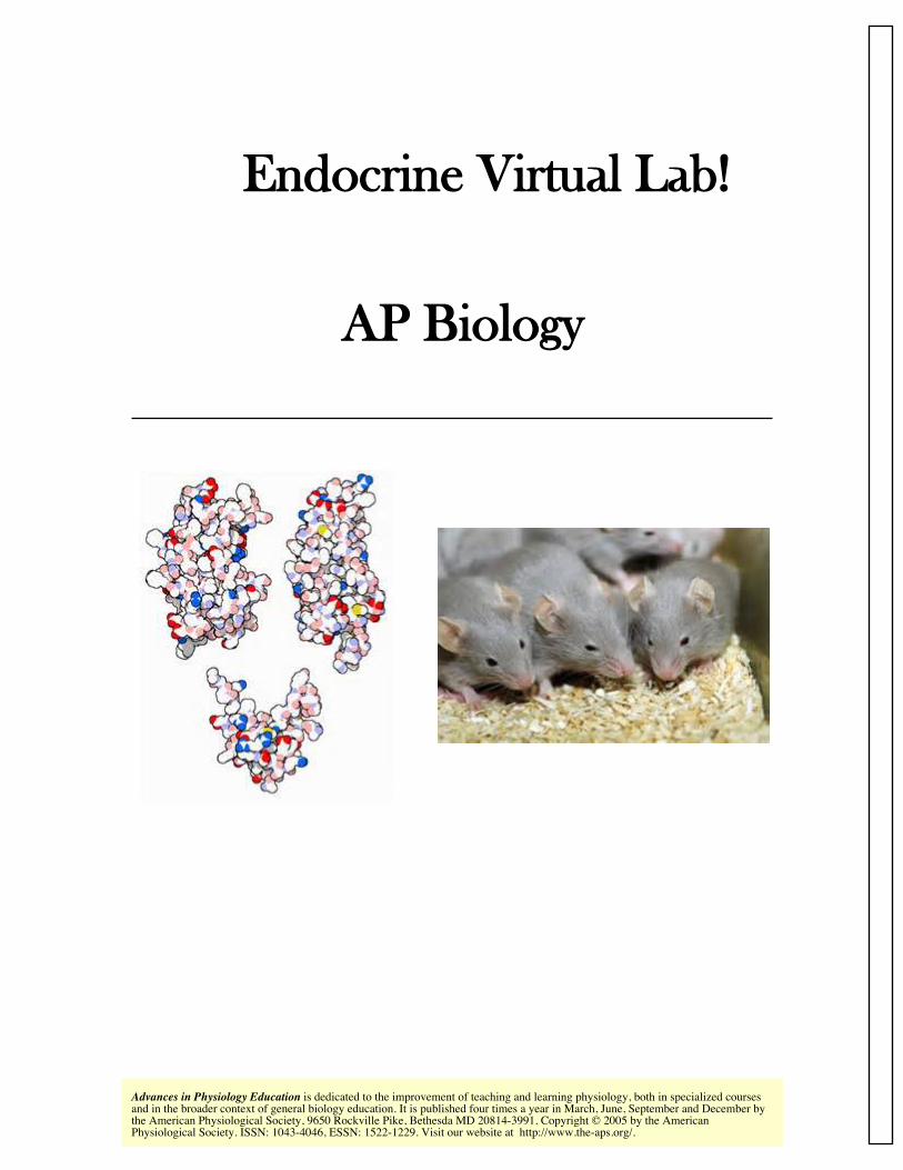
Endocrine Virtual Lab! AP Biology
17
http://www.the-aps.org/. Physiological Society. ISSN: 1043-4046, ESSN: 1522-1229. Visit our website at the American Physiological Society, 9650 Rockville Pike, Bethesda MD 20814-3991. Copyright © 2005 by the American and in the broader context of general biology education. It is published four times a year in March, June, September and December by is dedicated to the improvement of teaching and learning physiology, both in specialized courses Advances in Physiology Education Endocrine Virtual Lab! AP Biology
-
Upload
khangminh22 -
Category
Documents
-
view
0 -
download
0
Transcript of Endocrine Virtual Lab! AP Biology
http://www.the-aps.org/.Physiological Society. ISSN: 1043-4046, ESSN: 1522-1229. Visit our website at the American Physiological Society, 9650 Rockville Pike, Bethesda MD 20814-3991. Copyright © 2005 by the Americanand in the broader context of general biology education. It is published four times a year in March, June, September and December by
is dedicated to the improvement of teaching and learning physiology, both in specialized coursesAdvances in Physiology Education
Endocrine Virtual Lab!
AP Biology

LABORATORY EXERCISE
Objectives
A) To introduce the relationship between the hypo-thalamus and the pituitary gland;
B) To introduce various hormones and explain theireffects
C) To encourage small group discussion and enhanceanalytic thinking;
D) To have the student apply what he or she haslearned to an experimental situation by identifying anunknown hormone.
Time Requir ed
One block period plus one
regular period.
BACKGROUND AND KEY TERMS RELATED TOTHIS EXERCISE
Questions are integrated throughout the text to serveas a source of discussion and aid in the understandingof key points. Questions marked with arrows (=) areused to review the previous passage, and questionsmarked with asterisks (**) are used to provoke thoughton upcoming passages.
The organs of the body communicate with each otherthrough the nervous and endocrine systems to coordi-nate their activities. The nervous system uses neuro-transmitters and neurons to convey information toand from the brain. In contrast, the endocr inesystem uses hormones, which are chemical messen-gers produced by specific tissues in the body, totransmit information. These hormones travel throughthe bloodstream to exert their effects on distant targetorgans.
In a similar manner, people communicate with eachother by using telephones and the postal service. Thebody’s nervous system is comparable to the telephonesystem because it sends fast, direct messages. Theendocrine system is comparable to the postal servicebecause the delivery of the message is slower. Likebulk mail, the message is more diffuse (reaches agreater area) and affects more than one person ororgan. Although the hormone travels through thebody via the blood, it can only affect those cells withreceptors for that specific hormone. Hormones are aslower method of communication, but their effectslast longer.
The command center for the endocrine system is thehypothalamus, a small, penny-sized portion of thebrain. The hypothalamus acts as an endocrine organthat secretes oxytocin and anti-diuretic hormone(ADH, also known as vasopressin). These hormonestravel down the pituitary stalk to the posterior pitu-itary gland where they are released directly into thebloodstream. In addition, the hypothalamus also regu-lates anterior pituitary gland function through the
page 1
Manuscript # S20-6.12e /vm (Disk)
secretion of releasing hormones: thyroid-releasinghormone (TRH), corticotropin-releasing hormone(CRH), and gonadotropin-releasing hormone (GnRH).
These releasing hormones travel through a specializedblood vessel system (known as the hypothalamic-hypophysial portal system) that connects the hypo-thalamus to the anterior pituitary gland. From here,they stimulate the synthesis and secretion of anteriorpituitary hormones, which include thyroid-stimulat-ing hormone (TSH), luteinizing hormone (LH), follicle-stimulating hormone (FSH), growth hormone (GH),adrenocorticotropin hormone (ACTH), and prolactin.Each of these hormones is released into the blood-stream to affect specific target organs. For example,the hypothalamus secretes TRH, which travels to thepituitary gland to release TSH; TSH travels to thethyroid gland (the target organ) and stimulates therelease of thyroid hormone. It is important to note thatthe hypothalamic releasing hormones are only re-quired for the synthesis and release of the anteriorpituitary hormones. The posterior pituitary hormonesare synthesized by the hypothalamus and travel downneurons to be released from the posterior pituitarygland. Because the anterior pituitary gland secretesmultiple hormones, it is frequently referred to as the‘‘master gland.’’ For this experiment, we will focus onthe hypothalamus only as a regulator of the anteriorpituitary gland. Figure 1 shows the relationship be-tween the hypothalamus and the pituitary gland.
= 1) Describe the relationship between the hypothala-mus and the anterior pituitary gland.
= 2) List the hormones released by the anteriorpituitary gland.
= 3) Why is the anterior pituitary called the mastergland?
**4) What is negative feedback?
The release of hormones from the endocrine systemcan be regulated through positive or negative feed-back mechanisms. The negative feedback systemcan be compared with a thermostat set at a predeter-mined temperature (68°F). When the temperaturerises above the set point (72°F), the thermostat
detects the change and activates the air conditioner tocool the room. The thermostat will turn the airconditioner off once the temperature of the roomdrops below the set point (67°F). To keep the room ata fairly constant temperature, the thermostat assessesthe situation and turns the air conditioner on or offaccordingly. Figure 2 illustrates the concept of nega-tive feedback using the above example.
In the endocrine system, negative feedback is used toinhibit further hormone secretion. When a sufficientamount of hormone has been released, it communi-cates or ‘‘feeds back’’ to suppress the releasing organ.In other words, the gland has released enough hor-mone to fulfill its function; this is sensed by the body,and production of the hormone ceases. Negativefeedback not only inhibits the releasing organ, but canalso inhibit the pituitary gland and/or hypothalamus.By using a negative feedback system, the body pro-duces only the amount of hormone it needs withoutwasting its resources. Conversely, in positive feed-back, the end product further stimulates the releasingorgan. This form of feedback is less common.
The pathways of three hormones are examined in thisexperiment: thyroid hormone, cortisol, and testoster-one. The hormonal pathways are similar in all threecases. It is important to realize that the hypothalamussecretes a releasing hormone to regulate each of thehormones secreted from the anterior pituitary gland.In this way, the hypothalamus is like a commandcenter. If the hypothalamus is not stimulated, thehypothalamic releasing hormones (TRH, CRH, andGnRH) will not stimulate the anterior pituitary glandto secrete its hormones.
The hypothalamus releases TRH, which travels to theanterior pituitary gland via the bloodstream to stimu-late production of TSH. TSH travels to the thyroidgland (located by the trachea) to stimulate the produc-tion and release of thyroid hormone. Thyroid hor-mone influences the growth rate of many body tissuesand is necessary for proper central nervous systemdevelopment. Its main function is to increase a per-son’s basal metabolic rate (BMR) and to increase heatproduction. An excess of thyroid hormone can nega-tively feed back to inhibit further thyroid hormonerelease from the thyroid gland, TSH secretion from the
Page 2
Manuscript # S20-6.12e /vm (Disk)

anterior pituitary gland, and/or TRH release from thehypothalamus.
Similarly, ACTH is released from the anterior pituitarygland in response to CRH secreted from the hypothala-mus. ACTH stimulates the adrenal glands (located ontop of the kidneys) to secrete cor tisol, which pro-motes the breakdown of proteins and fats and helpsthe body adapt to stress. Cortisol functions to provide
the body with fuel by breaking down (catabolism) thematerials of the body. Under normal conditions,excess cortisol in the bloodstream will negatively feedback to the hypothalamus (to inhibit CRH release),anterior pituitary gland (to inhibit ACTH secretion),and/or to the adrenal gland (to inhibit further cortisolrelease). The release of CRH is regulated by negativefeedback, circadian rhythms, and stress. Cortisol canalso act as an immunosuppressive and anti-inflamma-
FIG. 1 .Secr etion of hypothalamic hormones. Hypothalamic r eleasing hormones travel down the hypothalamic-hypophysial por tal system to the anter ior pituitary gland, where they stimulate the synthesis and releaseof anter ior pituitary hormones [adrenocor ticotropin hormone (ACTH), thyroid-stimulating hormone(TSH), follicle-stimulating hormone (FSH), luteinizing hormone (LH), growth hormone (GH), andprolactin]. In contrast, the poster ior pituitary gland does not r equir e r eleasing hormones, because thehypothalamus synthesizes and secr etes both anti-diur etic hormone (ADH) and oxytocin.
Manuscript # S20-6.12e /vm (Disk)
Page 3

tory agent. If cortisol is administered in large doses, itsimmunosuppressive properties will cause the organsof the immune system to shrink. In this experiment,the thymus gland will represent the organs of theimmune system.
= 5) Describe the effects of thyroid hormone.
= 6) Describe the effects of cortisol.
** 7) Describe the role of LH in both males andfemales.
LH is released from the anterior pituitary gland inresponse to GnRH secreted from the hypothalamus.LH is seen in both males and females but has differentfunctions. In the male, LH travels to the Leydig cellsthat are located in the connective tissue between theseminiferous tubules of the testes. The Leydig cells re-lease testosterone, which is responsible for the malesex drive and secondary sex characteristics, such asincreased body hair and a deeper voice. An excess oftestosterone can cause an increase (anabolic) in musclemass. Negative effects of testosterone are male patternbaldness and increased secretion of the sebaceousglands, which can lead to acne. Figure 3 presents therelative anatomy of the male reproductive tract.
In the female, LH causes the follicle (developing egg)in the ovary to secrete estrogen . Estrogen partici-pates in either a positive or negative feedback loop,depending on the stage of the menstrual cycle. In thepreovulatory and postovulatory phases, estrogen regu-lates the release of LH through negative feedback.However, there is a large rise in levels of LH justbefore ovulation (release of the egg from the ovary)due to a positive feedback mechanism. During thisinterval, the secretion of estrogen from the folliclefurther stimulates the release of LH from the anteriorpituitary gland. The increased levels of LH are essen-tial for ovulation to occur. Estrogen causes the devel-opment of female secondary sex characteristics and
FIG. 2.Negative feedback compared with a thermostat control-ling room temperatur e. Solid lines and (1) r epr esentan enhancement of activity, whereas dotted lines and(2) r epr esent an inhibition of activity.
FIG. 3.Organs of the male r eproductive tract.
I N N O V A T I O N S A N D I D E A S
Page 4
Manuscript # S20-6.12e /vm (Disk)
sustains the female reproductive tract. Awoman wholacks ovaries (and therefore follicles) will not produceestrogen. However, the pituitary gland will secreteexcess LH because the feedback inhibition no longerexists. Excess levels of estrogen cause early sexualdevelopment in the female as do high levels oftestosterone in males.
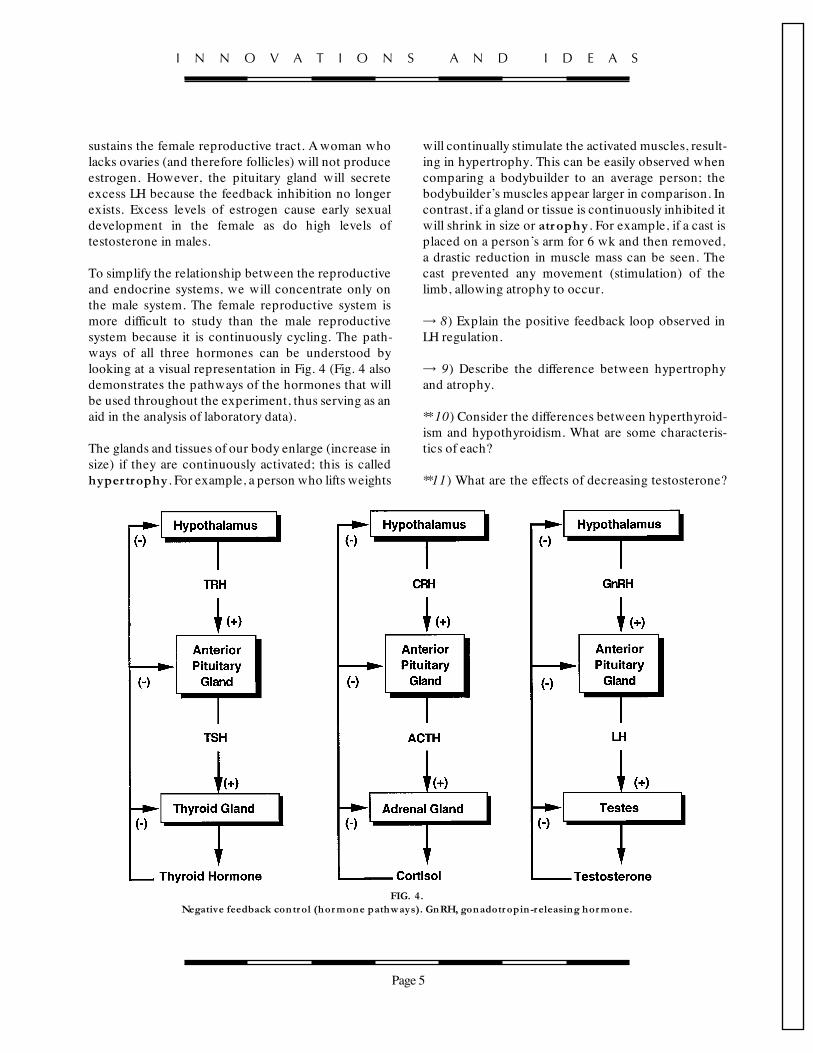
To simplify the relationship between the reproductiveand endocrine systems, we will concentrate only onthe male system. The female reproductive system ismore difficult to study than the male reproductivesystem because it is continuously cycling. The path-ways of all three hormones can be understood bylooking at a visual representation in Fig. 4 (Fig. 4 alsodemonstrates the pathways of the hormones that willbe used throughout the experiment, thus serving as anaid in the analysis of laboratory data).
The glands and tissues of our body enlarge (increase insize) if they are continuously activated; this is calledhypertrophy . For example, a person who lifts weights
will continually stimulate the activated muscles, result-ing in hypertrophy. This can be easily observed whencomparing a bodybuilder to an average person; thebodybuilder’s muscles appear larger in comparison. Incontrast, if a gland or tissue is continuously inhibited itwill shrink in size or atrophy . For example, if a cast isplaced on a person’s arm for 6 wk and then removed,a drastic reduction in muscle mass can be seen. Thecast prevented any movement (stimulation) of thelimb, allowing atrophy to occur.
= 8) Explain the positive feedback loop observed inLH regulation.
= 9) Describe the difference between hypertrophyand atrophy.
**10) Consider the differences between hyperthyroid-ism and hypothyroidism. What are some characteris-tics of each?
**11) What are the effects of decreasing testosterone?
FIG. 4.Negative feedback control (hormone pathways). GnRH, gonadotropin-r eleasing hormone.
I N N O V A T I O N S A N D I D E A S
Page 5
Manuscript # S20-6.12e /vm (Disk)
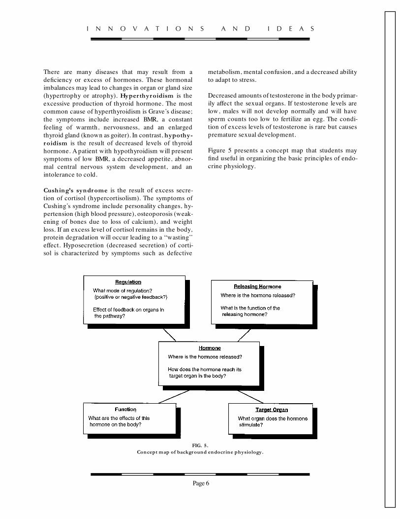
There are many diseases that may result from adeficiency or excess of hormones. These hormonalimbalances may lead to changes in organ or gland size(hypertrophy or atrophy). Hyper thyroidism is theexcessive production of thyroid hormone. The mostcommon cause of hyperthyroidism is Grave’s disease;the symptoms include increased BMR, a constantfeeling of warmth, nervousness, and an enlargedthyroid gland (known as goiter). In contrast, hypothy-roidism is the result of decreased levels of thyroidhormone. Apatient with hypothyroidism will presentsymptoms of low BMR, a decreased appetite, abnor-mal central nervous system development, and anintolerance to cold.
Cushing’s syndrome is the result of excess secre-tion of cortisol (hypercortisolism). The symptoms ofCushing’s syndrome include personality changes, hy-pertension (high blood pressure), osteoporosis (weak-ening of bones due to loss of calcium), and weightloss. If an excess level of cortisol remains in the body,protein degradation will occur leading to a ‘‘wasting’’effect. Hyposecretion (decreased secretion) of corti-sol is characterized by symptoms such as defective
metabolism, mental confusion, and a decreased abilityto adapt to stress.
Decreased amounts of testosterone in the body primar-ily affect the sexual organs. If testosterone levels arelow, males will not develop normally and will havesperm counts too low to fertilize an egg. The condi-tion of excess levels of testosterone is rare but causespremature sexual development.
Figure 5 presents a concept map that students mayfind useful in organizing the basic principles of endo-crine physiology.
FIG. 5.Concept map of background endocr ine physiology.
I N N O V A T I O N S A N D I D E A S
Page 6
Manuscript # S20-6.12e /vm (Disk)

The data for this laboratory were compiled from sevensets of male laboratory rats, two rats per set; one setwas the control group and the remaining six wereexperimental groups. The rats were all male to sim-plify the study of the relationship between the repro-ductive and endocrine systems. In each set of ratsthere was an ‘‘intact’’ rat and a ‘‘castrate’’ rat. Thecastration involved removal of the testes to eliminatetestosterone production. The two rats (normal andcastrate) of each group were treated alike in all otherways (food, water, etc.). All rats, except for those inthe control group were injected with a hormone on adaily basis for 2 wk. Autopsies were performed on theanimals at that time.
The group of students performing this exercise werevery disorganized and rushed through the work,making errors in labeling the bottles of hormone. Thestudents obtained the following results for organweights after the autopsies were performed. In thisshort period of time, the students noted amazingchanges in the size of certain organs when theycompared the experimental group of rats with thecontrol group. Using the flowchart (Fig. 4), Table 1,and the autopsy data, match the unknown rat groupswith their respective hormones. The bottles on the
TABLE 1Compar ison of hormonal effects on differ ent organs (to be completed by students)
TRH TSH ACTH CortisolTestosterone LH
Intact Castrate Intact Castrate
Pituitary gland
Thyroid gland
Adrenal glands
Thymus gland
Testes
Prostate
Seminal vesicles
Body weight
A1 denotes an increase in size. A2 denotes a decrease in size. Place the letters NC in the box where no change occurs. TRH, thyroid-releasinghormone; TSH, thyroid-stimulating hormone; ACTH, adrenocorticotropin hormone; LH, luteinizing hormone.
Page 7
Manuscript # S20-6.12e /vm (Disk)
LABORATORY PROCEDURE
refrigerator shelf were ACTH, cortisol, LH, TSH, TRH,and testosterone.

To help in determining the identity of the unknownhormones, the student should look for changes be-tween the control values and the values of theunknown hormone (both the intact and castrateanimal). The changes between the control rats and therats that were treated with the unknown hormoneshould be .20% if they are to be considered signifi-cantly different. If the change is ,20%, it is attributedto experimental or biological error. Experimentalerrors may include small errors in calibration proce-dures, measurements, or instrumentation. Any variabil-ity that occurs because of the differences betweenanimals is considered biological error.
Figure 6 represents the organs of the rats used in theexperiment.
Figure 7 shows your set of control rats; the data arethe results of the autopsy.
Determine the identity of horm one 1 using the datafrom the autopsy listed in Fig. 8, Table 1, and Fig. 4.
Determine the identity of horm one 2 using the datafrom the autopsy listed in Fig. 9, Table 1, and Fig. 4.
Determine the identity of horm one 3 using the datafrom the autopsy listed in Fig. 10, Table 1, and Fig. 4.
Determine the identity of horm one 4 using the datafrom the autopsy listed in Fig. 11, Table 1, and Fig. 4.
Determine the identity of horm one 5 using the datafrom the autopsy listed in Fig. 12, Table 1, and Fig. 4.
Determine the identity of horm one 6 using thedata from the autopsy listed in Fig. 13, Table 1, andFig. 4.
ANALYSIS
1) What was horm one 1?Explain your answer.
2) What was horm one 2?Explain your answer.
3) What was horm one 3?Explain your answer.
4) What was horm one 4?Explain your answer.
FIG. 6.Graphic r epr esentation of organs studied in theautopsy.
FIG. 7.Autopsy r esults fr om control rats.
I N N O V A T I O N S A N D I D E A S
Page 8
Manuscript # S20-6.12e /vm (Disk)

5) What was horm one 5?Explain your answer.
6) What was horm one 6?Explain your answer.
FIG. 8.Autopsy r esults fr om rats tr eated with hormone 1.
I N N O V A T I O N S A N D I D E A S
Page 9
Manuscript # S20-6.12e /vm (Disk)

FIG. 9.Autopsy r esults fr om rats tr eated with hormone 2.
I N N O V A T I O N S A N D I D E A S
Page 10
Manuscript # S20-6.12e /vm (Disk)
MALA KRISHNA
MALA KRISHNA

FIG. 1 0.Autopsy r esults fr om rats tr eated with hormone 3.
I N N O V A T I O N S A N D I D E A S
Page 11
Manuscript # S20-6.12e /vm (Disk)

FIG. 1 1 .Autopsy r esults fr om rats tr eated with hormone 4.
I N N O V A T I O N S A N D I D E A S
Page 12
Manuscript # S20-6.12e /vm (Disk)

FIG. 1 2.Autopsy r esults fr om rats tr eated with hormone 5.
I N N O V A T I O N S A N D I D E A S
Page 13
Manuscript # S20-6.12e /vm (Disk)

FIG. 1 3.Autopsy r esults fr om rats tr eated with hormone 6.
I N N O V A T I O N S A N D I D E A S
Page 14
Manuscript # S20-6.12e /vm (Disk)