AP PSYCHOLOGY
84
* AP PSYCHOLOGY Adapted from Aneeq Ahmad Henderson State University Worth Publishers, © 2006
-
Upload
khangminh22 -
Category
Documents
-
view
0 -
download
0
Transcript of AP PSYCHOLOGY

*
AP PSYCHOLOGY
Adapted from Aneeq Ahmad
Henderson State University
Worth Publishers, © 2006
*
Neuroscience and
Behavior
*
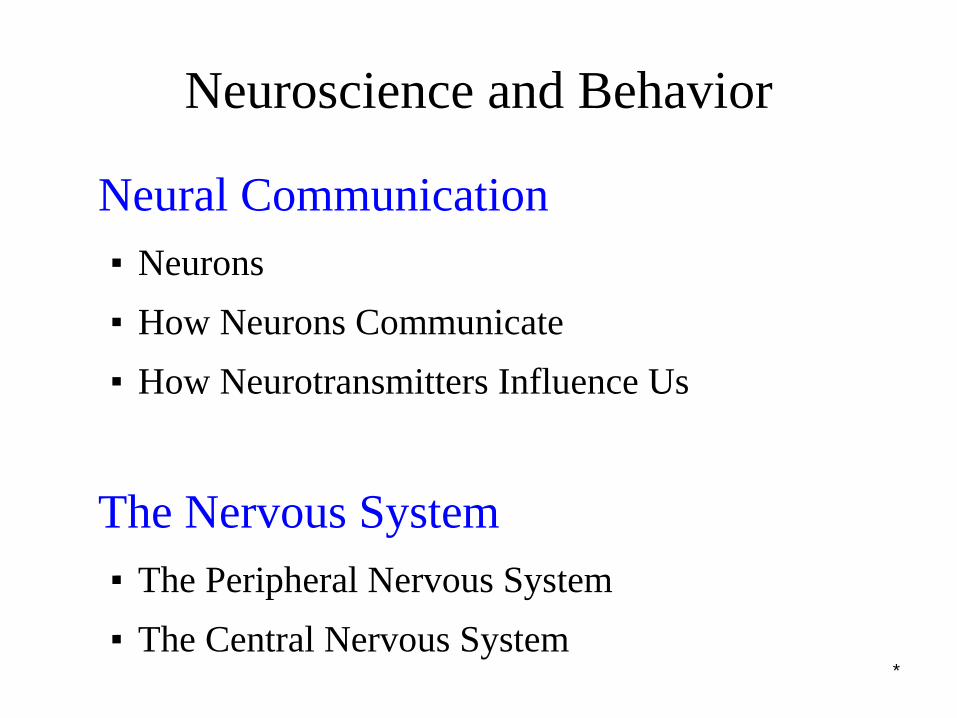
Neuroscience and Behavior
Neural Communication
▪ Neurons
▪ How Neurons Communicate
▪ How Neurotransmitters Influence Us
The Nervous System
▪ The Peripheral Nervous System
▪ The Central Nervous System
*
Neuroscience and Behavior
The Endocrine System
The Brain
▪ The Tools of Discovery
▪ Older Brain Structures
▪ The Cerebral Cortex
▪ Our Divided Brain
▪ Left Brain-Right Brain
*
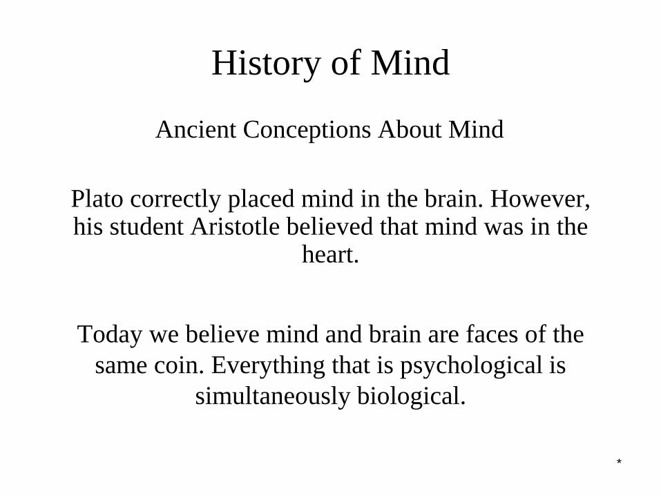
History of Mind
Plato correctly placed mind in the brain. However, his student Aristotle believed that mind was in the
heart.
Ancient Conceptions About Mind
Today we believe mind and brain are faces of the
same coin. Everything that is psychological is
simultaneously biological.
*
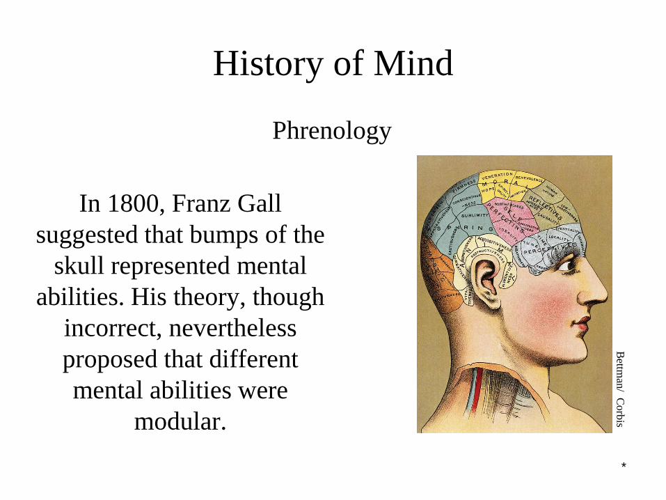
History of Mind
In 1800, Franz Gall
suggested that bumps of the
skull represented mental
abilities. His theory, though
incorrect, nevertheless
proposed that different
mental abilities were
modular.
Phrenology
Bettm
an/ C
orb
is
*
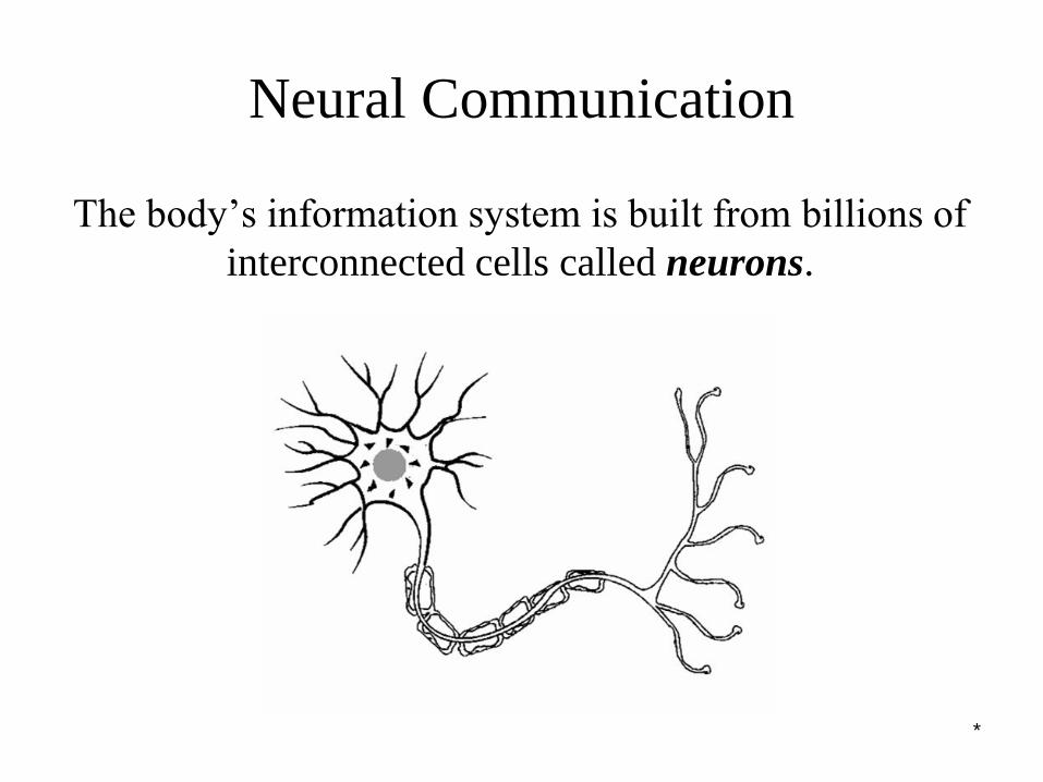
Neural Communication
The body’s information system is built from billions of
interconnected cells called neurons.
*
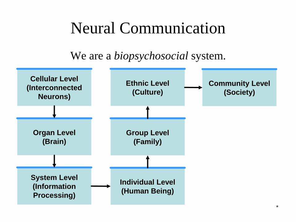
Neural Communication
We are a biopsychosocial system.
Cellular Level
(Interconnected
Neurons)
Organ Level
(Brain)
System Level
(Information
Processing)
Individual Level
(Human Being)
Group Level
(Family)
Ethnic Level
(Culture)
Community Level
(Society)
*
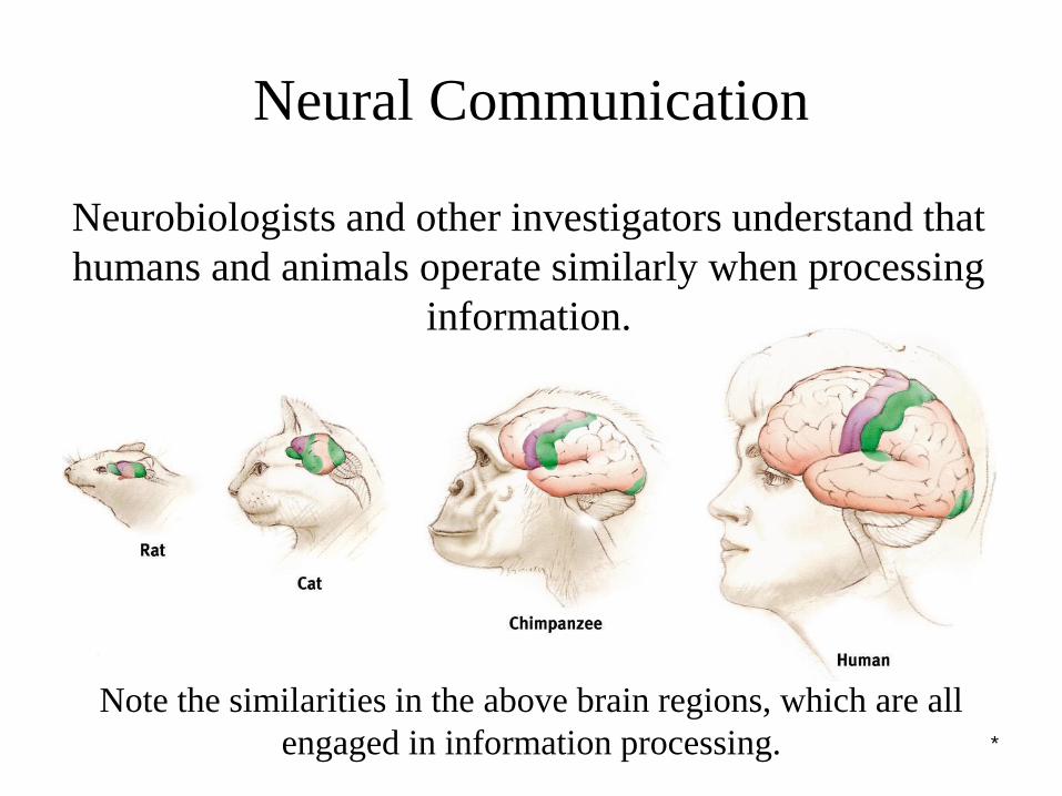
Neural Communication
Note the similarities in the above brain regions, which are all
engaged in information processing.
Neurobiologists and other investigators understand that
humans and animals operate similarly when processing
information.
*
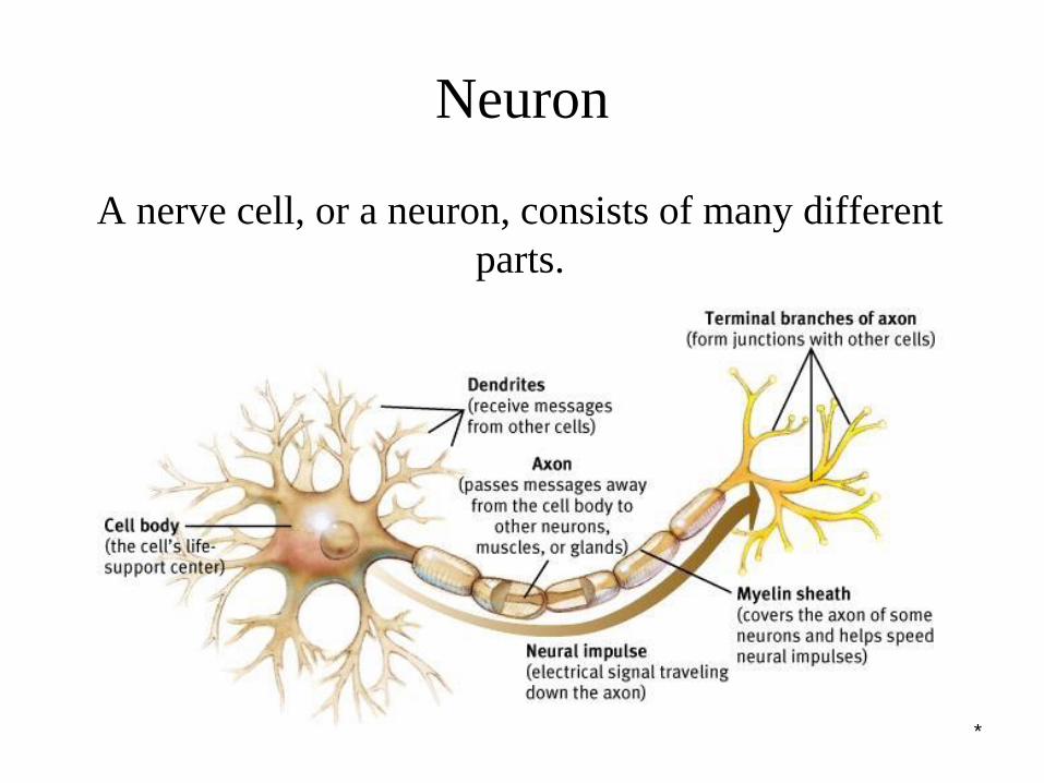
Neuron
A nerve cell, or a neuron, consists of many different
parts.
*
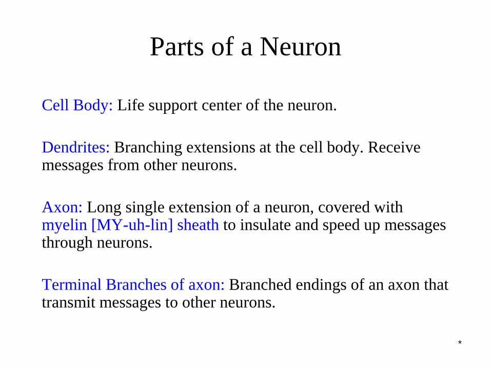
Parts of a Neuron
Cell Body: Life support center of the neuron.
Dendrites: Branching extensions at the cell body. Receive messages from other neurons.
Axon: Long single extension of a neuron, covered with myelin [MY-uh-lin] sheath to insulate and speed up messages through neurons.
Terminal Branches of axon: Branched endings of an axon that transmit messages to other neurons.
*
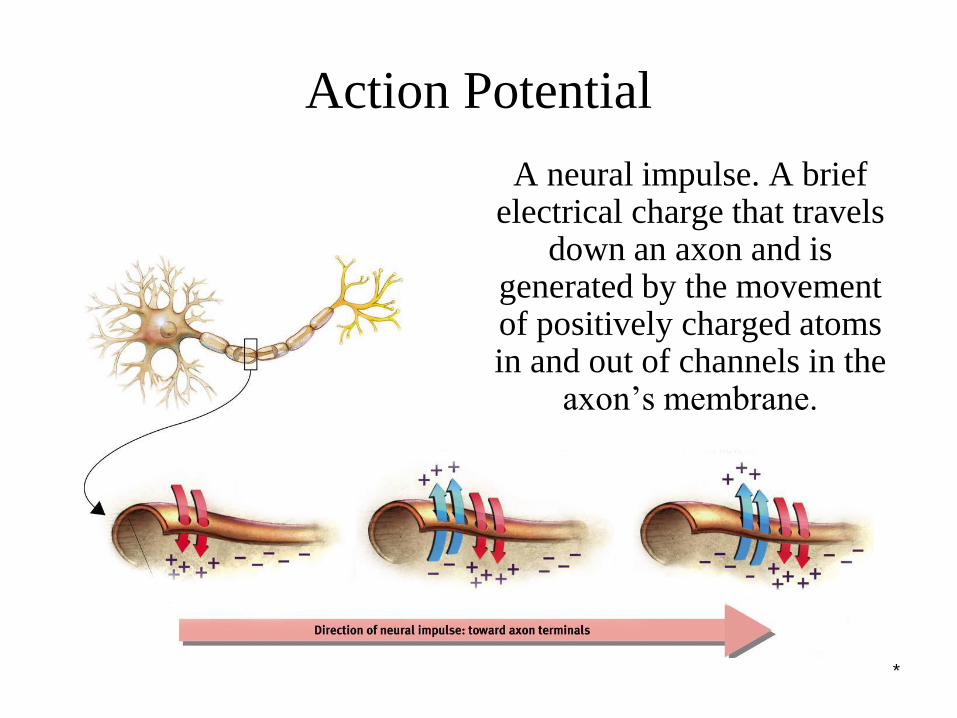
Action Potential
A neural impulse. A brief electrical charge that travels
down an axon and is generated by the movement of positively charged atoms in and out of channels in the
axon’s membrane.
*
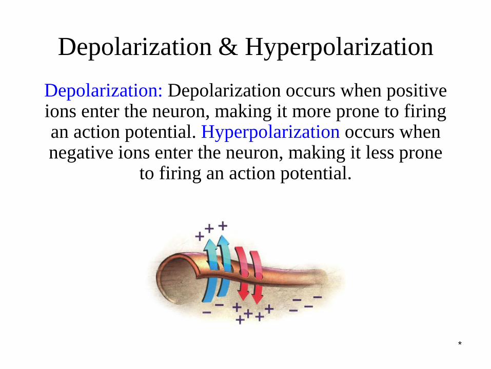
Depolarization & Hyperpolarization
Depolarization: Depolarization occurs when positive ions enter the neuron, making it more prone to firing an action potential. Hyperpolarization occurs when negative ions enter the neuron, making it less prone
to firing an action potential.
*
Threshold
Threshold: Each neuron receives depolarizing and
hyperpolarizing currents from many neurons. When
the depolarizing current (positive ions) minus the
hyperpolarizing current (negative ions) exceed
minimum intensity (threshold) the neuron fires an
action potential.
*
Refractory Period & Pumps
Refractory Period: After a neuron fires an action
potential it pauses for a short period to recharge itself
to fire again.
Sodium-Potassium Pumps: Sodium-potassium
pumps pump positive ions out from the inside of the
neuron, making them ready for another action
potential.
*
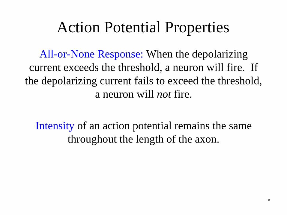
Action Potential Properties
All-or-None Response: When the depolarizing
current exceeds the threshold, a neuron will fire. If
the depolarizing current fails to exceed the threshold,
a neuron will not fire.
Intensity of an action potential remains the same
throughout the length of the axon.
*
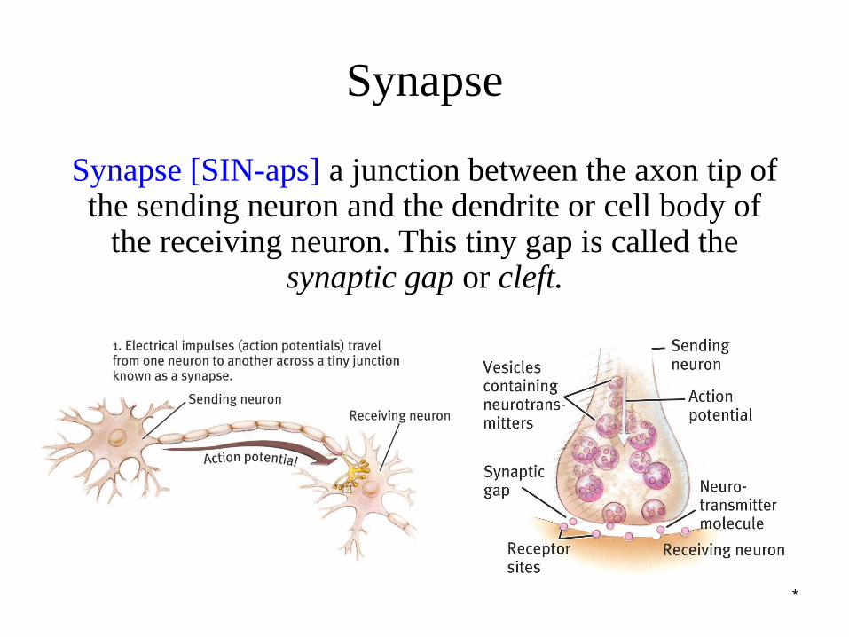
Synapse
Synapse [SIN-aps] a junction between the axon tip of the sending neuron and the dendrite or cell body of
the receiving neuron. This tiny gap is called the synaptic gap or cleft.
*
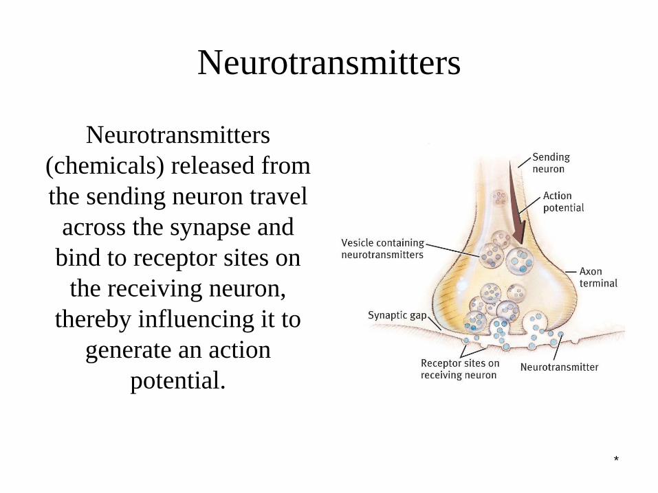
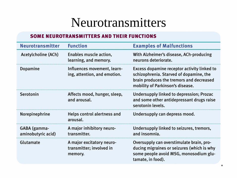
Neurotransmitters
Neurotransmitters
(chemicals) released from
the sending neuron travel
across the synapse and
bind to receptor sites on
the receiving neuron,
thereby influencing it to
generate an action
potential.
*
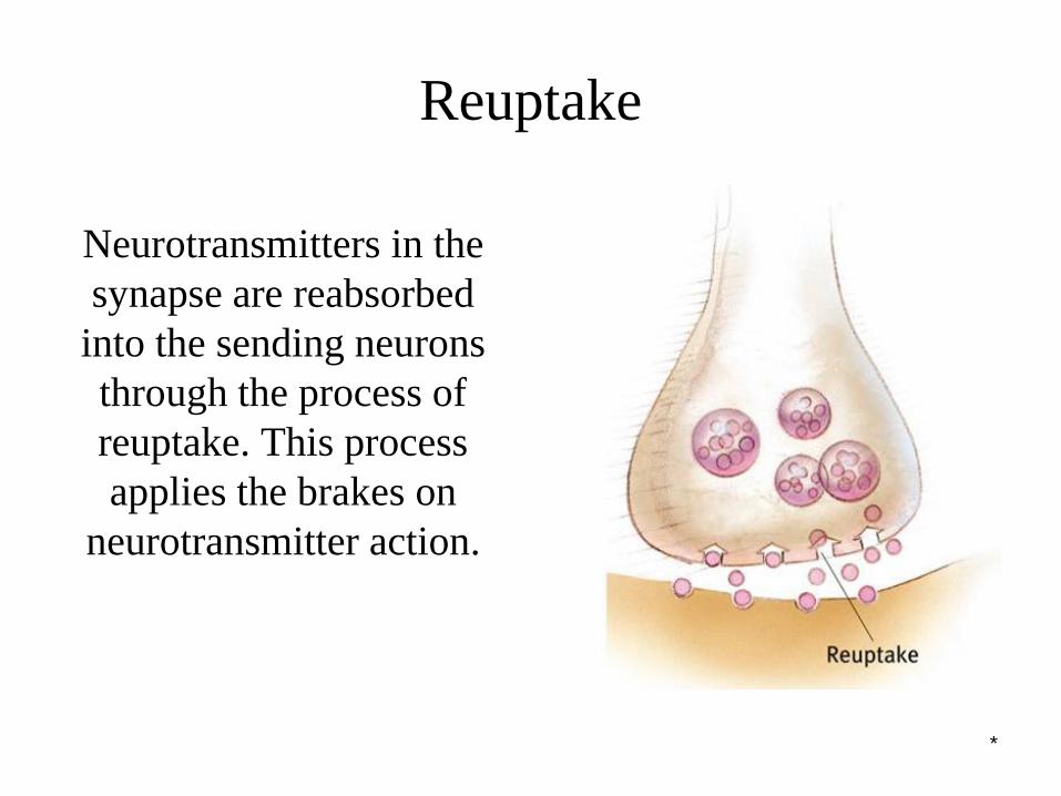
Reuptake
Neurotransmitters in the
synapse are reabsorbed
into the sending neurons
through the process of
reuptake. This process
applies the brakes on
neurotransmitter action.
*
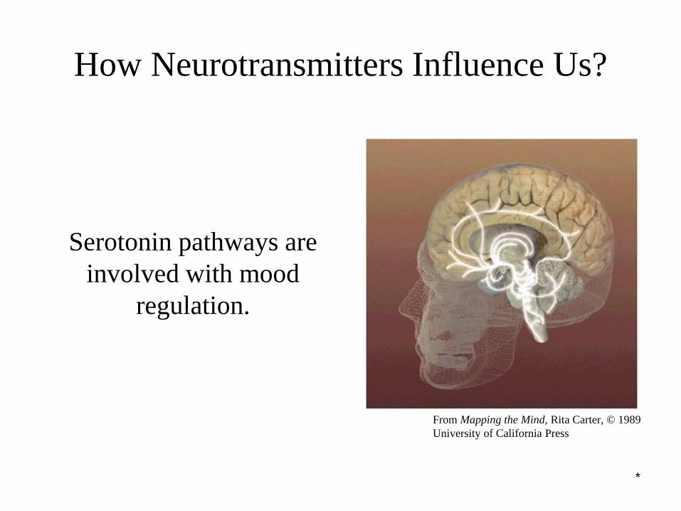
How Neurotransmitters Influence Us?
Serotonin pathways are
involved with mood
regulation.
From Mapping the Mind, Rita Carter, © 1989
University of California Press
*
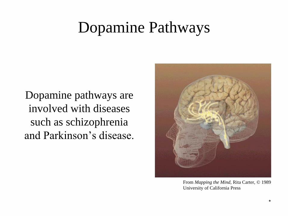
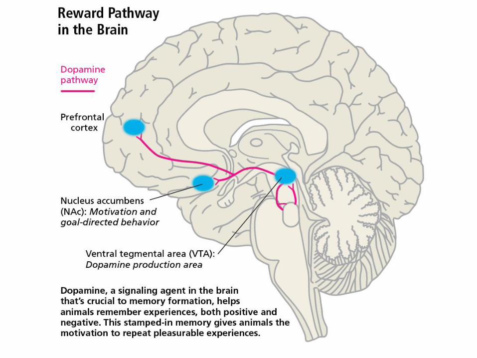
Dopamine Pathways
Dopamine pathways are
involved with diseases
such as schizophrenia
and Parkinson’s disease.
From Mapping the Mind, Rita Carter, © 1989
University of California Press
*
Neurotransmitters
*
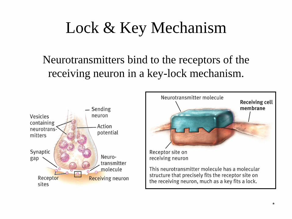
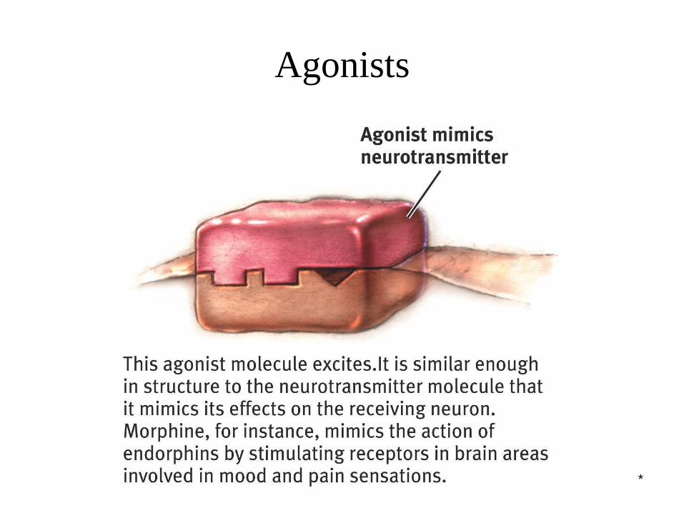
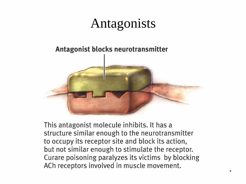
Lock & Key Mechanism
Neurotransmitters bind to the receptors of the
receiving neuron in a key-lock mechanism.
*
Agonists
*
Antagonists
*
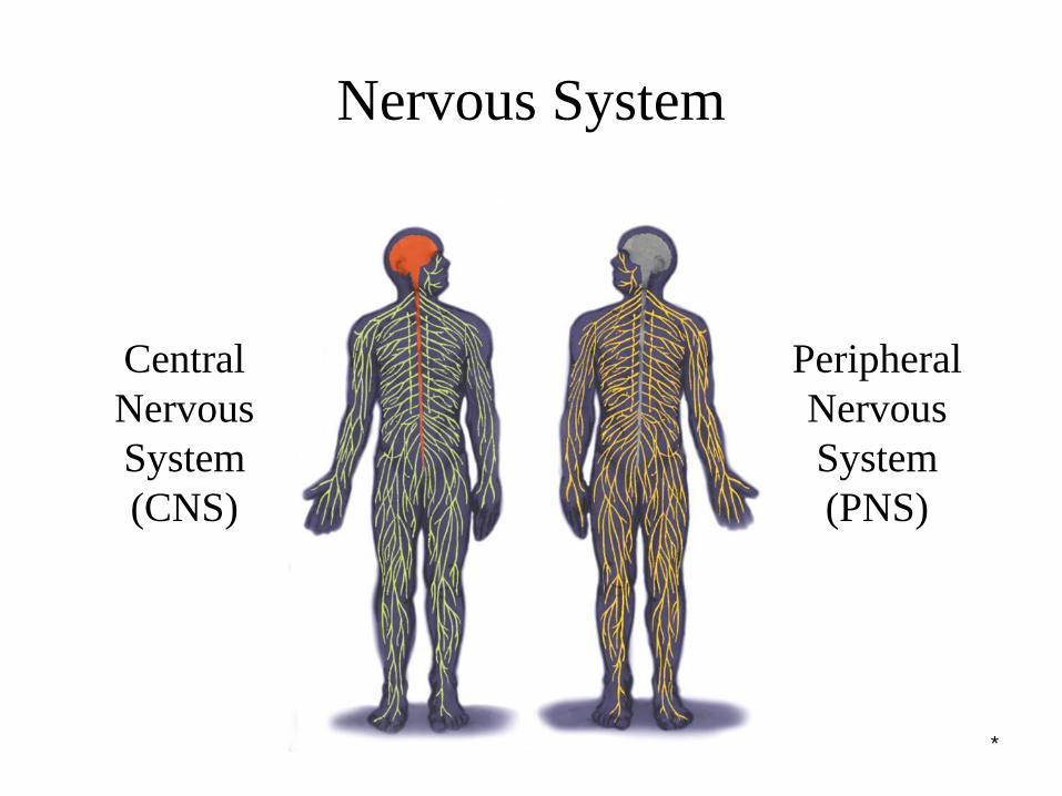
Nervous System
Central
Nervous
System
(CNS)
Peripheral
Nervous
System
(PNS)
*
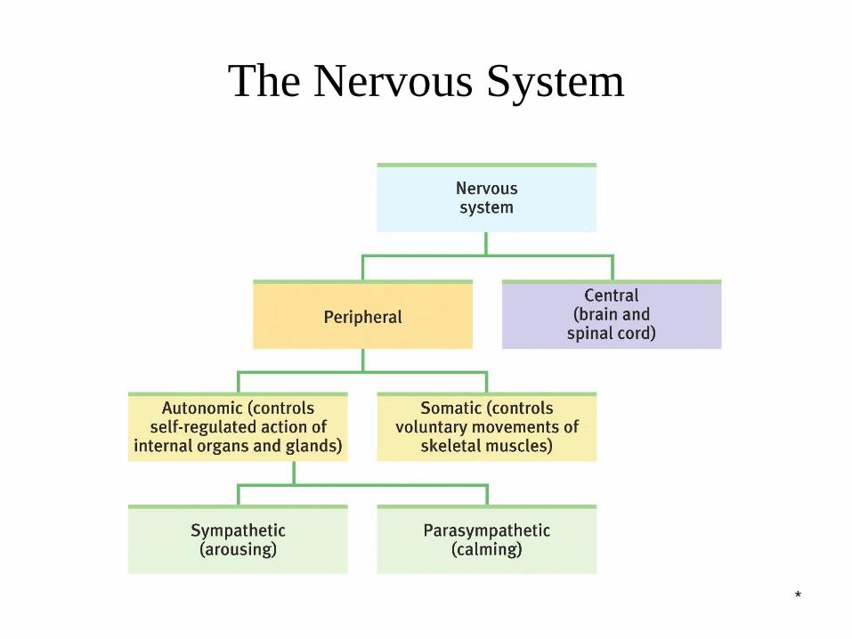
The Nervous System
Nervous System: Consists of all the nerve cells. It is the body’s speedy, electrochemical communication system.
Central Nervous System (CNS): the brain and spinal cord.
Peripheral Nervous System (PNS): the sensory and motor neurons that connect the central nervous system (CNS) to the rest of the body.
*
The Nervous System
*
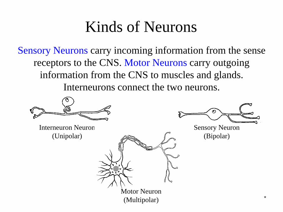
Kinds of Neurons
Sensory Neurons carry incoming information from the sense
receptors to the CNS. Motor Neurons carry outgoing
information from the CNS to muscles and glands.
Interneurons connect the two neurons.
Sensory Neuron
(Bipolar)
Interneuron Neuron
(Unipolar)
Motor Neuron
(Multipolar)
*
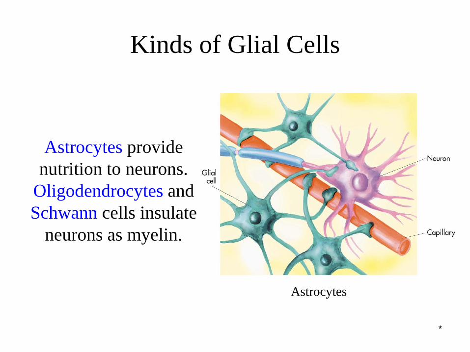
Kinds of Glial Cells
Astrocytes provide
nutrition to neurons.
Oligodendrocytes and
Schwann cells insulate
neurons as myelin.
Astrocytes

*
Peripheral Nervous System
Somatic Nervous System: The division of the peripheral
nervous system that controls the body’s skeletal
muscles.
Autonomic Nervous System: Part of the PNS that
controls the glands and other muscles.
*
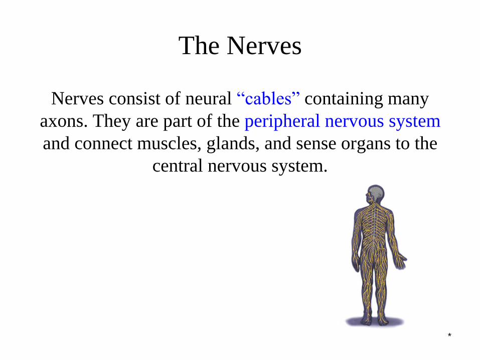
The Nerves
Nerves consist of neural “cables” containing many
axons. They are part of the peripheral nervous system
and connect muscles, glands, and sense organs to the
central nervous system.
*
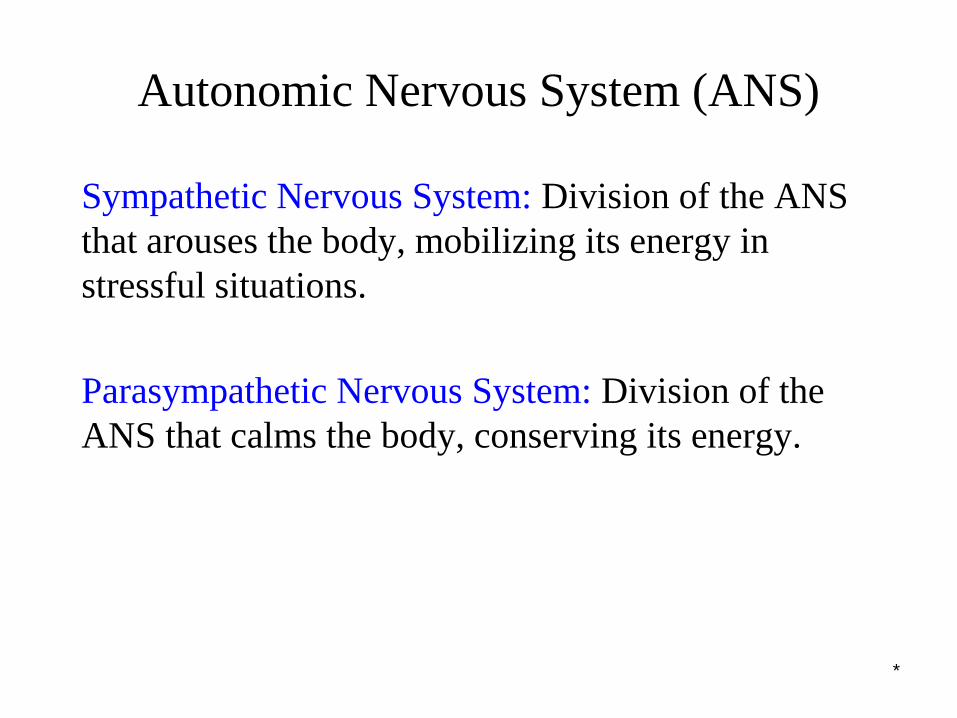
Autonomic Nervous System (ANS)
Sympathetic Nervous System: Division of the ANS
that arouses the body, mobilizing its energy in
stressful situations.
Parasympathetic Nervous System: Division of the
ANS that calms the body, conserving its energy.

*
Autonomic Nervous System (ANS)
Sympathetic NS “Arouses”
(fight-or-flight)
Parasympathetic NS “Calms”
(rest and digest)
*
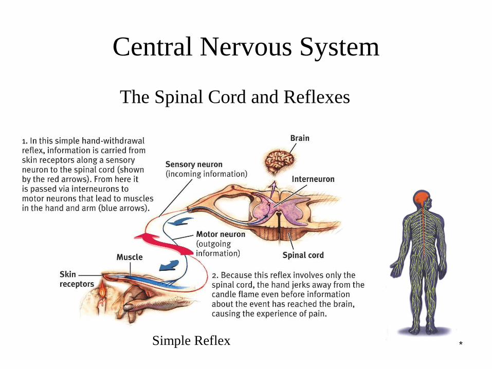
Central Nervous System
The Spinal Cord and Reflexes
Simple Reflex
*
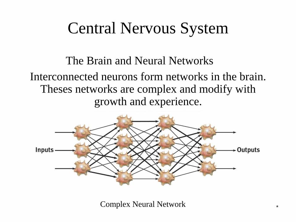
Central Nervous System
The Brain and Neural Networks
Complex Neural Network
Interconnected neurons form networks in the brain. Theses networks are complex and modify with
growth and experience.
*
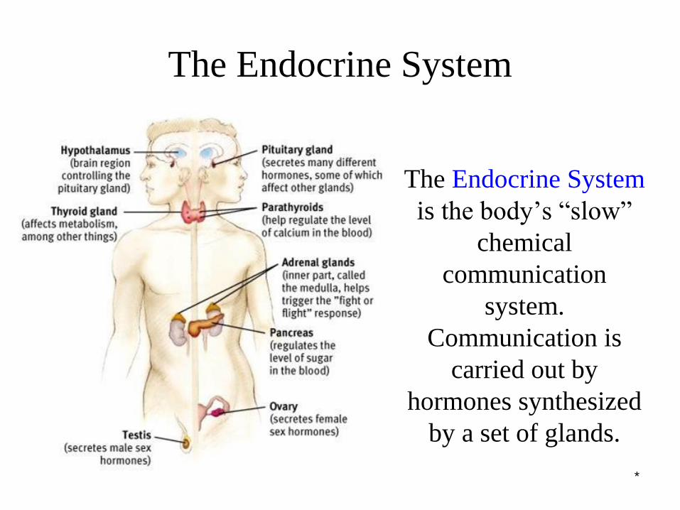
The Endocrine System
The Endocrine System
is the body’s “slow”
chemical
communication
system.
Communication is
carried out by
hormones synthesized
by a set of glands.
*
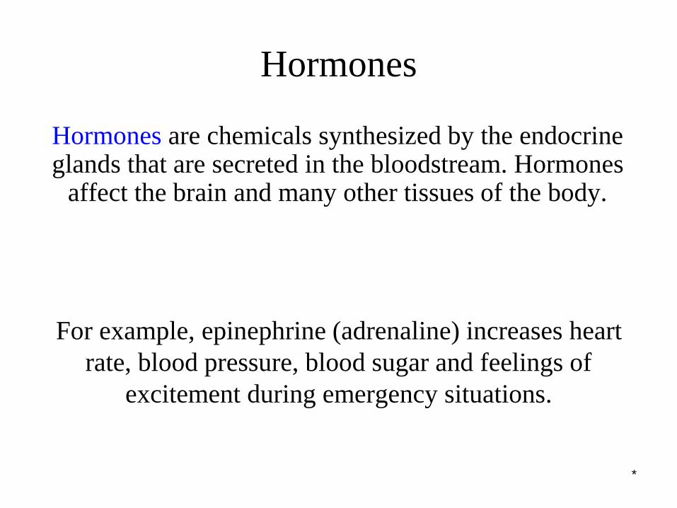
Hormones
Hormones are chemicals synthesized by the endocrine glands that are secreted in the bloodstream. Hormones
affect the brain and many other tissues of the body.
For example, epinephrine (adrenaline) increases heart
rate, blood pressure, blood sugar and feelings of
excitement during emergency situations.
*
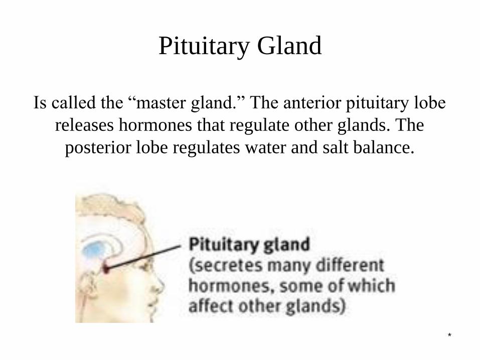
Pituitary Gland
Is called the “master gland.” The anterior pituitary lobe
releases hormones that regulate other glands. The
posterior lobe regulates water and salt balance.
*
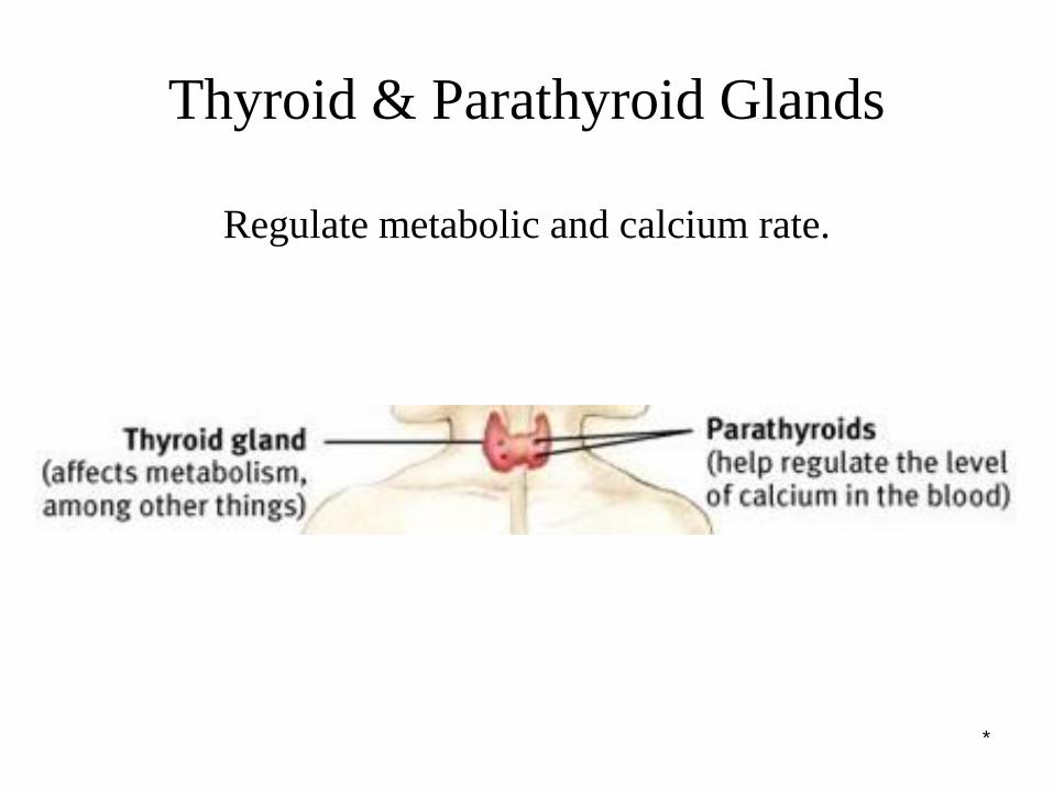
Thyroid & Parathyroid Glands
Regulate metabolic and calcium rate.
*
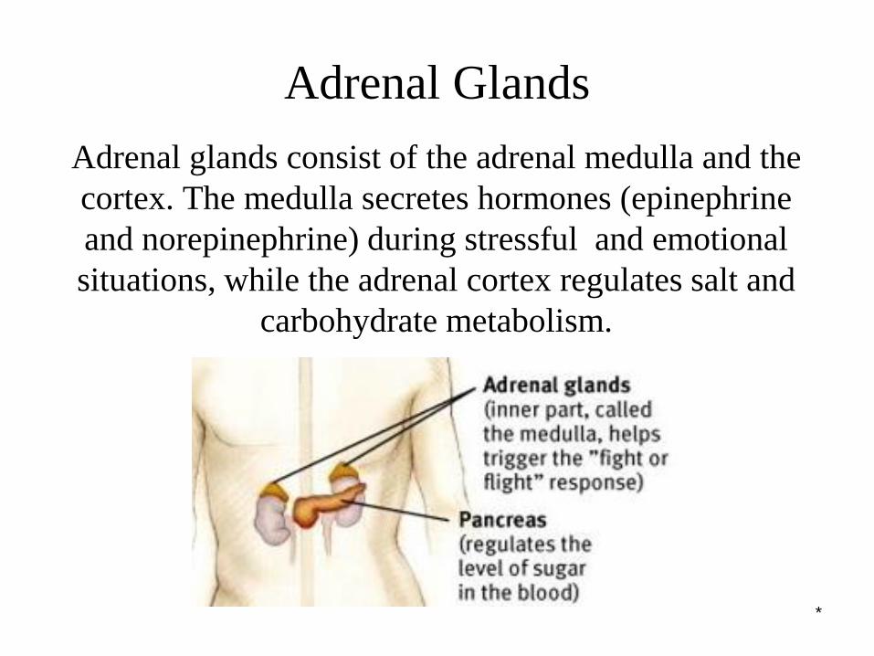
Adrenal Glands
Adrenal glands consist of the adrenal medulla and the
cortex. The medulla secretes hormones (epinephrine
and norepinephrine) during stressful and emotional
situations, while the adrenal cortex regulates salt and
carbohydrate metabolism.
*
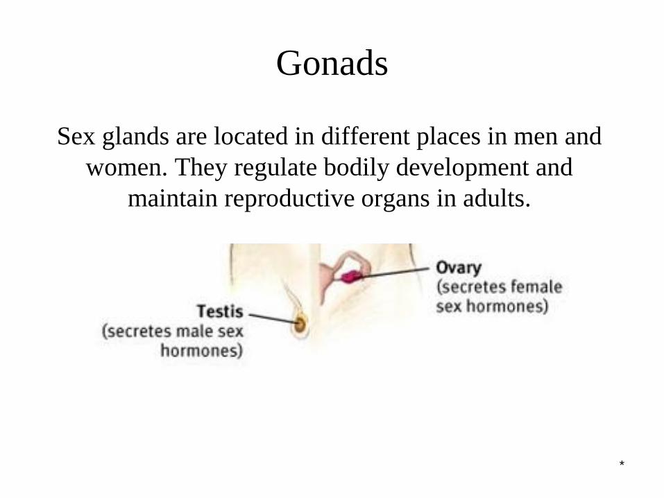
Gonads
Sex glands are located in different places in men and
women. They regulate bodily development and
maintain reproductive organs in adults.
*
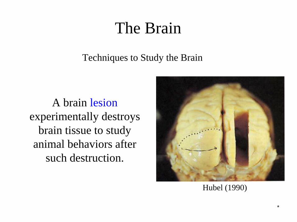
The Brain
Techniques to Study the Brain
A brain lesion
experimentally destroys
brain tissue to study
animal behaviors after
such destruction.
Hubel (1990)
*
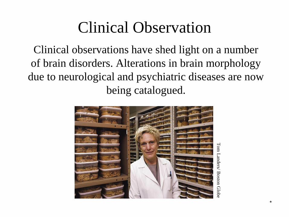
Clinical Observation
Clinical observations have shed light on a number
of brain disorders. Alterations in brain morphology
due to neurological and psychiatric diseases are now
being catalogued.
Tom
Lan
ders/ B
osto
n G
lobe
*
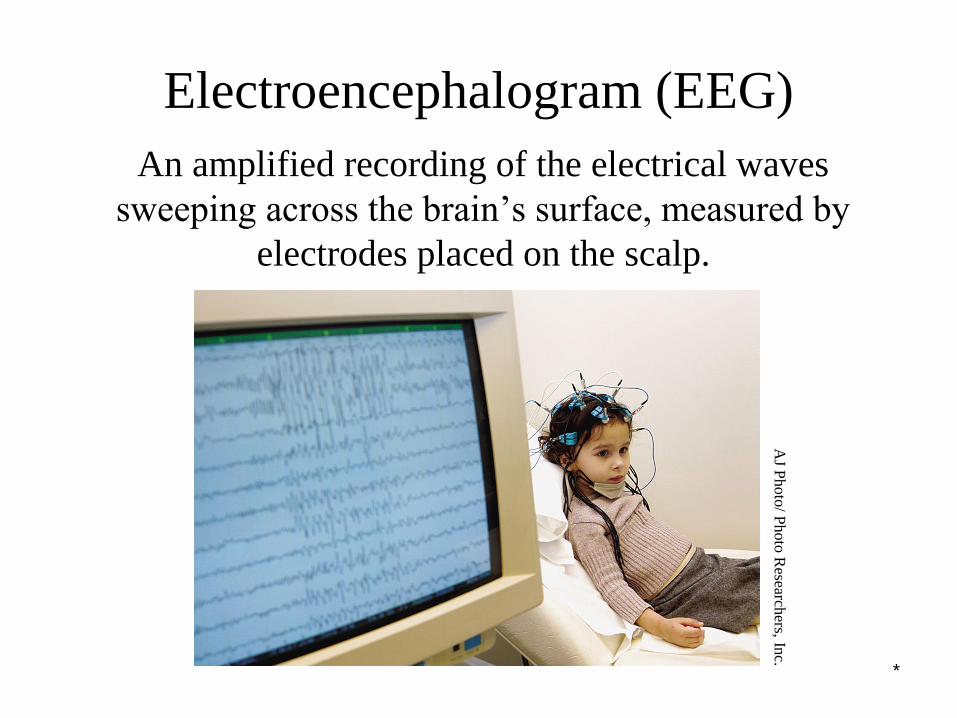
Electroencephalogram (EEG)
An amplified recording of the electrical waves
sweeping across the brain’s surface, measured by
electrodes placed on the scalp.
AJ P
hoto
/ Photo
Research
ers, Inc.
*
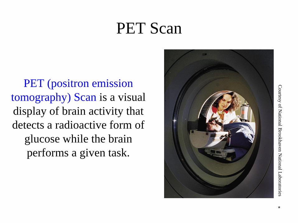
PET Scan
PET (positron emission
tomography) Scan is a visual
display of brain activity that
detects a radioactive form of
glucose while the brain
performs a given task.
Courtesy
of N
ational B
rookhav
en N
ational L
aborato
ries

*
MRI Scan
MRI (magnetic resonance
imaging) uses magnetic fields
and radio waves to produce
computer-generated images
that distinguish among
different types of brain tissue.
Top images show ventricular
enlargement in a
schizophrenic patient. Bottom
image shows brain regions
when a participants lies.
Both photos from Daniel Weinberger, M.D., CBDB, NIMH
James Salzano/ Salzano Photo Lucy Reading/ Lucy Illustrations
*
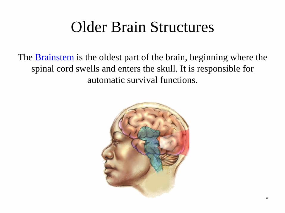
Older Brain Structures
The Brainstem is the oldest part of the brain, beginning where the
spinal cord swells and enters the skull. It is responsible for
automatic survival functions.
*
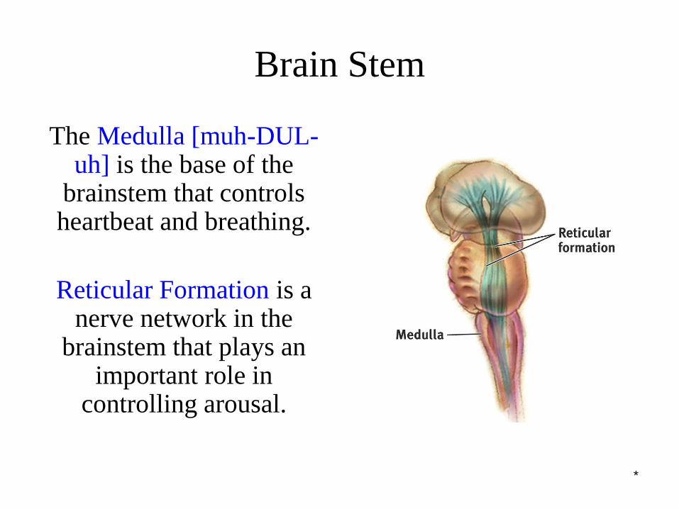
Brain Stem
The Medulla [muh-DUL-uh] is the base of the
brainstem that controls heartbeat and breathing.
Reticular Formation is a nerve network in the
brainstem that plays an important role in
controlling arousal.
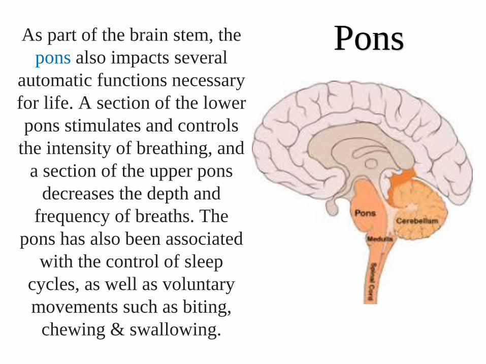
PonsAs part of the brain stem, the
pons also impacts several
automatic functions necessary
for life. A section of the lower
pons stimulates and controls
the intensity of breathing, and
a section of the upper pons
decreases the depth and
frequency of breaths. The
pons has also been associated
with the control of sleep
cycles, as well as voluntary
movements such as biting,
chewing & swallowing.
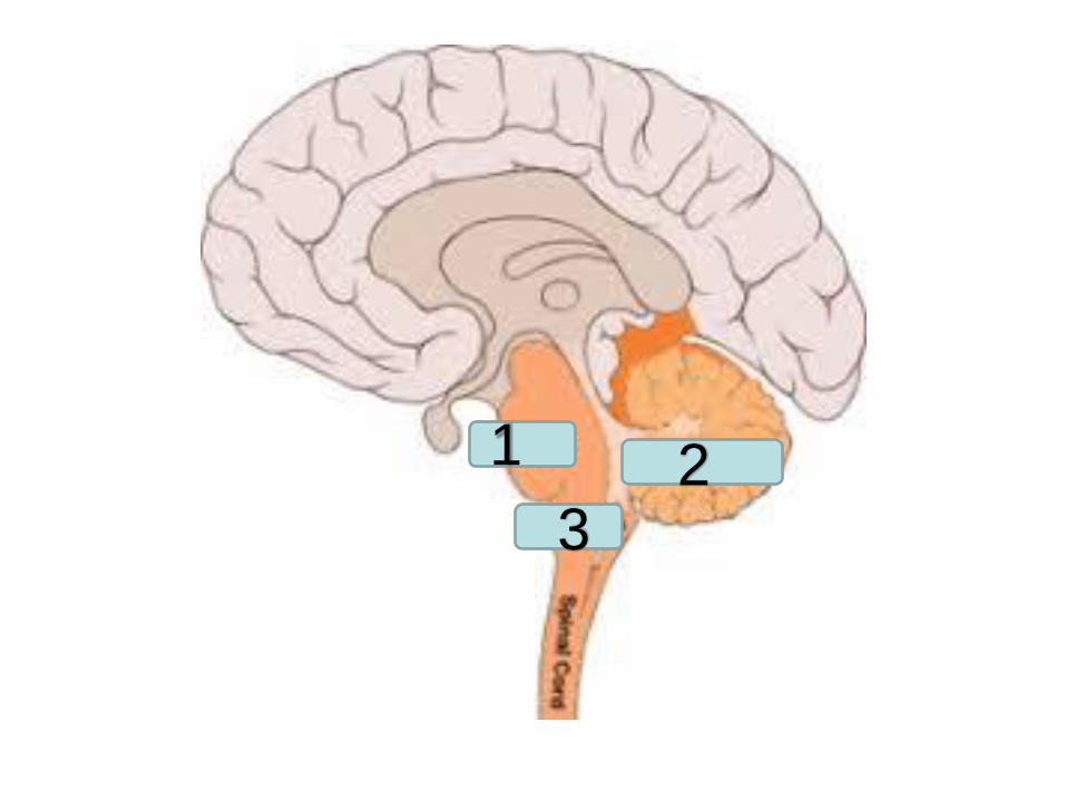
1 23
*
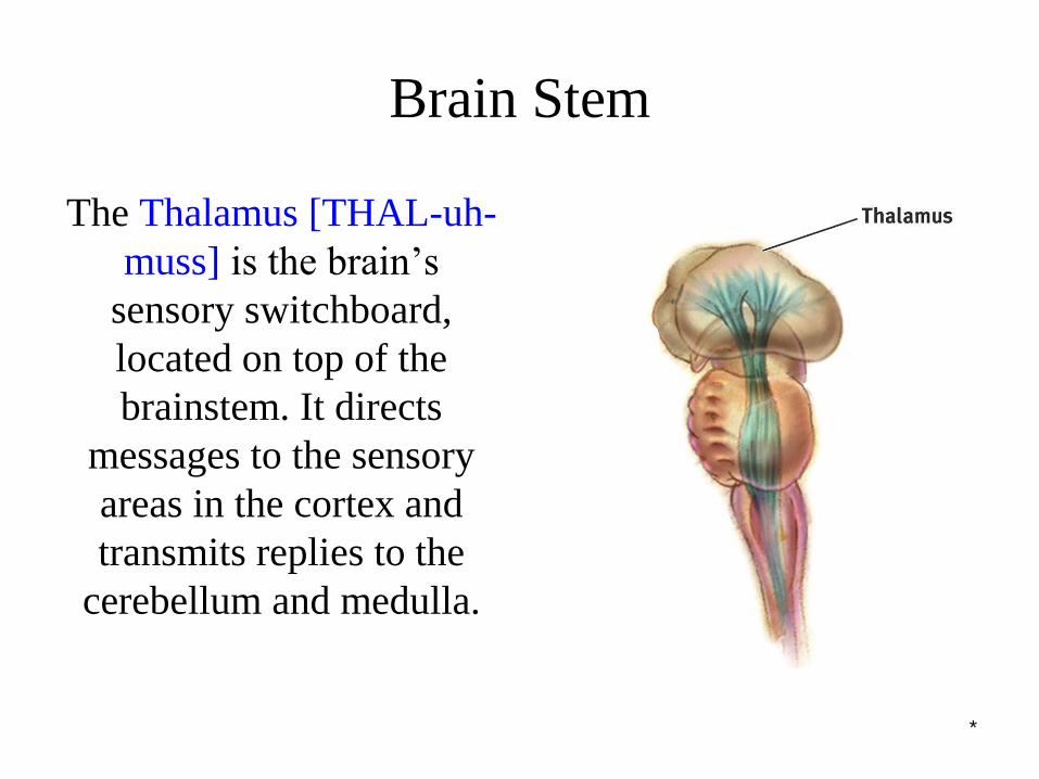
Brain Stem
The Thalamus [THAL-uh-
muss] is the brain’s
sensory switchboard,
located on top of the
brainstem. It directs
messages to the sensory
areas in the cortex and
transmits replies to the
cerebellum and medulla.
*
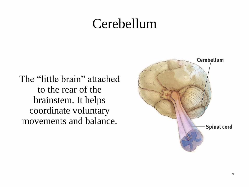
The “little brain” attached to the rear of the
brainstem. It helps coordinate voluntary
movements and balance.
Cerebellum
*
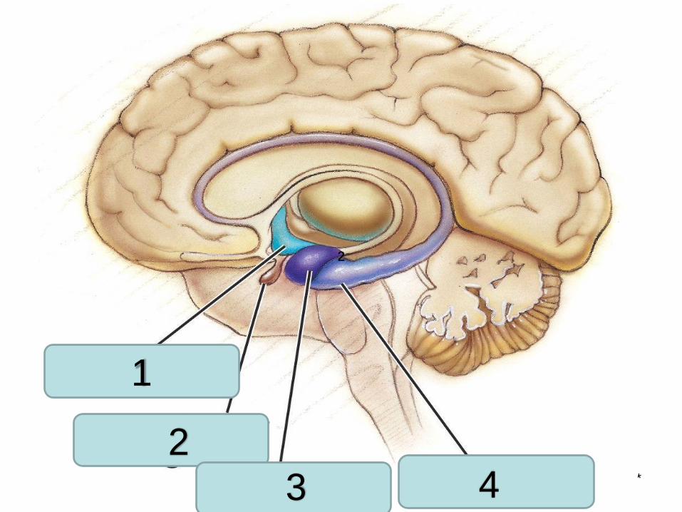
2
1
2
3 4
*
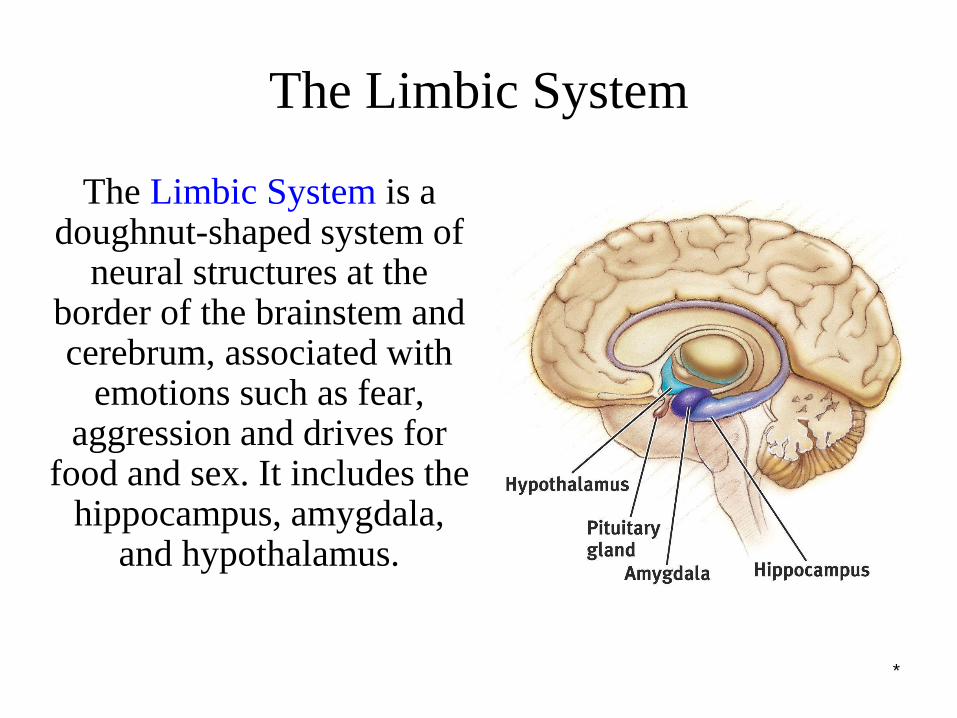
The Limbic System is a doughnut-shaped system of
neural structures at the border of the brainstem and cerebrum, associated with
emotions such as fear, aggression and drives for
food and sex. It includes the hippocampus, amygdala,
and hypothalamus.
The Limbic System
*
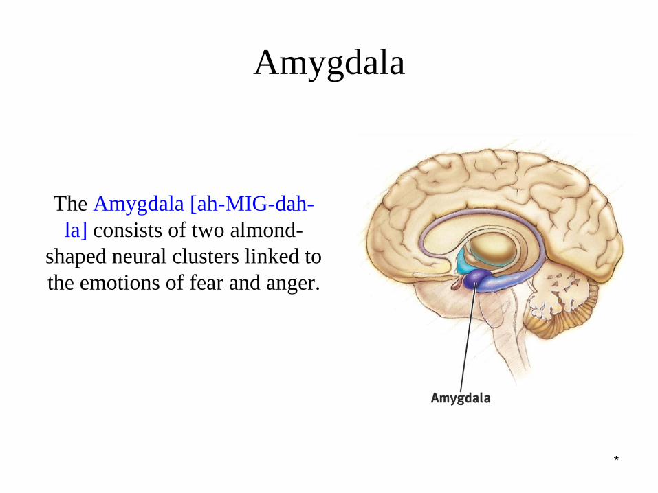
Amygdala
The Amygdala [ah-MIG-dah-
la] consists of two almond-
shaped neural clusters linked to
the emotions of fear and anger.
*
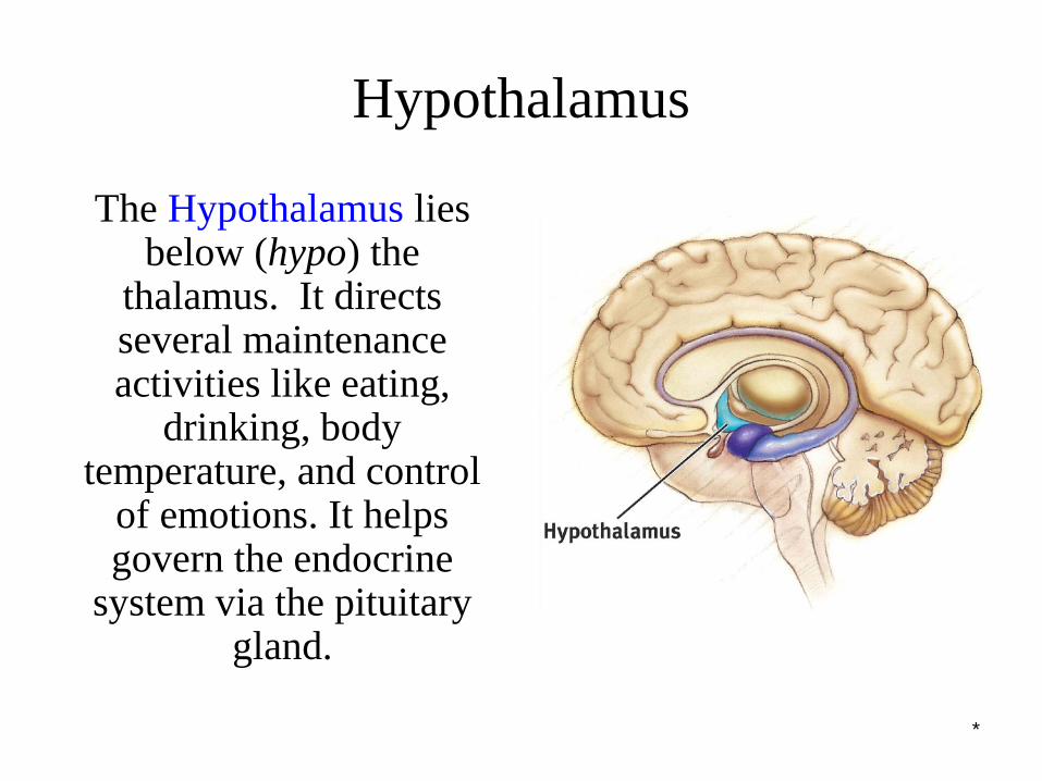
Hypothalamus
The Hypothalamus lies below (hypo) the
thalamus. It directs several maintenance activities like eating,
drinking, body temperature, and control
of emotions. It helps govern the endocrine
system via the pituitary gland.
*
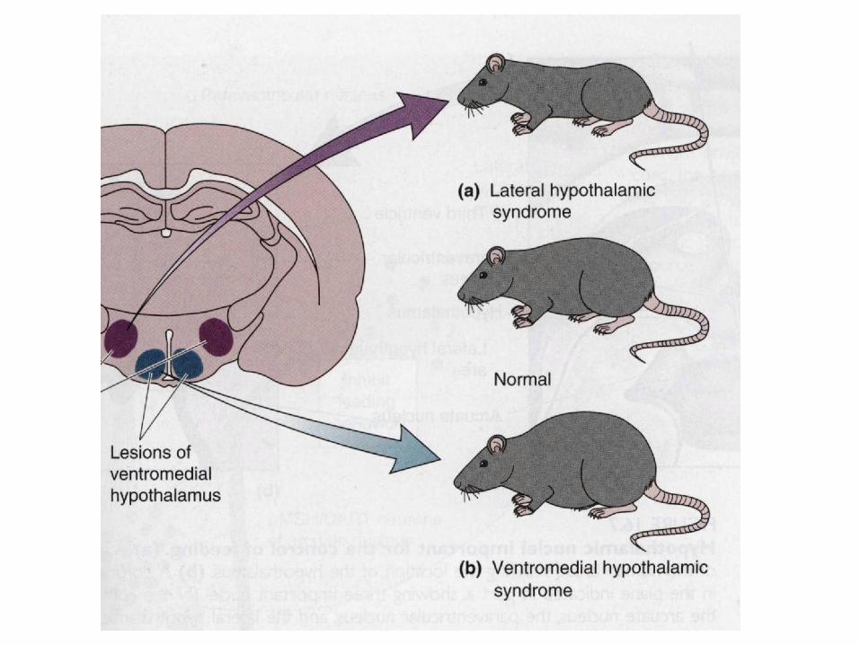
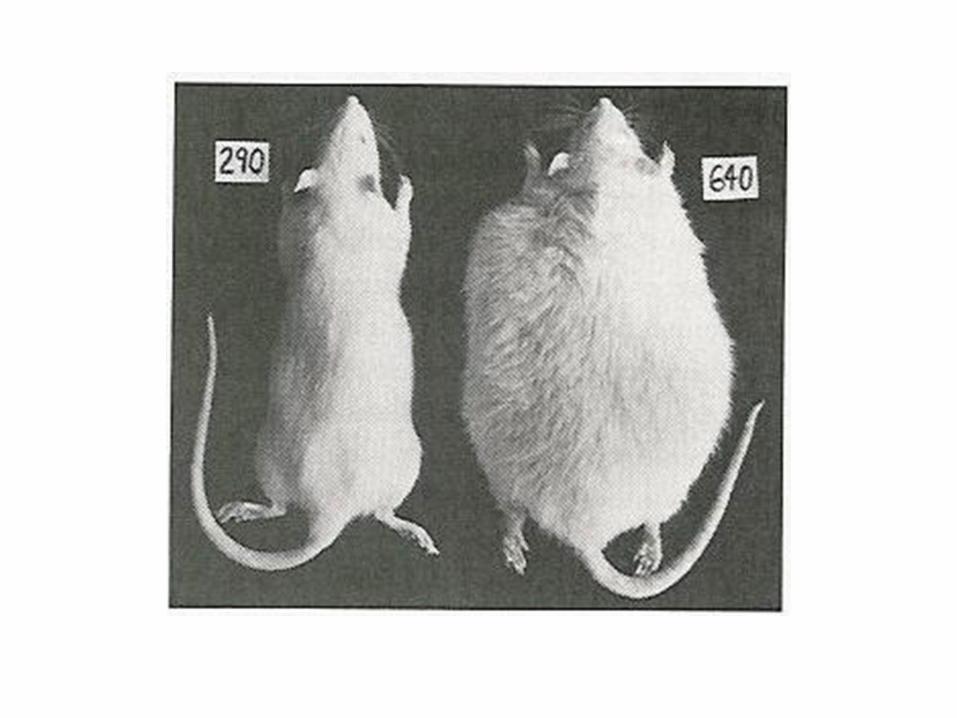
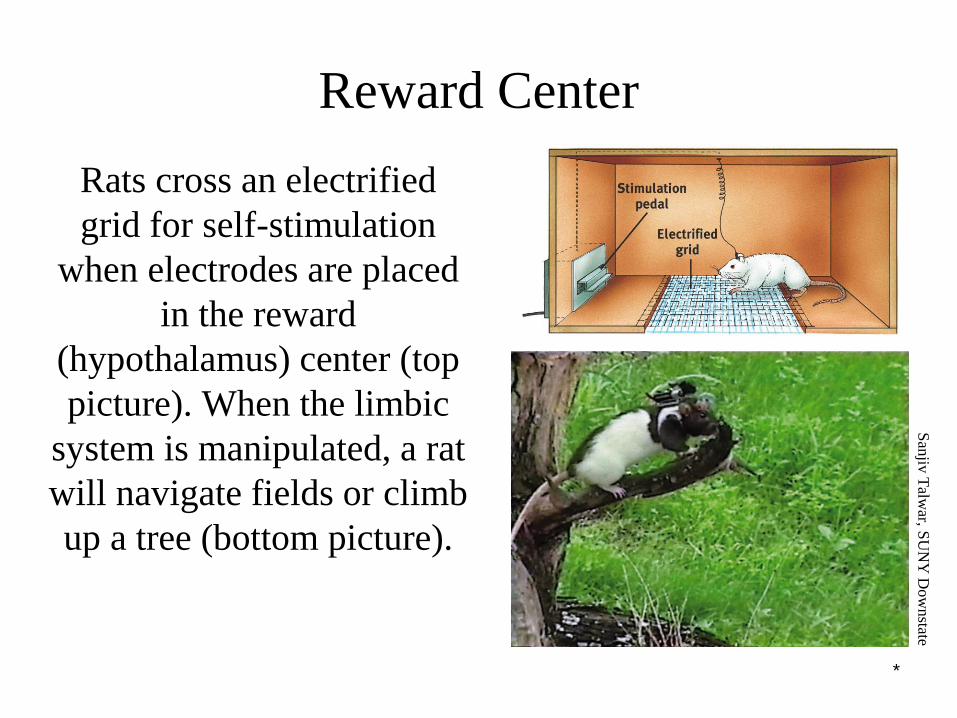
Rats cross an electrified
grid for self-stimulation
when electrodes are placed
in the reward
(hypothalamus) center (top
picture). When the limbic
system is manipulated, a rat
will navigate fields or climb
up a tree (bottom picture).
Reward Center
San
jiv T
alwar, S
UN
Y D
ow
nstate

*
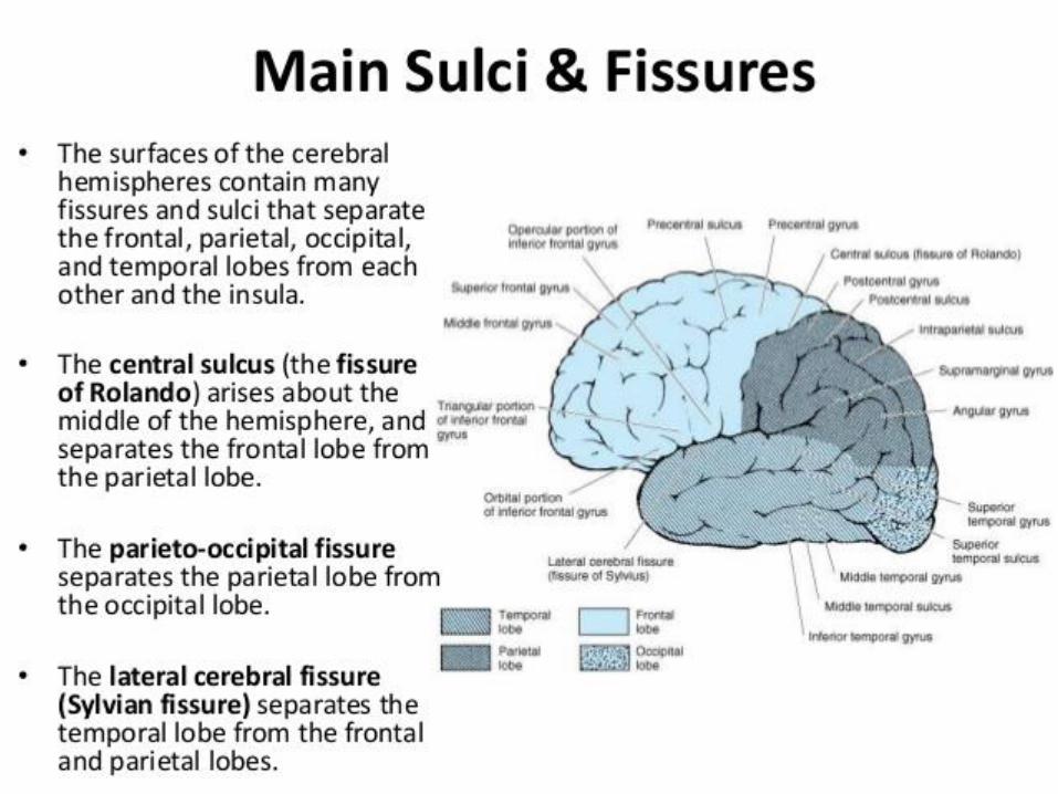
The Cerebral Cortex
The intricate fabric of interconnected neural cells that covers the cerebral hemispheres. It is the body’s ultimate control and
information processing center.
*
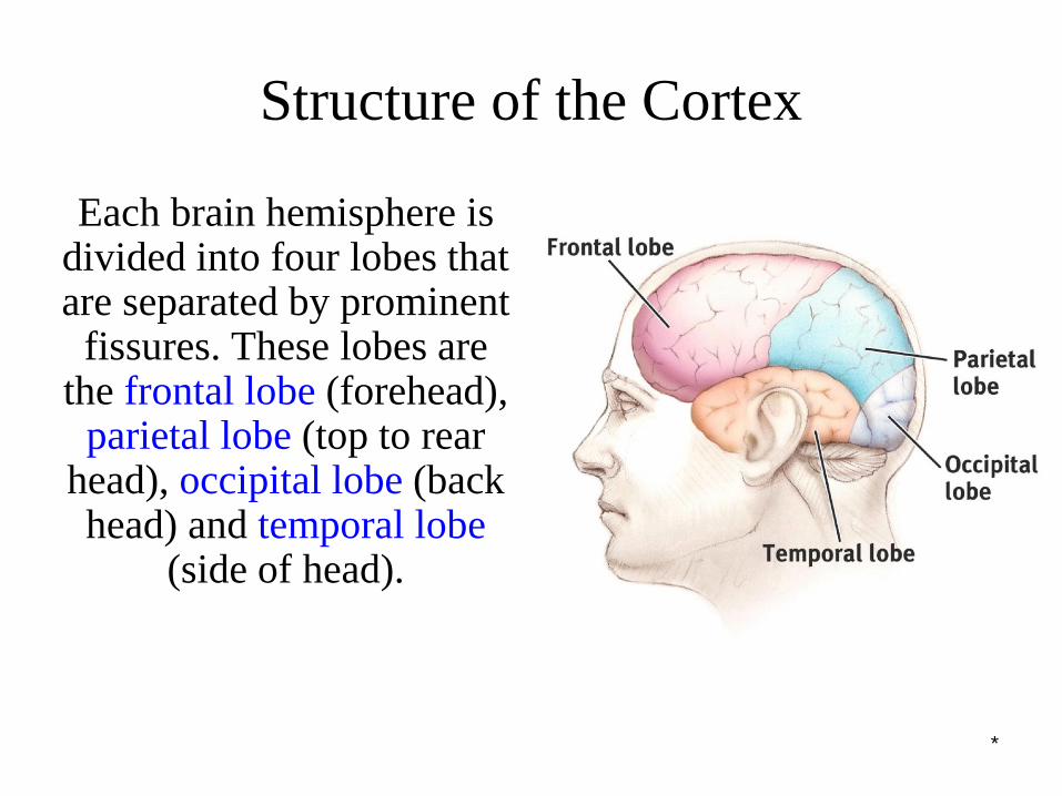
Structure of the Cortex
Each brain hemisphere is divided into four lobes that are separated by prominent
fissures. These lobes are the frontal lobe (forehead),
parietal lobe (top to rear head), occipital lobe (back head) and temporal lobe
(side of head).
*
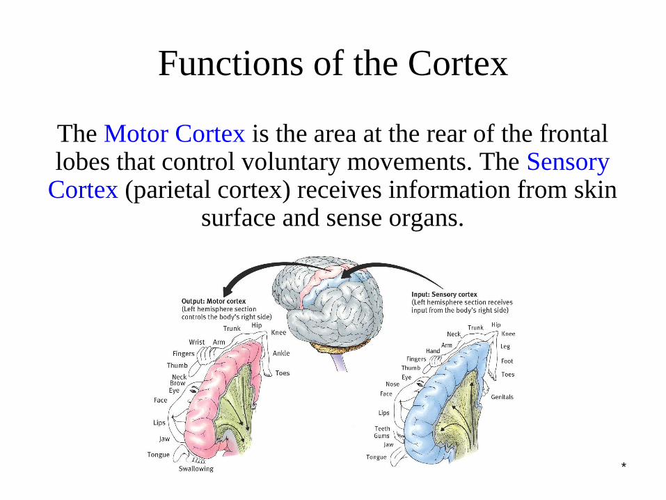
Functions of the Cortex
The Motor Cortex is the area at the rear of the frontal lobes that control voluntary movements. The Sensory
Cortex (parietal cortex) receives information from skin surface and sense organs.
*
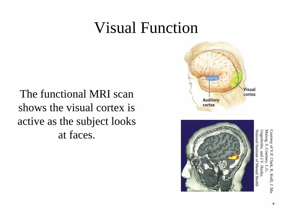
Visual Function
The functional MRI scan
shows the visual cortex is
active as the subject looks
at faces.
Co
urtesy
of V
.P. C
lark, K
. Keill, J. M
a.
Maiso
g, S
. Co
urtn
ey, L
.G.
Ungerleid
er, and
J.V. H
axb
y,
Natio
nal In
stitute o
f Men
tal Health
*
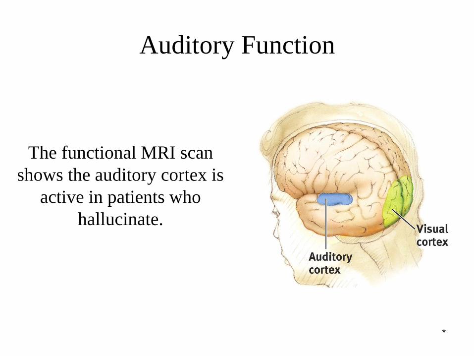
Auditory Function
The functional MRI scan
shows the auditory cortex is
active in patients who
hallucinate.
*
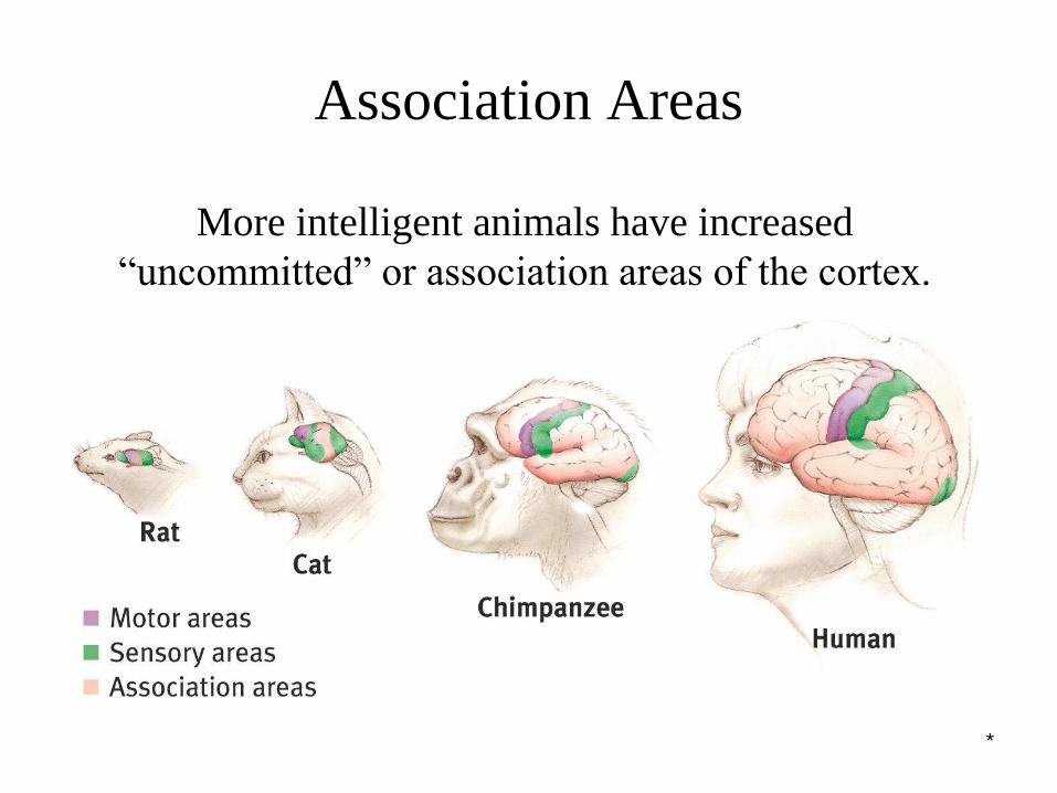
More intelligent animals have increased
“uncommitted” or association areas of the cortex.
Association Areas

*
Language
Aphasia is an impairment of language, usually caused
by left hemisphere damage either to Broca’s area
(impaired speaking) or to Wernicke’s area (impaired
understanding).
*
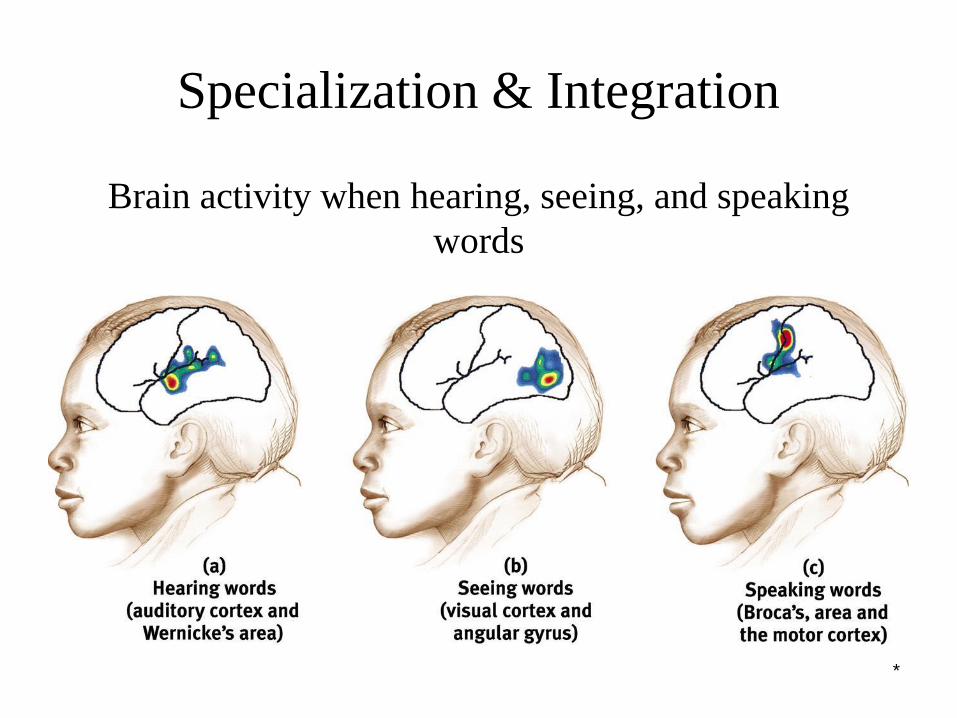
Specialization & Integration
Brain activity when hearing, seeing, and speaking
words
*
The brain is sculpted by our genes but also by our
experiences.
Plasticity refers to the brain’s ability to modify itself
after some type of injury or illness.
The Brain’s Plasticity


*
Our Divided Brain
Our brain is divided into two hemispheres.
The left hemisphere processes reading, writing,
speaking, mathematics, and comprehension skills. In
the 1960s, it was termed as the dominant brain.
*
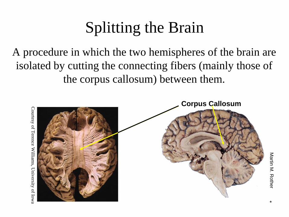
Splitting the Brain
A procedure in which the two hemispheres of the brain are
isolated by cutting the connecting fibers (mainly those of
the corpus callosum) between them.
Corpus Callosum
Ma
rtin M
. Roth
er
Courtesy
of T
erence W
illiams, U
niv
ersity o
f Iow
a
*
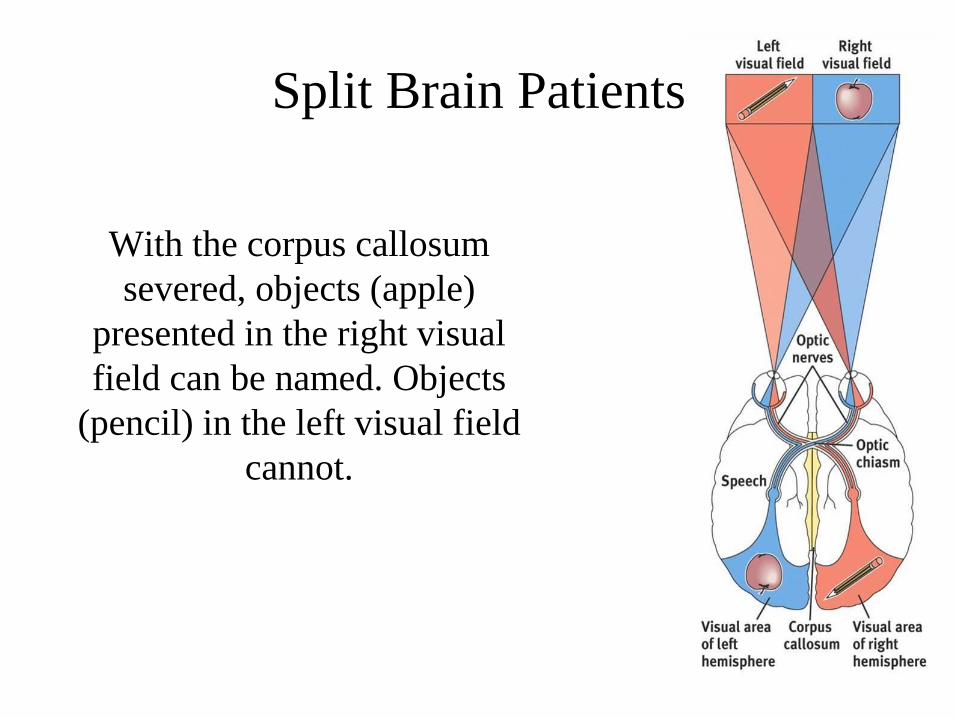
Split Brain Patients
With the corpus callosum
severed, objects (apple)
presented in the right visual
field can be named. Objects
(pencil) in the left visual field
cannot.
*
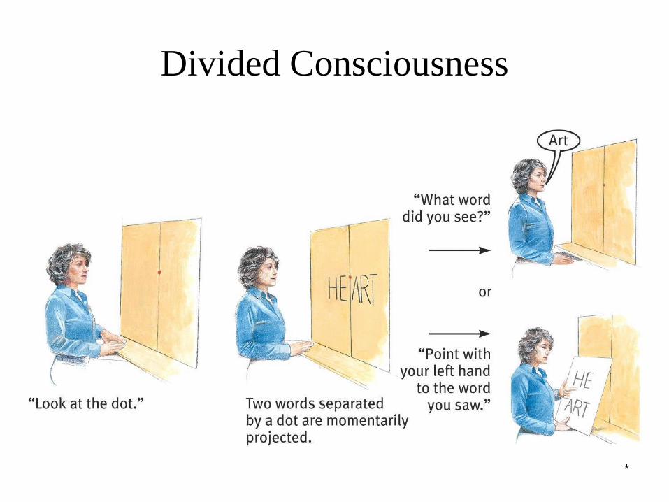
Divided Consciousness
*
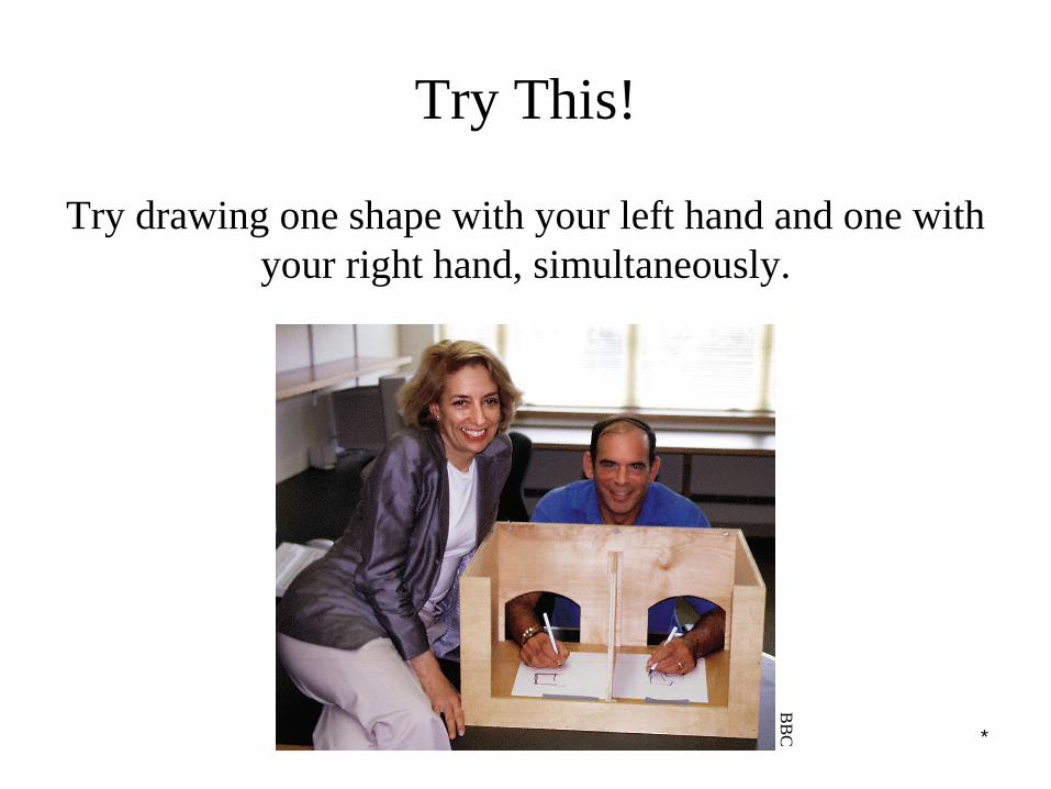
Try This!
Try drawing one shape with your left hand and one with
your right hand, simultaneously.B
BC

*
Non-Split Brains
People with intact brains also show left-right
hemispheric differences in mental abilities.
A number of brain scan studies show normal
individuals engage their right brain when completing
a perceptual task and their left brain when carrying
out a linguistic task.
*
Brain Organization & Handedness
Is handedness inherited? Yes. Archival and historic studies, as well as modern medical studies, show that the right hand is preferred. This suggests genes and/or
prenatal factors influence handedness.
*
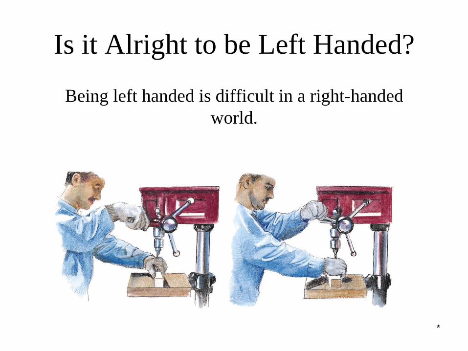
Is it Alright to be Left Handed?
Being left handed is difficult in a right-handed
world.

*
Is it Alright to be Left Handed?
The percentage of left-handed individuals decreases sharply in samples of older people (Coren,
1993).