Endocrine Oncology
409
HUMANA PRESS HUMANA PRESS Endocrine Oncology CONTEMPORARY ENDOCRINOLOGY ™ Edited by Stephen P. Ethier, PhD Endocrine Oncology Edited by Stephen P. Ethier, P hD
-
Upload
khangminh22 -
Category
Documents
-
view
0 -
download
0
Transcript of Endocrine Oncology

HUMANA PRESSHUMANA PRESS
EndocrineOncology
CONTEMPORARY ENDOCRINOLOGY™
Edited by
Stephen P. Ethier, PhD
EndocrineOncology
Edited by
Stephen P. Ethier, PhD

Contents i
ENDOCRINE ONCOLOGY

ii Contents
CONTEMPORARY ENDOCRINOLOGY
P. Michael Conn, SERIES EDITOR
Assisted Fertilization and Nuclear Transfer in Mammals, edited by DON P. WOLF AND
MARY ZELINSKI-WOOTEN, 2001Adrenal Disorders, edited by ANDREW N. MARGIORIS AND GEORGE P. CHROUSOS, 2001Endocrine Oncology, edited by STEPHEN P. ETHIER, 2000Endocrinology of the Lung: Development and Surfactant Synthesis, edited by
CAROLE R. MENDELSON, 2000Sports Endocrinology, edited by MICHELLE P. WARREN AND NAAMA W. CONSTANTINI, 2000Gene Engineering in Endocrinology, edited by MARGARET A. SHUPNIK, 2000Endocrinology of Aging, edited by JOHN E. MORLEY AND LUCRETIA VAN DEN BERG, 2000Human Growth Hormone: Research and Clinical Practice, edited by ROY G. SMITH AND
MICHAEL O. THORNER, 2000Hormones and the Heart in Health and Disease, edited by LEONARD SHARE, 1999Menopause: Endocrinology and Management, edited by DAVID B. SEIFER AND ELIZABETH
A. KENNARD, 1999The IGF System: Molecular Biology, Physiology, and Clinical Applications, edited
by RON G. ROSENFELD AND CHARLES T. ROBERTS, JR., 1999Neurosteroids: A New Regulatory Function in the Nervous System, edited by ETIENNE-
EMILE BAULIEU, MICHAEL SCHUMACHER, AND PAUL ROBEL, 1999Autoimmune Endocrinopathies, edited by ROBERT VOLPÉ, 1999Hormone Resistance Syndromes, edited by J. LARRY JAMESON, 1999Hormone Replacement Therapy, edited by A. WAYNE MEIKLE, 1999Insulin Resistance: The Metabolic Syndrome X, edited by GERALD M. REAVEN AND AMI LAWS, 1999Endocrinology of Breast Cancer, edited by ANDREA MANNI, 1999Molecular and Cellular Pediatric Endocrinology, edited by STUART HANDWERGER, 1999Gastrointestinal Endocrinology, edited by GEORGE H. GREELEY, JR., 1999The Endocrinology of Pregnancy, edited by FULLER W. BAZER, 1998Clinical Management of Diabetic Neuropathy, edited by ARISTIDIS VEVES, 1998G Proteins, Receptors, and Disease, edited by ALLEN M. SPIEGEL, 1998Natriuretic Peptides in Health and Disease, edited by WILLIS K. SAMSON AND ELLIS R. LEVIN, 1997Endocrinology of Critical Disease, edited by K. PATRICK OBER, 1997Diseases of the Pituitary: Diagnosis and Treatment, edited by MARGARET E. WIERMAN, 1997Diseases of the Thyroid, edited by LEWIS E. BRAVERMAN, 1997Endocrinology of the Vasculature, edited by JAMES R. SOWERS, 1996

Contents iii
ENDOCRINE
ONCOLOGY
HUMANA PRESSTOTOWA, NEW JERSEY
Edited by
STEPHEN P. ETHIER, PHDDepartment of Radiation Oncology,University of Michigan Cancer Center,Ann Arbor, MI


Contents vPREFACE
v
Endocrine oncology is a broad subject that would be difficult, if not impossible, tocover adequately in a single book. Cancers of endocrine tissues, such as breast andprostate, are very important from a public health point of view because of their increasingprevalence; they have also been the focus of intensive research, which has expandeddramatically in recent years. In order to keep this book to a manageable size and still haveit be useful to the reader with an interest in this subject, I decided to focus primarily onthe endocrinology of cancers of the breast, prostate, endometrium, and ovary. As a result,there is very little information in this book on the molecular genetics of endocrine can-cers, and such information is available in other excellent books.
Despite the great advances in our understanding of the genetics of endocrine can-cers, important and controversial issues relating to the endocrinology and cell biology ofmalignancies of endocrine tissues remain to be resolved, and I have tried to cover theseissues in detail in Endocrine Oncology. For example, while it has been known for manyyears that steroid hormones, particularly estradiol, influence breast cancer developmentand progression, many issues remain to be resolved regarding the true role of estradiolin breast cancer progression. Indeed, it is still not clear how to predict response of breastcancer patients with estrogen receptor-positive disease to antiestrogen therapy. Of furtherimportance to the field is the relatively limited understanding, still, of how steroid hor-mones function to regulate normal mammary gland homeostasis in humans. For thatreason, the first six chapters of this book focus on that specific area of research, and thefirst three chapters focus primarily on the role of estrogen and progesterone receptors innormal mammary gland function. The recent observations that estrogens and progestinssignal normal mammary epithelial cell proliferation via paracrine mechanisms to neigh-boring cells, which are steroid hormone receptor-negative, are exciting and may help toshed light on many aspects of human breast carcinogenesis. These findings are relevantto the question of how many pathways or precursor cells are able to give rise to humanbreast cancers that either express or do not express steroid hormone receptors, and thisimportant topic is the subject of Chapter 4. As is discussed in Chapter 5, expression ofcertain growth factor receptors can modify the expression of steroid hormone receptors,which in turn can influence breast cancer progression. These receptors may also influencethe response of steroid hormone receptor-positive breast cancer cells to antiestrogens. Adetailed discussion of factors that influence response to antiestrogens is presented inChapter 6. Thus, the first part of this book attempts to cover several important andintertwined issues in ways that may help to clarify the important issues that remain to beresolved in the field.
As is evident from Chapters 7 and 8, steroids are not the only hormones importantin breast cancer development. Prolactin, which is clearly important in rodent models ofbreast carcinogenesis, may play a similar role in human breast cancer development. Inaddition, peptide hormones such as chorionic gonadotropin may play important roles inmodifying breast cancer progression.
With a similar approach, the second part of the book examines the role of steroidhormones in prostate cancer development and treatment. In many ways, breast and pros-

vi Contentsvi Preface
tate cancers are parallel diseases in that they are both influenced by steroid hormones,both give rise to what is initially hormone receptor-positive disease that responds toendocrine therapy, and both eventually progresses to a hormone-independent state.These issues are discussed in Chapters 13 through 15, which also demonstrates that,while there are many parallels between breast and prostate cancers, there are manydistinguishing features as well.
The next two chapters focus on epithelial ovarian cancer and endometrial cancer.Once again, the emphasis of these chapters is on the endocrinology of these diseases.Since the pathogenesis of endometrial cancer appears to be influenced by certainantiestrogens that are used in breast cancer therapy, the issue of how estrogens affectdifferent target tissues is critically important to our understanding of disease progressionand the use of antiestrogen therapy.
Having focused on the role of hormones in the development of breast, prostate,ovarian, and endometrial cancer in the first 13 chapters of the book, the next four chapterspresent an in-depth discussion of the role of growth factors in endocrine neoplasia. Awealth of data in the literature points to an intimate interaction between hormones andgrowth factors in mediating normal tissue homeostasis and in pathological processesinvolving endocrine tissues. In particular, members of the epidermal growth factor fam-ily, the insulin-like growth factors and their binding proteins, and the fibroblast growthfactors have all been implicated in the progression of endocrine neoplasia. Clearly, a bookthat focuses on endocrine aspects of cancers of endocrine tissues would be incompletewithout a detailed discussion of the role of growth factors in the progression of thesediseases.
It has recently become clear that the ability of steroid hormone receptors to influ-ence gene expression is modified by the repertoire of transcriptional co-activators andco-repressors present in target cells. Furthermore, some of the genes that code for theseproteins may function as oncogenes in breast and other cancers. It is also now known thathormones can directly affect the expression of proteins that modify the cell death responseof epithelial cells under both physiologic and pathologic conditions. Finally, while it iswell known that inactivation of tumor suppressor genes is important in cancer progres-sion, endocrine tissues such as breast and ovary seem to have their own special suppressorgenes, BRCA1 and BRCA2. Thus, these three subjects, which are of particular importanceto the development of endocrine malignancies, are covered in the final three chapters ofthis book.
As I mentioned at the outset, no book on endocrine oncology can be complete, sincethis subject encompasses a vast area of clinical medicine and cancer biology research. Itwas my intention, and it is my hope, that in developing this book, some of the mostimportant issues relating to the endocrinology and cell biology of endocrine neoplasiahave been appropriately identified and thoroughly discussed. It is also my hope that thereaders of this book learn as much as I did from the outstanding contributions made bythe authors, to whom I am greatly indebted for their hard work and dedication.
Stephen P. Ethier, PhD

Contents viiCONTENTS
Preface ........................................................................................................ v
Contributors .............................................................................................. ix
1 Estrogen Receptor in Mammary Gland Physiology ........................ 1Elizabeth Anderson, Robert B. Clarke, and Anthony Howell
2 Multiple Facets of Estrogen Recptor in Human Breast Cancer ..... 17Leigh C. Murphy, Etienne Leygue, Helmut Dotzlaw,
Amanda Coutts, Biao Lu, Aihua Huang,and Peter H. Watson
3 Progesterone Receptors in Normal and Neoplastic Breast ............ 35Rosemary L. Balleine, Patricia A. Mote, Sybille M. N. Hunt,
Eileen M. McGowan, and Christine L. Clarke
4 Pathogenesis of Estrogen Receptor-Positive and -NegativeBreast Cancer .............................................................................. 49
Powel Brown, Suzanne Fuqua, and Craig Allred
5 Regulation of EGFR and ERBB2 Expressionby Estrogen Receptor in Breast Cancer ..................................... 69
Helen C. Hurst
6 Tamoxifen and Other Antiestrogens in Preventionand Therapy of Breast Cancer .................................................... 79
Kathy Yao and V. Craig Jordan
7 Prolactin in Human Breast Cancer Development ........................ 101Barbara K. Vonderhaar
8 Human Chorionic Gonadotropin in Breast Cancer Prevention ... 121Jose Russo and I. H. Russo
9 Epidermal Growth Factor-Related Peptidesin Endocrine Neoplasias ........................................................... 137
David S. Salomon, Caterina Bianco, Marta De Santis,Isabel Martinez-Lacaci, Christian Wechselberger,and Andreas D. Ebert
10 Insulin-Like Growth Factors and Endocrine Neoplasia .............. 193Douglas Yee and Adrian V. Lee
11 Insulin-Like Growth Factor Binding Proteinsin Endocrine-Related Neoplasia ............................................... 215
Giuseppe Minniti and Youngman Oh
12 Fibroblast Growth Factors and Their Receptors in Breastand Prostated Cancer ................................................................ 237
R. C. Coombes, S. Marsh, J. Gomm, and C. Johnston
13 Steroid Receptors in Prostate Cancer Developmentand Progression......................................................................... 255
Marco Marcelli, Nancy L. Weigel, and Dolores H. Lamb
vii

viii Contents
14 Type I Family Growth Factor Receptors and Their Ligandsin Prostate Cancer ..................................................................... 277
K. E. Leverton and W. J. Gullick
15 Hormonal Manipulation of Prostate Cancer ................................ 293Jeffrey M. Kamradt and Kenneth J. Pienta
16 Endocrinology of Epithelial Ovarian Cancer ............................... 313Vicki V. Baker
17 Molecular Pathogenesis of Endometrial Cancer:The Role of Tamoxifen .............................................................. 325
Yukio Sonoda and Richard R. Barakat
18 Structure and Function of BRCA Genes ....................................... 337Kenneth L. van Golen and Sofia D. Merajver
19 Role of Apoptosis and Its Modulation by Bcl-2 FamilyMembers in Breast and Prostate Cancer .................................. 353
Venil N. Sumantran, David R. Beidler, and Max S. Wicha
20 Transcriptional Coactivators in Cancer ........................................ 373Paul S. Meltzer
Index....................................................................................................... 383

Contents ixCONTRIBUTORS
ix
CRAIG ALLRED, MD, Division of Medical Oncology, Department of Pathology,University of Texas Health Science Center, San Antonio, TX
ELIZABETH ANDERSON, PHD, Clinical Research Department, Christie Hospital NHSTrust Manchester, UK
VICKI V. BAKER, MD, Department of Obstetrics and Gynecology, Universityof Michigan Medical Center, Ann Arbor, MI
ROSEMARY L. BALLEINE, PHD, FRCPA, Westmead Institute for Cancer Research,University of Sydney at Westmead Hospital, Westmead, Australia
RICHARD R. BARAKAT, MD, Gynecology Service Academic Office, MemorialSloan-Kettering Cancer Center, New York, NY
DAVID R. BEIDLER, PHD, Department of Medicine, University of Adelaide, Adelaide,South Australia
CATERINA BIANCO, MD, PHD, Tumor Growth Factor Section, Laboratory of TumorImmunology and Biology, National Cancer Institute, National Institutes of Health,Bethesda, MD
POWEL BROWN, MD, PHD, Division of Medical Oncology, Department of Medicine,University of Texas Health Science Center, San Antonio, TX
CHRISTINE L. CLARKE, PHD, Westmead Institute for Cancer Research, Universityof Sydney at Westmead Hospital, Westmead, Australia
ROBERT B. CLARKE, MD, PHD, Departments of Clinical Research and Medical Oncology,Christie Hospital NHS Trust, Manchester, UK
R. C. COOMBES, MD, PHD, Department of Cancer Medicine, Imperial College Schoolof Medicine, Charing Cross Hospital, London, UK
AMANDA COUTTS, PHD, Department of Biochemistry and Molecular Biology, Universityof Manitoba, Winnipeg, Manitoba, Canada
MARTA DE SANTIS, PHD, Tumor Growth Factor Section, Laboratory of TumorImmunology and Biology, National Cancer Institute, National Institutes of Health,Bethesda, MD
HELMUT DOTZLAW, PHD, Department of Biochemistry and Molecular Biology, Universityof Manitoba, Winnipeg, Manitoba, Canada
ANDREAS D. EBERT, MD, PHD, Department of Obstetrics and Gynecology, Laboratoryof Tumor Biology and Microcirculation, Medical Center Benjamin Franklin,Fee University Berlin, Berlin, Germany
SUZANNE FUQUA, PHD, Division of Medical Oncology, Department of Medicine,University of Texas Health Science Center, San Antonio, TX
J. GOMM, PHD, Department of Cancer Medicine, Imperial College School of Medicine,Charing Cross Hospital, London, UK
W. J. GULLICK, PHD, ICRF Molecular Oncology Unit, Imperial College School of Medicine,Hammersmith Hospital, London, UK
ANTHONY HOWELL, MD, Departments of Clinical Research and Medical Oncology,Christie Hospital NHS Trust, Manchester, UK
AIHUA HUANG, MSC, Department of Pathology, University of Manitoba, Winnipeg,Manitoba, Canada

x Contentsx Contributors
SYBILLE M. N. HUNT, PHD, Westmead Institute for Cancer Research, Universityof Sydney at Westmead Hospital, Westmead, Australia
HELEN C. HURST, PHD, ICRF Molecular Oncology Unit, Imperial College Schoolof Medicine, Hammersmith Hospital, London, UK
C. JOHNSTON, PHD, Department of Cancer Medicine, imperial College Schoolof Medicine, Charing Cross Hospital, London, UK
V. CRAIG JORDAN, PHD, DSC, Lynn Sage Breast Cancer Research Program, Robert H.Lurie Comprehensive Cancer Center, Chicago, IL
JEFFREY M. KAMRADT, MD, Division of Endocrinology, Metabolism, and Hypertension,Wayne State University School of Medicine, Detroit, MI
DOLORES J. LAMB, PHD, Division of Reproductive Medicine and Surgery, ScottDepartment of Urology and Department of Cell Biology, Baylor Collegeof Medicine, Houston, TX
ADREAN V. LEE, PHD, Division of Medical Oncology, Department of Medicine, Universityof Texas Health Science Center, San Antonio, TX
K. E. LEVERTON, PHD, ICRF Molecular Oncology Unit, Imperial College Schoolof Medicine, Hammersmith Hospital, London, UK
ETIENNE LEYGUE, PHD, Department of Biochemistry and Molecular Biology, Universityof Manitoba, Winipeg, Manitoba, Canada
BIAO LU, MSC, Department of Biochemistry and Molecular Biology, Universityof Manitoba, Winnipeg, Manitoba, Canada
MARCO MARCELLI, PHD, Department of Psychiatry and Behavioral Sciences, EmoryUniversity School of Medicine, Atlanta, GA
S. MARSH, FRCS, MD, Department of Cancer Medicine, Imperial College Schoolof Medicine, Charing Cross Hospital, London, UK
ISABEL MARTINEZ-LACACI, PHD, Tumor Growth Factor Section, Laboratory of TumorImmunology and Biology, National Cancer Institute, National Institutes of Health,Bethesda, MD
EILEEN M. MCGOWAN, MSC, Westmead Institute for Cancer Research, Universityof Sydney at Westmead Hospital, Westmead, Australia
PAUL S. MELTZER, MD, PHD, Cancer Genetics Branch, National Human GenomeResearch Institute, National Institutes of Health, Bethesda, MD
SOFIA D. MERAJVER, MD, Division of Hematology and Oncology, Departmentof Internal Medicine, University of Michigan Health Systems, Universityof Michigan Comprehensive Cancer Center, Ann Arbor, MI
GIUSEPPE MINNITI, MD, Department of Pediatrics, Oregon Health Sciences University,Portland, OR
PATRICIA A. MOTE, BSC, Westmead Institute for Cancer Research, University of Sydneyat Westmead Hospital, Westmead, Australia
LEIGH C. MURPHY, PHD, Department of Biochemistry and Molecular Biology,University of Manitoba, Winnipeg, Manitoba, Canada
YOUNGMAN OH, PHD, Department of Pediatrics, Oregon Health Sciences University,Portland, OR
KENNETH J. PIENTA, MD, University of Michigan Medical Center, Ann Arbor, MII. H. RUSSO, MD, Breast Cancer Research Laboratory, Fox Chase Cancer Center,
Philadelphia, PA

Contents xi
JOSE RUSSO, MD, Breast Cancer Research Laboratory, Fox Chase Cancer Center,Philadelphia, PA
DAVID S. SALOMON, PHD, Tumor Growth Factor Section, Laboratory of Tumor Immunologyand Biology, National Cancer Institute, National Institutes of Health, Bethesda, MD
MARA DE SANTIS, MD, Department of Psychiatry and Behavioral Sciences, EmoryUniversity School of Medicine, Atlanta, GA
YUKIO SONODA, MD, Gynecology Service, Department of Surgery, MemorialSloan-Kettering Cancer Center, New York, NY
VENIL N. SUMANTRAN, PHD, Division of Endocrinology, Metabolism, and Hypertension,Wayne State University School of Medicine, Detroit, MI
KENNETH L. VAN GOLEN, MD, Division of Hematology and Oncology, Departmentof Internal Medicine, University of Michigan Health System, University ofMichigan Comprehensive Cancer Center, Ann Arbor, MI
BARBARA K. VONDERHAAR, PHD, Molecular and Cellular Endocrinology Section,Laboratory of Tumor Immunology and Biology, National Cancer Institute,Bethesda, MD
PETER H. WATSON, MD, Department of Pathology, University of Manitoba, Winnipeg,Manitoba, Canada
CHRISTIAN WECHSELBERGER, PHD, Tumor Growth Factor Section, Laboratory of TumorImmunology and Biology, National Cancer Institute, National Institutes of Health,Bethesda, MD
NANCY L. WEIGEL, PHD, Department of Cell Biology, Baylor College of Medicine,Houston, TX
MAX S. WICHA, MD, Department of Internal Medicine and University of MichiganComprehensive Cancer Center, University of Michigan, Ann Arbor, MI
KATHY YAO, MD, Department of Surgery and the Robert H. Lurie ComprehensiveCancer Center, Northwestern University Medical School, Chicago, IL
DOUGLAS YEE, PHD, Division of Medical Oncology, Department of Medicine,University of Texas Health Science Center, San Antonio, TX
Contributors


Chapter 1 / ER in Mammary Gland 1
1
From: Contemporary Endocrinology: Endocrine OncologyEdited by: S. P. Ethier © Humana Press Inc., Totowa, NJ
Estrogen Receptorin Mammary Gland Physiology
1
Elizabeth Anderson, PHD,Robert B. Clarke, PHD, and Anthony Howell, MD
CONTENTS
INTRODUCTION
STRUCTURE OF HUMAN MAMMARY GLAND
ESTROGEN IS REQUIRED FOR BREAST DEVELOPMENT
AND TUMORIGENESIS
EFFECTS OF E2 ARE MEDIATED BY ERESTROGEN STIMULATES PROLIFERATION OF NORMAL HUMAN
NONPREGNANT, NONLACTATING BREAST EPITHELIUM
ESTROGEN INDUCES PROGESTERONE RECEPTOR EXPRESSION
IN HUMAN BREAST EPITHELIUM
PROLIFERATION AND PR EXPRESSION ARE DIFFERENTIALLY
SENSITIVE TO ESTROGEN STIMULATION
STEROID RECEPTOR EXPRESSION AND PROLIFERATION
ARE DISSOCIATED IN LUMINAL ECS
ARE EFFECTS OF ESTROGEN MEDIATED BY PARACRINE
GROWTH FACTORS?BIOLOGICAL CONSEQUENCES OF INDIRECT MECHANISM OF E2 ACTION
CLINICAL CONSEQUENCES OF INDIRECT MECHANISM OF E2 ACTION
SUMMARY
REFERENCES
INTRODUCTION
It is taken for granted that the ovarian steroid, estrogen, is required for normal humanbreast development and tumorigenesis, but we still do not know exactly how this steroidexerts its effects, or even exactly what these effects are. These questions are not justacademic: The mammary epithelium is the tissue from which most breast tumors arise,and understanding how processes such as its proliferation and differentiation are con-trolled may lead to an increased understanding of how cancers arise. Elucidation ofnormal breast function and physiology may also identify new targets and/or strategies for

2 Anderson, Clarke, and Howell
preventing breast cancer (BC), a disease that affects more than 1/12 women in the West-ern world. This chapter reviews what is known about the role of the estrogen receptor(ER) in controling mammary gland physiology in human female breast tissue, althoughanimal models will be referred to when appropriate.
STRUCTURE OF THE HUMAN MAMMARY GLAND
The mammary gland is an unusual organ in that is not fully developed at birth. Thereis further development at puberty, and the gland becomes fully differentiated and func-tional only at the time of pregnancy and subsequent lactation. The various stages of humanmammary gland development have been elegantly and comprehensively described byRusso and Russo (1). Briefly, the major histological unit of the human breast is the lobu-lar structure arising from a terminal duct. These ductal and lobular structures are linedby a continuous layer of luminal epithelial cells (ECs), which are, in turn, surrounded bya second layer of myo-ECs. In the adult nonpregnant, nonlactating breast, these myo-ECsare in direct contact with the basement membrane, and the whole structure is then sur-rounded by delimiting fibroblasts and a specialized intralobular stroma. Most humanbreast tumors are not only morphologically similar to the luminal EC population, but theyalso retain many of their biochemical characteristics (2). For example, most tumorsexpress the same cytokeratin profile as luminal ECs, they contain steroid receptors, andthey express polymorphic epithelial mucin (3,4). This leads to the conclusion that it isthe luminal ECs in the mammary gland that are the major targets for malignant transfor-mation and subsequent tumor formation. Consequently, an increased understanding ofthe mechanisms controling growth and differentiation of this population may identifynew targets and strategies for early detection and prevention of BC in women.
ESTROGEN IS REQUIREDFOR BREAST DEVELOPMENT AND TUMORIGENESIS
In terms of biological activity, the most important circulating estrogen in women isestradiol (E2). From the advent of menarche until the menopause, E2 is synthesized andsecreted in a cyclical manner by the ovaries under the control of the pituitary gonadotro-phins. The clinical and epidemiological evidence for an obligate role of estrogen in humanmammary gland development and tumor formation is considerable. Observation of girlswith estrogen deficiency through, e.g., gonadal dysgenesis or gonadotrophin deficiency,demonstrates that the steroid is strictly necessary (although probably not sufficient) forpubertal breast development (5). The incidence of BC in men is 1% of the incidence inwomen. Reducing exposure of the mammary gland to the fluctuating E2 levels of themenstrual cycle, through an early natural or artificially induced menopause, substantiallylowers the risk of developing BC. Conversely, increasing exposure through early men-arche, late menopause, or late age at first full-term pregnancy raises the risk of cancer (6).The paramount role of E2 in mammary gland development and tumorigenesis has beenconfirmed in several rat and mouse models, and perhaps the most compelling evidencecomes from studies on mice in which the gene for the ER, the mediator of E2 action, hasbeen disrupted or knocked out. The mammary glands in these ER knockout (ERKO) micecomprise rudimentary ducts without terminal end buds or alveolar buds. These structuresare confined to the nipple area, and cannot be induced to develop further (7). The rudi-mentary mammary glands of these ERKO mice are resistant to malignant tumor forma-

Chapter 1 / ER in Mammary Gland 3
tion caused by introduction of the wnt-1 oncogene through transgenic manipulation, whichprovides very strong evidence for the role of estrogen and its cognate receptor in tumorpromotion (7). Finally, a large number of studies on rat models of mammary carcinogen-esis show that administration of exogenous estrogens greatly enhances tumor formation;reduction of endogenous estrogen levels through, for example, ovariectomy or adminis-tration of inhibitors of E2 synthesis, reduces or even eliminates tumor incidence (8,9).
EFFECTS OF E2 ARE MEDIATED BY ER
Like the other steroids, E2 is lipophilic and enters cells and their nuclei primarily bydiffusing through plasma and nuclear membranes. Once in the nucleus, E2 encountersproteins known as ERs, because they bind E2 with high affinity and specificity. Until rela-tively recently, only one ER gene (now called the ER ) was thought to be present in eitherhumans or rodents. However, in 1996, a second species, or ER , was isolated and clonedfrom rat prostate and ovary, closely followed by the human homolog in the same year(10,11). Both ER and ER are members of the steroid/thyroid hormone nuclear receptorsuperfamily, and may be described as ligand-dependent nuclear transcription factors.Both proteins have the modular structure that typifies the nuclear receptor superfamilycomprising six functional domains designated A–F, which include regions involved insteroid binding and interaction with DNA. The ER gene shows a high degree of sequencehomology with the ER in its hormone-binding (96%) and DNA-binding (58%) domains(11). However, the ER gene is smaller than the ER gene, has a different chromosomallocation, and encodes a shorter protein (477 vs 595 amino acids for the ER and ER ,respectively) (12). These features, together with the overlapping but distinctly differenttissue distribution of ER , compared to ER mRNA, have led many workers to suggestthat the ER mediates some of the nonclassical effects of the estrogens and antiestrogens(13). Alternatively, the fact that the ER is expressed in some of the same tissues as ER hasled to speculation that ER might interact with and modulate the actions of the ER (14).Much progress has been made recently toward understanding how binding of E2 to eitherER or ER enhances specific gene transcription: This is described later in this volume.
As far as the human mammary gland is concerned, ER mRNA can be detected, butER mRNA appears to be present in greater amounts (12). Furthermore, mice in whichthe ER gene has been knocked out have fully developed and functional mammaryglands, but ER knockout mice, as mentioned above, have only vestigial ductal struc-tures (15). Taken together, these data imply that the ER might not play a major role in thephysiology of either the human or the mouse mammary gland, and the remainder of thischapter presents findings related only to the ER .
ESTROGEN STIMULATES PROLIFERATION OF NORMALHUMAN NONPREGNANT, NONLACTATING BREAST EPITHELIUM
The first studies on the effects of estrogen on the adult human mammary epitheliumexamined proliferative activity and other parameters throughout the menstrual cycle. Itis difficult to obtain truly normal breast tissue for investigations of this type. To study nor-mal physiology and function, it is highly desirable to use breast tissue from women whoare not at increased risk of cancer, or who do not have a pre-existing malignant lesion.This means that most groups, including the authors, have used tissue from reductionmammoplasties or tissue adjacent to fibroadenomas, because these lesions were thought

4 Anderson, Clarke, and Howell
not to be associated with an increased risk of cancer. More recently, the relationshipbetween the presence of a fibroadenoma and subsequent risk of BC has become lesscertain (16), but material from women with these lesions still represents the most normaltissue that can be obtained, and it continues to be used in the authors’ and other studies.
The proliferative activity of normal breast tissue taken at different times of the men-strual cycle was first assessed by labeling with tritiated thymidine ([3H]-dT). The tissue,once removed from the patient, is incubated with the [3H]-dT, then fixed and sectioned.Autoradiography reveals the cells that have incorporated [3H]-dT and a thymidine-label-ing index (TLI) can then be calculated as the percentage of ECs labeled with the radio-active nucleotide. One very striking finding is the variation in the TLI measurementsbetween individual patients, which cannot be attributed to experimental variability. Whetherthis has any biological significance in terms of breast tissue estrogen sensitivity and riskof BC remains to be seen. Despite the high level of interindividual variation, the consen-sus is that the proliferative activity of human breast ECs is elevated in the luteal phase ofthe menstrual cycle (17–23). Detailed analysis of other kinetic parameters, such as themitotic and apoptotic indices, shows a similar pattern of change, because they also arehigher in the second half of the cycle, compared to the first, although the apoptotic indexreaches a peak around 3 d after the peak of proliferative activity (19). Cyclical variationin epithelial expression of other proteins related to the proliferative and apoptotic pro-cesses has also been shown. For example, the antiapoptotic protein, Bcl-2, reaches peaklevels in the middle of the menstrual cycle, and falls to a nadir at the time when apoptosisis greatest (24).
Cyclical variation in mammary gland activity is not restricted to humans: The mousemammary gland also undergoes cycles of proliferation and quiescence within the 5 d ofthe estrous cycle, so that the epithelial TLI is 3–4 higher during estrous, compared toproestrous (25).
The obvious candidates for the control of these changes in the human and mousemammary epithelia are E2 and progesterone (P), because they are produced cyclically bythe ovaries, and maximal breast proliferative activity in humans coincides with the mid-luteal phase peaks of E2 and P secretion. In contrast, proliferation of the human endo-metrium, regarded as the classical estrogen target tissue, is highest in the follicular phase,and declines when P levels rise in the second half of the menstrual cycle. These obser-vations have led several groups to suggest that, for the human breast epithelium, P is themajor stimulatory steroid, either alone or after estrogen priming ( 21). This suggestion hasbeen investigated further, using a variety of different experimental approaches and models.
The simplest approach is the establishment of breast EC cultures in which the effectsof E2 and P can be determined under strictly defined conditions. However, it has proveddifficult to establish luminal ECs in culture without them losing their original characteris-tics and steroid responsiveness. There is only one study (26) in which reasonably normalECs have been cultured, but this does show that E2 and not P enhances proliferative activity.
Because cultures of human breast ECs appear to be poor models for the study of ster-oid responsiveness, the other approach adopted by many workers is an in vivo system inwhich human breast tissue is implanted into athymic nude mice. This approach was firstdevised in the 1980s, and involved implanting several small pieces of normal humanbreast tissue subcutaneously into female athymic nude mice (27). The mice were thentreated with E2, P, thyroxine, or human placental lactogen, either singly or in combina-tion, and breast tissue pieces were removed at various time points thereafter for measure-

Chapter 1 / ER in Mammary Gland 5
ment of proliferative activity by [3H]-dT labeling. This first study showed that humanbreast EC proliferation was stimulated by E2. Thyroxine also stimulated proliferation,but human placental lactogen alone was without effect, although it did enhance the effectsof E2 when given in combination. In contrast, P did not alter the proliferative activity ofthe implanted tissue either alone or in combination with the other agents. Other investi-gators (28–30) used variations of this model inwhich, e.g., the human breast tissue waspartially enzymatically digested before being implanted into either intact mammary fatpads of the athymic mice or into pads that had been cleared of their parenchyma. Yetanother variation involved embedding human mammary ECs into extracellular matrices(type I collagen or reconstituted basement membrane) before subcutaneous implantationinto the athymic mice (31). Few of these studies addressed the effects of the ovariansteroids, but, when these were investigated, E2 was shown to stimulate proliferation (29).
Although informative, some of the early in vivo experiments used tissue taken fromthe periphery of diffuse benign breast lesions, and most of the studies used treatmentschedules that resulted in the delivery of uncertain amounts of E2, P, and other hormones.The authors’ studies, using the in vivo model as originally conceived, tried to addressthese problems by ensuring that the tissue to be implanted was confirmed as histologi-cally normal by a pathologist. The authors also calibrated the slow-release steroid-silas-tic pellets used, so that they delivered serum levels of E2 and P similar to those seen inwomen during the menstrual cycle (32). The results of these experiments are summarizedin Fig. 1, and the most important take-home points are that E2 stimulates the proliferative
Fig. 1. Estradiol, but not P, stimulates the proliferative activity of normal human breast tissueimplanted into athymic nude mice. This figure summarizes the results of measuring the proliferativeactivity of tissue implanted into athymic mice 14 d after the insertion of pellets containing either nosteroid (Control), 2 mg estradiol (E2), 4 mg P (Prog), or 4 mg P inserted after priming for 7 d witha 2-mg E2 pellet (E2 + Prog). Proliferative activity was assessed by determining the uptake of [3H]-dT,and the data are the thymidine labeling indices (TLIs) calculated as the percentage of luminal ECscontaining radiolabel. The open columns indicate the interquartile ranges; the horizontal bars indi-cate the median values; the numbers in parentheses along the abscissa indicate the number of obser-vations in each group. *, significantly different (P < 0.05) from the pretreatment value by theMann-Whitney U test.

6 Anderson, Clarke, and Howell
activity of normal human breast ECs, as measured by [3H]-dT uptake, and that P, eitheralone or after E2 priming, has no effect on EC proliferation. Further experiments haveshown that the response to E2 is dose-dependent between median E2 serum concentra-tions of 400–1300 pmol/L, which are representative of those seen in the menstrual cycle.However, raising E2 levels to those of early pregnancy (a median of 4400 pmol/L) doesnot further increase the levels of proliferative activity. The conclusion of these studiesis that E2 is the major steroid mitogen for the nonpregnant, nonlactating human breast,but other factors, in addition to E2, must be required to enhance breast EC proliferationto the early pregnancy levels reported by other groups.
ESTROGEN INDUCES PROGESTERONERECEPTOR EXPRESSION IN HUMAN BREAST EPITHELIUM
Because it seemed clear that human breast epithelium was an estrogen target tissue interms of the control of proliferation, interest developed in other potential effects of estro-gen on this tissue. Again, the first studies examined the expression of the ER and productsof ER action, such as the progesterone receptor (PR) in breast tissue obtained at differenttimes through the menstrual cycle. Using immunohistochemistry (IHC) on frozen sec-tions of breast tissue, the authors, and others, demonstrated that approx 5% of luminalECs contained ER (33–36). Moreover, the number of cells expressing ER was highestin the follicular phase of the cycle, lowest in the luteal phase, and, consequently, wasinversely related to proliferative activity. In contrast, IHC detection of PR expression onadjacent frozen sections revealed that 15–20% of ECs contained PRs, and that thisproportion did not vary appreciably during the cycle. Comparison with the endometrium,where PR is an estrogen-inducible protein, led to the suggestion that the PR may beconstitutively expressed in the human breast. Again, the cell culture models proved dis-appointing, although, in the single report (26) of estrogen-responsive normal ECs inculture, E2 treatment was shown to increase expression of both ER and PR. The in vivomodel of human breast tissue implanted into athymic mice has provided more informa-tion on the control of steroid receptor content. First, it was found that the percentage ofnormal ECs expressing the ER fell from ~5%, at removal from the patients, to <1% 2 wkafter being implanted into untreated mice (32). Administration of E2 or P, either singlyor in combination, had no effect on ER expression, but it is now known that these ratherlow levels of ER expression resulted from the insensitivity of the IHC technique for ERdetection in frozen sections, especially after it had been adapted for use in the xenograftexperiments. Second, the proportion of ECs expressing PR was also <1% 2 wk after beingimplanted into untreated mice, but the percentage of cells expressing PR was increased15–20-fold by E2 treatment at luteal-phase concentrations. This provided firm evidencethat PR expression in human breast epithelium was controlled by estrogen, at least whenthe tissue is implanted into athymic mice.
The early studies on human breast EC proliferation and steroid receptor expressionsuggested that, although these processes may be controlled by ovarian steroids, the mech-anisms involved could be appreciably different from those in the endometrium. How-ever, later studies, using experimental models such as the implantation of human tissueinto athymic mice, demonstrated that both mammary EC proliferation and PR expressionare under estrogenic control. One explanation for the differences between the early andlate studies may lie in the differential sensitivity of proliferation and PR expression toestrogen stimulation.

Chapter 1 / ER in Mammary Gland 7
PROLIFERATION AND PR EXPRESSIONARE DIFFERENTIALLY SENSITIVE TO ESTROGEN STIMULATION
The suggestion that maximal induction of proliferation and PR expression may requireexposure to differing concentrations of estrogen came from studies using human tissueimplanted into athymic mice (37). The upper panel of Fig. 2 shows the results of IHCdetection of PR in sections cut from fixed and paraffin-embedded specimens of breasttissue xenografts removed from mice treated with follicular- or luteal-phase levels of E2.These measurements show that the low E2 levels of the follicular phase are sufficient tomaximally induce PR expression, because the higher luteal-phase concentrations do notfurther enhance PR expression. However, follicular-phase E2 levels are not sufficient tostimulate proliferative activity above that of the untreated breast tissue, and it is only
Fig. 2. Proliferation and PR expression in human breast tissue implanted into athymic mice aredifferentially sensitive to the effects of E2. This figure summarizes the results of experiments inwhich PR expression (upper panel) and proliferation (lower panel) were determined 7 d after theinsertion of pellets containing either no steroid (Con), 4 mg P (Prog); 0.5 mg E2 (Lo E2), whichincreased serum E2 concentrations to the equivalent of those in the human follicular phase, 2 mg E2(Hi E2), which gave the equivalent of human luteal-phase levels, and 2 mg E2 combined with a 4-mgP pellet (E2 + Prog) as in Fig. 1. The open columns represent the interquartile ranges of the data; thehorizontal bars indicate the medians. *, significantly different from the control by Mann-WhitneyU test.
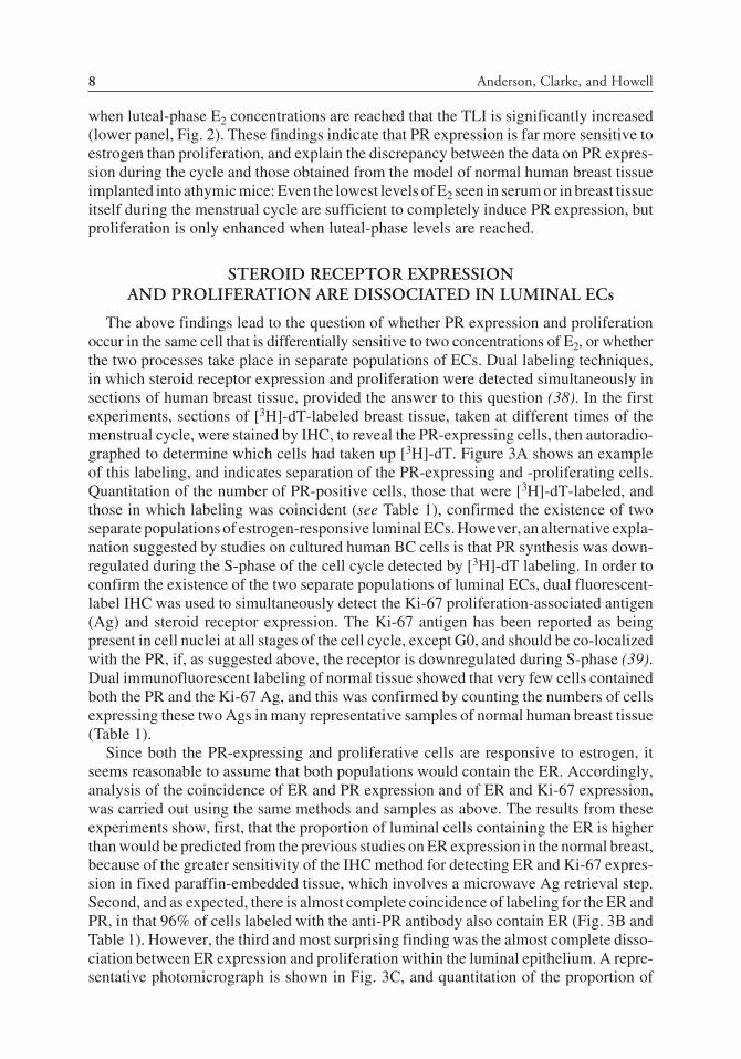
8 Anderson, Clarke, and Howell
when luteal-phase E2 concentrations are reached that the TLI is significantly increased(lower panel, Fig. 2). These findings indicate that PR expression is far more sensitive toestrogen than proliferation, and explain the discrepancy between the data on PR expres-sion during the cycle and those obtained from the model of normal human breast tissueimplanted into athymic mice: Even the lowest levels of E2 seen in serum or in breast tissueitself during the menstrual cycle are sufficient to completely induce PR expression, butproliferation is only enhanced when luteal-phase levels are reached.
STEROID RECEPTOR EXPRESSIONAND PROLIFERATION ARE DISSOCIATED IN LUMINAL ECs
The above findings lead to the question of whether PR expression and proliferationoccur in the same cell that is differentially sensitive to two concentrations of E2, or whetherthe two processes take place in separate populations of ECs. Dual labeling techniques,in which steroid receptor expression and proliferation were detected simultaneously insections of human breast tissue, provided the answer to this question (38). In the firstexperiments, sections of [3H]-dT-labeled breast tissue, taken at different times of themenstrual cycle, were stained by IHC, to reveal the PR-expressing cells, then autoradio-graphed to determine which cells had taken up [3H]-dT. Figure 3A shows an exampleof this labeling, and indicates separation of the PR-expressing and -proliferating cells.Quantitation of the number of PR-positive cells, those that were [3H]-dT-labeled, andthose in which labeling was coincident (see Table 1), confirmed the existence of twoseparate populations of estrogen-responsive luminal ECs. However, an alternative expla-nation suggested by studies on cultured human BC cells is that PR synthesis was down-regulated during the S-phase of the cell cycle detected by [3H]-dT labeling. In order toconfirm the existence of the two separate populations of luminal ECs, dual fluorescent-label IHC was used to simultaneously detect the Ki-67 proliferation-associated antigen(Ag) and steroid receptor expression. The Ki-67 antigen has been reported as beingpresent in cell nuclei at all stages of the cell cycle, except G0, and should be co-localizedwith the PR, if, as suggested above, the receptor is downregulated during S-phase (39).Dual immunofluorescent labeling of normal tissue showed that very few cells containedboth the PR and the Ki-67 Ag, and this was confirmed by counting the numbers of cellsexpressing these two Ags in many representative samples of normal human breast tissue(Table 1).
Since both the PR-expressing and proliferative cells are responsive to estrogen, itseems reasonable to assume that both populations would contain the ER. Accordingly,analysis of the coincidence of ER and PR expression and of ER and Ki-67 expression,was carried out using the same methods and samples as above. The results from theseexperiments show, first, that the proportion of luminal cells containing the ER is higherthan would be predicted from the previous studies on ER expression in the normal breast,because of the greater sensitivity of the IHC method for detecting ER and Ki-67 expres-sion in fixed paraffin-embedded tissue, which involves a microwave Ag retrieval step.Second, and as expected, there is almost complete coincidence of labeling for the ER andPR, in that 96% of cells labeled with the anti-PR antibody also contain ER (Fig. 3B andTable 1). However, the third and most surprising finding was the almost complete disso-ciation between ER expression and proliferation within the luminal epithelium. A repre-sentative photomicrograph is shown in Fig. 3C, and quantitation of the proportion of

Chapter 1 / ER in Mammary Gland 9
Fig. 3. Steroid receptors are not expressed in proliferating luminal ECs. These are representativephotomicrographs obtained from dual-label IHC and autoradiography. (A) Section of [3H]-dT-labeled normal human breast tissue, in which PR-expressing cells have been revealed by IHC(brown nuclei), followed by autoradiography, to show the proliferating cells (black grains overlyingcell nuclei, as indicated by the arrows). (B) Section of normal human breast tissue in which ER andPR expression have been determined using a dual-label immunofluorescent technique. In this par-ticular case, all the ER-expressing cells (which would fluoresce green) also express the PR (whichwould fluoresce red), to give a yellow color. The section has also been counterstained with the bluefluorochrome, DAPI. (C) section of normal human breast tissue in which ER and Ki-67 expressionhas been determined using a dual-label immunofluorescent technique. In this case, ER expressingcells are labeled with the red fluorochrome, and those cells expressing the Ki-67 Ag are labeledgreen. The arrows indicate areas in which proliferating cells are adjacent to those expressing the ER.

10 Anderson, Clarke, and Howell
doubly and singly labeled cells (Table 1) shows that far fewer cells than would beexpected contain both ER and the Ki-67 Ag. Also, as demonstrated in Fig. 3C, many ofthe proliferating cells are adjacent to cells expressing steroid receptors, which appear tobe evenly distributed throughout the luminal epithelium. This dissociation between lumi-nal cell steroid receptor expression and proliferation in the human breast has been con-firmed by at least one other group (40). It also appears that a similar situation may occurin rodents, because these authors have observed separate populations of PR expressingand [3H]-dT-labeled cells in the rat mammary gland (40). In the mouse mammary gland,there is dissociation between ER expression and [3H]-dT uptake in the terminal end budsduring both pubertal growth and estrous-cycle-associated proliferation (25).
It appears that the processes of PR expression and proliferation in human luminal ECsare differentially sensitive to the effects of E2. Expression of the PR is exquisitely sensi-tive, which means that, in vivo, there is always sufficient E2, either in the circulation orin the breast tissue itself, to ensure that the receptor is maximally expressed. It is also clearthat steroid receptor expression and proliferation occur in separate populations of lumi-nal ECs, although proliferating cells are usually adjacent to those containing steroidreceptors. The implication of these findings is that proliferation is not controlled directlyby E2, although other processes, such as PR expression, are. These findings raise manyquestions about the hormonal control of normal breast physiology and function.
ARE EFFECTS OF ESTROGENMEDIATED BY PARACRINE GROWTH FACTORS?
The studies described above provide some clues about how E2 may control EC prolif-eration indirectly, and these have been incorporated into the model illustrated in Fig. 4.The fact that ER-negative but proliferative cells are often adjacent to those that are ER-positive suggests that the ER-positive cells act as E2 sensors, which secrete juxtacrineand/or paracrine factors that trigger proliferation of the ER-negative cells, once a thresh-old E2 concentration is reached. Studies on cultured BC cell lines provide indirect supportfor this model. These show that medium conditioned by E2-treated ER-positive BC cellsstimulates proliferation of ER-negative cell lines growing in culture or as xenografts inathymic mice (41). Conversely, conditioned medium from ER-positive BC cells treatedwith antiestrogens inhibits proliferation when applied to ER-negative BC cells (42). Fur-ther analysis of the conditioned medium suggests that peptide growth factors, such as
Table 1Data from the Simultaneous Estimation of Proliferation
and Steroid Receptor Expression in Samples of Normal Human Breast Tissue
No. Cells No. cells % dualsamples counted ER+ve PR+ve [3H]-dT+ve Ki-67+ve dual-labeled labeled a
10 10,026 1735 46 1 225 25,302 3232 382 17 4.525 28,395 2107 639 9 1.425 28,018 3231 391 1.813 13,895 1792 1765 96
a Percentage of proliferating cells (as indicated by the cells labeled with [3H]-dT or the Ki-67 antibody),also containing steroid receptors.

Chapter 1 / ER in Mammary Gland 11
insulin-like growth factor-I, and transforming growth factors- and - , are involved inmediating these paracrine effects. A final piece of evidence for a paracrine mechanismof controling proliferation in the mammary gland comes from tissue recombinationstudies (43), in which epithelium isolated from ERKO mice is combined with stroma (inthis case, the fat pads) from wild-type mice, and cultured in the subrenal capsules of E2-treated athymic mice. The results of this experiment and its reverse, in which wild-typeepithelium is combined with ERKO fat pads, demonstrate that, in the mouse, E2 stimu-lates epithelial proliferation indirectly via a paracrine mechanism. However, the estro-gen-sensing cells appear to be in the stromal compartment, because wild-type epitheliumdoes not respond to E2 treatment when combined with ERKO stroma (or fat pads). Inhumans, the ER has never been detected in the stroma surrounding the mammary epithe-lium, leading to the conclusion that the estrogen-sensing cells are in the epithelial compart-ment. It is not as yet clear how separate populations of ER-expressing and -proliferatingmammary ECs might arise. The authors’ working hypothesis is that the ER-negative prolif-erating cells represent precursor or stem cells, which eventually differentiate to becomenonproliferative, ER-positive cells.
BIOLOGICAL CONSEQUENCESOF INDIRECT MECHANISM OF E2 ACTION
This chapter reviews the role of the ER in controling proliferation and other processesin the normal human nonpregnant, nonlactating breast. Both ER and its ligand, E2, arenecessary for the development of mammary epithelial structures. However, it is becom-ing clear that the ER is not expressed in the proliferating population of cells, which sug-gests that the effects of E2 are mediated by paracrine and/or juxtacrine factors. Why does
Fig. 4. Model for the indirect estrogenic control of normal human breast EC proliferation. In thismodel, the cells capable of proliferation are ER-negative (as indicated by the dark nuclei), but aresituated very close or adjacent to those containing the receptor (represented by the speckled nuclei).The ER-containing cells act as estrogen sensors and secrete paracrine or juxtacrine growth factors,which influence the activity of the ER-negative proliferative cells. In this model, the ER-containingcells secrete growth stimulatory factors when E2 levels are high, as shown in the lefthand part of thisscheme, and growth inhibitory peptides when E2 levels are low, as on the righthand side of thediagram.

12 Anderson, Clarke, and Howell
E2 exert its effects on the mammary epithelium by this indirect mechanism? The authorshave established that the human breast is an estrogen target tissue, as is the endometrium,but, unlike the epithelial elements of the endometrium, the proliferative activity of thebreast epithelium is not highly sensitive to estrogen. This makes biological sense, becauseit would be undesirable for the breast to undergo changes in proliferation of the magni-tude and speed as those seen in the endometrium during each menstrual cycle. Thereforean indirect or paracrine method of controlling breast EC proliferation may have evolvedto attenuate sensitivity to E2. During pregnancy, when more extensive proliferativeactivity is required, circulating levels of E2 are much higher, and there are large numbersof other factors that could enhance estrogen sensitivity.
If breast EC proliferative activity is so insensitive to E2, why is PR expression soexquisitely sensitive to the hormone? The authors’ hypothesis is that the ER- and PR-expressing cells are a differentiated population, and, if this is correct, it is possible thatP has a role in maintaining or even inducing this differentiation. In biological terms,induction of differentiation would be an additional means of preventing undesirable pro-liferative activity in the luminal population of breast ECs.
Breast tumor formation also requires E2, and a large proportion of cancers (~70%)express the ER, often at high levels. Examination of ER and Ki-67 expression in humanbreast tumors, using the techniques described above, reveals that some tumors maintaincomplete dissociation between receptor expression and proliferation, but others containlarge populations of proliferating cells that also express the ER (Fig. 5; 38). The impli-cations of this finding are not yet clear, but the authors’ favored explanation is that thesedual-labeled tumor cells have partially differentiated, so that they express the ER, retainthe capacity to divide, and are still being controled by paracrine mechanisms. The alter-native explanation is that the ER in the dual-labeled cells is driving proliferation via amore direct mechanism. Whichever explanation is correct, it seems likely that increasingER content would be one way in which tumors enhance their sensitivity to E2 stimulation,which may, in turn, accelerate their progression.
Fig. 5. The dissociation between steroid receptor expression and proliferation is maintained in somehuman breast tumors, but lost in others. This figure indicates the proportion of ER-containing cellsthat are also labeled with an antibody against the Ki-67-proliferation-associated Ag in 19 humanbreast tumors.

Chapter 1 / ER in Mammary Gland 13
CLINICAL CONSEQUENCESOF INDIRECT MECHANISM OF E2 ACTION
An increased understanding of the role of the ER in mammary gland physiology hasimportant clinical implications. First, there is considerable interindividual variation inbreast EC proliferative activity, but it is not yet known whether women with high ratesof proliferation are at increased risk of BC. Current interpretation of the mechanisms ofcarcinogenesis suggests that this may be the case, and it should be remembered that mostof the endocrinological BC risk factors increase the number of times that the breastepithelium undergoes a cyclical increase in proliferation. If it could be shown that estro-gen sensitivity of the luminal ECs does correlate with BC risk, individual risk predictionmight be envisaged, based on measuring products of E2 action in samples obtained byrelatively noninvasive techniques, such as nipple or fine-needle aspiration.
In the future, noninvasive functional scanning techniques, such as positron emissiontomography, based on the use of E2 or thymidine isotopes, may be used to detect breastepithelial activity. In terms of BC prevention, strategies that reduce or prevent the cycli-cal variation in EC proliferation should be effective. This could be achieved by adminis-tering antiestrogens, such as tamoxifen or raloxifene, both of which have been shown toreduce BC incidence in clinical trials (44,45). An alternative approach that is also beingtested is the use of gonadotrophin-releasing hormone agonists to inhibit ovarian steroidsecretion, combined with very small replacement doses of estrogen and androgen toprotect the cardiovascular and skeletal systems (46). A final approach, which may bemore specific with fewer associated side effects, could be the induction of differentiationwithin the luminal epithelium. Early first full-term pregnancy protects against BC, pre-sumably because a large number of ECs undergo terminal differentiation and become,therefore, resistant to malignant transformation (6). Furthermore, pregnancy-associatedhormones, such as human chorionic gonadotrophin, protect against induction of mam-mary tumors in rodents, but only if they are administered in early reproductive life, whenthe gland is most susceptible to carcinogenic agents (4,7). Unfortunately, the age at whichfirst full-term pregnancy occurs in women is increasing rapidly, which means that encour-aging early pregnancy is unlikely to succeed as a BC prevention strategy. However, short-term administration of a differentiating agent early in reproductive life could be aneffective and acceptable means of achieving the same aim.
SUMMARY
It is now clear that the ER and its ligand, E2 are obligatory for the growth and devel-opment of the mammary gland in both humans and rodents. E2 is also the stimulus forthe cyclical increases in mammary EC proliferation during the menstrual cycle in womenand during the estrous cycle in mice. However, the ER probably does not interact directlywith the intracellular mechanisms controlling proliferation, instead, ER containing cellsappear to alter the activity of adjacent proliferative ER-negative cells via the secretionof paracrine or juxtacrine growth factors. The authors postulate that this indirect effecton proliferation combined with the exquisite sensitivity of processes thought to be associ-ated with differentiation (e.g., PR expression) is one way of preventing unwanted pro-liferation in the mammary gland in the absence of pregnancy. In some human breasttumors, the dissociation between steroid receptor expression and proliferation is lost. Inthese cases, it is not clear whether E2 continues to drive proliferation by the indirect

14 Anderson, Clarke, and Howell
mechanisms that occur in the normal breast, or whether an alternative, more direct, effectof the ER has arisen during the process of malignant transformation. Thus, detailed char-acterization of EC ER expression in relation to proliferation, and other processes atvarious stages during development, has enhanced understanding of the role of the recep-tor in mammary gland physiology. Future studies of this type should lead to the identifi-cation of new targets, which, in turn, could form the basis of novel strategies for preventionand early detection of human BC.
REFERENCES
1. Russo J, Russo IH. Development of the human mammary gland. In: Neville MC, Daniel CW, eds. TheMammary Gland. Development, Regulation and Function. Plenum, New York, 1987, pp. 67–93.
2. Wellings SR, Jensen HM, Marcum RG. An atlas of subgross pathology of the human breast with specialreference to possible precancerous lesions. J Natl Cancer Inst 1975;55:231–273.
3. Taylor-Papadimitriou J, Millis R, Burchell J, Nash R, Pang L, Gilbert J. Patterns of reaction of mono-clonal antibodies HMFG-1 and 2 with benign breast tissues and breast carcinomas. J Exp Pathol 1986;2:247–260.
4. Taylor-Papadimitriou J, Stampfer M, Bartek J, Lewis A, Boshell M, Lane EB, Leigh IM. Keratin expres-sion in human mammary epithelial cells cultured from normal and malignant tissue: relation to in vivophenotypes and influence of medium. J Cell Sci 1987;94:403–413.
5. Laron Z, Pauli R, Pertzelan A. Clinical evidence on the role of oestrogens in the development of thebreasts. Proc Roy Soc Edin B 1989;95:13–22.
6. Key TJA, Pike MC. The role of oestrogens and progestagens in the epidemiology and prevention ofbreast cancer. Eur J Cancer Clin Oncol 1988;24:29–43.
7. Bocchinfuso WP, Korach KS. Mammary gland development and tumorigenesis in estrogen receptorknockout mice. J Mammary Gland Biol Neoplasia 1997;2:323–334.
8. Nandi S, Guzman RC, Yang J. Hormones and mammary carcinogenesis in mice, rats and humans: a uni-fying hypothesis. Proc Natl Acad Sci USA 1995;92:3650–3657.
9. Lubet RA, Steele VE, DeCoster R, Bowden C, You M, Juliana MM, Eto I, Kelloff GJ, Grubbs CJ.Chemopreventive effects of the aromatase inhibitor vorozole (R 83842) in the methylnitrosourea-induced mammary cancer model. Carcinogenesis 1998;19:1345–1351.
10. Kuiper GGJM, Enmark E, Pelto-Huikko M, Nilsson S, Gustafsson J-A. Cloning of a novel estrogenreceptor expressed in rat prostate and ovary. Proc Natl Acad Sci USA 1996;93:5925–5930.
11. Mosselman S, Polman J, Dijkema R. ER : identification and characterization of a novel human estrogenreceptor. FEBS Lett 1996;392:49–53.
12. Enmark E, Pelto-Huikko M, Grandien K, Lagercrantz S, Lagercrantz J, Fried G, Nordenskjold M,Gustafsson J-A. Human estrogen receptor -gene structure, chromosomal location and expressionpattern. J Clin Endocrinol Metab 1997;82:4258–4265.
13. Kuiper GGJM, Carlsson B, Grandien K, Enmark E, Haggblad J, Nilsson S, Gustafsson J-A. Comparisonof the ligand binding specificity and transcript tissue distribution of estrogen receptors and . Endo-crinology 1997;138:863–870.
14. Katzenellenbogen BS, Korach KS. Editorial: A new actor in the estrogen receptor drama—enter ER .Endocrinology 1997;138:861,862.
15. Krege JH, Hodgin JB, Couse JF, Enmark E, Warner M, Mahler JF, et al. Generation and reproductivephenotypes of mice lacking oestrogen receptor . Proc Natl Acad Sci USA 1998;95:15,677–15,682.
16. Dupont WD, Page DL, Parl FF, Vnencak-Jones CL, Plummer WD Jr, Rados MS, Schuyler PA. Longterm risk of breast cancer in women with fibroadenoma. N Engl J Med 1994;331:10–15.
17. Masters JRW, Drife JO, Scarisbrook JJ. Cyclic variation of DNA synthesis in human breast epithelium.J Natl Cancer Inst 1977;58:1263–1265.
18. Meyer JS. Cell proliferation in normal human breast ducts, fibroadenomas and other ductal hyperplasiasmeasured by nuclear labeling with tritiated thymidine. Effects of menstrual phase, age and oral contracep-tive hormones. Hum Pathol 1977;8:67–81.
19. Anderson TJ, Ferguson DJP, Raab GM. Cell turnover in the “resting” human breast: influence of parity,oral contraceptive pill, age and laterality. Br J Cancer 1982;46:376–382.
20. Going JJ, Anderson TJ, Battersby S, MacIntyre CCA. Proliferative and secretory activity in humanbreast tissue during natural and artificial menstrual cycles. Am J Pathol 1988;130:193–204.

Chapter 1 / ER in Mammary Gland 15
21. Potten CS, Watson RJ, Williams GT, Tickle S, Roberts SA, Harris M, Howell A. The effect of age andmenstrual cycle upon proliferative activity of the normal human breast. Br J Cancer 1988;58:163–170.
22. Olssen H, Jernstrom H, Alm P, Kreipe H, Ingvar P, Jonssen E, Ryden S. Proliferation of the breastepithelium in relation to menstrual cycle phase, hormonal use and reproductive factors. Breast CancerRes Treat 1996;40:187–196.
23. Soderqvist G, Isaksson E, von Schoultz B, Carlstrom K, Tani E, Skoog L. Proliferation of breast epi-thelial cells in healthy women during the menstrual cycle. Am J Obstet Gynecol 1997;176:123–128.
24. Sabourin JC, Martin A, Baruch J, Truc JB, Gompel A, Poitout P. bcl-2 expression in normal humanbreast tissue during the menstrual cycle. Int J Cancer 1994;59:1–6.
25. Zeps N, Bentel JM, Papadimitriou JM, D’Antuono MF, Dawkins HS. Estrogen receptor-negativeepithelial cells in mouse mammary gland development and growth. Differentiation 1998;62:221-226.
26. Malet C, Gompel A, Yaneva H, Cren H, Fidji N, Mowszowicz I, Kutenn F, Mauvai-Jarvis P. Estradioland progesterone receptors in cultured normal human breast epithelial cells and fibroblasts: immunocyto-chemical studies. J Clin Endocrinol Metab 1991;73:8–17.
27. McManus MJ, Welsch CW. The effect of estrogen, progesterone, thyroxine and human placental lacto-gen on DNA synthesis of human breast ductal epithelium maintained in athymic nude mice. Cancer1984;54:1920–1927.
28. Gusterson BA, Williams J, Bunnage H, O'Hare MJ, Dubois JD. Human breast epithelium transplantedinto nude mice. Proliferation and milk protein production in response to pregnancy. Virchows Arch APathol Anat Histopathol 1984;404:325–333.
29. Sheffield LG, Welsch CW. Transplantation of human breast epithelia to mammary-gland free fat padsof athymic nude mice: influence of mammotrophic hormones on growth of breast epithelia. Int J Cancer1988;41:713–719.
30. Dubois JD, O’Hare MJ, Monaghan P, Bartek J, Norris R, Gusterson BA. Human breast epithelial xeno-grafts: an immunocytochemical and ultrastructural study of differentiation and lactation. Differentiation1987;35:72–82.
31. Popnikolov NK, Yang J, Guzman RC, Nandi S. Reconstituted human normal breast in nude mice usingcollagen gel or Matrigel. Cell Biol Int 1995;19:539–546.
32. Laidlaw IJ, Clarke RB, Howell A, Owen AWMC, Potten CS, Anderson E. Proliferation of normal humanbreast tissue implanted into athymic nude mice is stimulated by estrogen and progesterone. Endocrinology1995;136:164–171.
33. Williams G, Anderson E, Howell A, Watson R, Coyne J, Roberts SA, Potten CS. Oral contraceptive useincreases proliferation and decreases oestrogen receptor content of epithelial cells in the normal humanbreast. Int J Cancer 1991;48:206–210.
34. Ricketts D, Turnbull L, Ryall R, Bakhshi R, Rawson NS, Gazet JC, Nolan C, Coombes RC. Estrogenand progesterone receptors in the normal female breast. Cancer Res 1991;51:1817–1822.
35. Soderqvist G, von Schoultz B, Tani E, Skoog L. Estrogen and progesterone receptor content in breastepithelial cells from healthy women during the menstrual cycle. Am J Obstet Gynecol 1993;168:874–879.
36. Boyd M, Hildebrandt RH, Bartow SA. Expression of the estrogen receptor gene in developing and adulthuman breast. Breast Cancer Res Treat 1996;37:243–251.
37. Clarke RB, Howell A, Anderson E. Estrogen sensitivity of normal human breast in vivo and implantedinto athymic nude mice: analysis of the relationship between estrogen-induced proliferation and proges-terone receptor expression. Breast Cancer Res Treat 1997;45:121–133.
38. Clarke RB, Howell A, Potten CS, Anderson E. Dissociation between steroid receptor expression and cellproliferation in the human breast. Cancer Res 1997;57:4987–4991.
39. Gerdes J, Lemke H, Baisch H, Wacker HH, Schwab U, Stein H. Cell cycle analysis of a cell proliferationassociated human nuclear antigen defined by the monoclonal antibody Ki-67. J Immunol 1984;133:1710–1715.
40. Russo IH, Russo J. Role of hormones in mammary cancer initiation and progression. J Mammary GlandBiol Neoplasia 1998;3:49–61.
41. Clarke R, Dickson RB, Lippman ME. Hormonal aspects of breast cancer. Growth factors, drugs andstromal interactions. Crit Rev Oncol Hematol 1992;12:1–23.
42. Knabbe C, Lippman ME, Wakefield LM, Flanders KC, Kasid A, Derynck R, Dickson RB. Evidence thattransforming growth factor is a hormonally regulated negative growth factor in human breast cancercells. Cell 1987;48:417–428.
43. Cunha GR, Young P, Hom YK, Cooke PS, Taylor JA, Lubahn DB. Elucidation of a role for stromalsteroid hormone receptors in mammary gland growth and development using tissue recombinationexperiments. J Mammary Gland Biol Neoplasia 1997;2:393–402.

16 Anderson, Clarke, and Howell
44. Fisher B, Costantino JP, Wickerham DL, Redmond CK, Kavanah M, Cronin WM, et al. Tamoxifen forprevention of breast cancer: report of the National Surgical Adjuvant Breast and Bowel ProjectP-1 study. J Natl Cancer Inst 1998;90:1371–1388.
45. Husten L. Raloxifene reduces breast cancer risk. Lancet 1999;353:44.46. Pike MC, Daniels JR, Spicer DV. A hormonal contraceptive approach to reducing breast and ovarian
cancer risk: an update. Endocr-Related Cancer 1997;4:125–133.47. Russo J, Russo IH. Role of differentiation in the pathogenesis and prevention of breast cancer. Endocr-
Related Cancer 1997;4:7–21.

Chapter 2 / ER in Human BC 17
17
From: Contemporary Endocrinology: Endocrine OncologyEdited by: S. P. Ethier © Humana Press Inc., Totowa, NJ
Multiple Facets of Estrogen Receptorin Human Breast Cancer
2
Leigh C. Murphy, PHD, Etienne Leygue, PHD,Helmut Dotzlaw, PHD, Amanda Coutts, PHD,Biao Lu, MSC, Aihua Huang, MSC,and Peter H. Watson, MB
CONTENTS
INTRODUCTION
ER AND ITS VARIANTS
ER AND ITS VARIANTS
EXPRESSION OF OTHER STEROID HORMONE RECEPTORS
AND THEIR VARIANTS IN HUMAN BCCONCLUSIONS AND CONTROVERSIES
ACKNOWLEDGMENTS
REFERENCES
INTRODUCTION
Estrogen is a major regulator of mammary gland development and function, andaffects the growth and progression of mammary cancers (1,2). In particular, the growthresponsiveness of breast cancer (BC) cells to estrogen is the basic rationale for the effi-cacy of the so-called endocrine therapies, such as antiestrogens. Estrogens mediate theiraction via the estrogen receptor (ER), which belongs to the steroid/thyroid/retinoidreceptor gene superfamily (3). The protein products of this family are intracellular, ligand-activated transcription factors regulating the expression of several gene products, whichultimately elicit a target tissue-specific response (4). Indeed, ER, together with progester-one receptor (PR), expression in human breast tumors, are important prognostic indica-tors, as well as markers of responsiveness to endocrine therapies (5,6). However, althoughthe majority of human BCs are thought to be initially hormone-responsive, it is well appre-ciated that alterations in responsiveness to estrogen occurs during breast tumorigenesis.During BC progression, some ER-positive BCs are de novo resistant to endocrine thera-pies, and of those that originally respond to antiestrogens, many develop resistance. Thisprogression from hormonal dependence to independence is a significant clinical problem,

18 Murphy et al.
because it limits the useful of the relatively nontoxic endocrine therapies, and is asso-ciated with a more aggressive disease phenotype (7). This occurs despite the continuedexpression of ER, and often PR (8,9). The ER is pivotal in estrogen and antiestrogenaction in any target cell, but the nature of the ER is clearly multifaceted.
Until recently, it was thought that only one ER gene existed. However, a novel ER,now referred to as ER , has recently been cloned and characterized (10,11). Moreover,it has recently been shown that ER mRNA is expressed in both normal and neoplastichuman breast tissue (12–14). This suggests that ER may have a role in estrogen actionin both normal and neoplastic human breast tissue. Furthermore, it has now becomeapparent that several variant mRNA species of both the classical ER and ER canbe expressed in human breast tissues, and may therefore have roles in estrogen andantiestrogen signal transduction (13,15–18). The current data suggest that an evaluationof estrogen interaction with human breast tissue needs to include ER , ER , and anyvariant forms of these receptors that may be expressed. The following chapter focuseson the multifaceted nature of the ER in human breast tissues.
ER AND ITS VARIANTS
Identification of ER Variant mRNAs in Human Breast TissuesA large body of data has accumulated supporting the existence of ER variants (19,20).
The majority of the data supporting the expression of ER variants has been at the mRNAlevel. Two main structural patterns of ER variant mRNAs have been consistently iden-tified: the truncated ER mRNAs (21) and the exon-deleted ER mRNAs (22). The trun-cated ER mRNAs were originally identified, by Northern blot analysis, as fairly abundantsmaller-sized mRNA species in some human BC biopsy samples (23). The cDNAs ofseveral truncated ER mRNAs have been cloned and found to contain authentic poly-adenylation signals followed by poly(A) tails. The exon-deleted ER mRNAs have beenidentified mostly from reverse transcription polymerase chain reaction (RT-PCR) products,using targeted primers.
Multiple ER variant mRNAs are often detected in individual tumor specimens. Inorder to determine the relative frequency and pattern of variant expression in a particularsample, an RT-PCR approach was developed that allowed the simultaneous detection ofall deleted ER variant mRNAs containing the primer annealing sites in exons 1 and 8,at levels that represent their initial relative representation in the RNA extract. Sincetruncated transcripts do not have exon 8 sequences, they will not be measured by thistechnique. Examples of the results obtained are shown (Fig. 1), and serve to illustrate that
Fig. 1. Top panel. Schematic representation of WT ER (WT-ER) cDNA and primers allowingco-amplification of most of the described exon-deleted ER variants. ER cDNA contains eightdifferent exons coding for a protein divided into structural and functional domains (A–F). RegionA/B of the receptor is implicated in transactivating function (AF-1). The DNA-binding domain islocated in the C region. Region E is implicated in hormone binding and another transactivatingfunction (AF-2). 1/8U and 1/8L primers allow amplification of 1381-bp fragment corresponding toWT ER mRNA. Co-amplification of all possible exon-deleted or -inserted variants, which containexon 1 and 8 sequences, can occur. Amplification of the previously described ER variant mRNAsdeleted in exon 3 (D3-ER), exon 4 (D4-ER), exon 7 (D7-ER), both exons 3 and 4 (D3–4-ER), exons2 and 3 (D2–3-ER), exons 4 and exon 7 (D4/7-ER), would generate 1264-, 1045-, 1197-, 928-,1073-, and 861-bp fragments, respectively. Bottom panel. Co-amplification of WT ER and deletedvariant mRNAs in breast tumor samples. Total RNA extracted from ER-positive (+) and ER-nega-tive ( ) breast tumors was reverse-transcribed and PCR-amplified, as described (24), using 1/8U

Chapter 2 / ER in Human BC 19
and 1/8L primers. Radioactive PCR products were separated on a 3.5% acrylamide gel, and visualizedby autoradiography. Bands reproducibly obtained within the set of tumors studied, and whichmigrated at 1381, 1197, 1045, 928, 889, 861, 737, and 580 bp, were identified as corresponding to WT-ER mRNA and variant mRNAs deleted in exon 7 (D7-ER), exon 4 (D4-ER), both exons 3 and 4 (D3–4-ER), exons 2, 3, and 7 (D2–3/7-ER), both exons 4 and 7 (D4/7-ER), exons 2, 3, and 4 (D2–3–4-ER),and within exon 3 to within exon 7 (D-3–7-ER), respectively. PCR products indicated by dashes (-),barely detectable within the tumor population, i.e., present in less than or equal to three particulartumors, have not yet been identified. M, Molecular weight marker (phi174, Gibco-BRL, GrandIsland, NY). Adapted with permission from ref. 24.

20 Murphy et al.
a complex pattern of exon-deleted variant ER transcripts are expressed in any onetumor, that the pattern and relative frequency of detection of ER variant mRNAs mayvary between tumors, and that, in some cases, the relative frequency of detection of indi-vidual ER variant mRNAs may be correlated with known prognostic markers (24).
An example of such a correlation is shown in Fig. 2 (25). The expression of the trun-cated clone-4 ER variant mRNA was measured relative to the wild-type (WT) ERmRNA in a group of breast tumors. The relative expression of the clone-4 variant wassignificantly increased in those tumors with characteristics of poor prognosis, comparedto those tumors with good prognostic characteristics, i.e., clone-4 expression was higherin large tumors with high S-phase fraction, and from patients with nodal involvement,compared to small tumors with low S-phase fraction from patients without nodal involve-ment. Also, in this group, the relative expression of clone-4 was significantly higher inPR-negative tumors vs PR-positive tumors, suggesting a correlation of increased trun-cated variant expression and markers of endocrine resistance.
Data support the possibility that ER variant proteins exist, and that their pattern andfrequency are different from different individuals. In some cases, the expression of singleER variant mRNA species was correlated with known markers of prognosis and endo-crine sensitivity. This, in turn, suggested the hypothesis that altered expression of ERvariants may be a mechanism associated with progression to hormone independence.
Putative Biological Significance of ER Variant mRNAsEXPRESSION OF ER VARIANT MRNAS IN NORMAL AND NEOPLASTIC HUMAN BREAST TISSUE
Most studies investigating ER variant mRNAs have used human BC tissues or celllines (19). However, it is now known that both truncated and exon-deleted ER variant
Fig. 2.Linear regression analysis of the relationship between the clone-4-truncated ER mRNA andthe WT ER mRNA in the various groups. Closed circles represent the good prognosis/ER-posi-tive–PR-positive group; open circles represent the good prognosis/ER-positive–PR-negative group;closed squares represent the poor prognosis/ER-positive–PR-negative group; open squares repre-sent the poor prognosis/ER-positive–PR-negative group. Good vs Poor, P = 0.0004; PR-negativevs PR-positive. P = 0.011. Reproduced with permission from ref. 25.

Chapter 2 / ER in Human BC 21
mRNAs can be detected in other tissues, including normal tissues (19). In particular, ERvariant mRNAs have been identified in normal human breast tissue and cells (26–29).Therefore, ER variant mRNAs are not tumor-specific, are not found in the completeabsence of the WT ER mRNA, and are probably generated by alternative splicingmechanisms.
These observations raised the question of whether the expression of ER variantmRNAs is altered during breast tumorigenesis and/or progression. When the level ofexpression of individual variant ER mRNAs was measured relative to the level of theWT ER transcript, differences between normal and breast tumor tissues were found. Therelative expression of clone-4-truncated ER variant mRNA and the exon-5-deletedER variant mRNA, but not the exon-7-deleted ER variant mRNA, was significantlyincreased in breast tumors, compared to normal breast tissues obtained from both reduc-tion mammoplasties and normal tissues adjacent to breast tumors (26,27). Preliminarydata suggests that this is also true for samples of ER-positive breast tumors and theirmatched, adjacent normal tissues (29a); there is also evidence suggesting that an exon-3-deleted ER variant mRNA is decreased in BCs, compared to normal human breastepithelium (29). Because this ER variant mRNA encodes a protein that can inhibit WTER transcriptional activity (30) and causes growth suppression when stably overex-pressed in ER-positive MCF-7 human BC cells (29), it was concluded that the exon-3-deleted ER variant may function to attenuate estrogenic effects in normal mammaryepithelium. This function is markedly reduced via decreased exon-3-deleted ER expres-sion during breast tumorigenesis. In preliminary studies of ER-positive human breast tumorsamples and their matched adjacent normal tissues, a statistically significant decreasedrelative expression of the exon-3-deleted ER mRNA in the tumor, compared to the nor-mal breast tissues, was noted (29a).
The available data provide evidence for an extensive and complex pattern of alternativesplicing associated with the ER gene, which may be altered during breast tumorigenesis.
SPECIFICITY OF ER SPLICE VARIANTS IN HUMAN BREAST TUMORS
It is unlikely that the mechanisms generating alternatively spliced forms of ER resultfrom a generalized deregulation of splicing processes within breast tumors, since similarvariants for the glucocorticoid receptor (16,28), the retinoic acid receptors- and -(28), and vitamin D3 receptor (16) have not been found in breast tumor tissues. However,similar splice variants of PR (see subheading Expression of Other Steroid HormoneReceptors, below) were found in both normal and neoplastic breast tissues (31,32).
EXPRESSION OF ER VARIANT MRNAS DURING BC PROGRESSION
As described above, the relative expression of at least one ER variant mRNA, i.e.,clone-4-truncated ER mRNA, is significantly higher in primary breast tumors withcharacteristics of poor prognosis (including the presence of concurrent lymph nodemetastases), compared to primary tumors with good prognostic markers (including lackof concurrent lymph node metastases) (25). An increased relative expression of exon-5-deleted ER mRNA has been found in locoregional BC relapse tissue (in the same breastas the original primary tumor, but no lymph node metastases) obtained from patientsfollowing a median disease-free interval of 15 mo, compared to both the correspondingprimary breast tumor (33) and the primary breast tumor tissue of patients who did notrelapse during this period. Although the difference did not reach statistical significance,

22 Murphy et al.
these same authors reported a trend toward higher relative expression of exon-5-deletedER mRNA in primary tumors of women who relapsed, compared to primary tumors ofthose that did not relapse. Together, these data suggest that, in addition to altered expres-sion of ER variant mRNA, which occurs during breast tumorigenesis, further changesin ER variant expression may occur during BC progression. However, another study(34) has recently found no significant differences in the relative expression of clone-4-truncated, exon-5-deleted, and exon-7-deleted ER mRNAs, between a series of primarybreast tumors and their matched concurrent lymph node metastasis, suggesting thataltered expression of ER variant mRNAs probably occurs prior to the acquisition of theability to metastasize, and therefore may be a marker of future metastatic potential. Thishypothesis remains to be tested.
EXPRESSION OF ER VARIANT MRNAS AND ENDOCRINE RESISTANCE
The hypothesis that altered forms of ER may be a mechanism associated with endo-crine resistance has been suggested for some time. Moreover, the identification of ERvariant mRNAs in human breast biopsy samples (23,35,36) provided good preliminarydata for the hypothesis. In addition, preliminary functional data of the recombinant exon-5-deleted ER protein suggested that it possessed constitutive, hormone-independenttranscriptional activity that was about 15% that of the WT ER (36). The data using a yeastexpression system were also consistent with the correlation of relatively high levelsof exon-5-deleted ER mRNA in several human BC biopsy samples classified as ER-negative and PR-positive and/or pS2-positive (36–38). It was also found that the exon-5-deleted ER mRNA was often co-expressed at relatively high levels with the WT ERin many human BC that were ER-positive (38). It has been observed that transientlyexpressed exon-5-deleted ER has an inhibitory effect on endogenously expressed WTER in MCF-7 human BC cells (39), although it does not decrease the WT activity to thesame extent as hydroxytamoxifen. In contrast, in human osteosarcoma cells, exon-5-deleted ER was shown to have little effect alone, but significantly enhanced estrogen-stimulated gene expression by transiently co-expressed WT ER (40). The limitationsof transient expression analysis were addressed by two groups who stably overexpressedthe exon-5-deleted ER in MCF-7 human BC cells (41,42). However, different pheno-types were obtained by the two groups. No effect of the recombinant exon-5-deleted ERon growth or estrogen/antiestrogen activity in MCF-7 cells was found in one study (41);in the other study (42), the overexpression of recombinant exon-5-deleted ER in MCF-7 cells was associated with estrogen-independent and antiestrogen-resistant growth. Thereasons for the differences between the two studies are unclear, but may be the result ofdifferent MCF-7 variants, or changes that could have occurred in the transfectants in addi-tion to transgene expression. The transgene in the Rea and Parker study (41) was episo-mally maintained; in the study by Fuqua et al. (42), the transgene was presumably integratedinto the host chromosomes in a random fashion.
Several laboratories have developed cell culture models of estrogen independenceand antiestrogen resistance. Variable results have been obtained when the association ofaltered ER variant mRNA expression with estrogen/antiestrogen responsiveness wasinvestigated. An increased relative expression of an exon-3 + 4-deleted ER variant mRNAwas found in an estrogen-independent MCF-7 cell line (T5-PRF) derived by long-termgrowth in estrogen-depleted medium (43,44). However, this cell line was still sensitiveto antiestrogens (43). Although one cell line that was tamoxifen (TAM)-resistant had

Chapter 2 / ER in Human BC 23
differential expression of an exon-2-deleted ER and an exon-5-deleted ER mRNA,compared to the parental cell line (45), other independently derived antiestrogen-resis-tant clones showed no major differences in the expression of ER variant mRNAs (46,47).
Investigation of ER splice variants, using clinical tissue samples, has also led tovariable conclusions. The relative expression of the clone-4-truncated ER variantmRNA was significantly increased in primary breast tumors with characteristics of poorprognosis, compared to tumors with good prognostic characteristics (25). Similarly, therelative expression of clone 4 was significantly higher in PR-negative vs PR-positivetumors, suggesting a correlation of increased truncated variant expression and markersof endocrine resistance (25). Furthermore, an increased frequency of detection of ERvariant mRNAs deleted in exons 2–4 and 3–7 was associated with high tumor grade, butan increased detection of an exon-4-deleted ER variant mRNA was associated with lowtumor grade (24). The presence of exon-5-deleted ER mRNA was found in one study(39) to be associated with increased disease-free survival. However, no difference in therelative expression of an exon-5-deleted ER variant mRNA was found between allTAM-resistant tumors and primary control breast tumors (37), although, in the subgroupof TAM-resistant tumors that were ER-positive/pS2-positive, the relative expression ofthe exon-5-deleted ER was significantly greater than the control TAM-sensitive group.
Although increased expression of any one ER variant does not correlate with TAMresistance of BCs overall, its association with, and therefore possible involvement in,endocrine resistance in some tumors cannot be excluded. Moreover, the presence ofmultiple types of ER variant mRNAs in any one tumor or normal tissue sample has beenwell documented (24,28), but no data have been published in which total ER splicevariant expression has been analyzed in relationship to endocrine resistance and progno-sis. Although mutations have been found in the ER gene in human breast tumors, theyare rare and are not more frequent in TAM-resistant tumors (48).
IDENTIFICATION OF ER VARIANT PROTEINS
The detection of proteins that correspond to ER variant mRNAs remains an impor-tant issue. It is relevant, therefore, to understand the structure of these proteins. Thepredicted proteins of some of the most frequently detected ER variant transcripts areshown schematically in Fig. 3. All of the variant transcripts would encode ER proteinsmissing some structural/functional domains of the WT ER . Although the ER varianttranscripts encode several different types of protein, there are some common themes thatemerge. A common feature of these putative proteins is the universal presence of the A/Bregion, which is known to contain the cell and promoter specific AF-1 function. Exon-4-deleted and exon-3 + 4-deleted ER mRNAs are in frame and encode proteins that do notbind ligand. However, the majority of the most abundantly expressed variant transcripts,i.e, exon-7-deleted, an exon-4 + 7-deleted, and the clone-4-truncated ER mRNAs,encode proteins that are C-terminally truncated, and cannot bind ligand. Thus, a commonfeature of these variants is the inability to bind ligand. The results obtained, in whichrecombinant techniques were used to measure the function of individual ER variantsin vitro, are variable, and often depend on co-expression of the WT receptor. It is difficultto make general conclusions, but many recombinant ER variant proteins have beenobserved to modulate the activity of the WT receptor. However, the relevance of the rela-tive levels of expression of WT and variant ER proteins that are achieved under theexperimental conditions used is unclear, because limited data have been published on the

24 Murphy et al.
detection of ER variant proteins encoded by known ER variant mRNAs in tissues orcells in vivo.
From a different perspective, the prediction that the majority of ER variant proteinsare C-terminally truncated has implications for the determination of clinical ER status.Early detection, and changes in clinical practice, have resulted in smaller amounts ofbreast tumor tissue being available for assay. For this and other reasons, the use ofimmunohistochemistry (IHC) methods to assess ER status is becoming more common.Therefore, depending on the antibodies (Abs) used, the presence of C-terminally trun-cated ER variant proteins could theoretically influence determination of ER status ofthe tumor sample. The authors have tested this experimentally, by transiently transfect-ing WT ER and clone-4-truncated ER expression vectors into Cos-1 cells, and deter-mining ER status of the cells, using Abs either to the N-terminus of the ER (Fig. 3, 1D5,Dako) or Abs to the C-terminus (Fig. 3, AER311, Neomarkers). Preliminary data, using
Fig. 3.Schematic representation of the ER variant proteins predicted to be encoded by ER variantmRNAs. Identical sequence is depicted by numbered exons. U, amino acid sequence unrelated toWT human ER amino acid sequence. U sequences are unique to any particular variant. The posi-tion of N- and C-terminal epitopes, recognized by 1D5 and AER311 Abs, respectively, are indicated.

Chapter 2 / ER in Human BC 25
different combinations of WT ER and variant ER expression vectors transfected intoCos-1 cells, indicate that the signals (expressed as H-scores, which take into account theintensity of staining and the number of positively staining cells) obtained with the N-terminal and C-terminal Abs, become increasing discrepant (N-terminal > C-terminalsignal) with increasing variant expression, presumably because of increased ER -like pro-teins containing the N-terminal region, but not the C-terminal region. These preliminarydata suggest that increased expression of C-terminally truncated ER variant proteinscould interfere with the IHC determination of ER status.
This possibility was investigated in human breast tumor tissues (49). A series of breasttumors was assayed for ER , using the set of Abs described above, and the H-scores fromeach Ab were compared for each tumor. The tumors fell into two distinct groups: one inwhich the H-scores obtained with each Ab were consistent and not significantly differentfrom each other; and another group, in which the H-scores obtained with each Ab wereinconsistent and significantly different from each other. Further, in all but one case, theH-score was higher for the N-terminal Ab, compared to the C-terminal Ab (50). In pre-liminary experiments using a subset of the original tumor set, the authors found similarresults, using another set of N-terminal and C-terminal ER Abs. Together with the pre-vious experimental data, one interpretation of the tumor data would be that the discrepanttumors had higher levels of C-terminally truncated ER -like proteins.
To address the hypothesis that the C-terminally truncated ER -like proteins couldcorrespond to proteins encoded by ER variant transcripts, the authors compared expres-sion of ER variant mRNAs in the consistent and inconsistent tumors. The results showa significantly higher relative expression and detection of ER variant mRNAs that wouldencode C-terminally truncated proteins in the inconsistent vs the consistent tumors (50).These results suggest that, irrespective of function, the expression of significant amountsof C-terminally truncated ER variant proteins could interfere with the IHC determina-tion of ER status, which, in turn, might underlie some of the inconsistencies between ERstatus and clinical response to endocrine therapy. These data are consistent with thehypothesis that ER variant mRNAs may be stably translated in vivo. However, suchdata are indirect, and other mechanisms, e.g., altered epitope detection, increased prote-olytic activity, and so on, may underlie the discrepant ER H-scores found in somehuman breast tumors.
More recently, data published from several independent groups support the detectionof ER -like proteins in cell lines and tissues in vivo, which could correspond to thosepredicted to be encoded by previously identified ER variant mRNAs. The presence ofan exon-5-deleted ER protein was demonstrated immunohistochemically in somehuman breast tumors, using a monoclonal Ab specific to the predicted unique C-terminalamino acids of the exon-5-deleted ER protein (39). However, although there was a cor-relation between IHC detection and presence or absence of exon-5-deleted ER mRNAdetermined by RT-PCR, the group was unable to detect any similar protein by Westernblotting, suggesting either very low levels, compared to WT ER , or differential stabilityof the variant protein relative to the WT ER during the extraction procedure. In addition,an ER -like protein, consistent with that predicted to be encoded by the exon-5-deletedER mRNA, is expressed in some BT 20 human BC cell lines, as determined by Westernblot analysis (51). Western blotting of ovarian tissue has identified both a 65-kDa WTER protein and a 53-kDa protein recognized by ER Abs to epitopes in the N-terminusand C-terminus of the WT protein, but not with an Ab recognizing an epitope encoded

26 Murphy et al.
by exon 4 (52). These results correlated with the presence of both WT and exon-4-deletedER mRNAs in these tissues, and suggested that the 53-kDa protein was derived fromthe exon-4-deleted ER mRNA.
More recently, a 61-kDa ER -like protein and a more abundant 65-kDa WT ERprotein were identified in MCF-7 cells (29). The 61-kDa protein is thought to be encodedby an exon-3-deleted ER mRNA expressed at low levels in these cells, and its co-migra-tion, both before and after dephosphorylation with the recombinant exon-3-deleted ERprotein, when expressed at higher levels after stable transgene expression in anotherMCF-7 clone, was thought to strongly suggest its identity with the recombinant exon-3-deleted ER protein.
There is accumulating evidence suggesting that variant ER proteins, which corre-spond to those predicted to be encoded by some of the ER variant mRNAs, can bedetected by conventional technologies in clinical specimens.
ER AND ITS VARIANTS
Identification of ER mRNA in Human Breast TissuesWith the discovery of ER , which had properties similar to, yet distinct from, ER (10,
11,53,54), and can interact with the ER (55,56), it became important to know whetherER was expressed in human breast tumors, and, if so, what role it plays in estrogen/antiestrogen action.
The authors have detected the presence of ER mRNA, both by RT-PCR (12,14) andby RNase protection assay (Fig. 4; 14), in some human BC biopsy samples and somehuman BC cell lines. In situ hybridization analysis suggested that expression of ERmRNA could be detected in the BC cells of a human BC biopsy sample (14). Using anRT-PCR approach to analyze both ER and ER mRNA expression in a range of breasttumors (12), the following was observed: There was no correlation between ER expres-sion and ER expression in breast tumors; in some cases, both ER and ER mRNA wereexpressed in the same tumor; in those tumors in which both ER mRNAs were expressed,
Fig. 4. Detection of ER mRNA in human breast tumors by RNase protection assay. (A) Schematicrepresentation of hER mRNA showing various exon sequences, and identifying the riboprobe pos-ition and size of the expected protected fragment (259 bp). (B) Total RNA was isolated from sevenbreast tumor samples, and 25 µg was used in an RNase protection assay, as previously described(21). Ovarian RNA was used as a positive control.

Chapter 2 / ER in Human BC 27
the relative expression appeared to vary widely among tumors. Furthermore, ER mRNAcan be detected in normal human breast tissues by RT-PCR (13) and RNase protectionassay (14). Although there are no data reporting the expression of ER protein(s) in humanbreast tissues as yet, the available information suggest that ER may be expressed in bothnormal and neoplastic human breast tissues, and may have a role in these tissues.
Expression of ER mRNA During Breast TumorigenesisThe demonstration of ER mRNA expression in both human breast tumors and normal
human breast tissue suggests that the well-documented role of estrogen in breast tumori-genesis (1,57) may involve both receptors. Using a multiplex RT-PCR approach, it hasbeen shown that the ER :ER ratio in a small group of ER-positive human breast tumorswas significantly higher than the ratio in their adjacent normal breast tissues (58). Theincrease in ER :ER ratio in breast tumors was primarily the result of a significant upreg-ulation of ER mRNA in all ER-positive tumors, in conjunction with a lower ER mRNAexpression in the tumor, compared to the normal compartment in some, but not all, ER-positive cases. Preliminary data suggest that the level of ER mRNA in breast tumorsmay be correlated with the degree of inflammation (unpublished data). Because in situhybridization data suggest that expression of ER mRNA could be detected in the cancercells of a human BC biopsy sample (14), and that human lymphocytes in lymph nodescan also express ER mRNA (14), it is possible that the cell type contributing to theexpression of ER mRNA may be heterogeneous, depending on the tumor characteris-tics. If the RNA studies reflect the protein levels of the two ERs, results to date provideevidence to suggest that the role of ER - and ER -driven pathways, and/or their inter-action, probably changes during breast tumorigenesis.
Identification of ER Variant mRNAs in Human Breast TissuesThe presence of multiple ER variant mRNAs in both normal and neoplastic human
breast tissues has led to the question of the expression of ER variant mRNAs. SeveralER variant mRNAs have been detected. The authors have identified an exon-5 + 6-deleted ER mRNA in human breast tumors (59). This transcript is in-frame, and wouldbe expected to encode an ER -like protein deleted of 91 amino acids within the hormonebinding domain. A human ER variant mRNA, deleted in exon 5, was identified in MDA-MB231 human BC cells and in some human breast tumor specimens (18). Although thatgroup was unable to detect an exon-5-deleted ER mRNA in normal human breast tissue,the authors have detected both exon-5-deleted ER mRNA and an exon-6-deleted ERmRNA, as well as an an exon-5 + 6-deleted ER mRNA, in normal human breast tissuesamples (13), and in some human breast tumors. The exon-5-deleted ER mRNA and theexon-6-deleted ER mRNA are out-of-frame and predicted to encode C-terminally trun-cated ER -like proteins, which would not bind ligand.
More recently, several exon-8-deleted human ER mRNAs have been identified (17)from a human testis cDNA library, and by RT-PCR from the human BC cell line MDA-MB435. These variants have been named human ER 2–5. It should be noted that humanER 2 is not the equivalent of the ER variant mRNA with an in-frame insertion of 54nucleotides between exons 5 and 6 identified in rodent tissues (13,60,61), and also namedER 2. The authors have been unable to detect an equivalent of the rodent ER 2 mRNAin any normal or neoplastic human tissue so far studied (13).

28 Murphy et al.
Several of the human ER variants deleted in exon 8, specifically hER 2 and hER 5,can be detected in normal human mammary gland and in several human BC cell lines(17). The predominant type of hER exon-8-deleted mRNA present varies among thedifferent cell lines. The authors have confirmed the presence of the hER 2 and the hER 5variant mRNAs in several normal human breast tissue samples from both reductionmammoplasties and normal tissue adjacent to breast tumors (Fig. 5; unpublished data).Moreover, the authors have identified both hER 2 and the hER 5 variant mRNAs inseveral human breast tumor samples (Fig. 5; unpublished data). Using a semiquantitativeRT-triple primer PCR approach (26), which simultaneously measures the relative expres-sion of the WT hER 1 and the two variant hER 2 and hER 5 mRNAs, it appears that,in most, but not all, cases, the level of the variant mRNA species exceeds that of the WThER 1 (Fig. 5; unpublished data) in both normal and neoplastic human breast tissues.The known sequence of all human ER -like transcripts is shown schematically in Fig. 6;also shown in this figure are the proteins predicted to be encoded by these variant hERmRNAs. All the hER variant mRNAs identified to date are predicted to encode proteinsthat are altered in the C-terminus in some fashion, and are unlikely to bind ligand (62).However, published data (17) suggest that some of these variant receptors can formhomo- or heterodimers among themselves and with WT hER and hER , and may pref-erentially inhibit hER DNA-binding transcriptional activity (62).
Putative Role of ER and Its Variants in Breast CancerTransient transfection studies have provided data which suggest that ER 1, i.e., the
WT ER , can only mediate an antagonist response when bound to TAM-like agents, incontrast to the TAM-bound WT ER , which can mediate either an antagonist or agonistactivity on a basal promoter linked to a classical estrogen response element (53,63). Thissuggests the possibility that altered relative expression of the two ERs may underlie
Fig. 5. RT-triple primer PCR analysis (26) of the relative expression of human ER 1, human ER 5,and human ER 2 mRNAs in normal (N) and breast tumor (T) tissue samples.

Chapter 2 / ER in Human BC 29
altered responses to antiestrogens, and could be a mechanism of altered responsivenessto antiestrogens in human BC. The activity of the estrogen-bound ER 1 on activatingprotein 1 (AP-1)-containing promoters is inhibitory, in contrast to that of estrogen-boundER , which stimulates transcription (54). Furthermore, antiestrogens of all types demon-strated marked transcriptional activity through ER 1 on promoters that contained AP-1 sites(54). A nonligand-binding hER variant protein, encoded by the variant hER 2 (alsonamed hER cx), can heterodimerize with ER 1, but preferentially heterodimerizes withER , and shows a dominant-negative activity only against ER -mediated transactivation(17,62). It is possible, therefore, that ER 1 and its variants could have a direct regula-tory role on ER activity. Since the authors have observed an increased ratio of ER :ERmRNA in human breast tumors, compared to their adjacent matched normal tissues, whichprimarily results from increased expression of ER mRNA in the breast tumor compo-nent (58), it is possible that this may translate into unregulated ER activity and unregu-lated growth responses mediated through ER .
Fig. 6. Human ER isoforms. All hER isoforms are aligned. White boxes indicate identity ofamino acid between sequences. Amino acid positions of the different structural domains are indi-cated for the hER 1 short (14), which contains eight extra N-terminal amino acids, compared to thefirst hER described (10). hER 1 long (Genbank AF051427) contains 45 additional N-terminalamino acids. hER 1 5 (13,18), hER 1 6 (13), hER 2 (Genbank AF051428, AB006589cx), hER 3(Genbank AF060555), hER 4 (Genbank AF061054), and hER 5 (Genbank AF061055) are truncated,and contain different C-terminal amino acids (black boxes). hER 5–6 (13) (Genbank AF074599)is missing 91 amino acids within the LBD/AF-2 domain. For each receptor, the length (aa) and thecalculated molecular mass (kDa), when known or corresponding to the short (S) or the long (L)forms of the putative proteins, are given. Broken boxes and question marks indicate that flankingamino acid sequences are unknown.

30 Murphy et al.
However, there are several issues that must be addressed before anyone can begin todevelop rational pathophysiologically relevant hypotheses regarding the role of ERand/or its variants in human breast tissues. First, it is not yet known whether ER andER are expressed together in the same breast cells, or separately in different normalor neoplastic cell populations. Second, studies so far have only measured mRNA levels.No studies of ER protein expression in human breast have been published to date.Therefore, the pathophysiological relevance of the relative levels of ER and ER expres-sion achieved in transient expression studies, and the resulting functional outcome, areunknown. Third, some in vitro studies have been done using an N-terminally truncatedER 1 (64), and the functional impact of this is also unknown.
EXPRESSION OF OTHER STEROIDHORMONE RECEPTORS AND THEIR VARIANTS IN HUMAN BC
The observation that the PR gene showed a complex pattern of alternative splicingsimilar to, although not as extensive as, that of ER , led to the further characterizationof PR variants (16,31,32). Two commonly expressed variant transcripts identified inhuman breast tumors and normal human breast tissue were cloned and sequenced. Var-iant PR mRNAs with either a precise deletion of exon 6 or exon 4 were identified in mostbreast tumors examined. PR transcripts deleted in exon 2, exons 3 + 6, or exons 5 + 6,were also found in a few breast tumors (31,32). The exon-6-deleted transcript was themost abundant and frequently expressed PR variant mRNA in the human breast tumorsexamined, and specific PCR primers were designed to determine the expression of thistranscript, relative to the WT PR, using RT-PCR analysis (27). Altered expression ofER variant mRNAs was observed previously between normal and neoplastic breasttissue; therefore, it was of interest to determine if exon-6-deleted PR mRNA expressionwas altered during breast tumorigenesis. Using an approach similar to that describedpreviously (27), the relative expression of the exon-6-deleted variant PR mRNA to theWT PR mRNA was examined in 10 normal reduction mammoplasty samples and 17breast tumors. The relative expression of the exon-6-deleted PR variant to the WT PRmRNA was found to be significantly lower (P < 0.01) in normal breast tissues (median= 4.8%) than in breast tumors (median = 13.9%) (unpublished data).
The exon-2-deleted PR mRNA encodes a C-terminally truncated PR-like protein with-out a DNA or a ligand-binding domain (32). The exon-4-deleted PR mRNA is in-frame,but encodes a protein deleted in exon 4 sequences, missing a nuclear localization signal,and the recombinant protein representing exon-4-deleted PR-A did not bind DNA andhad little effect on WT PR-A function (32). Exon-6-deleted PR variant mRNA is out-of-frame and encodes a C-terminally truncated PR-like protein lacking the hormone-bind-ing domain, and the exon-5 + 6-deleted PR variant mRNA is in-frame, but encodes a proteindeleted in exon 5 + 6 sequences of the hormone-binding domain (32). Richter et al. (32)have demonstrated that recombinant proteins, representing the exon-6-deleted PR-A andthe exon-5 + 6-deleted PR-A are dominant-negative transcriptional inhibitors of both theWT PR-A and PR-B (32). It is possible, therefore, that the presence of PR variant proteinsencoded by the identified PR variant mRNAs could modify WT PR activity and influenceresponses to endocrine therapies. Small, variant PR-like proteins have been identified byWestern blotting in some breast tumors (32,65,66), which correspond in size to some ofthe proteins predicted to be encoded by some of the exon-deleted PR mRNAs. However,

Chapter 2 / ER in Human BC 31
some data (66) suggest that the presence and abundance of PR variant mRNAs may notcorrelate with the detection of these smaller-sized PR immunoreactive species in humanbreast tumors.
The measurement of PR is an important tool in clinical decision-making with respectto prognosis and treatment of human BC. Furthermore, the level of PR expression pro-vides important clinical information (67). As the use of enzyme-linked immunosorbentassays and IHC assays for PR detection increases, it is likely that variant PR expressionwill interfere with these assays, whatever their function. PR Ab (AB-52 Ab) used in suchassays detect epitopes in the N-terminal region of the WT molecule, which is shared bytruncated PR-like molecules. If any or all of the deleted PR variant mRNAs so far identi-fied are translated into stable proteins, they will be co-detected with the WT PR in suchassays. Presence of PR variants may also be a factor contributing to discrepancies betweenbiochemical measurement and immunological detection of PR. Indeed, the potential forER variant expression to interfere with the IHC assessment of ER status has been docu-mented (49,50,68).
CONCLUSIONS AND CONTROVERSIES
The multifaceted nature of the ER is suggested by the expression of ER mRNA, ERmRNA, and their variant mRNAs in both normal and neoplastic human breast tissues(Fig. 7). There is a large body of molecular data that support at least the potential for themultifaceted nature of the ER, and therefore estrogen/antiestrogen signaling in bothnormal and neoplastic human breast tissues. Alterations in the relative expression ofseveral ER-like mRNAs have been shown to occur during breast tumorigenesis, and therelative frequency of detection and expression of individual ER-like mRNAs can becorrelated with different prognostic characteristics in BC. This, in turn, suggests a pos-sible role in breast tumorigenesis and possibly hormonal progression in BC. However,there are still major gaps that need to be filled before there can be a clear idea of thepathophysiological and functional relevance of the experimental results so far in hand.Unequivocal data are required to support the in vivo detection of variant ER , variantER , and WT ER proteins, which correspond to the variant ER , variant ER , and WTER mRNA species, respectively. There is a need to experimentally determine putativefunction, using expression levels that reflect pathophysiological levels of expression.There is a need to know if the two WT ERs and/or their variants are co-expressed in thesame cells within heterogeneous normal and neoplastic breast tissues. Further, given thedetection of multiple forms of variant ER-like species in any one breast tissue sample,the limitations in interpreting data from experimental systems, in which only one variantspecies is considered in the presence or absence of WT protein, needs to be understood.
Fig. 7. Schematic representation of the known and unknown (?) multiple facets of the estrogenreceptor (R).

32 Murphy et al.
ACKNOWLEDGMENTS
This work was supported by grants from the Canadian BC Research Initiative (CBCRI)and the U.S. Army Medical Research and Materiel Command (USAMRMC). The Mani-toba Breast Tumor Bank is supported by funding from the National Cancer Institute ofCanada (NCIC). LCM is a Medical Research Council of Canada (MRC) Scientist, PHWis a MRC Clinician-Scientist, EL is a recipient of a USAMRMC Postdoctoral Fellowship,AC is a recipient of a Manitoba Health Research Council (MHRC) Studentship.
REFERENCES
1. Vorherr H. Breast Cancer. Urban & Schwarzenberg, Baltimore, 1980.2. Clarke R, Skaar T, Baumann K, Leonessa F, James M, Lippman M, et al. Hormonal carcinogenesis in
breast cancer: cellular and molecular studies of malignant progression. Breast Cancer Res Treat 1994;31:237–248.
3. Green S, Kumar V, Krust A, Walter P, Chambon P. Structural and functional domains of the estrogenreceptor. Cold Spring Harbor Symp Quant Biol 1986;51:751–758.
4. Tsai MJ, O’Malley BW. Molecular mechanisms of action of steroid/thyroid receptor superfamily. AnnuRev Biochem 1994;63:451–486.
5. Horwitz KB, McGuire WL, Pearson OH, Segaloff A. Predicting response to endocrine therapy in humanbreast cancer: a hypothesis. Science 1975;189:726,727.
6. Horwitz K, Koseki Y, McGuire W. Estrogen control of progesterone receptor in human breast cancer:role of estradiol and antiestrogen. Endocrinology 1978;103:1742–1751.
7. Murphy L. Mechanisms associated with the progression of breast cancer from hormone dependence toindependence. Pezcoller Foundation J 1995;2:6–16.
8. Taylor R, Powles T, Humphreys J, Bettelheim R, Dowsett M, Casey A, Neville A, Coombes R. Effectsof endocrine therapy on steroid-receptor content of breast cancer. Br J Cancer 1982;45:80–84.
9. Katzenellenbogen B. Antiestrogen resistance: mechanisms by which breast cancer cells undermine theeffectiveness of endocrine therapy. J Natl Cancer Inst 1991;83:1434,1435.
10. Mosselman S, Polman J, Dijkema R. ER-beta: identification and characterization of a novel humanestrogen receptor. FEBS Lett 1996;392:49–53.
11. Kuiper G, Enmark E, Pelto-Huikko M, Nilsson S, Gustafsson J-A. Cloning of a novel estrogen receptorexpressed in rat prostate and ovary. Proc Natl Acad Sci USA 1996;93:5925–5930.
12. Dotzlaw H, Leygue E, Watson P, Murphy L. Expression of estrogen receptor-beta in human breasttumors. J Clin Endocrinol Metab 1997;82:2371–2374.
13. Lu B, Leygue E, Dotzlaw H, Murphy LJ, Murphy LC, Watson PH. Estrogen receptor-beta mRNAvariants in human and murine tissues. Mol Cell Endocrinol 1998;138:199–203.
14. Enmark E, Pelto-Huikko M, Grandien K, Lagercrantz S, Lagercrantz J, Fried G, Nordenskjold M,Gustafsson J-A. Human estrogen receptor gene structure, chromosomal localization and expressionpattern. J Clin Endocrinol Metab 1997;82:4258–4265.
15. Murphy L, Leygue E, Dotzlaw H, Douglas D, Coutts A, Watson P. Oestrogen receptor variants andmutations in human breast cancer. Ann Med 1997;29:221–234.
16. Murphy L, Dotzlaw H, Leygue E, Coutts A, Watson P. The Pathophysiological role of estrogen receptorvariants in human breast cancer. J Steroid Biochem Mol Biol 1998;65:175–180.
17. Moore J, McKee D, Slentz-Kesler K, Moore L, Jones S, Horne E, et al. Cloning and characterizationof human estrogen receptor isoforms. Biochem Biophys Res Commun 1998;247:75–78.
18. Vladusic E, Hornby A, Guerra-Vladusic F, Lupu R. Expression of estrogen receptor messenger RNAvariant in breast cancer. Cancer Res 1998;58:210–214.
19. Murphy L, Dotzlaw H, Leygue E, Douglas D, Coutts A, Watson P. Estrogen receptor variants and muta-tions: a review. J Steroid Biochem Mol Biol 1997;62:363–372.
20. McGuire WL, Chamness GC, Fuqua SA. Estrogen receptor variants in clinical breast cancer. Mol Endo-crinol 1991;5:1571–1577.
21. Dotzlaw H, Alkhalaf M, Murphy LC. Characterization of estrogen receptor variant mRNAs from humanbreast cancers. Mol Endocrinol 1992;6:773–785.
22. Fuqua SA, Allred DC, Auchus RJ. Expression of estrogen receptor variants. J Cell Biochem 1993;17G(Suppl):194–197.

Chapter 2 / ER in Human BC 33
23. Murphy LC, Dotzlaw H. Variant estrogen receptor mRNA species detected in human breast cancerbiopsy samples. Mol Endocrinol 1989;3:687–693.
24. Leygue E, Huang A, Murphy L, Watson P. Prevalence of estrogen receptor variant messenger RNAs inhuman breast cancer. Cancer Res 1996;56:4324–4327.
25. Murphy LC, Hilsenbeck SG, Dotzlaw H, Fuqua SAW. Relationship of clone 4 estrogen receptor variantmessenger RNA expression to some known prognostic variables in human breast cancer. Clin CancerRes 1995;1:155–159.
26. Leygue E, Murphy L, Kuttenn F, Watson P. Triple primer polymerase chain reaction. A new way toquantify truncated mRNA expression. Am J Pathol 1996;148:1097–1103.
27. Leygue ER, Watson PH, Murphy LC. Estrogen receptor variants in normal human mammary tissue. JNatl Cancer Inst 1996;88:284–290.
28. Pfeffer U, Fecarotta E, Vidali G. Coexpression of multiple estrogen receptor variant messenger RNAsin normal and neoplastic breast tissues and in MCF-7 cells. Cancer Res 1995;55:2158–2165.
29. Erenburg I, Schachter B, Lopez RM, Ossowski L. Loss of an estrogen receptor isoform (ER-alphadeleted 3) in breast cancer and the consequences of its reexpression: interference with estrogen-stimu-lated properties of malignant transformation. Mol Endocrinol 1997;11:2004–2015.
29a.Leygue E, Dotzlaw H, Watson PH, Murphy LC. Altered expression of estrogen receptor- variant mes-senger RNAs between adjacent normal breast and breast tumor tissues. Breast Cancer Res 1999;51:2–10.
30. Wang Y, Miksicek RJ. Identification of a dominant negative form of the human estrogen receptor. MolEndocrinol 1991;5:1707–1715.
31. Leygue E, Dotzlaw H, Watson PH, Murphy LC. Identification of novel exon-deleted progesteronereceptor variant mRNAs in human breast tissue. Biochem Biophys Res Commun 1996;228:63–68.
32. Richter J, Lange C, Wierman A, Brooks K, Tung L, Takimoto G, Horwitz KB. Progesterone receptorvariants found in breast cells repress transcription by wild type receptors. Breast Cancer Res Treat 1998;48:231–241.
33. Gallacchi P, Schoumacher F, Eppenberger-Castori S, Landenberg EV, Kueng W, Eppenberger U,Mueller H. Increased expression of estrogen receptor exon 5 deletion variant in relapse tissues of humanbreast cancer. Int J Cancer 1998;79:44–48.
34. Leygue E, Hall R, Dotzlaw H, Watson P, Murphy L. Estrogen receptor-alpha variant mRNA expressionin primary human breast tumors and matched lymph node metastases. Br J Cancer 1999;79:978–983.
35. Garcia T, Lehrer S, Bloomer WD, Schachter B. A variant estrogen receptor messenger ribonucleic acidis associated with reduced levels of estrogen binding in human mammary tumors. Mol Endocrinol 1988;2:785–791.
36. Fuqua SA, Fitzgerald SD, Chamness GC, Tandon AK, McDonnell DP, Nawaz Z, O’Malley BW,McGuire WL. Variant human breast tumor estrogen receptor with constitutive transcriptional activity.Cancer Res 1991;51:105–109.
37. Daffada AA, Johnston SR, Smith IE, Detre S, King N, Dowsett M. Exon 5 deletion variant estrogenreceptor messenger RNA expression in relation to tamoxifen resistance and progesterone receptor/pS2status in human breast cancer. Cancer Res 1995;55:288–293.
38. Zhang QX, Borg A, Fuqua SA. An exon 5 deletion variant of the estrogen receptor frequently coexpressedwith wild type estrogen receptor in human breast cancer. Cancer Res 1993;53:5882–5884.
39. Desai A, Luqmani Y, Coope R, Dagg B, Gomm J, Pace P, et al. Presence of exon 5 deleted oestro-gen receptor in human breast cancer: functional analysis and clinical significance. Br J Cancer 1997;75:1173–1184.
40. Chaidarun S, Alexander J. A tumor specific truncated estrogen receptor splice variant enhances estrogenstimulated gene expression. Mol Endocrinol 1998;12:1355–1366.
41. Rea D, Parker MG. Effects of an exon 5 variant of the estrogen receptor in MCF-7 breast cancer cells.Cancer Res 1996;56:1556–1563.
42. Fuqua SA, Wolf DM. Molecular aspects of estrogen receptor variants in breast cancer. Breast CancerRes Treat 1995;35:233–241.
43. Coutts A, Davie J, Dotzlaw H, Murphy L. Estrogen regulation of nuclear matrix-intermediate filamentproteins in human breast cancer cells. J Cell Biochem 1996;63:174–184.
44. Coutts A, Leygue E, Murphy L. Mechanisms of hormone independence in human breast cancer cells.Abstract. 1998. In: 88th Annual Meeting of the American Association for Cancer Research pp. 296, SanDiego, CA, 1997.
45. Madsen MW, Reiter BE Lykkesfeldt AE. Differential expression of estrogen receptor mRNA splicevariants in the tamoxifen resistant human breast cancer cell line, MCF-7/TAMR-1 compared to the par-ental MCF-7 cell line. Mol Cell Endocrinol 1995;109:197–207.

34 Murphy et al.
46. Madsen MW, Reiter BE, Larsen SS, Briand P, Lykkesfeldt AE. Estrogen receptor messenger RNAsplice variants are not involved in antiestrogen resistance in sublines of MCF-7 human breast cancercells. Cancer Res 1997;57:585–589.
47. Brunner N, Boysen B, Jirus S, Skaar T, Holst-Hanson C, Lippman J, et al. MCF7/LCC9: an antiestrogenresistant MCF 7 variant in which acquired resistance to the steroidal antiestrogen ICI 182780 confersan early cross resistance to the nonsteroidal antiestrogen tamoxifen. Cancer Res 1997;57:3486–3493.
48. Karnik PS, Kulkarni S, Liu XP, Budd GT, Bukowski RM. Estrogen receptor mutations in tamoxifen-resistant breast cancer. Cancer Res 1994;54:349–353.
49. Huang A, Pettigrew N, Watson P. Immunohistochemical assay for oestrogen receptors in paraffin waxsections of breast carcinoma using a new monoclonal antibody. J Pathol 1996;180:223–227.
50. Huang A, Leygue E, Snell L, Murphy L, Watson P. Expression of estrogen receptor variants mRNAsand determination of estrogen status in human breast cancer. Am J Pathol 1997;150:1827–1833.
51. Castles CG, Fuqua SA, Klotz DM, Hill SM. Expression of a constitutively active estrogen receptor vari-ant in the estrogen receptor-negative BT-20 human breast cancer cell line. Cancer Res 1993;53:5934–5939.
52. Park W, Choi J, Hwang E, Lee J. Identification of a variant estrogen receptor lacking exon 4 and its coex-pression with wild type estrogen receptor in ovarian carcinomas. Clin Cancer Res 1996;2:2029–2035.
53. Tremblay G, Tremblay A, Copeland N, Gilbert D, Jenkins N, Labrie F, Giguere V. Cloning, chromo-somal localization, and functional analysis of the murine estrogen receptor . Mol Endocrinol 1997;11:353–365.
54. Paech K, Webb P, Kuiper G, Nilsson S, Gustafsson JA, Kushner PJ, Scanlan TS. Differential ligandactivation of estrogen receptors ER alpha and ER beta at AP1 sites. Science 1997;277:1508–1510.
55. Cowley S, Hoare S, Mosselman S, Parker M. Estrogen receptors alpha and beta form heterodimers onDNA. J Biol Chem 1997;272:19,858–19,862.
56. Pettersson K, Grandien K, Kuiper GG, Gustafsson JA. Mouse estrogen receptor beta forms estrogenresponse element-binding heterodimers with estrogen receptor alpha. Mol Endocrinol 1997;11:1486–1496.
57. Colditz G. Relationship between estrogen levels, use of hormone replacement therapy, and breastcancer. J Natl Cancer Inst 1998;90:814–823.
58. Leygue E, Dotzlaw H, Watson P, Murphy L. Altered estrogen receptor alpha and beta mRNA expressionduring human breast tumorigenesis. Cancer Res 1998;58:3197–3201.
59. Leygue E, Dotzlaw H, Hare H, Watson P, Murphy L. Expression of estrogen receptor beta variantmRNAs in human breast tumors. Breast Cancer Res Treat 1997;46,48(Abstract).
60. Chu S, Fuller P. Identification of a splice variant of the rat estrogen receptor gene. Mol Cell Endocrinol1997;132:195–199.
61. Petersen D, Tkalcevic G, Koza-Taylor P, Turi T Brown T. Identification of estrogen receptor 2, a func-tional variant of estrogen receptor expressed in normal rat tissues. Endocrinology 1998;139:1082–1092.
62. Ogawa S, Inoue S, Watanabe T, Orimo A, Hosoi T, Ouchi Y, Muramatsu M. Molecular cloning andcharacterization of human estrogen receptor cx: potential inhibitor of estrogen action in human. NucleicAcids Res 1998;26:3505–3512.
63. Watanabe T, Inoue S, Ogawa S, Ishii Y, Hiroi H, Ikeda K, Orimo A, Muramatsu M. Agonistic effect oftamoxifen is dependent on cell type, ERE-promoter context and estrogen receptor subtype: functionaldifferences between estrogen receptors and . Biochem Biophys Res Commun 1997;236:140–145.
64. Leygue E, Dotzlaw H, Lu B, Glor C, Watson P, Murphy L. Estrogen receptor-beta: mine is longer thanyours? J Clin Endocrinol Metab 1998;83:3754–3755.
65. Graham J, Yeates C, Balleine R, Harvey S, Milliken J, Bilou M, Clarke C. Characterization of proges-terone A and B expression in human breast cancer. Cancer Res 1995;55:5063–5068.
66. Yeates C, Hunt S, Balleine R, Clarke C. Characterization of a truncated progesterone receptor proteinin breast tumors. J Clin Endocrinol Metab 1998;83:460–467.
67. Clark GM, McGuire WL, Hubay CA, Pearson OH, Marshall JS. Progesterone receptors as a prognosticfactor in stage II breast cancer. N Engl J Med 1983;309:1343–1347.
68. Huang A, Leygue E, Dotzlaw H, Murphy L, Watson P. Estrogen receptor- mRNA variants influencethe determination of estrogen receptor status in human breast cancer. Breast Cancer Res Treat 1999;58:219–225.

Chapter 3 / PRs in Breast 35
35
From: Contemporary Endocrinology: Endocrine OncologyEdited by: S. P. Ethier © Humana Press Inc., Totowa, NJ
Progesterone Receptorsin Normal and Neoplastic Breast
3
Rosemary L. Balleine, PHD, FRCPA,Patricia A. Mote, BSC,Sybille M. N. Hunt, PHD,Eileen M. McGowan, MSC,and Christine L. Clarke, PHD
CONTENTS
INTRODUCTION
PROGESTERONE RECEPTOR
PR AND NORMAL MAMMARY GLAND
PR IN BCCONCLUSION AND FUTURE PERSPECTIVES
REFERENCES
INTRODUCTION
The ovarian steroid hormone, progesterone (P), being fat-soluble, gains access to theintracellular compartment by diffusion through the lipid bilayer cell membrane. In targettissues such as the breast, it interacts with a specific receptor protein, progesterone recep-tor (PR), and induces formation of receptor dimers, which bind to palindromic hormoneresponse elements in DNA and affect gene transcription (1,2). By these means, P playsa fundamental role in the development and function of the normal breast, and, in thisprocess, PR is a critical intermediate.
In normal mouse mammary gland development, and also in pregnancy, P is requiredfor the development of the lobuloalveolar structures involved in lactation (3–5). P alsoaffects the nonlactating mature breast, as evidenced by increase in breast lobule size, epi-thelial mitoses, secretory activity, and stromal edema, seen in the P-rich luteal phase ofthe menstrual cycle (6). The role of P in development and/or progression in breast cancer(BC) is less clear, but PR is expressed in a significant proportion of breast tumors, and itspresence is associated with a higher likelihood of response to endocrine agents (7). Breasttumors that contain PR are also likely to display features of good prognosis (8–10).
This chapter reviews current knowledge on the expression, regulation, and functionof PR in the normal breast and in BC, with particular emphasis on evidence that the twoPR isoforms, PR-A and PR-B, may be expressed and regulated differently in breast tumors.

36 Balleine et al.
PROGESTERONE RECEPTOR
PR-A and PR-BThe PR protein exists as two isoforms, designated PR-A and PR-B, which are identi-
cal, except that the smaller PR-A protein has a 164-amino acid truncation at the N-termi-nus, compared to the larger protein PR-B (14). The two proteins are products of a singlegene that spans over 90 kb on chromosome 11q22-q23, and is divided into eight exons(12,13). PR-A and PR-B are translated from separate transcripts formed under the controlof distinct promoters in the 5' flanking region (14), which allows expression of the twoisoforms to be independently regulated (15).
Other PR ProteinsIn addition to PR-A and PR-B, there is suggestive evidence for the existence of other
smaller PR isoforms, although the significance of these in vivo remains to be determined.Wei and Miner (16) and Wei et al. (17) have identified a 60-kDa PR protein in the T47DBC cell line, which may be an N-terminally truncated isoform that arises from a transla-tion start site at methionine 595, compared with the full-length PR. Constructed in vitro,this protein, designated PR-C, was able to form heterodimers with PR-B, and apparentlyenhanced the transcriptional activity of the other PR isoforms in transfection studies(17,18). Studies from this laboratory, examining the expression of PR-A and PR-B inbreast tumor extracts by Western blot analysis, have revealed a third immunoreactiveprotein with a mol wt of approx 78 kDa. This protein, designated PR78kDa, was seen onlyin a proportion of cases, and, in 26% of all tumors, was present at levels greater than 20%of total PR concentration (19). Examination of PR at the mRNA level has also suggestedthe possibility that additional truncated forms of the receptor may exist. In studies analo-gous to those performed on the estrogen receptor (ER), the existence of PR transcripts,from which exon sequences have been specifically deleted by alternative splicing, hasrecently been described (20), and in vitro functional studies have provided evidence thatPR variants, lacking exon 6 and exons 5 + 6 of the PR sequence, may have dominant-negative activity relative to the wild-type receptor (21). It is unlikely that the PR78kDacorresponds to one of these truncated transcripts (22), and the extent to which they aretranslated in vivo is uncertain.
Function of PR-A and PR-BBoth PR-A and PR-B function as ligand-activated transcription factors, but it has
been suggested, on the basis of in vitro studies, that the two proteins are not functionallyequivalent. Transient co-expression of PR-B or PR-A and progestin-sensitive reportergenes has shown that, in general, PR-B is transcriptionally the more active of the twoisoforms (23,24). Furthermore, PR-A can act as a dominant repressor of PR-B activation(24–26), and similarly inhibits the transcriptional activity of receptors for androgens,glucocorticoids and mineralocorticoids (26,27). PR-A has also been implicated in inhi-bition of ER activity: co-expression of PR-A, ER, and transfection of estrogen-sensitivereporters has shown a striking diminution of ER transactivation (23,27,28).
Insight into the basis of the different functions of PR-A and PR-B has come fromstudies that have examined the specific functions of previously uncharacterized domainsof the receptor protein. Consistent with the observation that PR-B is the principal effectorisoform, a third transactivation function region, TAF 3, has been identified in the N-

Chapter 3 / PRs in Breast 37
terminal region of the receptor unique to PR-B (29), and the repressor activity of PR-Ahas been attributed to a region in the common amino terminus, which has inhibitoryfunction operative only in the context of the PR-A (24,30). Differences in phosphoryla-tion of PR-A and PR-B may also play a role in distinguishing the function of the tworeceptors (31–33).
Co-regulators of PR ActionIn recent years, it has become clear that there are numerous accessory molecules
involved in the control of steroid hormone receptor action. In the case of PR, co-repres-sors, such as nuclear receptor co-repressor and silencing mediator for retinoid and thy-roid human receptor, are associated with the inactive receptor complex, and function tolimit access of the receptor to DNA by a process of hypoacetylation of histone proteins(34,35). On binding of P, multiple co-activator proteins with acetyltransferase activity,such as SRC-1 and CBP/p300, are recruited and facilitate PR activity (36–39). It is likely,therefore, that the presence and levels of various co-regulatory molecules are importantdeterminants of the nature of P responses.
PR AND THE NORMAL MAMMARY GLAND
PR Expression in the Normal BreastGiven the hormone-responsive character of the normal breast, it is remarkable that
ER and PR are detectable in only a small proportion of epithelial cells (ECs). In studiesthat have reported immunohistochemical staining of tissue sections, it is consistentlyobserved that a minority of cells express receptors, and that there is considerable varia-bility in the number of positively stained cells in different lobules of the same breast (40).
Battersby et al. (40) described two patterns of PR staining in terminal duct lobularunits: sporadic, in which 5–30% of cells were positive; and ring-like, in which PR expres-sion was detected in more than 30% of nuclei. The co-existence of receptor-positive and-negative cells in the normal mammary gland epithelium is intriguing, and there is recentevidence to suggest that these may represent distinct cell subpopulations. In mice, a mor-phological distinction between receptor-positive and -negative cells has been observedas luminal ECs, which are sometimes PR-positive; are large, with round nuclei; and havebeen compared with undifferentiated mammary epithelial progenitor cells (41,42). Incontrast, cells with small, irregular nuclei and compact chromatin, resembling differen-tiated ductal cells, are always PR-negative (42).
Regulation of PR ExpressionPR can be detected in ECs of the breast at all stages of the menstrual cycle (40,43–45),
and there is no evidence in the human that its levels change in the fluctuating hormonalenvironment of the cycle, despite the unequivocal evidence, derived from other targettissues, that PR expression is under hormonal control (11). The number of PR-positivecells in the breast must be subject to some degree of hormonal regulation, however: Inpremenopausal women, PR is the predominant receptor, found in 12–29% of ECs, com-pared with ER, which is found in 4–10% of cells (45–47). Yet, in the relatively hormone-impoverished circumstance of postmenopause, ER expression is increased in the normalbreast, and few cells are PR-positive (46).

38 Balleine et al.
In addition to the regulation of PR levels by ovarian hormones in some circumstances,there is evidence of a reduction in PR expression coincident with functional differentia-tion, because PR, present in the mammary gland of nonpregnant female mice, is reducedduring pregnancy, and is virtually undetectable during established lactation (42). Fur-thermore, in humans, there is a relative reduction in PR expression in the more complexlobules of the breast, compared with simple, less well differentiated structures (48).
Paracrine Action of PRThe relative paucity of receptor-positive cells in the breast, despite its hormone-
respon-sive nature has focused attention on whether ovarian hormones act only in cellsthat express ER and/or PR. Recent data have shown that, in the human breast, ECs thatexpressed ER and PR did not proliferate, but were in close proximity to receptor-negativecells that proliferated (49), and it has been postulated that receptor-positive cells mayinfluence neighboring PR-negative cells by a paracrine mechanism (50). In support ofthis hypothesis is the demonstration that chimeric mammary epithelium, comprised ofepithelium derived from PR-null mice in close proximity to cells containing wild-typePR, underwent complete alveolar development (5).
Mechanisms of PR Action in the Mammary GlandThe effects of P in the mammary gland have been described as both proliferative and
differentiating, and, in support of this, a number of genes involved in cell cycle control,steroid and growth factor action, and differentiation have been shown to be directly orindirectly responsive to progestins in in vitro studies (51; Table 1). For example, usinga differential display technique in the T47D BC cell line, Kester et al. (52) identified theprogestin-sensitive nature of a number of genes involved in differentiation, and found alsothat estrogen treatment could repress expression of some of these. Consistent with this, treat-ment of T47D cells in culture with estradiol was associated with morphological dediffer-entiation, an effect that could be abrogated by simultaneous treatment with progestin (52).
P has been implicated in cell proliferation in the mammary gland, as peak mitotic activ-ity of breast epithelium is seen during the luteal phase of the menstrual cycle, when serumP levels are high (6,53,54). In addition, high proliferation rates in the breast were seenin women taking progestin-only oral contraceptive preparations (55), and animal studieshave demonstrated DNA synthesis in response to P in the mammary gland (56). Despitethis, in vivo studies have, in the main, supported the view that the principal mitogenicstimulus in the breast is estrogen (55,57–62). At a molecular level, P can both stimulateand inhibit cell cycle progression. Studies of the effects of progestins on the growth ofBC cell lines have shown that the response is bi-phasic, with a transient acceleration ofcells through the G1-phase, and consequent increase in the S-phase fraction, followed bycell cycle arrest and growth inhibition (63). These effects are accompanied by alterationin the expression and activity of cell cycle regulatory proteins (64). A recent study byGroshong et al. (65), using BC cell lines, suggests that P is neither inherently inhibitoryor proliferative, but that its effects are dependent on length of treatment, when sustainedexposure to P inhibits, and transient exposure stimulates, cell growth (65).
PR Expression in the Normal Mammary Gland: SummaryPR is expressed in the ECs of the normal mammary gland, and PR-positive cells may
represent distinct cell populations. The number of cells expressing PR is relatively low,

Chapter 3 / PRs in Breast 39
and is unchanged during the hormonal fluctuations in the ovarian cycle, but PR levels inthe gland decrease upon functional differentiation of the mammary gland, during preg-nancy, lactation, and after the menopause. P plays a role in mammary gland function andlobuloalveolar development, and the underlying mechanisms mediating these effects arelikely to include a number of molecular pathways, including those involved in cell cycleprogression, growth factor activity, and differentiation. A proportion of the effects ofP may be mediated by paracrine mechanisms, because there are emerging suggestionsthat ovarian hormones influence mammary gland biology by influencing the behavior ofreceptor-negative cells in the vicinity of cells that express receptors.
PR IN BC
PR Expression and Regulation in Premalignant and Invasive BCEvidence from studies (66) of breast histopathology and epidemiology shows that cancer
of the breast may evolve through a series of hyperplastic, then dysplastic, changes in theepithelium. Further support for this model has come from loss of heterozygosity studiesreported by O’Connell et al. (67), which showed that premalignant disease commonly
Table 1Recently Described Progestin Regulated Genes
Regulation of cell cycle:Cyclin D1 and cyclin-dependent kinase 4 (65,94,95)Cyclin-dependent kinase inhibitors p21 and p27Kip1 (65)Leukemia inhibitory factor: a pleiotropic inflammatory cytokine (96)Type 1 insulin-like growth factor receptor (97)PRG1: gene with strong homology to 6-phosphofructo-2-kinase/fructose- (98)
2,6-biphosphataseRegulation of differentiation:
CD-9, CD-59, and Desmoplakin (52)Regulation of gene expression:
TSC-22: a putative transcriptional regulatorPtg-12: a putative zinc finger proteinPtg-11: a gene with homology with members of the SR protein family
of spicing factorsFKBP51: an immunophilin (52)
Oncogenes and tumor suppressors:p53 (99)BRCA1 (100)
Genes involved in tumor invasion:Tumor inhibitor of metalloproteinase-2 (101)Vascular endothelial growth factor (102)Genes with other functions:Na+/K+-ATPase a1 subunit (52)Annexin-VI (52)Pepsinogen C (103)Flavin-containing mono-oxygenase 5 (104)Prostate specific antigen (105)
Other progestin regulated genes are described in (51).

40 Balleine et al.
shared the same genetic lesions as concurrent invasive cancer. There is little publishedinformation about the expression of PR in the process of malignant transformation, andinconsistencies in histopathological classification make the few available studies diffi-cult to compare. The available evidence suggests that PR expression may be increasedin premalignant lesions, compared with the normal breast, and, similarly, expression ofER and the estrogen-responsive protein, pS2, may also be higher (46,66,68,69). There is,however, clearly a need for more detailed studies in this area.
The rate of PR positivity in invasive BC ranges from 54 to 70%, compared with 69–81%for ER (9,10,70). A relationship between receptor expression and the hormonal environ-ment of a tumor indicates that a degree of hormonal regulation of receptor expression isretained in some breast tumors. It is further noted that alterations in tumor receptor pro-files under this influence are similar to those described in normal breast epithelium. Inpremenopausal women, rates of ER positivity in breast tumors were highest in the follic-ular phase of the menstrual cycle. Although there was no significant difference in PRpositivity between different phases, a tendency for higher PR expression at about the timeof ovulation was found (71). It also has been consistently reported that tumors of youngeror premenopausal women are more likely to be PR-positive, and to contain higher levels ofPR, than those of older women; conversely, tumors of older or postmenopausal patientsare more likely to be ER-positive and to contain higher levels of ER (8,9,71,72).
Expression of PR-A and PR-B in BCIn tumors that do express PR, the relative expression of the two PR isoforms, A and
B, is variable, and may influence the degree and nature of P responsiveness. In a seriesof 202 primary BC specimens, the ratio of PR-A to PR-B was measured on Western blotsof tumor cytosol extracts. In the majority of cases, the two proteins were present in similarquantities, with the median ratio of PR-A:B being 1.26, and 61% of cases having a ratioof PR-A:B between 0 and 2 (Table 2). In 25% of the cohort, however, the ratio of PR-A:Bwas greater than 4, indicating a particular excess of PR-A (19,73). Because the relativeexpression of the two PR isoforms in the normal breast is not currently known, the extentto which this variability in the levels of PR-A and PR-B reflects a pathological dysreg-ulation of receptor expression is uncertain. In vitro evidence, discussed earlier, whichindicates that the functions of the two PR isoforms are disparate, and that PR-A may
Table 2Relative Expression of PR-A and PR-Bin Primary BC Specimens Determined
by Western Blot Analysis of Tumor Cytosol Extracts
PR-A:B Ratio No. of tumors Total (%)
0–1 88 43.61–2 36 17.82–3 16 7.93–4 12 5.94–5 14 6.9>5 36 17.9
Adapted with permission from ref. 19 and 73.

Chapter 3 / PRs in Breast 41
repress the function of PR-B and other hormone receptors, including ER, suggests thata relative overexpression of PR-A may influence hormone responsiveness in BC. Theseissues form the basis of on-going studies at this laboratory.
Clinical Correlates of PR Expression in BCThe biological significance of receptor expression in breast tumors is reflected by the
fact that primary BCs that are receptor positive are likely to be smaller, less highly prolif-erative, and more highly differentiated than those that fail to express receptors (8–10).Receptor expression may also influence the pattern of metastatic spread, because it hasbeen reported that ER/PR-positive tumors have a propensity to recur in bone (74).
In ductal carcinoma in situ of the breast, there is an inverse relationship betweenexpression of ER and PR, and grade (75). In a recent report by Querzoli et al. (76) 92–100%of cribriform carcinoma in situ cases were PR-positive and 71–79% of other noncomedoductal carcinoma in situ variants were PR-positive. In contrast, only 26–27% of comedocarcinoma in situ cases expressed PR. The pattern of ER expression in these lesions wassimilar, but it was notable that, although all of the cases of lobular carcinoma in situ wereER-positive, only 47–50% of these expressed PR (76).
PR as a Clinical Marker in BCIn BC, PR is used clinically as a marker of hormone responsiveness and the likely
success of endocrine manipulation as a form of treatment. Because the most commonlyused therapeutic endocrine agent in BC, Tamoxifen, acts by binding to ER, the signifi-cance of PR derives principally from the fact that PR is synthesized in response to estro-gen activation of its cognate receptor, and the presence of PR in ER-positive breast tumorsis therefore evidence of an intact estrogen-response pathway (77). Consistent with this,rates of response to endocrine therapy in BC are higher for patients whose tumors expressboth ER and PR, compared with those that express ER alone (7,78) and, in patients whosetumor PR levels have been monitored during tamoxifen therapy, fluctuations in PR levelsin response to this treatment have been associated with favorable treatment outcome(79,80). The synthetic P analog, medroxyprogesterone acetate, is also used as a therapeu-tic agent in BC, and PR is likely to play a more direct role in mediating response to thisform of treatment (81).
Association of PR Expression with Progression in BCAlthough malignant tumors represent a monoclonal proliferation of cells, when sec-
tions of receptor-positive BC are stained for ER or PR by immunohistochemistry, con-siderable variation in receptor expression within a tumor mass is seen. This heterogeneityis of two types: variation in the signal from different cells within a tumor cell clump; andregional differences across the section, with tumor cells in one area staining strongly, butin another, little or no staining is seen (47,82). The clinical significance of this intrigu-ing observation is uncertain. It suggests the possibility that disease progression may beaccompanied by loss of receptor expression in BC, which, in turn, may be a potentialmechanism for the acquisition of resistance to hormonal therapy in this disease. Studiesthat have compared ER and PR in primary tumors with simultaneously or subsequentlysampled secondary tumor deposits have reported, however, that, in the majority of cases,the receptor phenotype of a tumor is not altered with disease progression (83–92).

42 Balleine et al.
Moreover, recent studies from this group have shown that secondary tumor depositsfrom postmenopausal women specifically lack PR. Because the ER and PR status of theprimary tumor is largely maintained in secondary deposits, this suggests that primarytumors lacking PR are more likely to progress to a secondary site than tumors that containPR (83), and implicates absence of PR with disease progression.
Function of PR in BCThe observation that primary tumors lacking PR are more likely to progress to a
secondary site than tumors that contain PR suggests that PR may play a role in BC otherthan as a marker of likely response to endocrine agents. P plays a role in inhibition ofestrogen action in the normal uterus, through downregulation of ER and induction ofestrogen metabolism (93), and also has these effects in BC cells in culture (93). Basedon in vitro studies, PRA may inhibit ER function (23,27,28), and a proportion of BCscontain very high relative levels of this protein (19). By these means, P may limit orinhibit estrogen action in breast tumors, and therefore serve a direct beneficial functionon progression of the disease. However, whether PR mediates any antiestrogenic effectsin BC in vivo is not known, and further studies in this area are urgently needed.
PR in BC: SummaryPR is expressed in primary invasive BCs, and approx 50–70% of cases are positive.
Primary and in situ BCs containing PR express markers of good prognosis, includingsmall size and low grade. There is little information on expression of PR in premalignantbreast lesions, but there is a suggestion that receptor levels are increased, compared withthe normal breast. Within PR-positive cases, the relative expression of PR-A and PR-Bis not normally distributed, with a significant proportion of cases expressing one pre-dominant isoform. Notably, 25% of cases have a fourfold excess of PR-A, which may beassociated with altered hormone responsiveness in a proportion of tumors.
Tumors containing PR are more likely to respond to endocrine agents, reflecting in partthe estrogen-responsive nature of PR, and the fact that presence of PR is taken as a markerof an intact estrogen-response pathway. In postmenopausal women, primary tumors thatlack PR, however, are more likely to progress to secondary sites, suggesting that PRexpression may be implicated with disease progression in BC.
CONCLUSION AND FUTURE PERSPECTIVES
P plays a major role in the growth and differentiation of the normal mammary gland,and PR expression in this tissue is indicative of this. However, there is still a great dealto be learned about P action in the normal breast. Notably, the relative levels of the twoPR isoforms, PR-A and PR-B, in the normal breast are not known, and elucidation of thisfeature is essential to understanding the physiological significance of the postulateddifferences in the activities of these proteins in vitro. Furthermore, many of the moleculartargets on which PR acts in the normal breast, in order to mediate its effects, remain tobe defined. There is a suggestion that P may act by paracrine mechanisms in the normalbreast, and future studies, aimed at examining the mechanisms through which such para-crine effects could be mediated, are needed.
The role of P and the presence of PR in premalignant lesions of the breast are poorlydefined and the role played by P in breast carcinogenesis unknown. The presence of PR

Chapter 3 / PRs in Breast 43
in breast tumors is held to be a marker of a functional ER, but there is evidence thatpresence of PR in breast tumors may have a role outside of its utility as a marker. Thereis an association between expression of PR and biological features associated with favor-able clinical outcome in BC, and also an apparent relationship between failure of PRexpression and disease progression. Given the differentiating and estrogen inhibitoryeffects of P in physiological circumstances, it is possible that these associations area consequence of a facility to control cellular processes in PR-positive breast tumors,which is lost when PR is not expressed. Future studies aimed at further elucidating themolecular mechanisms that determine the regulation and nature of P responses will allowthe relationship between PR expression and BC biology to be more fully understood.
REFERENCES
1. Evans RM. The steroid and thyroid hormone receptor superfamily. Science 1988;240:889–895.2. Mangelsdorf DJ, Thummel C, Beato M, Herrlich P, Schutz G, Umesono K, et al. The nuclear receptor
superfamily: the second decade. Cell 1995;83:835–839.3. Lydon JP, DeMayo FJ, Funk CR, Mani SK, Hughes AR, Montgomery CA Jr, et al. Mice lacking
progesterone receptor exhibit pleiotropic reproductive abnormalities. Genes Dev 1995;9:2266–2278.4. Humphreys RC, Lydon J, O’Malley BW, Rosen JM. Mammary gland development is mediated by both
stromal and epithelial progesterone receptors. Mol Endocrinol 1997;11:801–810.5. Brisken C, Park S, Vass T, Lydon JP, O’Malley BW, Weinberg RA. Paracrine role for the epithelial
progesterone receptor in mammary gland development. Proc Natl Acad Sci USA 1998;95:5076–5081.6. Longacre TA, Bartow SA. A correlative morphologic study of human breast and endometrium in the
menstrual cycle. Am J Surg Pathol 1986;10:382–393.7. McGuire WL, Chamness GC, Fuqua SAW. Estrogen receptor variants in clinical breast cancer. Mol
Endocrinol 1991;5:1571–1577.8. Clark GM, Osborne CK, McGuire WL. Correlations between estrogen receptor, progesterone recep-
tor, and patient characteristics in human breast cancer. J Clin Oncol 1984;2:1102–1109.9. Thorpe SM, Rose C. Oestrogen and progesterone receptor determinations in breast cancer: technology
and biology. Cancer Surv 1986;5:505–525.10. Wenger CR, Beardslee S, Owens MA, Pounds G, Oldaker T, Vendely P, et al. DNA ploidy, S-phase, and
steroid receptors in more than 127,000 breast cancer patients. Breast Cancer Res Treat 1993;28:9–26.11. Clarke CL. Ovarian steroid hormone receptors and their mechanisms of action. In: Rice GE, Brennecke
SP, eds. Molecular Aspects of Placental and Fetal Membrane Autocoids, CRC, Boca Raton, 1993,pp. 27–54.
12. Mattei M-G, Krust A, Stropp U, Mattei J-F, Chambon P. Assignment of the human progesteronereceptor to the q22 band of chromosome 11. Hum Genet 1988;78:96,97.
13. Misrahi M, Venencie P-Y, Saugier-Veber P, Sar S, Dessen P, Milgrom E. Structure of the human pro-gesterone receptor gene. Biochem Biophys Acta 1993;1216:289–292.
14. Kastner P, Krust A, Turcotte B, Stropp U, Tora L, Gronemeyer H, Chambon P. Two distinct estrogen-regulated promoters generate transcripts encoding the two functionally different human progesteronereceptor isoforms. EMBO J 1990;9:1603–1614.
15. Graham JD, Roman SD, McGowan E, Sutherland RL, Clarke CL. Preferential stimulation of humanprogesterone receptor B expression by estrogen in T-47D human breast cancer cells. J Biol Chem 1995;270:30,693–30,700.
16. Wei LL, Miner R. Evidence for the existence of a third progesterone receptor protein in human breastcancer cell line T47D. Cancer Res 1994;54:340–343.
17. Wei LL, Hawkins P, Baker C, Norris B, Sheridan PL, Quinn PG. An amino-terminal truncated proges-terone receptor isoform, PRc, enhances progestin-induced transcriptional activity. Mol Endocr 1996;10:1379–1387.
18. Wei LL, Norris BM, Baker CJ. An N-terminal truncated third progesterone receptor forms heterodimerswith PR B but interferes in PR B-DNA binding. J Steroid Biochem Mol Biol 1997;62:287–297.
19. Graham JD, Yeates C, Balleine RL, Harvey SS, Milliken JS, Bilous AM, Clarke CL. Characterizationof progesterone receptor A and B expression in human breast cancer. Cancer Res 1995;55:5063–5068.

44 Balleine et al.
20. Leygue E, Dotzlaw H, Watson PH, Murphy LC. Identification of novel exon-deleted progesteronereceptor variant mRNAs in human breast cancer. Biochem Biophys Res Commun 1996;228:63–68.
21. Richer JK, Lange CA, Wierman AM, Brooks KM, Tung L, Takimoto GS, Horwitz KB. Progesteronereceptor variants found in breast cells repress transcription by wild-type receptors. Breast Cancer ResTreat 1998;48:231–241.
22. Yeates C, Hunt SMN, Balleine RL, Clarke CL. Characterization of a truncated progesterone receptorprotein in breast tumours. J Clin Endocrinol Metab 1998;83:460–467.
23. Wen DX, Xu YF, Mais DE, Goldman ME, McDonnell DP. The A and B forms of the human proges-terone receptor operate through distinct signalling pathways within target cells. Mol Cell Biol 1994;14:8356–8364.
24. Giangrande PH, Pollio G, McDonnell DP. Mapping and characterization of the functional domainsresponsible for the differential activity of the A and B isoforms of the human progesterone receptor.J Biol Chem 1997;272:32,889–32,900.
25. Tung L, Mohamed MK, Hoeffler JP, Takimoto GS, Horwitz KB. Antagonist-occupied human proges-terone receptor B-receptors activate transcription without binding to progesterone response elementsand are dominantly inhibited by A receptors. Mol Endocrinol 1993;7:1256–1265.
26. Vegeto E, Shahbaz MM, Wen DX, Goldman ME, O’Malley BW, McDonnell DP. Human progesteronereceptor A form is a cell- and promotor-specific repressor of human progesterone receptor B function.Mol Endocrinol 1993;7:1244–1255.
27. McDonnell DP, Shahbaz MM, Vegeto E, Goldman ME. The human progesterone receptor A-formfunctions as a transcriptional modulator of mineralocorticoid receptor transcriptional activity. J SteroidBiochem Mol Biol 1994;48:425–432.
28. Kraus WL, Weis KE, Katzenellenbogen BS. Inhibitory cross-talk between steroid hormone receptors:differential targeting of estrogen receptor in the repression of its transcriptional activity by agonist- andantagonist-occupied progestin receptors. Mol Cell Biol 1995;15:1847–1857.
29. Sartorius CA, Melville MY, Hovland AR, Tung L, Takimoto GS, Horwitz KB. A third transactivationfunction (AF3) of human progesterone receptors located in the unique N-terminal segment of theB-isoform. Mol Endocrinol 1994;8:1347–1360.
30. Rudie Hovland A, Powell RL, Takimoto GS, Tung L, Horwitz KB. N-terminal inhibitory function, IF,suppresses transcription by the A-isoform but not the B-isoform of human progesterone receptors. J BiolChem 1998;273:5545–5560.
31. Kazmi SMI, Visconti V, Plante RK, Ishaque A, Lau C. Differential regulation of human progester-one receptor A and B form-mediated transactivation by phosphorylation. Endocrinology 1993;133:1230–1238.
32. Zhang Y, Beck CA, Poletti A, Edwards DP, Weigel NL. Identification of phosphorylation sites uniqueto the B form of human progesterone receptor. J Biol Chem 1994;49:31,034–31,040.
33. Zhang Y, Beck CA, Poletti A, Clement JP IV, Prendergast P, Yip T-T, et al. Phosphorylation of humanprogesterone receptor by cyclin-dependent kinase 2 on three sites that are authentic basal phosphory-lation sites in vivo. Mol Endocrinol 1997;11:823–832.
34. Wagner BL, Norris JD, Knotts TA, Weigel NL, McDonnell DP. The nuclear corepressors NCor andSMRT are key regulators of both ligand- and 8-bromo-cyclic AMP-dependent transcriptional activityof the human progesterone receptor. Mol Cell Biol 1998;18:1369–1378.
35. Wong C-W, Privalsky ML. Transcriptional repression by the SMRT-mSin3 corepressor: multipleinteractions, multiple mechanisms, and a potential role for TFIIB. Mol Cell Biol 1998;18:5500–5510.
36. Onate SA, Tsai SY, Tsai M-J, O’Malley BW. Sequence and characterization of a coactivator for thesteroid hormone receptor superfamily. Science 1995;270:1354–1357.
37. Onate SA, Boonyaratanakornkit V, Spencer TE, Tsai SY, Tsai M-J, Edwards DP, O’Malley BW. Thesteroid receptor coactivator-1 contains multiple receptor interacting and activation domains that coop-eratively enhance the activation function 1 (AF1) and AF2 domains of stroid receptors. J Biol Chem1998;273:12,101–12,108.
38. Yao T-P, Ku G, Zhou N, Scully R, Livingston DM. The nuclear hormone receptor coactivator SRC-1 is a specific target of p300. Proc Natl Acad Sci USA 1996;93:10,626–10,631.
39. Migliaccio A, Piccolo D, Castoria G, Di Domenico M, Bilancio A, Lombardi M, et al. Activation ofthe Src/p21ras/ERK pathway by progesterone receptor via cross-talk with estrogen receptor. EMBO J1998;17:2008–2018.
40. Battersby S, Robertson BJ, Anderson TJ, King RJB, McPherson K. Influence of menstrual cycle, parityand oral contraceptive use on steroid hormone receptors in normal breast. Br J Cancer 1992;65:601–607.

Chapter 3 / PRs in Breast 45
41. Smith GH. A morphologically distinct candidate for an epithelial stem cell in mouse mammary gland.J Cell Science 1988;89:173–183.
42. Shyamala G. Roles of estrogen and progesterone in normal mammary gland development. TrendsEndocrinol Metab 1997;8:34–39.
43. Joyeux C, Chalbos D, Rochefort H. Effects of progestins and menstrual cycle on fatty acid synthetaseand progesterone receptor in human mammary glands. J Clin Endocrinol Metab 1990;70:1438–1444.
44. Soderqvist G, Von Schoultz B, Tani E, Skoog L. Estrogen and progesterone receptor content in breastepithelial cells from healthy women during the menstrual cycle. Am J Obstet Gynecol 1993;168:874–879.
45. Williams G, Anderson E, Howell A, Watson R, Coyne J, Roberts SA, Potten CS. Oral contraceptive(OCP) use increases proliferation and decreases oestrogen receptor content of epithelial cells in thenormal human breast. Int J Cancer 1991;48:206–210.
46. Jacquemier JD, Hassoun J, Torrente M, Martin P-M. Distribution of estrogen and progesterone recep-tors in healthy tissue adjacent to breast lesions at various stages: immunohistochemical study of 107cases. Breast Cancer Res Treat 1990;15:109–117.
47. Zeimet AG, Muller-Holzner E, Marth C, Daxenbichler G. Immunocytochemical versus biochemicalreceptor determination in normal and tumorous tissues of the female reproductive tract and the breast.J Steroid Biochem Mol Biol 1994;49:365–372.
48. Calef G, Alvarado MV, Bonney GE, Amfoh KK, Russo J. Influence of lobular development on breastepithelial cell proliferation and steroid hormone receptor content. Int J Oncol 1995;7:1285–1288.
49. Clarke RB, Howell A, Potten CS, Anderson E. Dissociation between steroid receptor expression andcell proliferation in the human breast. Cancer Res 1997;57:4987–4991.
50. Silberstein GB, Van Horn K, Shyamala G, Daniel CW. Progesterone receptors in the mouse mammaryduct: distribution and developmental regulation. Cell Growth Differ 1996;7:945–952.
51. Graham JD, Clarke CL. Physiological action of progesterone in target tissues. Endocr Rev 1997;18:502–519.
52. Kester HA, van der Leede BM, van der Saag PT, van der Burg B. Novel progesterone target genesidentified by an improved differential display technique suggest that progestin-induced growth inhibi-tion of breast cancer cells coincides with enhanced differentiation. J Biol Chem 1997;272:16,637–16,643.
53. Going JJ, Anderseon TJ, Battersby S, MacIntyre CCA. Proliferative and secretory activity in humanbreast during natural and artificial menstrual cycles. Am J Pathol 1988;130:193–204.
54. Ferguson DJP, Anderson TJ. Morphological evaluation of cell turnover in relation to the menstrualcycle in the “resting” human breast. Br J Cancer 1981;44:177–181.
55. Anderson TJ, Battersby S, King RJB, McPherson K, Going J. Oral contraceptive use influences restingbreast proliferation. Hum Pathol 1989;20:1139–1144.
56. Haslam SZ. Progesterone effects on deoxyribonucleic acid synthesis in normal mouse mammaryglands. Endocrinology 1988;122:464–470.
57. Laidlaw IJ, Clarke RB, Howell A, Owen AW, Potten CS, Anderson E. The proliferation of normalhuman breast tissue implanted into athymic nude mice is stimulated by estrogen but not progesterone.Endocrinology 1995;136:164–171.
58. Anderson E, Clarke RB, Howell A. Changes in the normal breast throughout the menstrual cycle: rele-vance to breast carcinogenesis. Endocrine-Related Cancer 1997;4:23–33.
59. Clarke RB, Howell A, Anderson E. Estrogen sensitivity of normal human breast tissue in vivo andimplanted into athymic nude mice: Analysis of the relationship between estrogen-induced prolifera-tion and progesterone receptor expression. Breast Cancer Res Treat 1997;45:121–133.
60. McManus MJ, Welsch CW. Hormone-induced ductal DNA synthesis of human breast tissues main-tained in the athymic nude mouse. Cancer Res 1981;41:3300–3305.
61. McManus MJ, Welsch CW. The effect of estrogen, progesterone, thyroxine, and human placental lac-togen on DNA synthesis of human breast ductal epithelium maintained in athymic nude mice. Cancer1984;54:1920–1927.
62. Silberstein GB, Van Horn K, Shyamala G, Daniel CW. Essential role of endogenous estrogen in directlystimulating mammary growth demonstrated by implants containing pure antiestrogens. Endocrinology1994;134:84–90.
63. Musgrove EA, Lee CSL, Sutherland RL. Progestins both stimulate and inhibit breast cancer cell cycleprogression while increasing expression of transforming growth factor a, epidermal growth factorreceptor, c-fos and c-myc genes. Mol Cell Biol 1991;11:5032–5043.
64. Musgrove EA, Swarbrick A, Lee CSL, Cornish AL, Sutherland RL. Mechanisms of cyclin-dependentkinase inactivation by progestins. Mol Cell Biol 1998;18:1812–1825.

46 Balleine et al.
65. Groshong SD, Owen GI, Grimison B, Schauer IE, Todd MC, Langan TA, et al. Biphasic regulation ofbreast cancer cell growth by progesterone: role of the cyclin-dependent kinase inhibitors, p21 and p27Kip1.Mol Endocrinol 1997;11:1593–1607.
66. Allred DC, O’Connell P, Fuqua SAW, Osborne CK. Immunohistochemical studies of early breastcancer. Breast Cancer Res Treat 1994;32:13–18.
67. O’Connell P, Pekkel V, Fuqua SAW, Osborne CK, Clark GM, Allred DC. Analysis of loss of heterozy-gosity in 399 premalignant breast lesions at 15 genetic loci. J Natl Cancer Inst 1998;90:697–703.
68. Jacquemier JD, Rolland PH, Vague D, Lieutaud R, Spitalier JM, Martin PM. Relationships betweensteroid receptor and epithelial cell proliferation in benign fibrocystic disease of the breast. Cancer1982;49:2534–2536.
69. Luqmani YA, Campbell T, Soomro S, Shousha S, Rio MC, Coombes RC. Immunohistochemical local-isation of pS2 protein in ductal carcinoma in situ and benign lesions of the breast. Br J Cancer 1993;67:749–753.
70. Pichon MF, Broet P, Magdelenat H, Delarue JC, Spyratos F, Basuyan JP, et al. Prognostic value of steroidreceptors after long term follow-up of 2257 operable breast cancers. Br J Cancer 1996;73:1545–1551.
71. Pujol P, Daures J-P, Thezenas S, Guilleux F, Rouanet P, Grenier J. Changing estrogen and progest-erone receptor patterns in breast carcinoma during the menstrual cycle and menopause. Cancer 1998;83:698–705.
72. Romain S, Laine Bidron C, Martin PM, Magdelenat H. EORTC Receptor Study Group Report: steroidreceptor distribution in 47 892 breast cancers. A collaborative study of 7 European laboratories. EurJ Cancer 1995;31A:411–417.
73. Graham JD, Yeates C, Balleine RL, Harvey SS, Milliken JS, Bilous AM, Clarke CL. Progesterone recep-tor A and B protein expression in human breast cancer. J Steroid Biochem Mol Biol 1996;56:93–98.
74. Kamby C, Andersen J, Ejlertsen B, Birkler NE, Rytter L, Zedeler K, et al. Histological grade and steroidreceptor content of primary breast cancer: impact on prognosis and possible modes of action. Br J Cancer1988;58:480–486.
75. Millis RR, Bobrow LG, Barnes DM. Immunohistochemical evaluation of biological markers in mam-mary carcinoma in situ: correlation with morphological features and recently proposed schemes forhistological classification. Breast 1996;5:113–122.
76. Querzoli P, Albonico G, Ferretti S, Rinaldi R, Beccati D, Corcione S, Indelli M, Nenci I. Modulationof biomarkers in minimal breast carcinoma. A model for human breast carcinoma progression. Cancer1998;83:89–97.
77. Horwitz KB, McGuire WL, Pearson OH, Segaloff A. Predicting response to endocrine therapy inhuman breast cancer: a hypothesis. Science 1975;189:726,727.
78. Ravdin PM, Green S, Dorr TM, McGuire WL, Fabian C, Pugh RP, et al. Prognostic significance ofprogesterone receptor levels in estrogen receptor-positive patients with metastatic breast cancertreated with tamoxifen: results of a prospective Southwest Oncology Group study. J Clin Oncol 1992;10:1284–1291.
79. Howell A, Harland RNL, Barnes DM, Baildam DM, Wilkinson MJS, Hayward E, Swindell R, SellwoodRA. Endocrine therapy for advanced carcinoma of the breast: relationship between the effect of tam-oxifen upon concentrations of progesterone receptor and subsequent response to treatment. Cancer Res1987;47:300–304.
80. Murray PA, Gomm J, Ricketts D, Powels T, Coombes RC. The effect of endocrine therapy on the levelsof oestrogen and progesterone receptor and transforming growth factor-b1 in metastatic human breastcancer: an immunocytochemical study. Eur J Cancer 1994;30A:1218–1222.
81. Horwitz KB. The molecular biology of RU486. Is there a role for antiprogestins in the treatment ofbreast cancer? Endocr Rev 1992;13:146–163.
82. Kommoss F, Pfisterer J, Idris T, Giese E, Sauerbrei W, Schafer W, Thome M, Pfleiderer A. Steroidreceptors in carcinoma of the breast. Results of immunocytochemical and biochemical determinationand their effects on short-term prognosis. Anal Quant Cytol Histol 1994;16:203–210.
83. Balleine RL, Earl MJ, Greenberg ML, Clarke CL. Absence of progesterone receptor associated withsecondary breast cancer in postmenopausal women. Br J Cancer, 1998; in press.
84. Jakesz R, Dittrich CH, Hanusch J, Kolb R, Lenzhofer R, Moser K, et al. Simultaneous and sequentialdeterminations of steroid hormone receptors in human breast cancer. Ann Surg 1984;201:305–310.
85. Gross GE, Clark GM, Chamness GC, McGuire WL. Multiple progesterone receptor assays in humanbreast cancer. Cancer Res 1984;44:836–840.

Chapter 3 / PRs in Breast 47
86. Allegra JC, Barlock A, Huff K, Lippman ME. Changes in multiple or sequential estrogen receptordeterminations in breast cancer. Cancer 1980;45:792–794.
87. Paridaens R, Sylvester RJ, Ferrazzi E, Legros N, Leclercq G, Heuson JC. Clinical significance of thequantitative assessment of estrogen receptors in advanced breast cancer. Cancer 1980;46:2889–2895.
88. Peetz ME, Nunley DL, Moseley HS, Keenan EJ, Davenport CE, Fletcher WS. Multiple simultaneousand sequential estrogen receptor values in patients with breast cancer. Am J Surg 1982;143:591–594.
89. Hull DF III, Clark GM, Osborne CK, Chamness GC, Knight WA III, McGuire WL. Multiple estrogenreceptor assays in human breast cancer. Cancer Res 1983;43:413–416.
90. Crawford DJ, Cowan S, Fitch R, Smith DC, Leake RE. Stability of oestrogen receptor status insequential biopsies from patients with breast cancer. Br J Cancer 1987;56:137–140.
91. Spataro V, Price K, Goldhirsch A, Cavalli F, Simoncini E, Castiglione M, et al. Sequential estrogenreceptor determinations from primary breast cancer and at relapse: prognostic and therapeutic rele-vance. Ann Oncol 1992;3:733–740.
92. Kuukasjarvi T, Kononen J, Helin H, Holli K, Isola J. Loss of estrogen receptor in recurrent breastcancer is associated with poor response to endocrine therapy. J Clin Oncol 1996;14:2584–2589.
93. Clarke CL, Sutherland RL. Progestin regulation of cellular proliferation. Endocr Rev 1990;11:266–301.94. Said TK, Conneely OM, Medina D, O’Malley BW, Lydon JP. Progesterone, in addition to estrogen,
induces cyclin D1 expression in the murine mammary epithelial cell in vivo. Endocrinology 1997;138:3933–3939.
95. Musgrove EA, Lee CSL, Cornish AL, Swarbrick A, Sutherland RL. Antiprogestin inhibition of cellcycle progression in T47D breast cancer cells is accompanied by induction of the CDK inhibitor p21.Mol Endocrinol 1997;11:54–66.
96. Bamberger AM, Thuneke I, Schulte HM. Differential regulation of the human ‘leukemia inhibitoryfactor’ (LIF) promoter in the T47D and MDA-MB 231 breast cancer cells. Breast Cancer Res Treat1998;47:153–161.
97. Clarke RB, Howell A, Anderson E. Type I insulin-like growth factor receptor gene expression innormal human breast tissue treated with oestrogen and progesterone. Br J Cancer 1997;75:251–257.
98. Hamilton JA, Callaghan MJ, Sutherland RL, Watts CKW. Identification of PRG1, a novel progestin-responsive gene with sequence homology to 6-phosphofructo-2-kinase/fructose-2,6-bisphosphatase.Mol Endocr 1997;11:490–502.
99. Hurd C, Khattree N, Alban P, Nag K, Jhanwar SC, Dinda S, Moudgil VK. Hormonal regulation ofthe p53 tumor suppressor protein in T47D human breast carcinoma cell line. J Biol Chem 1995;270:28,507–28,510.
100. Rajan JV, Marquis ST, Perry Gardner H, Chodosh LA. Developmental expression of Brca2 colocalizeswith Brca1 and is associated with proliferation and differentiation in multiple tissues. Dev Biol 1997;184:385–401.
101. van den Brule F, Engel J, Stetler-Stevenson WG, Liu F-T, Sobel ME, Castronovo V. Genes involvedin tumor invasion and metastasis are differentially modulated by estradiol and progestin in humanbreast-cancer cells. Int J Cancer 1992;52:653–657.
102. Hyder SM, Murthy L, Stancel GM. Progestin regulation of vascular endothelial growth factor inhuman breast cancer cells. Cancer Res 1998;58:392–395.
103. Balbin M, Lopez-Otin C. Hormonal regulation of the human pepsinogen C gene in breast cancer cells.J Biol Chem 1996;271:15,175–15,181.
104. Miller MM, James RA, Richer JK, Gordon DF, Wood WM, Horwitz KB. Progesterone regulatedexpression of flavin-containing monooxygenase 5 by the B-isoform of progesterone receptors: impli-cations for tamoxifen carcinogenicity. J Clin Endocrinol Metab 1997;82:2956–2961.
105. Zarghami N, Grass L, Diamandis EP. Steroid hormone regulation of prostate-specific antigen geneexpression in breast cancer. Br J Cancer 1997;75:579–588.

48 Balleine et al.

Chapter 4 / Pathogenesis of ER+/ BC 49
49
From: Contemporary Endocrinology: Endocrine OncologyEdited by: S. P. Ethier © Humana Press Inc., Totowa, NJ
Pathogenesis of Estrogen-Receptor-Positive and -Negative Breast Cancer
4
Powel Brown, MD, PHD,Suzanne Fuqua, PHD, and Craig Allred, MD
CONTENTS
INTRODUCTION
IMPORTANCE OF ESTROGEN AND ER IN GENESIS OF BREAST CANCER
EFFECT OF ESTROGEN ON NORMAL GLAND DEVELOPMENT
ESTROGEN RECEPTORS
ER EXPRESSION DURING BREAST CANCER EVOLUTION
GENESIS OF BREAST CANCER
EVOLUTION OF ER-NEGATIVE BREAST CANCERS
STUDIES SUPPORTING HYPOTHESIS THAT ER-NEGATIVE CANCERS ARISE
FROM ER-POSITIVE PRECURSORS
STUDIES SUPPORTING HYPOTHESIS THAT ER-NEGATIVE CANCERS ARISE
INDEPENDENT OF ER-POSITIVE CELLS
CONCLUSIONS
REFERENCES
INTRODUCTION
Over the last 20 years, there have been major advances in the treatment and preventionof breast cancer (BC), including the development and widespread use of mammography todetect cancer at an early stage, and the use of adjuvant chemotherapy or hormonal therapyto treat these early cancers to improve survival. Most recently, it has been shown thatintervening at an even earlier stage is possible. Thus, clinical trials have demonstratedthat it is possible to reduce BC incidence by treating women at high risk of developingBC with antiestrogens. Although all of these advances have improved the outcome forwomen with BC or women at high risk, cancer remains the leading cause of death inwomen aged 40–79 yr, with BC being the most common cause of cancer-related deathin this age group, and the second most common cause of cancer-related death in womenoverall (1). In addition, hormonal therapies using antiestrogens may be ineffective in

50 Brown, Fuqua, and Allred
women who develop estrogen-receptor (ER)-negative BC. To continue to make progressin the treatment and prevention of this disease, it will be important to elucidate the stepsin the genesis of BC. Detailed knowledge of the molecular events that occur to transformnormal human mammary epithelial cells into invasive cancer will provide the clues forfuture efforts to irradicate this disease.
IMPORTANCE OF ESTROGENAND ER IN GENESIS OF BREAST CANCER
One of the most important features of breast cells is their sensitivity and response tothe hormone, estrogen. Epidemiologic studies have demonstrated that increased expo-sure to estrogen is associated with a high risk of developing BC (2). In addition, the ERis an important target for the treatment and prevention of BC, and the antiestrogen, tam-oxifen (TAM), is the most commonly used drug for the treatment of BC. While TAMeffectively inhibits the growth of BCs expressing the ER, many BCs do not expressthe receptor, and thus are resistant to antiestrogen therapy. Furthermore, although ER-positive cancers initially respond to antiestrogens, they invariably develop resistance toantiestrogen therapy. Thus, a better understanding of the processes responsible for thedevelopment of ER-positive and -negative BCs will facilitate the development of effec-tive ways to treat this disease.
EFFECT OF ESTROGEN ON NORMAL GLAND DEVELOPMENT
Estrogens are critical regulators of normal development of the breast and genital organs(reviewed in ref. 3). The physiologic role of these hormones in the breast is to stimulatethe growth of the ductal epithelium, and to induce the formation of lobules and ultimatelyterminal duct-lobular units. As described below, breast epithelial cells at these differentstages of differentiation express ERs and respond to estrogen.
ESTROGEN RECEPTORS
ERs are proteins of the nuclear steroid hormone receptor family, which bind estrogens,translocate to the nucleus, and bind DNA. These DNA-binding proteins then regulatetranscription and activate the expression of genes that have estrogen-responsive elements(ERE) within their promoters. Two ERs are now known to exist; ER and ER .
ER was initially cloned in 1985 by Walter et al. (4) and, in 1986, by Greene et al. (5).This gene was found to have several domains now typical of proteins of the steroid hor-mone receptor family. It contains two transactivation domains: a DNA-binding domain,and a hormone-binding domain (structural features shown in Fig. 1; reviewed in ref. 6).This protein is expressed in normal breast epithelium, as well as in other estrogen-responsive tissues, such as the ovaries, uterus, liver, and brain. It is also expressed inmalignant tumors of the breast, ovary, and uterus. In the case of BC, approx 50–70% ofall BC expresses ER . Many studies (7–11) have demonstrated that the expression of thisprotein is an important prognostic marker, and most of these studies found that patientswhose tumors express ER have an improved disease-free survival; many also showimproved overall survival as well. Results from studies from San Antonio and NationalSurgical and Adjuvant Breast and Bowel Project (NSABP) suggest that this disease-freesurvival advantage is approx 10% at 5 yr (12). ER is also an important predictive marker,predicting a response to antiestrogen therapy such as TAM (11).

Chapter 4 / Pathogenesis of ER+/ BC 51
The existence of ER has been known for many years, but the existence of a secondER, ER has been appreciated only for the last 3 yr (13–15). ER is very similar to ER :The ER protein has the same overall structure, with two transactivation domains (AF-1and AF-2), DNA-binding domain, and a hormone-binding domain (Fig. 1). In the human,ER and ER share 30% homology at the amino acid level in the A/B domains, whichcontain AF-1, 96% homology in the DNA-binding domain, and 53% homology in the Eand F domains, which contain the ligand-binding and AF-2 domains (16). Like ER , theER protein is expressed in estrogen-responsive tissues. However, these two genes showdifferential expression in estrogen-responsive tissues in mice, with ER being highlyexpressed in mammary, uterus, testis, pituitary, ovary, kidney, epididymis, and adrenaltissues; ER is highly expressed in prostate, ovary, lung, bladder, brain, and testis tissues(17). Other investigators have shown expression of ER in normal and malignant breastand ovary cells (13,18–22). However, many BCs and ovarian cancers do not express ER(18,21,22). Other studies have suggested that these two proteins may have differentfunctions: They both respond to estrogen by inducing gene expression, buy they appearto activate a different set of genes. In breast cells, estrogen binds to either ER or ER ,and activates the expression of genes with classical EREs within their promoters (6).When bound to ER , but not to ER , estrogen can also activate the expression of geneswith nonclassical response elements, such as genes with activating protein 1 (AP-1)responsive elements (23,24). Thus, the expression of these estrogen-responsive genescan be modulated by the relative amounts of ER and ER . ER and ER also can bindthe antiestrogen, TAM, which can antagonize the effect of estrogen and inhibit theexpression of genes with classical EREs within their promoters. However, TAM can alsofunction as an agonist in some cells, when bound to either ER or ER , and induce theexpression of genes with AP-1 sites within their promoters (23,24). In addition to thedifferent forms of ERs, the expression of estrogen-responsive genes is regulated by
Fig. 1. Schematic map of ER and ER : (A) The two transactivation domains (AF-1 and AF-2),the DNA-binding domain (DBD), and the ligand-binding domain (LBD) are shown. Also shown arethe homology regions of ER (A, B, C, D, E, and F regions). (B) The percent homology betweenhuman ER and ER . Note the similarity in the DBD and LBD domains.

52 Brown, Fuqua, and Allred
several different co-factors, termed co-activators or co-repressors (25). These proteinsbind the ERs and modulate the expression of estrogen-responsive genes. Thus, the spe-cific response to estrogen and TAM depends on the specific cell type, the levels of ERand ER , and the levels of co-activator and co-repressor proteins within a cell.
ER EXPRESSION DURING BREAST CANCER EVOLUTION
Invasive BC is thought to arise from benign premalignant lesions. A model of BCevolution has been proposed, based primarily on epidemiologic data (26,27). In thismodel, stem cells within normal terminal ductal lobular units (TDLUs) are hypothesizedto evolve through a series of increasingly abnormal proliferative breast lesions, fromusual hyperplasia (UH), to atypical hyperplasia (AH), to carcinoma in situ (CIS), andfinally to invasive carcinoma. Examples of these lesions are shown in Fig. 2. TDLUscontain benign ECs that line the terminal ducts and lobules. These cells show generallylow levels of proliferation and ER expression. Columnar alteration of lobules (CALlesions also known as unfolded lobules) contain benign ECs that have begun to prolifer-ate and are strongly ER-positive. Benign-appearing lesions showing more proliferationare termed UH. More atypical proliferative lesions are termed atypical ductal or atypicallobular hyperplasias (ADH or ALH). These lesions are associated with an increased risk ofinvasive BC (an approximate fivefold increase risk of a normal women) (28). Unequivocalpremalignant lesions include ductal carcinoma in situ (DCIS) and lobular carcinoma
Fig. 2. Representative examples of premalignant breast lesions. All proliferative breast disease,benign or malignant, is thought to arise from stem cells in normal terminal duct lobular units (TDLU).Commonly, through prolifieration of their lining epithelium, TDLUs unfold and enlarge, a processreferred to a columnar alteration of lobules (CAL), because of the tall columnar appearance of theactivated epithelium. Through continued proliferation, CAL often gives rise to more cellular, butstill benign, lesions, referred to as usual ductal hyperpasias (UDH), and the more clonal-appearingatypical ductal hyperplasias (ADH). Ductal carcinoma in situ (DCIS) is a malignant but noninvasiveprecursor that may arise from ADH. Atypical lobular hyperplasia (ALH) and lobular carcinoma insitu (LCIS) are clinically somewhat analagous to ADH and DCIS, but have distinct histologicalappearances. All these lesions are epidemilogic risk factors, and also probably precursors of inva-sive BCs (IBC), which are histologically very diverse.

Chapter 4 / Pathogenesis of ER+/ BC 53
in situ (LCIS). These lesions are more highly associated with risk of invasive BC (anapproximate 10-fold increase) (28). Although most investigators recognize this theoryof BC evolution as imperfect and oversimplified, particularly for early stages of tumorprogression, some elements of the model are almost certainly relevant, and it has beenvery useful in designing experimental studies into the biological mechanisms of BC evo-lution, which still are in their infancy.
Recent molecular data support this model by demonstrating that synchronous UH, AH(29), and CIS share identical genetic abnormalities (30–32). It is also known that womenwith these premalignant lesions carry an increased risk of eventually developing invasiveBC. Several investigators have examined ER expression in these lesions. ER is expressedin most normal TDLUs, but its expression is usually very low, with no more than 10%of cells expressing at any one time (33–35). This may be in part, because ER levels fluc-tuate during the menstrual cycle, with highest levels seen during the follicular phase ofthe cycle (36,37). In contrast to that seen in normal cells, about 60% of UH and 80% ofAH express ER, usually in a large proportion of cells (38–40). Similarly, low grade/non-comedo DCIS cells express generally high levels of ER (41). In contrast, in high grade/comedo DCIS, only about 30% of the lesions express ER (41). Finally, between 50 and70% of invasive BCs express ER, similar to that reported for UH, AH, and low- gradeDCIS premalignant lesions.
An elegant case-control study (42,43) has examined ER and progesterone receptor(PR) expression in patients with invasive BC, compared to benign breast disease con-trols. Those authors found that elevated ER expression in benign epithelium was a sig-nificant risk factor for BC development, and also suggest that overexpression of ER inTDLUs augments normal estrogen sensitivity. Since prolonged estrogen exposure is animportant risk factor for BC, abnormal ER expression in the benign epithelium may con-tribute to and increase the risk of progression to cancer.
GENESIS OF BREAST CANCER
Estrogen and progesterone and their receptors, ER and PR, are clearly important in thegenesis of BC. These hormones control the proliferation and differentiation of normalmammary ECs, and also are critical regulators of the growth of ER-positive BC cells.However, peptide growth factors (GF), receptors, and tumor suppressor genes are alsoimportant for the genesis of BC. These include GFs, such as epidermal growth factor(EGF), transforming growth factor , heregulins, and insulin-like growth factors (IGFs),and their receptors, such as EGFR, erb-2, -3, and -4, and IGFR; and the tumor suppressorgenes, p53, Rb, BRCA1, and BRCA2. These proteins and their role in breast carcinogen-esis are discussed later in Chapter 5, and in recent reviews (44). However, it should benoted that many of these GFs and GFRs are altered in more undifferentiated cells, whichtypically do not express ER. For example, breast cells that express high levels of erbB-2typically are ER-negative (45). A similar inverse relationship exists for EGFR (46). Inaddition, recent studies have suggested that tumors arising in individuals with BRCA1 or-2 mutations more commonly are high-grade (47–49), lack ER (47,50), and contain p53mutations (51), than do sporadic BCs. In general, well-differentiated breast tumors expressthe ER, and are stimulated to grow by estrogen; poorly differentiated breast tumors oftendo not express ER, and instead may have high expression of EGFR or erbB-2, and havetumor-suppressor gene mutations.

54 Brown, Fuqua, and Allred
EVOLUTION OF ER-NEGATIVE BREAST CANCERS
To fully understand the genesis of BC, it will be important to understand how bothER-positive and -negative BCs arise. One critical question that remains unanswered iswhether ER-negative BCs arise from ER-positive or -negative stem cells. The answer tothis question is of paramount importance, because it will affect understanding of breastoncogenesis as well as drive future drug development for the prevention and treatmentof BC. If ER-negative cancers arise independently of ER-positive cells, than it would bepredicted that antiestrogen therapies would be ineffective at preventing the developmentof ER-negative BCs. In that case, antiestrogen drugs may actually select for the develop-ment of ER-negative tumors. On the other hand, if ER-negative BCs arise from ER-posi-tive precursors, then antiestrogen drugs would be expected to suppress the developmentof both ER-positive and -negative BCs.
Figure 3 shows proposed pathways for the genesis of ER-positive and -negative inva-sive BCs. As shown in the top portion of the model, ER-positive BCs may arise from theER-heterogenous TDLU through an ER-positive precursor (ER-positive hyperplasias,such as UH and AH, and/or through ER-positive CIS lesions; marked A in Fig. 3). Asshown in the lower portion of the model, ER-negative invasive BCs may evolve fromTDLUs through precursors that are ER-negative (marked B in Fig. 3). In this case, ER-negative tumors would evolve independently of ER-positive cells. Shown in the middleof Fig. 3 is the conversion from ER-positive precursors to ER-negative lesions (markedC in Fig. 3). In this case, ER-negative cancers would arise from ER-positive precursors.As discussed below, data exist to support both models of ER-negative BC evolution.
Fig. 3. Proposed models of evolution of ER-positive and -negative invasive BC. The normal pre-cursor to BC is presumed to be the ER-heterogeneous TDLU. ER-positive BCs may evolve throughthe pathway marked “A”: through ER-positive hyperplasias, ER-positive CIS lesions to invasiveER-positive BC (it is possible that some BCs do not evolve through CIS lesions, as marked by thecurved arrow). ER-negative BCs may evolve through the pathway marked “B”: through an ER-negative precursor lesion, to ER-negative CIS lesions, typically seen with high-grade CIS, to inva-sive ER-negative BC. Alternatively, ER-negative BCs can evolve from ER-positive precursor lesions(conversion from ER-positive to -negative lesions), as marked by the dotted arrows and by “C”.

Chapter 4 / Pathogenesis of ER+/ BC 55
Thus, it is possible that ER-negative BCs can evolve either independently of ER-positivecells, or through a pathway involving an ER-positive precursor.
STUDIES SUPPORTING THE HYPOTHESIS THATER-NEGATIVE CANCERS ARISE FROM ER-POSITIVE PRECURSOR
Epidemiologic StudiesData from epidemiologic studies, clinical trials, and in vitro studies of BC cell lines
all provide support of the hypothesis that ER-negative cancers can arise from ER-posi-tive cells. A vast amount of data from these epidemiologic studies suggest that the mostimportant risk factor for BC development is the cumulative exposure to estrogen andpossibly also P hormones (52). Such studies demonstrate that BC risk is increased withearly menarche (53–56), late menopause (57,58), obesity in postmenopausal women (59,60), hormone replacement therapy (meta-analysis performed in refs. 61–63), all of whichlead to increased exposure to estrogen or progesterone. In addition, studies have shownthat factors that decrease exposure to estrogen or progesterone reduce BC risk. Well-established factors associated with reduced incidence of BC include early first-termpregnancy (64), lactation (65,66), and increased physical activity (67,68). In addition,premenopausal women who have had bilateral oophorectomy or pelvic radiation havemarkedly reduced risk of BC, up to a 50% reduction in risk, compared to the generalpopulation (69,70).
These epidemiologic data have led researchers to suggest that the driving force behindthese associations is the proliferative effect of estrogens on breast epithelial cells (presum-ably ER-positive breast cells). These proliferating cells then acquire genetic damage,resulting in mutation and amplification of oncogenes, mutation and loss of tumor-sup-pressor genes, and ultimately causing transformation of these cells into a fully malignantphenotype. If all BC evolve through this oncogenic pathway dependent on the tumor-pro-moting properties of estrogen, then it is likely that these cancers will arise from an ER-positive precursor. Since few, if any, of the epidemiologic studies distinguish betweenthe risk of developing ER-positive or -negative BC, it is not possible to definitely con-clude from the epidemiologic data that all BCs evolve from an ER-positive precursor.However, the strong association of overall BC risk with cumulative exposure to estrogenis supportive of the hypothesis that most BCs evolve from an ER-positive precursor.
Studies of Primary vs Metastatic TumorsResults of studies of metastatic BC also suggest that ER-negative BC cells can evolve
from ER-positive breast cells. It is possible that evolution of ER-negative cells can occureither spontaneously or through selection after treatment with antiestrogens. Althoughapparently an infrequent event, spontaneous evolution of ER-negative cancer cells hasbeen observed when the level of ER was measured in BC metastases arising in patientswith known ER-positive primary tumors. Thus, several studies (71–73) have noted thatthe ER status of recurrent or metastatic lesions can be discordant with the ER status ofthe primary tumor. In another study(74), sequential biopsies of primary tumors revealedconversion of ER-positive tumors into ER-negative tumors in 19% of the cases. The mostcompelling data was presented in a recent study reported by Kuukasjarvi et al. (71), whoreported the ER status of primary and matched asynchronous metastatic lesions frompatients who had not received any adjuvant therapy. In this study, 70% of the primary

56 Brown, Fuqua, and Allred
tumors were positive for ER; only 54% of the metastatic tumors were positive for ER.In the matched tumors, 46% of the tumors were ER-positive at both the primary site andthe metastatic site; 24% of the tumors showed discordance, with the primary site tumorsbeing ER-positive and the metastatic sites being ER-negative.
This evolution from an ER-positive phenotype to an ER-negative phenotype can alsooccur at the preinvasive stage, e.g., ER-positive DCIS can evolve to ER-negative inva-sive BC, but this occurs rarely. An example of this conversion during evolution is shownin Fig. 4. Shown are three examples of DCIS and closely associated invasive BC. In thefirst example, both lesions express ER; in the second example, neither the DCIS nor theinvasive BC express ER. The third example shows a ER-positive DCIS lesion with aclosely approximated ER-negative invasive BC. The frequency of ER-positive DCISlesions with ER-negative invasive cancer is low, but this example demonstrates that thisdiscordance can occur at the level of preinvasive disease. These data are consistent withthe hypothesis that ER-positive primary tumors can evolve into ER-negative tumorswhen they become metastatic.
In patients who have been treated with antiestrogens such as TAM, it is possible thatthe therapy selects for evolution of ER-negative cells. However, the majority of BCsretain ER when they acquire resistance to TAM: Only approx 10–20% of resistant tumorslose either ER or PR (75). It is thought that TAM resistance is probably multifactorial,with mutation of ER (76), alternative splicing to form truncated isoforms of ER (77–79), andother signaling pathway alterations all contributing to the resistant phenotype. Recently,truncated ER isoforms have been detected in a number of ER-negative breast tumorsand cell lines (80–83). The existence of ER isoforms in ER-negative BC, which areincapable of binding hormone, but nonetheless have potential modulatory effects on ERactivity, could prove to have important clinical implications for BC progression. Theirrole in TAM resistance is currently not known.
These clinical data support the hypothesis that ER-negative tumors can evolve fromER-positive primary tumors. However, it should be noted that these data are also consis-tent with an alternate possibility that the primary tumors contain a heterogeneous mixtureof ER-positive and -negative tumor cells, and that the ER-negative tumor cells have agrowth advantage that allows them to metastasize and recur. Indeed, most ER-positive
Fig. 4. Representative examples of tumors composed of both ductal carcinoma in situ (DCIS) andinvasive BC (IBC) immunostained for estrogen receptor (ER) (DCIS is on the left and IBC on theright in each panel). The most common (60–70%) phenotype consists of ER-positive DCIS asso-ciated with ER-positive IBC. In the next most common (30–40%) phenotype, the DCIS and IBCcomponents are both ER-negative. Rarely (<5%), cases are observed in which ER is positive in theDCIS, but negative in the IBC. The opposite phenotype is almost never observed.

Chapter 4 / Pathogenesis of ER+/ BC 57
invasive BCs typically show a heterogeneous staining pattern for ER, with both ER-positive and -negative cells being present. Thus, the available data do not provide defini-tive proof that ER-negative cells necessarily evolve from the ER-positive cells, but onlythat both can occur in the same patient.
In Vitro Conversionof ER-Positive Breast Cancer to ER-Negative Breast Cancer
Studies of BC cell lines also provide data to suggest that ER-negative BCs can arisefrom ER-positive cells. Previous studies (84–89) have demonstrated that overexpressionof peptide GFs and their receptors is associated with the ER-negative phenotype. Severalinvestigators (87,90) have demonstrated that breast tumors that have high expression ofEGFR are more commonly ER-negative, but many other studies have shown that highexpression of erbB-2 or HER2/neu is associated with ER negativity (84,85,88,89). Over-expression of the fibroblast growth factors (FGF) peptide GFs is also associated with theER-negative phenotype (91,92). These clinical observations led researchers to investi-gate the effect of over-expression of these peptide GFs, their receptors, or molecules,which transduce mitogenic signals in ER-positive cells. Thus, gene encoding EGFR,erbB-2, and FGF have all been transfected into ER-positive MCF-7 BC cells. In each ofthese cases, over-expression of these peptide GFs or their receptors interferes with estro-gen signaling. In the case of EGFR, Miller et al. (93) demonstrated that overexpressionof EGFR in MCF-7 cells resulted in enhanced growth in vitro, without loss of the ER.Overexpression of erbB-2 also induced loss of responsiveness to estrogen and resistanceto antiestrogens, without loss of the ER (94,95).
Data to support the hypothesis that ER-negative cells can arise from ER-positive cellsalso comes from studies overexpressing the molecules that transduce peptide mitogenicsignals. Such mitogenic transducers include the raf proto-oncogene, which is an impor-tant transducer of signals that activate the Ras/Raf/MAP kinase cascade, and the c-Junprotein, which is a component of the AP-1 transcription factor, a transcription factoractivated by the stress activated kinases, SAP kinase and p38, as well as by protein kinaseC-dependent signals. In studies of MCF-7 cells transfected with the raf proto-oncogene,El-Ashry et al. (96) have shown that overexpression of an activated Raf protein causesER-positive MCF-7 cells to downregulate ER, and become ER-negative. Concomitantly,these cells no longer respond to estrogen, and fail to be inhibited by antiestrogens. Thoseinvestigators then demonstrated that overexpression of an activated raf gene in thesecells causes transcriptional downregulation of ER, effectively transforming these cellsinto ER-negative BC cells. Similar results were observed by Wise et al. (97), when thec-Jun protein was overexpressed in MCF-7 cells. ER-positive MCF-7 cells were stablytransfected with the c-jun gene, and their response to estrogen and TAM was investigated.These studies showed that the c-Jun-overexpressing cells no longer expressed ER pro-tein, and that they failed to respond to estrogen, as assessed by estrogen-dependent reporterassays or by estrogen-induced mitogenic assays. As with the Raf-overexpressing cells,these c-Jun overexpressing MCF-7 cells grew in the absence of estrogen, were not growth-stimulated by estrogen, and were insensitive to antiestrogens. Those investigators alsodemonstrated that the c-Jun-overexpressing ER-negative MCF-7 cells grew in nude mice,as xenografts in an estrogen-independent manner (97). Thus, in at least some cases, over-expression of molecules that transduce peptide GF signals can cause ER-positive BCcells to lose expression of the ER and become estrogen-independent.

58 Brown, Fuqua, and Allred
Another example of ER-positive MCF-7 cells becoming ER-negative is the selectionof clones of MCF-7 cells that lack ER. One such subclone is the Adriamycin-resistantMCF-7 cell line, MCF-7 ADR, isolated by Fairchild et al. (98). This subclone was iso-lated by growing MCF-7 cells in the presence of the chemotherapeutic agent, Adriamycin(doxyrubicin). A subclone of the parental MCF-7 cells grew in the presence of Adria-mycin, which was found to overexpress the multidrug resistance gene, mdr-1. In addi-tion, these cells were noted to have lost ER expression. In subsequent studies, Vickerset al. (99) showed that these cells no longer responded to estrogen, and that they wereunaffected by antiestrogens.
In all of the above examples of ER-positive BC cells becoming ER-negative, the cellshave been transfected with GFs, receptors, or transcription factors, or have had a strongselection pressure to select for drug resistance. There have been no examples of sponta-neous loss of ER in any commonly used BC cell lines, with the exception of T47-D, whichwas originally shown to express both ER and PR (100). This cell line is well known forexhibiting a high degree of genetic instability, and loss of ER has been shown to occurwith simple dilution cloning without further selection (100–103). More recent data showthat T47-D cells can also be induced to lose their expression of ER by selection in estro-gen-deficient media (104,105). Murphy et al. (105) isolated subclones of T47-D cells thatdo not express ER or PR, by culturing the cells for extended times in media lackingestrogen. After more than 1 yr, an ER-negative/PR-negative clone grew, which, whensubcloned, stably retained an ER-negative phenotype. These ER-negative T47-D cells nolonger respond to estrogen, and are unaffected by antiestrogens.
These in vitro studies demonstrate that it is possible for ER-negative cells to arise fromER-positive BC cell lines. Although these in vitro experiments do not demonstrate thatthe ER-negative cells necessarily evolve from ER-positive cells, the data do demonstratethat, under certain conditions, it is possible for ER-positive cells to lose expression of ER,and thus become ER-negative.
STUDIES SUPPORTING THE HYPOTHESIS THAT ER-NEGATIVECANCERS ARISE INDEPENDENT OF ER-POSITIVE CELLS
The data discussed above support the hypothesis that ER-negative breast tumors canevolve from ER-positive tumors, but other data support the alternate hypothesis, that ER-positive and ER-negative tumors evolve independently. Data suggesting that these twoforms of BC arise independently include epidemiologic data, studies of primary breasttumors compared with paired metastases, and in vivo and in vitro studies of BC cells.
Epidemiologic StudiesMost studies of the epidemiology of BC have found associations between exposure to
estrogen or progesterone and risk of BC. However, these studies typically report anassociation with overall BC, and do not investigate whether there exists similar associa-tions between estrogen exposure and risk of ER-positive or ER-negative BC. ER-positiveBCs make up 50–70% of all BCs, depending on the assay used to measure ER. Therefore,if a positive association exists between estrogen exposure and risk of ER-positive BConly (i.e., no association with ER-negative BC), there still may be an observable associ-ation between estrogen exposure and risk of overall BC. Thus, the available epidemio-logic data does not specifically address whether the risk of ER-negative BCs is associatedwith chronic estrogen or P exposure.

Chapter 4 / Pathogenesis of ER+/ BC 59
One recent report of alcohol consumption and risk of BC investigated the relationshipbetween the amount of alcohol consumed and the risk of ER-positive and -negative BC(106). The results demonstrated that the risk of ER-positive BC was increased withincreasing consumption of alcohol; the risk of ER-negative BC was not related to con-sumption of alcohol. This effect of alcohol has been proposed to result from an increasein plasma estrogen levels in women consuming alcohol (107). Those studies suggest thatER-positive and -negative BCs may have different risk factors, and that ER-negative BCsmay be less dependent on exposure to estrogen than ER-positive BCs. It will be importantto analyze the risk of both ER-positive and -negative BCs in future epidemiologic studies,to clarify whether estrogen exposure influences the risk of ER-negative BC.
In Vivo Stability of ER Expression in BCsAs discussed above, there exist reports that ER-positive primary BCs can be associ-
ated with ER-negative primary or metastatic lesions, either sponateously or afterantiestrogen treatment (71–73,108,109). However, although such discordant cases areseen, the frequency of discordant ER expression in the breast carcinomas is less than40%. Discordance in the expression of ER between multiple intratumoral assays areperformed range from 12 to 32% (73,110,111); discordance between primary and meta-static lesions range from 15 to 36% (71,73,74,112). True discordance is likely to be muchless than 30%, since many of these studies were conducted using ligand-binding assaysto measure ER expression (73,110,111). The discordance seen in these older studiesprobably results from the high false-negative rate of ligand-binding assay, and not fromtrue discordance of ER expression. Thus, concordance in the expression of ER in multi-ple intratumoral biopsies and between primary and metastatic lesions is much morecommon. This is true even in cases in which the metastatic lesions have arisen after anti-estrogen treatment. The concordance between ER-negative primary and recurrent tumorsis also common, particularly in studies using immunohistochemistry to measure ERexpression. Thus, in their study, Kuukasjarvi et al. (71) observed that ER-negative primarytumors all developed ER-negative recurrences; Johnston et al. (108) found only 1/37 ER-negative primary tumors was ER-positive on recurrence. All of these data demonstrate that,although discordance in ER expression can occur between primary and recurrent tumors,concordance between primary and recurrent tumors is more common.
The data from these studies of paired primary and metastatic tumor samples can alsobe interpreted to support the concept of tumor heterogeneity at the primary tumor, asdiscussed above and by Osborne (73). It is possible, in women whose primary tumor isER-positive and whose metastatic tumor is ER-negative, that the original primary tumorcontained both ER-positive and -negative malignant cells. Thus, the results from studiesof ER expression in primary and recurrent tumors do not prove that ER-negative tumorsarose from an ER-positive precursor. The results are equally consistent with the hypoth-esis that ER-positive and -negative tumors arise independently, and that, rarely, someindividuals have primary tumors which are heterogeneous for the expression of ER.
In Vitro Stability of ER-Positive BC PhenotypeExamples of ER-positive BC cell lines converting to an ER-negative phenotype, either
spontaneously or through selection, have been described above. However, this conver-sion is a rare event. With the exception of the T47-D BC cell line, no other BC cell linehas been reported to have spontaneously converted to an ER-negative phenotype. Given

60 Brown, Fuqua, and Allred
the fact that many of the current widely studied BC cell lines have been in culture for morethan 20 yr, it is remarkable that almost all of the originally ER-positive cell lines haveretained the expression of the ER over this time. This observation suggests that, althoughloss of ER expression in previously ER-positive BC cells can occur, it is a rare event (atleast in cells grown in culture). This stability of the ER-positive phenotype provides sup-port to the hypothesis that BC cells do not commonly spontaneously convert to an ER-negative phenotype.
Extensive effort has also been made to select ER-negative MCF-7 clones that havebeen grown in the absence of estrogen, or in the presence of antiestrogens. In many ofthese studies, estrogen-independent and/or antiestrogen resistant cells have been isolated(113–115). However, in each case, expression of the ER is retained. Other events havetaken place in these cells that result in estrogen-independent growth, but in all cases, theER is still expressed. Thus, in MCF-7 cells, it is difficult to select for the loss of ER. Asmentioned above, ER-negative MCF-7 BC cells have been isolated only when the cellswere forced to overexpress signal transduction molecules (such as Raf or c-Jun), or whenthey were selected for resistance to chemotherapeutic agents.
Data from Chemoprevention Trials Using AntiestrogensAs discussed above, data from epidemiologic studies, clinical studies of metastases,
and in vitro studies of cell lines can be used to support either hypothesis for the genesisof ER-negative BC. However, results from recent clinical chemoprevention trials maybe able to provide the most conclusive evidence to support either one or the other of theseproposed mechanisms of carcinogenesis. These chemoprevention trials tested the abilityof antiestrogens to prevent BC. Results from three trials in which the antiestrogen, TAM,was used, were published last year (116–118). In addition, other clinical trials using thesecond-generation selective ER modulator (SERM), raloxifene, to prevent bone frac-tures in women with osteoporosis, were also published recently (119). This same cohortof women is being studied for the incidence of BC to determine whether this other anti-estrogen also reduces the incidence of BC.
The results from the three TAM chemoprevention trials were reported at interimanalysis, and are shown in Table 1. The largest of the three trials, the NSABP P-1 BreastCancer Prevention Trial, was stopped early, because results of an interim analysis showeda significant reduction in the incidence of BC in women taking TAM, compared to thosewho took placebo (116). In this trial, over 13,000 women at high risk of developing BC(e.g., women who had greater or equal to the risk of a 60-yr-old women as determinedby the Gail model [120]), were treated with either TAM or placebo for a planned periodof 5 yr. At interim analysis, the women had received an average of 4 yr of study drug. Theanalysis at time of publication demonstrated a 49% reduction in the incidence of BCin the women who took TAM (see Table 1). These results were highly significant, andrepresent the most convincing evidence that antiestrogens suppress the development orclinical appearance of BC in women without a previous history of BC.
The results of the two other TAM chemoprevention trials, which were reported to benegative, are also shown in Table 1. These studies include the trial conducted at the RoyalMarsden Hospital in the UK, in which 2471 women were randomized to receive eitherTAM or placebo (117), and the trial done in Italy, in which 5408 women who had ahysterectomy were treated with TAM or placebo (118). Both of these trials showed no sig-nificant difference in the incidence of BC in women taking TAM, compared to placebo.

Chapter 4 / Pathogenesis of E
R+/
BC
61
Table 1TAM BC Chemoprevention Trials
Results
Sample Woman-yr Median %Trial size follow-up follow-up (mo) Placebo TAM Placebo TAM Reduction Ref.
NSABP P-1 13,388 52,401 55 mo 175 89 6.76 3.43 49% (116)England 2471 12,355 70 mo 36 34 5.0 4.7 No significant (117)
reductionItalian 5408 20,731 46 mo 22 19 2.3 2.1 No significant (118)
reduction
61
No. Breast cancer Breast cancer/1000 woman yr

62 Brown, Fuqua, and Allred
These two negative trials had different eligibility criteria, compared to the NSABP P-1trial, and also had other features that may account for the difference in the results. Amongthese differences, allowing the use of hormone replacement, and the difference in patientpopulation (e.g., the Royal Marsden trial recruited women who were more likely to havefamilial BC than those enrolled in the NSABP P-1 trial), may contribute to the differentresults, compared to that reported in the NSABP P-1 trial.
The results of a clinical trial using the second-generation antiestrogen SERM, raloxi-fene, in women with osteoporosis, were also reported recently (119). In this clinical trial,the efficacy of raloxifene was studied to determine whether this antiestrogen wouldreduce bone fractures in women with osteoporosis. A secondary end point in this trial wasthe incidence of BC. It was hypothesized that, in addition to preventing bone fracturesand slowing the progression of bone loss in these women, raloxifene would reduce BCincidence. The results from this trial demonstrated that raloxifene slowed the progressionof bone loss (119), and also reduced the incidence of bone fractures and BC (121). Thus,this trial supports the finding of the NSABP P-1 trial that antiestrogens can reduce theincidence of BC in women who have not previously been diagnosed with BC.
The data from these clinical trials, suggesting that antiestrogens reduce BC incidence,support the proposal that BCs evolve from a precursor that is sensitive to antiestrogens(and thus are likely to be ER-positive). However, it is important to consider two addi-tional important features of these data. First, it must be noted that the follow-up of patientson these trials is short (average and median time of follow-up on the P-1 trial is 48 and55 mo, respectively [116]). Thus, the BCs that did arise probabaly arose from small BCsthat were present, but clinically undetectable. Some of the BCs that were prevented werelikely to also be clinically cryptic BCs whose growth was suppressed by the antiestrogentreatment. Thus, the data do not yet conclusively demonstrate that the reduction in inci-dence in BC in the antiestrogen-treated groups is the result of blocking the transformationof normal ER-positive precursor breast ECs into malignant BC cells. However, additionalanalysis of the NSABP P-1 data demonstrates that antiestrogens significantly reduced theincidence of preinvasive BC. Thus, these investigators also found a 50% reduction in theincidence of DCIS and LCIS lesions in the cohort of women taking TAM. These datasuggest that antiestrogens also suppress the development of preinvasive BC.
A second important feature of the data from both the NSABP P-1 and the raloxifenetrials is the effect that antiestrogen treatment had on the incidence of ER-positive and-negative BCs in women taking these antiestrogens. Antiestrogen-treatment caused anapprox 50% reduction in the incidence of overall BC, but this suppression of BC resultedtotally from a reduction in ER-positive BCs (see Table 2). Thus, in the NSABP P-1 trial,175 vs 89 cases of invasive BC were seen in the placebo vs TAM-treated groups. There
Table 2Incidence of ER-Positive and -Negative BCs in NSABP P-1
No. of BCs
Trial Cancer Placebo TAM % Reduction Ref.
NSABP P-1 All BC 175 89 49 (116)(N = 13,388) ER-positive 130 41 69
ER-negative 31 38 No reductionER unknown 14 10 29

Chapter 4 / Pathogenesis of ER+/ BC 63
was a 69% reduction in ER-positive BCs (there were 130 ER-positive BCs in the placebogroup and 41 ER-positive cancers in the TAM group). However, as shown in Table 2,there was no reduction in the incidence of ER-negative BCs (there were 31 ER-negativeBCs in the placebo group and 38 ER-negative cancers in the TAM group) (116). Similarresults were seen in trials using raloxifene (121). These data suggest that antiestrogensdo not affect the development of ER-negative BC. Alternatively, this lack of effect onER-negative BC could be the result of lack of effect on established, but clinically undetec-table ER-negative BCs. If ER-negative BCs evolve from ER-positive precursors, thenlonger follow-up should eventually show a reduction in both ER-positive and -negativeBCs. However, at this time, the above data are consistent with the hypothesis that ER-negative BCs evolve through an estrogen-independent process.
CONCLUSIONS
The above discussion points out that the mechanisms by which ER-negative BCs ariseare still mostly unknown. As presented, there exist data to suggest that ER-negative BCscan arise either independently from ER-positive breast cells or through a pathway involv-ing an ER-positive precursor cell. These two apparently opposing oncogenic pathwaysare not necessarily mutually exclusive. Thus, it is possible that some ER-negative BCsevolve from ER-positive cells; others evolve independently. Further study of premalig-nant human breast cells, and the mechanisms by which they become transformed into bothER-positive and -negative BCs, will be needed before these questions can be resolved.However, with recent exciting clinical trial data demonstrating the effectiveness of anti-estrogens in suppressing the development of BC (especially ER-positive BC), there hasbeen a renewed interest in developing effective chemopreventive strategies for the pre-vention of ER-negative BC. Future laboratory and clinical studies will probably focus onthe question of how best to prevent the development of ER-negative BC. Through suchstudies, major reductions in the incidence and mortality of BC are likely.
REFERENCES
1. Landis S, Murray T, Bolden S, Wingo P. Cancer Statistics 1999. CA J 1999;49:8–31.2. Henderson BE, Ross R, Bernstein L. Estrogens as a cause of human cancer: the Richard and Hindau
Rosenthal Foundation Award Lecture. Cancer Res 1988;48:246–253.3. Osborne M. Breast anatomy and development. In: Harris J, Lippman M, Morrow M, Hellman S, eds.
Diseases of the Breast. Lippincott-Raven, Philadelphia, 1996, pp. 1–14.4. Walter P, Green S, Greene G, Krust A, Bornert J, Jeltsch J, et al. Cloning of the human estrogen receptor
cDNA. Proc Natl Acad Sci USA 1985;82:7889–7893.5. Greene G, Gilna P, Waterfield M, Baker A, Hort Y, Shine J. Sequence and expression of human
estrogen receptor complementary DNA. Science 1986;231:1150–1154.6. Fuqua S. Chapter title. In: Harris J, Lippman M, Morrow M, Hellman S, eds. Diseases of the Breast.
Lippincott-Raven, Philadelphia, 1996, pp. 261–271.7. Knight W, Livingston R, Gregory E, McGuire W. Estrogen receptor as an independent prognostic
factor for early recurrence in breast cancer. Cancer Res 1977;37:4669–4671.8. Adami H, Graffman S, Lindgren A, Sallstrom J. Prognostic implication of estrogen receptor content
in breast cancer. Breast Cancer Res Treat 1985;5:293–300.9. Clark G, Osborne C, McGuire W. Correlations between estrogen receptor, progesterone receptor, and
patient characteristics in human breast cancer. J Clin Oncol 1984;2:1102–1109.10. Foekens J, Portengen H, Putten WV Peters H, Krijnen H, Alexieva-Figusch J, Klijn J. Prognostic value
of estrogen and progesterone receptors measured by enzyme immunoassays in human breast tumorcytosols. Cancer Res 1989;49:5823–5828.

64 Brown, Fuqua, and Allred
11. Clark G. Prognostic and predictive factors. In: Harris J, Lippman M, Morrow M, Hellman S, eds.Diseases of the Breast. Lippincott-Raven, Philadelphia, 1996, pp. 461–485.
12. Clark G, McGuire W. Steroid receptors and other prognostic factors in primary breast cancer. SeminOncol 1988;15:20–25.
13. Kuiper G, Enmark E, Pelto-Huikko M, Nilsson S, Gustafsson J-A. Cloning of a novel estrogen receptorexpressed in rat prostate and ovary. Proc Natl Acad Sci USA 1996;93:5925–5930.
14. Mosselman S, Polman J, Dijkema R. ER : identification and characterization of a novel human estro-gen receptor. FEBS 1996;392:49–53.
15. Tremblay G, Tremblay A, Copeland N, Gilbert D, Jenkins N, Labrie F, Giguere V. Cloning, chromo-somal localization, and functional analysis of the murine estrogen receptor beta. Mol Endocrinol 1997;11:353–365.
16. Ogawa S, Inoue S, Watanabe T, Hiroi H, Orimo A, Hosoi T, Ouchi Y, Muramatsu M. The completeprimary structure of human estrogen receptor (hER ) and its heterodimerization with ER in vivoand in vitro. Biochem Biophys Res Commun 1998;243:122–126.
17. Kuiper G, Gustafsson J-A. The novel estrogen receptor- subtype: potential role in the cell- andpromoter-specific actions of estrogens and anti-estrogens. FEBS Lett 1997;410:87–90.
18. Dotzlaw H, Leygue E, Watson P, Murphy L. Expression of estrogen receptor-beta in human breasttumors. J Clin Endocrinol Metab 1996;82:2371–2374.
19. Enmark E, Pelto-Huikko M, Grandien K, Lagercrantz S, Lagercrantz J, Fried G, Nordenskjold M,Gustafsson J. Human estrogen receptor beta-gene structure, chromosomal localization, and expressionpattern. J Clin Endorcrinol Metab 1997;82:4258–4265.
20. Lu B, Leygue E, Dotzlaw H, Murphy L, Murphy L, Watson P. Estrogen receptor- mRna variants inhuman and murine tissues. Mol Cell Endocrinol 1998;138:199–203.
21. Leygue E, Dotzlaw H, Watson P, Murphy L. Altered estrogen receptor and messenger RNA expres-sion during human breast tumorigenesis. Cancer Res 1998;58:3197–3201.
22. Brandenberger A, Tee M, Jaffe R. Estrogen receptor alpha (ER-alpha) and beta (ER-beta) mRNAs innormal ovary, ovarian serous cystadenocarcinoma and ovarian cancer cell lines: down-regulation ofER-beta in neoplastic tissues. J Clin Endocrinol Metab 1998;83:1025–1028.
23. Watanabe T, Inoue S, Ogawa S, Ishii Y, Hiroi H, Ikeda K, Orimo A, Muramatsu M. Agnostic effectof tamoxifen is dependent on cell type, ERE-promoter context, and estrogen receptor subtype: func-tional difference between estrogen receptors alpha and beta. Biochem Biophys Res Commun 1997;236:140–145.
24. Paech K, Webb P, Kuiper G, Nilsson S, Gustafsson J-A, Kushner P, Scanlan T. Differential ligandactivation of estrogen receptors ER and ER at AP1 sites. Science 1997;277:1508–1510.
25. Shibata H, Spencer T, Onate S, Jenster G, Tsai S, Tsai M, O’Malley B. Role of co-activators andco-repressors in the mechanism of steroid/thyroid receptor action. Recent Prog Horm Res 1997;52:141–164.
26. Dupont WD, Page DL. Risk factors for breast cancer in women with proliferative breast disease. N EnglJ Med 1985;312:146–151.
27. Dupont WD, Parl FF, Hartmann WH, Brinton LA, Winfield AC, Worrel JA, Schuyler AP, PlummerWD. Breast cancer risk associated with proliferative breast disease and atypical hyperplasia. Cancer1993;71:1258–1265.
28. Morrow M, Schnitt S, Harris F. In situ carcinomas. In: Harris J, Lippman M, Morrow M, Hellman S,eds. Diseases of the Breast. Lippincott-Raven, Philadelphia, 1996, pp. 355–376.
29. Rosenberg CL, Larson PS, Romo JD, De Las Morenas A, Faller DV. Microsatellite alterations indi-cating monoclonality in atypical hyperplasias associated with breast cancer. Human Pathol 1997;28:214–219.
30. O’Connell P, Pekkel V, Fuqua S, Osborne CK, Allred DC. Molecular genetic studies of early breastcancer evolution. Breast Cancer Res Treat 1994;32:5–12.
31. O’Connell P, Pekkel V, Fuqua SAW, Osborne CK, Allred DC. Analysis of loss of heterozygosity in399 premalignant breast lesions at 15 genetic loci. J Natl Cancer Inst 1998;90:697–703.
32. Lakhani SR, Slack DN, Hamoudi RA, Collins N, Stratton MR, Sloane JP. Detection of allelic imbal-ance indicates that a proportion of mammary hyperplasia of usual type are clonal neoplastic prolifera-tions. Lab Invest 1996;74:129–135.
33. Ricketts D, Turnbull L, Tyall G, Bakhshi R, Rawson NSB, Gazet JC, Nolan C, Coombes RC. Estrogenand progesterone receptors in the normal female breast. Cancer Res 1991;51:1817–1822.

Chapter 4 / Pathogenesis of ER+/ BC 65
34. Peterson OW, Hoyer PE, van Deurs B. Frequency and distribution of estrogen receptor-positive cellsin normal, nonlactating human breast tissue. J Natl Cancer Inst 1986;77:343–349.
35. Allegra JC, Lippman ME, Green L, Barlock A, Simon R, Thompson EB, Hugg KK, Griffin W.Estrogen receptor values in patients with benign breast disease. Cancer 1979;44:228–231.
36. Battersby S, Robertson BJ, Anderson TJ, King RJB, McPherson K. Influence of menstrual cycle,parity, and oral contraceptive use on steroid hormone receptors in normal breast. Br J Cancer 1992;65:601–607.
37. Markopoulos C, Berder U, Wilson P, Gazet JC, Coombes RC. Oestrogen receptor content of normalbreast cells and breast carcinoma throughout the menstrual cycle. Br Med J 1988;296:1149–1351.
38. Giri DD, Dundas AC, Nottingham JF, Underwood JCE. Oestrogen receptors in benign epitheliallesions and intraduct carcinomas of the breast: an immunohistological study. Histopathology 1989;15:575–584.
39. Karayiannakis AJ, Bastounis EA, Chatzigianni EB, Makri GG, Alexiou D, Karamanakos P. Immuno-histochemical detection of oestrogen receptors in ductal carcinoma in situ of the breast. Eur J SurgOncol 1996;22:578–582.
40. Zafrani B, Leroyer A, Fourquet A, Laurent M, Torphilme D, Validire P, Sastre-Garau A. Mammo-graphically detected ductal in situ carcinoma of the breast analyzed with a new classification. A studyof 127 cases: correlation with estrogen and progesterone receptors, p53, and c-erbB-2 proteins, andproliferative activity. Semin Diagn Pathol 1994;11:208–214.
41. Burr M, Zimarowski M, Schnitt S, Baker S, Lew R. Estrogen receptor immunohistochemistry incarcinoma in situ of the breast. Cancer 1992;69:1174–1181.
42. Khan S, Rogers M, Khurana K, Meguid M, Numann P. Estrogen receptor expression in benign breastepithelium and breast cancer risk. J Natl Cancer Inst 1998;90:37–42.
43. Khan S, Rogers M, Obando J, Tamsen A. Estrogen receptor expression of benign breast epitheliumand its association with breast cancer. Cancer Res 1994;54:993–997.
44. Dickson R, Lippman M. Oncogenes and suppressor genes. In: Harris J, Lippman M, Morrow M,Hellman S, eds. Diseases of the Breast. Lippincott-Raven, Philadelphia, 1996, pp. 221–234.
45. Allred D, Harvey J, Berardo M, Clark G. Prognostic and predictive factors in breast cancer by immuno-histochemical analysis. Mod Pathol 1998;11:155–168.
46. Mansour O, Zekri A, Harvey J, Teramoto Y, el-Ahmady O. Tissue and serum c-erbB-2 and tissueEGFR in breast carcinoma: three years follow-up. Anticancer Res 1997;17:3101–3106.
47. Johannsson O, Idvall I, Anderson C, Borg A, Barkardottir R, Egilsson V, Olsson H. Tumour biologicalfeatures of BRCA1-induced breast and ovarian cancer. Eur J Cancer 1997;33:362–371.
48. Consortium BCL. Pathology of familial breast cancer: differences between breast cancers in carriersof BRCA1 or BRCA2 mutations and sporadic cases. Breast Cancer Linkage Consortium. Lancet 1997;349:1505–1510.
49. Eiseinger F, Jacquemier J, Charpin C, Stoppa-Lyonnet D, Paillerets BB.-d, Peyrat J, et al. Mutationsat BRCA1: the medullary breast carcinoma revisited. Cancer Res 1998;58:1588–92.
50. Sourvinos G, Spandidos D. Decreased BRCA1 expression levels may arrest the cell cycle throughactivation of p53 checkpoint in human sporadic breast tumors. Biochem Biophys Res Commun 1998;245:75–80.
51. Rhei E, Bogomolniy F, Federici M, Maresco D, Offit K, Robson M, Saigo P, Boyd J. Molecular gene-tic characterization of BRCA1- and BRCA2-linked hereditary ovarian cancers. Cancer Res 1998;58:3193–3196.
52. Henderson B, Bernstein L. Endogenous and exogenous hormonal factrors. In: Harris J, Lippman M,Morrow M, Hellman S, eds. Diseases of the Breast. Lippincott-Raven, Philadelphia, 1996, pp. 185–200.
53. Henderson B, Ross R, Judd H, Krailo M, Pike M. Do regular ovulatory cycles increase breast cancerrisk? Cancer 1985;56:1206–1208.
54. Henderson B, Gerkins V, Rosario I, Casagrande J, Pike M. Elevated serum levels of estrogen andprolactin in daughters of patients with breast cancer. N Engl J Med 1975;293:790–795.
55. Trichopoulus D, Brown J, Garas J, Papaioannou A, MacMahon B. Elevated urine estrogen and preg-nanediol levels in daughters of breast cancer patients. J Natl Cancer Inst 1981;67:603–606.
56. MacMahon B, Trichopoulos D, Brown J, Andersen A, Aoki K, Cole P, et al. Age at menarche, prob-ability of ovulation and breast cancer risk. Int J Cancer 1982;29:13–16.
57. Trichopoulos D, MacMahon B, Cole P. Menopause and breast cancer risk. J Natl Cancer Inst 1972;48:605–613.

66 Brown, Fuqua, and Allred
58. Henderson B, Ross R, Pike M, Casagrande J. Endogenous hormones as a major factor in human cancer.Cancer Res 1982;42:3232–3239.
59. DeWaard F, Cornelis J, Aoki K, Yoshida M. Breast cancer incidence according to weight and heightin two cities of the Netherlands and in Aichi prefecture, Japan. Cancer 1977;40:1269–1275.
60. Hunter D, Willet W. Nutrition and breast cancer. Cancer Causes Control 1996;7:56–68.61. Steinberg K, Thacker S, Smith S, Stroup D, Zack M, Flanders W, Berkelman R. A meta-analysis of
the effect of estrogen replacement therapy on the risk of breast cancer. JAMA 1991;265:1985–1990.62. Sillero-Arenas M, Delgado-Rodriguez M, Rodigues-Canteras R, Bueno-Cavanillas A, Galvez-Vargas
R. Menopausal hormone replacement therapy and breast cancer: a meta-analysis. Obstet Gynecol1992;79:286–294.
63. Collaborative Group on Hormonal Factors in Breast Cancer, I.C.E.U., Radcliffe Infirmary, Oxford,UK. Breast cancer and hormonal contraceptives: collaborative reanalysis of individual data on 53 297women with breast cancer and 100 239 women without breast cancer from 54 epidemiological studies.Lancet 1996;347:1713–1727.
64. Yuan J, Yu M, Ross R, Gao Y, Henderson B. Risk factors for breast cancer in Chinese women inShanghai. Cancer Res 1988;48:1949–1953.
65. Enger S, Ross R, Henderson B, Bernstein L. Breastfeeding history, pregnancy experience and risk ofbreast cancer. Br J Cancer 1997;76:118–123.
66. Newcomb P, Storer B, Longnecker M, Mittendorf R, Greenberg E, Clapp R, et al. Lactation and areduced risk of premenopausal breast cancer. N Engl J Med 1994;330:81–87.
67. Bernstein L, Ross R, Lobo R, Hanisch R, Krailo M, Henderson B. The effects of moderate physicalactivity on mentrual cycle patterns in adolescence: implications for breast cancer prevention. Br JCancer 1987;55:681–685.
68. Bernstein L, Henderson B, Hanisch R, Sullivan-Halley J, Ross R. Physical exercise and reduced riskof breast cancer in young women. J Natl Cancer Inst 1994;86:1403–1408.
69. Parazzini F, Braga C, Vecchia CL, Negri E, Acerboni S, Franceschi S. Hysterectomy, oophorectomyin premenopause, and risk of breast cancer. Obstet Gynecol 1997;90:453–456.
70. Schairer C, Persson I, Falkeborn M, Naessen T, Troisi R, Brinton L. Breast cancer risk associated withgynecologic surgery and indications for such surgery. Int J Cancer 1997;70:150–154.
71. Kuukasjarvi T, Kononen J, Helin H, Holli IG, Isola J. Loss of estrogen receptor in recurrent breastcancer is associated with poor response to endocrine therapy. J Clin Oncol 1996;14:2584–2589.
72. Holdaway I, Bowditch F, Bowditch J. Variation in receptor status between primary and metastaticbreast cancer. Cancer 1983;52:479–485.
73. Osborne C. Heterogeneity in hormone receptor status in primary and metastatic breast cancer. SeminOncol 1985;12:317–326.
74. Hull D, Clark G, Osborne C, Chamness G, Knight W, McGuire W. Multiple estrogen receptor assaysin human breast cancer. Cancer Res 1983;43:413–416.
75. Encarnacion C, Ciocca D, McGuire W, Clark G, Fuqua S, Osborne C. Measurement of steroid hormonereceptors in breast cancer patients on tamoxifen. Breast Cancer Res Treat 1993;26:237–246.
76. Zhang Q, Borg A, Wolf D, Oesterreich S, Fuqua S. Estrogen receptor mutant with strong hormone-independent activity from a metastatic breast cancer. Cancer Res 1997;57:1244–1249.
77. Zhang Q, Hilsenbeck S, Fuqua S, Borg A. Multiple splicing variants of the estrogen receptor arepresent in individual human breast tumors. J Steroid Biochem Mol Biol 1996;59:251–260.
78. Murphy L, Leygue E, Dotzlaw H, Douglas D, Coutts A, Watson P. Oestrogen receptor variants andmutations in human breast cancer. Ann Med 1997;29:221–234.
79. McGuire W, Chamness G, Fuqua S. Estrogen receptor variants in clinical breast cancer. Mol Endocrinol1991;5:1571–1577.
80. Moore J, McKee D, Slentz-Kesler K, Moore L, Jones S, Horne E, et al. Cloning and characterizationof human estrogen receptor beta isoforms. Biochem Biophys Res Commun 1998;247:75–78.
81. Ogawa S, Inoue S, Watanabe T, Orimo A, Hosoi T, Ouchi Y, Muramatsu M. Molecular cloning andcharacterization of human estrogen receptor bcx: a potential inhibitor of estrogen action in humans.Nucleic Acids Res 1998;26:3505–3512.
82. Vladusic E, Hornby A, Guerra-Vladusic F, Lupu R. Expression of estrogen receptor beta messengerRNA variant in breast cancer. Cancer Res 1998;58:210–214.
83. Lu B, Leygue E, Dotzlaw H, Murphy L, Murphy L, Watson P. Estrogen receptor-beta mRNA variantsin human and murine tissues. Mol Cell Endocrinol 1998;138:199–203.

Chapter 4 / Pathogenesis of ER+/ BC 67
84. Elledge R, Green S, Ciocca D, Pugh R, Allred D, Clark G, et al. HER-2 expression and response totamoxifen in estrogen receptor-positive breast cancer: a Southwest Oncology Group Study. Clin CancerRes 1998;4:7–12.
85. Allred D, Clark G, Molina R, Tandon A, Schnitt S, Gilchrist K, et al. Overexpression of HER-2/neuand its relationship with other prognostic factors change during the progression of in situ to invasivebreast cancer. Hum Pathol 1992;23:974–979.
86. Carlomagno C, Perrone F, Gallo C, Laurentiis MD, Lauria R, Morabito A, et al. c-erb B2 over-expres-sion decreases the benefit of adjuvant tamoxifen in early-stage breast cancer without axillary lymph nodemetastases. J Clin Oncol 1996;14:2702–2708.
87. Schroeder W, Biesterfeld S, Zillessen S, Rath W. Epidermal growth factor receptor-immunohisto-chemical detection and clinical significance for treatment of primary breast cancer. Anticancer Res1997;17:2799–2802.
88. Zeillinger R, Kury F, Czerwenka K, Kubista E, Sliutz G, Knogler W, et al. HER-2 amplification, ste-roid receptors and epidermal growth factor receptor in primary breast cancer. Oncogene 1989;4:109–114.
89. Adnane J, Gaudray P, Simon M, Simony-Lafontaine J, Jeanteur P, Theillet C. Proto-oncogene ampli-fication and human breast tumor phenotype. Oncogene 1989;4:1389–1395.
90. Harris A, Nicholson S, Sainsbury R, Wright C, Farndon J. Epidermal growth factor receptor and otheroncogenes as prognostic markers. J Natl Cancer Inst Monogr 1992;11:181–187.
91. Eppenberger U, Kueng W, Schlaeppi J, Roesel J, Benz C, Mueller H, et al. Markers of tumor angio-genesis and proteolysis independently define high- low-risk subsets of node-negative breast cancerpatients. J Clin Oncol 1998;16:3129–3136.
92. Yiangou C, Gomm J, Coope R, Law M, Luqmani Y, Shousha S, Coombes R, Johnston C. Fibroblastgrowth factor 2 in breast cancer: occurrence and prognostic significance. Br J Cancer 1997;75:28–33.
93. Miller D, El-Ashry D, Cheville A, Liu Y, McLeskey S, Kern F. Emergence of MCF7 cells overex-pressing a transfected epidermal growth factor receptor (EGFR) under estrogen-depleted conditions: evi-dence for a role of EGFR in breast cancer growth and progression. Cell Growth Differ 1994;5:1263–1274.
94. Liu Y, El-Ashry D, Chen D, Ding I, Kern F. MCF7 breast cancer cells overexpressing transfected c-erbB2 have an in vitro growth advantage in estrogen-depleted conditions and reduced estrogen-depen-dence and tamoxifen-sensitivity in vivo. Breast Cancer Res Treat 1995;34:97–117.
95. Benz C, Scott G, Sarup J, Johnson R, Tripathy D, Coronado E, Shepard H, Osborne C. Estrogen-depen-dent, tamoxifen-resistant tumorigenic growth of MCF-7 cells transfected with HER2/neu. BreastCancer Res Treat 1993;24:85–95.
96. El-Ashry D, Miller D, Kharbanda S, Lippman M, Kern F. Constitutive Raf-1 kinase activity in breastcancer cells induces both estrogen-independent growth and apoptosis. Oncogene 1997;15:423–435.
97. Wise S, Smith L, Hendricks D, Sabichi A, Bober M, Brown P, Birrer M. Over-expression of the cJunoncoprotein in the human breast cancer cell line MCF-7 results in tamoxifen resistance and increasedinvasiveness. Proc Am Assoc Cancer Res 1997;38:173.
98. Fairchild C, Ivy S, Kao-Shan C, Whang-Peng J, Rosen N, Israel M, et al. Isolation of amplified and over-expressed DNA sequences from adriamycin-resistant human breast cancer cells. Cancer Res 1987;47:5141–5148.
99. Vickers P, Dickson R, Shoemaker R, Cowan K. A multidrug-resistant MCF-7 human breast cancer cellline which exhibits cross-resistance to antiestrogens and hormone-independent tumor growth in vivo.Mol Endocrinol 1988;2:886–892.
100. Reddel R, Alexander I, Koga M, Shine J, Sutherland R. Genetic instability and the development of ste-roid hormone insensitivity in cultured T 47D human breast cancer cells. Cancer Res 1988;48:4340–4347.
101. Graham M, Smith J, Jewett P, Horwitz K. Heterogeneity of progesterone receptor content and remodel-ing by tamoxifen characterize subpopulations of cultured human breast cancer cells: analysis by quan-titative dual parameter flow cytometry. Cancer Res 1992;52:593–602.
102. Sartorius C, Groshong S, Miller L, Powell R, Tung L, Takimoto G, Horwitz K. New T47D breast can-cer cell lines for the independent study of progesterone B- and A-receptors: only antiprogestin-occu-pied B-receptors are switched to transcriptional agonists by cAMP1. Cancer Res 1994;54:3868–3877.
103. Graham M, Dalquist K, Horwitz K. Simultaneous measurement of progesterone receptors and DNAindices by flow cytometry: analysis of breast cancer cell mixtures and genetic instability of the T47Dline. Cancer Res 1989;49:3943–3949.
104. Horwitz K, Mockus M, Lessey B. Variant T47D human breast cancer cells with high progesterone-receptor levels despite estrogen and antiestrogen resistance. Cell 1982;28:633–642.

68 Brown, Fuqua, and Allred
105. Murphy C, Pink J, Jordan V. Characterization of a receptor-negative hormone-nonresponsive clonederived from a T47D human breast cancer cell line kept under estrogen-free conditions. Cancer Res1990;50:7285–7292.
106. Nasca P, Liu S, Baptiste M, Kwon C, Jacobson H, Metzger B. Alcohol consumption and breast cancer:estrogen receptor status and histology. Am J Epidemiol 1994;140:980–988.
107. Reichman M, Judd J, Longcope C, Schatzkin A, Clevidence B, Nair P, Campbell W, Taylor P. Effectsof alcohol consumption on plasma and urinary hormone concentrations in premenopausal women.J Natl Cancer Inst 1993;85:722–727.
108. Johnston SR, Saccani-Jotti G, Smith I, Salter J, Newby J, Coppen M, Ebbs S, Dowsett M. Changes inestrogen receptor, progesterone receptor, and pS2 expression in tamoxifen-resistant human breastcancer. Cancer Res 1995;55:3331–3338.
109. Raemaekers J, Beex L, Koenders A, Pieters G, Smals A, Benraad T, Kloppenborg P. Concordance anddiscordance of estrogen and progesterone receptor content in sequential biopsies of patients withadvanced breast cancer: relation to survival. Eur J Cancer Clin Oncol 1984;20:1011–1018.
110. Tilley W, Keightley D, Cant E. Inter-site variation of oestrogen receptors in human breast cancers. BrJ Cancer 1978;38:544–546.
111. Davis B, Zava D, Locher G, Goldhirsch A, Hartmann W. Receptor heterogeneity of human breastcancer as measured by multiple intratumoral assays of estrogen and progesterone receptor. Eur JCancer Clin Oncol 1984;20:375–382.
112. Allegra J, Barlock A, Huff K, Lippman M. Changes in multiple or sequential estrogen receptor deter-minations in breast cancer. Cancer 1980;45:792–794.
113. Welshons W, Jordan V. Adaptation of estrogen-dependent MCF-7 cells to low estrogen (phenol red-free) culture. Eur J Cancer Clin Oncol 1987;23:1935–1939.
114. Herman M, Katzenellenbogen B. Response-specific antiestrogen resistance in a newly characterizedMCF-7 human breast cancer cell line resulting from long-term exposure to transhydroxytamoxifen.J Steroid Biochem Mol Biol 1996;59:121–134.
115. Katzenellenbogen B, Kendra K, Norman M, Berthois Y. Proliferation hormonal responsiveness andestrogen receptor content of MCF-7 human breast cancer cells grown in the short-term and long-termabsence of estrogens. Cancer Res 1987;47:4355–4360.
116. Fisher B, Costantino JP, Wickerham DL, Redmond CK, Kavanah M, Cronin WM, et al. Tamoxifenfor prevention of breast cancer: report of the National Surgical Adjuvant Breast and Bowel Project P-1study. J Natl Cancer Inst 1998;90:1371–1388.
117. Powles T, Eeles R, Ashley S, Easton D, Chang J, Dowsett M, et al. Interim analysis of the incidenceof breast cancer in the Royal Marsden Hospital tamoxifen randomised chemoprevention trial. Lancet1998;352:98–101.
118. Veronesi U, Maisonneuve P, Costa A, Sacchini V, Maltoni C, Roberson C, Rotmensz N, Boyle P.Prevention of breast cancer with tamoxifen: preliminary findings from the Italian randomised trialamong hysterectomised women. Lancet 1998;352:93–97.
119. Delmas P, Bjarnason H, Mitlak B, Ravoux A, Shah A, Huster W, Draper M, Christiansen C. Effectsof raloxifene on bone mineral density, serum cholesterol concentrations, and uterine endometrium inpostmenopausal women. N Engl J Med 1997;337:1641–1647.
120. Gail M, Benichou J. Validation studies on a model for breast cancer risk. J Natl Cancer Inst 1994;86:573–575.
121. Cummings S, Norton L, Eckert S, Grady D, Cauley J, Knickerbocker R, et al. Raloxifene reduces therisk of breast cancer and may decrease the risk of endometrial cancer in postmenopausal women. Two-year findings from the Multiple Outcomes of Raloxifene Evaluation trial. Proc Am Soc Clin Oncol1998;17:2a.

Chapter 5 / EGFR and ERBB2 Regulation by ER in BC 69
69
From: Contemporary Endocrinology: Endocrine OncologyEdited by: S. P. Ethier © Humana Press Inc., Totowa, NJ
Regulation of EGFR and ERBB2Expression by Estrogen Receptorin Breast Cancer
5
Helen C. Hurst, PHD
CONTENTS
INTRODUCTION
EXPRESSION IN NORMAL AND BREAST TUMOR TISSUE
REGULATION OF EGFR AND ERBB2 BY ER IN CELL LINES
DOES ER REGULATE EGFR AND ERBB2 EXPRESSION
IN BREAST TUMORS?DISCUSSION
REFERENCES
INTRODUCTION
The epidermal growth factor receptor (EGFR/ERBB1) and the ERBB2 (HER2/neu)genes are both members of the type I family of growth factor receptors, and encode trans-membrane receptor tyrosine kinase proteins expressed on the epithelia of a number oftissues during mammalian embryogenesis. Thus, these proteins play key roles duringdevelopment as demonstrated by the lack of viability of mice homologously deleted foreither gene (1,2). However, expression of these genes in most adult tissues is much lessmarked, with the significant exception of certain solid tumors. The potential import ofthese findings lies in the fact that both genes can act as oncogenes in a variety of in vitroand in vivo assays (3,4). There is therefore a large literature documenting the incidenceof overexpression of these proteins in a range of human carcinomas, including those fromendocrine and nonendocrine tissues. For completeness, summarized below are the keyfeatures of expression of these genes in the normal breast and during tumorigenesiswhich are relevant to this chapter.
EXPRESSION IN NORMAL AND BREAST TUMOR TISSUE
Epidermal Growth Factor ReceptorStudies in mice have shown that, in adult mammals, marked expression of EGFR
is confined to the mammary gland of pregnant animals. Expression peaks during the

70 Hurst
proliferative phase and declines later in pregnancy and during lactation (5). In humans,EGFR is expressed at variable levels in lobular, ductal, stromal, and myoepithelial cellsof the normal breast, and is infrequently overexpressed in mammary carcinoma, in whichit is associated with a poor clinical response (3). Tumor expression of EGFR is also gen-erally associated with a loss of the estrogen receptor (ER) (6). However, co-expressionof these two receptors can be observed both in tumor samples and derived cell lines,although a clear inverse linear relationship between levels of expression clearly exists(Table 1). Amplification of the EGFR gene is only observed sporadically in about 2%of breast carcinomas (7), and, consequently, most deregulation of expression levels isthought to occur at the level of transcription.
ERBB2Studies on the rodent homolog, neu, indicate that this gene is expressed in complex
patterns at all stages of mammary differentiation (8). Overexpression of ERBB2 is foundin 20–30% of human carcinomas, and this is frequently accompanied by a moderate (upto 8–10 copies) amplification of the gene (4). Because this is easily sampled in clinicaltissue, this feature of ERBB2 overexpression is the most frequently documented. How-ever, overexpression can also occur from a single copy gene, and, whether or not the geneis amplified, the transcriptional activity of ERBB2 is increased about fivefold in cellswith the overexpressing phenotype. This applies both to clinical specimens (9–11) andtumor-derived cell lines (12–14). Tumor expression of this gene is again associated witha poor clinical outcome and resistance to chemo- and endocrine therapy (4). The latterobservation has also been associated with a loss of ER expression in ERBB2-positivetumors and cell lines (Table 1).
Expression studies have therefore shown that there is often an inverse relationshipbetween ER status and tumor expression of either EGFR or ERBB2, suggesting that estro-gens act to repress transcription of these two genes. Because their expression is also asso-ciated with poor prognosis, the interest in taking these studies further lies in whetherunderstanding the mechanistic details can lead to improved patient care. Experimentally,the majority of hard data about how estrogens may regulate the expression of these twogenes has come from studies in breast-tumor-derived cell lines, but clearly the biological
Table 1Relative Levels of ER, EGFR,
and ERBB2 in Breast Tumor-Derived Cell Lines
Cell Line ER EGFR ERBB2
MCF7 +++ +/T47D ++ +/ +/ZR 75-1 + +/ +
BT474 +/ + +++a
SKBR3 + +++a
BT-20 ++MDA-MB231 ++MDA-MB468 +++a
MDA-MB453 - ++a Overexpression associated with gene amplification.Data compiled from references 12, 13, and 15.

Chapter 5 / EGFR and ERBB2 Regulation by ER in BC 71
relevance of these observations can only come from clinical data. The in vitro and in vivoinformation currently available is reviewed below, and these observations are broughttogether in a final discussion.
REGULATION OF EGFR AND ERBB2 BY ER IN CELL LINES
EGFRIt has proven difficult to establish that the estrogen-bound ER can directly repress
expression from the EGFR gene. Indeed, the majority of studies have shown the oppositeeffect. Thus, in three ER-positive cell lines, addition of estradiol (E2) following severaldays of estrogen withdrawal, led to a transient 2–3-fold induction of EGFR mRNA andprotein levels (16). This effect was abolished in the presence of antiestrogens. The rapiddownregulation of EGFR mRNA levels following stimulation indicated that the ER mayinduce factor(s) to limit EGFR expression in these cells. Although the authors notedimperfect ER DNA-binding sites within the 5' flanking regions of the EGFR gene, theseare highly speculative. Indeed, other data from the same group provide stronger evidencethat the real determinants of estrogen regulation of EGFR may lie elsewhere. By examin-ing the chromatin structure around the EGFR gene in a number of breast tumor lines, usingthe DNase I hypersensitive site assay, Chrysogelos (15) made a number of interestingfindings. First, ER-positive lines with low levels of EGFR exhibited a strong hypersensi-tive site at the exon 1–intron 1 boundary, which may indicate the binding of an ER-induci-ble repressor that blocks EGFR transcription. In other lines with the reverse phenotype(high EGFR, low ER), additional hypersensitive sites were mapped within the first intron,which may indicate the presence of positively acting enhancers that are not active in ERpositive lines. Unfortunately, none of these potentially interesting observations has so farbeen investigated further. Parallel studies, in which ER has been transfected into EGFR-positive lines, have also failed to demonstrate conclusively that the ER alone can act torepress EGFR expression with the overexpressing phenotype in cell lines (17).
ERBB2In contrast to the cell line studies on EGFR, it has been relatively straightforward to
demonstrate estrogen repression of ERBB2 expression. Two groups first showed that cul-turing ER-positive breast tumor lines with 1–10 nM E2 led to progressive loss of ERBB2mRNA and protein (about fourfold over 48 h), and this was reversed on withdrawal ofestrogen or addition of antiestrogens (18,19). Furthermore, similar results could alsobe replicated in mouse xenografts (20) and lines with amplified copies of the ERBB2 gene(21,22). Because ER-positive cells tend to arrest their growth and differentiate in theabsence of estrogen, it was also shown, using other growth modulalory molecules, thatthe effect of E2 on ERBB2 levels was specific and not directly related to the growth statusof the cells (23).
These cell line studies have now been extended to examine if the effect of estrogen isat the level of gene transcription, and what the mechanism may be. Nuclear run experi-ments have shown that ER-positive cell lines exhibit a 3–4-fold reduced level of ERBB2transcription when grown in E2, compared to when steroid is withdrawn or antiestrogensare added to the culture medium (24,25). This level of effect closely mirrors the changein expression monitored by Northern blots, and indicates that the majority of the effectof estrogens is indeed at the level of transcription. However, it is more difficult to

72 Hurst
establish whether this is a direct transcriptional response or one requiring the synthesisof an intermediate factor, i.e., perhaps a labile repressor encoded by a gene stimulatedby the estrogen-bound ER. Cycloheximide blockade of protein synthesis certainly pre-vented estrogen repression of ERBB2 expression (24), but this could also be explainedby loss of the ER protein itself, which has a half-life of 6 h.
Transcriptional regulation of genes is most commonly found to be at the level ofinitiation, and further work has therefore used reporter assays and transient transfectionexperiments to search for elements within the ERBB2 gene capable of mediating theresponse to estrogen. The author’s studies have led to the conclusion that estrogen repres-sion of ERBB2 transcription is mediated primarily by an enhancer that maps within thefirst intron of the gene (Fig. 1) and overlaps a DNase I hypersensitive site found promi-nently only in ERBB2-expressing breast lines (25). This enhancer was able to repressexpression from a minimal (to 86) ERBB2 proximal promoter in estrogenic conditionsin ER-positive breast cells and in ER-negative cells, when co-transfected with an ERexpression plasmid. Furthermore, the enhancer was also able to repress the activity of aheterologous promoter in estrogenic conditions. Consequently, three different types oftransfection experiment demonstrated the importance of this intronic element for estro-gen repression. In contrast, reporter constructs carrying just 5' flanking sequences (from
86 bp to 6 kbp) failed to reveal an estrogen-related element 5' of the ERBB2 transcrip-tion start site.
These findings disagree with two other reports (21,24), in which ERBB2 promoteractivity did appear to be suppressed by estrogens. It is likely, however, that this reflectsdifferences in the experimental systems used. One study examined the rodent neu pro-moter activity in NIH-3T3 mouse fibroblasts and CV-1 monkey fibroblasts co-transfectedwith an ER expression plasmid (21). Because the neu promoter is not well conserved withits human counterpart, e.g., it lacks a TATA box, this may account for the discrepancy.The use of fibroblasts, rather than breast epithelial cells, is probably also significant. Thesecond experimental system also employed the ERBB2 promoter and an ER-positivebreast cancer cell line, but transcriptional repression by estrogens could only be shownwhen ER levels were raised further by co-transfection of an ER expression construct (24).Those authors have more recently (26) localized the estrogen-responsive sequence in theirexperiments to the binding site for the transcription factor AP-2, which maps at 213 inthe promoter, relative to the transcriptional start site (27). Because estrogen repressionfrom promoters lacking AP-2 sites can be seen providing that the intron 1 enhancer is alsopresent, this observation is again at variance with the author’s work (see below). In thefinal analysis, it is possible that both the promoter and the intronic enhancer may berequired for full estrogen repression of ERBB2 expression.
Fig. 1. The diagram shows the 5' end (first 3 exons, numbered) of the ERBB2 gene, with an arrowmarking the major transcriptional start site. The position of the 400 bp XbaI/StuI restriction fragmentwithin the first intron, which acts as an estrogen-repressible enhancer (25), is indicated with a grey box.

Chapter 5 / EGFR and ERBB2 Regulation by ER in BC 73
The author’s recent studies have involved in vitro and in vivo footprinting of the small-est DNA (400 bp) that retains full enhancer activity. This has allowed mapping of four(termed FPA–D) transcription factor binding sites within this region (Fig. 2), consistentwith a role as a transcriptional enhancer. Mutagenesis of the individual factor bindingsites has shown that three of the four sites are required for estrogen-regulated enhanceractivity: two sites (FPC and D) play a minor role, and FPB appears to be the major site.FPC and D bind AP-2 proteins; FPB is related to a cAMP response element sequence andbinds CREB and activating transcription factor (ATF)-1 weakly, but shows a strong inter-action with a complex that is a heterodimer between ATFa and JunD. Examination of theDNA-binding activity and phosphorylation status of these factors, in extracts from cellsgrown in estrogenic or antiestrogenic conditions, has failed to reveal any changes thatmight account for the conditional activity of this enhancer. Therefore, the author hasturned attention to the ER itself, and the current hypothesis is that there is competitionbetween the ER and factors bound at the intronic enhancer for the recruitment of limiting,shared co-factors. A number of co-activators have been found to interact with the ER ina ligand-dependent manner. In estrogenic conditions, the ER might preferentially bindthese co-factors, thus depleting the ERBB2 enhancer and reducing its activity. However,addition of antiestrogen releases co-activators from the ER, and these may then be avail-able for recruitment by the enhancer-bound proteins, thereby increasing ERBB2 tran-scription levels (40).
A number of cell-based studies have also addressed the wider question of whetherexpression of ERBB2 in ER-positive cells alters their growth properties. Stable introduc-tion of an ERBB2 expression plasmid into ER-positive cells seems to result in cells thatmay continue to respond to E2 (28), but now fail to arrest their growth in the presence oftamoxifen (TAM) (28,29). These cells therefore apparently mimic the clinical phenom-enon of acquired TAM resistance, but this needs to be examined more closely usingclinical material.
DOES ER REGULATE EGFRAND ERBB2 EXPRESSION IN BREAST TUMORS?
There have been relatively few detailed clinical analyses into how ER may directlyregulate either EGFR or ERBB2 in breast cancers, and the studies that have been carriedout yielded contradictory results. Thus, although highly significant (P = 0.0032) inverse
Fig. 2. Diagram of the 400 bp XbaI/Stu I restriction fragment from the ERBB2 first intron, whichacts as an estrogen-suppressible enhancer. The four footprinting sites, A–D, are indicated with thenames of transcription factors that bind at the various sites, listed below each one in bold. Factorsthat bind weakly to FPB are also listed (40).

74 Hurst
relationships between ER and EGFR status can be documented in a series of tumorspecimens (6), this is not corroborated in other studies. For example, when paired sampleswere taken from a series of patients before and after short-term (7 d) treatment with theantiestrogen ICI 182780 (100 nM), ER protein levels were found to fall (P = 0.009), butlevels of EGFR were unaltered (30), when they might have been expected to rise.
There have been several clinical correlations between ERBB2 expression and a poorresponse to endocrine therapy in both node-positive (31,32) and node-negative (33)disease. This has led to the idea that ERBB2 may contribute to the phenomenon of TAMresistance. However, this is a highly controversial area, and one recent study (34) exam-ining ER-positive patients only (i.e., ones more likely to respond to TAM therapy) foundthat, although ERBB2 positivity was again associated with lower ER values, there wasno link with a poorer response to TAM or a more aggressive clinical course. Conse-quently, it would be an advantage to more directly examine the effect of antiestrogentreatment on the ERBB2 status in clinical samples. This has been attempted in two studiesin which ERBB2 levels in tumor samples from small patient groups were examined pre-and post-short-term (2–3 wk), preoperative TAM treatment. In the first of these, ERBB2levels were not significantly altered in the ER-positive cases, but they were unexpectedlyreduced in the ER-negative tumors (35). The second study did find significant upreg-ulation of ERBB2 expression in TAM-treated, ER-positive samples, and the pattern ofERBB2 immunoreactivity observed was consistent with the activation of a single copygene (36). Obviously, the second study more closely reflects the findings in cultured cellsand the conclusions from the majority of the clinical studies, but both of these reportssuffer from low patient numbers and limited treatment times.
Also at issue is the nature of the phenomenon being studied. TAM resistance has twomanifestations: de novo resistance, in which patients never respond, and acquired resis-tance, in which an initial response is followed by a relapse while on therapy. There is alsoa third scenario, in which adjuvant TAM, given postoperatively, is followed by relapse,usually at a distant site, which may reflect either de novo or acquired resistance. A recentstudy (37) has examined EGFR and ERBB2 expression in patients separated into groupsbased on the nature of their response to TAM. Interestingly, there was a correlationbetween the expression of either receptor with de novo resistance, with the patients express-ing either ERBB2 and/or EGFR having a much poorer chance of response than doublenegatives (P = 0.0039). However, the data argued against the expression of either recep-tor playing a role in acquired resistance (37).
DISCUSSION
Many of the studies examining the relationship between the expression of these recep-tor kinases and the ER in breast tumors and derived cell lines have been done in an attemptto find a molecular basis for the apparent progression of breast disease. The acquiredexpression of growth hormone receptors could thus provide an attractive explanation forhow the cells become hormone-independent for growth. However, the concept of tumorprogression itself is controversial, with several observers providing persuasive argu-ments that ER status does not actually alter at all and tumors are either positive or negativefrom the outset, arising by completely separate mechanisms (38; Chapter 4). Given thisbackground, it is thus not surprising that the question of whether ER can modulate EGFRand ERBB2 expression is still unresolved.
Chapter 5 / EGFR and ERBB2 Regulation by ER in BC 75
Overall, there is perhaps more evidence, both from clinical material and cell lines, thatexpression of ERBB2 is more likely than EGFR to be genuinely downregulated by theestrogen-bound ER. However, in both cases, the models being examined may be toosimplistic, and thus the final interpretation of the data could be confounded. Certainly,there is evidence that EGFR may be regulated differently in ER-positive vs –negativecells (26). Moreover, the expression in the same cells of potential ligands and/or dimer-ization partners for both receptors must also be considered. Thus, it has been shown thattranscription of the ERBB3 family member can also be downregulated by estrogens, andapparently upregulated by antiestrogen (25). ERBB3 and ERBB2 are thought to formpreferential dimerization partners in breast epithelia for the binding of their ligands, theneuregulins, and they are also known to cooperate in in vitro transformation assays (39).Given their similar response to antiestrogens, it is therefore possible that tumors with dualexpression of these two proteins may have a completely different prognosis and responseto therapy from ones that express just one of them. However, this aspect, particularly withreference to ER status, has not been examined in clinical material, to date.
Consequently, more detailed analyses of clinical specimens, particularly of samplesfrom patients before and after antiestrogen treatment, need to be performed. to determineif estrogens can really regulate EGFR and ERBB2 expression. This has become a pressingissue, given the publicity that has surrounded the administration of TAM prophylaticallyto women with a genetic predisposition to breast cancer. If these women are to beproperly counseled about the relative risks of their genetic inheritance vs the possible sideeffects of the long-term use of this drug, then it is vital to determine the true in vivo effectof estrogens on the expression levels of these proto-oncogenes.
REFERENCES
1. Sibilia M, Wagner EF. Strain-dependent epithelial defects in mice lacking the EGF receptor. Science1995;269:234–238.
2. Lee KF, Simon H, Chen H, Bates B, Hung MC, Hauser C. Requirement for neuregulin receptor ERBB2in neural and cardiac development. Nature 1995;378:394–398.
3. Chrysogelos SA, Dickson RB. EGF receptor expression, regulation, and function in breast cancer.Breast Cancer Res Treat 1994;29:29–40.
4. Hynes NE, Stern DF. The biology of erbB2/neu/Her-2 and its role in cancer. Biochim Biophys Acta1994;1198:165–184.
5. Edery M, Pang K, Tarson L, Colosi T, Nandi S. Epidermal growth factor receptor levels in mousemammary glands in various physiological states. Endocrinology 1985;117:405–411.
6. Fox SB, Smith K, Hollyer J, Greenall M, Hastrich D, Harris AL. The epidermal growth factor receptoras a prognostic marker: results of 370 patients and review of 3009 patients. Breast Cancer Res Treat1994;29:41–49.
7. Klijn JGM, Look MP, Portengen H, Alexieva-Figusch J, Vanputten WLJ, Foekens JA. The prognosticvalue of epidermal growth factor receptor (EGFR) in primary breasts cancer: results of a 10-year follow-up study. Breast Cancer Res Treat 1994;29:73–83.
8. Dati C, Maggiora P, Puech C, De Bortoli M, Escot C. (1996) Expression of the ERBB2 protooncogeneduring differentiation of the mammary gland in the rat. Cell Tissue Res 1996;285:403–410.
9. King CR, Swain SM, Porter L, Steinberg SM, Lippman ME, Gelmann EP. Heterogeneous expressionof erbB-2 messenger RNA in human-breast cancer. Cancer Res 1989;49:4185–-4191.
10. Kury FD, Schneeberger C, Sliutz G, Kubista E, Salzer H, Medl M. Determination of Her-2/neu ampli-fication and expression in tumor tissue and cultured cells using a simple, phenol free method for nucleicacid isolation. Oncogene 1990;5:1403–1408.
11. Parkes HC, Lillicrop K, Howell A, Craig RK. c-erbB-2 mRNA expression in human breast tumors: com-parison with c-erbB-2 DNA amplification and correlation with prognosis. Br J Cancer 1990;61:39–45.

76 Hurst
12. Kraus MH, Popescu NC, Amsbaugh C, King CR. Overexpression of the EGF receptor-related proto-oncogene erbB-2 in human mammary tumor lines by different molecular mechanisms. EMBO J 1987;6:605–610.
13. Hollywood DP, Hurst HC. Novel transcription factor, OB2-1, is required for overexpression of theprotooncogene c-erbB-2 in mammary tumor lines. EMBO J 1993;12:2369–2375.
14. Pasleau F, Grooteclaes M, Golwinkler R. Expression of the c-erbB2 gene in the BT474 human mammarytumor cell line: measurement of c-erbB2 messenger RNA half-life. Oncogene 1993;8:849–854.
15. Chrysogelos SA. Chromatin structure of the EGFR gene suggests a role for intron-l sequences in itsregulation in breast-cancer cells. Nucleic Acids Res 1993;21:5736–5741.
16. Yarden RI, Lauber AH, Elashry D, Chrysogelos SA. Bimodal regulation of epidermal growth factorreceptor by estrogen in breast-cancer cells. Endocrinology 1996;137:2739–2747.
17. Miller DL, Elashry D, Cheville AL, Liu YL, McLeskey SW, Kern FG. Emergence of MCF7 cellsoverexpressing a transfected epidermal growth factor receptor (EGFR) under estrogen-depleted condi-tions: evidence for a role of EGFR in breast-cancer growth and progression. Cell Growth Differ 1994;5:1263–1274.
18. Dati C, Antoniotti S, Taverna D, Perroteau I, De Bortoli, M. Inhibition of c-erbB-2 oncogene expressionby estrogens in human breast-cancer cells. Oncogene 1990;5:1001–1006.
19. Read LD, Keith D, Slamon DJ, Katzenellenbogen BS. Hormonal modulation of Her-2/neu protoonco-gene messenger-ribonucleioc acid and p185 protein expression in human breast cancer cell lines. CancerRes 1990;50:3947–3951.
20. Warri AM, Laine AM, Majasuo KE, Alitalo KK, Harkonen PL. Estrogen suppression of erbB2 expres-sion is associated with increased growth rate of ZR-75-1 human breast cancer cells in vitro and in nudemice. Int J Cancer 1991;49:616–623.
21. Russell KS, Hung MC. Transcriptional repression of the neu protooncogene by estrogen stimulatedestrogen-receptor. Cancer Res 1992;52:6624–6629.
22. Grunt TW, Saceda M, Martin MB, Lupu R, Dittrich E, Krupitza G, et al. Bidirectional interactionsbetween the estrogen-receptor and the c-erbs-2 signaling pathways: heregulin inhibits estrogenic effectsin breast cancer cells. Int J Cancer 1995;63:560–567.
23. Taverna D, Antoniotti S, Maggiora P, Dati C, De Bortoli M, Hynes NE. ErbB2 expression in estrogen-receptor-positive breast tumor cells is regulated by growth-modulatory reagents. Int J Cancer 1994;56:522–528.
24. Antoniotti S, Taverna D, Maggiora P, Sapei ML, Hynes NE, De Bortoli M. Estrogen and epidermalgrowth factor down-regulate erbb-2 oncogene protein expression in breast cancer cells by differentmechanisms. Br J Cancer 1994;70:1095–1101.
25. Bates NP, Hurst HC. An intron 1 enhancer element mediates oestrogen-induced suppression of ERBB2expression. Oncogene 1997;15:473–481.
26. De Bortoli M, Dati C. Hormonal regulation of Type I receptor tyrosine kinase expression in the mam-mary gland. J Mammary Gland Biol Neoplasia 1997;2:175–185.
27. Bosher JM, Williams T, Hurst HC. (1995) The developmently regulated transcription factor AP-2 isinvolved in c-erbb-2 overexpression in human mammary-carcinoma. Proc Natl Acad Sci USA 1995;92:744–747.
28. Benz CC, Scott GK, Sarup JC, Johnson RM, Tripathy D, Coronado E, Shepard HM, Osborne CK.Estrogen-dependent, tamoxifen-resistant tumorigenic growth of MCF-7 cells transfected with Her2/neu. Breast Cancer Res Treat 1992;24:85–95.
29. Pietras RJ, Arboleda J, Reese DM, Wongvipat N, Pegram MD, Ramos L, et al. Her-2 tyrosine kinasepathway targets estrogen-receptor and promotes hormone-independent growth in human breast-cancercells. Oncogene 1995;10:2435–2446.
30. McClelland RA, Gee JMW, Francis AB, Robertson JFR, Blamey RW, Wakelilng AE, Nicholson RI.Short-term effects of pure antiestrogen IC1182780 treatment on estrogen-receptor, epidermal growth-factor receptor and transforming growth-factor-alpha protein expression in human breast cancer. Eur JCancer 1996;32A:413–416.
31. Sjogren S, Inganas M, Lindgren A, Holmberg L, Bergh J. Prognostic and predictive value of c-erbB-2overexpression in primary breast cancer, alone and in combination with other prognostic markers. J ClinOncol 1998;16:462–469.
32. Borg A, Baldetorp B, Ferno M, Killander D, Olsson H, Ryden S, Sigurdsson H. Erbb2 amplification is asso-ciated with tamoxifen resistance in steroid-receptor positive breast cancer. Cancer Lett 1994;81:137–144.

Chapter 5 / EGFR and ERBB2 Regulation by ER in BC 77
33. Carlomagno C, Perrone F, Gallo C, Delaurentiis M, Lauria R, Morabito A, et al. c-erbB2 overexpressiondecreases the benefit of adjuvant tamoxifen in early-stage breast cancer without axillary lymph-nodemetastases. 1996.
34. Elledge RM, Green S, Ciocca D, Pugh R, Allred DC, Clark GM, et al. Her-2 expression and responseto tamoxifen in estrogen receptor-positive breast cancer: a Southwest Oncology Group study. Clin CanRes 1998;4:7–12.
35. Le Poy X, Escot C, Brouillet JP, Theillet C, Maudelonde T, Simony-Lafontaine J, Pujol H, RochefortH. Decrease of c-erbb-2 and c-myc RNA levels in tamoxifen-treated breast-cancer. Oncogene 1991;6:431–437.
36. Johnston SRD, MvLennan KA, Salter J, Sacks NM, McKinna JA, Baum M, Smith IE, Dowsett M.Tamoxifen induces the expression of cytoplasmic c-erbB2 immunoreaticivity in oestrogen-receptor-positive breast carcinomas in vivo. Breast 1993;2:93–99.
37. Newby JC, Johnston SRD, Smith IE, Dowsett M. Expression of epidermal growth-factor receptor andc-erbb2 during the development of tamoxifen resistance in human breast-cancer. Clin Cancer Res 1997;3:1643–1651.
38. Robertson JFR. Oestrogen-receptor: a stable phenotype in breast cancer. Br J Cancer 1996;73:5–12.39. Alimandi M, Romano A, Curia MC, Muraro R, Fedi P, Aaronson SA, Difiore PP, Kraus MH. Cooper-
ative signaling of Erbb3 and Erbb2 in neoplastic transformation and human mammary carcinomas.Oncogene 1995;10:1813–1821.
40. Newman SP, Bates NP, Vernimmen D, Parker MG, Hurst HC. Cofactor competition between the ligand-bound oestrogen receptor and an intron 1 enhancer leads to oestrogen repression of ERBB2 expressionin breast cancer. Oncogene 2000;19:490–497.

78 Hurst

Chapter 6 / TAM, Antiestrogens, and BC 79
79
From: Contemporary Endocrinology: Endocrine OncologyEdited by: S. P. Ethier © Humana Press Inc., Totowa, NJ
Tamoxifen and Other Antiestrogensin Prevention and Therapyof Breast Cancer
6
Kathy Yao, MD, and V. Craig Jordan, PHD, DSC
CONTENTS
INTRODUCTION
TAM AS ANTITUMOR AGENT
ER STATUS AND DURATION OF TAMCONTRALATERAL BCENDOMETRIAL CANCER
CONCLUSIONS
BIOLOGICAL BASIS FOR TAM AS BC PREVENTIVE
PREVENTION OF BC WITH TAMRALOXIFENE
STUDY OF TAM AND RALOXIFENE (STAR)THE GOAL OF PREVENTION
REFERENCES
INTRODUCTION
In 1936, Lacassagne (1) suggested that, if breast cancer (BC) was caused by a specialhereditary sensitivity to estrogen, then an antagonist to estrogen action could be used toprevent the disease. However, at the time, there was no antagonist to estrogen action,other than oophorectomy.
In 1962, Jensen and Jacobson (2) described the selective binding and retention ofradiolabeled estradiol in the estrogen target tissues of the immature rat. Jensen reasonedthat estrogen action required an estrogen receptor (ER) in its target tissue. The ER wassubsequently isolated as a soluble protein by Gorski’s group (3,4), and both Jensen (5)and Gorski (6) developed subcellular models to describe how estrogen could initiateestrogen action in the nucleus of target cells. However, Jensen took the concept one stepfurther by suggesting that the measurement of the ER in breast tumors could be usedto identify hormone-responsive BCs for endocrine therapy (7). This work, in the 1960sand 1970s, can now be viewed as an example of successful translational research inendocrinology (8).

80 Yao and Jordan
The discovery of the ER as the mechanism of estrogen action in target tissues byJensen, and the use of ER assays to predict the hormone sensitivity of breast tumors (7),also opened the door to therapeutic opportunities. Nonsteroidal antiestrogens block estro-gen action, but were initially evaluated for a variety of clinical applications. One com-pound, ICI 46,474, the trans isomer of a substituted triphenylethylene, was discoveredduring research on infertility (9,10), and was initially marketed as a profertility drug (11).ICI 46,474 blocked estrogen action in the rat uterus and vagina, and produced an effectivecontraceptive effect in rodents. One of the discoverers of ICI 46,474, the late Dr. ArthurWalpole at ICI Pharmaceuticals division (now Zeneca), was the head of the fertilitycontrol program, but promoted the testing of ICI 46,474 as a treatment for advanced BC(12). However, the development of the drug over the next 20 yr led to the selection oftamoxifen (TAM) (ICI 46,474) as the endocrine agent of choice for the treatment of allstages of BC.
TAM is a triumph of rational clinical testing based on successful translational research.Principles, established in the laboratory over the past 30 yr, can now be evaluated for theirbenefit in lives saved. Through this process, progress can be measured and new agents canbe evaluated, through the same method of translational research, to predict whether anadvantage can be anticipated over and above the gold standard, TAM.
TAM AS AN ANTITUMOR AGENT
TAM is the endocrine agent of choice for the treatment of all stages of BC. However,two features of the drug have set it apart from other anticancer agents. First, adjuvantTAM is the only single agent that confers a survival advantage with a minimum of sideeffects. Second, women who have had one BC are at increased risk for a second BC inthe opposite breast, but TAM is the only agent that has been shown to decrease the inci-dence of contralateral BC. The process of proving the efficacy of TAM has involved num-erous randomized clinical trials, with different designs (plus/minus chemotherapy) indifferent countries over the past twenty 20 yr. Fortunately, it is now easy to evaluate theimpact of TAM as a BC therapy. All of the world’s randomized clinical trials are period-ically reviewed at Oxford, and the resulting reports provide clinicians with an overviewanalysis as a guide for the standards of patient care.
The 1998 Oxford Overview Analysis (13) involved any randomized trial that wasstarted before 1990. The analysis included 55 trials of adjuvant TAM vs no TAM beforerecurrence. The study population was 37,000 women, thus comprising 87% of world evi-dence. Of these women, fewer than 8000 had a very low or zero level of ER, and 18,000were classified as ER-positive. The remaining nearly 12,000 women were unknown for ER,but it is estimated that two-thirds would be ER-positive. The 10-yr analysis was thereforeable to establish response rates based on receptor status, the effect of age on response,the impact of the duration of therapy on decreases in death rate, and the impact on contra-lateral BC. Contralateral BC incidence is a surrogate end point for the prevention of BC.
The overview shows that the proportional mortality reductions were similar for womenwith node-positive or -negative disease. However, the absolute reductions in mortalitywere much greater in node-positive than -negative disease. Additionally, patients withER-positive disease have an increased reduction in death rate with longer duration ofTAM treatment; patients who are ER-negative do not benefit from TAM, regardless ofthe duration of therapy.

Chapter 6 / TAM, Antiestrogens, and BC 81
This clinical trial database (13) was also used to answer the questions raised over thepast two decades by laboratory results and hypotheses. In the 1970s, three laboratoryobservations (14,15) emerged that merited evaluation in clinical trial: TAM blocks estro-gen binding to the ER, so that patients with ER-positive disease would be more likely torespond than those with ER-negative disease. TAM prevents mammary cancer in rats(13,14), so that the drug could reduce the incidence of primary BC; and long-term treat-ment was better than short-term treatment to prevent rat mammary carcinogenesis, solonger adjuvant therapy with TAM should be superior to short-term adjuvant therapy(15–17), i.e., 5 yr of TAM should be superior to 1 yr of TAM. In summary, the OverviewAnalysis provides compelling clinical evidence for the value of TAM as a therapeuticagent.
ER STATUS AND DURATION OF TAM
The ER status of a patient is highly predictive of a treatment response to long-termTAM therapy. The treatment effect, based on receptor status, is is summarized in Table 1.The recurrence reductions produced by TAM in ER-positive patients are all highly signif-icant (2 P < 0.00001), and the trend between them is also highly significant ( 2 = 45.5, 2 P< 0.00001). By contrast, the therapeutic effect of TAM on ER-negative patients is mini-mal. Additionally, the question could be asked, does more ER give a better response toTAM? In the trials of about 5 yr of TAM, the proportional reductions of recurrence were43 ± 5% and 60 ± 6% for patients with below or above 100 fmol/mg cystol protein. Thistranslated to a reduction in mortality of 23 ± 6% and 36 ± 7% , respectively. Clearly, onecan conclude that ER is a powerful predictor of TAM response, a conclusion consistentwith TAM’s proven mechanism of action as an estrogen antagonist in BC (18).
The Overview Analysis also provides unequivocal proof of the laboratory principle(15–17) that longer adjuvant TAM therapy was predicted to provide more benefit. Fiveyr of TAM was superior to 2 yr or 1 yr in the population of pre- and postmenopausalwomen, which excluded known ER-poor patients. TAM was virtually without benefitin ER poor patients (Fig. 1). The duration of TAM was critical for ER-positive womenwho were premenopausal. One yr of TAM was without benefit, but 5 yr of adjuvant TAM
Table 1Comparison of Proportional Risk Reduction
of Adjuvant TAM Therapy Based on ER Status
Percent reduction in Percent reduction inEstrogen receptor poor recurrence rates (±SD) death rates (±SD)
Duration of TAM (yr)1 6 ± 8 6 ± 82 13 ± 5 7 ± 55 6 ± 11 3 ± 11Estrogen receptor positive1 21 ± 5 14 ± 52 28 ± 3 18 ± 45 50 ± 4 28 ± 5
Nearly 8000 patients are ER poor and 18,000 patients ER-positive. Adapted withpermission from ref. 13.

82 Yao and Jordan
produced the same proportional benefits in pre- and postmenopausal women. The power-ful benefit of increasing the duration of TAM in premenopausal patients is illustrated inFig. 2. It is also important to point out that the reduction of death rates in women under50 yr of age and over 60 yr of age, treated with 5 yr of TAM, is identical, at around 33%(Table 2). By contrast, the effect of TAM duration on women over the age of 60 yr is less
Fig. 1. Reduction of death rate in all ER-positive patients based on the duration of adjuvant TAMtherapy (solid black histograms). By contrast, ER-poor patients (grey histogram) have little benefitfrom TAM. Adapted with permission from ref. 13.
Fig. 2. Relationship between the duration of adjuvant TAM therapy in ER-positive premenopausalpatients and reduction in recurrence and death rate. Longer duration has a dramatic effect on patientsurvival. Adapted with permission from ref. 13.

Chapter 6 / TAM, Antiestrogens, and BC 83
dramatic, because 1 yr of TAM is much more effective in postmenopausal women. Thesedata are illustrated in Table 2, which shows a 2–3-fold increase in the effectiveness ofTAM by increasing the duration from 1 to 5 yr; there is a 20-fold increase in TAM’s effec-tiveness for premenopausal women with an increased duration of 1–5 yr (Fig. 2).
CONTRALATERAL BC
TAM consistently reduced the risk of contralateral BC, independent of age, in the Over-view Analysis. Moreover, extending the duration of TAM has an increasing benefit onthe decreasing incidence of contralateral (new) BC. These data are illustrated in Fig. 3,and show that 1 yr of TAM produces only a 13% (SD 13) in the proportional reductionin contralateral BC, but 5 yr of TAM produces a 47% (SD 9) reduction However, the benefitof TAM is maintained until 10 yr after the diagnosis of the first BC, i.e., TAM administeredas a five-yr pulse, confers long-term breast protection for the 5 yr after treatment stops.These data have important implications for the prevention BC in well women, becauselong-term benefit must accrue from the random application of preventive application.
Table 2Proportional Risk Reductions
in 60–69-yr-old Women When the Known ER Poor Patients are Excluded
Percent reduction in Percent reduction inDuration of TAM (yr) recurrence rates (±SD) death rates (±SD)
1 26 ± 6 12 ± 62 33 ± 3 12 ± 65 54 ± 5 33 ± 6
The duration of TAM is 1, 2, or 5 yr. Adapted with permission from ref. 13.
Fig. 3. Relationship between the duration of adjuvant TAM and the reduction in contralateral BC(white histogram). Longer duration is clearly superior and 5 yr of TAM produces a 47% reductionin contralateral BC. Adapted with permission from ref. 13.

84 Yao and Jordan
Finally, the proportional reduction in contralateral BC appears to be similar in womenwith ER-poor tumors (29 ± 15%), compared with the rest of the study population (30 ± 6%).This is an important result for the potential application of TAM for the reduction ofcontralateral BC in the woman with a primary BC that is unequivocally ER-negative.
ENDOMETRIAL CANCER
The overall increase in the incidence of endometrial cancer was 2–3-fold in the Over-view Analysis. There was no association with dose; however, there was a suggestion that1 and 2 yr of TAM doubled the incidence of endometrial cancer, and 5 yr quadrupledincidence. However, the side effect is so rare (i.e., the numbers are very small) that theratios are not significantly different from one another for each duration of TAM. It is,however, important to state that the absolute increase in endometrial cancer was only halfas big as the absolute decrease in contralateral BC.
The Overview Analysis was able to identify 3673 women who took 5 yr of adjuvantTAM. With 26,400 woman-years of follow-up before BC recurrence in this group, therewere seven endometrial cancer deaths. It is estimated that, during the whole first decade,the cumulative risk was 2 deaths per 1000 women. It is important to state that the currentknowledge about the association of TAM with endometrial cancer will improve thesestatistics. In general, the reported trials were conducted without awareness of the endo-metrial side effects of TAM. This is no longer the situation, and early detection will improvemortality figures associated with TAM.
CONCLUSIONS
TAM has been extensively tested in clinical trials of adjuvant therapy for 20 yr. It isclear that TAM benefits the treatment of BC unlike anly other breast cancer agent, asdemonstrated in the recent Overview Analysis (Table 3). The value of a long duration oftreatment is most important for the premenopausal patient (Fig. 2). This latter finding isnew, because the results for premenopausal women could not be ascertained with cer-tainty in earlier overviews. The Oxford Overview Analysis has established the veracityof the laboratory concepts that TAM would be most effective in ER-positive disease,longer duration would be more beneficial, and TAM would prevent primary BC, in thiscase contralateral disease.
Overall, the absolute improvement in recurrence was greater during the first 5 yrfollowing surgery, but improvement in survival increased steadily throughout the first
Table 3Results of the 1998 Overview Analysis
Decreased mortality in node-negative Decreased contralateral BC in pre- and post-and -positive patients menopausal women
Decreased recurrence and mortality in • Longer TAM therapy has increasingER-positive patients benefit on decreasing contralateral
• No significant effect in ER-negative patients cancers5 yr of TAM superior to 1 or 2 yr Increased incidence of endometrial cancer
• Greater effect seen for premenopausal womenBenefit of TAM is maintained out to 10 yr
The analysis includes 55 trials of adjuvant TAM vs no TAM before recurrence. The study population was37,000 women. Adapted with permission from ref. 13.

Chapter 6 / TAM, Antiestrogens, and BC 85
10 yr. There is an accumulation of the tumoristatic/tumoricidal actions of TAM for atleast the first 5 yr of treatment, but the benefit continues after therapy stops. This is alsotrue for the reduction in contralateral BC; the breast seems to be protected, so that thevalue remains after therapy stops. This observation is important for the application ofTAM as a preventive, because a 5-yr pulse of TAM would be expected to protect a womanfrom BC for many years afterwards.
Finally, the risk:benefit ratio of TAM therapy can be stated to be strongly in the benefitcategory. The risk of endometrial cancer, a concept derived from laboratory studies (19),is of concern, but the benefits clearly outweigh the risks. In contrast, early concerns aboutthe carcinogenic effects of TAM in the rat liver do not translate to the clinic, because thereis no evidence from the Overview Analysis of an increase in either liver or colorectalcancer in patients who take TAM (13).
BIOLOGICAL BASIS FOR TAM AS BC PREVENTIVE
Knowledge obtained over the past 25 yr converged to make the choice of testing TAMin well women a logical extension of clinical experience. TAM was selected for testingbased on animal studies that demonstrated it could prevent carcinogenesis, an extensiveclinical experience that showed few serious side effects, a beneficial profile of estrogen-like action in maintaining bone density, reducing circulating cholesterol. The fact thatTAM was already known to reduce the incidence of contralateral BC made the drug theprimary agent to test in high-risk women.
Animal ModelsTAM prevents rat mammaty carcinogenesis induced by 1,2-dimethylbenz[a]anthra-
cene (DMBA), N-methyl-N-nitrosourea (NMU), and ionizing radiation (16,17,20,21), andlong-term treatment prevents spontaneous carcinogenesis in C3WOUJ mice infectedwith mouse mammary tumor virus (22). The latter result is of interest, because TAM isclassified as an estrogen in the uterus and vagina of the mouse (23,24). This again illu-strates the target-site specificity of TAM.
BonesTAM maintains bone density in the ovariectomized rat (25,26), and these observations
have been translated to clinical trial. Sporadic reports (27,28) and placebo-controlledrandomized trials (29,30) demonstrate that TAM can increase bone density, in the lumbarspine, forearm, and neck of the femur, by 1–2%. Although the increases are modest, com-pared to the results obtained with estrogen use or bis-phosphonates ( 5% increase inbone density), TAM produced a significant decrease in hip and wrist fractures as a secon-dary end point in the BC prevention trial (31).
LipidsTAM reduces circulating cholesterol (32,33). Low-density lipoprotein (LDL) choles-
terol is reduced by about 15%, but high-density lipoprotein (HDL) cholesterol is main-tained. It is hypothesized that the magnitude of the decrease in circulating cholesterol isa good surrogate marker for protection from coronary heart disease (CHD) and athero-sclerosis. In this regard, there is evidence that woman who have been treated with 5 yrof adjuvant TAM for BC have a reduced incidence of fatal myocardial infarction (34,35).Additionally, longer treatment (5 yr) appears to be superior to shorter treatment (2 yr) in

86 Yao and Jordan
reducing the number of hospital admissions for any cardiac condition (36). Conversely,a large study in the United States of 5 yr or more of TAM, for the adjuvant treatment ofBC, found no statistically significant evidence for the protection of women from CHD(37). Nevertheless, the incidence of CHD doubled, once TAM treatment was stopped,and there was no evidence for a detrimental effect of TAM, i.e., TAM did not increasethe rate of CHD in pre- or postmenopausal women.
UterusIt is well known that TAM produces a partial agonist action in the rat uterus (23), but
the histology is different than the epithelial hyperplasia noted with estradiol (38). Avariety of endometrial changes occur in unselected populations of woman (39). The mostsignificant finding is an increase in the stromal component, rather than endometrialhyperplasia (40,41). Despite the fact that TAM has been used to treat endometrial cancer,the laboratory data suggesting that TAM has the potential to encourage the growth of pre-existing disease harbored in the uterus (19–42) provoked an intense investigation of therates of detection of endometrial cancer in women using adjuvant TAM treatment for BC.These data have been reviewed (44), and it is clear that TAM does not cause an excessof endometrial cancer in premenopausal women, but does increase risk by 3–4-fold inpostmenopausal women. This is consistent with the fact that women harbor 4–5 thelevel of endometrial cancer than is detected clinically (44). In other words, the increasein the detection of endometrial cancer from 1/1000 women per year to 3/1000 women peryear is consistent with the known rate of occult disease. The stage and grade of endome-trial cancer observed in women taking TAM is the same as the general population (45).
PREVENTION OF BC WITH TAM
This subheading explores progress that has been achieved in the last decade to answerthe question, Does TAM have worth in the prevention of BC in high-risk women? Twostudies have addressed this question: The Royal Marsden Pilot Study and the NationalSurgical Adjuvant Breast and Bowel Project (NSABP) Protocol P-1. Additionally, anItalian report of the efficacy of TAM in a small number of low risk women (approx 5000)has been published. A comparison of patient characteristics in each trial is outlined inTable 4.
Royal Marsden Pilot StudyPowles et al. (46) recruited high-risk women aged 30–70 yr to a placebo-controlled
trial using 20 mg TAM daily for up to 8 yr. Women were eligible if their risk of BC wasincreased because of family history (involving a first-degree relative). Women with ahistory of benign breast biopsy and an affected first-degree relative of any age were alsoeligible. A total of 2494 women consented to participate in the study, and 23 wereexcluded from final analysis, because of the presence of pre-existing ductal carcinomain situ (DCIS) or invasive breast carcinoma (47). The trial was undertaken to evaluate theproblems of accrual, acute symptomatic toxicity, compliance, and safety as a basis forsubsequent large national, multicenter trials designed to test whether TAM can preventBC. However, the trial has also been analyzed for BC incidence (47). The stated goal ofthis pilot study was to act as a vanguard for a 20,000-volunteer trial throughout the UKand Australia. The national study is still ongoing, but the recruitment goal has been cutto 12,000.

Chapter 6 / TAM, Antiestrogens, and BC 87
Acute symptomatic toxicity was low for participants on TAM or placebo in the vangardstudy, and compliance remained correspondingly high: 77% of women on TAM and 82%of women on placeco remained on medication at 5 yr. There was a significant increasein hot flashes (34 vs 20%), mostly in premenopausal women (P < 0.005); vaginal dis-charge (16 vs 4%; P < 0.005); and menstrual irregularities (14 vs. 9%; P < 0.005), respec-tively. At the most recent followup, 320 women had discontinued TAM and 176 haddiscontinued placebo prior to the study’s completion (P < 0.005).
Until their report in 1994, (48), the Marsden group observed no thromboembolicepisodes; at 70 mo, no significant difference in the incidence of deep vein thrombosis orpulmonary embolism was observed between groups. A significant fall in total plasmacholesterol occurred within 3 mo, and was sustained over 5 yr of treatment (49–51). Thedecrease affected LDLs, with no change in HDL cholesterol.
In contrast, TAM exerted estrogenic effects on bone density, depending on meno-pausal status. In premenopausal women, early findings demonstrated a small but signifi-cant (P < 0.05) loss of bone in both the lumbar spine and hip at 3 yr (49). In contrast,postmenopausal women had increased bone mineral density in the spine (P < 0.005) andhip (P < 0.001), compared to untreated women.
Finally, the Marsden group has made an extensive study of endometrial complicationsassociated with TAM treatment in healthy women. Because uterine assessment by trans-vaginal ultrasound became available sometime after the trial’s start, many subjects didnot have a baseline evaluation. A careful examination of the uterus with transvaginal ultra-sonography, using color Doppler imaging in women taking TAM, showed that the organwas usually larger; moreover, women with sonographic abnormalities had significantlythicker endometria (52). Recent observation (41) shows that 20 mg TAM daily caused atime-dependent proliferation of the endometrium in premenopausal and early postmeno-pausal women. This effect appeared to be mediated by the stromal component, since nocases of cancer, or even epithelial hyperplasia, were observed among the TAM-treatedgroup in this Italian study with 33 women (41).
Although the Marsden study has provided invaluable information about the biologicaleffects of TAM in healthy women, the trial was not designed to answer the question ofwhether TAM prevents BC. Despite this, an analysis of BC incidence was reported at amedian follow-up of 70 mo, when 42% of the participants had completed therapy or
Table 4Comparison of Patient Characteristics in TAM Prevention Trials
Characteristic NSABP Royal Marsden Italian
Sample size 13,388 2471 5408Women years of follow-up 46,858 12,355 5408Patients <50 yr old 40% 62% 36%First degree relative with BC 40% 62% 36%Use of HRT 0% 42% 8%Percent of women having 0% 0% 48%
bilateral oophorectomyBC events 368 62 41BC incidence/1000
Placebo 6.7 5.5 2.3TAM 3.4 4.7 2.1

88 Yao and Jordan
withdrawn (47). During the study, 336 women on TAM and 305 on placebo received hor-mone-replacement therapy (HRT), No difference in the incidence of BC was observedbetween the groups. There were 34 carcinomas in the TAM group and 36 in the placebogroup, for a relative risk of 0.98. Of the 70 cancers, only eight were DCIS. An analysisof the subset of women on HRT did not demonstrate an interaction with TAM treatment.
NSABP/NCI StudyThis study opened in the United States and Canada in May 1992 with an accrual goal
of 16,000 women to be recruited at 100 North American sites. It closed after accruing13,338 in 1997, because of the high-risk status of the participants. Those eligible for aentry included any woman over the age of 60 yr, or women between the ages of 35 and59 yr, whose 5-yr risk of developing BC as predicted by the Gail model (53), was equalto that of a 60-yr-old woman. Additionally, any woman over age 35 yr with a diagnosisof lobular carcinoma in situ (LCIS) treated by biopsy alone, was eligible for entry to thestudy. In the absence of LCIS, the risk factors necessary to enter the study varied withage, so that a 35-yr-old woman must have had a relative risk (RR) of 5.07; the requiredRR for a 45-yr-old woman was 1.79. Routine endometrial biopsies to evaluate the inci-dence of endometrial carcinoma in both arms of the study were also performed.
The BC risk of women enrolled in the study was extremely high, with no age grouphaving an RR of less than 4, including the over-60-yr group. Recruitment was alsobalanced, with about one-third younger than 50-yr, one-third between 50 and 60 yr, andone-third older than 60 yr. Secondary end points of the study included the effect of TAMon the incidence of fractures and cardiovascular deaths. The study plans to provide thefirst prospective information about the role of genetic markers in the etiology of BC. Itwill also establish whether TAM has a role to play in the treatment of women who arefound to carry somatic mutations in the BRCA1 gene. Laboratory results are not yetavailable.
The first results of the NSABP study were reported m September 1998, after a meanfollow-up of 47,7 mo (31). There were a total of 368 invasive and noninvasive BCs in theparticipants: 124 in the TAM group and 224 in the placebo group. A 49% reduction inthe risk of invasive BC was seen in the TAM group, and a 50% reduction in the risk ofnoninvasive BC was observed. A subset analysis of women, at risk because of a diagnosisof LCIS, demonstrated a 56% reduction in this group (Fig. 4) The most dramatic reduc-tion was seen in women at risk because of atypical hyperplasia, in whom risk was reducedby 86% (Fig. 4).
The benefits of TAM were observed in all age groups, with a relative risk of BC rangingfrom 0.45 in women aged 60 yr and older to 0.49 for those in the 50–59 yr age group, and0.56 for women aged 49 yr and younger (Fig. 5). A benefit for TAM was also observedfor women with all levels of BC risk within the study, indicating that the benefits of TAMare not confined to a particular lower risk or higher risk subset (Fig. 6). Benefits wereobserved in women at risk on the basis of family history and in those whose risk resultedfrom other factors.
As expected, the effect of TAM was seen on the incidence of ER-positive tumors,which was reduced by 69%/yr. The rate of ER-negative tumors in the TAM group (1.46/1000 women) did not significantly differ from the placebo group (1.20/1000 women).TAM reduced the rate of invasive cancers of all sizes, but the greatest difference betweenthe groups was in the incidence of tumors 2.0 cm in size or less. TAM also reduced the

Chapter 6 / TAM, Antiestrogens, and BC 89
incidence of both node-positive and -negative BC. The beneficial effects of TAM wereobserved for each year of follow-up in the study. After yr 1, the risk was reduced by 33%,and, in yr 5, by 69%.
TAM also reduced the overall incidence of osteoporotic fractures of the hip, spine,and radius by 19%. However, the difference approached, but did not reach, statistical
Fig. 4. Reduction in the incidence of BC of women with lobular carcinoma in situ (LCIS) and withprior diagnosis of atypical hyperplasia. Adapted with permission from ref. 31.
Fig. 5. Overall reduction in invasive BC observed in women at high risk for the disease, recruitedto receive either TAM (20 mg daily) or placebo. For all women the number of BC was 175 womenin the placebo group vs 89 women in the TAM group. The women were also subdivided into agegroups and the same percentage reduction in the incidence of BC was observed. Adapted withpermission from ref. 31.

90 Yao and Jordan
significance. This reduction was greatest in women aged 50 yr and older at study entry.No difference in the risk ofmyocardial infarction, angina, coronary artery bypass graft-ing, or angioplasty was noted between groups.
This study confirmed the association between TAM and endometrial carcinoma. TAM-treated patients had a higher incidence of endometrial carcinoma, but this effect was seenmostly in the postmenopausal patients (Fig. 7). The relative risk of endometrial cancerin the TAM group was 2.5. The increased risk was seen in women aged 50 yr and older,whose relative risk was 4.01. All endometrial cancers in the TAM group were grade 1,and none of the women on TAM died of endometrial cancer. There was one endometrialcancer death in the placebo group. Although there is no doubt that TAM increases the riskof endometrial cancer, this increase translates to an incidence of 2.3/1000 women peryear who develop endometrial carcinoma.
More women in the TAM group developed deep vein thrombosis (DVT) than in theplacebo group. Again, this excess risk was confined to women aged 50 yr and older. Therelative risk of DYT in the older age group was 1.71, (95% CI 0.85–3.58). An increasein pulmonary emboli was also seen in the older women taking TAM, with a relative riskof approx 3. Three deaths from pulmonary emboli occurred in the TAM arm, but all werein women with significant comorbidities. An increase incidence of stroke (RR 1.75) wasalso seen in the TAM group, but this did not reach statistical significance.
An assessment of quality of life showed no difference in depression scores betweengroups. Hot flashes were noted in 81% of the women on TAM compared to 69% of theplacebo group, and the TAM-associated hot flashes appeared to be of no greater severitythan those in the placebo group. Moderately bothersome or severe vaginal discharge wasreported by 29% of the women in the TAM group and 13% in the placebo group.
Italian StudyThe third TAM prevention study, performed in Italy, began in October 1992, and ran-
domized 5408 women aged 35–70 yr to 20 mg TAM daily for 5 yr (54). Women were
Fig. 6. Reduction in the incidence of BC in the TAM prevention trial based on the calculated riskof developing the disease. Adapted with permission from ref. 31.

Chapter 6 / TAM, Antiestrogens, and BC 91
required to have had a hysterectomy for a nonneoplastic condition, to obviate concernsabout an increased risk of endometrial carcinoma. There was no requirement that partici-pants be at risk for BC development, and, in fact, whose who underwent premenopausaloophorectomy with hysterectomy actually had a slightly reduced risk of BC develop-ment. Women with endometriosis, cardiac disease, and OVT were excluded from thestudy. Although 5408 women were randomized into this study, 1422 withdrew and only149 completed 5 yr of treatment.
The incidence of BC did not differ between groups, with 19 cases in the TAM groupand 22 in the placebo group. Tumor characteristics, including size, grade, lymph nodestatus, and receptor status, also did not differ between groups.
The incidence of thrombophlebitis was increased in the TAM group. A total of 64events were reported: 38 in the TAM group and 18 in the placebo group (P = 0.0053).However, 42 of these were superficial phlebitis. No differences in the incidence of cere-brovascular ischemic events were observed.
ConclusionsBased on a single trial with a positive result and two with negative results, it may seem,
at first glance, that the role of TAM in BC prevention remains unresolved. However,critical differences exist among these three studies (see characteristics in Table 5).
The negative finding in the Italian study (54) is readily explained by the relatively lowrisk of BC development in the study population, the high dropout rate, and the smallnumber of participants who completed 5 yr of treatment. At present, the only conclusionthat can be drawn from this study is that TAM’s possible benefits are likely to be smallin women with an average or decreased risk of BC.
The Royal Marsden study was initially described as a pilot study to examine toxicityand compliance (46,48,51), which would serve as a feasibility assessment for a large trial
Fig. 7. The number of endometrial cancers in the TAM prevention trial. The increase in responseto TAM incidence is seen in postmenopausal women, but not in premenopausal women. Adaptedwith permission from ref. 31.

92 Yao and Jordan
to determine if TAM prevents BC. Despite being designed as a pilot study, the trial is nowsaid to have a 90% power to detect a 50% reduction in BC incidence, yet shows no effect(47). The authors suggest that the positive results of the NSABP trial at 3.5 yr follow-upprobably result from the treatment of clinically occult carcinoma, rather than the preven-tion of new BCs. However, of the 368 total cancers in the NSABP study (31),104 (28%)were DCIS, compared to 11% of the 70 cancers in the Royal Marsden study. The higherpercentage of DCIS in the NSABP trial indicates that the detection of subclinical cancersoccurred, and that any treated occult cancer was not truly amenable to detection by cur-rently available means. Whether occult carcinoma was treated, or whether true preven-tion occurred, a significantly greater number of women were spared surgery, irradiation,and chemotherapy.
Overall, the results of the NSABP trial (31), with its large study population, clearlysupport the benefit of TAM for BC prevention in high-risk women. These findings areconsistent with laboratory observations and with the contralateral BC risk reduction seenwith TAM therapy. TAM was approved in 1998 for the reduction of risk in pre- and post-menopausal women with a high risk of BC. The results of the NSABP prevention trialhave established TAM as the standard of care, but opened the door for the evaluation ofother agents in clinical trial.
RALOXIFENE
Raloxifene (originally named keoxifene, or LY 156758 [58]) was discovered as part ofthe BC program at the laboratories of Eli Lilly in Indianapolis. The drug has a high bind-ing affinity for ER (55,56), primarily because it has strategically located phenolic groups.It is a member of the selective estrogen receptor modulator (SERM) group of antiestro-gens, because it has been found to carry antiestrogenic activity in the breast and uterusand estrogenic action on the bones. Raloxifene’s characteristics are listed in Table 5.
Antitumor ActionRaloxifene inhibits the growth of DMBA-induced rat mammary carcinomata (57), but,
dose for dose, TAM is more effective (Fig. 8). A small study of 18 ER-positive patients,with previously untreated metastatic disease, showed modest response rates of 30%, witha dose of 300 mg daily (58). The key issue, which has not yet been addressed, is cross-resistance between raloxifene and TAM. More important, for the proposed evaluation asa preventative, raloxifene reduces the incidence of NMU-induced tumors (59,60), ifgiven after the carcinogen but before the appearance of palpable tumors (Fig. 8). How-ever, as would be anticipated with a drug that has a short biological half-life, raloxifeneis not superior to TAM at equivalent doses (59). There is no doubt that raloxifene and itsanalogs are effective and potent inhibitors of the growth of BC cells in culture (61,62), but
Table 5Characteristics of Raloxifene
Decrease spine fractures, maintains bone densityDecreases LDL cholesterolDecreased incidence of BC (preliminary results)Less estrogenicity in the uterus than TAM

Chapter 6 / TAM, Antiestrogens, and BC 93
the complication of first pass metabolism in vivo reduces potency. For this reason, dosesabove 60 mg raloxifene daily have been tested in clinical trial to prevent osteoporosis.
Based on the hypothesis that raloxifene could reduce the incidence of BC as a benefi-cial side effect of the prevention of osteoporosis (63), the placebo-controlled trials withraloxifene have been monitored. There are two separate databases to test the hypothesis.First, an ongoing single trial entitled “Multiple Outcomes of Raloxifene Evaluation” hasrandomized 7704 postmenopausal women (mean age 66.5 yr), who had osteoporosis (hipor spine bone density at least 2.5 SD below normal mean, or had vertebrate fractures) andno history of BC or endometrial cancer, to placebo or to 60 or 120 mg raloxifene daily.Results at 2 yr, with a total of 32 cases of BC confirmed, indicate a 70% reduction in therisk of BC (64). The second data base pools all placebo-controlled trials, and includes10,553 women monitored for 3 yr. In this group, a 54% reduction in the incidence of BCin the raloxifene-treated patients is observed (65,66). As was noted in the TAM study,raloxifene reduces the incidence of ER-positive BC, and has no effect on the incidenceof ER-negative BC. It should be pointed out that the data from the raloxifine study actu-ally represent three groups; one placebo control and two doses of raloxifene, 60 and 120mg daily. Because the raloxifene data are pooled and represented in the abstracts as apercent of control, the events that can be calculated are artificially high. However, theresult with raloxifene is strong preliminary data as a basis for the Study of Tamoxifen AndRaloxifene (STAR), which is comparing TAM, the standard of care, with the test drugraloxifene in women with a high-risk for BC (67).
BonesRaloxifene can maintain bone density in ovariectomized rats (25,67–73). Raloxifene
increases bone density by 2.4 ± 0.4% in the lumber spine and 2.4 ± 0.4% for the total hip(68). Although the percent increases in bone density are not as high as would be anticipated
Fig. 8. Effects of raloxifene on the incidence of rat mammary tumors following the administrationof NMU. Rats were treated with 100 and 500 µg raloxifene daily to prevent mammary carcinogenesis.Adapted with permission from ref. 59.

94 Yao and Jordan
with estrogen or bisphosphonates, it is clear that raloxifene produces a 40% decrease inspine fractures. There are, however, no reports of a significant decrease in hip fractureswith raloxifene. This contrasts with the 50% decrease noted with TAM in the preventionstudy (31), and adds further support for the need to compare and contrast the clinicalendocrinology of TAM and raloxifene in the STAR trial.
LipidsRaloxifene produces a significant decrease in LDL cholesterol, but HDL cholesterol
remains the same (74,75). Additionally, triglycerides do not rise during raloxifene treat-ment. Laboratory data from the rabbit (76) strongly supports the value of raloxifene toprevent atherosclerosis. However, data from primates fed high cholesterol diets do notshow a benefit for raloxifene (76). These results have proved to be controversial (77),because both HRT and TAM show positive results in the primate model. To address theissue directly, a prospective randomized clinical trial, Raloxifene Use for the Heart, isin place to address the question of whether raloxifene has merit for the reduction of riskfor CHD in postmenopausal women with elevated risk factors.
UterusRaloxifene and its analogs have low estrogen-like actions in the rat uterus (78,79).
Indeed, the raloxifene analog, LY117018, is able to block (at high doses) the estrogen-like effect of TAM on the rat uterus (80). However, raloxifene and its analogs cannot beclassified as pure antiestrogens in these tests. There is not a complete lack of uterotropicproperties (81,82), and estrogen-regulated genes, such as the progesterone receptor, arepartially activated (83).
Raloxifene is receiving a rigorous evaluation in the human uterus. This is important,because the drug is used to treat and prevent osteoporosis. A current evaluation in womenscreened to ensure the absence of pre-existing endometrial abnormalities shows thatraloxifene, unlike estrogen, does not increase endometrial thickness (84). Raloxifenedoes have less estrogenicity in the uterus than TAM, and it only increases the growth ofhuman endometrial carcinomas by about 50% of that noted with TAM (85). This, coupledwith the preliminary data with raloxifene as a potential preventive for BC in elderlywomen, is sufficient to propose testing against TAM as the current standard of care.
STUDY OF TAM AND RALOXIFENE (STAR)
The STAR trial is a Phase III, double-blind trial that will assign eligible postmeno-pausal women to either daily TAM (20 mg orally) or raloxifene (60 mg orally) therapyfor 5 yr. Trial participants will also complete a minimum of an additional 2 yr follow-up after therapy is stopped.
The STAR trial’s primary aim is to determine whether long-term therapy is effectivein preventing the occurrence of invasive BC in postmenopausal women who are identi-fied as being at high risk for the disease. Table 6 lists the eligibility criteria. The compari-son will be made to the established drug, TAM. Its secondary aim is to establish the neteffect of raloxifene therapy, by a comparsion of cardiovascular data, fracture data, andgeneral toxicities, with TAM. It is clear that SERM is similar for TAM and ralaxifene,but the evaluation of the overall benefits of the agents will be an important new databaseon the new antiestrogen. The results from the STAR are anticipated by 2006.

Chapter 6 / TAM, Antiestrogens, and BC 95
THE GOAL OF PREVENTION
The idea that BC could be prevented was first proposed by Lassagne (1) in 1936, andthe concept of chemoprevention of cancer was proposed by Sporn in the mid-1970s (86,87). In 1998, two agents, TAM and raloxifene, which did or had the potential to reducethe risk of BC, were available to the medical community. Despite enormous progressthrough translational research, the prevention of BC is currently surrounded by contro-versy. The discussion is not whether the clinical trial data are valid, but rather what the resultsmean for women. At the heart of the debate lies the definition of prevention. The socialissue has become whether a decrease in the incidence of BC within the time frame of ageneration of clinical trials can be considered prevention.
TAM reduces the incidence of BC by 50% in high-risk women (31) in a single trial thatwas stopped at 4 yr. These early results have prompted the argument that this is notprevention, but the treatment of undetected disease. Indeed, the same argument was madebefore the prevention trial started in the United States (88), so the debate is not new. Theterm “chemosuppression of BC” was proposed (88) to explain that the incidence wouldbe suppressed only as long as the drug was administered. In fact, this is the definition ofthe prevention of osteoporosis with estrogen. However, this hypothesis may not be correct.TAM has an unique property of sustained biologic activity that should be studied further.
Data from the Overview Analysis (13) demonstrate a reduction in the incidence ofcontralateral BC, which continues for at least another 5 yr after TAM treatment is stopped.The pharmacologic effect of TAM to reduce the incidence of BC is clearly superior to thetransient effect of estrogen to prevent osteoporosis. The challenge for future clinicalinvestigations will be to determine how long will be long enough, i.e., extending thetreatment duration for more that 5 yr, to determine the optimal protection from BC.Clearly, two competing issues will come into play, but cannot be predicted beforehandwithout further clinical trials. First, an optimal duration may be found that can preventoccult BC from growing, but this may be balanced out by the development of resistanceto antiestrogen, resulting in the renewed growth of subclinical disease. It is unclear howsoon antiestrogen resistance will appear, because the only model, thus far, has been adju-vant therapy to control the recurrence of genetically unstable micrometastatic disease.It may not be possible to use the model of adjuvant therapy to predict the optimal duration
Table 6Eligibility Criteria for STAR Trial
1. Postmenopausal women over the age of 60 yr, regardless of their risk level for developing BC.2. Postmenopausal women with a diagnosis of LCIS.3. Postmenopausal women between the ages of 35 and 59 yr, who possess risk factors that place
them at high risk for developing BC. Risk determination is based on a computerized calculationusing the modified Gail model previously utilized in the P-1 trial. This group of women musthave a combination of risk factors that increases the risk of developing BC.
These risk factors are based on:• age• number of first-degree relatives (mother, daughters, sisters) who have been diagnosed with BC• whether a women had any children and her age at first delivery• the number of times a woman has had breast lumps biopsied, especially if the tissue were shown
to have atypical hyperplasia• the woman’s age at her first menstrual period

96 Yao and Jordan
of antiestrogen use to hinder the development of primary disease. However, the issue willbe critical over the next decade, because raloxifene is designed for indefinite use as apreventive for osteoporosis. It is only possible, at this point, to guess about the effective-ness of a long-term preventive strategy for postmenopausal women, because cardiovas-cular, bone, and cancer risks will all come into play for a woman’s overall quality of lifeand, ultimately, longevity. Second, long durations of SERMs may be essential for trueBC prevention during the promotion phase. The resolution of the competing effects ofdrug resistance and prevention can only be solved by large clinical trials or by an assess-ment of risk benefits of SERMs by a future generation of epidemiologists.
Despite the existing uncertainties, the successful translation of laboratory ideas to theclinic should not be dismissed. Two clinical strategies to reduce the incidence of BC arenow available to women that were not available in 1997. Nevertheless, even though TAMhas been evaluated clinically for two decades, there is still a need for the close monitoringof well women through their lives. Raloxifene, by contrast, has only been used for about1 yr in the general population, and therefore requires very close monitoring for long-termtoxicities. Only by testing raloxifene against TAM in the STAR trial can further advancesin preventive therapeutics be established. The promising results with TAM and raloxifeneshould be viewed as a single step in the continuing process of BC research.
REFERENCES1. Lacassagne A. Hormonal pathogenesis of adenocarcinoma of the breast. Am J Cancer 1936;27:211–225.2. Jensen EV, Jacobson HI. Basic guides to the mechanism of estrogen action. Recent Prog Horm Res
1962;18:387–414.3. Toft D, Gorski J. A receptor molecule for estrogen, isolation from rat uterus and preliminary character-
ization. Proc Natl Acad Sci USA 1996;55:1574–1581.4. Toft D, Shyamala G, Gorski J. A receptor molecule for estrogens. Studies using a cell free system. Proc
Natl Acad Sci USA 1967;57:1740–1743.5. Jensen EV, Suzuki T, Kawashima T, Stumpf WE, Jungblut PW, DeSombre ER. A two-step mechanism
for the interaction of estradiol with rat uterus. Proc Natl Acad Sci USA 1968;59:632–638.6. Gorski J, Toft D, Shyamala G, Smith D, Notides A. Hormone receptors: studies on the interaction of
estrogen with the uterus. Recent Prog Horm Res 1968;24:45–72.7. Jensen EV, Block GE, Smith S, Kyser K, DeSombre ER. Estrogen receptors and breast cancer response
to adrenalectomy: prediction of response in cancer therapy. Natl Cancer Inst Monogr 1971;34:55–70.8. Jordan VC. Designer estrogens. Sci Am 1998;279:60–67.9. Harper MJK, Walpole AL. A new derivative of triphenylethylene: effect on implantation and mode of
action in rats. J Reprod Fertil 1967;13:101–119.10. Harper MJK, Walpole AL. Mode of action of ICI 46,474 in preventing implantation in rats. J Endocrinol
1967;37:83–92.11. Williamson JG, Ellis JD. The induction of ovulation by tamoxifen. J Obstet Gynaecol Br Comm 1973;
80:844–847.12. Jordan VC. The development of tamoxifen for breast cancer: a tribute to the late Arthur L. Wadpole.
Breast Cancer Res Treat 1988;11:197–209.13. Early Breast Cancer Trialists’ Collaborative Group. Tamoxifen for early breast cancer: an overview of
the randomized trials. Lancet 1998;351:1451.14. Jordan VC, Koerner S. Tamoxifen (ICI 46,474) and the human carcinoma 8S oestrogen receptor. Eur
J Cancer 1975;11:205.15. Jordan VC, Jaspan T. Tamoxifen as an antitumour agent: oestrogen binding as a predictive test for
tumour response. J Endocrinol 1976;68:453.16. Jordan VC. Antitumour activity of the antioestrogen ICI 46,474 (tamoxifen) in the dimethyl benzanthra-
cene (DMBA)-induced rat mammary carcinoma model. J Steroid Biochem 1974;5:354.17. Jordan VC. Effect of tamoxifen (ICI 46,474) on initiation and growth of DMBA-induced rat mammary
carcinoma. Eur J Cancer 1976;12:419.

Chapter 6 / TAM, Antiestrogens, and BC 97
18. MacGregor JI, Jordan VC. Basic guide to the mechanisms of antiestrogen action. Pharm Rev 1998;50:151.
19. Gottardis MM, Robinson SP, Satyaswaroo, PG, Jordan VC. Contrasting actions of tamoxifen on endo-metrial and breast tumor growth in the athymic mouse. Cancer Res 1988;48:812.
20. Jordan VC, Dix CJ, Alien KE. The effectiveness of long term tamoxifen treatment in a laboratory modelfor adjuvant hormone therapy of breast cancer. In: Salmon SE, Jones SE, eds. Adjuvant Therapy ofCancer II. Grune & Stratton, London, 1979, pp. 19–26.
21. Jordan VC, Allen KE. Evaluation of the antitumour activity of the nonsteroidal antioestrogen mono-hydroxytamoxifen in the DMBA-induced rat mammary carcinoma model. Eur J Cancer 1980;16:239.
22. Jordan VC, Lababidi MK, Langan-Fahey S. Suppression of mouse mammary tumorigenesis by long-term tamoxifen therapy. J Natl Cancer Inst 1991;83:492.
23. Harper MJK, Walpole AL. A new derivative of triphenylethylene: effect of on implantation and modeof action in rats. J Reprod Fertil 1967;13:101.
24. Terenius L. Structure-activity relationships of antioestrogens with regard to interaction with 17 oestra-diol in the mouse uterus and vagina. Acta Endocr 1971;66(Suppl):431.
25. Jordan VC, Phelps E, Lindgren JU. Effects of antiestrogens on bone in castrated and intact female rats.Breast Cancer Res Treat 1987;10:31.
26. Turner RT, Wakley GK, Hannon KS, Bell NH. Tamoxifen prevents the skeletal effects ovarian hormonedeficiency in rats. J Bone Miner Res 1987;2:449.
27. Turken S, Siris E, Seldin D, Flaster E, Hyman G, Lindsay R. Effects of tamoxifen on spinal bone densityin women with breast cancer. J Natl Cancer Inst 1989;81:1086.
28. Ward RL, Morgan G, Dalley D, Kelly PJ. Tamoxifen reduces bone turnover and prevents lumbar spineand proximal femoral bone loss in early postmenopausal women. Bone Miner 1993;22:87.
29. Love RR, Mazess RE, Barden HS, Epstein S, Newcomb PA, Jordan VC, Carbone PP, DeMets DL.Effects of tamoxifen on bone mineral density in postmenopausal women with breast cancer. N Engl JMed 1992;326:852.
30. Kristensen B, Ejlertsen B, Dolgard P, Larson L, Holmegaard SN, Transbol I, Mouridsen HT. Tamoxifenand bone metabolism in postmenopausal low risk breast cancer patients: a randomized study. J ClinOncol 1994;12:992.
31. Fisher B, Costantino JP, Wickerham DL, Redmond C, Kovanah M, Cronin WM, et al. Tamoxifen forprevention of breast cancer: report of the National Surgical Adjuvant Breast and Bowel Project P-lStudy. J Natl Cancer Inst 1998;90:1371.
32. Rossner S, Wallgren A. Serum lipoproteins and proteins after breast cancer surgery and effects oftamoxifen. Atherosclerosis 1984;52:339.
33. Love RR, Wiebe DA, Newcomb PA, Cameron L, Leventhal H, Jordan VC, Feyzi J, DeMets DL. Effectsof tamoxifen on cardiovascular risk factors in postmenopausal women. Am Int Med 1991;115:860.
34. McDonald CC, Stewart HJ. Fatal myocardial infarction in the Scottish tamoxifen trial. Br Med J 1991;303:435.
35. McDonald CC, Alexander FE, Wnyte BW, Forest AP, Steward HJ. Cardiac and vascular morbidity inwomen receiving adjuvant tamoxifen for breast cancer in a randomized trial. Br Med J 1995;311:977.
36. Rutquist LE, Matteson A. Cardiac and thromboembolic morbidity among postmenopausal women withearly stage breast cancer in a randomized trial of tamoxifen: The Stockholm Breast Cancer Study Group.J Natl Cancer Inst 1993;85:1398.
37. Costantino JP, Kuller LH, Ives DG, Fisher B, Dignam J. Coronary heart disease mortality and adjuvanttamoxifen therapy. J Natl Cancer Inst 1997;89:776.
38. Jordan VC, Dix CJ. Effect of oestradiol benzoate, tamoxifen and monohydroxytamoxifen on immaturerat uterine progesterone receptor synthesis and endometrial cell division. J Steriod Biochem 1979;11:285.
39. Assikis VJ, Jordan VC. Gynecological effects of tamoxifen and the association with endometrial cancer.Int J Gynecol Obstet 1995;49:241.
40. Goldstein SR. Unusual ultrasonographic appearance of the uterus in patients receiving tamoxifen. AmJ Obstet Gynecol 1994;170:447.
41. Decensi A, Fontana V, Bruno S, Costa A. Effect of tamoxifen on endometrial proliferation. J Clin Oncol1996;14:434.
42. Satyaswaroop PG, Zaino RJ, Mortel R. Estrogen-like effects of tamoxifen on endometrial carcinomatransplanted in nude mice. Cancer Res 1984;44:4006.
43. Assikis VJ, Neven P, Jordan VC, Vergote I. A realistic clinical perspective of tamoxifen and endome-trial carcinogenesis. Eur J Cancer 1996;32A:1464.

98 Yao and Jordan
44. Horwvitz RI, Feinstein AR, Horwitz SR. Necropsy diagnosis of endometrial cancer and detection-biasin case/control studies. Lancet 1981;ii:66.
45. Fisher B, Costantino JP, Redmond CK, Fisher ER, Wickerham DL, Cronin WM. Endometrial cancerin tamoxifen treated breast cancer patients. Findings from the National Surgical Adjuvant Breast andBowel Project (NSABP). J Natl Cancer Inst 1994;86:527.
46. Powles TJ, Hardy JR, Ashley SE, Farrington GM, Cosgrove D, Dovey JB, et al. A pilot trial to evaluatethe acute toxicity and feasibility of tamoxifen for prevention of breast cancer. Br J Cancer 1989;60:126.
47. Powles TJ, Eeles R, Ashley SE, Easton D, Chang J, Dowsett M, et al. Interim analysis of the incidentbreast cancer in the Royal Marsden Hospital tamoxifen randomized chemoprevention trial. Lancet1998;362:98.
48. Powles TI, Jones AL, Ashley SE, O’Brien MER, Tidy VA, Treleavan J, et al. The Royal MarsdenHospital pilot tamoxifen chemoprevention trial. Breast Cancer Res Treat 1994;31:73.
49. Powles TJ, Hickish T, Kanis JA, Tidy VA, Ashley S. Effect of tamoxifen on bone mineral density mea-sured by dual energy x-ray absorptiometry in healthy premenopausal and postmenopausal women. JClin Oncol 1996;14:78.
50. Jones AL, Powles TJ, Treleaven J, Burman JF, Nicolson MC, Ching HI, Ashley SE. Haemostaticchanges and thromboembolic risk during tamoxifen therapy in normal women. Br J Cancer 1992;66:744.
51. Powles TJ, Tillyer CP, Jones AL, Ashley SE, Treleaven J, Davey JB, McKinna JA. Prevention of breastcancer with tamoxifen: an update on the Royal Marsden pilot program. Eur J Cancer 1990;26:680.
52. Kedar RP, Bourne TH, Powles TJ. Effects of tamoxifen on uterus and ovaries of postmenopausal womenin a randomizes breast cancer prevention trial. Lancet 1994;342:1318.
53. Call MH, Brinton LA, Byar DP. Projecting individualized probabilities of developing breast cancer forwhite females who are being examined annually. J Natl Cancer Inst 1989;81:1879.
54. Veronesi U. Maisonneuve P, Costa A. Prevention of breast cancer with tamoxifen: preliminary findingsfrom the Italian randomized trial among hysterectomized women. Lancet 1998;362:93.
55. Black LJ, Jones CD, Clark JH, Clemens JA. LY156758: a unique antiestrogen displaying high afficityfor estrogen receptors negligible estrogenic activity and nearly total estrogen antagonism in vivo. BreastCancer Res Treat 1982;2:279.
56. Black LJ, Jones CD, Falcone JF. Antagonism of estrogen action with a new benzothiophene derivedantiestrogen. Life Sci 1983;32:103.
57. Clemens JA, Bennett DR, Black LJ, Jones CD. Effects of new antiestrogen keoxifene LY156758 ongrowth of carcinogen-induced mammary tumors and on LH and prolactin levels. Life Sci 1983;32:2869.
58. Gradishar WJ, Glusman JE, Vogel CL, Mansi JL, Stuart NSA, Carmichael J, et al. Raloxifene HCl a newendocrine agent is active in estrogen receptor positive metastatic breast cancer. Breast Cancer Res Treat1997;46:53(Abstract).
59. Gottardis MM, Jordan VC. The antitumor actions of keoxifene (raloxifene) and tamoxifen in the N-nitrosomethylurea-induced rat mammary carcinoma model. Cancer Res 1987;47:4020.
60. Anzano MA, Peer CW, Smith JM, Mullen LT, Shrader MW, Logsdon DL, et al. Chemoprevention ofmammary carcinogenesis in the rat: combined use of raloxifene and 9-cis-retinoic acid. J Natl CancerInst 1996;88:123.
61. Poulin R, Merand Y, Poirier D, Levesque C, Dufor J-M, Labrie F. Antiestrogenic properties of keoxifene,trans 4-hydroxytamoxifen and ICI 164, 380, an new steroidal antiestrogen in ZR-75-1 human breastcancer cells. Breast Cancer Res Treat 1989;14:65.
62. Jiang SY, Parker CJ, Jordan VC. A model to describe how a point mutation of the estrogen receptor altersthe structure function relationship of antiestrogens. Breast Cancer Res Treat 1993;26:139.
63. Lerner LJ, Jordan VC. Development of antiestrogens and their use in breast cancer. B.F. Cain MemorialLecture. Cancer Res 1990;50:4177.
64. Cummings SR, Norton L, Eckert S, Grady D, Cauley J, Knickerbocker R, et al. for the MORE Inves-tigators. Raloxifene reduces the risk of breast cancer and may decrease the risk of endometrial cancerin postmenopausal women. Two-year findings from the Multiple Outcomes of Raloxifene Evaluation(MORE) trial. Proc ASCO 1998;2a Abstract 3.
65. Jordan VC, Glusman JE, Eckert S, Lippman ME, Powles TJ, Costa A, Merrow M, Norton L. Incidentprimary breast cancers are reduced by raloxifene: integrated data from multicenter double blind, ran-domized trials in ~12,000 postmenopausal women. Proc ASCO 1998;122a Abstract 466.
66. Jordan VC, Glusman JE, Eckert S, Lippman ME, Powles TJ, Costa A, Merrow M, Norton L. Raloxifenereduces incident primary breast cancers. Integrated data from multicenter double blind, placebo con-trolled randomized trials in postmenopausal women. Breast Cancer Res Treat 1998;50;227(Abstract).

Chapter 6 / TAM, Antiestrogens, and BC 99
67. Black LJ, Sate M, Rowley ER, Magee DE, Bekele A, Williams DC, et al. Raloxifene (LY139 481 HC1)prevents bone loss and reduces serum cholesterol without causing uterine hypertrophy in ovariecto-mized rats. J Clin Invest 1994;93:63.
68. Frolick CA, Bryant HU, Black EC, Magee DE, Chandrasekhar S. Time-dependent changes in biochemi-cal bone markers and serum cholesterol in ovariectomized rats: effects of raloxifene HC1, tamoxifen,estrogen and alendronate. Bone 1996;18:621.
69. Sate M, McClintock C, Kim J, Turner CH, Bryant HU, Magee D, Slemenda CW. Dual-energy x-rayabsorptiometry of raloxifene effects on the lumbar vertebrae and femora of ovariectomized rats. J BoneMiner Res 1994;9:715.
70. Sate M, Kim J, Short LL, Slemenda CW, Bryant HU. Longitudinal and cross sectional analysis of ral-oxifene effects on tibiae from ovariectomized aged rats. J Pharmacol Exp Ther 1995;272:1252.
71. Sate M, Bryant HU, Iverson P, Helterbrand J, Smietana F, Bemis K, et al. Advantage ofraloxifene overalendronate or estrogen on non-reproductive and reproductive tissues in the long term dosing of ovari-ectomized rats. J Pharmacol Exp Ther 1996;279:298.
72. Sate M, Rippy MK, Bryant HU. Raloxifene, tamoxifen, nafoxidine and estrogen effects on reproductiveand non-reproductive tissues in ovariectomized rats. FASEB J 1996;10:905.
73. Turner CH, Sato M, Bryant HU. Raloxifene preserves bone strength and bone mass in ovariectomizedrats. Endocrinology 1994;135:2001.
74. Draper MW, Flowers DE, Huster WJ, Neild JA. Effects of raloxifene (LY139,481 HC1) on biochemicalmarkers of bone and lipid metabolism in healthy postmenopausal women. In: Christiansen C, Rii S, eds.Proceedings Fourth International Symposium on Osteoporosis and Consensus Development Confer-ence, Aalborg, Denmark, Handelstrykkeriet, Aalborg Ap. S. 1993, pp. 119.
75. Delmas PD, Bjarnason NH, Mitlak BH, Ravoux A-C, Shah AS, Huster WJ, Draper MW, ChristiansenC. Effects of raloxifene on bone mineral density, serum cholesterol concentrations and uterine endo-metrium in postmenopausal women. N Engl J Med 1997;337:1641.
76. Clarkson TB, Anthony MS, Jerome CP. Lack of effect of raloxifene on coronary artery atherosclerosisof postmenopausal monkeys. J Clin Endocrinol Metab 1998;83:721.
77. Grese TA, Cho S, Finley DR, Godfrey AG, Jones CD, Lugar CW, et al. Structure-activity relationshipsof selective estrogen receptor modulators: modifications to the 2-arylbenzothiophene core of raloxifene.J Med Chem 1997;40:146.
78. Jones CD, Jevnikar MG, Pike AJ, Peters MK, Black LJ, Thompson AR, Falcone JF, Clemens JA. Anti-estrogens 2 structure-activity studies in a series of 3 aroyl-2-arylbenzo[b]thiophene derivatives leadingto [6-hydroxy-2-(4-hydrotyphenyl) benzo[b] thiene-3-yl] [4-[2-(1 -piperidinyl) ethoxyl-phenyl] metha-none hydrochloride (LY156758), a remarkably effective estrogen antagonist with only minimal estro-genicity. J Med Chem 1984;27:1057.
79. Black LJ, Goode RL. Uterine bioassay of tamoxifen, trioxifene and a new estrogen antagonist (LY117018) in rats and mice. Life Sci 1980;26:1453.
80. Jordan VC, Gosden B. Inhibition of the uterotropic activity of estrogens and antiestrogens by the shortacting antiestrogen LY117018. Endocrinology 1983;113:463.
81. Grese TA, Sluka JP, Bryant HU, Cullinan GJ, Glasebrook AL, Jones CD, et al. Molecular determinantsof tissue selectivity in estrogen receptor modulators. Proc Natl Acad Sci USA 1997;94:14,105.
82. Boss SM, Huster WJ, Neild JA, Giant MD, Eisenhut CC, Draper MW. Effect of raloxifene hydrochlo-ride on the endometrium of postmenopausal women. Am J Obstet Gynecol 1997;177:1458.
83. Levenson AS, Jordan VC. The key to the antiestrogenic mechanism of raloxifene is amino acid 351(aspartate) in the estrogen receptor. Cancer Res 1998;58:1872.
84. Gottardis MM, Ricchio ME, Satyaswaroop PG, Jordan VC. Effect of steroidal and non steroidal anti-estrogens on the growth of a tamoxifen-stimulated human endometrial carcinoma (EnCa 101) in athymicmice. Cancer Res 1990;50:3189.
85. Sporn MB, Dunlop NM, Newton DL, Smith JM. Prevention of chemical carcinogenesis by vitamin Aand its synthetic analogs (retinoids). Fed Proc 1976;35:1332.
86. Sporn MB. Approaches to prevention of epithelial cancer during the preneoplastic period. Cancer Res1976;36:2699.
87. Jordan VC. Chemosuppression of breast cancer with tamoxifen: laboratory evidence and future clinicalinvestigations. Cancer Invest 1988;6:589.
88. Fisher B, Dignam J, DeCilllis A. Wickerham DL, Wolmark N, et al. Five years versus more than fiveyears of tamoxifen therapy for breast cancer patients with negative lymph nodes and estrogen receptor-positive tumors. J Natl Cancer Inst 1996;88:1529.

100 Yao and Jordan

Chapter 7 / PRL in Human BC 101
101
From: Contemporary Endocrinology: Endocrine OncologyEdited by: S. P. Ethier © Humana Press Inc., Totowa, NJ
Prolactin in HumanBreast Cancer Development
7
Barbara K. Vonderhaar, PHD
CONTENTS
INTRODUCTION
CRITERION 1: PRL RECEPTORS
CRITERION 2: BIOLOGICAL RESPONSE
CRITERION 3: RESPONSE TO HORMONE ABLATION
INHIBITION OF PRL ACTION
CONCLUSION
REFERENCES
INTRODUCTION
In order to establish a role for a hormone in breast cancer (BC), three criteria must firstbe met: Specific receptors must be present in or on human BC cells, human BC cells mustrespond to the hormone as a mitogen, and a clinical response must be achieved when thehormone is prevented from binding to its receptors or following hormone ablation. In thecase of estrogen, these three criteria have been met, and its role in human BC is widelyaccepted. However, the case for prolactin (PRL) has not been widely accepted, becausethe third criterion has been difficult to satisfy. Recent evidence from the author’s andothers’ laboratories has presented a possible explanation that accounts for the difficultyin establishing the third criterion: Human BC cells synthesize and secrete their ownbiologically active PRL. This review examines the three criteria, in order to establish thevalidity of a potential role of PRL in human BC, and to explore possible implications inmanagement of the disease.
CRITERION 1: PRL RECEPTORS
Receptor FormsPRL receptors (PRLR) belong to the cytokine hematopoietic family of receptors (1).
The members of this superfamily are single-membrane-spanning receptors organizedinto three domains comprising an extracellular ligand-binding domain, a hydrophobictransmembrane domain, and an intracellular domain containing a proline-rich motif.Both normal and malignant mammary glands contain specific receptors for PRL.

102 Vonderhaar
Three different forms of the PRLR have been defined, which differ in their cytoplas-mic domain. The long (90 kDa) and short (40 kDa) forms of the receptor differ only inthe length of the cytoplasmic domain (2). They are generated by differential splicing ofa single gene. The intermediate form of the receptor lacks 198 amino acids in its cytoplas-mic region, and is a deletion mutant of the long form. This form of the receptor is the pre-dominant form in Nb2 rat lymphoma cells (3). It is more sensitive to PRL, compared tothe other forms, and may be present in some human BCs (4). Both the long and the inter-mediate forms of the PRLR are able to induce differentiation, as measured by inductionof milk protein gene expression (5). The short form of the receptor acts as a negativeregulator of PRL-induced differentiation (6). All three forms induce mitogenesis (7,8).
PRLRs in Human BCBy specific binding assays, PRLRs have been demonstrated in over 70% of breast
biopsy samples (9–12). Both normal and malignant mammary cells contain long andshort forms of the receptor; the ratio of long and short forms is unknown. In human BC,there appears to be a correlation of disease parameters with binding to estrogen receptor(ER) and progesterone receptor (PR). However, no correlation with binding or presenceof PRLR mRNA and disease parameters has been established. This may, in part, resultfrom the multiple size and charged forms of the PRLR. Interaction of PRL with its recep-tor induces dimerization of the membrane-associated receptor (13); a variety of dimericcombinations are possible. However, the physiological significance of homo- vs hetero-dimerization of the different-sized forms has not been explored.
More than 90% of BC surgical samples are positive for PRLR mRNA, but the amountvaries considerably (14,15). In the study by the author et al. (15), receptor mRNA levelsfollowing in situ hybridization was regionally measured in areas corresponding to tumorcells and adipose cells in the same section. PRLR mRNA was found in normal breast,inflammatory lesions (mastitis), benign proliferative breast disease (fibroadenoma, papil-loma, adenosis, epitheliosis), intraductal carcinoma or lobular carcinoma in situ, andinvasive ductal, lobular, or medullary carcinoma. There was large individual variation,and no correlation with the level of PRLR mRNA was found with the histological typeof lesion (15). The expression of mRNA in malignant tissue was always greater than inadjacent uninvolved tissue, as determined by the polymerase chain reaction (PCR). Simi-larly, by immunohistochemistry, receptors were detected in some scattered stromal cells,but the staining intensity was always weaker than for the neoplastic epithelial cells (ECs).Using quantitative PCR and immunohistochemistry, Touraine et al. (16) made a similarobservation in tissue from 29 patients.
In the author et al.’s study (15), the expression of PRLR occurred regardless of the ERor PR status. In a similar study, Reynolds et al. (14) demonstrated by immunocytochem-istry that >95% of BCs and >93% of normal breast tissues expressed the PRLR. Therewas no association between the expression of PRLR and ER or PR status. These obser-vations are in contrast to the report from Ormandy et al. (17), who found that the levelof PRLR expression in BC cell lines was linearly related to that of the ER and PR.
PRL Mitogenic Signaling Pathways in Mammary TissueRECEPTOR-ASSOCIATED KINASES
The PRLR recruit kinases in order to transduce its mitogenic signals (Fig. 1). Thereceptor does not have intrinsic kinase activity. JAK2, a member of the Janus family of

Chapter 7 / PRL in Human BC 103
kinases, is associated with the PRLR constitutively. JAK2 is phosphorylated on PRLbinding in rat lymphoma (18,19), mouse mammary explants (20), and murine lymphoidBAF-3 cells (21). JAK1 also is associated with the PRLR, and is phosphorylated onligand binding in murine lymphoid BAF-3 cells transfected with the long form of thePRLR (21). Phosphorylation of the JAK proteins may be one of the earliest cellularevents in response to the hormone, which ultimately triggers a chain of events in the PRLsignaling pathway. The PRLR is also phosphorylated in response to PRL within 1 minof hormone treatment both in vivo in rabbit mammary gland and in vitro in CHO cellstransfected with the long form of PRLR cDNA (22). PRLRs induce phosphorylationof cytoplasmic transcription factors signal transducer and activator of transcription 1(STAT1), STAT3, and STAT5 (23). STAT5 is activated during PRL-induced differentia-tion (24). Recently, two different STAT5 proteins have been isolated from mouse mam-mary tissue (STAT5a and b). Both transcription factors recognize GAS sequences. Theirexpression is concurrent during mammary gland development, increasing from the virginstate, reaching a maximum at d 16 of pregnancy, and declining during lactation (25).
Fig 1. PRL signaling pathways. Reprinted with permission from ref. 53.

104 Vonderhaar
STAT5a is essential for full lobuloalveolar development and lactation, as demonstratedby use of knockout mice (26). The phosphorylation of STAT proteins has also beenreported in Nb2 cells (19) and in normal mouse mammary ECs (HC11) (24). The authoret al. (27) have reported activation of STAT5 upon PRL treatment in T47D BC cells.DaSilva et al. (23) also reported the phosphorylation of STAT1, STAT3, and STAT5 inT47D cells upon PRL treatment. Activation of STAT proteins results in translocation ofthe transcription factors to the nucleus and activation of gene transcription (28).
PRL induces the association of PRLR with pp60 c-src and activation of its tyrosinekinase (TK) activity in hepatocytes of lactating rats (29), in which the short form of thereceptor is predominant. PRL stimulation in rat lymphoma Nb2 cells induces the asso-ciation and activation of src family protein TK p59 fyn (30), and also the guanine nucle-otide-releasing factor (GNRF)-vav (31). Recently, a protein tyrosine phosphatase,PTP1D, was reported to be associated with the PRLR–JAK2 complex in Nb2 cells, whichwas essential for PRL signal transduction for induction of -casein (32). In CHO cellstransfected with the rabbit PRLR, two TK inhibitors, herbimysin A and tyrphostin, wereable to decrease the expression of a -lactoglobin promoter/catalase (CAT) construct byover 50%. Orthovanadate, an inhibitor of tyrosine phosphatase, was able to substitute forPRL in inducing CAT responses in these cells (33), which suggests an intricate role forboth kinases and phosphatases in PRL signal transduction. Whether there is a differencein signaling for differentiation versus mitogenesis is unknown.
RAS-MAPK PATHWAY
A variety of growth factors and cytokines mediate proliferation by activating the ras-MAP kinase (MAPK) pathway of signal transduction. Activation of Ras p21 protein byPRL has been reported in a variety of cell systems by measuring the guanine nucleotidesbound to the protein (34,35). Raf-1, MEK, and MAPK are downstream kinases in the ras-MAPK pathway, which are activated for mitosis induced by PRL. In Nb2 cells, PRLrapidly phosphorylates c-raf-1 (36). Raf-1 is closely associated with the PRLR in thesecells. The author et al. have reported the rapid and transient activation of these enzymesin mammary cells in response to 5 min of PRL treatment. TK inhibitors block MAPKactivity and PRL-induced growth in these mammary cells (27). Cells transfected with thelong or short form of the PRLR cDNA also activate MAPK. There was rapid activationof MAPK reaching a peak within 5 min of PRL treatment in both of the transfectants (8),suggesting that both the long and short forms of the PRLR are able to induce mitosis througha common signaling pathway. Activation and nuclear translocation of protein kinase C(PKC) and MAPK have been reported during PRL-induced proliferation of Nb2 cells (37,38).
PKC PATHWAY
The phosphoinositide cycle plays a role in signal transduction for PRL, which thenactivates PKC. Endogenously added phospholipase C, an enzyme that hydrolyzes L- -phosphoinositol 4,5-diphosphate to D-myo-inositol triphosphate and diacylglycerol(DAG), elicited PRL-like effects on ornithine decarboxylase activity and RNA synthesisin pregnant mouse mammary gland explants in culture (39). The author et al. havereported PRL-induced activation and translocation of PKC to the membranes in NOG-8mammary cells within 5–10 min of exposure (40). Waters and Rillema (41), using explantsfrom mouse mammary glands, showed translocation of PKC upon PRL treatment. Thephosphorylation and activation of PI3-kinase for signal transduction of PRL in Nb2 cellshas been reported (42).

Chapter 7 / PRL in Human BC 105
SHC-GRB PATHWAY
Growth hormone receptor, which is a member of the same receptor family as the PRLreceptor, activates the JAK–SHC pathway in 3T3-F442A cells (43). In breast cells, bothnormal and malignant, the author et al. have shown that PRL can phosphorylate SHCproteins within 1 min of hormone treatment, followed by association with the Grb2–Soscomplex. Also, JAK2 is phosphorylated and associated with SHC protein upon PRLactivation in these mammary ECs (44). Possible crosstalk between MAPK pathway andthe JAK–STAT pathway has been suggested (45).
Early-Response GenesModulation of early-response genes and late-response genes in the signaling pathways
of a mitogen results in an increase in cell growth. PRL has been shown to induce tran-scription of early genes in a number of systems. PRL stimulates expression of c-myc (46)and rapidly induces expression of the proto-oncogenes c-fos, c-jun, and c-src, even in thepresence of cyclohexamide (29). This suggests that PRL stimulates the expression ofgenes with activating protein 1 (AP-1) or SRE sequences in their promoter regions. Pim-1,an early-response gene, is stimulated by PRL during mitogenesis (47). Peak expressionoccurs at 2–4 h of PRL treatment, and is not affected by cyclohexamide. Pim-1, a proto-oncogene that encodes a conserved cytosolic serine/threonine protein kinase, is rapidlyinduced in hematopoietic cells, upon mitogen stimulation. Another early activation gene,induced over 20-fold in Nb2 cells by PRL, is interferon regulatory factor-1 (IRF-1). Thisgene is induced twice by PRL in a single cell cycle, first during G1 at 30–60 min, and againduring early S-phase at 10–12 h of hormone treatment (48). The second peak of IRF-1mRNA expression in early S-phase is dependent on the continuous presence of PRLthroughout G1, and is correlated tightly with DNA synthesis and subsequent cell prolifer-ation. The GAS site in the IRF-1 promoter is thought to act as a PRL-responsive elementthat responds to the mitogenic signal of PRL in T-cells. Its activation in breast cells hasnot been established.
Late-Response GenesCyclins play an important role in the progression of the cell cycle (49). Several cyclins
are amplified in malignant cells, compared to their normal counterpart. Cyclin D1 isamong the most commonly overexpressed oncogenes in BC. Cyclin D1 knockout micehave abnormal mammary gland development, and they are devoid of PRL-dependentlobuloalveolar structures in the mature gland (50). In addition, following PRL stimula-tion of quiescent Nb2 cells, cyclin D2 mRNA level increase in mid-G1-phase and decreasesharply before S-phase. Cyclin D3 level increased in late G1–early S-phase, and graduallydecreases during S-phase (51). Further elucidation of the activation of specific late-response genes should help clarify the distinguishing events in PRL-induced mitogenesisvs differentiation.
CRITERION 2: BIOLOGICAL RESPONSE
PRL as MitogenIn the mammary gland, PRL is both a differentiating agent and a mitogen. It is well
established that terminal differentiation, as defined by the induction of milk proteinsynthesis, is dependent on PRL both in vivo and in vitro (52). Less well-recognized is the

106 Vonderhaar
mitogenic action of PRL (53). Although the ovarian steroids, estrogen and progesterone,are involved in mammary ductal growth and branching, strong evidence suggests thatlobuloalveolar development and extensive growth of the alveolar cells require PRL(52,54). The development of PRL knockout mice has underscored the role of PRL.Although a ductal tree develops in these mice, it is devoid of alveolar buds and is unableto undergo full lobuloalveolar development (55). Progesterone acts synergistically withPRL to induce mitogenesis throughout mammary gland development, possibly by increas-ing the PRLR levels in these glands (56). On the other hand, PRL has been implicated inregulation of ER in the mammary gland (57,58).
Rodent Mammary CancerThe same hormones that are important for normal growth are also involved to varying
degrees in the development of BC. PRL’s role in rodent mammary cancer is well estab-lished (59). Multiple pituitary isografts in mice result in large amounts of PRL secretedinto the circulation. Subsequently, there is a significant increase in the incidence ofspontaneous mammary tumors (60). There is a direct correlation between serum PRLlevels and susceptibility of various rat strains to induction of tumors by chemical carcino-gens (61). Both N-methyl(-N-nitrosourea-) and 1,2-dimethylbenz(a)anthracene-inducedtumors in rats are dependent on PRL for sustained growth (62). There is a direct correla-tion between drug-induced hyperprolactinemia and increased tumor growth and hypo-prolactinemia and retarded tumor growth in rodents (63,64).
Human BCSERUM PRL LEVELS
The function of PRL in the etiology and progression of human BC, in contrast toits function in rodent model systems, is not well established. Significant contradictoryevidence has clouded the literature for several years. However, recent studies suggestthat a re-examination of the role of PRL in human BC is in order. As many as 44% ofpatients with metastatic breast disease have been reported to have hyperprolactinemiaduring the course of the disease (65). Several cases of breast carcinoma in associationwith prolactinoma have been reported (66). More than 70% of human breast biopsies arepositive for PRLRs (9–12). Approximately 80% of BC cells in culture respond to PRL’smitogenic signal, when proper conditions of reduced serum or serum-free conditions areemployed (67). Basal serum PRL levels are significantly elevated in a subset of womenat risk for familial BC (68,69). In addition, the circadian rhythm of PRL secretion fromthe pituitary differs between groups at high vs low risk of BC (70) with no seasonalvariations (71). In one study of node-positive BC patients, both when evaluated singlyand in conjunction with steroid receptor status, hyperprolactinemia was found to be animportant indicator of unfavorable prognosis (72). In another study (73), aggressivenessof the tumor, early disease relapse or metastases, and poor overall survival in patientswith node-negative BC were associated with hyperprolactinemia and/or alterations inlevels of p53. In contrast, a surgery-induced rise in PRL was paradoxically associatedwith a longer disease-free survival in operable breast carcinoma in patients both with orwithout axillary node involvement, despite the potential stimulation of cancer cell growthby the hormone (74). However, surgery-induced hyperprolactinemia was associatedwith a significant decline in the serum level of insulin-like growth factor I (IGF-I) (75),

Chapter 7 / PRL in Human BC 107
suggesting that the balance of specific hormones and growth factors may be a key etio-logical factor.
BIOLOGICAL RESPONSES TO PRL IN VIVO
Immunologically detectable PRL appears in 60–85% of human BC biopsies (76,77);specific PRLRs have been demonstrated in more than 70% of biopsy samples (9–12).However, there is no clear correlation between circulating PRL levels and the etiologyor prognosis of the BC (69,78,79). When patients were treated with PRL-inhibiting ergotdrugs, which significantly diminish circulating pituitary PRL, no change in disease statewas observed (80,81). Operating on the assumption that the lack of effect may have beencaused by the presence of human growth hormone (hGH), which is also a lactogen, Manniet al. (82) administered a combination therapy of bromocriptine and a somatostatinanalog to a group of women with advanced BC. Circulating levels of PRL, detected bya single assay, were abolished nearly completely in 8/9 patients; hGH levels were sup-pressed in 7/9 patients during treatment. Although overall antitumor effects could not beassessed reliably, because the patients entering the study had been pretreated heavilywith chemotherapeutic agents, only one patient experienced disease stabilization. In asimilar study, Anderson et al. (83) treated patients long-term with bromocriptine and thelong-acting, superpotent somatostatin analog, octretide, and found that there was no evi-dence of disease progression for periods up to 6 mo in 4/6 patients with advanced BC,who had failed first- and second-line endocrine therapies. Although immunoreactivePRL, GH, and IGF-I, in 24 h profiles of serum, were greatly reduced by these treatments,diurnal peaks of bioreactive lactogenic hormone, as well as GH levels, were still appar-ent, although much reduced.
BIOLOGICAL RESPONSES TO PRL IN VITRO
In contrast to the lack of convincing data in vivo, investigations in vitro using humanbreast tumor tissue and cells, show clear responses to PRL. These responses includeincreased DNA (84–87), protein (88), and -lactalbumin (89) synthesis; colony forma-tion (90,91); and changes in shape, adhesion, lipid accumulation (92) and ER content(93). Primary breast biopsy samples grown in nude mice respond to lactogenic hormoneswith increased growth (94). Malarkey et al. (90) found that physiological levels of humanPRL (hPRL) and hGH increased the population doubling of primary breast tumor cul-tures. The author et al. have shown that the majority of human BC cell lines, ER-positiveand -negative, express PRL receptors, and that more than 80% of human BC cell linestested respond to PRL’s mitogenic signal (67,95). Direct effects of PRL on growth of theER-positive cell lines, MCF-7, T47D, and ZR75.1, can be demonstrated only underproper growth conditions in the presence of charcoal-stripped serum (CSS). PRL-stimu-lated growth of MCF-7 cells was greater in the presence of 1% CSS, compared to 10%serum containing medum (67). Growth effects were seen at concentrations as low as 25ng/mL; the maximal effect was observed at 100–250 ng/mL hPRL. Growth was alsostimulated by hGH, human placental lactogen, and ovine PRL, but required higher con-centrations to achieve the same effect. Thus, MCF-7 cells were more sensitive to themitogenic effect of hPRL than to other lactogens. Bovine PRL had no effect on the growthof these human BC cells. The ER-negative cell line, T47Dco, also responds to PRL as amitogen in the presence of CSS (96). PRL acts as a mitogen in MCF-7 and ZR75.1 grownin serum-free media (97).

108 Vonderhaar
Effects of PRL on the growth of BC cells are modulated by the presence of othergrowth factors and hormones. Melatonin, the primary hormone from the pineal gland,completely blocks hPRL-induced growth of MCF-7 and ZR75.1 (97). Bovine PRL, whenadded simultaneously with hPRL, blocked the effect of hPRL on the growth of MCF-7cells (67). As little as 50–100 ng/mL bovine PRL was able to block the hPRL-inducedgrowth of these cells. In contrast, bovine PRL is an effective mitogen in normal mousemammary cell lines. The ability of bovine PRL to act as an antagonist of hPRL may beunique to the mitogenic action of PRL in human BC cells.
CRITERION 3: RESPONSE TO HORMONE ABLATION
PRL Synthesis by Mammary Cells
EXTRAPITUITARY PRLIn human BC, the lack of correlation between clinical data and in vitro responses to
PRL may be explained, in part, by the observation that BC cells themselves synthesizeand secrete this hormone. Although hPRL was first characterized as a 22–25-kDa pitu-itary (pit) hormone, in recent years, synthesis of PRL and PRL-like molecules by a varietyof tissues other than the pit has been reported (98). In humans, circulating levels of allpit hormones, except for PRL, become undetectable following surgical removal of thepituitary. Circulating levels of PRL remained at 30–80% of the presurgical levels for aslong as 10 mo in BC patients who received a total hypophysectomy (99). Patients givenbromocriptine plus somatostatin persistently maintained low levels of circulating bio-active PRL (83). These data could result from other PRL-like molecules circulating inthe blood, but it was also possible that PRL itself was produced by peripheral tissues.Both normal tissue and tumors appear to generate this hormone. Placenta is the richestextrapituitary source (100). It is now well established that the high concentrations of PRLin human amniotic fluid result from the decidua. PRL is also produced by the brain,uterus, prostate, dermal fibroblasts, and the immune system (100–105).
MAMMARY PRLSeveral laboratories, including the author’s, have shown that PRL is produced by both
normal and malignant mammary ECs, and thus may be an autocrine/paracrine factor forthis tissue (Fig. 2). By in situ hybridization, Steinmetz et al. (106) showed that PRL genetranscripts are present in secretory mammary ECs from pregnant rats. PRL mRNA hasalso been demonstrated by both Northern analysis and reverse transcriptase (RT)-PCRin mammary glands from lactating rats (107), and goats (108), suggesting local synthesisof PRL by this gland. More recently, the presence of PRL mRNA has been demonstratedin human BC cells line in this laboratory (109), and in some primary human breast car-cinomas (4).
In addition to the mRNA for the hormone, the author determined that bioactive PRLis synthesized by human BC cells and acts in an autocrine manner to stimulate cell pro-liferation. Growth of both T47Dco and MCF-7 human BC cell lines was inhibited by 20–90% when cells were treated with monoclonal antibodies (mAbs) raised against humanpit PRL (109). In addition, when T47Dco cells were treated with antisense RNA directedagainst the gene encoding for pit PRL, significant growth inhibition (>50%) was obtained(110). In parallel cultures treated with a randomized antisense RNA sequence, the cells

Chapter 7 / PRL in Human BC 109
grew at the same rate as untreated controls. RT-PCR, followed by Southern analysisusing pit hPRL cDNA as the probe, confirmed the presence of the mRNA in T47Dcoand MCF-7 cells (109). In addition, 82% of all BC cell lines tested contained mRNA forPRL (110).
That the protein is actually synthesized and secreted by the cells in culture was con-firmed by metabolically labeling T47Dco cells with 35S-cysteine. Conditioned mediaand cell extracts both contain a 22-kDa protein precipitated by anti-hPRL mAb. Con-ditioned media prepared from T47Dco cells stimulated the PRL-responsive Nb2 ratlymphoma cells to grow in a concentration-dependent manner. These cells respond topicogram quantities of lactogens. The level of biological activity in the conditionedmedia is equivalent to 0.7 mg/mL (14.5 pg PRL/cell) of pit PRL, as measured by the Nb2assay, and is approx 30% of the amount normally produced by the rat pit cell line, GH3(111,112). By using a specific RIA for human pit PRL, 0.35 mg /mL of PRL protein wasdetected. The activity in the conditioned media, like that of the human pit PRL, wasabolished when the media were pretreated with antipit PRL Ab (109).
More than 80% of all BC cell lines tested contain mRNA for PRL (95). In this sampleof cell lines, there is no correlation of ER status with PRLR or with the ability to synthe-size PRL. When human BC cells were grown as solid tumors in nude mice, the resultingtumors contained PRL gene transcripts (110). Immunologically detectable PRL waspresent in 60–85% of human BC biopsies (76,77). In addition, the author found that morethan 75% of primary BC surgical samples also contained mRNA for PRL. In the majorityof cases, the amount of mRNA for PRL and its receptors is significantly elevated incancerous vs adjacent, noninvolved tissue from the same patient. Similar results werereported by Touraine et al. (16), who found PRL mRNA in all breast samples tested from29 patients.
Fig 2. Autocrine/paracrine action of PRL in the mammary gland. Reprinted with permission fromref. 104.

110 Vonderhaar
Posttranslational Modifications of PRLAlthough alternate splicing of the PRL mRNA from rat brain has been reported, which
results in a variant lacking exon 4 (113), sequence analysis of the cDNA for PRL syn-thesized by late pregnant and lactating sheep and goat mammary glands differed from pittranscripts by only three mutations, two of which were silent (108). Recent (unpublished)data by the author et al. suggest at least 90% sequence identity between the mRNA fromthe pit and BC cells. This agrees with the observation of Shaw-Bruha et al. (114) in avariety of human BC cell lines and neoplastic breast tissue samples. Hence, posttransla-tional modifications of the hormone may play a key role in its action. There is significantevidence that many diverse activities of PRL are modulated by different posttranslationalmodifications. The immunoreactivity and biological activity of pit PRL are modified byglycosylation and phosphorylation (115,116). Phosphorylated forms of PRL are presentin murine, bovine, and avian species (117); phosphorylated PRL has less activity, com-pared to the nonphosphorylated form (118). Because of conformational change in thehormone, the phosphorylated form of PRL is unable to bind to the receptor; dephospho-rylation of PRL restores its biological activity. The biological activity of glycosylatedPRL in mammary casein synthesis, and in the Nb2 proliferation assay, is similar to, orlower than, that of the nonglycosylated PRL (119,120). However, receptor-bindingactivity and immunological crossreactivity are greatly reduced as a result of glycosyla-tion. Hoffman et al. (121) suggested that glycosylation may selectively downregulatePRL action in target tissues. Recombinant human PRL, both glycosylated and nongly-cosylated forms, was purified from the murine C127 cell expression system. The 23-kDanonglycosylated form of the PRL was 3–4 more active in the Nb2 mitogenesis bioassay,compared to the 25-kDa glycosylated form (122). The physiologically diverse effects ofPRL on target tissues may be caused by changes in the ratio of glycosylated and nonglyco-sylated forms of PRL (123).
The nature of the posttranslational alterations of PRL synthesized by the mammary glandand BC cells is unknown. Observations (109) that a panel of mAbs directed against pit PRLvary in their ability to recognize the PRL produced by BC cells suggest that there are markeddifferences in posttranslational modifications between the pit and the mammary gland.
Regulation of Expression of PRLThe hormones and growth factors that regulate expression of PRL by human BC cells
in vivo and in vitro are currently unknown. In the normal sheep and goat mammary gland,the PRL gene appears to be transcribed from the same promoter as in the pit (108). How-ever, recent studies (114) suggest that PRL synthesis is regulated in T47D cells by thedistal promoter used by decidua and lymphocytes, rather than the proximal promoterused by the pit. The author et al.’s data (unpublished) show that one of the most effectiveregulators of mammary PRL synthesis is the hormone itself, suggesting an autoregula-tory feedback mechanism. Such a mechanism may explain the observation that treatmentof lactating rats with bromocriptine results in a decrease in PRL localization to the endo-cytic organelles, and an increase in localization to the organelles associated with synthe-sis and exocytosis (124).
Cleaved PRL and Tumor AngiogenesisThe ability of the mammary gland to cleave PRL has been known for some time (125).
In rat mammary tissue, three PRL species (25-, 23-, and 14-kDa) have been detected

Chapter 7 / PRL in Human BC 111
immunologically (124). Extracts of normal mouse mammary tissue cleaved PRL to yieldtwo fragments; extracts from a transplantable rat tumor were unable to cleave PRL (126).The larger fragment of cleaved PRL, either 14-kDa or 16-kDa, has been shown to haveantiangiogenic activity and inhibits vascular endothelial growth factor (VEGF)-inducedgrowth of capillary endothelial cells (127,128). VEGF is essential for initial, but notcontinued, in vivo growth of human breast carcinoma cells (129).
INHIBITION OF PRL ACTION
Hormone AntagonistsThe fact that the mammary gland can make its own PRL, and the majority of human
breast tumors contain PRL receptors, suggests that manipulation of pituitary PRL is nota valid approach to therapy for this disease, and that new approaches based on the conceptof an autocrine/paracrine PRL may be necessary. The use of specific drugs acting asantilactogens (i.e., anti-PRLs) (96) or analogs of the hormone, which act directly at thereceptor level, may present an alternate clinical approach to controling this disease inPRL-responsive tumors. Included among these are mutants of hGH and hPRL, some ofwhich have been shown to have antilactogenic activity in vitro under defined-growth condi-tions (13,130–135). These mutants usually involve that portion of the hormone identifiedas site 2 (Fig. 3), where amino acids with small side-chain residues are replaced withamino acids carrying large side chains. Steric hindrance prevents binding of the mutantto the second receptor in the dimer, and hence they act as hormone antagonists (13).
Antilactogen Binding SiteTamoxifen (TAM), the first line of therapy in pre- and postmenopausal, ER-positive
BC patients, is also an antilactogen (Fig. 3; 136). TAM therapy has been shown to beeffective in 14–30% of ER-negative BC patients (137–139). Frequently, inhibition ofcell proliferation in vitro, with lower concentrations of TAM, can be reversed by estro-gens. However, the growth rates cannot be restored by estradiol in the presence of higherconcentrations of the antiestrogen (140). Micromolar concentrations of TAM inhibit thegrowth of several ER-negative human BC cell lines (96,141,142). Besides the ER, thereare several other cellular proteins that may be directly affected by TAM (136). Pollak et al.(143) have suggested that during TAM therapy, there is a decrease in circulating IGF-I,which could account for the estrogen-independent response in some patients. Othershave suggested that direct interaction of TAM with PKC may be responsible (144). Theauthor et al. have shown that the antilactogenic activity of TAM results from interactionwith the antilactogen binding site (ALBS) (145), which is located on the PRLR (Fig. 3).
The ALBS is a member of the family of high-affinity membrane-associated bindingsites called antiestrogen binding sites (AEBS) (146,147), which have been identified ina variety of tissues (148,149). The TAM-resistant clone of MCF-7 cells, RTx6, differsfrom the parent MCF-7 cells in having no AEBS, but is identical to the parent cells withrespect to the ER content and hormone affinity (150). These cells cannot be inhibited byTAM, as MCF-7 cells are. Antiestrogens, acting through the ALBS, inhibit the growthof PRL-responsive cells, even in the absence of ER (151). The ALBS, located on cellularmembranes, binds TAM and related nonsteroidal antiestrogens with high affinity, butdoes not bind estrogens (Fig. 4; 136,152). It is through the ALBS that TAM inhibits PRL-induced growth of ER-negative, Nb2 rat lymphoma cells (153): these effects are not

112 Vonderhaar
reversed by estradiol. The order of affinities of various nonsteroidal antiestrogens for theALBS parallels the order of their potencies as growth and PRL-binding inhibitors. Bind-ing of lactogenic hormones to particulate and solubilized microsomal membranes, iso-lated from normal mammary glands of lactating mice, was inhibited by direct additionof 10 10 M or greater concentrations of TAM to the binding assays (152). Estradiol didnot have this effect. Maximal inhibition of PRL binding by TAM was observed in the lightmicrosomes that contain the plasma membranes.
TAM acts by inhibiting the binding of PRL to its receptor, rather than promotingdissociation of the hormone-receptor complex (Fig. 3; 145). Both the ER-positive T47D
Fig 3. Inhibition of ligand-induced receptor dimerization. (A) Sequential hormone binding, first,through binding site 1, forming an inactive complex. The hormone then binds to a second receptorthrough site 2, which leads to dimerization and formation of an active complex. (B) Site 2 mutantsprevent formation of active dimers. (C) Inhibitors, such as TAM, interfere with the ability of thehormone to bind to the receptor. Reprinted with permission from ref. 154.

Chapter 7 / PRL in Human BC 113
and the ER-negative T47Dco cell lines bind lactogenic hormones specifically, and thisbinding was inhibited by 70–90% when 10 9 M TAM was added directly to the T47Dwhole-cell binding reaction (96). A similar inhibition of PRL binding was achieved inthe ER negative mouse mammary EC line, NOG-8 (151). Subsequently, TAM, at con-centrations as low as 10 9 M, rapidly inhibited PRL signal transduction in these cells. This,in turn, led to an inhibition of PRL-induced growth. Thus, it appears that TAM actsprimarily by inhibiting PRL’s binding to its receptor and, subsequently, by blocking thehormone’s signal transduction pathways. TAM acts by decreasing the number of bindingsites without changing the receptor’s affinity for PRL. The ALBS and PRL receptor co-purifyon either PRL-Sepharose or TAM-Sepharose affinity columns, and both are recognizedby the anti-PRLR mAb, B6.2 (145). Taken together, these data suggest that the ALBSis on the PRLR, and that TAM, and other related, nonsteroidal, triphenylethylene anti-estrogens, can inhibit growth of ER-negative human BC cells through this mechanism.
CONCLUSION
The role of PRL in human BC is still mostly undefined. The ability of this hormoneto stimulate growth of human BC cells in culture, coupled with the presence of activereceptors for PRL on the majority of breast carcinomas, suggests that this peptide hor-mone is an active player in this disease. Understanding its role is complicated by the factthat human BC cells synthesize and secrete significant amounts of biologically activePRL. These data suggest that clinically useful reagents should be sought that act at thelevel of the target tissue. The drug, TAM, in addition to its action as an antiestrogen, isalso an antilactogen, and hence may have clinical usefulness in patients whose tumorsare PRLR-positive, even if they are ER-negative.
Fig. 4. Interactions at the antilactogen binding site (ALBS) on the PRL receptor. Reprinted with per-mission from ref. 154.

114 Vonderhaar
REFERENCES
1. Kelly PA, Djiane J, Postel-Vinay MC, Edery M. The prolactin/growth hormone receptor family.Endocrine Rev 1991;12:235–251.
2. Kelly PA, Ali S, Rozakis M, Goujon L, Nagano M, Pellegrini I, et al. The growth hormone/prolactinreceptor family. Rec Prog Hormone Res 1993;48:123–164.
3. Ali S, Edery M, Pellegrini I, Lesueur L, Paly J, Djiane J, Kelly PA. The Nb2 form of prolactin receptoris able to activate a milk protein gene promoter. Mol Endocrinol 1992;6:1242–1248.
4. Clevenger CV, Chang WP, Ngo W, Pasha TLM, Montone KT, Tomaszewski JE. Expression of pro-lactin and prolactin receptor in human breast carcinoma. Am J Pathol 1995;146:695–705.
5. Lesueur L, Edery M, Ali S, Paly J, Kelly PA, Djiane J. Comparison of long and short forms of theprolactin receptor on prolactin-induced milk protein gene transcription. Proc Natl Acad Sci USA1991;88:482–828.
6. Berlanga JJ, Garcia-Ruiz JP, Perrot-Applanat M, Kelly PA, Edery M. The short form of the prolac-tin (PRL) receptor silences PRL induction of the -casein gene promoter. Mol Endocrinol 1997;11:1449–1457.
7. O’Neal KD, Yu Lee L. Differential signal transduction of the short, Nb2, and long prolactin receptors.J Biol Chem 1994;269:26,076–26,082.
8. Das R, Vonderhaar BK. Transduction of prolactin’s growth signal through both the long and shortforms of the prolactin receptor. Mol Endocrinol 1995;9:1750–1759.
9. Codegone ML, DiCarlo R, Muccioli G, Bussolati G. Histology and cytometrics in human breastcancers assayed for the presence of prolactin receptors. Tumori 1981;67:549–552.
10. Peyrat JP, DeWailly D, Djiane J, Kelly PA, Vandewalle B, Bonneterre J, LeFebvre J. Total prolactinbinding sites in human breast cancer biopsies. Breast Cancer Res Treat 1981;1:369–373.
11. Bonneterre J, Peyrat JP, Vandewalle B, Beuscart R, Vie MC, Cappelaere P. Prolactin receptors inhuman breast cancer. Eur J Cancer Clin Oncol 1982;18:1157–1162.
12. L’Hermite-Baleriaux M, Casteels S, Vokaer A, Loriaus C, Noel G, L’Hermite M. Prolactin and pro-lactin receptors in human breast disease. Prog Cancer Res Ther 1984;31:325–334.
13. Goffin V, Kelly PA. The prolactin/growth hormone receptor family: structure/function relations. J Mam-mary Gland Biol Neoplasia 1997;2:7–17.
14. Reynolds C, Montone KT, Powell CM, Tomaszewski JE, Clevenger CV. Expression of prolactin andits receptor in human breast carcinoma. Endocrinology 1997;138:5555–5560.
15. Mertani HC, Garcia-Caballero T, Lambert A, Gerard F, Palayer C, Boutin JM, et al. Cellular expressionof growth hormone and prolactin receptors in human breast disorders. Int J Cancer 1998;79:201–211.
16. Touraine P, Martini JF, Zafrani B, Durand JC, Labaille F, Malet C, et al. Increased expression of pro-lactin receptor gene assessed by quantitative polymerase chain reaction in human breast tumors versusnormal breast tissue. J Clin Endocrinol Metab 1998;83:667–674.
17. Ormandy CJ, Hall RE, Manning DL, Robertson JFR, Blamey RW, Kelly PA, Nicholson RI, SutherlandRL. Coexpression and cross-regulation of the prolactin receptor and sex steroid hormone receptors inbreast cancer. J Clin Endocrinol Metab 1997;82:3692–3699.
18. Rui H, Kirken RA, Farrar WL. Activation of receptor-associated tyrosine kinase JAK2 by prolactin.J Biol Chem 1994;269:5364–5368.
19. David M, Petricoin EF, Igarashi KI, Feldman GM, Finbloom DS, Larner AC. Prolactin activates theinterferon-regulated p91 transcription factor and the JAK2 kinase by tyrosine phosphorylation. ProcNatl Acad Sci USA 1994;91:7174–7178.
20. Campbell GS, Argetsinger LS, Ihle JN, Kelly PA, Rillema JA, Carter-Su C. Activation of JAK (JAK2)tyrosine kinase by prolactin receptors in NB (NB2) cells and mouse mammary gland explants. ProcNatl Acad Sci USA 1994;91:5232–5236.
21. Dusanter-Fourt I, Muller O, Ziemiecki A, Mayeux P, Drucker B, Djiane J, et al. Identification of JAKprotein tyrosine kinases as signaling molecules for prolactin. Functional analysis of prolactin receptor andprolactin-erythropoietin receptor chimera expressed in lymphoid cells. EMBO J 1994;13:2538–2591.
22. Waters MJ, Daniel N, Bignon C, Djiane J. The rabbit mammary gland prolactin receptor is tyrosinephosphorylated in response to prolactin in vivo and in vitro. J Biol Chem 1995;270:5136–5143.
23. DaSilva L, Rui H, Erwin RA, Howard OMZ, Kirken RA, Malabarba MG, et al. Prolactin recruitsSTAT1, STAT3 and STAT5 independent of conserved receptor tyrosine TYR402, TYR479, TYR515and TYR580. Mol Cell Endocrinol 1996;117:131–140.

Chapter 7 / PRL in Human BC 115
24. Wakao H, Gouilleux F, Groner B. Mammary gland factor (MGF) is a novel member of the cytokine reg-ulated transcription factor gene family and confers the prolactin response. EMBO J 1994;13:2182–2191.
25. Liu X, Robinson GW, Gouilleux F, Groner B, Hennighausen L. Cloning and expression of Stat5 andan additional homologue (Stat5b) involved in prolactin signal transduction in mouse mammary tissue.Proc Natl Acad Sci USA 1995;92:8831–8835.
26. Liu X, Robinson GW, Wagner KU, Garrett L, Wynshaw-Boris A, Hennighausen L. Stat5a is manda-tory for adult mammary gland development and lactogenesis. Genes Dev 1996;11:179–186.
27. Das R, Vonderhaar BK. Activation of raf-1, MEK and MAP kinase in prolactin responsive mammarycells. Breast Cancer Res Treat 1996;40:141–149.
28. Darnell JE Jr, Kerr IM, Stark GR. Jak-STAT pathways and transcriptional activation in response toIFNs and other extracellular signaling proteins. Science 1994;264:1415–1421.
29. Berlanga JJ, Vara JAF, Martin-Perez J, Garcia-Ruiz JP. Prolactin receptor is associated with c-srckinase in rat liver. Mol Endocrinol 1995;9:1461–1467.
30. Clevenger CV, Medaglia MV. The protein tyrosine kinase p59fyn is associated with prolactin (PRL)receptor and is activated by PRL stimulation of T-lymphocytes. Mol Endocrinol 1994;8:674–681.
31. Clevenger CV, Ngo W, Sokol DL, Luger SM, Gewirtz AM. Vav is necessary for prolactin-stimulatedproliferation and is translocated into the nucleus of a T-cell line. J Biol Chem 1995;270:13,246–13,253.
32. Ali S, Chen Z, Lebrun J-J, Vogel W, Kharitonenkov A, Kelly P, Ullrich A. PTP1D is a positive regu-lator of the prolactin signal leading to -casein promoter activation. EMBO J 1996;15:135–142.
33. Daniel N, Waters MJ, Bignon C, Djiane J. Involvement of a subset of tyrosine kinases and phosphatasesin regulation of the -lactoglobulin gene promoter by prolactin. Mol Cell Endocrinol 1996;118:25–35.
34. Erwin RA, Kirken RA, Malabarba MG, Farrar WL, Rui H. Prolactin activates Ras via signalingproteins SHC, growth factor receptor bound 2 and son of sevenless. Endocrinology 1995;136:3512–3518.
35. Elberg G, Rapoport MJ, Vashdi-Elberg D, Gertler A, Scechter Y. Lactogenic hormones rapidly acti-vate p21 ras/mitogen-activated protein kinase in Nb2-11C rat lymphoma cells. Endocrine 1996;4:65–71.
36. Clevenger CV, Torigoe T, Reed JC. Prolactin induces rapid phosphorylation and activation of prolactinreceptor-associated RAF-1 kinase in a T-cell line. J Biol Chem 1994;269:5559–5565.
37. Buckley AR, Rao YP, Buckley DJ, Gout PW. Prolactin-induced phosphorylation and nuclear trans-location of MAP kinase in Nb2 lymphoma cells. Biochem Biophys Res Commun 1994;204:1158–1164.
38. Ganguli S, Hu L, Menke P, Collier RJ, Gertler A. Nuclear accumulation of multiple protein kinasesduring prolactin-induced proliferation of Nb2 rat lymphoma cells. J Cell Physiol 1996;167:251–260.
39. Rillema JA, Wing LY, Foley KA. Effects of phospholipases on ornithine decarboxylase activity inmammary gland explants from midpregnancy mice. Endocrinology 1983;113:2024–2028.
40. Banerjee R, Vonderhaar BK. Prolactin induced protein kinase C activity in a mouse mammary epithe-lial cell line NOG-8. Mol Cell Endocrinol 1992;90:61–67.
41. Waters SB, Rillema JA. Role of protein kinase C in the prolactin-induced responses in mouse mam-mary gland explants. Mol Cell Endocrinol 1989;63:159–166.
42. Alsakkaf KA, Dobson PRM, Brown BL. Activation of phosphatidylinositol 3-kinase by prolactin inNb2 cells. Biochem Biophys Res Commun 1996;221:779–784.
43. VanderKuur J, Allevato G, Billestrup N, Norstedt G, Carter-Su C. Growth hormone-promoted tyrosylphosphorylation of SHC proteins and SHC association with Grb2. J Biol Chem 1995;270:7587–7593.
44. Das R, Vonderhaar BK. Involvement of SHC, Grb2, Sos and ras in prolactin signal transduction inmammary cells. Oncogene 1996;13:1139–1145.
45. David M, Petricoin E III, Benjamin C, Pine R, Weber MJ, Larner AC. Requirement for MAP Kinase(ERK2) activity in interferon - and interferon -stimulated gene expression through STAT proteins.Science 1995;269:1721–1723.
46. Zabala MT, Garcia-Ruiz JP. Regulation of expression of the messenger ribonucleic acid encoding thecytosolic form of phosphoenolpyruvate carboxykinase in liver and small intestine of lactating rats.Endocrinology 1989;125:2587–2593.
47. Buckley AR, Buckley DJ, Leff MA, Hoover DS, Magnuson NS. Rapid induction of pim-1 expressionby prolactin and interleukin-2 in rat Nb2 lymphoma cells. Endocrinology 1995;136:5252–5259.
48. Stevens AN, Wang Y, Sieger KA, Lu H-F, Yu-Lee LY. Biphasic transcription regulation of the inter-feron regulatory factor-1 gene by prolactin: involvement of -interferon-activated sequence and stat-related proteins. Mol Endocrinol 1995;9:513–525.
49. Musgrove EA, Hui R, Sweeney KJE, Watts CKW, Sutherland RL. Cyclins and breast cancer. J Mam-mary Gland Biol Neoplasia 1996;1:153–162.

116 Vonderhaar
50. Sicinski P, Donaher JL, Parker SB, Li T, Fazeli A, Gardner H, et al. Cyclin D1 provides a link betweendevelopment and oncogenesis in the retina and breast. Cell 1995;82:621–630.
51. Hosokawa Y, Onga T, Nakashima K. Induction of D2 and D3 cyclin-encoding genes during promotionof the G1/S transition by prolactin in rat Nb2 cells. Gene 1994;147:249–252.
52. Vonderhaar BK. Prolactin: transport function and receptors in mammary gland development and differ-entiation. In: Neville MC, Daniel CW, ed. The Mammary Gland. Plenum, New York, 1987, pp. 383–438.
53. Das R, Vonderhaar BK. Prolactin as a mitogen in mammary cells. J Mammary Gland Biol Neoplasia1997;2:29–39.
54. Vonderhaar BK. Hormones and growth factors in mammary gland development. In: Veneziale CM, ed.Control of Cell Growth and Proliferation. Van Nostrand, New York, 1984, pp. 11–33.
55. Horseman ND, Zhao W, Montecino-Rodriguez E, Tanaka M, Nakashima K, Engle SJ, et al. Defectivemammopoiesis, but normal hematopoiesis, in mice with a targeted disruption of the prolactin gene.EMBO J 1997;16:6926–6935.
56. Nagasawa H, Miur K, Niki K, Namiki H. Interrelationship between prolactin and progesterone innormal mammary gland growth in SHN virgin mice. Exp Clin Endocrinol 1985;86:357–360.
57. Muldoon TG. Interplay between estradiol and prolactin in the regulation of steroid hormone receptorlevels, nature, and functionality in normal mouse mammary tissue. Endocrinology 1981;109:1339–1346.
58. Muldoon TG. Prolactin mediation of estrogen-induced changes in mammary tissue estrogen and prog-esterone receptors. Endocrinology 1987;121:141–149.
59. Vonderhaar BK, Bhattacharjee M. The mammary gland: a model for hormonal control of differentia-tion and preneoplasia. In: Mihich E, ed. Biological Responses in Cancer, vol. 4. Plenum, New York,1985, pp. 125–159.
60. Muhlbock O, Boot LM. Induction of mammary cancer in mice without the mammary tumor agent byisografts of hypophyses. Cancer Res 1959;19:402–412.
61. Boyns AR, Buchan R, Cole EN, Forrest APM, Griffiths K. Basal prolactin blood levels in three strainsof rat with differing incidence of 7,12–dimethylbenzanthracene-induced mammary tumors in rats. EurJ Cancer 1973;9:169–171.
62. Mershon J, Sall W, Mitchner N, Ben-Jonathan N. Prolactin is a local growth factor in rat mammarytumors. Endocrinology 1995;136:3619–3623.
63. Welsch CW, Gribler C. Prophylaxis of spontaneously developing mammary carcinoma in C3H/HeJfemale mice by suppression of prolactin. Cancer Res 1973;33:2939–2946.
64. Welsch CW, Nagasawa H. Prolactin and murine mammary tumorigenesis: a review. Cancer Res 1977;37:951–963.
65. Holtkamp W, Nagel GA, Wander HE, Rauschecker HF, VonHeyden D. Hyperprolactenemia is an indi-cator of progressive disease and poor prognosis in advanced breast cancer. Int J Cancer 1984;34:323–328.
66. Strungs I, Gray RA, Rigby HB, Strutton G. Two case reports of breast carcinoma associated with pro-lactinoma. Pathology 1997;29:320–323.
67. Biswas R, Vonderhaar BK. Role of serum in prolactin responsiveness of MCF-7 human breast cancercells in long term tissue culture. Cancer Res 1987;47:3509–3514.
68. Love RR, Rose DP. Elevated bioactive prolactin in women at risk for familial breast cancer. Eur JCancer Clin Oncol 1985;21:1553,1554.
69. Love RR, Rose DR, Surawicz TS, Newcomb PA. Prolactin and growth hormone levels in premeno-pausal women with breast cancer and healthy women with a strong family history of breast cancer.Cancer 1991;68:1401–1405.
70. Haus E, Lakatua DJ, Halberg F, Halberg E, Cornelissen G, Sackett LL, et al. Chronobiological studiesof plasma prolactin in women in Kyushu, Japan and Minnesota, USA. J Clin Endocrinol Metab 1980;51:632–640.
71. Holdaway IM, Mason BH, Gibbs EE, Rajasoorya C, Lethaby A, Hopkins KD, et al. Seasonal variationin the secretion of mammotrophic hormones in normal women and women with previous breast cancer.Breast Cancer Res Treat 1997;42:15–22.
72. Bhatavdekar JM, Patel DD, Vora HH, Ghosh N, Shah NG, Karelia NH, et al. Node-positive breastcancer: prognostic significance of the plasma prolactin compared with steroid receptors and clinico-pathological features. 0ncol Rep 1994;1:841–845.
73. Patel DD, Bhatavdekar JM, Chikhlikar PR, Ghosh N, Suthar TP, Shah NG, Mehta RH, Balar DB. Nodenegative breast carcinoma: hyperprolactinemia and/or overexpression of p53 as an independent predic-tor of poor prognosis compared to newer and established prognosticators. J Surg Oncol 1996;62:86–92.

Chapter 7 / PRL in Human BC 117
74. Lissoni P, Barni S, Cazzaniga M, Ardizzoia A, Rovelli F, Tancici G, Brivio F, Frigerio F. Predictionof recurrence in operable breast cancer by postoperative changes in prolactin secretion. Oncology1995;52:439–442.
75. Barni S, Lissoni P, Brivio F, Fumagalli L, Merlini D, Cataldo M, Rovelli F, Tancini G. Serum levelsof insulin-like growth factor-I in operable breast cancer in relation to the main prognostic variables andtheir perioperative changes in relation to those of prolactin. Tumori 1994;80:212–215.
76. Purnell DM, Hillman EA, Heatfield BM, Trump BF. Immunoreactive prolactin in epithelial cells ofnormal and cancerous human breast and prostate detected by the unlabeled antibody peroxidase-anti-peroxidase method. Cancer Res 1982;42:2317–2324.
77. Agarwal PK, Tandon S, Agarwal AK, Kumar S. Highly specific sites of prolactin binding in benignand malignant breast disease. Indian J Exper Biol 1989;27:1035–1038.
78. Wang DY, Hampson S, Kwa HG, Moore JW, Bulbrook RD, Fentiman IS, et al. Serum prolactinlevels in women with breast cancer and their relationship to survival. Eur J Cancer Clin Oncol 1986;22:487–492.
79. Ingram DM, Nottage EM, Roberts AN. Prolactin and breast cancer risk. Med J Aust 1990;153:469–473.80. Henson JC, Coune A, Staquet M. Clinical trial of 2-Br- -ergocryptine (CB154) in advanced breast
cancer. Eur J Cancer 1972;8:155,156.81. Pearson OH, Manni A. Hormonal control of breast cancer growth in women and rats. In: Martini L,
James VHT, ed. Current Topics in Experimental Endocrinology. Academic, New York, 1978, pp. 75–92.82. Manni A, Boucher AE, Demers LM, Harvey HA, Lipton A, Simmonds MA, Bartholomew M. Endo-
crine effects of combined somatostatin analog and bromocriptine therapy in women with advancedbreast cancer. Breast Cancer Res Treat 1989;14:289–298.
83. Anderson E, Ferguson JE, Morten H, Shalet SM, Robinson EL, Howell A. Serum immunoreactive andbioactive lactogenic hormones in advanced breast cancer patients treated with bromocriptine andocteotide. Eur J Cancer 1993;29A:209–217.
84. Salih H, Brander W, Flax H, Hobbs JR. Prolactin dependence in human breast cancers. Lancet 1972;2:1103–1105.
85. Welsch CW, Iturri GC, Brennan MJ. DNA synthesis of human, mouse, and rat mammary carcinomasin vitro: influence of insulin and prolactin. Cancer 1976;38:1272–1281.
86. Peyrat JP, Djiane J, Bonneterre J, Vandewalle B, Vennin P, Delobelle A, Depadt G, Lefebvre J. Stim-ulation of DNA synthesis by prolactin in human breast tumor explants. Relation to prolactin receptors.Anticancer Res 1984;4:257–261.
87. Calaf G, Garrido F, Moyano C, Rodriguez R. Influence of hormones on DNA synthesis of breasttumors in culture. Breast Cancer Res Treat 1986;8:223–232.
88. Burke RE, Gaffney EV. Prolactin can stimulate general protein synthesis in human breast cancer cells(MCF-7) in long-term culture. Life Sci 1978;23:901–906.
89. Wilson GD, Woods KL, Walker RA, Howell A. Effect of prolactin on lactalbumin production bynormal and malignant breast tissue in organ culture. Cancer Res 1980;40:486–489.
90. Malarkey WB, Kennedy M, Allred LE, Milo G. Physiological concentrations of prolactin can promotethe growth of human breast tumor cells in culture. J Clin Endocrinol Metab 1983;56:673–677.
91. Manni A, Wright C, Davis G, Glenn J, Joehl R, Feil P. Promotion by prolactin of the growth of humanbreast neoplasms cultured in vitro in the soft agar clonogenic assay. Cancer Res 1986;46:1669–1672.
92. Shiu RP, Paterson JA. Alterations of cell shape, adhesion, and lipid accumulation in human breastcancer cells (T-47D) by human prolactin and growth hormone. Cancer Res 1984;44:1178–1186.
93. Shafie SM, Grantham FH. Role of hormones in the growth and regression of human breast cancer cells(MCF-7) transplanted into athymic nude mice. J Natl Cancer Inst 1981;67:51–56.
94. McManus MJ, Welsch CW. The effect of estrogen, progesterone, thyroxine, and human placentallactogen on DNA synthesis of human breast ductal epithelium maintained in athymic nude mice.Cancer 1984;54:1920–1927.
95. Vonderhaar BK. Prolactin: the forgotten hormone of human breast cancer. Pharmacol Ther 1998;79:169–178.
96. Das R, Ginsburg E, Vonderhaar BK. Tamoxifen as an antilactogen in human breast cancer cells, In:Rao RS, Deo MG, Sanghvi LD, Mittra I, ed. Proceedings of the International Cancer Congress.Monduzzi Editore, Bologna, 1994, pp. 1487–1491.
97. Lemus-Wilson A, Kelly PA, Blask DE. Melatonin blocks the stimulatory effects of prolactin on humanbreast cancer cell growth in culture. Br J Cancer 1995;72:1435–1440.

118 Vonderhaar
98. Ben-Jonathan N, Mershon JL, Allen DL, Steinmetz RW. Extrapituitary prolactin: distribution, regu-lation, functions, and clinical aspects. Endocr Rev 1996;17:639–669.
99. Lachelin GCL, Yen SSC, Alksne JF. Hormonal changes following hypophysectomy in humans. ObstetGynecol 1977;50:333–339.
100. Sinha YN. Structural variants of prolactin: occurrence and physiological significance. Endocr Rev1995;16:354–369.
101. Clevenger CV, Russell DH, Appasamy PM, Prystowsky MB. Regulation of IL-2-driven T-lympho-cyte proliferation by prolactin. Proc Natl Acad Sci USA 1990;87:6460–6464.
102. Gellerson B, Kempf R, Teglmann R, DiMattia GE. Nonpituitary human prolactin gene transcriptionis independent of pit-1 and differentially controlled in lymphocytes and in endometrial stroma. MolEndocrinol 1994;8:356–373.
103. Richards RG, Hartman SM. Human dermal fibroblast cells express prolactin in vitro. J Invest Dermatol1996;106:1250–1255.
104. Vonderhaar BK. Prolactin in development of the mammary gland and reproductive tract. In: DicksonRB, Salomon DS, ed. Hormones and Growth Factors in Development and Neoplasia. Wiley-Liss, NewYork, 1998, pp. 193–206.
105. Nevalainen MJ, Valve EM, Ingleton PM, Nurmi M, Martikainen PM, Harkonen PL. Prolactin andprolactin receptors are expressed and functioning in human prostate. J Clin Invest 1997;99:618–627.
106. Steinmetz RW, Grant AL, Malven PV. Transcription of prolactin gene in milk secretory cells of therat mammary gland. J Endocrinol 1993;36:271–276.
107. Kurtz A, Bristol LA, Toth BE, Lazar-Wesley E, Takacs L, Kacsoh B. Mammary epithelial cells oflactating rats express prolactin messenger ribonucleic acid. Biol Reproduction 1993;48:1095–1103.
108. LeProvost F, Leroux C, Martin P, Gaye P, Djiane J. Prolactin gene expression in ovine and caprinemammary gland. Neuroendocrinology 1994;60:305–313.
109. Ginsburg E, Vonderhaar BK. Prolactin synthesis and secretion by human breast cancer cells. CancerRes 1995;55:2591–2595.
110. Ginsburg E, Das R, Vonderhaar BK. Prolactin: an autocrine growth factor in the mammary gland. In:Wilde CJ, Peaker M, Taylor E, eds. Biological Signalling in the Mammary Gland. Hannah Institute,Ayr, Scotland, 1997, pp. 47–58.
111. Bancroft FC, Tashjian AH. Growth in suspension culture of rat pituitary cells which produce growthhormone and prolactin. Exp Cell Res 1971;64:125–128.
112. Tanaka T, Shiu RPC, Gout PW, Beer CT, Noble RL, Friesen HG. New sensitive and specific bioassayfor lactogenic hormones: measurement of prolactin and growth hormone in human serum. J ClinEndocrinol Metab 1980;51:1058–1063.
113. Emanuele NV, Jurgens JK, Halloran MM, Tentler JJ, Lawerence AM, Kelley MR. The rat prolactingene is expressed in brain tissue: detection of normal and alternatively spliced prolactin messengerRNA. Mol Endocrinol 1992;6:35–42.
114. Shaw-Bruha CM, Pirrucello SJ, Shull JD. Expression of the prolactin gene in normal and neoplastichuman breast tissues and human mammary cell lines: promoter usage and alternative mRNA splicing.Breast Cancer Res Treat 1997;4:243–253.
115. Markoff E, Sigel MB, Lacour N, Seavey BK, Friesen HG, Lewis UJ. Glycosylation selectively altersthe biological activity of prolactin. Endocrinology 1988;123:1303–1306.
116. Lewis UL, Singh RNP, Lewis LJ. Two forms of glycosylated prolactin have different pigeon crop sac-stimulating activities. Endocrinology 1989;124:1558–1563.
117. Walker AM. Phosphorylated and nonphosphorylated prolactin isoforms. Trends Endocrinol Metab1994;5:195–200.
118. Wicks JR, Brooks CL. Biological activity of phosphorylated and dephosphorylated bovine prolactin.Mol Cell Endocrinol 1995;112:223–229.
119. Pellegrini I, Lebrun JJ, Ali S, Kelly PA. Expression of prolactin and its receptors in human lymphoidcells. Mol Endocrinol 1992;6:1023–1031.
120. Sinha YN, DePaolo LV, Haro LS, Singh RNP, Jacobsen BP, Scott KE, Lewis UJ. Isolation and bio-chemical properties of four forms of glycosylated porcine prolactin. Mol Cell Endocrinol 1991;80:203–213.
121. Hoffman T, Penel C, Ronin C. Glycosylation of human prolactin regulates hormone bioactivity andmetabolic clearance. J Endocrinol Invest 1993;16:807–816.
122. Price AE, Loginenko KB, Higgins EA, Cole ES, Richards S. Studies on the microheterogeneity andin vitro activity of glycosylated and nonglycosylated recombinant human prolactin separated using anovel purification process. Endocrinology 1995;136:4827–4833.

Chapter 7 / PRL in Human BC 119
123. Young KH, Buhi WC, Horseman N, Davis J, Kraeling R, Linzer D, Bozer FW. Biological activitiesof glycosylated and nonglycosylated porcine prolactin. Mol Cell Endocrinol 1990;71:155–162.
124. Lkhider M, Delpal S, Olivier-Bousquet M. Rat prolactin in serum, milk and mammary tissue: charac-terization and intracellular localization. Endocrinology 1996;137:4969–4979.
125. Wong VLY, Compton MM, Witorsch RJ. Proteolytic modification of rat prolactin by subcellularfractions of the lactating rat mammary gland. Biochim Biophys Acta 1986;881:167–175.
126. Baldocchi RA, Tan L, Hom YK, Nicoll CS. Comparison of the ability of normal mouse mammarytissues and mammary adenocarcinoma to cleave rat prolactin. Proc Soc Exp Biol Med 1995;208:283–287.
127. Ferrara N, Clapp C, Weiner R. The 16K fragment of prolactin specifically inhibits basal and fibroblastgrowth factor stimulated growth of capillary endothelial cells. Endocrinology 1991;129:896–900.
128. D’Angelo G, Struman I, Martial J, Weiner RI. Activation of mitogen-activated protein kinases byvascular endothelial growth factor and basic fibroblast growth factor in capillary endothelial cells isinhibited by the antiangiogenic factor 16-kDa N-terminal fragment of prolactin. Proc Natl Acad SciUSA 1995;92:6374–6378.
129. Yoshiji H, Harris SR, Thorgeirsson UP. Vascular endothelial growth factor is essential for initial butnot continued in vivo growth of human breast carcinoma cells. Cancer Res 1997;57:3924–3928.
130. Fan G, Rillema JA. Prolactin stimulation of protein kinase C in isolated mouse mammary gland nuclei.Horm Metab Res 1993;25:564–568.
131. Fuh G, Cunningham BCRF, Nagata S, Goeddel DV, Wells JA. Rational design of potent antagoniststo the human growth hormone receptor. Science 1992;256:1677–1679.
132. Fuh G, Colosi P, Wood WI, Wells JA. Mechanism-based design of prolactin receptor antagonists.J Biol Chem 1993;268:5376–5381.
133. Fuh G, Wells JA. Prolactin receptor antagonists that inhibit the growth of breast cancer cell lines. J BiolChem 1995;270:13,133–13,137.
134. Dattani MT, Hindmarsh PC, Brook GD, Robinson ICAF, Kopchick JJ, Marshall NJ. G120R, a humangrowth hormone antagonist, shows zinc-dependent agonist and antagonist activity on Nb2 cells. J BiolChem 1995;270:9222–9226.
135. Mode A, Tollet P, Wells T, Carmignac DF, Clark RG, Chen WY, Kopchick JJ, Robinson ICAF. Thehuman growth hormone (hGH) antagonist G120RhGH does not antagonize GH in the rat but hasparadoxical agonist activity, probably via the prolactin receptor. Endocrinology 1996;137:447–454.
136. Vonderhaar BK, Banerjee R. Is tamoxifen also an antilactogen? Mol Cell Endocr 1991;79:C159–C163.137. Hawkins RA, Roberts MM, Forest APM. Estrogen receptors and breast cancer: current status. Br J Surg
1980;67:153–169.138. Furr BJA, Jordan VC. The pharmacology and clinical uses of tamoxifen. Pharmac Ther 1984;25:127–205.139. Nolvadex Adjuvant Trial Organization. Controlled trial of tamoxifen as single adjuvant agent in man-
agement of early breast cancer. Lancet 1985;i:836–839.140. Sutherland RL, Watts CKW, Hall RE, Ruenitz PC. Mechanism of growth inhibition by nonsteroidal
antioestrogens in human breast cancer cells. J Steroid Biochem 1987;27:891–897.141. Green MD, Whybourne AM, Taylor IW, Sutherland RL. Effects of antioestrogens on the growth and
cell cycle kinetics of cultured human mammary carcinoma cells. In: Sutherland RL, Jordan VC, ed.Non-Steroidal Antiestrogens: Molecular Pharmacology and Antitumor Actions. Academic, New York,1981, pp. 397–412.
142. Chouvet C, Vicard E, Frappart L, Falette N, Lefebvre MF, Saez S. Growth inhibitory effect of 4-hydroxy-tamoxifen on the BT-20 mammary cancer cell line. J Steroid Biochem 1988;31:655–663.
143. Pollak M, Constantino J, Polychronakas C, Blauer HG, Guyda H, Redmond C, Fisher B, Margolese R.Effect of tamoxifen on serum insulin-like growth factor I levels of stage I breast cancer patients. J NatlCancer Inst 1990;82:1693–1697.
144. O’Brian CA, Liskamp RM, Solomon DH, Weinstein IB. Inhibition of protein kinase C by tamoxifen.Cancer Res 1985;45:2462–2465.
145. Das R, Biswas R, Vonderhaar BK. Characteristics of the antilactogen binding site in mammary glandmembranes. Mol Cell Endocrinol 1993;98:1–8.
146. Sutherland RL, Foo MS. Differential binding of antiestrogen by rat uterine and chick oviduct cytosol.Biochem Biophys Res Commun 1979;91:183–191.
147. Sutherland RL, Murphy LC, Foo MS, Green MD, Whybourne AM, Krozowski ZS. High-affinity anti-oestrogen binding sites distinct from the oestrogen receptor. Nature 1980;288:273–275.
148. Gulino A, Pasqualini JR. Heterogeneity of binding sites for tamoxifen and tamoxifen derivatives inestrogen target and non-target fetal organs of guinea pig. Cancer Res 1982;42:1913–1921.

120 Vonderhaar
149. Mehta RG, Cerny WL, Moon RC. Distribution of antiestrogen-specific binding sites in normal andneoplastic mammary gland. Oncology 1984;41:387–392.
150. Faye JC, Fargin A, Valette A, Bayard F. Antiestrogens, different sites of action than the estrogenreceptor? Hormone Res 1987;28:202–211.
151. Das R, Vonderhaar BK. Tamoxifen inhibits prolactin signal transduction in estrogen receptor negativeNOG-8 mammary epithelial cells. Cancer Lett 1997;116:41–46.
152. Biswas R, Vonderhaar BK. Tamoxifen inhibition of prolactin action in the mouse mammary gland.Endocrinology 1991;128:532–538.
153. Biswas R, Vonderhaar BK. Antiestrogen inhibition of prolactin induced growth of the Nb2 rat lym-phoma cell line. Cancer Res 1989;49:6295–6299.
154. Vonderhaar BK. Prolactin and breast cancer: the new chapter. Women Cancer 1999;1:7–20.

Chapter 8 / hCG in BC Prevention 121
121
From: Contemporary Endocrinology: Endocrine OncologyEdited by: S. P. Ethier © Humana Press Inc., Totowa, NJ
Human Chorionic Gonadotropinin Breast Cancer Prevention
8
Jose Russo, MD and I. H. Russo, MD
CONTENTS
INTRODUCTION
NORMAL BREAST DEVELOPMENT
EFFECT OF HUMAN CHORIONIC GONADOTROPIN TREATMENT
ON MAMMARY GLAND DEVELOPMENT AND CARCINOGENESIS
ROLE OF PREGNANCY AND HCG IN MAMMARY CANCER INHIBITION
ROLE OF HCG IN BC PROGRESSION
EFFECT OF HCG ON INHIBIN EXPRESSION AND ITS RELATION
TO ACTIVATION OF EARLY RESPONSE GENES
EFFECT OF HCG ON PCD GENE EXPRESSION
ISOLATION AND CHARACTERIZATION OF NEW GENES
INDUCED BY HCG TREATMENT IN RAT MAMMARY GLAND
SUMMARY AND CONCLUSIONS
ACKNOWLEDGMENTS
REFERENCES
INTRODUCTION
Breast cancer (BC) is the most common cancer diagnosed in American and NorthernEuropean women, and the number one cause of cancer-related death in nonsmokers (1,2).Although a reduction in the mortality caused by this disease has been observed in theUnited States during the past few years, the incidence of BC is progressively and steadilyincreasing in most Western countries, and in societies that are becoming westernized(3–5). Although the reasons for this increase are uncertain, epidemiological and clinicalevidence indicates that endocrinological and reproductive influences play major roles inthis phenomenon. It has long been known that the incidence of BC is greater in nulli-parous than in parous women (5–7). Changes in lifestyle, which in turn influence theendocrinology of women, have been observed during recent decades in American women,namely, a progressive decrease in the age of menarche (5) and a progressive increase inthe age at which a woman bears her first child (6).
The significance of these changes is highlighted by the reduction in BC risk associatedwith late menarche and the completion of a full term pregnancy before age 24 yr, with

122 Russo and Russo
further reduction in the lifetime BC risk as the number of pregnancies increases (5–7).Women who undergo their first full-term pregnancy after age 30 yr, on the other hand,appear to be at higher risk of BC development than nulliparous women, suggesting thatparity-induced protection against BC is related to the timing of a first full-term preg-nancy. Although pregnancy appears to have a dual effect on BC risk, a transient increase(relative to nulliparous women), lasting 10–15 yr, followed thereafter by a decreasedrisk, the protection conferred lasts a lifetime (6). Women from different countries andethnic groups exhibit a similar degree of parity-induced protection from BC, regardlessof the endogenous incidence of this malignancy (8,9). This observation suggests that thereduction in BC risk associated with early first full-term pregnancy does not result fromfactors specific to a particular environmental, genetic, or socioeconomic setting, butrather from an intrinsic effect of parity on the biology of the breast (which neverthelessmay be modified by environmental, genetic, or other factors) (4–11).
These observations indicate that an early first full-term pregnancy modifies, throughmechanisms still poorly understood, specific biological characteristics of the breast,which result in a lifetime decreased risk of cancer development. This protection has beenattributed, in great part, to the induction of terminal differentiation of the mammarygland, a mechanism that has been found to reduce the susceptibility of the mammary epi-thelium to carcinogenesis (7,8,12–18). These observations indicate that the terminallydifferentiated state of lactation should be reached for attaining protection, although othermechanisms have been proposed for the protective effect of early first full-term pregnancy,including the occurrence of sustained changes in the level or regulation of hormones thataffect the breast (19,20). Regardless of the intervening mechanism, the end result of thefirst pregnancy is a dramatic modification of the architecture of the breast (16–18).
NORMAL BREAST DEVELOPMENT
The development of the breast is the result of a combined process of growth of themammary parenchyma and stromal changes. This process, which is initiated at child-hood, is manifested as the elongation and branching of ducts: It proceeds during pubertythrough sprouting of lobular structures, which evolve from the undifferentiated lobuletype 1 (Lob 1) to the more differentiated Lob 2 and Lob 3, which originate under the cyclichormonal stimulation of the ovaries. Full differentiation to Lob 4 is achieved by the endof pregnancy and during lactation (16,17). Lob 1, which are more frequently found in thebreast of young nulliparous women, have a high rate of cell proliferation and a highcontent of estrogen (ER) and progesterone (PR) receptors. Both the rate of epithelial cell(EC) proliferation and the content of steroid hormone receptors decrease progressivelyin the more differentiated Lob 2 and Lob 3, reaching their lowest values in the fullydifferentiated secretory Lob 4 (21–23). The more differentiated lobules express, instead,specific markers associated with cell differentiation, such as inhibin (24–26) and mam-mary-derived growth inhibitor (27).
EFFECT OF HUMAN CHORIONIC GONADOTROPIN TREATMENTON MAMMARY GLAND DEVELOPMENT AND CARCINOGENESIS
The direct association of BC risk with nulliparity, as well as the protection affordedby early first full-term pregnancy, have been in great part explained by experimentalstudies. This and other laboratories have demonstrated that mammary cancer in rodents

Chapter 8 / hCG in BC Prevention 123
can be induced with chemical carcinogens, such as dimethylbenz[a]anthracene (DMBA)or N-methyl-N-nitrosourea only in the young nulliparous females; completion of preg-nancy prior to carcinogen exposure prevents carcinoma development (7,12–15,19,20,28,29). This preventive effect has been attributed to the differentiation of the mammarygland induced by pregnancy. In the DMBA model, the authors have successfully repro-duced this effect by treating virgin rats with human chorionic gonadotropin (hCG) (28–32).
Mechanism of Action of hCGChorionic gonadotropin (CG) is a glycoprotein hormone first secreted by the fertilized
egg, and later by the placenta (33). The detection of hCG in the maternal circulation isthe only established way of determining the presence of pregnancy (33). The best knownfunction of hCG in the female is the maintenance of the corpus luteum, through its inter-action with a receptor shared with pituitary (pit) luteinizing hormone (LH), and the lutro-pin-choriogonadotropin-receptor (LH-CG-R) present in the granulosa and luteal cells ofthe ovary (34). Upon interaction with its receptor, CG increases adenylyl cyclase activity,an effect mediated by intracellular-membrane-associated G proteins. This, in turn, resultsin cAMP increases, leading to steroid and polypeptide hormone synthesis, with resultingincreases in serum levels of estrogen and progesterone in most species. Inhibin has beenalso found to be elevated in hCG-treated women (33–36).
Mammary Gland Development Under Influence of hCGThe study of mammary gland development in the rat requires an evaluation of changes
in the parenchyma of the gland, because, unlike in women, no significant external changesoccur in this organ after puberty (13,26,37). The six pairs of mammary glands of theyoung virgin rat are composed of ducts ending in club-shaped terminal end buds (TEBs),which are multilayered structures measuring 100–140 µm in diameter. They are lined bya 3–10-layer thick cuboidal epithelium that rests on a discontinuous layer of myo-ECs(37). After the beginning of ovarian function, the mammary ducts undergo further lon-gitudinal lengthening and branching with sprouting of a few alveolar buds, which pro-gressively evolve to lobular structures. The lobules found in the rat mammary gland canbe classified according to their degree of development as Lob 1, which consists ofclusters of approx 10 ± 4 ductules per unit. Individual ductules are lined by a single layerof cuboidal ECs and few myo-ECs. With further growth, Lob 1 evolves to Lob 2, whichis larger, and composed of approx 40 ± 7 ductules; these progress to Lob 3, which containapprox 60 ± 12 ductules or alveoli per lobule (32).
The administration of 100 IU/hCG/d for 40 d, to young virgin rats, dramatically affectsthe development of the mammary gland, modifying profoundly the relative proportionsof Lob 1, Lob 2, and Lob 3. Although the concentration of Lob 1 in the mammary glandof untreated or saline-injected control virgin rats decreases slightly as a consequence ofaging, in hCG-treated animals, the number of Lob 1 begins to decrease by d 10 of hor-monal treatment, and decreases further between d 20 and 40 (Fig. 1). After cessation oftreatment, their number increases sharply, reaching the same values found in controlanimals. Lob 2 are practically nonexistent in the 45-d-old animals; they first becomeevident when the animals reach the age of 75 d, and their percentage increases evenfurther in the next 10 d, reaching its peak in 85-d-old animals, remaining unchangedthereafter (Fig. 1). Under hCG treatment, the Lob 2 develop in a biphasic pattern. Theirconcentration increases progressively from 70 to 85 d of age, decrease significantly by

124 Russo and Russo
the time the animals reach the age of 105 d, and increase again after cessation of treatment(Fig. 1). Lob 3 formation, on the other hand, starts at d 10 of treatment, increases progres-sively between d 20 and 40, and decreases only after cessation of the hormonal treatment,because of their regression to Lob 2. The resulting recovery of this type of lobule is absentin control animals (Fig. 1).
Hormonal Profile Induced by hCGThe evaluation of the effect of hCG on the development of the mammary gland
requires one to assess the effect of this hormone on two important endocrine organs: theovary and the pituitary gland. Evaluation of the hormonal profile at various times duringand after hCG treatment requires determination of serum levels of the -subunit of theinjected hCG, as well as determination of the levels of the ovarian hormones, estrogen,P, and inhibin, and the pituitary hormones, prolactin (PRL), follicle-stimulating hor-mone (FSH), and LH (38,39).
Fig. 1. Percenteage of terminal end buds (TEB), lobules type 1 (Lob 1), lobules type 2 (Lob 2), andlobules type 3 (Lob 3) in control; hCG, animals injected daily with 100 IU hCG from 65 to 105 dof age; DMBA, rats treated with DMBA when they were 45 d old, followed by a daily ip saline injec-tion, and DMBA + hCG, animals treated with DMBA at the age of 45 d, and daily with hCG from65 to 105 d of age. Five animals per group were sacrificed at each one of the age periods indicated.

Chapter 8 / hCG in BC Prevention 125
Serum Levels of -hCGThe determination of the serum levels of the -subunit of hCG by radioimmunoassay
revealed that this hormone was completely absent at the beginning of treatment and incontrol animals. By the fifth day of treatment, it had reached a level of 2,845±575 mIU/mL. The levels remained elevated until d 20, declining rapidly thereafter, despite contin-uous administration of the hormone for an additional 20 d. The progressive increase inhCG serum levels paralleled the increase in ovarian size, which occurred because of theincreased number and size of corpora lutea. Ovarian-size returned to normal after cessa-tion of the hormonal treatment (38).
Serum Levels of Ovarian HormonesIn control animals, serum estradiol levels ranged from 32.4 to 50.2 pg/mL, with no sig-
nificant variations observed in association with aging. Serum estradiol levels were ele-vated in hCG-treated animals. Maximal values were observed between d 20 and 40 ofhormonal treatment (Fig. 2). The levels of estradiol decreased below those of the con-trols in the hCG-treated group after the fortieth injection, and even further 20 d later (39).
P levels were significantly elevated in hCG-treated groups in comparison to controlanimals (Fig. 3). The serum levels of P peaked between d 20 and 40 of hormonal treat-ment. In the hCG group, the serum levels dropped to the levels observed in the controlsby the time of the fortieth injection, and they remained low by 20 d after cessation of treat-ment (39).
Serum Levels of Pituitary HormonesSerum levels of the pituitary hormone, LH, were similar in all groups of animals studied,
ranging from 3.2 to 8.5 ng/mL. It became evident that hCG treatment had no significanteffect on the synthesis and/or secretion of LH, because the levels of LH measured in theserum of saline-treated rats were not significantly different from the levels in the hCG-treated groups (39). Serum FSH levels of female rats were not modified by treatment withhCG. All groups of animals had similar FSH levels, and no changes in serum levels were
Fig. 2. Estrogen serum levels. Mean ± SD of 5–10 animals/group. Groups as in Fig. 1. Abscissa:days after DMBA treatment.

126 Russo and Russo
observed with aging. The daily injections of hCG neither stimulated nor inhibited its pro-duction (39). The serum levels of PRL were not modified by aging. The hormonal treat-ment moderately affected PRL levels, since an elevation in serum levels was observedin hCG-treated groups, but the differences with controls were not significant (39).
Administration of hCG to young virgin rats raised the serum levels of estrogen andprogesterone, but the levels of PRL, FSH, LH, and inhibin were not modified signifi-cantly by the hormonal treatment (38,39). The increment in estrogen and P levels inducedby hCG was accompanied by an increase in the size of the ovaries, which was mostlycaused by the enlargement of the corpora lutea. These effects were transient, becauseovarian size regressed to normal values as early as 5 d after cessation of the hormonaltreatment. The effect of hCG treatment on the mammary gland, however, persisted evenafter the cessation of treatment, indicating that the hormonal milieu induced by hCGsufficed for differentiating the mammary epithelium.
ROLE OF PREGNANCY AND hCGIN MAMMARY CANCER INHIBITION
The direct association of BC risk with the prolongation in the period encompassedbetween menarche and the first full-term pregnancy, as well as the protection affordedby pregnancy, have been partially explained by experimental studies performed in thislaboratory (7,12,28–32). The authors have demonstrated that mammary cancer in rodentscan be induced with the polycyclic hydrocarbon, DMBA, preferentially when the carcin-ogen is administered to young nulliparous females (40). Those females that have com-pleted a full-term pregnancy prior to carcinogen exposure fail to develop carcinomas(7,12–15). The authors have demonstrated that the inhibitory effect of pregnancy onmammary cancer initiation is mediated by hCG, because virgin rats, treated for 21 d witha daily intraperitoneal injection of this hormone prior to carcinogen administration,exhibit a dose-related reduction in tumor incidence and multiplicity (26,28–32). Thisphenomenon is in great part mediated by the induction of mammary gland differentia-tion, inhibition of cell proliferation, increase in the DNA repair capabilities of the mam-
Fig. 3. Progesterone serum levels. Mean ± SD of 5–10 animals/group. Abbreviations as in Fig. 1.Abscissa: days after DMBA treatment.

Chapter 8 / hCG in BC Prevention 127
mary epithelium, decreased binding of the carcinogen to the DNA, and activation of genescontroling programmed cell death (PCD) (12,28–32,41–43). The activation of thesegenes by hCG is of great relevance, because PCD is a physiological and phylogeneticallyconserved form of active cell death (or apoptosis), which has been associated with spe-cific phases of development that control cell proliferation and differentiation (41–43).
ROLE OF hCG IN BC PROGRESSION
The authors’ studies of the protective effect of hCG-induced differentiation on experi-mental mammary carcinogenesis led to postulation of the possibility that hCG may beuseful in preventing the development of BC in women. The fact that the time of initiationof BC in the female population is not known represented a major drawback for accom-plishing the goal of instituting a truly preventative hormonal treatment. Thus, it has tobe assumed that all women are at risk of being carriers of initiated lesions. This assump-tion requires that, before hormonal treatment is undertaken, it has to be proven that iteither inhibits the progression of initiated cells, or at least does not cause tumor progres-sion. Based on previous observations that the chemical carcinogen, DMBA, induces neo-plastic transformation in the mammary gland by acting on the highly proliferating TEBsof the virgin animal (7,12,37), and that, once initiated, these structures progress to intra-ductal proliferations (IDPs) within 3 wk of exposure to the carcinogen (7,37), the authorstested the effect of hCG on tumor progression by administering 8 mg DMBA/100 g bodywt to 45-d-old virgin Sprague-Dawley rats. Twenty d later, when IDPs were already evi-dent, the animals were treated with 100 IU/hCG/d for 40 d (DMBA + hCG group). Age-matched, untreated, hCG , and DMBA + saline-treated rats were used as controls. Tissueswere collected at the time of DMBA administration, and at 5, 10, 20, and 40 d of hCGinjection, and 20 d postcessation of treatment (43).
Effect of hCG on TEBs,Intraductal Proliferations, and Ductal Carcinomas In Situ
The mammary gland of 45-d-old virgin rats contains the highest number of TEBs.In animals of the saline control group, the number of TEBs decreased slightly as a func-tion of age, as has been previously described (23); in the DMBA group, their numberremained constant. In both hCG-treated groups, a diminution in the relative percentageof TEBs was observed as early as 5 d after the initiation of treatment, and more sharplybetween d 10 and d 20 before reaching a plateau. The percentage of TEBs in these twogroups of animals was significantly lower than the values found in the saline control andDMBA groups (P < 0.01) (Fig. 4). A more noticeable effect of the hormonal treatmentoccurred at the level of IDPs and ductal carcinomas in situ (DCIS) (Fig. 4). In DMBA-treated animals there were 5.80 IDPs/gland when they reached the age of 105 d, whichis, 25-fold higher than the values observed in the hCG-treated animals, in which therewere 0.23 IDP/gland. These differences were still significant in the 125-d-old animals.
The number of DCIS was also higher in the DMBA-treated group, and their numberwas decreased 13-fold by hCG treatment. The number of DCIS increased slightly whenthe animals reached the age of 125 d, averaging 1.76 DCIS/gland; however, this was stillsignificantly lower than that observed in the DMBA group of animals, which contained23 DCIS/gland (Fig. 4). Occasional lactating adenomas were observed in both hCG andDMBA + hCG-treated animals (43).

128 Russo and Russo
Effect of hCG Treatment on DMBA-Induced Tumor ProgressionAlthough mammary tumors were palpated as early as 25 and 30 d postcarcinogen
administration in the DMBA + hCG and DMBA groups, respectively, none of the ani-mals in the saline control or the hCG-treated groups developed tumors. In the group ofanimals treated with DMBA, the number of palpable tumors continued increasing untilthe end of the experiment. In the DMBA + hCG group, the number of palpable tumorsreached a plateau when the animals were 105 d old, and no additional tumors weredetected in the 125-d-old animals. The highest total number of tumors and number oftumors per animal were observed in the DMBA group; the DMBA + hCG group showeda reduction in the total number of palpable tumors and number of tumors per animal atall time-points studied. The histopathological analysis of both palpable tumors andmicroscopic lesions revealed that most of them were adenocarcinomas with papillary,cribriform, or comedo features. Only three fibroadenomas developed in the DMBA andtwo in the DMBA + hCG groups, respectively. The hormonal treatment more noticeablyreduced the incidence of adenocarcinomas, from 8.3 in the DMBA to 1.8 adenocarcino-mas per animal in the DMBA + hCG group (Fig. 5; 43).
hCG treatment inhibited the progression of mammary carcinomas by stopping thedevelopment of early lesions, i.e., IDPs and carcinomas in situ (CIS). These findingsindicated that hCG has a significant potential as a chemopreventive agent, not only before
Fig. 4. Number of terminal end buds (TEB), intraductal proliferations (IDP) (lefthand side ordi-nate), and carcinoma in situ (CIS) (righthand side ordinate) in animals treated with DMBA whenthey were 45 d old, followed by a daily ip saline injection (DMBA) or hCG from 65 to 105 d of age(DMBA + hCG).

Chapter 8 / hCG in BC Prevention 129
the cell is initiated, but after the carcinogenic process has been initiated, and is vigorouslyprogressing. The authors’ report was the first to indicate that a hormone preventive agent,such as hCG, is able to stop initiated cells by inhibiting the formation of the intermediatestep, represented by the CIS (38–43).
EFFECT OF hCG ON INHIBIN EXPRESSIONAND ITS RELATION TO ACTIVATION OF EARLY-RESPONSE GENES
The authors’ observations that the hCG-induced differentiation of the mammary glandis associated with the synthesis of inhibin, a heterodimeric protein that is structurallyrelated to the transforming growth factor- (TGF- ) family (24,25,35,36,44), led to testof whether inhibin was also involved in the regression of DMBA-induced rat mammarycarcinomas. For these purposes, virgin rats that received 8 mg DMBA/100 g body wt,when they were 45 d old, were injected 20 d later with 100 IU/hCG/d for 40 d, as describedabove. Corresponding age-matched controls received saline, hCG-, or DMBA + salinetreatments. Mammary glands and ovaries were collected at the time of DMBA admin-istration, and at 5, 10, 20, and 40 d of hCG injection, and 20 d postcessation of treatment(43). Total and polyadenylated RNAs were probed for inhibin A, B, c-myc, c-fos, andc-jun. The mammary glands of hCG-treated animals exhibited elevated expression ofinhibin A (1.5–4-fold) and inhibin B (1.5–3-fold), from d 5 of hCG treatment to 20 dposttreatment. The expression of these genes was also enhanced by hCG in the DMBA-treated group; no changes occurred in the animals treated with DMBA alone. The hor-monal treatment markedly increased the expression of c-myc and c-jun by 4–7-fold and2–3-fold, respectively. No significant changes were found in the levels of c-fos expres-sion, and DMBA treatment alone did not modify the expression of these genes. Immuno-histochemical staining showed a strong immunoreactivity for inhibin - and -subunitsin the lobular epithelium. Inhibin expression became evident by d 10 of treatment, reach-ing a peak of expression by d 20. A similar pattern of reactivity was observed in animalstreated with hCG alone, or after DMBA. The expression of both inhibin subunits remainedelevated until 20 d posthormone withdrawal, even though the lobular structures had
Fig. 5. Number of palpable tumors per animal in rats treated with DMBA when they were 45 d old,followed by a daily ip saline injection (DMBA) or hCG from 65 to 105 d of age (DMBA + hCG).

130 Russo and Russo
involuted from the well-developed secretory Lob 3 and Lob 4 to Lob 2 and Lob 1. Thefinding that c-myc and c-jun were also elevated at the time of maximal inhibin synthesisindicated that early-response genes could be involved in the pathway of hCG-inhibin-induced synthesis (Fig. 6; 46–48).
The expression of both inhibin and was increased in the mammary gland of ratstreated with hCG, either alone or after DMBA, but it was absent in the mammary glandof the saline control and DMBA groups. The inhibin mRNA levels were maximal by d20 of treatment, but they remained elevated, even after hormone withdrawal. The changesobserved at the mRNA level were confirmed at protein level, since immunocytochemicalstains revealed increased expression of inhibin - and -subunits in the cytoplasm oflobular ECs. Inhibin was immunocytochemically detected, even in the lobules that hadregressed after hormone withdrawal. There were differences, however, in the patternof inhibin distribution between those animals treated with hCG alone and those thatreceived DMBA before the hCG. In the former, both inhibin subunits were diffusely dis-tributed in the cytoplasm of the lobular ECs; in the DMBA + hCG group, inhibin tendedto form clumps in localized areas of the cytoplasm. The effect of hCG was accompaniedby a significant activation of c-myc and c-jun, while c-fos was not modified by the treat-ment. These early genes remained activated, even after the cessation of hCG treatment,an indication that their expression was regulated by the hCG treatment in a fashionsimilar to that described for inhibin. Even though inhibin belongs to the TGF- family,hCG treatment did not affect the level of expression of TGF- , or of other members of
Fig. 6.Postulated mechanism of action of hCG. The hormone binds to a specific membrane receptor,activating genes identified to be specific for pregnancy- or hCG-induced differentiation, and thathave been found to be correlated with the lobular development of the mammary tissue. Thus, apathway of activation of p53 and ICE may lead to apoptosis, or through p21 to cell growth arrest.Activation of inhibin and , and of the milk proteins, casein, whey acidic protein (WAP), and HI-1,may lead to differentiation through autocrine or paracrine mechanisms.

Chapter 8 / hCG in BC Prevention 131
this family. The authors’ results clearly support the concept that hCG acts as an inducerof inhibin, and early-response gene expression, even in the mammary gland affected bya chemical carcinogen. Although more work needs to be done to understand the mecha-nisms mediating hCG’s effect on gene activation, our results indicate that both inhibinsubunits can be used as intermediate surrogate markers for evaluating the effect of hCGin the mammary gland.
EFFECT OF hCG ON PCD GENE EXPRESSION
The mammary glands of hCG and DMBA + hCG groups of animals showed elevatedexpression of testosterone repressed prostate message 2 (TRPM2) and interleukin-1 -converting enzyme (ICE) transcripts as early as 5 d after initiation of treatment (70-d-oldgroups): Their values remained elevated at all subsequent time-points tested, and up to20 d posttreatment (125-d-old groups) (43). The hormonal treatment induced an increaseof 2.5–5-fold and 1.5–5-fold in the expression of TRPM2 and ICE transcripts, respec-tively. Maximal induction was observed in the animals sacrificed at the ages of 85 and105 d. DMBA treatment alone, on the other hand, did not modify, or even slightly reduce,the expression of TRPM2 and ICE transcripts, since the values found were similar tothose of the respective control groups. The product of the proto-oncogene, bcl2 and oneof its family members, bcl-XL, are known to play a role in promoting cell survival andinhibiting apoptosis, and expression of bcl-XS is associated with the induction of apop-tosis. The authors examined, by Northern blot analysis, the effects of hCG treatment onthe mRNA expression of bcl2, bcl-XL and bcl-XS. Results demonstrated that neitherDMBA nor hCG treatments had an effect on the expression of bcl2 and bcl-XL at any ofthe time periods tested. Treatment with hCG, either alone or after DMBA, on the otherhand, induced the expression of bcl-XS, an effect that was not observed in the DMBA-treated group (43).
In order to determine whether the activation of PCD genes by hCG was dependent onp53 and c-myc, the authors studied their expression at different time periods after theinitiation of the hormonal treatment. hCG treatment induced an increase in the expres-sion of p53 (3–5-fold) and c-myc (2–4-fold) in the mammary gland. This increasedexpression was maintained from d 5 of treatment (70-d-old animals) until 20 d posttreat-ment (125-d-old animals). DMBA treatment did not modify the expression of thesegenes, but, in the DMBA + hCG group, a dramatic increase in the expression of p53 (10–14-fold increase) and c-myc (sevenfold increase) was noted in the groups of animalssacrificed at the ages of 85 and 105 d. These findings indicated that the induction of PCDobserved in the mammary gland of hCG-treated animals, was dependent on both p53 andc-myc (Fig. 6). TGF- and TGF- genes were normally expressed in the mammaryglands of control animals. Administration of hCG, either alone or after DMBA treatment,had little or no effect on the expression of TGF genes (43).
Effect of hCG Treatment on the Expressionof Apoptotic Genes in DMBA-Induced Mammary Carcinomas
Mammary adenocarcinomas that reached 1.5 cm in diameter, from the DMBA andthe DMBA + hCG groups, were tested for the expression of the same genes describedabove. In the nontumoral mammary glands and in the adenocarcinomas developed inthose animals treated with DMBA alone, the expression of p53, c-myc, ICE, bcl2, and

132 Russo and Russo
TGF- was not modified in any of the groups studied; in those animals that received hCGafter carcinogen treatment, the levels of p53, c-myc, and ICE were significantly elevated(43). The elevation was more marked in the nontumoral mammary gland than in thetumors, but the differences from the levels observed in adenocarcinomas developed bythe animals treated with DMBA alone were significant. The expression of TRPM2 wassignificantly elevated in the nontumoral mammary glands of DMBA + hCG-treated ani-mals, but it was not modified in any of the tumors. Neither the nontumoral mammaryglands nor the tumors exhibited changes in the expression of bcl2 and TGF- , or TGF- ,as a consequence of the hCG or DMBA treatments. These observations indicated that,even though in certain animals treated with hCG, tumors developed, the hormone wasstill capable of inducing a certain degree of activation of the apoptotic genes, which mayaccount for the lower overall tumorigenic response in hCG-treated animals (42,43).
Effect of hCG Treatment on the Expression of Apoptotic Genes in the OvaryThe specificity of the effect of hCG on the expression of apoptotic genes in the mam-
mary gland was verified by a comparison with the expression of these genes in the ovariesof the same animals. Northern blot analysis revealed that the ovaries of control animalshad detectable basal levels of all the apoptotic transcripts tested in the mammary glands,as well as of TGF- and TGF- . No significant alterations in mRNA expression in anyof the genes studied were observed to be induced by either hCG or DMBA treatments,at any of the time periods tested. These results indicated that the induction of PCDexpression by hCG occurred specifically in the mammary gland, but did not modify theseparameters in the ovary, despite it being the target organ of hCG action.
Effect of hCG Treatment on ApoptosisApoptosis was detected in 4-µm sections of formalin-fixed, paraffin-embedded tissue
sections of nontumoral mammary glands of control, hCG, DMBA and DMBA + hCG-treated animals, and in DMBA-induced mammary carcinomas in the two latter groupsof animals. Apoptotic cell nuclei were identified using the ApopTag kit (Oncor, Gaithers-burg, MD), utilizing standard procedures (43). The number of cells containing apoptoticnuclei was counted in ducts and lobules of nontumoral mammary glands of animals ofthe four groups under study, and in three DMBA-induced tumors developed in theDMBA and three in the DMBA + hCG groups of animals.
The percentage of positive cells over the total number of cells counted in each specificstructure represented the apoptotic index. In the nontumoral mammary gland, the lowestapoptotic index was observed in control animals, and this parameter was not modifiedby aging. The second lowest index was observed in the DMBA group of animals. Treat-ment with hCG induced an increase in the apoptotic index, which reached its maximumwhen the animals were 85 d old, and it remained elevated at the same level, even aftercessation of the hormonal treatment. Administration of hCG after DMBA induced asteady increase in the apoptotic index which reached a peak by the time the animals were105 d old, but its value decreased sharply after the discontinuation of the hormonaltreatment (43). The apoptotic index of DMBA-induced mammary carcinomas was mark-edly lower than that of the nontumoral mammary gland of the animals in the same group.hCG treatment resulted in a marked increase in the apoptotic index of mammary adenocar-cinomas, with respect to the values found in the tumors of the DMBA group, and werealso higher than in the nontumoral mammary glands of the same group of animals (Fig. 6).

Chapter 8 / hCG in BC Prevention 133
ISOLATION AND CHARACTERIZATION OF NEW GENESINDUCED BY hCG TREATMENT IN THE RAT MAMMARY GLAND
The authors tested the dual hypotheses that the inhibitory effect of hCG mammarycarcinogenesis was mediated by activation or downregulation of specific genes, and thatthe changes induced in gene expression would be practically permanent, and not onlylimited to the period of hormone administration. For this purpose, the authors analyzed,by differential display, RNA from mammary glands obtained from four groups of ani-mals: 15 d-pregnant rats; parous rats sacrificed 21 d postweaning; virgin rats treated witha daily injection of 100 IU hCG, administered for 15 d, and sacrificed on the day of thelast injection; and virgin rats treated with a daily injection of 100 IU hCG, administeredfor 15 d, and sacrificed 48 d after the last injection. Untreated, age-matched virgin ratswere used as controls for every group. Mammary gland RNA was isolated and reverse-transcribed, and cDNAs were amplified using four different sets of arbitrary primers.Selected fragments were cloned, sequenced, and used as probes in Northern blot analysis.
Three cDNA fragments were isolated. Two of the cDNA bands, which were calledR3A7 and R5C9, were found to be differentially expressed in differential display gels inpregnant, and hCG-treated animals, but they were absent in the virgin rat mammarygland. Differential expression was confirmed by Northern Blot for the two bands, after32P-labeling of their cloned cDNA fragments. Both R3A7 and R5C9 were found to beoverexpressed in both hCG-treated groups, as well as in the pregnant group. These geneswere found to be upregulated during the hormonal treatment and during pregnancy, andthey remained activated, even after hormone withdrawal and after weaning of the pups.The isolated and differentially expressed bands, R5C9 and R3A7, were 625 and 480 bpin length, respectively, and showed 97% homology to the rat -casein mRNA (R5C9) and98% homology to the rat whey acidic protein mRNA (R3A7). The third cDNA fragment,HI-I, was found differentially displayed in another gel, in the lanes containing poly-merase chain reaction products from hCG and pregnant animals, but it was absent in thevirgin rat mammary gland lane. The differential expression was confirmed in the sameNorthern blots used for the other two isolated fragments. No sequence homology matchin the gene bank was found for the third cloned fragment HI-1, indicating that this mayrepresent a novel gene (Fig. 6; 45).
SUMMARY AND CONCLUSIONS
The novel finding that hCG treatment inhibits the progression of DMBA-inducedmammary tumors led the authors to capitalize on knowledge of the pathogenesis of mam-mary cancer, for testing the effect of this hormone on the early phases of tumor progres-sion, namely, from TEBs damaged by DMBA to IDPs, in situ carcinomas, and invasivecarcinomas. The authors observed that treatment of young virgin rats with hCG induceda profuse lobular development of the mammary gland, practically eliminating the highlyproliferating TEBs, with overall reduction in the proliferative activity of the mammaryepithelium, and induction of the synthesis of inhibin, a secreted protein with tumor-suppressor activity. The hormonal treatment induced differentiation of the mammarygland, which was manifested at morphological, cell-kinetic, and functional levels. Themorphological changes consisted of progressive branching of the mammary parenchymaand lobule formation, which was accompanied by a reduction in the rate of cell prolif-eration. The functional changes included increased synthesis of inhibin, -casein, and

134 Russo and Russo
other milk-related bioactive peptides. In addition, hCG also increased the expression ofthe PCD gene, TRPM2, ICE, p53, c-myc, and bcl-XS. The authors found that PCD geneswere activated through a p53-dependent process, modulated by c-myc, and with partialdependence on the bcl-2 family-related genes. Lobular development, which reached itsmaximal expression after d 15 of hCG treatment, regressing after hormone withdrawal,was preceded by activation of genes associated with the expression of PCD, and, further-more, the expression of these genes, including the newly identified gene, HI-1, was stillelevated 20 d postcessation of treatment. Although lobular development regressed afterthe cessation of hormone administration, PCD genes remained activated. The authorspostulate that this mechanism plays a major role in the longlasting protection exerted byhCG from chemically induced carcinogenesis (Fig. 6), and may be even involved in thelifetime reduction in BC risk induced in women by full-term and multiple pregnancies.The implications of these observations are twofold: First, they indicate that hCG, as inpregnancy, may induce early genomic changes that control the progression of the differ-entiation pathway, and second, they show that these changes are permanently imprintedin the genome, regulating the longlasting refractoriness to carcinogenesis. The perma-nence of these changes, in turn, makes them ideal surrogate markers of the hCG effectin the evaluation of this hormone as a BC preventive agent.
ACKNOWLEDGMENTS
This work was supported by grant RO1 CA64896 from National Institutes of Health,National Cancer Institute, Department of Health and Human Services, and DevelopmentCommand Grant DAMD-J-94-2463.
REFERENCES1. Parker SL, Tong T, Bolden S, Wingo PA. Cancer statistics, 1997. CA Cancer J Clin 1997;47:5–27.2. Landis SH, Murray T, Bolden S, Wingo PA. Cancer statistics, 1998. CA Cancer J Clin 1998;48:6–29.3. King SE, Schottenfeld D. The epidemic of breast cancer in the U. S.—determining the factors. Oncology
1996;10:453–462.4. Gaudette LA, Silberberg C, Altmayer CA, Gao RN. Trends in breast cancer incidence and mortality.
Health Rep 1996;8:29–40.5. Kelsey JL, Horn-Ross PL. Breast cancer: magnitude of the problem and descriptive epidemiology.
Epidemiol Rev 1993;15:7–16.6. Lambe M, Hsieh C-C, Chan H-W, Ekbom A, Trichopoulos D, Adami HO. Parity, age at first and last
birth, and risk of breast cancer: a population-based study in Sweden. Breast Cancer Res Treat 1996;38:305–311.
7. Russo J, Tay LK, Russo IH. Differentiation of the mammary gland and susceptibility to carcinogenesis.Breast Cancer Res Treat 1982;2:5–37.
8. Rao DN, Ganesh B, Desai PB. Role of reproductive factors in breast cancer in a low-risk area: a case-control study. Br J Cancer 1994;70:129–152.
9. Coe K. Breast cancer in Hispanic women. Women Cancer 1998;1(Suppl):38–43.10. Apter D. Hormonal events during female puberty in relation to breast cancer risk. Eur J Cancer Prev
1996;5:476–482.11. Shivvers SA, Miller DS. Preinvasive and invasive breast and cervical cancer prior to or during preg-
nancy. Clin Perinatol 1997;24:369–389.12. Russo J, Wilgus G, Russo IH. Susceptibility of the mammary gland to carcinogenesis. I. Differentiation of
the mammary gland as determinant of tumor incidence and type of lesion. Am J Pathol 1979;96:721–734.13. Russo J, Russo IH. Influence of differentiation and cell kinetics on the susceptibility of the mammary
gland to carcinogenesis. Cancer Res 1980;40:2677–2687.14. Russo J, Russo IH. Susceptibility of the mammary gland to carcinogenesis. II. Pregnancy interruption
as a risk factor in tumor incidence. Am J Pathol 1980;100:497–512.

Chapter 8 / hCG in BC Prevention 135
15. Russo J, Russo IH. Toward a physiological approach to breast cancer prevention. Cancer Epidemiol.Biomarkers Prev 1994;3:353–364.
16. Russo J, Rivera R, Russo IH. Influence of age and parity on the development of the human breast. BreastCancer Res Treat 1992;23:211–218.
17. Russo J, Russo IH. Developmental pattern of the human breast and susceptibility to carcinogenesis. EurJ Cancer Prev 1993;2:85–100.
18. Magnusson C. Breast cancer epidemiology-Influence of hormone-related factors. Duvbo tryckeri ab,Stockholm, 1998.
19. Nandi S, Guzman RC, Yang J. Hormones and mammary carcinogenesis in mice, rats and humans:a unifying hypothesis. Proc Natl Acad Sci USA 1995;92:3650–3657.
20. Sinha DK, Patzik JE, Dao TL. Progression of rat mammary development with age and its relationshipto carcinogenesis by a chemical carcinogen. Int J Cancer 1983;31:321–327.
21. Russo IH, Russo J. Role of hormones in cancer initiation and progression. J Mammary Gland BiolNeoplasia 1998;3:49–61.
22. Russo IH, Russo J. Role of pregnancy and chorionic gonadotropin in breast cancer prevention. In:Birkhauser MH, Rozenbaum H, eds. Proc. IV. European Congress on Menopause. Editions ESKA,Paris, 1998, pp. 133–142.
23. Russo J, Ao X, Grill C, Russo IH. Pattern of distribution of cells positive for estrogen receptor andprogesterone receptor in relation to proliferating cells in the mammary gland. Breast Cancer Res Treat,1999;53:217–227.
24. Alvarado ME, Alvarado NE, Russo J, Russo IH. Human chorionic gonadotropin inhibits proliferationand induces expression of inhibin in human breast epithelial cells in vitro. In Vitro 1994;30A:4–8.
25. Alvarado MV, Russo J, Russo IH. Immunolocalization of inhibin in the mammary gland of rats treatedwith hCG. J Histochem Cytochem 1993;41:29–34.
26. Russo IH, Russo J. Role of hCG and inhibin in breast cancer. Int J Oncol 1994;4:297–306.27. Hu YF, Russo IH, Ao X, Russo J. Mammary derived growth inhibitor (MDGI) cloned from human
breast epithelial cells is expressed in fully differentiated lobular structures. Int J Oncol 1997;11:5–11.28. Russo IH, Koszalka M, Russo J. Comparative study of the influence of pregnancy and hormonal
treatment on mammary carcinogenesis. Br J Cancer 1991;64:481–484.29. Russo IH, Koszalka M, Russo J. Protective effect of chorionic gonadotropin on DMBA-induced mam-
mary carcinogenesis. Br J Cancer 1990;62:243–247.30. Russo IH, Russo J. Chorionic gonadotropin: a tumoristatic and preventive agent in breast cancer. In:
Teicher BA, ed. Drug Resistance in Oncology. Marcel Dekker, New York, 1993, pp. 537–560.31. Russo IH, Koszalka M, Russo J. Human chorionic gonadotropin and rat mammary cancer prevention.
J Natl Cancer Inst 1990;82:1286–1289.32. Russo IH, Koszalka M, Russo J. Effect of human chorionic gonadotropin on mammary gland differen-
tiation and carcinogenesis. Carcinogenesis 1990;11:1849–1855.33. Chen C, Jones WR, Fern B, Forde C. Monitoring embryos after in vitro fertilization using early preg-
nancy factor. In: Seppälä M, Edwards RG, eds. In Vitro Fertilization and Embryo Transfer. Ann NYAcad Sci 1985, pp. 420–428.
34. Hamberger L, Hahlin M, Hillensjö T, Johanson C, Sjögren A. Luteotropic and luteolytic factors regu-lating human corpus luteum function. In: Jones HW Jr, Schrader C, eds. In Vitro Fertilization and OtherAssisted Reproduction. Ann NY Acad Sci 1988;541:485–497.
35. Woodruff TK, Mayo KE. Regulation of inhibin synthesis in the rat ovary. Annu Rev Physiol 1990;52:807–827.
36. Meunier H, Rivier C, Evans R, Vale W. Gonadal and extragonadal expression of inhibin -, A-, andB-subunits in various tissues predicts diverse functions. Proc Natl Acad Sci USA 1988;85:247–251.
37. Russo IH, Russo J. Mammary gland neoplasia in long-term rodent studies. Environ Health Perspec1996;104:938–967.
38. Mgbonyebi OP, Tahin Q, Russo J, Russo IH. Serum levels of chorionic gonadotropin in treated femalerats during the progression of DMBA-induced tumorigenesis. Proc Am Assoc Cancer Res 1996;37:1564a.
39. Tahin Q, Mgbonyebi OP, Russo J, Russo IH. Influence of hormonal changes induced by the placentalhormone chorionic gonadotropin on the progression of mammary tumorigenesis. Proc Am Assoc CancerRes 1996;37:1622a.
40. Russo J, Saby J, Isenberg W, Russo IH. Pathogenesis of mammary carcinoma induced in rats by 7,12-dimethylbenz(a)anthracene. J Natl Cancer Inst 1977;59:435–445.

136 Russo and Russo
41. Russo J, Russo IH. Toward a unified concept of mammary carcinogenesis. In: Aldaz MC, Gould MN,McLachlan J, Slaga TJ, eds. Progress in Clinical and Biological Research, vol. 396, Etiology of Breastand Gynecological Cancers. Wiley-Liss, New York, 1997, pp. 1–16.
42. Russo IH, Srivastava P, Mgbonyebi OP, Russo J. Activation of programmed cell death by humanchorionic gonadotropin in breast cancer therapy. Acta Haematol 1997;98(Suppl):16.
43. Srivastava P, Russo J, Russo IH. Chorionic gonadotropin inhibits rat mammary carcinogenesis throughactivation of programmed cell death. Carcinogenesis 1998;18:1799–1808.
44. Matzuk MM, Finegold MJ, Su J-G, Hsueh AJW, Bradley E. -Inhibin is a tumour-suppressor gene withgonadal specificity in mice. Nature 1992;360:313–319.
45. Silva IDCG, Srivastava P, Russo J, Russo IH. Gene expression during differentiation of the rat mam-mary gland induced by pregnancy and human chorionic gonadotropin. Proc Am Assoc Cancer Res1998;39:776a.

Chapter 9 / EGF-related Peptides 137
137
From: Contemporary Endocrinology: Endocrine OncologyEdited by: S. P. Ethier © Humana Press Inc., Totowa, NJ
Epidermal Growth Factor-RelatedPeptides in Endocrine Neoplasias
9
David S. Salomon, PHD,Caterina Bianco, MD, PHD,Marta De Santis, PHD,Isabel Martinez-Lacaci, PHD,Christian Wechselberger, PHD,and Andreas D. Ebert, MD, PHD
CONTENTS
INTRODUCTION
EGF FAMILY
ERBB-RELATED TYPE 1 TYROSINE KINASE FAMILY OF RECEPTORS
BREAST CANCER
PROSTATE CANCER
DIAGNOSTIC, PROGNOSTIC, AND THERAPEUTIC APPLICATIONS
CERVICAL CANCER
ENDOMETRIAL CANCER
OVARIAN CANCER
REFERENCES
INTRODUCTION
The development of cancer results from the cumulative acquisition of somatic and/orgermline mutations in regulatory genes that control various aspects of cellular prolifera-tion, differentiation, apoptosis, and DNA repair (1–6). Gain or loss of function in proto-oncogenes or tumor suppressor genes accounts for the majority of these genetic defects(6–8). Generally, gain of function is observed in dominantly transforming oncogenes,which can occur by point mutations, gene amplification, chromosomal translocation, orinsertional mutagenesis (6,9,10). Conversely, loss of function, because of the inactiva-tion of tumor suppressor genes, can occur by point mutations or a loss of heterozygosity(LOH) in one allele (11,12). Changes in the expression of these genes can also contributeto the pathogenesis of cancer, and may be caused by environmental stimuli such asviruses, radiation, carcinogens, hormones, and growth factors (GFs) (6–10).

138 Salomon et al.
GFs and growth inhibitors are locally acting peptides that are normally involved inregulating cellular proliferation, survival, and differentiation (13–16). The expression ofGFs in different tissues is tightly regulated by various systemic hormones and cytokines(16). However, in carcinoma cells, in which proliferation becomes accelerated, there canbe a reduction in the requirement for exogenously supplied GFs, compared to their non-transformed counterparts, which may contribute to this phenotype (14). This relaxationin GF dependency may result in part, from the ability of tumor cells to synthesize andrespond to endogenously produced GFs (15). Tumor-derived GFs are known to functionthrough intracrine, juxtacrine, autocrine, or paracrine mechanisms, to regulate cellularproliferation and survival in an autonomous manner, in cells that are also expressingcognate receptors for these peptides (15,16). Additionally, overexpression of GF recep-tors can hypersensitize carcinoma cells to low concentrations of host- or tumor-derivedGFs. These effects may result from the activation of oncogenes, which have the capacityto indirectly regulate the expression of, or encode directly for, proteins that are GFs, GFreceptors, or intracellular proteins that are situated within GF-activated signal transduc-tion pathways (8–16).
The complexion of cancer therapy will drastically change in the 21st century. Conven-tional chemotherapy (Chemo) regimes will ultimately be wedded to more rational and bio-logically oriented therapies. One important target for these novel therapeutic approachesin solid tumors will be the epidermal growth factors (EGF)-related GFs and their receptors.
THE EGF FAMILY
The EGF-related peptide GF family constitutes one of the larger families of GFs,which have been demonstrated to be important in regulating various aspects of carci-noma development and growth (17). In fact, peptide GFs in this family are part of a largersuperfamily of proteins of diverse function, which are related by possessing singular ormultiple copies of a common motif, the EGF-like module (Table 1; 18). Proteins in thissuperfamily perform an important role during early embryonic development, by speci-fying and determining body axis and cell fate in Caenorhabditis elegans and Drosophila,and in mammals, and in regulating the renewal of stem cells in adult tissues (18–20).These proteins can be either secreted or membrane-bound, and can function as GFs,morphogens, proteases, adhesion molecules, receptors, or extracellular matrix-associ-ated molecules. The EGF-like domain, which is central to the biological activity of theGFs in this family, and which is also involved in mediating interactions with other pro-teins in this superfamily, consists of a stretch of six conserved cysteine residues spacedat defined intervals within a region of approx 40 amino acids (Fig. 1; 18). The cysteineresidues within this region are capable of forming three intramolecular disulfide bonds,which restrain the molecule in this domain in a three-loop secondary structure (Fig. 2;21). The peptide GFs that are members of this family of proteins include EGF, transform-ing growth factor- (TGF- ), heparin-binding EGF (HB-EGF), amphiregulin (AR),beta-cellulin (BTC), epiregulin (EPR), and the neuregulin (NRG) and EGF-CFC (cripto)subfamilies. The NRG subfamily includes and heregulins (HRG), glial growth factors,sensory motor neuron-derived growth factor, and acetylcholine receptor-inducing activ-ity (Table 2; 22–41). There are at least 45 known structural variants in this subfamily,which are derived from three distinct HRG genes by alternative mRNA splicing (22,34,35). Unlike other members in the EGF family, which are expressed predominantly inepithelial cells (ECs),NRGs are expressed in mesenchymal and neuronal tissues (33–35).

Chapter 9 / EGF-related Peptides 139
Table 1EGF Superfamily and EGFR (erbB) Family
Protein Function
EGF GFHeparin-binding EGF (HB-EGF) GFTransforming growth factor (TGF- ) GFAmphiregulin (AR) GFBetacellulin (BTC) GFEpiregulin (EPR) GFNeuregulin and (HRG)-1 GFNeuregulin-2 GFNeuregulin-3 GFCripto-1 (CR-1) GF/MorphogenCryptic (mouse) MorphogenOne-eyed pinhead (oep/Zebrafish) MorphogenFRL-1 (Xenopus laeavis) MorphogenVaccina virus growth facator (VGF) GFShopes fibroma virus growth factor (SFGF) GFMyxoma virus growth factor (MGF) GFpfs (Plasmodium falciparum)Notch (Drosophilia) Adhesion/neurogenesisDelta (Drosophilia) Adhesion/neurogenesisSerrate (Drosophilia) Adhesion/neurogenesisCrumbs (Drosophilia) Cell polaritySlit (Drosophilia) AdhesionGurken (Drosophilia) GFSpitz (Drosophilia) GFVein (Drosophilia) GFArgos (Drosophilia) GF antagonistlin-3 (C. elegans) GFglp-1 (C. elegans) AdhesionuEGF1 (sea urchin S. purpuratus) Exogastrulation(?)SpEGF2 (sea urchin S. purpuratus) Exogastrulation(?)Pref-1 (mouse) AdhesionAscites sialoglycoprotein-2 (rat) GF (?)Urokinase Serine proteaseTissue plasminogen activator (tPA) Serine proteaseClotting factors (VII, IX, X, XII)LDL receptor LDL uptakeLaminin 1 chain ECM proteinThrombospondin ECM proteinTenascin (Cytotactoin) ECM proteinNidogen ECM proteinHuman proteoglycan core protein ECM proteinType I EGFR family of cell surface TKsc-erbB/DER/let-23 p170-EGFRc-erbB-2 (HER-2/c-neu) p185c-erbB-3 (HER-3) p180c-erbB-4 (HER-4) p180

140 Salomon et al.
The EGF-CFC subfamily includes human cripto-1 (CR-1)/teratocarcinoma-derivedgrowth factor 1, mouse cripto-1, mouse cryptic, Zebrafish one-eyed pinhead, XenopusFRL-1 (37–41). Cripto-related proteins possess a modified EGF-like motif that lacks anA loop, but possesses a truncated B loop and has a complete C loop. This structuredifferentiates the EGF-CFC subfamily from other members within the EGF family ofpeptides. In addition to these vertebrate GFs, there are peptides in Drosophilia, such asspitz, gurken, vein, and argos or in C. elegans, such as lin-3, which can function as EGF-like GFs or GF antagonists by binding to the Drosophilia EGF receptor or to the C.elegans EGF receptor, let-23 (18–21). Finally, there is a series of DNA pox virus-derivedpeptides, including vaccinia virus growth factor (VGF), Shope fibroma growth factor,and myxoma virus growth factor, which possess an EGF motif, and which can bind to themammalian EGF receptor (EGFR) (21). EGF, HB-EGF, BTC, TGF- , AR, EPR, VGF,and some of the isoforms of NRG, are synthesized as glycosylated membrane-associatedprecursors that are biologically active, and that can function as such through a cell–cell,juxtacrine-mediated pathway (Fig. 3; 26). Some of these cell-associated molecules canalso serve as cell–cell adhesion molecules, which suggests that they may be importantin regulating chemotactic migration, and in controlling the colonization of specificorgans during metastasis (18,26). Selective and tissue-specific processing of the largercell-associated forms of these GFs can lead to cleavage at residues that flank the EGFdomain, and which leads to the production of soluble, smaller peptides. This processingoccurs under the action of a specific group of serine proteases and elastase-like enzymes,which may limit the diffusion and range of activity of these peptides (26,28,35). Inaddition to the conserved, extracellular, juxtamembrane EGF domain in these GFs, theNRG subfamily contains immunoglobulin (IgG) and kringle-like motifs that may serveother functions (Fig. 3).
erbB-RELATED TYPE 1 TYROSINE KINASE FAMILY OF RECEPTORS
There are four distinct members of the erbB type 1 GF receptor tyrosine kinase(TK) family, which includes the EGFR/erbB, c-erbB-2 (HER-2), c-erbB-3 (HER-3), andc-erbB-4 (HER-4) (42–48). The EGFR exhibits considerable homology to the avian ery-throblastosis virus transforming protein, v-erbB. This family of receptors is of particularinterest because of their frequent involvement in several types of human cancer (17,42,43). For example, amplification of the EGFR and erbB-2 genes has been detected in
Fig. 1. Alignment of amino acid residues in EGF-like domains of GFs with six conserved cysteineresidues (yellow).

Chapter 9 / EGF-related Peptides 141
Fig. 2. Proposed tertiary structure of EGF-like domains demonstrating three intramolecular disul-fide bridges, which can restrain the EGF-like motif in a structure that can form three intramoleculardisulfide-bonded loops, designated a, b, and c. Yellow amino acid residues represent basic aminoacid residues that could potentially function as nuclear localization sequences in AR and HB-EGF.

142 Salomon et al.
Fig. 3. Schematic of cell-associated precursor structures for EGF-related peptide GFs. Conservedextracellular EGF-like motif (EGF in purple) and transmembrane domains (TM) in pink.
Table 2EGF-Related GF Family and Their Receptors
Ligand Receptor
Epidermal growth factor (EGF) EGF Receptor (EGFR/erbB)Transforming growth factor (TGF- )Amphiregulin (AR)Epiregulin (EPP) EGFR and erbB-4Heparin-binding EGF (HB-EGF) EGFR and erbB-4Betacellulin (BTC) EGFR< erbB-3 and erbB-4? erbB-2Neuregulins erbB-3 and erbB-4Heregulin (HRG and )-1Glial cell growth factor (GGF)Sensory motor neuron-derived growth factor (SMDGF)Acetylcholine receptor-inducing activity (ARIA) erbB-4 > erbB-3Heregulin-2Heregulin-3 (DON)Cripto-1 (CR-1) ?CrypticFRL-1oep

Chapter 9 / EGF-related Peptides 143
several types of human malignancies; deletions or truncations of the EGFR, which con-stitutively activate the receptor in the absence of ligand binding, are also frequentlyobserved (17,43). All of these receptors were identified by low-stringency screening ofhuman cDNA libraries with a v-erbB probe or degenerate oligonucleotides to the con-served TK domain regions in these receptors. Receptor proteins in this family areglycosylated, and share a similar structure, consisting of an extracellular, ligand-bindingdomain (ECD), which has two cysteine-rich regions, a short juxtamembrane sequence,a hydrophobic transmembrane region, and an intracellular domain that contains a TKdomain that is adjacent to a hydrophilic carboxyl tail (Fig. 4). The carboxyl tail displayssequence heterogeneity and carries several tyrosine autophosphorylation sites (42), whichserve as docking sites for various cytoplasmic signaling proteins that share a src homology2 (SH2)-domain. Signal transduction by type 1 receptor TKs is initiated by ligand-induced dimerization, which is followed by receptor autophosphorylation and recruit-ment, and binding of specific SH2-domain-containing signaling proteins (17,42,43).Besides differences in docking sites for different SH2-containing proteins, the intrinsiccatalytical activities of each ErbB protein differ, in that ErbB-2 is characterized by a con-stitutively active, TK,whereasErbB-3 possesses an impaired TK (23,46). In addition, ligand-induced downregulation, resulting from internalization and degradation, occurs only forthe EGFR, demonstrating that endocytosis of the other erbB receptors is impaired (49).Ligands that bind exclusively to the EGFR include EGF, TGF- , and AR (Table 2; Fig. 5).
Fig. 4. Schematic of type 1 erbB receptor structure. Amino acid sequence homology of differentdomains between EGFR (erbB-1) and the three other erbB TK receptors. SP, signal peptide; CRD, cys-teine-rich domain; TM, transmembrane domain; TK, tyrosine kinase domain; and CT, cytoplasmic tail.

144 Salomon et al.
EPR, HB-EGF, and BTC are more promiscuous, because these peptides can also bind toerbB-3 and erbB-4 (23,50–52). In contrast, the NRGs bind to only erbB-3 or erbB-4.Therefore, a single EGF-like peptide may bind to multiple erbB receptors, and, recipro-cally, a single erbB receptor may accommodate multiple EGF-like ligands, suggestingthat signaling via this family of GFs may be quite complex (23). There is no known ligandthat directly binds to and activates erbB-2. Finally, the EGF-CFC subfamily of peptidesdoes not directly bind to any of the known erbB receptors, and its receptor has not yet beenidentified (53).
A common and important feature of the erbB receptor family is their ability to hetero-dimerize after ligand binding (54–57). Heterodimerization after ligand binding and activa-tion, which can lead to tyrosine phosphorylation in trans between two different receptors,increases signal diversification and the spectrum of biological responses, by recruitingdifferent SH2-containing proteins to these receptor complexes (22,23,56; Fig. 6). In thiscontext, erbB-2 functions as a co-receptor for the other three erbB receptors, and is thepreferred heterodimerization partner (54,55). In fact, there may be a hierarchy betweendifferent receptor heterodimers, depending on the type and stoichiometry of erbB recep-tors that are expressed on a target cell and the nature of the activating ligand (50,54).Ligand activation of heterodimers containing erbB-2 as a partner leads to a more sus-tained intracellular signal than homodimer activation, probably because of the impairedinternalization of erbB-2 (23,49). Another important consequence of heterodimerizationwith erbB-2 is to increase the affinity of respective ligand binding to the EGFR, erbB-3 or erbB-4 (22,23,55). However, this may be mutually exclusive, since binding of HRG
Fig. 5. Homodimerization and heterodimerization between the erbB type 1 receptors. Ten possibledimeric combinations are presented with known ligand (L) binding preferences. ECD, extracellulardomain; TK, tyrosine kinase.

Chapter 9 / EGF-related Peptides 145
to erbB-3, or erbB-4 in some types of tumor cells can indirectly inhibit EGF bindingto the EGFR (22). In addition to modulating affinity, heterodimers may also generatenovel, or expose previously cryptic, ligand binding sites that are not normally utilized inhomodimeric pairs. For example, erbB-2 overexpression in the context of erbB-4 orerbB-3 can lead to EGF or TGF- binding to erbB-3 or erbB-4 (52,57),which suggeststhat EGF-like ligands are bivalent in their binding capacity, with a high-affinity NH2terminal site and a low-affinity COOH-terminal site in the molecule (58). There is alsoevidence that secondary dimerization between erbB receptors can occur (59). In thiscase, GF-induced dimerization and receptor transphosphorylation results in the dissocia-tion of the original receptor dimer. Each phosphorylated monomer then interacts with anew receptor partner to form a secondary dimer. This second wave of signals mightelongate or terminate the effects induced by the first wave, thereby providing a meansfor control and fine-tuning of the primary signal. Finally, autoinduction and crossinduc-tion within the EGF-related GF family has been identified in several different types ofECs (60). All this evidence implies that a very complex network of interligand and erbBcrosstalk exists, leading to signal diversification (22,23). It also suggests that activationof multiple erbB receptors in tumors may contribute to synergistic or novel sets ofbiological responses to different endogenously produced EGF-like ligands (51).
BREAST CANCER
Breast cancer (BC) afflicts nearly 180,000 women each year, or 1/9 women in theUnited States, and accounts for approx 48,000 deaths/year. BC is the leading cause ofcancer deaths in women. Therefore, a large investment has been made in attempting toidentify genetic and epigenetic factors that can regulate the normal and abnormal growthand development of the mammary gland, because these facts may provide important
Fig. 6. Intracellular signal diversification resulting from ligand-induced type 1 receptor dimeriza-tion. Reproduced with permision from ref. 35.

146 Salomon et al.
clues to the pathogenesis of BC and information for improving prevention, diagnosis, andtherapy (61). Approximately 60% of BCs are initially estrogen-dependent at the time ofdiagnosis, possess functional estrogen receptors (ER), and respond to some sort of anti-estrogen therapy (62). It is conceivable that estrogens may indirectly function as mito-gens or morphogens for mammary ECs, by their ability to regulate the local synthesis,processing, and/or secretion of a spectrum of different endogenous peptide GFs or growthinhibitors, or their ability to modulate the expression of specific GF receptors (62–65).Alterations in GF signaling pathways is therefore also one factor that is likely to contrib-ute to the etiology and progression of BC (62–67).
GFs in Rodent Mammary Gland Development and Carcinogenesis
The mouse has been an important model for studying the genetics and developmentof human diseases. In this context, mammary gland growth and development has beenextensively studied in this species (68). Ductal growth and branching in the virgin mouseand lobuloalveolar development in the parous animal are tightly regulated and orches-trated by a complex interplay of systemic ovarian and pituitary (pit) hormones, such asestrogen, progesterone (P), and prolactin. Hormonal responsiveness in the rodent mam-mary gland is in turn regulated by stromal–epithelial interactions that are mediated bycomponents of the ECM and by various locally derived GFs (62–67,69).
GFs in Mammary Gland Development
Several different types of GFs are expressed either in the mammary stroma as paracrineeffectors or in the ductal or secretory epithelium as autocrine factors, at defined periodsduring mammary gland development in the mouse (70–72). In this context, EGF, TGF-
, AR, and HRG have been shown to be involved in various aspects of mammary glanddevelopment (73–85). EGF, AR, and TGF- are expressed in the virgin mouse mammarygland, in either ductal ECs (EGF and AR) or in the cap stem cells (TGF- ) within thegrowing terminal end buds (TEBs); HRG expression is generally restricted to a sub-population of mesenchymal cells (74,75,77,84). More recently, HB-EGF, BTC, and EPRhave been detected in the virgin and/or pregnant mammary gland (78). Expression ofEGF is under the control of estrogen in the pubertal mammary gland, and lactational hor-mones may regulate TGF- and HRG expression during pregnancy and lactation (76,79,77). All four erbB receptors are expressed in the epithelium during pregnancy and lac-tation, but only the EGFR and erbB-2 are expressed in the virgin mouse mammary gland(78). When implanted in Elvax pellets in vivo, EGF, TGF- , HRG , HRG , CR-1, and,to a lesser extent AR, can induce, to different degrees, longitudinal ductal growth, ductalbranching, and TEB development in the mammary gland of ovariectomized virgin mice(79–85). Conversely, targeted expression of a dominant-negative EGFR to the mammarygland in virgin transgenic mice produces an inhibition of mammary ductal developmentin which TEBs are smaller and less frequent, and in which lateral and terminal ductalbranching are severely impaired (86). These peptides can also influence biochemical dif-ferentiation in the mammary gland. For example, EGF, TGF- , CR-1, or various HRGisoforms can either stimulate or inhibit the transcriptional expression of milk proteins,such as -casein and whey acidic protein, depending on the presence or absence of prolactin,and depending on whether the mammary epithelial primary cultures or explants wereinitiated from mammary glands obtained from virgin or pregnant mice (77,83,87–89).

Chapter 9 / EGF-related Peptides 147
Carcinogen-induced rat mammary tumors, spontaneous mouse mammary tumors, andtumors in transgenic mice, which overexpress oncogenes in the mammary gland, alsoexhibit elevated expression of EGF, TGF- , AR, CR-1, and/or HRG, which can potenti-ally function as autocrine GFs in some of these cases (90–92). Overexpression of TGF- ,HRG 2, CR-1, or c-erbB-2 has been formally demonstrated to contribute to the malig-nant transformation of mouse, rat, or human mammary ECs in vitro and/or in vivo intransgenic mice or rats, utilizing mammary-directed promoters such as WAP, or mousemammary tumor virus promoters to drive expression of the transgene (93–102). In fact,TGF- and HRG 2 overexpression in transgenic mice or rats leads to the persistence ofTEBs, the appearance of hyperplastic alveolar nodules, and to a retardation in apoptosisin secretory alveolar ECs during involution (96,97,100). These events may collectivelycontribute to subsequent mammary tumor development in multiparous rodents, by facili-tating the survival of a potential stem cell population that normally would regress duringinvolution, and that may persist and be susceptible to genetic or hormonal insult (97).There is also a synergistic enhancement in the acceleration of mammary tumor formationand number in bitransgenic mice that overexpress c-myc or erbB-2 in combination withTGF- (103–105).
In addition to contributing directly to mammary EC transformation, some of theseGFs, such as TGF- , AR, HB-EGF, and HRG, may also indirectly mediate the transform-ing activity of different oncogenes, such as c-H-ras and c-erbB-2, since these GFs canbe differentially upregulated in oncogene-transformed mammary ECs, and since block-ade of their action can significantly inhibit the growth of these transformed cells (Fig. 7;92–94,106–109). Estrogen and antiestrogens can also regulate the expression of TGF-and AR in ER-positive human BC cell lines (110–114). In this respect, physiological,growth-promoting concentrations of 17 -estradiol (E2) can induce a 2–10-fold increaseof TGF- and AR mRNA and protein expression in ER-positive BC cells (110,111). Theestrogen-induced increase in TGF- and AR expression in vitro is an ER-mediatedresponse that can be blocked by treatment with the antiestrogens, tamoxifen, (TAM), ordroloxifene (111,112). Blocking expression of TGF- or impairing the activity of theEGFR, can significantly mute the growth-promoting effects of estrogen or P, suggestingthat TGF- may function as a mediator of estrogen and/or P action (66,115,116). Basal
Fig. 7. Coexpression of multiple EGF-related peptide GFs in nontransformed and c-Ha-ras and c-erbB-2 transformed human mammary MCF-10A ECs.

148 Salomon et al.
levels of TGF- are generally higher in ER-negative, estrogen-nonresponsive BC cell linesthan in ER-positive, estrogen-responsive cell lines; the reciprocal is observed for ARmRNA expression (111,120). Some human BC cell lines also express EGF mRNA(112,117,118). Specifically, the ER-positive BC cell lines, T47-D and ZR-75-1, exhibithigh levels of EGF mRNA expression. Progestins can increase the levels of EGF mRNAin T47-D cells; E2 has no effect on EGF mRNA levels in these cells (118,119). In additionto TGF- , EGF, and AR, human BC cell lines can also express HB-EGF, BTC, HRG, andCR-1 (119,120). In fact, co-expression of several of these GFs is a frequent observationin human breast carcinoma cell lines (119–121). In BC cells that express sufficiently highlevels of erbB-2, endogenous HRG may not only function as an EGF-like GF, but alsoas a surrogate insulin-like growth factor-I (IGF-I), especially in cells that are autonomousfor several exogenous GFs, such as EGF and IGF-I (122,123).
EGF-like Ligands in Primary Human Breast TumorsCumulative data obtained from multiple studies, representing nearly 1000 invasive
breast tumors, have demonstrated expression of HB-EGF and BTC, and overexpression(and, in a majority of cases, co-expression) of TGF- , AR, and/or CR-1 in breast tumors,relative to the level of expression in adjacent noninvolved breast tissue (Fig. 8; 124). Inaddition, overexpression of some of these GFs, such as TGF- and CR-1, can first bedetected in ductal carcinomas in situ (DCIS) (124). EGF mRNA has been detected in 83%of human BC biopsy samples (125). In this subset of tumors, a higher proportion of ER-and progesterone receptor (PR)-positive tumor biopsies had detectable EGF mRNAlevels, compared to tumors that were negative for ER or PR mRNA (125). Mizukami etal. (126) found an inverse correlation between ER status and EGF protein expression inbreast carcinomas. They also found that EGF expression was correlated with poor prog-nosis in BC patients, but no correlations between EGF expression and tumor size, degreeof differentiation, or metastatic progression were observed. In contrast, Pirinen et al.
Fig. 8. Expression of EGF-related peptide GFs in invasive human BC (IBC), ductal carcinoma insitu (DCIS), and noninvolved, adjacent breast tissue.

Chapter 9 / EGF-related Peptides 149
(127) has found, in 198 human breast carcinomas, expression of immunoreactive EGFprotein in 34% of the breast tumor specimens, with no association with steroid receptorstatus or with overall survival.
Examination of TGF- expression in breast tumor specimens has been more thor-oughly investigated. TGF- -specific mRNA has been found in 40–70% of primary andmetastatic human breast tumors (111,128–133). In situ hybridization has shown thatTGF- mRNA is expressed in breast tumor cells, and not in the surrounding stromal cellsor in infiltrating lymphoid cells (129). Most studies have found no significant correlationbetween TGF- mRNA expression and steroid receptor status, axillary lymph nodeinvolvement, or patient relapse and survival (128–131,136,137). However, one study(133) has shown significantly higher levels of TGF- and EGF in ER-positive humanbreast tumors than in ER-negative tumors. There is no evidence for rearrangements oramplifications of the TGF- gene in human breast tumors that could account for elevatedexpression (129). Immunoreactive and bioactive TGF- protein have also been detectedin premalignant atypical ductal hyperplasias, ductal hyperplasias, DCIS, and in 30–50%of primary and metastatic human breast carcinomas, at levels that generally are 2–3-foldhigher than TGF- levels found in benign breast lesions or in normal mammary tissues (126,134,135,137–141). The majority of BCs that express high levels of TGF- co-expressEGFR, suggesting that an autocrine loop may exist in vivo (132,133,135,136,138,142).TAM can reduce the production of immunoreactive TGF- by 30–70% in primary humanbreast tumors that are ER positive and PR-positive (143). In addition, in one study, TAMtreatment of BC patients resulted in a 10-fold reduction in tumor-associated TGF- (144).Bioactive and immunoreactive TGF- has also been found in pleural effusions and inurine from metastatic BC patients (129,145). High levels of TGF- in pleural effusionscorrelate with a poor prognosis and performance status, and with tumor burden.
Normal human mammary EC strains and nontransformed human mammary EC linesexpress AR mRNA and protein (146–148). AR can function as an autocrine GF in thesecells (119,146–148). AR protein is expressed in approx 80% of human primary breastcarcinomas, as detected by immunocytochemistry (137). Immunoreactive AR was alsofound in 43% of the adjacent noninvolved mammary EC specimens, but generally atlower levels, compared to the carcinomas (137). The pattern of staining was mostlycytoplasmic, but nuclear staining was also observed. A similar nuclear localization hasbeen noted for HRG in erbB-2-overexpressing breast carcinoma cells, which may relateto the presence of nuclear localization sequences in both AR and HRG (119,149). ARexpression is statistically associated with tumor histology, because a significantly higherpercentage of tumor cells in infiltrating lobular carcinomas were found to express AR,compared to infiltrating ductal carcinomas (137). An inverse relationship between ARexpression and the presence of a point-mutated p53 gene has also been observed. Nocorrelation between AR expression, ER status, axillary lymph node involvement, histo-logic grade, tumor size, proliferative index or LOH on chromosome 17p was found in thisstudy (137). However, a strong correlation between AR and ER expression was foundin a different subset of human primary breast carcinomas that were examined for ARmRNA expression by Northern blot analysis. AR mRNA expression was found in approx60% of these tumors, and all the tumors that were positive for AR expression were alsofound to be ER-positive (121). LeJeune et al. (150) reported AR expression in only 37%of human primary breast carcinomas, as assessed by immunocytochemistry and by RNAdot blot analysis. LeJeune et al. also found that expression of AR was more common

150 Salomon et al.
in lymph-node-positive cases than in lymph-node-negative cases, and no correlationbetween AR expression and tumor histology or p53 expression was observed. Finally,Visscher et al. (151) found that AR is expressed in 71% of breast tumors and in 59% ofsurrounding host stromal cells.
HRG mRNA has been detected in both normal and malignant breast tissues (151–155). Visscher et al. (151) showed that immunoreactive HRG could be detected in 38%of the tumors, and in the stroma in 50% of the cases. Peritumoral host stromal AR andHRG staining was significantly correlated with tumor recurrence. In this study, medul-lary carcinomas were found to express HRG at a higher frequency than infiltrating ductalcarcinomas (151). HRG may regulate estrogen-responsiveness. In this respect, HRG-trans-fected MCF-7 cells become estrogen-independent and develop resistance to antiestrogens(157). This phenotype may result from the ability of HRG to downregulate ER expression(158). A similar phenotype is observed after transfection and overexpression of c-erbB-2in MCF-7 BC cells (159,160). Finally, HRG 3 can induce apoptosis in BC cells that areoverexpressing erbB-2 (161).
Immunohistochemical (IHC) detection of CR-1 has been observed in approx 80% ofhuman primary infiltrating breast carcinomas, in approx 47% of DCIS, and in only 13%of noninvolved breast tissue samples (124,137,156,162,163). Since TGF- and AR areexpressed in 29 and 43% of noninvolved, adjacent breast tissues, respectively, thissuggests that the differential expression of CR-1 in malignant mammary ECs may serveas a more useful tumor marker than expression of other GFs for the early detection of BC(137,162,163). Nonetheless, no significant correlations were found between the percent-age of carcinoma cells that were positive for CR-1 and ER status, axillary lymph nodeinvolvement, histologic grade, tumor size, proliferative index, LOH on chromosome17p, or overall patient survival (137,162,163).
erbB Receptors in Primary Human Breast TumorsExpression of the EGFR was the first erbB TK to be detected in both lobular and duc-
tal human breast carcinomas (164). EGFR expression is generally associated with anestrogen-independent phenotype (164). ER-negative BC cell lines usually express highernumbers of EGFRs than ER-positive cell lines, and estrogen and P treatment upregu-lates expression of EGFR in ER-positive BC cells (165). However, overexpression of theEGFR alone is not sufficient to induce ER downregulation, and the appearance of ahormone-independent phenotype in vitro, in human BC cells (166). Several groups havereported EGFR expression in human breast carcinomas, with an incidence ranging from14 to 91% and a median value of 48% (164,167). Increased levels of EGFR expressionthat are observed in primary breast tumors, or in BC cell lines, are not the result of geneamplification, but generally result from an increased level of EGFR mRNA and proteinexpression (168). Although activating mutations within the EGFR gene are rare, at leastin BC, one study (169) has demonstrated in a subset of BC patients the existence of typeVIII mutations in the EGFR, in which regions of the ECD are deleted. Several studieshave correlated high expression of EGFR in breast tumors that have higher proliferativerates and axillary lymph node involvement (167). Studies following patients for longerthan 5 yr generally do not show a significant relationship between EGFR expression andrelapse-free survival or overall survival; studies analyzing patients after 1–2 yr havefound this association (167). These results indicate that EGFR status may define a subsetof early-relapsing, poor-prognosis patients. It has been demonstrated that EGFR expres-

Chapter 9 / EGF-related Peptides 151
sion is second only to lymph node involvement as a prognostic marker in lymph-node-positive patients, and is the only predictive marker for recurrence and overall survival innode-negative patients during short-term follow-up (170,171). These results have beenrecently confirmed by a prospective study demonstrating that EGFR expression is indic-ative of poor prognosis in BC patients (172). A statistically significant inverse relation-ship between EGFR and ER expression in human primary BC has also been found inseveral studies (167). The relapse-free survival and the overall survival for ER-negative,EGFR-positive patients has been demonstrated to be significantly worse than for double-negative patients (170,173). Thus, EGFR status stratifies the ER-negative population intogood and poor prognosis subgroups. Expression of EGFR in breast tumors of patientstreated with primary endocrine therapy has also been correlated with accelerated diseaseprogression and with a poor response to antiestrogen treatment (174,175). In addition,EGFR expression is also associated with a lack of response to endocrine therapy inpatients with BC relapse after surgical treatment, compared to EGFR-negative tumors inantiestrogen-treated patients (174–176).
The c-erbB-2 protein is expressed at generally low levels in normal breast ECs andmyo-ECs (177). Oncogenic activation of the c-erbB-2 protein may occur both by point-mutation in the transmembrane region or by overexpression of the wild-type protein(178–181). Although no activating transmembrane mutations have been detected in thec-erbB-2 proto-oncogene in primary human primary breast carcinomas, c-erbB-2 pro-to-oncogene amplification and/or erbB-2 protein overexpression has been detected inseveral human BC cell lines, and in approx 15–30% of primary human breast tumors(43,177,182,183). More recently, tyrosine phosphorylated erbB-2, which is indicative ofthe presence of an activated receptor, has been found in 35% of the erbB-2-positive cases(184,185). Overexpression of erbB-2 mRNA and protein can occur both in the presenceor absence of gene amplification (186). Overexpression of erbB-2 is associated withresistance to TAM and other drugs, suggesting that patients presenting with breast tumorsthat overexpress erbB-2 may derive the best benefits from high-dose Chemo (43,183,187,188). In benign tumors, overexpression of erbB-2 is relatively rare, although expres-sion of low levels of p185erbB-2 protein frequently occurs (189,190). In comedo types ofDCIS, over 90% of these lesions overexpress c-erbB-2, but other DCIS subtypes arenegative (191). In addition, c-erbB-2 overexpression in comedo DCIS is associated withtumors with a greater invasive potential (183,192). Overexpression of c-erbB-2 in inva-sive breast tumors is essentially confined to ductal carcinomas, and is more frequent ininflammatory ductal carcinomas than in noninflammatory tumors (193,194). There is noassociation of elevated c-erbB-2 expression with age, grade, size, or nodal involvement;there is a significant inverse association with steroid hormone receptor status, similar toexpression of the EGFR (177,183). This may be mechanistically significant, since it hasrecently been demonstrated that E2 can induce a dose-dependent decrease in c-erbB-2expression in estrogen-responsive human BC cells (187,195). In contrast, an increase inc-erbB-2 expression is observed in TAM-treated BC patients, suggesting that antiestrogentherapy may be contraindicated in erbB-2-positive tumors (187). erbB-2 overexpressionin lymph-node-positive patients is predictive of a poor outcome, independent of otherprognostic indicators (183). More controversial is the role of c-erbB-2 status as a prog-nostic indicator in node-negative patients. However, some recently published studieshave reported a prognostic role for c-erbB-2 that is independent of stage and nodal statusof patients (183,196). erbB-2 overexpression is a reasonable indicator of poor survival

152 Salomon et al.
in node-negative patients with breast tumors that possess low nuclear grade, and whonormally represent a low-risk group (197). Co-overexpression of c-erbB-2 and EGFR inthe same tumor is associated with a poorer prognosis than overexpression of eitheroncogene alone (183,198,199). This may be functionally significant, because the EGFRcan form heterodimers with p185erbB-2, can transphosphorylate p185 erbB-2 in response toligand binding, and can synergistically enhance cellular transformation in vitro withc-erbB-2 (23,43).
c-erbB-3 is expressed in both luminal ECs and myo-ECs of the adult mammary gland(200). Overexpression of c-erbB-3 has been found in several human BC cell lines in theabsence of either gene rearrangement or amplification (201). erbB-3 shows a wide rangeof expression in primary breast carcinomas. It is expressed in 60–90% of primary humanbreast tumors, and is overexpressed when compared to the adjacent noninvolved mucosain approx 15–30% of the tumors (202–205). Overexpression of c-erbB-3 is caused by anincrease in gene transcription, since no evidence of gene amplification has been found(202). High expression of c-erbB-3 is also positively associated with the presence oflymph node metastases, but not with patient survival (202). c-erbB-4 is expressed inseveral human BC cell lines (155,203). In addition, approx 50–70% of primary humanbreast tumors express erbB-4 in which erbB-4 expression correlates with ER and PRexpression (155). In a more recent study (204), erbB-4 expression in a small panel ofbreast tumors was found to be elevated in approx 20% of primary breast tumors, com-pared to adjacent noninvolved breast tissue.
PROSTATE CANCER
Prostate cancer (PC) is the most common cancer in men. There are approx 190,000new cases per year, of which there are nearly 38,000 deaths per year from this disease(205). The incidence of PC increases significantly with age. Less than 1% of PCs arediagnosed in men under 50 yr of age; 10% of men in the age range of 50–60 yr and 50%of men in the 70–80-yr range have histological foci of cancer within their prostate glands(203–207). This may relate to the fact that nearly 80% of men over 70 yr of age havesome degree of benign prostatic hypertrophy (BPH). Similar to BC, PC begins as a hor-mone-dependent disease with tumors that are androgen-receptor-positive and androgen-dependent, and eventually progress to a more aggressive, androgen-independent phenotype.In addition, as in BC, it is unclear whether androgens have a direct mitogenic effect onprostate ECs or function to enhance the expression of autocrine or paracrine factors inthe prostate stroma that mediate this effect (205,208–210).
EGF and TGF- can function as mitogenic and survival factors in both normal andmalignant prostate ECs (209–213). EGF has been detected in the ECs of the prostategland of rodents; TGF- is expressed both in the epithelium and in the stroma (211,214,215). EGF is mitogenic for normal rat and human prostate cells (211,216,217), aswell as for rat and human prostate carcinoma cells (211,215,216,218). EGF, AR, andTGF- expression in the rodent prostate, and in some human prostate carcinoma celllines, are inducible by androgens (219–222). Some human prostate carcinoma cell linescontain high levels of TGF- mRNA and exhibit an increased proliferation in responseto TGF- (223). The androgen-independent prostate carcinoma cell line, PC-3, exhibits areduced requirement for exogenous GFs. This results, in part, from the secretion of TGF-into the medium. and from the expression of EGFR on these cells (224). Pretreatment of

Chapter 9 / EGF-related Peptides 153
PC-3, DU-145, or ALVA101 prostate carcinoma cells with a humanized EGFR-blockingmonoclonal antibody (mAb) or TGF- -neutralizing Ab is able to block the growth stim-ulatory effects of TGF- in vitro, and to significantly inhibit the growth of PC-3, DU-145,and ALVA101 tumor xenografts (221,224). Likewise, growth of PC-3 tumor xenograftsin nude mice can be significantly retarded by the intratumoral injection of TGF-or EGFR antisense oligodeoxynucleotides (225). LNCaP cells can be stimulated to pro-liferate with either EGF or TGF- (226). The synthetic androgen, R1881, can stimulatethe growth and increase the number of EGF receptors in these cells, suggesting that theincreased growth rate of LNCaP cells is related in part to the increase in EGFR expres-sion, thereby hypersensitizing these cells to endogenously produced TGF- (226). Incontrast to EGF or TGF- , HRG was found to inhibit LNCaP growth, and to induce amore differentiated phenotype (227). Treatment of DU-145 cells, an androgen-indepen-dent human prostate carcinoma cell line, with suramin (a trypanocidal drug that caninhibit the binding of various GFs to their receptors), specifically reduced the binding ofTGF- to DU-145 cells and EGF to LNCaP cells (228,229). This effect was substantiallyreversed by culturing the cells in the presence of an excess of TGF- (229).
TGF- and EGF expression in prostate carcinomas is generally elevated, relative toBPH or prostatic intraepithelial neoplasia (PIN) (230,231). In the normal human pros-tate, TGF- may function as a paracrine effector, because it is expressed in the stroma;in prostate carcinoma cells, this GF probably becomes a predominantly autocrine growthregulator (208,210,231). The most intensive staining for TGF- has been demonstratedin less-differentiated prostate tumors. TGF- expression has been shown to be signifi-cantly correlated with the histopathological grade of the tumors, but not with the expres-sion of proliferation markers, such as Ki-67 (231). More recently, immunoreactive HRGhas been demonstrated to be expressed in nearly 70% of prostate carcinomas; its receptor,erbB-3, is expressed in 54% of the cases in the tumor ECs (232). Increased expressionof erbB-3 can be detected in PINs and prostate carcinoma, compared to nondysplasticnormal secretory prostate epithelium (233). In a second study, 95% of prostate carcino-mas were found to express erbB-3 and only 23% erbB-4 (234). In contrast, HRG expres-sion was found to be restricted to the stroma of normal and benign prostate lesions (234).
In contrast to TGF- and HRG , which are expressed in the normal prostate stroma,EGFR, erbB-2, erbB-3, and erbB-4 expression is restricted to the epithelial compart-ment, where expression of the EGFR is elevated in human prostate carcinomas, com-pared to normal or benign human prostate tissues (222,232–235). EGFR expression isnegatively regulated by androgens, and is correlated with high-grade tumors and withless-differentiated tumors (222,236,237). More recently, it has been demonstrated thatexpression of EGFR is highest in BPH, compared to PIN or prostate carcinoma (238).These data are at variance with other data, which demonstrate higher levels of EGFRexpression in PC tissue, compared to BPH (222,236).
There are conflicting data on the relative levels of expression of c-erbB-2 in BPH orprostate carcinomas, since expression levels range from 30 to 86% in prostate tumorsamples and from 0 to 62% in BPH samples. For example, no staining was found in 23PCs or in 10 BPH specimens, using an anti-erbB-2 Ab (239). In contrast, another study(240) found that 86% of 19 primary prostate tumors were moderately to strongly stained.Similar to EGFR expression, expression of erbB-2 mRNA is enhanced by androgens(241). More recently, 53 prostate carcinomas and 9 cases of BPH were examined forc-erbB-2 gene amplification and erbB-2 overexpression (242,243). Enhanced staining

154 Salomon et al.
was detected in 34% of the carcinomas and in none of the BPH samples (243). Over-expression of c-erbB-2 results fromenhanced levels of transcription, but also amplifica-tion has been detected by fluorescence in situ hybridization analysis and found to correlatewith Gleason score in approx 40% of prostate tumors that are generally nondiploid(244,245). Similar to EGFR expression, enhanced staining for erbB-2 was more preva-lent in poorly differentiated carcinomas than in well-differentiated prostate tumors.
DIAGNOSTIC, PROGNOSTIC, AND THERAPEUTIC APPLICATIONS
Differential overexpression of the EGFR or erbB-2 in breast, ovarian, and endometrialcarcinomas has already proven to be efficacious in stratifying patients with respect tosubsequent Chemo, and in prediction of shorter disease-free survival or overall survival.This suggests that these receptors, or the GFs or signaling proteins that function eitherupstream or downstream from these receptors, may represent novel targets for selectivetherapy (Table 3; 246–255). In addition, the detection of elevated levels of GFs in theurine and in the serous effusions of BC patients or the shed ECD of GF receptors, suchas the ECD of c-erbB-2, in the serum of BC or PC patients, suggests that these proteinsmay have some utility in the early diagnosis of BC. Detection of these peptides could alsobe useful in the prognosis of BC, PC, or ovarian cancer (OC), particularly for followingthe response to therapy, or for monitoring tumor progression or recurrence (256–264).For example, EGF concentrations are significantly higher in cystic breast fluid frompatients with associated proliferative benign or malignant breast pathology (265,266).Reciprocally, expression of the EGFR in ductal ECs found in needle aspirates is signifi-cantly higher in women at high risk for BC development (e.g., first-degree relative withBC ) than in low-risk women (267).
The selective overexpression of GF receptors in tumors also offers the potential fortherapeutic intervention, since humanized antireceptor mAbs, which can block the bind-ing of a ligand to a receptor such as the EGFR or erbB-2, have been demonstrated to beefficacious in perturbing tumor cell growth in vitro and in vivo, particularly in breast andovarian tumors that overexpress these receptors (249–252,254,255,268–273). A prime
Table 3Novel Therapeutic Targets
Growth factorsNeutralizing antibodiesAntagonistsSequestration drugs (Suramin/PPS)Antisense ODNs
Growth factor receptorsImmunotherapy and blocking antibodiesLigand-based toxinsProtein kinase inhibitorsAntisense ODNsVaccines
Intracellular signal transduction componentsPeptide antagonists for SH2/PIDFarnesylation inhibitorsPI-3-Kinase inhibitorsMAPK inhibitors

Chapter 9 / EGF-related Peptides 155
example is Herceptin, which is a humanized anti-erbB-2 mAb that can block signalingthrough this receptor. Herceptin has significantly lengthened survival time in 16% ofstage IV metastatic BC patients (273). In addition to the use of antireceptor-blockingmAbs, anti-GF neutralizing mAbs, such as those directed against TGF- , may also beequally as effective in directly or indirectly blocking tumor cell growth. In addition,combination or rotation therapy of these monoclonal antireceptor or anti-GF Abs orchimeric GF toxins with other more conventional chemotherapeutic drugs, such as cis-platinum, paclitaxel, or doxorubicin, may synergistically enhance the therapeutic effi-cacy of these drugs in breast and ovarian cancer. This may, therefore, reduce the systemicside effects of these drugs, because lower effective doses of these cytotoxic agents couldbe used (274–277). For example, Herceptin in combination with paclitaxel produced anapprox threefold increase in response rate (RR) in patients treated with Ab plus drug(45% RR), compared to the drug alone (16% RR) (273). It may also be possible to con-struct synthetic peptide analogs of erbB receptor transmembrane region(s), which areinvolved in heterodimer formation, and which could sterically inhibit receptor dimeriza-tion in a manner similar to the mechanism by which dominant-negative receptor mutantsabrogate normal receptor function. These peptides could be selectively introduced intotumor cells that are overexpressing a particular erbB receptor. Such an approach hasalready proven experimentally feasible, since expression vector constructs that produceshort transmembrane erbB-2 proteins, which lack ligand binding or intracellular kinasedomains, were able to significantly inhibit the in vitro growth and tumorigenicity oferbB-2 transformed cells (278).
For radioimaging and for therapy, conjugation or genetic chimerization of radio-nuclides, bacterial or fungal toxins or drugs, to GFs or to different anti-erbB receptor oranti-GF mAbs may provide a mechanism for selectively delivering these cytotoxic agentsto tumor cells that are overexpressing different erbB receptors or cell-associated EGF-like GFs. For example, chimeric toxins encoding GF genes, such as TGF- , HB-EGF,or HRG, have been selectively chimerized to the II and III domains of Pseudomonasexotoxin A (PE). Some of these recombinant PE-peptides, such as HRG 1-PE toxins,can selectively kill breast tumor cells in vitro and in vivo, which are expressing high num-bers of erbB-3 or erbB-4 (279–284).
Because members of the erbB family all possess an intracellular TK that is essentialfor signaling, specific inhibitors of this enzyme, such as the tryphostins and other quina-zolone compounds, may also have potential for use as therapeutic drugs (285–289). Infact, some of the newer pyrido–pyrimidine compounds are orally active against humanbreast tumor xenografts that overexpress EGFR (289). A more provocative gene therapyapproach might include the use of antisense RNA expression vectors, using ribozymesor chemically modified phosphorothioate or methylphosphonate antisense oligodeoxy-nucleotides (290–297). This approach has already proven to be effective in blocking theexpression of TGF- , AR, CR-1, EGFR, and c-erbB-2 in breast carcinoma cell lines, andin significantly inhibiting the growth and tumorigenicity of these cells. Their potentialuse in vivo may be facilitated by their encapsulation into liposomes containing a mAbthat is directed against a tumor-associated antigen, and that has been integrated intothe liposome membrane bilayer (298). Alternatively, it may be possible to immunizepatients against tumor-associated antigens, using peptide vaccine immunotherapy. Inthis context, tumors expressing the type VIII mutant form of the EGFR could be effec-tively targeted by prior immunization of mice with a peptide derived from the fusion

156 Salomon et al.
junction region of the type VIII EGFR (299). Design of specific inhibitors that can disruptthe activity or function of specific signal transduction proteins may also yield novel drugsthat can uncouple GF receptors from other downstream effectors in tumor cells, whichpossess a constitutively activated intracellular signal transduction pathway (247,248,253). For example, specific farnesyltransferase inhibitors (FPTIs) have been isolated,which can block the farnesylation of p21ras and which can prevent association of p21ras
with plasma membrane, thereby perturbing its activity. These compounds are effectivein blocking in vitro transformation induced by a point-mutated ras gene (300–305). It ispossible that cancer cells that possess an activated, point-mutated ras gene or that over-express p21ras in an active guanosine triphosphate-bound state caused by chronic auto-crine GF-induced receptor TK activation, may be particularly sensitive to this new classof compounds. In this context, mammary tumor growth in vivo can be effectively blockedby FPTIs (305). Finally, the availability of specific mitogen-activated protein kinase(MAPK) and phosphatidylinositol-3 kinase inhibitors now offers the possibility of target-ing these proteins, which are frequently engaged in erbB signaling pathways (247,253,306).
CERVICAL CANCER
The incidence of invasive cervical cancer (CC) is relatively low in the United States,with 15,800 patients diagnosed and 4500 deaths occurring in 1995 (307). It is clinicallywell established that the proliferation of normal squamous epithelium of the cervix isunder the control of sex hormones during the menstrual cycle. Immunohistochemically,it has been shown that, during the proliferative phase, cervical cells are ER-negative(308). As plasma E2 levels increase, the basal and parabasal cell layers become ER-positive. Finally, in the secretory phase of the menstrual cycle, the nuclei of the super-ficial layers (stratum spinosum, granulosum et corneum) also express ER. Secondaryestrogen responses on the cervical epithelium can be mediated by estrogen-dependenteffects through the stroma (309).
There are several studies that have assessed the presence of ER or PR in cervicalcarcinomas (310–316). The reported presence of ER in cervical carcinomas ranges from12 to 100%, depending on the method of assessment (for review, see ref. 317). The pres-ence of functional ER seems to be higher in adenocarcinomas of the cervix than in squa-mous epithelial neoplasms (315,318,319). In a study with 100 postmenopausal womenwith locally advanced cervical carcinomas, Battacharya et al. (320) demonstrated thatestrogen treatment increased the S-phase fraction, but also increased ER and PR content,suggesting the need for use of estrogen in combination with other treatment modalities.There are no valid data demonstrating the prognostic value of ER or PR in CC (311–315,321,322). However, the proportion of CC patients who might be expected to obtainobjective benefit from endocrine therapy is only about 30% (317). Since the regulationof EGF-related GFs, such as TGF- and AR by estrogen could be demonstrated, the samemechanisms might be assumed for cervical tissues (323–326).
Human Papilloma Virus and EGF-like GFs and Receptor ExpressionThe human papilloma virus (HPV-16, -18, -33) plays a dominant role in the etiology
of cervical carcinogenesis (327,328). The viral proteins, E6 and E7, which are expressedby the high-risk types of HPV, are able to bind to and inactivate the host tumor suppressorproteins p53 and Rb, which normally inhibit cell cycle progression (329,330). Inactiva-tion of p53 and Rb by E6 and E7 leads to dysregulated entry of cells into the S-phase of

Chapter 9 / EGF-related Peptides 157
the cell cycle (307,331–333). However, only a minority of HPV-infected women actuallydevelop invasive CC. Thus, additional environmental and/or genetic factors must beinvolved in cervical carcinogenesis.
Approximately 66% of cervical carcinomas and 100% of vulvar condylomas that arethe result of HPV-infection express high levels of TGF- mRNA (325). Because HPV-16 and HPV-18 are associated with approx 60–90% of cervical intraepithelial neopla-sias, and since these viruses have a major transforming potential, these data suggest thatoverexpression of TGF- may be associated with the early stages of disease after HPVinfection. In this context, the E6 and E7 genes are involved in the immortalization andtransforming activities of HPV, and the effects of these genes are accentuated by theproduct of the E5 gene (334). The E5 gene encodes for a small membrane-associatedprotein that can inhibit the downregulation and degradation of the EGFR in response toligand binding. In addition, E5 can induce hyperphosphorylation of the EGFR in a ligand-independent fashion, and can accelerate receptor recycling at the cell surface, therebyhypersensitizing infected cells to the effects of EGF-related ligands (335). This obser-vation is of clinical relevance, because EGFR expression can be detected in 85–100% ofHPV-related cervical dysplasias or condylomas (336–339). In a series of 97 HPV lesionsof the uterine cervix, EGFR expression was found in 95/97 (98%) specimens, mostly inthe basal or parabasal cells (340). No associations were established between the EGFRor erbB-2 and the specific HPV types, grade of cervical intraepithelial neoplasia (CIN),or the clinical course of cervical HPV lesions. Therefore, assessment of these factorsseems to be of limited value in explaining the development of HPV-associated CIN, andin predicting the prognosis of this disease.
The tumor growth rate in CC cells is related to the level of induction of HPV geneproducts, but increased proliferation is not associated with upregulation of EGFR levels(341). This finding suggests that EGFR overexpression by itself may not be an importantfeature in the etiology of cervical carcinoma. However, Woodworth et al. (342) havefound in HPV-immortalized and carcinoma-derived cervical ECs, that the proinflam-matory cytokines, interleukin 1 (IL-1 ) and tumor necrosis factor (TNF- ), stimulatethe autocrine expression of AR in vitro. In contrast, both cytokines only minimallystimulated TGF- mRNA expression. Additionally, treatment with recombinant TGF-or AR could stimulate the proliferation of CX16-2 cervical cells in the absence of EGF.Similar results were reported for five immortal or carcinoma cell lines. Those investiga-tors also observed that IL-1- and TNF- -induced mitogenesis is dependent on a func-tional EGFR, and is further dependent on autocrine stimulation by AR. Recently, Huet al. (343) demonstrated that either the expression of antisense HPV E6/E7 or the Rbtranscription unit results in decreased expression of EGFR protein. EGFR levels were notaffected at the transcriptional level, but at the posttranscriptional level. Reduced cancercell growth has now been shown to occur when the EGFR transduction pathway is turnedoff by a diverse set of therapeutic agents, including antisense E6/E7 constructs (344,345),anti-EGFR neutralizing Abs (346), and specific inhibitors of the EGFR kinase (347). Ithas also been shown that EGF has a direct stimulatory effect on the expression of E6 andE7 genes by activating AP-1 transcription factor activation (348).
Expression Patterns and Biological Effects of EGF-Related GFsRecently, using semiquantitative reverse transcriptase-polymerase chain reactions,
Pfeiffer et al. (349) investigated the mRNA expression pattern of several EGF-related

158 Salomon et al.
peptides in the normal human cervical epithelium and in CC, and found that CR-1 mRNAwas expressed only in the stroma of normal cervix and not in the epithelium. Expressionof TGF- mRNA was found in the stroma. EGF, AR, and BTC mRNA were equallyexpressed in epithelium and stroma. Furthermore, there were no significant differencesin the mRNA expression of any EGF-related peptide between normal cervix and CC. Inapprox 10% of the carcinomas, overexpression of EGF, AR, and TGF- mRNA wasobserved. In the majority of cervical carcinomas, the level of mRNA expression of EGF-related peptides seemed to decline with tumor progression. In undifferentiated stage IIcervical carcinomas, the mRNA expression of TGF- and HB-EGF was significantlyreduced, in comparison with more differentiated stage I tumors (349). Konishi et al. (350)demonstrated TGF- expression in normal, benign, and malignant lesions of the cervix.Immunohistochemically, normal squamous cells of the exocervix were found to beTGF- -negative; reserve cells and metaplastic squamous cells in the transformation zonewere positive. Most cases of CIN, with or without koilocytotic atypias, were TGF- -neg-ative. In invasive carcinomas (SCC), TGF- immunoreactivity was observed in 49% ofsquamous cell carcinomas, and in 100% of adenocarcinomas (ACs) and adenosquamouscarcinomas. There was no significant correlation between TGF- positivity and clinicalstage of disease or degree of keratinization.
GFs have been implicated as agents that stimulate proliferation and enhance thepossibility of malignant transformation in cervical cells (351). In proliferation assays,EGF can enhance CC cell growth in a dose-dependent manner (352,353). EGF may bestimulating proliferation by an EGF mitogenic pathway, and by indirectly reducing thelevels of inhibitory insulin-like GF binding proteins. It has also been demonstrated thatthe autocrine production of IGF-II and the overexpression of IGF-IR, are important incontroling the proliferation of CC cells, and that autocrine IGF-II production may alsoparticipate in the mitogenic signaling of EGF in these cells (354). This might be an impor-tant mechanism, since Tonkin et al. (355) did not find evidence of amplification of theEGFR gene in four CC cell lines, using Southern blot analysis. Additionally, infectionwith multiple sexually transmitted pathogens has been associated with inflammation ofthe cervix and an increased risk of CC in women who are also infected with HPVs.Various studies have demonstrated that IL-1 and TNF- stimulate the proliferation ofimmortal and malignant cervical cells by an EGFR-dependent pathway requiring auto-crine stimulation by AR (342). In addition, in the ME180 cervical carcinoma cell line,the expression and the TK activity of the EGFR may alter TNF- cytotoxicity and signaltransduction, and may therefore control TNF- responsiveness (356–358). In contrast,in the same cell line, retinoic acid reduces the synthesis of EGFR (359). In SKG-III cervi-cal carcinoma cells, EGF and TGF- stimulate haptotactic migration, invasive activity,and type-IV collagenase activity (360). In OMC-4 cervical AC cells, the production oflaminin and collagen IV is inhibited by EGF; that of tissue plasminogen activator wassignificantly promoted at physiological concentrations of EGF (361).
Clinical Use of erbB-Receptors as Prognostic FactorsThere are a number of publications demonstrating the expression of EGFR or erbB-
2 in normal and dysplastic crvical epithelim or CC. Generally, overexpression of EGFRis a hallmark of human SCC (362). In 52 cervical carcinomas and 40 normal cervicalepithelia, Pfeiffer et al. (363) found a higher expression of EGFR in homogenized malig-nant tissue than in the normal samples, using a ligand-binding technique. A high EGFR

Chapter 9 / EGF-related Peptides 159
expression was associated with a poor prognosis. In contrast, Kimmig et al. (364) haveshown that expression of EGFR in most cervical carcinomas is reduced, compared withthe normal cervical epithelium, indicating that dedifferentiation of cells may be associ-ated with a reduction of EGFR, instead of an increase. Using two-color flow cytometryin a prospective study on 73 primary cervical carcinomas and 11 normal controls, theyfound EGFR overexpression in only 10% of cervical carcinomas. They suggest thatcervical carcinomas overexpressing EGFR represent a small, biologically distinct sub-group of this cancer, which exhibits an enhanced aggressiveness associated with poorprognosis. In this context, EGFR levels seem to be indicative of the biological aggres-siveness of cervical carcinomas, and may provide additional information for the predic-tion of the prognosis of this disease (363,365). Recently, Pfeiffer et al. (366) showed thatEGFR levels per cell were high in the nondividing, upper cell layers of the normal humancervical epithelium, where a mitogenic function of EGF can be excluded. In the prolif-erative basal or parabasal strata, normal cells express intermediate or comparativelyreduced levels of EGFR per cell. CC cells displayed a significant reduction of specificEGF binding and of EGFR levels per cell, compared with normal epithelium. A signifi-cant negative correlation of cell density and EGFR number per cell was obtained (366).In a retrospective study, Hale et al. (367,368) found EGFR staining in approx 34% ofhuman invasive CCs. EGFR expression in SCC, adenosquamous carcinomas, and ACswas 50, 33, and 19%, respectively. Overall, EGFR staining was correlated with poorprognosis, but, in the histological subgroups, the results were quite different. Thus, inSCCs, there was no correlation between EGFR staining intensity and patient outcome.In contrast, in adenosquamous carcinoma there was a significant correlation betweenEGFR expression and mortality, both overall and in those lacking lymph node meta-stases. When lymph node metastasis was present, no such correlation could be shown.No correlation between EGFR expression and outcome could be demonstrated in ACs.However, in ACs, there was a correlation of EGFR expression and the presence of lymphnode metastasis. Hale et al. (367,368) concluded that IHC demonstration of EGFR expres-sion may be useful in identifying those patients with a poor prognosis, particularly thosewith adenosquamous carcinomas that have not metastasized to the regional lymph nodes.These findings are supported by Hayashi et al. (369), who found no correlation betweenEGFR expression and lymph node status in SCCs. On the other hand, patients withpositive staining for p21ras had a significantly higher incidence of lymph node metastasis.This suggests that expression of the ras oncogene product may be associated with bio-logical aggressiveness in cervical carcinoma.
An interesting diagnostic approach involved the use of lymphoscintigraphy, usingEGF as a tumor-seeking agent, in CC (370). However, labeled EGF did not localize tothe regional lymph nodes, where metastases could be detected. In fresh-frozen tissuesamples, Berchuck et al. (337) found EGFR expression in basal keratinocytes of normalepithelium; in carcinoma in situ, the EGFR expression was maintained throughout theentire thickness of the epithelium. They also found EGFR staining in many carcinomas,but the staining intensity did not appear to be significantly stronger than that seen in thebasal layer of normal cervical epithelium. In another study, Berchuck et al. (337) ana-lyzed EGFR levels in frozen tissues, using 125I-labeled EGF with a computerized imageanalysis system. Among 54 cervical carcinomas, 23 (43%) had an increase in EGFRlevels (<3-fold). High overexpression (>3-fold) was seen in 2/54 cases. Patients whosecancer had significant EGFR overexpression died of disease.

160 Salomon et al.
In 6/11 normal cervical tissue samples, Goeppinger et al. (371) found strong EGFRstaining, using frozen tissues samples from punch biopsies. The staining reaction wasconfined to the basal and deep parabasal cell layers. Staining was independent of the dayof the menstrual cycle, age, or the presence of inflammatory disease. In contrast to thenormal epithelium, in all 27 CIN samples (with or without HPV association), homogen-ous EGFR staining could be observed. In CINs, all dysplastic cells exhibited strongerEGFR staining. No correlation between HPV infection and EGFR staining, localization,or intensity could be found. No EGFR staining could be detected in the more differentiatedcell layers of the normal cervical epithelium. This finding suggests that there is an inhibi-tion of EGFR expression in the terminal phase of squamous epithelium cell differentiation.
Using an enzyme-linked immunosorbent assay in fresh tissue samples, Kim et al.(372) found no EGFR overexpression (defined as EGFR level exceeding the 250 fmol/mgprotein) in a control group of 10 chronic cervicitis, but EGFR overexpression was foundin 25% of 20 CIN II/III samples. In 40 invasive cervical carcinomas, 72.5% showedEGFR overexpression. Lesions of 4 cm or more had significantly higher EGFR levelsthan those under 4 cm. There were no significant differences in EGFR levels when stra-tified according to clinical stage, histological cell type, age, or menopausal status. Thesedata were confirmed by the IHC findings of Maruo et al. (336), who found no EGFRexpression in normal cervical epithelium, but 75% of dysplastic and 50% of malignanttissues were positive. Large-cell nonkeratinizing and keratinizing carcinomas containedhigh levels of EGFR; small-cell nonkeratinizing carcinomas lacked EGFR. Elevatedexpression of EGFR may be involved in the initial stage of tumorigenesis, and may berelated to the differentiation or dedifferentiation of cervical squamous carcinoma cells.
Expression of erbB-2 has also been examined in cervical tissues (373,374). Berchucket al. (337) found erbB-2 expression primarily in the ectocervical basal squamous cellsof the normal cervix. In contrast, in the endocervix, EGFR staining was seen primarilyin stroma, and erbB-2 expression was observed only in the glandular epithelium. In thesame study, 1/26 CCs was found to overexpress erbB-2: This was the only patient whopresented with pulmonary metastasis at the time of diagnosis. Mitra et al. (375) founderbB-2 amplification in 22% of 50 cervical carcinomas. Overexpression of erbB-2 wasobserved in 25% of cervical ACs (376). Using IHC in a retrospective clinical evaluationof 62 cases with CC (20 SCCs, 21 adenosquamous carcinomas, and 21 ACs), Hale et al.(367) detected erbB-2 membrane staining in 24/62 (38.7%) cases. Overall, a significantcorrelation between positive staining and poor outcome for patients, both with or withoutlymph node metastasis, was found. Additionally, a significant correlation between posi-tive staining and poor prognosis was seen in all three tumor subtypes. No significantcorrelation was detected between lymph node status and erbB-2 staining, except in ACs,in which there was significantly more staining in the tumors of patients in whom lymphnode metastasis had been identified. Where lymph node metastasis was absent, positiveerbB-2 staining was significantly related to poor outcome in SCCs and in adenosqua-mous carcinomas; in ACs, all 14 tumors lacking lymph node metastasis were negativefor erbB-2. Immunostaining for erbB-2 could be useful as a prognostic marker and mayhelp identify those patients for whom early adjuvant treatment might be beneficial.
Using IHC, Brumm et al. (377) showed at least focal positivity for erbB-2 proteinexpression in 100% (17/17) of CINs and in 6/8 cervical SCCs. In a larger retrospectiveIHC study of 125 patients with SCCs, 21 ACs, and 4 adenosquamous carcinomas, Ndubisiet al. (378) noted erbB-2 expression in 22% of total cases. Semiquantitative scoring of

Chapter 9 / EGF-related Peptides 161
erbB-2 expression did not show any correlation with stage, histology, grade, or patientage. There was no significant change in 5-yr survival rates in patients whose tumorsexpressed erbB-2. Lakshmi et al. (379), using IHC in 150 normal cervical tissue samples,found erbB-2 and EGFR expression in 83.3 and 86.6% of this group, respectively. Therewas no difference between erbB-2 and EGFR expression in 100 low-grade squamousintraepithelial lesions (SILs) vs staining in 122 high-grade SILs. In 166 invasive carci-nomas, erbB-2 and EGFR expression was observed in 96.8 and 93.7% of the cases,respectively. Also, there was a significant correlation between EGFR and erbB-2 expres-sion in the different cell layers of the cervix. In normal tissue, immunoreactivity forEGFR expression was mostly confined to the basal and parabasal cell layers, suggestingthat there is a reduction in EGFR expression during the terminal phase of squamous celldifferentiation. In SIL, all dysplastic cells were found to exhibit a stronger EGFR expres-sion. The expression intensity and the localization of EGFR are independent of the dayof the menstrual cycle (379).
Altavilla et al. (380) reported EGFR and erbB-2 expression in 43 cervical ACs, inwhich 41.5% were EGFR-positive and 53.5% were erbB-2 positive. There was no sig-nificant correlation between EGFR expression and stage of disease, lymph node status,vascular space invasion, isthmus infiltration, or overall survival. Additionally, there wasno significant correlation between EGFR or erbB-2 expression and different histologicalsubgroups (endometroide, mucinous, clear cell, serous, mesonephric, adenosquamous).For erbB-2 expression, a significant correlation was observed with lymph node status,vascular space involvement, and isthmus infiltration. In lymph-node-positive cases,erbB-2 was expressed in all the tumor samples. In a retrospective IHC analysis, Kristensenet al. (381) evaluated the expression of EGFR, erbB-2, and cathepsin D expression in 132patients with International Federation of Gynecology and Obstetrics (FIGO) Institute ofBiology (IB) cervical carcinomas that had been treated surgically, and in 10 controlpatients without cancer. Overexpression of the EGFR was observed in 25.8% (34) of thetumors, but no correlation with any histopathological variables was detected. Otherwise,the relapse-free survival was lower in patients whose tumor was overexpressing EGFR.erbB2 was positive in 12% of the cases. However, no correlation with relapse-free sur-vival was found. In a multivariate analysis, expression of EGFR and cathepsin D obtainedindependent prognostic significance, and, when considered together, were the strongestprognostic factors, after tumor size. Of clinical interest is the finding of no relapse in asubgroup of patients with squamous cervical cancer smaller than 2 cm in dimension andnot overexpressing the EGFR. In contrast to Hale et al. (367), erbB-2 was not found tobe useful in predicting prognosis in squamous cervical cancer. In 64 patients with SCCthat had been treated with radiation therapy, Nakano et al. (382) found IHC positivity forerbB-2 in 42.4% of the cases. A significant increase in erbB-2 expression was observedwith advanced stages, but no correlation between erbB-2 expression and histologicalsubtypes was detectable. The mean growth fraction of erbB-2-positive tumors was sig-nificantly lower than of erbB-2-negative tumors (26.2 vs 38.3%). Otherwise, the mitoticindex in erbB-2-positive tumors was significantly higher than in erbB-2-negative samples(3.7 vs 2.0%). The 5-yr survival rates for erbB-2-positive and erbB-2-negative patientswere 44.4 and 74.8%, respectively, indicating a poorer prognosis in erbB-2-positivepatients. A similar trend was observed in stage III patients, in which 5-yr survival ratesfor erbB-2-positive and erbB-2-negative tumors were 44.4 vs 69%, respectively. The 5-yrrecurrence rate of erbB-2-positive patients after radiotherapy was 37.3%, significantly

162 Salomon et al.
higher than the 8.3% rate for erbB-2-negative patients. The same trend was seen in stageIII patients. Nakano et al. (382) concluded that erbB-2 expression was chiefly associatedwith radiation resistance.
ENDOMETRIAL CANCER
Cancer of the endometrium is the most common female genital tract malignancy(383). It is estimated by the American Cancer Society that approx 35,000 women developendometrial cancer per year in the United States, making it the fourth most commoncancer in women. Multiple risk factors, such as late menopause, nulliparity, or obesity,have been identified, but the etiology of this disease is unknown. There have been severalstudies demonstrating the importance of EGF-related peptides and their receptors (384–386), in normal, hyperplastic, and malignant endometrial tissues (387–389).
Expression and Biological Effects of EGF-Like PeptidesGFs may play an important role in human endometrial cancer development, both alone
or in combination with sex steroids. In endometrial carcinoma cell lines, EGF-relatedGFs play a major role in regulating proliferation (390–392). EGF might exert its mito-genic effects by activating protein kinase C through increased breakdown of phosphati-dylinositol (393). In the poorly differentiated endometrial cancer cell line, KLE, EGFstimulation of cell growth is exerted through both protein kinase C-dependent and inde-pendent pathways (394). In vitro, the phosphatidylinositol cascade appears to be animportant signal transduction pathway that mediates the growth effects of EGF on HEC-1-A endometrial cancer cells (393). This may be significant, since, in endometrial car-cinoma, sex steroid hormones can regulate phospholipid turnover. TAM action can bemediated through an alteration of protein kinase C activation (395). An inverse relation-ship was found between EGFR content and the percentage of EGF-induced stimulationof phospholipid turnover. A similar relationship was observed between EGFR content inEGFR-positive samples and the extent of P suppression of EGF-induced turnover (396).In vitro, it has been demonstrated that EGF-like GFs and sex steroids have significanteffects on migration and invasion of endometrial cells (397), endometrial receptivity(398), proliferation and regulation of protein tyrosine phosphatase 1D (399), and malig-nant transformation (400).
Bauknecht et al. (401) found that the EGFR and EGF or TGF- can be detected in mostepithelial tumors of the female genital tract, such as CC, endometrial cancer, Fallopiantube cancer (402,403), and OC. Using an EGFR radioreceptor assay, 4/14 (28%) endo-metrial carcinomas exhibited enhanced levels of EGF-like activity.
Expression of mRNA for EGF, TGF- , AR, HB-EGF, BTC, and CR-1 has beendetected by Pfeiffer et al. (349) in normal human endometrium. In 19 endometrial can-cers, the same authors found that mRNA levels of these EGF-related peptides werehigher than in normal endometrium samples. The increase was significant for TGF- andAR, but no correlation of EGF-related peptide mRNA expression and tumor stage orhistological differentiation was observed. Using IHC, Niikura et al. (404) demonstratedpositive immunoreactivity for EGFR, EGF, TGF- , and CR-1 in 58.5, 66.7, 91.6, and66.7% of normal endometrium samples, respectively. In hyperplasia of the endometrium,positive immunoreactivity for EGFR, EGF, TGF- , and CR-1 was observed in 100, 15.4,100, and 30.8% of the samples, respectively. In endometrial cancer, they detected posi-

Chapter 9 / EGF-related Peptides 163
tive immunoreactivity for EGFR, EGF, TGF- , and CR-1 in 67.5, 32.5, 65, and 65% oftumor samples, respectively. AR was not detected in normal endometrium, hyperplasia,or endometrial carcinomas. In their study, Niikura et al. (404) demonstrated that TGF-
or EGFR expression correlated significantly with surgical stage, the depth of myome-trial invasion, and peritoneal washings. CR-1 expression correlated more significantlywith surgical stage than either EGFR or TGF- , and there was no significant correlationbetween CR-1 immunoreactivity and EGFR, TGF- , or EGF expression. Niikura et al.(404) also demonstrated that co-expression of EGFR and CR-1 or TGF- , and CR-1 inendometrial carcinoma specimens significantly correlated with advanced surgical stage,deeper myometrial invasion, and positive peritoneal washing cytology. Jasonni et al. (405)investigated 44 primary endometrial carcinomas for TGF- and EGFR expression. Theyfound a differential staining pattern for TGF- and EGFR. EGFR and TGF- co-expres-sion was a constant feature of benign squamous metaplasia. Reinartz et al. (406) foundthat positive TGF- staining (41/128, or 32%) correlated with increased myometrialinfiltration, reduced survival, and the presence of vascular invasion. In a multivariantanalysis. TGF- staining was not an independent predictor of survival.
Exogenous or unopposed estrogen is a known risk factor for the development of endo-metrial cancer. In several human endometrial carcinoma cell lines and primary tumors,EGFR is expressed and seems to be regulated by sex steroids (392,405,407,408). Addi-tionally, there is evidence demonstrating that human endometrial cancer cell lines mayalso have a regulatory system for their proliferation that is dependent on luteinizing hor-mone-releasing hormone (LH-RH) and EGF (409). For example, LH-RH analogs canentirely block EGF-induced MAPK activity in endometrial carcinoma cells (410). Inbenign endometrium, EGFR expression increases during the estrogen-dominant follic-ular phase, and decreases during the P-dominant luteal phase (411). Haining et al. (412)suggested that EGF may play a role in estrogen-stimulated proliferation of normal andendometriotic endometrium. Estrogen can increase c-H-ras expression and total TKactivity in endometrial fibroblasts and in Ishikawa endometrial carcinoma cells. In con-trast, P diminishes c-H-ras expression and TK activity induced by estrogen in the fibro-blasts, but not in the carcinoma cells, which persistently overexpress c-H-ras (413). EGFincreased c-H-ras expression, as did E2. Thus, it may be that a TK activity can lead to anoverexpression of c-H-ras in endometrial cancer cells after estrogen stimulation, whichmay therefore be associated with an increase in tumor cell growth potential (413). On theother hand, mutated Ki-ras can cause a loss of responsiveness to EGF in endometrial Ishi-kawa cells, where EGFR function is dispensable for the growth of mutant Ki-ras-positiveendometrial carcinoma cells (414). In human RL95-2 endometrial carcinoma cells, thereis also evidence for a differential effect of EGF on fos/jun and c-myc expression (415).
In normal human endometrium and in different types of endometrial hyperplasia,Niikura et al. (404) found that only the distribution of TGF- was concordant with thatof ER expression. The correlation between ER or PR expression and AR, EGF, EGFR,or CR-1 expression was not significant. In the same study, a significant inverse correla-tion between IHC positivity for TGF- and ER was detected in human endometrial car-cinoma samples of different histology. Within the same histological subtype, no correlationwas found. The correlation between ER or PR and EGF, AR, CR-1, or EGFR was notsignificant. Niikura et al. (404) suggested that an estrogen-independent, EGFR/TGF-autocrine pathway may be operative in poorly differentiated endometrial neoplasms thatexpress TGF- , but not ER. However, some moderately or well-differentiated endometrial

164 Salomon et al.
carcinomas apparently express TGF- through an estrogen-dependent pathway (416),similar to normal cycling endometrium or hyperplasia. In a total of 113 cases of endome-trial carcinomas, Yokoyama et al. (417) found positive immunoreactivity for EGF, EGFR,and TGF- in 25.6, 53.1, and 67% of the cases, respectively. EGF and EGFR expressionwas not correlated with histologic grade or clinical stage. Only TGF- expression wasfound to correlate with poor grading and advanced FIGO stage. There was a significantnegative association between E2 and TGF- status. Also, simultaneous expression of E2,EGF, and EGFR, or E2, TGF- , and EGFR was detected in 6.8 and 15.9% of all tumorsamples, respectively. Yokoyama et al. (417) suggested that a predominant number ofendometrial carcinomas escape autocrine or paracrine growth regulation by EGF and E2or TGF- and E2. EGF and TGF- are therefore expressed independent of E2 status inendometrial cancer. The relative amount of TGF- mRNA was significantly reduced bymedroxyprogesterone acetate in estrogen-responsive Ishikawa cells, but not in estrogen-unresponsive HEC-50 endometrial carcinoma cells (418,419). Exogenous TGF- stimu-lated proliferation of both cell lines (418). Neither E2 nor TAM affected mRNA levelsfor TGF- in HEC-50 cells. In Ishikawa cells, E2 increased TGF- mRNA and TAMlowered the TGF- mRNA (416).
The findings of Berchuck et al. (338) also showed that high-level erbB-2 expressionis associated with the absence of ER expression, and with an increased mortality ratefrom endometrial cancer. No significant correlations were revealed between EGFR andER/PR status in 26 patients (396). Estrogens may enhance EGFR expression in the uterus;EGF may promote a number of phenotypic changes in uterine ECs, that are normallyinduced by estrogens, such as regulation of ER and PR expression (420–422). EGFRexpression has been detected in 91% of endometrial carcinomas; 30% of these carcino-mas express erbB-2. In certain endometrial cell lines, the antiproliferative effects of P areassociated with a reduction in TGF- mRNA expression (405,423). The growth inhibi-tory effects of P could be significantly reversed by exogenous TGF- . Finally, an anti-EGFR blocking Ab could produce a 40–50% inhibition of anchorage-dependent growthof these cells, suggesting that TGF- was functioning as an autocrine GF. Also, a majorityof primary endometrial carcinomas (66%) were found to express TGF- mRNA (424).Treatment of endometrial carcinomas with a synthetic P, Danazol, results in a significantdecrease in TGF- and EGFR expression (405). TGF- expression was co-expressedwith EGFR in endometroid ACs and ACs with benign squamous metaplasia. In the prog-nostically unfavorable mucinous and serous papillary ACs, EGFR and TGF- immuno-staining was not observed. EGFR and TGF- coexpression appeared as a constant featureof benign squamous metaplasia (405). Normal endometrial cells are growth-inhibited byEGF, TGF- and IGF-I; endometrial carcinoma cells exhibit an increase in thymidineincorporation when treated with EGF, TGF- or IGF-I (411). These data are consistentwith the autocrine stimulation hypothesis for malignant cells, and illustrate differences,compared to normal endometrial GF-mediated proliferation.
Expression of erbB Receptors and Clinical OutcomeSeveral studies have focused on the expression of EGFR and/or erbB-2 and their
biological or clinical role in endometrial cancer (338,406,425,426,428), or in mixedmalignant Muellerian tumors (427). Overexpression of erbB-2 occurs in approx 10–30%of endometrial tumors, and is associated with aggressive, poorly differentiated tumors(377,428,429). Using a multiparameter flow-cytometric quantitation method, Van Dam

Chapter 9 / EGF-related Peptides 165
et al. (425) found overexpression of EGFR or erbB-2 in 2/15 (13%) and 2/13 (15%)endometrial carcinomas, respectively. The expression of both receptors was signifi-cantly higher in the malignant tissue than in the corresponding normal endometrium, butwas also higher in premenopausal than in postmenopausal patients. A direct correlationbetween the ploidy status and the expression of EGFR and erbB-2 has been found.Esteller et al. (426) examined the amplification of erbB-2 and EGFR in 50 normal endo-metrial tissues, 10 adenomatous hyperplasias, and 50 endometrial carcinomas, usinggenomic differential polymerase chain reaction. No EGFR gene amplification was foundin normal, hyperplastic, or malignant tissues, suggesting that mechanisms other thangene amplification could be responsible for the reported EGFR overexpression. Amplifi-cation of erbB-2 was found in 14% of endometrial carcinomas. None of the 50 endometrialnormal tissues or the 10 adenomatous hyperplasias showed erbB-2 gene amplification.Nyholm et al. (407) have shown that EGFR expression was not correlated with histologicgrade, surgical stage, or ER/PR status evaluated immunohistochemically or biochemi-cally in adjacent tissue sections of endometrial carcinomas. In 65 endometrial carcino-mas, 71% expressed positive EGFR immunoreactivity. EGFR immunoreactivity was alsoobserved in 77% of atrophic/inactive endometria and in 54% of adenomatous hyperpla-sias. EGFR immunoreactivity seems to be related to the endometrial cancer histotype,regardless of the tumor grade or extent of myometrial invasion (405,408).
Strong or diffuse staining for EGFR was found in 41/128 (32%) of endometrial car-cinomas, but EGFR did not correlate with any known prognostic variable (406). Expres-sion of erbB-2 was found to correlate with tumor type, but was not found to be a valuableprognostic indicator. In contrast, Berchuck et al. (338) and Hetzel et al. (428) found thaterbB-2 overexpression may be a useful prognostic factor in endometrial cancer. In 95patients with endometrial cancer and in 24 patients with normal endometrium, Berchucket al. (338) found that 9% of endometrial ACs had more intense staining for erbB-2 thanwas observed in normal endometrium. High erbB-2 expression was detected in 27% ofpatients with metastatic disease, compared with 4% of patients with disease confined tothe corpus uteri. In a very complete study representing 247 patients with endometrialcancer, Hetzel et al. (428) analyzed the expression of erbB-2. Strong overexpression oferbB-2 was associated with a poor overall survival. Multivariate analysis revealed thatoverexpression had independent significance in predicting progression-free and overallpatient survival. In stage I patients, the 5-yr progression-free survival was significantlydecreased in the erbB-2-overexpressing group, compared to the erbB-2-negative group(62 vs 97%). No correlation between erbB-2 gene amplification and FIGO stage, histo-logic grade, or lymph node metastasis were found by Seki et al. (430), but there was asignificant association between erbB-2 amplification and deep myometrial infiltration,suggesting that erbB-2 gene amplification may be involved in local progression, but isnot closely related to the loss of differentiation and metastasis. In contrast, Rasty et al.(389) published a significant correlation between the level of erbB-2 oncogene expres-sion and overall survival. They suggest that the association between erbB-2 oncogeneexpression, higher grade lesions, and poor survival in patients with endometrial cancermay also justify assessment of the erbB-2 oncogene as a reliable prognostic marker.
Recently, Ayhan et al. (431) have demonstrated that CR-1 was expressed in 71% ofendometrial carcinomas, but there was no significant correlation between cripto immu-noreactivity and major determinants of survival for endometrial cancer, such as grade,myometrial invasion, lymph node involvement, or lymphovascular space involvement.

166 Salomon et al.
CR-1 immunoreactivity was stronger in adenomatous hyperplasia with atypia than with-out hyperplasia, suggesting that CR-1 may play a role in the transition from atypicalhyperplasia to carcinoma. Costa and Walls (427) examined 82 cases of malignant, mixedMuellerian tumors, including 61 endometrial tumors for EGFR and erbB-2 expression.Immunoreactivity for erbB-2 was present in 96.3, and 99% of carcinomatous and sarcoma-tous tumor components, respectively. No association between erbB-2 immunoreactivityand pathological features was observed. In contrast, EGFR immunoreactivity was presentin the carcinomatous components only in 55% of the cases, in sarcomatous componentsin 18% of the cases, and in both components in 27% of the cases. EGFR immunoreac-tivity was associated with adenosquamous and heterologous rhabdomyosarcomatousdifferentiation. EGFR expression may be an indicator for aggressive biological behavior,but its prognostic utility was found to be limited.
OVARIAN CANCER
Because of the diagnostic difficulties and the high mortality rate, OC is the enigma ofgynecological oncology. About 26,500 new cases are diagnosed each year in the UnitedStates, and about 14,500 deaths occur annually as a result of this disease. Approximately12/1000 women in the United States, older than 40 yr, will develop OC (383). The etiologyof epithelial neoplasms of the ovary, but also of the other histological types, is unknowntoday. There is a urgent need for new diagnostic and therapeutic approaches in this malig-nancy and its possible premalignant lesions (432).
Role of EGF-Like Peptides in Ovarian CarcinogenesisEGF-related peptides and their receptors may be involved in the autonomous prolif-
eration of ovarian carcinoma cells, and may play a role in ovarian carcinogenesis (401,432–440). EGF and TGF- stimulate the growth of several human ovarian carcinomacell lines (441,442). The stimulatory effects of EGF and TGF- are accompanied bychanges in cell cycle distribution, as detected by flow cytometric analysis (443). LikeTGF- , AR is also mitogenic, but has less pronounced effects on proliferation (444). Thegrowth of some ovarian carcinoma cells can be inhibited by EGF, which suggests thatthe EGF effect is not only dependent on concentration, but also on different postreceptorevents in various cell lines (445). Neutralizing Abs directed against EGF or TGF- caninhibit ovarian cell proliferation in cells that possess functional EGFR, suggesting amajor role of TGF- /EGFR autocrine growth mechanisms in primary Ocs, but also in OCcell lines and in xenotransplants (441,446–448). Individual primary OCs vary widely intheir response to and production of known peptide GFs (437,438). Expression patternsof different GFs is dependent on the culture conditions (449). In a series of 174 ovariancarcinomas from 133 patients, EGF was detected in 27.6% samples and TGF- was pres-ent in 88.5% of the specimens. The mean levels for TGF- were at least 10-fold greaterthan those for EGF. No correlation between GF expression and tumor grading could befound (450). In OC cell lines, EGF or TGF- treatment leads to a significant reductionin erbB-2 expression (451). A role for erbB-2 in the development of benign ovariansurface epithelium, and in the genesis of neoplastic epithelium in borderline and somemalignant ACs, has been suggested (452).
In normal cycling ovaries, TGF- has been detected in 12/33 antral follicles in thefollicular phase of the menstrual cycle, but not in follicles of the luteal phase. Immunore-

Chapter 9 / EGF-related Peptides 167
active EGF, AR, and CR-1 were not found in any follicles. EGFR has been demonstratedin 24/33 antral follicles of the follicular phase and in 2/18 follicles of the luteal phase.TGF- is synthesized in theca cells of normal ovaries, and affects granulosa cells in aparacrine fashion (453). TGF- is found in ECs of nonmalignant tissues, as well as in dif-ferent ovarian carcinomas (435). No correlation between EGFR/TGF- and c-myc/c-junexpression was found (454,455).
EGF has been detected in 75% of normal ovarian surface epithelium; TGF- , AR, andCR-1 can be detected in 86, 0, and 86% of these samples. EGFR was found in 86% anderbB-2 in 100% of the specimens. In benign human cystadenomas, EGF, TGF- , AR,CR-1, EGFR, and erbB-2 were found in 65, 65, 10, 28.6, 53, and 90% of the lesions,respectively. In borderline tumors, an increase in the frequency of expression was demon-strated for EGF (100%), TGF- (70%), AR (90%), CR-1 (100%), EGFR (100%), anderbB-2 (100%). In ovarian ACs, EGF is expressed in 71%, TGF- in 64%, AR in 18%,CR-1 in 53%, EGFR in 100%, and erbB-2 in 94% of the cases. In other studies, EGFexpression has been reported in 28–71% of the tumors, TGF- in 50–100%, CR-1 in52%, and AR in 18–24% of the specimens (440,446,450,454,456). Co-expression ofmore than two EGF-related peptides or the EGFR significantly correlates with increasedsurgical stage in prognosticly unfavorable serous and clear cell carcinomas. AR expres-sion seems to correlate with mucinous differentiation, rather than with advanced stageof disease (440). Furthermore, EGF is present in 12.7% of normal ovaries and in 32% ofbenign tumors; TGF- was found in 85% of normal ovaries and in 84% of benign ovarianlesions (457). Thirty-one percent of ovarian carcinomas expressed EGF mRNA and 35%TGF- mRNA; in borderline lesions, 17% expressed TGF- mRNA and 0% EGF mRNA.In benign ovarian tumors, 40% expressed EGF mRNA and 7% expressed TGF- mRNA(458). The sequential increase in expression of EGF-related peptides and their receptorssuggests a possible role in malignant progression in ovarian carcinoma (440,442,459).The expression of EGFR in ovarian tumors is associated with TGF- expression, but notwith EGF expression. Neither EGF or TGF- showed an association with histologicalsubtypes or stage of disease, but EGFR mRNA expression was significantly associatedwith a serous histology, but not with stage or grade (458).
TGF- is a major GF present in normal ovaries and benign tumors (456,457). In ovar-ian carcinoma cell lines, dexamethasone may increase the number of high- and low-affinity EGFR in a dose-dependent manner (460). Recently, it has been demonstrated thatHRG can stimulate proliferation in several ovarian carcinoma cell lines. Because inter-action of HRG with erbB-3 and erbB-4 results in the transactivation of erbB-2, in thissystem, erbB-2 was a critical component in mediating HRG responsiveness (461). In ER-positive ovarian carcinoma cells, E2 may increase the production of TGF- and decreaseEGFR expression. In primary ovarian tumors, high concentrations of ER were also asso-ciated with an increased percentage of tumors expressing TGF- mRNA and a decreasedpercentage of cells expressing EGFR (462). P and E2 significantly increase the releaseof EGF and TGF- from human benign ovarian tumors, but not from normal ovaries andmalignant tissues (463). In several OC cell lines, human chorionic gonadotropin (hCG),human follicle-stimulating hormone and EGF increased cell growth in a dose- and time-dependent manner, but E2 inhibited cell growth. EGF and hCG were able to block the E2inhibitory effect, suggesting that EGF, hCG, and E2 may modulate growth of metastaticepithelial OC (464,465). Emons et al. (411) suggested that LH-RH analogs can effec-tively block EGF-induced MAPK activity in OC and endometrial cancer cells. Cytotoxic

168 Salomon et al.
analogs of LH-RH may therefore be useful for targeted Chemo in OC, endometrial can-cer, and in choriocarcinomas (410). Measurement of EGF and TGF- content in the urineof patients with OC may serve as a tumor marker (466–468).
TGF- is able to increase the CA125 and tissue plasminogen activator secretion levelsin EGFR-expressing OC cells (469). By IHC, the stroma is TGF- -negative in ovariancarcinomas (436,456).
Certain OC cell lines may secrete and respond to EGF and TGF- , suggesting thatendogenous activation of EGFR through autocrine or paracrine mechanisms may con-tribute to the proliferative response. In vitro, the proliferation of both human primary celllines or human OC cell lines can be inhibited by anti-EGFR or anti-TGF- mAbs (441,442,446). Additionally, EGFR antisense mRNA expression reduces the expression ofEGFR and suppresses the malignant phenotype (470). Although the mitogenic responseto EGF may be attenuated in several OC cell lines, loss of responsiveness to EGF doesnot appear to be caused by decreased EGFR expression (471). In contrast, using neutraliz-ing anti-EGFR Abs, Ottensmeier et al. (472) could show that EGFR activation through anautocrine pathway is not a major mechanism for the growth of several OC cell lines. Addi-tionally, EGFR-independent pathways may limit the effectiveness of strategies designedto inhibit OC cell growth. Ovarian carcinomas with an activated EGFR/TGF- systemseem to be biologically different, compared to EGFR/TGF- -negative tumors (436). Invitro, EGFRs may be affected by cytostatic agents (473). However, it has been demon-strated that EGF did not enhance the sensitivity of carcinoma cells to cisplatin, or altermorphology in cisplatin-resistant human carcinoma cells, despite the presence of func-tional EGFR, suggesting that cisplatin resistance may be associated with a defect in theEGFR signal transduction pathway (474). These data are confirmed by the finding thathuman ovarian carcinomas with no or low expression of TGF- , EGFR, myc, and jun donot respond to Chemo (434,475).
EGF-Receptors as Prognostic Factors in Human OCThe prognostic value of the erbB receptor family in human OC is still controversial.
Enhanced cytoplasmatic or membrane EGFR expression has been detected in approx40–70% in OC cells (476–482). No correlation between EGFR expression and histologi-cal type of the tumors has been observed (477). Low EGFR immunoreactivity has beenobserved in stromal and endothelial cells in both normal and tumorous ovarian tissues.Two of 30 normal ovarian epithelium samples were positive for EGFR expression;47/103 malignant tumors showed cytoplasmatic immunoreactivity for EGFR. No corre-lation between EGFR immunoreactivity and histological subtype could be detected (479).Also, no correlation was found between EGFR expression and clinical stage or any ofseveral other prognostic factors (482,483). EGFR expression showed no significantimpact on survival, and does not seem to have prognostic relevance (484). In contrast,EGFR-positive, TGF- -expressing ovarian carcinomas showed a high response rateto Chemo, and EGFR- or TGF- -negative tumors exhibited no change, or actuallyhad more progressive disease (436). Survival was significantly reduced in patients withEGFR-positive tumors than in EGFR-negative tumors. However, unlike protein expres-sion, the expression of TGF- mRNA or EGF mRNA was unrelated to survival (458).Irrespective of the method of determination, the level of EGFR showed a positive, butnot significant, correlation with the risk for disease progression (480). The assessment ofEGFR status at the time of surgery may be helpful in identifying a subset of patients with

Chapter 9 / EGF-related Peptides 169
a particularly poor prognosis in advanced ovarian carcinomas. Only the postoperativeresidual tumor and EGFR expression remained significantly associated with a high risk ofprogression (478).
In ovarian carcinomas, an association between EGFR gene copy number and metasta-sis was associated in a multivariate analysis (485). In 17 ovarian carcinomas, EGFR,erbB-2, erbB-3, but not erbB-4, was detected by Western blot analysis (486). EGFR expres-sion correlates with erbB-2 and erbB-3 levels, but the highest correlation was obtainedbetween erbB-2 and erbB-3. An increase of erbB receptors in OC may mediate increasedpropensity for tumor development.
No significant correlations between either EGFR or erbB-3 and other prognostic fac-tors was observed. However, erbB-2 was associated with favorable prognostic param-eters (481). Multiple expression of different TK type I receptors was significantly higherin malignant tumors than in borderline or benign ovarian lesions (Table 4).
Early-stage tumors are more likely to express multiple erbB-receptors than late stagetumors. Co-expression of EGFR with erbB-2, and erbB-2 with erbB-3, was significantlygreater in carcinomas than in borderline or benign tumors. In the OC subgroup, a positiveassociation between EGFR and erbB-3, and between erbB-2 and erbB-3, was observed. Itwas suggested that stimulation by the appropriate ligand may confer a selective advantageto cells expressing more than one receptor (481). Additionally, an association betweenEGFR and the androgen receptor, which is expressed in over 80% of ovarian carcinomas,has been demonstrated (487).
erbB-2 may be involved in the pathogenesis of OC, and overexpression of erbB-2receptors seems to be correlated with aggressiveness of disease (333,439,458,475,481,488,489). erbB-2 gene amplification was found in normal ovaries and malignant tumors(490). erbB-2 has been detected in 18% of ovarian carcinomas, where it was found tohave a significant prognostic value (484,489). Using IHC in a retrospective study, Rubinet al. (491) tested the hypothesis that there was prognostic significance to the level oferbB-2 in 105 patients with advanced OC. Overall, 24% of investigated tumors showedstrong membrane staining. No correlation of erbB-2 expression with stage, grade, celltype, and residual tumor was found. Additionally, there was no significant survival dif-ferences between patients with staining intensity levels similar to those of normal ovarianepithelium and those with increased expression. The authors concluded that erbB-2
Table 4Multiple Expression of Different
TK Type I Receptors in Ovarian Tumors
%Tumors positive
Benign Borderline Malignant
EGFR 39 38 65erbB-2 31 25 76erbB–3 62 100 89EGFR + erbB-2 23 13 50EGFR + erbB–3 39 38 63erbB-2 + erbB–3 23 25 74EGFR + erbB-2 + erbB–3 23 13 50
Adapted with permission from ref. 481.

170 Salomon et al.
expression does not appear to be an important prognostic factor in patients with advancedOC. The same authors examined the prevalence and significance of erbB-2 expressionin 40 early epithelial OCs (stage I or II), by immunoperoxidase technique in fresh-frozentissue samples. Again, no statistically significant relationship was found between erbB-2expression and survival, disease-free interval, stage, or grade, suggesting that it is unlikelythat such overexpression is a general early event in ovarian carcinogenesis (492). In 104common ovarian epithelial carcinomas, only 22% showed cytoplasmatic staining and9% cytoplasmatic and membrane immunoreactivity (493). Using IHC, only 10/56 ovar-ian carcinomas showed overexpression of erbB-2 (494), which did not correlate withhistological type, grade, FIGO stage, or prognosis. In contrast, Felip et al. (495) showed,in a multivariant analysis, that, in residual tumors, size and erbB-2 overexpression areindependent prognostic factors. However, no correlation was found between erbB-2expression and age, grade, or histological subtype. The percentage of tumors with erbB-2overexpression was higher in those with stage III/IV diseases, compared with thosepatients with stage I/II disease, and in patients who failed to respond to Chemo (carbo-platin, cyclophosphamide), compared with those who responded. erbB-2 overexpressionwas correlated with worsening survival rates. These data were confirmed by Berchucket al. (437). Patients with tumors showing a high erbB-2 expression had a significantlyshorter survival time than those with normal erbB-2 expression. Additionally, erbB-2-overexpressing tumors were significantly less likely to have a complete response to pri-mary therapy, suggesting that erbB-2 expression may play a role as a prognostic markerin a subset of human OC. In serous ovarian carcinomas, expression of erbB-2 may beassociated with high stage of disease, but it is not likely to identify the small fraction ofpatients with serous tumors of low malignant potential (496). It is likely that more of thepatients whose tumors showed strong membrane staining for erbB-2 suffer relapses ofthe disease by 3 and 4 yr than did patients whose tumor showed no or weak membranestaining (452). Recently, it was suggested that the increase of EGFR expression appearsto be associated with early stage of ovarian tumorigenesis. The enhancement of erbB-2reactivity may interact with EGFR activation in the development and progression ofovarian carcinomas (497).
REFERENCES
1. Knudson AG. Anti-oncogenes and human cancer. Proc Natl Acad Sci USA 1993;90:10,914–10,921.2. Levine AJ. The tumor suppressor genes. Ann Rev Biochem 1993;62:623–651.3. Carney D, Sikora K, eds. Genes and Cancer. John Wiley, New York, 1990.4. Vogelstein B, Kinzler KW. The multi–step nature of cancer. Trends Genet 1993;9:138–141.5. Eng C, Ponder B. Role of gene mutation in the genesis of familial cancers. Science 1992;256:668–670.6. Weinberg RA. Oncogenes and the Molecular Origins of Cancer. Cold Spring Harbor Laboratory, Cold
Spring Harbor, NY, 1989.7. Kahn P, Graf T, eds. Oncogenes and Growth Control. Springer-Verlag, New York, 1986.8. Hunter T. Oncogenes and cell proliferation. Curr Opin Genet Dev 1993;3:1–4.9. Bishop JM. Molecular themes in carcinogenesis. Cell 1991;64:235–248.
10. Hunter T. Cooperation between oncogenes. Cell 1991;64:249–270.11. Marshall CJ. Tumor suppressor genes. Cell 1991;64:313–326.12. Harris CC. p53: at the crossroads of molecular carcinogenesis and risk assessment. Science 1993;262:
1980,1981.13. Goustin AS, Leof EB, Shipley GD, Moses HL. Growth factors and cancer. Cancer Res 1986;46:
1015–1029.14. Aaronson SA. Growth factors and cancer. Science 1991;254:1146–1153.

Chapter 9 / EGF-related Peptides 171
15. Sporn MB, Roberts AB. Autocrine secretion: 10 years later. Ann Intern Med 1992;117:408–414.16. Pimentel E. Hormones, Growth Factors, and Oncogenes. CRC, Boca Raton, FL, 1987.17. Salomon DS, Brandt R, Ciardiello F, Normanno N. Epidermal growth factor-related peptides and their
receptors in human malignancies. Crit Rev Oncol/Hematol 1995;19:183–232.18. Campbell ID, Bork P. Epidermal growth factor-like modules. Curr Opin Struct Biol 1993;3:385–392.19. Perrimon N, Perkins LA. There must be 50 ways to rule the signal: the case of the Drosophila EGF
receptor. Cell 1997;89:13–16.20. Schweitzer R, Shilo B-Z. A thousand and one roles for the Drosophila EGF receptor. Trends Genet
1997;13:191–196.21. Groenen LC, Nice EC, Burgess AW. Structure-function relationships for the EGF/TGF- family of
mitogens. Growth Factors 1994;11:235–257.22. Alroy I, Yarden Y. The ErbB signaling network in embryogenesis and oncogenesis: signal diversifi-
cation through combanitorial ligand-receptor interactions. FEBS Lett 1997;410:83–86.23. Riese DJ III, Stern DF. Specificity within the EGF family/ErbB receptor family signaling network.
BioEssays 1998;20:41–48.24. Chang H, Riese DJ III, Gilbert W, Stern DF, McMahan UJ. Ligands for ErbB-family receptors encoded
by a neuregulin-like gene. Nature 1997;387:509–516.25. Zhang D, Sliwkowski MX, Mark M, Frantz G, Akita R, Sun Y, et al. Neuregulin-3 (NRG3): a novel
neural tissue-enriched protein that binds and activates ErbB4. Proc Natl Acad Sci USA 1997;94:9562–9567.
26. Massagué J, Pandiella A. Membrane-anchored growth factors. Ann Rev Biochem 1993;62:515–541.27. Carpenter G, Cohen S. Epidermal growth factor. J Biol Chem 1990;265:7709–7712.28. Browne CA. Epidermal growth factor and transforming growth factor . Baillière’s Clin Endocrinol
Metab 1991;5:553–569.29. Carpenter G. EGF: new tricks for an old growth factor. Curr Opin Cell Biol 1993;5:261–264.30. Shoyab M, Plowman GD, McDonald VL, Bradley JG, Todaro GJ. Structure and function of human
amphiregulin: a member of the epidermal growth factor family. Science 1989;243:1074–1076.31. Higashiyama S, Lau K, Besner GE, Abraham JA, Klagsbrun M Structure of heparin-binding EGF-like
growth factor. J Biol Chem 1992;267:6205–6212.32. Holmes WE, Sliwkowski MX, Akita RW, et al. Identification of heregulin, a specific activator of
p185erbB2. Science 1992;256:1205–1210.33. Falls DL, Rosen KM, Corfas G, Lane WS, Fischbach GD. ARIA, a protein that stimulates acetylcho-
line receptor synthesis, is a member of the Neu ligand family. Cell 1993;72:801–815.34. Marchionni MA, Goodearl ADJ, Chen MS, et al. Glial growth factors are alternatively spliced erbB2
ligands expressed in the nervous system. Nature 1993;362:312–318.35. Peles E, Yarden Y. Neu and its ligands: from an oncogene to neural factors. Bioessays 1993;15:815–824.36. Shing Y, Christofori G, Hanahan D, et al. Betacellulin: a mitogen from pancreatic beta cell tumors.
Science 1993;259:1604–1607.37. Ciccodicola A, Dono R, Obici S, Simeone A, Zollo M, Persico MG. Molecular characterization of a
gene of the ‘EGF family’ expressed in undifferentiated human NTERA2 teratocarcinoma cells. EMBOJ 1989;8:1987–1991.
38. Brandt R, Normanno N, Gullick WJ, Lin J-H, Harkins R, Schneider D, et al. Identification and bio-logical characterization of an epidermal growth factor-related protein: cripto-1. J Biol Chem 1994;269:17,320–17,328.
39. Kinoshita N, Minshull J, Kirschner MW. The identification of two novel ligands of the FGF receptorby a yeast screening method and their activity in Xenopus development. Cell 1995;83:621–630.
40. Shen MM, Wang H, Leder P. A differential display strategy identifies Cryptic, a novel EGF-relatedgene expressed in the axial and lateral mesoderm during mouse gastrulation. Development 1997;124:429–442.
41. Zhang J, Talbot WS, Schier AF. Positional cloning identifies zebrafish one-eyed pinhead as a permis-sive EGF-related ligand required during gastrulation. Cell 1998;92:241–251.
42. Mason S, Gullick WJ. Type 1 growth factor receptors: an overview of recent developments. Breast1995;4:11–18.
43. Hynes N, Stern DF. The biology of erbB–2/neu/HER–2 and its role in cancer. Biochim Biophys Acta1994;1198:165–184.
44. Ullrich A, Coussens L, Hayflick JS, et al. Human epidermal growth factor receptor cDNA sequenceand aberrant expression of the amplified gene in A431 epidermoid carcinoma cells. Nature 1984;309:418–425.

172 Salomon et al.
45. Coussens L, Yang–Feng TL, Liao YC, et al. Tyrosine kinase receptor with extensive homology to EGFreceptor shares chromosomal location with Neu oncogene. Science 1985;230:1132–1139.
46. Plowman GD, Whitney GS, Neubauer MG, et al. Molecular cloning and expression of an additionalepidermal growth factor receptor-related gene. Proc Natl Acad Sci USA 1990;87:4905–4909.
47. Kraus MH, Issing W, Miki T, Popescu NC, and Aaronson SA. Isolation and characterization ofERBB3: a third member of the ERBB/epidermal growth factor receptor family: evidence for over-expression in a subset of human mammary tumors. Proc Natl Acad Sci USA 1989;86:9193–9197.
48. Plowman GD, Culouscou J-M, and Whitney GS, et al. Ligand-specific activation of HER4/p180erbB4,a fourth member of the epidermal growth factor receptor family. Proc Natl Acad Sci USA 1993;90:1746–1750.
49. Baulida J, Kraus MH, Alimandi M, Di Fiore PP, Carpenter G. All ErbB receptors other than the epi-dermal growth factor receptor are endocytosis impaired. J Biol Chem 1996;271:5251–5257.
50. Beerli RR, Hynes NE. Epidermal growth factor-related peptides activate distinct subsets of ErbBreceptors and differ in their biological activities. J Biol Chem 1996;271:6071–6076.
51. Elenius K, Paul S, Allison G, Sun J, Klagsbrun M. Activation of HER4 by heparin-binding EGF-likegrowth factor stimulates chemotaxis but not proliferation. EMBO J 1997;16:1268–1278.
52. Alimandi M, Wang L-M, Bottaro D, Lee C-C, Kuo A, Frankel M, et al. Epidermal growth factor andbetacellulin mediate signal transduction through co-expressed ErbB2 and ErbB3 receptors. EMBO J1997;16:5608–5617.
53. Kannan S, De Santis M, Lohmeyer M, Riese D, Hynes NE, Smith GH, et al. Cripto stimulates the tyro-sine phosphorylation of Shc and activates MAP-kinase in mammary epithelial cells. J Biol Chem 1997;272:3330–3335.
54. Tzahar E, Waterman H, Chen X, Levkowitz G, Karunagaran D, Lavi S, Ratzkin BJ, Yarden Y. Hier-archical network of interreceptor interaction determines signal transduction by neu differentiation factor/neuregulin and epidermal growth factor. Mol Cell Biol 1996;16:5276–5287.
55. Karunagaran D, Tzahar E, Beerli RR, Chen X, Graus-Porta D, Ratzkin BJ, et al. ErbB-2 is a commonauxiliary subunit of NDF and EGF receptors: implications for breast cancer. EMBO J 1996;15:254–264.
56. Graus-Porta D, Beerli RR, Daly JM, Hynes NE. ErbB-2: the preferred heterodimerization partner ofall ErbB receptors, is a mediator of lateral signaling. EMBO J 1997;16:1647–1655.
57. Ling-Mei W, Kuo Alimandi M, Veri MC, Lee C-C, Kapoor V, et al. ErbB2 expression increases thespectrum and potency of ligand-mediated signal transduction through ErbB4. Proc Natl Acad Sci USA1998;95:6809–6814.
58. Tzahar E, Pinkas-Kramarski R, Moyer JD, Klapper LN, Alroy I, Levkowitz G, et al. Bivalence of EGF-like ligands drives the ErbB signaling network. EMBO J 1997;16,4938–4950.
59. Gamett DC, Pearson G, Cerione RA, and Friedberg I. Secondary dimerization between members ofthe epidermal growth factor receptor family. J Biol Chem 1997;272:12,052–12,056.
60. Barnard JA, Graves-Deal R, Pittelkow MR, DuBois R, Cook P, Ramsey GW, et al. Auto- and cross-induction within the mammalian epidermal growth factor-related peptide family. J Biol Chem 1994;269:22,817–22,822.
61. Medina D, Daniel C, eds. Experimental models of development function and neoplasia. J MammaryGland Biol Neoplasia 1996;1:1–135.
62. Dickson RB, Lippman ME. Growth factors in breast cancer. Endocr Rev 1995;16:559–589.63. Borellini F, Oka T. Growth control and differentiation in mammary epithelial cells. Environ Health
Perspect 1989;80:85–99.64. Oka T, Yoshimura M, Lavandero S, et al. Control of growth and differentiation of the mammary gland
by growth factors. J Dairy Sci 1991;74:2788–2800.65. Clarke R, Dickson RB, Lippman ME. Hormonal aspects of breast cancer: growth factors, drugs and
stromal interactions. Crit Rev Oncol/Hematol 1992;12:1–23.66. Ethier SP. Growth factor synthesis and human breast cancer progression. J Natl Cancer Inst 1995;87:
964–973.67. Normanno N, Ciardiello F, Brandt R, Salomon DS. Epidermal growth factor-related peptides in the
pathogenesis of human breast cancer. Breast Cancer Res Treat 1994;29:11–27.68. Medina D. Mammary gland: a unique organ for the study of development and tumorigenesis. J Mam-
mary Gland Biol Neoplasia 1996;1:5–20.69. Cunha GR, Hom YK. Role of mesenchymal-epithelial interactions in mammary gland development.
J Mammary Gland Biol Neoplasia 1996;1:21–36.70. Imagawa W, Bandyopadhyay GK, Nandi S. Regulation of mammary epithelial cell growth in mice and
rats. Endocr Rev 1990;11:494–523.

Chapter 9 / EGF-related Peptides 173
71. DiAugustine RP. The epidermal growth factor family in the mammary gland and other target organsfor ovarian steroids. In: Lippman ME, Dickson RB, eds. Mammary Tumorigenesis and Malignant Pro-gression. Kluwer, Boston, 1994, pp. 131–160.
72. Normanno N, Ciardiello F. EGF-related peptides in the pathophysiology of the mammary gland. J Mam-mary Gland Biol Neoplasia 1997;2:143–151.
73. DiAugustine RP, Richards RG, Sebastian J. EGF-related peptides and their receptors in mammarygland development. J Mammary Gland Biol Neoplasia 1997;2:109–118.
74. Liscia DS, Merlo G, Ciardiello F, et al. Transforming growth factor- messenger RNA localization in thedeveloping adult rat and human mammary gland by in situ hybridization. Dev Biol 1990;140:123–131.
75. Snedecker SM, Brown CF, DiAugustine RP. Expression and functional properties of TGFa and EGFduring mouse mammary gland ductal morphogenesis. Proc Natl Acad Sci USA 1991;88:276–280.
76. Ankrapp DP, Bennett JM, Haslam SZ. Role of epidermal growth factor in the acquisition of ovariansteroid hormone responsiveness in the normal mouse mammary gland. J Cell Physiol 1998;174:251–260.
77. Yang Y, Spitzer E, Meyer D, et al. Sequential requirement of hepatocyte growth factor and neuregulinin the morphogenesis and differentiation of the mammary gland. J Cell Biol 1995;131:215–226.
78. Schroeder JA, Lee DC. Dynamic expression and activation of ERBB receptors in the developingmouse mammary gland. Cell Growth Differ 1998;9:451–464.
79. Vonderhaar BK. Regulation of development of the normal mammary gland by hormones and growthfactors. In: Lippman ME, Dickson RB, eds. Breast Cancer: Cellular and Molecular Biology. Kluwer,Boston, 1988, pp. 251–266.
80. Daniel CW, Silberstein GB. Local effects of growth factors. In: Lippman M, Dickson R, eds.RegulatoryMechanisms in Breast Cancer. Kluwer, Boston, 1991, pp. 79–93.
81. Kenney NJ, Smith GH, Rosenberg K, Cutler ML, Dickson RB. Induction of ductal morphogenesisand lobular hyperplasia by amphiregulin in the mouse mammary gland. Cell Growth Differ 1996;7:1769–1781.
82. Hilakivi-Clarke L, Cho E, Raygada M, et al. Alterations in mammary gland development followingneonatal exposure to estradiol, transforming growth factor , and estrogen receptor antagonist ICI182,780. J Cell Physiol 1997;170:279–289.
83. Jones FE, Jerry DJ, Guarino BC, et al. Heregulin induces in vivo proliferation and differentiation ofmammary epithelium into secretory lobuloalveoli. Cell Growth Differ 1996;7:1031–1038.
84. Kenney NJ, Huang R-P, Johnson GR, et al. Detection and location of amphiregulin and cripto-1expression in the developing postnatal mouse mammary gland. Mol Reprod Dev 1995;41:277–286.
85. Kenney NJ, Smith GH, Johnson MD, et al. Cripto-1 activity in the intact and ovariectomized virginmouse mammary gland. Pathogenesis 1997;1:57–71.
86. Xie W, Paterson AJ, Chin E, et al. Targeted expression of a dominant negative epidermal growth factorreceptor in the mammary gland of transgenic mice inhibits pubertal mammary duct development. MolEndocrinol 1997;11:1766–1781.
87. Spitzer E, Zschiesche W, Binas B, et al. EGF and TGF modulate structural and functional differenti-ation of the mammary gland from pregnant mice in vitro: possible role of the arachidonic acid pathway.J Cell Biochem 1995;57:495–508.
88. Merlo GR, Graus-Porta D, Cella N, et al. Growth differentiation and survival of HC11 mammary epi-thelial cells: diverse effects of receptor tyrosine kinase-activating peptide growth factors. Eur J CellBiol 1996;70:97–105.
89. De Santis ML, Kannan S, Smith GH, et al. Cripto-1 inhibits -casein expression in mammary epithelialcells through a p21ras- and phosphatidylinositol-3 kinase-dependent pathway. Cell Growth Differ1997;8:1257–1266.
90. Kenney NJ, Smith GH, Maroulakou IG, et al. Detection of amphiregulin and Cripto-1 in mammarytumors from transgenic mice. Mol Carcinog 1996;15:44–56.
91. Martin G, Cricco G, Davio C, et al. Epidermal growth factor in NMU-induced mammary tumors in rats.Breast Cancer Res Treat 1998;48:175–185.
92. Ethier SP, Langton BC, Dilts CA. Growth factor-independent proliferation of rat mammary carcinomacells by autocrine secretion of neu-differentiation factor/heregulin and transforming growth factor- .Mol Carcinog 1996;15:134–143.
93. Ciardiello F, Dono R, Kim N, Persico MG, and Salomon DS. Expression of cripto, a novel gene of theepidermal growth factor gene family, leads to in vitro transformation of a normal mouse mammary epi-thelial cell line. Cancer Res 1991;51:1051–1054.
94. Ciardiello F, McGeady ML, Kim N, et al. Transforming growth factor- expression is enhanced inhuman mammary epithelial cells transformed by an activated c-Ha-ras proto-oncogene and overexpres-

174 Salomon et al.
sion of the transforming growth factor- a complementary DNA leads to transformation. Cell GrowthDiffer 1990;1:407–420.
95. Davies BR, Warren JR, Schmidt G, et al. Induction of a variety of preneoplasias and tumours in themammary glands of transgenic rats. Biochem Soc Symp 1998;63:167–184.
96. Krane IM, Leder P. NDF/heregulin induces persistence of terminal end buds and adenocarcinomas inthe mammary glands of transgenic mice. Oncogene 1996;12:1781–1788.
97. Smith GH, Sharp R, Kordon EC, Jhappan C, Merlino G. Transforming growth factor- promotes mam-mary tumorigenesis through selective survival and growth of secretory epithelial cells. Am J Pathol 1995;147:1081–1096.
98. Halter SA, Dempsey P, Matsui Y, et al. Distinctive patterns of hyperplasia in transgenic mice withmouse mammary tumor virus transforming growth factor : characterization of mammary gland andskin proliferations Am J Pathol 1992;140:1131–1146.
99. Guy CT, Webster MA, Schaller M, et al. Expression of the neu proto-oncogene in the mammary epi-thelium of transgenic mice induces metastatic disease. Proc Natl Acad Sci USA 1992;89:10,578–10,582.
100. Sandgren EP, Schroeder JA, Qui TH, et al. Inhibition of mammary gland involution is associated withtransforming growth factor but not c-myc-induced tumorigenesis in transgenic mice. Cancer Res1995;55:3915–3927.
101. Schroeder JA, Lee DC. Transgenic mice reveal roles for TGF and EGF receptor in mammary glanddevelopment and neoplasia. J Mammary Gland Biol Neoplasia 1997;2:119–130.
102. Cardiff RD. The biology of mammary transgenes: five rules. J Mammary Gland Biol Neoplasia 1996;1:61–74.
103. Muller WJ, Arteaga CL, Muthuswamy SK, et al. Synergistic interation of the neu proto-oncogeneproduct and transforming growth factor in the mammary epithelium of transgenic mice. Mol CellBiol 1996;16:5726–5736.
104. Amundadottir LT, Johnson MD, Merlino G, et al. Synergistic interaction of transforming growthfactor and c-myc in mouse mammary and salivary gland tumorigenesis. Cell Growth Differ 1995;6:737–748.
105. Amundadottir LT, Nass SJ, Berchem GJ, et al. Cooperation of TGF and c-Myc in mouse mammarytumorigenesis: coordinated stimulation of growth and suppression of apoptosis. Oncogene 1996;13:757–765.
106. Mincione G, Bianco C, Kannan S, et al. Enhanced expression of heregulin in c-erbB-2 and c-Ha-rastransformed mouse and human mammary epithelial cells. J Cell Biochem 1996;60:437–446.
107. Salomon DS, Ciardiello F, Valverius EM, Kim N. The role of ras gene expression and transforminggrowth factor production in the etiology and progression of rodent and human breast cancer. In: Lipp-man M, Dickson R, eds. Regulatory Mechanisms in Breast Cancer. Kluwer, Boston, 1991, pp. 107–157.
109. Ciardiello F, Kim N, Hynes NE, et al. Induction of transforming growth factor expression in mousemammary epithelial cells after transformation with a point mutated c-Ha-ras protooncogene. MolEndocrinol 1988;2:1202–1216.
110. Martinez-Lacaci I, Saceda M, Plowman G, et al. Estrogen and phorbol esters regulate amphiregu-lin expression by two separate mechanisms in human breast cancer cells. Endocrinology 1995;136:3983–3992.
111. Bates SE, Davidson N, Valverius EM, et al. Expression of transforming growth factor and its mes-senger ribonucleic acid in human breast cancer: its regulation by estrogen and its possible functionalsignificance. Mol Endocrinol 1988;2:543–555.
112. Murphy LC, Dotzlaw H. Regulation of transforming growth factor transforming growth factor messenger ribonucleic acid abundance in T-47D human breast cancer cells. Mol Endocrinol 1989;3:611–616.
113. Saeki T, Cristiano A, Lynch MJ, et al. Regulation by estrogen through the 5'-flanking region of thetransforming growth factor alpha gene. Mol Endocrinol 1991;5:1955–1963.
114. El-Ashry D, Chrysogelos S, Lippman ME, et al. Estrogen induction of TGF- is mediated by an estro-gen response element composed of two imperfect palindromes. J Steroid Biochem 1996;59:261–269.
115. Kenney N, Saeki T, Gottardis M, et al. Expression of transforming growth factor (TGF ) antisensemRNA inhibits the estrogen-induced production of TGF and estrogen-induced proliferation of estro-gen-responsive human breast cancer cells. J Cell Physiol 1993;156:497–514.
116. Ignar-Trowbridge DM, Pimentel M, Teng CT, et al. Cross talk between peptide growth factor andestrogen receptor signaling systems. Environ Health Perspect 1995;103(Suppl):35–38.
117. Murphy LC, Dotzlaw H, Wong MSJ, et al. Epidermal growth factor: receptor and ligand expressionin human breast cancer. Semin Cancer Biol 1994;1:305–315.

Chapter 9 / EGF-related Peptides 175
118. Murphy LC, Murphy LJ, Dubik D, Bell GI, Shiu RPC. Epidermal growth factor gene expression inhuman breast cancer cells: regulation of expression by progestins. Cancer Res 1988;48:4555–4560.
119. Normanno N, Selvam P, Qi C-F, et al. Amphiregulin as an autocrine growth factor for c-Ha-ras andc-erbB-2-transformed human mammary epithelial cells. Proc Natl Acad Sci USA 1994;91:2790–2794.
120. Normanno N, Qi C-F, Gullick WJ, et al. Expression of amphiregulin, cripto-1, and heregulin in humanbreast cancer cells. Int J Oncol 1993;2:903–911.
121. Salomon DS, Normanno N, Ciardiello F, et al. The role of amphiregulin in breast cancer. Breast CancerRes Treat 1995;33:103–114.
122. Ram TG, Kokeny K, Dilts CA, et al. Mitogenic activity of neu differentiation factor/heregulin mimicsthat of epidermal growth factor and insulin-like growth factor I in human mammary epithelial cells.J Cell Physiol 1995;163:589–596.
123. Ram TG, Dilts CA, Dziubinski ML, et al. Insulin-like growth factor and epidermal growth factor inde-pendence in human mammary carcinoma cells with c-erbB-2 gene amplification and progressivelyelevated levels of tyrosine-phosphorylated p185erbB-2. Mol Carcinog 1996;15:227–238.
124. Normanno N, Ciardiello F. EGF-related peptides in the pathophysiology of the mammary gland. J Mam-mary Gland Biol Neoplasia 1997;2:143–152.
125. Dotzlaw H, Miller T, Karvelas J, Murphy LC. Epidermal growth factor gene expression in humanbreast cancer biopsy samples: relationship to estrogen and progesterone receptor gene expression.Cancer Res 1990;50:4204–4208.
126. Mizukami Y, Nonomura A, Noguchi M, et al. Immunohistochemical study of oncogene product Rasp21, c-Myc and growth factor EGF in breast carcinomas. Anticancer Res 1991;11:1485–1494.
127. Pirinen R, Lipponen P, Aaltomaa S, et al. Prognostic value of epidermal growth factor expression inbreast cancer. J Cancer Res Clin Oncol 1997;123:63–68.
128. Murray PA, Barrett-Lee P, Travers M, Luqmani Y, Powles T, Coombes RC. The prognostic signifi-cance of transforming growth factors in human breast cancer. Br J Cancer 1993;67:1408–1412.
129. Ciardiello F, Kim N, Liscia DS, et al. Transforming growth factor (TGF ) mRNA expression inhuman breast carcinomas and TGF activity in the effusions of breast cancer patients. J Natl CancerInst 1989;81:1165–1171.
130. Murray PA, Barrett-Lee PJ, Travers MT, Lugmani Y, Powles T, Coombes RC The prognostic signifi-cance of transforming growth factors in breast cancer. Br J Cancer 1993;67:1408–1412.
131. Dublin EA, Barnes DM, Wang DY, King RJB, Levison DA. TGF alpha and TGF beta expression inmammary carcinoma. J Pathol 1993;170:15–22.
132. De Jong JS, Van Diest PJ, Van Der Valk P, et al. Expression of growth factors, growth inhibiting factors,and their receptors in invasive breast cancer. I: An inventory in search of autocrine and paracrine loops.J Pathol 1998;184:44–52.
133. Artagaveytia N, Le Penven S, Falette N, et al. Epidermal growth factor and transforming growth factoralpha mRNA expression in human breast cancer biopsies: analysis in relation to estradiol, progesteroneand EGF receptor content. J Steroid Biochem Mol Biol 1997;60:221–228.
134. Auvinen PK, Lipponen PK, Kataja V, et al. Prognostic significance of TGF- expression in breastcancer. Acta Oncol 1996;35:995–998.
135. Castellani R, Visscher DW, Wykes S, et al. Interaction of transforming growth factor-alpha and epi-dermal growth factor receptor in breast carcinoma. Cancer 1994;73:344–349.
136. Barrett-Lee P, Travers M, Luqmani Y, Coombes RC. Transcripts for transforming growth factors inhuman breast cancer: clinical correlates. Br J Cancer 1990;61:612–617.
137. Qi C, Liscia DS, Normanno N, Merlo G, et al. Expression of transforming growth factor alpha, amphi-regulin, and cripto-1 in human breast carcinomas. Br J Cancer 1994;69:903–910.
138. Umekita Y, Enokizono N, Sagara Y, et al. Immunohistochemical studies on oncogene products (EGF-R, c-erbB-2) and growth factors (EGF, TGF- ) in human breast cancer: their relationship to oestrogenreceptor status, histological grade, mitotic index and nodal status. Virchows Archiv A Pathol Anat His-topathol 1992;420:345–351.
139. Perroteau I, Salomon DS, Debortoli M, et al. Immunological detection and quantitation of alpha trans-forming growth factors in human breast carcinoma cells. Breast Cancer Res Treat 1986;7:201–210.
140. Parkam DH, Jankowski J. Transforming growth factor in epithelial proliferative diseases of thebreast. J Clin Pathol 1992;45:513–516.
141. Macias A, Perez R, Hagerstrom T, Skoog L. Transforming growth factor in human mammary carci-nomas and their metastases. Anticancer Res 1989;9:177–182.
142. Lundy J, Scuss A, Stanick D, McCormack ES, Kramer S, Sorvillo JM. Expression of neu protein, EGFand TGF in breast cancer. Am J Pathol 1991;138:1527–1534.

176 Salomon et al.
143. Noguchi S, Motomura K, Inaji H, Imaoka S, Koyama H. Down-regulation of transforming growthfactor- by tamoxifen in human breast cancer. Cancer 1993;4096:18,428–18,432.
144. Gregory H, Thomas CE, Willshire TR, et al. Epidermal growth factor and transforming growth factorin patients with breast tumors. Br J Cancer 1989;59:605–609.
145. Arteaga CL, Hanauske AR, Clark GM, et al. Immunoreactive transforming growth factor activityin effusions from cancer patients as a marker of tumor burden and patients prognosis. Cancer Res 1988;48:5023–5028.
146. Kenney N, Johnson GR, Selvan MP, et al. Transforming growth factor alpha (TGF ) and amphiregulin(AR) as autocrine growth factors in nontransformed, immortalized 184A1N4 human mammary epithe-lial cells. Mol Cell Differ 1993;1:163–184.
147. Normanno N, Selvan MP, Qi C, et al. Amphiregulin as an autocrine growth factor for c-Ha-ras andc-erb B-2 transformed human mammary epithelial cells. Proc Natl Acad Sci USA1994;91:2790–2794.
148. Li S, Plowman GD, Buckley SD, Shipley GD. Heparin inhibition of autonomous growth implicatesamphiregulin as an autocrine growth factor for normal human mammary epithelial cells. J Cell Physiol1992;153:103–111.
149. Li W, Park JW, Nuijens A, et al. Heregulin is rapidly translocated to the nucelus and its transport iscorrelated with c-myc induction in breast cancer cells. Oncogene 1996;12:2473–2477.
150. LeJeune S, Leek R, Horak E, Plowman GD, Greenall M, Harris AL. Amphiregulin, epidermal growthfactor receptor, and estrogen receptor expression in human primary breast cancer. Cancer Res 1993;53:3597–3602.
151. Visscher D, Sarkar FH, Kasunic TC, et al. Clinicopathologic analysis of amphiregulin and heregulinimmunostaining in breast neoplasia. Breast Cancer Res Treat 1997;45:75–80.
152. Peles E, Bacus SS, Koski RA, et al. Isolation of the neu/HER-2 stimulatory ligand: a 44 kd glycopro-tein that induces differentiation of mammary tumor cells. Cell 1992;69:205–216.
153. Holmes WE, Sliwkowski MX, Akita RW, et al. Identification of heregulin, a specific activator of p185erb B-2. Science 1992;256:1205–1210.
154. Bacus SS, Gudkov AV, Zelnick CR, et al. Neu differentiation factor (heregulin) induces expressionof intercellular adhesion molecule 1: implications for mammary tumors. Cancer Res 1993;53:5251–5261.
155. Bacus SS, Zelnick CR, Plowman G, et al. Expression of the erbB-2 family of growth factor receptorsand their ligands in breast cancers. Am J Clin Pathol 1994;102(Suppl 1):S13–S24.
156. Normanno N, Kim N, Wen D, et al. Expression of messenger RNA for amphiregulin, heregulin andcripto-1, three new members of the epidermal growth factor family, in human breast carcinomas.Breast Cancer Res Treat 1995;35:293–297.
157. Tang CK, Perez C, Grunt T, et al. Involvement of heregulin-B2 in the acquisition of the hormone-independent phenotype of breast cancer cells. Cancer Res 1996;56:3350–3358.
158. Mueller H, Kueng W, Schoumacher F, et al. Selective regulation of steroid receptor expression inMCF-7 breast cancer cells by a novel member of the heregulin family. Biochem Biophys Res Commun1995;26:1271–1278.
159. Pietras RJ, Arboleda J, Reese DM, et al. HER-2 tyrosine kinase pathway targets estrogen receptorand promotes hormone-independent growth in human breast cancer cells. Oncogene 1995;10:2435–2447.
160. Grunt TW, Saceda M, Martin MB, et al. Bidirectional interactions between the estrogen receptor andthe cerbB-2 signaling pathways: heregulin inhibits estrogenic effects in breast cancer cells. Int J Cancer1995;63:560–567.
161. Daly JM, Jannot CB, Beerli RR, et al. Neu differentiation factor induces ErbB2 down-regulation andapoptosis of ErbB2-overexpressing breast tumor cells. Cancer Res 1997;57:3804–3811.
162. Dublin EA, Bobrow LG, Barnes DM, et al. Amphiregulin and cripto overexpression in breast cancer:relationship with prognosis and clinical and molecular variables. Int J Oncol 1995;7:617–622.
163. Panico L, D’Antonio A, Salvatore G, et al. Differential immunohistochemical detection of transform-ing growth factor , amphiregulin and cripto in normal and malignant breast tissues. Int J Cancer 1996;65:51–56.
164. Fox SB, Harris Adrian L. The epidermal growth factor receptor in breast cancer. J Mammary GlandBiol Neoplasia 1997;2:131–142.
165. Davidson NE, Gelmann EP, Lippman ME, Dickson RB. Epidermal growth factor receptor geneexpression in estrogen receptor-positive and negative human breast cancer cell lines. Mol Endocrinol1987;1:216–223.

Chapter 9 / EGF-related Peptides 177
166. Valverius EM, Velu T, Shankar V, Ciardiello F, Kim N, Salomon DS. Over-expression of the epidermalgrowth factor receptor in human breast cancer cells fails to induce an estrogen-independent phenotype.Int J Cancer 1990;46:712–718.
167. Klijn JGM, Berns PMJJ, Schmitz PIM, Foekens JA. The clinical significance of epidermal growthfactor receptor (EGF-R) in human breast cancer: a review on 5,232 patients. Endocr Rev 1992;13:3–17.
168. Harris AL, Nicholson S. Epidermal growth factor receptors in human breast cancer. In: Lippman ME,Dickson RB, eds. Breast Cancer: Cellular and Molecular Biology. Kluwer, Boston, 1987, pp. 93–118.
169. Moscatello DK, Holdago-Madruga M, Godwin A, et al. Frequent expression of a mutant epidermalgrowth factor receptor in multiple human tumors. Cancer Res 1995;55:5536–5539.
170. Sainsbury JRC, Farndon JR, Needham GK, Malcolm AJ, Harris AL. Epidermal-growth-factor recep-tor status as predictor of early recurrence of and death from breast cancer. Lancet 1987;1:1398–1402.
171. Nicholson S, Richard J, Sainsbury C, et al. Epidermal growth factor receptor (EGFr); results of a 6 yearfollow-up study in operable breast cancer with emphasis on the node negative subgroup. Br J Cancer1991;63:146–150.
172. Koenders PG, Beex LVAM, Kienhuis CBM, Kloppenborg PWC, Benraad TJ. Epidermal growth factorreceptor and prognosis in human breast cancer: a prospective study. Breast Cancer Res Treat 1993;25:21–27.
173. Harlozinska A, Bar JK, Wenderski R, et al. Relationship between c-erbB-2 oncoprotein, epidermalgrowth factor receptor, and estrogen receptor expression in patients with ductal breast carcinoma:association with tumor phenotypes. In Vivo 1996;10:217–222.
174. Nicholson S, Halcrow P, Sainsbury JRC, et al. Epidermal growth factor receptor (EGFr) status asso-ciated with failure of primary endocrine therapy in elderly postmenopausal patients with breast cancer.Br J Cancer 1988;58:810–814.
175. Nicholson RI, Gee JMW, Jones H, et al. erbB signalling and endocrine sensitivity of human breastcancer. In: Hurbin RN, et al., eds. EGF Receptor in Tumor Growth and Progression. Einstein-ScheringFoundation Workshop, Springer-Verlag, Boston, 1997.
176. Nicholson S, Sainsbury JRC, Halcrow P, Chambers P, Farndon JR, Harris AL. Expression of epider-mal growth factor receptors associated with lack of response to endocrine therapy in recurrent breastcancer. Lancet 1989;1:182–186.
177. Gullick WJ. The role of the epidermal growth factor receptor and the c-erbB-2 protein in breast cancer.Int J Cancer 1990;5(Suppl):55–61.
178. Bargmann CI, Weinberg RA. Oncogenic activation of the neu-encoded receptor protein by point muta-tion and deletion. EMBO J 1988;7:2043–2052.
179. DiFiore PP, Pierce JH, Kraus MH, Segatto O, King CR, Aaronson SA. erbB-2 is a potent oncogenewhen overexpressed in NIH/3T3 cells. Science 1987;237:178–182.
180. Hudziak RM, Schlessinger J, Ullrich A. Increased expression of the putative growth factor receptorp185HER2 causes transformation and tumourigenesis of NIH 3T3 cells. Proc Natl Acad Sci USA 1987;84:7159–7163.
181. Lemoine NR, Staddon S, Dickson C, Barnes DM, Gullick WJ. Absence of activating transmembranemutations in the c-erbB-2 proto-oncogene in human breast cancer. Oncogene 1989;5:237–239.
182. Rilke F, Colnaghi MI, Cascinelli N, et al. Prognostic significance of HER-2/neu expression in breastcancer and its relationship to other prognostic factors. Int J Cancer 1991;49:44–49.
183. Revillion F, Bonneterre J, Peyrat JP. ERBB2 oncogene in human breast cancer and its clinical signifi-cance. Eur J Cancer 1998;34:791–808.
184. Bacus SS, Chin D, Yarden Y, et al. Type 1 receptor tyrosine kinases are differentially phosphory-lated in mammary carcinoma and differentially associated with steroid receptors. Am J Pathol 1996;148:549–558.
185. DiGiovanna MP, Carter D, Flynn SD, et al. Functional assay for HER-2/neu demonstrates active sig-nalling in a minority of HER-2/neu-overexpressing invasive human breast tumours. Br J Cancer 1996;74:802–806.
186. Slamon DJ, Clark GM, Wong SG, et al. Human breast cancer: correlation of relapse and survival withamplification of the HER-2/neu oncogene. Science 1987;235:177–182.
187. Antoniotti S, Maggioar P, Dati C, et al. Tamoxifen up-regulates c-erbB-2 expression in oestrogen-responsive breast cancer cells in vitro. Eur J Cancer 1992;28:318–321.
188. Yu D, Liu B, Jing T, et al. Overexpression of both p185c-erbB2 and p170mdr-1 renders breast cancer cellshighly resistant to taxol. Oncogene 1998;16:2087–2094.

178 Salomon et al.
189. Gusterson BA, Machin LG, Gullick WJ, et al. c-erbB-2 expression in benign and malignant breastdisease. Br J Cancer 1988;58:453–457.
190. Tsutsumi Y, Naber SP, DeLellis RA, et al. neu oncogene protein and epidermal growth factor recep-tor are independently expressed in benign and malignant breast tissues. Hum Pathol 1990;21:750–758.
191. Van De Vijer MJ, Peterse JL, Mooi WJ, et al. Neu-protein overexpression in breast cancer. Associationwith comedo-type ductal carcinoma in situ and limited prognostic value in stage II breast cancer.N Engl J Med 1988;319:1239–1242.
192. Barnes DM, Bartkova J, Camplejohn RS, Gullick WJ, Smith PJ, Millis RR. Overexpression of thec-erbB-2 oncoprotein: why does this occur more frequently in ductal carcinoma in situ than in invasivemammary carcinoma and is this of prognostic significance? Eur J Cancer 1992;28:644–648.
193. Guerin M, Gabillot M, Mathieu M-C, et al. Structure and expression of c-erbB-2 and EGF receptorgenes in inflammatory and non-inflammatory breast cancer: prognostic significance. Int J Cancer1989;43:201–208.
194. Garcia I, Dietrich P-Y, Aapro M, Vauthier G, Vadas L, Engel E. Genetic alterations of c-myc c-erbB-2 and c-Ha-ras proto-oncogenes and clinical associations in human breast carcinomas. Cancer Res1989;49:6675–6679.
195. Dati C, Antoniotti S, Taverna D, Perroteau I, De Bortoli M. Inhibition of c-erbB-2 oncogene expressionby estrogens in human breast cancer cells. Oncogene 1990;5:1001–1006.
196. Gullick WJ, Love SB, Wright C, et al. c-erbB-2 protein overexpression in breast cancer is a risk factorin patients with involved and uninvolved lymph nodes. Br J Cancer 1992;63:434–438.
197. Richter King C, Kraus MH, DiFiore PP, Paik S, Kasprzyk PG. Implications of erbB-2 overexpressionfor basic science and clinical medicine. Semin Cancer Biol 1990;1:329–337.
198. Harris AL, Nicholson S, Sainsbury JRC, Farndon J, Wright C. Epidermal growth factor receptors inbreast cancer: association with early relapse and death, poor response to hormones and interactionswith neu. J Steroid Biochem 1989;34:123–131.
199. Osaki A, Toi M, Yamada H, Kawami H, Kuroi K, Toge T. Prognostic significance of co-expressionof c-erbB-2 oncoprotein and epidermal growth factor receptor in breast cancer patients. Am J Surg1992;164:323–326.
200. Prigent SA, Lemoine NR, Hughes CM, Plowman GD, Selden C, Gullick WJ. Expression of the c-erbB-3 protein in normal human adult and fetal tissues. Oncogene 1992;7:1273–1278.
201. Kraus MH, Issing W, Miki T, Popescu NC, Aaronson SA. Isolation and characterization of ERBB3,a third member of the ERBB/epidermal growth factor receptor family: evidence for overexpression ina subset of human mammary tumors. Proc Natl Acad Sci USA 1989;86:9193–9197.
202. Lemoine NR, Barnes DM, Hollywood DP, et al. Expression of the ERBB3 gene product in breastcancer. Br J Cancer 1992;66:1116–1121.
203. Gasparini G, Gullick WJ, Maluta S, et al. c-erbB-3 and c-erbB-2 protein expression in node-negativebreast carcinoma: an immunocytochemical study. Eur. J Cancer 1994;30A:16–22.
204. Quinn CM, Ostrowski JL, Lane SA, et al. c-erbB-3 protein expression in human breast cancer: com-parison with other tumour variables and survival. Histopathology 1994;25:247–252.
205. Travis A, Pinder SE, Robertson JFR, Bell JA, Wencyk P, Gullick WJ, et al. C-erbB-3 in human breastcarcinoma: expression and relation to prognosis and established prognostic indicators. Br J Cancer1996;74:229–233.
206. Carter HB, Coffey D. Magnitude of the problem in the United States. In: Coffey D, Resnick M, DorrF, eds. A Multidisciplinary Analysis of Controversies in the Management of Prostate Cancer. Plenum,New York, 1988, pp. 1–9.
207. Carter HB, Piantadosi S, Isaacs JT. Clinical evidence for and implications of the multistep develop-ment of prostate cancer. J Urol 1990;143:742–746.
208. Russell PJ, Bennett S, Stricker P. Growth factor involvement in progression of prostate cancer. ClinChem 1998;44:705–723.
209. Sherwood ER, Lee C. Epidermal growth factor-related peptides and the epidermal growth factorreceptor in normal and malignant prostate. World J Urol 1995;13:290–296.
210. Culig Z, Hobisch A, Cronauer MV. Regulation of prostatic growth and function by peptide growthfactors. Prostate 1996;28:392–405.
211. Ching KZ, Ramsey E, Pettigrew N, et al. Expression of mRNA for epidermal growth factor, transform-ing growth factor–alpha and their receptor in human prostate tissue and cell lines. Mol Cell Biochem1993;126:151–158.

Chapter 9 / EGF-related Peptides 179
212. Myers RB, Kudlow JE, Grizzle WE. Expression of transforming growth factor- , epidermal growthfactor and epidermal growth factor receptor in adenocarcinoma of the prostate and benign prostatichyperplasia. Modern Pathol 1993;6:733–737.
213. Leav I, McNeal JE, Ziar J, et al. The localization of transforming growth factor alpha and epidermalgrowth factor receptor in stromal and epithelial compartments of developing human prostate andhyperplastic, dysplastic, and carcinomatous lesions. Hum Pathol 1998;29:668–675.
214. Hiramatsu M, Kashimata M, Minami N, et al. Androgenic regulation of epidermal growth factor in themouse ventral prostate. Biochem Int 1988;17:311–317.
215. Connolly JM, Rose DP. Production of epidermal growth factor and transforming growth factor bythe adrenogen-responsive LNCaP human prostate cancer cell line. Prostate 1990;16:209–218.
216. McKeehan WL, Adams PS, Rosser MP. Direct mitogenic effects of insulin, epidermal growth factor,glucocorticoid, cjoleratoxin, unknown pituitary factors and possibly prolactin, but not androgen, onnormal rat prostate epithelial cells in serum-free, primary cell culture. Cancer Res 1984;44:4998–5010.
217. Peehl DM, Stamey TA. Serum-free growth of adult human prostatic carcinoma. In Vitro Cell Dev Biol1986;22:82–90.
218. Schuurmans ALG, Bolt J, Mulder E. Androgens and transforming growth factor modulate the growthresponse to epidermal growth factor in human prostatic tumor cells. Mol Cell Endocrinol 1988;60:101–104.
219. Nishi N, Oya H, Matsumoto K, et al. Changes in gene expression of growth factors and their receptorsduring castration-induced involution and androgen-induced regrowth of rat prostates. The Prostate1996;28:139–152.
220. Sehgal I, Bailey J, Hitzemann K, et al. Epidermal growth factor receptor-dependent stimulation ofamphiregulin expression in androgen-stimulated human prostate cancer cells. Mol Biol Cell 1994;5:339–347.
221. Liu X–H, Wiley HS, Meikle AW. Androgens regulate proliferation of human prostate cancer cells inculture by increasing transforming growth factor- (TGF- ) and epidermal growth factor (EGF)/TGF-
receptor. J Clin Endocrinol Metab 1993;77:1472–1478.222. Morris GL, Dodd JG. Epidermal growth factor receptor mRNA levels in human prostatic tumors and
cell lines. J Urol 1990;143:1272–1274.223. Wilding GE, Valverius C, Knabbe C, et al. Role of transforming growth factor-alpha in human prostate
cancer cell growth. Prostate 1989;15:1–12.224. Hofer DR, Sherwood ER, Bromberg WD, et al. Autonomous growth of androgen-independent human
prostatic carcinoma cells: role of transforming growth factor . Cancer Res 1991;51:2780–2785.225. Rubenstein M, Mirochnik Y, Chou P, et al. Antisense oligonucleotide intralesional therapy for human
PC-3 prostate tumors carried in athymic nude mice. J Surg Oncol 1996;62:194–200.226. Schuurmans AL, Bolt J, Veldscholte J, et al. Regulation of growth of LNCaP human prostate tumor
cells by growth factors and steroid hormones. J Steroid Biochem Mol Biol 1991;40:193–197.227. Grasso AW, Wen D, Miller CM, et al. ErbB kinases and NDF signaling in human prostate cancer cells.
Oncogene 1997;15:2705–2716.228. Knabbe C, Kellner U, Schmahl M, et al. Growth factors in human prostate cancer cells: implications
for an improved treatment of prostate cancer. J Steroid Biochem Mol Biol 1991;40:185–192.229. Kim JH, Sherwood ER, Sutkowski DM, et al. Inhibition of prostatic tumor cell proliferation by sura-
min: alterations in TGF alpha-mediated autocrine growth regulation and cell cycle distribution. J Urol1991;146:171–176.
230. Yang Y, Chisholm GD, Habib FK. Epidermal growth factor and transforming growth factor concen-trations in BPH and cancer of the prostate: their relationships with tissue androgene levels. Br J Cancer1993;67:152–155.
231. Leav I, McNeal JE, Ziar J, et al. The localization of transforming growth factor alpha and epidermalgrowth factor receptor in stromal and epithelial compartments of developing human prostate andhyperplastic, dysplastic, and carcinomatous lesions. Human Pathol 1998;29:668–675.
232. Leung HY, Weston J, Gullick WJ, et al. A potential autocrine loop between heregulin-alpha and erbB-3 receptor in human prostatic adenocarcinoma. Br J Urol 1997;79:212–216.
233. Myers RB, Srivastava S, Oelschlager DK, et al. Expression of p160erbB-3 and p185erbB-2 in prostaticintraepithelial neoplasia and prostatic adenocarcinoma. J Natl Cancer Inst 1994;86:1140–1145.
234. Lyne JC, Melhern MF, Finley GG, et al. Tissue expression of neu differentiation factor/heregulin andits receptor complex in prostate cancer and its biologic effects on prostate cancer cells in vitro. CancerJ Sci Am 1997;3:21–30.

180 Salomon et al.
235. Harper ME, Goddard L, Glynne-Jones E, et al. Epidermal growth factor receptor expression bynorthern analysis and immunohistochemistry in benign and malignant prostatic tumours. Eur J Cancer1995;31A:1492–1497.
236. Davies P, Eaton C, France C, et al. Growth factor receptors and oncogene expression in prostate cells.Am J Clin Oncol 1988;11:1–6.
237. Davies P, Eaton CL. Binding of epidermal growth factor by human normal, hypertrophic and carcino-matous prostate. Prostate 1989;14:123–132.
238. Ibrahim GK, Kerns BM, MacDonald JA, et al. Differential immunoreactivity of epidermal growthfactor receptor in benign, dysplastic and malignant prostatic tissues. J Urol 1993;149:170–173.
239. McCann A, Dervan PA, Gullick WJ, et al. c-erbB-2 oncoprotein expression in malignant and nonma-lignant tissues. Proc Am Assoc Cancer Res 1989;30:914(Abstract).
240. Ware JL, Maygarden SJ, Koontz WW, et al. Differential reactivity with anti-erb-B2 antiserum amonghuman malignant and benign prostatic tissue. Proc Am Assoc Cancer Res 1989;30:1737(Abstract).
241. Zhau HE, Wan DS, Zhou J, et al. Expression of e-erb B-2/neu proto-oncogene in human prostaticcancer tissues and cell lines. Mol Carcinog 1992;5:320–327.
242. Kuhn EJ, Kurnot RA, Sesterhenn IA, Chang EH, Moul JW. Expression of the c-erbB-2 (HER-2/neu)oncoprotein in human prostatic carcinoma. J Urol 1993;150:1427–1433.
243. Schwartz S, Caceres C, Morote J, et al. Over-expression of epidermal growth factor receptor and c-erbB2/neu but not of int-2 genes in benign prostatic hyperplasia by means of semi-quantitative PCR.Int J Cancer 1998;76:464–467.
244. Ross JS, Sheehan C, Hayner-Buchan AM, et al. HER-2/neu gene amplification status in prostate cancerby fluorescence in situ hybridization. Hum Pathol 1997;28:827–833.
245. Ross JS, Sheehan CE, Hayner-Buchan AM, et al. Prognostic significance of HER-2/neu gene amplifica-tion status by fluorescence in situ hybridization of prostate carcinoma. Cancer 1997;79:2162–2170.
246. Gullick WJ. Inhibitors of growth factor receptors. In: Carney D, Sikora K, eds. Genes and Cancer.Wiley, New York, 1990, pp. 263–273.
247. Powis G, Kozikowski A. Growth factor and oncogene signalling pathways as targets for rational anti-cancer drug development. Clin Biochem 1991;24:385–397.
248. Garner A. Therapeutic potential of growth factors and their antagonists. Yale J Biol Med 1992;65:715–723.
249. Shepard HM, Lewis GD, Sarup JC, et al. Monoclonal antibody therapy of human cancer: taking theHER2 protooncogene to the clinic. J Clin Immunol 1991;11:117–127.
250. Stancovski I, Peles E, Ben Levy R, et al. Signal transduction by the neu/erbB-2 receptor: a potentialtarget for anti-tumor therapy. J Steroid Biochem Mol Biol 1992;43:95–103.
251. Hynes NE. Amplification and overexpression of the erbB-2 gene in human tumors: its involvementin tumor development, significance as a prognostic factor, and potential as a target for cancer therapy.Semin Cancer Biol 1993;4:19–26.
252. Baselga J, Mendelsohn J. Type I receptor tyrosine kinase as target for therapy in breast cancer. J Mam-mary Gland Biol Neoplasia 1997;2:165–174.
253. Yang D, Wang S. Small molecule antagonsists targeting growth factors/receptors. Curr Pharm Design1997;3:335–354.
254. Rusch V, Mendelsohn J, Dmitrovsky E. The epidermal growth factor receptor and its ligands as thera-peutic targets in human tumors. Cytokine Growth Factor Revs 1996;7:133–141.
255. O’Rourke DM, Greene MI. Immunologic approaches to inhibiting cell-surface-residing oncoproteinsin human tumors. Immunol Res 1998;17:179–189.
256. Mori S, Mori Y, Mukaiyama T, et al. In vitro and in vivo release of soluble erbB-2 protein from humancarcinoma cells. Jpn J Cancer Res 1990;81:489–494.
257. Carney WP, Hamer PJ, Petit D, et al. Detection and quantitation of the human neu oncoprotein. J TumorMarker Oncol 1991;6:53–72.
258. Breuer B, Luo J-C, DeVivo I, et al. Detection of elevated c-erbB-2 oncoprotein in the serum and tissuein breast cancer. Med Sci Res 1993;21:383,384.
259. Fleisher M. Prognostic markers other than hormone receptors in breast cancer. J Clin Ligand Assay1998;21:41–46.
260. Brandt-Rauf PW. The c-erbB transmembrane growth factor receptors as serum biomarkers in humancancer studies. Mutation Res 1995;333:203–208.
261. Müller-Newen G, Köhne C, Heinrich PC. Soluble receptors for cytokines and growth factors. Int ArchAllergy Immunol 1996;111:99–106.

Chapter 9 / EGF-related Peptides 181
262. Arai Y, Yoshiki T, Yoshida O. c-erbB-2 oncoprotein: a potential biomarker of advanced prostatecancer. Prostate 1997;1530:195–201.
263. Myers RB, Brown D, Oelschlager DK, et al. Elevated serum levels of p105 (erbB-2) in patients withadvanced-stage prostatic adenocarcinoma. Int J Cancer 1996;69:398–402.
264. Mehta RR, McDermott JH, Hieken TJ, et al. Plasma c-erbB-2 levels in breast cancer patients: prog-nostic significance in predicting response to chemotherapy. J Clin Oncol 1998;16:2409–2416.
265. Torrisi R, Zanardi S, Pensa F, et al. Epidermal growth factor content of breast cyst fluids from womenwith breast cancer or proliferative disease of the breast. Breast Cancer Res Treat 1995;33:219–224.
266. Gann P, Chatterton R, Vogelsong K, et al. Mitogenic growth factors in breast fluid obtained fromhealthy women: evaluation of biological and extraneous sources of variability. Cancer Epidemiol Bio-markers Prev 1997;6:421–428.
267. Fabian CJ, Zalles C, Kamel S, et al. Breast cytology and biomarkers obtained by random fine needle aspi-ration: use in risk assessment and early chemoprevention trials. J Cell Biochem 1997;28/29(Suppl):101–110.
268. Baselga J, Tripathy J, Mendelsohn J, et al. Phase II study of weekly intravenous recombinant human-ized anti-p185HER2 monoclonal antibody in patients with HER2/neu overexpressing metastatic breastcancer. J Clin Oncol 1996;14:737–744.
269. Schroeder W, Biesterfeld S, Zillessenm S, et al. Epidermal growth factor receptor-immunohistochemi-cal detection and clinical significance for treatment of primary breast cancer. Anticancer Res 1997;17:2799–2802.
270. Wikstrand CJ, Hale LP, Batra SK, et al. Monoclonal antibodies against EGFRvIII are tumor specificand react with breast and lung carcinomas and malignant gliomas. Cancer Res 1995;55:3140–3148.
271. Viloria Petit AM, Rak J, Hung M-C, et al. Neutralizing antibodies against epidermal growth factor andErbB-2/neu receptor tyrosine kinases down-regulate vascular endothelial growth factor production bytumor cells in vitro and in vivo. Am J Pathol 1997;151:1523–1530.
272. Di Massimo AM, Di Loreto M, Pacilli A, et al. Immunoconjugates made of an anti-EGF receptormonoclonal antibody and type 1 ribosome-inactivating proteins from Saponaria ocymoides or Vaccariapyramidata. Br. J Cancer 1997;75:822–828.
273. McNeil C. Herceptin raises its sights beyond advanced breast cancer. J Natl Cancer Inst 1998;90:882,883.
274. Hancock MC, Langton BC, Chan T. A monoclonal antibody against the c-erbB-2 protein enhances thecytotoxicity of cis-diamminedichloroplatinum against human breast and ovarian tumor cell lines.Cancer Res 1991;51:4575–4580.
275. Lan I, Baselga J, Masui H, and Mendelsohn J. Antitumor effect of anti-epidermal growth factorreceptor monoclonal antibodies plus cis-diamminedichloroplatinum on well established A431 cellxenografts. Cancer Res 1993;53:4637–4642.
276. Baselga J, Norton L, Masui H, et al. Antitumor effects of doxorubicin in combination with anti-epidermal growth factor receptor monoclonal antibodies. J Natl Cancer Inst 1998;85:1327–1333.
277. Baselga J, Norton L, Albanell J, et al. Recombinant humanized anti-HER2 antibody (Herceptin™)enhances the antitumor activity of paclitaxel and doxorubicin against HER2/neu overexpressing humanbreast cancer xenografts. Cancer Res 1998;58:2825–2831.
278. Lofts FJ, Hurst HC, Sternberg MJE, Gullick WJ. Specific short transmembrane sequences can inhi-bit transformation by the mutant neu growth factor receptor in vitro and in vivo. Oncogene 1993;8:2813–2820.
279. Theuer CP, Pastan I. Immunotoxin and recombinant toxins in the treatment of solid carcinomas. AmJ Surg 1993;166:284–288.
280. Jeschke M, Wels W, Dengler W, et al. Targeted inhibition of tumor-cell growth by recombinantheregulin-toxin fusion proteins. Int. J Cancer 1995;60:730–739.
281. Fiddes RJ, Janes PW, Sanderson GM, et al. Heregulin (HRG)-induced mitogenic signaling and cyto-toxic activity of a HRG/PE40 ligand toxin in human breast cancer cells. Cell Growth Differ 1995;6:1567–1577.
282. Groner B, Wick B, Jeschke M, et al. Intra-tumoral application of a heregulin-exotoxin-a fusion proteincauses rapid tumor regression without adverse systemic or local effects. Int J Cancer 1997;70:682–687.
283. Fominaya J, Uherek C, Wels W. A chimeric fusion protein containing transforming growth factor-alpha mediates gene transfer via binding to the EGF receptor. Gene Ther 1998;5:521–530.
284. Yang D, Kuan C-T, Payne J. Recombinant heregulin-pseudomonas exotoxin fusion proteins: interac-tions with the heregulin receptors and antitumor activity in vivo. Clin Cancer Res 1998;4:993–1004.

182 Salomon et al.
285. Osherov N, Gazit A, Gilon C, Levitzki A. Selective inhibition of the epidermal growth factor andHER2/neu receptors by tyrphostins. J Biol Chem 1993;268:11,134–11,142.
286. Fry DW, Nelson JM, Slintak V, et al. Biochemical and antiproliferative properties of 4-[ar(alk)ylamino]pyridopyrimidines, a new chemical class of potent and specific epidermal growth factor receptor tyro-sine kinase inhibitor. Biochem Pharmacol 1997;54:877–887.
287. Kelloff GJ, Fay JR, Steele VE, et al. Epidermal growth factor receptor tyrosine kinase inhibitors aspotential cancer chemopreventives. Cancer Epidemiol Biomarkers Prev 1996;5:657–666.
288. Bos M, Mendelsohn J, Kim Y-M, et al. PD153035, a tyrosine kinase inhibitor, prevents epidermalgrowth factor receptor activation and inhibits growth of cancer cells in a receptor number-dependentmanner. Clin Cancer Res 1997;3:2099–2106.
289. Lydon NB, Mett H, Mueller M, et al. A potent protein-tyrosine kinase inhibitor which selectivelyblocks proliferation of epidermal growth factor receptor-expressing tumor cells in vitro and in vivo.Int J Cancer 1998;76:154–163.
290. Sizeland AM, Burgess AW. Anti-sense transforming growth factor oligonucleotides inhibit autocrinestimulated proliferation of a colon carcinoma cell line. Mol Biol Cell 1992;3:1235–1243.
291. Moroni MC, Willingham MC, Beguinot L. EGF-R antisense RNA blocks expression of the epidermalgrowth factor receptor and suppresses the transforming phenotype of a human carcinoma cell line.J Biol Chem 1992;267:2714–2722.
292. Trojan J, Blossey BK, Johnson TR, et al. Loss of tumorigenicity of rat glioblastoma directed by epi-some-based antisense cDNA transcription of insulin-like growth factor I. Proc Natl Acad Sci USA1992;89:4874–4878.
293. Shaw Y-T, Chang S-H, Chiou S-T, Chang W-C, Lai M-D. Partial reversion of transformed phenotypeof B104 cancer cells by antisense nucleic acids. Cancer Lett 1993;69:27–32.
294. Rubenstein M, Mirochnik Y, Ray V, et al. Lack of toxicity associated with the systemic administrationof antisense oligonucleotides for treatment of rats bearing LNCaP prostate tumors. Med Oncol 1997;14:131–136.
295. Dixit M, Yang J-L, Poirier MC, et al. Abrogation of cisplatin-induced programmed cell death in humanbreast cancer cells by epidermal growth factor antisense RNA. J Natl Cancer Inst 1997;89:365–373.
296. Sacco MG, Barbieri O, Piccini D, et al. In vitro and in vivo antisense-mediated growth inhibition ofa mammary adenocarcinoma from MMTV-neu transgenic mice. Gene Ther 1998;5:388–393.
297. Yamazaki H, Kijima H, Ohnishi Y, et al. Inhibition of tumor growth by ribozyme-mediated suppres-sion of aberrant epidermal growth factor receptor gene expression. J Natl Cancer Inst 1998;90:581–587.
298. Asano T, Kleinerman ES. Liposome-encapsulated MTP-PE: a novel biologic agent for cancer therapy.J Immunother 1993;14:286–292.
299. Moscatello DK, Ramirez G, Wong AJ. A naturally occurring mutant human epidermal growth factorreceptor as a target for peptide vaccine immunotherapy of tumors. Cancer Res 1997;57:1419–1424.
300. Travis J. Novel anticancer agents move closer to reality. Science 1993;260:1877,1878.301. Kohl NE, Mosser SD, deSolms SJ, et al. Selective inhibition of ras-dependent transformation by a
farnesyltransferase inhibitor. Science 1993;260:1934–1936.302. James GL, Goldstein JL, Brown MS, et al. Benzodiazepine peptidomimetics: potent inhibitors of Ras
farnesylation in animal cells. Science 1993;260:1937–1942.303. Kelloff GJ, Lubet RA, Fay JR, et al. Farnesyl protein transferase inhibitors as potential cancer chemo-
preventives. Cancer Epidermiol Biomarkers Prev 1997;6:267–282.304. Gibbs JB, Oliff A. The potential of farnesyltransferase inhibitors as cancer chemotherapeutics. Ann
Rev Pharmacol Toxicol 1997;37:143–166.305. Mangues R, Corral T, Kohl NE, et al. Antitumor effect of a farnesyl protein transferase inhibitor in mam-
mary and lymphoid tumors overexpressing N-ras in transgenic mice. Cancer Res 1998;58:1253–1259.306. Li N, Batzer A, Daly R, et al. Guanine-nucleotide-releasing factor hSos1 binds to Grb2 and links
receptor tyrosine kinases to Ras signalling. Nature 1993;363:15–16.307. Cannistra SA, Niloff JM. Cancer of the uterine cervix. N Engl J Med 1996;334:1030–1038.308. Mosny DS, Herholtz J, Degan W, Bender HG. Immunhistochemical investigations of steroid receptors
in normal and neoplastic squamous epithelium of the uterine cervix. Gynecol Oncol 1989;35:373–377.309. Ciocca DR, Roig LM. Estrogen receptors in human nontarget tissues: biological and clinical implica-
tions. Endocr Rev 1995;16:35–62.310. Soutter WP, Pegoraro RJ, Green-Thompson RW, Naidoo DV, Joubert SM, Philpott RM. Nuclear and
cytoplasmatic oestrogen receptors in squamous carcinoma of the cervix. Br J Cancer 1981;44:154–159.

Chapter 9 / EGF-related Peptides 183
311. Martin JD, Hahnel R, McCartney AJ, Woodings T. Prognostic value of estrogen receptors in cancerof the uterine cervix. New Engl J Med 1982;306:485.
312. Martin JD, Hahnel R, McCartney AJ, DeKlerk N. The influence of estrogen and progesterone receptorson survival in patients with carcinoma of the uterine cervix. Gynecol Oncol 1986;23:329–335.
313. Darne J, Soutter WP, Ginsberg R, Sharp F. Nuclear and “cytoplasmatic” estrogen and progsteronereceptors in squamous cell carcinoma of the cervix. Gynecol Oncol 1990;38:216–219.
314. Hunter RE, Longcope C, Keough P. Steroid hormone receptors in carcinoma of the cervix. Cancer1987;60:392–396.
315. Harding M, McIntosh J, Paul J, Symonds RP, Reed N, Habeshaw T, Stewart M, Leake RE. Oestrogenand progesterone receptors in carcinoma of the cervix. Clin Oncol 1990;2:313–317.
316. Syrjala P, Kontula K, Janne O, Kuppila A, Vihko R. Steroid receptors in normal and neoplastic humanuterine tissue. In: Brush MG, King RJB, Taylor R, eds. Endometrial Cancer. Bailliere Tindall, London,1978, pp. 242–260.
317. Leake R. Cervical cancer: hormones growth factors and cytokines. In: Langdon SP, Miller WR,Berchuck A, eds. Biology of Female Cancer. CRC, Boca Raton, FL, 1997, pp. 245–250.
318. Gao YL, Twiggs LB, Leung BS, Yu WCY, Potish RA, Okagaki T, Adcock LL, Prem KA. Cytoplasmaticestrogen and progesterone receptors in primary cercival carcinoma: clinical and histopathologic cor-relates. Am J Obstet Gynecol 1983;146:299–306.
319. Ghandour FA, Attanoos R, Nahar K, Gee JW, Brigrigg A, Ismail SM. Immunocytochemical localiza-tion of oestrogen and progesterone receptors in primary adenocarcinoma of the cervix. Histopathology1994;24:49–55.
320. Bhattacharya D, Redkar A, Mittra I, Sutaria U, MacRae KD. Oestrogen increases S-phase fractionand oestrogen and progesterone receptors in human cervical cancer in vitro. Br J Cancer 1997;75:554–558.
321. Twiggs LB, Potish RA, Leung BS, Carson LF, Adcock LL, Savage JE, Prem JE. Cytosolic estrogenand progesterone receptors as prognostic parameters in stage IB cervical carcinoma. Gynecol Oncol1987;28:156–160.
322. Fujimoto J, Fujita H, Hosoda S, Okada H, Tamaya T. Prognosis of cervical cancers with reference tosteroid receptors. Nippon Gan Chiryo Gakkai Shi 1989;24:21–31.
323. Bates SE, Davidson NE, Valverius EM, Freter CE, Dickson RB, Tam JP, et al. Expression of transform-ing growth factor alpha and its messenger ribonucleic acid in human breast cancer: its regulation byestrogen and its possible functional significance. Mol Endocrinol 1988;2:543–555.
324. Salomon DS, Kidwell WR, Kim N, Ciardiello F, Bates SE, Valverius E, et al. Modulation by estrogenand growth factors of transforming growth factor-alpha and epidermal growth factor receptor expres-sion in normal and malignant human mammary epithelial cells. Recent Res Cancer Res 1989;113:57–69.
325. Salomon DS , Brandt R, Ciardiello F, Normanno N. Epidermal growth factor-related peptides and theirreceptors in human malignancies. Crit Rev Oncol Hematol 1995;19:83–232.
326. Martinez-Lacaci I, Saceda M, Plowman GD, Johnson GR, Normanno N, Salomon DS, Dickson RB.Estrogen and phorbol esters regulate amphiregulin expression by two separate mechanisms in humanbreast cancer cell lines. Endocrinology 1995;136:3983–3992.
327. Rorke EA. Antisense human papillomavirus (HPV) E6/E7 expression, reduced stability of epider-mal growth factor, and diminished growth of HPV-positive tumor cells. J Natl Cancer Inst 1997;89:1243–1246.
328. Arends MJ, Buckley CH, Wells M. Aetiology, pathogenesis, and pathology of cervical neoplasia. JClin Pathol 1998;51:96–103.
329. Stöppler H, Conrad Stöppler M, Schlegel R. Transforming proteins of the papilloma viruses. Inter-virology 1994;37:168–179.
330. Kubbutat MHG, Vousden KH. Role of E6 and E7 oncoproteins in HPV-induced anogenital malignan-cies. Semin Virol 1996;7:295–304.
331. Galloway DA, McDougall JK. The disruption of cell cycle checkpoints by papillomavirus oncoproteinscontributes to anogenital neoplasia. Semin Cancer Biol 1996;7:309–315.
332. Jones DL, Münger K. Interactions of the human papillomavirus E7 protein with cell cycle regulators.Semin Cancer Biol 1996;7:327–337.
333. Huibregtse JM, Beaudenon SL. Mechanism of HPV E6 proteins in cellular transformation. SeminCancer Biol 1996;7:317–326.

184 Salomon et al.
334. Leechanachai P, Banks L, Moreau F, Matlashewski G. The E5 gene from human papillomavirus type16 is an oncogene which enhances growth factor-mediated signal transduction to the nucleus. Oncogene1992;7:19–25.
335. Straight SW, Hinkle PM, Jewers RJ, McCance DJ. The E5 oncoprotein of human papillomavirus type16 transforms fibroblasts and effects the downregulation of the epidermal growth factor receptor inkeratinocytes. J Virol 1993;67:4521–4532.
336. Maruo T, Yamasaki M, Ladines-Llave CA, Mochizuki M. Immunohistochemical demonstration ofelevated expression of epidermal growth factor receptor in the neoplastic changes of cervical squa-mous epithelium. Cancer 1992;69:1182–1187.
337. Berchuck A, Rodriguez G, Kamel A, Soper JT, Clark-Pearson DL, Bast RC. Expression of epidermalgrowth factor receptor and ErbB-2 in normal and neoplastic cervix, vulva and vagina. Obstet Gynecol1990;76:381–387.
338. Berchuck A, Rodriguez G, Kinney RB, Soper JT, Dodge RK, Clarke-Pearson DL, Bast RC Jr. Over-expression of HER-2/neu in endometrial cancer is associated with advanced stage disease. Am J ObstetGynecol 1991;164:15–21.
339. Gullick WJ, Marsden JJ, Whittle N, Ward B, Bobrow L, Waterfield M. Expression of epidermal growthfactor receptors on human cervical, ovarian and vulvar carcinomas. Cancer Res 1986;46:285–292.
340. Tervahauta A, Syrjanen S, Syrjanen K. Epidermal growth factor receptor, c-berbB-2 proto-oncogeneand estrogen receptor expression in human pillomavirus lesions of the uterine cervix. Int J GynecolPathol 1994;13:234–240.
341. Doeberitz M, Gissmann L, Zur Hausen H. Growth-regulating functions of human papillomavirus earlygene products in cervical cancer cells acting dominant over enhanced epidermal growth factor receptorexpression. Cancer Res 1990;50:3730–3736.
342. Woodworth CD, McMullin E, Iglesias M, Plowman GD. Interleukin 1a and tumor necrosis factor astimulate autocrine amphiregulin expression and proliferation of human papillomavirus-immortalizedand carcinoma-derived cervical epithelial cells. Proc Natl Acad Sci USA 1995;92:2840–2844.
343. Hu G, Liu W, Mendelsohn J, Ellis LM, Radinsky R, Andreeff M, Deisseroth AB. Expression of epi-dermal growth factor receptor and human papillomavirus E6/E7 proteins in cervical carcinoma cells.J Natl Cancer Inst 1997;89:1271–1276.
344. Von Knebel–Doeberitz M, Rittmuller C, zur Hausen H, Durst M. Inhibition of tumorigenicity ofcervical cancer cells in nude mice by HPV E6/E7 antisense RNA. Int J Cancer 1992;51:51,831–51,834.
345. Hamada K, Sakaue M, Alemany R, Zhang WW, Horio Y, Roth JA, et al. Adenovirus-mediated transferof HPV 16 E6/E7 antisense RNA to human cervical cancer cells. Gynecol Oncol 1996;63:219–227.
346. Rusch V, Mendelsohn J, Dmitrovsky E. The epidermal growth factor receptor and its ligands as thera-peutic targets in human tumors. Cytokine Growth Factor Rev 1996;7:133–141.
347. Brunton VG, Carlin S, Workman P. Alterations in EGF–dependent proliferative and phosphorylationevents in squamous cell carcinoma cell lines by a tyrosine kinase inhibitor. Anticancer Drug Des 1994;9:311–329.
348. Peto M, Tolle-Ersu I, Krysch HG, Klock G. Epidermal growth factor induction of human papillomavirustype 16 E6/E7 mRNA in tumor cells involves two AP-1 binding sites in the viral enhancer. J Gen Virol1995;76:1945–1958.
349. Pfeiffer D, Spranger J, Al-Deiri M, Kimmig R, Fisseler–Eckhoff A, Scheidel P, Schatz H, Jensen A,Pfeiffer A. mRNA expression of ligands of the epidermal-growth-factor-receptor in the uterus. Int JCancer 1997;72:581–586.
350. Konishi I, Ishikawa Y, Wang S-Y, Wang D-P, Koshiyama M, Mandai M, Komatsu T, Yamamoto S,Mori T. Expression of transforming growth factor- in the normal cervix and in benign and malignantlesions of the uterine cervix. Br. J Obstet Gynaecol 1994;101:325–329.
351. Hembree JR, Agarwal C, Eckert RL. Epidermal growth factor suppresses insulin–like growth factorbinding protein 3 level in human papillomavirus type 16-immortalized cervical epithelial cells andthereby potentiates the effects of insulin-like growth factor-1. Cancer Res 1994;54:3160–3166.
352. Ueda M, Ueki M, Yamada T, Okamoto Y, Maeda T, Sugimoto O, OtsukiY. Scatchard analysis of EGFreceptor and effects of EGF on growth and TA-4 production of newly established uterine cervicalcancer cell line (OMC-1). Hum Cell 1989;2:401–410.
353. Steller MA, Delgado CH, Zou Z. Insulin-like growth factor II mediates epidermal growth factor-induced mitogenesis in cervical cancer cells. Proc Natl Acad Sci USA 1995;92:11,970–11,974.
354. Steller MA, Delgado CH, Bartels CJ, Woodworth CD, Zou Z. Overexpression of the insuline-likegrowth factor-1 receptor and autocrine stimulation in human cervical cancer cells. Cancer Res 1996;56:1761–1765.

Chapter 9 / EGF-related Peptides 185
355. Tonkin KS, Berger M, Ormerod M. Epidermal growth factor status in four carcinoma of the cervix celllines. Int J Gynecol Cancer 1991;1:185–192.
356. Donato NJ, Gallick GE, Steck PA, Rosenblum MG. Tumor necrosis factor modulates epidermalgrowth factor receptor phosphorylation and kinase activity in human tumor cells. Correlation withcytotoxicity. J Biol Chem 1989;264:20,474–20,481.
357. Nishikawa K, Rosenblum MG, Newman RA, Pandita TK, Hittelman WN, Donato NJ. Resistance ofhuman cervical carcinoma cells to tumor necrosis factor correlates with their increased sensitivity tocisplatin: evidence of a role for DNA repair and epidermal growth factor receptor. Cancer Res 1992;52:4758–4765.
358. Donato NJ, Yan DH, Hung MC, Rosenblum MG. Epidermal growth factor receptor expression andfunction control cytotoxic responsiveness to tumor necrosis factor in ME-180 squamous carcinomacells. Cell Growth Differ 1993;4:411–419.
359. Zheng ZS, Goldsmith LA. Modulation of epidermal growth factor receptors by retinoic acid in ME180. Cancer Res 1990;50:1201–1205.
360. Ueda M, Ueki M, Terai Y, Morimoto A, Fujii H, Yoshizawa K, Yanagihara T. Stimulatory effects ofEGF and TGF- on invasive activity and 5-deoxy-5-fluorouridine sensitivity in uterine cervical-car-cinoma SKG-IIIB cells. Int J Cancer 1997;72:1027–1033.
361. Ueda M, Ueki M, Sugimoto O. Characterization of epidermal growth factor (EGF) receptor and bio-logical effect of EGF on human uterine cervical adenocarcinoma cell line OMC-4. Hum Cell 1993;6:218–225.
362. Ozanne B, Richards CS, Hendler F, Burns D, Gusterson B. Overexpression of the EGF receptor is ahall mark of squamous cell carcinomas. J Pathol 1986;149:9–14.
363. Pfeiffer D, Stellwag B, Pfeiffer A, Borlinghaus P, Meier W, Scheidel P. Clinical implications of theepidermal growth factor receptor in the squamous cell carcinoma of the uterine cervix. Gynecol Oncol1989;33:146–150.
364. Kimmig R, Pfeiffer D, Landsmann H, Hepp H. Quantitative determination of the epidermal growthfactor receptor in cervical cancer and normal cervical epithelium by 2-color flow cytometry: evidencefor downregulation in cervical cancer. Int J Cancer 1997;74:365–373.
365. Stellwag B, Scheidel P, Pfeiffer D, Hepp H. EGF receptor and EGF-like activity as prognostic factorsin cervix cancer. Geburtsh Frauenheilkd 1993;53:177–181.
366. Pfeiffer D, Kimmig R, Herrmann J, Ruge M, Fisseler-Eckhoff A, Scheidel P, et al. Epidermal growthfactor receptor correlates negatively with cell density in cervical squamous epithelium and is down-regulated in cancers of the uterus. Int J Cancer 1998;79:49–55.
367. Hale RJ, Buckley CH, Fox H, Williams J. Prognostic value of c-erbB-2 expression in uterine cervicalcarcinoma. J Clin Pathol 1992;45:594–596.
368. Hale RJ, Buckley CH, Gullick WJ, Fox H, Williams J, Wilcox FL. Prognostic value of epidermalgrowth factor expression in cervical carcinoma. J Clin Pathol 1993;46:149–153.
369. Hayashi Y, Hachisuga T, Iwasaka T, Fukada K, Okuma Y, Yokoyama M, Sugimori H. Expression ofras oncogene product and EGF receptor in cervical lymph node involvement. Gynecol Oncol 1991;40:147–151.
370. Pateisky N, Schatten C, Vavra N, Ehrebock P, Angelberger P, Barrada M, Epenetos A. Lymphoscin-tigraphy using epidermal growth factor as tumour-seeking agent in uterine cervical cancer. Wien KlinWochenschr 1991;103:654–656.
371. Goeppinger A, Wittmaack FM, Wintzer HO, Ikenberg H, Bauknecht T. Localization of human epider-mal growth factor receptor in cervical intraepithelial neoplasias. J Cancer Res Clin Oncol 1989;115:259–263.
372. Kim JW, Kim YT, Kim DK, Song CH, Lee JW. Expression of epidermal growth factor receptor incarcinoma of the cervix. Gynecol Oncol 1996;60:283–287.
373. Costa MJ, Walls J, Trelford JD. c-erbB-2 oncoprotein overexpression in uterine cervix carcinoma withglandular differentiation. A frequent event but not an independent prognostic marker because it occurslate in the disease. Am J Clin Pathol 1995;104:634–642.
374. Langlois NE, Skinner L, Miller ID. C-erbB-2 in cervical carcinoma. Am J Clin Pathol 1996;106:556,557.
375. Mitra AB, Murty VV, Pratap M, Sodhani P, Chaganti RS. ErbB2 (HER2/neu) oncogene is frequentlyamplified in squamous cell carcinoma of the uterine cervix. Cancer Res 1994;54:637–639.
376. Kihana T, Tsuda H, Teshima S, Nomoto K, Tsugane S, Sonoda T, Matsuura S, Hirohashi S. Prognosticsignificance of the overexpression of c-erbB-2 protein in adenocarcinoma of uterine cervix. Cancer1994;73:148–153.

186 Salomon et al.
377. Brumm C, Riviere A, Wilckens C, Loning T. Immunohistochemical investigation and northern blotanalysis of c-erbB-2 expression in normal, premalignant and malignant tissues of the corpus and cervixuteri. Virchows Arch A Pathol Anat Histopathol 1990;417:477–484.
378. Ndubisi B, Sanz S, Lu L, Podczaski E, Benrubi G, Masood S. The prognostic value of HER-2/neuoncogene in cervical cancer. Ann Clin Lab Sci 1997;27:396–401.
379. Lakshmi S, Balaraman Nair M, Jayaprakash PG, Rajalekshmy TN, Krishnan Nair M, RadhakrishnaPillai M. c-erbB-2 oncoprotein and epidermal growth factor receptor in cervical lesions. Pathobiology1997;65:163–168.
380. Altavilla G, Castellan L, Wabersich J, Marchetti M, Onnis A. Prognostic significance of epidermalgrowth factor receptor (EGFR) and c-erbB-2 protein, overexpression in adenocarcinoma of the uterinecervix. Eur J Gynaecol Oncol 1996;XVII:267–270.
381. Kristensen GB, Holm R, Abeler VM, Trope CG. Evaluation of the prognostic significance of cathepsinD epidermal growth factor receptor, and c-erbB-2 in early cervical squamous cell carcinoma. Cancer1996;78:433–440.
382. Nakano T, Oka K, Ishikawa A, Morita S. Correlation of cervical carcinoma c-erbB-2 oncogene withcell proliferation parameters in patients treated with radiation therapy for cervical carcinoma. Cancer1997;79:513–520.
383. DiSaia PJ, Creasman WT. Clinical Gynecologic Oncology. Mosby, St. Louis, 1997.384. Nelson KG, Takahashi T, Bossert NL, Walmer DK, McLachlan JA. Epidermal growth factor replaces
estrogen in the stimulation of female genital tract growth and differentiation. Proc Natl Acad Sci USA1991;88:21–25.
385. Horowitz GM, Scott RT, Drews MR, Mavot D, Hofmann GE. Immunohistochemical localization oftransforming growth factor a in human endometrium, decidua, and trophoblast. J Clin EndocrinolMetab 1993;76:786–792.
386. Konopka B, Sasko E, Kluska A, Goluda M, Janiec-Jankowska A, Paszko Z, Ujec M. Changes in theconcentrations of receptors of insuline-like growth factor-1, epithelial growth factor, oestrogens andprogestagens in adenomyosis foci, endometrium and myometrium of women during menstrual cycle.Eur J Gynaecol Oncol 1998;19:93–97.
387. Hom YK, Young P, Wiesen JF, Miettinen PJ, Derynck R, Werb Z, Cunha GR. Uterine and vaginalorgan growth requires epidermal growth factor receptor signaling from stroma. Endocrinology 1998;139:913–921.
388. Miturski R, Semczuk A, Jakowicki JA. C-erbB-2 expression in human proliferative and hyperplasticendometrium. Int J Gynecol Obstet 1998;61:73,74.
389. Rasty G, Murray R, Lu L, Kubilis P, Benrubi G, Masood S. Expression of HER-2/neu oncogene innormal, hyperplastic and malignant endometrium. Ann Clin Lab Science 1998;28:138–143.
390. Pearl ML, Talavera F, Gretz HF, Roberts JA, Menon KM. Mitogenic activity of growth factors in thehuman endometrial adenocarcinoma cell lines HEC-1-A and KLE. Gynecol Oncol 1993;49:325–332.
391. Lelle RJ, Talavera F, Gretz H, Roberts JA, Menon KM. Epidermal growth factor receptor expressionin three different human endometrial cancer cell lines. Cancer 1993;72:519–525.
392. Watson H, Franks S, Bonney RC. The epidermal growth factor receptor in the human endometrialadenocarcinoma cell line HEC-1-B. J Steroid Biochem Mol Biol 1994;51:41–45.
393. Connor P, Talavera F, Kang JS, Burke J, Roberts J, Menon KM. Epidermal growth factor activatesprotein kinase C in the human endometrial cancer cell line HEC-1A. Gynecol Oncol 1997;67:46–50.
394. Gretz HF, Talavera F, Connor P, Pearl M, Lelle RJ, Roberts JA, Menon KM. Protein kinase C-dependent and -independent pathways mediate epidermal growth factor (EGF) effects in human endo-metrial adenocarcinoma cell line KLE. Gynecol Oncol 1994;53:228–233.
395. Krasilnikov MA, Shatskaya VA, Kuzmina ZV, Barinov VV, Letyagin VP, Bassalyk LS. Regulationof phospholipid turnover by steroid hormones in endometrial carcinoma and breast cancer cells. ActaEndocrinol 1993;128:543–548.
396. Gershtein ES, Shatskaya VA, Kostyleva OI, Ermilova VD, Kushlinsky NE, Krasilnikov MA. Com-parative analysis of the sensitivity of endometrial cancer cells to epidermal growth factor and steroidhormones. Cancer 1995;76:2524–2529.
397. Ueda M, Fujii H, Yoshizawa K, Abe F, Ueki M. Effects of sex steroids and growth factors on migrationand invasion of endometrial adenocarcinoma SNG-M cells in vitro. Jpn J Cancer Res 1996;87:524–533.
398. Somkuti SG, Yuan L, Fritz MA, Lessey BA. Epidermal growth factor and sex steroids dynamically reg-ulate a marker of endometrial receptivity in Ishikawa cells. J Clin Endocrinol Metab 1997;82:2192–2197.
399. Burke JJ, Talavera F, Menon KM. Regulation of PTP1D mRNA by peptide growth factors in the humanendometrial cell line HEC-1-A. J Soc Gynecol Investig 1997;4:310–315.

Chapter 9 / EGF-related Peptides 187
400. Bolufer P, Lluch A, Molina R, Alberola V, Vazquez C, Padilla J, et al. Epidermal growth factor inhuman breast cancer, endometrial carcinoma and lung cancer. Its rela-tionship to epidermal growthfactor receptor estradiol receptor, and tumor TNM. Clin Chim Acta 1993;215:51–61.
401. Bauknecht T, Kohler M, Janz I, Pfleiderer A. The occurrence of epidermal growth factor receptors andthe characterization of EGF-like factors in human ovarian endometrial, cervical and breast cancer. EGFreceptors and factors in gynecological carcinomas. J Cancer Res Clin Oncol 1989;115:193–199.
402. Adachi K, Kurachi H, Homma H, Adachi H, Imai T, Sakata M, et al. Estrogen induces epidermalgrowth factor (EGF) receptor and its ligands in human Fallopian tube: involvement of EGF but nottransforming growth factor-a in estrogen-induced tubal cell growth in vitro. Endocrinology 1995; 136:2110–2119.
403. Adachi K, Kurachi H, Adachi H, Imai T, Sakata M, Homma H, et al. Menstrual cycle specific expres-sion of epidermal growth factor receptors in human fallopian tube epithelium. J Endocrinol 1995;147:553–563.
404. Niikura H, Sasano N, Kaga K, Sato S, Yajima A. Expression of epidermal growth factor family proteinsand epidermal growth factor receptor in human endometrium. Hum Pathol 1996;27:282–289.
405. Jasonni VM, Amadori A, Santini D, Ceccarelli C, Naldi S, Flamigni C. Epidermal growth factorreceptor (EGF-R) and transforming growth factor alpha (TGFA) expression in different endometrialcancers. Anticancer Res 1995;15:1327–1332.
406. Reinartz JJ, George E, Lindgren BR, Niehans GA. Expression of p53, transforming growth factoralpha, epidermal growth factor receptor, and c-erbB-2 in endometrial carcinoma and correlation withsurvival and known predictors of survival. Hum Pathol 1994;25:1075–1083.
407. Nyholm HC, Nielsen AL, Ottesen B. Expression of epidermal growth factor receptors in human endo-metrial carcinomas. Int J Gynecol Pathol 1993;12:241–245.
408. Jasonni VM, Santini D, Amadori A, Ceccarelli C, Naldi S. Epidermal growth factor receptor expres-sion and endometrial cancer histotypes. Ann NY Acad Sci 1994;734:298–305.
409. Lamharzi N, Halmos G, Armatis P, Schally AV. Expression of mRNA for luteinizing hormone-releas-ing hormone receptors and epidermal growth factor receptors in human cancer cell lines. Int J Oncol1998;12:671–675.
410. Emons G, Muller V, Ortmann O, Schultz KD. Effects of LHRH-analogues on mitogenic signal trans-duction in cancer cells. J Steroid Biochem Mol Biol 1998;65:199–206.
411. Reynolds RK, Owens CA, Roberts JA. Cultured endometrial cancer cells exhibit autocrine growthfactor stimulation that is not observed in cultured normal endometrial cells. Gynecol Oncol 1996;60:380–386.
412. Haining RE, Cameron IT, van Papendorp C, Davenport AP, Prentice A, Thomas EJ, Smith SK. Epi-dermal growth factor in human endometrium: proliferative effects in culture and immunocytochemicallocalization in normal and endometriotic tissues. Hum Reprod 1991;6:1200–1205.
413. Fujimoto J, Ichigo S, Hori M, Morishita S, Tamaya T. Estrogen induces c-Ha-ras expression viaactivation of tyrosine kinase in uterine endometrial fibroblasts and cancer cells. J Steroid Biochem MolBiol 1995;55:25–33.
414. Kato K, Ueoka Y, Kato K, Tamura T, Nishida J, Wake N. Oncogenic Ras modulates epidermal growthfactor responsiveness in endometrial carcinomas. Eur J Cancer 1998;34:737–744.
415. Sallot M, Ordener C, Lascombe I, Propper A, Adessi GL, Jouvenot M. Differential EGF action onnuclear proto-oncogenes in human endometrial carcinoma RL95-2 cells. Anticancer Res 1996;16:401–406.
416. Anzai Y, Gong Y, Holinka CF, Murphy LJ, Murphy LC, Kuramoto H, Gurpide E. Effects of transform-ing growth factors and regulation of their mRNA levels in two human endometrial adenocarcinomacell lines. J Steroid Biochem Mol Biol 1992;42:449–455.
417. Yokoyama Y, Takahashi Y, Hashimoto M, Morishita S, Tamaya T. Immunhistochemical study ofestradiol, epidermal growth factor, transforming growth factor alpha and epidermal growth factorreceptor in endometrial neoplasia. Jpn J Clin Oncol 1996;26:411–416.
418. Murphy LC, Dotzlaw H, Alkhalaf M, Coutts A, Miller T, Wong MS, Gong Y, Murphy LJ. Mechanismsof growth inhibition by antiestrogens and progestins in human breast and endometrial cancer cells.J Steroid Biochem Mol Biol 1992;43:117–121.
419. Murphy LJ, Gong Y, Murphy LC. Regulation of transforming growth factor gene expression in humanendometrial adenocarcinoma cells. J Steroid Biochem Mol Biol 1992;41:309–314.
420. Ignar-Trowbridge DM, Nelson KG, Bidwell MC, Curtis SW, Washburn TF, McLachlan JA, KorachKS. Coupling of dual signalingpathways: epidermal growth factor action involves the estrogen recep-tor. Proc Natl Acad Sci USA 1992;89:4658–4662.

188 Salomon et al.
421. Ignar-Trowbridge DM, Pimentel M, Teng CT, Korach KS, McLachlan JA. Cross talk between peptidegrowth factor and estrogen receptor signaling systems. Environ Health Perspect 1995;103(Suppl 7):35–38.
422. Ignar-Trowbridge DM, Pimentel M, Parker MG, McLachlan JA, Korach KS. Peptide growth factorcross-talk with the estrogen receptor requires the A/B domain and occurs independently of proteinkinase C or estradiol. Endocrinology 1996;137:1735–1744.
423. Gong Y, Anzai Y, Murphy LC, Ballejo G, Holinka CF, Gurpide E, Murphy LJ. Transforming growthfactor gene expression in human endometrial adenocarcinoma cells: regulation by progestins. CancerRes 1991;51:5476–5481.
424. Xynos FP, Klos DJ, Hamilton PD, Schuette V, Fernandez-Pol JA. Expression of transforming growthfactor alpha mRNA in benign and malignant tissues derived from gynecologic patients with variousproliferative conditions. Anticancer Res 1992;12:1115–1120.
425. Van Dam PA, Lowe G, Watson JV, James M, Chard T, Hudson CN, Shepherd JH. Multiparameter flow-cytometric quantitation of epidermal growth factor receptor and c-erbB-2 oncoprotein in normal andneoplastic tissues of the female genital tract. Gynecol Oncol 1991;42:256–264.
426. Esteller M, Garcia A, Martinez I, Palones JM, Cabero A, Reventos J. Detection of c-erbB-2/neu andfibroblast growth factor-3/INT-2 but not epidermal growth factor gene amplification in endometrialcancer by differential polymerase chain reaction. Cancer 1995;75: 2139–2146.
427. Costa MJ, Walls J. Epidermal growth factor receptor and c-erbB-2 oncoprotein expression in femalegenital tract carcinosarcomas (malignant mixed mullerian tumors). Clinicopathologic study of 82cases. Cancer 1995;77:533–542.
428. Hetzel DJ, Wilson TO, Keeney GL, Roche PC, Cha SS, Podratz KC. HER-2/neu expression: a majorprognostic factor in endometrial cancer. Gynecol Oncol 1992;47:179–185.
429. Kohlberger P, Loesch A, Koelbl H, Breitenecker G, Kainz C, Gitsch G. Prognostic value of immunohis-tochemically detected HER-2/neu oncoprotein in endometrial cancer. Cancer Lett 1996;98:151–155.
430. Seki A, Nakamura K, Kodama J, Miyagi Y, Yoshinouchi M, Kudo T. A close correlation between c-erbB-2 gene amplification and local progression in endometrial adenocarcinoma. Eur J GynaecolOncol 1998;19:90–92.
431. Ayhan A, Tuncer ZS, Ruacan S, Ayhan A, Yasui W, Tahara E. Abnormal expression of cripto and p53protein in endometrial carcinoma and its precursor lesions. Eur J Gynaecol Oncol 1998;19:316–318.
432. Berchuck A, Kohler MF, Bast RC. Oncogenes in ovarian cancer. Hematol Oncol Clin North Am 1992;6:813–827.
433. Bauknecht T, Janz I, Kohler M, Pfleiderer A. Human ovarian carcinomas: correlation of malignancyand survival with the expression of epidermal growth factor receptors (EGF-R) and EGF-like faktors(EGF-F). Med Oncol Tumor Pharmacother 1989;6:121–127.
434. Bauknecht T, Angel P, Kohler M, Kommoss F, Birmelin G, Pfleiderer A, Wagner E. Gene structureand expression analysis of the epidermal growth factor receptor, transforming growth factor-alpha,myc, jun, and metallothionein in human ovarian carcinomas. Classification of malignant phenotypes.Cancer 1993;71:419–429.
435. Bauknecht T, Birmelin G, Kommoss F. Clinical significance of oncogenes and growth factors in ovar-ian carcinomas. J Steroid Biochem Mol Biol 1990;37:855–862.
436. Bauknecht T, Kommoss F, Birmelin G, von Kleist S, Kohler M, Pfleiderer A. Expression analysis ofEGF-R and TGF- in human ovarian carcinomas. Anticancer Res 1991;11:1523–1528.
437. Berchuck A, Kamel A, Whitaker R, Kerns B, Olt G, Kinney R, et al. Overexpression of HER-2/neuis associated with poor survival in advanced epithelial ovarian cancer. Cancer Res 1990;50:4087–4091.
438. Berchuck A, Olt GJ, Everitt L, Soisson AP, Bast RC, Boyer CM. Role of peptide growth factors in epi-thelial ovarian cancer. Obstet Gynecol 1990;75:255–262.
439. Westermann AM, Beijnen JH, Moolenaar WH, Rodenhuis S. Growth factors in human ovarian cancer.Cancer Treat Rev 1997;23:113–131.
440. Niikura H, Sasano H, Sato S, Yajima A. Expression of epidermal growth factor-related proteins andepidermal growth factor receptor in common epithelial ovarian tumors. Int J Gynecol Pathol 1997;16:60–68.
441. Morishige K, Kurachi H, Amemiya K, Adachi H, Inoue M, Miyake A, Tanizawa O, Sakoyama Y.Involvement of transforming growth factor alpha/epidermal growth factor receptor autocrine growthmechanism in an ovarian cancer cell line in vitro. Cancer Res 1991;51:5951–5955.
442. Stromberg K, Johnson GR, O’Connor DM, Sorensen CM, Gullick WJ, Kannan B. Frequent immuno-histochemical detection of EGF supergene family members in ovarians carcinogenesis. Int J GynecolPathol 1994;13:342–347.

Chapter 9 / EGF-related Peptides 189
443. Crew AJ, Langdon SP, Miller EP, Miller WR. Mitogenic effects of epidermal growth factor andtransforming growth factor-alpha on EGF-receptor positive human ovarian carcinoma cell lines. EurJ Cancer 1992;28:337–341.
444. Shoyab M, McDonald VL, Bradley JG, Todaro GJ. Amphiregulin: a bifunctional growth-modulatingglycoprotein produced by the phorbol 12-myristate 13-acetate-treated human breast adenocarcinomacell line MCF-7. Proc Natl Acad Sci USA 1988;85:6528–6532.
445. Zhou L, Leung BS. Growth regulation of ovarian cancer cells by epidermal growth factor and trans-forming growth factors alpha and beta 1. Biochim Biophys Acta 1992;1180:130–136.
446. Morishige K, Kurachi H, Amemiya K, Fujita Y, Yamamoto T, Miyake A, Tanizawa O. Evidence forthe involvement of transforming growth factor alpha and epidermal growth factor receptor autocrinegrowth mechanism in primary human ovarian cancers in vitro. Cancer Res 1991;51:5322–5328.
447. Kurachi H, Morishige K, Amemiya K, Adachi H, Hirota K, Miyake A, Tanizawa O. Importance oftransforming growth factor alpha/epidermal growth factor receptor autocrine growth mechanism in anovarian cancer cell line in vivo. Cancer Res 1991;51:5956–5959.
448. Kurachi H, Morishige K, Adachi H, Adachi K, Tasaka K, Sawada M, Miyake A. Implantation andgrowth of epidermal growth factor (EGF) receptor expressing human ovarian xenografts in nude miceis dependent on EGF. Cancer 1994;74:2984–2990.
449. Gordon AW, Pegues JC, Johnson GR, Kannan B, Auersperg N, Stromberg K. mRNA phenotyping ofthe major ligands and receptors of the EGF supergene family in human ovarian epithelial cells. CancerLett 1995;89:63–71.
450. Owens OJ, Stewart C, Leake RE. Growth factors in ovarian cancer. Br J Cancer 1991;64:1177–1181.451. Marth C, Lang T, Cronauer MV, Doppler W, Zeimet AG, Bachmair F, Ullrich A, Daxenbichler G. Epi-
dermal growth factor reduces HER-2 protein level in human ovarian carcinoma cells. Int J Cancer1992;52:311–316.
452. Kacinski BM, Mayer AG, King BL, Carter D, Chambers SK. NEU protein overexpression in benign,borderline, and malignant ovarian neoplasms. Gynecol Oncol 1992;44:245–253.
453. Tamura M, Sasano H, Suzuki T, Fukaya T, Funayama Y, Takayama K, Takaya R, Yajima A. Expressionof epidermal growth factors and epidermal growth factor receptor in normal cycling human ovaries.Hum Reprod 1995;10:1891–1896.
454. Kommoss F, Bauknecht T, Birmelin G, Kohler M, Tesch H, Pfleiderer A. Oncogene and growth factorexpression in ovarian cancer. Acta Obstet Gynecol Scand 1992;155:19–24.
455. Kommoss F, Wintzer HO, von Kleist S, Kohler M, Walker R, Langdon B, van Tran K, Pfleiderer A.In situ distribution of transforming growth factor alpha in normal tissues and malignant tumours of theovary. J Pathol 1992;162:223–228.
456. Kohler M, Bauknecht T, Grimm M, Birmelin G, Kommoss F, Wagner E. Epidermal growth factorreceptor and transforming growth factor alpha expression in human ovarian carcinomas. Eur J Cancer1992;28A:1432–1437.
457. Owens OJ, Leake RE. Growth factor content in normal and benign ovarian tumours. Eur J ObstetGynecol Reprod Biol 1992;47:223–228.
458. Bartlett JM, Langdon SP, Simpson BJ, Stewart M, Katsaros D, Sismondi P, et al. Prognostic value ofepidermal growth factor receptor mRNA expression in primary ovarian cancer. Br J Cancer 1996;73:301–306.
459. Yeh J, Yeh YC. Transforming growth factor-alpha and human cancer. Biomed Pharmacother 1989;43:651–659.
460. Ferrandina G, Scambia G, Benedetti-Pancini P, Bonanno G, De Vincenzo R, Rumi C, et al. Effects ofdexamethasone on the growth and epidermal growth factor expression of the OVCA 433 ovariancancer cells. Mol Cell Endocrinol 1992;83:183–193.
461. Lewis GD, Lofgren JA, McMurtrey AE, Nuijens A, Fendly BM, Bauer KD, Sliwkowski MX. Growthregulation of human breast and ovarian tumor cells by heregulin: evidence for the requirement ofErbB2 as a critical component in mediating heregulin responsiveness. Cancer Res 1996;56:1457–1465.
462. Simpson BJ, Langdon SP, Rabiasz GJ, Macleod KG, Hirst GL, Bartlett JM, et al. Estrogen regulationof transforming growth factor-alpha in ovarian cancer. J Steroid Biochem Mol Biol 1998;64:137–145.
463. Ridderheim M, Stendahl U, Backstrom T. Progesterone and estradiol stimulate release of epidermalgrowth factor/transforming growth factor alpha by ovarian tumours in vitro. Anticancer Res 1994;14:2763–2768.
464. Wimalasena J, Dostal R, Meehan D. Gonadotropins, estradiol, and growth factors regulate epithelialovarian cancer cell growth. Gynecol Oncol 1992;46:345–350.

190 Salomon et al.
465. Wimalasena J, Meehan D, Dostal R, Foster JS, Cameron M, Smith M. Growth factors interact withestradiol and gonadotropins in the regulation of ovarian cancer cell growth and growth factor receptors.Oncol Res 1993;5:325–337.
466. Ridderheim M, Cajander S, Tavelin B, Stendahl U, Bachstrom T. EGF/TGF-alpha and progesteronein urine of ovarian cancer patients. Anticancer Res 1994;14:2119–2123.
467. Feldkamper M, Enderle–Schmitt U, Hackenberg R, Schulz KD. Urinary excretion of growth factorsin patients with ovarian cancer. Eur J Cancer 1994;30A:1851–1858.
468. Shah NG, Bhatavdekar JM, Doctor SS, Suthar TP, Balar DB, Dave RS. Circulating epidermal growthfactor (EGF) and insulin-like growth factor-I (IGF-I) in patients with epithelial ovarian carcinoma.Neoplasma 1994;41:241–243.
469. Kurachi H, Adachi H, Morishige K, Adachi K, Takeda T, Homma H, Yamamoto T, Miyake A. Trans-forming growth factor-alpha promotes tumor markers secretion from human ovarian cancers in vitro.Cancer 1996;78:1049–1054.
470. Brader KR, Wolf JK, Chakrabarty S, Price JE. Epidermal growth factor receptor (EGFR) antisensetransfection reduces the expression of EGFR and suppresses the malignant phenotype of a humanovarian cancer cell line. Oncol Rep 1998;5:1269–1274.
471. Rodriguez GC, Berchuck A, Whitaker RS, Schlossman D, Clarke-Pearson DL, Bast RC. Epidermalgrowth factor receptor expression in normal ovarian epithelium and ovarian cancer. II. Relationshipbetween receptor expression and response to epidermal growth factor. Am J Obstet Gynecol 1991;164:745–750.
472. Ottensmeier C, Swanson L, Strobel T, Druker B, Niloff J, Cannistra SA. Absence of constitutive EGFreceptor activation in ovarian cancer cell lines. Br J Cancer 1996;74:446–452.
473. Lang T, Daxenbichler G, Merth C. Effects of cytostatic agents on the expression of epidermal growthfactor receptor in ovarian cancer cells. Anticancer Res 1994;14:1871–1874.
474. Christen RD, Hom DK, Porter DC, Andrews PA, MacLeod CL, Hafstrom L, Howell SB. Epidermalgrowth factor regulates the in vitro sensitivity of human ovarian carcinoma cells to cisplatin. J ClinInvest 1990;86:1632–1640.
475. Kohler M, Janz I, Wintzer HO, Wagner E, Bauknecht T. The expression of EGF receptors, EGF-likefactors and c-myc in ovarian and cervical carcinomas and their potential clinical significance. Anti-cancer Res 1989;9:1537–1547.
476. Berns EM, Klijn JG, Henzen-Logmans SC, Rodenburg CJ, van der Burg ME, Foekens JA. Receptorsfor hormones and growth factors and (onco)-gene amplification in human ovarian cancer. Int J Cancer1992;52:218–224.
477. Henzen-Logmans SC, van der Burg ME, Foekens JA, Berns PM, Brussee R, Fieret JH, et al. Occur-rence of epidermal growth factor receptors in benign and malignant ovarian tumors and normal ovariantissues: an immunohistochemical study. J Cancer Res Clin Oncol 1992;118:303–307.
478. Scambia G, Benedetti-Panici P, Battaglia F, Ferrandin G, Baiocchi G, Greggi S, De Vincenzo R,Mancuso S. Significance of epidermal growth factor receptor in advanced ovarian cancer. J Clin Oncol1992;10:529–535.
479. Stewart CJ, Owens OJ, Richmond JA, McNicol AM. Expression of epidermal growth factor receptorin normal ovary and in ovarian tumors. Int J Gynecol Pathol 1992;11:266–272.
480. Van der Burg ME, Henzen-Logmans SC, Foekens JA, Berns EM, Rodenburg CJ, van Putten WL, KlijnJG. The prognostic value of epidermal growth factor receptors, determined by both immunohisto-chemistry and ligand binding assays, in primary epi thelial ovarian cancer: a pilot study. Eur J Cancer1993;29A:1951–1957.
481. Simpson BJ, Phillips HA, Lessells AM, Lamgdon SP, Miller WR. C-erbB growth-factor-receptor pro-teins in ovarian tumours. Int J Cancer 1995;64:202–206.
482. Minguillon C, Schönborn I, Reles A, Bartel U, Lichtenegger W. EGF-R and PCNA expression inovarian carcinomas: correlation with classic prognostic factors. Gen Diagn Pathol 1996;141:197–201.
483. Janinis J, Nakopoulou L, Panagos G, Davaris P. Immunohistochemical expression of EGF-R in malig-nant surface epithelial ovarian neoplasms (SEON). Eur J Gynaecol Oncol 1994;15:19–23.
484. Meden H, Marx D, Raab T, Kron M, Schauer A, Kuhn W. EGF-R and overexpression of the oncogenec-erbB-2 in ovarian cancer: immunohistochemical findings and prognostic value. J Obstet Gynecol1995;21:167–178.
485. Brandt B, Vogt U, Schlotter CM, Jakisch C, Werkmeister R, Thomas M, et al. Prognostic relevanceof abberations in the c-erbB oncogenes from breast, ovarian, oral and lung cancers: double-differentialpolymerase chain reaction (ddPCR) for clinical diagnosis. Gene 1995;159:35–42.

Chapter 9 / EGF-related Peptides 191
486. Scoccia B, Lee YM, Niederberger C, Ilekis JV. Expression of the ErbB family of receptors in ovariancancer. J Soc Gynecol Invest 1998;5:161–165.
487. Ilekis JV, Connor JP, Prins GS, Ferrer K, Niederberger C, Scoccia B. Expression of epidermal growthfactor and androgen receptors in ovarian cancer. Gynecol Oncol 1997;66:250–254.
488. Slamon DJ, Godolphin W, Jones LA, Holt JA, Wong SG, Keith DE, et al. Studies of the HER-2/neuproto-oncogene in human breast and ovarian cancer. Science 1989;244:707–712.
489. Meden H, Kuhn W. Overexpression of the oncogene c-erbB-2 (HER2/neu) in ovarian cancer: a newprognostic factor. Eur J Obstet Gynecol Reprod Biol 1997;71:173–179.
490. Mileo AM, Fanuele M, Battaglia F, Scambia G, Benedetti-Panici C, Mattei E, Mancuso S, Delpino A.Preliminary evaluation of HER-2/neu oncogene and epidermal growth factor receptor expression innormal and neoplastic human ovaries. Int J Biol Markers 1992;7:114–118.
491. Rubin SC, Finstad CL, Wong GY, Almadrones L, Plante M, Lloyd KO. Prognostic significance ofHER-2/neu expression in advanced epithelial ovarian cancer: a multivariate analysis. Am J ObstetGynecol 1993;168:162–169.
492. Rubin SC, Finstad CL, Federici MG, Scheiner L, Lloyd KO, Hoskins WJ. Prevalence and significanceof HER-2/neu expression in early epithelial ovarian cancer. Cancer 1994;73:1456–1459.
493. Haldane JS, Hird V, Hughes CM, Gullick WJ. C-erbB-2 oncogene expression in ovarian cancer. J Pathol1990;162:231–237.
494. Singleton TP, Perrone T, Oakley G, Niehans GA, Carson L, Cha SS, Strickler JG. Activation of c-erbB-2 and prognosis in ovarian carcinoma. Cancer 1994;73:1460–1466.
495. Felip E, Del Campo JM, Rubio D, Vidal MT, Colomer R, Bermejo B. Overexpression of c-erbB-2 inepithelial ovarian cancer. Cancer 1995;75:2147–2152.
496. Seidman JD, Frisman DM, Norris HJ. Expression of the HER-2/neu proto-oncogene in serous ovarianneoplasms. Cancer 1992;70:2857–2860.
497. Harlozinska A, Bar JK, Sobanska E, Goluda M. Epidermal growth factor receptor and c-erbB-2 onco-proteins in tissue and tumor effusion cells of histopathologically different ovarian neoplasms. TumorBiol 1998;19:364–373.

192 Salomon et al.

Chapter 10 / IGFs and Endocrine Neoplasia 193
193
From: Contemporary Endocrinology: Endocrine OncologyEdited by: S. P. Ethier © Humana Press Inc., Totowa, NJ
Insulin-Like Growth Factorsand Endocrine Neoplasia
10
Douglas Yee, PHD and Adrian V. Lee, PHD
CONTENTS
INTRODUCTION
THE IGF SYSTEM
IGF SYSTEM COMPONENTS AND RISK OF CANCER
IGFS AND PCIGFS AND BCSUMMARY AND FUTURE DIRECTIONS
REFERENCES
INTRODUCTION
The observation that breast (BC) and prostate cancer (PC) are regulated by sex hor-mones provided a paradigm for treatment of these diseases (1,2). Despite the accumu-lation of multiple genetic abnormalities, the cancer cell is still responsive to extracellularsignals that were previously required for normal growth and development of the organ.Blockade of steroid hormone receptors, or inhibition of the synthesis of the ligands forthe receptors, are successful and relatively nontoxic therapeutic modalities for treatmentof virtually all stages of BC and PC (3,4). Thus, it is a worthwhile effort to define othergrowth regulatory pathways important for cancer cells, with the long-term goal of devel-oping additional specific therapies. This chapter discusses the evidence that the insulin-like growth factors (IGFs) regulate cancer cell growth, and that strategies directed atinhibition of IGF action could be useful therapeutic strategies for cancer treatment.
THE IGF SYSTEM
Like other growth factors (GFs), the IGF system is composed of an interacting networkof ligand and receptors. In addition, there are high-affinity binding proteins (IGFBPs)that bind the ligands with equal or higher affinity than the receptors, and have the poten-tial to influence ligand–receptor interactions.

194 Yee and Lee
IGF LigandsTwo well-characterized ligands, IGF-I and IGF-II, exist. Both ligands have a high
degree of homology to insulin, with a disulfide linkage between the amino and carboxyterminii (A and B domains), but, unlike insulin, both IGF-I and IGF-II retain the middleportion of the molecule (C domain). It is well-known that IGF-I is an important determi-nant of linear skeletal growth that accompanies puberty (5,6). Pituitary release of growthhormone (GH) results in increased gene expression of IGF-I by the liver, and people whohave mutations in GH receptor are dwarfs with no detectable circulating IGF-I (7). Inaddition to the endocrine function of IGF-I, mouse models have suggested that IGF-I alsohas an important role in prenatal growth. Mice with a homozygous deletion of the IGF-Igene have a birthweight of less than 60% of their wild-type littermates, and do notsurvive, because of hypodevelopment of the lungs and diaphragm (8–10). Recent datasuggest that this perinatal lethality can be controlled by reducing the level of gene dele-tion, using the cre recombinase system (11). A human with partial homozygous deletion ofthe IGF-I gene has also been described (12). Although this person’s gene deletion couldencode for a truncated protein, he still has significant growth retardation and abnormali-ties of the nervous system, compatible with observations made in the mouse (13). Thesedata support an important role for the local (paracrine or autocrine) expression of IGF-I,in addition to its endocrine function during normal growth and development of the entireorganism. Moreover, the data suggest that the function of IGF-I during fetal developmentcannot easily be substituted by other related GFs.
Mouse models have also suggested a role for IGF-II during prenatal development (14).When the paternal allele is deleted, animals are also growth-retarded at birth. However,they may survive postnatally and remain consistently smaller than their wild-type litter-mates, suggesting that the growth effects of IGF-II occur during embryonic life. Thesegene-knockout studies led to the observation that the IGF-II gene is imprinted, i.e.,only the paternal allele is expressed (15). In humans, the function of IGF-II is less wellunderstood. Unlike mice, whose IGF-II levels decline shortly after birth, humans retainhigh levels of IGF-II throughout life (16). Humans lacking IGF-II expression have notbeen described.
IGF ReceptorsThere are two well-defined receptors that have high affinity for the IGFs: the type I
and type II IGF receptor. However, these high-affinity receptors are completely differentin their structure and function. The type II IGF receptor (IGF-IIR) is a multifunctionalreceptor that binds IGF-II, transforming growth factor , leukemia inhibitory factor,retinoic acid, and lysosomal enzymes bearing mannose-6-phosphate moieties (17–19).The receptor is 300 kDa, with a small intracellular domain and a large extracellular domaincontaining 15 repeated motifs. Initial reports suggested that this receptor may signalthrough a G-protein-coupled signal transduction pathway (20–22), but subsequent reportshave not shown this to be the case (23). It appears that ligands that bind IGF-IIR are sortedto lysosomal compartments. However, there are distinct domains within the receptorresponsible for binding lysosomal enzymes, or IGF-II, with binding of IGF-II localizedto the eleventh repeat (24,25). In mice, the IGF-IIR gene is also imprinted with expres-sion of the maternal allele only (26,27). Mice that fail to express IGF-IIR, because ofdeletion of the maternal allele, are actually 30% larger at birth than their littermates;

Chapter 10 / IGFs and Endocrine Neoplasia 195
however, this results from accumulation of extracellular fluid, and is generally lethal. Inhumans, imprinting of IGF-IIR seems to be less frequent, and its imprinting may be apolymorphic trait (28–32). It is possible that IGF-IIR functions as a sink for IGF-II,thereby regulating its interaction with the type I IGF receptor (IGF-IR).
Most evidence suggests that the IGF-IR, or insulin receptor–IGF-IR hybrids, mediateIGF action. The IGF-IR is transcribed from a single gene, and is processed into - and
-subunits (33). The -subunits are extracellular and contain ligand binding domains.The -subunit spans the membrane and has tyrosine kinase (TK) activity, and dimersare assembled into a tetrameric structure, which is required for receptor function, becausetransphosphorylation of one -chain by the other chain is necessary for activation ofdownstream signaling pathways (34). Specific tyrosine residues have been identified inthe -subunit, which are associated with phenotypes important for cancer, such as trans-formation, proliferation, and protection from apoptosis (35–40). This observation sug-gests that IGF-IR can signal through a variety of different pathways, and this has indeedbeen shown, with multiple adaptor proteins binding directly to phosphorylated IGF-IR(41–45). Moreover, the insulin receptor substrate (IRS) family of adapter proteins aresubstrates for both IR and IGF-IR, and can also couple signals generated from eitherreceptor to many downstream activators (46). Thus, activation of IGF-IR can initiate amyriad of signaling pathways that could contribute to the maintenance of the malignantphenotype.
In addition to IGF-IR, it is clear that hybrids between the IR and IGF-IR also exist(47–54). These hybrids contain an dimer from each subtype. Studies suggest thatthese hybrids behave like IGF-IR in their ligand affinity (55). In addition, the IR can alsobind IGF-II with fairly high affinity, providing yet another way in which IGFs maystimulate signal transduction (38,56).
IGF Binding ProteinsBoth IGF-I and IGF-II are found complexed to high affinity binding proteins, when
examined in all extracellular fluids. Six distinct species have been cloned, and an addi-tional four have homologous structures and are termed IGFBP-related proteins (IGFBP-rP) (57). Obviously, the presence of IGFBPs lend another level of complexity to theregulation of IGF action. It is felt that most IGFBP species function to regulate accessof the IGF ligands to the IGF receptors. For example, in the circulation, most IGF-I andIGF-II are complexed in a high-mol wt complex composed of the IGF, IGFBP-3, and anadditional protein called the “acid labile subunit” (58). In this ternary complex, IGF-I isunable to initiate receptor signaling. However, during certain physiologic states (stress,pregnancy), IGFBP-3 is cleaved by a protease, freeing IGF-I to interact with its receptor(59,60). These data suggest that the IGFBPs play a critical role in IGF storage, transport,and tissue distribution. At the tissue level, IGFBPs could be a major determinant of IGFaction. It is likely that IGFBP interactions with specific proteases are also critical inregulating ligand–receptor interactions (61).
However, it is clear that the IGFBPs have physiologic functions that do not requiretheir interaction with the IGFs or the IGF-IR. For example, IGFBP-3 can induce apoptosisin cells that do not express IGF-IR (62). Furthermore, specific cell surface binding ofIGFBP-3 has been suggested (63,64). Although the exact nature of the IGFBP-3 receptorhas not been unambiguously identified, it is clear that IGFBP-3 can interact with the cell

196 Yee and Lee
membrane. Likewise, IGFBP-1 has been shown to interact with the 5 1 integrin, tomediate cell motility (65). Certain IGFBPs appear to be transported to the nucleus (66)in human BC cells. Their role as intranuclear proteins has not yet been defined; however,this location would suggest an extra-IGF function. Thus, it appears that the IGFBPs aremultifunctional proteins: In addition to binding IGFs with high affinity, they appear tohave additional functions that have nothing to do with IGF signaling
The IGFBP-rPs have only recently been described. These proteins were identifiedbased on their structural similarity to known IGFBPs; like the IGFBPs, they share a seriesof conserved cysteine residues in their amino terminii (67). It has not yet been determinedif these IGFBP-rPs have high affinity for the IGFs, thus they are called “related proteins.”Some of these proteins have interesting functions. For example, one of these species(initially called IGFBP-7/mac25, now referred to as IGFBP-rP1), appears to have highaffinity for insulin (68). Moreover, fragments of IGFBP-rP1 also bind insulin with highaffinity, as do fragments of other IGFBPs. Thus, both the IGFBPs and IGFBP-rPs appearto be multifunctional proteins.
The IGF System in PerspectiveIt is obvious that the potential for cellular regulation by the IGF system components
is complex and incompletely understood. Figure 1 demonstrates the potential for inter-actions. For example, IGF-II can potentially bind to four different receptors (IGF-IR,IGF-IIR, insulin/IGF-IR, and IRs) and this ligand–receptor interaction can be regulated
Fig. 1. Schematic representation of the IGF system. Four receptors: type I IGF receptor (IGF-IR),insulin receptor (Ins R), hybrid insulin/type I IGF receptors (Hyb R), and type II IGF receptor (IGF-IIR), all have the potential to interact with the two IGF ligands (IGF-I and IGF-II). In addition, thesix IGFBPs and four IGFBP-rPs can interact with the ligands. Once the TK receptors are activated,numerous downstream signaling molecules are phosphorylated, which convey the growth, transfor-mation, and survival signals mediated by both IGF-I and IGF-II.

Chapter 10 / IGFs and Endocrine Neoplasia 197
by six specific IGFBPs, which is influenced further by specific IGFBP proteases. Inaddition to the IGFBPs’ ability to influence IGF action, both the intact IGFBP and IGFBPproteolytic fragments may also influence cell behavior through IGF-independent path-ways. There are also IGFBP-rPs that may also influence IGF action. Thus, research duringthe past several years has added significant detail concerning the identity of the compo-nents involved in the IGF system. However, this detail has also enhanced the potentialcomplexity of this system. In order to understand IGF action in tumor cells, an under-standing of the key elements responsible for IGF action also needs to be elucidated.
IGF SYSTEM COMPONENTS AND RISK OF CANCER
Many proteins that regulate proliferation of the normal cell (Ras, Myc, cyclin D1, andso on) are also intimately associated with malignant transformation. Because the IGFsare key extracellular triggers involved in progression through the cell cycle, it is alsopossible that the IGFs also function in malignant transformation. Recent evidence sug-gests that this may be the case, because in vitro, animal, and human studies have all linkedIGF action to cancer.
Malignant Transformation and IGF-IRAlthough homozygous deletion of IGF-IIR is lethal to the animal, fibroblasts obtained
from the embryos have been created (8). Work from the laboratory of Baserga has shownthat these cells are refractory to transformation by a variety of known oncogenes (69–72).Specific tyrosine residues of the receptor have also been shown to be required for the abil-ity of the IGF-IR to cooperate with oncogenes (36,73), suggesting that specific signal trans-duction pathways engaged by the receptor are associated with transformation. AlthoughIGF-IR overexpression has been shown to transform cells in a NIH 3T3 focus-formingassay, in a ligand-dependent manner (74), gene amplification of IGF-IR is uncommon,at least in BC (75). Moreover, not all oncogenes require IGF-IR action to transform cellsin these model systems (76).
It is obvious that the in vivo transformation of human epithelial cells (ECs) is muchmore complex than these simple model systems performed in fibroblast cells. However,these experiments suggest that the genetically transformed cell may still require normalsignals required for controlled proliferation to fully display the transformed phenotypeof unregulated cellular proliferation. As noted in all of the studies examining the role ofIGF-IR in malignant transformation, stimulation of the receptor by ligand is required.Unlike other TK growth factor receptors (such as the epidermal growth factor [EGF]receptor family), overexpression of IGF-IR alone is insufficient to activate signal trans-duction. Thus, it appears that both receptor function and ligand action could have a rolein transformation. Studies examining the control of systemic IGF-I levels have supportedthis concept.
IGF Serum Levels and Malignant TransformationSerum IGF-I levels are controlled by a number of factors, one of which is diet (77,78),
which may account for up to 30% of all cancer-related deaths, and diet restriction canreduce cancer progression and incidence in animal systems (79). Hursting et al. (80)reported that serum IGF-I levels are decreased in rats that are diet-restricted (DR), andthat this is associated with a decrease in mononuclear cell leukemia. Further studies have

198 Yee and Lee
recently shown that DR can inhibit progression of chemically induced bladder carcinomain p53 heterozygous mice, and that restoration of serum IGF-I levels, by administration ofrecombinant IGF-I, relieved the DR inhibition, resulting in an increase in stage and inci-dence of bladder carcinoma (81). An insight into the mechanism of these effects comesfrom measurements of proliferative and apoptotic indices in hyperplastic foci within thebladder. DR resulted in a 10-fold decrease in proliferative index and a 10-fold increasein apoptotic index. Administration of IGF-I increased proliferation and decreased apop-tosis, reverting to levels seen in rats fed ad libidum. These results directly implicate circu-lating IGFs in bladder cancer growth, and suggest that approaches to lowering serumIGF-I levels may be beneficial for cancer prevention and treatment.
IGF-IIR and Malignant TransformationAs noted above, most evidence suggests that interaction of IGF-IR with its ligand,
either IGF-I or IGF-II, is required for transformation. The ability of IGF-IIR to functionin a signaling pathway has been brought into question (23). It is possible that the high-affinity binding of IGF-II to this class of receptors represents a sink for inhibition of IGF-II action.
Recent data have shown that the IGF-IIR demonstrates frequent loss of heterozygosity(LOH) in hepatocellular and BC, a finding compatible with its function as a tumor sup-pressor gene (82–84). Since IGF-IIR is a multifunctional protein that binds severalsecreted proteins, and also plays a key role in intracellular protein trafficking, there aremany potential explanations for this finding. However, one possible explanation for itsrole as a tumor suppressor gene is that it normally functions to suppress a proliferativesignal mediated by an IGF-II–IGF-IR interaction. When LOH occurs, and the remainingallele is subsequently mutated to disrupt its function as an IGF-II receptor, unopposedaction of IGF-II at IGF-IR results. IGF-II gene expression is often found adjacent totransformed ECs (see below), making it theoretically possible that the dysfunction ofIGF-IIR could influence IGF-II action in a paracrine manner at the tumor microenviron-ment. Additional experimentation using intact animals will be required to prove whetheror not this model is correct.
IGF-II and Risk of CancerAs mentioned, IGF-II is an imprinted gene with the paternal allele normally expressed
in adult tissues (27,85,86). Loss of imprinting was initially shown in several childhoodcancers (87–90), with the idea that loss of imprinting would allow increased expressionof IGF-II from two alleles, rather than only one allele. Similar findings have also beenmade in adult cancers, including BC (91–96), although the frequency and relevance ofthese observations are somewhat controversial. Nonetheless, these findings add furthercredence to the idea that dysregulation of IGF action at the tissue level could function topromote malignant transformation.
Serum IGF-I Levels and Risk of PC and BCRecent evidence taken from two prospective studies have shown that high IGF levels
are associated with an increased risk of BC and PC (97,98). In PC, data from the Physi-cian’s Health Study demonstrated that men with the highest quartile of serum IGF-I levelshad 4.3-fold increased relative risk, compared to men in the lowest quartile. In addition,

Chapter 10 / IGFs and Endocrine Neoplasia 199
there was a strong positive association between serum IGF-I levels and PC risk. Thesedata are compatible with several other studies linking IGF-I levels and PC risk (reviewedin ref. 99). Some of the earlier studies could be criticized for using an older method ofIGF-I analysis, which did not completely exclude interference of IGFBPs, but the twonewest studies (97,100) have used assays that eliminated this confounding variable.
In the Nurses Health Study, a similar prospective analysis of IGF-I levels and BC riskwas performed. In contrast to the PC study, no association between IGF-I levels and therisk of BC for the entire group was observed. However, when only premenopausalwomen were studied, it was found that the top tertile had a relative risk of 2.33, comparedto the lowest tertile. When women under age 50 yr were analyzed further, there was afurther increase in relative risk (4.58) in the highest quintile, compared to the lowest.If IGF-I levels were normalized for alterations in IGFBP-3 levels, then the relative riskwas further enhanced to more than sevenfold in the youngest cohort of women studied.No such relationship between IGF-I and BC risk was found in postmenopausal women.Although these studies have been criticized on statistical grounds (101–105), they doprovide evidence that serum levels are at least associated with BC and PC. However,because the interactions between the IGF system components are complex, it remains tobe shown that these changes are indeed causative and not a marker for another risk factor,such as nutritional status. Moreover, further study, accounting for the other key regula-tors of endocrine IGF action (IGFBPs, serum IGF-I and IGF-II levels, and IGFBP pro-teases), will need to be performed to fully explain the increased risk for cancer.
IGFS AND PC
IGFs and Normal Prostate EpitheliumWe have suggested that the normal EC at risk for transformation is likely to be respon-
sive to IGF-mediated mitogenesis. IGF-I has been shown to be a potent mitogen forprostate ECs in culture (106,107), but it is always of concern that these types of cellsremoved from the host are not actually representative of the behavior of the cell in theorgan. However, recently it has been shown that a 7-d administration of IGF-I, but notEGF, to Wistar rats, causes increased mean wet weight of the ventral prostate (108).Although it is likely that the doses used in this study are superphysiologic (400 µg/d), itdoes show that the same responsiveness to IGF-I observed in vitro is also evident in vivo.It has also been shown that inhibition of prostate growth by the 5 -reductase inhibitor,finasteride, reduces IGF-I and IGF-IR expression in the ventral prostate of the rat (109).Thus, both increased and decreased IGF action is associated with normal prostate growthin the rat. Moreover, it is possible that autocrine or paracrine expression of IGF-I in theprostate can regulate normal growth.
IGF as an Autocrine GF for PCIt has long been suggested that production of peptide GFs by cancer cells could lead
to autocrine growth characteristic of cancer cells (110). Several groups have studiedhuman PC cells in vitro, and have found that they indeed do express IGFs, and are impli-cated in autocrine growth (111–116). Furthermore, inhibition of IGF action inhibits growthin vitro (113,117–119). Most of these in vitro studies have documented IGF-II, but notIGF-I, gene expression. Thus, autocrine production of IGF-II is well documented intransformed prostate ECs.

200 Yee and Lee
In vivo, tumor growth of DU-145 human PC cell xenograft tumors are suppressed bya growth-hormone-releasing hormone (GH-RH) antagonist (120). In these animals, theGH-RH antagonist suppressed serum IGF-I levels as expected; however, significantdownregulation of tumor-expressed IGF-II was also seen. Thus, both endocrine andautocrine sources of IGFs may be important for PC growth.
Strategies designed to interrupt IGF-IR function have also been successful in inhib-iting PC growth. Burfeind et al. (121) demonstrated that rat PC cells, transfected with anantisense IGF-IR, grow poorly in rats. However, there is also evidence that IGF-IR mayoppose the expression of the malignant phenotype. Plymate et al. (122) found that pros-tate ECs, transformed with SV40T antigen, lost IGF-IR expression. When IGF-IR wasre-expressed in these cells, decreased cell growth was seen, suggesting that, in thiscontext, loss of IGF-IR was associated with progression of PC. Similarly, this groupobserved that IGF-IR gene expression is decreased in human PC, compared to benignprostatic hypertrophy (123). These results may seem contradictory, but it is possible thatthe different model systems account for the conflicting results. Moreover, it is possiblethat appropriate regulation of IGF-IR gene expression, rather than unregulated expres-sion, ultimately determines IGF action. For example, it is possible that high levels ofconstitutive expression, obtained by transfection, are toxic to cells, because appropri-ately regulated receptor function, as seen with the endogenous receptor, is required forcoupling to signal transduction pathways required for growth.
It has also been shown that the IGFs can stimulate transactivation of the androgenreceptor (124,125) in culture model systems. In these studies, transfection of androgen-receptor-negative cells with the androgen receptor and a reporter construct showedthat IGF-I could activate the androgen receptor in an androgen-independent manner.Although similar studies have not been done for PC cells that express endogenous andro-gen receptor, as have been done for BC (see below), these studies suggest that dualblockade of both androgen and IGF pathways could combine to inhibit PC cell growth.
Direct Effects of IGFBPs in PCThe original view of the IGFBPs is that they influenced the cell by influencing IGF
ligand–receptor interactions. It is now clear that the term “binding protein” only partiallydescribes the functions of the IGFBPs (126). In particular, IGF-independent effects of thebinding proteins are well-characterized in several model systems.
It has been suggested that a high-affinity cell surface receptor for IGFBP-3 exists(63,64). Moreover, cells lacking IGF-IR can be affected by IGFBP-3, highlighting theIGF-independent effects of this protein (62).
In PC cells, it has been shown that IGFBP-3 can directly induced apoptosis (127).Moreover, induction of apoptosis by TGF- in PC-3 PC cells is mediated by inductionof IGFBP-3. Fragments of IGFBP-3 generated by proteases also have activity in thisassay (128,129). Since prostate-specific antigen is an IGFBP-3 protease (130,131), theinterplay between cell growth, IGFBP-3 expression, and protease production is likely tobe quite complex. This situation may be even more complicated when also consideringthe IGF-dependent functions of IGFBP-3.
Expression of IGF System Components in Human Prostate TissuesExpression of IGF system components in vitro supports the idea that this GF system
can regulate prostate EC growth, but expression in human tissue specimens must also be
Chapter 10 / IGFs and Endocrine Neoplasia 201
shown to prove that this pathway is relevant. In both benign and malignant prostatetissues, a number of studies have shown that the ligands, receptors, and binding proteinsare present (106,123,132–137).
In benign disease of the prostate, stromal cells produce IGF-II, and have upregulatedIGF-IR, thus leading to a potential hyperproliferative state (137,138). In PC cells, IGF-II expression has been found to be enhanced (123,136). Although IGF-IR is present,levels appear to be reduced (123). However, this apparent decrease of receptor levels inmalignant cells, compared to normal cells, may reflect IGF-II-induced downregulationof the receptor, when the cells are undergoing autocrine growth stimulation. Thus, thekey components required for autocrine growth regulation of PC by IGF-II are expressed.Taken together with the data obtained from in vitro systems, inhibition of the IGF axismay result in inhibition of PC growth.
IGFs AND BC
IGFs Effects on BC CellsEstrogen receptor (ER)-positive BC cells (e.g., MCF-7, ZR-75, and T47D) are stimu-
lated to proliferate by pico- to nanomolar concentrations of IGF-I and IGF-II (139). Incontrast, ER-negative cells (e.g., MDA-435, MDA-231, or MDA-468) tend not to prolif-erate, or are minimally responsive to IGF stimulation. Expression of practically all mem-bers of the IGF family are under the control of the ER, including IGF-I (140), IGF-II(141,142), IGF-IR (143), and IGFBPs (144), possibly accounting for IGF action withincells that contain an active ER. Additionally, the ability of the ER to regulate IGF signal-ing molecule expression may account for the apparent growth synergism between estra-diol (E2) and IGF (145). However, it is not known at present whether ER is a requisitefor IGF-stimulated growth, or whether ER is simply an indicator of a cell line that hasa relatively intact growth signaling pathway (ER-positive cells are less aggressive andmore differentiated than ER-negative tumors). For example, ER-negative cells have beenshown to have defects in either IGF-IR action (146) or downstream signaling throughIRS-1 (147), and often have other constitutively active GF pathways.
The majority of studies have concentrated on the mitogenic ability of the IGFs, but,as discussed above, the IGF system can mediate a number of different responses. Manyreports have documented the ability of IGF to protect cells from apoptosis (148), and ithas recently been shown that IGF-I can protect BC cells from chemotherapy-induced celldeath (149,150). IGFs are chemoattractants for BC cells (151), possibly through inter-action with integrins (152). Finally, IGFs may be involved in cell migration and invasion,because dominant-negative IGF-IR constructs inhibit invasion and metastasis of MDA-435 BC cells in vitro and in vivo (153).
IGF Signaling Pathways in BCBecause expression of most of the IGF family members is regulated by estrogen, it is
not surprising that overexpression of IGFs in ER-positive BC cells results in reducedestrogen requirements or estrogen-independent growth. Overexpression of IGF-II inMCF-7 cells results in estrogen-independent growth (154,155). It is surprising, however,that overexpression of IGF-IR does not result in dramatic phenotypic changes, with onlyslight changes in IGF, estrogen responsiveness (156), and E-cadherin association (157).In contrast, overexpression of IRS-1 in the same cell line results in a large increase in

202 Yee and Lee
autocrine-mediated growth and reduced estrogen responsiveness (158), suggesting thatIRS-1 may be the rate-limiting step in IGF action in BC cells.
In many other cell systems, the contribution of the various IGF signaling pathways tocell proliferation has been analyzed by transfecting signaling components into cells thatdo not normally express them. This has undoubtedly furthered knowledge of IGF signal-ing pathways, but the relevance of these results to cells that normally express and respondto IGFs is only now being discovered. In BC cells that respond to IGF (ER-positive cells),IRS-1 is the predominant activated downstream signaling molecule (159). IRS-1 activa-tion results in downstream activation of mitogen activated protein kinase (MAPK) andphosphatidylinositol-3-kinase (PI-3-K). Inhibition of both of these pathways, using thespecific inhibitors, PD90859 (MAPK) and wortmannin (PI-3-K), can reduce IGF-medi-ated growth (159,160). However, the inability of either inhibitor to completely abolishIGF-mediated growth suggests the involvement of other signaling pathways.
One consequence of IGF signaling that has particular importance in BC is the abilityof IGF to activate the ER. Expression of progesterone receptor (PR), an estrogen-respon-sive gene, is regulated by many factors including E2 and IGF-I in BC cells (161), and thisoccurs at the level of gene expression (162). Further studies have shown transcriptionalactivation and phosphorylation of the ER by IGF-I (163–165). The authors have shownthat IGF-I can activate the ER in ER-positive BC cells (166), and further, like others, thatIGF-I can increase expression of estrogen-inducible genes, such as pS2 and PR in vitro,indicating that crosstalk between GF pathways and nuclear steroid hormone receptorscan occur within cells that normally express ER. Indeed, there is evidence that GF cross-talk with hormone receptors may occur in vivo. It is well established that EGF can elicitan estrogen-like uterotropic response in vivo. However, in ER knockout mice, EGF failsto cause this response, indicating in vivo GF crosstalk (167). The mechanism for IGFactivation of the ER remains unclear, which is, in part, because of the cell type and pro-moter specificity of the ER and the recent identification of a family of ER co-activatorsand co-repressors that can affect ER action (168).
IGF Expression in Human TissueAnalysis of IGF ligand expression has historically been hampered by the presence of
IGFBPs, which interfere with radioimmunoassays (169). Although IGF can be separatedfrom IGFBP by acid extraction and chromatography, a large portion of the IGF can belost. Furthermore, analysis of tumor extracts does not indicate where the IGF originated.For these two reasons, immunohstochemistry (IHC) and in situ hybridization studieshave given an insight into IGF expression in BC. IGF-I is rarely expressed in breast ECs,but is expressed in the stroma (170). IGF-II is also expressed mostly in the stroma (171),but may ocasionally be found in ECs (172). IHC studies have confirmed these observa-tions, which suggest a paracrine role for IGF action in BC (173). Strengthening this hypoth-esis is the fact that IGF-II expression is increased in breast stroma associated with amalignancy, compared to benign and normal breast (174). Furthermore, malignant breastECs can induce IGF-II expression in breast stroma in vitro (175).
In contrast to other GF families, which can only act by autocrine and paracrine mecha-nisms, the IGFs can also act in an endocrine manner. Recent evidence has shown thatcirculating IGFs can effect xenograft breast tumor growth (176) and tumor growth in p53heterozygous mice (81). Several retrospective studies have shown that circulating IGF

Chapter 10 / IGFs and Endocrine Neoplasia 203
levels are higher in BC patients than in normal controls (177), which is strengthened bythe recent finding that circulating IGF levels are a strong predictive risk factor for BC(98). Of further importance is the large body of evidence showing that the major therapyin BC, antiestrogens, consistently reduces serum IGF-I levels in patients (177). Further-more, in vitro evidence suggests that part of the action of antiestrogens may be throughanti-IGF mechanisms (178,179).
IGF-IR mRNA expression is detected in the majority of breast tumors (174). Proteinanalysis has revealed similar results (50–93% BCs express IGF-IR), and that IGF-IRexpression correlates with ER status (180,181). BCs express higher levels of IGF-IR thanbenign or normal tissue. Recent evidence suggests that radioresistant BCs express highIGF-IR levels (182), supporting the role for IGF-IR in antiapoptotic signaling pathways.Furthermore, IGF-IR from BCs has higher intrinsic TK activity than that from normalbreast (183), suggesting that the IGF-IR is in an active signaling cascade.
Very few studies have examined IGF-IIR expression in BC. However, it has beenshown that there is significant LOH at the IGF-IIR locus in BC, and that this is associatedwith a mutation in the other allele of IGF-IIR (82). Although it has yet to be shown thatthese mutations disrupt IGF-IIR action, they do appear in the ligand binding domain, andso possibly could disrupt IGF-II binding to IGF-IIR. Hypothetically, this could releaseIGF-II, that would then be able to act through IGF-IR. Thus, IGF-IIR may represent atumor suppressor gene in BC. Further studies are underway to confirm these observations.
The role of IGFBPs in IGF signaling remains unclear, but the authors have examined theirexpression in BC cell lines and tissue. ER-positive BC cells generally express IGFBP-2,-4, and -5; while ER-negative cell lines express IGFBP-1 and -3 (144). A similar patternof expression was seen in BCs, with all BCs expressing at least some of the IGFBPs, andIGFBP expression correlating with ER status (184). Because the cloning and characteri-zation of the six high-affinity binding proteins (IGFBP1–6), four more proteins that sharesome homology, and can bind IGF with lower affinity, have been identified (67) andtermed IGFBP-related proteins (IGFBP-rP). The first discovered was MAC25/IGFBP-7/TIA (IGFBP-rP1), which has been found to be expressed in normal breast tissue,but not BC specimens, suggesting a tumor-suppressor-like function (185). Additionally,there is frequent LOH (50%) at the chromosomal locus of IGFBP-rP1 (186) in humanBC. Studies have yet to be performed that examine expression of the other IGFBP-rPsin human BC.
If IGFs are important in BC growth and progression, then their expression should bean indicator of the prognosis of the disease. The authors and others have therefore exam-ined IGF expression in breast tumors. High IGF-II expression, measured by enzyme-linked immunosorbent assay, is associated with poor prognostic features (187). However,IGF-II expression measured by IHC is weakly, inversely related to poor prognostic fea-tures (173), perhaps reflecting the difficulty in measuring IGF ligand expression and theweakness of the correlations. IGF-IR is expressed in a high number of BCs, and expres-sion is an indication of good prognosis (188,189). IGFBP-3 expression is associated withpoor prognostic features (ER-negative, high S-phase, and so on) as is IGFBP-4 in a smallsubset of tumors (184,190). Finally the authors examined the prognostic potential of theIGF downstream signaling molecule, IRS-1, in BC patients. Examination of IRS-1 expres-sion in 200 node-negative patients revealed that IRS-1 expression was significantly corre-lated with ER status, and that, in a subset of patients with small tumors, IRS-1 expression

204 Yee and Lee
predicted a shorter disease-free survival (190). Thus, expression of nearly all of the IGFfamily members is associated or correlated with prognostic features, suggesting a role forthe IGFs in BC pathogenesis.
Inhibition of IGF Signaling as a Therapy for BCSubstantial circumstantial evidence implicates IGFs in breast tumor growth, but inhibi-
tion of IGF signaling in vitro, and translation of these finding to in vivo and clinical studies,has been problematic. Clearly, the IGF system is an ideal target (191,192), because theIGF-IR can provide signals for proliferation, survival, and transformation, which are cri-tical for tumor growth. Thus, it can be envisioned that inhibition of IGF-IR signaling, orremoval of IGF ligand, would have profound effects on tumors cells, and this is indeedthe case in most model systems.
Several different strategies have been employed to inhibit IGF signaling in BC, includ-ing dominant-negative IGF-IR (153), soluble IGF-IR (193), antisense IGF-IR (194), over-expression of IGFBP-1 (195), antisense IRS-1 (158), and blocking antibody against IGF-IR(196–198). All of these mechanisms can inhibit BC cell growth in vitro, and some havealso been shown to inhibit xenograft tumor growth.
Despite the wealth of evidence implicating the IGFs in breast tumor growth, very fewclinical trials have attempted to target IGFs. This is probably because of the difficulty ininhibiting autocrine, paracrine, and endocrine IGF action. Furthermore, the IGFs areimportant in several other physiological responses within the body, making targetingof IGF action within tumor tissue imperative. Estrogen was discovered as a mediator ofbreast tumor growth over 100 yr ago by simple ovariectomy of BC patients (2). Becauseproduction of circulating IGF is controlled by the hypothalamus, ablation of circulatingIGF-I by hypophysectomy is not practical. Despite this, there is evidence that hypophys-ectomy is an effective treatment of metastatic BC (199).
Clinical trials have already been performed, or are currently underway, examining theefficacy of somatostatin analogies on lowering IGF-I level as a therapy for BC. A trialof lanreotide failed to decrease GH levels in BC patients (200); a trial of somatulinelowered IGF-I levels, but had no effect on GH levels (201). Perhaps the most excitingdrug studied thus far is octreotide, which, in combination with tamoxifen (TAM), hasbeen shown to be effective in lowering circulating IGF-I levels, both experimentallyand clinically. In the 1,2-dimethylbenz[a]anthracene model of breast carcinogenesis, thecombination of octreotide and TAM was more potent than TAM alone at inhibiting tumorgrowth (202). In a small feasibility study involving 22 women, the combination of TAM,octreotide, and an antiprolactin gave a higher objective response rate (55%) than TAMalone (36%), and gave a longer time to disease progression (84 vs 33 wk) (203). Thecombination of octreotide and TAM is being tested in two randomized clinical trials(National Surgical Adjuvant Breast and Bowel Project B29 and National Cancer Instituteof Canada MA14).
SUMMARY AND FUTURE DIRECTIONS
It is clear that the IGF system is complex and the biological effects of the IGFs aredetermined by diverse interactions between many different molecules. However, mostof the data suggest that both BC and PC are responsive to the IGFs in many modelsystems. Moreover, the IGFs may have a role in malignant transformation of normalbreast and prostate epithelium.

Chapter 10 / IGFs and Endocrine Neoplasia 205
In these endocrine-sensitive cancers, a major advance was the demonstration thatagents that interfered with steroid hormone receptor function had significant clinicalactivity. In clinical oncology, the selective ER modulators (antiestrogens) for BC andstrategies to deprive the PC of androgens are clearly the most successful therapies avail-able. Neither antihormonal strategy is curative for patients with advanced cancer, butthey are effective and well-tolerated.
It is hoped that other hormonal pathways are also active in human cancer and can betargeted for therapy. This argument is validated by the recent demonstration that a humanizedantibody directed against the epidermal growth factor receptor, HER2/neu, has modestactivity in BC (204,205). In fact, this antibody strategy has been approved for treatmentin a relatively short period of time, because its relevance was demonstrated in BC as aprognostic factor (206).
What is needed to prove that the IGF system is also a relevant growth regulatory systemin BC and PC? IGF system components have been shown to be expressed in these cancers,growth regulation in vitro and in vivo is well documented, prognostic significance ofsome of the IGF system components has been demonstrated, and strategies designed toinhibit IGF action clearly reduced tumor growth in animals. Thus, the remaining proofrequired is the development of a therapeutic strategy demonstrating activity in BC andPC patients. The knowledge gained about the IGF system may soon translate into clinicalstrategies.
REFERENCES
1. Huggins C, Hodges CV. Studies on prostatic cancer: effect of castration, of estrogen and of androgeninjection on serum phosphtases in metastatic carcinoma of the prostate. Cancer Res 1941;1:293–297.
2. Beatson GT. On the treatment of inoperable cases of carcinoma of the mamma. Suggestions for a newmethod of treatment with illustrative cases. Lancet 1896;2:104–107.
3. Small EJ. Update on the diagnosis and treatment of prostate cancer. Curr Opin Oncol 1998;10:244–252.
4. Hortobagyi GN. Treatment of breast cancer. N Engl J Med 1998;339:974–984.5. Salmon WD, Daughaday WH. A hormonally controlled serum factor which stimulates sulfate incor-
poration by cartilage in vitro. J Lab Clin Med 1957;49:825–836.6. Daughaday WH, Rotwein P. Insulin-like growth factors I and II. Peptide, messenger ribonucleic acid
and gene structures, serum, and tissue concentrations. Endocr Rev 1989;10:68–91.7. Laron Z. Laron-type dwarfism (hereditary somatomedin deficiency): a review. Adv Intern Med Pediatr
1984;51:117.8. Liu JP, Baker J, Perkins AS, Robertson EJ, Efstratiadis A. Mice carrying null mutations of the genes
encoding insulin-like growth factor-I (IGF-1) and type-1 IGF receptor (IGF1r). Cell 1993;75:59–72.9. Baker J, Liu JP, Robertson EJ, Efstratiadis A. Role of insulin-like growth factors in embryonic and
postnatal growth. Cell 1993;75:73–82.10. Powell-Braxton L, Hollingshead P, Warburton C, Dowd M, Pittsmeek S, Dalton D, Gillett N, Stewart
TA. IGF-I is required for normal embryonic growth in mice. Gene Dev 1993;7:2609–2617.11. Liu JL, Grinberg A, Westphal H, Sauer B, Accili D, Karas M, LeRoith D. Insulin-like growth factor-
I affects perinatal lethality and postnatal development in a gene dosage-dependent manner: manipula-tion using the Cre/loxP system in transgenic mice. Mol Endocrinol 1998;12:1452–1462.
12. Woods KA, Camacho-Hubner C, Barter D, Clark AJ, Savage MO. Insulin-like growth factor I gene dele-tion causing intrauterine growth retardation and severe short stature. Acta Paediatr 1997;423(Suppl):39–45.
13. Beck KD, Powellbraxton L, Widmer HR, Valverde J, Hefti F. Igf1 gene disruption results in reducedbrain size, CNS hypomyelination, and loss of hippocampal granule and striatal parvalbumin-contain-ing neurons. Neuron 1995;14:717–730.
14. DeChiara TM, Efstratiadis A, Robertson EJ. A growth deficiency phenotype in heterozygous micecarrying an insulin-like growth factor II gene distributed by targeting. Nature 1990;345:78–80.

206 Yee and Lee
15. DeChiara TM, Robertson EJ, Efstratiadis A. Parental imprinting of the mouse insulin-like growthfactor II gene. Cell 1991;64:849–859.
16. LeRoith D. Seminars in medicine of the Beth Israel Deaconess Medical Center: insulin-like growthfactors. N Engl J Med 1997;336:633–640.
17. Morgan DO, Edman JC, Standring DN, Fried VA, Smith MC, Roth RA, Rutter WJ. Insulin-like growthfactor II receptor as a multifunctional binding protein. Nature 1987;329:301–307.
18. Blanchard F, Raher S, Duplomb L, Vusio P, Pitard V, Taupin JL, et al. The mannose 6-phosphate/insulin-like growth factor II receptor is a nanomolar affinity receptor for glycosylated human leukemiainhibitory factor. J Biol Chem 1998;273:20,886–20,893.
19. Kang JX, Li YY, Leaf A. Mannose-6-phosphate/insulin-like growth factor-II receptor is a receptor forretinoic acid. Proc Natl Acad Sci USA 1997;94:13,671–13,676.
20. Nishimoto I, Murayama Y, Katada T, Ui M, Ogata E. Possible direct linkage of insulin-like growthfactor II receptor with guanine nucleotide-binding protein. J Biol Chem 1989;264:14,029–14,038.
21. Kojima I, Nishimoto I, Iiri T, Ogata E, Rosenfeld R. Evidence that type II insulin-like growth factorreceptor is coupled to calcium gating system. Biochem Biophys Res Commun 1988;154:9–19.
22. Okamoto T, Katada T, Murayama Y, Ui M, Ogata E, Nishimoto I. A simple structure encodes G pro-tein-activating function of the IGF-II/mannose 6-phosphate receptor. Cell 1990;62:709–717.
23. Korner C, Nurnberg B, Uhde M, Braulke T. Mannose 6-phosphate insulin-like growth factor II recep-tor fails to interact with G-proteins: analysis of mutant cytoplasmic receptor domains. J Biol Chem1995;270:287–295.
24. Schmidt B, Kieckesiemsen C, Waheed A, Braulke T, von Figura K. Localization of the insulin-likegrowth factor II binding site to amino acids 1508-1566 in repeat 11 of the mannose 6-phosphate/insu-lin-like growth factor II receptor. J Biol Chem 1995;270:14,975–14,982.
25. Braulke T, Causin C, Waheed A, Junghans U, Hasilik A, Maly P, Humbel RE, vonFigura K. Mannose6-phosphate/insulin-like growth factor II receptor: distinct binding sites for mannose 6-phosphate andinsulin-like growth factor II. Biochem Biophys Res Commun 1988;150:1287–1293.
26. Barlow DP, Stoger R, Herrmann BG, Saito K, Schweifer N. The mouse insulin-like growth factor type-2 receptor is imprinted and closely linked to the Tme locus. Nature 1991;349:84–87.
27. Stoger R, Kubicka P, Liu CG, Kafri T, Razin A, Cedar H, Barlow DP. Maternal-Specific methylationof the imprinted mouse igf2r locus identifies the expressed locus as carrying the imprinting signal. Cell1993;73:61–71.
28. Xu Y, Goodyer CG, Deal C, Polychronakos C. Functional polymorphism in the parental imprintingof the human IGF2R gene. Biochem Biophys Res Commun 1993;197:747–754.
29. Smrzka OW, Fae I, Stoger R, Kurzbauer R, Fischer GF, Henn T, Weith A, Barlow DP. Conservation ofa maternal-specific methylation signal at the human IGF2R locus. Hum Mol Genet 1995;4:1945–1952.
30. Giannoukakis N, Deal C, Paquette J, Kukuvitis A, Polychronakos C. Polymorphic functional imprint-ing of the human IGF2 gene among individuals, in blood cells, is associated with H19 expression. Bio-chem Biophys Res Commun 1996;220:1014–1019.
31. Kalscheuer VM, Mariman EC, Schepens MT, Rehder H, Ropers HH. Insulin-like growth factor type-2 receptor gene is imprinted in the mouse but not in humans. Nature Genet 1993;5:74–78.
32. Ogawa O, McNoe LA, Eccles MR, Morison IM, Reeve AE. Human insulin-like growth factor type Iand type II receptors are not imprinted. Hum Mol Genet 1993;2:2163–2165.
33. Ullrich A, Gray A, Tam AW, Yang FT, Tsubokawa M, Collins C, et al. Insulin-like growth factor Ireceptor primary structure: comparison with insulin receptor suggests structural determinants thatdefine hormonal specificity. EMBO J 1986;5:2503–2512.
34. Tollefsen SE, Stoszek RM, Thompson K. Interaction of the alpha-beta dimers of the insulin-like growthfactor I receptor is required for receptor autophosphorylation. Biochemistry 1991;30:48–54.
35. Blakesley VA, Stannard BS, Kalebic T, Helman LJ, LeRoith D. Role of the IGF-I receptor in mutagen-esis and tumor promotion. J Endocrinol 1997;152:339–344.
36. Esposito DL, Blakesley VA, Koval AP, Scrimgeour AG, LeRoith D. Tyrosine residues in the C-ter-minal domain of the insulin-like growth factor-I receptor mediate mitogenic and tumorigenic signals.Endocrinology 1997;138:2979–2988.
37. Xu SQ, Tang DH, Chamberlain S, Pronk G, Masiarz FR, Kaur S, et al. The granulin/epithelin precursorabrogates the requirement for the insulin-like growth factor 1 receptor for growth in vitro. J Biol Chem1998;273:20,078–20,083.
38. Morrione A, Valentinis B, Xu SQ, Yumet G, Louvi A, Efstratiadis A, Baserga R. Insulin-like growthfactor II stimulates cell proliferation through the insulin receptor. Proc Natl Acad Sci USA 1997;94:3777–3782.

Chapter 10 / IGFs and Endocrine Neoplasia 207
39. Morrione A, Valentinis B, Li SW, Ooi JYT, Margolis B, Baserga R. Grb10: a new substrate of theinsulin-like growth factor i receptor. Cancer Res 1996;56:3165–3167.
40. Resnicoff M, Abraham D, Yutanawiboonchai W, Rotman HL, Kajstura J, Rubin R, Zoltick P, BasergaR. The insulin-like growth factor I receptor protects tumor cells from apoptosis in vivo. Cancer Res1995;55:2463–2469.
41. Dey BR, Frick K, Lopaczynski W, Nissley SP, Furlanetto RW. Evidence for the direct interaction ofthe insulin-like growth factor I receptor with IRS-1, shc, and grb10. Mol Endocrinol 1996;10:631–641.
42. Sun XJ, Wang L-M, Zhang Y, Yenush L, Myers MG Jr, Glasheen E, et al. Role of IRS-2 in insulin andcytokine signalling. Nature 1995;377:173–177.
43. Beitner-Johnson D, Leroith D. Insulin-like growth factor-I stimulates tyrosine phosphorylation ofendogenous c-Crk. J Biol Chem 1995;270:5187–5190.
44. Koval AP, Karas M, Zick Y, LeRoith D. Interplay of the proto-oncogene proteins CrkL and CrkII ininsulin-like growth factor-I receptor-mediated signal transduction. J Biol Chem 1998;273:14,780–14,787.
45. O’Neill TJ, Rose DW, Pillay TS, Hotta K, Olefsky JM, Gustafson TA. Interaction of a GRB-IR splicevariant (a human GRB10 homolog) with the insulin and insulin-like growth factor I receptors: evidencefor a role in mitogenic signaling. J Biol Chem 1996;271:22,506–22,513.
46. White MF. IRS-signaling system: a network of docking proteins that mediate insulin action. Mol CellBiochem 1998;182:3–11.
47. Federici M, Zucaro L, Porzio O, Massoud R, Borboni P, Lauro D, Sesti G. Increased expression of insulin/insulin-like growth factor-I hybrid receptors in skeletal muscle of noninsulin-dependent diabetes mel-litus subjects. J Clin Invest 1996;98:2887–2893.
48. Federici M, Porzio O, Zucaro L, Fusco A, Borboni P, Lauro D, Sesti G. Distribution of insulin/insulin-like growth factor-I hybrid receptors in human tissues. Mol Cell Endocrinol 1997;129:121–126.
49. Garrouste FL, RemacleBonnet MM, Lehmann MMA, Marvaldi JL, Pommier GJ. Up-regulation ofinsulin/insulin-like growth factor-I hybrid receptors during differentiation of HT29-D4 human coloniccarcinoma cells. Endocrinology 1997;138:2021- 2032.
50. Kasuya J, Paz IB, Maddux BA, Goldfine ID, Hefta SA, Fujita-yamaguchi Y. Characterization ofhuman placental insulin-like growth factor-I/insulin hybrid receptors by protein microsequencing andpurification. Biochemistry 1993;32:13,531–13,536.
51. Langlois WJ, Sasaoka T, Yip CC, Olefsky JM. Functional characterization of hybrid receptors com-posed of a truncated insulin receptor and wild type insulin-like growth factor 1 or insulin receptors.Endocrinology 1995;136:1978–1986.
52. Moxham CP, Duronio V, Jacobs S. Insulin-like growth factor I receptor a-subunit heterogeneity, evi-dence for hybrid tetramers composed of insulin-like growth factor I and insulin receptor heterodimers.J Biol Chem 1989;264:13,238–13,244.
53. Seely BL, Reichart DR, Takata Y, Yip C, Olefsky JM. A functional assessment of insulin/insulin-likegrowth factor-I hybrid receptors. Endocrinology 1995;136:1635–1641.
54. Soos MA, Whittaker J, Lammers R, Ullrich A, Siddle K. Receptors for insulin and insulin-like growthfactor-I can form hybrid dimers, characterisation of hybrid receptors in transfected cells. Biochem J1990;270:383–390.
55. Soos MA, Field CE, Siddle K. Purified hybrid insulin/insulin-like growth factor-I receptors bindinsulin-like growth factor-I, but not insulin, with high affinity. Biochem J 1993;290:419–426.
56. Sakano K, Enjoh T, Numata F, Fujiwara H, Marumoto Y, Higashihashi N, et al. The design, expres-sion, and characterization of human insulin-like growth factor II (IGF-II) mutants specific for eitherthe IGF-II/cation-independent mannose 6-phosphate receptor or IGF-I receptor. J Biol Chem 1991;266:20,626–20,635.
57. Baxter RC, Binoux MA, Clemmons DR, Conover CA, Drop SLS, Holly JMP, et al. Recommendationsfor nomenclature of the insulin-like growth factor binding protein superfamily. J Clin EndocrinolMetab 1998;83:3213–3213.
58. Leong SR, Baxter RC, Camerato T, Dai J, Wood WI. Structure and functional expression of the acid-labilesubunit of the insulin-like growth factor-binding protein complex. Mol Endocrinol 1992;6:870–876.
59. Kubler B, Cowell S, Zapf J, Braulke T. Proteolysis of insulin-like growth factor binding proteins by anovel 50-kilodalton metalloproteinase in human pregnancy serum. Endocrinology 1998;139:1556–1563.
60. Hossenlopp P, Segovia B, Lassarre C, Roghani M, Bredon M, Binoux M. Evidence of enzymatic deg-radation of insulin-like growth factor-binding proteins in the 150K complex during pregnancy. J ClinEndocrinol Metab 1990;71:797–805.
61. Clemmons DR, Busby W, Clarke JB, Parker A, Duan C, Nam TJ. Modifications of insulin-like growthfactor binding proteins and their role in controlling IGF actions. Endocr J 1998;45:S1–S8.

208 Yee and Lee
62. Valentinis B, Bhala A, Deangelis T, Baserga R, Cohen P. The human insulin-like growth factor (IGF)binding protein-3 inhibits the growth of fibroblasts with a targeted disruption of the IGF-I receptorgene. Mol Endocrinol 1995;9:361–367.
63. Oh Y, Mueller HL, Pham Y, Rosenfeld RG. Demonstration of receptors for insulin-like growth factorbinding protein-3 on Hs578T human breast cancer cells. J Biol Chem 1993;268:26,045–26,048.
64. Leal SM, Liu Q, Huang SS, Huang JS. The type V transforming growth factor beta receptor is theputative insulin-like growth factor-binding protein 3 receptor. J Biol Chem 1997;272:20,572–20,576.
65. Jones JI, Gockerman A, Busby WH Jr, Wright G, Clemmons DR. Insulin-like growth factor bindingprotein-1 stimulates cell migration and binds alpha5beta1 integrin by means of its arg-gly-asp sequence.Proc Natl Acad Sci USA 1993;90:10,553–10,557.
66. Schedlich LJ, Young TF, Firth SM, Baxter RC. Insulin-like growth factor-binding protein (IGFBP)-3 andIGFBP-5 share a common nuclear transport pathway in T47D human breast carcinoma cells. J BiolChem 1998;273:18,347–18,352.
67. Kim HS, Nagalla SR, Oh Y, Wilson E, Roberts CT Jr, Rosenfeld RG. Identification of a family of low-affinity insulin-like growth factor binding proteins (IGFBPs): characterization of connective tissue growthfactor as a member of the IGFBP superfamily. Proc Natl Acad Sci USA 1997;94:12,981–12,986.
68. Yamanaka Y, Wilson EM, Rosenfeld RG, Oh Y. Inhibition of insulin receptor activation by insulin-like growth factor binding proteins. J Biol Chem 1997;272:30,729–30,734.
69. Sell C, Dumneil G, Deveaud C, Miura M, Coppola D, DeAngeli T, et al. Effect of a null mutation ofthe type 1 IGF receptor gene on growth and transformation of mouse embryo fibroblasts. Mol Cell Biol1994;14:3604–3612.
70. Sell C, Rubini M, Rubin R, Liu JP, Efstratiadis A, Baserga R. Simian virus-40 large tumor antigen isunable to transform mouse embryonic fibroblasts lacking type-1 insulin-like growth factor receptor.Proc Natl Acad Sci USA 1993;90:11,217–11,221.
71. Valentinis B, Porcu P, Quinn K, Baserga R. The role of the insulin-like growth factor I receptor in thetransformation by simian virus 40 T antigen. Oncogene 1994;9:825- 831.
72. Coppola D, Ferber A, Miura M, Sell C, Dambrosio C, Rubin R, Baserga R. A functional insulin-likegrowth factor I receptor is required for the mitogenic and transforming activities of the epidermalgrowth factor receptor. Mol Cell Biol 1994;14:4588–4595.
73. Li SW, Resnicoff M, Baserga R. Effect of mutations at serines 1280-1283 on the mitogenic and trans-forming activities of the insulin-like growth factor I receptor. J Biol Chem 1996;271:12,254–12,260.
74. Kaleko M, Rutter WJ, Miller AD. Overexpression of the human insulin-like growth factor I receptorpromotes ligand-dependent neoplastic transformation. Mol Cell Biol 1990;10:464–473.
75. Berns EMJJ, Klign JGM, van Staveren IL, Portengen H, Foekens JA. Sporadic amplification of theinsulin-like growth factor I receptor gene in human breast tumors. Cancer Res 1992;52:1036–1039.
76. Valentinis B, Morrione A, Taylor SJ, Baserga R. Insulin-like growth factor I receptor signaling in trans-formation by src oncogenes. Mol Cell Biol 1997;17:3744–3754.
77. Parr T. Insulin exposure controls the rate of mammalian aging. Mech Ageing Dev 1996;88:75–82.78. Clemmons DR, Underwood LE. Nutritional regulation of IGF-I and IGF binding proteins. Annu Rev
Nutr 1991;11:393–412.79. Doll R, Peto R. The causes of cancer: quantitative estimates of avoidable risks of cancer in the United
States today. J Natl Cancer Inst 1981;66:1191–1308.80. Hursting SD, Perkins SN, Brown CC, Haines DC, Phang JM. Calorie restriction induces a p53-inde-
pendent delay of spontaneous carcinogenesis in p53-deficient and wild-type mice. Cancer Res 1997;57:2843–2846.
81. Dunn SE, Kari FW, French J, Leininger JR, Travlos G, Wilson R, Barrett JC. Dietary restrictionreduces insulin-like growth factor I levels, which modulates apoptosis, cell proliferation, and tumorprogression in p53-deficient mice. Cancer Res 1997;57:4667–4672.
82. Hankins GR, Desouza AT, Bentley RC, Patel MR, Marks JR, Iglehart JD, Jirtle RL. M6P/IGF2receptor: a candidate breast tumor suppressor gene. Oncogene 1996;12:2003–2009.
83. Desouza AT, Hankins GR, Washington MK, Orton TC, Jirtle RL. M6P/IGF2R gene is mutated inhuman hepatocellular carcinomas with loss of heterozygosity. Nature Genet 1995;11:447–449.
84. Desouza AT, Hankins GR, Washington MK, Fine RL, Orton TC, Jirtle RL. Frequent loss of heterozy-gosity on 6q at the mannose 6-phosphate/insulin-like growth factor II receptor locus in human hepato-cellular tumors. Oncogene 1995;10:1725–1729.
85. Rappolee DA, Sturm KS, Behrendtsen O, Schultz GA, Pedersen RA, Werb Z. Insulin-like growthfactor-II acts through an endogenous growth pathway regulated by imprinting in early mouse embryos.Gene Dev 1992;6:939–952.

Chapter 10 / IGFs and Endocrine Neoplasia 209
86. Haig D, Graham C. Genomic imprinting and the strange case of the insulin-like growth factor IIreceptor. Cell 1991;64:1045,1046.
87. Ogawa O, Eccles MR, Szeto J, McNoe LA, Yun K, Maw MA, Smith PJ, Reeve AE. Relaxation ofinsulin-like growth factor-II gene imprinting implicated in Wilms’ tumour. Nature 1993;362:749–751.
88. Zhan SL, Shapiro DN, Helman LJ. Activation of an imprinted allele of the insulin-like growth factorII gene implicated in rhabdomyosarcoma. J Clin Invest 1994;94:445–448.
89. Rainier S, Dobry CJ, Feinberg AP. Loss of imprinting in hepatoblastoma. Cancer Res 1995;55:1836–1838.
90. Zhan SL, Shapiro DN, Helman LJ. Loss of imprinting of IGF2 in ewing’s sarcoma. Oncogene 1995;11:2503–2507.
91. Zhan SL, Shapiro D, Zhan SX, Zhang LJ, Hirschfeld S, Elassal J, Helman LJ. Concordant loss ofimprinting of the human insulin-like growth factor II gene promoters in cancer. J Biol Chem 1995;270:27,983–27,986.
92. Vu TH, Yballe C, Boonyanit S, Hoffman AR. Insulin-like growth factor II in uterine smooth-muscletumors: maintenance of genomic imprinting in leiomyomata and loss of imprinting in leiomyosarco-mata. J Clin Endocrinol Metab 1995;80:1670–1676.
93. Doucrasy S, Barrois M, Fogel S, Ahomadegbe JC, Stehelin D, Coll J, Riou G. High incidence of loss ofheterozygosity and abnormal imprinting of h19 and IGF2 genes in invasive cervical carcinomas. Uncoupl-ing of h19 and IGF2 expression and biallelic hypomethylation of h19. Oncogene 1996;12:423–430.
94. Yballe CM, Vu TH, Hoffman AR. Imprinting and expression of insulin-like growth factor-II and h19in normal breast tissue and breast tumor. J Clin Endocrinol Metab 1996;81:1607–1612.
95. Nonomura N, Miki T, Nishimura K, Kanno N, Kojima Y, Okuyama A. Altered imprinting of the H19and insulin-like growth factor II genes in testicular tumors. J Urol 1997;157:1977–1979.
96. Wu HK, Squire JA, Catzavelos CG, Weksberg R. Relaxation of imprinting of human insulin-likegrowth factor II gene, IGF2, in sporadic breast carcinomas. Biochem Biophys Res Commun 1997;235:123–129.
97. Chan JM, Stampfer MJ, Giovannucci E, Gann PH, Ma J, Wilkinson P, Hennekens CH, Pollak M.Plasma insulin-like growth factor-I and prostate cancer risk: a prospective study. Science 1998;279:563–565.
98. Hankinson SE, Willett WC, Colditz GA, Hunter DJ, Michaud DS, Deroo B, et al. Circulating concentra-tions of insulin-like growth factor-I and risk of breast cancer. Lancet 1998;351:1373–1375.
99. Cohen P. Serum insulin-like growth factor-I levels and prostate cancer risk: interpreting the evidence.J Natl Cancer Inst 1998;90:876–879.
100. Wolk A, Mantzoros CS, Andersson SO, Bergstrom R, Signorello LB, Lagiou P, Adami HO, Tricho-poulos D. Insulin-like growth factor 1 and prostate cancer risk: a population-based; case-control study.J Natl Cancer Inst 1998;90:911–915.
101. Strohm O, Osterziel KJ, Dietz R. Insulin-like growth factor-I and risk of breast cancer. Lancet 1998;352:488,489.
102. Campagnoli C, Ambroggio S, Biglia N, Peris C, Sismondi P. Insulin-like growth factor-I and risk ofbreast cancer. Lancet 1998;352:488.
103. Florkowski C, Livesey J, Espiner E. Insulin-like growth factor-I and risk of breast cancer. Lancet 1998;352:489,490.
104. Hankinson SE, Willett WC, Speizer FE, Pollak M. Insulin-like growth factor-I and risk of breastcancer. Lancet 1998;352:488,489.
105. Janssen JAMJL, Lamberts SWJ. Insulin-like growth factor-I and risk of breast cancer. Lancet 1998;352:490.
106. Cohen P, Peehl DM, Lamson G, Rosenfeld RG. Insulin-like growth factors, IGF receptors, and IGFbinding proteins in primary cultures of prostate epithelial cells. J Clin Endocrinol Metab 1991;73:401–407.
107. Plymate SR, Tennant M, Birnbaum RS, Thrasher JB, Chatta G, Ware JL. The effect of the insulin-likegrowth factor system human prostate epithelial cells of immortalization and transformation by simianvirus-40 t antigen. J Clin Endocrinol Metab 1996;81:3709–3716.
108. Torring N, Vinter-Jensen L, Pederson SB, Sorenson FB, Flyvbjerg A, Nexo E. Systemic administra-tion of insulin-like growth factor I (IGF-I) causes growth of rat prostate. J Urol 1997;158:222–227.
109. Huynh H, Seyam RM, Brock GB. Reduction of ventral prostate weight by finasteride is associated withsuppression of insulin-like growth factor I (IGF-I) and IGF-I receptor genes wand with an increase inIGF binding protein 3. Cancer Res 1998;58:215–218.

210 Yee and Lee
110. Sporn MB, Roberts AB. Autocrine growth factors and cancer. Nature 1985;313:745–747.111. Angelloz-Nicoud P, Binoux M. Autocrine regulation of cell proliferation by the insulin-like growth
factor (IGF) and IGF binding protein-3 protease system in a human prostate carcinoma cell line(PC-3). Endocrinology 1995;136:5485–5492.
112. Connolly JM, Rose DP. Regulation of DU145 human prostate cancer cell proliferation by insulin-likegrowth factors and its interaction with the epidermal growth factor autocrine loop. Prostate 1994;24:167–175.
113. Figueroa JA, Lee AV, Jackson JG, Yee D. Proliferation of cultured human prostate cancer cells isinhibited by insulin-like growth factor binding protein-1: evidence for an IGF-II autocrine loop. J ClinEndocrinol Metab 1995;80:3476–3482.
114. Huynh H, Pollak M, Zhang JC. Regulation of insulin-like growth factor (IGF) II and IGF bindingprotein 3 autocrine loop in human PC-3 prostate cancer cells by vitamin D metabolite 1;25(OH)2D3and its analog EB1089. Int J Oncol 1998;13:137–143.
115. Iwamura M, Sluss PM, Casamento JB, Cockett ATK. Insulin-like growth factor-1: action and receptorcharacterization in human prostate cancer cell lines. Prostate 1993;22:243–252.
116. Matuo Y, Mishi N, Tanaka H, Sasaki I, Issacs JT, Wada F. Production of IGF-II-related peptide by ananaplastic cell line (AT-3) established from the Dunning prostate carcinoma of rats. In Vitro Dev Biol1988;24:1053–1056.
117. Pietrzkowski Z, Wernicke D, Porcu P, Jameson BA, Baserga R. Inhibition of cellular proliferation bypeptide analogues of insulin-like growth factor-1. Cancer Res 1992;52:6447–6451.
118. Pietrzkowski Z, Mulholland G, Gomella L, Jameson BA, Wernicke D, Baserga R. Inhibition of growthof prostatic cancer cell lines by peptide analogues of insulin-like growth factor-1. Cancer Res 1993;53:1102–1106.
119. Damon SE, Maddison L, Ware JL, Plymate SR. Overexpression of an inhibitory insulin-like growth factorbinding protein (IGFBP); IGFBP-4; delays onset of prostate tumor formation. Endocrinology 1998;139:3456–3464.
120. Lamharzi N, Schally AV, Koppan M, Groot K. Growth hormone-releasing hormone antagonist MZ-5-156 inhibits growth of DU-145 human androgen-independent prostate carcinoma in nude mice andsuppresses the levels and mRNA expression of insulin-like growth factor II in tumors. Proc Natl AcadSci USA 1998;95:8864–8868.
121. Burfeind P, Chernicky CL, Rininsland F, Ilan J, Ilan J. Antisense RNA to the type I insulin-like growthfactor receptor suppresses tumor growth and prevents invasion by rat prostate cancer cells in vivo. ProcNatl Acad Sci USA 1996;93:7263–7268.
122. Plymate SR, Bae VL, Maddison L, Quinn LS, Ware JL. Reexpression of the type 1 insulin-like growthfactor receptor inhibits the malignant phenotype of simian virus 40 T antigen immortalized humanprostate epithelial cells. Endocrinology 1997;138:1728–1735.
123. Tennant MK, Thrasher JB, Twomey PA, Drivdahl RH, Birnbaum RS, Plymate SR. Protein and mes-senger ribonucleic acid (mRNA) for the type 1 insulin-like growth factor (IGF) receptor is decreasedand IGF-II mRNA is increased in human prostate carcinoma compared to benign prostate epithelium.J Clin Endocrinol Metab 1996;81:3774–3782.
124. Reinikainen P, Palvimo JJ, Janne OA. Effects of mitogens on androgen receptor-mediated transacti-vation. Endocrinology 1996;137:4351–4357.
125. Culig Z, Hobisch A, Cronauer MV, Radmayr C, Trapman J, Hittmair A, Bartsch G, Klocker H.Androgen receptor activation in prostatic tumor cell lines by insulin- like growth factor-I, keratinocytegrowth factor, and epidermal growth factor. Cancer Res 1994;54:5474–5478.
126. Rechler MM. Growth inhibition by insulin-like growth factor (IGF) binding protein-3: what’s IGF gotto do with it? Endocrinology 1997;138:2645–2647.
127. Rajah R, Valentinis B, Cohen P. Insulin like growth factor (IGF)-binding protein-3 induces apoptosisand mediates the effects of transforming growth factor-beta 1 on programmed cell death through a p53-and IGF-independent mechanism. J Biol Chem 1997;272:12,181–12,188.
128. Lalou C, Lassarre C, Binoux M. A proteolytic fragment of insulin-like growth factor (IGF) bindingprotein-3 that fails to bind IGFs inhibits the mitogenic effects of IGF-I and insulin. Endocrinology1996;137:3206–3212.
129. Zadeh SM, Binoux M. The 16-kDa proteolytic fragment of insulin-like growth factor (IGF) bindingprotein-3 inhibits the mitogenic action of fibroblast growth factor on mouse fibroblasts with a targeteddisruption of the type 1 IGF receptor gene. Endocrinology 1997;138:3069–3072.

Chapter 10 / IGFs and Endocrine Neoplasia 211
130. Cohen P, Graves HCB, Peehl DM, Kamarei M, Giudice LC, Rosenfeld RG. Prostate-specific antigen(PSA) is an insulin-like growth factor binding protein-3 protease found in seminal plasma. J Clin Endo-crinol Metab 1992;75:1046–1053.
131. Fielder PJ, Rosenfeld RG, Graves HCB, Grandbois K, Maack CA, Sawamura S, et al. Biochemicalanalysis of prostate specific antigen-proteolyzed insulin-like growth factor binding protein-3. GrowthRegul 1994;4:164–172.
132. Bonnet P, Reiter E, Bruyninx M, Sente B, Dombrowicz D, Deleval J, Closset J, Hennen G. Benignprostatic hyperplasia and normal prostate aging: differences in types I and II 5 alpha-reductase andsteroid hormone receptor messenger ribonucleic acid (mRNA) levels, but not in insulin-like growthfactor mRNA levels. J Clin Endocrinol Metab 1993;77:1203–1208.
133. Fiorelli G, De Bellis A, Longo A, Giannini S, Natali A, Costantini A, Vannelli GB, Serio M. Insulin-like growth factor-I receptors in human hyperplastic prostate tissue: characterization, tissue localiza-tion, and their modulation by chronic treatment with a gonadotropin-releasing hormone analog. J ClinEndocrinol Metab 1991;72:740–746.
134. Tennant MK, Thrasher JB, Twomey PA, Birnbaum RS, Plymate SR. Insulin-like growth factor-bind-ing proteins (IGFBP)-4, -5, and -6 in the benign and malignant human prostate: IGFBP-5 messengerribonucleic acid localization differs from IGFBP-5 protein localization. J Clin Endocrinol Metab 1996;81:3783–3792.
135. Tennant MK, Thrasher JB, Twomey PA, Birnbaum RS, Plymate SR. Insulin-like growth factor-bind-ing protein-2 and -3 expression in benign human prostate epithelium, prostate intraepithelial neoplasia,and adenocarcinoma of the prostate. J Clin Endocrinol Metab 1996;81:411–420.
136. Li SL, Goko H, Xu ZD, Kimura G, Sun Y, Kawachi MH, et al. Expression of insulin-like growth factor(IGF)-II in human prostate, breast, bladder, and paraganglioma tumors. Cell Tissue Res 1998;291:469–479.
137. Monti S, DiSilverio F, Lanzara S, Varasano P, Martini C, TostiCroce C, Sciarra F. Insulin-like growthfactor-I and -II in human benign prostatic hyperplasia: relationship with binding proteins 2 and 3 andandrogens. Steroids 1998;63:362–366.
138. Cohen P, Peehl DM, Baker B, Liu F, Hintz RL, Rosenfeld RG. Insulin-like growth factor axis abnor-malities in prostatic stromal cells from patients with benign prostatic hyperplasia. J Clin EndocrinolMetab 1994;79:1410–1415.
139. Karey KP, Sirbasku DA. Differential responsiveness of human breast cancer cell lines MCF-7 andT47D to growth factors and 17 beta-estradiol. Cancer Res 1988;48:4083–4092.
140. Umayahara Y, Kawamori R, Watada H, Imano E, Iwama N, Morishima T, et al. Estrogen regulationof the insulin-like growth factor I gene transcription involves an AP-1 enhancer. J Biol Chem 1994;269:16,433–16,442.
141. Osborne CK, Coronado EB, Kitten LJ, Arteaga CI, Fuqua SAW, Ramasharma K, Marshall M, Li CH.Insulin-like growth factor-II (IGF-II): a potential autocrine/paracrine growth factor for human breastcancer acting via the IGF-I receptor. Mol Endocrinol 1989;3:1701–1709.
142. Lee AV, Darbre P, King RJ. Processing of insulin-like growth factor-II (IGF-II) by human breastcancer cells. Mol Cell Endocrinol 1994;99:211–220.
143. Stewart A, Johnson MD, May FEB, Westley BR. Role of insulin-like growth factors and the type 1insulin-like growth factor receptor in the estrogen-stimulated proliferation of human breast cancercells. J Biol Chem 1990;265:21,172–21,178.
144. McGuire W Jr, Jackson JG, Figueroa JA, Shimasaki SA, Powell DR, Yee D. Regulation of insulin-likegrowth factor-binding protein (IGFBP) expression by breast cancer cells: use of IGFBP-1 as an inhib-itor of insulin-like growth factor action. J Natl Cancer Inst 1992;84:1336–1341.
145. Stewart A, Westley B, May F. Modulation of the proliferative response of breast cancer cells to growthfactors by oestrogen. Br J Cancer 1992;66:640–648.
146. Costantino A, Milazzo G, Giorgino F, Russo P, Goldfine ID, Vigneri R, Belfiore A. Insulin-resistantMDA-MB231 human breast cancer cells contain a tyrosine kinase inhibiting activity. Mol Endocrinol1993;7:1667–1676.
147. Sepp-Lorenzino L, Rosen N, Lebwohl DE. Insulin and insulin-like growth factor signaling are defec-tive in the MDA MB-468 human breast cancer cell line. Cell Growth Differ 1994;5:1077–1083.
148. Rubin R, Baserga R. Insulin-like growth factor-I receptor. Its role in cell proliferation, apoptosis, andtumorigenicity. Lab Invest 1995;73:311–331.
149. Gooch JL, Van Den Berg CL, Yee D. Insulin-like growth factor (IGF)-I rescues breast cancer cells fromchemotherapy-induced cell death: proliferative and antiapoptotic effects. Breast Cancer Res Treat1998;56:1–10.

212 Yee and Lee
150. Dunn SE, Hardman RA, Kari FW, Barrett JC. Insulin-like growth factor 1 (IGF-1) alters drug sensi-tivity of HBL100 human breast cancer cells by inhibition of apoptosis induced by diverse anticancerdrugs. Cancer Res 1997;57:2687–2693.
151. Doerr ME, Jones JI. The roles of integrins and extracellular matrix proteins in the insulin-like growthfactor I-stimulated chemotaxis of human breast cancer cells. J Biol Chem 1996;271:2443–2447.
152. Jones JI, Prevette T, Gockerman A, Clemmons DR. Ligand occupancy of the alpha-V-beta3 integrinis necessary for smooth muscle cells to migrate in response to insulin-like growth factor. Proc NatlAcad Sci USA 1996;93:2482–2487.
153. Dunn SE, Ehrlich M, Sharp NJ, Reiss K, Solomon G, Hawkins R, Baserga R, Barrett JC. A dominantnegative mutant of the insulin-like growth factor-I receptor inhibits the adhesion, invasion, and meta-stasis of breast cancer. Cancer Res 1998;58:3353–3361.
154. Daly R, Harris W, Wang D, Darbre P. Autocrine production of insulin like growth factor-II using aninducible expression system results in reduced estrogen sensitivity of MCF-7 human breast cancercells. Cell Growth Differ 1991;2:457–464.
155. Cullen K, Lippman M, Chow D, Hill S, Rosen N, Zwiebel J. Insulin-like growth factor-II over expres-sion in MCF-7 cells induces phenotypic chances associated with malignant progression. Mol Endo-crinol 1992;6:91–100.
156. Daws MR, Westley BR, May FE. Paradoxical effects of overexpression of the type I insulin-like growthfactor (IGF) receptor on the responsiveness of human breast cancer cells to IGFs and estradiol. Endo-crinology 1996;137:1177–1186.
157. Guvakova MA, Surmacz E. Overexpressed IGF-I receptors reduce estrogen growth requirements,enhance survival, and promote E-cadherin-mediated cell–cell adhesion in human breast cancer cells.Exp Cell Res 1997;231:149–162.
158. Nolan MK, Jankowska L, Prisco M, Xu S, Guvakova MA, Surmacz E. Differential roles of IRS-1 andSHC signaling pathways in breast cancer cells. Int J Cancer 1997;72:828–834.
159. Jackson JG, White MF, Yee D. Insulin receptor substrate-1 is the predominant signaling molecule acti-vated by insulin-like growth factor-I, insulin, and interleukin-4 in estrogen receptor-positive humanbreast cancer cells. J Biol Chem 1998;273:9994–10,003.
160. Dufourny B, Alblas J, van Teeffelen HAAM, van Schaik FMA, van der Burg B, Steenbergh PH,Sussenbach JS. Mitogenic signaling of insulin-like growth factor I in MCF-7 human breast cancer cellsrequires phosphatidylinositol 3-kinase and is independent of mitogen-activated protein kinase. J BiolChem 1997;272:31,163–31,171.
161. Katzenellenbogen B, Norman MJ. Multihormonal regulation of the progesterone receptor in MCF-7human breast cancer cells: interrelationships among insulin/insulin-like growth factor-I, serum, andestrogen. Endocrinology 1990;126:891–898.
162. Cho H, Aronica S, Katzenellenbogen BS. Regulation of progesterone receptor gene expression inMCF-7 breast cancer cells: a comparison of the effects of cyclic adenosine 3'-5'-monophosphate,estradiol, insulin-like growth factor-I and serum factors. Endocrinology 1994;134:658–664.
163. Aronica SM, Katzenellenbogen BS. Stimulation of estrogen receptor-mediated transcription and alter-ation in the phosphorylation state of the rat uterine estrogen receptor by estrogen, cyclic adenosinemonophosphate, and insulin-like growth factor-I. Mol Endocrinol 1993;7:743–752.
164. Kato S, Endoh H, Masuhiro Y, Kitamoto T, Uchiyama S, Sasaki H, et al. Activation of the estrogenreceptor through phosphorylation by mitogen-activated protein kinase. Science 1995;270:1491–1494.
165. Bunone G, Briand PA, Miksicek RJ, Picard D. Activation of the unliganded estrogen receptor by EGFinvolves the MAP kinase pathway and direct phosphorylation. EMBO J 1996;15:2174–2183.
166. Lee AV, Weng CN, Jackson JG, Yee D. Activation of estrogen receptor-mediated gene transcriptionby IGF-I in human breast cancer cells. J Endocrinol 1997;152:39- 47.
167. Curtis SW, Washburn T, Sewall C, DiAugustine R, Lindzey J, Couse JF, Korach KS. Physiologicalcoupling of growth factor and steroid receptor signaling pathways: estrogen receptor knockout mice lackestrogen-like response to epidermal growth factor. Proc Natl Acad Sci USA 1996;93:12,626–12,630.
168. Horwitz KB, Jackson TA, Bain DL, Richer JK, Takimoto GS, Tung L. Nuclear receptor coactivatorsand corepressors. Mol Endocrinol 1996;10:1167–1177.
169. Ahmed SR, Manni A, Gray G, Hammond JM. Characterisation and hormonal regulation of radio-immunoassayable IGF-I (insulin-like growth factor I) like activity and IGF-binding proteins secretedby human breast cancer cells. Anticancer Res 1990;10:1217–1223.
170. Yee D, Paik S, Levovic GS, Marcus RR, Favoni RE, Cullen KJ, Lippman ME, Rosen N. Analysis ofinsulin-like growth factor I gene expression in malignancy: evidence for a paracrine role in humanbreast cancer. Mol Endocrinol 1989;3:509–517.

Chapter 10 / IGFs and Endocrine Neoplasia 213
171. Giani C, Cullen KJ, Campani D, Rasmussen A. IGF-II mRNA and protein are expressed in the stromaof invasive breast cancers: an in situ hybridization and immunohistochemistry study. Breast CancerRes Treat 1996;41:43–50.
172. Paik S. Expression of IGF-I and IGF-II mRNA in breast tissue. Breast Cancer Res Treat 1992;22:31–38.173. Toropainen EM, Lipponen PK, Syrjanen KJ. Expression of insulin-like growth factor II in human
breast cancer as related to established prognostic factors and long-term prognosis. Anticancer Res1995;15:2669–2674.
174. Cullen K, Yee D, Sly W, Perdue J, Hampton B, Lippman M, Rosen N. Insulin-like growth factorreceptor expression and function in human breast cancer. Cancer Res 1990;50:48–53.
175. Singer C, Rasmussen A, Smith HS, Lippman ME, Lynch T, Cullen KJ. Malignant breast epitheliumselects for insulin-like growth factor II expression in breast stroma: evidence for paracrine function.Cancer Res 1995;55:2448–2454.
176. Yang XF, Beamer WG, Huynh H, Pollak M. Reduced growth of human breast cancer xenografts inhosts homozygous for the lit mutation. Cancer Res 1996;56:1509–1511.
177. Pollak MN. Endocrine effects of IGF-I on normal and transformed breast epithelial cells: potential rele-vance to strategies for breast cancer treatment and prevention. Breast Cancer Res Treat 1998;47:209–217.
178. Westley BR, May FEB. Role of insulin-like growth factors in steroid modulated proliferation. J SteroidBiochem Mol Biol 1994;51:1–9.
179. Guvakova MA, Surmacz E. Tamoxifen interferes with the insulin-like growth factor I receptor (IGF-IR) signaling pathway in breast cancer cells. Cancer Res 1997;57:2606- 2610.
180. Milazzo G, Giorgino F, Damante G, Sung C, Stampfer M, Vigneri R, Goldfine I, Belfiore A. Insulinreceptor expression and function in human breast cancer cell lines. Cancer Res 1992;52:3924–3930.
181. Peyrat J-P, Bonneterre J, Beuscart J, Dijane J, Demaille A. Insulin-like growth factor I receptors inhuman breast cancer and their relationship to estradiol and progesterone receptors. Cancer Res 1988;48:6429–6433.
182. Turner BC, Haffty BG, Narayanan L, Yuan J, Havre PA, Gumbs AA, et al. Insulin-like growth factor-I receptor overexpression mediates cellular radioresistance and local breast cancer recurrence afterlumpectomy and radiation. Cancer Res 1997;57:3079–3083.
183. Resnik JL, Reichart DB, Huey K, Webster NJ, Seely BL. Elevated insulin-like growth factor I receptorautophosphorylation and kinase activity in human breast cancer. Cancer Res 1998;58:1159–1164.
184. Yee D, Sharma J, Hilsenbeck SG. Prognostic significance of insulin-like growth factor-binding proteinexpression in axillary lymph node-negative breast cancer. J Natl Cancer Inst 1994;86:1785–1789.
185. Swisshelm K, Ryan K, Tsuchiya K, Sager R. Enhanced expression of an insulin growth factor-likebinding protein (mac25) in senescent human mammary epithelial cells and induced expression withretinoic acid. Proc Natl Acad Sci USA 1995;92:4472–4476.
186. Burger AM, Zhang X, Li H, Ostrowski JL, Beatty B, Venanzoni M, Papas T, Seth A. Down-regulationof T1A12/mac25, a novel insulin-like growth factor binding protein related gene, is associated withdisease progression in breast carcinomas. Oncogene 1998;16:2459–2467.
187. Yu H, Levesque MA, Khosravi MJ, Papanastasiou-Diamandi A, Clark GM, Diamandis EP. Associa-tions between insulin-like growth factors and their binding proteins and other prognostic indicators inhuman breast cancer. Br J Cancer 1996;74:1242–1247.
188. Papa V, Gliozzo B, Clark G, McGuire W, Moore D, Fujita-Yamaguchi Y, et al. Insulin-like growthfactor-I receptors are overexpressed and predict a low risk in human breast cancer. Cancer Res 1993;53:3736–3740.
189. Bonneterre J, Peyrat JP, Beuscart R, Demaille A. Prognostic significance of insulin-like growth factorI receptors in human breast cancer. Cancer Res 1990;50:6931–6935.
190. Rocha RL, Hilsenbeck SG, Jackson JG, Van Den Berg CL, Weng C-N, Lee AV, Yee D. Insulin-likegrowth factor binding protein-3 and insulin receptor substrate-1 in breast cancer: correlation with clin-ical parameters and disease-free survival. Clin Cancer Res 1997;3:103–109.
191. Baserga R. Controlling IGF-receptor function: a possible strategy for tumor therapy. Trends Biotech1996;14:150–152.
192. Baserga R. The insulin-like growth factor I receptor: a key to tumor growth? Cancer Res 1995;55:249–252.
193. D’Ambrosio C, Ferber A, Resnicoff M, Baserga R. A soluble insulin-like growth factor I receptor thatinduces apoptosis of tumor cells in vivo and inhibits tumorigenesis. Cancer Res 1996;56:4013–4020.
194. Neuenschwander S, Roberts CT, Leroith D. Growth inhibition of human breast cancer cells by stableexpression of an insulin-like growth factor I receptor antisense ribonucleic acid. Endocrinology 1995;136:4298–4303.

214 Yee and Lee
195. Yee D, Jackson JG, Kozelsky TW, Figueroa JA. Insulin-like growth factor binding protein 1 expres-sion inhibits insulin-like growth factor I action in MCF-7 cells. Cell Growth Differ 1994;5:73–77.
196. Arteaga CL, Osborne CK. Growth inhibition of human breast cancer cells in vitro with an antibodyagainst the type I somatomedin receptor. Cancer Res 1989;49:6237- 6241.
197. Arteaga CL, Kitten LJ, Coronado EB, Jacobs S, Kull FC Jr, Allred DC, Osborne CK. Blockade of thetype I somatomedin receptor inhibits growth of human breast cancer cells in athymic mice. J Clin Invest1989;84:1418–1423.
198. Brunner N, Yee D, Kern FG, Spangthomsen M, Lippman ME, Cullen KJ. Effect of endocrine therapyon growth of T61 human breast cancer xenografts is directly correlated to a specific down-regulationof insulin-like growth factor-II (IGF-II). Eur J Cancer 1993;29A:562–569.
199. Ray BS, Pearson OH. Hypophysectomy in treatment of disseminated breast cancer. Surg Oncol ClinN Am 1962;12:419–433.
200. Di Leo A, Ferrari L, Bajetta E, Bartoli C, Vicario G, Moglia D, et al. Biological and clinical evaluationof lanreotide (BIM 23014), a somatostatin analogue, in the treatment of advanced breast cancer. A pilotstudy by the I.T.M.O. group. Italian Trials in Medical Oncology. Breast Cancer Res Treat 1995;34:237–244.
201. Canobbio L, Cannata D, Miglietta L, Boccardo F. Somatuline (BIM 23014) and tamoxifen treatmentof postmenopausal breast cancer patients: clinical activity and effect on insulin-like growth factor-I(IGF-I) levels. Anticancer Res 1995;15:2687–2690.
202. Weckbecker G, Tolcsvai L, Stolz B, Pollak M, Bruns C. Somatostatin analogue octreotide enhancesthe antineoplastic effects of tamoxifen and ovariectomy on 7,12-dimethylbenz(alpha)anthracene-induced rat mammary carcinomas. Cancer Res 1994;54:6334–6337.
203. Bontenbal M, Foekens JA, Lamberts SW, de Jong FH, van Putten WL, Braun HJ, et al. Feasibility,endocrine and anti-tumour effects of a triple endocrine therapy with tamoxifen, a somatostatin ana-logue and an antiprolactin in post-menopausal metastatic breast cancer: a randomized study with long-term follow-up. Br J Cancer 1998;77:115–122.
204. Baselga J, Tripathy D, Mendelsohn J, Baughman S, Benz CC, Dantis L, et al. Phase II study of weeklyintravenous recombinant humanized anti-p185HER2 monoclonal antibody in patients with HER2/neu-overexpressing metastatic breast cancer. J Clin Oncol 1996;14:737–744.
205. Pegram MD, Lipton A, Hayes DF, Weber BL, Baselga JM, Tripathy D, et al. Phase II study of receptor-enhanced chemosensitivity using recombinant humanized anti-p185HER2/neu monoclonal antibodyplus cisplatin in patients with HER2/neu-overexpressing metastatic breast cancer refractory to chemo-therapy treatment. J Clin Oncol 1998;16:2659–2671.
206. Slamon DJ, Clark GM, Wong SG, Levin WJ, Ullrich A, McGuire WL. Human breast cancer: correla-tion of relapse and survival with amplification of the HER-2/neu oncogene. Science 1987;235:177–182.

Chapter 11 / IGFBPs in Endocrine-Related Neoplasia 215
215
From: Contemporary Endocrinology: Endocrine OncologyEdited by: S. P. Ethier © Humana Press Inc., Totowa, NJ
Insulin-Like GrowthFactor Binding Proteinsin Endocrine-Related Neoplasia
11
Giuseppe Minniti, MD and Youngman Oh, PHD
CONTENTS
INTRODUCTION
IGFBPS: STRUCTURE AND FUNCTION
IGFBPS IN BCPROSTATE CANCER
ENDOMETRIAL CANCER AND OCCONCLUSION
REFERENCES
INTRODUCTION
The insulin-like growth factor binding proteins (IGFBPs) are components of the IGFsignaling system, which is comprised additionally of the IGF-I, IGF-II, and insulinligands, and a family of transmembrane receptors, including the insulin receptor (IR) andIGF-I and IGF-II receptors (IGF-IR and IGF-IIR) (1–3). Six IGFBPs have been identi-fied, cloned, and sequenced (3–5). They share a high degree of similarity in their primaryprotein structure, particularly in their N- and C-terminal regions, which are separated bya variable midprotein segment of 55–95 amino acid residues (5). IGFBPs bind IGF-I andIGF-II, but not insulin, with high affinity (6), and are essential to transport IGFs, toprolong their half-lives, and to regulate the availability of free IGFs for interaction withIGFRs, thereby modulating the effects of IGFs on growth and differentiation (6–10).Recent evidence indicates that some IGFBPs may themselves have direct receptor-mediated effects, independent of IGFs. A growing body of data has demonstrated thatIGFBP-3 is an important growth-suppressing factor in various cell systems, through anIGF-independent mechanism (11,12). In addition, the recent identification of proteinswith significant similarity to IGFBPs in their N-terminal domains suggests the existenceof other potential IGFBPs (13). This has led to the concept of an IGFBP superfamily withhigh- and low-affinity members, capable of influencing cell growth and differentiationby both IGF-dependent and IGF-independent means (Fig. 1, 13–15).

216 Minniti and Oh
Despite recent progress, several questions remain regarding the interaction of theIGFBPs with the IGFs and their receptors, the signaling pathways involved in IGF-inde-pendent action, and the role of some posttranslational modifications of IGFBPs that canalter their biological activity. In this chapter, the role of IGFBPs in human endocrineneoplasia is presented in the light of recent experimental and clinical evidence, especiallyIGF-independent antiproliferative action of IGFBP-3 and the role of the new low-affinitymembers of the IGFBP family.
IGFBPs: STRUCTURE AND FUNCTION
StructureSix IGFBPs that bind IGFs with high affinity, designated IGFBP-1–6, have been well
characterized (3–6,13). They share a high degree of amino acid sequence similarity intheir N- and C-terminal regions, including conserved cysteine (Cys) residues (13). TheN-terminal amino acid sequences of these proteins have an average of 58% similarity andcontain 12 Cys; in IGFBP-1–5 these Cys are well conserved, and, in IGFBP-6, 10/12 areinvariant. Within this region, a local motif, GCGCCXXC, is well conserved in IGFBP-1–5; in IGFBP-6, it is substituted with GCAEAEGC. The significance of this motif hasnot been elucidated. The C-terminal regions of the IGFBPs are also highly conserved andshare a similarity of more than 30%, with six Cys strictly conserved. The N- and C-terminal regions are responsible for the high affinity binding to IGFs, and are separatedby a variable midprotein region of 55–95 amino acids.
Recently, a group of Cys-rich proteins, with structural similarities to the IGFBPs anddemonstrably lower affinity for IGFs, have been identified (16–18), and it is proposedthat these IGFBP-related proteins (IGFBP-rPs) and the classical IGFBPs constitute anIGFBP superfamily, as shown in (Table 1 (14,15). Mac25 (renamed IGFBP-rP1) wasoriginally cloned from leptomeninges (19), and subsequently from human bladder car-cinoma (20) and breast carcinoma cell lines (21). The human connective tissue growthfactor (IGFBP-rP2) (22), nephroblastoma overexpression gene, NovH (IGFBP-rP3) (23),and Cyr61 (provisional IGFBP-rP4) (24), are members of the CCN family of proteins(25). The CCN proteins are highly related, comprised of four protein domains, and sharean overall similarity of 46% (25). Within these four IGFBP-rPs, 11/12 Cys found in the
Fig. 1. Schematic diagram of mechanisms for IGF-independent and -dependent antiproliferativeactions of IGFBP superfamily.

Chapter 11 / IGFBPs in Endocrine-Related Neoplasia 217
N-terminus of the IGFBPs are conserved, as well as the GCGCCXXC motif (13). Anotherrelated protein, L-56 (provisional IGFBP-rP5), shares 39% similarity with the IGFBPN-terminal domain, and retains all 12 conserved Cys in this region (26).
With the discovery of these an Cys-rich proteins closely related to an IGFBPs, the con-cept of an IGFBP family is changing, and new nomenclature for the IGFBP superfamilyhas been proposed. IGFBPs, therefore, may be derived from an ancestor gene/protein thatwas involved in the regulation of cell growth and was capable of binding IGFs. In the courseof evolution, some members evolved into high-affinity IGF binders (IGFBPs), and othersinto low-affinity binders (IGFBP-rPs), conferring to the IGF superfamily the ability toinfluence cell growth by both IGF-dependent and IGF-independent mechanisms.
FunctionThe current belief is that IGFBPs bind IGFs with high affinity to transport IGFs in
circulation and modulate their availability to IGF receptors. The predominant IGFBP inhumans is IGFBP-3. Most of the circulating IGF is sequestered by IGFBP-3 in a 150-kDaternary complex, with a bound acid-labile subunit (ALS) (27–29). When associated withthe 150-kDa complex, the IGFs do not cross the capillary barrier (29), and their half-lifecan be as long as 12 h; the half-life of free IGFs is less than 10 min (30). However, IGFBPsare not simply carriers of IGFs, but also influence IGF function, either inhibiting oraugmenting IGF action. Regulation of the interaction of IGFs with IGFRs by IGFBPsinvolves several factors: IGFBP affinity for IGFs, the association of the IGFBPs withproteins on the cell surface or in the extracellular matrix (ECM) (5,6,9), and posttransla-tional modifications of IGFBPs, such as phosphorylation (31–34), glycosylation (6,9,35),and proteolysis (36–47).
Table 1Structural Characteristics of the Human IGFBP Superfamily
No. of mRNA GeneMol wt amino No. of N-linked Chromosomal size size(kDa) a acids cysteines glycosylation location (kb) (kb)
IGFBP: high-affinity IGF binderIGFBP-1 25.3 234 18 No 7p 1.6 5.2IGFBP-2 31.4 289 18 No 2q 1.5 32IGFBP-3 28.7 264 18 Yes 7p 2.4 10IGFBP-4 26.0 237 20 Yes 17q 1.7 14IGFBP-5 28.6 252 18 No 2q 1.7 33IGFBP-6 22.8 216 16 No 12 1.1 ?IGFBP-related proteinb: low-affinity IGF binderIGFBP-rP1 26.4 256 18 Yes 4q 1.1 >30IGFBP-rP2 35.5 349 (pre) c 39 Yes 6q 2.4 ?IGFBP-rP3 36.0 357 (pre) 41 ? (No) 8q 2.4 ~7IGFBP-rP4 39.5 381 (pre) 35 ? (No) 1p 2.5 ?IGFBP-rP5 51.3 480 (pre) 16 ? ? 2.3 ?
a kDa, kiloDaltonb IGFBP-rP1, Mac25/TAF/PSF/IGFBP-7; IGFBP-rP2, CTGF; IGFBP-rP3, NovH; IGFBP-rP4, Cyr61;
IGFBP-rP5, L56.c pre = prepeptide.

218 Minniti and Oh
IGF-Dependent Action of the IGFBPsIGFBP-1 appears to be involved in a variety of metabolic and reproductive processes
(9,10). Both insulin-dependent diabetes mellitus and fasting result in increased IGFBP-1 (48,49). Inhibition of IGFBP-1 production by insulin and stimulation by glucocorti-coids are responsible for much of the change in IGFBP-1 expression associated withcatabolic processes (49). IGFBP-1 is the predominant IGFBP in amniotic fluid (50),suggesting involvement in gestational development, as well as in reproductive function,including the endometrial cycle and ovulation (51,52). IGFBP-1 has been reported toinhibit IGF actions in various cell systems, including porcine aortic smooth muscle cells,fibroblasts, cultured human granulosa, and lutheal cells, as the result of the formation ofIGF/IGFBP-1 complexes that cannot bind to the IGFR (6,9).
IGFBP-2 is a nonglycosylated, nonphosphorylated IGFBP (5), which is readily detec-table in serum, as well as in cerebrospinal, follicular, endometrial, and seminal fluids(9,10). In general, IGFBP-2 appears to inhibit IGF actions in some cell lines, such as chickembryo fibroblast (53) and human lung cancer cells (54); however, in porcine aorticsmooth cells, IGFBP-2 was shown to potentiate the response to IGF-1 (55).
IGFBP-3 is the major form of binding protein present in human circulation, andexhibits a molecular mass of 38–43 kDa, depending on glycosylation status (5,9). In cir-culation, IGFBP-3 is associated with IGFs and ALS to form a ternary complex of 150–200 kDa (27–29). IGFBP-3 has also been found in several biological fluids, includingurine, synovial fluid, milk, amniotic, and seminal fluids (10). IGFBP-3 undergoes post-translational modifications, including glycosylation (35), phosphorylation (33,34), andproteolysis (36–47). There are no significant differences in affinity for IGF-1 betweenglycosylated and nonglycosylated IGFBP-3 (35), nor does direct mutagenesis of phos-phorylated serines (Ser) affect IGF-binding characteristics (33). IGFBP-3 also has abil-ity to bind proteoglycans on the ECM via heparin-binding motifs in the C-terminus. ThisECM association of IGFBP-3 may contribute to the potentiation of IGF actions in somecell systems (56).
IGFBP-4 is the smallest of the IGFBPs, and is present in biological fluids as a 24-kDanonglycosylated form and a 28-kDa glycosylated species (57,58). IGFBP-4 appears tobe a potent inhibitor of IGF action in several human cell lines (59–61), but there have beenno reports to indicate a potentiation of IGF-stimulated cell proliferation.
IGFBP-5 is abundant in connective tissue, and is the predominant IGFBP in boneextracts, but is present at a very low concentration in serum (62–64). There are heparin-binding motifs present in the C-terminus, and it has been demonstrated that IGFBP-5adheres to the ECM and potentiates the effect of IGF-I on cell growth in bone (65,66).When IGFBP-5 is associated with the ECM, its affinity for IGF-I is strongly reduced,explaining at least partially the ability of ECM-associated IGFBP-5 to potentiate IGFaction. In contrast, IGFBP-5 exerts an inhibitory effect on follicle-stimulating hormone-stimulated steroidogenesis in granulosa cells, and also inhibits IGF-mediated cell matu-ration and proliferation, suggesting an important role in the process of follicular selectionof the ovarian cycle (67).
IGFBP-6 is a 30-kDa glycosylated protein present in serum and cerebrospinal andamniotic fluids (10). Studies of IGFBP-6 in vivo and in vitro are limited; however, arecent paper (68) reports an antigonadotropic effect of IGFBP-6 in ovary, suggesting apossible role of this protein in female reproductive function.

Chapter 11 / IGFBPs in Endocrine-Related Neoplasia 219
IGF-dependent actions of IGFBP-rPs are not yet elucidated. The fact that these IGFBP-rPs exhibit significantly lower affinity for IGFs suggests that their primary function maynot be regulating IGF action (16–18).
IGF-Independent Action of IGFBPsRecently, several experimental results have indicated that the IGFBPs might have
their own biological actions beyond their ability to regulate IGF action. In addition totheir structural and sequence similarity, some IGFBPs appear to possess unique charac-teristics. Ser phosphorylation occurs in IGFBP-1 (32), and -3 (33,34); IGFBP-1 and -2contain the integrin receptor recognition sequence, Arg-Gly-Asp (RGD sequence) (6,9);IGFBP-3, -5, and -6 contain heparin binding motifs (6,9); and IGFBP-3 has a nuclearlocalization sequence, and can be translocated to the nucleus (69,70).
The functional importance of specific binding of IGFBP-1 to the 5 1-integrin recep-tor on the cell surface has been demonstrated in Chinese hamster ovary cells, in whichIGFBP-1 treatment results in stimulation of cell migration in an IGF-independent man-ner (71). IGFBP-2, which contains the RGD sequence, may also bind to integrins andexert IGF-independent action, although this has not yet been demonstrated.
The observation that IGFBP-3, -5, and -6 bind to heparin- or heparin-sulfate-contain-ing proteoglycans in the ECM suggests IGF-dependent, as well as IGF-independentaction of these IGFBPs. When IGFBP-5 is associated with ECM, its affinity for IGF-Iis strongly reduced, explaining the ability of ECM-associated IGFBP-5 to potentiate IGFaction (65,66); however, the cell-surface association of IGFBP-5 could also mediateIGF-independent actions of IGFBP-5. Andress and Birbaum (72) have demonstrated thateither a carboxyl-truncated form of IGFBP-5, derived from U2 osteosarcoma cells, orhuman recombinant carboxyl-truncated IGFBP-51-169 (73) can stimulate mitogenesis inosteoblasts, in absence of exogenous IGF-I, suggesting an IGF-independent mechanism.
IGFBP-3 is also a well-documented inhibitor of cell proliferation. Although restrictingIGF access to the IGFRs appears to be an important mechanism by which IGFBP-3 inhi-bits cell growth, there is increasing evidence that IGFBP-3 by itself exerts an IGF-indepen-dent action through its own putative receptor (74–81). This action seems to be important,especially in growth inhibition of some breast cancer (BC) and prostate cancer (PC) celllines (see below).
The physiological functions of IGFBP-rPs, either in vitro or in vivo are not clear.IGFBP-rP1 has been detected in normal serum, urine, and amniotic, and cerebrospinalfluids, but its function is unknown (21). IGFBP-rP1 mRNA has been detected in a widerange of normal tissues, including breast (82), small intestine, colon, ovary, prostate,testes, heart, kidney, and pancreas (16), and appears to be absent in various tumors, suchas BC (82), glioblastoma (83), squamous cell carcinoma (83), and in several cancer celllines (16), suggesting that IGFBP-rP1 may function as a growth-suppressing factor.
IGFBP-rP2 has been demonstrated in various human biological fluids, such as normaland pregnancy serum, and cerebrospinal, amniotic, follicular, and peritoneal fluids (84).Some experimental evidence suggests that IGFBP-rP2 mediates the effects of transform-ing growth factor (TGF- ) in some physiological and pathological processes, includingwound repair (85,86) and atherosclerosis (86).
IGFBP-rP3 has been recently identified in several human biological fluids, such as normaland pregnancy serum, and cerebrospinal, amniotic, and follicular fluids (18). Moreover,

220 Minniti and Oh
IGFBP-rP3 mRNA is widely distributed in several human tissues, especially prostate,testes, heart, brain, and pancreas, consistent with the hypothesis that IGFBP-rP3 may beinvolved in cell growth regulation. So far, its function is unknown (18).
The provisional IGFBP-rP4, Cyr61, was identified in the mouse (24), and the humancounterpart has not yet been found (86). In vitro, IGFBP-rP4 has no detectable mitogenicactivity itself, but seems to potentiate the mitogenic effects of other growth factors, suchas fibroblast growth factor, on fibroblasts and endothelial cells (87).
Proteolysis of IGFBPsProteolysis has been shown for all IGFBPs. Several proteases have been identified in
serum and other biological fluids, including the Ser proteases, plasmin, thrombin, andprostate-specific antigen (PSA); the acid protease, cathepsin, and the matrix, metallo-protease (36–47).
IGFBP-3 can be specifically proteolyzed under various conditions, and an increase inproteolytic activity has been observed under different physiological and pathologicalcircumstances (8,44). Proteolysis reduces the affinity of IGFBP-3 for IGFs, and potenti-ates IGF-I action through a sustained release of free IGF-I available for the IGFR (35,44).More recently, IGFBP-3 fragments, derived from in vitro plasmin digestion, have beenshown to inhibit the mitogenic effects of IGF-I and insulin, including insulin-inducedautophosphorylation of the IR, and subsequent IR substrate I phosphorylation in IR-over-expressing NIH-3T3 cells (88,89), suggesting an IGF-independent biological activity ofIGFBP-3 proteolytic fragments.
IGFBP-4 is cleaved by a specific protease that is activated in presence of IGF-I or IGF-II (38,39,61). Proteolysis results in two fragments of 14 and 18 kDa, which appear to havevery low or no IGF-binding capacity; as a consequence, proteolysis has been suggestedas a mechanism whereby the inhibitory effect of IGFBP-4 can be relieved.
Proteolysis of IGFBP-5 results in fragments of approx 23, 20, and 17 kDa (42,64); butthe role of proteolysis is still unclear, because the fragments do not potentiate cell growthresponse to IGFs. However, the demonstration that the proteolytic 23-kDa IGFBP-5fragment may stimulate mitogenesis in osteoblasts in the absence of exogenous IGF-I(72,73) suggests that IGFBP-5 proteolysis could be an important mechanism for regu-lating cell growth and proliferation, in an IGF-independent manner.
IGFBPs IN BC
Most neoplastic cells produce IGFBPs, which can regulate the biological activity ofthe IGFs in an autocrine and paracrine manner (91). The expression pattern of IGFBPsis altered in cancer cells, compared with counterpart normal cells, again suggesting aspecific role for IGFBPs in cancer. The direct biological function of IGFBPs has beenextensively studied in endocrine-related neoplasia, such as BC, PC, ovaryian cancer(OC), and endometrial cancer.
The IGFs are important regulators of mammary epithelial cell (EC) growth, and playan important role in BC development (90–94). Both IGF-I and IGF-II have been shownto be potent mitogens for a number of BC cell lines in vitro (92), and IGF-I and IGF-II,as well as IGF-IR mRNA, are detectable in the majority of human breast tumor specimens(93–95). The importance of the IGF system in the development of BC has been recentlyaddressed by Hankinson et al. (96) in a large clinical case-control prospective study. In

Chapter 11 / IGFBPs in Endocrine-Related Neoplasia 221
an investigation of the relationship of circulating IGF-I and IGFBP-3 plasma levels to therisk of BC, they found a positive correlation between IGF-I concentration and BC riskin premenopausal women. The risk of developing cancer dramatically increased in pre-menopausal women when plasma IGF-1 levels were adjusted for IGFBP-3 levels, sug-gesting a potential role for IGFBPs in the development of BC.
In BC, the predominant secreted IGFBP appears to correlate with the estrogen receptorstatus of the cells (97,98). Estrogen-responsive cells secrete IGFBP-2 and IGFBP-4as major species, and IGFBP-3 and IGFBP-5 as minor binding proteins; estrogen-non-responsive cells predominantly secrete IGFBP-3 and IGFBP-4, and, to a lesser extent,IGFBP-6. This clearly implies that IGF system is complex, and, depending on estrogenreceptor status, the cellular response of IGFBPs to IGFs may be significantly different.
IGFBP-3 is the binding protein most extensively studied in BC. It appears to inhibitcell growth by modulating the biological action of IGFs, by regulating IGF availabilityto bind IGFRs (IGF-dependent action), or by exerting its own biological action directlyin the target cells (IGF-independent action). In general, the binding affinity of IGFs forIGFBP-3 is higher than that of IGFRs, implying that IGFBP-3 can modulate IGF bindingto its receptor, thereby blocking IGF’s biological actions in the local environment. Co-incubation of cells with IGFBP-3 and IGFs results in an inhibition of the IGF-stimulatedmitogenic effect in human BC cells in vitro (99), suggesting that the paracrine or autocrineeffects of IGFs can be modulated by IGFBP-3 produced by ECs and stromal fibroblasts.
In human BC cells, expression of IGFBP-3 is hormonally regulated: estrogens inhibitthe expression of IGFBP-3 in estrogen-responsive MCF-7 human BC cells; antiestrogens,such as tamoxifen (TAM) and ICI182780, stimulate its production (100). Proteolysis ofIGFBP-3 may be an additional factor in this system, resulting in a lower affinity of IGFBP-3 for IGFs, thereby increasing the availability of IGFs to IGFRs. IGFBP-3 can be proteo-lyzed by proteases, such as cathepsin, PSA, and plasmin, all of which are detected in humanBC cells (41,44,101,102).
Restricting IGF access to the IGFR is an important mechanism by which IGFBP-3inhibits mitogenic effects of IGF-I in human BC, recent data in estrogen-nonresponsiveHs578T and MDA-MB231 human BC cells suggest that IGFBP-3 inhibits cell growthby an IGF-independent mechanism, potentially mediated through an IGFBP-3 receptor(75–78). In Hs578T cells, IGFs and their analogs, des-(1-3)-IGF-I and [QAYL-L]-IGF-II, which have high affinity for the IGF-IR and reduced affinity for IGFBPs, have noeffect on Hs578T cell proliferation (75). Since both IGFs and their analogs show no effecton Hs578T cells, the failure of IGFs to stimulate cell growth cannot be attributed to inter-ference by endogenous IGFBPs. However, treatment with recombinant human IGFBP-3shows a significant inhibitory effect on monolayer growth of Hs578T cells, and co-incu-bation of IGFBP-3 with [QAYL-L]-IGF-II did not result in attenuation of the IGFBP-3inhibitory effect. In contrast, IGF-II, which binds with high affinity to IGFBP-3, is able toattenuate its inhibitory effect. Similar findings have been observed with IGF-I and IGF-Ianalogs, indicating that inhibitory action of IGFBP-3 cannot be attributed to blockingIGFs access to the IGFR. Taken together, these data suggest that IGFBP-3 can directly inhibitHs578T cells, and that IGFs prevent the inhibitory effect by forming IGF–IGFBP-3complexes. These observations provide the foundation for the concept of IGF-indepen-dent actions of IGFBP-3.
Affinity crosslinking of Hs578T monolayers and cell lysates with [125I]-IGFBP-3 hasbeen employed to investigate possible mechanisms for the cell surface interaction of

222 Minniti and Oh
IGFBP-3 (77). These studies revealed that IGFBP-3 interacts with a 25-kDa protein onthe cell surface, and with 25-, 28-, and 55-kDa species in cell lysates. IGFBP-3 bindingto these proteins appears to be specific, as demonstrated by a dose-dependent displace-ment of [125I]-IGFBP-3 by unlabeled IGFBP-3; immunoprecipitation of IGFBP-3 withspecific antibodies, but not with nonimmune serum; and Western ligand blots of celllysates with [125I]-IGFBP-3 reveals three proteins of the same size as those observed incell lysate crosslinking experiments. More recently, some experimental evidence seemsto indicate that the type V TGF- receptor is the putative IGFBP-3 receptor (103); how-ever, further studies are needed to confirm this finding.
Other studies have confirmed that IGFBP-3, and possibly the IGFBP-3/IGFBP-3receptor complex, functions as a major growth-suppressing factor in various cell sys-tems. These studies have shown that the growth rate of fibroblast is significantly reducedin IGFBP-3-transfected cells, with a targeted disruption of the IGF-IR gene (80); purifiedmouse IGFBP-3 binds to chick embryo fibroblasts cell surface and inhibits cell growth(104); upregulation of IGFBP-3 expression is correlated with retinoic acid (RA)-inducedcell growth inhibition in cervical ECs (105); and the tumor suppressor, p53, inducesIGFBP-3 expression, indicating that IGFBP-3 may be a mediator in p53 signaling (81).Further studies have revealed that various growth-inhibiting factors, such as TGF- , RA,and antiestrogens, stimulate the expression and secretion of IGFBP-3 in human BC cells(78,79,100). Oh et al. (78) have reported that the potent antiproliferative factors, TGF-
and RA, stimulate the expression and secretion of IGFBP-3 in human BC cells, sug-gesting that it may act as a mediator of their antiproliferative effects. Treatment witheither RA or TGF- stimulated IGFBP-3 gene expression and protein expression 2–3-fold, with a concurrent inhibition of cell growth. IGFBP-3 antisense oligodeoxynucle-otide (ODN) and IGF analogs have been employed to demonstrate that IGFBP-3 medi-ates RA and TGF- antiproliferative action. Inhibition of IGFBP-3 gene expression,using an IGFBP-3 antisense ODN, blocks RA- and TGF- -induced IGFBP-3 production byup to 90%, and inhibits their antiproliferative effect by 40–60%. Treatment with IGF-IIanalogs, which retain affinity for IGFBPs, but significantly reduced affinity for the IGF-IR also diminished TGF- -inhibitory effects; in contrast, treatment with [QAYL-L]-IGF-II, which has a significantly reduced affinity for both IGFBPs and the IGF-IR,resulted in no change of TGF- -inhibitory effect.
These findings suggest that TGF- - or RA-induced IGFBP-3 inhibits cell growth inan IGF-independent manner, and that IGF can modulate its inhibitory effect by formingan IGF-IGFBP-3 complex, thereby preventing IGFBP-3 action. Similar observations havebeen reported in MDA-MB231 cells (79). The direct antiproliferative effect of IGFBP-3via an IGFBP-3/IGFBP-3 receptor system, on the basis of these experimental data, maybe a major mechanism in the inhibitory actions of TGF- and RA on these cells.
The potent growth inhibitory effect of antiestrogens is associated with increases inboth IGFBP-3 mRNA and protein levels in MCF-7 human BC cells (100). In addition,treatment with recombinant IGFBP-3 inhibits basal and estradiol (E2)-stimulated MCF-7cell proliferation, and the use of IGFBP-3 antisense ODNs can reverse antiestrogen-inducedinhibition of cell proliferation (100). Taken together, these data suggest that the antiprolif-erative action of antiestrogens could be mediated, at least in part, by upregulation ofIGFBP-3. Similarly, it has been shown that IGFBP-3 expression may be induced by othergrowth inhibitory factors, such as tumor necrosis factor (TNF- ) and vitamin D analogs(106,107). In MCF-7 cells, TNF- stimulates the expression and secretion of IGFBP-3,
Chapter 11 / IGFBPs in Endocrine-Related Neoplasia 223
moreover, IGFBP-3 antisense ODN treatment antagonizes TNF- -induced inhibition ofcell proliferation and IGFBP-3 accumulation, implicating IGFBP-3 as downstream effec-tor in the inhibitory action of TNF- (106). The two vitamin D analogs, EB 1089 and CB1093, enhance the production of IGFBP-3 in Hs578T and MCF-7 cells, suggesting a rolefor IGFBP-3 in the growth-inhibitory effects of vitamin D analogs in BC (107).
Some experimental data support the ability of IGFBP-3 to induce apoptosis (108,109).In MCF-7 cells, treatment with recombinant human IGFBP-3 for 72 h has been shownto increase apoptosis and to inhibit [3H]-thymidine incorporation (108). In Hs578T humanBC cells, IGFBP-3 results in no direct induction of apoptosis; however, Gill et al. (109)demonstrated that preincubation of the cells with IGFBP-3 caused a dose-dependent poten-tiation of apoptosis by ceramide, an apoptotic-inducing reagent, suggesting the involve-ment of an IGF-independent activity of IGFBP-3.
Less is known about the role of the other IGFBPs in BC. An inhibitory role for IGFBP-5 in BC has been addressed. In MCF-7 cells, the vitamin D3 analog [1,25(OH)2D3], andtwo related compounds, stimulated production of IGFBP-5 and indirectly suppressedcell proliferation (110). Moreover, like IGFBP-3, IGFBP-5 seems to mediate the anti-proliferative effects of the antiestrogen, ICI 182780 (111), but it is not clear whether theeffect of IGFBP-5 on cell growth involves IGF peptides.
Some recent clinical studies have found that the combination of high IGF-I and lowIGFBP-3 levels are associated with ductal carcinoma in situ (112), and with the earlystage of breast carcinoma (113). Moreover, premenopausal women with high IGFBP-3plasma levels had a decreased risk for ductal carcinoma, addressing the importance ofalterations of IGFBPs, either in the progression or development of BC. These findingsprovide a novel rationale for the use of TAM and other drugs (antagonists of growthhormone-releasing hormine or growth hormone, somatostatin analogs) that can interferewith the IGF system as chemopreventive agents in BC; however, further large clinicaltrials are needed to address this point. Small-scale clinical studies have addressed theeffect of treatment with the antiestrogens, TAM and droloxifene, on the IGF system inadvanced BC (114,115). No significant effects on IGFBP-3 levels were found, but thedecreased levels of IGF-I, and the consequent decrease IGF:IGFBP-3 ratio, may explain,at least in part, the antitumoral effects of antiestrogen treatment in BC. Treatment withantiestrogens increases the concentration of IGFBP-1, which may contribute to the inhi-bition of the suggested proliferative effect of IGF-I in BC.
A possible role in BC has been suggested for the new low-affinity members of theIGFBP family. IGFBP-rP1 is produced by Hs578T cells, and its expression is upregulatedby TGF- (12,116). In addition, treatment with IGFBP-rP1 resulted in inhibition of DNAsynthesis and cell proliferation in a dose-dependent manner. It is clear that IGFBP-rP1primarily functions as a modulator of cell growth in an IGF-independent manner, similarto the action observed with IGFBP-3; however, to date, no information exists aboutthe specific mechanisms involved in IGFBP-rP1 inhibitory action. Burger et al. (117)have recently explored the expression of IGFBP-rP1 in vivo in sections of normal breasttissues, ductal carcinoma in situ, and infiltrating carcinoma. They found that the expressionof IGFBP-rP1 is abrogated during BC progression, with concomitant loss of heterozy-gosity on chromosome 4q, indicating that this gene product may have a tumor-suppressorfunction.
Similarly, in a recent experiment (Minniti and Oh, unpublished data), IGFBP-rP2 hasbeen demonstrated to be upregulated by TGF- (84) and RA at both the mRNA and

224 Minniti and Oh
protein levels in various BC cell lines. Preliminary results indicate that treatment of Hs578Tcells with IGFBP-rP2 results in inhibition of DNA synthesis, suggesting that IGFBP-rP2could act as a downstream effector of TGF- and RA in inhibiting cell proliferation inBC, as was observed for IGFBP-3 and IGFBP-rP1 (Minniti and Oh, unpublished data).
PROSTATE CANCER
The prostate is comprised of four glandular zones that differ in their histology andbiology: the transitional zone, the periurethral zone, the peripheral zone, and the centralzone (118). The peripheral zone, which accounts for 70% of prostate mass, is the regionmost susceptible for malignant transformation, with about 70% of all PCs arising here(118). In addition to this anatomical organization, the prostate is composed of ECs, whichare the source of prostate carcinoma, and stromal cells, which are implicated in thedevelopment of benign prostatic hyperplasia (118–120).
The IGF axis is involved in the control of prostate growth (121–123). Normal prostateECs express neither IGF-I nor IGF-II (121); the stromal cells produce detectable levelsof IGF-II, but not IGF-I (122). However, the IGF-IR has been identified in both ECs andstromal cells, which respond to the mitogenic effect of IGFs (123). Both prostate ECs andstromal cells are important sources of IGFBPs that modulate IGF action (121,122,124).IGFBP-2 and IGFBP-4 are produced by ECs and stromal cells, and represent the majorityof IGFBPs in seminal fluid. IGFBP-3 is synthesized by stromal and possibly ECs, andinhibits IGF-induced proliferation of normal prostate ECs in culture. IGFBP-5 and IGFBP-6 are produced by cultured prostatic cells, and have been shown in human prostate biop-sies. IGFBP-1 has not been detected in either prostate ECs or stromal cells. Among thenew low-affinity binding proteins, IGFBP-rP1 production has been demonstrated in nor-mal ECs (125); there are no data yet available for the other members of this family ofproteins.
The importance of the IGF system, specially the role of plasma IGF-I and IGFBP-3and the risk of developing PC, has been recently addressed. In a large prospective study(126), IGF-I was significantly associated with PC risk, but IGF-II and IGFBP-3 were not.However, IGFBP-3 was inversely correlated with risk of PC after controlling for IGF-Ilevels. In addition, with further adjustment for IGFBP-3, men with high levels of IGF-I andlow levels of IGFBP-3 had a fourfold increase of risk of PC compared to the referencecontrol group.
Changes in the IGFBP profile have been shown in PC (127–129), especially in secre-tion of IGFBP-2 and IGFBP-3. Tennant et al. (128), comparing normal prostate ECs, pros-tate intraepithelial neoplasia, and prostatic carcinoma, found that both IGFBP-2 mRNAand protein levels increased with progressive malignancy, suggesting that the increasedprotein synthesis of IGFBP-2 is caused by increased gene expression. On the contrary,IGFBP-3 protein was significantly decreased in adenocarcinoma (AC) cells; IGFBP-3mRNA labeling intensity in cancerous areas was no different from benign epitheliumor prostate intraepithelial neoplasia. It therefore appears that IGFBP-3 protein may beregulated by pre- or posttranslational events, such as proteolysis.
PSA is a Ser protease that is secreted by prostate ECs and cleaves IGFBP-3. It is activein seminal fluid (36), and presumably also in the prostate (130). The development of PCis correlated with elevation in serum PSA (131) and decreased intact IGFBP-3 levels(132). The significant negative association between PSA and IGFBP-3 in PC biopsies,

Chapter 11 / IGFBPs in Endocrine-Related Neoplasia 225
by immunohistochemical analysis, supports the hypothesis that PSA affects IGFBP-3proteolysis in malignant prostate tissue. Cathepsin D, another acid protease, has alsobeen recognized in prostate carcinoma cells (133). The impact of proteolysis on IGFBP-3 action in PC has been extensively studied in vitro, to investigate its relationship to cellproliferation and the IGF axis (133,134). Angelloz-Nicoud and Binoux (134), using PC-3ECs derived from a human prostate AC, showed that the addition of a Ser protease inhibi-tor was able to block both cation-dependent and cation-independent proteases, and inhib-ited cell proliferation up to 80%. At the same time, immunoblotting and ligand blottingshowed a reduction of proteolytic fragments and an increase of intact IGFBP-3. In addi-tion, the stimulatory action of IGF-II was potentiated in the presence of larger propor-tions of proteolyzed IGFBP-3, and suppressed in the presence of intact IGFBP-3. Similarly,Nunn et al. (133) demonstrated that proteolysis by cathepsin D of IGFBP-3 and otherIGFBPs, except IGFBP-6, resulted in proteolytic fragments with decreased affinity forIGFs. These results support the hypothesis that, in the prostate, the decreased levels ofIGFBP-3 in cancer cells may allow for greater IGF action because of the loss of IGFBP-3 inhibition, resulting in abnormally high cell proliferation rates.
As in BC, the IGF-independent inhibitory action of IGFBP-3 in PC has been exten-sively studied. TGF- and RA, both known to inhibit growth of prostate cells, positivelyregulate IGFBP-3 in a dose-dependent manner (135,136). Using PC-3 cells, Hwa et al.(135) demonstrated an increase in IGFBP-3 protein levels in conditioned media of TGF-
- and RA-treated cells, which was correlated with enhanced steady-state levels of IGFBP-3 mRNA, suggesting that the increased protein synthesis of IGFBP-3 results from increasedgene expression. In the prostate, the mechanism of TGF- and RA effects on prostate cellgrowth are not known; however, the observation that these two factors upregulate IGFBP-3 expression suggests that IGFBP-3 may play an important role in the control of cell pro-liferation as a downstream effector of these growth-inhibitory factors.
Rajah et al. (136) recently demonstrated that addition of recombinant IGFBP-3 to PC-3 cells resulted in a dose-dependent induction of apoptosis, and that this action wasmediated through an IGF–IGFR-independent pathway. In fact, in PC-3 cells, IGF-I onlypartially blocked IGFBP-3-induced apoptosis, even at fivefold higher molar concen-tration, supporting the notion that the pathway of IGFBP-3-induced apoptosis may beindependent from IGF–IGFR interactions. In addition, the IGF analog, Long-R3-IGF-I,which binds to the IGFR and not to IGFBP-3, was unable to block IGFBP-3-induced apop-tosis. These observations suggest, not only an IGF-independent role for IGFBP-3, butalso that activation of the IGF-IR does not reverse IGFBP-3-induced apoptosis. More-over, the authors demonstrated that IGFBP-3 mediates the growth-inhibitory effect ofTGF- by inducing apoptosis. In fact, the treatment of PC-3 cells with TGF- caused adramatic increase of IGFBP-3 protein within 12 h, and a significant effect of TGF- onapoptosis was observed 18–24 h after treatment, suggesting that the TGF- -induced ele-vation of IGFBP-3 is the signal of activation of apoptosis. Treatment with IGFBP-3 anti-sense ODNs inhibited TGF- -induced apoptosis.
Changes in IGFBP-4 and -5 also occur in prostate carcinoma. Tennant et al. (129)found a significant increase in IGFBP-4 mRNA and protein levels in AC cells in compar-ison with benign ECs, but the observed changes were less than the changes observed forIGFBP-2 and IGFBP-3. Damon et al. (137) demonstrated that overexpression of IGFBP-4could delay tumorogenesis in the M-12 PC cell line. In addition, IGFBP-4 was able to inhibittumor development in athymic nude mice. Apoptosis was increased in cells overexpressing

226 Minniti and Oh
IGFBP-4, explaining, at least in part, the slower rate of proliferation and tumor growthin these cells. Because des-(1-3)-IGF-I, an analog of IGF-I with high affinity for the IGF-IR, but low affinity for IGFBPs, was able to reverse the inhibiting action of IGFBP-4, theeffect of IGFBP-4 overexpression is clearly caused by the inhibition of IGFs and not byan IGF-independent effect. IGFBP-5 protein levels were slightly increased in AC, butIGFBP-5 mRNA levels were no different when compared to normal epithelium. No changeswere observed in IGFBP-6 protein and mRNA levels between normal and malignantepithelium.
The same pattern of IGFBPs changes seen in PC cell lines and primary culture occursin the serum of patients with PC. The most dramatic alteration is the increase of IGFBP-2(132,138,139); however, IGFBP-2 does not have the sensitivity and the specificity ofPSA as a screening tool in PC . The second most prominent alteration in serum IGFBPsin patients with PC is a decrease in IGFBP-3 levels (132). Activation of a specific IGFBP-3 protease could be possible mechanism for this reduction.
Figuroa et al. (140), using an RNase protection assay, examined the expression ofIGFBPs in 23 paired benign and neoplastic prostate tissue samples obtained from prosta-ectomy. They found a significant difference in expression of IGFBP-2, IGFBP-3, andIGFBP-5 between tumors with high Gleason score, those with low score, and benign tiss-ues. Expression of IGFBP-2 and IGFBP-5 was higher in high vs low Gleason score cancers;expression of IGFBP-3 was lower in high vs low Gleason score cancer specimens.
These results demonstrate that IGFBPs are significantly altered in the progression frombenign to malignant tissue, and suggest that differential expression of these binding pro-teins in PC could be explored as a potential prognostic indicator in this disease. To date,no data are available on the role of the new low-affinity binding proteins in PC. Futurestudies will clarify whether these new low-affinity IGFBP-rPs could be responsible inpart for transformation processes that occur in the development of PC.
ENDOMETRIAL CANCER AND OC
In uterine tissue, estrogen regulates various components of the IGF system. Inductionof uterine IGFR expression by estrogen has been demonstrated in vivo in several species,including human endometrium (141–143). The increased IGF-I and IGF-IR mRNAlevels in the endometrium during the proliferative phase of the menstrual cycle suggestsa role for IGF-I in estrogen-dependent endometrial proliferation (142,143). IGFBPs areregulated by estrogens, and their expression changes during the menstrual cycle (144).IGFBP-1 has been shown to be present in secretory, but not proliferative, endometrium.Endometrial cells synthesize and secrete IGFBP-2 and IGFBP-3, and their expression isupregulated by E2 and progesterone. In addition, their expression is differentially regu-lated in secretory compared to proliferative-phase endometrium.
IGF-I is a potent stimulator of endometrial cancer cell growth (145–147), and itsaction is modulated by IGFBP-3. In Ishikawa human endometrial cancer cells, Karas etal. (148) demonstrated that membrane-associated IGFBP-3 inhibits several componentsof the IGF-IR signal transduction pathway, such as receptor autophosphorylation, IRsubstrate-I tyrosine phosphorylation, c-Fos induction, and activating protein 1 activation.
In these cells, naturally expressing a high number of cell-surface-associated IGFBP-3,the IGF-I analog, des-(1-3)-IGF-I, is more potent than IGF-I in activating the IGF-IRresponse. This difference can be attributed to the attenuation of IGF-I-mediated IGF-IR

Chapter 11 / IGFBPs in Endocrine-Related Neoplasia 227
signaling by membrane-associated IGFBP-3 in an IGF-dependent manner. In addition,an IGF-IR binding assay in Ishikawa cell membranes shows that [125I]-IGF-I bound tocells could be completely displaced by IGF-I and IGF-II, but not by des-(1-3)-IGF-I,indicating that the majority of the binding sites for IGF-I on cell membranes are IGFBPs.
As in BC, estrogens are considered one of the most important factors in endometrialcarcinoma (144,149). In endometrial cancer cell lines, E2 upregulates IGFR levels anddownregulates soluble IGFBP-3 levels, which results in more availability of IGFs forIGF-IR (149). The reduction of IGFBP-3 occurs at protein and mRNA levels, indicatingan inhibition of IGFBP-3 gene expression by E2. These results suggest that E2-stimulatedendometrial cancer cell growth could be mediated by the IGF system, upregulating IGF-IR levels and decreasing IGFBP-3 levels. In contrast to BC, in which TAM preventsestrogen-stimulated tumor growth, endometrial tumor growth is enhanced by TAM (150,151), and the association between TAM treatment and development of endometrial carci-noma has been reported (152–154). Kleinman et al. (155) demonstrated that, in Ishikawaendometrial cancer cells, TAM and E2, increased IGF-IR tyrosine phosphorylation,without affecting the number or affinity of IGF-IR, and reduced the levels of both solubleand membrane-associated IGFBPs. The decrease of IGFBP-3 levels makes IGF-I moreavailable to its receptor, thus enhancing IGF activity. Huynh and Pollak (156) also foundthat, in the rat, the uterotropic action of TAM is associated with an inhibition of endome-trial IGFBP-3 gene expression. These results indicate that the growth effects of TAM inendometrial cancer cells could be, at least partially, mediated by changes in IGF-I andIGFBP-3 levels.
Despite the volume of data, no convincing results address the role of the IGF systemin endometrial cancer in vivo, and future studies will be fundamental to clarify the roleIGFs and IGFBPs as risk factors in the development and progression of endometrial cancer.
Less data are available on the role of the IGF system in OC. IGF-I expression bynormal human ovaries is well documented (157,158), and the mRNAs for both IGF-Iand its receptor have been found in primary and metastatic epithelial OC and in severalOC cell lines (159). OC cells express IGFBPs, predominantly IGFBP-2 and –3, which canmodulate the mitogenic effect of IGF-I (160,161). Karasik et. al. (160) investigated theIGF system in patients with an ovarian epithelial cystic neoplasm, and found that, as inPC, the major alteration in IGFBPs is the dramatic rise of IGFBP-2 in cyst fluid and serum,in comparison to the control benign ovarian cysts. The analysis of OC specimens inmalignant ovarian cyst fluid shows a significant increase in IGFBP-2 mRNA and proteinlevels, compared to benign neoplasia, suggesting that local production by the tumor isresponsible for the increased IGFBP-2 in malignant cyst fluid (161). IGFBP-2 mRNAlevels correlated with the aggressiveness of the tumor. In contrast, the level of IGFBP-3was low in ovarian cyst fluid, as well as in sera from patients with OC, probably becauseof proteolytic degradation. Further studies in OC could clarify the significance of thesechanges in IGFBPs, as well as the potential clinical use of cyst fluid IGF-I/IGFBP mea-surements in the preoperative diagnosis and staging of OC.
CONCLUSION
In the past few years, the important role of IGFBPs in breast, prostate, endometrial,and ovarian neoplasia has been extensively investigated. IGFBP-3 in various experimen-tal tumor models appears to be one of the most important factors in the regulation of

228 Minniti and Oh
cancer cell growth by both IGF-dependent and IGF-independent mechanisms. IGFBP-3,mediates the effects of the growth-inhibitory factors, TGF- and RA, as well as the anti-proliferative effects of vitamin D analogs and the antiestrogen, TAM. If these results areconfirmed, pharmacological and nutritional approaches to decreasing IGF-I and enhanc-ing IGFBP-3 bioactivity could represent an important risk-reduction strategy in thedevelopment of cancer treatments. In addition, the discovery of the new low-affinity IGFbinders (IGFBP-rPs) indicates a very complex system capable of modulating cancergrowth, presumably in an IGF-independent manner.
A fuller understanding of the IGF-independent action of IGFBPs and IGFBP-rPs willallow understanding of how the growth of neoplastic cells can be modulated by an IGF/IGFBP system, and how other growth factors or pharmacological agents can interfacewith this system. Potentially, this will allow development of IGFBP agonists/antagonistsas a new strategy for the endocrine therapy of endocrine-dependent neoplasia.
REFERENCES
1. Daughaday WH, Rotwein P. Insulin-like growth factors I and II: peptide messenger ribonucleic acidand gene structures serum and tissue concentrations. Endocr Rev 1989;10:68–91.
2. Werner H, Woloschak M, Stannard B, Shen-Orr Z, Roberts CT Jr, LeRoith D. Insulin-like growthfactor receptor: molecular biology, heterogeneity and regulation. In LeRoith D, ed. Insulin-like GrowthFactors: Molecular and Cellular Aspects. CRC, Boca Raton, 1991, pp. 17–47.
3. Baxter RC, Martin JL. Binding proteins for the insulin-like growth factors: structure, regulation andfunction. Prog Growth Factor Res 1989;1:49–68.
4. Rosenfeld RG, Lamson GL, Pham H, Oh Y, Conover C, DeLeon DD, et al. Insulin-like growth factor-binding proteins. Recent Progress Hormone Res 1991;46:99–159.
5. Shimasaki S, Ling N. Identification and molecular characterization of insulin-like growth factorbinding proteins (IGFBP-1, -2, -3, -4, -5 and -6). Progress Growth Factor Res 1991;3:243–266.
6. Jones JI, Clemmons DR. Insulin-like growth factors and their binding proteins: biological actions.Endocr Rev 1995;16:3–34.
7. Lowe L. Biological actions of the insulin-like growth factors. In: LeRoith D, ed. Insulin-like GrowthFactors: Molecular and Cellular Aspects. CRC, Boca Raton, 1991, pp. 49–85.
8. Oh Y, Muller HL, Neely EK, Lamson G, Rosenfeld RG. New concepts in insulin-like growth factorreceptor physiology. Growth Regul 1993;3:113–123.
9. Kelley KM, Oh Y, Gargosky SE, Gucev Z, Matsumoto T, Hwa V, et al. Insulin-like growth factor-binding proteins (IGFBPs) and their regulatory dynamics. Int J Biochem Cell Biol 1995;28 :619–637.
10. Rajaram S, Baylink DJ, Mohan S. Insulin-like growth factor-binding proteins in serum and otherbiological fluids: regulation and function. Endocr Rev 1997;18:801–831.
11. Oh Y. IGFBPs and neoplastic models: new concept for roles of IGFBPs in regulation of cancer cellgrowth. Endocrine 1997;7:111–113.
12. Oh Y. IGF-independent regulation of breast cancer growth by IGF binding proteins. Breast Cancer ResTreat 1998;47:283–293.
13. Hwa V, Oh Y, Rosenfeld RG. The insulin-like growth factor binding (IGFBP) superfamily. Acta PedScand 1999;428:37–45.
14. Baxter RC, Binoux M, Clemmons DR, Conover CA, Drop SLS, Holly JMP, et al. Recommendationsfor nomenclature for the insulin-like growth factor binding protein (IGFBP) superfamily. Endocrinol-ogy 1998;139:4036.
15. Baxter RC, Binoux MA, Clemmons DR, Conover CA, Drop SLS, Holly JMP, et al. Recommendationsfor nomenclature of the insulin-like growth factor-binding protein superfamily. J Clin EndocrinolMetab 1998;83:3213.
16. Oh Y, Nagalla SR, Yamanaka Y, Kim H-S, Wilson EM, Rosenfeld RG. Synthesis and characterizationof insulin-like growth factor binding protein (IGFBP-7). J Biol Chem 1996;271:30,322–30,325.
17. Kim H-S, Nagalla SR, Oh Y, Wilson EM, Roberts CT Jr, Rosenfeld RG. Identification of a family oflow-affinity insulin-like growth factor binding proteins (IGFBPs): characterization of connective tissuegrowth factor as a member of the IGFBP superfamily. Proc Natl Acad Sci USA 1997;94:12,981–12,986.

Chapter 11 / IGFBPs in Endocrine-Related Neoplasia 229
18. Burren CP, Wilson EM, Hwa V, Kim H-S, Oh Y, Rosenfeld RG. Binding properties and distributionof insulin-like growth factor binding protein-related protein 3 (IGFBP-rP3/NovH), an additionalmember of the IGFBP superfamily. J Clin Endocrinol Metab 1999;84:1096–1103.
19. Murphy M, Pykett MJ, Harnish P, Zang KD, George DL. Identification and characterization of genesdifferentially expressed in meningiomas. Cell Growth Differ 1993;4:715–722.
20. Akaogi K, Okabe Y, Funahashi K, Yoshitake Y, Nishikawa K, Yasunitsu H, Umeda M, Miyazaki M.Cell adhesion activity of a 30-kDa major secreted protein from human bladder carcinoma cells. BiochemBiophys Res Commun 1994;198:1046–1053.
21. Wilson EM, Oh Y, Rosenfeld RG. Generation and characterization of an IGFBP-7 antibody: identi-fication of 31-kDa IGFBP-7 in human biological fluids and Hs578T human breast cancer conditionedmedia. J Clin Endocrinol Metab 1997;82:1301–1303.
22. Bradham DM, Igarashi A, Potter RL, Grotendorst GR. Connective tissue growth factor: a cysteine-richmitogen secreted by human vascular endothelial cells is related to the SRC-induced immediate earlygene product CEF-10. J Cell Biol 1991;114:1285–1294.
23. Joliot V, Martinerie C, Dambrine G, Plassiart G, Brisac M. Proviral rearrangements and overexpressionof a new cellular gene (nov) in myeloblastosis-associated virus type I-induced nephroblastomas. MolCell Biol 1992;12:10–21.
24. O’Brien TP, Yang GP, Sanders L, Lau LF. Expression of cyr61, a growth factor-inducible immediate-early gene. Mol Cell Biol 1990;10:3569–3577.
25. Bork P. The modular architecture of a new family of growth regulators related to connective tissuegrowth factor. FEBS 1993;327:125–130.
26. Lenarcic B, Ritonja A, Strukelj B, Turk B, Turk V. Equistatin, a new inhibitor of cysteine proteinasesfrom Actinia equina, is structurally related to thyroglobulin type-I domain. J Biol Chem 1997;272:13,899–13,903.
27. Baxter RC. Characterization of the acid-labile subunit of the growth hormone-dependent insulin-likegrowth factor-binding protein complex. J Clin Endocrinol Metab 1988;67:265–272.
28. Baxter RC. Circulating levels and molecular distribution of the acid-labile (alpha) subunit of the highmolecular weight insulin-like growth factor-binding protein complex. J Clin Endocrinol Metab 1990;70:1347–1353.
29. Baxter RC Insulin-like growth factor-binding proteins in the human circulation, a review. Horm Res1994;42:140–144.
30. Guler HP, Zapf J, Schimd C, Froesh ER. Insulin-like growth factor I and II in healthy man. Estimationof half-lives and production rates. Acta Endocrinol (Copenh) 1989;121:753–758.
31. Jones JI, D’Ercole AJ, Camacho-Hubner C, Clemmons DR. Phosphorylation of insulin-like growthfactor binding proteins in cell culture and in vivo: effects on affinity for IGF-I. Proc Natl Acad Sci USA1991;88:7481–7485.
32. Frost RA, Tseng L. Insulin-like growth factor-binding protein-1 is phosphorilated by cultured humanendometrial stromal cells and multiple protein kinases in vitro. J Biol Chem 1991;266:18,082–18,088.
33. Hoeck WG, Mukku VR. Identification of the major sites of phosphorylation in IGF binding protein-3. J Cell Biochem 1994;56:262–273
34. Coverley J, Baxter RC. Regulation of insulin-like growth factor (IGF) binding protein-3 phosphory-lation by IGF-I. Endocrinology 1995;136:5778–5781.
35. Conover CA. Glycosylation of insulin-like growth factor binding protein-3 (IGFBP-3) is not requiredfor potentiation of IGF-I action, evidence for processing of cell-bound IGFBP-3. Endocrinology 1991;129:3259–3268.
36. Cohen P, Graves HCB, Peehl DM, Kamarei M, Giudice LC, Rosenfeld RG. Prostate-specific antigen(PSA) is an insulin-like growth factor binding protein-3 protease found in seminal plasma. J Clin Endo-crinol Metab 1992;75:1046–1053.
37. Campbell PG, Novak JF, Yanosick TB, McMaster JH. Involvement of the plasmin system in dissoci-ation of the insulin-like growth factor binding-protein complex. Endocrinology 1992;130:1401–1412.
38. Conover CA, Kiefer MC, Zapf J. Posttranslational regulation of insulin-like growth factor-bindingprotein-4 in normal and transformed human fibroblasts. J Clin Invest 1993;91:1129–1137.
39. Cheung PT, Wu J, Banach W, Chernausek SD. Glucocorticoid regulation of an insulin-like growth factor-binding protein-4 protease produced by a rat neuronal cell line. Endocrinology 1994;135:1328–1335.
40. Fowlkes JL, Enghild JJ, Suzuki K, Nagase H. Matrix metalloproteinases degrade the insulin-likegrowth factor binding protein-3 in dermal fibroblast cultures. J Biol Chem 1994;269:25,742–25,746.
41. Conover CA, De Leon DD. Acid-activated insulin-like growth factor-binding protein-3 proteolysis innormal and transformed cells. Role of cathepsin D. J Biol Chem 1994;269:7076–7080.

230 Minniti and Oh
42. Nam TJ, Busby WH, Clemmons DR. Human fibroblasts secrete a serine protease that cleaves insulin-like growth factor-binding protein-5. Endocrinology 1994;135:1385–1391.
43. Giudice LC. IGF binding protein-3 proteases: how sweet it is. J Clin Endocrinol Metab 1995;80:2279–2281.
44. Collett-Solberg PF, Cohen P. The role of insulin-like growth factor binding proteins and the IGFBPproteases in modulating IGF action. Endocrinol Metab Clin North Am 1996;25:591–614.
45. Both BA, Boes M, Bar RS. IGFBP-3 proteolysis by plasmin, thrombin, serum: heparin binding, IGFbinding, and structure of fragments. Am J Physiol (Endocrinol Metab) 1996;34:E465–E470.
46. Irwin JC, Kirk D, Gwatkin RB, Navre M, Cannon P, Giudice LC. Human endometrial matrix metallo-proteinase-2, a putative menstrual proteinase. Hormonal regulation in cultured stromal cells and mes-senger RNA expression during the menstrual cycle. J Clin Invest 1996;97:438–447.
47. Zumbrunn J, Trueb B. Primary structure of a putative serine protease specific for IGF-binding proteins.FEBS Lett 1996;398:187–192.
48. Lee PDK, Conover CA, Powell DA. Regulation and function of insulin-like growth factor-bindingprotein-1. Proc Soc Expl Biol Med 1993;204:4–29.
49. Thissen JP, Ketelslegers JM, Underwood LE. Nutritional regulation of the insulin-like growth factors.Endocr Rev 1994;15:80–101.
50. Wang HS, Chard T. Chromatografic characterization of insulin-like growth factor-binding proteins inhuman pregnancy serum. J Endocrinol 1992;133:149–159.
51. Giudice LC, Irwin JC, Dsupin BA, de las Fuentes L, Jin IH, Vu TH, Hoffman AR. Insulin-like growthfactor (IGFs), IGF binding proteins (IGFBPs) and IGFBP proteases in human endometrium, theirpotential relevance to endometrial cyclic function and maternal-embryonic interactions. In: BaxterRC, Gluckman PD, Rosenfeld RG, eds. The Insulin-like Growth Factors and Their Regulatory Pro-teins. Elsevier, Amsterdam, 1994, pp. 351–361.
52. Adashi EY. Regulation of intrafollicular IGFBPs, possible relevance to ovarian follicular selection.In: Baxter RC, Gluckman PD, Rosenfeld RG, eds. The Insulin-like Growth Factors and Their Regula-tory Proteins. Elsevier, Amsterdam, 1994, pp. 341–350.
53. Rechler MM, Nissley SP. Insulin-like growth factors. In: Sporn MB, Robert AB, eds. Peptide GrowthFactors and Their Receptors. Springer Verlag, Berlin, 1990, pp. 263–367.
54. Reeve JG, Schwander J, Bleehen NM. IGFBP-2: an important regulator of insulin-like growth factoraction in human lung tumors. Growth Regul 1993;3:82–84.
55. Bourner MJ, Busby WH, Seigel NR, Krivi GG, McCusker RH, Clemmons DR. Cloning and sequencedetermination of bovine insulin-like growth factor binding protein-2 (IGFBP-2): comparison of itsstructural and functional property with IGFBP-1. J Cell Biochem 1992;264:12,449–12,554.
56. Bar RS, Boes M, Dake BL, Moser DR, Erondu NE. Vascular endothelium, IGFs and IGF bindingproteins. In: Baxter RC, Gluckman PD, Rosenfeld RG, eds. The Insulin-like Growth Factors and TheirRegulatory Proteins. Elsevier, Amsterdam, 1994, pp. 237–244.
57. Ceda GP, Fielder PJ, Henzel WJ, Louie A, Donovan SM, Hoffman AR, Rosenfeld RG. Differentialeffects of insulin-like growth factor (IGF)-I and IGF-II on the expression of IGF binding proteins (IGFBPs)in a rat neuroblastoma cell line, isolation and characterization of two form of IGFBP-4. Endocrinology1991;128:2574–2580.
58. Mohan S, Bautista CM, Wergedal J, Baylink DJ. Isolation of an inhibitory insulin-like growth factor(IGF) binding protein from bone cell-condioned medium: a potential local regulator of IGF action.Proc Natl Acad Sci USA 1989;86:8338–8342.
59. Cheung PT, Smith EP, Shimasaki S, Ling N, Chernausek SD. Characterization of insulin-like growthfactor binding protein (IGFBP-4) produced by the B104 rat neuronal cell line, chemical and biologicalproperties and differential synthesis by sublines. Endocrinology 1991;129:1006–1015.
60. Kiefer MC, Schmid C, Waldvogel M, Schlapfer I, Futo E, Masiarz FR, et al. Characterization ofrecombinant insulin-like growth factor-binding proteins 4,5, and 6 produced in yeast. J Biol Chem1992;267:12,692–12,699.
61. Neely EK, Rosenfeld RG. Insulin-like growth factors (IGFs) reduce IGF-binding protein-4 (IGFBP-4) concentration and stimulate IGFBP-3 independently of IGF receptors in human fibroblasts andepidermal cells. Endocrinology 1992;131:985–993.
62. Bautista CM, Baylink DJ, Mohan S. Isolation of a novel insulin-like growth factor (IGF) bindingprotein from human bone, a potential candidate for fixing IGF-II in human bone. Biochem BiophysRes Commun 1991;176:756–763.

Chapter 11 / IGFBPs in Endocrine-Related Neoplasia 231
63. Mohan S. Insulin-like growth factor binding proteins in bone cells. Growth Regul 1993;3:67–70.64. Mohan S, Strong DD, Linkhart TA, Baylink DJ. Regulation and action of insulin-like growth factor
binding protein (IGFBP)-4 and IGFBP-5 in bone, physiological and clinical implications. In: BaxterRC, Gluckman PD, Rosenfeld RG, eds. The Insulin-like Growth Factors and Their Regulatory Pro-teins. Elsevier, Amsterdam, 1994, pp. 237–244.
65. Parker A, Clarke JB, Busby WHJ, Clemmons DR. Identification of the extracellular matrix bindingsites for insulin-like growth factor-binding protein 5. J Biol Chem 1996;271:13,523–13,529.
66. Jones JI, Gockerman A, Busby WHJ, Camacho Hubner C, Clemmons DR. Extracellular matrix con-tains insulin-like growth factor-binding protein-5: potentiation of the effects of IGF-I. J Cell Biol 1993;121:679–687.
67. Ling NC, Liu XJ, Malkowski M, Guo YL, Erickson GF, Shimasaki S. Structural and functional studiesof insulin-like growth factor binding proteins in the ovary. Growth Regul 1993;3:70–74.
68. Rohan RM, Ricciarelli E, Kiefer MC, Resnick CE, Adashi EY. Rat insulin-like growth factor bindingprotein-6: a hormonally regulated theca-interstitial-selective species with limited antigonadotropicactivity. Endocrinology 1993;132:2507–2512.
69. Li W, Fawcett J, Widmer HR, Fielder PJ, Rabkin R, Keller GA. Nuclear transport of insulin-likegrowth factor-I and insulin-like growth factor-binding protein-3 in opossum kidney cells. Endocrinol-ogy 1997;138:1763–1766.
70. Jaques G, Noll K, Wegmann B, Witten S, Kogan E, Radulescu RT, Havemann K. Nuclear localizationof insulin-like growth factor-binding protein 3 in a lung cancer cell line. Endocrinology 1997;138:1767–1770.
71. Jones JI, Gockeman A, Busby VH Jr, Wright G, Clemmons DR. Insulin-like growth factor bindingprotein 1 stimulates cell migration and binds to the 5 1 integrin receptor by means of its Arg-Gly-Asp sequence. Proc Natl Acad Sci USA 1993;90:10,553–10,557.
72. Andress DL, Birbaum RS. Human osteoblast-derived insulin-like growth factor (IGF) binding protein-5 stimulates osteoblast mitogenesis and potentiates IGF action. J Biol Chem 1992;267:22,467–22,472.
73. Andress DL. Heparin modulates the binding of insulin-like growth factor (IGF) binding protein-5 toa membrane protein in osteoblast cells. J Biol Chem 1995;270:28,289–28,296
74. Liu L, Delbe J, Blat C, Zapf J, Harel L. Insulin-like growth factor binding protein (IGFBP-3), aninhibitor of serum growth factors other than IGF-I and II. J Cell Physiol 1992;153:15–21.
75. Oh Y, Muller HL, Lamson G, Rosenfeld RG. Insulin-like growth factor (IGF)-independent action ofIGF-binding protein-3 in Hs578 human breast cancer cells. J Biol Chem 1993;268:14,964–14,971.
76. Cohen P, Lamson G, Okajima T, Rosenfeld RG. Transfection of the human insulin-like growth factor-binding protein-3 gene into Balb/c fibroblasts inhibits cellular growth. Mol Endocrinol 1993;7:380–386.
77. Oh Y, Muller HL, Pham HM, Rosenfeld RG. Demonstration of receptors for insulin-like bindingprotein-3 on Hs578T human breast cancer cells. J Biol Chem 1993;268:26,045–26,048.
78. Oh Y, Muller HL, Ng HM, Rosenfeld RG. Transforming growth factor- induced cell growth inhibitionin human breast cancer cells is mediated through IGFBP-3 action. J Biol Chem 1995;270:13,589–13,592.
79. Gucev ZS, Oh Y, Kelley KM, Rosenfeld RG. Insulin-like growth factor binding protein 3 mediatesretinoic acid- and transforming growth factor 2-induced growth inhibition in human breast cancercells. Cancer Res 1995;56:1545–1550.
80. Valentinis B, Bhala A, De Angelis T, Baserga R, Cohen P. The human insulin-like growth factor (IGF)binding protein-3 inhibits cell growth of fibroblasts with a target disruption of the IGF-I receptor gene.Mol Endocrinol 1995;9:361–367.
81. Buckbinder L, Talbott R, Velasco-Miguel S, Takenata I, Faha B, Seizinger BR, Kley N. Induction ofthe growth inhibitor IGF-binding protein 3 by p53. Nature 1995;377:646–649.
82. Swisshelm K, Ryan K, Tsuchiya K, Sager R. Enhanced expression of an insulin growth factor-likebinding protein (mac 25) in senescent human mammary epithelial cells and induced expression withretinoic acid. Proc Natl Acad Sci USA 1996;92:4472–4476.
83. Akaogi K, Okabe Y, Sato J, Nagashima Y, Yasumitsu H, Sughara K, Miyazaki K. Specific accumu-lation of tumor-derived adhesion factor in tumor blood vessels and in capillary tube-like structures ofcultured vascular endothelial cells. Proc Natl Acad Sci USA 1996;93:8384–8389.
84. Yang DH, Kim HS, Wilson EM, Rosenfeld RG, Oh Y. Identification of glycosylated 38-kDa connec-tive tissue growth factor (IGFBP-related protein 2) and proteolytic fragments in human biologicalfluids, and up-regulation of IGFBP-rP2 expression by TGF- in Hs578T human breast cancer cells.J Clin Endocrinol Metab 1998;83:2593–2596.

232 Minniti and Oh
85. Igarashi A, Nashiro K, Kikuchi K, Sata S, Ihn H, Fujimoto M, Grotendorst GR, Takehara K. Connec-tive tissue growth factor gene expression in tissue sections from localized scleroderma, keloid, andother fibrotic skin disorders. J Invest Dermatol 1996;106:729–733.
86. Oemar BS, Lusher TF. Connective tissue growth factor: friend or foe? Arterioscler Thromb Vasc Biol1997;17:1483–1489.
87. Kireeva ML, Mo FE, Yang GP, Lau LF. Cyr61, a product of a growth factor-inducible immediate-earlygene, promotes cell proliferation, migration, and adhesion. Mol Cell Biol 1996;16:1326–1334.
88. Lalou C, Lassarre C, Binoux M. A proteolytic fragments of insulin-like growth factor (IGF) bindingprotein-3 that fails to bind IGFs inhibits the mitogenic effects of IGF-I and insulin. Endocrinology1996;137:3206–3212.
89. Yamanaka Y, Wilson EM, Rosenfeld RG, Ho Y. Inhibition of insulin receptor activation by insulin-like growth factor binding proteins. J Biol Chem 1997;272:30,729–30,734.
90. De Leon DD, Wilson DM, Powers M, Rosenfeld RG. Effects of insulin-like growth factors (IGFs)and IGF receptor antibodies on the proliferation of human breast cancer cells. Growth Factors 1992;6:327–336.
91. Werner H, LeRoith D. The role of the insulin-like growth factor system in human cancer. AdvancesCancer Res 1996;68:183–223.
92. Yee D, Jackson JG, Weng CN, Gooch JL, Lee AV. The IGF system in breast cancer. In: Takano K,Hizuka N, Takahashi S-I, eds. Molecular Mechanisms to Regulate the Activities of Insulin-like GrowthFactors. Elsevier, Amsterdam, 1998, pp. 319–325.
93. Yee D. The insulin-like growth factors and breast cancer-revisited. Breast Cancer Res Treat 1998;47:197–199.
94. Cullen KJ, Yee D, Sly WS, Perdue J, Hampton B, Lippman ME, Rosen N. Insulin-like growth factorreceptor expression and function in human breast cancer. Cancer Res 1990;50:48–53.
95. Baserga R, Hongo A, Rubini M, Prisco M, Valentinis B. The IGF-I receptor in cell growth, transforma-tion and apoptosis. Biochem Biophys Acta 1997;1332:F105–F126.
96. Hankinson SE, Willet WC, Coldits GA, Hunter DJ, Michaud DS, Deroo B, et al. Circulating concen-trations of insulin-like growth factor-I and risk of breast cancer. Lancet 1998;351:1393–1396.
97. Pratt SE, Pollak MN. Estrogen and antiestrogen modulation of MCF-7 human breast cancer cell prolif-eration is associated with specific alterations in accumulation of insulin-like growth factor-bindingproteins in conditioned media. Cancer Res 1993;53:5193–5198.
98. Figueroa JA, Jackson JG, McGuire WL, Krywicki RF, Yee D. Expression of insulin-like growth factorbinding proteins in human breast cancer correlates with estrogen receptor status. J Cell Biochem 1993;52:196–205.
99. Martin JL, Coverley JA, Pattison ST, Baxter RC. Insulin-like growth factor-binding protein-3 produc-tion by MCF-7 breast cancer cells: stimulation by retinoic acid and cyclic adenosine monophosphateand differential effects of estradiol. Endocrinology 1995;136:1219–1226.
100. Huynh H, Yang X, Pollak M. Estradiol and antiestrogens regulate a growth inhibitory insulin likegrowth factor binding protein 3 autocrine loop in human breast cancer cells. J Biol Chem 1996;271:1016–1021.
101. Schmitt M, Goretzki L, Janicke F, Calvete, Eulitz M, Kobayashi H, Chucholowski N, Graeff H. Bio-logical and clinical relevance of the urokinase-type plasminogen activator in breast cancer. BiomedBiofis Acta 1991;50:731–741.
102. Yu H, Diamandis EP, Sutherland DJA. Immunoreactive prostate-specific antigen levels in female andmale breast tumors and its association with steroid hormone receptors and patient age. Clin Biochem1994;27:75–79.
103. Leal SM, Liu Q, Huang SS, Huang JS. The type V transforming growth factor beta receptor is theputative insulin-like growth factor-binding protein 3 receptor. J Biol Chem 1997;272:20,572–20,576.
104. Delbe J, Blat C, Desauty G, Harel L. Presence of IDF45 (mIGFBP-3) binding sites on chick embryofibroblasts. Biochem Biophys Res Commun 1991;179:495–501.
105. Andreatta-Van Leyen S, Hembree JR, Eckert RL. Regulation of insulin-like growth factor bindingprotein 3 levels by epidermal growth factor and retinoic acid in cervical epithelial cells. J Cell Physiol1994;160:265–274.
106. Rozen F, Zhang J, Pollak M. Antiproliferative action of tumor necrosis factor- on MCF-7 breastcancer cells is associated with increased insulin-like growth factor-binding protein-3 accumulation. IntJ Oncol 1998;13:865–869.

Chapter 11 / IGFBPs in Endocrine-Related Neoplasia 233
107. Colston KW, Perks CM, Xie SP, Holly JM. Growth inhibition of both MCF-7 cells and Hs578T humanbreast cancer cell lines by vitamin D analogues is associated with increased expression of insulin-likegrowth factor-binding protein-3. J Mol Endocrinol 1998;20:157–162.
108. Nickerson T, Huynh H, Pollak M. Insulin-like growth factor binding protein-3 induces apoptosis inMCF7 breast cancer cells. Biochem Biophys Res Commun 1997;237:690–693.
109. Gill ZP, Perks CM, Newbcomb PVN, Holly JMP. Insulin-like growth factor binding protein-3 predis-poses breast cancer cells to programmed cell death in a non-IGF dependent manner. J Biol Chem 1997;272:25,602–25,606.
110. Rozen F, Yang XF, Huynh H, Pollak M. Antiproliferative action of vitamin D-related compounds andinsulin-like growth factor-binding protein 5 accumulation. J Natl Cancer Inst 1997;89:652–656.
111. Huynh HT, Yang XF, Pollak M. A role for insulin-like growth factor-binding protein 5 in the antipro-liferative action of the antiestrogen ICI 182780. Cell Growth Differ 1996;7:1501–1506.
112. Bohlke K, Cramer DW, Trichopulos D, Mantzoros CS. Insulin-like growth factor-I in relation topremenopausal ductal carcinoma in situ of the breast. Epidemiology 1998;9:570–573.
113. Bruning PF, Van Doorn J, Bonfrar JMG, Van Noord PAH, Korse CM, Linders TC, Hart AAM. Insulin-like growth-factor-binding protein 3 is decreased in early-stage operable pre-menopausal breast cancer.Int J Cancer 1995;62:266–270.
114. Helle SI, Holly JMP, Tally M, Hall K, Van Der Stappen J, L nning PE. Influence of treatment withtamoxifen and change in tumor burden on the IGF-system in breast cancer patients. Int J Cancer 1996;69:335–339.
115. Helle SI, Anker GB, Tally M, Hall K, L nning PE. Influence of droloxifene on plasma levels of insulin-like growth factor (IGF)-I, pro-IGF-IIE, of insulin-like growth factor binding protein (IGFBP)-1 andIGFBP-3 in breast cancer patients. J Steroid Biochem Mol Biol 1996;57:167–171.
116. Oh Y, Wilson EM, Kim HS, Yang DH, Rutten MJ, Graham DL, et al. Regulation and biological actionof IGFBP-7 in human breast cancer cells. Proc. 79th Annual Meeting Endocrine Soc 1997, p. 351.
117. Burger AM, Zhang X, Li H, Ostrowski JL, Beatty B, Venanzoni M, Papas T, Seth A. Down-regulationof T1A12/mac25, a novel insulin-like growth factor-binding protein related gene, is associated withdisease progression in breast carcinomas. Oncogene 1998;16:2459–2467.
118. Mc Neal JE. Normal histology of the prostate. Am J Surg Pathol 1988;12:619–623.119. Fiorelli G, De Bellis A, Longo A, Pioli P, Costantini A, Giannini S, Forti G, Serio M. Growth factor
in the human prostate. J Steroid Biochem Mol Biol 1991;40:109–205.120. Cunha GR, Donjacour AA, Cooke PS, Mee S, Bigbsy RM, Higgins SJ, Sugimura Y. The endocrinol-
ogy and developmental biology of the prostate. Endocr Rev 1987;8:338–362.121. Cohen P, Peehl DM, Lamson G, Rosenfeld RG. Insulin-like growth factor (IGFs), IGF receptors, and
IGF-binding proteins in primary cultures of prostate epithelial cells. J Clin Endocrinol Metab 1991;73:401–407.
122. Cohen P, Peehl DM, Baker B, Liu F, Hintz RL, Rosenfeld RG. Insulin-like growth factor axis abnor-malities in prostatic stromal cells from patients with benign prostatic hyperplasia. J Clin EndocrinolMetab 1994;79:1410–1415.
123. Grimberg A, Rajah R, Zhao H, Cohen P. The prostatic IGF system: new levels of complexity. In:Takano K, Hizuka N, Takahashi S-I, eds. Molecular Mechanisms to Regulate the Activities of Insulin-like Growth Factors. Elsevier, Amsterdam, 1998, pp. 205–213.
124. Boudon C, Rodier G, Lechevallier E, Mottet N, Barenton B, Sultan CH. Secretion of insulin-likegrowth factors and their binding proteins in human normal and hyperplastic prostatic cells in primaryculture. J Clin Endocrinol Metab 1996;81:612–617.
125. Hwa V, Tommasini-Sprenger C, Lopez Bermejo A, Rosenfeld RG, Plymate SR. Characterization ofinsulin-like growth factor-binding protein-related protein-1 in prostate cells. J Clin Endocrinol Metab1998;83:4355–4362.
126. Chan JM, Stampfer MJ, Giovannucci E, Gann PH, Ma J, Wilkinson P, Hennekens, Pollak M. Insulin-like growth factor-I and prostate cancer risk: a prospective study. Science 1998;279:563–566.
127. Birnbaum RS, Ware JL, Playmate SR. Insulin-like growth factor binding protein-3 expression andsecretion by cultures of human prostate epithelial cells and stromal fibroblasts. J Endocrinol 1994;141:533–540.
128. Tennant MK, Thrasher JB, Twomey PA, Birnbaum RS, Plymate SR. Insulin-like growth factor-bind-ing protein-2 and -3 expression in benign human prostate epithelium, prostate intraepithelial neoplasia,and adenocarcinoma of the prostate. J Clin Endocrinol Metab 1996;81:411–420.

234 Minniti and Oh
129. Tennant MK, Thrasher JB, Twomey PA, Birnbaum RS, Plymate SR. Insulin-like growth factor-bind-ing proteins (IGFBP)-4, -5, and -6 in the benign and malignant human prostate: IGFBP-5 messengerribonucleic acid localization differs from IGFBP-5 protein localization. J Clin Endocrinol Metab 1996;81:3783–3792.
130. Plymate SR, Rosen CJ, Paulsen CA, Ware JL, Chen J, Vessella RE, Birnbaum RS. Proteolysis ofinsulin-like growth factor binding protein-3 in the male reproductive tract. J Clin Endocrinol Metab1995;81:618–624.
131. Stamey TA, Yang N, Hay AR, McNeal JE, Freiha FS, Redwine E. Prostate specific antigen is a serummarker for adenocarcinoma of the prostate. N Engl J Med 1987;317:909–918.
132. Kanety H, Madjar Y, Dagan Y, Levi J, Papa MZ, Pariente C, Goldwasser B, Karasik A. Serum insulin-like growth factor-binding protein-2 (IGFBP-2) is increased and IGFBP-3 is decreased in patients withprostate cancer: correlation with serum prostate-specific antigen. J Clin Endocrinol Metab 1993;7:229–233.
133. Nunn SE, Peehl DM, Cohen P. Acid-activated insulin-like growth factor binding protein proteaseactivity of cathepsin D in normal and malignant prostatic epithelial cells and seminal plasma. J CellPhysiol 1997;171:196–204.
134. Angelloz-Nicoud P, Binoux M. Autocrine regulation of cell proliferation by the insulin-like growthfactor (IGF) and IGF binding protein-3 protease system in a human prostate carcinoma cell line(PC-3). Endocrinology 1995;136:5485–5492.
135. Hwa V, Oh Y, Rosenfeld RG. Insulin-like growth factor binding protein-3 and 5 are regulated by andtransforming growth factor and retinoic acid in the human prostate adenocarcinoma cell line PC-3.Endocrine 1997;6:235–242.
136. Rajah R, Valenyinis B, Pinchas C. Insulin-like growth factor binding protein-3 induces apoptosis andmediates the effect of transforming growth factor- 1 on programmed cell death through a p53- andIGF-independent mechanism. J Biol Chem 1997;272:12,181–12,188.
137. Damon SE, Maddison L, Ware JL, Plymate SR. Overexpression of an inhibitory insulin-like growthfactor binding protein (IGFBP-4), delays onset of prostate tumor formation. Endocrinology 1998;130:3456–3468.
138. Cohen P, Peehl DM, Stamey TA, Wilson KF, Clemmons DR, Rosenfeld RG. Elevated levels of insulin-like growth factor-binding protein-2 in the serum of prostate cancer patients. J Clin Endocrinol Metab1993;76:1031–1035.
139. Ho PJ, Baxter RC. Insulin-like growth factor-binding protein-2 in patients with prostate carcinoma andbenign prostatic hyperplasia. Clin Endocrinol 1997;46:333–342.
140. Figueroa JA, De Raad S, Tadlock L, Speights VO, Rinehart JJ. Differential expression of insulin-like growth factor binding proteins in high versus low gleason score prostate cancer. J Urol 1998;159:1379–1383.
141. Murphy LJ, Murphy LC, Friesen HG. Estrogen induces insulin-like growth factor I expression in therat uterus. Mol Endocrinol 1988;1:445–451.
142. Murphy LJ, Ghahary A. Uterine insulin-like growth factor I: regulation of expression and its role inestrogen-induced uterine proliferation. Endocr Rev 1990;11:443–453.
143. Boehm KD, Dalmon M, Gorodeski IG, Shelan LA, Utian VH, Ilan J. Expression of insulin-like andplatelet-derived growth factor genes in human uterine tissues. Mol Reprod Dev 1990;27:93–97.
144. Giudice LC, Milkowski DA, Lamson G, Rosenfeld RG, Irwin JC. Insulin-like growth factor bindingproteins in human endometrium: steroid-dependent messenger ribonucleic acid expression and proteinsynthesis. J Clin Endocrinol Metab 1991;72:779–787.
145. Kleinman D, Roberts CT Jr, LeRoith D, Schally AV, Levy J, Sharoni Y. Growth regulation of endome-trial cancer cells by insulin-like growth factors and by luteinizing hormone-releasing hormone antago-nist SB-75. Regul Pept 1993;48:91–98.
146. Talavera F, Reynolds RK, Roberts JA, Menon KMJ. Insulin-like growth factor I receptors in normaland neoplastic human endometrium. Cancer Res 1990;50:3019–3024.
147. Naganami M, Stuart CA, Dunhardt PA, Doherty MG. Specific binding sites for insulin and insulin-like growth factor-I in human endometrial cancer. Am J Obstet Gynecol 1991;165:1865–1871.
148. Karas M, Danilenko M, Fishman D, LeRoith D, Levy J, Sharoni Y. Membrane-associated insulin-likegrowth-factor binding protein-3 inhibits insulin-like growth factor-I-induced insulin-like growth fac-tor-I receptor signaling in Ishikawa endometrial cancer cells. J Biol Chem 1996;272:16,514–16,520.
149. Kleinman D, Karas M, Roberts CT Jr, LeRoith D, Phillip M, Segev Y, Levy J, Sharoni Y. Modulationof insulin-like growth factor I (IGF-I) receptor and membrane-associated IGF-binding proteins inendometrial cancer cell by estradiol. Endocrinology 1995;136:2531–2537.

Chapter 11 / IGFBPs in Endocrine-Related Neoplasia 235
150. Gottardis MM, Robinson SP, Satyaswaroop PG, Jordan VC. Contrasting actions of tamoxifen onendometrial and breast tumor growth in the athymic mouse. Cancer Res 1993;48:812–815.
151. Marshall E. Tamoxifen: hanging in the balance. Science 1994;264:1526,1527.152. Magriples U, Naftolin F, Schwartz PE, Carcangiu ML. High-grade endometrial carcinoma in tamoxi-
fen-treated breast cancer patients. J Clin Oncol 1993;11:485–490.153. Fisher B, Costantino JP, Redmond CK, Fisher ER, Wickermam DL, Cronin WM. Endometrial cancer
in tamoxifen-treated breast cancer patients: findings from the National Surgical Adjuvant Breast andBowel Project (NSABP) B-14. J Natl Cancer Inst 1994;86:527–537.
154. Van Leeuwen F, Benraadt J, Coebergh JW, Kiemeney LA, Gimbrere CH, Otter R, et al. Risk of endo-metrial cancer after tamoxifen treatment of breast cancer. Lancet 1994;343:448–452.
155. Kleinman D, Karas M, Danilenko M, Arbeli A, Roberts CT Jr, LeRoith D, Levy J, Sharoni Y. Stimula-tion of endometrial cancer cell growth by tamoxifen is associated with increased insulin-like growth factor(IGF)-I induced tyrosine phosphorylation and reduction in IGF binding proteins. Endocrinology 1996;137:1089–1095.
156. Huyhn H, Pollak M. Uterotrophic actions of estradiol and tamoxifen are associated with inhibition ofuterine insulin-like growth factor binding protein 3 gene expression. Cancer Res 1994;54:3115–3159.
157. Adashi EY, Resnick CE, Hernandez, Svoboda ME, Van Wyk JJ. Potential relevance of insulin-likegrowth factor I to ovarian physiology: from basic science to clinical application. Semin Reprod Endo-crinol 1989;7:94–99.
158. Geisthovel F, Moretti-Rojas I, Rojas FJ, Asch RH. Insulin-like growth factors and thecal-granulosacell function. Hum Reprod 1990;5:785–799.
159. Yee D, Morales FR, Hamilton TC, Von Hoff DD. Expression of insulin-like growth factor I, its bindingproteins, and its receptor in ovarian cancer. Cancer Res 1991;51:5107–5112.
160. Karasik A, Menczer J, Pariente C, Kanety H. Insulin-like growth factor-I (IGF-I) and IGF-binding pro-tein-2 are increased in cyst fluids of epithelial ovarian cancer. J Clin Endocrinol Metab 1994;78:271–276.
161. Kanety H, Kattan M, Goldberg I, Kopolovic J, Ravia J, Menczer J, Karasik A. Increased insulin-likegrowth factor binding protein-2 (IGFBP-2) gene expression and protein production lead to highIGFBP-2 content in malignant ovarian cyst fluid. Br J Cancer 1996;73:1069–1073.

236 Minniti and Oh

Chapter 12 / FGFs and Receptors in BC and PC 237
237
From: Contemporary Endocrinology: Endocrine OncologyEdited by: S. P. Ethier © Humana Press Inc., Totowa, NJ
Fibroblast Growth Factorsand Their Receptors in Breastand Prostate Cancer
12
R. C. Coombes, MD, PHD, S. Marsh, FRCS, MD,J. Gomm, PHD, and C. Johnston, PHD
CONTENTS
INTRODUCTION
FGFS AND THEIR RECEPTORS IN BREAST CANCER
FGFS AND THEIR RECEPTORS IN PROSTATE CANCER
REFERENCES
INTRODUCTION
Fibroblast Growth FactorsThe fibroblast growth factor (FGF) family has emerged as perhaps the largest group
of polypeptide growth factors to be involved in cellular growth and differentiation. Itpresently comprises more than 20 published members that share a varying degree ofhomology, and, with the exception of FGF7 and FGF10, which specifically act on epi-thelial cells (ECs), have a similar broad mitogenic spectrum, i.e., they promote theproliferation of a variety of cells, including those of mesodermal origin, and are angio-genic. With the exception of FGF8, their genes have similar organization and, in somecases, map to comparable regions on human and mouse chromosomes. Their pattern ofexpression is very different, ranging from restricted expression in some stages of devel-opment, e.g., FGF3, FGF4, and FGF5, to rather ubiquitous expression in a variety oftissues and organs, in the cases of FGF1 and FGF2. They all appear to bind heparin andheparan sulphate proteoglycans (HSPG) in the extracellular matrix, which originallyled to their alternative grouping, as the heparin-binding growth factors (1).
Three other members of the family (K-FGF/HST, FGF5, INT2) were originally iden-tified as oncogenes (2–4); FGF6 and KGF/FGF7 were isolated by sequence homologyor factor purification and cloning (5,6). FGF8 was discovered as an androgen-inducedfactor in a hormone-sensitive mouse mammary tumor cell line. FGF9 was identified as

238 Coombes et al.
a glial-activating factor (7), and FGF-10 was identified from rat embryos, using homol-ogy-based polymerase chain reaction (PCR) (8). Further complexity arises because someof the members of the family exist in multiple forms arising from the initiation of transla-tion at alternative codons. Although it is clear that some of the FGFs can act as potentoncogenes in model systems, their involvement in human development and physiology,pathology and malignant disease, remains to be clarified.
FGF1, -2, and -9 lack a classical signal sequence; although FGF9 does have an Nterminal sequence that may be involved with secretion, it is not a signal peptide. BothFGF1 and FGF2 can, however, still be detected in conditioned media in culture, showingthat they can be secreted by viable cells, and are able to gain access to the cell surfacereceptors by a novel secretory mechanism (9). For example, cultured cells that have beentransfected to express FGF1 or FGF2 demonstrate an autocrine growth loop (10,11).Other FGFs (e.g., FGF3, -4, -5, -6, and -8, all of which have oncogenic potential, alongwith FGF7) have a secretory signal sequence, and can follow the classical secretory path-way of packaging into Golgi-associated secretory granules, fusion with the plasma mem-brane, and release to the extracellular environment, on receipt of the specific signal. TheFGFs can then act locally, either on the producing cell in an autocrine fashion, or onadjacent cells in a juxacrine fashion, or on neighboring cells via a paracrine mechanism.In all cases, the FGF must bind to its cognate receptor to produce a physiological effect.
FGF ReceptorsThe FGFs act through two distinct types of receptors. First, there is a family of high-
affinity tyrosine kinase (TK) receptors (the FGFRs), and second, the low-affinity HSPGs.Although the latter were originally thought to serve only a storage and release functionand the former to act as the true signaling receptor, it is probable that both are requiredto produce a balanced cellular response in vivo (12), although the situation remains tobe clarified.
The intracellular signaling of the FGFs is mediated by a group of high-affinity TKreceptors. Four distinct genes have been discovered, each coding for a different receptor,designated FGFR1–4. The first FGFR to be identified was cloned by its homology to thefms proto-oncogene, and termed the flg receptor (13). When the amino acid sequence ofan affinity-purified receptor for FGF-1 was obtained, it became apparent that flg was atransmembrane receptor for FGF1, and became known as FGFR1 (14). All four FGFRsshare a similar structure. The extracellular portion consists of a signal sequence and threeimmunoglobulin (Ig)-like domains. There is a group of acidic amino acids between thefirst and second Ig domains, which is unique to the FGFRs. There follows a transmem-brane region leading to the cytoplasmic section, consisting of a TK domain that is splitin two by a 14-amino-acid kinase insert, and ends in a C-terminal tail. The intracytoplas-mic regions of the FGFRs share 55–70% sequence homology, being most highly con-served in the kinase domain. A characteristic of the FGFRs is that the exon structure ofthe genes allows several different forms of the receptors to be generated by alternativeRNA splicing. Multiple variants of FGFR1, in addition to the full-length form, have beendescribed. They are classified according to whether the variations are extracellular orintracellular, or whether the receptor variants exist as secreted forms (15–17).
Other than the full-length form, the most common variant is that with the first Ig-likeloop deleted. Of those isoforms with intracellular alterations, many still contain suffi-cient structural information for ligand binding, but are not able to initiate signal transduc-

Chapter 12 / FGFs and Receptors in BC and PC 239
tion, because of a nonfunctional TK domain. By undergoing dimerization with fully func-tional receptors, these variants can act to decrease the signaling potential of a particularligand. Secreted forms include one form that consists of only the first Ig-like loop anda short amino acid tail, and another possesses all three loops with a slightly longer tail.In common with FGFR2 and FGFR3, the exon structure allows for two alternative formsfor the third Ig-like loop, designated IIIb and IIIc (15,17). FGFR1-IIIc is the most com-mon form, and is found in many tissues, but not in the liver. FGFR1-IIIb is found mostlyin the skin. As well as the IIIb/IIIc variants, FGFR2 exhibits a similar complexity toFGFR1 regarding other isoforms that exist. The variation in the third Ig-like loop, seenin FGFR1, -2, and –3, is not seen in FGFR4, in which, so far, no splice variants have beendetected (18). Only parts of the second and third Ig-like loops are required for ligandbinding (19,20), but, despite the fact that the first loop has no independent ligand-bindingactivity, it does interact with loops 2 and 3 to lower the affinity for the same FGF (21).Association of the ligand with heparin results in ligand binding, which, in turn, leads toreceptor dimerization (22); autophosphorylation of the receptor at specific tyrosine resi-dues follows, leading to activation of the kinase domain and phosphorylation of intra-cellular signaling molecules (23).
The FGFs display differential affinity for the different FGFRs. The genes for FGFR1,-2, and -3 possess two variable exons in the region coding for the C-terminal half of thethird Ig loop, and the exon that is expressed is chiefly responsible for the ligand-bindingspecificity (20). Exon IIIb appears to specify preference for FGF1, and, in FGFR2, forFGF7. The IIIb form of FGFR2 has been designated the FGF7 (KGF) receptor (15,19);FGFR1, -2, and -3 containing the IIIc loop, will bind FGF1 and FGF2. These studies ofthe different isoforms of the FGFRs, along with evidence from mutagenesis and peptideantagonists, suggests that the ligand-binding site must be formed by the second and thirdIg loops (24). It is likely that these differences in isoform expression coupled with thedifferent ligand binding affinities lead to the varying cellular responses that can occurwith the different receptors. It is possible that the different isoforms act to modulate theactivity of the receptors.
FGF AND THEIR RECEPTORS IN BREAST CANCER
FGF1 In Breast CancerFGF1 is found in both normal development of the mammary gland and in tumorigen-
esis. In the normal virgin mouse mammary gland, high levels of FGF1 mRNA were foundin ECs, with little expression found in the stroma (25). In humans, a similar situation isfound, with FGF1 mRNA and protein detected in normal ECs, but not in stromal cells(26,27). On malignant transformation, there appears to be a decrease in the expressionof FGF1, although, using sensitive methods, FGF1 can be detected in most breast cancer(BC) tissues analyzed (28,29). Significantly lower levels of mRNA were detected in BCtissues, compared to normal breast tissue (P = 0.001) (26–30). FGF1 protein levels werealso lower in BC tissues, as shown by Western blot and immunohistochemistry (IHC)(26). A more extensive IHC study of FGF1 expression in breast tissues showed that FGF1was present in the ECs of normal breast tissue, and was present at much lower levels, or notdetectable in the ECs of malignant breast samples (31). However, incubation of sectionsat 37°C in the presence of protease inhibitors revealed FGF1 in the stroma of all BC tis-sues analyzed, particularly around malignant cells, but not in the stroma of nonmalignant

240 Coombes et al.
breast tissues (31). The authors postulated that a protease specific to malignant breasttissue may be releasing FGF1 from sites where it is sequestered within the stroma. Thepresence of FGF1 in the stroma of cancer tissues explains the source of released FGF1detected in the conditioned medium of BC tissues (32).
The receptors to which FGF1 binds are present on BC cells, implying that the presenceof stromally released FGF1 may effect the behavior of BC cells (31,33,34). FGF1 hasbeen shown to have a stimulatory effect on the growth of mouse mammary ECs culturedin collagen gels (35) and on the growth of human mammary ECs (33,36,37). Transfectionexperiments have demonstrated an increased malignant phenotype of MCF-7 cellsoverexpressing FGF1, suggesting that this growth factor may have a role in BC pro-gression. Empty-vector-transfected MCF-7 cells remain estrogen-dependent in vivo,forming only small tumors in ovariectomised mice; FGF1-transfected MCF-7 cells formlarge, progressively growing tumors in ovariectomized mice (36). Overexpression ofFGF1 also increases growth in soft agar, in media deleted of estrogens and resistance tothe antiestregen ICI 182,780 and aromatase inhibitors (39). The effects of FGF1 over-expression on MCF-7 growth in ovariectomized mice are likely to result from a para-crine, as well as an autocrine component, since the expression of a dominant-negativeFGFR in the MCF-7 cells failed to inhibit tumor growth (38). One possible paracrine rolefor FGF1 is the promotion of angiogenesis, allowing more rapid tumor proliferation (40).
Tumor invasion and metastasis are important in the outcome of BC, and it is possiblethat FGF1 may have a role in this. FGF1 is able to promote a membrane-ruffling responsein BC cell lines, and such behavior has been linked to increased cell motility (33). Micro-metastases have been detected at high frequency in the lungs and lymph nodes of micebearing tumors containing FGF1 transfected cells, at least partially because of increasedneoangiogenesis (38).
FGF2 in Normal and Neoplastic Breast CellsStudies by Li and Shipley (41) showed that HBL100 cells and SV40 transformed cell
line derived from normal breast cells possessing FGF2 mRNA. The authors were uncer-tain as to the origin of HBL100, because they display some myoepithelial characteristics,and subsequently FGF2 expression was studied in purified breast epithelial, myoepithe-lial and fibroblasts. These studies showed that mRNA for FGF2 was only present in themyo-ECs, despite the presence of immunocytochemical staining for FGF2 in both celltypes. FGF2 had no effect on proliferation of myo-ECs, but it maintained the survival ofseparated ECs in low serum, and stimulated their growth in 5 and 10% fetal calf serum.Low-affinity binding sites for FGF2 were synthesized by ECs and myo-ECs, but myo-ECs possess a greater proportion of higher-affinity HSPGs. These results suggested thatmyo-EC-derived FGF2 may be an important paracrine factor controling ECs survivaland growth in the normal human breast (42).
A further role for FGF2 in differentiation is in branching morphogenesis. AlthoughFGF2 has not been shown to play a role in branching in mammary cells, the authors’ studieshave shown that the presence of myo-ECs is a requisite for luminal epithelial branchingmorphogenesis (42), suggesting that a combination of integrin ligation (specific to myo-epithelial contact) and paracrine FGF2 from myo-ECs could play an important role inluminal epithelial morphogenesis. FGF2 could also modulate integrin function (43). Thenet effect of FGF2 on these pleitrophic responses will determine the ultimate phenotypeobserved in BC cells.

Chapter 12 / FGFs and Receptors in BC and PC 241
Studies (29,30) in human breast tissue have shown high levels of FGF2 mRNA innonmalignant breast tissue, with reduced FGF2 mRNA levels in neoplastic breast tissue.However, both studies indicated that a small proportion of breast carcinomas expressedFGF2 mRNA at levels similar to those found in benign tissue. In contrast to the normalbreast, in which myo-ECs are the principal source of FGF2, myo-ECs are generallyabsent from BCs containing FGF2 mRNA. The authors’ previous IHC studies, usingparaffin sections, showed that FGF2 is predominantly in myo-ECs and basement mem-brane of the benign breast (44). Subsequent studies of the prognostic impact of FGF2 inBC found that tumors displaying a higher level of FGF2 mRNA were associated withimproved overall disease-free survival (42). Univariant log-rank analysis showed thatthis difference was significant, even taking into account all other prognostic parameters,particularly regarding to disease-free survival. The reduction in mRNA is mirrored bya reduction in the protein level, using Western blotting. IHC of BC sections showed someFGF2 staining of the nucleus and cytoplasm of a proportion of BC cells. FGF2 causesproliferation of some breast carcinoma cells lines (41.37), but inhibits proliferation of othercell lines, e.g., MCF-7 (46). The reported effects of FGF2 on MCF-7 cells are conflict-ing, however, with some reports indicating a stimulation of proliferation (47). Stewart et al.(48) reported that estradiol was required for FGF2 growth stimulation of MCF-7 cells.
The mechanisms by which FGF2 cells modulate BC cell proliferation remain uncertain.In MCF 7 cells, FGF2 treatment, although resulting in an upregulation of growth-inhibi-tory proteins responsible for mitogenic events, such as cyclin D1 and cyclin-dependentkinase 4, also decreases cyclin A and increases p21, which results in an inactivation ofcyclin-dependent kinase 2 and dephosphorylation of Rb. This leads to a net decrease incell proliferation (46). Concomitantly, FGF2 leads to an increase in ERK1 and -2 activa-tion in a dose-and-time-dependent manner (49).
The role of FGF2 in other breast carcinoma cells is unclear, but some studies havebeen done (50) in which MCF-7 breast carcinoma cells were retrovirally transduced with18-kDa or 18-kDa, together with the 22- and 24-kDa, basic FGF, showed that these con-structs grew more slowly than control cells. Cells transduced with 18-kDa FGF2 showedupregulation of p21, WAF1/CIP1, and cells transduced all forms of FGF2 failed to revealeither an upregulation of p21 or FGF1 autophosphorylation or MAP kinase (MAPK) acti-vation. In contrast, cells transduced with the 18-kDa form showed an increase in FGFR1and MAPK phosphorylation.
The authors’ results (Coombes, R. C. et al, submitted), in which it is shown that MCF-7cells transduced with the FGF2 gene also grow more slowly, also disclose that FGF2overexpression results in branching morphogenesis and suppression of tumorigenicityin nude mice.
The inhibitory effect of FGF2 predominated over the growth stimulatory effect of17- estradiol, insulin, or epidermal growth factor. The authors found an activation inP42 MAPK and P44 MAPK. Their observations also suggest that FGF2 is a pleiotrophicbiological activator capable of inducing mutually exclusive cellular functions (i.e., pro-liferation and antiproliferative effect) under different conditions, because some MCF-7cell lines can be inhibited by FGF2 when cultured in standard media, but proliferate whenstimulated by basic FGF, and by FGF2 under serum and hormone deprivation. A clearerpicture of the role of FGF2 has emerged in other tumor types, in which the growth factorappears to result in enhanced tumorigenicity, by either enhancing angiogenesis or meta-static potential examples are in melanoma, in which cells, infected with an Epstein-Barr

242 Coombes et al.
virus-based mammalian expression vector, containing antisense FGF2, showed com-plete growth arrest (51). Tumors regress as a result of inhibition of endothelial cell pro-liferation. This also seems to be the mechanism by which endometrial cells, transfectedwith an FGF2 expression vector, showed an upregulation of urokinase-type plasminogenactivator. In renal cell carcinoma, introduction of FGF2 results in enhanced metastaticpotential (52).
Other roles of FGF2 in breast carcinoma cells are, as yet, ill defined. FGF2 has inhibi-tory properties on some carcinoma cells (see above), but is also capable of rac activation,which results in membrane ruffling (33). FGF2 can also activate plasminogen activatorsynthesis, which can play a role in invasion and metastases (53).
FGF7 and Its Receptor, FGFR2-IIIb, in Breast CellsFGF7 and FGF10 are unusual, in that they have a stromal origin and appear to act spe-
cifically on ECs, and are therefore exclusively paracine growth factors (54,55). FGF7activates a splice variant of the FGFR2 (56). Alternative splicing of the carboxy terminalhalf of the third Ig-like domain changes the ligand-binding properties for FGFR2, leadingto FGFR2-IIIb binding to FGF1 and FGF7, and FGFR2-IIIc binding to FGF1 and FGF2(57). Whereas ECs express FGFR2-IIIb, cells of mesenchymal origin express FGFR2-IIIc(58,59), and this is found to be the case in most epithelial organs, including the breast (60).
Previously published reports have indicated the importance of FGF7 in controling thegrowth of mammary epithelium, including the report of Imagawa (35), who showed, inmurine mammary epithelium, that FGF7 induced the growth of mammary ECs in a col-lagen gel matrix, in a heparin-independent manner, and the studies of Ulich (61), whichdemonstrated that parenteral administration of FGF7 resulted in hyperplastic mammaryductal epithelium and excess new duct formation in rats. Histologically, these changesin female rats arose from the growth of new ducts lined by a mitotically active epithelium,and resulted in hyperplasia, which was so excessive that the lumina were often occluded.Transgenic mice, in which FGF7 has been targeted to the breast, under the control of themouse mammary tumor virus (MMTV) long terminal repeat, were generated by Kitsbergand Leder (62). These mice showed high levels of FGF7 transgene expression duringpregnancy and lactation, and developed hyperproliferation of the end buds and adeno-carcinomas, with high frequency. FGF7 stimulates the proliferation of both luminal ECsand myo-ECs and the growth of whole breast organoids within a Matrigel matrix.
Using IHC, the authors have demonstrated FGF7 to be located predominantly in a stro-mal location, both inter- and intralobular stroma (Roberts-Clarke, D. et al., submitted).The source of FGF7 in the normal breast appears to be a subset of fibroblasts, which areresponsible for FGF7 synthesis and secretion. The proportion of fibroblasts positive forFGF7 increases with FGF1 and FGF2 treatment. Occurrence of the KGF/FGF7 receptor,FGFR2, was also demonstrated, homogeneously distributed in the membrane and cyto-plasm of normal and neoplastic ECs. It was shown that breast fibroblast conditionedmedium contains FGF7, and that the conditioned medium-induced growth-promotingeffect on ECs is abolished by an FGF7 neutralizing antibody (Ab).
Variations in FGFR2-IIIb Structure and Expression in CancersThe authors’ group has studied the expression of mRNA encoding both the FGFR2-
IIIc and KGFR forms of FGFR2. In BC, the level of either variant was not related to prog-

Chapter 12 / FGFs and Receptors in BC and PC 243
nosis or clinical status in patients with BC, except that patients with larger tumors appearedto have a higher FGFR-2IIIc:-IIIb ratio (P = 0.01). Ten BC cell lines were also studiedand eight expressed predominantly FGF2-IIIb (KGFR); two had both FGFR2-IIIc andFGFR2-IIIb (60). Others (63) have described two forms of KGFR: one form, with a shortC-terminus lacking the putative (PLC) association site (tyrosine 769), and the otherwith a full-length carboxy terminus, with this site intact. They found that the form of thereceptor with the short C-terminus displayed a greater transforming activity than did thefull-length receptor. The shortened receptor was less autophosphorylated in NIH 3T3cells, but, the cells transfected with the shortened version showed a stronger mitogenicresponse to KGF than the full-length transfectants.
In addition, these cells showed no response when cultured in a differentiation-induc-ing medium; the cells transfected with the full-length differentiated appropriately. Theseresults suggest that the receptor may be similar to the c-ErbB-2 protein, which is regu-lated negatively by its major autophosphorylation site, tyrosine 1248, at the C-terminusdomain. These observations were corroborated by another group (64), who also demon-strated the importance of the C-terminal domain in regulating receptor activity, andsuggested that the isoforms of FGFR with C-terminal alterations had enhanced growth-promoting activity. The group also examined the different receptor subtypes in a varietyof human BC cell lines. Their studies also showed that overexpression of FGFR2 isoformsfor truncated C-terminal domains could, in part, enhance growth properties without anoverall increase in receptor content. They therefore carried out Northern blots, as wellas in vitro kinase studies to examine the activity of FGFR2-IIIb. MDA-MB415 BC celllines expressed the receptor at high levels as both protein and mRNA. MCF-7 cells, how-ever, showed exaggerated phosphorylation, despite possessing very low levels of full-length FGFR2-IIIb protein, suggesting that receptor is highly phosphorylated.
FGF8 in Normal and Neoplastic Breast CellsFGF8 was originally isolated from the conditioned medium of an androgen-dependent
mouse mammary carcinoma line (SC-3) as an androgen-induced growth factor, and waslater assigned as a member of the FGF family, on the basis of structural similarity (65).FGF8 appears to have an important role in embryogenesis. It is expressed in several areasof the developing mouse, and may play a critical role in the development of the face, limb,and central nervous system (66–69). Little expression of FGF8 is found in adult mousetissues, with comparatively low amounts detected only in the ovaries and testes (70,71).FGF8 was identified as an oncogene on the basis of overexpression of FGF8 in NIH3T3cells, leading to focus formation, growth in soft agar, and tumor formation in nude mice(72). FGF8 was subsequently found to act as a proto-oncogene co-operating with Wnt-1in mouse mammary tumorigenesis (70).
The structure of the FGF8 gene is more complex than the other members of this family.Most FGFs are encoded by three exons, but, in the case of FGF8, there are at least fourexons corresponding to the usual first exon of the other genes. Alternative splicing potenti-ally gives rise to eight different protein isoforms in the mouse, differing at the amino ter-minus, but remaining identical at the carboxyl end (66,67). In humans, only four of theseforms will be possible, because of a stop codon in exon 1B (73). The biological signifi-cance of these forms is, as yet, unknown, but there is already evidence that they possessdifferent transforming potentials with FGF8b having the highest transforming activityand widest receptor-binding properties (67,74,75).

244 Coombes et al.
The interaction of several splice variants of FGF8 with high-affinity receptors has beeninvestigated (67): FGF8c activates FGFR3-IIIc and FGFR4; FGF8b activates FGFR2-IIIc, as well as FGFR3-IIIc and FGFR4. There is some debate as to whether FGFR1 may bea receptor for FGF8, since a splice variant of FGFR1 has been shown to bind FGF8 (72).
Several experiments have linked FGF8 expression to BC. Insertion of MMTV adja-cent to FGF8 has been shown to induce BC in transgenic mice expressing wnt-1 (70).Northern analysis showed that 50% of tumors from these mice showed increased FGF8transcription, making it the preferred activation in mice with a wnt-1 background (76).Further experiments showed that MMTV-FGF8 transgenic mice developed mammaryand salivary gland neoplasia, as well as ovarian stromal hyperplasia (77). FGF8 is the firstmember of the FGF family whose expression is increased in BC, leading to the possibilitythat it plays a role in the initiation or maintenance of aspects of the malignant phenotype.FGF8 mRNA expression has been studied in human breast, and is found to be present inmore malignant human breast tissues, compared to nonmalignant breast tissues (P = 0.019)and at higher levels (P = 0.031) (78). Analysis of BC cell lines and purified populationsof normal breast cell populations showed that the highest levels of FGF8 mRNA expres-sion were found in malignant ECs, and this has been confirmed using in situ hybridization(78). IHC staining of breast tissues for FGF8 also found that FGF8 was present in bothnormal and malignant human breast tissues (79). Of the receptors to which FGF8 binds,FGFR2-IIIc is expressed in cells of mesenchymal origin, and is not present in breast ECs,although it is present in breast fibroblasts (80). Similarly, FGFR3-IIIc is not found in breastECs (the authors’ observations). However, FGFR-4 and FGFR-1 have been detected inBC cells, opening the possibility of an autocrine loop in breast ECs. If there are a propor-tion of BCs that are driven by such a loop, then interrupting this cycle could lead to thedevelopment of novel therapies.
FGFR in Normal Breast and Breast DiseaseThe majority of surveys (e.g., see refs. 81 and 82) showed that FGFRs are present in
nearly all tissues, particularly in early embryonic development. Initial studies from thisgroup (29), in which mRNA for FGFR1 and FGFR2 was examined in breast tissue andBC cell lines, showed that both FGFR1 and -2 were present in the majority of normalbreast tissues, and also in BC samples. The authors’ early observations also suggestedthat there was considerable variation, particularly in the expression of FGFR2, in BC celllines. Some of this variation could result from amplification or deletion of FGFR genes,especially because FGF3, FGF4, and KGF are co-localized in band 11q13, and FGFR1and FGFR2 are localized in band 8p12 and 10q26, respectively (83–85). A large pro-portion of tumors possessing amplification of 11q13 also show amplification of FGFR1.The FGFR1 gene is amplified in BC and ovarian cancer (14.5 and 7.8%, respectively)(86), and, in many cases, FGFR1 gene amplification was associated with overexpression.The same group studied the expression of FGFR1 in human breast carcinoma cells (87),and found increased copy number for FGFR1 in 9% of carcinomas. Amplification andoverexpression were approximately correlated, but did not strictly overlap. This group,as did the authors’ group (88), showed that FGFR1 mRNA originated from the breastECs. FGFRs have been found to be expressed in the mammary gland (84), using bindingstudies, and they are expressed immunohistochemically (90). FGFR1, FGFR2, and FGFR4are expressed at high levels in 22, 4, and 32%, respectively, of breast carcinomas in anotherstudy (28).

Chapter 12 / FGFs and Receptors in BC and PC 245
The exon-deleted forms of FGFR1 have also been studied in the human breast (88).Carcinomas were found to express FGFR1 at the protein level (135 kDa and 115 kDa),compared to FGFR1 and FGFR1 (lacking external Ig domain). The predominant FGFRisoform appeared to be FGFR1 , which was demonstrated, by microdisection, to be incarcinoma cells. In this study, the authors found that the predominant form of cancer-associated FGFR1 was FGFR1 ; FGFR1 predominated in normal breast ECs.
In order to determine the localization of FGFRs, the authors have raised specificantibodies to FGFR1-4 (33), and have also studied their involvement in membraneruffling. Essentially, the findings indicate that different FGFRs gave different stainingpatterns, indicating that they occupied different sites within the cell. Abs against FGFR1showed localization in cytoplasmic areas, but Abs against FGFR2 and -4 showed stainingconsistent with plasma membrane localization, with vesicular staining in the cytoplasm.For each of FGFR2 and -4, higher levels of staining were seen in BC cells, compared withnormal breast ECs studied. Unexpectedly, the Ab against FGFR3 showed a nuclearstaining pattern, and it was found that nuclear staining resulted from spliced variant ofFGFR3, which lacks exons 7 and 8. This resulted in a translation of FGFR3 lacking thetransmembrane domain, with an intact kinase domain, which could be a soluble intra-cellular receptor (33).
Although all four receptors appear to be present in BC cells, the authors’ studies indi-cated that the different receptors had different roles. A ruffling response, for example,was only seen when cells were transiently transfected with FGFR4, but not with FGFR1,-2, or -3 (33). The authors’ studies also showed that membrane ruffling was more pro-nounced in carcinoma cells, since normal breast cells showed very little ruffling in responseto FGF1 and FGF2. Subsequently, the localization of FGFRs in normal breast ECs, myo-ECs, and stromal cells was examined, using a system in which these cell types can beseparated and purified. These studies show that luminal ECs, myo-ECs, and stromal cellsall possess FGFR1 transcripts; the FGFR2-IIIc isoform is absent from these three celltypes. In contrast, FGFR2-IIIb is expressed in both ECs and myo-ECs, but absent fromstromal cells. FGFR3 was only found in ECs, and FGFR4 was expressed in all three cellstypes (36).
Although the high-affinity FGFR phenotype (in terms of FGFR1 and -4) did not appearto differ among ECs, myo-ECs, and stromal cells, low-affinity receptors for FGF2 differedsignificantly among the cell types. The HSPG synthesized by the myo-ECs has a far greaterproportion of high-affinity FGF2-binding molecules than those from either epithelial cul-tures or breast cell lines, both in the conditioned medium and in the cell layer fraction (36).
Subsequently, the authors have studied distribution of FGFR1 in BCs, using immuno-staining, as have others (91). The authors’ studies (31) disclosed that all BCs stainedshowed extensive localization of FGFR1 to principally the ECs in all breast carcinomasexamined. However, normal breast also expresses this receptor. More recent results (45)have shown that the 115-kDa -form of FGFR1 was the predominant isoform expressedin breast tissues. Probing with an Ab against the ligand binding domain of FGFR1, a 106-kDa band was visualized in benign cells, and, in particular, in myo-ECs. The 106-kDaform, unlike the 135- and 115-kDa forms, was difficult to detect in malignant tissueswhere there is loss of myo-ECs. Table 1 summarizes the published studies concerningFGFR expression in BC cell lines.
Four elements of FGFR function have recently been reported, which are relevant toFGFR function in breast cells. These are the intracellular localization of FGFR1 and its

246 Coombes et al.
function in nuclear trafficking, effects of FGFR1 in the acquisition of mesenchymalphenotype, the effect of FGFR1 in chemotaxis, and the interaction between FGFRs andestrogen responsiveness. The nuclear trafficking function has also been studied (92).Those workers studied the trafficking of FGFR1 to the perinuclear locale. The under-standing of nuclear traffic is important, because FGF1, -2, and -3 are frequently locatedin the nucleus. They showed that FGFR1 (the form lacking the external third Ig domain)induces morphological changes, upon stimulation of L6 myoblast cells with FGF1. Incontrast, FGFR1 transfection failed to induce any change in morphology in the trans-fected cells. However, FGFR1 was the only form that was able to mediate the nuclearlocalization of FGF1. Those workers also showed that it was exclusively the FGFR1that was in a nuclear localization, suggesting that the first Ig loop contains the structuralinformation requisite for perinuclear trafficking. Glycosylation may well be importantfor nuclear trafficking, since tunicamycin reduces the level of the high-mol wt forms ofFGFR in the nucleus.
In Madin-Darby canine kidney ECs, overexpression of FGFR1 resulted in the acqui-sition of a fibroblast-like morphology as a result of FGF1 stimulation. This transforma-tion was also accompanied by changes in actin cellular distribution (93).
Studies using site-directed mutagenesis have disclosed that a mutant FGFR1, lackingthe 63 C-terminal amino acid residues, failed to mediate chemotaxis (94). Because thismutant was truncated from amino acid 759, and Y766 is the site for the PLC -1 dockingsite, they also studied whether the defect in PLC -1 docking was responsible for that.However, cells expressing the mutant Y766F FGFR1 migrated as efficiently as the wild-type receptor. The PI-3-kinase inhibitor, wortmannin, suppressed wild-type FGFR1 medi-ated migration. The exact molecules mediating migration through the C-terminus of FGFR1have not yet been defined.
The role of FGF signaling in estrogen responsiveness and proliferation of MCF-7 cellshas also been studied (95): Johnston et al. (1998) group studied the crosstalk betweenFGF signaling and estrogen-response pathways in breast ECs , and confirmed that FGF
Table 1Summary of FGFR Expression in Breast Cell Lines
No. (%) positive
Breast Nonneoplasticcarcinoma breast cells
FGFR 1Both/either variant 17/23 (74) 0/2 (0)IIIb variant 0/5 (0) 3/3 (100)IIIc variant 3/5 (60) 1/2 (50)
FGFR2Both/either variant 10/22 (45) 1/4 (25)IIIb variant 6/10 (60) 0/2 (0)IIIc variant 8/10 (80) 2/4 (50)
FGFR3Both/either variant 5/17 (29) 0/2 (0)IIIb variant 1/5 (20) 2/2 (100)IIIc variant 2/5 (40) 2/2 (100)
FGFR4 20/22 (91) 4/4 (100)

Chapter 12 / FGFs and Receptors in BC and PC 247
signaling is growth-inhibitory for MCF-7 cells, and that it can downregulate estrogenresponse, despite activating the MAPK pathway. In MCF-7 cells treatment with FGF4reduced estrogen receptor (ER) transcriptional activity around 2.5-fold, compared withestrogen alone. These results were mirrored in NIH3T3 cells transfected with an ER expres-sion plasmid and an ERE-CAT reporter construct, which suggested that FGF could havea repressive effect on estrogen-stimulated ERE-CAT, particularly, since FGF treatmentalso inhibited progesterone receptor induction. The inhibitory effect of FGF was indepen-dent of MAPK phosphorylation of Serine 118, because it occurred in a mutant, (S118A)ER.Since STAT activation in response to EGF has been shown to be associated with growthinhibition, Johnston et al. (1998) determined whether FGF treatment activated the STATpathway, and found that FGF treatment induced a STAT1 phosphorylation. Recent find-ings also suggest that the signaling by mutant FGFR3 can induce STAT1 activation,leading to p21 induction (96). It may be, therefore, that STAT1-induced gene expressionis responsible for the FGF induction of inhibition of proliferation in MCF-7 cells.
Table 1 summarizes the current situation regarding receptor expression in normal (non-neoplastic) breast cell lines. There is general agreement that FGFR4 is expressed in thevast majority (24/26) of cell lines tested. Next in frequency is FGFR1 (20/26). Most celllines lack FGFR1-IIIb expression, and only a minority appear to express FGFR2-IIIb andFGFR3-IIIb.
The comparison with normal cell lines is difficult, because these comprise HBL100(an SV40-transformed probable myo-EC line), H578TBst, and normal cell lines pre-pared from a reduction mammoplasty. Arguably, the latter is most representative of thenormal luminal epithelial phenotype, since these cells are purified from contaminatingmyoepithelial, stromal, and other cell types (36). These cells express the FGFR2-IIIb recep-tor: the authors have never found the FGFR2-IIIc receptor in any sample tested so far.
These results suggest that a characteristic of some BCs may be to use exons generallyused in mesenchymal cells, particularly since some breast carcinomas express both iso-forms (60). This exon-switching has also been observed in prostatic ECs (97), and corre-lates with androgen insensitivity (98).
FGFs AND THEIR RECEPTORS IN PROSTATE CANCER
Several characterized members of the FGF family have been shown to be expressedin human prostate cells. These include FGF1, FGF2 (99), FGF7 (54), and FGF8 (65). Ingeneral, the roles of FGF2 and FGF7 have been studied in most depth. Other groups haveshown that prostatic fibroblast and ECs are both stimulated by FGF2, and that trans-fection of prostate ECs with FGF2 causes an increase in proliferation rate and also aconversion to anchorage-independent growth (100). Proliferation was inhibited by FGF2-neutralizing Abs. In contrast to BC, therefore, FGF2 appears to have an active role inproliferation of these cells.
There is less known about the expression of FGFRs in prostate cancer (PC) cells,compared with BC cells. Variable expression of FGFR1observed in PC3DU145 andLnCaP cells by Northern blotting and in rat prostate both FGFR1 and FGF2 are expressedin the nor-mal organ. FGFR1 is expressed by cultured prostate stromal cells, but not byECs; FGFR2 is expressed by both stromal and ECs (101).
The expression of a variety of ligands and receptors has been studied in the prostate,and only FGF7 was expressed in amounts detectable by Northern blotting (102). FGF2

248 Coombes et al.
was also expressed, but no FGF1 or FGF8 was found to be expressed unless PCR wasused, in which case the authors found a small level of FGF1 mRNA.
Those authors (102) also carried out Northern hybridization to quantitate FGFRs inhuman prostatic tissue. They failed to find FGFR4, but FGFR1, -2, and -3 were present.In terms of the isoforms expressed, FGFR1-IIIc, expressed in prostate, epithelial, andstromal cells, as was FGFR2-IIIc and FGFR2-IIIb. FGFR2-IIIb was found in total pros-tate cells and cultured ECs, but not in stromal cells, as is the case in breast cells. FGFR3was found in the IIIc form, and was found in both stromal cells and ECs. No FGFR3-IIIbform was found.
Various groups have studied the effects of FGF1 and -2 on growth of prostate ECs. Amitogenic response to FGF2 and FGF1 has been reported (103,104). However, FGF1 and-2 are not actively secreted, and it is uncertain as to the role of these two factors.
Both FGF8 and FGF7 have secretory peptides, and it is the occurrence of these ligands,with their respective receptors, that this review now focuses on.
FGF8 in Prostatic TissueFGF8 is expressed by PC cell lines (79,105). Three forms of FGF8 mRNA encoding
FGF8a, FGF8b, and FGF8e, were found to be expressed in a prostatic carcinoma cell line.Although northern analysis failed to detect FGF8 mRNA in adult human tissues, includ-ing prostate, reverse transcription (RT)-PCR could detect FGF8b mRNA in adult prostatetissue (74). Nested RT-PCR was also able to detect FGF8 mRNA in normal adult rat pros-tate, and also in the human prostate tumor cell lines, LNCaP and DU145 (105). An involve-ment in malignant prostate disease has been demonstrated, since FGF8 is detected in PCs,but not in benign prostatic hypertrophy (106). IHC staining of human benign and malig-nant prostate tissues revealed that FGF8 was frequently expressed in human PCs (93%);normal and prostatic hyperplasia specimens showed no staining for FGF8 (107).
FGF7 and its Receptor in Normal Prostate and PCFGF7 (KGF) is expressed in the prostate, and mediates androgen action in androgen-
dependant epithelial organs, such as prostate and seminal vesicles (91,100). Other stud-ies carried out on in vitro organ culture systems of the prostate have shown that KGF isa potent growth factor in the system, as well.(109). In addition, testosterone can induceKGF release in this system (102). Targeted overexpression results in mice developinghyperplasia of the male genital tract, including the seminal vesicle vas deferens and theprostate. Hyperplasia of the ventral and dorsolateral prostate were seen (62).
The expression and cellular localization of KGF and its receptor have been carried outby immunocytochemistry and in situ hybridization (110). Those studies show that, inbenign prostatic hypertrophic tissues, KGF mRNA was abundant, and appeared to bepredominantly expressed in the stromal cells; the receptor was mostly localized in theprostatic epithelium. KGF was determined by immunocytochemistry, and was also foundto be present by histochemistry (111). This mitogen was present in the stroma throughoutthe prostate, regardless of the functional region. To determine the effects of androgenremoval on the pattern of KGF expression, those authors obtained prostatic biopsies at0, 4, 7, and 21 d postcastration. By d 4, immunoreactivity for KGF was greatly reducedin the stroma. After involution, KGF immunoreactivity returned. However, after andro-gen replacement, no change in stromal KGF staining was seen, but KGF staining was

Chapter 12 / FGFs and Receptors in BC and PC 249
observed in the epithelium after this time. These results suggest that a regulation of KGFexpression is highly complex. An androgen-inducible element in the KGF promoter hasbeen described, but KGF synthesis is also affected by glucocorticoids (112,113). Thisreport was unusual, in that KGF staining in ECs was seen, but further work needs to bedone to elucidate this possible redistribution of expression.
The alternative splicing of FGFR2 in human PC has been studied (98). The LnCaP cellline and two xenografts (DUKAP 1 and DUKAP 2) were characterized as androgen-sensitive; two other cell lines (DU145 and PC-3 and the xenograft, DU9479) were shownto be androgen-independent. Those authors examined FGFR2-IIIb expression, and showedthat the loss of FGFR2 IIIb correlated with androgen insensitivity; indeed, DU9479 con-sisted almost entirely of FGFR2 transcript containing exon IIIc (97%). PC3 contained nodetectable FGFR2. Because FGFR2 is located on chromosome 10q26, and this chromo-some is disrupted in these cells, it may be that translocational deletion of the gene forFGFR2 has occurred.
This work is substantiated by other groups (114,115). Those authors have studied theDunning R3327 PAP rat prostate cell line, which originally was androgen sensitive, but,after castration, an aggressive-androgen insensitive line, R338T3, was generated. Thesecells consist of a heterogenous group of cells, all of which possessed FGFR1, but a sub-population possessed FGFR2, with both IIIb and IIIc isoforms, and others possessed theFGFR2-IIIc isoform only. This subline showed no influence on proliferation with theaddition of FGF1, -2, or -7. In contrast, the cell line, AT3R1 (expressing FGFR1), showedstimulation of proliferation by FGF1 and FGF2, but no effect of FGF7, since FGFR2-IIIbwas absent. Initial studies by that group (115) showed that expression of the FGFR1kinase in premalignant type 1 tumor ECs by transfection rapidly accelerated progressionto a malignant phenotype. Indeed, after 6 mo, the cells transfected with FGFR1- 1 were10 the weight of control cells. In contrast, transfection of FGFR2-IIIb resulted in a dra-matically reduced growth rate of derived tumors, and co-inoculation of transfected AT3cells with stromal cells derived from the differentiated tumor dramatically depressed thegrowth rate of the resultant tumors. In addition, AT3 cells, transfected with FGFR2-IIIb,gave rise to tumors that were less aggressive and exhibited more cell–cell contacts. Therewas some appearance of gland-like structures, and, in addition, the reappearance of cyto-keratins was observed in these tumors (116).
The same group (114) transfected cells with either the wild-type FGFR2 IIIb kinaseor an artificial chimeric construct (FGFR2-IIIb/R1) composed of the FGFR2-IIIb ecto-domain and the FGFR1 kinase domain. Both of these bind only FGF7, in contrast tountransfected tumor cells, and FGF7 was found to result in a dose-dependent net inhibi-tion of population growth rates of cells expressing the full-length FGFR2-IIIb construct.
Thus, in contrast to the nonmalignant parent cells, in which 100% of ECs expressexclusively the FGFR2-IIIb spliced variant, but no FGFR1, these cell lines express anFGFR1 that these authors observe as normally restricted to stromal cells (114). When theFGFR2-IIIb ectodomain was fused to the FGFR1 kinase, no effect was seen, suggestingthat the FGFR1 and -2 kinases are different, and do not elicit identical signals. TheFGFR2-IIIb kinase may be acting as a dominant-negative inhibitor of dimerization andactivation of the FGFR1 kinase. These studies (114) confirmed that the FGFR2 kinasehas a net growth-controling role in ECs, in addition to a role in stimulating cell prolifer-ation, which is distinct from the FGFR1 kinase in the context of prostate ECs.

250 Coombes et al.
REFERENCES
1. Burgess WH, Macaig T. The heparin-binding (fibroblast) growth factor family of proteins. Ann RevBiochem 1989;58:575–606.
2. Delli Bovi P, Curatoia AM, Kern FG, Greco A, Ittmann M, Basilico C. An oncogene isolated by trans-fection of Kaposi’s sarcoma DNA encodes a growth factor that is a member of the FGF family. Cell1987;50:729–737.
3. Zhan X, Bates B, Hu X, Goldfarb M. The human FGF-5 oncogene encodes a novel protein related tofibroblast growth factor. Mol Cell Biol 1988;8:3487–3495.
4. Partanen J, Vainikka S, Akitako K. Structural and functional specificity of FGF receptors. Phil TransR Soc Lond 1993;340:297–303.
5. Marics L, Adelaide J, Raybaud F, Mattei M-G, Coulier F, Planche J, de Lapeyriere O, Birnbaum D.Characterisation of the HST-related FGF6 gene, a new member of the fibroblast growth factor genefamily. Oncogene 1989;4:335–340.
6. Finch PW, Rubin JS, Miki R, Ron D, Aaronson SA. Human KGF is FGF-related with properties ofa paracine effector of epithelial cell growth. Science 1989;245:752–755.
7. Hecht D, Zimmerman N, Bedford N, Avivi A, Yayon A. Identification of fibroblast growth factor 9(FGF9) as a high affinity, heparin dependent ligand for FGF receptors 3 and 2 but not for FGF receptors1 and 4. Growth Factors 1995;12:223–233.
8. Yamasaki M, Miyake A, Tagashire S, Itoh N. Structure and expression of the rat mRNA encoding anovel member of the fibroblast growth factor family. J Biol Chem 1996;271:15,918–15,921.
9. Mignatti P, Morimoto T, Rifkin DB. Basic fibroblast growth factor, a protein devoid of secretory signalsequence, is released by cells via a pathway independent of the endoplasmic reticulum-Golgi complex.J Cell Physiol 1992;151:81–93.
10. Joaunneau J, Gavrilovic J, Caruelle D, Jaye M, Moens G, Caruells JP, Thiery JP. Secreted or nonsecretedforms of acidic fibroblast growth factor produced by transfected epithelial cells influence cell morphol-ogy, motility, and invasive potential. Proc Natl Acad Sci USA 1991;88:2893–2897.
11. Chao HH, Yang VC, Chen JK. Acidic FGF and EGF are involved in the autocrine growth stimulationof a human nasopharyngeal carcinoma cell line and sub-line cells. Int J Cancer 1993;54:807–812.
12. Klagsbrun M, Baird A. Dual receptor system is required for basic fibroblast growth factor activity. Cell1991;67:229–231.
13. Ruta M, Howk R, Ricca G, Drohan W, Zabelshansky M, Laureys G, et al. Novel protein tyrosine kinasegene whose expression is modulated during endothelial cell differentiation. Oncogene 1988;3:9–15.
14. Lee PL, Johnson DE, Cousens LS, Fried VA, Williams LT. Purification and complementary DNAcloning of a receptor for basic fibroblast growth factor. Science 1989;245:57–60.
15. Johnson DE, Lu J, Chen H, Werner S, Williams LT. The human fibroblast growth factor receptor genes:a common structural arrangement underlies the mechanism for generating receptor forms that differin their third immunoglobulin domain. Mol Cell Biol 1991;11:4627–4634.
16. Hou JZ, Kan MK, McKeehan K, McBride G, McKeehan WL. Fibroblast growth factor receptors fromliver vary in three structural domains. Science 1991;251:665–668.
17. Jaye M, Schlessinger J, Dionne CA. Fibroblast growth factor receptor tyrosine kinases: molecularanalysis and signal transduction. Biochim Biophys Acta 1992;1135:185–199.
18. Vainikka S, Partanen S, Bellosta P, Coulier C, Basilico C, Jaye M, Alitalo K. Fibroblast growth factorreceptor-4 shows novel features in genome structure, ligand binding, and signal transduction. EMBOJ 1992;11:4273–4280.
19. Zimmer Y, Givol D, Yayon A. Multiple structural elements determine ligand binding of fibroblastgrowth factor receptors. J Biol Chem 1993;268:7899–7903.
20. Wang F, Kan M, Xu J, Yan G, McKeehan WL. Ligand-specific structural domains in the fibroblastgrowth factor receptor. J Biol Chem 1995;270;10,222–10,230.
21. Wang F, Kan M, McKeehan K, Feng S, McKeehan WL. A homeointeraction sequence in the ectodomainof the fibroblast growth factor receptor. J Biol Chem 1997;272:23,887–23,895.
22. Heldin CH. Dimerization of cell surface receptors in signal transduction. Cell 1995;80:213–223.23. Fantl WJ, Johnson DE, William LT. Signalling by receptor tyrosine kinases. Ann Rev Biochem 1993;
62:453–481.24. Hou JZ, Kan MK, McKeehan K, McBride G, McKeehan WL. Fibroblast growth factor receptors from
liver vary in three structural domains. Science 1991;251:665–668.25. Coleman-Krnaick S, Rosen JM. Differential temporal and spatial gene expression of fibroblast growth
factor family members during mouse mammary gland development. Mol Endocrinol 1994;8:218–229.

Chapter 12 / FGFs and Receptors in BC and PC 251
26. Bansal GS, Yiangou C, Coope RC, Gomm JJ, Luqmani YA, Coombes RC, Johnston CL. Expressionof fibroblast growth factor 1 is lower in breast cancer than in the normal human breast. Br J Cancer1995;72:1420–1426.
27. Renaud F, El Yazidi I, Boilly-Marer Y, Cortois Y, Laurent M. Expression and regulation by serum ofmultiple FGF1 mRNA in normal transformed, and malignant human mammary epithelial cells. Bio-chem Biophys Res Commun1996;219:679–685.
28. Penault-Llorca F, Bertucci F, Adelaide J, Parc RC, Coulier F, Jacquemier J, Birnbaum D, DelapeyrierO. Expression of FGF and FGF receptors in human breast cancer. Int J Cancer 1995;61:170–176.
29. Lugmani YA, Graham M, Coombes RC. Expression of basic fibroblast growth factor, FGFR1 and FGFR2in normal and malignant human breast, and comparison with other normal tissues. Br J Cancer 1992;66:273–280.
30. Anandappa SY, Winstanley JHR, Leinster S, Green B, Rudland PS, Barraclough R. Comparativeexpression of fibroblast growth factor mRNAs in benign and malignant breast disease. Br J Cancer1994;69:772–776.
31. Coope RC, Browne PJ, Yiangou C, Bansal GS, Walters J, Groome N, et al. The location of acidicfibroblast growth factor in the breast is dependent on the activity of proteases present in breast cancertissue. Br J Cancer 1997;75:1621–1630.
32. Smith J, Yelland A, Baillie R, Coombes RC. Acidic and basic fibroblast growth factors in human breasttissue. Eur J Cancer 1994;30A:496–503.
33. Johnston CL, Cox HC, Gomm JJ, Coombes RC. Fibroblast growth factor receptors localise in differentcellular compartments. J Biol Chem 1995;270:1–8.
34. McLeskey SW, Ding IYF, Lippman ME, Kern HG. MDA-MB-134 breast carcinoma cells overexpressfibroblast growth factor (FGF) receptors and are growth-inhibited by FGF ligands. Cancer Res 1994;54:523–530.
35. Imagawa W, Cunha GR, Young P, Nandi S. Keratinocyte growth factor and acidic fibroblast growthfactor are mitogens for primary cultures of mammary epithelium. Biochem Biophys Res Commun 1994;204:1165–1169.
36. Gomm JJ, Browne PJ, Coope RC, Bansal GS, Yiangou C, Johnston CL, Mason R, Coombes RC.A paracrine role for myoepithelial cell-derived FGF2 in the normal human breast. Exp Cell Res 1997;234:165–173.
37. Souttou B, Gamby C, Crepin M, Hamelin R. Tumoral progression of human breast epithelial cellssecreting FGF2 and FGF4. Int J Cancer 1996;68:675–681.
38. Zhang L, Kharbanda S, Chen D, Bullocks J, Miller DL, Ding I, et al. MCF-7 breast carcinoma cellsoverexpressing FGF-1 from vascularised, metastatic tumors in ovariectomised or tamoxifen-treatednude mice. Oncogene 1997;15:2093–2108.
39. McLeskey SW, Zhang L, El-Ashey D, Trock BJ, Lopez CA, Kharbanda S, et al. Tamoxifen-resistantfibroblast growth factor-transfected MCF-7 cells are cross-resistant in vivo to the antiestrogen ICI182,780 and two aromatase inhibitors. Clin Cancer Res 1998;4:697–711.
40. Jouanneau J, Moens G, Montesano R, Thiery J-P. FGF-1 but not FGF-4 secreted by carcinoma cellsPromotes in vitro and in vivo angiogenesis and rapid tumour proliferation. Growth Factors 1995;12:37–47.
41. Li S, Shipey GD. Expression of multiple species of basic fibroblast growth factor mRNA and proteinin normal and tumor-derived mammary epithelial cells in culture. Cell Growth Differ 1991;2:195–202.
42. Gomm JJ, Browne PJ, Coope RC, Bansal GS, Yiangou C, Johnston CL, Mason R, Coombes RC. Aparacrine role for myoepithelial cell-derived FGF2 in the normal human breast. Exp Cell Res 1997;234:165–173.
43. Plopper GE, McNamee PH, Dike LE, Bojanowski K, Ignber DE. Convergence of intergrin and growthfactor receptor signalling pathways within the focal adhesion complex. Mol Biol Cell 1995;6:1349–1365.
44. Gomm JJ, Smith J, Ryall GK, Baillie R, Turnbull L, Coombes RC. Localisation of basic fibroblastgrowth factor and transforming growth factor beta 1 in the human mammary gland. Cancer Res 1991;51:4685–4692.
45. Yiangou C, Johnston CL, Gomm JJ, Coope RC, Luqmani YA, Shousha S, Coombes RC. Fibroblastgrowth factor 2 in breast cancer: occurrence and prognostic significance. Br J Cancer 1997;75:38–33.
46. Wang H, Rubin M, Fenig E, DeBlasio A, Mendelson J, Yahalom J. Basic fibroblast growth factorcauses growth arrest in MCF-7 human breast cancer cells while inducing both mitogenic and inhibitoryG1 events. Cancer Res 1977;57:1750–1757.
47. Karey KP, Sirbasku DA. Differential responsiveness of human breast cancer cell lines MCF-7 andT47D to growth factors and 17 -estradiol. Cancer Res 1988;38:4083–4092.

252 Coombes et al.
48. Stewart AJ, Westly BR, May FE. Modulation of the proliferative response of breast cancer cells togrowth factors by oestrogen. Br J Cancer 1992;66:640–648.
49. Fenig E, Wider R, Paglin S, Wang H, Persaud R, Haimovitz-Friedman A, Fuks Z, Yahalom J. Basicfibroblast growth factor confers growth inhibition and ERK activation in human breast cancer cells.Clin Cancer Res 1997;3:135–142.
50. Weider R, Wang H, Shirke S, Wang O, Menzel T, Feirt N, Jakubowksi AA, Gabrilove JL. Low levelexpression of basic FGF upregulates Bcl-2 and delays apoptosis, but high intracellular levels arerequired to induce transformation in NIH 3T3 cells. Growth Factors 1997;15:41–60.
51. Wang Y, Beck D. Antisense targeting of basic fibroblast growth factor and fibroblast growth factorreceptor-1 in human melanomas blocks intratumoral angiogenesis and tumor growth. Nat Med 1997;3:887–893.
52. Miyake H, Hara I, Yoshimura K, Eto H, Arakaw S, Wada S, Chihara K, Kamidono S. Introduction ofbasic fibroblast growth factor gene into mouse renal cell carcinoma cell line enhances its metastaticpotential. Cancer Res 1996;56:2440–2445.
53. Rusnati M, Dell’Era P, Urbinati C, Tanghetti E, Massardi ML, Nagamine Y, Monti E, Presta M.Distinct basic fibroblast growth factor (FGF-2)?FGF receptor interaction distinguishes urokinase-typeplasminogen activator induction from mitogenicity in endothelial cells. Mol Biol Cell 1996;7:369–381.
54. Rubin JS, Osada H, Finch PW, Taylor GW, Rudikoff S, Aaaronson SA. Purification and character-isation of a newly identified growth factor specific for epithelial cells. Proc Natl Acad Sci USA 1989;86:802–806.
55. Emoto H, Tagashira S, Mattei MG, Yamasaki M, Hashimoto G, Katsumata T, et al. Structure andexpression of human fibroblast factor 10. J Biol Chem 1997;272:23,191–23,194.
56. Miki T, Fleming TP, Bottaro DP, Rubib JS, Ron D, Aaronson SA. Expression cDNA cloning of theKGF receptor by reaction of a transforming autocrine loop. Science 1991;251:27–75.
57. Yayon A, Zimmer Y, Guo-Hong S. Avivi A, Yosef Y, Givol D. A confined variable region conferligand specificity on fibroblast growth factor receptors: implications for the origin of the immunoglo-bulin fold. EMBO J 1992;11:1885–1890
58. Pekonen F, Nyman T, Rutanen EM, Differential expression of keratinocyte growth factor and its recep-tor in the human uterus. Mol Cell Endocrinol 1993;95:43–49.
59. Savagner P, Valles AM, Jouanneau J, Yamada KM, Thiery JP. Alternative splicing in fibroblast growthfactor receptor 2 is associated with induced epithelial-mesenchymal transition in rat bladder carci-noma. Mol Cell Biol 1994;5:851–862.
60. Luqmani YA, Bansal GS, Mortimer C, Buluwela L, Coombes RC. Expression of FGFR2 BEK and K-SAM mRNA variants in normal and malignant human breast. Eur J Cancer 1996;32A:518–524.
61. Ulich TR, Yi ES, Cardiff R, Yin S, Bikhazi N, Biltz R, Morris CF, Pierce GF. Keratinocyte growthfactor is a growth factor for mammary epithelium. In vivo. Am J Pathol 1994;144:862–868.
62. Kitsberg DI, Leder P. Keratinocyte growth factor induces mammary and prostatic hyperplasia andmammary adenocarcinoma in transgenic mice. Oncogene 1996;13:2507–2515.
63. Ishii H, Yoshida T, Oh H, Yoshida S, Terada M. A Truncated K-sam product lacking the distal car-boxyl-terminal portion provides a reduced level of autophosphorylation and greater resistance againstinduction of differentiation. Mol Cell Biol 1995;15:3664–3671.
64. Lorenzi MV, Castagnino P, Chen Q, Chedid M, Miki T. Ligand-independent activation of fibroblastgrowth factor receptor-2 by carboxyl terminal alterations. Oncogene 1997;15:817–826.
65. Tanaka A, Miyamoto K, Minamino N, Takeda M, Sato B, Matsuo H, Matsumoto K. Cloning and char-acterization of an androgen-induced growth factor essential for the androgen-dependent growth ofmouse mammary-carcinoma cells. Proc Natl Acad Sci USA 1992;89:8928–8932.
66. Crossley PH, Martin GR. The mouse Fgf8 gene encodes a family of polypeptides and is expressed inregions that direct outgrowth and patterning in the developing embryo. Development (Camb) 1995;121:439–451.
67. MacArthur CA, Lawshe A, Xu J, Santos-Ocampo S, Heikinheimo M, Chellaiah AT, Ornitz DM. FGF8isoforms activate receptor splice forms that are expressed in mesenchymal regions of mouse develop-ment. Development (Camb) 1995;121:3603–3613.
68. Crossley PH, Martinez S, Martin GR. Midbrain development induced by FGF8 in the chick embryo.Nature (Lond) 1996;380:66–68.
69. Crossley PH, Minowada G, MacArthur CA, Martin GR. Roles for FGF8 in the induction, initiation andmaintenance of chick limb development. Cell 1996;84:127–136.
70. MacArthur CA, Shankar DB, Shackleford GM. FGF-8, activated by proviral insertion, cooperates withthe Wnt-1 transgene in murine mammary tumourigenesis. J Virol 1995;69:2501–2507.

Chapter 12 / FGFs and Receptors in BC and PC 253
71. Lorenzi MV, Long JE, Miki T, Aaronson SA. Expression cloning, developmental expression andchromosomal localization of fibroblast growth factor-8. Oncogene 1995;10:2051–2055.
72. Kouhara H, Koga M, Kasayama S, Tanaka A, Kishimoto T, Sato B. Transforming activity of a newlycloned androgen-induced growth factor. Oncogene 1994;9:455–462.
73. Gemel J, Gorry M, Enruch GD. Structure and sequence of human FGF8. Genomics 1996;35:253–257.74. Ghosh AK, Shankar DB, Shackleford GM, Wu K, T’Ang A, Miller GJ, Zheng J, Roy-Burman P.
Molecular cloning and characterisation of human FGF8 alternative messenger RNA forms. Cell GrowthDiffer 1996;7:1425–1434.
75. MacArthur CA, Lawshe A, Shankar DB, Heikinheimo M, Shackleford GM. FGF-8 isoforms differ inNIH3T3 cell transforming potential. Cell Growth Differ 1995;6:817–825.
76. Kapoun AM, Shackleford GM. Preferential activation of Fgf8 by proviral insertion in mammarytumors of Wnt1 transgenic mice. Oncogene 1997;14:2985–2989.
77. Daphna-Iken D, Shankar DB, Lawshe A, Ornitz DM, Shackleford GM, MacArthur CA. MMTV-Fgf8transgenic mice develop mammary and salivary gland neoplasia and ovarian stromal hyperplasia.Oncogene 1998;17:2711–2717.
78. Marsh SK, Bansal GS, Zammit C, Barnard R, Coope R, Roberts-Clarke D, et al. Increased expressionof fibroblast growth factor 8 in human breast cancer. Oncogene 1999;18:1052–1060.
79. Tanaka A, Miyamoto K, Matsuo H, Matsumoto K, Yoshida H. Human androgen-induced growth factorin prostate and breast cancer cells: its molecular cloning and growth properties. FEBS Lett 1995;363:226–230.
80. Bansal GS, Cox HC, Marsh S, Gomm JJ, Yiangou C, Luqmani Y, Coombes RC, Johnston CL. Expres-sion of keratinocyte growth factor and its receptor in human breast cancer. Br J Cancer 1997;75:1567–1574.
81. Kornbluth S, Paulson KS, Hanafusa H. Novel tyrosine kinase identified by phosphotyrosine antibodyscreening. Mol Cell Biol 1988;8:5541.
82. Reid HH, Wilks AF, Bernard O. Two forms of the basic fibroblast growth factor receptor-like mRNAare expressed in the developing mouse brain. Proc Natl Acad Sci USA 1990;87:1596.
83. Ruta M, Burgess W, Givol D, Epstein J, Neiger N, Kaploy J, et al. Receptor for acidid fibroblast growthfactor is related to tyrosine kinase encoded by the fms-like gene (FLG). Proc Natl Acad Sci USA 1989;86:8722–8726.
84. Mattei MG. Moreau A, Gesnel MC, Houssaint E, Breatnach R. Assignment by in situ hydridisation ofa fibroblast growth factor receptor gene to human chomosome band 10q26. Hum Genet 1991;87:84–86.
85. Dionne C, Modi W, Crumley G, O’Brien S, Schlessinger J, Jaye M. BEK, a receptor for multiple mem-bers of the fibroblast growth factor (FGF) family, maps to human chromosome 10q25.3-q26. Cyto-genet Cell Genet 1992;60:34–36.
86. Theillet C, Adelaide J, Louason G, Bonnet-Doriun F, Jacquemier J, Adnane J, et al. FGFR1 and Platgenes and DNA amplification at 8p12 in breast and ovarian cancers. Genes Chromosomes Cancer1993;7:219–226.
87. Jacquemier J, Adelaide J, Parc P, Penault-Llorca F, Planche J, DeLapeyiere O, Birnbaum D. Expres-sion of the FGFR1 gene in human breast carcinoma cells. Int J Cancer 1994;59:373–378.
88. Luqmani Y, Mortimer C, Yiangou C, Johnston CL, Bansal GS, Sinnett D, Law M, Coombes RC.Expression of 2 variant forms of fibroblast growth factor receptor 1 in human breast. Int J Cancer 1995;64:274–279.
89. Peyrat JP, Bonneterre J, Hondermarck H, Hecquet B, Adenis A, Louchez MM, et al. Basic fibroblastgrowth factor (bFGF): mitogenic activity and binding sites in human breast cancer. J Steroid BiochemMol Biol 1992;43:87–94.
90. Hughes SE, Hall PA. Immunolocalization of fibroblast growth factor receptor 1 and its ligands inhuman tissues. Lab Invest 1993;69:173–182.
91. Morikawa Y, Ishihara Y, Tohya K, Kakudo K, Kurokawa M, Matsuura N. Expression of the fibroblastgrowth factor receptor-1 in human normal tissues and tumours determined by a new monoclonal anti-body. Arch Pathol Lab Med 1996;120:490–496.
92. Prudovsky IA, Savion N, LaVallee TM, Maciag T. The nuclear trafficking of extracellular fibroblastgrowth factor (FGF)-1 Correlates with the perinuclear association of the FGF receptor-1 isoforms butnot the FGF receptor-1 isoforms. J Biol Chem 1996;271:14,198–14,205.
93. Migdal M, Soker S, Yarden Y, Neufeld G. Activation of a transfected FGFR-1 receptor in Madin-Darbyepithelial cells results in a reversible loss of epithelial properties. J Cell Physiol 1995;162:266–276.
94. Langren E, Klint P, Yokote K, Claesson-Welsh L. Fibroblast growth factor receptor 1 mediates chemo-taxis independently of direct SH-2-domain protein binding. Oncogene 1998;17:283–291.

254 Coombes et al.
95. Johnston MR, Valentine C, Basilico C, Mansukhani A. FGF signaling activates STAT1 and p21 andinhibits the estrogen response and proliferation of MCF-7 cells. Oncogene 1998;16:2647–2656.
96. Su WC, Kitagawa M, Xue N, Xie B, Garofalo S, Cho J, et al. Activation of Stat1 by mutant fibroblastgrowth-factor receptor in thanatopheric dysplasia in type II dwarfism. Nature 1997;386:288–292.
97. Yan G, Fukabori Y, McBride G, Nikolaropolous SM, McKeehan WL. Exon switching and activationof stromal and embryonic fibroblast growth factor (FGF)-FGF receptor genes in prostate epithelialcells accompany stromal independence and malignancy. Mol Cell Biol 1993;13:4513–4522.
98. Carstens RP, Eaton JV, Krigman HR, Walther PJ, Garcia-Blanco MA. Alternative splicing of fibro-blast growth factor receptor 2 (FGF-R2) in human prostate cancer. Oncogene 1997;15:3059–3065.
99. Storey MT, Hopp KA, Molter M, Meier DA. Characteristics of FGF-receptors expressed by stromal andepithelial cells cultured from normal and hyperplastic prostates. Growth Factors 1994;10:269–280.
100. Ropiquet F, Berthon P, Villette J-M, Le Brun G. Maitland NJ, Cussenot O, Fiet J. Constitutive expres-sion of FGF2/bFGF in non-tumorigenic human prostatic epithelial cells results in the acquisition ofa partial neoplastic phenotype. Int J Cancer 1997;72:532–547.
101. Nakamota T, Chang CS, Li AK, Chodak GW. Basic fibroblast growth factor in human prostate-cancercells. Cancer Res 1992;52:571–577.
102. Ittman M, Mansukhani A. Expression of fibroblast growth factors (FGFs) and FGF receptors in humanprostate. J Urol 1997;157:351–356.
103. Sherwood E, Fong C, Lee C, Kozlowski J. Basic fibroblast growth factor: a potential mediator ofstromal growth in the human prostate. Endocrinology 1992;130:2955.
104. Story MT. Regulation of prostate growth by fibroblast growth factors. World J Urol 1995;13:297–305.105. Schmitt JF, Hearn MTW, Risbridger GP. Expression of fibroblast growth factor 8 in adult rat tissues
and human prostate carcinoma cells. J Steroid Biochem Mol Biol 1996;57:173–178.106. Leung HY, Dickson C, Robson CN, Neal DE. Over-expression of fibroblast growth factor-8 in human
prostate cancer. Oncogene 1996;12:1833–1835.107. Tanaka A, Furuya A, Yamasaki M, Hanai N, Kuriki K, Kamiakito T, et al. High frequency fibroblast
growth factor (FGF) 8 expression in clinical prostate cancers and breast tissues, immunohistochemicallydemonstrated by a newly established neutralizing monoclonal antibody against FGF8. Cancer Res1998;58:2053–2056.
108. Cunha GR, Role of mesenchymal-epithelial interactions in normal and abnormal development of themammary gland and prostate. Cancer 1994;74:1030–1044.
109. Rubin JS, Bottaro DP, Chedid M, Miki T, Ron D, Cheon G, et al. Keratinocyte growth factor. Cell BiolInt 1995;19:339–411.
110. De Bellis A, Crescioli C, Grappone C, Milani S, Ghiandi P, Forti G, Serio M. Expression and cellularlocalization of keratinocyte growth factor and its receptor in human hyperplastic prostate tissue. J ClinEndocrinol Metab 1998;83:2186–2191.
111. Nemeth JA, Zelner DJ, Lang S, Lee C. Keratinocyte growth factor in the rat ventral prostate androgen-independent expression. J Endocrinol 1998;156:115–125.
112. Fasciana C, Van-Der-Made AC, Faber PW, Trapman J. Androgen regulation of the rat keratinocytegrowth factor (KGF/FGF7) promoter. Biochem Biophys Res Commun 1996;220:858–863.
113. Chedid M. Rubin JS, Csaky KG, Aaronson SA. Regulation of keratinocyte growth factor gene expres-sion by interleukin 1. J Biol Chem 1994;269:10,753–10,757.
114. Feng S, Wang F, Matsubara A, Kan M, McKeehan WL. Fibroblast growth factor receptor 2 limits andreceptor 1 accelerated tumorigenicity of prostate epithelial cells. Cancer Res 1997;57:5369–5378.
115. Matsubara A, Kan M, Feng S, McKeehan WL. Inhibition of growth of malignant rat prostate tumorcells by restoration of fibroblast growth factor receptor 2. Cancer Res 1998;58:1509–1514.

Chapter 13 / Steroid Receptors in PC 255
255
From: Contemporary Endocrinology: Endocrine OncologyEdited by: S. P. Ethier © Humana Press Inc., Totowa, NJ
Steroid Receptors in Prostate CancerDevelopment and Progression
13
Marco Marcelli, MD, Nancy L. Weigel, PHD,and Dolores J. Lamb, PHD
CONTENTS
INTRODUCTION
MOLECULAR BASIS OF HORMONAL INSENSITIVITY IN PCSUMMARY
REFERENCES
INTRODUCTION
The steroid receptor superfamily plays an important role in the development and main-tenance of differentiated function in the prostate. Notably, androgens are required for thedevelopment of the prostate, the normal function of the prostate in the adult, and may playa role in the development of prostate cancer (PC) and progression of the disease. Never-theless, other steroid receptors have also been implicated in the development and pro-gression of PC.
There is considerable homology between members of the steroid receptor superfamily(reviewed in ref. 1). There are three major regions of conserved amino acids. The mosthighly conserved region is in the DNA-binding domain. In this region, there are nine con-served cysteine residues with -helices that recognize the steroid-responsive elementsrequired for DNA specific binding (2–4). Conserved regions II and III, consisting ofapprox 220–250 amino acids, are found within the C-terminal portion of the molecule,and are found within the steroid-binding domain of the protein. The N-terminal domainis more immunogenic, and contains the regions required for transactivation. In additionto the classical receptors (such as androgen, estrogen [ER], progesterone [PR], glucocor-ticoid [GR], mineralocorticoid), and receptors such as the thyroid hormone receptor,vitamin D receptor, and retinoic acid (RA) receptors, there are a series of orphan recep-tors with structural homology to the classical receptors. At least some of these gene-regulatory transcription factors are known to be required for development, although theligand for most has not yet been identified. At least one of these orphan receptors, TR3,is expressed in prostate. Because a number of these receptors are expressed in normalprostate, and play a role in the normal development and maintenance of differentiated

256 Marcelli, Weigel, and Lamb
function in the adult, it is perhaps not surprising to learn that they also play an importantrole in PC. The following review focuses on the role of several members of the steroidreceptor superfamily in the regulation of PC development and progression.
PC: Lack of Cure for Advanced DiseasePC remains the second leading cause of cancer deaths in American men. The number
of new cases of PC predicted in the United States for 1999 was 179,300. There may be37,000 PC-related deaths in 1999, accounting for 13% of all cancer deaths among Ameri-can men (10). Radical prostatectomy is the chief curative treatment for men with organ-confined disease. Most men with nonorgan-confined disease will undergo palliationwith radiation or androgen ablation (11). Androgen ablation successfully shrinks pri-mary and metastatic lesions by inducing apoptosis of androgen-dependent PC cells (12).Unfortunately, nonorgan-confined PC is a heterogeneous lesion, and at the time of diagno-sis contains foci of both androgen-dependent and -independent cells (13). Androgen-independent cells escape apoptosis induced by androgen ablation (14), and by manycytotoxic drugs. They continue to proliferate and metastasize, despite profound changesof the surrounding hormonal milieu, and represent the most direct threat to patient survi-val. To develop new successful forms of treatment for nonorgan-confined PC, it is impor-tant that there is understanding of the molecular basis of hormone-refractory disease.
Androgens play an essential role in regulating the development, growth, and differ-entiation of the normal prostate. The intracellular mediator of androgen action is theandrogen receptor (AR), a member of a superfamily of ligand-activated nuclear transcrip-tion factors (1). Abnormal androgen production or loss of function of the AR are asso-ciated with various phenotypic abnormalities of the prostate. The ensuing clinical picturedepends on whether the defect developed in utero, before puberty, or after puberty.Typically, the prostate is absent or rudimentary in 46,XY patients, with prenatal abnor-malities of androgen biosynthesis or in the AR. Although prepubertal dysfunction ofandrogen biosynthesis is associated with a small, not fully matured gland, postpubertalabnormalities are usually associated with involutional changes. A hormonal etiologyinvolving androgen action has been hypothesized for PC (5,6). There is anecdotal evi-dence suggesting that the risk of developing PC and benign prostatic hyperplasia isdecreased in men with anomalies of androgen biosynthesis or the AR, and is increasedin hypogonadal men treated with androgens (7–9). Therefore, normal androgen biosyn-thesis and/or AR signaling pathway play at least a permissive role in the development ofthese two diseases.
MOLECULAR BASIS OF HORMONAL INSENSITIVITY IN PC
Androgen ReceptorThere is evidence that AR is expressed in all stages of PC evolution, including prosta-
tic intraepithelial neoplasia (15), primary (16,17), and metastatic (18,19) disease, beforeand after androgen ablation therapy. Only a minority of cancers are AR-negative (20),and thus, even hormone-refractory tumors are AR-positive. In view of this, investigatorshave proposed the following AR-related mechanisms to explain progression to andro-gen-independent growth:
1. Amplification of the AR gene, which would facilitate tumor growth at very low concen-trations of the ligand.

Chapter 13 / Steroid Receptors in PC 257
2. Point mutations of the AR gene.3. Changes in the number of glutamines in the aminoterminal polyglutamine repeats, which
may increase the response of AR to stimulation with androgens.4. Androgen-independent activation of AR.5. Activation of growth-stimulating signaling pathways with the ability to bypass AR-regu-
lated growth and differentiation.6. Increased local bioavailability of androgens to activate AR.7. Alternative co-activator-mediated mechanisms activating androgen signaling.
AMPLIFICATION OF THE AR GENE
Using comparative genomic hybridization for genome-wide screening of genetic aber-rations, Visakorpi et al. (21) identified a common DNA-amplification site in recurrent,hormone-refractory PCs in Xq11-q13, the site of the AR gene. High-level amplificationof the AR gene was subsequently identified in 30% of these specimens by fluorescencein situ hybridization, using an AR-specific probe (22,23). Amplification of AR was notobserved before implementation of androgen-ablative treatment, suggesting that thisphenomenon is not involved in the genesis of PC, and occurred as a result of selectionduring androgen-deprivation therapy. Additionally, molecular analysis of the AR in 13cases showed only one point mutation (G-A) at codon 674. Because this mutation did notchange the transactivational properties of the receptor, those authors concluded that ARamplification promotes a hormone-refractory phenotype, independent of point muta-tions, by sustaining cell growth, even in the presence of substantially reduced concen-trations of androgens.
POINT MUTATIONS OF ARThe possibility that point mutations in the AR may account for progression from
androgen-dependent to androgen-independent growth has been a popular theory eversince the AR cDNA was cloned (23–28). Numerous investigators have used polymerasechain reaction (PCR) single-stranded conformational polymorphism of DNA extractedfrom foci of PC to search for AR variants. A review of data published in the literatureshows that there is still considerable controversy in the field. As shown in Table 1, 581cases of PC have been analyzed at the molecular level for the presence of AR mutations.A total of 47 mutations (frequency 8%) causing amino acid changes or additions havebeen detected. Twenty-two of these mutations (46% of total) were reported by threegroups, from a total of 59 patients (frequency of mutations in these 59 patients, 37%) (29–31). The remaining 25 mutations were detected in 522 patients (frequency, 4.7%). Tounderstand the reason for these discrepancies in the relative frequency of AR mutationsamong different groups, the authors have correlated the prevalence of AR mutations todifferences in patient sampling or to methodological variables. There is uniform agree-ment that early PC (i.e., stage A or B disease) is rarely associated with mutations of AR(five mutations in 231 cases [2.1%]) (32–42). Primary lesions from patients with moreadvanced PC (stage C and D) are more likely to contain mutations of AR; however,the overall incidence is not high (29/238 cases [12%]) (21,22,29,31–34,36–39,41–44).Finally, the data available show that the prevalence of AR mutations is more substantialin metastatic PC (7/26 cases [22%] (30,33,42,45,46). This indicates that mutations of ARare not early events leading to neoplastic degeneration of prostatic tissue, but late devel-opments that may affect biologic behavior and/or response to conventional treatments.

258 Marcelli, Weigel, and Lamb
Methodological variables correlating positively with increased prevalence of ARmutations are the analysis of tumor-enriched DNA after microdissection of the sample,and the analysis of exon 1. This was shown by Tilley et al. (29), who performed carefulmicrodissection of the tissue, and detected 11 mutations in 25 patients tested. Five of 11mutations were localized to the first exon, which constitutes about 50% of the codingregion. Another potential methodological variable is the quality of DNA extracted fromparaffin embedded tissue, which, according to some reports, is not optimal, and mayaccount for an increased frequency of PCR infidelity (47).
FUNCTIONAL CONSEQUENCES OF AR MUTATIONS
Functional analysis of AR mutants detected in PC have identified a number of differ-ent phenotypes. In some cases, the mutation is of the gain-of-function type, and theresulting AR molecule is stimulated by ligands that cannot ordinarily activate the normalAR (43,48–50). Other mutations are associated with the creation of a superactive AR, inwhich a supraphysiologic activation of the molecule is observed in response to physi-ologic concentrations of ligand (51). In other cases, the mutation results in the loss offunction. In this case, the resulting AR is transcriptionally impaired and cannot be acti-vated. Finally, polymorphisms in the number of the poly-Q or poly-G repeats of the ARhave been described in the general population and in rare cases of PC, and are asso-ciatedwith differences in the transcriptional activity of the resulting receptor, and in the epi-demiological risk of developing PC (35,52–57).
Mutations Causing Gain of Function. AR carrying gain-of-function mutations havealso been promiscuous receptors (58). Historically, the first mutation associated withgain of function of the AR was described in the PC cell line, LNCaP (59), and resultedin the replacement of T877 with A. Transfection studies with this receptor showedincreased binding affinities for progestens and estradiol (E2). In addition, these ligandsactivated transcription at concentrations that were not sufficient for activation of the
Table 1Summary of Mutations
Detected in Patients with Clinically Diagnosed PC
No. of No. of % ofcases a mutations b mutations
Total 581 47 8(18, 19, 29, 46, 114)
Stage A-B 231 5 2.1(32–41)
Stage C-D 238 29 12(Primary lesion)(18, 19, 29, 31–34,36–39, 41, 42, 44, 115)
Metastatic tissue 59 13 22(33, 41, 45, 46)a The sum of cases with stage A-B, C-D, and metastatic tissue is lower than
the total number of patients, because it was not always possible to assign adisease stage to each patient reported. In addition 11 cases were analyzed bothat the primary and metastatic site (19,30).
b Only mutations resulting in amino acid residue changes or additions havebeen inserted in the list.

Chapter 13 / Steroid Receptors in PC 259
wild-type (WT) receptor. The T877A AR mutant was also activated by antiandrogenssuch as hydroxyflutamide, nilutamide, and cyproterone acetate, but not bicalutamide (60).Subsequently, functional analysis of other AR mutations detected in PC identified asubset of AR molecules with a phenotype similar to the T877A variant. These mutant ARmolecules were transcriptionally activated to a larger degree than WT AR by the antian-drogens, hydroxyflutamide (48–50) and nilutamide (50) (but not bicalutamide), and by weakagonists, such as the adrenal precursors, dehydroepiandrosterone (43,48,49) and andro-stenedione (48), or the androgen metabolites, androsterone and androstanediol (48).
A final interesting group of superactive mutants was identified by Tilley et al. (51).These receptors (I670T and S780N) produced a 2–3-fold increase in transcriptionalactivity, compared to the WT AR, upon stimulation with dihydrotestosterone (DHT), andmay therefore be activated by low circulating level of androgens, and be responsible forstimulation of growth in patients previously treated with conventional androgen abla-tion. The functional characteristics of these mutations have several implications and thepotential to explain some of the molecular aspects of hormone refractoriness in patientswith PC. Since most patients undergo procedures that remove testicular, but not adrenal,androgens, or receive potentially agonistic antiandrogens, PCs carrying mutations ofthe gain-of-function type can continue to sustain growth of the tumor. A clinical correlateto the observation that hydroxyflutamide can activate AR under certain conditions is theso-called “flutamide withdrawal syndrome,” which was described in patients with advanceddisease experiencing an unexpected decrease of PSA following withdrawal of antiandro-gen treatment (61).
An extension to this initial observation has recently been formulated by the group ofBalk et al. (46). These authors observed that a larger number of AR mutations (5/16)occurred in micrometastases obtained from patients undergoing treatment with androgenablation and flutamide, compared to patients treated with androgen-ablation monotherapy(1/17). The mutations of the first group were all localized to codon 877 (T877A or T877S),and were strongly stimulated by hydroxyflutamide, but the mutation of the second group(D890N) was not stimulated by hydroxyflutamide. When these patients were switchedto bicalutamide (which does not activate the T877A AR), a distinct decrease in PSA wasobserved in each of them. Many gain-of-function mutations may be the result of selectivepressure from an AR antagonist. Thus, AR mutations may contribute to maintain ARstimulation, and maybe tumor growth, in patients treated with flutamide. The molecularsignificance of the more recently described Casodex (62), diethylstilbestrol (63) andmedroxyprogesterone acetate (MPA) (64) withdrawal syndromes is not clear, but couldsimilarly be associated with AR variants that are activated by these drugs.
These observations point to the fact that the use of antiandrogens is not associated withan antiandrogenic effect in every circumstance. In addition, Miyamoto et al. (65) haverecently observed that the testicular precursor of testosterone 5 -androstenediol is an ARagonist in its own right, and that its ability to transactivate AR is not blocked by the anti-androgens, hydroxyflutamide and bicalutamide. In view of this, the effectiveness ofthese chemicals in the treatment of PC should be re-evaluated.
Mutations Causing Loss of Function. Mutations causing a loss of AR function havebeen described by the authors’ group in genomic DNA microdissected from metastaticlymph nodes of patients affected by stage D PC. Functional analysis of these mutations issignificant for a complete loss of transcriptional activation in co-transfection experiments,using the mutated AR and a reporter plasmid driven by an androgen-inducible promoter.

260 Marcelli, Weigel, and Lamb
One such mutant, C619Y, was fused to a green fluorescent protein and analyzed by high-resolution light and electron microscopy. The WT receptor undergoes immediate nuclearredistribution following addition of androgens, but the C619Y variant formed large,hollow cytoplasmic aggregates immediately following hormone addition, and under-went a gradual redistribution to the nucleus within minutes. These aggregates interactedand appeared to sequester most of the transfected SRC-1 from the cell. These resultsindicated that inactivating mutations of AR can have far-reaching effects on cell metabo-lism and function, in part resulting from AR inactivation itself, and in part because ofsequestration/inactivation of molecules regulating other critical activities within the cell.
Inactivating AR mutations are associated with PC. Activation of AR has traditionallybeen considered necessary to sustain growth of prostatic epithelium. However, evidence fora pure growth stimulatory role of AR in prostatic epithelium is controversial, and loss ofAR function may be associated with progressive loss of differentiated functions and witha faster replication rate of the cell. At least four observations illustrate these concepts:
1. The cell line, LNCaP, the most widely diffused PC cell line with a functioning AR, shows atypical biphasic response to androgens. At subsaturating concentrations of DHT (10-10 M),LNCaP cells growth is stimulated. However, concentrations of 10-9 M or higher inhibitgrowth (66,67), and this effect is associated with induction of differentiated functions, i.e.,induction of secretory proteins such as PSA (67).
2. The growth of PC-3 cells, stably transfected with a WT (68) or truncated (69) AR cDNA,is also inhibited. Additionally, differentiated functions, such as PSA production, havebeen detected in an AR-transfected PC-3 cell line (70).
3. Two LNCaP variants (LNCaP 104R1 and R2), grown for more than 100 passages in steroid-depleted medium, were inhibited by androgens, both in vitro (71,72) and in vivo (71). Thesevariants were growth-stimulated by the 5 -reductase inhibitor, finasteride, in vivo (71).
4. Also, the AR-positive PC cell line, AR CaP, derived from the ascitic fluid of a patient whofailed castration therapy, and showed an androgen-repressed phenotype, both in vitro andin vivo (73).
These findings, and the presence of inactivating mutations in some PC specimens,raise the possibility that disrupted androgen-mediated regulation of prostate cell differ-entiation may allow for unchecked tumor cell proliferation.
The presence of a loss-of-function mutation in patients who eventually succumbed tothe disease may represent a novel mechanism by which the AR plays an important rolein PC, by allowing for a less-differentiated and more-invasive phenotype. AR containstwo microsatellites, consisting of poly-Q and poly-G repeats in exon 1. The length ofthese two repeats is highly polymorphic in the general population (74). Transcriptionalactivity of AR is affected by the size of the poly-Q tract. Investigators agree that anexpanded poly-Q tract is associated with reduced transcriptional activity (75–79). Forinstance, the AR of patients with Kennedy’s disease (80) contains more than 41 glutamineresidues. This abnormality is associated with decreased transcriptional activity, but nor-mal 3H-DHT-binding capacity (75). This initial observation generated studies in whichdifferences in the size of the poly-Q repeat were correlated to the transcriptional activityof AR. Some (76,77,79), but not all (78), authors have concluded that there is an inversecorrelation between poly-Q size and transcriptional activity, and that the poly-Q tractexerts an inhibitory effect on transcription, either directly (76,77), or indirectly, by affect-ing AR mRNA stability (79). Somatic changes in the size of the poly-Q stretch have beenfound in specimens of PC, but they are infrequent (35,55).

Chapter 13 / Steroid Receptors in PC 261
Because of the relatively low frequency of AR mutations in PC (55), some authorshave postulated that the role of androgen in PC must be mediated by the normal, WT AR.Thus, data have been generated in which the number of Q repeats were compared tothe incidence of PC in the general population. This analysis has generated interestingconclusions, and has provided an association between an increased risk of developingthe disease and a shorter poly-Q tract (45,53,54,56). In addition, some studies have alsodetected an association between a shorter poly-Q repeat and the presence of metastaticdisease (45,54), high histologic grade (54), and younger age of onset (52). A correlationbetween the shorter median number of Qs (74) and the increased incidence, highermortality, and more aggressive nature of PC in the African-American population (81,82)has been detected in two studies (55,83). On the contrary, ethnic groups with lower epi-demiological risk of developing PC (Asian) have an increased median number of Qrepeats (83). A correlation between a shorter poly-G repeat and PC risk has also been iden-tified in two studies (53,55). However, since AR with a reduced poly-G tract has reducedtranscriptional activity (78), it is not clear how such receptors may predispose to PC.
LIGAND-INDEPENDENT ACTIVATION OF ARPhosphorylation of steroid receptors is an important posttranslational event that is
thought to affect transcriptional activity (84). Members of the steroid family of receptorscan be transcripitonally activated by molecules that directly or indirectly increase intra-cellular kinase activity or decrease phosphatase activity (85–89). Examples of thesecompounds include 8-bromo-cyclic adenosine monophosphate, forskolin, okadaic acid,growth factors such as insulin-like growth factor I (IGF-I), epidermal growth factor(EGF), and keratocyte growth factor (KGF), and the neurotransmitter, dopamine.
Ligand-independent activation of AR has far-reaching implications. Because thecurrent systemic treatment for metastatic PC removes circulating androgen, but leavesAR in place, it is conceivable that circulating molecules other than androgen can activatethe AR signaling pathway, and stimulate growth of the residual AR-positive tumor. Thatthe AR signaling pathway can be stimulated by molecules other than androgen has beendemonstrated in vitro using IGF-1, EGF, KGF (90–92), forskolin (91), and interleukin-6 (92). Stimulation of the AR signaling pathway by these nonandrogenic compounds wascounteracted by the AR antagonists, bicalutamide (90–92) and, in the case of forskolin,flutamide (91). The clinical implications of these observations are still uncertain. Thereis evidence that some of these molecules (i.e., IGF-I) promote growth of prostatic epi-thelial cells (EC) in vitro (93,94). In addition, plasma level of IGF-I is elevated in patientswith PC (95), an observation suggesting an independent role of the IGF axis in promotingPC. Nevertheless, it is conceivable that IGF-I may also contribute to transactivation ofAR, and to inducing growth of residual PC cells in the androgen-depleted environmentof men who underwent androgen ablation.
The concept of a growth-supporting role for some of the other molecules that activateAR in a ligand-independent fashion is still controversial. For instance, available datademonstrate clearly that interleukin-6 transactivates AR (92), but there is no uniformconsensus on the role of this cytokine in regulating growth of PC cell lines.
DEVELOPMENT OF PATHWAYS BYPASSING AR REGULATION
Clones of PC cells survive in the androgen-deprived endocrine milieu of patients whounderwent androgen ablation, presumably because they have developed growth mecha-nisms that are independent from AR signaling. An experimental model describing such

262 Marcelli, Weigel, and Lamb
a possibility was described by Voeller et al. in 1991 (96). Those authors stably transfectedthe cell line, LNCaP, with cadmium-inducible plasmid containing the cDNA of the pro-to-oncogene v-rasH. Clones of LNCaP cells overexpressing v-rasH were obtainedafter addition of Cd2+ to the medium. Their growth rate was comparable to that inducedby addition of DHT to native LNCaP cells. Although mutated ras genes have not beendetected at high frequency in Caucasian men (97–99), this molecular abnormality isdetected with higher frequency in Japanese men (100-103), and could facilitate the devel-opment of hormone-independent disease in this ethnic group, by sustaining growth ofprostatic epithelium in an androgen-independent way. The experiment of Voeller et al.was pivotal in creating a theory (96). Based on this theory, progression to hormone-independence could occur following overexpression or activating mutations of mole-cules that can sustain growth of prostatic epithelium in an androgen-independent way.No molecule with these characteristics was identified until the description of the roleplayed by caveolin-1 in PC (104,105).
Caveolin-1 was found to be overexpressed in metastatic PC cell lines (and metastaticspecimens obtained from patients) relative to their primary tumor counterparts (104). Fur-ther experiments with this model have produced information that could help in understand-ing the molecular basis of androgen-independence in PC (105). Those authors found thatsuppression of caveolin expression with an antisense vector converted androgen-insensi-tive PC cell lines to an androgen-sensitive phenotype (105). The mechanism by which cave-olin (over) expression prevents androgen sensitivity is not known at this time. However,this observation has the potential to represent a groundbreaking development, and linksthe development of androgen resistance to the development of the metastatic phenotype.
INCREASED LOCAL BIOAVAILABILITY OF ANDROGENS TO ACTIVATE ARThe prostate contains high concentrations of the enzyme steroid 5 -reductase type II
(SRD5A2), that catalyzes the conversion of testosterone into its more bioactive metabo-lite 5 -DHT, the prevalent androgen binding to the AR in the prostate (106). Population-based analysis for mutations of the SRD5A2 was performed to explore the hypothesisthat differences in the efficiency of the reaction catalyzed by SRD5A2 could be associ-ated with differences in the risk of developing PC. This investigation has identified twovariants with broad potential implications in patients with PC (107). The first variant(V89L) was relatively common among Asians, the ethnic group with the lowest inci-dence of PC, and accounted for the genotype of 22% of Chinese and Japanese men (108).On the contrary, this variant was found only in a minority of the ethnic groups with ahigher prevalence of PC (4% of whites and 3% of African-Americans). V89L correlatedwith lower circulating levels of androstanediol glycuronide, an index of in vivo 5 -reductase activity that was predicted to have ~33% lower activity than the WT enzyme(108). Thus, this variant was believed to have a protective effect for the development of PC.
Another significant SRD5A2 variant was A49T (107). Although very uncommonin healthy men, this mutation was associated with nearly 10% of all advanced PCs inAfrican-American and Latino men. In vitro functional analysis of the A49T variantsindicated a fivefold higher Vmax for testosterone conversion than the normal enzyme(107), suggesting that a more efficient intraprostatic production of DHT may increase therisk of developing PC. Although the clinical significance of these observations is stilluncertain, one can argue that differences in the way testosterone is converted to DHT inthe prostate may account for differences in the local bioavailability of androgens to

Chapter 13 / Steroid Receptors in PC 263
activate AR. This, in turn, could translate into an increased/decreased risk to develop PC,or in an increased resistance to androgen ablation in the different ethnic groups.
ALTERNATIVE MECHANISMS ACTIVATING ANDROGEN SIGNALING
AR interacts with a large number of accessory proteins when binding to DNA and acti-vating transcription. Co-activators, such as ARA-70 (109), SRC-1 (107), and RAC3 (111),have the ability to amplify the transcriptional activity of AR (112). One such co-activator,ARA-70, thought to be relatively specific for AR, stimulates AR activity in vitro, not onlyin the response to androgens, but also in response to E2 (112) and to antiandrogens, suchas bicalutamide, hydroxyflutamide, and cyproterone acetate (113). These results arguethat local expression of co-activators, such as ARA-70 or others, may play a role in deter-mining the response of AR to molecules that, under normal circumstances, have aninhibitory effect (such as the antiandrogens), or no effect at all (E2).
Progesterone ReceptorsThere are conflicting reports in the literature regarding the presence and significance
of PRs in PC. PRs are present both in normal prostatic tissue and some prostatic adeno-carcinomas. Early-binding assays using radiolabeled [3H]-R5020 demonstrated the pres-ence of an 8S form of the PR in prostate adenocarcinoma, which could be specificallycompeted with excess unlabeled R5020 (116,117). Although the steroid receptor profileof primary prostatic carcinoma (early-stage disease) resembled that of normal prostate,cytosol PR levels and ER levels were reduced in metastases, compared to the primarytumor (118,119). Some authors found a total absence of immunoreactivity in advancedmetastatic lesions (120). Others, using immunohistochemical techniques, found that thePR was present only in the nuclei of the stromal cells, but sparse in the ECs of the prostate(121,122). In another study, PR immunoreactivity in stromal cells was higher than inthe carcinoma cells. Again, as the disease advanced, fewer cells stained positively forPR (123).
In contrast, prostatic stromal sarcomas, a much less commonly found histologic typeof PC, showed only an occasional sample with ER present (1/7 samples), but 6/7 sampleswere positive for PR (124). Similarly, Sak et al. (125) found diffuse and strong stainingof a carcinosarcoma specimen, but the adenocarcinoma specimen was negative for PR.
The role of the PR in the regulation of prostate function and tumor growth is not clearlyunderstood. Lin et al. (126) observed growth inhibition of PC-3 cell tumors in a xenograftmodel using the nude mouse. They reported a 20% inhibition of tumor cell growth withprogesterone treatment, and mifepristone (RU486) elicited more than a 60% inhibitionover a 16-d exposure period. Whether this steroid antagonist was acting through the PRor GR is not known. In vitro studies using the PC-3 cell line have demonstrated that high-dose (10 µM) progestogen administration (MPA) to PC-3 cells results in a cytotoxicaction (127). Uptake studies suggested that the action of this progestin might have beendirectly on the cell membrane to influence ion transport and membrane permeability. Insome cell types, such as sperm, a nonnuclear PR has been identified.
Estrogen ReceptorsThe ER is found in normal prostate and some PC specimens (119,128–131). ER-posi-
tive nuclei have been detected only in stromal cell nuclei in carcinomatous prostates (121),and occasionally in some normal ECs of normal prostate tissue and benign prostate

264 Marcelli, Weigel, and Lamb
hypertrophy (BPH) specimens (121). Again, similar to the studies of the PR, ER contentin the metastatic tissue was significantly lower than that observed in primary prostaticcarcinoma obtained in the early stages of the disease (118,119).
Hiramatsu et al. (123) reported that immunoreactive ER was expressed exclusively inthe stromal cells of 23% of 26 patients with PC, but none of those with BPH. Lack of ERexpression in BPH has been noted by others (131) as well, and yet others (117) found ERpresent in 85% of BPH cases studied. When ER expression was studied after androgenablation therapy or estrogen therapy, despite the atrophic changes to the prostate, intenseER expression (immunoreactivity) was noted in stromal cells surrounding the prostaticglands, and some ECs also stained positive for ER expression. This was in contrast tountreated patients, who were predominantly ER-negative (131). In at least one study(116), the ER could not be detected in the prostate gland, perhaps because the patient wastreated with high doses of diethylstilbestrol diphosphate prior to surgery.
ER status has also been correlated with tumor grade. Nativ et al. (132) suggested thatthere was an inverse correlation between ER values and histologic or pathologic stage.
Recently, a new ER, named ER , was found to be present in prostate tissue. In the rat,Couse et al. (133) and Prins et al. (134) have shown that ER is found in the prostatic ECs(in contrast to the stromal localization of ER ), and its expression is developmentallyregulated. The gene structure is similar to the ER, with a high degree of conservation inthe DNA-binding domain and significant homology in the ligand binding domain (135).The gene is composed of a 1590-bp open reading frame, and GST pull-down assays andimmunoprecipitation assays suggest that the ER and ER may interact in vivo (136).In support of this hypothesis, ER knockout mice had slightly reduced levels of ERmRNA in the prostate, suggesting again that some functions of ER may require thepresence of ER (133). At least five isoforms of the gene exist in the human (137).
The function of E2 in human PC is unclear. In a study of 89 men who underwent pros-tatic biopsy because of suspicion of neoplastic change, the presence of cytoplasmic ERscorrelated positively with time to progression on hormone therapy. Receptor status (andro-gen and estrogen, nuclear or cytoplasmic) did not influence disease survival (129). E2administration to PC-3 human PC cells in vitro resulted in significant growth inhibition(138). The absolute level of ER expressed in PC3 cells is lower than in LNCaP cells, but,in the LNCaP cells, estrogen administration stimulates cell proliferation. This biologicalresponse can be blocked by administration of the antiestrogen, ICI 182, 780-128. Never-theless, because there is a mutation in the ligand-binding domain of the AR, causing estro-gen to be an agonist for the AR, it is possible that some of this growth stimulation mayoccur through the action of this steroid on the AR. In fact, Hobisch et al. (139) examinedboth ER and PR expression in LNCaP, PC-3, and DU-145 cells, using reverse transcrip-tase-PCR, ligand-binding assays, and immunohistochemistry, and found no ER or PR. Inaddition, PCs metastatic to lymph nodes from 21 patients were negative for ERs or PRs.
Markaverich and Alejandro (140) showed that naturally occurring bioflavonoids andrelated compounds (known to interact with the type II ER, which is not a steroid receptor),decreased mouse prostate weight, and had a direct effect on LNCaP and PC-3 cell pro-liferation. This resulted in a cytostatic inhibition of cell proliferation and an accumu-lation in cells in G2/M- and S-phase, through a block in mitosis. Although this type IIreceptor has not yet been cloned, it may offer an additional pathway for pharmacologicinhibition of PC growth in vivo.

Chapter 13 / Steroid Receptors in PC 265
GLUCOCORTICOID RECEPTORS
Glucocorticoids (GC) have been used in the treatment of advanced PC after androgenablation therapy has failed (141–150). Hydrocortisone, prednisone, medroxyprogeste-rone, and cortisone have been administered to patients after flutamide withdrawal orfailure of androgen ablation. The positive effect of these steroids may in part result fromthe suppression of adrenal steroid production (151–153), because agents such as keto-conazole, which inhibit adrenal and testicular steroidogenesis, may also prove effective(154–157). Scher et al. (158) observed that analysis of the clinical studies in the literatureof GC action in refractory PC is complicated by the variety of treatment regimens pre-ceding the GC trials and by variations in patient classification.
GRs are present in several cell lines derived from rat prostate tumors, and steroidtreatment inhibited cell proliferation in some of these studies (159–163). This inhibitoryaction of GCs may be through the protein kinase C pathway (164). Additional basicresearch studies are required to clarify the effect of GCs and their mechanism of actionon PC cell proliferation.
Vitamin D and PCLow levels of vitamin D have been implicated as a potential risk factor for PC, and
there is evidence that the active metabolite of vitamin D, 1,25-dihydroxyvitamin D3 (cal-citriol), or a less calcemic analog, may be of benefit in treating PC. Calcitriol acts throughbinding to the vitamin D receptor (VDR) (167), a member of the superfamily of ligand-activated nuclear receptors, and this results in induction or repression of target genes. Incontrast to the classical steroid receptors, which act as homodimers, the VDR forms heter-odimers with retinoid X receptors (RXR) (168), so that the activity of the VDR can alsobe modulated by 9-cis RA, the RXR ligand. Although the actions of VDR in bone andmineral metabolism have been the focus of VDR studies (167,169), more recent studieshave shown that VDR is expressed in a wide variety of tissues, and may play a role in dif-ferentiation (170–173). VDR is expressed in virtually all PC lines examined (174–176),and Peehl et al. (187) have shown that normal prostate stromal and ECs, as well as pri-mary cultures of human prostate tumors, express VDR.
Although Americans obtain some vitamin D through dietary sources, much vitamin Dis synthesized in the skin by a ultraviolet catalyzed reaction (177). Vitamin D is hydroxy-lated in the liver, then in the kidney, to produce the active metabolite, calcitriol (177).Studies of the relationship between vitamin D metabolite levels and PC risk have yieldedmixed results. One study (179) showed that men who subsequently developed PC hadlower serum levels of calcitriol than did a matched control population; other studies(180,181) have failed to find any relationship between vitamin D levels and PC inci-dence. However, several of the risk factors (age, race, and geographic location) associ-ated with PC may also be correlated with lower levels of vitamin D. For example, oldermen convert dehydrocholesterol to vitamin D less efficiently than younger men, andtypically are exposed to less sunlight than are younger men (182,183). African-Americanmen have a higher incidence of PC than Caucasian men: Melanin pigment in the skinreduces the efficiency of conversion of dehydrocholesterol (184). Finally, men fromgeographical areas that receive higher levels of sunlight, or where fish oil (a dietarysource of vitamin D) is a major component of the diet, are less likely to die from PC thanmen from regions that receive lower levels of sunlight (185,186).

266 Marcelli, Weigel, and Lamb
Additional studies indicate that VDR agonists may be useful in reducing tumor growth.A number of investigators (176,187,188) have shown that calcitriol inhibits the growthof PC cell lines and primary PC cell cultures in vitro. That this is a VDR-mediated responsehas been shown by Hedlund et al. (189), who have demonstrated that JCA-1 cells, whichare unresponsive to calcitriol and do not express receptor, are growth-inhibited when sta-bly transfected with an expression plasmid for VDR. Similarly, Alva 31 cells lose respon-siveness to calcitriol when treated with antisense VDR to reduce VDR expression (190).To date, studies of responses in animal models have been limited. One study (191) demon-strated tumor growth inhibition in a Dunning rat model of PC, but the levels of calcitriolrequired to achieve these results cause hypercalcemia. However, Schwartz et al. (192)were able to reduce PC3 cell tumor cell growth in a nude mouse model, with-out elevationof calcium levels, by using an analog, 1,25-dihydroxy-16-ene-23-yne vitamin D3.
Several polymorphisms have been identified in the gene that encodes the VDR (193,194), and some of these have been associated with altered levels of vitamin D and alteredbone mineral density. A number of studies have been performed to determine whetherthese alleles alter PC risk. The alleles can be identified by the presence or absence ofcharacteristic restriction enzyme sites, or by the length of the poly-A microsatellite in the3'-flanking region (L 18 As or S 17 As) (194). The restriction sites examined includea polymorphism in intron 8, which forms a BsmI site (B = no site; b = BsmI site), and analteration in exon 9, which generates a TaqI site (T = no site; t = Taq site). These poly-morphisms do not alter the coding sequence of VDR. Ingles et al. (194) found that PCrisk was increased in nonhispanic whites with the presence of at least one L allele. Incontrast, Ingles et al. (195) reported that the BL haplotype was associated with increasedrisk in African-Americans and the bL haplotype with decreased risk. Finally, Kibel et al.(196) failed to find a correlation with the length of the poly-A microsatellite repeat andthe risk of lethal PC. Collectively, these studies suggest that there may be another, as yetunidentified, alteration in or near the VDR gene that may contribute to PC risk.
Orphan ReceptorsInvestigations of orphan receptor members of the steroid receptor superfamily and PC
are still at an early stage. Chang et al. (165) first cloned TR3 from a human prostatelibrary. The gene was later shown to be an immediate early gene expressed rapidly afterandrogen administration, and is also known as nur77 and NGF1B. Expression of thisgene is also induced in the regressing prostate after androgen ablation. Recent studies(166) suggest that TR3 may modulate the cellular response to agents that induce apoptosis.It is clear that studies of TR3, as well as other orphan receptors expressed in the prostate,will be of great importance to understanding the role of these genes in regulating prostategrowth, development, and cancer progression.
SUMMARY
Understanding the molecular mechanisms responsible for the transition of PC to andro-gen independence is the initial step toward the development of new and effective way totreat hormone-independent disease. Although the evidence supports a permissive role ofandrogen and of AR in the development of this disease, it is not yet clear what the mech-anism is for development of androgen independence. The available evidence is contro-versial. Point mutations causing the development of promiscuous receptors have been

Chapter 13 / Steroid Receptors in PC 267
described in a minority of cases. Although the concept of a receptor that is activated bymolecules other than androgen is fascinating, there is no evidence that such mutationsplay a role in the development of PC. However, they provide a theoretical explanationfor the maintenance of tumor growth in patients who underwent androgen ablation.
Inactivating point mutations of AR have also been described. PCs carrying thesemutations have probably developed androgen-independent mechanisms to sustain theirgrowth. Nevertheless, because the overall incidence of AR mutations is low, other mecha-nisms should be considered. Some authors have postulated that progression of PC occursthrough activation of the WT AR through alternative pathways. These could involveamplification of the AR gene, changes in the size of the poly-Q repeat, or differences inthe local bioavailability of androgens, nonandrogenic ligands, or co-activators.
Many interesting observations have increased understanding of the molecular mecha-nisms leading to androgen independence in PC. Although none of these has offered a com-prehensive explanation of this phenomenon, continuing research in the field may generateuseful information to guide the development of new effective therapeutic approaches.
REFERENCES
1. O’Malley BO. Steroid receptor superfamily: more excitement predicted for the future. Mol Endocrinol1990;4:363–369.
2. Evans RM. The steroid and thyroid hormone receptor superfamily. Science 1988;240:889–895.3. Chang CS, Kokontis J, Liao ST. Molecular cloning of human and rat complementary DNA encoding
androgen receptor. Science 1988;240:324–326.4. Lubahn DB, Joseph DR, Sar M, et al. Cloning of human androgen receptor complementary DNA and
localization of the X chromosome. Science 1988;240:327–330.5. Wilding G. The importance of steroid hormones in prostate cancer. Cancer Surv 1992;14:113–130.6. Nomura AM, Kolonel LN. Prostate cancer: a current perspective. Epidemiol Rev 1991;13:200–227.7. Schroeder F. Does testosterone treatment increase the risk or induction of progression of occult cancer
of the prostate? In: Bhasin S, Gabelnick H, Spieler J, Swerdloff R, Wang C, eds. Pharmacology,Biology, and Clinical Applications of Androgens. Wiley-Liss, New York, 1996, pp. 137–141.
8. Cunningham G. Overview of androgens on the normal and abnormal prostate. In: Bhasin S, GabelnickH, Spieler J, Swerdloff R, Wang C, eds. Pharmacology, Biology, and Clinical Applications of Andro-gens. Wiley-Liss, New York, 1996, pp. 79–93.
9. Geller J. Androgen inhibition and BPH. Bhasin S, Gabelnick H, Spieler J, Swerdloff R, Wang C, eds.Pharmacology, Biology, and Clinical Applications of Androgens. Wiley-Liss, New York, 1996,pp. 103–110.
10. Landis S, Murray T, Bolden S, Wingo P. Cancer statistics 1999. CA Cancer J Clin 1999;49:8–31.11. Santen RJ. Endocrine treatment of prostate cancer. J Clin Endocrinol Metab 1992;75:685–689.12. Kyprianou N, English H, Isaacs J. Programmed cell death during regression of PC-82 human prostate
cancer following androgen ablation. Cancer Res 1990;50:3748–3753.13. Isaacs J, Coffey D. Adaptation vs. selection as the mechanism responsible for the relapse of prostatic
cancer to androgen ablation as studied in the Dunning R-3327 H adenocarcinoma. Cancer Res 1981;41:5070–5075.
14. Isaacs J, Lundmo P, Berges R, Martikainen P, Kyprianou N, English H. Androgen regulation of pro-grammed cell death of normal and malignant prostatic cells. J Androl 1992;13:457–464.
15. Van-der-Kwast T, Tetu B. Androgen receptors in untreated and treated prostatic intraepithelial neo-plasia. Eur Urol 1996;30:265–268.
16. Sadi MV, Walsh PC, Barrack ER. Immunohistochemical study of androgen receptors in metastaticprostate cancer. Cancer 1991;67:30,547–30,557.
17. Tilley WD, Lim-Tio SS, Horsfall DJ, Aspinall JO, Marshall VR, Skinner JM. Detection of discreteandrogen receptor epitopes in prostate cancer by immuno-staining: measurement by color video imageanalysis. Cancer Res 1994;54:4096–4102.

268 Marcelli, Weigel, and Lamb
18. Hobisch A, Culig Z, Radmayr C, Bartsch G, Klocker H, Hittmair A. Androgen receptor status of lymphnode metastases from prostate cancer. Prostate 1996;28:129–135.
19. Hobisch A, Culig Z, Radmayr C, Bartsch G, Klocker H, Hittmair A. Distant metastases from prostaticcarcinoma express androgen receptor protein. Cancer Res 1995;55:3068–3072.
20. Van-der-Kwast TH, Schalken J, Ruizeveld-de-Winter JA, Van-Vroonhoven CCJ, Mulder E, BoersmaW, Trapman J. Androgen receptors in endocrine-therapy resistant human prostate cancer. Int J Can1991;48:189–193.
21. Visakorpi T, Hyytinen E, Koivisto P, Tanner M, Keinanen R, Palmberg C, et al. In vivo amplificationof the androgen receptor gene and progression of human prostate cancer. Nat Genet 1995;9:401–406.
22. Koivisto P, Kononen J, Palmberg C, Tammela T, Hyytinen E, Isola J, et al. Androgen receptor geneamplification: a possible molecular mechanism for androgen deprivation therapy failure in prostatecancer. Cancer Res 1997;57:314–319.
23. Chang CS, Kokontis J, Liao ST. Structural analysis of complementary DNA and amino acid sequencesof human and rat androgen receptors. Proc Natl Acad Sci USA 1988;85:7211–7215.
24. Chang CS, Kokontis J, Liao ST. Molecular cloning of human and rat complementary DNA encodingandrogen receptors. Science 1988;240:324–326.
25. Lubahn DB, Joseph DR, Sullivan PM, Willard HF, French FS, Wilson EM. Cloning of human andro-gen receptor complementary DNA and localization to the X chromosome. Science 1988;240:327–330.
26. Lubahn DB, Joseph DR, Sar M, Tan J, Higgs HN, Larson RE, French FS, Wilson EM. The humanandrogen receptor: complementary deoxyribonucleic acid cloning, sequence analysis and gene expres-sion in the prostate. Mol Endocrinol 1988;2:1265–1275.
27. Trapman J, Klaassen P, Kuiper GG, van der Korput JA, Faber PW, van Rooij HC, et al. Cloning,structure and expression of a cDNA encoding the human androgen receptor. Biochem Biophys ResCommun 1988;153:241–248.
28. Tilley WD, Marcelli M, Wilson JD, McPhaul JM. Characterization and cloning of a cDNA encodingthe human androgen receptor. Proc Natl Acad Sci USA 1989;86:327–331.
29. Tilley W, Buchanan G, Hickey T, Bentel J. Mutations of the androgen receptor gene are associated withprogression of human prostate cancer to androgen independence. Clin Cancer Res 1996;2:277–285.
30. Taplin ME, Bubley GJ, Shuster T, Frantz M, Spooner A, Ogata G, Keer H, Balk S. Mutation of theandrogen-receptor gene in metastatic androgen-independent prostate cancer. N Engl J Med 1995;332:1393–1398.
31. Gaddipati JP, McLeod DG, Heidenberg HB, Sesterhenn IA, Finger MJ, Moul JW, Srivastava S.Frequent detection of codon 877 mutation in the androgen receptor gene in advanced prostate cancers.Cancer Res 1994;54:2861–2864.
32. Newmark JR, Hardy DO, Tonb DC, Carter BS, EpsteinJI, Isaacs WB, Brown TR, Barrack ER. Andro-gen receptor gene mutations in human prostate cancer. Proc Natl Acad Sci USA 1992;89:6319–6323.
33. Suzuki H, Sato N, Watabe Y, Masai M, Seino S, Shimazaki J. Androgen receptor gene mutations inhuman prostate cancer. J Steroid Biochem Mol Biol 1993;46:759–765.
34. Castagnaro M, Yandell DW, Dockhorn-Dworniczak B, Wolfe HJ, Poremba C. [Androgen receptorgene mutations and p53 gene analysis in advanced prostate cancer]. Verh Dtsch Ges Pathol 1993;77:119–123.
35. Shoenberg MP, Hakimi JM, Wang SP, Bova GS, Fischbeck KH, Isaacs WB, Walsh PC, Barrack ER.Microsatellite mutation (Cag(24->18)) in the androgen receptor gene inhuman prostate cancer.Biochem Biophys Res Commun 1994;198:74–80.
36. Ruizeveld-de-Winter JA, Janssen PJA, Sleddens HMEB, Verleun-Moojman MCT, Trapman J, Brink-mann AO, et al. Androgen receptor status in localized and locally progressive hormone refractoryhuman prostate cancer. Am J Pathol 1994;144:735–746.
37. Elo JP, Kvist L, Leinonen K, Isomaa V, Henttu P, Lukkarinen O, Vihko P. Mutated human androgenreceptor gene detected in a prostatic cancer patient is also activated by estradiol. J Clin EndocrinolMetab 1995;80:3494–3500.
38. Suzuki H, Akakura K, Komiya A, Aida S, Akimoto S, Shimazaki J. Codon 877 mutation in the andro-gen receptor gene in advanced prostate cancer: relation to antiandrogen withdrawal syndrome. Prostate1996;29:153–158.
39. Evans BA, Harper ME, Daniels CE, Watts CE,Matenhelia S, Green J, Griffiths K. Low incidence ofandrogen receptor gene mutations in human prostatic tumors using single strand conformation polymor-phism analysis. Prostate 1996;28:162–171.
40. Paz A, Lindner A, Zisman A, Siegel Y. A genetic sequence change in the 3'-noncoding region of theandrogen receptor gene in prostate carcinoma. Eur Urol 1997;31:209–215.

Chapter 13 / Steroid Receptors in PC 269
41. Watanabe M, Ushijima T, Shiraishi T, Yatani R, Shimazaki J, Kotake T, Sugimura T, Nagao M. Geneticalterations of androgen receptor gene in Japanese human prostate cancer. Jpn J Clin Oncol 1997;27:389–393.
42. Wang C, Uchida T. [Androgen receptor gene mutations in prostate cancer]. Nippon Hinyokika GakkaiZasshi 1997;88:550–556.
43. Culig Z, Hobisch A, Cronauer MV, Cato ACB, Hittmair A, Radmayr C, et al. Mutant androgen receptordetected in an advanced stage prostatic carcinoma is activated by adrenal androgens and progesterone.Mol Endocrinol 1993;7:1541–1550.
44. de Vere White R, Meyers F, Chi SG, Chamberlain S, Siders D, Lee F, Stewart S, Gumerlock PH.Human androgen receptor expression in prostate cancer following androgen ablation. Eur Urol 1997;31:1–6.
45. Hakimi J, Ahmed R, Isaacs W, Bova W, Barrack E. Mutational analysis of the androgen receptor genein hormone refractory metastases of prostate cancer. Eighty-ninth Annual Meeting of the AmericanAssociation for Cancer Research. New Orleans, 1998, pp. 3754.
46. Taplin ME, Rajeshkumar B, Small E, Bubley G, Ko Y-K, Upton M, Balk S. Selection for androgenreceptor mutations specifically in prostate cancers treated with flutamide. In: New Research Approachesin the Prevention and Cure of Prostate Cancer. Indian Wells, CA, 1998, pp. A–33.
47. Shiao YH, Buzard G, Weghorst C, Rice J. DNA template as a source of artifact in the detection of p53gene mutations using archived tissue. Bio-Techniques 1997;22:608–612.
48. Peterziel H, Culig Z, Stober J, Hobisch A, Radmayr C, Bartsch G, Klocker H, Cato AC. Mutantandrogen receptors in prostatic tumors distinguish between amino-acid-sequence requirements fortransactivation and ligand binding. Int J Cancer 1995;63:544–550.
49. Tan J, Sharief Y, Hamil KG, Gregory CW, Zang DY, Sar M, et al. Dehydroepiandrosterone activatesmutant androgen receptors expressed in the androgen-dependent human prostate cancer xenograftCWR22 and LNCaP cells. Mol Endocrinol 1997;11:450–459.
50. Fenton MA, Shuster TD, Fertig AM, Taplin ME, Kolvenbag G, Bubley GJ, Balk SP. Functional char-acterization of mutant androgen receptors from androgen-independent prostate cancer. Clin CancerRes 1997;3:1383–1388.
51. Tilley W, Pickering M, Buchanan G, Freeman N, Holds D, Bentel J, Marshall V. Functional analysisof androgen receptor gene mutations in human prostate cancer. Eighty-ninth Annual Meeting of theAmerican Association for Cancer Research. New Orleans, LA, 1998, p. 84.
52. Hardy DO, Scher HI, Bogenreider T, Sabbatini P, Zhang ZF, Nanus DM, Catterall JF. Androgenreceptor CAG repeat lengths in prostate cancer: correlation with age of onset. J Clin Endocrinol Metab1996;81:4400–4405.
53. Stanford JL, Just JJ, Gibbs M, Wicklund KG, Neal CL, Blumenstein BA, Ostrander EA. Polymorphicrepeats in the androgen receptor gene: molecular markers of prostate cancer risk. Cancer Res 1997;57:1194–1198.
54. Giovannucci E, Stampfer MJ, Krithivas K, Brown M, Dahl D, Brufsky A, et al. The CAG repeat withinthe androgen receptor gene and its relationship to prostate cancer [published erratum appears in ProcNatl Acad Sci USA 1997;94:8272], Proc Natl Acad Sci USA 1997;94:3320–3323.
55. Hakimi JM, Schoenberg MP, Rondinelli RH, Piantadosi S, Barrack ER. Androgen receptor variantswith short glutamine or glycine repeats may identify unique sub-populations of men with prostatecancer. Clin Cancer Res 1997;3:1599–1608.
56. Irvine RA, Yu MC, Ross RK, Coetzee GA. The CAG and GGC microsatellites of the androgen receptorgene are in linkage disequilibrium in men with prostate cancer. Cancer Res 1995;55:1937–1940.
57. Ingles SA, Ross RK, Yu MC, Irvine RA, La Pera G, Haile RW, Coetzee GA. Association of prostatecancer risk with genetic polymorphisms in vitamin D receptor and androgen receptor. J Natl CancerInst 1997;89:166–170.
58. Wilson J. The promiscuous receptor. Prostate cancer comes of age. New Engl J Med 1995;332:1440,1441.
59. Veldscholte J, Ris-Stalpers C, Kuiper GGJM, Jentser G, Berrevoets C, Claassen E, et al. A mutationin the ligand binding domain of the androgen receptor of LnCAP cells affects steroid binding charac-teristics and response to anti-androgens. Biochem Biophys Res Commun 1990;173:534–540.
60. Veldscholte J, Berrevoets CA, Brinkmann AO, Grootegoed JA, Mulder E. Anti-androgens and themutated androgen receptor of LNCaP cells: differential effects on binding affinity, heat-shock proteininteraction, and transcription activation. Biochemistry 1992;31:2393–2399.
61. Scher H, Kelly W. Flutamide withdrawal syndrome: its impact on clinical trials in hormone-refractoryprostate cancer. J Clin Oncol 1993;11:1566–1572.

270 Marcelli, Weigel, and Lamb
62. Small EJ, Carroll PR. Prostate-specific antigen decline after casodex withdrawal: evidence for an antiandrogen withdrawal syndrome. Urology 1994;43:408–410.
63. Bissada NK, Kaczmarek AT. Complete remission of hormone refractory adenocarcinoma of the pros-tate in response to withdrawal of diethylstilbestrol. J Urol 1995;153:1944,1945.
64. Dawson NA, McLeod DG. Dramatic prostate specific antigen decrease in response to discontinuationof megestrol acetate in advanced prostate cancer: expansion of the anti androgen withdrawal syn-drome. J Urol 1995;153:1946,1947.
65. Miyamoto H, Yeh S, Lardy H, Messing E, Chang C. Delta5-androstenediol is a natural hormone withandrogenic activity inhuman prostate cancer cells. Proc Natl Acad Sci USA 1998;95:11,083–11,088.
66. Sonnenschein C, Olea N, Pasanen M, Soto A. Negative controls of cell proliferation: human prostatecancers and androgens. Cancer Res 1989;49:3474–3481.
67. Lee C, Sutkowski D, Sensibar J, Zelner D, Kim I, Amsel I, et al. Regulation of proliferation andproduction of prostate specific antigen in androgen-sensitive prostate cancer cells, LNCaP, by dihydro-testosterone. Endocrinology 1995;136:796–803.
68. Yuan S, Trachtenberg J, Mills G, Brown T, Xu F, Keating A. Androgen-induced inhibition of cellproliferation in an androgen-insensitive prostate cancer cell line (PC-3) transfected with a humanandrogen receptor complementary DNA. Cancer Res 1993;53:1304–1311.
69. Marcelli M, Haidecher SJ, Plymate SR, Birnbaum RS. Altered growth and insulin-like growth factorbinding protein-3 (IGFBP-3) production in PC3 prostate carcinoma cells stably transfected with a con-stitutively active androgen receptor cDNA. Endocrinology 1995;136:1040–1048.
70. Dai JL, Maiorino CA, Gkonos PJ, Burnstein KL. Androgenic up-regulation of androgen receptorcDNA expression in androgen-independent prostate cancer cells. Steroids 1996;61:531–539.
71. Umekita Y, Hiipakka R, Kokontis J, Liao S. Human prostate tumor growth in athymic mice: inhibitionby androgens and stimulation by finasteride. Proc Natl Acad Sci USA 1996;93:11,802–11,807.
72. Kokontis JM, Hay N, Liao S. Progression of LNCaP prostate tumor cells during androgen deprivation:hormone-independent growth, repression of proliferation by androgen, and role for p27Kip1 in andro-gen-induced cell cycle arrest. Mol Endocrinol 1998;12:941–953.
73. Zhau H, Chang S-M, Chen B-Q, Wang Y, Zhang H, Kao C, et al. Androgen-repressed phenotype inhuman prostate cancer. Proc Natl Acad Sci USA 1996;93:15,152–15,157.
74. Edwards A, Hammond HA, Jin L, Caskey CT, Chakraborty R. Genetic variation at five trimeric andtetrameric tandem repeat loci in four human population groups. Genomics 1992;12:241–253.
75. Mhatre AN, Trifiro MA, Kaufman M, Kazemi-Esfarjani P, Figlewicz D, Rouleau G, Pinsky L.Reduced transcriptional regulatory competence of the androgen receptor in X-linked spinal and bulbarmuscular atrophy [published erratum appears in Nat Genet 1994;6:214]. Nat Genet 1993;5:184–188.
76. Kazemi-Esfarjani P, Trifiro MA, Pinsky L. Evidence for a repressive function of the long polyglutaminetract in the human androgen receptor: possible pathogenetic relevance for the (CAG)n-expanded neu-ronopathies. Hum Mol Genet 1995;4:523–527.
77. Chamberlain NL, Driver ED, Miesfeld RL. The length and location of CAG trinucleotide repeats inthe androgen receptor N-terminal domain affect transactivation function. Nucleic Acids Res 1994;22:3181–3186.
78. Gao T, Marcelli M, McPhaul M. Transcriptional activation and transient expression of the humanandrogen receptor. J Steroid Biochem Mol Biol 1996;59:9–20.
79. Choong CS, Kemppainen JA, Zhou ZX, Wilson EM. Reduced androgen receptor gene expression withfirst exon CAG repeat expansion. Mol Endocrinol 1996;10:1527–1535.
80. La Spada AR, Wilson EM, Lubahn DB, Harding AE, Fischbeck KH. Androgen receptor gene muta-tions in X-linked spinal and bulbar muscular atrophy. Nature 1991;352:77–79.
81. Ross RK, Paganini-Hill A, Henderson BE. The etiology of prostate cancer: what does the epidemiol-ogy suggest? Prostate 1983;4:333–444.
82. Morton RA Jr. Racial differences in adenocarcinoma of the prostate in North American men. Urology1994;44:637–645.
83. Coetzee GA, Ross RK. Re: Prostate cancer and the androgen receptor [letter]. J Natl Cancer Inst 1994;86:872,873.
84. Orti E, Bodwell JE, Munck A. Phosphorylation of steroid hormone receptors. Endocr Rev 1992;13:105–128.
85. Denner LA, Weigel NL, Maxwell BL, Schrader WT, O’Malley BW. Regulation of progesteronereceptor-mediated transcription by phosphorylation. Science 1990;250:1740–1743.
86. Power RF, Mani SK, Codina J, Conneely OM, O’Malley BW. Dopaminergic and ligand-independentactivation of steroid hormone receptors. Science 1991;254:1636–1639.

Chapter 13 / Steroid Receptors in PC 271
87. Zhang Y, Bai W, Allgood VE, Weigel NL. Multiple signaling pathways activate the chicken proges-terone receptor. Mol Endocrinol 1994;8:577–584.
88. Ignar-Trowbridge DM, Nelson KG, Bidwell MC, Curtis SW, Washburn TF, McLachlan JA, KorachKS. Coupling of dual signaling pathways: epidermal growth factor action involves the estrogen recep-tor. Proc Natl Acad Sci USA 1992;89:4658–4662.
89. Smith CL, Conneely OM, O’Malley BW. Modulation of the ligand-independent activation of thehuman estrogen receptor by hormone and antihormone. Proc Natl Acad Sci USA 1993;90:6120–6124.
90. Culig Z, Hobish A, Cronauer MV, Radmayr C, Trapman J, Hittmair A, Bartsch G, Klocker H. Andro-gen receptor activation in prostatic tumor cell lines by insulin-like growth factor I: keratinocyte growthfactor, and epidermal growth factor. Cancer Res 1994;54:5474–5478.
91. Nazareth L, Weigel N. Activation of the human androgen receptor through a proteinase A signalingpathway. J Biol Chem 1996;271:19,900–19,907.
92. Hobisch A, Eder IE, Putz T, Horninger W, Bartsch G, Klocker H, Culig Z. Interleukin-6 regulatesprostate-specific protein expression in prostate carcinoma cells by activation of the androgen receptor.Cancer Res 1998;58:4640–4645.
93. Cohen P, Peehl D, Lamson G, Rosenfeld RR. Insulin-like growth factors, IGF receptors and IGF-bind-ing proteins in primary cultures of prostate epithelial cells. J Clin Endocrinol Metab 1991;73:401–407.
94. Iwamura M, Sluss PM, Casamento JB, Cockett AT. Insulin-like growth factor, I, action and receptorcharacterization in human prostate cancer cell lines. Prostate 1993;22:243–252.
95. Chan JM, Stampfer MJ, Giovannucci E, Gann PH, Ma J, Wilkinson P, Hennekens CH, Pollak M. Plasmainsulin-like growth factor-I and prostate cancer risk: a prospective study. Science 1998;279:563–566.
96. Voeller H, Wilding G, Gelmann E. v-rasH expression confers hormone-independent in-vitro growthto LnCAP prostate carcinoma cells. Mol Endocrinol 1991;5:209–216.
97. Carter BS, Epstein JI, Isaacs WB. ras gene mutations in human prostate cancer. Cancer Res 1990;50:6830–6832.
98. Gumerlock PH, Poonamallee UR, Meyers FJ, deVereWhite RW. Activated as alleles in human carci-noma of the prostate are rare. Cancer Res 1991;51:1632–1637.
99. Moul JW, Friedrichs PA, Lance RS, Theune SM, Chang EH. Infrequent RAS oncogene mutations inhuman prostate cancer. Prostate 1992;20:327–338.
100. Konishi N, Enomoto T, Buzard G, Ohshima M, Ward JM, Rice JM. K-ras activation and ras p21expression in latent prostatic carcinoma in Japanese men. Cancer 1992;69:2293–2299.
101. Anwar K, Nakakuki K, Shiraishi T, Naiki H, Yatani R, Inuzuka M. Presence of ras oncogene mutationsand human papillomavirus DNA in human prostate carcinomas. Cancer Res 1992;52:5991–5996.
102. Konishi N, Hiasa Y, Tsuzuki T, Tao M, Enomoto T, Miller GJ. Comparison of ras activation in prostatecarcinoma in Japanese and American men. Prostate 1997;30:53–57.
103. Shiraishi T, Muneyuki T, Fukutome K, Ito H, Kotake T,Watanabe M, Yatani R. Mutations of ras genesare relatively frequent in Japanese prostate cancers: pointing to genetic differences between popula-tions. Anticancer Res 1998;18:2789–2792.
104. Yang G, Truong LD, Timme TL, Ren C, Wheeler TM, Park SH, et al. Elevated expression of caveolinis associated with prostate and breast cancer. Clin Cancer Res 1998;4:1873–1880.
105. Nasu Y, Timme T, Yang G, Bangma C, Li L, Ren C, et al. Suppression of caveolin expression inducesandrogen sensitivity in metastatic androgen-insensitive mouse prostate cancer. Nature Med 1998;4:1062–1064.
106. Bruchovsky N, Wilson JD. The intranuclear binding of testosterone and 5-alpha-androstan-17-beta-ol-3-one by rat prostate. J Biol Chem 1968;243:5953–5960.
107. Ross RK, Pike MC, Coetzee GA, Reichardt JK, Yu MC, Feigelson H, et al. Androgen metabolism andprostate cancer: establishing a model of genetic susceptibility. Cancer Res 1998;58:4497–4504.
108. Makridakis N, Ross RK, Pike MC, Chang L, Stanczyk FZ, Kolonel LN, et al. A prevalent missense sub-stitution that modulates activity of prostatic steroid 5alpha-reductase. Cancer Res 1997;57:1020–1022.
109. Yeh S, Chang C. Cloning and characterization of a specific coactivator, ARA70, for the androgenreceptor in human prostate cells. Proc Natl Acad Sci USA 1996;93:5517–5521.
110. Onate SA, Tsai SY, Tsai MJ, O’Malley BW. Sequence and characterization of a coactivator for thesteroid hormone receptor superfamily. Science 1995;270:1354–1357.
111. Chen H, Lin RJ, Schiltz RL, Chakravarti D, Nash A, Nagy L, et al. Nuclear receptor coactivator ACTRis a novel histone acetyl transferase and forms a multimeric activation complex with P/CAF and CBP/p300. Cell 1997;90:569–580.
112. Yeh S, Miyamoto H, Shima H, Chang C. From estrogen to androgen receptor: a new pathway for sexhormones in prostate. Proc Natl Acad Sci USA 1998;95:5527–5532.

272 Marcelli, Weigel, and Lamb
113. Miyamoto H, Yeh S, Wilding G, Chang C. Promotion of agonist activity of anti androgens by theandrogen receptor coactivator, ARA70, in human prostate cancer DU145 cells. Proc Natl Acad SciUSA 1998;95: 7379–7384.
114. Takahashi H, Furusato M, Allsbrook WC Jr, Nishii H, Wakui S, Barrett JC, Boyd J. Prevalence ofandrogen receptor gene mutations in latent prostatic carcinomas from Japanese men. Cancer Res 1995;55:1621–1624.
115. Culig Z, Klocker H, Eberle J, Kaspar F, Hobisch A, Cronauer MV, Bartsch G. DNA sequence of theandrogen receptor in prostatic tumor cell lines and tissue specimens assessed by means of the poly-merase chain reaction. Prostate 1993;22:11–22.
116. Bashirelahi N, Felder CC, Young JD. Characterization and stabilization of progesterone receptors inhuman benign prostatic hypertrophy. J Steroid Biochem 1983;18:801–809.
117. Kumar VL, Wadhwa SN, Kumar V, Farooq A. Androgen estrogen and progesterone receptor contentsand serum hormone profiles in patients with benign hypertrophy and carcinoma of the prostate. J SurgOncol 1990;44:122–128.
118. Ekman P, Brolin J. Steroid receptor profile in human prostate cancer metastases as compared withprimary prostatic carcinoma. Prostate 1991;18:147–153.
119. Mobbs BG, Johnson IE, Liu Y. Quantitation of cytosolic and nuclear estrogen and progesteronereceptor in benign, untreated, and treated malignant human prostatic tissue by radio ligand binding andenzyme-immunoassays. Prostate 1990;16:235–244.
120. Hobisch A. Metastatic lesions from prostate cancer do not express oestrogen and progesterone recep-tors. J Pathol 1997;182:356–361.
121. Brolin J, Skoog L, Ekman P. Immunohistochemistry and biochemistry in detection of androgen, pro-gesterone, and estrogen receptors in benign and malignant human prostatic tissue. Prostate 1992;20:281–295.
122. Mobbs BG, Liu Y. Immunohistochemical localization of progesterone receptor in benign and malig-nant human prostate. Prostate 1990;16:245–251.
123. Hiramatsu M, Maehara I, Orikasa S, Sasano H. Immunolocalization of estrogen and progesteronereceptors in prostatic hyperplasia and carcinoma. Histopathology 1996;28:163–168.
124. Gaudin PB, Rosai J, Epstein JI. Sarcomas and related proliferative lesions of specialized prostaticstroma: a clinico pathologic study of 22 cases. Am J Surg Pathol 1998;22:148–162.
125. Sak SD, Orhan D, Yaman O, Tulunay O, Ozdiler E. Carcino sarcoma of the prostate. A case report anda possible evidence on the role of hormonal therapy. Urol Int 1997;59:50–52.
126. Lin MF, Kawachi MH, Stallcup MR, Grunberg SM, Lin FF. Growth inhibition of androgen-insensitivehuman prostate carcinoma cells by a 19-norsteroid derivative agent, mifepristone. Prostate 1995;26:194–204.
127. Widmark A, Grankvist K, Bergh A, Henriksson R, Damber JE. Effects of estrogens and progestogenson the membrane permeability and growth of human prostatic carcinoma cells (PC-3) in vitro. Prostate1995;26:5–11.
128. Castagnetta LA, Carruba G. Human prostate cancer: a direct role for oestrogens. Ciba Found Symp1995;191:269–286.
129. Emtage LA, Dunn PJ, Rowse AD. Androgen and oestrogen receptor status in benign and neoplasticprostate disease. Study of prevalence and influence on time to progression and survival in prostatecancer treated by hormone manipulation. Br J Urol 1989;63:627–633.
130. Konishi N, Nakaoka S, Hiasa Y, et al. Immunohistochemical evaluation of estrogen receptor status inbenign prostatic hypertrophy and in prostate carcinoma and the relationship to efficacy of endocrinetherapy. Oncology 1993;50:259–263.
131. Kruithof-Dekker IG, Tetu B, Janssen PJ, Van der Kwast TH. Elevated estrogen receptor expression inhuman prostatic stromal cells by androgen ablation therapy. J Urol 1996;156:1194–1197.
132. Nativ O, Umehara T, Colvard DS, et al. Relationship between DNA ploidy and functional estrogenreceptors in operable prostate cancer. Eur Urol 1997;32:96–99.
133. Couse JF, Lindzey J, Grandien K, Gustafsson JA, Korach KS. Tissue distribution and quantitativeanalysis of estrogen receptor-alpha (ERalpha) and estrogen receptor-beta (ERbeta) messenger ribo-nucleic acid in the wild-type and ERalpha-knockout mouse. Endocrinology 1997;138:4613–4621.
134. Prins GS, Marmer M, Woodham C, et al. Estrogen receptor-beta messenger ribonucleic acid ontogenyin the prostate of normal and neonatally estrogenized rats. Endocrinology 1998;139:874–883.
135. Mosselman S, Polman J, Dijkema R. ER beta: identification and characterization of a novel humanestrogen receptor. FEBS Lett 1996;392:49–53.
Chapter 13 / Steroid Receptors in PC 273
136. Ogawa S, Inoue S, Watanabe T, et al. The complete primary structure of human estrogen receptor beta(hER beta) and its heterodimerization with ER alpha in vivo and in vitro. Biochem Biophys Res Com-mun 1998;243:122–126.
137. Moore JT, McKee DD, Slentz-Kesler K, et al. Cloning and characterization of human estrogen receptorbeta isoforms. Biochem Biophys Res Commun 1998;247:75–78.
138. Carruba G, Pfeffer U, Fecarotta E, et al. Estradiol inhibits growth of hormone-nonresponsive PC3human prostate cancer cells. Cancer Res 1994;54:1190–1193.
139. Hobisch A, Hittmair A, Daxenbichler G, et al. Metastatic lesions from prostate cancer do not expressoestrogen and progesterone receptors. J Pathol 1997;182:356–361.
140. Markaverich BM, Alejandro MA. Type II [3H] estradiol binding site antagonists: inhibition of normaland malignant prostate cell growth and proliferation. Int J Oncol 1998;12:1127–1135.
141. Bezwoda WR. Treatment of stage D2 prostatic cancer refractory to or relapsed following castrationplus oestrogens. Comparison of aminoglutethimide plus hydrocortisone with medroxy progesteroneacetate plus hydrocortisone. Br J Urol 1990;66:196–201.
142. Chang AY, Bennett JM, Pandya KJ, Asbury R, McCune C. A study of amino glutethemide and hydro-cortisone in patients with advanced and refractory prostate carcinoma. Am J Clin Oncol 1989;12:358–360.
143. Dowsett M, Shearer RJ, Ponder BA, Malone P, Jeffcoate SL. The effects of aminoglutethimide andhydrocortisone alone and combined, on androgen levels in postorchiectomy prostatic cancer patients.Br J Cancer 1988;57:190–192.
144. Figg WD, Kroog G, Duray P, et al. Flutamide withdrawal plus hydrocortisone resulted in clinicalcomplete response in a patient with prostate carcinoma. Cancer 1997;79:1964–1968.
145. Harland SJ, Duchesne GM. Suramin and prostate cancer: the role of hydrocortisone [letter]. Eur JCancer 1992;28A:1295.
146. Kelly WK, Curley T, Leibretz C, Dnistrian A, Schwartz M, Scher HI. Prospective evaluation of hydro-cortisone and suramin in patients with androgen-independent prostate cancer. J Clin Oncol 1995;13:2208–2213.
147. Kelly WK, Scher HI, Mazumdar M, et al. Suramin and hydrocortisone: determining drug efficacy inandrogen-independent prostate cancer. J Clin Oncol 1995;13:2214–2222.
148. Labrie F, Dupont A, Belanger A, et al. Anti-hormone treatment for prostate cancer relapsing after treat-ment with flutamide and castration. Addition of aminoglutethimide and low dose hydrocortisone tocombination therapy. Br J Urol 1989;63:634–638.
149. Plowman PN, Perry LA, Chard T. Androgen suppression by hydrocortisone without aminoglutethimidein orchiectomised men with prostatic cancer. Br J Urol 1987;59:255–257.
150. Small EJ, Baron A, Bok R. Simultaneous antiandrogen withdrawal and treatment with ketoconazoleand hydrocortisone in patients with advanced prostate carcinoma. Cancer 1997;80:1755–1759.
151. Brendler H. Adrenalectomy and hypophysectomy for prostatic cancer. Urology 1973;2:99–102.152. Schoonees R, Schalch DS, Reynoso G, Murphy GP. Bilateral adrenalectomy for advanced prostatic
carcinoma. J Urol 1972;108:123–125.153. Sogani PC, Fair WR. Treatment of advanced prostatic cancer. Urol Clin North Am 1987;14:353–371.154. Gerber GS, Chodak GW. Prostate specific antigen for assessing response to ketoconazole and pred-
nisone in patients with hormone refractory metastatic prostate cancer. J Urol 1990;144:1177–1179.155. Mahler C, Verhelst J, Denis L. Ketoconazole and liarozole in the treatment of advanced prostatic
cancer. Cancer 1993;71:1068–1073.156. Small EJ, Baron AD, Fippin L, Apodaca D. Ketoconazole retains activity in advanced prostate cancer
patients with progression despite flutamide withdrawal. J Urol 1997;157:1204–1207.157. Trachtenberg J, Zadra J. Steroid synthesis inhibition by ketoconazole: sites of action. Clin Invest Med
1988;11:1–5.158. Scher HI, Steineck G, Kelly WK. Hormone-refractory (D3) prostate cancer: refining the concept.
Urology 1995;46:142–148.159. Peehl DM, Stamey TA. Growth responses of normal, benign hyperplastic, and malignant human prosta-
tic epithelial cells in vitro to cholera toxin, pituitary extract, and hydrocortisone. Prostate 1986;8:51–61.160. Chan SY. Androgen and glucocorticoid receptors in the Pollard prostate adenocarcinoma cell lines.
Prostate 1980;1:53–60.161. Koutsilieris M, Grondin F, Lehoux JG. The expression of mRNA for glucocorticoid receptor gene and
functional glucocorticoid receptors detected in PA-III rat prostate adenocarcinoma cells. AnticancerRes 1992;12:899–904.

274 Marcelli, Weigel, and Lamb
162. Reyes-Moreno C, Frenette G, Boulanger J, Lavergne E, Govindan MV, Koutsilieris M. Mediation ofglucocorticoid receptor function by transforming growth factor beta I expression in human PC-3prostate cancer cells. Prostate 1995;26:260–269.
163. Smith RG, Syms AJ, Nag A, Lerner S, Norris JS. Mechanism of the glucocorticoid regulation ofgrowth of the androgen-sensitive prostate-derived R3327H-G8-A1 tumor cell line. J Biol Chem 1985;260:12,454–12,463.
164. Sosnowski J, Stetter-Neel C, Cole D, Durham JP, Mawhinney MG. Protein kinase C mediated anti-proliferative glucocorticoid-sphinganine synergism in cultured Pollard III prostate tumor cells. J Urol1997;158:269–274.
165. Chang C, Kokontis J, Liao SS, Chang Y. Isolation and characterization of human TR3 receptor: a mem-ber of steroid receptor superfamily. J Steroid Biochem 1989;34:391–395.
166. Uemura H, Chang C. Antisense TR3 orphan receptor can increase prostate cancer cell viability withetoposide treatment. Endocrinology 1998;139:2329–2334.
167. Haussler MR, Mangelsdorf DJ, Komm BS, Terpening CM, Yamazaki K, Allegretto EA, et al. Molecu-lar biology of the vitamin D hormone. In: Anonymous, ed. Recent Progress in Hormone Research.Academic, New York, 1988, pp. 263–305.
168. Haussler MR, Jurutka PW, Hsieh J-C, Thompson PD, Selznick SH, Haussler CA, Whitfield GK. Newunderstanding of the molecular mechanism of receptor-mediated genomic actions of the vitamin Dhormone. Bone 1995;17:33S–38S.
169. Anderson JJB, Toverud SU. Diet and vitamin D: a review with an emphasis on human function. J NutrBiochem 1994;5:58–65.
170. Walters MR. Newly identified actions of the vitamin D endocrine system. Endocr Rev 1992;13:719–764.171. Fife RS, Sledge GW, Proctor C. Effects of vitamin D3 on proliferation of cancer cells in vitro. Cancer
Lett 1997;120:65–69.172. Colston KW, James SY, Ofori-Kuragu EA, Binderup L, Grant AG. Vitamin D receptors and anti-pro-
liferative effects of vitamin D derivatives in human pancreatic carcinoma cells in vivo and in vitro. BrJ Cancer 1997;76:1017–1020.
173. Colston KW, Colston JM, Feldman D. 1,25-Dihydroxyvitamin D3 and malignant melanoma: the pres-ence of receptors and inhibition of cell growth in culture. Endocrinology 1981;108:1083–1086.
174. Miller GJ, Stapleton GE, Ferrara JA, Lucia MS, Pfister S, Hedlund TE, Upadhya P. Human prostaticcarcinoma cell line LNCaP expresses biologically active, specific receptors for 1 alpha, 25-dihydroxy-vitamin D3. Cancer Res 1992;52:515–520.
175. Miller GJ, Stapleton GE, Hedlund TE, Moffat KA. Vitamin D receptor expression, 24-hydroxylaseactivity, and inhibition of growth by 1,25-dihydroxyvitamin D3 in seven human prostatic carcinomacell lines. Clin Cancer Res 1995;1:997–1003.
176. Skowronski RJ, Peehl DM, Feldman D. Vitamin D and prostate cancer: 1,25-dihydroxyvitamin D3receptors and actions in human prostate cancer cell lines. Endocrinology 1993;132:1952–1960.
177. Holick MF. The photobiology of vitamin D3 in man. In: Kumar R, ed. Vitamin D: Basic and ClinicalAspects. Martinus Nijhoff, Boston, 1984, pp. 197–216.
178. Studzinski GP, Moore DC. Sunlight—can it prevent as well as cause cancer? Cancer Res 1995;55:4014–4022.
179. Corder EH, Guess HA, Hulka BS, Friedman GD, Sadler M, Vollmer RT, et al. Vitamin D and prostatecancer: a prediagnostic study with stored sera. Cancer Epidemiol Biomarkers Prev 1993;2:467–472.
180. Braun MM, Helzlsouer KJ, Hollis BW, Comstock GW. Prostate cancer and prediagnostic levels ofserum vitamin D metabolites. Cancer Causes Control 1995;6:235–239.
181. Gann PH, Ma J, Hennekens CH, Hollis BW, Haddad JG, Stampfer MJ. Circulating vitamin D metabo-lites in relation to subsequent development of prostate cancer. Cancer Epidemiol Biomarkers Prev 1996;5:121–126.
182. Baker MR, Peacock M, Nordin BE. The decline in vitamin D status with age. Age Ageing 1980;9:249–252.
183. Lawson DE, Paul AA, Black AE, Cole TJ, Mandal AR, Davie M. Relative contributions of diet andsunlight to vitamin D state in the elderly. Br Med J 1979;2:303–305.
184. Matusoka LY, Wortsman J, Haddad JG, Kolm P, Hollis BW. Racial pigmentation and the cutaneoussynthesis of vitamin D. Arch Dermatol 1991;127:536–538.
185. Haenszel W, Kurihara M. Studies of Japanese migrants. I. Mortality from cancer and other diseasesamong Japanese in the United States. J Natl Cancer Inst 1968;40:43–68.

Chapter 13 / Steroid Receptors in PC 275
186. Hanchette CL, Schwartz GG. Geographic patterns of prostate cancer mortality. Evidence for a protec-tive effect of ultraviolet radiation. Cancer 1992;70:2861–2869.
187. Peehl DM, Skowronski RJ, Leung GK, Wong ST, Stamey TA, Feldman D. Antiproliferative effectsof 1,25-dihydroxyvitamin D3 on primary cultures of human prostatic cells. Cancer Res 1994;54:805–810.
188. Blutt SE, Allegretto EA, Pike JW, Weigel NL. 1,25-dihydroxyvitamin D3 and 9-cis-retinoic acid actsynergistically to inhibit the growth of LNCaP prostate cells and cause accumulation of cells in G1.Endocrinology 1997;138:1491–1497.
189. Hedlund TE, Moffat KA, Miller GJ. Stable expression of the nuclear vitamin D receptor in the humanprostatic carcinoma cell line JCA-1: evidence that the antiproliferative effects of 1 , 25-dihydroxy-vitamin D3 are mediated exclusively through the genomic signaling pathway. Endocrinology 1996;137:1554–1561.
190. Hedlund TE, Moffatt KA, Miller GJ. Vitamin D receptor expression is required for growth modulationby 1a, 25-dihydroxyvitamin D3 in the human prostatic carcinoma cell line ALVA-31. J Steroid Biochem1996;58:277–288.
191. Getzenberg RH, Light BW, Lapco PE, Konety BR, Nangia AK, Acierno JS, et al. Vitamin D inhibitionof prostate adenocarcinoma growth and metastasis in the Dunning rat prostate model system. Urology1998;50:999–1006.
192. Schwartz GG, Hill CC, Oeler TA, Becich MJ, Bahnson RR. 1,24-dihydroxy-16-ene-23-yne-vitaminD3 and prostate cancer cell proliferation in vivo. Urology 1995;46:365–369.
193. Taylor JA, Hirvonen A, Watson M, Pittman G, Mohler JL, Bell DA. Association of prostate cancer withvitamin D receptor gene polymorphism. Cancer Res 1996;56:4108–4110.
194. Ingles SA, Ross RK, Yu MC, Irvine RA, Lapera G, Haile RW, Coetzee GA. Association of prostatecancer risk with genetic polymorphisms in vitamin D receptor and androgen receptor. J Natl CancerInst 1997;89:166–170.
195. Ingles SA, Coetzee GA, Ross RK, Henderson BE, Kolonel LN, Crocitto L, Wang W, Haile RW.Association of prostate cancer with vitamin D receptor haplotypes in African-Americans. Cancer Res1998;58:1620–1623.
196. Kibel AS, Isaacs SD, Isaacs WB, Bova GS. Vitamin D receptor polymorphisms and lethal prostatecancer. J Urol 1998;160:1405–1409.
197. Schwartz GG, Hulka BS. Is vitamin D deficiency a risk factor for prostate cancer? (hypothesis). Anti-cancer Res 1990;10:1307–1312.

276 Marcelli, Weigel, and Lamb

Chapter 14 / Type I GF Receptors and Ligands in PC 277
277
From: Contemporary Endocrinology: Endocrine OncologyEdited by: S. P. Ethier © Humana Press Inc., Totowa, NJ
Type I Family Growth FactorReceptors and Their Ligandsin Prostate Cancer
14
K. E. Leverton, PHD and W. J. Gullick, PHD
CONTENTS
INTRODUCTION
EGFR, EGF, AND TGF-C-ERBB-2C-ERBB-3C-ERBB-4HB-EGF, BETACELLULIN, AND EPIREGULIN
NEUREGULINS
SUMMARY
REFERENCES
INTRODUCTION
Cancer cells characteristically grow in an apparently unregulated manner, and it isreasonable to hypothesize that this is a consequence of some derangement in their naturalgrowth regulatory systems. Many experiments have been carried out to test this hypoth-esis, and there is much evidence to support the concept. The family of receptors andligands most manifestly implicated, and in some cases proven, to be involved in celltransformation is the type I family, which consists of four receptors, epidermal growthfactor receptor (EGFR), c-erbB-2, c-erbB-3, and c-erbB-4 (also known as HER1–4). Aplethora of ligands has been identified, currently totaling nine separate genes, but severalof these are produced as very complex sets of splice variants. Indeed, c-erbB-4 has recentlyalso been shown to be subject to splicing to produce four alternative full-length tran-scripts, and EGFR, c-erbB-2, and c-erbB-3 are all produced as alternatively spliced extra-cellular domain truncated proteins.
Despite the complexity of this family and their multiple interactions, some practi-cal knowledge has emerged on their role in human cancers. EGFR is commonly over-expressed in squamous cancers, and is mutated in gliomas, and c-erbB-2 is overexpressed

278 Leverton and Gullick
in many types of adenocarcinoma. c-erbB-3 and c-erbB-4 have been less well studied,but there is a suggestion that, although they may be important co-receptors, they are notthemselves involved in cell transformation, and may indeed be more influential in celldifferentiation.
In some cancer types, the structure and pattern of expression of these receptors andligands has been studied in detail. Indeed, in breast cancer, there is even a consensus asto the prevalence and mechanism of their activation, which has identified them as targetsfor new drug development. This is not yet the case in an equally important disease,prostate cancer (PC). The purpose of this review is to draw together what informationthere is on this family, and, as will become apparent, to criticize it, with the intention ofidentifying deficiencies in knowledge and priorities for new experiments.
PC is the second leading cause of cancer death in men in the Western world. Theincidence of the disease increases with age, and, by 80 yr up to 80% of men have evidenceof malignant cells at autopsy (1). However, it is currently difficult to predict the behaviorof individual PCs, because localized tumors may remain latent or progress to clinicalsignificance, but other, apparently localized tumors, may already have developed unde-tectable metastatic disease. There is currently a pressing need to develop markers that canbe used to predict the behavior of individual tumors, which would be greatly aided by animprovement in the knowledge of the molecular changes that occur during initiation anddevelopment of the disease. In addition, more understanding of the molecular biology ofthe disease may identify targets for new drug development.
The glandular composition of the prostate comprises two layers of epithelial cells(ECs): secretory (luminal) and basal (thought to be stem cells for secretory epithelia),interspersed with neuroendocrine cells, and separated by the basement membrane fromthe stroma (a mixture of smooth muscle cells, fibroblasts, blood vessels, neuromusculartissue, and extracellular matrix). Benign prostate hyperplasia (BPH), which is usuallyalready present when prostate carcinoma occurs, is a condition that increases in prev-alence with age, but is thought not to be a precursor of malignancy. BPH frequentlyaccompanies prostatic intraepithelial neoplasia (PIN), which is characterized by prolifer-ation of secretory ECs and eventual loss of the basal cell layer. PIN, classified as gradeI, II, or III, is found in greater than 85% of prostatic adenocarcinoma (PCA) specimens,and is thought to be the most likely precursor of this malignancy. However, the molecularmechanisms underlying the initiation and progression of PC are currently unclear. Andro-gens, acting through the androgen receptor (AR), are important in hormonal control inthe normal prostate, and may be involved in the development of malignancy. Treatmentfor PC often includes androgen ablation (deprivation) therapy, but, although tumors areusually initially responsive, many eventually progress to androgen independence. Themechanisms by which this occurs are not well characterized, but may include amplifica-tion of the AR gene during acquired resistance to treatment, expression of mutated formsof the protein, and eventual decrease and loss of AR expression in metastatic disease (1).
The establishment of PC cells in culture has proved difficult and there are currentlyonly three principal human PCA cell lines (2,3). LNCaP cells, derived from the lymphnode of a patient with hormone-refractory PCA, express a mutated form of the AR, arehormone-responsive, but not metastatic in animals. The hormonally unresponsive PC3 cellline was derived from a bone marrow metastasis, is metastatic, and does not appear toexpress the AR. DU145 cells, derived from a central nervous system metastasis, are alsohormone-unresponsive, and the majority of reports suggest that they are AR-negative.

Chapter 14 / Type I GF Receptors and Ligands in PC 279
Knowledge of the involvement of type I growth factor (GF) receptors and their ligandsin prostate malignancy is currently incomplete. Many studies have looked for changesin levels and/or patterns of expression of these receptors that could result in alterations incellular signaling pathways. Normal and hyperplastic prostate tissues, PCAs, PCA celllines, and xenografts have all been examined. These studies have used a number of dif-ferent methods, including protein expression by immunohistochemistry (IHC), Westernblot, and ligand-binding assays; mRNA expression by Northern/dot blot, reverse tran-scriptase-polymerase chain reaction (RT-PCR) and RNase protection assays; and geno-mic DNA analysis by PCR and Southern blot.
Experimental studies have also been carried out to ascertain the effects of alteringexpression levels of GFs and receptors in prostate-derived cell lines in vitro and prostatetissues in vivo. However, contrary to the frequently observed dramatic overexpressionof EGFR in squamous cell carcinomas and c-erbB-2 in adenocarcinomas, similar changeshave not been identified in prostate malignancy. In fact, the levels of these molecules innormal and malignant prostate tissues are not high, compared to many other normal ortumor tissues. Thus, most studies face the technical challenge of attempting to quantifysmall changes in expression levels. Another potentially confounding factor is that PCAsare heterogeneous in cellular composition. Indeed IHC studies have shown that tumorcells may infiltrate surrounding benign prostate, and the ratio of stroma to PCA to benignepithelium may vary considerably between specimens, making interpretation of quan-titative assays difficult. Thus, in some assays, reports of increased or overexpression ofa particular molecule may actually result from an increase in the proportion of tumor cellsin the tissue being analyzed, rather than specific increases in expression levels in thecancer cells. It is, nonetheless, possible that signal transduction mediated by these GFreceptors and their ligands, rather than gross changes in expression levels, may be impor-tant in their role in prostate malignancy.
EGFR, EGF, AND TRANSFORMING GROWTH FACTOR
Many studies have investigated, in parallel, the expression of the EGFR and two of itsligands, EGF and transforming growth factor (TGF- ), in PC, and, thus, these mole-cules are reviewed together. Other ligands, about which there is generally less informa-tion, are discussed later.
Quantitative Analyses of Protein ExpressionAttempts to determine differences in EGFR levels in benign and malignant prostate
tissues, using quantitative methods of analysis on whole tissues, have yielded conflictingresults, probably because tissue samples being analyzed are generally very heteroge-neous, and also because there are no major changes in protein expression levels. 125I-EGFligand-binding studies (4,5) showed a statistically significant increase in EGF-bindingby extracts of PCA tissues and cell lines, compared with benign prostate tissue, levelsincreased with increasing tumor grade. Another study, using fluorescence-activated cellsorting analysis, determined that the DU-145 and PC-3 cell lines express relatively highlevels of EGFR (approx 105 receptors/cell); the androgen-responsive LNCaP cell lineexpresses lower levels (approx 104/cell ) of surface receptor (6). These levels do not,however, approach those found in the squamoucarcinoma cell lines, A431 and MDA-MB468 (2 106 receptors/cell) or HN5 (1.5 107 receptors/cell), which result from

280 Leverton and Gullick
amplification of the EGFR gene. One study (7) found that BPH tissues bound approx 2.5as much EGF as PCA tissues, and that there was a propensity for poorly differentiatedtumors to express less EGFR than well-differentiated tumors. Interpretation of competi-tive ligand-binding assays is, however, complicated by the presence of endogenous,unlabeled ligands in the tissue being analyzed, which may reduce the apparent numberof binding sites, and also by the presence of more than one receptor type within a cell/tissue to which each ligand may bind with varying affinity. At the time most of theseexperiments were performed, few of the new ligands for EGFR had been identified.Clearly, the presence of these factors could confound such measurements, and shouldbe taken into account when assessing ligand-binding data. Western blot analyses, whichwould be unaffected by these factors, have shown that EGFR is expressed in an immor-talized, nontransformed prostate EC line, MLC-SV40, a PCA xenograft, CWR22 (8),and also the PCA cells lines, LNCaP, DU145, and PC-3 (8,9). Levels of EGFR expressionand activation are higher in PC3 and DU145 than in normal prostate epithelium or LNCaP(which is in agreement with ligand-binding studies), and are not altered in LNCaP byandrogen stimulation, implying that this receptor does not appear to have a functionalrole in androgen-stimulated growth of LNCaP in vitro (9). Other prostate-derived tumori-genic sublines have been shown by Western blot analysis to have decreased EGFR expres-sion, although TGF- levels are increased, suggesting the presence of an EGFR/TGF-autocrine network in these cells (10).
Western blot analyses have also demonstrated the presence of EGF and TGF- inhyperplastic human prostate tissue (11). Levels of these ligands, as determined by radio-immunoassay, are reportedly unchanged between benign (hyperplastic) and malignant(PCA) prostate tissues, with a TGF- -EGF ratio of approx 2:1, although an increase inTGF- and EGF levels, with increasing tumor grade, was observed (12).
ImmunohistochemistryIHC methods are more appropriate for the assessment of heterogeneous tissue samples,
because the analysis provides information about specific cellular localization, as well aslevels of expression. In recent years, a number of studies, by IHC analysis, of frozen orfixed tissue specimens, and of PCA cell lines and xenografts, have attempted to assesspossible changes in expression levels and/or localization of EGFR, and some of itsligands during the development and progression of prostate malignancy. It is, however,difficult to correlate the results of different analyses, because the methods employed andthe conditions chosen for the measurement vary considerably: For example, the use ofdifferent antibodies (Abs), incubation conditions, differences in the number of samplesanalyzed, different methods of tissue preparation and preservation, and individual varia-tions in assessment of tumor grade all can affect results. Bearing this in mind, the obser-vations published to date tend to suggest that there are no gross changes in expressionlevels of EGFR and/or its ligands. However, it is possible that the receptor may still beinvolved in the development of PC by more subtle changes. For instance, there may bea shift toward autocrine rather than paracrine regulation in tumor cells, and perhaps alsoincreased activation and/or decreased downregulation of receptors.
A number of studies have used IHC to assess the expression of EGFR, EGF, and TGF-in normal and hyperplastic prostate tissues. The majority of reports suggest that stainingfor EGFR is strong in basal ECs of normal and hyperplastic prostate, but low or absentin secretory ECs, and absent in the stroma (13–20). An investigation of EGFR expression

Chapter 14 / Type I GF Receptors and Ligands in PC 281
during development of the human prostate (21) revealed that, in fetal/neonatal and pre-pubertal human prostate tissue, EGFR expression was detected in basal ECs, but, in adultand hyperplastic tissues, expression was also noted in the lateral membranes of secretorycells, with staining more intense in hyperplastic than in normal tissue. Reports of TGF-expression in normal and hyperplastic prostate are more inconsistent. Some investigators(18,20,22,23) have claimed that there is very little or no TGF- in normal or hyperplasticcells, although this is contradicted by others (24), who have observed weak staining ofadult human prostatic glandular cells, using a TGF- monoclonal antibody (mAb) onfrozen tissue sections, or stromal staining in frozen BPH tissues (19). Analysis of TGF-expression during development of the human prostate (21) indicated expression at allstages in stromal smooth muscle; staining in basal and secretory epithelia was onlydetected in fetal tissues. Neonatal and prepubertal tissues showed a low level of stainingin secretory epithelia, but this was absent in normal and hyperplastic adult tissues. IHCanalysis of the developing rat prostate (25) revealed co-expression of TGF- and EGFRin ECs, but not stromal cells, of the ventral, but not the dorsal, prostate, although anothergroup reported TGF- staining in the lateral and dorsal, but not ventral, rat prostate (26).It has also been reported (27) that TGF- expression varies in different regions of the post-pubertal rat prostate, with cells being either androgen-sensitive or –insensitive, and thoseauthors suggest that the contradictions contained in previous reports may be explainedin part as a consequence of the specific region of the prostate sampled. Information onEGF expression in benign prostate tissue is also inconsistent. Some reports (23,28) havesuggested that there is no positive staining for EGF in normal or hyperplastic human andnormal rat prostate cells, but others (18) have observed staining for EGF in basal epitheliaof normal and hyperplastic glands, and in smooth muscle of hyperplastic cells, althoughthe authors themselves suggest that the staining may be nonspecific. In the normal ratprostate, expression of EGF has been reported to occur mostly in secretory acini in thedorsal lobe, with low expression in the lateral lobe, and virtually none in ventral lobe, andonce again, no stromal staining was detected (26,29).
Thus, in the light of these somewhat conflicting observations, it is difficult to drawconclusions regarding obvious changes in expression of EGFR and ligands in premalig-nant and malignant prostate. If anything, investigators have tended to report a generaldecrease in the percentage of cells staining positive for EGFR from benign tissue, throughlow-grade PIN (discontinuous staining in basal layer, with gaps corresponding to disrup-tion of basal layer), high-grade PIN, and increasing grades of PCA, with staining becomingmore diffuse and less intense, although variable between and within different specimens(15–19,23,30,31). One group reported that EGFR-positive tumors were generally of ahigher grade than those that were negative, and that these patients had a worse 10-yrsurvival, although this was not significant in a multivariate analysis (17). Recent studies(20,21) describe the localization of EGFR in PIN/PCA to the membranes of proliferating(secretory) cells, and co-expression in these cells of TGF- , which suggests the presenceof an autocrine regulatory system. Furthermore, it has been suggested that regulation viaEGFR/TGF- in the normal and hyperplastic adult prostate is paracrine, and that thesituation in malignancy is similar to that during development of the prostate, in whichautocrine regulation occurs (18,21). High levels of TGF- expression have been observedin PCAs, with a tendency toward increasing expression with more poorly differentiatedtumors (18,20,22), although, contrary to this, it has also been reported that TGF- stain-ing is absent in low- and high-grade PIN and most tumors. Staining for EGF has been

282 Leverton and Gullick
observed in malignant epithelial tissues and in smooth muscle of malignant prostates (18,32), although this could well be nonspecific, because others have reported weak or absentstaining for EGF in low- and high-grade PIN and most tumors (23).
Immunocytochemical analysis of cultured PC cell lines indicated positive staining forEGFR in basolateral cell membranes of DU145 cells. More diffuse, cytoplasmic, stain-ing was detected in PC-3 cells (33), and a highly metastatic, androgen-repressed humanPCA cell line, ARCaP, stained intensely for EGFR (34). PCA cell lines have also beenshown to secrete EGF and TGF- (35–38).
Analysis of mRNA ExpressionAs with protein analyses, different methods of mRNA detection may provide infor-
mation about the levels of expression and/or the site of mRNA synthesis. Whole-tissueanalyses, such as Northern blots, RNase protection, and RT-PCR, are complicated bythe heterogeneity of the specimen being analyzed; in situ hybridization (ISH) providesmore specific information about the location of mRNA expression within specific cells,but is more difficult to quantify. In both types of analyses, tissue preparation methodscause variations in observed expression levels, because RNA is particularly labile to deg-radation during handling and storage of samples. Additionally, methods based on ampli-cation from small quantities of target mRNA may be difficult to quantify, and may alsobe sensitive to contamination.
As with studies of protein expression, reports of mRNA expression and localizationfor EGFR and its ligands in benign and malignant prostate tissues vary. Using mRNAISH EGFR, EGF, and TGF- mRNA were all localized to the epithelial compartment ofhyperplastic human prostate tissues (11). In another study (23) using this method, normaland hyperplastic prostate tissues and PIN showed weak or absent expression of EGFRmRNA, but strong expression was seen in prostate tumors, although, in the same study, acorresponding upregulation of EGFR protein was not observed. Others (20) have reportedstrong EGFR mRNA expression by ISH in basal ECs of benign and malignant prostatetissues; TGF- mRNA was detected in secretory cells, and expression appeared to cor-relate more directly with the protein. High levels of EGFR mRNA expression have alsobeen detected by ISH in the PC-3 cell line, and in variants with different metastaticpotentials (39).
EGF mRNA has been detected in normal pig prostate, using RNase protection assay(40). Northern blot analyses of total RNA from normal prostates of male human postmor-tems were positive for EGFR in all cases (41). However, a study of hyperplastic humanprostate tissues detected no expression of EGFR, EGF, or TGF- by Northern blot anal-ysis (11). In a study of benign and malignant prostate tissues, as well as the PCA cell lines,DU145 and PC-3, all samples were positive for EGFR and TGF- mRNA by Northernblot, with no significant differences in levels between benign and malignant samples, andno detectable expression of EGF in any of the tissues or cell lines analyzed. However,previous analyses by Northern blot, RNA dot blot, and RNase protection assay found thatPCA tissues express higher levels of EGFR (42), EGF, and TGF- (43) mRNA thanhyperplastic prostate tissues, with the highest levels detected in PCA cell lines (EGFgreatest in LNCaP; TGF- and EGFR were higher in DU145 and PC-3).
RT-PCR analyses of mouse and rat tissues, and tissue recombination systems haveindicated that EGFR, EGF, and TGF- are all expressed in both epithelial and, to a lesser

Chapter 14 / Type I GF Receptors and Ligands in PC 283
extent, mesenchymal tissues, with EGFR levels varying during development and adult-hood; TGF- expression is reported to remain more constant (44–47). EGFR mRNA hasalso been detected in the normal human prostate by RT-PCR, with apparently increasedlevels in hyperplastic tissues (48). Another study detected expression of EGFR, EGF, andTGF- mRNA in hyperplastic human prostate tissues by RT-PCR, although the samesamples were negative by Northern blot analysis (11). In studies to establish tyrosinekinase (TK) expression profiles for benign and malignant prostate tissues and cell lines,EGFR expression was observed by RT-PCR analysis in all samples tested (49,50). Theresults of an RT-PCR analysis of three transformed (LNCaP, DU145, PC-3) and oneimmortalized, nontransformed (MLC-SV40) human prostate cell lines, for EGF andTGF- mRNA expression (8), indicated that all the cell lines expressed TGF- mRNA,but only the transformed cell lines were positive for EGF mRNA.
Gene AnalysisIn contrast to the frequently observed amplification of EGFR and c-erbB-2 observed
in other cancers, there have been no reports of such changes in prostate malignancy. Thefew studies that appear to have addressed this question report an absence of amplificationor gene rearrangements in hyperplastic prostate or PC cell lines, compared with normalhuman prostate (43,48), and it is probable that the small number of reports reflects the lackof positive findings by investigators. Only one study (51) has reported EGFR gene ampli-fication (approx 10 normal) in an immortalized, nontransformed, prostate-derived cellline, PNT1B, with a concomitant increase in EGFR mRNA expression and EGF-bindingsites, but no structural alterations of the gene were detected. Amplification and mutationof the AR gene, however, is frequently reported during the development of prostate carci-noma, suggesting that such changes can take place, and indicating that any alterationsthat may occur in the EGFR gene are not selected for in this malignancy.
Models of EGFR Function in PCExperimental observations in tumors have, despite not identifying gross alterations in
expression levels, suggested an involvement of EGFR and its ligands in normal and malig-nant prostate, and laboratory experiments support these findings. Androgens, actingthrough the AR, a member of the nuclear receptor superfamily of transcription factors,play a significant role in controling the growth and differentiation of prostatic cells, andmany prostatic cancers show an initial androgen dependence. The actions of androgenhave been shown to influence the expression of locally produced GFs and studies havebeen carried out to assess the role that the AR may play in the expression of EGFR andits ligands. A number of studies (44,47,52–54) have reported that androgen negativelyregulates EGFR expression, but does not directly regulate TGF- expression in the mouseand rat, although stimulation of mRNA synthesis, for either EGFR/TGF- or EGFRalone, has been reported upon treatment of androgen-sensitive human prostate tumor celllines, ALVA101 (55) and PC3-hAR (56), respectively, with androgen in vitro. Exoge-nous ligands have been used to assess the response of various models to receptor activa-tion. A number of studies (8,35–37,57) have shown that TGF- and/or EGF are mitogenicin DU145 and LNCaP cells. An equally valid experimental approach, to determine therole of these molecules, is to assess the contribution of endogenous ligand expression.One way to assess this is to use Abs that prevent ligand binding. An anti-EGFR mAb,

284 Leverton and Gullick
C225, has been shown to inhibit the growth of PC3 and DU145 xenografts in nude mice,as well as the ability of EGF to induce EGFR phosphorylation in PC3, DU145, andLNCaP cells (6), and it has been suggested that DU145 and PC-3 require EGFR activationfor continued progression in a tumor environment. It is likely that the activity of theEGFR is also moderated by other molecules within the cell, for instance, it has beenreported that human prostatic acid phosphatase (PAcP) selectively dephosphorlylatesEGFR, and is associated with a reduction in TK activity of the receptor (58).
c-erbB-2
Whereas the combined evidence for expression of the EGFR suggests that loss ofexpression is associated with increasing malignancy in PCs, overexpression of c-erbB-2has been reported. Because gene amplification has not been found, the range of expres-sion is apparently much less than in breast cancer. Again, therefore, the method of mea-surement and the care taken in its analysis have tended to lead to some variation in theconclusions of studies on the expression of this receptor.
ImmunohistochemistrySome studies have reported a failure to detect expression of c-erbB-2 by IHC analysis
of fixed tissues (17,59,60), although others (61) have reported that the polyclonal Abused (Ab-1) is not reactive in fixed tissues, calling into question the validity of theseresults. Lack of expression in benign prostate tissues has also been reported, using dif-ferent Abs and/or frozen tissue sections (60,62); however, other researchers (15,31,61,63–65) have reported IHC detection of c-erbB-2 in normal and hyperplastic benignprostate tissues localized to basal ECs, with low/absent expression in luminal cells, andnone in the stroma (64,65).
The majority of studies (31,59–62,64) using IHC have reported increased expressionof c-erbB-2 in PIN and PCA. One group (64) observed that, in PIN, staining for c-erbB-2is moderate to strong in basal and luminal ECs in both the membrane and cytoplasm.Similar patterns were seen in the majority of cells in localized PCA, in high-grade PCAsand matching metastatic lesions. The authors, therefore, suggest that the change in expres-sion pattern is an early event in PCA, which remains stable throughout progression tohigh-grade PCA and metastasis. Another study (31) has shown a significant associationbetween c-erbB-2 expression and poor survival in PCA, but there was no associationbetween EGFR and survival. Others (15,65) have reported no difference in c-erbB-2expression between benign and malignant prostate tissue, or even no detectable expres-sion of c-erbB-2 in prostate carcinomas (66). IHC analyses of PCA cell lines (LNCaP,DU145, PC-3, ARCaP) have indicated that all express c-erbB-2 (34,62,65).
Quantitative Protein AnalysisUsing Western blotting to analyze c-erbB-2 expression in prostate tissues and cell
lines, investigators (8,62) have reported no detectable c-erbB-2 in normal/hyperplastictissue; 60–80% of PCAs were positive, as were the PCA cell lines LNCaP, DU145 andPC-3, and the immortalized, nontransformed, prostate-derived cell line, MLC-SV40.Others (67) have shown that the level of c-erbB-2 phosphorylation in LNCaP cells isinversely correlated with the expression of PAcP, and note that c-erbB-2 is highly phos-phorylated in PC-3 and DU145 cell lines, which do not express PAcP.

Chapter 14 / Type I GF Receptors and Ligands in PC 285
Analysis of mRNA ExpressionThere have been few descriptions of c-erbB-2 mRNA expression levels in normal and
malignant prostate tissues. It has been reported (48), using RT-PCR analysis, that thereceptor is overexpressed in some cases of BPH, compared to normal prostate, althoughRT-PCR relies on amplification from small amounts of template, and the results were notconfirmed by other methods. A similar study, also by RT-PCR, of hyperplastic and malig-nant prostate tissues, a PCA xenograft (CWR22), and PCA cell lines (LNCaP, DU145,PC-3), showed that all except one tumor sample were positive for c-erbB-2.
Gene AnalysisTo date, there are no reports of amplification or abnormalities of the c-erbB-2 gene
in PC. Analyses by PCR and Southern blot of benign and malignant prostate tissuesand PCA cell lines have shown no amplification of c-erbB-2 in prostate hyperplasia ormalignancy (48,59,62,68,69).
Models of c-erbB-2 Function in PCExperimental studies have shown that an activated form of the rat c-erbB-2 receptor,
Neu, is transforming when transfected into rat prostatic ECs in culture (70), and whenexpressed in rat prostate ECs injected into nude mice (71). It has also been reported thatc-erbB-2 is necessary for interleukin-6 signaling in LNCaP cells, where treatment withinterleukin-6-induced phosphorylation of c-erbB-2 and c-erbB-3, but not EGFR (72). Asyet, however, other reports have not confirmed these findings. Since no ligand is knownto bind directly to c-erbB-2, it cannot be selectively activated. However, c-erbB-2 actsas a powerful co-receptor with all the other receptors in the family, and thus its presenceappreciably affects the response of cells to ligand addition. Reduction of c-erbB-2 signal-ing and its effect on malignant cells, has not been reported, but, with the developmentof Abs and selective TK inhibitors, such experiments are becoming feasible. At present,therefore, there is no compelling evidence that c-erbB-2 is a transforming influence inthis disease, nor that it is a target for treatment. Further experiments may, however, putits role more clearly in context.
c-erbB-3
The c-erbB-3 receptor, unusual among the erbB family because it has a greatly reducedTK activity, has been identified as a receptor for the neuregulins NDFs, or heregulins,and appears to be involved in the development of the peripheral nervous system, inparticular, in the growth and development of Schwann cells (73). Preliminary studieshave identified variable levels of c-erbB-3 expression in a range of human tumors (74),although gene amplification has not been observed. There have been few reports regard-ing c-erbB-3 expression in prostate tissues, but the studies published to date suggest thatpatterns of expression may be similar to those of c-erbB-2, with increased expressiondetected in malignant, compared with benign, prostate.
Analysis of Protein ExpressionJust as for c-erbB-2, some authors (75,76) have reported a failure to detect expression
of c-erbB-3 in benign prostate glandular tissue, using IHC, although others (64,65) havereported positive staining in basal ECs of normal and hyperplastic prostate, and possible

286 Leverton and Gullick
low levels of staining in luminal ECs. IHC studies of c-erbB-3 expression in prostatetumors have shown increased expression, compared with benign prostate tissues (64,75,76), although a statistical analysis showed no correlation with tumor grade (76). Local-ization of the receptor is described within the membrane and cytoplasm of basal ECs inPIN, with a similar moderate-to-strong pattern of expression in the majority of PCA cells(64). IHC analyses have also demonstrated c-erbB-3 expression in LNCaP, DU145, andPC-3 cells (65), and an androgen-repressed cell line, ARCaP (34). Expression of c-erbB-3in human PCA cell lines (LNCaP, DU145, PC-3), as well as an immortalized, nontrans-formed cell line (MLC-SV40) and a xenograft (CWR22), has also been demonstratedby Western blot analyses, with the highest levels detected in the AR-positive cell line,LNCaP, and the xenograft, CWR22 (8).
mRNA Expression and Gene AnalysisAn analysis of the expression of mRNAs encoding a range of TKs, by RT-PCR, in
benign and malignant human prostate tissues and cell lines, indicated expression ofc-erbB-3 mRNA in hyperplastic prostate tissues, as well as 7/8 PCAs, a PCA xenograft(CWR22), and PCA cell lines (LNCaP, DU145, and PC-3) (50). There have been noreports of mutations in, or amplification of, the c-erbB-3 gene in prostate malignancy.
c-erbB-4
Studies of the c-erbB-4 receptor are in their infancy. The protein shows greatesthomology to c-erbB-3, is also a receptor for the NDFs, and appears to be important incardiac muscle and neural development (77). Analysis of c-erbB-4 mRNA and proteinexpression in human tumors has shown loss of expression in most tumors (65,78), witha few tumors showing overexpression (78,79), although the occurrence of gene ampli-fication and/or rearrangement have not been examined. Analysis of prostate tissues forc-erbB-4, by IHC, has shown strong expression of the protein in basal and luminal ECs,but not the stroma, of benign tissues, with expression significantly less in PIN and PCA(65). In the same study, PCA cell lines (LNCaP, DU145, and PC-3) were all negative forc-erbB-4. Another IHC study (78) showed moderate and weak immunoreactivity with adulthuman prostatic epithelium and smooth muscle cells, respectively, again with reducedexpression in the majority (7/10) of PCAs analyzed, and overexpression in a small num-ber (2/10). Analyses of c-erbB-4 mRNA expression, by RT-PCR, have failed to detectexpression in PCA specimens, xenografts, or tumor cell lines (8).
HB-EGF, BETACELLULIN, AND EPIREGULIN
Heparin-binding EGF-like growth factor (HB-EGF), a ligand for EGFR and c-erbB-4, isa potent smooth muscle cell GF. HB-EGF mRNA has been detected in adult pig prostateby RT-PCR, although Northern blot analysis was negative, suggesting that expressionlevels were low (80). Expression was, however, detected by Northern blot analysis ofadult human prostate tissues (81). IHC of hyperplastic and malignant human prostatespecimens showed staining for HB-EGF mostly in interstitial and vascular smooth musclecells of the fibromuscular stroma, but no staining in normal glandular epithelium or car-cinoma cells, and with no evidence of increased expression in malignant, compared withhyperplastic, specimens (81). Analysis of LNCaP cells revealed low levels of HB-EGFmRNA (positive by RT-PCR, negative by Northern blot analysis); however, the ligand

Chapter 14 / Type I GF Receptors and Ligands in PC 287
is mitogenic for these cells in culture, inducing phosphorylation of EGFR and c-erbB-3, andit has been suggested that HB-EGF may have a specific role in PCA as a stromal mediatorof tumor cell growth (81).
Betacellulin, a ligand for EGFR and c-erbB-4, is expressed predominantly in the pan-creas and small intestine. Weak expression has been detected in the normal human pros-tate by Northern blot analysis (82), but analysis of expression in malignant prostatetissues has not been reported.
Epiregulin is apparently a relatively low-affinity ligand for several of the type I GF recep-tors (83). Expression of the mRNA for this ligand in human tissues has been described,predominantly in the placenta and peripheral blood leukocytes (by Northern blot analy-sis) (84). High levels of expression have also been described in some carcinomas, butthere have been no reports of analysis of prostate tumors.
NEUREGULINS
The neuregulins (NRGs, also known as NDFs or heregulins) are now known to be afamily of three separate genes, NRG1, -2, and -3, which encode a number of alternativelyspliced transcripts, most of which encode integral membrane proteins containing anextracellular EGF-like domain. A limited number of studies to date (85,86) suggest thatneuregulins are both growth and differentiation factors, which are expressed in normalepithelia and in some cancers.
IHC analysis of human prostate specimens with mAbs against NRG1 and NRG1isoforms (65) revealed strong expression in the stroma and in basal ECs, and moderateexpression in luminal ECs of normal and hyperplastic tissues, with very low levels in PINand PCA, and no detectable expression in PCA cell lines. Conversely, another IHC study(76), using a polyclonal Ab against NRG1 , showed no detectable expression in hyper-plastic prostate, but expression in the epithelium, but not stroma of approx 70% of PCAs.Western blot analysis of prostatic cell lines (LNCaP, DU145, PC-3, MLC-SV40) and aPCA xenograft (CWR22), revealed that only the nontransformed cell line, MLC-SV40,expressed NRG1 (8) and others (72) have also reported that NRG1 is not detected inLNCaP cells. Northern blot analyses have shown that NRG3 is highly expressed innormal brain, but undetectable in other tissues (87). mRNA ISH on whole, paraffin-embedded mouse embryos and tissues showed a similar pattern, but prostate tissues werenot specifically investigated (87). NRG1 mRNA has been detected in PCA cell lines(LNCaP, DU145, and PC-3) by RT-PCR, but NRG1 protein was not detected in thesecells by IHC (65). Models of NRG function in prostate tumor cells have shown that addi-tion of NRG1 to LNCaP cells in culture inhibits growth (8,65), and induces epithelialdifferentiation (8), although growth inhibitory effects were not seen in DU145 or PC-3cells (65).
SUMMARY
Gene amplification and concomitant gross overexpression of EGFR and c-erbB-2are now well established, both in clinical studies and in laboratory experiments, to beone of the events causing the development of several types of cancer. These do not occurin PC. However, a subset of the receptors and ligands of this highly interactive familyare expressed both in normal prostate and in intermediate stages, up to and includingmetastatic cancers. The relatively modest levels of expression require rigorous, careful,

288 Leverton and Gullick
measurement to assess if meaningful changes occur in expression. Such studies willgenerate hypotheses that can be tested experimentally. In addition, it may be worthexploring the relative production of GFs by cancer cells and surrounding stromal cells,and any changes that occur in this balance during transformation. Such studies maypossibly identify pathways that could be targeted by the new generation of signal trans-duction inhibitors currently being evaluated as treatments for other tumor types.
REFERENCES
1. Lalani E-L, Laniado ME, Abel PD. Molecular and cellular biology of prostate cancer. Cancer MetastasisRev 1997;16:29–66.
2. Leong SS, Horoszewicz JS. In vitro models for human prostatic cancer: cell lines of human prostaticcarcinoma. In: Webber MM, Sekely LI, eds. In Vitro Models for Cancer Research. Carcinoma of theProstate and Testis, Vol. 5. CRC, Boca Raton, FL, 1988, pp. 127–137.
3. Webber MM, Bello D, Quader S. Immortalized and tumorigenic adult human prostate epithelial celllines: characteristics and applications, part 2. Tumorigenic cell lines. Prostate 1997;30:58–64.
4. Davies P, Eaton CL, France TD, Phillips MEA. Growth factor receptors and oncogene expression inprostate cells. Am J Clin Oncol 1988;11(Suppl 2):S1–S7.
5. Eaton CL, Davies P, Phillips MEA. Growth factor involvement and oncogene expression in prostatictumours. J Steroid Biochem 1988;30:341–345.
6. Prewett M, Rockwell P, Rockwell RF, Giorgio NA, Mendelsohn J, Scher HI, Goldstein NI. The biologiceffects of C225, a chimeric monoclonal antibody to the EGFR, on human prostate carcinoma. J Immu-nother 1997;19:419–427.
7. Maddy SQ, Chisholm GD, Busuttil A, Habib FK. Epidermal growth factor receptors in human prostatecancer: correlation with histological differentiation of the tumour. Br J Cancer 1989;60:41–44.
8. Grasso AW, Duanzhi W, Miller CM, Rhim JS, Pretlow TG, Kung H-J. ErbB kinases and NDF signallingin human prostate cancer cells. Oncogene 1997;15:2705–2716.
9. Sherwood ER, Van Dongen JL, Wood CG, Liao S, Kozlowski JM, Lee C. Epidermal growth factorreceptor activation in androgen-independent but not androgen-stimulated growth of human prostaticcarcinoma cells. Br J Cancer 1998;77:855–861.
10. Bae VL, Jackson-Cook CK, Brothman AR, Maygarden SJ, Ware JL. Tumorigenicity of SV40 T antigenimmortalized human prostate epithelial cells: association with decreased epidermal growth factor recep-tor (EGFR) expression. Int J Cancer 1994;58:721–729.
11. de Bellis A, Ghiandi P, Comerci A, Fiorelli G, Grappone C, Milani S, et al. Epidermal growth factor,epidermal growth factor receptor, and transforming growth factor- in human hyperplastic prostatetissue: expression and cellular localization. J Clin Endocrinol Metab 1996;81:4148–4154.
12. Yang Y, Chisholm GD, Habib FK. Epidermal growth factor and transforming growth factor concen-trations in BPH and cancer of the prostate: their relationships with tissue androgen levels. Br J Cancer1993;67:152–155.
13. Damjanov I, Mildner B, Knowles BB. Immunohistochemical localization of the epidermal growth fac-tor receptor in normal human tissues. Lab Invest 1986;55:588–592.
14. Maddy SQ, Chisholm GD, Hawkins RA, Habib FK. Localization of epidermal growth factor receptors inthe human prostate by biochemical and immunocytochemical methods. J Endocrinol 1987;112:147–153.
15. Mellon K, Thompson S, Charlton RG, Marsh C, Robinson M, Lane DP, et al. p53, c-erbB-2 and theepidermal growth factor receptor in the benign and malignant prostate. J Urol 1992;147:496–499.
16. Maygarden SJ, Strom S, Ware JL. Localization of epidermal growth factor receptor by immunohisto-chemical methods in human prostatic carcinoma, prostatic intraepithelial neoplasia, and benign hyper-plasia. Arch Pathol Lab Med 1992;116:269–273.
17. Visakorpi T, Kallioniemi OP, Koivula T, Harvey J, Isola J. Expression of epidermal growth factor recep-tor and ERBB2 (HER-2/Neu) oncoprotein in prostatic carcinomas. Modern Pathol 1992;5:643–648.
18. Myers RB, Kudlow JE, Grizzle WE. Expression of transforming growth factor- , epidermal growthfactor and the epidermal growth factor receptor in adenocarcinoma of the prostate and benign prostatichyperplasia. Modern Pathol 1993;6:733–737.
19. Robertson CN, Robertson KM, Herzberg AJ, Kerns B-JM, Dodge RK, Paulson DF. Differential immuno-reactivity of transforming growth factor alpha in benign, dysplastic and malignant prostatic tissues. SurgOncol 1994;3:237–242.

Chapter 14 / Type I GF Receptors and Ligands in PC 289
20. Glynne-Jones E, Goddard L, Harper ME. Comparative analysis of mRNA and protein expression forepidermal growth factor receptor and ligands relative to the proliferative index in human prostate tissue.Hum Pathol 1996;27:688–694.
21. Leav I, NcNeal JE, Ziar J, Alroy J. The localization of transforming growth factor- and epidermalgrowth factor receptor in stromal and epithelial compartments of developing human prostate and hyper-plastic, dysplastic, and carcinomatous lesions. Hum Pathol 1998;29:668–675.
22. Harper ME, Goddard L, Glynne-Jones E, Wilson DW, Price-Thomas M, Peeling WB, Griffiths K. Animmunocytochemical analysis of TGF expression in benign and malignant prostate tumors. Prostate1993;23:9–23.
23. Turkeri LN, Sakr WA, Wykes SM, Grignon DJ, Pontes JE, Macoska JA. Comparative analysis of epi-dermal growth factor receptor gene expression and protein product in benign, premalignant, and malig-nant prostate tissue. Prostate 1994;25:199–205.
24. Yasui W, Ji Z-Q, Kuniyasu H, Ayhan A, Yokozaki H, Ito H, Tahara E. Expression of transforminggrowth factor alpha in human tissues: immunohistochemical study and Northern blot analysis. VirchowsArchiv A Pathol Anat 1992;421:513–519.
25. Taylor TB, Ramsdell JS. Transforming growth factor- and its receptor are expressed in the epitheliumof the rat prostate gland. Endocrinology 1993;133:1306–1311.
26. Wu HH, Kawamata H, Kawai K, Lee C, Oyasu R. Immunohistochemical localization of epidermalgrowth factor and transforming growth factor alpha in the male rat accessory sex organs. J Urol 1993;150:990–993.
27. Banerjee S, Banerjee PP, Zirkin BR, Brown TR. Regional expression of transforming growth factor- inrat ventral prostate during postnatal development, after androgen ablation, and after androgen replace-ment. Endocrinology 1998;139:3005–3013.
28. Poulsen SS, Nexo E, Olsen PS, Hess J, Kirkegaard P. Immunohistochemical localization of epidermalgrowth factor in rat and man. Biochemistry 1986;85:389–394.
29. Torring N, Jorgensen PE, Poulsen SS, Nexo E. Epidermal growth factor in the rat prostate: production,tissue content and molecular forms in the different prostatic lobes. Prostate 1998;35:35–42.
30. Ibrahim GK, Kerns B-JM, MacDonald JA, Ibrahim SN, Kinney RB, Humprey PA, Robertson CN.Differential immunoreactivity of epidermal growth factor receptor in benign, dysplastic and malignantprostatic tissues. J Urol 1993;149:170–173.
31. Fox SB, Persad RA, Coleman N, Day CA, Silcocks PB, Collins CC. Prognostic value of c-erbB-2 andepidermal growth factor receptor in stage A1 (T1a) prostatic adenocarcinoma. Br J Urol 1994;74:214–220.
32. Fowler JE, Lau JL, Ghosh L, Mills SE, Mounzer A. Epidermal growth factor and prostatic carcinoma:an immunohistochemical study. J Urol 1988;139:857–861.
33. Jones HE, Dutkowski CM, Barrow D, Harper ME, Wakeling AE, Nicholson RI. New EGF-R selectivetyrosine kinase inhibitor reveals variable growth responses in prostate carcinoma cell lines PC-3 andDU-145. Int J Cancer 1997;71:1010–1018.
34. Zhau HYE, Chang S-M, Chen B-Q, Wang Y, Zhang H, Kao C, et al. Androgen-repressed phenotype inhuman prostate cancer. Proc Natl Acad Sci USA 1996;93:15,152–15,157.
35. Connolly JM, Rose DP. Secretion of epidermal growth factor and related polypeptides by the DU 145human prostate cancer cell line. Prostate 1989;15:177–186.
36. Wilding G, Valverius E, Knabbe C, Gelmann EP. Role of transforming growth factor-alpha in humanprostate cancer cell growth. Prostate 1989;15:1–12.
37. Connolly JM, Rose DP. Production of epidermal growth factor and transforming growth factor-alphaby the androgen-responsive LNCaP human prostate cancer cell line. Prostate 1990;16:209–218.
38. Hofer DR, Sherwood ER, Bromberg WD, Mendelsohn J, Lee C, Kozlowski JM. Autonomous growthof androgen-independent human prostatic carcinoma cells: role of transforming growth factor alpha.Cancer Res 1991;51:2780–2785.
39. Greene GF, Kitadai Y, Pettaway CA, von Eschenbach AC, Bucana CD, Fidler IJ. Correlation of metasta-sis-related gene expression with metastatic potential in human prostate carcinoma cells implanted innude mice using an in situ messenger RNA hybridisation technique. Am J Pathol 1997;150:1571–1581.
40. Vaughan TJ, Pascall JC, James PS, Brown KD. Expression of epidermal growth factor and its mRNAin pig kidney, pancreas and other tissues. Biochem J 1991;279:315–318.
41. Kumar VL, Majumder PK, Murty OP, Kumar V. Detection of receptor transcripts for androgen, epider-mal growth factor and basic fibroblast growth factor in human prostate postmortem. Int Urol Nephrol1998;30:301–304.
42. Morris GL, Dodd JG. Epidermal growth factor receptor mRNA levels in human prostatic tumors andcell lines. J Urol 1990;143:1272–1274.

290 Leverton and Gullick
43. Ching KZ, Ramsey E, Pettigrew N, Cunha RD, Jason M, Dodd JG. Expression of mRNA for epidermalgrowth factor, transforming growth factor-alpha and their receptor in human prostate tissue and celllines. Mol Cell Biochem 1993;126:151–158.
44. Thomson AA, Foster BA, Cunha GR. Analysis of growth factor and receptor mRNA levels duringdevelopment of the rat seminal vesicle and prostate. Development 1997;124:2431–2439.
45. Haughney PC, Hayward SW, Dahiya R, Cunha GR. Species-specific detection of growth factor geneexpression in developing murine prostatic tissue. Biol Reprod 1998;59:93–99.
46. Hayward SW, Haughney PC, Rosen MA, Greulich KM, Weier H-UG, Dahiya R, Cunha GR. Interactionsbetween adult human prostatic epithelium and rat urogenital sinus mesenchyme in a tissue recombina-tion model. Differentiation 1998;63:131–140.
47. Itoh N, Patel U, Skinner MK. Developmental and hormonal regulation of transforming growth factor-and epidermal growth factor receptor gene expression in isolated prostatic epithelial and stromal cells.
Endocrinology 1998;139:1369–1377.48. Schwartz S, Caceres C, Morote J, de Torres I, Rodriguez-Vallejo JM, Gonzalez J, Reventos J. Over-
expression of epidermal growth factor receptor and c-erbB2/neu but not of int-2 genes in benign pros-tatic hyperplasia by means of semi-quantitative PCR. Int J Cancer 1998;76:464–467.
49. Wainstein MA, He F, Robinson D, Kung HJ, Schwartz S, Giaconia JM, et al. CWR22: androgen-dependent xenograft model derived from a primary human prostatic carcinoma. Cancer Res 1994;54:6049–6052.
50. Robinson D, He F, Pretlow T, Kung H-J. A tyrosine kinase profile of prostate carcinoma. Proc Natl AcadSci USA 1996;93:5958–5962.
51. Degeorges A, Hoffschir F, Cussenot O, Gauville C, Le Duc A, Dutrillaux B, Calvo F. Recurrent cyto-genetic alterations of prostate carcinoma and amplification of c-myc or epidermal growth factor receptorin subclones of immortalized PNT1 human prostate epithelial cell line. Int J Cancer 1995;62:724–731.
52. Traish AM, Wotiz HH. Prostatic epidermal growth factor receptors and their regulation by androgens.Endocrinology 1987;121:1461–1467.
53. Hiramatsu M, Kashimata M, Minami N, Sato A, Murayama M, Minami N. Androgenic regulation ofepidermal growth factor in the mouse ventral prostate. Biochem Int 1988;17:311–317.
54. St-Arnaud R, Poyet P, Walker P, Labrie F. Androgens modulate epidermal growth factor receptor levelsin the rat ventral prostate. Mol Cell Endocrinol 1988;56:21–27.
55. Liu X-H, Wiley HS, Meikle AW. Androgens regulate proliferation of human prostate cancer cells in cul-ture by increasing transforming growth factor- (TGF- ) and epidermal growth factor (EGF)/TGF-receptor. J Clin Endocrinol Metab 1993;77:1472–1478.
56. Brass AL, Barnard J, Patai BL, Salvi D, Rukstalis DB. Androgen upregulates epidermal growth factorreceptor expression and binding affinity in PC3 cell lines expressing the human androgen receptor.Cancer Res 1995;55:3197–3203.
57. Jones HE, Eaton CL, Barrow D, Dutkowski CM, Gee JMW, Griffiths K. Comparative studies of themitogenic effects of epidermal growth factor and transforming growth factor- and the expression ofvarious growth factors in neoplastic and non-neoplastic prostatic cell lines. Prostate 1997;30:219–231.
58. Lin MF, Clinton GM. The epidermal growth factor receptor from prostate cells is dephosphorylated bya prostate-specific phosphotyrosyl phosphatase. Mol Cell Biol 1988;8:5477–5485.
59. Kuhn EJ, Kurnot RA, Sesterhenn IA, Chang EH, Moul JW. Expression of the c-erbB-2 (HER-2/neu)oncoprotein in human prostatic carcinoma. J Urol 1993;150:1427–1433.
60. Sadasivan R, Morgan R, Jennings S, Austenfeld M, van Veldhuizen P, Stephens R, Noble M. Overexpres-sion of HER-2/NEU may be an indicator of poor prognosis in prostate cancer. J Urol 1993;150:126–131.
61. Ware JL, Maygarden SJ, Koontz WW, Strom SC. Immunohistochemical detection of c-erbB-2 proteinin human benign and neoplastic prostate. Hum Pathol 1991;22:254–258.
62. Zhau HE, Wan DS, Zhou J, Miller GJ, von Eschenbach AC. Expression of c-erbB-2/neu proto-oncogenein human prostatic cancer tissues and cell lines. Mol Carcinogen 1992;5:320–327.
63. Giri DK, Wadhwa SN, Upadhaya SN, Talwar GP. Expression of NEU/HER-2 in prostate tumours: andimmunohistochemical study. Prostate 1993;23:329–336.
64. Myers RB, Srivastava S, Oelschlager DK, Grizzle WE. Expression of p160erbB-3 and p185erbB-2 in pros-tatic intraepithelial neoplasia and prostatic adenocarcinoma. J Natl Cancer Inst 1994;86:1140–1145.
65. Lyne JC, Melhem MF, Finley GG, Wen D, Liu N, Deng DH, Salup R. Tissue expression of neu differ-entiation factor/heregulin and its receptor complex in prostate cancer and its biologic effects on prostatecancer cells in vitro. Cancer J Sci Am 1997;3:21–30.

Chapter 14 / Type I GF Receptors and Ligands in PC 291
66. McCann A, Dervan PA, Johnston PA, Gullick WJ, Carney DN. c-erbB-2 oncoprotein expression inprimary human tumors. Cancer 1990;65:88–92.
67. Meng T-C, Lin M-F. Tyrosine phosphorylation of c-erbB-2 is regulated by the cellular form of prostaticacid phosphatase in human prostate cancer cells. J Biol Chem 1998;273:22,096–22,104.
68. Latil A, Baron JC, Cussenot O, Fournier F, Boccon-Gibod L, Le Duc A, Lidereau R. Oncogene ampli-fications in early-stage human prostate carcinomas. Int J Cancer 1994;59:637,638.
69. Fournier G, Latil A, Amet Y, Abalain JH, Volant A, Mangin P, Floch HH, Lidereau R. Gene amplifi-cations in advanced-stage human prostate cancer. Urol Res 1995;22:343–347.
70. Sikes RA, Chung LWK. Acquisition of a tumorigenic phenotype by a rat ventral prostate epithelial cellline expressing a transfected activated neu oncogene. Cancer Res 1992;52:3174–3181.
71. Marengo SR, Sikes RA, Anezinis P, Chang S-M, Chung LWK. Metastasis induced by overexpressionof p185neu-T after orthotopic injection into a prostatic epithelial cell line (NbE). Mol Carcinogen1997;19:165–175.
72. Qiu Y, Ravi L, Kung H-J. Requirement of ErbB2 for signalling by interleukin-6 in prostate carcinomacells. Nature 1998;393:83–85.
73. Riethmacher D, Sonnenberg-Riethmacher E, Brinkmann V, Yamaai T, Lewin G.R, Birchmeier C.Severe neuropathies in mice with targeted mutations in the ErbB3 receptor. Nature 1997;389:725–730.
74. Gullick WJ. The c-erbB3/HER3 receptor in human cancer. Cancer Surveys 1997;27:339–349.75. Poller DN, Spendlove I, Baker C, Church R, Ellis IO, Plowman GD, Mayer RJ. Production and charac-
terization of a polyclonal antibody to the c-erbB-3 protein: examination of c-erbB-3 protein expressionin adenocarcinomas. J Pathol 1992;168:275–280.
76. Leung HY, Weston J, Gullick WJ, Williams G. A potential autocrine loop between heregulin-alpha anderbB-3 receptor in human prostatic adenocarcinoma. Br J Urol 1997;79:212–216.
77. Gassmann M, Casagranda F, Orioli D, Simon H, Lai C, Klein R, Lemke G. Aberrant neural and cardiacdevelopment in mice lacking the ErbB4 neuregulin receptor. Nature 1995;378:390–394.
78. Srinivasan R, Poulsom R, Hurst HC, Gullick WJ. Expression of the c-erbB-4/HER4 protein and mRNAin normal human fetal and adult tissues and in a survey of nine solid tumour types. J Pathol 1998;185:236–245.
79. Faksvag-Haugen DR, Akslen LA, Varhaug JE, Lillehaug JR. Expression of c-erbB-3 and c-erbB-4proteins in papillary thyroid carcinomas. Cancer Res 1996;56:1184–1188.
80. Vaughan TJ, Pascall JC, Brown KD. Tissue distribution of mRNA for heparin-binding epidermalgrowth factor. Biochem J 1992;287:681–684.
81. Freeman MR, Paul S, Kaefer M, Ishikawa M, Adam RM, Renshaw AA, Elenius K, Klagsbrun M.Heparin-binding EGF-like growth factor in the human prostate: synthesis predominantly by interstitial andvascular smooth muscle cells and action as a carcinoma cell mitogen. J Cell Biochem 1998;68:328–338.
82. Seno M, Tada H, Kosaka M, Sasada R, Igarashi K, Shing Y, et al. Human betacellulin, a member of theEGF family dominantly expressed in pancreas and small intestine, is fully active in a monomeric form.Growth Factors 1996;13:1–11.
83. Shelly M, Pinkas-Kramarski R, Guarino BC, Waterman H, Wang L-M, Lyass L, et al. Epiregulin is apotent pan-erbB ligand that preferentially activates heterodimeric receptor complexes. J Biol Chem1998;273:10,496–10,505.
84. Toyoda H, Komurasaki T, Uchida D, Morimoto S. Distribution of mRNA for human epiregulin, a dif-ferentially expressed member of the epidermal growth factor family. Biochem J 1997;326:69–75.
85. Salomon DS, Brandt R, Ciardiello F, Normanno N. Epidermal growth factor-related peptides and theirreceptors in human malignancies. Crit Rev Oncol/Hematol 1995;19:183–232.
86. Alroy I, Yarden Y. The ErbB signaling network in embryogenesis and oncogenesis: signal diversifica-tion through combinatorial ligand-receptor interactions. FEBS Lett 1997; 410:83–86.
87. Zhang D, Sliwkowski MX, Mark M, Frantz G, Akita R, Sun Y, et al. Neuregulin-3 (NRG3): A novelneural tissue-enriched protein that binds and activates ErbB4. Proc Natl Acad Sci USA 1997;94:9562–9567.

292 Leverton and Gullick

Chapter 15 / Hormonal Manipulation of PC 293
293
From: Contemporary Endocrinology: Endocrine OncologyEdited by: S. P. Ethier © Humana Press Inc., Totowa, NJ
Hormonal Manipulationof Prostate Cancer
15
Jeffrey M. Kamradt, MD,and Kenneth J. Pienta, MD
CONTENTS
INTRODUCTION
HORMONAL THERAPY OF ADVANCED DISEASE
SECONDARY THERAPY
HORMONAL THERAPY OF EARLY DISEASE
CONCLUSION
REFERENCES
INTRODUCTION
Prostate cancer (PC) is a hormonally responsive disease. Testosterone (T) is requiredfor the development of early PC and the disease retains this dependence through most ofits natural history. The reliance of most PC cells on T offers an opportunity for clinicalintervention in patients with this disease. That is, if the level of T can be diminished, theprostate cancer should respond with a reduction in size, which should bring about animprovement in clinical symptoms. Hormonal manipulations of PC all share one ulti-mate goal: Decrease the physiologic effect of T.
The majority of a male’s T production is from the Leydig cells of the testis, in responseto leutinizing hormone (LH) released from the anterior pituitary (pit). This mechanismof production is regulated by a negative feedback mechanism. That is, increased levelsof T suppress gonadotropin-releasing hormone (GnRH) release from the hypothalamus,with subsequent decreases in LH levels. However, approx 5–10% of T is produced fromthe conversion of adrenal androgens to T. Regardless of the site of production, T entersthe PC cell and is converted to dihydrotestosterone (DHT), a more potent physiologicform. This conversion is mediated by the enzyme, 5 -reductase. DHT then binds theandrogen receptor (AR), and this complex enters the nucleus, binds to androgen-respon-sive elements, and DNA transcription is effected.
The suppression of T can be accomplished through a variety of mechanisms (Fig. 1).T production can be curtailed through surgical orchiectomy or with the use on GnRH ana-logs. Likewise, a decrease in adrenal androgen synthesis (ketoconazole, aminoglutethimide

294 Kamradt and Pienta
[AG]) will lead to a diminished overall T activity. Also, the conversion of T to DHT canbe inhibited through clinical suppression (finasteride). The interaction of DHT with theAR can also be inhibited (nonsteroidal antiandrogens). Also, suppression of native GnRHlevels can be accomplished by capitalizing on the inherent negative feedback mechanismbetween certain hormones and the hypothalamus (estrogen therapy, diethylstilbestrol[DES]).
All of the previously mentioned mechanisms have been used to treat PC. Traditionally,hormonal therapy had been reserved for patients with advanced disease. However, hor-monal therapy has recently been investigated for patients with earlier stages of disease.
HORMONAL THERAPY OF ADVANCED DISEASE
Primary TherapyORCHIECTOMY
Huggins et al., in the early 1940s, demonstrated the responsiveness of PC to hormonalmanipulation (1–3). They examined the effect orchiectomy and estrogen therapy had onpatients with advanced, symptomatic prostate cancer. They presented a case series of 21patients treated with bilateral orchiectomy (2). They described a clinical effect that wasmanifested by shrinkage of the primary prostate lesion, declines in serum phosphatases(an indication of the level of bone damage), improvement in performance status withweight gain, improved anemia, pain relief, decreased sexual desire with loss of erectile
Fig. 1. Regulation of T and adrenal androgen synthesis. Sites of activity for clinically useful agentsare represented by thick hash marks.

Chapter 15 / Hormonal Manipulation of PC 295
function, and hot flashes. These studies established orchiectomy as one the first effectiveand simple means of relieving the symptoms of advanced PC. These conclusions havenot changed over the intervening 50 yr, and orchiectomy remains an effective and defi-nitive treatment option. Serum T levels are reduced quickly and reliably. However, manypatients are bothered by the stigma associated with surgical castration, and thereforeorchiectomy remains an underutilized treatment option.
ESTROGEN THERAPIES
In one of his seminal papers on the treatment of PC, Huggins (3) proposed severalpossible explanations for the mechanism of action of estrogen: “a direct action on pro-static epithelium, inactivation of the androgens, depression of the gonadotropic agentsof the anterior pituitary, and depression of interstitial cells of the testis.” His suppositionswere correct; the primary mode of action is feedback on the pit with suppression ofgonadatropin secretion, with a subsequent decrease of Leydig cell T production (4). DES,the estrogen with the largest clinical experience, decreases serum LH levels and sup-presses serum T. A dose of 5 mg/d DES has been shown to reliably suppress serum T intothe castrate range; some patients on a dose of 1 mg/d broke through (5,6). Also, whengiven for longer than 3 yr, DES may be withdrawn with no increase in T levels, indicatingirreversible damage to the Leydig cells (7).
In 1967, the Veterans Administration Cooperative Urological Cooperative ResearchGroup (VACURG) published the results of their first study of PC in 2316 patients (8).Patients were divided into two clinical groups, those with disease confined to the prostate(stages 1 and 2) and those with more advanced disease (stages 3 and 4). Patients withstage 1 or 2 disease were randomized to receive prostatectomy with either adjuvant DES(5 mg/d) or placebo. Patients with stage 3 or 4 disease were randomized to one of fourtreatment groups: estrogen therapy alone (DES 5 mg/d), orchiectomy alone, orchiectomyplus estrogen therapy or placebo. If a patient progressed on a particular therapy, he wasgiven further treatment, but was still considered in the analysis of results to belong to hisoriginal treatment groups (i.e., “intention to treat” analysis). The study demonstrated thatnot only was there no survival advantage for stage 1 and 2 patients who received DES,but that these patients actually had a poorer survival. For patients with stage 3 disease,the results were similar: Patients treated with DES had significantly lower survivals thanthose not receiving this therapy. For stage 4 patients, those receiving placebo had lowersurvival than estrogen-treated patients (although this did not reach statistical signifi-cance). When the cause of death was analyzed, it was found that patients receiving DEShad a lower death rate from PC, but had a larger noncancer mortality that was mostlycardiovascular in etiology. The recommendations arising from this study were: delayinghormonal therapy does not compromise outcome; DES at 5 mg/d is associated with exces-sive cardiovascular mortality; and combination therapy with DES and orchiectomy offersno advantage over orchiectomy alone.
The second VACURG study enrolled 508 patients, and divided them into the sameclinical categories as the earlier trial (9,10). Patients with stage 1 or 2 disease were ran-domized to either placebo or prostatectomy plus placebo. Results from this study did notfind a benefit to prostatectomy, although the authors believed a limited interpretation wasrequired because of small sample size and lack of thorough preoperative staging (11).Patients with stage 3 or 4 disease were randomized to one of the following treatment arms:placebo, 0.2 mg DES, 1.0 mg DES, or 5.0 mg DES. This study was stopped early because

296 Kamradt and Pienta
of excessive cardiac mortality of the 5 mg DES patients. The conclusions from VACURGII were that 1.0 mg DES is as effective as 5.0 mg at preventing progression of disease;treatment with 1.0 mg of DES, beginning at diagnosis, increased overall survival (OS)in stage 3 and 4 patients, compared to placebo; and immediate estrogen therapy for youngpatients and those with high-grade tumors is preferred (12).
The third VACURG trial enrolled 1112 patients from 1969 to 1975. Patients with stage3 or 4 disease were randomized to one of the following treatments: 2.5 mg Premarin, 30 mgProvera, 30 mg Provera plus 1.0 mg DES, or 1.0 mg DES alone. The DES-alone armwas more effective than the other treatments, leading the investigators to conclude thatthe other endocrine treatments were no better than 1.0 mg DES (12). Subgroup analysisrevealed that there was higher morbidity associated with the DES treatments in patientsolder than 75 yr.
The VACURG investigators published a summary of their findings of the three trials(12). They drew six main conclusions from the studies (Table 1).
The VACURG studies established estrogen therapy as the chief treatment modality ofadvanced PC. The indications and toxicities were established in several large trials. Thistherapy was used for many years, until supplanted by the development of GnRH agonists.
GNRH ANALOGS
The toxicity of estrogen-based therapy and the psychological impact of castration ledinvestigators to continue to look for alternative forms of hormonal therapy for PC. Outof this work came the GnRH analogs. When administered at a constant and sustainedlevel, rather than the normal pulsatile manner of release of GnRH from the hypothala-mus, these compounds initially stimulate LH and T release to about 1.5 basal levelswithin several days. This is followed by downregulation of the GnRH receptors in thepit, and castrate levels of T by 1 mo (13). This initial increase in T can serve as a growthstimulus to the tumor, and produce a flare reaction, with an increase in disease-relatedsymptoms. In one trial, 5–10% of patients treated with an GnRH analog alone exhibitedworsening urinary symptoms (14). This tumor flare can be prevented by pretreatmentwith an antiandrogen.
Several trials have compared GnRH analogs to the previous standard therapies of DES(14–16) or orchiectomy (15,17–19), and found similar response rates and duration. Thetrials with DES all used 3.0 mg/d, in order to guarantee testicular suppression. Therapywith DES was associated with more adverse effects, such as gynecomastia, nausea, edema,and thromboembolism. Also, the time to respond was shorter in the GnRH analog patients.In the largest trial comparing treatment to DES, the GnRH analog, leuprolide, produceda response rate of 86%, and DES had a similar response rate of 85%; time to progression,as well as survival at 1 yr, were equivalent (14). One trial of the GnRH analog, goserelin,
Table 1Results of VACURG Studies
1. 5.0 mg DES is associated with prohibitive cardiac morbidity.2. The combination of DES and orchiectomy is no more effective than either treatment alone.3. 1.0 mg DES is as effective at controling PC as higher doses.4. 1.0 mg DES is associated with less toxicity than higher doses.5. Other forms of estrogen therapy are no more effective than DES.6. Young patients and those with high-grade disease are candidates for early hormonal therapy.
DES, diethylstilbestrol.

Chapter 15 / Hormonal Manipulation of PC 297
vs orchiectomy found similar rates of objective response (82 vs 77%), median times todisease progression (52 vs 53 wk), and survival (119 vs 136 wks) (17). Adverse effectswere similar in both treatment arms, with the main toxicities being pain, hot flashes, andlower urinary tract symptoms.
The results of these and other trials established treatment with an GnRH as the favoredtreatment of advanced PC. Currently, the two most widely used agents are leuprolide andgoserelin. Both come in 1-mo and multiple-month depot preparations.
ANTIANDROGEN MONOTHERAPY
Flutamide (FLU) and bicalutamide are the two most widely used nonsteroid anti-androgens in PC. They compete with androgens for binding to the AR, and, when bound,have an antagonistic action (13). In peripheral tissues, they block cellular binding ofandrogens, and, centrally, they oppose the negative feedback of T on the pit. Both of theseagents have been compared to standard hormonal therapy in randomized trials of patientswith metastatic PC.
The Eastern Cooperative Oncology Groups conducted a trial comparing 3 mg/d DESand 750 mg/d FLU (20). The overall response rates were similar between the two groups:62% for DES and 50% for FLU (P = 0.36). However, DES produced both a significantlylonger time to treatment failure (26 vs 10 mo) and longer OS than Flu (43 vs 29 mo). Thisimprovement in survival was significant, despite increased cardiovascular and throm-botic toxicity in the DES-treated patients. The authors concluded that monotherapy withFLU was not as effective as traditional treatment with DES.
Bicalutamide has been studied in randomized trials in comparison to castration asinitial treatment for either locally advanced or metastatic PC. In the first trial, 50 mg/dbicalutamide was compared to castration (surgical or medical) in a three-group, open,randomized design with over 1000 patients, conducted in the United States and Europe(21). This study found that monotherapy with bicalutamide produces a shorter time totreatment failure, time to progression, and OS, compared with castration. Quality of lifeanalysis revealed higher sexual function and interest in the bicalutamide arm. Also, over-all health and well being was higher in the bicalutamide group at the first month; how-ever, by 3–6 mo, this advantage reversed, with the castration patients scoring higheron quality of life scales. A second multinational study compared 150 mg/d bicalutamidewith castration (medical or surgical) (22). This study also demonstrated an advantage forcastration, compared to bicalutamide, in patients with metastatic disease at time of entryinto the study; the hazard ratio for time to death was 1.3 for bicalutamide to castration.The data for patients with locally advanced disease was immature, and further analysiswas planned. Quality of life analysis once again demonstrated an advantage for bicaluta-mide monotherapy relative to castration.
It appears that monotherapy with either FLU or bicalutamide is associated with regres-sion of disease in patients with advanced PC. However, this response is associated witha shortened time to disease progression and OS, compared to castration. Sexual interestand function are better-preserved with this treatment modality. Antiandrogen mono-therapy could be considered as a second-best option for the treatment of advanced PC inpatients for whom castration is unacceptable.
COMBINED ANDROGEN BLOCKADE
Combined androgen blockade, the combined suppression of both testicular and adre-nal androgens, as a modality to treat advanced PC, reemerged as a therapeutic strategy

298 Kamradt and Pienta
secondary to the realization of two clinical observations and the development of readilyavailable nonsteroidal antiandrogens. The first observation was that certain patients escapedthe effects of testicular androgen suppression alone, as a result of T produced from theconversion of adrenal precursors. Second, elevated intracellular levels of DHT weredemonstrated in a subset of patients, despite a castrate level of serum T while on therapy(23). Combined androgen blockade had been attempted in the past, in a different manner:Patients who had failed bilateral orchiectomy underwent adrenalectomy (24). Earlyexperiences with this procedure were associated with substantial mortality (25); how-ever, modern postoperative endocrine management with cortisone allowed the proce-dure to be less dangerous, and a number of trials were performed, which demonstratedoccasional responses. However, the development that initiated the modern use of com-bined androgen blockade was the discovery of oral nonsteroidal antiandrogens. Thesecompounds are better tolerated than steroidal agents, because of decreased progesta-tional adverse effects.
In an early phase II trial, the combination of a GnRH analog and an antiandrogen pro-duced a response rate of 97% in a series of previously untreated patients (26). This trialand other similar trials suffered from being nonrandomized studies of a small number ofpatients with varying definitions of response. A large, randomized, and blinded multi-center trial was performed to see if the initial encouraging results of combined androgenblockade could be verified (27; Table 2). This study enrolled 603 patients with previouslyuntreated metastatic PC (stage D2), and randomly assigned them to treatment with theGnRH analog, leuprolide, and either the antiandrogen, FLU, or placebo. Patients treatedwith combined androgen blockade demonstrated both significantly longer time to pro-gression (16.5 vs 13.9 mo; P = 0.039) and median survival time (35.6 vs 28.3 mo;P = 0.035). Subgroup analysis revealed this difference to be more evident for patientswith minimal disease and good performance status. Also, symptomatic improvementfor combined androgen blockade was greatest in the first 12 wk; however, this is whenleuprolide monotherapy is known to produce a flare response (i.e., the exacerbation ofsymptoms secondary to the initial surge in LH seen with GnRH monotherapy). Otherlarge studies have found an advantage to combined androgen blockade, compared totesticular suppression. A European trial of combined androgen blockade with goserelinand FLU vs orchiectomy in 320 patients found increased time to progression and a sur-vival advantage for the combined androgen blockade arm (28). Another trial of 457patients found a prolonged time to disease progression in patients treated with orchiec-tomy and nilutamide, compared to those who received orchiectomy and placebo (29).However, not all the trials of combined androgen blockade have found an advantage forthe combined modality (30). Most trials have enrolled small numbers of patients. A num-ber of meta-analyses have been performed in an attempt to overcome this shortcoming.Unfortunately, some of these studies used only published patient results, instead of obtain-ing actual data from the original investigators. These studies have shown an advantagefor combined androgen blockade (31). A meta-analysis using original data from 22/25published trials of combined androgen blockade did not find a survival advantage forcombined therapy (32).
A large trial, comparing treatment with orchiectomy and either placebo or flutamidein 1387 patients with advanced PC, failed to demonstrate any difference in progression-free survival (18.6 vs 20.4 mo; P = 0.26) or improvement in survival (29.9 vs 33.5;P = 0.16) between placebo and FLU-treated patients (33). This trial was conducted by

Chapter 15 / H
ormonal M
anipulation of PC299
Table 2Select Trials of Combined Androgen Blockage
Progression-free survival (mo) Median survival (mo)
Signal-agent Combined Single-agent Combined Benefitandrogen androgen androgen androgen for
Year N Treatment blockade blockade blockade blockade CAB
Crawford (27) 1989 603 Leuprolide vs leuprolide/flutamide 13.9 16.5 28.3 35.6 YesDenis (28) 1993 320 Orchiectomy vs goserelin/flutamide 11.5 17.75 27.1 34.4 YesJanknegt (29) 1993 457 Orchiectomy vs orchiectomy/nilutamide 14.9 20.8 29.8 37.1 YesCrawford (30) 1998 1387 Orchiectomy vs orchiectomy/flutamide 18.6 20.4 29.9 33.5 No
5-yr survival (%)
Single-agent Combinedandrogen androgenblockade blockade
PCTCP 1995 5710 Single-agent vs combined androgen blockade 22.8 26.2 No meta-analysis
PCTCP, Prostate Cancer Trialists’ Collaborative Group.
299

300 Kamradt and Pienta
the same investigators who had originally demonstrated an advantage with combinedandrogen blockade (27). An explanation for these differences may be the flare reactionthat occurs with GnRH analog therapy, which can be blocked by treatment with an anti-androgen (34). In the original study, the improved time to progression may be a result ofthe flare suppression by the antiandrogen. The more recent trial removes the confoundingeffect of flare, by using orchiectomy instead of medical castration. The authors concludethat the benefit of combined androgen blockade in advanced PC is negligible.
Currently, the authors’ practice is to initially pretreat patients with an antiandrogen,when initiating therapy with a GnRH analog. The antiandrogen is then discontinued after1–3 mo and the patient is maintained only on the GnRH agonist. Patients who chooseorchiectomy for hormonal therapy do not require antiandrogen therapy.
INTERMITTENT ANDROGEN DEPRIVATION
Intermittent androgen deprivation entails the withdrawal of androgenic stimuli froma tumor until a maximum level of response is achieved, followed by the reintroduction ofandrogen. The early preclinical work was performed on the androgen-dependent Shionogimouse mammary carcinoma (35,36). Implanted tumors were allowed to progress to apredetermined size, at which time the host animals were castrated. Predictably, thetumors would regress by approx 90%, then progress in an androgen-independent fashionafter approx 50 d. The strategy of cyclic androgen deprivation entailed taking tumor afterregression and before secondary proliferation, and implanting into an intact mouse. Thetumor cells would proliferate in the new host, and, when the predetermined size was achieved,the host would be castrated. The tumor would again regress, demonstrating androgenresponsiveness. This cycle was repeated until the tumor failed to demonstrate androgenresponsiveness, which occurred after the fifth transplantation. The mean time to progres-sion to an androgen-independent phenotype was 150 d in the intermittent deprivationgroup and 50 d in the continuous androgen deprivation group. One possible theoreticalexplanation for this phenomena would be activation of previously androgen-repressedgenes (37). That is, the androgen-sensitive cells, through some undetermined mecha-nism, suppress the growth of the androgen independent clone.
A number of studies investigating intermittent androgen deprivation in a variety ofclinical situations have been reported. Two trials (38,39) treated patients both withlocalized disease and with a biochemical failure (i.e., rising prostate specific antigen[PSA] values in asymptomatic patients), following initial radical therapy. These patientswere initially treated with combined androgen blockade for 6–9 mo. Treatment was thenwithheld until the PSA value reached a predetermined value, at which point, treatment wasreinitiated. It took between 3.5 and 5 mo to reach the initial PSA nadir. The averageamount of time that patients were able to be off therapy was 38–45% of the observationperiod. While off treatment, patients reported less symptomatology associated withandrogen blockade. It took about 8 wk for T to return to baseline levels following ces-sation of therapy. In a subset of 14 patients with metastatic PC, seven progressed duringthe study period; the median time to progression was 128 wk (38). In a study of 16 patientswith metastatic disease, 10 patients demonstrated a stable hormone response followingthe first cycle of androgen suppression, and were given a treatment holiday (40). The dur-ation of response ranged from 2 to 8 mo, and all patients demonstrated a response follow-ing reinitiation of therapy. Another trial examined the utility of intermittent androgendeprivation in patients with clinically localized PC (41). This trial also found the meannadir time to be 4 mo. Patients were able to spend 47% of time off study.

Chapter 15 / Hormonal Manipulation of PC 301
Intermittent androgen deprivation appears to be a viable, although still experimental,option for certain patients with PC. The optimal patient population for treatment with thismodality has yet to be determined. Also, the durability and advantages of intermittentandrogen deprivation, compared to traditional continuous therapy, are unknown. TheSouthwest Oncology Group is conducting a randomized trial of intermittent androgen depri-vation vs continuous therapy. The results from this trial should answer some of theseunresolved questions.
SEQUENTIAL ANDROGEN BLOCKADE
Combined androgen blockade with a GnRH agonist and an antiandrogen is accompa-nied by the physiological effects of a decreased level of T. These adverse effects includehot flashes, gynecomastia, impotence, breast tenderness, weight gain, and possible oste-oporosis. The ideal treatment for PC would have the anticancer properties of combinedandrogen blockade, but not the adverse events. The combination of finasteride and anantiandrogen has been investigated as a mechanism to treat PC without suppressing T.Finasteride works by inhibiting 5 -reductase, the enzyme responsible for the intracel-lular conversion of T to DHT. Both finasteride and FLU have been examined as singleagents in treatment of PC, and have been shown to be not as effective as castration(20,42). Studies of the drug combination have included patients with locally advancedPC (disease that had spread outside of the prostate capsule or to regional lymph nodeswithout evidence of distant metastasis) or patients with an asymptomatic rise in PSAfollowing either prostatectomy or radiation therapy (RT) (43–48). An early study of 22patients with locally advanced PC, treated with finasteride and FLU, revealed a PSAdecrease from a pretreatment average of 43 ng/mL to a nadir of 2.8 ng/mL at 9 mo (45).This was accompanied by an increase in T by 46% from baseline. The majority of men(86%) maintained sexual potency. A trial of 20 patients examined the sequential admin-istration of FLU until a PSA nadir was achieved, followed by finasteride (43). PSAdeclined 87% from baseline following single-agent FLU therapy and 94% from baselinewith the combination of FLU and finasteride. The significance of this additional decreasein PSA is uncertain. A third trial examined initial treatment with finasteride until a PSAnadir, followed by FLU (44). Treatment with single-agent finasteride did not lower PSAor suppress T; however, DHT decreased by 74%. The addition of FLU produced a 91%decrease in PSA, as well as a 56% increase in T, with no further change in DHT.
The combination of finasteride and FLU appears to have achieved the goal of anti-cancer activity, while maintaining circulating T and minimizing adverse effects. How-ever, the PSA declines seen with this combined approach are not as complete as those ofpatients on combined androgen blockade. The significance of this is not known. Cur-rently, trials comparing the combination of finasteride and FLU to combined androgenblockade are maturing. The appropriate patient population to receive this therapy is notyet determined. Most patients in the trials had locally advanced disease or asymptomaticrises in PSA following initial radical treatment. Other potential patient groups wouldinclude older patients who did not desire radical treatments for early disease and thosewith advanced disease who wished to remain potent. Further study will be required toresolve these unanswered issues.
EARLY VS DELAYED THERAPY
Because the initial demonstration by Huggins and Hodges (1,2) that PC is a hormon-ally responsive disease, investigators have been attempting to determine the optimal

302 Kamradt and Pienta
timing of this therapeutic intervention. Early retrospective studies in the 1940s and1950s, comparing the survival rate between patients treated with androgen deprivationand historic controls, found a survival advantage to hormonal therapy (49,50). Thesestudies provided the scientific basis for early initiation of hormonal therapy in patientswith advanced PC through the 1960s.
Several studies produced results that questioned the concept of early therapy foradvanced PC. For example, the findings of the first VACURG study called into questionthe early initiation of hormonal therapy. Although the death rate from PC was less inthose treated with early therapy, the increased cardiovascular mortality in the treatmentgroup caused the overall death rates to be similar. The authors wrote, “What estrogentreatment wins from the cancer, it more than loses to other causes of death” (8). The studyalso demonstrated that patients who had treatment deferred received relief of symptomswhen treatment was initiated later. The investigators concluded that therapy should bewithheld until symptoms warrant treatment.
Published in 1973, the second VACURG study, using lower doses of DES, confirmedthese findings (9). Finally, a proportional hazards model was used to compare the out-comes of 100 historic nonhormonal-treated patients treated between 1937 and 1940 with100 patients who received hormonal therapy beginning in 1942 (51). The most signifi-cant variable was found to be date of diagnosis, rather than administration of hormonaltherapy. These findings combined to form the rationale for the decision to defer initiationof hormonal therapy until patients exhibit symptoms requiring treatment (52).
Further analysis of the VACURG outcomes has produced the recommendation thatpatients with higher-grade tumors and young patients most likely would benefit fromearly initiation of hormonal therapy (12). The authors offer the caveat that these studieswere not designed to determine if early hormonal therapy is advantageous to a deferredapproach. Several studies have examined the question of early hormonal therapy in thecontext of stage D1 PC (i.e., cancer that has spread to lymph nodes without evidence ofdistant disease) (53–56). These small single-institution retrospective studies suggest anadvantage for early therapy in this clinical situation. For example, in one study, the timeto death from diagnosis was 90 mo in the delayed group and 150 mo in the immediate-treatment group (53). These findings await validation in a more rigorous, controlledprospective manner.
The Medical Research Council conducted a trial specifically designed to assess thedifference between early and delayed hormonal therapy (Table 3; 57). This study enrolled938 patients with either locally advanced or asymptomatic metastatic PC. Hormonaltherapy was either orchiectomy or monotherapy with a GnRH agonist. Data was analyzedon an intention-to-treat basis. Overall, 11% of patients in the deferred arm died of unre-lated causes before treatment was required; however, only 3.8% of patients younger than70 yr at randomization did not require therapy. In the deferred arm, 91% of patients withmetastatic, and 75% with locally advanced, disease required treatment. Notably, treat-ment was started as frequently for symptoms related to local progression as for thedevelopment of metastatic disease. Progression to metastatic disease or death from PCin the patients with localized disease was 37.5% in the early treatment arm and 59%in the deferred treatment arm (P < 0.001). Also, spinal cord compression, pathologicfractures, and extraskeletal metastases occurred more commonly in deferred treatmentpatients. Overall, 67% of the patient deaths during the study were attributed to PC. Con-sidering all patients, more patients in the deferred treatment group died, relative to the

Chapter 15 / Hormonal Manipulation of PC 303
immediate therapy arm from all causes (77.6 vs 69.9%, P = 0.02) and from PC (71 vs62%, P = 0.0001). Analyzing the stratification variable of disease stage, the survivaldifferences were not significant for patients with metastatic disease at randomization, butwere significant for those with locally advanced disease, both in terms of OS and survivalfrom PC.
Several key findings come out of this randomized trial. First, the majority of patientsyounger than 70 yr old in the deferred-treatment arm eventually required therapy. Also,the death rate from PC in this study was higher (67%) than that of the VACURG trial(41%) (9), reflecting the improved life expectancy for men achieved since the two trialswere undertaken. For patients with localized disease, there is improved survival associ-ated with early initiation of hormonal therapy. This finding validates the results of theearlier trials in patients with stage D1 PC (53–56). Although this does not hold true forpatients with metastatic disease, there was a lower incidence of the catastrophic compli-cations of cord compression and pathologic fracture.
The findings of this study provide support for the early initiation of hormonal therapyin patients with locally advanced or asymptomatic metastatic disease. This finding, how-ever, may not apply to all patients with PC. For example, many patients present with arising PSA following prostatectomy or RT. At this time, it is not known whether earlyhormonal treatment is beneficial for this group.
Secondary Therapy
ANTIANDROGEN WITHDRAWAL
Antiandrogen withdrawal syndrome refers to a significant decline in PSA, occurringin patients after withdrawal of antiandrogen therapy. The first patients demonstrating asustained decline in PSA following the discontinuation of the antiandrogen FLU, weredescribed in 1993 (58). Three studies, including an extension of the original series,have looked at series of patients undergoing antiandrogen withdrawal (Table 4; 59–61).These trials have evaluated 139 patients and demonstrated greater than 50% decline inbaseline PSA in 15–33% of cases. The duration of this response varied from 3.5 to 5 mo.Besides declines in PSA, patients also experienced relief of symptoms. Five of eight
Table 3Summary of Medical Research Council Study of Early vs Deferred Hormonal Therapy
Localized disease (%) Metastatic disease (%)
Early Delayed Early Delayedtherapy therapy therapy therapy
Progression to metastatic disease ordeath from PC 38 a 59 NA NA
Intervention for local progression 25 a 58 NA NADevelopment of cord compression 1 1 4 b 11Overall survival 41 c 30 14 14Percent of deaths from PC 54 a 70 76 80
NA, Not available.a = p < 0.001.b = p < 0.05.c = p < 0.01 for immediate vs delayed treatment; otherwise, no significant difference.

304 Kamradt and Pienta
patients (62%) with symptoms in one trial experienced relief (61). In all three series, themedian duration of FLU therapy was longer in the responders (range, 18–28 mo) than inthe nonresponders (range, 12–18 mo).
The response to FLU withdrawal may be explained by the presence of functionallyaltered ARs, which recognized FLU as an androgen agonist, or by the unmasking of theagnostic property of FLU. An antiandrogen withdrawal response, with a decline in PSA,has also been reported with bicalutamide (62), megesterol acetate (63), and DES (64).It is recommended that, on disease progression during antiandrogen therapy, a trial ofobservation occur for any evidence of antiandrogen withdrawal effect, before the evalu-ation of any subsequent interventions are made.
ANTIADRENAL THERAPY
There are two distinct pathways of androgen production (13). The great majority of maleandrogens are produced by the testes. The remaining 5–10% are produced by the adrenalglands. Corticotrophin-releasing hormone from the hypothalamus signals the release ofadrenocorticotrophic hormone from the pit gland. This in turn stimulates the release ofadrenal androgens: androstenedione, dehydroepiandrosterone (DHEA) and DHEA sul-fate. DHEA is then sequentially converted into T and DHT.
This adrenal source of androgen production is not suppressed by either orchiectomyor treatment with a GnRH agonist. In the past, adrenalectomy was used to treat patientswith progressive disease, and produced occasional responses (24). Treatment with AG,which inhibits 20–24 desmolase, and therefore decreases adrenal androgen production,replaced the surgical approach. Besides suppressing adrenal androgen production, AG alsodecreases the synthesis of glucocorticoids and mineralocorticoids. Therefore, patientsreceiving therapy were also treated with hydrocortisone, to prevent clinical adrenalinsufficiency. This concurrent use of hydrocortisone, which has demonstrated activityagainst hormone-refractory PC as a single agent, limits interpretation of the activity ofAG (65). Early trials of the combination of AG and hydrocortisone, performed before theemergence of PSA monitoring of PC, demonstrated mild subjective therapeutic activity,with occasional objective responses (66–68).
Recently, studies have examined treatment of patients with hormone-refractory PCwith AG, in relation to antiandrogen withdrawal. A trail of 29 patients, who progressedafter initial combined hormonal therapy and treatment with suramin, were treated withsimultaneous FLU withdrawal and initiation of AG, and demonstrated PSA declines ofgreater than 80% in 48% of patients (69). This response rate is greater than would beexpected with antiandrogen withdrawal alone. Other investigators examined the role ofFLU withdrawal in patients who were already receiving therapy, some with AG and hydro-cortisone, and found significant PSA declines in greater than 70% of patients (70). It
Table 4Summary of Trials
Addressing Antiandrogen Withdrawal
No. of DurationStudy patients Response (mo)
Scher (59) 36 10/35 (29%) 5+Figg (60) 21 7/21 (33%) 3.7+Small (61) 82 12/82 (15%) 3.5

Chapter 15 / Hormonal Manipulation of PC 305
appears that some patients with hormone refractory PC will benefit from suppression ofadrenal androgens. However, the exact relationship between treatment with AG and anti-androgen withdrawal needs further investigation.
Ketoconazole is an antifungal agent and imidazole that interferes with gonadal andadrenal androgen production, as well as the synthesis of cholesterol (13). These effectsare dose-dependent and reversible. Also, ketoconazole has demonstrated suppression ofthe growth of human PC cell lines in an in vitro clonogenic tumor assay (71). This raisesthe possibility of a cytotoxic antitumor effect independent of hormonal suppression. Ina trial conducted before both the routine use of PSA and antiandrogens, 14% of 36patients with soft tissue disease experienced a partial response when treated with 1200mg/d ketoconazole (72). A trial of ketoconazole in 50 patients with progressive diseasefollowing antiandrogen withdrawal established a PSA response rate of 63% (73). Therewas no difference in response rates between patients who exhibited a response to anti-androgen withdrawal (40%) and those who did not (65%). Although certain responses weremore durable, the median response duration was 3.5 mo. A second trial examined simulta-neous antiandrogen withdrawal and initiation of ketoconazole (74). A PSA response rateof 55% was seen in 20 patients. However, the median duration of response was 8.5 mo.Further investigation into the optimal timing of antiandrogen withdrawal and treatmentwith ketoconazole is required.
HIGH-DOSE BICALUTAMIDE
Bicalutamide is a nonsteroidal antiandrogen used in combination therapy with medi-cal castration for the treatment of advanced PC. The drug has been extensively investi-gated in a variety of clinical settings for the treatment of PC (75). Bicalutamide differsfrom FLU, the first antiandrogen with wide clinical experience, in a number of ways.Bicalutamide’s affinity for the AR is 4 greater than the active metabolite of FLU (76).Also, a longer half-life allows once-daily dosing, and the drug is better-tolerated clini-cally with less-adverse events, leading to discontinuation of therapy. In the hormone-sensitive PC cell line, LNCaP, bicalutamide functions as an antagonist and FLU functionsas an agonist (77,78). These differences raise the possibility that crossresistance to anti-androgens may not be present in all patients with hormone-refractory PC.
Two trials have examined this question of crossresistance. Patients who had docu-mented disease progression while on FLU were treated with bicalutamide. Investigatorsat Memorial Sloan-Kettering treated 51 patients with hormone refractory PC with high-dose single-agent bicalutamide (200 mg/d) (79). These androgen-independent patientswere divided into three categories: patients who had progressed after orchiectomy ormonotherapy with a GnRH agonist, patients who had progressed after combined andro-gen blockade with FLU, and patients who had progressed after two or more hormonaltherapies. Overall, 12 patients experienced a 50% or greater decline in PSA from pre-treatment values (this degree of PSA decline has been associated with increased survivalin patients treated with cytotoxic chemotherapy [80]). However, none of the 16 patientswith soft tissue disease demonstrated a response in this parameter. None of the 12 patientswho had received two or more prior hormonal therapies had a PSA decline. Fifteenpercent (2/13) of patients who had failed orchiectomy or GnRH monotherapy, and 38%(10/26) of patients with prior exposure to FLU, had a PSA response. There was no dif-ference in response rates in the FLU treatment group between patients who demonstrateda FLU withdrawal response and those who did not.

306 Kamradt and Pienta
A second trial of high-dose bicalutamide found a difference in response rates betweenpatients with prior FLU exposure and those without (81). In this trial, 31 patients withhormone-refractory PC, who had failed orchiectomy or androgen blockade, receivedhigh-dose bicalutamide (150 mg/d). Seven patients (23%) demonstrated a PSA declineof greater than 50% from pretreatment values; six of these patients had been treated pre-viously with FLU. Examining the data from the perspective of prior FLU therapy demon-strates this to be a significant variable in predicting response. Six/14 (43%) of patientswith prior exposure to FLU experienced a response to high-dose bicalutamide. However,only 1/17 (6%) of patients with no prior FLU treatment demonstrated a response to high-dose bicalutamide.
LOW-DOSE DESDES has played a historic role as an initial treatment for advanced PC. However,
the use of GnRH agonists has eclipsed DES as the primary endocrine treatment for PC.The tumor-inhibiting effects of DES have been attributed to the suppression of androgensvia an estrogen-mediated mechanism. However, some patients with metastatic diseasedemonstrate disease regression without maximal suppression of plasma T (52). Recentlaboratory evidence demonstrates that DES has direct cytotoxic effects against PC cells,independent of its estrogen effect (82). This effect was seen in both androgen-sensitive(LNCaP) and androgen-independent cell lines (DU145 and PC-3). Cells were incubatedwith DES at a concentration that is cytotoxic to one-half of the cells. Flow cytometry ofthe remaining androgen-insensitive cells demonstrated increased hypodiploid cell num-ber, a decrease in G1- and S-phase fractions, and an accumulation of cells at the G2/M-phase. The microtubule apparatus of PC-3 cells appeared to remain intact when examinedby immunohistochemistry. Androgen-sensitive cells revealed a reverse pattern; a lowerpercentage of hypodiploid cells and no accumulation of cells at the G2/M-phase of thecell cycle. These effects were affected by the presence of the estrogen receptor. Estrogen-receptor-positive and -negative cells responded similarly. These results demonstrate thatDES has cytotoxic effects in PC that are independent of the estrogen receptor, and involvecell cycle arrest and apoptosis, but are independent of microtubule disruption.
DES has been investigated as a second-line hormonal therapy for patients with PCwho have failed initial androgen blockade (83). Twenty-one asymptomatic patients withrising PSA were enrolled in a phase II study of DES at a dose of 1 mg/d. The treatmentwas tolerated well, with the main side effect being nipple tenderness. Only one patientexperienced a thrombotic event (a deep venous thrombosis) while on study. Nine patients(43%) experienced PSA decline of greater than 50% from pretreatment values. None ofthe patients had soft tissue lesions that could be assessed for response. Similar to the trendseen in metastatic breast cancer patients treated with hormonal therapy, the fewer thenumber of prior hormonal therapies, the more likely the patient was to respond to DES.Eight/13 patients (62%) who had failed only one prior hormonal manipulation demon-strated a PSA response; only 1/8 (13%) who had received two or more prior manipula-tions responded.
HORMONAL THERAPY OF EARLY DISEASE
Adjuvant GnRH Therapy with RT for Locally Advanced DiseasePatients with cancer that has spread outside of the prostate gland, but has not spread to a
distant site, are no longer candidates for surgical resection. These patients with locally

Chapter 15 / Hormonal Manipulation of PC 307
advanced disease may be treated with external beam RT, which has about a 25% OS, at15 yr rate (84). The subgroup of patients with local control, who do not demonstrate alocal relapse, have substantially longer survival than those with local recurrence (85).Hormonal therapy has been investigated as a means of improving the local control rate,and perhaps increasing survival in this group of patients.
A trial of 277 patients with locally advanced disease randomized patients to oneof three treatment arms: orchiectomy alone, radiotherapy alone, or the combination oforchiectomy and RT (86). This study found no differences between the three arms interms of local recurrence. A Radiation Therapy Oncology Group (RTOG) study random-izing 471 patients to either combined androgen blockade with goserelin and FLU, begin-ning 2 mo prior to radiation and ending with RT, or observation, found an improvementin disease progression rates at 5 yr for the hormonally treated patients (71 vs 46%;P < 0.001) (87). This study failed to detect a survival advantage for this particular hor-monal regimen.
Another RTOG study evaluated adjuvant goserelin treatment added to standard RT(88). This trial randomized 977 patients to RT followed by either immediate goserelintreatment (continued until disease progression) or observation with initiation of goserelinat the time of disease progression. Patients treated with adjuvant hormonal therapy at5 yr had better local control (84 vs 71%; P < 0.0001), freedom from distant metastasis(83 vs 70%; P < 0.001), and disease-free survival (60 vs 44%; P < 0.0001). However, thisimproved disease control did not translate into improved survival; the survival rates at5 yr for the adjuvant and observation groups were 75 and 71%, respectively. Subgroupanalysis did demonstrate increased survival for those patients with a higher-grade tumor(Gleason 8–10).
A third trial randomized 415 patients with locally advanced disease to either RT aloneor RT with immediate treatment with goserelin for 3 yr (89). This trial confirmed theimproved disease progression rate for patients treated with hormonal therapy in the RTOGstudy (85 vs 48%; P < 0.001). Most noteworthy, however, was an increase in OS seen at5 yr for the patients who received goserelin (79 vs 62%; P = 0.001).
Based on the results of these randomized trials, the use of an adjuvant GnRH analogis becoming the standard of care for patients with locally advanced PC.
Neoadjuvant Hormonal Therapy Prior to ProstatectomyWith the growing awareness of the importance of PC screening, the number of patients
being diagnosed with early PC is increasing. A large number of these cases are potentiallycurable with surgery. Involvement of the surgical margin at the time of resection is a riskfactor for local recurrence, metastatic disease, and death from PC (90). For example,5-yr progression rates are about 50% for margin-positive disease and 20% for margin-negative disease (90). Hormonal therapy (generally with GnRH agonist monotherapy)prior to surgery (i.e., neoadjuvant) has been considered as a means of decreasing bothtumor size and the incidence of involvement of the margins.
Several nonrandomized phase II trials have shown a decrease in prostate volume, PSAvalues, and the rate of positive margins in patients treated with neoadjuvant hormonaltherapy. To follow-up these promising results, several randomized phase III trials ofneoadjuvant hormonal therapy have been performed (91–93). These trials have consis-tently demonstrated a decreased rate of positive-margin disease. However, involvementof both the seminal vesicles and regional lymph nodes have not been decreased with

308 Kamradt and Pienta
neoadjuvant therapy. Most importantly, longer follow-up from these and other trials hasfailed to demonstrate a difference in disease progression rates (94). Also, neoadjuvanttherapy can induce a fibrous reaction and increase the complexity of the surgery.
Neoadjuvant therapy, although able to decrease the rate of surgical margin involve-ment, has not demonstrated any benefit in terms of slowing disease progression.
CONCLUSION
The role for hormonal manipulation of PC is expanding. Treatment plans for localizeddisease, which rely on some form of hormonal suppression, are emerging as options forpatients previously not considered candidates for hormonal therapy. Also, new strategiesin the hormonal treatment of advanced PC are being explored. These approaches mayincrease the hormonally responsive time and minimize the toxicity of treatment forpatients with advanced PC.
REFERENCES
1. Huggins C, Hodges C. Studies on prostatic cancer. I: The effect of castration, of estrogen and of andro-gen injection on serum phosphotases in metastatic carcinoma of the prostate. Cancer Res 1941;1:293–297.
2. Huggins C, Stevens RE, Hodges C. Studies on prostatic cancer. II: The effects of castration on advancedcarcinoma of the prostate gland. Arch Surg 1941;43:209–223.
3. Huggins C, Scott WW, Hodges CV. Studies on prostatic cancer III. The effects of fever, of deoxycorti-costerone and of estrogen on clinical patients with metastatic carcinoma of the prostate. J Urol 1941;46:997–1001.
4. Cox RL, Crawford ED. Estrogens in the treatment of prostate cancer. J Urol 1995;154:1991–1998.5. Kent JR, Bischoff AJ, Arduino LJ, et al. Estrogen dosage and suppression of testosterone levels in
patients with prostatic carcinoma. J Urol 1973;109:858–862.6. Beck PH, McAninch JW, Goebel JL, Stutzman RE. Plasma testosterone in patients receiving diethyl-
stilbestrol. Urology 1978;11:157–160.7. Baker HW, Burger HG, DeKretser DM, Hudson B, Straffon WG. Effects of synthetic oral oestrogens
in normal men and patients with prostatic carcinoma: lack of gonadotrophin suppression by chlorotri-anisene. Clin Endocrinol 1973;2:297–301.
8. The Veteran’s Administration Cooperative Urological Research Group. Carcinoma of the prostate:treatment comparisons. J Urol 1967;98:516–522.
9. Byar DP. Veterans Administration Cooperative Urological Research Group’s studies of cancer of theprostate. Cancer 1973;32:1126–1130.
10. Bailar III JC, Byar DP. Veterans Administration Cooperative Urological Research Group. Estrogentreatment for cancer of the prostate: early results with three doses of diethylstilbestrol and placebo.Cancer 1970;26:257–261.
11. Byar DP, Corle DK. Veterans Administration Cooperative Urological Research Group randomized trialof radical prostatectomy for stages I and II prostate cancer. Urology 1981;17(Suppl):7–11.
12. Byar DP, Corle DK. Hormone therapy for prostate cancer: results of the Veteran’s Administration Coop-erative Urological Research Group Studies. Natl Cancer Inst Monogr 1988;7:165–170.
13. Galbraith SM, Duchesne GM. Androgens and prostate cancer: biology, pathology and hormonal therapy.Eur J Cancer 1997;33:545–554.
14. Leuprolide Study Group. Leuprolide versus diethylstilbestrol for metastatic prostate cancer. N Engl JMed 1984;311:1281–1286.
15. Peeling WB. Phase III studies to compare goserelin (Zoladex) with orchiectomy and with diethylstil-bestrol in treatment of prostatic carcinoma. Urology 1989;33(Suppl 5):45–52.
16. Waymont B, Lynch TH, Dunn JA, et al. Phase III randomised study of Zoladex versus stilbestrol in thetreatment of advanced prostate cancer. Br J Urol 1992;69:614–620.
17. Vogelzang NJ, Chodak GW, Soloway MS, et al. Goserelin versus orchiectomy in the treatment ofadvanced prostate cancer: final results of a randomized trial. Urology 1995;46:220–226.
18. Kaisary AV, Tyrrell CJ, Peeling WB, Griffiths K. Comparison of LHRH analogue (Zoladex) withorchiectomy in patients with metastatic prostatic carcinoma. Br J Urol 1991;67:502–508.

Chapter 15 / Hormonal Manipulation of PC 309
19. Parmar H, Edwards L, Phillips RH, Allen L, Lightman SL. Orchiectomy versus long acting D-Trp-6-LHRH in advanced prostate cancer. Br J Urol 1987;59:248–254.
20. Chang A, Yeap B, Davis T, et al. Double-blind, randomized study of primary hormonal treatment ofstage D2 prostate carcinoma: flutamide versus diethylstilbesterol. J Clin Oncol 1996;14:2250–2257.
21. Bales GT, Chodak GW. A controlled trial of bicalutamide versus castration in patients with advancedprostate cancer. Urology 1996;47(Suppl 1A):38–43.
22. Tyrrell CJ, Kaisary AV, Iversen P, et al. A randomised comparison of ‘Casodex’ (bicalutamide) 150 mgmonotherapy versus castration in the treatment of metastatic and locally advanced prostate cancer. EurUrol 1998;33:447–456.
23. Geller J, de la Vega DJ, Albert JD, Nachtsheim DA. Tissue dihydrotestosterone levels and clinicalresponse to hormonal therapy in patients with advanced prostate cancer. J Clin Endocrinol Metab 1984;58:36–40.
24. Brendler H. Adrenalectomy and hypophysectomy for prostatic cancer. Urology 1973;2:99–102.25. Huggins C, Scot WW. Bilateral adrenalectomy in prostatic cancer. Ann Surg 1945;122:1031–1041.26. Labrie F, Dupont A, Belanger A, et al. New approach in the treatment of prostate cancer: complete
instead of partial withdrawal of androgens. Prostate 1983;4:579–594.27. Crawford ED, Eisenberger MA, McLeod DG, et al. A controlled trial of leuprolide with and without
flutamide in prostatic carcinoma. N Engl J Med 1989;321:419–424.28. Denis L, Whelan P, Carneiro de Moura JL, et al. Goserelin acetate and flutamide versus bilateral
orchiectomy: a phase III EORTC trial (30853). Urology 1993;42:119–130.29. Janknegt RA, Abbou CC, Bartoletti R, et al. Orchiectomy and nilutamide or placebo as treatment of
metastatic prostatic cancer in a multinational double–blind randomized trial. J Urol 1993;149:77–83.30. Denis L, Murphy GP. Overview of phase III trials on combined androgen treatment in patients with
metastatic prostate cancer. Cancer 1993;72(Suppl):3888–3895.31. Caubet J, Tosteson TD, Dong EW, et al. Maximum androgen blockade in advanced prostate cancer: a
meta-analysis of published randomized controlled trials using nonsteroidal antiandrogens. Urology1997;49:71–78.
32. Prostate Cancer Trialists’ Collaborative Group. Maximum androgen blockade in advanced prostate can-cer: an overview of 22 randomised trials with 3283 deaths in 5710 patients. Lancet 1995;346:265–269.
33. Eisenberger MA, Blumenstein BA, Crawford ED, et al. Bilateral Orchiectomy with or without flutamidefor metastatic prostate cancer. N Engl J Med 1998;339:1036–1042.
34. Kuhn J, Billebaud T, Navrath H, et al. Prevention of the transient adverse effects of a gonadatropin-releasing hormone analogue (buserelin) in metastatic prostate carcinoma by administration of an anti-androgen (nilutamide). N Engl J Med 1989;321:413–418.
35. Akakura K, Bruchovsky N, Goldenberg SL, Rennie PS, Buckley AR, Sullivan LD. Effects of intermit-tent androgen suppression on androgen-independent tumors. Cancer 1993;71:2782–2790.
36. Bruchovsky N, Rennie PS, Goldman AJ, Goldenberg SL, To M, Lawson D. Effects of androgen with-drawal on the stem cell composition of the Shionogi carcinoma. Cancer Res 1990;50:2275–2282.
37. Tunn UW. Intermittent endocrine therapy of prostate cancer. Eur Urol 1996;30(Suppl 1):22–25.38. Goldenberg SL, Bruchovsky N, Gleave ME, Sullivan LD, Akakura K. Intermittent androgen suppres-
sion in the treatment of prostate cancer: a preliminary report. Urology 1995;45:839–844.39. Higano CS, Ellis W, Russell K, Lange PH. Intermittent androgen suppression with leuprolide and fluta-
mide for prostate cancer: a pilot study. Urology 1996;48:800–804.40. Horwich A, Huddart RA, Gadd J, et al. A pilot study of intermittent androgen deprivation in advanced
prostate cancer. Br J Urol 1998;81:96–99.41. Grossfeld GD, Small EJ, Carroll PR. Intermittent androgen deprivation for clinically localized prostate
cancer: initial experience. Urology 1998;51:137–144.42. Presti JC, Fair WR, Andriole G, et al. Multicenter, randomized, double-blind, placebo controlled study
to investigate the effect of finasteride (MK-906) on stage D prostate cancer. J Urol 1992;148:1201–1204.43. Brufsky A, Fontaine-Roth P, Berlane K, et al. Finasteride and flutamide as potency-sparing androgen-
ablative therapy for advanced adenocarcinoma of the prostate. Urology 1997;49:913–920.44. Ornstein DK, Rao GS, Johnson B, Charlton ET, Andriole GL. Combined finasteride and flutamide
therapy in men with advanced prostate cancer. Urology 1996;48:901–905.45. Fleshner NE, Trachtenberg J. Combination finasteride and flutamide in advanced carcinoma of the
prostate: effective therapy with minimal side effects. J Urol 1995;154:1642–1646.46. Fleshner NE, Fair WR. Anti-androgenic effects of combination finasteride plus flutamide in patients
with prostatic carcinoma. Br J Urol 1996;78:907–910.

310 Kamradt and Pienta
47. Fleshner NE, Trachtenberg J. Treatment of advanced prostate cancer with the combination of finasterideplus flutamide: early results. Eur Urol 1993;24(Suppl 2):106–112.
48. Staiman VR, Lowe FC. Tamoxifen for flutamide/finasteride induced gynecomastia. Urology 1997;50:929–933.
49. Vest SA, Frazier TH. Survival following castration for prostatic cancer. J Urol 1946;56:97–102.50. Nesbit RM, Baum WC. Endocrine control of prostatic carcinoma: clinical and statistical survey of 1,818
cases. J Am Med Assoc 1950;143:1317–1320.51. Lepor H, Ross A, Walsh PC. The influence of hormonal therapy on survival of men with advanced pros-
tatic cancer. J Urol 1982;128:335–340.52. Scott WW, Menon M, Walsh PC. Hormonal therapy of prostatic cancer. Cancer 1980;45:1929–1936.53. Kramolowsky EV. The value of testosterone deprivation in stage D1 carcinoma of the prostate. J Urol
1988;139:1242–1244.54. van Aubel OGJM, Hoekstra WJ, Schroder FH. Early orchiectomy for patients with stage D1 prostatic
carcinoma. J Urol 1985;134:292–294.55. Zincke H. Extended experience with surgical treatment of stage D1 adenocarcinoma of prostate: signifi-
cant influences of immediate adjuvant hormonal treatment (orchiectomy) on outcome. Urology 1989;33(Suppl 5):27–36.
56. Myers RP, Zincke H, Fleming TR, Farrow GM, Furlow WL, Utz DC. Hormonal treatment at time ofradical retropubic prostatectomy for stage D1 prostate cancer. J Urol 1983;130:99–101.
57. Medical Research Council Prostate Cancer Working Party Investigators Group. Immediate versus deferredtreatment for advanced prostatic cancer: initial results of the Medical Research Council trial. Br J Urol1997;79:235–246.
58. Kelly WK, Scher HI. Prostate specific antigen decline after antiandrogen withdrawal: the flutamidewithdrawal syndrome. J Urol 1993;149:607–609.
59. Scher HI, Kelly WK. Flutamide withdrawal syndrome: its impact on clinical trials in hormone-refrac-tory prostate cancer. J Clin Oncol 1993;11:1566–1572.
60. Figg WD, Sartor O, Cooper MR, et al. Prostate specific antigen decline following the discontinuationof flutamide in patients with stage D2 prostate cancer. Am J Med 1995;98:412–415.
61. Small EJ, Srinivas S. The antiandrogen withdrawal syndrome. Experience in a large cohort of unselectedpatients with advanced prostate cancer. Cancer 1995;76:1428–1434.
62. Small EJ, Carroll PR. Prostate-specific antigen decline after Casodex withdrawal: evidence for an anti-androgen withdrawal syndrome. Urology 1994;43:408–410.
63. Dawson NA, McLeod DG. Dramatic prostate specific antigen decrease in response to discontinuationof megesterol acetate in advanced prostate cancer: expansion of the antiandrogen withdrawal syndrome.J Urol 1995;153:1946–1949.
64. Bissada NK, Kaczmarek AT. Complete remission of hormone refractory adenocarcinoma of the prostatein response to withdrawal of diethylstilbestrol. J Urol 1995;153:1944,1945.
65. Kelly WK, Curley T, Leibretz C, Dnistrian A, Schwartz M, Scher HI. Prospective evaluation of hydrocorti-sone and suramin in patients with androgen-independent prostate cancer. J Clin Oncol 1995;13:2208–2213.
66. Drago JR, Santen RJ, Lipton A, et al. Clinical effect of aminoglutethimide, medical adrenalectomy, intreatment of 43 patients with advanced prostatic carcinoma. Cancer 1984;53:1447–1450.
67. Elomaa I, Taube T, Blomqvist C, Rissanen P, Rannikko S, Alfthan O. Aminoglutethimide for advancedprostatic cancer resistant to conventional hormonal therapy. Eur Urol 1988;14:104–106.
68. Chang AYC, Bennett JM, Pandya KJ, Asbury R, McCune C. A study of aminoglutethimide and hydro-cortisone in patients with advanced and refractory prostate carcinoma. Am J Clin Oncol 1989;12:358–360.
69. Sartor O, Cooper M, Weinberger M, et al. Surprising activity of flutamide withdrawal, when combinedwith aminoglutethimide in treatment of “hormone refractory” prostate cancer. J Natl Cancer Inst 1994;86:222–227.
70. Dupont A, Gomez J, Cusan L, Koutsiliers M, Labrie F. Response to flutamide withdrawal in advancedprostate cancer in progression under combination therapy. J Urol 1993;150:908–913.
71. Eichenberger T, Trachtenberg J, Toor P, Keating A. Ketoconazole: a possible direct cytotoxic effectson prostate carcinoma cells. J Urol 1989;141:190–191.
72. Trump DL, Havlin KH, Messing EM, Cummings KB, Lange PH, Jordan VC. High-dose ketoconazolein advanced hormone-refractory prostate cancer: endocrinologic and clinical effects. J Clin Oncol 1989;7:1093–1098.
73. Small EJ, Baron AD, Fippin L, Apodaca D. Ketoconazole retains activity in advanced prostate cancerpatients with progression despite flutamide withdrawal. J Urol 1997;157:1204–1207.

Chapter 15 / Hormonal Manipulation of PC 311
74. Small EJ, Baron A, Bok R. Simultaneous antiandrogen withdrawal and treatment with ketoconazole andhydrocortisone in patients with advanced prostate carcinoma. Cancer 1997;80:1755–1759.
75. Kolvenberg GJCM, Blackledge GRP, Gotting-Smith K. Bicalutamide (Casodex) in the treatment ofprostate cancer: history of clinical development. Prostate 1998;34:61–72.
76. Furr BJA. Casodex: preclinical studies. Eur Urol 1990;18(Suppl 3):22–25.77. Figg WD, McNall NA, Reed E, et al. The in vitro response of four antisteroid receptor agents on the
hormone responsive prostate cancer cell line LNCaP. Oncol Rep 1995;2:295–298.78. Wilding G, Chen M, Gelman EP. Aberrant response in vitro of hormone responsive prostate cancer cells
to anti-androgens. Prostate 1989;14:103–115.79. Scher HI, Liebertz C, Kelly WK, et al. Bicalutamide for advanced prostate cancer: the natural versus
treated history of disease. J Clin Oncol 1997;15:2928–2938.80. Smith DC, Dunn RL, Strawderman MS, Pienta KJ. Change in serum prostate-specific antigen as a
marker of response to cytotoxic therapy for hormone refractory prostate cancer. J Clin Oncol 1998;16:1835–1843.
81. Joyce R, Fenton MA, Rose P, et al. High dose bicalutamide for androgen independent prostate cancer:effect of prior hormonal therapy. J Urol 1998;159:149–153.
82. Robertson CN, Roberson KM, Padilla GM, et al. Induction of apoptosis by diethylstilbestrol in hor-mone-insensitive prostate cancer cells. J Natl Cancer Inst 1996;88:908–917.
83. Smith DC, Redman BG, Flaherty LE, Li L, Strawderman M, Pienta KJ. Phase II trial of oral diethyl-stilbesterol as a second line hormonal agent in advanced prostate cancer. Urology 1998;52:257–260.
84. Hanks GE, Corn BW, Lee WR, Hunt M, Hanlon A, Schultheiss TE. External beam irradiation of prostatecancer: conformal treatment techniques and outcomes for the 1990s. Cancer 1995;75:1972–1977.
85. Leibel SA, Fuks Z, Zelefsky MJ, Whitmore WF. The effects of local and regional treatment on the meta-static outcome in prostatic carcinoma with pelvic lymph node involvement. Int J Radiat Oncol Biol Phys1994;28:7–16.
86. Fellows GJ, Clark PB, Beynon LL, et al. Treatment of advanced localised prostatic cancer by orchiec-tomy, radiotherapy or combined treatment: a Medical Research Council study. Br J Urol 1992;70:304–309.
87. Pilepich MV, Suase WT, Shipley WU, et al. Androgen deprivation with radiation therapy comparedwith radiation therapy alone for locally advanced prostatic carcinoma: a randomized comparative trialof the Radiation Therapy Oncology Group. Urology 1995;45:616–623.
88. Pilepich MV, Caplan R, Byhardt RW, et al. Phase III trial of androgen suppression using goserelin inunfavorable-prognosis carcinoma of the prostate treated with definitive radiotherapy: report of Radia-tion Therapy Oncology Group protocol 85–31. J Clin Oncol 1997;15:1013–1021.
89. Bolla M, Gonzalez D, Warde P, et al. Improved survival in patients with locally advanced prostate cancertreated with radiotherapy and goserelin. N Engl J Med 1997;337:295–300.
90. Walsh PC, Partin AW, Epstein JI. Cancer control and quality of life following anatomical radicalprostatectomy: results at ten years. J Urol 1994;152:1831–1836.
91. Soloway MS, Sharifi R, Wajsman Z, et al. Randomized prospective study comparing radical prostatectomyalone versus radical prostatectomy preceded by androgen blockade in clinical stage B2 (T2bNxM0)prostate cancer. J Urol 1995;154:424–428.
92. Van Poppel H, de Ridder D, Elgamal AA, et al. Neoadjuvant hormonal therapy before radical prostate-ctomy decreases the number of positive surgical margins in stage T2 prostate cancer: interim results ofa prospective randomized trial. J Urol 1995;154:429–434.
93. Labrie F, Cusan L, Gomez JL, et al. Downstaging of early stage prostate cancer before radical prostate-ctomy: the first randomized trial of neoadjuvant combination therapy with flutamide and a leutinizinghormone-releasing hormone agonist. Urology 1994;44:29–37.
94. Wieder JA, Soloway MS. Incidence, etiology, location, prevention and treatment of positive surgicalmargins after radical prostatectomy for prostate cancer. J Urol 1998;160:299–315.

312 Kamradt and Pienta

Chapter 16 / Endocrinology of Epithelial OC 313
313
From: Contemporary Endocrinology: Endocrine OncologyEdited by: S. P. Ethier © Humana Press Inc., Totowa, NJ
Endocrinologyof Epithelial Ovarian Cancer
16
Vicki V. Baker, MD
CONTENTS
INTRODUCTION
OVARIAN SURFACE EPITHELIAL CELL
OVULATION
STEROID HORMONES
GONADOTROPINS
OVARIAN STROMAL GFS
GENETIC MUTATIONS
SUMMARY
REFERENCES
INTRODUCTION
Epithelial ovarian carcinoma occurs as a consequence of multiple genetic mutationsof the ovarian surface epithelial cell (EC). The vast majority of these cases (>90%) aresporadic rather than familial. Although the pathogenesis of epithelial ovarian cancer(OC) has not been fully defined, a growing body of evidence indicates that endocrino-logic factors exert significant influences on both the development and the progression ofthis disease.
OVARIAN SURFACE EC
The ovarian surface epithelium (OSE) is a single, focally stratified layer of modifiedperitoneal cells that is separated from the underlying ovarian stroma by a distinct base-ment membrane (1). The ovarian surface EC layer is contiguous with the mesothelial celllayer of the peritoneal cavity, but these cell populations can be differentiated on the basisof biochemical and functional differences (2,3).
Histochemical studies have revealed the presence of glycogen and mucopolysaccha-rides within the cells that cover the surface of the ovary (1,4). These cells weakly expressE-cadherin and CA-125 (5-7). In addition, cellular membrane receptors are present forestrogen and gonadotropins (Gns) (8,9). Ovarian surface ECs also exhibit 17- -hydroxy-steroid dehydrogenase activity (4).

314 Baker
Unlike the peritoneal mesothelium, the EC layer of the ovary (e.g., the ovarian meso-thelium) exhibits a much higher incidence of malignant transformation. Extraovarianprimary papillary peritoneal carcinoma accounts for only about 10% of cases presumedto be of ovarian origin, based upon intraoperative findings (10). Although primary OCand extraovarian papillary peritoneal cancer can be differentiated pathologically, thereare few, if any, differences in the epidemiology of these diseases (11). The proximity ofthe ovarian mesothelium to the underlying stroma provides a unique microenvironmentfor intercellular interactions (12). By virtue of this anatomic relationship, the OSE issubject to the effects of steroid hormones and growth factors produced by the stroma andgerm cell compartment of the ovary (Fig. 1).
OVULATION
Epidemiologic studies have identified ovulation as a particularly important risk factorfor the development of epithelial OC. Factors associated with uninterrupted ovulation(nulliparity) and a greater number of lifetime ovulatory cycles (early age at menarche andlate age at menopause) are associated with an increased risk of OC. Conversely, factorsthat decrease the lifetime number of ovulatory events are associated with a decreased life-
Fig. 1. The ovarian surface epithelium (OSE) lines the surface of the ovary. The underlying ovarianstroma and germ cell compartment, consisting of developing follicles and the corpus luteum, pro-vide the OSE with exposure to a steroid hormones and GFs.

Chapter 16 / Endocrinology of Epithelial OC 315
time risk of developing this disease. Oral contraceptive use and a reduced risk of OC isan established observation that is independent of study design and cohort selection (13,14).
Several hypotheses have been advanced to explain the association between ovulationand OC risk. Ovulation may lead to the formation of inclusion cysts, which are locatedwithin the stroma of the ovary. These cysts result from invaginations of the ovariansurface and are lined by ovarian surface ECs (15). Inclusion cysts are potential histologicprecursor lesions of epithelial OC (16,17). The location of inclusion cysts within theovarian stroma places ovarian surface ECs in a unique microenvironment, in which theyare subject to the influence of growth factors (GF) and hormones.
A second hypothesis to explain the association between ovulation and OC risk focuseson the disruption of the OSE as a consequence of ovulation (Fig. 2). Repair of the epi-thelial defect requires proliferation of surrounding OSE cells, mimicking the process ofwound repair (18). Proliferation of OSE cells provides an opportunity for genetic damageon the basis of chance alone. Because the fidelity of DNA replication is not 100%, thereis a small chance that a random error may occur during the duplication of one or morecritical genes, which may result in cellular transformation (19). The ovarian mesothelialcell is unique, because it behaves as a stem cell, yielding two daughter cells that bothexhibit an equal potential for future proliferation, as opposed to terminal differentiation(20). Theoretically, this increases the probability that a mutation is propogated in sub-sequent generations of cells. The importance of proliferation to OSE cell transformationhas been elegantly demonstrated by a series of experiments in which prolonged exposureof normal rat OSE cells to mitogens in vitro resulted in malignant transformation (21).When ovulation is inhibited, as with oral contraceptives, pregnancy, or breast-feeding,OSE cell proliferation is significantly reduced, and the likelihood of proliferation-relatedDNA damage is concomitantly diminished.
Fig. 2. During ovulation, extrusion of the ovum through the overlying OSE results in a surfaceepithelial defect. Repair of this defect or wound requires cellular proliferation, and introduces anopportunity for random genetic mutations to occur.

316 Baker
A third hypothesis to explain the protective effect of oral contraceptives is based on aprogesterone-mediated effect. Oral contraceptive use has been associated with an increasedrate of apoptosis of OSE cells, from a baseline of 5 to 25% (22). Apoptosis is a mechanismto prevent the accumulation of single-cell genetic damage that contributes to transforma-tion. Theoretically, an increase in apoptosis would be associated with a decreased riskof accumulating critical mutations that would lead to malignant transformation.
A relationship between ovulation and OC risk is generally accepted. However, thiscorrelation is nonlinear, which suggests that factors distinct from ovulation influenceneoplastic transformation of the OSE.
STEROID HORMONES
The ovary is the site of synthesis and a target organ for several steroid hormones,including estrogen, androgen, and progesterone. Estrogens induce proliferation of ovar-ian surface ECs. Estrogens also exert a direct effect on the ovarian stroma, and influencethe synthesis of GFs as discussed below. However, the influence of estrogen therapy onlifetime OC risk is unclear. In one large prospective study of 240,073 peri- and postmeno-pausal women, with a 7-yr period of follow-up, every use of estrogen was associated witha rate ratio for fatal OC of 1.15 (CI 0.94–1.42). The risk increased with the duration ofestrogen replacement therapy (ERT) (23). However, other studies have failed to demon-strate an increased relative risk of OC with ERT. In one study, ERT was correlated witha reduction in lifetime risk. Differences in study design and cohort selection precludemeaningful comparisons of published data, and a significant relationship between ERTand risk of epithelial OC remains unproven.
The effect, if any, of ERT on disease progression and risk of recurrent ovarian carci-noma has also not been determined. Normal OSE cells are estrogen-receptor (ER)-posi-tive. Epithelial OCs express the ER in 70% of cases and the progesterone receptor in 40%of cases (24). In addition, a proportion of carcinomas exhibit aromatase activity, and canconvert stromal androgen to estrogen (25). In vitro, 17 -estradiol stimulates the prolif-eration of some, but not all, ER-positive cell lines (26,27).
Because an estrogen effect cannot be predicated solely by the detection of the ER, itwould be inappropriate to conclude that ERT is contraindicated for all patients with ER-positive neoplasms. ERT is often prescribed to women with advanced-stage epithelialOC, in the absence of recognized contraindications, such as venous thrombosis or a his-tory of breast cancer. There are no published data based on prospective clinical trials tosuggest that estrogen therapy adversely influences the biology of the disease or its clini-cal course. Theoretically, estrogen therapy may be beneficial by reducing serum Gn levels,which, in turn, may reduce tumor angiogenesis (28). Estrogen also decreases invasivepotential and cell motility in vitro (29).
Antiestrogens, such as tamoxifen (TAM), inhibit the growth of normal OSE cells (30).TAM can also inhibit the growth in vitro of ER-positive OC cells (31). The antiprolifera-tive effect of TAM is not restricted to cells that express the ER. Neoplastic OSE cellsexhibit TAM-binding sites that are distinct from the ER, and occur in greater numbersthan those found on normal OSE cells (32). These in vitro observations have been testedin clinical trials. Disease stabilization or regression has been reported in approx 17–18%of women with advanced-stage disease treated with TAM as salvage therapy (33,34). Invitro, TAM, in conjunction with cisplatin, exerts a synergistic antiproliferative effect on

Chapter 16 / Endocrinology of Epithelial OC 317
primary OC cells (35). However, in one clinical study designed to address the efficacyof combined chemohormonal therapy, a beneficial effect was not apparent (36).
Androgens may also play a role in the pathobiology of OC. The ovarian stroma is onesite of androgen production, with androstenedione as the primary product (37). Andro-gen synthesis is most apparent in women in postmenopause, which is also the time periodwhen epithelial OC is typically diagnosed. Premenopausal women with polycystic ovar-ian disease, characterized by a relative androgen excess, may also be at increased risk ofepithelial OC, based on the results of one preliminary study (38). Although these obser-vations support the hypothesis that androgens influence the biology of epithelial OC, arelationship between serum androgen levels and epithelial OC is controversial. One studyhas reported elevated levels of androstenedione and dehydroepiandrosterone sulfate (39);other studies reported the opposite finding in women with OC (40,41).
Laboratory evidence supporting a role for androgens in the etiology of OC is found ina study by Silva et al. (42). The administration of testosterone to guinea pigs resulted inthe growth of papillary excrescences of the OSE, ovarian cysts within the stroma, adeno-mas, and papillomas. Androgens also stimulate the growth of normal OSE cells in vitro (30).
In contradistinction to a positive role for androgens in OC development, Thompsonand Adelson (25) have hypothesized that the relatively high local concentration of andro-gen in the stromal compartment of the postmenopausal ovary may actually suppress itsdevelopment. Using stable cell lines established from newly diagnosed cases of OC, aninhibitory effect of both testosterone and androstenedione on cell proliferation wasdemonstrated. This antiproliferative effect was steroid-receptor-independent.
Although limited data suggest that androgens may play a role in the pathogenesisof epithelial OC, there is no information to suggest that it plays a therapeutic role. Themajority of epithelial ovarian carcinomas express the androgen receptor (24,43). How-ever, clinical trials of androgen therapy and antiandrogen therapy have failed to demon-strate a beneficial effect (44,45).
GONADOTROPINS
Because epithelial OC is predominantly a disease of postmenopausal women, it hasbeen suggested that elevated Gn levels may play a role in its pathogenesis (46). Followingmenopause, serum levels of both follicle-stimulating hormone (FSH) and luteinizinghormone (LH) are markedly increased. A stimulatory effect of Gns upon the OSE is sug-gested by the more common occurrence of benign papillary excrescences of the OSE inpostmenopausal, compared to premenopausal, women (47).
Most of the tumor induction data based on animal models describes an associationbetween chronically elevated Gn levels and granulosa cell tumors. A notable exceptionis the work of Blaaker et al. (48). The Wx/Wv strain of mice exhibits a premature andrapid rise of serum Gn levels in response to spontaneous oocyte depletion at birth. Asthese animals age, they also exhibit an abnormally high incidence of bilateral complextubular adenomas. Of particular interest, these OSE-derived lesions do not develop whenWx/Wv mice are treated with gonadotropin-releasing hormone (GnRH) agonist therapyto suppress Gn levels. When ovarian carcinoma cells are heterotransplanted into femalenude athymic mice, castrate animals exhibit more rapid tumor growth and GnRH-ago-nist-treated mice exhibit less rapid tumor growth than control tumors in endocrinolog-ically intact animals.

318 Baker
The mechanism of action of Gns in ovarian carcinogenesis has not been defined. Gnreceptors for both FSH and LH are present on normal and malignant OSE cells (9,49).When FSH binds to its receptor on a normal OSE cell, proliferation is stimulated (50).FSH also causes an increase in cell proliferation of some, but not all, ovarian carcinomacell lines (28,51,52). Schiffernbauer et al. (28) have demonstrated that Gn stimulation ofovarian carcinoma spheroids implanted in nude mice is accompanied by neovasculari-zation and elevated serum levels of vascular endothelial growth factor (VEGF). As dis-cussed in the section below, VEGF is an important GF in OC.
Although GnRH agonist therapy should be of therapeutic benefit, based on the preced-ing data, this treatment strategy has not been particularly successful when tested in theclinical arena. Prospective, randomized clinical studies have failed to confirm the encour-aging observations initially reported in small series and anecdotal case reports of patientswith advanced stage OC who exhibited disease regression following GnRH agonisttherapy (53,54).
The relationship between Gns and epithelial OC is complex, and appears to involvemultiple pathways. It is a puzzling paradox that elevated Gn levels are associated withOC, yet serum FSH levels (but not LH) are actually lower in postmenopausal women withOC, as compared to matched, postmenopausal controls (39,41,55,56). These data sug-gest the presence of a tumor-specific inhibitor of FSH release, which results in lowerserum FSH values in women with OC. Inhibin, an ovarian peptide belonging to the trans-forming growth factor (TGF- ) superfamily, is a potential candidate for this role.Inhibin levels are elevated in some, but not all, women with advanced-stage OC (41,57).Among epithelial ovarian carcinoma, mucinous cystadenocarcinoma is more likely to beassociated with an increased inhibin value than the other histologic subtypes (58).
OVARIAN STROMAL GFs
A contemporary discussion of the endocrinology of gynecologic cancer must extendbeyond the boundaries of classical steroid biology, following the identification and char-acterization of peptide GFs. Through paracrine and autocrine mechanisms, GFs appearto exert an influence on the pathobiology of epithelial OC.
The ovarian stroma, which is located beneath the epithelial layer of ovarian mesothe-lial cells, and is separated from it by a basement membrane, is composed of spindle-shaped fibroblastic cells. The fibroblastic cells of the stroma differentiate into a varietyof cell types, including follicular theca interna cells, enzymatically active stromal cells,smooth muscle cells, decidual cells, endometrial stromal-type cells, fat cells, and stromalLeydig cells (59). After menopause, the volume and cellularity of the ovarian stromadecrease, although the stroma remains metabolically active.
Normal ovarian function, particularly ovulation, requires the participation of numer-ous interrelated GFs produced by the ovarian stroma via paracrine and autocrine path-ways (60). Alterations in the normal expression of GF receptors or growth-regulatorymolecules may increase OSE cell proliferation or reduce the number of OSE cells thatundergo programmed cell death. Abnormal expression of growth-regulatory moleculesand mutations of GF receptors undoubtedly contribute to the progression of OC.
The epidermal growth factor receptor (EGFR) family of tyrosine kinase GF receptorsplays a role in the growth regulation of normal OSE cells and epithelial ovarian carci-

Chapter 16 / Endocrinology of Epithelial OC 319
noma cells. OSE cells are weakly positive for the EGFR (61). Neoplastic ovarian carci-noma cells overexpress the EGFR in approx 30% of cases.
The EGFR binds both EGF and TGF- . These polypeptides exhibit 35% amino acidhomology and similar structural conformations (62). Both EGF and TGF- are producedby the ovarian stroma in response to elevated Gn levels, particularly FSH (63,64). Estra-diol also increases TGF- levels (65). TGF- is localized to the OSE and in the ECs thatline inclusion cysts (66). Both EGF and TGF- stimulate OSE cells to proliferate (67,68).Overexpression of either protein under the control of viral promoters in murine fibro-blasts results in tumor formation in nude mice (69,70).
The proliferation of transformed OSE cells can be inhibited with TGF- antibodies(71,72). Growth inhibition of OSE cells also occurs in response to TGF- , which isexpressed by normal and malignant ovarian ECs (73-75). Following malignant transfor-mation, OC cells often lose the ability to respond to TGF- (73,76). It has been suggestedthat alterations of the TGF- pathway that result in the loss of normal inhibition of OSEcell proliferation may indirectly increase the likelihood of malignant transformation.The TGF pathways may also influence cancer progression following transformation.OC cells can acquire the ability to produce TGF- (77) and stimulate their own growththrough an autocrine pathway. Stromberg et al. (71) have provided evidence based on invitro studies of 17 cell lines, in which an autocrine mechanism of TGF- autocrine stim-ulation of the EGFR was demonstrated.
Insulin-like growth factor I (IGF-I) and IGF-II exert mitogenic effects on normal andtransformed cells. These polypeptide hormones share 60% amino acid homology and47% homology with insulin. IGF-I and its receptor (IGF-IR) are important regulators ofOSE cell mitogenesis (78). Transfection of normal OSE with IGF-IR induces transforma-tion in vitro. In vitro, IGF-I and IGF-II alone exert little effect on the proliferative activityof established ovarian carcinoma cell lines (67). However, when combined with EGF,a synergistic effect was observed, with either IGF-I or IGF-II, on proliferative activity.
Both the fluid of an ovarian follicle and corpora lutea extracts contain substances thatexert a mitogenic effect on ovarian mesothelial cells. Corpora lutea extracts triggerproliferation of both ovarian surface ECs and peritoneal mesothelial cells (79). The fol-licular fluid contains a steroidogenesis-inducing protein that is a potent mitogen for epi-thelial ovarian carcinoma cell lines (80). This effect is potentiated by EGF, TGF- , andIGF-I. Additional supporting work, using the rabbit model, has also been published (68).Follicular fluid contains a number of GFs and hormonal substances, including IGF-I,IGF-II, EGF, TGF- , relaxin, GnRH, proopiomelanocortin, prorenin, antidiuretic hor-mone, and atrial natriuretic peptide (60). One or more of these substances undoubtedlyinduce a proliferative response in OSE cells.
The ovarian stroma also provides a microenviroment of elevated estrogen levels.Although it has not been investigated in epithelial ovarian carcinoma, studies in breastcancer demonstrate that elevations of local estrogen levels are sufficient to support localgrowth of MCF-7 cells in nude mice (81). Because of the potential estrogen responsive-ness of normal and transformed OSE cells, one may speculate that a similar phenomenonmay occur in OC.
VEGF appears to play a role in tumor development by promoting neovascularization.VEGF also increases vascular permeability. It is normally produced by the ovary during ovu-lation (82), and its levels are markedly elevated in ascites fluid from patients with OC (83).

320 Baker
GENETIC MUTATIONS
Many genetic mutations have been described in sporadic epithelial OC (84). Most pub-lished studies of genetic mutations in epithelial OC are based on analyses of advanced-stage cancer, and it is difficult to determine whether the mutations reflect causation orthe inherent genomic instability of advanced-stage disease. The early genetic alterationsassociated with epithelial OC have been difficult to define. The study of stage I diseaseis limited by delay in diagnosis, with resulting advanced-stage disease, reflecting theabsence of specific warning signs and screening tests.
The BRCA1 and BRCA2 genes have been the focus of considerable attention as impor-tant genes in the development of familial OC, which accounts for 5–10% of all cases ofepithelial OC. Mutations of BRCA1 increase the relative risk of OC approx 30-fold, andmutations of BRCA2 increase the risk by a factor of 10.
The c-fms oncogene encodes the colony-stimulating factor receptor, and is abnor-mally expressed in approx 10% of advanced-stage OCs. It has been postulated that thec-fms gene product participates in an autocrine stimulatory pathway for transformedcells. However, there are no data to suggest that alterations of this gene play a role in theearly events of ovarian EC transformation.
Loss or inactivation of p53 is an important event in neoplastic transformation. In alarge number of unrelated in vitro and in vivo models, mutation of the p53 gene is a cen-tral event in carcinogenesis. Inactivation of p53 contributes to malignant transformationby inhibiting apoptosis and permitting the accumulation of single-cell genetic damage (85).Mutations of p53 can be detected in at least 50% of patients with advanced-stage OC (86).Abnormalities in p53 can occur in ovarian inclusion cysts adjacent to invasive cancer, butare rare in benign tumors or borderline malignancies. Of particular note, there is a posi-tive relationship between the number of lifetime ovulatory cycles and p53 mutations inepithelial OC (87). The frequency of transitions, transversions, and deletions in sporadiccancer is consistent with spontaneous mutation arising from DNA deamination duringreplication, rather than from the activity of environmental carcinogens. The finding ofspontaneous mutations of p53, rather than those induced by exogenous compounds, isconsistent with the absence of epidemiologic data suggesting that environmental carcin-ogens play a role in the etiology of epithelial OC.
The EGFR is designated as the c-erbB oncogene product when its N-terminal extra-cellular sequences are deleted. The protein product of this mutated gene no longer bindsEGF or TNF- but is constitutively activated, providing a continuous intracellular signalfor cellular proliferation. The Her-2/neu oncogene is amplified or overexpressed in 20–30% of epithelial OCs. It is an indicator of poor prognosis, and has been associated withresistance to platinum-based chemotherapy. There are no published data to suggest thatalterations of this gene occur as an early event in the development of OC. Normal peri-toneum of women with OC overexpresses HER-2/neu, but not EGFR (88). The ErbB-2protein is weakly expressed by OSE cells, and is strongly expressed by ovarian carci-noma cells (61).
SUMMARY
The development of epithelial OC is facilitated by a variety of factors acting throughinterrelated and complex endocrine, paracrine, and autocrine pathways. Not only steroidhormones, but GFs synthesized by the ovarian stroma, stimulate proliferation of OSEcells. Following the occurrence of critical genetic mutations that result in transformation,

Chapter 16 / Endocrinology of Epithelial OC 321
these substances may further stimulate cellular proliferation and confer a growth advan-tage that favors the emergence of neoplastic clones and disease progression (Fig. 3).
REFERENCES
1. Mckay DG. The adult human ovary: a histochemical study. Obstet Gynecol 1961;18:13–39.2. Blaustein A. Peritoneal mesothelium and ovarian surface cells: shared characteristics. Int J Gynecol
Pathol 1984;3:361–375.3. Nicosia SV, Johnson JH. Surface morphology of ovarian mesothelium (surface epithelium) and of other
pelvic and extrapelvic mesothelial sites in the rabbit. Int J Gynecol Pathol 1984;3:249–260.4. Blaustein A, Lee H. Surface cells of the ovary and pelvic peritoneum: a histochemical and ultrastructural
comparison. Gynecol Oncol 1979;8:34.5. Nouwen EJ, Hendrix PG, Dauwe S, et al. Tumor markers in the human ovary and its neoplasms. A
comparative immunohistochemical study. Am J Pathol 1987;126:230–242.6. Maines-Bandiera SL, Auersperg N. Increased E-cadherin expression in ovarian surface epithelium: an
early step in metaplasia and dysplasia? Int J Gynecol Pathol 1997;16:250–255.7. Sundfeldt K, Piontkewitz Y, Ivarsson K, et al. E-cadherin expression in human epithelial ovarian cancer
and normal ovary.Int J Cancer 1997;74:275–280.8. Hamilton TC, Davies P, Griffiths K, et al. Oestrogen receptor-like binding in the surface germinal
epithelium of the rat ovary. J Endocrinol 1982;95:377–385.
Fig. 3. Epithelial ovarian carcinoma is the result of accumulated genetic mutations. Repeated ovu-lation provides a stimulus for EC proliferation and the concomitant opportunity for random geneticmutations to occur. In the event of abnormal DNA repair mechanisms, these genetic mutations arepassed to subsequent daughter cells. Over time, a sufficient number of mutations in critical genesmay result in cellular transformation. Steroid hormones and GFs facilitate this process, through theirpositive effects on cellular proliferation.

322 Baker
9. Zheng W, Magid MS, Kramer EE, Chen YT. Follicle-stimulating hormone receptor is expressed inhuman ovarian surface epithelium and fallopian tube. Am J Pathol 1996;148:47–53.
10. Eltabbakh GH, Piver MS. Extraovarian primary peritoneal carcinoma. Oncology 1998;12:813–819.11. Eltabbakh GH, Piver MS, Natarajan N, Mettlin CJ. Epidemiologic differences between women with
extraovarian primary peritoneal carcinoma and women with epithelial ovarian cancer. Obstet Gynecol1998;91:254–259.
12. Kruk PA, Uitto VJ, Firth JD, et al. Reciprocal interactions between human ovarian surface epithelial cellsand adjacent extracellular matrix. Exp Cell Res 1994;215:97–108.
13. Whittemore AS, Harris R, Itnyre J. Characteristics relating to ovarian cancer risk: collaborative analysisof 12 U.S. case-control studies. II. Invasive epithelial ovarian cancers in white women. Am J Epidemiol1992;136:1184–1203.
14. Franceschi S, Parazzini F, Negri E, et al. Pooled analysis of 3 European case-control studies. III. Oralcontraceptive use. Int J Cancer 1991;49:61–65.
15. Radisklavejevic SV. The pathogenesis of ovarian inclusion cysts and cystadenomas. Obstet Gynecol1977;49:424–429.
16. Clement PB. Nonneoplastic lesions of the ovary. In: Kurman RJ, ed. Blaustein’s Pathology of theFemale Genital Tract, 3rd ed. Springer, Berlin, New York, 1987, pp. 471–496.
17. Scully RE. Ovarian tumors. A review. Am J Pathol 1997;87:686–720.18. Nicosia X, et al. In: Familiari G, Sayoko, M, Pietro M, eds. Ultrastructure of the Ovary. Klewer, The
Netherlands, 1991, pp. 287–310.19. Mendelsohn ML. The somatic mutational component of human carcinogenesis. In: Moolgavkar SH, ed.
Scientific Issues in Quantitative Cancer Risk Assessment. Birkhaeuser, Boston, New York, 1990,pp. 22–31.
20. Hamilton TC. Ovarian cancer: part I: biology. Curr Probl Cancer 1992;16:1–57.21. Godwin AK, Testa JR, Handel LM, et al. Spontaneous transformation of rat ovarian surface epithelial
cells: association with cytogenetic changes and implications of repeated ovulation in the etiology ofovarian cancer. J Natl Cancer Inst 1992;84:592–601.
22. Rodriguez GC. Walmer DK, Cline M, et al. Effect of progestins on the ovarian epithelium of macaques:cancer prevention through apoptosis? J Soc Gynecol Invest 1998;5:271–276.
23. Rodriguez C, Calle EE, Coates RJ, et al. Estrogen replacement therapy and fatal ovarian cancer. Am JEpidemiol 1995;141:828–835.
24. Cardillo MR, Petrangeli E, Aliotta N, et al. Androgen receptors in ovarian tumors: correlation withoestrogen and progesterone receptors in an immunohistochemical and semiquantitative image analysisstudy. J Exp Clin Cancer Res 1998;17:231–237.
25. Thompson MA, Adelson MD. Aging and development of ovarian epithelial carcinoma: the relevanceof changes in ovarian stromal androgen production. Adv Exp Med Biol 1993;330:155–165.
26. Langdon SP, Hawkes MM, Lawrie SS, et al. Oestrogen receptor expression and the effects of oestrogenand tamoxifen on the growth of human ovarian carcinoma cell lines. Br J Cancer 1990;62:213–216.
27. Hua W, Christianson T, Rougeot C, et al. SKOV3 ovarian carcinoma cells have functional estrogenreceptor but are growth-resistant to estrogen and anti-estrogens. J Steroid Biochem Mol Med 1995;55:279–289.
28. Schiffernbauer YS, Abramovitch R, Meir G, et al. Loss of ovarian function promotes angiogenesis inhuman ovarian carcinoma. Proc Natl Acad Sci USA 1997;94:13,203–13,208.
29. Rochefort H, Platet N, Hayashido Y, et al. Estrogen receptor mediated inhibition of cancer cell invasionand motility: an overview. J Steroid Biochem Mol Biol 1998;65:163–168.
30. Hamilton TC, Davies P, Griffiths K. Steroid-hormone receptor status of the normal and neoplasticovarian surface germinal epithelium. In: Greenwald GS, Terranova PF, eds. Factors Regulating OvarianFunction. Raven, New York, 1983, pp. 81–85.
31. Nash JD, Ozols RF, Smyth JF, Hamilton TC. Estrogen and anti-estrogen effects on the growth of humanepithelial ovarian cancer in vitro. Obstet Gynecol 1989;73:1009–1016.
32. Batra S, Iosif CS. Elevated concentrations of antiestrogen binding sites in membrane fractions of humanovarian tumors. Gynecol Oncol 1996;60:228–232.
33. Ahlgren JD, Ellison NM, Gottlieb RJ, et al. Hormonal palliation of chemoresistant ovarian cancer: threeconsecutive phase II trials of the Mid-Atlantic Oncology Program. J Clin Oncol 1993;11:1957–1968.
34. Hatch KD, Beecham JB, Blessing JA, Creasman WT. Responsiveness of patients with advanced ovariancarcinoma to tamoxifen. A Gynecologic Oncology Group study of second-line therapy in 105 patients.Cancer 1991;68:269–271.

Chapter 16 / Endocrinology of Epithelial OC 323
35. Scambia G, Ranelletti FO, Benedetti Panici P, et al. Synergistic antiproliferative activity of tamoxifenand cisplatin on primary ovarian tumours. Eur J Cancer 1992;28A:1885–1889.
36. Schwartz PE, Chambers JP, Kohorn EI, et al. Tamoxifen in combination with cytotoxic chemotherapyin advanced epithelial ovarian cancer. Cancer 1989;63:1074.
37. Rice BF, Savard K. Steroid hormone formation in the human ovary. IV. Ovarian stromal compartment:formation of radioactive steroids from acetate-I-14C and action of gonadotropins. J Clin Endocrinol1966;26:593–609.
38. Schildkraut JM, Schwingel PJ, Bastos E, et al. Epithelial ovarian cancer risk among women withpolycystic ovary syndrome. Obstet Gynecol 1996;88:554–559.
39. Helzlsouer KJ, Alberg AJ, Gordon GB, et al. Serum gonadotropoins and steroid hormones and thedevelopment of ovarian cancer. JAMA 1995;274:1926–1930.
40. Heinonen PK. Androgen production by epithelial ovarian tumors in post-menopausal women. Maturitas1991;13:117–122.
41. Blaakaer J, Micic S, Morris ID, et al. Immunoreactive inhibin-production in postmenopausal womenwith malignant epithelial tumors. Eur J Obstet Gynecol Repro Biol 1993;52:105–110.
42. Silva EG, Tornos C, Fritsche HA, et al. The induction of benign epithelial neoplasms of the ovaries ofguinea pigs by testosterone stimulation: a potential animal model. Modern Pathol 1997;10:879–883.
43. Kuhnel R, de Graaff J, Rao BR, Stolk JG. Androgen receptor predominance in human ovarian carci-noma. J Steroid Biochem 1987;26:393–397.
44. Kavanagh JJ, Wharton JT, Roberts WS. Androgen therapy in the treatment of refractory epithelial ovar-ian cancer. Cancer Treat Rep 1987;71:537,538.
45. Tumolo S, Rao BR, van der Burg ME, et al. Phase II trial of flutamide in advanced ovarian cancer: anEORTC Gynecologic Cancer Cooperative Study. Eur J Cancer 1994;30A:911–914.
46. Biskind MS, Biskin GS. Development of tumors in the rat ovary after transplantation into the spleen.Proc Soc Exp Biol Med 1944;55:176–179.
47. Makabe S, Iwaka A, Hafez ES, Motta PM. Physiomorphology of fertile and infertile human ovaries. In:Motta PM, Hafez ESE, eds. Biology of the Ovary. Nijhoff, The Hague, 1980, pp. 279–290.
48. Blaakaer J, Baeksted M, Micic S, et al. Gonadotropin-releasing hormone agonist suppression of ovariantumorigenesis in mice of the Wx/Wv genotype. Biol Reprod 1995;53:775–779.
49. Al-Timimi A, Buckley CH, Fox X. An immunohistochemical study of the incidence and significanceof human gonadotropin and prolactin binding sites in normal and neoplastic human ovarian tissue. BrJ Cancer 1986;53:321–329.
50. Osterholzer HO, Streibel EJ, Nicosia SV. Growth effects of protein hormones on cultured rabbit ovariansurface epithelial cells. Biol Reprod 1985;33:247–258.
51. Feng Y, Zhang X, Gi B. Gonadotropins stimulate the proliferation of human epithelial ovarian cancercells. Chin J Obstet Gynecol 1996;31:166–168.
52. Ohtani K, Sakamoto H, Satoh K. Stimulatory effects of follicular stimulating hormone on the prolifera-tion of ovarian cancer cell lines in vitro and in vivo. Acta Obstet Gynecol Jpn 1992;44:717–724.
53. Emons G, Ortmann O, Teichert HM, et al. Luteinizing hormone-releasing hormone agonist triptorelinin combination with cytotoxic chemotherapy in patients with advanced ovarian carcinoma. A prospec-tive double blind randomized trial. Decapeptyl Ovarian Cancer Study Group. Cancer 1996;78:1452–1460.
54. Erickson LD, Hartmann LC, Su JQ, et al. Cyclophosphamide, cisplatin, and leuprolide acetate inpatients with debulked stage III and IV ovarian carcinoma. Gynecol Oncol 1994;54:196–200.
55. Blaakaer J, Djursing H, Hording U, et al. The pituitary-gonodal axis in women with benign or malignantovarian tumors. Acta Endocrinol 1992;127:127–130.
56. Mahlck G, Grankvist K, Kjellgren O, Backstrom T. Human chorionic gonadotropin, follicle-stimulatinghormone, and luteinizing hormone in patients with epithelial ovarian carcinoma. Gynecol Oncol 1990;36:219–225.
57. Burger HG, Robertson DM, Cahirn N, et al. Characterization of inhibin immunoreactivity in postmeno-pausal women with ovarian tumors. Clin Endocr 1996;55:413–418.
58. Healy DL, Mamers P, Bangah M, Burger HG. Clinical and pathophysiological aspects of inhibin.Human Reprod 1993;8:138–140.
59. Scully RE. Tumors of the ovary and maldeveloped gonads. In: Atlas of Tumor Pathology, Second Series,Fascicle 16. Armed Forces Institute of Pathology, Washington, DC, 1979.
60. Giordano G, Barreca A, Minuto F. Growth factors in the ovary. J Endocrinol Invest 1992;15:689–707.61. Wang D, Konishi I, Koshiyama M, et al. Immunohistochemical localization of c-erbB-2 protein and
epidermal growth factor receptor in normal surface epithelium, surface inclusion cysts, and commonepithelial tumors of the ovary. Virchows Arch A Pathol Anat 1992;421:393–400.

324 Baker
62. Marquardt H, Hunkapillar MW, Hood LE, Todaro GJ. Rat transforming growth factor alpha, structureand relation to epidermal growth factor. Science 1984;223:1079.
63. Roy SK, Greenwald GS. Immunohistochemical localization of epidermal growth factor-like activity inthe hamster ovary with a polyclonal antibody. Endocrinology 1990;126:1309.
64. Kudlow JE, Kobrin MS, Purchio AF, et al. Ovarian transforming growth factor alpha gene expresion:immunohistochemical localization to the theca-interstitial cells. Endocrinology 1987;121:1577.
65. Simpson BJ, Langdon SP, Rabiasz GJ, et al. Estrogen regulation of transforming growth factor alphain ovarian cancer. J Steroid Biochem Mol Biol 1998;64:137–145.
66. Jindal SK, Snoey DM, Lobb DK, Dorrington JH. Transforming growth factor alpha localization and rolein surface epithelium of normal human ovaries and in ovarian carcinoma cells. Gynecol Oncol 1994;53:17–23.
67. Rodriguez GC, et al. Epidermal growth factor receptor expression in normal ovarian epithelium andovarian cancer. III. Relationship between receptor expression and response to epidermal growth factor.Am J Oncol Gynecol 1991;164:745–750.
68. Pierroet E, Nicosia SV, Saunders B, et al. Influence of growth factors on proliferation and morphogen-esis of rabbit ovarian mesothelial cells in vitro. Biol Reprod 1996;54:660–669.
69. Rosenthal A, Lindquist PB, Bringman TS, et al. Expression in rat fibroblasts of a human transforminggrowth factor alpha cDNA results in transformation. Cell 1986;46:301.
70. Stern DF, Hare DL, Cecchini MA, Weinberg RA. Construction of a novel oncogene based on syntheticsequences encoding epidermal growth factor. Science 1987;235:321.
71. Stromberg K, Collins TJ, Gordon AW, et al. Transforming growth factor-alpha acts as an autocrinegrowth factor in ovarian carcinoma cell lines. Cancer Res 1992;52:341–347.
72. Morishige K, Kurachi H, Amemiya X, et al. Involvement of transforming growth factor alpha/epidermalgrowth factor receptor autocrine growth mechanism in an ovarian cancer cell line in vitro. Cancer Res1991;51:5951–5955.
73. Havrilesky LJ, Hurteau JA, Whitaker RS, et al. Regulation of apoptosis in normal and malignant ovarianepithelial cells by transforming growth factor beta. Cancer Res 1995;55:944–948.
74. Bartlett JM, Langdon SP, Scott WN, et al. Transforming growth factor-beta isoform expression inhuman ovarian tumours. Eur J Cancer 1997;33:2397–2403.
75. Nakanishi Y, Kodama J, Yoshiniouchi M, et al. The expression of vascular endothelial growth factorand transforming growth factor-beta associates with angiogenesis in epithelial ovarian cancer. Int JObstet Gynecol Pathol 1997;16:256–262.
76. Hurteau J, Rodriguez GC, Whitaker RS, et al. Transforming growth factor beta inhibits proliferation ofhuman ovarian cancer cells obtained from ascites. Cancer 1994;74:93–99.
77. Bauchnect T, Birmelin G, Kommoss F. Clinical significance of oncogenes and growth factors in ovariancarcinoma. J Steroid Biochem Mol Biol 1990;37:855–862.
78. Resnicoff M, Ambrose D, Coppola D, Rubin R. Insulin-like growth factor-1 and its receptor mediatethe autocrine proliferation of human ovarian carcinoma cell lines. Lab Invest 1993;69:756–760.
79. Setrakian S, Oliveros-Saunders B, Nicosia SV. Growth stimulation of ovarian and extraovarian meso-thelial cells by corpus luteum extract. In Vitro Cell Dev Biol Animal 1993;29A:879–883.
80. Khan SA, Matysiak-Zablocki E, Ball R, et al. Steroidogenesis-inducing protein, isolated from humanovarian follicular fluid, is a potent mitogen for cell lines derived from ovarian surface epithelial carcino-mas. Gynecol Oncol 1997;66:501–508.
81. Huseby RA, Maloney TM, McGrath CM. Evidence for a direct growth-stimulating effect of estradiolon human MCF-7 cells in vivo. Cancer Res 1984;44:2654.
82. Gordon JD, Mesiano S, Zaloudek CJ, Jaffe RB. Vascular endothelial growth factor localization inhuman ovary and fallopian tubes: possible role in reproductive function and ovarian cyst formation. ClinEndocr Metabol 1996;81:353–359.
83. Barton DP, Cai A, Wendt K, et al. Angiogenic protein expression in advanced epithelial ovarian cancer.Clin Cancer Res 1997;3:1579–1586.
84. Baker VV. The molecular genetics of epithelial ovarian cancer. Obstet Gynecol Clin N Am 1994;21:25–40.85. Wyllie AH. Apoptosis and carcinogenesis. Eur J Cell Biol 1997;73:189–197.86. Berchuck A. Kohler MF, Marks JR, et al. The p53 tumor suppressor gene frequently is altered in gyneco-
logic cancers. Am J Obstet Gynecol 1994;170:246–252.87. Schildkraut JM, Bastos E, Berchuck A. Relationship between lifetime ovulatory cycles and an expres-
sion of mutant p53 in epithelial ovarian cancer. J Natl Cancer Inst 1997;89:932–938.88. Jennings TS, Dottino PR, Mandeli JP, et al. Growth factor expression in normal peritoneum of patients
with gynecologic carcinoma. Gynecol Oncol 1994;55:190.

Chapter 17 / Pathogenesis of Endometrial Cancer: TAM 325
325
From: Contemporary Endocrinology: Endocrine OncologyEdited by: S. P. Ethier © Humana Press Inc., Totowa, NJ
Molecular Pathogenesisof Endometrial CancerThe Role of Tamoxifen
Yukio Sonoda, MD and Richard R. Barakat, MD
CONTENTS
INTRODUCTION
EPIDEMIOLOGY
RISK FACTORS
TYPES OF ENDOMETRIAL CANCER
MOLECULAR BASIS OF ENDOMETRIAL CANCERS
ONCOGENES
TUMOR-SUPPRESSOR GENES
DNA REPAIR GENES
ROLE OF TAM IN ENDOMETRIAL CARCINOGENESIS
CONCLUSION
REFERENCES
17
INTRODUCTION
The molecular pathogenesis of a neoplasm is a multistep process in which a normalhuman cell undergoes a series of progressive changes, eventually resulting in a cancerouscell (1). Genetic mutations are important etiologic factors in the development of cancer.More specifically, cancer is a consequence of the abnormal expression or function of spe-cific cellular genes, oncogenes, and tumor-suppressor genes. These alterations are thoughtto occur by one of two routes: Mutations can occur somatically or through inheritancevia the germline. These two routes correspond to the sporadic and hereditary classifica-tions of cancers. The increased incidence of most human tumors in the older populationsuggests that several events must take place for this transformation to occur. A geneticmodel for tumorigenesis has been proposed that illustrates the multistep process in thedevelopment of colorectal cancer (2). It is likely that this model can be applied to thestudy of other human neoplasms in which tumor development is less well defined, suchas endometrial carcinoma. This application would in turn benefit researchers in definingthe role of tamoxifen (TAM) in the development of endometrial cancer.

326 Sonoda and Barakat
EPIDEMIOLOGY
Carcinoma of the endometrium is the most common gynecologic cancer in the UnitedStates. It is the fourth most common malignancy in females, ranking behind breast (BC),colon, and lung cancers. According to the American Cancer Society, there were an esti-mated 36,100 cases in United States in 1998, with 6300 deaths (3). The relatively lowdeath rate is, in large part, because of the early stage at which the majority of cases arediagnosed. In 75% of the cases, the tumor is confined to the uterine corpus (stage I) atthe time of diagnosis, and uncorrected survival rates of 75% or more are expected (4).In contrast, 63% of ovarian cancer cases are diagnosed in stages III or IV, with survivalrates of just 22.9 and 14.3%, respectively (5).
RISK FACTORS
In general, risk factors for the development of endometrial cancer have revolved aroundthe theory of continuous stimulation of the endometrium by unopposed estrogen. Becauseof the associated peripheral conversion of androgens to estrogens, obesity ranks as themost significant risk factor (4). Other high-estrogen states that have been associated withthe development of endometrial cancer include early menarche, late menopause, nulli-parity, and polycystic ovary syndrome. Hypertension and diabetes mellitus, two diseasesoften seen in conjunction with obesity, are considered risk factors (4).
TYPES OF ENDOMETRIAL CANCER
Clinical and pathologic studies have suggested that two distinct forms of endometrialcarcinoma exist (6). Type I tumors comprise the classic estrogen-related carcinomas,usually of the endometroid subtype. They are frequently well-differentiated and associ-ated with endometrial hyperplasias. Type I tumors are usually seen in the pre- and peri-menopausal population, and carry a better prognosis. Type II tumors are more aggressive,and are usually seen in postmenopausal females. Serous, clear-cell, and poorly differenti-ated subtypes comprise this population, which does not seem to carry the strong correla-tion with unopposed estrogen, as with the Type I variety. It has been suggested that thereis a molecular genetic basis for the existence of these two types of endometrial carcino-mas, and recognition of such must be kept in mind as the search for their molecular andcellular etiologies continues (7).
MOLECULAR BASIS OF ENDOMETRIAL CANCERS
Understanding of the molecular pathogenesis of endometrial carcinoma is still incom-plete. It is evident that genetic alterations are responsible for the development of endome-trial cancer, but its exact nature is still unclear. Early evidence for the role of geneticalterations was first demonstrated by DNA ploidy analysis. Those studies (8) revealedthat 20% of endometrial cancers have increased DNA content (aneuploidy), comparedto normal cells. Aneuploid endometrial cancers tend to be of more advanced stage, haveadverse histologic features, and result in poorer survival, compared to the remaining 80%of endometrial cancers (8). Gross chromosomal alterations of endometrial cancers havealso been described in the literature (9,10).
Supporting evidence for the genetic component of endometrial carcinogenesis comesfrom several sources. Although familial cancers comprise only a fraction of the endome-

Chapter 17 / Pathogenesis of Endometrial Cancer: TAM 327
trial cancers, these tumors are indicative of a genetic predisposition to their development.Examples of inheritable forms of endometrial cancer include site-specific endometrialcancer and those associated with the Lynch II syndrome. The site-specific form com-prises approx 5% of endometrial cancers. This clustering of endometrial cancers occursin families with no other evidence for neoplasms at other sites. An additional 5% of endo-metrial cancers are associated with Lynch II syndrome, and comprise the most commonform of extracolonic neoplasm associated with this syndrome (11).
Significant progress has been made in the molecular characterization of many humanneoplasms, but a specific understanding of endometrial cancer is lacking. This chapterreviews the major findings thought to play a role in the molecular pathogenesis of endo-metrial neoplasms, as well as TAM’s role in its development.
ONCOGENES
The oncogenes associated with endometrial cancer are subclassified, according totheir biochemical activity, into three groups: peptide growth factors and their receptors,signal transducers, and nuclear transcription factors (12).
Epidermal Growth FactorsThe normal human endometrium is continuously undergoing change during the female’s
reproductive years. This change has been characterized in terms of the polypeptidecytokines and growth factors (GFs) regulating the endometrium (13). It has been postu-lated that peptide GFs and their receptors may be activated to stimulate growth of theendometrium (14). Epidermal growth factor (EGF) and its receptor (EGFR), which iscoded by the ErbB gene, have been shown to be present in the normal endometrium.Estrogen treatment increases both the level of EGF and its receptor. However, their rolein the pathogenesis of endometrial cancers still remains unclear. In other human neo-plasms, high EGFR expression has been correlated with an unfavorable prognosis, buttheir prognostic value in endometrial cancers remains unknown. In contrast to the over-expression of the EGFR observed in some squamous cell carcinomas, endometrial neo-plasms do not demonstrate this phenotype. In fact, endometrial carcinomas seem to havea decreased amount of receptor expression, compared to normal endometrium. Reynolds(15) used a radioreceptor assay to demonstrate that higher-grade endometrial cancers hadprogressively decreased amounts of EGFRs. From 34 total specimens (21 of which werenormal endometrium), six grade 1 and 2 tumors exhibited a 34% decrease in EGFR expres-sion, in contrast to seven grade 3 cancers, which had a 90% decrease. Loss of EGFR inendometrial cancers has also been demonstrated, using immunohistochemical staining.Berchuck et al. (16) examined 40 endometrial cancers for the presence of EGFR, andfound only 67.5% to have EGFR expression, compared to 20 controls, of which 95%exhibited EGFR expression. Scambia et al. (17) also attempted to characterize the role,if any, of EGFR as a prognostic tool. They also used a radioreceptor assay, and reportedthat 26/60 tumors expressed EGFR. These tumors were associated with an unfavorableprognosis. Because of the contrasting data, the role of EGFR has yet to be determined.
Transforming Growth Factor Transforming growth factor (TGF- ) is a GF related to EGF. It binds to the EGFR, and
seems to function as an autocrine growth stimulator in cultured endometrial carcinoma

328 Sonoda and Barakat
cells (7). TGF- mediates estrogen action in the normal mouse uterus (18), and has beennoted to be increased in normal proliferative phase endometrium, compared to secretoryendometrium (7). It is overexpressed in endometrial cancers; thus, the deregulation ofTGF- expression may be an important event in endometrial carcinogenesis (7).
HER-2/NEU
Like ERBB, HER-2/neu (ErbB-2), encodes for a receptor tyrosine kinase, and it too isfound in the normal endometrium (18). Amplification of this oncogene has been demon-strated in other cancers, and correlates with a poor prognosis. Its product has been shownto be overexpressed in 10–15 % of endometrial cancers. Hetzel et al. (19) studied 247endometrial cancers for HER-2/neu expression. Their results revealed that expression ofthis oncogene was more prevalent in advanced-stage cases, and this correlated withpoorer survival. Of the study population, 15% had high expression of HER-2/neu, 58%had moderate expression, and 27% had no expression. This translated into 56, 83, and95% 5-yr progression-free survival, respectively. Similar results regarding HER-2/neuamplification were observed by Saffari et al. (20). By using fluorescent in situ hybrid-ization (FISH), those authors showed that endometrial cancers with amplification of theHER-2/neu gene correlated with shorter survival. Other studies (18,21) have also sup-ported the association of HER-2/neu with poor clinical outcome.
FMS
The fms oncogene has also been studied in endometrial cancers. This gene encodes fora receptor tyrosine kinase for macrophage colony-stimulating factor (M-CSF). fms andM-CSF are co-expressed in endometrial cancers, and together may mediate an autocrinegrowth-stimulatory pathway (22). In cell lines that express significant levels of fms, M-CSFincreases their invasiveness, compared to cell lines that lack this expression. M-CSFlevels seem to be elevated in patients with endometrial cancers. Expression of fms mRNAhas been shown to correlate with high FIGO grade, advanced clinical stage of disease, anddeep myometrial invasion, all of which are predictors of worse clinical outcome. M-CSFlevels have been found to be increased in patients with endometrial cancer (22,23).
Nuclear Transcription FactorsC-MYC
The c- myc oncogene encodes for a nuclear transcription factor that has been impli-cated in the development of human cancers. It has been shown to be expressed in normalendometrium, with a higher amount of expression in the proliferative phase, comparedto the secretory phase (24). The presence of c- myc has been demonstrated in endometrialcancers, and is associated with poor tumor grade. Borst et al. (21) identified 10/15 endo-metrial cancers with c-mycgene expression. The correlation of c-mycexpression with highertumor grade was also confirmed by others (25). The intensity of immunostaining hasbeen shown to increase from normal to hyperplastic to carcinomatous endometrium (26).
G ProteinsRAS
The ras oncogene belongs to a family of guanine nucleotide-binding proteins knownas G proteins, which function as physiologic switches that regulate the activity of targetenzymes in response to a variety of signals (27). G proteins have intrinsic GTPase activ-

Chapter 17 / Pathogenesis of Endometrial Cancer: TAM 329
ity, and are able to catalyze the exchange of guanosine triphosphate for guanosine diphos-phate. When activated, Ras proteins are able to activate cytoplasmic serine/threoninekinases, which convey mitogenic stimuli to the nucleus (12). The activation of the Rasprotein is a common pathway that is activated after the binding of GFs to receptortyrosine kinases. Point mutations in the ras gene at codons 12, 13, or 61, which result inactivation, have been described in other cancers. As with c-myc, the Ras proteins arepresent in normal endometrium, and their clinical significance in endometrial cancer isunder investigation.
The presence of ras mutations in endometrial cancers was first demonstrated in celllines from 11 endometrial carcinomas. Codons 12, 13, and 61 of the H-ras, K-ras, andN-ras genes were screened for mutations. Sixty-four percent of cell lines exhibitedmutations, with codon 12 of the K-ras containing the highest number (4/11) (28–30). Theoccurrence of these mutations in premalignant lesions suggests that they comprise anearly event in the multistep pathway of endometrial carcinoma. Sasaki et al. (31) foundcodon 12 mutations of K-ras in 14/89 (16%) of endometrial hyperplasias and 15/84(18%) of endometrial carcinomas. Mutations were noted in simple, complex, and atypicalhyperplasia, in addition to moderately and well-differentiated carcinomas. No mutationswere noted in 20 type II carcinomas (poorly differentiated, clear-cell, or papillary serous).Duggan et al. (32) screened 60 endometrial cancer specimens, and identified nine (15%)occurrences of K-ras mutations. Adjacent areas of hyperplasia were also screened in thenine samples, but only those with hyperplastic areas, with nuclear atypia, contained the muta-tion. Thus, the K-ras mutation was felt to be an early oncogenic event in the multistepmodel of endometrial carcinogenesis.
Other OncogenesBCL-2
The investigation of the Bcl-2 protein in endometrial cancers is relatively limited. Ithas been shown to prolong cell survival by preventing apoptosis (33). Although previousstudies of Bcl-2 focused on hematolymphoid malignancies, Bcl-2 protein has beendetected in normal endometrium, where it peaks at the end of the proliferative phase, anddisappears at the onset of the secretory phase (34). Henderson et al. (35) demonstratedthat Bcl -2 protein is downregulated in atypical hyperplasia and adenocarcinoma. Similarfindings were reported by Zheng et al. (36), who found that bcl-2 staining diminishedprogressively from proliferative-phase endometrium to cancer. They hypothesized thatearly inactivation of bcl-2 may provide an opportunity for accumulating genetic muta-tions, and lead to the evolution of endometrial carcinoma. Endometrioid cancers had ahigher level of bcl -2 staining than papillary-serous carcinomas, suggesting that deregu-lation of the bcl-2 gene may play a larger role in this tumor type.
TUMOR-SUPPRESSOR GENES
p53p53 was the second tumor-suppressor gene to be characterized. However, loss of its
function is the single most frequent genetic alteration to be described in human cancers(37). The presence of p53 mutations in endometrial cancers has been extensively studiedin the literature, and approx one-third of endometrial adenocarcinomas have abnormali-ties of p53 by immunohistochemistry.

330 Sonoda and Barakat
Kohler et al. (38) reported overexpression of mutant p53 in 21% of 107 cases ofendometrial cancers. Abnormal p53 staining was frequent in more advanced-stage (III/IV) cancers (41%), compared to early-stage (I/II) cancers (9%). p53 overexpression wasassociated with lower survival rates, in addition to poor-prognosis factors, includingpoor grade, nonendometrioid histology, and advanced stage. The authors subsequentlysequenced eight cancers from the sample pool, and identified point mutations in all of thefive cancers with p53 overexpression, compared to no mutations in the three cancers withno p53 staining. Endometrial hyperplasias have also been screened for p53 mutations:The rarity of p53 mutations in hyperlasias suggests that this is either a late event in theprogression of type I tumors or an occurrence in type II endometrial carcinomas (7,39).
PTEN/MMAC1Recently, a potential tumor-suppressor gene, PTEN/MMAC1, was identified in endo-
metrial cancers. Early studies (40) have reported the incidence of the PTEN mutation tobe as high as 50%, which would make this the most commonly mutated gene in endo-metrial cancer. It has been shown that loss of heterozygosity exists on chromosome 10.However, it was only recently that the candidate-suppressor gene PTEN was mapped tochromosome 10 (41). This discovery led to the search for PTEN mutations in endometrialcancer. Tashiro et al. (40) examined 32 primary endometrial carcinomas for PTENmutations, and found 16 (50%) to contain this mutation. All were of the endometrioidsubtype, and all grades exhibited mutations in PTEN. However, none of six serous tumorsdemonstrated PTEN mutations, leading the authors to conclude that PTEN mutationsplay a significant role in the pathogenesis of endometrioid-type endometrial cancer.Others have confirmed the frequency of PTEN mutations in endometrial cancers. Risingeret al. (42) found PTEN mutations in 24/70 (34%) endometrial carcinomas. PTEN muta-tions have also been identified in 27% of complex atypical hyperplasias (CAH) withsynchronous endometrial cancers and 22% of isolated cases of CAH, thus supporting therole of PTEN in the pathogenesis of endometrioid adenocarcinoma (43).
DNA REPAIR GENES
Microsatellite instability is a term for a replication error phenotype that may be amarker for cancer (44). DNA mismatch repair genes are responsible for the correctionof errors made during replication; thus, mutations in these genes can lead to errors inrepetitive nucleotide sequences, or microsatellite repeats. This phenomenon was firstobserved in familial colorectal carcinomas of the hereditary nonpolyposis colorectalcancer (HNPCC) syndrome (45,46). However, such an occurrence has also been reportedin nonfamilial colorectal tumors, suggesting that both sporadic and inheritable cancerscan exhibit microsatellite instability (46). The search for errors in DNA repair capacitynaturally carried over to endometrial cancers, which are the most common extracoloniccancers in the HNPCC syndrome. Risinger et al. (47) found that 75% of endometrial can-cers associated with HNPCC and 17% of sporadic endometrial cancers contained micro-satellite instability. Duggan et al. (48) also examined the occurrence of microsatelliteinstability in sporadic endometrial cancers. They reported 9/45 (20%) cases containingreplication errors at three microsatellite loci. Of these nine, five (56%) had mutant k-rasalleles, compared to 5/36 (14%) cancers that did not exhibit microsatellite instability.

Chapter 17 / Pathogenesis of Endometrial Cancer: TAM 331
ROLE OF TAM IN ENDOMETRIAL CARGINOGENESIS
TAM is a nonsteroidal antiestrogen that has been used since 1978 for the treatment ofpatients with BC. It has demonstrated an improved recurrence-free and overall survivalin both premenopausal and postmenopausal women. In addition, TAM may play a rolein BC prevention in women at risk for the development of BC. Preliminary results fromthe Breast Cancer Prevention Trial (49) demonstrated a 45% reduction in BC incidencein women taking TAM, compared to placebo. Undoubtedly, an increase in the use of pro-phylactic TAM will be experienced in the near future, placing a large number of patientsat risk for the associated side effects of the medication. One of the most significant com-plications of long-term TAM use is the possible development of endometrial cancer.
Clinical Evidence for Carcinogenicity of TAMKillackey et al. (50) initially reported the occurrence of endometrial cancer in three
BC patients receiving antiestrogens. Since that time, there have been multiple cases ofTAM-associated endometrial cancer reported in the literature. Although many authorshave attempted to examine the correlation between TAM use and endometrial cancerdevelopment, the results are controversial. Data from three large Scandinavian trialswere pooled and analyzed. From the total patient population of 4914 individuals, theTAM-treated group had relative risk of 4.1 for the development of endometrial cancer,compared to controls (51). Examining the National Surgical Adjuvant Breast and BowelProject B14 trial data, Fisher (52) reported a relative risk of 7.5 in the TAM-treated group.Because of the unusually low incidence of endometrial carcinomas in the control popu-lation, this number was questioned by the authors. However, when SEER data were usedin place of the control population, a relative risk of 2.2 was obtained in the TAM group.In support of the suspected increased risk for endometrial cancer, the preliminary resultsfrom the Breast Cancer Prevention Trial revealed that 33 patients taking TAM developedendometrial cancer, compared to 14 in the control population (49).
Mechanisms of TAM-Associated CarcinogenesisAlthough the clinical correlation between TAM and endometrial cancer has been widely
examined, the mechanisms of TAM-associated endometrial carcinogenesis remain undeter-mined. Two theories have been postulated as to the carcinogenicity of TAM. The pres-ence of a genotoxic mechanism has been investigated. This has been undertaken by asearch for DNA adducts, covalent complexes between carcinogens or their metabolitesand DNA. Measurement of these complexes are a reflection of the carcinogenic potentialof a substance. DNA adduct formation has been detected in TAM-exposed rodents,illustrating its strong hepatocarcinogenic potential in these animals. In humans, however,no significant differences were noted in liver specimens from women on TAM, comparedto controls (53).
The potential of TAM to form DNA adducts in human endometrial tissue has beeninvestigated, but results are inconclusive. Carmichael et al. (54) used thin-layer chroma-tography (TLC) and found that DNA adducts were not present in human endometriumcultures treated with TAM. They did report that -hydroxytamoxifen, the metaboliteimplicated as the genotoxic intermediate in the rat liver, was detected in the endometrialcultures, but these were at levels too small to give rise to adducts. In contrast, Hemminkiet al. (55) used high-performance liquid chromatography (HPLC) to detect the presence

332 Sonoda and Barakat
of DNA adducts in 5/7 patients treated with TAM. They commented on improved abilityof HPLC over TLC in the detection of DNA adducts.
Other postulations on the carcinogenic potential of TAM center on its estrogen-agonistactivity. Many epidemiologic studies concluded that the oral administration of estrogenis associated with the development of endometrial cancer (56–58). As previously men-tioned, many of the risk factors for endometrial cancer center around the concept of unop-posed estrogen stimulation of the endometrium. TAM metabolites have been shown tohave estrogen-agonist effects. Metabolite E, which is formed by the removal of the amino-ethane side chain from TAM, is a weak agonist that binds to the estrogen receptor withlow affinity. However, when the hydroxyl group destabilizes the ethylene bond, isomeri-zation results in the E isomer, which is a potent estrogen agonist with high affinity forthe estrogen receptor (59). The clinical significance is controversial, since metabolite Ehas not been detected in women on TAM (60).
4-hydroxytamoxifen, another metabolite, has also shown agonist effects in endome-trial cancer cells. Gottardis et al. (61) transplanted endometrial cancer cells and BC cellsto athymic mice and found that treatment with TAM stimulated growth of the endo-metrial cancer xenografts, but the BC xenograft was inhibited. Although the presence ofan estrogenic pathway for TAM-mediated endometrial carcinogenesis has been ques-tioned (62), such a pathway could also be responsible for TAM’s role in endometrialcancer development. The recent report that estrogens induce the expression of HER-2/neu in Ishikawa human endometrial cells may provide support for that hypothesis (63).
Li et al. (64) have postulated that, in addition to being a carcinogen, TAM may exertindirect carcinogenic potential through enhancing the formation of endogenous DNAmodifications. Barakat et al. (65) demonstrated that the frequency of K-ras mutationswas similar in endometrial cancers occurring in BC patients, regardless of TAM treat-ment. They compared 14 cases of endometrial cancer patients, with prior TAM exposure,to 13 cases of endometrial cancers, with no prior TAM exposure, and found six cases ofK-ras mutations in each group. However, among the patients whose endometrial cancerscontained the K-ras mutations, TAM appeared to reduce the interval between the diag-nosis of BC and endometrial cancer. These findings may provide support for the afore-mentioned hypothesis regarding TAM’s role in the pathogenesis of endometrial cancer.
CONCLUSION
It is evident that human cancer development is a multistep process of genetic origin.Although the exact steps in the molecular pathogenesis of endometrial carcinomas arestill not established, the genetic model of colorectal tumorigenesis has provided a para-digm on which to base the search. Recent evidence has provided clues to the genetic alter-ations associated with endometrial carcinomas. However, much is still unknown. Oncea model for the development of endometrial neoplasia is established, it will undoubtedlyilluminate TAM’s role in endometrial carcinogenesis.
REFERENCES
1. Weinberg RA. Oncogenes, antioncogenes, and the molecular bases of multistep carcinogenesis. CancerRes 1989;49:3713–3721.
2. Fearon ER, Vogelstein B. A genetic model for colorectal tumorigenesis. Cell 1990;61:759–767.3. Landis SH, Murray T, Bolden S, Wingo PA. CA Cancer J Clin 1998;48:6–29.

Chapter 17 / Pathogenesis of Endometrial Cancer: TAM 333
4. Barakat RR, Park RC, Grigsby PW, Muss HD, Norris HJ. Corpus: epithelial tumors. In: Hoskins WJ,Perez CA, Young RC, eds. Principles and Practice of Gynecologic Oncology, 2nd ed. Lippincott-Raven,Philadelphia, 1997, pp. 859–896.
5. DiSaia PJ, Creasman WT. Clinical Gynecologic Oncology. 5th ed. Mosby, St. Louis, 1997, pp. 282–350.6. Kurman RJ, Zaino RJ, Norris HJ. Endometrial carcinoma. In: Kurman RJ, ed. Blaustein’s Pathology of
the Female Genital Tract, 4th ed. Springer-Verlag, New York, 1994, pp. 439–486.7. Boyd J. Estrogen as a carcinogen: the genetics and molecular biology of human endometrial carcinoma.
In: Huff JE, Boyd J, Barrett JC, eds. Cellular and Molecular Mechanisms of Hormonal Carcinogenesis:Environmental Influences. Wiley, New York, 1996, pp.151–173.
8. Podratz KC, Wilson TO, Gaffey TA, Cha SS, Katzmann JA. Deoxyribonucleic acid analysis facilitatesthe pretreatment identification of high-risk endometrial cancer patients. Am J Obstet Gynecol 1993;168:1206–1213.
9. Yoshida MA, Ohyashiki K, Piver SM, Sandberg AA. Recurrent endometrial adenocarcinoma withrearrangement of chromosomes 1 and 11. Cancer Genet Cytogenet 1996;20:159–162.
10. Fletcher JA, Aster JC. Morton CC. Association of trisomy 8 and squamous differentiation in an endome-trial adenocarcinoma. Cancer Genet Cytogenet 1989;39:185–189.
11. Vasen HF, Offerhaus GJ, den Hartog Jager FC, Menko FH, Nagengast FM, et al. The tumour spectrumin hereditary non-polyposis colorectal cancer: a study of 24 kindreds in the Netherlands. Int J Cancer1990;46:31–34.
12. Berchuck A, Evans AC, Boyd J. Alterations of oncogenes and tumor suppressor genes in endometrialcancer. In: Langdon SP, Miller WR, Berchuck A eds. Biology of Female Cancers. CRC, Boca Raton,1997, pp. 205–217.
13. Tabibzadeh S. Human endometrium: an active site of cytokine production and action. Endocr Rev 1991;12:272–290.
14. Mukku VR, Stancel GM. Regulation of epidermal growth factor receptor by estrogen. J Biol Chem1985;260:9820–9824.
15. Reynolds RK, Talavera F, Roberts JA, Hopkins MP, Menon KM. Characterization of epidermal growthfactor receptor in normal and neoplastic human endometrium. Cancer 1990;66:1967–1974.
16. Berchuck A, Soisson AP, Olt GJ, Soper JT, Clarke-Pearson DL, Bast RC Jr, McCarty KS Jr. Epidermalgrowth factor receptor expression in normal and malignant endometrium. Am J Obstet Gynecol 1989;161:1247–1252.
17. Scambia G, Benedetti Panici P, Ferrandina G, Battaglia F, Distefano M, D’Andrea G, et al. Significanceof epidermal growth factor receptor expression in primary human endometrial cancer. Int J Cancer 1994;56:26–30.
18. Nelson KG, Takahashi T, Lee DC, Luetteke NC, Bossert NL, Ross K, Eitzman BE, McLachlan JA.Transforming growth factor-alpha is a potential mediator of estrogen action in the mouse uterus. Endo-crinology 1992;131:1657–1664.
19. Hetzel DJ, Wilson TO, Keeney GL, Roche PC, Cha SS, Podratz KC. HER-2/neu expression: a majorprognostic factor in endometrial cancer. Gynecol Oncol 1992;47:179–185.
20. Saffari B, Jones LA, el-Naggar A, Felix JC, George J, Press MF. Amplification and overexpression ofHER-2/neu (c-erbB2) in endometrial cancers: correlation with overall survival. Cancer Res 1995;55:5693–5698.
21. Borst MP, Baker VV, Dixon D, Hatch KD, Shingleton HM, Miller DM. Oncogene alterations in endo-metrial carcinoma. Gynecol Oncol 1990;38:364–366.
22. Kacinski BM, Carter D, Mittal K, Kohorn EI, Bloodgood RS, Donahue J, et al. High level expressionof fms proto-oncogene mRNA is observed in clinically aggressive human endometrial adenocarcino-mas. Int J Radiat Oncol Biol Phys 1988;15:823–829.
23. Leiserowitz GS, Harris SA, Subramaniam M, Keeney GL, Podratz KC, Spelsberg TC. The proto-onco-gene c-fms is overexpressed in endometrial cancer. Gynecol Oncol 1993;49:190–196.
24. Odom LD, Barrett JM, Pantazis CG, Stoddard LD, McDonough PG. Immunocytochemical study of rasand myc proto-oncogene polypeptide expression in the human menstrual cycle. Am J Obstet Gynecol1989;161:1663–1668.
25. Monk BJ, Chapman JA, Johnson GA, Brightman BK, Wilczynski SP, Schell MJ, Fan H. Correlationof C-myc and HER-2/neu amplification and expression with histopathologic variables in uterine corpuscancer. Am J Obstet Gynecol 1994;171:1193–1198.
26. Bai MK, Costopoulos JS, Christoforidou BP, Papadimitriou CS. Immunohistochemical detection of thec-myc oncogene product in normal, hyperplastic and carcinomatous endometrium. Oncology 1994;51:314–319.

334 Sonoda and Barakat
27. Cooper GM. Oncogenes, 2nd ed. Jones and Bartlett, Boston, 1995, pp. 222–242.28. Boyd J, Risinger JI. Analysis of oncogene alterations in human endometrial carcinoma: prevalence of
ras mutations. Mol Carcinog 1991;4:189–195.29. Enomoto T, Inoue M, Perantoni AO, Buzard GS, Miki H, Tanizawa O, Rice JM. K-ras activation in
premalignant and malignant epithelial lesions of the human uterus. Cancer Res 1991;51:5308–5314.30. Mizuuchi H, Nasim S, Kudo R, Silverberg SG, Greenhouse S, Garrett CT. Clinical implications of
K-ras mutations in malignant epithelial tumors of the endometrium. Cancer Res 1992;52:2777–2781.31. Sasaki H, Nishii H, Takahashi H, Tada A, Furusato M, Terashima Y, et al. Mutation of the Ki-ras
protooncogene in human endometrial hyperplasia and carcinoma. Cancer Res 1993;53:1906–1910.32. Duggan BD, Felix JC, Muderspach LI, Tsao JL, Shibata DK. Early mutational activation of the c-Ki-
ras oncogene in endometrial carcinoma. Cancer Res 1994;54:1604–1607.33. Korsmeyer SJ. Bcl-2 initiates a new category of oncogenes: regulators of cell death. Blood 1992;80:
879–886.34. Garcia I, Martinou I, Tsujimoto Y, Martinou JC. Prevention of programmed cell death of sympathetic
neurons by the bcl-2 proto-oncogene. Science 1992;258:302–304.35. Henderson GS, Brown KA, Perkins SL, Abbott TM, Clayton F. bcl-2 is downregulated in atypical endo-
metrial hyperplasia and adenocarcinoma. Mod Pathol 1996;9:430–438.36. Zheng W, Cao P, Zheng M, Kramer EE, Godwin TA. p53 overexpression and bcl-2 persistence in endo-
metrial carcinoma: comparison of papillary serous and endometrioid subtypes. Gynecol Oncol 1996;61:167–174.
37. Berchuck A. Kohler MF, Marks JR, Wiseman R, Boyd J, Bast RC Jr. The p53 tumor suppressor genefrequently is altered in gynecologic cancers. Am J Obstet Gynecol 1994;170:246–252.
38. Kohler MF, Berchuck A, Davidoff AM, Humphrey PA, Dodge RK, Iglehart JD, et al. Overexpressionand mutation of p53 in endometrial carcinoma. Cancer Res 1992;52:1622–1627.
39. Kohler MF, Nishii H, Humphrey PA, Saski H, Marks J, Bast RC, et al. Mutation of the p53 tumor-suppressor gene is not a feature of endometrial hyperplasias. Am J Obstet Gynecol 1993;169:690–694.
40. Tashiro H, Blazes MS, Wu R, Cho KR, Bose S, Wang SI, et al. Mutations in PTEN are frequent in endo-metrial carcinoma but rare in other common gynecological malignancies. Cancer Res 1997;57:3935–3940.
41. Steck PA, Pershouse MA, Jasser SA, Yung WK, Lin H, Ligon AH, et al. Identification of a candi-date tumour suppressor gene, MMAC1, at chromosome 10q23.3 that is mutated in multiple advancedcancers. Nature Genet 1997;15:356–362.
42. Risinger JI, Hayes AK, Berchuck A, Barrett JC. PTEN/MMAC1 mutations in endometrial cancers.Cancer Res 1997;57:4736–4738.
43. Levine RL, Cargile CB, Blazes MS, van Rees B, Kurman RJ, Ellenson LH. PTEN mutations and micro-satellite instability in complex atypical hyperplasia, a precursor lesion to uterine endometrial carcinoma.Cancer Res 1998;58:3254–3258.
44. Loeb LA. Microsatellite instability: marker of a mutator phenotype in cancer. Cancer Res 1994;54:5059–5063.
45. Peltomaki P, Lothe RA, Aaltonen LA, Pylkkanen L, Nystrom-Lahti M, Seruca R, et al. Microsatelliteinstability is associated with tumors that characterize the hereditary non-polyposis colorectal carcinomasyndrome. Cancer Res 1993;53:5853–5855.
46. Thibodeau SN, Bren G, Schaid D. Microsatellite instability in cancer of the proximal colon. Science1993;260:816–819.
47. Risinger JI, Berchuck A, Kohler MF, Watson P, Lynch HT, Boyd J. Genetic instability of microsatellitesin endometrial carcinoma. Cancer Res 1993;53:5100–5103.
48. Duggan BD, Felix JC, Muderspach LI, Tourgeman D, Zheng J, Shibata D. Microsatellite instability insporadic endometrial carcinoma. J Natl Cancer Inst 1994;86:1216–1221.
49. Smigel K. Breast Cancer Prevention Trial shows major benefit, some risk. J Natl Cancer Inst 1998;90:647,648.
50. Killackey MA, Hakes TB, Pierce VK. Endometrial adenocarcinoma in breast cancer patients receivingantiestrogens. Cancer Treat Rep 1985;69:237,238.
51. Rutqvist LE, Johansson H, Signomklao T, Johansson U, Fornander T, Wilking N. Adjuvant tamoxifentherapy for early stage breast cancer and second primary malignancies. Stockholm Breast Cancer StudyGroup. J Natl Cancer Inst 1995;87:645–651.
52. Fisher B. Commentary on endometrial cancer deaths in tamoxifen-treated breast cancer patients. J ClinOncol 1996;14:1027–1039.

Chapter 17 / Pathogenesis of Endometrial Cancer: TAM 335
53. Martin EA, Rich KJ, White IN, Woods KL, Powles TJ, Smith LL. 32P-postlabelled DNA adducts in liverobtained from women treated with tamoxifen. Carcinogenesis 1995;16:1651–1654.
54. Carmichael PL, Ugwumadu AH, Neven P, Hewer AJ, Poon GK, Phillips DH. Lack of genotoxicity oftamoxifen in human endometrium. Cancer Res 1996;56:1475–1479.
55. Hemminki K, Rajaniemi H, Lindahl B, Moberger B. Tamoxifen-induced DNA adducts in endometrialsamples from breast cancer patients. Cancer Res 1996;56:4374–4377.
56. Mack TM, Pike MC, Henderson BE, Pfeffer RI, Gerkins VR, Arthur M, Brown SE. Estrogens andendometrial cancer in a retirement community. N Engl J Med 1976;294:1262–1267.
57. Jelovsek FR, Hammond CB, Woodard BH, Draffin R, Lee KL, Creasman WT, Parker RT. Risk ofexogenous estrogen therapy and endometrial cancer. Am J Obstet Gynecol 1980;137:85–91.
58. Jick H, Watkins RN, Hunter JR, Dinan BJ, Madsen S, Rothman KJ, Walker AM. Replacement estrogensand endometrial cancer. N Engl J Med 1979;300:218–222.
59. Wolf DM, Jordan VC. Gynecologic complications associated with long-term adjuvant tamoxifen therapyfor breast cancer. Gynecol Oncol 1992;45:118–128.
60. Langan-Fahey SM, Tormey DC, Jordan VC. Tamoxifen metabolites in patients on long-term adjuvanttherapy for breast cancer. Eur J Cancer 1990;26:883–888.
61. Gottardis MM, Robinson SP, Satyaswaroop PG, Jordan VC. Contrasting actions of tamoxifen on endo-metrial and breast tumor growth in the athymic mouse. Cancer Res 1988;48:812–815.
62. Kuwashima Y, Kurosumi M, Kobayashi Y, Tanuma J, Suemasu K, Higashi Y, et al. Tamoxifen medi-ated human endometrial carcinogenesis may not involve estrogenic pathways: a preliminary note. Anti-cancer Res 1996;16:2993–2996.
63. Markogiannakis E, Georgoulias V, Margioris AN, Zoumakis E, Stournaras C, Gravanis A. Estrogensand glucocorticoids induce the expression of c-erbB2/NEU receptor in Ishikawa human endometrialcells. Life Sci 1997;61:1083–1095.
64. Li D, Dragan Y, Jordan VC, Wang M, Pitot HC. Effects of chronic administration of tamoxifen andtoremifene on DNA adducts in rat liver, kidney, and uterus. Cancer Res 1997;57:1438–1441.
65. Barakat RR, Adhikari D, Saigo PE, O’Connor B, Banerjee D, Bertino JR. Mutation of c-Ki-ras in tam-oxifen-associated endometrial carcinoma. Abstract 42. 27th SGO Annual Meeting. Gynecol Oncol 1996;60:108.

336 Sonoda and Barakat

Chapter 18 / BRCA Genes 337
337
From: Contemporary Endocrinology: Endocrine OncologyEdited by: S. P. Ethier © Humana Press Inc., Totowa, NJ
Structure and Functionof BRCA Genes
18
Kenneth L. van Golen, MD
and Sofia D. Merajver, MD
CONTENTS
INTRODUCTION
BRCA GENE STRUCTURE
ADVANCES IN UNDERSTANDING BRCA1 AND BRCA2 FUNCTION
OVERVIEW
ACKNOWLEDGMENTS
REFERENCES
INTRODUCTION
The BRCA1 and BRCA2 genes fit the traditional description of tumor-suppressorgenes, according to Knudson’s classic two-hit model (1,2). In familial cancers, an indi-vidual inherits a germline mutation, and thus this first hit is present in all cells of the body.A somatic mutation represents the second hit on a given cell, resulting in the loss of thewild-type (WT) allele, thus rendering both copies of the gene inactive (3,4). In sporadiccancers, loss of function of a tumor-suppressor gene is accomplished by two somaticmutations that alter the alleles on both chromosomes (chrs). Knudson’s model accuratelyaccounts for the early onset of familial cancers caused by a pre-existing germline muta-tion; the accumulation of two somatic mutations in a single cell, a much less likely event,may occur once in several decades, giving rise to a sporadic cancer.
It is estimated that mutations in BRCA1 account for 50% of all familial early-onsetfemale breast cancers (BC) (5); mutations in BRCA2 may be responsible for up to 35%of the remaining hereditary BCs (6,7). Several methods have been employed to screenfor mutations in the BRCA genes, including direct sequencing of anomalous single-strandconformational polymorphism products (8), heteroduplex analysis (9,10), and proteintruncation test (9,11). During mutation screening, a host of polymorphisms have beenidentified for both the BRCA1 and BRCA2 genes (12,13). Polymorphisms are missensealterations that cause either no change or a one-amino-acid substitution in the proteinsequence. By definition, polymorphisms do not significantly modify the protein’s func-tion. Polymorphisms are found to varying degrees in the general population, and are typi-cally not associated with obvious disease. It is possible, however, that certain missense

338 van Golen and Merajver
alterations cause subtle modifications of protein structure or expression, which mayimpact on function without causing overt clinical disease.
BRCA GENE STRUCTURE
It has long been recognized that a family history of BC is a contributing factor to therisk of developing this disease. In 1990, a BC susceptibility gene responsible for early-onset BC was localized to chr 17q21 (14). A subsequent study confirmed this finding, andalso implicated this hereditary BC gene in familial BC and ovarian cancer (OC) (15). Thisgene, now known as BRCA1, was identified in 1994 by a combination of classical posi-tional cloning and candidate gene strategies (16). BRCA1 is a large, well-characterizedgene contained in an 81-kb segment of genomic DNA, which is rich in Alu-like repetitivesequences (17). The intron lengths range in size from 403 bp to 9.2 kb, and a ribosomalprotein pseudogene is contained within intron 13 (17). Three polymorphic intragenic micro-satellite markers, D17S1323, D17S1322, and D17S855, localize to introns 12, 19, and20, respectively (17).
The transcribed region of the BRCA1 gene itself has 5651 nucleotides (nt) in 24 exons,22 of which are coding exons: Almost half of the coding sequence is contained withinexon 11. BRCA1 encodes a 220-kDa cell cycle-regulated nuclear phosphoprotein com-posed of 1863 amino acids, with a zinc-finger domain near the N-terminus, typical ofnucleic-acid-binding proteins (18). The BRCA1 protein also has two functional nuclearlocalization motifs located at aa 503 and 607 (19,20). Despite its nuclear localization,BRCA1 may also exhibit growth-inhibitory granin-like properties (16,21,22).
Splice variants of BRCA1 mRNA, which exist in normal breast cells (23,24), havebeen identified. These alternatively spliced mRNAs code for truncated proteins; how-ever, it is unknown whether these truncated proteins exhibit tumor-suppressor functionor dominant-negative interactions. In BC-prone families, truncations of the BRCA1 proteinresult-ing from inherited mutations are correlated with a high mitotic index of breasttumor cells in the affected patients (25).
One naturally occurring splice variant of BRCA1, which was isolated from breast tumorand colon epithelial cDNA libraries, has been useful in BRCA1 localization studies (20).BRCA1- 11b, which is missing exon 4 and the majority of exon 11, has been shown tolose its ability to localize to the nucleus, and is subsequently confined to the cytoplasm.This variant of BRCA1 is expressed at similar levels in tissues, tumors, and cell lines,and, unlike the full-length BRCA1 protein, overexpression of the BRCA1- 11b proteinis not toxic to the cell (20), which suggests that this BRCA1 isoform has a role in cellularproliferation and differentiation. Two other naturally occurring splice variants of BRCA1,BRCA1a and BRCA1b, have been identified, and are found to code for 110-kDa and 100-kDa proteins, respectively (26). The 110-kDa protein retains the amino-terminal region,and appears to function as a transcriptional activator. However, BRCA1b only retains theC-terminal end of the full-length protein, and may act as a negative regulator of transcrip-tional activity.
The BRCA2 gene is notably similar to the BRCA1 gene in its history and structure.BRCA2 was localized to chr 13q12-13, using linkage analysis of cancer-prone kindreds(27). Utilizing the lessons learned in cloning BRCA1, a partial sequence for BRCA2 wasreported less than 1.5 yr later (28), almost simultaneously with the complete codingsequence of BRCA2 (29). The BRCA2 gene has 10,254 nt 27 exons, with a very large exon

Chapter 18 / BRCA Genes 339
11 containing almost half of the coding sequence (29). Like BRCA1, BRCA2 codes fora large, negatively charged protein with a putative granin domain (30).
BRCA1 and BRCA2 HomologsCloning and sequencing of canine and murine BRCA1 genes has demonstrated that the
genes are highly homologous to each other and to the human BRCA1 gene (31). The humanand canine genes are 84% identical at the nt level, and the human and murine genes are 72%identical. Key regions of the gene, such as the N-terminal and C-terminal ends, are greaterthan 80% identical in the three species, and significant homology exists in the areas thatappear to have functional importance, such as the RING finger motif and the granindomain. The distribution of missense mutations associated with disease also peaks inareas that are highly conserved throughout species, such as the RING finger domain,lending support to the concept that these areas are crucial to the normal function of thegene product. In contrast, common polymorphisms tend to arise in regions of signifi-cantly lower interspecies homology (31).
Using the human BRCA2 cDNA sequence as a guide, the mouse homolog was cloned(32). At the nt level, human and murine BRCA2 cDNAs are 74% homologous. This isunlike other tumor-suppressor genes, such as WT-1, NF-1, and APC, which are, on average,90% identical with their murine counterparts (33–35). Murine BRCA2 has been local-ized to mouse chr 5, in a region syntenic with human 13q12-q13 (32,36). Murine BRCA2is 90 amino acids shorter than, and has a 59% overall homology to, its human counterpart.
The rat BRCA2 gene maps to rat chr 12, and has 58% identity and 73% similarity on thent level with the human BRCA2 cDNA (37,38). In the process of cloning the rat Brca2gene, various polymorphisms were detected in different strains of rats that exhibit differ-ent susceptibilities to carcinogen-induced tumors. The specific role that these polymor-phisms play in the rat’s susceptibility to carcinogenesis has yet to be determined. As inthe case of BRCA1, several regions at the N-terminus, center, and the C-terminus of themurine, rat, and human BRCA2 proteins are conserved, with greater than 80% homology(37,38). The BRCA genes are of mammalian origin, with highly conserved functionalregions where disease-associated mutations have been identified in humans.
BRCA1 Mutations in BC and Other CancersSince its isolation in 1994, more than 100 mutations have been described in the BRCA1
gene alone. The majority of these mutations were identified in individuals who belongto families with several generations affected with either BC or BC and OC (4,12,39).Founder-effect mutations, common mutations presumably originating from a singleancestor within a historically isolated ethnic group, have been identified in several popula-tions, such as Ashkenazic Jews (40), French-Canadians (41), Japanese (42), Italians (43),Swedes (44), Finns (45), Icelanders (46), Belgians, and Dutch (47,48). A salient example ofthese is the 185delAG mutation in individuals of Ashkenazic Jewish decent (40,49–51).
This frame-shift mutation is a 2-bp deletion at base 185 in exon 2, which causes a pre-mature truncation of the protein by producing a premature stop at codon 39 (40). Othertypes of frame-shift mutations, caused either by deletions of 1–40 bp or insertions ofup to 11 bp, have been identified, which also result in a premature stop codons (52–54).Additionally, single-bp substitutions, resulting in missense, nonsense, or splicing muta-tions, have been described. Nearly 80% of mutations found so far in the BRCA1 gene

340 van Golen and Merajver
would produce a truncated protein (12,39,55–58). In many cases, however, it is notknown whether shorter or aberrant BRCA1 proteins are indeed expressed.
A different set of mutations occur in the regulatory regions of the BRCA1 gene, suchas methylatable CpG islands in promoters, enhancers, and repressors (59,60). Thesemutations cause an alteration in the level of gene transcription, and are generally charac-terized by the absence of mRNA. Research is under way to better define the clinicalsignificance of these mutations.
The role of BRCA1 has been extensively investigated in sporadic BC and OC (61–63).Several somatic mutations have been identified in the coding regions of BRCA1 in spora-dic ovarian tumors; however, until recently, none had been found in sporadic BCs (64).Indications of involvement of the BRCA1 gene in the formation of sporadic breast car-cinomas derived from loss of heterozygosity (LOH) experiments (61–63), putative lossof protein through unknown mechanisms (64), and the observation that the promoterregion of BRCA1 was hypermethylated in some invasive tumors (65). Although loss ofBRCA1 function occurs in sporadic BCs, neither the mechanism involved in this phe-nomenon nor its implications are known.
BRCA2 Mutations in BC and Other CancersGermline mutations in BRCA2 predispose female carriers to BC and OC, and male
carriers to BC and possibly prostate cancer. The most common disease-associated muta-tions detected in the BRCA2 gene of breast and ovarian patients have been microdeletionsresulting in a frame-shift, with notably few point mutations, compared to BRCA1 (8,13,29,66,67). Founder mutations have also been identified in the BRCA2 gene in membersof defined ethnic groups (68,69). This is once again well illustrated in the AshkenazicJewish population. A recurrent germline mutation, 6147delT, was detected in 8% of Ash-kenazic Jewish women between the ages of 42 and 50 yr who were diagnosed with early-onset BC (69). The 6147delT mutation is present in 1.5% of Ashkenazi Jews (69).
LOH at the BRCA2 locus has been observed in 30–40% of sporadic BC and OC (70);however, very few somatic mutations or deletions have been found in the remainingallele (67,71). This suggests that either BRCA2 may be an infrequent target for somaticinactivation, or that intron or regulatory sequences may be the primary targets of somaticmutation. In a study of 45 unselected, grade 3, sporadic infiltrating ductal carcinomas,21 cases demonstrated a concurrent LOH in the BRCA1 and BRCA2 loci (72). This sug-gests a common pathway of tumorigenesis in familial and sporadic BCs that involvesthese two BRCA genes.
In addition to an increase in the risk of familial female BC and OC, mutations inthe BRCA2 gene are also associated with an increased risk of sporadic and familial formsof pancreatic (73–75), hepatic (76), prostate (77,78), and, particularly, male BC (13).Mutations in the BRCA genes that are found in these types of cancers are similar tothose of breast and ovarian carcinomas, but no phenotype–genotype correlations havebeen established. In one study (79), mutations in the central region of the BRCA2 geneappear to carry higher risk of OC than mutations at either the 5' or 3' ends of the gene.This observation needs to be confirmed in studies of large numbers of BRCA2 carriers.Specific studies exploring the functional consequences of different mutations will berequired before the phenotype–genotype correlations can be fully validated and appliedin the clinic.

Chapter 18 / BRCA Genes 341
ADVANCES IN UNDERSTANDING BRCA1 AND BRCA2 FUNCTION
The role of BRCA1 in the regulation of cell growth has been investigated by studyingits expression in diverse tissues. In adult human tissues, the highest expression of BRCA1is seen in the testis and thymus, with moderate expression in the breast, ovary, uterus,lymph nodes, spleen, and liver (16). BRCA1 expression levels in invasive cancers are 5–15-fold lower than in ductal carcinoma in situ or normal breast tissue (80). Expressionof BRCA1 appears to be directly upregulated by estrogen stimulation in estrogen-recep-tor (ER)-positive MCF-7 BC and BG-1 OC cells (81,82), and by estrogen plus pro-gesterone in ovariectomized mice (83). However, recent evidence suggests that BRCA1expression is not caused by direct induction of the gene, but rather by the mitogenic activ-ity of estrogen (84). The kinetics of BRCA1 expression are different from those expectedof estrogen-inducible genes, such as pS2. Furthermore, treatment of ER-positive BCcells with insulin-like growth factor 1 or epithelial growth factor results in an increasein DNA synthesis and, subsequently, in BRCA1 upregulation (84).
BRCA1 expression increases during puberty in the mouse mammary gland, and isassociated with rapidly dividing cells of the mammary end bud, which differentiate dur-ing ductal morphogeneis (83). Also, high levels of BRCA1 expression are seen duringpregnancy in the differentiating aveolar buds (83). In vitro transfection of WT BRCA1into lung, colon, breast, and ovarian tumor cells have demonstrated growth inhibitionprimarily in BC and OC cell lines (85). BRCA1 antisense oligo-nt introduced into primarymammary epithelial cell cultures, or BRCA1 WT MCF-7 BC cells, accelerates the growthof the cells (86). High levels of BRCA1 expression are also seen in rapidly dividing anddifferentiating cells of the mouse embryo (83,87). Taken together, this evidence supportsa tissue-specific role for BRCA1 in differentiation of breast and ovarian tissues.
A variety of experiments in animal models lend further support to the above findings.Mice homozygous for mutations in exons 5 and 6 of BRCA1 (Brca15 6) were lethal atvarious embryonic stages; heterozygote mice were normal, and did not develop neo-plasms (88). Brca15 6 mice died in utero at 4.5–6.5 d after gestation, with poorly devel-oped embryonic tissues and an increase in expression of the CDK-inhibitor protein, p21.Furthermore, the embryonic stem cells from these mice appear nonviable. Similar cel-lular proliferation defects and embryonic lethality were observed in embryos carrying a184-bp deletion of the 5'-portion of exon 11 of BRCA1(89). These studies indicate that at leastone normal allele, coding for the full length protein, is required for normal embryonicdevelopment, and further supports the idea that BRCA1 not only has tumor-suppressorfunction, but also is essential for mammalian cell proliferation and embryo development.These findings led to investigations of the relationship of mutant Brca1 with other keygenes involved in cell differentiation and cell death.
Brca15 6 mutants showed a decrease in the expression of the p53 inhibitor mdm2, andan increase in the G1 cell-cycle inhibitor p21. To test whether embryonic lethality broughtabout by the Brca15 6 mutation could be circumvented, double-mutant mice, which wereBrca15 6-null, on either a p53-null (p53 ) or p21-null (p21 ) background, were produced(90). Survival was prolonged in the Brca15 6/p53 embryos from 7.5 to 9.5 d after gestation.And, although none of the Brca15 6/p21 embryos survived past 10.5 d after gestation,they were developmentally similar to their WT litter mates. Therefore, because deletionof p53 or p21 was unable to completely rescue the Brca15 6 embryos, a complex process ofembryo development, involving interactions between several molecules, is postulated.

342 van Golen and Merajver
A recent study (91) demonstrated that mice that were heterozygous for Brca1 (Brca1+/ )and deficient for p53 (p53 / ) had the same survival rate as mice that were Brca1+/+/p53 / . However, mammary tumors developed in 4/23 Brca1+/ /p53 / mice, compared toonly one Brca1+/+/p53 / . Although these data are suggestive of a trend of increased inci-dence of mammary tumors in Brca1+/ /p53 / mice, statistical significance was not reached.
In contrast to the Brca15 6 null mice, homozygous exon 11 (Brca11) knockout embryosdied at 9.5–13.5 d postgestation, because of severe neurological developmental defectsseemingly brought on by rapid cellular proliferation and excessive cell death (92). Thisexperiment implicates Brca1 in differentiation of the murine central nervous system latein embryo development. The difference in phenotypes between Brca15 6 and Brca111
mutants suggests that alternate forms of the Brca1 protein, such as some of the naturallyoccurring truncated forms described previously, may have distinct functions in differen-tiating cells during embryo development.
Further studies (93) with Brca111 demonstrated a hypersensitivity to -irradiation andmassive abnormalities in chr structure and number, resulting from an increase in geneticinstability when placed on a p53-null background. Accordingly, Brca1 appears to contrib-ute to the maintenance of the integrity and stability of the genome during -irradiation.These findings in vivo correlate with a body of experimental evidence from in vitro studies.
Independent in vitro studies have shown that Brca1 co-precipitates with Rad51, thehuman homolog of the Escherichia coli RecA DNA repair protein (94). Three potentialnuclear localization motifs were identified in BRCA1 at aa 503, 606, and 651 (19). Muta-tion of each of these individual motifs determined that the nuclear localization signalsat aa 503 and 607 were needed for transporting BRCA1 to the nucleus; the nuclear locali-zation motif at 651 was nonfunctional. Hence, the putative role of BRCA1 protein inDNA repair is supported by these data. It is reasonable to speculate that, in normal devel-oping embryos, Brca1, acting in concert with Rad51, are able to effectively repair DNAdamage. But, in homozygous Brca111 mutant embryos, the repair machinery is lost anda p53 cell-cycle checkpoint is induced. In the Brca111/p53 / mice, the checkpoint is lost,and severe chromosomal abnormalities can accumulate.
Paralleling the experiments on Brca1 knockouts, Brca2 knockout mice demonstratethat at least one normal copy of the Brca2 gene is needed for normal embyrogenesis(95,96). Likewise, mouse experiments have helped to correlate findings derived fromhuman normal and tumor breast cells, which show cell cycle regulation of BRCA2 cor-responding to a general upregulation of mRNA during S-phase and mitosis (97,98).
BRCA2 is also implicated in DNA repair (93), as shown in experiments that introducedvarious mutations in the mouse Brca2 gene by homologous recombination (99). Micewere developed that had a disruption in exon 11 of the Brca2 gene (Brca211). Some of themice that were homozygous for this Brca2 mutation were viable and lived to adulthood,but not beyond 5.5 mo of age. Fibroblasts cultured from the embryos of Brca211 mutantmice overexpressed p21 and p53, which is reminiscent of the observation in the Brca15 6
mice (90). When the Brca211 mutant fibroblasts were exposed to X-rays, the cells repaireddouble-stranded DNA breaks at a considerably slower rate than fibroblasts from WTBrca2 mice, or from mice that were heterozygous for the exon 11 deletion mutation (99).
The Role of BRCA1 and BRCA2 in DNA RepairThe Brca1 knockout mouse data described above provide strong evidence that Brca1
has a role in double-stranded DNA repair (100,101). BRCA1 and the human homolog of

Chapter 18 / BRCA Genes 343
the bacterial RecA protein, Rad51, have been found to be associated both by in vivo andin vitro experiments (94). Immunostaining of both meiotic and mitotic cells in S-phasedemonstrated that BRCA1 and Rad51 co-localized in nuclear foci. Furthermore, the twoproteins were co-immunoprecipitated from cells in S-phase. In vitro, BRCA1 and Rad51formed complexes, and BRCA1 residues 768–1064 were identified to be key in the for-mation of these complexes with Rad51. Tissue culture experiments, which analyzedmurine cells containing targeted truncated Brca2, demonstrated increased chromosomalabnormalities, and these cells had increased sensitivity to genotoxic agents, furtherimplicating Brca2 in DNA repair (102).
The human BRCA2 protein, like BRCA1, has been shown to associate with Rad51,and is therefore also involved in DNA repair (103). Murine embryonic fibroblasts weregenerated (104) containing a Brca2 gene with a C-terminal deletion at exon 27 (Brca227).These clones did not bind murine Rad51, and consequently were hypersensitive to -irradiation, suggesting a deficiency in DNA repair mechanisms. In addition, the Brca227
mutant cells had decreased proliferation rates, and were prematurely senescent, presum-ably because of inefficient DNA repair. These data seem to indicate that the most distalportion of Brca2 downstream contains the Rad51 binding element, and is functionallyimportant in DNA repair mediated by the Brca2–Rad51 complex. Multiple sites in BRCA2,termed BRC repeat motifs, interact with Rad51 (105). The BRC repeat motifs are com-prised of 59 amino acid residues, which are conserved in evolution, and are required forRad51 interaction with BRCA2 (103). In vitro experiments have demonstrated thatinteraction of Rad51 with these BRC repeat motifs of BRCA2 are critical for cellularresponse to DNA damage caused by genotoxic agents (106). The identification of thespecific regions of BRCA2 involved with binding to RAD51, and in effecting the DNArepair action of the complex, will assist in providing useful end points for functionalassays. This experimental evidence will need to be reconciled with the possibility thattruncation of the last 90–100 amino acids of the C-terminus of BRCA2 appears not to beassociated with disease in humans.
BRCA1 and p53 Tumor-Suppressor InteractionsIn vitro and in vivo experiments have shown that BRCA1 associates with another
tumor-suppressor gene, p53, and regulates p53-responsive gene transcription (107,108).Co-activation of p53-dependent genes by BRCA1 has been shown to be dependent on thepresence of WT p53 (107) and WT BRCA1 (107,108), suggesting a possible synergisticregulation of downstream genes by these two tumor suppressors. Investigation of the statusof p53 in breast tumors arising in BRCA1 and BRCA2 mutation carriers indicated thatp53 was mutant in 66% of the BRCA-related tumors, compared with only 35% of grade-matched non-BRCA-associated tumors (109).
It has been suggested that the CDK-inhibitor p21 is transactivated by BRCA1 in a p53-dependent manner, therefore arresting cells before S-phase (110). S-phase progressionwas inhibited by BRCA1 in cells that had WT p21, but not in either p21-null or BRCA1transactivation-deficient mutant cells. Taken together, this evidence suggests that thetumor-suppressor action of BRCA1 with p53 may be mediated via cooperative BRCA1regulation of WT p21.
BRCA1 Molecular InteractionsThe BRCA1 protein has been shown to interact with various molecules throughout
the cell cycle (108,111–113), further implicating it in normal DNA recombination

344 van Golen and Merajver
(98) and activation of transcription, when BRCA1 is physically linked to RNA polymer-ase II (114,115).
The expression pattern and subcellular localization of BRCA1 suggests that this pro-tein may have a role in cell-cycle progression. BRCA1 protein levels are seen to fluctuatethroughout the cell cycle, increasing in late G1 and reaching maximum levels duringS-phase (116). BARD1, a protein that binds and is structurally related to the NH2-ter-minal RING domain of BRCA1 (111), appears to co-localize with BRCA1 during in vivoS-phase in foci known as BRCA1 nuclear dots (116). The ability of BARD1 to associatewith BRCA1 is disrupted by BRCA1 tumorigenic mutations, such as the C61G mutation(117), which replaces a key zinc-binding cysteine within the BRCA1 RING domain(111). In addition, BARD1 missense mutations are accompanied by loss of the BARD1WT allele in primary BC, OC, and uterine cancer (118), further suggesting that loss ofBARD1 function may play a role in BRCA1-mediated tumor suppression.
The C-terminal end of the BRCA1 protein contains tandem BRCA C-terminal (BRCT)domains at aa 1649–1736 and 1756–1855: These domains have been shown to inter-act in vivo with the C-terminal interacting protein (CtIP), a transcriptional co-repressorprotein identified by its interaction with the C-terminal binding protein, CtBP, which wasidentified by its interacation with the transcriptional co-repressor protein, CtBP (119).Tumor-associated mutations within the BRCT domains disrupt the BRCA1–CtIP inter-action. Therefore, BRCA1 may have indirect tumor-suppression action through regula-tion of gene transcription via the CtBP pathway of transcriptional repression.
Several key proteins may be co-factors for BRCA1 in its tumor suppressor action. Theexact pathways affected by these interactions of BRCA1 with its partners remain understudy. Specific knowledge of which pathways are disturbed in different tumors may leadto novel therapeutic approaches based on modulating BRCA1 function.
Steroid Hormone Receptor Status of Tumorswith Respect to BRCA1 and BRCA2 Mutations
The levels of expression of the ER and progesterone receptor (PR) in BC are importantprognostic indicators, and modulate therapy for patients. Since work on animal modelsand tissue culture suggests that BRCA1 expression is, at least in part, hormonally regu-lated, research has been undertaken to explore the relationship between ER/PR status andBRCA1 expression in BC.
As previously described, the Ashkenazi Jewish population has four well-defined,recurrent heritable mutations in BRCA1 and BRCA2, which result in an increased suscep-tibility to BC and OC. In a study of 149 unselected Ashkenazi Jewish women with BC,germline BRCA1 and BRCA2 mutation status was assessed (120). Tumors from mutationcarriers were compared with tumors from noncarriers, with respect to nuclear grade andsteroid hormone receptor status. Tumors from women who harbored a BRCA1 mutationwere more often ER-negative and had a higher nuclear grade than the tumors formwomen without mutations. In contrast, tumors from four women harboring a BRCA2mutation were ER-positive.
Further studies on hereditary BCs have addressed this question outside of the well-defined Ashkenasic population (121). It was found that breast tumors harboring BRCA1mutations were predominantly ER-negative, compared with the tumors from other groups.Also, BRCA1-associated tumors had significantly lower expression of PR than those

Chapter 18 / BRCA Genes 345
tumors from hereditary cases not related to BRCA1 or BRCA2, but not lower than BRCA2-related tumors. Although larger studies are needed, these data suggest that diminution orloss of ER expression in breast cells containing a BRCA1 mutation may attenuate the pro-tective effects of antiestrogen preventive therapies such as TAM. Finally, familial maleBC samples that were BRCA2-related did not have ER or PR levels different from non-BRCA2-related tumors. When assaying ER levels in a large number of sporadic BCs, itwas found that loss or decrease of ER expression overwhelmingly coincided with LOHin the BRCA1,BRCA2, and TP53 chromosomal regions (122). Taken together, these data sig-nificantly link steroid hormone receptor status and the status of genes directly involvedin the pathogenesis of BC and OC. The implications of these correlations for prognosisand survival of patients with BRCA-associated tumors are yet to be elucidated. It is stillpremature to apply these data to the chemopreventive management of BRCA1 or BRCA2carriers with antiestrogens.
OVERVIEW
The idea that a tumor-suppressor gene may be responsible for early onset BC and OCled, through linkage analysis of families and classical positional cloning, to the identi-fication of BRCA1 and BRCA2. These genes are unique, because, aside from having weakhomology to each other, they are quite unlike any other gene known. The importance ofBRCA1 and BRCA2 mutations in familial BC and OC cannot be disputed. In less than5 yr since both genes were cloned, more than 150 mutations have been described. Throughanalysis of homologs from other mammalian species, regions of these genes that haveremained conserved throughout evolution have given insight into the functional domainsof human BRCA1 and BRCA2, and are beginning to shed light on the impact that amutation in these conserved regions may have on growth deregulation.
The functions of BRCA1 and BRCA2 are still under study. Results from experimentsof yeast two-hybrid systems, interpreted in the context of results from knockout mouseexperiments, point to the salient functions of these proteins. It is clear that both moleculeshave multiple functional domains that could allow interactions with several partners.Directed mutation of specific regions of either BRCA1 or BRCA2 in transgenic mice haveresulted in specific phenotypes, which strongly implicate both genes in double-strandedDNA repair, cellular proliferation, and differentiation. In the case of BRCA1, the evi-dence of multiple functions and, presumably, independent interactions with several mole-cules may be reconciled with the identification of alternatively spliced forms of BRCA1mRNA, resulting in tissue-specific truncated proteins with different functions. Clearly,understanding of the roles of BRCA1 and BRCA2 in the cell and in human disease hasgreatly increased in the past few years. However, the major milestones in developing anin vitro molecular or cellular functional assay for these genes has not yet been achieved.That work is required for a better understanding of the possible impact of specific alter-ations in patients.
Although much progress has been made, the elucidation of the specific pathways ofcell growth regulation that are altered by lack of BRCA expression still elude us. Suchwork is crucial to understand how loss of BRCA expression may participate in sporadicbreast and ovarian carcinogenesis.
In addition to the advances in molecular carcinogenesis, the advent of BRCA1 andBRCA2 has fostered the development of comprehensive clinical risk evaluation programs,

346 van Golen and Merajver
in the Americas, Europe, and some parts of Asia, of unprecedented proportions. Therefinement of counseling methods for patients, the development of educational programsfor patients and providers, and the creation of an ethical framework within which totranslate these scientific advances to the clinic are critical achievements fostered by thestudy of these two genes and other familial cancer genes. The multidisciplinary teams atwork on BRCA1 and BRCA2 throughout the world provide an invaluable paradigm forthe theory and practice of translational research.
ACKNOWLEDGMENTS
This work was supported by grants from the U.S. Army (DAMD 17-94-J-4054 toSDM), the Susan G. Komen Breast Cancer Foundation (SDM and KvG), and the NationalInstitutes of Health (P30-CA-46592 and T32-CA-09357).
REFERENCES
1. Rittling SR, Novick KE. Osteopontin expression in mammary gland development and tumorigenesis.Cell Growth Differ 1997;8:1061–1069.
2. Knudson AG. Mutation and cancer: statistical study of retinoblastoma. Proc Natl Acad Sci USA 1998;68:820–823.
3. Smith SA, Easton DF, Evans DG, Ponder BA. Allele losses in the region 17q12-21 in familial breastand ovarian cancer involve the wild-type chromosome. Nat Genet 1992;2:128–131.
4. Merajver SD, Frank TS, Xu J, Pham TM, Calzone KA, Bennett-Baker P, et al. Germline BRCA1mutations and loss of the wild-type allele in tumors from families with early onset breast and ovariancancer. Clin Can Res 1995;1:1–6.
5. Ford D, Easton DF, Bishop DT, Narod SA, Goldgar DE. Risks of cancer in BRCA1-mutation carriers.Breast Cancer Linkage Consortium. Lancet 1994;343:692–695.
6. Rebbeck TR, Couch FJ, Kant J, Calzone K, DeShano M, Peng Y, et al. Genetic heterogeneity inhereditary breast cancer: role of BRCA1 and BRCA2. Am J Hum Genet 1996;59:547–553.
7. Krainer M, Silva-Arrieta S, Fitzgerlad MG, Shimada A, Ishioka C, Kanamaru R, et al. Differential con-tributions of BRCA1 and BRCA2 to early-onset breast cancer. N Engl J Med 1997;336:1416–1421.
8. Miki Y, Katagiri T, Kasumi F, Yoshimoto T, Nakamura Y. Mutation analysis in the BRCA2 gene inprimary breast cancers. Nat Genet 1996;13:245–247.
9. Gayther SA, Harrington P, Russel P, Kharkevich G, Garkavtseva RF, Ponder BA. Rapid detection ofregionally clustered germ-line BRCA1 mutations by multiplex heteroduplex analysis. Am J HumGenet 1996;58:451–456.
10. Ozcelik H, Antebi YJ, Cole DE, Andrulis IL. Heteroduplex and protein truncation analysis of theBRCA1 185delAG mutation. Hum Genet 1996;98:310–312.
11. Lancaster JM, Cochran CJ, Brownlee HA, Evans AC, Berchuck A, Futreal PA, Wiseman RW. Detec-tion of BRCA1 mutations in women with early-onset ovarian cancer by use of the protein truncationtest. J Natl Cancer Inst 1996;88:552–554.
12. Couch FJ, Weber BL. Mutations and polymorphisms in the familial early-onset breast cancer (BRCA1)gene. Breast Cancer Information Core. Hum Mutat 1996;8:8–18.
13. Couch FJ, Farid LM, DeShano ML, Tavtigian SV, Calzone K, Campeau L, et al. BRCA2 germlinemutations in male breast cancer cases and breast cancer families. Nat Genet 1996;13:123–125.
14. Hall JM, Lee MK, Newman B, Morrow JE, Anderson LA, Huey B, King MC. Linkage of early-onsetfamilial breast cancer to chromosome 17q21. Science 1990;250:1684–1689.
15. Narod SA, Feunteun J, Lynch HT, Watson P, Conway T, Lynch J, Lenoir GM. Familial breast-ovariancancer locus on chromosome 17q12-q23. Lancet 1991;338:82,83.
16. Miki Y, Swensen J, Shattuck-Eidens D, Futreal PA, Harshman K, Tavtigian S, et al. Strong candidatefor the breast and ovarian cancer susceptibility gene BRCA1. Science 1994;266:66–71.
17. Smith TM, Lee MK, Szabo CI, Jerome N, McEuen M, Taylor M, Hood L, King MC. Complete genomicsequence and analysis of 117 kb of human DNA containing the gene BRCA1. Genome Res 1996;6:1029–1049.

Chapter 18 / BRCA Genes 347
18. Chen Y, Farmer AA, Chi-Fen C, Jones DC, Chen P-L, Lee W-H. BRCA1 is a 220-kDa nuclear phos-phoprotein that is expressed and phosphorylated in a cell cycle-dependent manner. Cancer Res 1996;56:3168–3172.
19. Chen CF, Li S, Chen Y, Chen PL, Sharp ZD, Lee WH. Nuclear localization sequences of the BRCA1protein interact with the importin-alpha subunit of the nuclear transport signal receptor. J Biol Chem1996;271:32,863–32,868.
20. Wilson CA, Payton MN, Pekar SK, Zhang K, Pacifici RE, Gudas JL, et al. BRCA1 protein products:antibody specificity. Nat Genet 1996;13:264,265.
21. Ruffner H, Verma IM. BRCA1 is a cell cycle-regulated nuclear phosphoprotein. Proc Natl Acad SciUSA 1994;94:7138–7143.
22. Jensen RA, Thompson ME, Jetton TL, Szabo CI, van der Meer R, Helou B, et al. BRCA1 is secretedand exhibits properties of a granin. Nat Genet 1996;12:303–308.
23. Xu CF, Brown MA, Chambers JA, Griffiths B, Nicolai H, Solomon E. Distinct transcription state sitesgenerate two forms of BRCA1 mRNA. Hum Mol Genet 1995;4:2259–2264.
24. Lu M, Conzen SD, Cole CN, Arrick BA. Characterization of functional messenger RNA splice variantsof BRCA1 expressed in nonmalignant and tumor-derived breast cells. Cancer Res 1996;56:4578–4581.
25. Sobol H, Stoppa-Lyonet D, Bressac-de-Paillerets B, Peyrat J-P, Kerangueven F, Janin N, et al. Trun-cation at conserved terminal regions of BRCA1 protein is associated with highly proliferating here-ditary breast cancers. Cancer Res 1996;56:3216–3219.
26. Cui JQ, Wang H, Reddy ES, Rao VN. Differential transcriptional activation by the N-terminal regionof BRCA1 splice variants BRCA1a and BRCA1b. Oncol Rep 1998;5:585–589.
27. Wooster R, Neuhausen SL, Mangion J, Quirk Y, Ford D, Collins N, et al . Localization of a breastcancer susceptibility gene, BRCA2, to chromosome 13q12-13. Science 1994;265:2088–2090.
28. Wooster R, Bignell G, Lancaster J, Swift S, Seal S, Mangion J, et al. Identification of the breast cancersusceptibility gene BRCA2. Nature 1995;378:789–792.
29. Tavtigian SV, Simard J, Rommens J, Couch F, Shattuck-Eidens D, Neuhausen S, et al. CompleteBRCA2 gene and mutations in chromosome 13q-linked kindreds. Nat Genet 1996;12:333–337.
30. Stratton MR. Recent advances in understanding of genetic susceptibility to breast cancer. Hum MolGenet 1996;5:1515–1519.
31. Szabo CI, Wagner LA, Francisco LV, Roach JC, Argonza R, King MC, Ostrander EA. Human, canineand murine BRCA1 genes: sequence comparison among species. Hum Mol Genet 1996;5:1289–1298.
32. Sharan SK, Bradley A. Murine BRCA2: sequence, map position, and expression pattern. Genomics1997;40:234–241.
33. Buckler AJ, Pelletier J, Haber DA, Glaser T, Housman DE. Isolation, characterization, and expres-sion of the Murine Wilms’ tumor gene (WT1) during kidney development. Mol Cell Biol 1991;11:1707–1712.
34. Hajra A, Martin-Gallardo A, Tarl’e SA, Freedman M, Wilson-Gunn S, Bernards A, Collins FS. DNAsequences in the promoter region of the NF1 gene are highly conserved between human and mouse.Genomics 1994;21:649–652.
35. Su L-K, Kinzler KW, Vogelstein B, Preisinger AC, Moser AR, Luongo C, Gould KA, Dove WF.Multiple intestinal neoplasia caused by a mutation in the murine homolog of the APC gene. Science1992;256:668–670.
36. Kozak CA, Bucan M, Goffinet A, Stephenson DA. Encyclopedia of the mouse genome V. Mouse chro-mosome 5. Mammalian Genome 1996;6:S97-S112.
37. Yamada S, Nakagama H, Toyota M, Ushijima T, Okada K, Sato K, Sugimura T, Nagao M. Cloningof rat BRCA2 and linkage mapping to chromosome 12. Mammalian Genome 1997;8:850–851.
38. McAllister KA, Haugen-Strano A, Hagevik S, Brownlee HA, Collins K, Futreal PA, Bennett LM,Wiseman RW. Characterization of the rat and mouse homologues of the BRCA2 breast cancer suscep-tibility gene. Cancer Res 1997;57:3121–3125.
39. Serova O, Montagna M, Torchard D, Narod SA, Tonin P, Sylla B, et al. A high incidence of BRCA1mutations in 20 breast-ovarian cancer families. Am J Hum Genet 1996;58:42–51.
40. Berman DB, Wagner-Costalas J, Schultz DC, Lynch HT, Daly M, Godwin AK. Two distinct originsof a common BRCA1 mutation in breast-ovarian cancer families: a genetic study of 15 185delAG-mutation kindreds. Am J Hum Genet 1996;58:1166–1176.
41. Tonin PN, Mes-Masson AM, Futreal PA, Morgan K, Mahon M, Foulkes WD, et al. Founder BRCA1and BRCA2 mutations in French Canadian breast and ovarian cancer families. Am J Hum Genet 1998;63:1341–1351.

348 van Golen and Merajver
42. Katagiri T, Emi M, Ito I, Kobayashi K, Yoshimoto M, Iwase T, et al. Mutations in the BRCA1 genein Japanese breast cancer patients. Hum Mutat 1996;7:334–339.
43. Montagna M, Santacatterina M, Corneo B, Menin C, Serova O, Lenoir GM, Chieco-Bianchi L, D’AndreaE. Identification of seven new BRCA1 germline mutations in italian breast and breast/ovarian cancerfamilies. Cancer Res 1996;56:5466–5469.
44. Johannsson O, Ostermeyer EA, Hakansson S, Friedman LS, Johansson U, Sellberg G, et al. FoundingBRCA1 mutations in hereditary breast and ovarian cancer in southern Sweden. Am J Hum Genet 1996;58:441–450.
45. Huusko P, Paakkonen K, Launonen V, Poyhonen M, Blanco G, Kauppila A, et al. Evidence of foundermutations in Finnish BRCA1 and BRCA2 families. Am J Hum Genet 1998;62:1544–1588.
46. Arason A, Jonasdottir A, Barkardottir RB, Bergthorsson JT, Teare MD, Easton DF, Egilsson V. Apopulation study of mutations and LOH at breast cancer gene loci in tumours from sister pairs: tworecurrent mutations seem to account for all BRCA1/BRCA2 linked breast cancer in Iceland. J MedGenet 1998;35:446–449.
47. Peelen T, vanVliet M, Petrij-Bosch A, Mieremet R, Czabo C, vandenOuweland A, et al. High propor-tion of novel mutations in BRCA1 with strong founder effects among Dutch and Belgian hereditaryBreast and Ovarian Cancer Families. Am J Hum Genet 1998;60, 1041–1049.
48. Petrij-Bosch A, Peelen T, van Vliet M, van Eijk R, Olmer R, Drüsedau M, et al. BRCA1 genomicdeletions are major founder mutations in Dutch breast cancer patients. Nat Genet 1997;17:341–345.
49. Offit K, Gilewski T, McGuire P, Schluger A, Hampel H, Brown K, et al. Germline BRCA1 185delAGmutations in Jewish women with breast cancer. Lancet 1996;347:1643–1645.
50. Tonin P, Serova O, Lenoir G, Lynch H, Durocher F, Simard J, Morgan K, Narod S. BRCA1 mutationsin Ashkenazi Jewish women letter. Am J Hum Genet 1995;57:189.
51. Friedman LS, Szabo CI, Ostermeyer EA, Dowd P, Butler L, Park T, et al. Novel inherited mutationsand variable expressivity of BRCA2 I alleles including the founder mutation 185delAG in AshkenaziJewish families. Am J Hum Genet 1995;57:1284–1297.
52. Wu LC, Wnag ZW, Tsan JT, Spillman MA, Phung A, Xu XL, et al. Identification of a RING proteinthat can interact in vivo with the BRCA1 gene product. Nat Genet 1996;14:430–440.
53. Weber BL. Types of mutations found in BRCA1. Cancer J 1996;2:302.54. Swensen J, Hoffman M, Skolnick MH, Neuhausen SL. Identification of a 14 kb deletion involving the
promoter region of BRCA1 in a breast cancer family. Hum Mol Genet 1997;6:1513–1517.55. Couch FJ, Garber J, Kiousis S, Calzone K, Hauser ER, Merajver SD, et al. Genetic analysis of eight
breast-ovarian cancer families with suspected BRCA1 mutations. J Natl Cancer Inst Monogr 1995;9–14.
56. Fitzgerald MG, MacDonald DJ, Krainer M, Hoover I, O’Neil E, Unsal H, et al. Germ-line BRCA1mutations in Jewish and non-Jewish women with early-onset breast cancer. N Engl J Med 1996;334:137–142.
57. Neuhausen SL, Mazoyer S, Friedman L, Stratton M, Offit K, Caligo A, et al. Haplotype and phenotypeanalysis of six recurrent BRCA1 mutations in 61 families: results of an international study. Am J HumGenet 1996;58:271–280.
58. Struewing JP, Brody LC, Erdos MR, Kase RG, Giambarresi TR, Smith SA, Collins FS, Tucker MA.Detection of eight BRCA1 mutations in 10 breast/ovarian cancer families, including 1 family with malebreast cancer. Am J Hum Genet 1995;57:1–7.
59. Rodenhiser D, Chakraborty P, Andrews J, Ainsworth P, Mancini D, Lopes E, Singh S. Heterogeneouspoint mutations in the BRCA1 breast cancer susceptibility gene occurs in high frequency at the siteof homonucleotide tracts, short repeats and methlatable CpG/CpNpG motifs. Oncogene 1996;12:2623–2629.
60. Rice JC, Massey-Brown KS, Futscher BW. Aberrant methylation of the BRCA1 CpG island promoter isassociated with decreased BRCA1 mRNA in sporadic breast cancer cells. Oncogene 1998;17:1807–1812.
61. Cropp CS, Nevanlinna HA, Pyrhonen S, Stenman UH, Salmikangas P, Albertsen H, White R, CallahanR. Evidence for involvement of BRCA1 in sporadic breast carcinomas. Cancer Res 1994;54:2548–2551.
62. Hosking L, Trowsdale J, Nicolai H, Solomon E, Foulkes W, Stamp G, Signer E, Jeffreys A. A somaticBRCA1 mutation in an ovarian tumour. Nat Genet 1995;9:343,344.
63. Merajver SD, Pham TM, Caduff RF, Chen M, Poy EL, Cooney KA, et al. Somatic mutations in theBRCA1 gene in sporadic ovarian tumours. Nat Genet 1995;9:439–443.
64. Zittoun RA, Mandelli F, Willemze R, de Witte T, Labar B, Resegotti L, et al. Autologous or allogeneicbone marrow transplantation compared with intensive chemotherapy in acute myelogenous leukemia.

Chapter 18 / BRCA Genes 349
European Organization for Research and Treatment of Cancer (EORTC) and the Gruppo ItalianoMalattie Ematologiche Maligne dell’Adulto (GIMEMA) Leukemia Cooperative Groups. N Engl JMed 1995;332:217–223.
65. Dobrovic A, Simpfendorfer D. Methylation of the BRCA1 gene in sporadic breast cancer. Cancer Res1997;57:3347–3350.
66. Phelan CM, Lancaster JM, Tonin P, Gumbs C, Cochran C, Carter R, et al. Mutation analysis of theBRCA2 gene in 49 site-specific breast cancer families. Nat Genet 1996;13:120–122.
67. Lancaster JM, Wooster R, Mangion J, Phelan CM, Cochran C, Cumbs C, et al. BRCA2 mutations inprimary breast and ovarian cancers. Nat Genet 1996;13:238–240.
68. Thorlacius S, Olafsdottir G, Tryggvadottir L, Neuhausen S, Jonasson JG, Tavtigian SV, et al. SingleBRCA2 mutation in male and female breast cancer families from Iceland with varied cancer pheno-types. Nat Genet 1996;13:117–119.
69. Neuhausen S, Gilewski T, Norton L, Tran T, McGuire P, Swensen J, et al. Recurrent BRCA2 6174delTmutations in Ashkenazi Jewish women affected by breast cancer. Nat Genet 1996;13:126–128.
70. Cleton-Jansen A-M, Collins N, Lakhani SR, Weissenbach J, Devilee P, Cornelisse CJ, Stratton MR.Loss of heterozygosity in sporadic breast tumors at the BRCA2 lovus on chromosome 13q12-q13. BrJ Cancer 1995;72:1241–1244.
71. Foster KA, Harrington P, Kerr J, Russell P, DiCioccio RA, Scott IV, et al. Somatic and germline muta-tions of the BRCA2 gene in sporadic ovarian cancer. Cancer Res 1996;56:3622–3625.
72. Kelsell DP, DSpurr NK, Barnes DM, Gusterson B, Bishop DT. Combined loss of BRCA1/BRCA2 ingrade 3 breast carcinomas. Lancet 1996;347:1554,1555.
73. Schutte M, da Costa LT, Hahn SA, Moskaluk C, Hoque AT, Rozenblum E, et al. Identification by repre-sentational difference analysis of a homozygous deletion in pancreatic carcinoma that lies within theBRCA2 region. Proc Natl Acad Sci USA 1995;92:5950–5954.
74. Goggins M, Schutte M, Lu J, Moskaluk CA, Weinstein CL, Petersen GM, et al. Germline BRCA2 genemutations in patients with apparently sporadic pancreatic carcinomas. Cancer Res 1996;56:5360–5364.
75. Ozcelik H, Schmocker B, Di Nicola N, Shi XH, Langer B, Moore M, et al. Germline BRCA2 6174delTmutations in Ashkenazi Jewish pancreatic cancer patients letter. Nat Genet 1997;16 :17–18.
76. Katagiri T, Nakamura Y, Miki Y. Mutations in the BRCA2 gene in hepatocellular carcinomas. CancerRes 1996;56:4575–4577.
77. Struewing J, Hartage P, Wacholder S, Baker SM, Berlin M, McAdams M, et al. Risk of cancer asso-ciated with specific mutations of BRCA1 and BRCA2 among Ashkenazi Jews. N Engl J Med 1997;336:1401–1408.
78. Thorlacius S, Sigurdsson S, Bjarnadottir H, Olafsdottir G, Jonasson JG, Tryggvadottir L, Tulinius H,Eyfj’ord JE. Study of a single BRCA2 mutation with high carrier frequency in a small population. AmJ Hum Genet 1997;60:1079–1084.
79. Gayther SA, Mangion J, Russell P, Seal S, Barfoot R, Ponder B, Stratton MR, Easton D. Variation ofrisks of breast and ovarian cancer associated with different germline mutations of the BRCA2 gene.Nat Genet 1997;15:103–105.
80. Thompson ME, Jensen RA, Obermiller PS, Page DL, Holt JT. Decreased expression of BRCA1 accel-erates growth and is often present during sporadic breast cancer progression. Nat Genet 1995;9:444–450.
81. Gudas JM, Nguyen H, Li T, Cowan KH. Hormone-dependent regulation of BRCA1 in human breastcancer cells. Cancer Res 1995;55:4561–4565.
82. Romagnolo D, Annab LA, Thompson TE, Risinger JI, Terry LA, Barrett JC, Afshari CA. Estrogenupregulation of BRCA1 expression with no effect on localization. Mol Carcinog 1998;22:102–109.
83. Marquis ST, Rajan JV, Wynshaw-Boris A, Xu J, Yin GY, Abel KJ, Weber BL, Cho X. The develop-ment pattern of BRCA1 expression implies a role in differentiation of the breast and other tissues. NatGenet 1995;11:17–26.
84. Marks JR, Huper G, Vaughn JP, Davis PL, Norris J, McDonnell DP, et al. BRCA1 expression is notdirectly responsive to estrogen. Oncogene 1997;14:115–121.
85. Holt JT, Thompson ME, Szabo C, Robinson-Benion C, Arteaga CL, King M-C, Jensen RA. Growthretardation and tumour inhibition by BRCA1. Nat Genet 1996;12:298–302.
86. Rao VN, Shao NS, Ahmad M, Reddy ESP. Antisense RNA to the putative tumor suppressor geneBRCA1 transforms mouse fibroblasts. Oncogene 1996;12:523–528.
87. Lane TF, Deng C, Elson A, Lyu MS, Kozak CA, Leder P. Expression of BRCA1 is associated withterminal differentiation of ectodermally and mesodermally derived tissues in mice. [published erratumappears in Genes Dev 1996;10:365]. Genes Dev 1995;9:2712–2722.

350 van Golen and Merajver
88. Haken R, de la Pompa JL, Sirard C, Mo R, Woo M, Hakem A, et al. The tumor suppressor gene BRCA1is required for embryonic cellular proliferation in the mouse. Cell 1996;85:1009–1023.
89. Liu CY, Flesken-Nikitin A, Li S, Zeng Y, Lee WH. Inactivation of the mouse BRCA1 gene leads tofailure in the morphogenesis of the egg cylinder in early postimplantation development. Genes Dev1996;10:1835–1843.
90. Haken R, de la Pompa J, Elia A, Potter J, Mak TW. Partial rescue of BRCA15-6 early embryoniclethality by p53 or p21 null mutation. Nat Genet 1997;16:298–302.
91. Cressman VL, Backlund DC, Hicks EM, Gowen LC, Godfrey V, Koller BK. Mammary tumor forma-tion in p53- and BRCA1-deficient mice. Cell Growth Differ 1999;10:1–10.
92. Gowen LC, Johnson BL, Latour AM, Sulik KK, Koller BH. BRCA1 deficiency results in early embry-onic lethality characterized by neuroepithelial abnormalities. Nat Genet 1996;12:191–194.
93. Shen SX, Weaver Z, Xu X, Li C, Weinstein M, Chen L, et al. A targeted disruption of the murineBRCA1 gene causes gamma-irradiation hypersensitivity and genetic instability. Oncogene 1998;17:3115–3124.
94. Scully R, Chen J, Plug A, Xiao Y, Weaver D, Feunteun J, Ashley T, Livingston DM. Association ofBRCA1 with Rad51 in mitotic and meiotic cells. Cell 1997;88:265–275.
95. Sharan SK, Morimatsu M, Albrecht U, Lim D-S, Regel E, Dinh C, et al. Embryonic lethality andradiation hypersensitivity mediated by Rad51 in mice lacking BRCA2. Nature 1997;386:804–810.
96. Suzuki A, de la Pompa JL, Haken R, Elia A, Yoshida R, Mo R, et al. BRCA2 is required for embryoniccellular proliferation in the mouse. Genes Dev 1997;11:1242–1252.
97. Vaughn JP, Cirisano FD, Huper G, Berchuck A, Futreal PA, Marks JR, Iglehart JD. Cell cycle controlof BRCA2. Cancer Res 1996;56:4590–4594.
98. Scully R, Chen J, Plug A, Xiao Y, Weaver D, Feunteun J, Ashley T, Livingston DM. Association ofBRCA1 with Rad51 in mitotic and meiotic cells. Cell 1997;88:265–275.
99. Connor F, Bertwistle D, Mee PJ, Ross GM, Swift S, Grigorieva E, Tybulewicz VLJ, Ashworth A.Tumorigenesis and a DNA repair defect in mice with a truncating BRCA2 mutation. Nat Genet 1997;17:423–430.
100. Gowen LC, Avrutskaya AV, Latour AM, Koller GH, Leadon SA. BRCA1 required for transcription-coupled repair of oxidative DNA damage. Science 1998;281:1009–1012.
101. Scully R, Chen J, Ochs RL, Keegan K, Hoekstra M, Feunteun J, Livingston DM. Dynamic changesif BRCA1 subnuclear location and phosphorylation state are initiated by DNA damage. Cell 1997;90:1–20.
102. Patel KJ, Yu V, Lee H, Corcoran A, Thistlethwaite F, Evans MJ, et al. Involvement of BRCA2 in DNArepair. Mol Cell 1998;1:347–357.
103. Wong A, Pero R, Ormonde P, Tavtigian SV, Bartel P. RAD51 interacts with the evolutionarily con-served BRC motifs in the human breast cancer susceptibility gene BRCA2. J Biol Chem 1997;272:31,941–31,944.
104. Morimatsu M, Donoho G, Hasty P. Cells deleted for BRCA2 COOH terminus exhibit hypersensitivityto y-radiation and premature senescence. Cancer Res 1998;58:3441–3447.
105. Katagiri T, Saito H, Shinohara A, Ogawa H, Kamada N, Nakamura Y, Miki Y. Multiple possible sitesof BRCA2 interacting with DNA repair protein RAD51. Genes Chromosomes Cancer 1998;21:217–222.
106. Chen PL, Chen CF, Chen Y, Xiao J, Sharp ZD, Lee WH. BRC repeats in BRCA2 are critical forRAD51 binding and resistance to methyl methanesulfonate treatment. Proc Natl Acad Sci USA 1995;95:5287–5292.
107. Ouchi T, Monteiro AN, August A, Aaronson SA, Hanafusa H. BRCA1 regulates p53-dependent geneexpression. Proc Natl Acad Sci USA 1998;95:2302–2306.
108. Zhang H, Somasundaram K, Peng Y, Tian H, Bi D, Weber BL, el-Deiry WS. BRCA1 physically asso-ciates with p53 and stimulates its transcriptional activity. Oncogene 1998;16:1713–1721.
109. Crook T, Brooks LA, Crossland S, Osin P, Barker KT, Waller J, et al. p53 mutation with frequent novelcondons but not a mutator phenotype in BRCA1 and BRCA2-associated breast tumours. Oncogene1998;17:1681–1689.
110. Somasundaram K, Zhang H, Zeng YX, Houvras Y, Peng Y, Wu GS, et al. Arrest of the cell cycle bythe tumour-suppressor BRCA1 requires the CDK-inhibitor p21WAF1/CiP1. Nature 1997;389:187–190.
111. Wu LC, Wang ZW, Tsan JT, Spillman MA, Phung A, Xu XL, et al. Identification of a RING proteinthat can interact in vivo with the BRCA1 gene product. Nat Genet 1996;14:430–440.
112. Burke TF, Cocke KS, Lemke SJ, Angleton E, Becker GW, Beckmann RP. Identification of a BRCA1-associated kinase with potential biological relevance. Oncogene 1998;16:1031–1040.

Chapter 18 / BRCA Genes 351
113. Jensen DE, Proctor M, Marquis ST, Gardner HP, Ha SI, Chodosh LA, et al. BAP1: a novel ubiquitinhydrolase which binds to the BRCA1 RING finger and enhances BRCA1-mediated cell growth sup-pression. Oncogene 1998;16:1097–1112.
114. Scully R, Anderson SF, Chao DM, Wei W, Ye L, Young RA, Livingston DM, Parvin JD. BRCA1 isa component of the RNA polymerase II holoenzyme. Proc Natl Acad Sci USA 1997;94:5605–5610.
115. Anderson SF, Schlegel BP, Nakajima T, Wolpin ES, Parvin JD. BRCA1 protein is linked to the RNapolymerase II holoenzyme complex via RNA helicase A. Nat Genet 1998;19:254–256.
116. Jin Y, Xu XL, Yang MC, Wei F, Ayi TC, Bowcock AM, Baer R. Cell cycle-dependent colocaliza-tion of BARD1 and BRCA1 proteins in discrete nuclear domains. Proc Natl Acad Sci USA 1997;94:12,075–12,080.
117. Brzovic PS, Meza J, King MC, Klevit RE. The cancer-predisposing mutation C61G disrupts homodimerformation in the NH2-terminal BRCA1 RING finger domain. J Biol Chem 1998;273:7795–7799.
118. Thai TH, Du F, Tsan JT, Jin Y, Phung A, Spillman MA, et al. Mutations in the BRCA1-associatedRING domain (BARD1) gene in primary breast, ovarian and uterine cancers. Hum Mol Genet 1998;7:195–202.
119. Xin Y, Wu LC, Bowcock AM, Aronheim A, Baer R. The C-terminal (BRCT) domains of BRCA1 inter-act in vivo with CtIP, a protein implicated in the CtBP pathway of transcriptional repression. J BiolChem 1998;273:25,388–25,392.
120. Karp SE, Tonin PN, Begin LR, Martinez JJ, Zhang JC, Pollak MN, Foulkes WD. Influence of BRCA1mutations on nuclear grade and estrogen receptor status of breast carcinoma in Ashkenazi Jewishwomen. Cancer 1997;80:435–441.
121. Loman N, Johannsson O, Bendahl PO, Borg A, Ferno M, Olsson H. Steroid receptors in hereditarybreast carcinomas associated with BRCA1 or BRCA2 mutations or unknown susceptibility genes.Cancer 1998;83:310–319.
122. Schmutzler RK, Bierhoff E, Werkausen T, Fimmers R, Speiser P, Kubista E, et al. Genomic deletionsin the BRCA1, BRCA2 and TP53 regions associate with low expression of the estrogen receptor insporadic breast carcinoma. Int J Cancer (Pred Oncol) 1997;74:322–325.

352 van Golen and Merajver

Chapter 19 / Apoptosis Modulation by Bcl-2 in BC/PC 353
353
From: Contemporary Endocrinology: Endocrine OncologyEdited by: S. P. Ethier © Humana Press Inc., Totowa, NJ
Role of Apoptosis and itsModulation by Bcl-2 Family Membersin Breast and Prostate Cancer
19
Venil N. Sumantran, PHD,David R. Beidler, PHD, and Max S. Wicha, MD
CONTENTS
OVERVIEW AND RELEVANCE OF APOPTOSIS
APOPTOTIC PATHWAYS
ROLE OF BCL-2 FAMILY IN BREAST AND PROSTATE MALIGNANCIES
HORMONAL EFFECTS ON APOPTOTIC THRESHOLD
MODULATION OF CASPASES BY PHOSPHORYLATION
ROLE OF ECM AND CELL–CELL CONTACT IN MODULATING
APOPTOTIC THRESHOLD
APOPTOSIS-BASED THERAPEUTIC STRATEGIES
CONCLUSIONS AND PERSPECTIVES
REFERENCES
OVERVIEW AND RELEVANCE OF APOPTOSIS
ApoptosisApoptosis is a process critical to tissue homeostasis. The term describes a series of
morphological changes that result in selective cell removal, but does not instigate ageneral inflammatory response (for review, see refs. 1 and 2). Apoptotic responses areinvolved in numerous systems, including development, maintenance of the immunesystem, and host defense against invasion/injury. In breast tissue, this selective cell deathand removal process is critical to the tissue-remodeling found during pregnancy, lacta-tion, and involution, as well as alterations during normal menstrual cycling. In addition,epithelial cells (ECs) in the normal adult prostate continuously turn over, and the andro-gen-dependent glandular ECs undergo rapid apoptosis following castration. The firstdescriptions of these morphological changes were in the early 1970s, in which apoptosiswas characterized by cell shrinkage, nuclear condensation, and cytoplasmic blebbing (3).

354 Sumantran, Beidler, and Wicha
During these events, membrane integrity was maintained, thus permitting the dismantl-ing and removal of specific apoptotic cells, without the initiation of widespread inflamma-tory responses. As a result of vigorous recent research, many of the molecular componentsresponsible for these early morphological observations have been revealed.
The Study of Apoptosis in Caenorhabditis elegansOne of the most valuable model systems for the study of apoptosis has been the
nematode Caenorhabditis elegans (4,5). During C. elegans development, 131/1090somatic cells that are produced undergo apoptosis. Through a series of genetic analyses,the deletion of two genes, ced3 and ced4, were found to prevent all 131 apoptotic events.In contrast, deletion of a third gene, ced9, was found to cause the majority of developingworm cells to die, resulting in embryonic lethality. These findings proved that cell deathoriginated in a genetically defined pathway, and was not just a random byproduct offaulty growth. Finally, the fact that the ced3, ced4, and ced9 genes of C. elegans hadhomologous mammalian counterparts confirmed that apoptosis was not only geneticallycontrolled, but its components were highly conserved throughout evolution.
The identification of the mammalian counterparts of this evolutionarily conserved genefamily has provided critical information in the control of apoptosis in mammals. Themammalian homologs of ced3 comprise a family of at least 12 cysteine proteases, calledcaspases (6). Caspases are present in the cell as inactive precursors, which, upon activa-tion, are cleaved into smaller subunits to form active tetramers (7). Caspases have beengrouped according to the size of their prodomains: Large prodomain caspases are involvedin the upstream initiation of caspase activity; small prodomain caspases are downstreameffectors of caspase activity. Once active, these cysteine proteases cleave a subset of pro-teins, including caspases themselves. Therefore, the activation of upstream regulatorycaspases initiate a cascade of downstream caspases, which are believed to bring about themorphological changes characteristic of apoptosis. The ced4 homolog, apaf-1, has beenfound to localize to the outer mitochondrial membrane (8). Upon activation, apaf-1 actsas an adapter protein to bind and subsequently activate an upstream caspase, caspase-9,by interacting with the N-terminal prodomain of caspase-9 through a caspase recruitmentdomain (CARD) (9). The antiapoptotic ced9 was found to be homologous to the Bcl-2family of proteins, which also resides on the outer mitochondrial membrane (10). Thebcl-2 gene (B-cell lymphoma gene 2) was first discovered by its involvement in thet(14;18) chromosomal translocation common to B-cell lymphomas (11). The membersof the bcl-2 family are responsible for the positive and negative regulation of apoptosis.The Bcl-2 family proteins possess the ability to homo- and heterodimerize with othermembers of the bcl-2 family. As a result, the relative ratios of positive and negativeapoptotic Bcl-2 family members determine a cells fate upon apoptotic insult.
Mouse Knockout ModelsMouse knockout studies allowed the assessment of the importance and/or redundancy
of individual caspase and bcl-2 family members in several apoptotic events. The pheno-types resulting from knockout mice revealed, not only the necessity of each protein in theapoptotic pathway, but also the role of the apoptotic pathway in development, homeo-stasis, and reaction to insult/stress. Knockouts of the various caspase family membershave revealed divergent roles for these family members. Targeted disruption of caspase-

Chapter 19 / Apoptosis Modulation by Bcl-2 in BC/PC 355
1, -2, and –11, resulted in negligible effects on apoptotic responses in these mice; altera-tions in cytokine processing were observed (12,13). However, caspase-8 / mice wereembryonic lethal at d 11, with abnormal cardiac development (14). Caspase-8 / micewere also resistant to tumor necrosis factor receptor 1 (TNFR1) or CD95-induced apop-tosis, but were sensitive to chemotherapeutic and dexamethasone-induced apoptosis. Incontrast, knockouts of caspases-3 and -9 did not affect cardiac development, but had sig-nificant effects on brain development (15–17). In contrast to caspase-8 / mice, caspase-3 / and -9 / mice were found to be resistant to apoptosis induced by UV, -irradiation,and dexamethasone, but only caspase-3 / and not caspase-9 / mice were also resistantto TNF/CD95-induced apoptosis. The individual roles of the caspases were also foundto be cell-type-specific; for example, caspase-3 / ES cells were resistant to the stimulimentioned above, but caspase-3 / thymocytes were sensitive to all of these agents.Finally, caspase-9 / thymocytes were resistant to apoptosis induced by -irradiation anddexamethasone, but not by apoptosis induced by UV irradiation. The dependency of agiven caspase was found to be specific to the apoptotic stimuli. Therefore, although thereis a measure of redundancy within the caspase family, it appears that distinct caspases(or caspase subsets) have distinct roles in apoptotic responses, and these roles are cell-type- and stimuli-specific.
Mice deficient in bcl-2 develop normally, which could indicate redundancy within theantiapoptotic family members (18). Shortly after birth, however, defects appear in a sub-set of locations, including the kidney, immune system, and the small intestine. In contrastto bcl-2, bcl-XL knockout mice were embryonic lethal on d 13, and were characterizedby elevated levels of apoptosis in the hematopoietic and central nervous systems (19).Mice that had the proapoptotic protein, Bax, deleted, developed normally, but were foundto have increased numbers of lymphoid cells, and Bax-deficient males were sterilebecause of gross abnormalities in the seminiferous tubules (20).
Caspase and bcl-2 family knockouts produced abnormalities in a subset of organsduring development, but mice deficient in apaf-1 demonstrated the central importanceof this molecule in apoptosis, since virtually all developmental events that requiredapoptosis were affected (8,20,21). However, not all apoptotic stimuli require apaf-1,because there were forms of apoptosis that were unaffected or partially inhibited by dele-tion of apaf-1 (such as Fas/CD95), which suggests that additional pathways independentfrom apaf-1 exist.
Role of Apoptosis in Tumor ProgressionNot only does apoptosis play an important role in development, but it also has been
shown to serve a vital function in tumor progression. The bcl-2 gene was originallyidentified as a novel gene that was overexpressed in follicular lymphoma, as the resultof a 14;18 chromosomal translocation (22). This was the first description of an oncogenethat did not deregulate cell division, but rather served to decrease the frequency of celldeath. In addition, the regulation of apoptosis by bcl-2 family members was responsiblefor solid tumor progression in a mouse islet -cell model of multistage carcinogenesis(23). Although apoptotic levels were elevated in the early hyperplastic stage and peakedin the intermediate angiogenic stage, a significant reduction in apoptosis was necessaryfor progression into the final stage of solid tumor formation. A shift from low levels ofBcl-XL in the hyperplastic and angiogenic stages to higher Bcl-XL levels in the solid
356 Sumantran, Beidler, and Wicha
tumor stage was found to be responsible for the regulation of apoptosis throughout tumorprogression. Also, the transition from small to large solid tumor formation required thecontinued elevation of Bcl-Xl levels in tumors, which suggested the regulation ofapoptosis was the cause, not the effect, of tumor growth. Finally, a recent study concludedthat, not only do caspase-9 and apaf-1 serve as essential downstream components ofMyc-induced apoptosis, but deletion of either caspase-9 or apaf-1 enhanced the tumor-genicity of mouse embryo fibroblasts in immunocompromised mice (24).
APOPTOTIC PATHWAYS
TNFR1/Fas/APO-1 Apoptotic PathwayMuch progress has been made on the apoptotic pathway(s) that mediates apoptosis
induced by the cytokine surface receptors TNFR1/Fas/APO-1 (25). TNFR1 and Fas/APO-1 are a family of surface receptors that are characterized by the presence of a cyto-plasmic tail that contains a conserved region called the death domain. Ligand bindingleads to receptor oligomerization, which triggers the recruitment of an adapter proteincalled Fas-associated death domain protein (FADD) via protein–protein interactionsthrough the death domain. The result of these interactions is the formation of a proteincomplex, termed the death-inducing signaling complex (DISC) complex, in which cas-pase-8 is recruited by virtue of the binding of its prodomain to receptor-bound FADD,through a second protein interaction motif called the death effector domain. The DISCcomplex releases active caspase-8, which ultimately activates a cascade of downstreamcaspases that are responsible for the apoptotic response. The steps leading directly fromreceptor activation to caspase activation seem straightforward, but questions on the roleof the antiapoptotic bcl-2 family members, as well as the role of mitochondria in TNF/Fas-induced apoptosis, remained unanswered.
Apoptotic Pathway Mediated Through MitochondriaA second apoptotic pathway has recently emerged that involves a series of events
located on the outer mitochondrial membrane, which ultimately leads to caspase activa-tion by a series of protein–protein interactions (26,27). Instead of receptor oligomeriza-tion found in the TNFR1/Fas/APO-1 pathway, this pathway is triggered by the releaseof the mitochondrial protein cytochrome C in response to mitochondrial insult. Oncereleased from the inner membrane space, cytochrome C, along with adenosine triphos-phate (ATP) or dATP, binds the adapter molecule, apaf-1, and induces a conformationalchange in apaf-1 that allows for caspase-9 binding through CARD domain interactionsfound on both proteins. As with caspase-8 in the DISC complex, the apaf-1–caspase-9complex results in the release of active caspase-9, which in turn cleaves downstreamcaspases (caspase-3) to execute the apoptotic response (Fig. 1). In addition to the releaseof cytochrome C and subsequent caspase activation, the mitochondrial damage causes thedisruption of the mitochondrial transmembrane potential ( ),which causes the increaseof reactive oxygen species and loss of ATP levels. These widespread cellular insults mayultimately lead, not to a controlled apoptotic response, but to a spontaneous necrotic celldeath. Therefore, the point of commitment to cell death (apoptotic or necrotic) mayreside on the release of cytochrome C and the resulting disruption of mitochondrial trans-membrane potential.

Chapter 19 / Apoptosis Modulation by Bcl-2 in BC/PC 357
Fig. 1. Interactions between apoptosis and survival pathways in a BC or PC cell. The H-Ras-dependent MAPK pathway consists of kinase cascades that induce cell survival or apoptosis. TheEGF, androgen, and c-erbB-2 receptors stimulate this pathway. This pathway interacts with specficcaspases, so that activation of caspase-3 and -7 is dependent on the MEKK-1 kinase. These caspasesalso activate protein phosphatases 1 and 2 (PP-1, -2) which in turn block the survival arm of thepathway (ERK). GF-induced PI-3-k promotes survival in two ways: blockade of caspase-9 activa-tion and sequesteration of Bad, so that Bcl-xL remains active. Bcl-2 itself is induced by estrogen.

358 Sumantran, Beidler, and Wicha
Role of bcl-2 Family in Apoptotic PathwaysHow does the bcl-2 family fit into these apoptotic pathways? It appears that bcl-2
family members, which have been shown to reside on the outer mitochondrial membrane,govern the release of cytochrome C. Antiapoptotic members, such as Bcl-xL, have beenshown to inhibit the release of cytochrome C in response to insult; proapoptotic mem-bers, such as BAX, in response to p53 induction, can stimulate the cytochrome C release.When it was found that the active caspase-8 could cleave Bid, a proapoptotic bcl-2 familymember, and truncated Bid went on to release cytochrome C, a link between the TNFR1/Fas/APO-1 and mitochondrial pathways was finally made (28–30). The current modelsallow for two different modes of apoptosis that can be induced by TNFR1/Fas/APO-1activation: one is characterized by abundant caspase-8, which can directly activate down-stream caspases; a second mode involves the cleavage of Bid by caspase-8, which in turnactivates the mitochodrial-based apoptotic response, with the release of cytochrome C (31).
Therefore, the bcl-2 family members are responsible for the regulation of the releaseof cytochrome C, which in turn regulates the activation of the apaf-1 adapter molecule.Since apaf-1 / mice were found to be deficient in virtually every apoptotic event requiredfor development, this pathway must be the dominant pathway utilized during develop-ment. As a result, the relative proportions of proapoptotic and antiapoptotic bcl-2 mem-bers is of paramount importance in the regulation of apoptosis.
THE ROLE OF BCL-2 FAMILYIN BREAST AND PROSTATE MALIGNANCIES
Expression Levels of bcl-2 Family Membersin Normal vs Neoplastic Mammary and Prostate Glands
Cyclic changes in bcl-2 expression occur within the mammary gland. Thus, high bcl-2expression is observed in the resting and pregnant phases of the mammary gland. Bcl-2expression peaks within the mammary epithelium, just before the gland enters the pro-liferative phase. By d 28, the proliferative rates, levels of estrogen, and bcl-2 decreasewithin the mammary gland (32). During lactation and involution, bcl-2 levels dropsharply, but Bax levels are high (32,33). Apoptosis occurs during the involution stage,which is consistent with in vitro studies showing that loss of attachment of normal mam-mary epithelium to the basement membrane induces apoptosis. Studies on immortalizedhuman mammary epithelial MCF-10A cells show that bcl-2 expression is modestlyincreased by epidermal growth factor (EGF) and insulin, and that ectopic Bcl-2 expres-sion protects these cells from apoptosis induced by growth factor (GF) removal (34).Conversely, Bax expression is unaffected by these GFs, but high cell density correlateswith increased Bax expression in these cells (34).
Immunohistochemical studies show that bcl-2 or Bcl-xL are overexpressed in up to80% of human breast cancers (BCs). Typically, bcl-2 overexpression occurs in estrogenreceptor (ER)-positive tumors (35); Bcl-xL overexpression occurs in the ER-negativelate-stage cancers associated with poor prognosis (36). Overexpression of bcl-2 is alsocommon in metastatic and hormone-refactory prostate cancers (PCs) (37,38). The mecha-nisms underlying overexpression of Bcl-2 and Bcl-xL proteins in breast and prostatetumor cells are not clear. Because these proteins confer resistance to cell death, tumorcells that overexpress them have a clear selective advantage during carcinogenesis.Tumors that overexpress Bcl-xL are often multidrug-resistant (39).

Chapter 19 / Apoptosis Modulation by Bcl-2 in BC/PC 359
In normal prostate and in androgen-dependent PC, the glandular epithelium depends onandrogen for growth and survival, i.e., androgens inhibit cell death (37,38). Upon castra-tion, the normal prostate rapidly undergoes apoptosis, which is accompanied by elevatedlevels of c-myc and the tumor suppressor gene, p53, which can induce apoptosis (37,38).
Regulation of the Apoptotic ThresholdThe potential of a cell to undergo programmed cell death (apoptosis) in response to
stress is defined as the apoptotic threshold. The Bcl-2 protein family primarily controlsthe apoptotic threshold, which is determined by the ratio of survival-promoting proteins(such as Bcl-2, Bcl-xL, Mcl-1, and Bag-1) to proteins that promote death (Bax, Bad, Bak,Bid, Bik, and so on) (10,40). In some cases, the apoptotic threshold is regulated byapoptosis-inducing agents that directly alter levels of transcription of the bcl-2, bax, orbcl-x genes. In other cases, apoptosis-inducing agents regulate the apoptotic thresholdby altering function of members of the Bcl-2 protein family.
The regulatory mechanisms that control the activities of Bcl-2 family membersinvolve the H-Ras-dependent MAP-kinase (MAPK) pathway. As shown in Fig. 1, thispathway includes kinase cascades that bring about proliferation (H-Ras-Raf-ERKs) orapoptosis mediated by stress kinases (H-Ras-[MEKK]-[MEK]-[JNK]) (41). These twoarms of the H-Ras-dependent pathway play a major role in determining the apoptoticthreshold. Thus, the functions of specific Bcl-2 family members and caspases are modu-lated by phosphorylation events mediated by serine (Ser) kinases in the MAPK pathway.Since phosphatases can attenuate the MAPK pathway (41), the effects of mitogen-induc-ible phosphatases on Bcl-2 family members, caspases, and apoptosis are under investi-gation. In addition to the MAPK pathway, factors regulating growth of the mammary andprostate glands, such as insulin-like growth factor I (IGF-I), EGF, and steroid hormones,regulate cell survival and sensitivity to apoptosis (32,42). The effects of IGF-I, EGF, andthe H-Ras-dependent MAPK pathway, on the expression and function of Bcl-2 familymembers, will be discussed.
Thus, the apoptotic threshold depends on a dynamic balance between GF-dependentpathways, which signal mitoses and cell survival, and those inducing apoptosis. Tran-scriptional and posttranslational modulation of Bcl-2 family members, and key signalingmolecules outside of the Bcl-2 family, affect the apoptotic threshold. A detailed discus-sion of each of these mechanisms follows.
Transcriptional Regulation of Bcl-2,Bcl-xL, and Bax in the Mammary Gland
Does the H-Ras–MAPK pathway affect Bcl-2 expression? Studies in hematopoieticcells expressing an inducible H-Ras show that interleukin 3 treatment inhibited apoptosisby transcriptional induction of bcl-2 and bcl-xL (but not bax), in a H-Ras-dependentmanner (43). There are lesions in BC that can constitutively and chronically activate H-Ras-dependent signaling in the absence of an activated H-ras oncogene. For example,erbB family members may be able to activate the H-Ras-dependent MAPK pathway,which may explain why there are BCs that exhibit constititive MAPK activation withoutH-ras mutation (42). Overexpression of EGFR and/or erb-B2 can also lead to chronicactivation of the H-Ras-dependent MAPK pathway, typically resulting in cell prolifera-tion and/or survival (42,44). This may cause constitutive bcl-2 expression, and may bemanifested as resistance to apoptosis. R-Ras is reported to associate with Bcl-2 (45).
360 Sumantran, Beidler, and Wicha
Are bcl-2 family members transcriptionally regulated by GFs, tumor suppressor genes,and oncogenes? In vitro studies show that estrogen increases the ratio of bcl-2:bax in ER-positive MCF-7 BC cells, without altering bcl-xL (46,47). This is consistent with datashowing that estrogen withdrawal causes decreased growth and increased apoptosis inMCF-7 cell xenografts in vivo (47). Subsequent treatment of estrogen-deprived cells withestrogen increased transcription of bcl-2, but not bax (46,47). Prolonged co-culture ofMCF-7 cells with estrogen and the antiestrogen, tamoxifen (TAM), did not alter the ratioof bcl-2:bax (47). In fact, TAM treatment enhanced Bcl-2 expression and reduced tumorgrowth in ER-positive patients (48). In several human BCs, the expression of Bcl-2 isinversely correlated with p53 expression. Overexpression of mutant p53 in MCF-7 cellswas shown to induce downregulation of bcl-2 mRNA and protein (49). It is not clearwhether wild-type p53 has a similar effect on bcl-2 transcription.
Bax is the major proapoptotic member of the Bcl-2 family, which mediates cell deathin response to several stresses, in a caspase-independent manner (50). The p53 tumorsuppressor gene induces bax transcription, and Bax-mediated apoptosis is p53-depen-dent (51,52). Bax levels are high in normal breast epithelium, but decreased Bax- expres-sion is associated with resistance to apoptosis in response to combination chemotherapy(Chemo) in metastatic breast adenocarcinoma (33). A large percentage of breast tumorswith decreased Bax expression have lost Bcl-2 expression (53). Conversely, overexpres-sion of Bax can increase sensitivity to drug- and radiation-induced apoptosis (50).
Transcriptional Regulation of Bcl-2 in the ProstateIn the normal prostate, Bcl-2 expression is negligible. Androgens induce bcl-2 expres-
sion in an androgen-dependent PC cell line, LNCaP (32,37). However, high bcl-2 expressioncan persist after androgen withdrawal, thus permitting development of androgen-inde-pendent tumors, i.e,, androgen-independent tumors have elevated Bcl-2 expression (32,37). LNCaP cells are not tumorigenic in vivo, but overexpression of Bcl-2 in these cellsresults in rapid formation of tumors (32,37). Mouse models of castration show that almostall PCs show decreased growth in response to androgen withdrawal, which is accom-panied by an increased sensitivity to apoptosis in some, but not all, prostate tumor models(32,37).
Posttranslational Modulation of Bcl-2 and Bcl-xL FunctionBecause Bcl-2 and Bcl-xL form ion channels that traverse the mitochondrial mem-
brane (54,55), changes in their three-dimensional structure can radically alter function.Posttanslational modification of these proteins provides an important mechanism toregulate their function. It has been reported that microtubule disrupters, such as taxanes(taxol, taxotere, and vincristine), phosphorylate and inactivate Bcl-2 and Bcl-xL in MCF-7 cells (56,57). The H-Ras-dependent Raf-1 kinase may phosphorylate Bcl-2/Bcl-xL onSer/threonine (Thr) residues in vivo (56). Phosphorylation of Bcl-2 induced by taxol hasalso been reported in the androgen-independent PC-3 PC cell line. The phosphorylatedBcl-2 was inactive, to the extent that it was unable to form heterodimers with Bax (57).Inactivation of Bcl-2/Bcl-xL may not be a necessary step in taxol-induced apoptosis,since the Bcl-2-negative DU-145 PC cell line is resistant to taxol (57). The stress kinasep54-SAPK/JNK- is activated by taxanes in MCF-7 cells, and can phosphorylate Bcl-2when it is co-expressed with bcl-2 in COS cells (58). Whether JNK- phosphorylatesBcl-2 or Bcl-xL in response to these agents in BC and PCs in vivo is unknown.
Chapter 19 / Apoptosis Modulation by Bcl-2 in BC/PC 361
The above data suggest that phosphorylation inactivates the survival-promoting func-tion of Bcl-2/Bcl-xL. However, recent mutational studies show that an N-terminal cyto-plasmic loop domain of Bcl-2 (and Bcl-xL) has critical phosphorylation sites required forthe antiapoptotic function in HL-60 leukemia cells (59). Thus, transfectants expressingBcl-2 or Bcl-xL lacking the loop domain were sensitive to taxol-induced apoptosis; cellsexpressing full-length Bcl-2 /Bcl-xL were resistant to taxol (59). However, this studyalso demonstrated that the loop domain is not required for the antiapoptotic function ofBcl-2 against ara-C- or etoposide-induced apoptosis. This is consistent with a reportshowing that Bcl-2 phosphorylation correlates with decreased apoptosis in Jurkat cellstreated with the protein kinase C (PKC) activator Bryostatin-1 (60).
These conflicting data on the effects of phosphorylation on Bcl-2 and Bcl-xL functionmay be dependent on which residues are phosphorylated. Thus, residues phosphorylatedin response to taxanes vs Bryostatin-1 may differ, and result in opposite effects on Bcl-2/Bcl-xL function. It is also possible that Bcl-2/Bcl-xL phosphorylation results in differenteffects on Bcl-2/Bcl-xL function in Jurkat cells and HL-60 cells, vs MCF-7 human BCand PC cells.
Regulating turnover of bcl-2 family members also affects expression levels and func-tion of the Bcl-2 protein family. Thus, upon cleavage by caspases, both Bcl-2 and Bcl-xL can be converted into forms that induce apoptosis (61,62). The stability of the Bax-protein can be affected by Bcl-2 levels. A study in Jurkat cells showed that Bcl-2 over-expression causes stabilization of Bax (63). Such posttranslational mechanisms may bea way to maintain the ratio of Bcl-2/Bax within a certain physiologically relevant rangein BC and PC.
Posttranslational Modulation of Bad FunctionThe activity of another Bcl-2 family member has been shown to be primarily regulated
by phosphorylation. Bad is a proapoptotic protein that acts by heterodimerizing prefer-entially with Bcl-xL and blocking its survival function (64). Figure 1 shows that GFs,such as interleukin 3, insulin, and IGF-I, recruit and activate PI-3-kinase (PI-3-K), whichin turn activates the AKT kinase. Active AKT phosphorylates Bad, so that it cannotdimerize with Bcl-xL. Thus, Bcl-xL is free to perform its survival function, and phospho-rylated Bad is sequestered in the cytosol as a complex with 14-3-3, a Raf-1 kinase-bind-ing protein (65–67). Active Raf-1 kinase also mediates phosphorylation of Bad (66,67).Immunohistochemical studies suggest that normal breast tissue and some PCs have highexpression of Bad protein (68). The significance of this is unclear. Whether the Bad pro-tein is phosphorylated or not in vivo also remains to be determined.
Modulation of expression levels, function, stability, localization, and dimerizationstatus of Bad, Bax, Bcl-2, and Bcl-xL has direct effects on the apoptotic threshold.
HORMONAL EFFECTS ON THE APOPTOTIC THRESHOLD
The general mechanisms regulating expression and activity of Bcl-2 family membershave been discussed. The unique hormonal environments of the mammary and prostateglands modulate the apoptotic threshold by affecting bcl-2 family members and/or activ-ity of the H-Ras-dependent signaling pathways. This subheading discusses the regulationof the apoptotic threshold in BCs overexpressing different hormone receptors. Andro-gen-dependent vs -independent PCs have varying apoptotic thresholds, and are alsodiscussed.

362 Sumantran, Beidler, and Wicha
ER Status and BCAcquisition of estrogen independence is a crucial step that often leads to BC progres-
sion. Estrogen independence arises because of attenuated ER signaling and developmentof mechanisms of estrogen-independent growth. Expression of transfected ER in ER-negative cells produces transfectants with inhibited growth, in spite of the expression ofcertain estrogen-responsive genes (42). Thus, overexpression of ER in an ER-negativecell is insufficient to restore patterns of GF signaling and sensitivity to apoptosis associ-ated with an ER-positive BC cell. ER-negative cells often develop autocrine/paracrineactivation of the EGFR or other receptor tyrosine kinases. The overexpression of certainoncogenes in BC cells with autoactivated EGFR (or other receptor tyrosine kinase sig-naling pathways, e.g., erb-B2), can result in tumors with very poor prognosis (42).
Apoptotic Threshold in ER-Positive CellsER function can be altered by different signaling mechanisms. Because estrogen-
mediated increase in the bcl-2:bax ratio occurs via the ER, it is important to note that ERfunction itself is regulated by phosphorylation. For example, cAMP and IGF-I modulateER by phosphorylation, so that TAM’s antiestrogen effects are blunted. This observationmay in part explain why some tumors relapse after TAM therapy (42). It will be interest-ing to determine whether the ER mutants and variants that have been discovered arecapable of appropriately modulating the bcl-2:bax ratio in response to estrogen or TAMstimulation. PKC, which is activated during lactation, can downmodulate ER mRNA andphosphorylate the ER protein, so that it loses function (42). This is consistent with theobservation that PKC is activated in late-stage, drug-resistant, ER-negative, but not ER-positive, BC (42).
Apoptotic Threshold in ER-Negative CellsTypically, ER-negative BC cells are aggressive, invasive, and overexpress EGFR, and,
in some cases, erb-B2. It has been hypothesized that ER-positive tumors with regulatablebcl-2 expression undergo deregulation of small subset of estrogen-dependent genes, result-ing in tumors that are ER-negative, EGFR-positive, and multidrug-resistant (42). Suchaggressive cancers typically overexpress Bcl-xL, and are resistant to chemo-inducedprogrammed cell death (36,39). This is consistent with decreased rates of apoptosis mea-sured in later stages of BC vs early stages (23). A direct link between EGFR and Bcl-xLhas been demonstrated. Thus, in human keratinocytes, both EGF and its homolog trans-forming growth factor (TGF- ), stimulate the EGFR and induce Bcl-xL expression.Conversely, blockade of the EGFR resulted in induction of cell death (69). TGF- actsvia the EGFR as a survival factor, and can block apoptosis during mammary gland invo-lution in vivo (42,70). TGF- accelerates mammary carcinogenesis and prevents apop-tosis in double-transgenic TGF- /c-myc mice; c-myc single-transgenic mice form tumorsthat undergo apoptosis (70).
The level of EGFR expression can determine whether EGF drives growth or apoptosis.In vitro studies show that EGF stimulates growth in ER-negative cells with low levels ofEGFR, but induces apoptosis in cells with significant EGFR overexpression (32,71).This induction of apoptosis is associated with a dramatic increase in c-myc expression(32). Whether the levels of EGFR expression have similar effects in vivo remains to bedetermined.

Chapter 19 / Apoptosis Modulation by Bcl-2 in BC/PC 363
c-erbB2/Her-2 + Status and Apoptotic ThresholdThe c-erbB-2 gene codes for a receptor tyrosine kinase, which is amplified at the gene
level in about 30% of BCs. Overexpression of c-ERBB-2 in MCF-7 cells was associated withupregulation of Bcl-2 and Bcl-xL (72), suggesting that c-ERBB-2 overexpression affectsthe apoptotic threshold. These data are consistent with the observation that c-ERBB-2-overexpressing tumors are aggressive, metastatic, and refractory to agents that induceapoptosis, leading to drug resistance. Herceptin is a c-ERBB-2 protein-specific antibody(Ab) that is growth-inhibitory to c-ERBB-2-overexpressing cells only. This Ab syner-gized with drugs in phase II clinical trials on c-ERBB-2-positive patients with metastaticBC (73). To date, herceptin has not been shown to induce apoptosis. However, introduc-tion of an adenoviral vector, encoding a single-chain anti-erbB-2 Ab, prolonged survivalof mice with solid tumors that overexpress c-erbB-2. This anti-erbB-2 Ab was shown toinduce apoptosis within these tumors (74).
The effects of heregulin, herceptin, and various c-ERBB-2-specific Abs on bcl-2family members, erbB family receptors, PI-3-K, AKT, and the MAPK pathway, need tobe investigated, in order to elucidate the pathways by which erb-2 signals and influencesthe apoptotic threshold. The ErbB-2 protein has been shown to be a potent activator ofPI-3-K and the H-Ras-dependent MAPK pathways (44,75).
Androgen Receptor Status and Apoptotic Threshold in PCsA major step in PC progression is acquisition of androgen independence. The signifi-
cance of this step is apparent when one considers androgen-dependent growth. Primarymediators of androgen-dependent growth are unknown, although induction of variouscell cycle genes, transcription factors, and peptide GF receptors are associated withstimulation of the androgen receptor (AR). The GFs, IGF-I, EGF, keratocyte growth fac-tor, fibroblast growth factor 7, and activated protein kinase A, can independently activatethe AR signaling pathway (38). This crosstalk between AR signaling and other GF signal-ing pathways may explain why androgen-deprived PC cells continue to grow and develophormone-refractory tumors. The H-Ras–MAPK pathway is also induced by AR stimu-lation. v-Ki-ras transformation of immortalized prostate ECs induced EGF-independentgrowth and resistance to all trans retinoic acid (ATRA) (76), suggesting a role for H-Ras-dependent signaling in acquisition of androgen-independent growth.
Androgen-dependent tumors are known to overexpress bcl-2. It is worth investigatingwhether the GF(s) that mimic AR signaling can modulate expression or function ofindividual bcl-2 family members in a manner that renders these tumors resistant to apop-tosis. In addition, androgen-independent cancers proliferate slowly and do not respondto Chemo agents that primarily target cycling cells. Therefore, these tumors are ofteninsensitive to Chemo-mediated induction of apoptosis in vivo (37,38). However, agentsthat induce a sustained elevation of intracellular calcium do induce apoptosis in thehormone-refractory tumors (32,77).
Hormone-independent BC and PCs may still be capable of apoptosis in response toanticancer therapies. However, the apoptosis pathways in these types of tumors areno longer hormonally modulatable (32). The result is that hormone-independent cellssurvive and outgrow their hormone-dependent counterparts, and are largely responsiblefor the relapses observed in patients who have undergone hormone-based treatment forBC or PC.

364 Sumantran, Beidler, and Wicha
MODULATION OF CASPASES BY PHOSPHORYLATION
GF signaling can affect the apoptotic threshold at several steps. The mechanisms bywhich GFs and the H-Ras-dependent signaling pathways modulate transcription, func-tion, and subcellular localization of Bcl-2 family members are discussed in the subhead-ing, Role of Bcl-2 Family in Breast and Prostate Malignancies. Specific mechanisms ofregulation of apoptosis in BC and PC were discussed in the subheading, Hormonal Effectson Apoptotic Threshold. Recent data suggest that GFs can modulate the apoptotic thresh-old without directly affecting Bcl-2 family members. This subheading discusses mech-anisms by which GF signaling via specific kinases and phosphatases directly regulate theactivation of stress kinases and caspases, which in turn directly affect sensitivity toapoptosis.
Kinases Regulating ApoptosisThe PI-3-K–AKT pathway not only enhances cell survival, but can block activation
of stress kinases. Thus, AKT inhibits caspase activity, which in turn blocks the caspase-dependent activation of stress kinase, p38, in HeLa cells (78). As shown in the Fig. 1,AKT can promote survival by blocking caspase-9 activation via specific phosphoryla-tion (79). These data show that H-Ras-dependent kinases can directly influence the apop-totic threshold by regulating the activation of caspase-9, a crucial caspase in the apoptoticcascade.
Conversely, caspases may modify kinase activities, which in turn can lead to apoptosis.Thus, cleavage by caspases converts certain kinases into apoptosis inducers. The activi-ties of the PKC isoforms, and , are regulated by caspases in this manner, and over-expression of the cleaved forms of PKC and induced apoptosis (80). Activation of theMEKK-1 kinase presents another interesting case: It is a Ser/Thr kinase in the H-Ras-dependent MAPK pathway that activates the JNK stress kinases in response to loss of con-tact with the extracellular matrix (ECM). MEKK-1 is a caspase substrate that is activatedupon cleavage by caspase-3 and -7. Active MEKK-1 in turn further activates caspases-7/3,thus forming an amplification loop for caspase activation (81; Fig. 1). Cleavage-resistantor kinase-inactive mutants of MEKK-1 are inactive, and prevent complete activation ofcaspases-3 and -7. MEKK-2, -3, and -4 are not caspase substrates, and are poor inducersof apoptosis (80,81).
Phosphatases Regulating ApoptosisThe activities of major protein kinase signaling pathways are regulated by protein
(Ser/Thr specific) phosphatases. Figure 1 shows that the proliferative arm of the MAPKpathway (H-Ras-Raf-ERK) is opposed by protein phosphatases 1 and 2 (PP-1 and PP-2);the MAP-kinase phosphatase-1 (MKP-1) can block activation of the JNK, the terminalkinase in the H-Ras-dependent stress kinase pathway.
Depending on the cell type, inhibitors of PP-1 and -2 inhibit or potentiate Chemo- andradiation-induced apoptosis. Thus, induction of MKP-1 protects cells from TNF- andUV-induced apoptosis by blocking sustained JNK activation. Conversely, phosphataseinhibitors, such as okadaic acid and calyculin A, augment TNF-induced apoptosis in BT-20 human BC cells, and in LNCaP human PC cells, respectively (82). These inhibitorshave also been shown to induce Bcl-2 phosphorylation and apoptosis in BC and PC cellsin vitro (82,83), suggesting that changes in phosphatase actvity can directly alter the

Chapter 19 / Apoptosis Modulation by Bcl-2 in BC/PC 365
apoptotic threshold. Indeed, two-hybrid studies suggest that Bcl-2 can associate with thephosphatase, calcineurin (PP-2B) (83,84). However, the bcl-2–calcineurin interactionwas not observed in vitro under physiological conditions (85). Therefore PP-1, PP- 2, andtheir inhibitors modulate sensitivity of cells to agents that induce apoptosis.
There is evidence showing that PP-2A is activated upon cleavage by caspase-3 (86).Further, PP-2A activation appears to be necessary for Fas-induced apoptosis in Jurkatcells, since caspase inhibitors completely blocked Fas killing and PP-2A activation withsimilar kinetics (86).
Key Ser/Thr kinases and phosphatases regulate H-Ras-dependent pathways, whichcontrol the balance between cell growth, death, and survival. The PI-3-K–AKT pathwaycan block activation of specific caspases. Conversely, the activities of crucial kinases andphosphatases, such as MEKK-1, p38, and PP-2A, are in turn regulated by caspases. It hasbeen proposed that caspases coordinately turn on stress-activated pathways and shut offGF-activated signaling pathways that promote survival (86). Whether GF-dependentkinases and phosphatases in turn alter the substrate specificity of caspases remains aninteresting question.
ROLE OF ECM AND CELL–CELL CONTACTIN MODULATING APOPTOTIC THRESHOLD
Differentiation of the mammary gland requires the ECM proteins laminin-1, and the1-integrins. Bcl-2 and Bax may be crucial intracellular mediators of apoptosis-inducing
signals originating at the ECM (34). Integrin-mediated suppression of apoptosis in nor-mal mammary ECs grown on ECM occurs in part because of upregulation of bcl-2 andinhibition of interleukin-1 -converting enzyme (87). The integrins also affect survivalby modulating the activities of the PI-3-K and MAPK pathways.
Cadherins are crucial players in mediating growth and survival signals associated withcell–cell contact in epithelial tumors. Studies on HSC-3 oral squamous carcinoma cellsshow that cadherins mediate cell–cell interactions that promote anchorage-independentgrowth and block apoptosis (88). Monolayers and multicellular aggregates of these cellsexpress high levels of bcl-2, and grow and survive; suspended single HSC-3 cells lackbcl-2 expression, and die (88). This action of cadherins may not apply to normal cells,since normal keratinocytes in aggregates express cadherins, but fail to survive in suspen-sion (88). In BC, loss of E-cadherin is associated with increased invasiveness (42).
Cadherin function may be more complex, because interactions with integrins andcatenins occur. Mutants of -catenin are found in certain cancers, and have been shownto disrupt E-cadherin– -catenin interaction (88). The result is decreased intercellularadhesiveness within the tumor. Since the EGFR and the Ras adapter protein, Shc, caninteract with cadherin–catenin complexes (28), cadherins may play an important role inanchorage-independent growth of BC and PC cells. In fact, alterations in E-cadherin/ -catenin-mediated cell–cell adhesions are reported to occur in 30% of PC cells, and maybe important in the acquisition of metastatic potential (89).
APOPTOSIS-BASED THERAPEUTIC STRATEGIES
Bcl-2 AntagonistsAntisense Bcl-2 oligonucleotides have been shown to sensitize some BC and PC cell
lines to Chemo-induced apoptosis (90). Antisense Bcl-2 oligonucleotides alone have

366 Sumantran, Beidler, and Wicha
been used in phase I trials of lymphoma patients, and have resulted in some antitumorresponse (90). The proapoptotic protein, Bcl-xS, antagonizes Bcl-2 and Bcl-xL function(91). Although the precise mechanism of Bcl-xS action is unknown, binding of Bcl-xSto Bcl-xL permits cytochrome C release into the cytosol, resulting in apoptosis (92). Theauthors showed that an adenoviral bcl-xS vector, used to transiently overexpress Bcl-xS,induces programmed cell death in MCF-7 cells (93), and partial tumor regression in asolid MCF-7 tumor model in nude mice in vivo (94). Therefore, this vector has potentialas a gene therapy agent for BC.
Anticancer Agents That Modulate Apoptotic ThresholdBoth Chemo and radiation therapy are known to work mostly by induction of apoptosis
in tumors; however, these anticancer therapies are also toxic to normal cells. Retinoidsand carotenoids are two classes of related dietary compounds that have chemopreventiveproperties. However, some of these compounds have clinical potential, because theyselectively induce apoptosis in epithelial tumor cell lines, but not in normal cells (95,96).In this subheading, the actions of these compounds in BC and PC is discussed in furtherdetail.
RETINOIDS
The retinoid, ATRA, primarily acts via growth inhibition, differentiation, or inductionof apoptosis. Retinoids mediate their effects via different isoforms of retinoic acid recep-tors (RAR and RXR) (97,98). In BCs, ATRA-induced apoptosis is mediated by RAR- ,and is independent of p53, bcl-2,and bax (98,99). Retinoids differentially affect ER-positive vs ER-negative cells, in part because RAR- expression is induced by retinoidsin ER-positive, but not ER-negative, cells. RAR- expression can also be induced byexpression of RAR- . RAR- loss occurs in human BC, but not in normal breast epithe-lium (100). Estrogen can also modulate sensitivity to retinoids by inducing levels ofRAR- (101). For these reasons, ER-positive BCs are more sensitive to ATRA than ER-negative tumors.
Resistance to ATRA is a significant problem, and results from absence of RAR- orlack of inducibility of RAR- expression in ER-negative cells. In some cases, ATRAresistance may result from Bag-1, an antiapoptotic member of the Bcl-2 family. Thus,in the ER-positive cell lines, MCF-7 and ZR-75-1, Bag-1 prevented ATRA-dependenttransriptional activation, by interfering with the binding of the RAR complex with speci-fic retinoic acid response elements (102). As mentioned above, ATRA resistance is observedin the v-Ki-Ras transformed pRNS-1-1/ras PC cell line (76). In lieu of ATRA resistance,ATRA analogs are becoming important as potential anticancer agents. The analog, Fen-retinamide (4-HPR) efficiently induces apoptosis in all human BC lines, regardless ofreceptor status (ER or RAR) (97,98). In clinical trials, 4-HPR efficiently prevented BCrecurrence in women who had resection of early-stage cancer (103).
CAROTENOIDS
Carotenoids, such as the provitamin A carotenoid -carotene, have significant chemo-preventive activity in animal models of BC. However, over 90% of carotenoids are non-provitamin A compounds, and are beginning to be studied. Lutein is a nonprovitamin Axanthophyll found in broccoli and spinach, which has been shown to have chemopreven-tive activity in vitro (96,104). Lutein, as well as another carotenoid, zeaxanthin, accounted

Chapter 19 / Apoptosis Modulation by Bcl-2 in BC/PC 367
for a part of the decreased BC risk in women on high vegetable and fiber diets (105,106).In addition, high plasma levels of lutein were associated with a favorable prognosis inwomen with newly diagnosed BC (107).
The authors’ data suggest a potentially new role for lutein in cancer Chemo. ATRA andlutein were found differentially modulate chemosensitivity in normal vs transformedhuman mammary ECs. Etoposide and cisplatin induced apoptosis in both cell types.However, lutein conferred significant protection from apoptosis induced by each of theseagents in normal cells only. This was consistent with lutein’s ability to increase the Bcl-2 + Bcl-xL:Bax ratio in normal cells, but not in transformed cells. Futher, lutein and high-dose ATRA each induce significant cell death in tumor cells, but not in normal mammarycells. A differential effect of certain carotenoids on proliferation in tumor cells vs normalcells has been reported (96). Recently, the nonprovitamin A carotenoid, lycopene, hasbeen associated with decreased PC risk (108).
CONCLUSIONS AND PERSPECTIVES
Figure 1 attempts to illustrate the numerous regulatory components of the apoptoticthreshold described in this review, but it is by no means a comprehensive illustrationof the numerous models of cell death. As can be seen from the figure and the discussionin the text, the apoptotic threshold is the sum of a set of upstream components thateventually lead to the regulation of bcl-2 family members. These bcl-2 family membersthen govern the task of commitment to cell death through mitochondrial disturbances. Ifleft to itself, this would provide a fairly straightforward explanation; however, numerousupstream factors (such as AKT and MEKK-1) can also regulate components downstreamof bcl-2 activity. Furthermore, as with TNF/Fas receptor-induced apoptosis, additionalcell death responses may altogether circumvent the regulatory importance of the bcl-2molecules. What arises from these multiple levels of regulation is a tightly controlledmeans of cell removal. Considering the magnitude of the decision to commit to cellremoval through apoptosis, this is no surprise. In terms of therapies to manipulate theseapoptotic thresholds, equally sophisticated and tightly controlled strategies must bedevised.
REFERENCES
1. Kaufmann SH. Apoptosis: Pharmacological implications and therapeutic opportunities. In: August JT,Anders MW, Coyle J. Advances in Pharmacology, Academic, New York, 1997.
2. Raff M. Cell suicide for beginners. Nature 1998;396:119–122.3. Kerr JF, Wyllie AH, Currie AR. Apoptosis: a basic biological phenomenon with wide-ranging implica-
tions in tissue kinetics. Br J Cancer 1972;26:239–257.4. Ellis HM, Horvitz HR. Genetic control of programmed cell death in the nematode C. elegans. Cell
1986;44:817–829.5. Hengartner MO, Horvitz HR. Programmed cell death in Caenorhabditis elegans. Curr Opin Genet Dev
1994;4:581-586.6. Yuan J, Shaham S, Ledoux S, Ellis HM, Horvitz HR. The C. elegans cell death gene ced-3 encodes
a protein similar to mammalian interleukin-1 beta-converting enzyme. Cell 1993;75:641-652.7. Salvesen GS, Dixit VM. Caspases: intracellular signaling by proteolysis. Cell 1997;91:443–446.8. Cecconi F, Alvarez-Bolado G, Meyer BI, Roth KA, Gruss P. Apaf1 (CED-4 homolog) regulates
programmed cell death in mammalian development. Cell 1998;94:727–737.9. Li P, Nijhawan D, Budihardjo I, Srinivasula SM, Ahmad M, Alnemri ES, Wang X. Cytochrome c and
dATP-dependent formation of Apaf-1/caspase-9 complex initiates an apoptotic protease cascade. Cell1997;91:479–489.

368 Sumantran, Beidler, and Wicha
10. Adams JM, Cory S. The Bcl-2 protein family: arbiters of cell survival. Science 1998;281:1322–1326.11. Kroemer G. The proto-oncogene Bcl-2 and its role in regulating apoptosis. Nat Med 1997;3:614-620.12. Li P, Allen H, Banerjee S, Franklin S, Herzog L, Johnston C, et al. Mice deficient in IL-1 beta-con-
verting enzyme are defective in production of mature IL-1 beta and resistant to endotoxic shock. Cell1995;80:401-411.
13. Kuida K, Lippke JA, Ku G, Harding MW, Livingston DJ, Su MS, Flavell RA. Altered cytokineexport and apoptosis in mice deficient in interleukin-1 beta converting enzyme. Science 1995;267:2000–2003.
14. Varfolomeev EE, Schuchmann M, Luria V, Chiannilkulchai N, Beckmann JS, Mett IL, et al. Targeteddisruption of the mouse Caspase 8 gene ablates cell death. Immunity 1998;9:267–276.
15. Kuida K, Zheng TS, Na S, Kuan C, Yang D, Karasuyama H, Rakic P, Flavell RA. Decreased apoptosisin the brain and premature lethality in CPP32-deficient mice. Nature 1996;384:368–372.
16. Kuida K, Haydar TF, Kuan CY, Gu Y, Taya C, Karasuyama H, et al. Reduced apoptosis and cyto-chrome c-mediated caspase activation in mice lacking caspase 9. Cell 1998;94:325-337.
17. Hakem R, Hakem A, Duncan GS, Henderson JT, Woo M, Soengas MS, et al. Differential requirementfor caspase 9 in apoptotic pathways in vivo. Cell 1998;94:339–352.
18. Veis DJ, Sorenson CM, Shutter JR, Korsmeyer SJ. Bcl-2-deficient mice demonstrate fulminant lym-phoid apoptosis, polycystic kidneys, and hypopigmented hair. Cell 1993;75:229–240.
19. Motoyama N, Wang F, Roth KA, Sawa H, Nakayama K, Nakayama K, et al. Massive cell death ofimmature hematopoietic cells and neurons in Bcl-x-deficient mice. Science 1995;267:1506–1510.
20. Knudson CM, Tung KS, Tourtellotte WG, Brown GA, Korsmeyer SJ. Bax-deficient mice with lym-phoid hyperplasia and male germ cell death. Science 1995;270:96–99.
21. Yoshida H, Kong YY, Yoshida R, Elia AJ, Hakem A, Hakem R, Penninger JM, Mak TW. Apaf1 isrequired for mitochondrial pathways of apoptosis and brain development. Cell 1998;94:739–750.
22. Cleary ML, Smith SD, Sklar J. Cloning and structural analysis of cDNAs for bcl-2 and a hybrid bcl-2/immunoglobulin transcript resulting from the t(14:18) translocation. Cell 1986;47:19–28.
23. Naik P, Karrim J, Hanahan D. The rise and fall of apoptosis during multistage tumorigenesis: down-mod-ulation contributes to tumor progression from angiogenic progenitors. Genes Dev 1996;10:2105-2116.
24. Soengas MS, Alarcon RM, Yoshida A, Giaccia AJ, Hakem R, Mak TW, Lowe SW. Apaf-1 and cas-pase-9 in p53-dependent apoptosis and tumor inhibition. Science 1999;284:156–159.
25. Chinnaiyan AM, Dixit VM. Portrait of an executioner: the molecular mechanism of Fas/APO-1-induced apoptosis. Semin Immunol 1997;9:69–76.
26. Green DR, Reed JC. Mitochondria and apoptosis. Science 1998;281:1309–1312.27. Green DR. Apoptotic pathways: the roads to ruin. Cell 1998;94:695-698.28. Hoschuetzky H, Aberle H, Kemler R. Beta-catenin mediates interaction of the cadherin-catenin com-
plex with the EGF-R. J Cell Biol 1994;127:1375-1380.29. Li H, Zhu H, Xu CJ, Yuan J. Cleavage of BID by caspase 8 mediates the mitochondrial damage in the
Fas pathway of apoptosis. Cell 1998;94:491–501.30. Luo X, Budihardjo I, Zou H, Slaughter C, Wang X. Bid, a Bcl2 interacting protein, mediates cyto-
chrome c release from mitochondria in response to activation of cell surface death receptors. Cell 1998;94:481–490.
31. Scaffidi C, Fulda S, Srinivasan A, Friesen C, Li F, Tomaselli KJ, et al. Two CD95 (APO-1/Fas) signal-ing pathways. EMBO J 1998;17:1675-1687.
32. Denmeade SR, McCloskey DE, Joseph JK, Hahm HA, Isaacs JT, Davidson NE. Apoptosis in hormoneresponsive malignancies. Adv Pharmacol 1997;41:552–583.
33. Bargou RC, Daniel PT, Mapara MY, Bommert K, Waegner C, Kallinich B, Royer HD, Dorken B.Expression of the bcl-2 gene family in normal and malignant breast tissue: low Bax a expression intumor cells correlates with resistance towards apoptosis. Int J Cancer 1995;60:854–859.
34. Merlo GR, Cell N, Hynes NE. Apoptosis is accompanied by changes in Bcl-2 and Bax expression,induced by loss of attachment, and inhibited by specific extracellular matrix proteins in mammary epi-thelial cells. Cell Growth Differ 1997;8:251–260.
35. Silvestrini R, Veneroni S, Daidone MG, Benini E, Borschi P. The Bcl-2 protein: a prognostic indicatorstrongly related to p53 protein in lymph node negative breast cancer patients. J Natl Cancer Inst 1994;86:499–504.
36. Olufunmilayo I, Olopade MO, Adenyaju SAR, Hagos FH, Mick R, Thompson CB. Overexpressionof Bcl-x protein in primary breast cancer is associated with high tumor grade and metastasis. CancerJ Sci Am 1997;3:230–237.

Chapter 19 / Apoptosis Modulation by Bcl-2 in BC/PC 369
37. Tang DC, Porter AT. Target to apoptosis: a hopeful weapon for prostate cancer. Prostate 1997;322:284–293.
38. Koivisto P, Kolmer M, Visakorppi T, Kallioniemi OP. Androgen receptor gene and hormonal therapyfailure of prostate cancer. Am J Pathol 1998;1522:1–9.
39. Minn AJ, Rudin CM, Boise LH. Expression of Bcl-xL can confer a multidrug resistant phenotype.Blood 1995;86:11,903–11,909.
40. Oltvai ZN, Milliman CL, Korsmeyer SJ. Bcl-2 heterodimerizes in vivo with a conserved homolog,Bax, that accelerates programmed cell death. Cell 1993;74:609–619.
41. Hunter T. Oncoprotein networks. Cell 1997;80:333–346.42. Dickson RB, Lippman ME. Growth factors in breast cancer. Endocr Rev 1995;16:559–589.43. Kinoshita T, Yokota T, Arai K, Miyajima A. Regulation of Bcl-2 expression by oncogenic Ras protein
in hematopoietic cells. Oncogene 1995;10:2207–2212.44. Clark GJ, Derr CJ. Aberrant function of Ras signal transduction in human breast cancer. Breast Cancer
Res Treat 1995;35:133–144.45. Fernandes-Sabria MJ, Bischoff JR. Bcl-2 associates with the ras-related protein R-Ras p23. Nature
1993;366:274,275.46. Texeira C, Reed JC, Pratt MAC. Estrogen promotes chemotherapeutic resistance by a mechanism
involving Bcl-2 protooncoge expression in human breast cancer cells. Cancer Res 1995;55:3902–3907.47. Huang Y, Roy S, Reed J, Ibrado A, Tang C, Nawabi A, Bhalla K. Estrogen increases intracellular
p26Bcl-2 to p21Bax ratios and inhibits taxol-induced apoptosis of human breast cancer MCF-7 cells.Breast Cancer Res Treat 1997;42:73–81.
48. Johnston SR, MacLennan KA, Sacks NP, Salter J, Smith IE, Dowsett M. Modulation of Bcl-2 and Ki67in oestrogen receptor-positive human breast cancer by tamoxifen. Eur J Cancer 1994;30A:1663–1669.
49. Haldar S, Negrini M, Monne M, Sabbioni S, Croce CM. Downregulation of bcl-2 by p53 in breastcancer cells. Cancer Res 1994;54:2095–2097.
50. Xiang J, Chao DT, Korsmeyer SJ. BAX-induced cell death may not require interleukin 1B-convertingenyme-like proteases. Proc Natl Acad Sci USA 1996;93:14,559–14,563.
51. Zhan Q, Fan S, Bae I, Guillohf CA, O’Connor PM, Fornace AJJ, et al. Induction of bax by genotoxicstress in human cells correlates with normal p53 status and apoptosis. Oncogene 1994;9:3743–3751.
52. Yin C, Knudson CM, Korsmeyer SJ, Van Dyke T. Bax suppresses tumorigenesis and stimulates apop-tosis in vivo. Nature 1997;385:637–640.
53. Reed J. Balancing cell life and death: bax apoptosis and breast cancer. J Clin Invest 1996;197:2403,2404.54. Reed JC. Double identity for proteins of the Bcl-2 family. Nature 1997;387:773–776.55. Vander Heiden MG, Chandel NS, Williamson EK, Schumacker PT, Thompson CB. Bcl-xL regulates
the membrane potential and volume homeostasis of mitochondria. Cell 1997;91:627–637.56. Blagosklonny MV, Schute T, Nguyen P, Trepel J, Neckers LM. Taxol-induced apoptosis and phosphor-
ylation of Bcl-2 protein involves c-Raf-1 and represents a novel c-Raf-1 Signal transduction pathway.Cancer Res 1996;1996:1851–1853.
57. Haldar SJC, Croce C. Taxol induces bcl-2 phosphorylation and death of prsotate cancer cells. CancerRes 1996;56:1253–1255.
58. Maundrell K, Antonsson B, Magnenat E, Camps M, Muda M, Chabert C, et al. Bcl-2 undergoes phos-phorylation by the c-Jun terminal kinase/stress-activated protein kinase in the presence of the consti-tutively active GTP-binding protein Rac1. J Biol Chem 1997;272:25,238–25,242.
59. Fang G, Chang BS, Kim CN, Perkins C, Thompson CB, Bhalla KN. “Loop” domain is necessary fortaxol-induced mobility shift and phosphorylation of Bcl-2 as well as for inhibiting taxol-induced cyto-solic accumulation of cytochrome c and apoptosis. Cancer Res 1998;58:3202–3208.
60. May S, Tyler G, Takahito I, Armstrong DK, Qatasha KA, Davidson NE. Interleukin-3 and bryostatin-1 mediate hyperphosphorylation of Bcl-2 a in association with suppression of apoptosis. J Biol Chem1994;269:26,865-26,870.
61. Clem RJ, Cheng EH, Karp CL, Kirsch DG, Ueno K, Hardwick JM. Modulation of cell death by Bcl-xL through caspase interaction. Proc Natl Acad Sci USA 1998;95:554–559.
62. Cheng EH, Kirsch DG, Clem RJ, Ravi R, Kastan MB, Bedi A, Ueno K, Hardwick JM. Conversion ofBcl-2 to a Bax-like death effector by caspases. Science 1997;278:1966–1968.
63. Miyashita T, Kitada S, Krajewski S, Horne WA, Delia D, Reed JC. Overexpression of the Bcl-2 proteinincreases the half-life of p21 Bax. J Biol Chem 1995;270:26,049–26,052.
64. Yang E, Zha J, Jockel J, Boise LH, Thompson CB, Korsmeyer SJ. Bad a heterodimeric partner for Bcl-XL and Bcl-2, displaces Bax and promotes cell death. Cell 1995;80:285-291.

370 Sumantran, Beidler, and Wicha
65. del Peso L, Gonzalez-Garcia M, Page C, Herrera R, Nunez G. Interleukin-3-induced phosphorylationof BAD through the protein kinase Akt. Science 1997;278:687–689.
66. Zha J, Harada H, Yang E, Jockel J, Korsemeyer S. Serine phosphorylation of death agonist Bad inresponse to survival factor results in binding of 14-3-3 not Bcl-xL. Cell 1996;87:619–628.
67. Datta SR, Dudek H, Tao X, Masters S, Fu H, Gotoh Y, Greenberg ME. Akt phosphorylation of BADcouples survival signals to the cell-intrinsic death machinery. Cell 1997;91:231–241.
68. Kitada S, Krajewska M, Zhang X, Scudiero D, Shabaik A, Tudor G, et al. Expression and location ofpro-apoptotic Bcl-2 family protein BAD in normal human tissues and tumor cell lines. Am J Pathol1998;152:51–61.
69. Stoll SW, Benedict M, Mitra R, Hiniker ATEJ, Nunez G. EGF receptor signalling inhibits keratinocyteapoptosis: evidence for mediation by Bcl-xL. Oncogene 1998;16:1493–1498.
70. Amundadottir LT, Nass SJ, Berchem GJ, Johnson MD, Dickson RB. Cooperation of TGF-a and c-mycin mouse mammary tumorigenesis: coordinated stimulation of growth and suppression of apoptosis.Oncogene 1996;13:757–765.
71. Armstrong DK, Kaufmann SH, Ottavino YL, Furya Y, Buckley JA, Isaacs JT, Davidson NE. Epider-mal growth factor mediated apoptosis of MDA-MB-468 human breast cancer cells. Cancer Res 1994;54:5280–5283.
72. Kumar R, Mandal ML, Lipton A, Harvey H, Thompson CB. Overexpression of Her2 modulates Bcl-2,Bcl-xL, and tamoxifen induced apoptosis in human MCF-7 breast cancer cells. Clin Cancer Res 1996;2:1215-1219.
73. Pegram MD, Lipton A, Hayes DF, Weber BL, Baselga J, Tripathy D, et al. Phase II study of receptorenhanced chemosensitivity using recombinant humanized anti-p185 Her2/neu monoclonal antibodyplus cisplatin in patients with Her2/neu-overexpressing metastatic breast cancer refractory to chemo-therapy treatment. J Clin Oncol 1998;16:2659–2671.
74. Deshane J, Grim J, Loechel F, Siegal GP, Alvarez RD. Intracellular antibody against erbB2 mediatestargeted tumor cell eradication by apoptosis. Cancer Gene Ther 1996;3:89–98.
75. Kapeller R, Cantley LC. Phosphatidylinositol 3-kinase. Bioessays 1994;16:565-576.76. Peehl DM, Wong ST, Sellers RG, Jin S, Rhim JS. Loss of response to EGF and retinoic acid accom-
panies the transformation of human prostatic epithelial cells to tumorigenicity with v-Ki-ras. Carcino-genesis 1997;18:11,643–11,650.
77. Issacs JT, Lundmo PI, Berges R, Martikainen P, Kyprianou N, English HF. Androgen regulation ofprogrammed death of normal and malignant prostatic cells. J Androl 1992;13:457–464.
78. Berra E, Diaz-Meco MT, Moscat J. The activation of p38 and apoptosis by the inhibition of ERK isantagonized by the phophoinositide 3-kinase/AKT pathway. J Biol Chem 1998;273:10,792–10,797.
79. Cardone MH, Roy N, Stennicke HR, Salvesan GS, Franke TF, Stanbridge E, Frisch S, Reed JC.Regulation of cell death protease caspase9 by phosphorylation. Science 1998;282:1318–1321.
80. Kidd VJ. Proteolytic activities that mediate apoptosis. Ann Rev Physiol 1998;60:533–573.81. Cardone MH, Salvesan GS, Widmann C, Johnsson G, Frosch SM. The regulation of anoikis: MEKK-
1 activation requires cleavage by caspases. Cell 1997;90:315-323.82. Wright SC, Zheng H, Zhong J, Torti FM, Larrick JW. Role of protein phosphorylation in TNF-induced
apoptosis: phosphatase inhibitors synergize with TNF to activate DNA fragmentation in normal as wellas TNF resistant variants. J Cell Biochem 1993;53:222–233.
83. Linette GP, Li Y, Roth K, Korsemeyer SJ. Cross-talk between cell death and cell cycle progression:Bcl-2 regulates NFAT-mediated activation. Proc Natl Acad Sci USA 1996;93:9545-9552.
84. Shibasaki F, Kondo E, Akagi T, McKeon F. Suppression of signalling through transcription factorNFAT by interactions between calcineurin and bcl-2. Nature 1997;386:728–731.
85. Otter I, Conus S, Raven U, Rager M, Olivier R, Monney L, Fabbro D, Borner C. The binding propertiesand biological properties of Bcl-2 and Bax in cells exposed to apoptotic stimuli. J Biol Chem 1998;273:6110–6120.
86. Santoro MF, Anand RR, Robertson MM, Peng Y, Brady MJ, Mankovich JA, et al. Regulation ofprotein phosphatase 2A activity by caspase-3 during apoptosis. J Biol Chem 1998;273:13,119–13,128.
87. Boudreau N, Sympson CJ, Werb Z, Bissel MJ. Suppression of ICE and apoptosis in mammary epithe-lial cells by extracellular matrix. Science 1995;267:891–893.
88. Kantak SS, Kramer RH. E-cadherin regulates anchorage independent growth and survival in oralsquamous carcinoma cells. J Biol Chem 1998;273:16,953–16,961.
89. Morton RA, Ewing CM, Nagafuchi A, Tsukita S, Issaccs WB. Reduction of E-cadherin levels anddeletion of the alpa-catenin gene in human prostate cancer cells. Cancer Res 1993;53:3585-3590.

Chapter 19 / Apoptosis Modulation by Bcl-2 in BC/PC 371
90. Reed J. Promise and problems of Bcl-2 antisense therapy. J Natl Cancer Inst 1997;89:988–990.91. Boise LH, Gonzalez-Garcia M, Postema CE, Ding L, Lindsten T, Turka LA, et al. bcl-x, a bcl-2-related
gene that functions as a dominant regulator of apoptotic cell death. Cell 1993;74:597–608.92. Kharbanda S, Pandey P, Schoffield L, Israels S, Roncinske R, Yoshida K, et al. Role for Bcl-xL as an
inhibitior of cytosolic cytochrome C accumulation in DNA damage induced apoptosis. Proc Natl AcadSci USA 1997;94:6939–6942.
93. Sumantran VN, Ealovega MW, Nunez G, Clarke MF, Wicha MS. Overexpression of Bcl-xS sensitizesMCF-7 cells to chemotherapy induced apoptosis. Cancer Res 1995;55:2507–2510.
94. Ealovega MW, McGinnis PK, Sumantran VN, Clarke MF, Wicha MS. bcl-xs gene therapy inducesapoptosis of human mammary tumors in nude mice. Cancer Res 1996;56:1965-1969.
95. Oridate N, Suzuki S, Higuchi M, Mitchell M, Hong WK, Lotan R. Involvement of reactive oxygenspecies in N-(4-hydroxyphenyl) retinamide-induced apoptosis in cervival carcinoma cells. J NatlCancer Inst 1997;89:1191–1197.
96. Krinsky NI. Actions of carotenoids in biological systems. Annu Rev Nutr 1993;13:561–587.97. Lotan R. Retinoids and apoptosis: implications for cancer chemoprevention and therapy. J Natl Cancer
Inst 1995;87:1655-1657.98. Muindi JRF. Retinoids in clinical cancer therapy. Cancer Treat Res 1996;87:3305-3342.99. Liu Y, Lee MO, Wang HG, Li Y, Zhang XK. Retinoic acid receptor b mediates the growth inhibitory
effects of retnoic acid by promoting apoptosis in human breast cancer cells. Mol Cell Biol 1996;16:1138–1149.
100. Swisshelm K, Ryan K, Lee X, Tsou HC, Peacocke M, Sager R. Down-regulation of retinoic acidreceptor b in mammary carcinoma cell lines and its up-regulation in senescing normal mammary epi-thelial cells. Cell Growth Diff 1994;5:133–141.
101. Roman SD, Ormandy CJ, Manning DL, Blamey RW, Clarke CL. Estradiol induction of retinoic acidreceptors in human breast cancer cells. Cancer Res 1993;53:5940–5945.
102. Liu R, Takayama S, Zheng Y, Froesch B, Chen G, Zhang X, Reed JC, Zhang XK. Interaction of BAG-1 with retinoic acid receptor and its inhibition of retinoic acid induced apoptosis in cancer cells. J BiolChem 1998;273:16,985-16,992.
103. Atiba JO, Meyskens FL. Chemoprevention of breast cancer. Semin Oncol 1992;9:220–229.104. Gerster H. Anticarcinogenic effect of common carotenoids. Int J Vit Nutr Res 1993;63:93–121.105. Potischman N, McCullough CE, Byers T. Breast cancer and dietary and plasma concentrations of caro-
tenoids and vitamin A. Am J Clin Nutr 1990;50:909–915.106. Van Poppel G. Carotenoids and cancer: an update with emphasis on human intervention studies. Eur
J Cancer 1993;29A:1335-1344.107. Rock CL, Saxe GA, Ruffin MT, August DA, Schottenfield D. Carotenoids, vitamin A, and estrogen
receptor status in breast cancer. J Nutr Cancer 1996;25:281–296.108. Giovanucci E, Ascherio A, Rimm EB, Stampfer MJ, Colditz GAA, Willet WC. Intake of carotenoids
and retinol in relation to prostate cancer. J Natl Cancer Inst 1995;87:1767–1776.

372 Sumantran, Beidler, and Wicha

Chapter 20 / Transcriptional Co-activators 373
373
From: Contemporary Endocrinology: Endocrine OncologyEdited by: S. P. Ethier © Humana Press Inc., Totowa, NJ
TranscriptionalCoactivators in Cancer
20
Paul S. Meltzer, MD, PHD
CONTENTS
INTRODUCTION
HAT CO-ACTIVATOR COMPLEXES
LARGE MULTIMERIC ACTIVATOR COMPLEXES
CO-ACTIVATORS AND CANCER
CONCLUSION
REFERENCES
INTRODUCTION
The trophic effects of steroid hormones on hormone-dependent cancers are mediatedby specific nuclear receptors (NRs), which act as transcriptional regulators. Androgen(AR), estrogen (ER), and progesterone (PR) receptors possess sequence-specific bindingaffinity for hormone response elements upstream of hormone-responsive genes. Thus,the principal mechanism of action of steroid hormones is the regulation of gene expres-sion (1), with NRs acting as signal transducers. This simple concept encompasses a remark-ably intricate biochemical mechanism, which involves numerous proteins in addition tothe NRs themselves. Recently, efforts in a number of laboratories have begun to delineatethe complex process by which signals impinging on steroid receptors regulate transcription.
Steroid receptors belong to the NR superfamily, and have a similar structure, contain-ing distinct domains (regions A–F) (1). Transcriptional activation properties are associatedwith two regions, the ligand-independent N-terminal AF-1 domain, and the ligand-dependent C-terminal AF-2 domain. NRs bind to DNA as homo- or heterodimers, whichform on ligand binding. Although ER, for example, can interact directly with compo-nents of the basal transcriptional apparatus (2,3), it has become increasingly apparent thatNRs do not act in isolation to alter transcription at their response elements. Instead, theyfunction as part of a multimeric protein complex that serves to transmit the hormonesignal to the basal transcriptional machinery. Accessory factors, termed co-activators orco-repressors, regulate the effects of NRs on transcription, and are an essential part of themechanism mediating hormone action. Disturbances in the integrity of this mechanismmay be of critical importance to the biology of hormone-responsive cancers by affectingtheir sensitivity to endocrine signals and disrupting the integration of signaling pathways.

374 Meltzer
Recognition of the complexity of the apparatus mediating NR transcription has beendriven by several biochemical approaches. Older studies, using reporter genes in tran-sient transfection assays, demonstrated interference between NRs (squelching), whichsuggested the existence of limiting intermediary factors essential for NR transactivation.Using the yeast two-hybrid system, a long list of proteins that interact with NRs have beenisolated. Supplementing this list are proteins that have been isolated by direct biochemi-cal analysis of purified transcriptional complexes, as well as molecules that also have theability to regulate viral oncoproteins and non-steroid signaling pathways, notably theE1A binding protein, p300, and the related molecule, CREB-binding protein (CBP).
The precise roles of several of these proteins remain to be determined. However,because ligand binding is the critical event in NR signaling, the subsequent discussionfocuses primarily on the network of factors that associate with NRs on ligand binding(Fig. 1). A general picture has emerged in which NRs participate in multimeric proteincomplexes. In the nonliganded state, association with co-repressors leads to the forma-tion of an inhibitory complex (4,5). On receptor formation by ligand binding (or vialigand-independent pathways), an activating complex forms, which promotes transcrip-tion. Chromatin modification by histone acetylation and deacetylation has emerged as animportant biochemical aspect of the process. The unliganded chromatin-bound NR co-repressor complex maintains a condensed chromatin state through its histone deacetylaseactivity. In contrast, liganded NRs recruit histone acetyltransferase (HAT) componentsof the co-activator complex. Decondensation of acetylated chromatin presumably facili-tates access to the DNA template by transcriptional proteins. Ligand activation of recep-tor can be conceptualized as promoting HAT activity over histone deacetylase activity.The combinatorial utilization of various possible co-regulators in specific cell typesestablishes the pattern of hormone responsiveness. In addition to complexes with HAT
Fig. 1. The regulation of transcription by a NR at the promoter of a hormone-responsive geneinvolves changes in the co-regulator complex bound to chromatin. In the inactive state, the receptoris engaged with a co-repressor complex with histone deacetylase activity. On ligand binding, thereceptor (NR) undergoes a conformational change, and binds co-activators with a histoneacetyltransferase activity, and affinity for the basal transcriptional machinery favoring the initiationof transcription.

Chapter 20 / Transcriptional Co-activators 375
activity, multimeric complexes with an adenosine triphosphate (ATP)-dependent nucle-osome remodeling activity (notably the SWI/SNF complex) function to enhance NR-dependent transcription. Recently, two proteins, originally described because of theirubiquitin ligase activity, have been demonstrated to co-activate NRs (6). Additionally,numerous proteins with ligand-independent NR-binding activity have been isolated.These last categories have not been directly related to cancer, and are not consideredfurther in this chapter. Finally, several large multimeric transcription complexes havebeen described and characterized to varying degrees.
HAT CO-ACTIVATOR COMPLEXES
Assembly of the HAT co-activator complex depends on the interaction of NRs withmembers of the p160 co-activator family. This class of molecules, defined by steroidreceptor co-activator 1 (SRC-1) is particularly well characterized. SRC-1 was originallyisolated as an interactor of the PR ligand-binding domain (7). This 160-kDa protein inter-acts with NRs in a ligand-dependent fashion, and co-activates several NRs in reporter-gene assays. SRC-1 defines a gene family including two closely related proteins, TIF2(8) and amplified in breast cancer (AIB1) (also known as ACTR/RAC3/TRAM1) (9–11).Each of these proteins has a domain structure that includes an N-terminal basic helix-loop-helix (bHLH)/Per-Arnt-Sim (PAS) domain. A number of transcription factors, suchas the Drosophila clock protein, Period, and the aryl hydrocarbon nuclear transporter,contain PAS domains that function as protein-interaction domains (12). Although highlyconserved in SRC-1, deletion of the bHLH/PAS domain from SRC-1 does not interferewith its function in transient transfection, and its role remains undefined. SRC-1 familyproteins also contain helical LXXLL motifs, which mediate the association of co-acti-vator with receptor, and which are also found in other NR-interacting proteins (13,14).In addition to these properties, SRC-1 interacts with CBP/p300 (see below), and recom-binant SRC-1 protein has HAT activity (15). SRC-1 has been shown to interact with andco-activate transcription factors outside the NR superfamily, including nuclear factor-
B and Jun/fos (16–18), and microinjection experiments suggest a role for the mousep160 (p/CIP) in CREB and STAT function (19). However, the relative biological impor-tance of p160 co-activator function to these various pathways remains to be establishedin vivo.
Mice homozygous for a null allele of SRC-1 are viable and fertile, but exhibit a bluntedresponse to steroid hormones (20). Some redundancy in the p160 family probablyaccounts for this mild phenotype. Knockouts for the remaining members of the p160family have not yet been reported. One issue, yet to be fully resolved, is whether the threeknown p160 co-activators or their isoforms preferentially co-activate specific NRs. Bio-chemical and structural studies suggest that is likely to be the case (21–25). Accordingto this view, specific expression of varying combinations of p160 co-activators couldregulate the pattern of hormone responsiveness in a tissue-specific manner. Recently,using chromatin immunoprecipitation techniques, it has been demonstrated that the p160ACTR is itself acetylated on lysine residues flanking the LXXLL motifs (26). This modi-fication disrupts the NR–co-activator interaction, and accounts for the pulse of transcrip-tional activity observed on hormone stimulation.
An important component of the p160–NR complex, CBP, was originally isolated becauseof its interaction with CREB, a cyclic adenosine monophosphate-responsive transcription

376 Meltzer
factor (27). The closely related p300 was identified as a regulator of adenovirus E1Aprotein (28). These molecules have potent HAT activity, and also recruit p/CAF, anotherHAT (29,30). Through interactions with PolII-associated proteins, CBP/p300 can func-tion as bridging factors between NRs and the basal transcriptional apparatus. In additionto binding to SRC-1 family proteins and NRs, CBP/p300 binds to and increases theactivity of numerous sequence-specific transcription factors (18,26,31–34). Because ofthis broad functional repertory, they have been termed “co-integrators.” Partitioninglimiting amounts of CBP/p300 among various promoters within a cell may be an impor-tant mechanism for balancing the effects of multiple signal transduction pathways.Although CBP/p300 interacts with SRC-1 family proteins, biochemical studies in T47-Dcells suggest that the majority of CBP is not associated with SRC-1 in vivo (35).
LARGE MULTIMERIC ACTIVATOR COMPLEXES
Several large transcriptional complexes have been identified in mammalian cells,including the SWI/SNF, vitamin D interacting proteins (DRIP), and thyroid-receptor-associated protein (TRAP) complexes. The SWI/SNF complex was first identified as alarge (2 Mda) complex in S. cerevesiae, with an ATP-dependent nucleosomal remodelingactivity (36). Yeast SWI/SNF can functionally interact with NRs, and mammalian rela-tives of SWI/SNF proteins have been identified. One of these, brahma-related gene 1(BRG-1) functions to co-activate ER and GR (37,38). Several large multicomponentactivator complexes have been described. With detailed characterization, they are tend-ing to merge into a few distinct complexes. For example, the DRIP complex is very similarto the ARC complex (activating complex), which interacts with several sequence-speci-fic activating transcription factors, including the vitamin D receptor and NF- B (39–41).The complexes TRAP and SRB/MED-containing co-factor complex are also identical(42). A number of less completely defined activator complexes have been reported,and it is expected that additional complexes, with distinct or overlapping composition,remain to be described.
CO-ACTIVATORS AND CANCER
Several observations have drawn the attention of cancer biologists to the problem oftranscriptional co-activation. Although not all of these results are directly related to hor-mone-responsive solid tumors, there is now enough information to strongly suggest thatdisturbances in the function of co-regulatory molecules are relevant to cancer progres-sion, and that involvement of these mechanisms in cancer progression deserves closescrutiny. The regulation of gene expression is finely tuned. The quantities of certain co-activator molecules may be limiting, and competition for this limited supply between dif-ferent signaling pathways is an essential aspect of transcriptional regulation. Varying thequantities of co-regulatory molecules of varying specificity, or altering them structur-ally, can result in major dysregulation of gene expression, with potentially catastrophiceffects on the orderly processes of the cell cycle and cell differentiation.
The oncogenic potential of CBP and p300 is underscored by the involvement of thesegenes in chromosome translocations in acute myeloid leukemia. In each case, the trans-location involves another transcription factor, and results in a fusion gene with presump-tive oncogenic properties. Reported partner genes for CBP have been MOZ and MLL forCBP and MLL for p300 (43–46). Remarkably, a fusion between MOZ and TIF-2 has also

Chapter 20 / Transcriptional Co-activators 377
been observed (47,48). CBP physically associates with PML, a consistent partner withRARA in chromosome translocations in acute promyelocytic leukemia (49). The precisemechanism of leukemogenesis in any of these translocations remains to be determined,but almost certainly involves altered chromatin acetylation and gene expression. Althoughbeyond the scope of this chapter, it is noteworthy that a number of leukemia fusion onco-protein transcription factors act to recruit co-regulator molecules, with a resultant distur-bance in the normal process of hematopoiesis. Finally, a relationship between co-regulatormolecules and the function of oncogenes, such as myc, and tumor suppressor genes,including p53 and RB, has emerged (34,50–54). Co-regulator molecules are involved inthe function of these molecules, which fundamentally act as transcription factors. Itis therefore possible that alterations in the cellular repertoire of co-regulators may disturbthe normal function of these genes. Additionally, p53 itself is a substrate for acetylationby CBP and p/CAF, with an increase in p53-dependent transcriptional activity as a resultof this modification (55).
Although chromosome translocations of genes encoding co-regulatory moleculeshave not been observed in solid tumors, other lines of evidence relate these genes to theevolution of solid tumors. These are derived from observations of patients with heredi-tary mutations in CBP and somatic alterations in co-activators in solid tumors. Mutationsin only one of the known co-regulators, CBP, are known to cause a hereditary disease,the Rubinstein-Taybi syndrome (RTS), in an autosomal-dominant fashion (56). Thiscongenital malformation syndrome is associated with disturbed growth caused by haplo-insufficiency for CBP. RTS patients have a propensity for keloid development, and anapparent excess of benign and malignant tumors (57,58). Malignant tumors in RTS havebeen diverse, including leukemia, medulloblastoma, neuroblastoma, and pheochromocy-toma. A mouse model for RTS exhibits anomalies in the heterozygotes, with embryoniclethality in the homozygotes (59). A similar phenotype is observed in p300-null mice,with worsening embronic lethality in the compound heterozyogote with the CBP-nullallele (60). However, no propensity for tumors has been reported in either the p300 orCBP-null mice. Extensive searches for somatic mutations in co-regulator genes have notbeen reported. However, somatic mutations in CBP, resulting in amino acid substitutionswith loss of the normal allele, have been observed in gastric and colorectal carcinomas (61).Truncating mutations of the SWI/SNF component, hSNF5/INI1, have been observed inmalignant rhabdoid tumor (62).
The p160 co-activator, AIB1 (also known as ACTR, RAC3, TRAM1, and SRC-3), hasbeen described as a target of gene amplification in breast cancer (BC) and ovarian cancer(9–11,63,64). Gene amplification, with overexpression of the target gene, is a frequentmechanism of increased gene expression in solid tumors, notably including BC. Origi-nally described in the ER-positive BC cell lines, BT-474, MCF-7, and ZR 75-1, as wellas in the ovarian carcinoma cell line BG-1, AIB1 amplification is also present in tumorsamples (9). The frequency of amplification in BC specimens was reported to be 9.5%by fluorescence in situ hybridization and 4.8% by Southern blot analysis, results that areconsistent with the differences in these techniques (65). Consistent with its function insteroid signaling, AIB1 amplification correlated positively with hormone receptor sta-tus. In cell lines, AIB1 gene amplification is associated with overexpression of AIB1mRNA. AIB1 expression has not yet been studied by immunohistochemistry in BCspecimens. However, high levels of AIB1 expression by mRNA in situ hybridizationwere considerably more frequent than AIB1 gene amplification.

378 Meltzer
Recently, a novel co-activator, designated AIB3/ASC-2, has been described (66). Thegene encoding this protein is amplified in 4.5% of BCs. AIB3/ASC-2 binds to ER, GR,TR, and RAR in a ligand-dependent fashion, and co-activates these receptors. It alsobinds to CBP and SRC-1, suggesting that it participates in co-activator complexes withthese molecules. AIB3/ASC-2 and AIB1, which both map to different regions of 20q, arefound co-amplified in BCs, raising the possibility that selection for overexpression ofboth genes is driven by their role in NR function. Amplification and overexpression ofanother LXXLL-containing NR co-activator in BC, termed “PBP/PPARBP” (also knownas TRIP-2 and TRAP 220), has been described, extending further the concept that alteredco-activator levels may provide enhanced tumor cell proliferation (67–69).
How might overexpression of a co-activator, such as AIB1, AIB3, or PBP, provide aselective advantage to tumor clones bearing amplification of this gene? One possibilitywould be that increased abundance of co-activators increases the efficiency of signalingthrough critical NR-dependent pathways. This possibility is consistent with the associa-tion of ER/PR positivity and AIB1 amplification in BCs, and the fact that all the AIB1-ampli-fied BC cell lines reported are ER-positive. An alternative explanation would consider thepossibility that excess AIB1 is acting on nonsteroid signaling pathways, either directlyor by altering the partition of limiting quantities of CBP/p300 between the multitude ofpathways acted on by these proteins.
Given the occurrence of co-activator gene amplification in BC, it is notable that abnor-malities of co-activators have not yet been reported in prostate cancer. Although a num-ber of AR co-activators have been described, mutations or amplification of these geneshave not been described. This contrasts with the observation of AR gene amplificationin prostate cancer, which has recurred following androgen deprivation therapy (70).
CONCLUSION
The regulation of the approx 100,000 or so genes in the human genome by developmen-tal, physiologic, temporal, and environmental signals involves an complex network of trans-criptional regulators. Although current concepts only partially describe the dynamicevents occurring during hormone-responsive transcription, they do begin to identify thecritical components of this system. Nonetheless, it is reasonable to propose that distur-bances in the gene expression patterns of cells, as they evolve into malignant tumors, maybe associated with genetic and epigenetic changes in the hormone-responsive transcrip-tional mechanism. The underlying complexity of these processes, and the large numberof proteins involved, necessarily mean that current information on alterations in thesegenes during the evolution of hormone-dependent cancer is fragmentary.
Within this complex system, there are certainly opportunities to understand the mecha-nisms underlying the development of malignancies in hormone-responsive tissues, aswell as important clinical phenomena, such as the evolution to the hormone-indepen-dent state. By defining the physical interactions of co-activator molecules with hormonereceptors and the biochemical activities of co-activators, new opportunities are createdfor therapeutic intervention via the design of new molecules that disrupt these functions.Finally, by applying new technologies for the large-scale monitoring of gene expression,such as cDNA microarray analysis, it will be possible to unravel the effects of alterationsin transcriptional function on the global pattern of gene expression in the tumor cell.
Chapter 20 / Transcriptional Co-activators 379
REFERENCES
1. Mangelsdorf DJ, Thummel C, Beato M, Herrlich P, Schutz G, Umesono K, et al. The nuclear receptorsuperfamily: the second decade. Cell 1995;83:835–839.
2. Ing NH, Beekman JM, Tsai SY, Tsai MJ, O’Malley BW. Members of the steroid hormone receptorsuperfamily interact with TFIIB (S300-II). J Biol Chem 1992;267:17,617–17,623.
3. Jacq X, Brou C, Lutz Y, Davidson I, Chambon P, Tora L. Human TAFII30 is present in a distinct TFIIDcomplex and is required for transcriptional activation by the estrogen receptor. Cell 1994;79:107–117.
4. Chen JD, Evans RM. A transcriptional co-repressor that interacts with nuclear hormone receptors.Nature 1995;377:454–457.
5. Nagy L, Kao HY, Chakravarti D, Lin RJ, Hassig CA, Ayer DE, Schreiber SL, Evans RM. Nuclearreceptor repression mediated by a complex containing SMRT, mSin3A, and histone deacetylase. Cell1997;89:373–380.
6. McKenna NJ, Xu J, Nawaz Z, Tsai SY, Tsai MJ, O’Malley BW. Nuclear receptor coactivators: multipleenzymes, multiple complexes, multiple functions. J Steroid Biochem Mol Biol 1999;69:3–12.
7. Onate SA, Tsai SY, Tsai MJ, O’Malley BW. Sequence and characterization of a coactivator for thesteroid hormone receptor superfamily. Science 1995;270:1354–7135.
8. Voegel JJ, Heine MJ, Zechel C, Chambon P, Gronemeyer H. TIF2, a 160 kDa transcriptional mediatorfor the ligand-dependent activation function AF-2 of nuclear receptors. Embo J 1996;15:3667–3675.
9. Anzick SL, Kononen J, Walker RL, Azorsa DO, Tanner MM, Guan XY, et al. AIB1, a steroid receptorcoactivator amplified in breast and ovarian cancer. Science 1997;277:965–968.
10. Chen H, Lin RJ, Schiltz RL, Chakravarti D, Nash A, Nagy L, et al. Nuclear receptor coactivator ACTRis a novel histone acetyltransferase and forms a multimeric activation complex with P/CAF and CBP/p300. Cell 1997;90:569–580.
11. Takeshita A, Cardona GR, Koibuchi N, Suen CS, Chin WW. TRAM-1, a novel 160-kDa thyroid hor-mone receptor activator molecule exhibits distinct properties from steroid receptor coactivator-1. J BiolChem 1997;272:27,629–27,634.
12. Huang ZJ, Edery I, Rosbash M. PAS is a dimerization domain common to Drosophila period and severaltranscription factors. Nature 1993;364:259–262.
13. Heery DM, Kalkhoven E, Hoare S, Parker MG. Signature motif in transcriptional co-activators mediatesbinding to nuclear receptors. Nature 1997;387:733–736.
14. Torchia J, Rose DW, Inostroza J, Kamei Y, Westin S, Glass CK, Rosenfeld MG. The transcriptional co-activator p/CIP binds CBP and mediates nuclear-receptor function. Nature 1997;387:677–684.
15. Spencer TE, Jenster G, Burcin MM, Allis CD, Zhou J, Mizzen CA, et al. Steroid receptor coactivator-1 is a histone acetyltransferase. Nature 1997;389:194–198.
16. Na SY, Lee SK, Han SJ, Choi HS, Im SY, Lee JW. Steroid receptor coactivator-1 interacts with the p50subunit and coactivates nuclear factor kappaB-mediated transactivations. J Biol Chem 1998;273:10,831–10,834.
17. Lee SK, Kim HJ, Na SY, Kim TS, Choi HS, Im SY, Lee JW. Steroid receptor coactivator-1 coactivatesactivating protein-1-mediated transactivations through interaction with the c-Jun and c-Fos subunits.J Biol Chem. 1998;273:16,651–16,654.
18. Sheppard KA, Phelps KM, Williams AJ, Thanos D, Glass CK, Rosenfeld MG, Gerritsen ME, CollinsT. Nuclear integration of glucocorticoid receptor and nuclear factor- kappaB signaling by CREB-bind-ing protein and steroid receptor coactivator-1. J Biol Chem 1998;273:29,291–29,294.
19. Torchia J, Glass C, Rosenfeld MG. Co-activators and co-repressors in the integration of transcriptionalresponses. Curr Opin Cell Biol 1998;10:373–383.
20. Xu J, Qiu Y, DeMayo FJ, Tsai SY, Tsai MJ, O’Malley BW. Partial hormone resistance in mice with dis-ruption of the steroid receptor coactivator-1 (SRC-1) gene. Science 1998;279:1922–1925.
21. McInerney EM, Rose DW, Flynn SE, Westin S, Mullen TM, Krones A, et al. Determinants of coactivatorLXXLL motif specificity in nuclear receptor transcriptional activation. Genes Dev 1998;12:3357–3368.
22. Kalkhoven E, Valentine JE, Heery DM, Parker MG. Isoforms of steroid receptor co-activator 1 differin their ability to potentiate transcription by the oestrogen receptor. EMBO J 1998;17:232–243.
23. Darimont BD, Wagner RL, Apriletti JW, Stallcup MR, Kushner PJ, Baxter JD, Fletterick RJ, YamamotoKR. Structure and specificity of nuclear receptor-coactivator interactions. Genes Dev 1998;12:3343–3356.
24. Feng W, Ribeiro RC, Wagner RL, Nguyen H, Apriletti JW, Fletterick RJ, et al. Hormone-dependentcoactivator binding to a hydrophobic cleft on nuclear receptors. Science 1998;280:1747–1749.

380 Meltzer
25. Shiau AK, Barstad D, Loria PM, Cheng L, Kushner PJ, Agard DA, Greene GL. The structural basis ofestrogen receptor/coactivator recognition and the antagonism of this interaction by tamoxifen. Cell1998;95:927–937.
26. Chen H, Lin RJ, Xie W, Wilpitz D, Evans RM. Regulation of hormone-induced histone hyperacetylationand gene activation via acetylation of an acetylase. Cell 1999;98:675–686.
27. Kwok RP, Lundblad JR, Chrivia JC, Richards JP, Bachinger HP, Brennan RG, et al. Nuclear proteinCBP is a coactivator for the transcription factor CREB. Nature 1994;370:223–226.
28. Eckner R, Ewen ME, Newsome D, Gerdes M, DeCaprio JA, Lawrence JB, Livingston DM. Molecularcloning and functional analysis of the adenovirus E1A-associated 300-kD protein (p300) reveals aprotein with properties of a transcriptional adaptor. Genes Dev 1994;8:869–884.
29. Bannister AJ, Kouzarides T. The CBP co-activator is a histone acetyltransferase. Nature 1996;384:641–643.
30. Yang XJ, Ogryzko VV, Nishikawa J, Howard BH, Nakatani Y. A p300/CBP-associated factor that com-petes with the adenoviral oncoprotein E1A. Nature 1996;382:319–324.
31. Kamei Y, Xu L, Heinzel T, Torchia J, Kurokawa R, Gloss B, et al. A CBP integrator complex mediatestranscriptional activation and AP-1 inhibition by nuclear receptors. Cell 1996;85:403–414.
32. Trouche D, Cook A, Kouzarides T. The CBP co-activator stimulates E2F1/DP1 activity. Nucleic AcidsRes 1996;24:4139–4145.
33. Yang C, Shapiro LH, Rivera M, Kumar A, Brindle PK. A role for CREB binding protein and p300transcriptional coactivators in Ets-1 transactivation functions. Mol Cell Biol 1998;18:2218–2229.
34. Avantaggiati ML, Ogryzko V, Gardner K, Giordano A, Levine AS, Kelly K. Recruitment of p300/CBPin p53-dependent signal pathways. Cell 1997;89:1175–1184.
35. McKenna NJ, Nawaz Z, Tsai SY, Tsai MJ, O’Malley BW. Distinct steady-state nuclear receptor coreg-ulator complexes exist in vivo. Proc Natl Acad Sci USA 1998;95:11,697–11,702.
36. Pollard KJ, Peterson CL. Chromatin remodeling: a marriage between two families? Bioessays 1998;20:771–780.
37. Khavari PA, Peterson CL, Tamkun JW, Mendel DB, Crabtree GR. BRG1 contains a conserved domain ofthe SWI2/SNF2 family necessary for normal mitotic growth and transcription. Nature 1993;366:170–174.
38. Fryer CJ, Archer TK. Chromatin remodelling by the glucocorticoid receptor requires the BRG1 com-plex. Nature 1998;393:88–91.
39. Rachez C, Lemon BD, Suldan Z, Bromleigh V, Gamble M, Naar AM, et al. Ligand-dependent transcrip-tion activation by nuclear receptors requires the DRIP complex. Nature 1999;398:824–828.
40. Rachez C, Suldan Z, Ward J, Chang CP, Burakov D, Erdjument-Bromage H, Tempst P, Freedman LP.A novel protein complex that interacts with the vitamin D3 receptor in a ligand-dependent manner andenhances VDR transactivation in a cell-free system. Genes Dev 1998;12:1787–1800.
41. Naar AM, Beaurang PA, Zhou S, Abraham S, Solomon W, Tjian R. Composite co-activator ARC medi-ates chromatin-directed transcriptional activation. Nature 1999;398:828–832.
42. Ito M, Yuan CX, Malik S, Gu W, Fondell JD, Yamamura S, et al. Identity between TRAP and SMCCcomplexes indicates novel pathways for the function of nuclear receptors and diverse mammalianactivators. Mol Cell 1999;3:361–370.
43. Taki T, Sako M, Tsuchida M, Hayashi Y. The t(11;16)(q23;p13) translocation in myelodysplastic syn-drome fuses the MLL gene to the CBP gene. Blood 1997;89:3945–3950.
44. Sobulo OM, Borrow J, Tomek R, Reshmi S, Harden A, Schlegelberger B, et al. MLL is fused to CBP,a histone acetyltransferase, in therapy-related acute myeloid leukemia with a t(11;16)(q23;p13.3). ProcNatl Acad Sci USA 1997;94:8732–8737.
45. Satake N, Ishida Y, Otoh Y, Hinohara S, Kobayashi H, Sakashita A, Maseki N, Kaneko Y. Novel MLL-CBP fusion transcript in therapy-related chronic myelomonocytic leukemia with a t(11;16)(q23;p13)chromosome translocation. Genes Chromosomes Cancer 1997;20:60–63.
46. Ida K, Kitabayashi I, Taki T, Taniwaki M, Noro K, Yamamoto M, Ohki M, Hayashi Y. Adenoviral E1A-associated protein p300 is involved in acute myeloid leukemia with t(11;22)(q23;q13). Blood 1997;90:4699–4704.
47. Liang J, Prouty L, Williams BJ, Dayton MA, Blanchard KL. Acute mixed lineage leukemia with aninv(8)(p11q13) resulting in fusion of the genes for MOZ and TIF2. Blood 1998;92:2118–2122.
48. Carapeti M, Aguiar RC, Goldman JM, Cross NC. A novel fusion between MOZ and the nuclear receptorcoactivator TIF2 in acute myeloid leukemia. Blood 1998;91:3127–3133.
49. Doucas V, Tini M, Egan DA, Evans RM. Modulation of CREB binding protein function by the promye-locytic (PML) oncoprotein suggests a role for nuclear bodies in hormone signaling. Proc Natl Acad SciUSA 1999;96:2627–2632.

Chapter 20 / Transcriptional Co-activators 381
50. Gu W, Shi XL, Roeder RG. Synergistic activation of transcription by CBP and p53. Nature 1997;387:819–823.
51. Lill NL, Grossman SR, Ginsberg D, DeCaprio J, Livingston DM. Binding and modulation of p53 byp300/CBP coactivators. Nature 1997;387:823–827.
52. Lee CW, Sorensen TS, Shikama N, La Thangue NB. Functional interplay between p53 and E2F throughco-activator p300. Oncogene 1998;16:2695–2710.
53. Scolnick DM, Chehab NH, Stavridi ES, Lien MC, Caruso L, Moran E, Berger SL, Halazonetis TD.CREB-binding protein and p300/CBP-associated factor are transcriptional coactivators of the p53tumor suppressor protein. Cancer Res 1997;57:3693–3696.
54. Trouche D, Le Chalony C, Muchardt C, Yaniv M, Kouzarides T. RB and hbrm cooperate to repress theactivation functions of E2F1. Proc Natl Acad Sci USA 1997;94:11,268–11,273.
55. Gu W, Roeder RG. Activation of p53 sequence-specific DNA binding by acetylation of the p53 C-ter-minal domain. Cell 1997;90:595–606.
56. Petrij F, Giles RH, Dauwerse HG, Saris JJ, Hennekam RC, Masuno M, et al. Rubinstein-Taybi syndromecaused by mutations in the transcriptional co-activator CBP. Nature 1995;376:348–351.
57. Siraganian PA, Rubinstein JH, Miller RW. Keloids and neoplasms in the Rubinstein-Taybi syndrome.Med Pediatr Oncol 1989;17:485–491.
58. Miller RW, Rubinstein JH. Tumors in Rubinstein-Taybi syndrome. Am J Med Genet 1995;56:112–115.59. Tanaka Y, Naruse I, Maekawa T, Masuya H, Shiroishi T, Ishii S. Abnormal skeletal patterning in embryos
lacking a single Cbp allele: a partial similarity with Rubinstein-Taybi syndrome. Proc Natl Acad SciUSA 1997;94:10,215–10,220.
60. Yao TP, Oh SP, Fuchs M, Zhou ND, Ch’ng LE, Newsome D, et al. Gene dosage-dependent embryonicdevelopment and proliferation defects in mice lacking the transcriptional integrator p300. Cell 1998;93:361–372.
61. Muraoka M, Konishi M, Kikuchi-Yanoshita R, Tanaka K, Shitara N, Chong JM, Iwama T, Miyaki M.p300 gene alterations in colorectal and gastric carcinomas. Oncogene 1996;12:1565–1569.
62. Versteege I, Sevenet N, Lange J, Rousseau-Merck MF, Ambros P, Handgretinger R, Aurias A, DelattreO. Truncating mutations of hSNF5/INI1 in aggressive paediatric cancer. Nature 1998;394:203–206.
63. Li H, Gomes PJ, Chen JD. RAC3, a steroid/nuclear receptor-associated coactivator that is related toSRC-1 and TIF2. Proc Natl Acad Sci USA 1997;94:8479–8484.
64. Suen CS, Berrodin TJ, Mastroeni R, Cheskis BJ, Lyttle CR, Frail DE. A transcriptional coactivator,steroid receptor coactivator-3, selectively augments steroid receptor transcriptional activity. J BiolChem 1998;273:27,645–27,653.
65. Bautista S, Valles H, Walker RL, Anzick S, Zeillinger R, Meltzer P, Theillet C. In breast cancer, amplifi-cation of the steroid receptor coactivator gene AIB1 is correlated with estrogen and progesterone recep-tor positivity. Clin Cancer Res 1998;4:2925–2929.
66. Lee SK, Anzick SL, Choi JE, Bubendorf L, Guan XY, et al. A nuclear factor, ASC-2, as a cancer-amplified transcriptional coactivator essential for ligand-dependent transactivation by nuclear receptorsin vivo. J Biol Chem 1999;274:34,283–34,293.
67. Lee JW, Choi HS, Gyuris J, Brent R, Moore DD. Two classes of proteins dependent on either thepresence or absence of thyroid hormone for interaction with the thyroid hormone receptor. Mol Endo-crinol 1995;9:243–254.
68. Zhu Y, Qi C, Jain S, Le Beau MM, Espinosa R III, Atkins GB, et al. Amplification and overexpressionof peroxisome proliferator-activated receptor binding protein (PBP/PPARBP) gene in breast cancer.Proc Natl Acad Sci USA 1999;96:10,848–10,853.
69. Yuan CX, Ito M, Fondell JD, Fu ZY, Roeder RG. The TRAP220 component of a thyroid hormonereceptor-associated protein (TRAP) coactivator complex interacts directly with nuclear receptors in aligand-dependent fashion. Proc Natl Acad Sci USA 1998;95:7939–7944.
70. Visakorpi T, Hyytinen E, Koivisto P, Tanner M, Keinanen R, Palmberg C, et al. In vivo amplificationof the androgen receptor gene and progression of human prostate cancer. Nat Genet 1995;9:401–406.

382 Meltzer


Contemporary Endocrinology™ENDOCRINE ONCOLOGYISBN: 0-89603-621-9
CONTEMPORARY ENDOCRINOLOGY™
P. Michael Conn, Series Editor
Endocrine OncologyEdited by
Stephen P. Ethier, PhDDepartment of Radiation Oncology, University of Michigan Cancer Center, Ann Arbor, MI
9 780896 036215
9 0 0 0 0>
FEATURES
CONTENTS
Cancers of endocrine tissues are an increasingly important public health concern whosegrowing prevalence has made them the focus of intensive research in recent years. In Endo-crine Oncology, Stephen P. Ethier and a panel of leading investigators comprehensively analyzethe cellular, molecular, and endocrine factors involved in the development of cancers of thebreast, prostate, endometrium, and ovary. Concentrating on defining the most important unre-solved issues in the field, the authors review how steroid hormones function to regulate normalmammary gland homeostasis in humans, with particular emphasis on the roles of estrogen,progesterone, and growth factors. Also examined are the effects of steroids in prostate can-cer development and treatment, the function of growth factors in endocrine neoplasia, theactions of such nonsteroid hormones as prolactin, and the role of the suppressor genes BRCA1and BRCA2. Throughout the book, the clinical relevance of scientific findings is emphasized.
Comprehensive and up-to-date, Endocrine Oncology offers both basic and clinical researchersnot only the latest molecular and cellular findings on endocrine cancers, but also a powerfulcritical analysis that will prove invaluable to all endocrinologists and oncologists working in thearea today.
� Comprehensive reviews of all majorendocrine cancers
� Thorough discussion of timely andcontroversial issues
� Written by leading experts in the field
� Exhaustive references to the mostimportant papers in the field
� Emphasis on connections between whathappens in normal and cancer cells
Estrogen Receptor in Mammary Gland Physiology. MultipleFacets of Estrogen Receptor in Human Breast Cancer.Progesterone Receptors in Normal and Neoplastic Breast.Pathogenesis of Estrogen Receptor-Positive and -NegativeBreast Cancer. Regulation of EGFR and ERBB2 Expressionby Estrogen Receptor in Breast Cancer. Tamoxifen and OtherAntiestrogens in Prevention and Therapy of Breast Cancer.Prolactin in Human Breast Cancer Development. HumanChorionic Gonadotropin in Breast Cancer Prevention. Epi-dermal Growth Factor-Related Peptides in EndocrineNeoplasias. Insulin-Like Growth Factors and EndocrineNeoplasia. Insulin-Like Growth Factor Binding Proteins in
Endocrine-Related Neoplasia. Fibroblast Growth Factors andTheir Receptors in Breast and Prostated Cancer. SteroidReceptors in Prostate Cancer Development and Progres-sion. Type I Family Growth Factor Receptors and TheirLigands in Prostate Cancer. Hormonal Manipulation of Pros-tate Cancer. Endocrinology of Epithelial Ovarian Cancer.Molecular Pathogenesis of Endometrial Cancer: The Roleof Tamoxifen. Structure and Function of BRCA Genes. Roleof Apoptosis and Its Modulation by Bcl-2 Family Membersin Breast and Prostate Cancer. Transcriptional Coactivatorsin Cancer. Index.