Drug-Induced Endocrine and Metabolic Disorders
31
Drug Safety 2007; 30 (3): 215-245 REVIEW ARTICLE 0114-5916/07/0003-0215/$44.95/0 © 2007 Adis Data Information BV. All rights reserved. Drug-Induced Endocrine and Metabolic Disorders Ronald C.W. Ma, 1 Alice P.S. Kong, 1,2 Norman Chan, 3 Peter C.Y. Tong 1 and Juliana C.N. Chan 1 1 Department of Medicine and Therapeutics, The Chinese University of Hong Kong, Shatin, New Territories, Hong Kong, China 2 Li Ka Shing Institute of Health Sciences, Faculty of Medicine, The Chinese University of Hong Kong, Shatin, New Territories, Hong Kong, China 3 Qualigenics Diabetes Centre, Hong Kong, China Contents Abstract .................................................................................... 216 1. Intermediary Metabolism ................................................................. 217 1.1 Insulin ............................................................................... 217 1.2 Counter-Regulatory Hormones ........................................................ 217 1.3 Free Fatty Acids and Glucose Metabolism .............................................. 218 1.4 The Hypothalamus-Pituitary Axis and Regulation of Appetite ............................. 218 1.5 Diabetes Mellitus, Obesity and Metabolic Syndrome .................................... 218 1.6 Drug-Induced Hyperglycaemia and Dyslipidaemia ...................................... 218 1.6.1 Antihypertensives .............................................................. 219 1.6.2 Corticosteroids ................................................................. 220 1.6.3 Sex Steroids .................................................................... 221 1.6.4 Antipsychotics ................................................................. 221 1.6.5 Protease Inhibitors and Highly Active Antiretroviral Therapy ......................... 222 1.7 Drug-Induced Hypoglycaemia ........................................................ 223 1.7.1 Insulin ......................................................................... 224 1.7.2 Sulphonylureas ................................................................. 225 1.7.3 Drugs that Enhance the Effects of Antihyperglycaemic Drugs ...................... 225 1.7.4 Renin-Angiotensin-Aldosterone System Inhibitors ................................... 225 1.7.5 β-Adrenoceptor Antagonists (β-Blockers) and Sympathomimetic Drugs .............. 225 1.7.6 Antimalarial Drugs .............................................................. 226 1.7.7 Pentamidine ................................................................... 226 2. Drug-Induced Electrolyte Abnormalities .................................................... 226 2.1 Homeostasis of Fluid and Electrolyte Balance ........................................... 226 2.2 Drug-Induced Hyponatraemia and Hypernatraemia .................................... 227 2.3 Drug-Induced Hypokalaemia and Hyperkalaemia ...................................... 228 2.4 Other Drug-Induced Electrolyte Abnormalities .......................................... 229 3. Drug-Induced Abnormalities in Calcium Balance ............................................ 229 3.1 Drug-Induced Hypocalcaemia ........................................................ 230 3.1.1 Antiepileptic Drugs ............................................................. 230 3.1.2 Bisphosphonates ............................................................... 230 3.1.3 Miscellaneous ................................................................. 231 3.2 Drug-Induced Hypercalcaemia ....................................................... 231 4. Drug-Induced Thyroid Disorders ............................................................ 232 4.1 Regulation of Thyroid Metabolism ..................................................... 232 4.2 Drug-Induced Hyper- and Hypothyroidism .............................................. 233
-
Upload
independent -
Category
Documents
-
view
0 -
download
0
Transcript of Drug-Induced Endocrine and Metabolic Disorders

Drug Safety 2007; 30 (3): 215-245REVIEW ARTICLE 0114-5916/07/0003-0215/$44.95/0
© 2007 Adis Data Information BV. All rights reserved.
Drug-Induced Endocrine andMetabolic DisordersRonald C.W. Ma,1 Alice P.S. Kong,1,2 Norman Chan,3 Peter C.Y. Tong1 andJuliana C.N. Chan1
1 Department of Medicine and Therapeutics, The Chinese University of Hong Kong, Shatin,New Territories, Hong Kong, China
2 Li Ka Shing Institute of Health Sciences, Faculty of Medicine, The Chinese University of HongKong, Shatin, New Territories, Hong Kong, China
3 Qualigenics Diabetes Centre, Hong Kong, China
ContentsAbstract . . . . . . . . . . . . . . . . . . . . . . . . . . . . . . . . . . . . . . . . . . . . . . . . . . . . . . . . . . . . . . . . . . . . . . . . . . . . . . . . . . . . 2161. Intermediary Metabolism . . . . . . . . . . . . . . . . . . . . . . . . . . . . . . . . . . . . . . . . . . . . . . . . . . . . . . . . . . . . . . . . . 217
1.1 Insulin . . . . . . . . . . . . . . . . . . . . . . . . . . . . . . . . . . . . . . . . . . . . . . . . . . . . . . . . . . . . . . . . . . . . . . . . . . . . . . . 2171.2 Counter-Regulatory Hormones . . . . . . . . . . . . . . . . . . . . . . . . . . . . . . . . . . . . . . . . . . . . . . . . . . . . . . . . 2171.3 Free Fatty Acids and Glucose Metabolism . . . . . . . . . . . . . . . . . . . . . . . . . . . . . . . . . . . . . . . . . . . . . . 2181.4 The Hypothalamus-Pituitary Axis and Regulation of Appetite . . . . . . . . . . . . . . . . . . . . . . . . . . . . . 2181.5 Diabetes Mellitus, Obesity and Metabolic Syndrome . . . . . . . . . . . . . . . . . . . . . . . . . . . . . . . . . . . . 2181.6 Drug-Induced Hyperglycaemia and Dyslipidaemia . . . . . . . . . . . . . . . . . . . . . . . . . . . . . . . . . . . . . . 218
1.6.1 Antihypertensives . . . . . . . . . . . . . . . . . . . . . . . . . . . . . . . . . . . . . . . . . . . . . . . . . . . . . . . . . . . . . . 2191.6.2 Corticosteroids . . . . . . . . . . . . . . . . . . . . . . . . . . . . . . . . . . . . . . . . . . . . . . . . . . . . . . . . . . . . . . . . . 2201.6.3 Sex Steroids . . . . . . . . . . . . . . . . . . . . . . . . . . . . . . . . . . . . . . . . . . . . . . . . . . . . . . . . . . . . . . . . . . . . 2211.6.4 Antipsychotics . . . . . . . . . . . . . . . . . . . . . . . . . . . . . . . . . . . . . . . . . . . . . . . . . . . . . . . . . . . . . . . . . 2211.6.5 Protease Inhibitors and Highly Active Antiretroviral Therapy . . . . . . . . . . . . . . . . . . . . . . . . . 222
1.7 Drug-Induced Hypoglycaemia . . . . . . . . . . . . . . . . . . . . . . . . . . . . . . . . . . . . . . . . . . . . . . . . . . . . . . . . 2231.7.1 Insulin . . . . . . . . . . . . . . . . . . . . . . . . . . . . . . . . . . . . . . . . . . . . . . . . . . . . . . . . . . . . . . . . . . . . . . . . . 2241.7.2 Sulphonylureas . . . . . . . . . . . . . . . . . . . . . . . . . . . . . . . . . . . . . . . . . . . . . . . . . . . . . . . . . . . . . . . . . 2251.7.3 Drugs that Enhance the Effects of Antihyperglycaemic Drugs . . . . . . . . . . . . . . . . . . . . . . 2251.7.4 Renin-Angiotensin-Aldosterone System Inhibitors . . . . . . . . . . . . . . . . . . . . . . . . . . . . . . . . . . . 2251.7.5 β-Adrenoceptor Antagonists (β-Blockers) and Sympathomimetic Drugs . . . . . . . . . . . . . . 2251.7.6 Antimalarial Drugs . . . . . . . . . . . . . . . . . . . . . . . . . . . . . . . . . . . . . . . . . . . . . . . . . . . . . . . . . . . . . . 2261.7.7 Pentamidine . . . . . . . . . . . . . . . . . . . . . . . . . . . . . . . . . . . . . . . . . . . . . . . . . . . . . . . . . . . . . . . . . . . 226
2. Drug-Induced Electrolyte Abnormalities . . . . . . . . . . . . . . . . . . . . . . . . . . . . . . . . . . . . . . . . . . . . . . . . . . . . 2262.1 Homeostasis of Fluid and Electrolyte Balance . . . . . . . . . . . . . . . . . . . . . . . . . . . . . . . . . . . . . . . . . . . 2262.2 Drug-Induced Hyponatraemia and Hypernatraemia . . . . . . . . . . . . . . . . . . . . . . . . . . . . . . . . . . . . 2272.3 Drug-Induced Hypokalaemia and Hyperkalaemia . . . . . . . . . . . . . . . . . . . . . . . . . . . . . . . . . . . . . . 2282.4 Other Drug-Induced Electrolyte Abnormalities . . . . . . . . . . . . . . . . . . . . . . . . . . . . . . . . . . . . . . . . . . 229
3. Drug-Induced Abnormalities in Calcium Balance . . . . . . . . . . . . . . . . . . . . . . . . . . . . . . . . . . . . . . . . . . . . 2293.1 Drug-Induced Hypocalcaemia . . . . . . . . . . . . . . . . . . . . . . . . . . . . . . . . . . . . . . . . . . . . . . . . . . . . . . . . 230
3.1.1 Antiepileptic Drugs . . . . . . . . . . . . . . . . . . . . . . . . . . . . . . . . . . . . . . . . . . . . . . . . . . . . . . . . . . . . . 2303.1.2 Bisphosphonates . . . . . . . . . . . . . . . . . . . . . . . . . . . . . . . . . . . . . . . . . . . . . . . . . . . . . . . . . . . . . . . 2303.1.3 Miscellaneous . . . . . . . . . . . . . . . . . . . . . . . . . . . . . . . . . . . . . . . . . . . . . . . . . . . . . . . . . . . . . . . . . 231
3.2 Drug-Induced Hypercalcaemia . . . . . . . . . . . . . . . . . . . . . . . . . . . . . . . . . . . . . . . . . . . . . . . . . . . . . . . 2314. Drug-Induced Thyroid Disorders . . . . . . . . . . . . . . . . . . . . . . . . . . . . . . . . . . . . . . . . . . . . . . . . . . . . . . . . . . . . 232
4.1 Regulation of Thyroid Metabolism . . . . . . . . . . . . . . . . . . . . . . . . . . . . . . . . . . . . . . . . . . . . . . . . . . . . . 2324.2 Drug-Induced Hyper- and Hypothyroidism . . . . . . . . . . . . . . . . . . . . . . . . . . . . . . . . . . . . . . . . . . . . . . 233
216 Ma et al.
4.2.1 Thionamides . . . . . . . . . . . . . . . . . . . . . . . . . . . . . . . . . . . . . . . . . . . . . . . . . . . . . . . . . . . . . . . . . . . 2334.2.2 Amiodarone . . . . . . . . . . . . . . . . . . . . . . . . . . . . . . . . . . . . . . . . . . . . . . . . . . . . . . . . . . . . . . . . . . . 2334.2.3 Lithium . . . . . . . . . . . . . . . . . . . . . . . . . . . . . . . . . . . . . . . . . . . . . . . . . . . . . . . . . . . . . . . . . . . . . . . . 2334.2.4 Levothyroxine Sodium (Thyroxine) and its Analogues . . . . . . . . . . . . . . . . . . . . . . . . . . . . . . . 2334.2.5 Traditional Chinese Herbal Medicines . . . . . . . . . . . . . . . . . . . . . . . . . . . . . . . . . . . . . . . . . . . . 2344.2.6 Interferon-α . . . . . . . . . . . . . . . . . . . . . . . . . . . . . . . . . . . . . . . . . . . . . . . . . . . . . . . . . . . . . . . . . . . . 234
5. Drug-Induced Gonadal Dysfunction . . . . . . . . . . . . . . . . . . . . . . . . . . . . . . . . . . . . . . . . . . . . . . . . . . . . . . . 2345.1 Reproductive Physiology and the Hypothalamus-Pituitary-Gonadal Axis . . . . . . . . . . . . . . . . . . . 2345.2 Drug-Induced Alterations in Gonadotropin Secretion . . . . . . . . . . . . . . . . . . . . . . . . . . . . . . . . . . . . 2355.3 Chemotherapy-Induced Gonadal Dysfunction . . . . . . . . . . . . . . . . . . . . . . . . . . . . . . . . . . . . . . . . . 2365.4 Environmental Toxin-Induced Endocrine Disruption . . . . . . . . . . . . . . . . . . . . . . . . . . . . . . . . . . . . . . 2365.5 Drug-Induced Virilisation . . . . . . . . . . . . . . . . . . . . . . . . . . . . . . . . . . . . . . . . . . . . . . . . . . . . . . . . . . . . . . 2365.6 Drug-Induced Gynaecomastia . . . . . . . . . . . . . . . . . . . . . . . . . . . . . . . . . . . . . . . . . . . . . . . . . . . . . . . . 2375.7 Drug-Induced Hyperprolactinaemia . . . . . . . . . . . . . . . . . . . . . . . . . . . . . . . . . . . . . . . . . . . . . . . . . . . 2375.8 Drug-Induced Alterations in Sexual Function . . . . . . . . . . . . . . . . . . . . . . . . . . . . . . . . . . . . . . . . . . . . 237
6. Diagnostic Approaches in Endocrine Disorders . . . . . . . . . . . . . . . . . . . . . . . . . . . . . . . . . . . . . . . . . . . . . . 2387. Conclusion . . . . . . . . . . . . . . . . . . . . . . . . . . . . . . . . . . . . . . . . . . . . . . . . . . . . . . . . . . . . . . . . . . . . . . . . . . . . . . 238
Complex interactions exist amongst the various components of theAbstractneuroendocrine system in order to maintain homeostasis, energy balance andreproductive function. These components include the hypothalamus-pituitary-adrenal and -gonadal axes, the renin-angiotensin-aldosterone system, the sympa-thetic nervous system and the pancreatic islets. These hormones, peptides andneurotransmitters act in concert to regulate the functions of many organs, notablythe liver, muscles, kidneys, thyroid, bone, adrenal glands, adipocytes, vasculature,intestinal tract and gonads, through many intermediary pathways. Endocrine andmetabolic disorders can arise from imbalance amongst numerous hormonal fac-tors. These disturbances may be due to endogenous processes, such as increasedsecretion of hormones from a tumour, as well as exogenous drug administration.Drugs can cause endocrine abnormalities via different mechanisms, includingdirect alteration of hormone production, changes in the regulation of the hormonalaxis, effects on hormonal transport, binding, and signalling, as well as similarchanges to counter-regulatory hormone systems. Furthermore, drugs can affectthe evaluation of endocrine parameters by causing interference with diagnostictests. Common drug-induced endocrine and metabolic disorders include disordersof carbohydrate metabolism, electrolyte and calcium abnormalities, as well asdrug-induced thyroid and gonadal disorders. An understanding of the proposedmechanisms of these drug effects and their evaluation and differential diagnosismay allow for more critical interpretation of the clinical observations associatedwith such disorders, better prediction of drug-induced adverse effects and betterchoices of and rationales for treatment.
There are complex interactions amongst various renin-angiotensin-aldosterone system (RAAS), thecomponents of the neuroendocrine system to main- sympathetic nervous system (SNS) and the pancre-tain homeostasis, energy balance and reproductive atic islets. These hormones, peptides and neuro-function. These components include the hypothala- transmitters act in concert to regulate the functionsmus-pituitary-adrenal (HPA) and -gonadal axes, the of many organs, notably the liver, muscles, kidneys,
© 2007 Adis Data Information BV. All rights reserved. Drug Safety 2007; 30 (3)

Drug-Induced Endocrine and Metabolic Disorders 217
thyroid, bone, adrenal glands, adipocytes and logical insulin secetagogues include FFA, ketonegonads, through many intermediary pathways. The bodies and amino acids.[1] These primary stimuli forcellular responses to these hormones in turn are insulin secretion exert their effects by altering fluxesdependent on the number, structure and activity of of cations across both the calcium and potassiumtheir respective receptors, binding ligands and channels of the β-cell membrane, which result indownstream signalling pathways. These interacting increased intracellular free calcium, followed bypathways are tightly regulated at multiple levels that pulsatile release of insulin.[2]
can be influenced by endogenous pathologicalprocesses and exogenous drug administration.
1.2 Counter-Regulatory HormonesThe clinical manifestations of endocrine and met-
abolic disorders can be varied, non-specific andIn the presence of hypoglycaemia, counter-regu-often subclinical, leading to diagnostic challenges.
latory hormones are released to restore euglycemia.On the other hand, many endocrine diseases areGlucagon secreted from α-cells of the pancreaticclosely, although not invariably, associated. Hence,islets elevates blood glucose levels by stimulatingthe drugs used to treat one condition may haveglycogenolysis, gluconeogenesis, and ketogenesis ineffects on other pathways, either unmasking sub-the liver.[3] The catecholamines, noradrenalineclinical conditions or giving rise to adverse effects.(norepinephrine) and especially adrenaline (epi-Many of these endocrine or metabolic conditionsnephrine) enhance glucagon secretion, inhibit therequire hormone therapy either for replacement oractivity of glycogen synthase and stimulate lipoly-treatment, which can give rise to different clinicalsis, glycogenolysis and gluconeogenesis. Catecho-effects depending on dosages and host responses.lamines have dual actions on insulin release. Stimu-In this review article, we describe some of thelation of the α-adrenoceptor mediated principally bycommon drug-induced endocrine and metabolic dis-noradrenaline limits insulin secretion, whereas thatorders and their underlying mechanisms. Althoughof β-adrenoceptor, mediated mainly by adrenaline,the list does not aim to be exhaustive, an understand-enhances insulin release.[1]
ing of the principles underlying these interactions isDuring hypoglycaemia or stress, catecholaminesimportant for the diagnosis and treatment of these
and glucagon rapidly mobilise fuel stores to increaseconditions.blood glucose and FFA levels. This is followed bythe long-term effects of cortisol and GH, which1. Intermediary Metabolismmaintain the elevation of blood glucose. Cortisolpromotes lipogenesis, increases protein breakdownand stimulates gluconeogensis. Cortisol also in-1.1 Insulincreases the synthesis and release of adrenaline from
The two principal fuels in the human body are the adrenal medulla, and contributes to the lipolyticglucose and free fatty acids (FFA), which are stored and hyperglycaemic state.[4] Long-term administra-intracellularly as glycogen and triglycerides, respec- tion of glucocorticoids activates glycogen synthasetively. Insulin is an anabolic hormone secreted and promotes glycogen deposition in the liver.[5] GHmainly during fed-state to promote glycogen synthe- exerts its effects via insulin-like growth factor-Isis in the liver and muscle and lipid storage in (IGF-I), secreted mainly by the liver and bone toadipocytes. Together with growth hormone (GH), promote cellular growth and differentiation. Underinsulin promotes protein synthesis notably in muscle physiological conditions, GH promotes protein syn-and bone. During starvation or periods of stress, thesis under the influence of insulin. In high dos-decreased insulin levels contribute to glycogen ages, GH inhibits glucose transport and utilisation inbreakdown, lipolysis, hepatic ketogenesis and prote- peripheral tissues. It also promotes lipolysis andin catabolism. In addition to glucose, other physio- elevates FFA levels as part of counter-regulation.
© 2007 Adis Data Information BV. All rights reserved. Drug Safety 2007; 30 (3)

218 Ma et al.
1.3 Free Fatty Acids and action or both.[21] Whilst autoimmune destruction ofGlucose Metabolism pancreatic β-cells is the primary cause for type 1
diabetes, type 2 diabetes is a heterogeneous condi-Increased FFA oxidation impairs glucose uptake tion with varying combinations of insulin resistance
and oxidation at peripheral sites.[6-8] This is in part and insulin deficiency. Closely associated with typedue to impaired activation of the insulin signalling 2 diabetes is the metabolic syndrome, a clustering ofpathway resulting from accumulation of intramy- cardiovascular risk factors including low high-den-ocellular lipids.[9,10] Positive energy balance, espe- sity lipoprotein-cholesterol (HDL-C) levels, elevat-cially in the presence of insufficient fat mass, can ed blood pressure, hypertriglyceridaemia, increasedlead to ectopic fat infiltration, notably in the viscera, adipocytokine levels (proinflammatory andmuscle, liver and pancreas. These ectopic fat depos- prothrombotic factors), with truncal obesity as theits can lead to insulin resistance and β-cell dysfunc- main linking factor.[22,23] Patients with metaboliction. Drugs that promote the differentiation of pre- syndrome a have 3- to 5-fold increased risk ofadipocytes to mature fat cells re-distribute the diabetes and premature cardiovascular disease.[24]
ectopic fat to subcutaneous fat depots and improve Excessive production of GH, cortisol, glucagonboth lipid and glucose metabolism.[11] Enzymes in- and catecholamines may lead to hyperglycaemia involved in lipogenesis and lipolysis are sensitive to association with acromegaly, Cushing’s syndrome,the actions of insulin, counter-regulatory hormones glucagonoma and phaeochromocytoma, respective-and sex steroids. Thus, drugs that influence these ly.[25] Subtle disturbances of these counter-regulato-hormonal actions can lead to abnormal lipid and ry hormones may impact on the development ofglucose metabolism.[12,13]
obesity and metabolic syndrome.[4,13,26-28] Activationof stress-related hormones, such as cortisol and cat-
1.4 The Hypothalamus-Pituitary Axis and echolamines,[4,13,26-28] and the age-associated declineRegulation of Appetite in levels of sex steroids and GH may lead to reduced
muscle mass, increased body fat, insulin resistanceIn addition to the autonomic nervous system,and the metabolic syndrome[26,29] (figure 1).cross-talk between signals generated by the brain,
the gastrointestinal tract and the adipocytes is in-1.6 Drug-Induced Hyperglycaemiavolved in the regulation of appetite and intermediaryand Dyslipidaemiametabolism. Several gastrointestinal peptide hor-
mones, including glucose-dependent insulinotropicMany drugs have been associated with hypergly-peptide (GIP), cholecystokinin and glucagon-like-
caemia (table I), especially in patients with diabetespeptide-1 (GLP-1) facilitate the release of insulinor with risk factors for diabetes, such as a positivefrom the pancreatic β-cells following a meal.[14,15]
family history, obesity, physical inactivity, and aLeptin, a hormone produced by adipocytes, regu-history of gestational diabetes (GDM), polycysticlates fat mass by decreasing appetite and activatingovary syndrome (PCOS) or other features of thethe SNS.[16,17] In addition to regulating appetite, themetabolic syndrome.[30] As a result of the frequenthypothalamus-pituitary gland axis and its surround-coexistence and concomitant treatment of obesity,ing neurons also regulate sleep, mood, libido anddepression, diabetes and hypertension, drug-inducedreproduction, which are all intimately related toadverse effects are not uncommon and may haveenergy metabolism.[18-20]
additive effects on the metabolic risk. The cortico-steroids are another class of drug commonly associ-1.5 Diabetes Mellitus, Obesity andated with hyperglycaemia and dyslipidaemia.[31,32]
Metabolic SyndromeOther drug and non-drug causes of dyslipidaemia,
Diabetes mellitus is characterised by hypergly- along with their clinical features, are outlined incaemia owing to defects in insulin secretion, insulin table II.
© 2007 Adis Data Information BV. All rights reserved. Drug Safety 2007; 30 (3)

Drug-Induced Endocrine and Metabolic Disorders 219
Hyperglycaemia/diabetes mellitus
↓Insulin secretion
Sympatheticnervous system
Insulin-sensitivetissues e.g.
muscle, adipose,liver
↓Insulin action
Counter-regulatoryhormones
↓Insulin signallingPancreas
Stress
HPA axis
CNS signals
Food intake
Mood
↑FFA
↑Adipokines
SleepAging
Centralobesity
Fig. 1. A schematic diagram showing the interactions of neurohormonal factors in the pathogenesis of central obesity, insulin resistance andhyperglycaemia. Drugs may lead to hyperglycaemia or diabetes mellitus by impacting any part of this complex system. FFA = free fattyacids; HPA = hypothalamic-pituitary-adrenal; ↓ indicates decrease; ↑ indicates increase.
1.6.1 Antihypertensives dosage.[39] β-Blockers can also increase insulin re-Diabetes and hypertension are frequently associ- sistance by increasing bodyweight, altering lipid
ated with each other. Thus, the use of antihyperten- enzyme activities, increasing vascular resistance andsive drugs, notably diuretics and β-adrenoceptor reducing peripheral blood flow.[40] β-Blockers thatantagonists (β-blockers), may unmask diabetes in have intrinsic sympathomimetic activities, such asat-risk patients or worsen glycaemic control in pa- celiprolol, or possess activities as α-adrenoceptortients with diabetes.[33] In a 6-year prospective study antagonists (α-blockers), such as carvedilol, haveinvolving 12 550 hypertensive patients, use of β- more favourable effects on glucose and lipid metab-blockers was associated with a 28% increase in the olism than the other β-blockers.[41-43]
risk of diabetes, after adjusting for other risk fac- Dyslipidaemia following antihypertensive treat-tors.[34] In the ASCOT-BPLA (Blood Pressure-Low- ment, especially with thiazide diuretics and β-block-ering Arm of the Anglo-Scandinavian Cardiac Out- ers, may attenuate their cardioprotective effects.[44]
comes Trial), which involved 19 247 hypertensive There has been much debate regarding the adversepatients and had a median 5.5 years of follow-up, metabolic profile associated with the thiazide diuret-combination therapy with amlodipine and per- ics and its potential impact on cardiovascular out-indopril was associated with a 30% risk reduction comes. In an earlier review of 12 reported trials thatfor new-onset diabetes and an 11% risk reduction included thiazide-treated patients, only two smallfor mortality compared with the combination of studies recorded more coronary heart disease eventsatenolol and thiazide diuretics.[35] In the elderly, in thiazide-treated patients than in controls.[45] In thesporadic cases of hyperglycaemic hyperosmolar large ALLHAT (Antihypertensive and Lipid-Low-non-ketotic coma have been reported following ering Treatment to Prevent Heart Attack Trial),treatment with indapamide,[36] chlortalidone and there was no increased cardiovascular events in thebumetanide.[37] group treated with the thiazide diuretic,
Thiazide diuretics can reduce total-body potassi- chlortalidone, compared with patients randomisedum levels, resulting in decreased insulin secretion to amlodipine or lisinopril after a mean follow-up ofand increased insulin resistance by activating the 4.9 years.[46] In the Seventh Report of the JointRAAS.[38] These adverse effects can be reversed National Committee on Prevention, Detection, Eval-with potassium supplements[38] or by using a lower uation, and Treatment of High Blood Pressure (JNC
© 2007 Adis Data Information BV. All rights reserved. Drug Safety 2007; 30 (3)

220 Ma et al.
Table I. Differential diagnosis of hyperglycaemia
Cause Examples
Nondrug-related
Type 1 diabetes mellitus (immune mediated or idiopathic)
Type 2 diabetes
Genetic defects of β-cell function Maturity-onset diabetes of the young, mitochondrial DNA mutations
Defects in insulin action
Pancreatic diseases Pancreatitis, pancreatectomy, cystic fibrosis, haemochromatosis
Endocrinopathies Acromegaly, Cushing’s syndrome, phaeochromocytoma, hyperthyroidism,somatostatinoma, glucagonoma, aldosteronoma
Infections
Anti-insulin receptor antibodies
Other genetic syndromes Down’s syndrome, Turner’s syndrome, Prader-Willi syndrome, Lawrence-Moon-Biedl syndrome, myotonic dystrophy
Gestational diabetes
Drug-related
Drugs that decrease pancreatic insulin secretion Diazoxide, thiazide diuretics, β-adrenoceptor antagonists [β-blockers],phenytoin, pentamidine, glucocorticoids, octreotide, streptozocin,ciclosporin, gatifloxacin, alloxan, vacor
Drugs that affect liver glucose metabolism Nicotinic acid, sex hormones, β-blockers, glucocorticoids
Drugs that increase peripheral tissue insulin resistance Thiazide diuretics, phenytoin, glucocorticoids, β-sympathomimetics,atypical antipsychotics, protease inhibitors/HAART
Drugs that increase counter-regulatory responses Corticosteroids, somatropin (growth hormone), diazoxideHAART = highly active antiretroviral therapy.
7), diuretics were still recommended as the first-line glucose production, increases adrenaline secretiontreatment for uncomplicated hypertension.[47] In ad- and decreases insulin sensitivity. These adversedition, the increased incidence of new-onset diabe- metabolic effects have been used to treat refractorytes among patients receiving thiazide diuretics is hypoglycaemia due to insulinoma.[52]
likely to be preventable through the treatment ofthiazide-induced hypokalaemia.[48,49]
1.6.2 CorticosteroidsAgonists and antagonists of adrenoceptors have Glucocorticoids induce insulin resistance by in-
opposing effects on lipoprotein lipase, leci- creasing hepatic gluconeogenesis[53] and inducingthin : cholesterol acyltransferase and low-density the expression of the nuclear transcription factorlipoprotein-cholesterol (LDL-C) uptake by low-den-
peroxisome proliferator-activated receptor (PPAR)-sity lipoprotein (LDL) receptors. Patients treated γ in adipocytes, leading to obesity.[54] Treatmentwith β-blockers have increased triglyceride and
with glucocorticoids is associated with increasedLDL-C levels, but decreased HDL-C levels.[50] Thelevels of triglycerides, LDL-C, increased HDL-Creverse lipid pattern is observed in patients treatedlevels and their subfractions.[55] These lipid abnor-with α-blockers.[50] Thiazide diuretics, partly by al-malities are in part mediated by insulin resistancetering insulin action, can increase LDL-C, very low-and hyperinsulinaemia, reduced uptake of LDL anddensity lipoprotein-cholesterol (VLDL-C) and trig-increased VLDL production in the liver.[31] Long-lyceride levels.[51] These adverse metabolic effects
can be minimised by using low-dose treatment.[43] term corticosteroid use can lead to other adverseeffects, including osteoporosis and suppression ofDiazoxide, a vasodilator that was once used as anadrenal steroid production; the latter can result inantihypertensive drug, causes hyperglycaemia byadrenal insufficiency following withdrawal of ster-reducing insulin secretion and increasing counter-oid therapy.[56]regulatory responses.[52] It also stimulates hepatic
© 2007 Adis Data Information BV. All rights reserved. Drug Safety 2007; 30 (3)

Drug-Induced Endocrine and Metabolic Disorders 221
1.6.3 Sex Steroids pared with estrogen-progestin high-dose oral contra-ceptives.[57]The use of the oral contraceptive pill (OCP) is
The use of the OCP is also associated with dys-associated with an increased risk of impaired glu-lipidaemia,[58] the occurrence of which is dependentcose regulation, especially in high-risk women suchon the estrogen dose relative to the progestin dose asas those who are obese, those with a family historywell as any androgen effects of the progestin.[59]
of diabetes and those with a history of GDM.[57]
Estrogens can increase triglyceride levels, but mayThese adverse effects are mainly attributable to thereduce LDL-C and increase HDL-C levels. Proges-
progestogens in the combination types of OCP, withtins tend to have the opposite effects i.e. they de-
norethisterone having the least, and norgestrel the crease triglyceride levels, while increasing LDL-Cgreatest, hyperglycaemic effect. These effects can and reducing HDL-C levels.[60,61]
be attenuated by the use of newer preparations ofOCP that contain a lower concentration of estrogen 1.6.4 Antipsychoticsand progestogens such as levonorgestrel or desoges- The close association between schizophrenia andtrel. The triphasic pills that contain low daily doses diabetes is now increasingly recognised althoughof levonorgestrel or gestodene are associated with a the nature of the relationship between the two disor-reduced risk of impaired glucose regulation com- ders remains to be clarified.[62] The use of older
Table II. Causes of dyslipidaemia
Cause Lipid abnormalities Other features or mechanism
Nondrug-related
Familial forms Variable
Dietary Variable
Diabetes mellitus ↑Triglycerides, ↓HDL-C, ↑VLDL-C ↑Glucose levels
Hypothyroidism ↑LDL-C, ↑triglycerides Low thyroid hormone levels
Nephrotic syndrome ↑LDL-C, VLDL-C Proteinuria, ↑VLDL-C production
Chronic liver disease ↑LDL-C
Cholestasis/biliary obstruction ↑LDL-C Diversion of biliary cholesterol into circulation
Chronic renal failure ↑Triglycerides, ↑VLDL-C Renal impairment
Cushing’s syndrome ↑Triglycerides Features of cortisol excess
Hypopituitarism ↑LDL-C, VLDL-C Pituitary disease/surgery
Anorexia nervosa ↑LDL-C ↓Biliary excretion of cholesterol and bile
Lipodystrophy ↑Triglycerides, ↑VLDL-C Congenital/acquired
Pregnancy ↑Triglycerides, ↑LDL-C
Obesity ↑Triglycerides, ↓HDL-C
Drug-related
Alcohol ↑VLDL-C, ↑triglycerides ↑VLDL-C production, ↑HDL production
Estrogen ↑VLDL-C, ↑triglycerides ↓Lipoprotein lipase activity ↑VLDL-C production,↓LDL-C, ↑HDL-C ↑HDL production
Progestogens ↑LDL-C, ↓HDL-C, ↑triglycerides
Glucocorticoids ↑LDL-C, ↑VLDL-C, ↑triglycerides, (↓Lipoprotein lipase activity) ↑VLDL-C production↑HDL-C
Diuretics ↑LDL-C, ↑VLDL-C, ↑triglycerides ↑Insulin resistance
β-Adrenoceptor antagonists (β-blockers) ↓HDL-C, ↑LDL-C, ↑triglycerides[non-cardioselective]
Androgens ↓HDL-C
Retinoids ↑LDL-C, ↑VLDL-C, ↑triglyceridesHDL-C = high-density lipoprotein-cholesterol; LDL-C = low-density lipoprotein-cholesterol; VLDL-C = very low-density lipoprotein-cholesterol; ↑ indicates increase; ↓ indicates decrease.
© 2007 Adis Data Information BV. All rights reserved. Drug Safety 2007; 30 (3)

222 Ma et al.
Table III. Conditions and drugs that are known to cause hypoglycaemia
Cause Examples
Nondrug-related fasting hypoglycaemia
Critical illness Liver failure, renal failure, cardiac failure, sepsis
Hormonal deficiencies GH deficiency, cortisol deficiency (Addison’s Disease)
Endogenous hyperinsulinaemia insulinoma, autoimmunity
Insulin-like growth factor-mediated hypoglycaemia NICTH
Enzymatic defects in carbohydrate, protein or fat Glycogen storage diseases, fatty acid oxidation defectsmetabolism
Nondrug-related reactive hypoglycaemia
Alimentary hypoglycaemia
Galactosaemia
Drug-related
Drugs that increase insulin levels or secretion Insulin, sulphonylureas, pentamidine, cotrimoxazole (trimethoprim/sulfamethoxazole), β-sympathomimetics, aspirin (acetylsalicylic acid), quinine
Drugs that increase insulin sensitivity ACE inhibitors, angiotensin II receptor antagonists (angiotensin II receptorblockers), α-adrenoceptor antagonists (α-blockers)
Drugs that decrease the action of counter-regulatory β-Adrenoceptor antagonists (β-blockers), ACE inhibitors, octreotide, alcoholhormonesGH = growth hormone; NICTH = non-islet cell tumour hypoglycaemia.
antipsychotic agents such as phenothiazines[63] and relationship between the diagnosis of diabetes andchlorpromazine[64] has been associated with hyperg- initiation of clozapine treatment, the relativelylycaemia, which has been attributed to weight gain young age of patients and the prompt reversibility ofor a reduction in insulin secretion. the disease upon drug withdrawal in some pa-
tients.[72]The use of second-generation antipsychotics,Other atypical antipsychotics, such as olanzapinecommonly referred to as ‘atypical antipsychotics’, is
and risperidone, have also been associated withgaining popularity as a result of their improvedweight gain and diabetes.[73-78] It has been postulatedtolerability and efficacy. However, these drugs havethat antagonism of the serotonin 5-HT1A receptoralso been associated with hyperglycaemia.[65,66] Themay inhibit insulin secretion and contribute to theatypical antipsychotics include clozapine, risper-adverse metabolic effects.[79]idone, olanzapine, quetiapine, ziprasdone and
aripiprazole. Since the first report of such a case in Given the high prevalence of obesity and meta-1994,[67,68] a high prevalence of previously undiag- bolic syndrome in modern societies, risk factors fornosed hyperglycaemia among patients with schizo- diabetes and cardiovascular disease should be con-phrenia receiving treatment with clozapine has been sidered at the time of commencing therapy withobserved.[69] In a study of 82 patients treated with second-generation antipsychotics. Patients who areclozapine, 30 patients (36.6%) were diagnosed with given second-generation antipsychotics should re-diabetes after 5 years of follow-up. The diagnosis of ceive appropriate education, baseline screening fordiabetes was associated with weight gain and in- metabolic abnormalities, regular monitoring and re-creased triglyceride levels.[70] In a 10-year follow-up ferral to specialist services when appropriate.[65]
study, 43% of clozapine-treated patients had new-onset diabetes and an associated increased risk of 1.6.5 Protease Inhibitors and Highly Active
Antiretroviral Therapycardiovascular death.[71] In a report on a US FDAdrug-surveillance programme for clozapine-associ- The morbidity and mortality of patients withated adverse events, the authors suggested a causal advanced HIV infection have been significantly re-relationship between clozapine and diabetes due to duced by the use of highly active antiretroviral ther-the large number of reports of diabetes, a temporal apy (HAART).[80] However, the protease inhibitors,
© 2007 Adis Data Information BV. All rights reserved. Drug Safety 2007; 30 (3)
Drug-Induced Endocrine and Metabolic Disorders 223
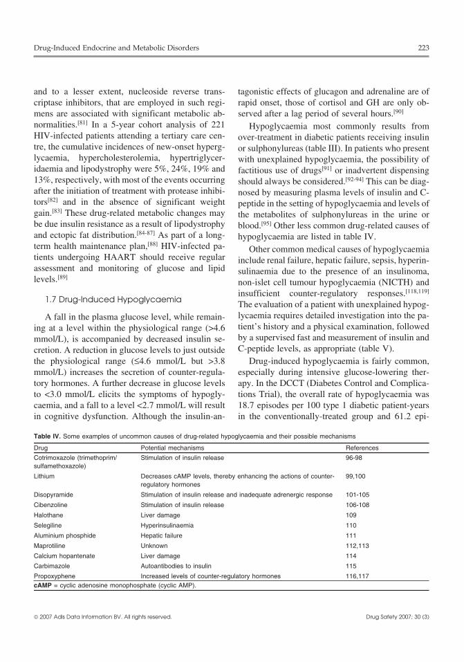
and to a lesser extent, nucleoside reverse trans- tagonistic effects of glucagon and adrenaline are ofcriptase inhibitors, that are employed in such regi- rapid onset, those of cortisol and GH are only ob-mens are associated with significant metabolic ab- served after a lag period of several hours.[90]
normalities.[81] In a 5-year cohort analysis of 221 Hypoglycaemia most commonly results fromHIV-infected patients attending a tertiary care cen- over-treatment in diabetic patients receiving insulintre, the cumulative incidences of new-onset hyperg- or sulphonylureas (table III). In patients who presentlycaemia, hypercholesterolemia, hypertriglycer- with unexplained hypoglycaemia, the possibility ofidaemia and lipodystrophy were 5%, 24%, 19% and factitious use of drugs[91] or inadvertent dispensing13%, respectively, with most of the events occurring should always be considered.[92-94] This can be diag-after the initiation of treatment with protease inhibi- nosed by measuring plasma levels of insulin and C-tors[82] and in the absence of significant weight peptide in the setting of hypoglycaemia and levels ofgain.[83] These drug-related metabolic changes may the metabolites of sulphonylureas in the urine orbe due insulin resistance as a result of lipodystrophy blood.[95] Other less common drug-related causes ofand ectopic fat distribution.[84-87] As part of a long- hypoglycaemia are listed in table IV.term health maintenance plan,[88] HIV-infected pa- Other common medical causes of hypoglycaemiatients undergoing HAART should receive regular include renal failure, hepatic failure, sepsis, hyperin-assessment and monitoring of glucose and lipid sulinaemia due to the presence of an insulinoma,levels.[89]
non-islet cell tumour hypoglycaemia (NICTH) andinsufficient counter-regulatory responses.[118,119]
1.7 Drug-Induced Hypoglycaemia The evaluation of a patient with unexplained hypog-lycaemia requires detailed investigation into the pa-A fall in the plasma glucose level, while remain-tient’s history and a physical examination, followeding at a level within the physiological range (>4.6by a supervised fast and measurement of insulin andmmol/L), is accompanied by decreased insulin se-C-peptide levels, as appropriate (table V).cretion. A reduction in glucose levels to just outside
the physiological range (≤4.6 mmol/L but >3.8 Drug-induced hypoglycaemia is fairly common,mmol/L) increases the secretion of counter-regula- especially during intensive glucose-lowering ther-tory hormones. A further decrease in glucose levels apy. In the DCCT (Diabetes Control and Complica-to <3.0 mmol/L elicits the symptoms of hypogly- tions Trial), the overall rate of hypoglycaemia wascaemia, and a fall to a level <2.7 mmol/L will result 18.7 episodes per 100 type 1 diabetic patient-yearsin cognitive dysfunction. Although the insulin-an- in the conventionally-treated group and 61.2 epi-
Table IV. Some examples of uncommon causes of drug-related hypoglycaemia and their possible mechanisms
Drug Potential mechanisms References
Cotrimoxazole (trimethoprim/ Stimulation of insulin release 96-98sulfamethoxazole)
Lithium Decreases cAMP levels, thereby enhancing the actions of counter- 99,100regulatory hormones
Disopyramide Stimulation of insulin release and inadequate adrenergic response 101-105
Cibenzoline Stimulation of insulin release 106-108
Halothane Liver damage 109
Selegiline Hyperinsulinaemia 110
Aluminium phosphide Hepatic failure 111
Maprotiline Unknown 112,113
Calcium hopantenate Liver damage 114
Carbimazole Autoantibodies to insulin 115
Propoxyphene Increased levels of counter-regulatory hormones 116,117cAMP = cyclic adenosine monophosphate (cyclic AMP).
© 2007 Adis Data Information BV. All rights reserved. Drug Safety 2007; 30 (3)

224 Ma et al.
Table V. Diagnostic evaluation of unexplained fasting hypoglycaemia
Diagnosis Plasma Plasma Plasma Plasma Other tests/diagnostic criteriaglucose insulin C-peptide hydroxybutyratelevels levels levels levels
Oral antihyperglycaemic ↓ ↑ ↑ Absent Sulphonylureas or their metabolitesagents detectable in plasma or urine
Insulinoma ↓ ↑or normal ↑ Absent Negative for sulphonylureas in theplasma or urine, imaging studies tolocalise tumour
Autoimmune ↓ ↑ ↑ Absent Positive insulin antibodieshypoglycaemia
Exogenous insulin ↓ ↑ ↓ Absent Presence of insulin antibodies
Insulin-like growth factors ↓ ↓ ↓ Absent ↑‘Big’ IGF-II,(e.g. NICTH) ↑IGF-II/IGF-I ratio,
presence of tumour
Non-insulin mediated ↓ ↓ ↓ Present Absent response to glucagon
Adrenal insufficiency ↓ ↓ ↓ Present Low cortisol level duringhypoglycaemia, short synacthen test
Ethanol ↓ ↓ ↓ Present Ethanol level, reduced levels ofcounter-regulatory hormones
IGF = insulin-like growth factor; NICTH = non-islet cell tumour hypoglycaemia; ↑ indicates increase; ↓ indicates decrease.
1.7.1 Insulinsodes per 100 patient-years in the intensive treat-ment group.[120] In the UK Prospective Diabetes When human insulin was first introduced, theStudy (UKPDS), the incidence of hypoglycaemia in increased number of reports of severe hypogly-insulin-treated type 2 diabetic patients was 2.3 epi- caemia associated with it was largely attributed tosodes per 100 patient-years.[121] the intensive glucose control associated with the
change in insulin regimen.[128] Compared with un-Unawareness of hypoglycaemia occurs whenmodified human insulin, rapidly-acting insulin ana-there are no warning symptoms prior to the onset oflogues (e.g. lispro) reduce post-prandial glucose ex-cognitive dysfunction resulting in confusion andcursions and the incidence of nocturnal hypogly-coma.[122] This phenomenon is associated withcaemia.[129,130] Insulin glargine, a long-acting insulindelayed and reduced neuroendocrine responses toanalogue with a peakless concentration profile isfalling blood glucose levels.[123,124] In the DCCT,also associated with a lower risk of nocturnal hypog-unawareness of hypoglycaemia was common, withlycaemia compared with insulin suspension iso-up to 36% of the hypoglycaemic episodes occurringphane (neutral protamine Hagedorn [NPH] insu-without symptoms.[125] Risk factors for unawarenesslin).[131] In patients with type 1 diabetes, a basal-of hypoglycaemia include a long duration of diabe-bolus regimen with insulin glargine and insulin lis-tes, intensified glycaemic control, the presence ofpro results in better glycaemic control and fewerautonomic neuropathy, pregnancy, increased age,episodes of nocturnal hypoglycaemia than are ob-renal and liver impairment and use of β-blockers,served with insulin suspension isophane and un-which may mask the adrenergic symptoms of hy-modified human insulin.[132] Lipoatrophy and li-poglycaemia.[126] Hypoglycaemic unawareness canpohypertrophy due to long-term insulin injectionbe prevented by strict avoidance of hypoglycaemia,may also contribute to erratic glycaemic control andwhich may restore counter-regulatory responses andhypoglycaemic episodes.[133] These complicationsawareness of symptoms such as sweating and palpi-can be prevented through patient education and thetation as a result of the activation of the autonomoususe of alternating insulin injection sites.nervous system.[127]
© 2007 Adis Data Information BV. All rights reserved. Drug Safety 2007; 30 (3)

Drug-Induced Endocrine and Metabolic Disorders 225
1.7.2 Sulphonylureas lying mechanisms of these adverse drug reactionsSulphonylureas bind to sulphonylurea receptors are not always clear and these reports highlight the
on the β-cell membrane. This is followed by closure complexity of the regulation of glucose homeosta-of the adenosine triphosphate (ATP)-sensitive po- sis.[90] In addition, alcohol can precipitate or exacer-tassium channels and membrane depolarisation, bate drug-induced hypoglycaemia in the fastingwhich opens the voltage-gated calcium channels, state by inhibiting gluconeogenesis and glycogenol-leading to an influx of calcium ions and the subse- ysis. On the other hand, when consumed with a mealquent release of insulin.[134] The older containing carbohydrates, alcohol is preferentiallysulphonylureas, such as chlorpropamide, have a metabolised, giving rise to higher post-prandial glu-long half-life and are associated with a high risk of cose excursions.[140]
hypoglycaemia, especially in elderly patients or pa- Ciclosporin, an inhibitor of cytochrome P450tients with renal or liver impairment.[126] The sec- (CYP) 3A4 and organic anion transporter 1B1, in-ond-generation sulphonylureas, such as gliben- creases the plasma concentration of repaglinide,clamide (glyburide), gliclazide, glipizide and gli- which is metabolised by CYP2C8 and 3A4, and thusquidone, have shorter half-lives. Glibenclamide, the risk of hypoglycaemia may be increased if thesewhich is commonly prescribed, has a prolonged drugs are coadministered.[141,142]
action due to active metabolites and is associated1.7.4 Renin-Angiotensin-Aldosteronewith a higher incidence of hypoglycaemia than theSystem Inhibitors
other new sulphonylureas.[135] The meglitinide ana-Based on a large number of randomised clinicallogues, such as repaglinide and nateglinide, are new
trials, treatment with inhibitors of the RAAS, in-insulin secretagogues, which bind to alternative sitescluding the ACE inhibitors and angiotensin II recep-on the β-cell membrane and have been shown to betor antagonists (angiotensin II receptor blockersassociated with a lower risk of hypoglycaemia than[ARBs]), is associated with a reduced incidence ofthe older sulphonylureas.[136-138] Since many lifes-new onset of diabetes compared with placebo ortyle factors, such as diet, weight changes, exercise,other antihypertensive drugs.[143-145] Apart from im-concurrent medical conditions and medications, canproving vascular function and reducing insulin re-influence blood glucose levels, patient education,sistance, the presence of the RAAS in the pancreaticself monitoring and regular clinical assessments areislets suggests that these drugs may have directimportant measures to reduce the risk of drug-in-beneficial effects on β-cell function.[146] Severalduced hypoglycaemia.angiotensin receptor antagonists, including telmis-artan and irbesartan, have a partial PPAR-γ agonist
1.7.3 Drugs that Enhance the Effects ofeffect,[147] which may further enhance insulin sensi-Antihyperglycaemic Drugstivity.[148]
Other antidiabetic drugs, such as the biguanides(metformin), PPAR-γ agonists (thiazolidinediones) 1.7.5 β-Adrenoceptor Antagonists (β-Blockers)
and Sympathomimetic Drugsand α-glucosidase inhibitors, do not have directeffects on insulin secretion but may enhance the Insulin release is usually inhibited by the α-glucose-lowering effects of insulin and the adrenergic effects of catecholamines and facilitatedsulphonylureas.[139] According to a review article by by their β-adrenergic effects.[15] The latter can stim-Chan et al.,[90] sporadic cases of hypoglycaemia ulate peripheral glucose production and lipolysis.have been reported in association with a large num- Except during times of stress, α-adrenergic effectsber of drugs. These include β-blockers, salicylates, predominate over those due to β-adrenoceptor stim-phenylbutazone, monoamine oxidase inhibitors, ulation.[15] Thus, depending on the pharmacologicalsulphonamides, trimethoprim, cotrimoxazole profiles and host response, β-blockers may cause(trimethoprim/sulfamethoxazole), the H2 receptor hyperglycaemia in predisposed patients or maskantagonists and tricyclic antidepressants. The under- symptoms of hypoglycaemia in diabetic patients
© 2007 Adis Data Information BV. All rights reserved. Drug Safety 2007; 30 (3)

226 Ma et al.
treated with antihyperglycaemic drugs. Sporadic severe clinical conditions complicated by shock andcases of β-blocker-induced hypoglycaemia in hypoxia.[171-173] Hypoglycaemia has also been re-healthy individuals have been reported during exer- ported in association with inhaled pentamidine ther-cise[149] and post-operatively,[150] both of which are apy.[174]
conditions that are associated with increased secre-tion of counter-regulatory hormones. 2. Drug-Induced
Isoxsuprine and ritodrine are β-sympathomimet- Electrolyte Abnormalitiesics, which relax smooth muscle cells and are used tohalt premature labour. Due to their potential stimu-
2.1 Homeostasis of Fluid andlatory effects on the pancreatic β-cells, there haveElectrolyte Balancebeen cases of neonatal hyperinsulinaemic hypogly-
caemia in infants born to women treated with theseThe kidney is a key organ in the regulation ofdrugs.[151-153] Hypoglycaemia following salbutamol
fluid and electrolyte balance, under the dual influ-(albuterol) overdose has also been reported in someence of the RAAS and antidiuretic hormone (ADH,paediatric patients treated with this drug for asth-also know as vasopressin) [figure 2]. In response toma.[154,155]
reduced renal blood flow, angiotensin II constricts1.7.6 Antimalarial Drugs
blood vessels and stimulates aldosterone release.The incidence of hypoglycaemia in patients with Aldosterone promotes sodium retention and urinary
falciparum malaria is high and can be either sponta- potassium loss. The expansion of blood volumeneous or a result of antimalarial treatment.[156] Risk restores renal perfusion pressure which suppressesfactors include young age, pregnancy, renal failure renin, thus completing the feedback loop. Drugs andand severe infection.[156] Antimalarial drugs such as clinical conditions that reduce renal blood flow canquinine, chloroquine, hydroxychloroquine and activate the RAAS causing hypokalaemia. Thesesulfadoxine-pyrimethamine may stimulate insulin include liver cirrhosis and nephrotic syndrome, duerelease and lead to hypoglycaemia.[156-161] However, to low albumin levels, and heart failure due tonot all hypoglycaemia in patients with malaria is due reduced cardiac output.[175]
to antimalarial treatment. In a study of MalawianADH is produced by neurons that originate in the
children with severe falciparum malaria, hypogly-supraoptic and paraventricular nucleus of the hypo-
caemia was associated with low plasma insulinthalamus and project through the pituitary stalk to
levels and elevated plasma levels of lactate, alanineterminate in posterior pituitary gland. The hormone
and 5′-nucleotidase.[162] These findings suggest thatexerts its biological action (reducing the urinary
impaired hepatic gluconeogenesis may explain hy-flow rate and increasing reabsorption of solute-free
poglycaemia in patients who have not yet receivedwater) through vasopressin (V2) receptors located in
antimalarial drugs.the distal and collecting tubules of kidneys. The
1.7.7 Pentamidine binding of ADH to V2 receptors increases the per-Pentamidine is an antiprotozoal drug used in the meability of the tubular cells through activation of
treatment of pneumocystis pneumonia in patients aquaporin-2, a protein that forms the water channelswith HIV that may cause acute pancreatitis.[163-165] in tubular cells. The secretion of ADH is stimulatedThis can sometimes lead to cytolytic release of by increased plasma osmolality, reduced circulatinginsulin, which in turn results in hypogly- blood volume and to a lesser extent, increased arteri-caemia.[166-170] The latter may be followed by de- al pressure, similar to that mediated by angiotensinstruction of the pancreatic β-cells, insulin insuffi- II.[176] In patients with diabetes insipidus due tociency and hyperglycaemia.[166,167] Risk factors for ADH deficiency, excessive urinary free water loss ispentamidine-induced dysglycaemia include renal compensated for by increased fluid intake in re-impairment, resulting in drug accumulation, and sponse to thirst sensation, the latter being regulated
© 2007 Adis Data Information BV. All rights reserved. Drug Safety 2007; 30 (3)

Drug-Induced Endocrine and Metabolic Disorders 227
↑Blood volumeDilutional hyponatraemia
↓Renal Naexcretion
↑Aldosteronerelease from
adrenal glands
↑ADH release fromposterior pituitary gland
↓Renal perfusion pressure
↑Renin releasefrom kidneys
Angiotensinogen
Angiotensin I
Angiotensin II
ACE
Vascular smooth muscle
Vasoconstriction
Catecholamines
↑Renal K loss viaaldosterone
sensitive cells incollecting tubules
Hypokalaemia
+
+
+
+
+ +
+
+
+
+
+
+
−−
−
Fig. 2. Feedback control between the renin-angiotensin-aldosterone system and antidiuretic hormone in the regulation of electrolytes andfluid balance. ACE = angiotensin converting enzyme; ADH = antidiuretic hormone; K = potassium; Na = sodium; ↑ indicates increase; ↓indicates decrease; + indicates positive effect; – indicates negative effect.
primarily by hypothalamic osmoreceptors and plas- with thiazides, loop diuretics are less likely to in-ma osmolality. duce severe hyponatraemia. This is due to the inhibi-
tion of sodium chloride transport in the loop ofHenle by loop diuretics, which prevents the genera-
2.2 Drug-Induced Hyponatraemiation of a counter-current gradient and thereby limitsand Hypernatraemiathe ability of ADH to induce water retention.[182] Asa result, clinicians should consider other causes of
Hyponatraemia can be dilutional (excess water hyponatraemia before attributing it to loop diuretics.retention) or depletional (excessive loss of sodium
Drug-induced causes of dilutional hyponatraemiawith or without water) in nature. Table VI listsinclude the syndrome of inappropriate ADHexamples of drugs that can cause hyponatraemia,(SIADH). Drugs can cause dilutional hyponatraemiawith their underlying mechanisms. Diuretics thatby enhancing ADH release or its effects (table VI).promote urinary loss of sodium are major causes of
depletional hyponatraemia. Thiazides, notably in- Hypernatraemia may be caused by drug-induceddapamide, can cause severe hyponatraemia and hy- diabetes insipidus due to suppression of ADH secre-pokalaemia[177-180] by acting at the distal renal tu- tion (e.g. caused by lithium, carbamazepine) orbules to enhance urinary loss of sodium and potassi- ADH deficiency (e.g. caused by lithium, demeclo-um. Patients who have polydipsia and weight gain
cycline, methoxyflurane, amphotericin B,appears to be the most at risk of thiazide-inducedrifampicin, cisplatin or foscarnet).[176] Excessive de-hyponatraemia.[181]
hydration is usually prevented by stimulation of theLoop diuretics act on the ascending limb of thethirst centre. Thus, hypernatraemia occurs only inloop of Henle and inhibit both sodium and waterindividuals in whom thirst sensation is impaired orreabsorption.[182] If water intake is high in the settingwater access is not feasible (e.g. elderly patients orof excessive urinary sodium loss induced by diuret-
ics, depletional hyponatraemia can occur. Compared those who have had a cerebrovascular accident).
© 2007 Adis Data Information BV. All rights reserved. Drug Safety 2007; 30 (3)

228 Ma et al.
2.3 Drug-Induced Hypokalaemia cess’.[185] Due to the similarities in the mineralocor-and Hyperkalaemia ticoid and glucocorticoid metabolic pathways, 11β-
hydroxysteroid dehydrogenase is produced by theDiuretics increase the delivery of sodium and kidneys to inactivate cortisol by converting it to
water to aldosterone-sensitive potassium secreting cortisone.[186] Naturally occurring liquorice, carbe-cells in the collecting tubules and promote urinary noxolone (a synthetic derivative of glycyrrhizinpotassium wasting. Due to volume contraction, [glycyrrhizinic acid], which is a constituent of liquo-these drugs also activate the RAAS and ADH secre-
rice) and some liquorice-containing chewing tobac-tion, which may worsen electrolyte abnormali-
cos can inactivate 11β-hydroxysteroid dehy-ties.[182] Indapamide, a thiazide-like diuretic, causes
drogenase.[185,186] As a result, there is increasedsevere hypokalaemia and hyponatraemia especially
binding of intra-renal cortisol to mineralcorticoidin the elderly population.[178] Laxatives also cause anreceptors in the distal tubules, leading to excessiveincreased loss of potassium from the gastro-intesti-water and sodium retention and urinary potassiumnal tract. Laxative- and diuretic-induced hy-loss. Liquorice is a common ingredient in manypokalaemia are not uncommon adverse effects intraditional Chinese herbal medicines and Chinesepatients treated with slimming pills.[183,184]
chewing snacks.Some naturally occurring compounds or pharma-In our body compartment, sodium is the maincological agents decrease 11β-hydroxysteroid
extracellular cation and potassium is the main intra-dehydrogenase levels and give rise to hormonalcellular cation. Thus, energy-dependent transmem-changes that mimic primary hyperaldosteronism, a
condition known as ‘apparent mineralocorticoid ex- brane cation exchange systems, such as the sodium-
Table VI. Examples of drugs and conditions that cause hyponatraemia
Cause Examples
Nondrug-related dilutional hyponatraemia
Excessive water intake
Increased water resorption Cirrhosis, congestive heart failure, nephrotic syndrome
Reduced renal excretion of water load SIADH (can also be caused by drugs), glucocorticoid deficiency
Drug-related dilutional hyponatraemia
Exogenous ADH Vasopressin used in the control of GI bleeding; desmopressin used inthe treatment of diabetes insipidus, platelet dysfunction, nocturia
Drugs that cause inappropriate ADH secretion Antipsychotics (phenothiazines, such as haloperidol, thioridazine),antidepressants (tricyclic antidepressants such as amitriptyline;monoamine oxidase inhibitors; SSRIs, such as fluoxetine andsertraline), chemotherapeutic agents (vincristine, vinblastine, cisplatin,cyclophosphamide), amiodarone, bromocriptine, nicotine,methylenedioxymethamfetamine (‘ecstasy’), ciprofloxacin
Drugs that cause increased sensitivity to ADH in the collecting Oxcarbazepine, carbamazepinetubular cells
Drugs that cause expression of increased numbers of ADH Chlorpropamidereceptors in collecting tubular cells
Nondrug-induced depletional hyponatraemia
Renal salt loss Tubulointerstitial nephritis, polycystic kidney disease, analgesicnephropathy
GI salt loss Fistulae
Skin loss
Drug-induced depletional hyponatraemia
Drugs that cause excessive urinary sodium loss Thiazide diuretics, loop diureticsADH = antidiuretic hormone; GI = gastrointestinal; SIADH = syndrome of inappropriate ADH; SSRIs = selective serotonin reuptakeinhibitors.
© 2007 Adis Data Information BV. All rights reserved. Drug Safety 2007; 30 (3)

Drug-Induced Endocrine and Metabolic Disorders 229
potassium adenosine triphosphatase (Na-K- patients who have cardiovascular and renal compli-cations and who are receiving multiple medica-ATPase), pump sodium out of the cells in exchangetions.[193,194]for potassium influx, in order to maintain the correct
electrolyte gradient for normal cellular function.2.4 Other Drug-InducedDrugs that increase activity of the Na-K-ATPase canElectrolyte Abnormalitieslead to increased potassium entry into the cells and
cause hypokalaemia.[187] These include catecho- Drugs that act on the renal tubules, such as looplamines, β2-adrenoceptor agonists (such as diuretics, can induce other electrolyte abnormalities,salbutamol, terbutaline or dopamine), insulin, e.g. hypomagnesaemia. In a review article,[195] 50levothyroxine sodium (thyroxine) and theophyl- medications were listed that can cause hy-line.[188] These drug effects may be exacerbated in pomagnesaemia. Of these, 17 drugs were consideredstressful situations, such as acute coronary syn- to be clinically important. These included cisplatin,dromes and sepsis, which are associated with in- amphotericin B and ciclosporin. In patients receiv-creased endogenous production of catecho- ing these drugs, routine monitoring of serum magne-lamines.[189]
sium levels and prophylactic administration of mag-Hyperkalaemia can be induced by drugs that nesium may be indicated.
decrease Na-K-ATPase activity leading to potassi-3. Drug-Induced Abnormalities inum leakage from cells. Non-selective β-blockers,Calcium Balancesuch as propranolol, selective β1-blockers, such as
atenolol or metoprolol, digoxin (digitalis) over- Calcium is a major intracellular signal that medi-dose,[190] calcineurin-inhibitors, diazoxide, minox- ates many biological actions; these actions are de-idil, anaesthetics such as isoflurane, suxamethonium pendent on the maintenance of a steady extracellularchloride (succinylcholine chloride) and arginine can calcium level. Over 98% of the body’s calcium storecause hyperkalaemia via this mechanism.[191,192]
and 85% of the phosphate are present in bone asDrugs that induce aldosterone deficiency or resis- hydroxyapatite. In the plasma, about 40% of calci-
tance can also cause hyperkalaemia. ACE inhibitors, um is bound to albumin and 15% is associated withNSAIDs and ciclosporin can cause aldosterone defi- phosphate, citrate or sulphate. The remaining 45% isciency whereas amiloride, spironolactone, triamter- in the form of free (ionised) calcium, which isene, trimethoprim and pentamidine can induce al- physiologically active.[196]
dosterone resistance, causing hyperkalaemia. Hy- The extracellular or serum calcium level is tight-pertonic mannitol can induce hyperkalaemia ly regulated through interaction of the actions ofthrough elevation of plasma osmolality and potassi- vitamin D and parathyroid hormone (PTH) on theirum efflux through a passive gradient and solvent target organs, notably the intestine, kidneys anddrag.[192] Since ADH promotes solute-free water bone (figure 3). A decreased level of ionised calci-retention while aldosterone has effects on sodium, um stimulates the release of PTH. In the presence ofwater and potassium handling, patients receiving the vitamin D, PTH increases the serum calcium levelaforementioned medications may develop hypona- by stimulating bone resorption and promoting thetraemia or hypokalaemia or both, depending on diet- release of calcium phosphate from bone. In theary intake of sodium and potassium, concomitant kidneys, PTH increases the active reabsorption ofmedications and clinical conditions. Other patients calcium and enhances production ofat high risk for drug-induced electrolyte abnormali- 1,25-dihyhydroxyvitamin D3 (calcitriol). In the in-ties include those with renal artery stenosis or defec- testine, calcium absorption is augmented by calcitri-tive autoregulation, such as diabetic patients with ol. Collectively, these processes raise the serumautonomic neuropathy. These conditions may coex- calcium level and restore homeostasis. Calcitonin isist in the same patient, such as elderly diabetic synthesised and secreted by the parafollicular cells
© 2007 Adis Data Information BV. All rights reserved. Drug Safety 2007; 30 (3)

230 Ma et al.
hyperparathyroidism, which may result in hypercal-caemia due to tertiary hyperparathyroidism.[197]
A variety of drugs can cause imbalances betweenbone mineralisation and resorption, as well as be-tween calcium absorption and excretion. Drug-in-duced hypercalcaemia is more likely to occur inimmobilised patients, and in those with malignantconditions or renal impairment, whereas drug-in-duced hypocalcaemia may occur in institutionalisedor malnourished patients.[198]
3.1 Drug-Induced Hypocalcaemia
3.1.1 Antiepileptic DrugsPatients receiving long-term antiepileptic treat-
ment are at risk of developing vitamin D deficiency.Phenytoin, carbamazepine and phenobarbitone in-duce up-regulation of hepatic microsomal enzymes,which increase the turnover of 25 hydroxyvitaminD. The reduction in calcitriol causes hypocalcaemia,resulting in secondary hyperparathyroidism.[199] Thelack of vitamin D impairs the ability of PTH tonormalize the serum calcium level. Eventually, pa-tients may develop antiepileptic-induced bone dis-ease, which is characterised by marked elevation inlevels of serum alkaline phosphatase and rickets orosteomalacia.[200]
3.1.2 BisphosphonatesBisphosphonates are potent inhibitors of osteo-
clastic activity and thereby reduce the serum calci-
7-dehydrocholesterol (from diet)
Sunlight
Colecalciferol (skin)
Bound colecalciferol (blood)
25-hydroxycolecalciferol (liver)
1, 25-dihydroxycolecalciferol (kidneys)
Calcium absorption (from GI tract)
b
Vitamin D-binding protein
a
Low calcium level in blood
Release of parathyroid hormone
Bone Kidneys
Calcium releasefrom bone
Decreased urinaryexcretion of calcium
Vitamin DGI tract
Increased GIabsorption of calcium
level of calcium in blood↑
↑
Fig. 3. Schematic diagram of (a) vitamin D metabolism and (b)calcium metabolism showing the tight regulation of serum calciumlevels through interactive effects of parathyroid hormone and vita-min D on the bone, intestine and kidneys. GI = gastrointestinal;↑ indicates increase.
um level. Indications for bisphosphonate therapyinclude hypercalcaemia of malignancy and treat-of the thyroid gland in response to high calciumment of osteoporosis and multiple myeloma.[201]
levels. Calcitonin antagonises the actions of PTH,Bisphosphonates are also used in prophylaxisalthough its effects are only transient.[197]
against metastatic bone disease and for the treatmentVitamin D3 (colecalciferol) is from dietary of Paget’s disease of the bone. Bisphosphonates are
sources or synthesised in the skin is hydroxylated usually well tolerated, with minor adverse effects offirst in the liver and then in the kidney to form the transient arthralgia or flu-like symptoms. Thesemost active form of vitamin D, calcitriol. Under the drugs are rapidly excreted via the kidney and toxici-influence of calcitriol and PTH, calcium is reab- ty may occur in the patients with renal failure.sorbed from the renal tubules to regulate the serum Zoledronic acid is a new intravenouscalcium level. In patients with renal failure, in bisphosphonate, which is about 1000 times morewhom the synthesis of calcitriol is impaired, normal potent than pamidronate, alendronate or etidronate.serum calcium levels are maintained by secondary Cases of severe hypocalcaemia have been reported
© 2007 Adis Data Information BV. All rights reserved. Drug Safety 2007; 30 (3)

Drug-Induced Endocrine and Metabolic Disorders 231
in association with intravenous zoledronic acid ther- are associated with hypocalcaemia include colchi-apy.[202,203] cine,[214] which is postulated to cause inhibition of
bone resorption,[215] and intravenous diltiazem.[216]Because of a compensatory increase in the secre-Other conditions that can cause hypocalcaemiation of PTH, most patients receiving intravenous
include hypoparathyroidism (e.g. resulting from de-bisphosphonate therapy do not develop hypocal-struction of the parathyroid glands by irradiation orcaemia. In patients with occult vitamin D deficien-from a failure of parathyroid development), in-cy, hypomagnesaemia or renal failure, compensato-creased uptake of calcium into the bone (e.g. hungryry mechanisms may not be effective.[204,205] Hence,bone syndrome following surgical treatment of pro-in patients with normocalcaemia or hypercalcaemialonged hyperparathyroidism) and abnormal vitaminwith low urinary calcium excretion, caution shouldD metabolism (e.g. renal failure and following acutebe exercised to prevent hypocalcaemia during intra-pancreatitis).[198]venous bisphosphonate therapy.
3.1.3 Miscellaneous 3.2 Drug-Induced HypercalcaemiaIonised calcium levels may be reduced by in-
creased calcium deposition in the tissue or by in- With the widespread use of automated multi-travascular binding. The use of phosphate enema for channel analysers, hypercalcaemia is a common in-the treatment of faecal retention has been associated cidental finding. The major causes of hypercal-with hyperphosphataemia and hypocalcaemic teta- caemia are malignancy and hyperparathyroidism,ny.[206,207] Calcium is deposited with phosphate in which can be differentiated by their clinicalthe bone and in extra-skeletal tissues, resulting in presentations and the measurement of PTHhypocalcaemia. In patients receiving massive blood levels.[217] Drugs are another important cause oftransfusions, rapid plasma exchange or leukapher- hypercalcaemia. Enhanced calcium absorption andesis, the large amount of citrate may chelate calcium increased bone turnover is evident in patients takingand reduce ionised calcium levels.[208] Hypocal- high doses of either vitamin D or calcifediolcaemia is also a known complication of chelation (calcidiol) and the topical vitamin D analogue, calci-therapy. Deaths associated with hypocalcaemia have potriol (calcipotriene), for skin lesions.[218] Hypervi-been reported in patients who developed cardiac taminosis A[219] or the use of tretinoin (retinoic acid)arrest after receiving edetic acid (disodium for the treatment of acne and certain haematologicalethylenediaminetetraacetate [Na2EDTA]) for the malignancies[220] can cause hypercalcaemia by in-treatment of lead poisoning.[209] In patients with creasing bone resorption. Thiazide diuretics stimu-refractory cytomegalovirus and herpes infections, late renal tubular calcium reabsorption and may leadthe use of the antiviral agent foscarnet has been to hypercalcaemia, especially in patients with riskassociated with hypocalcaemia.[210] factors such as immobilisation or subclinical
hyperparathyroidism.[217]Magnesium depletion causes PTH resistance andreduced PTH secretion, resulting in hypocalcaemia. Excessive intake of calcium, in the form of calci-Hypomagnesaemia is a known complication in pa- um carbonate, may lead to hypercalcaemia,[221] al-tients receiving diuretics.[211] In addition, aminog- kalaemia, nephrocalcinosis and renal failure. Thelycosides[211] and cisplatin can cause urinary magne- incidence of hypercalcaemia appears to be increas-sium wasting, thereby causing hypomagnesaemia ing as more people take calcium supplements forand sometimes hypocalcaemia.[212] osteoporosis, peptic ulcers, gastritis, gas-
troesophageal reflux and chronic kidney disease.[222]In patients with ethylene glycol poisoning, ethy-lene glycol is metabolised to oxalate. Widespread Lithium treatment may cause hypercalcaemia bydeposition of calcium oxalate in various organs altering the set point at which calcium suppressesleads to reduced levels of ionised calcium, resulting the secretion of PTH.[223] Increased SNS activity duein hypocalcaemia.[213] Other examples of drugs that to over-treatment with levothyroxine sodium or
© 2007 Adis Data Information BV. All rights reserved. Drug Safety 2007; 30 (3)

232 Ma et al.
sympathomimetics, such as theophylline, stimulates Without iodine, there is no synthesis of the thy-osteoclastic bone resorption and cause mild hyper- roid hormones. Thyroid hormone synthesis can becalcaemia, which may be attenuated by β-blocker transiently blocked by the inhibitory effect of hightreatment.[224,225] dose iodide on intrathyroidal iodine uptake; the so
called Wolff-Chaikoff effect.[227] However, withcontinued exposure to excess iodide, there is usually4. Drug-Induced Thyroid Disordersescape from this effect. This is due to decreasedtransport of iodide into the thyroid by the sodium/iodide transporter, thereby lowering intracellular io-4.1 Regulation of Thyroid Metabolismdide levels to those that are insufficient to maintainthe inhibitory effect.[228,229]Thyroid hormones are the only iodine-containing
hormones in vertebrates. The synthesis of thyroid The thyroid hormones secreted from thyroidhormones is dependent on the intake of iodine (or gland are over 99% protein-bound, with T3 beingiodide [I-], in its ionised form), as this the rate- less tightly bound than T4.[230] Only free thyroidlimiting substrate in the process. Iodide uptake into hormones are biologically active. Thyroid hormonesthyroid follicular cells is the first step in thyroid exert their end-organ effects through binding to nu-hormone synthesis. After iodide enters the thyroid, it clear receptors, with T3 having 10–15 times greateris oxidised by thyroid peroxidase, and this is fol- affinity than T4. Conversion from T4 to T3 in periph-lowed by iodination of thyroglobulin, resulting in eral tissues is mediated by 5′-monodeiodinase. Thethe formation of the mono- and di-iodotyrosines. actions of this enzyme can be affected by manyTwo iodotyrosines are coupled to form thyroxine medications (table VII). Thyroid hormone synthesis(T4), whilst triiodothyronine (T3) can be formed by and secretion is under feedback control by thyrotro-the coupling of one mono-iodotyrosine and one di- pin (TSH) released from the pituitary gland. Hence,iodotyrosine within a thyroglobulin molecule. How- drugs that modify TSH secretion may also affectever, most T3 is produced by extrathyroidal de- thyroid function. The symptoms of thyrotoxicosis,idination of T4.[226] such as palpitation, tachycardia, hypertension,
Table VII. Examples of drugs affecting thyroid function and their underlying mechanisms
Mechanisms Examples of drugs
Decreased TSH secretion Dopamine, glucocorticoids, somatostatin, levothyroxine sodium (thyroxine) or anyexogenous thyroid hormones, amiodarone
Increased TSH secretion Antipsychotics, metoclopramide, theophylline
Intrinsic effects on the thyroid and thyroid hormone Amiodarone, lithium, organic iodine, radiographic contrast agents, amfetamine,synthesis thionamides, thalidomide, interferon-α, traditional Chinese herbal medicines with
high iodine content
Decreased peripheral tissue conversion from T4 to Amiodarone, propranolol, glucocorticoids, sodium iopodate, iopanoic acidT3
Decreased TBG binding Androgens, glucocorticoids, diuretics (very large intravenous dose)
Increased TBG binding Estrogen therapy or estrogen-containing oral contraceptives, tamoxifen, heroin,methadone, phenothiazines
Altered TBG binding NSAIDs, phenytoin
Interference with assays of thyroid function tests Monoclonal antibodies, tiratricol (triiodothyroacetic acid [TRIAC]), levothyroxinesodium or its analogues
Altered hepatic metabolism of thyroid hormones Phenytoin, phenobarbital, carbamazepine, rifampicin, any drugs that can increasethe activity of cytochrome P450 enzymes
Decreased absorption of T4 (of clinical relevance in Calcium carbonate, cholestyramine, colestipol, aluminium hydroxide, ferroushypothyroid patients who need regular T4 sulphate, sucralfate, dietary soy proteins and fibresreplacement therapy)TBG = thyroxine-binding globulin; TSH = thyrotropin; T3 = triiodothyronine; T4 = thyroxine.
© 2007 Adis Data Information BV. All rights reserved. Drug Safety 2007; 30 (3)

Drug-Induced Endocrine and Metabolic Disorders 233
sweating and diarrhoea, are mediated through acti- administration may result in hyperthyroidism.[236]
vation of the autonomic nervous system. Medica- This effect is also seen in patients from iodine-tions, including β-blockers, may mask the symp- sufficient areas who have nodular goitre containingtoms of thyrotoxicosis and lead to delayed or missed autonomously functioning thyroid nodules, and isdiagnosis.[231] due to increased substrate provision after escape
from the Wolff-Chaikoff effect (type 1 amiodarone-induced thyrotoxicosis).[236] In addition, amiodarone4.2 Drug-Induced Hyper-can also cause a destructive thyroiditis, therebyand Hypothyroidismcausing transient thyrotoxicosis (type 2 amiodarone-induced thyrotoxicosis).[236]
4.2.1 ThionamidesAmiodarone can also induce hypothyroidism byThe thionamides are the main antithyroid drugs,
inhibiting 5′-monodeiodinase resulting in a de-and these include propylthiouracil, carbimazole andcreased T3 level,[238] by reducing T3 binding to itsmethimazole (an active metabolite of carbimazole).receptor, by antagonising the effects of the thyroidThese drugs inhibit thyroid peroxidase and thus, thehormones[236] and by suppressing TSH secretion. Allsynthesis of the thyroid hormones.[231]
of these mechanisms contribute to the wide range of4.2.2 Amiodarone thyroid dysfunction associated with amiodaroneAmiodarone therapy can cause both hyper- and therapy.
hypothyroidism.[232] This drug is widely used as a4.2.3 Lithiumclass III anti-arrhythmic drug and has a high iodideLithium is used as treatment for manic-depres-content (75mg per 200mg tablet). After hepatic me-
sive disorders. Due to its inhibitory effect on thyroidtabolism, about 6mg of iodine per 200mg of ami-hormone synthesis, it was used as an anti-thyroidodarone is released into the circulation.[233] This isdrug in early 1970s.[239] It is still occasionally usedcompared with the WHO recommendation of a dailyas a second-line therapy or an interim measureiodine intake of 150μg for adults.[234] Hence, ami-before definitive treatment in thyrotoxic patientsodarone significantly increases the body iodine load.who are allergic or not responsive to thionamides.Dietary iodine content partly predicts amiodarone-Approximately 1–3% of patients treated with lithi-induced thyroid dysfunction, with hypothyroidismum develop goitre, typically diffuse, with or withoutbeing more common in iodine-sufficient areas andthyroid dysfunction.[240] Lithium has multiple ac-hyperthyroidism being more common in iodine-de-tions resembling those of iodine, but the exact mech-ficient regions.[235,236] This reflects the fact that theanisms of lithium-induced thyroid dysfunction re-clinical effects of amiodarone and iodine in an indi-main to be clarified. Lithium can block thyroidvidual depend on the underlying status of the thyroidhormone release from thyroglobulin,[241] inhibitgland. Excess iodine causes hypothyroidism due to aadenylate cyclase and prevent TSH or thyroid stimu-transient inhibitory effect on intrathyroidal iodinelating antibodies from activating thyroid cells viauptake. Populations with chronically high iodinethe TSH receptor.[242] As with amiodarone, lithiumintake have a higher prevalence of autoimmune thy-may precipitate or adversely affect thyroid functionroid disease.[237] Such patients with underlyingin individuals with autoimmune thyroid diseas-autoimmune thyroid disease appear to be particular-es.[243]
ly sensitive to the inhibitory Wolff-Chaikoff effectand are sometimes unable to escape from these 4.2.4 Levothyroxine Sodium (Thyroxine) and
its Analoguesinhibitory effects. As a result, excess iodine or ami-odarone will result in increased goitre and hypothy- Levothyroxine sodium is used as replacementroidism in patients with Hashimoto thyroiditis. Con- therapy in patients with hypothyroidism. However,versely, in patients from areas of iodine deficiency due to its weight-loss effect, levothyroxine sodiumwho have endemic goitre, iodine and amiodarone and its analogues or metabolites (e.g. tiratricol
© 2007 Adis Data Information BV. All rights reserved. Drug Safety 2007; 30 (3)

234 Ma et al.
[3,5,3′-triiodothyroacetic acid or TRIAC], a physio- (figure 4a). Gonadotropin-releasing hormonelogical metabolite of T3) have been used for slim- (GnRH), secreted in a pulsatile manner, is transport-ming purposes, often by young women.[244] Al- ed via the portal circulation to the pituitary. Inthough T3 has been used by some doctors to treat women, the secretion of GnRH is under negativeobesity, this clinical approach remains controver- feedback control by estrogen and progesterone, andsial.[245] Since levothyroxine sodium and tiratricol is also regulated by other neuropeptides, such ascan cross-react with T3 assays,[244] the possibility of prolactin and serotonin. Follicle-stimulating hor-inappropriate use of levothyroxine sodium or its mone (FSH) promotes ovarian follicular maturation,analogues should be considered in patients with whereas lutenising hormone (LH) stimulates pro-spurious thyroid function tests. duction of estrogen and plays an important role in
ovulation. During follicular growth, the granulosa4.2.5 Traditional Chinese Herbal Medicinesand thecal cell layers in the ovary work in synchronyGoitre and hypothyroidism due to iodine defi-to produce estradiol from the steroid precursorciency are a worldwide health problem, particularlypregnenolone, a process stimulated by LH. A posi-in iodine deficient areas. Some traditional Chinese
herbal medicines with claims to treat thyroid diseasehave a high iodine content, which can induce thyroiddysfunction.[246] In patients with thyrotoxicosis whoare refractory to thionamide therapy despite gooddrug compliance, the concomitant use of iodine-richsubstances should be considered. These includetraditional Chinese herbal medicines or foods withhigh iodine contents (e.g. seafood, seaweeds, fish),which are not uncommonly taken by patients whomay mistakenly think that dietary iodine insufficien-cy is a contributory factor for their conditions.
4.2.6 Interferon-αInterferon-α can induce both hyper- and hypo-
thyroidism, with the latter being more common,especially during treatment for chronic hepatitis Cinfection.[247] Interferon-α-induced thyroid dysfunc-tion is usually transient, although prolonged effectshave been reported.[248] The exact mechanism is notfully understood. In addition to direct effects on thethyroid gland, interferon-α can lead to increasedlevels of anti-thyroid antibodies and expression ofmajor histocompatability class I antigens.
5. Drug-Induced Gonadal Dysfunction
5.1 Reproductive Physiology and theHypothalamus-Pituitary-Gonadal Axis
In women, normal ovulation and menstruationrequires intact functioning of the hypothalamus andpituitary, as well as the ovaries and endometrium
Hypothalamus
Pituitary
LH FSH
OvaryE2
PEndometrium
E2, P (−)
E2, P (−)
Inhibin(−)
Serotonin
DopamineNoradrenaline Opioids
CRH
GnRH
a
b
Hypothalamus
Pituitary
LH FSH
Serotonin
DopamineNoradrenaline Opioids
CRH
GnRH
Inhibin (−)
T (−)
Testis
Leydigcells
T (−)
Seminiferous tubulesSperm
Activin (+)
Fig. 4. Regulation of the hypothalamic-pituitary-gonadal axis in the(a) female and (b) male. CRH = corticotropin-releasing hormone;E2 = estradiol; FSH = follicle-stimulating hormone; GnRH =gonadotropin-releasing hormone; LH = lutenising hormone; P =progesterone; T = testosterone; – indicates inhibitory effect; + indi-cates stimulatory effect.
© 2007 Adis Data Information BV. All rights reserved. Drug Safety 2007; 30 (3)

Drug-Induced Endocrine and Metabolic Disorders 235
tive feedback mechanism is present whereby LH function by altering libido and other physiologicaland FSH surge mid-cycle in response to increasing functions. Libido, or sex drive, is influenced by sexestrogen secretion, leading to ovulation. LH then hormones and emotions. Sexual arousal leads topromotes the production of progesterone from the erection in men and swelling of the vagina, labiacorpus luteum following ovulation. In addition to minora and clitoris in women. The control of erec-estradiol, the ovary produces androgens and proges- tion and the corresponding female changes involveterone, a process involving the conversion of a cho- vascular changes that are mediated by parasympa-lesterol substrate to pregnenolone via multiple en- thetic and sympathetic outflow to the peripheralzyme pathways that are regulated by gonadotropin organs. The brainstem and spinal cord, where thestimulation. neurons that innervate the cardiovascular system
and penis arise from, play an important role in thisA similar pathway operates in men (figure 4b),circuit.[250] The dopaminergic and serotoninergicwhereby testicular function is regulated by LH andpathways, as well as neurotransmitters such asFSH from the pituitary. FSH stimulates spermato-noradrenaline, acetylcholine and GABA, are alsogenesis and plays an indirect role in androgen bio-involved. Nitric oxide is the key molecule in thesynthesis by inducing maturation of the Leydig cellsfinal pathway in this process, whereby, upon itsduring development. LH acts on the Leydig cells torelease from endothelial cells and neurons, it acts aspromote androgen synthesis from cholesterol steroida vasodilator causing penile engorgement and erec-precursors. A feedback loop operates whereby thetion.[251]
pituitary and hypothalamus are under negative feed-back control by androgens produced in the Leydigcells and inhibin from the seminiferous tubules, as
5.2 Drug-Induced Alterations inwell as positive control via the production of activin.
Gonadotropin SecretionSexual development is regulated by activity of
the hypothalamus-pituitary-gonadal axis, wherebyDisorders of the reproductive axes can be due toan increase in episodic secretion of GnRH and in-
drugs affecting pituitary hormone secretion, gonadalcreased secretion of gonadotropins in puberty trig-function or the relative balance of androgens andger progressive development of secondary sexualestrogens. Long-acting GnRH analogues are used incharacteristics and initiate spermatogenesis in menthe treatment of precocious puberty, endometriosis,and ovulation in women.and prostate and breast carcinoma. These drugs
Whilst estradiol is the predominant female sex abolish the pulsatile GnRH stimulation, leading tohormone and testosterone is the predominant male pituitary desensitisation with reduced gonadotropinhormone, low levels of androgens are also present in and sex hormone production. Hence, treatment withwomen and similarly, estrogens in men. Androgens these drugs may lead to menopausal symptoms,are produced in both the adrenal glands and the reduced libido and osteoporosis in female patients orgonads. Subsequent peripheral conversion produces impotence and reduced libido in male patients.[252]
17β-estradiol from testosterone and estrone from Chronic glucocorticoid excess can inhibit LH andandrostenedione. Thus, in men, estradiol arises from FSH secretion, resulting in reduced spermatogenesisplasma testosterone, and estrone is derived from or ovulation and lowering of gonadal sex hormoneandostenedione, with some direct secretion of these levels.[253] Anti-androgens, such as cyproterone, in-estrogens from the testes.[249] Imbalances between hibit the action of adrenal and testicular androgensthese hormonal factors can manifest in disorders by competitive antagonism at the target organ recep-such as gynaecomastia. tors. Their progestogenic activity also reduces
gonadotropin secretion leading to gynaecomastia,In addition to affecting the hypothalamus-pituita-ry-gonadal axis, drugs can interfere with sexual reduced testosterone levels and sperm count.[254]
© 2007 Adis Data Information BV. All rights reserved. Drug Safety 2007; 30 (3)

236 Ma et al.
Table VIII. Differential diagnosis of gynaecomastia
Cause Examples
Nondrug-related
Physiological Puberty (onset at adolescence), idiopathic
Hypogonadism (low testosterone levels) Primary (history of injury to the testicles?), secondary (history of pituitarydisease?)
Estrogen, hCG or aromatase-producing tumours Testicular cancer, adrenal cancer, lung cancer(↑estrogen ↓testosterone ↑hCG levels)
Endocrine disorders Thyrotoxicosis, acromegaly, androgen insensitivity due to androgenreceptor defects
Cirrhosis (↑SHBG levels)
Renal failure (↓testosterone; ↑LH, FSH levels)
Obesity
Klinefelter’s syndrome (↑FSH, LH; ↓testosterone)
Drug-related
Drugs that increase estrogenic activity Conjugated and synthetic estrogens, digoxin (digitalis), oralcontraceptives
Drugs that stimulate estrogen secretion Anabolic steroids, hCG, LHRH, menopausal gonadotropins
Drugs that reduce the effects or synthesis of testosterone Antiandrogens (e.g. cyproterone, flutamide, finasteride), cimetidine,cytotoxic agents, spironolactone, phenytoin, ketoconazole, reserpine
Drugs that cause gynaecomastia by an unknown mechanism Calcium channel antagonists, methadone, marijuana, methyldopaFSH = Follicle-stimulating hormone; GH = growth hormone; hCG = human chorionic gonadotropin; LH = lutenising hormone; LHRH =lutenising hormone release hormone; SHBG = sex hormone-binding globulin; ↑ indicates increase; ↓ indicates decrease.
5.3 Chemotherapy-Induced congenital malformations of the reproductive tractGonadal Dysfunction and early onset of puberty in children.[262]
Cytotoxic chemotherapy and radiotherapy can 5.5 Drug-Induced Virilisationcause significant gonadal damage in both men and
In utero exposure to androgens can lead to mas-women.[255] The risk is particularly high with alky-culinisation of the female fetus. Exposure to excesslating agents such as cyclophosphamide,androgens during development can lead to preco-chlorambucil, procarbazine and busulfan. Cisplatin-cious puberty in children.[263] Increased androgenbased chemotherapy causes temporary azoospermialevels in women can result in hirsutism, clitorome-in most men, with recovery to normospermia in 80%galy, deepening of the voice and increased muscleby 5 years.[256] Ovarian failure occurs in at least 50%development. Anabolic steroids are known to causeof women treated with alkylating agent monother-interrupted growth and virilisation in children, birthapy.[257]
defects in the unborn child and severe virilisation inwomen.[264] In men, these drugs can cause testicular5.4 Environmental Toxin-Inducedatrophy and reduced gonadotropin and testosteroneEndocrine Disruptionlevels.[264] Similar effects are observed with agentssuch as danazol, stanozolol, methyltestosterone[265]Both naturally occurring phytoestrogens[258] andand, to a lesser extent, oxandrolone.[266]synthetic compounds, such as organochlorine pesti-
cides, polychlorinated biphenyls, phenolic com- Hyperandrogenism and chronic anovulation arepounds and phthalate esters, may mimic the actions common endocrine abnormalities that affect womenof sex hormones and interfere with the reproductive of reproductive age. Some of these women fulfill theaxes.[259-261] Such ‘endocrine disruptors’ may pos- diagnostic criteria for PCOS and may have polycys-sess estrogenic or anti-androgenic properties and tic ovaries upon ovarian ultrasonography.[267] Thehave been implicated in male fertility disorders, use of anticonvulsants, such as valproic acid (sodi-
© 2007 Adis Data Information BV. All rights reserved. Drug Safety 2007; 30 (3)

Drug-Induced Endocrine and Metabolic Disorders 237
um valproate), has been associated with the devel- predominantly by inhibiting the action of dopamine,opment of PCOS, although the mechanism is not and possibly via enhancing the effect of serotonin inentirely clear.[268] the hypothalamus (table IX).[269]
5.6 Drug-Induced Gynaecomastia5.8 Drug-Induced Alterations inSexual FunctionGynaecomastia is the enlargement of the male
breast due to increased glandular and stromal tis-sues. It usually results from an imbalance between Drugs can affect sexual function by altering libi-the effects of testosterone and the estrogens. Com- do, which may be secondary to changes in sexmon conditions and drugs that cause gynaecomastia hormone levels or through direct effects onare listed in table VIII. neurochemical pathways, mood or physiological
changes relevant to penile erection.[270] Agents with5.7 Drug-Induced Hyperprolactinaemia such effects in men include antipsychotic drugs,
antidepressants, lithium[270,271] and antihypertensiveSecretion of prolactin from the anterior pituitaryagents such as β-blockers, diuretics and centrallyis usually inhibited by the hypothalamicacting sympathetic antagonists.[272] Few data aredopaminergic system and stimulated by the ser-available regarding adverse effects of antihyperten-otonergic system. Prolactin secretion can also besives on sexual function in women, althoughstimulated by thyrotrophin-releasing hormone, asclonidine, methyldopa, guanethidine and reserpineseen in hypothyroidism. Estrogens also act directlymay have such effects.[230] Disorders that reduceon the pituitary to enhance prolactin secretion. Ele-nitric oxide synthesis or release in the erectile tissuevated levels of prolactin impact the reproductivecommonly manifest as erectile dysfunction.[273] In-axes by altering LH-releasing hormone and gonado-creased understanding of the pathophysiology oftropin production and by interfering with the actionserectile dysfunction has led to development of phar-of LH and FSH. Clinical manifestations includemacological agents, including phosphodiesterase-5galactorrhoea, amenorrhoea and infertility in wo-inhibitors (e.g. sildenafil), for the treatment of erec-men, and impotence and loss of libido in men. Atile dysfunction.[274]variety of drugs can cause hyperprolactinaemia,
Table IX. Examples of conditions and drugs that cause hyperprolactinaemia
Cause Examples (diagnostic features)
Physiological Pregnancy (positive pregnancy test), lactation (resolves aftercessation of lactation), stress
Prolactin-secreting adenoma
Lesions of the hypothalamus or pituitary Hypothalamic lesions (diabetes insipidus is often present), lesions ofthe pituitary stalk (hypopituitarism), non-functioning pituitary tumours(hypopituitarism and mass effect), acromegaly (elevated GH, IGF-Ilevels)
Hypothyroidism (↑TRH)
Chronic renal failure
Idiopathic
Drug-related
Dopamine receptor antagonists Phenothiazines, haloperidol, domperidone, metoclopramide,risperidone, sulpiride
Catecholamine inhibitors Reserpine, methyldopa
Drugs that cause hyperprolactinaemia by a miscellaneous or Verapamil, cimetidine, opioids, estrogensunknown mechanismGH = growth hormone; IGF-I = insulin-like growth factor-I; LH = lutenising hormone; TRH = thyrotropin-releasing hormone.
© 2007 Adis Data Information BV. All rights reserved. Drug Safety 2007; 30 (3)

238 Ma et al.
6. Diagnostic Approaches in Another diagnostic pitfall that is unique to drug-induced endocrine disorders is the ability of drugs toEndocrine Disordersinterfere with diagnostic tests, including the mea-surement of hormone levels, thus giving rise toEndocrine disorders can arise from imbalanceserroneous results or misdiagnosis. For example,amongst numerous hormonal factors. These distur-carbamazepine interferes with the measurement ofbances may be due to endogenous processes, such asurinary cortisol levels by high-performance liquidincreased secretion from a tumour, or exogenouschromatography methods[276] and agents such asdrug administration. Since many hormones are se-tiratricol can interfere with the measurement of thy-creted in a pulsatile manner, confirmation of hyper-roid hormone levels.[244] A large number of drugs,or hyposecretion often requires stimulation or sup-such as antidepressants, antipsychotic agents,pression tests, e.g. a dexamethasone suppression testlevodopa, methyldopa and labetalol, are known tofor suspected Cushing’s syndrome or a synactheninterfere with the measurement of urinary catecho-test for suspected hypoadrenalism. This is followedlamine or metanephrine levels, which are commonlyby further investigations to identify the cause. Usingused to screen for the presence of phaeochromo-Cushing’s syndrome as an example, having exclud-cytomas.[277,278] Other examples include enhanceded exogenous corticosteroid ingestion, excess en-metabolism of dexamethasone by CYP450-inducingdogenous glucocorticoid production is confirmed bydrugs such as isoniazid, phenytoin anda lack of suppression of production by exogenouscarbamazepine, leading to false-positive dex-
dexamethasone. This is then followed by other tests,amethasone suppression tests in the diagnostic eval-
including measurement of corticotropin levels,uation for Cushing’s syndrome.[279]
which will be elevated in individuals with pituitarylesions or suppressed in those with adrenal le-
7. Conclusionsions.[275]
In general, increased hormone levels may be due Endocrine and metabolic disorders are commonto abnormal secretion from endocrine tumours, conditions observed in both inpatient and outpatientwhich can be localised using imaging studies or settings. Although diabetes and thyroid diseases ac-other such techniques. Hormone deficiency states count for the majority of patients with endocrinemay be due to destruction of the endocrine gland by disorders, other conditions such as pituitary or adre-tumours, infection, irradiation, autoimmunity or ge- nal diseases carry significant morbidity and mortali-netic defects resulting in inadequate hormone pro- ty. Many asymptomatic conditions such as abnor-duction or actions. In addition, disorders of hormone malities of electrolyte levels or thyroid function testsreceptors or hormone transport and metabolism can are frequently encountered as part of routine clinicalresult in endocrine disorders. Thus, endocrine disor- assessments. Drug-induced disturbances of carbo-ders are unique, in that all disorders are diagnosed hydrate metabolism are commonly encountered bymainly on the basis of measurement of hormone clinicians, and of particular importance given thelevels. This has important implications in the diag- current epidemic of obesity and diabetes. As illus-nosis of drug-induced endocrine or metabolic disor- trated in this review, many drugs have potentialders, which usually mediate their effects by altering adverse effects on various components of the endo-hormone levels or their regulation. Thus, in estab- crine system. Thus, clinicians should be alert to thelishing a diagnosis of drug-induced endocrine disor- possibility of drug effects when evaluating patientsder, a careful drug history, with particular emphasis with endocrine disorders. Many patients with endo-on the temporal relationship between drug ingestion crine or metabolic diseases require long-term medi-and disease presentation, is extremely important. cations, including hormone therapy. UnderstandingSometimes, drug withdrawal followed by rechal- the complexity of these hormonal pathways andlenge may be needed to establish the diagnosis. their interactions with host responses and drug ac-
© 2007 Adis Data Information BV. All rights reserved. Drug Safety 2007; 30 (3)
Drug-Induced Endocrine and Metabolic Disorders 239
17. Seals DR, Bell C. Chronic sympathetic activation: consequencetions should not only improve clinical managementand cause of age-associated obesity? Diabetes 2004; 53 (2):
but also alert clinicians to aberrant observations, 276-8418. Bruning JC, Gautam D, Burks DJ, et al. Role of brain insulinwhich may bring new insights into the regulation of
receptor in control of body weight and reproduction. Sciencethe neuroendocrine systems.2000; 289 (5487): 2122-5
19. Chehab FF. A broader role for leptin. Nat Med 1996; 2 (7):723-4Acknowledgements
20. Schwartz MW, Woods SC, Porte D, et al. Central nervoussystem control of food intake. Nature 2000; 404 (6778):
No sources of funding were used to assist in the prepara- 661-71tion of this review. The authors have no conflicts of interest 21. Diagnosis and classification of diabetes mellitus. Diabetes Carethat are directly relevant to the content of this review. 2006; 29 Suppl 1: S43-8
22. Grundy SM, Cleeman JI, Daniels SR, et al. Diagnosis andmanagement of the metabolic syndrome: an American HeartAssociation/National Heart, Lung, and Blood Institute Scien-Referencestific Statement. Circulation 2005; 112 (17): 2735-521. Cavaghan ML, Polonsky KS. Insulin secretion in vivo. In: Kahn
23. Alberti KG, Zimmet P, Shaw J. The metabolic syndrome - a newCR, Weir GC, King GL, editors. Joslin’s diabetes mellitus.worldwide definition. Lancet 2005; 366 (9491): 1059-62Boston (MA): Lippincott Williams and Wilkins, 2005: 109-24
2. Henquin JC. Pathways in beta-cell stimulus-secretion coupling 24. Eckel RH, Grundy SM, Zimmet PZ. The metabolic syndrome.as targets for therapeutic insulin secretagogues. Diabetes 2004; Lancet 2005; 365 (9468): 1415-2853 Suppl. 3: S48-58 25. Emmer M, Gorden P, Roth J. Diabetes in association with other
3. Jiang G, Zhang BB. Glucagon and regulation of glucose metab- endocrine disorders. Med Clin North Am 1971 Jul; 55 (4):olism. Am J Physiol Endocrinol Metab 2003; 284 (4): E671-8 1057-64
4. Axelrod J, Reisine TD. Stress hormones: their interaction and 26. Bjorntorp P. Do stress reactions cause abdominal obesity andregulation. Science 1984; 224 (4648): 452-9 comorbidities? Obes Rev 2001; 2 (2): 73-86
5. Stalmans W, Laloux M. Glucocorticoids and hepatic glycogen 27. Lee ZS, Chan JC, Yeung VT, et al. Plasma insulin, growthmetabolism. Monogr Endocrinol 1979; 12: 517-33 hormone, cortisol, and central obesity among young Chinese
6. Randle PJ, Garland PB, Hales CN, et al. The glucose fatty-acid type 2 diabetic patients. Diabetes Care 1999; 22 (9): 1450-7cycle: its role in insulin sensitivity and the metabolic distur- 28. Lee ZS, Critchley JA, Tomlinson B, et al. Urinary epinephrinebances of diabetes mellitus. Lancet 1963; I: 785-9 and norepinephrine interrelations with obesity, insulin, and the
7. McGarry JD. Banting lecture 2001: dysregulation of fatty acid metabolic syndrome in Hong Kong Chinese. Metabolismmetabolism in the etiology of type 2 diabetes. Diabetes 2002; 2001; 50 (2): 135-4351 (1): 7-18
29. Tong PC, Ho CS, Yeung VT, et al. Association of testosterone,8. Boden G, Chen X, Ruiz J, et al. Mechanisms of fatty acid-
insulin-like growth factor-I, and C-reactive protein with meta-induced inhibition of glucose uptake. J Clin Invest 1994; 93
bolic syndrome in Chinese middle-aged men with a family(6): 2438-46
history of type 2 diabetes. J Clin Endocrinol Metab 2005; 909. Krssak M, Falk Petersen K, Dresner A, et al. Intramyocellular (12): 6418-23
lipid concentrations are correlated with insulin sensitivity in30. Pandit MK, Burke J, Gustafson AB, et al. Drug-induced disor-humans: a 1H NMR spectroscopy study. Diabetologia 1999;
ders of glucose intolerance. Ann Intern Med 1993 Apr 1; 11842 (1): 113-6(7): 529-3910. Dresner A, Laurent D, Marcucci M, et al. Effects of free fatty
31. Sholter DE, Armstrong PW. Adverse effects of corticosteroidsacids on glucose transport and IRS-1-associated phosphatidyli-on the cardiovascular system. Can J Cardiol 2000; 16 (4):nositol 3-kinase activity. J Clin Invest 1999; 103 (2): 253-9505-1111. Ferre P. The biology of peroxisome proliferator-activated recep-
32. Leaming RD, Prince MJ. Endocrine and pharmacologic causestors: relationship with lipid metabolism and insulin sensitivity.of insulin resistance. In: Chrousos GP, Olefsky JM, Samols E,Diabetes 2004; 53 Suppl. 1: S43-50editors. Hormone resistance and hypersensitivity states. Phila-12. Deeb SS, Zambon A, Carr MC, et al. Hepatic lipase and dys-delphia (PA): Lippincott Williams and Wilkins, 2002lipidemia: interactions among genetic variants, obesity, gen-
33. Yudkin JS. Factors influencing threshold and choice of treat-der, and diet. J Lipid Res 2003; 44 (7): 1279-86ment for hypertension in NIDDM: cardiovascular factors. Dia-13. Brindley DN. Role of glucocorticoids and fatty acids in thebetes Care 1991; 14 Suppl. 4: 27-32impairment of lipid metabolism observed in the metabolic
syndrome. Int J Obes Relat Metab Disord 1995; 19 Suppl. 1: 34. Gress TW, Nieto FJ, Shahar E, et al. Hypertension and an-S69-75 tihypertensive therapy as risk factors for type 2 diabetes mel-
litus: atherosclerosis Risk in Communities Study. N Engl J14. Holst JJ. Glucagon-like peptide-1: from extract to agent. TheMed 2000; 342 (13): 905-12Claude Bernard Lecture, 2005. Diabetologia 2006; 49 (2):
253-60 35. Dahlof B, Sever PS, Poulter NR, et al. Prevention of cardiovas-15. Ahren B. Autonomic regulation of islet hormone secretion: cular events with an antihypertensive regimen of amlodipine
implications for health and disease. Diabetologia 2000 Apr; 43 adding perindopril as required versus atenolol adding ben-(4): 393-410 droflumethiazide as required, in the Anglo-Scandinavian Car-
16. Saladin R, De Vos P, Guerre-Millo M, et al. Transient increase diac Outcomes Trial-Blood Pressure Lowering Arm (ASCOT-in obese gene expression after food intake or insulin adminis- BPLA): a multicentre randomised controlled trial. Lancettration. Nature 1995; 377 (6549): 527-9 2005; 366 (9489): 895-906
© 2007 Adis Data Information BV. All rights reserved. Drug Safety 2007; 30 (3)

240 Ma et al.
36. Fonseca V, Phear DN. Hyperosmolar non-ketotic diabetic syn- 56. Trence DL. Management of patients on chronic glucocorticoiddrome precipitated by treatment with diuretics. BMJ (Clin Res therapy: an endocrine perspective. Prim Care 2003; 30 (3):Ed) 1982; 284 (6308): 36-7 593-605
57. Harvengt C. Effect of oral contraceptive use on the incidence of37. Hall S. Hyperosmolar non-ketotic diabetic syndrome precipitat-impaired glucose tolerance and diabetes mellitus. Diabetesed by treatment with diuretics. BMJ (Clin Res Ed) 1982; 284Metab 1992; 18 (2): 71-7(6316): 665-6
58. Henkin Y, Como JA, Oberman A. Secondary dyslipidemia:38. Helderman JH, Elahi D, Andersen DK, et al. Prevention of theinadvertent effects of drugs in clinical practice. JAMA 1992;glucose intolerance of thiazide diuretics by maintenance of267 (7): 961-8body potassium. Diabetes 1983; 32 (2): 106-11
59. Lipson A, Stoy DB, LaRosa JC, et al. Progestins and oral39. Neutel JM. Metabolic manifestations of low-dose diuretics. Amcontraceptive-induced lipoprotein changes: a prospectiveJ Med 1996; 101 (3A): 71S-82Sstudy. Contraception 1986 Aug; 34 (2): 121-34
40. Jacob S, Rett K, Henriksen EJ. Antihypertensive therapy and60. Seibert C, Barbouche E, Fagan J, et al. Prescribing oral contra-insulin sensitivity: do we have to redefine the role of beta-
ceptives for women older than 35 years of age. Ann Intern Medblocking agents? Am J Hypertens 1998; 11 (10): 1258-652003; 138 (1): 54-64
41. Preuss HG, Burris JF. Adverse metabolic effects of an- 61. LaRosa JC. Effects of oral contraceptives on circulating lipidstihypertensive drugs: implications for treatment. Drug Saf and lipoproteins: maximizing benefit, minimizing risk. Int J1996; 14 (6): 355-64 Fertil 1989; 34 Suppl.: 71-84
42. Bell DS. Advantages of a third-generation beta-blocker in pa- 62. Ryan MC, Collins P, Thakore JH. Impaired fasting glucosetients with diabetes mellitus. Am J Cardiol 2004; 93 (9A): tolerance in first-episode, drug-naive patients with schizophre-49B-52B nia. Am J Psychiatry 2003; 160 (2): 284-9
43. Weir MR, Moser M. Diuretics and beta-blockers: is there a risk 63. Arneson GA. Phenothiazine derivatives and glucose metabo-for dyslipidemia? Am Heart J 2000; 139 (1 Pt 1): 174-83 lism. J Neuropsychiatr 1964; 5: 181-5
44. Leren P. Effects of oral antihypertensive agents on blood lipid 64. Haupt DW, Newcomer JW. Hyperglycemia and antipsychoticlevels. Clin Ther 1986; 8 (3): 309-19 medications. J Clin Psychiatry 2001; 62 Suppl. 27: 15-26;
discussion 40-145. Freis ED. The cardiotoxicity of thiazide diuretics: review of the65. Consensus development conference on antipsychotic drugs andevidence. J Hypertens Suppl 1990; 8 (2): S23-32
obesity and diabetes. Diabetes Care 2004; 27 (2): 596-60146. Major outcomes in high-risk hypertensive patients randomized66. Luna B, Feinglos MN. Drug-induced hyperglycemia. JAMAto angiotensin-converting enzyme inhibitor or calcium channel
2001; 286 (16): 1945-8blocker vs diuretic: the Antihypertensive and Lipid-Lowering67. Koval MS, Rames LJ, Christie S. Diabetic ketoacidosis associ-Treatment to Prevent Heart Attack Trial (ALLHAT). JAMA
ated with clozapine treatment. Am J Psychiatry 1994; 1512002; 288 (23): 2981-97(10): 1520-147. Chobanian AV, Bakris GL, Black HR, et al. The Seventh Report
68. Kamran A, Doraiswamy PM, Jane JL, et al. Severe hyperglyce-of the Joint National Committee on Prevention, Detection,mia associated with high doses of clozapine. Am J PsychiatryEvaluation, and Treatment of High Blood Pressure: the JNC 71994; 151 (9): 1395report. JAMA 2003; 289 (19): 2560-72
69. Sernyak MJ, Gulanski B, Leslie DL, et al. Undiagnosed hyper-48. Zillich AJ, Garg J, Basu S, et al. Thiazide diuretics, potassium,glycemia in clozapine-treated patients with schizophrenia. Jand the development of diabetes: a quantitative review. Hyper-Clin Psychiatry 2003; 64 (5): 605-8tension 2006; 48 (2): 219-24
70. Henderson DC, Cagliero E, Gray C, et al. Clozapine, diabetes49. Helderman JH, Elahi D, Andersen DK, et al. Prevention of themellitus, weight gain, and lipid abnormalities: a five-yearglucose intolerance of thiazide diuretics by maintenance ofnaturalistic study. Am J Psychiatry 2000; 157 (6): 975-81body potassium. Diabetes 1983; 32: 106-11
71. Henderson DC, Nguyen DD, Copeland PM, et al. Clozapine,50. Rabkin SW. Mechanisms of action of adrenergic receptor block- diabetes mellitus, hyperlipidemia, and cardiovascular risks and
ers on lipids during antihypertensive drug treatment. J Clin mortality: results of a 10-year naturalistic study. J Clin Psychi-Pharmacol 1993; 33 (3): 286-91 atry 2005; 66 (9): 1116-21
51. Weidmann P, Gerber A. Effects of treatment with diuretics on 72. Koller E, Schneider B, Bennett K, et al. Clozapine-associatedserum lipoproteins. J Cardiovasc Pharmacol 1984; 6 Suppl. 1: diabetes. Am J Med 2001; 111 (9): 716-23S260-8 73. Bonanno DG, Davydov L, Botts SR. Olanzapine-induced diabe-
52. Goode PN, Farndon JR, Anderson J, et al. Diazoxide in the tes mellitus. Ann Pharmacother 2001; 35 (5): 563-5management of patients with insulinoma. World J Surg 1986; 74. Wirshing DA, Pierre JM, Eyeler J, et al. Risperidone-associated10 (4): 586-92 new-onset diabetes. Biol Psychiatry 2001; 50 (2): 148-9
53. Bernal-Mizrachi C, Weng S, Feng C, et al. Dexamethasone 75. Smith RC, Lindenmayer JP, Bark N, et al. Clozapine, risper-induction of hypertension and diabetes is PPAR-alpha depen- idone, olanzapine, and conventional antipsychotic drug effectsdent in LDL receptor-null mice. Nat Med 2003; 9 (8): 1069-75 on glucose, lipids, and leptin in schizophrenic patients. Int J
54. Vidal-Puig AJ, Considine RV, Jimenez-Linan M, et al. Perox- Neuropsychopharmacol 2005; 8 (2): 183-94isome proliferator-activated receptor gene expression in 76. Sumiyoshi T, Roy A, Anil AE, et al. A comparison of incidencehuman tissues: effects of obesity, weight loss, and regulation of diabetes mellitus between atypical antipsychotic drugs: aby insulin and glucocorticoids. J Clin Invest 1997; 99 (10): survey for clozapine, risperidone, olanzapine, and quetiapine. J2416-22 Clin Psychopharmacol 2004; 24 (3): 345-8
55. Ettinger WH Jr, Hazzard WR. Prednisone increases very low 77. Moisan J, Gregoire JP, Gaudet M, et al. Exploring the risk ofdensity lipoprotein and high density lipoprotein in healthy diabetes mellitus and dyslipidemia among ambulatory users ofmen. Metabolism 1988 Nov; 37 (11): 1055-8 atypical antipsychotics: a population-based comparison of ris-
© 2007 Adis Data Information BV. All rights reserved. Drug Safety 2007; 30 (3)

Drug-Induced Endocrine and Metabolic Disorders 241
peridone and olanzapine. Pharmacoepidemiol Drug Saf 2005; 96. Schattner A, Rimon E, Green L, et al. Hypoglycemia induced by14 (6): 427-36 co-trimoxazole in AIDS. BMJ 1988; 297 (6650): 742
97. Johnson JA, Kappel JE, Sharif MN. Hypoglycemia secondary to78. Caro JJ, Ward A, Levinton C, et al. The risk of diabetes duringtrimethoprim/sulfamethoxazole administration in a renal trans-olanzapine use compared with risperidone use: a retrospectiveplant patient. Ann Pharmacother 1993; 27 (3): 304-6database analysis. J Clin Psychiatry 2002; 63 (12): 1135-9
98. Mathews WA, Manint JE, Kleiss J. Trimethoprim-sulfamethox-79. Bettinger TL, Mendelson SC, Dorson PG, et al. Olanzapine-azole-induced hypoglycemia as a cause of altered mental statusinduced glucose dysregulation. Ann Pharmacother 2000; 34in an elderly patient. J Am Board Fam Pract 2000; 13 (3):(7-8): 865-7211-280. Crum NF, Riffenburgh RH, Wegner S, et al. Comparisons of
99. Shah JH, DeLeon-Jones FA, Schickler R, et al. Symptomaticcauses of death and mortality rates amoung HIV-infectedreactive hypoglycemia during glucose tolerance test in lithi-persons: analysis of the pre-, early, and late HAART (highlyum-treated patients. Metabolism 1986; 35 (7): 634-9active antiretroviral therapy) eras. J Acquir Immune Defic
Syndr 2006 Feb 1; 24 (2): 194-200 100. Hunt NJ. Hypoglycemic effect of lithium. Biol Psychiatry 1987;22 (6): 798-981. Hardy H, Esch LD, Morse GD. Glucose disorders associated
with HIV and its drug therapy. Ann Pharmacother 2001; 35 101. Smith RC, Sullivan M, Geller J. Inadequate adrenergic response(3): 343-51 to disopyramide-induced hypoglycemia. Ann Pharmacother
1992; 26 (4): 490-182. Tsiodras S, Mantzoros C, Hammer S, et al. Effects of proteaseinhibitors on hyperglycemia, hyperlipidemia, and lipodys- 102. Croxson MS, Shaw DW, Henley PG, et al. Disopyramide-trophy: a 5-year cohort study. Arch Intern Med 2000; 160 (13): induced hypoglycaemia and increased serum insulin. N Z Med2050-6 J 1987; 100 (827): 407-8
103. Cacoub P, Deray G, Baumelou A, et al. Disopyramide-induced83. Mulligan K, Grunfeld C, Tai VW, et al. Hyperlipidemia andhypoglycemia: case report and review of the literature.insulin resistance are induced by protease inhibitors indepen-Fundam Clin Pharmacol 1989; 3 (5): 527-35dent of changes in body composition in patients with HIV
infection. J Acquir Immune Defic Syndr 2000; 23 (1): 35-43 104. Strathman I, Schubert EN, Cohen A, et al. Hypoglycemia inpatients receiving disopyramide phosphate. Drug Intell Clin84. Carr A, Samaras K, Thorisdottir A, et al. Diagnosis, prediction,Pharm 1983; 17 (9): 635-8and natural course of HIV-1 protease-inhibitor-associated li-
podystrophy, hyperlipidaemia, and diabetes mellitus: a cohort 105. Reynolds RM, Walker JD. Hypoglycaemia induced by dis-study. Lancet 1999; 353 (9170): 2093-9 opyramide in a patient with type 2 diabetes mellitus. Diabet
Med 2001; 18 (12): 1009-1085. Walli R, Herfort O, Michl GM, et al. Treatment with proteaseinhibitors associated with peripheral insulin resistance and 106. Bertrand G, Gross R, Petit P, et al. Evidence for a directimpaired oral glucose tolerance in HIV-1-infected patients. stimulatory effect of cibenzoline on insulin secretion in rats.AIDS 1998; 12 (15): F167-73 Eur J Pharmacol 1992; 214 (2-3): 159-63
86. Yarasheski KE, Tebas P, Sigmund C, et al. Insulin resistance in 107. Jeandel C, Preiss MA, Pierson H. Hypoglycaemia induced byHIV protease inhibitor-associated diabetes. J Acquir Immune cibenzoline. Lancet 1988; I: 1532-3Defic Syndr 1999; 21 (3): 209-16 108. Gachot BA, Bezier M, Cherrier JF, et al. Cibenzoline and
87. Unger RH. Minireview: weapons of lean body mass destruction: hypoglycaemia. Lancet 1988; 2 (8605): 280the role of ectopic lipids in the metabolic syndrome. Endocri- 109. Bichel T, Canivet JL, Damas P, et al. Malignant hyperthermianology 2003; 144 (12): 5159-65 and severe hypoglycemia after reexposure to halothane. Acta
88. Hadigan C. Diabetes, insulin resistance, and HIV. Curr Infect Anaesthesiol Belg 1994; 45 (1): 23-7Dis Rep 2006; 8 (1): 69-75 110. Rowland MJ, Bransome ED Jr, Hendry LB. Hypoglycemia
89. Schambelan M, Benson CA, Carr A, et al. Management of caused by selegiline, an antiparkinsonian drug: can such sidemetabolic complications associated with antiretroviral therapy effects be predicted? J Clin Pharmacol 1994; 34 (1): 80-5for HIV-1 infection: recommendations of an International 111. Singh B, Gupta S, Minocha SK, et al. Hypoglycaemia in alu-AIDS Society-USA panel. J Acquir Immune Defic Syndr minium phosphide poisoning. J Assoc Physicians India 1994;2002; 31 (3): 257-75 42 (8): 663
90. Chan JC, Cockram CS, Critchley JA. Drug-induced disorders of 112. Isotani H, Kameoka K. Hypoglycemia associated with mapro-glucose metabolism. Mechanisms and management. Drug Saf tiline in a patient with type 1 diabetes. Diabetes Care 1999; 221996; 15 (2): 135-57 (5): 862-3
91. Moore GL, McBurney PL, Service FJ. Self-induced hypoglyce- 113. Zogno MG, Tolfo L, Draghi E. Hypoglycemia caused bymia: a review of psychiatric aspects and report of three cases. maprotiline in a patient taking oral antidiabetics. AnnInt J Psychiatry Med 1973; 4 (3): 301-11 Pharmacother 1994; 28 (3): 406
92. Gordon MR, Flockhart D, Zawadzki JK, et al. Hypoglycemia 114. Otsuka M, Akiba T, Okita Y, et al. Lactic acidosis with hypogly-due to inadvertent dispensing of chlorpropamide. Am J Med cemia and hyperammonemia observed in two uremic patients1988; 85 (2): 271-2 during calcium hopantenate treatment. Jpn J Med 1990; 29 (3):
324-893. Shumak SL, Corenblum B, Steiner G. Recurrent hypoglycemiasecondary to drug-dispensing error. Arch Intern Med 1991; 115. Wong ST, Ng WY, Thai AC. Case report: autoimmune insulin151 (9): 1877-8 syndrome in a Chinese female with Graves’ disease. Ann Acad
Med Singapore 1996; 25 (6): 882-594. Moore DF, Wood DF, Volans GN. Features, prevention andmanagement of acute overdose due to antidiabetic drugs. Drug 116. Wiederholt IC, Genco M, Foley JM. Recurrent episodes ofSaf 1993; 9 (3): 218-29 hypoglycemia induced by propoxyphene. Neurology 1967; 17
(7): 703-695. Service FJ. Diagnostic approach to adults with hypoglycemicdisorders. Endocrinol Metab Clin North Am 1999; 28 (3): 117. Florez LR, Rozanski J, Castro A, et al. Propoxyphene-induced519-32, vi hypoglycemia. J Fla Med Assoc 1977; 64 (3): 163-4
© 2007 Adis Data Information BV. All rights reserved. Drug Safety 2007; 30 (3)

242 Ma et al.
118. Service FJ. Hypoglycemic disorders. N Engl J Med 1995; 332 136. Goldberg RB, Einhorn D, Lucas CP, et al. A randomized(17): 1144-52 placebo-controlled trial of repaglinide in the treatment of type
2 diabetes. Diabetes Care 1998; 21 (11): 1897-903119. Ma RC, Tong PC, Chan JC, et al. A 67-year-old woman with137. Malaisse WJ. Pharmacology of the meglitinide analogs: newrecurrent hypoglycemia: non-islet cell tumour hypoglycemia.
treatment options for type 2 diabetes mellitus. Treat Endocri-CMAJ 2005; 173 (4): 359-61nol 2003; 2 (6): 401-14120. Hypoglycemia in the Diabetes Control and Complications Trial.
138. Rosenstock J, Hassman DR, Madder RD, et al. RepaglinideThe Diabetes Control and Complications Trial Researchversus nateglinide monotherapy: a randomized, multicenterGroup. Diabetes 1997; 46 (2): 271-86study. Diabetes Care 2004; 27 (6): 1265-70
121. Intensive blood-glucose control with sulphonylureas or insulin139. Scheen AJ. Drug interactions of clinical importance with an-compared with conventional treatment and risk of complica-
tihyperglycaemic agents: an update. Drug Saf 2005; 28 (7):tions in patients with type 2 diabetes (UKPDS 33). UK Pro-601-31spective Diabetes Study (UKPDS) Group. Lancet 1998; 352
140. van de Wiel A. Diabetes mellitus and alcohol. Diabetes Metab(9131): 837-53Res Rev 2004; 20 (4): 263-7
122. Maran A, Lomas J, Macdonald IA, et al. Lack of preservation of141. Kajosaari LI, Niemi M, Neuvonen M, et al. Cyclosporine mark-higher brain function during hypoglycaemia in patients with
edly raises the plasma concentrations of repaglinide. Clinintensively-treated IDDM. Diabetologia 1995; 38 (12): 1412-8Pharmacol Ther 2005; 78 (4): 388-99
123. Amiel SA, Tamborlane WV, Simonson DC, et al. Defective 142. Hatorp V, Hansen KT, Thomsen MS. Influence of drugs inter-glucose counterregulation after strict glycemic control of insu- acting with CYP3A4 on the pharmacokinetics, pharmacody-lin-dependent diabetes mellitus. N Engl J Med 1987; 316 (22): namics, and safety of the prandial glucose regulator repagli-1376-83 nide. J Clin Pharmacol 2003; 43 (6): 649-60
124. Boyle PJ, Kempers SF, O’Connor AM, et al. Brain glucose 143. Scheen AJ. Renin-angiotensin system inhibition prevents type 2uptake and unawareness of hypoglycemia in patients with diabetes mellitus: part 1. A meta-analysis of randomisedinsulin-dependent diabetes mellitus. N Engl J Med 1995; 333 clinical trials. Diabetes Metab 2004; 30 (6): 487-96(26): 1726-31 144. Scheen AJ. Prevention of type 2 diabetes mellitus through
125. Epidemiology of severe hypoglycemia in the diabetes control inhibition of the renin-angiotensin system. Drugs 2004; 64and complications trial. The DCCT Research Group. Am J (22): 2537-65Med 1991; 90 (4): 450-9 145. Kjeldsen SE, Lyle PA, Tershakovec AM, et al. Targeting the
126. Cryer PE. Hypoglycemia: pathophysiology, diagnosis, and renin-angiotensin system for the reduction of cardiovasculartreatment. New York: Oxford University Press, 1997 outcomes in hypertension: angiotensin-converting enzyme in-
hibitors and angiotensin receptor blockers. Expert Opin Emerg127. Fanelli CG, Epifano L, Rambotti AM, et al. Meticulous preven-Drugs 2005; 10 (4): 729-45tion of hypoglycemia normalizes the glycemic thresholds and
146. Leung PS, Carlsson PO. Pancreatic islet renin angiotensin sys-magnitude of most of neuroendocrine responses to, symptomstem: its novel roles in islet function and in diabetes mellitus.of, and cognitive function during hypoglycemia in intensivelyPancreas 2005; 30 (4): 293-8treated patients with short-term IDDM. Diabetes 1993; 42
147. Schupp M, Janke J, Clasen R, et al. Angiotensin type 1 receptor(11): 1683-9blockers induce peroxisome proliferator-activated receptor-128. Amiel SA. Human insulin in hypoglycemia: a new arena?gamma activity. Circulation 2004; 109 (17): 2054-7Diabetes 1995; 44 (3): 257-60
148. Miura Y, Yamamoto N, Tsunekawa S, et al. Replacement of129. Anderson JH, Brunelle RL, Koivisto VA, et al. Reduction ofvalsartan and candesartan by telmisartan in hypertensive pa-postprandial hyperglycemia and frequency of hypoglycemia intients with type 2 diabetes: metabolic and antiatherogenicIDDM patients on insulin-analog treatment. Multicenter Insu-consequences. Diabetes Care 2005; 28 (3): 757-8lin Lispro Study Group. Diabetes 1997; 46 (2): 265-70
149. Holm G, Herlitz J, Smith U. Severe hypoglycaemia during130. Holleman F, Schmitt H, Rottiers R, et al. Reduced frequency of physical exercise and treatment with beta-blockers. BMJ (Clin
severe hypoglycemia and coma in well-controlled IDDM pa- Res Ed) 1981; 282 (6273): 1360tients treated with insulin lispro. The Benelux-UK Insulin
150. Brown DR, Brown MJ. Hypoglycemia associated with preoper-Lispro Study Group. Diabetes Care 1997; 20 (12): 1827-32ative metoprolol administration. Anesth Analg 2004; 99 (5):
131. Wang F, Carabino JM, Vergara CM. Insulin glargine: a system- 1427-8atic review of a long-acting insulin analogue. Clin Ther 2003; 151. Brazy JE, Pupkin MJ. Effects of maternal isoxsuprine adminis-25 (6): 1541-77, discussion 1539-40 tration on preterm infants. J Pediatr 1979; 94 (3): 444-8
132. Ashwell SG, Amiel SA, Bilous RW, et al. Improved glycaemic 152. Musci MN Jr, Abbasi S, Otis C, et al. Prolonged fetal ritodrinecontrol with insulin glargine plus insulin lispro: a multicentre, exposure and immediate neonatal outcome. J Perinatol 1988; 8randomized, cross-over trial in people with type 1 diabetes. (1): 27-32Diabet Med 2006; 23 (3): 285-92 153. Epstein MF, Nicholls E, Stubblefield PG. Neonatal hypoglyce-
133. Richardson T, Kerr D. Skin-related complications of insulin mia after beta-sympathomimetic tocolytic therapy. J Pediatrtherapy: epidemiology and emerging management strategies. 1979; 94 (3): 449-53Am J Clin Dermatol 2003; 4 (10): 661-7 154. Ozdemir D, Yilmaz E, Duman M, et al. Hypoglycemia after
134. Jackson JE, Bressler R. Clinical pharmacology of sulphonylurea albuterol overdose in a pediatric patient. Pediatr Emerg Carehypoglycaemic agents: part 2. Drugs 1981; 22 (4): 295-320 2004; 20 (7): 464-5
155. Wasserman D, Amitai Y. Hypoglycemia following albuterol135. Chan TY. Anti-diabetic drugs as a cause of hypoglycaemiaoverdose in a child. Am J Emerg Med 1992; 10 (6): 556-7among acute medical admissions in Hong Kong and Singa-
pore--relationship to the prescribing patterns in diabetic pa- 156. Okitolonda W, Delacollette C, Malengreau M, et al. High inci-tients. Singapore Med J 1998; 39 (4): 186-8 dence of hypoglycaemia in African patients treated with intra-
© 2007 Adis Data Information BV. All rights reserved. Drug Safety 2007; 30 (3)

Drug-Induced Endocrine and Metabolic Disorders 243
venous quinine for severe malaria. BMJ (Clin Res Ed) 1987; 177. Chan TY. Indapamide-induced severe hyponatremia and hy-295 (6600): 716-8 pokalemia. Ann Pharmacother 1995; 29 (11): 1124-8
178. Chapman MD, Hanrahan R, McEwen J, et al. Hyponatraemia157. Davis TM, Pukrittayakamee S, Supanaranond W, et al. Glucoseand hypokalaemia due to indapamide. Med J Aust 2002; 176metabolism in quinine-treated patients with uncomplicated(5): 219-21falciparum malaria. Clin Endocrinol (Oxf) 1990; 33 (6):
739-49 179. Chow KM, Szeto CC, Wong TY, et al. Risk factors for thiazide-induced hyponatraemia. QJM 2003; 96 (12): 911-7158. Fairhurst RM, Sadou B, Guindo A, et al. Case report: life-
threatening hypoglycaemia associated with sulfadoxine- 180. Chow KM, Kwan BC, Szeto CC. Clinical studies of thiazide-pyrimethamine, a commonly used antimalarial drug. Trans R induced hyponatremia. J Natl Med Assoc 2004; 96 (10):Soc Trop Med Hyg 2003; 97 (5): 595-6 1305-8
159. Shojania K, Koehler BE, Elliott T. Hypoglycemia induced by 181. Friedman E, Shadel M, Halkin H, et al. Thiazide-induced hy-hydroxychloroquine in a type II diabetic treated for polyar- ponatremia: reproducibility by single dose rechallenge and anthritis. J Rheumatol 1999; 26 (1): 195-6 analysis of pathogenesis. Ann Intern Med 1989; 110 (1): 24-30
182. Rose BD. Diuretics. Kidney Int 1991 Feb; 39 (2): 336-52160. Abu-Shakra M, Lee P. Hypoglycemia: an unusual adverse reac-tion to chloroquine. Clin Exp Rheumatol 1994; 12 (1): 95 183. Henry RC. Weight reduction pills. JAMA 1967; 201 (11): 895-6
161. Goyal V, Bordia A. The hypoglycemic effect of chloroquine. J 184. Kruger J, Galuska DA, Serdula MK, et al. Attempting to loseAssoc Physicians India 1995; 43 (1): 17-8 weight: specific practices among U.S. adults. Am J Prev Med
2004; 26 (5): 402-6162. Taylor TE, Molyneux ME, Wirima JJ, et al. Blood glucoselevels in Malawian children before and during the administra- 185. Stewart PM. Mineralcorticoid hypertension. Lancet 1999 Aprtion of intravenous quinine for severe falciparum malaria. N 17; 353 (9161): 1341-7Engl J Med 1988; 319 (16): 1040-7 186. Funder JW. 11 beta-Hydroxysteroid dehydrogenase: new an-
163. Foisy MM, Slayter KL, Hewitt RG, et al. Pancreatitis during swers, new questions. Eur J Endocrinol 1996 Mar; 134 (3):intravenous pentamidine therapy in an AIDS patient with prior 301-7exposure to didanosine. Ann Pharmacother 1994; 28 (9): 187. Clausen T, Flatman JA. Effects of insulin and epinephrine on1025-8 Na+-K+ and glucose transport in soleus muscle. Am J Physiol
164. Pais JR, Cazorla C, Novo E, et al. Massive haemorrhage from 1987; 252 (4 Pt 1): E492-9rupture of a pancreatic pseudocyst after pentamidine-associat- 188. Brown MJ, Brown DC, Murphy MB. Hypokalemia fromed pancreatitis. Eur J Med 1992; 1 (4): 251-3 beta2-receptor stimulation by circulating epinephrine. N Engl J
165. Villamil A, Hammer RA, Rodriguez FH. Edematous pancrea- Med 1983; 309 (23): 1414-9titis associated with intravenous pentamidine. South Med J 189. Di Giantomasso D, Bellomo R, May CN. The haemodynamic1991; 84 (6): 796-8 and metabolic effects of epinephrine in experimental
hyperdynamic septic shock. Intensive Care Med 2005; 31 (3):166. Perronne C, Bricaire F, Leport C, et al. Hypoglycaemia and454-62diabetes mellitus following parenteral pentamidine mesylate
treatment in AIDS patients. Diabet Med 1990; 7 (7): 585-9 190. Reza MJ, Kovick RB, Shine KI, et al. Massive intravenousdigoxin overdosage. N Engl J Med 1974; 291 (15): 777-8167. Bouchard P, Sai P, Reach G, et al. Diabetes mellitus following
pentamidine-induced hypoglycemia in humans. Diabetes 191. Sterns RH, Cox M, Feig PU, et al. Internal potassium balance1982; 31 (1): 40-5 and the control of the plasma potassium concentration.
Medicine (Baltimore) 1981 Sep; 60 (5): 339-54168. Sattler FR, Waskin H. Pentamidine and fatal hypoglycemia.Ann Intern Med 1987; 107 (5): 789-90 192. Preston RA, Hirsh MJ, Oster MD Jr, et al. University of Miami
Division of Clinical Pharmacology therapeutic rounds: drug-169. Stahl-Bayliss CM, Kalman CM, Laskin OL. Pentamidine-in-induced hyperkalemia. Am J Ther 1998 Mar: 5 (2): 125-32duced hypoglycemia in patients with the acquired immune
deficiency syndrome. Clin Pharmacol Ther 1986; 39 (3): 271-5 193. Tuck M, Sambhi M, Levin L. Hyporeninemic hypoaldosteron-ism in diabetes mellitus. Diabetes 1979; 28: 237-41170. Spadafora MP, Roberts JR. Hypoglycemic coma from
pentamadine in an AIDS patient. Am J Emerg Med 1986; 4 194. Williams GH. Hyporeninemic hypoaldosteronism. N Engl J(4): 384 Med 1986; 314 (16): 1041-2
171. Assan R, Perronne C, Assan D, et al. Pentamidine-induced 195. Atsmon J, Dolev E. Drug-induced hypomagnesaemia: scopederangements of glucose homeostasis. Determinant roles of and management. Drug Saf 2005; 28 (9): 763-88renal failure and drug accumulation. A study of 128 patients. 196. Parfitt AM. Calcium homeostasis. In: Mundy GR, Martin JJ,Diabetes Care 1995; 18 (1): 47-55 editors. Physiology and phamacology of bone. Berlin: Spring-
172. Waskin H, Stehr-Green JK, Helmick CG, et al. Risk factors for er-Verlag, 1993: 1-6hypoglycemia associated with pentamidine therapy for 197. Brown EM, Hebert SC. Calcium-receptor-regulated parathyroidPneumocystis pneumonia. Jama 1988; 260 (3): 345-7 and renal function. Bone 1997 Apr; 20 (4): 303-9
173. Comtois R, Pouliot J, Gervais A, et al. High pentamidine levels 198. Bohannon AD, Lyles KW. Drug-induced bone disease. Clinassociated with hypoglycemia and azotemia in a patient with Geriatr Med 1994 Nov; 10 (4): 611-23Pneumocystis carinii pneumonia. Diagn Microbiol Infect Dis 199. Bouillon R, Reynaert J, Claes JH, et al. The effect of anticonvul-1992; 15 (6): 523-6 sant therapy on serum levels of 25-hydroxy-vitamin D, calci-
174. Karboski JA, Godley PJ. Inhaled pentamidine and hypoglyce- um, and parathyroid hormone. J Clin Endocrinol Metab 1975;mia. Ann Intern Med 1988; 108 (3): 490 41 (6): 1130-5
175. Henriksen JH, Moller S. Liver cirrhosis and arterial hyperten- 200. Hahn TJ, Birge SJ, Scharp CR, et al. Phenobarbital-inducedsion. World J Gastroenterol 2006; 12 (5): 678-85 alterations in vitamin D metabolism. J Clin Invest 1972; 51 (4):
741-8176. Robertson GL. Antidiuretic hormone. Normal and disorderedfunction. Endocrinol Metab Clin North Am 2001 Sep; 30 (3): 201. Devogelaer JP. Clinical use of bisphosphonates. Curr Opin671-94 Rheumatol 1996; 8 (4): 384-91
© 2007 Adis Data Information BV. All rights reserved. Drug Safety 2007; 30 (3)

244 Ma et al.
202. Peter R, Mishra V, Fraser WD. Severe hypocalcaemia after 223. Spiegel AM, Rudorfer MV, Marx SJ, et al. The effect of shortbeing given intravenous bisphosphonate. BMJ 2004; 328 term lithium administration on suppressibility of parathyroid(7435): 335-6 hormone secretion by calcium in vivo. J Clin Endocrinol
Metab 1984; 59 (2): 354-7203. Henley D, Kaye J, Walsh J, et al. Symptomatic hypocalcaemia224. Togari A. Adrenergic regulation of bone metabolism: possibleand renal impairment associated with bisphosphonate treat-
involvement of sympathetic innervation of osteoblastic andment in patients with multiple myeloma. Intern Med J 2005; 35osteoclastic cells. Microsc Res Tech 2002; 58 (2): 77-84(12): 726-8
225. McPherson ML, Prince SR, Atamer ER, et al. Theophylline-204. Rosen CJ, Brown S. Severe hypocalcemia after intravenousinduced hypercalcemia. Ann Intern Med 1986; 105 (1): 52-4bisphosphonate therapy in occult vitamin D deficiency. N Engl
J Med 2003; 348 (15): 1503-4 226. Bartalena L. Recent achievements in studies on thyroid hor-mone-binding proteins. Endocr Rev 1990 Feb; 11 (1): 47-64205. Hahn TJ, Hendin BA, Scharp CR, et al. Effect of chronic
anticonvulsant therapy on serum 25-hydroxycalciferol levels 227. Wolff J, Chaikoff I. Plasma inorganic iodide as homeostaticin adults. N Engl J Med 1972; 287 (18): 900-4 regulator of thyroid function. J Biol Chem 1948; 174: 555-64
206. Craig JC, Hodson EM, Martin HC. Phosphate enema poisoning 228. Braverman LE, Ingbar SH. Changes in thyroidal function duringin children. Med J Aust 1994; 160 (6): 347-51 adapation to large doses of iodide. J Clin Invest 1963; 42: 1216
207. Korzets A, Dicker D, Chaimoff C, et al. Life-threatening 229. Eng PH, Cardona GR, Fang SL, et al. Escape from the acutehyperphosphatemia and hypocalcemic tetany following the use Wolff-Chaikoff effect is associated with a decrease in thyroidof fleet enemas. J Am Geriatr Soc 1992; 40 (6): 620-1 sodium/iodide symporter messenger ribonucleic acid and pro-
tein. Endocrinology 1999; 140 (8): 3404-10208. Bolan CD, Cecco SA, Wesley RA, et al. Controlled study ofcitrate effects and response to i.v. calcium administration 230. Duncan L, Bateman DN. Sexual function in women: do an-during allogeneic peripheral blood progenitor cell donation. tihypertensive drugs have an impact? Drug Saf 1993; 8 (3):Transfusion 2002; 42 (7): 935-46 225-34
209. Deaths associated with hypocalcemia from chelation therapy-- 231. Cooper DS. Hyperthyroidism. Lancet 2003 Aug 9; 362 (9382):Texas, Pennsylvania, and Oregon, 2003-2005. MMWR Morb 459-68Mortal Wkly Rep 2006; 55 (8): 204-7 232. Vorperian VR, Havighurst TC, Miller S, et al. Adverse effects
210. Jacobson MA, Gambertoglio JG, Aweeka FT, et al. Foscarnet- of low dose amiodarone: a meta-analysis. J Am Coll Cardiolinduced hypocalcemia and effects of foscarnet on calcium 1997; 30 (3): 791-8metabolism. J Clin Endocrinol Metab 1991; 72 (5): 1130-5 233. Kennedy RL, Griffiths H, Gray TA. Amiodarone and the thy-
211. Atsmon J, Dolev E. Drug-induced hypomagnesaemia: scope roid. Clin Chem 1989; 35 (9): 1882-7and management. Drug Saf 2005; 28 (9): 763-88 234. Assessment of the iodine deficiency disorders and monitoring
their elimination. A guide for programme managers. 2nd ed.212. Lyman NW, Hemalatha C, Viscuso RL, et al. Cisplatin-inducedGeneva: World Health Organization, 2001 (WHO/NHD/01.1)hypocalcemia and hypomagnesemia. Arch Intern Med 1980;
140 (11): 1513-4 235. Trifanescu R, Fica S, Barbu C, et al. Amiodarone-inducedthyroid dysfunction in cardiac patients from areas with iodine213. Barceloux DG, Krenzelok EP, Olson K, et al. American Acade-deficiency. Rom J Intern Med 2004; 42 (3): 595-605my of Clinical Toxicology Practice Guidelines on the Treat-
ment of Ethylene Glycol Poisoning. Ad Hoc Committee. J 236. Basaria S, Cooper DS. Amiodarone and the thyroid. Am J MedToxicol Clin Toxicol 1999; 37 (5): 537-60 2005; 118 (7): 706-14
214. Heath DA, Palmer JS, Aurbach GD. The hypocalcemic action of 237. Dayan CM, Daniels GH. Chronic autoimmune thyroiditis. Ncolchicine. Endocrinology 1972; 90 (6): 1589-93 Engl J Med 1996; 335 (2): 99-107
215. Frayha RA, Tabbara Z, Berbir N. Acute colchicine poisoning 238. Rao RH, McCready VR, Spathis GS. Iodine kinetic studiespresenting as symptomatic hypocalcaemia. Br J Rheumatol during amiodarone treatment. J Clin Endocrinol Metab 1986;1984; 23 (4): 292-5 62 (3): 563-8
216. Vinson DR, Burke TF, Sung HM. Rapid reversal of life-threat- 239. Temple R, Berman M, Robbins J, et al. The use of lithium in theening diltiazem-induced tetany with calcium chloride. Ann treatment of thyrotoxicosis. J Clin Invest 1972; 51 (10):Emerg Med 1999; 34 (5): 676-8 2746-56
217. Bilezikian JP, Silverberg SJ. Clinical practice. Asymptomatic 240. Brownlie BE, Chambers ST, Sadler WA, et al. Lithium associat-primary hyperparathyroidism. N Engl J Med 2004 Apr 22; 350 ed thyroid disease: a report of 14 cases of hypothyroidism and(17): 1746-51 4 cases of thyrotoxicosis. Aust N Z J Med 1976; 6 (3): 223-9
218. Hoeck HC, Laurberg G, Laurberg P. Hypercalcaemic crisis after 241. Spaulding SW, Burrow GN, Bermudez F, et al. The inhibitoryexcessive topical use of a vitamin D derivative. J Intern Med effect of lithium on thyroid hormone release in both euthyroid1994; 235 (3): 281-2 and thyrotoxic patients. J Clin Endocrinol Metab 1972; 35 (6):
905-11219. Ragavan VV, Smith JE, Bilezikian JP. Vitamin A toxicity andhypercalcemia. Am J Med Sci 1982; 283 (3): 161-4 242. Lazarus JH. The effects of lithium therapy on thyroid and
thyrotropin-releasing hormone. Thyroid 1998; 8 (10): 909-13220. Akiyama H, Nakamura N, Nagasaka S, et al. Hypercalcaemiadue to all-trans retinoic acid. Lancet 1992; 339 (8788): 308-9 243. Bagchi N, Brown TR, Mack RE. Studies on the mechanism of
inhibition of thyroid function by lithium. Biochim Biophys221. Picolos MK, Lavis VR, Orlander PR. Milk-alkali syndrome is aActa 1978; 542 (1): 163-9major cause of hypercalcaemia among non-end-stage renal
disease (non-ESRD) inpatients. Clin Endocrinol (Oxf) 2005; 244. Chan WB, Chow CC, Cockram CS. A patient with low free T463 (5): 566-76 and low thyroid-stimulating hormone without hypopituitarism.
Int J Clin Pract 2004; 58 (10): 983-4222. Picolos MK, Orlander PR. Calcium carbonate toxicity: theupdated milk-alkali syndrome; report of 3 cases and review of 245. Krotkiewski M. Thyroid hormones and treatment of obesity. Intthe literature. Endocr Pract 2005 Jul-Aug; 11 (4): 272-80 J Obes Relat Metab Disord 2000; 24 Suppl. 2: S116-9
© 2007 Adis Data Information BV. All rights reserved. Drug Safety 2007; 30 (3)

Drug-Induced Endocrine and Metabolic Disorders 245
246. Chen WL. [Controversy in medical practice: the pros and cons 265. Maravellas C, Dona A, Stefanidou M, et al. Adverse effects ofof treating hyperthyroidism with iodine-containing Chinese anabolic steroids in athletes: a constant threat. Toxicol Letttraditional herb medicine (author’s transl)]. Zhonghua Nei Ke 2005 Sep 15; 158 (3): 167-75Za Zhi 1980; 19 (5): 377-8
266. Church JA. Oxandrolone treatment of childhood hereditary247. Preziati D, La Rosa L, Covini G, et al. Autoimmunity and
angioedema. Ann Allergy Asthma Immunol 2004; 92 (3):thyroid function in patients with chronic active hepatitis treat-377-8ed with recombinant interferon alpha-2a. Eur J Endocrinol
1995; 132 (5): 587-93 267. Kaltsas GA, Isidori AM, Besser GM, et al. Secondary forms of
polycystic ovary syndrome. Trends Endocrinol Metab 2004;248. Braga-Basaria M, Basaria S. Interferon-alpha-induced transientsevere hypothyroidism in a patient with Graves’ disease. J 15 (5): 204-10Endocrinol Invest 2003; 26 (3): 261-4 268. Meo R, Bilo L. Polycystic ovary syndrome and epilepsy: a
249. MacDonald PC, Madden JD, Brenner PF, et al. Origin of review of the evidence. Drugs 2003; 63 (12): 1185-227estrogen in normal men and in women with testicular femini-
269. Emiliano AB, Fudge JL. From galactorrhea to osteopenia: re-zation. J Clin Endocrinol Metab 1979; 49 (6): 905-16thinking serotonin-prolactin interactions. Neurop-
250. Rampin O, Giuliano F. Central control of the cardiovascular and sychopharmacology 2004 May; 29 (5): 833-46erection systems: possible mechanisms and interactions. Am J
270. Gitlin MJ. Psychotropic medications and their effects on sexualCardiol 2000; 86 (2A): 19F-22Ffunction: diagnosis, biology, and treatment approaches. J Clin
251. Burnett AL, Musicki B. The nitric oxide signaling pathway inPsychiatry 1994; 55 (9): 406-13the penis. Curr Pharm Des 2005; 11 (31): 3987-94
271. Baldwin DS. Sexual dysfunction associated with antidepressant252. Plosker GL, Brogden RN. Leuprorelin. A review of its pharma-drugs. Expert Opin Drug Saf 2004; 3 (5): 457-70cology and therapeutic use in prostatic cancer, endometriosis
and other sex hormone-related disorders. Drugs 1994; 48 (6): 272. Dusing R. Sexual dysfunction in male patients with hyperten-930-67 sion: influence of antihypertensive drugs. Drugs 2005; 65 (6):
253. MacAdams MR, White RH, Chipps BE. Reduction of serum 773-86testosterone levels during chronic glucocorticoid therapy. Ann
273. Maggi M, Filippi S, Ledda F, et al. Erectile dysfunction: fromIntern Med 1986; 104 (5): 648-51biochemical pharmacology to advances in medical therapy.
254. Barradell LB, Faulds D. Cyproterone. A review of its pharma- Eur J Endocrinol 2000; 143 (2): 143-54cology and therapeutic efficacy in prostate cancer. Drugs Ag-
274. Briganti A, Salonia A, Gallina A, et al. Emerging oral drugs foring 1994; 5 (1): 59-80erectile dysfunction. Expert Opin Emerg Drugs 2004; 9 (1):255. Barton C, Waxman J. Effects of chemotherapy on fertility.179-89Blood Rev 1990; 4 (3): 187-95
275. Nieman LK, Ilias I. Evaluation and treatment of Cushing’s256. Howell SJ, Shalet SM. Testicular function following chemo-syndrome. Am J Med 2005 Dec; 118 (12): 1340-6therapy. Hum Reprod Update 2001; 7 (4): 363-9
276. Turpeinen U, Markkanen H, Valimaki M, et al. Determination257. Molina JR, Barton DL, Loprinzi CL. Chemotherapy-inducedof urinary free cortisol by HPLC. Clin Chem 1997; 43 (8 Pt 1):ovarian failure: manifestations and management. Drug Saf
2005; 28 (5): 401-16 1386-91
277. Kudva YC, Sawka AM, Young WF, et al. Clinical review 164.258. Whitten PL, Naftolin F. Reproductive actions of phytoes-trogens. Baillieres Clin Endocrinol Metab 1998; 12 (4): 667-90 The laboratory diagnosis of adrenal pheochromocytoma: the
Mayo Clinic experience. J Clin Endocrinol Metab 2003; 88259. Solomon GM, Schettler T. Environment and health: 6. Endo-crine disruption and potential human health implications. (10): 4533-9CMAJ 2000; 163 (11): 1471-6
278. Wassell J, Reed P, Kane J, et al. Freedom from drug interference260. Eertmans F, Dhooge W, Stuyvaert S, et al. Endocrine disruptors: in new immunoassays for urinary catecholamines and
effects on male fertility and screening tools for their assess- metanephrines. Clin Chem 1999; 45 (12): 2216-23ment. Toxicol In Vitro 2003; 17 (5-6): 515-24
279. Ma RC, Chan WB, So WY, et al. Carbamazepine and false261. Turner KJ, Sharpe RM. Environmental oestrogens: present un- positive dexamethasone suppression tests for Cushing’s syn-
derstanding. Rev Reprod 1997; 2 (2): 69-73drome. BMJ 2005; 330 (7486): 299-300
262. Landrigan P, Garg A, Droller DB. Assessing the effects ofendocrine disruptors in the National Children’s Study. EnvironHealth Perspect 2003; 111 (13): 1678-82 Correspondence: Dr Ronald C.W. Ma, Department of
263. Merke DP, Bornstein SR. Congenital adrenal hyperplasia. Lan- Medicine and Therapeutics, Chinese University of Hongcet 2005 Jun 18-24; 365 (9477): 2125-36
Kong, Prince of Wales Hospital, Shatin, New Territories,264. Lamb DR. Anabolic steroids in athletics: how well do they work
Hong Kong, China.and how dangerous are they? Am J Sports Med 1984; 12 (1):31-8 E-mail: [email protected]
© 2007 Adis Data Information BV. All rights reserved. Drug Safety 2007; 30 (3)