Central nervous system degenerative and metabolic disorders
64
CENTRAL NERVOUS SYSTEM DEGENERATIVE AND METABOLIC DISORDERS CENTRAL NERVOUS SYSTEM DEGENERATIVE AND METABOLIC DISORDERS Christine Hulette MD [email protected]
-
Upload
khangminh22 -
Category
Documents
-
view
4 -
download
0
Transcript of Central nervous system degenerative and metabolic disorders

CENTRAL NERVOUS SYSTEMDEGENERATIVE AND
METABOLIC DISORDERS
CENTRAL NERVOUS SYSTEMDEGENERATIVE AND
METABOLIC DISORDERS
Christine Hulette [email protected]
hulet001
Approved

ObjectivesObjectives Recognize and describe the pathology of common
degenerative diseases of the CNS: Alzheimer's disease, Parkinson's disease, Pick disease, Huntington's disease, amyotrophic lateral sclerosis, acquired metabolic disorders, inherited metabolic disorders.
Chapter 5 Genetic Disorders pages 150-155 Explain the pathophysiology of common
degenerative disorders of the CNS
vrr4
Text Box
Alzheimer's is THE most common neurological disease.
vrr4
Callout
This is updated to be correct for this year's edition of Robbins

vrr4
Text Box
Ronald Reagan (In case you were unaware).

ALZHEIMER DISEASEALZHEIMER DISEASE
Most common cause of dementia in the elderly.
Affects over 5 million Americans with an estimated annual cost of $172 billion.
2:1 Female predominance. Duration 5 - 20 years.
vrr4
Callout
Includes lost productivity of affected individuals and family members who care for patient.
vrr4
Callout
More likely with early onset
vrr4
Callout
More likely with late onset
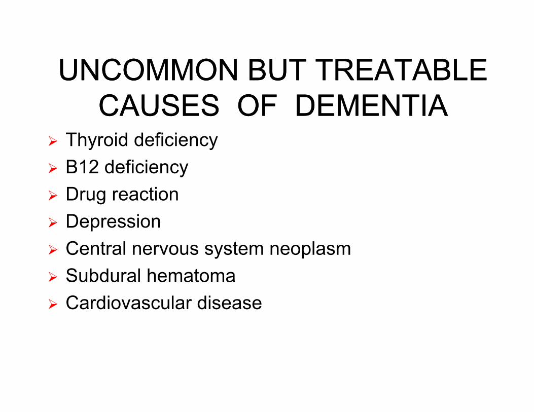
UNCOMMON BUT TREATABLE CAUSES OF DEMENTIA
UNCOMMON BUT TREATABLE CAUSES OF DEMENTIA
Thyroid deficiency B12 deficiency Drug reaction Depression Central nervous system neoplasm Subdural hematoma Cardiovascular disease
vrr4
Text Box
Rule these out before diagnosing with Alzheimer Disease
vrr4
Text Box
Can be evaluated with blood test
vrr4
Line
vrr4
Text Box
Situational Depression is very common. If the depression is identified and treated early on, the patient can recover memory deficits. If depression is left in place for long period of time, treatment will not cause reversal of memory loss.
vrr4
Line
vrr4
Text Box
Elderly- are more likely to fall and also have normal shrinkage of brain, so more at risk. Especially if on anticoagulants.

RISK FACTORS FOR ADRISK FACTORS FOR AD
Family history Head trauma Hematologic malignancies Down’s syndrome Apolipoprotein E allele ε4
vrr4
Callout
Any time during life
vrr4
Callout
Reason not clear
vrr4
Callout
Beta amyloid precursor protein is encoded on Chromosome 21. Results in inevitable Alzheimer's Disease development
vrr4
Callout
Biggest risk factor. Lowers age of onset
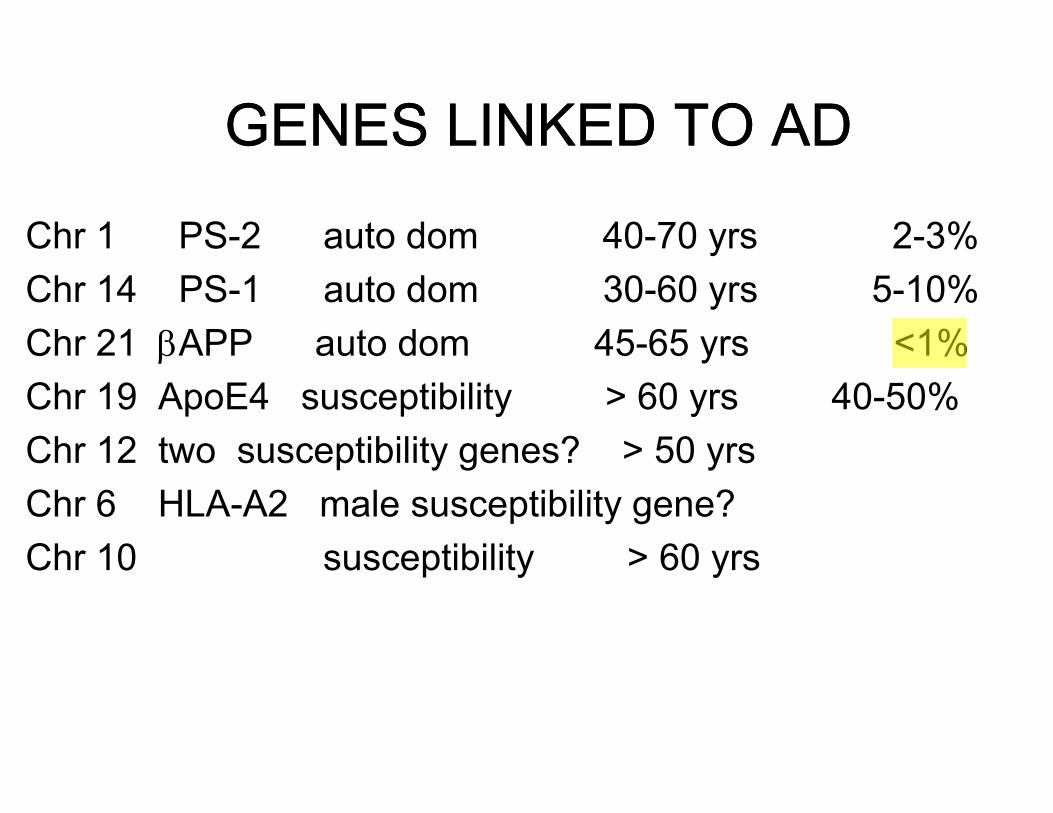
GENES LINKED TO ADGENES LINKED TO AD
Chr 1 PS-2 auto dom 40-70 yrs 2-3%Chr 14 PS-1 auto dom 30-60 yrs 5-10%Chr 21 APP auto dom 45-65 yrs <1%Chr 19 ApoE4 susceptibility > 60 yrs 40-50%Chr 12 two susceptibility genes? > 50 yrsChr 6 HLA-A2 male susceptibility gene? Chr 10 susceptibility > 60 yrs
vrr4
Text Box
early onset
vrr4
Callout
discovered first
vrr4
Rectangle
vrr4
Rectangle
vrr4
Highlight
vrr4
Underline
vrr4
Text Box
TOMM40 also discovered on Chromosome 19, but contribution is unclear
vrr4
Text Box
A few months ago published paper identified 4 new genetic risk factors (not included here).
vrr4
Text Box
She read through this whole slide.
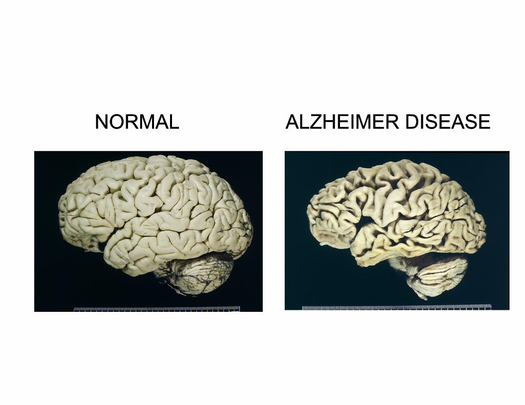
ALZHEIMER DISEASEALZHEIMER DISEASENORMALNORMAL
vrr4
Text Box
SHRINKAGE of brain! (Always)
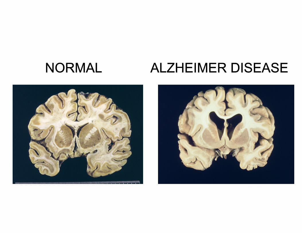
ALZHEIMER DISEASEALZHEIMER DISEASENORMALNORMAL
vrr4
Callout
Marked ventricular dilation
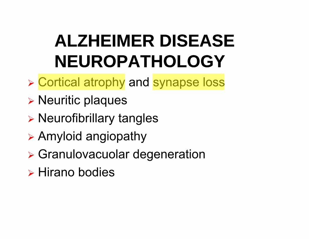
ALZHEIMER DISEASE NEUROPATHOLOGYALZHEIMER DISEASE NEUROPATHOLOGY
Cortical atrophy and synapse loss Neuritic plaques Neurofibrillary tangles Amyloid angiopathy Granulovacuolar degeneration Hirano bodies
vrr4
Text Box
Extracellular deposits of actin also seen in hippocampal formation.
vrr4
Highlight
vrr4
Highlight
vrr4
Text Box
Cross-linked microtubule-associated protein fills up neuronal cell body.
vrr4
Text Box
Protein deposit in blood vessels. Occurs more often in people with APO e4 allele
vrr4
Text Box
Cytopathological change in Purkinje Neurons in the hippocampus
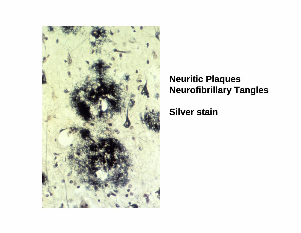
Neuritic Plaques Neurofibrillary Tangles
Silver stain
Neuritic Plaques Neurofibrillary Tangles
Silver stain
vrr4
Callout
Intraneuronal: cross-linked microtubule-associated proteins (Looks like a cell body)
vrr4
Callout
Extracellular deposits of amyloid, Tau and other inflammatory mediators
vrr4
Line
vrr4
Line

Hirano Body and Granulovacular degeneration
Hirano Body and Granulovacular degeneration
vrr4
Text Box
Eosinophilic extracellular deposits of actin
vrr4
Line
vrr4
Line
vrr4
Text Box
Granules with "halos" that occur in the cytoplasm of Purkinje neurons in the hippocampus
vrr4
Line
vrr4
Line

AMYLOID ANGIOPATHYCongo Red stain viewed under polarized light
AMYLOID ANGIOPATHYCongo Red stain viewed under polarized light
vrr4
Text Box
Amyloid: any protein with B-pleated sheet structure.
vrr4
Highlight
vrr4
Line
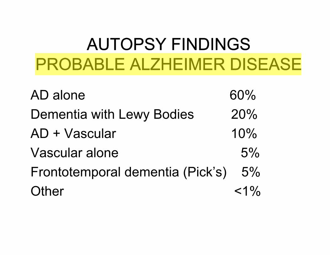
AUTOPSY FINDINGS PROBABLE ALZHEIMER DISEASE
AUTOPSY FINDINGS PROBABLE ALZHEIMER DISEASE
AD alone 60%Dementia with Lewy Bodies 20%AD + Vascular 10%Vascular alone 5%Frontotemporal dementia (Pick’s) 5%Other <1%
vrr4
Highlight
vrr4
Text Box
"Alzheimer's + Parkinson's"
vrr4
Text Box
We'll talk about this in more detail later. Mutations in Microtubule associated protein (Tau)
vrr4
Text Box
Alzheimer Disease is a diagnosis that can only be confirmed at autopsy
vrr4
Text Box
Severe cerebrovascular atherosclerosis or strategic infarcts (e.g. PCA territory or dorsal medial nucleus of thalamus)
vrr4
Line
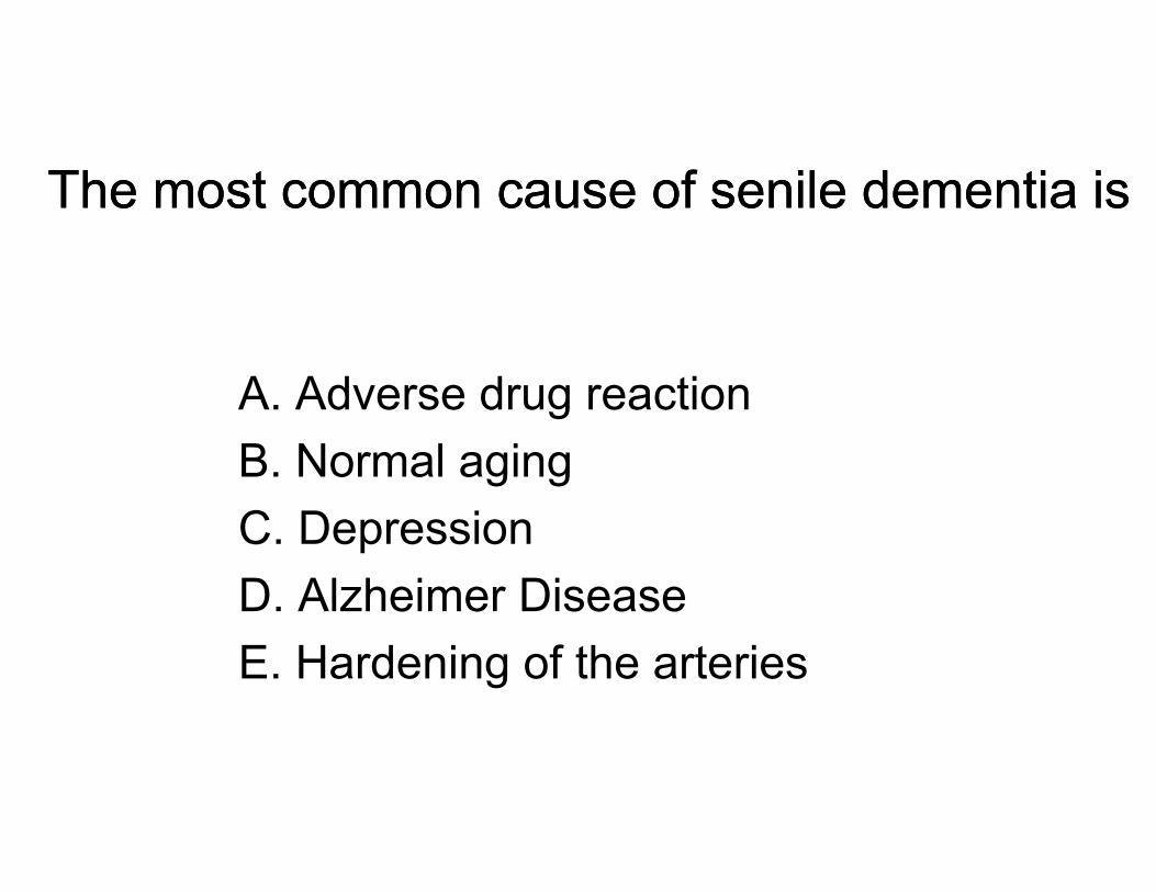
The most common cause of senile dementia isThe most common cause of senile dementia is
A. Adverse drug reactionB. Normal agingC. DepressionD. Alzheimer DiseaseE. Hardening of the arteries
vrr4
Text Box
Question: Of the pathological finding for Alzheimer's, the diagnosis is done on autopsy. What is the criteria for diagnosis? Low probability: Low amount of plaques and tangles in person with dementia Med Probability: Med plaques and tangles in person with dementia High probability: Significant plaques and tangles in person with dementia.
vrr4
Text Box
Answer on next page
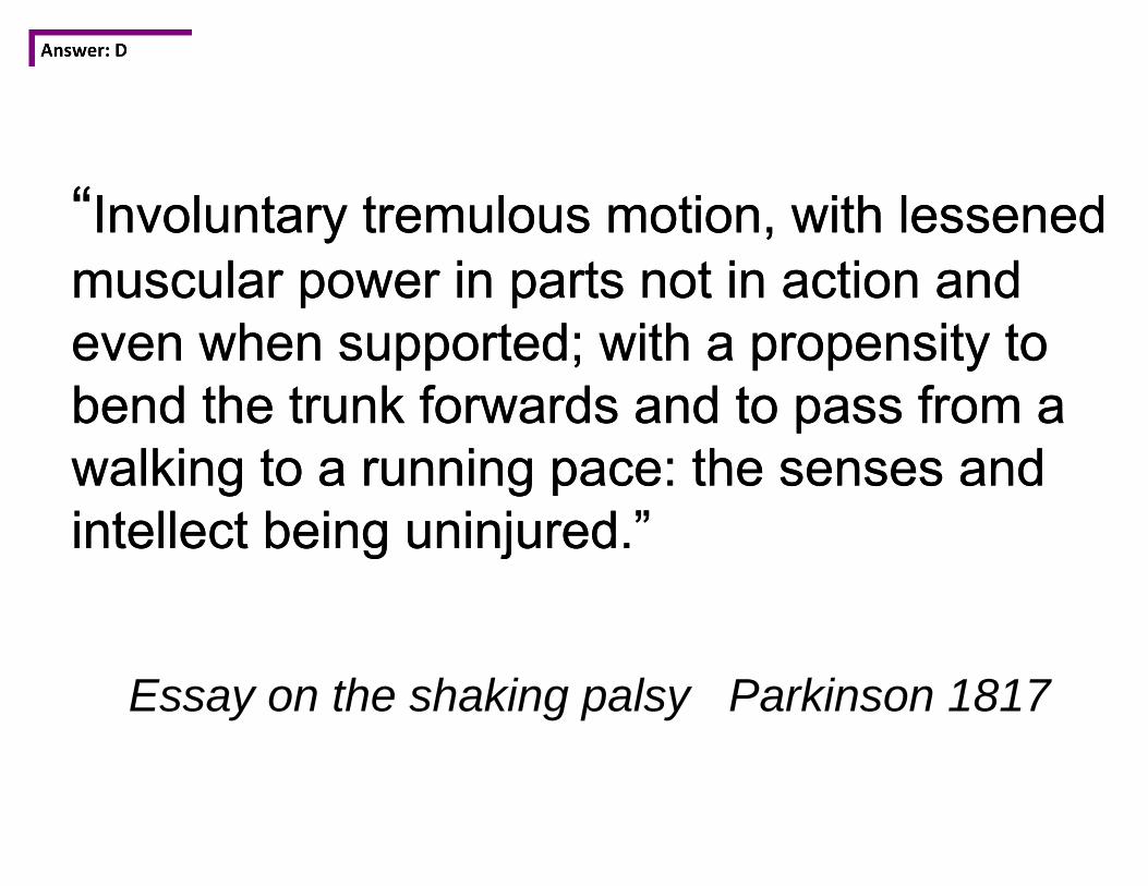
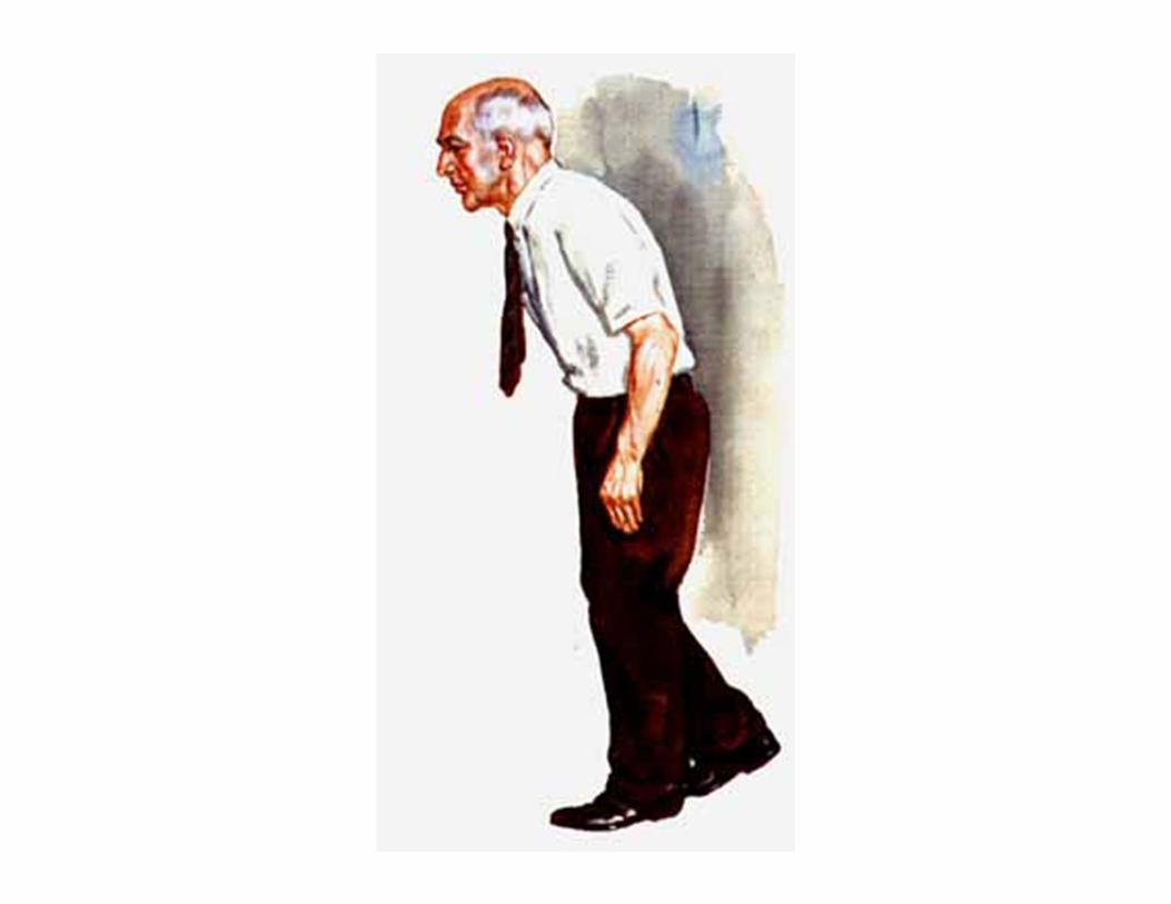
“Involuntary tremulous motion, with lessened muscular power in parts not in action and even when supported; with a propensity to bend the trunk forwards and to pass from a walking to a running pace: the senses and intellect being uninjured.”
“Involuntary tremulous motion, with lessened muscular power in parts not in action and even when supported; with a propensity to bend the trunk forwards and to pass from a walking to a running pace: the senses and intellect being uninjured.”
Essay on the shaking palsy Parkinson 1817
vrr4
Text Box
Answer: D Alzheimer Disease
vrr4
Text Box
All are contributing factors!
vrr4
Text Box
Trunk bent forward
vrr4
Text Box
Impairment of arm and leg motion.
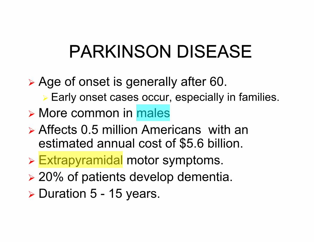
PARKINSON DISEASEPARKINSON DISEASE Age of onset is generally after 60. Early onset cases occur, especially in families.
More common in males Affects 0.5 million Americans with an
estimated annual cost of $5.6 billion. Extrapyramidal motor symptoms. 20% of patients develop dementia. Duration 5 - 15 years.
vrr4
Highlight
vrr4
Text Box
Because of treatment with L-DOPA, patients live longer, but then they develop dementia.
vrr4
Highlight
vrr4
Text Box
Rigidity, Tremor, Bradykinesia

PARKINSON DISEASEPARKINSON DISEASE Defect is due to loss of dopaminergic neurons in the
substantia nigra and brainstem. 75% of cases have Lewy bodies histopathologically. Postencephalitic Parkinsonism is characterized by
neurofibrillary tangles. Rarely PD is caused by the neurotoxin MPTP
(methyl phenyl tretrahydro pyridine) Synthetic opioid contaminant 1976
Treatment is with L-Dopa and similar drugs.
vrr4
Highlight
vrr4
Highlight
vrr4
Callout
We don't see this any more. (Caused by flu pandemic of early 1900s)
vrr4
Highlight
vrr4
Callout
Useful in development of animal models
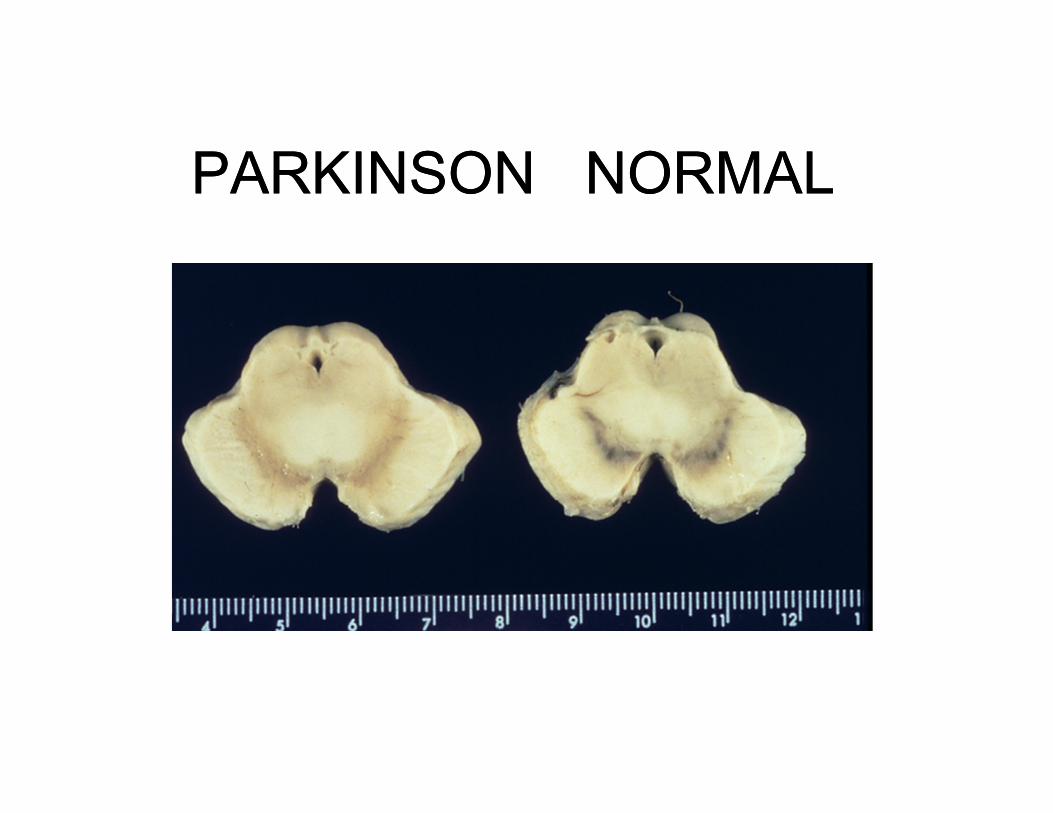
PARKINSON NORMALPARKINSON NORMAL
vrr4
Text Box
Midbrain
vrr4
Callout
Substantia Nigra is Brown! Neuromelanin (precursor of dopamine) normally secreted by these cells is lost
vrr4
Callout
Cerebral Peduncle
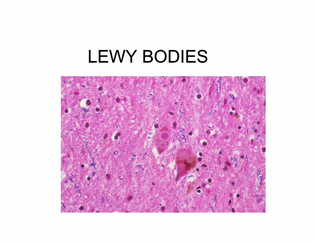
LEWY BODIESLEWY BODIES
vrr4
Text Box
Typically see in cytoplasm of pigemented neurons in substantia nigra
vrr4
Text Box
Lewy Bodies: Intracytoplasmic, eosinophilic inclusions with a halo
vrr4
Line

PICK’S DISEASEFRONTOTEMPORAL DEMENTIA
PICK’S DISEASEFRONTOTEMPORAL DEMENTIA Clinical presentation is similar to AD. Slightly earlier onset. Frontal and temporal lobe signs. Behavioral abnormalities. Extrapyramidal symptoms. Some cases are due to mutations in
microtubule associated protein (tau) on Chr 17.
vrr4
Highlight
vrr4
Text Box
Sparing of Parietal and Occipital lobes
vrr4
Text Box
Difficult to manage these patients!
vrr4
Callout
Many different mutations have been identified
vrr4
Underline
vrr4
Underline
vrr4
Underline
vrr4
Highlight
vrr4
Highlight
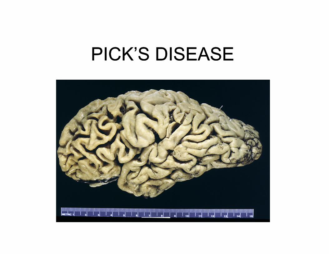
PICK’S DISEASEPICK’S DISEASE
vrr4
Callout
Profound atrophy of frontal lobe
vrr4
Callout
Preservation of parietal lobe

PICK BODIESPICK BODIES
vrr4
Text Box
Tau-positive cytoplasmic inclusions that occur in the neurons. "Pencil erasers"
vrr4
Line
vrr4
Text Box
For comparison: Neurofibrillary tangles in AD follow structure of neuron
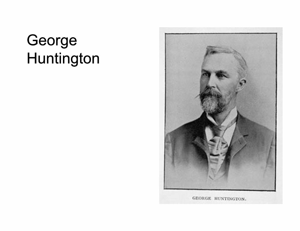
George HuntingtonGeorge Huntington
vrr4
Text Box
Neurologist who described Huntington's disease
HUNTINGTON DISEASEHUNTINGTON DISEASE
Age of onset is 35-45 years. There are personality changes, chorea and
dementia. Duration is approximately 15 years. Inherited in an autosomal dominant fashion. “huntingtin” gene on chr 4
vrr4
Highlight
vrr4
Highlight
vrr4
Callout
telomere region!

HUNTINGTON NORMALHUNTINGTON NORMAL
vrr4
Text Box
-Global atrophy -Caudate nucleus is flattened -Atrophy of Putamen and Globus Pallidus to lesser degree
vrr4
Rectangle
vrr4
Rectangle
vrr4
Line

HUNTINGTONHUNTINGTONNORMALNORMAL
vrr4
Callout
Internal Capsule
vrr4
Callout
Caudate Nucleus
vrr4
Callout
Can hardly see Caudate Nucleus
vrr4
Text Box
Amygdala is the same
vrr4
Callout
Putamen
vrr4
Callout
Globus Pallidus
vrr4
Callout
Internal capsule is about the same

AMYTROPHIC LATERAL SCLEROSIS
AMYTROPHIC LATERAL SCLEROSIS
Age of onset is in mid to late life. Male predominance. Duration 3 - 5 years Symptoms are caused by degeneration of
corticospinal tract. Familial cases may be due to superoxide
dismutase gene mutation on chr 21.
vrr4
Highlight
vrr4
Highlight
vrr4
Callout
Very rapid progression
vrr4
Highlight
vrr4
Text Box
10% of cases
vrr4
Text Box
Pathology is the same for sporadic vs genetic

AMYOTROPHIC LATERAL SCLEROSIS MOTOR CORTEXAMYOTROPHIC LATERAL SCLEROSIS MOTOR CORTEX
vrr4
Text Box
Loss of pyramidal neurons
vrr4
Callout
Spongy appearance
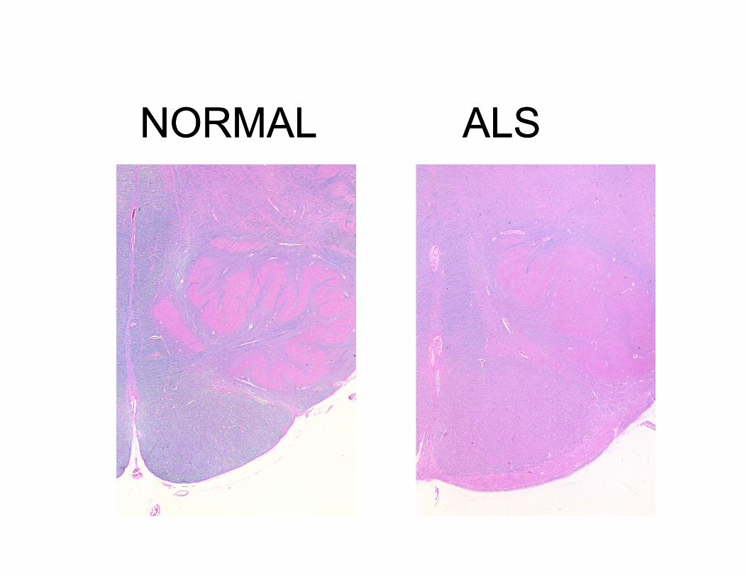
ALSALSNORMALNORMAL
vrr4
Text Box
Descending Corticospinal Tract
vrr4
Callout
Inferior Olivary Nucleus
vrr4
Callout
Myelin lost. Atrophy of pyramidal tract
vrr4
Text Box
Myelin = blue w stain
vrr4
Callout
Pyramid w Descending corticospinal tract

ALS BUNINA BODYALS BUNINA BODY
vrr4
Highlight
vrr4
Text Box
Lower motor neuron of spinal cord and cranial nerve
vrr4
Rectangle
vrr4
Text Box
Eosinophilic "dumbell-like" inclusion body
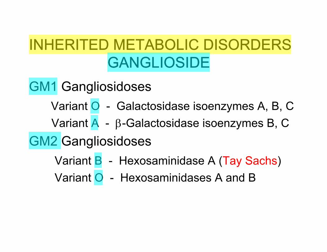
INHERITED METABOLIC DISORDERSGANGLIOSIDE
INHERITED METABOLIC DISORDERSGANGLIOSIDE
GM1 GangliosidosesVariant O - Galactosidase isoenzymes A, B, CVariant A - -Galactosidase isoenzymes B, C
GM2 GangliosidosesVariant B - Hexosaminidase A (Tay Sachs)Variant O - Hexosaminidases A and B
vrr4
Highlight
vrr4
Highlight
vrr4
Highlight
vrr4
Highlight
vrr4
Highlight
vrr4
Highlight
vrr4
Highlight
vrr4
Highlight
vrr4
Callout
Most common of the Ganglioside disorders!
vrr4
Text Box
Deficiencies:
vrr4
Text Box
Deficiencies:
vrr4
Callout
More severe

vrr4
Text Box
6 mo old Baby with Tay Sachs
vrr4
Callout
Enlarged head. "Frontal Bossing"

TAY SACHS DISEASETAY SACHS DISEASE
GM2 gangliosidosis Hexosamindase A Motor and mental deterioration beginning
at 6 months “Amaurotic familial idiocy”
vrr4
Highlight
vrr4
Underline
vrr4
Text Box
Born normal
vrr4
Callout
Blind
vrr4
Callout
Failed development
vrr4
Callout
Old literature
vrr4
Callout
Inherited

TAY SACHS CHERRY RED SPOT
Fovea
TAY SACHS CHERRY RED SPOT
Fovea
vrr4
Highlight
vrr4
Text Box
Ganglioside accumulates in Retinal neurons: Retina becomes pale. Fovea then appears as red spot because no retinal ganglia cells are present there.
vrr4
Text Box
Side Note: If you want to see this, you'll have to go into pediatric neuro.

TAY SACHSSTORAGE PRODUCT
TAY SACHSSTORAGE PRODUCT
vrr4
Text Box
Multilamellar profiles in the cytoplasm of neurons (electron micrograph)

INHERITED METABOLIC DISORDERSSPHINGOMYELIN
INHERITED METABOLIC DISORDERSSPHINGOMYELIN
SphingomyelinaseNiemann-Pick disease Type A,B,C
vrr4
Highlight
vrr4
Text Box
<--Loss of this
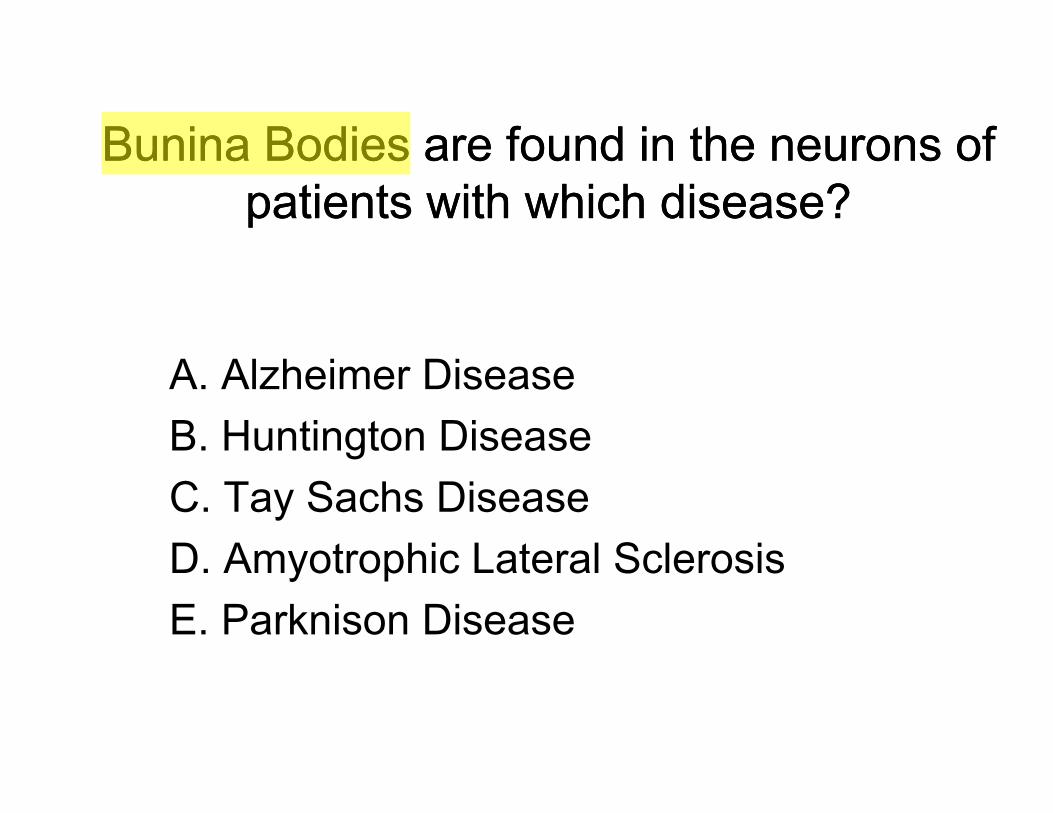
Bunina Bodies are found in the neurons of patients with which disease?
Bunina Bodies are found in the neurons of patients with which disease?
A. Alzheimer DiseaseB. Huntington DiseaseC. Tay Sachs DiseaseD. Amyotrophic Lateral SclerosisE. Parknison Disease
vrr4
Highlight
vrr4
Text Box
Answer on next page

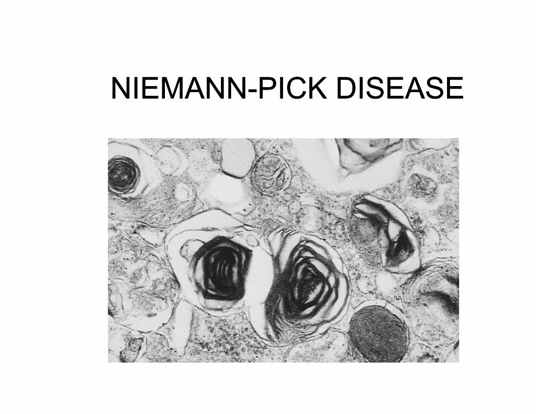
NIEMANN-PICK DISEASENIEMANN-PICK DISEASE
Sphingomyelinase deficiency Genetically and biochemically heterogeneous Type A - infantile Type B - juvenile, no CNS involvement Type C - juvenile, CNS involvement, may
present in adulthood
vrr4
Underline
vrr4
Text Box
Answer: D Amyotrophic Lateral Sclerosis
vrr4
Underline
vrr4
Underline
vrr4
Underline

NIEMANN-PICK DISEASENIEMANN-PICK DISEASE
vrr4
Callout
Neuron
vrr4
Callout
Cytoplasm engorged with the lipid product
NIEMANN-PICK DISEASENIEMANN-PICK DISEASE
vrr4
Text Box
Oligolamellar profile rather than multilamellar seen in Tay Sachs
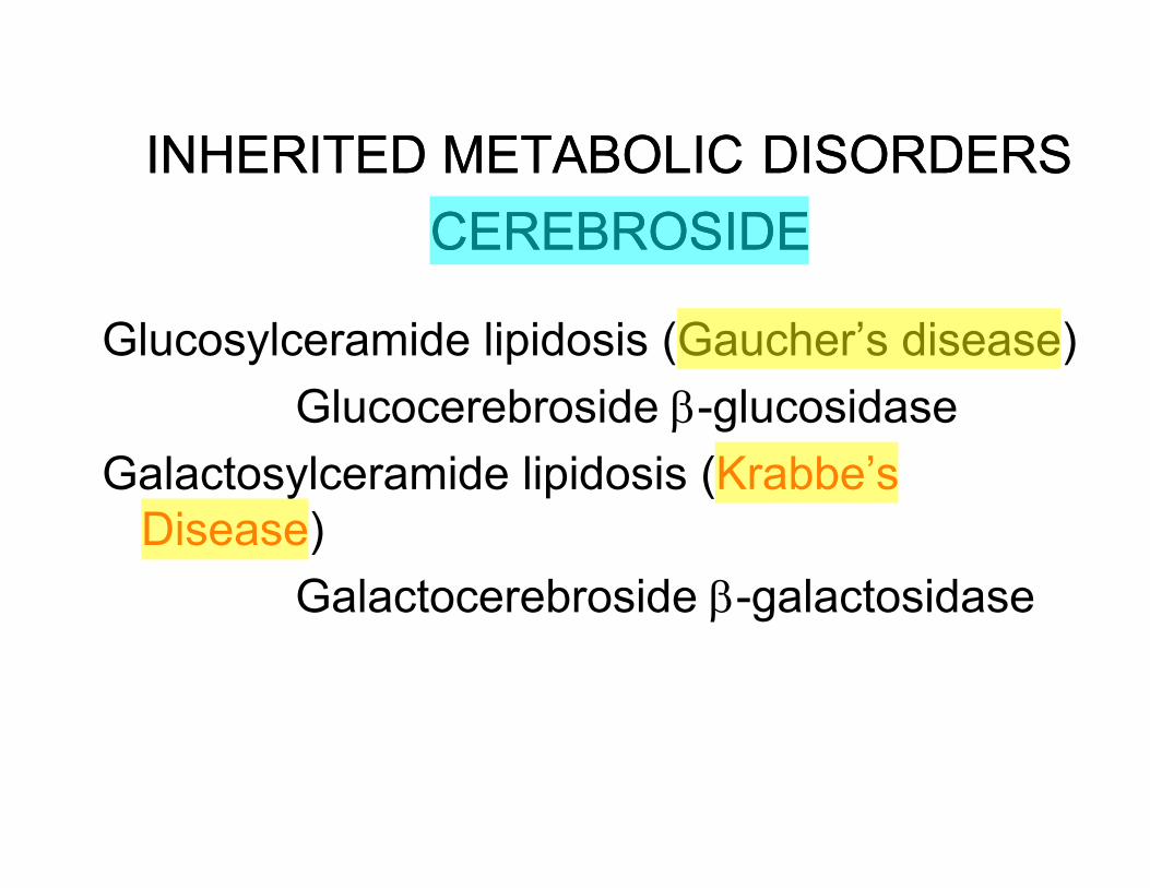
INHERITED METABOLIC DISORDERSCEREBROSIDE
INHERITED METABOLIC DISORDERSCEREBROSIDE
Glucosylceramide lipidosis (Gaucher’s disease)Glucocerebroside -glucosidase
Galactosylceramide lipidosis (Krabbe’s Disease)
Galactocerebroside -galactosidase
vrr4
Highlight
vrr4
Highlight
vrr4
Highlight
vrr4
Text Box
deficiency in
vrr4
Line
vrr4
Line
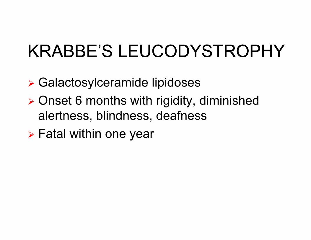
KRABBE’S LEUCODYSTROPHYKRABBE’S LEUCODYSTROPHY
Galactosylceramide lipidoses Onset 6 months with rigidity, diminished
alertness, blindness, deafness Fatal within one year
vrr4
Underline
vrr4
Underline
vrr4
Text Box
Stem Cell transplant is being attempted at Duke with reasonable success
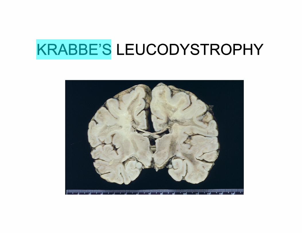
KRABBE’S LEUCODYSTROPHYKRABBE’S LEUCODYSTROPHY
vrr4
Highlight
vrr4
Underline
vrr4
Callout
"Leuco" = "White" so pathology is in white matter
vrr4
Text Box
Question: Why is white matter effected, not gray matter? Genetic defect in enzyme important in metabolizing myelin as opposed to an intra-neuronal lipid.
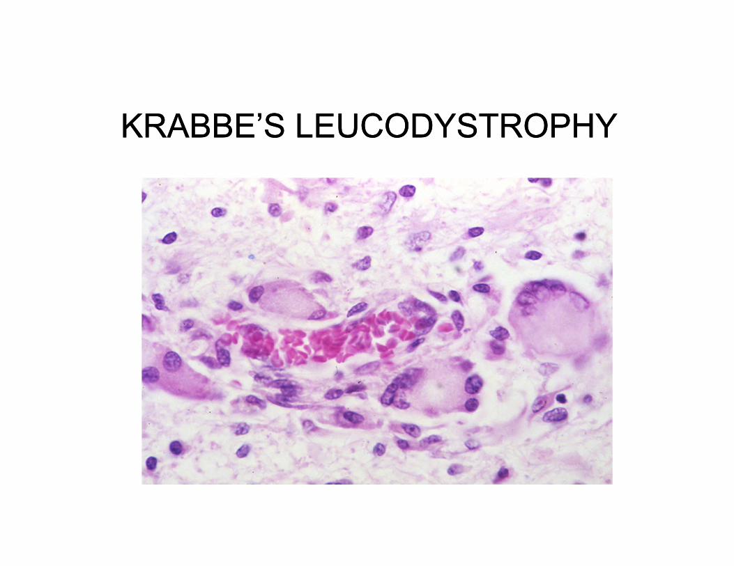
KRABBE’S LEUCODYSTROPHYKRABBE’S LEUCODYSTROPHY
vrr4
Text Box
Perivascular Giant Cells: Engulfing the abnormal lipid, recycling into blood stream
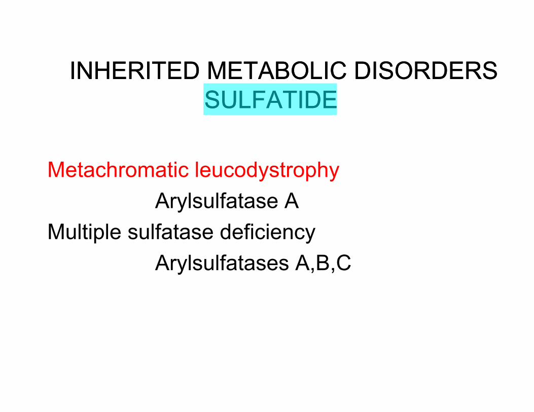
INHERITED METABOLIC DISORDERSSULFATIDE
INHERITED METABOLIC DISORDERSSULFATIDE
Metachromatic leucodystrophyArylsulfatase A
Multiple sulfatase deficiencyArylsulfatases A,B,C
vrr4
Highlight

METACHROMATIC LEUCODYSTRPHYMETACHROMATIC LEUCODYSTRPHY
Onset 1 - 4 years Rare adult forms Motor and mental deterioration Peripheral neuropathy
vrr4
Underline
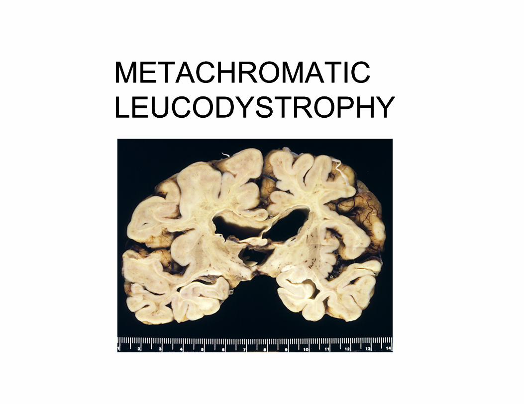
METACHROMATIC LEUCODYSTROPHYMETACHROMATIC LEUCODYSTROPHY
vrr4
Text Box
This is an adult who developed dementia at age 35. No family history. Example of rare adult-onset
vrr4
Callout
Corpus Callosum (mostly white matter) is very atrophic
vrr4
Callout
Cerebral cortex is normal
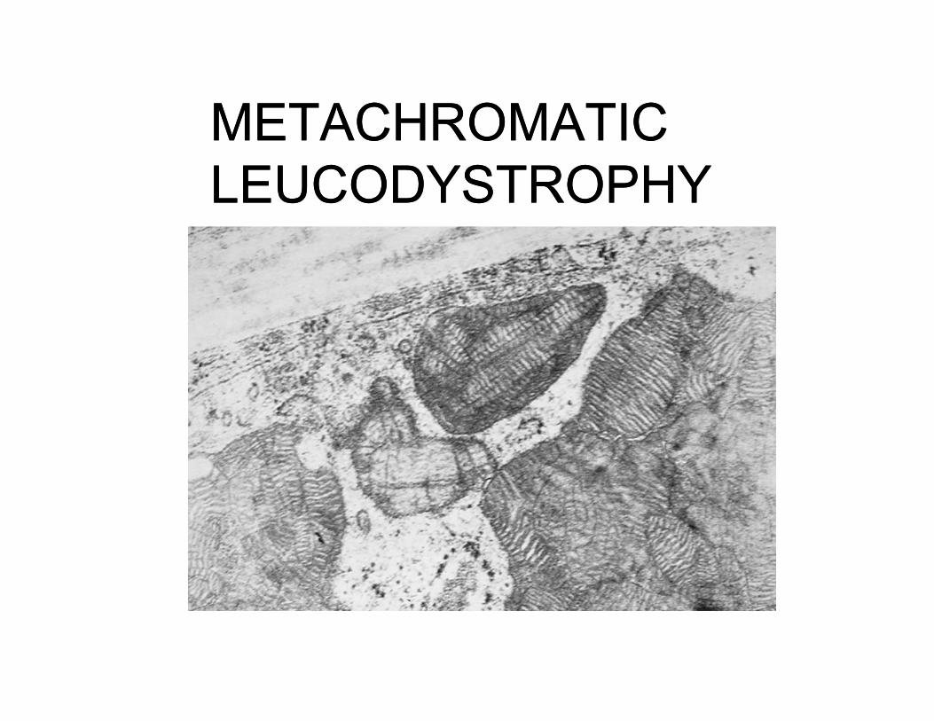
METACHROMATIC LEUCODYSTROPHYMETACHROMATIC LEUCODYSTROPHY
vrr4
Text Box
Characteristic inclusions in astrocytes and oligodendroglia in white matter.

Which of the following diseases is a leucodystrophy?Which of the following diseases is a leucodystrophy?
A. Tay Sachs DiseaseB. Amyotrophic Lateral SclerosisC. Krabbe’s diseaseD. Huntington’s diseaseD. Leukemia
vrr4
Text Box
Answer on next page

INHERITED METABOLIC DISEASESAFFECTING THE CNS
INHERITED METABOLIC DISEASESAFFECTING THE CNS
Hepatolenticular DegenerationWilson disease Abnormal copper transportDecreased ceruloplasmin Autosomal recessive
Phenylketonuria
vrr4
Text Box
Favorite test topics!!
vrr4
Underline
vrr4
Callout
Globus pallidus (lenticular nuclei)
vrr4
Underline
vrr4
Callout
Liver
vrr4
Highlight
vrr4
Underline
vrr4
Callout
Protein carrying Copper around in blood
vrr4
Text Box
Largely eliminated because infants are tested and given a special diet.
vrr4
Text Box
Answer: C Krabbe's Disease
vrr4
Highlight
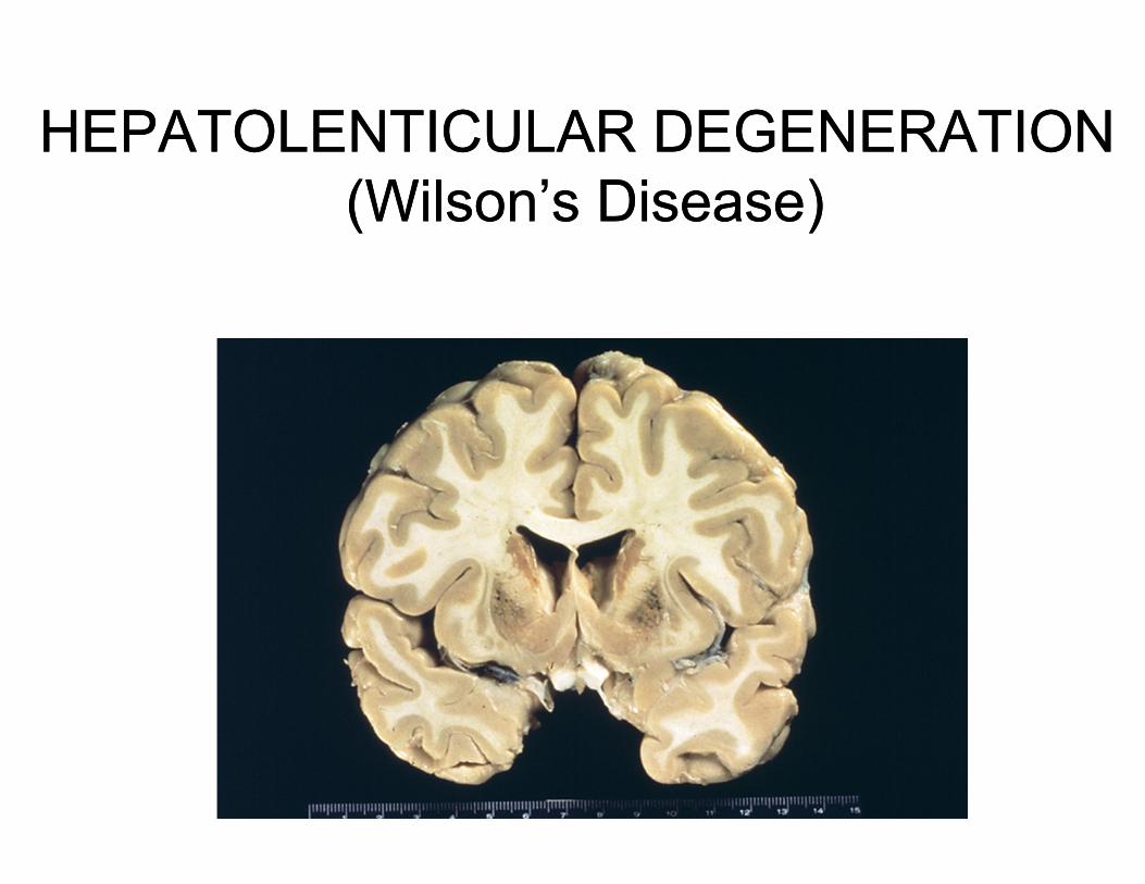
HEPATOLENTICULAR DEGENERATION (Wilson’s Disease)
HEPATOLENTICULAR DEGENERATION (Wilson’s Disease)
vrr4
Callout
Discoloration of the Caudate and Putamen, Globus Pallidus
vrr4
Callout
Copper deposit
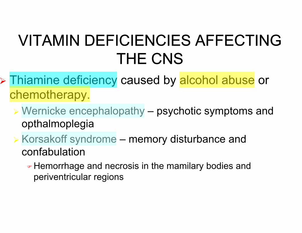
VITAMIN DEFICIENCIES AFFECTING THE CNS
VITAMIN DEFICIENCIES AFFECTING THE CNS
Thiamine deficiency caused by alcohol abuse or chemotherapy.Wernicke encephalopathy – psychotic symptoms and
opthalmoplegia Korsakoff syndrome – memory disturbance and
confabulationHemorrhage and necrosis in the mamilary bodies and
periventricular regions
vrr4
Highlight
vrr4
Highlight
vrr4
Highlight
vrr4
Highlight
vrr4
Highlight
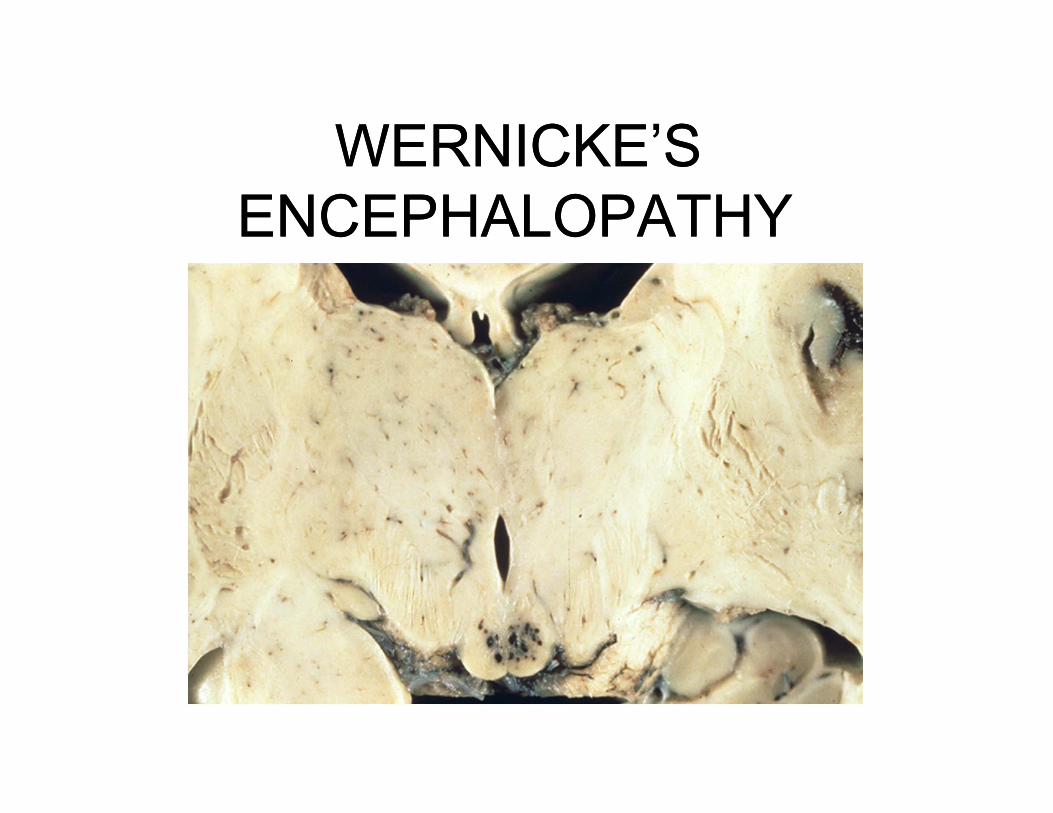
WERNICKE’SENCEPHALOPATHY
WERNICKE’SENCEPHALOPATHY
vrr4
Callout
Hemorrhages in mamillary bodies
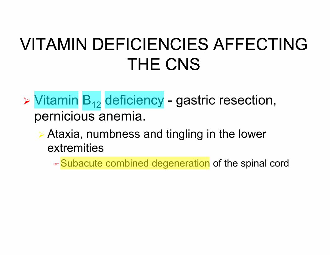
VITAMIN DEFICIENCIES AFFECTING THE CNS
VITAMIN DEFICIENCIES AFFECTING THE CNS
Vitamin B12 deficiency - gastric resection, pernicious anemia. Ataxia, numbness and tingling in the lower
extremities Subacute combined degeneration of the spinal cord
vrr4
Highlight
vrr4
Callout
After this happens, it is not reversible
vrr4
Highlight
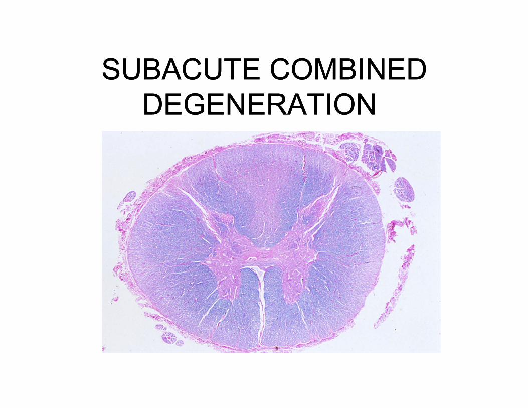
SUBACUTE COMBINED DEGENERATION
SUBACUTE COMBINED DEGENERATION
vrr4
Callout
Pallor in the ascending sensory columns
vrr4
Underline

ACQIRED METABOLIC DISEASES AFFECTING THE
CNS
ACQIRED METABOLIC DISEASES AFFECTING THE
CNS
Cretinism Thyroid deficiency
KernicterusHyperbilirubinemia in the neonatal period
vrr4
Callout
Common in some areas of China. Access to Sea have enough iodine, but in internal regions they don't.
vrr4
Highlight
vrr4
Highlight
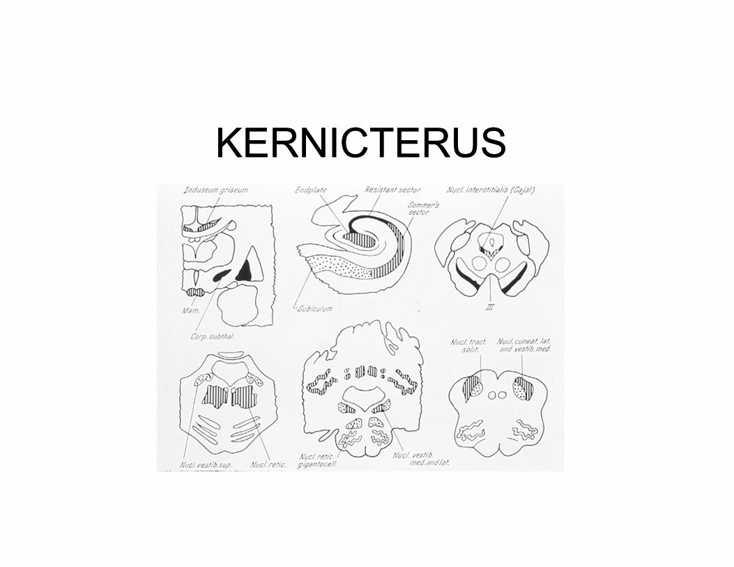
KERNICTERUSKERNICTERUS
vrr4
Text Box
This diagram shows where billirubin accumulates and how it causes damage: Substantia nigra, Globus Pallidus, Hippocampus
vrr4
Underline
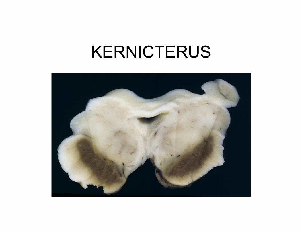
KERNICTERUSKERNICTERUS
vrr4
Text Box
Patient had ABO incompatibility and jaundice at birth.
vrr4
Callout
Billirubin deposit and destruction of substantia nigra

ETHANOLETHANOL
vrr4
Text Box
Profound cortical atrophy and dementia with alcohol abuse
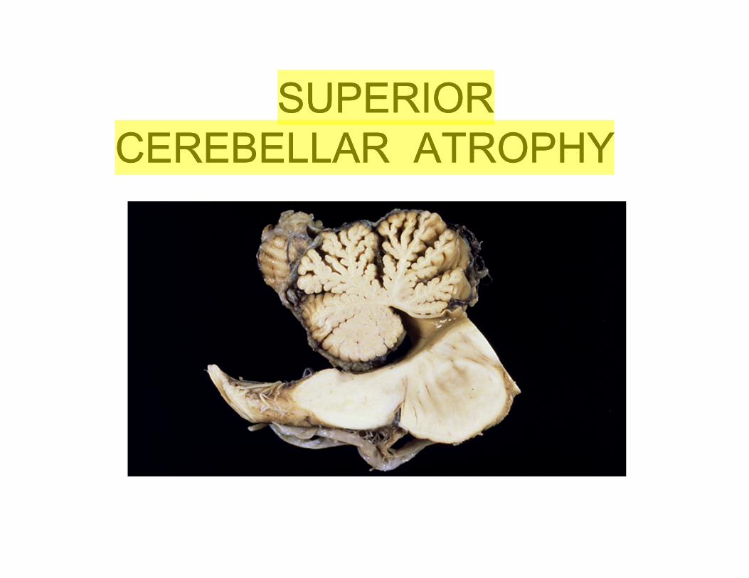
SUPERIOR CEREBELLAR ATROPHY
SUPERIOR CEREBELLAR ATROPHY
vrr4
Highlight
vrr4
Callout
Inferior is fine
vrr4
Callout
Atrophy causes Ataxic gait
vrr4
Text Box
Dilantin can also do this. Anti-seizure drug
ALZHEIMER DISEASEPARKINSON DISEASE
PICK DISEASEHUNTINGTON DISEASE
AMYOTROPHIC LATERAL SCLEROSISINHERITED METABOLIC DISORDERS
TAY SACHSNEIMANN-PICK
KRABBE’S LECUODYSTROPHYMETACHROMATIC LECUODYSTROPHYHEPATOLENTICULAR DEGENERATION
ACQUIRED METABOLIC DISORDERSKERNICTERUS
SUBACUTE COMBINED DEGENERATIONALCOHOL
vrr4
Text Box
All the conditions we covered in this lecture!