Primary Central Nervous System Lymphoma
216
Primary Central Nervous System Lymphoma Diagnosc evaluaon, neurocognive funconing and health-related quality of life Mahijs van der Meulen
-
Upload
khangminh22 -
Category
Documents
-
view
0 -
download
0
Transcript of Primary Central Nervous System Lymphoma

Primary Central Nervous System Lymphoma
Diagnostic evaluation, neurocognitive functioning
and health-related quality of life
Matthijs van der Meulen

Lay-out and printing by Optima Grafische Communicatie
The printing of this thesis was kindly supported by:ChipsoftDepartment of Neurology, Erasmus MC

Primary Central Nervous System LymphomaDiagnostic evaluation, neurocognitive functioning and health-related quality of life
Primair centraal zenuwstelsel lymfoomDiagnostiek, neurocognitief functioneren en kwaliteit van leven
Proefschrift
ter verkrijging van de graad van doctor aan deErasmus Universiteit Rotterdam
op gezag van derector magnificus
Prof.dr. F.A. van der Duin Schouten
en volgens besluit van het College voor Promoties.De openbare verdediging zal plaatsvinden op
woensdag 21 april 2021 om 15.30 uur
door
Matthijs van der Meulengeboren te Oldebroek.

PromotieCommiSSie:
Promotor: prof. dr. M.J. van den Bent
overige leden: prof. dr. M.J.B. Taphoornprof. dr. M. Smitsdr. J.K. Doorduijn
Copromotor: dr. J.E.C. Bromberg

CoNteNtS
Chapter 1 General introduction and scope of this thesis 7
PArt i epidemiologyChapter 2 Improved survival in primary central nervous system lymphoma
up to age 70 only: a population-based study on incidence, primary treatment and survival in the Netherlands, 1989-2015
21
Chapter 3 Primary therapy and survival in patients aged over 70-years-old with primary central nervous system lymphoma: a contemporary, nationwide, population-based study in the Netherlands
35
PArt ii Diagnostic evaluationChapter 4 Flow cytometry shows added value in diagnosing lymphoma in
brain biopsies51
Chapter 5 Extent of radiological response does not reflect survival in primary central nervous system lymphoma
65
PArt iii Neurocognitive functioning and health-related quality of lifeChapter 6 Cognitive functioning and health-related quality of life in patients
with newly diagnosed primary central nervous system lymphoma: a systematic literature review
85
Chapter 7 Neurocognitive functioning and radiological changes in primary CNS lymphoma: results from a RCT
127
Chapter 8 Health-related quality of life after chemotherapy with or without rituximab in primary central nervous system lymphoma patients: results from a randomized phase III study
147
PArt iV PrognosisChapter 9 MMSE is an independent prognostic factor in primary central
nervous system lymphoma patients167
Chapter 10 General discussion 177Chapter 11 Summary en samenvatting in het Nederlands 191
Appendix 203List of publications 205About the author 207Portfolio 209Dankwoord 211


1General introduction and scope of this thesis


9
General introduction
1ChAPter 1 GeNerAL iNtroduCtioN ANd SCoPe of thiS theSiS
Primary central nervous system lymphoma (PCNSL) is a rare non-Hodgkin lymphoma (NHL) which is confined to the brain, leptomeningines, spinal cord and eyes without manifesta-tions outside the central nervous system. This tumour was first described by Bailey as ‘sar-coma of the brain, arising from the leptomeninges’ at the beginning of the 20th century.1 Later it was recognized as NHL.
The exact pathophysiology is unknown and although it is called a primary CNS lym-phoma, there is evidence the tumour originates outside the central nervous system. Almost all PCNSL contained Bcl-6, an oncogenic protein that is only expressed on B-cells in the germinal centre. Since the central nervous system does not contain germ centre structures, it suggest an extraneural origin of the tumour that subsequently migrates to the central nervous system.2 At histological examination, >90% of PCNSL are diffuse large B-cell lymphoma (DLBCL), the remaining 10% are Burkitt, T-cell or low-grade lymphoma.3,4
Epidemiology
Over the last decades the incidence of PCNSL has increased, mainly among elderly (>60-year old) to 0.44-0.47/ 100,000 per year.5 The reason for this increase is unknown; the only known risk factors for this disease are older age and being immunocompromised. However, the incidence is increasing among immune competent patients and despite the ageing population, the incidence of systemic DLBCL and glioma did not increase with a similar rate.6,7 Median age at diagnosis is around 65 years and the incidence in men is slightly higher than in women.8
Diagnosis
The most frequent presenting symptoms are focal neurological deficits (70%), signs of increased intracranial pressure, such as headache (51%) and cranial nerve palsies, neuro-psychiatric/cognitive symptoms (26-43%) and seizures (14%).9,10
When a brain tumour, is considered, an MRI of the brain with and without gadolineum is the first designated test. On MRI a PCNSL is characterized by solitary (65%) or multiple (35%) space occupying lesions, surrounded by vasogenic oedema. Most (90%) lesions show homogeneous contrast enhancement, with diffusion restriction (Figure 1). Preferred locations are periventricular, the corpus callosum and basal ganglia.11,12 However, more rarely non-enhancing space occupying lesions in addition to enhancing lesions may occur. These lesions diminished in size or even vanished after treatment, which suggests that these lesions should also be considered as tumor.13
Cytological or histological confirmation of the tumour is essential, as treatment is inten-sive and potentially toxic. In a minority of patients the diagnosis can be made by cytologi-cal analysis of cerebrospinal fluid (CSF) after a lumbar puncture, or of vitreous fluid in case

Chapter 1
10
of eye involvement. Although flow cytometry added tot cytology increases the sensitivity, compared to cytological analysis alone14, diagnosis can be made on CSF-analysis alone in only 30% of the cases.15 Eye involvement occurs in just circa 4% of the cases.16 As a result, in most patients a brain biopsy is remains necessary to obtain a diagnosis. In addition to this cytological or histological confirmation a comprehensive screening is necessary to determine the extent of the disease and to determine whether the lymphoma is a primary CNS lymphoma or a secondary manifestation of a systemic lymphoma. Typically, the screening consists of a slit lamp eye examination, a lumbar puncture, a CT of the chest and abdomen, a bone marrow analysis and a complete blood analysis.
treatment
The first described treatment for PCNSL was a gross total resection of the tumour, but this resulted in median overall survival (OS) of just 1-4 months.17 After the failed effect of sur-gery, whole brain radiotherapy (WBRT) became the treatment of first choice, but despite rapid responses, survival remained more limited than that observed in other lymphoma limited to one organ.18 Based on multiple large uncontrolled phase II studies, chemother-apy based on high-dose intravenous methotrexate (HD-MTX) became subsequently the cornerstone of first line treatment, with studies utilizing various HD-MTX-based regimens
A
C
B
d
figure 1. T1W (A) and T1W with gadolinium (B) images show multiple homogenous enhancing lesions. A T2W (C) shows vasogenic oedema around the lesions and the diffusion weighted image (D) shows diffusion restric-tion in all enhancing lesions.

11
General introduction
1reporting a median overall survival of about 60 months.19-21 The addition of high-dose cytarabine (HD-Ara-C) to HD-MTX was then reported to further improve the progression free survival (PFS) and OS.22
The role of WBRT given after chemotherapy remains disputed. The addition of WBRT after chemotherapy, may improve the PFS compared to chemotherapy alone (12 versus 18 months); the overall survival remained similar, however.23 On top, patients treated with combined chemotherapy and 45Gy WBRT, had significantly worse scores on neuropsycho-logical tests, compared to patients treated with chemotherapy only.24
In particular because of these effects on neurocognitive functioning, alternatives for con-solidation treatment with high-dose WBRT are necessary . An uncontrolled study showed a similar effect on survival but without these cognitive consequences with a reduced dose of WBRT (23.4Gy).25 Two phase II randomized controlled trials compared autologous stem cell transplantation (ASCT) with WBRT as consolidation therapy. No significant differences in PFS were found but cognitive performance was reported to be significantly better in the ASCT group. Although the OS also seemed similar between both groups and comparable to historical treated patients, further follow-up is needed to explore outcome in the long run.26,27
Rituximab, a chimeric monoclonal antibody targeting the CD20 cell surface protein, is very effective if given in addition to standard chemotherapy in systemic CD20 positive B-cell lymphoma.28,29 Since most PCNSL are CD20 positive diffuse large B-cell lymphoma, rituximab was assumed to be effective also in PCNSL. A phase II trial randomised 219 patients between HD-MTX/Ara-C, HD-MTX/Ara-C combined with rituximab of HD-MTX/Ara-C combined with rituximab and thiotepa. The latter arm, now known as the MATRix-regime had significantly better PFS and OS.30 Unfortunately there was no arm with thio-tepa and without rituximab; in addition this study was not powered or designed for a comparison between three arms, this made the role of additional rituximab uncertain. In a large international phase III randomised controlled trial, the HOVON 105/ ALLG NHL 24 study, 199 patients were randomized between HD-MTX-based chemotherapy (MBVP: methotrexate, tenoposide, BCNU and prednisolone) with or without rituximab and fol-lowed by HD-cytarabine and, in patients up to 60 years-old with a lower dose of WBRT (30Gy). No differences were found between the arms regarding the 1-year event-free survival (R-MBVP versus MBVP: 52% (95% confidence interval [CI]: 42-61) versus 49% (95% CI: 39-58), p=0.99), PFS and OS.31
Cognitive functioning and health-related quality of life
Cognitive decline and other symptoms, caused by the tumour and/ or the treatment can compromise health-related quality of life (HRQoL).32 Up to 43% of PCNSL patients have cognitive disturbances to a certain extent at diagnosis.9 In PCNSL symptoms can greatly

Chapter 1
12
improve following treatment but reports differ regarding the extent to which this is also the case for cognitive symptoms.
Since the prognosis of PCNSL patients has improved over the last decades, (late) ef-fects on cognitive performance and HRQoL have become more important to measure.5 Preventing cognitive decline and even better, improving cognitive functioning are major challenges in PCNSL patients. Assessment of this important part of patient functioning requires the evaluation of cognition at baseline and during follow-up.33,34 In addition, as-sessing cognitive performance and HRQoL in research is necessary to determine the ‘net clinical benefit’ of a (new) treatment. Information on both, survival and neurocognitive functioning and HRQoL enables the physicians and patients to make a weighted decision regarding patients’ treatment.
Prognosis
Although the prognosis for patients with PCNSL at group level improved over the last decades,5 it remains difficult to predict the prognosis for the individual patient. Two prog-nostic models are currently widely used in PCNSL patients.- The externally validated Memorial Sloan Kettering Cancer Center (MSKCC) model,
consist of two factors: age and the Karnofsky performance score (KPS), in which a higher age (>50 years-old) and a lower KPS (<70) are unfavourable prognostic factors.35
- The International Extranodal Lymphoma Study Group (IELSG) developed a model of 5 unfavourable prognostic factors: higher age (>60 years old), higher ECOG/ WHO performance score (>1), a high serum LDH, a higher total protein in CSF (>45mg/dL in patients ≤60 years-old or >60mg/dL for elderly) and involvement of deep structures (i.e. brain stem, cerebellum, periventricular or basal ganglia).36
The latter model is unfortunately limited by missing data, and external validation is still needed.
Aims and scope of this thesis
The aims of this thesis are to describe incidence, primary treatment and survival among adult PCNSL patients in the Netherlands over the last three decades (Chapter 2) and to describe primary treatment and survival among elderly (>70 year-old) in the modern era: 2014-2017 (chapter 3). As described above, flow cytometry improves the sensitivity over immunohistochemistry alone in CSF. In chapter 4 we aim to define the value of flow cytometry on brain biopsies from lesions suspected to be a brain lymphoma. Since PCNSL is a rare disease, and choices regarding treatment in clinical studies are generally based on local response assessment, we determined the value of a central radiology review and the influence of centrally determined response rate on survival (chapter 5). Secondary endpoints of the HOVON 105/ALLG NHL 24 study31 were differences between treatment

13
General introduction
1arms regarding health-related quality of life and neurocognitive functioning. PART III starts with a comprehensive systematic literature review of neurocognitive functioning and HRQoL in PCNSL patients (Chapter 6). In chapter 7 and 8 we describe neurocognitive functioning and HRQoL-scores over time and the effect of rituximab on these. Chapter 7 also describes the association of brain atrophy and white matter abnormalities with neurocognitive functioning. Lastly, we aim to determine the prognostic value of the Mini-Mental State Examination score at baseline in chapter 9.

Chapter 1
14
refereNCeS
1. Bailey P. Intracranial sarcomatous tumors of leptomeningeal origin. Arch Surg 1929; 18(4): 1359-402.
2. Thompsett AR, Ellison DW, Stevenson FK, Zhu D. V(H) gene sequences from primary central nervous system lymphomas indicate derivation from highly mutated germinal center B cells with ongoing mutational activity. Blood 1999; 94(5): 1738-46.
3. Miller DC, Hochberg FH, Harris NL, Gruber ML, Louis DN, Cohen H. Pathology with clinical correlations of primary central ner-vous system non-Hodgkin’s lymphoma. The Massachusetts General Hospital experience 1958-1989. Cancer 1994; 74(4): 1383-97.
4. Swerdlow SH, Campo E, Pileri SA, et al. The 2016 revision of the World Health Organiza-tion classification of lymphoid neoplasms. Blood 2016; 127(20): 2375-90.
5. Villano JL, Koshy M, Shaikh H, Dolecek TA, McCarthy BJ. Age, gender, and racial differ-ences in incidence and survival in primary CNS lymphoma. Br J Cancer 2011; 105(9): 1414-8.
6. Ho VK, Reijneveld JC, Enting RH, et al. Changing incidence and improved survival of gliomas. Eur J Cancer 2014; 50(13): 2309-18.
7. Issa DE, van de Schans SA, Chamuleau ME, et al. Trends in incidence, treatment and survival of aggressive B-cell lymphoma in the Netherlands 1989-2010. Haematologica 2015; 100(4): 525-33.
8. Shiels MS, Pfeiffer RM, Besson C, et al. Trends in primary central nervous system lymphoma incidence and survival in the U.S. Br J Haematol 2016; 174(3): 417-24.
9. Bataille B, Delwail V, Menet E, et al. Primary intracerebral malignant lymphoma: report of 248 cases. J Neurosurg 2000; 92(2): 261-6.
10. Ashrafi F, Zali A, Amiri M, Shabani F. Clini-cal Presentations of 176 Cases of Primary Central Nervous System Lymphoma: A Case
Series. Archives of Neuroscience 2016; 3(2): e32127.
11. Yap KK, Sutherland T, Liew E, Tartaglia CJ, Pang M, Trost N. Magnetic resonance features of primary central nervous system lymphoma in the immunocompetent patient: a pictorial essay. J Med Imaging Radiat Oncol 2012; 56(2): 179-86.
12. Kuker W, Nagele T, Korfel A, et al. Pri-mary central nervous system lymphomas (PCNSL): MRI features at presentation in 100 patients. J Neurooncol 2005; 72(2): 169-77.
13. Tabouret E, Houillier C, Martin-Duverneuil N, et al. Patterns of response and relapse in primary CNS lymphomas after first-line chemotherapy: imaging analysis of the ANOCEF-GOELAMS prospective randomized trial. Neuro Oncol 2017; 19(3): 422-9.
14. Bromberg JE, Breems DA, Kraan J, et al. CSF flow cytometry greatly improves diagnostic accuracy in CNS hematologic malignancies. Neurology 2007; 68(20): 1674-9.
15. Schroers R, Baraniskin A, Heute C, et al. Diagnosis of leptomeningeal disease in dif-fuse large B-cell lymphomas of the central nervous system by flow cytometry and cytopathology. Eur J Haematol 2010; 85(6): 520-8.
16. Touitou V, LeHoang P, Bodaghi B. Pri-mary CNS lymphoma. Curr Opin Ophthalmol 2015; 26(6): 526-33.
17. Henry JM, Heffner RR, Jr., Dillard SH, Earle KM, Davis RL. Primary malignant lympho-mas of the central nervous system. Cancer 1974; 34(4): 1293-302.
18. Nelson DF, Martz KL, Bonner H, et al. Non-Hodgkin’s lymphoma of the brain: can high dose, large volume radiation therapy im-prove survival? Report on a prospective trial by the Radiation Therapy Oncology Group (RTOG): RTOG 8315. Int J Radiat Oncol Biol Phys 1992; 23(1): 9-17.

15
General introduction
1 19. DeAngelis LM, Wong E, Rosenblum M, Furneaux H. Epstein-Barr virus in acquired immune deficiency syndrome (AIDS) and non-AIDS primary central nervous system lymphoma. Cancer 1992; 70(6): 1607-11.
20. Glass J, Gruber ML, Cher L, Hochberg FH. Preirradiation methotrexate chemotherapy of primary central nervous system lym-phoma: long-term outcome. J Neurosurg 1994; 81(2): 188-95.
21. Abrey LE, Yahalom J, DeAngelis LM. Treat-ment for primary CNS lymphoma: the next step. J Clin Oncol 2000; 18(17): 3144-50.
22. Ferreri AJ, Reni M, Foppoli M, et al. High-dose cytarabine plus high-dose methotrex-ate versus high-dose methotrexate alone in patients with primary CNS lymphoma: a randomised phase 2 trial. Lancet 2009; 374(9700): 1512-20.
23. Thiel E, Korfel A, Martus P, et al. High-dose methotrexate with or without whole brain radiotherapy for primary CNS lymphoma (G-PCNSL-SG-1): a phase 3, randomised, non-inferiority trial. Lancet Oncol 2010; 11(11): 1036-47.
24. Correa DD, Shi W, Abrey LE, et al. Cognitive functions in primary CNS lymphoma after single or combined modality regimens. Neuro Oncol 2012; 14(1): 101-8.
25. Morris PG, Correa DD, Yahalom J, et al. Rituximab, methotrexate, procarbazine, and vincristine followed by consolidation reduced-dose whole-brain radiotherapy and cytarabine in newly diagnosed primary CNS lymphoma: Final results and long-term outcome. J Clin Oncol 2013; 31(31): 3971-9.
26. Ferreri AJM, Cwynarski K, Pulczynski E, et al. Whole-brain radiotherapy or autologous stem-cell transplantation as consolidation strategies after high-dose methotrexate-based chemoimmunotherapy in patients with primary CNS lymphoma: results of the second randomisation of the International Extranodal Lymphoma Study Group-32 phase 2 trial. Lancet Haematol 2017; 4(11): e510-e23.
27. Houillier C, Taillandier L, Dureau S, et al. Radiotherapy or Autologous Stem-Cell Transplantation for Primary CNS Lymphoma in Patients 60 Years of Age and Younger: Re-sults of the Intergroup ANOCEF-GOELAMS Randomized Phase II PRECIS Study. J Clin Oncol 2019; 37(10): 823-33.
28. Coiffier B, Lepage E, Briere J, et al. CHOP chemotherapy plus rituximab compared with CHOP alone in elderly patients with diffuse large-B-cell lymphoma. N Engl J Med 2002; 346(4): 235-42.
29. Pfreundschuh M, Schubert J, Ziepert M, et al. Six versus eight cycles of bi-weekly CHOP-14 with or without rituximab in elderly patients with aggressive CD20+ B-cell lymphomas: a randomised controlled trial (RICOVER-60). Lancet Oncol 2008; 9(2): 105-16.
30. Ferreri AJ, Cwynarski K, Pulczynski E, et al. Chemoimmunotherapy with methotrex-ate, cytarabine, thiotepa, and rituximab (MATRix regimen) in patients with primary CNS lymphoma: results of the first randomi-sation of the International Extranodal Lym-phoma Study Group-32 (IELSG32) phase 2 trial. Lancet Haematol 2016; 3(5): e217-27.
31. Bromberg JEC, Issa S, Bakunina K, et al. Rituximab in patients with primary CNS lymphoma (HOVON 105/ALLG NHL 24): a randomised, open-label, phase 3 intergroup study. Lancet Oncol 2019; 20(2): 216-28.
32. Boele FW, Zant M, Heine EC, et al. The asso-ciation between cognitive functioning and health-related quality of life in low-grade glioma patients. Neurooncol Pract 2014; 1(2): 40-6.
33. Correa DD, Maron L, Harder H, et al. Cogni-tive functions in primary central nervous system lymphoma: literature review and assessment guidelines. Ann Oncol 2007; 18(7): 1145-51.
34. Abrey LE, Batchelor TT, Ferreri AJ, et al. Report of an international workshop to standardize baseline evaluation and re-

Chapter 1
16
sponse criteria for primary CNS lymphoma. J Clin Oncol 2005; 23(22): 5034-43.
35. Abrey LE, Ben-Porat L, Panageas KS, et al. Primary central nervous system lymphoma: the Memorial Sloan-Kettering Cancer Center prognostic model. J Clin Oncol 2006; 24(36): 5711-5.
36. Ferreri AJ, Blay JY, Reni M, et al. Prognostic scoring system for primary CNS lymphomas: the International Extranodal Lymphoma Study Group experience. J Clin Oncol 2003; 21(2): 266-72.


1
7
4
10
2
8
5
11
3
9
6
A
PArt iepidemiology


2Improved survival in primary central nervous system
lymphoma up to age 70 only: a population-based study on incidence, primary treatment and survival in the
Netherlands, 1989-2015
Matthijs van der Meulen,1,6 Avinash G. Dinmohamed,2-4,6 Otto Visser5, Jeanette K. Doorduijn,4 Jacoline E.C. Bromberg1
1Erasmus MC Cancer Institute, University Medical Centre Rotterdam, Department of Neuro-Oncology; 2Department of Research, Netherlands Comprehensive Cancer Organisation (IKNL), Utrecht, the Netherlands; 3Erasmus MC, University
Medical Center Rotterdam, Department of Public Health; 4Erasmus MC Cancer Institute, University Medical Center, Rotterdam, Department of Hematology; 5Department of Registration, Netherlands Comprehensive Cancer Organisation
(IKNL), Utrecht, the Netherlands; 6These two authors contributed equally to this work.
2017 Leukemia, 31(8), 1822-1825.


23
Improved survival in PCNSL up to age 70 only: a population-based study in the Netherlands, 1989-2015
2
Letter to the editor
Primary central nervous system lymphoma (PCNSL) is a rare, aggressive form of extranodal non-Hodgkin lymphoma that exclusively affects the CNS. Although PCNSL has tradition-ally been associated with a sinister prognosis, recent findings from the few available prospective studies demonstrated improved outcome in PCNSL.1-3 However, the results from such studies may not reflect the actual clinical practice due to patient selection. Population-based studies complement prospective intervention studies by addressing a non-selected group of patients within a well-defined geographic area. Currently, compre-hensive population-based studies that assess long-term patterns of incidence, treatment and survival in PCNSL are virtually lacking.
Here we report the outcomes of a comprehensive nationwide population-based study on incidence, primary treatment and survival among adult PCNSL patients diagnosed in the Netherlands during a 27-year period.
We identified all adult (≥18 years) PCNSL patients diagnosed between 1989-2015, with survival follow-up through February 2016, from the nationwide population-based Netherlands Cancer Registry (NCR). Established in 1989, the NCR, which is maintained and hosted by the Netherlands Comprehensive Cancer Organisation, has an overall coverage of >95% of all malignancies in the Netherlands.4 The NCR is based on comprehensive case notifications through the Nationwide Network of Histopathology and Cytopathology and the National Registry of Hospital Discharges. PCNSL of the diffuse large B-cell type was defined using International Classification of Diseases for Oncology morphology and topog-raphy codes. The selected codes are described in the Supplementary Methods. Informa-tion on dates of birth and diagnosis, sex, disease topography and morphology, primary treatment (that is, no anti-neoplastic therapy, chemotherapy, radiotherapy, combined chemoradiotherapy (CT+RT), and other or unknown therapy), and vital statistics (that is, alive, emigration or death) is available for individual patients.
Age-standardized incidence rates (ASRs) were calculated per 100,000 person-years, using the annual mid-year population size as obtained from Statistics Netherlands. ASRs were standardized according to the European standard population. We calculated relative survival (RS) for four calendar periods (1989-1995, 1996-2002, 2003-2008 and 2009-2015) and three age groups (18-60, 61-70 and >70 years) using the cohort methodology.5 RS is the observed patient survival (that is, overall survival, OS) corrected for the expected survival of an equivalent group in the general population with respect to age, sex and period. This to eliminate the effect of general changes in population survival over time. Expected sur-vival was calculated using the Ederer-II methodology. RS was calculated from the time of diagnosis until death, emigration or end of follow-up, whichever occurred first. We applied a generalized linear model that assumed a Poisson distribution for the observed number of deaths to assess linear trends in RS over time and to estimate the relative excess risk

Chapter 2
24
of mortality during the fi rst 5 years aft er PCNSL diagnosis. For these analyses, a P-value <0.05 was considered stati sti cally signifi cant. Pati ents diagnosed without pathologic and/or cytologic confi rmati on (n=50) and pati ents diagnosed at autopsy were excluded (n=32). However, as is customary for incidence esti mati on, these cases were included to calculate the ASRs. This study was approved by the Privacy Review Board of the NCR.
A total of 1,673 adult PCNSL pati ents (median age 65 years; 53% males) were included in the study. The characteristi cs of these pati ents are presented in Supplemental Table S1. In the overall series, 35%, 34% and 31% of pati ents were aged 18-60, 61-70 and >70, respecti vely. The overall ASR of PCNSL increased from 0.30/100,000 persons in the period 1989-1995 to 0.44/100,000 persons in the period 2009-2015. The increase was a result of the increasing incidence in the age groups 61-70 and >70 years (Supplemental Figure S1).
Informati on on primary treatment according to age and calendar period of diagnosis is shown in Figure 1. There were some notable age-related treatment diff erences over ti me. The applicati on of CT+RT increased exclusively among pati ents age 18-60; from 26% in the period 1989-1995 to 60% in the period 2009-2015. The use of radiotherapy alone among pati ents above age 60 decreased with each calendar period, following the wider use of chemotherapy alone over ti me. The use of chemotherapy alone increased most prominently for pati ents age 61-70 (64% in the most recent calendar period). Approxi-mately 40% of pati ents above age 70 did not receive anti -neoplasti c therapy throughout the enti re study period.
figure 1. Primary treatment of adult pati ents with PCNSL in the Netherlands according to calendar period of
diagnosis and age at diagnosis, 1989-2015. The table presents the proporti on of pati ents receiving a parti cular treatment within a specifi c calendar period and age group. The absolute number of pati ents within a specifi c calendar period and age group is shown in Supplementary Table S1.

25
Improved survival in PCNSL up to age 70 only: a population-based study in the Netherlands, 1989-2015
2
The overall 5-year age-standardized RS (95% confidence interval) among adult patients with PCNSL increased from 11% (8%-15%) in 1989-1995 to 30% (27%-34%) in 2009-2015 (Supplementary Figure S2).
RS according to age and calendar period of diagnosis is shown in Figure 2. Significant improvement in 5-year RS was confined to patients age 70 or below. This improvement was most pronounced in the most recent calendar period. More specifically, 5-year RS (95% confidence interval) for patients age 18-60 improved from 22% (16%-30%) to 56% (47%-64%), and for patients age 61-70 from 13% (7%-22%) to 35% (28%-43%) between the calendar periods 1989-1995 and 2009-2015. Five-year RS for patients above age 70 remained poor throughout the calendar periods studied (6% in the calendar period 2009-2015).
We analyzed the influence of calendar period, age, sex and therapy on the relative excess risk of mortality in a multivariable model (Supplementary Table S2). The primary multivariable model (that is, without treatment) demonstrated an adverse effect of older age and an improvement of survival over time. However, when information on treatment was added to the model, the effect of period lost statistical significance. This fits with treatment contributing to the improved survival over time. Older age remained a predic-tor of poor prognosis.
In this comprehensive population-based study, we firstly observed an increasing in-cidence of PCNSL that was confined to patients above age 60. Similar trends were also observed in recent population-based studies from western and eastern countries.6,7 This increase is most likely driven by immunocompetent patients, which may in part be explained by greater diagnostic diligence in elderly patients. This, however, does not com-pletely explain the increase, as incidence rates of gliomas and non-CNS-related DLBCL in the Netherlands did not increase in a similar manner.8,9
Second, we demonstrated a significant improvement in prognosis over the past 2 de-cades among PCNSL patients aged 18-70, with the major improvement taking place during the most recent calendar period (2009-2015). Similarly, Kasenda et al. found improved survival in elderly PCNSL patients in the last decades; however, age-related trends over time were not analyzed separately.10 The improvements are most likely related to the increased application of chemotherapy (18-70 years) and combined chemoradiotherapy (18-60 years) over time. Most,6,7,11 although not all,12,13 population-based studies lack information on treatment and come from the US. The present comprehensive study thus extends on prior studies.
In the most recent study from the United States by Fallah et al.,13 with survival follow-up through 2012, 3-year overall survival for the entire PCNSL cohort improved from 36% in the period 2004-2006 to 41% in the period 2010-2012. It was suggested that the improve-ment was related to the increased application of chemotherapy alone, and a concurrent decrease in the overall application of radiotherapy. Age-related trends in treatment and

Chapter 2
26
figure 2. Relati ve survival rates (RSRs) of adult pati ents with PCNSL in the Netherlands according to calendar period of diagnosis and age at diagnosis, 1989-2015. RSRs are shown according to the following age catego-ries: (a) 18-60 years, (b) 61-70 years and (c) >70 years. The table presents the projected 1-, 3- and 5-year RSRs with 95% CIs according to calendar period of diagnosis. *P-value for linear trend from the calendar period 1989–1995 to the calendar period 2009–2015.

27
Improved survival in PCNSL up to age 70 only: a population-based study in the Netherlands, 1989-2015
2
survival were, however, not assessed in that and most other studies. Similarly, we observed that the use of radiotherapy alone decreased substantially, with a concurrent increase in the use of chemotherapy alone, especially among patients age 61-70. This suggests that physicians and patients age 61-70 more often opt for curative treatment without whole-brain radiotherapy (WBRT) to prevent late radiotherapy-induced neurotoxicity, which constitutes a major concern among patients above age 60.10 In contrast to the study by Fallah et al.,13 we demonstrated an increased application of combined chemoradiotherapy among patients age 18-60. The disparity in treatment practices between the Netherlands and the US may be related to the ongoing debate about the risks and benefits of con-solidation WBRT after high-dose methotrexate-based chemotherapy among younger (≤60 years) PCNSL patients.14
Although we demonstrated that the use of chemotherapy increased among patients above age 70, their survival was still disappointingly poor. Approximately 40% of these patients received no anti-neoplastic therapy throughout the entire study period. A population-based study among elderly (≥65 years) PCNSL patients in the United States showed that only ~20% of patients received no treatment over the study period (1994-2002).12 Nevertheless, survival of these patients was poor and remained unchanged over time. At present, optimal treatment approaches for elderly PCNSL patients are yet to be defined.10,14 Therefore, there is an urgent need to design more international trials in order to advance treatment approaches and improve outcomes without increasing (neuro)toxicity, especially, but not exclusively, for elderly PCNSL patients. Moreover, it has been recently shown that prospective studies in elderly patients are feasible, thereby providing good grounds for optimism to offer patients the newest therapeutic options.15
The strength of our study includes the use of a nationwide population-based cancer registry with comprehensive information available for individual patients. Therefore, un-like most population-based studies, we could directly link improvements in survival with changing treatment practices over time. Limitations of our study mainly pertain to the lack of detailed clinical information throughout most of the registry (1989-2013). Despite that limitation, cancer registries remain the gold standard for cancer surveillance.
In summary, the incidence of PCNSL continues to increase among patients aged over 60. Relative survival increased over the past decades for PCNSL patients aged 18-70. This is largely explained by the increased use of intensive therapy over time. Although the use of chemotherapy gradually increased among patients above age 70, their survival is still poor.

Chapter 2
28
ACkNowLedGemeNtS
The authors would like to thank the registration clerks of the Netherlands Cancer Registry (NCR) for their dedicated data collection. The nationwide population-based NCR is main-tained and hosted by the Netherlands Comprehensive Cancer Organisation (IKNL).

29
Improved survival in PCNSL up to age 70 only: a population-based study in the Netherlands, 1989-2015
2
refereNCeS
1. Ferreri AJ, Reni M, Foppoli M, et al. High-dose cytarabine plus high-dose methotrex-ate versus high-dose methotrexate alone in patients with primary CNS lymphoma: a randomised phase 2 trial. Lancet 2009; 374(9700): 1512-20.
2. Thiel E, Korfel A, Martus P, et al. High-dose methotrexate with or without whole brain radiotherapy for primary CNS lymphoma (G-PCNSL-SG-1): a phase 3, randomised, non-inferiority trial. Lancet Oncol 2010; 11(11): 1036-47.
3. Omuro A, Chinot O, Taillandier L, et al. Methotrexate and temozolomide versus methotrexate, procarbazine, vincristine, and cytarabine for primary CNS lymphoma in an elderly population: an intergroup ANOCEF-GOELAMS randomised phase 2 trial. The Lancet Haematology 2015; 2(6): e251-9.
4. Schouten LJ, Hoppener P, van den Brandt PA, Knottnerus JA, Jager JJ. Completeness of cancer registration in Limburg, The Nether-lands. Int J Epidemiol 1993; 22(3): 369-76.
5. Dickman PW, Adami HO. Interpreting trends in cancer patient survival. J Intern Med 2006; 260(2): 103-17.
6. Villano JL, Koshy M, Shaikh H, Dolecek TA, McCarthy BJ. Age, gender, and racial differ-ences in incidence and survival in primary CNS lymphoma. Br J Cancer 2011; 105(9): 1414-8.
7. Shin SH, Jung KW, Ha J, Lee SH, Won YJ, Yoo H. Population-based Incidence and Survival for Primary Central Nervous System Lymphoma in Korea, 1999-2009. Cancer Res Treat 2015; 47(4): 569-74.
8. Ho VK, Reijneveld JC, Enting RH, et al. Changing incidence and improved survival of gliomas. Eur J Cancer 2014; 50(13): 2309-18.
9. Issa DE, van de Schans SA, Chamuleau ME, et al. Trends in incidence, treatment and survival of aggressive B-cell lymphoma in the Netherlands 1989-2010. Haematologica 2015; 100(4): 525-33.
10. Kasenda B, Ferreri AJ, Marturano E, et al. First-line treatment and outcome of el-derly patients with primary central nervous system lymphoma (PCNSL)--a systematic review and individual patient data meta-analysis. Ann Oncol 2015; 26(7): 1305-13.
11. Shiels MS, Pfeiffer RM, Besson C, et al. Trends in primary central nervous system lymphoma incidence and survival in the U.S. Br J Haematol 2016; 174(3): 417-24.
12. Panageas KS, Elkin EB, Ben-Porat L, Dean-gelis LM, Abrey LE. Patterns of treatment in older adults with primary central nervous system lymphoma. Cancer 2007; 110(6): 1338-44.
13. Fallah J, Qunaj L, Olszewski AJ. Therapy and outcomes of primary central nervous system lymphoma in the United States: analysis of the National Cancer Database. Blood Advances 2016; 1(2): 112-21.
14. Hoang-Xuan K, Bessell E, Bromberg J, et al. Diagnosis and treatment of primary CNS lymphoma in immunocompetent patients: guidelines from the European Association for Neuro-Oncology. Lancet Oncol 2015; 16(7): e322-32.
15. Fritsch K, Kasenda B, Schorb E, et al. High-dose methotrexate-based immuno-chemo-therapy for elderly primary CNS lymphoma patients (PRIMAIN study). Leukemia 2016.

Chapter 2
30
SuPPLemeNtAry iNformAtioN
Supplementary Methods
Primary central nervous system lymphoma (PCNSL) of the diffuse large B-cell type was defined using International Classification of Diseases for Oncology morphology (i.e. 9590, 9591, 9593, 9595, 9675 and 9680-9684) and topography codes (i.e. C69.2, C69.4, C71.0-C71.9, C72.0 and C72.8). The selected topography codes are consistent with anatomical location in the brain (C71.0-C71.9; n=1,552), spinal cord (C72.0; n=54), eyes (C69.2 and C69.4; n=50), and leptomeninges or cerebrospinal fluid (C72.8; n=17). Although C72.8 is not specific for the latter two localizations, coding rules of the NCR designate C72.8 for localizations in the leptopmeninges or cerebrospinal fluid.

31
Improved survival in PCNSL up to age 70 only: a population-based study in the Netherlands, 1989-2015
2
Supplementary Figure S1
Supplementary figure S1. Age-specific incidence rates of adult patients with PCNSL in the Netherlands, 1989-
2015. Age-specific incidence rates are shown according to the following sexes: (a) males and females together, (b) only males and (c) only females.

Chapter 2
32
Years after diagnosis
Calender period 1989-1995 1996-2002 2003-2008 2009-2015 RSR (in%) with 95% CI 1-year 35 (29-41) 31 (26-36) 44 (40-49) 50 (46-53) 3-year 19 (14-24) 17 (14-21) 30 (25-34) 38 (34-41) 5-year 11 (8-15) 14 (10-18) 22 (18-26) 30 (27-34) Numbers at risk 1-year 119 107 195 283 3-year 62 60 129 133 5-year 37 36 83 81
-
Calender period
1989-1995 1996-2002 2003-2008 2009-2015
RSR (in%) with 95% CI
1-year 35 (29-41) 31 (26-36) 44 (40-49) 50 (46-53)
3-year 19 (14-24) 17 (14-21) 30 (25-34) 38 (34-41)
5-year 11 (8-15) 14 (10-18) 22 (18-26) 30 (27-34)
Numbers at risk
1-year 119 107 195 283
3-year 62 60 129 133
5-year 37 36 83 81
Supplementary figure S2. Age-standardized relative survival of adult patients with PCNSL in the Netherlands according to calendar period of diagnosis, 1989-2015. Relative survival was age-standardized according to the standard population as defined by the International Cancer Survival Standard (ICSS).

33
Improved survival in PCNSL up to age 70 only: a population-based study in the Netherlands, 1989-2015
2
Supplementary table S1. Patient characteristics
Characteristics
Calendar periodtotal
1989-1995 1996-2002 2003-2008 2009-2015
No. (%) ASra No. (%) ASra No. (%) ASra No. (%) ASra No. (%) ASra
Total No. of patients 300 0.30 316 0.29 416 0.39 641 0.44 1,673 0.35
Sex
Male 155 (52) 0.33 177 (56) 0.33 217 (52) 0.42 336 (52) 0.48 885 (53) 0.39
Female 145 (48) 0.26 139 (44) 0.24 199 (48) 0.35 305 (48) 0.40 788 (47) 0.31
Age, years
Median (range) 62 (20-87) 65 (19-87) 64 (25-87) 66 (21-87) 65 (19-87)
18-60 136 (45) 0.23 108 (34) 0.17 153 (37) 0.25 188 (29) 0.27 585 (35) 0.23
61-70 83 (28) 0.90 112 (35) 1.20 130 (31) 1.44 242 (38) 1.87 567 (34) 1.35
>70 81 (27) 1.00 96 (30) 1.16 133 (32) 1.72 211 (33) 1.95 521 (31) 1.45
Abbreviation: ASR, age-standardized incidence rate.aAge-standardized according to the European standard population and present per 100,000 person-years. Incidence rates were calculated using the annual mid-year population size as obtained from Statistics Netherlands.
Supplementary table S2. Excess mortality ratio (EMR) during the first five years after PCNSL diagnosis
Covariate
model without therapy model with therapy
emra 95% Ci P-valueb emra 95% Ci P-valueb
Period of diagnosis
1989-1995 1 Reference
1996-2002 1.10 0.93-1.31 0.269 1.18 0.98-1.41 0.076
2003-2008 0.73 0.61-0.86 <0.001 1.04 0.87-1.25 0.650
2009-2015 0.59 0.50-0.70 <0.001 0.86 0.72-1.03 0.100
Sex
Male 1 Reference 1 Reference
Female 1.05 0.93-1.18 0.451 0.98 0.87-1.11 0.774
Age at diagnosis, years
18-60 1 Reference 1 Reference
61-70 1.67 1.44-1.93 <0.001 1.26 1.08-1.48 <0.001
>70 3.11 2.70-3.60 <0.001 1.50 1.28-1.76 <0.001
Primary therapy
No anti-neoplastic therapy 1 Reference
Chemotherapy and radiotherapy 0.08 0.06-0.09 <0.001
Chemotherapy 0.14 0.12-0.17 <0.001
Radiotherapy 0.22 0.19-0.26 <0.001
Other/unknown therapy 0.28 0.19-0.39 <0.001
Abbreviations: EMR, excess mortality ratio; PCNSL, primary central nervous system lymphoma.aEach covariate is simultaneously adjusted for all other covariates in the table, along with five years of follow-up.bP-values are compared with the reference category.


3Primary therapy and survival in patients aged over 70-years-old with primary central nervous system
lymphoma: a contemporary, nationwide, population-based study in the Netherlands
Authors and affiliations
Matthijs van der Meulen1, Jacoline E.C. Bromberg1, Marcel Nijland2, Otto Visser3, Jeanette K. Doorduijn4, Avinash G. Dinmohamed4-6
1Erasmus MC Cancer Institute, University Medical Center Rotterdam, Department of Neuro-Oncology; 2Department of Hematology, University Medical Center Groningen, Groningen, The Netherlands; 3Department of Registration,
Netherlands Comprehensive Cancer Organisation (IKNL), Utrecht, The Netherlands; 4Erasmus MC Cancer Institute, University Medical Center Rotterdam, Department of Hematology; 5Department of Research and Development,
Netherlands Comprehensive Cancer Organisation (IKNL), Utrecht, The Netherlands; 6Erasmus MC, University Medical Center Rotterdam, Department of Public Health
2020 Haematologica, 106(2), 597-600


37
Primary therapy and survival in PCNSL patients aged over 70-years-old: a population-based study
3
Letter to the editor
Primary central nervous system lymphoma (PCNSL) is an uncommon, but aggressive non-Hodgkin lymphoma confined to the brain, leptomeninges, spinal cord, and eyes. Its incidence has increased substantially over the past decades among over 60-year-olds.1 The median age at diagnosis is around 65 years, and approximately one-third of newly diagnosed patients are >70 years.1 Nevertheless, elderly PCNSL patients-especially those above age 70-are frequently excluded from or underrepresented in clinical trials due to concomitant comorbidities, poor performance status, or concerns regarding treatment-related sequelae.2,3 Prospective studies specifically designed for elderly PCNSL patients are scarce.4-6 Furthermore, the few available, somewhat outdated series mostly included rela-tively small numbers of patients (range, 10-107). These studies congruently showed that the prognosis of elderly patients remained poor and unchanged over the past decades, with overall survival (OS) ranging between 14-37 months. Collectively, apart from omitting consolidation radiotherapy after chemotherapy, the optimal treatment for elderly PCNSL patients is ill-defined.1,6,7
Population-based studies can complement prospective trials, especially in settings where data from prospective trials are scarce. At present, contemporary population-based studies with detailed data regarding primary therapy specifically among over 70-year-old PCNSL patients to inform clinical practice are lacking. Therefore, in this contemporary, nationwide, population-based study, we assessed primary therapy and OS among over 70-year-old PCNSL patients diagnosed in the Netherlands.
Established in 1989, the nationwide Netherlands Cancer Registry (NCR) has an overall coverage of >95% of all malignancies in the Netherlands.8 We identified all over 70-year-old PCNSL patients diagnosed-confirmed with cytology, histology, and/or flow cytometry-between January 1, 2014 and December 31, 2017 from the NCR. PCNSL of the diffuse large B-cell type was defined using the International Classification of Diseases for Oncology morphology and topography codes (Supplementary Methods). Two patients diagnosed at autopsy were excluded. We included patients diagnosed from 2014 because the NCR collected data on the therapeutic regimen from that year onwards. The NCR is based on comprehensive case notifications through the Nationwide Network of Histopathology and Cytopathology, and the National Registry of Hospital Discharges (i.e. outpatient and inpatient discharges). Information on dates of birth and diagnosis, sex, disease stage, topography, and morphology, performance score, and primary therapy was available for individual patients. This information is collected by trained registrars of the NCR through retrospective medical records review. Primary therapy was categorized into chemo-therapy, radiotherapy only, and supportive care only. Corticosteroids are not standardly registered in the NCR and may have been given in all treatment groups. The category of

Chapter 3
38
chemotherapy was broken down by the exact therapeutic regimen. The Privacy Review Board of the NCR approved the use of anonymous data for this study.
The primary survival endpoint was OS, defined as the time from diagnosis until death. Patients were censored at emigration or end of follow-up (February 1, 2019). OS was cal-culated for three age groups (71-74, 75-79, and ≥80 years) and according to primary treat-ment (chemotherapy, radiotherapy only, and supportive care only) using the Kaplan-Meier method. Survival distributions were compared with the log-rank test. Multivariable Cox regression was conducted to assess covariates (sex, age at diagnosis, a prior malignancy before PCNSL diagnosis, receipt of rituximab, and type of primary therapy) associated with OS. A P<0.05 was considered statistically significant. See Supplemental Data for further details about the statistical analyses.
A total of 145 over 70-year-old PCNSL patients (50% males) were included in the study. The median age was 75 years (range, 71-87), with 55 (38%), 58 (40%), and 32 (22%) of patients aged 71-74, 75-79, and ≥80 years at diagnosis, respectively (Table 1).
table 1. Patient characteristics
Characteristic N (%)
Total no. of patients 145
Sex
Male 73 (50)
Female 72 (50)
Age at diagnosis, years
Median (range) 75 (71-87)
71-74 55 (38)
75-79 58 (40)
≥80 32 (22)
Performance score
0-1 24 (17)
2-4 52 (36)
Unknown 69 (48)
Prior malignancy
No 113 (78)
Yes 32 (22)
Vital status
Alive 27 (19)
Death 118 (81)
Median follow-up, months (range)
Overall 4.1 (0.0-60.0)
Alive 31.7 (15.2-60.0)
Death 2.6 (0.0-41.4)

39
Primary therapy and survival in PCNSL patients aged over 70-years-old: a population-based study
3
Overall, 43% of patients received chemotherapy, 20% radiotherapy only, and 37% sup-portive care only (Table 2). The receipt of chemotherapy decreased with older age (58%, 40%, and 22% across the three age groups), while radiotherapy only or supportive care only increased (P=0.002; Table 2 and Supplemental Table 1). All 62 (43%) chemotherapy-treated patients except one, were treated with either methotrexate (MTX)-monotherapy (n=25) or a variety of MTX-based regimens (n=36; Table 2). MTX with teniposide, car-mustine, and prednisolone (MBVP) was the most commonly applied MTX-based regimen (25/36; 69%). Rituximab was added to chemotherapy in 17/62 (27%) patients (Table 2). Of note, six of seven chemotherapy-treated patients aged ≥80 years were treated with MTX-monotherapy.
table 2. Detailed information on primary therapy in over 70-year-old patients with PCNSL
Age at diagnosis, yearstotal
71-74 75-79 ≥80
Primary therapy N (%) N (%) N (%) N (%)
Total no. of patients 55 58 32 145
Supportive care only 17 (31) 22 (38) 15 (47) 54 (37)
radiotherapy alone 6 (11) 13 (22) 10 (31) 29 (20)
Chemotherapy 32 (58) 23 (40) 7 (22) 62 (43)
MTX-based 20 (36) 15 (26) 1 (3) 36 (25)
MBVPa,e 15 (27) 10 (17) 0 - 25 (17)
MPb,f 2 (4) 3 (5) 1 (3) 6 (4)
MCPMc 1 (2) 1 (2) 0 - 2 (1)
MCPc 1 (2) 0 - 0 - 1 (1)
R-CHOP + MTX 0 - 1 (2) 0 - 1 (1)
MAc 1 (2) 0 - 0 - 1 (1)
MTX onlyd 12 (22) 7 (12) 6 (19) 25 (17)
Other 0 - 1 (2) 0 - 1 (1)
PC 0 - 1 (2) 0 - 1 (1)
Abbreviations: MTX, methotrexate; MBVP, MTX, teniposide, carmustine, and prednisolone; MP, methotrexate and procarbazine; MCPM, methotrexate, lomustine, procarbazine, and cytarabine; MCP, methotrexate, lo-mustine, and procarbazine; R-CHOP, rituximab, cyclophosphamide, doxorubicin, vincristine, and prednisone; MA, MTX and cytarabine; and PC, procarbazine and lomustine.aCytarabine and rituximab were applied in 15 and 2 patients, respectively.bRituximab was applied in 5 patients.cRituximab was applied in 1 patient. dRituximab was applied in 6 patients.eWhole brain radiotherapy was administered after chemotherapy in 3 patients.fWhole brain radiotherapy was administered after chemotherapy in 1 patient.

Chapter 3
40
During follow-up, 118 (81%) patients died. The median follow-up of patients still alive was 31.7 months (range: 15.2-60.0). Overall, median OS was 4.1 months (95% confidence interval [CI], 2.8-6.8) and 2-year OS was 25% (95% CI, 18%-32%; Figure 1A). There were no significant differences in OS between the three age groups (P=0.185; Figure 1B). The difference in OS between 71-74 year-olds (7.7 months, 95% CI, 2.6-16.3) and those ≥75 years (3.9 months, 95% CI, 2.4-5.5) was also not statistically significant (P=0.08; Supple-mental Figure 1). OS according to primary treatment did show significant differences, with chemotherapy-treated patients having a superior median OS (16.3 months 95% CI 7.8-35.2) compared with those who received radiotherapy only (7.7 months, 95% CI 4.6-13.2) or supportive care only (1.4 months, 95% CI 1.1-1.7; P<0.001; Figure 1C). Two-year OS was 45% (95% CI, 32%-57%) in recipients of chemotherapy, whereas it was exceedingly low in the other two treatment groups (Figure 1C). The multivariable Cox regression analysis revealed that primary treatment was the only factor associated with OS, whereas sex, age, a prior malignancy before PCNSL diagnosis, and the receipt of rituximab were not associ-ated with OS (Supplementary Table 2). Excluding the four patients in the chemotherapy group who subsequently received whole-brain radiotherapy did not change survival esti-mates. Within the chemotherapy group, median OS for recipients of MTX-monotherapy was 5 months (95% CI, 2.6-41.4) and for MTX-based regimens 27 months (95% CI, 10.3-not reached; Figure 1D). That difference was not statistically significant (P=0.170). Also, and more importantly, the number of patients was small for a meaningful comparison. There-fore, a multivariable analysis of MTX only versus MTX-based regimens was not performed.
In this contemporary, nationwide, population-based study among newly diagnosed over 70-year-old patients with PCNSL, we observed that the prognosis of these patients remains poor. This finding is congruent with prior population-based studies spanning the past decades.1
Age is a strong prognostic factor in adult PCNSL patients.9,10 However, within our study population encompassing over 70-year-olds, there was no clear prognostic gradient with increasing age, although with greater patient numbers, the association of age on OS might show a statistically significant difference. Instead, despite the small patient numbers, treatment was a strong prognostic factor. Although only 22% of patients aged ≥80 years received chemotherapy—which possibly hints towards selection bias or confounding by indication—this finding suggests that treatment, more than age, influences survival in elderly patients judged fit enough to receive therapy. Selection bias might also hold for MTX-monotherapy versus MTX-based chemotherapy. Performance status and comorbidi-ty-in particular renal insufficiency-might have influenced the choice of chemotherapeutic regimen.
Prior prospective studies provided evidence that high-dose MTX-especially when com-bined with alkylating chemotherapy-is the most efficacious treatment for elderly PCNSL patients.11 Although conflicting data exist on the therapeutic value of chemoradiation over

41
Primary therapy and survival in PCNSL patients aged over 70-years-old: a population-based study
3
chemotherapy alone in elderly PCNSL patients,11,12 it is unquestionable that consolidation with radiotherapy in this population carries a high risk of neurotoxicity and severe cogni-tive decline.13
Controversy exists regarding the therapeutic value of rituximab in PCNSL. Findings from the current study and a recent randomised phase III trial among PCNSL patients aged
- -
figure 1. Overall survival (OS) among over 70-year-old patients with primary central nervous system lym-phoma in the Netherlands, 2014-2017. OS is shown for the total cohort (A), and according to age at diagnosis (B), treatment group (C), and the type of therapy with methotrexate (d). The tables below panels B through D show the median OS, and the projected 1- and 2-year OS with associated 95% confidence intervals. Abbrevia-tions: OS, overall survival; CI, confidence interval; and MTX, methotrexate.

Chapter 3
42
18-70 years showed no added therapeutic value of rituximab on survival outcomes.3 However, our results should be cautiously interpreted given the low number of rituximab-treated patients. Similarly, a meta-analysis encompassing 343 patients with PCNSL aged 50-67 years showed a possible effect of rituximab on PFS but not on OS.14 In contrast, a recent population-based study among 164 adult PCNSL patients diagnosed between 2005-2010 in Austria-of whom 40% were >70 years-suggested that rituximab might augment survival, after a short follow-up: median 12 months.15
The strength of the current study is the use of a nationwide population-based cancer registry. As such, our study is not plagued by selection and/or referral biases to the extent encountered in clinical trials. Therefore, our study represents the general population of elderly PCNSL patients. Limitations of our study mainly pertain to the lack of data throughout most of the registry on comorbidities, the use of corticosteroids and the dose of steroids and chemotherapeutic agents, relapse rates, and salvage treatment. Also, the performance score is poorly documented in medical records, thereby hampering its inclu-sion in the regression analyses due to the high percentage of unknown values (48%; Table 1). The latter factor limits insight into the decision-making process of physicians based on performance status.
In summary, in this nationwide, population-based study, survival among over 70-year-old PCNSL patients remains poor in contemporary clinical practice. Nevertheless, our data demonstrate that MTX-based multi-agent chemotherapy-as compared with radiotherapy only and supportive care only-results in the best outcome in elderly patients judged eligible to receive such treatment, with a 2-year OS that approximates 50%. The challenge remains to balance the benefits and risks of intensive chemotherapy in this patient group. Therefore, future prospective intervention studies are needed to assess which elderly patients can benefit from intensive chemotherapy or less intensive approaches.
ACkNowLedGmeNtS
The authors would like to thank the registration clerks of the Netherlands Cancer Registry (NCR) for their dedicated data collection. The nationwide population-based NCR is main-tained and hosted by the Netherlands Comprehensive Cancer Organisation (IKNL).

43
Primary therapy and survival in PCNSL patients aged over 70-years-old: a population-based study
3
refereNCeS
1. van der Meulen M, Dinmohamed AG, Visser O, Doorduijn JK, Bromberg JEC. Improved survival in primary central nervous system lymphoma up to age 70 only: a population-based study on incidence, primary treat-ment and survival in the Netherlands, 1989-2015. Leukemia 2017; 31(8): 1822-5.
2. Ferreri AJM, Cwynarski K, Pulczynski E, et al. Whole-brain radiotherapy or autologous stem-cell transplantation as consolidation strategies after high-dose methotrexate-based chemoimmunotherapy in patients with primary CNS lymphoma: results of the second randomisation of the International Extranodal Lymphoma Study Group-32 phase 2 trial. Lancet Haematol 2017; 4(11): e510-e23.
3. Bromberg JEC, Issa S, Bakunina K, et al. Rituximab in patients with primary CNS lymphoma (HOVON 105/ALLG NHL 24): a randomised, open-label, phase 3 intergroup study. Lancet Oncol 2019; 20(2): 216-28.
4. Omuro A, Chinot O, Taillandier L, et al. Methotrexate and temozolomide versus methotrexate, procarbazine, vincristine, and cytarabine for primary CNS lymphoma in an elderly population: An intergroup ANOCEF-GOELAMS randomised phase 2 trial. Lancet Haematol 2015; 2(6): e251-e9.
5. Fritsch K, Kasenda B, Schorb E, et al. High-dose methotrexate-based immuno-chemo-therapy for elderly primary CNS lymphoma patients (PRIMAIN study). Leukemia 2017; 31(4): 846-52.
6. Siegal T, Bairey O. Primary CNS Lymphoma in the Elderly: The Challenge. Acta Haema-tol 2019; 141(3): 138-45.
7. Mendez JS, Ostrom QT, Gittleman H, et al. The elderly left behind-changes in survival trends of primary central nervous system lymphoma over the past 4 decades. Neuro Oncol 2018; 20(5): 687-94.
8. Schouten LJ, Hoppener P, van den Brandt PA, Knottnerus JA, Jager JJ. Completeness of cancer registration in Limburg, The Nether-lands. Int J Epidemiol 1993; 22(3): 369-76.
9. Ferreri AJ, Blay JY, Reni M, et al. Prognostic scoring system for primary CNS lymphomas: the International Extranodal Lymphoma Study Group experience. J Clin Oncol 2003; 21(2): 266-72.
10. Abrey LE, Ben-Porat L, Panageas KS, et al. Primary central nervous system lymphoma: the Memorial Sloan-Kettering Cancer Center prognostic model. J Clin Oncol 2006; 24(36): 5711-5.
11. Kasenda B, Ferreri AJ, Marturano E, et al. First-line treatment and outcome of el-derly patients with primary central nervous system lymphoma (PCNSL)--a systematic review and individual patient data meta-analysis. Ann Oncol 2015; 26(7): 1305-13.
12. Korfel A, Thiel E, Martus P, et al. Randomized phase III study of whole-brain radiotherapy for primary CNS lymphoma. Neurology 2015; 84(12): 1242-8.
13. Thiel E, Korfel A, Martus P, et al. High-dose methotrexate with or without whole brain radiotherapy for primary CNS lymphoma (G-PCNSL-SG-1): a phase 3, randomised, non-inferiority trial. Lancet Oncol 2010; 11(11): 1036-47.
14. Schmitt AM, Herbrand AK, Fox CP, et al. Rituximab in primary central nervous system lymphoma-A systematic review and meta-analysis. Hematol Oncol 2019.
15. Neuhauser M, Roetzer T, Oberndorfer S, et al. Increasing use of immunotherapy and prolonged survival among younger patients with primary CNS lymphoma: a population-based study. Acta Oncol 2019; 58(7): 967-76.

Chapter 3
44
SuPPLemeNtAry iNformAtioN
Supplemental methods
Statistical analysesThe Fisher’s exact test for categorical variables was applied to test for differences between groups. Univariable and multivariable logistic regression analyses were conducted to in-vestigate the association of age at diagnosis (71-74, 75-79, and ≥80 years), sex, and a prior malignancy before primary central nervous system lymphoma (PCNSL) diagnosis with the receipt of chemotherapy. Linear trends in age with chemotherapy receipt were evaluated using Wald statistics. Also, univariable and multivariable Cox regression analyses were conducted to investigate the prognostic effect of age at diagnosis (71-74, 75-79, and ≥80 years), sex, a prior malignancy before PCNSL diagnosis, primary therapy (chemotherapy, radiotherapy only, and supportive care only), and the application of rituximab on overall survival (OS). For both the multivariable logistic and Cox regression analyses, we used a reduced model in which variables were included with a forward selection method, after adjusting for the influence of the variables already selected according to their level of significance. The reduced model was achieved when the P-value for entering an additional variable was below 0.05. Also, we developed a full model where all the variables mentioned earlier were simultaneously adjusted. The likelihood ratio test (LRT) was used to compare the fit of the reduced model to the full model. All statistical analyses were performed with STATA Statistical Software Release 14.2 (College Station, TX, United States).
Morphology and topography codesPrimary central nervous system lymphoma (PCNSL) of the diffuse large B-cell type was defined using International Classification of Diseases for Oncology morphology (i.e. 9590, 9591, 9593, 9595, 9675 and 9680-9684) and topography codes (i.e. C69.2, C69.4, C71.0-C71.9, C72.0, and C72.8). The selected topography codes are consistent with an anatomical location in the brain (C71.0-C71.9), spinal cord (C72.0), eyes (C69.2 and C69.4), and leptomeninges or cerebrospinal fluid (C72.8), according to the WHO Classification of Tumours of Haematopoietic and Lymphoid Tissues.(1) Although C72.8 is not specific for the latter two localizations, coding rules of the NCR designate C72.8 for localizations in the leptomeninges or cerebrospinal fluid.
Supplemental results
Chemotherapy receiptUnivariable and multivariable analyses revealed that only age ≥80 years at PCNSL diagno-sis (ORreduced model, 0.20; 95% CI, 0.07-0.54; P=0.002), as compared with age 71-74 years, was the sole variable associated with a lower odds to receive chemotherapy (Supplemental Table 1). However, though, there was a linear effect of a lower odds of chemotherapy

45
Primary therapy and survival in PCNSL patients aged over 70-years-old: a population-based study
3
receipt with increasing age (P for trend = 0.002; Supplemental Table 1). The addition of the remaining covariates into the reduced model did not improve the fit of that model (P for LRT = 0.199). Also, the linear effect of a lower odds of chemotherapy receipt with increas-ing age remained significant in the full model (P for trend = 0.003; Supplemental Table 1)
Overall survivalAs shown in Supplemental Table 2, the univariable analysis showed that patients who received radiotherapy only or supportive care only had a higher risk of mortality, as compared with recipients of chemotherapy. In addition, patients who received rituximab had a lower risk of mortality, as compared to patients who did not receive rituximab. However, multivariable analyses demonstrated that primary therapy (i.e. chemotherapy, radiotherapy only or supportive care only) was the sole variable that was associated with OS. The addition of the remaining covariates into the reduced model with primary therapy only did not improve the fit of the model (P for LRT = 0.930).
Supplemental Figure 1
- -Supplemental figure 1. Overall survival (OS) among over 70-year-old patients with primary central nervous system lymphoma in the Netherlands, 2014-2017. OS is shown according to age at diagnosis (that is, 71-74 versus ≥75 years). The median OS was 7.7 (95% CI, 2.6-16.3) and 3.9 (95% CI, 2.4-5.5) for patients aged 71-74 and ≥75 years (P for log-rank = 0.08), respectively. The corresponding estimates of 2-year OS were 34% (95% CI, 22%-47) and 19% (95% CI, 12%-28%), respectively.

Chapter 3
46
Supplemental table 1. Results of the logistic regression analyses on potential predictors associated with the receipt of chemotherapy
univariableMultivariable
reduced model full model
Covariate or 95% Ci P or 95% Ci P or 95% Ci P
Sex
Male 1 (ref) 1 (ref)
Female 1.28 0.66-2.48 0.458 1.09 0.54-2.21 0.815
Age at diagnosis, years 0.002* 0.002* 0.003*
71-74 1 (ref) 1 (ref) 1 (ref)
75-79 0.47 0.22-1.00 0.050 0.47 0.22-1.00 0.050 0.47 0.22-1.01 0.053
≥80 0.20 0.07-0.54 0.002 0.20 0.07-0.54 0.002 0.21 0.08-0.59 0.003
Prior malignancy
No 1 (ref) 1 (ref)
Yes 0.44 0.19-1.04 0.062 0.45 0.19-1.10 0.081
Abbreviations: OR, odds ratio; and CI, confidence interval.*, P for trend
Supplemental table 2. Results of the Cox regression analyses on potential predictors associated with overall survival
univariableMultivariable
reduced model full model
Covariate hr 95% Ci P hr 95% Ci P hr 95% Ci P
Sex
Male 1 (ref) 1 (ref)
Female 0.88 0.61-1.26 0.480 0.98 0.68-1.43 0.923
Age at diagnosis, years
71-74 1 (ref) 1 (ref)
75-79 1.34 0.89-2.04 0.166 0.99 0.63-1.55 0.958
≥80 1.52 0.94-2.47 0.089 0.91 0.53-1.56 0.723
Prior malignancy
No 1 (ref) 1 (ref)
Yes 0.92 0.60-1.42 0.709 0.91 0.57-1.45 0.696
Primary therapy
Chemotherapy 1 (ref) (ref) 1 (ref)
Radiotherapy alone 1.92 1.15-3.20 0.013 1.92 1.15-3.20 0.013 1.85 1.02-3.36 0.042
Supportive care only 6.91 4.35-10.97 <0.001 6.91 4.35-10.97 <0.001 6.48 3.80-11.1 <0.001
Application of rituximab
No 1 (ref) 1 (ref)
Yes 0.37 0.19-0.74 0.005 0.71 0.33-1.51 0.373
Abbreviations: HR, hazard ratio; and CI, confidence interval.

47
Primary therapy and survival in PCNSL patients aged over 70-years-old: a population-based study
3
SuPPLemeNtAL refereNCeS
1. Swerdlow SH. WHO Classification of Tumours of Haematopoietic and Lymphoid Tissues. 2008.


1
7
4
10
2
8
5
11
3
9
6
A
PArt iiDiagnostic evaluation


4Flow cytometry shows added value in diagnosing
lymphoma in brain biopsies
Matthijs van der Meulen1, MD Jacoline E.C. Bromberg1, MD, PhD King H. Lam2, MD, PhD Ruben Dammers3, MD, PhD Anton W. Langerak4, PhD Jeanette K. Doorduijn5, MD, PhD Johan M.
Kros6, MD, PhD Martin J. van den Bent1, MD, PhD Vincent H.J. van der Velden4, PhD
1Erasmus MC Cancer Institute, Brain Tumor Center, University Medical Center Rotterdam, Department of Neuro-Oncology; 2Erasmus MC Cancer Institute, University Medical Center, Rotterdam, Department of Pathology; 3Erasmus
MC Cancer Institute, Brain Tumor Center, University Medical Center, Rotterdam, Department of Neurosurgery; 4Erasmus MC, University Medical Center Rotterdam, Laboratory Medical Immunology, Department of Immunology; 5Erasmus MC
Cancer Institute, University Medical Center, Rotterdam, Department of Hematology; 6Erasmus MC Cancer Institute, Brain Tumor Center, University Medical Center, Rotterdam, Department of Pathology
2018 Cytometry B Clinical Cytometry, 94(6):928-934

Chapter 4
52
ABStrACt
Background: To assess the sensitivity, specificity and turnaround time of flow cytometric analysis on brain biopsies compared to histology plus immunohistochemistry analysis in tumors with clinical suspicion of lymphoma.methods: All brain biopsies performed between 2010 and 2015 at our institution and analyzed by both immunohistochemistry and flow cytometry were included in this retro-spective study. Immunohistochemistry was considered the gold standard.results: In a total of 77 biopsies from 71 patients, 49 lymphomas were diagnosed by immunohistochemistry, flow cytometry results were concordant in 71 biopsies (92.2%). We found a specificity and sensitivity of flow cytometry of 100% and 87.8%, respectively. The time between the biopsy and reporting the result (turnaround time) was significantly shorter for flow cytometry, compared to immunohistochemistry (median: 1 vs. 5 days).Conclusions: Flow cytometry has a high specificity and can confirm the diagnosis of a lymphoma significantly faster than immunohistochemistry. This allows for rapid initiation of treatment in this highly aggressive tumor. However, since its sensitivity is less than 100%, we recommend to perform histology plus immunohistochemistry in parallel to flow cytometry.

53
Flow cytometry shows added value in diagnosing lymphoma in brain biopsies
4
iNtroduCtioN
Primary central nervous system lymphoma (PCNSL) is a rare non-Hodgkin lymphoma confined to the brain, leptomeninges, eyes, or spinal cord.1 Approximately 3% of all brain tumors are PCNSL. Secondary central nervous system lymphoma, or CNS localization of systemic lymphoma, occurs most frequently in Burkitt lymphoma (up to 43%) or in diffuse large B-cell lymphoma (DLBCL) patients (5-14%, depending on its stage or risk factors).2,3 Common presenting symptoms of a CNS lymphoma are focal neurological deficits, neu-ropsychiatric symptoms, headache and, less typically, seizures.4 MRI mostly shows single or multiple space occupying lesions, with homogeneous contrast enhancement. Before starting treatment, cytological, or histologic confirmation of the presence of a lymphoma is required. Since clinical deterioration is frequent in both primary and secondary CNS lymphoma, a rapid diagnosis is preferable. Sometimes a CNS lymphoma can be diagnosed by vitreous or cerebrospinal fluid (CSF) analysis.5 However, a spinal tap may be contraindi-cated in space occupying lesions and even if safely possible, PCNSL is diagnosed on CSF in about 30% of patients only.6 Consequently, a brain biopsy remains necessary in the majority of the patients. Similarly, systemic lymphoma may also present with intraparenchymatous lesions, and may present with diagnostic uncertainties requiring histological confirmation. Histology with immunohistochemistry (IHC) is considered the gold standard in the analysis of brain biopsies in diagnosing a lymphoma. Immunophenotyping by flow cytometry is an objective and quantitative method ideally suited to identify small populations of cells with aberrant phenotypes.7 It is particularly helpful for the detection of small clonal popula-tions of B-lymphocytes. The technique has proven its value in the analysis of bone marrow, fine needle aspiration of lymph nodes and in cerebrospinal fluid.8-12 In cerebrospinal fluid the sensitivity increases 2 to 3 times.13-17 However, few data defining the added value and diagnostic accuracy of flow cytometry in brain biopsies have been published. In our center immunophenotyping using eight-color flow cytometry has been utilized in addition to histology with IHC in brain biopsies since 2010 in brain tumor patients in whom a lym-phoma was suspected, based on clinical and radiological features. The aim of this study was to determine the added clinical and diagnostic value of immunophenotyping by flow cytometry in brain biopsies. Furthermore, since analysis by flow cytometry is in general much faster than by immunohistochemistry, we also sought to investigate the difference in time needed to acquire a diagnosis by these two techniques.

Chapter 4
54
methodS
Patients
All brain biopsies performed at the Erasmus University Medical Center in Rotterdam, the Netherlands, between January 2010 and December 2015 were retrospectively extracted from patient and laboratory registries. See Figure S1 for the flowchart of selecting biop-sies. Only biopsies which were analyzed by both IHC and flow cytometry were included for statistical analysis. Flow cytometric analysis was routinely performed when a lymphoma was suspected on radiological grounds. In addition, HIV-status, use of corticosteroids and immunomodulating medication, of all patients were collected. Turnaround time (time between biopsy and report of the analysis) was extracted from patient files or laboratory log. Preliminary results given to the clinician were not included in our statistical analysis. The size of the biopsies and the numbers of cells within the flow cytometric analysis were registered. As a check for lymphoma patients not included, all patients with CNS lymphoma diagnosed in the same period in our center were extracted from the national pathology database PALGRA. The study was approved of by the Independent Review Board of our institution.
Neurosurgical procedures
Brain tissue was collected by image-guided stereotactic biopsies; when a high grade glioma was suspected patients went for open surgery. The stereotactic biopsies were frame-lessly performed using the Medtronic Stealth TreonTM Vertek® system until 2010 and the Brainlab® Varioguide neuronavigation system ever since.18,19 In general, four biopsies were obtained at the preoperatively determined target, as well as two to four more biopsies at a site proximal to the target on the same biopsy trajectory. Open biopsies were performed using image-guided navigation and the operation microscope. After surgery the collected biopsies were divided for histopathology and flow cytometry by the neurosurgeon, or by the pathologist if all material had initially been sent to the pathology laboratory. Intra-operative freeze sections were not performed in most patients to maximize available tissue for definitive pathology and flow cytometry.
Histology and immunohistochemistry
All tumors were classified according to the World Health Organization (WHO) classifica-tion of Tumours of Haematopoietic and Lymphoid Tissues version 2008 by conventional histological assessment on 2 µm hematoxylin and eosin (H&E) stained sections and on 4 µm immunohistochemically stained sections. Sections were cut from formalin-fixed brain tumor tissues, embedded in paraffin blocks using standard pathology tissue processing procedures.20 For immunohistochemistry, the following primary antibodies were used: CD3, CD5, CD10, CD19, CD20, CD79a, Bcl-2, Bcl-6, and Mib-1. When appropriate this panel

55
Flow cytometry shows added value in diagnosing lymphoma in brain biopsies
4
was extended with one or more of the following antibodies: BOB-1, MUM1, CD 15, cyclin D1, Smlgkappa, Smlglambda, CD21, CD23, CD68, CD138, CD4, GFAP, CD31, CD43, TIA-1, ALK-1, CD8 and PAX-5. All immunohistochemical procedures using primary and second-ary antibodies and detection systems, were performed according to the manufacturer’s recommendations on a Ventana Benchmark Ultra platform (Ventana Medical Systems Inc., Tucson, USA), tested and validated according to ISO 15189 standards. See Figure 1 for an example of a cerebral NHL, analyzed by histology with immunohistochemistry.
Flow cytometry
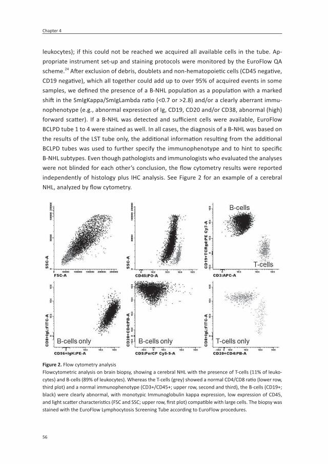
Cell suspensions were generated from a single, unfixed brain biopsy by gentle manual disaggregation on a 100 µm strainer using a 10 mL syringe plunger rod and wash buffer (PBS/BSA 0.5%; not using any enzymes). The released cells were collected by rinsing with a total volume of 10 mL wash buffer and washed twice in 10 mL wash buffer; centrifu-gation steps were for 5 minutes at 540g. After the last wash step, the supernatant was discarded and the pellet of cells was suspended in wash buffer. Fifty microliters of the cell suspension were stained using the EuroFlow Lymphocytosis Screening Tube (LST), according to the EuroFlow protocol.21,22 The LST contains antibodies CD20-Pacific Blue (Clone: 2H7; Biolegend), CD4-Pacific Blue (RPA-T4; Biolegend), CD45-Pacific Orange (HI30; Invitrogen), CD8-FITC, SmIgλ-FITC, CD56-PE, SmIgκ-PE (SLPC mix; Cytognos), CD5-PerCP-Cy5.5 (L17F12, BD Biosciences, CD19-PC7 (J3-119; Beckman Coulter), SmCD3-APC (SK7, BD Biosciences), and CD38-APCH7 (HB7; BD Biosciences). Subsequently the suspension was acquired on a FACSCanto II flowcytometer (BD Biosciences, Erembodegem, BE) using EuroFlow settings.23 We aimed to acquire at least 5000 B-cells (with a minimum of 50.000
figure 1. Histology and immunohistochemistry analysisHE-staining with diffuse large B-cell lymphoma, activated blast type. Left: HE-staining with diffuse large B-cell lymphoma, activated blast type. Brain tissue with infiltration of blastic cells with large vesicular nuclei with nucleoli. Right: These tumour cells express CD20, CD79a, BCL-2, BCL-6 and MUM1 and very weak expression of CD10.
Chapter 4
56
leukocytes); if this could not be reached we acquired all available cells in the tube. Ap-propriate instrument set-up and staining protocols were monitored by the EuroFlow QA scheme.24 After exclusion of debris, doublets and non-hematopoietic cells (CD45 negative, CD19 negative), which all together could add up to over 95% of acquired events in some samples, we defined the presence of a B-NHL population as a population with a marked shift in the SmIgKappa/SmIgLambda ratio (<0.7 or >2.8) and/or a clearly aberrant immu-nophenotype (e.g., abnormal expression of Ig, CD19, CD20 and/or CD38, abnormal (high) forward scatter). If a B-NHL was detected and sufficient cells were available, EuroFlow BCLPD tube 1 to 4 were stained as well. In all cases, the diagnosis of a B-NHL was based on the results of the LST tube only, the additional information resulting from the additional BCLPD tubes was used to further specify the immunophenotype and to hint to specific B-NHL subtypes. Even though pathologists and immunologists who evaluated the analyses were not blinded for each other’s conclusion, the flow cytometry results were reported independently of histology plus IHC analysis. See Figure 2 for an example of a cerebral NHL, analyzed by flow cytometry.
figure 2. Flow cytometry analysisFlowcytometric analysis on brain biopsy, showing a cerebral NHL with the presence of T-cells (11% of leuko-cytes) and B-cells (89% of leukocytes). Whereas the T-cells (grey) showed a normal CD4/CD8 ratio (lower row, third plot) and a normal immunophenotype (CD3+/CD45+; upper row, second and third), the B-cells (CD19+; black) were clearly abnormal, with monotypic Immunoglobulin kappa expression, low expression of CD45, and light scatter characteristics (FSC and SSC; upper row, first plot) compatible with large cells. The biopsy was stained with the EuroFlow Lymphocytosis Screening Tube according to EuroFlow procedures.
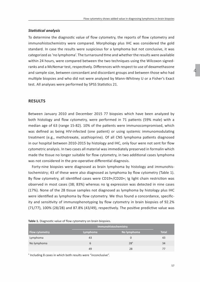
57
Flow cytometry shows added value in diagnosing lymphoma in brain biopsies
4
Statistical analysis
To determine the diagnostic value of flow cytometry, the reports of flow cytometry and immunohistochemistry were compared. Morphology plus IHC was considered the gold standard. In case the results were suspicious for a lymphoma but not conclusive, it was categorized as ‘no lymphoma’. The turnaround time and whether the results were available within 24 hours, were compared between the two techniques using the Wilcoxon signed-ranks and a McNemar test, respectively. Differences with respect to use of dexamethasone and sample size, between concordant and discordant groups and between those who had multiple biopsies and who did not were analyzed by Mann-Whitney U or a Fisher’s Exact test. All analyses were performed by SPSS Statistics 21.
reSuLtS
Between January 2010 and December 2015 77 biopsies which have been analyzed by both histology and flow cytometry, were performed in 71 patients (59% male) with a median age of 63 (range 15-82). 10% of the patients were immunocompromised, which was defined as being HIV-infected (one patient) or using systemic immunomodulating treatment (e.g., methotrexate, azathioprine). Of all CNS lymphoma patients diagnosed in our hospital between 2010-2015 by histology and IHC, only four were not sent for flow cytometric analysis. In two cases all material was immediately preserved in formalin which made the tissue no longer suitable for flow cytometry, in two additional cases lymphoma was not considered in the pre-operative differential diagnosis.
Forty-nine biopsies were diagnosed as brain lymphoma by histology and immunohis-tochemistry; 43 of these were also diagnosed as lymphoma by flow cytometry (Table 1). By flow cytometry, all identified cases were CD19+/CD20+; Ig light chain restriction was observed in most cases (38; 83%) whereas no Ig expression was detected in nine cases (17%). None of the 28 tissue samples not diagnosed as lymphoma by histology plus IHC were identified as lymphoma by flow cytometry. We thus found a concordance, specific-ity and sensitivity of immunophenotyping by flow cytometry in brain biopsies of 92.2% (71/77), 100% (28/28) and 87.8% (43/49), respectively. The positive predictive value was
table 1. Diagnostic value of flow cytometry on brain biopsies.
immunohistochemistry
flow cytometry Lymphoma No lymphoma total
Lymphoma 43 0 43
No lymphoma 6 28a 34
49 28 77a Including 8 cases in which both results were “inconclusive”.

Chapter 4
58
100% (43/43) and the negative predictive value was 82.4% (28/34). Numbers of leukocytes (after exclusion of debris, doublets and non-hematopietic cells) that could be analyzed by flow cytometric analysis ranged widely: 9425 (29-207,259), median (range). Although statistical analysis to compare biopsies with discordant and concordant results should be interpreted with caution due to small numbers, no significant differences were found with respect to sample size (P = 0.06), number of cells acquired by flow cytometry (P = 0.62), or corticosteroid use prior to biopsy (P = 0.108). All 6 discordant cases were DLBCL, without unusual evidence of necrosis. In 6/71 patients a second biopsy and in 2/71 patients even a third biopsy was necessary to make a diagnosis, because of an inconclusive diagnosis in previous biopsies. Only those biopsies which were investigated by both techniques (6/8) were included in the statistical analysis. In the biopsies, analyzed by IHC only, two addi-tional lymphoma were found. Use of corticosteroids prior to first biopsy (P = 0.06), size of the biopsy (P = 0.68 ) and/ or number of cells for flow cytometric analysis (P = 0.19) were similar in patients with conclusive and inconclusive diagnoses. The 20 patients without a lymphoma were diagnosed with a myriad of diseases: 11 glioblastoma, 1 anaplastic astrocytoma, 1 germinoma, 1 stroke, 5 infections and one CLIPPERS syndrome (chronic lymphocytic inflammation with pontine perivascular enhancement responsive to steroids), a rare auto-immune disorder. We found a significantly shortened time to reporting of the results (turnaround time) for flow cytometry, compared to IHC (Table 2). Furthermore, in 54% of the biopsies the diagnosis was provided within 24 hours using flow cytometry, compared to 9% using histology plus IHC.
diSCuSSioN
In this study, we compared flow cytometry with histology plus IHC on 77 brain biopsies, performed in patients clinically suspected of having a lymphoma. We found a high concor-dance between both techniques (92.2%) and a specificity and sensitivity of flow cytometry by immunophenotyping in brain biopsies of 100% and 88%, respectively. In 6 patients with histologically proven NHL, the presence of a lymphoma could not be identified by flow cytometry. No factors (e.g., sample size, use of corticosteroids prior to the biopsy) could be identified which could explain the missing diagnosis in flow cytometry. Unlike
table 2. Time to diagnosis
immunohistochemistryn=77
flow cytometryn=76
Turnaround time (days) 5 (0-18) 1 (0-7) p <0.00a
Diagnosis <24 hours (biopsies) 7 (9%) 41 (54%) p< 0.00b
Median time (range) in days between biopsy and diagnosis. Significance was calculated by aWilcoxon signed-ranks test and bMcNemar Test. In one biopsy the date of reporting was missing for flow cytometry analysis.

59
Flow cytometry shows added value in diagnosing lymphoma in brain biopsies
4
in CSF or bone marrow analysis no additional cases of brain lymphoma were identified by flow cytometry that had not been identified by immunohistochemistry. We found a significant difference in turnaround time for the two techniques. After biopsy a diagnosis was given with a median time of 5 days (range 0-18) for immunohistochemistry, compared to median of 1 day (range 0-7) for flow cytometry. In 54% of the biopsies the presence or absence of a lymphoma could be confirmed within 24 hours by flow cytometry, compared to 9% for immunohistochemistry (P < 0.00), which means that correct treatment could be initiated within 24 hours. It should be noted that the preliminary results of the flow cytometric analysis were frequently reported to the clinician on the day of biopsy. Given the frequently rapid clinical deterioration in CNS lymphoma and the negative impact of a lower performance score on survival, according to the two largest validated prognos-tic models25,26, early diagnosis may improve prognosis.27 Similar findings were reported in a much smaller cohort of 18 stereotactic biopsies recently.28 Cordone et al. found a significant agreement between flow cytometry and immunohistochemistry diagnosis (P = 0.0034). They described a sensitivity and specificity of flow cytometry by immunophe-notyping of 89% and 100% respectively. In the 2/18 PCNSL biopsies not identified by flow cytometry more central necrosis was present, compared to biopsies with concordant results and both patients used corticosteroids prior to the biopsy.28 We did not find more central necrosis in our discordant biopsies and corticosteroid use did not differ between concordant and discordant pairs. One other study analyzed flow cytometry on rinse fluid. Even though rinse fluid from the biopsy needle cannot be completely compared to brain tissue itself, this study showed similar results.29 In a small sample, a high specificity (100%) and sensitivity (75% on rinse fluid and 100% on tissue sample) of flow cytometry in detect-ing a brain lymphoma were found. The added value was again the time in which the flow cytometry could confirm the diagnosis (±3-20 hours, compared to 2-10 days for histo-pathological diagnosis). Because the diagnosis could be confirmed within 24 hours in 75% of the cases, the authors recommend to use both techniques, allowing chemotherapy to commence within 24 hours. In contrast with the results of our study and two comparable, though much smaller studies on brain biopsies, flow cytometry on bone marrow and CSF allowed identification of additional lymphoma cases over cytology. The sensitivity of cytological analysis of CSF for lymphoma cells is low (2-32%).30 Several authors found that additional flow cytometry on CSF improves the sensitivity, up to 2-3 fold.13-15 In up to 80% the lymphoma cells are detected in the first CSF sample, analyzed by flow cytometry.15 It is likely that this additional sensitivity of flow cytometry is a result of the low number of tumor cells available for diagnosis in CSF and bone marrow. Corticosteroids can induce apoptosis in lymphoma cells. This can mask the morphology and can even cause the tumor to vanish.31-33 In lymph nodes and CSF samples, flow cytometry can confirm a diagnosis on samples with a low cell count. We hypothesized that flow cytometry, being a more sensi-tive technique, may be able to recognize lymphoma in patients in whom, after steroid use,

Chapter 4
60
lympholysis had taken place and histology plus IHC was negative. Unfortunately, this was not the case in our series nor in the other two smaller studies available. Five patients who went for multiple biopsies and were diagnosed with brain lymphoma after their second or third biopsy, used corticosteroids prior to their first (and second) biopsy. In none of these patients flow cytometry analysis was able to make the diagnosis when histology plus IHC were non-diagnostic. Clearly, immunohistochemistry as well as flow cytometry analysis can be compromised in patients using corticosteroids prior to the biopsy.
The strengths of this study are the comprehensive clinical and laboratory data in a large, unselected sample, allowing calculation of the diagnostic and clinical value of flow cytometry on brain biopsies. To the best of our knowledge, this is the largest cohort ever described comparing flow cytometry to immunohistochemistry in brain biopsies. Fur-thermore, due to our large population, we were able to show that the negative effect of corticosteroids on the diagnostic value of flow cytometry was similar to that on IHC. Even our series, however, still concerns a relatively small number of cases. The main drawback of our study is its retrospective nature: we may have missed some biopsies, even though we did a thorough search through all available databases in our hospital (neurosurgery, flow cytometry, pathology and neuro-oncology) and the immunologist and pathologist were not blinded for each other’s results. Nevertheless the flowcytometric result was always reported without knowledge of the pathological evaluation. In addition, we did not perform freeze sections, so comparison with intraoperative diagnosis could not be made.
CoNCLuSioN
Flow cytometry analysis in brain biopsy is a feasible technique with 100% specificity to confirm the diagnosis of brain lymphoma in patients suspected for lymphoma on clini-cal grounds. The added clinical value is the speed by which flow cytometry can establish or confirm the diagnosis, enabling a faster initiation of treatment, while false positive cases were not identified. Flow cytometry is complementary to, but not more sensitive than, histopathology with immunohistochemistry analysis. We recommend to perform flow cytometry and immunohistochemistry in parallel in brain biopsies, suspected for a lymphoma.
ACkNowLeGdemeNtS
The authors would like to thank J.A. van Ipenburg, MD for providing figures of immunohis-tochemistry of PCNSL.

61
Flow cytometry shows added value in diagnosing lymphoma in brain biopsies
4
refereNCeS
1. Korfel A, Schlegel U. Diagnosis and treat-ment of primary CNS lymphoma. Nat Rev Neurol 2013; 9(6): 317-27.
2. Colocci N, Glantz M, Recht L. Prevention and treatment of central nervous system involvement by non-Hodgkin’s lymphoma: a review of the literature. Semin Neurol 2004; 24(4): 395-404.
3. Schmitz N, Zeynalova S, Nickelsen M, et al. CNS International Prognostic Index: A Risk Model for CNS Relapse in Patients With Dif-fuse Large B-Cell Lymphoma Treated With R-CHOP. J Clin Oncol 2016; 34(26): 3150-6.
4. Bataille B, Delwail V, Menet E, et al. Primary intracerebral malignant lymphoma: report of 248 cases. J Neurosurg 2000; 92(2): 261-6.
5. de Graaf MT, de Jongste AH, Kraan J, Boon-stra JG, Sillevis Smitt PA, Gratama JW. Flow cytometric characterization of cerebrospi-nal fluid cells. Cytometry B Clin Cytom 2011; 80(5): 271-81.
6. Schroers R, Baraniskin A, Heute C, et al. Diagnosis of leptomeningeal disease in dif-fuse large B-cell lymphomas of the central nervous system by flow cytometry and cytopathology. Eur J Haematol 2010; 85(6): 520-8.
7. Ward MS. The use of flow cytometry in the diagnosis and monitoring of malignant hematological disorders. Pathology 1999; 31(4): 382-92.
8. Kaleem Z. Flow cytometric analysis of lymphomas: current status and usefulness. Arch Pathol Lab Med 2006; 130(12): 1850-8.
9. Dey P, Amir T, Al Jassar A, et al. Combined applications of fine needle aspiration cytol-ogy and flow cytometric immunphenotyp-ing for diagnosis and classification of non Hodgkin lymphoma. Cytojournal 2006; 3: 24.
10. Meda BA, Buss DH, Woodruff RD, et al. Di-agnosis and subclassification of primary and
recurrent lymphoma. The usefulness and limitations of combined fine-needle aspira-tion cytomorphology and flow cytometry. Am J Clin Pathol 2000; 113(5): 688-99.
11. Stacchini A, Aliberti S, Demurtas A, Benevo-lo G, Godio L. Ten antibodies, six colors, twelve parameters: a multiparameter flow cytometric approach to evaluate lepto-meningeal disease in B-cell non-Hodgkin’s lymphomas. Cytometry B Clin Cytom 2012; 82(3): 139-44.
12. Vafaii P, DiGiuseppe JA. Detection of B-cell populations with monotypic light chain expression in cerebrospinal fluid specimens from patients with multiple sclerosis by polychromatic flow cytometry. Cytometry B Clin Cytom 2014; 86(2): 106-10.
13. Balmaceda C, Gaynor JJ, Sun M, Gluck JT, DeAngelis LM. Leptomeningeal tumor in primary central nervous system lymphoma: recognition, significance, and implications. Ann Neurol 1995; 38(2): 202-9.
14. Fine HA, Mayer RJ. Primary central nervous system lymphoma. Ann Intern Med 1993; 119(11): 1093-104.
15. Bromberg JE, Breems DA, Kraan J, et al. CSF flow cytometry greatly improves diagnostic accuracy in CNS hematologic malignancies. Neurology 2007; 68(20): 1674-9.
16. Hegde U, Filie A, Little RF, et al. High inci-dence of occult leptomeningeal disease detected by flow cytometry in newly diag-nosed aggressive B-cell lymphomas at risk for central nervous system involvement: the role of flow cytometry versus cytology. Blood 2005; 105(2): 496-502.
17. Roma AA, Garcia A, Avagnina A, Rescia C, Elsner B. Lymphoid and myeloid neoplasms involving cerebrospinal fluid: comparison of morphologic examination and immu-nophenotyping by flow cytometry. Diagn Cytopathol 2002; 27(5): 271-5.
18. Dammers R, Haitsma IK, Schouten JW, Kros JM, Avezaat CJ, Vincent AJ. Safety and

Chapter 4
62
efficacy of frameless and frame-based intra-cranial biopsy techniques. Acta Neurochir (Wien) 2008; 150(1): 23-9.
19. Verploegh IS, Volovici V, Haitsma IK, et al. Contemporary frameless intracranial biopsy techniques: Might variation in safety and ef-ficacy be expected? Acta Neurochir (Wien) 2015; 157(11): 2011-6; discussion 6.
20. Swerdlow SH, Campo, E., Harris, N.L., Jaffe, E.S., Pileri, S.A., Stein, H., Thiele, J., Vard-iman, J.W. WHO Classification of Tumours of Haematopoietic and Lymphoid Tissues, Fourth Edition. 2008; WHO Classification of tumours, Volume 2(IARC WHO Classifica-tion of Tumours, No 2).
21. Kalina T, Flores-Montero J, van der Velden VH, et al. EuroFlow standardization of flow cytometer instrument settings and immu-nophenotyping protocols. Leukemia 2012; 26(9): 1986-2010.
22. Preijers FW, Huys E, Favre C, Moshaver B. Establishment of harmonization in im-munophenotyping: A comparative study of a standardized one-tube lymphocyte-screening panel. Cytometry B Clin Cytom 2014; 86(6): 418-25.
23. van Dongen JJ, Lhermitte L, Bottcher S, et al. EuroFlow antibody panels for stan-dardized n-dimensional flow cytometric immunophenotyping of normal, reactive and malignant leukocytes. Leukemia 2012; 26(9): 1908-75.
24. Kalina T, Flores-Montero J, Lecrevisse Q, et al. Quality assessment program for EuroFlow protocols: summary results of four-year (2010-2013) quality assurance rounds. Cytometry A 2015; 87(2): 145-56.
25. Ferreri AJ, Blay JY, Reni M, et al. Prognostic scoring system for primary CNS lymphomas:
the International Extranodal Lymphoma Study Group experience. J Clin Oncol 2003; 21(2): 266-72.
26. Abrey LE, Ben-Porat L, Panageas KS, et al. Primary central nervous system lymphoma: the Memorial Sloan-Kettering Cancer Center prognostic model. J Clin Oncol 2006; 24(36): 5711-5.
27. Hochberg FH, Miller DC. Primary central nervous system lymphoma. J Neurosurg 1988; 68(6): 835-53.
28. Cordone I, Masi S, Carosi M, et al. Brain stereotactic biopsy flow cytometry for central nervous system lymphoma charac-terization: advantages and pitfalls. J Exp Clin Cancer Res 2016; 35(1): 128.
29. Debliquis A, Voirin J, Harzallah I, et al. Cytomorphology and flow cytometry of brain biopsy rinse fluid enables faster and multidisciplinary diagnosis of large B-cell lymphoma of the central nervous system. Cytometry B Clin Cytom 2018; 94(1): 182-8.
30. Scott BJ, Douglas VC, Tihan T, Rubenstein JL, Josephson SA. A systematic approach to the diagnosis of suspected central nervous sys-tem lymphoma. JAMA Neurol 2013; 70(3): 311-9.
31. Baraniskin A, Deckert M, Schulte-Altedor-neburg G, Schlegel U, Schroers R. Current strategies in the diagnosis of diffuse large B-cell lymphoma of the central nervous sys-tem. Br J Haematol 2012; 156(4): 421-32.
32. Ghosal N, Hegde AS, Murthy G, Furtado SV. Smear preparation of intracranial lesions: a retrospective study of 306 cases. Diagn Cytopathol 2011; 39(8): 582-92.
33. Bromberg JE, Siemers MD, Taphoorn MJ. Is a “vanishing tumor” always a lymphoma? Neurology 2002; 59(5): 762-4.

63
Flow cytometry shows added value in diagnosing lymphoma in brain biopsies
4
SuPPLemeNtAry dAtA
figure S1 FlowchartSelection of eligible biopsies. *Some biopsies turned out to be analyzed also by flow cytometry, in addition to immunohistochemistry


5Extent of radiological response does not reflect survival in
primary central nervous system lymphoma
Matthijs van der Meulen MD1, Alida A. Postma MD, PhD2, Marion Smits MD, PhD3, Katerina Bakunina MSc4, Monique C. Minnema MD, PhD5, Tatjana Seute MD, PhD6, Gavin Cull MD7,8,
Roelien H. Enting MD, PhD9, Marjolein van der Poel MD, PhD10, Wendy B.C. Stevens MD11, Dieta Brandsma MD12, Aart Beeker MD13, Jeanette K. Doorduijn MD, PhD14, Samar Issa MD15,
Martin J. van den Bent MD, PhD1, Jacoline E.C. Bromberg MD, PhD1
1Erasmus MC Cancer Institute, Brain Tumor Center, University Medical Center Rotterdam, the Netherlands, Department of Neuro-Oncology;
2Maastricht University Medical Center, Maastricht, School for Mental Health & Sciences, The Netherlands, Department of Radiology and Nuclear Medicine;
3Erasmus MC Cancer Institute, Brain Tumor Center, University Medical Center Rotterdam, the Netherlands, Department of Radiology & Nuclear Medicine;
4 HOVON Data Center, Department of Hematology, Erasmus MC Cancer Institute, Rotterdam, Netherlands;5 Department of Hematology, University Medical Center Utrecht, Utrecht, Netherlands;
6 Department of Neurology, University Medical Center Utrecht, Utrecht, Netherlands;7 Haematology Department, Sir Charles Gairdner Hospital and PathWest Laboratory Medicine, Nedlands, WA, Australia;
8 University of Western Australia, Crawley, WA, Australia;9 Department of Neurology, University of Groningen, University Medical Center Groningen, Groningen, Netherlands;
10 Department of Hematology, University Medical Center, Maastricht, Netherlands;11 Department of Hematology, Radboud University Medical Center, Nijmegen, Netherlands;
12 Department of Neuro-Oncology, Netherlands Cancer Institute-Antoni van Leeuwenhoek, Amsterdam, Netherlands;13 Department of Hematology, Spaarne Gasthuis, Haarlem, Netherlands;
14 Erasmus MC Cancer Institute, University Medical Center Rotterdam, the Netherlands, Department of Hematology;15 Middlemore Hospital, Auckland, New Zealand, department of Haematology.
2021 Neuro-Oncology Advances, 3(1)

Chapter 5
66
ABStrACt
Background. In primary central nervous system lymphoma (PCNSL), small enhancing le-sions can persist after treatment. It is unknown whether a difference in response category (complete response (CR), complete response unconfirmed (CRu) or partial response (PR)) reflects survival. We aimed to determine the value of a central radiology review on re-sponse assessment and whether the extent of response influenced progression-free and/or overall survival.methods. All patients in the HOVON 105/ALLG NHL 24 study with at least a baseline MRI and one MRI made for response evaluation available for central review were included. Tumor measurements were done by two independent central reviewers, disagreements were adjudicated by a third reviewer. Crude agreement and interobserver agreement (Co-hen’s kappa) were calculated. Differences in progression-free and overall survival between different categories of response at the end-of-protocol-treatment were assessed by the log-rank test in a landmark survival-analysis.results. Agreement between the central reviewers was 61.7% and between local and central response assessment was 63.0%. Cohen’s kappa’s, which corrects for expected agreement, were 0.44 and 0.46 (moderate), respectively. Agreement on progression or not was 93.3% (kappa 0.87) between local and central response assessment. There were no significant differences in progression-free and overall survival between patients with CR, CRu or PR at the end-of-protocol-treatment, according to both local and central response assessment.Conclusions. Reliability of response assessment (CR/CRu/PR) is moderate even by central radiology review and these response categories do not reliably predict survival. Therefore, primary outcome in PCNSL studies should be survival rather than CR or CR/CRu-rate.

67
Extent of radiological response does not reflect survival in PCNSL patients
5
iNtroduCtioN
Primary central nervous system lymphoma (PCNSL) is a rare non-Hodgkin lymphoma confined to the brain, leptomeninges, spinal cord and eyes without manifestations out-side the central nervous system. For response assessment in PCNSL the criteria from the International Primary CNS Lymphoma Collaborative Group (IPCG), are commonly used.1 These response criteria are based on radiological, ophthalmologic and spinal fluid cytology examination, and the use of corticosteroids. The MRI response evaluation defines the fol-lowing categories: complete response (CR): no signs of abnormal gadolinium-based con-trast agent enhancement, complete response unconfirmed (CRu): a small but persistent contrast enhancement abnormality likely related to biopsy or focal hemorrhage, partial response (PR): a reduction of ≥50% of the contrast enhancing lesion, stable disease (SD): <50% reduction and ≤25% increase of the contrast enhancing lesion, progressive disease (PD): >25% increase in contrast enhancing lesion, relapse: a new contrast enhancing lesion after prior CR or CRu.1 These response criteria do not take non-enhancing lesions into account. Recent findings and earlier reports suggest that these lesions might, however, be considered as tumor as well.2,3
The correlation of radiological response with survival endpoints (progression-free sur-vival [PFS] and overall survival [OS]) is uncertain: one study showed that patients with a CR at the end of induction chemotherapy had a better OS than those who did not reach CR, but in this study PR, SD and progression were combined.4 In another study highly variable outcomes were found in patients with PR at the end-of-treatment2, and a third study did not show a survival difference between those who attained CR compared to those who did not reach CR at the end of induction treatment.5 Thus, it is questionable whether in PCNSL the extent of radiological response is relevant for predicting OS, the golden endpoint in oncology studies. It is also unclear whether interobserver variation exists in assessing response in PCNSL, which if present, will affect the reliability of that endpoint.
In the HOVON 105/ ALLG NHL 24 trial, the primary endpoint was event-free survival (EFS). Events were defined as ‘not reaching complete response’ or ‘complete response unconfirmed at the end-of-treatment’, or ‘progression or death after response’.6 Because event-free survival includes a radiological evaluation as endpoint (i.e. achieving CR or CRu), based on local assessment, central MRI review is important for the trial analysis. The aim of the present study was to review the local assessment by central radiology review and to assess whether CR, CRu and PR reflect PFS and OS. In addition, we evaluated the relevance of non-enhancing lesions at baseline and after treatment.

Chapter 5
68
methodS
Patient selection
The HOVON 105/ALLG NHL 24 study is a phase III randomized controlled trial, in which between 2010 and 2016 199 patients were recruited from Dutch, Australian and New Zea-land hospitals. The treatment protocol and primary outcome results have been published before.6 In short, immunocompetent patients with a newly diagnosed, CD20 positive B-cell PCNSL aged 18-70 years with WHO/ECOG performance status 0-3 were included. Patients were randomized for two courses of high-dose methotrexate (HD-MTX)-based chemotherapy: methotrexate, teniposide, BCNU and prednisolone (MBVP) versus MBVP with rituximab (R-MBVP). This was followed by HD-cytarabine (Ara-C) chemotherapy. Patients ≤60 years-old subsequently received 30Gy whole brain radiotherapy (WBRT). A simultaneous focal boost of 10Gy was given to the original enhancing tumor in patients who only achieved partial response (PR).
Patients were included for the central MRI review if they gave informed consent for central radiology review, and if a baseline MRI as well as at least one follow-up MRI was available for central review. Additionally, a measurable brain lesion had to be present at baseline in order to be able to assess response. Patients were excluded if only a CT was available at baseline and/or if only CTs were used for evaluation at subsequent time points. The baseline MRI had to have been made within21 days before initiation of protocol treat-ment.
Radiological follow-up
According to protocol, MRI evaluations were performed before the initiation of chemo-therapy (baseline), after the second (R-)MBVP course, after Ara-C and after WBRT, if ap-plicable. Follow-up MRIs were made every 3 months in the first 2 years after treatment, followed by every 6 months up to 5 years after treatment and yearly thereafter.
At least the following MRI sequences were performed: an axial T1 weighted scan before and after gadolinium-based contrast agent administration, and an axial T2 weighted and/or fluid-attenuated inversion recovery (FLAIR) scan. If locally possible additional sagittal or coronal T1 weighted scans with gadolinium-based contrast agent administration were also performed or reconstructed. All MR images were acquired on a 1.5 or 3.0 Tesla scanner.
Central radiology review
Scans made at baseline and after each treatment component were centrally reviewed to evaluate response of the tumor to treatment. In case of relapse or progression, the MRI on which this was diagnosed according to the local physician, as well as the last MRI made before progression were also centrally reviewed, to verify progression and to make sure true progression had not occurred earlier than locally ascertained. Scans made in follow-

69
Extent of radiological response does not reflect survival in PCNSL patients
5
up were not reviewed if response was not changed according to the local evaluation. PD was defined as relapse or progression at any site (brain, spinal cord, cerebrospinal fluid or eyes). In case progression was located outside the brain parenchyma, the MRI was not included in this analysis.
At the end of the study, all MR images were submitted for review on DVD or CD, and stored on a secured central server. Except for the baseline scan, locally assessed response rates were collected for all MRIs performed for this study. Local physicians were not blinded for treatment arm and/or other clinical information.
Central evaluation of response was performed retrospectively, in parallel by two review-ers (M.M. and A.A.P.). In case of disagreement on the response between these reviewers, an adjudicator (M.S.) finalized the central response category. The central reviewers and the adjudicator were blinded for study-arm and clinical information.
Single evaluations were excluded if a) both central reviewers considered an MRI not assessable, b) no MRI with gadolinium-based contrast agent administration was made or c) the MRI was made outside +/- 3 weeks around the planned evaluation moment during the treatment period.
MRI Tumor measurement
For all enhancing lesions the largest diameters on the axial post gadolinium-based contrast agent T1 weighted images were measured as well as their perpendicular diameter on the same slice. The product of these measurements was used to define the size of the tumor. In case of multiple lesions, response assessment was based on the sum of all products, up to a maximum of four lesions.
Non-enhancing space occupying lesions were measured by one of the central reviewers (M.M.) on FLAIR images if possible, and otherwise on the T2 weighted images. In patients experiencing recurrent disease the localization of the recurrence was compared to the localization of the initial non-enhancing and enhancing lesions.
Landmark analysis
To estimate the survival probability for the different response categories (CR, CRu, or PR) in an unbiased way, a landmark analysis was performed.7 Regardless of the type of last administered treatment on protocol, the response at the end-of-protocol-treatment was related to PFS and OS for those still at risk at that timepoint. PFS was defined as time from randomization to progression, relapse, or death from any cause, whichever came first. OS was defined as time from randomization to death from any cause. Patients still alive at the date of last contact were censored. Follow-up data were available up to October 1, 2019. In this landmark analysis, all patients alive at the landmark timepoint who had an MRI at end-of-protocol-treatment and were classified as CR, CRu or PR on that MRI were included. The reference time point (landmark) was set between 4 weeks after last treat-

Chapter 5
70
ment, but before the first follow-up MRI (i.e. 3 months after end-of-protocol-treatment), in such a way that most patients could be included. The landmark analysis was performed for both local and central response assessment.
Statistical analysis
Since the HOVON 105/ ALLG NHL 24 study showed no differences in EFS, PFS or OS be-tween the two treatment arms we analyzed both arms together.6 For the interobserver agreement between the central reviewers, and between central and local response evalu-ations we calculated the crude agreement and Cohen’s kappa8, in which crude agreement is corrected for expected agreement (i.e. the agreement that would occur ‘by chance’). First, interobserver agreement was assessed for all response categories separately; second, agreement was assessed for combined categories CR/CRu and PD/relapse, and third for three categories: response (CR/CRu/PR), SD, and progression (PD/relapse). Lastly, the crude agreement and kappa’s interobserver agreement for progression versus no progression were calculated on the MRI on which progression was diagnosed and on the preceding MRI.
In the landmark analysis, the survival curves for PFS and OS were constructed using the Kaplan-Meier method for the different categories of response (i.e. CR, CRu and PR) according to central and to local response evaluation at the end-of-protocol-treatment. Differences by response were assessed with a log-rank test with a 5% significance level. All analyses were performed with Stata, version 15.0. The study was approved by the ethics committee of all participating centers. All participants signed informed consent for the randomized controlled trial and separately for the central radiology review.
reSuLtS
Of the 199 trial patients, 115 were included in this study. Three patients were excluded because they did not give informed consent for the radiology review, 12 patients because no baseline MRI was present and 3 for whom only CT was available, 61 patients were excluded because baseline MRI was made outside the predefined time window, and 5 for other reasons (see CONSORT diagram, Figure 1). The median age of patients included in this study was 61 years (range: 38-70), 44% were female, and 73% had WHO performance score <2, see Table 1. On October 1, 2019 (last follow-up), in the central radiology review cohort 45 patients were alive without progression and 15 were alive with progression.
Of these 115 included patients, 396 scans were centrally reviewed: 115 baseline MRIs, 235 after treatment and 46 PD or last before PD scans. Scans were excluded if they were not received for central review (n=154), were made outside the predefined time window (n=7), or progression was not located in the brain parenchyma (n=14).

71
Extent of radiological response does not reflect survival in PCNSL patients
5
Central radiology review
For the MRIs made during treatment (n=235) the agreement between central reviewer 1 and 2 and between local and central response assessment was higher than the expected agreement by chance (p<0.001). Between the central reviewers the agreement for all response categories was 61.7%, with a kappa of 0.44, see Table 2. After adjudication, if necessary, the agreement between local and central response assessment was 63.0% with a kappa of 0.46, see Table 3.
199 HOVON 105/ ALLG NHL 24 patients
196 patients
3 no consent for central MRI review
12 no baseline MRI3 only CT scan available
1 no parenchymal brain involvement4 no measurable brain lesion
4 baseline scan made afterstart protocol treatment
57 baseline scan made >21 days before start protocol treatment
181 patients
176 patients
115 patients
10 MRIs after first (R-)MBVP cycle
97 MRIs after second (R-)MBVP cycle
85 MRIs after HD-Ara-C
43 MRIs after WBRT
figure 1. CONSORT diagram. 235 MRIs from 115 patients were assessed.(R-)MBVP = (rituximab), methotrexate, teniposide, BCNU and prednisolone, HD-Ara-C = high-dose cytarabine, WBRT = whole-brain radiotherapy.

Chapter 5
72
table 1. Baseline characteristics of the patients included in this study, those who were excluded and for the total study population.
Included patientsn=115
Excluded patientsn=84
totaln=199
Sex (n, % males) 64 (56%) 45 (54%) 109 (55%)
Age (median, range) 61 (38-70) 61 (26-70) 61 (26-70)
WHO performance score (n, %)
0 27 (23%) 16 (19%) 43 (22%)
1 57 (50%) 44 (53%) 101 (51%)
2 17 (15%) 17 (20%) 34 (17%)
3 14 (12%) 7 (8%) 21 (10%)
Comorbidities (n>2, %) 60 (52%) 44 (52%) 104 (52%)
Solitary lesions (n, %)Missing/ NA
66 (57%)1 (1%)
37 (44%)18 (21%)
103 (52%)19 (10%)
Deep lesion (n, %) 83 (72%) 42 (50%) 125 (63%)
Periventricular (n, %) 61 (53%) 35 (42%) 96 (48%)
Basal ganglia (n, %) 8 (7%) 6 (7%) 14 (7%)
Cerebellar (n, %) 22 (19%) 8 (10%) 30 (15%)
Brain stem (n, %) 10 (9%) 2 (2%) 12 (6%)
Spinal (n, %) 2 (2%) - 2 (1%)
Lobar (n, %) 58 (50%) 37 (44%) 95 (48%)
Study drug exposure
High-dose cytarabine (n, %) 98 (85%) 63 (75%) 161 (81%)
WBRT (n, %) 48 (42%) 22 (26%) 70 (35%)
Radiation boost given (n, %) 24 (21%) 15 (18%) 39 (20%)
Intrathecal treatment given (n, %) 12 (10%) 4 (5%) 16 (8%)
NA = not applicable in case of no brain lesion; WBRT = whole brain radiotherapy.
table 2. Level of agreement between central reviewer 1 and central reviewer 2 in all 235 scans made after each treatment module for all response categories.
reviewer 2
revi
ewer
1
Cr Cru Pr Sd Pd relapse total
CR 32 34 11 1 0 0 78
CRu 0 38 15 0 0 1 54
PR 0 17 74 4 0 0 95
SD 0 0 2 1 1 0 4
PD 0 0 0 1 0 1 2
relapse 0 0 1 0 1 0 2
Total 32 89 103 7 2 2 235
Agreement 62%, kappa 0.44. CR = complete response; CRu = complete response unconfirmed; PR = partial response; SD = stable disease; PD = progressive disease.

73
Extent of radiological response does not reflect survival in PCNSL patients
5
When CR and CRu were combined into one category, and the categories PD and relapse were combined, the interobserver agreement and kappa values increased, but the lat-ter remained in the moderate range. Between reviewer 1 and 2, agreement increased to 77.0% (kappa 0.57) and between local and central assessment agreement improved to 74.5% (kappa 0.54), see Supplemental Table 1 and Supplemental Table 2, respectively. When response categories were classified into response (CR, CRu or PR), stable disease, or progression (PD or relapse) the agreement increased to 95.3% (kappa 0.40) between the central reviewers and 94.9% (kappa 0.41) between local and central response assessment. The kappa remained relatively low, because of the increased expected agreement.
The response assessment for the MRIs on which progression or relapse was diagnosed, and the last MRI made before progression were analyzed separately. Agreement on whether there was progression or relapse versus ‘no progression’ was 96.7% between central reviewer 1 and 2 (kappa 0.93) and 93.3% between the local and central response assessment (kappa 0.87), both were significantly higher than expected agreement (p<0.001), Table 4A and 4B.
table 3. Level of agreement between local and central assessment in all 235 scans made after each treatment module for all response categories.
LoCALCe
Ntr
AL
Cr Cru Pr Sd Pd relapse total
CR 42 12 8 0 0 1 63
CRu 15 28 30 0 0 0 73
PR 1 9 74 1 0 1 86
SD 0 0 4 1 1 0 6
PD 0 0 2 0 3 0 5
relapse 0 0 2 0 0 0 2
Total 58 49 120 2 4 2 235
Agreement 63%, kappa 0.46. CR = complete response; CRu = complete response unconfirmed; PR = partial response; SD = stable disease; PD = progressive disease.
table 4. Level of agreement A) between both central reviewers: agreement 96.7%, kappa 0.93 and B) be-tween local and central assessment.
A REVIEWER 2 B LOCAL
REVI
EWER
1 No PD PD
CEN
TRAL
No PD PD
No PD 16 1 17 No PD 14 1 15
PD 0 13 13 PD 1 14 15
16 14 30 15 15 30
Agreement 93.3%, kappa 0.87 in all scans which confirmed PD and made ‘last before PD’. PD = progressive disease, including relapses.

Chapter 5
74
Landmark analysis on CR, CRu and PR
In total 91 ‘end-of-protocol-treatment’ MRIs were available and were locally and centrally assessed. The landmark, aiming to include as many pati ents as possible aft er the end-of-protocol-treatment MRI but before fi rst follow-up MRI, was positi oned at 6.9 months aft er randomizati on. Only those with a CR, CRu or PR at the end-of-protocol-treatment were included in this analysis. Two pati ents who had not had their end-of-protocol-treatment scan yet were excluded. For the PFS analysis we also excluded those who had less than PR (n=7 according to central response and 8 according to the local response assessment) at the end-of-protocol-treatment, had progression before the landmark (n=9 in central and n=6 in local response assessment) or died without progression (n=1). For the OS analysis we excluded those who had less than PR (see above) or died (n=2) Since we analyzed survival according to local as well as to central response assessment, the number of pa-ti ents the analyses were based on diff ered: survival analysis was performed on 72 (central assessment) and 74 (local assessment) pati ents for PFS and 80 (central assessment) and 79 (local assessment) pati ents for OS.
figure 2. Progression free survival (A and B) and overall survival (C and D) for those pati ents who had a parti al response (PR), complete response (CR) or complete response unconfi rmed (CRu) at the end of treatment MRI, according to central (A and C) and local response (B and D) assessment.

75
Extent of radiological response does not reflect survival in PCNSL patients
5
Regarding PFS (Figure 2A and B) there was no statistically significant difference between those judged as CR, CRu or PR, both in central response assessment (p=0.97) and accord-ing to local judgement (p=0.76). Similar results were found for overall survival (Figure 2C and D), for central (p=0.69) and local (p=0.16) response assessment.
Non-enhancing lesions
At baseline seven patients were identified with non-enhancing space occupying lesions. Baseline characteristics in these patients were similar to the total trial population, Supple-mental Table 3. After chemotherapy, in five of the seven patients, the lesions diminished with ≥50%.
Four of these patients relapsed, in two patients this was at the same location as the original enhancing lesion. None of the patients had a relapse at the location of the non-enhancing lesions.
diSCuSSioN
We found an excellent crude agreement (96.7%) and kappa score (0.93) between the central reviewers and between local and central radiological evaluations (crude agree-ment 93.3%, kappa 0.86) in differentiating progression from no progression. However, for response assessment after treatment, interobserver agreement was moderate at best. Furthermore, the crude interobserver agreement and kappa statistics between the two central reviewers and between local and central radiology response assessment after each treatment component (n=235) were almost identical (local vs central kappa 0.46 and both central reviewers 0.44). This suggests that there is little added value of a central radiol-ogy review in PCNSL patients. Crude interobserver agreement increased when response categories were combined, but the kappa statistics remained in the range of moderate agreement. This is most likely due to increased expected agreement, since Cohen’s kappa statistic is the agreement found, corrected for expected agreement to occur by chance. Thus, our data show that although the presence of response is well agreed upon, judge-ment regarding the extent of response is less reliable. This suggests, together with the excellent agreement regarding the moment of progression, that PFS and OS are more reliable endpoints than specific and more detailed response categories and which also better reflect patient benefit.
To the best of our knowledge, a central radiology review in PCNSL with assessment of the interobserver agreement has not been described before. Several studies assessed interobserver agreement in glioma patients.9-13 Our excellent agreement on PD vs no PD contrasts with the interobserver agreement in standard radiology assessment for progres-sion in glioma.9,12 This might be explained by the rapid evolution of most PCNSL and its

Chapter 5
76
easily recognizable appearance on the MR images: PCNSL, at relapse or progression as well as primary presentation, generally appears as a homogenously enhancing, circumscribed space-occupying lesion, rather than the ill-defined mass and irregular enhancement in high-grade glioma.
In our landmark analyses we found no difference in PFS or OS for the different types of response: CR, CRu and PR. The lack of difference in outcome between CR and CRu patients is in line with the current response criteria1, which state regarding CRu lesions that if the type of abnormality does not change or slowly involutes over time without therapy or cor-ticosteroids, it is reasonable to categorize these lesions as CR. However, we also found that partial response was associated with a similar PFS and even a similar OS, suggesting that these response categories do not translate into meaningful differences in outcome and are therefore not reliable surrogate endpoints in PCNSL. A few other studies compared survival for different response categories.2,4,5,14 Only one of these studies2 used a landmark analysis, resulting in selection bias in the other studies (i.e. immortal time bias), since response and survival are influenced by the passing time. If survival analysis is done after end-of-protocol-treatment, regardless of when the MRI was made, those who have had a later MRI would have had more chance to achieve CR. One large, prospective study (n=511) showed a significant difference between CR versus no CR (PR, SD and PD combined) for OS (39 versus 22 months; p<0.0001) and PFS (36 versus 6 months).4 In that study, CR was defined as complete resolution of contrast enhancing lesions on MRI or on CT. The latter radiological examination might have missed small contrast enhancing foci. Furthermore, combining PR with non-responding and progressive patients does not allow conclusions regarding the partially responding (PR) patients. Similarly, in a retrospective analysis of a phase II study in 85 patients, differences in survival rates between patients with CR, PR, SD or progression after the end of chemotherapy were calculated using a single log-rank test. A significant difference was found for OS (p<0.001), and a nearly significant difference for PFS (p=0.076).2 Again, due to the comparison of all groups including non-responding or progressive patients this analysis does not allow comparison between patients with different extents of response. Lastly, a small retrospective single center series, evaluated patients after chemotherapy. Those with CR after the completion of chemotherapy (n=10) had no better PFS or OS than those with no CR (n=30).5 In that study, however, patients without CR subsequently received additional treatment: radiotherapy or autologous stem cell transplantation.
Two dimensional measurements are the golden standard in the current PCNSL re-sponse criteria1, and were therefore also applied in this study. This might, underestimate volumetric changes. In glioma, volumetric measurements, either manual or computerized, improved agreement regarding radiological response compared to 2D measurements in some studies,13,15 and fully automated segmentation was significantly better in predicting OS (p<0.0001) than the conventional 2D measurements.13 In contrast, one other smaller

77
Extent of radiological response does not reflect survival in PCNSL patients
5
study showed no differences in predicting OS between manual 2D and 3D measurement or computerized segmentation of the tumor.16 Response in PCNSL is generally easily recognizable with reductions > 50% being the rule, so small changes in the volume of the enhancing lesion are unlikely to influence response rates. However, small changes in residual abnormalities might result in a change in response category, between CRu and PR.
Our study has some limitations: first, the landmark analyses were performed for dif-ferent categories of response based on the MRI made at the end-of-protocol-treatment. This might have led to bias, since patients >60 years-old with a PR at the end-of-protocol-treatment could have had additional treatment, depending on the discretion of the treating physician. Second, analyses were performed on a subgroup from a large clinical trial and inadvertent bias may have occurred in the selection of patients for this study, even though this selection was based on the availability of scans only. Our results should therefore be validated in a larger external cohort.
In conclusion, our results suggest that at the end-of-protocol-treatment, specific radio-logical response categories (CR, CRu, or PR) do not reliably predict survival in PCNSL pa-tients, even after central radiology review, but that interobserver agreement in diagnosing relapse or progression is high. Therefore, the primary outcome measure in PCNSL studies should be PFS or OS; as secondary outcome measure combined response rate (CR, CRu and PR) is more reliable than CR or CR/CRu-rate.
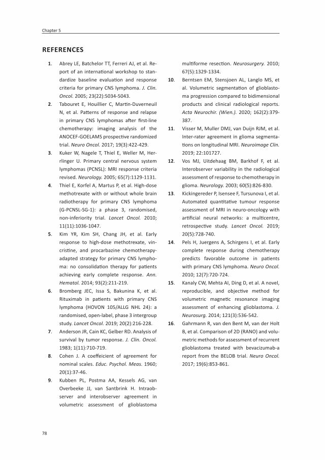
Chapter 5
78
refereNCeS
1. Abrey LE, Batchelor TT, Ferreri AJ, et al. Re-port of an international workshop to stan-dardize baseline evaluation and response criteria for primary CNS lymphoma. J. Clin. Oncol. 2005; 23(22):5034-5043.
2. Tabouret E, Houillier C, Martin-Duverneuil N, et al. Patterns of response and relapse in primary CNS lymphomas after first-line chemotherapy: imaging analysis of the ANOCEF-GOELAMS prospective randomized trial. Neuro Oncol. 2017; 19(3):422-429.
3. Kuker W, Nagele T, Thiel E, Weller M, Her-rlinger U. Primary central nervous system lymphomas (PCNSL): MRI response criteria revised. Neurology. 2005; 65(7):1129-1131.
4. Thiel E, Korfel A, Martus P, et al. High-dose methotrexate with or without whole brain radiotherapy for primary CNS lymphoma (G-PCNSL-SG-1): a phase 3, randomised, non-inferiority trial. Lancet Oncol. 2010; 11(11):1036-1047.
5. Kim YR, Kim SH, Chang JH, et al. Early response to high-dose methotrexate, vin-cristine, and procarbazine chemotherapy-adapted strategy for primary CNS lympho-ma: no consolidation therapy for patients achieving early complete response. Ann. Hematol. 2014; 93(2):211-219.
6. Bromberg JEC, Issa S, Bakunina K, et al. Rituximab in patients with primary CNS lymphoma (HOVON 105/ALLG NHL 24): a randomised, open-label, phase 3 intergroup study. Lancet Oncol. 2019; 20(2):216-228.
7. Anderson JR, Cain KC, Gelber RD. Analysis of survival by tumor response. J. Clin. Oncol. 1983; 1(11):710-719.
8. Cohen J. A coeffeicient of agreement for nominal scales. Educ. Psychol. Meas. 1960; 20(1):37-46.
9. Kubben PL, Postma AA, Kessels AG, van Overbeeke JJ, van Santbrink H. Intraob-server and interobserver agreement in volumetric assessment of glioblastoma
multiforme resection. Neurosurgery. 2010; 67(5):1329-1334.
10. Berntsen EM, Stensjoen AL, Langlo MS, et al. Volumetric segmentation of glioblasto-ma progression compared to bidimensional products and clinical radiological reports. Acta Neurochir. (Wien.). 2020; 162(2):379-387.
11. Visser M, Muller DMJ, van Duijn RJM, et al. Inter-rater agreement in glioma segmenta-tions on longitudinal MRI. Neuroimage Clin. 2019; 22:101727.
12. Vos MJ, Uitdehaag BM, Barkhof F, et al. Interobserver variability in the radiological assessment of response to chemotherapy in glioma. Neurology. 2003; 60(5):826-830.
13. Kickingereder P, Isensee F, Tursunova I, et al. Automated quantitative tumour response assessment of MRI in neuro-oncology with artificial neural networks: a multicentre, retrospective study. Lancet Oncol. 2019; 20(5):728-740.
14. Pels H, Juergens A, Schirgens I, et al. Early complete response during chemotherapy predicts favorable outcome in patients with primary CNS lymphoma. Neuro Oncol. 2010; 12(7):720-724.
15. Kanaly CW, Mehta AI, Ding D, et al. A novel, reproducible, and objective method for volumetric magnetic resonance imaging assessment of enhancing glioblastoma. J. Neurosurg. 2014; 121(3):536-542.
16. Gahrmann R, van den Bent M, van der Holt B, et al. Comparison of 2D (RANO) and volu-metric methods for assessment of recurrent glioblastoma treated with bevacizumab-a report from the BELOB trial. Neuro Oncol. 2017; 19(6):853-861.

79
Extent of radiological response does not reflect survival in PCNSL patients
5
SuPPLemeNtAry iNformAtioN
Supplemental Methods:Kappakappa value Agreement
0 absent
0.01-0.20 slight
0.21-0.40 fair
0.41-0.60 moderate
0.61-0.80 good
0.81-1.00 excellent
Supplemental table 1. Level of agreement between central reviewer 1 and central reviewer 2 in all 235 scans made after each treatment module for clinically relevant response categories. Agreement 77.0%, kappa 0.40.
reviewer 2
revi
ewer
1
Cr/Cru Pr Sd Pd/relapse total
CR/CRu 104 26 1 1 132
PR 17 74 4 0 95
SD 0 2 1 1 4
PD/relapse 0 1 1 2 4
Total 121 103 7 4 235
CR = complete response; CRu = complete response unconfirmed; PR = partial response; SD = stable disease; PD = progressive disease.
Supplemental table 2. Level of agreement between local and central assessment in all 235 scans made after each treatment module for clinically relevant response categories. Agreement 74.5%, kappa 0.54.
LoCAL
CeN
trAL
Cr/Cru Pr Sd Pd/relapse total
CR/CRu 97 38 0 1 136
PR 10 74 1 1 86
SD 0 4 1 1 6
PD/relapse 0 4 0 3 7
Total 107 120 2 6 235
CR = complete response; CRu = complete response unconfirmed; PR = partial response; SD = stable disease; PD = progressive disease.

Chapter 5
80
Supplemental table 3. Baseline characteristics of the patients with and without non-enhancing lesions at baseline, and for the total study population.
n=192 n=7 n=199
Sex (n, % males) 106 (55%) 3 (43%) 109 (55%)
Age (median, range) 61 (26-70) 61 (55-68) 61 (26-70)
WHO performance score (n, %)
0 43 (22%) - 43 (22%)
1 97 (51%) 4 (57%) 101 (51%)
2 32 (17%) 2 (29%) 34 (17%)
3 20 (10%) 1 (14%) 21 (11%)
Unilateral lesions (n, %) 103 (54%) 2 (29%) 105 (53%)
Missing/ NA 19 (10%) 1 (14%) 20 (7%)
Deep lesion (n, %) 120 (63%) 5 (71%) 125 (63%)
Treatment arm
MBVP (n, %) 96 (50%) 4 (57%) 100 (50%)
R-MBVP (n, %) 96 (50%) 3 (43%) 99 (50%)
WHO = World Health Organisation; NA = not applicable, no brain lesion(s); MBVP = methotrexate, tenoposide, BCNU and prednisolone; R = rituximab


1
7
4
10
2
8
5
11
3
9
6
A
PArt iiiNeurocognitive functioning and
health-related quality of life

6Cognitive functioning and health-related quality of life in
patients with newly diagnosed primary central nervous system lymphoma: a systematic literature review
M. van der Meulen1, L. Dirven2,3, E.J.J. Habets3,4, M.J. van den Bent1, M.J.B. Taphoorn2,3, J.E.C. Bromberg1
1Erasmus MC Cancer Institute, Brain Tumor Center, University Medical Center Rotterdam, Department of Neuro-Oncology; 2Leiden University Medical Center, Department of Neurology; 3Haaglanden Medical Center, The Hague,
Department of Neurology; 4Haaglanden Medical Center, The Hague, Department of Medical Psychology.
2018 Lancet Oncology, 19(8), e407-e418.

Chapter 6
86
SummAry
Incidence of primary CNS lymphoma (PCNSL) is increasing, while prognosis is improving as treatment advance. However, declined cognitive functioning remains a major challenge in the treatment of PCNSL. This cognitive decline, in conjunction with other symptoms caused by the disease or its treatment, or both, can compromise health-related quality of life (HRQOL). The aim of this Review was to give a comprehensive overview on cognitive functioning and HRQOL for patients with PCNSL, including an evaluation of patient-related and treatment-related factors that can influence cognitive functioning and HRQOL. We re-viewed the literature for studies on cognitive functioning and HRQOL in newly diagnosed adult patients with PCNSL using MEDLINE/Pubmed, Embase, Web of Science, Scopus, Cochrane, PsycINFO, CINAHL EBSCO, and Google scholar, up to Jan 4, 2018. Articles were selected using predetermined inclusion and exclusion criteria; 42 articles were eligible for inclusion. Findings show that the tumour itself has a great effect on cognitive functioning and HRQOL. Initially, induction chemotherapy results in improvement of cognition and HRQOL in most patients. In the long-term, additional whole brain radiotherapy (WBRT) has a negative impact on cognitive functioning, but the magnitude of this impact is not always clinically relevant. HRQOL scores were worse compared with controls, and worse after combined chemotherapy and radiotherapy when compared with chemotherapy only, particularly in the long term. Therefore, combined chemotherapy and radiotherapy seems to have a negative effect on HRQOL and cognition in PCNSL patients. Although prolonged progression-free survival is achieved with combined therapy, information on its effect on cognition and HRQOL should also be included in clinical decision-making.

87
Cognitive functioning and health-related quality of life in patients with newly diagnosed PCNSL
6
iNtroduCtioN
Primary CNS lymphoma (PCNSL) is a rare non-Hodgkin lymphoma confined to the brain, leptomeninges, spinal cord and eyes. In previous decades, the incidence rate has increased while prognosis has improved.1-3 Improved survival is largely dependent on changes in treatment, particularly the addition of high-dose methotrexate-based chemotherapy to the previously standard treatment of whole-brain radiotherapy (WBRT). One phase 3 study has shown that the omission of WBRT decreased progression-free survival (PFS), but not overall survival (OS) in patients with PCNSL.4 However, more patients who were given chemotherapy and radiotherapy, compared with chemotherapy alone, developed neurotoxicity (49% vs 26%), which was defined as progressive neurological or cognitive impairment as documented in serial clinical examinations in the absence of recurrent lymphoma.4
Although improved survival has been achieved in this population, declined cognitive functioning remains a major challenge in the treatment of PCNSL.5 Subsequently, cogni-tive decline and other symptoms caused by the disease or its treatment, or both, can compromise health-related quality of life (HRQOL).6,7 To establish the net clinical benefit of a treatment regimen, information on survival has to be combined with information from patient-centered outcomes, eg, HRQOL and cognition. Combining both sources of information allows informed decision making on the effect of a specific treatment, which is useful for all specialists involved in the treatment of patients with PCNSL.
In 2007, a systematic review based on a search in MEDLINE was done to identify studies reporting om the effect of different treatment modalities on cognition in PCNSL patients.5 With the introduction of new treatment modalities, however, and the increased use of patient-reported outcomes, an update on the field is warranted. Therefore, the aim of this systematic Review was to give a comprehensive overview on the effect of PCNSL and different treatment types on cognitive functioning and HRQOL in adult patients with newly diagnosed PCNSL. Furthermore, we aimed to identify which patient-related and treatment-related factors were associated with cognitive functioning and HRQOL. Possible underlying pathophysiological mechanisms, which might explain a decline in cognition or HRQOL, or both, are beyond the scope of this Review, and thus will not be discussed.
dAtA CoLLeCtioN
Search strategy and selection criteria
We dis an extensive search for articles published up until Jan 4, 2018, using the electronic databases MEDLINE/PubMed, Embase, Web of Science, Scopus, Cochrane, PsycINFO, CINAHL EBSCO and Google scholar. Search terms related to “PCNSL” and “cognition” or

Chapter 6
88
“HRQOL”, or both, were used, and terms were formulated to exclude case reports and studies that only include animals (see appendix for the search strategy in MEDLINE). Original peer-reviewed articles published in English, and reported on cognitive functioning or HRQOL, or both, in newly diagnosed adult patients with PCNSL (whole population or reported separately as a subgroup) were eligible for inclusion. Cognitive functioning had to be assessed with a performance outcome instrument8, including formal neurocogni-tive tests (eg, Neuropsychological Test Battery) or screening tools (Mini-Mental State Examination [MMSE]9 or Montreal Cognitive Assessment [MoCA]10), and HRQOL had to be measured with a patient-reported outcome (PRO) measure. For both outcomes, the tests or scales used for assessment had to be reported, and results clearly described. There were no restrictions regarding the study design.
All identified abstracts were screened independently by two reviewers (MvdM and LD), and potentially relevant articles were reviewed. The reference lists of these articles were screened for additional eligible studies. Disagreements were discussed in a consensus meeting between MvdM and LD. The interpretation of the predefined were discussed when interpreted differently. If necessary, a third reviewer (JECB) was consulted in instances when there was a disagreement. Preferred Reporting Items for Systematic Reviews and Meta-Analysis (PRISMA) guidelines were followed to document the search strategy and selection process.11
Data extraction
In addition to information on study design, we extracted patient demographics (age, sex, and baseline performance status) from each study too. For cognition and HRQOL, the assessment schedule, tests or questionnaires used, and the results were extracted. To assess whether a change in score in cognitive tests and HRQOL assessments were clinically relevant, previously published cut-offs were used. These were MMSE scores less than 27,12 or Z scores less than -1.513 in cross-sectional analyses, a change or difference of three or more points in MMSE score,14 one or more points in Z score15 for cognitive tests, or ten or more points for scales oritems of the EORTC QLQ-C30.16
fiNdiNGS
The search resulted in 1634 unique records, of which, 197 were assessed for further eligi-bility (figure). Of these, 42 articles were eligible according to our inclusion criteria. Most articles were excluded because of small sample sizes, because cognition was not formally tested or HRQOL was not assessed using a PRO. The study characteristics of the 42 eligible articles are described in the appendix. 25 articles (60%) described the effect of treatment on cognitive functioning only, two (5%) assessed the effect on HRQOL only, and 15 (36%)

89
Cognitive functioning and health-related quality of life in patients with newly diagnosed PCNSL
6
mentioned the effect of treatment on both outcomes. Tables 1-3 show the rsults for formal cognitive tests, screening tools and HRQOL for patients with newly diagnosed PCNSL. Four studies were randomised controlled trials (RCTs), and the rest were cohort studies. Sample sizes from each study ranged from ten to 318 patients. Only four articles (10%) described the effect of treatment on cognition or HRQOL in elderly patients only.17-20 We grouped the articles according to treatment modality: immunochemotherapy or chemotherapy alone (n=5), with blood-brain barrier disruption (n=5), chemotherapy followed by autologous stem cell transplantation (ASCT; n=4), intravenous and intrathecal chemotherapy (n=5), and chemotherapy combined with WBRT (n=21). For WBRT the results are separately reported for low (<35 Gy) and high dose radiotherapy (≥35 Gy), on the basis of the dose given to most patients. Lastly, two articles made a direct comparison between different treatment modalities.
CoGNitiVe fuNCtioNiNG
Immunochemotherapy or chemotherapy
Four studies17-19,21 reported on the effect of chemotherapy with or without rituximab, on cognitive functioning. Treatment with rituximab, methotrexate, procarbazine, and lomustine (R-MPL), a high-dose methotrexate-based chemotherapy with rituximab, in elderly patients (66-85 years, n=74)18 resulted in a clinically relevant improvement in MMSE18 directly after completion of chemotherapy, which remained stable during further treatment.18 Another study observed an improvement immediately after initiation of rituximab, methotrexate, procarbazine, and vincristine (R-MPV) on all cognitive tests.22 Nevertheless, on two tests, in the memory and motor speed domain, Z scores were worse compared to the general population (≤-1.5).22 In an RCT in elderly people (≥60 years), most patients (48[80%] of 60) had serious cognitive impairments (Mattis Dementia Rating Scale23 [MDRS], <135) at baseline, but improved to a statistically significant degree in several domains (attention and memory) after completion of treatment (high-dose methotrexate alone or in combination with temozolomide).17 Similarly, MMSE scores remained stable19,24 or improved19,21 shortly after chemotherapy in most patients (92%), including the elderly population.19 A direct comparison between chemotherapy and immunochemotherapy on the long term effects of therapy on cognitive functioning cannot be made, because treatment with different additional therapies were used (eg, reduced dose WBRT, full dose WBRT, or ASCT). In the short-term in changes in cognition between these treatment modalities (chemotherapy and immunochemotherapy) were observed. After an initial improvement, scores on the cognitive tests remained stable up to 22 months of follow-up in the general21 and in the elderly17,19 population, suggesting that chemotherapy does not have a detrimental effect on cognition.

Chapter 6
90
tabl
e 1A
. Cog
nitiv
e fu
nctio
ning
(for
mal
neu
rops
ycho
logi
cal t
ests
). O
nly
clin
ical
ly re
leva
nt a
nd/o
r sta
tistic
ally
sign
ifica
nt re
sults
are
show
n
Artic
len
Mom
ent o
f mea
sure
men
t/ C
ogni
tive
test
sm
edia
n fu
(r
ange
)re
sults
(Imm
uno-
)che
mot
hera
py
Om
uro,
20
1517
71Ba
selin
e, e
very
6 m
onth
s for
the
first
2
year
s, th
erea
fter y
early
/ MDR
S32
mon
ths
IQR
26-3
6N
o di
ffere
nce
betw
een
grou
ps (M
TX +
TM
Z vs
MTX
+ p
roca
rbaz
ine,
vin
cristi
ne a
nd c
ytar
abin
e).
Impr
ovem
ent a
fter t
reat
men
t, w
hich
rem
aine
d st
able
dur
ing
follo
w u
p, u
p to
16
mon
ths
Chem
othe
rapy
with
blo
od b
rain
bar
rier d
isrup
tion
(BBB
D)
Neu
wel
t, 19
9126
12Ba
selin
e, a
fter c
ompl
etion
of t
hera
py, y
early
th
erea
fter/
WAI
S-R,
WM
S(-R
,) TM
T-A,
-B,
RAVL
T, C
VLT,
CFT
, TAP
, GRI
P
Min
imal
1
year
(1-7
)M
ost p
atien
ts re
mai
ned
stab
le o
r im
prov
ed. W
ithou
t rad
iatio
n 2
patie
nts i
mpr
oved
(cha
nge
in z-
scor
e ≥+
1) a
nd 5
rem
aine
d st
able
. With
radi
ation
4 p
atien
ts re
mai
ned
stab
le, 1
dec
lined
(cha
nge
in z-
scor
e ≥-
1)
Dahl
borg
19
9627
23Ba
selin
e, a
fter c
ompl
etion
of t
hera
py, y
early
aft
erw
ards
/ WAI
S-R,
WM
S-R,
TM
T-A,
-B,
CVLT
, CFT
, TAP
, GRI
P
Min
imal
1
year
(1-7
)M
ost p
atien
ts re
mai
ned
stab
le o
r im
prov
ed. W
ithou
t rad
iatio
n: 0
/15
had
a co
gniti
ve d
eclin
e. W
ith
WBR
T: 3
/8 p
atien
ts su
ffere
d co
gniti
ve d
eclin
e, o
ne d
eclin
ed c
linic
ally
rele
vant
(>1
poin
t in
z-sc
ore)
. Re
sults
par
tly d
escr
ibed
in N
euw
elt,
1991
McA
llist
er,
2000
2523
Pre-
* an
d po
st-t
reat
men
t/ F
SIQ
, GM
I, DR
I, AC
I, CF
T CO
PY, C
FT R
CL, C
VLT
INDX
, TM
T-B,
TA
PMN
, GRP
MN
, Sum
mar
y
* Ra
w b
asel
ine
scor
es a
re n
ot re
port
ed
Mea
n 16
,5
mon
ths
(SD
11)
Chan
ge in
mea
n Z-
scor
e(SD
), sh
owin
g th
at p
atien
ts im
prov
ed o
r rem
aine
d st
able
in m
ost d
omai
ns;
Num
ber o
f pati
ents
impr
oved
/ sta
ble/
dec
lined
.
FSIQ
: 0.5
5 (0
.94)
; 7/1
5/1
GMI:
1.14
(1.1
3); 9
/10/
0DR
I: 1.
17 (1
.02)
; 9/8
/0AC
I: 1.
07 (1
.09)
; 10/
5/1
CFT
COPY
: 0.9
5 (1
.19)
; 8/1
1/10
CFT
RCL:
1.0
0 ( 1
.14)
; 10
/7/1
CVLT
INDX
: 0.3
0 (1
.30)
; 1/
9/0
TMT-
B: 0
.69
(1.6
2); 6
/14/
2
TAPM
N: 0
.74
(1.2
7); 5
/12/
3GR
P M
N: 0
.53
(1.9
9); 6
/12/
2Su
mm
ary:
0.8
5 (0
.94)
; 8/1
5/0
Neu
wel
t, 20
0528
16Ba
selin
e, 1
yea
r afte
r CT
and
year
ly
ther
eafte
r/ W
AIS-
R, W
MS(
-R),
TMT-
A,-B
, RA
VLT,
CVL
T, C
FT, T
AP, G
RIP
55 m
onth
s(R
ange
NA)
Mos
t pati
ents
impr
oved
clin
ical
ly re
leva
nt a
fter c
hem
othe
rapy
, com
pare
d to
bas
elin
e: M
ean
tota
l Z-
scor
e (S
D) (n
=16)
cha
nged
from
-1,1
(1.1
) to
-0.3
5 (0
.52)
. At l
ong-
term
follo
w u
p m
ost r
emai
ned
stab
le (n
=9),
mea
n Z-
scor
e ch
ange
d +
0.16
(ran
ge -0
.37
to +
0.71
). O
verla
p w
ith M
CAlli
star
2000
Dool
ittle
, 20
1329
24Ba
selin
e an
d m
inim
al 2
yea
rs a
fter
achi
evin
g CR
/ DF,
DB, T
MT
A , -
B, V
erba
l m
emor
y, HV
LT R
‡
12 y
ears
(2-2
6)O
n in
divi
dual
leve
l mos
t pati
ents
impr
oved
or r
emai
ned
stab
le b
etw
een
base
line
and
12 y
ears
of
follo
w u
p. N
umbe
r of p
atien
ts im
prov
ed/ s
tabl
e/ d
eclin
ed in
z-sc
ore
are
prov
ided
.
DF: 0
/14/
3DB
: 2/1
1/4
TMT-
A: 1
/12/
8
TMT-
B: 3
/9/8
Verb
al m
emor
y: 2
/8/4
HVLT
R: 2
/8/6
Chem
othe
rapy
com
bine
d w
ith st
em c
ell t
rans
plan
tatio
n

91
Cognitive functioning and health-related quality of life in patients with newly diagnosed PCNSL
6
tabl
e 1A
. Cog
nitiv
e fu
nctio
ning
(for
mal
neu
rops
ycho
logi
cal t
ests
). O
nly
clin
ical
ly re
leva
nt a
nd/o
r sta
tistic
ally
sign
ifica
nt re
sults
are
show
n (c
ontin
ued)
Artic
len
Mom
ent o
f mea
sure
men
t/ C
ogni
tive
test
sm
edia
n fu
(r
ange
)re
sults
Abre
y20
0330
14Ba
selin
e, b
efor
e CT
, 6 m
onth
s afte
r ASC
T,
ever
y 6
mon
ths t
here
after
/ DSB
, DSF
, TM
T-A,
-B, B
TA, S
troo
p, P
hone
mic
VF,
HVLT
, GPT
, BN
T, c
ateg
ory
fluen
cy te
st, c
lock
dr
awin
g‡
28 m
onth
s(1
-49)
Base
line
(n=1
4)-
After
indu
ction
CT
(n=7
): im
prov
emen
t in
all d
omai
ns-
Follo
w-u
p aft
er A
SCT
(n=4
): re
mai
ned
stab
le, n
o de
clin
e.
Om
uro
2015
2216
Base
line,
afte
r R-M
PV, 6
, 12,
18
and
24
mon
ths a
fter S
CT/ T
MT-
A, -B
, BTA
, CO
WA,
HV
LT R
TL, H
VTL-
RDEL
, HVL
T-RD
I, GP
T-D,
GP
T-N
D‡
45 m
onth
s (2
7-86
)Im
prov
emen
t afte
r che
mot
hera
py, r
emai
ned
stab
le a
fter A
SCT
and
at fo
llow
-up.
Sco
re o
n m
otor
fu
nctio
ning
rem
aine
d lo
wer
than
oth
er d
omai
ns.
TMT-
A: -1
,5/-
0,8/
-0,6
/-0,
3/-0
,1/-
0,2
TMT-
B: -1
,6/-
1,0/
-0,7
/-0,
6/-0
,3/-
0,7
BTA:
-1,1
/-0,
4/-0
,1/-
0,2/
0,0/
0,0
COW
A: -1
,0/-
0,7/
-0,4
/-0,
4/-0
,2/0
,1
HVLT
-RTL
: -1,
9/-1
,6/-
1,4/
-0,8
/-0,
8/-0
,8HV
LT-R
DEL:
-2,2
/-1,
4/-1
,6/-
1,2/
-0,8
/-0,
7HV
LT-R
DI: -
1,4/
-0,9
/-1,
2/-0
,8/-
0,8/
-0,4
GPD-
D: -1
,7/-
1,6/
-1,5
/-1,
0/-0
,7/-
1,2
GPT-
ND:
-2,2
/-1,
4/-1
,6/-
1,2/
-1,2
/-1,
0
Intr
aven
ous a
nd in
trat
heca
l che
mot
hera
py (p
artia
lly o
verla
ppin
g po
pula
tions
)
Schl
egel
, 20
0134
10Ba
selin
e an
d at
follo
w u
p (e
very
4 m
onth
s)/
atten
tion,
ver
bal a
nd n
on-v
erba
l mem
ory,
verb
al fl
uenc
y, an
d vi
suoc
onst
ructi
on
32 m
onth
s (2
-59)
Raw
bas
elin
e sc
ores
are
not
repo
rted
. The
med
ian
scor
e at
last
follo
w-u
p w
as 9
5 (ra
nge
89 to
107
), w
ith 1
00 =
refe
renc
e va
lue
for a
vera
ge c
ogni
tive
func
tion.
Pels,
2003
3522
Firs
t yea
r eve
ry 4
mon
ths,
and
eve
ry 6
m
onth
s the
reaft
er/ a
ttenti
on, W
F, ve
rbal
m
emor
y, vi
sual
rete
ntion
, visu
ocon
stru
ction
33 m
onth
s (1
9-82
)Al
thou
gh n
o ra
w b
asel
ine
scor
e w
ere
repo
rted
, no
cogn
itive
dec
line
in a
ny o
f the
pati
ents
was
ob
serv
ed; p
atien
ts o
ver 6
0 ye
ars o
ld te
nded
to h
ave
low
er sc
ores
.
Flie
ssba
ch,
2003
3710
Base
line,
4, 1
2 m
onth
s afte
r tre
atm
ent,
and
at la
st fo
llow
-up/
BDT
, BVR
T, V
LMT-
L,
VLM
T-R,
WF,
ZVT
36 m
onth
s (2
1-69
)Di
ffere
nces
bet
wee
n 4
mon
ths a
nd la
st F
U. N
umbe
r of p
atien
ts Im
prov
ed/ s
tabl
e/ d
eclin
ed w
ere:
BDT:
1/8
/0BV
RT: 2
/5/1
VLM
T-L:
3/6
/1VL
MT-
R: 1
/8/1
WF:
1/8
/1ZV
T: 1
/8/0
Flie
ssba
ch,
2005
3612
Base
line,
afte
r tre
atm
ent (
3-4
mon
ths)
an
d at
last
follo
w u
p/ A
ttenti
on/
exec
utive
, mem
ory,
ST m
emor
y, W
F, vi
suoc
onst
ructi
on, p
sych
omot
or sp
eed‡
44 m
onth
s (1
7-96
)M
ost p
atien
ts im
prov
ed o
r rem
aine
d st
able
ove
r tim
e. P
re/ p
ost/
follo
w-u
p sc
ores
wer
e:
Atten
tion/
Exe
: -1,
7/-0
,9/-
0,5
Mem
ory:
-1,2
/-0,
3/-0
,4ST
mem
: -1,
1/-0
,8/-
0,8
Wor
d flu
ency
: -2,
0/-1
,4/-
1,2
Visu
ocon
stru
ction
: 0,5
/0/0
,5M
otor
spee
d: -0
,8/-
0,6/
-0,4

Chapter 6
92
tabl
e 1A
. Cog
nitiv
e fu
nctio
ning
(for
mal
neu
rops
ycho
logi
cal t
ests
). O
nly
clin
ical
ly re
leva
nt a
nd/o
r sta
tistic
ally
sign
ifica
nt re
sults
are
show
n (c
ontin
ued)
Artic
len
Mom
ent o
f mea
sure
men
t/ C
ogni
tive
test
sm
edia
n fu
(r
ange
)re
sults
Juer
gens
, 20
1033
21Ba
selin
e/ a
fter t
reat
men
t (m
edia
n 4
mon
ths)
, fol
low
-up
/ Atte
ntion
/ exe
cutiv
e,
ST &
wor
king
mem
ory,
verb
al e
piso
dic
mem
ory,
Non
verb
al e
piso
dic
mem
ory,
Visu
ocon
stru
ction
, WF,
mot
or sp
eed‡
100
mon
ths
(77-
149)
Impr
ovem
ent i
n al
l dom
ains
, exc
ept f
or n
on-v
erb
episo
dic
mem
ory.
Base
line
/ afte
r/ a
t las
t fol
low
-up
scor
es w
ere:
Atten
tion/
Exe:
82,
5/90
,6/9
5,1*
ST &
W m
em: 9
0,2/
93,2
/97,
0*Ve
rb E
Mem
: 91,
0/10
2,1/
103,
2N
on-V
erb
E M
em: 9
9,3/
102,
3/88
,5
Visu
ocon
stru
ction
: 90,
3/99
,3/1
03,9
Wor
d Fl
uenc
y: 8
3,3/
94,0
/105
,1M
otor
spee
d: 8
1,9/
92,4
/100
,4O
f not
e, m
ean
=100
(con
vert
ed sc
ores
)
Chem
othe
rapy
com
bine
d w
ith w
hole
bra
in ra
diot
hera
py
Hard
er,
2004
5519
At le
ast 6
mon
ths a
fter t
reat
men
t/ D
igit
Span
, CVL
T - T
otal
lear
ning
, CVL
T - I
R, C
VLT
- Del
ayed
free
reca
ll, R
ey C
ompl
ex t-
reca
ll,
Digi
t Sym
bol,
TMT-
A, S
troo
p Te
st II
, TM
T-B,
TA
P do
min
ant,
TAP
non-
dom
inan
t, Si
ngle
M
otor
tim
e, C
ompl
ex M
otor
tim
e, S
ingl
e De
cisio
n tim
e‡
PCN
SLM
ean
23
mon
ths
(SD:
14)
Cont
rols
Mea
n: 1
6 m
onth
s SD
: 7)
Pres
ente
d ar
e m
ean
(SD)
scor
e; p
erce
ntag
e of
impa
ired
patie
nts (
base
d on
the
z-sc
ore,
sinc
e no
ba
selin
e sc
ores
wer
e av
aila
ble)
.
PCN
SLDS
: 11
(3) 0
%
TL: 4
1 (1
5)IR
: 7 (4
)DR
: 8 (5
)RC
TR: 1
7 (7
) 17%
D Sy
mbo
l: 39
(15)
11%
TMT-
A: 5
2 (3
3) 2
1%
Cont
rols
13 (3
) 0%
*58
(7)*
12
(2)*
13 (2
)*24
(7) 1
1%*
58 (9
) 0%
*25
(8) 0
%*
PCN
SLSt
roop
II: 2
7 (7
) 44%
TMT-
B: 1
22 (7
5) 2
6%TA
P D:
286
(67)
47%
TAP
ND:
245
(59)
58%
S m
ot ti
me:
192
(55)
37%
C m
ot ti
me:
204
(75)
47%
SDT:
358
(50)
27%
Cont
rols
22 (4
) 11%
*62
(24)
0%
*35
1 (4
6) 1
6%*
310
(40)
5%
*13
0 (3
8) 1
1%*
136
(37)
11%
*32
7 (5
2) 1
6%*
Corr
ea,
2004
5428
After
trea
tmen
t (cr
oss-
secti
onal
, bas
elin
e sc
ores
are
lack
ing)
/ Atte
ntion
/ exe
cutiv
eM
emor
y, Ps
ycho
mot
or, L
angu
age,
Visu
al-
cons
truc
tion,
VIQ
NA
WBR
T +
Chem
o(n=
18) v
s Che
mo
only
(n=1
0)Att
entio
n/ e
xecu
tive:
-1,7
0 vs
-0.4
6M
emor
y: -2
.83
vs -0
.84
Psyc
hom
otor
: -4.
89 v
s -3.
95
Lang
uage
: -1.
79 v
s 1.1
9Vi
suoc
onst
ructi
on: -
0.15
vs -
1.15
VIQ
109
: (11
) vs 1
12 (9
.3)
Shah
, 20
0739
12Ba
selin
e, p
ost-R
-MVP
, 6 a
nd 1
2 m
onth
s/
DSF,
DSB,
TM
TA, -
B, B
TA, V
F, HV
LT R
TL, H
VLT
RD, H
VLT
DI, G
PD, G
PND,
BN
T, A
F‡
37 m
onth
s(1
8-55
)Sc
ores
for b
asel
ine/
post
R-M
VP/6
/12
mon
ths
HVLT
DI:
-1.3
2/-0
.89/
-1.1
3/-
0.68
GPD:
-1.7
4/-1
.67/
-1.3
5/-1
.17
GPN
D: -1
.63/
-1.5
9/-1
.18/
-0.8
6BN
T: -1
.70/
-0.4
0/-0
.32/
-0.1
4AF
: -1.
82/-
1.10
/-0.
59/-
0.70
DSF:
-0.0
3/-0
.21/
-0.3
3/-0
.12
DSB:
-0.7
2/-0
.31/
-0.1
3/-0
.01
TMTA
: -0.
84/-
0.35
/-0.
22/-
0.34
TMTB
: -1.
80/-
0.69
/-0.
05/-
0.43
BTA:
-1.9
2/-0
.82/
-0.7
7/-0
.42
VF: -
1.39
/ -0.
68/-
0.75
/-0.
52HV
LTRT
L: -1
.90/
-1.2
4/-1
.23-
0.58
HVLT
RD: -
1.94
/-1.
50/-
1.19
/-1.
09

93
Cognitive functioning and health-related quality of life in patients with newly diagnosed PCNSL
6
tabl
e 1A
. Cog
nitiv
e fu
nctio
ning
(for
mal
neu
rops
ycho
logi
cal t
ests
). O
nly
clin
ical
ly re
leva
nt a
nd/o
r sta
tistic
ally
sign
ifica
nt re
sults
are
show
n (c
ontin
ued)
Artic
len
Mom
ent o
f mea
sure
men
t/ C
ogni
tive
test
sm
edia
n fu
(r
ange
)re
sults
Yam
anak
a,
2007
4113
Pret
reat
men
t and
at l
ast f
ollo
w-u
p W
AIS
59 m
onth
s (4
0-10
8)pr
e-/ p
ost-t
reat
men
t. Fu
ll –s
cale
IQ M
ean
(rang
e):
78 (5
8-96
) / 8
3,5
(60-
107)
All,
but o
ne im
prov
ed (5
) or s
core
d sim
ilar
(2) (
impr
ovem
ent r
ange
d +3
to +
21)
Corr
ea,
2009
4012
Base
line,
pos
t-R-M
VP, 6
,12,
18
and
24
mon
ths a
fter t
reat
men
t/ T
MTA
, -B,
BTA
, HV
LTRT
L, H
VLTR
D, G
PD, G
PND‡
9 pa
tient
s co
mpl
eted
2y
of F
U
Scor
es fo
r bas
elin
e/po
st R
-MVP
/6/1
2/18
/24
mon
ths:
TMT-
A: -1
.34/
-0.3
5/-0
.22/
-0.3
4/-0
.04/
0.01
TMT-
B: -1
.80/
-0.6
9/-0
.05/
-0.3
6/-0
.15/
-0.1
5BT
A: -1
.92/
-0.8
2/-0
.77/
-0.4
2/-0
.62/
-0.1
7
HVLT
RTL
: -1.
90/-
1.24
/-1.
23/-
0.58
/- 1
.49/
-1.
22HV
LT R
D: -1
.94/
-1.5
0/-1
.19/
-1.0
9/-1
.39/
-1.
60GP
D: -1
.74/
-1.6
7/-1
.35/
-1.1
7/-1
.17-
0.78
GP N
D: -1
.63/
-1.5
9/-1
.18/
-0.8
6/-1
.20/
-1.0
8
Corr
ea,
2012
5350
After
trea
tmen
t (cr
oss-
secti
onal
, bas
elin
e sc
ores
are
lack
ing)
/ DF,
DB, T
MT-
A, -B
, GP,
BT
AVF
, HVL
T Le
anin
g, H
VLT
Dela
y, HV
LT
Disc
rimin
ation
‡
Follo
w-u
p aft
er 1
6 ve
rsus
14
mon
ths
Chem
othe
rapy
+ W
BRT
vs C
hem
o on
lyDF
: -0.
5 vs
0.2
DB: -
0.3
vs 0
.1TM
T-A:
-1.6
vs -
0.8
TMT-
B: -1
.6 v
s -1.
0GP
: -2.
3 vs
1-1
.3
BTA:
-1.7
vs -
0.9
VF: -
1.0
vs -0
.7HV
LT L
earn
ing:
-2.0
vs -
1.2
HVLT
Del
ay: -
1.8
vs -1
.1HV
LT D
iscrim
inati
on: -
1.5
vs -0
.4
Mor
ris,
2013
3812
Base
line/
pos
t R-M
PV, 1
, 2, 3
and
4 y
ears
aft
er tr
eatm
ent/
TM
T-A,
-B, B
TA, H
VLT
RTL,
HVLT
RD,
HVL
T DI
, GPT
-D, G
PT-N
D‡
6 ye
ar
(rang
e N
A)M
ean
z-sc
ores
(n=1
2) a
t bas
elin
e/ p
ost R
-MPV
/ /2/
3/4y
:TM
T-A:
-1.2
/-0.
3/-0
.3/0
.2/-
0.03
/-0.
1TM
T-B:
-1.2
/-0.
7/-0
.4/-
0.3/
-0.3
/-0.
4BT
A: -1
.6/-
0.6/
-0.2
/-0.
2/-0
.3/0
.1HV
LT R
TL: -
2.0/
-1.5
/-1.
2/-1
.3/-
0.8/
-1.0
HVLT
RD:
-2.3
/-1.
5/-1
.3/-
1.5/
-0.9
/-1.
3HV
LT D
I: -1
.3/-
0.9/
-0.6
/-0.
9/-0
.4/-
0.7
GPT
D: -1
.7/-
1.6/
-1.1
/-0.
9/-1
.3/-
1.5
GPT
ND:
-1.3
/-1.
5/-0
.8/-
1/-1
.1/-
1.7
Dire
ct c
ompa
rison
of m
ultip
le tr
eatm
ent m
odal
ities
Dool
ittle
, 20
1362
80Aft
er a
chie
ving
CR
(cro
ss-s
ectio
nal,
base
line
scor
es a
re la
ckin
g)/ A
ttenti
on, e
xecu
tive,
ve
rbal
mem
ory,
mot
or sk
ills,
com
posit
e sc
ore‡
5.5
year
s(2
-26)
HD M
TX a
lone
vs H
D M
TX +
BBB
D vs
HD
MTX
+ A
SCT
Atten
tion/
exe
cutiv
e: -0
.56
vs -0
.42
vs -0
.32
Verb
al m
emor
y: -0
.57
vs -0
.88
vs -1
.17
Mot
or sk
ills:
-1.0
8 vs
-1.0
6 vs
-0.7
0Co
mpo
site
scor
e: -0
.75
vs -0
.80
vs -0
.76
HD M
TX +
WBR
T-1
.10
-1.3
6-2
.18
-1.5
2

Chapter 6
94
tabl
e 1A
. Cog
nitiv
e fu
nctio
ning
(for
mal
neu
rops
ycho
logi
cal t
ests
). O
nly
clin
ical
ly re
leva
nt a
nd/o
r sta
tistic
ally
sign
ifica
nt re
sults
are
show
n (c
ontin
ued)
Artic
len
Mom
ent o
f mea
sure
men
t/ C
ogni
tive
test
sm
edia
n fu
(r
ange
)re
sults
Ferr
eri,
2017
6357
Base
line
(sco
res a
re n
ot re
port
ed),
after
co
nsol
idati
on th
erap
y (C
T), e
very
6 m
onth
s th
erea
fter/
DF,
DB, T
MT-
A, -B
, BTA
, WCS
T nu
mbe
r com
plet
ed, W
CST
num
ber o
f tot
. er
rors
, WCS
T pr
eser
vatio
n er
rors
, RAV
LT
– de
laye
d re
call,
RAV
LT- t
otal
lear
ning
, CF
T, C
FT-R
, Tok
en Te
st, P
hone
mic
Ver
bal
Flue
ncy,
Sem
antic
Ver
bal F
luen
cy, G
PT- l
eft,
GPT-
righ
t‡
40 m
onth
s (IQ
R 32
-49)
Impr
ovem
ent(
+)/ d
eclin
e(-)/
stab
le(0
) bet
wee
n ba
selin
e an
d aft
er c
onso
lidati
on tr
eatm
ent (
WBR
T/AS
CT):
TMT-
A: -/
+*TM
T-B:
+/+
*Ph
onem
ic V
erba
l Flu
ency
: +/+
*
Impr
ovem
ent(
+)/ d
eclin
e(-)/
stab
le(0
) be
twee
n aft
er c
onso
lidati
on tr
eatm
ent
(WBR
T/AS
CT) a
nd 2
yea
rs o
f fol
low
up:
WCS
T nu
mb.
Com
plet
ed: -
/+*
WCS
T nu
mb.
of t
ot. e
rror
s: -/
+*W
CST
pres
erva
tion
erro
rs: -
/+*
RAVL
T –
dela
yed
reca
ll: +
/+*
Abbr
evia
tions
: ACI
Atte
ntion
conc
entr
ate
inde
x; A
F an
imal
flue
ncy;
(A)S
CT a
utol
ogou
s ste
m ce
ll tr
ansp
lant
ation
; BBB
D bl
ood
brai
n ba
rrie
r disr
uptio
n; B
DT B
lock
Des
ign
Test
; BN
T Bo
ston
Nam
ing
Test
; BTA
Brie
f Tes
t of A
ttenti
on; B
(VR)
T Be
nton
Visu
al R
eten
tion
Test
; C m
ot ti
me
Com
plex
Mot
or ti
me;
CFT
/ RCF
T Re
y-O
ster
reith
Com
plex
Fig
ure
test
; CT
F co
py, R
ey-O
ster
reith
com
plex
figu
re c
opy;
CFT
(RCL
)/ R
CTR/
Rey
-Ost
erre
ith c
ompl
ex fi
gure
reca
ll; C
LT-F
Com
pute
r Lea
rnin
g Ta
sk-F
igur
al; C
LT-V
Com
pute
r Lea
rnin
g Ta
sk-
Verb
al; C
OW
A(T)
Con
trol
led
Ora
l Wor
d As
soci
ation
Tas
k; C
R co
mpl
ete
resp
onse
; CT
chem
othe
rapy
; CVL
T Ca
lifor
nia
verb
al le
arni
ng te
st; D
Sym
bol D
igit
Sym
bol;
DR D
elay
ed
free
reca
ll; D
RI D
elay
ed re
call
inde
x; D
S Di
git S
pan;
D(S
)B D
igit
Span
Bac
kwar
d; D
(S)F
Dig
it Sp
an F
orw
ard;
Exe
exe
cutiv
e; F
SIQ
Ful
l Sca
le In
telli
genc
e qu
otien
t; FU
follo
w-u
p;
GMI g
ener
al m
emor
y in
dex;
GP(
T) G
roov
ed P
egbo
ard
Test
; GPT
D Gr
oove
d Pe
gboa
rd T
est D
omin
ant;
GPTN
D Gr
oove
d Pe
gboa
rd T
est N
on, D
omin
ant;
GRIP
MN
Grip
mea
n sc
ore;
Grip
grip
stre
ngth
; Gy
Gray
; HD
high
dos
e; H
VLT-
D Ho
pkin
s Ver
bal L
earn
ing
Test
, Del
ayed
Rec
all;
HVLT
-DI H
opki
ns V
erba
l Lea
rnin
g Te
st, D
iscrim
inati
on; H
VLT-
L Hop
kins
Ve
rbal
Lea
rnin
g Te
st, T
otal
Lea
rnin
g; H
VLT-
R Ho
pkin
s Ve
rbal
Lea
rnin
g Te
st- r
evise
d; IQ
R in
terq
uarti
le ra
nge;
IR Im
med
iate
free
reca
ll; M
DRS
Matti
s de
men
tia ra
ting
scal
e;
Mem
mem
ory;
MTX
met
hotr
exat
e; n
num
ber o
f pati
ents
; NA
not a
vaila
ble;
PCN
SL p
rimar
y ce
ntra
l ner
vous
syst
em ly
mph
oma;
R-M
PV ri
tuxi
mab
, met
hotr
exat
e, p
roca
rba-
zine,
and
vin
cristi
ne; R
AVLT
Rey
Aud
itory
Ver
bal L
earn
ing
Test
s; R
T ra
diot
hera
py; S
mot
tim
e Si
ngle
Mot
or ti
me;
SD
stan
dard
dev
iatio
n; S
DT S
ingl
e De
cisio
n tim
e; S
T M
em:
shor
t ter
m m
emor
y; TA
P D
TAP
dom
inan
t; TA
P M
N TA
P m
ean
scor
e; TA
P N
D TA
P no
n-do
min
ant;
TL To
tal l
earn
ing;
TM
T-A
Trai
l Mak
ing
Test
A; T
MT-
B Tr
ail M
akin
g Te
st B
; TM
Z Te
moz
olom
ide;
(Non
) Ver
b E
mem
(non
-) ve
rbal
epi
sodi
c m
emor
y; V
F ve
rbal
flue
ncy;
VIQ
ver
bal I
ntel
ligen
ce q
uotie
nt; V
LMT-
L Ve
rbal
Lea
rnin
g an
d M
emor
y Te
st–L
earn
ing;
VL
MT-
R Ve
rbal
Lear
ning
and
Mem
ory;
VLM
T-R
Verb
al Le
arni
ng a
nd M
emor
y Ta
sk –
Rec
all;
VR I
Visu
al R
epro
ducti
on I;
VR
II Vi
sual
Rep
rodu
ction
II; W
Mem
wor
king
mem
ory;
(r
d)W
BRT
(red
uced
dos
e) w
hole
bra
in ra
diot
hera
py; W
F W
ord
Flue
ncy;
WAI
S(-R
) Wec
hsle
r Adu
lt In
telli
genc
e Sc
ale–
Revi
sed;
WCS
T W
inco
nsin
Car
t Sor
ting
Test
; WM
S (-R
) W
echs
ler m
emor
y sc
ale—
revi
sed;
ZVT
Lett
er C
onne
ction
Test
. *p<
0.05
; ‡ u
sed
min
imal
test
s des
crib
ed a
nd a
dvise
by
by to
Cor
ea D
D et
al,
Anna
ls of
Onc
olog
y, 2
007

95
Cognitive functioning and health-related quality of life in patients with newly diagnosed PCNSL
6
tabl
e 1B
. cog
nitiv
e fu
nctio
ning
(scr
eeni
ng to
ols)
Artic
len
Mom
ent o
f mea
sure
men
t/ C
ogni
tive
test
sm
edia
n fu
(r
ange
)re
sults
(Imm
uno-
)che
mot
hera
py
Batc
helo
r, 20
0321
19Ba
selin
e an
d at
leas
t one
follo
w-u
p/ M
MSE
23 m
onth
s (1
-37)
Mea
n M
MSE
at b
asel
ine:
stab
le d
iseas
e (n
=7) v
s PR
or C
R (n
=17)
25
vs 2
5Al
l but
one
pati
ent i
mpr
oved
.
Hoan
g-Xu
an20
0319
38Ba
selin
e, e
very
6 w
eeks
dur
ing
trea
tmen
t an
d du
ring
follo
w u
p ev
ery
3 m
onth
s/
MM
SE
Up
to 3
6 m
onth
s(ra
nge
NA)
Med
ian
base
line
MM
SE sc
ore:
22
(rang
e7-3
0). N
umbe
r (%
) of p
atien
ts th
at sh
owed
. Im
prov
e-m
ent/
stab
le/ d
eclin
e M
MSE
afte
r CT:
17
(45%
)/ 1
8 (4
7%)/
3 (8
%).
The
maj
ority
re
mai
ned
stab
le o
r im
prov
ed a
fter c
ompl
etion
of c
hem
othe
rapy
.
Frits
ch,
2017
1893
Base
line,
eve
ry 3
mon
ths (
first
yea
r) e
very
6
mon
ths t
here
after
/ MM
SE34
mon
ths
(rang
e N
A)M
ean
MM
SE sc
ore
at b
asel
ine:
23
(0-3
0). I
ntra
-indi
vidu
al m
ean
diffe
renc
es in
scor
es: a
fter
1st c
ycle
+3.
15 (-
7-+2
1), a
fter 2
nd c
ycle
+3.
07 (-
4-+1
7), a
nd a
fter c
ompl
eting
trea
tmen
t +3
.58
(-7-+
16).
Rele
vant
impr
ovem
ent a
fter 1
st c
ycle
, sta
ble
ther
eafte
r.
Chem
othe
rapy
com
bine
d w
ith st
em c
ell t
rans
plan
tatio
n
Iller
haus
2008
3110
At la
st fo
llow
up
only
(cro
ss-s
ectio
nal,
base
line
scor
es a
re la
ckin
g), n
o de
tails
/ M
MSE
25 m
onth
s(2
-50)
Med
ian
(rang
e) M
MSE
scor
e: 2
9 (2
6-30
).M
oreo
ver,
90%
had
a n
orm
al M
MSE
scor
e (≥
27) a
t las
t fol
low
up
Iller
haus
2016
3273
Base
line,
dur
ing
and
after
com
pleti
on o
f the
st
udy/
MM
SE57
mon
ths
(54-
61)
Slig
ht im
prov
emen
t dur
ing
trea
tmen
t and
stab
ilizin
g aft
er tr
eatm
ent.
Mea
n (S
D); r
ange
at
base
line
was
25.
4; (6
.2) 1
-30;
dur
ing
was
27.
7; (3
.0) 1
4-30
; and
afte
r the
stud
y w
as28
.2;
(2.9
) 16-
30.
Chem
othe
rapy
com
bine
d w
ith w
hole
bra
in ra
diot
hera
py
O’N
eill,
199
92446
Base
line,
dur
ing
trea
tmen
t (w
eeks
3, 6
, 16
, 20,
and
25)
, and
at q
uart
erly
inte
rval
s th
erea
fter/
MM
SE
Up
to 1
ye
ar a
fter
trea
tmen
t
Med
ian
MM
SE sc
ores
at b
asel
ine/
wee
k 3/
wee
k 6/
wee
k 16
/ wee
k 20
/ wee
k 25
/ 1 y
ear
wer
e:26
/28/
26/2
7/26
/27/
27.
Patie
nts o
ver 6
0 ye
ars h
ad h
ighe
r MM
SE sc
ores
than
the
youn
ger (
<60)
pati
ents
.
DeAn
gelis
, 200
24540
Base
line
and
at 8
mon
ths f
ollo
w-u
p/ M
MSE
55.9
m
onth
s (ra
nge
NA)
Med
ian
MM
SE sc
ore
at b
asel
ine
was
26.
5.N
o sig
nific
ant d
iffer
ence
in M
MSE
scor
es a
t 8 m
onth
s, o
r in
time
to d
ecre
ase
of M
MSE
sc
ore
belo
w 2
4 be
twee
n W
BRT
and
hype
r-fra
ction
ated
RT.
Fish
er, 2
00546
40Ba
selin
e an
d 8
mon
ths a
fter t
reat
men
t, tw
ice
a ye
ar a
fter 3
yea
rs/ M
MSE
Up
to 4
ye
ars
Med
ian
base
line
MM
SE sc
ore
was
26.
5.Im
prov
emen
t at 8
mon
ths:
0.8
1 fo
r RT
and
1.1
for H
FX a
rm. I
n th
e RT
arm
, 8/2
0 pa
tient
s dr
oppe
d be
low
a sc
ore
of 2
4 ve
rsus
2/9
in th
e HF
X ar
m.
Chapter 6
96
tabl
e 1B
. cog
nitiv
e fu
nctio
ning
(scr
eeni
ng to
ols)
(con
tinue
d)
Artic
len
Mom
ent o
f mea
sure
men
t/ C
ogni
tive
test
sm
edia
n fu
(r
ange
)re
sults
Laac
k, 2
00620
19Ba
selin
e an
d du
ring
follo
w-u
p (n
ine
patie
nts
had
at le
ast 1
follo
w-u
p M
MSE
)/ M
MSE
No
deta
ilsTh
e m
ajor
ity o
f pati
ents
who
rece
ived
HD
Met
hylp
redn
isolo
ne +
WBR
T ha
d st
able
scor
es.
Med
ian
(rang
e) b
asel
ine
MM
SE sc
ore
was
27
(12-
30).
Durin
g fo
llow
-up
: 4 p
atien
ts
incr
ease
d in
scor
e, 3
wer
e st
able
, 2 d
ecre
ased
and
2 sh
owed
bot
h in
crea
sed
and
decr
ease
d sc
ores
.
Ferr
eri,
2009
4959
Base
line,
afte
r tre
atm
ent a
nd e
very
6
mon
ths t
here
after
/ MM
SE30
mon
ths
(15-
55)
MTX
+ W
BRT
vs M
TX +
cyt
arab
ine
+ W
BRT
Base
line
med
ian
scor
e: 2
5 (1
0-30
) vs 2
7 (1
0-30
)Aft
er 2
yea
rs m
edia
n sc
ore:
28
(15-
30) v
s 29
(18-
30)
Ferr
eri,
2011
5117
Base
line
and
at fo
llow
up
(no
deta
ils)/
M
MSE
50 m
onth
s (1
1-14
0)Ba
selin
e m
edia
n (r
ange
) sco
re w
as 2
7 (2
2-29
) and
28
(5-3
0) a
t las
t fol
low
-up.
Impr
oved
/ sta
ble/
decl
ined
: 11/
1/5
patie
nts
A hi
gher
dos
e of
WBR
T (≥
45Gy
) is
asso
ciat
ed w
ith a
hig
her r
ate
of c
ogni
tive
decl
ine.
Laac
k, 2
01143
34Ba
selin
e/ d
urin
g fo
llow
-up
(no
deta
ils)/
M
MSE
Min
imal
1y
(Det
ails
NA)
CHO
D +B
CNU
+ BV
AM +
WBR
T: 7
9% o
f pati
ents
had
MM
SE sc
ores
≥27
at b
asel
ine.
No
signi
fican
t cha
nges
in M
MSE
scor
e ov
er ti
me.
Ichi
kaw
a, 2
01447
23Ba
selin
e, a
fter M
-CHO
P, 6
mon
ths,
12
mon
ths/
MM
SE(a
n ex
tend
ed N
PE h
as b
een
perf
orm
ed, b
ut n
ot re
port
ed
70 m
onth
s (1
4-12
5)W
ith W
BRT
(n=8
) vs w
ithou
t WBR
T (n
=15)
Mea
n M
MSE
at b
asel
ine/
afte
r M-C
HOP/
6/ 1
2 m
onth
s: 2
4 vs
22/
28
vs 2
5 / 2
5 vs
24
/ 22
vs 2
4
Ferr
eri,
2014
529
Befo
re (b
asel
ine
scor
es a
re n
ot re
port
ed)
and
ever
y 6
mon
ths a
fter t
reat
men
t / M
MSE
144
mon
ths
(47-
153)
9/41
pati
ents
wer
e lo
ng-te
rm su
rviv
ors.
All
but o
ne h
ad M
MSE
scor
e ≥2
9 on
the
long
-te
rm. C
ause
of d
eclin
e in
one
pati
ent w
as u
nrel
ated
her
petic
enc
epha
litis.
Glas
s, 2
01644
50Ba
selin
e, a
fter R
T, 6
mon
ths,
12
mon
ths,
3
year
afte
r / M
MSE
43 m
onth
s (ra
nge
NA)
Med
ian
(rang
e) IQ
R sc
ores
:Ba
selin
e: 2
8 (6
-30)
23-
29Po
st R
T: 2
9 (1
9-30
) 26-
30
6 m
onth
s: 2
9 (1
5-30
) 26.
5-30
12 m
onth
s: 2
9 (2
1-30
) 27-
303
year
s: 2
9 (2
6-30
) 28-
30
Herr
linge
r, 20
1748
144
At ra
ndom
izatio
n, e
very
yea
r onw
ards
/ M
MSE
Up
to 4
y (ra
nge
NA)
Chem
othe
rapy
onl
y vs
CT
+ W
BRT
Base
line
scor
e: m
ean
26 (r
ange
: 22-
29) v
s 27
(23-
29)
Scor
e aft
er 2
4 m
onth
s: 2
9 (ra
nge:
27-
29) v
s 26
(24-
29)*

97
Cognitive functioning and health-related quality of life in patients with newly diagnosed PCNSL
6
tabl
e 1B
. cog
nitiv
e fu
nctio
ning
(scr
eeni
ng to
ols)
(con
tinue
d)
Artic
len
Mom
ent o
f mea
sure
men
t/ C
ogni
tive
test
sm
edia
n fu
(r
ange
)re
sults
Chan
swan
gphu
wan
a,
2017
5010
Two
times
afte
r end
of t
reat
men
t (ba
selin
e sc
ores
are
lack
ing)
/ MoC
A1st
23
(9-
19)
2nd 4
2 m
onth
s(2
0-72
)
Firs
t MoC
A1
dem
entia
(sco
re 9
)4
MCI
(sco
res 2
1-23
)5
norm
al (s
core
s 25-
27)
Seco
nd M
oCa
5 M
CI (s
core
s 18-
21)
4 no
rmal
(sco
res 2
5-27
)2
decl
ined
, 1 im
prov
ed, 1
lost
Kabu
raki
,20
1742
16Be
fore
rdW
BRT,
1 o
r 2, 3
, 4, a
nd 5
yea
rs
ther
eafte
r / M
MSE
49 m
onth
s (1
5-95
)M
edia
n (ra
nge)
MM
SE sc
ores
:sc
ore
befo
re C
T ar
e no
t rep
orte
dPr
e rd
WBR
T: 2
7 (2
1-30
)1
or 2
yea
rs: 2
7 (1
9-30
)
3 ye
ars:
29
(21-
30)
4 ye
ars:
28
(26-
30)
5 ye
ars:
29
(25-
30)
Abbr
evia
tions
: BCN
U C
arm
ustin
e; B
VAM
, cyt
osin
e ar
abin
osid
e an
d m
etho
trex
ate;
CHO
D cy
clop
hosp
ham
ide,
dox
orub
icin
, vin
cristi
ne a
nd d
exam
etha
sone
; CR
com
plet
e re
-sp
onse
; CT
chem
othe
rapy
; Gy
Gray
; HD
high
dos
e; IQ
R in
terq
uarti
le ra
nge;
M-C
HOP
Met
hotr
exat
e-cy
clop
hosp
ham
ide,
hyd
rocy
daun
orub
icin
, vin
cristi
ne a
nd p
redi
niso
lone
; M
CI: M
ild C
ogni
tive
Impa
irmen
t; M
MSE
min
i men
tal s
tate
exa
min
ation
; MoC
a: M
ontr
eal C
ogni
tive
Asse
ssm
ent;
MTX
met
hotr
exat
e; N
A no
t ava
ilabl
e; N
PE n
euro
psyc
holo
gi-
cal e
valu
ation
; PR
parti
al re
spon
se; R
T ra
diot
hera
py; S
D st
anda
rd d
evia
tion;
(rd)
WBR
T (r
educ
ed d
ose)
who
le b
rain
radi
othe
rapy
;

Chapter 6
98
tabl
e 2.
Hea
lth-r
elat
ed Q
ualit
y of
Life
- O
nly
clin
ical
ly re
leva
nt a
nd/o
r sta
tistic
ally
sign
ifica
nt re
sults
are
show
n
Artic
len
mom
ent o
f mea
sure
men
t/
Que
stion
naire
sm
edia
n fu
(ran
ge)
resu
lts
(Imm
uno-
)che
mot
hera
py
Guha
-Th
akur
ta19
9956
11M
edia
n 16
mon
ths a
fter a
chie
ving
CR
(cro
ss-s
ectio
nal,
base
line
scor
es
are
lack
ing)
/ FAC
T-Br
22 m
onth
s(ra
nge
NA)
Mea
n FA
CT-B
r: 15
8,6
(mod
erat
e to
goo
d sc
ore)
Om
uro
2015
1771
Base
line,
eve
ry 6
mon
ths (
first
2
year
s), y
early
afte
rwar
ds/ Q
LQ-C
30 &
Q
LQ B
N-2
0
32 m
onth
s(IQ
R 26
-36)
Sign
ifica
ntly
impa
ired
HRQ
oL a
t bas
elin
e. Im
prov
emen
t afte
r tre
atm
ent,
com
pare
d to
bas
elin
e.
Rem
aine
d st
able
up
to 2
4 m
onth
s
Frits
ch, 2
01718
74Ba
selin
e, e
very
3 m
onth
s (fir
st y
ear)
, ev
ery
6 m
onth
s the
reaft
er/ E
ORT
C Q
LQ-C
30
34 m
onth
s(ra
nge
NA)
Med
ian
(IQR)
GH
stat
us sc
ore
at b
asel
ine
was
impa
ired,
50
(33-
58);
sligh
tly im
prov
ed a
fter R
-MPL
, 58
.33
(50-
79);
and
stab
le a
fterw
ards
Chem
othe
rapy
com
bine
d w
ith st
em c
ell t
rans
plan
tatio
n
Om
uro
2015
2216
At b
asel
ine,
afte
r R-M
PV, a
nd 6
, 12,
18
, and
24
mon
ths a
fter S
CT/ F
ACT-
Br45
mon
ths
(27-
86)
FACT
-Br m
ean
scor
es im
prov
ed o
ver ti
me.
Sco
res a
t bas
elin
e/ a
fter R
-MPV
/6/1
2/18
/24m
onth
s w
ere:
120
.0/1
34.0
/144
.0/1
49.0
/152
.0/1
58.0
Iller
haus
2016
3277
No
deta
ils/ E
ORT
C Q
LQ-C
30 &
QLQ
-BN
20
57 m
onth
s (5
4-61
)M
edia
n GH
stat
us sc
ore
(IQR)
: im
paire
d at
bas
elin
e, 5
0 (3
3-58
); im
prov
emen
t afte
r CT-
ASCT
, 59
(42-
73);
stab
le u
p to
3 m
onth
s the
reaft
er, 6
7 (4
2-83
)
Intr
aven
ous a
nd in
trat
heca
l che
mot
hera
py (p
artia
lly o
verla
ppin
g po
pula
tions
)
Flie
ssba
ch,
2005
3623
Pre-
trea
tmen
t (sc
ores
are
not
re
port
ed),
after
trea
tmen
t (3-
4 m
onth
s) a
nd a
t las
t fol
low
up
(44
mon
ths)
/ EO
RTC
QLQ
-C30
44 m
onth
s(1
7-96
)M
ean
scor
es (S
D) a
fter 4
4 m
onth
s whi
ch w
ere
clin
ical
ly re
leva
nt w
orse
than
gen
eral
pop
ulati
on;
num
ber o
f pati
ents
nor
mal
/ abn
orm
al sc
ores
.
- GHS
: 66.
7 (2
2.6)
- PF:
65.
1 (3
2.8)
; 16/
7- R
F: 5
6.1
(36.
6); 1
6/7
- EF:
62.
5 (2
9.7)
; 17/
6- C
F: 6
2.9
(30.
8); 1
5/8
- SF:
65.
1 (3
5.6)
; 17/
6
Juer
gens
2010
3312
Base
line
(sco
res a
re n
ot re
port
ed)/
aft
er tr
eatm
ent (
4 m
onth
s), l
onge
r-te
rm fo
llow
-up
(100
mon
ths)
/ EO
RTC
QLQ
-C30
100
mon
ths
(7
7-14
9)M
ean
scor
es (S
D) a
fter 1
00 m
onth
s; n
umbe
r of p
atien
ts w
ith n
orm
al/ a
bnor
mal
scor
es:
- PF:
68.
2 (2
9.7)
; 10/
8- R
F: 5
8.3
(37.
2); 1
0/8
- EF:
64.
8 (2
6.6)
; 13/
5- C
F: 6
6.7
(24.
3); 1
1/7
- SF:
56.
5 (3
9.3)
; 10/
8 (4
/12
dete
riora
ted*
on
the
long
-term
)
Chem
othe
rapy
com
bine
d w
ith w
hole
bra
in ra
diot
hera
py

99
Cognitive functioning and health-related quality of life in patients with newly diagnosed PCNSL
6
tabl
e 2.
Hea
lth-r
elat
ed Q
ualit
y of
Life
- O
nly
clin
ical
ly re
leva
nt a
nd/o
r sta
tistic
ally
sign
ifica
nt re
sults
are
show
n (c
ontin
ued)
Artic
len
mom
ent o
f mea
sure
men
t/
Que
stion
naire
sm
edia
n fu
(ran
ge)
resu
lts
Hard
er, 2
00455
19At
leas
t 6 m
onth
s afte
r tre
atm
ent
(cro
ss-s
ectio
nal,
base
line
scor
es
are
lack
ing)
/ EO
RTC
QLQ
-C30
scal
es
(mea
n (S
D))
Patie
nts
Mea
n 23
m
onth
s (SD
: 14
); Co
ntro
lsM
ean
16
mon
ths (
SD: 7
)
Patie
nts
- GH:
68 ±
17 -
FA: 3
5 ±
25
- PF:
82 ±
27 -
PA: 1
6 ±
20- R
F: 6
3 ±
33 -
DY: 2
± 8
- EF:
62 ±
28 -
SL: 3
± 3
5- C
F: 6
4 ±
27 -
AP: 9
± 1
9- S
F 70
± 3
2 - F
I: 16
± 3
0
Cont
rols
- GH:
78 ±
16 -
FA: 2
2 ±
21- P
F: 9
2 ±
17 -
PA: 6
± 1
2- R
F: 8
0 ±
25 -
DY: 1
6 ±
20*
- EF:
81 ±
21*
- SL:
19 ±
26- C
F: 8
2 ±
15 -
AP: 0
* -
- SF:
88 ±
21*
- FI:
11 ±
19
Corr
ea, 2
00454
28Po
st-t
reat
men
t (n=
28) a
nd 8
mon
ths
after
trea
tmen
t (n
= 14
)/ FA
CT-B
rN
AN
o sig
nific
ant d
iffer
ence
s bet
wee
n ch
emo
only
(mea
n 14
2, S
D: 1
8.4)
and
WBR
T +
chem
o (m
ean:
13
1, S
D: 2
5.9)
Corr
ea, 2
00940
12Ba
selin
e, p
ost-R
-MPV
, and
6, 1
2,
18 a
nd 2
4 m
onth
s afte
r tre
atm
ent/
FA
CT-B
r
n=9:
2y
FU
com
plet
edM
ean
FACT
-Br s
core
s im
prov
ed o
ver ti
me,
up
to 1
y, aft
er w
hich
it w
as st
able
. Sco
res a
t bas
elin
e/
post
-R-M
PV/ 6
/ 12/
18/
24:
122
.75/
140.
55/1
53.5
0/15
4.92
/146
.89/
153.
89
Corr
ea, 2
01253
50At
follo
w-u
p (c
ross
-sec
tiona
l, ba
selin
e sc
ores
are
lack
ing)
/ FA
CT-B
rAt
FU
16
vs 1
4 m
onth
sSc
ores
wer
e sim
ilar f
or c
hem
o +
WBR
T (n
=24)
, mea
n FA
CT-B
r: 13
9,8,
and
che
mo
only
(n=2
6), m
ean
FACT
-Br:
127,
5.
Mor
ris, 2
01338
12Ba
selin
e, p
ost R
-MPV
, 1 y
ear,
2 ye
ar,
3 ye
ar a
nd 4
yea
rs a
fter t
reat
men
t FA
CT-B
r
6 ye
ars (
rang
e N
A)M
ean
FACT
-Br s
core
s im
prov
ed o
ver ti
me.
Sco
res a
t bas
elin
e/ p
ost R
-MPV
/1y/
2y/3
y/4
year
wer
e:
129/
142/
157/
154/
157/
156
Glas
s, 2
01644
52Ba
selin
e, a
fter W
BRT,
and
6, 1
2, a
nd
36 m
onth
s afte
r tre
atm
ent/
Spi
tzer
Q
oL
43 m
onth
s(ra
nge
NA)
Smal
l im
prov
emen
t in
scor
es o
ver ti
me;
med
ian
(rang
e; IQ
R) sc
ores
- Bas
elin
e: 6
(0-1
0; 4
-8)
- Pos
t RT:
7 (0
-10;
5-1
0)- 6
mon
ths:
8 (0
-10;
5.5
-10)
- 12
mon
ths:
9 (0
-10;
6-1
0)- 3
yea
rs: 1
0 (3
-10;
9-1
0)
Oki
ta,
2016
6127
After
trea
tmen
t (cr
oss-
secti
onal
, ba
selin
e sc
ores
are
lack
ing)
/ EO
RTC
QLQ
-C30
and
BN
20m
ean
scor
e (S
D)
3.3
year
s (1
-14)
afte
r di
agno
sis
year
of d
iagn
osis
<5y
- RF:
64.
8 (3
4.1)
- EF:
82.
4 (1
9.0)
- CF:
64.
8 (2
7.9)
- SF:
68.
5 (3
4.1)
≥5 y
- RF:
76.
7 (3
5.3)
- EF:
84.
2 (1
4.9)
- CF:
83.
3 (2
0.8)
- SF:
81.
7 (2
6.6)
year
of d
iagn
osis
<5y
- FA:
35.
0 (2
7.9)
- FU
: 25.
3 (2
5.1)
- WL:
40.
7 (3
2.5)
≥5 y
- FA:
18.
9 (2
0.3)
- FU
: 12.
5 (1
4.3)
- WL:
23.
3 (2
2.5)

Chapter 6
100
tabl
e 2.
Hea
lth-r
elat
ed Q
ualit
y of
Life
- O
nly
clin
ical
ly re
leva
nt a
nd/o
r sta
tistic
ally
sign
ifica
nt re
sults
are
show
n (c
ontin
ued)
Artic
len
mom
ent o
f mea
sure
men
t/
Que
stion
naire
sm
edia
n fu
(ran
ge)
resu
lts
Herr
linge
r, 20
1748
144
At ra
ndom
izatio
n, e
very
yea
r th
erea
fter/
EO
RTC
QLQ
-C30
and
BN
20m
ean
chan
ge p
er y
ear
Up
to 4
ye
ars a
fter
rand
omiza
tion
No
early
WBR
T- G
H: +
1.74
- PF:
+0.
38- R
F: −
0.29
- EF:
+1.
54- C
F: −
1.06
- FA:
−1.
01- P
A: −
0.37
Early
WBR
T- G
H: -2
.97*
- PF:
-6.1
1*- R
F: −
1.74
*- E
F: −
4.48
*- C
F: −
3.13
*- F
A: +
2.5*
- PA:
+2.
94*
No
early
WBR
T- S
L: -1
.62
- AP:
−1.
48- F
U: +
1.36
- VD:
−1.
58- C
D: −
0.08
- DR:
−0.
06- I
S: +
1.40
- WL:
−0.
40
Early
WBR
T- S
L: +
2.73
*- A
P: +
2.31
*- F
U: +
4.83
*- V
D: +
3.21
*- C
D: +
2.93
*- D
R: +
4.28
*- I
S: +
5.75
*- W
L: +
4.86
*
Dire
ct c
ompa
ring
of m
ultip
le tr
eatm
ent m
odal
ities
Dool
ittle
2013
6280
After
ach
ievi
ng C
R (c
ross
-sec
tiona
l, ba
selin
e sc
ores
are
lack
ing)
/EO
RTC
QLQ
-C30
and
BN
20m
ean
scor
es
5.5
year
s(2
-26)
MTX
- PF:
71.
14- R
F: 6
3.69
- CF:
66.
09- S
F: 5
5.75
- GH:
62.
36- F
A: 2
9.31
- PA:
18.
97- S
L: 3
0.86
- CO
: 11.
49- F
U: 3
1.61
- VD:
15.
71- M
D: 2
4.90
- CD:
21.
84- H
A: 1
7.24
- DR:
25.
29- W
L: 1
9.54
- BC:
13.
79
MTX
BBB
D- P
F: 8
5.60
- RF:
84.
00- C
F: 7
3.33
- SF:
79.
33- G
H: 8
2.67
- FA:
23.
56- P
A: 2
1.33
- SL:
29.
33- C
O: 1
3.33
- FU
: 18.
00- V
D: 1
1.11
- MD:
12.
00- C
D: 1
7.33
- HA:
16.
00- D
R: 1
6.00
- WL:
9.7
2- B
C: 1
4.67
MTX
+ A
SCT
- PF:
83.
33- R
F: 8
6.11
- CF:
72.
22- S
F: 8
8.89
- GH:
72.
22- F
A: 3
1.48
- PA:
16.
67- S
L: 2
7.78
- CO
: 5.5
6- F
U: 5
.56
- VD:
1.8
5- M
D: 1
1.11
- CD:
22.
22- H
A: 2
2.22
- DR:
27.
78- W
L: 5
.56
- BC:
16.
67
MTX
+ W
BRT
- PF:
50.
45*
- RF:
48.
48*
- CF:
43.
94*
- SF:
39.
39*
- GH:
48.
48*
- FA:
61.
62*
- PA:
43.
94*
- SL:
60.
61*
- CO
: 30.
00*
- FU
: 50.
51*
- VD:
41.
41*
- MD:
48.
48*
- CD:
39.
39*
- HA:
27.
27*
- DR:
54.
55*
- WL:
54.
55*
- BC:
27.
27*

101
Cognitive functioning and health-related quality of life in patients with newly diagnosed PCNSL
6
tabl
e 2.
Hea
lth-r
elat
ed Q
ualit
y of
Life
- O
nly
clin
ical
ly re
leva
nt a
nd/o
r sta
tistic
ally
sign
ifica
nt re
sults
are
show
n (c
ontin
ued)
Artic
len
mom
ent o
f mea
sure
men
t/
Que
stion
naire
sm
edia
n fu
(ran
ge)
resu
lts
Ferr
eri,
2017
6357
Base
line
(sco
res a
re n
ot re
port
ed),
after
trea
tmen
t, an
d ev
ery
6 m
onth
s th
erea
fter/
EO
RTC
QLQ
-C30
- GH
40 m
onth
s(IQ
R 32
-49)
After
trea
tmen
t bot
h ar
ms i
mpr
oved
(no
clin
ical
ly re
leva
nt d
iffer
ence
). Aft
er 2
y of
follo
w-u
p, W
BRT
trea
ted
patie
nts d
eclin
ed, A
SCT
impr
oved
*.
Abbr
evia
tions
: (A)
SCT
auto
logo
us s
tem
cel
l tra
nspl
anta
tion;
AP
appe
tite
loss
; BBB
D bl
ood-
brai
n ba
rrie
r di
srup
tion;
BC
blad
der
cont
rol;
BN20
bra
in m
odul
e; C
F co
gniti
ve
func
tion;
CD
com
mun
icati
on d
efici
t; CO
con
stipa
tion;
CR
com
plet
e re
spon
sive;
DR
drow
sines
s; C
T ch
emot
hera
py; D
Y dy
spne
a; E
F em
otion
al fu
nctio
n; (E
ORT
C) Q
LQ-C
30
Euro
pean
Org
aniza
tion
Rese
arch
and
trea
tmen
t of C
ance
r Qua
lity
of L
ife Q
uesti
onna
ire; F
A fa
tigue
; FAC
T-Br
Fun
ction
al A
sses
smen
t of C
ance
r The
rapy
-Bra
in; F
I fina
ncia
l dif-
ficul
ties;
FU
futu
re u
ncer
tain
ty; F
U fo
llow
-up;
GH(
S) g
loba
l hea
lth st
atus
; HA
head
ache
s; H
RQoL
hea
lth re
late
d qu
ality
of l
ife; I
QR
inte
rqua
rtile
rang
e; IS
itch
y sk
in; M
D m
otor
dy
sfun
ction
; MTX
met
hotr
exat
e; n
num
ber o
f pati
ents
; NA
not a
vaila
ble;
PA
pain
; PF
phys
ical
func
tion;
R-M
PL ri
tuxi
mab
, met
hotr
exat
e, p
roba
rbaz
ine
and
lom
ustin
e; R
-MPV
rit
uxim
ab, m
etho
trex
ate,
pro
carb
azin
e, a
nd v
incr
istine
; RF
role
func
tion;
RT
radi
othe
rapy
; SD
stan
dard
dev
iatio
n; S
F so
cial
func
tion;
SL
inso
mni
a; V
D vi
sual
diso
rder
; WBR
T w
hole
bra
in ra
diot
hera
py; W
L w
eakn
ess o
f leg
s; y
yea
r. *p
<0.0
5

Chapter 6
102
Chemotherapy with blood-brain barrier disruption
A small study (n=23) showed that all patients given chemotherapy with blood-brain bar-rier disruption had stable or a clinically relevant improvement in their summary cogni-tion score at the end of treatment and in the short-term, after a mean follow-up of 16.5 months.25 Three of 23 patients showed declined functioning in motor speed only.25 Up to 7 years after treatment, most patients (75-83%) had stable or improved cognitive function-ing. Of those patients who deteriorated cognitive functioning, 31% received WBRT before or after chemotherapy.25-28 No details were available on radiotherapy dose. At a median of 12 years after chemotherapy, 24 patients showed improved mean scores on all cognitive test compared to baseline, of which the trail making tests A and B (measuring multiple cognitive domains) were statistically significant, but not clinically relevant.29 These results suggest no detrimental effect of blood-brain barrier disruption chemotherapy on cognitive functioning, even in the long term.
Chemotherapy followed by stem cell transplantation
Three small22,30,31 (n=13-32 patients) and one larger cohort study (n=79)32 described the effect of combined treatment with chemotherapy and ASCT on cognitive function-ing. At 3-6 months after ASCT, additional ASCT resulted in improved cognition in most domains (attention, executive function and verbal memory) compared with baseline, but these changes were not clinically relevant.22,30,32 Over time, with a median follow-up of 45 months, cognition remained largely stable after initial improvement, with slightly improved scores on motor speed, although these were still lower than normative values.22 Similarly, an improvement in MMSE score was seen at 57 months; from 25 at baseline to 27 during combined treatment, and up to 28 after a median follow-up of 57 months.32 Lastly, a cross-sectional analysis in ten patients, after a median follow-up of 25 months, showed that 90% had a normal MMSE score (≥27) in the long-term, even though four out of ten had received WBRT (36-50 Gy).31
Intravenous chemotherapy combined with intrathecal chemotherapy
Five articles33-37 were published describing partially overlapping small populations (n=20-65), in which patients were given combined intravenous and intrathecal methotrexate-based chemotherapy. After a median of 4 months, this treatment resulted in clinically relevant improvements in verbal episodic memory, word fluency and psychomotor speed domains, whereas other domains remained stable.33 After a median follow-up of 32-44 months, most patients (70%) improved or remained stable.34-37 In a subgroup op 12 pa-tients, mean standard values slightly improved at 100 months in all domains except for non-verbal episodic memory, in which scores diminished (102.3 to 88.5).33

103
Cognitive functioning and health-related quality of life in patients with newly diagnosed PCNSL
6
Chemotherapy combined with WBRT
Three articles38-40 described the effect of R-MPV followed by reduced-dose WBRT (23.4 Gy) in overlapping populations (n=52 in total). In the short-term, improved cognition was seen in all domains, but it was only clinically relevant for executive function. Scores in motor domain improved the least (n=12).38-40 Up to 4 years after treatment, cognition scores remained stable after treatment in these 12 patients, implying that reduced-dose radiation did not have a clinically relevant negative effect on cognitive functioning.38,40 Ad-ditionally, the majority (six of eight) of a small subset had stable or improved intelligence approximately 5 years after treatment (20 Gy),41 and MMSE scores improved or remained stable directly after treatment and remained stable up to five years after combined chemotherapy and radiotherapy (23.4 Gy).42 Studies of high-dose radiotherapy reporting MMSE and MoCA10 scores showed improved or stable results43 in 24 of 33 patients (73%)44 directly after combined chemotherapy and radiotherapy (high-dose methotrexate-based; 36-45Gy),45,46 lasting up to 1 year.24,42 Even in the elderly a stable MMSE score of 27 (normal-good) was reported over time.20 One study (n=24) showed an initial improvement in cognition after treatment with chemotherapy (high-dose methotrexate, cyclophospha-mide, doxorubicin, vincristine, and prednisolone) alone or combined with WBRT (36-60 Gy), at 6 months post-treatment, patients treated without WBRT had stable MMSE scores (from baseline to 6 months after treatment, 25 to 24) whereas patients given WBRT declined from baseline to 6 months after treatment (28 to 22), representing a clinically relevant detoriation.47 For long-term data, results are conflicting. One study reported that patients given WBRT (45 Gy) had worse MMSE scores to a clinically relevant degree 2 years after randomisation compared with those treated without WBRT (26 vs 29).48 By contrast, other studies showed that MMSE scores improved (from 25 to 28 for high-dose methotrexate and WBRT treated patients, and from 27 to 29 in high-dose methotrexate, Ara-C, and WBRT treated patients, radiation dose 36-40 Gy)49 or remained stable for up to 3 years,44,46,50 irrespective of radiation dose.46 By contrast, a small retrospective study reported that a higher dose (>40Gy) was related to poorer cognitive functioning (three of four patients [75%] vs two of 13 patients [15%] with a lower dose of <40 Gy).51 After a median of 12 years after combined chemotherapy and radiotherapy, eight of nine patients still had an MMSE score of 29 or more.52
Cognition formally measured with a neuropsychological test battery showed slightly different results. Two articles, describing partially overlapping populations (total n=50), showed that, when measured cross-sectionally, scores in patients given WBRT (36-59 Gy) were statistically significantly worse than in patients treated without WBRT immedi-ately after treatment. Clinically relevant differences were found for attention and verbal memory.53,54 Moreover, patients with PCNSL treated with chemotherapy and radiotherapy were significantly more impaired, compared with normative data 2 years after treatment, in all domains except verbal and visuoconstructive ability.55

Chapter 6
104
HEAlTH-rElATED QuAlITy Of lIfE
Immunochemotherapy or chemotherapy
Three studies17,18,56 reported HRQOL results in patients given immunochemotherapy or chemotherapy only. Immediately after completion of chemotherapy, patients showed statistically significant and clinically relevant improvements in HRQOL scores, as measured with the FACT-Br (improvement from 120 to 134) and the EORTC QLQ-C3057 global health status scale (50 to approximately 66).17,22 The global health scores at 24 months did not further improve nor decline.17 In 11 patients given high-dose methotrexate, HRQOL was measured cross-sectionnally with the FACT-Br (mean 158) after a median follow-up of 22 months56; similar to FACT-Br scores measured in another study at 24 months follow-up.22 In elderly patients with PCNSL, global health status improved, though not to a clinically relevant degree, straight after treatment with R-MPL (50 vs 58), and remained stable up
figure 1. PRISMA flowchartPCNSL=primary CNS lymphoma. QOL=quality of life. PRO=patient-reported outcome. NPE=neuropsychological evaluation. MMSE-Mini-Mental State Examination.

105
Cognitive functioning and health-related quality of life in patients with newly diagnosed PCNSL
6
to 18 months thereafter.18 Similar results are found, both in the short and long term, for chemotherapy17,56 and immunochemotherapy.18,22 Although 83% of patients reported an average to good global health status score, and 72% a good to very good score, following intravenous and intrathecal chemotherapy after a median follow-up of 4436 and 10033 months, respectively, patients’ functioning levels (functional scales of the EORTC QLQ-C30) were worse to a clinically relevant degree compared with that of the general popula-tion.33,36 Longitudinal measurements up to 100 months in 12 patients showed a significant deterioration in social functioning over time.33 Since raw HRQoL scores are absent, it is not clear whether this was also a clinically relevant deterioration.
Chemotherapy combined with ASCT
Two studies22,32 showed that 4-6 months after treatment with chemotherapy and ASCT the FACT-Br58 score improved significantly (120 to 144)22 and that the global health status im-proved to a clinically relevant degree (50 to approximately 66).32 After 1 year of follow-up, global health status remained relatively stable (66 to 60).32 The FACT-Br score, however, showed a further improvement over time, to 149 points at 12 months and 158 points at 24 months after treatment.22
Chemotherapy combined with whole brain radiotherapy
Two studies of reduced dose WBRT (23.4 Gy) showed that FACT-Br scores improved from baseline (122 to 129) at 1 year after reduced dose WBRT (154 to157) and remained stable up to 2 years thereafter (154 to 156).38,40 Two articles,53,54 describing partially overlapping populations, showed that immediately after treatment, HRQoL scores (FACT-Br) were worse in patients given combined chemotherapy and radiotherapy (36-54 Gy) compared with those given chemotherapy only (131 vs 142; n=28 in total). However, after 14-16 months of follow-up (n=50), these scores were 139 versus 127, in favour of patients given chemotherapy and radiotherapy.53,54 Also the Spitzer QOL score59 improved from a median score of six at baseline to ten (maximum) 3 years after treatment with combined chemo-therapy and radiotherapy (36 Gy) compared to the chemotherapy alone group.44 Several significant differences in HRQOL, among physical, role, emotional and cognitive function-ing, weakness of the legs, and future uncertainty, were found in favour of no early WBRT (45 Gy), compared with upfront WBRT. However, whether these differences were clinically relevant or not, remains unknown because crude data were not reported.48 Compared with controls (patients treated for systemic haematologic malignancies), HRQOL was worse in almost all domains, about 2 years after treatment with combined chemotherapy and radiotherapy (39-40 Gy) to a statistically significantly and clinically relevantly degree.55
In one remaining article, the radiation dose was unknown; after a median follow-up of 3.3 years, younger patients less than 65 years had better scores to a statistically signifi-cantly and clinically relevant degree on several scales and items of the EORTC QLQ-C30 and
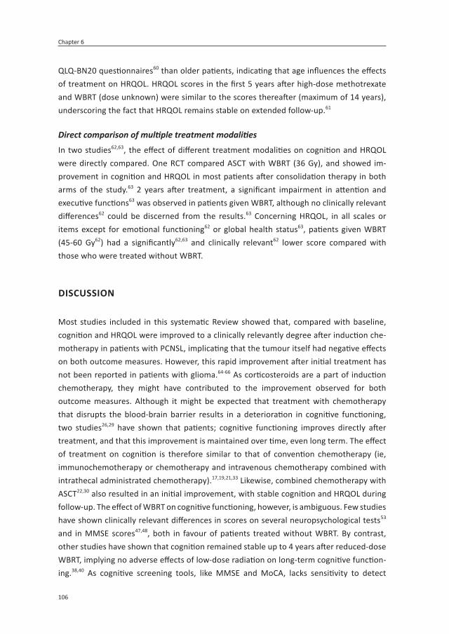
Chapter 6
106
QLQ-BN20 questionnaires60 than older patients, indicating that age influences the effects of treatment on HRQOL. HRQOL scores in the first 5 years after high-dose methotrexate and WBRT (dose unknown) were similar to the scores thereafter (maximum of 14 years), underscoring the fact that HRQOL remains stable on extended follow-up.61
Direct comparison of multiple treatment modalities
In two studies62,63, the effect of different treatment modalities on cognition and HRQOL were directly compared. One RCT compared ASCT with WBRT (36 Gy), and showed im-provement in cognition and HRQOL in most patients after consolidation therapy in both arms of the study.63 2 years after treatment, a significant impairment in attention and executive functions63 was observed in patients given WBRT, although no clinically relevant differences62 could be discerned from the results.63 Concerning HRQOL, in all scales or items except for emotional functioning62 or global health status63, patients given WBRT (45-60 Gy62) had a significantly62,63 and clinically relevant62 lower score compared with those who were treated without WBRT.
diSCuSSioN
Most studies included in this systematic Review showed that, compared with baseline, cognition and HRQOL were improved to a clinically relevantly degree after induction che-motherapy in patients with PCNSL, implicating that the tumour itself had negative effects on both outcome measures. However, this rapid improvement after initial treatment has not been reported in patients with glioma.64-66 As corticosteroids are a part of induction chemotherapy, they might have contributed to the improvement observed for both outcome measures. Although it might be expected that treatment with chemotherapy that disrupts the blood-brain barrier results in a deterioration in cognitive functioning, two studies26,29 have shown that patients; cognitive functioning improves directly after treatment, and that this improvement is maintained over time, even long term. The effect of treatment on cognition is therefore similar to that of convention chemotherapy (ie, immunochemotherapy or chemotherapy and intravenous chemotherapy combined with intrathecal administrated chemotherapy).17,19,21,33 Likewise, combined chemotherapy with ASCT22,30 also resulted in an initial improvement, with stable cognition and HRQOL during follow-up. The effect of WBRT on cognitive functioning, however, is ambiguous. Few studies have shown clinically relevant differences in scores on several neuropsychological tests53 and in MMSE scores47,48, both in favour of patients treated without WBRT. By contrast, other studies have shown that cognition remained stable up to 4 years after reduced-dose WBRT, implying no adverse effects of low-dose radiation on long-term cognitive function-ing.38,40 As cognitive screening tools, like MMSE and MoCA, lacks sensitivity to detect

107
Cognitive functioning and health-related quality of life in patients with newly diagnosed PCNSL
6
cognitive impairment67 and results should be interpreted with caution, we combined the results of these tests with those from formal neuropsychological evaluation (ie, the gold standard) in case they showed a similar trend. It remains important, however, to note that MMSE scores - either measured cross-sectionally or longitudinally - are likely to provide an underestimation of the incidence of cognitive impairment and changes that may have occurred over time in patients with PCNSL. Thus, results from screening instruments, such as MMSE or MoCA, are most likely represent an underestimation of the patients’ actual level of impairment, limiting the interpretation of the magnitude of the cognitive adverse effects of the various treatment regimens used in PCNSL. Moreover, it is important to emphasise that future clinical studies use appropriate standard measures in this popula-tion, as proposed by the International Primary CNS Lymphoma Collaborative Group, to better evaluate cognition.5
For HRQOL, most studies showed stable HRQoL directly after WBRT, with a further improvement over time in one study44. Only one study showed did a direct comparison bewteen patienst given WBRT and those treatedwith other modalities, which showed a worse score for a small group (n=11) of patients given WBRT compared with those given other modalities (n=60 in total).62 As an example, the global health score was 48 for those treated with chemotherapy followed by radiotherapy, and 62-82 for those treated with other modalities without radiotherapy.62 Although not always clinically relevant, these results do suggest that treatment with radiotherapy results in worse cognitive functioning and HRQOL compared with treatment with chemotherapy only, or in combination with other therapies. This information should be included in clinical decision making, together with information about survival. Although the interpretation of the study is difficult due to a large amount of cross-over between study arms, a prolonged progression-free survival (18 vs 12 months) was achieved in patients given combined chemotherapy and radiother-apy, compared with chemotherapy only;4 however, overall survival did not differ between groups.4,63 Benefits of extended progression-free survival with WBRT and the associated delayed negative effects of recurrent disease on cognition and HRQOL should be weighed against the negative effects of full-dose radiotherapy on cognition and HRQOL. It could also be argued to preclude the addition of full-dose WBRT from first-line therapy, because of its negative impact on cognition and HRQoL without proof of prolonged overall survival. As such, reduced-dose WBRT could be an appropriate alternative. Treatment with ASCT may also be an alternative to WBRT, as progression-free survival outcomes are similar bewteen modalities,63,68 and the effects on cognition are likely to be less detrimental than WBRT. The Effect of ASCT on overall survival, however, remains to be seen.
With respect to patient-related factors that are associated with cognition or HRQOL, age was only described in a few studies. Most elderly patients (80%) had serious cognitive impairment (MDRS, <135) at baseline,17 which improved immediately after treatment and at short-term follow-up (up to 1 year),18,19 and remained stable afterwards.17,21 HRQOL

Chapter 6
108
was also shown to improve after treatment in the elderly population, although this was not clinically relevant.18 Nevertheless, younger patients (<65 years) had statistically signifi-cantly better scores to a clinically relevant degree on several scales and items of the EORTC QLQ-C30 and QLQ-BN20 questionnaires when compared with older patients, indicating that age influences the effects of treatment on HRQOL long-term.61
It has been recognized that both cognitive functioning and HRQOL are important outcome measures in patients with PCNSL.69 However, both outcomes are measured, evaluated and described in many different ways, which make it difficult to perform a meta-analysis and draw conclusions on the effect of different treatment modalities in this patient popula-tion. Moreover, it is difficult to determine the long-term impact of treatments, because of the large drop-out rate of participants on cognitive tests or HRQOL questionnaires over time. In addition, results from the available cross-sectional studies are often hampered by selection bias; those who had neurotoxicity may not have returned for their MMSE or neuropsychological testing appointments during follow-up.21 Another limitation is the level of reporting of the results described in the included studies. Unfortunately, many articles only reported cross-sectional post-treatment results. Without baseline scores it is not possible to determine the net change in cognitive functioning that has occurred over time. Additionally, sometimes statistically significant differences were described, without presenting the actual scores, which meant it was not possible to identify if these differences were also clinically relevant. If raw scores were present, significant differences should not be automatically interpreted to be clinically relevant too.
CoNCLuSioN
The results of the studies included in this Review showed that both the tumour and additional full-dose WBRT can effect cognition and HRQOL in a clinically relevant, and negative, way. It is important that this information is included in clinical decision making, when discussing the benefits and risks of certain treatments. We advise that research in patients with PCNSL address cognition and HRQOL, as recommended by the guideline5 to help clinicians to make a reasoned decision, regarding treatment.
ACkNowLedGmeNtS
There was no funding source for this study. The corresponding author had full access to all the data in the study and had final responsibility for the decision to submit for publication.
The authors would like to thank W.M. Bramer, information specialist Medical Library Erasmus Medical Center, Rotterdam for his help in the electronic search.

109
Cognitive functioning and health-related quality of life in patients with newly diagnosed PCNSL
6
refereNCeS
1. Ostrom QT, Gittleman H, Xu J, et al. CBTRUS Statistical Report: Primary Brain and Other Central Nervous System Tumors Diagnosed in the United States in 2009-2013. Neuro Oncol 2016; 18(suppl_5): v1-v75.
2. Villano JL, Koshy M, Shaikh H, Dolecek TA, McCarthy BJ. Age, gender, and racial differ-ences in incidence and survival in primary CNS lymphoma. Br J Cancer 2011; 105(9): 1414-8.
3. van der Meulen M, Dinmohamed AG, Visser O, Doorduijn JK, Bromberg JEC. Improved survival in primary central nervous system lymphoma up to age 70 only: a population-based study on incidence, primary treat-ment and survival in the Netherlands, 1989-2015. Leukemia 2017; 31(8): 1822-5.
4. Thiel E, Korfel A, Martus P, et al. High-dose methotrexate with or without whole brain radiotherapy for primary CNS lymphoma (G-PCNSL-SG-1): a phase 3, randomised, non-inferiority trial. Lancet Oncol 2010; 11(11): 1036-47.
5. Correa DD, Maron L, Harder H, et al. Cogni-tive functions in primary central nervous system lymphoma: literature review and assessment guidelines. Ann Oncol 2007; 18(7): 1145-51.
6. Boele FW, Zant M, Heine EC, et al. The asso-ciation between cognitive functioning and health-related quality of life in low-grade glioma patients. Neurooncol Pract 2014; 1(2): 40-6.
7. Aaronson NK, Taphoorn MJ, Heimans JJ, et al. Compromised health-related quality of life in patients with low-grade glioma. J Clin Oncol 2011; 29(33): 4430-5.
8. Clinical Outcome Assessment Qualification Program. https://www.fda.gov/Drugs/DevelopmentApprovalProcess/DrugDe-velopmentToolsQualificationProgram/ucm284077.htm (accessed April 16, 2018.
9. Folstein MF, Folstein SE, McHugh PR. “Mini-mental state”. A practical method for
grading the cognitive state of patients for the clinician. J Psychiatr Res 1975; 12(3): 189-98.
10. Nasreddine ZS, Phillips NA, Bedirian V, et al. The Montreal Cognitive Assessment, MoCA: a brief screening tool for mild cognitive impairment. J Am Geriatr Soc 2005; 53(4): 695-9.
11. Moher D, Liberati A, Tetzlaff J, Altman DG, Group P. Preferred reporting items for systematic reviews and meta-analyses: the PRISMA statement. J Clin Epidemiol 2009; 62(10): 1006-12.
12. Crum RM, Anthony JC, Bassett SS, Folstein MF. Population-based norms for the Mini-Mental State Examination by age and educational level. JAMA 1993; 269(18): 2386-91.
13. Lezak MD, Howieson DB, Loring DW. Neuro-psychological Assessment. New York Oxford Univerity Press; 2004.
14. Feeney J, Savva GM, O’Regan C, King-Kalli-manis B, Cronin H, Kenny RA. Measurement Error, Reliability, and Minimum Detectable Change in the Mini-Mental State Examina-tion, Montreal Cognitive Assessment, and Color Trails Test among Community Living Middle-Aged and Older Adults. J Alzheimers Dis 2016; 53(3): 1107-14.
15. Brown PD, Jaeckle K, Ballman KV, et al. Ef-fect of Radiosurgery Alone vs Radiosurgery With Whole Brain Radiation Therapy on Cognitive Function in Patients With 1 to 3 Brain Metastases: A Randomized Clinical Trial. JAMA 2016; 316(4): 401-9.
16. Osoba D, Rodrigues G, Myles J, Zee B, Pater J. Interpreting the significance of changes in health-related quality-of-life scores. . Journal of Clinical Oncology 1998; 16(1): 139-44.
17. Omuro A, Chinot O, Taillandier L, et al. Methotrexate and temozolomide versus methotrexate, procarbazine, vincristine, and cytarabine for primary CNS lymphoma

Chapter 6
110
in an elderly population: An intergroup ANOCEF-GOELAMS randomised phase 2 trial. Lancet Haematol 2015; 2(6): e251-e9.
18. Fritsch K, Kasenda B, Schorb E, et al. High-dose methotrexate-based immuno-chemo-therapy for elderly primary CNS lymphoma patients (PRIMAIN study). Leukemia 2017; 31(4): 846-52.
19. Hoang-Xuan K, Taillandier L, Chinot O, et al. Chemotherapy alone as initial treatment for primary CNS lymphoma in patients older than 60 years: A multicenter phase II study (26952) of the European Organization for Research and Treatment of Cancer Brain Tumor Group. J Clin Oncol 2003; 21(14): 2726-31.
20. Laack NN, Ballman KV, Brown PB, O’Neill BP. Whole-brain radiotherapy and high-dose methylprednisolone for elderly patients with primary central nervous system lymphoma: Results of North Central Can-cer Treatment Group (NCCTG) 96-73-51. International Journal of Radiation Oncology Biology Physics 2006; 65(5): 1429-39.
21. Batchelor T, Carson K, O’Neill A, et al. Treatment of primary CNS lymphoma with methotrexate and deferred radiotherapy: a report of NABTT 96-07. J Clin Oncol 2003; 21(6): 1044-9.
22. Omuro A, Correa DD, DeAngelis LM, et al. R-MPV followed by high-dose chemo-therapy with TBC and autologous stem-cell transplant for newly diagnosed primary CNS lymphoma. Blood 2015; 125(9): 1403-10.
23. S M. Mental status examination for organic mental syndrome in the elderly patients. Geriatric Psychiatry. New York: Grune and Stratton; 1976: 77-121.
24. O’Neill BP, Wang CH, O’Fallon JR, et al. The consequences of treatment and disease in patients with primary CNS non-Hodgkin’s lymphoma: cognitive function and per-formance status. North Central Cancer Treatment Group. Neuro-oncol 1999; 1(3): 196-203.
25. McAllister LD, Doolittle ND, Guastadis-egni PE, et al. Cognitive outcomes and long-term follow-up results after enhanced chemotherapy delivery for primary central nervous system lymphoma. Neurosurgery 2000; 46(1): 51-61.
26. Neuwelt EA, Goldman DL, Dahlborg SA, et al. Primary CNS lymphoma treated with osmotic blood-brain barrier disruption: Prolonged survival and preservation of cognitive function. J CLIN ONCOL 1991; 9(9): 1580-90.
27. Dahlborg SA, Henner WD, Crossen JR, et al. Non-AIDS primary CNS lymphoma: First example of a durable response in a primary brain tumor using enhanced chemotherapy delivery without cognitive loss and without radiotherapy. Cancer J 1996; 2(3): 166-74.
28. Neuwelt EA, Guastadisegni PE, Várallyay P, Doolittle ND. Imaging changes and cognitive outcome in primary CNS lymphoma after enhanced chemotherapy delivery. Am J Neuroradiol 2005; 26(2): 258-65.
29. Doolittle ND, Dósa E, Fu R, et al. Preserva-tion of cognitive function in primary CNS lymphoma survivors a median of 12 years after enhanced chemotherapy delivery. J Clin Oncol 2013; 31(31): 4026-7.
30. Abrey LE, Moskowitz CH, Mason WP, et al. Intensive methotrexate and cytarabine followed by high-dose chemotherapy with autologous stem-cell rescue in patients with newly diagnosed primary CNS lym-phoma: An intent-to-treat analysis. J Clin Oncol 2003; 21(22): 4151-6.
31. Illerhaus G, Müller F, Feuerhake F, Schäfer AO, Ostertag C, Finke J. High-dose che-motherapy and autologous stem-cell transplantation without consolidating radiotherapy as first-line treatment for primary lymphoma of the central nervous system. Haematologica 2008; 93(1): 147-8.
32. Illerhaus G, Kasenda B, Ihorst G, et al. High-dose chemotherapy with autologous haemopoietic stem cell transplantation for newly diagnosed primary CNS lymphoma:

111
Cognitive functioning and health-related quality of life in patients with newly diagnosed PCNSL
6
a prospective, single-arm, phase 2 trial. Lancet Haematol 2016; 3(8): e388-e97.
33. Juergens A, Pels H, Rogowski S, et al. Long-term survival with favorable cognitive outcome after chemotherapy in primary central nervous system lymphoma. Ann Neurol 2010; 67(2): 182-9.
34. Schlegel U, Pels H, Glasmacher A, et al. Combined systemic and intraventricular chemotherapy in primary CNS lymphoma: A pilot study. J Neurol Neurosurg Psychiatry 2001; 71(1): 118-22.
35. Pels H, Schmidt-Wolf IGH, Glasmacher A, et al. Primary central nervous system lymphoma: Results of a pilot and phase II study of systemic and intraventricular chemotherapy with deferred radiotherapy. J Clin Oncol 2003; 21(24): 4489-95.
36. Fliessbach K, Helmstaedter C, Urbach H, et al. Neuropsychological outcome after chemotherapy for primary CNS lymphoma: A prospective study. Neurology 2005; 64(7): 1184-8.
37. Fliessbach K, Urbach H, Helmstaedter C, et al. Cognitive performance and magnetic resonance imaging findings after high-dose systemic and intraventricular chemo-therapy for primary central nervous system lymphoma. Arch Neurol 2003; 60(4): 563-8.
38. Morris PG, Correa DD, Yahalom J, et al. Rituximab, methotrexate, procarbazine, and vincristine followed by consolidation reduced-dose whole-brain radiotherapy and cytarabine in newly diagnosed primary CNS lymphoma: Final results and long-term outcome. J Clin Oncol 2013; 31(31): 3971-9.
39. Shah GD, Yahalom J, Correa DD, et al. Com-bined immunochemotherapy with reduced whole-brain radiotherapy for newly diag-nosed primary CNS lymphoma. J Clin Oncol 2007; 25(30): 4730-5.
40. Correa DD, Rocco-Donovan M, DeAngelis LM, et al. Prospective cognitive follow-up in primary CNS lymphoma patients treated with chemotherapy and reduced-dose
radiotherapy. J Neuro-Oncol 2009; 91(3): 315-21.
41. Yamanaka R, Shinbo Y, Sano M, et al. Salvage therapy and late neurotoxicity in patients with recurrent primary CNS lymphoma treated with a modified ProMACE-MOPP hybrid regimen. Leuk Lymphoma 2007; 48(6): 1119-26.
42. Kaburaki T, Taoka K, Matsuda J, et al. Combined intravitreal methotrexate and immunochemotherapy followed by reduced-dose whole-brain radiotherapy for newly diagnosed B-cell primary intraocular lymphoma. Br J Haematol 2017; 179(2): 246-55.
43. Laack NN, O’Neill BP, Ballman KV, et al. CHOD/BVAM chemotherapy and whole-brain radiotherapy for newly diagnosed primary central nervous system lymphoma. International Journal of Radiation Oncology Biology Physics 2011; 81(2): 476-82.
44. Glass J, Won M, Schultz CJ, et al. Phase I and II study of induction chemotherapy with methotrexate, rituximab, and temo-zolomide, followed by whole-brain radio-therapy and postirradiation temozolomide for primary CNS lymphoma: NRG oncology RTOG 0227. J Clin Oncol 2016; 34(14): 1620-5.
45. DeAngelis LM, Seiferheld W, Clifford Schold S, Fisher B, Schultz CJ. Combination che-motherapy and radiotherapy for primary central nervous system lymphoma: Radia-tion Therapy Oncology Group study 93-10. J Clin Oncol 2002; 20(24): 4643-8.
46. Fisher B, Seiferheld W, Schultz C, et al. Secondary analysis of radiation therapy oncology group study (RTOG) 9310: An intergroup phase II combined modality treatment of primary central nervous sys-tem lymphoma. J Neuro-Oncol 2005; 74(2): 201-5.
47. Ichikawa T, Kurozumi K, Michiue H, et al. Reduced neurotoxicity with combined treatment of high-dose methotrexate, cyclophosphamide, doxorubicin, vincristine

Chapter 6
112
and prednisolone (M-CHOP) and deferred radiotherapy for primary central nervous system lymphoma. Clinical Neurology and Neurosurgery 2014; 127((Ichikawa T., [email protected]; Kurozumi K.; Ishida J.; Kawasaki A.; Date I.) Depart-ment of Neurological Surgery, Okayama University, Graduate School of Medicine, Dentistry, and Pharmaceutical Sciences, Kita-ku, Okayama, Japan): 106-11.
48. Herrlinger U, Schäfer N, Fimmers R, et al. Early whole brain radiotherapy in primary CNS lymphoma: negative impact on quality of life in the randomized G-PCNSL-SG1 trial. J Cancer Res Clin Oncol 2017: 1-7.
49. Ferreri AJ, Reni M, Foppoli M, et al. High-dose cytarabine plus high-dose methotrex-ate versus high-dose methotrexate alone in patients with primary CNS lymphoma: a randomised phase 2 trial. Lancet 2009; 374(9700): 1512-20.
50. Chanswangphuwana C, Rojnuckarin P, Cherdchoo N, Raiyawa T, Uaprasert N. Balancing relapses versus cognitive impair-ment in primary central nervous system lymphoma: a single-center experience. Hematology 2017: 1-6.
51. Ferreri AJM, Verona C, Politi LS, et al. Con-solidation radiotherapy in primary central nervous system lymphomas: Impact on outcome of different fields and doses in pa-tients in complete remission after upfront chemotherapy. Int J Radiat Oncol Biol Phys 2011; 80(1): 169-75.
52. Ferreri AJM, Ciceri F, Brandes AA, et al. MATILDE chemotherapy regimen for primary CNS lymphoma: Results at a me-dian follow-up of 12 years. Neurology 2014; 82(15): 1370-3.
53. Correa DD, Shi W, Abrey LE, et al. Cognitive functions in primary CNS lymphoma after single or combined modality regimens. Neuro-Oncology 2012; 14(1): 101-8.
54. Correa DD, DeAngelis LM, Shi W, Thaler H, Glass A, Abrey LE. Cognitive functions in
survivors of primary central nervous system lymphoma. Neurology 2004; 62(4): 548-55.
55. Harder H, Holtel H, Bromberg JEC, et al. Cognitive status and quality of life after treatment for primary CNS lymphoma. Neurology 2004; 62(4): 544-7.
56. Guha-Thakurta N, Damek D, Pollack C, Hochberg FH. Intravenous methotrexate as initial treatment for primary central nervous system lymphoma: Response to therapy and quality of life of patients. J Neuro-Oncol 1999; 43(3): 259-68.
57. Aaronson NK, Ahmedzai S, Bergman B, et al. The European Organization for Research and Treatment of Cancer QLQ-C30: a quali-ty-of-life instrument for use in international clinical trials in oncology. J Natl Cancer Inst 1993; 85(5): 365-76.
58. Mackworth N, Fobair P, Prados MD. Quality of life self-reports from 200 brain tumor patients: comparisons with Karnofsky per-formance scores. J Neurooncol 1992; 14(3): 243-53.
59. Spitzer WO, Dobson AJ, Hall J, et al. Measur-ing the quality of life of cancer patients: a concise QL-index for use by physicians. J Chronic Dis 1981; 34(12): 585-97.
60. Osoba D, Aaronson NK, Muller M, et al. Effect of neurological dysfunction on health-related quality of life in patients with high-grade glioma. J Neurooncol 1997; 34(3): 263-78.
61. Okita Y, Narita Y, Miyakita Y, et al. Health-related quality of life in outpatients with primary central nervous system lymphoma after radiotherapy and high-dose metho-trexate chemotherapy. Mol Clin Oncol 2016; 5(3): 179-85.
62. Doolittle ND, Korfel A, Lubow MA, et al. Long-term cognitive function, neuroimag-ing, and quality of life in primary CNS lymphoma. Neurology 2013; 81(1): 84-92.
63. Ferreri AJM, Cwynarski K, Pulczynski E, et al. Whole-brain radiotherapy or autologous stem-cell transplantation as consolidation strategies after high-dose methotrexate-

113
Cognitive functioning and health-related quality of life in patients with newly diagnosed PCNSL
6
based chemoimmunotherapy in patients with primary CNS lymphoma: results of the second randomisation of the International Extranodal Lymphoma Study Group-32 phase 2 trial. Lancet Haematol 2017; 4(11): e510-e23.
64. Habets EJ, Kloet A, Walchenbach R, Vecht CJ, Klein M, Taphoorn MJ. Tumour and surgery effects on cognitive functioning in high-grade glioma patients. Acta Neurochir (Wien) 2014; 156(8): 1451-9.
65. Satoer D, Vork J, Visch-Brink E, Smits M, Dirven C, Vincent A. Cognitive functioning early after surgery of gliomas in eloquent areas. J Neurosurg 2012; 117(5): 831-8.
66. Hilverda K, Bosma I, Heimans JJ, et al. Cog-nitive functioning in glioblastoma patients during radiotherapy and temozolomide treatment: initial findings. J Neurooncol 2010; 97(1): 89-94.
67. Meyers CA, Wefel JS. The use of the mini-mental state examination to assess cognitive functioning in cancer trials: no ifs, ands, buts, or sensitivity. J Clin Oncol 2003; 21(19): 3557-8.
68. Houillier C, Taillandier L, Lamy T, et al. Whole Brain Radiotherapy (WBRT) Versus Intensive Chemotherapy with Haemato-poietic Stem Cell Rescue (IC + HCR) for Primary Central Nervous System Lymphoma (PCNSL) in Young Patients: An Intergroup Anocef-Goelams Randomized Phase II Trial (PRECIS). 2016: American Society of Hema-tology; 2016. p. 1.
69. Abrey LE, Batchelor TT, Ferreri AJ, et al. Report of an international workshop to standardize baseline evaluation and re-sponse criteria for primary CNS lymphoma. J Clin Oncol 2005; 23(22): 5034-43.
Chapter 6
114
SuPPLemeNtAry dAtA
Content:
Search strategy in Medline ovid (Pubmed)
Table: study characteristics of all included articles
References
SEArCH STrATEgy IN MEDlINE OvID (PubMED)
((Lymphoma/ AND (exp “Central Nervous System”/)) OR “Intraocular Lymphoma”/ OR ((primar* ADJ6 (“central nervous system” OR CNS OR intraocular OR intra-ocular OR brain* OR cerebral* OR spine OR spinal) ADJ3 lymphoma*) OR PCNSL OR (Primar* ADJ6 CNSL)).ab,ti.) AND (“quality of life”/ OR “Health Status”/ OR “health status indicators”/ OR exp “fatigue”/ OR “depression”/ OR exp “Depressive Disorder”/ OR exp “emotions”/ OR “So-cial Environment”/ OR “Social Isolation”/ OR “Social Alienation”/ OR “Social Perception”/ OR “Social Participation”/ OR “Social Learning”/ OR “Emotional Adjustment”/ OR psychol-ogy.xs. OR exp “Psychological Tests”/ OR “Adaptation, Psychological”/ OR “self report”/ OR “Symptom Assessment”/ OR “Sexual Dysfunctions, Psychological”/ OR sexuality/ OR “Sexual Behavior”/ OR exp “cognition”/ OR “learning”/ OR exp Memory/ OR “Language Tests”/ OR “Cognition Disorders”/ OR “Neuropsychiatry”/ OR “Outcome Assessment (Health Care)”/ OR Neuropsychology/ OR “Mental Health”/ OR ((quality ADJ3 life) OR hrql OR hrqol OR qol OR ((health OR function* OR patient* OR mental* OR perform*) ADJ3 (status* OR outcome* OR assess*)) OR “mental health” OR (outcome* ADJ3 assess*) OR fatigue OR depress* OR emotion* OR anxi* OR fear OR social* OR psychosocial* OR stress OR distress* OR psycholog* OR neuropsycholog* OR well-being OR wellbeing OR coping OR ((self OR patient*) ADJ report*) OR burden OR (symptom* ADJ3 assess*) OR sexual* OR functioning* OR cognit* OR learning OR neurocogniti* OR memory OR neuropsych* OR language* OR reading* OR pro OR pros OR attention* OR Neurobehav* OR (Executive* ADJ3 function*) OR Aggressi*).ab,ti.) AND english.la. NOT ((exp child/ OR exp infant/) NOT exp adult/) NOT (“case reports”/ OR (“case report*”).ti.)

115
Cognitive functioning and health-related quality of life in patients with newly diagnosed PCNSL
6
tABL
e: S
tud
y Ch
ArA
Cter
iSti
CS o
f A
LL iN
CLu
ded
Art
iCLe
S
Artic
len
test
ed/
n PC
NSL
med
ian
age;
Perc
enta
ge
olde
r pati
ents
(if
avai
labl
e)
Perf
orm
ance
Sc
ore
(ran
ge)
desi
gnCo
gniti
on/
hrQ
oL/
Both
Prim
ary,
seco
ndar
y ai
m
imm
unoc
hem
othe
rapy
or c
hem
othe
rapy
Guha
-Tha
kurt
a,
1999
5711
/31
63 (3
5-87
)Te
sted
:56
(46-
79)
KPS
40(1
0-80
)Si
ngle
cen
ter p
rosp
ectiv
e Co
hort
HRQ
oLPr
imar
y: R
adio
logi
cal r
espo
nse,
PFS
, ove
rall
surv
ival
and
HRQ
oL in
pati
ents
with
PCN
SL w
ho re
ceiv
ed h
igh-
dose
M
TX.
Batc
helo
r,20
0339
19/2
5M
ean
age
60
(SD
12)
KPS
80M
ean
78M
ultic
ente
r, sin
gle-
agen
t, pr
ospe
ctive
coh
ort,
phas
e II
Cogn
ition
Prim
ary:
radi
ogra
phic
resp
onse
.Se
cond
ary:
surv
ival
and
toxi
city
and
cog
nitio
n
Hoan
g-Xu
an, 2
00340
38/5
072
(60-
81);
KPS
50(4
0-10
0)Pr
ospe
ctive
coh
ort,
phas
e II
Cogn
ition
Prim
ary:
effi
cacy
(res
pons
e to
ther
apy,
OS)
and
toxi
city
, in
clud
ing
cogn
ition
, in
olde
r pati
ents
Om
uro,
2015
1771
/95
72 (6
0-85
)KP
S 70
(40-
100)
Pros
pecti
ve ra
ndom
ized
cont
rolle
d tr
ial,
phas
e II
Both
Prim
ary:
1 y
ear P
FS a
fter t
wo
diffe
rent
MTX
bas
ed
chem
othe
rapy
regi
men
s.Se
cond
ary:
cog
nitio
n an
d HR
QoL
Frits
ch, 2
01741
74-9
3/ 1
12;
HRQ
oL 7
473
(66-
85)
KPS
70(3
0-10
0)Pr
ospe
ctive
coh
ort
Both
Prim
ary:
CR
and
PFS
with
R-M
PL in
eld
erly
with
PCN
SLSe
cond
ary:
cog
nitio
n an
d HR
QoL
Chem
othe
rapy
with
blo
od b
rain
bar
rier d
isru
ption
(bbb
D)
Neu
wel
t, 19
9118
12/3
0M
ean
age
WBR
T +
BBBD
42.
5 ye
ars v
ersu
sBB
BD (±
WBR
T)
53.7
yea
rs
Mea
n KP
SW
BRT+
BBBD
:73
.1Ve
rsus
BBBD
(±
WBR
T): 7
7.4
Pros
pecti
ve, n
on-
rand
omize
d co
mpa
ring
coho
rt st
udy
Cogn
ition
Prim
ary:
Res
pons
e an
d su
rviv
al o
f BBB
D al
one,
bef
ore
or
after
radi
ation
Seco
ndar
y: c
ogni
tion
Dahl
borg
, 199
61923
/58
56 (5
-71)
;34
% >
60 y
ears
KPS
80(4
0-10
0)Pr
ospe
ctive
, non
-ra
ndom
ized
com
parin
g co
hort
Cogn
ition
Prim
ary:
radi
ogra
phic
tum
or re
spon
se a
nd su
rviv
al in
BBB
D ch
emot
hera
py w
ith o
r with
out W
BRT.
Sec
onda
ry: c
ogni
tion

Chapter 6
116
Artic
len
test
ed/
n PC
NSL
med
ian
age;
Perc
enta
ge
olde
r pati
ents
(if
avai
labl
e)
Perf
orm
ance
Sc
ore
(ran
ge)
desi
gnCo
gniti
on/
hrQ
oL/
Both
Prim
ary,
seco
ndar
y ai
m
McA
llist
er, 2
00020
23/7
451
%≥6
0 ye
ars
62%
KPS
>70
Pros
pecti
ve c
ohor
tCo
gniti
onPr
imar
y: 5
-yea
r ove
rall
surv
ival
and
cog
nitiv
e ou
tcom
e
Neu
wel
t, 20
0521
16/1
647
(10-
68)
NA
Pros
pecti
ve C
ohor
tCo
gniti
onPr
imar
y: re
latio
n be
twee
n im
agin
g ch
ange
s and
cog
nitio
n aft
er a
chie
ving
CR
Dool
ittle
, 201
32224
/26
50 (r
ange
NA)
;8
(33%
) ≥60
ye
ars
29%
KPS
<70
Pros
pecti
ve c
ohor
tCo
gniti
onPr
imar
y: C
ogni
tive
func
tioni
ng in
long
term
follo
w u
p co
hort
trea
ted
with
che
mot
hera
py o
nly.
Chem
othe
rapy
com
bine
d w
ith st
em c
ell t
rans
plan
tatio
n
Abre
y,20
0323
14/2
853
(25-
71)
KPS
70(3
0-10
0)Pr
ospe
ctive
coh
ort
Cogn
ition
Prim
ary:
Fea
sibili
ty o
f MTX
, cyt
arab
ine
(indu
ction
) and
HD-
CT (B
EAM
). Se
cond
ary:
EFS
and
OS,
cog
nitio
n
Iller
haus
,20
0842
10/1
354
(38-
67)
KPS
90(3
0-10
0)Pr
ospe
ctive
coh
ort ,
pilo
tCo
gniti
onPr
imar
y: D
FS a
nd O
S of
che
mot
hera
py a
nd A
SCT,
with
lim
ited
WBR
T, se
cond
ary:
cog
nitio
n
Om
uro,
2015
2416
/ 32
57 (2
3-67
);36
(34%
) >60
ye
ars
KPS
80(4
0-10
0)Pr
ospe
ctive
coh
ort,
phas
e II
Both
Prim
ary:
1 y
ear P
FS a
fter R
-MPV
and
ASC
T.Se
cond
ary:
cog
nitio
n, H
RQoL
Popu
latio
n is
parti
ally
ove
rlapp
ing
with
Cor
rea,
200
9
Iller
haus
, 201
64373
-77/
79;
HRQ
oL 7
756
(51-
62);
25 (3
2%) >
60
year
s
KPS
90(7
0-90
)Pr
ospe
ctive
coh
ort,
phas
e II
Both
Prim
ary:
pro
porti
on a
chie
ving
a C
R 30
day
s afte
r co
mpl
eting
CT-
ASCT
. Sec
onda
ry: c
ogni
tion
and
HRQ
oL
Intr
aven
ous a
nd in
trat
heca
l che
mot
hera
py (p
artia
lly o
verla
ppin
g po
pula
tions
)
Schl
egel
, 200
12510
/20
64 (2
7-71
);10
(50%
) ≥65
ye
ars
KPS
70(3
0-80
)Pr
ospe
ctive
coh
ort,
pilo
tCo
gniti
onPr
imar
y: re
spon
se ra
te, r
espo
nse
dura
tion,
and
toxi
city
aft
er sy
stem
ic a
nd in
trav
entr
icul
ar c
hem
othe
rapy
Pels,
2003
2622
/65;
>60
year
s 35
(54%
)
62 (2
7-75
)Te
sted
:61
(ran
ge N
A)
KPS
70(2
0-90
)Pr
ospe
ctive
coh
ort,
phas
e II
Cogn
ition
Prim
ary:
Res
pons
e ra
te, r
espo
nse
dura
tion,
OS,
and
toxi
city
aft
er sy
stem
ic a
nd in
trav
entr
icul
ar c
hem
othe
rapy
with
de
ferr
ed ra
diot
hera
py

117
Cognitive functioning and health-related quality of life in patients with newly diagnosed PCNSL
6
Artic
len
test
ed/
n PC
NSL
med
ian
age;
Perc
enta
ge
olde
r pati
ents
(if
avai
labl
e)
Perf
orm
ance
Sc
ore
(ran
ge)
desi
gnCo
gniti
on/
hrQ
oL/
Both
Prim
ary,
seco
ndar
y ai
m
Flie
ssba
ch, 2
00327
10/2
060
(27-
67);
50%
> 6
0 ye
ars
NA
Pros
pecti
ve c
ohor
t, pi
lot
Cogn
ition
Prim
ary:
long
itudi
nal c
ogni
tive
perfo
rman
ce w
ith e
xten
ded
follo
w u
p) a
nd re
late
d to
MRI
find
ings
Flie
ssba
ch, 2
00528
23/4
754
(28-
68)
KPS
70(5
0-90
)M
ultic
ente
r pro
spec
tive
coho
rt, p
hase
II.
Both
Prim
ary:
the
impa
ct o
f the
tum
or it
self
and
its tr
eatm
ent
on w
ith H
D-M
TX c
hem
othe
rapy
plu
s it C
T on
long
term
co
gniti
on a
nd H
RQoL
Juer
gens
, 201
02912
-21/
65
HRQ
oL 1
262
(27-
75);
35 (5
4%) >
60
year
s
KPS
70(2
0-90
)Pr
ospe
ctive
coh
ort:
Pilo
t/ p
hase
II (c
ombi
ned)
Both
Prim
ary:
long
-term
OS,
PFS
, HRQ
oL a
nd c
ogni
tive
func
tioni
ng a
fter s
yste
mic
and
intr
aven
tric
ular
ch
emot
hera
py w
ithou
t WBR
T.
Chem
othe
rapy
com
bine
d w
ith w
hole
bra
in ra
diot
hera
py
O’N
eill,
199
94446
/53
60 (R
ange
NA)
Med
ian
ECO
G 2
Pros
pecti
ve c
ohor
t, ph
ase
IICo
gniti
onPr
imar
y: c
ogni
tion
(MM
SE) a
nd p
erfo
rman
ce st
atus
(ECO
G)
durin
g tr
eatm
ent
DeAn
gelis
, 200
24540
/102
57 (r
ange
NA)
;42
% >
60
year
sKP
S 80
(rang
e N
A)Pr
ospe
ctive
coh
ort
Cogn
ition
Prim
ary:
2-y
ear o
vera
ll su
rviv
al. S
econ
dary
: res
pons
e to
th
erap
y pr
ior t
o ra
diot
hera
py a
nd c
ogni
tive
func
tioni
ng
Hard
er, 2
00430
19/2
544
(24-
63);
2 (5
%) >
60 y
ears
Mea
n KP
S 87
(SD
8)Pr
ospe
ctive
coh
ort,
phas
e II
Both
Prim
ary:
cog
nitiv
e st
atus
and
HRQ
oL in
pati
ents
with
PCN
SL
in c
ompl
ete
rem
issio
n co
mpa
red
to p
atien
ts w
ith o
ther
he
mat
olog
ical
mal
igna
ncie
s.
Corr
ea, 2
00431
28/6
260
(36-
85)
NA
Retr
ospe
ctive
coh
ort
Both
Prim
ary:
cog
nitiv
e fu
nctio
ning
and
HRQ
ol in
surv
ivor
s of
PCN
SL
Fish
er,
2005
4640
/102
NA
NA
Pros
pecti
ve, n
on-
rand
omize
d co
mpa
ring
coho
rt, p
hase
II
Cogn
ition
Prim
ary:
surv
ival
bet
wee
n W
BRT
and
frac
tioni
zed
radi
ation
. Lon
g te
rm fo
llow
up
from
pop
ulati
on d
escr
ibed
by
DeA
ngel
is, 2
002.
Sec
onda
ry: c
ogni
tion
Laac
k,20
0647
19/1
976
(70-
83)
Med
ian
ECO
G 1.
5(0
-3)
Pros
pecti
ve c
ohor
tCo
gniti
onPr
imar
y: e
ffica
cy, t
oxic
ity, a
nd su
rviv
al o
f WBR
T tr
eate
d an
d hi
gh-d
ose
met
hylp
redn
isolo
ne tr
eate
d in
eld
erly
pati
ents

Chapter 6
118
Artic
len
test
ed/
n PC
NSL
med
ian
age;
Perc
enta
ge
olde
r pati
ents
(if
avai
labl
e)
Perf
orm
ance
Sc
ore
(ran
ge)
desi
gnCo
gniti
on/
hrQ
oL/
Both
Prim
ary,
seco
ndar
y ai
m
Shah
,20
0732
12/3
057
(30-
76)
KPS
70(5
0-90
)Pr
ospe
ctive
coh
ort
Cogn
ition
Prim
ary:
safe
ty o
f add
ing
ritux
imab
to M
TX c
hem
othe
rapy
an
d effi
cacy
of r
educ
e do
sed
WBR
T aft
er C
R. S
econ
dary
: co
gniti
onPa
rtial
ly o
verla
ppin
g po
pula
tion
with
Mor
ris, 2
013;
Cor
rea,
20
09
Yam
anak
a, 2
00733
13/3
261
(34-
73)
test
ed: m
ean
age
64 (3
4-73
);8
(62%
) >60
ye
ars
KPS
70(ra
nge
NA)
Test
edKP
S 90
(40-
100)
Pros
pecti
ve c
ohor
tCo
gniti
onPr
imar
y: 2
-yea
r and
5-y
ear O
S an
d tu
mor
resp
onse
, with
an
d w
ithou
t sal
vage
ther
apy.
Cog
nitio
n be
fore
and
afte
r tr
eatm
ent d
escr
ibed
sepa
rate
ly fo
r pati
ents
with
out a
re
laps
e.
Corr
ea, 2
00934
12/1
958
(47-
76)
NA
Pros
pecti
ve c
ohor
tBo
thPr
imar
y: si
de-s
tudy
of a
pha
se II
clin
ical
tria
l: eff
ect o
f ad
ditio
nal r
ituxi
mab
to st
anda
rd tr
eatm
ent.
Seco
ndar
y:
cogn
ition
and
HRQ
oLPa
rtial
ly o
verla
ppin
g po
pula
tion
with
Sha
h, 2
007;
Mor
ris,
2013
Ferr
eri,
2009
4859
/79
MTX
:58
(27-
72) v
ersu
sM
TX+A
ra-C
:59
(25-
74)
ECO
G >1
MTX
: 50%
ve
rsus
MTX
+Ara
-c:
26%
Mul
ticen
ter p
rosp
ectiv
e,
rand
omize
d co
ntro
lled
tria
l, ph
ase
II
Cogn
ition
Prim
ary:
com
plet
e re
miss
ion
rate
afte
r che
mot
hera
pyM
TX +
WBR
T or
MTX
-cyt
arab
ine
+ W
BRT.
Seco
ndar
y: c
ogni
tion
Parti
ally
ove
rlapp
ing
popu
latio
n w
ith F
erre
ri, 2
011
and
2014
Ferr
eri,
2011
4917
/33
55 (2
6-73
);13
(39%
) >70
ye
ars o
ld
ECO
G 0-
1: 9
1%Re
tros
pecti
ve c
ohor
tCo
gniti
onPr
imar
y: th
e im
pact
on
outc
ome
and
neur
olog
ical
pe
rfor
man
ce o
f diff
eren
t rad
iatio
n fie
lds.
Sec
onda
ry:
cogn
ition
Parti
ally
ove
rlapp
ing
popu
latio
n w
ith F
erre
ri, 2
009
and
2014
Laac
k, 2
01150
34/3
661
(34-
69)
ECO
G 0-
1: 8
3%Pr
ospe
ctive
coh
ort,
phas
e II
Cogn
ition
Prim
ary:
effe
ct o
f tre
atm
ent i
n ra
diol
ogic
al re
spon
se,
toxi
city
and
surv
ival
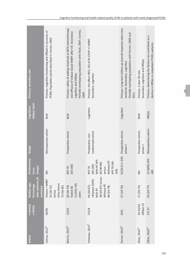
119
Cognitive functioning and health-related quality of life in patients with newly diagnosed PCNSL
6
Artic
len
test
ed/
n PC
NSL
med
ian
age;
Perc
enta
ge
olde
r pati
ents
(if
avai
labl
e)
Perf
orm
ance
Sc
ore
(ran
ge)
desi
gnCo
gniti
on/
hrQ
oL/
Both
Prim
ary,
seco
ndar
y ai
m
Corr
ea, 2
01235
50/9
5Ch
emo
+ W
BRT
52 (3
6-72
)ve
rsus
Onl
y ch
emo
73 (5
6-85
)
NA
Retr
ospe
ctive
coh
ort
Both
Prim
ary:
Cog
nitiv
e fu
nctio
ning
and
HRQ
ol in
surv
ivor
s of
PCN
SL P
opul
ation
par
tly d
escr
ibed
in C
orre
a, 2
004
Mor
ris, 2
01336
12/5
260
(30-
79)
Test
ed 5
83
(25%
) >60
ye
ars
KPS
70(5
0-10
0)Pr
ospe
ctive
coh
ort
Both
Prim
ary:
safe
ty o
f add
ing
ritux
imab
to M
TX c
hem
othe
rapy
an
d effi
cacy
of r
educ
e do
sed
WBR
T aft
er C
R. S
econ
dary
: co
gniti
on a
nd H
RQoL
Parti
ally
ove
rlapp
ing
popu
latio
n w
ith S
hah,
200
7; C
orre
a,
2009
Ichi
kaw
a, 2
01451
23/2
464
(50-
67);
>65y
ears
(54%
)W
ith R
T58
(50-
67) v
ersu
sW
ithou
t RT
68 (5
3-78
)
KPS
70(4
0-10
0)W
ith R
T KP
S 60
(40-
80)
vers
usW
ithou
t RT
KPS
70 (4
0-10
0)
Pros
pecti
ve, n
on-
rand
omize
d co
hort
Cogn
ition
Prim
ary:
the
effec
t (RF
S, O
S) o
f M-C
HOP
+/-W
BRT.
Seco
ndar
y: c
ogni
tion
Ferr
eri,
2014
529/
4157
(19-
70)
ECO
G 0-
1: 6
3%Pr
ospe
ctive
coh
ort,
phas
e II
Cogn
ition
Prim
ary:
Lon
g-te
rm fo
llow
-up
(ove
rall
resp
onse
rate
) cro
ss
secti
onal
. Sec
onda
ry: c
ogni
tion
Parti
ally
ove
rlapp
ing
popu
latio
n w
ith F
erre
ri, 2
009
and
2011
Glas
s, 2
01653
50-5
2/53
HRQ
oL 5
257
(24-
73)
NA
Pros
pecti
ve c
ohor
t, ph
ase
IIBo
thPr
imar
y: 2
-yea
r OS
rate
Seco
ndar
y: c
ogni
tion
and
HRQ
oL
Oki
ta, 2
01658
37/ 3
763
(33-
77)
22 (6
0%) K
PS
≥80
Retr
ospe
ctive
coh
ort
HRQ
oLPr
imar
y: d
eter
min
ing
the
fact
ors c
an c
ontr
ibut
e to
a
decl
ine
in H
RQoL
in o
utpa
tient
PCN
SL p
atien
ts.

Chapter 6
120
Artic
len
test
ed/
n PC
NSL
med
ian
age;
Perc
enta
ge
olde
r pati
ents
(if
avai
labl
e)
Perf
orm
ance
Sc
ore
(ran
ge)
desi
gnCo
gniti
on/
hrQ
oL/
Both
Prim
ary,
seco
ndar
y ai
m
Herr
linge
r, 20
1754
144/
318
61 (5
3-68
)KP
S 80
(60-
90)
Pros
pecti
ve ra
ndom
ized
cont
rolle
d tr
ial,
phas
e III
Both
Prim
ary:
diff
eren
ces i
n co
gniti
on a
nd H
RQoL
bet
wee
n ch
emot
hera
py a
nd c
hem
othe
rapy
com
bine
d w
ith W
BRT
upfr
ont
Chan
swan
gphu
wan
a 20
1755
10/3
756
(16-
78)
NA
Retr
ospe
ctive
coh
ort
Cogn
ition
Prim
ary:
the
role
of l
ow-d
ose
WBR
T an
d tr
eatm
ent
outc
omes
of M
TX b
ased
che
mot
hera
py. S
econ
dary
: co
gniti
ve im
pairm
ent
Kabu
raki
,20
1756
16/1
763
(43-
72)
NA
Pros
pecti
ve c
ohor
tCo
gniti
onPr
imar
y: th
e effi
cacy
of t
reat
men
t pro
toco
l for
PIO
L to
pr
even
t new
CN
S re
laps
e an
d pr
olon
g O
S by
ear
ly in
itiati
on
of C
NS
prop
hyla
xis w
ith a
com
bina
tion
of M
TX a
nd
rdW
BRT.
Sec
onda
ry: c
ogni
tion
Dire
ct c
ompa
ring
of m
ultip
le tr
eatm
ent m
odal
ities
Dool
ittle
, 201
33780
/80
59 (1
0-78
);35
(44%
) ≥60
ye
ars
KPS
80(2
0-10
0)Pr
ospe
ctive
coh
ort
Both
Prim
ary:
des
crib
e co
gniti
on a
nd H
RQoL
and
cor
rela
te to
ra
diol
ogy
in lo
ng-t
erm
surv
ivor
s who
wer
e tr
eate
d w
ith
high
-dos
e m
etho
trex
ate–
base
d re
gim
ens w
ith o
r with
out
WBR
T
Ferr
eri,
2017
3857
/113
57 (I
QR
51-6
3)17
(14%
) ≥65
ye
ars
ECO
G >1
31 (2
7%)
Rand
omize
d co
ntro
lled
tria
lBo
thPr
imar
y: 2
-yea
r PFS
afte
r sec
ond
rand
omiza
tion
(WBR
T or
AS
CT a
s con
solid
ation
)Se
cond
ary:
cog
nitio
n an
d HR
QoL
Abbr
evia
tions
: Ara
-C C
ytar
abin
e; A
SCT
auto
logo
us st
em c
ell t
rans
plan
tatio
n; B
BBD
bloo
d br
ain
barr
ier d
isrup
tion;
BEA
M c
arm
ustin
e, e
topo
side,
cyt
arab
ine
and
mel
phal
an;
CR c
ompl
ete
resp
onse
; CT
chem
othe
rapy
; DFS
dise
ase
free
sur
viva
l; EC
OG
East
ern
Coop
erati
ve O
ncol
ogy
Grou
p pe
rfor
man
ce s
core
; EFS
eve
nt fr
ee s
urvi
val;
HD h
igh
dose
; HR
QoL
hea
lth r
elat
ed q
ualit
y of
life
; it
intr
athe
cal;
KPS
Karn
ofsk
y pe
rfor
man
ce s
core
; M-C
HOP
Met
hotr
exat
e-cy
clop
hosp
ham
ide,
hyd
rocy
daun
orub
icin
, vin
cristi
ne a
nd
pred
iniso
lone
; MM
SE m
ini-m
enta
l sta
te e
xam
inati
on; M
TX m
etho
trex
ate;
n n
umbe
r of p
atien
ts; N
A no
t ava
ilabl
e; O
S ov
eral
l sur
viva
l; PI
OL
prim
ary
intr
a-oc
ular
lym
phom
a;
PCN
SL p
rimar
y ce
ntra
l ner
vous
sys
tem
lym
phom
a; P
FS p
rogr
essio
n fr
ee s
urvi
val;
RFS
rela
pse
free
sur
viva
l; R-
MPL
ritu
xim
ab –
met
hotr
exat
e, p
roba
rbaz
ine
and
lom
ustin
e;
R-M
PV ri
tuxi
mab
– m
etho
trex
ate,
pro
barb
azin
e an
d vi
ncris
tine;
RT
radi
othe
rapy
; SD
stan
dard
dev
iatio
n; (r
d)W
BRT
(red
uced
dos
e) w
hole
bra
in ra
diot
hera
py.

121
Cognitive functioning and health-related quality of life in patients with newly diagnosed PCNSL
6
refereNCeS
1. Ostrom QT, Gittleman H, Xu J, et al. CBTRUS statistical report: primary brain and other central nervous system tumors diagnosed in the United States in 2009–2013. Neuro Oncol 2016; 18 (suppl 5): v1–75.
2. Villano JL, Koshy M, Shaikh H, et al. Age, gender, and racial differences in incidence and survival in primary CNS lymphoma. Br J Cancer 2011; 105: 1414–18.
3. van der Meulen M, Dinmohamed AG, Visser O. Improved survival in primary central nervous system lymphoma up to age 70 only: a population-based study on incidence, primary treatment and survival in the Netherlands, 1989–2015. Leukemia 2017; 31: 1822–25.
4. Thiel E, Korfel A, Martus P, et al. High-dose methotrexate with or without whole brain radiotherapy for primary CNS lymphoma (G-PCNSL-SG-1): a phase 3, randomised, non-inferiority trial. Lancet Oncol 2010; 11: 1036–47.
5. Correa DD, Maron L, Harder H, et al. Cogni-tive functions in primary central nervous system lymphoma: literature review and assessment guidelines. Ann Oncol 2007; 18: 1145–51.
6. Boele FW, Zant M, Heine EC, et al. The asso-ciation between cognitive functioning and health-related quality of life in low-grade glioma patients. Neurooncol Pract 2014; 1: 40–46.
7. Aaronson NK, Taphoorn MJ, Heimans JJ, et al. Compromised healthrelated quality of life in patients with low-grade glioma. J Clin Oncol 2011; 29: 4430–35.
8. US Food and Drug Administration. Clinical outcome assessment qualification program. https://www.fda.gov/Drugs/Developmen-tApprovalProcess/DrugDevelopmentTool-sQualificationProgram/ucm284077.htm (accessed April 16, 2018).
9. Folstein MF, Folstein SE, McHugh PR. “Mini-mental state”. A practical method for grad-
ing the cognitive state of patients for the clinician. J Psychiatr Res 1975; 12: 189–98.
10. Nasreddine ZS, Phillips NA, Bedirian V, et al. The Montreal Cognitive Assessment, MoCA: a brief screening tool for mild cognitive impairment. J Am Geriatr Soc 2005; 53: 695–99.
11. Moher D, Liberati A, Tetzlaff J, et al. Preferred reporting items for systematic reviews and meta-analyses: the PRISMA statement. J Clin Epidemiol 2009; 62: 1006–12.
12. Crum RM, Anthony JC, Bassett SS, Folstein MF. Population-based norms for the Mini-mental State Examination by age and edu-cational level. JAMA 1993; 269: 2386–91.
13. Lezak MD, Howieson DB, Loring DW. Neuropsychological assessment. New York: Oxford University Press, 2004.
14. Feeney J, Savva GM, O’Regan C, et al. Mea-surement error, reliability, and minimum detectable change in the mini-mental state examination, Montreal cognitive assess-ment, and color trails test among commu-nity living middle-aged and older adults.J Alzheimers Dis 2016; 53: 1107–14.
15. Brown PD, Jaeckle K, Ballman KV, et al. Ef-fect of radiosurgery alone vs radiosurgery with whole brain radiation therapy on cognitive function in patients with 1 to 3 brain metastases: a randomized clinical trial. JAMA 2016; 316: 401–09.
16. Osoba D, Rodrigues G, Myles J, et al. Interpreting the significance of changes in health-related quality-of-life scores. J Clin Oncol 1998; 16: 139–44.
17. Omuro A, Chinot O, Taillandier L, et al. Methotrexate and temozolomide versus methotrexate, procarbazine, vincristine, and cytarabine for primary CNS lymphoma in an elderly population: an intergroup ANOCEF-GOELAMS randomised phase 2 trial. Lancet Haematol 2015; 2: e251–59.

Chapter 6
122
18. Neuwelt EA, Goldman DL, Dahlborg SA, et al. Primary CNS lymphoma treated with osmotic blood-brain barrier disruption: prolonged survival and preservation of cognitive function. J Clin Oncol 1991; 9: 1580–90.
19. Dahlborg SA, Henner WD, Crossen JR, et al. Non-AIDS primary CNS lymphoma: first example of a durable response in a primary brain tumor using enhanced chemotherapy delivery without cognitive loss and without radiotherapy. Cancer J 1996; 2: 166–74.
20. McAllister LD, Doolittle ND, Guastadis-egni PE, et al. Cognitive outcomes and long-term follow-up results after enhanced chemotherapy delivery for primary central nervous system lymphoma. Neurosurgery 2000; 46: 51–61.
21. Neuwelt EA, Guastadisegni PE, Varallyay P, Doolittle ND. Imaging changes and cognitive outcome in primary CNS lymphoma after enhanced chemotherapy delivery. Am J Neuroradiol 2005; 26: 258–65.
22. Doolittle ND, Dosa E, Fu R, et al. Preserva-tion of cognitive function in primary CNS lymphoma survivors a median of 12 years after enhanced chemotherapy delivery. J Clin Oncol 2013; 31: 4026–27.
23. Abrey LE, Moskowitz CH, Mason WP, et al. Intensive methotrexate and cytarabine followed by high-dose chemotherapy with autologous stem-cell rescue in patients with newly diagnosed primary CNS lym-phoma: an intent-to-treat analysis. J Clin Oncol 2003; 21: 4151–56.
24. Omuro A, Correa DD, DeAngelis LM, et al. R-MPV followed by high-dose chemo-therapy with TBC and autologous stem-cell transplant for newly diagnosed primary CNS lymphoma. Blood 2015; 125: 1403–10.
25. Schlegel U, Pels H, Glasmacher A, et al. Combined systemic and intraventricular chemotherapy in primary CNS lymphoma: a pilot study. J Neurol Neurosurg Psychiatry 2001; 71: 118–22.
26. Pels H, Schmidt-Wolf IGH, Glasmacher A, et al. Primary central nervous system lymphoma: results of a pilot and phase II study of systemic and intraventricular chemotherapy with deferred radiotherapy. J Clin Oncol 2003; 21: 4489–95.
27. Fliessbach K, Urbach H, Helmstaedter C, et al. Cognitive performance and magnetic resonance imaging findings after high-dose systemic and intraventricular chemo-therapy for primary central nervous system lymphoma. Arch Neurol 2003; 60: 563–68.
28. Fliessbach K, Helmstaedter C, Urbach H, et al. Neuropsychological outcome after chemotherapy for primary CNS lymphoma: a prospective study. Neurology 2005; 64: 1184–88.
29. Juergens A, Pels H, Rogowski S, et al. Long-term survival with favorable cognitive outcome after chemotherapy in primary central nervous system lymphoma. Ann Neurol 2010; 67: 182–89.
30. Harder H, Holtel H, Bromberg JEC, et al. Cognitive status and quality of life after treatment for primary CNS lymphoma. Neurology 2004; 62: 544–47.
31. Correa DD, DeAngelis LM, Shi W, et al. Cognitive functions in survivors of primary central nervous system lymphoma. Neurol-ogy 2004; 62: 548–55.
32. Shah GD, Yahalom J, Correa DD, et al. Com-bined immunochemotherapy with reduced whole-brain radiotherapy for newly diag-nosed primary CNS lymphoma. J Clin Oncol 2007; 25: 4730–35.
33. Yamanaka R, Shinbo Y, Sano M, et al. Salvage therapy and late neurotoxicity in patients with recurrent primary CNS lymphoma treated with a modified ProMACE-MOPP hybrid regimen. Leuk Lymphoma 2007; 48: 1119–26.
34. Correa DD, Rocco-Donovan M, DeAngelis LM, et al. Prospective cognitive follow-up in primary CNS lymphoma patients treated with chemotherapy and reduced-dose ra-diotherapy. J Neurooncol 2009; 91: 315–21.

123
Cognitive functioning and health-related quality of life in patients with newly diagnosed PCNSL
6
35. Correa DD, Shi W, Abrey LE, et al. Cognitive functions in primary CNS lymphoma after single or combined modality regimens. Neuro Oncol 2012; 14: 101–08.
36. Morris PG, Correa DD, Yahalom J, et al. Rituximab, methotrexate, procarbazine, and vincristine followed by consolidation reduced-dose whole-brain radiotherapy and cytarabine in newly diagnosed primary CNS lymphoma: final results and long-term outcome. J Clin Oncol 2013; 31: 3971–79.
37. Doolittle ND, Korfel A, Lubow MA, et al. Long-term cognitive function, neuroimag-ing, and quality of life in primary CNS lymphoma. Neurology 2013; 81: 84–92.
38. Ferreri AJM, Cwynarski K, Pulczynski E, et al. Whole-brain radiotherapy or autologous stem-cell transplantation as consolidation strategies after high-dose methotrexate-based chemoimmunotherapy in patients with primary CNS lymphoma: results of the second randomisation of the International Extranodal Lymphoma Study Group-32 phase 2 trial. Lancet Haematol 2017; 4: e510–23.
39. Batchelor T, Carson K, O’Neill A, et al. Treatment of primary CNS lymphoma with methotrexate and deferred radiotherapy: a report of NABTT 96-07. J Clin Oncol 2003; 21: 1044–49.
40. Hoang-Xuan K, Taillandier L, Chinot O, et al. Chemotherapy alone as initial treatment for primary CNS lymphoma in patients older than 60 years: a multicenter phase II study (26952) of the European Organization for Research and Treatment of Cancer Brain Tu-mor Group. J Clin Oncol 2003; 21: 2726–31.
41. Fritsch K, Kasenda B, Schorb E, et al. High-dose methotrexate-based immuno-chemo-therapy for elderly primary CNS lymphoma patients (PRIMAIN study). Leukemia 2017; 31: 846–52.
42. Illerhaus G, Muller F, Feuerhake F, et al. High-dose chemotherapy and autologous stem-cell transplantation without consoli-dating radiotherapy as first-line treatment
for primary lymphoma of the central nervous system. Haematologica 2008; 93: 147–48.
43. llerhaus G, Kasenda B, Ihorst G, et al. High-dose chemotherapy with autologous haemopoietic stem cell transplantation for newly diagnosed primary CNS lymphoma: a prospective, single-arm, phase 2 trial. Lancet Haematol 2016; 3: e388–97.
44. O’Neill BP, Wang CH, O’Fallon JR, et al. The consequences of treatment and disease in patients with primary CNS non-Hodgkin’s lymphoma: cognitive function and per-formance status. Neuro Oncol 1999; 1: 196–203.
45. DeAngelis LM, Seiferheld W, Clifford Schold S, et al. Combination chemotherapy and radiotherapy for primary central nervous system lymphoma: Radiation Therapy Oncology Group study 93-10. J Clin Oncol 2002; 20: 4643–48.
46. Fisher B, Seiferheld W, Schultz C, et al. Secondary analysis of radiation therapy oncology group study (RTOG) 9310: An in-tergroup phase II combined modality treat-ment of primary central nervous system lymphoma. J Neurooncol 2005; 74: 201–05.
47. Laack NN, Ballman KV, Brown PB, O’Neill BP. Whole-brain radiotherapy and high-dose methylprednisolone for elderly patients with primary central nervous system lymphoma: results of North Central Cancer Treatment Group (NCCTG) 96-73-51. Int J Radiat Oncol Biol Phys 2006; 65: 1429–39.
48. Ferreri AJ, Reni M, Foppoli M, et al. High-dose cytarabine plus high-dose methotrex-ate versus high-dose methotrexate alone in patients with primary CNS lymphoma: a randomised phase 2 trial. Lancet 2009; 374: 1512–20.
49. Ferreri AJM, Verona C, Politi LS, et al. Con-solidation radiotherapy in primary central nervous system lymphomas: impact on outcome of different fields and doses in pa-tients in complete remission after upfront

Chapter 6
124
chemotherapy. Int J Radiat Oncol Biol Phys 2011; 80: 169–75.
50. Laack NN, O’Neill BP, Ballman KV, et al. CHOD/BVAM chemotherapy and whole-brain radiotherapy for newly diagnosed pri-mary central nervous system lymphoma. Int J Radiat Oncol Biol Phys 2011; 81: 476–82.
51. Ichikawa T, Kurozumi K, Michiue H, et al. Reduced neurotoxicity with combined treatment of high-dose methotrexate, cyclophosphamide, doxorubicin, vincristine and prednisolone (M-CHOP) and deferred radiotherapy for primary central nervous system lymphoma. Clin Neurol Neurosurg 2014; 127: 106–11.
52. Ferreri AJM, Ciceri F, Brandes AA, et al. MATILDE chemotherapy regimen for pri-mary CNS lymphoma: results at a median follow-up of 12 years. Neurology 2014; 82: 1370–73.
53. Glass J, Won M, Schultz CJ, et al. Phase I and II study of induction chemotherapy with methotrexate, rituximab, and temo-zolomide, followed by whole-brain radio-therapy and postirradiation temozolomide for primary CNS lymphoma: NRG oncology RTOG 0227. J Clin Oncol 2016; 34: 1620–25.
54. Herrlinger U, Schafer N, Fimmers R, et al. Early whole brain radiotherapy in primary CNS lymphoma: negative impact on quality of life in the randomized G-PCNSL-SG1 trial. J Cancer Res Clin Oncol 2017; 143: 1815–21.
55. Chanswangphuwana C, Rojnuckarin P, Cherdchoo N, et al. Balancing relapses versus cognitive impairment in primary central nervous system lymphoma: a single-center experience. Hematol-ogy 2017; published online Dec 19. DOI: 10.1080/10245332.2017.1418610.
56. Kaburaki T, Taoka K, Matsuda J, et al. Com-bined intravitreal methotrexate and immu-nochemotherapy followed by reduced-dose whole-brain radiotherapy for newly diag-nosed B-cell primary intraocular lymphoma. Br J Haematol 2017; 179: 246–55.
57. Guha-Thakurta N, Damek D, Pollack C, Hochberg FH. Intravenous methotrexate as initial treatment for primary central nervous system lymphoma: response to therapy and quality of life of patients. J Neurooncol 1999; 43: 259–68.
58. Okita Y, Narita Y, Miyakita Y, et al. Health-related quality of life in outpatients with primary central nervous system lymphoma after radiotherapy and high-dose metho-trexate chemotherapy. Mol Clin Oncol 2016; 5: 179–85.
59. Mattis, S. Mental status examination for organic mental syndrome in the elderly patients. Geriatric Psychiatry. New York: Grune and Stratton; 1976: 77–121.
60. Aaronson NK, Ahmedzai S, Bergman B, et al. The European Organization for Research and Treatment of Cancer QLQ-C30: a quali-ty-of-life instrument for use in international clinical trials in oncology. J Natl Cancer Inst 1993; 85: 365–76.
61. Mackworth N, Fobair P, Prados MD. Quality of life self-reports from 200 brain tumor patients: comparisons with Karnofsky performance scores. J Neurooncol 1992; 14: 243–53.
62. Spitzer WO, Dobson AJ, Hall J, et al. Measur-ing the quality of life of cancer patients: a concise QL-index for use by physicians. J Chronic Dis 1981; 34: 585–97.
63. Osoba D, Aaronson NK, Muller M, et al. Effect of neurological dysfunction on health-related quality of life in patients with high-grade glioma. J Neurooncol 1997; 34: 263–78.
64. Habets EJ, Kloet A, Walchenbach R, et al. Tumour and surgery effects on cognitive functioning in high-grade glioma patients. Acta Neurochir (Wien) 2014; 156: 1451–59.
65. Satoer D, Vork J, Visch-Brink E, et al. Cognitive functioning early after surgery of gliomas in eloquent areas. J Neurosurg 2012; 117: 831–38.
66. Hilverda K, Bosma I, Heimans JJ, et al. Cog-nitive functioning in glioblastoma patients

125
Cognitive functioning and health-related quality of life in patients with newly diagnosed PCNSL
6
during radiotherapy and temozolomide treatment: initial findings. J Neurooncol 2010; 97: 89–94.
67. Meyers CA, Wefel JS. The use of the mini-mental state examination to assess cogni-tive functioning in cancer trials: no ifs, ands, buts, or sensitivity. J Clin Oncol 2003; 21: 3557–58.
68. Houillier C, Taillandier L, Lamy T, et al. Whole brain radiotherapy (WBRT) versus intensive chemotherapy with haematopoi-etic stem cell rescue (IC + HCR) for primary central nervous system lymphoma (PCNSL) in young patients: an intergroup Anocef-Goelams randomized phase II trial (PRECIS). Blood 2016; 128: 782.
69. Abrey LE, Batchelor TT, Ferreri AJ, et al. Report of an international workshop to standardize baseline evaluation and re-sponse criteria for primary CNS lymphoma. J Clin Oncol 2005; 23: 5034–43.


7Neurocognitive functioning and radiological changes in
primary CNS lymphoma: results from a RCT
Matthijs van der Meulen MD1, Linda Dirven PhD2,3, Esther J.J. Habets, PhD3,4, Katerina Bakunina MSc5, Marion Smits, MD, PhD6, Hakim C. Achterberg PhD6, Tatjana Seute MD, PhD7,
Gavin Cull MD8,9, Harry Schouten MD10, Josée M. Zijlstra MD, PhD11, Dieta Brandsma MD12, Roelien H. Enting MD, PhD13, Max Beijert MD14, Martin J.B. Taphoorn MD, PhD2,3, Martin J. van den Bent1 MD, PhD, Samar Issa MD15, Jeanette K. Doorduijn MD, PhD16, Jacoline E.C. Bromberg
MD, PhD1
1 Erasmus MC Cancer Institute, Brain Tumor Center, University Medical Center Rotterdam, Department of Neuro-Oncology, the Netherlands;
2 Leiden University Medical Center, Leiden, Department of Neurology;3 Haaglanden Medical Center, The Hague, Department of Neurology;
4 Haaglanden Medical Center, The Hague, Department of Medical Psychology;5 HOVON Data Center, Department of Hematology, Erasmus MC Cancer Institute, Rotterdam, Netherlands;
6 Department of Radiology and Nuclear Medicine, Erasmus MC, University Medical Center, Rotterdam, The Netherlands;7 Department of Neurology, University Medical Center Utrecht, Utrecht, Netherlands;
8 Haematology Department, Sir Charles Gairdner Hospital and PathWest Laboratory Medicine, Nedlands, WA, Australia;9 University of Western Australia, Crawley, WA, Australia;
10 Department of Hematology, University Medical Center, Maastricht, Netherlands;11 Department of Hematology, Amsterdam UMC-Vrije Universiteit Amsterdam, Cancer Center Amsterdam, Netherlands;
12 Department of Neuro-oncology, Netherlands Cancer Institute-Antoni van Leeuwenhoek, Amsterdam, Netherlands;13 Department of Neurology, University of Groningen, University Medical Center Groningen, Groningen, Netherlands;
14 Department of Radiotherapy, University Medical Center Groningen, Groningen, Netherlands;15 Department of Hematology, Middlemore Hospital, Auckland, New Zealand;
16 Erasmus MC Cancer Institute, University Medical Center Rotterdam, Department of Hematology
Accepted in Neuro-Oncology

Chapter 7
128
ABStrACt
Objective: To analyze the effect of treatment on neurocognitive functioning and the as-sociation of neurocognition with radiological abnormalities in primary central nervous system lymphoma (PCNSL).methods: 199 patients from a phase III trial (HOVON 105/ALLG NHL 24), randomized to standard chemotherapy with or without rituximab, followed in patients ≤60 years-old by 30Gy WBRT, were asked to participate in a neuropsychological evaluation before and during treatment, and up to 2 years post-treatment. Scores were transformed into a standardized z-score; clinically relevant changes were defined as a change in z-score of ≥1 standard deviation. The effect of WBRT was analyzed in irradiated patients. All MRIs were centrally assessed for white matter abnormalities and cerebral atrophy, and their relation with neurocognitive scores over time in each domain was calculated.results: 125/199 patients consented to neurocognitive evaluation. Statistically significant improvements in neurocognition were seen in all domains. A clinically relevant improve-ment was seen only in the motor speed domain, without differences between the arms. In the follow-up of irradiated patients (n=43), no change was observed in any domain score, compared to after WBRT. Small but significant inverse correlations were found between neurocognitive scores over time and changes in white matter abnormalities (regression coefficients: -0.048 to -0.347) and cerebral atrophy (-0.212 to -1.774).Conclusions: Addition of rituximab to standard treatment in PCNSL patients did not impact neurocognitive functioning up to two years post-treatment, nor did treatment with 30Gy WBRT in patients ≤60 years-old. Increased white matter abnormalities and brain atrophy showed weak associations with neurocognition.

129
Neurocognitive functioning and radiological changes in PCNSL
7
iNtroduCtioN
Primary central nervous system lymphoma (PCNSL) is a rare non-Hodgkin lymphoma confined to the brain, leptomeninges, spinal cord and eyes. Over the last three decades the prognosis for patients with PCNSL improved significantly due to improvement of treatment, though mainly among patients below the age of 70 years.1,2 Preservation of neurocognitive functioning remains a major challenge in the treatment of PCNSL.3 Neu-rocognitive decline, along with other symptoms caused by the tumor and/ or treatment, may subsequently compromise health-related quality of life (HRQoL).4,5
In systemic diffuse large B-cell lymphoma (DLBCL) patients, rituximab, a chimeric anti-CD20 monoclonal antibody that targets the CD20 cell surface protein, improves survival when added to standard chemotherapy.6,7 It has been hypothesized that rituximab added to standard high-dose methotrexate (HD-MTX) based chemotherapy could also improve survival in PCNSL patients. The HOVON 105/ ALLG NHL 24, a large international multicenter phase III randomized controlled trial (RCT), investigated the addition of rituximab to MBVP (methotrexate, tenoposide, BCNU and prednisolone) chemotherapy, followed in patients ≤60 years-old, by whole brain radiotherapy (WBRT). This study showed that rituximab did not improve event-free, progression free and overall survival (OS), although OS data were still immature.8
For any new treatment regimen, information on the impact of this treatment on both the quantity and quality of life should be evaluated to determine the ‘net clinical benefit’. Neurocognitive impairment is an important factor that may negatively influence HRQoL in brain tumor patients and should therefore be considered in this evaluation. Although a direct effect of rituximab on neurocognition was not necessarily expected, improved efficacy of treatment resulting in fewer patients needing radiotherapy (boost) could influ-ence the neurocognitive effect of the treatment. Combined survival and quality of survival will allow clinicians and patients to make informed decisions about the best treatment for each individual patient.
Radiological features, in particular brain volume and white matter lesions, have been found to correlate with worse neurocognitive functioning in patients treated for PCNSL in some, but not all, studies.9-13 Most of these studies were limited by a cross sectional design and/or small cohorts (n=16-28).11,12
Rituximab was found not to affect HRQoL in patients from the HOVON 105/ ALLG NHL24 trial.14 The primary aim of this study was to determine the effect of rituximab, when added to standard treatment for PCNSL, on neurocognitive functioning, which was a predefined secondary endpoint of the HOVON 105/ALLG NHL 24 trial. In addition, we aimed to evaluate the effect of low-dose WBRT on neurocognitive functioning in irradiated patients. Lastly, we aimed to identify whether there is a relation between brain volume and/ or white matter lesions and neurocognitive functioning over time in PCNSL patients.

Chapter 7
130
methodS
Study design and patient population
In the HOVON 105/ ALLG NHL 24 trial 199 immunocompetent patients, aged 18-70 years, with newly diagnosed CD20 positive B-cell PCNSL were included from Dutch, Australian and New Zealand hospitals between 2010 and 2016. Only patients who were fluent in Eng-lish or Dutch and were treated in a center that was equipped for neuropsychological evalu-ation (NPE) were eligible for participation in this neurocognitive study. The trial design and treatment details were published elsewhere.8 In short, patients were randomized for two cycles of MBVP without or with rituximab (R-MBVP). Irrespective of treatment arm, this induction treatment was followed by consolidative HD-cytarabine chemotherapy. Patients ≤60 years-old subsequently received 30Gy (20x1.5Gy) WBRT. An integrated boost of 10Gy to the tumor-bed was given simultaneously with WBRT to patients who achieved only a partial response.15 All participants signed informed consent for the RCT and separately for undergoing neurocognitive assessments. The study and the neurocognitive testing part were approved by the ethics committees of all participating centers. The HOVON 105/ALLG NHL 24 trial was registered: EUdraCT number 2009-014722-42 and in the Nether-lands Trial Register: Trial NL2321.
Neuropsychological evaluation
All patients were tested by a trained research nurse or neuropsychologist using a standard test battery, as described in the assessment guidelines in PCNSL.3 For testing attention/executive functioning, the WAIS III digit span (DS) forward and backward16 and the Trail Making Test parts A and B17 were used. The written version of the Letter Digit Substitution Test (LDST)18 was used to determine information processing speed. Memory was tested with the Rey Auditory Verbal Learning Test (RAVLT)19, and motor speed with the Grooved Pegboard Test20 in the dominant and non-dominant hand. To prevent practice effects, different versions were used at different visits for the RAVLT, LDST and DS. At baseline, premorbid intelligence (IQ) was determined with the national adult reading test (NART) or Dutch adult reading test in English and Dutch speaking patients, respectively.21,22
According to protocol, patients underwent NPE before chemotherapy (baseline), after completion of chemotherapy, after radiotherapy (if given), and at 3, 6, 12, and 24 months post-treatment. NPEs were discontinued if a patient received <2 cycles of (R-)MBVP, when a relapse or progression occurred, or when a patient chose to withdraw from the either the RCT or NPE side-study.
Radiological assessments
At baseline, after each treatment part (i.e. (R-)MBVP, HD-cytarabine, WBRT (if applicable)), and thereafter every three months in the first two years of follow-up, all patients under-

131
Neurocognitive functioning and radiological changes in PCNSL
7
went cranial MRI. The degree of white matter abnormality (WMA) and brain atrophy were assessed centrally. For this evaluation the ‘end of treatment’ MRI, irrespective of the final treatment modality, was considered the reference scan. MRI scans at 6, 12 and 24 months of follow-up were used to determine changes.
WMA were scored in five brain areas on both sides (frontal, temporal, parieto-occipital, basal ganglia and infratentorial) according to Fazekas (range 0-3), with 0 denoting no le-sions or symmetrical caps or bands, 1 indicating small focal lesions and 2 and 3 indicating beginning or diffuse confluent lesions, respectively.23 The lobe or lobes in which the tumor lesion or lesions and its surrounding edema were located were not scored to prevent overestimation of WMA. The sum score (0-30) of WMA in all scored brain areas at each time point was used to calculated individual changes over time. The WMA ratio, defined as the sum score divided by the maximum possible sum score for each patient (excluding brain areas with tumor), was used to assess the correlation between WMA and neurocog-nitive functioning at each time point, for each domain. Grey matter (GM), white matter (WM) and cerebrospinal fluid (CSF) volume were calculated by automatic segmentation to determine brain volume, using a validated method.24 Total brain volume was defined as GM+WM volumes, while total intracranial volume was defined as GM+WM+CSF.
StAtiStiCAL ANALySiS
Calculation of neurocognitive scores
For the adult reading test individual test scores were converted to a standardized score, corrected for age and sex, and transformed into an IQ-score.25 For all other tests individual test scores were transformed into a z-score, corrected for age, sex and/or level of educa-tion26, using scores from the general population.27
Descriptive analysis
Clinical and sociodemographic characteristics were compared between treatment arms, and between patients who did and did not participate in the NPEs to address selection bias. Differences were tested using a Chi-Square test for categorical data, and a Mann-Whitney U or an independent t-test for continuous data, depending on the distribution of the tested variable. Compliance with NPE at each time point was calculated, and defined as the number of completed NPEs (i.e. all tests in at least one domain should have been completed) at a specific time point divided by the number of evaluations expected at that time point. A specified time window was defined for each evaluation point (Supplemental Methods). Only tests performed within the specified time windows were considered com-pliant with the assessment protocol and were analyzed. A domain score was calculated as the mean of the z-scores of the different tests within that domain. A change in z-score

Chapter 7
132
over time or a difference between groups of ≥1 point (1 standard deviation [SD]) was considered clinically relevant.
Neurocognitive scores over time
To estimate the impact of the treatment on neurocognitive functioning over time, linear mixed models were used, which allow the inclusion of all patients who underwent a NPE at least once, with fixed effects for treatment arm, time (as factor), and their interaction. Estimated marginal mean scores and their 95% CI were calculated for each domain.
In patients who completed at least two NPEs, one of which was before treatment, we as-sessed the change from baseline in each domain for each individual patient. Next, patients were classified as improved, stable or deteriorated, depending on a change in z-score of ≥1 SD in each time period.
Impact of WBRT
Using the same methods, we studied neurocognitive functioning after WBRT in irradiated patients. For these analyses the scores after WBRT measurement were considered as baseline.
Relation of neurocognition with brain volume and white matter abnormalitiesPearson correlation coefficients were calculated between the z-score of each neurocogni-tive domain and the WMA ratio of the Fazekas score, and the total brain volume at the 6, 12 and 24 months follow-up visits, cross-sectionally. Next, linear mixed model analyses were performed to assess the association between changes in WMA or atrophy up to two years post-treatment, and changes in neurocognitive functioning over time. These analy-ses were corrected for time (i.e. visits), multiple lesions, sex, age, and education, and brain volume was also corrected for total intracranial volume. All analyses were conducted with Stata, version 15, and a two-tailed p-value <0.05 was considered statistically significant.
dAtA AVAiLABiLity StAtemeNt
Individual de-identified participant data, collected for this study, including the statistical analysis plan will be made available for other research to others upon request, after ap-proval by the HOVON executive board. The data will be available until a maximum of 15 years after the study has ended.
Please find the trial protocol, a Data Request Form and the criteria for data sharing on www.hovon.nl.
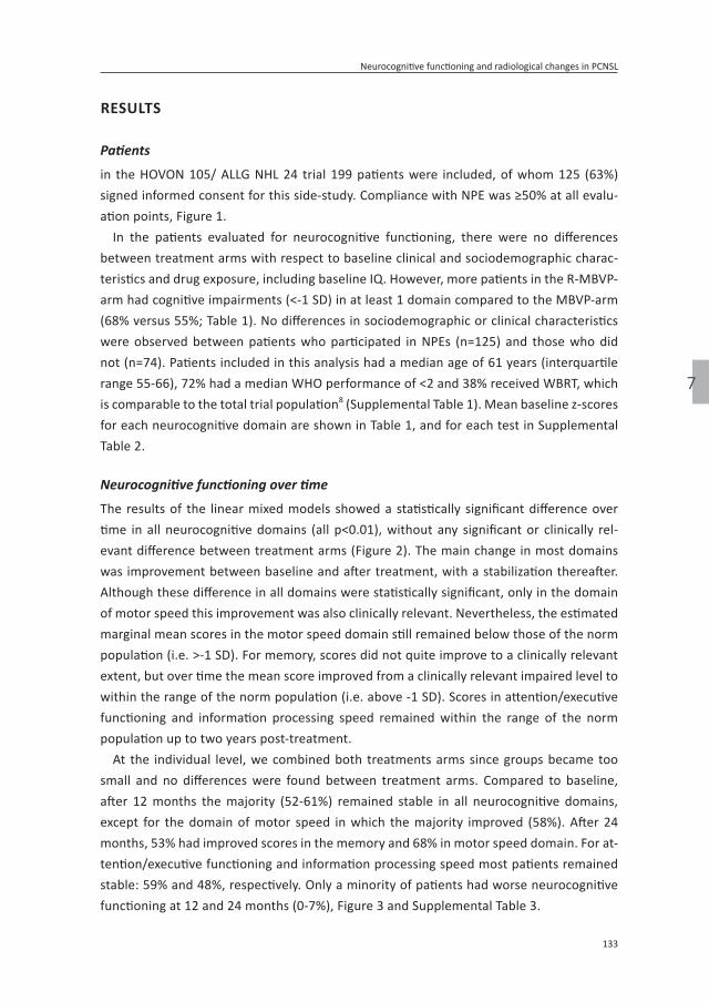
133
Neurocognitive functioning and radiological changes in PCNSL
7
reSuLtS
Patients
in the HOVON 105/ ALLG NHL 24 trial 199 patients were included, of whom 125 (63%) signed informed consent for this side-study. Compliance with NPE was ≥50% at all evalu-ation points, Figure 1.
In the patients evaluated for neurocognitive functioning, there were no differences between treatment arms with respect to baseline clinical and sociodemographic charac-teristics and drug exposure, including baseline IQ. However, more patients in the R-MBVP-arm had cognitive impairments (<-1 SD) in at least 1 domain compared to the MBVP-arm (68% versus 55%; Table 1). No differences in sociodemographic or clinical characteristics were observed between patients who participated in NPEs (n=125) and those who did not (n=74). Patients included in this analysis had a median age of 61 years (interquartile range 55-66), 72% had a median WHO performance of <2 and 38% received WBRT, which is comparable to the total trial population8 (Supplemental Table 1). Mean baseline z-scores for each neurocognitive domain are shown in Table 1, and for each test in Supplemental Table 2.
Neurocognitive functioning over time
The results of the linear mixed models showed a statistically significant difference over time in all neurocognitive domains (all p<0.01), without any significant or clinically rel-evant difference between treatment arms (Figure 2). The main change in most domains was improvement between baseline and after treatment, with a stabilization thereafter. Although these difference in all domains were statistically significant, only in the domain of motor speed this improvement was also clinically relevant. Nevertheless, the estimated marginal mean scores in the motor speed domain still remained below those of the norm population (i.e. >-1 SD). For memory, scores did not quite improve to a clinically relevant extent, but over time the mean score improved from a clinically relevant impaired level to within the range of the norm population (i.e. above -1 SD). Scores in attention/executive functioning and information processing speed remained within the range of the norm population up to two years post-treatment.
At the individual level, we combined both treatments arms since groups became too small and no differences were found between treatment arms. Compared to baseline, after 12 months the majority (52-61%) remained stable in all neurocognitive domains, except for the domain of motor speed in which the majority improved (58%). After 24 months, 53% had improved scores in the memory and 68% in motor speed domain. For at-tention/executive functioning and information processing speed most patients remained stable: 59% and 48%, respectively. Only a minority of patients had worse neurocognitive functioning at 12 and 24 months (0-7%), Figure 3 and Supplemental Table 3.

Chapter 7
134
Not eligible n= 1
Included in NPE analysesn= 65
Baselinen=48/65 (74%)
NPE stopped n=5 - death n= 0 - earlier off protocol n= 3 - refusal n= 1 - relapse/progression n= 1
After chemotherapyn=33/60 (55%)
NPE stopped n=12 - death n= 0 - earlier off protocol n= 3 - refusal n= 2 - relapse/progression n= 7
Included in NPE analysesn= 60
Baselinen=38/60 (63%)
After chemotherapyn=24/48 (50%)
Not eligible n= 0
NPE stopped n=12 - death n= 0 - earlier off protocol n= 1 - refusal n= 4 - relapse/progression n= 7
NPE stopped n=10 - death n= 0 - earlier off protocol n= 1 - refusal n= 4 - relapse/progression n= 5
200 assessed for eligibility and randomized
No consent for NPE n= 8 No consent for NPE n= 10
Arm R-MBVP n= 100 Arm: MBVP n= 100
Less than two MBVP n= 6 Less than two R-MBVP n= 6
After WBRTn=20/26 (77%)
NPE stopped n=2 - death n= 0 - earlier off protocol n= 0 - refusal n= 0 - relapse/progression n= 2
After WBRTn=9/17 (53%)
NPE stopped n=2 - death n= 0 - earlier off protocol n= 0 - refusal n= 0 - relapse/progression n= 2
3m FUn=31/46 (67%)
NPE stopped n=6 - death n= 0 - earlier off protocol n= 0 - refusal n= 1 - relapse/progression n= 5
3m FUn=21/36 (58%)
NPE stopped n=2 - death n= 0 - earlier off protocol n= 0 - refusal n= 0 - relapse/progression n= 2
6m FUn=26/40 (65%)
NPE stopped n=7 - death n= 1 - earlier off protocol n= 0 - refusal n= 1 - relapse/progression n= 5
6m FUn=21/34 (62%)
NPE stopped n=2 - death n= 0 - earlier off protocol n= 0 - refusal n= 0 - relapse/progression n= 2 12m FU
n=27/33 (82%)
NPE stopped n=3 - death n= 0 - earlier off protocol n= 0 - refusal n= 0 - relapse/progression n= 3
12m FUn=23/32 (72%)
NPE stopped n=7 - death n= 0 - earlier off protocol n= 0 - refusal n= 0 - relapse/progression n= 7
24m FUn=21/30 (70%)
24m FUn=16/25 (64%)
Patient not a native speaker n= 1 Patient not a native speaker n= 0
Hospital does not participate in NPE side study n= 25 Hospital does not participate in NPE side study n= 18
figure 1. CONSORT diagram showing reasons for not-participating in the neuropsychological evaluations as well as the compliance rates at each time point, separately for the treatment arms.‘After WBRT’ was assessed only in those who had WBRT. FU=follow-up; NPE=Neuropsychological evaluation

135
Neurocognitive functioning and radiological changes in PCNSL
7
table 1. Baseline clinical and sociodemographic characteristics of the patients included in the neuropsycho-logical evaluations, per treatment arm and for the total study population.
mBVPn=60
r-MbvPn=65
totaln=125
Sex (n, % male) 35 (58%) 29 (45%) 64 (51%)
Age (median, IQR) 60 (55-66) 61 (55-67) 61 (55-66)
WHO performance score (n, %)
WHO 0 16 (27%) 16 (25%) 32 (26%)
WHO 1 28 (47%) 33 (51%) 61 (49%)
WHO 2 11 (18%) 9 (14%) 20 (16%)
WHO 3 5 (8%) 7 (11%) 12 (10%)
Comorbidities active at baseline (n, % ≥2) 29 (48%) 33 (51%) 62 (50%)
Baseline IQ (median, IQR) 93 (76-106) 92 (77-106) 93 (77-106)
Level of education (years of education; n,%)
Low (≤6) 10 (17%) 11 (17%) 21 (17%)
Average (7-9) 30 (50%) 36 (55%) 66 (53%)
High (10-18+) 12 (20%) 11 (17%) 23 (18%)
Missing 8 (13%) 7(11%) 15 (12%)
Solitary lesion (n, %) 35 (58%) 32 (49%) 67 (54%)
Missing/ NA 3 (5%) 3 (5%) 6 (5%)
Bilateral involvement (n, %) 22 (37%) 27 (42%) 49 (39%)
Missing/ NA 3 (5%) 3 (5%) 6 (5%)
Deep structures involved (n, %) 39 (65%) 43 (66%) 82 (66%)
Study drug exposure
HD Cytarabine (n, %) 55 (92%) 59 (91%) 114 (91%)
WBRT (n, %) 22 (37%) 26 (40%) 48 (38%)
Radiation boost given (n, %) 10 (17%) 16 (25%) 26 (21%)
Intrathecal treatment given (n, %) 6 (10%) 6 (9%) 12 (10%)
Baseline score for each neurocognitive domain
Neurocognitive domain mBVP r-MbvP total
Attention/ executive functioning (mean, SD) -0.52 (0.95) -0.85 (1.03) -0.70 (1.00)
Information processing speed (mean, SD) -0.99 (1.74) -1.29 (1.72) -1.16 (1.72)
Memory (mean, SD) -1.52 (1.20) -1.70 (1.19) -1.62 (1.19)
Motor speed (mean, SD) -2.78 (3.11) -4.43 (5.57) -3.62 (4.57)
Impaired cognitive functioning (<1 SD) in at least one domain 33 (55%) 44 (68%) 77 (62%)
NA = not applicable in case of no brain lesion(s)
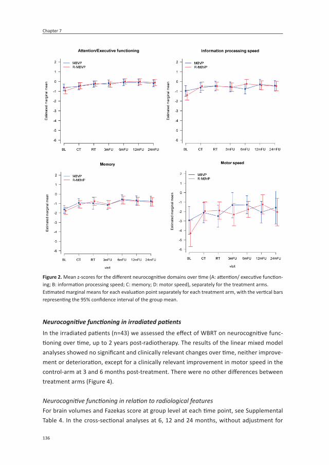
Chapter 7
136
Neurocognitive functioning in irradiated patients
In the irradiated patients (n=43) we assessed the effect of WBRT on neurocognitive func-tioning over time, up to 2 years post-radiotherapy. The results of the linear mixed model analyses showed no significant and clinically relevant changes over time, neither improve-ment or deterioration, except for a clinically relevant improvement in motor speed in the control-arm at 3 and 6 months post-treatment. There were no other differences between treatment arms (Figure 4).
Neurocognitive functioning in relation to radiological featuresFor brain volumes and Fazekas score at group level at each time point, see Supplemental Table 4. In the cross-sectional analyses at 6, 12 and 24 months, without adjustment for
figure 2. Mean z-scores for the different neurocognitive domains over time (A: attention/ executive function-ing; B: information processing speed; C: memory; D: motor speed), separately for the treatment arms.Estimated marginal means for each evaluation point separately for each treatment arm, with the vertical bars representing the 95% confidence interval of the group mean.
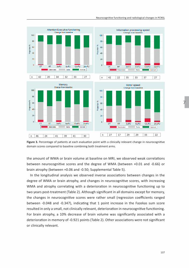
137
Neurocognitive functioning and radiological changes in PCNSL
7
the amount of WMA or brain volume at baseline on MRI, we observed weak correlations between neurocognitive scores and the degree of WMA (between +0.01 and -0.66) or brain atrophy (between +0.06 and -0.50; Supplemental Table 5).
In the longitudinal analysis we observed inverse associations between changes in the degree of WMA or brain atrophy, and changes in neurocognitive scores, with increasing WMA and atrophy correlating with a deterioration in neurocognitive functioning up to two years post-treatment (Table 2). Although significant in all domains except for memory, the changes in neurocognitive scores were rather small (regression coefficients ranged between -0.048 and -0.347), indicating that 1 point increase in the Fazekas sum score resulted in only a small, not clinically relevant, deterioration in neurocognitive functioning. For brain atrophy, a 10% decrease of brain volume was significantly associated with a deterioration in memory of -0.921 points (Table 2). Other associations were not significant or clinically relevant.
n 42 20 34 32 33 27 n 42 22 35 33 37 27
n 46 24 41 39 41 30 n 27 17 29 29 33 22
figure 3. Percentage of patients at each evaluation point with a clinically relevant change in neurocognitive domain scores compared to baseline combining both treatment arms.

Chapter 7
138
diSCuSSioN
The addition of rituximab to standard MBVP-chemotherapy did not improve the event-free survival and did not impact HRQoL.8,14 The current analysis shows that this treatment regimen resulted in a significant improvement in all neurocognitive domains, compared to baseline, although these differences were not clinically relevant except for motor speed. There were no significant or clinically relevant differences in neurocognitive functioning between those treated with and without rituximab.
figure 4. Mean z-scores for each neurocognitive domain over time (A: attention/ executive functioning; B: information processing speed; C: memory; D: motor speed), separately for the two treatment arms in the irradiated patients only.Estimated marginal means are shown for each evaluation point separately for each treatment arm, with the vertical bars representing the 95% confidence interval of the group mean.

139
Neurocognitive functioning and radiological changes in PCNSL
7
Impairments in neurocognitive functioning or behavioral problems are reported to be presenting symptoms in 32-48% of patients with PCNSL.28 In this study, 62% of the patients had impairments in at least one neurocognitive domain before treatment. The slightly bet-ter scores in all domains at the ‘end of treatment’ compared to baseline, with a stabiliza-tion thereafter, is a pattern that has been described in multiple PCNSL cohorts.12,29-31 These findings suggest that neurocognitive functioning is mostly hampered by the tumor itself, and that treating the tumor results in improved neurocognitive functioning. Additionally, we found that the extent of improvement between baseline and ‘end of treatment’ was not clinically relevant, except for motor speed.
The results at the individual patient level support the finding that neurocognitive scores improved over time, although most patients remained stable, and only a minority (0-7%) deteriorated. Only in the motor speed domain the majority of the patients improved at 12 and 24 months, compared to baseline. This supports the results of the longitudinal analyses (linear mixed models) suggesting that motor speed improved over time to a clinically relevant extent. This finding is, however, in contrast with other studies showing less pronounced improvements or even deterioration in this domain, particularly when compared to other domains.32,33 An explanation for this may be that patients in our cohort had very low scores at baseline, allowing patients to improve. It should be noted though, that despite the improvement, the estimated marginal means continued to be lower than the norm population (i.e. <-1SD).
In irradiated patients, scores on all neurocognitive domains rather unexpectedly re-mained stable for up to two years after treatment with 30Gy (20x1.5Gy) WBRT in both arms, compared to the scores shortly after WBRT. We used ‘after WBRT’ as baseline because we expected maximal reduction of tumor and tumor-related symptoms at that timepoint. Stable neurocognitive functioning was also found in a previously reported small cohort of PCNSL patients treated with HD-MTX based chemotherapy combined with rituximab and followed by reduced dose (rd)WBRT (23.4Gy).12 In that same cohort, a small but significant decline in the neurocognitive domains attention and memory was observed between three and five years post-treatment.34 This late, non-clinically relevant deterioration, however, was also observed in patients who received autologous stem cell transplantation (ASCT) instead of WBRT.34 Two randomized trials in adult PCNSL patients compared WBRT with ASCT as consolidation therapy.29,31 In the IELSG-32 study, 118 patients who achieved at least stable disease after induction chemotherapy were randomized for ASCT or 36Gy WBRT as consolidation.29 After two years of follow-up, those who received WBRT had significantly worse scores in attention/executive functioning and memory domains.29 In the PRECIS study 104 patients aged 18-60 years were randomized between ASCT and 40Gy WBRT as consolidation therapy.31 Similarly, significant deterioration was seen in attention/executive functioning in irradiated patients compared to ‘end of induction chemotherapy’, while those treated with ASCT remained stable for up to three years of follow-up.31 Several

Chapter 7
140
factors could explain the discrepancy between the above two contemporary studies and our study regarding neurocognitive functioning after WBRT. First, the lower total dose and fraction dose used in our study could reduce the negative impact of WBRT, as suggested by the findings of Morris et al.12 Second, it is possible that in our study longer follow-up will show deterioration of neurocognitive function. Such late deterioration has also been found to occur in patients with other brain tumors, such as low-grade glioma, in whom neurocognitive deterioration after (focal) radiotherapy was found after 12 years but not after 6 years.35,36 Lastly, the absence of published individual scores and/or z-scores of the neurocognitive tests in the IELSG-3229 and PRECIS31 studies do not allow estimation of the magnitude of changes in neurocognition, and consequently the clinical relevance of these changes, which may affect the interpretation of results.
Up to two years post-treatment, we observed that the increase in the degree of WMA was significantly associated with worsening in all neurocognitive domains except memory, while an increase of brain atrophy was associated with worsening in the memory domain only. The associations, however, were weak to moderate, indicating that in the first two years post-treatment the impact of WMA and brain atrophy on neurocognitive function-ing seems modest, possibly partially because the extent of decrease in brain volume was limited. In a large (n=80) long-term PCNSL survivors cohort (median follow-up of 5·5 years, range 2-26 years), the amount of WMA was significantly correlated with worse neurocog-nitive functioning on the long-term. Moreover, those treated with WBRT (n=15; 45-60Gy) in this survivors cohort had twice as much WMA as those treated without WBRT (n=65; p<0.001), though this resulted in a clinically relevant difference in the motor speed domain only.10 In contrast, although more WMA were observed after rdWBRT (23.4Gy) than after ASCT (70% versus 40%, p=0·03), there was no difference in neurocognitive functioning between these groups, up to five years post-treatment.34 While assessed in small groups and with different durations of follow-up, these results suggest that radiation dose could be crucial for neurocognitive functioning in PCNSL patients and this is supported by our findings. Longer follow-up is nevertheless needed to determine the effect of 30Gy WBRT and of white matter changes and cerebral atrophy on neurocognitive functioning in our cohort on the longer term. We were unable to investigate a direct effect of rituximab on WMA or cerebral atrophy because the number of patients in these subgroups became too small for a meaningful analysis. A recent, small (n=47) retrospective study, however, found after a mean follow-up of five years that more patients treated with rituximab plus HD-MTX developed white matter lesions (68%), compared to rituximab naïve patients (46%).37 Although this finding does not necessarily support causation, further analysis might help to determine the etiology of these lesions.
The strengths of this study are the large, uniformly treated group of patients in which radiological assessments were done over time and neurocognitive functioning in multiple domains was assessed prospectively, allowing extensive analyses. Limitations of our study

141
Neurocognitive functioning and radiological changes in PCNSL
7
are the limited time of follow-up, i.e. two years after end-of-treatment, and our relatively crude, visual measurement of the WMA with the Fazekas score as opposed to automatic exact measurements of white matter abnormalities, which may have masked an existing effect of WMA on neurocognition. Automatic segmentation of WMA was not possible due to different scan protocols in different including centers. Although we had some missing neurocognitive data over time, our longitudinal analyses were not hampered by this since we used linear mixed models, which deal with missing data in a sophisticated way. Lastly, we could not compare irradiated with non-irradiated patients with respect to neurocogni-tive functioning and radiological changes, because these patients differed in age due to the study design (i.e. irradiation in younger patients only). For this same reason we could not compare younger and older patients.
In conclusion, this analysis showed no effect of the addition of rituximab on neurocogni-tive functioning, neither positive nor negative. The lack of effect on event-free survival8, the primary endpoint, as well as HRQoL14 and this neurocognitive study, as secondary end-points, do not, however, support the use of rituximab in patients with newly-diagnosed PCNSL. Whether specific subgroups of patients benefit from this treatment regimen re-mains to be investigated. Moreover, in the first two years post-treatment, a lower dose of WBRT was not harmful for neurocognitive functioning, compared to just after WBRT. The association between white matter abnormalities and brain atrophy and neurocognitive functioning was modest and longer follow-up is needed to draw definitive conclusions.
For supplemental Tables and Figures: Neurocognitive functioning and radiologic changes in primary CNS lymphoma patients: results from the HOVON 105/ ALLG NHL 24 randomised controlled trial | Neuro-Oncology | Oxford Academic (oup.com)https://academic.oup.com/neuro-oncology/advance-article/doi/10.1093/neuonc/noab021/6131741?login=true

Chapter 7
142
refereNCeS
1. van der Meulen M, Dinmohamed AG, Visser O, Doorduijn JK, Bromberg JEC. Improved survival in primary central nervous system lymphoma up to age 70 only: a population-based study on incidence, primary treat-ment and survival in the Netherlands, 1989-2015. Leukemia 2017;31:1822-1825.
2. Villano JL, Koshy M, Shaikh H, Dolecek TA, McCarthy BJ. Age, gender, and racial differ-ences in incidence and survival in primary CNS lymphoma. Br J Cancer 2011;105:1414-1418.
3. Correa DD, Maron L, Harder H, et al. Cogni-tive functions in primary central nervous system lymphoma: literature review and assessment guidelines. Ann Oncol 2007;18:1145-1151.
4. Boele FW, Zant M, Heine EC, et al. The association between cognitive functioning and health-related quality of life in low-grade glioma patients. Neurooncol Pract 2014;1:40-46.
5. Aaronson NK, Taphoorn MJ, Heimans JJ, et al. Compromised health-related quality of life in patients with low-grade glioma. J Clin Oncol 2011;29:4430-4435.
6. Coiffier B, Lepage E, Briere J, et al. CHOP chemotherapy plus rituximab compared with CHOP alone in elderly patients with diffuse large-B-cell lymphoma. N Engl J Med 2002;346:235-242.
7. Pfreundschuh M, Schubert J, Ziepert M, et al. Six versus eight cycles of bi-weekly CHOP-14 with or without rituximab in el-derly patients with aggressive CD20+ B-cell lymphomas: a randomised controlled trial (RICOVER-60). Lancet Oncol 2008;9:105-116.
8. Bromberg JEC, Issa S, Bakunina K, et al. Rituximab in patients with primary CNS lymphoma (HOVON 105/ALLG NHL 24): a randomised, open-label, phase 3 intergroup study. Lancet Oncol 2019;20:216-228.
9. Harder H, Holtel H, Bromberg JEC, et al. Cognitive status and quality of life after treatment for primary CNS lymphoma. Neurology 2004;62:544-547.
10. Doolittle ND, Korfel A, Lubow MA, et al. Long-term cognitive function, neuroimag-ing, and quality of life in primary CNS lymphoma. Neurology 2013;81:84-92.
11. Neuwelt EA, Guastadisegni PE, Várallyay P, Doolittle ND. Imaging changes and cognitive outcome in primary CNS lymphoma after enhanced chemotherapy delivery. Am J Neuroradiol 2005;26:258-265.
12. Morris PG, Correa DD, Yahalom J, et al. Rituximab, methotrexate, procarbazine, and vincristine followed by consolidation reduced-dose whole-brain radiotherapy and cytarabine in newly diagnosed primary CNS lymphoma: Final results and long-term outcome. J Clin Oncol 2013;31:3971-3979.
13. Fliessbach K, Helmstaedter C, Urbach H, et al. Neuropsychological outcome after chemotherapy for primary CNS lymphoma: A prospective study. Neurology 2005;64:1184-1188.
14. van der Meulen M, Bakunina K, Nijland M, et al. Health-related Quality of Life After Chemotherapy With or Without Rituximab in Primary Central Nervous System Lympho-ma Patients: Results From a Randomised Phase III Study. 2020;Ann Oncol.
15. Abrey LE, Batchelor TT, Ferreri AJ, et al. Report of an international workshop to standardize baseline evaluation and re-sponse criteria for primary CNS lymphoma. J Clin Oncol 2005;23:5034-5043.
16. Wechsler D. Wechsler Adult Intelligence Scale (WAIS-III), 3rd ed. San Antonio: Har-court Brace & Co, 1997.
17. R.M. R. Trail making test. Manual for ad-ministration and scoring. Tuscon, Arizona: Reitan Neuropsychological Laboratory, 1992.

143
Neurocognitive functioning and radiological changes in PCNSL
7
18. Lezak MD. Neuropsychological assessment. : York Oxford University Press, 1976
19. Lezak MD, Howieson DB, Bigler ED, D. t. Neuropsychological Assessment, 5th Re-vised edition ed. New York Oxford Univerity Press, 2012.
20. Heaton RK, Grant I, Matthews CG. Compre-hensive Norms for an Expanded Halstead-Reitan Battery: Demographic Corrections, Research Findings and Clinical Applications. : Odessa: Psychological Assessment Re-sources, 1991.
21. Nelson H. National adult reading test (NART): Test manual. Windsor, U.K.: Nfer-Nelson.1982.
22. Schmand B, Bakker D, Saan R, Louman J. [The Dutch Reading Test for Adults: a measure of premorbid intelligence level] De Nederlandse Leestest voor Volwassenen: een maat voor het premorbide intel-ligentieniveau. Tijdschr Gerontol Geriatr 1991;22:15-19.
23. Wahlund LO, Barkhof F, Fazekas F, et al. A new rating scale for age-related white matter changes applicable to MRI and CT. Stroke 2001;32:1318-1322.
24. de Boer R, Vrooman HA, van der Lijn F, et al. White matter lesion extension to automatic brain tissue segmentation on MRI. Neuro-image 2009;45:1151-1161.
25. Mulder J, Bouma JM, Schmand B. Neder-landse leestest voor volwassenen. In: Bou-ma JM, J. Mulder, J. Lindeboom, Schmand B, eds. Handboek neuropsychologische diagnostiek Amsterdam: Pearson Assess-ment and Information B.V., 2012: 127-138.
26. Verhage F. Intelligentie en leeftijd onder-zoek bij Nederlanders van twaalf tot zevenenzeventig jaar [Intelligence and age: Research study in Dutch individuals aged twelve to seventy-seven]. . Assen, Neth-erlands: : Van Gorcum/Prakke & Prakke., 1964.
27. van der Elst W, van Boxtel MP, van Breukelen GJ, Jolles J. The Letter Digit Substitution Test: normative data for 1,858 healthy par-
ticipants aged 24-81 from the Maastricht Aging Study (MAAS): influence of age, education, and sex. J Clin Exp Neuropsychol 2006;28:998-1009.
28. Bataille B, Delwail V, Menet E, et al. Primary intracerebral malignant lymphoma: report of 248 cases. J Neurosurg 2000;92:261-266.
29. Ferreri AJM, Cwynarski K, Pulczynski E, et al. Whole-brain radiotherapy or autologous stem-cell transplantation as consolidation strategies after high-dose methotrexate-based chemoimmunotherapy in patients with primary CNS lymphoma: results of the second randomisation of the International Extranodal Lymphoma Study Group-32 phase 2 trial. Lancet Haematol 2017;4:e510-e523.
30. van der Meulen M, Dirven L, Habets EJJ, van den Bent MJ, Taphoorn MJB, Bromberg JEC. Cognitive functioning and health-related quality of life in patients with newly diag-nosed primary CNS lymphoma: a systematic review. Lancet Oncol 2018;19:e407-e418.
31. Houillier C, Taillandier L, Dureau S, et al. Radiotherapy or Autologous Stem-Cell Transplantation for Primary CNS Lymphoma in Patients 60 Years of Age and Younger: Re-sults of the Intergroup ANOCEF-GOELAMS Randomized Phase II PRECIS Study. J Clin Oncol 2019;37:823-833.
32. Correa DD, DeAngelis LM, Shi W, Thaler H, Glass A, Abrey LE. Cognitive functions in survivors of primary central nervous system lymphoma. Neurology 2004;62:548-555.
33. Omuro A, Correa DD, DeAngelis LM, et al. R-MPV followed by high-dose chemo-therapy with TBC and autologous stem-cell transplant for newly diagnosed primary CNS lymphoma. Blood 2015;125:1403-1410.
34. Correa DD, Braun E, Kryza-Lacombe M, et al. Longitudinal cognitive assessment in pa-tients with primary CNS lymphoma treated with induction chemotherapy followed by reduced-dose whole-brain radiotherapy or autologous stem cell transplantation. J Neurooncol 2019;144:553-562.

Chapter 7
144
35. Klein M, Heimans JJ, Aaronson NK, et al. Effect of radiotherapy and other treatment-related factors on mid-term to long-term cognitive sequelae in low-grade gliomas: a comparative study. Lancet 2002;360:1361-1368.
36. Douw L, Klein M, Fagel SS, et al. Cognitive and radiological effects of radiotherapy in patients with low-grade glioma: long-term follow-up. Lancet Neurol 2009;8:810-818.
37. Estephan F, Ye X, Dzaye O, et al. White matter changes in primary central nervous system lymphoma patients treated with high-dose methotrexate with or without rituximab. J Neurooncol 2019;145:461-466.


8Health-related quality of life after chemotherapy with
or without rituximab in primary central nervous system lymphoma patients: results from a randomized phase III
study
Matthijs van der Meulen* MD1, Katerina Bakunina* MSc2, Marcel Nijland MD, PhD3, Monique C. Minnema MD, PhD4, Gavin Cull MD5,6, Wendy B.C. Stevens MD7, Joke W. Baars MD8, Kylie D.
Mason MBBS, PhD9, Aart Beeker MD10, Max Beijert MD11, Martin J.B. Taphoorn MD, PhD12,13, Martin J. van den Bent MD, PhD1, Samar Issa MD14, Jeanette K. Doorduijn MD, PhD15, Jacoline
E.C. Bromberg MD, PhD1, Linda Dirven PhD12,13
*contributed equally
1 Erasmus MC Cancer Institute, Brain Tumor Center, University Medical Center Rotterdam, Netherlands, Department of Neuro-Oncology;
2 HOVON Data Center, Department of Hematology, Erasmus MC Cancer Institute, Rotterdam,Netherlands;
3 Department of Hematology, University Medical Center Groningen, Groningen, Netherlands;4 Department of Hematology, University Medical Center Utrecht, Utrecht, Netherlands;
5 Haematology Department, Sir Charles Gairdner Hospital and PathWest Laboratory Medicine, Nedlands, WA, Australia;6 University of Western Australia, Crawley, WA, Australia;
7 Department of Hematology, Radboud University Medical Center, Nijmegen, Netherlands;8 Department of Hemato-Oncology, Antoni van Leeuwenhoek Hospital/Dutch Cancer Institute, Amsterdam, Netherlands;
9 Department of Hematology, Royal Melbourne Hospital, Melbourne, VIC, Australia;10 Department of Hematology, Spaarne Gasthuis, Haarlem, Netherlands;
11 Department of Radiotherapy, University Medical Center Groningen, Groningen, Netherlands;12 Leiden University Medical Center, Leiden, Netherlands, Department of Neurology;
13 Haaglanden Medical Center, The Hague, Netherlands, Department of Neurology;14 Department of Hematology, Middlemore Hospital, Auckland, New Zealand;
15 Erasmus MC Cancer Institute, University Medical Center Rotterdam, Department of Hematology.
2020 Annals of Oncology, 31(8):1046-1055.

Chapter 8
148
ABStrACt
Background: The impact of rituximab on health-related quality of life (HRQoL) in primary central nervous system lymphoma patients is not well-known. We determined the impact of rituximab added to standard high-dose methotrexate-based treatment on HRQoL from patients in a large randomized trial.Patients and methods: Patients from a large phase III trial (HOVON 105/ALLG NHL 24), randomized to standard chemotherapy with or without rituximab and followed by 30Gy whole brain radiotherapy (WBRT) in patients ≤60 years, completed the EORTC QLQ-C30 and QLQ-BN20 questionnaires before and during treatment, and up to 24 months of follow-up or progression. Differences between treatment arms over time in global health status, role functioning, social functioning, fatigue, and motor dysfunction were assessed. Differences ≥10 points were deemed clinically relevant. The effect of WBRT on HRQoL was analyzed in irradiated patients.results: 160/175 patients eligible for the HRQoL study completed at least one question-naire and were included. Over time, scores improved statistically significant and clinically relevant in both arms. Between arms, there were no differences on any scale (range: -3.8 to +4.0). Scores on all scales were improved to a clinically relevant extent at 12 and 24 months compared to baseline in both arms, except for fatigue and motor dysfunction at 12 months (-7.4 and -8.8, respectively). In irradiated patients (N=59), scores in all preselected scales except motor dysfunction, remained stable up to 24 months compared to shortly after WBRT, overall mean difference ranging between 0.02 and 4.570.Conclusion: Compared to baseline, treatment resulted in improved HRQoL scores. The addition of rituximab to standard chemotherapy did not impact HRQoL over time. WBRT did not result in deterioration of HRQoL in the first two years.

149
Health-related quality of life after chemotherapy with or without rituximab in PCNSL
8
iNtroduCtioN
Primary central nervous system lymphoma (PCNSL) is a rare non-Hodgkin lymphoma con-fined to the brain, leptomeninges, spinal cord and eyes. Over the last three decades the incidence rate has increased, mainly amongst patients >60 years-old, and prognosis has improved significantly.1,2 This prolonged survival has largely been determined by improve-ments in treatment.1,3
In systemic diffuse large B-cell lymphoma (DLBCL) patients, the addition of rituximab, a chimeric monoclonal antibody targeting the CD20 cell surface protein, to standard treatment has been shown to improve progression-free (PFS) and overall survival (OS).4,5 Since most PCNSL are DLBCL, it has been hypothesized that the addition of rituximab to standard treatment with HD-MTX-based chemotherapy could also improve survival in PCNSL patients. The HOVON 105/ALLG NHL 24, a large international multicentre phase III randomized controlled trial (RCT), investigated the addition of rituximab to standard high-dose methotrexate (HD-MTX)-based chemotherapy, followed by 30Gy whole-brain radio-therapy (WBRT) in patients aged ≤60 years. The primary endpoint, the 1-year event-free survival (EFS), was not improved by rituximab D-MTX, tenoposide,BCNU (carmustine), and prednisolone without (MBVP)versus with rituximab (R-MBVP): 49% versus 52%, P=0.99].6
When introducing a new treatment, information on both survival and the patients’ functioning and well-being should be taken into account. Combined, these outcomes determine the ‘net clinical benefit’ of a treatment strategy. By combining both sources of information, clinicians and patients are better able to make well-informed decisions concerning which treatment is most suitable for an individual patient.
In this study we describe the HRQoL trajectories in one of the largest RCT’s in PCNSL patients and determined whether the addition of rituximab to standard therapy had an impact on HRQoL. Second, we aimed to determine the effect of a lower dose WBRT on HRQoL in this patient population.
methodS
Study design and patient population
In the HOVON 105/ALLG NHL 24 study, 199 immunocompetent patients aged 18-70 years with a newly diagnosed, CD20 positive B-cell PCNSL were included from Dutch, Australian and New Zealand hospitals between 2010 and 2016.6 Patients were randomized between two courses of HD-MTX, tenoposide, BCNU and prednisolone without (MBVP) or with rituximab (R-MBVP). Irrespective of treatment arm, this induction regimen was followed by consolidative HD-cytarabine chemotherapy in responding patients, and in patients ≤60 years-old 30Gy WBRT was subsequently added. An integrated boost to the tumour-bed of

Chapter 8
150
10Gy was given to patients who only achieved partial response.7 (Immuno-)chemotherapy treatment duration was 2.5-3.5 months and WBRT was administered in one month. Fur-ther details on the study design and treatment have been published previously.6 The study was approved by the ethics committees of all participating centres. All participants who signed informed consent for the RCT and for participating in the HRQoL study were eligible for inclusion in this analysis.
Health-Related Quality of Life
HRQoL was one of the prespecified secondary outcomes. HRQoL was assessed using the European Organisation for Research and Treatment of Cancer (EORTC) core quality of life questionnaire (QLQ-C30)8, and the brain cancer module (QLQ-BN20).9,10 The QLQ-C30 com-prises five functional scales (physical, role, emotional, social and cognitive functioning), three symptom scales (fatigue, pain, and nausea and vomiting), a global health status/quality of life scale, and six single items assessing additional symptoms (dyspnoea, sleep disturbance, appetite loss, constipation and diarrhoea) and perceived financial difficul-ties. The QLQ-BN20 module includes 20 items, comprising four scales (visual disorders, motor dysfunction, communication deficit and future uncertainty), and seven disease- or toxicity-related symptoms (headache, seizures, drowsiness, hair loss, itchy skin, weakness of the legs, and bladder control).
According to protocol, patients had to complete the questionnaires before starting chemotherapy, after completion of all chemotherapy, after completion of radiotherapy (if given), and 3, 6, 12, and 24 months after completion of protocol treatment. The assessment of HRQoL was stopped when a relapse or progression occurred, or when a patient wanted to withdraw from participation in either the RCT or HRQoL sub-study. Only questionnaires that were completed within a prespecified time window (see Supplemental methods) for each evaluation point were included in the statistical analysis.
StAtiStiCAL ANALySiS
Calculation of HRQoL Scores
Following the EORTC procedures, raw item scores were converted to a linear scale rang-ing from 0 to 100.11 A difference of ≥10 points on each HRQoL scale/item was defined as clinically relevant.12 Based on clinical relevance for PCNSL patients, these five scales were selected for primary analysis: three functional scales (global health status (GH), role functioning (RF) and social functioning (SF), with higher scores representing better functioning), and two symptom scales (fatigue and motor dysfunction (MD), with high scores representing worse functioning). Results of the primary analysis (i.e. scores over time assessed with linear mixed models in the 5 predetermined scales, were corrected for

151
Health-related quality of life after chemotherapy with or without rituximab in PCNSL
8
multiple testing. The remaining scales and items were analysed on an exploratory basis. All analyses were conducted with Stata, version 15, and a P-value <0·05 was considered to be statistically significant.
Descriptive statistics
Patients eligible for the HOVON 105/ ALLG NHL 24 study who received less than two courses of (R-)MBVP, or did not give consent for the HRQoL sub-study, were excluded from the analysis. We performed a non-response analysis to assess possible imbalances between those who gave consent for the HRQoL sub-study and those who did not with respect to sociodemographic and clinical characteristics.
Differences were tested using a Chi-Square test for categorical data and Mann-Whitney U or independent t-test for continuous data, depending on the distribution of the data. Normality was determined with the Kolmogorov-Smirnov test. In addition, for each time point the compliance was evaluated, defined as the number of completed questionnaires divided by the number of questionnaires expected at that time point. We defined a completed questionnaire as a returned form from which at least a score of one of the predetermined primary scales could be derived.
HRQoL scores over time
At group level, mean changes from baseline were calculated and plotted for those patients who filled in the questionnaire at baseline and at least at one follow-up point. Differences between arms at 12 and 24 months of follow-up were assessed with an independent t-test. A linear mixed model, which allows inclusion of all patients, with fixed effects for treat-ment arm, time (i.e. evaluation moments) as a categorical covariate, and their interaction, was used to assess whether there is a difference in the HRQoL scores over time between the treatment arms. For each scale, the most suitable covariance structure was chosen to estimate the impact of the treatment on HRQoL over time.
At the individual patient level, changes in HRQoL between baseline and both 12 and 24 months of follow-up were calculated for those patients the questionnaires at these time points were available. Patients were categorized as deteriorated, stable or improved, based on a change of ≥10-points. Differences between treatment arms were assessed with the Chi-square statistic.
Deterioration-free survival and time to deterioration
Deterioration-free survival was defined as a deterioration of ≥10 points on a scale/item compared to baseline without an improvement of ≥10 points at the subsequent HRQoL assessment, or progressive disease (PD), or death in the absence of definite deterioration before the next assessment. Time to deterioration was defined similarly as deterioration-free survival, only excluding PD as an event.13 PD was defined according to the international

Chapter 8
152
PCNSL response criteria.7 MRI’s were centrally reviewed; the centrally scored progression data were used for these analyses. Questionnaires filled in at the time of or after centrally scored PD were excluded. Kaplan-Meier curves were generated for both deterioration-free survival and time to deterioration and 95% confidence intervals were calculated using the Greenwood formula.
Impact of WBRT
In a subgroup, those who received WBRT (N=59), we evaluated the impact of radiation on HRQoL. The mean changes from the ‘after WBRT’ time point onwards were calculated and linear mixed model analyses were performed to analyse HRQoL scores over the post-WBRT period.
reSuLtS
Patients
Of the 199 patients included in the HOVON 105/ALLG NHL 24 trial, 193 (97%) gave in-formed consent for HRQoL assessment. Eighteen patients were excluded because they did not complete two courses of (R-)MBVP, resulting in 175/199 (88%) eligible patients for the HRQoL analysis. Of these 175 patients included in the HRQoL analysis, 160 completed at least one questionnaire. Compliance of HRQoL evaluation was ≥60% at each time point, except ‘after WBRT’ in the MBVP-arm (46%), Figure 1.
In the population participating in the HRQoL analysis, patients in the treatment arms were well-balanced with respect to clinical and sociodemographic features and study drug exposure. Those included in this HRQoL analysis had a median age of 61 years (interquar-tile range 55-66 years), and 74% had a WHO performance score <2, which is similar to the total trial population6 (Table 1). The non-response analysis showed that there were no significant differences with respect to baseline characteristics between those who gave consent for the HRQoL sub-study and those who did not (Supplemental Table 1). Baseline HRQoL scores for all scales and items for both treatment arms are summarised in Supplemental Table 2.
HRQoL scores over time
In all selected primary scales, the mean change from baseline, assessed in those who filled in the questionnaires at baseline and at least once thereafter, showed a statistically significant (all P<0.002) and clinically relevant improvement in both arms after the end of treatment (i.e. ‘after chemotherapy’ or ‘after WBRT’), when compared to baseline, except for fatigue. Fatigue improved more slowly: clinically relevant improvement was not reached before 3 months post-treatment. The differences in scores between the arms at

153
Health-related quality of life after chemotherapy with or without rituximab in PCNSL
8
Not eligible n= 1
Included in HRQoL analysesn=85
Baselinen=65/85 (76%) HRQoL stopped n=8
- death n= 0 - earlier off protocol n= 5 - refusal n= 2 - relapse/progression n= 1
After chemotherapyn=52/77 (68%)
HRQoL stopped n=15 - death n= 0 - earlier off protocol n= 4 - refusal n= 2 - relapse/progression n= 9
Included in HRQoL analysesn= 90
Baselinen=67/90 (74%)
After chemotherapyn=46/72 (64%)
Not eligible n= 0
HRQoL stopped n=18 - death n= 2 - earlier off protocol n= 1 - refusal n= 5 - relapse/progression n= 10
HRQoL stopped n=17 - death n= 0 - earlier off protocol n= 3 - refusal n= 5 - relapse/progression n= 9
200 assessed for eligibility and randomized
No consent for HRQoL n= 3 No consent for HRQoL n= 3
Arm R-MBVP n= 100 Arm: MBVP n= 100
Less than two MBVP n= 7 Less than two R-MBVP n= 11
After WBRTn=22/33* (67%)
HRQoL stopped n=2 - death n= 0 - earlier off protocol n= 0 - refusal n= 0 - relapse/progression n= 2
After WBRTn=12/26* (46%)
HRQoL stopped n=3 - death n= 0 - earlier off protocol n= 0 - refusal n= 0 - relapse/progression n= 3 3m FU
n=37/60 (62%)
HRQoL stopped n=6 - death n= 0 - earlier off protocol n= 0 - refusal n= 1 - relapse/progression n= 5
3m FUn=31/52 (60%)
HRQoL stopped n=5 - death n= 1 - earlier off protocol n= 0 - refusal n= 0 - relapse/progression n= 4
6m FUn=33/54 (61%)
HRQoL stopped n=8 - death n= 1 - earlier off protocol n= 0 - refusal n= 1 - relapse/progression n= 6
6m FUn=29/47 (62%)
HRQoL stopped n=3 - death n= 0 - earlier off protocol n= 0 - refusal n= 0 - relapse/progression n= 3
12m FUn=42/46 (91%)
HRQoL stopped n=4 - death n= 0 - earlier off protocol n= 0 - refusal n= 1 - relapse/progression n= 3
12m FUn=32/44 (73%)
HRQoL stopped n=10 - death n= 0 - earlier off protocol n= 0 - refusal n= 1 - relapse/progression n= 9
24m FUn=29/42 (69%)
24m FUn=24/34 (71%)
figure 1. CONSOrT Diagram representing the compliance with HrQol assessments during follow-up, sepa-rately for the two treatment arms. FU, months of follow-up after end of treatment; HRQoL, health-related quality of life; MBVP, methotrexate, tenoposide, BCNU (carmustine), and prednisolone; R-MBVP, rituximab with methotrexate, tenoposide, BCNU (carmustine), and prednisolone; WBRT, whole brain radiotherapy. *In those who had WBRT only.
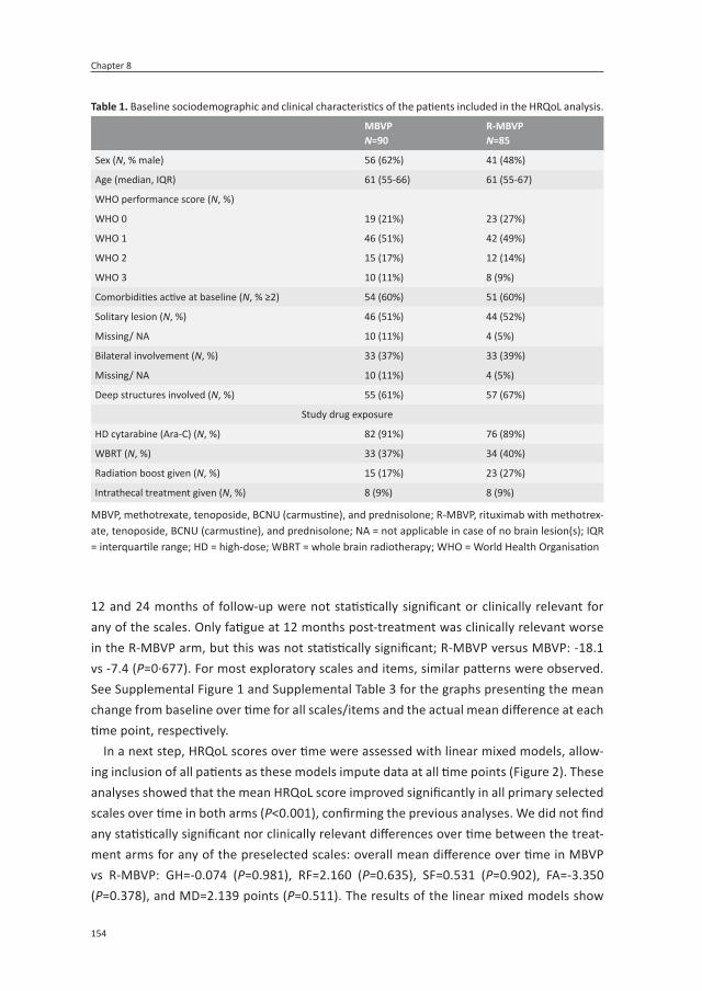
Chapter 8
154
12 and 24 months of follow-up were not statistically significant or clinically relevant for any of the scales. Only fatigue at 12 months post-treatment was clinically relevant worse in the R-MBVP arm, but this was not statistically significant; R-MBVP versus MBVP: -18.1 vs -7.4 (P=0·677). For most exploratory scales and items, similar patterns were observed. See Supplemental Figure 1 and Supplemental Table 3 for the graphs presenting the mean change from baseline over time for all scales/items and the actual mean difference at each time point, respectively.
In a next step, HRQoL scores over time were assessed with linear mixed models, allow-ing inclusion of all patients as these models impute data at all time points (Figure 2). These analyses showed that the mean HRQoL score improved significantly in all primary selected scales over time in both arms (P<0.001), confirming the previous analyses. We did not find any statistically significant nor clinically relevant differences over time between the treat-ment arms for any of the preselected scales: overall mean difference over time in MBVP vs R-MBVP: GH=-0.074 (P=0.981), RF=2.160 (P=0.635), SF=0.531 (P=0.902), FA=-3.350 (P=0.378), and MD=2.139 points (P=0.511). The results of the linear mixed models show
table 1. Baseline sociodemographic and clinical characteristics of the patients included in the HRQoL analysis.
mBVPN=90
r-MbvPN=85
Sex (N, % male) 56 (62%) 41 (48%)
Age (median, IQR) 61 (55-66) 61 (55-67)
WHO performance score (N, %)
WHO 0 19 (21%) 23 (27%)
WHO 1 46 (51%) 42 (49%)
WHO 2 15 (17%) 12 (14%)
WHO 3 10 (11%) 8 (9%)
Comorbidities active at baseline (N, % ≥2) 54 (60%) 51 (60%)
Solitary lesion (N, %) 46 (51%) 44 (52%)
Missing/ NA 10 (11%) 4 (5%)
Bilateral involvement (N, %) 33 (37%) 33 (39%)
Missing/ NA 10 (11%) 4 (5%)
Deep structures involved (N, %) 55 (61%) 57 (67%)
Study drug exposure
HD cytarabine (Ara-C) (N, %) 82 (91%) 76 (89%)
WBRT (N, %) 33 (37%) 34 (40%)
Radiation boost given (N, %) 15 (17%) 23 (27%)
Intrathecal treatment given (N, %) 8 (9%) 8 (9%)
MBVP, methotrexate, tenoposide, BCNU (carmustine), and prednisolone; R-MBVP, rituximab with methotrex-ate, tenoposide, BCNU (carmustine), and prednisolone; NA = not applicable in case of no brain lesion(s); IQR = interquartile range; HD = high-dose; WBRT = whole brain radiotherapy; WHO = World Health Organisation

155
Health-related quality of life after chemotherapy with or without rituximab in PCNSL
8
that the largest improvement in scores were between baseline and ‘end of treatment’, thereafter the scores gradually improved further, but to a lesser extent. GH remained stable from end of treatment until 24 months post-treatment. Exploratory scales and items are shown in Supplemental Figure 2.
rituximab with methotrexate,
figure 2. HrQol scores over time for the five primary scales (A: global health status; b: role functioning; C: social functioning; D: fatigue; E: motor dysfunction), separately for both treatment arms in the total study population, based on results of the linear mixed model analyses. BL, baseline; CT, chemotherapy; FU, follow-up; MBVP, methotrexate, tenoposide, BCNU (carmustine), and prednisolone; R-MBVP, rituximab with methotrexate, tenoposide, BCNU (carmustine), and prednisolone; RT, radiotherapy.

Chapter 8
156
Assessing the change from baseline scores at the individual level, in all patients who filled in the questionnaires at baseline and at least at one point thereafter, we observed in a large proportion of patients in both arms an improvement to a clinically relevant extent in HRQoL scores on all primary scales. At 12 months of follow-up, 46-78% of the patients had improved scores compared to baseline and these percentages were between 53-82% at 24 months of follow-up (Figure 3). There were no significant differences between the arms. See Supplemental Table 4 for the exact number of patients who improved, remained stable or deteriorated in both the preselected and exploratory HRQoL scales and items.
Deterioration-free survival and time to deterioration
The addition of rituximab to MBVP-chemotherapy did not result in a statistically significant longer deterioration-free survival or time to deterioration in any of the preselected scales. The median deterioration-free survival in RF was not reached. The median deterioration-free survival in MBVP versus R-MBVP were for GH: 19.6 vs not reached, SF: 19.6 vs 22.7, FA: 6.7 vs 6.7, and MD: 4.2 vs 3.8 months. For time to deterioration, the median was not
-
0%20%40%60%80%
100%
MBV
P
R-M
BVP
MBV
P
R-M
BVP
MBV
P
R-M
BVP
MBV
P
R-M
BVP
MBV
P
R-M
BVP
GH RF SF FA MD
12 months of follow-up
deteriorated stable improved
0%20%40%60%80%
100%
MBV
P
R-M
BVP
MBV
P
R-M
BVP
MBV
P
R-M
BVP
MBV
P
R-M
BVP
MBV
P
R-M
BVP
GH RF SF FA MD
24 months of follow-up
deteriorated stable improved
figure 3. Individual changes in HrQol from baseline to 12 months and 24 months of follow-up. FA, fatigue; GH, global health status/quality of life; MBVP, methotrexate, tenoposide, BCNU (carmustine), and predniso-lone; MD, motor dysfunction; R-MBVP, rituximab with methotrexate, tenoposide, BCNU (carmustine), and prednisolone; RF, role functioning; SF, social functioning

157
Health-related quality of life aft er chemotherapy with or without rituximab in PCNSL
8
reached in GH, RF, and SF. Median ti me to deteriorati on in MBVP versus R-MBVP in FA was 10.2 vs 7.5 and MD: 4.7 vs 3.8 months. See Figure 4 for the deteriorati on-free survival and ti me to deteriorati on for GH and Supplemental Figure 3 and Supplemental Table 5 for the remaining primary scales/items.
Impact of WBRT
Seventy pati ents received WBRT, of whom 59 parti cipated in the HRQoL evaluati on. The linear mixed model analysis (which allows inclusion of all 59 pati ents) showed no stati sti -cally signifi cant change over ti me aft er WBRT up to 24 months, except for a deteriorati on in MD (P=0.048). There were no signifi cant diff erences between treatment arms: overall mean diff erence MBVP vs R-MBVP GH=1.951 (P=0.694), RF=4.570 (P=0.542), SF=3.007 (P=0.677), FA=-1.887 (P=0.776), and MD=0.020 (P=0.997), see Figure 5.
MBVP R-MBVP Logrank P =0.70
N 46
Logrank P =0.70Logrank P =0.7051
Logrank P =0.70Logrank P =0.70Logrank P =0.70Logrank P =0.70
F 24 25
MBVP R-MBVP
15 May 2019 At risk: 46
15 May 201915 May 201951 36
41 27 37 26
29 22 26
MBVP MBVP
R-MBVP
0
25
50
75
100
25
50
75
Cum
ulat
ive
perc
enta
ge
months At risk:46
0 36
6 2727
12 2626
18 2222
24
Deterioration-free survival: Global health status
MBVP R-MBVP Logrank P =0.96
N 46
Logrank P =0.96Logrank P =0.96
464651
Logrank P =0.96Logrank P =0.96Logrank P =0.96Logrank P =0.96
F 12 14
MBVP R-MBVP
15 May 2019 At risk: 46
15 May 201915 May 201951 36
41 27 37 26
29 22 26
MBVP R-MBVP
0
25
50
75
100
25
50
75
Cum
ulat
ive
perc
enta
ge
months At risk:46
0 36
6 2727
12 2626
18 2222
24
Time-to-deterioration: Global health status
figure 4. Deteriorati on-free survival and ti me-to deteriorati on for global health status (gH), separately for the treatment arms. MBVP, methotrexate, tenoposide, BCNU (carmusti ne), and prednisolone; R-MBVP, rituximab with methotrexate, tenoposide, BCNU (carmusti ne), and prednisolone.
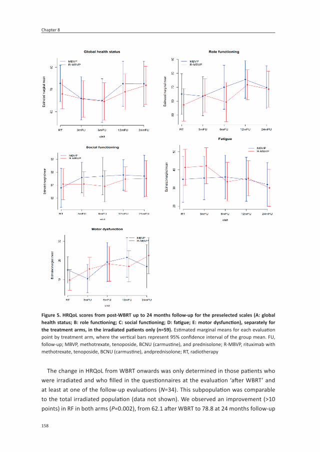
Chapter 8
158
The change in HRQoL from WBRT onwards was only determined in those patients who were irradiated and who filled in the questionnaires at the evaluation ‘after WBRT’ and at least at one of the follow-up evaluations (N=34). This subpopulation was comparable to the total irradiated population (data not shown). We observed an improvement (>10 points) in RF in both arms (P=0.002), from 62.1 after WBRT to 78.8 at 24 months follow-up
figure 5. HrQol scores from post-WbrT up to 24 months follow-up for the preselected scales (A: global health status; b: role functioning; C: social functioning; D: fatigue; E: motor dysfunction), separately for the treatment arms, in the irradiated patients only (n=59). Estimated marginal means for each evaluation point by treatment arm, where the vertical bars represent 95% confidence interval of the group mean. FU, follow-up; MBVP, methotrexate, tenoposide, BCNU (carmustine), and prednisolone; R-MBVP, rituximab with methotrexate, tenoposide, BCNU (carmustine), andprednisolone; RT, radiotherapy
159
Health-related quality of life after chemotherapy with or without rituximab in PCNSL
8
in the R-MBVP arm, and from 65.3 to 77.5 in the MBVP arm. Scores in the other prese-lected scales remained stable in both treatment arms (see Supplemental Figure 4 for the preselected scales).
diSCuSSioN
In the HOVON 105/ALLG NHL 24 trial, the addition of rituximab to standard chemotherapy in adult PCNSL patients did not prolong EFS or PFS; in this HRQoL analysis in 160 patients (80% of total study population), we showed that the addition of rituximab did not improve or deteriorate the patients’ functioning and well-being either. We did, however, observe that anti-tumour treatment resulted in improvements in HRQoL, which were statistically significant and clinically relevant, but did not differ between patients treated with or with-out rituximab.
The largest improvements in HRQoL were observed directly after treatment (i.e. after induction chemotherapy with or without rituximab and after consolidation with WBRT if given). Thereafter, HRQoL scores remained relatively stable or improved more slowly, but gradually over time. Other non-randomized studies investigating the effect of chemo-therapy and/or chemotherapy with rituximab on HRQoL have described a similar pattern: an initial improvement after treatment, followed by stabilization of HRQoL scores up to three years of follow-up.14-16 Two small studies (in which 12/52 and 16/33 of the patients participated in the HRQoL sub-study) even showed an ongoing improvement in HRQoL scores (as measured with the FACT-Br) up to 1217 and 24 months of follow-up.18 Clinical relevance of this change was, however, not defined. Nevertheless, this pattern suggests that HRQoL in PSCNL patients is mainly compromised by the lymphoma itself rather than by the treatment, and that treating the tumour improves patient-reported HRQoL.
Mean baseline HRQoL scores in our cohort are much lower (≥20 points) than in the general population,19 and also lower (≥10 points) than in patients with brain metastases, low-grade glioma and glioblastoma.20-22 HRQoL scores in our population improved signifi-cantly over time, to levels of the general population for some scales, whereas scores in low and high-grade glioma patients typically remain stable up to 24 months of follow-up.21,22 The non-significant differences between treatment arms in deterioration-free survival and time to deterioration for any of the HRQoL scales suggest that treatment itself did not cause a major deterioration in HRQoL.
WBRT as consolidation treatment is under debate because of its presumed negative effect on neurocognitive functioning and subsequently on HRQoL.23 Surprisingly, we found that the HRQoL scores of those patients receiving radiation remained stable for up to two years of follow-up after WBRT, suggesting that the WBRT dose (30Gy) used in this study does not compromise HRQoL in patients up to 60 years in the period covered by this analy-

Chapter 8
160
sis, despite the fact that 17% of the patients in the MBVP-arm and 27% in the R-MBVP arm also received an integrated boost of 10Gy to the tumor-bed. A possible explanation for the stable HRQoL in our cohort after WBRT may be that the 24 month period in our study is too short to develop radiation-induced damage detectable with HRQoL instru-ments, or that the lower radiation dose is less detrimental. Our findings are supported by a study by Correa et al. in which patients treated with R-MPV followed by low-dose WBRT (23.4Gy) also remained stable in their HRQoL scores, even up to five years of follow-up.24 Only a small number of patients could be analysed in our sub analysis and results should therefore be interpreted with caution. In addition, patients who received WBRT could not be compared directly to those who did not receive WBRT because of the age difference between the irradiated and non-irradiated patients. Thus, although our results cannot be generalized and follow-up is still short, our results do challenge the negative role of a relatively low-dose WBRT in this younger subpopulation.
The strengths of our study are the size and standardized treatment of the population studied, the use of validated measures for brain cancer patients, the fact that the majority of this trial population participated in the HRQoL sub-study and that the compliance at every time point was relatively high (>60%). Nevertheless, the actual number of patients who filled in the questionnaires was relatively low at 12 and 24 months of follow-up (N=74 and N=53, respectively) due to progression or death, and patients filling out the question-naires might have a higher level of functioning and well-being than those not filling-out the questionnaires. Also, HRQoL was not systematically assessed at the moment of and beyond progression, hampering information on the impact of progression on HRQoL in this patient population. Another limitation is possible selection bias, because we analysed (subgroups of) a trial population and results may therefore not be generalizable to all patients. However, most patients in the trial also participated in the HRQoL sub-study and only those who did not tolerate two complete courses of (R-)MBVP were excluded. Lastly, the impact of treatment on neurocognition is important in this patient population, and will be described in a separate publication.
In conclusion, HRQoL scores improved after treatment but were not impacted by the addition of rituximab to standard chemotherapy in adult PCNSL patients. Secondly, treat-ment with 30Gy WBRT did not reduce HRQOL in the first two years after treatment in patients up to 60 years old.
For supplemental Tables and Figures: Health-related quality of life after chemotherapy with or without rituximab in primary central nervous system lymphoma patients: results from a randomised phase III study - ScienceDirecthttps://www.sciencedirect.com/science/article/pii/S0923753420393133

161
Health-related quality of life after chemotherapy with or without rituximab in PCNSL
8
refereNCeS
1. van der Meulen M, Dinmohamed AG, Visser O, Doorduijn JK, Bromberg JEC. Improved survival in primary central nervous system lymphoma up to age 70 only: a population-based study on incidence, primary treat-ment and survival in the Netherlands, 1989-2015. Leukemia 2017; 31(8): 1822-5.
2. Villano JL, Koshy M, Shaikh H, Dolecek TA, McCarthy BJ. Age, gender, and racial differ-ences in incidence and survival in primary CNS lymphoma. Br J Cancer 2011; 105(9): 1414-8.
3. Ferreri AJ, Reni M, Pasini F, et al. A multi-center study of treatment of primary CNS lymphoma. Neurology 2002; 58(10): 1513-20.
4. Coiffier B, Lepage E, Briere J, et al. CHOP chemotherapy plus rituximab compared with CHOP alone in elderly patients with diffuse large-B-cell lymphoma. N Engl J Med 2002; 346(4): 235-42.
5. Pfreundschuh M, Schubert J, Ziepert M, et al. Six versus eight cycles of bi-weekly CHOP-14 with or without rituximab in elderly patients with aggressive CD20+ B-cell lymphomas: a randomised controlled trial (RICOVER-60). Lancet Oncol 2008; 9(2): 105-16.
6. Bromberg JEC, Issa S, Bakunina K, et al. Rituximab in patients with primary CNS lymphoma (HOVON 105/ALLG NHL 24): a randomised, open-label, phase 3 intergroup study. Lancet Oncol 2019; 20(2): 216-28.
7. Abrey LE, Batchelor TT, Ferreri AJ, et al. Report of an international workshop to standardize baseline evaluation and re-sponse criteria for primary CNS lymphoma. J Clin Oncol 2005; 23(22): 5034-43.
8. Aaronson NK, Ahmedzai S, Bergman B, et al. The European Organization for Research and Treatment of Cancer QLQ-C30: a quali-ty-of-life instrument for use in international clinical trials in oncology. J Natl Cancer Inst 1993; 85(5): 365-76.
9. Osoba D, Aaronson NK, Muller M, et al. The development and psychometric validation of a brain cancer quality-of-life question-naire for use in combination with general cancer-specific questionnaires. Qual Life Res 1996; 5(1): 139-50.
10. Taphoorn MJ, Claassens L, Aaronson NK, et al. An international validation study of the EORTC brain cancer module (EORTC QLQ-BN20) for assessing health-related quality of life and symptoms in brain cancer patients. Eur J Cancer 2010; 46(6): 1033-40.
11. Fayers PM, Aaronson NK, Bjordal K, Curran D, Groenvold M. The EORTC QLQ-c30 Scor-ing Manual,. 3rd ed. ed. Brussels, Belgium: European Organization for Research and Treatment of Cancer, ; 2001.
12. Osoba D, Rodrigues G, Myles J, Zee B, Pater J. Interpreting the significance of changes in health-related quality-of-life scores. . J Clin Oncol 1998; 16(1): 139-44.
13. Taphoorn MJ, Henriksson R, Bottomley A, et al. Health-Related Quality of Life in a Ran-domized Phase III Study of Bevacizumab, Temozolomide, and Radiotherapy in Newly Diagnosed Glioblastoma. J Clin Oncol 2015; 33(19): 2166-75.
14. Omuro A, Chinot O, Taillandier L, et al. Methotrexate and temozolomide versus methotrexate, procarbazine, vincristine, and cytarabine for primary CNS lymphoma in an elderly population: An intergroup ANOCEF-GOELAMS randomised phase 2 trial. Lancet Haematol 2015; 2(6): e251-e9.
15. Fritsch K, Kasenda B, Schorb E, et al. High-dose methotrexate-based immuno-chemo-therapy for elderly primary CNS lymphoma patients (PRIMAIN study). Leukemia 2017; 31(4): 846-52.
16. Correa DD, Rocco-Donovan M, DeAngelis LM, et al. Prospective cognitive follow-up in primary CNS lymphoma patients treated with chemotherapy and reduced-dose

Chapter 8
162
radiotherapy. J Neuro-Oncol 2009; 91(3): 315-21.
17. Morris PG, Correa DD, Yahalom J, et al. Rituximab, methotrexate, procarbazine, and vincristine followed by consolidation reduced-dose whole-brain radiotherapy and cytarabine in newly diagnosed primary CNS lymphoma: Final results and long-term outcome. J Clin Oncol 2013; 31(31): 3971-9.
18. Omuro A, Correa DD, DeAngelis LM, et al. R-MPV followed by high-dose chemo-therapy with TBC and autologous stem-cell transplant for newly diagnosed primary CNS lymphoma. Blood 2015; 125(9): 1403-10.
19. van de Poll-Franse LV, Mols F, Gundy CM, et al. Normative data for the EORTC QLQ-C30 and EORTC-sexuality items in the general Dutch population. Eur J Cancer 2011; 47(5): 667-75.
20. Habets EJ, Dirven L, Wiggenraad RG, et al. Neurocognitive functioning and health-related quality of life in patients treated with stereotactic radiotherapy for brain metastases: a prospective study. Neuro Oncol 2016; 18(3): 435-44.
21. Reijneveld JC, Taphoorn MJB, Coens C, et al. Health-related quality of life in patients with high-risk low-grade glioma (EORTC 22033-26033): a randomised, open-label, phase 3 intergroup study. Lancet Oncol 2016; 17(11): 1533-42.
22. Taphoorn MJB, Dirven L, Kanner AA, et al. Influence of Treatment With Tumor-Treating Fields on Health-Related Quality of Life of Patients With Newly Diagnosed Glioblastoma: A Secondary Analysis of a Randomized Clinical Trial. JAMA Oncol 2018; 4(4): 495-504.
23. Doolittle ND, Korfel A, Lubow MA, et al. Long-term cognitive function, neuroimag-ing, and quality of life in primary CNS lymphoma. Neurology 2013; 81(1): 84-92.
24. Correa DD, Braun E, Kryza-Lacombe M, et al. Longitudinal cognitive assessment in pa-tients with primary CNS lymphoma treated with induction chemotherapy followed by reduced-dose whole-brain radiotherapy or autologous stem cell transplantation. J Neurooncol 2019; 144(3): 553-62.


1
7
4
10
2
8
5
11
3
9
6
A
PArt iVPrognosis


9MMSE is an independent prognostic factor in primary
central nervous system lymphoma patients
Matthijs van der Meulen MD1, Linda Dirven PhD2,3, Katerina Bakunina MSc4, Martin J. van den Bent MD, PhD1, Samar Issa MD5, Jeanette K. Doorduijn MD, PhD6, Jacoline E.C. Bromberg MD,
PhD1
1 Erasmus MC Cancer Institute, Brain Tumor Center, University Medical Center Rotterdam, Department of Neuro-Oncology;
2 Leiden University Medical Center, Department of Neurology;3 Haaglanden Medical Center, The Hague, Department of Neurology;
4 HOVON Data Center, Department of Hematology, Erasmus MC Cancer Institute, Rotterdam, Netherlands;5 Department of Hematology, Middlemore Hospital, Auckland, New Zealand;
6 Erasmus MC Cancer Institute, University Medical Center Rotterdam, Department of Hematology
Accepted in Journal of Neuro-Oncology

Chapter 9
168
ABStrACt
Objective: To assess the value of the Mini-Mental State Examination (MMSE)-score at baseline in predicting survival in adult primary central nervous system lymphoma (PCNSL) patients.methods: In the HOVON 105/ ALLG NHL 24 phase III study patients with newly-diagnosed PCNSL were randomized between high-dose methotrexate-based chemotherapy with or without rituximab. Data on potential (MMSE-score), and known baseline prognostic fac-tors (age, performance status, serum LDH, cerebrospinal fluid (CSF) total protein, involve-ment of deep brain structures, and multiple cerebral lesions) were collected prospectively. Multivariate stepwise Cox regression analyses were used to assess the prognostic value of all factors on progression free survival (PFS) and overall survival (OS) among patients with available MMSE score at baseline. Age was analysed as continuous variable, the MMSE-score both as a continuous and as a categorical variable.results: In univariate analysis, age, MMSE-score and whether the patient received rituximab were statistically significantly prognostic factors for PFS. Age and MMSE-score were statistically significantly associated with OS. In a multivariate analysis of the univari-ately significant factors only MMSE-score was independently associated with the survival endpoints, as a continuous variable (HR for PFS 1.04, 95% CI 1.01-1.08; OS 1.06 (95% CI 1.02-1.10) and as categorical variable HR (<27 versus ≥27 for PFS 1.55 (1.02-2.35); OS 1.68 (1.05-2.70). In our population, performance status, serum LDH, and CSF protein level were not of prognostic value.Conclusion: Neurocognitive disturbances, measured with the MMSE at baseline, are an unfavourable prognostic factor for both PFS and OS in adult PCNSL patients up to 70 years-old.

169
MMSE is an independent prognostic factor in PCNSL patients
9
iNtroduCtioN
Primary central nervous system lymphoma (PCNSL) is a rare non-Hodgkin lymphoma con-fined to the brain, leptomeninges, spinal cord and eyes. Over the last decades prognosis has improved significantly.1 Although several prognostic factors have been identified and prognostic models have been developed, it remains difficult to predict the prognosis of individual patients.
Two prognostic models are currently widely used in PCNSL patients: the externally validated Memorial Sloan Kettering Cancer Center (MSKCC) prognostic score:2age (>50 years-old) and Karnofsky Performance score (KPS; <70), and the International Extranodal Lymphoma Study Group (IELSG) score: age (>60 years-old), WHO/ECOG Performance Sta-tus (PS; >1), Lactate dehydrogenase (LDH) serum level, cerebrospinal fluid (CSF) protein level and involvement of deep brain structures.3
The Mini-Mental State Examination (MMSE)4 is a crude screening tool for neurocogni-tive impairment. In high-grade glioma, the MMSE-score was an independent prognostic factor for both progression free survival (PFS) and overall survival (OS).5
In PCNSL patients, data regarding the prognostic value of the MMSE are scarce, despite the fact that cognitive symptoms occur frequently (up to 43%) in this disease.6 One study describes 95 elderly (>60 years-old) PCNSL patients, and found that MMSE-score ≤24 was the only independent prognostic factor for OS, while age and PS were not.7 In the present study we aimed to assess whether the MMSE-score at baseline was independently prognostic for both PFS and OS, in a large trial population with adult PCNSL patients up to 70 years-old.
methodS
Patients in the HOVON 105/ALLG NHL 24 study, a large multicentre phase III randomized controlled trial (RCT) for immunocompetent adult patients with newly diagnosed CD20 positive B-cell PCNSL with WHO/ECOG PS 0-3, were included.8 The treatment regimen consisted of two cycles of high-dose methotrexate-based chemotherapy, with or with-out rituximab, followed by high-dose-cytarabine. Patients <61 years-old subsequently received 30Gy whole brain radiotherapy. The study was approved by the ethics committee at all participating centres and all participants gave informed consent. Patients underwent an MMSE if possible and baseline scores were obtained before chemotherapy was started.
All patients for whom an MMSE-score at baseline was available were included in this study. In addition, the following information was collected: sex, age, WHO/ECOG PS, CSF protein and serum LDH levels at baseline and whether the patient had multiple cerebral lesions, involvement of deep brain structures (periventricular regions, basal ganglia, brain-stem and/ or cerebellum), and whether they received rituximab.

Chapter 9
170
First, possible imbalances were assessed with respect to baseline characteristics, treat-ment details and survival between those who participated in this side-study and those who could not due to missing MMSE-scores at baseline.
Subsequently, all the above mentioned prognostic factors were assessed separately for association with PFS and OS using univariate Cox regression analysis. PFS was defined as time from randomization to progression, relapse or death from any cause, whichever came first. OS was defined as time from randomization to death from any cause.8 Patients still alive at the date of last contact were censored. MMSE was included both as a continuous variable and as categorical variable (<27 or ≥27). Age was included as a continuous variable. ECOG status (≤1 versus >1), serum LDH (above versus below local upper limit of normal), and CSF protein (above versus below cut-off values according to the IELSG score3) were included as categorical variables. Factors that were statistically significant in univariate analysis were included in the stepwise multivariate Cox proportional hazards models. A p-value <0.05 was considered statistically significant. All analyses were performed with Stata version 15.
data availability statement
Individual de-identified participant data, collected for this study, including the statistical analysis plan will be made available for other research to others upon request, after ap-proval by the HOVON executive board. The data will be available until a maximum of 15 years after the study has ended.
Please find the trial protocol, a Data Request Form and the criteria for data sharing on www.hovon.nl.
reSuLtS
MMSE-score at baseline was available for 153 of the 199 (77%) trial patients. Baseline characteristics and survival were comparable between those who were included and were not, Supplemental Table 1 and Supplemental Figure 1A and B.
In the univariate regression analyses age, receipt of rituximab and baseline MMSE-score were associated with PFS. Only age and MMSE were statistically significant predictors of OS (Table 1). In multivariate analysis, only MMSE-score at baseline was independently associated with both PFS and OS. We found that each unit decrease in MMSE-score was associated with a poorer prognosis: for PFS (Hazard Ratio [HR], 95% confidence interval [CI] 1.04, 1.01-1.08) and OS (HR, 95% CI: 1.06, 1.02-1.10), Table 2. When including the MMSE-score as categorical variable in multivariate analyses, corrected for age and ritux-imab, a baseline-score <27 (as compared to a score ≥27) was the only factor associated with PFS (HR 1.55, 95% CI: 1.02-2.35) and overall survival (HR 1.68, 95% CI: 1.05-2.70), Table 2 and Figure 1.

171
MMSE is an independent prognostic factor in PCNSL patients
9
table 1. Univariate and multivariate Cox regression analysis for all risk factors with MMSE as a continuous variable for the progression-free survival and overall survival.
Progression-free survival
univariate Multivariate
n HR (95% CI) p HR (95% CI) p
Female 153 0.85 (0.57-1.28) 0.44
Age (increase; unit = 10 years) 153 1.33 (1.04-1.71) 0.025 1.28 (0.99-1.65) 0.061
WHO/ECOG >1 153 0.92 (0.57-1.50) 0.74
Multiple lesions 138 0.89 (0.58-1.37) 0.59
Deep structures involved 153 1.39 (0.92-2.09) 0.12
Elevated CSF total protein 93 0.78 (0.45-1.37) 0.40
LDH >ULN 153 1.19 (0.77-1.82) 0.44
Rituximab 153 0.66 (0.44-1.00) 0.049 0.69 (0.45-1.04) 0.075
MMSE (decrease unit = 1 point) 153 1.05 (1.01-1.08) 0.0042 1.04 (1.01-1.08) 0.008
overall survival
univariate Multivariate
n HR (95% CI) p HR (95% CI) p
Female 153 1.12 (0.71-1.76) 0.64
Age (increase; unit = 10 years) 153 1.36 (1.02-1.82) 0.036 1.32 (0.97-1.77) 0.069
WHO/ECOG >1 153 1.29 (0.77-2.16) 0.32
Multiple lesions 138 1.01 (0.62-1.63) 0.97
Deep structures involved 153 1.25 (0.79-1.99) 0.34
Elevated CSF total protein 93 0.64 (0.33-1.26) 0.20
LDH >ULN 153 1.15 (0.71-1.88) 0.57
Rituximab 153 0.86 (0.55-1.35) 0.51
MMSE decrease unit = 1 point) 153 1.06 (1.02-1.10) 0.001 1.06 (1.02-1.10) 0.002
Hazard ratio’s (HR) and 95% confidence intervals (CI) are shown with their p-value. WHO = World Health Organization, ECOG = Eastern Cooperative Oncology Group, CSF = cerebrospinal fluid, LDH = lactate dehydro-genase, ULN = upper limit of normal, MMSE = mini-mental state examination.
table 2. Multivariate Cox regression analysis for univariately significant risk factors with MMSE as a categorical variable for the progression-free survival and overall survival.
Progression-free survival
Multivariate
HR (95% CI) p
Age (increase; unit = 10 years) 1.24 (0.95-1.60) 0.109
Rituximab 0.70 (0.47-1.05) 0.087
MMSE <27 1.55 (1.02-2.35) 0.040
overall survival
Multivariate
HR (95% CI) p
Age (increase; unit = 10 years) 1.26 (0.94-1.71) 0.127
MMSE <27 1.68 (1.05-2.70) 0.031
Hazard ratio’s (HR) and 95% confidence intervals (CI) are shown with their p-value. MMSE = mini-mental state examination.
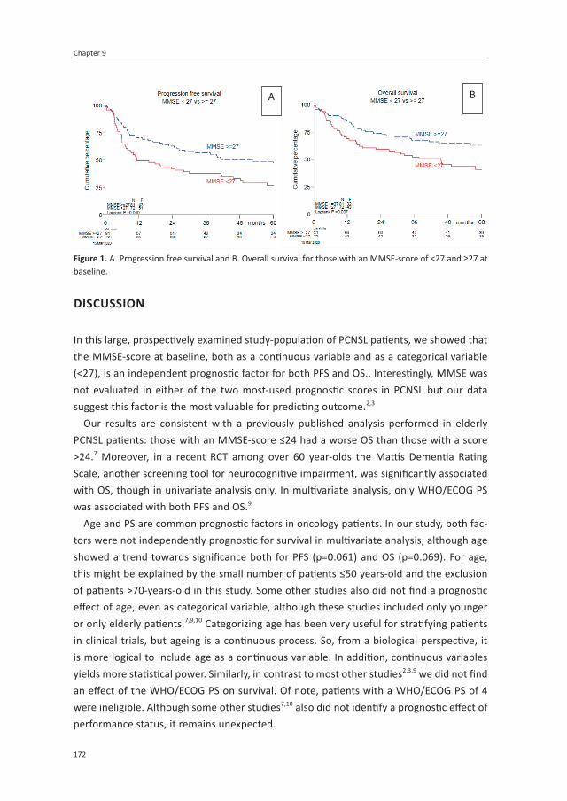
Chapter 9
172
diSCuSSioN
In this large, prospectively examined study-population of PCNSL patients, we showed that the MMSE-score at baseline, both as a continuous variable and as a categorical variable (<27), is an independent prognostic factor for both PFS and OS.. Interestingly, MMSE was not evaluated in either of the two most-used prognostic scores in PCNSL but our data suggest this factor is the most valuable for predicting outcome.2,3
Our results are consistent with a previously published analysis performed in elderly PCNSL patients: those with an MMSE-score ≤24 had a worse OS than those with a score >24.7 Moreover, in a recent RCT among over 60 year-olds the Mattis Dementia Rating Scale, another screening tool for neurocognitive impairment, was significantly associated with OS, though in univariate analysis only. In multivariate analysis, only WHO/ECOG PS was associated with both PFS and OS.9
Age and PS are common prognostic factors in oncology patients. In our study, both fac-tors were not independently prognostic for survival in multivariate analysis, although age showed a trend towards significance both for PFS (p=0.061) and OS (p=0.069). For age, this might be explained by the small number of patients ≤50 years-old and the exclusion of patients >70-years-old in this study. Some other studies also did not find a prognostic effect of age, even as categorical variable, although these studies included only younger or only elderly patients.7,9,10 Categorizing age has been very useful for stratifying patients in clinical trials, but ageing is a continuous process. So, from a biological perspective, it is more logical to include age as a continuous variable. In addition, continuous variables yields more statistical power. Similarly, in contrast to most other studies2,3,9 we did not find an effect of the WHO/ECOG PS on survival. Of note, patients with a WHO/ECOG PS of 4 were ineligible. Although some other studies7,10 also did not identify a prognostic effect of performance status, it remains unexpected.
A B
figure 1. A. Progression free survival and B. Overall survival for those with an MMSE-score of <27 and ≥27 at baseline.
173
MMSE is an independent prognostic factor in PCNSL patients
9
The major strength of our study is the prospective data collection within a large clinical trial resulting in MMSE-scores for the majority of patients and a uniform treatment and evaluation protocol. A limitation is the relatively small number of patients for prognostica-tion; our sample size is smaller than that in the MSKCC (n=238) and IELSG models (n=378). A down-side of all studies based on trial patients is that findings may not be generalizable to the whole PCNSL population.
To conclude, the MMSE is an easily assessable and relevant clinical factor which has not been included in prior prognostic studies in patients with PCNSL. In this dataset the MMSE-score at baseline is an independent clinical prognostic factor in adult PCNSL patients up to 70-years-old. If validated in another large population, patients should be counselled with this effect in mind, and other prognostic scores should be re-evaluated.
For supplemental Tables and Figures: MMSE is an independent prognostic factor for sur-vival in primary central nervous system lymphoma | SpringerLinkhttps://link.springer.com/article/10.1007/s11060-021-03708-8

Chapter 9
174
refereNCeS
1. van der Meulen M, Dinmohamed AG, Visser O, Doorduijn JK, Bromberg JEC. Improved survival in primary central nervous system lymphoma up to age 70 only: a population-based study on incidence, primary treat-ment and survival in the Netherlands, 1989-2015. Leukemia 2017;31:1822-1825.
2. Abrey LE, Ben-Porat L, Panageas KS, et al. Primary central nervous system lymphoma: the Memorial Sloan-Kettering Cancer Center prognostic model. J Clin Oncol 2006;24:5711-5715.
3. Ferreri AJ, Blay JY, Reni M, et al. Prognostic scoring system for primary CNS lymphomas: the International Extranodal Lymphoma Study Group experience. J Clin Oncol 2003;21:266-272.
4. Folstein MF, Folstein SE, McHugh PR. “Mini-mental state”. A practical method for grad-ing the cognitive state of patients for the clinician. J Psychiatr Res 1975;12:189-198.
5. Klein M, Postma TJ, Taphoorn MJ, et al. The prognostic value of cognitive functioning in the survival of patients with high-grade glioma. Neurology 2003;61:1796-1798.
6. Bataille B, Delwail V, Menet E, et al. Primary intracerebral malignant lymphoma: report of 248 cases. J Neurosurg 2000;92:261-266.
7. Omuro A, Chinot O, Taillandier L, et al. Methotrexate and temozolomide versus methotrexate, procarbazine, vincristine, and cytarabine for primary CNS lymphoma in an elderly population: An intergroup ANOCEF-GOELAMS randomised phase 2 trial. Lancet Haematol 2015;2:e251-e259.
8. Bromberg JEC, Issa S, Bakunina K, et al. Rituximab in patients with primary CNS lymphoma (HOVON 105/ALLG NHL 24): a randomised, open-label, phase 3 intergroup study. Lancet Oncol 2019;20:216-228.
9. Houillier C, Taillandier L, Dureau S, et al. Radiotherapy or Autologous Stem-Cell Transplantation for Primary CNS Lymphoma in Patients 60 Years of Age and Younger: Re-sults of the Intergroup ANOCEF-GOELAMS Randomized Phase II PRECIS Study. J Clin Oncol 2019;37:823-833.
10. Fritsch K, Kasenda B, Schorb E, et al. High-dose methotrexate-based immuno-chemotherapy for elderly primary CNS lym-phoma patients (PRIMAIN study). Leukemia 2017;31:846-852.

175
MMSE is an independent prognostic factor in PCNSL patients
9


10General discussion


179
General discussion
10
Epidemiology
Primary central nervous system lymphoma is a rare disease and due to stringent inclu-sion criteria in clinical trials, large population-based studies can complement data from prospective intervention studies by addressing an unbiased group of patients within a well-defined geographic area. In chapter 2 we described the incidence, primary treatment and survival among 1,673 PCNSL patients diagnosed in the Netherlands between 1989 and 2015 using the comprehensive data of the Netherlands Cancer Registry (NCR). We showed that the incidence of primary central nervous system lymphoma increased, but only among those over the age of 60. Two major factors have been identified as risk factors for PCNSL: being immunocompromised and higher age. However, the incidence increased mainly among immunocompetent patients and although the population in the Nether-lands is ageing, the incidence of systemic lymphoma and of glioma did not increase as much as the incidence of PCNSL.1,2 Other population-based studies in Western and Eastern countries described similar trends in incidence and survival, also without an explanation for the increased incidence.3-5 So it remains undermined which factors contributed to this increase.
Survival in PCNSL has improved over the last decades. In this study on 1,673 patients we showed that the 5-year relative survival rate, correcting for survival in the general popula-tion increased from 11% (95% confidence interval [CI] 8-15%) to 30% (95% CI 27-34%) over the past 30 years. However, this increased survival was observed only among those below the age of 70. In a multivariate regression analysis we demonstrated that age and treatment were significantly associated with survival; more chemotherapy was associated with better survival. Survival in patients over 70 remained poor, with a 5-year relative survival rate of 6% (95% confidence interval [CI] 2-13%) in 2009-2015. This poor survival among elderly PCNSL patients is also described in a population-based study on nearly 26,000 elderly in the United States, based on the Central Brain Tumor Registry of the United States (CBTRUS) and Surveillance, Epidemiology, and End Results (SEER) database, entitled ‘The elderly left behind’.6
In chapter 3 we described the primary treatment and survival of 145 elderly (>70 year-old) patients with PCNSL diagnosed in the Netherlands between 2014 and 2017. Overall, median overall survival was only 4.1 months (95% CI, 2.8-6.8) After dividing the elderly into three groups (71-74, 75-80, >80 years-old) we found no difference in survival between these three groups. This might be explained by small numbers (n=58-32) in each group, the poor survival even in the youngest of these age groups, or other factors, such as performance status or comorbidity. Details on the latter two factors are, unfortunately, incomplete. We found that over the last 27 years, the increased use of chemotherapy did not result in an improved survival among over 70 year-olds. In the contemporary era (2014-2017), however, those elderly patients (>70 years-old) judged fit enough to receive chemotherapy had a better overall survival (median: 16.3 months, 95% CI 7.8-35.2) than

Chapter 10
180
those who received radiotherapy only (median 7.7 months, 95% CI 4.6-13.2) or supportive care only (median 1.4 months, 95% CI 1.1-1.7). Siegal and Bairey, who summarized tri-als done in elderly PCNSL patients, described the treatment challenges in elderly.7 First, elderly are defined differently in the performed trials, which complicates clear treatment recommendations for elderly PCNSL patients. Second, although methotrexate causes more treatment-related toxicities in elderly patients, a considerable number of elderly patients are nevertheless eligible for this important antineoplastic treatment in PCNSL. The key here is to identify those who are too much at risk for treatment-related toxicity and those who are not. In line with these authors we concluded that new prospective studies are needed to define the best treatment for different groups of elderly diagnosed with PCNSL.
Diagnostic evaluation
Before a long, intensive and potentially toxic treatment for PCNSL can be initiated, a definitive diagnosis is essential. This diagnosis can be obtained by the examination of vitreous fluid in case of eye involvement, of cerebrospinal fluid (CSF) or by a brain biopsy. Flow cytometry on CSF, in addition to cytological analysis, is more sensitive than cytology alone.8-10 In chapter 4 we showed that flow cytometry analysis on brain biopsies has a 100%-specificity for the diagnosis of lymphoma, but sensitivity was only 88%. A major added value of flow cytometry on brain biopsies was the speed in which a diagnosis could be given: median time in days (range) was 1 (0-7) for flow cytometry and 5 (0-18) for histology and immunohistochemistry. Because of the highly aggressive nature of PCNSL, patients can quickly deteriorate, and a faster diagnostic process is therefore beneficial. Our analysis was limited by a retrospective single centre design, but two smaller series, one on brain biopsies and one on brain biopsy rinse fluid, gave the same results.11,12 We recommend to perform both, histology and immunohistochemistry and flow cytometry, if the latter is positive for a lymphoma, treatment steps can be initiated.
After treatment is initiated it is important to measure treatment response, in order to be able to adapt treatment in patients not responding sufficiently. According to current guidelines, an MRI is made at baseline, after each treatment component, and during fol-low-up.13,14 After treatment and during follow-up response criteria are used to determine whether there is a response, what the extent of response is, or whether there is stable or progressive disease (progression or relapse).14 Since PCNSL is a rare disease, most hospi-tals have limited experience in assessing radiological response in PCNSL. Most treatment decisions, however, are based on these response criteria. In chapter 5 we assessed the value of a central radiology review based on patients treated in the HOVON 105/ ALLG NHL 24 study, an international multicentre phase III study on the effect of rituximab in newly diagnosed patients with PCNSL.15 In 235 MRIs made after each treatment component, the interobserver agreement was rather modest and similar between the two central radiology reviewers and between local and central response assessment: kappa 0.45.

181
General discussion
10
Even between the experienced central reviewers we observed great discrepancies (e.g. complete response versus partial response). However, interobserver agreement regarding progression or no progression was excellent between the central reviewers (kappa 0.93) as well as between local and central radiology assessment (kappa 0.86). Together, these data suggest that there is little added value of a central radiology review in PCNSL trial for assessing progression and that response categorisation is not straightforward.
Based on the treatment response at the end-of-treatment MRIs we did a landmark analysis based on the central response assessment: there were no differences in progres-sion free survival or overall survival between those categorized as complete response (CR), complete response unconfirmed (CRu) or partial response (PR) at 6.9 months after randomization (the landmark). Since progression can be clearly recognized and no dif-ferences in survival was found between different types of response, this suggests that survival endpoints are more reliable in clinical trials than the complete response rate. Other articles did find a difference between those categorized as CR versus no CR.16,17 However, in these studies a landmark analysis was not performed and patients who did not respond (SD) or even already had progression were included in the ‘no CR’ group. One article describes a landmark analysis and found a significant difference between response categories, however, all response category: CR, CRu and PR, but also stable disease and progression were compared with each other in a single log-rank test.18 Our study concerns a modest number of patients and results would need to be validated in a larger cohort. However if validated, the response criteria could be simplified into: response, defined as a decrease of enhancement of >50%, stable disease or progression.
Cognition and health-related quality of life
In the HOVON 105/ ALLG NHL 24 study, 199 immunocompetent patients with a newly diagnosed CD20+ B-cell PCNSL and aged between 18-70 years-old were randomized 1:1 between high-dose methotrexate based chemotherapy (MBVP) with or without rituximab: R-MBVP versus MBVP.15 This induction treatment was followed by consolidative high-dose cytarabine (Ara-C), and for patients ≤60 years-old also by reduced dose (30Gy) whole-brain radiotherapy (WBRT). The primary endpoint of this study, the 1-year event-free survival, did not differ between the treatment arms: R-MBVP versus MBVP: 52% (95% CI 42-61) versus 49% (95% CI 39-58), p=0.99. Moreover, the progression-free survival and overall survival, after a median follow-up of 32.9 months, also did not differ. Secondary endpoints of this randomized controlled trial were differences in neurocognitive function and health-related quality of life (HRQoL). In the search for the best treatment the aim is to prolong both the length of survival and the quality of survival. The latter can be influenced by neurocognitive functioning and HRQoL. Information about both, survival and quality of survival – the net clinical benefit - can help physicians and patients to make a well-informed decision regarding individual treatment options.
Chapter 10
182
In chapter 6 we systematically reviewed the current literature on neurocognitive func-tioning and HRQoL in PCNSL patients. The main conclusions were that the tumor itself had a large impact on both cognitive functioning and HRQoL, and that WBRT in addition to chemotherapy had a negative impact on cognitive functioning. However, the magnitude of this effectt was mostly not considered clinically relevant (with clinical relevance defined as a change in z-score of >1 or >1.5 standard deviation).
In the HOVON 105/ ALLG NHL 24, neurocognitive functioning and HRQoL was assessed before, during and up to 2-years after treatment. At group level, we found a significant improvement over time in all cognitive domains (chapter 7) and all primary scales of the HRQoL analysis (chapter 8), without differences between the treatment arms. In addition to a statistically significant improvement, at 12 and 24 months of follow-up, HRQoL-scores also improved to a clinically relevant extent (≥10 points), compared to baseline. The larg-est improvement was seen between baseline and end-of-treatment, thereafter scores remained stable, which is a pattern that has been described in other studies.19-24 Again, this suggests that neurocognitive functioning and HRQoL is mostly compromised by the tumor itself.
At the individual level, we observed that scores at 12 months after treatment in all cognitive domains, compared to baseline, improved in 33-58% of the patients. After 24 months, 53% had improved scores in the memory and 68% in motor speed domain. For at-tention/executive functioning and information processing speed most patients remained stable: 59% and 48%, respectively. Only a minority of patients had worse neurocognitive functioning at 12 and 24 months (0-7%).In the HRQoL analysis, at individual level, the majority improved at 12 (46-78%) and 24 (53-82%) months post-treatment, compared to baseline.
In irradiated patients we observed, surprisingly, that up to 2 years post-treatment, scores in all cognitive domains and HRQoL scales remained stable, compared to shortly after WBRT. The role of WBRT in the treatment of PCNSL continues to be under debate, mainly because of its negative effect on neurocognitive functioning. Since only younger patients (≤60 years-old) received WBRT, our results cannot be generalized to the PCNSL patients of all ages. The effect of WBRT, both on survival and cognitive functioning has been assessed in multiples studies, but in different ways.16,19-21 WBRT prolonged progression free survival when WBRT was added to high-dose methotrexate (HD-MTX) chemotherapy, compared to chemotherapy only in a large randomised study: 18 versus 12 months, although overall survival was similar between both groups.16 However, more neurotoxicity, unfortunately only measured with clinical and radiological examinations, was found in those treated with 45Gy WBRT compared radiation naïve patients. Formal cognitive testing was not performed. A meta-analysis showed that those who received WBRT (45-60Gy; n=65) had more white matter lesions and significantly worse scores on multiple cognitive domains, compared to those treated without WBRT (n=15, p<0.001). These differences were,

183
General discussion
10
however, small and not clinically relevant.25 This comparison was made after a median follow-up of 5.5 years, range 2-26 years.
Two recent trials randomized PCNSL patients responding to induction chemotherapy, to WBRT or to autologous stem cell transplantation (ASCT).20,21 Both showed significantly better neurocognitive functioning in the ASCT-arm, up to two20 and three21 years of follow-up. Both studies used a higher dose of radiotherapy (36-40 Gy), neither presented indi-vidual scores or standardised scores of neurocognitive functioning and HRQoL was not performed21 or reported on.20 This hampers conclusions about the clinical relevance of the changes in neurocognitive functioning and the comparison with our data.
Lastly, a single arm phase II study showed that HD-MTX-based chemotherapy followed by 23.4Gy WBRT gave similar survival compared to earlier trials with higher doses of radiation, and with neurocognitive functioning remaining stable even up to 5-years post-treatment.26 These results are in line with our study: patients ≤60 years-old, were treated with 30Gy WBRT (20x1.5Gy) and remained stable during the follow-up of 2 years. These findings suggest that radiation dose is crucial in the effect of cognitive functioning in PCNSL patients. However, longer follow-up is necessary to draw definitive conclusions on neurocognitive function after 30Gy WBRT in our population.
In patients for whom a neuropsychological evaluation and MRI images were available at end-of-treatment, at 6, 12 and/or 24 months of follow-up we determined the correlation between white matter abnormalities (WMA) and brain atrophy on changes (i.e. deteriora-tion) in each cognitive domain. There was a significant association between an increase in WMA and a decrease in z-score in the domains of attention/executive functioning, in-formation processing speed and motor speed and between brain atrophy and the domain of memory. The associations were modest, the regression coefficients ranged between -0.048 and -0.921, which is the decrease in z-score after an increase of 1 point in the Faze-kas27 sumscore for white matter lesions (the Fazekas score in 5 brain areas on both sides (0-30)) or after a decrease in brain volume with 10%. One study, which combined data on neurocognitive functioning and WMA from multiple studies, also showed a significant effect between increased WMA and decreased neurocognitive functioning.25
Prognosis
As discussed in chapter 2, the prognosis of PCNSL patients has improved significantly over the past decades. Despite the fact that multiple prognostic models28-30 and prognostic factors31-35 have been described, it remains difficult to predict survival for individual pa-tients. The two most used prognostic models today are the externally validated Memorial Sloan Kettering Cancer Center (MSKCC) prognostic model, including only age (>50) and Karnofsky Performance Score (PS; >70) and the International Extranodal Lymphoma Study Group (IELSG) score, including age (>60), WHO/ECOG PS (>1), the involvement of deep

Chapter 10
184
brain structures, increased serum LDH and/ or increased cerebrospinal fluid (CSF) total protein.36,37
The Mini-Mental State Examination (MMSE38) score, a simple bedside test originally developed for patients with dementia, is a prognostic factor in both low and high-grade glioma.39,40 In chapter 9 we showed that, based on HOVON 105/ALLG NHL 24 trial patients, the MMSE-score at baseline, i.e. before the start of chemotherapy, is an independent prognostic factor associated with progression free and overall survival in PCNSL patients up to the age of 70.
Interestingly age and performance status were not independent prognostic factors in our multivariate analysis. Several factors could explain the discrepancy between our results in this prognostic study, the population-based studies3,41 and the MSKCC and IELSG scores.36,37 The small number of patients included below the age of 50 and the exclusion of those over 70 years-old in the trial, as well as the relatively small number of patients might play a role. The other four studies were larger (n=238-3,100) than ours (n=153), and thus had more power to detect correlations. Furthermore, there may be an interaction between age, performance status and cognition – all significant in univariate analysis, resulting in only one of them remaining significant in multivariate analysis. Lastly, prospective clinical trials use stringent inclusion criteria regarding age, performance status and comorbidity.
Future directions
Given the data from recent randomized phase II trials,20,21 which showed a comparable efficacy but less cognitive deterioration in patients treated with autologous stem cell transplantation (ASCT), probably fewer PCNSL patients, will receive WBRT as consolidative therapy in the near future. However, reduced dose WBRT could be a reasonable option for some patients, given the above described results, if cognition remains stable even with longer-term follow-up. For elderly or frail patients, in whom neither WBRT nor ASCT are therapeutic options, maintenance therapy is under investigation.45,46 All considered options have advantages and disadvantages, which are influenced by age, comorbidity and immune status. Taking all these factors into account, treatment will likely become more personalized.
It is becoming more and more clear that not only the length but also the quality of survival counts. Combining these can help determine the so called ‘net clinical benefit’ of treatments. The challenge here is to measure, calculate and report on neurocognitive functioning and HRQoL in a standardized manner so that information from different stud-ies can be compared. Due to difference in these aspects, as shown above, we could not compare results between trials. A consensus paper was published previously about when to measure neurocognitive functioning and HRQoL and which tests can be used best.47 a universal protocol how to measure and to report on these data would facilitate com-parison across trials, as was developed for research in patients with brain metastases.48

185
General discussion
10
Because survival has increased and neurocognitive functioning remains below the level of the norm population (<-1 in z-scores), a more personalized approached, based on toxicity, white matter lesions and cognitive functioning would be a helpful in order to counsel patients through their follow-up.
Lastly, many PCNSL researchers are currently already collaborating, but in order to answer important research questions that a stronger international collaboration is needed. Questions such as: what are the reasons for the increased incidence?, which factors main-tain prognostic value in large, unbiased populations? can perhaps be answered if national databases, such as the American SEER and CBTRUS databases, our NCR database, and the French Oculo-Cerebral Lymphoma (LOC)-network were combined. This will, however be quite a challenge since these databases are organized differently and do not include the same parameters. Wider collaboration is however clearly needed in this rare disease.

Chapter 10
186
refereNCeS
1. Ho VK, Reijneveld JC, Enting RH, et al. Changing incidence and improved survival of gliomas. Eur J Cancer 2014; 50(13): 2309-18.
2. Issa DE, van de Schans SA, Chamuleau ME, et al. Trends in incidence, treatment and survival of aggressive B-cell lymphoma in the Netherlands 1989-2010. Haematologica 2015; 100(4): 525-33.
3. Villano JL, Koshy M, Shaikh H, Dolecek TA, McCarthy BJ. Age, gender, and racial differ-ences in incidence and survival in primary CNS lymphoma. Br J Cancer 2011; 105(9): 1414-8.
4. Shin SH, Jung KW, Ha J, Lee SH, Won YJ, Yoo H. Population-based Incidence and Survival for Primary Central Nervous System Lymphoma in Korea, 1999-2009. Cancer Res Treat 2015; 47(4): 569-74.
5. Neuhauser M, Roetzer T, Oberndorfer S, et al. Increasing use of immunotherapy and prolonged survival among younger patients with primary CNS lymphoma: a population-based study. Acta Oncol 2019; 58(7): 967-76.
6. Mendez JS, Ostrom QT, Gittleman H, et al. The elderly left behind-changes in survival trends of primary central nervous system lymphoma over the past 4 decades. Neuro Oncol 2018; 20(5): 687-94.
7. Siegal T, Bairey O. Primary CNS Lymphoma in the Elderly: The Challenge. Acta Haema-tol 2019; 141(3): 138-45.
8. Bromberg JE, Breems DA, Kraan J, et al. CSF flow cytometry greatly improves diagnostic accuracy in CNS hematologic malignancies. Neurology 2007; 68(20): 1674-9.
9. Roma AA, Garcia A, Avagnina A, Rescia C, Elsner B. Lymphoid and myeloid neoplasms involving cerebrospinal fluid: comparison of morphologic examination and immu-nophenotyping by flow cytometry. Diagn Cytopathol 2002; 27(5): 271-5.
10. Hegde U, Filie A, Little RF, et al. High inci-dence of occult leptomeningeal disease detected by flow cytometry in newly diag-nosed aggressive B-cell lymphomas at risk for central nervous system involvement: the role of flow cytometry versus cytology. Blood 2005; 105(2): 496-502.
11. Cordone I, Masi S, Carosi M, et al. Brain stereotactic biopsy flow cytometry for central nervous system lymphoma charac-terization: advantages and pitfalls. J Exp Clin Cancer Res 2016; 35(1): 128.
12. Debliquis A, Voirin J, Harzallah I, et al. Cytomorphology and flow cytometry of brain biopsy rinse fluid enables faster and multidisciplinary diagnosis of large B-cell lymphoma of the central nervous system. Cytometry B Clin Cytom 2018; 94(1): 182-8.
13. Hoang-Xuan K, Bessell E, Bromberg J, et al. Diagnosis and treatment of primary CNS lymphoma in immunocompetent patients: guidelines from the European Association for Neuro-Oncology. Lancet Oncol 2015; 16(7): e322-32.
14. Abrey LE, Batchelor TT, Ferreri AJ, et al. Report of an international workshop to standardize baseline evaluation and re-sponse criteria for primary CNS lymphoma. J Clin Oncol 2005; 23(22): 5034-43.
15. Bromberg JEC, Issa S, Bakunina K, et al. Rituximab in patients with primary CNS lymphoma (HOVON 105/ALLG NHL 24): a randomised, open-label, phase 3 intergroup study. Lancet Oncol 2019; 20(2): 216-28.
16. Thiel E, Korfel A, Martus P, et al. High-dose methotrexate with or without whole brain radiotherapy for primary CNS lymphoma (G-PCNSL-SG-1): a phase 3, randomised, non-inferiority trial. Lancet Oncol 2010; 11(11): 1036-47.
17. Kim YR, Kim SH, Chang JH, et al. Early response to high-dose methotrexate, vin-cristine, and procarbazine chemotherapy-adapted strategy for primary CNS lympho-

187
General discussion
10
ma: no consolidation therapy for patients achieving early complete response. Ann Hematol 2014; 93(2): 211-9.
18. Tabouret E, Houillier C, Martin-Duverneuil N, et al. Patterns of response and relapse in primary CNS lymphomas after first-line chemotherapy: imaging analysis of the ANOCEF-GOELAMS prospective randomized trial. Neuro Oncol 2017; 19(3): 422-9.
19. Morris PG, Correa DD, Yahalom J, et al. Rituximab, methotrexate, procarbazine, and vincristine followed by consolidation reduced-dose whole-brain radiotherapy and cytarabine in newly diagnosed primary CNS lymphoma: Final results and long-term outcome. J Clin Oncol 2013; 31(31): 3971-9.
20. Ferreri AJM, Cwynarski K, Pulczynski E, et al. Whole-brain radiotherapy or autologous stem-cell transplantation as consolidation strategies after high-dose methotrexate-based chemoimmunotherapy in patients with primary CNS lymphoma: results of the second randomisation of the International Extranodal Lymphoma Study Group-32 phase 2 trial. Lancet Haematol 2017; 4(11): e510-e23.
21. Houillier C, Taillandier L, Dureau S, et al. Radiotherapy or Autologous Stem-Cell Transplantation for Primary CNS Lymphoma in Patients 60 Years of Age and Younger: Re-sults of the Intergroup ANOCEF-GOELAMS Randomized Phase II PRECIS Study. J Clin Oncol 2019; 37(10): 823-33.
22. Omuro A, Chinot O, Taillandier L, et al. Methotrexate and temozolomide versus methotrexate, procarbazine, vincristine, and cytarabine for primary CNS lymphoma in an elderly population: An intergroup ANOCEF-GOELAMS randomised phase 2 trial. Lancet Haematol 2015; 2(6): e251-e9.
23. Fritsch K, Kasenda B, Schorb E, et al. High-dose methotrexate-based immuno-chemo-therapy for elderly primary CNS lymphoma patients (PRIMAIN study). Leukemia 2017; 31(4): 846-52.
24. Correa DD, Rocco-Donovan M, DeAngelis LM, et al. Prospective cognitive follow-up in primary CNS lymphoma patients treated with chemotherapy and reduced-dose radiotherapy. J Neuro-Oncol 2009; 91(3): 315-21.
25. Doolittle ND, Korfel A, Lubow MA, et al. Long-term cognitive function, neuroimag-ing, and quality of life in primary CNS lymphoma. Neurology 2013; 81(1): 84-92.
26. Correa DD, Braun E, Kryza-Lacombe M, et al. Longitudinal cognitive assessment in pa-tients with primary CNS lymphoma treated with induction chemotherapy followed by reduced-dose whole-brain radiotherapy or autologous stem cell transplantation. J Neurooncol 2019; 144(3): 553-62.
27. Wahlund LO, Barkhof F, Fazekas F, et al. A new rating scale for age-related white mat-ter changes applicable to MRI and CT. Stroke 2001; 32(6): 1318-22.
28. Bessell EM, Graus F, Lopez-Guillermo A, et al. Primary non-Hodgkin’s lymphoma of the CNS treated with CHOD/BVAM or BVAM chemotherapy before radiotherapy: long-term survival and prognostic factors. Int J Radiat Oncol Biol Phys 2004; 59(2): 501-8.
29. Jang JE, Kim YR, Kim SJ, et al. A new prog-nostic model using absolute lymphocyte count in patients with primary central nervous system lymphoma. Eur J Cancer 2016; 57: 127-35.
30. Kreher S, Strehlow F, Martus P, et al. Prog-nostic impact of intraocular involvement in primary CNS lymphoma: experience from the G-PCNSL-SG1 trial. Ann Hematol 2015; 94(3): 409-14.
31. Kim S, Nam SJ, Kwon D, et al. MYC and BCL2 overexpression is associated with a higher class of Memorial Sloan-Kettering Cancer Center prognostic model and poor clinical outcome in primary diffuse large B-cell lymphoma of the central nervous system. BMC Cancer 2016; 16: 363.
32. Makino K, Nakamura H, Shinojima N, et al. BCL2 expression is associated with a poor

Chapter 10
188
prognosis independent of cellular origin in primary central nervous system diffuse large B-cell lymphoma. J Neurooncol 2018.
33. Song MK, Chung JS, Joo YD, et al. Clinical importance of Bcl-6-positive non-deep-site involvement in non-HIV-related primary central nervous system diffuse large B-cell lymphoma. J Neurooncol 2011; 104(3): 825-31.
34. Barajas RF, Jr., Rubenstein JL, Chang JS, Hwang J, Cha S. Diffusion-weighted MR im-aging derived apparent diffusion coefficient is predictive of clinical outcome in primary central nervous system lymphoma. AJNR Am J Neuroradiol 2010; 31(1): 60-6.
35. Valles FE, Perez-Valles CL, Regalado S, Barajas RF, Rubenstein JL, Cha S. Combined diffusion and perfusion MR imaging as biomarkers of prognosis in immunocompe-tent patients with primary central nervous system lymphoma. AJNR Am J Neuroradiol 2013; 34(1): 35-40.
36. Abrey LE, Ben-Porat L, Panageas KS, et al. Primary central nervous system lymphoma: the Memorial Sloan-Kettering Cancer Center prognostic model. J Clin Oncol 2006; 24(36): 5711-5.
37. Ferreri AJ, Blay JY, Reni M, et al. Prognostic scoring system for primary CNS lymphomas: the International Extranodal Lymphoma Study Group experience. J Clin Oncol 2003; 21(2): 266-72.
38. Folstein MF, Folstein SE, McHugh PR. “Mini-mental state”. A practical method for grading the cognitive state of patients for the clinician. J Psychiatr Res 1975; 12(3): 189-98.
39. Brown PD, Buckner JC, O’Fallon JR, et al. Importance of baseline mini-mental state examination as a prognostic factor for patients with low-grade glioma. Int J Radiat Oncol Biol Phys 2004; 59(1): 117-25.
40. Klein M, Postma TJ, Taphoorn MJ, et al. The prognostic value of cognitive functioning in the survival of patients with high-grade glioma. Neurology 2003; 61(12): 1796-8.
41. van der Meulen M, Dinmohamed AG, Visser O, Doorduijn JK, Bromberg JEC. Improved survival in primary central nervous system lymphoma up to age 70 only: a population-based study on incidence, primary treat-ment and survival in the Netherlands, 1989-2015. Leukemia 2017; 31(8): 1822-5.
42. Kluge A, Lukas M, Toth V, Pyka T, Zimmer C, Preibisch C. Analysis of three leakage-correction methods for DSC-based mea-surement of relative cerebral blood volume with respect to heterogeneity in human gliomas. Magn Reson Imaging 2016; 34(4): 410-21.
43. Zacharia TT, Law M, Naidich TP, Leeds NE. Central nervous system lymphoma charac-terization by diffusion-weighted imaging and MR spectroscopy. J Neuroimaging 2008; 18(4): 411-7.
44. van den Bent MJ, Weller M, Wen PY, Kros JM, Aldape K, Chang S. A clinical perspective on the 2016 WHO brain tumor classification and routine molecular diagnostics. Neuro Oncol 2017; 19(5): 614-24.
45. Schorb E, Finke J, Ferreri AJ, et al. High-dose chemotherapy and autologous stem cell transplant compared with conventional chemotherapy for consolidation in newly diagnosed primary CNS lymphoma--a ran-domized phase III trial (MATRix). BMC Cancer 2016; 16: 282.
46. Lionakis MS, Dunleavy K, Roschewski M, et al. Inhibition of B Cell Receptor Signaling by Ibrutinib in Primary CNS Lymphoma. Cancer Cell 2017; 31(6): 833-43 e5.
47. Correa DD, Maron L, Harder H, et al. Cogni-tive functions in primary central nervous system lymphoma: literature review and assessment guidelines. Ann Oncol 2007; 18(7): 1145-51.
48. Lin NU, Wefel JS, Lee EQ, et al. Challenges relating to solid tumour brain metastases in clinical trials, part 2: neurocognitive, neu-rological, and quality-of-life outcomes. A report from the RANO group. Lancet Oncol 2013; 14(10): e407-16.


11Summary en samenvatting in het Nederlands


193
Summary en samenvatting in het Nederlands
11
SummAry
In this thesis we describe aspects of the epidemiology, diagnostic evaluation, neurocog-nitive function, health-related quality of life and prognosis in primary central nervous system lymphoma (PCNSL) patients. A PCNSL is a rare non-Hodgkin lymphoma limited to the brain, leptomeninges, spinal cord and the eyes without systemic localizations.
Epidemiology
In chapter 2 we described the incidence, primary treatment and survival of PCNSL in the Netherlands between 1989 and 2015. Using the comprehensive data of the Netherlands Cancer Registry (NCR) we identified 1,673 patients diagnosed with PCNSL in this era. After dividing patients into three categories: 18-60, 61-70, and >70 years-olds we found that the incidence over time increased, but only among those over the age of 60. Although our finding is supported by other population-based studies, a clear explanation for this increase could not be found.
The primary treatment changed over the last three decades: the use of combined chemo- and radiotherapy and the use of chemotherapy only increased among 18-60 year-olds and a decreasing number of patients were treated with radiotherapy only. Among 61-70 year-olds and to a lesser extent in those over the age of 70 we found an increase of the use of chemotherapy in lieu of radiotherapy only. Lastly we found a clear improvement in survival in PCNSL patients, but only in those up to 70-years-old. By multivariate analysis we found that the change in treatment contributed significantly to the improved survival.
Despite the increased use of chemotherapy only instead of radiotherapy only, the survival of elderly (>70 years-old) did not increase since 1989. In chapter 3 we described the primary treatment and survival of all patients (n=145) over the age of 70, diagnosed with PCNSL between 2014 and 2017 in the Netherlands. Again, patients were divided into three categories: 71-74, 75-80, and >80 years-old. In general, age is a strong prognostic factor in PCNSL patients, however, above the age of 70, we found no difference in survival between these three groups. Primary treatment was the only significant factor associated with overall survival: those judged fit enough to receive chemotherapy had a significantly longer median overall survival (OS; 16.3 months 95% CI 7.8-35.2), than those who received radiotherapy only (7.7 months, 95% CI 4.6-13.2) or best supportive care only (1.4 months, 95% CI 1.1-1.7; p<0.001) with a 2-year overall survival of nearly 50%.
Diagnostic evaluation
A quick, but reliable diagnosis is essential before starting anti-tumour therapy targeting lymphoma. In most cases a brain biopsy is necessary to obtain a diagnosis. In chapter 4 we examined the value of flow cytometry on brain biopsies, in addition to classical histology and immunohistochemistry. We found a high specificity of flow cytometry (100%), but

Chapter 11
194
lower sensitivity (88%). In other words, if a brain lymphoma was found by flow cytometry analysis, the diagnosis was confirmed, but some cases were missed by flow cytometry. The additional value of flow cytometry on brain biopsies in lesions suspected for a brain lymphoma was the speed in which the diagnosis was obtained. In our retrospective series, the median time to diagnosis obtained by flow cytometry was 1 day, compared with 5 days by histology plus immunohistochemistry.
After initiation of treatment, treatment response is assessed with MR imaging. In chapter 5 we assessed the value of a central radiology review in MRIs made in a large, international, phase III study in patients with newly diagnosed PCNSL, the the HOVON 105/ ALLG NHL 24 study. Each MRI was assessed for response by a local physician in the hospital in which the patient was treated. Central radiology review was based on two experienced reviewers, and in case of disagreement a third reviewer was asked to adju-dicate. In 235 MRIs, made after each treatment component, we found a rather modest interobserver agreement between local and central radiology review: kappa 0.46. Surpris-ingly, the interobserver agreement between both central reviewers was similar, suggesting that there is no added value of a central radiology review in PCNSL in clinical studies. In defining progression versus no progression, the interobserver agreement was excellent, both between the central reviewers (kappa 0.93) and between local and central radiology review (kappa 0.87).
In addition, we calculated differences in progression free survival (PFS) and overall survival for three different levels of response: complete response (CR), complete response unconfirmed (CRu), and partial response (PR) at the end-of-treatment MRI, which had to have been made before a predetermined timespan after randomization (the landmark). In this landmark analysis, in which all ‘end-of-treatment’ MRIs made before 6.9 months after randomization were included, we found no differences in PFS or OS for those catego-rized as complete response, complete response unconfirmed or as partial response, both according to central and local radiology review. The latter finding suggests that survival endpoints (PFS and OS) or combined response rate are more useful endpoints in clinical studies, than the complete response rate.
Neurocognitive function and health-related quality of life
In chapter 6 we describe the results of a systematic review of the literature on neurocogni-tive functioning and health-related quality of life (HRQoL) in PCNSL patients, published be-fore January 2018. The main conclusions were that the tumour itself had a large impact on both neurocognitive functioning and HRQoL, and that whole-brain radiotherapy (WBRT) in addition to chemotherapy had a negative impact on neurocognitive functioning. However, the magnitude of this impact was not always clinically relevant, with clinical relevance defined as a change in z-score of >1 or >1.5 standard deviation.

195
Summary en samenvatting in het Nederlands
11
In the HOVON 105/ ALLG NHL 24 study, 199 immunocompetent patients with a newly diagnosed CD20+ B-cell PCNSL were randomized 1:1 between high-dose methotrexate based chemotherapy (MBVP) with or without rituximab. This was followed by consoli-dative high-dose cytarabine (Ara-C) and, for patients ≤60 years-old, also by 30Gy WBRT. There were no differences between event-free, progression-free and overall survival between the treatment arms. In chapter 7 and chapter 8 we described two secondary endpoints of this trial: neurocognitive functioning and HRQoL, respectively. Both neuro-cognitive functioning and HRQoL improved significantly over time, between baseline and up to two years post-treatment. The largest improvement was seen between baseline and end-of-treatment, implicating that the tumour itself has a large impact on neurocognitive functioning and HRQoL. The primary scales we assessed in het HRQoL analysis improved to a clinically relevant extent (≥10 points). Scores in all cognitive domains, improved but not to a clinically relevant extent, defined as a change in z-score of ≥1 standard devia-tion. Only motor speed showed a clinically relevant improvement. In those patients who received both chemotherapy and WBRT we found that neurocognitive functioning and HRQoL-scores maintained stable, up to 2-years of follow-up, compared to scores after WBRT. This contrasts with most other studies, which might be explained by the lower dose of radiation we used (30Gy), compared to most other trials (36-45Gy). Furthermore, our follow-up of two years might be too short to detect deterioration in neurocognitive functioning, although this has previously been described to occur as early as 6-12 months following treatment.
In chapter 7 we described not only cognitive changes but also the investigation of an association between cognitive changes and radiological changes over time in white mat-ter abnormalities (WMA) and brain atrophy. We found a significant, but rather modest relation between increased WMA and brain atrophy and a decrease in neurocognitive functioning over time.
Prognosis
Many patients with PCNSL present with cognitive symptoms and in glioma, cognitive impairment at diagnosis is associated with a worse prognosis. The Mini-Mental State Examination (MMSE) is a screening tool to detect cognitive impairment. In chapter 9 we showed in the HOVON 105/ALLG NHL 24 study that the MMSE-score at baseline is an inde-pendent prognostic factor in predicting progression free survival (PFS) and overall survival (OS). We found that each unit decrease in MMSE-score was associated with a decreased prognosis for PFS (Hazard Ratio [HR], 95% confidence interval [CI] 1.04, 1.01-1.08) and OS (HR, 95% CI: 1.06, 1.02-1.10). When including the MMSE-score as categorical variable, a baseline-score <27 (as compared to a score ≥27) was again the only factor associated with PFS (HR 1.55, 95% CI: 1.02-2.35) and overall survival (HR 1.68, 95% CI: 1.05-2.70). Age and performance status, common prognostic factors in oncology and both included in the

Chapter 11
196
most used prognostic models in PCNSL, were not significantly associated with progression free survival (PFS) and overall survival (OS) in multivariate analysis of our PCNSL cohort of 153 patients aged up to 70 years. Of note, age did show a trend towards significance for both PFS (p=0.061) and OS (p=0.069).
In chapter 10 we discussed the most relevant results of chapters 2 to 9 described in this thesis in the light of recent literature.

197
Summary en samenvatting in het Nederlands
11
SAmeNVAttiNG iN het NederLANdS
Het primair centraal zenuwstelsel lymfoom (PCZSL) is een zeldzame vorm van een non-Hodgkin lymfoom dat zich beperkt tot de hersenen, de hersenvliezen (ofwel leptomenin-gen), het ruggenmerg en de ogen, zonder aanwijzingen voor ziekteactiviteit in de rest van het lichaam. De tumor presenteert zich meestal met klachten die binnen enkele weken ontstaan, zoals uitval (verlamming of niet kunnen spreken) of cognitieve veranderingen. In dit proefschrift beschrijven we aspecten van de epidemiologie, diagnostiek, neurocog-nitief functioneren, kwaliteit van leven en prognose van het primair centraal zenuwstelsel lymfoom.
Epidemiologie
In hoofdstuk 2 beschrijven we onderzoek naar het voorkomen, de primaire behandeling en de overleving van patiënten met het PCZSL in Nederland tussen 1989 en 2015 gebruik makend van de data van de Nederlandse Kanker Registratie (NKR). In deze periode werden 1.673 patiënten met PCZSL gediagnosticeerd. De patiënten werden verdeeld in drie groe-pen: 18-60 jaar, 61-70 jaar en >70 jaar. We zagen dat het voorkomen (de incidentie) van de ziekte sterk was toegenomen, maar alleen onder patiënten >60 jaar. Deze bevinding komt overeen met bevolkingsonderzoeken in andere landen, maar de oorzaak hiervan is niet bekend.
De primaire behandeling van het PCZSL is sterk veranderd in de afgelopen dertig jaar: zowel behandeling met chemotherapie in combinatie met radiotherapie als behandeling met alleen chemotherapie nam toe in de leeftijdscategorie 18-60, en behandeling met alleen radiotherapie nam juist af. In de categorieën 61-70 jaar en in mindere mate in patiënten >70 jaar nam behandeling met alleen chemotherapie toe en nam behandeling met alleen radiotherapie af. Tenslotte zagen we dat de overleving sterk was toegenomen de afgelopen dertig jaar, maar alleen in patiënten tot 70 jaar. Doordat de Nederlandse Kan-ker Registratie ook behandelgegevens bevat konden we vaststellen dat deze verbeterde overleving werd verklaard door veranderingen in de behandeling.
Ondanks het toegenomen gebruik van chemotherapie bij patiënten ouder dan onder 70 blijft de overleving in deze groep slecht. In hoofdstuk 3 beschrijven we de primaire behandeling en overleving van 145 ouderen (>70 jaar) met een PCZSL in Nederland, gedi-agnosticeerd tussen 2014 en 2016. Deze groep werd verdeeld in leeftijdsgroepen: 71-74, 75-80 en >80 jaar. In zijn algemeenheid is leeftijd een sterke prognostische factor in PCZSL patiënten, echter wij zagen geen verschil meer in overleving tussen de drie groepen die allen ouder waren dan 70 jaar. De primaire behandeling was de enige factor die significant geassocieerd was met totale overleving: diegene die kennelijk geschikt waren bevonden om chemotherapie te krijgen hadden een significant betere overleving (mediane totale overleving [mOS] 16,3 maanden, 95% betrouwbaarheidsinterval [BI]: 7,8-35,2) dan die-

Chapter 11
198
gene die alleen radiotherapie (mOS 7,7 maanden, 95% BI 4,6-13,2) of alleen ‘supportive care’ kregen (mOS 1,4 maanden, 95% BI 1,1-1,7; p<0,001).
Diagnostiek
Het vaststellen van een zekere diagnose van een PCZSL is noodzakelijk voordat chemo-therapie kan worden gestart en doordat deze patiënten vaak snel achteruit gaan is hierbij haast geboden. In veel gevallen is er een hersenbiopt nodig om de diagnose te stellen. In hoofdstuk 4 onderzochten we de waarde van flowcytometrie op hersenbiopten in aanvulling op de gebruikelijke histologie en immuunhistochemie. Flowcytometrie had een specificiteit van 100% en een sensitiviteit van 88%. Met andere woorden, indien er een lymfoom werd gevonden met flowcytometrie, dan was de diagnose bevestigd. Er werden wel enkele gevallen gemist. De toegevoegde waarde van flowcytometrie was de snelheid waarmee een diagnose kon worden bevestigd. In onze retrospectieve serie kon de diag-nose <24uur worden gegeven in 54% van de biopten met behulp van flowcytometrie, ten opzichte van 9% met histologie en immuunhistochemie.
Na het starten van de behandeling wordt het effect van die behandeling gemonitord middels MRI beelden van de hersenen. In hoofdstuk 5 onderzoeken we de waarde van een centrale radiologische beoordeling van MRI’s die gemaakt werden in het kader van de behandeling van patiënten in de HOVON105/ ALLG NHL24 trial. Elke MRI werd beoordeeld op de mate van respons van de tumor in het ziekenhuis waar de patiënt werd behandeld door een lokale arts. De centrale radiologiebeoordeling was gebaseerd op twee ervaren beoordelaars en in geval zij van mening verschilde, besliste een derde beoordelaar. In 235 MRI’s, gemaakt gedurende de behandeling, vonden we een matige overeenstemming tus-sen de beoordelaars (interobserver agreement) tussen de lokale en centrale beoordeling: kappa 0.46. Verrassend genoeg was de interobserver agreement tussen beide centrale beoordelaars niet beter. Dit suggereert dat de beoordeling voor de mate van respons beperkt is en dat er geen toegevoegde waarde is van een centrale radiologische beoorde-ling van de respons bij PCZSL in klinische studies. Bij het onderscheid tussen progressie of recidief en geen progressie of recidief was de interobserver agreement uitstekend, zowel tussen de centrale beoordelaars (kappa 0.93) als tussen de lokale en centrale beoordeling (kappa 0.87).
Daarnaast berekenden we verschillen in progressie vrije overleving en totale overleving voor erkende categorieën van de mate van respons: complete respons (CR), onbevestigde complete respons (CRu) en partiële respons (PR) op de MRI gemaakt aan het einde van de behandeling. Deze MRI moest zijn gemaakt binnen een vooraf gedefinieerde periode (zogenoemde landmark analyse) vanaf de randomisatie. In deze landmark analyse, wer-den alle ‘end-of-treatment’-MRI’s meegenomen die vóór 6,9 maanden na randomisatie waren gemaakt. Hierin vonden we geen verschillen in progressie vrije overleving en totale overleving tussen patiënten met een complete respons (CR), onbevestigde complete res-

199
Summary en samenvatting in het Nederlands
11
pons (CRu) en partiële respons (PR), zowel volgens de centrale als de lokale radiologische beoordeling. De laatste bevinding suggereert dat de verschillende response maten geen goed surrogaat eindpunt zijn, noch voor progressie vrije noch voor totale overleving.
Neurocognitief functioneren en kwaliteit van leven
In hoofdstuk 6 geven we een uitgebreid, systematisch overzicht van de literatuur over neurocognitief functioneren en kwaliteit van leven in PCZSL patiënten, gepubliceerd vóór januari 2018. De belangrijkste conclusies waren dat de tumor zelf een grote impact heeft op zowel neurocognitief functioneren als kwaliteit van leven, en dat totale schedelbestra-ling in aanvulling op chemotherapie een negatief effect heeft op neurocognitief functio-neren. De mate van deze impact was echter niet altijd klinisch relevant, waarbij relevantie gedefinieerd was als een verandering in z-score van >1 of >1,5 standaarddeviatie.
In de HOVON 105/ ALLG NHL 24 trial werden 199 immuun competente patiënten met een nieuw gediagnosticeerde CD20+ B-cel PCZSL 1:1 gerandomiseerd tussen chemo-therapie, gebaseerd op hoge dosis methotrexaat (MBVP), met of zonder rituximab. Dit werd gevolgd door consolidatie behandeling met hoge dosis cytarabine (Ara-C) en voor patiënten ≤60 jaar gevolgd door 30Gy bestraling van de gehele schedelinhoud. Er waren geen verschillen tussen de event vrije, progressie vrije en totale overleving tussen de twee behandelarmen. In hoofdstuk 7 en hoofdstuk 8 beschrijven we de secundaire eindpunten van deze trial: respectievelijk neurocognitief functioneren en kwaliteit van leven. Zowel het neurocognitief functioneren als de kwaliteit van leven verbeterden significant in de tijd, vanaf baseline tot twee jaar na behandeling. De grootste verbetering trad op tussen de start en het einde van de behandeling. Doordat het PCZSL heel goed kan reageren op de behandeling impliceert dit dat de tumor zelf een grote invloed heeft op het neurocog-nitief functioneren en kwaliteit van leven. De verbetering in de primaire schalen die we gebruikten in de kwaliteit van leven analyse was ook klinisch relevant (≥10 punten). De scores in alle cognitieve domeinen, behalve motorsnelheid, verbeterden niet dusdanig dat dit klinisch relevant genoemd mag worden, waarbij dit gedefinieerd was als een verandering in z-score van ≥1 standaarddeviatie. Bij die patiënten die chemotherapie en totale schedelbestraling kregen, zagen we dat neurocognitief functioneren en kwaliteit van leven-scores stabiel bleven tot 2 jaar follow-up, ten opzicht van scores na WBRT. Dit is in tegenstelling tot de meeste andere studies, hetgeen mogelijk verklaard kan worden door de lagere dosis bestraling die wij hebben gebruikt (30Gy), vergeleken met de meeste andere onderzoeken (36-45Gy) en onze follow-up is mogelijk nog te kort om achteruitgang in neurocognitief functioneren te detecteren. Echter, het is eerder beschreven dat cogni-tieve achteruitgang al na 6-12 maanden na de bestraling kan optreden.
In hoofdstuk 7 beschrijven we niet alleen de cognitieve veranderingen, maar ook of er een relatie is tussen cognitieve veranderingen en radiologische veranderingen: witte
Chapter 11
200
stof afwijkingen en breinatrofie. Er was een significante, maar zwakke relatie tussen een toename in WSA en hersenatrofie en een afname van neurocognitief functioneren.
Prognose
Veel patiënten met PCZSL hebben cognitieve stoornissen bij het debuut van de ziekte. De Mini-Mental State Examination (MMSE) is een grove screeningstool om neurocognitieve stoornissen te detecteren. In hoofdstuk 9 is de prognostische waarde van de MMSE on-derzocht in patiënten uit de HOVON105/ ALLG NHL24 studie. De MMSE-score, als continue variabele bij de start van de behandeling , voor start van de chemotherapie, bleek een onafhankelijke prognostische factor is bij het voorspellen van de progressievrije overleving en totale overleving. We zagen dat elk punt daling van de MMSE-score geassocieerd was met een slechtere prognose: voor progressievrije overleving (Hazard Ratio [HR], 95% be-trouwbaarheidsinterval [BI] 1,04, 1,01-1,08) en voor totale overleving (HR, 95% BI: 1,06, 1,02-1,10). Wanneer de MMSE-score als categoriale variabele (afwijkend versus normaal) werd gebruikt voor de analyse was een baseline-score <27 (vergeleken met een score ≥ 27) opnieuw de enige factor die geassocieerd was met progressievrije (HR 1,55, 95% BI: 1,02-2,35) en totale overleving (HR 1,68 95% BI: 1,05-2,70). Leeftijd en ‘performance status’ zijn belangrijke prognostische factoren bij veel oncologische aandoeningen en beide worden gebruikt in de belangrijke prognostische modellen voor PCZSL patiënten. In ons cohort van 153 PCZSL patiënten, waarin alleen patiënten tot de leeftijd van 70 werden geïncludeerd, waren deze in multivariate analyse niet geassocieerd met progressievrije overleving en totale overleving. Echter, leeftijd vertoonde wel een trend naar significantie voor progressievrije overleving (p=0,061) en totale overleving (p=0,069).
In hoofdstuk 10 bespreken we de meest relevante resultaten van de hoofdstukken 2 tot en met 9 die in dit proefschrift werden beschreven in het licht van recente literatuur.


AList of publications
About the author
Portfolio
Dankwoord


205
List of publications
A
LiSt of PuBLiCAtioNS
Van der meulen m*, Dinmohamed AG*, Visser O, Doorduijn JK, Bromberg JEC. (2017) improved survival in primary central nervous system lymphoma up to age 70 only: a population-based study on incidence, primary treatment and survival in the Nether-lands, 1989-2015. Leukemia, 31(8): 1822-1825.
Van der meulen m, Dirven L, Habets EJJ, van den Bent MJ, Taphoorn MJB, Bromberg JEC. (2018). Cognitive functioning and health-related quality of life in patients with newly di-agnosed primary CNS lymphoma: a systematic review. Lancet Oncology, 19(8): e407-e418.
Van der meulen m, Bromberg JEC, Lam KH, Dammers R, Langerak AW, Doorduijn JK, Kros JM, van den Bent MJ, van der Velden VHJ. (2018). flow cytometry shows added value in diagnosing lymphoma in brain biopsies. Cytometry B Clinical Cytometry, 94(6): 928-934.
Van der meulen m*, Bakunina K*, Nijland M, Minnema MC, Cull G, Stevens WBC, Baars JW, Mason KD, Beeker A, Beijert M, Taphoorn MJB, van den Bent MJ, Issa S, Doorduijn JK, Bromberg JEC, Dirven L. (2020) Health-related quality of life after chemotherapy with or without rituximab in primary central nervous system lymphoma patients: results from a randomised phase iii study.Annals of Oncology, 31(8): 1046-1055.
Van der meulen m, Bromberg JEC, Nijland M, Visser O, Doorduijn JK, Dinmohamed AG. (2021) Primary therapy and survival in patients aged over 70-years-old with primary cen-tral nervous system lymphoma: a contemporary, nationwide, population-based study in the Netherlands. Haematologica, 106(2): 597-600
Van der meulen m, Dirven L, Habets EJJ, Bakunina K, Smits M, Achterberg HC, Seute T, Cull G, Schouten H, Zijlstra JM, Brandsma D, Enting RH, Beijert M, Taphoorn MJB, van den Bent MJ, Issa S, Doorduijn JK, Bromberg JEC. (2021) Neurocognitive functioning and radiological changes in primary CNS lymphoma: results from a rCtAccepted in Neuro-Oncology
Van der meulen m, Postma AA, Smits M, Bakunina K, Minnema MC, Seute T, Cull G, Enting RH, van der Poel M, Stevens WBC, Brandsma D, Beeker A, Doorduijn JK, Issa S, van den Bent MJ, Bromberg JEC. (2021) Extent of radiological response does not reflect survival in primary central nervous system lymphoma.Neuro-Oncology Advances, 3(1)

Appendix
206
Van der meulen m, Dirven L, Bakunina K, van den Bent MJ, Issa S, Doorduijn JK, Bromberg JEC. (2021) MMSE is an independent prognostic factor for survival in primary central nervous system lymphoma.Accepted in Journal of Neuro-Oncology
Bromberg JEC, van der meulen m, Doorduijn JK. (2020) the role of rituximab in Primary Central Nervous system Lymphoma.Current Oncology Reports, 22(8): 78.
Van der meulen m, van den Bent MJ, Doorduijn, JK, Bromberg JEC. (2020). Primair centraal zenuwstelsel lymfoom: een behandelbare hersentumor - Een praktisch overzicht van de diagnostiek en behandeling. Tijdschrift voor Neurologie en Neurochirurgie, 121(2):79-84
Dinmohamed AG, van der meulen m, Visser O, Doorduijn JK, Bromberg JEC. (2020) Condi-tional relative survival in primary central nervous system lymphoma: a population based study in the Netherlands. Neuro-Oncology Advances, 2(1): 1-3
Kleineberg NN*, van der meulen m*, Franke C, Klingelhoefer L, Sauerbier A, Di Liberto G, Carvalho V, Berendse HW, Deuschl G. (2020) Differences in neurology residency training programmes across Europe – a survey among EAN-rrfS national representatives.European Journal of Neurology, 27(8): 1356-1363
Van der meulen m, Kleineberg NN, Schreier DR, García-Azorin D, Di Lorenzo F. (2020) COvID-19 and neurological training in Europe: from early challenges to future perspec-tives. Neurological sciences, 41(12): 3377-3379
*Shared first author position

207
About the Author
A
ABout the Author
Matthijs van der Meulen was born on February 6, 1986 in Wezep (Oldebroek), the Nether-lands. He graduated from pre-university education (gymnasium) at the Thomas a Kempis College in Zwolle in 2004 and started subsequently medical school at the University of Utrecht. He obtained his medical degree in 2011 and started to work as a resident (ANIOS) in the Slotervaart hospital in Amsterdam, and in April 2012 he started his residency train-ing at the Erasmus Medical Center in Rotterdam (head: prof. dr. P.A.E. Sillevis Smitt). In March 2016 he started with his PhD project about diagnostic evaluation, neurocognitive functioning and health-related quality of life in primary central nervous system lymphoma patients under supervision of dr. J.E.C. Bromberg and prof. dr. M.J. van den Bent. The research in this project led to this thesis. He obtained his neurology degree at October 1, 2020 and started to work in Medisch Spectrum Twente as a neurologist/ neuro-oncologist and from July 1, 2021 he will do a one-year neuro-oncology fellowship in the Princess Margaret Cancer Center in Toronto, Canada (head: dr. W.P. Mason).


209
PhD Portfolio
A
Phd PortfoLio
General courses year workload (eCtS)
Basiscursus Regelgeving Klinisch Onderzoek (BROK) 2016 0.9
Integriteit in medisch onderzoek 2017 0.3
Biostatistical methods I: Basic principles 2017 5.7
Biostatistical Methods II: Classical Regression Models 2017 4.3
Biomedical English Writing and Communication 2019 3.0
14.2
Presentations
Wetenschapsdagen Nederlandse Vereniging voor Neurologie, Nunspeet, the Netherlands (oral presentations)
2016 1.01.0
European Association of Neurology CongressAmsterdam, the Netherlands (oral presentation)Lisbon, Portugal (e-poster presentation)Oslo, Norway (oral presentation)
201720182019
1.00.51.0
Wetenschappelijke vergadering Landelijke Werkgroep Neuro-oncologie, Utrecht, the Netherlands (oral presentations)
20172019
1.01.0
European Association of Neuro-Oncology CongressStockholm, SwedenLyon, France (oral presentations)
20182019 2.0
Society of Neuro-OncologyPhoenix, Arizona, United States (poster presentation) 2019 0.5
Journal Club neuro-oncology/radiology 2016-2019 1.0
10
teaching
Lecture about PCNSL, Rotterdam, the Netherlands 2020 1.0
Lecture ‘neurological complications of hematological malignancies’ 20182020
1.01.0
Teaching nurses ‘neuro-oncology’ 2018 1.0
Supervising Master Thesis M.A.J. Snoek 2018 1.5
Supervising Master Thesis S. Ahmed 2019 1.5
7.0
total 31.2


211
Dankwoord
A
dANkwoord
Er zijn veel mensen die mij geholpen hebben het proefschrift dat voor u ligt te voltooien. Voordat ik diegene hieronder bedank, wil ik allereerst graag alle patiënten die hebben meegedaan aan de HOVON 105/ ALLG NHL 24 studie enorm bedanken. In het bijzonder voor het ondergaan van meerdere neuropsychologische onderzoeken en het invullen van de kwaliteit van leven vragenlijsten. Zonder jullie had ik niet kunnen promoveren.
Prof. dr. m.J. van den Bent, beste Martin, in het begin was je meer op afstand betrokken bij mijn promotie, maar ik kon altijd bij je terecht voor vragen over het onderzoek, de plan-ning, discussies over eindpunten en mijn carrière(plannen). De manuscripten kwamen altijd zeer snel met behulpzaam en soms humoristisch commentaar terug. In de laatste 6 maanden van mijn opleiding heb ik in mijn stage neuro-oncologie veel mogen leren. Veel dank voor het vertrouwen, in het bijzonder om mij te steunen in mijn Canada plannen, en alles wat ik van je geleerd heb in onderzoek doen en de zorg voor neuro-oncologische patiënten.
dr. J.e.C. Bromberg, beste Jacoline, toen ik begon aan deze promotie, begon jij aan 3 nieuwe projecten tegelijkertijd: opleider worden van ca. 40 A(N)IOS, supervisor binnen de algemene neurologie (na 12 jaar Daniël den Hoed) en co-promotor van je eerste pro-movendus. Hoe je de afgelopen jaren tussen al deze taken tijd wist te maken voor mij, mijn stukken en de overleggen met Linda, Katerina, Marion, Esther en Martin zijn mij een raadsel. Naast de kans en het vertrouwen dat ik kreeg om met de data van ‘jouw’ HOVON 105/ ALLG NHL 24 studie aan de slag te gaan, heb je mij ook betrokken in de samenwerking met het IKNL en mocht ik ook kijken naar allerhande radiologische aspecten van primair centraal zenuwstelsel lymfomen. Je geeft veel vertrouwen, stimuleert en benadrukt wat er goed gaat, wat er al wél af is of wat er al wél gedaan is, in plaats van wat nog moet. Dit werkt enorm stimulerend en ik hoop, ondanks je volle agenda, dat er na mij nog vele promovendi mogen volgen die door jou begeleid gaan worden.
dr. L. dirven, beste Linda, als het gaat om de analyse en interpretatie van data over kwaliteit van leven en cognitief functioneren in primair centraal zenuwstelsel lymfoom-patiënten was jij een beetje mijn tweede co-promotor. Het enthousiasme en de snelheid waarmee je mij hebt willen helpen met de analyses en het opschrijven van meerdere stukken was enorm behulpzaam. Daarnaast hebben we samen met Marijke, Maarten en Jaap Rotterdam en half Limburg op de racefiets verkend en in Stockholm en Lyon een mooi feestje gebouwd tijdens de congressen. Veel dank voor de hulp en de gezelligheid. Ik vind het leuk dat je ook in mijn promotiecommissie hebt willen plaatsnemen.

Appendix
212
De analyses in dit proefschrift waren niet gelukt zonder katerina Bakunina. Veel vraagstel-lingen en onderwerpen waren nieuw voor je, maar je bent de vele analyses en figuren onverminderd blijven uitvoeren en aanpassen, op ons verzoek of op verzoek van een reviewer. Veel dank voor al het werk dat je gedaan hebt, zelfs nadat je al officieel uit dienst was van het Erasmus MC.
dr. J.k. doorduijn and dr. issa, beste Jeanette and dear Samar, thank you for your confidence and support I received as PhD candidate, but also as resident of neurology doing analyses and writing manuscripts about your ‘hematological’ HOVON 105/ ALLG NHL 24 trial. I am grateful I had the possibility to write many manuscripts with both of you. Jeanette, dank dat je het manuscript kritisch hebt willen beoordelen en hebt willen plaatsnemen in mijn promotiecommissie.
Het HOVON data center heeft de afgelopen jaren veel data bijgehouden voor de HOVON 105/ ALLG NHL 24 studie. Ook data die niet dagelijks in hematologie studies wordt gere-gistreerd: kwaliteit van leven en in het bijzonder cognitief functioneren. Met veel dank aan Martine Abrahamse, voor het achterhalen en bijhouden van al deze gegevens.
prof. dr. m.J.B. taphoorn, beste Martin, ik mocht de afgelopen jaren een beetje deel uit-maken van jouw onderzoeksgroep waarin kliniek en onderzoek naar neurocognitief func-tioneren en kwaliteit van leven gecombineerd worden. Mede dankzij jouw vertrouwen hebben we onze systematic review aangeboden aan The Lancet Oncology, die het artikel gelukkig wilde publiceren. Hopelijk kunnen we de komende jaren nog veel samenwerken in het onderzoek.
prof. dr. m. Smits, beste Marion, mede dankzij jou hebben we het eerste artikel over de waarde van een centrale radiologie review in primair centraal zenuwstelsel lymfoom patiënten kunnen schrijven. Voor de verschillende radiologische projecten binnen mijn promotie wist je mensen uit jouw onderzoeksgroep te vinden om mee samen te werken en de beelden snel door te laten analyseren. Hopelijk gaat het nog een keer lukken om de prognostische waarde van diffusie en perfusie samen op te schrijven.
Samen met prof. Taphoorn en prof. Smits, ook veel dank aan prof. dr. m.C. minnema en prof. dr. m. klein, voor jullie bereidheid plaats te nemen in mijn promotiecommissie.
De artikelen heb ik met veel verschillende co-auteurs mogen schrijven. Ik bedank iedereen voor de hulp en feedback op de manuscripten. Een aantal wil ik in het bijzonder noemen: dr. A.G. dinmohamed, beste Avinash, door jouw enthousiasme hebben we samen maar liefst drie artikelen geschreven (waaronder mijn eerste artikel), veel dank dat je mij hebt

213
Dankwoord
A
betrokken bij ‘jouw’ population-based studies. Ik heb in korte tijd veel geleerd van de epidemiologische analyses op populatieniveau. Hopelijk kunnen we over een paar jaar een update schrijven. dr. e.J.J. habets, beste Esther, als dokter wilde ik meteen een soort sumscore maken van de cognitieve scores, “dat werkt veel makkelijker”, maar jij hebt me uitgelegd dat dat niks zegt en dat de cognitieve tests afzonderlijk gewogen en bekeken moeten worden. Dank voor je hulp en uitleg over alle tests en domeinen, tussen jouw drukke spreekuren en eigen promotie door. Dr. A.A. Jacobi-Postma, beste Linda, ondanks een niet erg behulpzaam programma heb je meer dan 600 MRI’s beoordeeld in het kader van onze centrale radiologie review en hebben we het eerste artikel hierover in primair centraal zenuwstelsel lymfoom patiënten kunnen schrijven. Dr. Sebastian van der voort en dr. hakim Achterberg, dank voor jullie hulp en snelheid waarmee de verzamelde MRI’s konden worden geanalyseerd. dr. V.h.J. van der Velden, beste Vincent, dankzij jullie analyses konden we het eerste artikel schrijven over de waarde van flowcytometrie op hersenbiopten wanneer gedacht wordt aan een centraal zenuwstelsel lymfoom. Dank voor de hulp bij het schrijven van dit artikel en het was erg leuk om een ochtend mee te lopen op jullie laboratorium.
marit eland en dianne Coule, jullie hebben voor de meer dan 50(!) Erasmus MC patiënten die meededen aan de HOVON 105/ ALLG NHL 24 studie de cognitieve tests afgenomen en toen ik voorstelde om ook op de langere termijn deze testen te blijven afnemen en dit wel zelf wilde doen, gaven jullie aan dit wel te willen voortzetten. Veel dank voor deze enorme klus, bovenop jullie volle poli’s en het vele werk dat jullie doen voor de neuro-oncologie patiënten.
Het trialbureau van de radiologie, in het bijzonder Laurens Groenendijk en mashiro van dal hebben enorm geholpen om een overzicht te maken van welke scans uit welke centra aanwezig waren en welke nog miste, waardoor deze laatste konden worden opgevraagd. Daarnaast hebben jullie alles ingelezen in Keosys, waardoor Linda Jacobi, Marion Smits en ik de beelden konden analyseren. Dank!
Prof. dr. P.A.E. Sillevis Smitt en em. prof. dr. P.J. koudstaal, beste Peter en Peter, in juli 2012 namen jullie mij aan voor de opleiding tot neuroloog in Rotterdam. Veel dank aan jullie en de andere stafleden voor het in mij gestelde vertrouwen om de opleiding bij jullie te mogen doen. Ik heb buitengewoon veel geleerd in Rotterdam en een hele mooie tijd gehad. Peter (Sillevis Smitt) je bent al die tijd mijn opleider gebleven en tijdens mijn neuro-oncologie stages mijn polisupervisor geweest, ik vind het dan ook bijzonder leuk dat je in mijn promotiecommissie wilde plaatsnemen, waarvoor dank.
Tijdens de laatste jaren van mijn opleiding heb ik zowel in de Daniël den Hoed als in het Erasmus MC (de centrum locatie) een stage neuro-oncologie mogen doen. Veel dank aan

Appendix
214
dr. walter taal, dr. Joost Jongen en dr. marjolein Geurts voor de mooie tijd en alles wat ik van jullie heb geleerd. De ondersteuning die ik als AIOS mocht ondervinden van het secre-tariaat, van ria, lisette en Hafida was erg prettig en heb me altijd zeer welkom gevoeld.
Alex, Arlette, Carina, Christa, daniëlle, harmke, Joyce, katelijn, Laurike, merel, roos, Sonja, yuyi en yvette, jullie hebben mij het gevoel gegeven dat ik onderdeel was van een onderzoeksgroep, ik mis de vele koffie- en taartmomenten en het bij jullie (kunnen) bin-nenlopen op de 22ste.
Tijdens mijn promotie mocht ik twee master studenten begeleiden: merel Snoek en Saad Ahmed, dank voor jullie vertrouwen. Ik wens jullie beiden veel succes met jullie carrières als respectievelijk plastisch chirurg en reumatoloog.
Mijn tijd in Rotterdam was nooit zo leuk geweest als er niet zo’n bijzonder leuke assisten-tengroep was geweest. Ook al zijn we allang geen sjaarsjes meer, zoals onze WhatsApp groep nog steeds heet; Bob, Christine, harro, Nabil en wan Zheng, het was fantastisch om met jullie samen de opleiding te beginnen en te doorlopen en natuurlijk om te bow-len tijdens de AIOS dagen. En Christine, we hebben samen veel lief en leed gedeeld de afgelopen jaren en vind het superleuk dat je 21 april naast me staat. maarten, we hebben vele gezamenlijke interesses: eten, wijn, après-ski, fietsen en de neuro-oncologie. Hopelijk kunnen we nog veel blijven fietsen en samen naar een volgend congres en daar wél op stap. Verder heb ik met veel plezier deel uitgemaakt van de AIOS vertegenwoordiging en ik mis de vele borrels bij WP en daarbuiten ;-), de promotiefeestjes en de gezamenlijke lunches op vrijdag.
Toen ik begon aan mijn promotie begon ik ook als VAAN-bestuurslid en de laatste twee jaar van mijn promotie heb ik ook voorzitter mogen zijn van het VAAN-bestuur. We hebben ongelooflijk veel vergaderd, maar ook veel lol gemaakt en pizza gegeten. Dankzij dit avon-tuur heb ik ook Europees mogen meedenken over de opleiding tot neuroloog en daar een artikel over mogen schrijven. Veel dank voor alle mooie momenten en het vertrouwen om 2 jaar lang de kar te mogen trekken. Deze dank gaat ook uit naar de mede-commissieleden van de CWON, de ZeN en het kern Kernconsilium neurologie, waar ik als VAAN-bestuurslid deel vanuit heb mogen maken.
Na mijn opleiding mocht ik aan de slag in het Medisch Spectrum Twente, als neuroloog met als aandachtsgebied neuro-oncologie. Ik vond het heel spannend om te verhuizen naar de andere kant van het land, maar veel dank aan de neurologen: Angelique, ioana, iris, Jeroen, Jos, Joyce, Lucille, michel, Paul, renate en ruben, de A(N)IOS en de collega medisch specialisten voor jullie welkome en collegiale ontvangst. Samen met Angelique,

215
Dankwoord
A
Paulien, Astrid en de collega’s hoop ik het mooie neuro-oncologisch centrum verder te kunnen uitbouwen de komende tijd.
dr. w.P. mason, dear Warren, thank you for the opportunity to do a fellowship neuro-oncology in the Princess Margaret Cancer Centre in Toronto starting in July 2021. I am really looking forward to have a wonderful time in Canada.
Pap en mam, daniël, Annemarie en irene, van Hattemerbroek naar Utrecht naar Rot-terdam maakte de fysieke afstand tot jullie alleen maar groter, maar jullie betrokkenheid en interesse in mijn onderzoek en werk waren onverminderd aanwezig. Bij elke verhuizing stonden jullie klaar om mij te helpen en om meteen te komen kijken als het af was.
Grada, ik ken je al meer dan 16 jaar en we hebben samen heel wat avonturen in binnen- en buitenland beleefd en ben blij dat ik altijd bij je terecht kan. Als ik in de buurt van Zwolle ben probeer ik altijd bij jullie langs te gaan. Niels, ik ben blij dat je weer in Zwolle zelf woont en kom graag regelmatig een avondje langs, wat nu weer een stuk makkelijker gaat.
Heren, Bart, Bart, freek, Niek en timo, dank voor jullie interesse en betrokkenheid de afgelopen jaren. We hebben samen regelmatig Utrecht onveilig gemaakt, eindeloos ge-borreld, gegeten, de Maarsseveense plassen over gezwommen en de hele wereld over gereisd: van Groningen naar Boston en van Antwerpen naar San Francisco. Na Corona hoop ik dat onze maandelijkse etentjes weer worden opgepakt om elkaar regelmatig te blijven zien, ondanks dat we over het land zijn uitgewaaid. Niek, ben blij dat ik heb gezien waar jullie in Heidelberg zijn gaan wonen, maar mocht helaas daarna het afgelopen jaar de Duitse grens niet meer over. We zijn allebei de neuro-oncologie ingegaan en ben dan ook bijzonder blij dat je 21 april naast me staat.
Bart, samen met Niels hebben we in het Sportraad bestuur gezeten, waarin we veel leerde (iets met bier en bitterballen). Ik ben blij dat we sindsdien nog vaak zijn blijven afspreken in Utrecht, Rotterdam en Enschede om te fietsen, te eten of gewoon het leven te bespre-ken en om goede wijn te drinken.
De afgelopen jaren heb ik veel mooie reizen mogen maken, waardoor ik nieuwe vrienden heb mogen ontmoeten. Het was in Zuid-Afrika en de reünies daarna altijd één groot feest, met een bijzonder dank aan edwin, huibert, reinier en Stef. En Stef, hopelijk maken we samen, na Corona, nog meer mooie reizen.
De vrienden die ik in Rotterdam heb ontmoet maakte het een mooie tijd en moeilijk om weg te gaan. Sid, we hebben veel geborreld op het Smalthof en in Locus Publicus en ik

Appendix
216
vind het heel leuk dat je al snel in Enschede bent langs geweest. mike, wat begonnen is als een cursus in Utrecht is uitgemond in een vriendschap waarbij we samen hebben geborreld, gegeten, gesquasht, gefietst en de kleine en de grote problemen bespreken en soms lossen we ze ook op. Dat we maar veel mogen blijven afspreken.
Enschede, voorjaar 2021