11 Introduction to the Nervous System and Nervous Tissue
43
11.1 Overview of the Nervous System 381 11.2 Nervous Tissue 384 11.3 Electrophysiology of Neurons 393 11.4 Neuronal Synapses 406 11.5 Neurotransmitters 413 11.6 Functional Groups of Neurons 417 Y ou can’t turn on the television or radio, much less go online, without seeing some- thing to remind you of the nervous system. From advertisements for medications to treat depression and other psychiatric conditions to stories about celebrities and their battles with illegal drugs, information about the nervous system is everywhere in our popular culture. And there is good reason for this—the nervous system controls our perception and experience of the world. In addition, it directs voluntary movement, and is the seat of our consciousness, personality, and learning and memory. Along with the endocrine system, the nervous system regulates many aspects of homeostasis, including respiratory rate, blood pressure, body temperature, the sleep/wake cycle, and blood pH. In this chapter we introduce the multitasking nervous system and its basic functions and divisions. We then examine the structure and physiology of the main tissue of the nervous system: nervous tissue. As you read, notice that many of the same principles you discovered in the muscle tissue chapter (see Chapter 10) apply here as well. MODULE 11.1 Overview of the Nervous System Learning Outcomes 1. Describe the major functions of the nervous system. 2. Describe the structures and basic functions of each organ of the central and peripheral nervous systems. 3. Explain the major differences between the two functional divisions of the peripheral nervous system. In this module we introduce the organs of the nervous system and how they fit within anatomical and functional divisions. These organs and their classifications are covered in more detail in later chapters (see Chapters 12, 13, and 14). Anatomical Divisions of the Nervous System ◀ ◀ Flashback 1. Define neuron, neuroglial cell, and axon. (p. 150) 2. Where is the foramen magnum located, and what is the main nervous system structure that passes through it? (p. 216) 3. What are vertebral foramina? (p. 232) 381 11 Introduction to the Nervous System and Nervous Tissue Computer-generated image: A synapse between two nerve cells is shown. 8th proof
-
Upload
khangminh22 -
Category
Documents
-
view
0 -
download
0
Transcript of 11 Introduction to the Nervous System and Nervous Tissue

11.1 Overview of the Nervous System 381
11.2 Nervous Tissue 384
11.3 Electrophysiology of Neurons 393
11.4 Neuronal Synapses 406
11.5 Neurotransmitters 413
11.6 Functional Groups of Neurons 417
You can’t turn on the television or radio, much less go online, without seeing some-thing to remind you of the nervous system. From advertisements for medications to treat depression and other psychiatric conditions to stories about celebrities and
their battles with illegal drugs, information about the nervous system is everywhere in our popular culture. And there is good reason for this—the nervous system controls our perception and experience of the world. In addition, it directs voluntary movement, and is the seat of our consciousness, personality, and learning and memory. Along with the endocrine system, the nervous system regulates many aspects of homeostasis, including respiratory rate, blood pressure, body temperature, the sleep/wake cycle, and blood pH.
In this chapter we introduce the multitasking nervous system and its basic functions and divisions. We then examine the structure and physiology of the main tissue of the nervous system: nervous tissue. As you read, notice that many of the same principles you discovered in the muscle tissue chapter (see Chapter 10) apply here as well.
M O D U L E
11.1Overview of the Nervous SystemLearning Outcomes
1. Describe the major functions of the nervous system. 2. Describe the structures and basic functions of each organ of the central and peripheral
nervous systems. 3. Explain the major differences between the two functional divisions of the peripheral
nervous system.
In this module we introduce the organs of the nervous system and how they fit within anatomical and functional divisions. These organs and their classifications are covered in more detail in later chapters (see Chapters 12, 13, and 14).
Anatomical Divisions of the Nervous System
◀ ◀ Flashback
1. Define neuron, neuroglial cell, and axon. (p. 150) 2. Where is the foramen magnum located, and what is the main nervous system
structure that passes through it? (p. 216) 3. What are vertebral foramina? (p. 232)
381
11
Introduction to the Nervous System and
Nervous Tissue
Computer-generated image: A synapse between two nerve cells is shown.
M11_AMER2952_01_SE_C11_381-423.indd 381 6/19/14 10:44 AM
8th proof

382 Chapter 11 | Introduction to the Nervous System and Nervous Tissue
The nervous system can be divided anatomically into the cen-tral nervous system (CNS) and the peripheral nervous system (PNS). The CNS is made up of the brain and spinal cord, whereas nerves make up the PNS (Figure 11.1). Let’s look at each of these divisions more closely.
The Central Nervous SystemThe organ of the central nervous system that is likely most famil-iar to you, yet still holds the greatest mysteries for physiologists, is the brain. Enclosed completely by the skull, the brain is com-posed primarily of nervous tissue. This remarkable organ consists of about 100 billion cells called neurons (NOOR-onz), or nerve cells, that enable everything from the regulation of breathing and the processing of algebra to performing in the creative arts. The cells that make up nervous tissue are discussed in Module 11.2.
At the foramen magnum, the brain merges with the next organ of the central nervous system: the spinal cord. The spinal cord passes through the vertebral foramen of the first cervical vertebra and continues inferiorly to the first or second lumbar vertebra (see Chapter 7). It contains fewer cells than the brain, with only about 100 million neurons. The spinal cord enables the brain to communicate with most parts of the body below the head and neck; it is also able to carry out certain functions on its own (which are discussed in later chapters).
The Peripheral Nervous SystemThe peripheral nervous system is made up of the most numer-ous organs of the nervous system, the nerves, which carry
signals to and from the central nervous system. A nerve con-sists of a bundle of long neuron “arms” known as axons that are packaged together with blood vessels and surrounded by con-nective tissue sheaths. Nerves are classified according to their origin or destination: Those originating from or traveling to the brain are called cranial nerves, and those originating from or traveling to the spinal cord are called spinal nerves (see Figure 11.1). There are 12 pairs of cranial nerves and 31 pairs of spi-nal nerves. The PNS has separate functional divisions, which we discuss next.
Quick Check
□ 1. What are the organs of the CNS?□ 2. What are the organs of the PNS?
Functional Divisions of the Nervous SystemAs the nervous system performs its many tasks, millions of pro-cesses may be occurring simultaneously. However, all of these tasks or functions generally belong to one of three types: sen-sory, integrative, or motor. Sensory functions involve gathering information about the internal and external environments of the body. Integrative functions analyze and interpret incoming sen-sory information and determine an appropriate response. Motor functions are the actions performed in response to integration. An example of these functions is illustrated in Figure 11.2, which shows a woman 1 seeing a soccer ball moving toward her, 2 integrating this input to interpret the position of the ball, and
then 3 kicking the ball.Sensory input is gathered by the sensory, or afferent, division
(AF-er-ent; “carrying toward”) of the PNS. Integration is performed entirely by the CNS, mostly by the brain. Motor output is performed by the motor, or efferent, division (EE-fer-ent; “carrying away”) of the PNS. Let’s look more at these three functional divisions:
● PNS sensory division. Sensory information is first de-tected by structures of the PNS called sensory receptors. The structure of these receptors is diverse—they range from small tips of neurons found in the skin that sense tempera-ture to complex receptors within muscles that sense muscle stretch. Depending on the location of the sensory recep-tors, the PNS sensory division may be further classified as follows:○ The somatic sensory division (soma- = “body”) con-
sists of neurons that carry signals from skeletal muscles, bones, joints, and skin. This division also includes spe-cial sensory neurons that transmit signals from the or-gans of vision, hearing, taste, smell, and balance (see Chapter 15). Sometimes the neurons of this division are referred to as the special sensory division.
○ The visceral sensory division consists of neurons that transmit signals from viscera (organs) such as the heart, lungs, stomach, intestines, kidneys, and urinary bladder.
Figure 11.1 Structure of the nervous system.
Brain
CNS(central nervous
system)
Spinal cordCranial nerves
PNS(peripheral nervous
system)
Spinal nervesand theirbranches
M11_AMER2952_01_SE_C11_381-423.indd 382 6/19/14 10:44 AM
8th proof

11.1 | Overview of the Nervous System 383
information, it responds by disregarding about 99% of such integrated data, a process that happens subconsciously. For example, you are likely unaware of the watch on your wrist or the hum of the air conditioner, because this information is filtered out as unimportant. However, that small percent-age of sensory stimuli to which the CNS does respond gen-erally leads to a motor response.
● PNS motor division. The PNS motor division consists of motor neurons that carry out the motor functions of the nervous system. Motor output traveling from the brain and spinal cord via cranial and spinal nerves of the PNS may be used to control the contraction of muscle or secretion from a gland. Organs that carry out the effects of the nervous sys-tem are often called effectors. Like the sensory division, the motor division may be further classified based on the organs that the neurons contact:○ The somatic motor division consists of neurons that
transmit signals to skeletal muscles. Because skeletal muscle tissue is under conscious control, this division is sometimes referred to as the voluntary motor division.
○ The visceral motor division, better known as the auto-nomic nervous system (aw-toh-NOM-ik; ANS), con-sists of neurons that carry signals primarily to thoracic and abdominal viscera. The ANS regulates secretion from certain glands, the contraction of smooth muscle, and the contraction of cardiac muscle in the heart. Be-cause these functions are not generally under voluntary control, the ANS is sometimes called the involuntary motor division. The ANS, which is very important for maintaining homeostasis of the internal environment, is discussed in its own chapter (see Chapter 14).
Although the divisions of the nervous system are classified separately, both functionally and anatomically, remember that all functions of the nervous system rely on these divisions work-ing together smoothly—no division operates independently. Figure 11.3 summarizes the divisions, organs, and functions of the nervous system.
Sensory input from both divisions is carried from sensory receptors to the spinal cord and/or the brain by cranial and spinal nerves of the PNS.
● CNS. The neurons of the CNS put together the many dif-ferent types of sensory input, or integrate them, to form a more complete picture that can then elicit response if necessary. Interestingly, once the CNS integrates sensory
Figure 11.2 Functions of the nervous system.
Sensory input: A woman sees a soccer ball moving toward her; sensory signals are sent to her brain.
1
Motor output: When the muscles of her thigh and leg receive the motor signals, they contract and kick the soccer ball.
3
Integration: Her brain receives and integrates the sensory input, and sends signals for an appropriate motor response.
2
Brain
Spinal cord
Figure 11.3 Summary of the structural and functional divisions of the nervous system.
PNS: Cranial nerves and spinal nerves
CNS: Brain and spinal cord
Sensory (afferent) division
Somatic sensorydivision
Carries general sensory signals from muscles, bones, joints, and the skin,
as well as special sensory signals
Visceral sensory division
Carries signals from organs
Somatic motor division
Carries signals to skeletal muscles
Autonomic nervous system (ANS)
Carries signals to smooth muscle, cardiac
muscle, and glands
Motor (efferent) division
CENTRAL NERVOUS SYSTEM (CNS)Brain and spinal cord integrate information
PERIPHERAL NERVOUS SYSTEM (PNS)Cranial and spinal nerves link CNS and rest of body; perform motor and sensory functions
M11_AMER2952_01_SE_C11_381-423.indd 383 6/19/14 10:44 AM
8th proof

384 Chapter 11 | Introduction to the Nervous System and Nervous Tissue
Neurons
◀ ◀ Flashback
1. What are the functions of nucleoli, ribosomes, rough ER, the Golgi apparatus, intermediate filaments, and microtubules? (pp. 94, 96, 102)
2. What are the three components of a neuron? (p. 150) 3. Do neurons undergo mitosis? (p. 150)
The billions of neurons in nervous tissue are directly responsible for its sensory, integrative, and motor functions. Neurons are the excitable cell type responsible for sending and receiving signals in the form of action potentials. Recall that most neurons are ami-totic, meaning that at a certain point in development, they lose their centrioles and after that lack the ability to undergo mitosis (see Chapter 4). Luckily, neurons are very long-lived cells, and some can easily survive the entire lifespan of an organism if given adequate nutrition and oxygen in a supportive environment.
Neurons vary greatly in size. Some tiny neurons in the CNS are only 1 mm long, whereas some PNS neurons may be up to 1 m or longer. As Figure 11.5 shows, most neurons consist of three parts: the central cell body, where the majority of the biosynthetic processes of the cell occur; one or more dendrites, which carry electrical signals to the cell body; and one axon, the long “arm” that generally carries electrical signals away from the cell body. Let’s examine each of these parts in greater detail.
The Cell BodyThe most conspicuous part of a neuron is its large cell body, or soma, which ranges from 5 to 100 μm in diameter. The cell body is the most metabolically active part of the neuron, because it is responsible for maintaining the sometimes huge cytoplasmic
Quick Check
□ 3. Describe the sensory, integrative, and motor functions of the nervous system.
□ 4. What are the differences between the somatic and visceral sensory divisions of the PNS?
□ 5. How does the somatic motor division of the PNS differ from the ANS?
Apply What You Learned
□ 1. Imagine you have just picked up a cup of coffee. List all of the sensory, integrative, and motor functions that your nervous system is performing as you do so.
□ 2. Injuries may damage the nerves of any motor or sensory division of the PNS. In which PNS subdivision would a nerve injury be most threatening to survival? Explain.
See answers in Appendix A.
M O D U L E
11.2Nervous TissueLearning Outcomes
1. Describe the structure and function of each component of the neuron.
2. Describe the structure and function of each type of neuron. 3. Describe how the structure of each type of neuron supports its
function. 4. Describe the structure and function of the four types of CNS
neuroglial cells and the two types of PNS neuroglial cells. 5. Explain how the structure of each neuroglial cell supports its
function.
The majority of tissue that makes up nervous system organs is nervous tissue, although connective and epithelial tissues are also present. Recall that all tissues consist of two components: cells and extracellular matrix (ECM) (see Chapter 4). Some tissues, such as epithelial tissue, are primarily cellular with very little ECM. Others, such as many connective tissues, have few cells and are mostly ECM.
Like epithelial tissue, nervous tissue is highly cellular; about 80% of nervous tissue volume consists of cells (Figure 11.4). When you look at such a micrograph of nervous tissue, the most obvi-ous type of cell is the neuron, which is the excitable cell type responsible for sending and receiving signals. The other cell type in nervous tissue is the smaller and more prevalent neuroglial cell (noor-oh-GLEE-ahl; “nerve glue”), or neuroglia, which gener-ally does not transmit signals but rather serves a variety of sup-portive functions. This module examines each of these cell types in greater detail, as well as the covering—the myelin sheath—that insulates and protects certain neurons. We also discuss how some of these cells can be regenerated if they are damaged.
Figure 11.4 Nervous tissue.
Neurons
Dendrites Axon
Nuclei of neuroglia Extracellular matrix
LM (220×)
M11_AMER2952_01_SE_C11_381-423.indd 384 6/19/14 10:44 AM
8th proof

11.2 | Nervous Tissue 385
which provide structural support that extends out into the den-drites and axon of the neuron as well (see Figure 11.5). The cytoskeleton also contains microtubules that provide structural support and a means for transporting chemicals between the cell body and the axon.
Processes: Dendrites and AxonsExtending from all neuron cell bodies are long “arms,” cytoplas-mic extensions that are called processes. These processes allow the neuron to communicate with other cells. Most neurons have two types of processes, including one or more dendrites and one axon.
Dendrites Dendrites (DEN-drytz; dendr- = “branch or tree”) are typically short, highly forked processes that resemble the branches of a tree limb. They receive input from other neurons, which they transmit in the form of electrical impulses toward the cell body. Note, however, that dendrites usually do not gener-ate or conduct action potentials. Their cytoplasm contains most of the same organelles as the cell body, including mitochon-dria, ribosomes, and smooth endoplasmic reticulum. The exten-sively forked “dendritic trees” of most neurons give them a huge
volume of the neuron and also for manufacturing all of the pro-teins the neuron needs. This high level of biosynthetic activity is reflected in the composition of the organelles within its cytoplasm:
● Free ribosomes and rough endoplasmic reticulum (RER) are found in abundance, reflecting the commitment of the cell body to protein synthesis. Note that the association of ribosomes and RER forms what appears under a micro-scope as dark-staining clusters called Nissl bodies; these are represented in Figure 11.5.
● Other organelles involved in protein synthesis, including the Golgi apparatus and one or more prominent nucleoli, are present.
● Mitochondria are found in large numbers, indicating the high metabolic demands of the neuron.
Additionally, the cytoplasm of the cell body contains lysosomes, smooth ER, and other organelles found in most cells.
The characteristic shape of the cell body is maintained by another component of the cytoplasm—the neuronal cytoskeleton, which is composed of intermediate filaments. These filaments bundle together to form larger structures called neurofibrils,
Figure 11.5 Neuron structure.
Dendrites
Axon collateralAxonhillock
Axoplasm
Axolemma
NeurofibrilsNucleus
Intermediatefilaments
Nissl bodies(ribosomes andrough ER)
Mitochondrion Axon
Cellbody
Myelin sheath
Telodendria
Target cells
Axon terminals
Practice art labeling
M11_AMER2952_01_SE_C11_381-423.indd 385 6/19/14 10:44 AM
8th proof

386 Chapter 11 | Introduction to the Nervous System and Nervous Tissue
Functional Regions of NeuronsNow let’s briefly discuss how these various components of the typical neuron function together. As you see here, the neuron has three main functional parts:
Receptiveregion
Conductingregion
Secretoryregion
The receptive region of the neuron consists of the dendrites and cell body. The dendrites may receive signals from other neu-rons, or may monitor the external and internal environments via sensory receptors. The received signals are collected in the cell body, which then may send a signal down the axon, the con-ducting region of the neuron. When the signal reaches the axon terminals, they secrete chemicals that trigger changes in their target cells.
Classification of NeuronsAs with many topics that we’ve covered, neurons can be classi-fied according to both their structure and their function. These classification schemes overlap—certain functional groups of neurons often have the same structural features, another exam-ple of how “form follows function” in the body.
Structural Classification Neurons vary widely in shape, with the greatest structural variation seen in the number and form of the
receptive surface area. Interestingly, the branches of the dendritic tree change throughout an individual’s lifetime: They grow and are “pruned” as a person grows and develops and as functional demands on the nervous system change.
Axon Although a neuron may have multiple dendrites, each neu-ron has only a single axon, sometimes called a nerve fiber. Tra-ditionally, an axon was defined as a process that carried a signal away from the cell body. However, the axons of certain neurons can carry a signal both toward and away from the cell body. For this reason, new criteria have been developed to define an axon: They are considered processes that can generate and conduct action potentials.
Notice in Figure 11.5 that each axon arises from an area of the cell body called the axon hillock, and then tapers to form the slen-der axon, which is often wrapped in the insulating myelin sheath. Depending on the type of neuron, the axon may range in length from short to very long; in some neurons the axon accounts for most of the length of the neuron. For example, the axons of motor neurons going to the foot must extend from the lumbar portion of the spinal cord all the way down the lower limb and to the foot.
Extending from some axons are branches that typically arise at right angles to the axon, called axon collaterals. Both the axon and its collaterals split near their ends to produce multiple fine branches known as telodendria (tee′-loh-DEN-dree-ah). The telodendria terminate in axon terminals, or synaptic knobs, that communicate with a target cell. Each axon generally splits into 1000 or more axon terminals.
The plasma membrane that envelops the axon is called the axolemma (aks-oh-LEM-ah), and its cytoplasm is known as axoplasm. Although dendrites have most of the same organ-elles as the cell body, axons do not. Axons contain mitochon-dria, abundant intermediate filaments, vesicles, and lysosomes; however, they do not contain protein-making organelles such as ribosomes or Golgi apparatus. The composition of the axoplasm is dynamic, as substances move both toward and away from the cell body along the axon’s length.
Substances may travel through the axoplasm using one of two types of transport, which are together termed axonal transport or flow:
● Slow axonal transport. Substances within the axoplasm, such as cytoskeletal proteins and other types of proteins, move by slow axonal transport. These substances move only away from the cell body and do so at a rate of about 1–3 mm/day.
● Fast axonal transport. Vesicles and membrane-bounded organelles use fast axonal transport to travel much more rapidly through the axon. This type of transport relies on motor proteins in the axoplasm that consume ATP to move substances along microtubules either toward the cell body (called retrograde transport), at a maximum rate of about 200 mm/day, or away from the cell body (called anterograde transport), at a maximum rate of about 400 mm/day. See A&P in the Real World: Poliovirus and Retrograde Axonal Transport to see how microorganisms use this method of transport to cause disease.
A P in the
Real World
M11_AMER2952_01_SE_C11_381-423.indd 386 6/19/14 10:44 AM
8th proof

11.2 | Nervous Tissue 387
processes extending from the cell body. On this basis, neurons are classed structurally into three groups:
● Multipolar neurons. Over 99% of neurons in the human body fall into the group known as multipolar neurons. These neurons have a single axon and typically multiple highly branched dendrites. This group of neurons has the widest variability in terms of shape and size.
● Bipolar neurons. A bipolar neuron has only two processes: one axon and one dendrite. In humans the majority of bipolar neurons are sensory neurons, located in places such as the ret-ina of the eye and the olfactory epithelium of the nasal cavity.
● Pseudounipolar neurons. Pseudounipolar neurons (soo′-doh-yoo-nih-POH-lar; formerly referred to as unipolar neu-rons) begin developmentally as bipolar neurons, but their two processes fuse to give rise to a single axon. As the axon extends from the cell body, it splits into two processes: one that brings information from sensory receptors to the cell body, called the peripheral process or axon, and one that travels to the spi-nal cord away from the cell body, called the central process or axon. The pseudounipolar neurons are sensory neurons that sense information such as touch, pressure, and pain.
Functional Classification Functionally, neurons are grouped into three classes based on the direction in which they carry informa-tion. The three classes are as follows, in order of information flow:
1. Sensory, or afferent, neurons carry information toward the central nervous system. These neurons receive information from a sensory receptor and transmit this information to their cell body in the PNS, then down their axon to the brain or spinal cord. Because sensory neurons receive information
from one area, they are generally pseudounipolar or bipolar in structure. Sensory neurons detect the internal and external environments (such as from the skin and viscera) and facili-tate motor coordination (such as in joints and muscles).
2. Interneurons, also called association neurons, relay messages within the CNS, primarily between sensory and motor neurons, and are the location of most information processing. The vast majority of neurons are interneurons. Multipolar in structure, interneurons generally communicate with many other neurons (for example, one Purkinje cell [per-KIN-jee] of the cerebellum can receive as many as 150,000 contacts from other neurons).
3. Motor, or efferent, neurons carry information away from their cell bodies in the CNS to muscles and glands. As motor tasks are generally complicated and require input from many other neurons, most motor neurons are multipolar.
The classification systems of neurons are summarized in Table 11.1. Note that Table 11.1 includes three different examples of multipolar neurons—one from the spinal cord (spinal motor neuron), one from the hippocampus of the brain (pyramidal cell), and another from the cerebellum of the brain (Purkinje cell).
Structural Groups of Neuron ComponentsIn the CNS and PNS, specific neuron components group together. For example, cell bodies of neurons are typically found within clusters, most of which are in the CNS, where they are called nuclei. Within the PNS, clusters of cell bodies are called ganglia (GANG-glee-ah; singular, ganglion; gangli- = “knot”). In addition, axons tend to be bundled together in the CNS and the PNS. In the CNS, these bundles are referred to as tracts, and in the PNS, as nerves.
Table 11.1 NeuroN ClassifiCaTioN
Structural Class Multipolar Neurons Bipolar Neurons Pseudounipolar Neurons
Structural Features
One axon with two or more dendrites; typically have highly branched dendritic tree
Dendrites
Cell body
Axon
DendritesDendrites
Cell body
Cell body
AxonAxon
Spinal motor neuron
Pyramidalcell
Purkinje cell
One axon and one dendrite
Dendrite
Cell body
Axon
Special sensory neuron
Single short process that splits into two axons (no dendrites)
Receptive endings
Cell body
Peripheral axon
Central axon
General sensory neuron
Typical Functional Class
Motor (efferent) neurons, interneurons Sensory (afferent) neurons
Sensory (afferent) neurons
Location Most neurons in the CNS, motor neurons in the PNS Special sense organs in the PNS, such as the retina and olfactory epithelium
Sensory neurons in the PNS associated with touch, pain, and vibration sensations
M11_AMER2952_01_SE_C11_381-423.indd 387 6/19/14 10:44 AM
8th proof

388 Chapter 11 | Introduction to the Nervous System and Nervous Tissue
Six different types of neuroglia can be found in the nervous sys-tem, four in the CNS and two in the PNS. Like all cells we’ve covered, the form of each type of neuroglial cell is specialized for its function, another example of the Structure- Function Core Principle (p. 25). Keep this in mind as we examine each of the six types of cells.
Neuroglia in the CNSNeuroglia are about 10 times more abundant in the CNS than neurons, and they make up about half the mass of the brain. Within the CNS we find four types of neuroglia: astrocytes, oligo-dendrocytes, microglia, and ependymal cells (Figure 11.6).
Astrocytes The star-shaped astrocytes (ASS-troh-sytz; “star cells”) are the most numerous and the largest of the neuroglia in the CNS. Note in Figure 11.6 that each astrocyte has a central portion and numerous processes, all of which terminate in struc-tures called end-feet. This anatomical feature equips astrocytes to perform multiple functions, including the following:
● Anchoring neurons and blood vessels in place. Astrocytes help form the three-dimensional structure of the brain by using their end-feet to anchor neurons and blood vessels in place. In addition, astrocytes may facilitate the transport of nutrients and gases from the blood vessels to neurons.
Quick Check □ 1. What are the functions of the cell body, dendrites, and
axon?□ 2. What are the structural differences between multipolar,
bipolar, and pseudounipolar neurons?□ 3. What are the functional differences between sensory
neurons, interneurons, and motor neurons?
Neuroglia
◀ ◀ Flashback
1. Why do nonpolar, lipid-based substances diffuse easily across cell membranes, but polar compounds do not? (p. 76)
2. What are tight junctions and gap junctions? How does their form follow their function? (p. 127)
Neuroglia (noo-ROG-lee-ah), or neuroglial cells, were named for the early scientific idea that these cells “glued together” the neurons, as the word root glia means “glue.” However, we now recognize that neuroglia also serve many more functions. Some of their roles include maintaining the environment around neurons, protecting them, and assisting in their proper functioning. Unlike the mostly amitotic neurons, neuroglia retain their ability to divide, and they fill in gaps left when neurons die.
Figure 11.6 Neuroglial cells of the CNS.
FUNCTION • Anchor neurons and blood vessels• Regulate the extracellular environment• Facilitate the formation of the blood-brain barrier• Repair damaged tissue
• Line cavities• Cilia circulate fluid around brain and spinal cord• Some secrete this fluid
• Myelinate certain axons in the CNS
• Act as phagocytes
NEUROGLIALCELL TYPE
ASTROCYTE OLIGODENDROCYTE MICROGLIAL CELL EPENDYMAL CELL
Processes
Capillary
Particlebeingingested
Cilia
Fluid beingsecreted
Neuron
End-feet
Practice art labeling
M11_AMER2952_01_SE_C11_381-423.indd 388 6/19/14 10:44 AM
8th proof

11.2 | Nervous Tissue 389
cells also play a role in the formation of this fluid, and others are thought to monitor its composition.
Neuroglia in the PNSIn the PNS the two types of neuroglia are Schwann cells and satel-lite cells (Figure 11.7). Like those in the CNS, the neuroglia of the PNS serve supportive and protective functions, with, once again, their form specialized for their function.
Schwann Cells Larger axons of the PNS are also covered with a myelin sheath that has structural and functional properties nearly identical to those of the myelin sheath in the CNS. How-ever, the myelin sheath of the PNS is created by a different type of neuroglial cell: the sausage-shaped Schwann cells (see Figure 11.7, left). As we will see, unmyelinated axons are also encased in Schwann cells. Additionally, Schwann cells play a vital role in repair of damaged axons in the PNS.
Satellite Cells Satellite cells are flat cells that surround the cell bodies of neurons in the PNS (see Figure 11.7, right). The most poorly understood of the neuroglia, they appear to enclose and support the cell bodies, and have intertwined processes that link them with other parts of the neuron, other satellite cells, and also neighboring Schwann cells.
Quick Check □ 4. What are the functions of astrocytes?□ 5. What are the functions of microglia?□ 6. Which neuroglial cell forms and circulates the fluid
surrounding the brain and spinal cord?
The Myelin Sheath
◀ ◀ Flashback
1. What is the difference between polar and nonpolar covalent bonds? (p. 39)
● Regulating the extracellular environment of the brain. As-trocytes are connected by gap junctions that allow them to communicate with one another about the local extracellular environment within the brain. Via this communication they can act as a “clean-up crew,” removing excess extracellular potassium ions as well as chemicals known as neurotrans-mitters. Although neurons use neurotransmitters to send signals, their extracellular accumulation can lead to toxicity.
● Assisting in the formation of the blood-brain barrier. Astrocytes facilitate the formation of a protective struc-ture called the blood-brain barrier by ensheathing capillaries and inducing their cells to form tight junctions. These tight junctions render the capillaries virtually imperme-able to most proteins and polar compounds, and the only substances that can cross these capillaries easily are those that are nonpolar and lipid-soluble and/or those for which special transporters exist. The double barrier separates the blood from the brain ECF, which ensures selective transport of substances between the two fluids. The blood-brain bar-rier is discussed fully in the CNS chapter (see Chapter 12).
● Repairing damaged brain tissue. When brain injury oc-curs, astrocytes are triggered to divide rapidly. Although this growth stabilizes the damaged tissue, it may also im-pede complete healing. Recent research has demonstrated that excess astrocyte activity actually inhibits the regrowth of neurons, leading to more permanent defects.
Astrocytes are critical to normal functioning of the nervous sys-tem, so when they undergo rapid, uncontrolled cell division, the results can be devastating. Find out more about this in A&P in the Real World: Gliomas and Astrocytomas on page 391.
Oligodendrocytes Like astrocytes, oligodendrocytes (oh-lig′-oh-DEN-droh-sytz; oligo- = “few”) also have radiating processes, but they are fewer in number and smaller than those of astrocytes. The flattened ends of some of these processes wrap around part of the axons of certain neurons. These wrapped processes form concentric layers of plasma membrane that are collectively called myelin (MY-eh-lin). Repeating segments of myelin along the length of an axon form the myelin sheath. Observe in Figure 11.6 that each oligodendrocyte has several of these processes that wrap around multiple axons. We will consider the formation of the myelin sheath and its functional significance later in this module.
Microglia The least numerous neuroglial cells are the small and branching microglia (my-KROG-lee-ah). Although many func-tions of microglia are still under investigation, we do know that they are activated by injury within the brain and become wan-dering phagocytes—cells that “clean up” the environment in the brain. When activated, microglia ingest disease-causing organ-isms, dead neurons, and other cellular debris.
Ependymal Cells Within the brain and spinal cord are fluid-filled cavities lined with ciliated neuroglia known as ependymal cells (eh-PEN-dih-mal). These ciliated cells have a variety of func-tions, including circulating cerebrospinal fluid, which is the fluid in the cavities of the brain and spinal cord. Certain ependymal
FUNCTION • Myelinate certain axons in the PNS
• Surround and support cell bodies
NEUROGLIALCELL TYPE
SCHWANN CELL SATELLITE CELL
Peripheral axon
Cell body
Centralaxon
Figure 11.7 Neuroglial cells of the PNS.Practice art labeling
M11_AMER2952_01_SE_C11_381-423.indd 389 6/19/14 10:44 AM
8th proof

390 Chapter 11 | Introduction to the Nervous System and Nervous Tissue
various lipids, including cholesterol, phospholipids, and other unique lipids.
In the fluids of the body, electric current is the movement of ions. Ions do not easily pass through the phospholipid bilayer of the plasma membrane, and so the high lipid content of myelin makes it an excellent insulator of electrical current (akin to rub-ber tubing around a copper wire). The overall effect of this insula-tion is to increase the speed of conduction of action potentials: Myelinated axons conduct action potentials about 15–150 times faster than unmyelinated axons. This is a good example of the Structure-Function Core Principle (p. 25).
Recall that myelin is formed by Schwann cells in the PNS and by oligodendrocytes in the CNS. The formation of the myelin sheath is known as myelination (my′-eh-lin-AY-shun).
2. What are the differences between hydrophobic and hydrophilic compounds? (p. 47)
3. Are lipids polar covalent or nonpolar covalent compounds? Are they hydrophilic or hydrophobic? (p. 52)
As we discussed, certain neuroglia wrap themselves around the axons of neurons to create a structure known as the myelin sheath (Figure 11.8). Myelin is composed of repeating layers of the plasma membrane of the neuroglial cell, so it has the same substances as any plasma membrane: phospholipids, other lip-ids, and proteins. The main components (70–80%) of myelin are
Figure 11.8 The myelin sheath in the PNS and CNS.
(a) The myelin sheath and myelination in the PNS
(b) The myelin sheath in the CNS
SEM (30,700×)
Dendrites
Cell body
Axon hillock
Nodes of Ranvier
Schwann cell
Nucleus of Schwann cell
Oligodendrocytes
Axons
Neurolemma
Myelin
Axon
Myelin
Axon
Nucleus
Axon Neurolemma MyelinSchwann cell beginning to myelinate the axon
Node of Ranvier Internode
Internode
Cross-section of a myelin sheath
Practice art labeling
M11_AMER2952_01_SE_C11_381-423.indd 390 6/19/14 10:44 AM
8th proof

11.2 | Nervous Tissue 391
In the CNS you can actually see which regions of the brain and spinal cord contain myelinated axons and which do not. In sections of both the spinal cord and the brain, regions of darker- and lighter-colored tissue (see Figure 12.2) can be noted. This color difference reflects the distribution of the myelin sheath. The lighter-colored areas, or white matter, are composed of myelinated axons. The darker-colored areas, or gray matter, are made up primarily of cell bodies and dendrites, which are never myelinated, as well as small unmyelinated axons.
Quick Check □ 7. What is the function of the myelin sheath?□ 8. How does the myelin sheath differ in the CNS and the PNS?
During this process in the PNS, a Schwann cell wraps itself out-ward away from the axon in successively tighter bands, forming a myelin sheath up to 100 layers thick (see Figure 11.8a). The basic process is similar for an oligodendrocyte in the CNS. How-ever, in the CNS the arms of an oligodendrocyte wrap inward toward the axon—the opposite direction from the Schwann cells (see Figure 11.8b).
Many other differences can be found between myelination in the PNS and CNS, including the following:
● Presence or absence of a neurolemma. Note in Figure 11.8a that on the outer surface of a myelinated axon in the PNS we find the nucleus and the bulk of the cytoplasm and organ-elles of the Schwann cell, known as the neurolemma (noor-uh-LEM-ah). Because the nucleus and cytoplasm of the oligodendrocyte remain in a centralized location, no outer neurolemma is found in the CNS (Figure 11.8b).
● Number of axons myelinated by a single glial cell. Also see that each oligodendrocyte may send out multiple processes to envelop parts of several axons. However, Schwann cells can encircle only a portion of a single axon.
● Timing of myelination. The timing of myelination is also different within the CNS and the PNS. In the PNS myelina-tion begins during the early fetal period, whereas myelination in the CNS, particularly in the brain, begins much later. Very little myelin is present in the brain of the newborn (which is why babies and toddlers need adequate fat in their diets).
In both the CNS and the PNS, axons are generally much lon-ger than a single oligodendrocyte or Schwann cell, so more than one cell is needed to myelinate the entire axon. The segments of an axon that are covered by neuroglia are called internodes, and they range from 0.15 to 1.5 mm in length. Between each inter-node is a gap about 1 μm wide called a node of Ranvier (RAHN-vee-ay), or myelin sheath gap, where no myelin is found. Also unmyelinated is a short region from the axon hillock to the first neuroglial cell; this is known as the initial segment.
Short axons in both the CNS and the PNS are nearly always unmyelinated. However, in the PNS, even axons that lack a myelin sheath associate with Schwann cells (Figure 11.9). Take note, though, that the Schwann cells do not wrap themselves around these axons. Instead, they enclose them much like a hot dog in a bun. A single Schwann cell can envelop multiple axons in this manner.
A P in the
Real World
Figure 11.9 Unmyelinated peripheral axons and Schwann cells.
Unmyelinated axons
Schwann cell
Schwann cell nucleus
M11_AMER2952_01_SE_C11_381-423.indd 391 6/19/14 10:44 AM
8th proof

392 Chapter 11 | Introduction to the Nervous System and Nervous Tissue
Regeneration of Nervous Tissue
◀ ◀ Flashback
1. What is the difference between regeneration and fibrosis? Which tissues are generally able to regenerate? (p. 154 )
2. What is a basal lamina? (p. 129)
Human nervous tissue has a fairly limited capacity for regeneration, or replacement of damaged tissue with new tissue. Damaged axons and dendrites in the CNS almost never regen-erate, a phenomenon apparently due to several factors. For example, oligodendrocytes may inhibit the process of neuronal growth, and chemicals called growth factors that trigger mitosis are largely absent in the CNS. In addition, the growth of astro-cytes creates space-filling scar tissue that also prohibits regenera-tion. For these reasons, injuries to the brain or spinal cord have largely permanent effects. However, in some circumstances lost function may be regained through retraining of the remaining neurons.
In contrast, neural tissue in the PNS is capable of regeneration to some extent. Within the PNS, a neuron will regenerate only if the cell body remains intact. When a peripheral axon is dam-aged, the following sequence of events repairs the damaged neu-ron (Figure 11.10):
1 The axon and myelin sheath distal to the injury degen-erate. The damaged axon is cut off from the cell body, and so from all of the protein-synthesis machinery located there. Thus, the axon and myelin sheath distal to the injury begin to degenerate via a process called Wallerian degen-eration (vah-LAIR-ee-an), in which phagocytes digest the cellular debris.
2 Growth processes form from the proximal end of the axon. As Wallerian degeneration occurs, protein synthe-sis within the cell body increases, and several small growth processes sprout from the proximal end of the axon.
3 Schwann cells and the basal lamina form a regeneration tube. Schwann cells begin to proliferate along the length of the surrounding basal lamina near the site of the injury, forming a cylinder called the regeneration tube.
4 A single growth process grows into the regeneration tube. Note in step 2 of Figure 11.10 that several growth processes form; however, only one will make it into the regeneration tube. In the tube, Schwann cells secrete growth factors that stimulate regrowth of the axon. The regeneration tube then guides the axon to grow toward its target cell at a rate of about 1.5–3 mm/day.
5 The axon is reconnected with the target cell. If the axon continues to grow, it most likely will meet up with its tar-get cell and re-establish its synaptic contacts. Over time, the Schwann cells re-form the myelin sheath.
This process occurs only under ideal conditions. Even with the cell body intact, the process often stalls after axon degen-eration, and the neuron dies. And if regeneration occurs, the Figure 11.10 Repair of axon damage in the PNS.
The axon is reconnected with the target cell.5
A single growth process grows into the regeneration tube.4
Schwann cells and the basal lamina form a regeneration tube.3
Growth processes form from the proximal end of the axon.2
Axon and myelin sheath distal to the injury degenerate (Wallerian degeneration).
1
Reconnected synaptic contacts
Axon severed
Target cell (skeletal muscle fiber)
Growth process in regeneration tube
Axon Schwann cell
Regeneration tube
Schwann cells
Growth processes
Phagocytes digesting debris
M11_AMER2952_01_SE_C11_381-423.indd 392 6/19/14 10:44 AM
8th proof

11.3 | Electrophysiology of Neurons 393
electrical changes across the plasma membrane don’t stay in one place. Instead, they are rapidly conducted along the entire length of the membrane, similar to how an electrical impulse is con-ducted through a copper wire.
The electrical changes across a neuron’s plasma membrane come in two forms: (1) local potentials, which travel only short distances, and (2) action potentials, which travel the entire length of an axon. Both types of potentials rely on the same principles of electrophysiology that we discussed with muscle tissue (see Chapter 10). In this module we re-examine these principles in terms of the electrophysiology of neurons, and you will see that these two types of potentials allow the nervous system to per-form virtually all of its functions.
Principles of Electrophysiology
◀ ◀ Flashback
1. Is the concentration of sodium ions greater in the cytosol or in the extracellular fluid? How about the concentration of potassium ions? What maintains these two gradients? (p. 79)
2. What is the resting membrane potential? (p. 82) 3. What are the two classes of ion channels? (p. 349)
In the muscle tissue chapter you were introduced to some of the concepts of electrophysiology—the branch of physiology that studies electrical changes across the plasma membrane and the accompanying physiological processes (see Chapter 10). Although discussion in that chapter revolved around the electro-physiology of the muscle fiber, the same basic principles apply to the electrophysiology of neurons. Like muscle fibers, electri-cal changes across the plasma membrane of neurons rely on the presence of ion channels in the membrane and a resting mem-brane potential. So, before we move on, let’s review these impor-tant concepts.
Ion Channels and GradientsIons cannot pass through the hydrophobic portion of the phos-pholipid bilayer of the plasma membrane because they are charged particles. For this reason, their movement across the plasma membrane is dependent on specific protein channels. There are two main classes of channels:
● Leak channels are always open and continually allow ions to follow their concentration gradient into or out of the cell.
● Gated channels are closed at rest, and open only in response to certain stimuli (see Chapter 10). Some gated channels, called ligand-gated channels, open in response to a certain chemical binding to the channel (or to an associated recep-tor). Other channels, called voltage-gated channels, open or close in response to changes in voltage across the membrane. A third type of gated channel is the mechanically gated channel, which opens or closes in response to mechanical stimulation such as stretch, pressure, and vibration.
results are often imperfect. Occasionally, the axon will contact the wrong target cell, or contact between the cells will not be re-established.
Quick Check □ 9. Are neurons more likely to regenerate in the CNS or in the
PNS? Why?□ 10. What must be intact for a neuron to regenerate?
Apply What You Learned
□ 1. When a pathologist performs an autopsy on a person who died of a brain injury, explain why he or she typically finds large numbers of microglia in the brain.
□ 2. Guillain-Barré (GEE-yan bar-RAY) syndrome is caused by the patient’s own immune system attacking the myelin sheath of PNS neurons. Predict the symptoms and effects of such a disease.
□ 3. Ms. Karabekian suffers a vertebral fracture that damages a large number of ganglia, then loses feeling in much of her right leg. Is she likely to recover the function of these damaged neurons? Why or why not? (Hint: Are ganglia part of the CNS or PNS? What do ganglia contain?)
See answers in Appendix A.
M O D U L E
11.3Electrophysiology of NeuronsLearning Outcomes
1. Explain how ion channels cause development of the resting membrane potential in neurons.
2. Describe the voltage-gated ion channels that are essential for the development of the action potential.
3. Interpret a graph showing the voltage-versus-time relationship of an action potential, and relate the terms depolarize, repolarize, and hyperpolarize to the events of an action potential.
4. Explain the physiological basis of the absolute and relative refractory periods.
5. Compare and contrast continuous and saltatory conduction. 6. Explain how axon diameter and myelination affect conduction
velocity.
Neurons share two key properties with skeletal muscle fibers (see Chapter 10). For one, all neurons are excitable (responsive) in the presence of various stimuli, including chemical signals, local electrical signals, and mechanical deformation. These stim-uli generate electrical changes across the plasma membrane of the neuron. Another property is conductivity, which means that
M11_AMER2952_01_SE_C11_381-423.indd 393 6/19/14 10:44 AM
8th proof

394 Chapter 11 | Introduction to the Nervous System and Nervous Tissue
Table 11.2 reviews the different types of channels involved in generating and transmitting action potentials.
You’ve also been introduced to the vitally important concen-tration gradients of sodium and potassium ions that exist across
the plasma membrane, an example of the Gradients Core Principle (p. 26). In a neuron, as in a muscle fiber, the concentration of sodium ions is higher
in the extracellular fluid than in the cytosol, and the concentra-tion of potassium ions is higher in the cytosol than in the extra-cellular fluid.
These gradients are maintained (and to some degree even cre-ated) by the ATP-consuming Na+>K+ pump, which brings two potassium ions into the cytosol as it moves three sodium ions into the extracellular fluid.
The Resting Membrane Potential
We’ve also discussed the separation of charges across the plasma membrane—there is a thin layer of negative charges in the cyto-sol lining the inside of the membrane and a thin layer of positive charges in the extracellular fluid lining the outside of the mem-brane (see Chapter 10). Recall that this separation of charges, called a voltage, is simply a type of gradient referred to as an elec-trical gradient. The electrical gradient across the cell membrane is known as a membrane potential, named for the fact that, like any gradient, an electrical gradient is a source of potential energy for the cell.
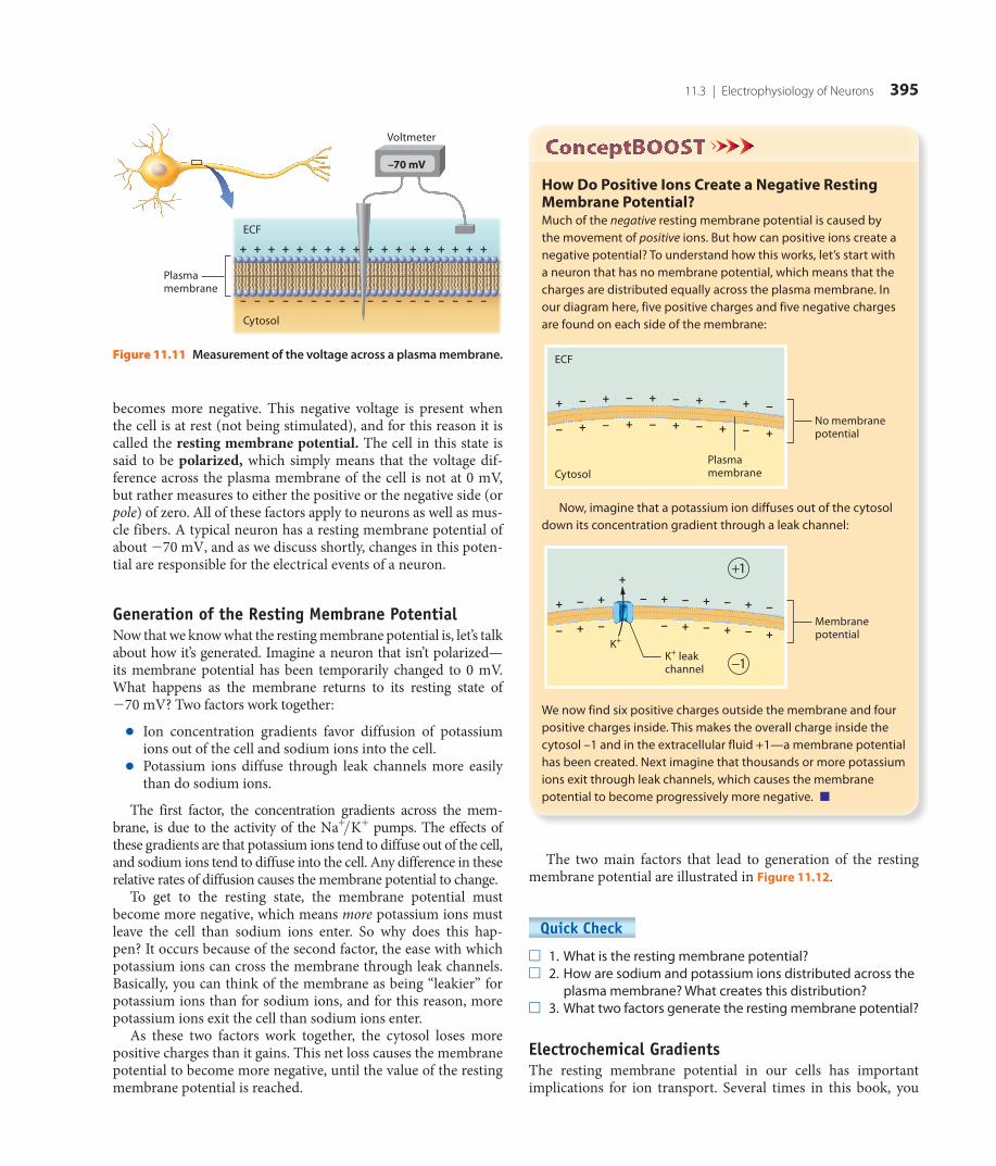
The voltage across the membrane may be measured with a voltmeter, as shown in Figure 11.11. Notice that as you mea-sure from outside to inside the cell with a voltmeter, the voltage
Table 11.2 Types of ioN ChaNNels iN NeuroNs aNd oTher eleCTriCally exCiTable Cells
Type of Channel Structure Stimulus for Opening/Closing
Leak Channel
ECF
Cytosol
Plasma membrane
None, always open
Ligand-Gated Channel
Closed Open
LigandBinding of a ligand to a receptor associated with the channel
Voltage-Gated Channel
Closed Open
+
–
+
–
+
–
+
–
+
–
+
–
–
+
–
+
–
+
–
+
–
+
–
+
Voltage changes across the plasma membrane
Mechanically Gated Channel
Closed Open
Mechanical deformations of the channel (by stretch, pressure, etc.)
M11_AMER2952_01_SE_C11_381-423.indd 394 6/19/14 10:44 AM
8th proof
11.3 | Electrophysiology of Neurons 395
How Do Positive Ions Create a Negative Resting Membrane Potential?Much of the negative resting membrane potential is caused by the movement of positive ions. But how can positive ions create a negative potential? To understand how this works, let’s start with a neuron that has no membrane potential, which means that the charges are distributed equally across the plasma membrane. In our diagram here, five positive charges and five negative charges are found on each side of the membrane:
No membrane potential
Cytosol
ECF
+ − + − + − + − + −
− + − + − + − + − +
Plasmamembrane
Now, imagine that a potassium ion diffuses out of the cytosol down its concentration gradient through a leak channel:
Membrane potential
K+
K+ leak channel
+ − +
+
− + − + − +
+1
−1
−
− + − − + − + − +
We now find six positive charges outside the membrane and four positive charges inside. This makes the overall charge inside the cytosol –1 and in the extracellular fluid +1—a membrane potential has been created. Next imagine that thousands or more potassium ions exit through leak channels, which causes the membrane potential to become progressively more negative. ■
The two main factors that lead to generation of the resting membrane potential are illustrated in Figure 11.12.
Quick Check □ 1. What is the resting membrane potential?□ 2. How are sodium and potassium ions distributed across the
plasma membrane? What creates this distribution?□ 3. What two factors generate the resting membrane potential?
Electrochemical GradientsThe resting membrane potential in our cells has important implications for ion transport. Several times in this book, you
becomes more negative. This negative voltage is present when the cell is at rest (not being stimulated), and for this reason it is called the resting membrane potential. The cell in this state is said to be polarized, which simply means that the voltage dif-ference across the plasma membrane of the cell is not at 0 mV, but rather measures to either the positive or the negative side (or pole) of zero. All of these factors apply to neurons as well as mus-cle fibers. A typical neuron has a resting membrane potential of about -70 mV, and as we discuss shortly, changes in this poten-tial are responsible for the electrical events of a neuron.
Generation of the Resting Membrane PotentialNow that we know what the resting membrane potential is, let’s talk about how it’s generated. Imagine a neuron that isn’t polarized— its membrane potential has been temporarily changed to 0 mV. What happens as the membrane returns to its resting state of -70 mV? Two factors work together:
● Ion concentration gradients favor diffusion of potassium ions out of the cell and sodium ions into the cell.
● Potassium ions diffuse through leak channels more easily than do sodium ions.
The first factor, the concentration gradients across the mem-brane, is due to the activity of the Na+>K+ pumps. The effects of these gradients are that potassium ions tend to diffuse out of the cell, and sodium ions tend to diffuse into the cell. Any difference in these relative rates of diffusion causes the membrane potential to change.
To get to the resting state, the membrane potential must become more negative, which means more potassium ions must leave the cell than sodium ions enter. So why does this hap-pen? It occurs because of the second factor, the ease with which potassium ions can cross the membrane through leak channels. Basically, you can think of the membrane as being “leakier” for potassium ions than for sodium ions, and for this reason, more potassium ions exit the cell than sodium ions enter.
As these two factors work together, the cytosol loses more positive charges than it gains. This net loss causes the membrane potential to become more negative, until the value of the resting membrane potential is reached.
Figure 11.11 Measurement of the voltage across a plasma membrane.
ECF
Cytosol
Plasma membrane
–
+
–
+
–
+
–
–70 mV
+
–
+
–
+
–
+
–
+
–
+
–
+
–
+
–
+
–
+
–
+
–
+
–
+
–
+
–
+
Voltmeter
M11_AMER2952_01_SE_C11_381-423.indd 395 6/19/14 10:44 AM
8th proof

Figure 11.12 Generation of the resting membrane potential.
ECF Na+
K+
Na+
K+
K+ leak channel
Na+ leak channel
Voltmeter
Cytosol
Favored direction of K+ diffusion
Favored direction of Na+
diffusion
K+ diffuse through leak channels more easily than do Na+.
Ion concentration gradients (due to the activity of the Na+/K+ pump) favor diffusion of K+ out of the cell and Na+ into the cell.
These two factors cause the cytosol to lose more positive charges than it gains, leading to the negative resting membrane potential.
++ + +++++++++
–––––––– –– – ––
+
–70 mV
Plasma membrane
396 Chapter 11 | Introduction to the Nervous System and Nervous Tissue
have seen how solutes move across membranes by diffusion according to their concentration gradient. In fact, the concentra-tion gradient is the main factor that determines the movement of uncharged solutes such as carbon dioxide, glucose, and oxygen. But the story for ions is more complicated because they are also affected by electrical gradients. For this reason, diffusion of an ion across the plasma membrane is determined by both its con-centration gradient and its electrical gradient. These two com-bined forces are called the electrochemical gradient.
As an example, consider a potassium ion in the cytosol of a neu-ron (Figure 11.13). You have already seen that 1 the concentration gradient for potassium ions favors their diffusion into the extra-cellular fluid. But now let’s add the force of the electrical gradient. The -70 mV resting potential means that the cytosol is negatively charged relative to the extracellular fluid. As you know, opposite
charges attract, so the positively charged potassium ion is attracted to the negatively charged cytosol. 2 This electrical gradient then favors the movement of potassium ions in the opposite direction, into the cytosol. The overall electrochemical gradient is the sum of these two forces—one drawing potassium ions into the cytosol and one drawing them into the extracellular fluid. If these two forces were equal, no net movement of potassium ions would occur.
However, 3 the concentration gradient for potassium ions is stronger than the electrical gradient by a small amount. For this reason, the net electrochemical gradient is a small force that draws potassium ions into the extracellular fluid. The small size of the electrochemical gradient for potassium ions in a neuron at rest helps to ensure that the cell doesn’t lose too many potassium ions to the extracellular fluid through leak channels.
When we look at sodium ions, however, a different pic-ture emerges. You already know that the concentration gradi-ent favors the movement of sodium ions into the cytosol. The electrical gradient also favors their movement into the cytosol, as the positively charged sodium ions are attracted to its nega-tive charges. This creates a strong electrochemical gradient for sodium ions that draws them into the cytosol.
Quick Check □ 4. How is an electrochemical gradient different from a
concentration gradient?□ 5. How do the electrochemical gradients for potassium ions
and sodium ions differ?
Changes in the Membrane Potential: Ion MovementsNow let’s connect the two concepts we have been discussing: ion channels and gradients plus the resting membrane potential. Figure 11.13 The electrochemical gradient for potassium ions.
Cytosol
ECF
K+K+ leak channel
–
+
–
+
–
+
–
+
–
+
–
+
–
+
–
+The electrical gradient favors the movement of K+ to the cytosol.
2
The concentration gradient is slightly stronger, so a small force favors the movement of K+ into the ECF.
The concentration gradient favors the movement of K+ to the ECF.
1
3
M11_AMER2952_01_SE_C11_381-423.indd 396 6/19/14 10:44 AM
8th proof

11.3 | Electrophysiology of Neurons 397
Local PotentialsYou read in the muscle tissue chapter that each stimulus from a motor neuron leads to a quick, temporary reversal in the mem-brane potential of a muscle fiber, called an action potential (see Chapter 10). However, when a neuron is stimulated just once, a full action potential rarely results. Instead, a small, local change in the membrane potential of the neuron, called a local potential, is produced (see Figure 11.14).
A local potential may have one of two effects:
● It may cause a depolarization in which positive charges en-ter the cytosol and make the membrane potential less nega-tive (e.g., a change from -70 to -60 mV).
● Alternatively, it may cause a hyperpolarization in which either positive charges exit or negative charges enter the cytosol, which makes the membrane potential more negative (e.g., a change from -70 to -80 mV).
Local potentials are sometimes called graded potentials because they vary greatly in size—some produce a larger change in membrane potential than others. The degree of change in the membrane potential during a local potential depends on multi-ple factors, including length of stimulation, number of ion chan-nels that open, and type(s) of ion channels that open. Another feature of local potentials is that they are reversible; on cessation of the stimulus that caused the ion channels to open, the neu-ron quickly returns to its resting potential. Local potentials are also decremental in nature: The changes in membrane potential they produce are small, and the current generated is lost across the membrane over the distance of a few millimeters. Conse-quently, local potentials cannot send signals over great distances, but are useful for short-distance signaling only (which is why they’re called local potentials). However, even though they occur only over short distances, we will see in the next section that local potentials are vital triggers for action potentials, our long- distance signals.
Because the resting membrane potential results from the unequal distribution of ions and their different abilities for crossing the membrane, if we change the ability of the ions to cross the mem-brane, the membrane potential will change as well. This happens by opening gated channels in the plasma membrane. As shown in Figure 11.14a, if gated sodium ion channels open, sodium ions follow their electrochemical gradient and rush into the cell, and the cell gains positive charges. The influx of positive charges makes the membrane potential more positive, a change called depolarization. By this process, the cell becomes less polarized as its membrane potential approaches 0 mV. When a cell returns to its resting membrane potential, repolarization has occurred.
If we instead open gated potassium ion channels, potassium ions follow their electrochemical gradient out of the cell, and the cell loses positive charges. This causes the membrane poten-tial to become more negative than it is at rest, a change termed hyperpolarization (Figure 11.14b). Note that hyperpolarization may also result from the opening of channels for anions, such as chloride ions, which would allow these negatively charged ions to flow into the cell. (This additional change in membrane potential doesn’t occur in muscle fibers, which is why we didn’t discuss it in the muscle tissue chapter.)
Both types of changes in membrane potential are seen in neu-rons. In the upcoming sections, we see how this applies to nervous system physiology and the ability of the neuron to send signals.
Quick Check □ 6. In and around the axon, where is the higher concentration
of sodium ions? Where is the higher concentration of potassium ions? What maintains this gradient?
□ 7. What is the resting membrane potential, and what is responsible for generating it?
□ 8. Define depolarization, repolarization, and hyperpolarization.
Figure 11.14 Ion movements leading to changes in the membrane potential. The changes shown here are local potentials.
–60 mV –80 mV
ECF
Ligand
CytosolLigand-gated cation channel
Ligand-gated cation channel
Hyperpolarization
Resting potential
Resting potential
Ligand-gated anion channel
–
+
+
–
+
–
+
–
+
–
+
–
–
+
+
–
–
+
–
+
–
+
–
+
–
+
–
+
Voltmeter
+ + +
++
++ + +
++ +
++ +
++ + +
++++
++
–––
––
––
––
––
Time (ms)
–80–70
0
+30
Mem
bran
e po
tent
ial (
mV)
Depolarization
Time (ms)
–70–60
0
+30
Mem
bran
e po
tent
ial (
mV)
(a) Depolarization: Gain of positive charges makes the inside of the cell less negative, causing depolarization.
(b) Hyperpolarization: Loss of positive charges (or gain of negative charges) makes the inside of the cell more negative, causing hyperpolarization.
Play animation
M11_AMER2952_01_SE_C11_381-423.indd 397 6/19/14 10:44 AM
8th proof

398 Chapter 11 | Introduction to the Nervous System and Nervous Tissue
The structures of voltage-gated potassium and sodium ion channels are depicted in Figure 11.15. Notice in Figure 11.15a that the voltage-gated potassium ion channel has two possi-ble states: resting and activated. In the resting state, the channel is closed. In the activated state, the channel is open and allows potassium ions to cross the axolemma.
The voltage-gated sodium ion channel shown in Figure 11.15b is more complicated. It has two gates: an activation gate and an inactivation gate. This means a sodium ion channel has three potential “states”:
● Resting state: Inactivation gate opened, activation gate closed. During the resting state the neuron is not being stimulated, and the activation gate is closed and the inac-tivation gate is open. No sodium ions cross the membrane when the channel is in the resting state.
● Activated state: Both activation and inactivation gates opened. When an action potential is initiated, the voltage change opens the activation gates and the channel is in its activated state. The channel in the activated state allows sodium ions to cross the axolemma.
● Inactivated state: Inactivation gate closed, activation gate opened. When the inactivation gate closes, the chan-nel is in its inactivated state. The channel in this state no longer allows sodium ions to pass through. Observe that during this state, the activation gate remains open. When the action potential is finished, the channel returns to the resting state.
Events of an Action PotentialLet’s examine the sequence of events of an action potential in a section of axon, illustrated in Figure 11.16. The entire sequence takes just a few milliseconds. Neuronal action potentials have
Quick Check □ 9. Define local potential. Why is it also called a graded potential?□ 10. Why are local potentials useful only for short-distance signaling?
Action Potentials
◀ ◀ Flashback
1. What are negative and positive feedback loops? (p. 22) 2. What takes place during an action potential? (p. 350)
An action potential is a uniform, rapid depolarization and repo-larization of the membrane potential of a cell (see Chapter 10). This change in the membrane potential causes a response of some sort. For a muscle fiber, the change initiates events that lead to muscle fiber contraction. Within the nervous system, signals are sent through an axon to another neuron, a muscle fiber, or a gland.
Recall that only axons generate action potentials; dendrites and cell bodies generate local potentials only. Action potentials are generated in a region called the trigger zone, which includes the axon hillock and the initial segment of the axon.
In this section we look at what happens during an action potential. First, however, we need to delve deeper into the func-tion of the voltage-gated channels that allow ions to move and change the membrane potential of the neuron.
States of Voltage-Gated ChannelsTwo types of voltage-gated channels function in the depolariza-tion and repolarization of the action potential—one for sodium ions and one for potassium ions. Voltage-gated channels are found most abundantly in the axolemma of the neuron, which is why only axons have action potentials.
Figure 11.15 States of voltage-gated channels.
ECF
Cytosol
Activated state
Resting state Resting state
Inactivated state Activated state(a) The two states of a voltage-gated K+ channel (b) The three states of a voltage-gated Na+ channel
K+ Na+
Activation gate closed
Activation gate closed
Activation gate open
Inactivation gate closed
Inactivation gate open
Activation gate open
Activation gate open
Inactivation gate open
M11_AMER2952_01_SE_C11_381-423.indd 398 6/19/14 10:44 AM
8th proof

11.3 | Electrophysiology of Neurons 399
Figure 11.16 Events of an action potential.
Voltage-gated Na+ channel in resting state
Voltage-gated Na+ channel in resting state
ECF
Axoplasm
Local potential
Trigger zone
Axolemma
Voltage-gated K+ channel in resting state
Membrane potential reverses.
Na+
K+
K+
Na+ inactivation gate closed
Membrane potential returns to negative.
Time (ms)
–70–55
0
+30
Mem
bran
e po
tent
ial (
mV)
Time (ms)
Mem
bran
e po
tent
ial (
mV)
Time (ms)
Mem
bran
e po
tent
ial (
mV)
Time (ms)
Mem
bran
e po
tent
ial (
mV)
Time (ms)
Mem
bran
e po
tent
ial (
mV)
–70–55
0
+30
–70–55
0
+30
–70–55
0
+30
Threshold
–70–55
0
+30
–
+
–
+
–
+
–
+
–
+
–
+
–
+
–
+
–
+
–
+
–
+
–
+
–
+
–
+
–
+
–
+
–
+
–
+
–
+
–
+
–
+
–
+
–
+
–
+
–
+
–
+
–
+
–
+
–
+
–
+
–
+
–
+
–
– – – – –
+
+ + + + +
–
+
–
+
–
+
–
+
–
+
–
+
–
+
–
+
–
+
–
+
–
+
–
+
–
+
–
+
–
+
–
+
–
+
–
+
–
+
–
+
–
+
–
+
–
+
–
+
Voltage-gated Na+ channels activate, Na+ enter, and the axon section depolarizes.
2
A local potential depolarizes the axolemma of the trigger zone to threshold.
Na+ channels inactivate and voltage-gated K+ channels activate, so Na+ stop entering and K+ exit the axon— repolarization begins.
Na+ channels return to the resting state and repolarization continues.
The axolemma may hyperpolarize before K+ channels return to the resting state; after this, the axolemma returns to the resting membrane potential.
4
3
1
5
Play animation
M11_AMER2952_01_SE_C11_381-423.indd 399 6/19/14 10:44 AM
8th proof

400 Chapter 11 | Introduction to the Nervous System and Nervous Tissue
many axons, the outflow of potassium ions continues until the membrane potential of the axolemma hyperpolar-izes, possibly becoming as negative as -90 mV. The axo-lemma hyperpolarizes because the gates of the potassium ion channels are slow to close, allowing additional potas-sium ions to leak out of the cell. Hyperpolarization fin-ishes as the voltage-gated potassium ion channels return to their resting state. After the action potential, the potas-sium leak channels and Na+>K+ pumps re-establish the resting membrane potential.
Throughout the preceding sequence of events of a single action potential, very little change occurs in the intracellular or extracel-lular concentration of sodium or potassium ions, and therefore the gradient isn’t too disturbed. However, with repetitive action potentials, the gradient will eventually deplete, and the neuron relies on the Na+>K+ pumps in the axolemma to restore it. Read A&P in the Real World: Local Anesthetic Drugs to find out what happens when sodium ion channels are blocked on purpose.
Quick Check □ 11. What takes place during the depolarization phase of an action
potential? How is it an example of a positive feedback loop?□ 12. What must be reached in order for voltage-gated sodium
ion channels to open?□ 13. What takes place during the repolarization and
hyperpolarization phases of an action potential?
The Refractory PeriodNeurons are limited in how often they can fire action potentials. For a brief time after a neuron has produced an action potential, the membrane cannot be stimulated to fire another one. This time is called the refractory period (Figure 11.17). The refrac-tory period may be divided into two phases: the absolute refrac-tory period and the relative refractory period.
three general phases: the depolarization phase, the repolar-ization phase, and the hyperpolarization phase. During the depolarization phase, the membrane potential rises toward zero and then becomes briefly positive. The membrane potential returns to a negative value during the repolarization phase, and then becomes temporarily more negative than resting during the hyperpolarization phase. Each phase occurs because of the selec-tive opening and closing of specific ion channels. Note that before the action potential, when the membrane is at rest, the gates for both the sodium and the potassium ion channels are closed.
The action potential proceeds as follows:
1 A local potential depolarizes the axolemma of the trig-ger zone to threshold. The action potential begins when the voltage-gated sodium ion channels in the axolemma of the trigger zone enter the activated (open) state (see Fig-ure 11.15b). However, these voltage-gated channels will become activated only if the axon is depolarized. The source of this depolarization in the trigger zone is generally a local potential that arrives from the cell body. Note that the local potential must be strong enough to depolarize the axon to a level known as threshold, usually -55 mV.
2 Voltage-gated sodium ion channels activate, sodium ions enter, and the axon section depolarizes. When threshold is reached, the sodium ion channels in the trigger zone are activated (open) and sodium ions rush into the neuron with their electrochemical gradient. As the membrane poten-tial becomes more positive, more voltage-gated sodium ion channels are activated. This cycle continues, and the more the axon depolarizes, the more voltage-gated sodium ion channels are activated. This influx of positive charges
causes rapid depolarization to about +30 mV. You may recognize this as an example of a positive feedback loop—the initial input (activation of
sodium ion channels and depolarization) amplifies the out-put (more sodium ion channels are activated and the axo-lemma depolarizes further), an example of the Feedback Loops Core Principle (p. 21).
3 Sodium ion channels inactivate and voltage-gated potas-sium ion channels activate, so sodium ions stop entering and potassium ions exit the axon—repolarization begins. When the axolemma is fully depolarized (about +30 mV), the inactivation gates of the voltage-gated sodium ion chan-nels close, and sodium ions stop entering the axon. As this occurs, voltage-gated potassium ion channels slowly open and potassium ions flow out of the axon along their elec-trochemical gradient, causing the axolemma of the trigger zone to lose positive charges and so to begin repolarization.
4 Sodium ion channels return to the resting state and repolarization continues. As potassium ions exit the axon and repolarization continues, the activation gates of the sodium ion channels close and the inactivation gates open, returning the sodium ion channels to their resting state.
5 The axolemma may hyperpolarize before potassium ion channels return to the resting state; after this, the axo-lemma returns to the resting membrane potential. In
A P in the
Real World
M11_AMER2952_01_SE_C11_381-423.indd 400 6/19/14 10:44 AM
8th proof

11.3 | Electrophysiology of Neurons 401
Local and Action Potentials ComparedNow that we have discussed both local potentials and action potentials, let’s highlight their differences. You discovered ear-lier that local potentials are graded, and so produce changes in membrane potential of varying degree; however, each action potential will cause a maximum depolarization of the same amount, to about +30 mV. This is due to a phenomenon called the all-or-none principle. Simply put, this principle refers to an event, in this case an action potential, that either happens com-pletely or doesn’t happen at all. If a neuron does not depolarize to threshold, an action potential does not occur. If the neuron does depolarize to threshold, the result is an action potential of a characteristic strength. The size of the action potential is not determined by the strength, frequency, or length of the stimulus, and therefore is not graded like a local potential.
The all-or-none principle leads us to a second difference between local potentials and action potentials: their reversibil-ity. Recall that a local potential is reversible; once the stimulus stops, the ion channels close and the resting membrane potential is restored. However, a key feature of an action potential is that when one occurs, it is irreversible—once threshold is reached, it cannot be stopped and will proceed to completion.
Finally, a third important difference between local potentials and action potentials is the distance over which the signal trav-els. Whereas local potentials are decremental and decrease over short distances, action potentials are nondecremental; that is, their strength does not diminish. Without this property, signals could not be sent over long distances in the nervous system.
Quick Check □ 15. How do local potentials and action potentials differ?□ 16. Which is useful for long-distance signaling, and why?
Propagation of Action PotentialsA single action potential in one spot can’t perform its main func-tion, which is to act as a method of long-distance signaling. To do this, it has to be conducted, or propagated, down the length of the axon. This movement creates a flow of charged particles, a current. Action potentials are self-propagating, meaning that each action potential triggers another one in a neighboring sec-tion of the axon. You can imagine this process like a string of dominoes—when the first one is tipped over, the next one falls, which triggers the next to fall, and the process continues until the end of the line is reached. Only the first domino needs the “push,” and once they start to fall, the process sustains itself until the end.
Action potential transmission occurs largely in one direc-tion—from the trigger zone to the axon terminals—and at a con-stant speed. Propagation takes place in a single direction because the membrane in the previous section (behind the action poten-tial) is still in the refractory period. Recall that the sodium chan-nels in refractory parts of the membrane are in their inactivated state, which means that the wave of depolarization cannot trig-ger them to open.
During the absolute refractory period, no additional stimu-lus, no matter how strong, is able to produce an additional action potential. Notice in Figure 11.17 that this period coincides with the voltage-gated sodium ion channels being in their activated and inactivated states; sodium ion channels may not be activated until they return to their resting states with their activation gates closed and their inactivation gates open.
Immediately following the absolute refractory period is the relative refractory period, during which only a strong stimulus will produce an action potential. The relative refractory period is marked by a return of voltage-gated sodium ion channels to their resting state while some potassium ion channels remain acti-vated. Because the potassium ion channels are activated and the membrane is repolarizing or even hyperpolarizing, it’s difficult to depolarize the membrane to threshold and trigger an action potential. However, if a greater than normal stimulus is applied, the membrane may depolarize to threshold, and the axon may fire off another action potential.
The absolute and relative refractory periods limit the fre-quency of action potential production. In addition, the relative refractory period ensures that stronger stimuli trigger more fre-quent action potentials.
Quick Check □ 14. What are the absolute and relative refractory periods?
Figure 11.17 Refractory periods of an action potential.
Mem
bran
e po
tent
ial (
mV)
Na+ channels activated, K+ channels activating slowly
Na+ channels inactivated, K+ channels activated
Na+ channels in resting state,K+ channels remain activated
Na+ and K+ channels in resting state (closed)
Na+ and K+ channels in resting state (closed)
Time (ms)
0
–70
–55
0
+30
1 2 3
Absolute refractory period
Relative refractory period
M11_AMER2952_01_SE_C11_381-423.indd 401 6/19/14 10:44 AM
8th proof

402 Chapter 11 | Introduction to the Nervous System and Nervous Tissue
depolarized to threshold by local potentials from the den-drites, cell body, or axon.
2 As sodium ion channels activate, an action potential is triggered and spreads positive charges down the axon. Voltage-gated sodium ion channels are activated (open) and an action potential occurs. When this happens, posi-tive charges flow down the axon through the axoplasm.
3 The next section of the axolemma depolarizes to thresh-old and fires an action potential as the previous section of the axolemma repolarizes. As the depolarizing current
Propagation of action potentials down an axon forms the nerve impulse. In this section we look at how propagation occurs and the factors that influence the speed of action potential conduction.
Events of PropagationThe action potential is propagated along the axon by the follow-ing sequence of events, shown in Figure 11.18:
1 The axolemma depolarizes to threshold due to local potentials. The axolemma of the trigger zone is
Figure 11.18 Propagation of an action potential.
Depolarizedmembrane—localpotentials
Depolarizingmembrane—action potential
Depolarizingmembrane—action potential
Repolarizingmembrane
Restingmembrane
Restingmembrane
Axon
Triggerzone
Restingmembrane
Restingmembrane
Depolarizingmembrane—actionpotential
Repolarizingmembrane
Restingmembrane
Restingmembrane
Axolemma
Voltage-gated Na+ channel
Na+
Na+
K+
Voltage-gated K+ channel
Axoplasm
The axolemma depolarizes to threshold due to local potentials.
1
As Na+ channels activate, an action potential is triggered and spreads down the axon.
2
The next section of the axolemma depolarizes to threshold and fires an action potential as the previous section of the axolemma repolarizes.
3
The current continues to move down the axon, and the process repeats.
4
+–
+–
+–
+–
–+ +
–
+–
+–
–+ +
–
+–
+–
–+ +
–
+–
+–
+–
+–
–+ +
–
+–
+–
–+ +
–
+–
+–
–+ +
–
+–
+–
+–
+–+ + + +
–
–+
–+
–+
–+
–+
–+
–+
–+
–+
–+
–+
–+
–+ +
–
+–
+–
+–
+–
+–
+–
–+
–+
–+
–+
–+
–+
–+
–+
–+
–+
–+
–+
–+
–+
–+
–+
–+ +
–
+–
+–
–+ +
–
+–
+–
–+ +
–
+–
+–
+–
+–
–+
–+
–+
–+
–+
–+
–+
–+
–+
–+
+–
+–
– – –
Play animation
M11_AMER2952_01_SE_C11_381-423.indd 402 6/19/14 10:45 AM
8th proof

11.3 | Electrophysiology of Neurons 403
reaches the next section of the axolemma, it depolarizes that section to threshold. The voltage-gated sodium ion channels in that part of the axolemma are activated, and that section then generates an action potential. The current flows down to the next section of the axon. Note that the section of the axolemma that had an action potential in step 2 is repo-larizing (its potassium channels are activated and potassium ions are exiting the axoplasm) and is in its refractory period, so any current that flows backward can’t trigger an action potential.
4 The current continues to move down the axon, and the process repeats. The current flowing into the next section of the axolemma causes it to depolarize to threshold, acti-vating voltage-gated sodium ion channels and producing an action potential there.
The process then repeats down the length of the axon until it reaches the axon terminals.
Conduction SpeedThe rate at which propagation occurs is called conduction speed, and it determines how rapidly signaling can occur within the nervous system. Conduction speed is influenced by two main factors: the diameter of the axon and the presence or absence of a myelin sheath. The diameter of the axon affects the conduc-tion of current through the axon because larger axons have lower resistance to conduction, and therefore current flows through them more easily.
The second determinant of conduction speed is the presence or absence of a myelin sheath. Two types of conduction can take place in an axon: saltatory conduction, in which the myelin sheath is
present, and continuous conduction, in which it is absent (Figure 11.19). Recall from our discussion in Module 11.2 that myelin is an excellent insulator of electrical charge. Thus, the flow of current is far more efficient in a myelinated axon, which causes saltatory conduction to be significantly faster than continuous conduction.
Figure 11.19 compares conduction along a myelinated axon and an unmyelinated axon, and depicts the effect of the myelin sheath on conduction speed. You can see that on the myelinated axon, only the nodes of Ranvier must be depolarized to thresh-old. When the node, rich in voltage-gated sodium ion channels, is depolarized to threshold, an action potential is triggered. This action potential generates a current that flows passively and effi-ciently with little loss of charge through the next myelinated seg-ment, or internode (see Figure 11.8 for a review of anatomical terminology). When the current reaches the next node of Ranvier, another action potential is generated. This cycle is repeated down the length of the axon, and the current “jumps” from one node to the next (in fact, “saltatory” comes from the Latin word saltare, which means “leaping”). See A&P in the Real World: Multiple Scle-rosis on page 406 for information on how loss of the myelin sheath affects conduction speed.
Compare these “leaping” action potentials with the much slower and more gradual continuous conduction seen in the unmyelinated axon. Figure 11.19 shows that in continuous con-duction, absence of the myelin sheath means that each section of the axolemma must be depolarized to threshold. This is why con-duction of this sort is called continuous—action potentials must be generated in a continuous sequence along the entire axolemma for the current to spread down the length of the axon. The flow of cur-rent in a myelinated axon is much faster than the process of trig-gering action potentials in each part of an unmyelinated axon.
Depolarizingmembrane
Restingmembrane
Myelinated axon
Unmyelinated axon
First action potential1
Saltatory conduction in myelinated axon
Continuous conduction in unmyelinated axon
Second action potential2 Third action potential3
+ ++ +
+ +
+ +
+ +
++
+ +++
– –
– –
– –
– –
–
–
– –
– –
– –
– –
– –
– –
Depolarizingmembrane
Restingmembrane
++ +
+
–
– ++
+–
–+
– –
– –
+ +
+ +
– –
– –
+ +
+ +
– –
– –
+ +
+ +
– –
– –
+ +
+ +
– –
– –
+ +
+ +
– –
– –
+–
–+
+–
–+
+
+
–
–
+ +
+ +
– –
– –
+
+
–
–
+ +
+ +
– –
– –
+ +
+ +
– –
– –
+ +
+ +
– –
– –
+ +
+ +
– –
– –
++ +
+
–
–++
+–
–+
– –
– –
+
+
–
–
+ +
+ +
– –
– –
+
+
–
–
+ +
+ +
– –
– –
+ +
+ +
– –
– –
+ +
+ +
– –
– –
–
–
– –
– –
– –
– –
+ ++ +
+ +
++
+ +++
– –
– –
+ +
+ +
– –
– –
––
–
–
–
– –
– –
– –
– –
+ +
+ +
– –
–
–
– –
–
–
– –
– –
– –
– –
+ +
+ +
– –
– –
First action potential1 Second action potential2 Third action potential3
Figure 11.19 Comparison of saltatory and continuous conduction.
M11_AMER2952_01_SE_C11_381-423.indd 403 6/19/14 10:45 AM
8th proof

404 Chapter 11 | Introduction to the Nervous System and Nervous Tissue
must constantly be regenerated along the length of the axolemma. This requires the opening of voltage-gated ion channels, which takes time, so propagation is much slower.
A myelinated axon more closely resembles the wire in the final illustration. Myelin is a very good insulator because it is a poor conductor of electricity, and so it prevents current from leaking out through the axolemma. This means that the signal decreases very little in strength as it travels through an internode. The action potential can then propagate through the internode without having to be regenerated. It is not until the current reaches the unmyelinated node of Ranvier that it starts to dissipate and the action potential must be regenerated. This is why the action potentials of saltatory conduction can “leap” from node to node and why this type of propagation is so much faster than continu-ous conduction. ■
Classification of Axons by Conduction SpeedAxons are often classified according to conduction speed. The two primary defining features in this classification are the diam-eter of the axon and the presence or absence of the myelin sheath. The three main classes are as follows:
● Type A fibers are the largest-diameter axons (5–20 μm), all of which are myelinated. These characteristics give them the fastest conduction speed, with a maximum velocity of about 120 m/sec (about 250 mi/h). Type A fibers are found in parts of the body with which the CNS must communicate extremely rapidly, such as certain sensory axons from joints and muscle fibers, as well as motor axons to skeletal muscles.
● Type B fibers are intermediate in diameter (2–3 μm) and most are myelinated. They typically have a maximum con-duction speed of about 15 m/sec (about 32 mi/h), and in-clude certain efferent fibers of the autonomic nervous system and certain sensory axons coming from organs.
● Type C fibers are the smallest fibers (0.5–1.5 μm) and are unmyelinated. Their conduction velocity is the slowest, con-ducting action potentials only at about 0.5–2 m/sec (about 1–5 mi/h). Type C fibers include other efferent fibers of the autonomic nervous system and certain sensory axons that transmit pain, temperature, and certain pressure sensations.
Quick Check □ 17. How is an action potential propagated down an axon?□ 18. How do saltatory conduction and continuous conduction
differ? Which is faster, and why?
Putting It All Together: The Big Picture of Action PotentialsAfter all of this talk of action potentials, you may still find some of these concepts to be rather abstract. Before moving on to the next module, make sure you think about the big picture of
How Does Myelin Insulate an Axon and Increase Its Speed of Propagation?We just discussed how myelin increases the speed of action poten-tial propagation by “insulating” the charge and enabling saltatory conduction. But how, exactly, is charge insulated, and why does this make propagation occur more quickly? To understand this, we have to go back to some of the basic principles of electricity. Let’s first think of the electrical current flowing down a bare copper wire that is going to a light bulb. Ideally, the current flows directly down the wire and illuminates the light bulb, as shown here:
Wire
Electric current
But, if you touch the wire with a metal probe, as depicted in the next illustration, most of the current might instead flow down the probe, a situation known as a short circuit:
Short circuit
Probe
However, if the wire is encased in a material that is a poor conduc-tor of electricity, as shown in this final illustration, the current is insulated and unable to move from the copper wire to the probe. This prevents a short circuit.
Insulation
Probe
Now let’s apply this to axons. An unmyelinated axon most closely resembles the wire in the middle illustration. The axolemma is very leaky with respect to current, and so the current flows easily from the axoplasm to the extracellular fluid, just as current flowed easily from the copper wire to the metal probe. Remember from our earlier discussion that the strength of an electrical signal de-creases as the ions that make up the current leak across the mem-brane. So in an unmyelinated axon, the current generated by the action potential dissipates over a short distance, which could cause the action potential to fail. For this reason, the action potential
M11_AMER2952_01_SE_C11_381-423.indd 404 6/19/14 10:45 AM
8th proof

□ 2. What do you think would happen to the neuronal action potential if the concentration of sodium ions in the extracellular fluid decreased significantly, to the point of reversing the gradient?
□ 3. Sometimes when you pull your dinner out of the microwave you have to hold your fingertips to the food for a second or two before you can tell if it is hot or cold. Explain why this happens. (Hint: What type of fiber carries temperature information?)
See answers in Appendix A.
how and why the action potential occurs. The how of the action potential is shown in Figure 11.20. The why of the action poten-tial is long-distance signaling. As you will see in the upcoming module, the arrival of the action potential at the axon terminal is what allows the neuron to communicate with its target cells.
Apply What You Learned
□ 1. Predict the effect of the poison ouabain (way-BAH-in), which blocks Na+/K+ pumps, on the neuronal action potential. (Hint: What would happen to the sodium and potassium ion gradients?)
Axonterminals
Dendrite
Local potentials
Na+
Na+ AP
AP AP AP AP
Trigger zone
Direction of AP
Axon
Voltage-gatedNa+ channel
Local potentials: Local potentials usually generated in the neuron’s dendrites accumulateand reach the trigger zone of the axon(see Figure 11.14).
1
Action potential: The trigger zone depolarizes to threshold and generates an action potential (AP)(see Figure 11.16).
2
Action potential propagation: The action potential is propagated down the axon to the axon terminals (see Figure 11.18).
3
+–
+–
+–
+–
+–
+–
+–
+–
+–
+–
+–
+–
+–
+–
The Big Picture of Action PotentialsFigure 11.20
405
Play animation
M11_AMER2952_01_SE_C11_381-423.indd 405 6/19/14 10:45 AM
8th proof

406 Chapter 11 | Introduction to the Nervous System and Nervous Tissue
two types, electrical and chemical. We examine both types in this module, although we focus on chemical synapses. The module concludes with a look at neural integration, or the way in which the many synapses of a neuron impact its integrative processes.
Overview of Neuronal SynapsesNeuronal synapses generally occur between an axon and another part of a neuron; they may occur between an axon and a den-drite, an axon and a cell body, and an axon and another axon. These types are called axodendritic, axosomatic, and axoaxonic synapses, respectively (Figure 11.21).
Regardless of the type of synapse, we use certain terms to describe the neurons sending and receiving the message:
● Presynaptic neuron. The presynaptic neuron is the neuron that is sending the message from its axon terminal.
● Postsynaptic neuron. The postsynaptic neuron is the neuron that is receiving the message from its dendrite, cell body, or axon.
The transfer of chemical or electrical signals between neu-rons at a synapse is called synaptic transmission, and it is the fundamental process for most functions of the nervous system. Synaptic transmission allows voluntary movement, cognition, sensation, and emotion, as well as countless other processes. Each neuron has an enormous number of synapses. Recall from Module 11.2 that each axon generally splits into 1000 or more axon terminals, and each terminal meets up with another axon, dendrite, or cell body. So an average presynaptic neuron, then, generally forms synapses with about 1000 postsynaptic neurons. A postsynaptic neuron can receive input from even more syn-apses—an average neuron can have as many as 10,000 synaptic connections from different presynaptic neurons.
Quick Check □ 1. What are the three most common locations where
presynaptic axons connect with a postsynaptic neuron?□ 2. Define synaptic transmission.
M O D U L E
11.4Neuronal SynapsesLearning Outcomes
1. Compare and contrast electrical and chemical synapses. 2. List the structures that make up a chemical synapse. 3. Discuss the relationship between a neurotransmitter and its
receptor. 4. Describe the events of chemical synaptic transmission in
chronological order. 5. Define excitatory postsynaptic potential (EPSP) and inhibitory
postsynaptic potential (IPSP), and interpret graphs showing the voltage-versus-time relationship of an EPSP and an IPSP.
6. Explain temporal and spatial summation of synaptic potentials.
Up to this point, we have discussed how signals are generated and propagated within a neuron. Remember, though, that neu-rons must communicate with other cells, including other neu-rons, in order to carry out their functions—an example of the
Cell-Cell Communication Core Principle (p. 27). Therefore, in this module, we will dis-cuss how signals are trans-
mitted between neurons at locations called synapses. Recall that a synapse (SIN-aps; syn- = “to clasp or join”) is where a neuron meets its target cell (see Chapter 10). The discussion in that chap-ter revolved around a specific type of synapse—the neuromuscu-lar junction. Here we explore the synapses that occur between two neurons, or neuronal synapses. These synapses may be of
Figure 11.21 Structural types of synapses.
Axodendritic synapses (connection between axon and dendrite)
Axoaxonic synapses (connection between axon and axon)
Axosomatic synapses (connection between axon and cell body)A P
in the Real World
M11_AMER2952_01_SE_C11_381-423.indd 406 6/19/14 10:45 AM
8th proof

11.4 | Neuronal Synapses 407
Figure 11.22 The structures of electrical and chemical synapses.
Presynaptic neuron
Synapticvesicle
Neurotransmittermolecules
Neurotransmitterreceptor
Synaptic cleft
Postsynapticneuron
Gapjunction Synaptic cleft
Axon terminal
Axon terminal
Channels
Ions
(a) Electrical synapse (b) Chemical synapse
Practice art labeling
programmed, automatic behaviors such as breathing. They are also present in developing nervous tissue in the embryo and fetus and are thought to assist in the development of the brain. In addi-tion, electrical synapses are found outside the nervous system in locations such as cardiac and visceral smooth muscle, where they allow those tissues to engage in coordinated muscle activity.
Quick Check □ 3. What are the two main features of an electrical synapse?
Chemical Synapses
◀ ◀ Flashback
1. What is a synaptic vesicle? (p. 352) 2. What is a neurotransmitter? (p. 352)
The vast majority of synapses in the nervous system are chem-ical synapses. These synapses are more common because they are more efficient—the current in electrical synapses eventu-ally becomes weaker as it dissipates into the extracellular fluid. A chemical synapse, in contrast, converts an electrical signal into a controlled chemical signal, so there is no loss of strength. The chemical signal is reconverted into an electrical signal in the postsynaptic neuron. In the upcoming sections, we explore how this takes place. But first let’s look a little more closely at the dif-ferences between chemical and electrical synapses.
Electrical SynapsesAn electrical synapse (Figure 11.22a) occurs between cells that are electrically coupled via gap junctions. Observe that in these synapses the axolemmas of the two neurons are nearly touching (they are separated by only about 3.5 nm) and that the gap junc-tions contain precisely aligned channels that form pores through which ions and other small substances may travel. This allows the electrical current to flow directly from the axoplasm of one neuron to that of the next.
This arrangement creates two unique features of electrical synapses:
● Synaptic transmission is bidirectional. In an electrical synapse, transmission is usually bidirectional, which means that either neuron may act as the presynaptic or the post-synaptic neuron and that current may flow in either direc-tion between the two cells.
● Synaptic transmission is nearly instantaneous. The de-lay between depolarization of the presynaptic neuron and change in potential of the postsynaptic neuron is less than 0.1 ms (millisecond), which is extraordinarily fast (we will see that transmission at most chemical synapses requires from one to a few milliseconds).
These features of electrical synapses allow the activity of a group of cells to be synchronized—when stimulated, the cells will produce action potentials in unison. Electrical synapses are found primarily in areas of the brain that are responsible for
M11_AMER2952_01_SE_C11_381-423.indd 407 6/19/14 10:45 AM
8th proof
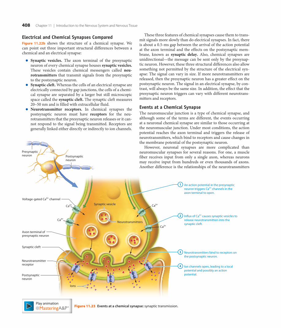
408 Chapter 11 | Introduction to the Nervous System and Nervous Tissue
These three features of chemical synapses cause them to trans-mit signals more slowly than do electrical synapses. In fact, there is about a 0.5-ms gap between the arrival of the action potential at the axon terminal and the effects on the postsynaptic mem-brane, known as synaptic delay. Also, chemical synapses are unidirectional—the message can be sent only by the presynap-tic neuron. However, these three structural differences also allow something not permitted by the structure of the electrical syn-apse: The signal can vary in size. If more neurotransmitters are released, then the presynaptic neuron has a greater effect on the postsynaptic neuron. The signal in an electrical synapse, by con-trast, will always be the same size. In addition, the effect that the presynaptic neuron triggers can vary with different neurotrans-mitters and receptors.
Events at a Chemical SynapseThe neuromuscular junction is a type of chemical synapse, and although some of the terms are different, the events occurring at a neuronal chemical synapse are similar to those occurring at the neuromuscular junction. Under most conditions, the action potential reaches the axon terminal and triggers the release of neurotransmitters, which bind to receptors and cause changes in the membrane potential of the postsynaptic neuron.
However, neuronal synapses are more complicated than neuromuscular synapses for several reasons. For one, a muscle fiber receives input from only a single axon, whereas neurons may receive input from hundreds or even thousands of axons. Another difference is the relationships of the neurotransmitters
Electrical and Chemical Synapses ComparedFigure 11.22b shows the structure of a chemical synapse. We can point out three important structural differences between a chemical and an electrical synapse:
● Synaptic vesicles. The axon terminal of the presynaptic neuron of every chemical synapse houses synaptic vesicles. These vesicles contain chemical messengers called neu-rotransmitters that transmit signals from the presynaptic to the postsynaptic neuron.
● Synaptic cleft. Whereas the cells of an electrical synapse are electrically connected by gap junctions, the cells of a chemi-cal synapse are separated by a larger but still microscopic space called the synaptic cleft. The synaptic cleft measures 20–50 nm and is filled with extracellular fluid.
● Neurotransmitter receptors. In chemical synapses the postsynaptic neuron must have receptors for the neu-rotransmitters that the presynaptic neuron releases or it can-not respond to the signal being transmitted. Receptors are generally linked either directly or indirectly to ion channels.
Figure 11.23 Events at a chemical synapse: synaptic transmission.
Voltage-gated Ca2+ channel
Neurotransmitters
Ca2+
Ca2+
Ca2+
Ca2+Synaptic vesicle
Neurotransmitterreceptor
Axon terminal ofpresynaptic neuron
Synaptic cleft
Postsynapticneuron
Presynapticneuron Postsynaptic
neuron
Ions
An action potential in the presynaptic neuron triggers Ca2+ channels in the axon terminal to open.
1
Influx of Ca2+ causes synaptic vesicles to release neurotransmitters into the synaptic cleft.
2
Neurotransmitters bind to receptors on the postsynaptic neuron.
3
Ion channels open, leading to a local potential and possibly an action potential.
4
Play animation
M11_AMER2952_01_SE_C11_381-423.indd 408 6/19/14 10:45 AM
8th proof

11.4 | Neuronal Synapses 409
to their target cells. At a neuromuscular synapse, the only neu-rotransmitter used is acetylcholine, and acetylcholine serves only to excite the muscle fiber. However, in neuronal synapses, a vari-ety of neurotransmitters may be released to excite or inhibit the postsynaptic neuron. Finally, at the neuromuscular junction, each action potential in the neuron triggers an action poten-tial in the muscle fiber. A postsynaptic neuron, however, may require the input of 100 or more presynaptic neurons to trigger an action potential.
Let’s now dig into what actually takes place at a neuronal syn-apse. Figure 11.23 depicts the following events:
1 An action potential in the presynaptic neuron triggers calcium ion channels in the axon terminal to open. An action potential reaches the axon terminal of the presyn-aptic neuron, which triggers the opening of voltage-gated calcium ion channels in its axolemma.
2 Influx of calcium ions causes synaptic vesicles to release neurotransmitters into the synaptic cleft. Calcium ions enter the axon terminal, causing synaptic vesicles in the area to fuse with the presynaptic membrane. This releases neurotransmitters into the synaptic cleft via exocytosis.
3 Neurotransmitters bind to receptors on the postsynap-tic neuron. The neurotransmitters diffuse across the syn-aptic cleft, where they bind to neurotransmitter receptors on the membrane of the postsynaptic neuron.
4 Ion channels open, leading to a local potential and possibly an action potential. The binding of neurotrans-mitters to receptors generally either opens or closes ligand-gated ion channels in the postsynaptic membrane, resulting in a local potential. Such local potentials may or may not lead to an action potential in the postsynaptic neuron.
Postsynaptic PotentialsLocal potentials in the membrane of the postsynaptic neuron, which are called postsynaptic potentials, can move the mem-brane either closer to or farther away from threshold. Therefore, depending on which channels are opened, one of two events may occur (Figure 11.24):
● The membrane potential of the postsynaptic neuron moves closer to threshold. A small, local depolarization called an excitatory postsynaptic potential (EPSP) occurs, which brings the membrane of the postsynaptic neuron closer to threshold (Figure 11.24a). If the membrane poten-tial reaches threshold, an action potential is triggered.
● The membrane potential of the postsynaptic neuron moves away from threshold. A small, local hyperpolariza-tion known as an inhibitory postsynaptic potential (IPSP) occurs, moving the membrane of the postsynaptic neuron farther away from threshold, and so tending to inhibit an action potential from firing (Figure 11.24b).
EPSPs typically result from the opening of ion channels such as those for sodium or calcium ions and the entrance of positive charges into the postsynaptic neuron (see Figure 11.24a). A single EPSP produces only a very small, local potential. However, each successive EPSP makes the membrane more depolarized and so more likely to reach threshold and fire an action potential.
Note in Figure 11.24b that an IPSP typically results from either the opening of potassium ion channels, which causes a loss of positive charges, or the opening of chloride ion channels. Chloride ions are more abundant in the extracellular fluid than in the neuron, so when chloride ion channels open, these anions enter the neuron and make the membrane potential more nega-tive. Opening of either type of channel will yield the same result:
Figure 11.24 Postsynaptic potentials.
–
Threshold
OR
Small depolarization(EPSP)
Time (ms)
–70
–90
–55
0
Mem
bran
e po
tent
ial (
mV)
Threshold
Small hyperpolarization(IPSP)
Time (ms)
–70
–90
–55
0
Mem
bran
e po
tent
ial (
mV)
(a) The membrane potential of the postsynaptic neuron moves closer to threshold.
Ion channels open and positive charges enter the postsynaptic neuron, causing an excitatory postsynaptic potential (EPSP).
Ion channels open and the postsynaptic neuron gains negative charges or loses positive charges, causing an inhibitory postsynaptic potential (IPSP).
(b) The membrane potential of the postsynaptic neuron moves away from threshold.
Ligand-gated K+ channel
Ligand-gated Cl– channel
+
+ +++
++
+–
––
––
––
Ligand-gated Na+ channel
Neurotransmitter
Ligand-gated Ca2+ channel
+ ++++
+
+ +
++
+ +
+ +
+Postsynapticneuron
Postsynapticneuron
Play animation
M11_AMER2952_01_SE_C11_381-423.indd 409 6/19/14 10:45 AM
8th proof

410 Chapter 11 | Introduction to the Nervous System and Nervous Tissue
proteins in the axolemma of the presynaptic neuron trans-port them back into the presynaptic neuron. Depending on their type, these neurotransmitters may be repackaged into synaptic vesicles or degraded by enzymes.
Once the neurotransmitter has been removed from the synap-tic cleft, synaptic transmission is complete. See A&P in the Real World: Arthropod Venom to learn what happens when contin-ued synaptic transmission causes postsynaptic neurons to be overstimulated.
Quick Check □ 4. What is the role of calcium ions in a chemical synapse?□ 5. How do the two types of postsynaptic potentials differ?□ 6. How is synaptic transmission terminated?
Putting It All Together: The Big Picture of Chemical Synaptic TransmissionAt this point we’ve discussed the particulars of synaptic trans-mission at a chemical synapse: how the action potential triggers the release of neurotransmitters from the presynaptic neuron, how the neurotransmitters induce an EPSP or IPSP in the post-synaptic neuron, and how transmission is ended. Now we can summarize the whole process, as shown in Figure 11.26.
The membrane potential moves farther away from threshold, and an action potential becomes less likely.
Termination of Synaptic TransmissionWe move now to the final step of synaptic transmission— termination. But why terminate synaptic transmission? Once presynaptic neurons have triggered EPSPs and/or IPSPs and so generated a specific response in the postsynaptic neuron, the response cannot be initiated again until the postsynaptic neuron stops being stimulated. The neurons involved in breathing pro-vide a simple example. When we need to inhale, specific neurons are stimulated to trigger our respiratory muscles to contract. Once we have taken a breath, our nervous system needs to stop stimulating these neurons or we will continue to inhale. This is accomplished by stopping synaptic transmission.
The messenger of synaptic transmission is the neurotransmitter released by the presynaptic neuron. Therefore, synaptic transmis-sion may be terminated by ending the effects of the neurotrans-mitter. In general, this happens in three ways (Figure 11.25):
● Diffusion and absorption. Some neurotransmitters simply diffuse away from the synaptic cleft through the extracellu-lar fluid, where they diffuse through the plasma membrane of a neuron or astrocyte and are then returned to the presyn-aptic neuron.
● Degradation in the synaptic cleft. Certain neurotransmit-ters are broken down by enzymes that reside in the synap-tic cleft. The components of the destroyed neurotransmitter are often then taken back up by the presynaptic neuron and resynthesized into the original neurotransmitter.
● Reuptake into the presynaptic neuron. Some neurotrans-mitters are removed by a process called reuptake, in which
A P in the
Real World
Figure 11.25 Methods of termi-nation of synaptic transmission.
Synapticcleft
Presynapticneuron
Postsynapticneuron
Diffusion and AbsorptionNeurotransmitters diffuse away from the synaptic cleft and are returned to the presynaptic neuron.
DegradationNeurotransmitters are degraded by enzymatic reactions in the synaptic cleft.
ReuptakeNeurotransmitters are taken back into the presynaptic neuron.
Play animation
M11_AMER2952_01_SE_C11_381-423.indd 410 6/19/14 10:45 AM
8th proof

Axon terminal ofpresynaptic neuron
Actionpotential
NeurotransmittersCa2+
Ca2+ Synaptic transmission: Ca2+ channels open in the presynaptic neuron; neurotransmitters are released from synaptic vesicles and bind to receptors on the postsynaptic neuron(see Figure 11.23).
2
Postsynaptic potentials: Neurotransmitters triggeran EPSP or IPSP, moving the membrane potential of thepostsynaptic neuron eithercloser to or farther fromthreshold (see Figure 11.24).
3
Termination of synaptic transmission: Neurotransmitter concentration in the synaptic cleft decreases and synaptic transmission is terminated(see Figure 11.25).
4
Action potential: An action potential reaches the axon terminal of the presynaptic neuron.
1
Synapticcleft
Open receptor/ion channel
OR
Postsynaptic neuron
Diffusion
Degradation Closed receptor/ion channel
Reuptake
EPSP IPSP
Threshold Threshold
+ –
+
The Big Picture of Chemical Synaptic Transmission
Figure 11.26
411
Play animation
M11_AMER2952_01_SE_C11_381-423.indd 411 6/19/14 10:45 AM
8th proof
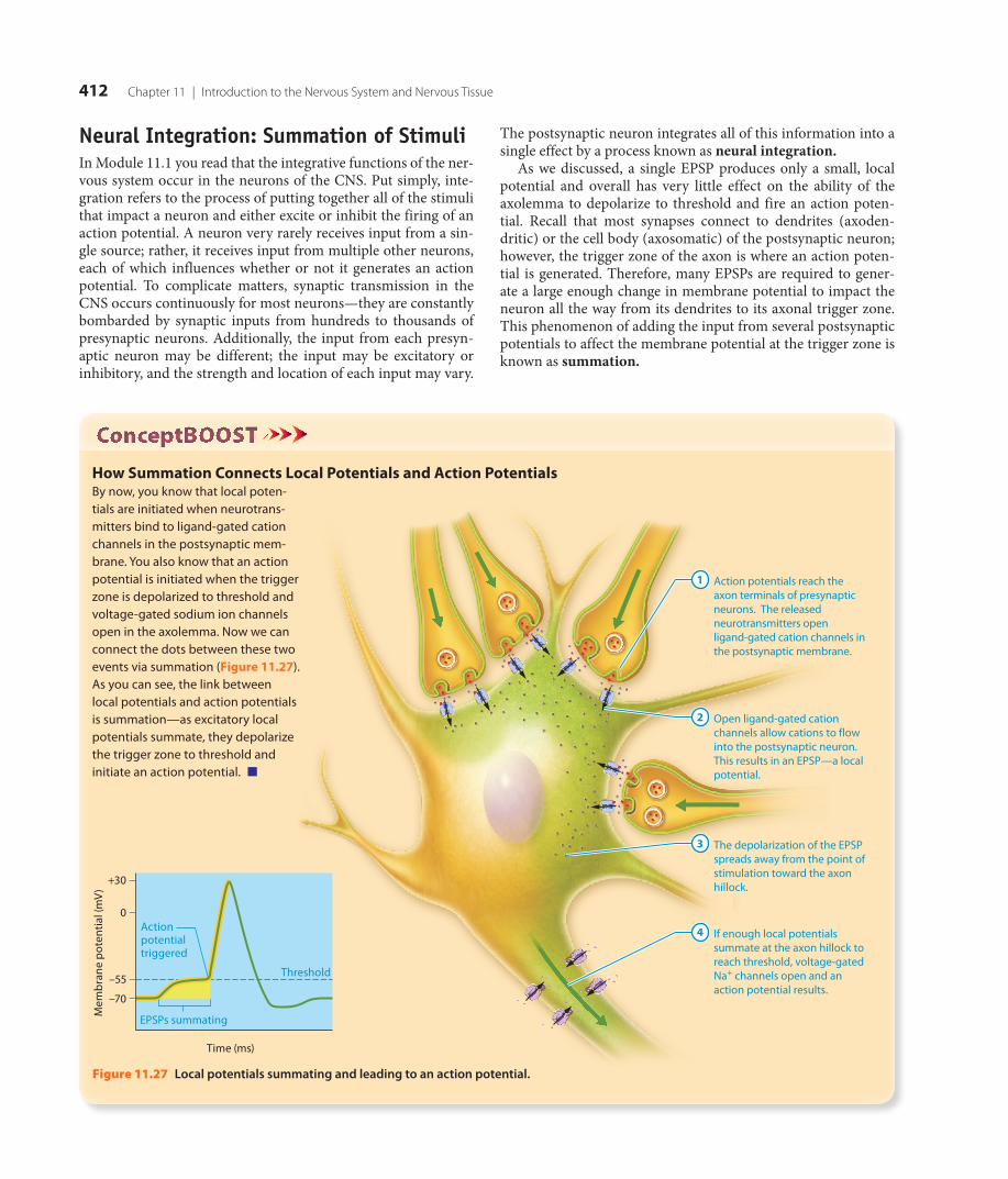
412 Chapter 11 | Introduction to the Nervous System and Nervous Tissue
The postsynaptic neuron integrates all of this information into a single effect by a process known as neural integration.
As we discussed, a single EPSP produces only a small, local potential and overall has very little effect on the ability of the axolemma to depolarize to threshold and fire an action poten-tial. Recall that most synapses connect to dendrites (axoden-dritic) or the cell body (axosomatic) of the postsynaptic neuron; however, the trigger zone of the axon is where an action poten-tial is generated. Therefore, many EPSPs are required to gener-ate a large enough change in membrane potential to impact the neuron all the way from its dendrites to its axonal trigger zone. This phenomenon of adding the input from several postsynaptic potentials to affect the membrane potential at the trigger zone is known as summation.
Neural Integration: Summation of StimuliIn Module 11.1 you read that the integrative functions of the ner-vous system occur in the neurons of the CNS. Put simply, inte-gration refers to the process of putting together all of the stimuli that impact a neuron and either excite or inhibit the firing of an action potential. A neuron very rarely receives input from a sin-gle source; rather, it receives input from multiple other neurons, each of which influences whether or not it generates an action potential. To complicate matters, synaptic transmission in the CNS occurs continuously for most neurons—they are constantly bombarded by synaptic inputs from hundreds to thousands of presynaptic neurons. Additionally, the input from each presyn-aptic neuron may be different; the input may be excitatory or inhibitory, and the strength and location of each input may vary.
Figure 11.27 Local potentials summating and leading to an action potential.
Action potentials reach the axon terminals of presynaptic neurons. The released neurotransmitters open ligand-gated cation channels in the postsynaptic membrane.
1
Open ligand-gated cation channels allow cations to flow into the postsynaptic neuron. This results in an EPSP—a local potential.
2
The depolarization of the EPSP spreads away from the point of stimulation toward the axon hillock.
3
If enough local potentials summate at the axon hillock to reach threshold, voltage-gated Na+ channels open and an action potential results.
4
Mem
bran
e po
tent
ial (
mV)
Time (ms)
–70
–55
0
Threshold
EPSPs summating
+30
Action potential triggered
How Summation Connects Local Potentials and Action PotentialsBy now, you know that local poten-tials are initiated when neurotrans-mitters bind to ligand-gated cation channels in the postsynaptic mem-brane. You also know that an action potential is initiated when the trigger zone is depolarized to threshold and voltage-gated sodium ion channels open in the axolemma. Now we can connect the dots between these two events via summation (Figure 11.27). As you can see, the link between local potentials and action potentials is summation—as excitatory local potentials summate, they depolarize the trigger zone to threshold and initiate an action potential. ■
M11_AMER2952_01_SE_C11_381-423.indd 412 6/19/14 10:45 AM
8th proof

11.5 | Neurotransmitters 413
Quick Check □ 7. How do temporal summation and spatial summation differ?
Apply What You Learned
□ 1. Predict how a poison that blocks calcium ion channels in the axon terminal would affect synaptic transmission.
□ 2. A new drug opens calcium ion channels in the membrane of the postsynaptic neuron. Would this produce an EPSP or an IPSP? Would this make an action potential more or less likely to occur?
□ 3. Explain how you could increase the likelihood that a certain neuron will reach threshold and have an action potential. (Hint: Think about the different types of summation.)
See answers in Appendix A.
M O D U L E
11.5NeurotransmittersLearning Outcomes
1. Explain how a single neurotransmitter may be excitatory at one synapse and inhibitory at another.
2. Describe the structural and functional properties of the major classes of neurotransmitters.
3. Describe the most common excitatory and inhibitory neurotransmitters in the CNS.
There are two types of summation, temporal and spatial, which differ in the timing of neurotransmitter release and number of presynaptic neurons, respectively. The first, temporal summa-tion, occurs when neurotransmitter is released repeatedly from the axon terminal of a single presynaptic neuron (Figure 11.28a). The resulting EPSPs propagate toward the trigger zone. Each EPSP is very quick, lasting no more than about 15 ms. For this reason, the EPSPs must occur rapidly in succession in order for temporal summation to occur, or the trigger zone will not reach threshold.
The second type of summation, spatial summation, involves the simultaneous release of neurotransmitters from the axon ter-minals of multiple presynaptic neurons (Figure 11.28b). In Figure 11.28 the graph of temporal summation shows a staircase-like rise in membrane potential, whereas the membrane potential in the graph of spatial summation shows a smooth rise as large quantities of neurotransmitters are released at once.
Spatial summation can combine with temporal summation. When several presynaptic neurons fire together and trigger EPSPs in the postsynaptic neuron, the membrane at the trigger zone depolarizes, and the potential approaches threshold. The closer the membrane potential gets to threshold due to spatial summation of those EPSPs, the more likely it becomes that the next EPSP will trigger an action potential due to temporal summation, even if the stimulus is smaller.
Although we have discussed summation of EPSPs, IPSPs can summate both temporally and spatially as well. With summation of IPSPs, the postsynaptic neuron becomes less and less likely to fire an action potential. Additionally, IPSPs and EPSPs can summate. The overall result of this will depend on the individ-ual strength of the IPSP and EPSP—if the IPSP is stronger, the membrane potential will hyperpolarize slightly, and if the EPSP is stronger, the membrane potential will depolarize slightly.
Figure 11.28 Temporal and spatial summation of excitatory postsynaptic potentials (EPSPs).
Threshold
Time
–80
–70
–55
0
+30
Mem
bran
e po
tent
ial (
mV)
Time
–80
–70
–55
0
+30
Mem
bran
e po
tent
ial (
mV)
Threshold reached, action potential fires
Threshold reached, action potential fires
(a) Temporal summation: Successive EPSPs bring the trigger zone to threshold. (b) Spatial summation: Simultaneous EPSPs from multiple neurons bring the trigger zone to threshold.
FirstEPSP
SecondEPSP
ThirdEPSP
SimultaneousEPSP EPSP EPSP
Trigger zone
M11_AMER2952_01_SE_C11_381-423.indd 413 6/19/14 10:45 AM
8th proof

414 Chapter 11 | Introduction to the Nervous System and Nervous Tissue
begins a cascade of enzyme-catalyzed reactions that ends in the formation of a molecule inside the postsynaptic neuron, called a second messenger (in this system the neurotrans-mitter molecule is considered the “first messenger”). The sec-ond messenger then opens or closes an ion channel in the plasma membrane of the postsynaptic neuron. The changes that metabotropic receptors elicit in the membrane potential of the postsynaptic neuron occur much more slowly, but are typically longer-lasting and more varied than those of iono-tropic receptors. An example of a common second messen-ger is the molecule cyclic adenosine monophosphate (or cAMP), which is derived from ATP. In the neuron, cAMP has multiple functions, including binding a group of en-zymes that add phosphate groups to ion channels, triggering them to open or close. Second messengers are covered more fully in the endocrine chapter (see Chapter 16).
Quick Check □ 1. What are the two classes of neurotransmitter receptors?□ 2. What are G-proteins and second messengers?
Major Neurotransmitters
◀ ◀ Flashback
1. What are amino acids and peptides? (pp. 55–56) 2. What are ATP and adenosine? (p. 59) 3. What effect does acetylcholine have on the motor end
plate of a muscle fiber? (p. 353)
Regardless of the type of receptor that a neurotransmitter binds, that binding leads to either EPSPs or IPSPs. Neurotransmitters that induce EPSPs in the postsynaptic neuron are said to have excitatory effects; those that induce IPSPs have inhibitory effects. Most neurotransmitters can have both excitatory and
The search for new neurotransmitters is still going on in labora-tories around the world every day. The precise number of neu-rotransmitters operating in the human nervous system is not yet known but is well above 100, and they are diverse in structure and function. Despite their diversity, however, they share simi-lar features. For one, they nearly all undergo a similar pattern of use: They are made in either the cell body or the axon terminal and packaged into synaptic vesicles, they are released from the presynaptic neuron, they bind to their receptors on the postsyn-aptic membrane, and finally their effects are often rapidly termi-nated through removal and/or degradation. In this module we will explore the properties and effects of the major neurotrans-mitters and their receptors.
Neurotransmitter ReceptorsNearly all neurotransmitters induce postsynaptic potentials by binding to their receptors in the postsynaptic membrane. The type of receptor to which a neurotransmitter binds determines the postsynaptic response. Two types of neurotransmitter receptors have been identified: ionotropic and metabotropic (Figure 11.29):
● Ionotropic receptors (eye-ON-oh-troh′-pik) are simply re-ceptors that are part of ligand-gated ion channels. They are called ionotropic because they directly control the move-ment of ions into or out of the neuron when bound by a neurotransmitter. Neurotransmitters that bind ionotropic receptors have very rapid but short-lived effects on the membrane potential of the postsynaptic neuron.
● Metabotropic receptors (meh-TAB-oh-troh′-pik) are receptors within the plasma membrane that are connected to a separate ion channel in some fashion. They are called metabotropic because they are directly connected to metabolic processes that begin when they are bound by neurotransmitters. Most are connected through a group of intracellular enzymes called G-proteins. When the neurotransmitter molecule binds to the receptor, it activates one or more G-proteins and
Figure 11.29 Types of neurotransmitter receptors.
ECF ECF
Ionotropic receptor/ion channel (closed)
Ionotropic receptor/ion channel (open)
Neurotransmitter bound to receptor site
Neurotransmitter bound to receptor site
G-protein (inactive)
G-protein (active)
Enzymatic reactions
Second messenger
Metabotropic receptor
Ion channel (closed)
Ion channel (open)
Cytosol Cytosol
(a) Ionotropic receptor (b) Metabotropic receptor
M11_AMER2952_01_SE_C11_381-423.indd 414 6/19/14 10:45 AM
8th proof

11.5 | Neurotransmitters 415
and choline. The presynaptic neuron then takes the choline back up, to be used in the synthesis of new ACh molecules.
The Biogenic AminesThe biogenic amines, also called the monoamines, are a class of five neurotransmitters synthesized from amino acids. Most bio-genic amines are widely used by the CNS and the PNS, and have diverse functions including regulation of homeostasis and cog-nition (thinking). The biogenic amines are implicated in a wide variety of psychiatric disorders and are often the targets of drug therapy for these disorders.
Three of the biogenic amines form a subgroup called the cate-cholamines (kat′-eh-KOHL-ah-meenz), all of which are synthe-sized from the amino acid tyrosine and share a similar chemical structure. Though many of their synapses are excitatory, like most neurotransmitters, catecholamines can cause inhibition as well. The three catecholamines are as follows:
● Norepinephrine. Norepinephrine (nor′-ep-ih-NEF-rin; also called noradrenalin) is widely used by the ANS, where it in-fluences functions such as heart rate, blood pressure, and digestion. Neurons that secrete norepinephrine in the CNS are largely confined to the brainstem, where they work to regulate the sleep/wake cycle, attention, and feeding behaviors.
inhibitory effects, depending on which postsynaptic neuron receptors they bind. In fact, a single neurotransmitter can have several receptor types. This makes a purely functional classifi-cation of neurotransmitters difficult. For this reason the major neurotransmitters operating within the nervous system are usu-ally classified into four groups by their chemical structures, which we will now explore.
For quick reference, Table 11.3 summarizes the location, func-tion, and effects of selected neurotransmitters.
AcetylcholineThe best-studied, and one of the most widely used neurotrans-mitters by the nervous system overall, is the small-molecule neurotransmitter acetylcholine (ACh) (ah-seet’l-KOH-leen). Synapses that use ACh, called cholinergic synapses, are located at the neuromuscular junction, within the brain and spinal cord, and within the autonomic nervous system (ANS). Its effects are largely excitatory; however, it does exhibit inhibitory effects at some PNS synapses.
ACh is synthesized from the precursors choline and acetyl-CoA (an acetic acid molecule bound to coenzyme A) and then pack-aged into synaptic vesicles. Once ACh is released from the synap-tic vesicles, its activity is rapidly terminated by an enzyme in the synaptic cleft known as acetylcholinesterase (AChE; ah-seet’l′-koh-leh-NESS-ter-ayz). AChE degrades ACh back into acetic acid
Table 11.3 Major NeuroTraNsMiTTers
Neurotransmitter Precursor Molecule(s)Predominant Postsynaptic Effect Location(s)
Type of Receptor(s)
Acetylcholine Acetyl-CoA and choline Excitatory CNS: brain and spinal cord PNS: neuromuscular junction and ANS
Ionotropic and metabotropic
Biogenic Amines
Catecholamines (norepi-nephrine, epinephrine, dopamine)
Tyrosine Excitatory CNS: brain and spinal cord PNS: ANS (sympathetic division)
Metabotropic
Serotonin Tryptophan Excitatory CNS: brain Metabotropic
Histamine Histidine Excitatory CNS: brain Metabotropic
Amino Acids
Glutamate Glutamine Excitatory CNS: brain (major neurotransmitter of the brain)
Ionotropic and metabotropic
GABA (G-aminobutyric acid)
Glutamate Inhibitory CNS: brain and spinal cord Ionotropic and metabotropic
Glycine Serine Inhibitory CNS: brain and spinal cord (most com-mon inhibitory neurotransmitter in the spinal cord)
Ionotropic
Neuropeptides
Substance P Amino acids Excitatory and inhibitory
CNS: brain and spinal cord (major neurotransmitter for pain perception) PNS: enteric nervous system (neurons in the digestive tract)
Metabotropic
Opioids (enkephalin, A-endorphin, dynorphin-A)
Amino acids Excitatory and inhibitory
CNS: brain and spinal cord (major neurotransmitters for pain control)
Metabotropic
Neuropeptide Y — Excitatory and inhibitory
CNS: brain PNS: ANS
Metabotropic
M11_AMER2952_01_SE_C11_381-423.indd 415 6/19/14 10:45 AM
8th proof

416 Chapter 11 | Introduction to the Nervous System and Nervous Tissue
● Substance P. Substance P was the first identified neuro-peptide (its name comes from the fact that it was extracted from brain and gut powder). It is released from type C sen-sory afferent fibers that carry information about pain and temperature (leading many students to use the mnemonic that the “P” stands for “pain”). It is also released by neurons in the brain, spinal cord, and gut.
● Epinephrine. Epinephrine (also called adrenalin) is also used by the ANS, where it has the same effects as norepi-nephrine. However, it is more widely used as a hormone by the endocrine system (see Chapter 16 for details).
● Dopamine. Dopamine, used extensively in the CNS, has a variety of functions. It helps to coordinate movement, and is also involved in emotion and motivation. The receptor for dopamine in the brain is a target for certain illegal drugs, such as cocaine and amphetamine, and is likely responsible for the behavioral changes seen with addiction to these drugs.
Another biogenic amine is serotonin (sair-oh-TOH-nin), which is synthesized from the amino acid tryptophan. Most neu-rons that use serotonin are found in the brainstem, and their axons project to multiple places in the brain. Serotonin is thought to be one of the major neurotransmitters involved in mood regu-lation (possibly along with norepinephrine), and it is a common target in the treatment of depression. Additionally, serotonin acts to affect emotions, attention and other cognitive functions, motor behaviors, feeding behaviors, and daily rhythms.
The final biogenic amine we’ll discuss is histamine (HISS-tah-meen), which is synthesized from the amino acid histidine. His-tamine is involved in a large number of processes in the CNS, including regulation of arousal and attention. In addition, out-side the nervous system, histamine is an important mediator of allergic responses. Drugs called antihistamines block histamine receptors outside the nervous system to alleviate allergy symp-toms, but most also block histamine receptors in the CNS. As histamine plays a part in arousal, blocking its actions often leads to the common side effect of drowsiness seen with these drugs.
Amino Acid NeurotransmittersThere are three major amino acid neurotransmitters: gluta-mate; glycine; and g-aminobutyric acid, or GABA. Glutamate is the most important excitatory neurotransmitter in the CNS—it is estimated that over half of all synapses in the CNS release glu-tamate. When it binds to its ionotropic postsynaptic receptors, glutamate triggers the opening of a type of channel that can pass both sodium and calcium ions. This elicits an EPSP in the post-synaptic neuron.
Glycine and GABA are the two major inhibitory neurotrans-mitters of the nervous system. Both induce IPSPs in the postsyn-aptic neurons primarily by opening chloride ion channels and hyperpolarizing the axolemma. GABA use is widespread in the CNS; as many as one-third of neurons in the brain use it as their major inhibitory neurotransmitter. Glycine is found in about half of the inhibitory synapses in the spinal cord; the remainder of the synapses use GABA.
NeuropeptidesThe neuropeptides are a group of neurotransmitters that have a wide variety of effects within the nervous system. Because they are peptides rather than modified amino acids, they must be synthesized in the cell body, as axons lack the organelles for pro-tein synthesis. Multiple neuropeptides have been identified, and a few are described next.
●
●
●
●
A P in the
Real World
M11_AMER2952_01_SE_C11_381-423.indd 416 6/19/14 10:45 AM
8th proof

11.6 | Functional Groups of Neurons 417
and discuss how groups of neurons work together to carry out the many activities of the nervous system.
Neuronal PoolsNeuronal pools are groups of interneurons within the CNS. These pools typically are a tangled mat of neuroglial cells, den-drites, and axons in the brain, while their cell bodies may lie in other parts of the CNS. The type of information that can be pro-cessed by a pool is defined by the synaptic connections of that pool. The connections between pools allow for complex mental activity such as planned movement, cognition, and personality.
Each neuronal pool begins with one or more neurons called input neurons that initiate the series of signals. The input neuron branches repeatedly to serve multiple neurons in the pool; how-ever, it may have different effects on different neurons. For some neurons, it may generate EPSPs that trigger an action potential, and for others, it may simply bring the neuron closer to thresh-old. This difference is determined by the number of contacts the input neuron makes with the postsynaptic neuron.
A small neuronal pool with one input neuron and its post-synaptic neurons is illustrated in Figure 11.30. You can see that the postsynaptic neurons in the center (surrounded by green) have the highest number of synaptic contacts with the input neu-ron. Because of these connections, spatial summation is possible and the firing of the input neuron is likely to generate adequate EPSPs to trigger an action potential.
● Opioids. The opioids (OH-pee-oydz) make up a family of more than 20 neuropeptides that includes three classes: the endorphins, the enkephalins, and the dynorphins. All share the same property of eliciting pain relief (called analgesia [an′-al-JEE-zee-ah]), and all are nervous system depressants. They also appear to be involved in sexual attraction and aggressive or submissive behaviors.
● Neuropeptide Y. Neuropeptide Y is a large neuropeptide with 36 amino acids. It appears to function in feeding be-haviors, and may mediate hunger or feeling “full.”
Read about how medications can affect synaptic transmission in A&P in the Real World: Psychiatric Disorders and Treatments.
Quick Check □ 3. How do neurotransmitters excite a postsynaptic neuron?
How do they inhibit a postsynaptic neuron?□ 4. Which neurotransmitters have largely excitatory effects?□ 5. Which neurotransmitters have largely inhibitory effects?
Apply What You Learned
□ 1. Toxins from the cone snail block glutamate receptors in the postsynaptic membrane. What specifically will this action inhibit? (Hint: What kind of receptor binds glutamate?)
□ 2. Predict the effects of the poison strychnine, which blocks glycine receptors on postsynaptic neurons of the CNS.
□ 3. What would happen to synaptic transmission if you blocked the degradation and/or reuptake of excitatory neurotransmitters in the synaptic cleft? What if the neurotransmitters were inhibitory?
See answers in Appendix A.
M O D U L E
11.6Functional Groups of NeuronsLearning Outcomes
1. Define a neuronal pool, and explain its purpose. 2. Compare and contrast the two main types of neural circuits in
the central nervous system.
So far, we have mostly discussed the behavior of individual neu-rons—how action potentials are generated and conducted, how an action potential leads to synaptic transmission, and how one neuron sends a message to another neuron. Now we explore the behavior of groups of neurons. Neurons don’t typically operate as discrete entities but instead form networks called neuronal pools that perform a common function. Neuronal pools are organized into functional groups called neural circuits (or neural networks). In this module, we examine neuronal pools and neural circuits Figure 11.30 A neuronal pool.
Axon of input neuron
Postsynaptic neurons
Fewer synapses; input neuron alone cannot trigger an action potential.
Fewer synapses; input neuron alone cannot trigger an action potential.
More synapses; firing of input neuron can trigger an action potential.
M11_AMER2952_01_SE_C11_381-423.indd 417 6/19/14 10:45 AM
8th proof

418 Chapter 11 | Introduction to the Nervous System and Nervous Tissue
Figure 11.31a, left, that some diverging circuits are ampli-fying circuits, in which the signal passes through a progres-sively greater number of neurons. We start with one neuron, which branches to excite two, the two then excite four, and so on. Some diverging circuits (Figure 11.31a, right) split into multiple tracts, each of which goes in a different di-rection. This type of circuit is characteristic of those trans-mitting incoming sensory information, which is sent from neurons in the spinal cord to different neuronal pools in the brain for processing.
● Converging circuit. A converging circuit (Figure 11.31b) is essentially the opposite of a diverging circuit. In converging circuits, axon terminals from multiple input neurons con-verge onto a single postsynaptic neuron, allowing for spatial summation of synapses. Converging circuits are important for control of skeletal muscle movement—the interneurons in the spinal cord receive input from neurons in different regions of the brain, which then converges to synapse on the motor efferent neurons that stimulate skeletal muscle contraction. Converging circuits also allow the nervous sys-tem to respond to the sensory information that it collects and processes; an example is shown in Figure 11.31b.
Given that every part of the brain contacts virtually every other part of the brain via some neural circuit, disorganized elec-trical activity could be disastrous (see A&P in the Real World: Epileptic Seizures). To prevent electrical activity in the brain from becoming chaotic and overly excitatory, the CNS has two basic mechanisms to stabilize neural circuits. The first mech-anism is simply inhibitory circuits. Most neural circuits have an intrinsic negative feedback mechanism that inhibits either the input neurons or the postsynaptic neurons of their pools if they become overly excited. Additionally, some neuronal pools consist largely of inhibitory neurons that control the activity of other neural circuits.
Notice, however, that the neurons in the yellow area on either side have fewer synaptic contacts with the input neuron. As a result, the input neuron acting alone will not be able to bring these neurons to threshold and elicit an action potential. How-ever, the input neuron can help another input neuron bring the postsynaptic neurons to threshold.
Until now we’ve been discussing only excitatory input; how-ever, remember that inhibitory synapses occur as well. The degrees of inhibition also correlate strongly with the number of synaptic contacts. Action potentials are effectively prevented in the postsynaptic neurons that receive the greatest number of IPSPs from the input neuron.
Neural CircuitsAs you know, form follows function, so the functional charac-teristics of a neuronal pool are determined largely by its pattern of structural organization. The patterns of synaptic connection between neuronal pools are called neural circuits. Each neuro-nal pool in a circuit receives input from other pools, and then produces output that travels to additional pools. How the pools are connected determines the function of the circuits.
There are two basic types of neural circuits, diverging and converging:
● Diverging circuit. As shown in Figure 11.31a, a diverg-ing circuit begins with one axon of an input neuron that branches to make contacts with multiple postsynaptic neu-rons. The axons of these postsynaptic neurons then branch to contact more neurons, which in turn make contact with yet more neurons, and so on. Thus, when a signal is transmitted down the pathway of the circuit, an increas-ing number of neurons are excited. Diverging circuits are critical because they allow a single neuron to communicate with multiple parts of the brain and/or body. Observe in
Figure 11.31 Types of neural circuits.
Input neuron in the spinal cord sends signals to different parts of the brain. Multiple input neurons from
sensory receptors convergeonto a neuron in the brain.
Input neuron in the brain sends signals to multiple muscle fibers.
Amplifying circuit Multiple tracts
Spinal cord
Sensory receptors
Muscle fibers
(a) Diverging circuits (b) Converging circuits
Brain
M11_AMER2952_01_SE_C11_381-423.indd 418 6/19/14 10:45 AM
8th proof

Chapter Summary 419
The second mechanism for stabilizing neural circuits is a prop-erty of synapses called synaptic fatigue. Fatigue refers to the fact that synaptic transmission becomes progressively weaker with prolonged and intense excitation. This is in part due to a gradual decrease in the sensitivity of the postsynaptic neurons in the circuit to neurotransmitters when they are overused. Further, over the long term, the number of neurotransmitter receptors in the plasma membrane of the postsynaptic neuron actually decreases when high levels of neurotransmitters are present for extended periods of time. This “downregulation” of postsynaptic receptors is thought to be why people develop a tolerance to certain medications that modulate neurotransmitter release.
Quick Check □ 1. Why are neurons organized into neuronal pools?□ 2. How do diverging and converging circuits differ?□ 3. What mechanisms stabilize neural circuits?
Apply What You Learned
□ 1. Sometimes diverging circuits split into excitatory and inhibitory paths. When might such a circuit be required? (Hint: Think about muscle contraction.)
□ 2. Cocaine blocks the reuptake of dopamine, causing high levels of dopamine to stimulate the postsynaptic receptors for an extended period. Explain why people who abuse cocaine eventually need more of the drug to reach an equivalent “high.”
See answers in Appendix A.
○ The PNS sensory, or afferent, division uses sensory receptors to detect changes in the internal and external environment. This division has two branches: the somatic sensory division and the visceral sensory division.
○ The CNS receives the sensory input and processes it; this is called integration.
○ The PNS motor, or efferent, division carries out the direc-tions of the CNS by performing motor functions. The two branches of the motor division are the somatic motor divi-sion and the autonomic nervous system (ANS). (Figures 11.2 and 11.3, p. 383)
M O D U L E 11.2Nervous Tissue 384● Nervous tissue consists of neurons and neuroglial cells. (Fig-
ure 11.4, p. 384)● Neurons are excitable cells that send, propagate, and receive
action potentials. They consist of three parts:○ The cell body contains the nucleus and the bulk of the
organelles.○ Most neurons have one or more dendrites, which are the
receptive ends of the neuron.
CHAPTER SuMMARY
Take full advantage of the invaluable online practice and study tools for this chapter by going to
. There you will find:
Practice tests, module quizzes, art-labeling and sequence-the-steps activities
Animated figures with quizzes
Practicing A&P worksheets
Author-narrated podcast
Dynamic Study Modules
Interactive Physiology
A P in the
Real World
M O D U L E 11.1Overview of the Nervous System 381● The nervous system is anatomically divided into the central
nervous system (CNS) and the peripheral nervous system (PNS). The brain and spinal cord make up the CNS. The PNS includes the cranial and spinal nerves. (Figure 11.1, p. 382)
● The nervous system can be functionally classified into three divisions:
M11_AMER2952_01_SE_C11_381-423.indd 419 6/19/14 10:45 AM
8th proof

420 Chapter 11 | Introduction to the Nervous System and Nervous Tissue
● A separation of charges occurs across the membrane of neurons. An unstimulated neuron shows a decrease in voltage across the membrane, called the resting membrane potential. Neurons at rest are polarized with a resting membrane potential of about –70 mV. The negative resting membrane potential is due to the loss of K+ through leak channels and the actions of the Na+>K+ pumps. (Figure 11.11, p. 395; Figure 11.12, p. 396)
● Ion movement is driven by the forces of the concentration gradi-ent and the electrical gradient. The sum of these two forces is the electrochemical gradient. (Figure 11.13, p. 396)
Play Interactive Physiology tutorial on Nervous System I: Ion Channels.
● A local potential is a small, local change in the membrane poten-tial of a neuron. (Figure 11.14, p. 397)○ A local potential may either depolarize the neuron, making
it less negative, or hyperpolarize the neuron, making it more negative.
○ Local potentials are graded, reversible, decremental with distance, and useful for short-distance signaling only.
Play animation on local potentials: Figure 11.14.
● Voltage-gated K+ channels have two states, resting and activated, whereas voltage-gated Na+ channels have three: resting, activated, and inactivated. (Figure 11.15, p. 398)
Play Interactive Physiology tutorial on Nervous System I: Resting Membrane Potential.
● An action potential is a rapid depolarization and repolarization of the membrane potential of the cell. (Figure 11.16, p. 399)○ During the depolarization phase Na+ flood the axon, and the
membrane potential rises toward a positive value.○ During the repolarization phase K+ flow out of the axon, return-
ing the axon to its negative resting membrane potential. For many neurons, K+ flow out even after the axolemma has returned to resting, causing hyperpolarization.
Play animation on action potentials: Figure 11.16.
● Action potentials are nondecremental, they obey the all-or-none principle, they are irreversible, and they are long-distance signals.
● The refractory period is the span of time during which it is difficult or impossible to elicit another action potential. (Figure 11.17, p. 401)
● Action potentials are propagated along an axon via the flow of current. (Figure 11.18, p. 402)
Play animation on action potential propagation: Figure 11.18.
● The speed of action potential propagation depends on the diam-eter of the axon (larger axons conduct more rapidly) and on the presence or absence of a myelin sheath. Conduction may occur in two ways: (Figure 11.19, p. 403)○ Saltatory conduction occurs rapidly because the current is in-
sulated as it flows through each internode and action potentials are generated only at nodes of Ranvier.
○ Continuous conduction occurs much more slowly, as each consecutive region of the membrane must be depolarized to threshold and generate an action potential.
○ Each neuron has only a single axon, which generally carries information away from the cell body to another neuron, muscle fiber, or gland. Materials are transported through an axon via axonal transport. (Figure 11.5, p. 385)
Practice art-labeling exercise: Figure 11.5.
● Neurons are classified both structurally and functionally.○ Structural classes include multipolar neurons, bipolar neu-
rons, and pseudounipolar neurons,○ Functional classes include sensory, or afferent, neurons; inter-
neurons; and motor, or efferent, neurons. (Table 11.1, p. 387)● The neuroglia in the CNS include the following:
○ Astrocytes anchor neurons and blood vessels in place, assist in the formation of the blood-brain barrier, regulate the extra-cellular environment of the brain, and participate in repair of damaged brain tissue.
○ Oligodendrocytes wrap around the axons of neurons and form the myelin sheath.
○ Microglia are phagocytes that “clean up” the extracellular envi-ronment of the brain.
○ Ependymal cells are ciliated cells that line the cavities of the brain and spinal cord and produce and circulate cerebrospinal fluid. (Figure 11.6, p. 388)
Practice art-labeling exercise: Figure 11.6.
● The neuroglia of the PNS include the following:○ Schwann cells form the myelin sheath in the PNS.○ Satellite cells surround cell bodies of neurons in the PNS.
(Figure 11.7, p. 389)
Practice art-labeling exercise: Figure 11.7.
● Oligodendrocytes in the CNS and Schwann cells in the PNS wrap around the axon up to 100 times to form the myelin sheath. This covering significantly speeds up conduction of an action potential through the axon.
● The segment of an axon that is myelinated by one glial cell is an internode, and the small gap between internodes is called a node of Ranvier. (Figure 11.8, p. 390)
Practice art-labeling exercise: Figure 11.8.
● Unmyelinated axons in the PNS are embedded in Schwann cells. (Figure 11.9, p. 391)
● Axons of the PNS may be regenerated if the cell body remains intact. When regeneration occurs, an axonal growth process is guided toward its target cell by a regeneration tube made of Schwann cells and the basal lamina. (Figure 11.10, p. 392)
Play Interactive Physiology tutorial on Nervous System I: Anatomy Review.
M O D U L E 11.3Electrophysiology of Neurons 393● Ions move across the axolemma via channels. Two types of channels
are leak (always open) and gated channels. (Table 11.2, p. 394)● Two important ion gradients are those of Na+ and K+: The concen-
tration of Na+ is higher in the extracellular fluid, and the concentra-tion of K+ is higher in the cytosol.
M11_AMER2952_01_SE_C11_381-423.indd 420 7/25/14 10:03 AM
8th proof

○ In temporal summation, a single presynaptic neuron fires at a rapid pace to influence the postsynaptic neuron. In spatial summation, multiple presynaptic neurons fire simultaneously to influence the postsynaptic neuron. (Figure 11.28, p. 413)
Play Interactive Physiology tutorial on Nervous System II: Synaptic Potentials and Cellular Integration.
M O D U L E 11.5Neurotransmitters 413● Neurotransmitters produce their effects by influencing the open-
ing or closing of ion channels in the axolemma of the postsynaptic neuron.
● There are two types of neurotransmitter receptors: (1) ionotropic receptors, and (2) metabotropic receptors. (Figure 11.29, p. 414)
● The effects of a neurotransmitter are described as excitatory if they generally induce EPSPs and inhibitory if they generally induce IPSPs. Many neurotransmitters are capable of generating both EPSPs and IPSPs.
● The major neurotransmitters include the following:○ Acetylcholine is mostly excitatory, and is degraded by
acetylcholinesterase.○ The biogenic amines include the catecholamines
(norepinephrine, dopamine, and epinephrine), serotonin, and histamine.
○ The amino acid neurotransmitters include glutamate, glycine, and g-aminobutyric acid (GABA). Glutamate is the major ex-citatory neurotransmitter. Both GABA and glycine are major inhibitory neurotransmitters.
○ Other neurotransmitters are summarized in Table 11.3 (p. 415).
Play Interactive Physiology tutorial on Nervous System II: Synaptic Transmission.
M O D U L E 11.6Functional Groups of Neurons 417● Interneurons are organized into neuronal pools that enable
specialization within the CNS and so higher mental activity.● An input neuron is the presynaptic neuron that initiates the series
of signals in a neuronal pool.○ If the input neuron has sufficient synaptic connections with the
postsynaptic neuron, it may trigger it to depolarize to threshold on its own.
○ With sufficient synaptic connections, the IPSPs of an input neuron can inhibit an action potential in postsynaptic neurons. (Figure 11.30, p. 417)
● The pattern of connection between neuronal pools is called a neural circuit. There are two main types of neural circuits:○ A diverging circuit begins with one or more input neurons that
contact an increasing number of postsynaptic neurons○ A converging circuit is one in which the signals from multiple
neurons converge onto one or more final postsynaptic neurons. (Figure 11.31, p. 418)
● The big picture of action potentials is shown in Figure 11.20 (p. 405).
Play Interactive Physiology tutorial on Nervous System I: The Action Potential.
Play animation on the big picture of action potentials: Figure 11.20.
M O D U L E 11.4Neuronal Synapses 406● A synapse is the location where a neuron meets its target cell. The
transfer of information between neurons at a synapse is called synaptic transmission. (Figure 11.21, p. 406)
● Electrical synapses occur between neurons whose axolemmas are electrically joined via gap junctions. Information transfer at an electrical synapse is nearly instantaneous and bidirectional. (Figure 11.22a, p. 407)
● The majority of synapses in the nervous system are chemical syn-apses, which rely on neurotransmitters to send signals. Chemical synapses are slower than electrical synapses and are unidirec-tional. (Figure 11.22b, p. 407)
Practice art-labeling exercise: Figure 11.22.
Play Interactive Physiology tutorials on Nervous System II: Anatomy Review, Ion Channels, Synaptic Transmission.
● The events at a chemical synapse start with an action potential reaching the axon terminal of the presynaptic neuron, which opens calcium ion channels. The influx of calcium ions triggers exocytosis of neurotransmitters stored in synaptic vesicles. The neurotransmitters diffuse through the synaptic cleft and bind to receptors on the membrane of the postsynaptic neuron. When the transmitter binds, a local postsynaptic potential results. (Figure 11.23, p. 408)
Play animation on events at a chemical synapse: Figure 11.23.
● One of two things may happen during a postsynaptic potential: (Figure 11.24, p. 409)○ The postsynaptic neuron may be depolarized by an excitatory
postsynaptic potential (EPSP).○ The postsynaptic neuron may be hyperpolarized by an
inhibitory postsynaptic potential (IPSP).
Play animation on postsynaptic potentials: Figure 11.24.
● The effects of synaptic transmission are terminated by removal of the neurotransmitters from the synaptic cleft. (Figure 11.25, p. 410)
Play animation on methods of termination of synaptic transmission: Figure 11.25.
● The big picture of synaptic transmission is shown in Figure 11.26 (p. 411).
Play animation on the big picture of chemical synaptic transmission: Figure 11.26.
● Neural integration is the process of combining the factors that influence whether or not a neuron fires an action potential.○ Summation is the phenomenon that combines local postsynap-
tic potentials. (Figure 11.27, p. 412)
Chapter Summary 421
M11_AMER2952_01_SE_C11_381-423.indd 421 6/19/14 10:45 AM
8th proof

422 Chapter 11 | Introduction to the Nervous System and Nervous Tissue
6. Fill in the blanks: The segment of an axon that is covered by a glial cell is called a/an ___________; the gaps between glial cells where the axolemma is exposed are called ___________.
7. Fill in the blanks: The ___________ is the period of time during which it is impossible to stimulate a neuron to have an action potential, whereas the ___________ is the period of time during which a larger-than-normal stimulus is required to elicit an action potential.
8. With respect to the conduction of action potentials, which of the following statements is false?a. Every region of the membrane must be depolarized and
triggered to generate an action potential via saltatory conduction.
b. In an unmyelinated axon, every region of the membrane must be depolarized and generate action potentials.
c. Type A fibers are the largest, have the most myelin, and conduct action potentials the fastest.
d. The myelin sheath allows saltatory conduction, in which only the nodes of Ranvier must generate action potentials.
9. Identify the following as properties of electrical synapses (ES), chemical synapses (CS), or both (B).a. _____ The plasma membranes of presynaptic and postsyn-
aptic neurons are joined by gap junctions.b. _____ Transmission is unidirectional and delayed.c. _____ A presynaptic neuron and a postsynaptic neuron are
involved.d. _____ The use of neurotransmitters packaged into synaptic
vesicles is required.e. _____ Transmission is nearly instantaneous and
bidirectional.
10. The trigger for exocytosis of synaptic vesicles from the presyn-aptic neuron is:a. arrival of an action potential at the axon terminal and influx
of calcium.b. summation of IPSPs at the presynaptic neuron.c. binding of neurotransmitters to the axon hillock.d. influx of Na+ into the postsynaptic neuron.
11. Match the following neurotransmitters with their correct description._____ GABA a. Neuropeptide involved in transmis-
sion of pain_____ Dopamine b. Neurotransmitter released at the
neuromuscular junction_____ Substance P
c. Major excitatory neurotransmitter in the brain
_____ Acetylcholine
d. Major inhibitory neurotransmitter in the brain
_____ Glutamate
e. Neuropeptide involved in relief of pain
_____ Endorphins
f. Catecholamine involved in the autonomic nervous system, the sleep/wake cycle, attention, and feeding behaviors
_____ Norepinephrine
g. Catecholamine involved in move-ment and behavior
ASSESS WHAT You LEARNED
See answers in Appendix A.
LeveL 1 Check Your Recall 1. Which of the following statements about the general functions
of the nervous system is false?a. The three primary functions of the nervous system include
sensory, integrative, and motor functions.b. The integrative functions of the nervous system are its
processing functions.c. Sensory information is transmitted on sensory efferent fibers
to a sensory receptor.d. Motor functions are carried out by fibers that carry signals to
an effector. 2. Regulation of heart rate, blood pressure, and digestive functions
is carried out by the:a. somatic motor division of the peripheral nervous system.b. central nervous system.c. visceral sensory division of the peripheral nervous system.d. autonomic nervous system.
3. Match each type of neuroglial cell with its correct function._____ Schwann cells a. Phagocytic cells of the CNS_____ Ependymal cells b. Surround the cell bodies of
neurons in the PNS_____ Microglial cells c. Create the myelin sheath
in the PNS_____ Oligodendrocytes
d. Anchor neurons and blood vessels, maintain extracellular environment around neurons, assist in repair of damaged brain tissue
_____ Satellite cells
e. Create the myelin sheath in the CNS
_____ Astrocytes
f. Ciliated cells in the CNS that produce and circulate the fluid around the brain and spinal cord
4. With respect to the cell body of a neuron, which of the follow-ing statements is false?a. Aggregates of Golgi apparatus and lysosomes form dark-
staining Nissl bodies within it.b. Reflecting its function of protein synthesis, the cell body
contains a high density of ribosomes, rough endoplasmic reticulum, and Golgi apparatus.
c. Within its cytoplasm are bundles of intermediate filaments that come together to form neurofibrils.
d. The cell body has high metabolic demands, and thus has large numbers of mitochondria.
5. An axon is best defined as a process that:a. carries information only toward the cell body.b. can generate action potentials.c. carries information only away from the cell body.d. cannot generate action potentials.
M11_AMER2952_01_SE_C11_381-423.indd 422 6/19/14 10:45 AM
8th proof

17. Mark the following statements as true or false. If a statement if false, correct it to make a true statement.a. An excitatory postsynaptic potential is caused by K+ or Cl-
channels opening in the membrane of the postsynaptic neuron.
b. Postsynaptic potentials may summate by spatial summation in which multiple neurons fire onto a single postsynaptic neuron.
c. An inhibitory postsynaptic potential causes the membrane potential of the postsynaptic neuron to approach threshold.
d. Spatial summation can combine two EPSPs, two IPSPs, or an EPSP and an IPSP.
LeveL 2 Check Your Understanding 1. A drug that blocks Na+ channels in neurons does so not
only in the axon but also in the dendrites and cell body. What overall effect would this have on action potential generation?
2. What would happen if the drug blocked K+ channels instead? 3. What conditions must be met for an axon to regenerate? How
does regeneration occur? 4. Explain how an action potential is propagated down an axon in
continuous conduction. Why is saltatory conduction faster than continuous conduction?
LeveL 3 Apply Your Knowledge
Part a: Application and Analysis 1. The drug neostigmine blocks the actions of acetylcholinesterase
in the synaptic cleft. What effect would this have on synaptic transmission? What effects might you expect to see as a result of this drug?
2. During a surgical procedure, an anesthesiologist administers to the patient an inhaled anesthetic agent that opens Cl- channels in the postsynaptic membranes of neurons of the brain. Explain why this would put the patient “to sleep” for the duration of the surgical procedure.
3. Albert accidentally ingests the poison tetrodotoxin from the puff-erfish, which you know blocks voltage-gated Na+ channels. Pre-dict the symptoms Albert will experience from this poisoning.
4. Albert, the patient in question 3, takes the drug lithium, which reduces the permeability of the neuronal axolemma to Na+. Predict the effect this would normally have on his neuronal action potentials. Do you think this drug would be beneficial or harmful, considering his condition?
Part B: Make the Connection 5. Predict the effect that tetrodotoxin would have on Albert’s
muscle fiber action potentials (see question 3). Would it affect end-plate potentials at the motor end plate? Why or why not? (Connects to Chapter 10)
6. Explain what would happen if depolarization of the axon hillock triggered a negative feedback loop instead of a positive one. (Connects to Chapter 1)
12. Which of the following is not a method by which the effects of neurotransmitters are terminated?a. Reuptake into the presynaptic neuronb. Diffusion away from the synaptic cleft and uptake by glial
cellsc. Movement back to the cell body by retrograde axonal
transportd. Degradation by enzymes in the synaptic cleft
13. A ___________ is characterized by multiple input neurons synapsing on one postsynaptic neuron.a. diverging circuit c. facilitation zoneb. discharge zone d. converging circuit
14. Mark the following statements as true or false. If a statement is false, correct it to make a true statement.a. The myelin sheath is formed by oligodendrocytes in the CNS
and Schwann cells in the PNS.b. Dendrites are highly branched receptive processes that gener-
ate and conduct action potentials.c. The myelin sheath insulates the axon and increases the
rate at which action potentials are conducted through the axon.
d. Multipolar neurons have multiple axons and a single dendrite.
e. Areas of unmyelinated processes and cell bodies appear white and are called white matter.
f. Interneurons are generally multipolar neurons that perform integrative functions.
15. Mark the following statements as true or false. If a statement is false, correct it to make a true statement.a. The resting membrane potential refers to the voltage differ-
ence across the membranes of excitable cells at rest.b. The concentration of Na+ is highest in the cytosol, and
the concentration of K+ is highest in the extracellular fluid.
c. The Na+>K+ pumps and gated channels maintain the Na+ and K+ gradients necessary for action potentials to occur.
d. A depolarization is a change in membrane potential that makes the potential less negative.
e. A local potential is a change in membrane potential that conducts the long-distance signals of the nervous system.
16. Sequence the following list of events of a neuronal action poten-tial by placing 1 next to the first event, 2 next to the second event, and so on.a. _____ The activation gates of voltage-gated Na+ channels
open, Na+ flood the cytoplasm, and depolarization occurs.
b. _____ K+ continue to flow out of the axon until the mem-brane is hyperpolarized.
c. _____ Local potentials cause the membrane to depolarize to threshold.
d. _____ The inactivation gates of voltage-gated Na+ channels close as voltage-gated K+ channels open, K+ begin to exit the axon, and repolarization begins.
e. _____ Repolarization continues and Na+ channels return to resting.
Assess What You Learned 423
M11_AMER2952_01_SE_C11_381-423.indd 423 6/19/14 10:45 AM
8th proof