Sialyltransferase Regulates Nervous System Function in Drosophila
29
Sialyltransferase regulates nervous system function in Drosophila Elena Repnikova 1,* , Kate Koles 1,*,# , Michiko Nakamura 1 , Jared Pitts 1 , Haiwen Li 1 , Apoorva Ambavane 1 , Mark J. Zoran 2 , and Vladislav M. Panin 1 1 Department of Biochemistry and Biophysics Texas A&M University, College Station, Texas 77843 2 Department of Biology, Texas A&M University, College Station, Texas 77843 Abstract In vertebrates, sialylated glycans participate in a wide range of biological processes and affect nervous system’s development and function. While the complexity of glycosylation and the functional redundancy among sialyltransferases provide obstacles for revealing biological roles of sialylation in mammals, Drosophila possesses a sole vertebrate-type sialyltransferase, DSiaT, with significant homology to its mammalian counterparts, suggesting that Drosophila could be a suitable model to investigate the function of sialylation. To explore this possibility and investigate the role of sialylation in Drosophila, we inactivated DSiaT in vivo by gene targeting and analyzed phenotypes of DSiaT mutants using a combination of behavioural, immunolabeling, electrophysiological and pharmacological approaches. Our experiments demonstrated that DSiaT expression is restricted to a subset of CNS neurons throughout development. We found that DSiaT mutations result in significantly decreased life span, locomotor abnormalities, temperature- sensitive paralysis and defects of neuromuscular junctions. Our results indicate that DSiaT regulates neuronal excitability and affects the function of a voltage-gated sodium channel. Finally, we showed that sialyltransferase activity is required for DSiaT function in vivo, which suggests that DSiaT mutant phenotypes result from a defect in sialylation of N-glycans. This work provided the first evidence that sialylation has an important biological function in protostomes, while also revealing a novel, nervous system-specific function of α2,6 sialylation. Thus, our data shed light on one of the most ancient functions of sialic acids in metazoan organisms and suggest a possibility that this function is evolutionarily conserved between flies and mammals. Keywords neural excitability; neuromuscular junctions; temperature-sensitive paralysis; voltage-gated sodium channel; sialylation; glycosylation Introduction Sialic acids, a family of 9-carbon backbone acidic sugars, mainly occupy terminal positions of carbohydrate modification of glycoproteins and glycolipids (Angata and Varki, 2002). In vertebrates, sialylated glycans are abundantly present on cell surfaces and in the extracellular milieu; they are involved in cell adhesion and cell communication, Corresponding author: Dr. Vlad Panin, Department of Biochemistry, Texas A&M University, 2128 TAMU, College Station TX 77843. Tel. 979-458-4630, FAX 979-845-9274. [email protected]. * These authors contributed approximately equally to this work # Current address: Neurobiology Department, UMASS Medical School, Worcester, MA 01605 NIH Public Access Author Manuscript J Neurosci. Author manuscript; available in PMC 2012 May 17. Published in final edited form as: J Neurosci. 2010 May 5; 30(18): 6466–6476. doi:10.1523/JNEUROSCI.5253-09.2010. NIH-PA Author Manuscript NIH-PA Author Manuscript NIH-PA Author Manuscript
-
Upload
independent -
Category
Documents
-
view
1 -
download
0
Transcript of Sialyltransferase Regulates Nervous System Function in Drosophila

Sialyltransferase regulates nervous system function inDrosophila
Elena Repnikova1,*, Kate Koles1,*,#, Michiko Nakamura1, Jared Pitts1, Haiwen Li1, ApoorvaAmbavane1, Mark J. Zoran2, and Vladislav M. Panin1
1Department of Biochemistry and Biophysics Texas A&M University, College Station, Texas778432Department of Biology, Texas A&M University, College Station, Texas 77843
AbstractIn vertebrates, sialylated glycans participate in a wide range of biological processes and affectnervous system’s development and function. While the complexity of glycosylation and thefunctional redundancy among sialyltransferases provide obstacles for revealing biological roles ofsialylation in mammals, Drosophila possesses a sole vertebrate-type sialyltransferase, DSiaT, withsignificant homology to its mammalian counterparts, suggesting that Drosophila could be asuitable model to investigate the function of sialylation. To explore this possibility and investigatethe role of sialylation in Drosophila, we inactivated DSiaT in vivo by gene targeting and analyzedphenotypes of DSiaT mutants using a combination of behavioural, immunolabeling,electrophysiological and pharmacological approaches. Our experiments demonstrated that DSiaTexpression is restricted to a subset of CNS neurons throughout development. We found that DSiaTmutations result in significantly decreased life span, locomotor abnormalities, temperature-sensitive paralysis and defects of neuromuscular junctions. Our results indicate that DSiaTregulates neuronal excitability and affects the function of a voltage-gated sodium channel. Finally,we showed that sialyltransferase activity is required for DSiaT function in vivo, which suggeststhat DSiaT mutant phenotypes result from a defect in sialylation of N-glycans. This work providedthe first evidence that sialylation has an important biological function in protostomes, while alsorevealing a novel, nervous system-specific function of α2,6 sialylation. Thus, our data shed lighton one of the most ancient functions of sialic acids in metazoan organisms and suggest apossibility that this function is evolutionarily conserved between flies and mammals.
Keywordsneural excitability; neuromuscular junctions; temperature-sensitive paralysis; voltage-gatedsodium channel; sialylation; glycosylation
IntroductionSialic acids, a family of 9-carbon backbone acidic sugars, mainly occupy terminal positionsof carbohydrate modification of glycoproteins and glycolipids (Angata and Varki, 2002). Invertebrates, sialylated glycans are abundantly present on cell surfaces and in theextracellular milieu; they are involved in cell adhesion and cell communication,
Corresponding author: Dr. Vlad Panin, Department of Biochemistry, Texas A&M University, 2128 TAMU, College Station TX77843. Tel. 979-458-4630, FAX 979-845-9274. [email protected].*These authors contributed approximately equally to this work#Current address: Neurobiology Department, UMASS Medical School, Worcester, MA 01605
NIH Public AccessAuthor ManuscriptJ Neurosci. Author manuscript; available in PMC 2012 May 17.
Published in final edited form as:J Neurosci. 2010 May 5; 30(18): 6466–6476. doi:10.1523/JNEUROSCI.5253-09.2010.
NIH
-PA Author Manuscript
NIH
-PA Author Manuscript
NIH
-PA Author Manuscript

participating in a variety of crucial biological processes (Varki, 2007). Abnormal sialylationhas been implicated in a number of pathobiological conditions, including neurologicaldiseases, immunodeficiency, and cancer (Varki, 2008). Glycoprotein sialylation has beenextensively studied in mammals and some roles of this posttranslational modification havebeen determined. These roles often depend on the nature of acceptors, as well as on thelinkage of sialylation, both determined by properties of sialyltransferases, enzymes thatattach sialic acid to specific acceptors (Harduin-Lepers et al., 2001). Phenotype analysis ofmouse knockouts for genes involved in sialylation has been an important approach inrevealing biological functions of sialylated structures (Hennet et al., 1998; Eckhardt et al.,2000; Moody et al., 2001; Angata et al., 2004). While the prominent role of polysialylaionof the neural cell adhesion molecule (NCAM) in mammalian brain development andneurophysiology has been established (Rutishauser, 2008; Muhlenhoff et al., 2009), thefunction of other types of glycoprotein sialylation in the nervous system remains largelyunknown. The complexity of the mammalian nervous system, functional redundancy amongsialyltransferases and intricate regulation of sialylation pathway impede the analysis ofneural functions of sialylation in mammals (Harduin-Lepers et al., 2001; Martin et al., 2002;Hildebrandt et al., 2007), suggesting that a suitable model organism could be a useful toolfor this analysis. Yet, little is known about sialylation in animals besides vertebrates.Recently, Drosophila has been shown to possess functional homologues of vertebrateenzymes for several key steps in sialylation pathways, including sialic acid phosphatesynthetase (Kim et al., 2002), CMP-sialic acid synthetase (Viswanathan et al., 2006), and asialyltransferase, DSiaT (Koles et al., 2004). Functional characterization of DSiaT revealedits evolutionary relationship to mammalian ST6Gal sialyltransferases (Fig. 1), suggested thatDSiaT functions in the nervous system, and predicted that N-linked glycans are putativetargets of sialylation in vivo (Koles et al., 2004). The presence of predicted α2,6-sialylatedN-linked glycans has been recently confirmed in Drosophila by mass spectrometry (Aoki etal., 2007; Koles et al., 2007). However, until now the biological function of sialylation inDrosophila or any other protostome species (including arthropods, annelids and mollusks)was unknown.
To shed light on this function and analyze its relationship to the role of sialylation in higheranimals, we generated Drosophila sialyltransferase knockout mutants and analyzed theirphenotypes using behavioral, genetic, electrophysiological and pharmacological approaches.We found that DSiaT plays a pivotal role in the nervous system, regulating excitability ofneurons, affecting development of neuromuscular junctions and influencing behaviors. Ourresults demonstrate that sialyltransferase enzymatic activity is required for DSiaT in vivofunction and suggest that DSiaT modulates the function of voltage-gated sodium channels.Taken together, our results reveal a novel, neuron-specific function of ST6Gal-typesialyltransferases and suggest a possibility that this function is evolutionarily conserved inanimals.
Materials and MethodsDrosophila strains
RKKeve-GAL4 was obtained from David Featherstone (Featherstone et al., 2000). Wildtypecontrol w1118 Canton-S was from Josh Dubnau (Dubnau et al., 2001), paraLK5 (loss-of-function allele), parats1 (hypomorph, conditional temperature-sensitive allele) and paraDp
(aka Dp(1;4)r+f+, a duplication of para on the 4th chromosome) were from Barry Ganetzky(Ganetzky, 1984), tipE1 (loss-of-function paralytic allele) was obtained from Linda Hall(Feng et al., 1995), and cacts2 (temperature-sensitive paralytic mutant) was received fromRichard Ordway (Brooks et al., 2003). y1w1, C380-GAL4, Cha-GAL4, UAS-GFP and UAS-CD8-GFP lines were from the Bloomington Stock Center (Indiana University). All
Repnikova et al. Page 2
J Neurosci. Author manuscript; available in PMC 2012 May 17.
NIH
-PA Author Manuscript
NIH
-PA Author Manuscript
NIH
-PA Author Manuscript

Drosophila strains were reared in a controlled environment incubator (25°C, 35% humidity,12h:12h light: darkness) on standard cornmeal-malt-yeast medium.
Generation of DSiaT allelesTwo loss-of-function alleles were created by homologous recombination. The S23 loss-of-function allele was generated by an ends-in gene targeting approach using pTV2 vector-based donor construct (Rong and Golic, 2000) including 8 kb DSiaT genomic region. S23includes two premature stop codons within the DSiaT coding region that are predicted toinactivate the gene. The upstream stop codon is expected to prematurely terminatetranslation after the first 17 amino acids of the DSiaT protein. The downstream stop codon ispredicted to result in DSiaT truncated in the middle of its S-sialylmotif and missing 84 C-terminal amino acids. Both engineered stop codons also introduced additional restrictionsites (BspH I and Nhe I), which facilitated the analysis of the mutant (Supplemental Fig.S1). Another loss-of-function allele, L22, was generated with an ends-out targetingtechnique (Gong and Golic, 2003) by introducing a deletion that removes most of the DSiaTcoding sequence. Targeting was carried out essentially as previously described (Gong andGolic, 2004). Briefly, the donor construct was based on pW25 vector and included 8 kbgenomic region of DSiaT with engineered 1.7 kb deletion of the 2 kb DSiaT open readingframe (ORF). The donor construct also included a 729 bp fragment encoding GFP protein,so the resulting mutant ORF encoded a fusion of first 44 amino acids of DSiaT with GFPprotein. The design was aimed at creating a mutant allele with GFP expression inendogenous DSiaT pattern, however the GFP-coding sequence was damaged by a shortinsertion (~30 bp) during the targeting event, which abolished GFP expression. The mini-white marker associated with donor construct was removed from targeted locus by Crerecombinase (Gong and Golic, 2004). The KI48 allele was generated using the same strategythat was applied for creating S23. KI48 includes a short sequence encoding two tandem HAtags (Niman et al., 1983) in frame with endogenous ORF of DSiaT, which results in theexpression of the DSiaT protein with 2 HA tags within its stem region. All three alleles wereconfirmed by Southern blot hybridization and sequencing of DSiaT coding region plusadjacent 2-kb flanking genomic fragments (Supplemental Fig. S1).
To minimize potential influence of genetic background, DSiaT mutant alleles wereoutcrossed at least 10 times to their matching ‘wildtype’ control genotypes, w1118 Canton S(designated as WT) and y1w1 (designated as yw) for S23 and L22 alleles, respectively. In allfunctional assays, S23 and L22 alleles were indistinguishable from each other, and theywere used in this work interchangeably. The P[DSiaT+] transgenic insertion on the 3rd
chromosome was generated by P-element mediated transformation. The construct contained8 kb genomic locus of DSiaT including ~3kb upstream and downstream regions of the geneand no other predicted ORFs.
Behavioral assaysLarvae—For larval locomotion assays, we used larvae from controlled-density populations(Stewart and McLean, 2004). To this end, adult parent flies were allowed to lay eggs on agrape juice plate for 3 hours. 24 hours later, first instar larvae were transferred from the plateto vials with ~7 ml of food, 20 larvae per vial, and allowed to develop until mid-third instar(80–84 hours after egg laying). Before analyses, individual larvae were briefly rinsed in 25%sucrose and then in distilled water. Using a brush, larvae were placed gently in the center ofa 100 mm 3% agar plate. Each larva was given 2 minutes to adapt to the plate. The plate wasplaced onto a 0.5×0.5 mm grid paper, and movies were recorded for 30 seconds for eachlarva using Sony HD digital camera. Speed and number of waves of body musclecontractions per second were obtained from straight-path crawling intervals. Distancetravelled per one wave of body muscle contraction (“distance per muscle contraction”) was
Repnikova et al. Page 3
J Neurosci. Author manuscript; available in PMC 2012 May 17.
NIH
-PA Author Manuscript
NIH
-PA Author Manuscript
NIH
-PA Author Manuscript

calculated as larva speed divided by contraction frequency. For crawling pattern assay, themotion of individual 3rd instar larvae on agar plates was recorded during 30 seconds, anddigitized tracks were obtained from recorded movies by tracing larval head movements.
Adult flies—For individual longevity, adult male flies were collected within 24 hoursfollowing eclosion. Each fly was placed into individual vial with food and transferred to newvial with fresh food every 3 days. Group longevity was assayed with groups of 10 flies(males)/vial. Flies were transferred to fresh food every 3 days, and dead flies were countedevery day. For locomotion assays, individual flies were collected on the day of enclosureand aged for 3, 5 and 7 days, as indicated. Before the assay, each fly was placed in an emptyvial and allowed to adapt for 10 minutes. Locomotion assays were carried out essentially asdescribed previously (Haines and Stewart, 2007). Briefly, the vial with a single fly wasbanged 5 times onto a soft rubber pad. The time that it took the fly to right itself after fallingon its back was recorded. For each fly, the assay was performed with 2 trials and 10 minutesrecovery in between. For the TS-paralysis assay, flies were transferred to empty vials andtemperature was shifted to 38°C by submerging vials in a controlled-temperature water bath.We defined paralysis as a condition when a fly lies on the bottom of a vial and is unable tostand and walk. To analyze the kinetics of paralysis, flies were collected on the day ofenclosure and aged as indicated. Paralysis was assayed in groups of 10 flies in empty plasticvials while counting number of paralyzed flies in one-minute intervals. For geneticinteractions, flies were assayed individually, and the time of paralysis onset was recorded forevery fly.
NMJ analysisWe used 3rd instar larvae collected from controlled-density populations. Larvae weredissected along the ventral midline in ice-cold PBS, fixed in 4% paraformaldehyde, 50 mMNaCl, 0.1 M Pipes pH 7.2 for 10 minutes at room temperature, and then their NMJs wereanalyzed by immunostaining. Fluorescent z-stack optical sections of muscle 1 from 3rdabdominal segments were used to analyze the number of boutons, branches, and activezones. Since identified DSiaT-expressing motoneuron (MN1-Ib) produces type Ib boutons,we did not include type II boutons in our analyses. However, we counted type Ib and type Isboutons together becuase morphological difference between them is sometimes ambiguous.For active zone analysis, muscle 1 NMJs were double-stained with rabbit anti-HRP andmouse nc82 (anti-Bruchpilot) primary antibodies and anti-rabbit Alexa-488 and anti-mouseCy3 conjugated secondary antibodies. Distinct nc82 puncta were counted in optical sectionsand verified with 3D-reconstructed images of individual boutons using AxioVision software.For nc82 puncta analysis, five Ib boutons (3–6 µm in size) were randomly selected fromeach analyzed muscle 1 NMJ in abdominal segment 3 of late 3rd instar larvae.
Immunofluorescent staining and microscopyImmunostaining was performed essentially as described earlier (Lyalin et al., 2006). Thefollowing primary antibodies and corresponding dilutions were used: Rabbit anti-Lva (a giftfrom John Sisson, University of Texas, Austin), 1:2000, was used to label the Golgicompartment (Sisson et al., 2000); rabbit anti-DVGlut (a gift from Aaron DiAntonio,Washington University, St. Louis), 1:4000, was used to label motor neurons (Daniels et al.,2008); rat anti-HA (Roche), 1:800, was used to reveal the pattern of DSiaT expression inKI48 allele; rabbit anti-HRP (Jackson ImmunoResearch Laboratories), 1:800, and mouseanti-Dlg (Developmental Studies Hybridoma Bank, Iowa City), 1:200, were used tovisualize presynaptic membrane (Jan and Jan, 1982) and subsynaptic reticulum (Budnik etal., 1996), respectively. We used mouse anti-Repo antibody (1:5) to label glial cells(Alfonso and Jones, 2002). Mouse nc82 antibody from Developmental Studies HybridomaBank was used at 1:50 dilution to visualize adult brain neuropil and to label active zones in
Repnikova et al. Page 4
J Neurosci. Author manuscript; available in PMC 2012 May 17.
NIH
-PA Author Manuscript
NIH
-PA Author Manuscript
NIH
-PA Author Manuscript

synaptic boutons of larval NMJs. We used the following fluorescent secondary antibodies(with corresponding dilutions): anti-mouse-Cy3 (1:250), anti-rabbit-FITC (1:150), anti-mouse-Cy5 (1:250) (all from donkey, Jackson Laboratories), and goat anti-rabbit Alexa-488(1:140) (Molecular Probes). Fluorescent images were acquired using an Olympus FV1000confocal microscope or a Zeiss Axioplan 2 microscope with ApoTome module for opticalsectioning. Z-projections were generated using Zeiss AxioVision and ImageJ software(Abramoff et al., 2004).
ElectrophysiologyCurrent-clamp intracellular recordings were carried out from muscles of dissected thirdinstar larvae essentially as previously described (Stewart et al., 1994). Briefly, Drosophilathird instar larvae were dissected in ice-cold Ca2+ free HL3 solution and recordings werecarried out at room temperature. The recording solution was supplemented with CaCl2 asindicated for the specific experiments. We used microelectrodes with an input resistance of8–16 MΩ prepared from 1.2 mm borosilicate glass capillaries and filled with 3 M KCl.Membrane potentials were amplified by the Axoclamp 200B amplifier (Axon Instruments),filtered at 10 kHz, digitized and recorded on a Dell PC computer equipped with pClamp10software (Axon Instruments). For spontaneous miniEJPs recordings, the extracellular Ca2+
was 1mM. MiniEJP was analyzed using a semi-automated protocol with the event detectionmode in pCLAMP and a template obtained from miniEJP recording of wildtype controlgenotype based on >200 individual events. To elicit postsynaptic response, segmental nerveswere stimulated with 0.2 ms pulses at 2 times the stimulus amplitude required for athreshold response. Excitatory junction potentials (EJPs) were analyzed using the cursoroptions of Clampfit 10.0 (Molecular Devices, Sunnyvale, CA). EJP analysis was performedby averaging ten events for each muscle and considering the average amplitude as onemeasurement for the genotype assayed. Paired-pulse facilitation was performed in 0.75 mMCa2+ HL3 saline essentially as described (Wairkar et al., 2008), with 50 msec periodbetween stimulating pulses. Facilitation index was calculated as the ratio between EJPamplitudes evoked by the second and the first pulses. High-frequency stimulation wasperformed in 4 mM Ca2+ HL3-saline as described previously (Song et al., 2002; Verstrekenet al., 2002) with 3000 pulses delivered to stimulated nerves at a frequency of 10 Hz. ForTTX sensitivity assays, 10 µM TTX was added to the recording saline (1 mM Ca2+ HL3)and then evoked EJP was recorded from segment A3 muscle 1 with one-minute intervalsuntil the response disappeared. EJP amplitudes were normalized to the amplitude beforeTTX addition.
DDT sensitivity assayFlies were tested for sensitivity to DDT essentially as previously described (Pittendrigh etal., 1997) with some modifications. Briefly, groups of ten 4-day-old flies were exposed tothe insecticide in DDT-coated empty vials at 25°C for 12 hours. The vials were prepared byadding 10 µg of DDT dissolved in acetone and drying the solvent completely. In controlexperiments, vials were treated with acetone alone.
DSiaTHK mutantConstruct encoding DSiaT with H406K substitution (DSiaTHK) was generated by changingthe corresponding CAT codon to AAG in the sequence encoding C-terminally HA-taggedwildtype DSiaT (DSiaTWT) (Koles et al., 2004). Site-specific mutagenesis was performedby PCR-based method (Panin et al., 2002). Full-length DSiaTWT and DSiaTHK weresubcloned into pUAST for in vivo expression. For cell culture experiments, we generatedProtA-DSiaTHK construct in pMK33 vector applying the strategy previously used to obtainProtein A-tagged wildtype DSiaT (Koles et al., 2004).
Repnikova et al. Page 5
J Neurosci. Author manuscript; available in PMC 2012 May 17.
NIH
-PA Author Manuscript
NIH
-PA Author Manuscript
NIH
-PA Author Manuscript
Protein purification and sialyltransferase assayCell culture expression, purification and in vitro sialyltransferase assays of DSiaTWT andDSiaTHK were carried out using previously described protocols (Koles et al., 2004).
ResultsDrosophila sialyltransferase mutants have decreased longevity, locomotor abnormalityand temperature-sensitive paralysis
With only one sialyltransferase gene present in the genome (Koles et al., 2004), Drosophilarepresents an attractive system to explore the biological function of sialylation using reversegenetics. We generated two independent loss-of-function DSiaT alleles by introducingpremature stop codons or a large deletion within the gene ORF (alleles S23 and L22,respectively) via gene-targeting (Maggert et al., 2008) (see Materials and Methods andSupplemental Fig. S1). Both mutants were homozygous viable, fertile, and did not havenoticeable morphological defects. However, homozygous mutant flies were rare in balancedheterozygous stocks, which indicated their decreased viability. In addition, we found thatthese mutants have significantly decreased longevity (Fig. 2A, Supplemental Fig. S2A) andpronounced locomotor defects, as revealed by behavioral assays in larvae and adults. Mutantlarvae crawl significantly slower than wildtype and rescued mutant controls, as measured bythe frequency of body muscle contractions and the distance travelled per contraction (Fig. 2,B–C). In addition, mutant larvae make many more turns in comparison to wildtype controlswhen subjected to an open field crawling assay (Fig. 2D, Supplemental Fig. S2B). Mutantadults have impaired coordination, and they are unable to promptly right themselves afterbeing knocked down to the bottom of a vial by a gentle agitation (Fig. 2E), with the severityof this defect rapidly increasing with age. Since neurological mutations in Drosophila oftenexhibit temperature-sensitive (TS) paralysis (Suzuki et al., 1971), we tested the behavior ofDSiaT mutants at elevated temperature and found that they become paralyzed at 38°C withinseveral minutes. Paralysis is reversible as flies fully recover within 10–15 minutes oncetemperature is shifted back to 25°C. Similar to the locomotion phenotype, the sensitivity toelevated temperature notably increases with age (Fig. 2F), suggesting that the mutantnervous system undergoes a progressive age-dependent deterioration.
DSiaT expression is restricted to a subset of CNS neurons throughout developmentTo elucidate the mechanism underlying behavioral phenotypes of DSiaT mutants, weinvestigated the expression of DSiaT at different developmental stages. To this end, weintroduced a short sequence encoding two tandem HA epitopes into the endogenous DSiaTlocus using gene targeting. We found that the pattern of endogenously expressed HA-taggedDSiaT closely reflects the distribution of DSiaT mRNA detected by in situ hybridization(Koles et al., 2004; Koles et al., 2009). The embryonic expression of DSiaT is initiated atstage late 15 in 10–14 cells within the ventral ganglion, with the number of prominentlyexpressing cells gradually increasing during development, until it reaches approximately 400in the late third instar larval brain (Fig. 3 A, B). Dual labeling for glial cells with anti-Repoantibody revealed that DSiaT is not expressed in glia, while staining with the panneuronalmarker Elav confirmed that all DSiaT-expressing cells are differentiated neurons (Fig. 3, C–F). While no expression was detected in dopamine and serotonin neurons labeled by dopadecarboxylase DDC-GAL4 driver (data not shown) (Li et al., 2000), labeling withinterneuron and motor neuron-specific drivers revealed that DSiaT is present in subsets ofinterneurons and motor neurons throughout the CNS (Supplemental Fig. S2C). In addition,we found that DSiaT is expressed in the hemisegmentally-repeated pattern in a previouslydescribed group of dorsomedial motor neurons in the 3rd instar ventral ganglion (Fig. 3, G–I). The two motor neurons with prominent DSiaT expression were identified as MN1-Ib andMN30-Ib (Choi et al., 2004). The expression of DSiaT in MN-1b was also confirmed by
Repnikova et al. Page 6
J Neurosci. Author manuscript; available in PMC 2012 May 17.
NIH
-PA Author Manuscript
NIH
-PA Author Manuscript
NIH
-PA Author Manuscript

labeling with RKKeve-Gal4 driver (Featherstone et al., 2000) (Supplemental Fig. S2D). Inthe young adult brain, DSiaT is expressed in a distinct pattern, including medulla and lobulaplate regions, ocelli, the region of olfactory projection neurons, as well as some otherneurons (Fig. 3J). We did not detect the expression of DSiaT outside of the CNS at anydevelopmental stage. Thus, these results suggest that DSiaT function is limited to subsets ofneurons in the CNS throughout all developmental stages, which is consistent with theneurological phenotypes of DSiaT mutants.
DSiaT mutants have defects in the development and physiology of neuromuscularjunctions
Many neurological mutations in Drosophila are known to impinge on the function anddevelopment of neuromuscular junctions (NMJs) (Collins and DiAntonio, 2007). Thus, wecharacterized the neuroanatomy of NMJs in DSiaT mutant larvae. We focused our analyseson the dorsal abdominal muscle 1 innervated by a DSiaT-expressing motor neuron MN1-Ib.We found no targeting defects as all examined muscle 1 NMJs were innervated properly inDSiaT mutants, however the number of branches and synaptic boutons was significantlyreduced (Fig. 4, A–B, Supplemental Fig. S2, E and F), indicating a role for DSiaT functionin NMJ development. The NMJ morphology defect was rescued by introducing a transgenicDSiaT genomic construct. However, the rescue was partial, which is likely due to the factthat the construct does not fully recapitulate the endogenous expression of DSiaT.
Since the mutant behavioral phenotypes suggested dysfunctional neural signaling, weexamined neuromuscular synaptic function in DSiaT mutants using electrophysiologicalapproaches. We did not detect any significant difference in the resting membrane potentialof muscle 1 between mutants and control genotypes (S23 mutants: −60.1±0.6 mV; WTcontrol: −59.0 ±0.8 mV; L22 mutants: −60.7±0.4 mV; yw control: −60.1±0.3 mV (for eachgenotype, errors are s.e.m., sample size N=10)), which suggested that muscle functions arenot affected in DSiaT mutants. Using intracellular recordings, we analyzed the excitatoryjunction potential (EJP) evoked in muscles in response to extracellular nerve stimulation.The evoked EJP amplitude was significantly decreased at mutant NMJs (Fig. 4, C and D),indicating that the excitability of axonal membrane and/or the mechanism of NMJ synaptictransmission are compromised. Notably, evoked EJP was normal at muscle 6/7 NMJinnervated by MN6/7-Ib, a motor neuron without detectable DSiaT expression(Supplemental Fig. S3, A and B), which suggested that DSiaT affects EJP cell-autonomously. To further investigate the mechanism underlying the defect in evoked EJP,we first examined miniature excitatory junction potential (miniEJP) activity as a measure ofspontaneous synaptic transmission in the absence of nerve stimulation (Fig. 4C). Wedetected no significant difference between control and mutants in either the amplitude orfrequency of miniEJP: amplitude was 1.56±0.16 mV and 1.52±0.09 mV, and frequency was2.43±0.34 Hz and 2.55±0.25 Hz for wildtype and S23 mutants, respectively (errors ares.e.m., t-test p>0.7, 10 larvae were analyzed for each genotype). These results suggested thatthe stimulus-independent synaptic vesicle exocytosis, the amount of neurotransmitter pervesicle, and the postsynaptic sensitivity to neurotransmitter are not affected in mutants.Thus, we concluded that the decreased evoked EJP is likely associated with a defect ofexcitation (action potential function) at the synapse upon nerve stimulation, or withabnormal synaptic functions downstream of the action potential. In general, the synapticresponse depends on the number of functional active zones, the sites of neurotransmitterrelease, and the probability of release of synaptic vesicles at these sites (Zucker and Regehr,2002). To test the possibility that the evoked EJP defect in DSiaT mutants might beassociated with a decreased number of active zones, we labeled them with nc82 antibody(anti-Bruchpilot (Kittel et al., 2006; Wagh et al., 2006)) and estimated the density of nc82puncta in MN1-Ib synaptic boutons. No decrease in active zone density or the intensity of
Repnikova et al. Page 7
J Neurosci. Author manuscript; available in PMC 2012 May 17.
NIH
-PA Author Manuscript
NIH
-PA Author Manuscript
NIH
-PA Author Manuscript

nc82 staining was found in mutants (Fig. 4E). Moreover, the density of nc82 puncta wasslightly increased in mutant boutons (0.56±0.04 puncta/µm2 in S23 mutants vs 0.46±0.04puncta/µm2 in wildtype control, errors are s.e.m., t-test p<0.0005, 11 larvae were analyzedfor each genotype), suggesting a possible compensatory mechanism that tends to maintainthe overall strength of synaptic connection at mutant synapses with decreased NMJ size. Tofurther shed light on synaptic processes, we measured evoked EJP amplitude in the presenceof high external Ca2+ concentrations. This condition is expected to increase the probabilityof synaptic vesicle fusion and transmitter release to nearly saturated level and has beenshown to rescue defective EJPs with a diminished probability of release when the number offunctional active zones is not effected (Wairkar et al., 2008). However, elevated Ca2+
concentration would unlikely rescue the phenotype associated with decreased number ofactive zones. We found that EJP amplitude reached saturation at ≥ 4 mM extracellularcalcium concentration, and that this elevated level of external Ca2+ fully rescued the EJPdefect (Fig. 4F), further suggesting that the function of active zones is not affected in DSiaTmutants. Moreover, this result suggests that Ca2+-triggered exocytosis is unaffected inDSiaT mutants, since increased Ca2+ concentration cannot rescue decreased neuraltransmission in SNARE mutants (Stewart et al., 2000), as well as in several other mutantswith impaired Ca2+-triggered exocytosis (Aravamudan et al., 1999; Bao et al., 2005; Long etal., 2008). To further elucidate the physiology of Ca2+-dependent excitation-secretioncoupling, we analyzed short-term plasticity that is known to depend on spatiotemporalregulation of action potential-triggered Ca2+ influx (Atwood, 1967; Zucker and Regehr,2002). We applied a paired-pulse facilitation assay that is commonly used to revealabnormalities of synaptic transmission, where facilitation results from residual Ca2+
enhancing release probability at a closely spaced second stimulation (Katz and Miledi,1968). The absence of facilitation could be explained by a depletion of readily-releasablepool of vesicles (von Gersdorff et al., 1997), while abnormally enhanced facilitation isindicative of a decreased probability of release at the initial pulse that results in a significantpotentiation of vesicle release by accumulated intracellular Ca2+ in response to the followingstimulus (as, for instance, observed in mutants with defects in synaptic vesicle release, suchas bruchpilot, rotated abdomen, synaptotagmin 1, and lap (Bao et al., 2005; Kittel et al.,2006; Saraswati et al., 2007; Wairkar et al., 2008). We detected no difference in short-termfacilitation between DSiaT mutants and wildtype controls: facilitation index was 1.48±0.07and 1.46±0.05 for wildtype and DSiaT mutants, respectively (errors are s.e.m., t-test p>0.3,19 larvae were analyzed for each genotype; Supplemental Fig. S3C). This result suggestedthat probability of release is not affected by DSiaT. Finally, we tested the cycling of synapticvesicles by applying high-frequency prolonged stimulation (10 Hz, 5 minutes), a protocolthat results in dramatic synaptic depression in mutants with defective vesicle cycling andcompromized pool of synaptic vesicles (Verstreken et al., 2002; Verstreken et al., 2003; Kohet al., 2007). We did not detect any difference between wildtype control and mutants in thisassay (Supplemental Fig. S3D), indicating that the synaptic vesicle cycling is notsignificantly affected by DSiaT. Taken together, our electrophysiological results are mostconsistent with the conclusion that synaptic secretory mechanisms are not significantlyaffected in DSiaT mutants, while suggesting that the evoked EJP defect is a byproduct ofabnormal neuronal excitability due to an impaired action potential.
DSiaT mutations affect the function of voltage-gated sodium channelTo investigate the possibility that neuronal excitability (action potential function) mayunderlie our observed physiological and behavioral defects in DSiaT mutants, we haveconducted additional genetic and pharmacological analyses. Action potential propertiesdepend on the function of several families of voltage-gated ion channels (Bean, 2007), witha voltage-gated Na+ channel encoded by the para gene playing a major role in actionpotential initiation and propagation in most fly neurons (Loughney et al., 1989).
Repnikova et al. Page 8
J Neurosci. Author manuscript; available in PMC 2012 May 17.
NIH
-PA Author Manuscript
NIH
-PA Author Manuscript
NIH
-PA Author Manuscript
Interestingly, mutations in para also result in TS paralysis (Suzuki et al., 1971), while ahypomorphic para mutant has a larval crawling pattern phenotype similar to that of DSiaTmutants (Fig. 2D) (Wang et al., 1997). We analyzed genetic interactions between para andDSiaT using the TS paralysis assay and found that a copy of a temperature-sensitiveconditional (ts1) or a loss-of-function (LK5) allele of para could significantly enhance theTS paralysis phenotype of DSiaT mutants, indicating strong synergistic interactions betweenthese genes (Fig. 5A). No significant interaction was detected by this assay for two other TSparalytic mutants, cacts2 (conditional TS allele of voltage-gated Ca2+ channel α1 subunit(Kawasaki et al., 2002)) and tipE1 (loss-of-function allele of auxiliary subunit of voltage-gated Na+ channel (Feng et al., 1995)) (Fig. S4). These results further supported thespecificity of genetic interactions between para and DSiaT, suggesting that the function ofPara is affected in DSiaT mutants. To examine this effect more directly, we used apharmacological strategy based on the specific inhibition of Para by tetrodotoxin (TTX), aneurotoxin that blocks the channel by binding its pore loops with high affinity (Catterall,2000). We hypothesized that if Para properties are changed in DSiaT mutants, it may resultin an altered TTX-mediated inhibition of Para. Indeed, we found that the kinetics ofinhibition measured by the gradual disappearance of the stimulus-evoked EJP in muscle 1after TTX addition was significantly slower in DSiaT mutants as compared to wildtypecontrols (Fig. 5, B and C). Yet another evidence suggesting that Para function is affected inDSiaT mutants came from the decreased sensitivity of the mutants to DDT (Fig. 5D,Supplemental Fig. S3E), an insecticide that specifically interacts with voltage-gated sodiumchannels and alters their gating (Davies et al., 2007). The DDT sensitivity phenotype wasrescued by the DSiaT transgene (the rescue was incomplete, probably because the transgenedid not fully restore the endogenous level of DSiaT activity). Together, these results indicatethat DSiaT regulates the functional properties of Para, strongly suggesting that decreasedneural transmission in DSiaT mutant neurons is associated with altered neuronal excitabilitydue to an action potential defect.
Sialyltransferase activity is required for DSiaT function in vivoWe tested whether the sialyltransferase activity of the DSiaT protein is important for itsfunction in vivo. To this end, we embarked on generating an enzymatically inactive form ofDSiaT that would still be correctly folded, retaining an overall normal structure. Aconserved histidine residue within the VS sialylmotif was proposed to be involved in thecatalytic activity of mammalian sialyltransferases (Kitazume-Kawaguchi et al., 2001).Mutations of this residue inactivated mammalian sialyltransferases, while preserving theirfolding and substrate binding (Kitazume-Kawaguchi et al., 2001; Jeanneau et al., 2004).Thus, we generated an expression construct encoding DSiaTHK, a point mutant with theconserved His406 in the VS motif changed to Lys (Supplemental Fig. S5). This mutant wasexpressed as a protein A-tagged secreted construct in Drosophila S2 cells, purified andassayed for enzymatic activity in vitro. We found that the mutant protein has no detectablesialyltransferase activity (Fig. 6A). At the same time, when the full-length DSiaTHK proteinwas expressed in vivo using UAS-GAL4 system, the level of expression and subcellularlocalization of DSiaTHK were indistinguishable from those of the matching wildtypecontrol, DSiaTWT (Fig. 6B), suggesting that the H406K mutation indeed does not affect thefolding and proper localization of the sialyltransferase protein within the cell. Yet, theexpression of DSiaTHK was unable to rescue the TS paralysis and locomotor abnormalitiesof DSiaT mutants (Fig. 6 C, D). Thus, these results demonstrated that the sialyltransferaseactivity of DSiaT is essential for its biological function.
Repnikova et al. Page 9
J Neurosci. Author manuscript; available in PMC 2012 May 17.
NIH
-PA Author Manuscript
NIH
-PA Author Manuscript
NIH
-PA Author Manuscript
DiscussionOur results revealed that Drosophila sialyltransferase functions specifically in the CNSthroughout development, modulating excitability of individual neurons, influencing NMJdevelopment, and affecting behaviors and longevity at the organismal level. This is the firstdemonstration, to our knowledge, of a specific biological role of an ST6Gal sialyltransferasein the nervous system. Mammals have two ST6Gal enzymes, with one of them, ST6Gal I,being implicated in immune system function and cancer (Hennet et al., 1998; Hedlund et al.,2008). No mutants have been yet characterized for the second enzyme, ST6Gal II, whosefunction remains unknown. While ST6Gal I is expressed ubiquitously, ST6Gal II has atissue-specific expression, with elevated level being detected in the fetal and adult brain(Takashima et al., 2002; Krzewinski-Recchi et al., 2003). Based on its structural andenzymatic properties (Takashima et al., 2002; Krzewinski-Recchi et al., 2003; Rohfritsch etal., 2006), ST6Gal II represents the closest mammalian counterpart of DSiaT (Fig. 1, (Koleset al., 2004)). Thus, our results suggest an intriguing possibility that ST6Gal II, similar to itsDrosophila homologue, may be involved in the regulation of nervous system function,which will require further investigation.
DSiaT has a substantial impact on CNS function at the organismal level. While DSiaTactivity is required for properly coordinated locomotion and longevity under normalconditions, its effect on neural functions is compounded at elevated temperature. Takentogether, these results implicate DSiaT-mediated sialylation in essential neuronal networkregulation. Moreover, the severity of behavioral phenotypes in DSiaT mutants rapidlyincreases with age, which suggests a progressive deterioration of neural functions andpotential neurodegeneration. This is consistent with significantly decreased life span ofDSiaT mutants, and with the fact that the mutants exhibiting TS paralysis and defects inmembrane excitability commonly have neurodegeneration (Palladino et al., 2002; Fergestadet al., 2006; Fergestad et al., 2008). Further studies will reveal the cellular mechanismsunderlying the age-dependent exacerbation of behavioral phenotypes in DSiaT mutants.
Our analysis of larval NMJ morphology revealed a decreased number of synaptic boutonsand branches in DSiaT mutants, which suggests that DSiaT activity promotes the growth ofsynaptic connections. It is interesting that despite the decreased size of the neuromuscularsynapses, the synaptic activity appears to be properly maintained in DSiaT mutants at thewhole-muscle level, as suggested by increased density of active zones, normal miniEJP,unchanged pared-pulsed facilitation and proper synaptic vesicle dynamics, as well as by therescue of evoked EJP phenotype by increased Ca2+ concentration. This conclusion isconsistent with the fact that transgenic DSiaT construct can fully rescue the EJP defect,albeit it does not completely restore NMJ morphology (likely because its expression doesnot fully recapitulate the endogenous DSiaT expression during NMJ development), whichindicates that the decreased EJP and abnormal NMJ are two separable phenotypes. Theeffect of DSiaT on synaptic morphology may be explained by a decrease in neuralexcitability during the growth and development of larval NMJ synapses. Neural excitabilitywas shown to positively regulate the postembryonic development of motor terminals, whilereduced impulse activity results in smaller NMJs (Budnik et al., 1990; Sigrist et al., 2003;Zhong and Wu, 2004; Guan et al., 2005). Moreover, genetic manipulations with paraactivity revealed that the increased level of para results in NMJ synapse overgrowth, whilereducing para activity (e.g., using nap mutations) has an opposite effect, leading to adecreased number of branches and fully suppressing the NMJ overgrowth induced byelevated temperature or by the mutations resulting in membrane hyperexcitability, such aseag and Sh (Budnik et al., 1990; Zhong and Wu, 2004). These data are consistent with ourresults indicating that DSiaT affects Para function.
Repnikova et al. Page 10
J Neurosci. Author manuscript; available in PMC 2012 May 17.
NIH
-PA Author Manuscript
NIH
-PA Author Manuscript
NIH
-PA Author Manuscript

We demonstrated that sialyltransferase activity is required for DSiaT in vivo function, whichindicates that DSiaT mutant phenotypes result from a deficiency of sialylation. To ourknowledge, this is the first evidence that sialylation has an important biological function inprotostomes. Previous research on sialylation in insect cells has been controversial, withsome experiments suggesting the presence of sialylated structures or biochemical potentialfor sialylation, and others unable to detect sialic acids (Marchal et al., 2001; Angata andVarki, 2002; Koles et al., 2009). The absence of reliable evidence of sialylation or itsfunction in protostomes has led to the long-standing paradigm that sialic acid biosynthesis isa late evolutionary invention of deuterostomes (vertebrates, ascidians and echinoderms),while protostomes, including Drosophila, are being devoid of sialic acids or any importantrole of sialylation (Angata and Varki, 2002). Although more recently sialylated structureshave been unambiguously detected in Drosophila (Aoki et al., 2007; Koles et al., 2007),their biological role remained elusive. Our results shed light on the function of sialylation inDrosophila and suggest that sialylation is tightly regulated by spatially and temporallyrestricted expression of DSiaT throughout development. These results also indicate thatsialylated glycans may be confined in Drosophila to a subset of CNS neurons.
Using behavioral, genetic, electrophysiological and pharmacological approaches, we presentfour different lines of evidence that DSiaT activity impinges upon the function of Para, avoltage-gated sodium channel. A possible scenario involves a direct mechanism ofsialylation of putative N-linked glycans located on Para pore loops (Loughney et al., 1989).The potential sialylation of pore loops may influence Para interactions with TTX and DDT,or/and it may modify channel gating, thus affecting the efficacy of their action. Thispossibility is consistent with the fact that single amino acid substitutions within the poreloops can confer resistance to TTX and DDT (Pittendrigh et al., 1997; Catterall, 2000). Invertebrates, voltage-gated Na+ channels are known to be heavily sialylated (Miller et al.,1983; James and Agnew, 1987; Schmidt and Catterall, 1987), with sialic acids beingresponsible for changes in gating properties of the channels in vitro (Recio-Pinto et al.,1990) and in cell culture in an isoform-specific manner (Bennett, 2002). In rat models, thechannel glycosylation was found to be developmentally regulated, and treatments withglycosidases that remove sialylated glycans suggested that the membrane sialylationmodulates voltage-gated Na+ channels in dorsal root ganglion neurons (Tyrrell et al., 2001)and in the hippocampus, while influencing network excitability and seizure threshold inkindling epilepsy models (Isaev et al., 2007). Our experiments demonstrated that sialylationplays an important role in Drosophila CNS, regulating excitability of neuronal membranesand modulating the function of a voltage-gated Na+ channel. These findings not only revealthe evolutionary conservation of the biological function of sialylation between Drosophilaand mammals, but also present the first in vivo evidence that N-linked sialylation plays aspecific role in the control of neuronal transmission, while demonstrating the significance ofsuch a role at the organismal level. Importantly, our results suggest a novel function ofsialylation in the regulation of voltage-gated Na+ channel since DSiaT-generated structuresare predicted to be monosialylated biantennary N-glycans (Koles et al., 2004; Aoki et al.,2007; Koles et al., 2007), as opposed to the heavily sialylated carbohydrates modificationspresent on vertebrate voltage-gated Na+ channels (Miller et al., 1983; Roberts and Barchi,1987). So far, the effect of sialylation on the function of voltage-gated Na+ channels inneurons has been attributed to a large negative charge due to a significant number of sialicacid residues attached to the channel (estimated as more than 100 residues per channel(Miller et al., 1983; Bennett, 2002)), including polymers of sialic acid (James and Agnew,1987; Zuber et al., 1992). Our data, on the other hand, imply that sialylation can have a morespecific effect on ion channel function, not necessarily associated with a significant chargeaccumulated in the vicinity of channel pore, and suggest that sialylated N-glycans may havea novel role in regulating neural excitability. Such a specific function of sialylation inmodulating voltage-gated sodium channel is consistent with the fact that Endo-N treatment
Repnikova et al. Page 11
J Neurosci. Author manuscript; available in PMC 2012 May 17.
NIH
-PA Author Manuscript
NIH
-PA Author Manuscript
NIH
-PA Author Manuscript
that specifically removes polysialic acids has no effect on evoked synaptic transmission andintracellularly recorded action potentials in rat hippocampus (Muller et al., 1996). Thisfunction may be also involved in the regulation of cardiac sodium channel which issignificantly less sialylated than its neuronal counterparts and whose properties appear to beaffected by certain ‘functional’ sialic acids (Stocker and Bennett, 2006) rather than by a totalsurface charge accumulated due to sialylation (Ufret-Vincenty et al., 2001; Bennett, 2002;Fozzard and Kyle, 2002). Molecular mechanisms underlying the role of sialylated N-glycansin the function of Para, including direct analysis of its potential sialylation, will requirefurther investigation, while taking into account that a large number of Para variants can beproduced by alternative splicing (Olson et al., 2008) and RNA editing (Palladino et al.,2000), and that these variants may be differently affected by DSiaT. Taken together, ourresults suggest that Drosophila can be a useful and experimentally amenable model forelucidating the function of sialylation in the nervous system.
Supplementary MaterialRefer to Web version on PubMed Central for supplementary material.
AcknowledgmentsThis work was supported by NIGMS grant GM069952 to V.M.P. We thank P. Hardin for comments on thismanuscript, B. Ganetzky for para mutant strains, D. Featherstone for RKKeve-GAL4 flies, J. Hirsh for DDC-GAL4driver, R. Ordway for cacts2 flies, L. Hall for tipE mutants, J. Dubnau for isogenised w1118 Canton-S flies, A.DiAntonio for DVGlut antibody, J. Sisson for Lva antibody, S. Vitha for technical advice, N. Nakamura and D.Lyalin for discussion and technical help. We acknowledge the use of TAMU MIC confocal facility, antibodies fromDevelopmental Studies Hybridoma Bank (University of Iowa) and Drosophila strains from the Bloomington StockCenter (Indiana University).
Abbreviations
DSiaT Drosophila melanogaster sialyltransferase
HA hemagglutinin
ORF open reading frame
NMJ neuromuscular junction
EJP excitatory junction potential
miniEJP miniature excitatory junction potential
TS temperature-sensitive
TTX tetrodotoxin
LacdiNAc GalNAcβ1-4GlcNAc
DDT Dichloro-Diphenyl-Trichloroethane
ReferencesAbramoff MD, Magelhaes PJ, Ram SJ. Image Processing with ImageJ. Biophotonics International.
2004; 11:36042.
Alfonso TB, Jones BW. gcm2 promotes glial cell differentiation and is required with glial cellsmissing for macrophage development in Drosophila. Dev Biol. 2002; 248:369–383. [PubMed:12167411]
Angata K, Long JM, Bukalo O, Lee W, Dityatev A, Wynshaw-Boris A, Schachner M, Fukuda M,Marth JD. Sialyltransferase ST8Sia-II assembles a subset of polysialic acid that directs hippocampal
Repnikova et al. Page 12
J Neurosci. Author manuscript; available in PMC 2012 May 17.
NIH
-PA Author Manuscript
NIH
-PA Author Manuscript
NIH
-PA Author Manuscript

axonal targeting and promotes fear behavior. J Biol Chem. 2004; 279:32603–32613. [PubMed:15140899]
Angata T, Varki A. Chemical diversity in the sialic acids and related alpha-keto acids: an evolutionaryperspective. Chem Rev. 2002; 102:439–469. [PubMed: 11841250]
Aoki K, Perlman M, Lim JM, Cantu R, Wells L, Tiemeyer M. Dynamic developmental elaboration ofN-linked glycan complexity in the Drosophila melanogaster embryo. J Biol Chem. 2007; 282:9127–9142. [PubMed: 17264077]
Aravamudan B, Fergestad T, Davis WS, Rodesch CK, Broadie K. Drosophila UNC-13 is essential forsynaptic transmission. Nat Neurosci. 1999; 2:965–971. [PubMed: 10526334]
Atwood HL. Variation in physiological properties of crustacean motor synapses. Nature. 1967;215:57–58. [PubMed: 4293258]
Bao H, Daniels RW, MacLeod GT, Charlton MP, Atwood HL, Zhang B. AP180 maintains thedistribution of synaptic and vesicle proteins in the nerve terminal and indirectly regulates theefficacy of Ca2+-triggered exocytosis. J Neurophysiol. 2005; 94:1888–1903. [PubMed: 15888532]
Bean BP. The action potential in mammalian central neurons. Nat Rev Neurosci. 2007; 8:451–465.[PubMed: 17514198]
Bennett ES. Isoform-specific effects of sialic acid on voltage-dependent Na+ channel gating:Functional sialic acids are localized to the S5–S6 loop of domain I. Journal of Physiology. 2002;538:675–690. [PubMed: 11826157]
Brooks IM, Felling R, Kawasaki F, Ordway RW. Genetic analysis of a synaptic calcium channel inDrosophila: intragenic modifiers of a temperature-sensitive paralytic mutant of cacophony.Genetics. 2003; 164:163–171. [PubMed: 12750329]
Budnik V, Zhong Y, Wu CF. Morphological plasticity of motor axons in Drosophila mutants withaltered excitability. J Neurosci. 1990; 10:3754–3768. [PubMed: 1700086]
Budnik V, Koh YH, Guan B, Hartmann B, Hough C, Woods D, Gorczyca M. Regulation of synapsestructure and function by the Drosophila tumor suppressor gene dlg. Neuron. 1996; 17:627–640.[PubMed: 8893021]
Catterall WA. From ionic currents to molecular mechanisms: the structure and function of voltage-gated sodium channels. Neuron. 2000; 26:13–25. [PubMed: 10798388]
Choi JC, Park D, Griffith LC. Electrophysiological and morphological characterization of identifiedmotor neurons in the Drosophila third instar larva central nervous system. J Neurophysiol. 2004;91:2353–2365. [PubMed: 14695352]
Collins CA, DiAntonio A. Synaptic development: insights from Drosophila. Curr Opin Neurobiol.2007; 17:35–42. [PubMed: 17229568]
Daniels RW, Gelfand MV, Collins CA, DiAntonio A. Visualizing glutamatergic cell bodies andsynapses in Drosophila larval and adult CNS. J Comp Neurol. 2008; 508:131–152. [PubMed:18302156]
Davies TG, Field LM, Usherwood PN, Williamson MS. DDT, pyrethrins, pyrethroids and insectsodium channels. IUBMB Life. 2007; 59:151–162. [PubMed: 17487686]
Dubnau J, Grady L, Kitamoto T, Tully T. Disruption of neurotransmission in Drosophila mushroombody blocks retrieval but not acquisition of memory. Nature. 2001; 411:476–480. [PubMed:11373680]
Eckhardt M, Bukalo O, Chazal G, Wang L, Goridis C, Schachner M, Gerardy-Schahn R, Cremer H,Dityatev A. Mice deficient in the polysialyltransferase ST8SiaIV/PST-1 allow discrimination ofthe roles of neural cell adhesion molecule protein and polysialic acid in neural development andsynaptic plasticity. J Neurosci. 2000; 20:5234–5244. [PubMed: 10884307]
Featherstone DE, Rushton EM, Hilderbrand-Chae M, Phillips AM, Jackson FR, Broadie K.Presynaptic glutamic acid decarboxylase is required for induction of the postsynaptic receptor fieldat a glutamatergic synapse. Neuron. 2000; 27:71–84. [PubMed: 10939332]
Feng G, Deak P, Chopra M, Hall LM. Cloning and functional analysis of TipE, a novel membraneprotein that enhances Drosophila para sodium channel function. Cell. 1995; 82:1001–1011.[PubMed: 7553842]
Fergestad T, Ganetzky B, Palladino MJ. Neuropathology in Drosophila membrane excitabilitymutants. Genetics. 2006; 172:1031–1042. [PubMed: 16272407]
Repnikova et al. Page 13
J Neurosci. Author manuscript; available in PMC 2012 May 17.
NIH
-PA Author Manuscript
NIH
-PA Author Manuscript
NIH
-PA Author Manuscript
Fergestad T, Olson L, Patel KP, Miller R, Palladino MJ, Ganetzky B. Neuropathology in Drosophilamutants with increased seizure susceptibility. Genetics. 2008; 178:947–956. [PubMed: 18245348]
Fozzard HA, Kyle JW. Do defects in ion channel glycosylation set the stage for lethal cardiacarrhythmias? Sci STKE. 2002; 2002:PE19. [PubMed: 11983936]
Ganetzky B. Genetic studies of membrane excitability in Drosophila: lethal interaction between twotemperature-sensitive paralytic mutations. Genetics. 1984; 108:897–911. [PubMed: 6096206]
Gong WJ, Golic KG. Ends-out, or replacement, gene targeting in Drosophila. Proc Natl Acad Sci U SA. 2003; 100:2556–2561. Epub 2003 Feb 2514. [PubMed: 12589026]
Gong WJ, Golic KG. Genomic deletions of the Drosophila melanogaster Hsp70 genes. Genetics. 2004;168:1467–1476. [PubMed: 15579699]
Guan Z, Saraswati S, Adolfsen B, Littleton JT. Genome-wide transcriptional changes associated withenhanced activity in the Drosophila nervous system. Neuron. 2005; 48:91–107. [PubMed:16202711]
Haines N, Stewart BA. Functional roles for beta1,4-N-acetlygalactosaminyltransferase-A inDrosophila larval neurons and muscles. Genetics. 2007; 175:671–679. [PubMed: 17151241]
Harduin-Lepers A, Vallejo-Ruiz V, Krzewinski-Recchi MA, Samyn-Petit B, Julien S, Delannoy P. Thehuman sialyltransferase family. Biochimie. 2001; 83:727–737. [PubMed: 11530204]
Hedlund M, Ng E, Varki A, Varki NM. alpha 2–6-Linked sialic acids on N-glycans modulatecarcinoma differentiation in vivo. Cancer Res. 2008; 68:388–394. [PubMed: 18199532]
Hennet T, Chui D, Paulson JC, Marth JD. Immune regulation by the ST6Gal sialyltransferase. ProcNatl Acad Sci U S A. 1998; 95:4504–4509. [PubMed: 9539767]
Hildebrandt H, Muhlenhoff M, Weinhold B, Gerardy-Schahn R. Dissecting polysialic acid and NCAMfunctions in brain development. J Neurochem. 2007; 103(Suppl 1):56–64. [PubMed: 17986140]
Isaev D, Isaeva E, Shatskih T, Zhao Q, Smits NC, Shworak NW, Khazipov R, Holmes GL. Role ofextracellular sialic acid in regulation of neuronal and network excitability in the rat hippocampus.J Neurosci. 2007; 27:11587–11594. [PubMed: 17959801]
James WM, Agnew WS. Multiple oligosaccharide chains in the voltage-sensitive Na channel fromelectrophorus electricus: evidence for alpha-2,8-linked polysialic acid. Biochem Biophys ResCommun. 1987; 148:817–826. [PubMed: 2446601]
Jan LY, Jan YN. Antibodies to horseradish peroxidase as specific neuronal markers in Drosophila andin grasshopper embryos. Proc Natl Acad Sci U S A. 1982; 79:2700–2704. [PubMed: 6806816]
Jeanneau C, Chazalet V, Auge C, Soumpasis DM, Harduin-Lepers A, Delannoy P, Imberty A, BretonC. Structure-function analysis of the human sialyltransferase ST3Gal I: role of n-glycosylation anda novel conserved sialylmotif. J Biol Chem. 2004; 279:13461–13468. [PubMed: 14722111]
Katz B, Miledi R. The role of calcium in neuromuscular facilitation. J Physiol. 1968; 195:481–492.[PubMed: 4296699]
Kawasaki F, Collins SC, Ordway RW. Synaptic calcium-channel function in Drosophila: analysis andtransformation rescue of temperature-sensitive paralytic and lethal mutations of cacophony. JNeurosci. 2002; 22:5856–5864. [PubMed: 12122048]
Kim K, Lawrence SM, Park J, Pitts L, Vann WF, Betenbaugh MJ, Palter KB. Expression of afunctional Drosophila melanogaster N-acetylneuraminic acid (Neu5Ac) phosphate synthase gene:evidence for endogenous sialic acid biosynthetic ability in insects. Glycobiology. 2002; 12:73–83.[PubMed: 11886840]
Kitazume-Kawaguchi S, Kabata S, Arita M. Differential biosynthesis of polysialic or disialic acidStructure by ST8Sia II and ST8Sia IV. J Biol Chem. 2001; 276:15696–15703. [PubMed:11278664]
Kittel RJ, Wichmann C, Rasse TM, Fouquet W, Schmidt M, Schmid A, Wagh DA, Pawlu C, KellnerRR, Willig KI, Hell SW, Buchner E, Heckmann M, Sigrist SJ. Bruchpilot promotes active zoneassembly, Ca2+ channel clustering, and vesicle release. Science. 2006; 312:1051–1054. [PubMed:16614170]
Koh TW, Korolchuk VI, Wairkar YP, Jiao W, Evergren E, Pan H, Zhou Y, Venken KJ, Shupliakov O,Robinson IM, O'Kane CJ, Bellen HJ. Eps15 and Dap160 control synaptic vesicle membraneretrieval and synapse development. J Cell Biol. 2007; 178:309–322. [PubMed: 17620409]
Repnikova et al. Page 14
J Neurosci. Author manuscript; available in PMC 2012 May 17.
NIH
-PA Author Manuscript
NIH
-PA Author Manuscript
NIH
-PA Author Manuscript
Koles K, Irvine KD, Panin VM. Functional characterization of Drosophila sialyltransferase. J BiolChem. 2004; 279:4346–4357. [PubMed: 14612445]
Koles K, Repnikova E, Pavlova G, Korochkin LI, Panin VM. Sialylation in protostomes: a perspectivefrom Drosophila genetics and biochemistry. Glycoconj J. 2009; 26:313–324. [PubMed: 18568399]
Koles K, Lim JM, Aoki K, Porterfield M, Tiemeyer M, Wells L, Panin V. Identification of N-glycosylated proteins from the central nervous system of Drosophila melanogaster. Glycobiology.2007; 17:1388–1403. [PubMed: 17893096]
Krzewinski-Recchi MA, Julien S, Juliant S, Teintenier-Lelievre M, Samyn-Petit B, Montiel MD, MirAM, Cerutti M, Harduin-Lepers A, Delannoy P. Identification and functional expression of asecond human beta-galactoside alpha2,6-sialyltransferase, ST6Gal II. Eur J Biochem. 2003;270:950–961. [PubMed: 12603328]
Larkin MA, Blackshields G, Brown NP, Chenna R, McGettigan PA, McWilliam H, Valentin F,Wallace IM, Wilm A, Lopez R, Thompson JD, Gibson TJ, Higgins DG. Clustal W and Clustal Xversion 2.0. Bioinformatics. 2007; 23:2947–2948. [PubMed: 17846036]
Li H, Chaney S, Roberts IJ, Forte M, Hirsh J. Ectopic G-protein expression in dopamine and serotoninneurons blocks cocaine sensitization in Drosophila melanogaster. Curr Biol. 2000; 10:211–214.[PubMed: 10704417]
Long AA, Kim E, Leung HT, Woodruff E 3rd, An L, Doerge RW, Pak WL, Broadie K. Presynapticcalcium channel localization and calcium-dependent synaptic vesicle exocytosis regulated by theFuseless protein. J Neurosci. 2008; 28:3668–3682. [PubMed: 18385325]
Loughney K, Kreber R, Ganetzky B. Molecular analysis of the para locus, a sodium channel gene inDrosophila. Cell. 1989; 58:1143–1154. [PubMed: 2550145]
Lyalin D, Koles K, Roosendaal SD, Repnikova E, Van Wechel L, Panin VM. The twisted geneencodes Drosophila protein O-mannosyltransferase 2 and genetically interacts with the rotatedabdomen gene encoding Drosophila protein O-mannosyltransferase 1. Genetics. 2006; 172:343–353. [PubMed: 16219785]
Maggert KA, Gong WJ, Golic KG. Methods for homologous recombination in Drosophila. MethodsMol Biol. 2008; 420:155–174. [PubMed: 18641946]
Marchal I, Jarvis DL, Cacan R, Verbert A. Glycoproteins from insect cells: sialylated or not? BiolChem. 2001; 382:151–159. [PubMed: 11308014]
Martin LT, Marth JD, Varki A, Varki NM. Genetically altered mice with different sialyltransferasedeficiencies show tissue-specific alterations in sialylation and sialic acid 9-O-acetylation. J BiolChem. 2002; 277:32930–32938. [PubMed: 12068010]
Miller JA, Agnew WS, Levinson SR. Principal glycopeptide of the tetrodotoxin/saxitoxin bindingprotein from Electrophorus electricus: isolation and partial chemical and physical characterization.Biochemistry. 1983; 22:462–470. [PubMed: 6186277]
Moody AM, Chui D, Reche PA, Priatel JJ, Marth JD, Reinherz EL. Developmentally regulatedglycosylation of the CD8alphabeta coreceptor stalk modulates ligand binding. Cell. 2001;107:501–512. [PubMed: 11719190]
Muhlenhoff M, Oltmann-Norden I, Weinhold B, Hildebrandt H, Gerardy-Schahn R. Braindevelopment needs sugar: the role of polysialic acid in controlling NCAM functions. Biol Chem.2009; 390:567–574. [PubMed: 19426138]
Muller D, Wang C, Skibo G, Toni N, Cremer H, Calaora V, Rougon G, Kiss JZ. PSA-NCAM isrequired for activity-induced synaptic plasticity. Neuron. 1996; 17:413–422. [PubMed: 8816705]
Niman HL, Houghten RA, Walker LE, Reisfeld RA, Wilson IA, Hogle JM, Lerner RA. Generation ofprotein-reactive antibodies by short peptides is an event of high frequency: implications for thestructural basis of immune recognition. Proc Natl Acad Sci U S A. 1983; 80:4949–4953.[PubMed: 6192445]
Olson RO, Liu Z, Nomura Y, Song W, Dong K. Molecular and functional characterization of voltage-gated sodium channel variants from Drosophila melanogaster. Insect Biochem Mol Biol. 2008;38:604–610. [PubMed: 18405837]
Palladino MJ, Hadley TJ, Ganetzky B. Temperature-sensitive paralytic mutants are enriched for thosecausing neurodegeneration in Drosophila. Genetics. 2002; 161:1197–1208. [PubMed: 12136022]
Repnikova et al. Page 15
J Neurosci. Author manuscript; available in PMC 2012 May 17.
NIH
-PA Author Manuscript
NIH
-PA Author Manuscript
NIH
-PA Author Manuscript

Palladino MJ, Keegan LP, O'Connell MA, Reenan RA. A-to-I pre-mRNA editing in Drosophila isprimarily involved in adult nervous system function and integrity. Cell. 2000; 102:437–449.[PubMed: 10966106]
Panin VM, Shao L, Lei L, Moloney DJ, Irvine KD, Haltiwanger RS. Notch ligands are substrates forprotein O-fucosyltransferase-1 and Fringe. J Biol Chem. 2002; 277:29945–29952. [PubMed:12036964]
Pittendrigh B, Reenan R, ffrench-Constant RH, Ganetzky B. Point mutations in the Drosophila sodiumchannel gene para associated with resistance to DDT and pyrethroid insecticides. Mol Gen Genet.1997; 256:602–610. [PubMed: 9435785]
Recio-Pinto E, Thornhill WB, Duch DS, Levinson SR, Urban BW. Neuraminidase treatment modifiesthe function of electroplax sodium channels in planar lipid bilayers. Neuron. 1990; 5:675–684.[PubMed: 2171591]
Roberts RH, Barchi RL. The voltage-sensitive sodium channel from rabbit skeletal muscle. Chemicalcharacterization of subunits. J Biol Chem. 1987; 262:2298–2303. [PubMed: 2434481]
Rohfritsch PF, Joosten JA, Krzewinski-Recchi MA, Harduin-Lepers A, Laporte B, Juliant S, CeruttiM, Delannoy P, Vliegenthart JF, Kamerling JP. Probing the substrate specificity of four differentsialyltransferases using synthetic beta-D-Galp-(1-->4)-beta-D-GlcpNAc-(1-->2)-alpha-D-Manp-(1-->O) (CH(2))7CH3 analogues general activating effect of replacing N-acetylglucosamine by N-propionylglucosamine. Biochim Biophys Acta. 2006; 1760:685–692. [PubMed: 16439063]
Rong YS, Golic KG. Gene targeting by homologous recombination in Drosophila. Science. 2000;288:2013–2018. [PubMed: 10856208]
Rutishauser U. Polysialic acid in the plasticity of the developing and adult vertebrate nervous system.Nat Rev Neurosci. 2008; 9:26–35. [PubMed: 18059411]
Saraswati S, Adolfsen B, Littleton JT. Characterization of the role of the Synaptotagmin family ascalcium sensors in facilitation and asynchronous neurotransmitter release. Proc Natl Acad Sci U SA. 2007; 104:14122–14127. [PubMed: 17709738]
Schmidt JW, Catterall WA. Palmitylation, sulfation, and glycosylation of the alpha subunit of thesodium channel. Role of post-translational modifications in channel assembly. J Biol Chem. 1987;262:13713–13723. [PubMed: 2443496]
Sigrist SJ, Reiff DF, Thiel PR, Steinert JR, Schuster CM. Experience-dependent strengthening ofDrosophila neuromuscular junctions. J Neurosci. 2003; 23:6546–6556. [PubMed: 12878696]
Sisson JC, Field C, Ventura R, Royou A, Sullivan W. Lava lamp, a novel peripheral golgi protein, isrequired for Drosophila melanogaster cellularization. J Cell Biol. 2000; 151:905–918. [PubMed:11076973]
Song W, Ranjan R, Dawson-Scully K, Bronk P, Marin L, Seroude L, Lin YJ, Nie Z, Atwood HL,Benzer S, Zinsmaier KE. Presynaptic regulation of neurotransmission in Drosophila by the gprotein-coupled receptor methuselah. Neuron. 2002; 36:105–119. [PubMed: 12367510]
Stewart BA, McLean JR. Population density regulates Drosophila synaptic morphology in a Fasciclin-II-dependent manner. J Neurobiol. 2004; 61:392–399. [PubMed: 15490479]
Stewart BA, Mohtashami M, Trimble WS, Boulianne GL. SNARE proteins contribute to calciumcooperativity of synaptic transmission. Proc Natl Acad Sci U S A. 2000; 97:13955–13960.[PubMed: 11095753]
Stewart BA, Atwood HL, Renger JJ, Wang J, Wu CF. Improved stability of Drosophila larvalneuromuscular preparations in haemolymph-like physiological solutions. J Comp Physiol [A].1994; 175:179–191.
Stocker PJ, Bennett ES. Differential sialylation modulates voltage-gated Na+ channel gatingthroughout the developing myocardium. Journal of General Physiology. 2006; 127:253–265.[PubMed: 16476705]
Suzuki DT, Grigliatti T, Williamson R. Temperature-sensitive mutations in Drosophila melanogaster.VII. A mutation (para-ts) causing reversible adult paralysis. Proc Natl Acad Sci U S A. 1971;68:890–893. [PubMed: 5280526]
Takashima S, Tsuji S, Tsujimoto M. Characterization of the second type of human beta - galactosidealpha 2,6-sialyltransferase (ST6Gal II), which sialylates galbeta 1,4GlcNAc structures onoligosaccharides preferentially. J Biol Chem. 2002; 277:45719–45728. [PubMed: 12235148]
Repnikova et al. Page 16
J Neurosci. Author manuscript; available in PMC 2012 May 17.
NIH
-PA Author Manuscript
NIH
-PA Author Manuscript
NIH
-PA Author Manuscript
Tyrrell L, Renganathan M, Dib-Hajj SD, Waxman SG. Glycosylation alters steady-state inactivation ofsodium channel Nav1.9/NaN in dorsal root ganglion neurons and is developmentally regulated. JNeurosci. 2001; 21:9629–9637. [PubMed: 11739573]
Ufret-Vincenty CA, Baro DJ, Lederer WJ, Rockman HA, Quinones LE, Santana LF. Role of sodiumchannel deglycosylation in the genesis of cardiac arrhythmias in heart failure. J Biol Chem. 2001;276:28197–28203. [PubMed: 11369778]
Varki A. Glycan-based interactions involving vertebrate sialic-acid-recognizing proteins. Nature.2007; 446:1023–1029. [PubMed: 17460663]
Varki A. Sialic acids in human health and disease. Trends Mol Med. 2008; 14:351–360. [PubMed:18606570]
Verstreken P, Kjaerulff O, Lloyd TE, Atkinson R, Zhou Y, Meinertzhagen IA, Bellen HJ. Endophilinmutations block clathrin-mediated endocytosis but not neurotransmitter release. Cell. 2002;109:101–112. [PubMed: 11955450]
Verstreken P, Koh TW, Schulze KL, Zhai RG, Hiesinger PR, Zhou Y, Mehta SQ, Cao Y, Roos J,Bellen HJ. Synaptojanin is recruited by endophilin to promote synaptic vesicle uncoating. Neuron.2003; 40:733–748. [PubMed: 14622578]
Viswanathan K, Tomiya N, Park J, Singh S, Lee YC, Palter K, Betenbaugh MJ. Expression of afunctional Drosophila melanogaster CMP-sialic acid synthetase. Differential localization of theDrosophila and human enzymes. J Biol Chem. 2006; 281:15929–15940. [PubMed: 16537535]
von Gersdorff H, Schneggenburger R, Weis S, Neher E. Presynaptic depression at a calyx synapse: thesmall contribution of metabotropic glutamate receptors. J Neurosci. 1997; 17:8137–8146.[PubMed: 9334389]
Wagh DA, Rasse TM, Asan E, Hofbauer A, Schwenkert I, Durrbeck H, Buchner S, Dabauvalle MC,Schmidt M, Qin G, Wichmann C, Kittel R, Sigrist SJ, Buchner E. Bruchpilot, a protein withhomology to ELKS/CAST, is required for structural integrity and function of synaptic active zonesin Drosophila. Neuron. 2006; 49:833–844. [PubMed: 16543132]
Wairkar YP, Fradkin LG, Noordermeer JN, DiAntonio A. Synaptic defects in a Drosophila model ofcongenital muscular dystrophy. J Neurosci. 2008; 28:3781–3789. [PubMed: 18385336]
Wang JW, Sylwester AW, Reed D, Wu DA, Soll DR, Wu CF. Morphometric description of thewandering behavior in Drosophila larvae: aberrant locomotion in Na+ and K+ channel mutantsrevealed by computer-assisted motion analysis. J Neurogenet. 1997; 11:231–254. [PubMed:10876655]
Zhong Y, Wu CF. Neuronal activity and adenylyl cyclase in environment-dependent plasticity ofaxonal outgrowth in Drosophila. J Neurosci. 2004; 24:1439–1445. [PubMed: 14960616]
Zuber C, Lackie PM, Catterall WA, Roth J. Polysialic acid is associated with sodium channels and theneural cell adhesion molecule N-CAM in adult rat brain. J Biol Chem. 1992; 267:9965–9971.[PubMed: 1315775]
Zucker RS, Regehr WG. Short-term synaptic plasticity. Annu Rev Physiol. 2002; 64:355–405.[PubMed: 11826273]
Repnikova et al. Page 17
J Neurosci. Author manuscript; available in PMC 2012 May 17.
NIH
-PA Author Manuscript
NIH
-PA Author Manuscript
NIH
-PA Author Manuscript
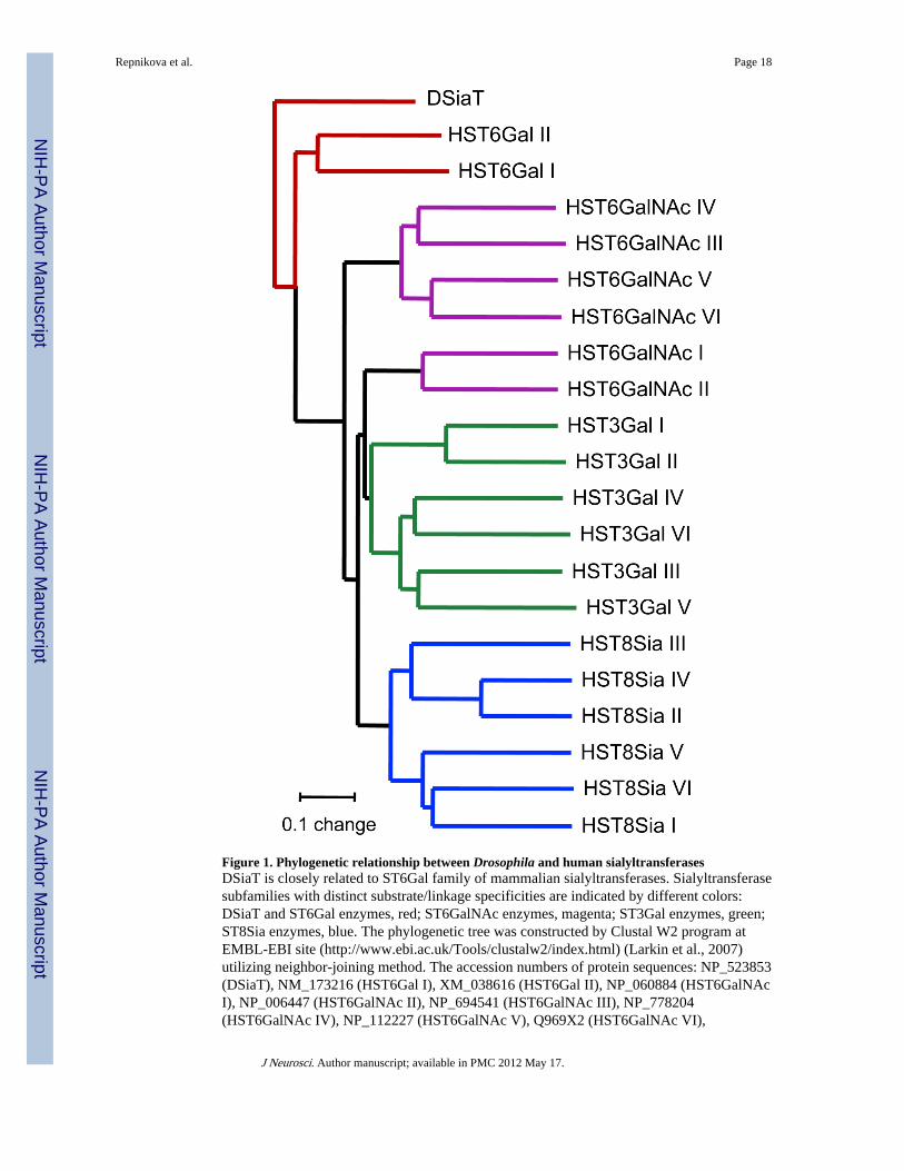
Figure 1. Phylogenetic relationship between Drosophila and human sialyltransferasesDSiaT is closely related to ST6Gal family of mammalian sialyltransferases. Sialyltransferasesubfamilies with distinct substrate/linkage specificities are indicated by different colors:DSiaT and ST6Gal enzymes, red; ST6GalNAc enzymes, magenta; ST3Gal enzymes, green;ST8Sia enzymes, blue. The phylogenetic tree was constructed by Clustal W2 program atEMBL-EBI site (http://www.ebi.ac.uk/Tools/clustalw2/index.html) (Larkin et al., 2007)utilizing neighbor-joining method. The accession numbers of protein sequences: NP_523853(DSiaT), NM_173216 (HST6Gal I), XM_038616 (HST6Gal II), NP_060884 (HST6GalNAcI), NP_006447 (HST6GalNAc II), NP_694541 (HST6GalNAc III), NP_778204(HST6GalNAc IV), NP_112227 (HST6GalNAc V), Q969X2 (HST6GalNAc VI),
Repnikova et al. Page 18
J Neurosci. Author manuscript; available in PMC 2012 May 17.
NIH
-PA Author Manuscript
NIH
-PA Author Manuscript
NIH
-PA Author Manuscript
NP_003024 (HST3Gal I), Q16842 (HST3Gal II), NP_777631 (HST3Gal III), NP_006269(HST3Gal IV), NP_003887 (HST3Gal V), NP_006091 (HST3Gal VI), NP_003025(HST8Sia I), NP_006002 (HST8Sia II), NP_056963 (HST8Sia III), NP_005659 (HST8SiaIV), NP_037437 (HST8Sia V), NP_001004470 (HST8Sia VI).
Repnikova et al. Page 19
J Neurosci. Author manuscript; available in PMC 2012 May 17.
NIH
-PA Author Manuscript
NIH
-PA Author Manuscript
NIH
-PA Author Manuscript

Figure 2. Behavioral phenotypes of DSiaT mutantsA, DSiaT mutants have significantly reduced longevity. For each genotype, 150 flies wereassayed for group longevity; error bars represent 95% confidence intervals. B–C, Mutantlarvae have crawling defects as revealed by decreased distance per muscle contraction (B)and reduced speed of muscle contractions (C). 20 larvae were assayed for each genotype. D,Mutant larvae have a crawling pattern abnormality, making many more turns. Graphs show20 superimposed larval tracks with the initial positions overlaid in the middle. E, Mutantflies have a defect in the ability of righting themselves after falling on the back. 20 flieswere assayed for each genotype. F, Mutant flies have TS-paralysis phenotype. Flies wereassayed at 38°C, 50 flies per genotype. Note that the severity of locomotor defects (E) andthe sensitivity of mutants to elevated temperature (F) significantly increased with age. In allexperiments, the rescue genotype was w1118; S23; P[DSiaT+], where P[DSiaT+] is atransgenic insertion of wildtype DSiaT gene. Error bars represent s.d. in F, and s.e.m. in B,C and E. ** indicates statistically significant difference with t-test p<0.01. Similarbehavioral phenotypes were observed with the L22 allele (e.g., Supplemental Fig. S3B).
Repnikova et al. Page 20
J Neurosci. Author manuscript; available in PMC 2012 May 17.
NIH
-PA Author Manuscript
NIH
-PA Author Manuscript
NIH
-PA Author Manuscript
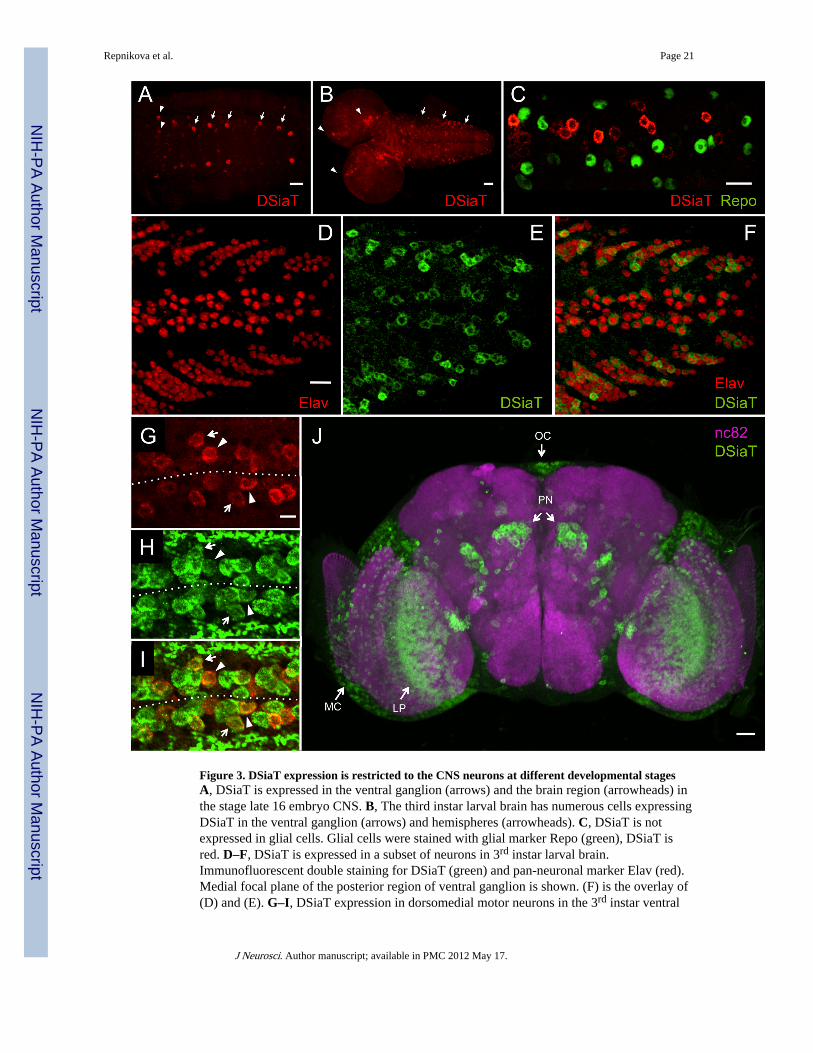
Figure 3. DSiaT expression is restricted to the CNS neurons at different developmental stagesA, DSiaT is expressed in the ventral ganglion (arrows) and the brain region (arrowheads) inthe stage late 16 embryo CNS. B, The third instar larval brain has numerous cells expressingDSiaT in the ventral ganglion (arrows) and hemispheres (arrowheads). C, DSiaT is notexpressed in glial cells. Glial cells were stained with glial marker Repo (green), DSiaT isred. D–F, DSiaT is expressed in a subset of neurons in 3rd instar larval brain.Immunofluorescent double staining for DSiaT (green) and pan-neuronal marker Elav (red).Medial focal plane of the posterior region of ventral ganglion is shown. (F) is the overlay of(D) and (E). G–I, DSiaT expression in dorsomedial motor neurons in the 3rd instar ventral
Repnikova et al. Page 21
J Neurosci. Author manuscript; available in PMC 2012 May 17.
NIH
-PA Author Manuscript
NIH
-PA Author Manuscript
NIH
-PA Author Manuscript

ganglion. Double staining for DSiaT (red) and DVGlut (green) reveals prominent DSiaTexpression in two motor neurons, MN1-Ib (arrowheads) and MN30-Ib (arrows). Dotted lineindicates the approximate position of midline. (I) is the overlay of (G) and (H). Fourabdominal segments (3–6, from left to right) of the ventral ganglion are shown. J,Expression of DSiaT (green) in the pharate adult brain. The overall morphology of the brainis visualized by neuropil staining with nc82 antibody (magenta). The expression of DSiaT ispresent in the optic lobes (MC, medulla cortex; LP, lobula plate), the region of projectionneurons (PN) and ocelli (OC). A–I, anterior is to the left; J, frontal view, dorsal is up. Scalebars: A, C–F, and J, 20 µm; B, 40 µm; G–I, 10µm.
Repnikova et al. Page 22
J Neurosci. Author manuscript; available in PMC 2012 May 17.
NIH
-PA Author Manuscript
NIH
-PA Author Manuscript
NIH
-PA Author Manuscript
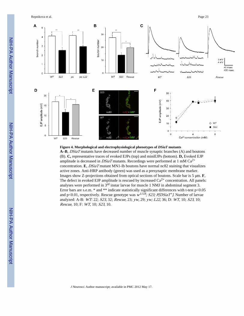
Figure 4. Morphological and electrophysiological phenotypes of DSiaT mutantsA–B, DSiaT mutants have decreased number of muscle synaptic branches (A) and boutons(B). C, representative traces of evoked EJPs (top) and miniEJPs (bottom). D, Evoked EJPamplitude is decreased in DSiaT mutants. Recordings were performed at 1 mM Ca2+
concentration. E, DSiaT mutant MN1-Ib boutons have normal nc82 staining that visualizesactive zones. Anti-HRP antibody (green) was used as a presynaptic membrane marker.Images show Z-projections obtained from optical sections of boutons. Scale bar is 5 µm. F,The defect in evoked EJP amplitude is rescued by increased Ca2+ concentration. All panels:analyses were performed in 3rd instar larvae for muscle 1 NMJ in abdominal segment 3.Error bars are s.e.m. * and ** indicate statistically significant differences with t-test p<0.05and p<0.01, respectively. Rescue genotype was w1118; S23; P[DSiaT+]. Number of larvaeanalyzed: A-B: WT: 22; S23, 32; Rescue, 23; yw, 29; yw; L22, 36; D: WT, 10; S23, 10;Rescue, 10; F: WT, 10; S23, 10.
Repnikova et al. Page 23
J Neurosci. Author manuscript; available in PMC 2012 May 17.
NIH
-PA Author Manuscript
NIH
-PA Author Manuscript
NIH
-PA Author Manuscript
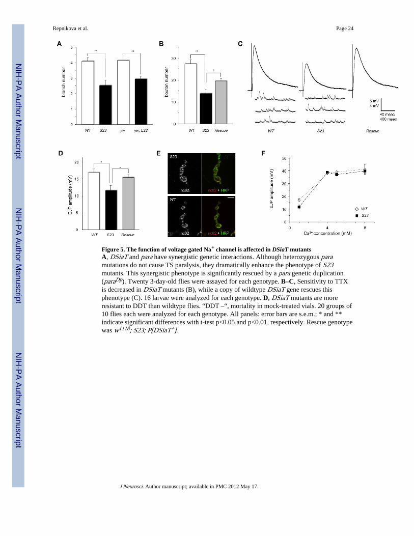
Figure 5. The function of voltage gated Na+ channel is affected in DSiaT mutantsA, DSiaT and para have synergistic genetic interactions. Although heterozygous paramutations do not cause TS paralysis, they dramatically enhance the phenotype of S23mutants. This synergistic phenotype is significantly rescued by a para genetic duplication(paraDp). Twenty 3-day-old flies were assayed for each genotype. B–C, Sensitivity to TTXis decreased in DSiaT mutants (B), while a copy of wildtype DSiaT gene rescues thisphenotype (C). 16 larvae were analyzed for each genotype. D, DSiaT mutants are moreresistant to DDT than wildtype flies. “DDT –“, mortality in mock-treated vials. 20 groups of10 flies each were analyzed for each genotype. All panels: error bars are s.e.m.; * and **indicate significant differences with t-test p<0.05 and p<0.01, respectively. Rescue genotypewas w1118; S23; P[DSiaT+].
Repnikova et al. Page 24
J Neurosci. Author manuscript; available in PMC 2012 May 17.
NIH
-PA Author Manuscript
NIH
-PA Author Manuscript
NIH
-PA Author Manuscript

Figure 6. Sialyltransferase activity is required for in vivo DSiaT functioningA, DSiaTHK mutant protein has no detectable activity in vitro. DSiaTHK and DSiaTWT
expressed in Drosophila S2 cells as Protein A-fusion constructs were assayed forsialyltransferase activity using CMP-[14C]Sia as a donor and lactoferrin as an acceptor. Lefttwo lanes are the autoradiograph of products of the assays separated on SDS gel; right twolanes show the Coomassie staining of the same gel as a control for protein amount. IgG,immunoglobulin leached from beads used in purification (Koles et al., 2004). B, DSiaTHK isexpressed and localized normally in brain neurons. DSiaTHK and DSiaTWT were expressedin vivo as C-terminally HA-tagged full-length constructs using C155-Gal4 driver. There isno difference in the level of expression or subcellular localization of these proteins, asrevealed by double staining with a Golgi marker, Lva (green) (Sisson et al., 2000). C, Thetransgenic expression of DSiaT wildtype construct with a neuronal driver fully rescues theTS paralysis phenotype of DSiaT mutants, however the expression of enzymatically inactiveDSiaT protein, DSiaTHK, is unable to rescue the mutant phenotype. D, Sialyltransferase
Repnikova et al. Page 25
J Neurosci. Author manuscript; available in PMC 2012 May 17.
NIH
-PA Author Manuscript
NIH
-PA Author Manuscript
NIH
-PA Author Manuscript

activity is required for rescuing locomotor defect of DSiaT mutants. While transgenicDSiaTWT fully rescues locomotor abnormalities, DSiaTHK is unable to rescue the mutantphenotype. Genotypes: WT - wildtype control (w1118 Canton S), Driver - C155-GAL4/+,S23+Driver::DsiaTWT - C155-GAL4/+;S23/S23;UAS-DSiaTWT/+, S23+Driver::DsiaTHK -C155-GAL4/+;S23/S23;UAS-DSiaTHK/+. Twenty 5-day-old flies were assayed for eachgenotype. Error bars are s.e.m.; ** indicate the results with significant differences fromwildtype phenotypes (t-test p<0.01) (but these results are not significantly different fromeach other).
Repnikova et al. Page 26
J Neurosci. Author manuscript; available in PMC 2012 May 17.
NIH
-PA Author Manuscript
NIH
-PA Author Manuscript
NIH
-PA Author Manuscript
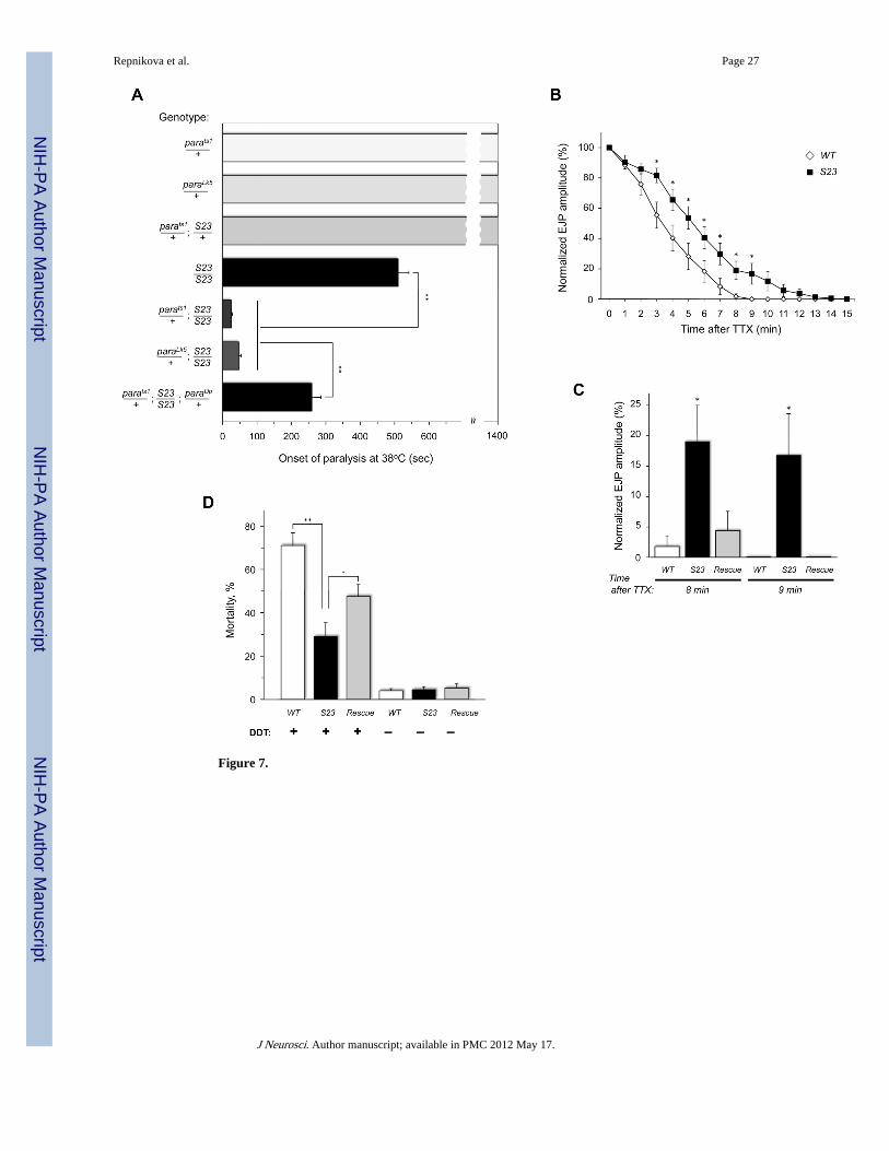
Figure 7.
Repnikova et al. Page 27
J Neurosci. Author manuscript; available in PMC 2012 May 17.
NIH
-PA Author Manuscript
NIH
-PA Author Manuscript
NIH
-PA Author Manuscript

Figure 8.
Repnikova et al. Page 28
J Neurosci. Author manuscript; available in PMC 2012 May 17.
NIH
-PA Author Manuscript
NIH
-PA Author Manuscript
NIH
-PA Author Manuscript

Figure 9.
Repnikova et al. Page 29
J Neurosci. Author manuscript; available in PMC 2012 May 17.
NIH
-PA Author Manuscript
NIH
-PA Author Manuscript
NIH
-PA Author Manuscript