Preclinical Neurochemical and Electrophysiological Profile of 1192U90, A Potential Antipsychotic
Upload
independentCategory
view
2download
0
Electrophysiological and Morphological Characterization of Identified MotorNeurons in the Drosophila Third Instar Larva Central Nervous System
James C. Choi, Demian Park, and Leslie C. GriffithDepartment of Biology and Volen Center for Complex Systems, Brandeis University, Waltham, Massachusetts 02454-9110
Submitted 18 November 2003; accepted in final form 18 December 2003
Choi, James C., Demian Park, and Leslie C. Griffith. Electrophys-iological and morphological characterization of identified motor neu-rons in the Drosophila third instar larva central nervous system. JNeurophysiol 91: 2353–2365, 2004. First published December 24,2003; 10.1152/jn.01115.2003. We have used dye fills and electro-physiological recordings to identify and characterize a cluster ofmotor neurons in the third instar larval ventral ganglion. This clusterof neurons is similar in position to the well-studied embryonic RPneurons. Dye fills of larval dorsomedial neurons demonstrate thatindividual neurons within the cluster can be reproducibly identified byobserving their muscle targets and bouton morphology. The terminaltargets of these five neurons are body wall muscles 6/7, 1, 14, and 30and the intersegmental nerve (ISN) terminal muscles (1, 2, 3, 4, 9, 10,19, 20). All cells except the ISN neuron, which has a type Is ending,display type Ib boutons. Two of these neurons appear to be identicalto the embryonic RP3 and aCC cells, which define the most proximaland distal innervations within a hemisegment. The targets of the otherneurons in the larval dorsomedial cluster do not correspond to em-bryonic targets of the neurons in the RP cluster, suggesting rewiringof this circuit during early larval stages. Electrophysiological studiesof the five neurons in current clamp revealed that type Is neurons havea longer delay in the appearance of the first spike compared with typeIb neurons. Genetic, biophysical, and pharmacological studies incurrent and voltage clamp show this delay is controlled by the kineticsand voltage sensitivity of inactivation of a current whose propertiessuggest that it may be the Shal IA current. The combination of geneticidentification and whole cell recording allows us to directly explorethe cellular substrates of neural and locomotor behavior in an intactsystem.
I N T R O D U C T I O N
Locomotion is a complex behavior that requires coordinatednervous system output to multiple muscles. This coordinatedoutput has been shown to be the result of central patterngenerator circuits in both vertebrate and invertebrate systems(Marder and Calabrese 1996). The output of most centralpattern generators drives motor neuron firing and can beshaped by sensory inputs. Changes in locomotion usually in-volve alterations of synaptic strengths and/or the intrinsic prop-erties of the constituent neurons. In both vertebrate spinal cord(Kiehn 1991), where the motor neurons are not part of thecentral pattern generator, and in lobster stomatogastric gan-glion (Marder 1998), where they are, modulation of the prop-erties of motor neurons is also important for adjusting output ofthe central pattern generator.
In Drosophila third instar larvae, the function of centralpattern generators has been studied only by recording muscle
output (Cattaert and Birman 2001; Gorczyca et al. 1991);circuit components have not yet been identified. The outputsynapse, the third instar larval neuromuscular junction (NMJ)has been studied intensely and is arguably the premier systemfor using genetics and electrophysiology to understand synap-tic development, transmission and plasticity. In the traditionalNMJ preparation, the synaptic response is measured by impal-ing the muscle or making a focal patch over a bouton. Typi-cally, recordings are done with the CNS cut off so that thecontribution of the presynaptic cell is limited to that of thedistal axon and the terminal.
For understanding coordinated nervous system function, theNMJ preparation is not ideal. In the animal, the activation ofsynapses usually occurs as a consequence of an action potentialgenerated by integration of synaptic inputs in the somatoden-dritic compartment. The firing properties of the neuron, e.g.,spike frequency, amplitude, and duration, are determined bythe balance of inward and outward currents in the cell. Inprinciple, this allows individual cells to develop a uniqueresponse to inputs. Until recently (Rohrbough and Broadie2002), third instar Drosophila motor neurons have been char-acterized primarily by their terminal morphology, position, andneurotransmitter content (Anderson et al. 1988; Hoang andChiba 2001; Jia et al. 1993; Johansen et al. 1989; Kurdyak etal. 1994; Monastirioti et al. 1995). Differences in the releaseproperties and plasticity of Ib and Is terminals have beendocumented indirectly with focal patch recordings over the twobouton types or low-level stimulation to distinguish terminalthresholds (Kurdyak et al. 1994; Lnenicka and Keshishian2000), suggesting that these two neurons types are functionallydifferent. Direct electrophysiological experiments in neuronsof the larval stage were limited to cultured, dissociated cells(Solc and Aldrich 1988; Wu et al. 1983b) and boutons (Mar-tinez-Padron and Ferrus 1997). In contrast, embryonic motorneurons have been studied electrophysiologically in vivo(Baines and Bate 1998; Baines et al. 1999, 2001) as have adultthoracic ganglion motor neurons (Trimarchi and Murphey1997), suggesting that access to ventral ganglion motor neu-rons was technically feasible. Recordings from unidentifiedventral ganglion neurons confirmed this (Rohrbough andBroadie 2002).
In addition to answering functional questions, direct accessto intact identified motor neurons in the third instar would alsofacilitate investigation of development of central circuits. Neu-rons of the ventral ganglion are arrayed in bilaterally symmet-rical repeats that correlate with the segmental organization of
Address for reprint requests and other correspondence: L. C. Griffith, Dept.of Biology, MS008, Brandeis University, 415 South St., Waltham, MA 02454-9110 (E-mail: [email protected]).
The costs of publication of this article were defrayed in part by the paymentof page charges. The article must therefore be hereby marked ‘‘advertisement’’in accordance with 18 U.S.C. Section 1734 solely to indicate this fact.
J Neurophysiol 91: 2353–2365, 2004.First published December 24, 2003; 10.1152/jn.01115.2003.
23530022-3077/04 $5.00 Copyright © 2004 The American Physiological Societywww.jn.org
the body wall, and the relative orientations of neuronal somataare stereotypic throughout segments A2–A7. During embry-onic and larval development, the growth cone and varicositiesof motor neurons are in a state of constant flux (Yoshihara etal. 1997). In the embryo, outgrowth, pathfinding, and targetrecognition of a group of dorsomedial neurons (RP1–4 andaCC) has been extensively studied (Bate and Broadie 1995;Broadie et al. 1993; Halpern et al. 1991; Sink and Whitington1991b; Thomas et al. 1984). Filling of embryonic neuronsallowed the identification of initial targets. Backfilling of thirdinstar larval neurons from the terminal has suggested that theposition and/or targets of these embryonic neurons may changeduring development of the larval CNS (Hoang and Chiba 2001;Sink and Whitington 1991a). The final targets, central connec-tions, and relative positions in the ventral ganglion of theseneurons need to be investigated by accessing the cell bodies.
To study the intact mature third instar CNS, we have mor-phologically and electrophysiologically characterized a subsetof the motor neurons that innervate the body wall muscles.These neurons receive output from the central pattern generatorfor locomotion, and their modulation is likely to be importantto plasticity of locomotor behavior. We find that significantstructural reorganization occurs between the late embryo andthe third instar. We also find that there is diversity in theintrinsic properties of larval motor neurons and that differencesin potassium current regulation can control the cell-specificproperties of identified neurons.
M E T H O D S
Preparation
All experiments were performed on wandering, primarily female,third instar larvae. Animals were dissected and pinned in 0.2 mMCa2� “A” solution (which contained, in mM, 128 NaCl, 2 KCl, 35sucrose, 4 MgCl2, and 5 HEPES, pH 7.1–7.2 (original solution con-tains 1.8 mM Ca2�) (Jan and Jan 1976), with care taken to leavesegmental nerves and the CNS intact. Calcium-free “A” solution wasused as the external solution for all experiments with the exception ofthe confocal dye-fill experiments (see details in the following text).The preparation was visualized using an upright Olympus microscopeequipped with differential interference contrast and epifluorescence.Under constant perfusion with laminar flow traversing, the larvatoward the anterior direction, collagenase type I or protease XIV (1and 2 mg/ml, respectively, Sigma-Aldrich, St. Louis, MO) was focallyadministered to the ventral ganglion using a glass pipette pulled to�10 �m tip diameter. Debris was constantly cleared using the pipettewith positive and negative pressure. With adequate exposure, theouter layer of the ganglion is disrupted, revealing neural tissue.Prominent somata with diameters of 10–15 �m could be seen at thispoint. The protease treatment was continued until an inner membranelayer enclosing the group of neurons was disrupted. A successfulprocedure rendered the soma clean, smooth and loose, but intactwithin its cluster. Recordings were performed mainly in segment A3.
Genetics, cell visualization, and identification
Motor neurons were visualized using GFP fluorescence. GFP ex-pression was driven by C164-GAL4 (a generous gift of Vivian Bud-nik, University of Massachusetts, Amherst, MA), an enhancer trapline that expresses in all motor neurons (Packard et al. 2002). Crossedwith UAS-mCD8-GFP, C164-GAL4 allows sharp, distinct visualiza-tion of cells, as mCD8 targets the GFP to the membrane (Lee and Luo1999). After dissection and protease digestion, the targeted soma was
approached with constant positive pressure until the pipette tip wasapposed to the surface. After establishing a tight seal, the membranewas broken with instantaneous suction and/or current injection. Therecording intracellular solution usually contained tetramethyl rhoda-mine dextran 3000 (10 mg/ml, Molecular Probes, Eugene, OR), eitherbackfilled into the pipette or as part of the solution, which gave aslightly lower patch success rate but did not alter neuronal responses(data not shown). After �15 min of filling, during which physiolog-ical recordings were obtained, the processes and terminal morphologyof the targeted neurons were visualized on the same microscope.Images were taken with a Photometrics PXL liquid-cooled CCDcamera and IPLab software package on a Macintosh computer. Com-binations of brightfield, GFP, and dye images were rendered usingNIH Image and Adobe Photoshop.
For every experiment, a specific soma within a stereotypical clusterin the dorsomedial surface was targeted, relying on its orientationrelative to the midline and the other somata. ShKS133, a point mutationin the Sh gene (Lichtinghagen et al. 1990) that eliminates IA in muscle(Solc et al. 1987; Wu et al. 1983a) was used to dissect the IA
potassium current. In this case, DIC and not GFP was used to targetneurons for recording, and the identity of the neuron was confirmedwith dye fill.
For detailed confocal visualization of axon and dendrites of indi-vidual motor neurons using confocal microscopy, larvae were dis-sected in HL3 (0 mM Ca2�), and the recording intracellular solutioncontained a 10:2 mixture of tetramethyl rhodamine dextran 3000 (10mg/ml) and AlexaFluor 568 hydrazide sodium salt (10 mg/ml, Mo-lecular Probes). After 30–60 min of filling, larvae were fixed inphosphate-buffered 4% paraformaldehyde for 30 min at room tem-perature. The larval tissues were then washed in HL3, followed by 30,60, and 100% glycerol incubations for 10 min prior to mounting inVectashield (Vector Laboratories, Burlingame, CA). Confocal imageswere acquired using a Leica confocal system. Images were thenfurther processed in Adobe Photoshop.
Electrophysiology
Filamented thin- and thick-walled capillary pipettes (WPI, Sara-sota, FL) were pulled and fire polished to a resistance of 5–10 M�.Intracellular recording solution (adapted from O’Dowd 1995) con-tained (in mM) 120 potassium gluconate, 20 KCl, 10 HEPES, 1.1EGTA, 2 MgCl2, and 0.1 CaCl2. The pH was adjusted to 7.2 and theosmolarity to 280 mmol/kg. Recordings were performed with anAxopatch 200B amplifier (Axon Instruments, Foster City, CA) andITC-16 data acquisition board (National Instruments, Austin, TX)with Igor software (Wavemetrics, Lake Oswego, OR). Cell membranepotentials were recorded in whole cell current-clamp mode. Spikeswere evoked by a series of current injections delivered via the record-ing pipette. The duration of current steps was 500 or 1,000 ms, and theamplitude ranged from –20 to 140 pA in 20-pA increments. Inprepulse experiments under current clamp, the external solution con-tained 0 mM Ca2�. The duration of the prepulse was 1 s, and theamplitude was �20 pA below spike threshold, usually 20 pA. Theaverage prepulse depolarization was to –43 � 1 mV (n � 8 cells). Theprepulse was immediately followed by test current steps of 1-s dura-tion. In experiments to pharmacologically eliminate transient potas-sium currents, 4-aminopyridine (4-AP; 1.5 mM, Calbiochem, SanDiego, CA) was bath-applied. Recordings were obtained before andafter application, and also 2 min after several washes of externalsolution. For recordings of spontaneous activity, 2.4 mM Ca2� wasadded to the external solution, and passive recordings were done inwhole cell configuration.
In neuron-muscle dual recordings, EJPs were recorded using sharpelectrodes (�30 M�) filled with 3 M KCl for muscle. Havingachieved whole cell access to the neuron, the preparation was visu-alized under lower magnification (�10), and the corresponding mus-cle was impaled with a recording electrode. The external Ca2� con-
2354 J. C. CHOI, D. PARK, AND L. C. GRIFFITH
J Neurophysiol • VOL 91 • MAY 2004 • www.jn.org
centration was increased to 0.4 mM. Under current clamp, a stimulusprotocol was performed on the neuron and recordings made simulta-neously from neuron and muscle. EJPs were amplified with Axoclamp2B (Axon Instruments), which was connected to the same acquisitionboard.
For voltage-clamp recordings, neurons were switched to voltage-clamp mode after initial current-clamp recordings. Sodium channelswere blocked with tetrodotoxin (Sigma, 100 nM). Cells were held ateither –80 or –100 mV. For trace subtractions, voltage was raised to�40 mV for 1 s, and the difference between the waveform obtainedand one recorded from a holding potential of –40 mV was calculated.For current-voltage data, test pulses ranging from –120 to �40 mV (in20-mV increments) were given in 15-s intervals. Waveforms obtainedwith the –20 mV pulse was used to subtract leakage currents. Toobtain voltage-dependent inactivation plots, a 1-s prepulse of variableamplitude (�100 to �40 mV in 10-mV increments) was given, and atest pulse of �40 mV amplitude was given after a 10-ms interpulseinterval. Perfusion was usually stopped for voltage-clamp recordings.Identity of neurons was subsequently verified by visualizing the dyefill. Capacitance normalization was done with values obtained directlyfrom the parameter settings on the amplifier.
Series resistances of cells ranged from 20 to �100 M�, most likelydue to clogging of the pipette tip by cellular constituents. As anacceptance criterion, only experiments where Rin �1 G� and calcu-lated maximum voltage error 25 mV were analyzed.
Data analysis
For injection of different levels of current, time to peak wasmeasured as the time between the beginning of current injection to thepeak of the first spike observed in the voltage trace. Peak amplitudewas derived by averaging the amplitude of all the spikes in a givenvoltage trace. Spike frequency was calculated by averaging the in-stantaneous frequencies derived by measuring the time interval be-tween adjacent spikes. Only the first 500 ms of each trace wasanalyzed. The inactivation time constant (�) was obtained by using asingle exponential fit from peak to steady state for each waveform. Forthe inactivation plot, the peak was normalized with the maximum andminimum values, and plotted with respected to prepulse amplitude. Aregular Boltzmann equation was used to fit the plot, and the half-maximal inactivation voltage was obtained.
Statistical analyses were done in Statview 4.5 (Abacus Concepts/SAS, Carry, NC) or the analysis tools package in Excel (Microsoft,Redmond, WA). P values reported for multiple comparisons were ascer-tained using ANOVA with Scheffe’s post hoc test for significance.
R E S U L T S
Identification of dorsomedial motor neuron projections andtargets in the third instar larva
Using the standard NMJ dissection, neurons in the ventralganglion can be readily visualized and particular groups ofneurons identified using cell-specific GAL4 drivers. We haveused C164-GAL4 (Packard et al. 2002) to drive expression ofGFP in motor neurons to target our studies to this population.The motor neurons in the ventral ganglion are organized ste-reotypically (Sink and Whitington 1991a) with segmental ar-rays of somata repeating along the anterior-posterior axis.Axons project to the body-wall muscle through nerves thatemanate from the ganglion in each hemisegment. We focusedon the bilaterally symmetric dorsomedial clusters that arealigned parallel to the midline. Figure 1A shows GFP expres-sion in the posterior end of a preparation after dissection. A setof five neurons that are GFP positive repeats in segmentsA2–A7 with all the neurons lying in the same plane of view.
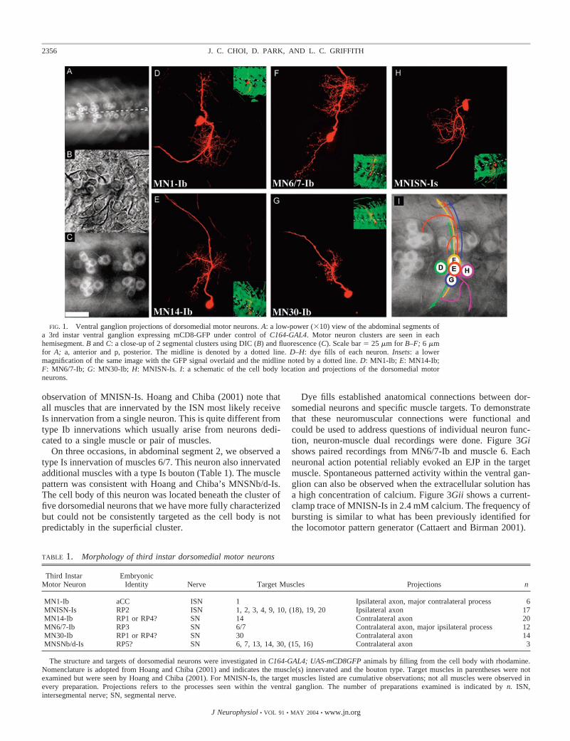
Figure 1, B and C, shows brightfield and GFP visualizations ofthe dorsomedial cluster at higher magnification after enzymaticdigestion. The somata are typically in a cross formation, al-though shifts in relative orientation can cause some deviationfrom this pattern.
The identities of the neurons in the dorsomedial cluster weredetermined by filling GFP-positive cell bodies from the record-ing patch pipette, which contained rhodamine and/or Alexa568. After recording, the body-wall muscles were imaged forGFP and dye. Each fill resulted in dye signal in a single axonand bouton arbor that could be visualized in the live prepara-tion. Correlation of soma position, muscle target, and boutonmorphology suggests that innervation type and target are in-variant for each cell within the cluster. Each neuron has anelaborate projection pattern within the ventral ganglion that isalways in the same relative position. Example fills of each ofthe five neurons are shown in Fig. 1, D–H, and a schematic ofthe arborization patterns is shown in I.
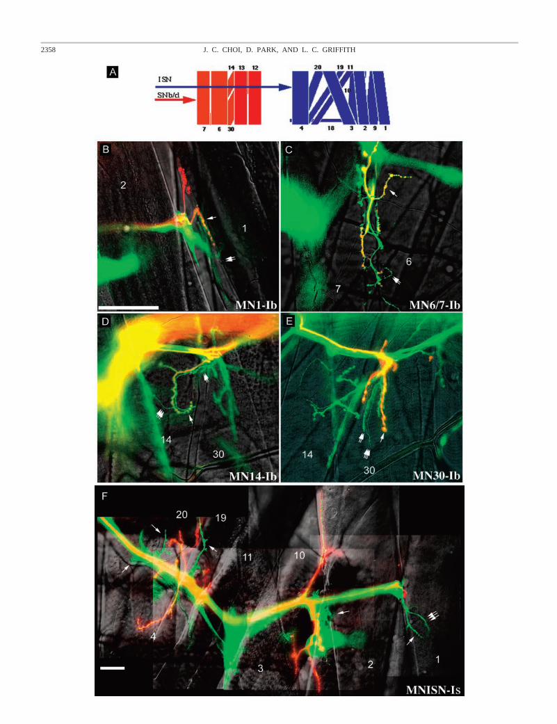
To name the neurons, we have adopted the nomenclaturedevised by Hoang and Chiba (2001), naming each neuronaccording to its target muscle and the morphology of its ter-minal boutons. The five neurons of the dorsomedial clusterwere identified as MN1-Ib, MN6/7-Ib, MN14-Ib, MN30-Ib,and MNISN-Is. Table 1 provides a summary of the cell’smorphological features. Bouton types were categorized primar-ily by relative size compared with the other boutons in the fieldof view, and their consistency with previously published ac-counts of muscle innervations (Hoang and Chiba 2001). Figure2 shows the muscle termini of each of the five neurons. Aschematic of the organization of the muscles and nerves in onehemisegment is shown in Fig. 2A.
Four of the five neurons in the dorsomedial cluster terminatewith type Ib boutons. MN14-Ib and MN30-Ib are unipolar andproject contralaterally through the segmental nerve (Fig. 1, Eand G). Axons from these two neurons travel together until theterminal arborization point in SNb (segmental nerve branch b)where they diverge to innervate their respective neighboringmuscle targets, 14 (Fig. 2D) and 30 (Fig. 2E). MN6/7-Ibprojects bilaterally in the ventral ganglion, but a single processextends contralaterally past the midline and out through thesegmental nerve, terminating exclusively with type Ib boutonson muscles 6 and 7 (Figs. 1F and 2C). MN1-Ib has prominentbilateral projections with two dendritic arborizations within theventral ganglion (Fig. 1D); the motor axon projects ipsilater-ally through the ISN (intersegmental nerve) to innervate thedistal muscle 1 (Fig. 2B).
The anterior-most neuron in the cluster is MNISN-Is, whichprojects ipsilaterally through the ISN. The axonal processclimbs up and exits the nerve of the anterior segment, so thatits muscle innervation is actually one segment anterior to thoseof other neurons within the cluster (Fig. 1H). This neuron targetsmultiple muscles on the distal (dorsal) body wall (Fig. 2F). Theidentification of this neuron as a Is neuron was supported by thefact that in muscle 4, in view of the slightly bigger boutons ofIb, the dye-filled innervating bouton type was clearly type Isand not type II (Fig. 2F). We unambiguously observed inner-vation of muscles 1, 2, 3, 4, 9, 10, 19, and 20, and muscle 18may also have innervation that was less clear and is not seen inFig. 2. Hoang and Chiba describe a neuron (MNISN-Is) thatinnervates muscles 1, 2, 3, 4, 9, 10, 18, 19, and 20 (Hoang andChiba 2001). This is almost the exact target pattern of our
2355VENTRAL GANGLION MOTOR NEURONS
J Neurophysiol • VOL 91 • MAY 2004 • www.jn.org
observation of MNISN-Is. Hoang and Chiba (2001) note thatall muscles that are innervated by the ISN most likely receiveIs innervation from a single neuron. This is quite different fromtype Ib innervations which usually arise from neurons dedi-cated to a single muscle or pair of muscles.
On three occasions, in abdominal segment 2, we observed atype Is innervation of muscles 6/7. This neuron also innervatedadditional muscles with a type Is bouton (Table 1). The musclepattern was consistent with Hoang and Chiba’s MNSNb/d-Is.The cell body of this neuron was located beneath the cluster offive dorsomedial neurons that we have more fully characterizedbut could not be consistently targeted as the cell body is notpredictably in the superficial cluster.
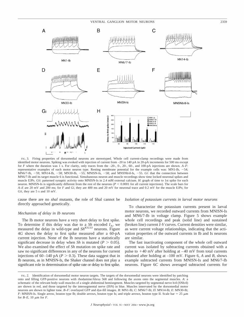
Dye fills established anatomical connections between dor-somedial neurons and specific muscle targets. To demonstratethat these neuromuscular connections were functional andcould be used to address questions of individual neuron func-tion, neuron-muscle dual recordings were done. Figure 3Gishows paired recordings from MN6/7-Ib and muscle 6. Eachneuronal action potential reliably evoked an EJP in the targetmuscle. Spontaneous patterned activity within the ventral gan-glion can also be observed when the extracellular solution hasa high concentration of calcium. Figure 3Gii shows a current-clamp trace of MNISN-Is in 2.4 mM calcium. The frequency ofbursting is similar to what has been previously identified forthe locomotor pattern generator (Cattaert and Birman 2001).
TABLE 1. Morphology of third instar dorsomedial motor neurons
Third InstarMotor Neuron
EmbryonicIdentity Nerve Target Muscles Projections n
MN1-Ib aCC ISN 1 Ipsilateral axon, major contralateral process 6MNISN-Is RP2 ISN 1, 2, 3, 4, 9, 10, (18), 19, 20 Ipsilateral axon 17MN14-Ib RP1 or RP4? SN 14 Contralateral axon 20MN6/7-Ib RP3 SN 6/7 Contralateral axon, major ipsilateral process 12MN30-Ib RP1 or RP4? SN 30 Contralateral axon 14MNSNb/d-Is RP5? SN 6, 7, 13, 14, 30, (15, 16) Contralateral axon 3
The structure and targets of dorsomedial neurons were investigated in C164-GAL4; UAS-mCD8GFP animals by filling from the cell body with rhodamine.Nomenclature is adopted from Hoang and Chiba (2001) and indicates the muscle(s) innervated and the bouton type. Target muscles in parentheses were notexamined but were seen by Hoang and Chiba (2001). For MNISN-Is, the target muscles listed are cumulative observations; not all muscles were observed inevery preparation. Projections refers to the processes seen within the ventral ganglion. The number of preparations examined is indicated by n. ISN,intersegmental nerve; SN, segmental nerve.
FIG. 1. Ventral ganglion projections of dorsomedial motor neurons. A: a low-power (�10) view of the abdominal segments ofa 3rd instar ventral ganglion expressing mCD8-GFP under control of C164-GAL4. Motor neuron clusters are seen in eachhemisegment. B and C: a close-up of 2 segmental clusters using DIC (B) and fluorescence (C). Scale bar � 25 �m for B–F; 6 �mfor A; a, anterior and p, posterior. The midline is denoted by a dotted line. D–H: dye fills of each neuron. Insets: a lowermagnification of the same image with the GFP signal overlaid and the midline noted by a dotted line. D: MN1-Ib; E: MN14-Ib;F: MN6/7-Ib; G: MN30-Ib; H: MNISN-Is. I: a schematic of the cell body location and projections of the dorsomedial motorneurons.
2356 J. C. CHOI, D. PARK, AND L. C. GRIFFITH
J Neurophysiol • VOL 91 • MAY 2004 • www.jn.org
Evoked spiking activity in dorsomedial larval neurons
The dorsomedial neurons show consistent position, target,and morphology from animal to animal. To determine if firingproperties were also determined by cell identity, we recordedevoked action potentials from the somata of these neurons.Current injection readily caused repetitive spikes at frequenciesthat were dependent on stimulus intensity. Hyperpolarizingprepulses did not evoke significant activity (data not shown).Figure 3, A-F, shows representative traces from current-clamprecordings of the five neurons within the cluster and a record-ing from a putative MNSNb/d-Is cell. Data acquisition wastypically done as the cells were being filled with dye; thisallowed unequivocal identification of the neurons and correla-tion of morphology with physiology. Resting membrane po-tential, resting input resistance and current required for spikingare shown in Table 2. The amount of current required (in20-pA increments) to initiate spiking was not different com-paring the Ib neurons to each other, but MNISN-Is requiredsignificantly more current than any of the Ib cells (P 0.001).
Data for time to first spike for each of the five cells areshown in Fig. 3H. The MNISN-Is neuron had a significantdelay to spiking compared with the Ib neurons (Fig. 3H, P 0.0001 for all current injections). Time to first spike was notdifferent between Ib neurons at any current injection level (Fig.3H, P � 0.35).
Mechanism of the MNISN-Is delay
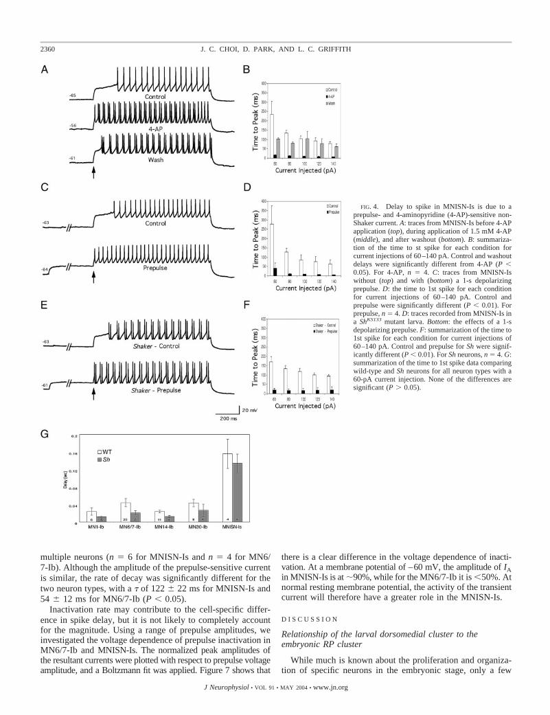
Because the identity and properties of the larval motorneurons are stereotyped, the molecular basis of the behavior ofeach neuron can be addressed using pharmacological, biophys-ical, and genetic tools. A distinct structural feature ofMNISN-Is and MNSNb/d-Is is that, unlike the type Ib neurons,they innervate multiple muscles. The striking delay in the timeto first spike is likely to have effects on patterned muscleactivity. In other neurons where such delays have been ob-served, they have often been due to the presence of an inacti-vating potassium current (Turrigiano et al. 1996; Zhao and Wu1997). To determine if this was the mechanism underlying theMNISN-Is firing kinetics, we examined the effects of depolar-izing prepulses and 4-AP on the delay. Both of these manip-ulations are known to decrease IA-type currents. Figure 4Ashows traces from an MNISN-Is cell before, during and afterwashout of 1.5 mM 4-AP. During exposure to the drug, thereis a decrease in the time to first spike that is reversed onwashout. 4-AP also affects spiking behavior; stereotyped, reg-ular single spike activity is altered to give doublet spikes. In theexample shown in Fig. 4A, the doublet spiking is also seen inthe wash, but in other cells this effect of 4-AP did wash out(data not shown). Depolarizing prepulses delivered prior tosuprathreshold test amplitudes had an effect similar to 4-AP(Fig. 4C), decreasing the time to spike onset. The mean level ofdepolarization achieved with prepulse was –43 � 1 mV (n �8). Prepulse also caused doublets in the spike train, but theeffect was less consistent, with multiple spike activity appear-ing sporadically during test pulse (data not shown). Subsequentexperiments with longer test pulse protocols in control cellsrevealed that with long current injections (�2 s), even controlcells could show doublet spiking.
The changes in delay with 4-AP and prepulse are quantifiedin Fig. 4, B and D, for different levels of current injection. Thedecrease in time to first spike was significant for comparison ofcontrol and washout with 4-AP for all current injections (P 0.05). Control and washout were not significantly different,except at the lowest stimulus amplitude (P � 0.02). For pre-pulse experiments, control and prepulse delays were signifi-cantly different at all current spike-evoking injection levels(P 0.01).
To determine the identity of the 4-AP- and prepulse-sensi-tive potassium current(s) responsible for the delay in onset ofspiking and doublet spiking, we examined the firing behaviorof MNISN-Is in ShKS133 mutants. The Sh gene encodes one ofthe channel proteins known to produce IA. In Sh mutants, thefast inactivating current of third instar larval muscles is selec-tively eliminated (Wu and Haugland 1985; Wu et al. 1983a).The other gene known to encode an IA channel is shal. TheShal protein is thought to be responsible for the majority of IA
in embryonic neurons (Scholz et al. 1988; Tsunoda and Salkoff1995). A comparison of the delay in MNISN-Is recordingsfrom ShKS133 (Fig. 4E) and control (Fig. 4A) neurons showsthat the magnitude of delay is not significantly different for anyspike-inducing current injection level (P � 0.1). In all of therecordings from Sh mutant neurons, doublet spikes were seenin the spike train (Fig. 4E). These doublets were qualitativelysimilar to those seen with 4-AP treatment and prepulse. Pre-pulse of MNISN-Is in Sh mutants is still able to decrease thetime to first spike, suggesting that there is a non-Shaker pre-pulse-sensitive current that causes the delay. A comparison ofthe delay before and after prepulse for different current levelsis shown in Fig. 4F. For all levels of current injection, thedecrease in the delay ranged from 80 to 95%, and Sh wassignificantly different from Sh with prepulse (P 0.05 for allcurrent injection levels). These results suggest that the role ofShaker in the MNISN-Is neuron does not involve setting thespike delay. Spike delay is mediated by another prepulse-sensitive current, likely carried by Shal. Unfortunately, be-
TABLE 2. Properties of third instar dorsomedial motor neurons
Motor Neuron
Resting InputResistance,
M�
RestingMembrane
Potential, mV
Average CurrentInjection to
Evoke Spikes,pA n
MN1-Ib 977 � 89 �56 � 1 27 � 5 6MN6/7-Ib 1204 � 79 �58 � 2 32 � 4 12MN14-Ib 1322 � 127 �54 � 1 26 � 4 10MN30-Ib 1435 � 70 �58 � 2 29 � 4 7MNISN-Is 1653 � 172* �61 � 2 74 � 7** 10
Electrophysiological properties of dorsomedial neurons were measured inwhole cell current-clamp mode. Resting input resistance was measured bypotential difference with a �20-pA current pulse. MNISN-Is was had asignificantly higher resting input resistance compared to MN1-Ib (*, P 0.05).Spikes were induced by current injections from 20 to 140 pA in 20-pAincrements. The level of current injection that 1st induced spiking was notedfor each cell. There was no significant difference in the average currentrequired to induce spiking in Ib neurons (P � 0.05). MNISN-Is requiredsignificantly more current than any of the Ib neurons (**, P 0.001). Therewas no difference in resting membrane potential among all the neurons(P � 0.05). The number of cells of each type characterized is indicated by n.Values are means � SE.
2357VENTRAL GANGLION MOTOR NEURONS
2358 J. C. CHOI, D. PARK, AND L. C. GRIFFITH
cause there are no shal mutants, the role of Shal cannot bedirectly approached genetically.
Mechanism of delay in Ib neurons
The Ib motor neurons have a very short delay to first spike.To determine if this delay was due to a Sh encoded IA, wemeasured the delay in wild-type and ShKS133 neurons. Figure4G shows the delay to first spike measured after a 60-pAcurrent injection. None of the Ib neurons have a statisticallysignificant decrease in delay when Sh is mutated (P � 0.05).We also examined the effect of Sh mutation on spike rate andsaw no significant differences in any of the neurons for currentinjections of 60–140 pA (P � 0.3). These data suggest that inIb neurons, as in MNISN-Is, the Shaker channel does not play asignificant role in determination of spike rate or delay to first spike.
Isolation of potassium currents in larval motor neurons
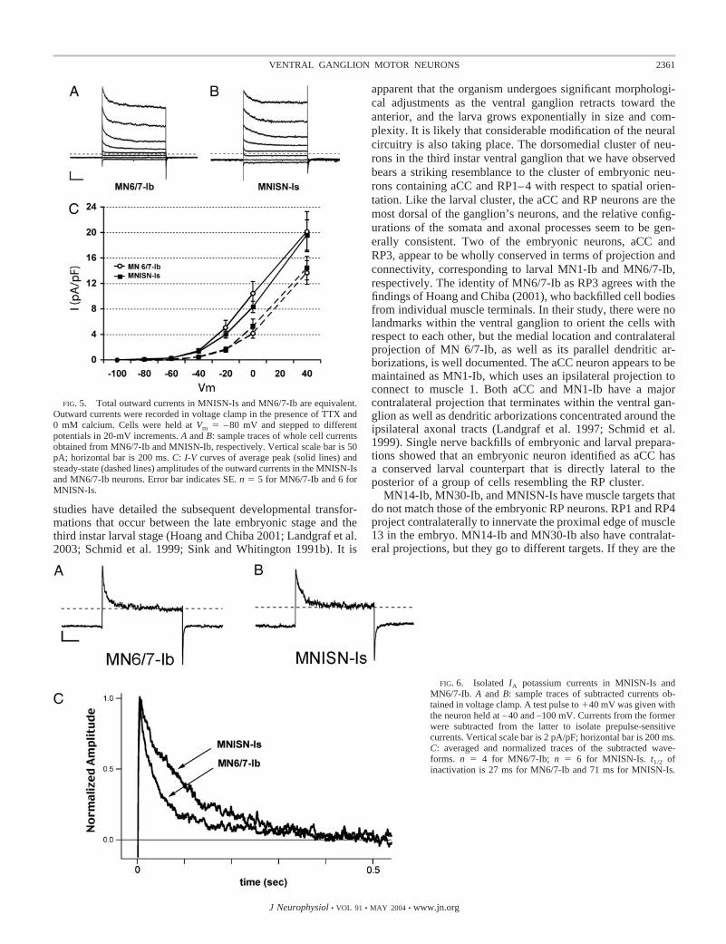
To characterize the potassium currents present in larvalmotor neurons, we recorded outward currents from MNISN-Isand MN6/7-Ib in voltage clamp. Figure 5 shows examplewhole cell recordings and peak (solid line) and sustained(broken line) current I-V curves. Current densities were similar,as were current voltage relationships, indicating that the acti-vation properties of the outward currents in Ib and Is neuronsare similar.
The fast inactivating component of the whole cell outwardcurrent was isolated by subtracting currents obtained with apulse to �40 mV after holding at –40 mV from total currentsobtained after holding at –100 mV. Figure 6, A and B, showsexample subtracted currents from MNISN-Is and MN6/7-Ibneurons. Figure 6C shows averaged subtracted currents for
FIG. 3. Firing properties of dorsomedial neurons are stereotyped. Whole cell current-clamp recordings were made fromidentified motor neurons. Spiking was evoked with injection of current from –20 to 140 pA in 20-pA increments for 500 ms exceptfor F where the duration was 1 s. For clarity, only traces from the –20-, 0-, 20-, 60-, and 100-pA injections are shown. A–F:representative examples of each motor neuron type. Resting membrane potential for the example cells was: MN1-Ib, �54;MN6/7-Ib, �59; MN14-Ib, �58; MN30-Ib, �55; MNISN-Is, �58; and MNSNb/d-Is, �55. Gi: that the connection betweenMN6/7-Ib and its target muscle 6 is functional. Simultaneous neuron and muscle recordings show time locked neuronal spikes andmuscle EJPs. Gii: patterned synaptic activity onto MNISN-Is in 2.4 mM external calcium. H: graph of time to 1st spike for eachneuron. MNISN-Is is significantly different from the rest of the neurons (P 0.0001 for all current injections). The scale bars forA–E are 20 mV and 200 ms; for F and Gi, they are 400 ms and 20 mV for neuronal trace and 0.2 mV for the muscle EJPs; forGii, they are 5 s and 10 mV.
FIG. 2. Identification of dorsomedial motor neuron targets. The targets of the dorsomedial neurons were identified by patchingonto and filling GFP-positive neurons with rhodamine/Alexa 568 and following the axons onto the segmental muscles. A: aschematic of the relevant body-wall muscles of a single abdominal hemisegment. Muscles targeted by segmental nerve b/d (SNb/d)are shown in red, and those targeted by the intersegmental nerve (ISN) in blue. Muscles innervated by the dorsomedial motorneurons are shown in lighter tone. B–F: overlayed GFP and dye-fill images. B: MN1-Ib; C: MN6/7-Ib; D: MN14-Ib; E: MN30-Ib;F: MNISN-Is. Single arrow, bouton type Ib; double arrows, bouton type Is; and triple arrows, bouton type II. Scale bar � 25 �mfor B–E, 10 �m for F.
2359VENTRAL GANGLION MOTOR NEURONS
J Neurophysiol • VOL 91 • MAY 2004 • www.jn.org
multiple neurons (n � 6 for MNISN-Is and n � 4 for MN6/7-Ib). Although the amplitude of the prepulse-sensitive currentis similar, the rate of decay was significantly different for thetwo neuron types, with a � of 122 � 22 ms for MNISN-Is and54 � 12 ms for MN6/7-Ib (P 0.05).
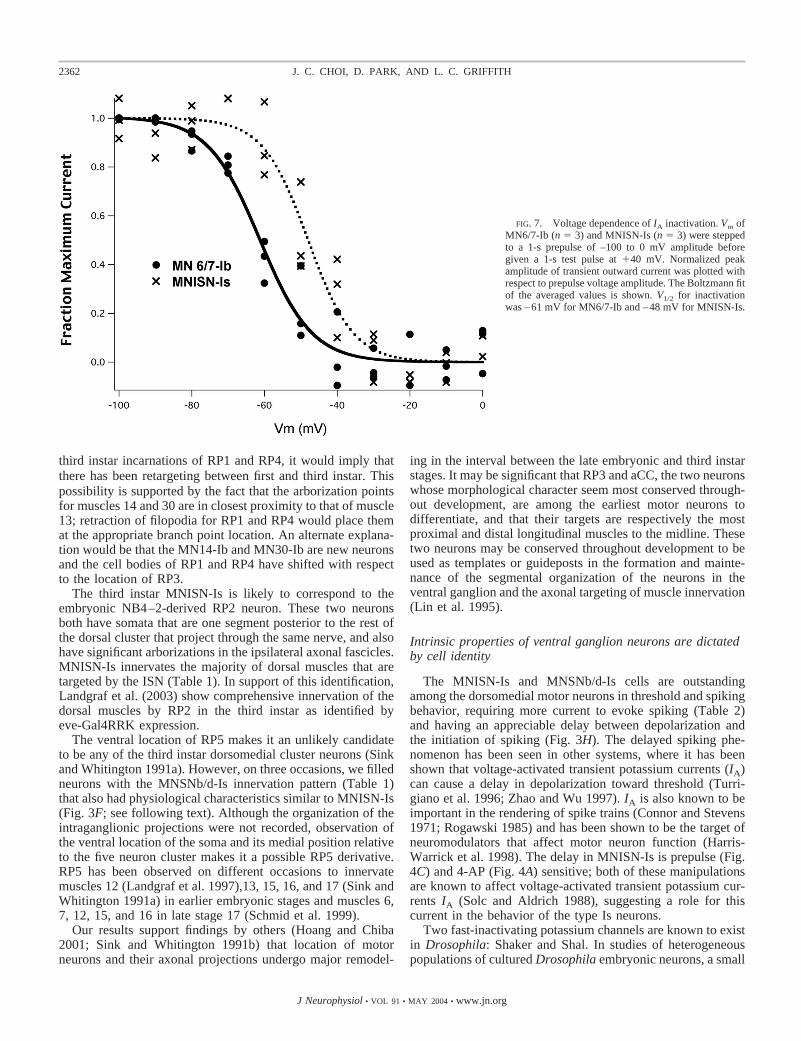
Inactivation rate may contribute to the cell-specific differ-ence in spike delay, but it is not likely to completely accountfor the magnitude. Using a range of prepulse amplitudes, weinvestigated the voltage dependence of prepulse inactivation inMN6/7-Ib and MNISN-Is. The normalized peak amplitudes ofthe resultant currents were plotted with respect to prepulse voltageamplitude, and a Boltzmann fit was applied. Figure 7 shows that
there is a clear difference in the voltage dependence of inacti-vation. At a membrane potential of –60 mV, the amplitude of IAin MNISN-Is is at �90%, while for the MN6/7-Ib it is 50%. Atnormal resting membrane potential, the activity of the transientcurrent will therefore have a greater role in the MNISN-Is.
D I S C U S S I O N
Relationship of the larval dorsomedial cluster to theembryonic RP cluster
While much is known about the proliferation and organiza-tion of specific neurons in the embryonic stage, only a few
FIG. 4. Delay to spike in MNISN-Is is due to aprepulse- and 4-aminopyridine (4-AP)-sensitive non-Shaker current. A: traces from MNISN-Is before 4-APapplication (top), during application of 1.5 mM 4-AP(middle), and after washout (bottom). B: summariza-tion of the time to st spike for each condition forcurrent injections of 60–140 pA. Control and washoutdelays were significantly different from 4-AP (P 0.05). For 4-AP, n � 4. C: traces from MNISN-Iswithout (top) and with (bottom) a 1-s depolarizingprepulse. D: the time to 1st spike for each conditionfor current injections of 60–140 pA. Control andprepulse were significantly different (P 0.01). Forprepulse, n � 4. D: traces recorded from MNISN-Is ina ShKS133 mutant larva. Bottom: the effects of a 1-sdepolarizing prepulse. F: summarization of the time to1st spike for each condition for current injections of60–140 pA. Control and prepulse for Sh were signif-icantly different (P 0.01). For Sh neurons, n � 4. G:summarization of the time to 1st spike data comparingwild-type and Sh neurons for all neuron types with a60-pA current injection. None of the differences aresignificant (P � 0.05).
2360 J. C. CHOI, D. PARK, AND L. C. GRIFFITH
J Neurophysiol • VOL 91 • MAY 2004 • www.jn.org
studies have detailed the subsequent developmental transfor-mations that occur between the late embryonic stage and thethird instar larval stage (Hoang and Chiba 2001; Landgraf et al.2003; Schmid et al. 1999; Sink and Whitington 1991b). It is
apparent that the organism undergoes significant morphologi-cal adjustments as the ventral ganglion retracts toward theanterior, and the larva grows exponentially in size and com-plexity. It is likely that considerable modification of the neuralcircuitry is also taking place. The dorsomedial cluster of neu-rons in the third instar ventral ganglion that we have observedbears a striking resemblance to the cluster of embryonic neu-rons containing aCC and RP1–4 with respect to spatial orien-tation. Like the larval cluster, the aCC and RP neurons are themost dorsal of the ganglion’s neurons, and the relative config-urations of the somata and axonal processes seem to be gen-erally consistent. Two of the embryonic neurons, aCC andRP3, appear to be wholly conserved in terms of projection andconnectivity, corresponding to larval MN1-Ib and MN6/7-Ib,respectively. The identity of MN6/7-Ib as RP3 agrees with thefindings of Hoang and Chiba (2001), who backfilled cell bodiesfrom individual muscle terminals. In their study, there were nolandmarks within the ventral ganglion to orient the cells withrespect to each other, but the medial location and contralateralprojection of MN 6/7-Ib, as well as its parallel dendritic ar-borizations, is well documented. The aCC neuron appears to bemaintained as MN1-Ib, which uses an ipsilateral projection toconnect to muscle 1. Both aCC and MN1-Ib have a majorcontralateral projection that terminates within the ventral gan-glion as well as dendritic arborizations concentrated around theipsilateral axonal tracts (Landgraf et al. 1997; Schmid et al.1999). Single nerve backfills of embryonic and larval prepara-tions showed that an embryonic neuron identified as aCC hasa conserved larval counterpart that is directly lateral to theposterior of a group of cells resembling the RP cluster.
MN14-Ib, MN30-Ib, and MNISN-Is have muscle targets thatdo not match those of the embryonic RP neurons. RP1 and RP4project contralaterally to innervate the proximal edge of muscle13 in the embryo. MN14-Ib and MN30-Ib also have contralat-eral projections, but they go to different targets. If they are the
FIG. 5. Total outward currents in MNISN-Is and MN6/7-Ib are equivalent.Outward currents were recorded in voltage clamp in the presence of TTX and0 mM calcium. Cells were held at Vm � –80 mV and stepped to differentpotentials in 20-mV increments. A and B: sample traces of whole cell currentsobtained from MN6/7-Ib and MNISN-Ib, respectively. Vertical scale bar is 50pA; horizontal bar is 200 ms. C: I-V curves of average peak (solid lines) andsteady-state (dashed lines) amplitudes of the outward currents in the MNISN-Isand MN6/7-Ib neurons. Error bar indicates SE. n � 5 for MN6/7-Ib and 6 forMNISN-Is.
FIG. 6. Isolated IA potassium currents in MNISN-Is andMN6/7-Ib. A and B: sample traces of subtracted currents ob-tained in voltage clamp. A test pulse to �40 mV was given withthe neuron held at –40 and –100 mV. Currents from the formerwere subtracted from the latter to isolate prepulse-sensitivecurrents. Vertical scale bar is 2 pA/pF; horizontal bar is 200 ms.C: averaged and normalized traces of the subtracted wave-forms. n � 4 for MN6/7-Ib; n � 6 for MNISN-Is. t1/2 ofinactivation is 27 ms for MN6/7-Ib and 71 ms for MNISN-Is.
2361VENTRAL GANGLION MOTOR NEURONS
J Neurophysiol • VOL 91 • MAY 2004 • www.jn.org
third instar incarnations of RP1 and RP4, it would imply thatthere has been retargeting between first and third instar. Thispossibility is supported by the fact that the arborization pointsfor muscles 14 and 30 are in closest proximity to that of muscle13; retraction of filopodia for RP1 and RP4 would place themat the appropriate branch point location. An alternate explana-tion would be that the MN14-Ib and MN30-Ib are new neuronsand the cell bodies of RP1 and RP4 have shifted with respectto the location of RP3.
The third instar MNISN-Is is likely to correspond to theembryonic NB4–2-derived RP2 neuron. These two neuronsboth have somata that are one segment posterior to the rest ofthe dorsal cluster that project through the same nerve, and alsohave significant arborizations in the ipsilateral axonal fascicles.MNISN-Is innervates the majority of dorsal muscles that aretargeted by the ISN (Table 1). In support of this identification,Landgraf et al. (2003) show comprehensive innervation of thedorsal muscles by RP2 in the third instar as identified byeve-Gal4RRK expression.
The ventral location of RP5 makes it an unlikely candidateto be any of the third instar dorsomedial cluster neurons (Sinkand Whitington 1991a). However, on three occasions, we filledneurons with the MNSNb/d-Is innervation pattern (Table 1)that also had physiological characteristics similar to MNISN-Is(Fig. 3F; see following text). Although the organization of theintraganglionic projections were not recorded, observation ofthe ventral location of the soma and its medial position relativeto the five neuron cluster makes it a possible RP5 derivative.RP5 has been observed on different occasions to innervatemuscles 12 (Landgraf et al. 1997),13, 15, 16, and 17 (Sink andWhitington 1991a) in earlier embryonic stages and muscles 6,7, 12, 15, and 16 in late stage 17 (Schmid et al. 1999).
Our results support findings by others (Hoang and Chiba2001; Sink and Whitington 1991b) that location of motorneurons and their axonal projections undergo major remodel-
ing in the interval between the late embryonic and third instarstages. It may be significant that RP3 and aCC, the two neuronswhose morphological character seem most conserved through-out development, are among the earliest motor neurons todifferentiate, and that their targets are respectively the mostproximal and distal longitudinal muscles to the midline. Thesetwo neurons may be conserved throughout development to beused as templates or guideposts in the formation and mainte-nance of the segmental organization of the neurons in theventral ganglion and the axonal targeting of muscle innervation(Lin et al. 1995).
Intrinsic properties of ventral ganglion neurons are dictatedby cell identity
The MNISN-Is and MNSNb/d-Is cells are outstandingamong the dorsomedial motor neurons in threshold and spikingbehavior, requiring more current to evoke spiking (Table 2)and having an appreciable delay between depolarization andthe initiation of spiking (Fig. 3H). The delayed spiking phe-nomenon has been seen in other systems, where it has beenshown that voltage-activated transient potassium currents (IA)can cause a delay in depolarization toward threshold (Turri-giano et al. 1996; Zhao and Wu 1997). IA is also known to beimportant in the rendering of spike trains (Connor and Stevens1971; Rogawski 1985) and has been shown to be the target ofneuromodulators that affect motor neuron function (Harris-Warrick et al. 1998). The delay in MNISN-Is is prepulse (Fig.4C) and 4-AP (Fig. 4A) sensitive; both of these manipulationsare known to affect voltage-activated transient potassium cur-rents IA (Solc and Aldrich 1988), suggesting a role for thiscurrent in the behavior of the type Is neurons.
Two fast-inactivating potassium channels are known to existin Drosophila: Shaker and Shal. In studies of heterogeneouspopulations of cultured Drosophila embryonic neurons, a small
FIG. 7. Voltage dependence of IA inactivation. Vm ofMN6/7-Ib (n � 3) and MNISN-Is (n � 3) were steppedto a 1-s prepulse of –100 to 0 mV amplitude beforegiven a 1-s test pulse at �40 mV. Normalized peakamplitude of transient outward current was plotted withrespect to prepulse voltage amplitude. The Boltzmann fitof the averaged values is shown. V1/2 for inactivationwas –61 mV for MN6/7-Ib and –48 mV for MNISN-Is.
2362 J. C. CHOI, D. PARK, AND L. C. GRIFFITH
J Neurophysiol • VOL 91 • MAY 2004 • www.jn.org
fraction were found to have both Shaker and Shal currents(Tsunoda and Salkoff 1995). Recordings from Sh mutantsshowed that the Shaker current does not have a significant rolein soma-evoked spiking behavior (initial delay or rate) in anyof the five motor neurons in this study. Thus the IA current thatcontrols the delay to first spike is likely to be Shal in both typeIs and Ib neurons.
How then are the behaviors of type Is and Ib neuronsdifferentiated? Distinct activation kinetics or amplitude differ-ences are unlikely to underlie the cellular phenotypes. The I-Vrelationships for both the fast-inactivating and sustained out-ward currents were not statistically different (Fig. 5C). Using aprepulse subtraction protocol, we isolated the fast-inactivatingpotassium currents in MNISN-Is and MN6/7-Ib and foundthem to be similar in amplitude (Fig. 6, A and B). The majordifferences between the IA currents in the two cell types was intheir inactivation. IA currents in MNISN-Is had a significantlylonger time course of inactivation. This effect alone, whilesubstantial, is unlikely to account for the magnitude of thedifference in time to first spike in the two neuron types. Aninvestigation of the voltage dependence of prepulse inactiva-tion revealed a significant rightward shift for the MNISN-Is IAas compared with that in MN6/7-Ib (Fig. 7). At the averageresting potential (�61 mV) of MNISN-Is cells, the Shal cur-rent shows essentially no inactivation. In type Ib neurons,where resting membrane potential ranges from –58 to –54 mV,the current is already almost completely inactivated. Together,these differences in inactivation are likely to account for thedisparity in the time to first spike that is seen between type Iband Is motor neurons. There are also likely to be other currents(sodium, calcium, chloride) that are important determinants ofthe cell-specific properties of the type Ib and Is neurons, andtheir roles remain to be determined.
If both type Is and Ib neurons use Shal gene products toproduce IA, the differences in inactivation are likely to involvealternative splicing, posttranslational modification of Shalchannels or differences in auxiliary subunit expression. TheShal/Kv4 family is highly conserved across species (Baldwinet al. 1991; Baro et al. 1996; Pak et al. 1991), and all membersof this gene family are alternatively spliced. Interestingly, therehas been no demonstration of isoform-dependent variability ininactivation kinetics in heterologous expression systems (Baroet al. 2001; Po et al. 2001). Significant heterogeneity in Shalinactivation kinetics measured in situ, however, has been ob-served in cultured embryonic Drosophila neurons (Tsunodaand Salkoff 1995) and has also been seen in the lobster sto-matogastric ganglion (Golowasch et al. 1999). In Drosophila,the shift between kinetic forms was very fast and seen at thesingle channel level, leading the authors to postulate phosphor-ylation as a mechanism of Shal modulation. Auxiliary subunit-dependent changes in the voltage dependence of inactivationhave been demonstrated for Kv4.3 and for lobster Shal (Decheret al. 2001; Zhang et al. 2003). Proteins of the KChip/Freque-nin family were able to selectively modulate inactivation whileminimally affecting activation. In Drosophila, a mutant thatoverexpresses Frequenin has alterations in IA (Poulain et al.1994). Differences in the Shal currents in third instar type Iband Is motor neurons could be a consequence of differentialexpression of a member of the KChip/Frequenin family.
Implications of Ib/Is differences for locomotor activity
The similarities in firing behavior between MNISN-Is andMNSNb/d-Is and the similarities within the Ib neuron groupsuggests the possibility that there are inherent functional andmolecular differences between these morphologically distinctneurons. Type Is neurons innervate groups of longitudinal andoblique muscles of a given hemisegment dorsally (MNISN-Is),ventrally (MNSNb/d-Is), and laterally (MNSNa-Is), allowingthese neurons to control or modulate a large number of func-tionally related muscles in concert. This sharply contrasts withrole of the Ib neurons which innervate only one or two mus-cles, and may act as muscle-specific “drivers.” Activation of(groups of) individual muscles by Ib neurons may provide afine tuning of locomotion.
The delayed spiking mediated by Shal has been observed inother systems involving motor pattern generation. In Aplysia,L14 motor neurons, which control inking, fire action potentialsonly after a critical build-up of synaptic input that is able toinactivate transient potassium currents (Byrne et al. 1979).These neurons display strikingly similar spiking behavior tothe Is neuron under current clamp. Whereas a sudden supra-threshold input will produce a delayed, slow spiking activity, atonic subthreshold input could make instantaneous burstingactivity more likely on suprathreshold input. This sort of ac-tivity dependence is particularly pertinent to the activity driv-ing locomotor muscles as the particular sequence of drivinginput can affect contraction timing and amplitude. Also, theinsensitivity of the neuron to short, nonsummating input mayallow the motorneuron to act as a low-pass filter (Byrne et al.1979). Further study of the differences between the two sub-types will elucidate how locomotor behavior is organized in thelarval system.
Direct study of CNS neurons in the Drosophila thirdinstar larva
The ventral ganglion preparation developed in this study hasallowed direct access to identified central neurons in Drosoph-ila. Previous studies using cultured larval neurons have docu-mented the great diversity of morphological and electrophysi-ological properties in such preparations (Zhao and Wu 1997).A recent study by Rohrbough and Broadie in a similar ventralganglion preparation has already shown great promise ofthis system as a means to study central synaptic transmission(Rohrbough and Broadie 2002). The stereotyped connectivityand functional properties of the motor neurons characterized inthis study will allow genetics and pharmacology to be used todetermine the molecular basis of modulation and cellular be-havior. The function of specific motor neurons in multiplyinnervated muscle fibers as well as their role in the network ofneurons effecting locomotor behavior can now be studiedwithin the intact larval system.
A C K N O W L E D G M E N T S
We thank E. Marder for helpful discussions and E. Dougherty for help withfigures. We also thank J. Rohrbough for discussing details of his larval CNSrecording studies.
2363VENTRAL GANGLION MOTOR NEURONS
J Neurophysiol • VOL 91 • MAY 2004 • www.jn.org
G R A N T S
This work was supported by National Institutes of Health Grants P01GM-33205 and MH-067284 to L. C. Griffith and Training Grant P32 NS-07292 to D. Park.
R E F E R E N C E S
Anderson MS, Halpern ME, and Keshishian H. Identification of theneuropeptide transmitter proctolin in Drosophila larvae: characterizationof muscle fiber-specific neuromuscular endings. J Neurosci 8: 242–255,1988.
Baines RA and Bate M. Electrophysiological development of central neuronsin the Drosophila embryo. J Neurosci 18: 4673–4683, 1998.
Baines RA, Robinson SG, Fujioka M, Jaynes JB, and Bate M. Postsynapticexpression of tetanus toxin light chain blocks synaptogenesis in Drosophila.Curr Biol 9: 1267–1270, 1999.
Baines RA, Uhler JP, Thompson A, Sweeney ST, and Bate M. Alteredelectrical properties in Drosophila neurons developing without synaptictransmission. J Neurosci 21: 1523–1531, 2001.
Baldwin TJ, Tsaur ML, Lopez GA, Jan YN, and Jan LY. Characterizationof a mammalian cDNA for an inactivating voltage-sensitive K� channel.Neuron 7: 471–483, 1991.
Baro DJ, Coniglio LM, Cole CL, Rodriguez HE, Lubell JK, Kim MT, andHarris-Warrick RM. Lobster shal: comparison with Drosophila shal andnative potassium currents in identified neurons. J Neurosci 16: 1689–1701,1996.
Baro DJ, Quinones L, Lanning CC, Harris-Warrick RM, and Ruiz M.Alternate splicing of the shal gene and the origin of I(A) diversityamong neurons in a dynamic motor network. Neuroscience 106: 419 –432, 2001.
Bate M and Broadie K. Wiring by fly: the neuromuscular system of theDrosophila embryo. Neuron 15: 513–525, 1995.
Broadie K, Sink H, Van Vactor D, Fambrough D, Whitington PM, BateM, and Goodman CS. From growth cone to synapse: the life history of theRP3 motor neuron. Dev Suppl: 227–238, 1993.
Byrne JH, Shapiro E, Dieringer N, and Koester J. Biophysical mechanismscontributing to inking behavior in Aplysia. J Neurophysiol 42: 1233–1250,1979.
Cattaert D and Birman S. Blockade of the central generator of locomotorrhythm by noncompetitive NMDA receptor antagonists in Drosophila lar-vae. J Neurobiol 48: 58–73, 2001.
Connor JA and Stevens CF. Voltage clamp studies of a transient outwardmembrane current in gastropod neural somata. J Physiol 213: 21–30,1971.
Decher N, Uyguner O, Scherer CR, Karaman B, Yuksel-Apak M, BuschAE, Steinmeyer K, and Wollnik B. hKChIP2 is a functional modifier ofhKv4.3 potassium channels: cloning and expression of a short hKChIP2splice variant. Cardiovasc Res 52: 255–264, 2001.
Golowasch J, Abbott LF, and Marder E. Activity-dependent regulation ofpotassium currents in an identified neuron of the stomatogastric ganglion ofthe crab Cancer borealis. J Neurosci 19: RC33, 1999.
Gorczyca MG, Budnik V, White K, and Wu CF. Dual muscarinic andnicotinic action on a motor program in Drosophila. J Neurobiol 22: 391–404, 1991.
Halpern ME, Chiba A, Johansen J, and Keshishian H. Growth conebehavior underlying the development of stereotypic synaptic connections inDrosophila embryos. J Neurosci 11: 3227–3238, 1991.
Harris-Warrick RM, Johnson BR, Peck JH, Kloppenburg P, Ayali A, andSkarbinski J. Distributed effects of dopamine modulation in the crustaceanpyloric network. Ann NY Acad Sci 860: 155–167, 1998.
Hoang B and Chiba A. Single-cell analysis of Drosophila larval neuromus-cular synapses. Dev Biol 229: 55–70, 2001.
Jan LY and Jan YN. Properties of the larval neuromuscular junction inDrosophila melanogaster. J Physiol 262: 189–214, 1976.
Jia XX, Gorczyca M, and Budnik V. Ultrastructure of neuromuscular junc-tions in Drosophila: comparison of wild type and mutants with increasedexcitability. J Neurobiol 24: 1025–1044, 1993.
Johansen J, Halpern ME, Johansen KM, and Keshishian H. Stereotypicmorphology of glutamatergic synapses on identified muscle cells of Dro-sophila larvae. J Neurosci 9: 710–725, 1989.
Kiehn O. Plateau potentials and active integration in the “final commonpathway” for motor behaviour. Trends Neurosci 14: 68–73, 1991.
Kurdyak P, Atwood HL, Stewart BA, and Wu C-F. Differential physiologyand morphology of motor axons to ventral longitudinal muscles in larvalDrosophila. J Comp Neurol 350: 463–472, 1994.
Landgraf M, Bossing T, Technau GM, and Bate M. The origin, location,and projections of the embryonic abdominal motorneurons of Drosophila.J Neurosci 17: 9642–9655, 1997.
Landgraf M, Sanchez-Soriano N, Technau GM, Urban J, and Prokop A.Charting the Drosophila neuropile: a strategy for the standardized char-acterization of genetically amenable neurites. Dev Biol 260: 207–225,2003.
Lee T and Luo L. Mosaic analysis with a repressible cell marker forstudies of gene function in neuronal morphogenesis. Neuron 22: 451–461, 1999.
Lichtinghagen R, Stocker M, Wittka R, Boheim G, Stuhmer W, Ferrus A,and Pongs O. Molecular basis of altered excitability in Shaker mutants ofDrosophila melanogaster. Embo J 9: 4399–4407, 1990.
Lin DM, Auld VJ, and Goodman CS. Targeted neuronal cell ablation in theDrosophila embryo: pathfinding by follower growth cones in the absence ofpioneers. Neuron 14: 707–715, 1995.
Lnenicka GA and Keshishian H. Identified motor terminals in Drosophilalarvae show distinct differences in morphology and physiology. J Neurobiol43: 186–197, 2000.
Marder E. From biophysics to models of network function. Annu Rev Neu-rosci 21: 25–45, 1998.
Marder E and Calabrese RL. Principles of rhythmic motor pattern genera-tion. Physiol Rev 76: 687–717, 1996.
Martinez-Padron M and Ferrus A. Presynaptic recordings from Drosophila:correlation of macroscopic and single channel K� currents. J Neurosci 17:3412–3424, 1997.
Monastirioti M, Gorczyca M, Rapus J, Eckert M, White K, and Budnik V.Octopamine immunoreactivity in the fruit fly Drosophila melanogaster.J Comp Neurol 356: 275–287, 1995.
O’Dowd DK. Voltage-gated currents and firing properties of embryonic Dro-sophila neurons grown in a chemically defined medium. J Neurobiol 27:113–126, 1995.
Packard M, Koo ES, Gorczyca M, Sharpe J, Cumberledge S, and BudnikV. The Drosophila Wnt, wingless, provides an essential signal for pre- andpostsynaptic differentiation. Cell 111: 319–330, 2002.
Pak MD, Baker K, Covarrubias M, Butler A, Ratcliffe A, and Salkoff L.mShal, a subfamily of A-type K� channel cloned from mammalian brain.Proc Natl Acad Sci USA 88: 4386–4390, 1991.
Po SS, Wu RC, Juang GJ, Kong W, and Tomaselli GF. Mechanism ofalpha-adrenergic regulation of expressed hKv4.3 currents. Am J PhysiolHeart Circ Physiol 281: H2518–2527, 2001.
Poulain C, Ferrus A, and Mallart A. Modulation of type A K� current inDrosophila larval muscle by internal Ca2�; effects of the overexpression offrequenin. Pfluegers 427: 71–79, 1994.
Rogawski MA. The A-current: how ubiquitous a feature of excitable cells isit? Trends Neurosci 8: 214–219, 1985.
Rohrbough J and Broadie K. Electrophysiological analysis of synaptictransmission in central neurons of Drosophila larvae. J Neurophysiol 88:847–860, 2002.
Schmid A, Chiba A, and Doe CQ. Clonal analysis of Drosophila embryonicneuroblasts: neural cell types, axon projections and muscle targets. Devel-opment 126: 4653–4689, 1999.
Scholz WK, Baitinger CSH, and Kelly PT. Developmental changes inCa2�/calmodulin-dependent protein kinase II in cultures of hippocam-pal pyramidal neurons and astrocytes. J Neurosci 8: 1039 –1051,1988.
Sink H and Whitington PM. Location and connectivity of abdominal mo-toneurons in the embryo and larva of Drosophila melanogaster. J Neurobiol22: 298–311, 1991a.
Sink H and Whitington PM. Pathfinding in the central nervous system andperiphery by identified embryonic Drosophila motor axons. Development112: 307–316, 1991b.
Solc CK and Aldrich RJ. Voltage-gated potassium channels in larval CNSneurons of Drosophila. J Neurosci 8, 1988.
Solc CK, Zagotta WN, and Aldrich RW. Single-channel and genetic anal-yses reveal two distinct A-type potassium channels in Drosophila. Science236: 1094–1098, 1987.
Thomas JB, Bastiani MJ, Bate M, and Goodman CS. From grasshopper toDrosophila: a common plan for neuronal development. Nature 310: 203–207, 1984.
2364 J. C. CHOI, D. PARK, AND L. C. GRIFFITH
J Neurophysiol • VOL 91 • MAY 2004 • www.jn.org

Trimarchi JR and Murphey RK. The shaking-B2 mutation disrupts electri-cal synapses in a flight circuit in adult Drosophila. J Neurosci 17: 4700–4710, 1997.
Tsunoda S and Salkoff L. Genetic analysis of Drosophila neurons: Shal,Shaw, and Shab encode most embryonic potassium currents. J Neurosci 15:1741–1754, 1995.
Turrigiano GG, Marder E, and Abbott LF. Cellular short-term mem-ory from a slow potassium conductance. J Neurophysiol 75: 963–966, 1996.
Wu CF, Ganetzky B, Haugland FN, and Liu AX. Potassium currentsinDrosophila: different components affected by mutations of two genes.Science 220: 1076–1078, 1983a.
Wu C-F and Haugland FN. Voltage-clamp analysis of membrane currents inlarval muscle fibers of Drosophila: alteration of potassium currents inShaker mutants. J Neurosci 5: 2626–2640, 1985.
Wu C-F, Suzuki N, and Poo M-M. Dissociated neurons from normal andmutant Drosophila larval central nervous system in culture. J Neurosci 9:1888–1899, 1983b.
Yoshihara M, Rheuben MB, and Kidokoro Y. Transition from growth coneto functional motor nerve terminal in Drosophila embryos. J Neurosci 17:8408–8426, 1997.
Zhang Y, MacLean JN, An WF, Lanning CC, and Harris-Warrick RM.KChIP1 and frequenin modify shal-evoked potassium currents in pyloricneurons in the lobster stomatogastric ganglion. J Neurophysiol 89: 1902–1909, 2003.
Zhao ML and Wu CF. Alterations in frequency coding and activity depen-dence of excitability in cultured neurons of Drosophila memory mutants.J Neurosci 17: 2187–2199, 1997.
2365VENTRAL GANGLION MOTOR NEURONS
J Neurophysiol • VOL 91 • MAY 2004 • www.jn.org
Copyright © 2022 FDOKUMEN