Cytogenetic studies in non-Hodgkin lymphomas-Results from surgical biopsies
Upload
khangminh22Category
view
1download
0
s2Volume 98, supplement no. 2, October 2013
www.haematologica.org
9th International Symposium on Hodgkin Lymphoma Cologne, Germany, October 12-15, 2013
Guest EditorsBoris Böll, Andreas Engert, Bastian von Tresckow

Owned & published by the Ferrata Storti Foundation, Pavia, Italy
Editor-in-Chief
Jan Cools (Leuven)
Deputy Editor
Luca Malcovati (Pavia)
Associate Editors
Clara Camaschella (Milan), Elias Campo (Barcelona), Claire Harrison (London), Ross Levine (New York), Andreas Rosenwald (Wuerzburg), Juerg Schwaller (Basel), Pieter Sonneveld (Rotterdam), Jean Soulier (Paris), Freda K. Stevenson (Southampton), Matthias Theobald (Mainz), Ivo P. Touw (Rotterdam)
Assistant Editors
Gaetano Bergamaschi (CME), Matteo Giovanni Della Porta (CME), Anne Freckleton (English Editor), Rosangela Inv-ernizzi (CME), Cristiana Pascutto (Statistical Consultant), Rachel Stenner (English Editor), Vittorio Rosti (CME)
Editorial Board
Walter Ageno (Varese), Maurizio Aricò (Firenze), Paolo Arosio (Brescia), Yesim Aydinok (Izmir), Giuseppe Basso (Pado-va), Sigbjørn Berentsen (Haugesund), Erik Berntorp (Malmö), Jackie Boultwood (Oxford), David Bowen (Leeds), Moni-ka Bruggemann (Kiel), Oystein Bruserud (Bergen), Michele Cavo (Bologna), Francisco Cervantes (Barcelona), Oliver Cor-nely (Köln), Javier Corral (Murcia), Francesco Dazzi (London), Marcos De Lima (Houston), Valerio De Stefano (Roma),Ruud Delwel (Rotterdam), Meletios A. Dimopoulos (Athens), Inderjeet Dokal (London), Hervet Dombret (Paris),Johannes Drach (Vienna), Peter Dreger (Hamburg), Martin Dreyling (München), Sabine Eichinger (Vienna), EmmanuelFavaloro (Westmead), Augusto Federici (Milano), Jean Feuillard (Limoges), Letizia Foroni (London), Jonathan W. Fried-berg (Rochester), Dave Gailani (Nashville), Renzo Galanello (Cagliari), Carlo Gambacorti-Passerini (Monza), Guiller-mo Garcia Manero (Houston), Christian Geisler (Copenhagen), James N. George (Oklahoma City), Ulrich Germing (Düs-seldorf), Paolo Ghia (Milano), Piero Giordano (Leiden), Corrado Girmenia (Roma), Mark T. Gladwin (Bethesda), ThomasM. Habermann (Rochester), Claudia Haferlach (München), Christine Harrison (Southampton), Claire Harrison (Lon-don), Andreas Hochhaus (Jena), Ulrich Jaeger (Vienna), Leonid Karawajew (Berlin), Gregory Kato (Bethesda), JohnKoreth (Boston), Robert Kralovics (Vienna), Nicolaus Kröger (Hamburg), Thomas J. Kunicki (La Jolla), Ralf Küppers(Essen), Marco Ladetto (Torino), David Jacobsohn (Chicago), Ola Landgren (Bethesda), Jean Jacques Lataillade (Clamart),Veronique Leblond (Paris), Roberto Lemoli (Bologna), Per Ljungman (Stockholm), Francesco Lo Coco (Roma), Henk M.Lokhorst (Utrecht), Rita Maccario (Pavia), Guido Marcucci (Columbus), Judith Marsh (London), Giampaolo Merlini(Pavia), Anna Rita Migliaccio (Roma), Constantine S. Mitsiades (Boston), Mohamad Mohty (Nantes), Rosario Notaro(Firenze), Johannes Oldenburg (Bonn), Jan Palmblad (Stockholm), Animesh Pardanani (Rochester), Jakob Passweg (Gene-va), Louis Pelus (Indianapolis), Melanie J. Percy (Belfast), Rob Pieters (Rotterdam), Stefano Pileri (Bologna), Miguel Piris(Madrid), Paolo Prandoni (Padova), Jerald P. Radich (Seattle), Andreas Reiter (Mannheim), Mats Remberger (Stock-holm), Josep-Maria Ribera (Barcelona), Francesco Rodeghiero (Vicenza), Radek C. Skoda (Basel), Roberto Stasi (AlbanoLaziale), David P. Steensma (Rochester), Martin H. Steinberg (Boston), David Stroncek (Bethesda), Ronald Taylor (Char-lottesville), Evangelos Terpos (Athens), Xavier Thomas (Lyon), Armando Tripodi (Milano), Han-Mou Tsai (New York),Alvaro Urbano-Ispizua (Sevilla), Alessandro M. Vannucchi (Firenze), Edo Vellenga (Groningen), Umberto Vitolo (Tori-no), Guenter Weiss (Innsbruck), Mervin Yoder (Indianapolis), Alberto Zanella (Milano)
Editorial Office
Michele Moscato (Production Manager), Lorella Ripari (Peer Review Manager), Matteo Giovanni Della Porta (PeerReview), Paola Cariati (Production), Igor Ebuli Poletti (Production), Marta Fossati (Peer Review)
Affiliated Scientific Societies
SIE (Italian Society of Hematology, www.siematologia.it)SEHH (Spanish Society of Hematology and Hemotherapy, www.sehh.org)SIES (Italian Society of Experimental Hematology, www.siesonline.it)EAHP (European Association for Haematopathology, www.socforheme.org/eahp)
European Hematology Association (EHA)
The European Hematology Association (EHA) aims to promote excellence in clinical practice, research and education in European hematology.
EHA was founded in June 1992 and today – with over 3500 members from 100 countries – is a consolidated representative of European hematologists.
Our aim
− To become the official European representative of hematology and hematologists – especially where research, education and regulatory issues are concerned – and to become a conduit for European harmonization;
− To promote the creation of a highly attractive market for practitioners and researchers in Europe thusfostering the mobility of hematologists in and to Europe;
− To reach out and offer a platform to countries that wish to further develop excellence in hematology;− To promote education, training and scientific research in hematology in Europe;− To exchange and disseminate knowledge and scientific information in the field of hematology.
Our activities
− Organizing an annual scientific and educational congress in a major European city; − Dissemination of medical research, both basic and clinic, through the Haematologica/The Hematology
Journal;− Collaborating with other leading organizations in the field of hematology and oncology;− Providing postgraduate education through the annual congress, tutorials and workshops; − Supporting junior basic and clinical researchers in the development of their careers through the EHA
Fellowship Program. − Strengthening the quality and professional status of hematology throughout Europe by accrediting scientific
meetings and providing CME accounts.
EHA Membership
Join the European Hematology Association's 3500 members from 100 countries and support programs and projectwhich promote excellence in clinical practice, research and education in European hematology.
Benefits of EHA membership:− Subscription to Haematologica/ The Hematology Journal, including on-line access− Reduced registration fee for the EHA Annual Congresses− Opportunity to apply for fellowships and awards of the EHA Career Development Program− EHA Newsletter− EHA E-bulletin− Access to webcast sessions of the EHA Annual Congress− Access to online EHA Membership Directory
For information about how to become an EHA Member, contact us at [email protected]
www.ehaweb.org
Information for readers, authors and subscribers
Haematologica/The Hematology Journal (print edition, pISSN 0390-6078, eISSN 1592-8721) publishes peer-reviewed paperson all areas of experimental and clinical hematology. The journal is owned by a non-profit organization, the Ferrata StortiFoundation, and serves the scientific community strictly following the World Association of Medical Editors (WAME) rec-ommendations on publication ethics policies for medical journals (www.wame.org/pubethicrecom.htm).
Haematologica/The Hematology Journal publishes editorials, perspectives, research papers, decision making & problem solv-ing papers, review articles, brief reports and scientific letters. Manuscripts should be prepared according to the UniformRequirements for Manuscripts Submitted to Biomedical Journals, prepared by the International Committee of MedicalJournal Editors (ICMJE) and fully available online (http://www.icmje.org). Additional papers may be considered for the pure-ly online journal (Haematologica/The Hematology Journal on Internet, eISSN 1592-8721). Because there are no space con-straints online, Haematologica/The Hematology Journal will publish several items deemed by peer review to be scientifi-cally sound and mainly useful as educational papers. These will include case reports, irreplaceable images, educational mate-rial from scientific meetings, meeting abstracts, and letters to the Editor.
Papers should be submitted online at http://www.haematologica.org/.
Conflict of interests. According to the International Committee of Medical Journal Editors (http://www.icmje.org/#conflicts),“Public trust in the peer review process and the credibility of published articles depend in part on how well conflict of inter-est is handled during writing, peer review, and editorial decision making”. The ad hoc journal’s policy is reported in detailonline (http://www.haematologica.org/misc/about.dtl).
Galley Proofs and Reprints. Galley proofs should be corrected and returned by email, fax or express delivery within 48 hours.Minor corrections or reasonable additions are permitted; however, excessive alterations will require editorial re-evaluationand will be possibly charged to the authors. Papers accepted for publication will be printed without cost. The cost of print-ing color figures will be communicated upon request. Reprints may be ordered at cost by returning the appropriate formsent by the Publisher.
Transfer of Copyright and Permission to Reproduce Parts of Published Papers. Authors will grant copyright of their articles to theFerrata Storti Foundation. No formal permission will be required to reproduce parts (tables or illustrations) of publishedpapers, provided the source is quoted appropriately and reproduction has no commercial intent. Reproductions with com-mercial intent will require written permission and payment of royalties.
Haematologica/The Hematology Journal is published in two printed editions: International (worldwide except Italy, Spain,Portugal and Latin America), and Spanish (Spain, Portugal and Latin America). Detailed information about subscriptions isavailable online at URL http://www.haematologica.org. While access to the online journal is free, online access to someadditional items available on http://www.haematologica.org may require either institutional or personal subscription.
Rates of the International edition for the year 2013 are as following:Institutional Personal
Print edition (including full access to the online CME for personal subscribers) Euro 500 Euro 150Full access to the online CME only Euro 75
To subscribe to the International edition, please visit our web site http://www.haematologica.org/misc/subscribe.dtl or con-tact: Haematologica Office, via Giuseppe Belli 4, 27100 Pavia, Italy (phone +39.0382.27129, fax +39.0382.394705, E-mail:[email protected]).
Advertisements. Contact the Advertising Manager, Haematologica Office, via Giuseppe Belli 4, 27100 Pavia, Italy (phone+39.0382.27129, fax +39.0382.394705, e-mail: [email protected]).
Disclaimer. Whilst every effort is made by the publishers and the editorial board to see that no inaccurate or misleadingdata, opinion or statement appears in this journal, they wish to make it clear that the data and opinions appearing in thearticles or advertisements herein are the responsibility of the contributor or advisor concerned. Accordingly, the publisher,the editorial board and their respective employees, officers and agents accept no liability whatsoever for the consequencesof any inaccurate or misleading data, opinion or statement. Whilst all due care is taken to ensure that drug doses and oth-er quantities are presented accurately, readers are advised that new methods and techniques involving drug usage, anddescribed within this journal, should only be followed in conjunction with the drug manufacturer’s own published litera-ture.
Direttore responsabile: Prof. Edoardo Ascari; Autorizzazione del Tribunale di Pavia n. 63 del 5 marzo 1955.Printing: Tipografia PI-ME, via Vigentina 136, Pavia, Italy. Printed in September 2013.

9th International Symposium on Hodgkin Lymphoma Cologne, Germany, October 12-15, 2013
Guest EditorsBoris Böll, Andreas Engert, Bastian von Tresckow
ORAL AND POSTERS
session 1. Advanced Stages . . . . . . . . . . . . . . . . . . . . . . . . . . . . . . . . . . . . . . . . . . . . . . . . . . . . . 1
session 2. Early Stages . . . . . . . . . . . . . . . . . . . . . . . . . . . . . . . . . . . . . . . . . . . . . . . . . . . . . . . . . 11
session 3. Biology and Microenvironment . . . . . . . . . . . . . . . . . . . . . . . . . . . . . . . . . . . . . . . . . 17
session 4. Hodgkin Lymphoma in Older Patients . . . . . . . . . . . . . . . . . . . . . . . . . . . . . . . . . . . . 26
session 5. Pathways . . . . . . . . . . . . . . . . . . . . . . . . . . . . . . . . . . . . . . . . . . . . . . . . . . . . . . . . . . . 28
session 6. PET and Prediction . . . . . . . . . . . . . . . . . . . . . . . . . . . . . . . . . . . . . . . . . . . . . . . . . . . 36
session 7. Relapsed and Refractory Hodgkin Lymphoma . . . . . . . . . . . . . . . . . . . . . . . . . . . . . . 44
session 8. Survivorship. . . . . . . . . . . . . . . . . . . . . . . . . . . . . . . . . . . . . . . . . . . . . . . . . . . . . . . . . 54
Author index . . . . . . . . . . . . . . . . . . . . . . . . . . . . . . . . . . . . . . . . . . . . . . . . . . . . . . . . a
Table of Contents
supplement 2 - October 2013
haematologica | 2013; 98(s2) | 1
Advanced Stages
T001IMPACT OF DOSE REDUCTIONS OF BLEOMYCIN AND VINCRISTINE IN PATIENTS WITHADVANCED HODGKIN LYMPHOMA TREATED WITH BEACOPP POLYCHEMOTHERAPY: ACOMPREHENSIVE ANALYSIS OF THE GERMAN HODGKIN STUDY GROUP (GHSG) HD12AND HD15 TRIALS
von Tresckow B, Haverkamp H, Böll B, Eichenauer DA, Sasse S, FuchsM, Borchmann P, Engert A
German Hodgkin Study Group (GHSG), Department of Internal Medicine I,University Hospital of Cologne, Germany
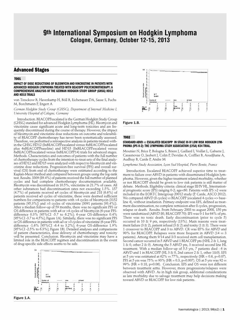
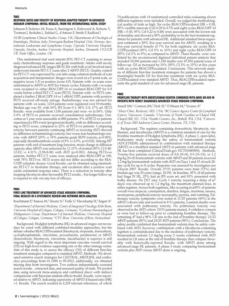
Introduction. BEACOPPescalated is the German Hodgkin Study Group(GHSG) standard for advanced Hodgkin Lymphoma (HL). Bleomycin andvincristine cause significant acute and long-term toxicities and are fre-quently discontinued during the course of therapy. However, the impactof bleomycin and vincristine dose reductions on outcome and tolerabili-ty of BEACOPP chemotherapy has never been systematically assessed.Therefore, we performed a retrospective analysis in patients treated with-in the GHSG HD12 (8xBEACOPPescalated versus 4xBEACOPPescalatedplus 4xBEACOPPbaseline) and HD15 (8xBEACOPPescalated versus6xBEACOPPescalated versus 8xBEACOPP14) trials for advanced stages.Methods. Characteristics and outcomes of patients with the full numberof chemotherapy cycles from the intention-to-treat sets of the final analy-ses of HD12 and HD15 were analyzed with respect to bleomycin and vin-cristine dose reductions. Progression-free survival (PFS) and overall sur-vival (OS) from end of chemotherapy were estimated according to theKaplan-Meier method and compared between groups using the log-ranktest. Results. 3309 (89.4%) of patients received the full number of plannedcycles and had complete chemotherapy documentation available.Bleomycin was discontinued in 10.5%, vincristine in 21.7% of cases. Allother substances had discontinuation rates not exceeding 1.5%. 157(4.7%) of patients received ≤4 cycles of bleomycin and 218 (6.6%) ofpatients received ≤3 cycles of vincristine, these were deemed sufficientnumbers for comparisons to patients with >4 cycles of bleomycin (3152patients [95.3%]) and >3 cycles of vincristine (3091 patients [93.4%]).After a median follow-up of 59 months, there was no significant PFS orOS difference in patients with ≤4 or >4 cycles of bleomycin (6-year PFS-difference 0.3% [95%CI -5.7 to 6.2%]; 6-year OS-difference 0.4%[95%CI -3.7 to 4.5%]; Figure 1A). Similarly, there was no significant PFSor OS difference in patients with ≤3 or >3 cycles of vincristine (6-year PFS-difference -1.6% [95%CI -6.4 to 3.2%]; 6-year OS-difference 1.9%[95%CI -2.5% to 6.3%]; Figure 1B). Detailed analyses and comparisonsof patient characteristics, dose delivery of chemotherapy and toxicitywill be presented. Conclusion. Bleomycin and vincristine may have alimited role in the BEACOPP regimen and discontinuation in the eventof drug-specific side effects seems to be safe.
Figure 1.A.
Figure 1.B.
T002STANDARD ABVD vs ESCALATED BEACOPP IN STAGE III-IV LOW RISK HODGKIN LYM-PHOMA (IPS 0-2): THE LYMPHOMA STUDY ASSOCIATION (LYSA) H34 TRIAL.
Mounier N, Brice P, Bologna S, Briere J, Gaillard I, Voillat L, Gabarre J,Casasnovas O, Jaubert J, Colin P, Devidas A, Coiffier B, Aoudjhane A,Audhuy B, Carde P, Andre M
Lymphoma Study Association, Lyon Sud Hospital, Pierre Benite, France
Introduction. Escalated BEACOPP achieved superior time to treat-ment to failure over ABVD in patients with disseminated Hodgkin lym-phoma. However, given the higher treatment related morbidity, whetheror not BEACOPP should be given to low risk patients is still matter ofdebate. Methods. Eligibility criteria: clinical stage III/IV HL, Internation-al prognostic score (IPS) ranging 0-2; age<60. Patients with IPS >2 wereincluded in the EORTC Intergroup 20012 study (P. Carde, ASCO 2012).We compared ABVD (8 cycles) vs BEACOPP (escalated 4 cycles => base-line 4), without irradiation. Primary endpoint was EFS, defined as treat-ment discontinuation, no complete remission after 8 cycles, progression,relapse or death. Results. From February 2003 to august 2008, 150 ptswere randomized (ABVD 80, BEACOPP 70): IPS was 0-1 for 64% of pts.There was no toxic death. Early discontinuation (prior to cycle 5)occurred in 10 & 9 pts, respectively (13%). Main reasons were treat-ment failure (3 & 2), patient refusal (3 & 2) and toxity (1 & 3). There were1 crossover to BEACOPP and 3 to ABVD. CR was 85% for ABVD and90% for BEACOPP. Relapses were more frequent in ABVD (14 vs 3patients). Among them 9/14 and 3/3 received stem cell transplantation.Second cancer occurred in 5 ABVD and 1 BEACOPP pts (NHL 2 & 1, lung1 & 0, other 2 & 0). Among the 5 ABVD pts, 3 received second line HLtreatment. With a median follow-up of 5.5 yrs, 7 patients died : 6 inABVD and 1 in BEACOPP (HL 3 & 0, 2nd cancer 2 & 1, other 1&0). EFSat 5 yrs was estimated at 62% vs 77%, respectively (HR = 0.6, p=0.07).PFS at 5 yrs was 75% vs 93% (HR = 0.3, p=0.007). OS at 5 yrs was 92 vs99% (HR = 0.18, p=0.06). Conclusion. EFS and OS were not differentbetween treatment arms. However, more progressions/relapses wereobserved with ABVD. As in high risk group, additional considerationsas late morbidity due to salvage treatment may help decisions makingtoward ABVD or BEACOPP for low risk patients.
9th International Symposium on Hodgkin Lymphoma Cologne, Germany, October 12-15, 2013
Abstracts
2 | haematologica | 2013; 98(s2)
T003RESPONSE RATES AND TOXICITY OF RESPONSE-ADAPTED THERAPY IN ADVANCEDHODGKIN LYMPHOMA: INITIAL RESULTS. FROM THE INTERNATIONAL RATHL STUDY
Johnson P, Federico M, Fossa A, Barrington S, Kirkwood A, Roberts T,Trotman J, Berkahn L, Enblad G, d’Amore F, Smith P, Radford J
NCRI Lymphoma Clinical Studies Group, UK; Dipartimento di Oncologia edEmatologia, Modena, Italy; Norwegian Radium Hospital, Oslo, Norway; Aus-tralasian Leukaemia and Lymphoma Group; Uppsala University Hospital,Uppsala, Sweden; Aarhus University Hospital, Aarhus, Denmark; UCL/CRUK Trials Office, London, UK
This randomised trial used interim FDG PET-CT scanning to assessearly chemotherapy response and guide treatment. Adults with newlydiagnosed advanced HL (stages IIB-IV, IIA with bulk or ≥3 involved sites)underwent interim PET-CT scans after 2 ABVD (PET2). Quality controlfor PET-CT was supervised by core labs using common methods of scanacquisition and interpretation. Images were scored on a 5-point scale, asnegative (score 1-3) or positive (score 4,5). Patients with -ve scans wererandomised to ABVD or AVD for 4 more cycles. Patients with +ve scanswere escalated to either BEACOPP-14 or escalated BEACOPP for 8-9weeks before a third PET-CT scan (PET3). Patients with -ve PET3 com-pleted a further 2 BEACOPP-14 or 1 eBEACOPP; patients with positivePET3 received off-study salvage. Radiotherapy was not advised forpatients with -ve scans. 1214 patients were registered over 54 months.Median age was 33, with 34% IPS Score 0-1; 48% 2-3; 17% ≥4. PET2Results. were available from 1136 patients and were -ve in 84%. Only2.4% of PET2-ve patients received consolidation radiotherapy. Out-comes at 1 year were assessable in 680 patients. 9% of PET2-ve patientsexperienced a PFS event (progression/death), with no difference accord-ing to PET2 score, compared to 22% of PET2+ve patients. Comparingtoxicity between patients continuing ABVD or receiving AVD showedno difference in haematologic toxicity, but worse non-haematologic tox-icity with ABVD (29% vs 19% patients grade III/IV toxicity, p=0.001),especially thromboembolic events (4.9% vs 2.0%, p=0.018). Among 587patients with end of treatment lung function, mean change in diffusioncapacity after ABVD was reduction by 11.15% of normal (95% CI 9.48-12.81) vs 4.31% (2.56-6.06) with AVD (p<0.001). Among PET2+vepatients, further PET-CT scans were analysed after BEACOPP in 156,with 76% PET3-ve. PET3 scores did not differ according to the BEA-COPP schedule chosen. Good Results. can be obtained using intemedi-ate PET-CT to modulate therapy, and escalation after a positive scanyields substantial response rates. There is a reduction in toxicity afterstopping bleomycin after favourable PET2 results. , but longer follow upis required to rule out any loss of efficacy.
P004FIRST-LINE TREATMENT OF ADVANCED STAGE HODGKIN LYMPHOMA.FINAL RESULTS OF A SYSTEMATIC REVIEW AND NETWORK META-ANALYSIS
Borchmann P,1 Rancea M,2 Skoetz N,2 Trelle S,3 Haverkamp H,1 Engert A1
1Department I of Internal Medicine, Center of Integrated Oncology Köln Bonn,University Hospital of Cologne, Cologne, Germany; 2Cochrane HaematologicalMalignancies Group, Department I of Internal Medicine, University Hospitalof Cologne, Cologne, Germany; 3CTU Bern, University of Bern, Switzerland
Background. Hodgkin lymphoma (HL) in advanced stages can nowa-days be cured with different combined-modality approaches, but thedebate whether BEACOPPescalated (bleomycin, etoposide, doxorubicin,cyclophosphamide, vincristine, procarbazine, prednisone) or ABVD(doxorubicin, bleomycin, vincristine, dacarbazine) is superior is stillongoing. With regard to the most important outcome overall survival(OS) no high-level evidence supporting one or the other strategy exists.Aim of this study is, to assess the efficacy (OS) of different first-linetreatment strategies compared to standard ABVD. Methods. We devel-oped sensitive search strategies for CENTRAL, MEDLINE, and confer-ence proceedings from 01.1980 to 09.2012, additionally, we obtainedmissing data from investigators. Two authors independently screenedsearch results. , extracted data, and assessed quality of trials. We pooleddata using network meta-analysis and combined direct with indirectcomparisons with Bayesian random-effects model. Results. were report-ed relative to ABVD, indicating superiority of ABVD if hazard ratio (HR)>1. Results. The search resulted in 2,229 relevant references, of which
74 publications with 14 randomized controlled trials evaluating elevendifferent regimens were included. Overall, we judged the methodolog-ical quality of trials as high. Six cycles BEACOPPescalated (HR = 0.38,95% credible intervals (CrI) 0.20 to 0.75) and eight cycles BEACOPP-14(HR = 0.43, 95% CrI 0.22 to 0.86) were associated with the lowest riskof mortality and showed a 98% probability to be the best treatment reg-imens for patients with advanced HL. Additional standard meta-regres-sion estimated a 89% five-year survival rate for ABVD, resulting in afive-year survival benefit of 7% for both regimens: six cycles BEA-COPPescalated (95% CrI 3% to 10%) and eight cycles BEACOPP-14(95% CrI 2% to 9%) as compared to ABVD. These Results. were con-firmed by the reconstructed digitized individual patient analysis thatincluded 10,042 patients and 1,189 deaths over 47,033 patient-years offollow-up. OS as increased by 10% (95% CI 3% to 15%) at five yearswith six cycles BEACOPPescalated. Conclusions. This network analy-sis of different first-line treatment strategies for patients has shown ameaningful benefit OS for first-line treatment with six cycles BEA-COPPescalated over standard ABVD. Thus, BEACOPPescalated repre-sents the gold standard of care for advanced stage HL patients.
P005FRONTLINE THERAPY WITH BRENTUXIMAB VEDOTIN COMBINED WITH ABVD OR AVD INPATIENTS WITH NEWLY DIAGNOSED ADVANCED STAGE HODGKIN LYMPHOMA
Ansell SM,1 Connors JM,2 Park SI,3 O’Meara M,4 Younes A5
1Mayo Clinic, Rochester, MN, USA; 2BC Cancer Agency Centre for LymphoidCancer, Vancouver, Canada; 3University of North Carolina at Chapel Hill,Chapel Hill, NC, USA; 4Seattle Genetics, Inc., Bothell, WA, USA; 5Universi-ty of Texas MD Anderson Cancer Center, Houston, TX, USA
Background. The regimen containing doxorubicin, bleomycin, vin-blastine, and dacarbazine (ABVD) is a common standard of care for thefrontline treatment of Hodgkin lymphoma (HL). A phase 1, open-label,multicenter study evaluating the safety of brentuximab vedotin(ADCETRIS®) administered in combination with standard therapy(ABVD) or a modified standard (AVD) in patients with advanced stageHL has been completed (ClinicalTrials.gov NCT01060904). Methods.Twenty-five patients received doses of 0.6 (N=6), 0.9 (N=13), or 1.2mg/kg (N=6) brentuximab vedotin with ABVD and 26 patients received1.2 mg/kg brentuximab vedotin with AVD on Days 1 and 15 of each 28-day cycle for up to 6 cycles. Response was assessed by investigator perCheson 2007. Results. Overall, 37/51 patients were male (73%) andmedian age was 33 years (range, 18-59). At baseline, 45% of all patientshad Stage IV HL, 25% had an IPS score ≥4, and 33% presented withbulky disease. No DLT (any Cycle 1 toxicity requiring a delay of ≥7days) was observed up to 1.2 mg/kg, the maximum planned dose, ineither regimen. Across both regimens, AEs occurring in ≥30% of patientsoverall were alopecia, constipation, diarrhea, fatigue, insomnia, nausea,neutropenia, peripheral sensory neuropathy, pyrexia, and vomiting. Pul-monary toxicity symptoms were noted in 11/25 patients (44%) in theABVD cohorts only and resolved in 9/11 patients; 2 patient deaths wereassociated with pulmonary toxicity. No pulmonary toxicity wasobserved in the AVD cohort. Of 51 patients treated, 4 withdrew consentor were lost to follow-up prior to completing frontline therapy. Theremaining 47 had a 96% CR rate at the end of frontline therapy: 21/22ABVD patients (95%) and 24/25 AVD patients (96%). Conclusions. Thesafety profile confirmed that brentuximab vedotin may be safely com-bined with AVD; however, combination with a bleomycin-containingregimen is contraindicated due to the incidence of pulmonary toxicity.Brentuximab vedotin 1.2 mg/kg every 2 weeks combined with AVDresulted in CR rates at the end of frontline therapy that compare favor-ably with historically-reported Results. with ABVD alone amongadvanced-stage HL patients. A phase 3 study comparing brentuximabvedotin plus AVD versus ABVD alone is ongoing.
9th International Symposium on Hodgkin Lymphoma, Cologne, Germany, October 12-15, 2013
haematologica | 2013; 98(s2) | 3
P006EARLY TREATMENT INTENSIFICATION IN ADVANCED-STAGE HIGH-RISK HODGKIN LYM-PHOMA (HL) PATIENTS, WITH A POSITIVE FDG-PET SCAN AFTER TWO ABVDCOURSES–SECOND INTERIM ANALYSIS OF THE GITIL/FIL HD0607 CLINICAL TRIAL
Gallamini A,1 Rossi A,2 Patti C,3 Picardi M,4 Di Raimondo F,5 Can-tonetti M,6 La Nasa G,7 Viviani S,8 Bolis S,9 Trentin L,10 Olivieri A,11Zoli V,12 Biggi A,13 Chauvie S,14 Fiore F,1 Borra A,1 Prosperini G,15Cavazzina R,15 Marchioli R,15 Parvis G,16 Zanotti R,17 Gavarotti P,18Dodero A,19 Schiavotto C,20 Ciceri F,21 Avigdor A,22 Mulè A,23 TarellaC,24 Gianni AM,25 Rambaldi A2
1Hematology, Azienda Ospedaliera S. Croce e Carle, Cuneo, Italy; 2Hematol-ogy, Ospedali Riuniti di Bergamo, Bergamo, Italy; 3Hematology, Ospedale V.Cervello, Palermo, Italy; 4Hematology, Az. Ospedaliera Policlinico Federico II,Napoli, Italy; 5Hematology, Ospedale Ferrarotto, Catania, Italy; 6Hematology,Policlinico Universitario Tor Vergata, Roma, Italy; 7Department of Internal Med-icine, Ospedale R. Binaghi, Cagliari, Italy; 8Department of Medical Oncology,Fondazione IRCCS Istituto Nazionale Tumori, Milano, Italy; 9Hematology,Ospedale San Gerardo, Monza, Italy; 10Hematology, Internal Medicine, Uni-versità di Padova, Padova, Italy; 11Hematology, Ospedale San Carlo, Potenza,Italy; 12Hematology, Az. Ospedaliera S. Camillo Forlanini, Roma, Italy; 13
Nuclear Medicine S. Croce Hospital, Cuneo, Italy; 14Medical Physics Unit, S.Croce e Carle, Cuneo, Italy; 15Laboratory of Clinical Epidemiology, Mario NegriSud Institute, Santa Maria Imbaro, Italy; 16Hematology, S. Luigi Hospital,Orbassano, Torino, Italy; 17Hematology, Policlinico B.G. Rossi, Verona, Italy;18Hematology, S. Giovanni Battista Hospital, Torino, Italy; 19Hematology, Fon-dazione IRCCS Istituto Nazionale Tumori, Milano, Italy; 20Hematology, S. Bor-tolo Hospital, Vicenza, Italy; 21Hematology and Bone Marrow TransplantationUnit, San Raffaele Scientific Institute, Milano, Italy; 22Hematology, Tel-AshomerHospital, Tel-Avis, Israel; 23Azienda Ospedali Riuniti Villa Sofia Cervello, Paler-mo, Italy; 24Hematology, Mauriziano-Umberto I Hospital, Torino, Italy; 25Med-ical Oncology Unit, Fondazione IRCCS Istituto Nazionale Tumori Milano,Milan, Italy
Background. Despite its prognostic role in advanced-stage HL (aHL),in still unproven whether early therapy intensification in patients (p.)with a positive interim PET after 2 ABVD (PET-2) improves overall treat-ment efficacy for all aHL pts compared to standard ABVD Patients andMethods. In the HD 0607 clinical trial aHL p. are treated with 2 ABVDcourses and a PET-2 performed afterwards. PET-2+p. are randomized toeither BEACOPP escalated (Be) plus BEACOPP baseline (Bb) (4+4 cours-es) or Be+Bb (4+4) and Rituximab (R). PET-2- p. are treated with 4 addi-tional ABVD and, upon CR achievement, randomized to either consol-idation radiotherapy (Rxt) on the sites of initial bulky disease or no fur-ther treatment. Scans are interpreted by an expert panel of reviewers ina Blinded Independent Central Review (BICR) according to the Deauville5-point scale (5PS). Results. 627 aHL pts were consecutively enrolledand 590 scanned with PET-2. 287/590 (49%) PET-2 underwent review:117 were adjudged as positive (80 score 4, 37 score 5) and 170 as nega-tive (score 1-3). Altogether, of 590 PET-2, 117 (20%) resulted positive and473 (80%) negative. Treatment efficacy could be assessed in a cohort of330 pts. with a minimum follow-up of 24 months from enrolment: 59(17,9%) with a positive and 271 (82.1%) with a negative PET-2. Themedian follow-up was 952 days (736-1181). Among 59 PET-2+ p., CCR(Continuous CR) was recorded in 40 (68%), Pro/Rel in 9 (15%), SD in 2p. (3%) and death in 8 pt (13%), 2 of them for progressive disease.Among 271 PET-2- p. CCR was recorded in 240 (89%), Pro/Rel in 19(7%), SD in 2 (0.7%), death in 8 (3%), 4 of them for disease progression.The 2-y PFS was 67.8%, 88.6% and 84.6% for PET-2+p., PET-2-p., andfor all p., respectively (p<.001). Conclusions. These preliminary findingssuggest that 1) an early switch from ABVD to escalated BEACOPP caninduce PET negativity and sustained CR achievement in most cases; 2)BICR is feasible and allows a real time decision making process in aprospective multicentre clinical trial.
P007RELAPSE ANALYSIS OF IRRADIATED PATIENTS WITHIN THE HD15 TRIAL OF THE GERMANHODGKIN STUDY GROUP
Kriz J,1 Reinartz G,1 Kobe C,2 Kuhnert G,2 Haverkamp H,3 HaverkampU,1 Hegerfeld K,1 Baues C,4 Engert A,3 Eich HT1
1Department of Radiation Oncology, University Muenster; 2Department ofNuclear Medicine, University of Cologne, Germany; 3First Department of Inter-
nal Medicine, University of Cologne; 4Department of Radiation Oncology, Uni-versity of Cologne, Germany
Introduction. The role of consolidation radiotherapy (RT) after effec-tive chemotherapy (CTX) in advanced stages Hodgkin Lymphoma (HL)is unclear. In the HD15 trial of the German Hodgkin Study Group there-fore patients having residual disease of ≥2.5 cm after 6-8 cycles BEA-COPP were evaluated using Fluorodesoxyglucose positron emissiontomography (PET). PET-positive patients were subsequently irradiated.The aim of the present study was to determine whether sites of relaps-es were within the irradiated area and to analyse whether the definitionof the RT volume was correct. In addition, the correlation between qual-ity of RT and the pattern of relapse was assessed. Methods. After com-pletion of CTX, all patients with residual disease ≥2.5 cm were evaluat-ed using PET and in case of a PET-positive result, these patients were irra-diated with 30 Gy local RT to the site of residual disease. For all patientsreceiving RT who had a documented relapse, we analysed sites of dis-ease before and after chemotherapy and especially the PET positive sitesthat were irradiated. Documentation of RT, treatment planning and por-tal images were carefully analysed and compared to the centrally recom-mended RT prescription. The irradiated sites were compared to sites ofrelapse using follow-up CT scans. Results. Of all patients in this trial,191 (26%) had PET-positive residues after CTX and 175 of these patients(92%) were irradiated with 30 Gy to the PET-positive site. 28 irradiatedpatients relapsed. RT radiation plans and follow-up CT scans of thesepatients were analysed. Overall, 12 patients (42%) had an in-fieldrelapse, 8 (29%) patients relapsed outside the irradiated site and an addi-tional 8 patients (29%) had in- and outfield relapse. In these relapsingpatients, RT was not performed according to the radiation plan in 19/28(68%) and in additional 7 patients (32%), the RT was not realized cor-rectly. Conclusion. The pattern of relapse suggests that local RT is suf-ficient for patients in advanced stages HL. There was no correlationbetween quality of RT and occurrence of relapse.
P008CHARACTERISTICS AND OUTCOME OF HIV ASSOCIATED HODGKIN LYMPHOMA AMONG59 PATIENTS INCLUDED IN THE FRENCH ANRS CO16 LYMPHOVIR COHORT STUDY
Besson C,1 Prevot S,2 Hendel-Chavez H,1 Lancar R,3 Génin M,3 Trabel-si S,3 Marchand L,4 Meyohas MC,5 Marchou B,6 Gabarre J,7 Bonnet F,8Goujard C,1 Boué F,2 Mounier N,9 Partisani M,10 Raffi M,11 CostelloR,12 Raphael M,1 Taoufik Y,1 Costagliola D7
1Université Paris Sud, Le Kremlin-Bicetre, France; 2Université Paris Sud, Cla-mart, France; 3INSERM u943, France; 4ANRS, France, 5CHU Saint-Antoine,Paris, France; 6CHU Toulouse, France; 7CHU Pitié-Salpétrière, Paris, France;8CHU Bordeaux, France; 9CHU Nice, Nice, France; 10CHU Strasbourg, France;11CHU Nantes, Nantes, France; 12 CHU Marseille, France
Background. Human Immunodeficiency Virus (HIV) infection is asso-ciated with an increased risk of Hodgkin (HL) and non Hodgkin lym-phoma (NHL). The widespread use of combined antiretroviral therapy(cART) has reduced the incidence of NHL but not the incidence of HL.Methods. The national prospective Cohort of HIV-related lymphomas(ANRS CO16 Lymphovir cohort sponsored by Inserm-ANRS) enrolled138 adult patients at diagnosis of lymphoma in 32 centres between July2008 and April 2012. Investigations were performed after approval of theethic committee and competent autority. Pathological materials of 40patients were centralized and reviewed. Diagnoses were based on WorldHealth Organization criteria. Patients are followed every 6 months dur-ing 5 years. Results. Among the 138 patients, 43% (59) were diagnosedwith HL. Median age was 44 years (ranging from 20 to 61), male/femaleratio was 6.4 (51/8). HIV infection had been diagnosed for a median of156 months (0 to 312) and all patients except one were treated withcART at HL diagnosis. Median CD4 T-cell count was 368/mm3 (range37-1518) and HIV RNA was <50 in 78% (46/59) of the patients. HLmixed cellularity subtype was noticed in 37 out of 40 reviewed cases,nodular sclerosing in 2 cases and nodular lymphocyte predominant (NLP)in one case. All the 40 tested cases for in situ EBV hybridization werepositive except the NLP HL. Advanced clinical stage (III/IV) was noticedin 77% of the cases. The median interval between lymphoma occurrenceand last follow-up was 26 months. During follow-up, all patients weretreated with cART. 50 out of 54 were treated with ABVD, the patientwith NLP received Rituximab. At 24 months, overall survival was 96%[95%CI 92, 100] and progression free survival was 91% [83, 99]. All the

Abstracts
4 | haematologica | 2013; 98(s2)
5 events occurred among the patients with stage III and IV.. There were2 deaths from disease progression (1) and sepsis (1). Summary / Conclu-sion. The present study points out the high proportion of HL among HIVinfection with lymphoma in the cART era. Although these patients haveadvanced stage at diagnosis, their prognosis has largely improved.
P009PROGNOSTIC IMPACT OF CD20 EXPRESSION IN ADVANCED STAGE CLASSICALHODGKIN’S LYMPHOMA (CHL) TREATED WITH ABVD CHEMOTHERAPY:A RETROSPECTIVE ANALYSIS
Sengar M,1 Sridhar E,2 Menon H,1 Dangi U,1 Shet T,2 Gujral S,2 JainH,1 Laskar S,3 Khanna N3
1Department of Medical Oncology; 2Department of Pathology; 3Department ofRadiation Oncology Tata Memorial Centre, Mumbai, India
Background. CD20 expression on Hodgkin and Reed-Sternberg (HRS)cells is seen in approximately 5-58% of CHLs. The prognostic relevanceof CD20 expression in CHL remains conflicting. Given the efficacy ofrituximab in CHLs in a few studies, we felt it relevant to further inves-tigate its prognostic role. Herein we report the prognostic impact ofCD20 expression in patients with advanced stage CHL treated at ourcentre with ABVD chemotherapy. Methods. The electronic medicalrecords of newly diagnosed advanced stage (stage IIB, III and IV) CHLpatients (14 years or more) registered at our centre from January 2008to December 2010 were reviewed for baseline disease characteristics,international prognostic score (IPS), treatment, response, death , pro-gression or relapse. CD20 expression (focal or diffuse membrane posi-tivity in HRS cells) was noted. Patients receiving more than 2 cycleswith evaluable response were analysed for overall survival (OS) andprogression free survival (PFS). Results. A total of 143 patients (males-112) were analysed. Median age was 28 years (range 14-70 years). 73%of patients had stage III/IV disease. B symptoms, bulky disease, IPS ≥4and bone marrow involvement was seen in 75%, 38%, 22% and 15%,respectively. Post 6 cycles of ABVD 88% achieved complete responsewhereas 8% experienced disease progression. CD20 positivity was not-ed in 22% (evaluable in 112 patients). CD15 was positive in 31/92patients. Baseline disease characteristics were similar in the CD20+ andnegative subset. The 3-year PFS and OS were 80% and 95% respective-ly. 3-year PFS was 65.5% and 87% (p=0.035) respectively in CD20+ andnegative subset, while OS was 88% and 97% respectively (p=0.06).CD20+, IPS≥4 was associated with inferior PFS both in the univariateand multivariate analysis. Absolute lymphocyte and monocyte ratio andCD15 expression did not impact PFS or OS. Conclusion. In our studyCD20 expression did affect progression free survival. However therewas no impact on overall survival which may be explained by short fol-low up and availability of effective salvage therapies.
P010PROSPECTIVE EVALUATION OF THE UTILITY OF THE INTERNATIONAL PROGNOSTICSCORE (IPS) FOR PATIENTS WITH ADVANCED HODGKIN LYMPHOMA (HL) TREATED WITHCONTEMPORARY THERAPY: RESULTS FROM US INTERGROUP TRIAL E2496
Diefenbach CS, Li H, Hong F, Gordon LI, Fisher RI, Bartlett NL,Crump M, Gascoyne RD, Wagner H, Stiff PJ, Cheson BD, Stewart D,Kahl BS, Friedberg JW, Blum KA, Habermann TM, Tuscano JM,Hoppe RT, Horning SJ, Advani RH
New York University School of Medicine/NYU; Cancer Institute; Dana FarberCancer Institute; Northwestern University; Fox Chase Cancer Center-TempleHealth; Washington University Sch. of Med. Siteman Cancer Center; PrincessMargaret Hospital; BC Cancer Agency; Penn State Milton S Hershey MedicalCenter; Loyola University Medical Center; Georgetown University HospitalLomabardi Cancer Center; Tom Baker Cancer Center University of Calgary;University of Wisconsin Hospital and Clinics; James P Wilmot Cancer CenterUniversity of Rochester; Ohio State University Comprehensive Cancer Center;Mayo Clinic; UC Davis Cancer Center; Stanford University; Genentech Inc.;Stanford University, USA
Background. The International Prognostic Score (Hasenclever, NEJM1988) built from a retrospective analysis of patients treated before 1992continues to be the most commonly used risk stratification index foradvanced HL and predicts for 5 year freedom from progression (FFP) of42%-84% and overall survival (OS) of 56%-89%. A retrospective analy-
sis from British Columbia (Moccia JCO 2012) suggests that more recent-ly the predictive range of the IPS has narrowed. To prospectively con-firm the latter findings in the context of changes in staging and treatmentparadigms in the modern era, we assessed the ability of the IPS to pre-dict outcome in patients enrolled on the US Intergroup trial E2496. Meth-ods. The seven IPS variables were recorded at study entry. FFP and OSwere correlated with the IPS and compared to the published Hasen-clever and Moccia reports. For FFP, all deaths from unrelated causes werecensored. Results. From 1996-2006, 854 patients were randomized totreatment with ABVD or Stanford V, with no differences in outcome(Gordon et al JCO 2013). On multivariate analysis, only hemoglobinand stage remained significant for FFP, while hemoglobin<10.5, stage IV,and age>45yrs were significant for OS. The IPS remained prognostic forboth FFP (p=0.011) and OS (p<0.001). Five year FFP rates of 81%, 74%and 63%, and 5-year OS rates of 92%, 85% and 60% respectively werefound for low (0-2), intermediate (3-4), and high risk (>4 ) patients whengrouped according to number of risk factors. Table 1 lists our Results. aswell as those of the 2 retrospective analyses. Conclusion. The IPSremained prognostic for FFP and OS in our recently treated patients,however we found superior clinical outcomes than were originallyreported, particularly for intermediate and high risk groups, and the pre-dictive range has narrowed for all patient groups. Improved tools for riskassessment are needed to further stratify low and intermediate riskpatients, and inform therapeutic decision-making. For the highest riskpatients, there is an ongoing need for novel therapeutic strategies.
Table 1. Outcome comparison.
% Patients 5 yr %FFP 5 yr %OS
# Risk E2496 Moccia Hasenclever E2496 Moccia Hasenclever E2496 Moccia Hasenclever
factors
0-2 67 60 58 81 80-88 67-84 92 91-98 81-90
3-4 27 33 35 74 67-74 51-60 85 85-88 61-78
≥5 6 7 7 63 62 42 60 67 56
P011A PILOT STUDY OF A REDUCED THERAPY DRIVEN BY EARLY PET RESPONSE IN 64PATIENTS TREATED WITH 2 CYCLES OF BEACOPPESC FOR ADVANCED HODGKIN LYM-PHOMA
Deau B,1 Franchi P,2 Briere J,3 Ohana J,4 Thieblemont C,2 Brice P2
1Hematology department, Hôpital Cochin, Paris, France; 2Hematology depart-ment, Hôpital Saint Louis, Paris, France; 3Histopathology department, HôpitalSaint Louis, Paris, France; 4Nuclear Medicine department, Hôpital Saint Louis,Paris, France
The better efficiency of BEACOPPesc against advanced Hodgkin lym-phoma is associated to an increase of toxicities. PET performed after 2courses of chemotherapy (PET2) was shown to predict outcome. In thissetting, a pilot study was designed to test in advanced HL patients, areduced treatment strategy driven by PET after 2 cycles of BEACOPPesc.This is a monocentric prospective study, from 2008 to 2012. Patientswith a stage IIB (according to the GHSG criteria), III, and IV, were includ-ed at diagnosis. A baseline PET scan was performed. BEACOPPesc wasslightly modified for ambulatory administration, including Etoposide200 mg/m2 with orally administration for days 2 and 3 and prednisonefrom day 1 to 8. In case of PET2 negative, the treatment was complet-ed by 4 cycles of ABVD with a CT scan after 2 and 4 courses. In case ofPET2 positive, the treatment was completed by 2 cycles of BEA-COPPesc, then the response was assessed on a PET (PET4), with deci-sional value for the following treatment. 64 patients were included, 58%female, median age at 25 years (range 15 to 57 years). 11 patients hadhigh risk stage IIB, 9 and 44 patients had stage III and IV disease respec-tively. 55 patients (86%) obtained a negative PET2, whereas 9 patients(14%) did not. After a median follow up of 28,7 months (range : 7-60),the PFS was 85%. The overall relapse rate was 15% at a median of 8months after the end of treatment (range : 4-14 months), no deaths. Inthe PET2 negative arm, all patients were in complete remission afterABVD. Six relapses (11%) occurred, treated with high dose therapy. Inthe PET2 positive arm : 4 patients relapsed (44%). The 2 years PFS forthe PET2 negative and positive arm was 87% and 47% respectively (p
9th International Symposium on Hodgkin Lymphoma, Cologne, Germany, October 12-15, 2013
haematologica | 2013; 98(s2) | 5
=0,0059). This pilot study confirm that the substitution of oral etoposideis feasible and effective. Therefore, a therapy strategy driven by earlyPET negative with a reduced treatment is effective. Nevertheless, thisResults. have to be confirmed : the randomized trial AHL11 is ongoingin France.
P012HIGH INCIDENCE OF ASEPTIC HIP NECROSIS IN HODGKIN LYMPHOMA PATIENTS TREATED WITH ESCALATED BEACOPP WITH METHYL-PREDNISOLONE
Basic-Kinda S, Durakovic N, Karlak I, Lubina ZI, Radman I, Dotlic S,Hude I, Aurer I
Division of Hematology; Department of Internal Medicine; University HospitalCentre Zagreb Department of Traumatology; University Hospital Centre Sistersof Mercy Department of Radiology; University Hospital Merkur Department ofPathology; University Hospital Centre Zagreb, Croatia
Escalated BEACOPP (eBEACOPP) is a very effective but toxic regimenfor treatment of Hodgkin lymphoma (HL). Hematologic toxicity isalmost universal and infections are frequent. Other prominent side-effects include thrombosis, pulmonary toxicity and neuropathy. Asep-tic hip necrosis (AHN) was not frequently observed in Germany, whereeBEACOPP originated and where more than 2000 patients were treat-ed with this regimen but occurred frequently in Czech and Norwegianseries. After observing an unusually high incidence of AHN in ourpatients treated with eBEACOPP we decided to perform this study. Weidentified 25 patients with HL treated at our centre who were scheduledto receive at least 4 cycles of eBEACOPP for newly diagnosed high-riskadvanced stage HL (Hasenclever index >2) or who were PET+ after 2ABVD cycles. One patient died of neutropenic sepsis during treatment,all other responded. With a median follow-up of 24 months all surviv-ing patients are in continuous remission. Two-year overall and progres-sion-free survival rates are 96%. Toxicity was largely as expected (grade3&4 hematological toxicity 96%, infections 36%, DVT/PE 12%), exceptfor the fact that 5 patients developed clinically significant hip problemsand two needed hip replacement. We therefore invited all patients toundergo hip MRI and orthopedic evaluation, 17 responded (71%). Six,four symptomatic and two asymptomatic, had radiological signs of AHN(35% of the examined and 25% of the total cohort). AHN did not cor-relate with the total dose of steroids administered (calculated as theprednisone-equivalent glucocorticoid dose), neither absolute nor rela-tive to weight or body surface. However, there was a strong correlationwith the use of methyl-prednisolone in comparison to prednisone (5 outof 7 vs 1 out of 10, p=0.0345 2-sided Fisher’s exact test). On reviewingthe literature, we found two papers describing increased AHN in rabbitsand chicken treated with methyl-prednisolone in comparison to pred-nisolone in glucocorticoid-equivalent doses. We could not find any suchreport in humans. eBEACOPP is a very effective regimen for treatmentof HL but the steroid used in it should not be methyl-prednisolone dueto a very high risk of AHN.
P013TREATMENT RESULTS OF SIX CYCLES EACOPP-14 ±RT IN ADVANCED STAGE HODGKINLYMPHOMA. MULTICENTERS STUDY IN RUSSIA
Demina EA,1Tumyan GS,1 Stroyakovskiy DL,2 Ryabukhina YE,1Kuliev RG,2 Leontyeva AA,1 Profatilo IV,2 Ovchinnikova EG,3Minenko SV,4 Biyachuev ER,4 Strelnikova TB,2 Nyashin VE,2Yurchenkov AN,2 Larina YV,4 Trofimova OP,1 Sotnikov VM,5 LarionivaVB,1 Osmanov EA1
1N.N.Blokhin Russian Cancer Research Center RANS, Moscow, Russia; 2
Moscow City Oncology Hospital 62, Moscow, Russia; 3Regional oncology dis-pensary 1, Nizhny Novgorod, Russia; 4Moscow State Medical Institution Munic-ipal Clinical Hospital n.a. S.P. Botkin, Moscow, Russia; 5Russian Scientific Cen-ter of Radiology, Moscow, Russia
Purpose. Intensified chemotherapy (CT) with eight cycles of BEA-COPPescalated + RT in advanced stage Hodgkin lymphoma (HL) is high-ly effective but also associated with relevant treatment related toxicity.To reduce toxicity without losing efficacy we used -14 where bleomycinwas excluded, dose of adriblastin was up to 50 mg/m2 and number ofcycles reduced to 6. This modification was named EACOPP-14. Meth-ods. Between June 2008 and December 2013, 129 patients (pts) with
newly diagnosed HL aged 17-51 years in Ann-Arbor stage II with largemediastinal mass or extranodal lesions and in stage III or IV were includ-ed, male/female were 68/61. After completion of chemotherapy pts inpartial response (PR) with a persistent mass 2.5cm and more receivedadditional radiotherapy (RT) with 30-36Gy. All pts who included havefollow-up at least 3 months after the end of treatment. Results. All 6cycles EACOPP-14 reseived 116pts (89,9%), 13pts CT was reduced dueto toxicity or progression. RT reseived 88pts (68,2%). Complete response(CR) with or without residual abnormalities were achieved in 92,8%. PRwas achieved in 1, progression disease in 6 pts and 1pt had relapse aftera year. After a median follow-up of 23 months, there were 3 deaths(2.3%): one from acute myeloid leukemia, one from pneumonia in CRand one from progression. Acute toxicity (WHO grade 3 or 4) were leu-copenia 80% pts, anemia 67,3%, thrombocytopenia 16,4%, infections30,7%, noncontrolled hyperglikemia in 1 and cardiotioxicity in 2 (heartattack in zone of previously stenotic right coronary artery in one andmyocardiodistrophia in another). There were lung fibrosis according theRT field in 7pts and clinically mean pulmonitis in 2 (1,8%). Three yearsrate of freedom from treatment failure (FFTF) was 89%, overall survival(OS) 96.3% and progression-free survival (PFS) 89%. After the treatment5 women became pregnant, three of them delivered healthy childrenand two pregnancies continued. One of these women has second preg-nancy after treatment. Conclusion. This preliminary results show thatsix cycles of EACOPP-14 followed by RT is an effective and low toxicprogram for advanced stage HL. Disclosures: No relevant conflicts ofinterest to declare.
P014PERIPHERAL BLOOD LYMPHOCYTE/MONOCYTE RATIO IN ADVANCED STAGE CLASSICALHODGKIN LYMPHOMA: PROGNOSTIC VALUE AND CORRELATION WITH IPS AND TUMORASSOCIATED MACROPHAGES
Jakovic LR,1 Andjelic BM,1 Bogdanovic AD,1,2 Perunicic JovanovicMD,3 Babic D,4 Bumbasirevic VZ,5 Mihaljevic BS1,2
1Clinic for Hematology, Clinical Center of Serbia; 2Faculty of Medicine, Univer-sity of Belgrade; 3Department for Histopathology, Clinical Center of Serbia;4Institute for Medical statistics and Informatics, Faculty of Medicine Universityof Belgrade; 5Institute of Histology and Embryology, Faculty of Medicine Univer-sity of Belgrade, Serbia
Background. High content of tumor associated macrophages (TAM),originated from circulating monocytes, is negative prognostic factor inadvanced stage Hodgkin’s lymphoma (HL). The aim of the study was todetermine whether peripheral blood absolute lymphocyte/monocytecount ratio (ALC/AMC) at diagnosis, a simple biomarker combining sur-rogates of host immune homeostasis and tumor microenvironment, couldadd prognostic value to International Prognostic Score (IPS) and contributeto better risk stratification of HL. Material and Methods. We examinedthe prognostic impact of the ALC/AMC ratio in 303 advanced classicalHL (cHL) patients treated from 1997-2008 using receiver operating char-acteristic curve analysis for optimal cutoff values, survival analysis withCox proportional hazard regression model and compared these findingswith TAM content (Leuk Lymphoma 2011, Onkologie 2012). Results.The median follow-up was 90 months. The absolute lymph ocy -te/absolute monocyte count ratio at diagnosis of 2.2 or more had the bestcut-off value for survival (CI 1.9-2.4). Univariate analysis revealed that thefollowing factors were associated with lower OS: low ALC/AMC ratio(<2.2) (OS5yrs with/without risk factor 64% vs 85%, respectively; logrank p=0.0001) and high IPS (>2) score (OS5yrs 62% vs 85%, respective-ly, log rank p<0.001). Similarly, these factors also had significant impacton EFS: low ALC/AMC ratio (<2.2) (EFS5yrs 47% vs 74%, respectively;log rank p=0.00001); high IPS (>2) score (EFS5yrs 50% vs 70%, respective-ly, log rank p=0.0001). Multivariate analysis identified both lowALC/AMC ratio (<2.2) and high IPS (>2) to be independent variables forOS (p=0.0003, p=0.0002, respectively). Furthermore, low ALC/AMC ratio(<2.2) and high IPS (>2) affected EFS (p=0.0001 p=0.004, respectively). ASpearman correlation test with TAM content showed a negative correla-tion with the ALC/AMC ratio (p 0.05). Also, 50% of patients with lowALC/AMC ratio had more than 25% of TAM, while only 28% of thosewith high ALC/AMC ratio had >25% TAM (Chi square and Fisher testp=0.03 and p=0.04). Conclusion. Our findings suggest that the ALC/AMCratio might be a simple, independent prognostic factor for survival inpatients with advanced cHL and may contribute to their better stratifica-tion in addition to the IPS and TAM content.
P015BEVACIZUMAB ADDED TO ABVD FOR THE TREATMENT OF ADVANCED STAGE CLASSICALHODGKIN LYMPHOMA
Barnes JA, Hochberg EP, Takvorian T, Feng Y, Sohani A, Neuberg D,Abramson JS
Massachusetts General Hospital Cancer Center, Dana-Farber Cancer Institute;Harvard Medical School, Boston MA, USA
Background. In classical Hodgkin Lymphoma (cHL), increased angio-genesis as measured by increased microvessel density and elevations inserum vascular endothelial growth factor (VEGF) are associated withinferior overall survival. We designed a phase II study to assess the effi-cacy of incorporating the anti-VEGF monoclonal antibody bevacizum-ab with standard ABVD for advanced stage cHL. Methods. This is aphase II single institution study with a primary end point of 2-year fail-ure free survival (FFS). Patients with advanced stage cHL were treatedwith bevacizumab 10 mg/kg and standard ABVD on days 1 and 15 every28-day cycle for a total of 6 cycles. Correlative studies include evalua-tion of microvessel density, serum and tissue levels of VEGF isoforms,and correlating with outcome. Results. Twenty-five subjects wereenrolled and are available for analysis. Median age was 29 years (range18-52), 60% of patients were male, 60% stage IV, 48% with B symptomsand a median international prognostic score of 3 (range 1-5). The over-all response rate was 96% with complete response rate of 52%. The twoyear FFS was 67% (90% CI [50-84%]). At a median follow-up of 18months, the progression free survival was 73%. Seven subjects relapsedless than 12 months after the completion of therapy and one addition-al subject relapsed at 24 months. Seven subjects with relapse underwentsalvage therapy with high dose chemotherapy and autologous stem celltransplant. One subject relapsed after autologous transplant and under-went allogeneic transplant. One subject with relapse is still under activetreatment awaiting auto transplant. All subjects remain alive at last fol-low-up. The most common grade 3-4 adverse events were neutropenia(22 patients), and febrile neutropenia (4). Three subjects experiencedhypertension with only 1 grade 3 and no grade 4. There were 2 incidentsof grade 3 thrombosis requiring anticoagulation. No patients experi-enced proteinuria. Two subjects discontinued treatment due to toxicity.Conclusions. In this non-comparative study, bevacizumab combinedwith ABVD does not appear to offer improvement over ABVD alone inadvanced stage cHL, and does add additional toxicity. Correlative stud-ies investigating biomarkers of angiogenesis and response are ongoing.
P016EARLY SALVAGE WITH HIGH-DOSE CHEMOTHERAPY AND STEM CELL TRANSPLANTATIONIN ADVANCED STAGE HODGKIN’S LYMPHOMA PATIENTS WITH POSITIVE POSITRONEMISSION TOMOGRAPHY AFTER TWO COURSES OF CHEMOTHERAPY: PRELIMINARYRESULTS OF THE IIL-HD0801 STUDY
Zinzani PL,1 Bonfichi M,2 Rossi G,3 Zaja F,4 Vitolo U,5 Pavone V,6 Pul-soni A,7 Rigacci L,8 Gaidano G,9 Santoro A,10 Stelitano C,11 RusconiC,12 Castagna L,10 Zaccaria A,13 Fattori PP,14 Liberati AM,15 FreiloneR,16 Petti MC,17 Molinari A,18 Spina M,19 Latte G,20 Gioia D,21 FerrantiA,21 Ciccone G,22 Evangelista A,22 Castagnoli A,23 Riccardi U,24 LevisA25
1University of Bologna, Institute of Haematology , Bologna, Italy; 2Haematol-ogy, Policlinico San Matteo Foundation, Pavia, Italy; 3Haematology, SpedaliCivili Hospital, Brescia, Italy; 4Haematology, S.Maria della Misericordia Uni-versity and Hospital, Udine, Italy; 5Haematology, Città della Salute e dellaScienza Hospital, Torino, Italy; 6Haematology, Cardinale Panico Hospital, Tri-case, Italy; 7Haematology, La Sapienza University, Roma, Italy; 8Haematology,Careggi University and Hospital, Firenze, Italy; 9Haematology, A AvogadroUniversity, Novara, Italy; 10Haematology, IRCCS Humanitas, Rozzano, Italy;11Haematology, Bianchi Melacrino Morelli Hospital, Reggio Calabria, Italy;12Haematology, Niguarda Hospital, Milano, Italy; 13Haematology, S. Maria dellCroci Hospital, Ravenna, Italy; 14IRST of Meldola, Meldola, Italy; 15Onco-Haematology Department, S.Maria Hospital, Terni, Italy; 16SC MedicinaTrasfusionale ed Ematologia-ASL TO4, Ivrea, Ciriè e Chivasso, Italy; 17Haema-tology, Regina Elena Institute, IFO, Roma, Italy; 18Haematology, Ospedale degliInfermi, Rimini, Italy; 19Division of Medical Oncology A, National Cancer Insti-tute, Aviano, Italy; 20Ematologia e Centro Trapianti, San Francesco Hospital,Nuoro, Italy; 21Haematology, Italian Lymphoma Foundation Onlus, Alessandria,Italy; 22Oncology Epidemiology, Città della Salute e della Scienza Hospital,
Torino, Italy; 23Nuclear Medicine, AUSL4 Hospital, Prato, Italy; 24Radiother-apy, Città della Salute e della Scienza Hospital, Torino, Italy; 25Haematology,SS Antonio e Biagio Hospital, Alessandria, Italy
IIL-HD0801 is an ongoing prospective multicenter clinical trial aimingto assess the early salvage with high-dose chemotherapy and autologousstem cell transplantation (ASCT) in advanced stage Hodgkin’s lym-phoma (HL) patients with positive positron emission tomography (PET-2 positive) after two courses of doxorubicin, bleomycin, vinblastine, anddacarbazine (ABVD). The study focuses also on comparison of radio-therapy versus no radiotherapy in PET-2 negative patients in completeremission after 4 additional ABVD courses. At the time of this presentinterim analysis, focusing on PET-2 positive patients and on their out-come after salvage approach, 417 subjects were deemed evaluable. PET-2 positive patients were scheduled for 4 courses of ifosfamide, gemc-itabine, vinorelbine and prednisolone (IGEV) chemotherapy. After IGEV,a second PET evaluation was carried out: PET-IGEV negative patientsreceived high-dose BEAM chemotherapy followed by ASCT, PET-IGEVpositive patients received high-dose chemotherapy followed by twoASCT or one ASCT and one allogeneic stem cell transplant accordingto donor availability. Baseline characteristics of PET-2 positive patients(n=81) were: 42 (52%) males, median age was 31 years, 73% (n=59)nodular sclerosis HL, 42 (52%) stage IV and 36% (n=29) bulky. 55/81PET-2 positive patients were evaluable after 4-IGEV courses: 32 (58.2%)obtained a negative PET and underwent ASCT. 26 patients were alsorestaged after ASCT: 24 (92.3%) had a final negative PET, while only twohad a positive PET. 23/55 (41.8%) patients were PET positive after IGEV:6 went out of therapy and the others are ongoing. At a median time offollow up of 19 months, preliminary results showed that patients judgedresistant due to residual PET-positive masses after the first two courseof ABVD can be salvaged by early shift to high-dose chemotherapy sup-ported by stem cell rescue. Further updated results will be presented atthe meeting as the enrollment was just closed.
P017PHASE 3 STUDY OF BRENTUXIMAB VEDOTIN PLUS DOXORUBICIN, VINBLASTINE ANDDACARBAZINE (A+AVD) VS DOXORUBICIN, BLEOMYCIN, VINBLASTINE AND DACAR-BAZINE (ABVD) AS FRONT-LINE TREATMENT FOR ADVANCED CLASSICAL HODGKIN LYM-PHOMA (HL): THE ECHELON-1 STUDY
Radford J,1 Younes A,2 Ansell SM,3 Gallamini A,4 Kim WS,5 FeldmanTA,6 Hamadani M,7 Chung J,8 Wang J,8 Huebner D,9 Connors JM1
1University of Manchester and The Christie NHS Foundation Trust, Manches-ter, United Kingdom; 2Memorial Sloan-Kettering Cancer Center, New York NY,USA; 3Mayo Clinic, Rochester, MN, USA; 4Nice University, Nice, France; 5Sam-sung Medical Center, Seoul, South Korea; 6John Theurer Cancer Center, Hack-ensack University Medical Center, Hackensack, NJ, USA; 7West Virginia Uni-versity, Morgantown, WV, USA; 8Millennium: The Takeda Oncology Compa-ny, Cambridge, MA, USA; 9Centre for Lymphoid Cancer, British Columbia Can-cer Agency and the University of British Columbia, Vancouver BC, Canada
Brentuximab vedotin (ADCETRIS®), a CD30-targeted antibody-drugconjugate, has conditional approval in Europe for adult relapsed/refrac-tory CD30-positive HL following autologous stem cell transplant(ASCT) or following ≥2 prior therapies when ASCT or multi-agentchemotherapy is not a treatment option. ABVD, a common front-lineregimen for advanced HL, achieves complete response (CR) rates of70–80%. However, 10–20% of patients have diseases refractory to front-line treatment and up to 35% relapse after front-line multi-modalitytherapy. In patients with relapsed HL post-ASCT, objective responserate to single-agent brentuximab vedotin is 75% (CR, 33%; Chen, ASH2012). In a phase 1, dose-escalation study (SG035-009; NCT01060904),51 patients (median 33 years; range, 18–59) with treatment-naïve HLstage IIA bulky or stage IIB-IV disease were enrolled to evaluate safety,maximum tolerated dose, and antitumor activity of brentuximab vedotinin combination with either ABVD (A+ABVD) or AVD (A+AVD) (Ansell,ASH 2012). Brentuximab vedotin was administered on Days 1 and 15of 28-day cycles (≤6 cycles). 3 A+ABVD cohorts received brentuximabvedotin 0.6, 0.9, or 1.2 mg/kg, respectively; 1 A+AVD cohort receivedbrentuximab vedotin 1.2 mg/kg. A+AVD was associated with manage-able toxicity and a CR rate of 96%, whereas A+ABVD induced unac-
6 | haematologica | 2013; 98(s2)
Abstracts
ceptable pulmonary toxicity. We hypothesize that substitutingbleomycin with brentuximab vedotin may improve progression-free sur-vival (PFS) compared to standard ABVD while simultaneously eliminat-ing bleomycin-associated pulmonary toxicity. ECHELON-1, an ongo-ing, open-label, randomized, multicenter study (C25003; NCT01712490)will compare A+AVD vs ABVD in 1040 patients with advanced stageclassical HL. Key inclusion criteria: histologically-confirmed previouslyuntreated stage III or IV classical HL. Patients will receive A+AVD (bren-tuximab vedotin 1.2 mg/kg with each dose of AVD) or ABVD adminis-tered intravenously on Days 1 and 15 of 28-day cycles (≤6 cycles). Pri-mary endpoint: modified (m) PFS (death, progression, and receipt ofchemotherapy or radiotherapy by patients not in CR after completingfront-line therapy count as progression events). A total of 260 mPFSevents will provide 90% power to detect a hazard ratio of 0.67 at a 1-sided significance level of 0.025. Key secondary endpoint: overall sur-vival. Disease status, patient reported outcomes and adverse events willalso be assessed.
P018RESULTS OF TREATMENT OF HODGKIN LYMPHOMA IN PREGNANT WOMEN
Sharkunov NN,1 Moiseeva TN,1 Al-Radi LS,1 Zybunova EE,1 ShmakovRG,2 Kravchenko SK11Hematology Research Center, Moscow, Russia; 2V.I. Kulakov Center of Obstet-rics, Gynecology and Perinatology, Moscow, Russia
Introduction. Hematologic tumors during pregnancy are rare, but rep-resent a significant risk to the life of mother and fetus. One of the mostcommon hematological tumors in young women is Hodgkin lymphoma(HL). Patients and methods. From 1993 to 2013 we observed 37 pregnantor just delivered women with newly diagnosed HL, median age was 28years (range 17-37). Among them 32 (86%) patients had advanced-stageHL (IIB-IV), 78% histology was nodular sclerosis. There were 14 patientsin 2nd–3rd trimester of pregnancy and 23 patients were after childbirth.Results. Chemotherapy during pregnancy was started in 6 patientsbecause of clinical indications to the urgent treatment (1–4 coursesCOPP-ABVD, or BEACOPP, or ABVD). One patient had radiotherapy.Another 7 pregnant women were observed without treatment untildelivery. In 3 weeks after delivery they underwent chemotherapy BEA-COPPesc/BEACOPP-14 or ABVD. The same treatment was performedto 23 HL patients diagnosed in the postpartum period. Consolidatingradiotherapy was performed in 27 (73%) patients in a total dose of 30to 40 Gy. All women gave birth on 36-40 weeks of pregnansy healthychildren without signs of hypoxia. Six women (86%) treated duringpregnancy achieved complete remissions (CR), lasting 18-240 months(median follow up-51 months). In 30 HL patients treated after deliveryCR were achieved in 29 (97%) patients, and a partial response in 1patient (then achieved CR after second-line chemotherapy), follow upis 1-110 months (median–18 months). One patient relapsed in 3.5months. Conclusions. Starting treatment in HL pregnant womendepends on the period of pregnancy, tumor size, the presence of medi-astinal compression, B-symptoms, tumor growth. Early pregnancy inHL patient should be terminated (except local forms with non-aggressivecourse of disease). HL women in the 2nd–3rd trimester of pregnancyneed careful observation to administrate chemotherapy before deliveryin cases of advanced-stage HL and/or unfavorable course of the disease,using non-intensive programs us ABVD, BEACOPP. After birth,chemotherapy may be started as necessary, optimally in 3 weeks.
P019SUBSTITUTION OF CYCLOPHOSPHAMIDE FOR NITROGEN MUSTARD IN THE STANFORD V(SV) REGIMEN DOES NOT COMPROMISE OUTCOME IN ADULTS WITH HODGKINLYMPHOMA
Advani RH,1 Varma G,1 Horning SJ,1 Allen J,1 Rosenberg SA,1 Hoppe RT21Medical Oncology, Stanford University Medical Center; 2Radiation Oncology,Stanford University Medical Center, USA
Background. Stanford V is an effective combined modality regimen fortreating Hodgkin Lymphoma (HL). In 2010, a national shortage ofmechlorethamine (M) led to modification of the Stanford V chemother-apy in which cyclophosphamide (650 mg/m2) was substituted for M (6mg/m2) (SV-Cy). A pediatric group recently reported significantly infe-rior 2-year event free survival with SV-Cy compared to SV-M, 75% ver-
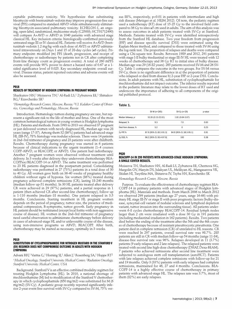
sus 88%, respectively, p=0.01 in patients with intermediate and highrisk disease (Metzger et al. NEJM 2012). Of note, the pediatric regimenused a radiotherapy (RT) dose of 15-25 Gy to the involved field com-pared to 36 Gy to sites ≥5 cm used in adults. The aim of this analysis wasto assess outcomes in adult patients treated with SV-Cy at Stanford.Methods. Patients treated with SV-Cy were identified retrospectivelyfrom the Stanford HL database. Two year freedom from progression(FFP) and disease specific survival (DSS) were estimated using theKaplan-Meier method, and compared to those treated with SV-M usingthe log-rank test. The proportion of relapses and deaths were comparedusing a Chi-square test. Results. Between 1989 and 2012, 291 patientswith stage I-II bulky mediastinal or stage III-IV HL were treated with 12weeks of chemotherapy and 36 Gy RT to initial sites of bulky disease.Median age was 29 (16-82 years). 265 patients received SV-M and 26 SV-Cy. Table 1 compares the outcome in the two groups. For SV-M versusSV-Cy, there is no significant difference in a) the proportion of patientswho relapsed or died from disease b) 2-year FFP or 2-year DSS. Conclu-sions. In adult patients with HL, substitution of cyclophosphamide formechlorethamine did not impact outcome. The inferior results reportedin the pediatric literature may relate to the lower doses of RT used andunderscore the importance of adhering to all components of the origi-nal published protocol.
Table 1.
SV-M (n=265) SV-Cy (n=26) p-value
Median followup, yr 10.35 (0.13-22.61) 1.81 (0.64-3.07)
Relapsed, % 9.3 7.0 0.63
Died from disease, % 5.2 0 0.23
2y FFP, % 87.9 [95% CI, 83.3-91.3] 84.9 [95% CI, 58.3-95.1] 0.91
2y DSS, % 96.2 [95% CI, 93.4-97.9] 100 0.38
P020BEACOPP-14 IN 299 PATIENTS WITH ADVANCED-STAGE HODGKIN LYMPHOMA:A SINGLE CENTER RESULTS.
Moiseeva TN, Sharkunov NN, Al-Radi LS, Zybunova EE, Chernova NG,Margolin OV, Maryin DS, Julakyan UL, Melikyan AL, Mangasarova YK,Skidan NI, Tseytlina MA, Shitareva IV, Tsyba NN, Kravchenko SKHematology Research Center, Moscow, Russia
Purpose. To evaluate the effectiveness of chemotherapy regimen BEA-COPP-14 in primary patients with advanced stages of Hodgkin lym-phoma (HL). Materials and methods. From March 2006 to March 2012we observed 299 patients (median age 27 years, range 16-66) with pri-mary HL stage III-IV or stage II with poor prognostic factors (bulky-dis-ease, syncytial-cell variant of nodular sclerosis and lymphoid depletionvariant, tumor invasion into the surrounding tissue). All patients under-went 6-8 cycles chemotherapy BEACOPP-14. Residual lymph nodeslarger than 2 cm were irradiated with a dose 30 Gy in 191 patients(including mediastinal irradiation in 162 patients). Results. Two patientsdied before the ending of the treatment after the 5th and the 6th cours-es of chemotherapy becouse of resistant pneumocystic pneumonia. Onepatient died in complete remission (CR) of unrelated to HL reasons. CRwere reached In 297 patients, overall survival rate was 98.7%. 285patients are still in CR with median follow-up 54 months (range 11-84),disease-free survival rate was 96%. Relapses developed in 11 (3.7%)patients (9 early relapses and 2 late relapses). The relapsed patients weretreated with second line high-dose chemotherapy (DHAP, Dexa-BEAM).7 patients who achieved remissions after second line treatment weresubjected to autologous stem cell transplantation (autoHCT). Patientswith late relapses achieved complete remissions with follow-up for 21and 19 months. Only 3 (33%) patients with early relapses had completeremissions, maintained for 46, 27 and 4 months. Conclusions. BEA-COPP-14 is a highly effective course of chemotherapy in primarypatients with advanced-stage HL. The relapses rate was 3.7%, most ofthem (82%) are early relapses.
haematologica | 2013; 98(s2) | 7
9th International Symposium on Hodgkin Lymphoma, Cologne, Germany, October 12-15, 2013
P021NODULAR LYMPHOCYTE PREDOMINANT HODGKIN LYMPHOMA WITH BONE MARROWINVOLVEMENT
Khlavno AB, Moiseeva TN, Kovrigina AM, Al-Radi LS, SharkunovNN, Kravchenko SKHematology Research Center, Moscow, Russia
Introduction. Bone marrow involvement in nodular lymphocyte pre-dominant Hodgkin Lymphoma (NLPHL) is considered as a very rareevent. Approximate incidence of bone marrow involvement in this caseis around 1% (V. Diehl et al , 1999). Materials and methods. From 2003to 2013 we revealed 28 patients with NLPHL. Median age was 39 years(range 17- 63 years), M:F=3:1. Most of the patients had advanced stagedisease (17(61%) patients with stages III-IV ), without -symptoms (21(75%) patients). Results. Bone marrow involvement was found histolog-ically and immunohistochemically in 8 (29%) patients (7 males and 1female). All patients with bone marrow involvement had multiplelesions, 5 (62,5%) of them had extranodal involvement of bones and softtissues. -symptoms were absent in 7 (87,5%) patients. In 4 patients werevealed NLPHL transformation to T cell rich B cell lymphoma (TCRB-CL) in bone marrow, 2 of them with concordant transformation inperipheral lymph node biopsy. Bone marrow involvement was predom-inantly found in older age group (median age 44 years, range 33-63years). History of prolonged asymptomatic lymphadenopathy was reg-istered in three patients (lasting for 3, 5 and 11 years). Patients underwentfollowing treatment: ABVD -1 patient,-2 patients, esc- 1 patient, P-14 +/-Rituximab–4 patients. In two cases involved field radiotherapy wasadministered in combination with chemotherapy. One patient under-went salvage chemotherapy followed by autologous stem cell trans-plant. All patients are alive, treatment is completed in 6 patients, 4 ofthem are in complete remission with median follow up 19 months(range 8-87 months). Two patients are still being treated. Two patientshad early relapses. Conclusion. In our patients bone marrow involve-ment in advanced stages of NLPHL is more frequent event in compari-son with published data. The evidence of possibility of transformationof NLPHL to TCRBCL in cases with bone marrow involvement needsfurther investigation.
P022LONG-TERM RESULTS. OF RISK- ADAPTED THERAPY FOR ADVANCED HODGKIN’S LYM-PHOMA: ONE-CENTER EXPERIENCE (1998-2012)
Bogatyreva TI, Pavlov VVMedical Radiological Research Centre, Obninsk, Russia
Introduction. This prospective MRRC trial was aimed to investigatepossibilities: 1) to minimize over-treatment in advanced Hodgkin’s lym-phoma (HL) by prescribing first-line chemotherapy (ABVD or BEA-COPPbas) according to the proposed risk factors (RF), and 2) to reducecumulative cardiopulmonary toxicity both by change for COPP in 1-2cycles which preceded mediastinal irradiation and by treating residualtumor with low dose radiotherapy (RT) 20-22 Gy in accelerated hyper-fractionated regimen. Patients and Methods. Between 1998 and 2012,the adult patients with advanced (stage III-IV) HL were allocated BEA-COPPbas if they had one or more RF, associated with early progression:a) lymphoid depletion or NSII histology, b) pericardial effusion, c) bonesor bone marrow involvement plus splenic lesions. Patients without RFreceived ABVD. Total six cycles were planned for supradiaphragmaticdisease and 8 cycles-for involvement on both sides of the diaphragm(including 1-2 COPP), with involved-field RT to residual lesions. Over-all survival (OS) and progression-free survival (PFS) were evaluated withregard to IPI scores 0-1, 2-3 or ≥4 (subgroups a,b,c). Results. With a medi-an follow-up of 7 years, total 383 patients were eligible for analysis; 250patients allocated BEACOPPbas (gr.1) and 133 patients started ABVD(gr.2);% pts with IPI score≥4 was higher in gr.1 (23% vs 3%). The %receiving 1-2 COPP at the end of chemotherapy course was 72% in gr.1and 49% in gr.2. For all pts of gr.1, OS and PFS at 10 years were 85%and 71%; for pts of gr.2, 96% and 87%, respectively. In subgroups a/b/cof IPI score, 10-year OS was 93%/87%/71% for gr.1 and100%/93%/100% for gr.2; PFS was, respectively, 88%/71%/55% forgr.1 and 93%/82%/75% for gr.2. There were no statistically significantdifferences in the outcome of 179 pts after 4 to 6 BEACOPPbas+2 COPP(OS 92%, PFS 86%) as compared to 35 pts after 6-8 BEACOPP (OS 73%,PFS 73%). Similarly, the outcome of 65 pts after 5-7 ABVD+ COPP (OS
96%, PFS 93%) did not differ from that in 40 pts after 6-8 ABVD (OS100%, PFS 90%). Conclusion. Our data show that tailoring of inductionchemotherapy to the risk factors (RF) helps to discriminate a proportionof patients with advanced stage for less toxic treatment.
P023INTENSIFIED BEACOPP THERAPY FOR NEWLY DIAGNOSED PATIENT WITH ADVANCEDSTAGES CLASSICAL HODGKIN’S LYMPHOMA
Kriachok I, Novosad O, Titorenko I, Filonenko K, Aleksik E, Kadniko-va T, Gubareva A, Kushchevoy E, Martynchyk A, Pastushenko Y,Ulyanchenko K, Stepanishyna IDepartment of Oncohematology, National Cancer Institute, Kiev, Ukraine
Background. According to insufficient treatment results in high riskpatients with Hodgkin’s Lymphoma (HL) and possible improvement ofthe efficiency by intensification in this group of patients, we initiated theprospective randomized multicenter study, in order to compare the effi-cacy and toxicity -14 and -esc.in high risk group patients with HL. Meth-ods. 111 patients (from September 2009 until December 2012) from 18to 65 years old (median–30.6), 40.5% male and female 59.4%, withstage III-IV, were treated with -14 (n=47) and -esc (n=64). The treatmentefficacy in both groups was evaluated after 4, 6, 8 cycles by heson cri-teria (1999, 2007). All patients received supporting therapy (erythropoi-etin, G-CSF). Results. Overall response rate of -14 and -esc were 97.7%and 96.5%, respectively ( >0.05); Complete response rate was also equal(60.8% and 67.24%, respectively) ( >0.05). Maximal observation periodis 38 months. The 3-year progression-free (PFS) and overal survival (OS)in the whole group (n=111) were 94.0% and 94.7%, respectively. OS inBEACOPP-esc group was 91.9% and 85.9% in BEACOPP-14 group(p>0.05). 5 patients relapsed, among them 2 pts in BEACOPP-14 groupand 3 pts–in BEACOPP-esc group, in term 1.2 to 30.5 months after treat-ment completed. Toxicity profile was represented mainly by hematolog-ical toxicity in 91.9% of cases (Grade 3/4 toxicities -30%). Anemia(62.8% vs 50.9%, >0.05) and thrombocytopenia (20.3% vs 11.6%,p>0.05) predominated in BEACOPP-esc group, while neutropenia ratewas slightly higher in BEACOPP-14 group (71.3% vs 60.0%, > 0.05).Conclusion. Treatment of patients with advanced stages of HL with -14and -esc is highly effective. Toxicity rate is acceptable provided that sup-portive therapy is used.
P024BONE MARROW INVOLVEMENT IN CLASSICAL AND NODULAR LYMPHOCYTE PREDOMI-NANT HODGKIN LYMPHOMA–INDIAN TERTIARY CANCER CENTRE EXPERIENCE
Epari S,1 Sengar M2, Gujral S, Menon H,2 Laskar S,1 Shet T21Departments of Pathology, Radiation Oncology and Medical Oncology; 2TataMemorial Centre, Mumbai, India
Introduction. Hodgkin lymphoma, both classical (CHL) and Nodularlymphocyte predominant (NLPHL) types have lesser a propensity forinvolvement of bone marrow. This study compares the frequency ofinvolvement of bone marrow in CHL and NLPHL. Material and Meth-ods. All the consecutive cases from the years 2010 to 2012 of both CHLand NLPHL, where there is available bone marrow biopsies were includ-ed. The bone marrow involvement in these cases was also evaluated andcompiled. Results. A total of 56 and 521 diagnosed cases of NLPHL andCHL with presence of bone marrow biopsies were included in the study.Age range was 6-68 (<18yrs: 11, 19-30yrs:21, 31-50yrs:18 &>50yrs: 18)with M:F ratio of 2.5:1 for NLPHL and for CHL was 3-78yrs(<18yrs:162,19-30yrs:150, 31-50yrs:140 &>50yrs: 69) with M:F ratio of2.84:1. Of these cases, 3 (5.4%) and 32 (6.1%) cases of NLPHL and CHLcases showed bone marrow involvement at presentation. Cases pre-senting at stage IV but no histological documentation of bone marrowinvolvement have excluded. The pattern of involvement was predomi-nantly focal in both types (though some cases of CHL showed diffusemarrow replacement). But the fibrosis and the presence of admixedeosinophils was seen in CHL but no case NLPHL showed fibrosis andeosinophils. Conclusion. Bone marrow involvement at presentation inHodgkin lymphoma (both NLPHL and HL) is rare and as noted in thepresent study, there does not appear to be any significant difference intheir frequency.
8 | haematologica | 2013; 98(s2)
Abstracts
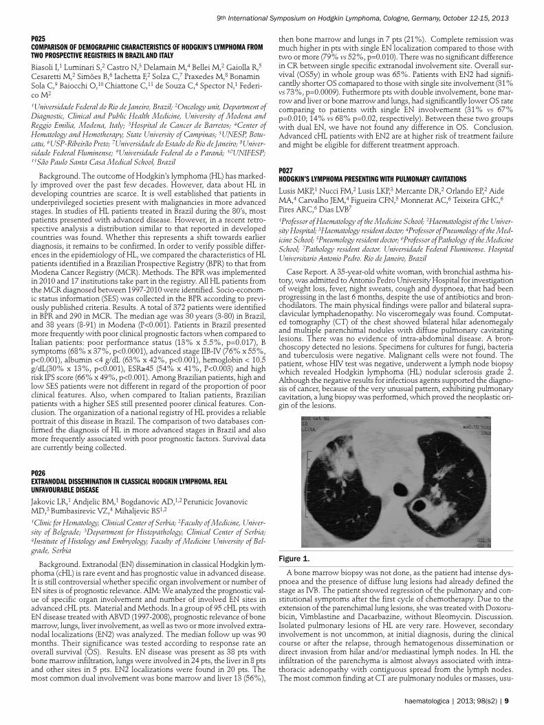
P025COMPARISON OF DEMOGRAPHIC CHARACTERISTICS OF HODGKIN’S LYMPHOMA FROMTWO PROSPECTIVE REGISTRIES IN BRAZIL AND ITALY
Biasoli I,1 Luminari S,2 Castro N,3 Delamain M,4 Bellei M,2 Gaiolla R,5Cesaretti M,2 Simões B,6 Iachetta F,2 Solza C,7 Praxedes M,8 BonaminSola C,9 Baiocchi O,10 Chiattone C,11 de Souza C,4 Spector N,1 Federi-co M2
1Universidade Federal do Rio de Janeiro, Brazil; 2Oncology unit, Department ofDiagnostic, Clinical and Public Health Medicine, University of Modena andReggio Emilia, Modena, Italy; 3Hospital de Cancer de Barretos; 4Center ofHematology and Hemotherapy, State University of Campinas; 5UNESP, Botu-catu, 6USP-Ribeirão Preto; 7Universidade do Estado do Rio de Janeiro; 8Univer-sidade Federal Fluminense; 9Universidade Federal do o Paraná; 10UNIFESP;11São Paulo Santa Casa Medical School, Brazil
Background. The outcome of Hodgkin’s lymphoma (HL) has marked-ly improved over the past few decades. However, data about HL indeveloping countries are scarce. It is well established that patients inunderprivileged societies present with malignancies in more advancedstages. In studies of HL patients treated in Brazil during the 80’s, mostpatients presented with advanced disease. However, in a recent retro-spective analysis a distribution similar to that reported in developedcountries was found. Whether this represents a shift towards earlierdiagnosis, it remains to be confirmed. In order to verify possible differ-ences in the epidemiology of HL, we compared the characteristics of HLpatients identified in a Brazilian Prospective Registry (BPR) to that fromModena Cancer Registry (MCR). Methods. The BPR was implementedin 2010 and 17 institutions take part in the registry. All HL patients fromthe MCR diagnosed between 1997-2010 were identified. Socio-econom-ic status information (SES) was collected in the BPR according to previ-ously published criteria. Results. A total of 372 patients were identifiedin BPR and 290 in MCR. The median age was 30 years (3-80) in Brazil,and 38 years (8-91) in Modena (P<0.001). Patients in Brazil presentedmore frequently with poor clinical prognostic factors when compared toItalian patients: poor performance status (13% x 5.5%, p=0.017), Bsymptoms (68% x 37%, p<0.0001), advanced stage IIB-IV (76% x 55%,p<0.001), albumin <4 g/dL (63% x 42%, p<0.001), hemoglobin < 10.5g/dL(30% x 13%, p<0.001), ESR≥45 (54% x 41%, P<0.003) and highrisk IPS score (66% x 49%, p<0.001). Among Brazilian patients, high andlow SES patients were not different in regard of the proportion of poorclinical features. Also, when compared to Italian patients, Brazilianpatients with a higher SES still presented poorer clinical features. Con-clusion. The organization of a national registry of HL provides a reliableportrait of this disease in Brazil. The comparison of two databases con-firmed the diagnosis of HL in more advanced stages in Brazil and alsomore frequently associated with poor prognostic factors. Survival dataare currently being collected.
P026EXTRANODAL DISSEMINATION IN CLASSICAL HODGKIN LYMPHOMA. REALUNFAVOURABLE DISEASE
Jakovic LR,1 Andjelic BM,1 Bogdanovic AD,1,2 Perunicic JovanovicMD,3 Bumbasirevic VZ,4 Mihaljevic BS1,2
1Clinic for Hematology, Clinical Center of Serbia; 2Faculty of Medicine, Univer-sity of Belgrade; 3Department for Histopathology, Clinical Center of Serbia;4Institute of Histology and Embryology, Faculty of Medicine University of Bel-grade, Serbia
Background. Extranodal (EN) dissemination in classical Hodgkin lym-phoma (cHL) is rare event and has prognostic value in advanced disease.It is still controversial whether specific organ involvement or number ofEN sites is of prognostic relevance. AIM: We analyzed the prognostic val-ue of specific organ involvement and number of involved EN sites inadvanced cHL pts. Material and Methods. In a group of 95 cHL pts withEN disease treated with ABVD (1997-2008), prognostic relevance of bonemarrow, lungs, liver involvement, as well as two or more involved extra-nodal localizations (EN2) was analyzed. The median follow up was 90months. Their significance was tested according to response rate anoverall survival (OS). Results. EN disease was present as 38 pts withbone marrow infiltration, lungs were involved in 24 pts, the liver in 8 ptsand other sites in 5 pts. EN2 localizations were found in 20 pts. Themost common dual involvement was bone marrow and liver 13 (56%),
then bone marrow and lungs in 7 pts (21%). Complete remission wasmuch higher in pts with single EN localization compared to those withtwo or more (79% vs 52%, p=0.010). There was no significant differencein CR between single specific extranodal involvement site. Overall sur-vival (OS5y) in whole group was 65%. Patients with EN2 had signifi-cantlly shorter OS comapared to those with single site involvement (31%vs 73%, p=0.0009). Futhermore pts with double involvement, bone mar-row and liver or bone marrow and lungs, had significantlly lower OS ratecomparing to patients with single EN involvement (31% vs 67%p=0.010; 14% vs 68% p=0.02, respectively). Between these two groupswith dual EN, we have not found any difference in OS. Conclusion.Advanced cHL patients with EN2 are at higher risk of treatment failureand might be eligible for different treatment approach.
P027HODGKIN’S LYMPHOMA PRESENTING WITH PULMONARY CAVITATIONS
Lusis MKP,1 Nucci FM,2 Lusis LKP,3 Mercante DR,2 Orlando EP,2 AideMA,4 Carvalho JEM,4 Figueira CFN,5 Monnerat AC,6 Teixeira GHC,6Pires ARC,6 Dias LVB7
1Professor of Haematology of the Medicine School; 2Haematologist of the Univer-sity Hospital; 3Haematology resident doctor; 4Professor of Pneumology of the Med-icine School; 5Pneumology resident doctor; 6Professor of Pathology of the MedicineSchool; 7Pathology resident doctor. Universidade Federal Fluminense. HospitalUniversitario Antonio Pedro. Rio de Janeiro, Brazil
Case Report. A 35-year-old white woman, with bronchial asthma his-tory, was admitted to Antonio Pedro University Hospital for investigationof weight loss, fever, night sweats, cough and dyspnoea, that had beenprogressing in the last 6 months, despite the use of antibiotics and bron-chodilators. The main physical findings were pallor and bilateral supra-clavicular lymphadenopathy. No visceromegaly was found. Computat-ed tomography (CT) of the chest showed bilateral hilar adenomegalyand multiple parenchimal nodules with diffuse pulmonary cavitatinglesions. There was no evidence of intra-abdominal disease. A bron-choscopy detected no lesions. Specimens for cultures for fungi, bacteriaand tuberculosis were negative. Malignant cells were not found. Thepatient, whose HIV test was negative, underwent a lymph node biopsywhich revealed Hodgkin lymphoma (HL) nodular sclerosis grade 2.Although the negative results for infectious agents supported the diagno-sis of cancer, because of the very unusual pattern, exhibiting pulmonarycavitation, a lung biopsy was performed, which proved the neoplastic ori-gin of the lesions.
Figure 1.
A bone marrow biopsy was not done, as the patient had intense dys-pnoea and the presence of diffuse lung lesions had already defined thestage as IVB. The patient showed regression of the pulmonary and con-stitutional symptoms after the first cycle of chemotherapy. Due to theextension of the parenchimal lung lesions, she was treated with Doxoru-bicin, Vimblastine and Dacarbazine, without Bleomycin. Discussion.Isolated pulmonary lesions of HL are very rare. However, secondaryinvolvement is not uncommon, at initial diagnosis, during the clinicalcourse or after the relapse, through hematogenous dissemination ordirect invasion from hilar and/or mediastinal lymph nodes. In HL theinfiltration of the parenchyma is almost always associated with intra-thoracic adenopathy with contiguous spread from the lymph nodes.The most common finding at CT are pulmonary nodules or masses, usu-
haematologica | 2013; 98(s2) | 9
9th International Symposium on Hodgkin Lymphoma, Cologne, Germany, October 12-15, 2013
ally multiple and bilateral, but cavitation is extremely rare. This patient’sunusual presentation required histological confirmation in order toestablish a differential diagnosis with tuberculosis, fungal infection,bronchoalveolar carcinoma, bronchiolitis obliterans organizing pneu-monia, lymphomatoid granulomatosis, collagen vascular disorders andvasculitis as Wegener’s granulomatosis.
P028THERAPEUTIC RESPONSE OF TUNISIAN NATIONAL PROTOCOL: EXPERIENCE OF TWOCENTERS
Kacem K,1 Zriba S,2 Ghédira H,2 Yahyaoui Y,1 Mansouri R,1 Dridi M,1Hadj Mansour M,1 Manai Z,1 Zarrouk M,1 Ben Neji H,1 Ben Abden-nebi Y,1 Jeddi R,1 Aissaoui L,1 Belhadj Ali Z,1 Ben Abid H,1 M’Sadek F,2Ben Lakhal R,1 Meddeb B1
1Clinical Hematology Department of Aziza Othmana Hospital of Tunis; 2Clin-ical Hematology Department of Military Hospital of Tunis, Tunisia
Purpose. Assessment of therapeutic response of Hodgkin lymphoma(HL) treated according the national protocol HL-2008. Patients andMethods. Between 2008 and 2011, 173 patients from 2 departmentswere enrolled in HL-2008 protocol (a third version of prospective trial).Results. Median age was 30 years (15-82) with a sex ratio 0.99. HL-2008defined 5 groups: G1: favorable early stage, G2: unfavorable early stage,G3: advanced stage and localized stage with mediastinal bulk(MTI>0.35), G4: early stage in elderly patients, G5: advanced stage inelderly group. We recommend 2 cycles ABVD + radiotherapy (IFRT) forG1, 4 cycles of ABVD + IFRT for G2, 8 cycles of BEACOPP (4 escalated+4 baseline) for G3. Elderly groups received 6 and 8 cycles of ABVD forG4 and G5 respectively. Early assessment of response was recommend-ed after 2 cycles in each group and an escalation to BEACOPP-R tookplace if response was less than 75%. 66.5% of patients were treated inG3, 19.5% in G2, 7% in G1, 6% in G5 and 1% in G4. 82% of patientswere in complete response after the first line therapy. 12% were refrac-tory. Median follow-up was of 32 months at 3 years, OS, EFS and RFSwere respectively of 90, 75 and 95%. IFRT at 36 Gy was done in 48patients with a median delay of 2 months (1-4). 5 patients received RTfor residual disease. Conclusion. Comparatively to the second version,intensive chemotherapy for unfavorable HL (advanced stage and bulkymediastinal disease with stage II), RFS and OS are better.
P029THE DIFFERENCES IN RISK PROFILE OF MALE AND FEMALE PATIENTS WITH ADVANCEDHODGKIN’S LYMPHOMA
Andjelic B,1 Jakovic Lj,1 Antic D,1,2 Todorovic M,1,2 Bila J,1,2 SretenovicA,1 Djurasinovic V,1 Mihaljevic B1,2
1Clinic for Hematology, Clinical Center of Serbia, Belgrade, Serbia; 2Faculty ofMedicine, University of Belgrade, Belgrade, Serbia
Background. Male gender is considered as an unfavorable prognosticfactor for Hodgkin’s lymphoma (HL). Modern studies are having theaim to define risk adopted treatment approach. Currently, the HL treat-ment for both genders is uniform. Aim. The aim of this study is to iden-tify risk factors for poor outcome according to the gender in patientswith advanced HL. Patients and Methods. A retrospective study was per-formed on 163 female and 151 male patients with advanced HL diag-nosed in the period June 1997-December 2008. The initial treatmentwas 6-8 cycles of ABVD followed by radiotherapy. Prognostic relevanceof histological subtype, presence of B symptoms, large mediastinaltumor, “bulky” disease, extranodal disease (EN), erythrocyte sedimen-tation rate (ESR) ≥50 mm/h and risk factors included in IPS score (exceptgender) was examined. Results. The median age of female patients was31 (range 16-69) and for males 35 (range 17-80). Complete response ratewas higher in females (86.5% vs 80.7%), but not statistically significant(p>0.05). There was a trend toward better both 5 yrs overall survival(79.8% vs 71.5%; p=0.074) and 5 yrs event free survival (66.3% vs57.6%; p=0.06) in females. In univariate analysis, bulky disease and his-tological subtype influenced overall survival (OS) in females (p=0.045;p=0.011, respectively) and there was a trend towards worse event freesurvival (EFS) in patients with EN and clinical stage IV (p=0.083; p=0.051,respectively). The presence of B symptoms, EN, ESR≥50 mm/h, anemiaand lymphopenia influenced in males both OS (p=0.015; p=0.002;p=0.015; p=0.005; p=0.004, respectively) and EFS (p=0.045; p=0.043;
p=0.043; p=0.010; p=0.006, respectively) in univariate analysis. Maleswith CS IV and age>45 years had worse OS (p=0.021; p=0.03, respec-tively) and there was a trend towards worse EFS in males with CS IV(p=0.054). The multivariate analysis identified bulky disease in femalesas the independent prognostic factor for OS, while in males these are ENand anemia. Furthermore, lymphopenia is independent prognostic fac-tor for EFS in males. Conclusion. ABVD is reasonable solution in femaleswithout large tumor, especially if planning pregnancy in future. Maleshaving EN, anemia or lymphopenia require more effective initial treat-ment.
P030THE IDENTIFICATION OF HIGH RISK ABVD TREATED ADVANCED HODGKIN’S LYMPHOMAPATIENTS-IPS REMAINS THE MOST APPROPRIATE PROGNOSTIC TOOL
Andjelic B,1 Antic D,1,2 Jakovic Lj,1 Todorovic M,1,2 Bogdanovic A,1,2Bila J,1,2 Mihaljevic B1,21Clinic for Hematology, Clinical Center of Serbia, Belgrade, Serbia; 2Faculty ofMedicine, University of Belgrade, Belgrade, Serbia
Background. International prognostic score (IPS) was derived fromlarge cohort of Hodgkin’s lymphoma (HL) patients, who were partiallytreated with historical regimens. Nowadays, the improvement in treat-ment results with modern strategies lead to question whether IPS is stillan appropriate prognostic tool in HL. Aim: The aim of this study wasto assess prognostic value of IPS in patients with advanced HL. Also, wetried to identify other risk factors who could additionally contribute inrisk stratification. Patients and Methods. A retrospective study was per-formed on 314 advanced classical HL patients, diagnosed in the periodJune 1997-December 2008. The standard of initial care was 6-8 cyclesof ABVD followed by radiotherapy. Prognostic relevance of IPS≥3, IPS≥4,histological subtype, presence of B symptoms, large mediastinal tumor,“bulky” disease, extranodal involvement of more than 1 site (EN2) anderythrocyte sedimentation rate (ESR) ≥50 mm/h was examined. Results.The median age of the patients was 32 (range 16-80). The median fol-low up was 90 months. For the whole group 5 years event free survival(EFS) was 62.1% and 5 years overall survival (OS) was 75.8%. In uni-variate analysis, worse OS was found in patients with IPS≥3, IPS≥4,bulky disease, EN2 and ESR≥50 mm/h (log rank; p=0.000, p=0.000,p=0.042, p=0.000, p=0.006, respectively). The patients with IPS≥3, IPS≥4 and and EN2 had also significantly worse EFS (log rank; p=0.000,p=0.007, p=0.000, respectively). The multivariate Cox regression analy-sis identified IPS≥3, EN2 and bulky disease as the independent prognos-tic factors for OS (p=0.000; p=0.000; p=0.023, respectively). Further-more, IPS≥3 (p=0.001) and EN2 (p=0.001) are also independent prognos-tic factors for EFS. Conclusion. IPS can effectively identify advanced HLpatients at high risk for poor outcome if treated with ABVD. Patientswith IPS 3 or higher should be considered as high risk. More effectivetreatment is needed for patients with high IPS and/or EN2 and/or bulkydisease.
P031LOWER DOSE INTENSITY CHEMOTHERAPY; IMPACT ON TREATMENT OUTCOME ANDSURVIVAL OF PATIENTS WITH ADVANCED HODGKIN LYMPHOMA: SINGLE INSTITUTIONEXPERIENCE
Tomasevic Z, Tomasevic ZInstitute for Oncology and Radiology, Belgrade, Serbia
Background. Combined chemotherapy regimen, although highlyeffective, still fail to cure about one third of patients with advancedHodgkin lymphoma (aHL). On the other hand, similar proportions ofpatients are probably over treated and cure might be obtained with lessintensive doses. The aim of this retrospective analysis is to evaluatechemotherapy dose intensity and cumulative dose in aHL, in the con-text of treatment outcome and survival. Methods. At the Institute forOncology and Radiology of Serbia, during 1991-1993 all consecutiveaHL patients have been treated with MOPP (M; 41 pts) and afterwards(1994-1996) with MOPP/ABVD (M/A; 40 pts). In accordance to previ-ous local practice, chemotherapy was given in projected, absolute dos-es, calculated on average body surface area of 1.7 m2. Characteristics ofpatients in both regimens were well balanced for age, gender, PS, Bsymptoms, histology, stage, laboratory prognostic parameters (Interna-tional Prognostic Score- IPS). Approximately 50% pts in both groups
10 | haematologica | 2013; 98(s2)
Abstracts
were in high/intermediate risk IPS. Relative dose intensity (RDI), definedas given/scheduled dose ratio per week and relative cumulative dose(RCD), defined as given/projected dose ratio, were retrospectively ana-lyzed whether there is potential negative impact on response, time torelapse and survival. Results. The average RCD and RDI for both regi-mens were 85% and 58%. Complete remission (CR) was achieved in60% (M/A 67% vs M 54%) (p=NS); 90% of pts with CR in both groupshad no relapse during follow up. Different cumulative doses and doseintensity had no significant impact on time to relapse and overall sur-vival. However, trend for better overall survival for patients receivingRDI over 60% was observed (p=0.09). Hemoglobin level ≤10.5 g/dl hadsignificant adverse effect on outcome regardless of treatment dosage(p=0.02). No second malignancy has been registered during follow up.Conclusions. Although this retrospective analysis does not pretend torecommend lower chemotherapy dose regimen for routine treatment, itseems that selected aHL patients could still be effectively treated withless chemotherapy dose intensity, because 90% of pts with CR are cured.
P032DIAGNOSTICAL CHALLENGES IN OUR ADOLESCENT PATIENT WITH LYMPHOMA
Magyari F,1 Rajnai H,3 Barna S,2 Miltényi Z,1 Váróczy L,1 Csomor J,3Illés A11Institute for Internal Medicine, Medical and Health Science Center, Universityof Debrecen; 2Scanomed LTD; 31st Dept. of Pathology an Experimental CancerResearch, University of Semmelweis, Hungary
Objectives. The B-cell lymphoma, unclassifiable, showing intermedi-ate features typical for both diffuse largeB-celllymphoma (DLBCL) andclassical Hodgkinlymphoma (HL) is a novel category of diffuse large B-cell lymphomas (DLBCL/HL), which has been described by the WHOclassification in 2008. This rare type of lymphomas presents peculiarclinical, morphological and immunophenotypical patterns, previouslycalled as gray zone lymphomas. Case Report. In December 2011 a 17-year old male was diagnosed with mixed cellularity subtype of classicalHL The staging18FDG-PET/CT scan showed a disseminated, abdomi-nal bulky disease. His clinical stage was IV/BXS with unfavourable prog-nosis. After an incomplete course of ABVD chemotherapy the patient’ssymptoms disappeared and the sizes of the involved lymph nodesdecreased as well. Because of the unusally extended disease (nodal-extra-nodal-bulky) a histological revision was performed. The diagnosischanged into DLBCL/HL, so the treatment was modified to R-CHOP-14 regimen. After 3 cycles of R-CHOP-14 he achieved a complete meta-bolic remission (CMR), which was confirmed by a 18FDG-PET/CT scan.Receiving further 3 cycles of R-CHOP-14 therapy the patient was still inCMR, but a PET negative large mass (7x3 centimeter) still remained vis-ible in the abdominal region. Cosidering this residual tumor and theagressive subtype of lymphoma he was referred for an autologous hemo-poietic stem cell transplantation (AHSCT). After 2 cycles of R-DHAPregimen, successful CD34 positive stem cell collection was performed inAugust 2012. In September of 2012, he underwent a R-BEAM condi-tioning followed by AHSCT. The next18FDG-PET/CT scan performedwas 100 days after the AHSCT and it still showed CMR. Conclusions.Upon this case, it should be underlined that the diagnosis may needrevision if a patient represents atypical clinical signs and behavior, andthe importance of cooperation between clinicians and pathologistsis alsostrongly emphasized.
Early Stages
T033IMPACT OF BLEOMYCIN AND DACARBAZINE WITHIN THE ABVD REGIMEN IN THETREATMENT OF EARLY-STAGE FAVORABLE HODGKIN LYMPHOMA: FINAL RESULTS OF THE GHSG HD13 TRIAL
Behringer K,1 Goergen H,1 Borchmann P,1 Diehl V,1 Fuchs M,1 Hitz F,2Zijlstra JM,3 Greil R,4 Markova J,5 Topp MS,6 Soekler M,7 Mathas S,8Meissner J,9 von Tresckow B,1 Böll B,1 Engert A11German Hodgkin Study Group (GHSG), Department of Internal Medicine I,University Hospital of Cologne, Germany; 2Onkologie-Hämatologie Kantons -spital St.Gallen, St.Gallen, Switzerland; 3Vrije Universiteit University MedicalCentre Amsterdam, Amsterdam, The Netherlands; 4University of Salzburg,Salzburg, Austria; 5University Hospital Kralovske Vinohrady, Charles Univer-sity Prague, Prague, Czech Republic; 6University of Wuerzburg, Wuerzburg,Germany; 7University of Tuebingen, Tuebingen, Germany; 8Campus VirchowKlinikum, Berlin, Germany; 9University of Heidelberg, Heidelberg, Germany
Background. Combined modality treatment consisting of two cyclesof ABVD (adriamycin, bleomycin, vinblastine, dacarbazine) followedby involved-field radiotherapy (IFRT) is regarded as standard of care forearly-stage favorable Hodgkin Lymphoma (HL). However, the impact ofbleomycin and dacarbazine in this combination has been questioned foryears. Patients and Methods. The GHSG HD13 study compared twocycles of ABVD with a dacarbazine-deleted variant (ABV), a bleomycin-deleted variant (AVB), and a variant in which both, dacarbazine andbleomycin were deleted (AV). In each treatment arm, chemotherapywas followed by IFRT of 30 Gy. Primary objective was to demonstratenon-inferiority of the three experimental variants compared to ABVDregarding the primary endpoint freedom from treatment failure (FFTF)by excluding a difference of 6% after 5 years. Between 01/2003 and09/2009, 1710 patients were enrolled into the study; 72 were excludedfrom all analyses due to revision of HL diagnosis, revision of staging andparticipation in another GHSG trial, or loss to follow-up before start oftreatment. Due to higher event rates during a continuous safety analy-sis, the AV and ABV arms were closed early in 09/2005 and 02/2006after recruitment of 186 and 209 patients, respectively. Inferiority ofthese arms regarding FFTF could be confirmed in an interim analysis in09/2009; results will be updated in this final analysis. For the compari-son of ABVD and AVD, 1243 patients were analyzed for therapy adher-ence, toxicity, and efficacy. Results. Patient characteristics were well bal-anced between the four treatment arms: Median age was 39 years, 68%had stage II disease, and there were more male patients included (59%).The most frequent histologic subtypes were nodular sclerosis and mixedcellularity (38% each). Inclusion criteria were violated in 136 patients(8%), who were excluded from analyses of protocol adherence and tox-icity. Most qualified patients received chemo- and radiotherapy as perprotocol; protocol deviations are reported in 3%. Results. Regardingacute toxicity and efficacy will be presented. Conclusion. Dacarbazinecannot be deleted from the ABVD regimen without a significant loss ofefficacy. Whether bleomycin can be safely omitted will be discussed inview of the final analysis presented.
T034DEFINITION OF BULKY DISEASE IN EARLY STAGE HODGKIN LYMPHOMA IN COMPUTEDTOMOGRAPHY ERA: PROGNOSTIC SIGNIFICANCE OF MEASUREMENTS IN THE CORONALAND TRANSVERSE PLANES
Kumar A,1 Burger IA,2 Zhang Z,3 Maragulia J,1 Yahalom J,4Moskowitz CH,1 Zelenetz AD11Lymphoma Service, Department of Medicine, Memorial Sloan-Kettering Can-cer Center; 2Department Medical Radiology, University Hospital Zurich, Switzer-land; 3Biostatistics and Epidemiology, Memorial Sloan-Kettering Cancer Cen-ter; 4Department of Radiation Oncology, Memorial Sloan-Kettering Cancer Cen-ter, New York, NY, USA
Purpose. To investigate the prognostic significance of bulky diseasemeasured in the transverse and coronal planes on computed tomogra-phy (CT) imaging in early stage Hodgkin lymphoma (HL). Patients andMethods. This single-center retrospective study included 195 pediatricand adult patients with early stage classical HL treated with doxoru-
haematologica | 2013; 98(s2) | 11
9th International Symposium on Hodgkin Lymphoma, Cologne, Germany, October 12-15, 2013
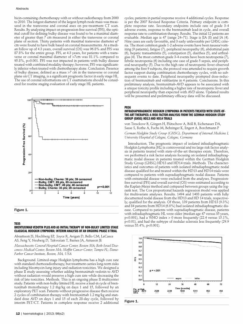
bicin-containing chemotherapy with or without radiotherapy from 2000to 2010. The longest diameter of the largest lymph node mass was meas-ured in the transverse and coronal axes on pre-treatment CT scan.Results. By analyzing impact on progression-free survival (PFS), the opti-mal cutoff for defining bulky disease was found to be a maximal diam-eter of greater than 7 cm measured in either the transverse or coronalplane of section. Thirty patients with maximal transverse diameter ≤7cm were found to have bulk based on coronal measurements. At a medi-an follow up of 4.3 years, overall survival (OS) was 96.8% and PFS was87.8% for the entire group. PFS, at 4.3 years, for patients with a trans-verse or coronal maximal diameter of >7cm was 81.1% compared to95.8%, p=0.001. PFS was not impacted in patients with bulky diseasetreated with combined modality therapy; however, PFS was significant-ly inferior when treated with chemotherapy alone. Conclusion. Presenceof bulky disease, defined as a mass >7 cm in the transverse or coronalplane on CT imaging, is a significant prognostic factor in early stage HL.The use of coronal reformations on CT examinations should be consid-ered for routine staging evaluation of early stage HL patients.
Figure 1.
T035BRENTUXIMAB VEDOTIN PLUS AVD AS INITIAL THERAPY OF NON-BULKY LIMITED STAGECLASSICAL HODGKIN LYMPHOMA: INTERIM ANALYSIS OF AN ONGOING PHASE II TRIAL
Abramson JS, Hochberg EP, Joyce R, Avigan D, Bello CM, LaCasceAS, Feng Y, Neuberg D, Takvorian T, Barnes JA, Arnason JEMassachusetts General Hospital Cancer Center, Boston MA; Beth-Israel Dea-coness Medical Center, Boston MA; Moffitt Cancer Center, Tampa FL; Dana-Farber Cancer Institute, Boston, MA, USA
Background. Limited-stage Hodgkin lymphoma has a high cure ratewith standard chemoradiotherapy, but treatment carries long-term risksincluding bleomycin-lung injury and radiation toxicities. We designed aphase II study assessing whether adding brentuximab vedotin to AVDwithout radiation would preserve a high cure rate while decreasing therisk of late toxicities. Methods. This is an ongoing phase II multicenterstudy. Patients with non-bulky limited HL receive a lead-in cycle of bren-tuximab monotherapy 1.2 mg/kg on days 1 and 15, followed by anexploratory PET scan. Patients without progressive disease then receive2 cycles of combination therapy with brentuximab 1.2 mg/kg and stan-dard dose AVD on days 1 and 15 of each 28-day cycle, followed byinterim PET/CT. Patients in complete response receive 2 additional
cycles; patients in partial response receive 4 additional cycles. Responseis per the 2007 Revised Response Criteria. Primary endpoint is com-plete response rate. Secondary endpoints include rate of grade 3-4adverse events, response to the brentuximab lead-in cycle, and overallresponse rate to combination therapy. Results. The initial 12 patients areevaluable. Median age is 47 (range 24–71). Stage is IIA (8) and IA (4).Nine cases are early favorable, and 3 early unfavorable per GHSG crite-ria. The most common grade 1-2 adverse events have been nausea/vom-iting (9 patients), fatigue (7), peripheral neuropathy (6), abdominal pain(6), hepatic transaminitis (5), constipation (5), diarrhea (5), and arthral-gias (4). The most common grade 3-4 events have been neutropenia (7),febrile neutropenia (6) including one case of grade 5 sepsis, and periph-eral neuropathy (5). Due to the high rate of neutropenic fever observedamong the first 9 subjects, the protocol was amended to require growthfactor support during combination chemotherapy cycles, with no sub-sequent events to date. Peripheral neuropathy prompted dose-reduc-tion of brentuximab and vinblastine in 4 patients. Conclusions. In thispreliminary analysis, brentuximab-AVD appears to be associated witha unique toxicity profile including a higher rate of neutropenic fever andperipheral neuropathy than expected with AVD alone. Updated resultswill be presented and preliminary efficacy data will be discussed.
P036INFRADIAPHRAGMATIC HODGKIN LYMPHOMA IN PATIENTS TREATED WITH STATE-OF-THE-ART THERAPIES: A RISK FACTOR ANALYSIS FROM THE GERMAN HODGKIN STUDYGROUP (GHSG) HD13 AND HD14 TRIALS
von Tresckow B, Görgen H, Plütschow A, Böll B, Eichenauer DA,Sasse S, Rothe A, Fuchs M, Behringer K, Engert A, Borchmann P
German Hodgkin Study Group (GHSG), Department of Internal Medicine I,University Hospital of Cologne, Cologne, Germany
Introduction. The prognostic impact of isolated infradiaphragmaticHodgkin Lymphoma (HL) is controversial and no large risk factor analy-sis in patients treated with state-of-the-art therapies exists. Therefore,we performed a risk factor analysis focusing on isolated infradiaphrag-matic nodal disease in patients treated within the German HodgkinStudy Group (GHSG) HD13 and HD14 trials. Methods. The character-istics and outcomes of patients with isolated infradiaphragmatic nodaldisease qualified for and treated within the HD13 and HD14 trials werecompared to patients with supradiaphragmatic nodal disease. Patientswith extranodal disease were excluded from the analyses. Progression-free survival (PFS) and overall survival (OS) were estimated according tothe Kaplan-Meier method and compared between groups using the log-rank test. The Cox proportional hazards regression model was appliedfor multivariate analyses. Results. 1494 and 1403 patients with fullydocumented nodal disease from the HD13 and HD 14 trials, respective-ly, qualified for the analysis. Of those, 139 patients from HD13 (9.3%)and 84 patients from HD14 (6.0%) had isolated infradiaphragmatic dis-ease. Compared to patients with supradiaphragmatic disease, patientswith infradiaphragmatic HL were older (median age 47 versus 35 years,p<0.001), had a WHO index > 0 more frequently (22.4 versus 15.1%,p<0.01), and had the subtype of nodular sclerosis less frequently (29.9versus 55.4%, p<0.001).
Figure 1.
12 | haematologica | 2013; 98(s2)
Abstracts
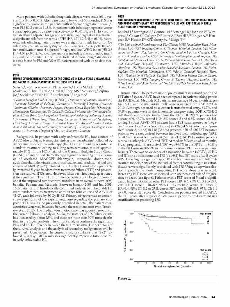
More patients with infradiaphragmatic disease were male (69.1 ver-sus 51.9%, p<0.001). After a median follow-up of 50 months, PFS wassignificantly worse in the patients with infradiaphragmatic disease (5-year PFS 80.2 versus 91.3% in patients with infradiaphragmatic versussupradiaphragmatic disease, respectively, p<0.001, Figure 1). In a multi-variate model adjusted for age and sex, infradiaphragmatic HL remaineda significant risk factor in terms of PFS (HR 1.7 [1.2-2.4], p<0.01). More-over, infradiaphragmatic disease was a significant risk factor for OSwhen analyzed univariately (5-year OS 91.7 versus 97.7%, p<0.001) andin a multivariate model adjusted for age, trial and WHO index (HR 2.3[1.4-4.0], p<0.01). Multivariate analyses including chemotherapy inten-sity will be presented. Conclusion. Isolated infradiaphragmatic diseaseis a risk factor for PFS and OS in HL patients treated with up-to-date ther-apies.
P037IMPACT OF DOSE INTENSIFICATION ON THE OUTCOME IN EARLY-STAGE UNFAVORABLEHL: 7-YEAR FOLLOW-UP ANALYSIS OF THE GHSG HD14 TRIAL
Sasse S,1, von Tresckow B,1, Plütschow A,1 Fuchs M,1 Klimm B,1Markova J,2 Hitz F,3 Kral Z,4 Greil R,5 Topp MS,6 Meissner J,7 ZijlstraJM,8 Soekler M,9 Eich HT,10 Borchmann P,1 Engert A11German Hodgkin Study Group (GHSG), Department of Internal Medicine I,University Hospital of Cologne, Germany; 2University Hospital KralovskeVinohrady, Charles University Prague, Prague, Czech Republic; 3Onkologie-Hämatologie Kantonsspital St.Gallen,St.Gallen, Switzerland; 4University Hos-pital of Brno, Brno, Czech Republic; 5University of Salzburg, Salzburg, Austria;6University of Wuerzburg, Wuerzburg, Germany; 7University of Heidelberg,Heidelberg, Germany; 8Vrije Universiteit University Medical Centre Amster-dam, Amsterdam, The Netherlands; 9University of Tuebingen, Tuebingen, Ger-many; 10University Hospital of Münster, Münster, Germany
Background. In patients with early unfavorable HL, four courses ofABVD (doxorubicin, bleomycin, vinblastine, dacarbazine) followed by30 Gy involved-field radiotherapy (IF-RT) are still widely regarded asstandard treatment leading to a long-term remission rate of approxi-mately 80%. In the HD14 trial of the German Hodgkin Study Group(GHSG), an intensified chemotherapy regimen consisting of two cours-es of escalated BEACOPP (bleomycin, etoposide, doxorubicin,cyclophosphamide, vincristine, procarbazine, and prednisone) and twocourses of ABVD (“2+2”) followed by 30 Gy IF-RT resulted in significant-ly improved 5-year freedom from treatment failure (FFTF) and progres-sion free survival (PFS) rates. However, it has been frequently questionedif the significant PFS and FFTF difference persists with longer follow-upand if the improved tumor control translates in an overall survival (OS)benefit. Patients and Methods. Between January 2003 and Juli 2008,1655 patients with histologically confirmed early-stage unfavorable HLwere randomized to treatment with either four courses of ABVD or“2+2”, each followed by 30 Gy IF-RT. Primary objective was to demon-strate superiority of the experimental arm regarding the primary end-point FFTF. Results. As previously described in detail, the patient char-acteristics were well balanced between the treatment arms (von Tresck-ow et al., 2012). The median observation time was about 70 months inthe current follow-up analysis. So far, the number of PFS failure eventshas increased by about 25%, and there are more than 50% more deathsthan in the 5-year analysis. The current analysis confirms the significantPFS- and FFTF-difference between the treatment arms. Further details ofthe survival analysis and the analysis of secondary malignancies will bepresented. Conclusion. The current analysis confirms that “2+2” fol-lowed by 30 Gy IF-RT results in a significantly improved tumor controlin early unfavorable HL.
P038PROGNOSTIC PERFORMANCE OF PRE-TREATMENT EORTC, GHSG AND IPI RISK FACTORSAND POST-CHEMOTHERAPY PET RESPONSE IN THE UK NCRI RAPID TRIAL IN EARLYSTAGE HODGKIN LYMPHOMA (HL)
Radford J,1 Barrington S,2 Counsell N,3 Pettengell R,4 Johnson P,5 Wim-peris J,6 Coltart S,7 Culligan D,8 Lister A,9 Bessell E,10 Kruger A,11 Han-cock B,12 Hoskin P,13 O’Doherty M,14 Illidge T151The University of Manchester and The Christie NHS Foundation Trust, Man-chester, UK; 2PET Imaging Centre, St Thomas’ Hospital, London, UK; 3Can-cer Research and UCL Cancer Trials Centre, London UK; 4St George’s, Uni-versity of London, London, UK; 5University of Southampton, Southampton, UK;6Norfolk and Norwich University NHS Foundation Trust, Norwich UK; 7Kentand Canterbury Hospital, Canterbury UK; 8Aberdeen Royal Infirmary,Aberdeen, UK; 9Barts and the London School of Medicine, London, UK; 10Not-tingham City Hospital, Nottingham, UK; 11Royal Cornwall Hospital, Truro,UK; 12University of Sheffield, Sheffield, UK; 13Mount Vernon Cancer Centre,Northwood, UK; 14PET Imaging Centre, St Thomas’ Hospital, London, UK;15The University of Manchester and The Christie NHS Foundation Trust, Man-chester, UK
Introduction. The performance of pre-treatment risk stratification andPET after 3 cycles ABVD have been compared in patients taking part inthe RAPID trial. Methods 602 patients, median age 34 years, with stagesIA/IIA HL and no mediastinal bulk were registered into RAPID 2003-2010. Although not used as selection factors for trial entry, 61.7% and67.8% patients were in the favourable category using EORTC/GHSGrisk stratifications respectively. Using the IPI for HL, 21.8% patients hada score of 0, 47.7% scored 1, 24.3% scored 2 and 6.3% scored >2. Fol-lowing 3 cycles ABVD, 571 patients had a PET scan reported as “nega-tive” (score 1 or 2 on a 5-point scale) in 426 (74.6%) patients or “posi-tive” (score 3, 4 or 5) in 145 (25.4%) patients. 420 of 426 PET negativepatients were randomised between involved field radiotherapy (IFRT,n=209) and no further treatment (NFT, n=211); 145 PET positive patientsreceived a 4th cycle ABVD and IFRT. At median follow-up of 48 months,3-year progression-free survival (PFS) was 94.5% in the IFRT arm, 90.8%in the NFT arm and 86.2% in the non-randomised PET positive patients.Results. There was no evidence of association between EORTC, GHSGand IPI risk stratifications and PFS (p’s >0.1) but PET score after 3 cyclesABVD was highly significant (p <0.01). In both univariate and full mul-tivariate models, none of the individual factors contributing to risk strat-ifications were significantly associated with PFS. Using a stepwise selec-tion approach the model comprising PET score alone was selected.Increasing PET score was associated with an increased risk of progres-sion or death (see figure). Patients with a PET score of 5 had a signifi-cantly higher risk than all other PET scores (HR=6.6, 95% CI: 3.2 to 13.7,versus PET score 1; HR=6.6, 95% CI: 2.7 to 15.9, versus PET score 2;HR=9.4, 95% CI: 3.2 to 27.8, versus PET score 3; HR=3.3, 95% CI: 1.1to 9.8, versus PET score 4). Conclusion For patients treated in RAPID,the PET score after 3 cycles ABVD was superior to pre-treatment riskstratification in predicting PFS.
Figure 1.
haematologica | 2013; 98(s2) | 13
9th International Symposium on Hodgkin Lymphoma, Cologne, Germany, October 12-15, 2013
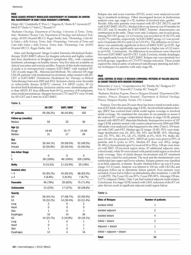
P039IMAGE-GUIDED INTENSITY MODULATED RADIOTHERAPY VS STANDARD 3D-CONFOR-MAL RADIOTHERAPY IN EARLY STAGE HODGKIN’S LYMPHOMA.
Filippi AR,1 Ciammella P,2 Piva C,1 Ragona R,1 Botto B,3 Gavarotti P,4Merli F,5 Vitolo U,3 Iotti C,2 Ricardi U1
1Radiation Oncology, Department of Oncology, University of Torino, Torino,Italy; 2Radiation Therapy Unit, Department of Oncology and Advanced Tech-nology, ASMN Hospital-IRCCS, Reggio Emilia, Italy; 3Hematology, Città del-la Salute e della Scienza, Torino, Italy; 4Hematology, University of Torino andCittà della Salute e della Scienza, Torino, Italy; 5Hematology Unit, ASMNHospital-IRCCS, Reggio Emilia, Italy
Aims and Background. Image-Guided Intensity-Modulated Radio-therapy (IGRT-IMRT) allows for margins reduction and highly confor-mal dose distribution in Hodgkin’s Lymphoma (HL), with consistentdosimetric advantages on healthy tissues. Very few data are available onclinical outcomes and toxicity profiles. Purpose of this multi-institution-al study is to retrospectively compare IGRT-IMRT with standard 3D-CRT in early stages HL. Material and Methods. We collected 105 stageI-IIA HL patients with mediastinal involvement, either treated with 3D-CRT or IGRT-IMRT (Volumetric Modulated Arc Therapy or HelicalTomotherapy) between 2005 and 2012. Inclusion criteria were: favorableor unfavorable disease (EORTC criteria), 3-4 ABVD cycles, 30 GyInvolved Field Radiotherapy. Exclusion criteria were: chemotherapy oth-er than ABVD, RT dose different from 30 Gy, presence of B symptoms,extra-nodal presentations. Margins between CTV and PTV were 10 mmfor 3D-CRT and 5 mm for IGRT-IMRT.
Table 1.3D-CRT IGRT/IMRT Total
N° patients 59 (56.2%) 46 (43.8%) 105
Follow-up (months)Median 55 22 46
Age (y)Range 16–84 15–77 15–84Median 31 27 29
SexMale 26 (44.1%) 26 (56.5%) 52 (49.5%)Female 33 (55.9%) 20 (43.5%) 53 (50.5%)
Ann Arbor StageI 0 0 0II 59 (100%) 46 (100%) 105 (100%)
Bulky 9 (15.2%) 11 (23.9%) 20 (19%)
Involved sites< 4 55 (93.2%) 43 (93.5%) 98 (93.3%)≥ 4 4 (6.8%) 3 (6.5%) 7 (6.7%)
Favorable 46 (78%) 29 (63%) 75 (71.4%)
Unfavorable 13 (22%) 17 (37%) 30 (28.6%)
ToxicityG0 26 (44.1%) 27 (58.7%) 53 (50.5%)G1 19 (32.2%) 14 (30.4%) 33 (31.4%)Lung 2 0 2Heart 0 0 0Skin 1 0 1Esophagus 16 14 30G2 14 (23.7%) 5 (10.9%) 19 (18.1%)Lung 1 0 1Heart 0 1 1Skin 1 0 1Esophagus 12 4 16
Relapse rate and acute toxicities (RTOG score) were analyzed accord-ing to treatment technique. Other investigated factors in multivariateanalysis were: age, stage (I vs II), number of involved sites, gender. Results. Fifty-nine patients were treated with 3D-CRT (56.2%) and 46with IGRT-IMRT (43.8%). Median follow-up interval was 55 months for3D-CRT and 22 months for IGRT-IMRT. Patients’ characteristics aresummarized in the table. There were only 2 relapses, one in each group.Among 3D-CRT group, G1-G2 toxicity was recorded in 19 (32.2%) and14 (23.7%) patients, respectively. In IGRT-IMRT group, G1 was record-ed in 14 patients (30.4%) and G2 in 5 (10.9%). The difference in G2 inci-dence was statistically significant in favor of IMRT-IGRT (p=0.05). Age>30 years old was significantly associated to a higher rate of G2 toxici-ty (p=0.03). Conclusions. No differences in recurrences were observedbetween IGRT-IMRT and 3D-CRT. Grade 2 toxicity, mainly dysphagia,was significantly lower for IGRT-IMRT. Relapse rate was extremely lowin both groups, regardless of CTV-PTV margin reduction. These resultssupport the clinical safety of advanced radiotherapy planning and deliv-ery techniques for patients with early stage HL.
P040LOCAL CONTROL IN STAGE I-II HODGKIN LYMPHOMA: PATTERNS OF FAILURE ANALYSISIN COHORT TREATED WITH MODERN TECHNIQUE
Skliarenko J,1 Lao L,1 Gospodarowicz M,1 Pintilie M,2 Hodgson D,1Sun A,1 Kukreti V,3 Kuruvill J,3 Crump M,3 Tsang R1
1Radiation Medicine Program, Princess Margaret Hospital; 2Department of Bio-statistics, Princess Margaret Hospital; 3Department of Medical Oncology,Princess Margaret Hospital, Toronto, ON, Canada
Purpose. Over the past 20 years there has been a trend towards reduc-tion in RT fields when treating stage I-II HL. Involved field radiation ther-apy (IFRT) has evolved from whole nodal region coverage to involvednodal coverage. We examined the outcomes in our patients to assess ifthe reduced RT coverage compromised disease in stage I-II HL patientstreated with ABVD+RT. Materials/Methods. Retrospective review of 187stage I-II HL patients treated with curative intent between 2004 and 2008.160 adults were analyzed (what happened to the other 27pts?): 154 treat-ed with CMT and 6 RT. Median age-32 (range: 18-85), 53% were male.Stage distribution was: IA- 28%, IIA- 54% and IB/IIB- 18%. Histologywas: NS: 75%, MC: 6%, LR: 2%, NLPHL in 8%, NOS: 9%. Bulky dis-ease (10 cm) was present in 24%. ABVD ≤4 cycles were given to 59% ofpatients and 6 cycles to 33%. Median RT dose was 30Gy (range:20–40Gy); most patients (give%) received 30 or 35Gy. 126 pts were treat-ed with IFRT: 39-involved region alone; 87-additional adjacent unin-volved nodal, while 30 were treated with partial nodal region or involvednode coverage. Sites of initial disease involvement and RT treatmentfields were coded for each patient. The neck and the mediastinum weresubdivided into upper and lower subsites. Relapse pattern was classifiedas in-field, adjacent, or distant. Results. Median follow-up was 4.3 years(range: 0.5–8 years). Analysis was limited to 156 pts with CR/CRu andadequate follow-up to determine failure pattern. Four patients wereexcluded: 2were lost to follow up immediately after treatment, 1 with PR,1 with PD). The 5 year OS was 95%, 5 year DFS-90%. Amongst 156 pts,11(7%) relapsed (Table). Only 2 pts had isolated adjacent nodal relapse.Conclusions. For stage I-II HL treated with CMT, reduction of the RT vol-ume did not result in significant adjacent nodal region failure.
Table 1.
Sites of Relapse Number of patients
Isolated infield 1
Isolated adjacent 2
Isolated distant 4
Infield + distant 2
Adjacent + distant 1
Infield + adjacent + distant 1
Total: 11
14 | haematologica | 2013; 98(s2)
Abstracts
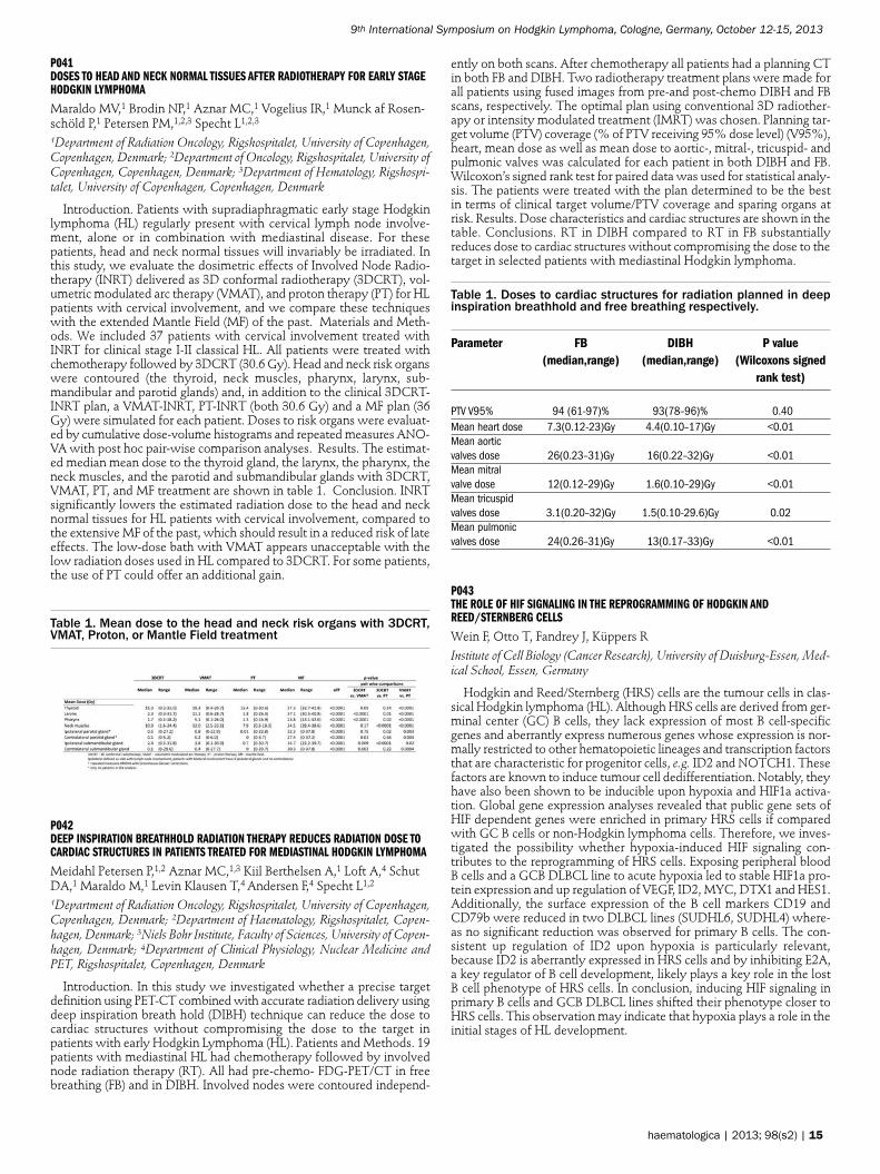
P041DOSES TO HEAD AND NECK NORMAL TISSUES AFTER RADIOTHERAPY FOR EARLY STAGEHODGKIN LYMPHOMA
Maraldo MV,1 Brodin NP,1 Aznar MC,1 Vogelius IR,1 Munck af Rosen-schöld P,1 Petersen PM,1,2,3 Specht L1,2,31Department of Radiation Oncology, Rigshospitalet, University of Copenhagen,Copenhagen, Denmark; 2Department of Oncology, Rigshospitalet, University ofCopenhagen, Copenhagen, Denmark; 3Department of Hematology, Rigshospi-talet, University of Copenhagen, Copenhagen, Denmark
Introduction. Patients with supradiaphragmatic early stage Hodgkinlymphoma (HL) regularly present with cervical lymph node involve-ment, alone or in combination with mediastinal disease. For thesepatients, head and neck normal tissues will invariably be irradiated. Inthis study, we evaluate the dosimetric effects of Involved Node Radio-therapy (INRT) delivered as 3D conformal radiotherapy (3DCRT), vol-umetric modulated arc therapy (VMAT), and proton therapy (PT) for HLpatients with cervical involvement, and we compare these techniqueswith the extended Mantle Field (MF) of the past. Materials and Meth-ods. We included 37 patients with cervical involvement treated withINRT for clinical stage I-II classical HL. All patients were treated withchemotherapy followed by 3DCRT (30.6 Gy). Head and neck risk organswere contoured (the thyroid, neck muscles, pharynx, larynx, sub-mandibular and parotid glands) and, in addition to the clinical 3DCRT-INRT plan, a VMAT-INRT, PT-INRT (both 30.6 Gy) and a MF plan (36Gy) were simulated for each patient. Doses to risk organs were evaluat-ed by cumulative dose-volume histograms and repeated measures ANO-VA with post hoc pair-wise comparison analyses. Results. The estimat-ed median mean dose to the thyroid gland, the larynx, the pharynx, theneck muscles, and the parotid and submandibular glands with 3DCRT,VMAT, PT, and MF treatment are shown in table 1. Conclusion. INRTsignificantly lowers the estimated radiation dose to the head and necknormal tissues for HL patients with cervical involvement, compared tothe extensive MF of the past, which should result in a reduced risk of lateeffects. The low-dose bath with VMAT appears unacceptable with thelow radiation doses used in HL compared to 3DCRT. For some patients,the use of PT could offer an additional gain.
Table 1. Mean dose to the head and neck risk organs with 3DCRT,VMAT, Proton, or Mantle Field treatment
P042DEEP INSPIRATION BREATHHOLD RADIATION THERAPY REDUCES RADIATION DOSE TOCARDIAC STRUCTURES IN PATIENTS TREATED FOR MEDIASTINAL HODGKIN LYMPHOMA
Meidahl Petersen P,1,2 Aznar MC,1,3 Kiil Berthelsen A,1 Loft A,4 SchutDA,1 Maraldo M,1 Levin Klausen T,4 Andersen F,4 Specht L1,21Department of Radiation Oncology, Rigshospitalet, University of Copenhagen,Copenhagen, Denmark; 2Department of Haematology, Rigshospitalet, Copen-hagen, Denmark; 3Niels Bohr Institute, Faculty of Sciences, University of Copen-hagen, Denmark; 4Department of Clinical Physiology, Nuclear Medicine andPET, Rigshospitalet, Copenhagen, Denmark
Introduction. In this study we investigated whether a precise targetdefinition using PET-CT combined with accurate radiation delivery usingdeep inspiration breath hold (DIBH) technique can reduce the dose tocardiac structures without compromising the dose to the target inpatients with early Hodgkin Lymphoma (HL). Patients and Methods. 19patients with mediastinal HL had chemotherapy followed by involvednode radiation therapy (RT). All had pre-chemo- FDG-PET/CT in freebreathing (FB) and in DIBH. Involved nodes were contoured independ-
ently on both scans. After chemotherapy all patients had a planning CTin both FB and DIBH. Two radiotherapy treatment plans were made forall patients using fused images from pre-and post-chemo DIBH and FBscans, respectively. The optimal plan using conventional 3D radiother-apy or intensity modulated treatment (IMRT) was chosen. Planning tar-get volume (PTV) coverage (% of PTV receiving 95% dose level) (V95%),heart, mean dose as well as mean dose to aortic-, mitral-, tricuspid- andpulmonic valves was calculated for each patient in both DIBH and FB.Wilcoxon’s signed rank test for paired data was used for statistical analy-sis. The patients were treated with the plan determined to be the bestin terms of clinical target volume/PTV coverage and sparing organs atrisk. Results. Dose characteristics and cardiac structures are shown in thetable. Conclusions. RT in DIBH compared to RT in FB substantiallyreduces dose to cardiac structures without compromising the dose to thetarget in selected patients with mediastinal Hodgkin lymphoma.
Table 1. Doses to cardiac structures for radiation planned in deepinspiration breathhold and free breathing respectively.
Parameter FB DIBH P value (median,range) (median,range) (Wilcoxons signed
rank test)
PTV V95% 94 (61-97)% 93(78–96)% 0.40Mean heart dose 7.3(0.12-23)Gy 4.4(0.10–17)Gy <0.01Mean aorticvalves dose 26(0.23–31)Gy 16(0.22–32)Gy <0.01Mean mitralvalve dose 12(0.12–29)Gy 1.6(0.10–29)Gy <0.01Mean tricuspidvalves dose 3.1(0.20–32)Gy 1.5(0.10-29.6)Gy 0.02Mean pulmonicvalves dose 24(0.26–31)Gy 13(0.17–33)Gy <0.01
P043THE ROLE OF HIF SIGNALING IN THE REPROGRAMMING OF HODGKIN ANDREED/STERNBERG CELLS
Wein F, Otto T, Fandrey J, Küppers RInstitute of Cell Biology (Cancer Research), University of Duisburg-Essen, Med-ical School, Essen, Germany
Hodgkin and Reed/Sternberg (HRS) cells are the tumour cells in clas-sical Hodgkin lymphoma (HL). Although HRS cells are derived from ger-minal center (GC) B cells, they lack expression of most B cell-specificgenes and aberrantly express numerous genes whose expression is nor-mally restricted to other hematopoietic lineages and transcription factorsthat are characteristic for progenitor cells, e.g. ID2 and NOTCH1. Thesefactors are known to induce tumour cell dedifferentiation. Notably, theyhave also been shown to be inducible upon hypoxia and HIF1a activa-tion. Global gene expression analyses revealed that public gene sets ofHIF dependent genes were enriched in primary HRS cells if comparedwith GC B cells or non-Hodgkin lymphoma cells. Therefore, we inves-tigated the possibility whether hypoxia-induced HIF signaling con-tributes to the reprogramming of HRS cells. Exposing peripheral bloodB cells and a GCB DLBCL line to acute hypoxia led to stable HIF1a pro-tein expression and up regulation of VEGF, ID2, MYC, DTX1 and HES1.Additionally, the surface expression of the B cell markers CD19 andCD79b were reduced in two DLBCL lines (SUDHL6, SUDHL4) where-as no significant reduction was observed for primary B cells. The con-sistent up regulation of ID2 upon hypoxia is particularly relevant,because ID2 is aberrantly expressed in HRS cells and by inhibiting E2A,a key regulator of B cell development, likely plays a key role in the lostB cell phenotype of HRS cells. In conclusion, inducing HIF signaling inprimary B cells and GCB DLBCL lines shifted their phenotype closer toHRS cells. This observation may indicate that hypoxia plays a role in theinitial stages of HL development.
haematologica | 2013; 98(s2) | 15
9th International Symposium on Hodgkin Lymphoma, Cologne, Germany, October 12-15, 2013
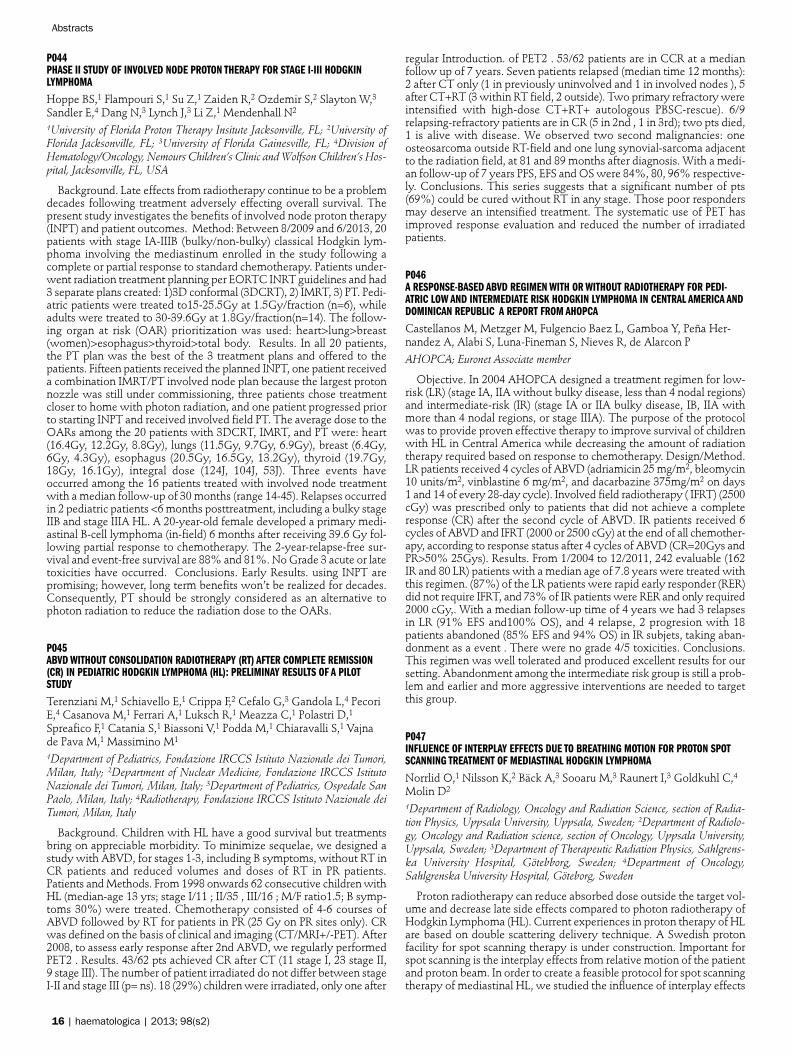
P044PHASE II STUDY OF INVOLVED NODE PROTON THERAPY FOR STAGE I-III HODGKIN LYMPHOMA
Hoppe BS,1 Flampouri S,1 Su Z,1 Zaiden R,2 Ozdemir S,2 Slayton W,3Sandler E,4 Dang N,3 Lynch J,3 Li Z,1 Mendenhall N2
1University of Florida Proton Therapy Insitute Jacksonville, FL; 2University ofFlorida Jacksonville, FL; 3University of Florida Gainesville, FL; 4Division ofHematology/Oncology, Nemours Children’s Clinic and Wolfson Children’s Hos-pital, Jacksonville, FL, USA
Background. Late effects from radiotherapy continue to be a problemdecades following treatment adversely effecting overall survival. Thepresent study investigates the benefits of involved node proton therapy(INPT) and patient outcomes. Method: Between 8/2009 and 6/2013, 20patients with stage IA-IIIB (bulky/non-bulky) classical Hodgkin lym-phoma involving the mediastinum enrolled in the study following acomplete or partial response to standard chemotherapy. Patients under-went radiation treatment planning per EORTC INRT guidelines and had3 separate plans created: 1)3D conformal (3DCRT), 2) IMRT, 3) PT. Pedi-atric patients were treated to15-25.5Gy at 1.5Gy/fraction (n=6), whileadults were treated to 30-39.6Gy at 1.8Gy/fraction(n=14). The follow-ing organ at risk (OAR) prioritization was used: heart>lung>breast(women)>esophagus>thyroid>total body. Results. In all 20 patients,the PT plan was the best of the 3 treatment plans and offered to thepatients. Fifteen patients received the planned INPT, one patient receiveda combination IMRT/PT involved node plan because the largest protonnozzle was still under commissioning, three patients chose treatmentcloser to home with photon radiation, and one patient progressed priorto starting INPT and received involved field PT. The average dose to theOARs among the 20 patients with 3DCRT, IMRT, and PT were: heart(16.4Gy, 12.2Gy, 8.8Gy), lungs (11.5Gy, 9.7Gy, 6.9Gy), breast (6.4Gy,6Gy, 4.3Gy), esophagus (20.5Gy, 16.5Gy, 13.2Gy), thyroid (19.7Gy,18Gy, 16.1Gy), integral dose (124J, 104J, 53J). Three events haveoccurred among the 16 patients treated with involved node treatmentwith a median follow-up of 30 months (range 14-45). Relapses occurredin 2 pediatric patients <6 months posttreatment, including a bulky stageIIB and stage IIIA HL. A 20-year-old female developed a primary medi-astinal B-cell lymphoma (in-field) 6 months after receiving 39.6 Gy fol-lowing partial response to chemotherapy. The 2-year-relapse-free sur-vival and event-free survival are 88% and 81%. No Grade 3 acute or latetoxicities have occurred. Conclusions. Early Results. using INPT arepromising; however, long term benefits won’t be realized for decades.Consequently, PT should be strongly considered as an alternative tophoton radiation to reduce the radiation dose to the OARs.
P045ABVD WITHOUT CONSOLIDATION RADIOTHERAPY (RT) AFTER COMPLETE REMISSION(CR) IN PEDIATRIC HODGKIN LYMPHOMA (HL): PRELIMINAY RESULTS OF A PILOTSTUDY
Terenziani M,1 Schiavello E,1 Crippa F,2 Cefalo G,3 Gandola L,4 PecoriE,4 Casanova M,1 Ferrari A,1 Luksch R,1 Meazza C,1 Polastri D,1Spreafico F,1 Catania S,1 Biassoni V,1 Podda M,1 Chiaravalli S,1 Vajnade Pava M,1 Massimino M1
1Department of Pediatrics, Fondazione IRCCS Istituto Nazionale dei Tumori,Milan, Italy; 2Department of Nuclear Medicine, Fondazione IRCCS IstitutoNazionale dei Tumori, Milan, Italy; 3Department of Pediatrics, Ospedale SanPaolo, Milan, Italy; 4Radiotherapy, Fondazione IRCCS Istituto Nazionale deiTumori, Milan, Italy
Background. Children with HL have a good survival but treatmentsbring on appreciable morbidity. To minimize sequelae, we designed astudy with ABVD, for stages 1-3, including B symptoms, without RT inCR patients and reduced volumes and doses of RT in PR patients.Patients and Methods. From 1998 onwards 62 consecutive children withHL (median-age 13 yrs; stage I/11 ; II/35 , III/16 ; M/F ratio1.5; B symp-toms 30%) were treated. Chemotherapy consisted of 4-6 courses ofABVD followed by RT for patients in PR (25 Gy on PR sites only). CRwas defined on the basis of clinical and imaging (CT/MRI+/-PET). After2008, to assess early response after 2nd ABVD, we regularly performedPET2 . Results. 43/62 pts achieved CR after CT (11 stage I, 23 stage II,9 stage III). The number of patient irradiated do not differ between stageI-II and stage III (p= ns). 18 (29%) children were irradiated, only one after
regular Introduction. of PET2 . 53/62 patients are in CCR at a medianfollow up of 7 years. Seven patients relapsed (median time 12 months):2 after CT only (1 in previously uninvolved and 1 in involved nodes ), 5after CT+RT (3 within RT field, 2 outside). Two primary refractory wereintensified with high-dose CT+RT+ autologous PBSC-rescue). 6/9relapsing-refractory patients are in CR (5 in 2nd , 1 in 3rd); two pts died,1 is alive with disease. We observed two second malignancies: oneosteosarcoma outside RT-field and one lung synovial-sarcoma adjacentto the radiation field, at 81 and 89 months after diagnosis. With a medi-an follow-up of 7 years PFS, EFS and OS were 84%, 80, 96% respective-ly. Conclusions. This series suggests that a significant number of pts(69%) could be cured without RT in any stage. Those poor respondersmay deserve an intensified treatment. The systematic use of PET hasimproved response evaluation and reduced the number of irradiatedpatients.
P046A RESPONSE-BASED ABVD REGIMEN WITH OR WITHOUT RADIOTHERAPY FOR PEDI-ATRIC LOW AND INTERMEDIATE RISK HODGKIN LYMPHOMA IN CENTRAL AMERICA ANDDOMINICAN REPUBLIC A REPORT FROM AHOPCA
Castellanos M, Metzger M, Fulgencio Baez L, Gamboa Y, Peña Her-nandez A, Alabi S, Luna-Fineman S, Nieves R, de Alarcon P
AHOPCA; Euronet Associate member
Objective. In 2004 AHOPCA designed a treatment regimen for low-risk (LR) (stage IA, IIA without bulky disease, less than 4 nodal regions)and intermediate-risk (IR) (stage IA or IIA bulky disease, IB, IIA withmore than 4 nodal regions, or stage IIIA). The purpose of the protocolwas to provide proven effective therapy to improve survival of childrenwith HL in Central America while decreasing the amount of radiationtherapy required based on response to chemotherapy. Design/Method.LR patients received 4 cycles of ABVD (adriamicin 25 mg/m2, bleomycin10 units/m2, vinblastine 6 mg/m2, and dacarbazine 375mg/m2 on days1 and 14 of every 28-day cycle). Involved field radiotherapy ( IFRT) (2500cGy) was prescribed only to patients that did not achieve a completeresponse (CR) after the second cycle of ABVD. IR patients received 6cycles of ABVD and IFRT (2000 or 2500 cGy) at the end of all chemother-apy, according to response status after 4 cycles of ABVD (CR=20Gys andPR>50% 25Gys). Results. From 1/2004 to 12/2011, 242 evaluable (162IR and 80 LR) patients with a median age of 7.8 years were treated withthis regimen. (87%) of the LR patients were rapid early responder (RER)did not require IFRT, and 73% of IR patients were RER and only required2000 cGy,. With a median follow-up time of 4 years we had 3 relapsesin LR (91% EFS and100% OS), and 4 relapse, 2 progresion with 18patients abandoned (85% EFS and 94% OS) in IR subjets, taking aban-donment as a event . There were no grade 4/5 toxicities. Conclusions.This regimen was well tolerated and produced excellent results for oursetting. Abandonment among the intermediate risk group is still a prob-lem and earlier and more aggressive interventions are needed to targetthis group.
P047INFLUENCE OF INTERPLAY EFFECTS DUE TO BREATHING MOTION FOR PROTON SPOTSCANNING TREATMENT OF MEDIASTINAL HODGKIN LYMPHOMA
Norrlid O,1 Nilsson K,2 Bäck A,3 Sooaru M,3 Raunert I,3 Goldkuhl C,4Molin D2
1Department of Radiology, Oncology and Radiation Science, section of Radia-tion Physics, Uppsala University, Uppsala, Sweden; 2Department of Radiolo-gy, Oncology and Radiation science, section of Oncology, Uppsala University,Uppsala, Sweden; 3Department of Therapeutic Radiation Physics, Sahlgrens-ka University Hospital, Götebborg, Sweden; 4Department of Oncology,Sahlgrenska University Hospital, Göteborg, Sweden
Proton radiotherapy can reduce absorbed dose outside the target vol-ume and decrease late side effects compared to photon radiotherapy ofHodgkin Lymphoma (HL). Current experiences in proton therapy of HLare based on double scattering delivery technique. A Swedish protonfacility for spot scanning therapy is under construction. Important forspot scanning is the interplay effects from relative motion of the patientand proton beam. In order to create a feasible protocol for spot scanningtherapy of mediastinal HL, we studied the influence of interplay effects
16 | haematologica | 2013; 98(s2)
Abstracts
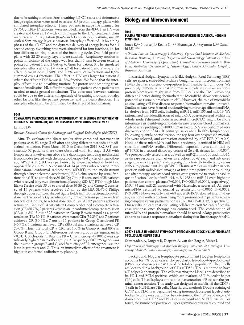
due to breathing motions. Free breathing 4D-CT scans and deformableimage registration were used to assess 3D proton therapy plans withsimulated interplay effects. Three patients in stage IIA planned for29.75Gy(RBE) (17 fractions) were included. From the 4D-CT an ITV wascreated and then a PTV with 7mm margin to the ITV. Treatment planswere created in RayStation (RaySearch Laboratories) planning systemwith 0.5cm energy layer separation. Interplay effects of 10 breathingphases of the 4D-CT and the dynamic delivery of energy layers for a 1second energy switching time were simulated for four fractions, i.e. forfour different starting phases of the breathing cycle. One field protonplans with 22-25 energy layers were studied. Respiratory motion inpoints in vicinity of the target was less than 5 mm between extremepoints for patient 1 and 2 but up to 8mm for patient 3. The simulatedinterplay effects in the ITV were small for patient 1 and 2 where theeffect in mean dose, D98% and D2% were <1%/fraction and 0,5%summed over 4 fractions. The effect in ITV was larger for patient 3where the effect in D98% was 8-13%/fraction. We found that the inter-play effects due to breathing motions for proton spot scanning treat-ment of mediastinal HL differ from patient to patient. More patients areneeded to make general conclusions. The difference between patientscould be due to the different magnitude of breathing motions, but alsoother factors, like the patient geometry, and the beam direction. Theinterplay effects will be diminished by the effect of fractionation.
P048COMPARATIVE CHARACTERISTICS OF RADIOTHERAPY (RT) METHODS IN TREATMENT OFHODGKIN’S LYMPHOMA (HL) WITH MEDIASTINAL LYMPH NODES INVOLVEMENT
Larinov DVRussian Research Center for Radiology and Surgical Technologies (RRCRST)
Aim. To evaluate the direct results after combined treatment inpatients with HL stage II AB after applying different methods of medi-astinal irradiation. From March 2010 to December 2012 RRCRST con-sistently 52 patients there were observed with HL stage II AB withlymph mass involvement above the diaphragm, including mediastinallymph nodes treated with chemoradiotherapy (2-4 cycles of chemother-apy ABVD + RT). RT was performed by impact irradiation from twoopposed fields. Group A consisted of 14 patients who received three-dimensional conformal radiotherapy (3D-RT) to the mediastinumthrough a linear electron accelerator (LEA) Elekta Axesse by usual frac-tionation (UF) to a total dose 30-36 Gy; Group B consisted of 23 patientswho received it by two-dimensional planning (2D-RT) RT through LEAElekta Precise with UF up to a total dose 30-36 Gy and Group C consist-ed of 15 patients who received 2D-RT by the LEA SL-75-5 Philipsthrough upper complex shaped figure fields in multi-fractionation (MF),dose per fraction-1.2 Gy, irradiation rhythm-2 fractions per day with aninterval of 4 hours, to a total dose 30-36 Gy. All 52 patients achievedremission. 12 out of 14 patients in Group A obtained a complete remis-sion (CR) 85.7%, 2 patients were in an unconfirmed complete remission(CRu)-14.3%; 7 out of 23 patients in Group B were stated as a partialremission (PR)-30.4%, 9 patients were stated CRu (39.2%) and 7 patientsachieved CR (30.4%); 7 out of 15 patients in Group C achieved PR(46.7%), 5 patients achieved CRu (33.3%) and 2 patients achieved CR20.0%. Thus, the total CR + CRu set 100% in Group A, and 80% inGroup B and Group C. Differences between groups are significant (p<0,01). Conclusions. 1. Rate the PR + CRu in Group A (100%) was sig-nificantly higher than in other groups. 2. Frequency of RP emergence wasthe lowest in groups B and C, and frequency of RE emergence was theleast in groups A and C. Thus, an immediate effect of the treatment ishigher in conformal radiotherapy planning.
Biology and Microenvironment
T049PLASMA MICRORNA ARE DISEASE RESPONSE BIOMARKERS IN CLASSICAL HODGKINLYMPHOMA
Jones K,1,2 Nourse JP,1 Keane C,1,2,3 Bhatnagar A,1 Seymour L,2,3 Gand-hi MK1,2,31Clinical Immunohaematology Laboratory, Queensland Institute of MedicalResearch, Brisbane, Australia; 2Experimental Haematology Laboratory, Schoolof Medicine, University of Queensland, Translational Research Institute, Bris-bane, Australia; 3Department of Haematology, Princess Alexandra Hospital,Brisbane, Australia
In classical Hodgkin lymphoma (cHL), Hodgkin-Reed-Sternberg (HRS)cells are sparse, embedded within a benign tumour microenvironment(TME) that has a distinct composition from healthy lymph nodes. Wepreviously demonstrated that informative circulating disease responseprotein biomarkers might arise from HRS cells or the TME, exhibitingdifferent kinetics during chemotherapy. MicroRNA show considerablepotential as tissue biomarkers in cHL. However, the role of microRNAas circulating cell-free disease response biomarkers remains untested.Studies to date have focused on identifying tumour-specific microRNA,i.e. derived from HRS cells, including miR-21, miR-155 and miR-16. Werationalized that identification of microRNA over-expressed within thewhole node (‘diseased node associated microRNA’) might be moreinformative in identifying candidate disease response blood biomarkers.Agilent array profiling of >1000 human microRNA was performed on adiscovery cohort of 14 cHL primary tissues and 8 healthy lymph nodes.Following quantile normalization, the top four over-expressed microR-NA were selected, and expression correlated by qRT-PCR (all r>0.6).None of these microRNA had been previously identified in HRS-cellspecific microRNA studies. Differential expression was confirmed byqRT-PCR in a second discovery cohort of 26 cHL tissues (all P<0.004).We prospectively tested these, as well as miR-21, miR-155 and miR-16,as disease response biomarkers in a cohort of 42 early and advancedstage disease cHL patients undergoing induction chemotherapy, versus20 healthy participants by qRT-PCR. Plasma samples were taken in con-junction with radiological imaging at fixed time-points prior to, duringand after therapy, and standard curves were generated to enable absolutequantification. Levels of miR-494, miR-1973 and miR-21 were higher inpatients than control plasma (P=0.004, P=0.008, P<0.0001, respectively).MiR-494 and miR-21 associated with Hasenclever scores ≥3. All threemicroRNA returned to normal at remission (P=0.0006, P=0.0002,P<0.0001). However, only miR-494 and miR-1973 reflected interim ther-apy response with reduction being more pronounced in patients achiev-ing complete versus partial responses (P=0.043, P=0.0012, respectively).Our results indicate that circulating cell-free microRNA can reflect dis-ease response once therapy has commenced. The combination ofmicroRNA and protein biomarkers should be tested in large prospectivecohorts as disease response biomarkers during first-line therapy for cHL.
T050CD57+ T CELLS IN NODULAR LYMPHOCYTE PREDOMINANT HODGKIN’S LYMPHOMA ARET-FOLLICULAR HELPER CELLS
Sattarzadeh A, Rutgers B, Diepstra A, van den Berg A, Visser LDepartment of Pathology and Medical Biology, University of Groningen, Uni-versity Medical Center Groningen, Groningen, the Netherlands
Background. Nodular lymphocyte predominant Hodgkin lymphomaaccounts for 5% of all cases. The neoplastic lymphocyte-predominant(LP) cells, compose less than 1% of the total cell population. The LP cellsare localized in a background. of CD4+CD57+ T cells reported to havea T helper 2 phenotype. The cells rosetting the LP cells are described tobe PD-1 and BCL6 positive, which are markers of T follicular helper(Tfh) cells. Tfh cells play a critical role in maturation of B-cells in the ger-minal center reaction. This study was designed to establish if the CD57+T cells in NLPHL are Tfh cells. Material and methods Double staining ofCD57 and PD-1 was performed using immunofluorescent labeled anti-bodies. Scoring was performed by determining the number of single anddouble positive CD57 and PD-1 cells in tonsil and NLPHL tissues. Fortonsil, the number of positive cells per germinal center were counted and
haematologica | 2013; 98(s2) | 17
9th International Symposium on Hodgkin Lymphoma, Cologne, Germany, October 12-15, 2013
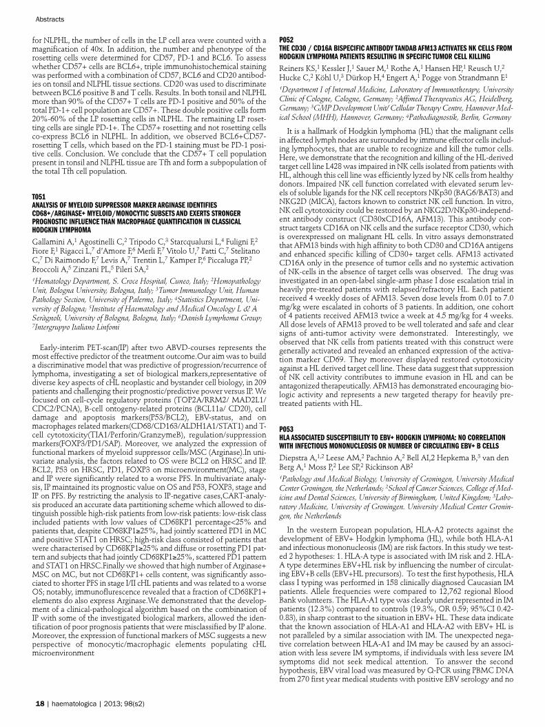
for NLPHL, the number of cells in the LP cell area were counted with amagnification of 40x. In addition, the number and phenotype of therosetting cells were determined for CD57, PD-1 and BCL6. To assesswhether CD57+ cells are BCL6+, triple immunohistochemical stainingwas performed with a combination of CD57, BCL6 and CD20 antibod-ies on tonsil and NLPHL tissue sections. CD20 was used to discriminatebetween BCL6 positive B and T cells. Results. In both tonsil and NLPHLmore than 90% of the CD57+ T cells are PD-1 positive and 50% of thetotal PD-1+ cell population are CD57+. These double positive cells form20%-60% of the LP rosetting cells in NLPHL. The remaining LP roset-ting cells are single PD-1+. The CD57+ rosetting and not rosetting cellsco-express BCL6 in NLPHL. In addition, we observed BCL6+CD57-rosetting T cells, which based on the PD-1 staining must be PD-1 posi-tive cells. Conclusion. We conclude that the CD57+ T cell populationpresent in tonsil and NLPHL tissue are Tfh and form a subpopulation ofthe total Tfh cell population.
T051ANALYSIS OF MYELOID SUPPRESSOR MARKER ARGINASE IDENTIFIESCD68+/ARGINASE+ MYELOID/MONOCYTIC SUBSETS AND EXERTS STRONGER PROGNOSTIC INFLUENCE THAN MACROPHAGE QUANTIFICATION IN CLASSICALHODGKIN LYMPHOMA
Gallamini A,1 Agostinelli C,2 Tripodo C,3 Starcqualursi L,4 Fuligni F,2Fiore F,1 Rigacci L,7 d’Amore F,6 Merli F,7 Vitolo U,7 Patti C,7 StelitanoC,7 Di Raimondo F,7 Levis A,7 Trentin L,7 Kamper P,6 Piccaluga PP,2Broccoli A,5 Zinzani PL,5 Pileri SA,2
1Hematology Department, S. Croce Hospital, Cuneo, Italy; 2HemopathologyUnit, Bologna University, Bologna, Italy; 3Tumor Immunology Unit, HumanPathology Section, University of Palermo, Italy; 4Statistics Department, Uni-versity of Bologna; 5Institute of Haematology and Medical Oncology L & ASeràgnoli, University of Bologna, Bologna, Italy; 6Danish Lymphoma Group;7Intergruppo Italiano Linfomi
Early-interim PET-scan(IP) after two ABVD-courses represents themost effective predictor of the treatment outcome.Our aim was to builda discriminative model that was predictive of progression/recurrence oflymphoma, investigating a set of biological markers,representative ofdiverse key aspects of cHL neoplastic and bystander cell biology, in 209patients and challenging their prognostic/predictive power versus IP. Wefocused on cell-cycle regulatory proteins (TOP2A/RRM2/ MAD2L1/CDC2/PCNA), B-cell ontogeny-related proteins (BCL11a/ CD20), celldamage and apoptosis markers(P53/BCL2), EBV-status, and onmacrophages related markers(CD68/CD163/ALDH1A1/STAT1) and T-cell cytotoxicity(TIA1/Perforin/GranzymeB), regulation/suppressionmarkers(FOXP3/PD1/SAP). Moreover, we analyzed the expression offunctional markers of myeloid suppressor cells/MSC (Arginase).In uni-variate analysis, the factors related to OS were BCL2 on HRSC and IP.BCL2, P53 on HRSC, PD1, FOXP3 on microenvironment(MC), stageand IP were significantly related to a worse PFS. In multivariate analy-sis, IP maintained its prognostic value on OS and P53, FOXP3, stage andIP on PFS. By restricting the analysis to IP-negative cases,CART-analy-sis produced an accurate data partitioning scheme which allowed to dis-tinguish possible high-risk patients from low-risk patients: low-risk classincluded patients with low values of CD68KP1 percentage<25% andpatients that, despite CD68KP1≥25%, had jointly scattered PD1 in MCand positive STAT1 on HRSC; high-risk class consisted of patients thatwere characterised by CD68KP1≥25% and diffuse or rosetting PD1 pat-tern and subjects that had jointly CD68KP1≥25%, scattered PD1 patternand STAT1 on HRSC.Finally we showed that high number of Arginase+MSC on MC, but not CD68KP1+ cells content, was significantly asso-ciated to shorter PFS in stage I/II cHL patients and was related to a worseOS; notably, immunoflurescence revealed that a fraction of CD68KP1+elements do also express Arginase.We demonstrated that the develop-ment of a clinical-pathological algorithm based on the combination ofIP with some of the investigated biological markers, allowed the iden-tification of poor prognosis patients that were misclassified by IP alone.Moreover, the expression of functional markers of MSC suggests a newperspective of monocytic/macrophagic elements populating cHLmicroenvironment
P052THE CD30 / CD16A BISPECIFIC ANTIBODY TANDAB AFM13 ACTIVATES NK CELLS FROMHODGKIN LYMPHOMA PATIENTS RESULTING IN SPECIFIC TUMOR CELL KILLING
Reiners KS,1 Kessler J,1 Sauer M,1 Rothe A,1 Hansen HP,1 Reusch U,2Hucke C,2 Köhl U,3 Dürkop H,4 Engert A,1 Pogge von Strandmann E1
1Department I of Internal Medicine, Laboratory of Immunotherapy, UniversityClinic of Cologne, Cologne, Germany; 2Affimed Therapeutics AG, Heidelberg,Germany; 3GMP Development Unit/ Cellular Therapy Centre, Hannover Med-ical School (MHH), Hannover, Germany; 4Pathodiagnostik, Berlin, Germany
It is a hallmark of Hodgkin lymphoma (HL) that the malignant cellsin affected lymph nodes are surrounded by immune effector cells includ-ing lymphocytes, that are unable to recognize and kill the tumor cells.Here, we demonstrate that the recognition and killing of the HL-derivedtarget cell line L428 was impaired in NK cells isolated from patients withHL, although this cell line was efficiently lyzed by NK cells from healthydonors. Impaired NK cell function correlated with elevated serum lev-els of soluble ligands for the NK cell receptors NKp30 (BAG6/BAT3) andNKG2D (MICA), factors known to constrict NK cell function. In vitro,NK cell cytotoxicity could be restored by an NKG2D/NKp30-independ-ent antibody construct (CD30xCD16A, AFM13). This antibody con-struct targets CD16A on NK cells and the surface receptor CD30, whichis overexpressed on malignant HL cells. In vitro assays demonstratedthat AFM13 binds with high affinity to both CD30 and CD16A antigensand enhanced specific killing of CD30+ target cells. AFM13 activatedCD16A only in the presence of tumor cells and no systemic activationof NK-cells in the absence of target cells was observed. The drug wasinvestigated in an open-label single-arm phase I dose escalation trial inheavily pre-treated patients with relapsed/refractory HL. Each patientreceived 4 weekly doses of AFM13. Seven dose levels from 0.01 to 7.0mg/kg were escalated in cohorts of 3 patients. In addition, one cohortof 4 patients received AFM13 twice a week at 4.5 mg/kg for 4 weeks.All dose levels of AFM13 proved to be well tolerated and safe and clearsigns of anti-tumor activity were demonstrated. Interestingly, weobserved that NK cells from patients treated with this construct weregenerally activated and revealed an enhanced expression of the activa-tion marker CD69. They moreover displayed restored cytotoxicityagainst a HL derived target cell line. These data suggest that suppressionof NK cell activity contributes to immune evasion in HL and can beantagonized therapeutically. AFM13 has demonstrated encouraging bio-logic activity and represents a new targeted therapy for heavily pre-treated patients with HL.
P053HLA ASSOCIATED SUSCEPTIBILITY TO EBV+ HODGKIN LYMPHOMA: NO CORRELATIONWITH INFECTIOUS MONONUCLEOSIS OR NUMBER OF CIRCULATING EBV+ B CELLS
Diepstra A,1,2 Leese AM,2 Pachnio A,2 Bell AI,2 Hepkema B,3 van denBerg A,1 Moss P,2 Lee SP,2 Rickinson AB2
1Pathology and Medical Biology, University of Groningen, University MedicalCenter Groningen, the Netherlands; 2School of Cancer Sciences, College of Med-icine and Dental Sciences, University of Birmingham, United Kingdom; 3Labo-ratory Medicine, University of Groningen. University Medical Center Gronin-gen, the Netherlands
In the western European population, HLA-A2 protects against thedevelopment of EBV+ Hodgkin lymphoma (HL), while both HLA-A1and infectious mononucleosis (IM) are risk factors. In this study we test-ed 2 hypotheses: 1. HLA-A type is associated with IM risk and 2. HLA-A type determines EBV+HL risk by influencing the number of circulat-ing EBV+B cells (EBV+HL precursors). To test the first hypothesis, HLAclass I typing was performed in 158 clinically diagnosed Caucasian IMpatients. Allele frequencies were compared to 12,762 regional BloodBank volunteers. The HLA-A1 type was clearly under represented in IMpatients (12.3%) compared to controls (19.3%, OR 0.59; 95%CI 0.42-0.83), in sharp contrast to the situation in EBV+ HL. These data indicatethat the known association of HLA-A1 and HLA-A2 with EBV+ HL isnot paralleled by a similar association with IM. The unexpected nega-tive correlation between HLA-A1 and IM may be caused by an associ-ation with less severe IM symptoms, if individuals with less severe IMsymptoms did not seek medical attention. To answer the secondhypothesis, EBV viral load was measured by Q-PCR using PBMC DNAfrom 270 first year medical students with positive EBV serology and no
18 | haematologica | 2013; 98(s2)
Abstracts
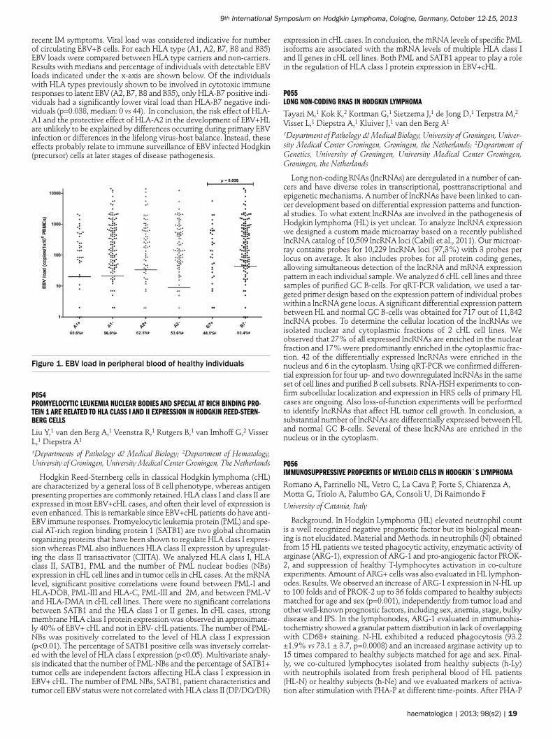
recent IM symptoms. Viral load was considered indicative for numberof circulating EBV+B cells. For each HLA type (A1, A2, B7, B8 and B35)EBV loads were compared between HLA type carriers and non-carriers.Results with medians and percentage of individuals with detectable EBVloads indicated under the x-axis are shown below. Of the individualswith HLA types previously shown to be involved in cytotoxic immuneresponses to latent EBV (A2, B7, B8 and B35), only HLA-B7 positive indi-viduals had a significantly lower viral load than HLA-B7 negative indi-viduals (p=0.038, median: 0 vs 44). In conclusion, the risk effect of HLA-A1 and the protective effect of HLA-A2 in the development of EBV+HLare unlikely to be explained by differences occurring during primary EBVinfection or differences in the lifelong virus-host balance. Instead, theseeffects probably relate to immune surveillance of EBV infected Hodgkin(precursor) cells at later stages of disease pathogenesis.
Figure 1. EBV load in peripheral blood of healthy individuals
P054PROMYELOCYTIC LEUKEMIA NUCLEAR BODIES AND SPECIAL AT RICH BINDING PRO-TEIN 1 ARE RELATED TO HLA CLASS I AND II EXPRESSION IN HODGKIN REED-STERN-BERG CELLS
Liu Y,1 van den Berg A,1 Veenstra R,1 Rutgers B,1 van Imhoff G,2 VisserL,1 Diepstra A1
1Departments of Pathology & Medical Biology; 2Department of Hematology,University of Groningen, University Medical Center Groningen, The Netherlands
Hodgkin Reed-Sternberg cells in classical Hodgkin lymphoma (cHL)are characterized by a general loss of B cell phenotype, whereas antigenpresenting properties are commonly retained. HLA class I and class II areexpressed in most EBV+cHL cases, and often their level of expression iseven enhanced. This is remarkable since EBV+cHL patients do have anti-EBV immune responses. Promyelocytic leukemia protein (PML) and spe-cial AT-rich region binding protein 1 (SATB1) are two global chromatinorganizing proteins that have been shown to regulate HLA class I expres-sion whereas PML also influences HLA class II expression by upregulat-ing the class II transactivator (CIITA). We analyzed HLA class I, HLAclass II, SATB1, PML and the number of PML nuclear bodies (NBs)expression in cHL cell lines and in tumor cells in cHL cases. At the mRNAlevel, significant positive correlations were found between PML-I andHLA-DOB, PML-III and HLA-C, PML-III and 2M, and between PML-Vand HLA-DMA in cHL cell lines. There were no significant correlationsbetween SATB1 and the HLA class I or II genes. In cHL cases, strongmembrane HLA class I protein expression was observed in approximate-ly 40% of EBV+ cHL and not in EBV- cHL patients. The number of PML-NBs was positively correlated to the level of HLA class I expression(p<0.01). The percentage of SATB1 positive cells was inversely correlat-ed with the level of HLA class I expression (p<0.05). Multivariate analy-sis indicated that the number of PML-NBs and the percentage of SATB1+tumor cells are independent factors affecting HLA class I expression inEBV+ cHL. The number of PML NBs, SATB1, patient characteristics andtumor cell EBV status were not correlated with HLA class II (DP/DQ/DR)
expression in cHL cases. In conclusion, the mRNA levels of specific PMLisoforms are associated with the mRNA levels of multiple HLA class Iand II genes in cHL cell lines. Both PML and SATB1 appear to play a rolein the regulation of HLA class I protein expression in EBV+cHL.
P055LONG NON-CODING RNAS IN HODGKIN LYMPHOMA
Tayari M,1 Kok K,2 Kortman G,1 Sietzema J,1 de Jong D,1 Terpstra M,2Visser L,1 Diepstra A,1 Kluiver J,1 van den Berg A1
1Department of Pathology & Medical Biology, University of Groningen, Univer-sity Medical Center Groningen, Groningen, the Netherlands; 2Department ofGenetics, University of Groningen, University Medical Center Groningen,Groningen, the Netherlands
Long non-coding RNAs (lncRNAs) are deregulated in a number of can-cers and have diverse roles in transcriptional, posttranscriptional andepigenetic mechanisms. A number of lncRNAs have been linked to can-cer development based on differential expression patterns and function-al studies. To what extent lncRNAs are involved in the pathogenesis ofHodgkin lymphoma (HL) is yet unclear. To analyze lncRNA expressionwe designed a custom made microarray based on a recently publishedlncRNA catalog of 10,509 lncRNA loci (Cabili et al., 2011). Our microar-ray contains probes for 10,229 lncRNA loci (97,3%) with 3 probes perlocus on average. It also includes probes for all protein coding genes,allowing simultaneous detection of the lncRNA and mRNA expressionpattern in each individual sample. We analyzed 6 cHL cell lines and threesamples of purified GC B-cells. For qRT-PCR validation, we used a tar-geted primer design based on the expression pattern of individual probeswithin a lncRNA gene locus. A significant differential expression patternbetween HL and normal GC B-cells was obtained for 717 out of 11,842lncRNA probes. To determine the cellular location of the lncRNAs weisolated nuclear and cytoplasmic fractions of 2 cHL cell lines. Weobserved that 27% of all expressed lncRNAs are enriched in the nuclearfraction and 17% were predominantly enriched in the cytoplasmic frac-tion. 42 of the differentially expressed lncRNAs were enriched in thenucleus and 6 in the cytoplasm. Using qRT-PCR we confirmed differen-tial expression for four up- and two downregulated lncRNAs in the sameset of cell lines and purified B cell subsets. RNA-FISH experiments to con-firm subcellular localization and expression in HRS cells of primary HLcases are ongoing. Also loss-of-function experiments will be performedto identify lncRNAs that affect HL tumor cell growth. In conclusion, asubstantial number of lncRNAs are differentially expressed between HLand normal GC B-cells. Several of these lncRNAs are enriched in thenucleus or in the cytoplasm.
P056IMMUNOSUPPRESSIVE PROPERTIES OF MYELOID CELLS IN HODGKIN`S LYMPHOMA
Romano A, Parrinello NL, Vetro C, La Cava P, Forte S, Chiarenza A,Motta G, Triolo A, Palumbo GA, Consoli U, Di Raimondo F
University of Catania, Italy
Background. In Hodgkin Lymphoma (HL) elevated neutrophil countis a well recognized negative prognostic factor but its biological mean-ing is not elucidated. Material and Methods. in neutrophils (N) obtainedfrom 15 HL patients we tested phagocytic activity, enzymatic activity ofarginase (ARG-1), expression of ARG-1 and pro-angiogenic factor PROK-2, and suppression of healthy T-lymphocytes activation in co-cultureexperiments. Amount of ARG+ cells was also evaluated in HL lymphon-odes. Results. We observed an increase of ARG-1 expression in N-HL upto 100 folds and of PROK-2 up to 36 folds compared to healthy subjectsmatched for age and sex (p=0.001), independently from tumor load andother well-known prognostic factors, including sex, anemia, stage, bulkydisease and IPS. In the lymphonodes, ARG-1 evaluated in immunohis-tochemistry showed a granular pattern distribution in lack of overlappingwith CD68+ staining. N-HL exhibited a reduced phagocytosis (93.2±1.9% vs 73.1 ± 3.7, p=0.0008) and an increased arginase activity up to15 times compared to healthy subjects matched for age and sex. Final-ly, we co-cultured lymphocytes isolated from healthy subjects (h-Ly)with neutrophils isolated from fresh peripheral blood of HL patients(HL-N) or healthy subjects (h-Ne) and we evaluated markers of activa-tion after stimulation with PHA-P at different time-points. After PHA-P
haematologica | 2013; 98(s2) | 19
9th International Symposium on Hodgkin Lymphoma, Cologne, Germany, October 12-15, 2013
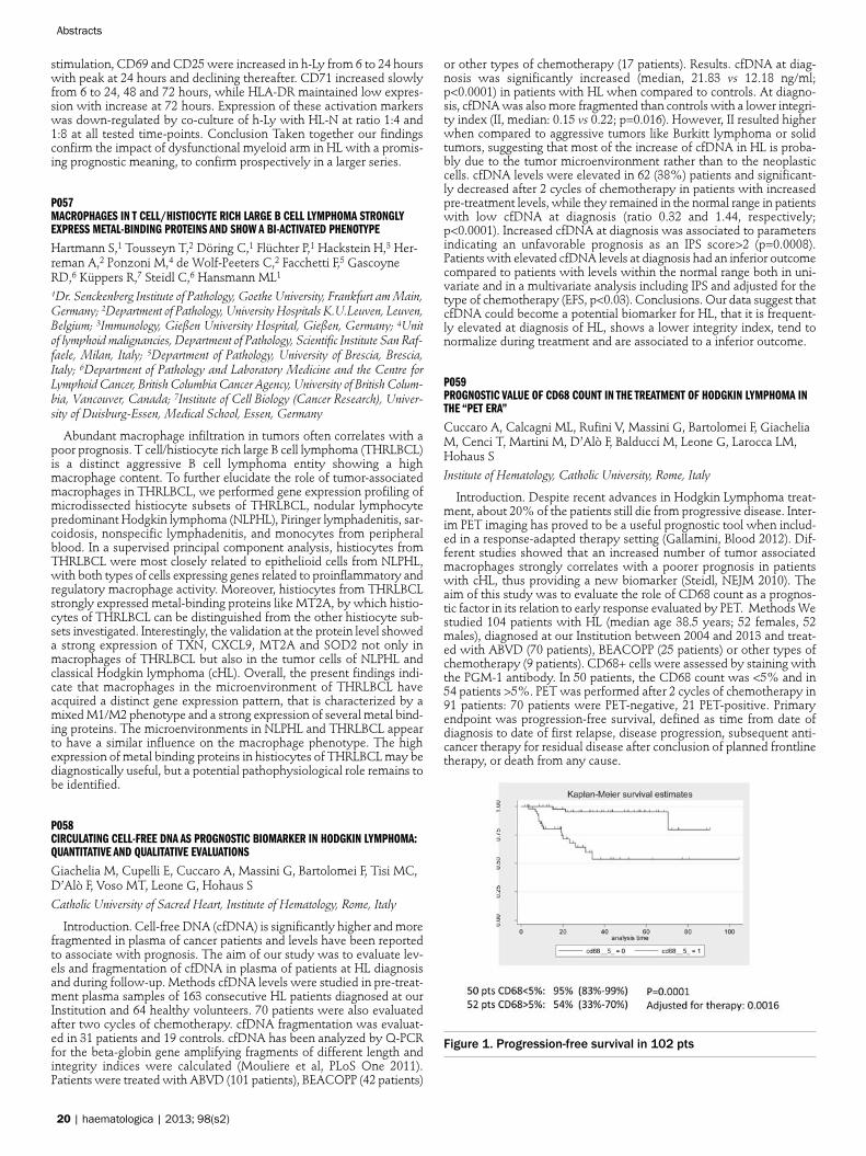
stimulation, CD69 and CD25 were increased in h-Ly from 6 to 24 hourswith peak at 24 hours and declining thereafter. CD71 increased slowlyfrom 6 to 24, 48 and 72 hours, while HLA-DR maintained low expres-sion with increase at 72 hours. Expression of these activation markerswas down-regulated by co-culture of h-Ly with HL-N at ratio 1:4 and1:8 at all tested time-points. Conclusion Taken together our findingsconfirm the impact of dysfunctional myeloid arm in HL with a promis-ing prognostic meaning, to confirm prospectively in a larger series.
P057MACROPHAGES IN T CELL/HISTIOCYTE RICH LARGE B CELL LYMPHOMA STRONGLYEXPRESS METAL-BINDING PROTEINS AND SHOW A BI-ACTIVATED PHENOTYPE
Hartmann S,1 Tousseyn T,2 Döring C,1 Flüchter P,1 Hackstein H,3 Her-reman A,2 Ponzoni M,4 de Wolf-Peeters C,2 Facchetti F,5 GascoyneRD,6 Küppers R,7 Steidl C,6 Hansmann ML1
1Dr. Senckenberg Institute of Pathology, Goethe University, Frankfurt am Main,Germany; 2Department of Pathology, University Hospitals K.U.Leuven, Leuven,Belgium; 3Immunology, Gießen University Hospital, Gießen, Germany; 4Unitof lymphoid malignancies, Department of Pathology, Scientific Institute San Raf-faele, Milan, Italy; 5Department of Pathology, University of Brescia, Brescia,Italy; 6Department of Pathology and Laboratory Medicine and the Centre forLymphoid Cancer, British Columbia Cancer Agency, University of British Colum-bia, Vancouver, Canada; 7Institute of Cell Biology (Cancer Research), Univer-sity of Duisburg-Essen, Medical School, Essen, Germany
Abundant macrophage infiltration in tumors often correlates with apoor prognosis. T cell/histiocyte rich large B cell lymphoma (THRLBCL)is a distinct aggressive B cell lymphoma entity showing a highmacrophage content. To further elucidate the role of tumor-associatedmacrophages in THRLBCL, we performed gene expression profiling ofmicrodissected histiocyte subsets of THRLBCL, nodular lymphocytepredominant Hodgkin lymphoma (NLPHL), Piringer lymphadenitis, sar-coidosis, nonspecific lymphadenitis, and monocytes from peripheralblood. In a supervised principal component analysis, histiocytes fromTHRLBCL were most closely related to epithelioid cells from NLPHL,with both types of cells expressing genes related to proinflammatory andregulatory macrophage activity. Moreover, histiocytes from THRLBCLstrongly expressed metal-binding proteins like MT2A, by which histio-cytes of THRLBCL can be distinguished from the other histiocyte sub-sets investigated. Interestingly, the validation at the protein level showeda strong expression of TXN, CXCL9, MT2A and SOD2 not only inmacrophages of THRLBCL but also in the tumor cells of NLPHL andclassical Hodgkin lymphoma (cHL). Overall, the present findings indi-cate that macrophages in the microenvironment of THRLBCL haveacquired a distinct gene expression pattern, that is characterized by amixed M1/M2 phenotype and a strong expression of several metal bind-ing proteins. The microenvironments in NLPHL and THRLBCL appearto have a similar influence on the macrophage phenotype. The highexpression of metal binding proteins in histiocytes of THRLBCL may bediagnostically useful, but a potential pathophysiological role remains tobe identified.
P058CIRCULATING CELL-FREE DNA AS PROGNOSTIC BIOMARKER IN HODGKIN LYMPHOMA:QUANTITATIVE AND QUALITATIVE EVALUATIONS
Giachelia M, Cupelli E, Cuccaro A, Massini G, Bartolomei F, Tisi MC,D’Alò F, Voso MT, Leone G, Hohaus S
Catholic University of Sacred Heart, Institute of Hematology, Rome, Italy
Introduction. Cell-free DNA (cfDNA) is significantly higher and morefragmented in plasma of cancer patients and levels have been reportedto associate with prognosis. The aim of our study was to evaluate lev-els and fragmentation of cfDNA in plasma of patients at HL diagnosisand during follow-up. Methods cfDNA levels were studied in pre-treat-ment plasma samples of 163 consecutive HL patients diagnosed at ourInstitution and 64 healthy volunteers. 70 patients were also evaluatedafter two cycles of chemotherapy. cfDNA fragmentation was evaluat-ed in 31 patients and 19 controls. cfDNA has been analyzed by Q-PCRfor the beta-globin gene amplifying fragments of different length andintegrity indices were calculated (Mouliere et al, PLoS One 2011).Patients were treated with ABVD (101 patients), BEACOPP (42 patients)
or other types of chemotherapy (17 patients). Results. cfDNA at diag-nosis was significantly increased (median, 21.83 vs 12.18 ng/ml;p<0.0001) in patients with HL when compared to controls. At diagno-sis, cfDNA was also more fragmented than controls with a lower integri-ty index (II, median: 0.15 vs 0.22; p=0.016). However, II resulted higherwhen compared to aggressive tumors like Burkitt lymphoma or solidtumors, suggesting that most of the increase of cfDNA in HL is proba-bly due to the tumor microenvironment rather than to the neoplasticcells. cfDNA levels were elevated in 62 (38%) patients and significant-ly decreased after 2 cycles of chemotherapy in patients with increasedpre-treatment levels, while they remained in the normal range in patientswith low cfDNA at diagnosis (ratio 0.32 and 1.44, respectively;p<0.0001). Increased cfDNA at diagnosis was associated to parametersindicating an unfavorable prognosis as an IPS score>2 (p=0.0008).Patients with elevated cfDNA levels at diagnosis had an inferior outcomecompared to patients with levels within the normal range both in uni-variate and in a multivariate analysis including IPS and adjusted for thetype of chemotherapy (EFS, p<0.03). Conclusions. Our data suggest thatcfDNA could become a potential biomarker for HL, that it is frequent-ly elevated at diagnosis of HL, shows a lower integrity index, tend tonormalize during treatment and are associated to a inferior outcome.
P059PROGNOSTIC VALUE OF CD68 COUNT IN THE TREATMENT OF HODGKIN LYMPHOMA INTHE “PET ERA”
Cuccaro A, Calcagni ML, Rufini V, Massini G, Bartolomei F, GiacheliaM, Cenci T, Martini M, D’Alò F, Balducci M, Leone G, Larocca LM,Hohaus SInstitute of Hematology, Catholic University, Rome, Italy
Introduction. Despite recent advances in Hodgkin Lymphoma treat-ment, about 20% of the patients still die from progressive disease. Inter-im PET imaging has proved to be a useful prognostic tool when includ-ed in a response-adapted therapy setting (Gallamini, Blood 2012). Dif-ferent studies showed that an increased number of tumor associatedmacrophages strongly correlates with a poorer prognosis in patientswith cHL, thus providing a new biomarker (Steidl, NEJM 2010). Theaim of this study was to evaluate the role of CD68 count as a prognos-tic factor in its relation to early response evaluated by PET. Methods Westudied 104 patients with HL (median age 38.5 years; 52 females, 52males), diagnosed at our Institution between 2004 and 2013 and treat-ed with ABVD (70 patients), BEACOPP (25 patients) or other types ofchemotherapy (9 patients). CD68+ cells were assessed by staining withthe PGM-1 antibody. In 50 patients, the CD68 count was <5% and in54 patients >5%. PET was performed after 2 cycles of chemotherapy in91 patients: 70 patients were PET-negative, 21 PET-positive. Primaryendpoint was progression-free survival, defined as time from date ofdiagnosis to date of first relapse, disease progression, subsequent anti-cancer therapy for residual disease after conclusion of planned frontlinetherapy, or death from any cause.
Figure 1. Progression-free survival in 102 pts
20 | haematologica | 2013; 98(s2)
Abstracts
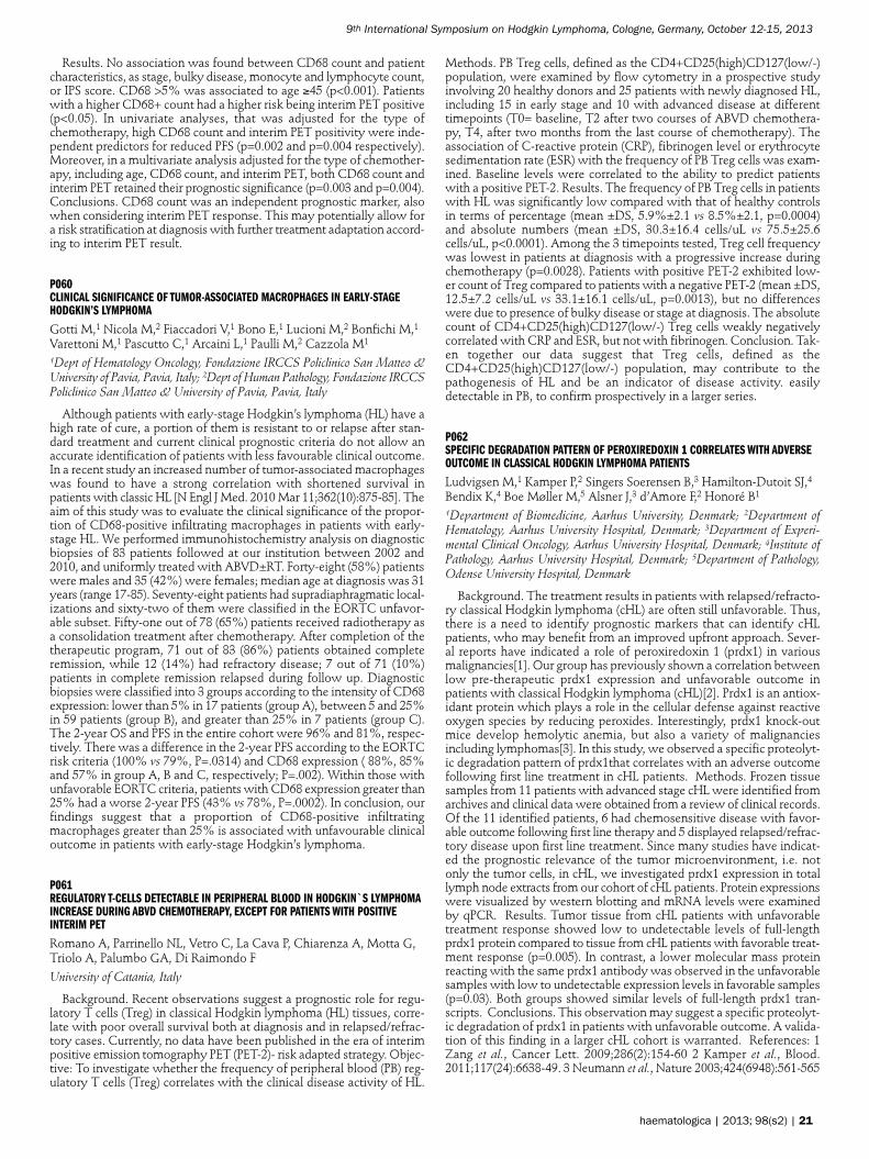
Results. No association was found between CD68 count and patientcharacteristics, as stage, bulky disease, monocyte and lymphocyte count,or IPS score. CD68 >5% was associated to age ≥45 (p<0.001). Patientswith a higher CD68+ count had a higher risk being interim PET positive(p<0.05). In univariate analyses, that was adjusted for the type ofchemotherapy, high CD68 count and interim PET positivity were inde-pendent predictors for reduced PFS (p=0.002 and p=0.004 respectively).Moreover, in a multivariate analysis adjusted for the type of chemother-apy, including age, CD68 count, and interim PET, both CD68 count andinterim PET retained their prognostic significance (p=0.003 and p=0.004).Conclusions. CD68 count was an independent prognostic marker, alsowhen considering interim PET response. This may potentially allow fora risk stratification at diagnosis with further treatment adaptation accord-ing to interim PET result.
P060CLINICAL SIGNIFICANCE OF TUMOR-ASSOCIATED MACROPHAGES IN EARLY-STAGEHODGKIN’S LYMPHOMA
Gotti M,1 Nicola M,2 Fiaccadori V,1 Bono E,1 Lucioni M,2 Bonfichi M,1Varettoni M,1 Pascutto C,1 Arcaini L,1 Paulli M,2 Cazzola M1
1Dept of Hematology Oncology, Fondazione IRCCS Policlinico San Matteo &University of Pavia, Pavia, Italy; 2Dept of Human Pathology, Fondazione IRCCSPoliclinico San Matteo & University of Pavia, Pavia, Italy
Although patients with early-stage Hodgkin’s lymphoma (HL) have ahigh rate of cure, a portion of them is resistant to or relapse after stan-dard treatment and current clinical prognostic criteria do not allow anaccurate identification of patients with less favourable clinical outcome.In a recent study an increased number of tumor-associated macrophageswas found to have a strong correlation with shortened survival inpatients with classic HL [N Engl J Med. 2010 Mar 11;362(10):875-85]. Theaim of this study was to evaluate the clinical significance of the propor-tion of CD68-positive infiltrating macrophages in patients with early-stage HL. We performed immunohistochemistry analysis on diagnosticbiopsies of 83 patients followed at our institution between 2002 and2010, and uniformly treated with ABVD±RT. Forty-eight (58%) patientswere males and 35 (42%) were females; median age at diagnosis was 31years (range 17-85). Seventy-eight patients had supradiaphragmatic local-izations and sixty-two of them were classified in the EORTC unfavor-able subset. Fifty-one out of 78 (65%) patients received radiotherapy asa consolidation treatment after chemotherapy. After completion of thetherapeutic program, 71 out of 83 (86%) patients obtained completeremission, while 12 (14%) had refractory disease; 7 out of 71 (10%)patients in complete remission relapsed during follow up. Diagnosticbiopsies were classified into 3 groups according to the intensity of CD68expression: lower than 5% in 17 patients (group A), between 5 and 25%in 59 patients (group B), and greater than 25% in 7 patients (group C).The 2-year OS and PFS in the entire cohort were 96% and 81%, respec-tively. There was a difference in the 2-year PFS according to the EORTCrisk criteria (100% vs 79%, P=.0314) and CD68 expression ( 88%, 85%and 57% in group A, B and C, respectively; P=.002). Within those withunfavorable EORTC criteria, patients with CD68 expression greater than25% had a worse 2-year PFS (43% vs 78%, P=.0002). In conclusion, ourfindings suggest that a proportion of CD68-positive infiltratingmacrophages greater than 25% is associated with unfavourable clinicaloutcome in patients with early-stage Hodgkin’s lymphoma.
P061REGULATORY T-CELLS DETECTABLE IN PERIPHERAL BLOOD IN HODGKIN`S LYMPHOMAINCREASE DURING ABVD CHEMOTHERAPY, EXCEPT FOR PATIENTS WITH POSITIVEINTERIM PET
Romano A, Parrinello NL, Vetro C, La Cava P, Chiarenza A, Motta G,Triolo A, Palumbo GA, Di Raimondo FUniversity of Catania, Italy
Background. Recent observations suggest a prognostic role for regu-latory T cells (Treg) in classical Hodgkin lymphoma (HL) tissues, corre-late with poor overall survival both at diagnosis and in relapsed/refrac-tory cases. Currently, no data have been published in the era of interimpositive emission tomography PET (PET-2)- risk adapted strategy. Objec-tive: To investigate whether the frequency of peripheral blood (PB) reg-ulatory T cells (Treg) correlates with the clinical disease activity of HL.
Methods. PB Treg cells, defined as the CD4+CD25(high)CD127(low/-)population, were examined by flow cytometry in a prospective studyinvolving 20 healthy donors and 25 patients with newly diagnosed HL,including 15 in early stage and 10 with advanced disease at differenttimepoints (T0= baseline, T2 after two courses of ABVD chemothera-py, T4, after two months from the last course of chemotherapy). Theassociation of C-reactive protein (CRP), fibrinogen level or erythrocytesedimentation rate (ESR) with the frequency of PB Treg cells was exam-ined. Baseline levels were correlated to the ability to predict patientswith a positive PET-2. Results. The frequency of PB Treg cells in patientswith HL was significantly low compared with that of healthy controlsin terms of percentage (mean ±DS, 5.9%±2.1 vs 8.5%±2.1, p=0.0004)and absolute numbers (mean ±DS, 30.3±16.4 cells/uL vs 75.5±25.6cells/uL, p<0.0001). Among the 3 timepoints tested, Treg cell frequencywas lowest in patients at diagnosis with a progressive increase duringchemotherapy (p=0.0028). Patients with positive PET-2 exhibited low-er count of Treg compared to patients with a negative PET-2 (mean ±DS,12.5±7.2 cells/uL vs 33.1±16.1 cells/uL, p=0.0013), but no differenceswere due to presence of bulky disease or stage at diagnosis. The absolutecount of CD4+CD25(high)CD127(low/-) Treg cells weakly negativelycorrelated with CRP and ESR, but not with fibrinogen. Conclusion. Tak-en together our data suggest that Treg cells, defined as theCD4+CD25(high)CD127(low/-) population, may contribute to thepathogenesis of HL and be an indicator of disease activity. easilydetectable in PB, to confirm prospectively in a larger series.
P062SPECIFIC DEGRADATION PATTERN OF PEROXIREDOXIN 1 CORRELATES WITH ADVERSEOUTCOME IN CLASSICAL HODGKIN LYMPHOMA PATIENTS
Ludvigsen M,1 Kamper P,2 Singers Soerensen B,3 Hamilton-Dutoit SJ,4Bendix K,4 Boe Møller M,5 Alsner J,3 d’Amore F,2 Honoré B11Department of Biomedicine, Aarhus University, Denmark; 2Department ofHematology, Aarhus University Hospital, Denmark; 3Department of Experi-mental Clinical Oncology, Aarhus University Hospital, Denmark; 4Institute ofPathology, Aarhus University Hospital, Denmark; 5Department of Pathology,Odense University Hospital, Denmark
Background. The treatment results in patients with relapsed/refracto-ry classical Hodgkin lymphoma (cHL) are often still unfavorable. Thus,there is a need to identify prognostic markers that can identify cHLpatients, who may benefit from an improved upfront approach. Sever-al reports have indicated a role of peroxiredoxin 1 (prdx1) in variousmalignancies[1]. Our group has previously shown a correlation betweenlow pre-therapeutic prdx1 expression and unfavorable outcome inpatients with classical Hodgkin lymphoma (cHL)[2]. Prdx1 is an antiox-idant protein which plays a role in the cellular defense against reactiveoxygen species by reducing peroxides. Interestingly, prdx1 knock-outmice develop hemolytic anemia, but also a variety of malignanciesincluding lymphomas[3]. In this study, we observed a specific proteolyt-ic degradation pattern of prdx1that correlates with an adverse outcomefollowing first line treatment in cHL patients. Methods. Frozen tissuesamples from 11 patients with advanced stage cHL were identified fromarchives and clinical data were obtained from a review of clinical records.Of the 11 identified patients, 6 had chemosensitive disease with favor-able outcome following first line therapy and 5 displayed relapsed/refrac-tory disease upon first line treatment. Since many studies have indicat-ed the prognostic relevance of the tumor microenvironment, i.e. notonly the tumor cells, in cHL, we investigated prdx1 expression in totallymph node extracts from our cohort of cHL patients. Protein expressionswere visualized by western blotting and mRNA levels were examinedby qPCR. Results. Tumor tissue from cHL patients with unfavorabletreatment response showed low to undetectable levels of full-lengthprdx1 protein compared to tissue from cHL patients with favorable treat-ment response (p=0.005). In contrast, a lower molecular mass proteinreacting with the same prdx1 antibody was observed in the unfavorablesamples with low to undetectable expression levels in favorable samples(p=0.03). Both groups showed similar levels of full-length prdx1 tran-scripts. Conclusions. This observation may suggest a specific proteolyt-ic degradation of prdx1 in patients with unfavorable outcome. A valida-tion of this finding in a larger cHL cohort is warranted. References: 1Zang et al., Cancer Lett. 2009;286(2):154-60 2 Kamper et al., Blood.2011;117(24):6638-49. 3 Neumann et al., Nature 2003;424(6948):561-565
haematologica | 2013; 98(s2) | 21
9th International Symposium on Hodgkin Lymphoma, Cologne, Germany, October 12-15, 2013
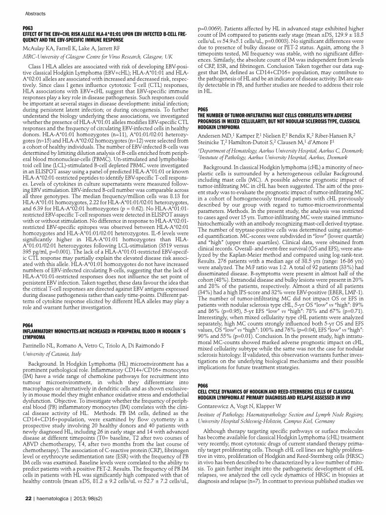
P063EFFECT OF THE EBV+CHL RISK ALLELE HLA-A*01:01 UPON EBV INFECTED B-CELL FRE-QUENCY AND THE EBV-SPECIFIC IMMUNE RESPONSE
McAulay KA, Farrell K, Lake A, Jarrett RFMRC-University of Glasgow Centre for Virus Research, Glasgow, UK
Class I HLA alleles are associated with risk of developing EBV-posi-tive classical Hodgkin Lymphoma (EBV+cHL); HLA-A*01:01 and HLA-A*02:01 alleles are associated with increased and decreased risk, respec-tively. Since class I genes influence cytotoxic T-cell (CTL) responses,HLA associations with EBV+cHL suggest that EBV-specific immuneresponses play a key role in disease pathogenesis. Such responses couldbe important at several stages in disease development: initial infection;during persistent latent infection; or during oncogenesis. To furtherunderstand the biology underlying these associations, we investigatedwhether the presence of HLA-A*01:01 alleles modifies EBV-specific CTLresponses and the frequency of circulating EBV-infected cells in healthydonors. HLA-A*01:01 homozygotes (n=11), A*01:01/02:01 heterozy-gotes (n=15) and HLA-A*02:02 homozygotes (n=12) were selected froma cohort of healthy individuals. The number of EBV-infected B-cells wasdetermined by limiting dilution analysis of B-cells enriched from periph-eral blood mononuclear-cells (PBMC). Un-stimulated and lymphoblas-toid cell line (LCL)-stimulated B-cell depleted PBMC were investigatedin an ELISPOT assay using a panel of predicted HLA-A*01:01 or knownHLA-A*02:01-restricted peptides to identify EBV-specific T-cell respons-es. Levels of cytokines in culture supernatants were measured follow-ing EBV stimulation. EBV-infected B-cell number was comparable acrossall three genotypes. The median frequency/million cells was 8.13 forHLA-A*01:01 homozygotes, 2.22 for HLA-A*01:01/02:01 heterozygotes,and 6.39 for HLA-A*02:01 homozygotes (p = 0.62). No HLA-A*01:01-restricted EBV-specific T-cell responses were detected in ELISPOT assayswith or without stimulation. No difference in response to HLA-A*02:01-restricted EBV-specific epitopes was observed between HLA-A*02:01homozygotes and HLA-A*01:01/02:01 heterozygotes. IL-6 levels weresignificantly higher in HLA-A*01:01 homozygotes than HLA-A*01:01/02:01 heterozygotes following LCL-stimulation (3519 versus395 pg/ml, p=0.001). The lack of a HLA-A*01:01-restricted EBV-specif-ic CTL response may partially explain the elevated disease risk associ-ated with this allele. HLA-A*01:01 homozygotes do not have increasednumbers of EBV-infected circulating B-cells, suggesting that the lack ofHLA-A*01:01-restricted responses does not influence the set point ofpersistent EBV infection. Taken together, these data favour the idea thatthe critical T-cell responses are directed against EBV antigens expressedduring disease pathogenesis rather than early time-points. Different pat-terns of cytokine response elicited by different HLA alleles may play arole and warrant further investigation.
P064INFLAMMATORY MONOCYTES ARE INCREASED IN PERIPHERAL BLOOD IN HODGKIN`SLYMPHOMA
Parrinello NL, Romano A, Vetro C, Triolo A, Di Raimondo FUniversity of Catania, Italy
Background. In Hodgkin Lymphoma (HL) microenvironment has aprominent pathological role. Inflammatory CD14+/CD16+ monocytes(IM) have a wide range of chemokine pathways for recruitment intotumour microenvironment, in which they differentiate intomacrophages or alternatively in dendritic cells and as shown exclusive-ly in mouse model they might enhance oxidative stress and endothelialdysfunction. Objective. To investigate whether the frequency of periph-eral blood (PB) inflammatory monocytes (IM) correlates with the clini-cal disease activity of HL. Methods. PB IM cells, defined as theCD14+CD16+population, were examined by flow cytometry in aprospective study involving 20 healthy donors and 40 patients withnewly diagnosed HL, including 26 in early stage and 14 with advanceddisease at different timepoints (T0= baseline, T2 after two courses ofABVD chemotherapy, T4, after two months from the last course ofchemotherapy). The association of C-reactive protein (CRP), fibrinogenlevel or erythrocyte sedimentation rate (ESR) with the frequency of PBIM cells was examined. Baseline levels were correlated to the ability topredict patients with a positive PET-2. Results. The frequency of PB IMcells in patients with HL was significantly high compared with that ofhealthy controls (mean ±DS, 81.2 ± 9.2 cells/uL vs 52.7 ± 7.2 cells/uL,
p=0.0069). Patients affected by HL in advanced stage exhibited highercount of IM compared to patients early stage (mean ±DS, 129.9 ± 18.5cells/uL vs 54.9±5.1 cells/uL, p=0.0003). No significant differences weredue to presence of bulky disease or PET-2 status. Again, among the 3timepoints tested, MI frequency was stable, with no significant differ-ences. Similarly, the absolute count of IM was independent from levelsof CRP, ESR, and fibrinogen. Conclusion Taken together our data sug-gest that IM, defined as CD14+CD16+ population, may contribute tothe pathogenesis of HL and be an indicator of disease activity. IM are eas-ily detectable in PB, and further studies are needed to address their rolein HL.
P065THE NUMBER OF TUMOR-INFILTRATING MAST CELLS CORRELATES WITH ADVERSEPROGNOSIS IN MIXED CELLULARITY, BUT NOT NODULAR SCLEROSIS TYPE, CLASSICALHODGKIN LYMPHOMA
Andersen MD,1 Kamper P,1 Nielsen P,2 Bendix K,2 Riber-Hansen R,2Steinicke T,2 Hamilton-Dutoit S,2 Clausen M,1 d’Amore F1
1Department of Hematology, Aarhus University Hospital, Aarhus C, Denmark;2Instutute of Pathology, Aarhus University Hospital, Aarhus, Denmark
Background. In classical Hodgkin lymphoma (cHL) a minority of neo-plastic cells is surrounded by a heterogeneous cellular Background.including mast cells (MC). A possible adverse prognostic impact oftumor-infiltrating MC in cHL has been suggested. The aim of the pres-ent study was to evaluate the prognostic impact of tumor-infiltrating MCin a cohort of homogeneously treated patients with cHL previouslydescribed by our group with regard to tumor-microenvironmentalparameters. Methods. In the present study, the analysis was restrictedto cases aged over 15 yrs. Tumor-infiltrating MC were stained immuno-histochemically with an antibody recognizing mast-cell derived tryptase.The number of tryptase-positive cells was determined using automat-ed quantification. MC-scores were subdivided in “low” (lower quartile)and “high” (upper three quartiles). Clinical data, were obtained fromclinical records. Overall- and event-free survival (OS and EFS), were ana-lyzed by the Kaplan-Meier method and compared using log-rank-test.Results. 276 patients with a median age of 38.5 yrs (range: 16-86 yrs)were analyzed. The M:F ratio was 1:2. A total of 92 patients (33%) haddisseminated disease. B-symptoms were present in almost half of thecohort (48%). Extranodal disease and bulky lesions were present in 20%and 28% of the patients, respectively. Almost a third of all patients(34%) had a high IPS-score and 32% were EBV-positive (EBER, LMP-1).The number of tumor-infiltrating MC did not impact OS or EFS inpatients with nodular sclerosis type cHL, 5-yr OS “low” vs “high”: 89%and 86% (p=0.95), 5-yr EFS “low” vs “high”: 78% and 67% (p=0.71).Interestingly, when mixed cellularity type cHL patients were analyzedseparately, high MC counts strongly influenced both 5-yr OS and EFSvalues, OS “low” vs “high”: 100% and 76% (p=0.04), EFS “low” vs “high”:90% and 55% (p=0.01). Conclusion. In the present study, high intratu-moral MC-counts showed marked adverse prognostic impact on cHL,mixed cellularity subtype while the same was not the case for nodularsclerosis histology. If validated, this observation warrants further inves-tigations on the underlying biological mechanisms and their possibleimplications for future treatment strategies.
P066CELL CYCLE DYNAMICS OF HODGKIN AND REED-STERNBERG CELLS OF CLASSICALHODGKIN LYMPHOMA AT PRIMARY DIAGNOSIS AND RELAPSE ASSESSED IN VIVO
Gontarewicz A, Vogt N, Klapper W
Institute of Pathology, Haematopathology Section and Lymph Node Registry,University Hospital Schleswig-Holstein, Campus Kiel, Germany
Although therapy targeting specific pathways or surface moleculeshas become available for classical Hodgkin Lymphoma (cHL) treatmentvery recently, most cytotoxic drugs of current standard therapy prima-rily target proliferating cells. Though cHL cell lines are highly prolifera-tive in vitro, proliferation of Hodgkin and Reed-Sternberg cells (HRSC)in vivo has been described to be characterized by a low number of mito-sis. To gain further insight into the pathogenetic development of cHLrelapses, we analyzed the cell cycle dynamics of HRSC in biopsies atdiagnosis and relapse (n=7). In contrast to previous published studies we
22 | haematologica | 2013; 98(s2)
Abstracts
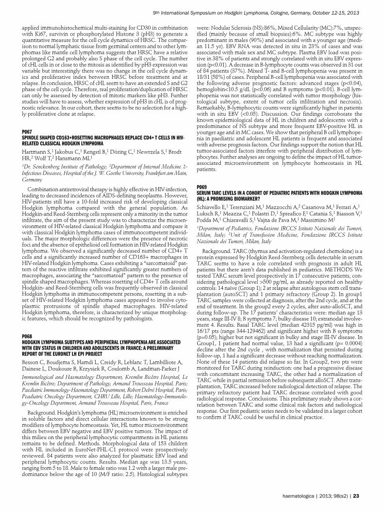
applied immunohistochemical multi-staining for CD30 in combinationwith Ki67, survivin or phosphorylated Histone 3 (pH3) to generate aquantitative measure for the cell cycle dynamics of HRSC. The compar-ison to normal lymphatic tissue from germinal centers and to other lym-phomas like mantle cell lymphoma suggests that HRSC have a relativeprolonged G2 and probably also S phase of the cell cycle. The numberof cHL cells in or close to the mitosis as identified by pH3 expression wasvariable but interestingly there was no change in the cell cycle dynam-ics and proliferative index between HRSC before treatment and atrelapse. In conclusion, HRSC of cHL seem to have an extended S and G2phase of the cell cycle. Therefore, real proliferation/duplication of HRSCcan only be assessed by detection of mitotic markers like pH3. Furtherstudies will have to assess, whether expression of pH3 in cHL is of prog-nostic relevance. In our cohort, there seems to be no selection for a high-ly proliferative clone at relapse.
P067SPINDLE SHAPED CD163+ROSETTING MACROPHAGES REPLACE CD4+ T CELLS IN HIV-RELATED CLASSICAL HODGKIN LYMPHOMA
Hartmann S,1 Jakobus C,1 Rengstl B,1 Döring C,1 Newrzela S,1 BrodtHR,2 Wolf T,2 Hansmann ML11Dr. Senckenberg Institute of Pathology; 2Department of Internal Medicine 2-Infectious Diseases, Hospital of the J. W. Goethe University, Frankfurt am Main,Germany
Combination antiretroviral therapy is highly effective in HIV-infection,leading to decreased incidences of AIDS-defining neoplasms. However,HIV-patients still have a 10-fold increased risk of developing classicalHodgkin lymphoma compared with the general population. AsHodgkin-and Reed-Sternberg cells represent only a minority in the tumorinfiltrate, the aim of the present study was to characterize the microen-vironment of HIV-related classical Hodgkin lymphoma and compare itwith classical Hodgkin lymphoma cases of immunocompetent individ-uals. The major morphologic differences were the presence of necroticfoci and the absence of epithelioid cell formation in HIV-related Hodgkinlymphoma. We observed a significantly decreased number of CD4+ Tcells and a significantly increased number of CD163+ macrophages inHIV-related Hodgkin lymphoma. Cases exhibiting a “sarcomatoid” pat-tern of the reactive infiltrate exhibited significantly greater numbers ofmacrophages, associating the “sarcomatoid” pattern to the presence ofspindle shaped macrophages. Whereas rosetting of CD4+ T cells aroundHodgkin- and Reed-Sternberg cells was frequently observed in classicalHodgkin lymphoma in immunocompetent persons, rosetting in a sub-set of HIV-related Hodgkin lymphoma cases appeared to involve cyto-plasmic protrusions of spindle shaped macrophages. HIV-relatedHodgkin lymphoma, therefore, is characterized by unique morpholog-ic features, which should be recognized by pathologists.
P068HODGKIN LYMPHOMA SUBTYPES AND PERIPHERAL LYMPHOPENIA ARE ASSOCIATEDWITH EBV STATUS IN CHILDREN AND ADOLESCENTS IN FRANCE: A PRELIMINARYREPORT OF THE EURONET LH EPI PROJECT
Besson C, Boudjema S, Hamdi L, Creidy R, Leblanc T, Lambilliote A,Dainese L, Doukoure B, Krzysiek R, Coulomb A, Landman-Parker JImmunological and Haematology Department, Kremlin Bicêtre Hospital, LeKremlin Bicêtre; Department of Pathology, Armand Trousseau Hospital, Paris;Paediatric Immunology-Haematology Department, Robert Debré Hospital, Paris;Peadiatric Oncology Department, CHRU Lille, Lille; Haematology-Immunolo-gy-Oncology Department, Armand Trousseau Hospital, Paris, France
Background. Hodgkin’s lymphoma (HL) microenvironment is enrichedin soluble factors and direct cellular interactions known to be strongmodifiers of lymphocyte homeostasis. Yet, HL tumor microenvironmentdiffers between EBV negative and EBV positive tumors. The impact ofthis milieu on the peripheral lymphocytic compartments in HL patientsremains to be defined. Methods. Morphological data of 153 childrenwith HL included in EuroNet-PHL-C1 protocol were prospectivelyreviewed. 84 patients were also analyzed for plasmatic EBV load andperipheral lymphocytic counts. Results. Median age was 13.5 years,ranging from 5 to 18. Male to female ratio was 1.2 with a larger male pre-dominance below the age of 10 (M/F ratio: 2.5). Histological subtypes
were: Nodular Sclerosis (NS):86%, Mixed Cellularity (MC):7%, unspec-ified (mainly because of small biopsies):6%. MC subtype was highlypredominant in males (90%) and associated with a younger age (medi-an 11.5 yr). EBV RNA was detected in situ in 23% of cases and wasassociated with male sex and MC subtype. Plasma EBV load was posi-tive in 38% of patients and strongly correlated with in situ EBV expres-sion (p<0.01). A decrease in B-lymphocyte counts was observed in 31 outof 84 patients (37%). Mixed T- and B-cell lymphopenia was present in18/31 (58%) of cases. Peripheral B-cell lymphopenia was associated withthe following adverse prognostic factors: advanced stages (p<0.04),hemoglobin<10.5 g/dL (p<0.06) and B symptoms (p<0.01). B-cell lym-phopenia was not statistically correlated with tumor morphology (his-tological subtype, extent of tumor cells infiltration and necrosis).Remarkably, B-lymphocytic counts were significantly higher in patientswith in situ EBV (<0.05). Discussion. Our findings corroborate theknown epidemiological data of HL in children and adolescents with apredominance of NS subtype and more frequent EBV-positive HL inyounger age and in MC cases. We show that peripheral B cell lymphope-nia in paediatric and adolescent HL patients is frequent and associatedwith adverse prognosis factors. Our findings support the notion that HLtumor-associated factors interfere with peripheral distribution of lym-phocytes. Further analyses are ongoing to define the impact of HL tumor-associated microenvironment on lymphocyte homeostasis in HLpatients.
P069SERUM TARC LEVELS IN A COHORT OF PEDIATRIC PATIENTS WITH HODGKIN LYMPHOMA(HL): A PROMISING BIOMARKER?
Schiavello E,1 Terenziani M,1 Mazzocchi A,2 Casanova M,1 Ferrari A,1Luksch R,1 Meazza C,1 Polastri D,1 Spreafico F,1 Catania S,1 Biasson V,1Podda M,1 Chiaravalli S,1 Vajna de Pava M,1 Massimino M1
1Department of Pediatrics, Fondazione IRCCS Istituto Nazionale dei Tumori,Milan, Italy; 2Unit of Transfusion Medicine, Fondazione IRCCS IstitutoNazionale dei Tumori, Milan, Italy
Background. TARC (thymus and activation-regulated chemokine) is aprotein expressed by Hodgkin Reed-Sternberg cells detectable in serumTARC seems to have a role correlated with prognosis in adult HLpatients but there aren’t data published in pediatrics. METHODS Wetested TARC serum level prospectively in 17 consecutive patients, con-sidering pathological level >500 pg/ml, as already reported on healthycontrols: 14 naïve (Group 1); 2 at relapse after autologous stem cell trans-plantation (autoSCT) and 1 primary refractory (Group 2). In group1TARC samples were collected at diagnosis, after the 2nd cycle, and at theend of treatment. In the group2 every 2 cycles, after auto-alloSCT, andduring follow-up. The 17 patients’ characteristics were: median age 13years, stage III-IV 8; B symptoms 7; bulky disease 10; extranodal involve-ment 4. Results. Basal TARC level (median 42315 pg/ml) was high in16/17 pts (range 344-129462) and significant higher with B symptoms(p=0.05); higher but not significant in bulky and stage III-IV disease. InGroup1, 1 patient had normal value, 13 had a significant (p= 0.0004)decline after the 2nd cycle , with normalization that persisted duringfollow-up, 1 had a significant decrease without reaching normalization.None of these 14 patients did relapse so far. In Group2, two pts weremonitored for TARC during reinduction: one had a progressive diseasewith concomitant increasing TARC, the other had a normalization ofTARC while in partial remission before subsequent alloSCT. After trans-plantation, TARC increased before radiological detection of relapse. Theprimary refractory patient had TARC decrease correlated with goodradiological response. Conclusions. This preliminary study shows a cor-relation between TARC and some clinical risk factors and radiologicalresponse. Our first pediatric series needs to be validated in a larger cohortto confirm if TARC could be useful in clinical practice.
haematologica | 2013; 98(s2) | 23
9th International Symposium on Hodgkin Lymphoma, Cologne, Germany, October 12-15, 2013
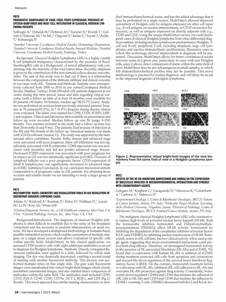
P070PROGNOSTIC SIGNIFICANCE OF CD68, CD20, FOXP3 EXPRESSION; PRESENCE OFEPSTEIN-BARR VIRUS AND MAST CELL INFILTRATION IN CLASSICAL HODGKIN LYM-PHOMA PATIENTS
Salihoglu A,1 Ozbalak M,2 Demiroz AS,3 Tuzuner N,4 Elverdi T,1 Gul-turk E,1Eskazan AE,1 Ar MC,1 Ongoren S,1 Baslar Z,1 Soysal T,1AydinY,1 Ferhanoglu B1
1Istanbul University Cerrahpasa Medical Faculty, Hematology Department;2Istanbul University Cerrahpasa Medical Faculty, Internal Medicine; 3IstanbulUniversity Cerrahpasa Medical Faculty, Pathology, Turkey
Background. Classical Hodgkin Lymphoma (cHL) is a highly curableB cell lymphoid malignancy characterized by the presence of Reed-Sternberg(RS) cells in a Background. of mixed inflammatory cells con-stituting only the minority of the total tumor mass. Recently, emphasisis given to the contribution of the non-tumoral cells to disease outcome.Aims: The aim of this study was to find out if there is a relationshipbetween the composition of the immune infiltrate and clinical outcomein our cases with cHL. Patients and Methods. Patients were retrospec-tively collected from 2000 to 2010 in one center(Cerrahpasa MedicalFaculty, Istanbul, Turkey). From 250 adult cHL patients diagnosed at ourcenter during this time period, tissue and data regarding clinical out-come (with a follow up time of at least 24 months) were available for83 patients (49 males, 34 females, median age 38(13-77) years). Analy-sis was performed on sections from previously untreated patients’ biop-sies in 76 patients(91.6%), in 7 (8.4%) biopsies during disease relapsewere evaluated. The slides were stained for CD68, CD20, FOXP3, LMP-1 and tryptase. Clinical and laboratory data available on presentation andfollow up were recorded. Median follow up was 56 (range 6-156)months. Two patients included in the study had a follow up time lessthan 24 months (6 and 9 mo). The patients died because of infection atthe 6th and 9th month of the follow up. Statistical analysis was madewith STATA software (version 11). The study was approved by the insti-tutional ethics committee. Results. Bulky disease and advanced stagewere associated with poor prognosis. Mast cell infiltration was not sig-nificantly associated with B symptoms. CD68 expression was not asso-ciated with mortality and did not predict advanced stage disease.Decreased FOXP3 expression was associated with poor prognosis butits impact on OS was not statistically significant (p=0.063). Decrease oflymphoid follicles was a poor prognostic factor. CD20 expression ofsmall B lymphocytes was significantly decreased in advanced stage(p=0.048). Summary/Conclusion. In our conclusion microenvironmentcomposition is of prognostic value in cHL patients. For obtaining moreaccurate and reliable results we are intending to study a larger group ofpatients.
P071MULTIOMYXTM: NOVEL CHEMISTRY AND VISUALIZATION TOOLS TO AID RESOLUTION OFDISCREPANT HODGKIN LYMPHOMA CASES
Adams A,1 Bordwell A,1 Bouman D,1 Fisher D,1 Hollman D,1, LazareM,1 Li M,1 Ross AS,1 Weiss LM2
1Clarient Diagnostic Services, Inc., a GE Healthcare company, Aliso Viejo, CA,USA; 2Clarient Pathology Services, Inc., Aliso Viejo, CA, USA
Background/Introduction. The diagnosis of classical Hodgkin lym-phoma is often difficult to establish due to the rarity of the neoplasticcomponent and the necessity to perform immunostains on serial sec-tions. We have developed a multiplexed methodology in formalin-fixed,paraffin-embedded sections which enables assessment of multiple anti-gens on a single tissue section and allows evaluation of specific cellswithin specific fields (MultiOmyx). In this clinical application, weassessed CD30-positive cells with eight additional antibodies as an aidto diagnosis for Hodgkin lymphoma. Methods. Directly conjugated flu-orescent antibodies were applied to a slide, followed by whole slideimaging. The dye was chemically inactivated, enabling a second roundof staining with another fluorescent antibody. This process was per-formed multiple times on that single slide. The gray scale fluorescentimages were transformed into virtual bright-field images which closelyresembled conventional images, and also enabled direct comparison ofantibodies within the same field. The antibodies used included CD30,CD15, PAX-5, CD45, CD20, CD79a, OCT-2, BOB.1, and CD3 (Fig.1).Results. This novel approach has similar staining characteristics as stan-
dard immunohistochemical stains, and has the added advantage that itmay be performed on a single section. MultiOmyx allowed improvedassessment of Hodgkin cells for antigens expressed on other cell types(e.g., B-cell antigens on reactive immunoblasts, or CD15 on reactive his-tiocytes), as well as antigens expressed on directly adjacent cells (e.g.,CD45 and CD3). Using the unique MultiOmyx viewer, we could distin-guish cases of classical Hodgkin lymphoma from other differential diag-nosis entities, including nodular lymphocyte predominance Hodgkin, T-cell rich B-cell, peripheral T-cell, including anaplastic large cell lym-phoma, and reactive immunoblastic proliferations. Illustrative cases inwhich this technology allowed improvement in diagnosis will be pre-sented. Discussion: MultiOmyx allows for better correlation of resultsbetween stains in a given case, particularly in cases with rare Hodgkincells, since it allows direct comparison of stains within the same field ofview. MultiOmyx may be also advantageous in small samples, in whichfull immunohistochemical profiles may not be possible. This novelmethodology is practical for routine diagnosis, and will likely be an aidto the improved diagnosis of Hodgkin lymphoma.
Figure 1. Representative virtual bright-field images of the nine bio-markers from the same field of view in a Hodgkin Lymphoma sam-ple
P072EFFECTS OF THE NF-KB INHIBITORS BORTEZOMIB AND DHMEQ ON THE EXPRESSIONOF MOLECULES INVOLVED IN MICROENVIRONMENTAL INTERACTIONS AND SYNERGYWITH CHEMOTHERAPY AGENTS
Celegato M,1 Borghese C,1 Casagrande N,1 Umezawa K,2 ColombattiA,1 Carbone A,3 Aldinucci D1
1Experimental Oncology 2, Centro di Riferimento Oncologico, IRCCS-Nation-al Cancer Institute, Aviano, PN, Italy; 2Molecular Target Medicine Screening,Aichi Medical University, Nagakute, Japan; 3Division of Pathology, Centro diRiferimento Oncologico, IRCCS-National Cancer Institute, Aviano, PN, Italy
The malignant classical Hodgkin lymphoma (cHL) cells constitutive-ly express high levels of activated nuclear factor kappaB (NF-kB). Boththe proteasome inhibitor bortezomib and Dehydroxymethyle-poxyquinomicin (DHMEQ) affect NF-kB activity: bortezomib byinhibiting the degradation of the cytoplasmic inhibitor of nuclear factor-B (I B ) and DHMEQ by inhibiting nuclear translocation of NF-kB. Borte-zomib, active in cHL cell lines, has low clinical activity when used as sin-gle agent, suggesting that micro-environmental interactions could pro-tect from drug efficacy. Therefore, we investigated bortezomib activityin the presence of HL-associated fibroblasts (HL-AFs) and sCD40L. Wefound that co-cultivation with human HL-AFs or addition of sCD40Lduring treatment protected cHL-cells from apoptosis and cytotoxicityand rescued the down-regulation of the survival factor Interferon Reg-ulatory Factor 4 (IRF4). On the contrary, bortezomib treatment beforeco-cultivation with HL-AFs, inhibited cHL cells adhesion to HL-AFs andovercame HL-AFs protection against drug activity. Consistently, borte-zomib down-regulated CD49d and CD44 that mediate the adhesion ofcHL cells to HL-AFs and CD54 and CD40 that mediate the adhesion toCD40L+ rosetting T-cells. DHMEQ decreased both Bcl-2 and Bcl-xL lev-
24 | haematologica | 2013; 98(s2)
Abstracts
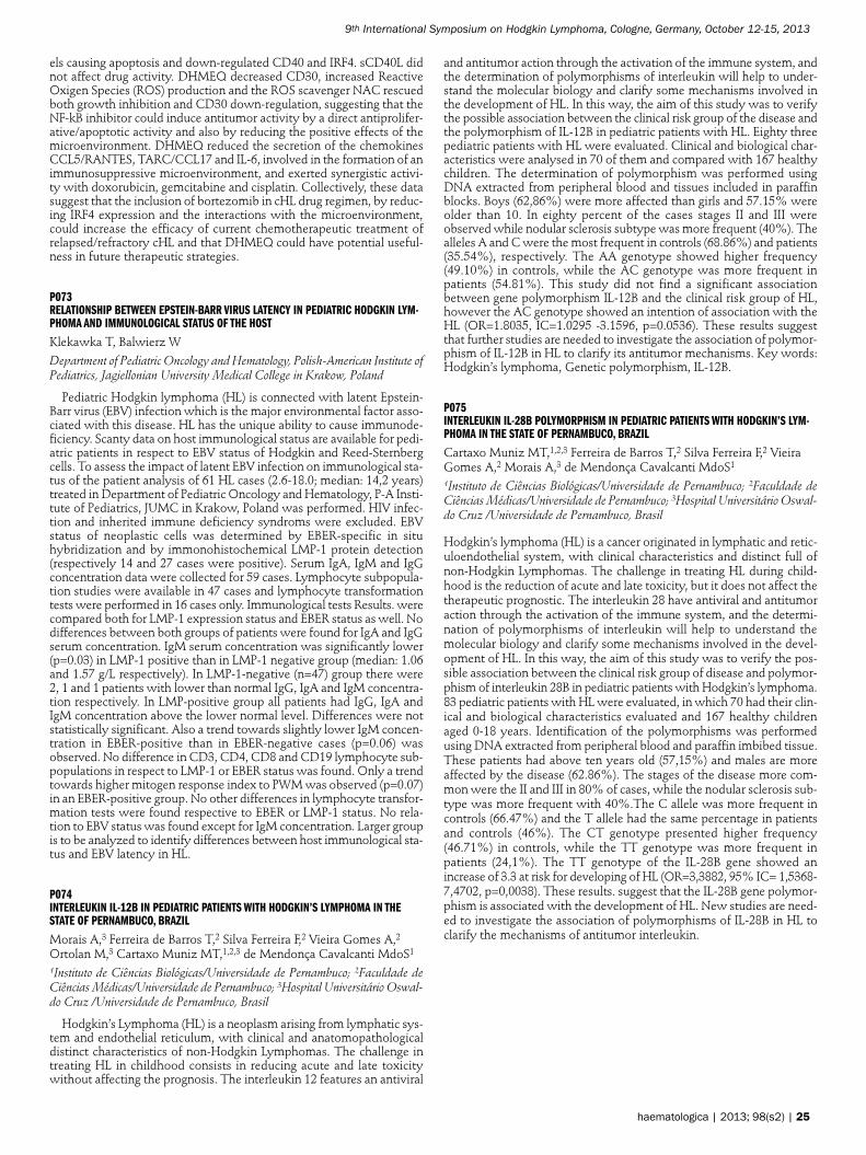
els causing apoptosis and down-regulated CD40 and IRF4. sCD40L didnot affect drug activity. DHMEQ decreased CD30, increased ReactiveOxigen Species (ROS) production and the ROS scavenger NAC rescuedboth growth inhibition and CD30 down-regulation, suggesting that theNF-kB inhibitor could induce antitumor activity by a direct antiprolifer-ative/apoptotic activity and also by reducing the positive effects of themicroenvironment. DHMEQ reduced the secretion of the chemokinesCCL5/RANTES, TARC/CCL17 and IL-6, involved in the formation of animmunosuppressive microenvironment, and exerted synergistic activi-ty with doxorubicin, gemcitabine and cisplatin. Collectively, these datasuggest that the inclusion of bortezomib in cHL drug regimen, by reduc-ing IRF4 expression and the interactions with the microenvironment,could increase the efficacy of current chemotherapeutic treatment ofrelapsed/refractory cHL and that DHMEQ could have potential useful-ness in future therapeutic strategies.
P073RELATIONSHIP BETWEEN EPSTEIN-BARR VIRUS LATENCY IN PEDIATRIC HODGKIN LYM-PHOMA AND IMMUNOLOGICAL STATUS OF THE HOST
Klekawka T, Balwierz WDepartment of Pediatric Oncology and Hematology, Polish-American Institute ofPediatrics, Jagiellonian University Medical College in Krakow, Poland
Pediatric Hodgkin lymphoma (HL) is connected with latent Epstein-Barr virus (EBV) infection which is the major environmental factor asso-ciated with this disease. HL has the unique ability to cause immunode-ficiency. Scanty data on host immunological status are available for pedi-atric patients in respect to EBV status of Hodgkin and Reed-Sternbergcells. To assess the impact of latent EBV infection on immunological sta-tus of the patient analysis of 61 HL cases (2.6-18.0; median: 14,2 years)treated in Department of Pediatric Oncology and Hematology, P-A Insti-tute of Pediatrics, JUMC in Krakow, Poland was performed. HIV infec-tion and inherited immune deficiency syndroms were excluded. EBVstatus of neoplastic cells was determined by EBER-specific in situhybridization and by immonohistochemical LMP-1 protein detection(respectively 14 and 27 cases were positive). Serum IgA, IgM and IgGconcentration data were collected for 59 cases. Lymphocyte subpopula-tion studies were available in 47 cases and lymphocyte transformationtests were performed in 16 cases only. Immunological tests Results. werecompared both for LMP-1 expression status and EBER status as well. Nodifferences between both groups of patients were found for IgA and IgGserum concentration. IgM serum concentration was significantly lower(p=0.03) in LMP-1 positive than in LMP-1 negative group (median: 1.06and 1.57 g/L respectively). In LMP-1-negative (n=47) group there were2, 1 and 1 patients with lower than normal IgG, IgA and IgM concentra-tion respectively. In LMP-positive group all patients had IgG, IgA andIgM concentration above the lower normal level. Differences were notstatistically significant. Also a trend towards slightly lower IgM concen-tration in EBER-positive than in EBER-negative cases (p=0.06) wasobserved. No difference in CD3, CD4, CD8 and CD19 lymphocyte sub-populations in respect to LMP-1 or EBER status was found. Only a trendtowards higher mitogen response index to PWM was observed (p=0.07)in an EBER-positive group. No other differences in lymphocyte transfor-mation tests were found respective to EBER or LMP-1 status. No rela-tion to EBV status was found except for IgM concentration. Larger groupis to be analyzed to identify differences between host immunological sta-tus and EBV latency in HL.
P074INTERLEUKIN IL-12B IN PEDIATRIC PATIENTS WITH HODGKIN’S LYMPHOMA IN THESTATE OF PERNAMBUCO, BRAZIL
Morais A,3 Ferreira de Barros T,2 Silva Ferreira F,2 Vieira Gomes A,2Ortolan M,3 Cartaxo Muniz MT,1,2,3 de Mendonça Cavalcanti MdoS11Instituto de Ciências Biológicas/Universidade de Pernambuco; 2Faculdade deCiências Médicas/Universidade de Pernambuco; 3Hospital Universitário Oswal-do Cruz /Universidade de Pernambuco, Brasil
Hodgkin’s Lymphoma (HL) is a neoplasm arising from lymphatic sys-tem and endothelial reticulum, with clinical and anatomopathologicaldistinct characteristics of non-Hodgkin Lymphomas. The challenge intreating HL in childhood consists in reducing acute and late toxicitywithout affecting the prognosis. The interleukin 12 features an antiviral
and antitumor action through the activation of the immune system, andthe determination of polymorphisms of interleukin will help to under-stand the molecular biology and clarify some mechanisms involved inthe development of HL. In this way, the aim of this study was to verifythe possible association between the clinical risk group of the disease andthe polymorphism of IL-12B in pediatric patients with HL. Eighty threepediatric patients with HL were evaluated. Clinical and biological char-acteristics were analysed in 70 of them and compared with 167 healthychildren. The determination of polymorphism was performed usingDNA extracted from peripheral blood and tissues included in paraffinblocks. Boys (62,86%) were more affected than girls and 57.15% wereolder than 10. In eighty percent of the cases stages II and III wereobserved while nodular sclerosis subtype was more frequent (40%). Thealleles A and C were the most frequent in controls (68.86%) and patients(35.54%), respectively. The AA genotype showed higher frequency(49.10%) in controls, while the AC genotype was more frequent inpatients (54.81%). This study did not find a significant associationbetween gene polymorphism IL-12B and the clinical risk group of HL,however the AC genotype showed an intention of association with theHL (OR=1.8035, IC=1.0295 -3.1596, p=0.0536). These results suggestthat further studies are needed to investigate the association of polymor-phism of IL-12B in HL to clarify its antitumor mechanisms. Key words:Hodgkin’s lymphoma, Genetic polymorphism, IL-12B.
P075INTERLEUKIN IL-28B POLYMORPHISM IN PEDIATRIC PATIENTS WITH HODGKIN’S LYM-PHOMA IN THE STATE OF PERNAMBUCO, BRAZIL
Cartaxo Muniz MT,1,2,3 Ferreira de Barros T,2 Silva Ferreira F,2 VieiraGomes A,2 Morais A,3 de Mendonça Cavalcanti MdoS11Instituto de Ciências Biológicas/Universidade de Pernambuco; 2Faculdade deCiências Médicas/Universidade de Pernambuco; 3Hospital Universitário Oswal-do Cruz /Universidade de Pernambuco, Brasil
Hodgkin’s lymphoma (HL) is a cancer originated in lymphatic and retic-uloendothelial system, with clinical characteristics and distinct full ofnon-Hodgkin Lymphomas. The challenge in treating HL during child-hood is the reduction of acute and late toxicity, but it does not affect thetherapeutic prognostic. The interleukin 28 have antiviral and antitumoraction through the activation of the immune system, and the determi-nation of polymorphisms of interleukin will help to understand themolecular biology and clarify some mechanisms involved in the devel-opment of HL. In this way, the aim of this study was to verify the pos-sible association between the clinical risk group of disease and polymor-phism of interleukin 28B in pediatric patients with Hodgkin’s lymphoma.83 pediatric patients with HL were evaluated, in which 70 had their clin-ical and biological characteristics evaluated and 167 healthy childrenaged 0-18 years. Identification of the polymorphisms was performedusing DNA extracted from peripheral blood and paraffin imbibed tissue.These patients had above ten years old (57,15%) and males are moreaffected by the disease (62.86%). The stages of the disease more com-mon were the II and III in 80% of cases, while the nodular sclerosis sub-type was more frequent with 40%.The C allele was more frequent incontrols (66.47%) and the T allele had the same percentage in patientsand controls (46%). The CT genotype presented higher frequency(46.71%) in controls, while the TT genotype was more frequent inpatients (24,1%). The TT genotype of the IL-28B gene showed anincrease of 3.3 at risk for developing of HL (OR=3,3882, 95% IC= 1,5368-7,4702, p=0,0038). These results. suggest that the IL-28B gene polymor-phism is associated with the development of HL. New studies are need-ed to investigate the association of polymorphisms of IL-28B in HL toclarify the mechanisms of antitumor interleukin.
haematologica | 2013; 98(s2) | 25
9th International Symposium on Hodgkin Lymphoma, Cologne, Germany, October 12-15, 2013
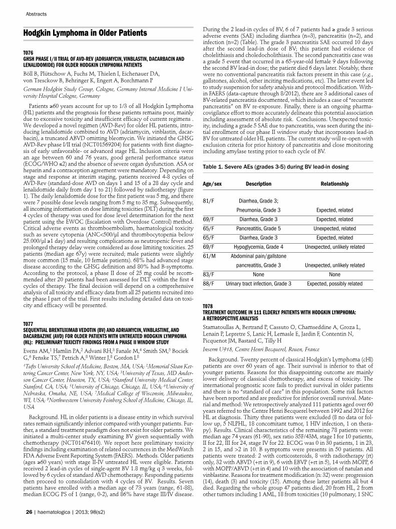
Hodgkin Lymphoma in Older Patients
T076GHSH PHASE I/II TRIAL OF AVD-REV (ADRIAMYCIN, VINBLASTIN, DACARBACIN ANDLENALIDOMIDE) FOR OLDER HODGKIN LYMPHOMA PATIENTS
Böll B, Plütschow A, Fuchs M, Thielen I, Eichenauer DA, von Tresckow B, Behringer K, Engert A, Borchmann P
German Hodgkin Study Group, Cologne, Germany Internal Medicine I Uni-versity Hospital Cologne, Germany
Patients ≥60 years account for up to 1/3 of all Hodgkin Lymphoma(HL) patients and the prognosis for these patients remains poor, mainlydue to excessive toxicity and insufficient efficacy of current regimens .We developed a novel regimen (AVD-Rev) for older HL patients, intro-ducing lenalidomide combined to AVD (adriamycin, vinblastin, dacar-bacin), a truncated ABVD omitting bleomycin. We initiated the GHSGAVD-Rev phase I/II trial (NCT01569204) for patients with first diagno-sis of early unfavorable- or advanced stage HL. Inclusion criteria werean age between 60 and 76 years, good general performance status(ECOG/WHO ≤2) and the absence of severe organ dysfunction. ASA orheparin and a contraception agreement were mandatory. Depending onstage and response at interim staging, patients received 4-8 cycles ofAVD-Rev (standard-dose AVD on days 1 and 15 of a 28 day cycle andlenalidomide daily from day 1 to 21) followed by radiotherapy (figure1). The daily lenalidomide dose for the first patient was 5 mg, and therewere 7 possible dose levels ranging from 5 mg to 35 mg. Subsequently,all incoming information on dose limiting toxicities (DLT) during the first4 cycles of therapy was used for dose level determination for the nextpatient using the EWOC (Escalation with Overdose Control) method.Critical adverse events as thromboembolism, haematological toxicitysuch as severe cytopenia (ANC<500/µl and thrombocytopenia below25.000/µl ≥1 day) and resulting complications as neutropenic fever andprolonged therapy delay were considered as dose limiting toxicities. 25patients (median age 67y) were recruited; male patients were slightlymore common (15 male, 10 female patients). 68% had advanced stagedisease according to the GHSG definition and 80% had B-symptoms.According to the protocol, a phase II dose of 25 mg could be recom-mended after 20 patients had been assessed for DLT within the first 4cycles of therapy. The final decision will depend on a comprehensiveanalysis of all toxicity and efficacy data from all 25 patients recruited intothe phase I part of the trial. First results including detailed data on toxi-city and efficacy will be presented.
T077SEQUENTIAL BRENTUXIMAB VEDOTIN (BV) AND ADRIAMYCIN, VINBLASTINE, ANDDACARBAZINE (AVD) FOR OLDER PATIENTS WITH UNTREATED HODGKIN LYMPHOMA(HL): PRELIMINARY TOXICITY FINDINGS FROM A PHASE II WINDOW STUDY
Evens AM,1 Hamlin PA,2 Advani RH,3 Fanale M,4 Smith SM,5 BociekG,6 Fenske TS,7 Petrich A,8 Winter J,8 Gordon L8
1Tufts University School of Medicine, Boston, MA, USA; 2Memorial Sloan Ket-tering Cancer Center, New York, NY, USA; 3University of Texas, MD Ander-son Cancer Center, Houston, TX, USA; 4Stanford University Medical Center,Stanford, CA, USA; 5University of Chicago, Chicago, IL, USA; 6University ofNebraska, Omaha, NE, USA; 7Medical College of Wisconsin, Milwaukee,WI, USA; 8Northwestern University Feinberg School of Medicine, Chicago, IL,USA
Background. HL in older patients is a disease entity in which survivalrates remain significantly inferior compared with younger patients. Fur-ther, a standard treatment paradigm does not exist for older patients. Weinitiated a multi-center study examining BV given sequentially withchemotherapy (NCT01476410). We report here preliminary toxicityfindings including examination of related occurrences in the MedWatchFDA Adverse Event Reporting System (FAERS). Methods. Older patients(ages ≥60 years) with stage II-IV untreated HL were eligible. Patientsreceived 2 lead-in cycles of single-agent BV 1.8 mg/kg q 3 weeks, fol-lowed by 6 cycles of standard AVD chemotherapy. Responding patientsthen proceed to consolidation with 4 cycles of BV. Results. Sevenpatients have enrolled with a median age of 73 years (range, 61-88),median ECOG PS of 1 (range, 0-2), and 86% have stage III/IV disease.
During the 2 lead-in cycles of BV, 6 of 7 patients had ≥ grade 3 seriousadverse events (SAE) including diarrhea (n=3), pancreatitis (n=2), andinfection (n=2) (Table). The grade 3 pancreatitis SAE occurred 10 daysafter the second lead-in dose of BV; this patient had evidence ofcholelithiasis and choledocholithiasis. The second pancreatitis case wasa grade 5 event that occurred in a 65-year-old female 9 days followingthe second BV lead-in dose; the patient died 6 days later. Notably, therewere no conventional pancreatitis risk factors present in this case (e.g.,gallstones, alcohol, other inciting medications, etc). The latter event ledto study suspension for safety analysis and protocol modification. With-in FAERS (data-capture through 8/2012), there are 3 additional cases ofBV-related pancreatitis documented, which includes a case of “recurrentpancreatitis” on BV re-exposure. Finally, there is an ongoing pharma-covigilance effort to more accurately delineate this potential associationincluding assessment of absolute risk. Conclusions. Unexpected toxic-ity, including a grade 5 SAE due to pancreatitis, was seen during the ini-tial enrollment of our phase II window study that incorporates lead-inBV for untreated older HL patients. The current study will re-open withexclusion criteria for prior history of pancreatitis and close monitoringincluding amylase testing prior to each cycle of BV.
Table 1. Severe AEs (grades 3-5) during BV lead-in dosing
Age/sex Description Relationship
81/F Diarrhea, Grade 3;
Pneumonia, Grade 3 Expected, related
69/F Diarrhea, Grade 3 Expected, related
65/F Pancreatitis, Grade 5 Unexpected, related
65/F Diarrhea, Grade 3 Expected, related
69/F Hypoglycemia, Grade 4 Unexpected, unlikely related
61/M Abdominal pain/gallstone
pancreatitis, Grade 3 Unexpected, unlikely related
83/F None None
88/F Urinary tract infection, Grade 3 Expected, possibly related
T078TREATMENT OUTCOME IN 111 ELDERLY PATIENTS WITH HODGKIN LYMPHOMA: A RETROSPECTIVE ANALYSIS
Stamatoullas A, Bertrand P, Cassuto O, Chamseddine A, Groza L,Lenain P, Lepretre S, Lanic H, Lemasle E, Jardin F, Contentin N, Picquenot JM, Bastard C, Tilly H
Inserm U918, Centre Henri Becquerel, Rouen, France
Background. Twenty percent of classical Hodgkin’s Lymphoma (cHl)patients are over 60 years of age. Their survival is inferior to that ofyounger patients. Reasons for this disappointing outcome are mainlylower delivery of classical chemotherapy, and excess of toxicity. Theinternational prognostic score fails to predict survival in older patientsand there is no “standard of care” in this population. Some risk factorshave been reported and are predictive for inferior overall survival. Mate-rial and method: We retrospectively analyzed 111 patients aged over 60years referred to the Centre Henri Becquerel between 1992 and 2012 forHL at diagnosis. Thirty three patients were excluded (8 no data or fol-low up, 5 NLPHL, 18 concomitant tumor, 1 HIV infection, 1 on thera-py). Results. Clinical characteristics of the remaining 78 patients were:median age 74 years (61-90), sex ratio 35F/43M, stage I for 10 patients,II for 22, III for 24, stage IV for 22. ECOG was 0 in 30 patients, 1 in 23,2 in 15, and >2 in 10. B symptoms were presents in 50 patients. Allpatients were treated: 2 with corticosteroids, 8 with radiotherapy (rt)only, 32 with ABVD (+rt in 9), 6 with EBVP (+rt in 5), 14 with MOPP, 6with MOPP/ABVD (+rt in 4) and 10 with the association of natulan andvinblastine. Reasons for treatment modification (n: 32) were: progression(14), death (3) and toxicity (15). Among these latter patients all but 4died. Regarding the whole group 47 patients died, 20 from HL, 2 fromother tumors including 1 AML, 18 from toxicities (10 pulmonary, 1 SNC
26 | haematologica | 2013; 98(s2)
Abstracts
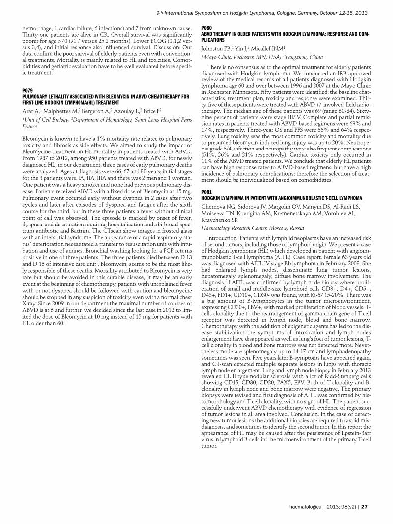
hemorrhage, 1 cardiac failure, 6 infections) and 7 from unknown cause.Thirty one patients are alive in CR. Overall survival was significantlypoorer for age >70 (91.7 versus 25.2 months). Lower ECOG (0,1,2 ver-sus 3,4), and initial response also influenced survival. Discussion: Ourdata confirm the poor survival of elderly patients even with convention-al treatments. Mortality is mainly related to HL and toxicities. Comor-bidities and geriatric evaluation have to be well evaluated before specif-ic treatment.
P079PULMONARY LETHALITY ASSOCIATED WITH BLEOMYCIN IN ABVD CHEMOTHERAPY FORFIRST-LINE HODGKIN LYMPHOMA(HL) TREATMENT
Arar A,1 Malphettes M,2 Bergeron A,2 Azoulay E,2 Brice P21Unit of Cell Biology, 2Department of Hematology, Saint Louis Hospital ParisFrance
Bleomycin is known to have a 1% mortality rate related to pulmonarytoxicity and fibrosis as side effects. We aimed to study the impact ofBleomycine treatment on HL mortality in patients treated with ABVD.From 1987 to 2012, among 950 patients treated with ABVD, for newlydiagnosed HL, in our department, three cases of early pulmonary deathswere analyzed. Ages at diagnosis were 66, 67 and 80 years; initial stagesfor the 3 patients were: IA, IIA, IIIA and there was 2 men and 1 woman.One patient was a heavy smoker and none had previous pulmonary dis-ease. Patients received ABVD with a fixed dose of Bleomycin at 15 mg.Pulmonary event occurred early without dyspnea in 2 cases after twocycles and later after episodes of dyspnea and fatigue after the sixthcourse for the third, but in these three patients a fever without clinicalpoint of call was observed. The episode is marked by onset of fever,dyspnea, and desaturation requiring hospitalization and a bi-broad-spec-trum antibiotic and Bactrim. The CTscan show images in frosted glasswith an interstitial syndrome. The appearance of a rapid respiratory sta-tus’ deterioration necessitated a transfer to resuscitation unit with intu-bation and use of amines. Bronchial washing looking for a PCP returnspositive in one of three patients. The three patients died between D 13and D 16 of intensive care unit . Bleomycin, seems to be the most like-ly responsible of these deaths. Mortality attributed to Bleomycin is veryrare but should be avoided in this curable disease, It may be an earlyevent at the beginning of chemotherapy, patients with unexplained feverwith or not dyspnea should be followed with caution and bleomycineshould be stopped in any suspicion of toxicity even with a normal chestX ray. Since 2009 in our department the maximal number of courses ofABVD is at 6 and further, we decided since the last case in 2012 to lim-ited the dose of Bleomycin at 10 mg instead of 15 mg for patients withHL older than 60.
P080ABVD THERAPY IN OLDER PATIENTS WITH HODGKIN LYMPHOMA: RESPONSE AND COM-PLICATIONS
Johnston PB,1 Yin J,2 Micallef INM1
1Mayo Clinic, Rochester, MN, USA; 2Yangzhou, China
There is no consensus as to the optimal treatment for elderly patientsdiagnosed with Hodgkin lymphoma. We conducted an IRB approvedreview of the medical records of all patients diagnosed with Hodgkinlymphoma age 60 and over between 1996 and 2007 at the Mayo Clinicin Rochester, Minnesota. Fifty patients were identified; the baseline char-acteristics, treatment plan, toxicity and response were examined. Thir-ty-five of these patients were treated with ABVD +/ involved-field radio-therapy. The median age of these patients was 69 (range 60-84). Sixty-nine percent of patients were stage III/IV. Complete and partial remis-sion rates in patients treated with ABVD-based regimens were 69% and17%, respectively. Three-year OS and PFS were 66% and 64% respec-tively. Lung toxicity was the most common toxicity and mortality dueto presumed bleomycin-induced lung injury was up to 20%. Neutrope-nia grade 3/4, infection and neuropathy were also frequent complications(51%, 26% and 21% respectively). Cardiac toxicity only occurred in11% of the ABVD treated patients. We conclude that elderly HL patientscan have high response rates to ABVD-based regimens, but have a highincidence of pulmonary complications; therefore the selection of treat-ment should be individualized based on comorbidities.
P081HODGKIN LYMPHOMA IN PATIENT WITH ANGIOIMMUNOBLASTIC T-CELL LYMPHOMA
Chernova NG, Sidorova JV, Margolin OV, Mariyin DS, Al-Radi LS,Moiseeva TN, Kovrigina AM, Kremenetskaya AM, Vorobiev AI,Kravchenko SKHaematology Research Center, Moscow, Russia
Introduction. Patients with lymph id neoplasms have an increased riskof second tumors, including those of lymphoid origin. We present a caseof Hodgkin lymphoma (HL) which developed in patient with angioim-munoblastic T-cell lymphoma (AITL). Case report. Female 63 years oldwas diagnosed with AITL IV stage Bb lymphoma in February 2008. Shehad enlarged lymph nodes, disseminate lung tumor lesions,hepatomegaly, splenomegaly, diffuse bone marrow involvement. Thediagnosis of AITL was confirmed by lymph node biopsy where prolif-eration of small and middle-size lymphoid cells CD3+, D4+, CD5+,D43+, PD1+, CD10+, CD30- was found, with Ki-67 15-20%. There wasa big amount of B-lymphocytes in the tumor microenvironment,expressing CD30+, EBV+, with marked proliferation of blood vessels. T-cells clonality due to the rearrangement of gamma-chain gene of T-cellreceptor was detected in lymph node, blood and bone marrow.Chemotherapy with the addition of epigenetic agents has led to the dis-ease stabilization–the symptoms of intoxication and lymph nodesenlargement have disappeared as well as lung’s foci of tumor lesions, T-cell clonality in blood and bone marrow was not detected more. Never-theless moderate splenomegaly up to 14-17 cm and lymphadenopathysometimes was seen. Five years later B-symptoms have appeared again,and CT-scan detected multiple separate lesions in lungs with thoraciclymph node enlargement. Lung and lymph node biopsy in February 2013revealed HL II type nodular sclerosis with a lot of Ridd-Stenberg cellsshowing CD15, CD30, CD20, PAX5, EBV. Both of T-clonality and B-clonality in lymph node and bone marrow were negative. The primarybiopsys were revised and first diagnosis of AITL was confirmed by his-tomorphology and T-cell clonality, with no signs of HL. The patient suc-cessfully underwent ABVD chemotherapy with evidence of regressionof tumor lesions in all area involved. Conclusion. In the case of detect-ing new tumor lesions the additional biopsies are required to avoid mis-diagnosis, and sometimes to identify the second tumor. In this report theappearance of HL may be caused after the persistence of Epstein-Barrvirus in lymphoid B-cells inf the microenvironment of the primary T-celltumor.
haematologica | 2013; 98(s2) | 27
9th International Symposium on Hodgkin Lymphoma, Cologne, Germany, October 12-15, 2013

Pathways
T082DISRUPTION OF B CELL DIFFERENTIATION BY CONDITIONAL ACTIVATION OF THEHODGKIN LYMPHOMA-ASSOCIATED E2A ANTAGONISTS ID2 AND ABF1 IN VIVO
Janz M,1,2 Li S,1,2 Calado DP,1 Mathas S,1,2 Dörken B,1,2 Rajewsky K1
1Hematology, Oncology and Tumorimmunology, Charité, University HospitalBerlin, Campus Virchow-Klinikum, Berlin, Germany; 2Max Delbrück Centerfor Molecular Medicine, Berlin, Germany
Oncogenic transformation is not only characterized by enhanced pro-liferation and higher apoptosis resistance of malignant cells, but also bydisruption of physiological differentiation. Among human lymphoidmalignancies, classical Hodgkin lymphoma (cHL) constitutes one of themost prominent examples for this aspect of tumor biology. In contrastto their origin from germinal center (GC) or post-GC B cells, the malig-nant Hodgkin/Reed-Sternberg (HRS) cells of cHL have lost a consider-able part of the B cell-specific gene expression program and show an up-regulation of genes that are usually considered to be associated withother hematopoietic lineages. In previous work, we have shown thatthis phenotype is linked to an aberrant activity of the transcription fac-tor E2A, which is functionally inhibited in HRS cells by overexpressionof the helix-loop-helix (HLH) proteins ID2 and ABF1. To enable adetailed analysis of Id2- or Abf1-dependent effects on growth and dif-ferentiation of lymphoid cells in vivo, we have generated transgenicmice that allow for conditional activation of Id2 or Abf1 from the Rosa26locus in a lineage- and stage-specific manner via cre-mediated recombi-nation. Induction of Id2 and Abf1 in mb1-cre mice, which express crein early stages of B cell differentiation, revealed a pronounced block ofB cell development in the bone marrow. Analysis of C 1-cre; Rosa26Id2stopFL and C 1-cre; Rosa26 Abf1stopFL mice, in which cre becomesactive when B cells enter the GC reaction, demonstrated that both fac-tors inhibit the GC response as well as the terminal differentiationtowards plasma cells. Our experiments show that induction of Id2 andAbf1 results in disruption of the differentiation process of B cells, a fea-ture which reflects a key aspect of cHL biology. These data support ourhypothesis that the functional inhibition of E2A activity is a critical stepin cHL pathogenesis.
T083INTEGRATED GENOMIC ANALYSIS OF FLOW SORTED HODGKIN AND REED-STERNBERGCELL IN PRIMARY CALSSICAL HODGKIN LYMPHOMA
Reichel J,1 Cesarman E,1 Radaban R,2 Elemento O,3 Shaknovich R,1Roshal M1
1Department of Pathology and Laboratory Medicine, Weill-Cornell MedicalCollege; 2Department of Biomedical Informatics Center for Computational Biol-ogy and Bioinformatics Columbia University College of Physicians and Sur-geons; 3Department of Physiology and Biophysics Institute for ComputationalBiomedicine Weill Cornell Medical College, new York, NY, USA
Genomic studies of classical Hodgkin lymphoma (CHL) have beenhampered by the difficulty of isolating purified Hodgkin and Reed-Stern-berg (HRS) cell populations. Recent Introduction. of flow cytometric cellisolation has greatly eased the process allowing rapid isolation of thou-sands of viable HRS cells from primary CHL tumors. We have utilizedthe technique to produce what is to our knowledge the first integratedstudy of the whole exome, methylome and transcriptome sequencingof primary cases of Hodgkin Lymphoma. For exome sequencing wehave developed a modified low input library generation procedure toobtain an average of approximately 30X sequence coverage of HRS, aswell as tumor-infiltrating T cells (used as somatic control) from as lowas 1000 sorted HRS cells. Using this techniques we perfomed wholeexome sequencing of nine primary cases of CHL. We identified recur-rent mutations in genes involved in mitotic checkpoint regulation, epi-genetic regulation and immune evasion. In addition we performed high-resolution copy number variation analysis of the exome sequencing dataand identified recurrent losses and gains of genes involved in B-cell mat-uration, transcriptional control, epigenetic regulation and immune eva-sion. We also performed whole transcriptome analysis of the HRS cellsas well as tumor infiltrating B cells to confirm RNA expression level
alteration of the genes affected by somatic mutations and copy numbervariations in HRS. These analyses also confirmed numerous reportedtranscription-level alterations obtained from previous studies using lasercapture microdissected cells. Finally we utilized enhanced form ofreduced representation bisulfite sequencing technology to obtain pro-moter methylation status of 14764 genes. The analysis revealed globalpromoter hypermethylation within isolated HRS cells compared tonaïve, memory and centroblast B cell subsets suggesting key role of epi-genetic regulation in CHL pathogenesis. The simultaneous analysis ofexome, transcriptome and methylome on the flow-sorted HRS cellsallows for expanded study of CHL pathogenesis, new target identifica-tion and potentially individualized approaches to CHL therapy.
T084TCF3/E2A (19P13.3) IS A NOVEL HODGKIN LYMPHOMA SUSCEPTIBILITY LOCUS; AMETA-GWAS STUDY FROM THE INTERLYMPH HODGKIN LYMPHOMA CONSORTIUM
Van den Berg A1 Cozen W*, Li D*, Timofeeva M*, Diepstra A*,Hazelet D*, Delahaye-Sourdeix M*, Edlund CK*, Rostgaard K, VanDen Berg DJ, Glaser SL, Robison LL, Mack TM, Ghesquieres H, SalleG, Bhatia S, Strong LC, Hwang AE, Nieters A, Smedby K, de SanjoseS, Cortessis VK, Lightfoot T, Roman E, Becker N, Foretova L,Benavente Y, Maynadie M, Visser L, Veenstra RN, Staines A, CoccoPL, Boffetta P, Kiemeney L, Lake A, Montgomery D, Slager SL, CerhanJR, Gallagher A, Taylor GM, Brennan P, Conti DV& , Coetzee GA&,Onel K&, Jarrett RF&, Hjalgrim H&, McKay JD&
*Co-first authors, &Co-Last authors. Presented on behalf of the Hodgkin lym-phoma working group of the InterLymph consortium. 1Department of Pathologyand Medical Biology, University Medical Centre Groningen, University ofGroningen, Groningen, the Netherlands
Hodgkin lymphoma (HL) is known to have a strong genetic suscep-tibility component, with a highly increased risk in co-twins of patients.Currently reported GWAS’s identified several HLA and non-HLA riskloci. In order to identify additional variants associated with HL, we per-formed a meta-analysis of three HL GWAS studies followed by a vali-dation study. 1,810 cases and 7,879 controls, all of European descent,were included in the meta analysis. Promising variants were tested in anindependent set of 1,163 cases and 2,580 controls. We noted strongassociations in HLA, with 564 genetic variants presenting with P<10-4,and in non-HLA loci, i.e. REL, PVT1 and GATA3, consistent with previ-ous publications. In addition, we identified a novel variant at 19p13.3that was associated with risk of HL(rs1860661; odds ratio [OR]= 0.81,95% confidence interval [95% CI]= 0.76-0.86, Pcombined = 3.5x10⁻¹⁰).This SNP is located in intron 2 of the TCF3/E2A gene, a key regulatorof B-cell lineage commitment. In silico analysis indicates that rs1860661is located in a genomic region with an open chromatin structure, activehistone marks and a ZBTB7A/LRF transcription factor binding motif.The protective minor G allele maps to a ZBTB7A binding motif, whichis disrupted by the major A allele. Two additional variants highly corre-lated with rs1860661, rs10413888 (r2=0.90) and rs8103453 (r2=0.89),are within an E2F1 and another ZBTB7A/LRF binding motif, respective-ly. Analysis of TCF3 expression levels in LCL cell lines of 31 genotypedindividuals revealed a trend towards lower levels of expression for AAindividuals, intermediate levels for AG individuals and the highest lev-els for GG individuals. This supports a direct functional and protectiveeffect of the rs1860661 SNP by enhancing binding of ZBTB7A and as aresult increasing TCF3/E2A expression levels. Thus, this meta-analysisidentified a novel association with TCF3/E2A and supports previouslyreported susceptibility associations with SNPs in other HLA and non-HLA genes. Because the allele predicted to have enhanced TCF3/E2Aexpression is protective for HL, we suggest that TCF3/E2A functions asa tumor suppressor gene for HL, probably via promotion and/or main-tenance of the B-cell phenotype.
T085HODGKIN-REED-STERNBERG CELLS IN CLASSICAL HODGKIN LYMPHOMA SHOW ALTER-ATIONS OF GENES ENCODING THE NADPH OXIDASE COMPLEX AND IMPAIRED REAC-TIVE OXYGEN SPECIES SYNTHESIS CAPACITY
Giefing M,1,7 Winoto-Morbach S,2 Sosna J,2 Döring C,3 Klapper W,4Küppers R,5 Böttcher S,6 Adam D,2 Siebert R,1 Schütze S21Institute of Human Genetics, Christian-Albrechts University Kiel & Universi-
28 | haematologica | 2013; 98(s2)
Abstracts
ty Hospital Schleswig-Holstein, Campus Kiel, Kiel, Germany; 2Institute ofImmunology, Christian-Albrechts University Kiel & University HospitalSchleswig-Holstein, Campus Kiel, Kiel, Germany; 3Senckenberg Institute ofPathology, University of Frankfurt, Medical School, Frankfurt, Germany; 4Department of Pathology, Hematopathology Section and Lymph Node Registry,Kiel, Germany; 5Institute of Cell Biology (Cancer Research), University of Duis-burg-Essen, Medical School, Essen, Germany; 6Second Department of Medicine,University Hospital Schleswig-Holstein, Campus Kiel, Kiel, Germany; 7Instituteof Human Genetics, Polish Academy of Sciences, Poznan, Poland
The membrane bound NADPH oxidase involved in the synthesis ofreactive oxygen species (ROS) is a multi-protein enzyme encoded byCYBA, CYBB, NCF1, NCF2 and NCF4 genes. Besides its microbicidalfunction in phagocytes, growing evidence suggests a role of ROS in themodulation of signaling pathways of non-phagocytic cells, including dif-ferentiation and proliferation of B-cell progenitors. We have recentlyreported recurrent transcriptional downregulation of the CYBB gene incell lines of the B-cell derived classical Hodgkin lymphoma (cHL). Thus,here we explored functional consequences of CYBB downregulation onthe NADPH complex. To trigger superoxide anion production we stim-ulated the CD30 receptor in cHL and non-cHL cell lines. Using flowcytometry we quantified the superoxide anion synthesis and identifiedrecurrent loss of superoxide anion production in all stimulated cHL celllines in contrast to stimulated non-Hodgkin lymphoma cell lines. AsCYBB loss proved to exert a deleterious effect on the NADPH oxidasecomplex in cHL cell lines, we analyzed the CYBB locus in Hodgkin andReed-Sternberg (HRS) cells of primary cHL biopsies by in situ hybridis-ation and identified recurrent deletions of the gene in 8/18 cases. More-over, by microarray profiling of cHL cell lines we identified additionalalterations of NADPH oxidase genes including CYBA copy number lossin 3/7 cell lines and a significant loss of the NCF1 gene on mRNA level(p=0.006) compared to normal B-cell subsets. Besides, the NCF1 proteinwas significantly downregulated (p<0.005) in cHL compared to otherlymphoma cell lines. Together these findings show recurrent alterationsof the NADPH oxidase encoding genes that result in functional inacti-vation of the enzyme and loss of superoxide anion in cHL. In light of theinduction of ROS by CD30 signaling in the CD30+cHL cell lines and thestrong and constitutive CD30 expression in primary HRS cells, one mayspeculate that the inactivation of NADPH oxidase represents a strategyto escape from an overwhelming and toxic ROS production, that couldotherwise impair HRS cell survival. Moreover, the loss of ROS signalingmay potentially deregulate B-cell lineage development and contribute tothe loss of B-cell phenotype of HRS cells.
P086DOWNREGULATION OF FOXO1 PREVENTS INDUCTION OF THE TUMOR SUPPRESSORPRDM1 IN CHL
Vogel MJ,1 Xie L,2 Guan H,3 Kick A,1 Brüderlein S,4 Melo JV,5 ToozeRM,6 Ushmorov A,1 Wirth T11Institute of Physiological Chemistry, University of Ulm, Ulm, Germany; 2Can-cer Center of Union Hospital, Tongji Medical College, HuaZhong University ofScience and Technology, Wuhan, China; 3Department of Orthopaedic Surgery,Tongji Hospital, Tongji Medical College, HuaZhong University of Science andTechnology, Wuhan, China; 4Institute of Pathology, University Medical CenterUlm, Ulm, Germany; 5Department of Haematology, Centre for Cancer Biology,SA Pathology University of Adelaide, Adelaide, South Australia, Australia; 6Sec-tion of Experimental Haematology, Leeds Institute of Molecular Medicine, Uni-versity of Leeds, Leeds, United Kingdom
In contrast to other B-cell lymphomas, Hodgkin and Reed-Sternberg(HRS) cells, the malignant cells of classical Hodgkin lymphoma (cHL),have mainly lost their B cell identity. Of note, HRS cells do not undergoplasma cell differentiation despite constitutive expression of NF- B, IRF4,and STAT3, which are known to induce the master regulator of plasmacell differentiation PRDM1. Still, PRDM1 levels remain low in HRS cells,yet for unknown reasons, thereby preventing terminal differentiation.Recently, we reported that the transcription factor FOXO1, indispensa-ble for B cell development and differentiation, is downregulated in HRScells whereas reactivation of FOXO1 results in growth arrest and apop-tosis. In the present work we show that overexpression of FOXO1 reac-tivated expression of germinal center-specific genes including BCL6,BACH2, and AICDA in cHL cell lines. Surprisingly, FOXO1 induced also
PRDM1, which has been shown to be a tumor suppressor in activatedB-cell-like diffuse large B cell lymphoma. Overexpression of PRDM1 , theactive isoform of PRDM1, strongly inhibited growth of cHL cell lines,whereas PRDM1 , the truncated isoform with impaired transrepressingactivity, demonstrated weak or no growth-inhibitory activity. Anti-tumor effects of PRDM1 might at least partially be explained by repres-sion of the protooncogene MYC. Searching for other mechanisms ofPRDM1 down-regulation we found that its promoter was hyperme-thylated in two cHL cell lines. Further, our data indicate that constitu-tive expression of MYC might also contribute to PRDM1 repression incHL. Interestingly, we found that the dominant-negative variant, PRDM1, which is normally not detected in B cells, was upregulated in cHL celllines. This might reduce DNA binding and decrease function of residualPRDM1 in cHL. Taken together, our data indicate that FOXO1 in cHLis essential for PRDM1 induction. In addition, we identified PRDM1 asa tumor suppressor in cHL.
P087DEVELOPMENT OF AN ORIGINAL XENOGRAFT MODEL OF HUMAN HODGKIN LYMPHOMA:TOWARD A BETTER UNDERSTANDING OF THE PATHOLOGY AND THE DESIGN OF NEWTHERAPIES
M’kacher R,1 Frenzel M,1 Corina C,1 Bauchet A,2 Leunen A,1 HempelW,1 Jinckel S,3 Girinsky T,4 Carde P,5 Boisgard R,6 Plumio FF,7 SabatierL11Radiobiology and Oncology laboratory, CEA, iRCM; 2Head of Platform forexperimental pathology PathEX / CRC MIRCen / CEA-INSERM Fontenayaux roses France; 3Institute of Human Genetics, University of Aarhus, AarhusC, Denmark; 4Department of Radiation Oncology, Institut Gustave Roussy,Ville-juif, France; 5Department of Medicine; Institut Gustave Roussy; Villejuif, France;6Laboratoire d’Imagerie Moléculaire Expérimentale Groupe d’Imagerie du petitanimal CEA / DSV / I2BM / SHFJ / U1023 Orsay France; 7Laboratoire desCellules Souches Hématopoïétiques et Leucémiques, UMR967 INSERM, Uni-versités Paris 7 et Paris 11, IRCM/CEA, Fontenay-aux-Roses, France
Hodgkin lymphoma (HL) is a malignancy of the immune system, char-acterized by poor growth in vitro and the lack of reliable animal mod-els. Recently, the presence of clonotypic B cells in HL has been docu-mented. However, the low frequency of these cells has precluded thegeneration of the disease when transplanted into immunodeficient mice.In this study, we have successfully determined the conditions for the invitro amplification and characterisation of such cells and their transplan-tation into immunodeficient NOD-SCID-gammac-/- (NSG) mice. Usingthe Hodgkin cell lines L428 and KMH2, clonotypic B cells were isolatedand amplified in semi-solid cultures. Such clones showed a very highcloning efficiency in comparison to the original total cell population(50% vs 2%) and increased proliferation. Higher signal intensity of CD15and CD14 was observed in the clones compared to the cell lines. In addi-tion, CD30 expression was observed in both the clones and HL cell lines.Cytogenetic characterization by M-FISH revealed a complex karyotypeand the presence of small metaphases. This enrichment allowed thetransplantation of these cells by the intravenous route (retro orbital sinus)into immune-deficient NSG mice. Four to eleven weeks later, regardlessof the number of cells injected (106 to 103), an infiltration of tumor cellswas observed, essentially in the lymph nodes, liver, bone marrow, andspleen. The tumor cells were identified based on the cell surface expres-sion of CD30, CD15, CD14 and CD68 and DNA probes of alfa-satelliteof human chromosomes. Significant infiltration of tumor cells (CD30+,CD15+, CD14+, CD68+ and alfa-satellite probes) resulting in majortumor masses was observed in the liver. Cell lines were re-establishedfrom xenografted tumors: they were of B-lymphoid origin and showednumerical and structural chromosomal aberrations similar to those of theoriginal L428 cell line and small metaphases as observed. 2-[18F]-fluoro-2-deoxyglucose (FDG)-PET was used to monitor the reproducibility ofthe model. The establishment of this animal model of HL is a majoradvance, not only for understanding the pathology of this enigmatic dis-ease, but also for the development of novel therapeutic strategies.
haematologica | 2013; 98(s2) | 29
9th International Symposium on Hodgkin Lymphoma, Cologne, Germany, October 12-15, 2013
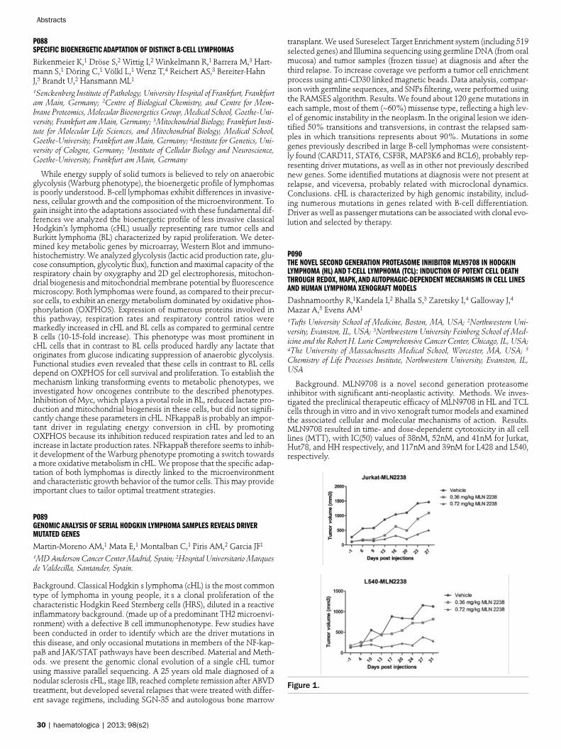
P088SPECIFIC BIOENERGETIC ADAPTATION OF DISTINCT B-CELL LYMPHOMAS
Birkenmeier K,1 Dröse S,2 Wittig I,2 Winkelmann R,1 Barrera M,3 Hart-mann S,1 Döring C,1 Völkl L,1 Wenz T,4 Reichert AS,3 Bereiter-HahnJ,5 Brandt U,2 Hansmann ML1
1Senckenberg Institute of Pathology, University Hospital of Frankfurt, Frankfurtam Main, Germany; 2Centre of Biological Chemistry, and Centre for Mem-brane Proteomics, Molecular Bioenergetics Group, Medical School, Goethe-Uni-versity, Frankfurt am Main, Germany; 3Mitochondrial Biology, Frankfurt Insti-tute for Molecular Life Sciences, and Mitochondrial Biology, Medical School,Goethe-University, Frankfurt am Main, Germany; 4Institute for Genetics, Uni-versity of Cologne, Germany; 5Institute of Cellular Biology and Neuroscience,Goethe-University, Frankfurt am Main, Germany
While energy supply of solid tumors is believed to rely on anaerobicglycolysis (Warburg phenotype), the bioenergetic profile of lymphomasis poorly understood. B-cell lymphomas exhibit differences in invasive-ness, cellular growth and the composition of the microenvironment. Togain insight into the adaptations associated with these fundamental dif-ferences we analyzed the bioenergetic profile of less invasive classicalHodgkin’s lymphoma (cHL) usually representing rare tumor cells andBurkitt lymphoma (BL) characterized by rapid proliferation. We deter-mined key metabolic genes by microarray, Western Blot and immuno-histochemistry. We analyzed glycolysis (lactic acid production rate, glu-cose consumption, glycolytic flux), function and maximal capacity of therespiratory chain by oxygraphy and 2D gel electrophoresis, mitochon-drial biogenesis and mitochondrial membrane potential by fluorescencemicroscopy. Both lymphomas were found, as compared to their precur-sor cells, to exhibit an energy metabolism dominated by oxidative phos-phorylation (OXPHOS). Expression of numerous proteins involved inthis pathway, respiration rates and respiratory control ratios weremarkedly increased in cHL and BL cells as compared to germinal centreB cells (10-15-fold increase). This phenotype was most prominent incHL cells that in contrast to BL cells produced hardly any lactate thatoriginates from glucose indicating suppression of anaerobic glycolysis.Functional studies even revealed that these cells in contrast to BL cellsdepend on OXPHOS for cell survival and proliferation. To establish themechanism linking transforming events to metabolic phenotypes, weinvestigated how oncogenes contribute to the described phenotypes.Inhibition of Myc, which plays a pivotal role in BL, reduced lactate pro-duction and mitochondrial biogenesis in these cells, but did not signifi-cantly change these parameters in cHL. NFkappaB is probably an impor-tant driver in regulating energy conversion in cHL by promotingOXPHOS because its inhibition reduced respiration rates and led to anincrease in lactate production rates. NFkappaB therefore seems to inhib-it development of the Warburg phenotype promoting a switch towardsa more oxidative metabolism in cHL. We propose that the specific adap-tation of both lymphomas is directly linked to the microenvironmentand characteristic growth behavior of the tumor cells. This may provideimportant clues to tailor optimal treatment strategies.
P089GENOMIC ANALYSIS OF SERIAL HODGKIN LYMPHOMA SAMPLES REVEALS DRIVERMUTATED GENES
Martin-Moreno AM,1 Mata E,1 Montalban C,1 Piris AM,2 Garcia JF1
1MD Anderson Cancer Center Madrid, Spain; 2Hospital Universitario Marquesde Valdecilla, Santander, Spain.
Background. Classical Hodgkin�s lymphoma (cHL) is the most commontype of lymphoma in young people, it�s a clonal proliferation of thecharacteristic Hodgkin Reed Sternberg cells (HRS), diluted in a reactiveinflammatory background. (made up of a predominant TH2 microenvi-ronment) with a defective B cell immunophenotype. Few studies havebeen conducted in order to identify which are the driver mutations inthis disease, and only occasional mutations in members of the NF-kap-paB and JAK/STAT pathways have been described. Material and Meth-ods. we present the genomic clonal evolution of a single cHL tumorusing massive parallel sequencing. A 25 years old male diagnosed of anodular sclerosis cHL, stage IIB, reached complete remission after ABVDtreatment, but developed several relapses that were treated with differ-ent savage regimens, including SGN-35 and autologous bone marrow
transplant. We used Sureselect Target Enrichment system (including 519selected genes) and Illumina sequencing using germline DNA (from oralmucosa) and tumor samples (frozen tissue) at diagnosis and after thethird relapse. To increase coverage we perform a tumor cell enrichmentprocess using anti-CD30 linked magnetic beads. Data analysis, compar-ison with germline sequences, and SNPs filtering, were performed usingthe RAMSES algorithm. Results. We found about 120 gene mutations ineach sample, most of them (~60%) missense type, reflecting a high lev-el of genomic instability in the neoplasm. In the original lesion we iden-tified 50% transitions and transversions, in contrast the relapsed sam-ples in which transitions represents about 90%. Mutations in somegenes previously described in large B-cell lymphomas were consistent-ly found (CARD11, STAT6, CSF3R, MAP3K6 and BCL6), probably rep-resenting driver mutations, as well as in other not previously describednew genes. Some identified mutations at diagnosis were not present atrelapse, and viceversa, probably related with microclonal dynamics.Conclusions. cHL is characterized by high genomic instability, includ-ing numerous mutations in genes related with B-cell differentiation.Driver as well as passenger mutations can be associated with clonal evo-lution and selected by therapy.
P090THE NOVEL SECOND GENERATION PROTEASOME INHIBITOR MLN9708 IN HODGKINLYMPHOMA (HL) AND T-CELL LYMPHOMA (TCL): INDUCTION OF POTENT CELL DEATHTHROUGH REDOX, MAPK, AND AUTOPHAGIC-DEPENDENT MECHANISMS IN CELL LINESAND HUMAN LYMPHOMA XENOGRAFT MODELS
Dashnamoorthy R,1Kandela I,2 Bhalla S,3 Zaretsky I,4 Galloway J,4Mazar A,5 Evens AM1
1Tufts University School of Medicine, Boston, MA, USA; 2Northwestern Uni-versity, Evanston, IL, USA; 3Northwestern University Feinberg School of Med-icine and the Robert H. Lurie Comprehensive Cancer Center, Chicago, IL, USA;4The University of Massachusetts Medical School, Worcester, MA, USA; 5Chemistry of Life Processes Institute, Northwestern University, Evanston, IL,USA
Background. MLN9708 is a novel second generation proteasomeinhibitor with significant anti-neoplastic activity. Methods. We inves-tigated the preclinical therapeutic efficacy of MLN9708 in HL and TCLcells through in vitro and in vivo xenograft tumor models and examinedthe associated cellular and molecular mechanisms of action. Results.MLN9708 resulted in time- and dose-dependent cytotoxicity in all celllines (MTT), with IC(50) values of 38nM, 52nM, and 41nM for Jurkat,Hut78, and HH respectively, and 117nM and 39nM for L428 and L540,respectively.
Figure 1.
30 | haematologica | 2013; 98(s2)
Abstracts
Treatment with MLN9708 resulted in dose-dependent increases inapoptosis by Annexin-V/PI, PARP cleavage, and activation of caspases 3,8, 9 in all cell lines. Furthermore, these effects were superior comparedwith bortezomib treatment.Using CellROX-Red FACS analysis,MLN9708 resulted in increased intracellular oxidative stress. Moreover,MLN9708 resulted in increased p21 expression, degradation of MYCprotein, and accumulation of LC3A (autophagy). Apoptosis, oxidativestress, and MYC degradation were abrogated in the presence of N-acetyl-cysteine, however LC3A accumulation occurred in a redox-independentmanner. Further, spautin (a potent inhibitor of autophagy) significantlyreduced MLN9708-related cell death. Examination of MAPK signalingshowed that p-ERK and p-p38 were downreglated and p-JNK unregulat-ed in HL cells with MLN9708; these findings were opposite in TCL lines.In TCL, shRNA knock-down of ERK and p38 had minimal effect onMLN9708-induced apoptosis, while there was additive cell death withJNK shRNA. In HL, ERK and JNK shRNA knock-outs abrogated the effectof MLN9708, while the cytotoxic effect was potentiated with p38shRNA knock-out. Finally, MLN9708 resulted in significanttumor/growth inhibition (P<0.001) and improvement in survival(P<0.001) compared with controls in Jurkat and L540-derived SCIDxenograft models (Figure). Conclusions. Altogether, the novel protea-some inhibitor, MLN9708, induced potent cell death at clinically achiev-able concentrations in HL and TCL cell lines and in SCID xenograft mod-els. MLN9708 down-regulated MYC and induced redox-dependent celldeath through autophagy in HL and TCL. Further, the cytotoxic effectof MLN9708 was potentiated through JNK in TCL and p38 in HL. Con-tinued examination of this novel agent alone and in rational combina-tions in HL and TCL is warranted.
P091GENOME-WIDE ASSOCIATION STUDY IDENTIFIES GERMLINE POLYMORPHISMS ASSOCI-ATED WITH AGGRESSIVE DISEASE AND TREATMENT RESPONSE OF HODGKIN LYM-PHOMA
Yang JJ, Lim J, Shi L, Pounds S, Wu J, Hudson MM, Metzger MLSt Jude Children’s Research Hospital, Memphis, TN, USA
Background. and Aim. Although Hodgkin lymphoma (HL) is highlycurable, its pathogenesis is poorly understood and biological determi-nants of treatment response are largely uncharacterized. Tumor microen-vironment strongly influences HL prognosis, suggesting a critical role ofinherited (germline) genetic variations in anti-tumor drug sensitivity. Weperformed a pharmacogenomic study to identify germline genetic poly-morphisms associated with treatment failure and/or aggressive HL, withthe aim to discover genetic markers for individualized therapy to mini-mize toxicity and to further improve survival. Methods. Germline DNAwas extracted from 224 patients with newly-diagnosed HL treated onfrontline protocols (or according to guidelines) between 2000 and 2010at St. Jude Children’s Research Hospital. Genotyping was performedusing Illumina HumanExome Beadchip for 247,870 polymorphisms. Weevaluated the associations of germline polymorphisms with previousexposure to Epstein-Barr virus (EBV), tumor EBV status, B-symptoms atdiagnosis, early response to therapy and relapse. Results. Completeresponse by PET/CT after 2 cycles was achived by 28% of patients, and20% (44 out of 224) relapsed. In the genome-wide association study,we identified 509 SNPs associated with risk of relapse (P<0.01) and 648SNPs related to early treatment response (P<0.01), 9 of which were con-cordantly significant for both phenotypes. Genetic variants influencingtreatment response were particularly enriched in the D-glutamine/glu-tamate metabolism and focal adhesion pathways (P<0.01), includingdirect molecular targets of chemotherapeutic agents commonly used inHL therapy. Genotype at 579 SNPs were significantly different betweenpatients with B-symptoms vs without, and the strongest association wasobserved in genes involved in cell adhesion. Finally, within 142 patientswith prior EBV exposure, 613 SNPs were associated with tumor EBV sta-tus (P<0.01). Conclusion. Taking a genome-wide approach, we haveidentified germline genetic variants associated with aggressive diseaseand/or treatment outcome of HL. These findings revealed novel biolo-gy of HL and potential novel prognostic markers, warranting further val-idation and functional studies.
P092THE IMPACT OF THE GSTP1 A313G POLYMORPHISM ON CLINICAL OUTCOME INHODGKIN’S LYMPHOMA PATIENTS
Kryachok I, Khranovska N, Svergun N, Novosad O, Titorenko INational Cancer Institute, Kiev, Ukraine
Introduction. Hodgkin lymphoma (HL) is a highly curable malignan-cy, but treatment outcome might be influenced by inherited gene poly-morphisms determining anticancer agent metabolism.We assessed theimpact of the GSTP1 A313G polymorphism on clinical outcome inpatients with HL. Material and Methods. The case group comprised 202patients with HL (stages: IA-IIA–68, IIB–43, III-IV–91; median age: 32years, range: 17-66) treated at National Cancer Institute from 2008 to2012. Anthracycline-based chemotherapy: ABVD or BEACOPP (14/esc)were administered as a first-line therapy. Radiotherapy was used to treatresidual masses of bulky disease for III–IV and all involved areas forIIA–IIBstages. The treatment efficacy was estimated according to theCheson criteria. The polymorphicvariants of GSTP1 gene were analyzedby Allelic Discrimination Real-Time PCR using TaqMan MGB probes.Results. The distribution of GSTP1 genotypes was consistent withHardy-Weinberg equilibrium in the control cohort ( 2=0.59, p=0.44) aswell as in the HL patients’ cohort ( 2=0.05, p=0.82).For 202 patients theOR rate after the first-line therapy was 94.6% with a CR of 75.7% anda PR– 17.8%. Response rate didn’t differ significantly among GSTP1genotypes. Disease progression during the therapy was observed in6.5% of the patients with GSTP1AA genotype and 5.7% of AG geno-type, while it wasn’t noticed for the GG genotype. Among the patientswho achieved CR, during the followup period 33(21.6%) had relapses:26.1% of patients with AA genotype, 19.4%–AG; 11.8%–GG (p<0.05).Median time to relapse was 14.2 months for AA genotype, 17.4–for AG,22–for GG (p<0.05). 3-year OS for patients with GG genotype of GSTP1geneis 100%, for patients with AG genotype –92.2%, and for patientswith AA genotype–88.2% (p<0.05). The Cox multivariate analysisshowed that GSTP1 A313G polymorphism was an independent prog-nostic factor. Conclusions.The GSTP1 A313G polymorphismcanpredictclinical outcome in patientswith HL and seems to be promising for thefuture studies.
P093DRUG RE-PROFILING IN HODGKIN’S LYMPHOMA IDENTIFIES MONOAMINE OXIDASEOVER-EXPRESSION AS A POTENTIAL THERAPEUTIC TARGET
Fairbanks JY, Nagy E, Khanim F, Wei W, Lu X, Bunce C, Drayson M,Murray PGUniversity of Birmingham, UK
Drug re-profiling, which is quicker and less expensive than tradition-al drug development, has already proved to be successful, for example,in the redeployment of thalidomide for the treatment of patients withmultiple myeloma. We have investigated the influence on the viabilityof Hodgkin’s lymphoma cell lines, of 97 approved off-patent drugs whenused at their clinically relevant peak serum concentrations. Seven drugs;colchicine, zinc acetate, selegiline hydrochloride, methotrexate, meben-dazole, desferrioxamine mesilate and niclosamide significantly decreasedthe viability of HL cell lines. We focused on selegiline hydrochloride,which inhibits monoamine oxidase A (MAOA) at concentrations usedto treat depression. MAOA is a mitochondrial membrane enzyme thatcatalyses the oxidative deamination of biogenic amines. MAOA maycontribute to cancer as the deaminated products of MAOA activity havebeen shown to promote cellular proliferation and to inhibit apoptosis.Re-analysis of two published microarray datasets revealed that MAOAis over-expressed in micro-dissected primary Hodgkin Reed Sternbergcells, but not in Burkitt’s lymphoma, follicular lymphoma or diffuse largeB cell lymphomas. These observations identify a potentially importantnovel pathway contributing to HL survival. We are currently investigat-ing how the over-expression of MAOA confers sensitivity to selegilinehydrochloride induced cell death in HL cells.
haematologica | 2013; 98(s2) | 31
9th International Symposium on Hodgkin Lymphoma, Cologne, Germany, October 12-15, 2013
P094MECHANISMS OF THE “DEDIFFERENTIATION” OF THE HRS CELLS IN CLASSICALHODGKIN LYMPHOMA
Schneider M,1 Bräuninger A,2 Hansmann ML,3 Küppers R1
1Institute of Cell Biology (Cancer Research), Faculty of Medicine, University ofDuisburg-Essen, Essen, Germany; 2University of Giessen, Institute of Patholo-gy, Giessen, Germany; 3Senckenberg Institute of Pathology, University of Frank-furt, Frankfurt am Main, Germany
Although Hodgkin and Reed-Sternberg (HRS) cells in classicalHodgkin lymphoma (cHL) are derived from preapoptotic germinal cen-ter (GC) B cells, they have lost their B cell phenotype and aberrantlyexpress markers and transcription regulators normally restricted to oth-er hematopoietic lineages. This “dedifferentiation” or “reprogramming”of HRS cells is likely of key importance for cHL pathogenesis. In recentyears several factors contributing to the dramatic dedifferentiation ofHRS cells have been identified, including NOTCH1, which is involvedin T cell fate, and MSC and ID2, which both inhibit B cell differentia-tion. We used gene expression profiles from HRS and GC B cells to iden-tify aberrantly expressed transcription factors that might further con-tribute to the cellular “dedifferentiation” of HRS cells. Compared withGC B cells CEBPB expression was elevated in HRS cells. CEBPB is atranscription factor involved in the differentiation of myeloid cells. Afterconfirming CEBPB expression in primary cases and cHL cell lines eithervia immunohistochemistry or Western blot, lentiviral-mediated knock-down of CEBPB was performed in cHL cell lines. Analyses of the con-sequences of this knockdown are ongoing. A second transcription fac-tor we are interested in is MYC, a well-known oncogene with diversefunctions. In the gene expression profiles MYC was found to be elevat-ed in a subset of primary cases but MYC signatures, defined by elevat-ed levels of MYC target genes, were found in all primary cHL cases.Western blot and real-time PCRs showed MYC expression in cHL celllines and therefore we now plan to perform lentiviral-mediated knock-down of MYC in cHL cell lines and subsequent analysis of cell pheno-type, cell survival and proliferation.
P095MODELLING OF HLA ASSOCIATIONS IN EBV-POSITIVE AND -NEGATIVE CLASSICALHODGKIN LYMPHOMA SUGGESTS DISTINCT MECHANISMS IN DISEASE PATHOGENESIS
Johnson PCD,1 McAulay KA,2 Montgomery D,2 Lake A,2 GallagherA,2 Taylor GM,3 Jarrett RF2
1Institute of Biodiversity, Animal Health and Comparative Medicine, Universi-ty of Glasgow, Glasgow, UK; 2MRC-University of Glasgow Centre for VirusResearch, Institute of Infection, Immunity and Inflammation, College of Medical,Veterinary and Life Sciences, University of Glasgow, Glasgow, UK; 3School ofCancer Sciences, University of Manchester, St Mary’s Hospital, Manchester, UK
HLA genotyping and genome wide association studies provide evi-dence for associations between HLA alleles and risk of classical Hodgkinlymphoma (cHL). Increased and decreased risks of EBV-positive cHL(EBV+veHL) are associated with HLA-A*01:01 and HLA-A*02:01,respectively, and risk of EBV-negative cHL (EBV-veHL) is associated withHLA-DR alleles. To further study these associations, we typed HLA-A,B, C and DR genes in 469 cHL cases and 311 controls, and DQA1, DQB1and DPB1 genes in 246 cases and 305 controls; 31% of cases hadEBV+veHL. SNP genotyping Results. for rs6903608 (associated withEBV-veHL) and rs2248462 and rs2395185 (associated with bothEBV+veHL and EBV-veHL) were available for all subjects. To identifythe HLA alleles that best predict outcome in analyses of cases versus con-trols, EBV+veHL versus controls and EBV-veHL versus controls, aBayesian variable selection approach was used. In case versus controlanalyses, increased risk was associated with HLA-DRB1*03:01 and HLA-DRB1*15:01 whereas HLA-DRB1*07:01, HLA-DRB1*04:01 and HLA-DPB1*01:01 were associated with protection. Following inclusion ofSNPs, rs6903608, HLA-DRB1*03:01, HLA-DPB1*01:01 and HLA-DQB1*03:03 were selected in the model. In analyses of EBV+veHL ver-sus controls, HLA-A*01:01, HLA-B*37:01, and HLA-DRB1*03:01 wereassociated with increased risk and HLA-DRB1*15:01 and HLA-DPB1*01:01 with protection; none of the SNPs was selected in this mod-el. In analyses of EBV-veHL versus controls, HLA-DRB1*07:01 was asso-ciated with decreased risk and HLA-DQB1*06:02 (in linkage disequilib-rium with HLA-DRB1*15:01) with increased risk. Following inclusion of
SNPs, rs6903608 was selected in the model and neither HLA-DRB1*15:01 nor HLA-DQB1*06:02 was included; HLA-DRB1*03:01,HLA-DQB1*03:03 and HLA-B*15:01 were associated with increased dis-ease risk. These analyses confirm that HLA associations with EBV-veHLand EBV+veHL are largely distinct but identify HLA-DRB1*03:01 as arisk allele for both disease subgroups. HLA-DRB1*15:01 and HLA-DPB1*01:01 are identified as alleles associated with protection fromEBV+veHL. In EBV-veHL, the HLA class II SNP rs6903608 is thestrongest predictor of outcome, strongly suggesting that additional HLAgenes, not included in this analysis, play a critical role in modifying riskof this disease.
P096STUDIES ON THE SIGNAL TRANSDUCTION PATHWAY INVOLVED IN LEUKOTRIENE D4-INDUCED RELEASE OF CYTOKINES FROM THE HODGKIN LYMPHOMA CELL LINE L1236
Han H,1 Xue-Franzén Y,2 Nagy E,3 Xu D,1 Sjöberg J,1 Björkholm M,1Claesson HE1
1Department of Medicine, Division of Hematology, Karolinska University Hos-pital Solna and Karolinska Institutet, Stockholm; 2Department of LaboratoryMedicine, Karolinska Institutet, Stockholm; 3Department of Medicine, KI,Department of Cardiology, Karolinska University Hospital Solna and Karolin-ska Institutet, Stockholm, Sweden
Classical Hodgkin lymphoma (cHL) is characterized by a minority ofmalignant Hodgkin Reed-Sternberg (H-RS) cells interspersed among anabundant infiltrate of inflammatory cells. Our previous study demon-strated that leukotriene (LT) C4 and its metabolite LTD4, produced byinfiltrating eosinophils, macrophages and mast cells, can induce therelease of TNF-a, IL-6 and IL-8 from the HL derived cell line L1236. Todelineate the LTD4-induced pathways, which mediate the cytokineinduction, we stimulated L1236 cells with LTD4 and determined theexpression of early regulated genes by microarray analysis. Stringent fil-ters yielded 17 induced and 19 down-regulated genes (≥2-fold). Wefocused our study on five top up-regulated genes: early growth response1, EGR; NGFI-A binding proteins member 2, NAB2; steroid-thyroid hor-mone-retinoid receptor Nuclear Receptor Subfamily 4 Group A Mem-ber 3, NR4A3; FOS protein B, FOSB and Tribbles homolog 1, TRIB1.These five genes were stimulated by LTD4 treatment in a time- anddose-dependent manner. The effect of LTD4 on the expression of earlygenes was blocked by zafirlukast, which is a specific cysteinyl-leukotriene receptor type1 antagonist. Furthermore, the inhibitory actionof zafirlukast on LTD4-induced expression of EGR1 was also confirmedat the protein level. In essence, this study indicates that EGR1 and cer-tain other early genes are involved in the signal transduction pathwayleading to LTD4-induced release of TNF-a, IL-6, and IL-8 in L1236 cells.In summary, this presentation describes novel biochemical signal trans-duction pathways operating in H-RS cells and reveals the potential clin-ical effect of zafirlukast in the treatment of HL.
P097HLA CLASS II ALLELES ARE ASSOCIATED WITH TUMOR CELL HLA CLASS II EXPRESSIONIN CLASSICAL HODGKIN LYMPHOMA
Kushekhar K1 Nolte I,2 Hepkema B,3 Veenstra R,1 Poppema S,1 VisserL,1 Diepstra A,1 van den Berg A1
1Department of Pathology and Medical Biology; 2Department of Epidemiology;3Department of Laboratory Medicine, University of Groningen, University Med-ical Centre Groningen, Groningen, the Netherlands
Introduction. Hodgkin tumor cells adopt several mechanisms to evadean effective anti-tumor immune response, including downregulation ofmembranous HLA class I and II expression. In a previous study weshowed that downregulation of HLA class II is an independent adverseprognostic factor in cHL. The aim of this study is to determine whetherretention or downregulation of HLA class II expression is associatedwith certain HLA alleles. Methods. Immunohistochemistry for HLAclass II using a mouse anti-HLA-DR, DP, DQ antibody was performedon 239 cHL cases. Clear membrane staining on the majority of tumorcells was scored as positive and no staining or cytoplasmic staining wasscored negative. Infiltrating cells served as internal positive controls. APCR-SSOP approach was used to define the HLA genotype. ControlHLA typing data (HLA-A, B, DRB1) was obtained from >5,000 healthy
32 | haematologica | 2013; 98(s2)
Abstracts
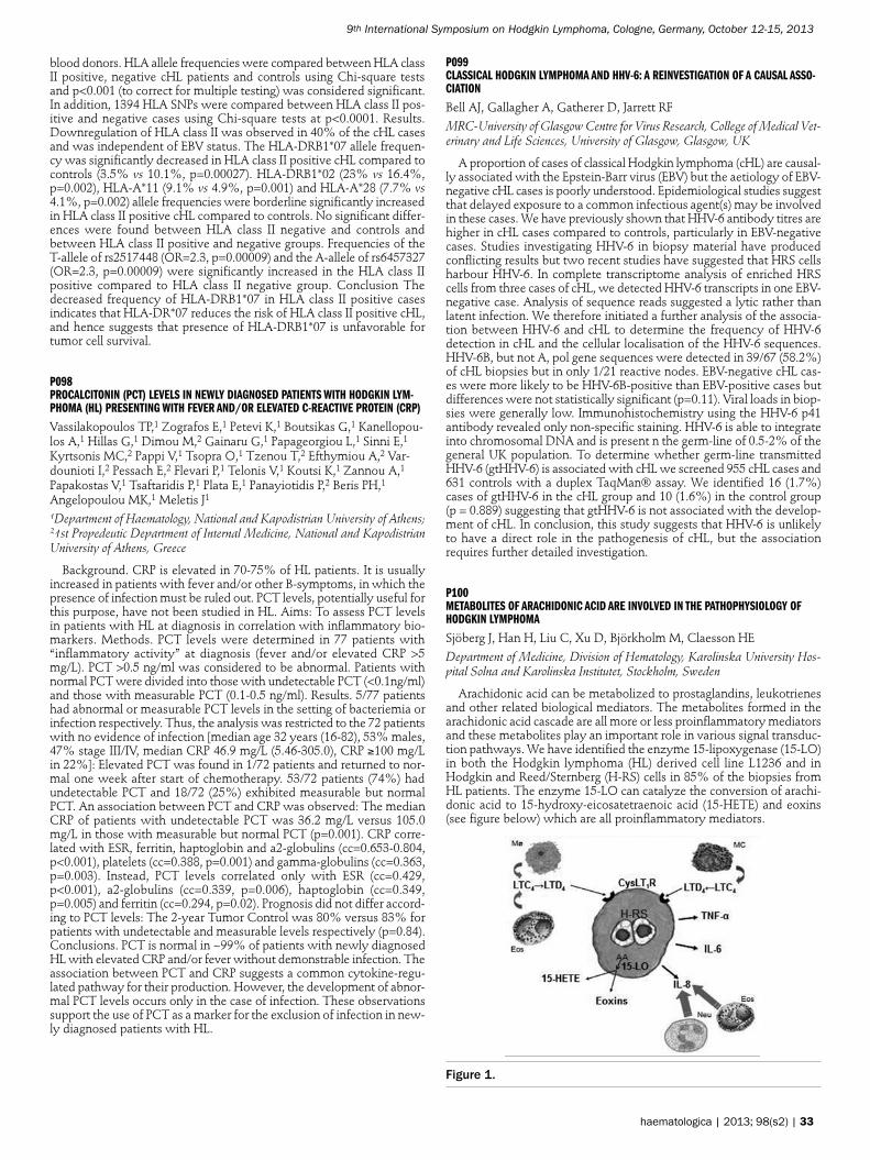
blood donors. HLA allele frequencies were compared between HLA classII positive, negative cHL patients and controls using Chi-square testsand p<0.001 (to correct for multiple testing) was considered significant.In addition, 1394 HLA SNPs were compared between HLA class II pos-itive and negative cases using Chi-square tests at p<0.0001. Results.Downregulation of HLA class II was observed in 40% of the cHL casesand was independent of EBV status. The HLA-DRB1*07 allele frequen-cy was significantly decreased in HLA class II positive cHL compared tocontrols (3.5% vs 10.1%, p=0.00027). HLA-DRB1*02 (23% vs 16.4%,p=0.002), HLA-A*11 (9.1% vs 4.9%, p=0.001) and HLA-A*28 (7.7% vs4.1%, p=0.002) allele frequencies were borderline significantly increasedin HLA class II positive cHL compared to controls. No significant differ-ences were found between HLA class II negative and controls andbetween HLA class II positive and negative groups. Frequencies of theT-allele of rs2517448 (OR=2.3, p=0.00009) and the A-allele of rs6457327(OR=2.3, p=0.00009) were significantly increased in the HLA class IIpositive compared to HLA class II negative group. Conclusion Thedecreased frequency of HLA-DRB1*07 in HLA class II positive casesindicates that HLA-DR*07 reduces the risk of HLA class II positive cHL,and hence suggests that presence of HLA-DRB1*07 is unfavorable fortumor cell survival.
P098PROCALCITONIN (PCT) LEVELS IN NEWLY DIAGNOSED PATIENTS WITH HODGKIN LYM-PHOMA (HL) PRESENTING WITH FEVER AND/OR ELEVATED C-REACTIVE PROTEIN (CRP)
Vassilakopoulos TP,1 Zografos E,1 Petevi K,1 Boutsikas G,1 Kanellopou-los A,1 Hillas G,1 Dimou M,2 Gainaru G,1 Papageorgiou L,1 Sinni E,1Kyrtsonis MC,2 Pappi V,1 Tsopra O,1 Tzenou T,2 Efthymiou A,2 Var-dounioti I,2 Pessach E,2 Flevari P,1 Telonis V,1 Koutsi K,1 Zannou A,1Papakostas V,1 Tsaftaridis P,1 Plata E,1 Panayiotidis P,2 Beris PH,1Angelopoulou MK,1 Meletis J11Department of Haematology, National and Kapodistrian University of Athens;21st Propedeutic Department of Internal Medicine, National and KapodistrianUniversity of Athens, Greece
Background. CRP is elevated in 70-75% of HL patients. It is usuallyincreased in patients with fever and/or other B-symptoms, in which thepresence of infection must be ruled out. PCT levels, potentially useful forthis purpose, have not been studied in HL. Aims: To assess PCT levelsin patients with HL at diagnosis in correlation with inflammatory bio-markers. Methods. PCT levels were determined in 77 patients with“inflammatory activity” at diagnosis (fever and/or elevated CRP >5mg/L). PCT >0.5 ng/ml was considered to be abnormal. Patients withnormal PCT were divided into those with undetectable PCT (<0.1ng/ml)and those with measurable PCT (0.1-0.5 ng/ml). Results. 5/77 patientshad abnormal or measurable PCT levels in the setting of bacteriemia orinfection respectively. Thus, the analysis was restricted to the 72 patientswith no evidence of infection [median age 32 years (16-82), 53% males,47% stage III/IV, median CRP 46.9 mg/L (5.46-305.0), CRP ≥100 mg/Lin 22%]: Elevated PCT was found in 1/72 patients and returned to nor-mal one week after start of chemotherapy. 53/72 patients (74%) hadundetectable PCT and 18/72 (25%) exhibited measurable but normalPCT. An association between PCT and CRP was observed: The medianCRP of patients with undetectable PCT was 36.2 mg/L versus 105.0mg/L in those with measurable but normal PCT (p=0.001). CRP corre-lated with ESR, ferritin, haptoglobin and a2-globulins (cc=0.653-0.804,p<0.001), platelets (cc=0.388, p=0.001) and gamma-globulins (cc=0.363,p=0.003). Instead, PCT levels correlated only with ESR (cc=0.429,p<0.001), a2-globulins (cc=0.339, p=0.006), haptoglobin (cc=0.349,p=0.005) and ferritin (cc=0.294, p=0.02). Prognosis did not differ accord-ing to PCT levels: The 2-year Tumor Control was 80% versus 83% forpatients with undetectable and measurable levels respectively (p=0.84).Conclusions. PCT is normal in ~99% of patients with newly diagnosedHL with elevated CRP and/or fever without demonstrable infection. Theassociation between PCT and CRP suggests a common cytokine-regu-lated pathway for their production. However, the development of abnor-mal PCT levels occurs only in the case of infection. These observationssupport the use of PCT as a marker for the exclusion of infection in new-ly diagnosed patients with HL.
P099CLASSICAL HODGKIN LYMPHOMA AND HHV-6: A REINVESTIGATION OF A CAUSAL ASSO-CIATION
Bell AJ, Gallagher A, Gatherer D, Jarrett RFMRC-University of Glasgow Centre for Virus Research, College of Medical Vet-erinary and Life Sciences, University of Glasgow, Glasgow, UK
A proportion of cases of classical Hodgkin lymphoma (cHL) are causal-ly associated with the Epstein-Barr virus (EBV) but the aetiology of EBV-negative cHL cases is poorly understood. Epidemiological studies suggestthat delayed exposure to a common infectious agent(s) may be involvedin these cases. We have previously shown that HHV-6 antibody titres arehigher in cHL cases compared to controls, particularly in EBV-negativecases. Studies investigating HHV-6 in biopsy material have producedconflicting results but two recent studies have suggested that HRS cellsharbour HHV-6. In complete transcriptome analysis of enriched HRScells from three cases of cHL, we detected HHV-6 transcripts in one EBV-negative case. Analysis of sequence reads suggested a lytic rather thanlatent infection. We therefore initiated a further analysis of the associa-tion between HHV-6 and cHL to determine the frequency of HHV-6detection in cHL and the cellular localisation of the HHV-6 sequences.HHV-6B, but not A, pol gene sequences were detected in 39/67 (58.2%)of cHL biopsies but in only 1/21 reactive nodes. EBV-negative cHL cas-es were more likely to be HHV-6B-positive than EBV-positive cases butdifferences were not statistically significant (p=0.11). Viral loads in biop-sies were generally low. Immunohistochemistry using the HHV-6 p41antibody revealed only non-specific staining. HHV-6 is able to integrateinto chromosomal DNA and is present n the germ-line of 0.5-2% of thegeneral UK population. To determine whether germ-line transmittedHHV-6 (gtHHV-6) is associated with cHL we screened 955 cHL cases and631 controls with a duplex TaqMan® assay. We identified 16 (1.7%)cases of gtHHV-6 in the cHL group and 10 (1.6%) in the control group(p = 0.889) suggesting that gtHHV-6 is not associated with the develop-ment of cHL. In conclusion, this study suggests that HHV-6 is unlikelyto have a direct role in the pathogenesis of cHL, but the associationrequires further detailed investigation.
P100METABOLITES OF ARACHIDONIC ACID ARE INVOLVED IN THE PATHOPHYSIOLOGY OFHODGKIN LYMPHOMA
Sjöberg J, Han H, Liu C, Xu D, Björkholm M, Claesson HEDepartment of Medicine, Division of Hematology, Karolinska University Hos-pital Solna and Karolinska Institutet, Stockholm, Sweden
Arachidonic acid can be metabolized to prostaglandins, leukotrienesand other related biological mediators. The metabolites formed in thearachidonic acid cascade are all more or less proinflammatory mediatorsand these metabolites play an important role in various signal transduc-tion pathways. We have identified the enzyme 15-lipoxygenase (15-LO)in both the Hodgkin lymphoma (HL) derived cell line L1236 and inHodgkin and Reed/Sternberg (H-RS) cells in 85% of the biopsies fromHL patients. The enzyme 15-LO can catalyze the conversion of arachi-donic acid to 15-hydroxy-eicosatetraenoic acid (15-HETE) and eoxins(see figure below) which are all proinflammatory mediators.
Figure 1.
haematologica | 2013; 98(s2) | 33
9th International Symposium on Hodgkin Lymphoma, Cologne, Germany, October 12-15, 2013
Thus, the 15-LO pathway may contribute to the characteristic inflam-matory features of HL. H-RS cells express also the high-affinity recep-tor for cysteinyl-containing leukotrienes, named CysLT1. Both HL celllines and primary biopsies from HL patients express this receptor.Leukotriene D4 stimulated the release of tumor necrosis factor- , inter-leukin-6 and interleukin-8 from the HL cell line L1236 (see figure). Fur-thermore, the HL microenvironment contains eosinophils, mast cellsand macrophages. All these cells can both produce and respond to cys-teinyl-containing leukotrienes. Thus, blocking the CysLT1 receptormight have an impact on the function of H-RS cells and cells in themicroenvironment. Montelukast is a potent CysLT1 antagonist whichis used for treatment of asthma. In summary, this presentation describesnovel biochemical signal transduction pathways operating in H-RS cellsand reveals the potential clinical effect of montelukast in the treatmentof HL. AA, arachidonic acid; LT, leukotriene; CysLT1R, receptor forLTC4 and LTD4; 15-LO, 15-lipoxygenase
P101THE ROLE OF MICRORNA IN HODGKIN LYMPHOMA
Schmidt A,1 Döring C,2 Hansmann ML,2 Küppers R1
1Institute of Cell Biology, Medical School Essen, Dpt. of Molecular Genetics; 2
Senckenberg Institute of Pathology, Medical School, Frankfurt/Main, Germany
Hodgkin lymphoma (HL) is characterized by the rare occurrence ofmalignant Hodgkin/Reed-Sternberg (HRS) cells in an inflammatorytumor microenvironment. The scarcity of HRS cells has hampered theirmolecular analysis. In this study, a protocol for microRNA (miRNA) pro-filing of HRS cells microdissected from paraffin-embedded lymph nodebiopsies was established. We investigated 22 patient samples from dif-ferent HL subtypes, among them CD20- nodular sclerosis (NS), CD20+nodular sclerosis (NS CD20+), mixed cellularity (MC) and nodular lym-phocyte predominant (LP) HL. Expression of 360 miRNAs was deter-mined and compared to that of germinal center (GC) B cells purifiedfrom human tonsils. Unsupervised clustering analyses revealed signifi-cantly different miRNA expression patterns not only of HL and GC Bcell samples but also of different HL subtypes. An HL-specific 5-miRNA-signature could be identified, with 3 miRNAs being up- and 2 miRNAsbeing downregulated in all HL samples and cell lines compared to GCB cells. Functional analyses in HL cell lines with two promising miRNAcandidates being upregulated in HRS cells versus normal GC B cells arecurrently underway. Preliminary data indicate that downregulation ofthese miRNAs indeed influences activity of signaling pathways and sur-vival in HL.
P102TREATMENT PATHWAYS AND RESOURCE USE ASSOCIATED WITH THE MANAGEMENT OFRECURRENT HODGKIN LYMPHOMA AFTER AUTOLOGOUS STEM CELL TRANSPLANTATION
Radford J,1 Johnson R,2 Mckay P,3 Malladi R,4 Peggs K,5 Sureda A,6Bloor A,7 Kerrigan M,8 Ralston S,9 Cooper S,10 Uden R,11 ChristensenK12
1The University of Manchester and The Christie NHS Foundation Trust, Man-chester, UK; 2The Leeds Teaching Hospitals NHS Trust, Leeds, UK; 3The Beat-son West of Scotland Cancer Centre, Glasgow, UK; 4.University Hospitals Birm-ingham NHS Foundation Trust, Birmingham, UK; 5University College LondonHospitals, London, UK; 6Addenbrookes Hospital, Cambridge, UK; 7TheChristie NHS Foundation Trust, Manchester, UK, 8PHMR Associates, Lon-don, UK; 9PHMR Associates, London, UK; 10pH Associates, Marlow, UK;11pH Associates, Marlow, UK; 12Takeda Pharmaceuticals International GmbH
Introduction. Recurrence of Hodgkin lymphoma (HL) occurs inapproximately 50% of patients after autologous stem cell transplanta-tion (ASCT). This retrospective, observational study describes treatmentpathways and resource use at 5 UK Centres in patients with HL whorelapse after ASCT. Methods Patients aged ≥16 years with HL whoreceived ASCT January 2005 to December 2009 and subsequently devel-oped recurrence were identified from hospital records at the 5 Centres.Summary data pre-ASCT and detailed data post-ASCT were collectedfrom recurrence to death or until end December 2012. Incomplete med-ical records were not excluded. Costs were taken from nationally pub-lished sources and applied to the resource use data to derive estimatesof overall financial costs of treatment. Results. 40 patients (22 males, 18
females; mean age at ASCT 33, range 17-71) were included. Mediantime to recurrence post-ASCT was 6 months (range 0.23–65 months).Following recurrence, 19 patients (47.5%) received palliative chemother-apy, 14(35%) received chemotherapy followed by allogeneic transplant(allo), 6(15%) received best supportive care (BSC); 1(2.5%) receivedchemotherapy followed by a 2nd ASCT. Of the 34(85%) patients whoreceived chemotherapy (including allo and 2nd ASCT patients), 12(30%)patients went on to have a 2ndregimen, 6(15%) a 3rd and 2(5%) a 4th.A summary of resource use and mean cost per patient post-ASCT recur-rence are detailed in Table 1. Allo was associated with the fewest out-patient visits (mean 10.81/patient/year) and shortest duration of hospi-tal stay (mean 25.27 days/patient/year) but was the most costly in finan-cial terms (mean £110,374/patient). At April 2012, 10(67%)allo/2ndASCT patients and 3(16%) palliative chemotherapy-onlypatients were alive but all BSC patients had died. Conclusions. Treat-ment for recurrent HL after ASCT is highly variable in terms of intensi-ty, outcome and resource use. 62.5% of patients in this study receivedpalliative or supportive treatment and 37.5% received chemotherapyfollowed by a transplant (mainly allo). Chemotherapy followed by allowas the most expensive treatment pathway but also the most success-ful and represents a cost effective option for appropriately selectedpatients. Less intensive approaches are associated with a dismal out-come.
Table 1.Resource use and costs of treatment pathways for recur-rent Hodgkin Lymphoma after ASCT.
P103THE NOVEL SIRTUIN INHIBITOR, AS4.064 IS ACTIVE AGAINST HODGKIN LYMPHOMALINES AS SINGLE AGENT AND IN COMBINATION WITH OTHER HISTONE DEACETYLASEINHIBITORS.
Kirschbaum MH, Scata K, Sharma A, Desai D, Amin S
Penn State Hershey Medical Center, PA, USA
HDAC inhibitors (HDACi) have proven preclinical and clinical activ-ity against Hodgkin Lymphoma. HDACs are grouped into four cate-gories with classes I, II, and IV, constituting the zinc-dependent familymembers and class III comprising the NAD+ dependent members (alsoknown as sirtuins). The agents that have been studied thus far clinical-ly have been against the zinc based enzymes, but have no activityagainst the NAD+ sirtuins. SelSA-1 is a novel selenium-containinghydroxamic acid derivative HDACi and AS4.064 is a novel seleniumanalog of thiobarbituric acid made in our lab. We have shown thatAS4.064 inhibits sirtuins 1-3 in enzymatic studies. Hodgkin Lymphomalines, along with other lymphoid lines, show decreased proliferationand increased apoptosis when treated with SelSA-1 and/or with AS4.064by viability studies and annexin V studies. Increased acetylation at H3K9and H4K8 is seen with SelSA-1 and enhanced by combination withAS4.064. We have previously demonstrated an important role for c-Mycinhibition in the activity of HDACi in lymphoid malignancies. The com-bination of AS4.064 and SelSA-1 showed enhanced inhibition of c-Mycprotein levels in L540 cells. The acetylation of p53 at various lysines isdifferentially enhanced by single agent versus the combination of thetwo agents. DNA damage response elements such as GADD45 andFOXO3A, which we have previously published as being a critical com-
34 | haematologica | 2013; 98(s2)
Abstracts
ponent of the activity of HDACi, are increased differentially by the twoclasses of agents, results of telomerase, a variety of related miRNAs andother metabolic pathway studies that may be uniquely linked to sirtuininhibition will be shown. In summary, both the novel SIRT1 inhibitor,AS-64, and the novel HDACi, SelSa-1, show growth inhibitory and pro-apoptotic activity in Hodgkin Lymphoma lines, alone and in combina-tion. Inhibition of all HDAC classes, including the NAD+ catalyzeddeacetylases, may enhance the activity seen with the classic zinc basedHDACi. Clinical development of these agents is warranted.
P104THE EPHRIN AND EPHRIN-RECEPTOR FAMILIES ARE WIDELY EXPRESSED IN HODGKINLYMPHOMA
Veenstra R, de Jager W, van den Berg A, Diepstra A, Visser LDepartment of Pathology & Medical Biology, University of Groningen, Univer-sity Medical Center Groningen, The Netherlands
Ephrin (Eph) receptors are the largest family of receptor tyrosine kinas-es and are activated by protein ligands, known as Ephrins (Efn). Hodgkinlymphoma (HL) has a unique cellular composition and consists of lessthan 1% of tumor cells that are surrounded by a reactive infiltrate con-sisting mainly of T-cells. In HL tissue, expression of one Eph-receptor,EphB1, together with a high affinity ligand, EfnB1 has been observed.Expression of 21 Eph-receptors and Efn was analyzed in eight Hodgkinlymphoma cell lines using quantitative RT-PCR. Protein expression of 3Eph-receptors (EphA1, EphA3 and EphB1) and 3 Efn (EfnA3, EfnA4 andEfnB1) was evaluated in 5 nodular lymphocyte predominant (NLP)-HLand 25 classical HL (cHL) tissue samples. EfnA3, EfnA4 and EfnB1mRNA expression was detected in almost all HL cell lines. Expression ofEphA1, EphA3, EfnA5, EphB1, EphB4 and EfnB3 was found in approx-imately 50% of the HL cell lines, while EphA4 was present in 2/8 HL celllines. Protein expression was observed in the tumor cells of all NLP-HLcases for EfnA4 and EfnB1, in 3/5 NLP-HL cases for EphA1 and in 4/5 cas-es for EphA3. EfnA3 was not detectable in NLP-HL. Protein expressionfor EphA3, EfnA3 EfnA4 and EfnB1 was found in 50% of the cHLpatients. EphA1 was not detectable in the cHL cases and EphB1 stainingwas inconclusive. The main difference between NLP-HL and cHL wasexpression of EphA1 in NLP-HL and not in cHL and expression of EfnA3in cHL and not in NLP-HL. In conclusion, we show that the Eph-fami-ly is widely expressed in HL tumor cells, with marked differences forEphA1 and EfnA3 between NLP and cHL.
P105TELOMERASE DEPENDENT AND TELOMERASE INDEPENDENT TELOMERE MAINTENANCEMECHANISMS IN HODGKIN LYMPHOMA
Cuceu C,1 Morat L,1 Lemain A,1 Shim G,1 Frenzel M,1 Ricoul M,1 Hen-pel W,1 Guileto E,2 Junker S,3 Girinsky T,4 Carde P,5 Delhem N,6Sabatier L,1 M’kacher R11Laboratoire de Radiobiologie et d’Oncologie, IRCM/DSV/CEA Fontenay auxroses, France; 2Laboratory of Cellular and Molecular biology, University DiPavia, Italy; 3Institute of Human Genetics University of Aarhus Hospital, Den-mark; 4Departement of Radiation Therapy, Gustave Roussy Institut , VillejuifFrance; 5Departement of Medicine, Gustave Roussy Institut , Villejuif France;6Immunoregulation of Viro-induit Cancers, Institut of Biology, Lille France
Background. Telomere dysfunction in Hodgkin Lymphoma (HL)requires that the cells activate a telomere maintenance mechanism(TMM) to support immortalization. Most tumor cells activate expressionof the enzyme telomerase. Some cells elongate telomeres using telom-erase-independent mechanisms, known as alternative lengthening oftelomeres (ALT). A telomerase-independent mechanism for TMM in HLhas been proposed in the absence of detectable telomerase activity (TA)in some cases. In this study, we have analyzed the TMM in HL cell lines,lymph nodes and peripheral blood lymphocytes of HL patients. Ourstudies demonstrate that TMMs are not mutually exclusive in Hodgkinlymphoma and, indeed, reveal the presence of multiple TMM. Materi-als and Methods. Spectral Karyotype, FISH painting, telomere length(TL), TA measurement, immunofluorescence and western blot of differ-ent proteins were performed on 50 frozen samples obtained frompatients with classic Hodgkin lymphoma during diagnostic lymph nodebiopsy, blood samples and 7 HL cell lines (L428, KMH2, L540, L591,
HD-LM2, L1236, SUP-HD). Results. The major finding of this study isthe presence of both telomerase and ALT in selected lymph nodes of HLpatients. This TMM heterogeneity was revealed in circulating lympho-cytes and in HL cell lines. We have identified a subset of tumors withsome cells containing ALT-associated PML bodies, , and separate cellsexpressing telomerase in the same tumor. In 50 lymph node tumors, 3contained only ALT-positive cells, 6 contained only telomerase positivecells and 41 contained a mixed population of ALT and telomerase-posi-tive cells. Similarly, in HL cell lines, a high level of TA was detected inL428 and SUP-HD. L1236 presented an ALT profile. In circulating lym-phocytes, Telomerase activity was detected in 18 from 50. A significantcorrelation was observed between telomere length and the TMM. Inter-estingly, Jak2 amplification and complex chromosomal rearrangementsincluding dicentrics were observed in patients and cell lines with an ALTprofile. Conclusion. The presence of both TMMs (TA and ALT) inLymph nodes, peripheral blood lymphocytes and cell lines cells is unique.Further investigation could establish the relationship between TMMsand the clinical outcome of patients.
P106PRELIMINARY CHARACTERIZATION OF MHC POLYMORPHISMS AS RISK FACTORS FORPEDIATRIC AND ADULT CLASSICAL HODGKIN LYMPHOMA IN A REGION WITH EARLY EBVSEROCONVERSION
Hassan R,1 Garcia A,1 Cohen M,2 Vera-Lozada G,1 Oliveira-Silva M,1Segges P,1 Gantuz M,2 De Matteo E,2 Barros MH,3 Preciado MV,2Chabay P21Oncovirology Laboratory, Instituto Nacional do Câncer-INCA, Rio de Janeiro,Brazil; 2Molecular Biology Laboratory, Pathology Division, Ricardo GutiérrezChildren Hospital, Ciudad de Buenos Aires, Argentina; 3Institute for Pathology,Unfallkrankenhaus Berlin, Berlin, Germany
Genetic factors linked to the Major Histocompatibility Complex(MHC) have been associated to the risk of classical Hodgkin lymphoma(cHL) in distinct epidemiological frameworks (i.e. developed countries,EBV late seroconversion, adults). This is a pilot study aiming to explorethe role of MHC genetic variants in the risk of cHL in developing coun-tries where early EBV seronversion is the rule. A group of 84 children andadolescents (3-18 years, median 11; M:F 2.45) and 67 adults (19-82 years,median 32; M:F 1.7) with cHL, and 67 reactive hyperplasia (2-48 years,M:F 1.2) were included, from cases diagnosed in Brazil and Argentinapublic Hospitals. EBV was detected by EBER-ISH and PCR. In 147 cas-es, HLAs A/B/DRB were genotyped using the LabType®SSO typing test(Luminex). HLA-linked SNPs (rs6457110, rs2530388, rs6903608, andrs2523969) were genotyped using TaqMan® probes. NS subtype wasdetected in 46% children and 68% adults, followed by MC (41% vs14%). EBV was detected in 64% of pediatric cHL, 54.5% of adult cHLand 39% controls. The more frequent HLA alleles were HLA-A*02(43%), HLA-B*44 (14%) e HLA-DRB*13 (28%), without differencesbetween cases and controls, regardless of EBV status. DR3 and DR8 alle-les were preferentially associated with EBV+ cHL females (38.5% vs6.4% males; p=0.017; p=0.003). Case-control comparison for rs6457110disclosed no significant associations. When compared EBV+ and EBV-cHL stratified by sex, the A allele/AA genotype were more frequent inmale EBV+ cases (27% vs 5% female, p=0.02). For SNP rs2330388, no dif-ferences were found between cases and controls. A allele/AA genotypewere associated with EBV+ cHL male children (p=0.018). For SNPrs6903608, genotypic frequencies were significantly different in casesand controls (CC 20% vs 6%; p= 0.016), more marked in the EBV-neg-ative cases. In sum, no direct associations were demonstrated betweenHLA-A*01 or HLA-A*02 and risk of cHL in this region. However, a com-plex interaction was found among specific alleles of HLA and SNPs inrespect of sex, age, and EBV, which demands, besides in=creasing sam-ple size, specific experimental designs to disclose age- and sex-associat-ed risks in this region with early EBV seroconversion. Financial Support:CAPES (Brazil)/MinCyT (Argentina).
haematologica | 2013; 98(s2) | 35
9th International Symposium on Hodgkin Lymphoma, Cologne, Germany, October 12-15, 2013
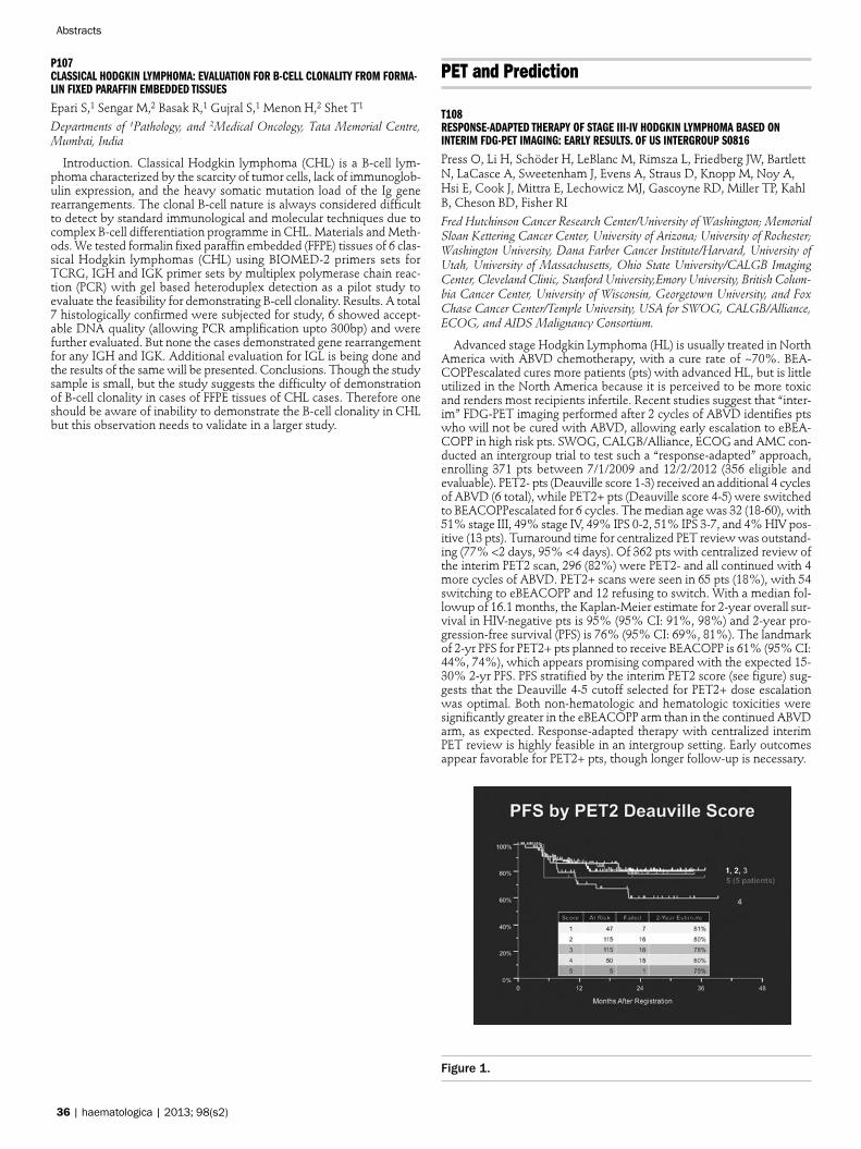
P107CLASSICAL HODGKIN LYMPHOMA: EVALUATION FOR B-CELL CLONALITY FROM FORMA-LIN FIXED PARAFFIN EMBEDDED TISSUES
Epari S,1 Sengar M,2 Basak R,1 Gujral S,1 Menon H,2 Shet T1
Departments of 1Pathology, and 2Medical Oncology, Tata Memorial Centre,Mumbai, India
Introduction. Classical Hodgkin lymphoma (CHL) is a B-cell lym-phoma characterized by the scarcity of tumor cells, lack of immunoglob-ulin expression, and the heavy somatic mutation load of the Ig generearrangements. The clonal B-cell nature is always considered difficultto detect by standard immunological and molecular techniques due tocomplex B-cell differentiation programme in CHL. Materials and Meth-ods. We tested formalin fixed paraffin embedded (FFPE) tissues of 6 clas-sical Hodgkin lymphomas (CHL) using BIOMED-2 primers sets forTCRG, IGH and IGK primer sets by multiplex polymerase chain reac-tion (PCR) with gel based heteroduplex detection as a pilot study toevaluate the feasibility for demonstrating B-cell clonality. Results. A total7 histologically confirmed were subjected for study, 6 showed accept-able DNA quality (allowing PCR amplification upto 300bp) and werefurther evaluated. But none the cases demonstrated gene rearrangementfor any IGH and IGK. Additional evaluation for IGL is being done andthe results of the same will be presented. Conclusions. Though the studysample is small, but the study suggests the difficulty of demonstrationof B-cell clonality in cases of FFPE tissues of CHL cases. Therefore oneshould be aware of inability to demonstrate the B-cell clonality in CHLbut this observation needs to validate in a larger study.
PET and Prediction
T108RESPONSE-ADAPTED THERAPY OF STAGE III-IV HODGKIN LYMPHOMA BASED ONINTERIM FDG-PET IMAGING: EARLY RESULTS. OF US INTERGROUP S0816
Press O, Li H, Schöder H, LeBlanc M, Rimsza L, Friedberg JW, BartlettN, LaCasce A, Sweetenham J, Evens A, Straus D, Knopp M, Noy A,Hsi E, Cook J, Mittra E, Lechowicz MJ, Gascoyne RD, Miller TP, KahlB, Cheson BD, Fisher RI
Fred Hutchinson Cancer Research Center/University of Washington; MemorialSloan Kettering Cancer Center, University of Arizona; University of Rochester;Washington University, Dana Farber Cancer Institute/Harvard, University ofUtah, University of Massachusetts, Ohio State University/CALGB ImagingCenter, Cleveland Clinic, Stanford University,Emory University, British Colum-bia Cancer Center, University of Wisconsin, Georgetown University, and FoxChase Cancer Center/Temple University, USA for SWOG, CALGB/Alliance,ECOG, and AIDS Malignancy Consortium.
Advanced stage Hodgkin Lymphoma (HL) is usually treated in NorthAmerica with ABVD chemotherapy, with a cure rate of ~70%. BEA-COPPescalated cures more patients (pts) with advanced HL, but is littleutilized in the North America because it is perceived to be more toxicand renders most recipients infertile. Recent studies suggest that “inter-im” FDG-PET imaging performed after 2 cycles of ABVD identifies ptswho will not be cured with ABVD, allowing early escalation to eBEA-COPP in high risk pts. SWOG, CALGB/Alliance, ECOG and AMC con-ducted an intergroup trial to test such a “response-adapted” approach,enrolling 371 pts between 7/1/2009 and 12/2/2012 (356 eligible andevaluable). PET2- pts (Deauville score 1-3) received an additional 4 cyclesof ABVD (6 total), while PET2+ pts (Deauville score 4-5) were switchedto BEACOPPescalated for 6 cycles. The median age was 32 (18-60), with51% stage III, 49% stage IV, 49% IPS 0-2, 51% IPS 3-7, and 4% HIV pos-itive (13 pts). Turnaround time for centralized PET review was outstand-ing (77% <2 days, 95% <4 days). Of 362 pts with centralized review ofthe interim PET2 scan, 296 (82%) were PET2- and all continued with 4more cycles of ABVD. PET2+ scans were seen in 65 pts (18%), with 54switching to eBEACOPP and 12 refusing to switch. With a median fol-lowup of 16.1 months, the Kaplan-Meier estimate for 2-year overall sur-vival in HIV-negative pts is 95% (95% CI: 91%, 98%) and 2-year pro-gression-free survival (PFS) is 76% (95% CI: 69%, 81%). The landmarkof 2-yr PFS for PET2+ pts planned to receive BEACOPP is 61% (95% CI:44%, 74%), which appears promising compared with the expected 15-30% 2-yr PFS. PFS stratified by the interim PET2 score (see figure) sug-gests that the Deauville 4-5 cutoff selected for PET2+ dose escalationwas optimal. Both non-hematologic and hematologic toxicities weresignificantly greater in the eBEACOPP arm than in the continued ABVDarm, as expected. Response-adapted therapy with centralized interimPET review is highly feasible in an intergroup setting. Early outcomesappear favorable for PET2+ pts, though longer follow-up is necessary.
Figure 1.
36 | haematologica | 2013; 98(s2)
Abstracts
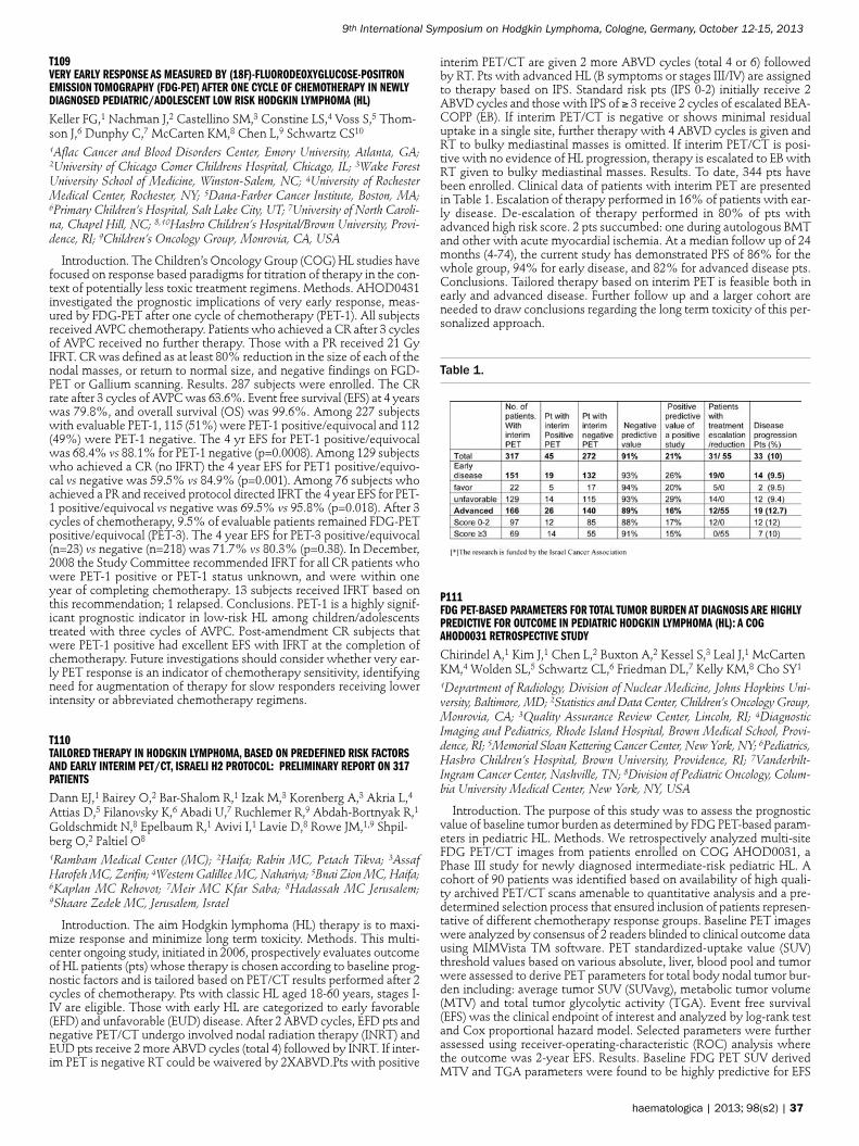
T109VERY EARLY RESPONSE AS MEASURED BY (18F)-FLUORODEOXYGLUCOSE-POSITRONEMISSION TOMOGRAPHY (FDG-PET) AFTER ONE CYCLE OF CHEMOTHERAPY IN NEWLYDIAGNOSED PEDIATRIC/ADOLESCENT LOW RISK HODGKIN LYMPHOMA (HL)
Keller FG,1 Nachman J,2 Castellino SM,3 Constine LS,4 Voss S,5 Thom-son J,6 Dunphy C,7 McCarten KM,8 Chen L,9 Schwartz CS101Aflac Cancer and Blood Disorders Center, Emory University, Atlanta, GA;2University of Chicago Comer Childrens Hospital, Chicago, IL; 3Wake ForestUniversity School of Medicine, Winston-Salem, NC; 4University of RochesterMedical Center, Rochester, NY; 5Dana-Farber Cancer Institute, Boston, MA;6Primary Children’s Hospital, Salt Lake City, UT; 7University of North Caroli-na, Chapel Hill, NC; 8,10Hasbro Children’s Hospital/Brown University, Provi-dence, RI; 9Children’s Oncology Group, Monrovia, CA, USA
Introduction. The Children’s Oncology Group (COG) HL studies havefocused on response based paradigms for titration of therapy in the con-text of potentially less toxic treatment regimens. Methods. AHOD0431investigated the prognostic implications of very early response, meas-ured by FDG-PET after one cycle of chemotherapy (PET-1). All subjectsreceived AVPC chemotherapy. Patients who achieved a CR after 3 cyclesof AVPC received no further therapy. Those with a PR received 21 GyIFRT. CR was defined as at least 80% reduction in the size of each of thenodal masses, or return to normal size, and negative findings on FGD-PET or Gallium scanning. Results. 287 subjects were enrolled. The CRrate after 3 cycles of AVPC was 63.6%. Event free survival (EFS) at 4 yearswas 79.8%, and overall survival (OS) was 99.6%. Among 227 subjectswith evaluable PET-1, 115 (51%) were PET-1 positive/equivocal and 112(49%) were PET-1 negative. The 4 yr EFS for PET-1 positive/equivocalwas 68.4% vs 88.1% for PET-1 negative (p=0.0008). Among 129 subjectswho achieved a CR (no IFRT) the 4 year EFS for PET1 positive/equivo-cal vs negative was 59.5% vs 84.9% (p=0.001). Among 76 subjects whoachieved a PR and received protocol directed IFRT the 4 year EFS for PET-1 positive/equivocal vs negative was 69.5% vs 95.8% (p=0.018). After 3cycles of chemotherapy, 9.5% of evaluable patients remained FDG-PETpositive/equivocal (PET-3). The 4 year EFS for PET-3 positive/equivocal(n=23) vs negative (n=218) was 71.7% vs 80.3% (p=0.38). In December,2008 the Study Committee recommended IFRT for all CR patients whowere PET-1 positive or PET-1 status unknown, and were within oneyear of completing chemotherapy. 13 subjects received IFRT based onthis recommendation; 1 relapsed. Conclusions. PET-1 is a highly signif-icant prognostic indicator in low-risk HL among children/adolescentstreated with three cycles of AVPC. Post-amendment CR subjects thatwere PET-1 positive had excellent EFS with IFRT at the completion ofchemotherapy. Future investigations should consider whether very ear-ly PET response is an indicator of chemotherapy sensitivity, identifyingneed for augmentation of therapy for slow responders receiving lowerintensity or abbreviated chemotherapy regimens.
T110TAILORED THERAPY IN HODGKIN LYMPHOMA, BASED ON PREDEFINED RISK FACTORSAND EARLY INTERIM PET/CT, ISRAELI H2 PROTOCOL: PRELIMINARY REPORT ON 317PATIENTS
Dann EJ,1 Bairey O,2 Bar-Shalom R,1 Izak M,3 Korenberg A,3 Akria L,4Attias D,5 Filanovsky K,6 Abadi U,7 Ruchlemer R,9 Abdah-Bortnyak R,1Goldschmidt N,8 Epelbaum R,1 Avivi I,1 Lavie D,8 Rowe JM,1,9 Shpil-berg O,2 Paltiel O8
1Rambam Medical Center (MC); 2Haifa; Rabin MC, Petach Tikva; 3AssafHarofeh MC, Zerifin; 4Western Galillee MC, Nahariya; 5Bnai Zion MC, Haifa;6Kaplan MC Rehovot; 7Meir MC Kfar Saba; 8Hadassah MC Jerusalem;9Shaare Zedek MC, Jerusalem, Israel
Introduction. The aim Hodgkin lymphoma (HL) therapy is to maxi-mize response and minimize long term toxicity. Methods. This multi-center ongoing study, initiated in 2006, prospectively evaluates outcomeof HL patients (pts) whose therapy is chosen according to baseline prog-nostic factors and is tailored based on PET/CT results performed after 2cycles of chemotherapy. Pts with classic HL aged 18-60 years, stages I-IV are eligible. Those with early HL are categorized to early favorable(EFD) and unfavorable (EUD) disease. After 2 ABVD cycles, EFD pts andnegative PET/CT undergo involved nodal radiation therapy (INRT) andEUD pts receive 2 more ABVD cycles (total 4) followed by INRT. If inter-im PET is negative RT could be waivered by 2XABVD.Pts with positive
interim PET/CT are given 2 more ABVD cycles (total 4 or 6) followedby RT. Pts with advanced HL (B symptoms or stages III/IV) are assignedto therapy based on IPS. Standard risk pts (IPS 0-2) initially receive 2ABVD cycles and those with IPS of ≥ 3 receive 2 cycles of escalated BEA-COPP (EB). If interim PET/CT is negative or shows minimal residualuptake in a single site, further therapy with 4 ABVD cycles is given andRT to bulky mediastinal masses is omitted. If interim PET/CT is posi-tive with no evidence of HL progression, therapy is escalated to EB withRT given to bulky mediastinal masses. Results. To date, 344 pts havebeen enrolled. Clinical data of patients with interim PET are presentedin Table 1. Escalation of therapy performed in 16% of patients with ear-ly disease. De-escalation of therapy performed in 80% of pts withadvanced high risk score. 2 pts succumbed: one during autologous BMTand other with acute myocardial ischemia. At a median follow up of 24months (4-74), the current study has demonstrated PFS of 86% for thewhole group, 94% for early disease, and 82% for advanced disease pts.Conclusions. Tailored therapy based on interim PET is feasible both inearly and advanced disease. Further follow up and a larger cohort areneeded to draw conclusions regarding the long term toxicity of this per-sonalized approach.
Table 1.
P111FDG PET-BASED PARAMETERS FOR TOTAL TUMOR BURDEN AT DIAGNOSIS ARE HIGHLYPREDICTIVE FOR OUTCOME IN PEDIATRIC HODGKIN LYMPHOMA (HL): A COGAHOD0031 RETROSPECTIVE STUDY
Chirindel A,1 Kim J,1 Chen L,2 Buxton A,2 Kessel S,3 Leal J,1 McCartenKM,4 Wolden SL,5 Schwartz CL,6 Friedman DL,7 Kelly KM,8 Cho SY11Department of Radiology, Division of Nuclear Medicine, Johns Hopkins Uni-versity, Baltimore, MD; 2Statistics and Data Center, Children’s Oncology Group,Monrovia, CA; 3Quality Assurance Review Center, Lincoln, RI; 4DiagnosticImaging and Pediatrics, Rhode Island Hospital, Brown Medical School, Provi-dence, RI; 5Memorial Sloan Kettering Cancer Center, New York, NY; 6Pediatrics,Hasbro Children’s Hospital, Brown University, Providence, RI; 7Vanderbilt-Ingram Cancer Center, Nashville, TN; 8Division of Pediatric Oncology, Colum-bia University Medical Center, New York, NY, USA
Introduction. The purpose of this study was to assess the prognosticvalue of baseline tumor burden as determined by FDG PET-based param-eters in pediatric HL. Methods. We retrospectively analyzed multi-siteFDG PET/CT images from patients enrolled on COG AHOD0031, aPhase III study for newly diagnosed intermediate-risk pediatric HL. Acohort of 90 patients was identified based on availability of high quali-ty archived PET/CT scans amenable to quantitative analysis and a pre-determined selection process that ensured inclusion of patients represen-tative of different chemotherapy response groups. Baseline PET imageswere analyzed by consensus of 2 readers blinded to clinical outcome datausing MIMVista TM software. PET standardized-uptake value (SUV)threshold values based on various absolute, liver, blood pool and tumorwere assessed to derive PET parameters for total body nodal tumor bur-den including: average tumor SUV (SUVavg), metabolic tumor volume(MTV) and total tumor glycolytic activity (TGA). Event free survival(EFS) was the clinical endpoint of interest and analyzed by log-rank testand Cox proportional hazard model. Selected parameters were furtherassessed using receiver-operating-characteristic (ROC) analysis wherethe outcome was 2-year EFS. Results. Baseline FDG PET SUV derivedMTV and TGA parameters were found to be highly predictive for EFS
haematologica | 2013; 98(s2) | 37
9th International Symposium on Hodgkin Lymphoma, Cologne, Germany, October 12-15, 2013
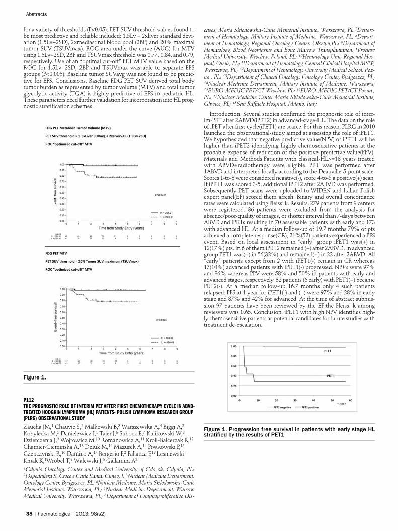
for a variety of thresholds (P<0.05). PET SUV threshold values found tobe most predictive and reliable included: 1.5Lv + 2xliver standard devi-ation (1.5Lv+2SD), 2xmediastinal blood pool (2BP) and 20% maximaltumor SUV (TSUVmax). ROC area under the curve (AUC) for MTVusing 1.5Lv+2SD, 2BP and TSUVmax threshold was 0.77, 0.84, and 0.79,respectively. Use of an “optimal cut-off” PET MTV value based on theROC for 1.5Lv+2SD, 2BP and TSUVmax was able to separate EFSgroups (P<0.005). Baseline tumor SUVavg was not found to be predic-tive for EFS. Conclusions. Baseline FDG PET SUV derived total bodytumor burden as represented by tumor volume (MTV) and total tumorglycolytic activity (TGA) is highly predictive of EFS in pediatric HL.These parameters need further validation for incorporation into HL prog-nostic stratification schemes.
Figure 1.
P112THE PROGNOSTIC ROLE OF INTERIM PET AFTER FIRST CHEMOTHERAPY CYCLE IN ABVD-TREATED HODGKIN LYMPHOMA (HL) PATIENTS- POLISH LYMPHOMA RESEARCH GROUP(PLRG) OBSERVATIONAL STUDY
Zaucha JM,1 Chauvie S,2 Malkowski B,3 Warszewska A,4 Biggi A,2Kobylecka M,5 Danielewicz I,1 Tajer J,6 Subocz E,7 Kulikowski W,8Dzietczenia J,9 Wojtowicz M,10 Romanowicz A,11 Kroll-Balcerzak R,12Chamier-Ciemin�ska A,13 Dziuk M,14 Mazurek A,14 Piwkowski P,15Czepczyn�ski R,16 Damico A,17 Bergesio F,2 Fallanca F,18 Lesniewski-Kmak K,1Wróbel T,9 Walewski J,6 Gallamini A2
1Gdynia Oncology Center and Medical University of Gda sk, Gdynia, PL;2Ospedaliera S. Croce e Carle Santa, Cuneo, I; 3Nuclear Medicine Department,Oncology Center, Bydgoszcz, PL; 4Nuclear Medicine, Maria Sklodowska-CurieMemorial Institute, Warszawa, PL; 5Nuclear Medicine Department, WarsawMedical University, Warszawa, PL; 6Department of Lymphoproliferative Dis-
eases, Maria Sklodowska-Curie Memorial Institute, Warszawa, PL 7Depart-ment of Hematology, Military Institute of Medicine, Warszawa, PL; 8Depart-ment of Hematology, Regional Oncology Center, Olsztyn,PL; 9Department ofHematology, Blood Neoplasms and Bone Marrow Transplantation, WroclawMedical University, Wroclaw, Poland, PL; 10Hematology Unit, Regional Hos-pital, Opole, PL; 11Department of Hematology, Central Clinical Hospital MSW,Warszawa, PL; 12Department of Hematology, University Medical School, Poz-na , PL; 13Department of Clinical Oncology, Oncology Center, Bydgoszcz, PL;14Nuclear Medicine Department, Military Institute of Medicine, Warszawa;15EURO-MEDIC PET/CT Wroclaw, PL; 16EURO-MEDIC PET/CT Pozna ,PL; 17Nuclear Medicine Center Maria Sklodowska-Curie Memorial Institute,Gliwice, PL; 18San Raffaele Hospital, Milano, Italy
Introduction. Several studies confirmed the prognostic role of inter-im-PET after 2ABVD(iPET2) in advanced-stage-HL. The data on the roleof iPET after first-cycle(iPET1) are scarce. For this reason, PLRG in 2010launched the observational-study aimed at assessing the role of iPET1.We hypothesized that negative predictive value(NPV) of iPET1 will behigher than iPET2 identifying highly chemosensitive patients at theprobable expense of reduction of the positive predictive value(PPV).Materials and Methods.Patients with classical-HL>=18 years treatedwith ABVD±radiotherapy were eligible. PET was performed after1ABVD and interpreted locally according to the Deauville-5-point scale.Scores 1-to-3 were considered negative(-), score 4-to-5 a positive(+) scan.If iPET1 was scored 3-5, additional iPET2 after 2ABVD was performed.Subsequently PET scans were uploaded to WIDEN and Italian-Polishexpert panel(EP) scored them afresh. Binary and overall concordancerates were calculated using Fleiss’ k. Results. 279 patients from 9 centerswere registered. 36 patients were excluded from the analysis forabsence/poor-quality of images, or shorter interval than 7-days betweenABVD and iPETs resulting in 70 assessable patients with early and 173with advanced HL. At a median follow-up of 19.7 months 79% of ptsachieved a complete response(CR), 21%(52) patients experienced a PFSevent. Based on local assessment in “early” group iPET1 was(+) in12(17%) pts. In 6 of them iPET2 remained (+) after 2ABVD. In advancedgroup PET1 was(+) in 56(32%) and remained(+) in 22 after 2ABVD. All“early” patients except from 2 with iPET1(-) remain in CR whereas17(10%) advanced patients with iPET1(-) progressed. NPVs were 97%and 86% whereas PPV were 58% and 50% in patients with early andadvanced stages, respectively. 32 patients (6 early) with PET1(+) becamePET2(-). At a median follow-up 16.7 months only 4 such patientsrelapsed. PFS at 1 year for iPET1(-) and (+) were 97% and 28% in earlystage and 87% and 42% for advanced. At the time of abstract submis-sion 97 patients have been reviewed by the EP:the Fleiss’ k amongreviewers was 0.65. Conclusion. iPET1 with high NPV identifies high-ly chemosensitive patients as potential candidates for future studies withtreatment de-escalation.
Figure 1. Progression free survival in patients with early stage HLstratified by the results of PET1
38 | haematologica | 2013; 98(s2)
Abstracts

P113UPSTAGING BY PET-ASCERTAINED BONE AND BONE MARROW INVOLVEMENT INHODGKIN LYMPHOMA–DEFINITIONS MATTER
Borra A,1 El-Galaly TC,2 Zaucha JM,3 Rigacci L,4 Rapezzi D,1 Rago A,5Bolis S,6 Gavarotti P,7 Rusconi C,8 Trentin L,9 Stelitano C,10 Biggi A,11Gormsen LC,12 d’Amore F,13 Hutchings M,14 Gallamini A11Hematology Department Cuneo Hospital, Italy; 2Hematology Department,Aarhus and Aalborg University Hospital, Denmark; 3Onco-Hematology Depart-ment, Gdynya University Hospital, Poland; 4Hematology Chair , PoliclinicoCareggi, University of Florence, Florence, Italy; 5University Chair, HematologyDepartment, Latina Hospital; 6Hematology Department, S. Gerardo Hospital,Monza, Italy; 7Hematology Chair, S. Giovanni Battista Hospital, Turin Univer-sity, Turin, Italy; 8Hematology Department, Ca’ Granda Niguarda Hospital,Milan, Italy; 9Hematology Department, University of Padua, Italy; 10Hematol-ogy Department, Melacrino and Morelli Hospital, Reggio Calabria, Italy;11Nuclear Medicine Department, Cuneo Hospital, Italy; 12Nuclear MedicineDepartment, Aarhus University Hospital, Denmark; 13Hematology Department,Aarhus University Hospital, Denmark; 14Department of Hematology, Rigshos-pitalet, Copenhagen University Hospital, Denmark
Background. PET/CT is sensitive for extranodal involvement inHodgkin lymphoma (HL). Different patterns of FDG-uptake in thebone/bone marrow (B/BM) are reported indicating HL infiltration. Meth-ods. Patients with intermediate-advanced stage HL from 11 Italian, Dan-ish and Polish centres were included in this retrospective study. Allpatients had been treated with ABVD chemotherapy +/- consolidatingradiotherapy. Baseline CT and PET/CT scans were reviewed accordingto a predefined scheme. FDG-uptake in the B/BM was categorized as:1) focal uptake > liver-uptake, 2) diffuse uptake > liver-uptake, 3) nouptake. The CT and PET/CT stages were compared when applying dif-ferent definitions for B/BM involvement. In setting A) criteria only 1) wasaccepted as B/BM disease. In setting B) criteria 1) and 2) were equallyaccepted as B/BM disease. Results. 143 patients mainly with classical HL(99%) were included. In setting A) PET/CT upstaged 19% (n=27) anddownstaged 7% (n=10) of the patients. In setting B) PET/CT upstaged30% (n=43) and downstaged 6% (n=8), p=0.04 for setting A vs B. In set-ting A) upstaging to stage IV by PET/CT was due to B/BM alone in 10%(n=14) and other extranodal involvement +/- B/BM in 4% (n=6).In set-ting B) upstaging to stage IV by PET/CT was due to B/BM alone in 21%(n=33) and other extranodal involvement +/- B/BM in 4% (n=6). In 8 outof 9 patients with positive BM biopsy focal B/BM FDG-uptake was pres-ent, while none had diffuse BM FDG-uptake. Patients with diffuse BMFDG-uptake had higher median leukocyte count (11400 vs 9445/mm3,p=0.0006) and lower median haemoglobin level (117 vs 131 g/L, p=0.01)as compared to patients without diffuse uptake. In patients with FDG-uptake in B/BM, a diffuse FDG-uptake was associated with trend towardbetter outcome (p=0.3) as compared to focal FDG-uptake. Conclusions.Detection of B/BM involvement by PET/CT is the most frequent causeof stage IV upstaging by PET/CT and magnitude of upstaging dependson the criteria used for B/BM disease. Diffuse uptake even when exceed-ing liver uptake is unlikely related to B/BM invasion. Strict and uniforminterpretation criteria for FDG-uptake in the BM are needed.
P114COMPARATIVE ASSESSMENT OF BONE MARROW INVOLVEMENT (BMI) BY BM BIOPSY(BMB) OR POSITRON EMISSION TOMOGRAPHY / COMPUTED TOMOGRAPHY (PET/CT)IN HODGKIN LYMPHOMA (HL)
Vassilakopoulos TP,1 Angelopoulou MK,1 Prassopoulos V,2 Chatziioan-nou S,3 Moschogiannis M,4 Tsirkinidis P,4 Poziopoulos C,5 SymeonidisA,6 Repoussis P,7 Matsouka Ch,8 Kontopidou FN,9 Sotiropoulos V,10Variami E,11 Viniou NA,11 Zikos P,12 Petevi K,1 Boutsikas G,1 Kanel-lopoulos A,1 Papageorgiou L,1 Panayiotidis P,13 Pangalis GA,13 DatserisI,14 Meletis J,1 Rondogianni Ph141Department of Haematology, National and Kapodistrian University of Athens,2Department of Nuclear Medicine/PET, HYGEIA Hospital, 3Department ofNuclear Medicine/PET, Biomedical Research Foundation, Academy of Athens,4Department of Haematology, Athens Medical Center, 5Department of Haema-tology, Metropolitan General Hospital, 6Department of Haematology, Universi-ty of Patras, 7Department of Haematology, Metaxas Anticancer Hospital,8Department of Therapeutics, National and Kapodistrian University of Athens,92nd Department of Internal Medicine, National and Kapodistrian University
of Athens, 10Department of Nuclear Medicine, Athens Medical Center, 111stDepartment of Internal Medicine, National and Kapodistrian University ofAthens, 12Department of Haematology, Ag. Andreas General Hospital, 131stPropedeutic Department of Internal Medicine, National and Kapodistrian Uni-versity of Athens, 14Department of Nuclear Medicine/PET, Evangelismos Gen-eral Hospital, Greece
Introduction. PET/CT is a sensitive tool for HL staging. Data suggestthat few pts may have positive BMB in the absence of PET/CT evidence.Aims. 1)To correlate BMB and disease characteristics with BM-PET/CTfindings; 2)to assess the impact of our published clinical prediction rule(Vassilakopoulos et al, Blood. 2005) on the frequency of BMI; 3)to assessthe ability to omit BMB in selected or even all pts. Methods. Data regard-ing BMB, clinical and laboratory characteristics were retrieved. PET/CTdata were reviewed according to osseous/BM findings and were visual-ly graded as follows: (1)no increased; (2)increased ≤liver; (3)increasedBM FDG uptake >liver; (4)solitary osseous/BM focus without CT cor-relate; (5)multiple osseous/BM foci. Pts were classified according to ourprediction rule for BMI in low-, standard- and high-risk groups. Results.PET/CT and BMB data were available for 172 pts; PET/CT was negativefor BMI in 142 (82%) and positive in 30 pts: 3 had a single focus and 27had multiple foci. 13 had BMI by BMB (7.6%). None of the pts ofPET/CT categories 1,2,3 or 4 had positive BMB; 13/27 pts graded as “5”had positive BMB (48%). Our prediction rule was well validated. The fre-quency of BMI was 0%, 1.7% and 20.8% by BMB and 0%, 6.9% and37.7% by PET/CT in low-, standard- and high-risk groups respectively.The outcome of pts with BMI by PET/CT was significantly inferior (3-yr FFS 49% vs 86%, p=0.0002). Pts with BMI by PET/CT and negativeor positive BMB had similar outcomes (p=0.54). Among pts with nega-tive BM by PET/CT, those with diffuse FDG uptake >liver had increased“inflammatory” activity (higher leukocyte and platelet counts, ESR, CRP).Conclusions. PET/CT is more efficient than BMB in detecting BMI.Increased diffuse BM FDG uptake reflects “inflammatory” activity. Ourclinical prediction rule was validated regarding the prediction of BMI byeither BMB or PET/CT. There was no case of positive BMB in theabsence of BMI by PET/CT. There was no high risk group, which couldobtain benefit from BMB. Thus, BMB can be safely omitted in all HL ptsstaged by PET/CT.
P115PRETREATMENT AND INTERIM TUMOR BURDEN BY CT PREDICTS EVENT-FREE SURVIVALIN CHILDREN AND ADOLESCENTS WITH HODGKIN LYMPHOMA (HL): A COG AHOD0031RETROSPECTIVE STUDY
Kelly KM,1 Xie C,2 Chen L,3 Buxton A,3 Tan Y,4 Friedman DL,5Schwartz CL,6 Zhao B,4 Schwartz L41Division of Pediatric Oncology, Columbia University Medical Center (CUMC),New York, NY; 2Department of Medical Imaging and Interventional Radiology,Cancer Center of Sun Yat-sen University, State Key laboratory of Oncology inSouthern China. Guangzhou, China; 3Statistics and Data Center, Children’sOncology Group, Monrovia, CA; 4Department of Radiology, Columbia Univer-sity Medical Center (CUMC), New York, NY; 5Vanderbilt-Ingram Cancer Cen-ter, Nashville, TN; 6Pediatrics, MD Anderson Cancer Center, Houston, TX, USA
Introduction. Pretreatment disease burden and early response to treat-ment of HL are strong prognostic indicators for risk-stratification in pedi-atric and adult HL, however the optimal imaging study and criteria usedto assess tumor burden is not adequately defined, especially as measuredby anatomic scans including CT. The purpose of this study was to assessthe prognostic value of baseline and interim tumor burden as determinedby CT in pediatric HL. Methods. We retrospectively analyzed multi-siteCT images from COG AHOD0031, a phase III study for newly diag-nosed pediatric intermediate-risk HL. We randomly selected patientswho were randomized or assigned to the same standard therapy and hadPET post-cycle 2, with 50 patients from each of the three responsegroups in the study defined by post-cycle 2 and end-of-therapy CT. Fromthis cohort, a subset of patients (N=84) with qualified CT scans wereselected for review. Using software developed by the ComputationalImage Analysis (CIA) Lab at CUMC that incorporates an advanced seg-mentation algorithm for assessment of tumor volume, uni-dimensional,bi-dimensional and 3-dimensional measures of total tumor burden wereassessed at baseline and after 2 cycles of chemotherapy. Event free sur-vival (EFS) was chosen as a clinical endpoint of interest. Receiver-oper-ating-characteristics (ROC) analysis was used to select an “optimal” cut-
haematologica | 2013; 98(s2) | 39
9th International Symposium on Hodgkin Lymphoma, Cologne, Germany, October 12-15, 2013
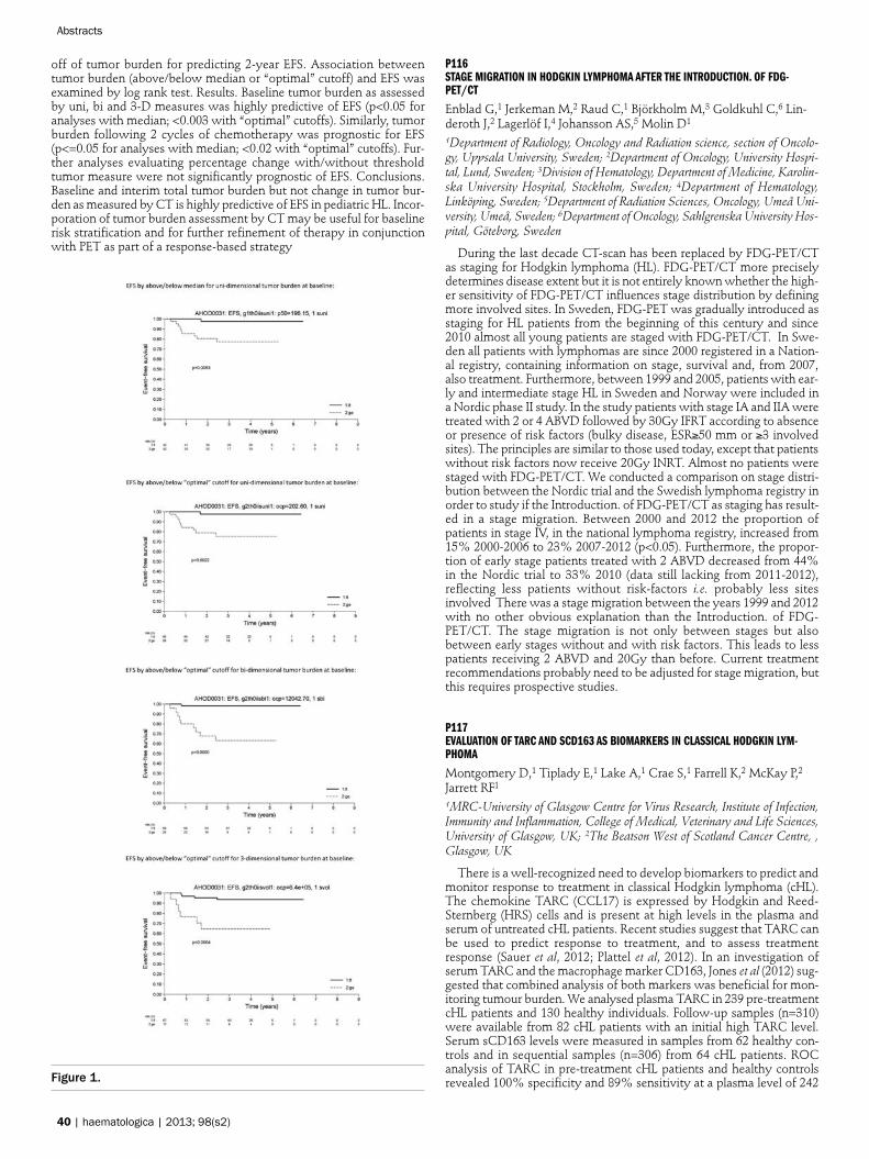
off of tumor burden for predicting 2-year EFS. Association betweentumor burden (above/below median or “optimal” cutoff) and EFS wasexamined by log rank test. Results. Baseline tumor burden as assessedby uni, bi and 3-D measures was highly predictive of EFS (p<0.05 foranalyses with median; <0.003 with “optimal” cutoffs). Similarly, tumorburden following 2 cycles of chemotherapy was prognostic for EFS(p<=0.05 for analyses with median; <0.02 with “optimal” cutoffs). Fur-ther analyses evaluating percentage change with/without thresholdtumor measure were not significantly prognostic of EFS. Conclusions.Baseline and interim total tumor burden but not change in tumor bur-den as measured by CT is highly predictive of EFS in pediatric HL. Incor-poration of tumor burden assessment by CT may be useful for baselinerisk stratification and for further refinement of therapy in conjunctionwith PET as part of a response-based strategy
Figure 1.
P116STAGE MIGRATION IN HODGKIN LYMPHOMA AFTER THE INTRODUCTION. OF FDG-PET/CT
Enblad G,1 Jerkeman M,2 Raud C,1 Björkholm M,3 Goldkuhl C,6 Lin-deroth J,2 Lagerlöf I,4 Johansson AS,5 Molin D1
1Department of Radiology, Oncology and Radiation science, section of Oncolo-gy, Uppsala University, Sweden; 2Department of Oncology, University Hospi-tal, Lund, Sweden; 3Division of Hematology, Department of Medicine, Karolin-ska University Hospital, Stockholm, Sweden; 4Department of Hematology,Linköping, Sweden; 5Department of Radiation Sciences, Oncology, Umeå Uni-versity, Umeå, Sweden; 6Department of Oncology, Sahlgrenska University Hos-pital, Göteborg, Sweden
During the last decade CT-scan has been replaced by FDG-PET/CTas staging for Hodgkin lymphoma (HL). FDG-PET/CT more preciselydetermines disease extent but it is not entirely known whether the high-er sensitivity of FDG-PET/CT influences stage distribution by definingmore involved sites. In Sweden, FDG-PET was gradually introduced asstaging for HL patients from the beginning of this century and since2010 almost all young patients are staged with FDG-PET/CT. In Swe-den all patients with lymphomas are since 2000 registered in a Nation-al registry, containing information on stage, survival and, from 2007,also treatment. Furthermore, between 1999 and 2005, patients with ear-ly and intermediate stage HL in Sweden and Norway were included ina Nordic phase II study. In the study patients with stage IA and IIA weretreated with 2 or 4 ABVD followed by 30Gy IFRT according to absenceor presence of risk factors (bulky disease, ESR≥50 mm or ≥3 involvedsites). The principles are similar to those used today, except that patientswithout risk factors now receive 20Gy INRT. Almost no patients werestaged with FDG-PET/CT. We conducted a comparison on stage distri-bution between the Nordic trial and the Swedish lymphoma registry inorder to study if the Introduction. of FDG-PET/CT as staging has result-ed in a stage migration. Between 2000 and 2012 the proportion ofpatients in stage IV, in the national lymphoma registry, increased from15% 2000-2006 to 23% 2007-2012 (p<0.05). Furthermore, the propor-tion of early stage patients treated with 2 ABVD decreased from 44%in the Nordic trial to 33% 2010 (data still lacking from 2011-2012),reflecting less patients without risk-factors i.e. probably less sitesinvolved There was a stage migration between the years 1999 and 2012with no other obvious explanation than the Introduction. of FDG-PET/CT. The stage migration is not only between stages but alsobetween early stages without and with risk factors. This leads to lesspatients receiving 2 ABVD and 20Gy than before. Current treatmentrecommendations probably need to be adjusted for stage migration, butthis requires prospective studies.
P117EVALUATION OF TARC AND SCD163 AS BIOMARKERS IN CLASSICAL HODGKIN LYM-PHOMA
Montgomery D,1 Tiplady E,1 Lake A,1 Crae S,1 Farrell K,2 McKay P,2Jarrett RF1
1MRC-University of Glasgow Centre for Virus Research, Institute of Infection,Immunity and Inflammation, College of Medical, Veterinary and Life Sciences,University of Glasgow, UK; 2The Beatson West of Scotland Cancer Centre, ,Glasgow, UK
There is a well-recognized need to develop biomarkers to predict andmonitor response to treatment in classical Hodgkin lymphoma (cHL).The chemokine TARC (CCL17) is expressed by Hodgkin and Reed-Sternberg (HRS) cells and is present at high levels in the plasma andserum of untreated cHL patients. Recent studies suggest that TARC canbe used to predict response to treatment, and to assess treatmentresponse (Sauer et al, 2012; Plattel et al, 2012). In an investigation ofserum TARC and the macrophage marker CD163, Jones et al (2012) sug-gested that combined analysis of both markers was beneficial for mon-itoring tumour burden. We analysed plasma TARC in 239 pre-treatmentcHL patients and 130 healthy individuals. Follow-up samples (n=310)were available from 82 cHL patients with an initial high TARC level.Serum sCD163 levels were measured in samples from 62 healthy con-trols and in sequential samples (n=306) from 64 cHL patients. ROCanalysis of TARC in pre-treatment cHL patients and healthy controlsrevealed 100% specificity and 89% sensitivity at a plasma level of 242
40 | haematologica | 2013; 98(s2)
Abstracts
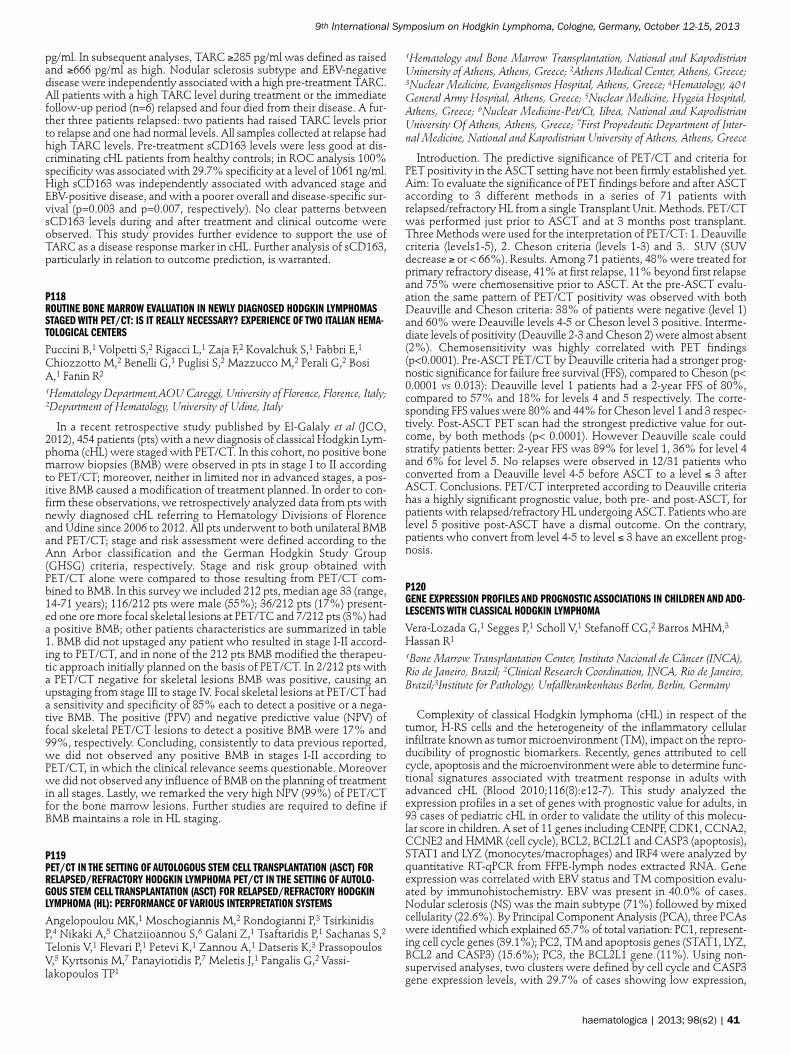
pg/ml. In subsequent analyses, TARC ≥285 pg/ml was defined as raisedand ≥666 pg/ml as high. Nodular sclerosis subtype and EBV-negativedisease were independently associated with a high pre-treatment TARC.All patients with a high TARC level during treatment or the immediatefollow-up period (n=6) relapsed and four died from their disease. A fur-ther three patients relapsed: two patients had raised TARC levels priorto relapse and one had normal levels. All samples collected at relapse hadhigh TARC levels. Pre-treatment sCD163 levels were less good at dis-criminating cHL patients from healthy controls; in ROC analysis 100%specificity was associated with 29.7% specificity at a level of 1061 ng/ml.High sCD163 was independently associated with advanced stage andEBV-positive disease, and with a poorer overall and disease-specific sur-vival (p=0.003 and p=0.007, respectively). No clear patterns betweensCD163 levels during and after treatment and clinical outcome wereobserved. This study provides further evidence to support the use ofTARC as a disease response marker in cHL. Further analysis of sCD163,particularly in relation to outcome prediction, is warranted.
P118ROUTINE BONE MARROW EVALUATION IN NEWLY DIAGNOSED HODGKIN LYMPHOMASSTAGED WITH PET/CT: IS IT REALLY NECESSARY? EXPERIENCE OF TWO ITALIAN HEMA-TOLOGICAL CENTERS
Puccini B,1 Volpetti S,2 Rigacci L,1 Zaja F,2 Kovalchuk S,1 Fabbri E,1Chiozzotto M,2 Benelli G,1 Puglisi S,2 Mazzucco M,2 Perali G,2 BosiA,1 Fanin R21Hematology Department,AOU Careggi, University of Florence, Florence, Italy;2Department of Hematology, University of Udine, Italy
In a recent retrospective study published by El-Galaly et al (JCO,2012), 454 patients (pts) with a new diagnosis of classical Hodgkin Lym-phoma (cHL) were staged with PET/CT. In this cohort, no positive bonemarrow biopsies (BMB) were observed in pts in stage I to II accordingto PET/CT; moreover, neither in limited nor in advanced stages, a pos-itive BMB caused a modification of treatment planned. In order to con-firm these observations, we retrospectively analyzed data from pts withnewly diagnosed cHL referring to Hematology Divisions of Florenceand Udine since 2006 to 2012. All pts underwent to both unilateral BMBand PET/CT; stage and risk assessment were defined according to theAnn Arbor classification and the German Hodgkin Study Group(GHSG) criteria, respectively. Stage and risk group obtained withPET/CT alone were compared to those resulting from PET/CT com-bined to BMB. In this survey we included 212 pts, median age 33 (range,14-71 years); 116/212 pts were male (55%); 36/212 pts (17%) present-ed one ore more focal skeletal lesions at PET/TC and 7/212 pts (3%) hada positive BMB; other patients characteristics are summarized in table1. BMB did not upstaged any patient who resulted in stage I-II accord-ing to PET/CT, and in none of the 212 pts BMB modified the therapeu-tic approach initially planned on the basis of PET/CT. In 2/212 pts witha PET/CT negative for skeletal lesions BMB was positive, causing anupstaging from stage III to stage IV. Focal skeletal lesions at PET/CT hada sensitivity and specificity of 85% each to detect a positive or a nega-tive BMB. The positive (PPV) and negative predictive value (NPV) offocal skeletal PET/CT lesions to detect a positive BMB were 17% and99%, respectively. Concluding, consistently to data previous reported,we did not observed any positive BMB in stages I-II according toPET/CT, in which the clinical relevance seems questionable. Moreoverwe did not observed any influence of BMB on the planning of treatmentin all stages. Lastly, we remarked the very high NPV (99%) of PET/CTfor the bone marrow lesions. Further studies are required to define ifBMB maintains a role in HL staging.
P119PET/CT IN THE SETTING OF AUTOLOGOUS STEM CELL TRANSPLANTATION (ASCT) FORRELAPSED/REFRACTORY HODGKIN LYMPHOMA PET/CT IN THE SETTING OF AUTOLO-GOUS STEM CELL TRANSPLANTATION (ASCT) FOR RELAPSED/REFRACTORY HODGKINLYMPHOMA (HL): PERFORMANCE OF VARIOUS INTERPRETATION SYSTEMS
Angelopoulou MK,1 Moschogiannis M,2 Rondogianni P,3 TsirkinidisP,4 Nikaki A,5 Chatziioannou S,6 Galani Z,1 Tsaftaridis P,1 Sachanas S,2Telonis V,1 Flevari P,1 Petevi K,1 Zannou A,1 Datseris K,3 PrassopoulosV,5 Kyrtsonis M,7 Panayiotidis P,7 Meletis J,1 Pangalis G,2 Vassi-lakopoulos TP1
1Hematology and Bone Marrow Transplantation, National and KapodistrianUninersity of Athens, Athens, Greece; 2Athens Medical Center, Athens, Greece;3Nuclear Medicine, Evangelismos Hospital, Athens, Greece; 4Hematology, 401General Army Hospital, Athens, Greece; 5Nuclear Medicine, Hygeia Hospital,Athens, Greece; 6Nuclear Medicine-Pet/Ct, Iibea, National and KapodistrianUniversity Of Athens, Athens, Greece; 7First Propedeutic Department of Inter-nal Medicine, National and Kapodistrian University of Athens, Athens, Greece
Introduction. The predictive significance of PET/CT and criteria forPET positivity in the ASCT setting have not been firmly established yet.Aim: To evaluate the significance of PET findings before and after ASCTaccording to 3 different methods in a series of 71 patients withrelapsed/refractory HL from a single Transplant Unit. Methods. PET/CTwas performed just prior to ASCT and at 3 months post transplant.Three Methods were used for the interpretation of PET/CT: 1. Deauvillecriteria (levels1-5), 2. Cheson criteria (levels 1-3) and 3. SUV (SUVdecrease ≥ or < 66%). Results. Among 71 patients, 48% were treated forprimary refractory disease, 41% at first relapse, 11% beyond first relapseand 75% were chemosensitive prior to ASCT. At the pre-ASCT evalu-ation the same pattern of PET/CT positivity was observed with bothDeauville and Cheson criteria: 38% of patients were negative (level 1)and 60% were Deauville levels 4-5 or Cheson level 3 positive. Interme-diate levels of positivity (Deauville 2-3 and Cheson 2) were almost absent(2%). Chemosensitivity was highly correlated with PET findings(p<0.0001). Pre-ASCT PET/CT by Deauville criteria had a stronger prog-nostic significance for failure free survival (FFS), compared to Cheson (p<0.0001 vs 0.013): Deauville level 1 patients had a 2-year FFS of 80%,compared to 57% and 18% for levels 4 and 5 respectively. The corre-sponding FFS values were 80% and 44% for Cheson level 1 and 3 respec-tively. Post-ASCT PET scan had the strongest predictive value for out-come, by both methods (p< 0.0001). However Deauville scale couldstratify patients better: 2-year FFS was 89% for level 1, 36% for level 4and 6% for level 5. No relapses were observed in 12/31 patients whoconverted from a Deauville level 4-5 before ASCT to a level ≤ 3 afterASCT. Conclusions. PET/CT interpreted according to Deauville criteriahas a highly significant prognostic value, both pre- and post-ASCT, forpatients with relapsed/refractory HL undergoing ASCT. Patients who arelevel 5 positive post-ASCT have a dismal outcome. On the contrary,patients who convert from level 4-5 to level ≤ 3 have an excellent prog-nosis.
P120GENE EXPRESSION PROFILES AND PROGNOSTIC ASSOCIATIONS IN CHILDREN AND ADO-LESCENTS WITH CLASSICAL HODGKIN LYMPHOMA
Vera-Lozada G,1 Segges P,1 Scholl V,1 Stefanoff CG,2 Barros MHM,3Hassan R11Bone Marrow Transplantation Center, Instituto Nacional de Câncer (INCA),Rio de Janeiro, Brazil; 2Clinical Research Coordination, INCA, Rio de Janeiro,Brazil;3Institute for Pathology, Unfallkrankenhaus Berlin, Berlin, Germany
Complexity of classical Hodgkin lymphoma (cHL) in respect of thetumor, H-RS cells and the heterogeneity of the inflammatory cellularinfiltrate known as tumor microenvironment (TM), impact on the repro-ducibility of prognostic biomarkers. Recently, genes attributed to cellcycle, apoptosis and the microenvironment were able to determine func-tional signatures associated with treatment response in adults withadvanced cHL (Blood 2010;116(8):e12-7). This study analyzed theexpression profiles in a set of genes with prognostic value for adults, in93 cases of pediatric cHL in order to validate the utility of this molecu-lar score in children. A set of 11 genes including CENPF, CDK1, CCNA2,CCNE2 and HMMR (cell cycle), BCL2, BCL2L1 and CASP3 (apoptosis),STAT1 and LYZ (monocytes/macrophages) and IRF4 were analyzed byquantitative RT-qPCR from FFPE-lymph nodes extracted RNA. Geneexpression was correlated with EBV status and TM composition evalu-ated by immunohistochemistry. EBV was present in 40.0% of cases.Nodular sclerosis (NS) was the main subtype (71%) followed by mixedcellularity (22.6%). By Principal Component Analysis (PCA), three PCAswere identified which explained 65.7% of total variation: PC1, represent-ing cell cycle genes (39.1%); PC2, TM and apoptosis genes (STAT1, LYZ,BCL2 and CASP3) (15.6%); PC3, the BCL2L1 gene (11%). Using non-supervised analyses, two clusters were defined by cell cycle and CASP3gene expression levels, with 29.7% of cases showing low expression,
haematologica | 2013; 98(s2) | 41
9th International Symposium on Hodgkin Lymphoma, Cologne, Germany, October 12-15, 2013
most EBV- and grade 2 NS subtype. Positive correlations were observedbetween number of cytotoxic cells (CD8+, GRZB+, TIA1+) and CASP3,CDK1 and BCL2 (P<0.05), number of TIA1+ or GRZB+ with STAT1and/or LYZ and number of macrophages (CD68+ and CD163+) withSTAT1, LYZ and IRF4 (P<0.01). EBV+ cases exhibited high expression ofLYZ and STAT1 (P<0.05). Progression-free survival (PFS) was significant-ly lower in cases with high expression of CASP3 (87.2% vs 68.4%,P=0.04), mainly in the EBV- cases (P= 0.012). Our results show that thereare distinguishable gene expression patterns with biological meaning inpediatric cHL, associated with cell pathways and TM. However, the 11-gene expression based score proposed for molecular prediction in adultcHL is not directly reproducible in children.
P121INTERIM AND SUBSEQUENT 18FDG-PET-CT DETERMINE THE OUTCOME OF PEDIATRICHODGKIN’S LYMPHOMA
Zaghloul MS, Elhaddad A, Attia E, Omar W, Mohamed O, ElmenawySChildren’s Cancer Hospital, Egypt ana National Cancer Institute, Cairo Uni-versity, Cairo, Egypt
Purpose. To investigate the value of interim and subsequent 18 FDGP-PET-CT as a predictor and prognostic factor in pediatric Hodgkin’s lym-phoma treated according to patients’ risk status. Patients and Methods.Four hundreds twelve children below the age of 18 years were treatedat Children’s Cancer Hospital, Egypt (CCHE) during the period fromJuly 2007 and June 2012. Mixed cellularity was the major pathologicalsubtype (199= 48%) followed by nodular sclerosis (172=42%). Stage Iwas encountered in 11%, stage II in 49%, stage III in 25% and stage IVin 15%. They were treated via risk-adapted protocol, entailing 4 or 6courses of ABVD chemotherapy and involved field (IF) radiotherapy forlow and intermediate risk patients respectively. High-risk patientsreceived 6-8 ABVD courses ± IF radiotherapy. Two hundreds and six(45%) had low risk status, 107 (26%) and 119 (29%) had intermediateand high risk, respectively. Results. The 5-year overall survival (OS) andevent-free survival (EFS) of the whole group was 96.5 ± 1.4% and88.0±2.4%, respectively. Early responders showing negative PET-CTafter 2 courses had significantly higher OS and EFS (99.2±2.7% and95.5±2.4%) than late responders (with positive PET-CT after 2 courses)who had 93.2±3.2% and 80.6±7.0% OS and EFS respectively. Thesedifferences are statistically significant (p 0.001for both). Late PET-CT(after 4 courses) can still determine the EFS in these late responders.Negative PET had EFS of 92.2±5.3% compared to 58.4± 13.8% for thosehaving positive PET (p=0.029). The OS though higher with negativePET yet, it did not rank to the level of significance. Other OS and EFSprognostic factors were: stage (p=0.011& 0.005), risk status (0.038 &0.000), pathology subtype (p=0.003 & 0.000). The Cox multivariateanalysis revealed that Interim PET-CT is an independent working pre-dictor factor. Conclusion. Early response for pediatric Hodgkin’s Lym-phoma to 2 courses of chemotherapy, predict both the OS and EFS inrisk adapted treatment protocol. The subsequent PET predicts the EFSin late responders.
P122COMPARISON OF PLASMA TARC WITH GALECTIN-1 AS BIOMARKERS FOR RESPONSE INCLASSICAL HODGKIN LYMPHOMA
Plattel WJ,1,2 Van den Berg A,2 Van Imhoff GW,1 Diepstra A,2 Visser L21Department of Hematology, University Medical Center Groningen, Universityof Groningen, The Netherlands; 2Department of Pathology and Medical Biolo-gy, University Medical Center Groningen, University of Groningen, The Nether-lands
We previously reported plasma Thymus and Activation RegulatedChemokine (TARC) to be a very sensitive biomarker for classicalHodgkin Lymphoma (cHL) tumor burden and a highly promising mark-er for early response evaluation. Recently, Galectin-1 gained interest asa potential biomarker since elevated levels of Galectin-1 were found inserum of cHL patients and associated with parameters of cHL tumor bur-den. Galectin-1 may be involved in the pathogenesis of classical HodgkinLymphoma (cHL) by suppressing Th1 and Th17 cells and stimulatingregulatory T cells. We therefore investigated circulating Galectin-1 lev-
els before and after treatment in plasma of 66 newly diagnosed cHLpatients and compared it to plasma TARC levels. Pre-treatment plasmaGalectin-1 levels among cHL patients were significantly elevated com-pared to healthy controls (mean Galectin-1 level 22.8 ng/ml vs 2.4 ng/ml,p<.001). Plasma TARC could more accurately discriminate patients fromcontrols with pre-treatment samples being elevated in 92% of patientscompared to 45% for Galectin-1. In contrast to plasma TARC, plasmaGalectin-1 did not correlate with stage of disease, presence of bulky dis-ease or tumor volume as measured by quantification of pre-treatmentFDG-PET scans. In the entire group, plasma Galectin-1 levels signifi-cantly decreased after treatment compared to pre-treatment (mean 4.6ng/ml, p<.001). In three responsive patients Galectin-1 levels rose fromnormal levels before treatment to elevated levels after treatment andremained high before and after treatment in two other responsivepatients. In one out of three non-responsive patients Galectin-1 levelswere elevated before and after treatment, while levels were not elevat-ed before and after treatment in the two other non-responsive patients.In contrast, TARC levels decreased to normal in all responsive patientsand remained high in the three non-responsive patients. There was nocorrelation between plasma TARC and plasma Galectin-1 levels. In con-clusion, in our cohort pre-treatment plasma Galectin-1 levels are signif-icantly elevated in cHL patients but in contrast to plasma TARC,Galectin-1 levels do not correlate with tumor burden and levels aftertreatment do not correlate with treatment response.
P123INTERIM PET/CT IN HODGKIN-LYMPHOMA- FINAL RESULTS. OF THE HUNGARIANCHEAP STUDY (2007-2011)
Miltényi Z,1 Simon Z,1 Jóna A,1 Magyari F,1 Barna S,2 Garai I,2 Illés A,1and the CHEAP study group1University of Debrecen, Hematology Department, 2Scanomed Ltd, Hungary
Background. Interim 18FDG-PET/CT is predicting prognosis betterthan any of the available prognostic factors. Patients and Methods.Between 2007 and 2011 a total of 113 Hodgkin-lymphoma (HL) patientsunderwent satging, interim and restaging 18FDG-PET/CT. We investi-gated the value of 18FDG-PET/CT for prediction of progression-freesurvival (PFS) and overall survival (OS). Results. We examined 113patients (57 men, 56 women). The median age of the patients was 38years. 62 patients had early and 51 advanced stage disease at the timeof diagnosis. 59 patients had B signs. All of the patients got ABVD ther-apy, and there was no treatment change based ont he result of the inter-im PET/CT. 83 patients had negative interim FDG-PET (intPET) scans,30 patients had intPET positive scans by the Deauville criteria. The neg-ative predictive value was 0.927, and the positive predictive value was0.566. Three patients died of the negativ intPET group, all of them withsecond malignancy, and eight patients died in the int positive group, allof them due to Hodgkin’s lymphoma. There were 6 relapse in the int-PET negativ group, they had advanced stage disease at the time of diag-nosis, they underwent autolog stem cell transplantation. There was sig-nificant different between the groups in progression free survival (PFS)(p<0,001) based on the results of interim PET/CT. PFS was 92,7% in theintPET negative group, and 40.8% in the intPET positive group at 60months. The 5 year overall survival was 93.4% in the interim PET neg-ative and 58% in the positive group (p<0.001). Patients with early stagedisease had 100% PFS at 60 months in intPET negative group, and 35.9%in the intPET positive group (p<0.0001). Conclusion. Interim FDG-PET/CT is an useful and independent predictor of PFS in HL. It could bepossible to avoid under- and over treatment of patients with used ofinterim PET/CT.
42 | haematologica | 2013; 98(s2)
Abstracts
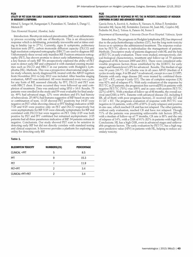
P124UTILITY OF PET SCAN FOR EARLY DIAGNOSIS OF BLEOMYCIN INDUCED PNEUMONITISIN HODGKIN’S LYMPHOMA
Mittal S, Sengar M, Rangarajan V, Purandare N, Tandon S, Dangi U,Menon HTata Memorial Hospital, Mumbai, India
Introduction. Bleomycin induced pneumonitis (BIP) is an inflammato-ry process occurring with use of Bleomycin. This is an idiosyncraticresponse which is debilitating, potentially irreversible and at times end-ing in fatality (up to 27%). Currently, signs & symptoms, pulmonaryfunction tests (PFT), carbon monoxide diffusion capacity (DLCO) andhigh resolution computed tomography (HRCT) are used to diagnose BIP.Unfortunately these reveal only manifest BIP. Positron emission tomog-raphy (PET), has shown promise to detect early inflammatory changes,a key feature of early BIP. We prospectively explored the abilty of PETscan to detect early BIP and compared it with standard existing modal-ities such as DLCO and HRCT in our patients with Hodgkin’s Lym-phoma (HL). Methods. This was a prospective observational single cen-tre study wherein, newly diagnosed HL treated with the ABVD regimenfrom November 2011 to July 2012 was included. After baseline stagingevaluation, ABVD was instituted. All were monitored every two cyclesfor any form of BIP, assessed clinically, by PFT, DLCO and PET scanwith HRCT chest. If none occurred they were followed up until com-pletion of treatment. Data was analyzed using SPSS v 16.0. Results. 75patients were enrolled in the study and 59 were evaluable for final analy-sis. 49% had advanced stage, 12% were smokers and 8% had historyof tuberculosis. 25 (40%) had features suggestive of BIP based on any oneor combination of tests. 11/25 showed PET positivity but 14/25 werenegative on PET while showing clinical or PFT findings indicative of BIP.7/25 and 9/25 were positive only on PET and DLCO respectively butwere asymptomatic for BIP. 5/25 were clinically symptomatic for BIP andcorrelated with DLCO but were negative on PET. Only 2/25 was bothpositive by PET and PFT combined but remained asymptomatic. 2/25patients had all three parameters indicative of BIP. 34 patients remainednegative. Conclusions. Our study showed PET scan to be sensitive indetecting early BIP but did not directly correlate with standard testingand clinical suspicion. It however provides a platform for exploring itsutility for detecting early BIP.
Table 1.
BLEOMYCIN TOXICITY NUMBERS(25) PERCENTAGE
CLINICAL +PFT 5 8.5
PFT 9 15.3
PET 7 11.9
PET+PFT 2 3.4
CLINICAL+PFT+PET 2 3.4
P125EARLY EVALUATION OF RESPONSE BY PET/TAC IN PATIENTS DIAGNOSED OF HODGKINLYMPHOMA IN EARLY AND ADVANCED DISEASE
García Feria A, Escrivà A, Andreu R, Varzaru A, Ribas P, FernándezLlavador MJ, Sayas MJ, Fernández Zarzoso M, Juan ML, Cejalvo MJ,Pedreño M, Ros J, Tolosa A, Panero M, Ferrer SDepartment of Haematology. University Doctor Peset Hospital. Valencia, Spain
Introduction. The prognosis in Hodgkin lymphoma (HL) has improveddue to the polychemotherapy schemes (QT), but the long-term toxicityforces us to optimize the administered treatment. The response evalua-tion by PET/TC allows to individualize the management of patients.Methods. Descriptive study of patients diagnosed with HL and the helpof PET/TC in early evaluation. There were studied, retrospectively, clin-ical-biological characteristics, treatment and follow-up of 70 patientsdiagnosed of HL between 2000 and 2011. There were considered unfa-vorable prognosis factors those established by the EORTC for earlystages and Hassenclever’s IPS for advanced. Results, The median of agewas 34 years (16-77). QT scheme was in all cases ABVD (median of 4cycles in early stage, 6 in IIB and 7 in advanced), except in a case (COPP).Patients with early stage disease (38) were treated by combined thera-py (QT + RT), except 4 (only QT). The rate of complete response (CR)was 92% and of relapses 6%. With early evaluation of the response byPET/TC in 23 patients, the progression free survival(PFS) in patients withnegative PET/TC (78%) was 100% and in cases with positive PET/TC(22%) of 60%. With a median of follow-up of 68 months, the overall sur-vival rate(OSR) is 94%. Patients with advanced disease (32, including 8IIB, all of them with poor prognosis factors), 21 received only QT and11 QT + RT. The prognosis evaluation of response with PET/TC wasnegative in 13 patients, with a PFS of 85% (2 early relapses) and positivein one case, which reached CR and has not relapsed. The other patients,without early evaluation, reached CR and have not relapsed. Though71% of the patients was presenting unfavorable risk factors (IPS=3),with a median of follow-up of 77 months, CR rate is 88% and the rateof relapses of 14%, with a OSR of 91% (82% in patients with high IPS).Conclusions. HL has a high OSR, even in advanced stages and unfavor-able prognosis factors. The early evaluation by PET/TC has a high neg-ative predictive value (NPV) in patients with HL, helping to reduce sec-ondary toxicity.
haematologica | 2013; 98(s2) | 43
9th International Symposium on Hodgkin Lymphoma, Cologne, Germany, October 12-15, 2013
Relapsed and Refractory Hodgkin lymphoma
T126PHASE 2 STUDY EVEROLIMUS FOR RELAPSED/REFRACTORY CLASSICAL HODGKINLYMPHOMA (CHL)
Johnston PB,1 Pinter-Brown L,2 Rogerio J,3 Warsi G,3 White K,3 Ram-chandren R41Mayo Clinic, Rochester, MN, USA; 2Geffen School of Medicine at UCLA, LosAngeles, CA, USA; 3Novartis Pharmaceuticals Corporation, East Hanover, NJ,USA; 4Karmanos Cancer Institute, Detroit, MI, USA
Background. Novel treatment options are needed to improve out-comes in patients with cHL that is refractory to or relapses after initialchemotherapy or subsequent high-dose chemotherapy with autologoushematopoietic stem cell transplant (AHSCT). The oral mammalian tar-get of rapamycin inhibitor everolimus showed promising efficacy andacceptable toxicity in 19 patients with heavily pretreated cHL enrolledin a phase 2 study of everolimus for relapsed, rare lymphomas. To con-firm everolimus efficacy and safety in patients with relapsed/refractorycHL, we conducted a multicenter, open-label, 2-step, phase 2 study.Methods. Adults with cHL that progressed after high-dose chemother-apy with AHSCT or a gemcitabine-, vinorelbine-, or vinblastine-contain-ing regimen received everolimus 10 mg/day until disease progression orunacceptable toxicity. Response was assessed every 12 weeks via com-puted tomography with contrast or integrated positron emission tomog-raphy/computed tomography with contrast. The primary study end-point was the overall response rate (ORR) per the modified response cri-teria for malignant lymphoma (Cheson 2007). Secondary endpointsincluded the disease control rate (DCR; percentage of patients with com-plete or partial response), duration of response, duration of disease con-trol, progression-free survival (PFS), and safety. Results. Fifty-sevenpatients were enrolled (57.9% women; median age, 32.0 years; 57.9%pretreated with AHSCT; 66.7% experienced disease progression duringprevious therapy). ORR and DCR were 42.1% and 77.2%, respective-ly. Median time to response, duration of response, and PFS were 57days, 85.5 days, and 9.0 months, respectively. Seven patients remain ontreatment (average duration, 2.08 years), including 1 patient who expe-rienced a partial response and has been on therapy for 2.88 years (Table).The most common grade 3/4 adverse events were thrombocytopenia(21.1%) and anemia (12.3%). Stomatitis was experienced by 24.6% ofpatients and was of grade 3 severity in 3.5%. Pneumonitis was observedin 10.5% of patients and was of grade 1 severity in 3.5% and grade 2severity in 7.0%; only 1 patient discontinued therapy due to pneumoni-tis. Conclusions. The favorable efficacy, short time to response, andmanageable safety profile observed in this heavily pretreated,relapsed/refractory cHL population confirm previous results and supportfurther evaluation of everolimus in cHL.
Table 1.
Patient Best Overall Current Response Duration of Therapy,Response years
1 Partial response Partial response 2.88
2 Partial response Partial response 1.95
3 Unknown Unknown 1.54
4 Partial response Partial response 2.45
5 Partial response Partial response 1.94
6 Complete response Complete response 1.54
7 Stable disease Stable disease 2.26
T127OUTCOME OF PATIENTS TREATED WITH AUTOLOGOUS STEM-CELL TRANSPLANTATIONFOR FIRST RELAPSED OR REFRACTORY HODGKIN LYMPHOMA: A LONG-TERM ANALYSISOF THE PROSPECTIVE LYSA/SFGM-TC H96 TRIAL
Sibon D,1 Resche-Rigon M,2 Morschhauser F,3 Fermé C,4 Dupuis J,5Marçais A,6 Bouabdallah R,7 Sebban C,8 Salles G,9 Brice P101Necker University Hospital, Paris, France; 2Saint-Louis University Hospital,Paris, France; 3Claude Huriez University Hospital, Lille, France; 4GustaveRoussy Institute, Villejuif, France; 5Henri Mondor University Hospital, Créteil,France; 6Necker University Hospital, Paris, France; 7Paoli-Calmettes Institute,Marseille, France; 8Léon Bérard Cancer Center, Lyon, France; 9Lyon-Sud Uni-versity Hospital, Pierre Bénite, France; 10Saint-Louis University Hospital, Paris,France
Aims. To assess prospectively the long-term efficacy and toxicity ofautologous stem-cell transplantation (ASCT) for first relapsed or refrac-tory Hodgkin lymphoma (HL) patients (pts) included in the H96 trial(Morschhauser F, J Clin Oncol 2008, median follow-up 4.25 years). Herewe present an updated analysis after a median follow-up of 10.4 years.Patients and Methods. H96 trial evaluated a risk-adapted salvage treat-ment with single or tandem ASCT for 245 HL pts. Poor-risk pts (n=150)had primary refractory HL (n=77) or unfavorable relapse (≥2 of the fol-lowing risk factors at first relapse: time to relapse <12 months, stage IIIor IV at relapse, and relapse within previously irradiated sites, n=73)and were eligible for tandem ASCT. Intermediate-risk pts (n=95) had onerisk factor at first relapse and were eligible for single ASCT. Freedomfrom second failure (FF2F) was the primary end-point. Results. Byintent-to-treat analysis, the 10-y FF2F and overall survival (OS) rateswere 64% and 70%, respectively, for the intermediate-risk group, and40% and 47%, respectively, for the poor-risk group. Disease status attime of ASCT (1999 international response criteria) was a major prog-nostic factor driving outcome. In the poor-risk group, the 10-y OS was68%, 58%, 16% and 22% for pts in complete remission (CR)/uncon-firmed CR (CRu), partial remission (PR), stable disease (SD) and progres-sive disease (PD), respectively (p<0.0001), without significant differencebetween CR/CRu and PR pts (p=0.12). In the intermediate-risk group,92/95 pts were in CR/CRu or PR at time of ASCT. The 10-y OS was 72%and 66% for pts in CR/CRu and PR, respectively, without significant dif-ference (p=0.96).In the poor-risk group and the intermediate-risk group,the 10-y cumulative incidence of relapse was 51% and 27%, respec-tively. In all, 110 pts died (intermediate-risk group, n=30; poor-risk group,n=80). The main cause of death was HL (50% and 85% of causes ofdeath, in the intermediate-risk group and the poor-risk group, respective-ly). Conclusion. With long-term follow-up, single ASCT remains appro-priate for intermediate-risk pts. For poor-risk-pts, tandem ASCT remainsa valuable option for pts in CR/CRu or PR at time of ASCT.
T128PET ADAPTED SEQUENTIAL SALVAGE THERAPY WITH BRENTUXIMAB VEDOTIN AND AUG-MENTED ICE FOR TRANSPLANT ELIGIBLE PATIENTS WITH RELAPSED AND REFRACTORYHODGKIN LYMPHOMA
Moskowitz AJ, Schöder H, Gerecitano J, Hamlin P, Horwitz S,Matasar M, Keskinyan VS, McCall S, Noy A, Palomba ML, Portlock C,Straus D, Yahalom J, Younes A, Zelenetz A, Moskowitz CHMemorial Sloan-Kettering Cancer Center, New York, NY, USA
Background. Pre-transplant FDG-PET (PET) normalization is one ofthe strongest predictors of outcome following autologous stem cell trans-plant (ASCT) for patients with relapsed or refractory (rel/ref) Hodgkinlymphoma (HL). We have previously shown that the outcome forpatients with normal pre-transplant PET is excellent, regardless ofwhether PET normalization is achieved following ICE therapy alone orICE followed by additional non-cross resistant chemotherapy. Due to itshigh efficacy and excellent tolerability in ASCT failures, we aimed todetermine whether brentuximab vedotin (BV) can replace ICE salvagetherapy or increase the rate of PET normalization through PET-adaptedsequential administration with augmented ICE (aug-ICE). Methods.Patients with rel/ref HL who have failed 1 prior regimen are enrollingon this phase II clinical trial. Patients receive weekly BV administered at1.2 mg/Kg IV weekly for 3 weeks on and 1 week off for 2 cycles, fol-lowed by PET. Patients who achieve normalization of PET (Deauville 2or less) proceed to ASCT. Patients with PET scores of Deauville 3 or
44 | haematologica | 2013; 98(s2)
Abstracts
higher receive 2 cycles of aug-ICE. Results. 40 of planned 46 patientshave enrolled and 33 are evaluable for response, including 70% malesand 45% with primary refractory disease. The most common adversereactions of any grade with BV are rash (70%) and neuropathy (45%).Of the 33 evaluable pts, 10 (30%) achieved PET normalization follow-ing BV alone; 26 (79%) achieved normalization of PET following theentire treatment program and proceeded to ASCT. Among the remain-ing 7 pts, 1 proceeded to ASCT in good PR, 5 received radiation thera-py with continued response and proceeded to ASCT, and 1 remains withactive disease. Overall, 32 of 33 pts have undergone ASCT and with amedian follow-up of 6 months following ASCT, there have been 2relapses and 1 death due to progressive multifocal leukoencepalopathy.Conclusion. PET-adapted sequential salvage therapy with BV followedby augICE has allowed 30% of patients to avoid ICE-based therapy,produces high CR rates, and facilitates referral to ASCT for virtually allpts. Updated results. Will be presented at the meeting.
P129THE PROGNOSTIC ROLE OF DIFFERENT HISTOPATHOLOGIC GROWTH PATTERNS INNODULAR LYMPHOCYTE-PREDOMINANT HODGKIN LYMPHOMA: A REPORT FROM THEGERMAN HODGKIN STUDY GROUP (GHSG)
Hartmann S,1 Eichenauer DA,2,3 Plütschow A,2,3 Mottok A,4 RoshanakB,5 Koch K,6 Bernd HW,7 Cogliatti S,8 Hummel M,9 Feller AC,7 OttG,10 Möller P,11 Rosenwald A,4 Stein H,5 Hansmann ML,1 Engert A,2,3Klapper W6
1Dr. Senckenberg Institute of Pathology, Goethe University, Frankfurt am Main,Germany; 2First Department of Internal Medicine; 3German Hodgkin StudyGroup, University Hospital of Cologne, Germany; 4Institute of Pathology, Uni-versityWürzburg, Germany; 5Practice of Pathology, Berlin, Germany; 6Instituteof Pathology, Haematopathology Section and Lymph Node Registry, Universität-sklinikumSchleswig-Holstein, Campus Kiel, Germany; 7Institute of Pathology,UniversitätsklinikumSchleswig-Holstein, Campus Lübeck, Germany; 8Instituteof Pathology, Kantonsspital St. Gallen, Switzerland; 9Institute of Pathology,Charité University Hospital, Berlin, Germany; 10Department of Clinical Pathol-ogy, Robert-Bosch-Krankenhaus, and Dr Margarete Fischer-Bosch Institute ofClinical Pharmacology, Stuttgart, Germany; 11Institute of Pathology, UniversityHospital Ulm, Germany
Nodular lymphocyte-predominant Hodgkin lymphoma (NLPHL)accounts for an estimated 5% of all Hodgkin lymphoma cases. NLPHLrepresents a diagnostic challenge since histopathologic variants can mim-ic aggressive B-cell non-Hodgkin lymphoma. The aim of the presentstudy was to evaluate the possible role of histopathologic variants on theclinical outcome. Biopsies of 423 NLPHL patients treated within theprospective German Hodgkin Study Group trials HD10-HD15, LPHD,LP and RIPL were reviewed and classified as tumor-cell rich (TCR, n=10),typical NLPHL (n=308) or histopathologic variants characterized by lym-phoma cells outside of B-cell nodules or B-cell depletion of the microen-vironment (n=105). Differences in terms of baseline characteristics andclinical outcome between patients presenting with typical NLPHL his-tology and histopathologic NLPHL variants were evaluated. A varianthistopathologic growth pattern was associated with advanced disease(29.5% versus 14.6%) and an increased 5-year progression/relapse rate(18.1% versus 6.5%) as compared with a typical NLPHL histology. Vari-ant NLPHL histology represented an independent prognostic factor (oddsratio=2.955) in a multivariate model for progression/relapse. A prognos-tic score including the risk factors variant histopathologic growth pattern,low serum albumin and male gender was derived from the model andallowed the definition of three distinct risk groups. Patients presentingwith histopathologic NLPHL variants have a poorer outcome as com-pared with typical NLPHL. According to a newly developed prognosticscore combining histology and clinical features, NLPHL patients can beallocated to defined risk groups for progression/relapse. High-riskpatients may benefit from novel treatment strategies.
P130A PHASE I STUDY OF BISPECIFIC ANTI-CD30 X ANTI-CD16A ANTIBODY CONSTRUCTAFM13 IN PATIENTS WITH RELAPSED OR REFRACTORY HODGKIN LYMPHOMA
Rothe A,1 von Tresckow B,1 Topp MS,2 Younes A,3 Eichenauer DA,1Hummel H,2 Reiners KS,4 Dietlein M,5 Kessler J,4 Ravic M,6 Hucke C,6Pogge von Strandmann E,4 Engert A11Department I for Internal Medicine, University Hospital of Cologne, Cologne,Germany; 2Department of Internal Medicine II, Division of Hematology andMedical Oncology, Wuerzburg University Medical Center, Wuerzburg, Ger-many; 3Lymphoma and Myeloma, The University of Texas MD Anderson Can-cer Center, Houston, TX; 4Laboratory for Immunotherapy. Department I for Inter-nal Medicine, University Hospital of Cologne, Cologne, Germany; 5Departmentof Nuclear Medicine, University Hospital of Cologne, Cologne, Germany;6Affimed Therapeutics AG, Heidelberg, Germany
Background. AFM13 is a bispecific, tetravalent human antibody con-struct (TandAb®) designed for the treatment of CD30-expressing malig-nancies. AFM13 specifically targets CD30 on Hodgkin tumor cells andCD16A on NK cells. Preclinical data demonstrate a specific and efficientanti-tumor activity. Methods. This was an open, single arm phase I doseescalation trial for patients with relapsed and/or refractory HodgkinLymphoma. The overall objective of this study was to evaluate safety,tolerability, pharmacokinetics, immunogenicity, antitumor activity, themaximum tolerated dose (MTD) and the optimal biological dose (OBD)of AFM13. Each patient received a single cycle, consisting of four week-ly doses of AFM13. The doses were escalated in cohorts of three patientsat dose levels of 0.01, 0.04, 0.15, 0.5, 1.5, 4.5 and 7.0 mg/kg. An addition-al group of patients received 4.5 mg/kg twice weekly. Antitumor activ-ity was assessed using Cheson criteria. Results. 24 patients with HodgkinLymphoma completed the escalation phase. The median age was 38.22/28 patients had received prior high-dose chemotherapy and autolo-gous stem cell transplantation. Patients had received a median of 6 pri-or lines of treatment; 14/28 patients were refractory to their most recenttherapy. All weekly doses from 0.01 mg/kg to 7mg/kg, as well as thetwice weekly dosing regimen, proved to be well tolerated and safe, andthe MTD was not reached. Adverse events were generally mild, with themost frequent drug-related event being grade 1/2 infusion reaction in15/28 patients. One patient developed a possibly drug-related dose-lim-iting grade 4 hemolytic anemia. Of 26 evaluable patients 2 patientsachieved PR and 14 SD. Around 50% of patients achieved reduction intheir tumor volumes. There was a statistically significant dose depend-ent increase in activation of NK cells, and reduction in sCD30. Geomet-ric mean apparent terminal half-life of AFM13 was 22.4 h. Conclusions.AFM13 has demonstrated encouraging biologic activity and seems tobe a new feasible targeted therapy for heavily pre-treated patients withHodgkin Lymphoma. Full analysis and clinical data from all patients willbe presented.
P131BRENTUXIMAB VEDOTIN AS FIRST LINE SALVAGE THERAPY IN RELAPSED/REFRACTORY HL
Chen R, Palmer J, Thomas S, Kim Y, Chen BT, Krishnan A, NathwaniN, Sahebi F, Martin P, Mott M, Matsuoka D, Forman SCity of Hope National Medical Center, Weill Cornell Medical College, NewYork, NY, USA
Brentuximab vedotin (BV), an antibody-drug conjugate, selectivelydelivers monomethyl auristatin E to CD 30+ lymphoma cells. In a piv-otal phase II trial for relapsed/refractory Hodgkin lymphoma (HL) fol-lowing autologous stem cell transplant (ASCT), BV demonstrated anoverall response rate (ORR) of 75% and complete response (CR) rate of34%. To examine outcomes of BV given prior to ASCT, we retrospec-tively analyzed data from a consecutive case-series of HL patients whoreceived BV as first -line salvage therapy at City of Hope. Three patientsreceived BV as salvage therapy on an expanded access protocol and 18were on a prospective phase II trial. All patients had relapsed/refractoryHL post-induction therapy +/- consolidative radiotherapy. Patients weretreated with 1.8 mg/kg of BV intravenously every 3 weeks as outpa-tients for up to 4 cycles. Response rate was determined with either CTor PET at cycle 2 and CT/PET at cycle 4 as per 2007 Cheson criteria. 21patients were treated between 8/2011 to 5/2013. See table 1 for charac-teristics. All patients received ABVD or ABVD/BEACOPP combinationexcept one pediatric patient who received ABVE-PC + vinorelbine/ifos-
haematologica | 2013; 98(s2) | 45
9th International Symposium on Hodgkin Lymphoma, Cologne, Germany, October 12-15, 2013
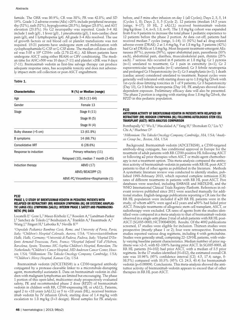
famide. The ORR was 80.9%; CR was 38%; PR was 42.8%; and SD19%. Grade 1-2 adverse events (AEs) >20% include peripheral neuropa-thy (52%), rash (52%), hypoglycemia (28.5%), fatigue (23.8%), and ASTelevation (23.8%). No G2 neuropathy was reported. Grade 3-4 AEinclude 1 rash (g3) , 1 fever (g3) , 1 pneumonitis (g3), 1 non-cardiac chestpain (g3), and 1 lymphopenia (g4). All grade 3-4 AEs resolved. The useof growth factors or red blood cell or platelet transfusions was notrequired. 15/21 patients have undergone stem cell mobilization withcyclophosphamide/G-CSF or G-CSF alone. The median cell dose collect-ed was 5.05 x 10⁶ CD34+ cells (2.78-22.41,). All fifteen patients haveundergone ASCT using either BEAM or CBV conditioning. The medi-an time for ANC>500 was 10 days (7-11) and platelet >20K was 9 days(7-11). Brentuximab vedotin as first-line salvage therapy can produceadequate response rates, has acceptable toxicity and does not adverse-ly impact stem cell collection or post-ASCT engraftment.
Table 1.
Characteristics N (%) or Median (range)
Age 36.5 (11-64)
Gender Female 13
Stage Stage II (11)
Stage III (5)
Stage IV (4)
Bulky disease (>5 cm) 13 (61.9%)
B symptoms 14 (66.7%)
Consolidative XRT 6 (28.6%)
Response to induction Primary refractory (11)
Relapsed (10), median 7 month (3-45)
Induction therapy ABVD (17)
ABVD/BEACOPP (2)
ABVE-PC/Vinorelbine+Ifosphamide (1)
P132PHASE 1/2 STUDY OF BRENTUXIMAB VEDOTIN IN PEDIATRIC PATIENTS WITHRELAPSED OR REFRACTORY (RR) HODGKIN LYMPHOMA (HL) OR SYSTEMIC ANAPLAS-TIC LARGE-CELL LYMPHOMA (SALCL): INTERIM PHASE 1 SAFETY AND PHARMACOKI-NETIC (PK) DATA
Locatelli F,1 Gore L,2 Mauz-Körholz C,3 Rosolen A,4 Landman-ParkerJ,5 Sanchez de Toledo J,6 Beishuizen A,7 Franklin A,8 Fasanmade A,9Wang J,9 Fingert H,9 Labotka R,9 Neville K10
1Ospedale Pediatrico Bambino Gesù, Rome, and University of Pavia, Pavia,Italy; 2Children’s Hospital Colorado, Aurora, USA; 3UniversitaetsklinikumHalle, Halle, Germany; 4Università di Padova, Padova, Italy; 5Hopital D’En-fants Armand-Trousseau, Paris, France; 6Hospital Infantil Vall d’Hebron,Barcelona, Spain; 7Erasmus MC-Sophia Children’s Hospital, Rotterdam, TheNetherlands; 8Children’s Cancer Hospital, MD Anderson Cancer Center, Hous-ton, USA; 9Millennium: The Takeda Oncology Company, Cambridge, USA;10Children’s Mercy Hospital, Kansas City, USA
Brentuximab vedotin (ADCETRIS®) is a CD30-targeted antibodyconjugated by a protease-cleavable linker to a microtubule-disruptingagent, monomethyl auristatin E. Data on brentuximab vedotin in chil-dren with malignant lymphomas are limited but encouraging. The phase1 portion of this open-label, multicenter study prospectively evaluatedsafety, PK and recommended phase 2 dose (RP2D) of brentuximabvedotin in children with RR, CD30-expressing HL or sALCL. Patients,aged 2 to <18 years (sALCL) or 5 to <18 years (HL), received brentux-imab vedotin by IV infusion Q3wk; starting dose of 1.4 mg/kg withescalation to 1.8 mg/kg (3+3 design). Blood samples for PK analysis:
before, and 5 mins after infusion on day 1 (all Cycles); Days 2, 3, 5, 14(Cycles 1, 8); Days 2, 3, 5 (Cycle 2). 12 patients (median 14.5 years[range, 9–17]; 10 HL, 2 sALCL) received brentuximab vedotin(mg/kg/dose: 1.4, n=3; 1.8, n=9). The 1.8 mg/kg cohort was expandedfrom 6 to 9 patients to increase the total phase 1 pediatric experience to12 patients before the phase 2 portion. At data cut-off, patients hadreceived median 7 cycles (range, 1–14); 11 (92%) had ≥1 drug-relatedadverse-event (DRAE): 2 at 1.4 mg/kg, 9 at 1.8 mg/kg. 5 patients (42%)had Gr ≥3 DRAEs at 1.8 mg/kg. Most frequent treatment-emergent AEs:nausea (67%), pyrexia (50%), upper abdominal pain, paresthesia (33%each), abdominal pain, diarrhea, musculoskeletal pain, rhinitis (25%each). 7 serious AEs occurred in 6 patients at 1.8 mg/kg: Gr 1 pyrexia(n=1) unrelated to treatment; Gr 1 pain in extremity (n=1); Gr 2supraventricular tachycardia (n=1) unrelated; Gr 3 febrile neutropeniaand prolonged Gr 3 hepatotoxicity (n=1); Gr 3 anaphylaxis (n=1); 1 death(cardiac arrest) considered unrelated to treatment. Repeat cycles weregenerally well tolerated with starting doses up to 1.8 mg/kg Q3wk with1st-cycle dose-limiting toxicities in 1 of 9 patients; Gr 3 hepatotoxicity(Day 13), Gr 3 febrile neutropenia (Day 14). PK analyses showed dose-dependent exposure. Preliminary efficacy data will also be presented.The phase 2 portion is ongoing with starting dose 1.8 mg/kg Q3wk, theRP2D in this pediatric population.
P133ANTITUMOR ACTIVITY OF BRENTUXIMAB VEDOTIN IN PATIENTS WITH RELAPSED ORREFRACTORY (RR) HODGKIN LYMPHOMA (HL) FOLLOWING AUTOLOGOUS STEM CELLTRANSPLANT (ASCT): META-ANALYSIS COMPARISON
Bonthapally V,1 Wu E,2 Macalalad A,2 Yang H,2 Shonukan O,1 Liu Y,1Chi A,1 Huebner D1
1Millennium: The Takeda Oncology Company, Cambridge, MA, USA; 2Analy-sis Group Inc., Boston, MA, USA
Background. Brentuximab vedotin (ADCETRIS®), a CD30-targetedantibody-drug conjugate, has conditional approval in Europe for thetreatment of adult patients with RR CD30-positive HL following ASCTor following ≥2 prior therapies when ASCT or multi-agent chemother-apy is not a treatment option. This meta-analysis compared the antitu-mor activity of brentuximab vedotin in patients with RR HL post-ASCTpatients to that of other agents as published in the literature. MethodsA systematic literature review was conducted to identify studies, pub-lished 1993–February 2013, which reported complete remission (CR)rates of different treatments in patients with RR HL post-ASCT. Fivedatabases were searched, including EMBASE and MEDLINE, and theWHO International Clinical Trials Registry Platform. References in rel-evant reviews published since 2011 were searched manually for addi-tional studies. English-language publications reporting a CR rate for theRR HL population were included if ≥20 RR HL patients were in thestudy, of whom ≥80% were aged ≥12 years and ≥50% had failed priorASCT. Principle treatments of allogeneic stem cell transplant, ASCT, orradiotherapy were excluded. CR rates of agents from the studies iden-tified were compared in a meta-analysis to that of brentuximab vedotinobserved in a single-arm phase 2 trial of adult patients with RR HL post-ASCT (SG035-0003; NCT00848926). Results. Of the 4092 publicationsscreened, 17 studies were eligible for inclusion. Thirteen studies wereprospective (mostly phase 1 or 2); four were retrospective. Fourteenstudies reported various drug regimens, including 4 with gemcitabine.Studies were generally small, comprising 22–129 HL patients, with wide-ly varying baseline patient characteristics. Median number of prior reg-imens was >2–5, with 62–100% having prior ASCT. In SG035-0003, allRR HL patients (N=102) had prior ASCT, with a median of 3.5 priorregimens. In the 17 studies identified (N=812), the estimated overall CRrate was 10.96% (95% confidence interval [CI]: 4.5, 17.4; range, 0-38.5%) compared with 33.3% (95% CI: 24.3, 43.4) for brentuximabvedotin (p=0.00009). Conclusions. This meta-analysis showed the anti-tumor activity of brentuximab vedotin appears to exceed that of othertherapies in RR HL post-ASCT.
46 | haematologica | 2013; 98(s2)
Abstracts
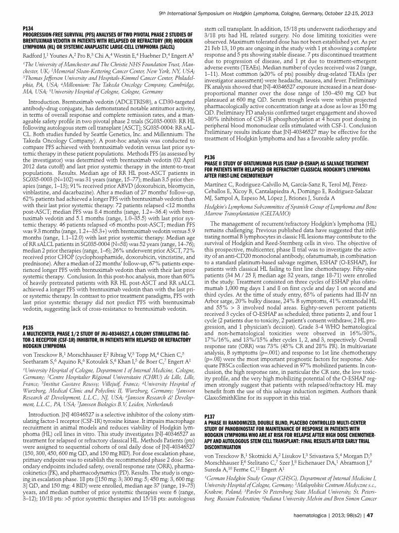
P134PROGRESSION-FREE SURVIVAL (PFS) ANALYSES OF TWO PIVOTAL PHASE 2 STUDIES OFBRENTUXIMAB VEDOTIN IN PATIENTS WITH RELAPSED OR REFRACTORY (RR) HODGKINLYMPHOMA (HL) OR SYSTEMIC ANAPLASTIC LARGE-CELL LYMPHOMA (SALCL)
Radford J,1 Younes A,2 Pro B,3 Chi A,4 Westin E,4 Huebner D,4 Engert A5
1The University of Manchester and The Christie NHS Foundation Trust, Man-chester, UK; 2Memorial Sloan-Kettering Cancer Center, New York, NY, USA;3Thomas Jefferson University and Hospitals-Kimmel Cancer Center, Philadel-phia, PA, USA; 4Millennium: The Takeda Oncology Company, Cambridge,MA, USA; 5University Hospital of Cologne, Cologne, Germany
Introduction. Brentuximab vedotin (ADCETRIS®), a CD30-targetedantibody-drug conjugate, has demonstrated notable antitumor activity,in terms of overall response and complete remission rates, and a man-ageable safety profile in two pivotal phase 2 trials (SG035-0003: RR HLfollowing autologous stem cell transplant [ASCT]; SG035-0004: RR sAL-CL. Both studies funded by Seattle Genetics, Inc. and Millennium: TheTakeda Oncology Company). A post-hoc analysis was conducted tocompare PFS achieved with brentuximab vedotin versus last prior sys-temic therapy in these patient populations. Methods PFS (as assessed bythe investigator) was determined with brentuximab vedotin (02 April2012 data cutoff) and last prior systemic therapy in the intent-to-treatpopulations. Results. Median age of RR HL post-ASCT patients inSG035-0003 (N=102) was 31 years (range, 15–77); median 3.5 prior ther-apies (range, 1–13); 91% received prior ABVD (doxorubicin, bleomycin,vinblastine, and dacarbazine). After a median of 27 months’ follow-up,62% patients had achieved a longer PFS with brentuximab vedotin thanwith their last prior systemic therapy. 72 patients relapsed <12 monthspost-ASCT; median PFS was 8.4 months (range, 1.2+–36.4) with bren-tuximab vedotin and 5.1 months (range, 1.0–35.5) with last prior sys-temic therapy. 46 patients relapsed <6 months post-ASCT; median PFSwas 9.3 months (range, 1.2+–35.3+) with brentuximab vedotin versus 5.9months (range, 1.1–12.0) with last prior systemic therapy. Median ageof RR sALCL patients in SG035-0004 (N=58) was 52 years (range, 14–76);median 2 prior therapies (range, 1–6); 26% underwent prior ASCT, 72%received prior CHOP (cyclophosphamide, doxorubicin, vincristine, andprednisone). After a median of 22 months’ follow-up, 67% patients expe-rienced longer PFS with brentuximab vedotin than with their last priorsystemic therapy. Conclusion. In this post-hoc analysis, more than 60%of heavily pretreated patients with RR HL post-ASCT and RR sALCLachieved a longer PFS with brentuximab vedotin than with the last pri-or systemic therapy. In contrast to prior treatment paradigms, PFS withlast prior systemic therapy did not predict PFS with brentuximabvedotin, suggesting lack of cross-resistance to brentuximab vedotin.
P135A MULTICENTER, PHASE 1/2 STUDY OF JNJ-40346527, A COLONY STIMULATING FAC-TOR-1 RECEPTOR (CSF-1R) INHIBITOR, IN PATIENTS WITH RELAPSED OR REFRACTORYHODGKIN LYMPHOMA
von Tresckow B,1 Morschhauser F,2 Ribrag V,3 Topp M,4 Chien C,5Seetharam S,6 Aquino R,6 Kotoulek S,6 Khan I,5 de Boer C,7 Engert A1
1University Hospital of Cologne, Department I of Internal Medicine, Cologne,Germany; 2Centre Hospitalier Régional Universitaire (CHRU) de Lille, Lille,France; 3Institut Gustave Roussy, Villejuif, France; 4University Hospital ofWurzburg, Medical Clinic and Polyclinic II, Wurzburg, Germany; 5JanssenResearch & Development, L.L.C., NJ, USA; 6Janssen Research & Develop-ment, L.L.C., PA, USA; 7Janssen Biologics B.V, Leiden, Netherlands
Introduction. JNJ 40346527 is a selective inhibitor of the colony stim-ulating factor-1 receptor (CSF-1R) tyrosine kinase. It impairs macrophagerecruitment in animal models and reduces viability of Hodgkin lym-phoma (HL) cell lines in vitro. This study investigates JNJ-40346527 astreatment for relapsed or refractory classical HL. Methods Patients (pts)were assigned to sequential cohorts of oral daily dose of JNJ-40346527(150, 300, 450, 600 mg QD, and 150 mg BID). For dose escalation phase,primary endpoint was to establish the recommended phase 2 dose. Sec-ondary endpoints included safety, overall response rate (ORR), pharma-cokinetics (PK), and pharmacodynamics (PD). Results. The study is ongo-ing in escalation phase. 18 pts ([150 mg: 3; 300 mg: 5; 450 mg: 3, 600 mg:3] QD, and 150 mg: 4 BID) were enrolled, median age 37 (range, 19–75)years, and median number of prior systemic therapies were 6 (range,3–12); 10/18 pts: >5 prior systemic therapies and 15/18 pts: autologous
stem cell transplant. In addition, 15/18 pts underwent radiotherapy and3/18 pts had HL related surgery. No dose limiting toxicities wereobserved. Maximum tolerated dose has not been established yet. As per21 Feb 13, 10 pts are ongoing in the study with 1 pt showing a completeresponse and 5 pts showing stable disease. 7 pts discontinued treatmentdue to progression of disease, and 1 pt due to treatment-emergentadverse events (TEAEs). Median number of cycles received was 2 (range,1–11). Most common (≥20% of pts) possibly drug-related TEAEs (perinvestigator assessment) were headache, nausea, and fever. PreliminaryPK analysis showed that JNJ-40346527 exposure increased in a near dose-proportional manner over the dose range of 150–450 mg QD butplateaued at 600 mg QD. Serum trough levels were within projectedpharmacologically active concentration range at a dose as low as 150 mgQD. Preliminary PD analysis confirmed target engagement and showed>80% inhibition of CSF-1R phosphorylation at 4 hours post dosing inperipheral blood mononuclear cells stimulated with CSF-1. ConclusionPreliminary results indicate that JNJ-40346527 may be effective for thetreatment of Hodgkin lymphoma and has a favorable safety profile.
P136PHASE II STUDY OF OFATUMUMAB PLUS ESHAP (O-ESHAP) AS SALVAGE TREATMENTFOR PATIENTS WITH RELAPSED OR REFRACTORY CLASSICAL HODGKIN’S LYMPHOMAAFTER FIRST-LINE CHEMOTHERAPY
Martínez C, Rodriguez-Calvillo M, García-Sanz R, Terol MJ, Pérez-Ceballos E, Xicoy B, Cantalapiedra A, Domingo E, Rodriguez-SalazarMJ, Sampol A, Espeso M, López J, Briones J, Sureda A
Hodgkin’s Lymphoma Subcommittee of Spanish Group of Lymphoma and BoneMarrow Transplantation (GELTAMO)
The management of recurrent/refractory Hodgkin’s lymphoma (HL)remains challenging. Previous published data have suggested that infil-trating normal B lymphocytes in classic HL lesions may contribute to thesurvival of Hodgkin and Reed-Sternberg cells in vivo. The objective ofthis prospective, multicenter, phase II trial was to investigate the activ-ity of an anti-CD20 monoclonal antibody, ofatumumab, in combinationto a standard platinum-based salvage regimen, ESHAP (O-ESHAP), forpatients with classical HL failing to first line chemotherapy. Fifty-ninepatients (34 M / 25 F, median age 32 years, range 18-71) were enrolledin the study. Treatment consisted on three cycles of ESHAP plus ofatu-mumab 1,000 mg days 1 and 8 on first cycle and day 1 on second andthird cycles. At the time of study entry, 65% of patients had III-IV nnArbor satge, 20% bulky disease, 24% B symptoms, 41% extranodal HLand 55% > 3 involved nodal areas. Eighty-seven percent patientsreceived 3 cycles of O-ESHAP as scheduled; three patients 2, and four 1cycle (2 patients due to toxicity, 2 patient’s consent withdraw, 2 HL pro-gression, and 1 physician’s decision). Grade 3-4 WHO hematologicaland non-hematological toxicities were observed in 16%/30%,17%/16%, and 13%/15% after cycles 1, 2, and 3, respectively. Overallresponse rate (ORR) was 73% (45% CR and 28% PR). In multivariateanalysis, B symptoms (p=.001) and response to 1st line chemotherapy(p=.08) were the most important prognostic factors for response. Ade-quate PBSCs collection was achieved in 97% mobilized patients. In con-clusion, the high response rate, in particular the CR rate, the low toxic-ity profile, and the very high mobilizing potential of the O-ESHAP reg-imen strongly suggest that patients with relapsed/refractory HL maybenefit from the use of this salvage induction regimen. Authors thankGlaxoSmithKline for its support in this trial.
P137A PHASE III RANDOMIZED, DOUBLE BLIND, PLACEBO CONTROLLED MULTI-CENTERSTUDY OF PANOBINOSTAT FOR MAINTENANCE OF RESPONSE IN PATIENTS WITHHODGKIN LYMPHOMA WHO ARE AT RISK FOR RELAPSE AFTER HIGH DOSE CHEMOTHER-APY AND AUTOLOGOUS STEM CELL TRANSPLANT: FINAL RESULTS AFTER EARLY TRIALDISCONTINUATION
von Tresckow B,1 Skotnicki A,2 Lisukov I,3 Srivastava S,4 Morgan D,5Morschhauser F,6 Stelitano C,7 Szer J,8 Eichenauer DA,1 Abramson J,9Sureda A,10 Ferme C,11 Engert A1
1German Hodgkin Study Group (GHSG), Department of Internal Medicine I,University Hospital of Cologne, Germany; 2Malopolskie Centrum Medyczne s.c.,Krakow, Poland; 3Pavlov St Petersburg State Medical University, St. Peters-burg Russian Federation; 4Indiana University Melvin and Bren Simon Cancer
haematologica | 2013; 98(s2) | 47
9th International Symposium on Hodgkin Lymphoma, Cologne, Germany, October 12-15, 2013
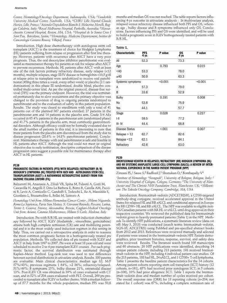
Center, Hematology/Oncology Department, Indianapolis, USA; 5VanderbiltUniversity Medical Center, Nashville, USA; 6CHRU Lille Hopital ClaudeHuriez, Lille, France; 7Azienda Ospedaliera Bianchi E Melacrino Morelli, Reg-gio Calabria, Italy; 8Royal Melbourne Hospital, Parkville, Australia; 9Massa-chusetts General Hospital, Boston, MA, USA; 10Hospital de la Santa Creu ISant Pau, Barcelona, Spain; 11Hematology, Medecine Departement, Institut deCancerologie Gustave Roussy, Villejuif, France
Introduction. High dose chemotherapy with autologous stem celltransplant (ASCT) is the treatment of choice for Hodgkin Lymphoma(HL) patients suffering from relapse or progression after first line thera-py. However, patients with recurrence after ASCT have a very poorprognosis. Thus, the oral deacetylase inhibitor panobinostat was eval-uated as maintenance therapy for patients at risk for relapse after ASCTto prevent recurrences. Methods. HL patients after ASCT with at leastone of the risk factors primary refractory disease, early relapse (<12months), multiple relapses, stage III/IV disease or hemoglobin <10,5 g/dlat relapse prior to transplant were randomized to receive oral panobi-nostat (45mg three times a week, every other week) or placebo (2:1 ran-domization) in this phase III randomized, double blind, placebo con-trolled multi-center trial. As per the original protocol, disease-free sur-vival (DFS) was the primary endpoint. However, the trial was terminat-ed prematurely due to slow recruitment and the primary objective waschanged to the provision of drug to ongoing patients randomized topanobinostat and to the evaluation of safety in this patient population.Results. The study was closed to enrollment with only a total of 41patients out of the planned 367 patients enrolled; 27 patients in thepanobinostat arm and 14 patients in the placebo arm. Grade 3/4 AEsoccurred in 65.4% patients in the panobinostat arm (randomized phase)and 41.7% patients in the placebo arm, most commonly gastrointesti-nal disorders. Although efficacy could not be formally evaluated due tothe small number of patients in this trial, it is interesting to note thatmore patients from the placebo arm discontinued from the study due todisease progression (28.6% vs 14.8% panobinostat patients). Conclu-sion. Maintenance therapy with oral panobinostat is safe and feasible inHL patients after ASCT. Although the trial could not meet ist originalobjective due to early termination, descriptive comparison of the diseaseprogression rates suggest a possible role for maintenance therapy afterASCT in HL patients.
P138PROGNOSTIC FACTORS IN PATIENTS (PTS) WITH RELAPSED/REFRACTORY (R/R)HODGKIN’S LYMPHOMA (HL) TREATED WITH IGEV AND AUTOLOGOUS STEM CELLTRANSPLANTATION (ASCT ): A NATIONWIDE RETROSPECTIVE SURVEY FROM FON-DAZIONE ITALIANA LINFOMI (FIL)
Balzarotti M, Anastasia A, Zilioli VR, Spina M, Pulsoni A, Gotti M,Cascavilla N, Angrilli F, Ortu La Barbera E, Botto B, Carella AM, Pucci-ni B, Levis A, Conticello C, Gandolfi S, Tedeschi L, Re A, Massidda S,Giordano L, Brusamolino E, Bellei M, Santoro AHematology Unit from: Milano Humanitas Cancer Center , Milano Niguarda,Roma La Sapienza, Pavia San Matteo, S. Giovanni Rotondo, Pescara, Latina,Torino 2, Genova, Firenze, Alessandria, Brescia, Cagliari-Medical OncologyUnit from: Aviano, Catania Mediterraneo, Milano S Carlo, Modena, Italy
Introduction. Pts with R/R HL are treated with induction chemother-apy followed by ASCT. IGEV (ifosfamide , vinorelbine, gemcitabine)attains both high complete remission (CR) rate and mobilizing poten-tial and it is the most widely used induction regimen in this setting inItaly. Thus, we carried out a retrospective analysis in order to reassessthe most common prognostic factors in a homogeneously treated ptspopulation. Methods. We collected data of pts treated with IGEV andASCT in Italy from 1997 to 2007. Pts were at least 18 year-old and werescheduled to receive 2 to 4 pre-transplant IGEV courses . For each prog-nostic factor, the survival distribution was estimated throughKaplan–Meier method. Log-rank test was used to test differencesbetween survival distributions in univariate analysis. Results. 330 patentsare evaluable. Main clinical characteristics: median age 32; M/F57%/43%; previous regimens 1 64%, >2 36%; refractory/relapsed:51%/49%; B symptoms 15%, bulky disease 21%, extranodal disease13%. Post-IGEV CR was obtained in 39% of cases evaluated with CTscan, and in 52% of 204 cases evaluated with PET. Overall, 269 pts pro-ceeded to transplant after IGEV or further chemo. With a median follow-up of 57.7 months for the whole population, median PFS was 50,6
months and median OS was not reached. The table reports factors influ-encing 4-yr outcome in univariate analsysis : In multivariate analysis,relapsed versus refractory disease influenced both PFS and OS, where-as age , bulky disease and B symptoms influenced only OS. Conclu-sions. Factors influencing PFS and OS were identified, and will be usedto build a prognostic score in IGEV homogenously treated patients withR/R HL.
Table 1.Characteristic PFS P value OS P value
4 ys 4 ys
All 52.3 73.3
Age 0.793 0.015
<40 53.3 78.0
≥40 50.9 63.3
Systemic symptoms <0.001 <0.001
A 57.3 79.0
B 33.6 52.9
Bulky 0.191 0.008
No 53.8 75.9
Yes 44.1 57.7
Stage 0.028 0.227
I-II 59.5 77.2
III- IV 44.4 68.8
Disease Status <.001 0.007
Relapse > 12 62.7 82.6
Relapse <12 62.1 84.1
Refractory 42.6 63.0
P139BRENTUXIMAB VEDOTIN IN RELAPSED/REFRACTORY (RR) HODGKIN LYMPHOMA (HL)AND RR SYSTEMIC ANAPLASTIC LARGE-CELL LYMPHOMA (SALCL): A REVIEW OF INTER-NATIONAL EXPERIENCE IN THE NAMED PATIENT PROGRAM (NPP)
Zinzani PL,1 Sasse S,2 Radford J,3 Shonukan O,4 Bonthapally V4
1Institute of Hematology “Seragnoli”, University of Bologna, Bologna, Italy; 2
University Hospital of Cologne, Cologne, Germany; 3The University of Man-chester and The Christie NHS Foundation Trust, Manchester, UK; 4Millenni-um: The Takeda Oncology Company, Cambridge, MA, USA
Introduction. Brentuximab vedotin (ADCETRIS®), a CD30-targetedantibody-drug conjugate, received accelerated approval in the UnitedStates for relapsed HL and RR sALCL and conditional approval in Europefor RR CD30+ HL and RR sALCL. The NPP was available to eligible non-US/Canadian patients with RR HL or sALCL until drug approval in theirrespective countries. We reviewed the published data for brentuximabvedotin given to heavily pretreated patients (Table 1) in the NPP. Meth-ods To identify NPP publications, a systematic literature review (data cutoff: May 3, 2013) was conducted (search terms: brentuximab vedotin,SGN-35, ADCETRIS) using PubMed and pre-specified abstract booksfrom 2012 and 2013. References were reviewed manually and selectedif patients were treated in the brentuximab vedotin NPP. Patient demo-graphics, prior treatment histories, response, survival, and safety datawere reviewed. Results. The literature search found 105 manuscriptsand 60 abstracts. 20 NPP publications were identified, describing 14unique patient cohorts, including 213 patients; 193 received brentux-imab vedotin in the NPP, including 4 individual patient case studies. Ofthe 213 patients, 188 had HL, 24 sALCL, and 1 CD30+ T-cell lymphoma.Table 1 presents the baseline patient characteristics for the 14 cohorts.Among patient cohorts reporting stem cell transplant (SCT) history (12cohorts, n=161), 65% had received prior autologous SCT; in 5 cohorts(n=106), 10% had prior allogeneic SCT. Table 1 reports the brentux-imab vedotin dose and median number of cycles received per cohort.The overall response rate (ORR) for 13 reporting cohorts (n=204; notstated for 1 cohort) was 67%, including a complete remission rate of
48 | haematologica | 2013; 98(s2)
Abstracts
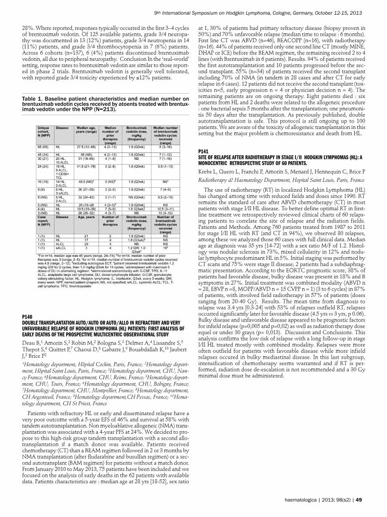
28%. Where reported, responses typically occurred in the first 3–4 cyclesof brentuximab vedotin. Of 125 available patients, grade 3/4 neuropa-thy was documented in 13 (12%) patients, grade 3/4 neutropenia in 14(11%) patients, and grade 3/4 thrombocytopenia in 7 (6%) patients.Across 6 cohorts (n=137), 6 (4%) patients discontinued brentuximabvedotin, all due to peripheral neuropathy. Conclusion In the ‘real-world’setting, response rates to brentuximab vedotin are similar to those report-ed in phase 2 trials. Brentuximab vedotin is generally well tolerated,with reported grade 3/4 toxicity experienced by ≤12% patients.
Table 1. Baseline patient characteristics and median number onbrentuximab vedotin cycles received by atoents treated with brentux-imab vedotin under the NPP (N=213).
P140DOUBLE TRANSPLANTATION AUTO/AUTO OR AUTO/ALLO IN REFRACTORY AND VERYUNFAVORABLE RELAPSE OF HODGKIN LYMPHOMA (HL) PATIENTS: FIRST ANALYSIS OFEARLY DEATHS OF THE PROSPECTIVE MULTICENTRIC OBSERVATIONAL STUDY
Deau B,1 Amorin S,2 Robin M,2 Bologna S,3 Delmer A,4 Lissandre S,5Thepot S,6 Quittet P,7 Chaoui D,8 Gabarre J,9 Bouabdallah K,10 JaubertJ,2 Brice P21Hematology department, Hôpital Cochin, Paris, France; 2Hematology depart-ment, Hôpital Saint Louis, Paris, France; 3Hematology department, CHU, Nan-cy France; 4Hematology department, CHU, Reims, France; 5Hematology depart-ment, CHU, Tours, France; 6Hematology department, CHU, Bobigny, France;7Hematology department, CHU, Montpellier, France; 8Hematology department,CH Argenteuil, France; 9Hematology department,CH Pessac, France; 10Hema-tology department, CH St Priest, France
Patients with refractory HL or early and disseminated relapse have avery poor outcome with a 5-year EFS of 46% and survival at 58% withtandem autotransplantation. Non myeloablative allogeneic (NMA) trans-plantation was associated with a 4-year PFS at 24%. We decided to pro-pose to this high-risk group tandem transplantation with a second allo-transplantation if a match donor was available. Patients receivedchemotherapy (CT) than a BEAM regimen followed in 2 or 3 months byNMA transplantation (after fludarabine and busulfan regimen) or a sec-ond autotransplant (BAM regimen) for patients without a match donor.From January 2010 to May 2013, 75 patients have been included and wefocused on the analysis of early deaths in the 62 patients with availabledata. Patients characteristics are : median age at 28 yrs [18-52], sex ratio
at 1, 30% of patients had primary refractory disease (biopsy proven in50%) and 70% unfavorable relapse (median time to relapse : 6 months).First line CT was ABVD (n=46), BEACOPP (n=16), with radiotherapy(n=16). 44% of patients received only one second line CT (mostly MINE,DHAP or ICE) before the BEAM regimen, the remaining received 2 to 4lines (with Brentuximab in 6 patients). Results. 94% of patients receivedthe first autotransplantation and 10 patients progressed before the sec-ond transplant. 55% (n=34) of patients received the second transplantincluding 70% of NMA (in tandem in 28 cases and after CT for earlyrelapse in 6 cases). 12 patients did not receive the second transplant (tox-icities n=5, early progression n = 4 or physician decision n = 4). Theremaining patients are on ongoing therapy. Eight patients died : sixpatients from HL and 2 deaths were related to the allogeneic procedure: one bacterial sepsis 5 months after the transplantation; one pneumoni-tis 50 days after the transplantation. As previously published, doubleautotransplantation is safe. This protocol is still ongoing up to 100patients. We are aware of the toxicity of allogeneic transplantation in thissetting but the major problem is chemoresistance and death from HL.
P141SITE OF RELAPSE AFTER RADIOTHERAPY IN STAGE I/II HODGKIN LYMPHOMAS (HL): AMONOCENTRIC RETROSPECTIVE STUDY OF 60 PATIENTS.
Krebs L, Quero L, Franchi P, Amorin S, Menard J, Hennequin C, Brice PRadiotherapy & Haematology Department, Hopital Saint Louis, Paris, France
The use of radiotherapy (RT) in localized Hodgkin Lymphoma (HL)has changed among time with reduced fields and doses since 1990. RTremains the standard of care after ABVD chemotherapy (CT) in mostpatients with stage I/II HL disease. To better define optimal RT in first-line treatment we retrospectively reviewed clinical charts of 60 relaps-ing patients to correlate the site of relapse and the radiation fields.Patients and Methods. Among 760 patients treated from 1987 to 2011for stage I/II HL with RT (and CT in 94%), we observed 80 relapses,among these we analyzed those 60 cases with full clinical data. Medianage at diagnosis was 35 yrs [14-72] with a sex ratio M/F of 1.2. Histol-ogy was nodular sclerosis in 73%, mixed cellularity in 12% and nodu-lar lymphocyte predominant HL in 5%. Initial staging was performed byCT scans and 75% were stage II disease; 2 patients had a subdiaphrag-matic presentation. According to the EORTC prognostic score, 38% ofpatients had favorable disease, bulky disease was present in 18% and Bsymptoms in 27%. Initial treatment was combined modality (ABVD n= 28, EBVP n =8, MOPP/ABVD n = 15 CVPP n = 1) (3 to 6 cycles) in 87%of patients, with involved field radiotherapy in 57% of patients (dosesranging from 20-40 Gy). Results. The mean time from diagnosis torelapse was 3.4 yrs [0.3-24] with 53% of relapses outfield RT, relapsesoccurred significantly later for favorable disease (4.5 yrs vs 3 yrs, p 0.06).Bulky disease and unfavorable disease appeared to be prognostic factorsfor infield relapse (p=0,005 and p=0,02) as well as radiation therapy doseequel or under 30 grays (p= 0,013). Discussion and Conclusions. Thisanalysis confirms the low risk of relapse with a long follow-up in stageI/II HL treated mostly with combined modality. Relapses were moreoften outfield for patients with favorable disease while more infieldrelapses occured in bulky mediastinal disease. In this last subgroup,intensification of chemotherapy seems warranted and if RT is per-formed, radiation dose de-escalation is not recommended and a 30 Gyminimal dose must be administered.
haematologica | 2013; 98(s2) | 49
9th International Symposium on Hodgkin Lymphoma, Cologne, Germany, October 12-15, 2013
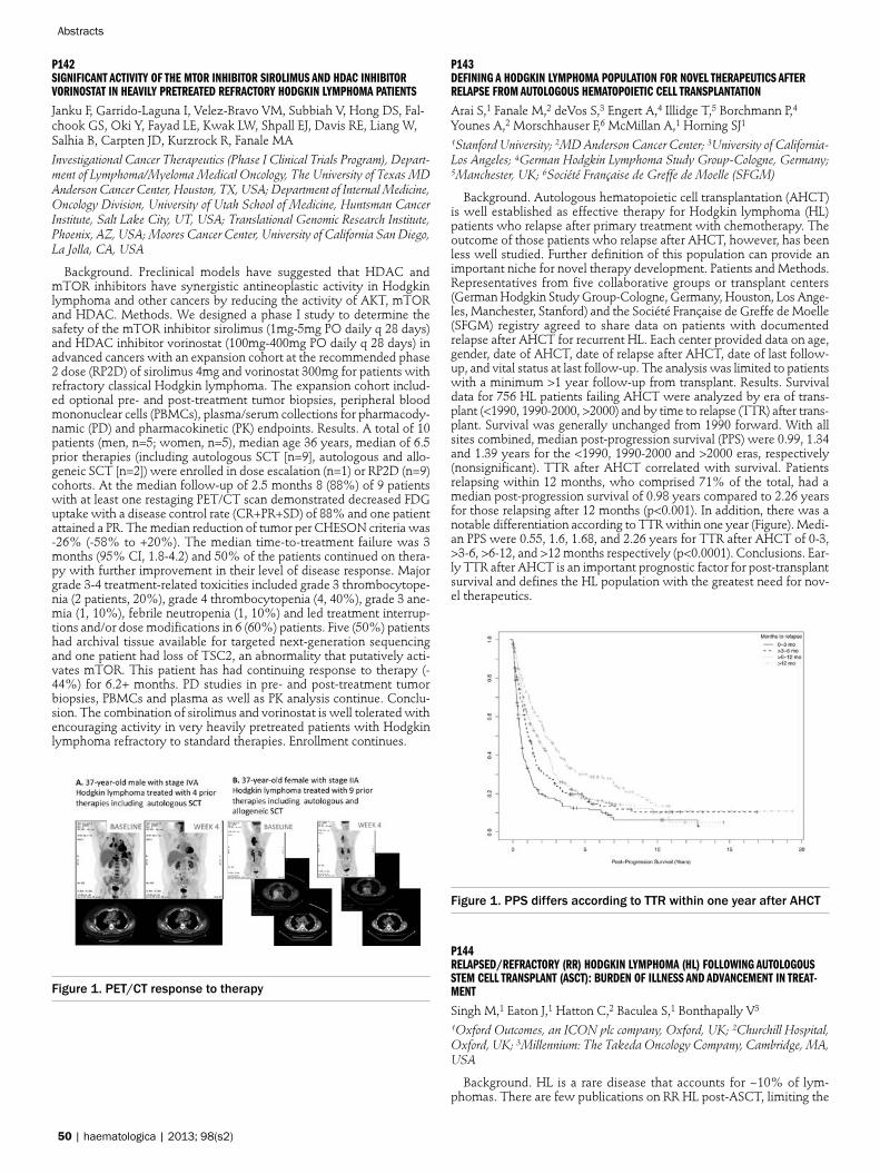
P142SIGNIFICANT ACTIVITY OF THE MTOR INHIBITOR SIROLIMUS AND HDAC INHIBITORVORINOSTAT IN HEAVILY PRETREATED REFRACTORY HODGKIN LYMPHOMA PATIENTS
Janku F, Garrido-Laguna I, Velez-Bravo VM, Subbiah V, Hong DS, Fal-chook GS, Oki Y, Fayad LE, Kwak LW, Shpall EJ, Davis RE, Liang W,Salhia B, Carpten JD, Kurzrock R, Fanale MA
Investigational Cancer Therapeutics (Phase I Clinical Trials Program), Depart-ment of Lymphoma/Myeloma Medical Oncology, The University of Texas MDAnderson Cancer Center, Houston, TX, USA; Department of Internal Medicine,Oncology Division, University of Utah School of Medicine, Huntsman CancerInstitute, Salt Lake City, UT, USA; Translational Genomic Research Institute,Phoenix, AZ, USA; Moores Cancer Center, University of California San Diego,La Jolla, CA, USA
Background. Preclinical models have suggested that HDAC andmTOR inhibitors have synergistic antineoplastic activity in Hodgkinlymphoma and other cancers by reducing the activity of AKT, mTORand HDAC. Methods. We designed a phase I study to determine thesafety of the mTOR inhibitor sirolimus (1mg-5mg PO daily q 28 days)and HDAC inhibitor vorinostat (100mg-400mg PO daily q 28 days) inadvanced cancers with an expansion cohort at the recommended phase2 dose (RP2D) of sirolimus 4mg and vorinostat 300mg for patients withrefractory classical Hodgkin lymphoma. The expansion cohort includ-ed optional pre- and post-treatment tumor biopsies, peripheral bloodmononuclear cells (PBMCs), plasma/serum collections for pharmacody-namic (PD) and pharmacokinetic (PK) endpoints. Results. A total of 10patients (men, n=5; women, n=5), median age 36 years, median of 6.5prior therapies (including autologous SCT [n=9], autologous and allo-geneic SCT [n=2]) were enrolled in dose escalation (n=1) or RP2D (n=9)cohorts. At the median follow-up of 2.5 months 8 (88%) of 9 patientswith at least one restaging PET/CT scan demonstrated decreased FDGuptake with a disease control rate (CR+PR+SD) of 88% and one patientattained a PR. The median reduction of tumor per CHESON criteria was-26% (-58% to +20%). The median time-to-treatment failure was 3months (95% CI, 1.8-4.2) and 50% of the patients continued on thera-py with further improvement in their level of disease response. Majorgrade 3-4 treatment-related toxicities included grade 3 thrombocytope-nia (2 patients, 20%), grade 4 thrombocytopenia (4, 40%), grade 3 ane-mia (1, 10%), febrile neutropenia (1, 10%) and led treatment interrup-tions and/or dose modifications in 6 (60%) patients. Five (50%) patientshad archival tissue available for targeted next-generation sequencingand one patient had loss of TSC2, an abnormality that putatively acti-vates mTOR. This patient has had continuing response to therapy (-44%) for 6.2+ months. PD studies in pre- and post-treatment tumorbiopsies, PBMCs and plasma as well as PK analysis continue. Conclu-sion. The combination of sirolimus and vorinostat is well tolerated withencouraging activity in very heavily pretreated patients with Hodgkinlymphoma refractory to standard therapies. Enrollment continues.
Figure 1. PET/CT response to therapy
P143DEFINING A HODGKIN LYMPHOMA POPULATION FOR NOVEL THERAPEUTICS AFTERRELAPSE FROM AUTOLOGOUS HEMATOPOIETIC CELL TRANSPLANTATION
Arai S,1 Fanale M,2 deVos S,3 Engert A,4 Illidge T,5 Borchmann P,4Younes A,2 Morschhauser F,6 McMillan A,1 Horning SJ1
1Stanford University; 2MD Anderson Cancer Center; 3University of California-Los Angeles; 4German Hodgkin Lymphoma Study Group-Cologne, Germany;5Manchester, UK; 6Société Française de Greffe de Moelle (SFGM)
Background. Autologous hematopoietic cell transplantation (AHCT)is well established as effective therapy for Hodgkin lymphoma (HL)patients who relapse after primary treatment with chemotherapy. Theoutcome of those patients who relapse after AHCT, however, has beenless well studied. Further definition of this population can provide animportant niche for novel therapy development. Patients and Methods.Representatives from five collaborative groups or transplant centers(German Hodgkin Study Group-Cologne, Germany, Houston, Los Ange-les, Manchester, Stanford) and the Société Française de Greffe de Moelle(SFGM) registry agreed to share data on patients with documentedrelapse after AHCT for recurrent HL. Each center provided data on age,gender, date of AHCT, date of relapse after AHCT, date of last follow-up, and vital status at last follow-up. The analysis was limited to patientswith a minimum >1 year follow-up from transplant. Results. Survivaldata for 756 HL patients failing AHCT were analyzed by era of trans-plant (<1990, 1990-2000, >2000) and by time to relapse (TTR) after trans-plant. Survival was generally unchanged from 1990 forward. With allsites combined, median post-progression survival (PPS) were 0.99, 1.34and 1.39 years for the <1990, 1990-2000 and >2000 eras, respectively(nonsignificant). TTR after AHCT correlated with survival. Patientsrelapsing within 12 months, who comprised 71% of the total, had amedian post-progression survival of 0.98 years compared to 2.26 yearsfor those relapsing after 12 months (p<0.001). In addition, there was anotable differentiation according to TTR within one year (Figure). Medi-an PPS were 0.55, 1.6, 1.68, and 2.26 years for TTR after AHCT of 0-3,>3-6, >6-12, and >12 months respectively (p<0.0001). Conclusions. Ear-ly TTR after AHCT is an important prognostic factor for post-transplantsurvival and defines the HL population with the greatest need for nov-el therapeutics.
Figure 1. PPS differs according to TTR within one year after AHCT
P144RELAPSED/REFRACTORY (RR) HODGKIN LYMPHOMA (HL) FOLLOWING AUTOLOGOUSSTEM CELL TRANSPLANT (ASCT): BURDEN OF ILLNESS AND ADVANCEMENT IN TREAT-MENT
Singh M,1 Eaton J,1 Hatton C,2 Baculea S,1 Bonthapally V3
1Oxford Outcomes, an ICON plc company, Oxford, UK; 2Churchill Hospital,Oxford, UK; 3Millennium: The Takeda Oncology Company, Cambridge, MA,USA
Background. HL is a rare disease that accounts for ~10% of lym-phomas. There are few publications on RR HL post-ASCT, limiting the
50 | haematologica | 2013; 98(s2)
Abstracts
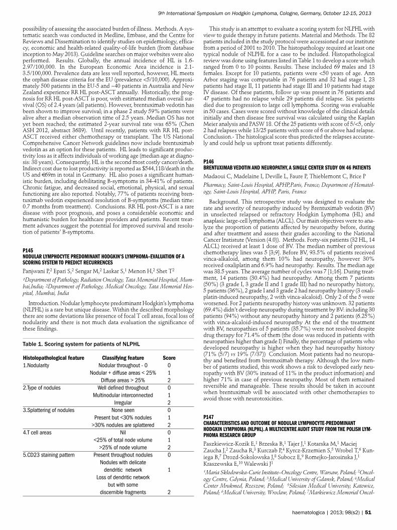
possibility of assessing the associated burden of illness. Methods. A sys-tematic search was conducted in Medline, Embase, and the Centre forReviews and Dissemination to identify studies on epidemiology, effica-cy, economic and health-related quality-of-life burden (from databaseinception to May 2013). Guideline searches on major websites were alsoperformed. Results. Globally, the annual incidence of HL is 1.6-2.97/100,000. In the European Economic Area incidence is 2.1-3.5/100,000. Prevalence data are less well reported, however, HL meetsthe orphan disease criteria for the EU (prevalence <5/10,000). Approxi-mately 500 patients in the EU-5 and ~40 patients in Australia and NewZealand experience RR HL post-ASCT annually. Historically, the prog-nosis for RR HL post-ASCT is poor, with estimated median overall sur-vival (OS) of 2.4 years (all patients). However, brentuximab vedotin hasbeen shown to improve survival; in a phase 2 study 59% patients werealive after a median observation time of 2.5 years. Median OS has notyet been reached; the estimated 2-year survival rate was 65% (ChenASH 2012, abstract 3689). Until recently, patients with RR HL post-ASCT received either chemotherapy or transplant. The US NationalComprehensive Cancer Network guidelines now include brentuximabvedotin as an option for these patients. HL leads to significant produc-tivity loss as it affects individuals of working age (median age at diagno-sis: 38 years). Consequently, HL is the second most costly cancer/death.Indirect cost due to lost productivity is reported as $544,118/death in theUS and €69m in total in Germany. HL also poses a significant human-istic burden, including debilitating B-symptoms in 34-41% of patients.Chronic fatigue, and decreased social, emotional, physical, and sexualfunctioning are also reported. Notably, 77% of patients receiving bren-tuximab vedotin experienced resolution of B-symptoms (median time:0.7 months from treatment). Conclusions. RR HL post-ASCT is a raredisease with poor prognosis, and poses a considerable economic andhumanistic burden for healthcare providers and patients. Recent treat-ment advances suggest the potential for improved survival and resolu-tion of patients’ B-symptoms.
P145NODULAR LYMPHOCYTE PREDOMINANT HODGKIN’S LYMPHOMA–EVALUATION OF ASCORING SYSTEM TO PREDICT RECURRENCES
Panjwani P,2 Epari S,2 Sengar M,2 Laskar S,1 Menon H,2 Shet T2
1Department of Pathology, Radiation Oncology, Tata Memorial Hospital, Mum-bai,India; 2Department of Pathology, Medical Oncology, Tata Memorial Hos-pital, Mumbai, India
Introduction. Nodular lymphocyte predominant Hodgkin’s lymphoma(NLPHL) is a rare but unique disease. Within the described morphologythere are some deviations like presence of focal T cell areas, focal loss ofnodularity and there is not much data evaluation the significance ofthese findings.
Table 1. Scoring system for patients of NLPHL
Histolopathological feature Classifying feature Score1.Nodularity Nodular throughout - 0 0
Nodular + diffuse areas < 25% 1Diffuse areas > 25% 2
2.Type of nodules Well defined throughout 0Multinodular interconnected 1
Irregular 23.Splattering of nodules None seen 0
Present but <30% nodules 1>30% nodules are splattered 2
4.T cell areas Nil 0<25% of total node volume 1
>25% of node volume 25.CD23 staining pattern Present throughout nodules 0
Nodules with delicatedendritic network 1
Loss of dendritic networkbut with some
discernible fragments 2
This study is an attempt to evaluate a scoring system for NLPHL withview to guide therapy in future patients. Material and Methods. The 82patients included in the study protocol were accessioned at our institutefrom a period of 2001 to 2010. The histopathology required at least onetypical nodule of NLPHL for a case to be included. Histopathologicalreview was done using features listed in Table 1 to develop a score whichranged from 0 to 10 points. Results. These included 69 males and 13females. Except for 10 patients, patients were <50 years of age. AnnArbor staging was computable in 76 patients and 32 had stage I, 23patients had stage II, 11 patients had stage III and 10 patients had stageIV disease. Of these patients, follow up was present in 76 patients and47 patients had no relapse while 29 patients did relapse. Six patientsdied due to progression to large cell lymphoma. Scoring was evaluablein 50 cases. Cases were scored without knowledge of the clinical detailsinitially and then disease free survival was calculated using the KaplanMeier analysis and PASW 18. Of the 25 patients with score of 5/<5, only2 had relapses while 13/25 patients with score of 6 or above had relapse.Conclusion.- The histological score thus predicted the relapses accurate-ly and could help us upfront treat patients differently.
P146BRENTUXIMAB VEDOTIN AND NEUROPATHY, A SINGLE CENTER STUDY ON 46 PATIENTS
Madaoui C, Madelaine I, Deville L, Faure P, Thieblemont C, Brice PPharmacy, Saint-Louis Hospital, APHP,Paris, France; Department of Hematol-ogy, Saint-Louis Hospital, APHP, Paris, France
Background. This retrospective study was designed to evaluate therate and severity of neuropathy induced by Brentuximab vedotin (BV)in unselected relapsed or refractory Hodgkin Lymphoma (HL) andanaplasic large-cell lymphoma (ALCL). Our main objectives were to ana-lyze the proportion of patients affected by neuropathy before, duringand after treatment and assess their grades according to the NationalCancer Instistute (Version (4.0)). Methods. Forty-six patients (32 HL, 14ALCL) received at least 1 dose of BV. The median number of previouschemotherapy lines was 5 [3;9]. Before BV, 93.5% of patients receivedvinca-alkaloid, among them 10% had neuropathy, however 30%received oxaliplatin and 6.9% had neuropathy. Results. The median agewas 38.5 years. The average number of cycles was 7 [1;16]. During treat-ment, 14 patients (30.4%) had neuropathy. Among them 7 patients(50%) (3 grade I, 3 grade II and 1 grade III) had no neuropathy history,5 patients (36%), 2 grade I and 3 grade 2 had neuropathy history (3 oxali-platin-induced neuropathy, 2 with vinca-alcaloid). Only 2 of the 5 wereworsened. For 2 patients neuropathy history was unknown. 32 patients(69.4%) didn’t develop neuropathy during treatment by BV: including 30patients (94%) without any neuropathy history and 2 patients (6.25%)with vinca-alcaloid-induced neuropathy At the end of the treatmentwith BV, neuropathies of 5 patients (35.7%) were not resolved despitedrug therapy for 71.4% of them (the dose was reduced in patients withneuropathies higher than grade I) Finally, the percentage of patients whodeveloped neuropathy is higher when they had neuropathy history(71% (5/7) vs 19% (7/37)) Conclusion. Most patients had no neuropa-thy and benefited from brentuximab therapy. Although the low num-ber of patients studied, this work shows a risk to developed early neu-ropathy with BV (30% instead of 11% in the product information) andhigher 71% in case of previous neuropathy. Most of them remainedreversible and manageable. These results should be taken in accountwhen brentuximab will be associated with other chemotherapies toavoid those with neurotoxicities.
P147CHARACTERISTICS AND OUTCOME OF NODULAR LYMPHOCYTE-PREDOMINANTHODGKIN LYMPHOMA (NLPHL). A MULTICENTRE AUDIT STUDY FROM THE POLISH LYM-PHOMA RESEARCH GROUP
Paszkiewicz-Kozik E,1 Brzeska B,1 Tajer J,1 Kotarska M,1 MaciejZaucha J,2 Zaucha R,3 Kurczab P,4 Kyrcz-Krzemien S,5 Wrobel T,6 Kun-iega B,7 Drozd-Sokolowska J,8 Subocz E,9 Romejko-Jarosinska J,1Kraszewska E,10 Walewski J11Maria Sklodowska-Curie Institute–Oncology Centre, Warsaw, Poland; 2Oncol-ogy Centre, Gdynia, Poland; 3Medical University of Gdansk, Poland; 4MedicalCenter Mrukmed, Rzeszow, Poland; 5Silesian Medical University, Katowice,Poland; 6Medical University, Wroclaw, Poland; 7Markiewicz Memorial Oncol-
haematologica | 2013; 98(s2) | 51
9th International Symposium on Hodgkin Lymphoma, Cologne, Germany, October 12-15, 2013
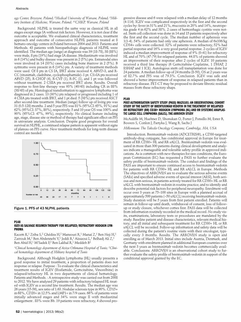
ogy Center, Brzozow, Poland; 8Medical University of Warsaw, Poland; 9Mili-tary Institute of Medicine, Warsaw, Poland; 10CMKP, Warsaw, Poland
Background. NLPHL is treated the same way as classical HL in allstages except stage IA without risk factors. However, it is not clear if theoutcome is acceptable. We evaluated clinical characteristics, treatmentapproach and outcome of consecutive NLPHL patients treated at 8hemato-oncolgy centers in Poland between 2004 and 2013. Patients andMethods. 43 patients with histopathologic diagnosis of NLPHL wereidentified. The median age (range) at diagnosis was 39 (18-78), 38 (88%)were male, 8 pts (19%) had stage IA disease. Mediastinum was involvedin 6 (14%) and bulky disease was present in 2 (5%) pts. Extranodal siteswere involved in 14 (33%) cases including bone marrow in 2 (5%). Bsymtoms were present in 6 (14%) pts. A variety of treatment methodswere used. Of 8 pts in CS IA, IFRT alone received 4, ABVD-3, and R-CC (rituximab, cladribine, cyclophosphamide)–1 pt. CS>IA pts receivedABVD (25), R-CHOP (4), R-CVP (1), R-FC (1), and 1 pt was followedwithout treatment. 2 CSIIA pts received IFRT alone. Results. Overallresponse to first-line therapy was 93% (40/43) including CR in 88%(38/43) of pts. Histological transformation to aggressive lymphoma wasdiagnosed in 2 cases. 13 (30%) pts relapsed or progressed including 1 of4 CSIA pts treated with IFRT, and 1 pt died. 5 (38%) pts received ASCTafter second-line treatment. Median (range) follow up of living pts was31 (0.5-128) months. 2 and 5 year PFS was 81% (95%CI: 65%, 91%) and62% (95%CI: 37%, 80%), respectively. 5 and 10 year OS was 100% and90% (95%CI: 47%, 99%), respectively. No clinical feature includingage, stage, disease site or method of therapy had significant effect on PFSin univariate analysis. Conclusion. Despite good prognosis for overallsurvival in NLPHL, a continued relapse pattern is apparent without signsof plateau on PFS curve. New treatment methods for long-term diseasecontrol are needed.
Figure 1. PFS of 43 NLPHL patients
P148IGEV AS SALVAGE REGIMEN THERAPY FOR RELAPSED/REFRACTORY HODGKIN LYM-PHOMA
Kacem K,1 Zriba S,2 Ghédira H,2 Mansouri R,1 Manaï Z,1 Ben Neji H,1Zarrouk M,1 Ben Abdennebi Y,1 Jeddi R,1 Aïssaoui L,1 Belhadj Ali Z,1Ben Abid H,1 M’Sadek F,2 Ben Lakhal R,1 Meddeb B1
1Clinical hematology department of Aziza Othmana Hospital of Tunis; 2Clini-cal hematology department of Military hospital of Tunis
Background. Although Hodgkin Lymphoma (HL) usually presents agood response to initial treatment, a proportion of patients does notresponse or relapse. Purpose : To evaluate the clinical characteristics andtreatment results of IGEV (Ifosfamide, Gemcitabine, Vinorelbine) inrelapsed/refractory HL in two departments of clinical hematology.Patients and Methods. : A retrospective study was carried out from 2008to 2012. We have analysed 29 patients with refractory/relapsed HL treat-ed with IGEV as a second line treatment. Results. The median age was26 years (15-56), sex ratio of 1.63. Nodular sclerosis type in 90%. CD15+in 68%, CD20+ in 12.5% and LMP1+ in 7/9 cases. 66% of patients wereinitially advanced stages and 34% were stage II with mediastinalenlargement . 83% were Bb. 19 patients were refractory, 4 showed pro-
gressive disease and 6 were relapsed with a median delay of 12 months(4-114). IGEV was complicated respectively in the first and the secondcycles by: fever in 31% and 21%, mucositis in 20% and 14%, neutrope-nia grade 4 in 62% and 38%. 2 cases of hemorrhagic cystitis were not-ed. Stem cell collection was done in 14 and 15 patients respectively afterthe first and the second cycle. The median number of apheresis was1(1-4). 54% of patients had only one apheresis. A median of 6x10⁶/kgCD34+ cells were collected. 32% of patients were refractory, 52% hadpartial response and 16% a very good partial response. 2 cycles of IGEVinduced a median improvement of response of 9% (0-41) for refractoryHL and of 70% (47-79) for relapsed patients. 44.8% of patients showedan improvement of their response after 2 cycles of IGEV. 10 patientsreceived a third line therapy (6 Gemcitabine-Cisplatine, 1 DHAP, 2ESHAP and 1 ICE). Autologous stem cell transplantation was done for24 patients. A median follow-up of 24 months (8-50), OS at 2 years wasof 82.7% and PFS was of 79.3%. Conclusion. IGEV was safe andshowed a better improvement of response in relapsed patients than inrefractory disease. PET-CT may be proposed to deviate fibrotic residualmasses from these refractory shaps.
P149POST-AUTHORISATION SAFETY STUDY (PASS) MA25101: AN OBSERVATIONAL COHORTSTUDY OF THE SAFETY OF BRENTUXIMAB VEDOTIN IN THE TREATMENT OF RELAPSEDOR REFRACTORY (RR) CD30+ HODGKIN LYMPHOMA (HL) AND RR SYSTEMIC ANAPLAS-TIC LARGE CELL LYMPHOMA (SALCL). THE ARROVEN STUDY
McAuliffe M, Huebner D, Shonukan O, Porter J, Ponsillo M, Exter B,Bowers S, Conlon J, Partyka J, Wang B, Sachs J
Millennium: The Takeda Oncology Company, Cambridge, MA, USA
Introduction. Brentuximab vedotin (ADCETRIS®), a CD30-targetedantibody-drug conjugate, has conditional approval in Europe for treat-ment of RR CD30+ HL and RR sALCL. Brentuximab vedotin was eval-uated in more than 300 patients during clinical development and analy-ses indicate a manageable and tolerable safety profile in approved indi-cations. As is common with new therapies for rare conditions, the Euro-pean Commission (EC) has requested a PASS to further evaluate thesafety profile of brentuximab vedotin. The conduct and findings of thePASS are important to ensure continued access to brentuximab vedotinfor patients with RR CD30+ HL and RR sALCL in Europe. MethodsThe objectives of ARROVEN are to evaluate the serious adverse events(SAEs) and specified adverse events of special interest (AESI), both seri-ous and non-serious, in patients actively treated for RR CD30+ HL or RRsALCL with brentuximab vedotin in routine practice; and to identify anddescribe potential risk factors for peripheral neuropathy. Enrolment willoccur over 3 years at 75–100 sites in Europe with a planned accrual ofapproximately 500 patients (~50 sALCL) receiving brentuximab vedotin.Study duration will be 5 years from first patient enrolled. Patients willremain in follow-up until death, withdrawal of consent, loss of follow-up or study closure, whichever comes first. PASS data will be collectedfrom information routinely recorded in the medical record. No study vis-its, examinations, laboratory tests or procedures are mandated by thestudy. Baseline patient and disease characteristics, relevant medical his-tory, and all initial and subsequent treatment for RR CD30+ HL or RRsALCL will be recorded. Follow-up information and safety data will becollected during the patient’s routine visits with their oncologist, typi-cally every 3 months. Results. The ARROVEN study is open andenrolling as of March 2013. Initial sites include Austria, Denmark, andGermany with enrolment planned in additional European countries overthe next 3 years as brentuximab vedotin becomes commercially avail-able. Conclusions. ARROVEN is an observational cohort study to fur-ther evaluate the safety profile of brentuximab vedotin in support of theconditional approval granted by the EC.
52 | haematologica | 2013; 98(s2)
Abstracts
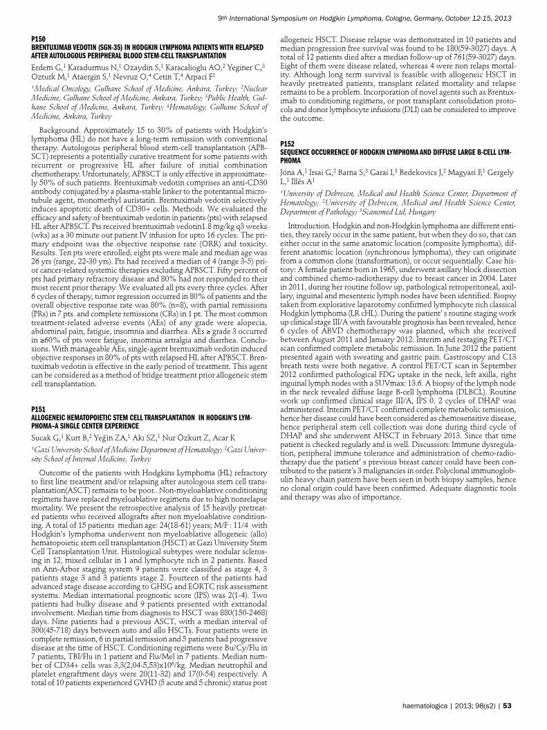
P150BRENTUXIMAB VEDOTIN (SGN-35) IN HODGKIN LYMPHOMA PATIENTS WITH RELAPSEDAFTER AUTOLOGOUS PERIPHERAL BLOOD STEM-CELL TRANSPLANTATION
Erdem G,1 Karadurmus N,1 Ozaydin S,1 Karacalioglu AO,2 Yeginer C,3Ozturk M,1 Ataergin S,1 Nevruz O,4 Cetin T,4 Arpaci F11Medical Oncology, Gulhane School of Medicine, Ankara, Turkey; 2NuclearMedicine, Gulhane School of Medicine, Ankara, Turkey; 3Public Health, Gul-hane School of Medicine, Ankara, Turkey; 4Hematology, Gulhane School ofMedicine, Ankara, Turkey
Background. Approximately 15 to 30% of patients with Hodgkin’slymphoma (HL) do not have a long-term remission with conventionaltherapy. Autologous peripheral blood stem-cell transplantation (APB-SCT) represents a potentially curative treatment for some patients withrecurrent or progressive HL after failure of initial combinationchemotherapy. Unfortunately, APBSCT is only effective in approximate-ly 50% of such patients. Brentuximab vedotin comprises an anti-CD30antibody conjugated by a plasma-stable linker to the potentantial micro-tubule agent, monomethyl auristatin. Brentuximab vedotin selectivelyinduces apoptotic death of CD30+ cells. Methods. We evaluated theefficacy and safety of brentuximab vedotin in patients (pts) with relapsedHL after APBSCT. Pts received brentuximab vedotin1.8 mg/kg q3 weeks(wks) as a 30 minute out patient IV infusion for upto 16 cycles. The pri-mary endpoint was the objective response rate (ORR) and toxicity.Results. Ten pts were enrolled; eight pts were male and median age was26 yrs (range, 22-30 yrs). Pts had received a median of 4 (range 3-5) pri-or cancer-related systemic therapies excluding APBSCT. Fifty percent ofpts had primary refractory disease and 80% had not responded to theirmost recent prior therapy. We evaluated all pts every three cycles. After6 cycles of therapy, tumor regression occurred in 80% of patients and theoverall objective response rate was 80% (n=8), with partial remissions(PRs) in 7 pts. and complete remissions (CRs) in 1 pt. The most commontreatment-related adverse events (AEs) of any grade were alopecia,abdominal pain, fatigue, insomnia and diarrhea. AEs ≥ grade 3 occurredin ≥60% of pts were fatigue, insomnia artralgia and diarrhea. Conclu-sions. With manageable AEs, single-agent brentuximab vedotin inducedobjective responses in 80% of pts with relapsed HL after APBSCT. Bren-tuximab vedotin is effective in the early period of treatment. This agentcan be considered as a method of bridge treatment prior allogeneic stemcell transplantation.
P151ALLOGENEIC HEMATOPOIETIC STEM CELL TRANSPLANTATION IN HODGKIN’S LYM-PHOMA–A SINGLE CENTER EXPERIENCE
Sucak G,1 Kurt B,2 Yegin ZA,1 Akı SZ,1 Nur Özkurt Z, Acar K1Gazi University School of Medicine Department of Hematology; 2Gazi Univer-sity School of Internal Medicine, Turkey
Outcome of the patients with Hodgkins Lymphoma (HL) refractoryto first line treatment and/or relapsing after autologous stem cell trans-plantation(ASCT) remains to be poor.. Non-myeloablative conditioningregimens have replaced myeloablative regimens due to high nonrelapsemortality. We present the retrospective analysis of 15 heavily pretreat-ed patients who received allografts after non myeloablative condition-ing. A total of 15 patients median age: 24(18-61) years; M/F : 11/4 withHodgkin’s lymphoma underwent non myeloablative allogeneic (allo)hematopoietic stem cell transplantation (HSCT) at Gazi University StemCell Transplantation Unit. Histological subtypes were nodular scleros-ing in 12, mixed cellular in 1 and lymphocyte rich in 2 patients. Basedon Ann-Arbor staging system 9 patients were classified as stage 4, 3patients stage 3 and 3 patients stage 2. Fourteen of the patients hadadvanced stage disease according to GHSG and EORTC risk assessmentsystems. Median international prognostic score (IPS) was 2(1-4). Twopatients had bulky disease and 9 patients presented with extranodalinvolvement. Median time from diagnosis to HSCT was 880(150-2468)days. Nine patients had a previous ASCT, with a median interval of300(45-718) days between auto and allo HSCTs. Four patients were incomplete remission, 6 in partial remission and 5 patients had progressivedisease at the time of HSCT. Conditioning regimens were Bu/Cy/Flu in7 patients, TBI/Flu in 1 patient and Flu/Mel in 7 patients. Median num-ber of CD34+ cells was 3,3(2,04-5,53)x106/kg. Median neutrophil andplatelet engraftment days were 20(11-32) and 17(0-54) respectively. Atotal of 10 patients experienced GVHD (5 acute and 5 chronic) status post
allogeneic HSCT. Disease relapse was demonstrated in 10 patients andmedian progression free survival was found to be 180(59-3027) days. Atotal of 12 patients died after a median follow-up of 761(59-3027) days.Eight of them were disease related, whereas 4 were non relaps mortal-ity. Although long term survival is feasible with allogeneic HSCT inheavily pretreated patients, transplant related mortality and relapseremains to be a problem. Incorporation of novel agents such as Brentux-imab to conditioning regimens, or post transplant consolidation proto-cols and donor lymphocyte infusions (DLI) can be considered to improvethe outcome.
P152SEQUENCE OCCURRENCE OF HODGKIN LYMPHOMA AND DIFFUSE LARGE B-CELL LYM-PHOMA
Jóna A,1 Irsai G,2 Barna S,3 Garai I,3 Bedekovics J,2 Magyari F,1 GergelyL,1 Illés A11University of Debrecen, Medical and Health Science Center, Department ofHematology; 2University of Debrecen, Medical and Health Science Center,Department of Pathology; 3Scanomed Ltd, Hungary
Introduction. Hodgkin and non-Hodgkin lymphoma are different enti-ties, they rarely occur in the same patient, but when they do so, that caneither occur in the same anatomic location (composite lymphoma), dif-ferent anatomic location (synchronous lymphoma), they can originatefrom a common clone (transformation), or occur sequentially. Case his-tory: A female patient born in 1965, underwent axillary block dissectionand combined chemo-radiotherapy due to breast cancer in 2004. Laterin 2011, during her routine follow up, pathological retroperitoneal, axil-lary, inguinal and mesenteric lymph nodes have been identified. Biopsytaken from explorative laparotomy confirmed lymphocyte rich classicalHodgkin lymphoma (LR cHL). During the patient’ s routine staging workup clinical stage III/A with favourable prognosis has been revealed, hence6 cycles of ABVD chemotherapy was planned, which she receivedbetween August 2011 and January 2012. Interim and restaging PET/CTscan confirmed complete metabolic remission. In June 2012 the patientpresented again with sweating and gastric pain. Gastroscopy and C13breath tests were both negative. A control PET/CT scan in September2012 confirmed pathological FDG uptake in the neck, left axilla, rightinguinal lymph nodes with a SUVmax: 13.6. A biopsy of the lymph nodein the neck revealed diffuse large B-cell lymphoma (DLBCL). Routinework up confirmed clinical stage III/A, IPS 0. 2 cycles of DHAP wasadministered. Interim PET/CT confirmed complete metabolic remission,hence her disease could have been considered as chemosensitive disease,hence peripheral stem cell collection was done during third cycle ofDHAP and she underwent AHSCT in February 2013. Since that timepatient is checked regularly and is well. Discussion: Immune dysregula-tion, peripheral immune tolerance and administration of chemo-radio-therapy due the patient’ s previous breast cancer could have been con-tributed to the patient’s 3 malignancies in order. Polyclonal immunoglob-ulin heavy chain pattern have been seen in both biopsy samples, henceno clonal origin could have been confirmed. Adequate diagnostic toolsand therapy was also of importance.
haematologica | 2013; 98(s2) | 53
9th International Symposium on Hodgkin Lymphoma, Cologne, Germany, October 12-15, 2013
Survivorship
T153CHANGES IN BREAST CANCER RISK AS LONG-TERM HODGKIN LYMPHOMA SURVIVORSREACH MENOPAUSAL AGE
Wong JS,1 Catalano PJ,2 Chen Y,2 Marcus KJ,1 Michaelson EM,1Mauch PM,1 Ng AK1
1Department of Radiation Oncology; 2Department of Biostatistics and Compu-tational Biology; Dana-Farber Cancer Institute/Brigham and Women’s Hospi-tal, Baltimore, USA
Women who received chest irradiation (RT) for Hodgkin lymphoma(HL) at a young age are at increased risk for breast cancer (BC). Treat-ment-related early menopause is protective against BC, although limit-ed data are available on changes in risk as survivors enter menopause.This study aims to assess the excess BC risk of HL survivors comparedwith a normal, age-matched population as survivors pass menopause,and to compare the excess risks of premenopausal versus post-menopausal survivors. HL treatment history, subsequent BC develop-ment, menopausal status, and hormone-replacement therapy (HRT) his-tory were abstracted from a HL database and medical records with IRBapproval. The study included 555 women who had chest RT at age <35between 1968 and 2005. Age >50, pelvic RT or MOPP chemotherapyexposure was used as a surrogate for menopause in cases of missingmenopausal status data. Postmenopausal women on HRT were consid-ered premenopausal. Median follow-up was 20 years (range 0.7-42).Median age at RT was 23 (range 6-35). Median current attained age was39 (range 12-69) and median time from RT to BC diagnosis was 18 years(range 6-39). 107 women had at least one breast cancer. SEER data wereused to calculate relative (RR) and absolute excess risk (AR, per 10,000person-years). The overall RR for developing BC was 14.4 (95% CI 11.8-17.4) with an AR of 107.8. For survivors with attained age <50, 50-55and 56-60, the RR was 32 (25.7-39.5), 6.51 (3.56-10.93) and 3.69 (1.2-8.6),respectively (p-trend <0.001). The corresponding ARs were 134, 73.6and 47.2. The RRs of premenopausal and postmenopausal survivorswere 27.74 (22.03-34.48) and 5.78 (3.77-8.47), respectively (p <0.001).The corresponding ARs were 136.81 and 60.87. These results show a sig-nificant drop in BC risk as HL survivors reach menopausal age, althoughthe risk remains elevated compared with normal women. The declinein BC risk in postmenopausal survivors suggests that they may be con-sidered for less aggressive BC screening (e.g., mammogram alone), andmay have implications for broadening breast-conserving therapy optionsand for preserving the contralateral breast in postmenopausal survivorswho developed BC.
T154SECOND CANCER RISK FORTY YEARS AFTER CURE FOR HODGKIN LYMPHOMA
Schaapveld M,1,2 Aleman BMP,3 van Eggermond AM,1 Janus CPM,4Krol ADG,5 van der Maazen RWM,6 Raemaekers JMM,7 de Boer JP,8Zijlstra JM,9 van Imhoff GM,10 Beijert M,11 Lybeert ML,12 PoortmansPhMP,13 Visser O,2 Louwman M,14 Lugtenburg PJ,15 van Leeuwen FE1
1Department of Psychosocial Research and Epidemiology, Netherlands CancerInstitute, Amsterdam; 2Comprehensive Cancer Centre the Netherlands, Utrecht;3Department of Radiotherapy, Netherlands Cancer Institute, Amsterdam;4Department of Radiotherapy, Daniel den Hoed Cancer Center/ Erasmus MC,Rotterdam; 5Department of Radiotherapy, Leiden University Medical Center, Lei-den; 6Department of Radiotherapy, Radboud University Nijmegen Medical Cen-ter, Nijmegen; 7Department of Haematology, Radboud University NijmegenMedical Center, Nijmegen; 8Department of Haematology, Netherlands CancerInstitute, Amsterdam; 9Department of Haematology, VU University MedicalCenter Amsterdam, Amsterdam; 10Department of Haematology, UniversityMedical Center Groningen, Groningen; 11Department of Radiotherapy, Univer-sity Medical Center Groningen, Groningen; 12Department of Radiotherapy,Catharina hospital, Eindhoven; 13Department of Radiation Oncology, Dr.Bernard Verbeeten Institute, Tilburg; 14Comprehensive Cancer Centre SouthNetherlands, Eindhoven; 15Department of Haematology, Daniel den Hoed Can-cer Center/ Erasmus MC, Rotterdam; the Netherlands
Background. Over the last decades Hodgkin Lymphoma (HL) treat-ment changed towards less toxic chemotherapy schemes and more lim-
ited radiotherapy target volumes and doses. The impact of these changeson second cancer (SC) risk is still unknown. Method. We calculated stan-dardized incidence ratios (SIR), comparing SC risk after HL treatmentwith expected risk, based on cancer incidence in the general population,and compared SC risk between treatment modalities, accounting forcompeting events, in a Dutch cohort comprising 3,390 5-years HL sur-vivors, diagnosed between 1965-2000 and aged 15-51 years at HL treat-ment. Results. The median follow-up was 18.2 years; follow up was≥25 years in 23%. During follow-up 734 SCs and 92 third cancers (TC)occurred. The SIR for any SC was 4.5 (95% confidence interval (95%CI)4.1-4.9). SC risk was still elevated after 35 years of follow-up (SIR 3.9;95%CI 2.5-5.8) and cumulative incidence (CI) reached 47.1% (95%CI43.6-50.5) at 40 years follow-up. For TCs the SIR was 5.5 (95%CI 4.4-6.9); the 20-year CI was 22.3% (95%CI 17.8-27.2). Risks of NHL andleukemia strongly decreased in more recent treatment periods (P-trend<0.001). The CI of solid tumors (ST) 5-19 years after HL treatment didnot differ for patients treated between 1965-1979, 1980-1989 or 1990-2000 (P=0.21; 19-year CI 9.1%, 11.6% and 11.4%, respectively). Supra-diaphragmatic radiotherapy (RT) increased risk of supradiaphragmaticSTs (hazard ratio (HR) 2.4, P<0.001), while subdiaphragmatic RT wasassociated with a 1.7-fold increased HR of a subdiaphragmatic ST(P=0.001). An incomplete mantle field was associated with significant-ly lower breast cancer (BC) risk compared to a full mantle field (hazardratio (HR) 0.4, 95%CI 0.2-0.8). A cumulative procarbazine dose >4.2g/m2 yielded a 1.3-fold increased HR (95%CI 1.0-1.7) for non-breastSTs and a 2-fold (95%CI 1.2-3.1) increased HR for gastrointestinal STs,but was associated with a strongly decreased BC risk (HR 0.3, 95%CI0.2-0.6). Conclusions. SC risk after HL has decreased over the lastdecades, due to strongly decreasing risk of leukemia and NHL. Smallerradiation fields and procarbazine doses >4.2 g/m2 are associated withlower BC risk, while high procarbazine doses are associated withincreased risk of gastrointestinal STs.
T155A DUTCH NATIONWIDE SURVIVORSHIP CARE PROGRAMME FOR HODGKIN LYMPHOMASURVIVORS
van Leeuwen FE,1 Dekker N,1 van ’t Veer MB,1 Lugtenburg P,2 KrolADG,3 van der Maazen RWM,4 van Imhoff GM,5 Zijlstra JM,6 JanusCPM,2 Ong F,7 Borger J,8 Petersen EJ,9 Poortmans PMP,10 Kersten MJ,11Vos-Westerman J,12 Schouten HC,13 Roesink JM,9 Beijert M,5 MeijerOWM,6 Noordijk EM,3 de Boer JP,1 Lybeert M,14 Schaapveld M,1 Kre-mer LC,11 Aleman BMP,1 Raemaekers JMM4,15
1The Netherlands Cancer Institute, Amsterdam, the Netherlands; 2Erasmus MCUniversity Medical Center Rotterdam, Rotterdam, the Netherlands; 3Leiden Uni-versity Medical Center, Leiden, the Netherlands; 4Radboud University NijmegenMedical Centre, Nijmegen, the Netherlands; 5University Medical CenterGroningen, Groningen, the Netherlands; 6VU University Medical Center, Ams-terdam, the Netherlands; 7Medisch Spectrum Twente, Enschede, the Nether-lands; 8MAASTRO clinic, Maastricht, the Netherlands; 9University MedicalCenter Utrecht, Utrecht, the Netherlands; 10Dr. Bernard Verbeeten Instituut,Tilburg, the Netherlands; 11Academic Medical Center, Amsterdam, the Nether-lands; 12Isala Klinieken, Zwolle, the Netherlands; 13Academic Hospital Maas-tricht, Maastricht, the Netherlands; 14Catharina Hospital, Eindhoven, theNetherlands; 15Rijnstate, Arnhem, the Netherlands
Background. Survivors of Hodgkin lymphoma (HL) are at increasedrisk of various late adverse effects of treatment, leading to substantialexcess morbidity and mortality. The need for long-term follow-up isincreasingly recognized. Long-term follow-up care programmes havebeen established for childhood cancer survivors, but not yet for HL sur-vivors. Therefore, the Dutch BETER consortium (Better care afterHodgkin lymphoma: Evaluation of long-term Treatment Effects andscreening Recommendations) is developing a nationwide infrastructurefor Survivorship Care Clinics for survivors of HL (and subgroups ofNHL). The consortium aims to: 1) establish evidence-based follow-upguidelines for survivors; 2) identify and trace survivors eligible for fol-low-up care; 3) educate survivors about possible late adverse effects oftreatment; and 4) provide risk-based care and advice regarding preven-tion. Methods and Results. Follow-up guidelines were developedaccording to international standards. The guideline development groupconsisted of clinicians, methodological experts and patient representa-tives. We developed guidelines for second malignancies, cardiovascular
54 | haematologica | 2013; 98(s2)
Abstracts
disease, thyroid disease and osteoporosis after premature menopause.Recommendations are given for fertility care and family planning, ther-apy for neck muscle weakness, and functional asplenia infection prophy-laxis. We are currently identifying and tracing a cohort of approximate-ly 8,500 HL survivors in 20 centers throughout the Netherlands. Eligiblepatients for follow-up care survived for ≥5 years and were treated atages 15-70 years from 1970 onwards. Survivors are identified through theNetherlands Cancer Registry, the nationwide Pathology database andhospital-based registries. Tracing of current addresses of survivors isdone through the nationwide database of Municipal Offices. For all sur-vivors, HL treatment data are collected from medical records to providerisk-based screening recommendations. The website http://www.beter-nahodgkin.nl was developed to inform and educate survivors about lateeffects. A survivorship care plan is being developed. A nationwide data-base, including screening and adverse events, will be developed to eval-uate the follow-up guidelines for diagnostic value and efficacy. Conclu-sions. We expect that the BETTER project will improve healthy lifeexpectancy and quality of life for HL survivors. Evaluation of follow-upcare will lead to improved knowledge regarding the diagnostic valueand efficacy of the proposed screening methods, contributing to moreevidence-based follow-up programmes
P156SYSTEMATIC REVIEW AND INDIVIDUAL PATIENT DATA META-ANALYSIS OF TREATMENT-RELATED RISK OF SECONDARY MALIGNANT NEOPLASMS AFTER HODGKIN LYMPHOMA:FIRST RESULTS
Eichenauer D,1 Franklin J,2 Kaul I,2 Engert A11Dept. of Internal Medicine, University Hospital, Cologne, Germany; 2Instituteof Medical Statistics, Informatics and Epidemiology, University of Cologne, Ger-many
Introduction. Secondary malignant neoplasms (SMN) are a major lateeffect of treatment for Hodgkin lymphoma (HL). Single trials have inad-equate power to detect differences in SMN rates, while the many large-scale cohort-based studies of SMN after HL suffer from non-randomised,potentially biased comparisons of treatment strategies. The conse-quences of choice of first-line treatment for SMN risk remain unclear. Weperformed a Cochrane systematic review addressing this question basedon our previous such review in 2000-2004. Material and Methods. Indi-vidual patient data (IPD) were collected from randomised controlled tri-als testing 5 currently relevant experimental strategies: avoidance of addi-tional radiotherapy (RT) after chemotherapy (CT); reduction of RT field;reduction of RT dose; use of fewer CT cycles; intensification of CT reg-imen. All trials employed modern ABVD-like regimens and limited radi-ation fields, and recruited at least 50 patients per treatment group. Inci-dence of SMN, overall survival (OS) and progression-free survival (PFS)were analysed. Due to small numbers of events, SMN was analysedusing Peto’s method. Results. Data from 16 of the 21 eligible trials wereobtained, including 9498 patients. These trials recruited between 1984and 2007 and randomised between 100 and 1351 patients each. Standardchemotherapy was predominantly ABVD, followed by COPP/ABVDand MOPP/ABV. Most frequent intensified chemotherapy was BEA-COPP escalated, then Stanford V. For each study question, the numberof trials, total number of patients (N), median length of follow-up andtotal number of SMN events were: Avoidance of RT: 3 trials, N=1011,7.8 years, 40 SMN; Smaller RT field: 4 trials, N=2397, 10.8 years, 188SMN; Lower RT dose: 3 trials, N=2962, 7.4 years, 110 SMN; Fewer CTcycles: 3 trials, N=2403, 7.8 years, 101 SMN; Intensified CT regimen: 7trials, N=2996, 6.7 years, 91 SMN. Avoidance of additional RT signifi-cantly reduced the SMN risk (Peto odds ratio 0.433, 95% confidenceinterval (0.28; 0.82), p=0.010). Intensified chemotherapy regimens wereassociated with a consistent slightly higher risk but the effect was notsignificant. Other study questions showed no marked effects on SMNrisk. Discussion. Further results. will be presented and evaluated.
P157LONG-TERM RISK OF ISCHEMIC HEART DISEASE FOLLOWING HODGKIN LYMPHOMATREATMENT
van Nimwegen FA,1 Schaapveld M,1,2 Krol ADG,3 Janus CPM,4 CutterD,5,6 Darby S,6 Aleman BMP,7 van Leeuwen FE11Department of Epidemiology, The Netherlands Cancer Institute, Amsterdam, theNetherlands; 2Comprehensive Cancer Centre, the Netherlands, the Netherlands;3Division of Radiotherapy, Leiden University Medical Center, Leiden, the Nether-lands; 4Division of Radiotherapy, Erasmus Medical Center-Daniel den Hoed,Rotterdam, the Netherlands; 5Oxford Cancer Center, Oxford University Hospi-tals NHS Trust, Oxford, United Kingdom; 6Clinical Trial Service Unit, Univer-sity of Oxford, Oxford, United Kingdom; 7Department of Radiotherapy, TheNetherlands Cancer Institute, Amsterdam, the Netherlands.
Background. Hodgkin lymphoma (HL) is the prototype of a curablemalignancy. However, treatment causes excess cardiovascular morbidi-ty and mortality in long-term survivors. The objective of this study is toidentify risk factors for ischemic heart disease (IHD), defined as myocar-dial infarction (MI) and angina pectoris (AP) (≥ grade 2 CTCAE4.0). Wequantified separate and joint effects of radiation dose to the heart,anthracycline dose, other chemotherapeutic agents, lifestyle factors andestablished cardiovascular risk factors. Methods. A nested case-controlstudy was conducted in a cohort of 2201 5-year HL survivors, diagnosedbefore age 51 and treated in the Netherlands between 1965 and 1995.Cases with IHD were matched to controls with HL who did not devel-op IHD (ratio 1:2 at least) on sex, age at HL diagnosis, date of HL diag-nosis and duration of follow-up. Detailed treatment information wascollected from medical records. Radiation dose to the heart was basedon the prescribed mediastinal dose reported in the radiotherapy charts.Conditional logistic regression was used for analyses. Results. 180 cas-es with IHD as a first cardiac event were identified from the cohort andmatched with 499 controls, with a mean time to event of 16.7 years(range 0.2–40.8 years). 148 cases were diagnosed with MI, 32 with AP.Mediastinal radiotherapy (usually performed using parallel opposedfields) was associated with an increased risk of IHD (OR: 3.0, 95%CI:1.7-5.4). A dose-response relationship was identified (OR per 10 Gy: 1.2,p=0.004). As compared to patients who did not receive mediastinal irra-diation, we observed increased risks of IHD for patients who received20-34 Gy (OR: 2.1, 95%CI: 0.97-4.63), 35-39 Gy (OR: 2.0, 95%CI: 1.10-3.51) or ≥40 Gy to the mediastinum (OR: 3.6, 95%CI: 1.94-6.77)(p<0.001), after adjusting for smoking at HL diagnosis and the presenceof cardiovascular risk factors at cut-off (Diabetes Mellitus type II, hyper-tension, hypercholesterolemia) or obesity at diagnosis or at time of cut-off. No associations were found with (anthracycline-containing)chemotherapy. Conclusions. Mediastinal irradiation is associated witha dose-dependent increased risk of IHD in patients treated for Hodgkinlymphoma.
P158TEMPORAL TRENDS IN MORTALITY FROM DISEASES OF THE CIRCULATORY SYSTEMAFTER TREATMENT FOR HODGKIN LYMPHOMA-A POPULATION-BASED COHORT STUDYIN SWEDEN (1973-2006)
Eloranta S,1 Lambert PC,1,2 Sjöberg J,3 Andersson TML,1 BjörkholmM,3 Dickman PW1
1Department of Medical Epidemiology and Biostatistics, Karolinska Institutet,Stockholm, Sweden; 2Department of Health Sciences, University of Leicester,Leicester, United Kingdom; 3Department of Medicine, Division of Hematology,Karolinska University Hospital Solna and Karolinska Institutet, Stockholm,Sweden
Background. Hodgkin lymphoma survival in Sweden has improveddramatically over the past 40 years (Sjöberg et al Blood. 2012;119:990),but little is known about the extent to which efforts aimed at reducinglong-term treatment-related mortality have contributed to the improvedprognosis. Aims. To estimate the contribution of treatment-related mor-tality due to diseases of the circulatory system (DCS) to temporal trendsin excess Hodgkin lymphoma mortality among Swedish patients. Meth-ods. We used population-based data from the national Swedish cancerregister. Flexible parametric survival models were used to estimate excessmortality among 5,462 patients diagnosed at ages 19 to 80 between 1973and 2006. The total excess mortality experienced by the patients waspartitioned into component parts in order to isolate the excess DCS mor-
haematologica | 2013; 98(s2) | 55
9th International Symposium on Hodgkin Lymphoma, Cologne, Germany, October 12-15, 2013
tality (assumed to be caused by the treatment) from the remaining excessmortality (such as that from the underlying disease, second malignan-cies and infections). In addition we utilized recent advances in statisti-cal methodology to estimate excess mortality in the presence of com-peting causes of death. These models were used to predict the long-term risk for patients diagnosed in the modern era to die from treatment-related DCS. Results. Excess DCS mortality within 20-years after diag-nosis has decreased continually since the mid 1980�s and is expected tofurther decrease among patients diagnosed in the modern era. Age atdiagnosis and sex were important predictors for excess DCS mortality,with advanced age and male sex being associated with higher excessDCS mortality. However, when accounting for competing causes ofdeath, we found that excess DCS mortality constitutes a relatively smallproportion of the overall mortality among Hodgkin lymphoma patientsin Sweden. Summary/Conclusions. Excess DCS mortality is no longera common source of mortality among Swedish HL patients. The maincauses of death among long-term survivors today are deaths from oth-er causes than Hodgkin lymphoma, although other (non-DCS) excessmortality also persists as long as 20 years after diagnosis, particularlyamong older patients. Results from this study were recently published(Eloranta et al, J Clin Oncol. 2013; 31:1435).
P159RISK OF VALVULAR HEART DISEASE AFTER RADIOTHERAPY FOR HODGKIN’S LYM-PHOMA
Cutter DJ,1,2 Schaapveld M,3,4 van Leeuwen FE,3 Hauptmann M,3Janus C,5 Krol ADG,6 van Nimwegen FA,1 Darby SC,6 Aleman BMP7
1Oxford Cancer Center, Oxford University Hospitals NHS Trust, Oxford, Unit-ed Kingdom; 2Clinical Trial Service Unit, University of Oxford, Oxford, Unit-ed Kingdom; 3Department of Epidemiology and Biostatistics, Netherlands Can-cer Institute, Amsterdam, the Netherlands; 4Comprehensive Cancer Centre, theNetherlands, the Netherlands; 5Division of Radiotherapy, Erasmus MedicalCenter-Daniel den Hoed, Rotterdam, the Netherlands; 6Division of Radiother-apy, Leiden University Medical Center, Leiden, the Netherlands; 7Departmentof Radiotherapy, Netherlands Cancer Institute, Amsterdam, the Netherlands
Introduction. Mediastinal radiotherapy (MRT) for Hodgkin Lym-phoma (HL) is known to increase cardiovascular disease (CVD) risk inHL survivors, including valvular heart disease (VHD). Little is known,however, about the dose-response for radiation-induced VHD or otherrisk factors for VHD. Methods. We performed a nested case-controlstudy of VHD in a cohort comprising 1,861 5-year HL survivors, diag-nosed between age 14 and 41 years, treated between 1965 and 1995, andidentified through hospital-based cancer registries in the Netherlands.Cases were diagnosed with VHD (CTCAE v.4 grade ≥2) as a first CVDfollowing HL treatment. For each case we selected at least two controls,who were free from CVD at the cut-off date, matched for gender, eraof diagnosis, age at diagnosis and follow-up interval. Detailed data werecollected from patient records including HL staging, chemotherapy andradiotherapy (RT) (primary and salvage), other medical conditions andCVD risk factors. RT data included original RT prescriptions and X-rayimaging. RT doses to the heart and cardiac substructures were retrospec-tively estimated by reconstructing the RT treatment on surrogate CTdata sets using a CT-based treatment planning system. Results. We iden-tified 89 cases and selected 200 controls. Of these cases, 45 presentedwith an aortic valve defect, 22 with a mitral valve defect, 12 with com-bined aortic and mitral valve defects and the remaining 10 with othercombined valve defects. The mean dose to the affected valve in equiv-alent dose of 2 Gray fractions was higher for cases than controls (37.0vs 30.2, p=0.001). A linear model gave an Excess Odds Ratio of 0.116(95% Confidence Interval 0.018- 0.761) per Gray. However, a linearmodel did not fit the data well as there was evidence for an upward cur-vature in the data (p=0.011). In multivariate analysis, besides valve dosesplenectomy was associated with increased VHD risk (Odds Ratio(OR)=2.07, p=0.037). VHD risk was not significantly increased afteranthracycline treatment (OR=2.48, p=0.14). Conclusions. Radiation doseto the (affected) valve was associated with a non-linear increasing riskof VHD in HL survivors. Our analysis may benefit future MRT planningand improve counselling of patients regarding risks.
P160ACCELERATED TELOMERE SHORTENING IN PERIPHERAL BLOOD LYMPHOCYTES OFHODGKIN LYMPHOMA WITH CORONARY ARTERY DISEASE
M’kacher R,1,2 Girinsky T,3 Colicchio B,4, Ricoul M,1 Dieterlen A,4Jeandidier E,5 Heidingsfelder L,6 Cuceu C,1 Lenain A,1 Bourhis J,2,5Hempel WM,1 Carde P,7 Sabatier L1
1Radiobiology and Oncology laboratory, CEA, DSV/iRCM, Fontenay-aux-Ros-es; 2Laboratory of Radiation Sensitivity and Radio-carcinogenesis INSERM ,IGR Villejuif; 3Department of Radiation Oncology, IGR; 4Laboratoire MIPS-Groupe TIIM3D Université de Haute-Alsace, Mulhouse Cedex; 5Departmentof hematology, Mulhouse; 6MetaSystems GmbH , Altlussheim, Germany;7Department of Medicine, Institut Gustave Roussy,Villejuif, France
Background. Telomere length (TL) has been proposed as a marker ofmitotic cell age and as a general index of human organism aging. Shortabsolute leukocyte telomere length has been linked to cardiovascular-related morbidity and mortality. Survivors of Hodgkin lymphoma (HL)are at significant risk for radiation therapy (RT)–induced cardiovasculardisease. In our previous study, multivariate analysis demonstrated thathypertension and TL were the only independent risk factors of acutecoronary artery disease (CAD). The goal of the present study was toinvestigate this telomere shortening in the same cohort of HL patientswho developed CAD. Experimental design: we compared TL in periph-eral blood lymphocytes and the morphology of the cells from 176 HLpatients mostly treated with a combination of chemotherapy and radio-therapy and with a median follow-up of 9 years (range : 3-40 years).Sequential analysis of TL was established in 40 patients before treatmentand during the follow-up. One hundred age matched controls wereincluded. The Teloquant technique, based on the Q-FISH technique,measures not only telomere length, for which the values correlate wellwith telomere length measured by Southern blot and PCR, but also themorphological heterogeneity of the cells. CAD complications weredetected by coronary CT angiography. Results. Compared with con-trols, HL patients showed drastic telomeres shortening (p<10-6), high-er irregularity of the nucleus (p<10-16) and specific concavity of cells(p<10-4). A significant correlation was observed between the occurrenceof CAD and TL in the peripheral blood lymphocytes (p<10-4) speciallyin young patients at treatment (<21 years of age) (p<10-6). Moreover, 9out 40 HL patients sampled sequentially before treatment and during thefollow-up, had developed CAD. These patients presented a drastictelomere shortening before treatment. Conclusion. This is the first studyin a large cohort of patients that demonstrates significant telomere short-ening in HL patients who developed CAD. In addition, the follow-up ofpatients revealed that drastic telomere shortening in CAD patientsoccurs prior to treatment. TL appears to be an independent prognosticfactor that could help determine patients at high risk of developing CADafter treatment in order to implement early detection and prevention.
P161PROSPECTIVE CORONARY HEART DISEASE SCREENING IN ASYMPTOMATIC HODGKINLYMPHOMA PATIENTS USING CORONARY CT ANGIOGRAPHY (CCTA): RESULTS AND RISKFACTOR ANALYSIS
Girinsky T,1 M’Kacher R,2 Lessard N,1,7 Koscielny S,3 Elfassy E,4Raoux F,4 Carde P,5 Dos Santos M,1 Margainaud JP,6 Sabatier L,2 Ghal-ibafian M,1,8 Paul JF4
1Department of Radiation Oncology, Institut Gustave Roussy, Villejuif, France;2Laboratory of radiobiology and oncology, IRCM/DSV CEA, Fontenay aux Ros-es, France; 3Biostatistics and Epidemiology Unit, Institut Gustave Roussy, Ville-juif, France; 4Department of Radiology, Marie Lannelongue, Chatenay- Mal-abry, France; 5Department of hematology, Institut Gustave Roussy, Villejuif,France; 6Department of head and neck surgery, Institut Gustave Roussy, Ville-juif, France; 7Department of Radiation Oncology,Hotel-Dieu, Québec, Canada;8Department of Radiation Oncology, Mahak Charity Hospital, Tehran, Iran
Purpose. To prospectively investigate the coronary artery status usingcoronary CT angiography (CCTA) in patients with Hodgkin lymphomatreated with combined modalities and mediastinal irradiation Patientsand Methods. All consecutive asymptomatic patients with Hodgkinlymphoma entered the study during follow-up, from August 2007 toMay 2012. CCTA was performed and risk factors were recorded alongwith leucocyte telomere length (LTL) measurements. Results. One hun-dred and seventy-nine patients entered the 5-year study. The median fol-
56 | haematologica | 2013; 98(s2)
Abstracts
low-up was 11.6 years (range: 2.1-40.2), the median interval betweentreatment and the CCTA was 9.5 years (range: 0.5- 40). Coronary arteryabnormalities were demonstrated in 46 patients (26%). CCTA abnormal-ities were fdetected in nearly 15% of the patients within the first 5 yearsafter treatment. A significant increase (34%) occurred 10 years after treat-ment (p=0.05). Stenoses were mostly non ostial. Severe stenoses wereobserved in 6.7% (12) of the patients entailing surgery with either angio-plasty with stent placement or bypass grafting in 10 of them (5.5%). Amultivariate analysis demonstrated that age at treatment, hypertension,hypercholesterolemia as well as the radiation dose to the coronary arteryorigins (CAO) were prognostic factors. In the group of patients with leu-cocyte telomere length (LTL) measurements, hypertension, and LTL werethe only independent risk factors. Conclusions. The findings suggestthat coronary CT angiography can identify asymptomatic individuals atrisk of acute coronary artery disease who might require either preven-tive or curative measures. Conventional risk factors, the radiation doseand LTL were independent prognostic factors.
P162ROLES OF RADIOTHERAPY, CHEMOTHERAPY, AND HORMONAL FACTORS IN THE RISK OFBREAST CANCER FOLLOWING TREATMENT FOR HODGKIN’S LYMPHOMA
van Eggermond AM,1 Schaapveld M,1 De Bruin ML,2 Janus CPM,3Krol ADG,4 Aleman BMP,5 Russell NS,5 van Leeuwen FE1
1Department of Epidemiology, The Netherlands Cancer Institute–Amsterdam2Division of Pharmacoepidemiology & Clinical Pharmacology, Utrecht Univer-sity-Utrecht; 3Division of Radiotherapy, Daniel den Hoed Cancer Center/ Eras-mus Medical Center–Rotterdam; 4Division of Radiotherapy, Leiden UniversityMedical Center–Leiden; 5Department of Radiotherapy, The Netherlands Can-cer Institute–Amsterdam, The Netherlands
Background. Female patients treated with radiotherapy (RT) forHodgkin’s lymphoma (HL) at a young age have a strongly increased riskof breast cancer (BC). There are still many questions about the influenceof reproductive and hormonal factors on BC risk in these patients. Thisstudy assesses the influence of radiation dose, and modification of thiseffect by chemotherapy (CT), menopausal age and hormonal factors forthe risk of BC after HL treatment. Methods. We conducted a matchedcase-control study within a cohort of 1,011 female 5-year HL survivorsdiagnosed before age 41 between 1965 and 1995. We compared HL treat-ments and hormonal factors between 143 patients with and 368 controlswithout breast cancer after HL. Analyses were restricted to cases andcontrols who received chest irradiation. Results. Patients treated with RTand CT had a lower risk of developing BC compared with patients treat-ed with RT alone (Odds ratio (OR) 0.57, (95% confidence interval (CI)0.37-0.88)). Compared with women treated with RT only, the OR for BCwas 0.43 (95%CI 0.22-0.85) for women treated with 4.2-8.4 g/m2 pro-carbazine and 0.30 (95%CI 0.10-0.89) for those treated with more than8.4 g/m2 procarbazine. This association appeared to be due to the effectof CT on ovarian function: 14% of women treated with RT only becamemenopausal before age 41 compared to 93% of women treated with 4.2g/m2 procarbazine or more. Reaching menopause before age 31 wasassociated with a lower risk of BC compared to women who reachedmenopause after age 50 (OR 0.35, 95%CI 0.13-1.46). Analyses concern-ing the influence of other hormonal factors and dosimetry are currentlybeing performed. Conclusion. A procarbazine dose of 4.2 g/m2 or morereduces the risk of RT-induced BC due to CT-induced prematuremenopause. results Regarding other hormonal factors and radiationdosimetry will be presented.
P163RISK OF DIABETES MELLITUS IN LONG-TERM SURVIVORS OF HODGKIN LYMPHOMA
van Nimwegen FA,1 Schaapveld M,1,2 Janus CPM,3 Krol ADG,4 vander Maazen RWM,5 Kremer LCM,6 Aleman BMP,7 van Leeuwen FE1
1Department of Epidemiology, The Netherlands Cancer Institute, Amsterdam;2Comprehensive Cancer Centre; 3Division of Radiotherapy, Erasmus MC-Danielden Hoed, Rotterdam; 4Division of Radiotherapy, Leiden University MedicalCenter, Leiden; 5Department of Radiotherapy, Radboud University NijmegenMedical Centre, Nijmegen; 6Department of Pediatric Oncology, Emma Chil-dren’s Hospital/Academic Medical Center, Amsterdam,; 7Department of Radio-therapy, The Netherlands Cancer Institute, Amsterdam, the Netherlands
Background. In childhood cancer survivors, an association has beenobserved between radiotherapy to the pancreas and an increased risk ofdiabetes mellitus (DM). Hodgkin lymphoma (HL) survivors may alsoexperience an increased risk of DM, as they often received infradiaphrag-matic radiation in the past. We evaluated the association between treat-ment and DM risk in 5-year HL survivors. Methods. Our Dutch studycohort comprised 2264 5-year HL survivors, diagnosed before age 51and treated between 1965 and 1995. Treatment and follow-up informa-tion was collected from medical records and general practitioners. DMcases were confirmed by general practitioners. Para-aortic radiation andsplenic/splenic hilum radiation were used as proxy for radiation to thepancreatic head and body, and pancreatic tail, respectively. Cumulativeincidence of DM was estimated and risk factors for DM were evaluat-ed using Cox regression. Results. 151 cases of DM after HL were iden-tified after a median follow-up of 22 years (range 5.1–44.6 years). Medi-an age at treatment was 27 years, median age at diagnosis of DM was54 years. Overall cumulative incidence of DM 30 years after initial treat-ment, with death as a competing risk, was 7.9% (95%Confidence Inter-val (CI): 6.5-9.3). After irradiation of the spleen/splenic hilum, 30-yearcumulative incidence of DM was 11.1% (95%CI: 8.2-14.4). Radiation tothe spleen/splenic hilum was associated with an increased risk of DM(Hazard Ratio (HR): 1.6, 95%CI: 1.1-2.3), compared to patients who didnot receive infradiaphragmatic radiotherapy (reference group). Infradi-aphragmatic radiation to the para-aortic lymph nodes without radiationof the spleen/splenic hilum was not associated with an increased risk ofDM (HR:1.3, 95%CI:0.9-2.1). Analyses were adjusted for year of diag-nosis and treatment with procarbazine-containing chemotherapy, whichdecreased the risk of DM. Patients who received a high radiation doseto the spleen (40 Gy or more) experienced the highest risk of DM (HR:2.5, 95%CI: 1.26–5.10). Conclusion. Radiation to the spleen/splenichilum, leading to radiation exposure of the pancreatic tail, increased therisk of developing DM in 5-year HL survivors. Para-aortic radiation alonedid not increase DM risk.
P164HODGKIN LYMPHOMA IN ADULTS: AN INTERDISCIPLINARY EVIDENCE-BASED GUIDE-LINE
Skoetz N, Rancea M, Borchmann P, von Tresckow B, Halbsguth T,Behringer K, Wongso D, Böll B, Klimm B, Thielen I, Eichenauer D,Engert A
Department I of Internal Medicine, University Hospital of Cologne, Germany
Hodgkin lymphoma (HL) is rare, but nonetheless one of the mostcommon cancers in young adults. It is regarded as curable disease, evenin advanced stages. Current research focuses on the avoidance of short-and long-term toxicity and secondary malignancies. To improve andstandardise diagnosis, therapy and follow-up for these patients, a clini-cal practice guideline was developed and consented by all major med-ical societies. The guideline was funded by the German Program forGuidelines in Oncology (No: 109230). Methods. We searched MED-LINE, CENTRAL, and the Guideline International Network for guide-lines, systematic reviews, randomised controlled trials and cohort stud-ies. Two experts independently screened references, assessed study qual-ity and extracted data from potentially relevant publications into evi-dence tables. Results. On the basis of evidence 32 key questions wereanswered with 160 recommendations. There is a strong consensus thatall patients should be offered the opportunity to be treated within a clin-ical trial unless they fail to meet the inclusion criteria. Two cycles ofABVD followed by involved-field radiotherapy (IF-RT) at 20Gy arestrongly recommended in early favorable stages. Patients in early unfa-vorable (intermediate) stages should be treated with 2 cycles of BEA-COPP escalated followed by 2 cycles of ABVD and IF-RT of 30Gy.Patients up to the age of 60 in advanced stages should be treated with 6cycles of BEACOPP escalated. Consolidation radiotherapy (30 Gy) ofPET-positive residual mass ≥2.5 cm is strongly recommended. There isunclear evidence for the additional value of PET, and the recommenda-tions differ between staging, interim and follow-up evaluations. Forpatients up to the age of 60 with relapsed HL high-dose chemotherapywith autologous stem-cell transplantation is strongly recommended.Structured follow-up care should be provided to detect relapses, long-term organ toxicities and secondary malignancies. The guideline is avail-able at www.awmf.org and http://leitlinienprogramm-onkologie.de/Leitlinien.7.0.html. The patient guideline will be published soon. Con-
haematologica | 2013; 98(s2) | 57
9th International Symposium on Hodgkin Lymphoma, Cologne, Germany, October 12-15, 2013
clusion. This is the first published S3 guideline giving evidence- and con-sensus-based recommendations for the diagnosis, treatment, and fol-low-up of HL. With these freely available treatment recommendationsall HL patients can receive optimized, individually adapted care.
P165INFRADIAPHRAGMATIC RADIOTHERAPY IN PATIENTS WITH HODGKIN�S LYMPHOMA(HL)-INFERTILITY AS A POSSIBLE CONSEQUENCE
Baues CM,1 Müller H,2 Nast-Kolb B,1 Celic E,1 Kriz J,3 Görgen H,2Semrau R,1 Engert A,2 Borchmann P,2 Behringer K2
1Radiooncology University Hospital of Cologne, Cologne, Germany; 2GermanHodgkin Study Group, First Department of Internal Medicine, University Hos-pital of Cologne, Cologne, Germany; 3Radiooncology University Hospital ofMünster, Münster, Germany
Purpose. Toxicity of chemotherapy in the treatment of HL is oftendescribed but there is still few data about the impact of radiotherapy.Here we analyze effects of infradiaphragmatic involved field radiother-apy (IF-RX-infra) on gonadal hormone function. Patients and Methods.In the HD14 trial 1,528 patients with early unfavorable HL were treat-ed with either 4xABVD or 2xBEACOPPesc + 2xABVD and consolidat-ing involved field radiotherapy (30 Gy). All survivors in ongoing remis-sion at least 1 year after treatment and younger than 40 years at diag-nosis (females) or 50 years at diagnosis (males) were asked for partici-pation in our prospective fertility study (Behringer et al. 2012). In thepresent analysis of radiotherapy sequelae we evaluated the levels of Fol-licle-stimulating hormone (FSH) and Luteinizing hormone (LH) as sur-rogate parameters of gonadal functioning. The effects of IF-RX-infra ver-sus supradiaphragmatic radiotherapy (IF-RX-supra) were tested in a mul-tiple regression model with adjustment for age, gender and chemother-apy. Results. FSH and LH levels of 274 female and 247 male survivorswere analyzed. Of these, in total 21 patients (4%), 6 women and 15men, had received IF-RX-infra. The adjusted log values of FSH and LHwere significantly higher in patients who had received IF-RX-infra com-pared to IF-RX-supra (FSH p= 0.0006, LH p= 0.0127). The magnitude ofthe IF-RX-infra effects were comparable with the toxic effects of 2 cyclesof BEACOPPesc. Age, chemotherapy and the interaction of age and gen-der proved to be additional significant factors. Whether IF-RX-infraresulted in hormone levels which indicate infertility depended highly onage and gender as well as the applied chemotherapy regimen. Femaleselder than 30 years and treated with BEACOPPesc had the worst hor-monal outcome. Cave: as tumor sites and sites of radiotherapy are iden-tical in this study, the following conclusion presupposes without anytestable option that infradiaphragmal tumors do not per se cause theobserved hormonal differences. Conclusion. This study indicates a sig-nificant influence of infradiaphragmatic involved field radiotherapy ongonadal hormone levels. Thus, involved field radiotherapy of infradi-aphragmatic tumor sites may cause infertility, especially in females eld-er than 30 years and treated with BEACOPPesc.
P166SCREENING FOR CORONARY ARTERY DISEASE AFTER MEDIASTINAL IRRADIATION INHODGKIN LYMPHOMA SURVIVORS USING COMPUTED TOMOGRAPHIC ANGIOGRAPHY:PHASE II STUDY OF INDICATION AND ACCEPTANCE
Daniëls LA,1 Krol ADG,1 de Graaf MA,2, 6 Scholte AJHA,2 van ’t VeerMB,3 Putter H,4 de Roos A,5 Schalij MJ,2 Creutzberg CL1
1Departments of Clinical Oncology; 2Departments of Cardiology; 3Departmentsof Hematology; 4Departments of Medical Statistics and Bio-informatics;5Departments of Radiology,Leiden University Medical Center, Leiden, TheNetherlands; 6The Interuniversity Cardiology Institute of the Netherlands,Utrecht, The Netherlands
Background. Cardiovascular diseases are the most common non-malignant cause of death in Hodgkin Lymphoma (HL) survivors, espe-cially after mediastinal irradiation. We investigated the role of CT coro-nary angiography (CTA) as a screening tool for coronary artery disease(CAD) in asymptomatic HL survivors, related CTA findings to exercisetesting and subsequent interventions, and evaluated health-related qual-ity of life (HRQoL) and acceptance of screening. Methods. Patients wereeligible if at least 10 years disease free and treated with mediastinalradiotherapy. All patients were screened with CTA, ECG and exercise
tests. Primary endpoint was significant CAD (>50% stenosis) on CTA.Screening was considered to be indicated for testing in a larger popula-tion if ≥12% of all scanned patients would need revascularisation.HRQoL was evaluated both at baseline and after completing the screen-ing protocol by means of the EORTC QLQ-C30, the INFO-25, theFatigue Assessment Scale (FAS) and a self designed questionnaire tospecifically address acceptance of screening. Results. Fifty-two patientswere included, 48 patients underwent CTA. Median age was 47 years,time since HL diagnosis 21 years and median dose of mediastinal radio-therapy was 36 Gy. Significant CAD on CTA was found in 20% (N=9),and after coronary angiography confirmed in 11% (N=5). Additionally,two patients were treated with optimal medical therapy. 96% of thepatients participated in the HRQoL part of the study, the response rateafter screening was 85%. Mean rating for general quality of life andhealth were both 72 points out of 100. Overall, 77% of the patientsreported symptoms of fatigue. Visiting the cardiac outpatient clinic andundergoing CTA was perceived cumbersome by 10% and 20% respec-tively. The majority (90%) was content with participating in screening,although 25% perceived the emphasis that was placed on possible lateeffects of treatment as bothersome. Conclusions. Prevalence of signifi-cant CAD among HL survivors is high, while asymptomatic even in thepresence of life-threatening CAD. Acceptance of screening is high,regardless whether CTA showed abnormalities or not. This might jus-tify screening by CTA in asymptomatic HL survivors who underwentmediastinal radiotherapy, but needs to be evaluated in a larger cohort.
P167COMPARISON OF RITUXIMAB IN THE FIRST AND SECOND LINE TREATMENT IN LYMPHO-CYTE PREDOMINANT HODGKIN LYMPHOMA
Mocikova H,1 Pytlik R,2 Stepankova P,3 Michalka J,4 Raida L,5 Marko-va J,1 Koren J,2 Belada D,3 Kral Z,4 Kozak T1
1Dept. of Clinical Hematology, University Hospital Kralovske Vinohrady, ThirdFaculty of Medicine, Charles University in Prague, Czech Republic; 2First Med-ical Dept.–Clinical Dept. of Haematooncology, First Faculty of Medicine andGeneral Teaching Hospital, Charles University in Prague, Czech Republic;3Fourth Internal Clinic of Hematology, Charles University, Faculty of Medicineand University Hospital, Hradec Kralove, Czech Republic; 4Dept. of InternalMedicine and Hematooncology, University Hospital, Brno, Czech Republic;5Dept. of Hemato-Oncology of the Faculty of Medicine and Dentistry PalackyUniversity Olomouc, Czech Republic
Background. Strong CD20 expression in lymphocyte predominantHodgkin lymphoma (LPHL) suggests the feasibility of rituximab in the treat-ment of this disease. Low toxcity and high efficacy were reported in LPHLpatients treated with rituximab, however, it is unclear whether patientsbenefit from early treatment with this drug. Therefore, the aim of this studywas to compare the time from diagnosis to second relapse in patients treat-ed with rituximab in the first and second line therapy. Patients and Meth-ods. Overall 23 LPHL patients (13 treated with rituximab in the first line and10 treated with rituximab in the second line treatment) were analysed.Median age of patients was 33.2 years. Characteristics of patients (gender,clinical stage, B symptoms, risk factors and combination of rituximab withchemotherapy and/ or radiotherapy) did not differ significantly betweenboth groups. Survival of patients was calculated using Kaplan-Meier methodand differences of survival between subgroups with log-rank test. Results.Median follow-up of the whole group was 51.3 months (rituximab in thefirst line 32.1 months and rituximab in the second line since diagnosis 86,9months and since the first relapse 19.8 months). The 5-year overall survivalrate of the whole group was 100%. None of the patients treated with rit-uximab in the first line relapsed and only one patient relapsed after ritux-imab in the second line treatment. There was no statistical difference in thetime from diagnosis to second relapse between both groups (p=1.0). Histo-logic transformation (HT) to diffuse large B-cell lymphoma was notobserved in this group of 23 patients. Conclusions. The time from diagno-sis to second relapse did not differ between patients treated with rituximabin the first and the second line treatment. The prognosis of LPHL is excel-lent and it is feasible to wait with the rituximab treatment until the relapse.Supported by the Research project P 27/2012 awarded by Charles University inPrague, Third Faculty of Medicine, Prague, Czech Republic
58 | haematologica | 2013; 98(s2)
Abstracts

P168SURVIVAL AFTER HIGH DOSE THERAPY WITH AUTOLOGOUS STEM CELL SUPPORT FORADULT RELAPSED OR REFRACTORY HODGKIN’S LYMPHOMA IN NORWAY–A NATIONALMULTICENTER STUDY
Smeland K,1 Kiserud CE,1 Lauritzsen GF,2 Fosså A,2 Østenstad B,2 Kol-stad A,2 Fagerli UM,3, Fluge Ø,4 Maisenhølder M,5 Loge JH,1 Kvaløy S,2Holte H21National Resource Center for Late Effects, Department of Oncology, Oslo Uni-versity Hospital; 2Department of Oncology, Oslo University Hospital; 3Depart-ment of Oncology, St. Olav’s Hospital; 4Department of Oncology, HaukelandUniversity Hospital; 5Department of Oncology, University Hospital of NorthNorway
Aim. To investigate survival after high dose therapy with autologousstem cell support (HDT) for relapsed/refractory Hodgkin’s lymphoma(HL) compared to age- and gender matched controls, and analyse causeof death. Methods All adult patients (≥18 years) treated with HDT forrelapsed/refractory HL in Norway 1987-2008 were included. Date andcause of death were obtained from Statistics Norway and linked withclinical data from each hospital. Observation time was estimated fromHDT to death or cut-off on December 31 2011. Crude cumulative prob-abilities for survival were calculated by Kaplan-Meier method, andgroups compared with log-rank tests. Five age- and gender matched con-trols per patient were drawn from the general Norwegian population,and risk for death among HL patients compared to controls wereanalysed using the Cox proportional Hazard method. Results. In total,149 HL-patients have been treated with HDT in Norway until 2008.Median age at HDT was 33 years (range 18-64) and 59% were men.Total body irradiation and high dose cyclophosphamide (TBI) was usedas high dose regimen in the first 15 patients (10%), whereas 147 (90%)received chemotherapy only (BEAM). Ninety-seven patients (65%) werealive at cut-off with median observation time 116 months. Only 2patients died within the first 100 days after HDT. The 10- and 15-yearoverall survival was 64% (95% CI: 56%-72%) and 60% ((95% CI: 50%-69%). Compared to controls the mortality risk was 19 times higher (HR19; 95% CI: 11-33) (figure 1). Among the HL patients, there was no sig-nificant difference in survival between genders or high dose regimens .Cause of death was HL for 30 (60%), NHL for 9 (18%), other haemato-logical malignancies for 3 (6%), heart disease for 2 (4%), and other caus-es for 6 (12%) patients. Conclusions. HDT has been used for HL since1987, with only a few cases per year until it was established as standardtherapy in the mid-1990s. The survival after HDT for HL in Norway isgood, and comparable with other published reports. HL was the mostcommon cause of death.
Figure 1: Overall survival after HDT for Hodgkin’s lymphoma in green(n=149) versus age- and gender matched controls in blue (n=745).HR 19 (95% CI: 11-32), p<0.001.
P169PROGNOSTIC SIGNIFICANCE OF CD20 EXPRESSION AND EPSTEIN-BARR VIRUS (EBV)ASSOCIATION IN CLASSICAL HODGKIN’S LYMPHOMA
Elsayed AA,1,2 Asano N,3 Ohshima K,4 Izutsu K,5 Kinoshita T,6 Naka-mura S11Dept. of Pathology and Clinical Laboratories, Nagoya University Hospital,Nagoya, Japan; 2Dept. of Pathology, Faculty of Medicine, Mansoura Universi-ty, Mansoura, Egypt; 3Dept. of Clinical Laboratory, Nagoya University Hospi-tal, Nagoya, Japan; 4Dept. of Pathology, Kurume University School of Medicine,Fukuoka, Japan; 5Dept. of Hematology, Toranomon Hospital, Tokyo, Japan;6Dept. of Hematology and Cell Therapy, Aichi Cancer Center, Nagoya, Japan
Background/Purpose. Hodgkin and Reed-Sterberg (HRS) cells of clas-sical Hodgkin lymphoma (CHL) show various phenotypic characteris-tics, being mostly of B-cell phenotype. However, some cases show T-cell/cytotoxic molecule (CM) phenotype on HRS cells which adverselyaffects the prognosis. Here, we investigated the clinicopathological sig-nificance of CD20 expression and EBV-association in CHL. Design andMethods. CD20 expression and EBV positivity (by EBER in situhybridization) was investigated in 389 CHL patients. None of themshowed expression of T/Cytotoxic molecule (CM) phenotype. Results.The patients of this study consisted of 125 females and 262 males withmedian age of 48 years. They included 74 CD20-positive cases (19%)(median age, 56 years; male-to-female ratio, 1.64) and 315 CD20-nega-tive cases (81%) (median age, 45 years; male-to-female ratio, 2.22). EBV-positive cases comprised 44% of our series (n=173) in contrast to 216EBV-negative cases (56%). Compared to patients with CD20-negativeCHL, CD20-positive cases showed a significantly older age at onset(P=.018) and a higher association with EBV (61% vs 41%, P=.002). Five-year overall survival (OS) rate for CD20-positive and CD20-negativecases were 75% and 79% respectively (P=.872). The international prog-nostic index (IPI) could stratify the prognosis of CHL in this analysis;however, some other unique factors, including EBV positivity (but notCD20 positivity), presence of B symptoms, and thrombocytopenia inaddition to elevated lactate dehydrogenase (LDH) and performance sta-tus >1 were identified as independent prognostic factors for OS. Wethus attempted to construct a new prognostic model with these 5 prog-nostic factors. We classified patients into 3 risk groups using the follow-ing terms: low risk, 0 or 1 adverse factor; intermediate risk, 2 or 3 fac-tors; high risk, 4 or 5 factors. This novel prognostic model could strati-fy the prognosis of CHL patients (P<.0001). The five-year OS for low-risk, intermediate-risk and high-risk groups was 91%, 66% and 36%respectively (P<.0001). Conclusion. IPI was still useful for prognostica-tion of CHL, and other factors, not included in IPI, such as EBV positiv-ity, presence of B symptoms, and thrombocytopenia, also significantlyaffected prognosis. Examination of EBV association in CHL is recom-mended as a routine pathologic practice especially in East Asian coun-tries.
Figure 1.
haematologica | 2013; 98(s2) | 59
9th International Symposium on Hodgkin Lymphoma, Cologne, Germany, October 12-15, 2013
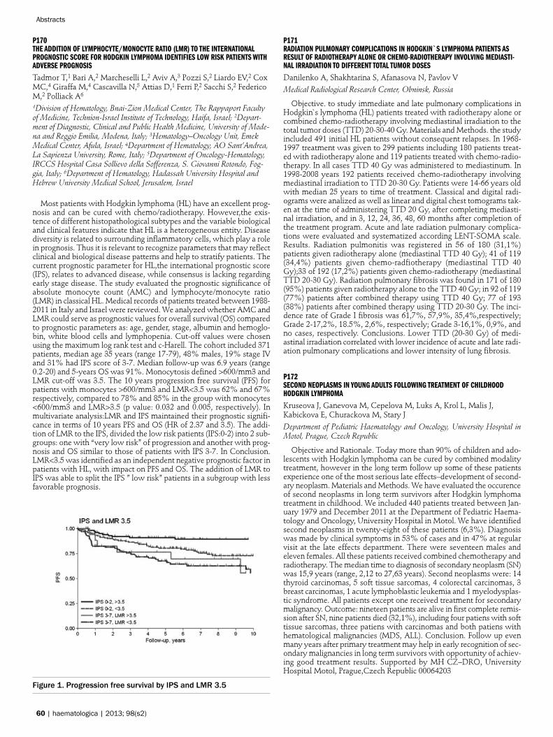
P170THE ADDITION OF LYMPHOCYTE/MONOCYTE RATIO (LMR) TO THE INTERNATIONALPROGNOSTIC SCORE FOR HODGKIN LYMPHOMA IDENTIFIES LOW RISK PATIENTS WITHADVERSE PROGNOSIS
Tadmor T,1 Bari A,2 Marcheselli L,2 Aviv A,3 Pozzi S,2 Liardo EV,2 CoxMC,4 Giraffa M,4 Cascavilla N,5 Attias D,1 Ferri P,2 Sacchi S,2 FedericoM,2 Polliack A6
1Division of Hematology, Bnai-Zion Medical Center, The Rappaport Facultyof Medicine, Technion-Israel Institute of Technology, Haifa, Israel; 2Depart-ment of Diagnostic, Clinical and Public Health Medicine, University of Mode-na and Reggio Emilia, Modena, Italy; 3Hematology–Oncology Unit, EmekMedical Center, Afula, Israel; 4Department of Hematology, AO Sant’Andrea,La Sapienza University, Rome, Italy; 5Department of Oncology-Hematology,IRCCS Hospital Casa Sollievo della Sofferenza, S. Giovanni Rotondo, Fog-gia, Italy; 6Department of Hematology, Hadassah University Hospital andHebrew University Medical School, Jerusalem, Israel
Most patients with Hodgkin lymphoma (HL) have an excellent prog-nosis and can be cured with chemo/radiotherapy. However,the exis-tence of different histopathological subtypes and the variable biologicaland clinical features indicate that HL is a heterogeneous entity. Diseasediversity is related to surrounding inflammatory cells, which play a rolein prognosis. Thus it is relevant to recognize parameters that may reflectclinical and biological disease patterns and help to stratify patients. Thecurrent prognostic parameter for HL,the international prognostic score(IPS), relates to advanced disease, while consensus is lacking regardingearly stage disease. The study evaluated the prognostic significance ofabsolute monocyte count (AMC) and lymphocyte/monocyte ratio(LMR) in classical HL. Medical records of patients treated between 1988-2011 in Italy and Israel were reviewed. We analyzed whether AMC andLMR could serve as prognostic values for overall survival (OS) comparedto prognostic parameters as: age, gender, stage, albumin and hemoglo-bin, white blood cells and lymphopenia. Cut-off values were chosenusing the maximum log rank test and c-Harell. The cohort included 371patients, median age 35 years (range 17-79), 48% males, 19% stage IVand 31% had IPS score of 3-7. Median follow-up was 6.9 years (range0.2-20) and 5-years OS was 91%. Monocytosis defined >600/mm3 andLMR cut-off was 3.5. The 10 years progression free survival (PFS) forpatients with monocytes >600/mm3 and LMR<3.5 was 62% and 67%respectively, compared to 78% and 85% in the group with monocytes<600/mm3 and LMR>3.5 (p value: 0.032 and 0.005, respectively). Inmultivariate analysis:LMR and IPS maintained their prognostic signifi-cance in terms of 10 years PFS and OS (HR of 2.37 and 3.5). The addi-tion of LMR to the IPS, divided the low risk patients (IPS:0-2) into 2 sub-groups: one with “very low risk” of progression and another with prog-nosis and OS similar to those of patients with IPS 3-7. In Conclusion.LMR<3.5 was identified as an independent negative prognostic factor inpatients with HL, with impact on PFS and OS. The addition of LMR toIPS was able to split the IPS ” low risk” patients in a subgroup with lessfavorable prognosis.
Figure 1. Progression free survival by IPS and LMR 3.5
P171RADIATION PULMONARY COMPLICATIONS IN HODGKIN`S LYMPHOMA PATIENTS ASRESULT OF RADIOTHERAPY ALONE OR CHEMO-RADIOTHERAPY INVOLVING MEDIASTI-NAL IRRADIATION TO DIFFERENT TOTAL TUMOR DOSES
Danilenko A, Shakhtarina S, Afanasova N, Pavlov VMedical Radiological Research Center, Obninsk, Russia
Objective. to study immediate and late pulmonary complications inHodgkin`s lymphoma (HL) patients treated with radiotherapy alone orcombined chemo-radiotherapy involving mediastinal irradiation to thetotal tumor doses (TTD) 20-30-40 Gy. Materials and Methods. the studyincluded 491 initial HL patients without consequent relapses. In 1968-1997 treatment was given to 299 patients including 180 patients treat-ed with radiotherapy alone and 119 patients treated with chemo-radio-therapy. In all cases TTD 40 Gy was administered to mediastinum. In1998-2008 years 192 patients received chemo-radiotherapy involvingmediastinal irradiation to TTD 20-30 Gy. Patients were 14-66 years oldwith median 25 years to time of treatment. Classical and digital radi-ograms were analized as well as linear and digital chest tomograms tak-en at the time of administering TTD 20 Gy, after completing mediasti-nal irradiation, and in 3, 12, 24, 36, 48, 60 months after completion ofthe treatment program. Acute and late radiation pulmonary complica-tions were evaluated and systematized according LENT-SOMA scale.Results. Radiation pulmonitis was registered in 56 of 180 (31,1%)patients given radiotherapy alone (mediastinal TTD 40 Gy); 41 of 119(34,4%) patients given chemo-radfiotherapy (mediastinal TTD 40Gy);33 of 192 (17,2%) patients given chemo-radiotherapy (mediastinalTTD 20-30 Gy). Radiation pulmonary fibrosis was found in 171 of 180(95%) patients given radiotherapy alone to the TTD 40 Gy; in 92 of 119(77%) patients after combined therapy using TTD 40 Gy; 77 of 193(38%) patients after combined therapy using TTD 20-30 Gy. The inci-dence rate of Grade I fibrosis was 61,7%, 57,9%, 35,4%,respectively;Grade 2-17,2%, 18.5%, 2,6%, respectively; Grade 3-16,1%, 0,9%, andno cases, respectively. Conclusions. Lower TTD (20-30 Gy) of medi-astinal irradiation correlated with lower incidence of acute and late radi-ation pulmonary complications and lower intensity of lung fibrosis.
P172SECOND NEOPLASMS IN YOUNG ADULTS FOLLOWING TREATMENT OF CHILDHOODHODGKIN LYMPHOMA
Kruseova J, Ganevova M, Cepelova M, Luks A, Krol L, Malis J,Kabickova E, Churackova M, Stary JDepartment of Pediatric Haematology and Oncology, University Hospital inMotol, Prague, Czech Republic
Objective and Rationale. Today more than 90% of children and ado-lescents with Hodgkin lymphoma can be cured by combined modalitytreatment, however in the long term follow up some of these patientsexperience one of the most serious late effects–development of second-ary neoplasm. Materials and Methods. We have evaluated the occurenceof second neoplasms in long term survivors after Hodgkin lymphomatreatment in childhood. We included 440 patients treated between Jan-uary 1979 and December 2011 at the Department of Pediatric Haema-tology and Oncology, University Hospital in Motol. We have identifiedsecond neoplasms in twenty-eight of these patients (6,3%). Diagnosiswas made by clinical symptoms in 53% of cases and in 47% at regularvisit at the late effects department. There were seventeen males andeleven females. All these patients received combined chemotherapy andradiotherapy. The median time to diagnosis of secondary neoplasm (SN)was 15,9 years (range, 2,12 to 27,63 years). Second neoplasms were: 14thyroid carcinomas, 5 soft tissue sarcomas, 4 colorectal carcinomas, 3breast carcinomas, 1 acute lymphoblastic leukemia and 1 myelodysplas-tic syndrome. All patients except one received treatment for secondarymalignancy. Outcome: nineteen patients are alive in first complete remis-sion after SN, nine patients died (32,1%), including four patiens with softtissue sarcomas, three patiens with carcinomas and both patiens withhematological malignancies (MDS, ALL). Conclusion. Follow up evenmany years after primary treatment may help in early recognition of sec-ondary malignancies in long term survivors with opportunity of achiev-ing good treatment results. Supported by MH CZ–DRO, UniversityHospital Motol, Prague,Czech Republic 00064203
60 | haematologica | 2013; 98(s2)
Abstracts
P173THE BREAST CANCER AFTER RADIOTHERAPY DATASET (BARD): AN INITIATIVE TOIMPROVE SCREENING FOR BREAST CANCER IN A HIGH RISK COHORT OF FEMALEHODGKIN LYMPHOMA SURVIVORS IN ENGLAND
Radford J,1 Howell S,2 3 O’Hara C,4 Goode V,5 Vaughan K,6 Davies S,7Cowan R,8 Swerdlow A81The University of Manchester and The Christie NHS Foundation Trust, Man-chester, UK; 2The University of Manchester and The Christie NHS FoundationTrust, Manchester, UK; 3Public Health England (Northwest), Manchester, UK;4The Christie NHS Foundation Trust, Manchester, UK; 5The University of Man-chester, Manchester, UK; 6Teenage Cancer Trust, London, UK; 7The ChristieNHS Foundation Trust, Manchester, UK; 8The Institute of Cancer Research,London, UK
Introduction. Female survivors of Hodgkin lymphoma who receivedradiotherapy involving breast tissue under age 36 are at increased risk ofbreast cancer. Since 2003, national guidelines have recommended thesewomen undergo annual breast screening starting 8 years after treatmentor from age 25 whichever is later. In 2009 we showed that penetrationof screening into this at-risk population was poor (Howell S et al, BritishJournal of Cancer, 2009). In addition, anecdotal evidence suggests thatappointments for screening are not always issued in a timely way and/orrequire repeat referrals from the treating oncologist or family physician.These factors limit the effectiveness of screening and generate anxietyfor at-risk women. Methods and Results. To address these issues wehave established a national breast cancer after radiotherapy dataset(BARD), hosted at Public Health England (Northwest) in Manchesterand funded by Teenage Cancer Trust. BARD comprises contact detailsfor women irradiated pre-2003 and identified as part of a recall exerciseundertaken in 2003, those irradiated 2003-2009 identified from cancerregistries/radiotherapy centre records and finally women irradiated 2009-2013 and beyond identified from the nationally mandated radiotherapydataset. The names of women irradiated under age 30 and requiringscreening will be forwarded annually to the NHS Breast Screening Pro-gramme (NHSBSP) that will then issue a screening appointment at alocal breast screening centre. For women irradiated 30-35 whom NHS-BSP is not currently commissioned to screen, BARD will send a letter tothe family physician prompting them to refer for screening. Conclusions.For the first time BARD brings together a cohort of women in Englandat high risk of breast cancer following radiotherapy to breast tissue dur-ing treatment for Hodgkin lymphoma under age 36. In collaborationwith NHSBSP, BARD will facilitate the timely issue of screening appoint-ments and improve the efficiency and reliability of screening and the userexperience. Evaluation of the screening protocol, trends in breast cancerincidence as radiotherapy practice evolves and primary research intoradiation induced breast cancer will also be possible. Discussions aroundextending the scope of BARD to other countries in the UK are underway.
P174CLINICAL AND RADIOLOGICAL MANIFESTATIONS OF PULMONARY TOXICITY INHODGKIN`S LYMPHOMA PATIENTS TREATED WITH CHEMOTHERAPY AND RADIOTHERA-PY TO TOTAL TUMOR DOSES 20-30 GY
Shakhtarina S, Danilenko A, Afanasova N, Pavlov VMedical Radiological Research Center, Obninsk, Russian Federation
Objective. To study pulmonary changes in Hodgkin`s lymphoma (HL)patients during and after chemo-radiotherapy using lowered radiationtotal tumor doses (TTD). Materials and Methods. 192 initial HL patientstreated with chemo-radiotherapy involving mediastinal irradiation toTTD 20-30 Gy. Ages: 15-68 (median-27) years. According to chemother-apy and TTD the following groups were defined: I-COPP+RT 20-22Gy(34 pts), II-ABVD+RT 24-30 Gy (125 pts), III-BEACOPP+RT 30 Gy (33pts). Results. During chemotherapy in 49 of 192 (25,5%) radiographyshowed pathological pattern of lung structure due to enlarged andincreased in number lung vessels surrounded with denced intersticialtissue up to the vessels of IV-V order. Pathological process was of diffusebilateral type, “air” bronchograms were ansent. Eight of 49 (16,3%)patients showed focal or confluent pulmonary infiltration, 3 (6,1%)patients had denced interlobular pleura, intersticial septs. Radiologicalsigns of cytostatic impact: in Group I-6 of 34 (17,6%) patients; Group II-36 of 125 (28,8%); Group III-7 of 33 (25,5%). Diffuse intersticial changesof pulmonary stroma were seen seen in all groups of patients; focal orconfluent pulmonary infiltration was only seen in Group II patients.
Clinical manifestations of pulmonaru toxicity were also found in patientsof Group II only (4 of 125; 3,2%). Fibrotic pulmonary changes due tocytostatic impact were not found. Radiological signs of short-term radi-ation changes in lungs were registered in 61 of 192 (31,8%) patients:Group I-11 of 34 (32,3%); Group II-40 of 125 (32%); Group III-10 of 33(30,3%). Late complications-in 73 of 192 (39%) patients: Group I-10 of34 (29,4%); Group II-47 of 125 (37,6%), Group III-16 of 33 (48,5%).Grade I fibrosis was predominating (68 0f 192; 35,4%) while Grade 2fibrosis was found in 5 (2,6%) patients. Conclusion. radiological signs ofpulmonary toxicity due to cytostatics were found after COPP, ABVD,BEACOPP. Clinically significant-after ABVD only. Incidence rate ofshort-term and late radiation pulmonary toxicity as result of mediasti-nal irradiation to TTD 20-30 Gy was lower than that from literaturedata concerning 40 Gy
P175FERTILITY IN MALE PATIENTS WITH HODGKIN LYMPHOMA TREATED WITH BEACOPPREGIMEN
Smardova L,1 Crha I,2 Michalka J,1 Kral Z,1 Janikova A,1 CharouzkovaJ,1 Mayer J1
1Department of Internal Medicine, Hematology and Oncology, University Hos-pital Brno and Masaryk University, Czech Republic; 2Department of Obstetricsand Gynecology, University Hospital Brno and Masaryk University, CzechRepublic
Introduction. BEACOPP regimen is currently the standard treatmentof advanced stage Hodgkin lymphoma (HL). However, its administrationis associated with a higher risk of adverse effects, including impaired fer-tility due to containment of several gonadotoxic agents. Methods. Weevaluated the impact of BEACOPP regimen on fertility status of 23patients with advanced stage HL. The median age was 26 years (20-37).Most patients were in clinical stage IIB with risk factors (N=11), fol-lowed by the group of patients in stage III (N=8) and patients in stage IV(N=4). All patients were treated with 6-8 cycles of BEACOPP baseline(N=6) or escalated (N=17). For all patients were performed semen analy-sis and measurement of hormonal levels of gonadotropic hormones (fol-licle-stimulating hormone (FSH), luteinizing hormone (LH) and testos-terone) before and after treatment. Results. Only 1 patient had normo-zoospermia and 22 patients had dysspermia before treatment. For allpatients we found normal levels of gonadotropic hormones and testos-terone. After treatment azoospermia was observed for all patients (themedian time of semen examination was 24 month). For 20/23 (87%)patients was found FSH elevation (13.1 to 35.8 IU/l), for 5/23 (21%)patients LH elevation (9.2 to 15.0). After treatment, testosterone levelswere normal for all patients. 6 patients (26%) used previously frozensperm samples to attempt in vitro fertilization, in case of 4 patients it wassuccessfull (healthy children were born 2-8 years after treatment). Con-clusion. Male patients with HL are at high risk of infertility after treat-ment with BEACOPP regimen. Before starting treatment sperm cryop-reservation should be offered to every patient. FSH levels probably cor-relate with spermatogonial damage after treatment.
P176COMBINATION OF FERTILITY PRESERVATION STRATEGIES IN YOUNG WOMEN WITHRECENTLY DIAGNOSED HODGKIN LYMPHOMA
Smardova L,1 Huser M,2 Michalka J,1 Zakova J,2 Kral Z,1 Crha I,2Janikova A,1 Krizova L,1 Mayer J,1 Ventruba P2
1Department of Internal Medicine, Hematology and Oncology, University Hos-pital Brno and Masaryk University, Czech Republic; 2Department of Obstetricsand Gynecology University Hospital Brno and Masaryk University, CzechRepublic
Introduction. One of the most common long term consequences ofchemotherapy is infertility due to the destruction of gonadal cells. Theaim of this study is to describe the clinical management and outcomesof currently available fertility preservation techniques in a set of 154female patients with Hodgkin lymphoma. Methods. Patients in repro-ductive age with newly diagnosed Hodgkin lymphoma were offeredembryo or oocyte cryopreservation, ovarian tissue cryopreservation andthe administration of GnRH analogues during chemotherapy. Results.During study period (2004-2009), 154 young female patients were
haematologica | 2013; 98(s2) | 61
9th International Symposium on Hodgkin Lymphoma, Cologne, Germany, October 12-15, 2013
offered fertility preservation counseling. Patient’s average age was 29.4years. The majority of women were nuliparas or primiparas at the timeof diagnosis. All referred patients were individually consulted regardingtheir risk of fertility impairment or loss due to planned gonadotoxicchemotherapy. The optimal approach was chosen on a strictly individ-ual basis and was depended on the type of planned treatment, timeavailable till the onset of treatment, the patient’s age, and whether thepatient has a partner. Administration of GnRH analogues (N=123,79,9%) and ovarian tissue cryopreservation (N=16, 10,4%) were themost commonly used fertility preservation strategies. In 20 cases(12,9%), combination of several fertility preservation techniques waspreferred in individually selected patients. In these cases, administra-tion of GnRH analoques during chemotherapy was combined with oneof the cryopreservation strategies. Conclusion. Combination of fertilitypreservation techniques gives young cancer patients the best chance forfuture fertility and should be concentrated in specialized centers.
P177NEW PROGNOSTIC SCORE BASED ON BCL-6 EXPRESSION AND IPS STRONGLY PRE-DICTS SURVIVAL IN PATIENTS WITH HODGKIN LYMPHOMA
Antic D,1,2 Andelic B,1 Milic N,3 Durašinovic V,1 Todorovic M,1,2 BilaJ,1,2 Vukovic V,1 Jelicic J,1 Mihaljevic B1,2
1Clinical Center Serbia, Clinic for hematology; 2Medical Faculty, University inBelgrade; 3Institute for medical statistic and informatic, Medical faculty, Univer-sity in Belgrade, Belgrade, Serbia
Introduction. Biological factors like p53, pRb, bcl-6 and Ki67 may con-tribute to malignant transformation and may be of prognostic relevance.Aim: The aim of this study was to evaluate whether the expression ofp53, pRb, bcl-6 and Ki67, could be important predict factors for out-come of patients with HL. Methods. Between October 2001 and June2003, 40 de novo patients with a confirmed diagnosis of Hodgkin lym-phoma were studied. Immunohistochemical analysis was performed onparaffin-embedded lymph node specimens using monoclonal antibod-ies. Also, in all patients risk was assessed by International PrognosticScore (IPS) and patients were subsequently treated with ABVD and BEA-COPP chemotherapy regimens and followed up for a minimum of 10years. Results. In our group 25 patients were treated with ABVD while15 received escalated BEACOPP. 10-years overall survival (OS) was76,3% and 36 (90%) patients achieved complete remission. Patientswith low risk based on IPS score had significantly better survival thanintermediate/poor risk patients (134 vs 91, respectively, p=0.005). Expres-sion of p53, pRb and Ki67 did not affect response to therapy and sur-vival significantly. Regarding to bcl-6 expression patients were dividedin two subgroups; patients with bcl-6 expression in >20% cells had sig-nificantly longer mean OS of 122 months while patients with bcl-6expression in ≤20% cells had mean OS of 87 months (p=0,037). Bcl-6expression was an independent survival predicting factor in multivari-ate analysis together with the IPS (p=0,041 and p=0,022, respectively).Aggregate bcl-6-IPS score improved IPS prognostic stratification for theinteremediate and high risk patients’ by adding information on bcl-6expression (132 vs 113 vs 64 months for low, intermediate and high riskscore, respectively, p<0.001). New bcl6-IPS score retained his prognos-tic significance in multivariate analysis (p<0,001;RR=4,670; 95% CI1,675-13,022). Conclusions. Cell cycle regulatory molecules includingp53, pRb, bcl-6 and Ki67, have different level of expression in patientswith HL, but we found that only bcl-6 expression had influence on theoutcome. Aggregate bcl-6-IPS score improved standard prognosticstratification for the high and intermediate risk patients.
P178ACUTE PULMONARY TOXICITY OF BLEOMYCIN-INCIDENCE AND RISK FACTORS IN ASERIES OF HODGKIN LYMPHOMA (HL) PATIENTS TREATED IN A SINGLE CENTER
Santos J, Carvalho S, Esteves S, Gomes da Silva M
Instituto Português de Oncologia de Lisboa Francisco Gentil, Portugal
Introduction. Bleomycin may cause acute pulmonary toxicity (BPT),with potentially irreversible consequences on lung function and impactupon treatment continuation. Epidemiological and treatment factors(sex, age, G-CSF, smoking, previous lung disease) may increase the riskof BPT. Aim: to evaluate the incidence, risk factors and clinical course
of BPT in patients with Hodgkin lymphoma (HL) receiving ABVD.Methods. retrospective unicenter study in HL patients treated between2010 and 2011. BPT was defined as 1) clinical and imagiologic pneu-monitis, without an infectious cause, and with a decreased lung diffu-sion capacity (DLCO), or 2) at least one of the above criteria, leading tobleomycin discontinuation and/or treatment with corticosteroids. Riskfactors were evaluated with the Chi square test, Fisher exact test, andWilcoxon test, as appropriate. Results. 65 patients were analyzed, 57%male, median age 28 years (range 15-86): 26,2% had early favorable dis-ease, 21,5% early unfavorable disease and 50,8% advanced disease(1,5% not assessed), according to GHLSG. Median number of ABVDcycles was 6 and 24,6% received mediastinal radiotherapy. Overallresponse rate was 94%. BPT occurred in 16 patients (24,6%): only 1 ful-filled all criteria. Bleomycin was discontinued in 9 patients, of whom 6also received corticosteroids; the remaining 7 were treated with corti-costeroids and kept on ABVD; 4 patients developed pulmonary fibrosisgrade 2 to 5. Compared to patients without BPT, patients with BPTwere older (median 36 versus 26 yo, p=0,077) and mainly female (68,5%versus 34,7%, p=0,017). Smokers and non-smokers had a similar inci-dence of BPT (p=0,528), as well as patients exposed (51%) or not to G-CSF (p=0,943). The cumulative dose of bleomycin and the DLCO, eval-uated before treatment (in 57% in non-BPT patients and 87,5% BTPpatients), were not different between the 2 groups (p=0,081 and p=0,197respectively). Conclusions. the incidence of BPT was similar to whathas been reported and was moderate to severe in 25% of patients, lead-ing to a change in treatment in 56%. In this series, BPT was more fre-quent in women and the incidence appeared to increase with age.Prospective studies are needed with larger numbers of patients to iden-tify populations at risk.
P179TESTICULAR FUNCTION ASSESSMENT IN YOUNG MEN TREATED FOR HODGKIN’S LYM-PHOMA DURING CHILDHOOD AND ADOLESCENCE
Moryl-Bujakowska A,1 Balwierz W,1 Malek A,2 Sztefko K31Department of Pediatric Oncology and Hematology, Polish-American Instituteof Pediatrics, Jagiellonian University Medical College, Krakow, Poland; 2Depart-ment of Clinical Biochemistry, Children’s University Hospital in Krakow, Poland;3Department of Clinical Biochemistry, Polish-American Institute of Pediatrics inKrakow, Jagiellonian University Medical College, Poland
Introduction. of modern methods of combined therapy allows thecure more than 90% of children and adolescents with Hodgkin’s lym-phoma. However, the intensive treatment carries the risk of late com-plications including gonadal dysfunction in men and women. In thisreport preliminary results of testicular function assessment in youngmen after Hodgkin’s lymphoma therapy are presented. Testicular func-tion was evaluated in 29 men [age: 5.0-18.2 (median 14.5) years, and18.0-26.3 (median 22.8) years at the beginning of treatment and the endof observation, respectively] treated between 1.01.1999 and 31.12.2008in Department of Pediatric Oncology and Hematology PAIP JU-MC inKrakow, according to PGP-HD-97 protocol. Multidrug chemotherapy(MOPP given alternatively with B-DOPA) combined with low-doseinvolved-field radiotherapy (15-25 Gy) was used in 25 men, and in 4 oth-er men-chemotherapy only. Therapy was completed in all analyzedpatients. Physical examination, Tanner stages of pubic hair and genitaldevelopment were recorded as well as the plasma levels of follicle stim-ulating hormone (FSH), luteinizing hormone and testosterone weremeasured in all patients. Testicular volumes were measured in 18 men.The study was conducted between 1.01.2009 and 31.12. 2011. All menreached Tanner stages of pubic hair and genital development appropri-ate for their age. In all patients 207 measurements of hormones levelswere performed. At least one abnormal result of measurements wasfound in 17/29 (58.6%) of men. The most frequent abnormality was theincreased level of FSH (in 15 patients; 46.4% of FSH measurements).Abnormal results of measurements were found in 15/24 (62.5%) patientswho received 6-8 chemotherapy cycles and in 2/5 patients treated with3-5 cycles. Within the group of 17 patients with abnormal results eightmen previously received radiotherapy of subdiaphragmatic region.Semen analysis was performed in two men and revealed azoospermia.Initial results of reproductive system assessment indicate the impair-ment of testicular function depending on intensity of treatment. Furtherobservation and repeated gonadal function tests (including semen analy-sis) are needed in young men with completed Hodgkin’s lymphoma
62 | haematologica | 2013; 98(s2)
Abstracts
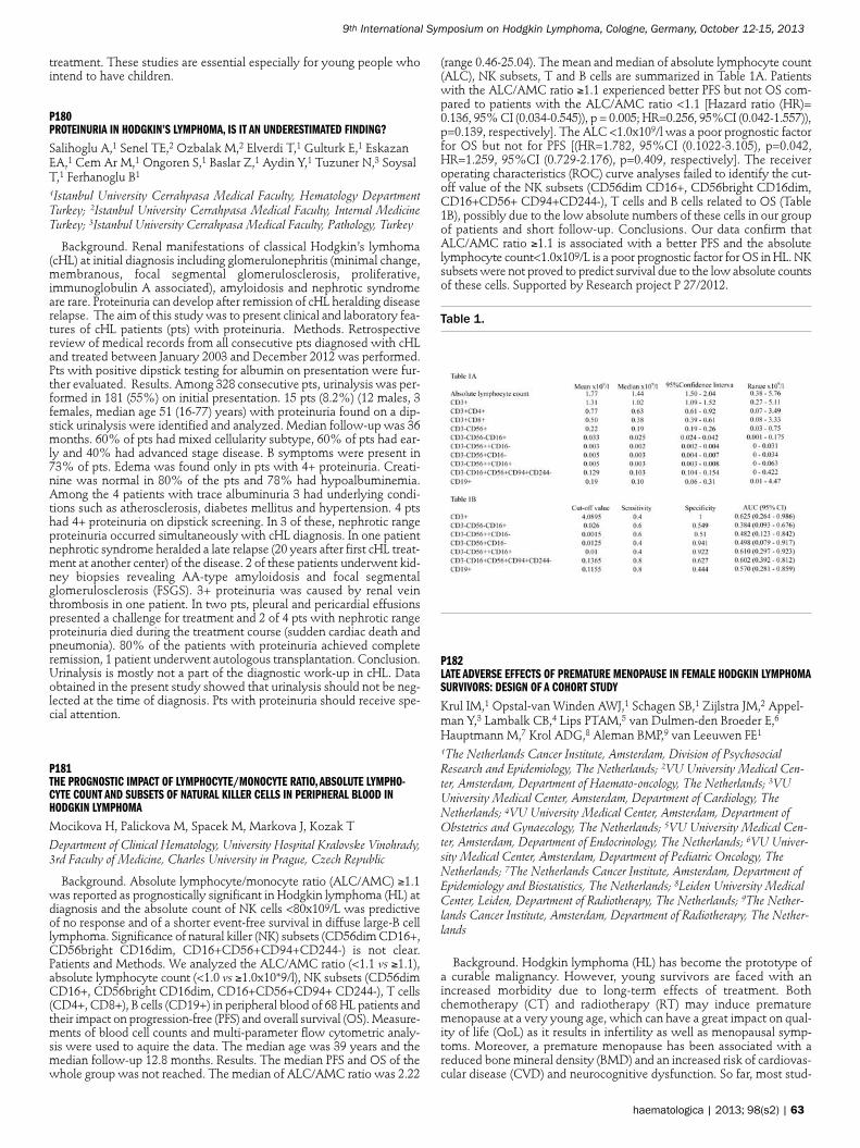
treatment. These studies are essential especially for young people whointend to have children.
P180PROTEINURIA IN HODGKIN’S LYMPHOMA, IS IT AN UNDERESTIMATED FINDING?
Salihoglu A,1 Senel TE,2 Ozbalak M,2 Elverdi T,1 Gulturk E,1 EskazanEA,1 Cem Ar M,1 Ongoren S,1 Baslar Z,1 Aydin Y,1 Tuzuner N,3 SoysalT,1 Ferhanoglu B11Istanbul University Cerrahpasa Medical Faculty, Hematology DepartmentTurkey; 2Istanbul University Cerrahpasa Medical Faculty, Internal MedicineTurkey; 3Istanbul University Cerrahpasa Medical Faculty, Pathology, Turkey
Background. Renal manifestations of classical Hodgkin’s lymhoma(cHL) at initial diagnosis including glomerulonephritis (minimal change,membranous, focal segmental glomerulosclerosis, proliferative,immunoglobulin A associated), amyloidosis and nephrotic syndromeare rare. Proteinuria can develop after remission of cHL heralding diseaserelapse. The aim of this study was to present clinical and laboratory fea-tures of cHL patients (pts) with proteinuria. Methods. Retrospectivereview of medical records from all consecutive pts diagnosed with cHLand treated between January 2003 and December 2012 was performed.Pts with positive dipstick testing for albumin on presentation were fur-ther evaluated. Results. Among 328 consecutive pts, urinalysis was per-formed in 181 (55%) on initial presentation. 15 pts (8.2%) (12 males, 3females, median age 51 (16-77) years) with proteinuria found on a dip-stick urinalysis were identified and analyzed. Median follow-up was 36months. 60% of pts had mixed cellularity subtype, 60% of pts had ear-ly and 40% had advanced stage disease. B symptoms were present in73% of pts. Edema was found only in pts with 4+ proteinuria. Creati-nine was normal in 80% of the pts and 78% had hypoalbuminemia.Among the 4 patients with trace albuminuria 3 had underlying condi-tions such as atherosclerosis, diabetes mellitus and hypertension. 4 ptshad 4+ proteinuria on dipstick screening. In 3 of these, nephrotic rangeproteinuria occurred simultaneously with cHL diagnosis. In one patientnephrotic syndrome heralded a late relapse (20 years after first cHL treat-ment at another center) of the disease. 2 of these patients underwent kid-ney biopsies revealing AA-type amyloidosis and focal segmentalglomerulosclerosis (FSGS). 3+ proteinuria was caused by renal veinthrombosis in one patient. In two pts, pleural and pericardial effusionspresented a challenge for treatment and 2 of 4 pts with nephrotic rangeproteinuria died during the treatment course (sudden cardiac death andpneumonia). 80% of the patients with proteinuria achieved completeremission, 1 patient underwent autologous transplantation. Conclusion.Urinalysis is mostly not a part of the diagnostic work-up in cHL. Dataobtained in the present study showed that urinalysis should not be neg-lected at the time of diagnosis. Pts with proteinuria should receive spe-cial attention.
P181THE PROGNOSTIC IMPACT OF LYMPHOCYTE/MONOCYTE RATIO, ABSOLUTE LYMPHO-CYTE COUNT AND SUBSETS OF NATURAL KILLER CELLS IN PERIPHERAL BLOOD INHODGKIN LYMPHOMA
Mocikova H, Palickova M, Spacek M, Markova J, Kozak TDepartment of Clinical Hematology, University Hospital Kralovske Vinohrady,3rd Faculty of Medicine, Charles University in Prague, Czech Republic
Background. Absolute lymphocyte/monocyte ratio (ALC/AMC) ≥1.1was reported as prognostically significant in Hodgkin lymphoma (HL) atdiagnosis and the absolute count of NK cells <80x109/L was predictiveof no response and of a shorter event-free survival in diffuse large-B celllymphoma. Significance of natural killer (NK) subsets (CD56dim CD16+,CD56bright CD16dim, CD16+CD56+CD94+CD244-) is not clear.Patients and Methods. We analyzed the ALC/AMC ratio (<1.1 vs ≥1.1),absolute lymphocyte count (<1.0 vs ≥1.0x10*9/l), NK subsets (CD56dimCD16+, CD56bright CD16dim, CD16+CD56+CD94+ CD244-), T cells(CD4+, CD8+), B cells (CD19+) in peripheral blood of 68 HL patients andtheir impact on progression-free (PFS) and overall survival (OS). Measure-ments of blood cell counts and multi-parameter flow cytometric analy-sis were used to aquire the data. The median age was 39 years and themedian follow-up 12.8 months. Results. The median PFS and OS of thewhole group was not reached. The median of ALC/AMC ratio was 2.22
(range 0.46-25.04). The mean and median of absolute lymphocyte count(ALC), NK subsets, T and B cells are summarized in Table 1A. Patientswith the ALC/AMC ratio ≥1.1 experienced better PFS but not OS com-pared to patients with the ALC/AMC ratio <1.1 [Hazard ratio (HR)=0.136, 95% CI (0.034-0.545)), p = 0.005; HR=0.256, 95%CI (0.042-1.557)),p=0.139, respectively]. The ALC <1.0x10⁹/l was a poor prognostic factorfor OS but not for PFS [(HR=1.782, 95%CI (0.1022-3.105), p=0.042,HR=1.259, 95%CI (0.729-2.176), p=0.409, respectively]. The receiveroperating characteristics (ROC) curve analyses failed to identify the cut-off value of the NK subsets (CD56dim CD16+, CD56bright CD16dim,CD16+CD56+ CD94+CD244-), T cells and B cells related to OS (Table1B), possibly due to the low absolute numbers of these cells in our groupof patients and short follow-up. Conclusions. Our data confirm thatALC/AMC ratio ≥1.1 is associated with a better PFS and the absolutelymphocyte count<1.0x10⁹/L is a poor prognostic factor for OS in HL. NKsubsets were not proved to predict survival due to the low absolute countsof these cells. Supported by Research project P 27/2012.
Table 1.
P182LATE ADVERSE EFFECTS OF PREMATURE MENOPAUSE IN FEMALE HODGKIN LYMPHOMASURVIVORS: DESIGN OF A COHORT STUDY
Krul IM,1 Opstal-van Winden AWJ,1 Schagen SB,1 Zijlstra JM,2 Appel-man Y,3 Lambalk CB,4 Lips PTAM,5 van Dulmen-den Broeder E,6Hauptmann M,7 Krol ADG,8 Aleman BMP,9 van Leeuwen FE1
1The Netherlands Cancer Institute, Amsterdam, Division of PsychosocialResearch and Epidemiology, The Netherlands; 2VU University Medical Cen-ter, Amsterdam, Department of Haemato-oncology, The Netherlands; 3VUUniversity Medical Center, Amsterdam, Department of Cardiology, TheNetherlands; 4VU University Medical Center, Amsterdam, Department ofObstetrics and Gynaecology, The Netherlands; 5VU University Medical Cen-ter, Amsterdam, Department of Endocrinology, The Netherlands; 6VU Univer-sity Medical Center, Amsterdam, Department of Pediatric Oncology, TheNetherlands; 7The Netherlands Cancer Institute, Amsterdam, Department ofEpidemiology and Biostatistics, The Netherlands; 8Leiden University MedicalCenter, Leiden, Department of Radiotherapy, The Netherlands; 9The Nether-lands Cancer Institute, Amsterdam, Department of Radiotherapy, The Nether-lands
Background. Hodgkin lymphoma (HL) has become the prototype ofa curable malignancy. However, young survivors are faced with anincreased morbidity due to long-term effects of treatment. Bothchemotherapy (CT) and radiotherapy (RT) may induce prematuremenopause at a very young age, which can have a great impact on qual-ity of life (QoL) as it results in infertility as well as menopausal symp-toms. Moreover, a premature menopause has been associated with areduced bone mineral density (BMD) and an increased risk of cardiovas-cular disease (CVD) and neurocognitive dysfunction. So far, most stud-
haematologica | 2013; 98(s2) | 63
9th International Symposium on Hodgkin Lymphoma, Cologne, Germany, October 12-15, 2013
ies on these conditions have been conducted in women with an earlynatural or surgery-induced menopause with short follow-up times. Inthis study we will examine the long-term effects of a CT- and/or RT-induced premature menopause on BMD, cardiovascular status, neu-rocognitive function and QoL in female HL survivors. Methods. Cross-sectional measurements will be performed within an existing cohort offemale HL survivors. Women treated for HL in three large hospitalsbetween 1965 and 2005 at ages 15-39 years will be invited for a visit toa survivorship care outpatient clinic. Participants will be asked to com-plete a questionnaire, provide a blood sample and to undergo severalmedical and neurocognitive tests. Multivariate regression analyses willbe used to compare the outcomes of women who developed prematuremenopause with those who did not. Results. This study will increaseknowledge about the BMD, cardiovascular status and neurocognitivefunction in long-term female HL survivors with a CT- and/or RT-inducedpremature menopause. It will also provide insight into the influence ofthese long-term effects on quality of life. Conclusions. Results of thisstudy will lead to the identification of those HL survivors who are atincreased risk for osteoporosis, CVD and neurocognitive dysfunctiondue to premature menopause as a consequence of their former treat-ment. This provides the opportunity to timely refer high-risk women forinterventions in order to reduce morbidity and enhance QoL. Moreover,women who will experience premature menopause in the future can bebetter informed about potential long-term effects.
P183PREDICTIVE FACTORS OF RESPONSE IN TUNISIAN ADULT HODGKIN LYMPHOMA
Kacem K,1 Zriba S,2 Ghédira H,2 Yahyaoui Y,1 Mansouri R,1 Dridi M,1Hadj Mansour M,1 Manai Z,1 Zarrouk M,1 Ben Neji H,1 Ben Abden-nebi Y,1 Jeddi R,1 Aissaoui L,1 Belhadj Ali Z,1 Ben Abid H,1 M’Sadek F,2Ben Lakhal R,1 Meddeb B11Clinical Hematology Department of Aziza Othmana of Tunis; 2Clinical Hema-tology Department of Military Hospital of Tunis, USA
Purpose. Study of prognostic factors in HL population treated in twocenters according to national protocol HL-2008. Patients and Methods.173 patients were included and prospectively analyzed for response andoutcome. Chemotherapy regimen was ABVD for early stages and esca-lated BEACOPP for advanced disease and stage II with IMT> 0.35. Com-plete response (RC) rate was of 82%. At 3 years, OS, EFS, RFS wererespectively 90%, 75%, and 95%. In localized disease RC, OS, EFS, andRFS were respectively 86%, 95.5%, 84% and 95.5%. predictive factorsof response was albumin<30g/l (p=0.04) and IMT> 0.35 (p=0.001). Inaddition to these factors, refractoriness (p= 0.009) and VS> 30+B(p=0.015) significantly influence OS and EFS. RC, OS, EFS, RFS were80%, 86%, 69%,and 86% in advanced stages.In multivariate analysispredictive factors for response were SPI>2 (p=0.01), lyphopenia< 600(p=0.001), stage IV (p=0.036), hepatic localization (p=0.04). For EFS, pre-dictive factors were SPI>2, lymphopenia, stage IV, male sex and expres-sion of CD20 (p=0.05). peripheral bulky disease and baseline WBC>15x109/L (p=0.003) significantly influence RFS while OS was signifi-cantly influenced by remission status at the end of treatement(p<0.0001), bone marrow infiltration , bulky mediastinal disease, expres-sion of CD20, age> 45, and male sexe. Conclusion. Our results were inaccordance with literature.
P184CASE REPORT OF A PATIENT WITH HODGKIN LYMPHOMA AFTER TREATED WITH TNFALFA INHIBITOR BECAUSE OF PSORIASIS AND PSORIATIC ARTHRITIS AND OVERVIEW OFTHE LITERATURE
Páyer E,1 Gál A,1 Szegedi A,2 Barna S,3 Illés A11Institution of Medicine, 2Department of Dermatology, 3Scanomed Ltd,Univer-sity of Debrecen, Hungary
TNF alfa inhibitors means milestone in treatment of chronic inflam-matory diseases, however tumors, especially lymphomas may be morefrequent in these patients. We riport about a 30-year-old patient present-ed with psoriasis vulgaris and psoriatic arthritis in 1998. It was treatedwith azathioprin, methotrexate and after progression infliximab fromJan 2009 to Jan 2011. In Dec 2010 cervical lymph node enlargement wasdetected on the right side, and histological analysis of bioptic sampleproved Hodgin lymphoma, lymphocyte rich type. By the stagingPET/CT Ann Arbor stage was II/A. ABVD treatment was started. After2 cycles interim PET/CT was performed. On the right side the lym-phoma disappeared, but on the left side of the neck new lymph nodeand tonsillary activity was showed which seemed to be an infection,thus ABVD was continued, and antibiotic therapy was added. After 6cycles of ABVD PET/CT was performed again which showed completemetabolic remission. His treatment was completed with radiotherapyon the right side of cervical and supraclavicular region (32.2 Gy). Restag-ing PET/CT showed complete metabolic remission also and the patientis well up to this day. However psoriasis and arthritis worsened, whichmeans treatment challange. This case raises two main questions. Firstof all, are there lymphomas or other hematologic malignacies reallymore frequent in patients treated with TNF alfa inhibitors? On the oth-er hand, how can we treat the inflammatory diseases after succsessfultreatment of hematologic malignacy?
64 | haematologica | 2013; 98(s2)
Abstracts
Abadi, U, 37Abdah-Bortnyak, R, 37Abramson, JS, 6, 12Acar, K, 53Adam, D, 28Adams, A, 24Advani, RH, 4, 7, 26Afanasova, N, 60, 61Agostinelli, C, 18Aide, MA, 9Aissaoui, L, 10, 52, 64Akı, SZ, 53Akria, L, 37Al-Radi, LS, 7, 8, 27Alabi, S, 16Aldinucci, D, 24Aleksik, E, 8Aleman, BMP, 54, 55, 56, 57, 63Allen, J, 7Alsner, J, 21Amin, S, 34Amorin, S, 49Anastasia, A, 48Andelic, B, 62Andersen, F, 15Andersen, MD, 22Andersson, TML, 55Andjelic, B, 10Andjelic, BM, 5, 9Andre, M, 1Andreu, R, 43Angelopoulou, MK, 33, 39, 41Angrilli, F, 48Ansell, SM, 2, 6Antic, D, 10, 62Aoudjhane, A, 1Appelman, Y, 63Aquino, R, 47Ar, MC, 24Arai, S, 50Arar, A, 27Arcaini, L, 21Arnason, JE, 12Arpaci, F, 53Asano, N, 59Ataergin, S, 53Attia, E, 42Attias, D, 37, 60Audhuy, B, 1Aurer, I, 5Avigan, D, 12Avigdor, A, 3Aviv, A, 60Avivi, I, 37Aydin, Y, 24, 63Aznar, MC, 15Azoulay, E, 27
Babic, D, 5Bäck, A, 16Baculea, S, 50Baiocchi, O, 9Bairey, O, 37Balducci, M, 20Balwierz, W, 25, 62Balzarotti, M, 48Bar-Shalom, R, 37Bari, A, 60Barna, S, 11, 42, 53, 64Barnes, JA, 6, 12Barrera, M, 30Barrington, S, 2, 13Barros, MH, 35, 41Bartlett, N, 4, 36Bartolomei, F, 20Basak, R, 36Basic-Kinda, S, 5Baslar, Z, 24, 63Bastard, C, 26Bauchet, A, 29Baues, C, 3, 58Becker, N, 28Bedekovics, J, 53
Behringer, K, 11, 12, 26, 57, 58Beijert, M, 54Beishuizen, A, 46Belada, D, 58Belhadj, Ali, Z, 10, 52, 64Bell, AI, 18, 33Bellei, M, 9, 48Bello, CM, 12Ben Abdennebi, Y, 10, 52, 64Ben Abid, H, 10, 52, 64Ben Lakhal, R, 10, 52, 64Ben Neji, H, 10, 52, 64Benavente, Y, 28Bendix, K, 21, 22Benelli, G, 41Bereiter-Hahn, J, 30Bergeron, A, 27Bergesio, F, 38Beris, PH, 33Berkahn, L, 2Bernd, HW, 45Bertrand, P, 26Bessell, E, 13Besson, C, 3, 23Bhalla, S, 30Bhatia, S, 28Bhatnagar, A, 17Biasoli, I, 9Biasson, V, 23Biassoni, V, 16Biggi, A, 3, 38, 39Bila, J, 10, 62Birkenmeier, K, 30Biyachuev, ER, 5Björkholm, M, 32, 33, 40, 55Bloor, A, 34Blum, KA, 4Bociek, G, 26Boe Møller, M, 21Boffetta, P, 28Bogatyreva, TI, 8Bogdanovic, A, 5, 9, 10Boisgard, R, 29Bolis, S, 3, 39Böll, B, 1, 11, 12, 26, 57Bologna, S, 1, 49Bonamin Sola, C, 9Bonfichi, M, 6, 21Bonnet, F, 3Bono, E, 21Bonthapally, V, 46, 48, 50Borchmann, P, 1, 2, 11, 12, 13, 26, 50, 57, 58Bordwell, A, 24Borger, J, 54Borghese, C, 24Borra, A, 3, 39Bosi, A, 41Böttcher, S, 28Botto, B, 14, 48Bouabdallah, K, 49Bouabdallah, R, 44Boudjema, S, 23Boué, F, 3Bouman, D, 24Bourhis, J, 56Boutsikas, G, 33, 39Bowers, S, 52Brandt, U, 30Bräuninger, A, 32Brennan, P, 28Brice, P, 1, 4, 27, 44, 49, 51Briere, J, 1, 4Briones, J, 47Broccoli, A, 18Brodin, NP,, 15Brodt, HR, 23Brüderlein, S, 29Brusamolino, E, 48Brzeska, B, 51Bumbasirevic, VZ, 5, 9Bunce, C, 31Burger, IA, 11Buxton, A, 37, 39
Calado, DP, 28Calcagni, ML, 20Cantalapiedra, A, 47Cantonetti, M, 3Carbone, A, 24Carde, P, 1, 29, 35, 56Carella, AM, 48Carpten, JD, 50Cartaxo Muniz, MT, 25Carvalho, JEM, 9Carvalho, S, 62Casagrande, N, 24Casanova, M, 16, 23Casasnovas, O, 1Cascavilla, N, 48, 60Cassuto, O, 26Castagna, L, 6Castagnoli, A, 6Castellanos, M, 16Castellino, SM, 37Castro, N, 9Catalano, PJ, 54Catania, S, 16, 23Cavazzina, R, 3Cefalo, G, 16Cejalvo, MJ, 43Celegato, M, 24Celic, E, 58Cem, Ar, M, 63Cenci, T, 20Cepelova, M, 60Cerhan, JR, 28Cesaretti, M, 9Cesarman, E, 28Cetin, T, 53Chabay, P, 35Chamier-Cieminska, A, 38Chamseddine, A, 26Chaoui, D, 49Charouzkova, J, 61Chatziioannou, S, 39, 41Chauvie, S, 3, 38Chen, BT, 45Chen, L, 37, 39Chen, R, 45Chen, Y, 54Chernova, NG, 7, 27Cheson, BD, 4, 36Chi, A, 46, 47Chiaravalli, S, 16, 23Chiarenza, A, 19, 21Chiattone, C, 9Chien, C, 47Chiozzotto, M, 41Chirindel, A, 37Cho, SY, 37Christensen, K, 34Chung, J, 6Churackova, M, 60Ciammella, P, 14Ciccone, G, 6Ciceri, F,, 3Claesson, HE, 32, 33Clausen, M, 22Cocco, PL, 28Coetzee, GA, 28Cogliatti, S, 45Cohen, M, 35Coiffier, B, 1Colicchio, B, 56Colin, P, 1Colombatti, A, 24Coltart, S, 13Conlon, J, 52Connors, JM, 2, 6Consoli, U, 19Constine, LS, 37Contentin, N, 26Conti, DV, 28Conticello, C, 48Cook, J, 36Cooper, S, 34Corina, C, 29
haematologica | 2011; 96(s3) | a
Authors Indexsurname, page number
Cortessis, VK, 28Costagliola, 3Costello, R, 3Coulomb, A, 23Counsell, N, 13Cowan, R, 61Cox, MC, 60Cozen, W, 28Crae, S, 40Creidy, R, 23Creutzberg, CL, 58Crha, I, 61Crippa, F, 16Crump, M, 4, 14Csomor, J, 11Cuccaro, A, 20Cuceu, C, 35, 56Culligan, D, 13Cupelli, E, 20Cutter, D, 55Cutter, DJ, 56Czepczynski, R, 38
D’Alò, F, 20d’Amore, F, 2, 21, 22, 39Dainese, L, 23Damico, A, 38Dang, N, 16Dangi, U, 4, 43Danielewicz, I, 38Daniëls, LA, 58Danilenko, A, 60, 61Dann, EJ, 37Darby, S, 55Darby, SC, 56Dashnamoorthy, R, 30Datseris, I, 39Datseris, K, 41Davies, S, 61Davis, RE, 50De Alarcon, P, 16De Boer, C, 47De Boer, JP, 54De Bruin, ML, 57De Graaf, MA, 58De Jager, W, 35De Jong, D, 19De Matteo, E, 35De Mendonça Cavalcanti, MdoS, 25De Roos, A, 58De Sanjose, S, 28De Souza, C, 9de Wolf-Peeters, C, 20Deau, B, 4, 49Dekker, N, 54Delahaye-Sourdeix, M, 28Delamain, M, 9Delhem, N, 35Delmer, A, 49Demina, EA, 5Demiroz, AS, 24Desai, D, 34Devidas, A, 1Deville, L, 51deVos, S, 50Di Raimondo, F, 3, 18, 19, 21, 22Dias, LVB, 9Dickman, PW, 55Diefenbach, CS, 4Diehl, V, 11Diepstra, A, 17, 18, 19, 28, 32, 35, 42Dieterlen, A, 56Dietlein, M, 45Dimou, M, 33Djurasinovic, V, 10Dodero, A, 3Domingo, E, 47Döring, C, 20, 23, 28, 30, 34Dörken, B, 28Dos, Santos, M, 56Dotlic, S, 5Doukoure, B, 23Drayson, M, 31Dridi, M, 10, 64Dröse, S, 30Drozd-Sokolowska, J,, 51Dunphy, C, 37Dupuis, J, 44
Durakovic, N, 5Durašinovic, V, 62Dürkop, H, 18Dzietczenia, J, 38Dziuk, M, 38
Eaton, J, 50Edlund, CK, 28Efthymiou, A, 33Eich, HT, 3, 13, Eichenauer, DA, 1, 12, 26, 45, 47, 55, 57El-Galaly, TC, 39Elemento, O, 28Elfassy, E, 56Elhaddad, A, 42Elmenawy, S, 42Eloranta, S, 55Elsayed, AA, 59Elverdi, T, 24, 63Enblad, G, 2, 40Engert, A, 1, 2, 3, 11, 12, 13, 18, 26, 45, 47, 50, 55,57, 58
Epari, S, 8, 36, 51Epelbaum, R, 37Erdem, G, 53Escrivà, A, 43Eskazan, AE, 24, 63Espeso, M, 47Esteves, S, 62Evangelista, A, 6Evens, AM, 26, 30, 36Exter, B, 52
Fabbri, E, 41Facchetti, F, 20Fagerli, UM, 59Fairbanks, JY, 31Falchook, GS, 50Fallanca, F, 38Fanale, M, 26, 50Fandrey, J, 15Fanin, R, 41Farrell, K, 22, 40Fasanmade, A, 46Fattori, PP, 6Faure, P, 51Fayad, LE, 50Federico, M, 2, 9, 60Feldman, TA, 6Feller, AC, 45Feng, Y, 6, 12Fenske, TS, 26Ferhanoglu, B, 24, 63Fermé, C, 44, 47Fernández Llavador, MJ, 43Fernández Zarzoso, M, 43Ferranti, A, 6Ferrari, A, 16, 23Ferreira de Barros, T, 25Ferrer, S, 43Ferri, P, 60Fiaccadori, V, 21Filanovsky, K, 37Filippi, AR, 14Filonenko, K, 8Fingert, H, 46Fiore, F, 3, 18Fisher, D, 24Fisher, RI, 4, 36Flampouri, S, 16Flevari, P, 33, 41Flüchter, P, 20Fluge, Ø, 59Foretova, L, 28Forman, S, 45Forte, S, 19Fossa, A, 2Fosså, A, 59Franchi, P, 4, 49Franklin, A, 46Franklin, J, 55Freilone, R, 6Frenzel, M, 29, 35Friedberg, JW, 4, 36Friedman, DL, 37, 39Fuchs, M, 1, 11, 12, 13, 26Fulgencio Baez, L, 16Fuligni, F, 18
Gabarre, J, 1, 3, 49Gaidano, G, 6Gaillard, I, 1Gainaru, G, 33Gaiolla, R, 9Gál, A, 64Galani, Z, 41Gallagher, A, 28, 32, 33Gallamini, A, 3, 6, 18, 38, 39Galloway, J, 30Gamboa, Y, 16Gandhi, MK, 17Gandola, L, 16Gandolfi, S, 48Ganevova, M, 60Gantuz, M, 35Garai, I, 42, 53Garcia, A, 35Garcia, JF, 30García Feria, A, 43García-Sanz, R, 47Garrido-Laguna, I, 50Gascoyne, RD, 4, 20, 36Gatherer, D, 33Gavarotti, P, 3, 14, 39Génin, M, 3Gerecitano, J, 44Gergely, L, 53Ghalibafian, M, 56Ghédira, H, 10, 52, 64Ghesquieres, H, 28Giachelia, M, 20Gianni, AM, 3Giefing, M, 28Gioia, D, 6Giordano, L, 48Giraffa, M, 60Girinsky, T, 29, 35, 56Glaser, SL, 28Goergen, H, 11Goldkuhl, C, 16, 40Goldschmidt, N, 37Gomes da Silva, M, 62Gontarewicz, A, 22Goode, V, 61Gordon, L, 4, 26Gore, L, 46Görgen, H, 12, 58Gormsen, LC, 39Gospodarowicz, M, 14Gotti, M, 21, 48Goujard, C, 3Greil, R, 11, 13Groza, L, 26Guan, H, 29Gubareva, A, 8Guileto, E, 35Gujral, S, 4, 8, 36Gülsan, S, 53Gulturk, E, 24, 63
Habermann, TM, 4Hackstein, H, 20Hadj Mansour, M, 10, 64Halbsguth, T, 57Hamadani, M, 6Hamdi, L, 23Hamilton-Dutoit, S, 21, 22Hamlin, P, 26, 44Han, H, 32, 33Hancock, B, 13Hansen, HP, 18Hansmann, ML, 20, 23, 30, 32, 34, 45Hartmann, S, 20, 23, 30, 45Hassan, R, 35, 41Hatton, C, 50Hauptmann, M, 56, 63Haverkamp, H, 1, 2, 3Haverkamp, U, 3Hazelet, D, 28Hegerfeld, K, 3Heidingsfelder, L, 56Hempel, W, 29, 56Hendel-Chavez, H, 3Hennequin, C, 49Henpel, W, 35Hepkema, B, 18, 32Herreman, A, 20
b | haematologica| 2013; 98(s2)
Authors Index
Hillas, G, 33Hitz, F, 11, 13Hjalgrim, H, 28Hochberg, EP, 6, 12Hodgson, D, 14Hohaus, S, 20Hollman, D, 24Holte, H, 59Hong, DS, 50Hong, F, 4Honoré, B, 21Hoppe, BS, 16Hoppe, RT, 4, 7Horning, SJ, 4, 7, 50Horwitz, S, 44Hoskin, P, 13Howell, S, 61Hsi, E, 36Hucke, C, 18, 45Hude, I, 5Hudson, MM, 31Huebner, D, 6, 46, 47, 52Hummel, H, 45Hummel, M, 45Huser, M, 61Hutchings, M, 39Hwang, AE, 28
Iachetta, F, 9Illés, A, 11, 42, 53, 64Illidge, T, 13, 50Iotti, C, 14Irsai, G, 53Izak, M, 37Izutsu, K, 59
Jain, H, 4Jakobus, C, 23Jakovic, LJ, 10Jakovic, LR, 5, 9Janikova, A, 61Janku, F, 50Janus, C, 54, 55, 56, 57Janz, M, 28Jardin, F, 26Jarrett, RF, 22, 28, 32, 33, 40Jaubert, J, 1, 49Jeandidier, E, 56Jeddi, R, 10, 52, 64Jelicic, J, 62Jerkeman, M, 40Jinckel, S, 29Johansson, AS, 40Johnson, P, 2, 13, 32Johnson, R, 34Johnston, PB, 27, 44Jóna, A, 42, 53Jones, K, 17Joyce, R, 12Juan, ML, 43Julakyan, UL, 7Junker, S, 35
Kabickova, E, 60Kacem, K, 10, 52, 64Kadnikova, T, 8Kahl, B, 4, 36Kamper, P, 18, 21, 22Kandela, I, 30Kanellopoulos, A, 33, 39Karacalioglu, AO, 53Karadurmus, N, 53Karlak, I, 5Kaul, I, 55Keane, C, 17Keller, FG, 37Kelly, KM, 37, 39Kerrigan, M, 34Kersten, MJ, 54Keskinyan, VS, 44Kessel, S, 37Kessler, J, 18, 45Khan, I, 47Khanim, F,, 31Khanna, N, 4Khlavno, AB, 8Khranovska, N, 31Kick, A, 29
Kiemeney, L, 28Kiil Berthelsen, A, 15Kim, J, 37Kim, WS, 6Kim, Y, 45Kinoshita, T, 59Kirkwood, A, 2Kirschbaum, MH, 34Kiserud, CE, 59Klapper, W, 22, 28, 45Klekawka, T, 25Klimm, B, 13, 57Kluiver, J, 19Knopp, M, 36Kobe, C, 3Kobylecka, M, 38Koch, K, 45Köhl, U, 18Kok, K, 19Kolstad, A, 59Kontopidou, FN, 39Koren, J, 58Korenberg, A, 37Kortman, G, 19Koscielny, S, 56Kotarska, M, 51Kotoulek, S, 47Koutsi, K, 33Kovalchuk, S, 41Kovrigina, AM, 8, 27Kozak, T, 58, 63Kral, Z, 13, 58, 61Kraszewska, E, 51Kravchenko, SK, 7, 8, 27Krebs, L, 49Kremenetskaya, AM, 27Kremer, LC, 54, 57Kriachok, I, 8Krishnan, A, 45Kriz, J, 3, 58Krizova, L, 61Krol, ADG, 54, 55, 56, 57, 58, 63Krol, L, 60Kroll-Balcerzak, R, 38Kruger, A, 13Krul, IM, 63Kruseova, J, 60Kryachok, I, 31Krzysiek, R, 23Kuhnert, G, 3Kukreti, V, 14Kuliev, RG, 5Kulikowski, W, 38Kumar, A, 11Kuniega, B, 51Küppers, R, 15, 20, 28, 32, 34Kurczab, P, 51Kurt, B, 53Kuruvill, J, 14Kurzrock, R, 50Kushakhar, K, 32Kushchevoy, E, 8Kvaløy, S, 59Kwak, LW, 50Kyrcz-Krzemien, S, 51Kyrtsonis, M, 33, 41
La Cava, P, 19, 21La Nasa, G, 3Labotka, R, 46LaCasce, A, 12, 36Lagerlöf, I, 40Lake, A, 22, 28, 32, 40Lambalk, CB, 63Lambert, PC, 55Lambilliote, A, 23Lancar, R, 3Landman-Parker, J, 23, 46Lanic, H, 26Lao, L, 14Larina, YV, 5Larinov, DV, 17Larioniva, VB, 5Larocca, LM, 20Laskar, S, 4, 8, 51Latte, G, 6Lauritzsen, GF, 59Lavie, D, 37
Lazare, M, 24Leal, J, 37LeBlanc, M, 36Leblanc, T, 23Lechowicz, MJ, 36Lee, SP, 18Leese, AM, 18Lemain, A, 35Lemasle, E, 26Lenain, A, 56Lenain, P, 26Leone, G, 20Leontyeva, AA, 5Lepretre, S, 26Lesniewski-Kmak, K, 38Lessard, N, 56Leunen, A, 29Levin Klausen, T, 15Levis, A, 6, 18, 48Li, D, 28Li, H, 4, 36Li, M, 24Li, S, 28Li, Z, 16Liang, W, 50Liardo, EV, 60Liberati, AM, 6Lightfoot, T, 28Lim, J, 31Linderoth, J, 40Lips, PTAM, 63Lissandre, S, 49Lister, A, 13Lisukov, I, 47Liu, C, 33Liu, Y, 19, 46Locatelli, F, 46Loft, A, 15Loge, JH, 59López, J, 47Louwman, M, 54Lu, X, 31Lubina, ZI, 5Lucioni, M, 21Ludvigsen, M, 21Lugtenburg, P, 54Lugtenburg, PJ, 54Luks, A, 60Luksch, R, 16, 23Luminari, S, 9Luna-Fineman, S, 16Lusis, LKP, 9Lusis, MKP, 9Lybeert, M, 54Lynch, J, 16
M’Kacher, R, 29, 35, 56M’Sadek, F, 10, 52, 64Macalalad, A, 46Maciej Zaucha, J, 51Mack, TM, 28Madaoui, C, 51Madelaine, I, 51Magyari, F, 11, 42, 53Maisenhølder, M, 59Malek, A, 62Malis, J, 60Malkowski, B, 38Malladi, R, 34Malphettes, M, 27Manai, Z, 10, 64Manaï, Z, 52Mangasarova, YK, 7Mansouri, R, 10, 52, 64Maragulia, J, 11Maraldo, M, 15Marçais, A, 44Marchand, L, 3Marcheselli, L, 60Marchioli, R, 3Marchou, B, 3Marcus, KJ, 54Margainaud, JP, 56Margolin, OV, 7, 27Mariyin, DS, 27Markova, J, 11, 13, 58, 63Martin, P, 45Martin-Moreno, AM, 30
haematologica | 2013; 98(s2) | c
9th International Symposium on Hodgkin Lymphoma, Cologne, Germany, October 12-15, 2013
Martínez, C, 47Martini, M, 20Martynchyk, A, 8Maryin, DS, 7Massidda, S, 48Massimino, M, 16, 23Massini, G, 20Mata, E, 30Matasar, M, 44Mathas, S, 11, 28Matsouka, Ch, 39Matsuoka, D, 45Mauch, PM, 54Mauz-Körholz, C, 46Mayer, J, 61Maynadie, M, 28Mazar, A, 30Mazurek, A, 38Mazzocchi, A, 23Mazzucco, M, 41McAulay, KA, 22, 32McAuliffe, M, 52McCall, S, 44McCarten, KM, 37McKay, JD, 28McKay, P, 34, 40McMillan, A, 50Meazza, C, 16, 23Meddeb, B, 10, 52, 64Meidahl Petersen, P, 15Meijer, OWM, 54Meissner, J, 11, 13Meletis, J, 33, 39, 41Melikyan, AL, 7Melo, JV, 29Menard, J, 49Mendenhall, N, 16Menon, H, 4, 8, 36, 43, 51Mercante, DR, 9Merli, F, 14, 18Metzger, M, 16, 31Meyohas, MC, 3Micallef, INM, 27Michaelson, EM, 54Michalka, J, 58, 61Mihaljevic, B, 5, 9, 10, 62Milic, N, 62Miller, TP, 36Miltényi, Z, 11, 42Minenko, SV, 5Mittal, S, 43Mittra, E, 36Mocikova, H, 58, 63Mohamed, O, 42Moiseeva, TN, 7, 8, 27Molin, D, 16, 40Molinari, A, 6Möller, P, 45Monnerat, AC, 9Montalban, C, 30Montgomery, D, 28, 32, 40Morais, A, 25Morat, L, 35Morgan, D, 47Morschhauser, F, 44, 47, 50Moryl-Bujakowska, A, 62Moschogiannis, M, 39, 41Moskowitz, AJ, 44Moskowitz, CH, 11, 44Moss, P, 18Mott, M, 45Motta, G, 19, 21Mottok, A, 45Mounier, N, 1, 3Mulè, A, 3Müller, H, 58Munck af Rosenschöld, P, 15Murray, PG, 31
Nachman, J, 37Nagy, E, 31, 32Nakamura, S, 59Nast-Kolb, B, 58Nathwani, N, 45Neuberg, D, 6, 12Neville, K, 46Nevruz, O, 53Newrzela, S, 23
Ng, AK, 54Nicola, M, 21Nielsen, P, 22Nieters, A, 28Nieves, R, 16Nikaki, A, 41Nilsson, K, 16Nolte, I, 32Noordijk, EM, 54Norrlid, O, 16Nourse, JP, 17Novosad, O, 8, 31Noy, A, 36, 44Nucci, FM, 9Nur, Özkurt, Z, 53Nyashin, VE, 5
O’Doherty, M, 13O’Hara, C, 61O’Meara, M, 2Ohana, J, 4Ohshima, K, 59Oki, Y, 50Oliveira-Silva, M, 35Olivieri, A, 3Omar, W, 42Onel, K, 28Ong, F, 54Ongoren, S, 24, 63Opstal-van Winden, AWJ, 63Orlando, EP, 9Ortolan, M, 25Ortu La Barbera, E, 48Osmanov, EA, 5Østenstad, B, 59Ott, G, 45Otto, T, 15Ovchinnikova, EG, 5Ozaydin, S, 53Ozbalak, M, 24, 63Ozdemir, S, 16Ozturk, M, 53
Pachnio, A, 18Palickova, M, 63Palmer, J, 45Palomba, ML, 44Paltiel, O, 37Palumbo, GA, 19, 21Panayiotidis, P, 33, 39, 41Panero, M, 43Pangalis, G, 39, 41Panjwani, P, 51Papageorgiou, L, 33, 39Papakostas, V, 33Pappi, V, 33Park, SI, 2Parrinello, NL, 19, 21, 22Partisani, M, 3Partyka, J, 52Parvis, G, 3Pascutto, C, 21Pastushenko, Y, 8Paszkiewicz-Kozik, E, 51Patti, C, 3, 18Paul, JF, 56Paulli, M, 21Pavlov, V, 8, 60, 61Pavone, V, 6Páyer, E, 64Pecori, E, 16Pedreño, M, 43Peggs, K, 34Peña Hernandez, A, 16Perali, G, 41Pérez-Ceballos, E, 47Perunicic Jovanovic, MD, 5, 9Pessach, E, 33Petersen, EJ, 54Petersen, PM, 15Petevi, K, 33, 39, 41Petrich, A, 26Pettengell, R, 13Petti, MC, 6Picardi, M, 3Piccaluga, PP, 18Picquenot, JM, 26Pileri, SA, 18
Pinter-Brown, L, 44Pintilie, M, 14Pires, ARC, 9Piris, AM, 30Piva, C, 14Piwkowski, P, 38Plata, E, 33Plattel, WJ, 42Plumio, FF, 29Plütschow, A, 12, 13, 26, 45Podda, M, 16, 23Pogge von Strandmann, E, 18, 45Polastri, D, 16, 23Polliack, A, 60Ponsillo, M, 52Ponzoni, M, 20Poortmans, PMP, 54Poppema, S, 32Porter, J, 52Portlock, C, 44Pounds, S, 31Poziopoulos, C, 39Pozzi, S, 60Prassopoulos, V, 39, 41Praxedes, M, 9Preciado, MV, 35Press, O, 36Prevot, S, 3Pro, B, 47Profatilo, IV, 5Prosperini, G, 3Puccini, B, 41, 48Puglisi, S, 41Pulsoni, A, 6, 48Purandare, N, 43Putter, H, 58Pytlik, R, 58
Quero, L, 49Quittet, P, 49
Radaban, R, 28Radford, J, 2, 6, 13, 34, 47, 48, 61Radman, I, 5Raemaekers, JMM, 54Raffi, M, 3Rago, A, 39Ragona, R, 14Raida, L, 58Rajewsky, K, 28Rajnai, H, 11Ralston, S, 34Rambaldi, A, 3Ramchandren, R, 44Rancea, M, 2, 57Rangarajan, V, 43Raoux, F, 56Rapezzi, D, 39Raphael, M, 3Raud, C, 40Raunert, I, 16Ravic, M, 45Re, A, 48Reichel, J, 28Reichert, AS, 30Reinartz, G, 3Reiners, KS, 18, 45Rengstl, B, 23Repoussis, P, 39Resche-Rigon, M, 44Reusch, U, 18Ribas, P, 43Riber-Hansen, R, 22Ribrag, V, 47Ricardi, U, 14Riccardi, U, 6Rickinson, AB, 18Ricoul, M, 35, 56Rigacci, L, 6, 18, 39, 41Rimsza, L, 36Roberts, T, 2Robin, M, 49Robison, LL, 28Rodriguez-Calvillo, M, 47Rodriguez-Salazar, MJ, 47Roesink, JM, 54Rogerio, J, 44Roman, E, 28
d | haematologica | 2013; 98(s2)
Authors Index
Romano, A, 19, 21, 22Romanowicz, A, 38Romejko-Jarosinska, J, 51Rondogianni, P, 41Rondogianni, Ph, 39Ros, J, 43Rosenberg, SA, 7Rosenwald, A, 45Roshal, M, 28Roshanak, B, 45Rosolen, A, 46Ross, AS, 24Rossi, A, 3Rossi, G, 6Rostgaard, K, 28Rothe, A, 12, 18, 45Rowe, JM, 37Ruchlemer, R, 37Rufini, V, 20Rusconi, C, 6, 39Russell, NS, 57Rutgers, B, 17, 19Ryabukhina, YE, 5
Sabatier, L, 29, 35, 56Sacchi, S, 60Sachanas, S, 41Sachs, J, 52Sahebi, F, 45Salhia, B, 50Salihoglu, A, 24, 63Salle, G, 28Salles, G, 44Sampol, A, 47Sanchez de Toledo, J, 46Sandler, E, 16Santoro, A, 48Santoro, A,, 6Santos, J, 62Sasse, S, 1, 12, 13, 48Sattarzadeh, A, 17Sauer, M, 18Sayas, MJ, 43Scata, K, 34Schaapveld, M, 54, 55, 56, 57Schagen, SB, 63Schalij, MJ, 58Schiavello, E, 16, 23Schiavotto, C, 3Schmidt, A, 34Schneider, M, 32Schöder, H, 36, 44Scholl, V, 41Scholte, AJHA, 58Schouten, HC, 54Schut, DA, 15Schütze, S, 28Schwartz, CL, 37, 39Schwartz, CS, 37Schwartz, L, 39Sebban, C, 44Seetharam, S, 47Segges, P, 35, 41Semrau, R, 58Senel, TE, 63Sengar, M, 4, 8, 36, 43, 51Seymour, L, 17Shakhtarina, S, 60, 61Shaknovich, R, 28Sharkunov, NN, 7, 8Sharma, A, 34Shet, T, 4, 8, 36, 51Shi, L, 31Shim, G, 35Shitareva, IV, 7Shmakov, RG, 7Shonukan, O, 46, 48, 52Shpall, EJ, 50Shpilberg, O, 37Sibon, D, 44Sidorova, JV, 27Siebert, R, 28Sietzema, J, 19Silva Ferreira, F, 25Simões, B, 9Simon, Z, 42Singers Soerensen, B, 21Singh, M, 50
Sinni, E, 33Sjöberg, J, 32, 33, 55Skidan, NI, 7Skliarenko, J, 14Skoetz, N, 2, 57Skotnicki, A, 47Slager, SL, 28Slayton, W, 16Smardova, L, 61Smedby, K, 28Smeland, K, 59Smith, P, 2Smith, SM, 26Soekler, M, 11, 13Sohani, A, 6Solza, C, 9Sooaru, M, 16Sosna, J, 28Sotiropoulos, V, 39Sotnikov, VM, 5Soysal, T, 24, 63Spacek, M, 63Specht, L, 15Spector, N, 9Spina, M, 6, 48Spreafico, F, 16, 23Sretenovic, A, 10Sridhar, E, 4Srivastava, S, 47Staines, A, 28Stamatoullas, A, 26Starcqualursi, L, 18Stary, J, 60Stefanoff, CG, 41Steidl, C, 20Stein, H, 45Steinicke, T, 22Stelitano, C, 6, 18, 39, 47Stepankova, P, 58Stewart, D, 4Stiff, PJ, 4Straus, D, 36, 44Strelnikova, TB, 5Strong, LC, 28Stroyakovskiy, DL, 5Su, Z, 16Subbiah, V, 50Subocz, E, 38, 51Sucak, G, 53Sun, A, 14Sureda, A, 34, 47Svergun, N, 31Sweetenham, J, 36Swerdlow, A, 61Symeonidis, A, 39Szegedi, A, 64Szer, J, 47Sztefko, K, 62
Tadmor, T, 60Tajer, J, 38, 51Takvorian, T, 6, 12Tan, Y, 39Tandon, S, 43Taoufik, Y, 3Tarella, C, 3Tayari, M, 19Taylor, GM, 28, 32Tedeschi, L, 48Teixeira, GHC, 9Telonis, V, 33, 41Terenziani, M, 16, 23Terol, MJ, 47Terpstra, M, 19Thepot, S, 49Thieblemont, C, 4, 51Thielen, I, 26, 57Thomas, S, 45Thomson, J, 37Tilly, H, 26Timofeeva, M, 28Tiplady, E, 40Tisi, MC, 20Titorenko, I, 8, 31Todorovic, M, 10, 62Tolosa, A, 43Tomasevic, Z, 10Tooze, RM, 29
Topp, M, 11, 13, 45, 47Tousseyn, T, 20Trabelsi, S, 3Trelle, S, 2Trentin, L, 3, 18, 39Triolo, A, 19, 21, 22Tripodo, C, 18Trofimova, OP, 5Trotman, J, 2Tsaftaridis, P, 33, 41Tsang, R, 14Tseytlina, MA, 7Tsirkinidis, P, 39, 41Tsopra, O, 33Tsyba, NN, 7Tumyan, GS, 5Tuscano, JM, 4Tuzuner, N, 24, 63Tzenou, T, 33
Uden, R, 34Ulyanchenko, K, 8Umezawa, K, 24Ushmorov, A, 29
Vajna de Pava, M, 16, 23van den Berg, A, 17, 18, 19, 28, 32, 35, 42Van Den Berg, DJ, 28Van der Maazen, RWM, 54, 57Van Dulmen-den Broeder, E, 63Van Eggermond, AM, 54, 57Van Imhoff, G, 19Van Imhoff, GM, 54Van Imhoff, GW, 42Van Leeuwen, FE, 54, 55, 56, 57, 63Van Nimwegen, FA, 55, 56, 57van’t Veer, MB, 54, 58Vardounioti, I, 33Varettoni, M, 21Variami, E, 39Varma, G, 7Váróczy, L, 11Varzaru, A, 43Vassilakopoulos, TP, 33, 39, 41Vaughan, K, 61Veenstra, R, 19, 32, 35Veenstra, RN, 28Velez-Bravo, VM, 50Ventruba, P, 61Vera-Lozada, G, 35, 41Vetro, C, 19, 21, 22Vieira Gomes, A, 25Viniou, NA, 39Visser, L, 17, 19, 28, 32, 35, 42Visser, O, 54Vitolo, U, 6, 14, 18Viviani, S, 3Vogel, MJ, 29Vogelius, IR, 15Vogt, N, 22Voillat, L, 1Völkl, L, 30Volpetti, S, 41Von Tresckow, B, 1, 11, 12, 13, 26, 45, 47, 57Vorobiev, AI, 27Vos-Westerman, J, 54Voso, MT, 20Voss, S, 37Vukovic, V, 62
Wagner, H, 4Walewski, J, 38, 51Wang, B, 52Wang, J, 6, 46Warsi, G, 44Warszewska, A, 38Wei, W, 31Wein, F, 15Weiss, LM, 24Wenz, T, 30Westin, E, 47White, K, 44Wimperis, J, 13Winkelmann, R, 30Winoto-Morbach, S, 28Winter, J, 26Wirth, T, 29Wittig, I, 30
haematologica | 2013; 98(s2) | e
9th International Symposium on Hodgkin Lymphoma, Cologne, Germany, October 12-15, 2013
Wojtowicz, M, 38Wolden, SL, 37Wolf, T, 23Wong, JS, 54Wongso, D, 57Wróbel, T, 38Wrobel, T, 51Wu, E, 46Wu, J, 31
Xicoy, B, 47Xie, C, 39Xie, L, 29Xu, D, 32, 33Xue-Franzén, Y, 32
Yahalom, J, 11, 44Yahyaoui, Y, 10, 64Yang, H, 46Yang, JJ, 31Yeg˘in, ZA, 53Yeginer, C, 53Yin, J, 27Younes, A, 2, 6, 44, 45, 47, 50Yurchenkov, AN, 5
Zaccaria, A, 6Zaghloul, MS, 42Zaiden, R, 16Zaja, F, 6, 41Zakova, J, 61Zannou, A, 33, 41Zanotti, R, 3Zaretsky, I, 30Zarrouk, M, 10, 52, 64Zaucha, JM, 38, 39Zaucha, R, 51Zelenetz, A, 44Zelenetz, AD, 11Zhang, Z, 11Zhao, B, 39Zijlstra, JM, 11, 13, 54, 63Zikos, P, 39Zilioli, VR, 48Zinzani, PL, 6, 18, 48Zografos, E, 33Zoli, V, 3Zriba, S, 10, 52, 64Zybunova, EE, 7
f | haematologica | 2013; 98(s2)
Authors Index
Copyright © 2022 FDOKUMEN