Frequency of aberrant promoter methylation of p15INK4b and O6-methylguanine-DNA methyltransferase...
12
Arch. Biol. Sci., Belgrade, 62 (2), 211-221, 2010 DOI:10.2298/ABS1002211K 211 FREQUENCY OF ABERRANT PROMOTER METHYLATION OF P15 INK4B AND O 6 -METHYLGUANINE-DNA METHYLTRANSFERASE GENES IN B-CELL NON-HODGKIN LYMPHOMA: A PILOT STUDY NADA KRAGULJAC KURTOVIĆ 2 , MILENA KRAJNOVIĆ 1 , B. DIMITRIJEVIĆ 1 , BILJANA MIHALJEVIĆ 2 , MIRJANA GOTIĆ 2 , and KOVILJKA KRTOLICA 1 1 Institute for Nuclear Sciences “Vinča”, 11000 Belgrade, Serbia 2 Institute of Hematology, Clinical Center of Serbia, 11000 Belgrade, Serbia Abstract – The methylation status of the target promoter sequences of p15 INK4B (p15) and O 6 -methylguanine-DNA methyltransferase (MGMT) genes was studied by methylation-specific PCR in 10 adult patients with de novo B-cell non- Hodgkin lymphoma (B-NHL). The aberrant hypermethylation of the p15 gene was more frequent (50%) compared to the hypermethylation of the MGMT gene (30%), and was detected in different types of B-NHL in both genes. Hyper- methylation of the MGMT gene occurred exclusively in association with the hypermethylation of the p15 gene. All lymphoma patients with hypermethylation of the p15 and/or MGMT genes had a higher clinical stage of the disease (IV - V). We show the association of anemia and/or thrombocytopenia with the hypermethylation of the p15 gene, ascribing the p15 gene as a potential prognostic marker in B-NHL. Comethylation of MGMT with the p15 gene represents a novel finding and presents both genes as candidates for future studies of the hypermethylation profiles of B-NHL. Key words: Methylation status, p15 INK4B , O 6 -methylguanine-DNA methyltransferase, B-cell non-Hodgkin lymphoma UDC 577.21:616.155.3:616.006.44 INTRODUCTION The initiation and development of cancer involves several genomic alterations which are generally caused by genetic and epigenetic mechanisms (Ha- nahan & Weinberg, 2000; Verma & Srivastava, 2002; Baylin & Ohm, 2006). In normal mammalian cells, the methylation of cytosine (C) bases within the CpG dinucleotide islands of gene promoter regions represents the most important epigenetic mechanism for the modulation of gene activity (Jones et al., 1998; Verma & Srivastava, 2002). DNA methylation patterns, called epigenotypes (Esteller, 2003), are profoundly deranged in different types of human cancer (Esteller et al., 2001), including acute leukemias and lymphoproliferative disorders (Baylin et al., 1998; Rush & Plass, 2002; Esteller, 2003; Galm et al., 2005). Aberrant hypermethyl- ation of CpG islands, resulting in the silencing of gene expression, has been observed for many can- cer-related genes (Galm et al., 2005). Molecular pathways affected by DNA methylation changes include cell cycle control, cell invasion and adhe- sion, regulation of apoptosis, DNA damage repair and growth-factor response (Galm et al., 2005). In normal cells, the protein P15, which is a product of the gene located at chromosome 9p21 of the human genome, acts as a tumor suppressor (Herman et al., 1996b) by inhibiting cyclin-dependent kinase 4 and 6 activity and inducing cell cycle arrest (Sherr & Roberts, 1999). It is also known that P15 is one of the intracellular mediators of transforming the growth factor β (TGF-β) signaling pathway, a strong negative regulator of cell proliferation and promoter of hematopoietic stem cells differentiation (Visser & Kast, 2002). Hypermethylation of normally unmethylated CpG islands of the promoter region of the p15 INK4B (p15) gene was detected in some tumor cell lines (Paz et al., 2003) and primary human
-
Upload
independent -
Category
Documents
-
view
3 -
download
0
Transcript of Frequency of aberrant promoter methylation of p15INK4b and O6-methylguanine-DNA methyltransferase...
Arch. Biol. Sci., Belgrade, 62 (2), 211-221, 2010 DOI:10.2298/ABS1002211K
211
FREQUENCY OF ABERRANT PROMOTER METHYLATION OF P15INK4B AND O6-METHYLGUANINE-DNA METHYLTRANSFERASE GENES IN B-CELL
NON-HODGKIN LYMPHOMA: A PILOT STUDY
NADA KRAGULJAC KURTOVIĆ2, MILENA KRAJNOVIĆ1, B. DIMITRIJEVIĆ1, BILJANA MIHALJEVIĆ2, MIRJANA GOTIĆ2, and KOVILJKA KRTOLICA1
1Institute for Nuclear Sciences “Vinča”, 11000 Belgrade, Serbia 2Institute of Hematology, Clinical Center of Serbia, 11000 Belgrade, Serbia
Abstract – The methylation status of the target promoter sequences of p15INK4B (p15) and O6-methylguanine-DNA methyltransferase (MGMT) genes was studied by methylation-specific PCR in 10 adult patients with de novo B-cell non-Hodgkin lymphoma (B-NHL). The aberrant hypermethylation of the p15 gene was more frequent (50%) compared to the hypermethylation of the MGMT gene (30%), and was detected in different types of B-NHL in both genes. Hyper-methylation of the MGMT gene occurred exclusively in association with the hypermethylation of the p15 gene. All lymphoma patients with hypermethylation of the p15 and/or MGMT genes had a higher clinical stage of the disease (IV - V). We show the association of anemia and/or thrombocytopenia with the hypermethylation of the p15 gene, ascribing the p15 gene as a potential prognostic marker in B-NHL. Comethylation of MGMT with the p15 gene represents a novel finding and presents both genes as candidates for future studies of the hypermethylation profiles of B-NHL.
Key words: Methylation status, p15INK4B, O6-methylguanine-DNA methyltransferase, B-cell non-Hodgkin lymphoma
UDC 577.21:616.155.3:616.006.44
INTRODUCTION
The initiation and development of cancer involves several genomic alterations which are generally caused by genetic and epigenetic mechanisms (Ha-nahan & Weinberg, 2000; Verma & Srivastava, 2002; Baylin & Ohm, 2006). In normal mammalian cells, the methylation of cytosine (C) bases within the CpG dinucleotide islands of gene promoter regions represents the most important epigenetic mechanism for the modulation of gene activity (Jones et al., 1998; Verma & Srivastava, 2002). DNA methylation patterns, called epigenotypes (Esteller, 2003), are profoundly deranged in different types of human cancer (Esteller et al., 2001), including acute leukemias and lymphoproliferative disorders (Baylin et al., 1998; Rush & Plass, 2002; Esteller, 2003; Galm et al., 2005). Aberrant hypermethyl-ation of CpG islands, resulting in the silencing of gene expression, has been observed for many can-
cer-related genes (Galm et al., 2005). Molecular pathways affected by DNA methylation changes include cell cycle control, cell invasion and adhe-sion, regulation of apoptosis, DNA damage repair and growth-factor response (Galm et al., 2005).
In normal cells, the protein P15, which is a product of the gene located at chromosome 9p21 of the human genome, acts as a tumor suppressor (Herman et al., 1996b) by inhibiting cyclin-dependent kinase 4 and 6 activity and inducing cell cycle arrest (Sherr & Roberts, 1999). It is also known that P15 is one of the intracellular mediators of transforming the growth factor β (TGF-β) signaling pathway, a strong negative regulator of cell proliferation and promoter of hematopoietic stem cells differentiation (Visser & Kast, 2002). Hypermethylation of normally unmethylated CpG islands of the promoter region of the p15INK4B (p15) gene was detected in some tumor cell lines (Paz et al., 2003) and primary human

212 NADA KRAGULJAC KURTOVIĆ ET AL.
cancers, with highest frequencies in glioma as well as in acute leukemia (AL) and non-Hodgkin lymphoma (NHL) (Herman et al., 1996b; Drexler, 1998; Baur et al., 1999; Galm et al., 2005).
O6-Methylguanine-DNA Methyltransferase (MGMT) is a unique DNA repair enzyme (E.C. 2.1.1.63) that removes mutagenic and cytotoxic alkyl adducts from the O6 position of guanine (Gerson, 2002; Margison et al., 2003). A deficiency of MGMT protein induces the accumulation of premutagenic lesions, predominantly O6-methyl-guanine, which leads to the fixation of transition G → A and the generation of a “mutator phenotype” (Esteller & Herman, 2004). Hypermethylation of the promoter region of the MGMT gene, located at chromosome 10q26 of the human genome, was identified in some tumor cell lines (Paz et al., 2003; Bogdanovic et al., 2007) and some of the primary human cancers, including NHL (Esteller et al., 1999; Margison et al., 2003). It has been shown that increased methylation of the MGMT gene may be a new prognostic marker of survival in patients with diffuse large B-cell lymphoma (DLBCL) treated with alkylating drugs such as cyclophosphamide (Esteller et al., 2002).
The primary aim of this pilot study was to optimize the methylation-specific PCR (MS-PCR) assay for the analysis of p15 and MGMT genes, and to evaluate the frequency of aberrant de novo methyl-ation of these genes in a selected group of adult patients (pts) with different types of de novo B-NHL. The second aim of the study was to test the potential association of epigenetic changes with the clinico-pathological characteristics of studied patients.
MATERIALS AND METHODS
Patients
Our study included specimens of 10 adult patients with diagnosis (dg) of de novo B-NHL, collected between 2001 and 2004. The patients were diagnosed and treated at the Institute of Hematology, Clinical Center of Serbia. Patients signed informed consent
prior to specimen collection according to the requi-rements of the Institutional ethical committee. All patients underwent standard diagnostic procedures, including cytomorphology with cytochemistry (Hou-wen, 2000), histopathology (Jack et al., 2005), immu-nophenotyping by flow cytometry (4/10 pts) (Rothe and Schmitz, 1996; Stelzer et al., 1997) or by immu-nohistochemistry (6/10 pts) (Jack et al., 2005). Patients were diagnosed according to the criteria given by the World Health Organization (WHO) (Harris, 2001; Jaffe et al., 2008), and treated by standard chemo-therapy protocols for adults with dg B-NHL (Hid-demann et al., 2005; National Cancer Institute, 2002).
Peripheral blood and bone marrow specimens were anticoagulated by acid-citrate-dextrose (Tatsumi et al., 2002). Six out of ten DNA samples were isolated from mononuclear cells (MNC) obtained from peripheral blood (4 pts) or bone marrow (2 pts). The MNC were isolated using a Lympho Separation medium (1.077 g/ml, ICN, USA) as reported by Boyum (1968). Aliquots of 10 x 106 MNC were spun down at 720g/5 min, and the pellets were frozen at -20oC. In all analyzed specimens, lymphoma cells were the predominant population (>90% of total cells). Four out of ten DNA samples were isolated from tissue sections (7-8 μm) of formalin-fixed paraffin embedded lymph node blocks, according to the protocol by Goelz et al., (1985).
Control Specimens
Three specimens of peripheral blood, taken from healthy laboratory personnel, as well as three com-mercial cell lines, were tested in parallel with the patients’ specimens. DNA from commercial cell li-nes. K562 (European Collection of Cell Cultures, ECACC No 89121407), established from a patient with Chronic Myeloid Leukemia undergoing ery-throblastic transformation; SuDHL-4 (kindly provi-ded by Dr. A. L. Epstein, Stanford University, Cali-fornia, USA), established from a patient with Follicular cell NHL, and RAJI (European Collection of Cell Cultures, ECACC No. 85011429), established from a patient with Burkitt type B-NHL, were used for optimization of PCR and MS-PCR assays.
P15 AND MGMT METHYLATION IN B-NHL 213
DNA extraction and PCR assay
Genomic DNA was extracted by standard sodium dodecyl sulfate-proteinase K treatment, phenol-chloroform-isoamylalcohol extraction and ammo-nium acetate-isopropanol precipitation (Sambrook et al., 1989). PCR amplification of unmodified genomic DNA (150 ng) using primer sets specific for wild-type promoter sequences (W) of p15 and MGMT genes (Table 1), was performed according to the protocol by Herman et al., (1996a). Wild-type sequences of unmodified DNA for both genes cor-responded to the position of target modified DNA sequences selected for methylation status analysis.
Bisulfite modification of DNA
Bisulfite modification of 2 μg of genomic DNA was performed according to the method originally descri-bed by Herman et al., (1996a), with slight modifi-cations (Grunau et al., 2001; Krtolica et al., 2007). The bisulfite-treated DNA was purified using a QIAquick Gel Extraction Kit (Qiagen, Hilden, Germany).
MS-PCR assay
An MS-PCR assay was performed to detect the methylation status of selected promoter sequences of p15 and MGMT genes. Forty μl of bisulfite-modified DNA was prepared from each sample and 4 μl was amplified using 300 ng of primers specific for the methylated (M sets) and unmethylated (U sets) sequences (Table 1, Herman et al., 1996a; Esteller et al., 1999). DNA amplification was carried out using 1.25 U Taq DNA polymerase (MBI Fermentas, Lithuania) in 50 μl reaction mixture, in a Hybaid OmniGene temperature cycler (Hybaid, Teddington, UK). Reactions were hot started as an initial denaturation step, at 95oC for 5 min, followed by 33 cycles of denaturation at 95oC, annealing at 57oC and extending at 72oC for 30 s each, with a final 4 min extension step at 72oC.
All MS-PCR experiments were performed with positive and negative controls for both un-methylated and methylated alleles. Also, control experiments without DNA were performed in
parallel for each set of PCR primers. The products were separated by electrophoresis, either on 2% agarose gels or nondenaturing 8% polyacrylamide gels, stained with SYBR Green I and silver nitrate, respectively.
Statistical analysis
For statistical purposes, B-NHL patients were classified into 2 different methylation groups: unmethylated (no methylated genes) and methyl-ated (at least one methylated gene). For statistical analysis of the differences between the groups, Fisher’s exact test, Chi-Square test, Student’s T test and the Mann-Whitney test were used. Values were considered statistically significant if the p value was less than 0.05.
RESULTS
PCR analysis of unmodified DNA
PCR amplification of unmodified genomic DNA was performed with the aim to identify the presence of intact 5’CpG island sequences of the promoter region of p15 and MGMT genes. We used wild-type DNA from the control samples with W primer sets (Table 1) The intact target sequence of the p15 gene (137 bp, Fig. 1a) was detected in the control peripheral blood (line 1) and cell line SuDHL-4 (line 3), while it did not amplify in K562 (line 2) and RAJI (line 4) cell lines. The intact target sequence of the MGMT gene (93 bp Fig. 1b) was detected in all examined samples, including the control peripheral blood (line 1) and cell lines K562, SuDHL-4 and RAJI (lines 2 to 4).
Optimization of MS-PCR assay
Optimization of the MS-PCR assay for p15 and MGMT genes included two steps. The first was to select the optimal annealing temperature for the assay. The range of temperatures tested was from 54oC to 60oC and from 54oC to 59oC with the U and M primer sets, respectively (results not shown). In both genes the most abundant specific amplifica-tion products were obtained at the annealing tem-

214 NADA KRAGULJAC KURTOVIĆ ET AL.
(A)
(B)
Fig. 1. Detection of the wild-type sequences of 5' region of p15(Fig. 1a, 137 bp product) and MGMT (Fig. 1b, 93 bp product)genes in control specimens. An 8% polyacrylamide gel of PCRproducts with W primer set. Line 1, control peripheral bloodfrom a healthy donor; Line 2, cell line K562; Line 3, cell lineSuDHL-4; Line 4, cell line RAJI; St, Mw Standard (50 bp).
perature of 57oC (Table 1). This annealing tempe-rature was therefore used in further experiments with lymphoma samples.
The second step in optimization was to deter-mine the optimal concentration of the target DNA in the MS-PCR assays. The concentration of mo-dified DNA was tested in the range from 0.4 μl to 4
μl of the final DNA volume (40 μl). Results of MS-PCR amplification with the U and M primer sets of the MGMT gene as a function of DNA concen-tration are shown in Figure 2a (control peripheral blood) and 2b (cell line K562), respectively. The most efficient MS-PCR amplification was achieved for the highest tested DNA concentration (4 μl) for both of the examined genes.
MS-PCR analysis of B-NHL samples
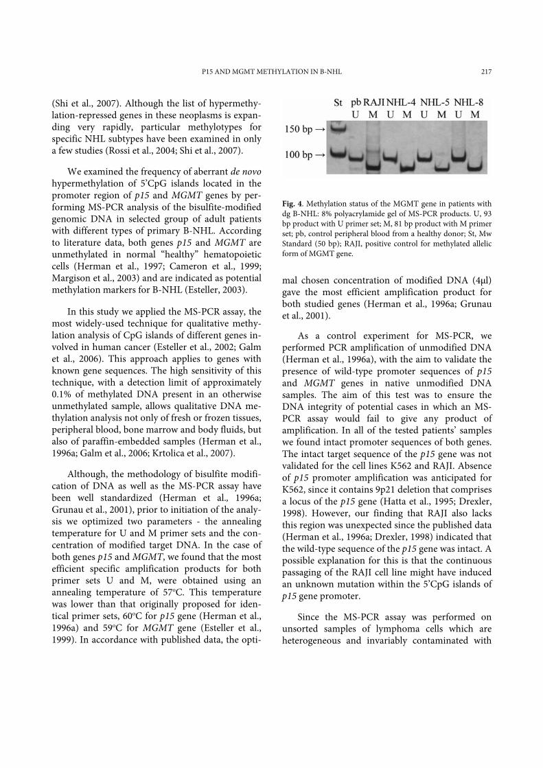
The methylation status of the target promoter sequences of p15 and MGMT genes was studied by MS-PCR in 10 patients with de novo B-NHL (Fig. 3 and 4) The unmethylated allelic form of the p15 (u-p15, 154 bp product) and MGMT (u-MGMT, 93 bp product) genes was detected in all examined patients (10/10, 100%). The presence of the methylated allelic form of the p15 gene (m-p15, 148 bp product) was shown in half of the tested patients (5/10, 50% pts), while in the case of the MGMT gene, a lower frequency (3/10, 30%) of methylated allelic form (m-MGMT, 81 bp product) was detected.
Three of the five patients with aberrant methyl-ation of the p15 gene had aberrant methylation of the MGMT gene (m-p15/m-MGMT profile), whereas 2/5 patients had aberrant methylation of the p15 gene as a sole aberration (m-p15/u-MGMT profile, Table 2). Half of tested patients (5/10, 50%) had only unmethylated allelic forms of both genes (u-p15/u-MGMT profile, Table 2).
While expression of m-p15/m-MGMT or m-p15/u-MGMT profiles did not associate itself with
Table 1. Primer sets used for MS-PCR
Primer set Sense primer Antisense primer Size (bp) / AT
p15-WT 5'-CGCACCCTGCGGCCAGA-3' 5'-AGTGGCCGAGCGGCCGG-3' 137bp / 65 oC p15-M 5'-GCGTTCGTATTTTGCGGTT-3' 5'-CGTACAATAACCGAACGACCGA-3' 148bp / 57 oC p15-U 5'-TGTGATGTGTTTGTATTTTGTGGTT-3' 5'-CCATACAATAACCAAACAACCAA-3' 154bp / 57 oC MGMT-WT 5'-TTTGCGTCCCGACGCCCG-3’ 5’-AGCTCCGCACTCTTCCGG-3’ 93bp / 62 oC MGMT-M 5'-TTTCGACGTTCGTAGGTTTTCGC-3’ 5’-GCACTCTTCCGAAAACGAAACG-3’ 81bp / 57 oC MGMT-U 5'-TTTGTGTTTGATGTTTGTAGGTTTTTGT-3’ 5’-AACTCCACACTCTTCCAAAAACAAAAA-3’ 93 bp / 57 oC
Abbreviations: WT, primer set for unmodified wild-type DNA sequence; U, primer set for unmethylated modified DNA sequence; M, primer set for methylated modified DNA sequence; AT, annealing temperature.

P15 AND MGMT METHYLATION IN B-NHL 215
(A)
(B)
Fig. 2. Testing of the efficiency of MS-PCR amplification of thesequence of 5’ region of the MGMT gene as a function of theconcentration of modified DNA from: a) control peripheralblood with U primer set and MS-PCR product of 93 bp; b) cell line K562 with M primer set and MS-PCR product of 81 bp.The lines 1-4 present volume of DNA loaded on 2% agarosegel: 0.4μl; 1 μl; 2μl; 4 μl; H2O, reaction mix without DNA.
Fig. 3. Methylation status of p15 gene in patients with dg B-NHL: 8% polyacrylamide gel of MS-PCR products. U, 154 bp product with U primer set ; M, 148 bp product with M primer set; pb, control peripheral blood from a healthy donor; St, Mw Standard (50 bp); H2O, reaction mix without DNA with M primer set.
any particular type of B-NHL, all patients in this group presented a higher clinical stadium (CS) of the disease: CS IV or V (Table 2). Only 1/5 patients with dg B-NHL-Follicular showed hypermethylation of the studied genes, having an m-p15/m-MGMT profile (Table 2, pt NHL-8).
p15 and MGMT gene methylation status and clinicopathological features
We examined the relationship between the p15 or MGMT gene methylation status and the clinicopathological features of the patients (Table 3). In the group with the m-p15 allelic form of the gene, we found that there is a statistically significant lower median hemo-globin value compared to the group with u-p15 allelic form (p=0.004, t-test). The same trend of decreased hemoglobin values, although sta-tistically insignificant, was shown for the group of patients with an m-MGMT allelic form of the MGMT gene. In the group of patients with an m-
p15 allelic form of the gene we also found a lower, although statistically insignificant, median platelet level. Further analysis revealed a significant association between the presence of an m-p15 allelic form of the gene and decreased platelet levels (<150.000 x 10⁹/l, p=0.038, Mann-Whitney test). The methylation status of the MGMT gene was independent of platelet values.
Patients with an m-MGMT allelic form of the MGMT gene showed a trend towards a lower me-dian leucocyte value, as well as lower proportions of lymphoma cells in the peripheral blood and bone marrow, compared to the group of patients with a u-MGMT allelic form of the gene (Table 3). The methylation status of the p15 gene was independent of the leucocyte values.
There was no correlation between the methy-lation status of the p15 and MGMT genes and the gender and age of the patients. Analysis of the res-ponse to the therapy did not reveal clear differences between the groups with different gene methylation profiles (data not shown).
DISCUSSION
The profile of CpG island hypermethylation in hematological malignancies is an epigenetic signa-ture unique for each subtype of acute leukemia or lymphoma (Esteller, 2003). NHL exhibits non-ran-dom methylation patterns in which germinal center tumors seem to be prone to de novo methylation

216 NADA KRAGULJAC KURTOVIĆ ET AL.
Table 3. Clinical and hematological characteristics of B-NHL patients in correlation to the methylation status of p15 and MGMT genes
PPaarraammeetteerr mm--pp1155 ggeennee uu--pp1155 ggeennee pp vvaalluuee mm--MMGGMMTT ggeennee uu--MMGGMMTT ggeennee pp vvaalluuee
SSeexx ((%%)) male
female
3/5 (60)
2/5 (40)
4/5 (80)
1/5 (20) 0.490
2/3 (67)
1/3 (33)
5/7 (71)
2/7 (29) 0.880
AAggee ((yyeeaarr)) (x±SD)
53 ± 13 55 ± 18 0.850 57 ± 17 52 ± 16 0.691
LLee ((xx110099//ll)) (x±SD)
29 ± 44 23 ± 25 0.810 5 ± 3 35 ± 37 0.219
PPlltt ((xx110099//ll)) (x±SD)
135 ± 84 214 ± 71 0.147 183 ± 69 171 ± 95 0.849
PPlltt ((%%)) (>150 x109/l) (<150 x109/l)
2/5 (40)
3/5 (60)
5/5 (100)
0/5 (0) 0.038*
2/3 (67)
1/3 (33)
5/7 (71)
2/7 (29) 0.880
HHbb ((gg//ddll)) (x±SD)
92 ± 21 134 ± 9 0.004* 98 ± 21 119 ± 28 0.278
PPBB LLyyCC ((%%)) (x±SD)
59 ± 29 51 ± 31 0.676 41 ± 19 61 ± 31 0.337
BBMM LLyyCC ((%%)) (x±SD)
57 ± 31 67 ± 19 0.709 50 ± 39 65 ± 24 0.598
Abbreviations: u, unnmethylated; m, methylated; PB LyC, peripheral blood lymphoma cells; BM LyC, bone marrow lymphoma cells; Le, leukocytes; Plt, platelet; Hb, hemoglobin; *, statistically significant (p<0.05).
Table 2. Basic clinical and biological characteristics of patients with dg B-NHL
Patient Sex / Age Sample type Immuno-hisopathologic type / CS p15 / MGMT gene methylation status
NHL-1 F / 34 PB B-NHL-Follicular / VB u / u
NHL-2 M / 65 PB B-HLL/SLL / II u / u
NHL-3 F / 41 PB B-NHL-Marginal zone / IV m / u
NHL-4 M / 66 BM B-NHL, NOS, low grade / IVB m / m
NHL-5 M / 38 BM B-NHL-Lymphoplasmocytic, IgGkappa / IVB m / m
NHL-6 M / 52 PB B-NHL-Mantle zone / V m / u
NHL-7 M / 50 LN B-NHL-Follicular / IIEA u / u
NHL-8 F / 67 LN B-NHL-Follicular / IVA m / m
NHL-9 M / 81 LN B-NHL-Follicular / IIIB u / u
NHL-10 M / 44 LN B-NHL-Follicular / IVB u / u
Abbreviations: M, male; F, female; PB, peripheral blood; BM, bone marrow; LN, lymph node; CS, clinical stage; NOS, not otherwise specified; u, unmethylated; m, methylated.
P15 AND MGMT METHYLATION IN B-NHL 217
Fig. 4. Methylation status of the MGMT gene in patients with dg B-NHL: 8% polyacrylamide gel of MS-PCR products. U, 93 bp product with U primer set; M, 81 bp product with M primer set; pb, control peripheral blood from a healthy donor; St, Mw Standard (50 bp); RAJI, positive control for methylated allelic form of MGMT gene.
(Shi et al., 2007). Although the list of hypermethy-lation-repressed genes in these neoplasms is expan-ding very rapidly, particular methylotypes for specific NHL subtypes have been examined in only a few studies (Rossi et al., 2004; Shi et al., 2007).
We examined the frequency of aberrant de novo hypermethylation of 5’CpG islands located in the promoter region of p15 and MGMT genes by per-forming MS-PCR analysis of the bisulfite-modified genomic DNA in selected group of adult patients with different types of primary B-NHL. According to literature data, both genes p15 and MGMT are unmethylated in normal “healthy” hematopoietic cells (Herman et al., 1997; Cameron et al., 1999; Margison et al., 2003) and are indicated as potential methylation markers for B-NHL (Esteller, 2003).
In this study we applied the MS-PCR assay, the most widely-used technique for qualitative methy-lation analysis of CpG islands of different genes in-volved in human cancer (Esteller et al., 2002; Galm et al., 2006). This approach applies to genes with known gene sequences. The high sensitivity of this technique, with a detection limit of approximately 0.1% of methylated DNA present in an otherwise unmethylated sample, allows qualitative DNA me-thylation analysis not only of fresh or frozen tissues, peripheral blood, bone marrow and body fluids, but also of paraffin-embedded samples (Herman et al., 1996a; Galm et al., 2006; Krtolica et al., 2007).
Although, the methodology of bisulfite modifi-cation of DNA as well as the MS-PCR assay have been well standardized (Herman et al., 1996a; Grunau et al., 2001), prior to initiation of the analy-sis we optimized two parameters - the annealing temperature for U and M primer sets and the con-centration of modified target DNA. In the case of both genes p15 and MGMT, we found that the most efficient specific amplification products for both primer sets U and M, were obtained using an annealing temperature of 57oC. This temperature was lower than that originally proposed for iden-tical primer sets, 60oC for p15 gene (Herman et al., 1996a) and 59oC for MGMT gene (Esteller et al., 1999). In accordance with published data, the opti-
mal chosen concentration of modified DNA (4μl) gave the most efficient amplification product for both studied genes (Herman et al., 1996a; Grunau et al., 2001).
As a control experiment for MS-PCR, we performed PCR amplification of unmodified DNA (Herman et al., 1996a), with the aim to validate the presence of wild-type promoter sequences of p15 and MGMT genes in native unmodified DNA samples. The aim of this test was to ensure the DNA integrity of potential cases in which an MS-PCR assay would fail to give any product of amplification. In all of the tested patients’ samples we found intact promoter sequences of both genes. The intact target sequence of the p15 gene was not validated for the cell lines K562 and RAJI. Absence of p15 promoter amplification was anticipated for K562, since it contains 9p21 deletion that comprises a locus of the p15 gene (Hatta et al., 1995; Drexler, 1998). However, our finding that RAJI also lacks this region was unexpected since the published data (Herman et al., 1996a; Drexler, 1998) indicated that the wild-type sequence of the p15 gene was intact. A possible explanation for this is that the continuous passaging of the RAJI cell line might have induced an unknown mutation within the 5’CpG islands of p15 gene promoter.
Since the MS-PCR assay was performed on unsorted samples of lymphoma cells which are heterogeneous and invariably contaminated with

218 NADA KRAGULJAC KURTOVIĆ ET AL.
normal cells, we could not identify the type of aberrant methylation, biallelic vs. monoallelic, of the studied genes (Esteller et al., 1999). On the other hand, we revealed the biallelic type of aberrant methylation of the MGMT gene in all of the tested cell lines, K562, SuDHL-4 and RAJI. This finding is in agreement with the published profile of MGMT gene methylation in other human cancer cell lines (Esteller et al., 1999; Paz et al., 2003).
Inactivation of the p15 gene by promoter hypermethylation is one of the most studied epigenetic changes in hematological malignancies, especially in acute leukemias and myelodysplastic syndromes (Rush & Plass, 2002; Esteller, 2003; Galm et al., 2006). According to the published data, p15 hypermethylation is extremely common in leukemias and relatively rare in lymphomas (Es-teller, 2003; Galm et al., 2006). Moreover, inacti-vation of the p15 gene by genetic mechanisms, either by point mutations (Baur et al., 1999) or by homozygous deletion of chromosome 9p21 (Her-man et al., 1997), is very rare. Our preliminary data showed that the frequency of hypermethylation of the p15 gene was 50% (5/10 pts) in the whole B-NHL group. Our data are in agreement with the results of study by Baur et al., (1999), who reported a frequency of 55% for aberrant p15 methylation in an unselected group of low risk B-NHL.
With respect to the type of examined B-NHL, we detected p15 hypermethylation in all the studied B-NHL types, except B-HLL/SLL. Despite the small number of cases in our study, our results were in accordance with the data reported by other groups (Baur et al., 1999; Martinez-Delgado et al., 2000).
Published data indicate that hypermethylation of the p15 gene plays an important role in the different steps of lymphomagenesis, including the phase of disseminated disease, which is in positive correlation with a higher CS of the disease (Baur et al., 1999; Martinez-Delgado et al., 2000). All lym-phoma patients with an m-p15 allelic form had higher CS of the disease (IV - V). Hypermethyla-tion of the p15 gene could be generated as a pri-mary de novo (Martinez-Delgado et al., 2000) or as
a secondary de novo aberration caused by chemo-therapeutic treatment (Au et al., 2003). The silen-cing of the p15 gene by hypermethylation can lead to the loss of sensitivity of the lymphoma cells to growth inhibition by TGF-β and interferon-α, the potent regulators of differentiation and prolifera-tion of lymphohematopoietic cells (Herman et al., 1997; Sherr & Roberts, 1999; Sangfelt et al., 1999; Visser & Kast, 2002).
A statistically significant positive association of different clinicopathological features (especially anemia and thrombocytopenia) with the presence of hypermethylation of the p15 gene, represents a novel finding. Anemia and thrombocytopenia are associated with disseminated disease and bone marrow infiltration that reflect a higher CS of the disease (Jaffe et al., 2008).
MGMT is one of the genes that is frequently hypermethylated in the major types of B-NHL (Esteller et al., 1999; Esteller et al., 2002; Rush & Plass, 2002). Our preliminary data showed that the frequency of overall hypermethylation of MGMT gene was 30% (3/10 pts) in our B-NHL group at diagnosis. The frequency of hypermethylation of the MGMT gene is reported in the literature to be between 25% for an unselected group of B-NHL (Esteller et al., 1999; Rossi et al., 2004) and 36% for a selected DLBCL type (Esteller et al., 2002), and these results that are in agreement with our data.
All three patients with a hypermethylated allelic form of the MGMT gene had a high CS of the disease (IV) and were comethylated for the p15 gene. There are few literature reports that explain the potential role of hypermethylation of the MGMT gene in lymphomagenesis. They suggest that loss of the MGMT protein from the cells in the early phase of the disease can cause the generation of a new mutator phenotype characterized by G → A transitions and the generation and accumulation of point mutations in various genes involved in cancer etiology, such as the tumor suppressor p53 and the oncogene K-ras (Esteller et al., 1999; Esteller & Herman, 2004). From this point, it is important to note that MGMT hypermethylation
P15 AND MGMT METHYLATION IN B-NHL 219
alone is a poor prognostic factor in many human cancers (Esteller & Herman, 2004). On the other hand, loss of the MGMT protein from tumor cells due to aberrant hypermethylation can predict a better chemoresponse of human tumors to alky-lating agents such as cyclophosphamide and others (Esteller & Herman, 2004).
Clinicopathological parameters showed a po-sitive but insignificant association with the presence of hypermethylation of the MGMT gene, presenting a lower median value of hemoglobin and leuko-cytes, as well as a lower median proportion of peri-pheral blood and bone marrow lymphoma cells.
The comethylation of the MGMT with the p15 gene, as we report in our study, represents a novel finding and selects both genes as candidates in future studies of the hypermethylation profiles of B-NHL. The biological and clinical significance of this association remains undetermined due to the small size of the patient group involved in this study. Similar comethylation of the p16 with the p15 gene has been previously reported (Baur et al., 1999; Martinez-Delgado et al., 2000).
In summary, aberrant DNA hypermethylation of the p15 and MGMT genes in B-NHL could play a role as an epigenetic biomarker with possible applications in diagnostics and the monitoring of treatment efficacy (Rush & Plass, 2002; Esteller, 2003; Galm et al., 2006; Shi et al., 2007).
Acknowledgments - This work was supported by grant no. 143010 from the Ministry of Science and Technology of the Republic of Serbia
REFERENCES
Au, W. Y., Fung, A., Man, C., Ma, S. K., Wan, T. S., Liang, R., and Y. L. Kwong (2003). Aberrant p15 gene promoter methylation in therapy-related myelodysplastic syndrome and acute myeloid leukaemia: clinicopathological and karyotypic associations. Br. J. Haematol. 120, 1062-1065.
Baur, A. S., Shaw, P., Burri, N., Delacretaz, F., Bosman, F. T., and P. Chaubert (1999). Frequent methylation silencing of p15INK4B (MTS2) and p16INK4A (MTS1) in B-cell and T-cell lymphomas. Blood 94, 1773-1781.
Baylin, S. B., Herman, J. G., Graff, J. R., Vertino, P. M., and J. P. Issa (1998). Alterations in DNA methylation: a fundamental aspect of neoplasia. Adv. Cancer Res. 72, 141-196.
Baylin, S. B., and J. E. Ohm (2006). Epigenetic gene silencing in cancer - a mechanism for early oncogenic pathway addiction? Nat. Rev. Cancer. 6, 107-116.
Bogdanović, G., Jurišić, V., Kraguljac, N., Mrdjanović, J., Jakimov, P., Krtolica, K., Krajnović, M., Magić, Z., Stojiljković, B., Andrijević, L.j, Srdić, T., Baltić, M., and S. Popović (2007). Characteristics of novel myeloid precursor cell line, PC-MDS, established from a bone marrow of the patient with therapy - related myelodysplastic syndrome. Leuk Res. 31, 1105-1113.
Boyum, A. Separation of leucocytes from blood and bone marrow (1968). Scand. J.Clin. Lab. Invest. 21, (suppl. 97), 77-89.
Cameron, E. E., Baylin, S. B., and J. G. Herman (1999). p15INK4B CpG island methylation in primary acute leukemia is heterogeneous and suggests density as a critical factor for transcriptional silencing. Blood. 94, 2445-2451.
Drexler, H. G. (1998). Review of alterations of the cyclin-dependent kinase inhibitor INK4 family genes p15, p16, p18 and p19 in human leukemia-lymphoma cells. Leukemia. 12, 845-859.
Hiddemann, W., Dreyling, M., and R.A. Stahel (2005). Minimum clinical recommendations for diagnosis, treatment and follow-up of newly diagnosed follicular lymphoma. Ann Onc. 16, i56-i57.
Esteller, M., Hamilton, S. R., Burger, P. C., Baylin, S. B., and J. G. Herman (1999). Inactivation of the DNA repair gene O6-methylguanine-DNA methyltransferase by promoter hypermethylation is a common event in primary human neoplasia. Cancer Res. 59, 793-797.
Esteller, M., Toyota, M., Sanchez-Cespedes, M., Capella, G., Peinado, M. A., Watkins, D. N., Issa, J. P. J., Sidransky, D., Baylin, S. B., and J. G. Herman (2000). Inactivation of the DNA repair gene O6-Methylguanine-DNA Methyltransferase by promoter hypermethylation is associated with G to A mutations in K-ras in colorectal tumorigenesis. Cancer Res. 60, 2368-2371.
Esteller, M., Corn, P. G., Baylin, S. B, and J. G. Herman (2001).A gene hypermethylation profile of human cancer. Cancer Res. 61, 3225-3229.
Esteller, M., Gaidano, G., Goodman, S. N., Zagonel, V., Capello, D., Botto, B., Rossi, D., Gloghini, A., Vitolo, U., Carbone, A., Baylin, S. B., and J. G. Herman (2002). Hyper-methylation of the DNA repair gene O6-methylguanine-DNA methyltransferase and survival of patients with diffuse large B-cell lymphoma. J. Natl. Cancer Inst. 94, 26-32.

220 NADA KRAGULJAC KURTOVIĆ ET AL.
Esteller, M. (2003). Profiling aberrant methylation in hematologic neoplasms: a view from the tip of the iceberg. Clin. Immunol. 109, 80-88.
Esteller, M. and J. G. Herman (2004). Generating mutations but providing chemosensitivity: the role of O6-methylguanine DNA methyltrasferase in human cancer. Oncogene. 23, 1-8.
Galm, O., Wilop, S., Luders, C., Jost, E., Gehbauer, G., Herman, J. G., and R. Osieka (2005). Clinical implications of aberrant DNA methylation patterns in acute myelogenous leukemia. Ann. Hematol. 84, 39-46.
Galm, O., Herman, J. G., and S. B. Baylin (2006). The fundamental role of epigenetics in hematopoietic malignancies. Blood rev. 29, 1-13.
Goelz, S. E., Hamilton, S. R., and B. Vogelstein (1985). Purification of DNA from formaldehyde fixed and paraffin embedded human tissue. Biochem. Biophys. Res. Commun. 130, 118-126.
Gerson, S. L. (2002). Clinical relevance of MGMT in the treatment of cancer. J. Clin. Oncol. 9, 2388-2399.
Grunau, C., Clark, S. J., and A. Rosenthal (2001). Bisulfite genomic sequencing: systematic investigation of critical experimental parameters. Nucleic Acids Res. 29, E65-5.
Hanahan, D., and R. A. Weinberg (2000). The Hallmarks of Cancer. Cell. 100, 57-70.
Hatta, Y., Hirama, T., Miller, C. W., Yamada, Y., Tomonaga, M., and P. Koeffler (1995). Homozygous deletions of the p15 (MTS2) and p16 (CDKN2/MTS1) genes in adult T-cell leukemia. Blood. 85, 2699-2704.
Herman, J. G., Graff, J. R., Myohanen, S., Nelkin, B. D., and S. B. Baylin (1996a). Methylation-specific PCR: A novel PCR assay for methylation status of CpG islands. Proc Natl Acad Sci USA. 93, 9821-9826.
Herman, J. G., Jen, J., Merlo, A., and S. B. Baylin (1996b). Hypermethylation-associated inactivation indicates a tumor suppressor role for p15INK4B1. Cancer Res. 56, 722-727.
Herman, J. G., Civin, C. I., Issa, J. P. J., Collector, M. I., Sharkis, S. J., and S. B. Baylin (1997). Distinct patterns of inactivation of p15INK4B and p16INK4a characterize the major types of hematological malignancies. Cancer Res. 57, 837-841.
Houwen, B. (2000). Blood film preparation and staining procedures. Lab. Hematol. 6, 1-7.
Jack, A., Barrans, S., Dlythe, D., and A. Rawstron (2005). Demonstration of a germinal center immunophenotype in lymphomas by immunocytochemistry and flow cytometry. In: Methods in Molecular Medicine. (Eds. T. Illidge, and P.W.M. Johnson), 65-91. Humana Press Inc USA.
Harris, N.L. (2001). Nature B-cell neoplasms: Introduction. In: Pathology and Genetics of Tumors of Haematopoietic and Lymphoid Tissues. (Eds. E.S. Jaffe, N.L. Harris, H. Stein, and J.W. Vardiman), 121-126. IARC, Lyon.
Jones, P. L., Veenstra, G. J., Wade, P. A., Vermaak, D., Kass, S. U., Landsberger, N., Strouboulis, J., and A. P. Wolffe (1998). Methylated DNA and MeCP2 recruit histone deacetylase to repress transcription. Nat. Genet. 23, 62-66.
Krtolica, K., Krajnović, M., Ušaj-Knežević, S., Babić, D., Jovanović, D., and B. Dimitrijević (2007). Comethylation of p16 and MGMT genes in colorectal carcinoma: Correlation with clinicopathological features and prognostic value. World J. Gastroenterol. 13, 1187-1194.
Margison, G. P., Povey, A. C., Kaina, B., and M. F. Santibanez-Koref (2003). Variability and regulation of O6-alkylguanine-DNA alkyltransferase. Carcinogenesis 24, 625-635.
Martinez-Delgado, B., Richart, A., Garcia, M. J., Robledo, M., Osorio, A., Cebrian, A., Rivas, C., and J. Benitez (2000). Hypermethylation of p16INK4A and p15INK4B genes as marker of disease in the follow-up of non-Hodgkin's lymphomas. Br. J. Haematol. 109, 97-103.
National Cancer Institute (2002). Strategic plan for addressing for the recommendations of the leukemia, lymphoma and myeloma. Progress Review Group 1-60.
Paz, M. F., Fraga, M. F., Avila, S., Guo, M., Pollan, M., Herman, J. G., and M. Esteller (2003). A systematic profile of DNA methylation in human cancer cell lines. Cancer Res. 63, 1114-1121.
Rossi, D., Capello, D., Gloghini, A., Franceschetti, S., Paulli, M., Bhatia, K., Saglio, G., Vitolo, U., Pileri, A. S., Esteller, M., Carbone, A., and G. Gaidano (2004). Aberrant promoter methylation of multiple genes throughout the clinico-pathologic spectrum of B-cell neoplasia. Haematologica 89, 154-164.
Rothe, G., and G. Schmitz (1996). Consensus protocol for the flow cytometric immunophenotyping of hematopoietic malignancies. Leukemia 10, 877-895.
Rush, L. J., and C. Plass (2002). Alterations of DNA methylation in hematologic malignancies. Cancer lett. 185, 1-12.
Sambrook, J., Fritsch, E. F., and T. Maniatis (1989). Molecular cloning. A laboratory manual, 2nd Ed. (Eds. N. Ford, Ch. Nolan and M. Ferguson), Cold Spring Harbor Laboratory Press, New York.
Sangfelt, O., Erickson, S., Heiden, C. J., Gustafsson, A., Einhorn, S., and D. Grander (1999). Molecular mechanisms underlying interferon-alpha-induced G0/G1 arrest: CKI-mediated regulation of G1 Cdk-complexes and activation of pocket proteins. Oncogene. 18, 2798-2810.

P15 AND MGMT METHYLATION IN B-NHL 221
Sherr, C. J., and J. M. Roberts (1999). CDK inhibitors: positive and negative regulators of G1-phase progression. Genes & Dev. 13, 1501-1512.
Shi, H., Guo, J., Duff, D. J., Rahmatpanah, F., Chitima-Matsiga, R., Al-Kuhlani, M., Taylor, K. H., Sjahputera, O., Andreski, M., Wooldridge, J. E., and C. W. Caldwell (2007). Discovery of novel epigenetic markers in non-Hodgkin’s lymphoma. Carcinogenesis. 28, 60-70.
Stelzer, G. T., Marti, G., Hurley, A., McCoy, P., Lovett, E. J., and A. Schwartz (1997). U.S.-Canadian consensus recommendation on the immunophenotypic analysis of hematologic neoplasia by flow cytometry: Standardization and validation of laboratory procedures. Cytometry (Comm. Clin. Cytometry). 30, 214-230.
Jaffe, E.S., Harris, N.L., Stein, H., Campo, E., Tileri, S.A., and S.H. Swerdlow (2008). Introduction and overview of the
classification of the lymphoid neoplasms. In: WHO Classification of Tumours of Haematopoietic and Lymphoid Tissue. Eds. S.H. Swerdlow, E. Campo, N.L. Harris, E.S. Jaffe, S.A. Pileri, H. Stein, J. Thiele, and J. W. Vardiman), 158-166. IARC, Lyon.
Tatsumi, N., van Assendelft, O. W., and K. Naka on Behalf of the ICSH Expert Panel on Cytometry 2002. International Council for Standardization in Haematology (ICSH) Recommendations for Single-Use Evacuated Containers for Blood Specimen Collection for Hematological Analyses. Lab. Hem. 8, 1-6.
Verma, M., and S. Srivastava (2002). Epigenetics in cancer: implications for early detection and prevention. Lancet Oncol. 3, 755-763.
Visser, K. E., and W. M. Kast (2002). Effects of TGF-β on the immune system: implications for early detection and prevention. Lancet Oncol. 3, 755-763.
УЧЕСТАЛОСТ АБЕРАНТНЕ METИЛАЦИЈЕ ПРОМОТОРА ГЕНА P15INK4B И O6-МЕТИЛГУАНИН-ДНK МЕТИЛТРАНСФЕРАЗЕ KOД Б-ЋЕЛИЈСКОГ НЕХОЏКИНОВОГ ЛИМФОМА:
ПИЛОТ СТУДИЈА
НАДА КРАГУЉАЦ КУРТОВИЋ2, МИЛЕНА КРАЈНОВИЋ1, Б. ДИМИТРИЈЕВИЋ1, БИЉАНА MИХАЉЕВИЋ2, MИРЈАНА ГОТИЋ2 И KOВИЉКА KРТОЛИЦA1
1Институт за нуклеарне нaуке “Винча”, 11000 Београд, Србија 2Институт за хематологију, Kлинички центар Србије, 11000 Београд, Србија
Метилациони статус таргет промоторских секвенци гена p15 и O6-метилгуанин-ДНK метилтрансферазе (MGMT) je у нашој пилот студији истраживан применом методе мети-лација зависним PCR-oм (MS-PCR) koд 10 па-цијената сa дијагнозом de novo Б-нехочкинског лимфома (B-NHL). Аберантна хиперметилација p15 гена je учесталија (50%) у oдносу нa аберан-тну хиперметилацију MGMT гена (30%), и утвр-ђена је код различитих типова B-NHL у случају оба испитивана гена. Хиперметилација MGMТ
гена je oткривена искључиво у асоцијацији са хиперметилацијом p15 гена. Сви пацијенти сa хиперметилацијом p15 и/или MGMT гена имали су виши клинички стадијум болести (IV – V). Значајна асоцијација анемије и/или тромбоци-топеније са хиперметилацијом p15 гена, указује на његов потенцијални прогностички значај у B-NHL. Кометилација MGMT и p15 гена пред-стављa нов податак, и издваја ове гене као кан-дидате у будућим истраживањима хиперметила-ционих профила у B-NHL.