Aberrant Epigenomic Modulation of Glucocorticoid Receptor ...
17
International Journal of Environmental Research and Public Health Review Aberrant Epigenomic Modulation of Glucocorticoid Receptor Gene (NR3C1) in Early Life Stress and Major Depressive Disorder Correlation: Systematic Review and Quantitative Evidence Synthesis Laurens Holmes, Jr. 1,2,3, *, Emily Shutman 1,4 , Chinacherem Chinaka 1,5,6 , Kerti Deepika 1 , Lavisha Pelaez 1 and Kirk W. Dabney 1,7 1 Nemours Healthcare System for Children, Translational Health Disparities Science Research Program, Wilmington, DE 19803, USA; [email protected] (E.S.); [email protected] (C.C.); [email protected] (K.D.); [email protected] (L.P.); [email protected] (K.W.D.) 2 Biological Sciences Department, University of Delaware, Newark, DE 19716, USA 3 College of Population Health, Thomas Jefferson University, Philadelphia, PA 19107, USA 4 Biological Sciences Department, Haverford College, Haverford, PA 19041, USA 5 Department of public health, Eastern Virginia Medical School, Norfolk, VA 23507, USA 6 Community Environmental Health Department, Old Dominion University, Norfolk, VA 23507, USA 7 Sidney Kimmel Medical School, Thomas Jefferson University, Philadelphia, PA 19107, USA * Correspondence: [email protected]; Tel.: +1-302-298-7741; Fax: +1-302-298-7758 Received: 10 October 2019; Accepted: 28 October 2019; Published: 4 November 2019 Abstract: Early life stress (ELS) induced by psychological trauma, child maltreatment, maternal separation, and domestic violence predisposes to psycho-behavioral pathologies during adulthood, namely major depressive disorder (MDD), anxiety, and bipolar affective disorder. While environmental data are available in illustrating this association, data remain to be established on the epigenomic underpinning of the nexus between ELS and MDD predisposition. Specifically, despite the observed aberrant epigenomic modulation of the NR3C1, a glucocorticoid receptor gene, in early social adversity and social threats in animal and human models, reliable scientific data for intervention mapping in reducing social adversity and improving human health is required. We sought to synthesize the findings of studies evaluating (a) epigenomic modulations, mainly DNA methylation resulting in MDD following ELS, (b) epigenomic modifications associated with ELS, and (c) epigenomic alterations associated with MDD. A systematic review and quantitative evidence synthesis (QES) were utilized with the random effect meta-analytic procedure. The search strategy involved both the PubMed and hand search of relevant references. Of the 1534 studies identified through electronic search, 592 studies were screened, 11 met the eligibility criteria for inclusion in the QES, and 5 examined ELS and MDD; 4 studies assessed epigenomic modulation and ELS, while 2 studies examined epigenomic modulations and MDD. The dense DNA methylation of the 1F exon of the NR3C1, implying the hypermethylated region of the glucocorticoid receptor gene, was observed in the nexus between ELS and MDD, common effect size (CES) = 14.96, 95%CI, 10.06–19.85. With respect to epigenomic modulation associated with child ELS, hypermethylation was observed, CES = 23.2%, 95%CI, 8.00–38.48. In addition, marginal epigenomic alteration was indicated in MDD, where hypermethylation was associated with increased risk of MDD, CES = 2.12%, 95%CI, -0.63–4.86. Substantial evidence supports the implication of NR3C1 and environmental interaction, mainly DNA methylation, in the predisposition to MDD following ELS. This QES further supports aberrant epigenomic modulation identified in ELS as well as major depressive episodes involving dysfunctional glucocorticoid-mediated negative feedback as a result of allostatic overload. These findings recommend prospective investigation of social adversity and its predisposition to the MDD epidemic via aberrant epigenomic modulation. Such data will facilitate early intervention mapping in reducing MDD in the United States population. Int. J. Environ. Res. Public Health 2019, 16, 4280; doi:10.3390/ijerph16214280 www.mdpi.com/journal/ijerph
-
Upload
khangminh22 -
Category
Documents
-
view
1 -
download
0
Transcript of Aberrant Epigenomic Modulation of Glucocorticoid Receptor ...

International Journal of
Environmental Research
and Public Health
Review
Aberrant Epigenomic Modulation of GlucocorticoidReceptor Gene (NR3C1) in Early Life Stress andMajor Depressive Disorder Correlation: SystematicReview and Quantitative Evidence Synthesis
Laurens Holmes, Jr. 1,2,3,*, Emily Shutman 1,4, Chinacherem Chinaka 1,5,6, Kerti Deepika 1,Lavisha Pelaez 1 and Kirk W. Dabney 1,7
1 Nemours Healthcare System for Children, Translational Health Disparities Science Research Program,Wilmington, DE 19803, USA; [email protected] (E.S.); [email protected] (C.C.);[email protected] (K.D.); [email protected] (L.P.); [email protected] (K.W.D.)
2 Biological Sciences Department, University of Delaware, Newark, DE 19716, USA3 College of Population Health, Thomas Jefferson University, Philadelphia, PA 19107, USA4 Biological Sciences Department, Haverford College, Haverford, PA 19041, USA5 Department of public health, Eastern Virginia Medical School, Norfolk, VA 23507, USA6 Community Environmental Health Department, Old Dominion University, Norfolk, VA 23507, USA7 Sidney Kimmel Medical School, Thomas Jefferson University, Philadelphia, PA 19107, USA* Correspondence: [email protected]; Tel.: +1-302-298-7741; Fax: +1-302-298-7758
Received: 10 October 2019; Accepted: 28 October 2019; Published: 4 November 2019�����������������
Abstract: Early life stress (ELS) induced by psychological trauma, child maltreatment, maternalseparation, and domestic violence predisposes to psycho-behavioral pathologies during adulthood,namely major depressive disorder (MDD), anxiety, and bipolar affective disorder. While environmentaldata are available in illustrating this association, data remain to be established on the epigenomicunderpinning of the nexus between ELS and MDD predisposition. Specifically, despite the observedaberrant epigenomic modulation of the NR3C1, a glucocorticoid receptor gene, in early social adversityand social threats in animal and human models, reliable scientific data for intervention mapping inreducing social adversity and improving human health is required. We sought to synthesize thefindings of studies evaluating (a) epigenomic modulations, mainly DNA methylation resulting inMDD following ELS, (b) epigenomic modifications associated with ELS, and (c) epigenomic alterationsassociated with MDD. A systematic review and quantitative evidence synthesis (QES) were utilizedwith the random effect meta-analytic procedure. The search strategy involved both the PubMed andhand search of relevant references. Of the 1534 studies identified through electronic search, 592 studieswere screened, 11 met the eligibility criteria for inclusion in the QES, and 5 examined ELS and MDD; 4studies assessed epigenomic modulation and ELS, while 2 studies examined epigenomic modulationsand MDD. The dense DNA methylation of the 1F exon of the NR3C1, implying the hypermethylatedregion of the glucocorticoid receptor gene, was observed in the nexus between ELS and MDD, commoneffect size (CES) = 14.96, 95%CI, 10.06–19.85. With respect to epigenomic modulation associated withchild ELS, hypermethylation was observed, CES = 23.2%, 95%CI, 8.00–38.48. In addition, marginalepigenomic alteration was indicated in MDD, where hypermethylation was associated with increasedrisk of MDD, CES = 2.12%, 95%CI, −0.63–4.86. Substantial evidence supports the implication ofNR3C1 and environmental interaction, mainly DNA methylation, in the predisposition to MDDfollowing ELS. This QES further supports aberrant epigenomic modulation identified in ELS as wellas major depressive episodes involving dysfunctional glucocorticoid-mediated negative feedback as aresult of allostatic overload. These findings recommend prospective investigation of social adversityand its predisposition to the MDD epidemic via aberrant epigenomic modulation. Such data willfacilitate early intervention mapping in reducing MDD in the United States population.
Int. J. Environ. Res. Public Health 2019, 16, 4280; doi:10.3390/ijerph16214280 www.mdpi.com/journal/ijerph
Int. J. Environ. Res. Public Health 2019, 16, 4280 2 of 17
Keywords: early life stress (ELS); glucocorticoid receptor gene (NR3C1); major depressive disorder(MDD); DNA methylation (mDNA); aberrant epigenomic modulation
1. Introduction
1.1. Early Life Stress and Biological Underpinnings of Epigenomic Modulation
Early life stress (ELS) such as perinatal, intrapartum, prenatal, and maternal stress has beenindicated to influence physical and psychological health in adulthood. The observed connectionis explained by social signal transduction, which results in molecular level events such as genetranscription inhibition, impaired mRNA translation, and gene expression, influencing physiologicfunction and disease development. For example, maternal stress and isolation in rodents and humanshave been implicated in glucocorticoid gene receptor dysregulation, which is involved in allostaticstress response and pro-inflammatory cytokine elaboration [1]. The epigenomic mechanistic processupon which inappropriate protein in synthesized involves the methylation of DNA in the promoterregion of the gene or acetylation phosphorylation of the histone protein at the amino-terminal region(lysine) [2].
Studies have illustrated the mediating biologic consequences of maternal deprivation and lackof care of offspring in animal models via gene and environment interaction, as indicated by aberrantepigenomic modulation of the candidate genes involved in these conditions [3–6]. Similarly, lowSES(Socio-economic status), which reflects income inequalities, implies variabilities in access to social,educational, health, and other resources, resulting in status shame, anxiety, depression, self-harm, andother psychopathologies [7–10]. Data on social hierarchies driven by high SES observed privilegedaccess to social determinants of health (SDH) and basic life resources such as water, food, and shelteras an appropriate living condition. The SES, which correlated with anxiety in most cases as observedin a capitalistic society, reflects social adversity as exposure function of childhood physical andemotional neglect, restricted control over decision-making and life choices, as well as marginalizedsocial interaction and decreased trust and confidence with societal members [11]. Epidemiologic andclinical data indicate hypercortisolemia and increased pro-inflammatory cytokines in low social rankedindividuals as a marker of social gradient in health [7]. Available animal and human model studiesobserved DNA hypermethylation of the CpG regions of the genes such as NR3C1 involved in allostaticstress response. However, it is not fully understood if the neural activities involving the hypothalamuspituitary adrenal (HPA) axis and the cortisol, mediated negative feedback mechanism induced bysocial signal transduction via social isolation, low SES, unstable social status, and ELS, such as childabuse and neglect, regulated solely NR3C1.
The conserved transcriptional response to adversity (CTRA) gene expression has been observedin low socioeconomic status, chronic stress, posttraumatic stress, and other life stressors [1]. Reliablefindings in humans and animal models have linked the CTRA genes with the upregulationof pro-inflammatory response as well as inhibition of the anti-inflammatory mediators such asinterferon-gamma (IFN-γ) and IgG. The mechanical process involves the leukocytes and the mediationthrough the innate immune mechanism, namely the macrophages as well as the specific immuneresponse through the activated lymphocytes, namely the CD4 cells. Specifically, the methylationprocess of this gene (CTRA) involves the binding to the gene promoter region being the 5’cytosinewith the methyl group resulting in 5’methylcytosine due to the excessive availability of the DNAmethyltransferase enzyme and the subsequent inhibition of the transcription factors negativelyimpacting the CTRA expression, abnormal protein synthesis, and cellular function dysregulation.Specifically, the CTRA gene expression involving the leukocytes results in the pro-inflammatoryelaboration, namely IL-2 and IL-6 [1]. The expression of the CTRA gene due to chronic stress increasesthe immune system response with pro-inflammatory cytokines and inhibition of IFN-γ elaboration
Int. J. Environ. Res. Public Health 2019, 16, 4280 3 of 17
required in response to a virally infected cells, as well as a decreased response of IgG which is a specifictherapeutic antibody needed for bacterial pathogens destruction via immune complex formation(antigen and antibody interaction).
Cells have a robust system for gene expression that is highly regulated by various signalingmolecules and RNAs. Gene expression is controlled by specific transcription factors that bind to CpGislands in the promoter region to initiate transcription of a particular mRNA sequence, which is thentranslated into a specific protein sequence. This process is highly regulated by various developmental,physiologic, pathological, and environmental cues that ensure that the necessary protein is producedin response to specific stimuli. While epigenomic modulation does not directly involve the DNAsequence, genomic mutations can change the base sequence in a portion of the genome. If located inthe protein-coding portion of a gene or if it results in a frameshift mutation, this will often deleteriouslyaffect the resultant protein sequence.
In contrast, other mechanisms do not change gene sequence but affect gene expression byconferring long-term programming to genes. These are called epigenetic changes and are defined by“a long-term change in gene function that persists even when the initial trigger is long gone and doesnot involve a change in gene sequence or structure [12].” Some definitions mandate heritability individing somatic cells while other definitions simply require a change in gene expression.
Epigenetic changes can be conferred in multiple ways, all of which affect the accessibility ofthe DNA to the transcription machinery. Inaccessible genes are relatively silenced, while accessiblegenes can be actively transcribed. One of the most predominant ways in which this silencing occursis through modifications of the chromatin—the protein-based structure around which the DNA iswrapped. The chromatin protein is composed of nucleosome “beads,” each of which is composed ofeight histone proteins. The N-terminal tails of these histones are frequently modified by methylation,acetylation, phosphorylation, and ubiquitination [13]. Typically, histones are positively charged,which allows favorable electrostatic interactions to occur between the DNA backbone and the histoneproteins, enabling the DNA to wrap tightly around nucleosomes. The aforementioned modificationsneutralize the charge on the tail, weakening the interaction between the genome and the nucleosomecore. Weak interactions between the genome and histones reduce chromatin density, enabling greaterpenetrance of transcription factors, which, in turn, increases transcription. In contrast, demethylationand deacetylation reduce gene accessibility and decrease transcription. The mechanisms of thesemodifications are not fully understood, but histone demethylase enzymes have been implicated in themechanism of histone demethylation [14].
An alternative mechanism of epigenetic regulation involves the DNA molecule itself. In vertebrates,the identity of a cell is regulated by methylation at the 5’ position of the cytosine ring in the CGdinucleotide sequence. When this methylation occurs in the promoter region, transcription factors areunable to bind to initiate transcription of a particular gene. This principle confers cell identity in thatdiffering patterns of methylation between different cell types allows for specific activation or silencingof specific genes. This enables different cells of an organism to produce different protein products andexecute different functions [15]. While most research on genome-based methylation is focused in thepromoter region, there is evidence that methylation in other gene elements plays a role in regulatinggene function. For example, decreased DNA methylation in gene bodies is associated with increasedchromatin accessibility, which is likely due to a disturbance in the favorable electrostatic interactionbetween the DNA and the chromatin [12].
Furthermore, some evidence suggests that small interfering RNAs (siRNAs) have epigeneticimplications. Available data suggest siRNAs have a role in both DNA methylation and histonemodifications. A study observed that siRNAs impair the function of DNMT1, 3a, and 3b, which are keyDNA methylation enzymes [16]. In addition, miRNAs may regulate chromatin structure by impairingthe function of a histone modifier enzyme called histone deacetylase 4 [17].
Int. J. Environ. Res. Public Health 2019, 16, 4280 4 of 17
1.2. Epigenetic Implications of Childhood Trauma
The aforementioned epigenetic mechanisms all have the effect of increasing or decreasingaccessibility to the genome, impacting the level of gene expression by way of mRNA production.These epigenetic mechanisms are highly impacted by environmental stimuli and can be observedin response to chronic stress or trauma [18]. These epigenetic effects may be able to explain thecorrelation between childhood trauma or early life adversity (ELA) and health outcomes in adults at abiological level. Studies pertaining to the epigenetic effects of childhood trauma and subsequent healthoutcomes often reference the NR3C1 gene—a gene that codes for the protein of the glucocorticoidreceptor. This receptor is the site at which cortisol and other glucocorticoids bind. When the receptorbinds to glucocorticoids, its primary mechanism of action is the regulation of gene transcription. Thereceptor–cortisol complex effectively works to decrease inflammation in tissues by either upregulatingthe expression of anti-inflammatory proteins in the nucleus or by repressing the expression ofpro-inflammatory proteins in the cytosol [19]. It executes the latter by preventing the transport oftranscription factors from the cytosol to the nucleus. In effect, the reduction in inflammation is anegative feedback mechanism that terminates the stress response after the end of a threat [20]. There isevidence that decreased NR3C1 expression in the hypothalamic–pituitary–adrenal (HPA) center isoften observed in suicide victims with a history of childhood abuse.
The activity of this gene can be affected by methylation at the promoter region of the gene.Specifically, previous study identified that DNA methylation at a CG dinucleotide in exon 5, whichis located in the promoter, caused decreased mRNA production. This was determined by using aDNA microchip assay to ascertain the level of hybridized NR3C1 mRNA in the cytoplasm. Thisstudy indicated lower levels of the mRNA than expected, demonstrating methylation at the promoterthat impaired transcription. Previous studies indicated that low levels of NR3C1 mRNA wereobserved in individuals with co-occurring child abuse and suicide, schizophrenia, and depression [13].This relationship can be explained by the methylation at exon 5, which caused low levels ofNR3C1 expression. Low levels of glucocorticoid receptor actually induced increased activity inthe hypothalamic–pituitary–adrenal (HPA) center, which results in altered stress responses, likelydue to an increase in inflammation of the region. Ultimately, the altered stress response impairs theefficacy of the negative feedback loop, mirroring a situation in which the individual has an increasedperception of being in a threatening situation. Specifically, this renders individuals less equipped tohandle daily stressors and is implicated in serious psychological conditions.
Similar correlations between childhood trauma and disadvantageous health outcomes have beenobserved in reference to the FKBP5 gene. This gene codes for a protein that is an important functionalregulator of the glucocorticoid receptor complex. Childhood trauma, compounded with a specificpolymorphism called rs1360780, caused a 12.3% decrease in DNA demethylation on intron 7 of thegene—a site near the transcription start site [21]. This demethylation increases FKBP5 transcriptionwhich ultimately decreases glucocorticoid receptor sensitivity, leading to a prolongation of stresshormone system activation following exposure to stress, and is linked to many stress-related psychiatricdisorders, namely predisposition to PTSD and depression.
1.3. Structural Inequity in Childhood Trauma
While the biological mechanisms of DNA (de)methylation can happen to any individual whoexperiences childhood trauma, it is important to note the inequity in which different populationsexperience trauma. Childhood trauma—stemming from neglect, abuse, and violence—are moreprevalent in populations that are already structurally disadvantaged. Accumulated structuraldisadvantage, as manifested in limited access to healthy foods, housing insecurity, and internalizedsocietal racism, can exacerbate the biological effects of trauma in marginalized populations. For thisreason, an efficacious study will examine the correlation among childhood trauma, gene expression,and adult health outcomes in reference to different subpopulations.

Int. J. Environ. Res. Public Health 2019, 16, 4280 5 of 17
1.4. Current Study Objective
The current study aimed to: (a) assess evidence in published literature on early life stress andmajor depressive disorders in adulthood, as well as correlate the DNA methylation profile on theseconditions, (b) quantitatively synthesize the results of these published literature using random-effectmeta-analysis procedure, and (c) examine heterogeneity of the studies to determine the reliability ofthe combined or common effect sizes (CES).
2. Experimental Section
2.1. Design
This study involves a systematic review and applied meta-analysis termed quantitative evidencesynthesis (QES). The utilization of QES ensures a quantitative systematic review with common effectsize in providing a summary estimate on the epigenomic modulation involved in the glucocorticoidgene (NR3C1) receptor, thus allowing for scientific statement of dense DNA methylation of NR3C1 inELS and major depressive disorder (MDD).
The search included identifying relevant literature, selecting articles based on a set ofpredetermined inclusion criteria, and performing study quality assessment. The qualitative synthesisof selected studies included data abstraction and summary. The QES included data extraction fromeligible published literature, the pool assessment estimation, test for heterogeneity, and the creation offorest plots.
The overarching objectives of QES are to (1) minimize random error and (2) marginalizemeasurement errors which have a huge effect on the point estimate by down-drifting away fromthe null. Because all studies have measurement errors, and some studies have more measurementerrors than others, QES assesses the differences between studies that are due to measurement errors.Additionally, because studies in medicine and public health are often conducted with small samples,such samples have increased random errors. The QES, which is a method of summarizing the effectacross studies, increases the study or sample size and, therefore, minimizes random error and enhancesgeneralizability of findings. Furthermore, QES integrates results across studies to identify patterns andto some extent establish causation. In effect, the overall relevance of QES is to generate scientific datathat is accumulative and reliable in improving health or other conditions upon which it is applied.
The methodology used in QES [22] differs from that of traditional meta-analysis. Whilemeta-analysis utilizes fixed and random effect methods, QES only employs random effect methodand examines heterogeneity after and not before the pool estimates. The fixed effect method is onlyapplicable to QES when the combined studies or publications are from multicenter trial where the studyprotocol is identical. However, when studies are combined from different settings, observation andmeasurement errors induce significant variability in the observed estimates, limiting such combinationwithout adjusting for between studies variability. The random effect method compensates for thebetween studies variability, hence its unique application in QES.
Scientific endeavor makes sense of the accumulating literature in medicine and public healthgiven the confounding and contradicting results. QES reflects such attempts at study integration forpublic health and clinical decision-making.
A unique feature of QES is temporality, in which findings in QES accumulate with time. Forexample, if QES was performed on the implication of epigenetic as a consequence of early childhoodtrauma and adult-onset MDD, this study must identify the time of conduct and continue to addfindings and reanalyze the data for contrasting or negative findings with time. Subsequently, theemergence of new data on epigenetic modulation (physical, in utero, social, endocrine, neurobiologic,environment) informs the results of QES by shifting the direction of the previously observed data,implying the change in the results of QES, thus moving evidence in a different direction.
Science and scientific endeavors are not static but dynamic (continually shifting and modifyingas new evidence emerges), implying the emergence of new data creating a shift in evidence. The

Int. J. Environ. Res. Public Health 2019, 16, 4280 6 of 17
scientific community cannot wait until evidence accumulates to such a point that no further addition isrequired with respect to evidence discovery in order to initiate an intervention. Consequently, QEScan inform and generate knowledge required in risk characterization (specific risk characterization),subclinical and clinical disease process, disease prognosis, as well as control and disease prevention atthe population level.
2.2. Search Engine and Strategies
We conducted the online database search in June 2018 of MEDLINE via PubMed. Search termswere created based on medical subject headings (MeSH) and terms used in epigenetic literature reviewsof ELS and MDD in order to maximize sensitivity:
(gene expression OR DNA methylation OR histone acetylation OR microRNA OR genetranscription OR siRNA OR mRNA OR histone methylation OR epigenotype AND earlychildhood trauma) OR (gene expression OR DNA methylation OR histone acetylationOR microRNA OR gene transcription OR siRNA OR mRNA OR histone methylationOR epigenotype AND schizophrenia) OR (gene expression OR DNA methylation ORhistone acetylation OR microRNA OR gene transcription OR siRNA OR mRNA ORhistone methylation OR epigenotype AND neuroendocrine development (cortisol, pituitaryhypothalamus, HPA axis, vasopressin, corticotropin hormone, CRF, ERA, glucocorticoidreceptor gene, (NR3C1), brain derived neurotropic factor (BDNF), glucocorticoid receptor(GR)), GABA aminobutyric acid AND major depressive disorders AND human)
Additionally, we performed hand searches through reference lists of relevant articles. Such articleswere identified in advance based upon their investigation of epigenetic alterations and MDD.
2.3. Eligibility Criteria
One author (ES) screened abstracts for inclusion in the full-text evaluation. Eligible articles had tomeet the following criteria: (a) study published in English from 1 January 2000, to 1 June 2018; (b)study investigates epigenetic changes and ELS as well as MDD; (c) study with a well-defined outcome,namely MDD and ELS; and (d) study that contains quantitative data, such as the parameter values(odds ratio, risk ratio, relative risk). Studies with loss to follow-up >25% or sample size smaller than10 were excluded to lessen the likelihood of selection bias and sparse data bias, respectively (QES).Inclusion criteria were developed in order to maximize incorporation of any potentially useful findingswhile limiting inclusion of irrelevant data. When a study was identified that alluded to the existence ofquantitative data but did not disclose any of it in the article itself, we contacted the authors via e-mailin an effort to obtain additional data.
2.4. Data Extraction Reliability
Two authors (ES and KD) independently read the full texts (kappa = 9.3, indicative of excellentinter-rater agreement). Discrepancies in agreement were resolved through discussion between thestudy investigators. The final list of studies included in the qualitative and quantitative syntheseswas the product of this discussion following the initial independent review. Studies included in thequalitative synthesis had to meet the inclusion criteria above, implying studies involving epigeneticmodulation measured by DNA methylation and its association with ELS and MDD.
2.5. Study Variables
The study variables included early life stress (ELS) and major depressive disorders (MDD)as the response. Epigenetic alterations as well as neuroendocrine markers of neurobehavioraldysfunctions were the independent variables, namely glucocorticoid receptor gene (NR3C1) and theDNA methylation profile. Cellular function commences with the central dogma of molecular biology:replication as the origin of cellular function, transcription, RNA translation, and protein synthesis.
Int. J. Environ. Res. Public Health 2019, 16, 4280 7 of 17
2.6. Data Collection and Processing
We collected data from all eligible studies based on the outcome variables and specific researchquestions. For the qualitative synthesis, all available data concerning epigenetic markers and MDDas well as ELS were obtained. For the QES, we abstracted data on DNA methylation percentagefrom ELS versus non-ELS for evidence of dense DNA methylation in ELS. We also applied the sameapproach in MDD with DNA methylation percentage as well as relative point estimate. When datawere not available on the measure of the outcome, we estimated that the absence or presence ofepigenetic changes and the outcome of MDD or ELS and manually estimated the precision measure ofthe parameter estimate using 95% confidence interval (CI).
2.7. Study Quality Assessment
Two authors (ES and KD) assessed the eligible studies’ quality based upon study design, samplingtechniques, clarity of aims or purposes, and adequacy of statistical analysis. Studies were alsoassessed for any confounding factors that might have influenced the outcomes and any potential bias,including selection, information, and misclassification bias. This study quality assessment techniquewas adequately comparable to the preferred method of reporting for systematic reviews [PRISMAstatement] [23].
2.8. Statistical Analysis: QES
The QES analysis that involved the pooled estimate for the CESs was performed prior to theheterogeneity test. In addition, we created a template to transform the proportion of epigeneticmodulation and MDD cases into percentage, standard error, and 95% confidence intervals to enablethe application of meta-analytic command using STATA, namely metan percent lower CI upper CI, label(namevar = study) random.
To test the hypothesis with respect to the implication of dense DNA methylation in ELS orMDD as well as MDD due to ELS, we used the random effect analysis of Dersimonian–Laird. TheDersimonian–Laird method was applicable given significant studies’ heterogeneity, while the fixedmethod was employed given absence of heterogeneity (homogenous studies). This procedure, namelyrandom effect method, examines the between studies effect as well as the effect sizes of the combinedstudies weighing each study for their contributions to the overall sample size involved in the poolestimate. In addition, the study precision was measured by the 95% confidence interval. The effectssize was estimated by testing the null hypothesis that the CES = 0, based on the standardized normal[z statistic].
We performed heterogeneity test to determine variability among studies overall and withineach outcome category. The heterogeneity test was performed using Q = (1/variance_i) * (effect_i −effect_pool) 2. The variance was estimated using: variance_i = (upper limit − lower limit) / (2*z) 2. Thetest of heterogeneity reflects the variation in the effects size (ES) that is attributable to the differencesbetween studies effect sizes.
The significance level was 5% (0.05 Type I error tolerance) and all tests were two-tailed. Allanalyses were performed using STATA 15.0 (StataCorp, College Station, TX, USA).
3. Results and Discussion
3.1. Results
These data represent a review of published literature on epigenomic programming modulationswith respect to early childhood trauma or early life stress (ELS) as social adversity and the correlationwith behavioral pathology, namely major depressive disorders (MDD). Eleven studies were identifiedthat met the criteria for quantitative evidence synthesis (QES) as an applied meta-analysis and providedsubstantial information in transforming the point estimate to a CES measure as point estimate orperimeter value, as well as the precision measure, such as the confidence intervals. Of these eleven

Int. J. Environ. Res. Public Health 2019, 16, 4280 8 of 17
studies, five studies with DNA methylation involving ELS and MDD correlation were incorporatedinto the QES. Regarding early life stress, there were four studies that met the inclusion criteria, whiletwo studies on major depressive disorder and DNA methylation met the criteria as well. Of the 1534studies identified through the search of literature, 592 were screened; with 386 studies involving ELSand MDD correlation as well as MDD methylation, while 206 studies met the inclusion criteria for ELSDNA methylation (Figure 1).
Int. J. Environ. Res. Public Health 2019, 16, x FOR PEER REVIEW 8 of 18
Figure 1. DNA methylation (mDNA) of candidate gene (1F exon of NR3C1 (glucocorticoid receptor) gene) associated with early life stress (ELS) and major depressive disorder (MDD).
3.1.1. NR3C1 DNA Methylation in Early Life Stress and Major Depressive Disorder Correlation
Table 1 demonstrates the studies with DNA methylation (mDNA) in childhood adversity and adult major depressive disorder (MDD) correlation. The sample size for the combined studies in the QES assessment was n = 531, while the study size was k = 5. The epigenomic mechanistic process used in all the studies was the DNA methylation with the epigenomic modulation involving the transcription region of the gene, namely the promoter (or enhancer for the transcription mechanism). These studies examined the mDNA of the glucocorticoid receptor gene, NR3C1, on the 1F exon region, the promoter sites of NR3C1. The DNA methylation was observed to increase in all the studies, implying dense DNA methylation in the correlation between ELS and MDD.
Table 1. mDNA of NR3C1 in Social Adversity and Major Depressive Disorder Correlation.
Author, Year Exposure Outcomes Sample
Size Epigenetic Mechanism Gene Gene
Region %
Change (MI)
95%CI
Farrell et al. (2011)
Childhood trauma MDD 77 mDNA NR3C1 1F Exon
Promoter 6% 5.47–6.43
McGowan, P et al. (2009)
Childhood trauma MDD 36 mDNA NR3CI 1F Exon
Promoter 27% 24.55–31.90
Melas, P et al. (2009)
Childhood sexual abuse
MDD 176 mDNA NR3C1 1F Exon Promoter 5.4% 5.07–
5.73
Perroud, N et al.
(2011)
Childhood trauma MDD 200 mDNA NR3C1 1F Exon
Promoter 1.1% 1.09–1.34
Yehuda, R (2014)
ELS due to parental
PTSD MDD 42 mDNA NR3C1 1F Exon
Promoter 37% 35.4–38.66
Notes and Abbreviations: CI = Confidence Interval; NR3C1= nuclear receptor subfamily 3, group C, member 1, which is a glucocorticoid receptor gene involved in cortisol utilization; 1F Exon = promoter region of the NR3C1 gene; MDD = major depressive disorder; PTSD = post-traumatic stress disorder; ELS = early life stress; mDNA = DNA methylation; MI = Methylation index.
Figure 1. DNA methylation (mDNA) of candidate gene (1F exon of NR3C1 (glucocorticoid receptor)gene) associated with early life stress (ELS) and major depressive disorder (MDD).
3.1.1. NR3C1 DNA Methylation in Early Life Stress and Major Depressive Disorder Correlation
Table 1 demonstrates the studies with DNA methylation (mDNA) in childhood adversity andadult major depressive disorder (MDD) correlation. The sample size for the combined studies in theQES assessment was n = 531, while the study size was k = 5. The epigenomic mechanistic processused in all the studies was the DNA methylation with the epigenomic modulation involving thetranscription region of the gene, namely the promoter (or enhancer for the transcription mechanism).These studies examined the mDNA of the glucocorticoid receptor gene, NR3C1, on the 1F exon region,the promoter sites of NR3C1. The DNA methylation was observed to increase in all the studies,implying dense DNA methylation in the correlation between ELS and MDD.

Int. J. Environ. Res. Public Health 2019, 16, 4280 9 of 17
Table 1. mDNA of NR3C1 in Social Adversity and Major Depressive Disorder Correlation.
Author,Year Exposure Outcomes Sample
SizeEpigeneticMechanism Gene Gene
Region
%Change
(MI)95%CI
Farrell et al.(2011)
Childhoodtrauma MDD 77 mDNA NR3C1 1F Exon
Promoter 6% 5.47–6.43
McGowan,P et al.(2009)
Childhoodtrauma MDD 36 mDNA NR3CI 1F Exon
Promoter 27% 24.55–31.90
Melas, P etal. (2009)
Childhoodsexual abuse MDD 176 mDNA NR3C1 1F Exon
Promoter 5.4% 5.07–5.73
Perroud, Net al.
(2011)
Childhoodtrauma MDD 200 mDNA NR3C1 1F Exon
Promoter 1.1% 1.09–1.34
Yehuda, R(2014)
ELS due toparental
PTSDMDD 42 mDNA NR3C1 1F Exon
Promoter 37% 35.4–38.66
Notes and Abbreviations: CI = Confidence Interval; NR3C1= nuclear receptor subfamily 3, group C, member 1,which is a glucocorticoid receptor gene involved in cortisol utilization; 1F Exon = promoter region of the NR3C1gene; MDD = major depressive disorder; PTSD = post-traumatic stress disorder; ELS = early life stress; mDNA =DNA methylation; MI = Methylation index.
Figure 2 presents the effect sizes of the individual studies on DNA methylation of the NR3C1gene in the association between early childhood adversity as ELS and MDD. The combination of theseindividual study effect sizes resulted in the pool estimate of the overall effect size in this association.The individual studies incorporated in this QES all observed increased DNA methylation ranging from1.10% to 37.0%. The combined or pool summary estimates indicated a significant 14.96% dense orincreased methylation associated with epigenomic modulations implicated in the association betweenELS and MDD, with common effect size (CES) = 14.96%, 95% CI = 10.06–19.85. The heterogeneity (I2)indicates significant differences in the individual effect sizes that generated the CES, I2, χ2 (4) = 1038.7,p < 0.001. The test of the variance from zero in the CES indicated a significant difference, implyingCES > 0, z = 5.75, p < 0.001.
Int. J. Environ. Res. Public Health 2019, 16, x FOR PEER REVIEW 9 of 18
Figure 2 presents the effect sizes of the individual studies on DNA methylation of the NR3C1 gene in the association between early childhood adversity as ELS and MDD. The combination of these individual study effect sizes resulted in the pool estimate of the overall effect size in this association. The individual studies incorporated in this QES all observed increased DNA methylation ranging from 1.10% to 37.0%. The combined or pool summary estimates indicated a significant 14.96% dense or increased methylation associated with epigenomic modulations implicated in the association between ELS and MDD, with common effect size (CES) = 14.96%, 95% CI = 10.06–19.85. The heterogeneity (I2) indicates significant differences in the individual effect sizes that generated the CES, I2, χ2 (4) = 1038.7, p < 0.001. The test of the variance from zero in the CES indicated a significant difference, implying CES > 0, z = 5.75, p < 0.001.
Figure 2. DNA Methylation of NR3C1 (glucocorticoid receptor gene) in Social Adversity as Early Life Stress and Major
Depressive Disorder Correlation.
The subgroup DNA methylation indicated increased or dense DNA methylation following childhood trauma as social adversity (SA). The DNA methylation profile for SA indicated a CES = 10.45, 95%CI 5.57–15.34. Child sexual abuse was associated with 5.4% hypermethylation, while ELS due to parental post-traumatic stress disorder was associated with hypermethylation of 37% (Figure 3).
Figure 2. DNA Methylation of NR3C1 (glucocorticoid receptor gene) in Social Adversity as Early LifeStress and Major Depressive Disorder Correlation.

Int. J. Environ. Res. Public Health 2019, 16, 4280 10 of 17
The subgroup DNA methylation indicated increased or dense DNA methylation followingchildhood trauma as social adversity (SA). The DNA methylation profile for SA indicated a CES = 10.45,95%CI 5.57–15.34. Child sexual abuse was associated with 5.4% hypermethylation, while ELS due toparental post-traumatic stress disorder was associated with hypermethylation of 37% (Figure 3).
Int. J. Environ. Res. Public Health 2019, 16, x FOR PEER REVIEW 10 of 18
Figure 3. Meta-regression of NR3C1 (Glucocorticoid receptor) gene DNA Methylation in Social Adversity and Major
Depressive Disorder Correlation.
3.1.2. NR3CI DNA Methylation in ELS
Table 2 presents the studies that examined the DNA methylation profile in childhood adversity. There were four studies with quantitative methylation profile that constituted the QES assessment. These studies examined different CpG’s within the 1F exon, namely CpG 3, 5, 6, and 7. Overall, the sample size for this study was 361. The epigenomic mechanism of modulation was DNA methylation involving the promoter region where transcription factors are influenced.
Table 2. DNA Methylation of NR3C1 in Social Adversity.
Author, Year Exposure Sample
Size Epigenetic Mechanism Gene Gene
Region %
Change (MI)
95%CI
Romens, S et al.
(2015)
Childhood trauma 56 mDNA NR3C1 1F Exon
Promoter 3% 2.45–3.45
Tyrka, A et al. (2012)
Childhood trauma 99 mDNA NR3C1 1F Exon
Promoter 23% 21.17–23.83
Tyrka, A et al. (2015)
Childhood sexual abuse 184 mDNA NR3C1 1F Exon
Promoter 23% 22.39–23.61
van der Knapp, L et al. (2014)
Childhood trauma 22 mDNA NR3C1 1F Exon
Promoter 44% 42.9–46.1
Notes and Abbreviations: CI = Confidence Interval; NR3C1= nuclear receptor subfamily 3, group C, member 1, which is a glucocorticoid receptor gene involved in cortisol utilization; 1F Exon= promoter region of the NR3C1 gene; MDD = major depressive disorder; PTSD = post-traumatic stress disorder; ELS= early life stress; mDNA= DNA methylation; MI=Methylation Index.
Figure 3. Meta-regression of NR3C1 (Glucocorticoid receptor) gene DNA Methylation in SocialAdversity and Major Depressive Disorder Correlation.
3.1.2. NR3CI DNA Methylation in ELS
Table 2 presents the studies that examined the DNA methylation profile in childhood adversity.There were four studies with quantitative methylation profile that constituted the QES assessment.These studies examined different CpG’s within the 1F exon, namely CpG 3, 5, 6, and 7. Overall, thesample size for this study was 361. The epigenomic mechanism of modulation was DNA methylationinvolving the promoter region where transcription factors are influenced.
Table 2. DNA Methylation of NR3C1 in Social Adversity.
Author, Year Exposure SampleSize
EpigeneticMechanism Gene Gene
Region% Change
(MI) 95%CI
Romens, S et al.(2015)
Childhoodtrauma 56 mDNA NR3C1 1F Exon
Promoter 3% 2.45–3.45
Tyrka, A et al.(2012)
Childhoodtrauma 99 mDNA NR3C1 1F Exon
Promoter 23% 21.17–23.83
Tyrka, A et al.(2015)
Childhoodsexualabuse
184 mDNA NR3C1 1F ExonPromoter 23% 22.39–23.61
van der Knapp, Let al. (2014)
Childhoodtrauma 22 mDNA NR3C1 1F Exon
Promoter 44% 42.9–46.1
Notes and Abbreviations: CI = Confidence Interval; NR3C1 = nuclear receptor subfamily 3, group C, member 1,which is a glucocorticoid receptor gene involved in cortisol utilization; 1F Exon = promoter region of the NR3C1gene; MDD = major depressive disorder; PTSD = post-traumatic stress disorder; ELS = early life stress; mDNA =DNA methylation; MI=Methylation Index.
Int. J. Environ. Res. Public Health 2019, 16, 4280 11 of 17
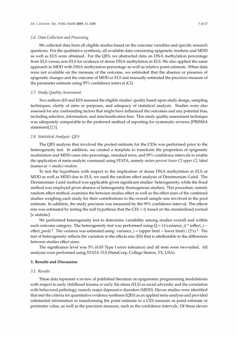
Figure 4 illustrates the forest plot of the methylation differential profile, comparing mDNA meanindex in subjects with and without ELS. Relative to subjects without ELS, there was a 23% increasedDNA methylation profile among those with ELS as childhood adversity, CES = 23.2%, 95% CI =
8.00–38.48. These studies were significantly heterogeneous with respect to the individual effect sizes,implying the between studies variance, I2, χ2 (3) = 4217, p < 0.001. The test of the variance from zero inthe CES indicated a significant difference, implying CES > 0, z = 2.99, p = 0.003.
Int. J. Environ. Res. Public Health 2019, 16, x FOR PEER REVIEW 11 of 18
Figure 4 illustrates the forest plot of the methylation differential profile, comparing mDNA mean index in subjects with and without ELS. Relative to subjects without ELS, there was a 23% increased DNA methylation profile among those with ELS as childhood adversity, CES = 23.2%, 95% CI = 8.00–38.48. These studies were significantly heterogeneous with respect to the individual effect sizes, implying the between studies variance, I2, χ2 (3) = 4217, p < 0.001. The test of the variance from zero in the CES indicated a significant difference, implying CES > 0, z = 2.99, p = 0.003.
Figure 4. DNA Methylation of NR3C1 (glucocorticoid receptor gene) in Social Adversity with Early Life Stress
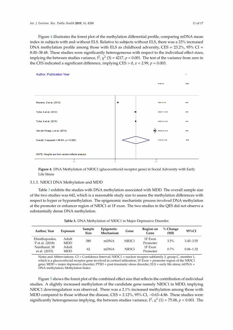
3.1.3. NR3CI DNA Methylation and MDD
Table 3 exhibits the studies with DNA methylation associated with MDD. The overall sample size of the two studies was 642, which is a reasonable study size to assess the methylation differences with respect to hyper or hypomethylation. The epigenomic mechanistic process involved DNA methylation at the promoter or enhancer region of NR3C1 at 1F exon. The two studies in the QES did not observe a substantially dense DNA methylation.
Table 3. DNA Methylation of NR3C1 in Major Depressive Disorder.
Author, Year Exposure Sample Size
Epigenetic Mechanism Gene Region on
Gene %
Change (MI)
95%CI
Efstathopoulos, P et al. (2018)
Adult MDD 580 mDNA NR3C1 1F Exon
Promoter 3.5% 3.45–3.55
Nantharat, M et al. (2015)
Adult MDD 62 mDNA NR3CI 1F Exon
Promoter 0.7% 0.06–1.32
Notes and Abbreviations: CI = Confidence Interval; NR3C1= nuclear receptor subfamily 3, group C, member 1, which is a glucocorticoid receptor gene involved in cortisol utilization; 1F Exon= promoter region of the NR3C1 gene; MDD = major depressive disorder; PTSD = post-traumatic stress disorder; ELS = early life stress; mDNA = DNA methylation; Methylation Index.
Figure 5 shows the forest plot of the combined effect size that reflects the contribution of individual studies. A slightly increased methylation of the candidate gene namely NR3C1 in MDD, implying NR3C1 downregulation was observed. There was a 2.1% increased methylation among
Figure 4. DNA Methylation of NR3C1 (glucocorticoid receptor gene) in Social Adversity with EarlyLife Stress
3.1.3. NR3CI DNA Methylation and MDD
Table 3 exhibits the studies with DNA methylation associated with MDD. The overall sample sizeof the two studies was 642, which is a reasonable study size to assess the methylation differences withrespect to hyper or hypomethylation. The epigenomic mechanistic process involved DNA methylationat the promoter or enhancer region of NR3C1 at 1F exon. The two studies in the QES did not observe asubstantially dense DNA methylation.
Table 3. DNA Methylation of NR3C1 in Major Depressive Disorder.
Author, Year Exposure SampleSize
EpigeneticMechanism Gene Region on
Gene% Change
(MI) 95%CI
Efstathopoulos,P et al. (2018)
AdultMDD 580 mDNA NR3C1 1F Exon
Promoter 3.5% 3.45–3.55
Nantharat, Met al. (2015)
AdultMDD 62 mDNA NR3CI 1F Exon
Promoter 0.7% 0.06–1.32
Notes and Abbreviations: CI = Confidence Interval; NR3C1 = nuclear receptor subfamily 3, group C, member 1,which is a glucocorticoid receptor gene involved in cortisol utilization; 1F Exon = promoter region of the NR3C1gene; MDD = major depressive disorder; PTSD = post-traumatic stress disorder; ELS = early life stress; mDNA =DNA methylation; Methylation Index.
Figure 5 shows the forest plot of the combined effect size that reflects the contribution of individualstudies. A slightly increased methylation of the candidate gene namely NR3C1 in MDD, implyingNR3C1 downregulation was observed. There was a 2.1% increased methylation among those withMDD compared to those without the disease, CES = 2.12%, 95% CI, −0.63–4.86. These studies weresignificantly heterogeneous implying, the between studies variance, I2, χ2 (1) = 75.08, p < 0.001. The

Int. J. Environ. Res. Public Health 2019, 16, 4280 12 of 17
test of the variance from zero in the common CES indicated no significant difference, implying CES ≤0, z = 1.5, p = 0.13.
Int. J. Environ. Res. Public Health 2019, 16, x FOR PEER REVIEW 12 of 18
those with MDD compared to those without the disease, CES = 2.12%, 95% CI, −0.63–4.86. These studies were significantly heterogeneous implying, the between studies variance, I2, χ2 (1) = 75.08, p < 0.001. The test of the variance from zero in the common CES indicated no significant difference, implying CES ≤ 0, z = 1.5, p = 0.13.
Figure 5. DNA Methylation of NR3C1 (glucocorticoid receptor gene ) in Major Depressive Disorder (Unipolar Affective Disorder)
3.2. Discussion
The need for a scientific statement and evidence-based understanding of the epigenomic modulation or programming in early childhood stress or adversity implying early life stress as well as the modification associated MDD and the correlation between ELS and MDD motivated the current systematic review and QES. This study assessed the epigenomic modulation in terms of DNA methylation mechanistic process in early life stress and MDD, as well as the relationship between ELS and MDD in terms of epigenomic aberrations induced by dense DNA methylation. There are a few relevant findings based on this QES. First, DNA methylation of the 1F exon on the NR3C1 gene, a glucocorticoid gene is associated with ELS and directly correlated with MDD in adulthood. Secondly, dense DNA methylation of the 1F exon of the NR3C1 gene is associated with increased risk of ELS. Thirdly, DNA hyper-methylation of the 1F exon of NR3C1 marginally increases the risk of MDD.
We have demonstrated in this systematic review and QES that the DNA methylation of the glucocorticoid receptor gene (NR3C1) results in impaired gene expression and the abnormal protein synthesis, adversely affecting the cellular function of this receptor in response to cortisol and its biologic function in responding to stress and early life adversity, or early life stress. The observed hypermethylation of this receptor gene reflects impaired response resulting in chronic stress and subsequent mental illness given the pathway of cortisol response in MDD. The activation of the hypothalamus–pituitary–adrenal (HPA) axis by social signal transduction results in rapid response to stress and stressful environment and with inhibition of the transcription factor resulting in inactivation of the protein and receptor function. An example of social stressors as ELS has been illustrated in pre and postnatal stress and maternal care insufficiencies associated with behavioral
Figure 5. DNA Methylation of NR3C1 (glucocorticoid receptor gene) in Major Depressive Disorder(Unipolar Affective Disorder)
3.2. Discussion
The need for a scientific statement and evidence-based understanding of the epigenomicmodulation or programming in early childhood stress or adversity implying early life stress aswell as the modification associated MDD and the correlation between ELS and MDD motivated thecurrent systematic review and QES. This study assessed the epigenomic modulation in terms of DNAmethylation mechanistic process in early life stress and MDD, as well as the relationship between ELSand MDD in terms of epigenomic aberrations induced by dense DNA methylation. There are a fewrelevant findings based on this QES. First, DNA methylation of the 1F exon on the NR3C1 gene, aglucocorticoid gene is associated with ELS and directly correlated with MDD in adulthood. Secondly,dense DNA methylation of the 1F exon of the NR3C1 gene is associated with increased risk of ELS.Thirdly, DNA hyper-methylation of the 1F exon of NR3C1 marginally increases the risk of MDD.
We have demonstrated in this systematic review and QES that the DNA methylation of theglucocorticoid receptor gene (NR3C1) results in impaired gene expression and the abnormal proteinsynthesis, adversely affecting the cellular function of this receptor in response to cortisol and itsbiologic function in responding to stress and early life adversity, or early life stress. The observedhypermethylation of this receptor gene reflects impaired response resulting in chronic stress andsubsequent mental illness given the pathway of cortisol response in MDD. The activation of thehypothalamus–pituitary–adrenal (HPA) axis by social signal transduction results in rapid response tostress and stressful environment and with inhibition of the transcription factor resulting in inactivationof the protein and receptor function. An example of social stressors as ELS has been illustrated in preand postnatal stress and maternal care insufficiencies associated with behavioral pathologies [24–26].Animal models with rodents demonstrated an inverse correlation between adequate postnatal maternalcare and increased stress-reactivity, anxiety, and fear reaction [5].
In general, the main neural substrates involved in epigenetic modulation of ELS and maternalcare include HPA, amygdala, medial prefrontal cortex, and hippocampal region of the brain. Theobserved phenotypes in the aberrant epigenomic modulation in ELS are explained by the NR3C1 genedownregulation and decreased expression, a glucocorticoid receptor (GR) gene. The animal model inthis context observed the dense methylation of the CpG in the promoter region of NR3C1, increasedactivity of the HPA axis and elevated serum glucocorticoids level [5,6,27]. Additionally, animalmodel reveals increased stress reactivity, anxiety, impaired social interaction, and depressive episodes
Int. J. Environ. Res. Public Health 2019, 16, 4280 13 of 17
associated with maternal separation. The observed manifestations correlated with genome-wideaberrant DNA methylation and brain neural pathway genes transcripts [28–30].
Prolonged activation of the HPA axis, altered gene transcription, and aberrant DNA methylationwere observed in CNS and peripheral T lymphocytes among infants and juveniles deprived ofmaternal care [31–36]. Aberrant epigenomic modulation (mDNA) and hydroxymethyl cytosine atthe brain-derived neurotrophic factor (BDNF) locus, a member of the nerve growth factor familyof neutrophils, had been observed in adolescence and adulthood distress associated with maternalmaltreatment during neonatal nursing of offspring. Other neural changes include hippocampus,amygdala, and medial prefrontal cortex [37–40]. Further studies observed increased expression ofarginine vasopressin (Avp) and gene loci, a protein-coding gene for antidiuretic hormone (Vasopressin)associated with diabetes insipidus and DNA hypomethylation in paraventricular nucleus of thehippocampus in ELS, due to maternal separation. Similarly, the overexpression of proopiomelanocortin(pomc) gene loci, which is involved in the production of adrenocorticotropic hormone (ACTH)and binds to melanocortin 2 receptor (MC2R), stimulating cortisol release is observed in ELSassociated with maternal separation [41]. The overexpression of NR3C1 inversely correlates withcorticotrophin-releasing hormone (CRH) and transcription dysregulation in chronic stress induced byELS [42].
Human models have clearly illustrated how prenatal, perinatal, infant, and adolescence socialadversities exposure lead to aberrant epigenomic modulation that are transformational into adulthood.Whereas NR3C1 implicated in ELS in rodents, varieties of NR3CI sequences are observed as regulatorytargets with the most consistent and common epigenomic modulation being hypermethylation atexon 1F of human NR3C1 gene in ELS or early life social adversity. The DNA sequence element thatencodes methylation-sensitive binding site associated with neural activity-regulated transcriptionactivator (NGFTA) is embedded in exon 1F region of the NR3C1. The hypermethylation of NR3C1 inELS, implying prolonged stress that results in allostatic load, interrupts the glucocorticoids-mediatedto the pituitary and hypothalamus, enhancing HPA axis persistent activation and the consequentpsychopathology including chronic inflammation and major depressive disorders (MDD) [43]. Acorrelation between parental stress, such as domestic violence and intimate partner violence, andNR3C1 CpG hypermethylation had been observed. Likewise, child abuse and neglect had beenshown to be associated with dense NR3C1 CpG methylation and the subsequent impaired NR3C1transcription, leading to NR3C1 downregulation and decreased gene expression [44–49]. WhileNR3C1 hypermethylation results in NR3C1-reduced transcription, it is not clear if other pathways orepigenomic processes influence NR3C1 gene expression. However, noncoding RNA namely IncRNA,GASS, and miRNA have been shown to impair NR3C1 gene expression and protein activities [50,51].
Other epigenomic modulations associated with early life adversities in animal models haveimplicated similar genes in humans such as BDNF, CRH, PM2D1, and CHRBP [36,52]. Additionally,genes involved in early life adversities include MAOA, SLC17A3, MORC1, and FKBP5 [27,53–57].Overall, child maltreatment implying abuse and neglect and parental care deprivation signalaberrant epigenomic modulation inducing psychopathology and chronic disease such as type IIdiabetes and hypertension in adolescence and adult life due to allostatic overload and marginalizedglucocorticoid-mediated negative feedback to pituitary and hypothalamus.
We have demonstrated that ELS is associated with hypermethylation of glucocorticoid gene(NR3C1) receptor. These findings are supported by previous data including the individual studiesin QES that observed hypermethylation of candidate gene expression with ELS. Specifically, themethylation of the promoter region of DNA in this context results in the impairment in RNApolymerase response with respect to transcription and translation and the inhibition of the transcriptionfactor resulting in abnormal gene expression and irregular protein synthesis affecting the receptorfunctionality. Overall, the observed cellular function is due to this methylation as an effect of socialtransduction in ELS, such as isolation, sexual abuse, child neglect, violent environment experiencedby children at very young age, and maternal isolation and separation. Similarly, ELS has been

Int. J. Environ. Res. Public Health 2019, 16, 4280 14 of 17
observed in previous studies to result in psychiatric condition, including though not limited toschizophrenia, anxiety, bipolar, personality disorders, and post-traumatic stress disorder (PTSD) [23].To assess the molecular events associated with ELS and psychiatric conditions, those without ELS andMDD were examined. These studies clearly indicated the hypermethylation of glucocorticoid geneamong those with depression who suffered ELS relative with those who did not. The finding in thissystematic review is supported by previous data [23] in both animal and human models on epigenomicprogramming that indicated the epigenomic programming commencing from gametogenesis that aretransgenerational and predispose to disease conditions, but reversible. Studies have also indicatedthat mental activity such as yoga, meditation, and social network improves individual genome as wellas “community genome” to response to stress implying lower incidence and prevalence of MDD insuch environments [45].
The evidence accumulated in this QES may be questionable due to the difficulties in determiningthe CpG sites with unknown biologic functions that may be incorporated into the methylation or thehypermethylation process while describing the findings in the original papers. Since the methylationstate of differentiated tissues is considered to be highly specific, it is not very clear how methylationprocess in the circulating blood cells or saliva will provide information or data about changes in DNAfrom the less accessible tissue has the human brain and the autonomic nervous system, as well as theimmune system cells.
Despite the strength of these findings in correlating MDD with ELS and providing a scientificstatement based on the NR3C1 DNA methylation, there are some limitations. This approach involvesthe synthesis and quantification of given studies in order to arrive at a CES as evidence in interventionor therapeutics. First, systematic review and QES as an applied meta-analysis is a retrospective designthat is prone to information and selection biases. Additionally, some studies incorporated in thisQES despite the similarity of the design, implying a comparable measure of effect or point estimate,some studies failed to control for confounding, since no single variable explains everything withrespect to causation. In effect, the scientific evidence and statement generated by this QES are subjectto biases and unmeasured as well as residual confounding. However, it is highly unlikely that themagnitude of evidence provided on hypermethylation of NR3CI gene in ELS and MD is driven slowlyby these limitations.
4. Conclusions
In summary, dense DNA methylation of the glucocorticoid receptor gene (NR3C1) is associatedwith ELS that predisposes to MDD. Addressing maternal stress, separation, and child abuse andneglect has substantial potential in lowering the incidence and prevalence of behavioral pathologies,namely major depressive disorder and bipolar affective disorders in all human societies.
Whereas these findings implicate epigenomic modulation of NR3CI in ELS, as well as the nexusbetween ELS and MDD, caution is required in the interpretation and the application of these QES inintervention mapping, given the reversible nature of epigenomic modulations, as well as the timing ofspecimen collection in causal inference. Overall, this data suggests effective policy implementationand evaluation in ensuring that all children regardless of birth accidence are provided with adequateenvironment that supports proper growth and development, whereby, protective against subjectiveand objective adversity.
Author Contributions: L.H.J. conceptualized the study, designed and analyzed the data, prepared the manuscript,and reviewed and approved the final draft. E.S. performed the literature search, abstracted the data, preparedthe data extraction sheet, assisted in the preparation of the manuscript, and reviewed and approved the finaldraft. C.C. assisted in the manuscript preparation and reviewed and approved the final draft. K.D. facilitatedthe manuscript preparation and reviewed and approved the final draft. L.P. assisted in manuscript preparationand reviewed and approved the final draft. K.W.D. assisted in the study conceptualization, provided input ofmanuscript preparation, and reviewed and approved the final draft.
Funding: This study received no external funding.
Int. J. Environ. Res. Public Health 2019, 16, 4280 15 of 17
Acknowledgments: The authors would like to thank Patricia Oceanic and April Aguilera for coordinatingthis study.
Conflicts of Interest: All authors have declared no conflict of interest.
References
1. Powell, N.D.; Sloan, E.K.; Bailey, M.T.; Arevalo, J.M.; Miller, G.E.; Chen, E.; Kobor, M.S.; Reader, B.F.;Sheridan, J.F.; Cole, S.W. Social stress up-regulates inflammatory gene expression in the leukocytetranscriptome via beta-adrenergic induction of myelopoiesis. Proc. Natl. Acad. Sci. USA 2013, 110,16574–16579. [CrossRef]
2. Vreugdenhil, E.; Verissimo, C.S.; Mariman, R.; Kamphorst, J.T.; Barbosa, J.S.; Zweers, T.; Champagne, D.L.;Schouten, T.; Meijer, O.C.; de Kloet, E.R.; et al. MicroRNA 18 and 124a down-regulate the glucocorticoidreceptor: Implications for glucocorticoid responsiveness in the brain. Endocrinology 2009, 150, 2220–2228.[CrossRef]
3. Chen, E.; Miller, G.E.; Kobor, M.S.; Cole, S.W. Maternal warmth buffers the effects of low early-lifesocioeconomic status on pro-inflammatory signaling in adulthood. Mol. Psychiatry 2011, 16, 729–737.[CrossRef]
4. Mulligan, C.J.; D’Errico, N.C.; Stees, J.; Hughes, D.A. Methylation changes at NR3C1 in newborns associatewith maternal prenatal stress exposure and newborn birth weight. Epigenetics 2012, 7, 853–857. [CrossRef]
5. Weaver, I.C.; Meaney, M.J.; Szyf, M. Maternal care effects on the hippocampal transcriptome andanxiety-mediated behaviors in the offspring that are reversible in adulthood. Proc. Natl. Acad. Sci. USA2006, 103, 3480–3485. [CrossRef]
6. McGowan, P.O.; Suderman, M.; Sasaki, A.; Huang, T.C.; Hallett, M.; Meaney, M.J.; Szyf, M. Broad epigeneticsignature of maternal care in the brain of adult rats. PLoS ONE 2011, 6, e14739. [CrossRef]
7. Dickerson, S.S.; Kemeny, M.E. Acute stressors and cortisol responses: A theoretical integration and synthesisof laboratory research. Psychol. Bull. 2004, 130, 355–391. [CrossRef]
8. Gilbert, P.; McEwan, K.; Bellew, R.; Mills, A.; Gale, C. The dark side of competition: How competitivebehaviour and striving to avoid inferiority are linked to depression, anxiety, stress and self-harm. Psychol.Psychother. 2009, 82 Pt 2, 123–136. [CrossRef]
9. Layte, R. The association between income inequality and mental health: Testing status anxiety, social capital,and neo-materialist explanations. Eur. Soc. Rev. 2011, 28, 498–511. [CrossRef]
10. Layte, R.; Whelan, C.T. Who feels inferior? A test of the status anxiety hypothesis of social inequalities inhealth. Eur. Soc. Rev. 2014, 30, 525–535. [CrossRef]
11. Frankel, D.; Arbel, T. Group formation by two-year olds. Int. J. Behav. Dev. 1980, 3, 287–298. [CrossRef]12. McGowan, P.; Sasaki, A.; D’Alessio, A.; Dymov, S.; Labonté, B.; Szyf, M.; Turecki, G.; Meaney, M.J. Epigenetic
Regulation of the Glucocorticoid Receptor in Human Brain Associates with Childhood Abuse. Nat. Neurosci.2009, 12, 342–348. [CrossRef]
13. McGowan, P.O.; Szyf, M. The epigenetics of social adversity in early life: Implications for mental healthoutcomes. Neurobiol. Dis. 2010, 39, 66–72. [CrossRef]
14. Lachner, M.; O’Carroll, D.; Rea, S.; Mechtler, K.; Jenuwein, T. Methylation of histone H3 lysine 9 creates abinding site for HP1 proteins. Nature 2001, 410, 116–120. [CrossRef]
15. Razin, A. CpG methylation, chromatin structure and gene silencing-a three-way connection. EMBO J. 1998,17, 4905–4908. [CrossRef]
16. Rajewsky, N. MicroRNA Target Predictions in Animals. Nat. Genet. 2006, 38, S8–S13. [CrossRef]17. Tuddenham, L.; Wheeler, G.; Hajihosseini, M.K.; Clark, I.; Dalmay, T. The cartilage specific microRNA-140
targets histone deacetylase 4 in mouse cells. FEBS Lett. 2006, 580, 4214–4217. [CrossRef]18. Cole, S.W.; Conti, G.; Arevalo, J.M.; Ruggerio, A.M.; Heckman, J.J.; Suomi, S.J. Transcriptional modulation
of the developing immune system by early life social adversity. Proc. Natl. Acad. Sci. USA 2012, 109,20578–20583. [CrossRef]
19. Rhen, T.; Cidlowski, J. Antiinflammatory Action of Glucocorticoids—New Mechanisms for Old Drugs.N. Engl. J. Med. 2005, 353, 1711–1723. [CrossRef]
20. Binder Elisabeth, B. The Role of FKBP5, a Co-Chaperone of the Glucocorticoid Receptor in the Pathogenesisand Therapy of Affective and Anxiety Disorders. Psychoneuroendocrinology 2009, 34, S186–S195. [CrossRef]

Int. J. Environ. Res. Public Health 2019, 16, 4280 16 of 17
21. Klengel, T.; Mehta, D.; Anacker, C.; Rex-Haffner, M.; Pruessner, J.C.; Pariante, C.M.; Pace, T.W.; Mercer, K.B.;Mayberg, H.S.; Bradley, B. Allele-specific FKBP5 DNA demethylation mediates gene-childhood traumainteractions. Nat. Neurosci. 2013, 16, 33–41. [CrossRef]
22. Kadhim, M.; Holmes, L.; Gesheff, M.G.; Conway, J.D. Treatment Options for Nonunion with SegmentalBone Defects: Systematic Review and Quantitative Evidence Synthesis. J. Orthop. Trauma 2016, 31, 111–119.[CrossRef]
23. Nawijn, F.; Ham, W.; Houwert, R.M.; Groenwold, R.; Hietbrink, F.; Smeeing, D. Quality of reporting ofsystematic reviews and meta-analyses in emergency medicine based on the PRISMA statement. BMC Emerg.Med. 2019, 19, 19. [CrossRef]
24. Anacker, C.; O’Donnell, K.J.; Meaney, M.J. Early life adversity and the epigenetic programming ofhypothalamic-pituitary-adrenal function. Dialogues Clin. Neurosci. 2014, 16, 321–333.
25. Maccari, S.; Krugers, H.J.; Morley-Fletcher, S.; Szyf, M.; Brunton, P.J. The consequences of early-life adversity:Neurobiological, behavioural and epigenetic adaptations. J. Neuroendocrinol. 2014, 26, 707–723. [CrossRef]
26. Kundakovic, M.; Champagne, F.A. Early-life experience, epigenetics, and the developing brain.Neuropsychopharmacology 2015, 40, 141–153. [CrossRef]
27. Suderman, M.; McGowan, P.O.; Sasaki, A.; Huang, T.C.T.; Hallett, M.T.; Meaney, M.J.; Turecki, G.; Szyf, M.Conserved epigenetic sensitivity to early life experience in the rat and human hippocampus. Proc. Natl.Acad. Sci. USA 2012, 109 (Suppl. 2), 17266–17272. [CrossRef]
28. Murgatroyd, C.; Patchev, A.V.; Wu, Y.; Micale, V.; Brockmϋhl, Y.; Fischer, D.; Holsboer, F.; Wotjak, C.T.;Almeida, O.F.; Spengler, D. Dynamic DNA methylation programs persistent adverse effects of early-lifestress. Nat. Neurosci. 2009, 12, 1559–1566. [CrossRef]
29. Franklin, T.B.; Russig, H.; Weiss, I.C.; Gräff, J.; Linder, N.; Michalon, A.; Vizi, S.; Mansuy, I.M. Epigenetictransmission of the impact of early stress across generations. Biol. Psychiatry 2010, 68, 408–415. [CrossRef]
30. Franklin, T.B.; Saab, B.J.; Mansuy, I.M. Neural mechanisms of stress resilience and vulnerability. Neuron 2012,75, 747–761. [CrossRef]
31. Provencal, N.; Suderman, M.J.; Guillemin, C.; Massart, R.; Ruggiero, A.; Wang, D.; Bennett, A.J.; Pierre, P.J.;Friedman, D.P.; Côtè, S.M.; et al. The signature of maternal rearing in the methylome in rhesus macaqueprefrontal cortex and T cells. J. Neurosci. 2012, 32, 15626–15642. [CrossRef] [PubMed]
32. Elwenspoek, M.M.; Hengesch, X.; Leenen, F.A.; Schritz, A.; Sias, K.; Schaan, V.K.; Mériaux, S.B.; Schmitz, S.;Bonnemberger, F.; Schächinger, H.; et al. Proinflammatory T Cell Status Associated with Early Life Adversity.J. Immunol. 2017, 199, 4046–4055. [CrossRef] [PubMed]
33. Suomi, S.J. Risk, resilience, and gene-environment interplay in primates. J. Can. Acad. Child. Adolesc.Psychiatry 2011, 20, 289–297. [PubMed]
34. Dettmer, A.M.; Suomi, S.J. Nonhuman primate models of neuropsychiatric disorders: In uences of earlyrearing, genetics, and epigenetics. ILAR J. 2014, 55, 361–370. [CrossRef]
35. Kinnally, E.L. Epigenetic plasticity following early stress predicts long-term health outcomes in rhesusmacaques. Am. J. Phys. Anthropol. 2014, 155, 192–199. [CrossRef]
36. Nieratschker, V.; Massart, R.; Gilles, M.; Luoni, A.; Suderman, M.J.; Krumm, B.; Meier, S.; Witt, S.H.;Nöthen, M.M.; Suomi, S.J.; et al. MORC1 exhibits cross-species differential methylation in association withearly life stress as well as genome-wide association with MDD. Transl. Psychiatry 2014, 4, e429. [CrossRef]
37. McEwen, B.S.; Nasca, C.; Gray, J.D. Stress effects on neuronal structure: Hippocampus, amygdala, andprefrontal cortex. Neuropsychopharmacology 2016, 41, 3–23. [CrossRef]
38. Blaze, J.; Scheuing, L.; Roth, T.L. Differential methylation of genes in the medial prefrontal cortex of developingand adult rats following exposure to maltreatment or nurturing care during infancy. Dev. Neurosci. 2013, 35,306–316. [CrossRef]
39. Roth, T.L.; Matt, S.; Chen, K.; Blaze, J. Bdnf DNA methylation modi cations in the hippocampus and amygdalaof male and female rats exposed to different caregiving environments outside the homecage. Dev. Psychobiol.2014, 56, 1755–1763. [CrossRef]
40. Doherty, T.S.; Forster, A.; Roth, T.L. Global and gene-speci c DNA methylation alterations in the adolescentamygdala and hippocampus in an animal model of caregiver maltreatment. Behav. Brain Res. 2016, 298 Pt A,55–61. [CrossRef]
41. Wu, Y.; Patchev, A.V.; Daniel, G.; Almeida, O.F.; Spengler, D. Early-life stress reduces DNA methylation ofthe Pomc gene in male mice. Endocrinology 2014, 155, 1751–1762. [CrossRef] [PubMed]

Int. J. Environ. Res. Public Health 2019, 16, 4280 17 of 17
42. Bockmϋhl, Y.; Patchev, A.V.; Madejska, A.; Hoffmann, A.; Sousa, J.C.; Sousa, N.; Holsboer, F.; Almeida, O.F.X.;Spengler, D. Methylation at the CpG island shore region upregulates Nr3c1 promoter activity after early-lifestress. Epigenetics 2015, 10, 247–257. [CrossRef] [PubMed]
43. Radtke, K.M.; Ruf, M.; Gunter, H.M.; Dohrmann, K.; Schauer, M.; Meyer, A.; Elbert, T. Transgenerationalimpact of intimate partner violence on methylation in the promoter of the glucocorticoid receptor.Transl. Psychiatry 2011, 1, e21. [CrossRef] [PubMed]
44. Tyrka, A.R.; Parade, S.H.; Eslinger, N.M.; Marsit, C.J.; Lesseur, C.; Armstrong, D.A.; Philip, N.S.; Josefson, B.;Seifer, R. Methylation of exons 1D, 1F, and 1H of the glucocorticoid receptor gene promoter and exposure toadversity in preschool-aged children. Dev. Psychopathol. 2015, 27, 577–585. [CrossRef]
45. Van der Knaap, L.J.; Riese, H.; Hudziak, J.J.; Verbiest, M.M.P.J.; Verhulst, F.C.; Oldehinkel, A.J.; van Oort, F.V.A.Glucocorticoid receptor gene (NR3C1) methylation following stressful events between birth and adolescence.The TRAILS study. Transl. Psychiatry 2014, 4, e381. [CrossRef]
46. Braithwaite, E.C.; Kundakovic, M.; Ramchandani, P.G.; Murphy, S.E.; Champagne, F.A. Maternal prenataldepressive symptoms predict infant NR3C1 1F and BDNF IV DNA methylation. Epigenetics 2015, 10, 408–417.[CrossRef]
47. Murgatroyd, C.; Quinn, J.P.; Sharp, H.M.; Pickles, A.; Hill, J. Effects of prenatal and postnatal depression, andmaternal stroking, at the glucocorticoid receptor gene. Transl. Psychiatry 2015, 5, e560. [CrossRef]
48. Radtke, K.M.; Schauer, M.; Gunter, H.M.; Ruf-Leuschner, M.; Sill, J.; Meyer, A.; Elbert, T. Epigeneticmodifications of the glucocorticoid receptor gene are associated with the vulnerability to psychopathologyin childhood maltreatment. Transl. Psychiatry 2015, 5, e571. [CrossRef]
49. Kertes, D.A.; Kamin, H.S.; Hughes, D.A.; Rodney, N.C.; Bhatt, S.; Mulligan, C.J. Prenatal maternal stresspredicts methylation of genes regulating the hypothalamic–pituitary–adrenocortical system in mothers andnewborns in the Democratic Republic of Congo. Child. Dev. 2016, 87, 61–72. [CrossRef]
50. Kino, T. Stress, glucocorticoid hormones, and hippocampal neural progenitor cells: implications to mooddisorders. Front Physiol. 2015, 6. [CrossRef]
51. Pan-Vazquez, A.; Rye, N.; Ameri, M.; McSparron, B.; Smallwood, G.; Bickerdyke, J.; Rathbone, A.;Dajas-Bailador, F.; Toledo-Rodriguez, M. Impact of voluntary exercise and housing conditions on hippocampalglucocorticoid receptor, miR-124 and anxiety. Mol. Brain 2015, 8, 40. [CrossRef] [PubMed]
52. Weder, N.; Zhang, H.; Jensen, K.; Yang, B.Z.; Simen, A.; Jackowski, A.; Lipschitz, D.; Douglas-Palumberi, H.;Ge, M.; Perepletchikova, F.; et al. Child abuse, depression, and methylation in genes involved with stress,neural plasticity, and brain circuitry. J. Am. Acad. Child. Adolesc. Psychiatry 2014, 53, 417–424. [CrossRef][PubMed]
53. Melas, P.A.; Wei, Y.; Wong, C.C.; Sjöholm, L.K.; Aberg, E.; Johnathan, M.; Schalling, M.; Forsell, Y.; Lavebratt, C.Genetic and epigenetic associations of MAOA and NR3C1 with depression and childhood adversities. Int. J.Neuropsychopharmacol. 2013, 16, 1513–1528. [CrossRef] [PubMed]
54. Cao-Lei, L.; Massart, R.; Suderman, M.J.; Machnes, Z.; Elgbeili, G.; Laplante, D.P.; Szyf, M.; King, S. DNAmethylation signatures triggered by prenatal maternal stress exposure to a natural disaster: Project Ice Storm.PLoS ONE 2014, 9, e107653. [CrossRef] [PubMed]
55. Khulan, B.; Manning, J.R.; Dunbar, D.R.; Seckl, J.R.; Raikkonen, K.; Eriksson, J.G.; Drake, A.J. Epigenomicprofiling of men exposed to early-life stress reveals DNA methylation differences in association with currentmental state. Transl. Psychiatry 2014, 4, e448. [CrossRef] [PubMed]
56. Non, A.L.; Hollister, B.M.; Humphreys, K.L.; Childebayeva, A.; Esteves, K.; Zeanah, C.H.; Fox, N.A.;Nelson, C.A.; Drury, S.S. DNA methylation at stress-related genes is associated with exposure to early lifeinstitutionalization. Am. J. Phys. Anthropol. 2016, 161, 84–93. [CrossRef]
57. Houtepen, L.C.; Vinkers, C.H.; Carrillo-Roa, T.; Hiemstra, M.; van Lier, P.A.; Meeus, W.; Branje, S.; Heim, C.M.;Nemeroff, C.B.; Mill, J.; et al. Genome-wide DNA methylation levels and altered cortisol stress reactivityfollowing childhood trauma in humans. Nat. Commun. 2016, 7, 10967. [CrossRef]
© 2019 by the authors. Licensee MDPI, Basel, Switzerland. This article is an open accessarticle distributed under the terms and conditions of the Creative Commons Attribution(CC BY) license (http://creativecommons.org/licenses/by/4.0/).