Regulation of acetylcholine receptor clustering by the tumor suppressor APC

Selenoprotein P Regulation by the Glucocorticoid Receptor
Colleen Rock and Philip J. MoosDepartment of Pharmacology and Toxicology, University of Utah, Salt Lake City, Utah 84112
AbstractMaintenance of the antioxidant activity of selenoproteins is one potential mechanism of thebeneficial health effects of selenium. Selenoprotein P is the primary selenium distribution proteinof the body as well as the major selenium containing protein in serum. The transcriptionalregulation of selenoprotein P is of interest since the extrahepatic expression of this gene hasdemonstrated differentiation-dependent expression in development as well as under differentdisease states. SEPP1 displays patterned expression in numerous tissues during development andthe loss of SEPP1 expression has been observed in malignancy. In addition, factors that influenceinflammatory processes like cytokines and their regulators have been implicated in selenoprotein Ptranscriptional control. Herein, we identify a retinoid responsive element and describe amechanism where the glucocorticoid receptor negatively regulates expression of selenoprotein P.Luciferase reporter assays and quantitative PCR were used to measure selenoprotein Ptranscription in engineered HEK-293 cells. When stimulated with ecdysone analogs, selenoproteinP expression was increased with the use of a fusion transcription factor that contains theglucocorticoid receptor DNA binding domain, an ecdysone ligand-binding domain, and a strongtransactivation domain as well as the retinoid X receptor. The native glucocorticoid receptorinhibited selenoprotein P transactivation, and selenoprotein P was further attenuated in thepresence of dexamethasone. Our results may provide insight into a potential mechanism by whichselenium is redistributed during development, differentiation or under conditions of critical illness,where glucocorticoid levels are typically increased.
Keywordsselenoprotein P; glucocortiod receptor; glucocorticoids; antioxidant; retinoids
IntroductionSelenoprotein P (SelP) is an extracellular glycoprotein that carries approximately 40% ofplasma selenium (Akesson et al. 1994). SelP is unique among the selenoproteins in that itcan possess up to ten selenocysteine residues in mammals (Burk & Hill 2005). SelPprimarily functions in selenium distribution (Hill et al. 2003; Renko et al. 2008), withknockout mice displaying altered selenium distribution, particularly to the testes and brain(Hill et al. 2003; Burk et al. 2006). The majority of SelP is derived from hepatic sources,however; the mRNA can be detected in almost all tissues, with appreciable concentrationsobserved in the kidney, heart, lung, brain, skeletal muscle, and testis (Burk & Hill 2005)
The regulation of selenoprotein P gene (SEPP1) expression is an active area of investigationwith changes in SEPP1 noted under a broad spectrum of biological processes. In HepG2cells and primary rat hepatocytes, promoter activity has been shown to be inhibited by
Address correspondence to: Philip J. Moos, Department of Pharmacology & Toxicology, University of Utah, L.S. Skagg’s Pharmacy,Rm. 201, 30 S 2000 East, Salt Lake City, UT 84112, Tel. 801-585-5952, Fax. 801-585-5111, [email protected].
NIH Public AccessAuthor ManuscriptBiometals. Author manuscript; available in PMC 2011 February 15.
Published in final edited form as:Biometals. 2009 December ; 22(6): 995–1009. doi:10.1007/s10534-009-9251-2.
NIH
-PA Author Manuscript
NIH
-PA Author Manuscript
NIH
-PA Author Manuscript

cytokines including interleukin 1β, tumor necrosis factor α, interferon γ, and transforminggrowth factor β1 (Dreher et al. 1997; Mostert et al. 2001). This inhibition suggests that theSEPP1 gene product may function as a negative acute-phase protein in response toinflammation. Alternatively, promoter activity is stimulated in hepatic cells through theFOXO1a and HNF-4α transcription factors (Speckmann et al. 2008; Walter et al. 2008).
In addition to inflammation, microarray analyses have revealed changes in SEPP1expression during development and following alterations in the differentiation state ofextrahepatic cells. Elegant developmental studies have demonstrated SEPP1 orthologspatiotemporal expression in both zebrafish (Thisse et al. 2003) and murine model systems(Lee et al. 2008). Increased expression has been observed in differentiating myeloid,pulmonary, and Sertoli cells (Tabuchi et al. 2005; Ghassabeh et al. 2006; Wade et al. 2006).Conversely, SEPP1 expression is decreased with neoplastic progression from normal tissue,to carcinoma, to metastatic disease in cells of prostate origin (Dhanasekaran et al. 2001).Evaluation of SEPP1 expression in the Oncomine database (Rhodes et al. 2004) alsoidentifies decreased SEPP1 expression in melanoma, lung, and colon cancer compared tonormal tissue suggesting that decreased SEPP1 expression may be a common feature ofmalignancies. Indeed, work in colorectal cancer suggests that specific selenoenzymes arereduced, indicating that changes in SEPP1 is not a general alteration in nutrition ordecreased selenium (Al-Taie et al. 2004).
In the present study, we observed SEPP1 induction in human cells stably transfected withthe ecdysone inducible system (VgEcR-RXR). Due to VgEcR’s glucocorticoid receptorDNA binding domain, as well as evidence of SEPP1 modulation during development andinflammation, we sought to determine if SEPP1 was regulated by the glucocorticoid receptoror the retinoid X receptor. In addition, evidence exists for changes in plasma selenium levelsfollowing glucocorticoid administration, with both increases and decreases noted underdifferent sets of conditions (Peretz et al. 1987; Marano et al. 1990; Watanabe et al. 1997).Although the effect of glucocorticoids on selenium levels has not been fully characterized, itis believed that these changes result from redistribution of selenium between tissue andplasma. It is unknown what role SelP may play in this glucocorticoid-induced seleniumredistribution. Therefore, an aim of this study was to examine the glucocorticoidresponsiveness of the SEPP1 promoter, and we found that the glucocorticoid receptor (GR)inhibits the expression of SEPP1.
Materials & MethodsMaterials
The HEK-293 cell line was purchased from American Type Culture Collection. AdvancedDMEM, T4 DNA ligase, HindIII, XhoI, and SstI, Accuprime Pfx DNA polymerase,SuperScript III reverse transcriptase reagents, OneShot Top 10 chemically competent cells,zeocin, geneticin, Lipofectamine 2000, and Ni-NTA agarose were purchased fromInvitrogen. Ponasterone A (PonA) was purchased from A.G. Scientific. Dexamethasone(Dex) was purchased from EMD Chemicals. RNeasy Mini Kit and EndoFree Maxi-andMini-prep Kits were obtained from Qiagen. Lightcycler 480 SYBR Green I master mix waspurchased from Roche Diagnostics. Biolase DNA polymerase, dNTPs, magnesium chloride,and NH4 reaction buffer were purchased from Bioline. SYBR Green I was purchased fromCambrex. Human genomic DNA, the Dual Luciferase Reporter Assay System waspurchased from Promega. NE-PER nuclear extraction reagents, Biotin 3′ end DNA labelingkit, and Lightshift chemiluminescent EMSA Kit were purchased from Pierce.
Rock and Moos Page 2
Biometals. Author manuscript; available in PMC 2011 February 15.
NIH
-PA Author Manuscript
NIH
-PA Author Manuscript
NIH
-PA Author Manuscript

PlasmidspVgEcR (Invitrogen) encodes the fusion transcription factor used to generate ecdysone-inducible cells. pRL-RSV and pGL4.21 (Promega) were used in the luciferase reporterassays; pRL-RSV constitutively expresses Renilla reniformis luciferase, and SEPP1promoter fragments were cloned into the pGL4.2.1 plasmid that contains firefly luciferase.The pLTRluc glucocorticoid reporter plasmid and pDsRed-hGR glucocorticoid receptorexpression plasmid were gifts from Dr. Carol Lim (University of Utah).
Cell CultureHuman embryonic kidney line HEK-293, as well as all subsequently engineered cells, werecultured in Advanced DMEM medium containing 2% fetal bovine serum and 2mM L-glutamine. Cells were maintained at 37°C in a humidified incubator with 5% CO2.
HEK-293 were transfected with pVgEcR and selected for Zeocin resistance to generatestable expression of the VgEcR gene product and are referred to as 293-EcR. 293-EcR cellsthat conditionally express 15-LOX-1 and ΔIle662 15-LOX-1 were previously described (Yuet al. 2004; Cordray et al. 2007). Conditional expression of β-galactosidase in the 293-EcRwas achieved using similar methods.
An expression vector, pDsRed-hGR, that constitutively expresses a DsRed2-labeled,functional human GR was generously provided by Dr. Carol Lim, University of Utah. The293-EcR cells were stably transfected with this expression vector and selected for neomycinresistance in order to study the effects of GR signaling in HEK-293 cells. These cells arereferred to as EcR-GR.
Polymerase Chain Reaction (PCR)The Transcription Regulatory Element Database (Jiang et al. 2007) was used to identify the~2 Kb sequence surrounding the transcriptional start site of SEPP1, promoter ID #34663(1770 bp upstream of start site, 300 bp downstream) and used as an electronic template togenerate promoter constructs. A 1.9 Kb sequence was amplified from human genomic DNAby PCR using the primer pair 5′-TAGGTACCCCAGTTCTTTCCGGTGTTCA-3′ and 5′-TACTCGAGCGCACTGGGAACTTCACCTA-3′. The PCR product was digested withXhoI and SstI and cloned into the pGL4.21 luciferase reporter vector. This construct isreferred to as −1652 to +247 and was utilized as template DNA in subsequent PCR reactionsused to synthesize smaller fragments of the SEPP1 promoter region of interest. A HindIIIdigestion of the −1652 to +247 construct generated −1652 to −385 and −391 to +247promoter fragments. The fragments were cloned into the pGL4.21 vector following HindIIIdigestion. Due to the use of the HindIII site in the pGL4.21 vector, the −391 to +247fragment was only subcloned in the reverse orientation, and despite several attempts, nocolonies were obtained with this fragment in the forward orientation. The −109 to +247 andthe −53 to +247 fragment were generated using PCR and cloned into the pGL4.21 vectorusing XhoI and SstI digestions.
Quantitative PCR was used to assess SelP mRNA expression. 293-EcR and EcR-GR cellswere treated with 10 μM ponasterone A 24 hours prior to mRNA collection, and 10 nMdexamethasone was then added at 8 or 16 hours prior to mRNA purification. Vehicletreatments with ethanol (EtOH) or dimethyl sulfoxide (DMSO) were used as controls. TheQiagen RNeasy Mini Kit was used to collect and purify mRNA from cells. First strandcDNA was synthesized using Superscript III reverse transcriptase and these cDNA sampleswere run in triplicate as 1:5 dilutions. Standards were run in duplicate at concentrationsbetween 103 to 108 copies/μl and β2 microglobulin was run as a reference gene. The SEPP1amplicon consisted of the 100 bp spanning the final intron of the genomic sequence. The
Rock and Moos Page 3
Biometals. Author manuscript; available in PMC 2011 February 15.
NIH
-PA Author Manuscript
NIH
-PA Author Manuscript
NIH
-PA Author Manuscript

primer pair 5′-TTCGGGCAGAGGAGAACA-3′ and 5′-CTGGCACTGGCTTCTGTG-3′were used to amplify this region. Average threshold copy number was used to calculatechanges in expression level as compared to vehicle treated controls.
Site-directed MutagenesisPutative response elements of interest were mutated using a PCR-based strategy. Theputative GRE sequence CAAGAATGAACATTGAACT at position −87 of the SEPP1promoter (GRE #1) was mutated to the sequence CAAGAATGACTATTGAACT using theprimer 5′-GGTCACTGCAAGAATGACTATTGAACTTTGGACTATAC-3′ and itscomplementary sequence (exchanged nucleotides are bold and underlined). The putativeGRE sequence TCAGAGTGTGCT at position −24 of the SEPP1 promoter (GRE #2) wasmutated to the sequence TCAGAGGATGCT using the primer 5′-GGACTATAAATATCAGAGGATGCTGCTGTGGCTTTGTG-3′ and its complementarysequence. These mutations should eliminate activity of potential GRE half sites (Nordeen etal. 1990). The putative retinoid responsive element sequence ACATTGAACTTTGG atposition −73 of the SEPP1 promoter (RRE) was mutated to the sequenceACATCTTACTTTGG using the primer 5′-CTGCAAGAATGAACATCTTACTTTGGACTATACCTGAGG-3′ and its complementarysequence. The FoxO1a binding sequence GTAAACAA at position −46 of the SEPP1promoter was mutated to the sequence GTAAATCA using the primter 5′-CCTGAGGGGTGAGGTAAATCACAGGACTATAAATATCAGAG-3′ and itscomplementary sequence.
Luciferase Reporter AssayReporter assays were quantified using a Dual Luciferase reporter assay. SEPP1 promoterconstructs cloned into pGL4.21 or a mouse mammary tumor virus promoter reporterconstruct (pLTRluc) were co-transfected with the pRL-RSV plasmid that serves as aninternal control for transfection efficiency. Cells were seeded into 6-well plates at aconcentration of 5 ×10⁵ cells/well. Each well was cotransfected with approximately 1 μg offirefly reporter plasmid along with 50 ng of the pRL-RSV vector. Twenty four hours aftertransfection, medium was replaced. Cells transfected with SEPP1 promoter constructs weretreated with either 10 μM of the ecdysone analong ponasterone A, 10 nM dexamethasone, ora combination of both for an additional 24 hr. Vehicle treatment with EtOH and/or DMSOserved as negative controls. Cells transfected with pLTRluc were treated with either DMSOor 10 nM dexamethasone for 24 hr. Following treatments, cells were collected in 200 μl ofPassive Lysis Buffer and stored at −80°C at least overnight to allow for cell membranedisruption. Cell lysates were diluted in Passive Lysis Buffer and each sample was quantifiedin triplicate on Perkin-Elmer Victor3 V plate reader. The sequential addition of LuciferaseAssay Reagent II and Stop & Glo reagent allowed for the measurement of firefly and Renillaluciferase activity, respectively.
ImmunoblottingEcR-GR cells were supplemented with 1 μM sodium selenite and treated with EtOH as avehicle control, 10 μM ponasterone A, 10 nM dexamethasone, or a combination of both for24 hr. SelP was partially purified from the culture medium of these cells using Ni-NTAagarose. Culture medium was mixed with the Ni-NTA agarose and the mixture wasincubated on a nutating mixer at 4°C overnight. The Ni-NTA beads, along with any boundproteins, were collected by centrifugation, washed twice with 500 μl cold PBS, and thenmixed with loading buffer and separated by NuPAGE 4–12% Bis-Tris gels. Proteins weretransferred to a polyvinyl difluoride membrane. Membranes were blocked with 5% nonfatdry milk in TBS-T and then probed for SelP (antibody specific for SelP was a gift from Drs.
Rock and Moos Page 4
Biometals. Author manuscript; available in PMC 2011 February 15.
NIH
-PA Author Manuscript
NIH
-PA Author Manuscript
NIH
-PA Author Manuscript

Kris Hill & Raymond Burk, Vanderbilt University). A peroxidase conjugated secondaryantibody was used to detect chemiluminescence indicative of protein expression.
Electrophoretic Mobility Shift AssayNuclear fractions were collected from 293-EcR and EcR-GR using NE-PER nuclearextraction reagents. Gel shift assays were run using the Lightshift chemiluminescentelectrophoretic mobility shift assay kit. Double-stranded 5′-biotinylated oligonucleotides (5′-GGTCACTGCAAGAATGAACATTGAACTTTGGACTATAC-3′) corresponding to thewild-type sequence of GRE #1 was used as a probe. Following end-labeling withbiotinylated UTP, complementary oligonulceotides in equimolar amounts were heated to95°C for 1 min, cooled to 65°C, and then stored at −20°C. Binding reactions wereperformed at a 20 μl volume containing 20 fmol labeled probe, 5 μg nuclear proteins, 10mM Tris, pH 7.5, 50 mM KCl, 1 mM dithiothreitol, 5 mM MgCl2, 2.5% glycerol, 0.05%NP-40, 1 μg herring sperm DNA, and 1 μg bovine serum albumin. Where indicated, 4 pmolof unlabeled competitor probe was added to reactions. For supershift experiments, 1 μg anti-glucocorticoid receptor antibody (BuGR2; Calbiochem) was added 10 minutes after additionof biotinylated probe and nuclear extract and incubated for an additional 20 minutes at roomtemperature. Reactions were then loaded onto an 8% TBE gel in 22.25% Tris, pH 8.4,22.25% boric acid, 0.5 mM EDTA and electrophoresed at 22°C. DNA was transferred to apositively charged nylon membrane, UV cross-linked, probed using Lightshiftchemiluminescent EMSA reagents, and detected on a Kodak Image Station 440CF.
Statistical AnalysisGraphPad Instat, version 3.06, was used to evaluate the statistical significance of the results.Two-tailed student’s t-tests were used to determine statistical significance when comparingtwo data sets. In cases where multiple data sets were compared, statistical significance wasdetermined by one-way ANOVA with Tukey or Tukey-Kramer multiple comparison posthoc tests, and differences were considered significant for p<0.05.
ResultsPrevious results using 293-EcR cells with ecdysone-inducible 15-LOX-1, whensupplemented with an appropriate substrate, like arachidonate, show an inhibition of theselenoprotein thioredoxin reductase activity by ~50% (Yu et al. 2004). This raised thequestion of whether other selenoenzymes might demonstrate altered expression undersimilar conditions. Quantitative PCR experiments performed using 293-EcR cells withstable, ecdysone-inducible 15-LOX-1, as well as the control cell lines with inducible ΔIle662
15-LOX-1, and β-galactosidase, demonstrated enhanced expression of SEPP1 followingponasterone A treatment (Figure 1). Since SEPP1 demonstrated increased expression in allthese cell lines, even without substrate for the 15-LOX-1, it is likely that the changes inSEPP1 expression resulted from components of the ecdysone-inducible system rather than aresponse to 15-LOX-1 catalysis.
We examined the SEPP1 promoter, from −1652 to +247, based on promoter ID #34663 inthe Transcriptional Regulatory Element Database, to determine the region of the promoterresponsible for this ecdysone-inducible transcription. Fragments of the promoter were testedusing the luciferase reporter assay in the 293-EcR cells. Fragments included −1652 to +247,−1652 to −385, −391 to +247, −109 to +247, and −53 to +247 (Figure 2A). The greatestlevel of transcriptional activation following treatment with ponasterone A was observed onthe −109 to +247 fragment, suggesting that a site within this region of the promoter maybind a component of the ecdysone-inducible system and induce transcription of SEPP1(Figure 2B).
Rock and Moos Page 5
Biometals. Author manuscript; available in PMC 2011 February 15.
NIH
-PA Author Manuscript
NIH
-PA Author Manuscript
NIH
-PA Author Manuscript

VgEcR is a synthetic transcription factor that is a fusion of the ligand-binding anddimerization domain of the Drosophila ecdysone receptor, the DNA-binding domain of theGR, and the transcriptional activation domain of herpes simplex virus VP16. This geneexpression system is designed to activate transcription upon dimerization of VgEcR with theretinoid X receptor (RXR), and binding of the heterodimer transactivates a syntheticecdysone-responsive element (Saez et al. 2000).
Many of the nuclear hormone receptors have similar DNA binding sites. The VgEcR-RXRbinds the sequence AGTGCATTGTTCTC in the synthetic response element (the bindingsites for RXR and the GR DNA binding domains are underlined), the GR binding sequenceis TGT(T/C)CT(G/T/C) (Beato et al. 1989; Nordeen et al. 1990), and, for comparison, theendogenous ecdysone receptor binds the sequence (A/G)G(G/T)T(C/T)A (Vogtli et al. 1998;Panguluri et al. 2007). It is also worth noting that RXR and HNF-4α can bind with similaraffinity to direct repeats of (A/G)G(G/T)TCA with one base spacing (Nakshatri & Chambon1994; Nakshatri & Bhat-Nakshatri 1998). Due to the similarities in the response elements, itseemed prudent to evaluate cellular responses to both VgEcR and GR as well as to evaluateRXR DNA-binding sequences.
In order to evaluate the interplay between the VgEcR-RXR system and the GR on theSEPP1 promoter, the 293-EcR cell line was engineered to express a DsRed2-labeled,functional human GR. The pLTRluc reporter assay confirmed that the GR is activated bydexamethasone in these EcR-GR cells, with minimal activity in HEK-293 or 293-EcR cells(Figure 3). To evaluate possible cross-talk between VgEcR-RXR and GR, ponasterone Awas used to treat 293-EcR or EcR-GR cells, transiently transfected with pLTRluc, and onlybackground reporter activity was seen (data not shown).
When the luciferase reporter assay was run in the EcR-GR cells to test activation of theSEPP1 promoter constructs, the GR exerted a repressive effect on this promoter (Figure 4).Even in the absence of dexamethasone activation, ponasterone A-induced activity wasattenuated in the EcR-GR cells, as compared to 293-EcR cells with no active GR. When theEcR-GR cells were treated with dexamethasone, promoter activity was repressed by ~82%on the −1652 to +247 fragment, as compared to vehicle control (Figure 4A). Activation wasrepressed by ~37% on the −109 to +247 fragment under the same conditions (Figure 4B).Simultaneous treatment of the EcR-GR cells with ponasterone A and dexamethasone causedattenuation of ponasterone A activity, with an ~84% reduction in activation observed on the−1652 to +247 fragment as compared ponasterone A only treatment (0.6 vs. 3.9 foldchange). An ~55% reduction was observed on the −109 to +247 fragment under the sameconditions (2.9 vs. 6.5 fold change). In comparison, dexamethasone treatment was unable toexert a significant influence on ponasterone A activation in the 293-EcR cells, with only an~26% reduction in activity observed on the −1652 to +247 fragment (5.1 vs. 6.9 foldchange) and an ~6% reduction observed on the −109 to +247 fragment (46.1 vs. 49.1 foldchange). Treatment with dexamethasone alone did not cause repression of promoter activityin the 293-EcR cells. Neither ponasterone A nor dexamethasone exerted a significant effecton the SEPP1 promoter constructs in HEK-293 cells.
Based on the luciferase reporter assay results observed in the −109 to +247 region, weexamined this region of the SEPP1 promoter for evidence of response elements that couldpotentially serve as binding sites for GR or VgEcR, as well as RXR. Two putative GREswere identified using the Transcription Element Search System (Schug & Overton 1997).These response elements are referred to as GRE #1 and GRE #2 and are found at position−87 and −24 of the SEPP1 promoter, respectively. The precise sequences suggest that thesesites may not function as classical GREs but appeared to best define half-sites (Nordeen etal. 1990). In addition, a putative retinoid receptor binding site was identified at position −73
Rock and Moos Page 6
Biometals. Author manuscript; available in PMC 2011 February 15.
NIH
-PA Author Manuscript
NIH
-PA Author Manuscript
NIH
-PA Author Manuscript

of the SEPP1 promoter, and is referred to as a putative RRE. GRE #1 and the RRE aresequential with one another and together could form a potential binding site for VgEcR-RXR. These sites also overlap with a previously characterized HNF-4α binding site in theSEPP1 promoter (Speckmann et al. 2008; Walter et al. 2008).
In order to determine if these binding site(s) were responsible for the VgEcR-RXR and GRmediated effects, the luciferase reporter assay was repeated with SEPP1 reporter constructsin which the two putative GREs or the RRE were mutated (Figure 5A). Despite the fact thatGRE #2 was located within the −53 to +247 fragment that did not display any ponasteroneA-induced luciferase activity in 293-EcR cells (Figure 2B), a mutant form of this bindingsite was tested. This GRE more closely matched the consensus sequence, with an invertedrepeat of the GR binding site that could accommodate a GR homodimer, and therefore,could be involved in GR-mediated repression.
In both 293-EcR and EcR-GR cells, ponasterone A induced transactivation was completelylost upon mutation of GRE #1 or the RRE (Figure 5B and 5C), with the firefly:renillaluciferase ratio being decreased by approximately10-fold on the RRE mutant constructcompared to the mutant GRE#1 reporter (data not shown). These results suggest that both ofthese response elements serve as binding sites for the VgEcR-RXR transcriptional activationsystem. Transactivation was still observed with the mutated GRE #2 (Figure 5B and 5C)construct in both cell lines following ponasterone A treatment; however, it was slightlyreduced compared to the non-mutated form. This indicates that this element may also beinvolved in activation of SEPP1 through VgEcR-RXR, although to a much lesser extent thanGRE #1 or the RRE. The addition of dexamethasone plus ponasterone A resulted inattenuation of ponasterone A activity on the mutated GRE#2 luciferase reporter in the EcR-GR cells but not with the mutated GRE #1 reporter, indicating GRE #2 is not involved in theGR-mediated repression. FOXO1a has previously been shown to regulate SEPP1transcription in hepatic cells through a binding site at position −46 of the promoter(Speckmann et al. 2008; Walter et al. 2008). As this regulatory mechanism involvedcoordination of FOXO1a with the dexamethasone-responsive cofactor PGC-1α, we alsoevaluated SEPP1 transcription following mutation to the FOXO1a site. Neither a change inPonA-induced SEPP1 transactivation, nor repression by GR was observed in either 293-EcRor EcR-GR cells following mutation of the FOXO1a site (Figure 5B and 5C).
Quantitative PCR results further qualified the induction of SEPP1 by VgEcR-RXR, and itsrepression by the GR (Figure 6A). In 293-EcR, gene induction of ~5 fold was observedfollowing 24 hours of ponasterone A treatment, and dexamethasone treatment had no effecton this induction. Similar to the responses observed with the luciferase activity assays, theability of ponasterone A to activate SEPP1 was attenuated in EcR-GR cells. SEPP1expression was reduced by ~80% in these cells, even in the absence of dexamethasonetreatment. Treatment with dexamethasone for 8 or 16 hours eliminated the ability ofponasterone A to induce gene expression, and led to additional repression of SEPP1 in atime dependent manner. In addition, immunochemical analysis of SelP from a Ni-NTA beadpull-down of the media from EcR-GR cells demonstrated a similar pattern of proteinexpression (Figure 6B).
To determine if the GR directly binds the GRE #1 site, we utilized electrophoretic mobilityshift assays (Figure 7). We observe a protein:DNA complex binding to the GRE #1 in boththe 293-EcR and EcR-GR cells. There appears to be minimal modulation of the amountbound in the 293-EcR cells consistent with the expectation of binding by VgEcR-RXR withor without ligand present as is expected for the ecdysone-inducible system (Figure 7, lanes12–15); however, we observe dexamethasone-dependent inhibition of binding in the EcR-GR cells (Figure 7, lanes 2–5 and 9–11). The amount of protein:DNA complex observed
Rock and Moos Page 7
Biometals. Author manuscript; available in PMC 2011 February 15.
NIH
-PA Author Manuscript
NIH
-PA Author Manuscript
NIH
-PA Author Manuscript

appears to be consistent with the results from the heterologous reporter assays (Figure 4).However, we were unable to demonstrate that the protein:DNA complex contains the GR asthe addition of antibodies directed at the GR do not produce a supershift or substantiallyalter the relative levels of protein:DNA complex.
DiscussionThe effects of supplemental selenium intake have been evaluated in multiple chronic andacute diseases, including cancer, cardiovascular disease, and inflammatory conditions suchas sepsis, trauma, and burns (Clark et al. 1996; Mark et al. 2000; Nomura et al. 2000; Brown& Arthur 2001; Angstwurm & Gaertner 2006; Angstwurm et al. 2007). In many studies,selenium has demonstrated beneficial properties but the results of the Selenium and VitaminE Trial (SELECT) do not support the utility of supplemental selenomethionine in prostatecancer prevention (Lippman et al. 2009). The mechanism by which selenium exerts itseffects during disease conditions is not completely understood; however, it has beenhypothesized to be due to the antioxidant activity of selenoproteins (Diwadkar-Navsariwala& Diamond 2004; Irons et al. 2006). These proteins contain selenium incorporated as theamino acid selenocysteine during translation of the protein (Tujebajeva et al. 2000; Small-Howard et al. 2006; Howard et al. 2007). Adequate selenium intake is important inmaintaining proper translation and function of the selenoproteins (Bermano et al. 1996;Wingler & Brigelius-Flohe 1999). Therefore, maintenance of selenoprotein function may bethe mechanism by which supplemental selenium intake exerts a beneficial health effect. Inparticular, the primary function of SelP is thought to be selenium distribution and themajority of the protein is synthesized in the liver for this purpose. However, most tissues canexpress SEPP1; suggesting alternative functions beyond selenium delivery may exist forSelP (Burk & Hill 2005) as well as the possibility of tissue selective modulation of SEPP1expression.
While the majority of SelP is expressed in the liver of adult mammals, SEPP1 orthologs infish and mammals demonstrate broad tissue expression. Zebrafish, who have an extensiveselenoproteome, includes two SEPP1 isoforms encoded by distinct genes; one (sepp1a) witha selenocysteine-rich C-terminus containing 16 selenocysteine residues, and a secondisoform (sepp1b) that lacks the selenocysteine-rich C-terminus (Kryukov & Gladyshev2000). These genes demonstrate distinct spatiotemporal expression patterns throughout thedevelopment of the zebrafish with sepp1a displaying expression in multiple organs includingthe heart, brain and kidney, but only limited hepatic expression, while sepp1b demonstratesstrong hepatic expression (Thisse et al. 2003). In addition, a recent study of the expressionof the murine ortholog of SEPP1 in mouse embryos also highlights a potential role of SelPin growth and developmental processes. Spatiotemporal expression of Sepp was observed inthe central nervous system, limb buds, blood cells, lung, liver, intestine, testis, anddeveloping epithelia, as well as in extraembryonic tissues, during organogenesis. Theauthors suggest that this increase in Sepp may provide antioxidant protection against thereactive oxygen species formed during embryogenesis, as well as provide a transplacental orintraembyronic selenium transport function (Lee et al. 2008). Additional evidencesupporting a role for SelP in growth and development includes observations from the SelPknockout mouse, which displays a phenotype that includes growth retardation, neurologicalimpairment, and male infertility (Hill et al. 2003; Schomburg et al. 2003; Renko et al. 2008).The regulatory signals responsible for modulating SEPP1 expression for the purpose ofgrowth and development are currently under investigation.
Recently, hepatic SEPP1 expression was shown to be controlled through coordination of thetranscription factors FOXO1a and HNF-4α by the coactivator PGC-1α (Speckmann et al.2008; Walter et al. 2008). Discovery of this mechanism introduces the idea that SEPP1 can
Rock and Moos Page 8
Biometals. Author manuscript; available in PMC 2011 February 15.
NIH
-PA Author Manuscript
NIH
-PA Author Manuscript
NIH
-PA Author Manuscript

be regulated in response to hormonal stimuli and may be responsive to various nuclearreceptors due to the versatility of PGC-1α.
Nuclear receptors are members of a large superfamily of proteins that function as ligand-inducible transcription factors (Germain et al. 2006; Teboul et al. 2008). This familycontains steroid hormone receptors such as the glucocorticoid, estrogen, and androgenreceptors, as well as receptors for thyroid hormones and retinoic acid. In addition, orphannuclear receptors exist for which ligands have not been identified (Teboul et al. 2008).Examples of such orphan receptors include HNF-4α and chicken ovalbumin upstreampromoter-transcription factors (Benoit et al. 2006). These receptors regulate genetranscription by binding to hormone response elements in the promoter region of targetgenes. Most receptors bind as homo- or hetero-dimers to response elements composed oftwo core hexameric motifs. Consensus sequences for these motifs include AGAACA forsteroid receptors and AG(G/T)TCA for the remaining nuclear receptors (Aranda & Pascual2001). Multiple nuclear receptor types can bind these sequences and mediate transcriptionalactivity, allowing for differential control of overlapping gene networks (Bedo et al. 1989;Umesono et al. 1991). Nuclear receptors have a well established role in growth,development and homeostasis as has been reviewed (Flamant et al. 2006).
The decrease in serum selenium observed during critical illness is believed to result fromredistribution of the micronutrient to high priority organs (Angstwurm & Gaertner 2006).The selenium distribution (Hill et al. 2003; Renko et al. 2008) and negative acute phasefunctions (Dreher et al. 1997) of SelP support a potential role for this protein in seleniumchanges observed during critical illness. Recently, a newly developed immunoassay wasused to show a decrease in SelP in the serum of septic patients (Hollenbach et al. 2008). Theexact mechanism responsible for this decreased protein expression is not known; however,the authors propose that it is due to proinflammatory cytokines that are induced as a result ofthe acute phase reaction occurring during sepsis, since several cytokines can repress SEPP1expression (Dreher et al. 1997; Mostert et al. 2001). The evidence presented here alsosupports a potential role for the GR in regulating SEPP1 expression. Glucocorticoidresponsiveness of SEPP1 could be of significance in critically ill patients, as these patientstend to have increased levels of free plasma cortisol levels (Hamrahian et al. 2004). Suchregulation of SEPP1 by glucocorticoids could serve as an alternative explanation for thechanges in SelP, and therefore the changes in serum selenium levels, observed duringcritical illness. However, a recent study demonstrates that the decrease in SelP in the acute-phase response appears to be a deficit in translation rather than a transcriptional response(Renko et al. 2009); therefore, the data herein may be more relevant for development ordifferentiation.
We have identified the VgEcR-RXR gene expression system as a tool for studying theexpression of SEPP1. Our results indicate that once activated by ponasterone A, VgEcR-RXR is capable of inducing transcription of SEPP1 through a GRE located at position −87or a RRE at position −73 of the promoter. In the EcR-GR cells, treatment with the GRagonist dexamethasone resulted in an attenuation of the ponasterone A-induced transcriptionof SEPP1 compared to ponasterone A treatment alone. This suggests that once activated bydexamethasone, the GR can travel to the nucleus and alter VgEcR-RXR binding at the siteidentified as GRE #1. While the EMSA failed to demonstrate GR binding by supershiftingthe protein:DNA complex, nuclear extract from the EcR-GR cells does displaydexamethasone-dependent modulation of the protein:DNA complex that was consistent withthe heterologous reporter expression assays. When a functional GR was stably integrated tomake the EcR-GR cells, a generalized repression of SEPP1 was observed compared to the293-EcR cells. This data supports the idea that the GR may indirectly regulate expression ofthis gene, and this effect was further validated by the evaluation of the protein levels of SelP
Rock and Moos Page 9
Biometals. Author manuscript; available in PMC 2011 February 15.
NIH
-PA Author Manuscript
NIH
-PA Author Manuscript
NIH
-PA Author Manuscript

expressed in the EcR-GR cells. An indirect mechanism of GR modulation of transcriptionhas been described previously through the interaction with CCAAT/enhancer-bindingproteins (Rudiger et al. 2002). These proteins are involved in a broad spectrum of biologicalactivities including development and differentiation (Ramji & Foka 2002). Whether a GRinteraction with a CCAAT/enhancer-binding protein might be involved in SEPP1 regulationwill require further study, and the precise cause for the repression observed in this study isunknown; however, transfection of GRs has previously been shown to be sufficient for therepression of hormone-responsive genes (Gougat et al. 2002).
The GR usually binds DNA as a homodimer; however, it has been demonstrated thatmonomers can bind to ‘half-sites’ and modulate transactivation when they are either close tothe TATA element or can cooperate synergistically with other transcription factors (Strahleet al. 1988). The GRE #1 site we identified is 47 bp 5′ to the TATA element, and perhapsanother cryptic GRE is present within this region that we have not yet identified. Anothersite for GR binding might explain the repression observed with dexamethasone treatment aswell as the reduction of the protein:DNA complex observed in the EMSA if GR bindingwould modulate the occupancy of other regulators of SEPP1 expression.
The local region we identified as GRE #1 is within a region that has already demonstratedinsulin-dependent attenuation of SEPP1 expression by modulation of HNF-4α activity(Speckmann et al. 2008), and therefore, this could be a critical region that determines theexpression levels of SEPP1 based on the affinity and availability of transcriptionalregulators in different cell types. Other genes have HNF-4α responsive elements that overlapwith GR or RXR responsive elements and perhaps this allows for more intricate modulationof these genes in development (Crestani et al. 1998; Bailly et al. 2001). It is unlikely that theeffects on transactivation we observe are related to interactions with HNF-4α since thistranscription factor is not expressed in HEK-293 cells (Lucas et al. 2005), and it is unclearhow HNF-4α-mediated SEPP1 regulation would account for alterations in serum seleniumlevels in critically ill patients since insulin sensitivity changes would allow for more SelPexpression (Lazzeri et al. 2009).
In addition to the region we primarily focused on, the −109 to +247 fragment, it appearsthere are other dexamethasone-dependent repressive elements acting within the −1652 to+247 fragment. Ponasterone A-induced activation is reduced on this fragment as comparedto the −109 to +247 fragment (Figures 2, 4 and 5). Plus, attenuation of the SEPP1 promoterwas observed on the larger fragment in the EcR-GR cells following dexamethasonetreatment, but was not observed on the smaller fragment (Figure 4). In silico evaluation ofthis region identified additional potential GREs, but again, these sites are primarily half-sitesand do not appear to be classical GREs. Furthermore, the region 5′ to −109 in the SEPP1promoter appears to have additional repressive elements (Figure 2). These elements are notwell characterized and in silico evaluation did not reveal obvious potential repressiveelements; however, one complex repeat region has demonstrated repression of SEPP1expression with certain polymorphisms (Al-Taie et al. 2002). This region overlaps the 5′ endof the promoter reporter construct −391 to +247 we used in this study, and perhaps wasresponsible for the attenuated response we observed compared to the −109 to +247 promoterconstruct.
Finally, despite the fact that the VgEcR-RXR system is not expected to transactivate hostgenes by itself, changes in endogenous gene levels have been previously observed inmammalian cells treated with ecdysone receptor ligands (Oehme et al. 2006; Panguluri et al.2007). In the experiments described here, activation of the transcriptional machinery wasshown to be sufficient for changes in expression of at least one host gene, SEPP1. Due to thecomplex nature of selenoprotein translation (Tujebajeva et al. 2000; Small-Howard et al.
Rock and Moos Page 10
Biometals. Author manuscript; available in PMC 2011 February 15.
NIH
-PA Author Manuscript
NIH
-PA Author Manuscript
NIH
-PA Author Manuscript

2006; Howard et al. 2007), many cell lines that are commonly used express selenoproteinspoorly; however, HEK-293 cells have been successfully used in other studies for theexpression of selenoenzymes (Madeja et al. 2005; Squires et al. 2007). Therefore, this 293-EcR system may function as a particularly effective system for the study of SelPtranscription and translation process. While serving as a beneficial tool in the studiespresented herein, the potential for this system to transactivate host genes may be consideredas a possible limitation to the use of this inducible gene expression system in other studies.
In conclusion, we provide data supporting alternative mechanisms for extrahepaticregulatory mechanisms of SEPP1 expression that may help explain SEPP1 expression ininflammation, development and differentiation. We took advantage of an engineered, fusiontranscription factor that contains the GR’s DNA binding domain coupled with a strongtransactivation domain, along with RXR, to identify the site responsible for the induction ofSEPP1 expression. However, our studies revealed that the native GR inhibits the expressionof SEPP1 through an indirect mechanism. Therefore, the ability of corticosteroids, andperhaps retinoids, to modulate SEPP1 expression may be a mechanism that could result inaltered tissue selenium distribution since SelP is the major carrier of selenium.
AcknowledgmentsThis work was supported by CA115616 (PJM) from the National Cancer Institute and P30 CA042014 to HuntsmanCancer Institute for support of core facilities. We thank Drs. Hill and Burk for providing the anti-SelP antiserum.We also thank Drs. Carol Lim, Michael Franklin, and the reviewers for their helpful suggestions on this manuscript.
Abbreviations Used
Dex dexamethasone
DMSO dimethyl sulfoxide
EtOH ethanol
GR glucocorticoid receptor
FoxO1a forkhead box, class O1a
GRE glucocorticoid response element
HNF-4α hepatocyte nuclear factor-4α
PCR polymerase chain reaction
PGC-1α peroxisomal proliferator activated receptor-γ coactivator 1α
PonA ponasterone A
RRE retinoid responsive element
RXR retinoid X receptor
SelP selenoprotein P gene product
SEPP1 selenoprotein P gene
VgEcR ecdysone-inducible fusion transcription factor
ReferencesAkesson B, Bellew T, Burk RF. Purification of selenoprotein P from human plasma. Biochim Biophys
Acta. 1994; 1204(2):243–9. [PubMed: 8142465]Al-Taie OH, Seufert J, Mork H, Treis H, Mentrup B, Thalheimer A, Starostik P, Abel J, Scheurlen M,
Kohrle J, et al. A complex DNA-repeat structure within the Selenoprotein P promoter contains a
Rock and Moos Page 11
Biometals. Author manuscript; available in PMC 2011 February 15.
NIH
-PA Author Manuscript
NIH
-PA Author Manuscript
NIH
-PA Author Manuscript

functionally relevant polymorphism and is genetically unstable under conditions of mismatch repairdeficiency. Eur J Hum Genet. 2002; 10(9):499–504. [PubMed: 12173025]
Al-Taie OH, Uceyler N, Eubner U, Jakob F, Mork H, Scheurlen M, Brigelius-Flohe R, Schottker K,Abel J, Thalheimer A, et al. Expression profiling and genetic alterations of the selenoproteins GI-GPx and SePP in colorectal carcinogenesis. Nutr Cancer. 2004; 48(1):6–14. [PubMed: 15203372]
Angstwurm MW, Gaertner R. Practicalities of selenium supplementation in critically ill patients. CurrOpin Clin Nutr Metab Care. 2006; 9(3):233–8. [PubMed: 16607122]
Angstwurm MW, Engelmann L, Zimmermann T, Lehmann C, Spes CH, Abel P, Strauss R, Meier-Hellmann A, Insel R, Radke J, et al. Selenium in Intensive Care (SIC): results of a prospectiverandomized, placebo-controlled, multiple-center study in patients with severe systemicinflammatory response syndrome, sepsis, and septic shock. Crit Care Med. 2007; 35(1):118–26.[PubMed: 17095947]
Aranda A, Pascual A. Nuclear hormone receptors and gene expression. Physiol Rev. 2001; 81(3):1269–304. [PubMed: 11427696]
Bailly A, Torres-Padilla ME, Tinel AP, Weiss MC. An enhancer element 6 kb upstream of the mouseHNF4alpha1 promoter is activated by glucocorticoids and liver-enriched transcription factors.Nucleic Acids Res. 2001; 29(17):3495–505. [PubMed: 11522818]
Beato M, Chalepakis G, Schauer M, Slater EP. DNA regulatory elements for steroid hormones. JSteroid Biochem. 1989; 32(5):737–47. [PubMed: 2661921]
Bedo G, Santisteban P, Aranda A. Retinoic acid regulates growth hormone gene expression. Nature.1989; 339(6221):231–4. [PubMed: 2716850]
Benoit G, Cooney A, Giguere V, Ingraham H, Lazar M, Muscat G, Perlmann T, Renaud JP, SchwabeJ, Sladek F, et al. International Union of Pharmacology. LXVI. Orphan nuclear receptors.Pharmacol Rev. 2006; 58(4):798–836. [PubMed: 17132856]
Bermano G, Nicol F, Dyer JA, Sunde RA, Beckett GJ, Arthur JR, Hesketh JE. Selenoprotein geneexpression during selenium-repletion of selenium-deficient rats. Biol Trace Elem Res. 1996;51(3):211–23. [PubMed: 8727669]
Brown KM, Arthur JR. Selenium, selenoproteins and human health: a review. Public Health Nutr.2001; 4(2B):593–9. [PubMed: 11683552]
Burk RF, Hill KE. Selenoprotein P: an extracellular protein with unique physical characteristics and arole in selenium homeostasis. Annu Rev Nutr. 2005; 25:215–35. [PubMed: 16011466]
Burk RF, Hill KE, Motley AK, Austin LM, Norsworthy BK. Deletion of selenoprotein P upregulatesurinary selenium excretion and depresses whole-body selenium content. Biochim Biophys Acta.2006; 1760(12):1789–93. [PubMed: 17014962]
Clark LC, Combs GF Jr, Turnbull BW, Slate EH, Chalker DK, Chow J, Davis LS, Glover RA, GrahamGF, Gross EG, et al. Effects of selenium supplementation for cancer prevention in patients withcarcinoma of the skin. A randomized controlled trial. Nutritional Prevention of Cancer StudyGroup. Jama. 1996; 276(24):1957–63. [PubMed: 8971064]
Cordray P, Doyle K, Edes K, Moos PJ, Fitzpatrick FA. Oxidation of 2-Cys-peroxiredoxins byarachidonic acid peroxide metabolites of lipoxygenases and cyclooxygenase-2. J Biol Chem. 2007;282(45):32623–9. [PubMed: 17855346]
Crestani M, Sadeghpour A, Stroup D, Galli G, Chiang JY. Transcriptional activation of the cholesterol7alpha-hydroxylase gene (CYP7A) by nuclear hormone receptors. J Lipid Res. 1998; 39(11):2192–200. [PubMed: 9799805]
Dhanasekaran SM, Barrette TR, Ghosh D, Shah R, Varambally S, Kurachi K, Pienta KJ, Rubin MA,Chinnaiyan AM. Delineation of prognostic biomarkers in prostate cancer. Nature. 2001;412(6849):822–6. [PubMed: 11518967]
Diwadkar-Navsariwala V, Diamond AM. The link between selenium and chemoprevention: a case forselenoproteins. J Nutr. 2004; 134(11):2899–902. [PubMed: 15514248]
Dreher I, Jakobs TC, Kohrle J. Cloning and characterization of the human selenoprotein P promoter.Response of selenoprotein P expression to cytokines in liver cells. J Biol Chem. 1997; 272(46):29364–71. [PubMed: 9361018]
Flamant F, Baxter JD, Forrest D, Refetoff S, Samuels H, Scanlan TS, Vennstrom B, Samarut J.International Union of Pharmacology. LIX. The pharmacology and classification of the nuclear
Rock and Moos Page 12
Biometals. Author manuscript; available in PMC 2011 February 15.
NIH
-PA Author Manuscript
NIH
-PA Author Manuscript
NIH
-PA Author Manuscript

receptor superfamily: thyroid hormone receptors. Pharmacol Rev. 2006; 58(4):705–11. [PubMed:17132849]
Germain P, Staels B, Dacquet C, Spedding M, Laudet V. Overview of nomenclature of nuclearreceptors. Pharmacol Rev. 2006; 58(4):685–704. [PubMed: 17132848]
Ghassabeh GH, De Baetselier P, Brys L, Noel W, Van Ginderachter JA, Meerschaut S, Beschin A,Brombacher F, Raes G. Identification of a common gene signature for type II cytokine-associatedmyeloid cells elicited in vivo in different pathologic conditions. Blood. 2006; 108(2):575–83.[PubMed: 16556895]
Gougat C, Jaffuel D, Gagliardo R, Henriquet C, Bousquet J, Demoly P, Mathieu M. Overexpression ofthe human glucocorticoid receptor alpha and beta isoforms inhibits AP-1 and NF-kappaB activitieshormone independently. J Mol Med. 2002; 80(5):309–18. [PubMed: 12021843]
Hamrahian AH, Oseni TS, Arafah BM. Measurements of serum free cortisol in critically ill patients. NEngl J Med. 2004; 350(16):1629–38. [PubMed: 15084695]
Hill KE, Zhou J, McMahan WJ, Motley AK, Atkins JF, Gesteland RF, Burk RF. Deletion ofselenoprotein P alters distribution of selenium in the mouse. J Biol Chem. 2003; 278(16):13640–6.[PubMed: 12574155]
Hollenbach B, Morgenthaler NG, Struck J, Alonso C, Bergmann A, Kohrle J, Schomburg L. Newassay for the measurement of selenoprotein P as a sepsis biomarker from serum. J Trace ElemMed Biol. 2008; 22(1):24–32. [PubMed: 18319137]
Howard MT, Moyle MW, Aggarwal G, Carlson BA, Anderson CB. A recoding element that stimulatesdecoding of UGA codons by Sec tRNA[Ser]Sec. Rna. 2007; 13(6):912–20. [PubMed: 17456565]
Irons R, Carlson BA, Hatfield DL, Davis CD. Both selenoproteins and low molecular weightselenocompounds reduce colon cancer risk in mice with genetically impaired selenoproteinexpression. J Nutr. 2006; 136(5):1311–7. [PubMed: 16614422]
Jiang C, Xuan Z, Zhao F, Zhang MQ. TRED: a transcriptional regulatory element database, newentries and other development. Nucleic Acids Res. 2007; 35(Database issue):D137–40. [PubMed:17202159]
Kryukov GV, Gladyshev VN. Selenium metabolism in zebrafish: multiplicity of selenoprotein genesand expression of a protein containing 17 selenocysteine residues. Genes Cells. 2000; 5(12):1049–60. [PubMed: 11168591]
Lazzeri C, Tarquini R, Giunta F, Gensini GF. Glucose dysmetabolism and prognosis in critical illness.Intern Emerg Med. 2009; 4(2):147–56. [PubMed: 19030949]
Lee SR, Yon JM, Baek IJ, Kim MR, Park CG, Lee BJ, Yun YW, Nam SY. Spatiotemporal expressionof the selenoprotein P gene in postimplantational mouse embryos. Int J Dev Biol. 2008; 52(7):1005–11. [PubMed: 18956332]
Lippman SM, Klein EA, Goodman PJ, Lucia MS, Thompson IM, Ford LG, Parnes HL, Minasian LM,Gaziano JM, Hartline JA, et al. Effect of selenium and vitamin E on risk of prostate cancer andother cancers: the Selenium and Vitamin E Cancer Prevention Trial (SELECT). Jama. 2009;301(1):39–51. [PubMed: 19066370]
Lucas B, Grigo K, Erdmann S, Lausen J, Klein-Hitpass L, Ryffel GU. HNF4alpha reducesproliferation of kidney cells and affects genes deregulated in renal cell carcinoma. Oncogene.2005; 24(42):6418–31. [PubMed: 16007190]
Madeja Z, Sroka J, Nystrom C, Bjorkhem-Bergman L, Nordman T, Damdimopoulos A, Nalvarte I,Eriksson LC, Spyrou G, Olsson JM, et al. The role of thioredoxin reductase activity in selenium-induced cytotoxicity. Biochem Pharmacol. 2005; 69(12):1765–72. [PubMed: 15935149]
Marano G, Fischioni P, Graziano C, Iannone M, Morisi G. Increased serum selenium levels in patientsunder corticosteroid treatment. Pharmacol Toxicol. 1990; 67(2):120–2. [PubMed: 2255663]
Mark SD, Qiao YL, Dawsey SM, Wu YP, Katki H, Gunter EW, Fraumeni JF Jr, Blot WJ, Dong ZW,Taylor PR. Prospective study of serum selenium levels and incident esophageal and gastriccancers. J Natl Cancer Inst. 2000; 92(21):1753–63. [PubMed: 11058618]
Mostert V, Dreher I, Kohrle J, Wolff S, Abel J. Modulation of selenoprotein P expression by TGF-beta(1) is mediated by Smad proteins. Biofactors. 2001; 14(1–4):135–42. [PubMed: 11568450]
Rock and Moos Page 13
Biometals. Author manuscript; available in PMC 2011 February 15.
NIH
-PA Author Manuscript
NIH
-PA Author Manuscript
NIH
-PA Author Manuscript

Nakshatri H, Chambon P. The directly repeated RG(G/T)TCA motifs of the rat and mouse cellularretinol-binding protein II genes are promiscuous binding sites for RAR, RXR, HNF-4, and ARP-1homo- and heterodimers. J Biol Chem. 1994; 269(2):890–902. [PubMed: 8288643]
Nakshatri H, Bhat-Nakshatri P. Multiple parameters determine the specificity of transcriptionalresponse by nuclear receptors HNF-4, ARP-1, PPAR, RAR and RXR through common responseelements. Nucleic Acids Res. 1998; 26(10):2491–9. [PubMed: 9580705]
Nomura AM, Lee J, Stemmermann GN, Combs GF Jr. Serum selenium and subsequent risk of prostatecancer. Cancer Epidemiol Biomarkers Prev. 2000; 9(9):883–7. [PubMed: 11008904]
Nordeen SK, Suh BJ, Kuhnel B, Hutchison CD. Structural determinants of a glucocorticoid receptorrecognition element. Mol Endocrinol. 1990; 4(12):1866–73. [PubMed: 1964489]
Oehme I, Bosser S, Zornig M. Agonists of an ecdysone-inducible mammalian expression systeminhibit Fas Ligand- and TRAIL-induced apoptosis in the human colon carcinoma cell line RKO.Cell Death Differ. 2006; 13(2):189–201. [PubMed: 16082389]
Panguluri SK, Li B, Hormann RE, Palli SR. Effect of ecdysone receptor gene switch ligands onendogenous gene expression in 293 cells. Febs J. 2007; 274(21):5669–89. [PubMed: 17922837]
Peretz A, Neve J, Vertongen F, Famaey JP, Molle L. Selenium status in relation to clinical variablesand corticosteroid treatment in rheumatoid arthritis. J Rheumatol. 1987; 14(6):1104–7. [PubMed:3437416]
Ramji DP, Foka P. CCAAT/enhancer-binding proteins: structure, function and regulation. Biochem J.2002; 365(Pt 3):561–75. [PubMed: 12006103]
Renko K, Hofmann PJ, Stoedter M, Hollenbach B, Behrends T, Kohrle J, Schweizer U, Schomburg L.Down-regulation of the hepatic selenoprotein biosynthesis machinery impairs seleniummetabolism during the acute phase response in mice. Faseb J. 2009; 23(6):1758–65. [PubMed:19136613]
Renko K, Werner M, Renner-Muller I, Cooper TG, Yeung CH, Hollenbach B, Scharpf M, Kohrle J,Schomburg L, Schweizer U. Hepatic selenoprotein P (SePP) expression restores seleniumtransport and prevents infertility and motor-incoordination in Sepp-knockout mice. Biochem J.2008; 409(3):741–9. [PubMed: 17961124]
Rhodes DR, Yu J, Shanker K, Deshpande N, Varambally R, Ghosh D, Barrette T, Pandey A,Chinnaiyan AM. ONCOMINE: a cancer microarray database and integrated data-mining platform.Neoplasia. 2004; 6(1):1–6. [PubMed: 15068665]
Rudiger JJ, Roth M, Bihl MP, Cornelius BC, Johnson M, Ziesche R, Block LH. Interaction of C/EBPalpha and the glucocorticoid receptor in vivo and in nontransformed human cells. Faseb J.2002; 16(2):177–84. [PubMed: 11818365]
Saez E, Nelson MC, Eshelman B, Banayo E, Koder A, Cho GJ, Evans RM. Identification of ligandsand coligands for the ecdysone-regulated gene switch. Proc Natl Acad Sci U S A. 2000; 97(26):14512–7. [PubMed: 11114195]
Schomburg L, Schweizer U, Holtmann B, Flohe L, Sendtner M, Kohrle J. Gene disruption disclosesrole of selenoprotein P in selenium delivery to target tissues. Biochem J. 2003; 370(Pt 2):397–402.[PubMed: 12521380]
Schug, J.; Overton, GC. Technical Report CBIL-TR-1997-1001-v0.0. 1997.Small-Howard A, Morozova N, Stoytcheva Z, Forry EP, Mansell JB, Harney JW, Carlson BA, Xu
XM, Hatfield DL, Berry MJ. Supramolecular complexes mediate selenocysteine incorporation invivo. Mol Cell Biol. 2006; 26(6):2337–46. [PubMed: 16508009]
Speckmann B, Walter PL, Alili L, Reinehr R, Sies H, Klotz LO, Steinbrenner H. Selenoprotein Pexpression is controlled through interaction of the coactivator PGC-1alpha with FoxO1a andhepatocyte nuclear factor 4alpha transcription factors. Hepatology. 2008; 48(6):1998–2006.[PubMed: 18972406]
Squires JE, Stoytchev I, Forry EP, Berry MJ. SBP2 binding affinity is a major determinant indifferential selenoprotein mRNA translation and sensitivity to nonsense-mediated decay. Mol CellBiol. 2007; 27(22):7848–55. [PubMed: 17846120]
Strahle U, Schmid W, Schutz G. Synergistic action of the glucocorticoid receptor with transcriptionfactors. Embo J. 1988; 7(11):3389–95. [PubMed: 2463158]
Rock and Moos Page 14
Biometals. Author manuscript; available in PMC 2011 February 15.
NIH
-PA Author Manuscript
NIH
-PA Author Manuscript
NIH
-PA Author Manuscript

Tabuchi Y, Kondo T, Suzuki Y, Obinata M. Genes involved in nonpermissive temperature-inducedcell differentiation in Sertoli TTE3 cells bearing temperature-sensitive simian virus 40 large T-antigen. Biochem Biophys Res Commun. 2005; 329(3):947–56. [PubMed: 15752748]
Teboul M, Guillaumond F, Grechez-Cassiau A, Delaunay F. The nuclear hormone receptor familyround the clock. Mol Endocrinol. 2008; 22(12):2573–82. [PubMed: 18653780]
Thisse C, Degrave A, Kryukov GV, Gladyshev VN, Obrecht-Pflumio S, Krol A, Thisse B, Lescure A.Spatial and temporal expression patterns of selenoprotein genes during embryogenesis inzebrafish. Gene Expr Patterns. 2003; 3(4):525–32. [PubMed: 12915322]
Tujebajeva RM, Copeland PR, Xu XM, Carlson BA, Harney JW, Driscoll DM, Hatfield DL, BerryMJ. Decoding apparatus for eukaryotic selenocysteine insertion. EMBO Rep. 2000; 1(2):158–63.[PubMed: 11265756]
Umesono K, Murakami KK, Thompson CC, Evans RM. Direct repeats as selective response elementsfor the thyroid hormone, retinoic acid, and vitamin D3 receptors. Cell. 1991; 65(7):1255–66.[PubMed: 1648450]
Vogtli M, Elke C, Imhof MO, Lezzi M. High level transactivation by the ecdysone receptor complex atthe core recognition motif. Nucleic Acids Res. 1998; 26(10):2407–14. [PubMed: 9580693]
Wade KC, Guttentag SH, Gonzales LW, Maschhoff KL, Gonzales J, Kolla V, Singhal S, Ballard PL.Gene induction during differentiation of human pulmonary type II cells in vitro. Am J Respir CellMol Biol. 2006; 34(6):727–37. [PubMed: 16474099]
Walter PL, Steinbrenner H, Barthel A, Klotz LO. Stimulation of selenoprotein P promoter activity inhepatoma cells by FoxO1a transcription factor. Biochem Biophys Res Commun. 2008; 365(2):316–21. [PubMed: 17986386]
Watanabe C, Kim CY, Satoh H. Tissue-specific modification of selenium concentration by acute andchronic dexamethasone administration in mice. Br J Nutr. 1997; 78(3):501–9. [PubMed: 9306890]
Wingler K, Brigelius-Flohe R. Gastrointestinal glutathione peroxidase. Biofactors. 1999; 10(2–3):245–9. [PubMed: 10609889]
Yu MK, Moos PJ, Cassidy P, Wade M, Fitzpatrick FA. Conditional expression of 15-lipoxygenase-1inhibits the selenoenzyme thioredoxin reductase: modulation of selenoproteins by lipoxygenaseenzymes. J Biol Chem. 2004; 279(27):28028–35. [PubMed: 15123685]
Rock and Moos Page 15
Biometals. Author manuscript; available in PMC 2011 February 15.
NIH
-PA Author Manuscript
NIH
-PA Author Manuscript
NIH
-PA Author Manuscript

Figure 1. Quantitative PCR analysis of SEPP1 expression in HEK-293 EcR, 15-LOX and controlcell linesEcdysone inducible expression of 15-LOX, 15-LOX-ΔI (ΔIle662 15-LOX-1), or β-galactosidase was achieved through a stable co-transfection of pVgEcR into HEK-293 cells.Cells were treated with EtOH (white) or 10 μM PonA (grey) for 24 hours prior to mRNApurification. SEPP1 expression was measured by quantitative PCR. The data are presentedas the mean ± standard error of relative gene expression changes observed over a minimumof three experiments and demonstrate differential expression as assessed by a two-tailed t-test (*, p<0.05).
Rock and Moos Page 16
Biometals. Author manuscript; available in PMC 2011 February 15.
NIH
-PA Author Manuscript
NIH
-PA Author Manuscript
NIH
-PA Author Manuscript

Figure 2. PonA induction of SEPP1 luciferase reporter constructs(A) Schematic of SEPP1 promoter fragments that were synthesized by PCR and cloned intothe pGL4.21 vector. (B) 293-EcR were engineered through a stable transfection of pVgEcRinto HEK-293 cells. 293-EcR cells were transfected with SEPP1 reporter constructs. Twentyfour hours after transfection, medium was replaced and cells were treated with EtOH (white)or 10 μM PonA (grey) for an additional 24 hours. Cells were lysed and relative fireflyluciferase activity was measured using a Dual Luciferase reporter assay. Technical replicateswere run in each experiment, and data are presented as in figure 1 but representing therelative activity changes observed over a minimum of 3 distinct biological experiments anddemonstrate differential luciferase activity as assessed by a two-tailed t-test (***, p<0.001).
Rock and Moos Page 17
Biometals. Author manuscript; available in PMC 2011 February 15.
NIH
-PA Author Manuscript
NIH
-PA Author Manuscript
NIH
-PA Author Manuscript

Figure 3. Glucocorticoid receptor luciferase reporterStable transfection of the 293-EcR cells with the expression vector, pDsRed-hGR producedthe EcR-GR cell line. HEK-293, 293-EcR, and EcR-GR cells were transfected with themouse mammary tumor virus promoter reporter construct pLTRluc. Twenty four hours aftertransfection, medium was replaced and cells were treated with DMSO (white) or 10 nM Dex(grey) for an additional 24 hours. Cells were lysed and relative firefly luciferase activity wasmeasured using a Dual Luciferase reporter assay. The data are presented as in previousfigures and represent triplicate experiments (***, p<0.001).
Rock and Moos Page 18
Biometals. Author manuscript; available in PMC 2011 February 15.
NIH
-PA Author Manuscript
NIH
-PA Author Manuscript
NIH
-PA Author Manuscript

Figure 4. Glucocorticoid responsiveness of SEPP1 luciferase reporter constructsHEK-293, 293-EcR, and EcR-GR cells were transfected with either (A) −1652 to +247SEPP1 luciferase reporter or (B) −109 to +247 SEPP1 luciferase reporter. Twenty fourhours after transfection, medium was replaced and cells were treated with EtOH (white), 10nM Dex (grey), 10 μM PonA (light grey), or a combination of 10 nM Dex and 10 μM PonA(dark grey) for an additional 24 hours. Cells were lysed and relative firefly luciferaseactivity was measured using a Dual Luciferase reporter assay. Triplicate samples were run ineach experiment and data are presented as the mean ± standard error of relative activitychanges observed over at least 3 biological replicates. ANOVA of each cell line revealed nosignificant differences among the treatments in the 293 cells but highly significant,p<0.0001, differences in the EcR and EcR-GR cells. Post hoc tests reveal differences fromthe vehicle control (*, p<0.05; **, p<0.01; ***, p<0.001) or differences among selecttreatment subsets (†††, p<0.001).
Rock and Moos Page 19
Biometals. Author manuscript; available in PMC 2011 February 15.
NIH
-PA Author Manuscript
NIH
-PA Author Manuscript
NIH
-PA Author Manuscript

Figure 5. Site-directed mutagenesis of GRE’s identified within the SEPP1 promoter(A) Schematic of the two putative GREs and RRE identified within the −109 to +247SEPP1 promoter fragment along with previously identified sites in the same region(FOXO1a and HNF-4α). These response elements were mutagenized, as indicated by thebases identified with a bar, using a PCR-based strategy. (B) 293-EcR, and (C) EcR-GR cellswere transfected with, appropriate mutant, −109 to +247 SEPP1 reporter constructs. Twentyfour hours after transfection, medium was replaced and cells were treated with EtOH(white), 10 nM Dex (grey), 10 μM PonA (light grey), or a combination of 10 nM Dex and10μM PonA (dark grey) for an additional 24 hours. Cells were lysed and relative fireflyluciferase activity was measured using a Dual Luciferase reporter assay. Triplicate sampleswere run in each experiment and data are presented as the mean ± standard error of relativeactivity changes observed over at least 3 biological replicates. ANOVA of each cell linerevealed no significant differences when GRE #1 or the RRE is mutated, indicating that thisis the important site for transactivation in 293-EcR and EcR-GR cells, but significant,p<0.005, differences in the 293-EcR and EcR-GR cells when evaluating a mutation of GRE#2 or the FOXO1a binding site. Post hoc tests reveal differences from the vehicle control(EtOH) (*, p<0.05; **, p<0.01; ***, p<0.001) or differences among select treatment subsets(†, p<0.05; ††, p<0.01 ).
Rock and Moos Page 20
Biometals. Author manuscript; available in PMC 2011 February 15.
NIH
-PA Author Manuscript
NIH
-PA Author Manuscript
NIH
-PA Author Manuscript

Figure 6. Analysis of SEPP1 expression in 293-EcR and EcR-GR cells(A) 293-EcR and EcR-GR cells were treated with EtOH or 10 μM PonA 24 hrs prior tomRNA collection. Beginning 8 hours after PonA was added, cells were treated with 10 nMDex for 8 or 16 hours prior to mRNA purification. SEPP1 expression was measured byquantitative PCR. Triplicate samples were run in each experiment and data are presented asthe mean ± standard error of relative activity changes observed over at least 5 biologicalreplicates. ANOVA of each cell line revealed significant differences of SEPP1 expression inEcR and EcR-GR cells, p<0.05. Post hoc tests reveal differences from the vehicle control (*,p<0.05; **, p<0.01) or differences among select treatment subsets (†, p<0.05). (B) SelPprotein from Ni-NTA bead pull-downs from culture media demonstrate expression increasesin EcR-GR cells following 24 hrs treatment with PonA but Dex treatment attenuated theSelP expression.
Rock and Moos Page 21
Biometals. Author manuscript; available in PMC 2011 February 15.
NIH
-PA Author Manuscript
NIH
-PA Author Manuscript
NIH
-PA Author Manuscript
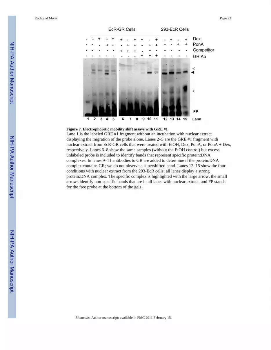
Figure 7. Electrophoretic mobility shift assays with GRE #1Lane 1 is the labeled GRE #1 fragment without an incubation with nuclear extractdisplaying the migration of the probe alone. Lanes 2–5 are the GRE #1 fragment withnuclear extract from EcR-GR cells that were treated with EtOH, Dex, PonA, or PonA + Dex,respectively. Lanes 6–8 show the same samples (without the EtOH control) but excessunlabeled probe is included to identify bands that represent specific protein:DNAcomplexes. In lanes 9–11 antibodies to GR are added to determine if the protein:DNAcomplex contains GR; we do not observe a supershifted band. Lanes 12–15 show the fourconditions with nuclear extract from the 293-EcR cells; all lanes display a strongprotein:DNA complex. The specific complex is highlighted with the large arrow, the smallarrows identify non-specific bands that are in all lanes with nuclear extract, and FP standsfor the free probe at the bottom of the gels.
Rock and Moos Page 22
Biometals. Author manuscript; available in PMC 2011 February 15.
NIH
-PA Author Manuscript
NIH
-PA Author Manuscript
NIH
-PA Author Manuscript
Copyright © 2022 FDOKUMEN