Apoptosis and Autophagy in Breast Cancer Cells following Exemestane Treatment
Upload
independentCategory
view
0download
0
Regulation of autophagy by the inositol trisphosphatereceptor
A Criollo1,2,3,4, MC Maiuri1,2,5, E Tasdemir1,2,3, I Vitale1,2,3, AA Fiebig6, D Andrews6, J Molgo7, J Dıaz4, S Lavandero4, F Harper8,
G Pierron8, D di Stefano9, R Rizzuto9, G Szabadkai9 and G Kroemer*,1,2,3
The reduction of intracellular 1,4,5-inositol trisphosphate (IP3) levels stimulates autophagy, whereas the enhancement of IP3
levels inhibits autophagy induced by nutrient depletion. Here, we show that knockdown of the IP3 receptor (IP3R) with smallinterfering RNAs and pharmacological IP3R blockade is a strong stimulus for the induction of autophagy. The IP3R is known toreside in the membranes of the endoplasmic reticulum (ER) as well as within ER–mitochondrial contact sites, and IP3R blockadetriggered the autophagy of both ER and mitochondria, as exactly observed in starvation-induced autophagy. ER stressors suchas tunicamycin and thapsigargin also induced autophagy of ER and, to less extent, of mitochondria. Autophagy triggered bystarvation or IP3R blockade was inhibited by Bcl-2 and Bcl-XL specifically targeted to ER but not Bcl-2 or Bcl-XL proteins targetedto mitochondria. In contrast, ER stress-induced autophagy was not inhibited by Bcl-2 and Bcl-XL. Autophagy promoted by IP3Rinhibition could not be attributed to a modulation of steady-state Ca2þ levels in the ER or in the cytosol, yet involved the obligatecontribution of Beclin-1, autophagy-related gene (Atg)5, Atg10, Atg12 and hVps34. Altogether, these results strongly suggestthat IP3R exerts a major role in the physiological control of autophagy.Cell Death and Differentiation (2007) 14, 1029–1039. doi:10.1038/sj.cdd.4402099; published online 26 January 2007
The first step of macroautophagy consists in the gradualenvelopment of cytoplasmic material (cytosol and/or orga-nelles) in the phagophore, a cistern that finally sequesterscytoplasmic material in autophagosomes (also called auto-phagic vacuoles (AVs)) lined by two membranes. Autophago-somes then undergo a progressive maturation by fusion withendosomes and/or lysosomes. This latter step createsautolysosomes in which the inner membrane as well as theluminal content of the AVs is degraded by lysosomalenzymes. The process of autophagy is controlled by a seriesof evolutionary conserved genes, the atg genes, whoseproducts are essential for specific steps of the autophagicprocess.1,2
One of the strongest triggers of autophagy is nutrientstress.3,4 In response to starvation, cells degrade nonessen-tial components thereby generating nutrients for meetingthe cell’s energetic demand as well as for vital biosyntheticreactions. In such circumstances, when autophagy is anadaptation, inhibition of autophagy has a negative impact oncell survival. For example, mice lacking the atg5 gene survivewithout any major developmental defect until birth, yetsuccumb to the stressful neonatal period unless puppies areforce-fed with milk within the first hours after birth.5 Similarly,
human cell lines cultured in nutrient-free media mount acytoprotective autophagic response. Suppression of auto-phagy by chemical inhibitors or knockdown of essential genesthus sensitize cells to starvation-induced cell death.6,7
Autophagy inhibition also sensitizes cells to the depletion ofobligatory growth (or survival) factors resulting in a decreaseof nutrient import through the plasma membrane. Forexample, the knockdown of Atg5, Atg10 or Atg12 (which areall involved in a ubiquitin-like conjugation system requiredfor autophagosome formation) prevents the generation of AVand sensitizes cells to cell death induced by withdrawal ofessential growth factors.8 Similarly, knockdown of Atg6/Beclin-1 (whose major physiological partner is the class IIIphosphatidylinositol 3- kinase Vps34) inhibits autophagy andsensitizes to cell death induced by starvation.6,8 Beclin-1 alsointeracts with Bcl-2,9 which inhibits autophagy,10 perhaps bypreventing the interaction between Beclin-1 and Vps34.11
One recent study suggested that myo-inositol-1,4,5-tris-phosphate (IP3) could regulate autophagy because inhibitionof inositol monophosphatase by lithium or L-690,330 stimu-lates autophagy through the depletion of inositol and IP3.12 IP3
is a second messenger produced primarily in response to thestimulation of G-protein-coupled receptor or receptor tyrosine
Received 11.10.06; revised 05.12.06; accepted 13.12.06; Edited by M Piacentini; published online 26.1.07
1INSERM, U848, Institut Gustave Roussy, PR1 39 rue Camille Desmoulins, Villejuif, France; 2Institut Gustave Roussy, 39 rue Camille Desmoulins, Villejuif, France;3Universite Paris Sud, Institut Gustave Roussy, 39 rue Camille Desmoulins, Villejuif, France; 4FONDAP Center CEMC, Faculty of Chemical and PharmaceuticalSciences, University of Chile, Santiago, Chile; 5Department of Experimental Pharmacology, Faculty of Biotechnological Sciences, Universita degli Studi di Napoli,Federico II, Italy; 6Department of Biochemistry and Biomedical Sciences, McMaster University Health Sciences Centre, CDN-Hamilton, Ontario, Canada; 7CentreNational de la Recherche Scientifique, Institut de Neurobiologie Alfred Fessard, FRC2118, UPR 9040, Gif-sur-Yvette, France; 8CNRS, FRE 2937, Institut Andre Lwoff,Villejuif, France and 9Department of Experimental and Diagnostic Medicine and Interdisciplinary Center for the Study of Inflammation and ER-GenTech, University ofFerrara, Ferrara, Italy*Corresponding author: G Kroemer, CNRS-UMR8125, Institut Gustave Roussy, PR1, 38 rue Camille Desmoulins, F-94805 Villejuif, France. Tel: þ 33 1 42116046;Fax: þ 33 1 42116047; E-mail: [email protected]: apoptosis; Bcl-2; autophagic vacuoles; endoplasmic reticulum; mitochondriaAbbreviations: Atg, autophagy-related gene; AV, autophagic vacuole; ER, endoplasmic reticulum; IP3, 1,4,5-inositol trisphosphate; IP3R, IP3 receptor; DCm,mitochondrial transmembrane potential; PI, propidium iodine
Cell Death and Differentiation (2007) 14, 1029–1039& 2007 Nature Publishing Group All rights reserved 1350-9047/07 $30.00
www.nature.com/cdd

kinases. IP3 acts on the IP3 receptor (IP3R), a mostlyendoplasmic reticulum (ER)-sessile Ca2þ release channelthat integrates signals from numerous small molecules andproteins including nucleotides, kinases and phosphatases, aswell as nonenzyme proteins.13 IP3R is known to play a majorrole within the Ca2þ microdomains that transmit Ca2þ spikesgenerated by the ER to mitochondria.14,15 IP3R is alsoregulated by Bcl-2 16,17 and Bcl-XL,18 which affect Ca2þ
fluxes through IP3R by direct molecular interactions, byinfluencing its regulatory phosphorylation and/or by modula-ting its response to IP3. Through these regulatory mecha-nisms, IP3R modulates diverse cellular functions, whichinclude, but are not limited to, contraction/excitation, geneexpression, cellular growth and apoptosis.13,19
On the basis of these premises, we decided to investigatethe contribution of IP3R to the regulation of autophagy. Here,we report that the depletion of IP3R or its inhibition triggersrapid autophagy affecting both ER and mitochondria. IP3Rfunctions as a Bcl-2-regulated repressor of autophagy.
Results and Discussion
Starvation-induced autophagy concerns both ER andmitochondria. Upon 12 h of culture in the absence ofserum and nutrients (starvation), HeLa cells manifest therelocalization of LC3-GFP into AVs and this effect is inhibitedby addition of the cell-permeable IP3 precursor myo-inositol(Figure 1a and b). Under these conditions, little cell death (asdetermined with the vital dye propidium iodine (PI)) occurredafter serum withdrawal and the reduction of the mitochondrialtransmembrane potential (DCm) induced by starvation wasinhibited by myo-inositol (Figure 1c). Starvation-inducedLC3-GFP aggregates partially colocalized with the ERmarker calreticulin and with the mitochondrial matrix markerHSP60, indicating that this regimen caused autophagy ofboth ER and mitochondria (Figure 1d–f).
ER stress but not mitochondrial stress induceautophagy. As starvation can induce an ER stressresponse20,21 as well as mitochondrial stress,22 weinvestigated whether direct induction of organelle-specificstress would stimulate autophagy. Thapsigargin (whichselectively inhibits the Ca2þ–ATPase responsible for Ca2þ
accumulation by the ER, SERCA), tunicamycin (which blocksN-linked protein glycosylation), brefeldin A (which inhibitsER–golgi transport) are well known for their capacity toinduce ER stress21,23 and all of them induce rapid LC3-GFPaggregation in AVs (Figure 2a and b), well before they causecell death and DCm dissipation (Figure 2c). In contrast,induction of mitochondrial stress by antimycin A (whichinhibits complex III of the respiratory chain), betulinic acid(which can cause direct permeabilization of mitochondrialmembranes)24 or cadmium (a heavy metal that inducesoxidative stress and permeability transition)25 did notstimulate autophagy under conditions in which all threeagents caused a significant DCm loss (Supplementary FigureS1). These data suggest that ER stress is a stronger inducerof autophagy than mitochondrial stress.
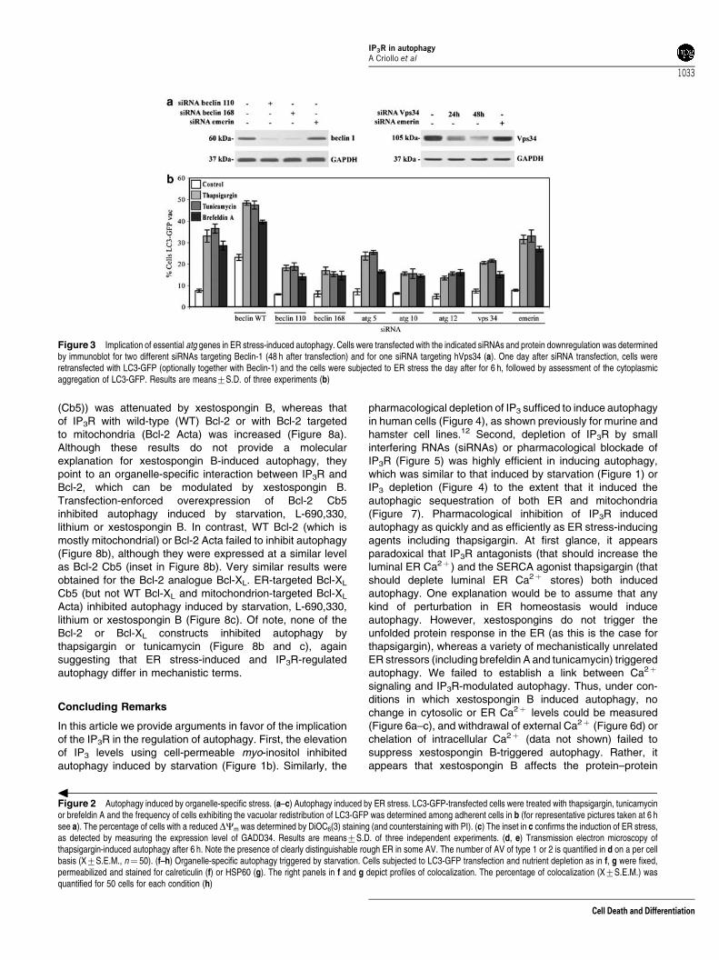
ER stress-induced autophagy affects ER andmitochondria. ER stress triggered the emergence of bonafide AVs, as defined by cytoplasmic structures lined by twomembranes that can be detected by transmission electronmicroscopy (Figure 2d and e). The LC3-GFP-positivevesicles induced by thapsigargin or tunicamycin colocalizedwith the ER marker calreticulin, but also colocalized in partwith the mitochondrial matrix marker HSP60, suggesting thatER stress causes autophagy of both ER and mitochondria(Figure 2f–h). ER stress-induced autophagy followed aclassical autophagic pathway to the extent that theknockdown of essential atg genes such as atg6/beclin-1,atg5, atg10, atg12 as well as the knockdown of themammalian class III phosphatidylinositol 3-kinase vps34inhibited autophagy induced by thapsigargin, tunicamycinor brefeldin A (Figure 3). Altogether, these data suggest thatER stress effectively trigger autophagy that follows acanonical pathway.
IP3 depletion and IP3R inhibition stimulates autophagyof both ER and mitochondria. The inositol mono-phosphatase inhibitors L-690,330 and lithium both inducedautophagy (Figure 4a and b) under conditions in whichneither agent induced toxic effects on the viability or bio-energetic state of cells (Figure 4c). Both L-690,330 andlithium triggered significant autophagy of both ER andmitochondria (Figure 4d–f). Very similar results wereobtained by inhibition of IP3R. Knockdown of the IP3R I andIII isoforms caused a strong, nonsynergistic induction ofautophagy, as measured by LC3-GFP relocalization (Figure5a and b). Similarly, xestospongin B, a chemical antagonistof IP3R,26 caused LC3-GFP aggregation in cytoplasmicvacuoles (Figure 5c). Stimulation of autophagy was morepronounced for xestospongin B than for xestospongin C,which has a lower affinity for IP3R27 (Figure 5d). Moreover,the xestospongin B effect was rapid (p2 h, Figure 5e) anddid not involve any major toxicity on mitochondria (Figure 5f)nor on the ER (as estimated by measuring the ER stressindicator GADD34; inset in Figure 5f). Xestospongin B-induced autophagy required atg6/beclin-1, atg5, atg10, atg12and vps34 (Figure 5g), indicating that it followed an orthodoxpathway. Under the conditions in which xestospongin B wasemployed in this study, the luminal ER Ca2þ and thebaseline cytosolic Ca2þ concentrations were not modulated(Figure 6a and b), although xestospongin clearly bluntedthe histamine-induced IP3/IP3R-dependent ER Ca2þ efflux,as an internal control of its efficacy (Figure 6c). Moreover,depletion of extracellular Ca2þ did not affect thexestospongin B-induced autophagy (Figure 6d), suggestingthat the xestospongin B effects were not mediated by grossperturbations of intracellular Ca2þ fluxes. Ultrastructuralanalysis of xestospongin B-elicited AV indicated that theyfrequently contained degenerating mitochondria or ER(Figure 7a). Accordingly, LC3-GFP aggregates elicited byxestospongin B colocalized with either calreticulin or HSP60(Figure 7b–d). Thus, IP3R blockade caused the same patternof autophagy as that induced by starvation.
ER-targeted Bcl-2 and Bcl-XL differentially inhibitautophagy. IP3R has been shown to interact physically
IP3R in autophagyA Criollo et al
1030
Cell Death and Differentiation

Figure 1 Autophagy induced by starvation. (a–c) LC3-GFP redistribution inhibited by myo-inositol. HeLa cells transiently transfected with LC3-GFP were subjected tonutrient depletion in the presence or absence of myo-inositol for 12 h and representative pictures of Hoechst 33342-counterstained cells were taken (a) and the percentage ofadherent cells exhibiting a clear LC3-GFP relocalization into cytoplasmic vacuoles was determined (b). The frequency of adherent and nonadherent cells with a low DCm wasdetermined by DiOC6(3) staining (and counterstaining with PI) (c). Results are means7S.D., of three independent experiments. (d–f). Organelle-specific autophagy triggeredby starvation. Cells subjected to LC3-GFP transfection and nutrient depletion as in a–c were fixed, permeabilized and stained for calreticulin (d) or HSP60 (e) to determine thecolocalization of LC3-GFP and ER (d) or mitochondrial (e). The right panels in d and e indicate the profiles of colocalization within the area of interest (labeled by arrows), asdetermined by quantitative confocal microscopy. (f) Mean level of colocalization of LC3-GFP with calreticulin or HSP60, as determined for 50 cells (X7S.E.M.)
IP3R in autophagyA Criollo et al
1031
Cell Death and Differentiation

with Bcl-2 within the ER.16,17 Moreover, the interaction of IP3
with the ANT interactome is strongly influenced by pro-apoptotic signaling.15 Xestospongin B strongly affected the
coimmunoprecipitation of Bcl-2 and IP3R, depending on thesubcellular localization of Bcl-2. The coimmunoprecipitationof IP3R with Bcl-2 targeted to the ER (Bcl-2 cytochrome b5
IP3R in autophagyA Criollo et al
1032
Cell Death and Differentiation
(Cb5)) was attenuated by xestospongin B, whereas thatof IP3R with wild-type (WT) Bcl-2 or with Bcl-2 targetedto mitochondria (Bcl-2 Acta) was increased (Figure 8a).Although these results do not provide a molecularexplanation for xestospongin B-induced autophagy, theypoint to an organelle-specific interaction between IP3R andBcl-2, which can be modulated by xestospongin B.Transfection-enforced overexpression of Bcl-2 Cb5inhibited autophagy induced by starvation, L-690,330,lithium or xestospongin B. In contrast, WT Bcl-2 (which ismostly mitochondrial) or Bcl-2 Acta failed to inhibit autophagy(Figure 8b), although they were expressed at a similar levelas Bcl-2 Cb5 (inset in Figure 8b). Very similar results wereobtained for the Bcl-2 analogue Bcl-XL. ER-targeted Bcl-XL
Cb5 (but not WT Bcl-XL and mitochondrion-targeted Bcl-XL
Acta) inhibited autophagy induced by starvation, L-690,330,lithium or xestospongin B (Figure 8c). Of note, none of theBcl-2 or Bcl-XL constructs inhibited autophagy bythapsigargin or tunicamycin (Figure 8b and c), againsuggesting that ER stress-induced and IP3R-regulatedautophagy differ in mechanistic terms.
Concluding Remarks
In this article we provide arguments in favor of the implicationof the IP3R in the regulation of autophagy. First, the elevationof IP3 levels using cell-permeable myo-inositol inhibitedautophagy induced by starvation (Figure 1b). Similarly, the
pharmacological depletion of IP3 sufficed to induce autophagyin human cells (Figure 4), as shown previously for murine andhamster cell lines.12 Second, depletion of IP3R by smallinterfering RNAs (siRNAs) or pharmacological blockade ofIP3R (Figure 5) was highly efficient in inducing autophagy,which was similar to that induced by starvation (Figure 1) orIP3 depletion (Figure 4) to the extent that it induced theautophagic sequestration of both ER and mitochondria(Figure 7). Pharmacological inhibition of IP3R inducedautophagy as quickly and as efficiently as ER stress-inducingagents including thapsigargin. At first glance, it appearsparadoxical that IP3R antagonists (that should increase theluminal ER Ca2þ ) and the SERCA agonist thapsigargin (thatshould deplete luminal ER Ca2þ stores) both inducedautophagy. One explanation would be to assume that anykind of perturbation in ER homeostasis would induceautophagy. However, xestospongins do not trigger theunfolded protein response in the ER (as this is the case forthapsigargin), whereas a variety of mechanistically unrelatedER stressors (including brefeldin A and tunicamycin) triggeredautophagy. We failed to establish a link between Ca2þ
signaling and IP3R-modulated autophagy. Thus, under con-ditions in which xestospongin B induced autophagy, nochange in cytosolic or ER Ca2þ levels could be measured(Figure 6a–c), and withdrawal of external Ca2þ (Figure 6d) orchelation of intracellular Ca2þ (data not shown) failed tosuppress xestospongin B-triggered autophagy. Rather, itappears that xestospongin B affects the protein–protein
Figure 3 Implication of essential atg genes in ER stress-induced autophagy. Cells were transfected with the indicated siRNAs and protein downregulation was determinedby immunoblot for two different siRNAs targeting Beclin-1 (48 h after transfection) and for one siRNA targeting hVps34 (a). One day after siRNA transfection, cells wereretransfected with LC3-GFP (optionally together with Beclin-1) and the cells were subjected to ER stress the day after for 6 h, followed by assessment of the cytoplasmicaggregation of LC3-GFP. Results are means7S.D. of three experiments (b)
Figure 2 Autophagy induced by organelle-specific stress. (a–c) Autophagy induced by ER stress. LC3-GFP-transfected cells were treated with thapsigargin, tunicamycinor brefeldin A and the frequency of cells exhibiting the vacuolar redistribution of LC3-GFP was determined among adherent cells in b (for representative pictures taken at 6 hsee a). The percentage of cells with a reduced DCm was determined by DiOC6(3) staining (and counterstaining with PI). (c) The inset in c confirms the induction of ER stress,as detected by measuring the expression level of GADD34. Results are means7S.D. of three independent experiments. (d, e) Transmission electron microscopy ofthapsigargin-induced autophagy after 6 h. Note the presence of clearly distinguishable rough ER in some AV. The number of AV of type 1 or 2 is quantified in d on a per cellbasis (X7S.E.M., n¼ 50). (f–h) Organelle-specific autophagy triggered by starvation. Cells subjected to LC3-GFP transfection and nutrient depletion as in f, g were fixed,permeabilized and stained for calreticulin (f) or HSP60 (g). The right panels in f and g depict profiles of colocalization. The percentage of colocalization (X7S.E.M.) wasquantified for 50 cells for each condition (h)
IP3R in autophagyA Criollo et al
1033
Cell Death and Differentiation

interaction in which IP3R engages, including the interactionwith Bcl-2 in the ER (Figure 8a). Specifically ER-targetedBcl-2 (Figure 8b) or Bcl-XL (Figure 8c) strongly inhibited theinduction of autophagy by starvations, several agents thatreduce IP3 levels and the IP3R antagonist xestospongin B.
Under the same experimental setting, however, ER-targetedBcl-2 (Figure 8b) or Bcl-XL (Figure 8c) failed to reduceautophagy induced by ER stressors including thapsi-gargin. These data strongly support the notion that in-duction of autophagy by inhibition of the IP3/IP3R signaling
Figure 4 Induction of autophagy by inositol monophosphate inhibitors. Cells were treated with L-690,330, lithium and/or myo-inositol for 12 h, followed by determination ofthe cytoplasmic redistribution of LC3-GFP (a, b) or the quantitation of the DCm (c). Results are means7S.D. of three experiments. In addition, the colocalizaton of LC3-GFPand calreticulin (d, f) or HSP60 (e, f) was determined (n¼ 50 in f)
IP3R in autophagyA Criollo et al
1034
Cell Death and Differentiation

cascade is mechanistically different from autophagy inducedby ER stress. It remains a conundrum through whichmolecular mechanisms ER stress can activate the autophagicresponse.
In conclusion, our results strongly support a signalingpathway in which IP3 and IP3R act to suppress autophagy.Thus, interruption of this pathway by depletion/inhibition of IP3
or IP3R can unleash the autophagic response. These findings
Figure 5 Autophagy triggered by IP3R knockdown. HeLa cells were transfected with siRNA specific for distinct IP3R isoforms. The protein expression level was controlled48 h after transfection (a). In addition, the impact on LC3-GFP relocalization (X7S.D., n¼ 3) was determined at different times posttransfection (b). (c–e) LC3-GFPaggregation induced by IP3R inhibitors. LC3-GFP-expressing cells were treated with the indicated doses of xestospongin B or C and the degree of cells with vacuolarredistribution of LC3-GFP was measured (standard dose for xestospongin B is 2 mM in c and e; the treatment is 6 h in c and d). (f) Effect of IP3R blockade on the DCm, asdetermined by cytofluorometry and on GADD34 expression as determined by immunoblot (insert). (g) Implication of essential atg genes in autophagy induced by IP3Rinhibition. Cells were transfected with the indicated siRNAs (day 0), then with LC3-GFP (optionally together with Beclin-1, on day 1), treated with xestospongin B (day 2) for 6 hand scored for redistribution of LC3-GFP
IP3R in autophagyA Criollo et al
1035
Cell Death and Differentiation

underscore the cardinal importance of IP3R in the integrationof cell fate-determining signals and their conversion intobiological responses including cell growth, motility, differen-tiation, apoptosis and, as shown here, autophagy.
Materials and MethodsCell lines, culture conditions and treatment. Rat-1 fibroblaststransfected with an empty control vector (CMV) or with a plasmid-encoding Bcl-2,either in WT configuration or fused to peptides that allow the targeting tomitochondria (Acta) or to ER (Cb5) were described previously.28 Rat-1 fibroblasts aswell as HeLa cells were cultured in Dulbecco’s modified Eagle’s medium containing10% fetal calf serum, 10 mM N-2-hydroxyethylpiperazine-N0-2-ethanesulfonic(HEPES) buffer, 100 units/ml penicillin G sodium and 100mg/ml streptomycinsulfate at 371C under 5% CO2. All media and supplements for cell culture werepurchased from Gibco-Invitrogen (Carlsbad, CA, USA). For serum and amino-acidstarvation, cells were cultured in serum-free Earle’s Balanced Salt Solution medium(Sigma).6 Cells (30–50� 103)were seeded in 12- or 24-well plates and grown for24 h before treatment with antimycin A (from 0.06 to 0.5mM; Sigma-Aldrich, StLouis, MO, USA), betulinic acid (from 1.25 to 10 mM; Sigma-Aldrich), brefeldin A(20mM; Sigma-Aldrich), cadmium (from 2.5 to 20mM; Sigma-Aldrich), L-690330(100mM; Tocris), lithium chloride (10 mM; Sigma-Aldrich), myo-inositol(Bt3(1,3,5)P3/AM; 10mM; Calbiochem), thapsigargin (3mM; Calbiochem),tunicamycin (2.5mM; Sigma-Aldrich), xestospongin B (from 0.08 to 10 mM;extracted from the marine sponge Xestospongia exigua),26 or X. C (from 0.08 to10mM; Calbiochem) for 6–24 h.
Plasmids, transfection and RNA interference. Cells were cultured insix-well plates and transfected at 80% confluence with Oligofectamine reagent(Invitrogen), in the presence of 100 nM of siRNAs specific for human Beclin-1 andother atg genes,6,7 IP3-RI and IP3-RIII (Oakes SA et al., PNAS 2005), hVps34,29,30
a siRNA targeting the unrelated protein emerin.31 siRNA effects were controlled byimmunoblots with suitable antibodies specific for Beclin-1 (Santa Cruz) or Vps34(Zymed). Transient transfections with plasmids were performed with Lipofectamine2000 reagent (Invitrogen) and cells were used 24 h after transfection unlessspecified differently. Cells were transfected with empty vector alone or together withLC3-GFP,7 in the presence or absence of WT Beclin-1, WT Bcl-2, ER-targeted Bcl-2(Bcl-2 Cb5), mitochondrion-targeted Bcl-2 (Bcl-2 Acta),28 WT Bcl-XL, ER-targetedBcl-XL (Bcl-XL Cb5) or mitochondrion-targeted Bcl-XL (Bcl-XL acta). The cDNAencoding amino acids 1–210 of Bcl-XL were fused either to the sequence encodingthe C-terminus of Acta or the Cb5 hydrophobic tail for mitochondrial or ER targeting,respectively. Correct subcellular localization of the mutants expressed in cells wasverified by immunofluorescence microscopy as described previously,28 using rabbitanti-Bcl-XL anti-sera followed by either a monoclonal antibody to the ER proteincalreticulin or to an inner mitochondrial membrane protein (2G2, ExAlphaBiologicals).
Flow cytometry. The following fluorochromes were employed bycytofluorometry: 3,30-dihexyloxacarbocyanine iodide (DiOC6(3), 40 nM) forquantification or the DCm (Molecular Probes) and PI (1mg/ml, Sigma-Aldrich) fordetermination of cell viability.6 Cells were trypsinized and labeled with thefluorochromes at 371C, followed by cytofluorometric analysis with a fluorescence-activated cell sorter scan (Becton Dickinson, San Jose, CA, USA). Data werestatistically evaluated using Cell Quest software (Becton Dickinson).
Light microscopy and immunofluorescence. Cells were fixed withparaformaldehyde (4%, w/v) for LC3-GFP and immunofluorescence assays.6 Cellswere stained for the detection of HSP60 (mAb from Sigma-Aldrich) or calreticulin32
(pAb from Stressgen) revealed by goat anti-mouse or anti-rabbit immunoglobulinAlexa fluor conjugates (Molecular Probes). Nuclei were labeled with 10 mg/mlHoechst 33342 (Molecular Probes-Invitrogen). Fluorescence microscopy wasanalyzed with a Leica IRE2 equipped with a DC300F camera. Confocal microscopywas performed with a LSM 510 Zeiss microscope equipped with a � 63 objective.To determine the percentage of colocalization, images were loaded into Image Jsoftware.
Electron microscopy. Cells were fixed for 1 h at 41C in 1.6% glutaraldehydein 0.1 M Sorensen phosphate buffer (pH 7.3), washed, fixed again in aqueous2% osmium tetroxide and finally embedded in Epon. Electron microscopy wasperformed with a Zeiss EM 902 transmission electron microscope, at 90 kV, onultrathin sections (80 nm), stained with lead citrate and uranyl acetate.
Dynamic in vivo measurements with targeted aequorin probesand fura-2. cytAEQ, erAEQmut-expressing cells, were reconstituted withcoelenterazine, transferred to the perfusion chamber, light signal was collected in
Figure 6 Ca2þ independence of xestospongin-induced autophagy. (a) XeBinhibits IP3-induced Ca2þ release but does not change steady [Ca2þ ]er, ER Ca2þ
leak and basal [Ca2þ ]c [Ca2þ ]er was measured in erAEQmut-transfected HeLacells ([Ca2þ ]er was 475725mM in controls, 481732 in XeB-treated cells; n¼ 7).XeB (1 mM) was added to the cells 2 h before the experiment. Cells were kept inKRB–EGTA (600mM) solution, then ER was Ca2þ loaded by the addition of 1 mM
CaCl2 (in KRB) to the cells. ER Ca2þ leak was estimated by the addition of theSERCA blocker tert-butylhydroquinone (tBHQ). (b) Basal [Ca2þ ]c was measured incells loaded with fura-2 as described in the Materials and Methods section (controls:46.274.5 nM; after XeB treatment: 42.275.5 nM, n¼ 35). After the control period,tBHQ (in KRB 1 mM CaCl2) was added to the cells in order to estimate Ca2þ leakinto the cytosol and the activity of Ca2þ extrusion through the plasma membrane.(c) Histamine-induced Ca2þ release was measured in cytAEQ-transfected cells(lower right panel). As an internal control of the efficacy of xestospongin B as a IP3Rantagonist, histamine-induced (IP3/IP3R-dependent) Ca2þ variations in the ERwere measured in the absence or presence of 2mM xestospongin. The peak[Ca2þ ]c response was 3.370.6mM in controls, 0.8770.4mM after XeB treatment(n¼ 9). Note that xestospongin did not affect the baseline ER Ca2þ concentration.(d) Effect of Ca2þ depletion on xestospongin-induced autophagy. The concentra-tion of extracellular Ca2þ was modulated before addition of xestospongin B for 6 hand determination of LC3-GFP aggregation. Results are means7S.D. of threeexperiments
IP3R in autophagyA Criollo et al
1036
Cell Death and Differentiation

a purpose-built luminometer and calibrated into [Ca2þ ] values as describedpreviously.33 All aequorin measurements were carried out in KRB containing 1 mM
CaCl2 (KRB/Ca2þ , Krebs-Ringer modified buffer: 135 mM NaCl, 5 mM KCl, 1 mM
MgSO4, 0.4 mM K2HPO4, 1 mM CaCl2, 5.5 mM glucose, 20 mM HEPES, pH 7.4). For[Ca2þ ]er measurements, erAEQmut-transfected cells were reconstituted withcoelenterazine n, following ER Ca2þ depletion in a solution containing 0 [Ca2þ ],
Figure 7 Organelle-specific autophagy after IP3R inhibition. (a) Transmission electron microscopy of xestospongin B-induced autophagy after 6 h (I). Note the presence ofmitochondria and rough ER in some AV (II). The number of type 1 or type 2 AV is quantified in B (X7S.E.M., n¼ 50) (III). (b–d) Organelle-specific autophagy triggered byIP3R inhibition. LC3-GFP-expressing cells exposed to xestospongin B were stained for calreticulin (b) or HSP60 (c), both labeled in red. The degree of colocalization(X7S.E.M.) was measured for 50 cells (d)
IP3R in autophagyA Criollo et al
1037
Cell Death and Differentiation

600mM ethylene glycol bis(b-aminoethylether)-N,N,N’,N’,-tetraacetic acid (EGTA),1mM ionomycin, as described.34 Kinetic imaging of [Ca2þ ]c transients in fura-2-loaded cells was performed as described.34
Immunoblots and immunoprecipitation. HeLa cells (1� 106 cells)were washed with cold phosphate-buffered saline (PBS) at 41C and lysedas described previously.7 Forty micrograms of protein were loaded on a 5–12%sodium dodecyl sulfate–polyacrylamide gel electrophoresis and transferred topolyvinylidene fluoride membrane (Millipore). The membrane was incubated for 1 h
in PBS–Tween 20 (0.05%) containing 5% bovine serum albumin. Primary antibodyAnti-Beclin-1 (Santacruz), anti-GADD34 (Abcam), anti-IP3-RI (Calbiochem), anti-IP3-RIII (BD Biosciences) and anti-hVps34 (Zymed) were incubated overnight at41C and revealed with appropriate horseradish peroxidase-labeled secondaryantibodies (SouthernBiotech, Birmingham, AL, USA) and the SuperSignal WestPico chemoluminiscent substrate (Pierce). Anti-glyceraldehyde-3-phosphatedehydrogenase (Chemicon) was used to control equal loading.Rat-1 fibroblasts (8� 106 cells) were collected, lysed, and immunoprecipitationwas performed as described16 using anti-Bcl-2 (SantaCruz) antibody and
Figure 8 Impact of Bcl-2 and Bcl-XL on autophagy induced by IP3R inhibition. (a) Relocalization of Bcl-2 after IP3R association. Cells expressing WT Bcl-2, mitochondrion-targeted Bcl-2 (Acta) or ER-targeted Bcl-2 (Cb5) were treated with xestospongin B, followed by immunoprecipation of Bcl-2 and immunochemical detection of IP3R-III or Bcl-2.(b, c) Impact of WT, mitochondrion-targeted (Acta) or ER-targeted (Cb5) Bcl-2 (b) or Bcl-XL (c) on autophagy induced by IP3R inhibition, ER stress or inositolmonophosphatase inhibition. Cells were transfected with LC3-GFP together with the indicated constructs, and 24 h later the cells were exposed to different stimuli for 12 h.Then, LC3-GFP redistribution (X7S.D., n¼ 3) was determined by fluorescence microscopy. Asterisks indicate significant (Po0.01) inhibitory effects of ER-targeted Bcl-2and Bcl-XL. The inserts in b and c confirm the expression of WT, mitochondrion-targeted (Acta) or ER-targeted (Cb5) Bcl-2 and Bcl-XL, respectively
IP3R in autophagyA Criollo et al
1038
Cell Death and Differentiation

Protein G Sepharose (GE Healthcare) followed by western blotting with anti-IP3-RIIIantibody.
Acknowledgements. We thank Dr Abdelali Jalil for confocal microscopy andthe members of our laboratory for continuous discussion. We also thank theInternational Collaboration Program ECOS-CONICYT, grant C04B03 (to GK andSL). A Criollo is a recipient of a PhD fellowship from CONICYT, Chile. GK issupported by a special grant of the Ligue Nationale contre le Cancer (equipelabelisee), European Commission (RIGHT), Canceropole Ile-de-France and InstitutNational contre le Cancer (INCa).
1. Shintani T, Klionsky DJ. Autophagy in health and disease: a double-edged sword. Science2004; 306: 990–995.
2. Yorimitsu T, Klionsky DJ. Autophagy: molecular machinery for self-eating. Cell Death Differ2005; 12 (Suppl 2): 1542–1552.
3. Levine B, Yuan J. Autophagy in cell death: an innocent convict? J Clin Invest 2005; 115:2679–2688.
4. Lum JJ, DeBerardinis RJ, Thompson CB. Autophagy in metazoans: cell survival in the landof plenty. Nat Rev Mol Cell Biol 2005; 6: 439–448.
5. Mizushima N, Yamamoto A, Matsui M, Yoshimori T, Ohsumi Y. In vivo analysis ofautophagy in response to nutrient starvation using transgenic mice expressing afluorescent autophagosome marker. Mol Biol Cell 2004; 15: 1101–1111.
6. Boya P, Gonzalez-Polo R-A, Casares N, Perfettini J-L, Dessen P, Larochette N et al.Inhibition of macroautophagy triggers apoptosis. Mol Cell Biol 2005; 25: 1025–1040.
7. Gonzalez-Polo R-A, Boya P, Paulau A-L, Jalil A-A, Larochette N, Souquere S et al. Theapoptosis/autophagy paradox. Accumulation of autophagic vacuoles triggers apoptosis.J Cell Sci 2005; 118: 3091–3102.
8. Lum JJ, Bauer DE, Kong M, Harris HM, Li C, Lindsten T et al. Growth factor regulation ofautophagy and cell survival in the absence of apoptosis. Cell 2005; 120: 237–248.
9. Liang XH, Jackson S, Seaman M, Brown K, Kempkes B, Hibshoosh H et al. Induction ofautophagy and inhibition of tumorigenesis by beclin 1. Nature 1999; 402: 672–676.
10. Pattingre S, Tassa A, Qu X, Garuti R, Liang XH, Mizushima N et al. Bcl-2 antiapoptoticproteins inhibit Beclin 1-dependent autophagy. Cell 2005; 122: 927–939.
11. Takacs-Vellai K, Vellai T, Puoti A, Passannante M, Wicky C, Streit A et al. Inactivation ofthe autophagy gene bec-1 triggers apoptotic cell death in C. elegans. Curr Biol 2005; 15:1513–1517.
12. Sarkar S, Floto RA, Berger Z, Imarisio S, Cordenier A, Pasco M et al. Lithium inducesautophagy by inhibiting inositol monophosphatase. J Cell Biol 2005; 170: 1101–1111.
13. Patterson RL, Boehning D, Snyder SH. Inositol 1,4,5-trisphosphate receptors as signalintegrators. Annu Rev Biochem 2004; 73: 437–465.
14. Bianchi K, Rimessi A, Prandini A, Szabadkai G, Rizzuto R. Calcium and mitochondria:mechanisms and functions of a troubled relationship. Biochim Biophys Acta 2004; 1742:119–131.
15. Verrier F, Deniaud A, Lebras M, Metivier D, Kroemer G, Mignotte B et al. Dynamicevolution of the adenine nucleotide translocase interactome during chemotherapy-inducedapoptosis. Oncogene 2004; 23: 8049–8064.
16. Chen R, Valencia I, Zhong F, McColl KS, Roderick HL, Bootman MD et al. Bcl-2functionally interacts with inositol 1,4,5-trisphosphate receptors to regulate calcium releasefrom the ER in response to inositol 1,4,5-trisphosphate. J Cell Biol 2004; 166: 193–203.
17. Oakes SA, Scorrano L, Opferman JT, Bassik MC, Nishino M, Pozzan T et al. ProapoptoticBAX and BAK regulate the type 1 inositol trisphosphate receptor and calcium leak from theendoplasmic reticulum. Proc Natl Acad Sci USA 2005; 102: 105–110.
18. White C, Li C, Yang J, Petrenko NB, Madesh M, Thompson CB et al. The endoplasmicreticulum gateway to apoptosis by Bcl-X(L) modulation of the InsP3R. Nat Cell Biol 2005; 7:1021–1028.
19. Boehning D, van Rossum DB, Patterson RL, Snyder SH. A peptide inhibitor of cytochromec/inositol 1,4,5-trisphosphate receptor binding blocks intrinsic and extrinsic cell deathpathways. Proc Natl Acad Sci USA 2005; 102: 1466–1471.
20. Harding HP, Zhang Y, Zeng H, Novoa I, Lu PD, Calfon M et al. An integrated stressresponse regulates amino acid metabolism and resistance to oxidative stress. Mol Cell2003; 11: 619–633.
21. Schroder M, Kaufman RJ. The mammalian unfolded protein response. Annu Rev Biochem2005; 74: 739–789.
22. Green DR, Kroemer G. The pathophysiology of mitochondrial cell death. Science 2004;305: 626–629.
23. Ferri KF, Kroemer GK. Organelle-specific initiation of cell death pathways. Nat Cell Biol2001; 3: E255–E263.
24. Fulda S, Scaffidi C, Susin SA, Krammer PH, Kroemer G, Peter ME et al. Activation ofmitochondria and release of mitochondrial apoptogenic factors by betulinic acid. J BiolChem 1998; 273: 33942–33948.
25. Zoratti M, Szabo I. The mitochondrial permeability transition. Biochem Biophys Acta – RevBiomembr 1995; 1241: 139–176.
26. Jaimovich E, Mattei C, Liberona JL, Cardenas C, Estrada M, Barbier J et al. XestosponginB, a competitive inhibitor of IP3-mediated Ca2+ signalling in cultured rat myotubes, isolatedmyonuclei, and neuroblastoma (NG108-15) cells. FEBS Lett 2005; 579: 2051–2057.
27. Gafni J, Munsch JA, Lam TH, Catlin MC, Costa LG, Molinski TF et al. Xestospongins:potent membrane permeable blockers of the inositol 1,4,5-trisphosphate receptor. Neuron1997; 19: 723–733.
28. Zhu W, Cowie A, Wasfy GW, Penn LZ, Leber B, Andrews DW. Bcl-2 mutants with restrictedsubcellular location reveal spatially distinct pathways for apoptosis in different cell types.EMBO J 1996; 15: 4130–4141.
29. Nobukuni T, Joaquin M, Roccio M, Dann SG, Kim SY, Gulati P et al. Amino acids mediatemTOR/raptor signaling through activation of class 3 phosphatidylinositol 3OH-kinase. ProcNatl Acad Sci USA 2005; 102: 14238–14243.
30. Byfield MP, Murray JT, Backer JM. hVps34 is a nutrient-regulated lipid kinase required foractivation of p70 S6 kinase. J Biol Chem 2005; 280: 33076–33082.
31. Harborth J, Elbashir SM, Bechert K, Tuschl T, Weber K. Identification of essential genes incultured mammalian cells using small interfering RNAs. J Cell Sci 2001; 114: 4557–4565.
32. Obeid M, Tesniere A, Ghiringhelli F, Firmia GM, Apetoh L, Perfettini JL et al. Calreticulinexposure dictates the immunogenicity of cancer cell death. Nat Med 2007; 13: 54–61.
33. Chiesa A, Rapizzi E, Tosello V, Pinton P, de Virgilio M, Fogarty KE, et al. Recombinantaequorin and green fluorescent protein as valuable tools in the study of cell signalling.Biochem J 2001; 355: 1–12.
34. Szabadkai G, Simoni AM, Chami M, Wieckowski MR, Youle RJ, Rizzuto R. Drp-1-dependent division of the mitochondrial network blocks intraorganellar Ca2+ waves andprotects against Ca2+-mediated apoptosis. Mol Cell 2004; 8: 59–68.
Supplementary Information accompanies the paper on Cell Death and Differentiation website (http://www.nature.com/cdd)
IP3R in autophagyA Criollo et al
1039
Cell Death and Differentiation
Copyright © 2022 FDOKUMEN