Tachycardia-induced silencing of subcellular Ca2+ signaling in atrial myocytes

Inositol 1,4,5-Trisphosphate Receptor in Heart: Evidence for its Concentration in Purkinje Myocytes of the Conduction System Luisa Gorza, Stefano Schiaftino, and Pompeo Volpe
Centro di Studio per la Biologia e la Fisiopatologia Muscolare del Consiglio Nazionale delle Ricerche, Dipartimento di Scienze Biomediche Sperimentali dell'Universit~ di Padova, 35121 Padova, Italy
Abstract. Inositol 1,4,5-trisphosphate (IP3) is one of the second messengers capable of releasing Ca 2÷ from sarcoplasmic reticulum/ER subcompartments. The mRNA encoding the intracellular IP3 receptor (Ca 2÷ channel) has been detected in low amounts in the heart of various species by Northern blot analysis. The myocardium, however, is a heterogeneous tissue composed of working myocytes and conduction system cells, i.e., myocytes specialized for the beat generation and stimulus propagation. In the present study, the cellular distribution of the heart IP3 receptor has been investigated. [3H]IP3 binding experiments, Western blot analysis and immunofluorescence, with anti-peptide antibodies specific for the IP3 receptor, indicated that the majority of Purkinje myocytes (the ventricular con- duction system) express much higher IP3 receptor lev- els than atrial and ventricular myocardium. Heteroge- neous distribution of IP3 receptor immunoreactivity
was detected both at the cellular and subceUular lev- els. In situ hybridization to a riboprobe generated from the brain type 1 IP3 receptor cDNA, showed in- creased accumulation of IP3 receptor mRNA in the heart conduction system. Evidence for IP3-sensitive Ca 2÷ stores in Purkinje myocytes was obtained by dou- ble immunolabeling experiments for IP3 receptor and cardiac calsequestrin, the sarcoplasmic reticulum in- tralumenal calcium binding protein. The present findings provide a molecular basis for the hypothesis that Ca 2+ release from IP3-sensitive Ca 2+ stores evoked by o~t-adrenergic stimulation is responsible for the in- crease in automaticity of Purkinje myocytes (del Balzo, U., M. R. Rosen, G. Malfatto, L. M. Kaplan, and S. E Steinberg. 1990. Circ. Res. 67:1535-1551), and open new perspectives in the hormonal modula- tion of chronotropism, and generation of arrhythmias.
NE of the mechanisms of cell activation relies on re- ceptor-stimulated hydrolysis of phosphatidyl inosi- tol 4,5-bisphosphate (PtInsP2') via G protein-depen-
dent phospholipase C (Berridge and Irvine, 1989). Hydro- lysis of PtInsP2 yields two intracellular second messengers, diacylglycerol and IP3.
In cardiac muscle, several plasma membrane receptors (c¢~-adrenergic, muscarinic, endothelin, angiotensin III) are coupled to PtInsP2 turnover (Eckel et al., 1991; Renard and Poggioli, 1987; Yorikane et al., 1990; Kern et al., 1991; Brown et al., 1985; Kentish et al., 1990; Vites and Pappano, 1990), and different metabolic pathways lead to IP3 produc- tion (Brown et al., 1985; Renard and Poggioli, 1987). The mRNA encoding the intracellular IP3 receptor (Ca 2+ chan- nel) has been detected in low amounts in the heart of various
Address correspondence to: Dr. Pompeo Volpe, Dipartimento di Scienze Bimediche Sperimentali, via Trieste 75, 35121 Padova, Italy.
1. Abbreviations used in this paper: CS, calsequestrin; IP3, inositol 1,4,5- trisphosphate; PtlnsP2, phosphatidyl inositol 4,5-bisphosphate; SR, sar- coplasmic retieulum.
species by Northern blot analysis (Furuichi et al., 1990; Mignery et al., 1990; Nakagawa et al., 1991). IP3-induced Ca 2÷ release from the sarcoplasmic reticulum (SR) of both atrial and ventricular myocytes (Fabiato, 1986; Kentish et al., 1990; Eckel et al., 1991; Vites and Pappano, 1991) has been demonstrated, and a positive inotropic effect has been postulated (Eckel et al., 1991; Vites and Pappano, 1991). Moreover, Borgatta et al. (1991) have recently shown the oc- currence of a low-conducting Ca 2+ release channel sensitive to 1I>3 in canine heart SR vesicles. IP3-gated Ca 2+ channels were almost exclusively recorded from SR vesicles isolated from the ventricle septum (Borgatta et al., 1991), where cells of the conduction system are numerous (Sommer and Jen- nings, 1986).
The myocardium is a heterogeneous tissue composed of working (atrial and ventricular) myocytes and conduction system myocytes. The heart conduction system is composed of subpopulations of myocytes specialized for the beat gener- ation and stimulus propagation, that display precise anatomi- cal localization (Sommer and Jennings, 1986). Sino-atrial node myocytes represent the pacemaker, atrio-ventricular nodal myocytes control the ventricular excitation and coor- dinate it through the atrio-ventricular bundle, the right and
© The Rockefeller University Press, 0021-9525/93/04/345/9 $2.00 The Journal of Cell Biology, Volume 121, Number 2, April 1993 345-353 345
on August 29, 2014
jcb.rupress.orgD
ownloaded from
Published April 15, 1993

left bundle branches and the peripheral network of ventricu- lar conduction system myocytes, i.e., the Purkinje myocytes.
In this paper, we have investigated the cellular distribution of the IP3 receptor in cardiac muscle by biochemical, im- munohistochemical and in situ hybridization methods. We found that the majority of Purkinje myocytes express much higher IP3 receptor levels than atrial and ventricular myo- cytes, and other portions of the conduction system. Hetero- geneous distribution of IP3 receptor immunoreactivity was detected both at the cellular and subcellular levels. The bio- logical relevance of these observations is discussed in rela- tion to phenomena such as automaticity, chronotropism and arrhythmias.
Materials and Methods
Materials
[3H]IP3 and mouse anti-desmin mAbs were obtained from Amersham In- ternational (Buckinghamshire, UK), alkaline-phosphatase-conjugated anti- rabbit IgGs were obtained from Sigma Chemical Co. (St. Louis, MO) goat rhodamine-labeled anti-mouse IgGs from Cappel Laboratories (Malvern, PA) and swine fluorescein-labeled anti-rabbit IgGs from Dako Corporation (Carpinteria, CA).
Tissue Sources
Adult rats were killed by decapitation and their cerebella and hearts were quickly removed and stored at -80"C until used. Bovine and horse hearts were obtained from a local slaughterhouse. Samples of different cardiac regions corresponding to both working myocardium and conduction system were dissected out. Analogous samples were excised from mongrel dog and chicken hearts. Whole hearts were dissected out from bovine fetuses col- lected at 3, 4, and 5 mo of gestational age. All samples were immediately frozen in liquid nitrogen and stored at -80°C until used.
For immunohistochemical studies performed with anti caisequestfin (CS) antibodies, tissues were fixed with 4% paraformaldehyde in PBS for 2 h at 4°C, cryoprotected with 18% sucrose and frozen in liquid nitrogen.
cRNA Probes and Abs
A rat IP3 cDNA (clone pI2a; Mignery et al., 1990) containing a 2-kb EcoRI fragment of the 3' coding and noncoding regions of the type 1 IP3 receptor mRNA, was used for in situ hybridization analysis.
The 19-mer COOH-terminal peptide of the mouse/rat type 1 IP3 recep- tor sequence (cf. Furuichi et al., 1989; Mignery et al., 1990) was synthe- sized, conjugated to keyhole limpet hemocyanine (protein carder) by Multi- ple Peptide Systems (San Diego, CA), and used as immunogen in rabbits. Antibody specificity for the IPa receptor was ascertained by Western blot of cerebellum microsomal fractions (see Fig. 1 a).
The mouse polyclonal antibody specific for dog cardiac CS were ob- tained as described (Biral et al., 1992). The specificity of the antibody was assessed by Western blot (Biral et al., 1992).
Mouse mAbs specific for either the myosin heavy chain subunit (BA-D5) or the t~ myosin heavy chain (BA-G5) were obtained and characterized as described in Schiafiino et ai. (1989) and Rudnicki et al. (1990), respectively.
Isolation of Microsomal Fractions and Biochemical Assays Microsomes from rat cerebellum and different areas of bovine heart (atrium, ventdcular wall, septum and false tendon) were prepared by differential cen- trifugation as described by Volpe et ai. (1991). Protein concentration was determined by the method of Lowry et al. (1951) using bovine serum albu- min as a standard.
Microsomal fractions were analyzed by SDS PAGE (5-10% linear gra- dient), transferred to nitrocellulose and reacted with antibodies as described (Volpe et al., 1991).
[3H]IP3 binding was carded out essentially as described (Alderson and Volpe, 1989) in a medium containing 0.1 M KCI, 50 mM Tris-Cl, pH 8.3, 1 mM EDTA, 150 #g of microsomal protein, 50 nM [3H]IP3, in the ab-
sence and presence of 5 #M cold IP3 for total and nonspeciflc binding, respectively. Filtration and rinsings were as described (Volpe et al., 1991).
Immunocytochemistry
Indirect immunofluorescence was performed as previously described (Sar- tore et al., 1978; Gorza et al., 1986). Paraformaldehyde-fixed cryosections were quenched before incubation with antibodies in 0.2 M Tris-glycine, pH 7.4, for 30 min at room temperature. 5- to 10-t~m cryosections were incu- bated with adequate dilutions of anti-IP3 receptor Abs for 20 min at 37"C. After several washes with PBS, sections were incubated with fluorescein- conjugated anti-rabbit IgGs, rinsed again in PBS, mounted with glycerol and observed with a Zeiss Axioplan microscope equipped with epifluores- cence optics.
For double-labeling immunofluorescence, anti-IP3 receptor antibodies were mixed with mouse anti-CS or anti-myosin heavy chain Abs in the first incubation. Anti-mouse and anti-rabbit secondary antibodies, labeled with rhodamine or fluoresceine, respectively, were sequentially applied, after absorption with heterelogous Ig in order to eliminate interspecies cross- reactivity (cf. Vitadello et al., 1990).
In Situ Hybridization
Antisense and sense cRNA probes were transcribed from the 2-kb pI2a cDNA clone (Mignery et al., 1990) after linearization with Hind III and BamHI, respectively, of the Bluescript plasmid and labeled with 35S as de- scribed (Ausoni et al., 1991; Gorza et al., 1993). Probes were digested to 50-100 nucleotides by mild alkaline hydrolysis. Cryosections were fixed with formaldehyde, digested with proteinase K (Gorza et al., 1993), and hy- bridized overnight at 52°C with 10 t~l of probe at a concentration ranging between 2.5 and 20 × 104 cpm//~l, and washed at 65°C with 50% forma- mide, 2 × SSC and 0.1 M DTT. Sections were dehydrated, dipped in Kodak NTB-2 autoradiographic emulsion diluted 1:1 with water, and exposed for 7-21 d at 40C. Slides were developed with Kodak D 19 for 3.5 rain, fixed and examined with a Zeiss Axioplan microscope equipped with dark field optics. Serial sections of the rat heart were processed for routine hematoxylin-eosin histology.
Results
Biochemical Studies The bulk of the experiments were carried out on bovine heart because the false tendon could be identified, dissected out and used also for biochemical analysis. The false tendon con- tains, within a coat of collagen fibers, bundles of Purkinje myocytes, sympathetic nerves and their terminals, some blood vessels and fibroblasts (Sommer and Jennings, 1986).
Microsomal fractions were isolated from different areas of bovine heart, and probed with polyclonal anti-peptide anti- bodies specific for the IP3 receptor in Western blot experi- ments. Fig. 1 shows that false tendon microsomes (lane b) contained an immunologicaUy reactive polypeptide having the same electrophoretic mobility of the rat cerebellum IP3 receptor (lane a; about 260,000 D), whereas microsomes isolated from the ventricle wall or the conduction-system- free septum were not reactive (lanes c and d, respectively).
Specific [3H]IP3 binding sites were quantitated in micro- somes of atrium, ventricle, free septum and false tendon. Ta- ble I shows that false tendon microsomes contained 640 fmol IP3 bound/mg of protein, i.e., levels of IP3 receptor about eightfold higher than any other heart region investigated. The apparent discrepancy between low level of [3H]IP3 binding and lack of immunoreactivity reactivity in working myocardium microsomes (Fig. 1), could be due to the lower detection sensitivity of blotting techniques.
The results of both Western blot and [3H]IP3 binding ex- periments per se do not warrant the conclusion that the IP3
The Journal of Cell Biology, Volume 121, 1993 346
on August 29, 2014
jcb.rupress.orgD
ownloaded from
Published April 15, 1993

Figure 1. Western blotting of microsomal fractions obtained from different areas of bovine heart with anti-IP3 receptor antibodies. SDS PAGE (5-10% linear gradient), electrophoretic transfer to nitrocellulose and immunoblot were carried out as described in Materials and Methods. Microsomes (100/~g of protein) derived from rat cerebellum (lane a), bovine heart false tendon (lane b, FT), ventricle wall (lane c, V) and free septum (lane d, S) were probed with anti-IP3 receptor Abs. Bio-Rad (Bio-Rad Laborato- ries) molecular weight standards are indicated by arrows. IPrR, IP3 receptor.
receptor is either localized or restricted to Purkinje myo- cytes, the main, yet not exclusive, constituents of the false tendon (see above). This question was further addressed by immunofluorescence and in situ hybridization experiments.
Immunocytochemistry
Cryosections of bovine heart were stained by immunofluo- rescence with antibodies specific for type 1 IP3 receptor (Fig. 2, b, c, e-g). Myocytes of the working atrial (not shown) and ventricular (Fig. 2, c, e-g) myocardium were
Table I. [3H]IP3 Binding to Microsomal Fractions Isolated from Bovine Atrium, Ventricle, Free Septum, False Tendon and Cerebellum
fmol/mg of protein
Atr ium 85.8 ± 9.4 (4) Ventricle 88.0 ± 7.8 (5) Free septum 83.6 ± 13.8 (5) False tendon 640.4 ± 55.3 (5) Cerebel lum 8860.3 ± 310.6 (3)
Experiments were carded out as described in Materials and Methods. Data are given as mean 5: SD for the number of different preparations shown in parenthe- sis. Scatchard plot analysis of [3H]IP3 binding to cerebellum and false tendon microsomes yielded Ka values of 12.3 and 14.1 nM, respectively, to a single class of binding sites.
uniformly weakly labeled. In contrast, the distribution of IP3 receptor immunoreactivity varied among the different regions of the conduction system (diagram in 2 a). Sino-atrial node myocytes were weakly reactive (not shown), atrio- ventricular node myocytes were only in part strongly labeled (arrows, b), whereas Purkinje myocytes were, in general, strongly reactive. Fig. 2 c shows bundles of large immuno- reactive cells that were identified as myocytes on the account of their reactivity with anti-myosin antibodies, as judged by double labeling experiments (Fig. 2 d). Fig. 2 e shows pe- ripheral Purkinje strands, at the most distal portions of the conduction system, where IP3 receptor-labeled specialized myocytes intermingle with and can not be distinguished mor- phologically from ordinary myocytes.
Heterogeneity in staining intensity was also observed within and between Purkinje myocytes. Fig. 2 c shows that areas close to cell to cell junctions displayed a lower level of labeling with anti-IP3 antibodies than central areas. Weakly-labeled Purkinje myocytes (arrowheads, Fig. 2 f ) were detected either grouped in the same bundle or packed together with strongly positive myocytes (arrow). At the level of peripheral Purkinje strands (Fig. 2 g), homogene- ously weakly labeled bundles (arrowhead, Fig. 2 g) were seen adjacent to strongly positive bundles (arrow, Fig. 2 g). In Fig. 2 h (serial section to Fig. 2 g), Purkinje myocytes were identified on the account of their immunoreactivity for anti-desmin antibodies (Thornell et al., 1985). Although subpopulations of conduction system myocytes can be distin- guished on the basis of immunoreactivity to distinct myosin heavy chain isoforms, such as nodal myosin in sino-atrial and atrio-ventricular nodes and a-myosin heavy chain in the Pur- kinje myocytes (Gorza et al., 1986; Sartore et al., 1980), IP3 receptor labeling was not obviously related to a specific myosin heavy chain composition (not shown).
IP3 receptor immunoreactivity was also observed in the ventricular conduction system of the other mammalian spe- cies investigated and in the chicken heart (not shown).
The cellular compartmentalization of IP3 receptor im- munoreactivity in Purkinje myocytes of the bovine heart ap- pears to occur very early during prenatal development. A population of myocytes of the ventricular septum of a 3-mo- old bovine fetus, corresponding to the right and left bundle branches, reacted strongly with anti-IP3 receptor antibodies (RB in Fig. 3 a), at variance with the weak reactivity dis- played by ventricular myocytes (V in Fig. 3 a). Higher magnification images of serial sections (Fig. 3 b and c) clearly show that labeling for the IP3 receptor (b) was re- stricted to myocytes also reactive for anti-myosin antibodies (c). Comparable results were obtained in later stages of de- velopment (4- and 5-mo old fetuses): bundles of large myo- cytes were strongly reactive with anti-IP3 receptor antibod- ies (Fig. 3 d). In serial section (e), Purkinje myocytes were unambiguously identified on the account of their reactivity with antibodies specific for o~ myosin heavy chain, a marker of Purkinje myocytes in developing bovine heart (Thornell et al., 1984). Other conduction system myocytes e.g., nodal myocytes, were as weakly labeled as working myocytes (not shown).
In Situ Hybridization
Analysis at the mRNA level was performed by in situ hybrid-
Gorza et al. 1t>3 Receptor in Purkinje Myocytes 347
on August 29, 2014
jcb.rupress.orgD
ownloaded from
Published April 15, 1993
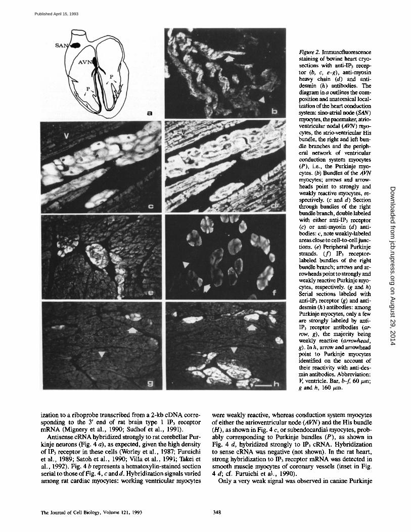
Figure 2. Immunofluorescence staining of bovine heart cryo- sections with anti-IP3 recep- tor (b, c, e-g), anti-myosin heavy chain (d) and anti- desmin (h) antibodies. The diagram in a outlines the com- position and anatomical local- ization of the heart conduction system: sino-atrial node (SAN) rnyocytes, the pacemaker, atrio- ventricular nodal (AVN) myo- cytes, the atrio-ventricular His bundle, the right and left bun- dle branches and the periph- eral network of ventricular conduction system myocytes (P), i.e., the Purkinje myo- cytes. (b) Bundles of the AVN myocytes; arrows and arrow- heads point to strongly and weakly reactive myocytes, re- spectively. (c and d) Section through bundles of the right bundle branch, double labeled with either anti-IP3 receptor (c) or anti-myosin (d) anti- bodies: c, note weakly-labeled areas close to cell-to-cell junc- tions. (e) Peripheral Purkinje strands. (f) IP3 receptor- labeled bundles of the right bundle branch; arrows and ar- rowheads point to strongly and weakly reactive Purkinje myo- cytes, respectively. (g and h) Serial sections labeled with anti-IP3 receptor (g) and anti- desmin (h) antibodies: among Purkinje myocytes, only a few are strongly labeled by anti- IP3 receptor antibodies (ar- row, g), the majority being weakly reactive (arrowhead, g). In h, arrow and arrowhead point to Purkinje myocytes identified on the account of their reactivity with anti-des- min antibodies. Abbreviation: V, ventricle. Bar, b-f, 60/zm; g and h, 160/~m.
ization to a riboprobe transcribed from a 2-kb cDNA corre- sponding to the 3' end of rat brain type 1 IP3 receptor mRNA (Mignery et al., 1990; Sudhof et al., 1991).
Antisense cRNA hybridized strongly to rat cerebellar Pur- kinje neurons (Fig. 4 a), as expected, given the high density of IP3 receptor in these cells (Worley et al., 1987; Furuichi et al., 1989; Satoh et al., 1990; Villa et al., 1991; Takei et al., 1992). Fig. 4 b represents a hematoxylin-stained section serial to those of Fig. 4, c and d. Hybridization signals varied among rat cardiac myocytes: working ventricular myocytes
were weakly reactive, whereas conduction system myocytes of either the atrioventricular node (AVN) and the His bundle (H), as shown in Fig. 4 c, or subendocardial myocytes, prob- ably corresponding to Purkinje bundles (P), as shown in Fig. 4 d, hybridized strongly to IP3 cRNA. Hybridization to sense eRNA was negative (not shown). In the rat heart, strong hybridization to IP3 receptor mRNA was detected in smooth muscle myocytes of coronary vessels (inset in Fig. 4 d; cf. Furuichi et al., 1990).
Only a very weak signal was observed in canine Purkinje
The Journal of Cell Biology, Volume 121, 1993 348
on August 29, 2014
jcb.rupress.orgD
ownloaded from
Published April 15, 1993

Figure 3. Immunofluorescence staining of bovine embryonic heart cryosections with anti- IP3 receptor (a, b and d), anti-myosin heavy chain (c), and anti-~ myosin heavy chain (e) antibodies. Serial sections of the ventricular septum of a 3-too old fetus were labeled with anti-IP3 receptor (b) and anti-myosin heavy chain (c) antibodies; labeling for IP3 receptor is detectable in myo- sin-positive myocytes of the right bundle branch (arrows, b and c). Serial sections of the ventricular myocardium of a 5-too-old fetus were labeled with anti-IP3 receptor (d) and anti-tx myosin heavy chain (e) antibodies: IP3 receptor-posi- tive bundles of large conduc- tion myoeytes were detected. Abbreviations: I~, ventricle, RB, right bundle branch. Bar, a, 60/zm; b-e, 30/~m.
neurons with rat IP3 receptor cRNA and no signal at all in both bovine and canine myocytes (not shown).
SubceUular Localization and Heterogeneous Distribution of the IP3 Receptor and Calsequestrin
The cellular and subeellular distribution of both IP3 recep- tor and CS, the SR intralumenal low-affinity, high-capacity Ca 2÷ binding protein (MacLennan and Wong, 1971) was in- vestigated by immunofluorescent labeling in bovine Purkinje myocytes.
Double labeling revealed uneven distribution of the two proteins in IP3 receptor-positive Purkinje myocytes. The IP3 receptor labeling was widespread throughout the cyto- plasm, showing regional staining differences (Fig. 5, a and b), i.e., the staining was more intense in the central cytoplas- mic region (see also Fig. 2 c) that is filled with intermediate filament proteins in bovine Purkinje myocytes (Thornell et al., 1985). On the other hand, the punctate staining of CS (cf. Jorgensen et al., 1984) was more evident in, but not lim- ited to, subsarcolemmal areas (Fig. 5, c and d) where myo- fibrils are concentrated. In all panels, large arrows point to myocytes showing strong reactivity for the IP3 receptor in central areas apparently devoid of CS; small arrows point to subsarcolemmal regions reactive for both the IP3 receptor and CS.
Discussion
Heterogeneous Distribution of the Cardiac I ) ~ 1 IP~ Receptor and Its Concentration in Purkinje Myocytes
In this paper we demonstrate, for the first time, that the type
1 IP3 receptor is highly concentrated in the majority of Pur- kinje myocytes, the distal elements of the heart conduction system, and some fascicles of the atrio-ventricular node. We also show much lower amounts of IP3 receptor in the work- ing myocardium and other portions of the conduction sys- tem. The differential distribution of the IP3 receptor be- tween conduction system myocytes and working myocytes is also observed during embryonic development, i.e., at the earliest stage examined, conduction myocytes are found to express a higher level of IP3 receptor.
There are, thus, two levels of heterogeneity whose func- tional relevance remains to be investigated: one among the different subpopulations of the heart conduction system and the working myocytes, the other between and within the Pur- kinje myocytes themselves. In the latter cases, the heteroge- neity of Purkinje myocytes pertains both to the subceUular and cellular level, as judged by serial section analysis: there are, in fact, areas close to cell-cell junctions which are weakly stained (Fig. 2 c) as compared to central cytoplasmic areas; moreover, either individual myocytes within a bundle (Fig. 2 f ) or entire bundles of conduction system myocytes (of. Fig. 2, f-h) are weakly labeled as compared to either adja- cent myocytes or bundles.
A rat type 2 IP3 receptor isoform, the product of a dis- tinct gene (Sudhof et al., 1991), has been recently described. Ligand binding studies on recombinant IP3 receptor pro- teins, have shown that type 2 11>3 receptor displays slightly higher affinity for IP3 than type 1 IP3 receptor. Analysis of the deduced amino acid sequence indicates divergence in the COOH-terminal 19-mer peptide, chosen to raise the anti- bodies specific for type 1 IP3 receptor and used in this study. Low degree of homology is also observed in the 3'
Gorza et al. IP~ Receptor in Purkinje Myocytes 349
on August 29, 2014
jcb.rupress.orgD
ownloaded from
Published April 15, 1993
Figure 4. In situ hybridization. Dark fields micrographs of IP3 receptor cRNA hybridized to rat cerebellum (a) and heart (b and c). An- tisense probe hybridization was detected in the soma and dendrites of cerebeUum Purkinje neurons (a), in coronary smooth muscle myocytes (inset, d), in subendocardial Purkinje myocytes (d, P, arrowhead), and in conduction myocytes of the AV node (AVN) and the His bundle (H: c); myoeytes of the ventricular free wall (V) and septum (S) were weakly reactive (c and d). b, which represents a section serial to those of c and d, was stained with hematoxylin and eosin. Bar, a, c, d, and inset, 240 t~m; b, 400 #m.
coding and noncoding regions of type 1 and 2 IP3 receptor mRNAs (Sudhof et al., 1991). Although we cannot rule out with certainty that other IP3 receptor isoforms are expressed (up to four distinct IP3 receptor cDNAs have been described in the mouse; Ross et al., 1992), the specificity of the anti- bodies and cRNA probes used along with our [3H]IP3 bind- ing data, strongly indicate that the type 1 IP3 receptor rep- resents a major isoform in Purkinje myocytes.
IPrsensit ive Ca 2+ Stores Purkinje Myocytes
In Purkinje myocytes, the SR is composed of three contin- uous membrane domains, the free SR, corbular SR and junc- tional SR (Sommer and Jennings, 1986) whose relative de- velopment shows regional variations. The corbular and junctional SR share ultrastructural features and contain
intralumenal granules largely made up of CS (Jorgensen and Campbell, 1984; Jorgensen et al., 1988). Junctional SR pro- files are observed exclusively in the subsarcolemmal rim all around myofibrils. Free and corbular SR, as well as ER cis- ternae are also observed in myofilament-free areas of central cytoplasm (Sommer and Jennings, 1986).
The IP3 receptor is a ligand-gated Ca 2÷ channel (Maeda et al., 1991), found to be concentrated in the limiting mem- brane of smooth-surfaced ER subcompartments in both chick- en and mammalian cerebellum Purkinje neurons (Satoh et al., 1990; Volpe et al., 1991; Takei et al., 1992). CS, an in- tralumenal low-affinity, high-capacity Ca ~+ binding protein (MacLennan and Wong, 1971), is localized in striated mus- cle SR subcompartments, i.e., terminal cisternae (Jorgensen et al., 1984), and ER moderately dense-cored vacuoles in chicken cerebellum Purkinje neurons (Villa et al., 1991;
The Journal of Cell Biology, Volume 121, 1993 350
on August 29, 2014
jcb.rupress.orgD
ownloaded from
Published April 15, 1993

Figure 5. Double immunoflu- orescence staining of bovine heart cryosections with anti- IP3 receptor (a and b) and anti-cardiac CS (c and d) Abs. Large arrows point to myocytes showing strong reactivity for the IP3 receptor in central areas apparently devoid of CS; small arrows point to sub- sarcolemmai regions reactive for both the IP3 receptor and CS. Bar, a and c, 40 #rn; b and d, 60 #m.
Volpe et al., 1991). The occurrence of both CS and IP3 re- ceptor on the same subcellular compartments is taken as evi- dence of functional IPrsensitive Ca 2+ stores (Volpe et al., 1991; Takei et al., 1992; reviewed in Meldolesi et al., 1992).
In the present study, Purkinje myocytes are labeled by anti- bodies for both the IPa receptor and CS (Fig. 5), thus indi- cating the existence of IPrsensitive Ca 2+ stores. The label- ing pattern indicates that the IP~ receptor is not restricted to subsarcolemmal SR but possibly present in corbular SR (or parts of it) and ER subcompartments, scattered through- out the cell (Sommer and Jennings, 1986), which might be in lumenal continuity with junctional SR (cf. Volpe et al., 1992). The finding that central areas of a number of Purkinje myocytes are strongly reactive for the IP~ receptor and poorly reactive for CS (Fig. 5), points to subcellular hetero- geneity of Ca 2+ stores in conduction system cells.
The relevance of the present observations and the detailed ultrastructural features of Ca :+ stores in Purkinje myocytes will be fully understood after completion of EM immuno- gold labeling experiments, which are in progress. Molecular
heterogeneity of intracellular Ca 2+ stores has been already demonstrated in the Purkinje neuron of the chicken, where different subcellular membrane compartments were found, by EM immunogold labeling, to be variably endowed with either CS or IP3 receptor (Villa et al., 1991; Volpe et al., 1991; Takei et al., 1992).
Functional Significance of the IP3 Receptor in Purkinje Myocytes
After plasma membrane depolarization, transient increases of [Ca2+]~ appear to be caused by other entry of extracellu- lar Ca 2+ and release of Ca 2+ from the SR (Wier, 1980; Fabi- ato, 1985) of Purkinje myocytes. However, the physiological role of Ca 2+ release has remained elusive thus far. In Pur- kinje myocytes, IP~-sensitive Ca 2+ stores may be involved in the increase of automaticity after o~t-adrenergic stimula- tion. del Balzo et al. (1990) have recently shown that WB 4101, a competitive inhibitor of o~l^-adrenergic receptor in canine Purkinje myocytes, antagonizes the oel-adrenergic
Gorza et al. IP3 Receptor in Purkinje Myocytes 351
on August 29, 2014
jcb.rupress.orgD
ownloaded from
Published April 15, 1993

positive chronotropic response, and prevents norepinephrine- dependent inositol phosphate accumulation, del Balzo et al. (1990), thus, suggested that the products of PtInsP2 hydro- lysis, either diacylglycerol, IP3 or both, are "mechanistically involved in the al-adrenergic positive chronotropic response7 Moreover, automatic arrhythmias induced by oti-adrenergic stimulation in experimental models of "ischemiC and "reper- fused" canine Purkinje myocytes (Anyukhowsky and Rosen, 1991; Molina-Viamonte et al., 1991), were abolished by pre- treatment with WB 4101. IP3-induced transient elevation of [Ca2+]i may bring about decrease of membrane potential and, in turn, increased automaticity via activation of a non- specific cation conductance (Colquoun et al., 1981; Kass and Tsien, 1982; Molina-Viamonte et al., 1990). Thus, IP3- induced Ca 2+ release appears to be causally related to both increase in automaticity and generation of some arrhythmias. The demonstration of the high level of IP3 receptor in Pur- kinje myocytes provides a molecular basis for this interpre- tation.
In Purkinje myocytes, the heterogeneous distribution of the IP3 receptor might be related to that of phospholipase C-coupled pertussis toxin-insensitive G proteins, which ap- pear to be functionally present only in 50% of Purkinje myo- cytes (see del Balzo et al., 1990). The transduction pathway activated by c~lA-adrenergic agonists and controlled by a pertussis-insensitive G protein appears to be developmen- tally regulated. At birth almost all Purkinje myocytes re- spond to ctl-adrenergic agonists with an increase in au- tomaticity (see del Balzo et al., 1990, and references therein) whereas only 50% of adult Purkinje myocytes do so. The IP3 receptor immunolabeling pattern observed in adult and embryonic Purkinje myocytes is formally consistent with these changes, insofar as Purkinje myocytes are far less bet- erogeneous as compared to adult ones. However, other mech- anisms might be responsible for the negative chronotropism induced in about 50% of adult Purkinje bundles upon stimu- lation with ct~-adrenergic agonists. A pertussis toxin-sensi- tive G protein has been shown to be expressed after birth and to couple activation of ctlB-adrenergic receptor subtype to a Na+-K + pump, whose operation would determine the de- crease in automaticity (Zaza et al., 1990). Interestingly, spe- cific ct2 and ct3 isoforms of the Na+-K + pump are expressed in conduction system myocytes (Zahler et al., 1992). It re- mains to be ascertained whether both transduction pathways are functional in adult Purkinje myocytes or are segregated to distinct Purkinje myocytes subpopulations.
Sincere thanks are due to Dr. Thomas C. Sudhof (Department of Molecular Genetics, University of Texas Southwestern Medical Center, Dallas, TX) for the generous gift of the pI2a cDNA clone.
Work supported by institutional funds from the Consiglio Nazionale delle Ricerche of Italy, grant GM-40I~g-03 from the National Institutes of Health, and funds from the Ministero dell'Universi~ e della Ricerca Scientifica e Tecnologica of Italy.
Received for publication 29 July 1992 and in revised form 15 January.
References
Alderson, B. H., and P. Volpe. 1989. Distribution of endoplasmic reticulum and calciosome markers in membrane fractions isolated from different regions of the canine brain. Archly. Biochem. Biophys. 272:162-174.
Anyukhovsky, E. P,, and M. R. Rosen. 1991. Abnormal automatic rhythms in ischemic Purkinje fibers are modulated by a specific a~-adrenergic recep- tor subtype. Circulation. 83:2076-2082.
Ausoni, S., C. De Nardi, P. Moretti, L. Gorza, and S. Schiattino. 1991. De- velopmental expression of rat cardiac troponin I mRNA. Development. 112:1041-1051.
Berridge, M. J., and R. F. Irvine. 1989. Inositol phosphates and cell signalling. Nature (Lond. ). 341:197-205.
Biral, D., P. Volpe, E. Damiani, and A. Margreth. 1992. Coexistence of two calsequestrin isoforms in slow-twitch skeletal muscle fibers. FEBS (Fed. Eur. Biochem. Soc.) Lett. 299:175-178.
Borgatta, L., J. Watras, A. M. Katz, and B. E. Ehrlich. 1991. Proc. Natl. Acad. Sci. USA. Regional differences in calcium-release channels from heart. 88:2486-2489.
Brown, J. H., I. L. Buxton, and L. L. Bruntun. 1985. cc~-adrenergic and mus- carirtic cholinergic stimulation of phosphoinnsitide hydrolysis in adult rat cardiomyoeytes. Circ. Res. 57:532-537.
Colquhoun, D., E. Neher, H. Reuter, and C. H. Stevens. 1981. Inward current channels activated by intracellular Ca in cultured cardiac ceils. Nature (Lond.). 294:752-754.
del Balzo, U., M. R., Rosen, G. Malfatto, L. M. Kaplan, and S. F. Steinberg. 1990. Specific c~-adrenergic receptor subtypes modulate catecholamine- induced increases and decreases in ventricular automaticity. Circ. Res. 67:1535-1551.
Eckel, J., E. Gedach-Eskucben, and H. Reinauer. 1991. Alpha-adrenoceptur- mediated increase in cytosolic free calcium in isolated cardiac myocytes. J. Mol. Cell. Cardiol. 23:617-625.
Fabiato, A. 1985. Time and calcium dependence of activation and inactivation of calcium-induced release of calcium from the sareoplasmic reticulum of a skinned canine cardiac Purkinje cell. J. Gen. Physiol. 85:247-289.
Fabiato, A. 1986. Inositul (1,4,5) trisphosphate-induced release of Ca 2. from the SR of skinned cardiac ceils. Biophys. J. 49:190a. (Abstr.)
Furnichi, T., S. YoshikaWa, A. Miyakawi, K. Wada, N. Maeda, and K. Mikoshiba. 1989. Primary and functional expression of the inositol 1,4,5- triphosphate-binding protein P4o0. Nature (Lond.). 342:32-38.
Furnichi, T., C. Shiota, and K. Mikoshibu. 1990. Distribution of inositol 1,4,5- trisphosphate receptor mRNA in mouse tissues. FEBS (Fed. Fur. Biochem. Soc.) Left. 267:85-88.
Gorza, L., S. Sartore, L. E. Thornell, and S. Sehiaflino. 1986. Myosin types and fiber types in cardiac muscle. III. Nodal conduction tissue. J. Cell Biol. 102:1758-1766.
Gorza, L., S. Ausoni, N. Mercial, K. E. M. Hastings, and S. Schialiino. 1993. Regional differences in troponin I isoform switching during heart develop- ment. Dev. Biol. In press.
Jorgensen, A. O., and K. P. Campbell. 1984. Evidence for the presence of cal- sequestrin in two distinct regions of myocardial sarcoplasmic reticulum. J. Cell Biol. 98:1597-1602.
Jorgensen, A. O., A. G. McLeod, K. P. Campbell, andG. H. Denney. 1984. Evidence for the presence of calsequestrin in both peripheral and interior regions of sheep Purkinje fibers. Circ. Res. 55:267-270.
Jorgensen, A. O., R. Broderiek, A. P. Somlyo, and A. V. Somlyo. 1988. Two structurally distinct calcium storage sites in rat cardiac sareoplasmic reticu- lure: an electron microprobe analysis study. Circ. Res. 63:1060-1069.
Kass, R. S., and R. W. Tsien. 1982. Fluctuations in membrane current driven by intracellular calcium in cardiac Purkinje fibers. Biophys. J. 38:255-269.
Kern, D. C., E. I. M. Johnson, A. M. Capponi, D. Chardonnes, U. Lang, B. Blondel, H. Koshida, and M. Vallotton. 1991. Effect of angiotensin II on cytosolic free calcium in neonatal rat cardiomyocytes. Am. J. Physiol. 261:C77-C85.
Kentish, J. C., R. J, Barsotti, T. J. Lea, I. P. Mulligan, J. R. Patel, and M. A. Ferenczi. 1990. Calcium release from cardiac sarcoplasmic reticulum in- duced by photorelease of calcium or Ins(1,4,5)P3. Am. J. Physiol. 258: H610-H615.
Lowry, O. H., N. J. Rosebrough, A. H. Farr, and R. J. Randall. 1951. Protein measurements with the ]=olin phenol reagent. J. Biol. Chem. 193:265-275.
Maeda, N., T. Kawasaki, S. Nakade, N. Yokota, T. Taguchi, M. Kasai, and K. Mikoshiha. 1991. Structural and functional characterization of inositol 1,4,5-trisphosphate receptor channel from mouse cerebellum. J. Biol. Chem. 266:1109-1116.
MacLennan, D. H., and P. T. S. Wong. 1971. Isolation of a calcium-sequester- ing protein from sarcoplasmic reticulum. Proc. Natl. Acad. Sci. USA. 68: 1231-1235.
Meldolesi, J., A. Villa, P. Volpe, and T. Pozzan. 1992. Cellular sites of IP3 action. In Inositol Polyphosphates and Ca 2+. J. W. Putney, editor. Raven Press, New York. 187-208.
Mignery, G. A., C. L. Newton, B. T. III Archer, and T. C. Sudhof. 1990. Structure and expression of the rat inositol 1,4,5-trisphosphate receptor. J. Biol. Chem. 265:12679-12685.
Molina-Viamonte, V., S. F. Steinberg, Y. K. Chow, M. J. Legato, R. B. Robinson, and M. R. Rosen. 1990. Phospholipase C modulates automaticity of canine cardiac Purkinje fibers. J. Pharmacol. Exp. 7"her. 252:886-893.
Molina-Viamonte, V., E. P. Anyukowsky, and M. R. Rosen. 1991. An otl-adren- ergic receptor subtype is responsible for delayed afierpolarizations and trig- gered activity during simulated ischemia and reperfusion of isolated Purkinje fibers. Circulation. 84:1732-1740.
Nakagawa, T., H. Okano, T. Furuichi, J. Arnga, and K. Mikoshiba. 1991. The subtypes of the mouse inositol 1,4,5-trisphosphate receptor are expressed in
The Journal of Cell Biology, Volume 121, 1993 352
on August 29, 2014
jcb.rupress.orgD
ownloaded from
Published April 15, 1993

a tissue-specific and developmentally specific manner. Proc. Natl. Acad. Sci. USA. 88:6244-6288.
Renard, D., and J. Poggioli. 1987. Does the inositol tris/tetrakisphosphate path- way exist in rat heart? FEBS (Fed. Fur. Biochem. Soc.) Left. 217:117-123.
Ross, C. A., S. K. Danoff, M. Schell, S. H. Snyder, and A. Ullrich. 1992. Three additional inositol 1,4,5-triphosphate receptors: Molecular cloning and differential localization in brain and peripheral tissues. Proc. Natl. Acad. Sci. USA. 89:4265-4269.
Rudnicki, M. A., G. Jackowski, L. Saggin, and M. W. McBurney. 1990. Actin and myosin expression during development of cardiac muscle from cultured embryonal carcinoma cells. Dev. Biol. 138:348-358.
Sartore, S., S. Pierobon-Bormioli, and S. Schiaffino. 1978. Immunohistochem- ical evidence for myosin polymorphism in the chicken heart. Nature (Lond.). 274:82-83.
Sartore, S., L. Gorza, S. Pierobon-Bormioli, L. Dalla Libera, and S. Schiaffino. 1980. Myosin types and fiber types in cardiac muscle. I. Ventricular myocar- dium. J. Cell Biol. 88:226-233.
Satoh, T., C. A. Ross, A. Villa, S. Supattapone, T. Pozzan, S. H. Snyder, and J. Meldolesi. 1990. The inositol 1,4,5-trisphosphate receptor in cerebellar Purkinje cells: quantitative immunogold labeling reveals concentration in an ER subeompartment. J. Cell Biol. 111:615-624.
Schiaffino, S., L. Gorza, S. Sartore, L. Saggin, S. Ausoni, M. Vianello, K. Gundersen, and T. Lomo. 1989. Three myosin heavy chain isoforms in type 2 skeletal muscle fibers. J. Muscle Res. Cell Motil. 10:197-205.
Sommer, J. R., and R. D. Jennings. 1986. Ultrastructure of cardiac muscle. In Heart and the Cardiovascular System. H. A. Fozzard, E. Haber, R. B. Jen- nings, A. M. Katz, H. E. Morgan, editors. Raven Press, New York, 61-100.
Sudhof, T. C., C. L. Newton, B. T. Archer, III, Y. A. Ushkaryov, and G. A. Mignery. 1991. Structure of a novel InsP3 receptor. EMBO (Fur. Mol. Biol. Organ.)J. 10:3199-3206.
Takei, K., A. Metealf, H. Stukenbrook, G. A. Mignery, T. C. Sudhof, P. Volpe, and P. De Camilli. 1992. Ca 2+ stores in Purkinje neurons: endoplas- mic reticulum subcompartments demonstrated by the heterogeneous distribu- tion of the InsP3 receptor, Ca2+-ATPase, and calsequestrin. J. Neurosci. 12:489-505.
Thornell, L. E., S. Forsgren, L. Gorza, S. Sartore, and S. Schiaffino. 1984. Differentiation of fiber types in cardiac muscle. In Etiology and Morphogen- esis of Congenital Heart Disease. A. Takao, editor. Futura Publishing Co.,
New York. 157-172. Thornell, L. E., A. Eriksson, B. Johansson, U. Kjorell, W. W. Franke, I. Vir-
tanen, and V.-P. Lehto. 1985. Intermediate filament and associated proteins in heart Purkinje fibers: a membrane-myofibril anchored cytoskeletal sys- tem. Ann. N.Y. Acad. Sci. 455:213-240.
Villa, A., P. Podini, D. O. Clegg, T. Pozzan, andJ. Meldolesi. 1991. Intracel- lular Ca 2+ stores in chicken Purkinje neurons: differential distribution of the low afffinity-high capacity Ca 2+ binding protein, calsequestrin, of Ca 2+ ATPase and of the ER lumenal protein, BiP. J. Cell Biol. 111:779-791.
Vitadello, M., M. Matteoli, and L. Gorza. 1990. Neurofilament proteins are coexpressed with desmin in heart conduction system myocytes. J. Cell Sci. 97:11-21.
Vires, A.-M., and A. J. Pappano. 1990. Inositol 1,4,5-trisphosphate releases Ca 2+ in permeabilized chick atria. Am. J. Physiol. 258:H1745-H1752.
Volpe, P., A. Villa, E. Damiani, A. H. Sharp, P. Podini, S. H. Snyder, and J. Meldolesi. 1991. Heterogeneity of microsomal Ca 2+ stores in chicken Purkinje neurons. EMBO (Fur. Mol. Biol. Organ.) J. 10:3183-3189.
Volpe, P., A. Villa, P. Podini, A. Martini, A. Nod, M. C. Panzeri, andJ. Mel- dolesi. 1992. The endoplasmic reticulum-sarcoplasmic reticulum connec- tion: distribution of endoplasmic reticulum markers in the sarcoplasmic reticuhim of skeletal muscle fibers. Proc. Natl. Acad. Sci. USA. 89:6142- 6146.
Wier, W. O. 1980. Calcium transients during excitation-contraction coupling in mammalian heart: aequorin signals of canine Purkinje fibers. Science (Wash. DC). 207:1085-1087.
Worley, P. F., J. M. Baraban, J. S. Colvin, and S. H. Snyder. 1987. Inositol trisphosphate receptor localization in brain: variable stoichiometry with pro- tein kinase C. Nature (Land.). 325:159-161.
Yorikane, R., H. Shiga, S. Miyake, and H. Koike. 1990. Evidence for a direct arrhythmogenic action of endothelin. Biochem. Biophys. Res. Commun. 173:457-462.
Zahler, R., M. Brines, M. Kashgarian, E. J. Benz, Jr., and M. Gilmore-Hebert. 1992. The cardiac conduction system in the rat expresses the o.2 and a3 iso- forms of the Na+, K +-ATPUse. Proc. Natl. Acad. Sci. USA. 89:99-103.
Zaza, A., R. P. Kline, and M. R. Rosen. 1990. Effects of c~-adrenergic stimula- tion on intracelhilar sodium activity and automaticity in canine Purkinje fibers. Circ. Res. 66:416-426.
Gorza et al. IPj Receptor in Purkinje Myocytes 353
on August 29, 2014
jcb.rupress.orgD
ownloaded from
Published April 15, 1993
Copyright © 2022 FDOKUMEN


![Ca]i elevation and oxidative stress induce KCNQ1 translocation from cytosol to cell surface and increase IKs in cardiac myocytes](https://static.fdokumen.com/doc/165x107/6313ba673ed465f0570ace55/cai-elevation-and-oxidative-stress-induce-kcnq1-translocation-from-cytosol-to-cell.jpg)