Hepatocyte growth factor and its receptor are expressed in cardiac myocytes during early...
24
Institution: WAYNE STATE UNIV Sign In as Member/Individual (Non- Member) Subscription Activation (Circulation Research. 1996;78:1028-1036.) © 1996 American Heart Association, Inc. Articles Hepatocyte Growth Factor and Its Receptor Are Expressed in Cardiac Myocytes During Early Cardiogenesis Daniel A. Rappolee, Anand Iyer, Yogesh Patel From the Department of Obstetrics and Gynecology (D.A.R., Y.P.), the Department of Cell and Molecular Biology and Feinberg Cardiovascular Research Institute (D.A.R.), and the Department of Pathology (A.I.), Northwestern University Medical School, Chicago, Ill, and the Center for Reproductive Sciences (D.A.R.), Northwestern University, Evanston, Ill. Correspondence to Daniel A. Rappolee, PhD, Department of Obstetrics and Gynecology, Northwestern University Medical School, 303 E Chicago Ave, Tarry Building 4-725, Chicago, IL 60611. E-mail [email protected]. This Article Abstract Alert me when this article is cited Alert me if a correction is posted Citation Map Services Email this article to a friend Similar articles in this journal Similar articles in PubMed Alert me to new issues of the journal Download to citation manager Cited by other online articles PubMed PubMed Citation Articles by Rappolee, D. A. Articles by Patel, Y.
Transcript of Hepatocyte growth factor and its receptor are expressed in cardiac myocytes during early...
Institution: WAYNE STATE UNIV Sign In as Member/Individual (Non-Member) Subscription Activation
(Circulation Research. 1996;78:1028-1036.) © 1996 American Heart Association, Inc.
Articles
Hepatocyte Growth Factor and Its Receptor Are Expressed in Cardiac Myocytes During Early Cardiogenesis
Daniel A. Rappolee, Anand Iyer, Yogesh Patel
From the Department of Obstetrics and Gynecology (D.A.R., Y.P.), the Department of Cell and Molecular Biology and Feinberg Cardiovascular Research Institute (D.A.R.), and the Department of Pathology (A.I.), Northwestern University Medical School, Chicago, Ill, and the Center for Reproductive Sciences (D.A.R.), Northwestern University, Evanston, Ill.
Correspondence to Daniel A. Rappolee, PhD, Department of Obstetrics and Gynecology, Northwestern University Medical School, 303 E Chicago Ave, Tarry Building 4-725, Chicago, IL 60611. E-mail [email protected].
This Article
Abstract
Alert me when this article is cited
Alert me if a correction is posted
Citation Map
Services
Email this article to a friend
Similar articles in this journal
Similar articles in PubMed
Alert me to new issues of the journal
Download to citation manager
Cited by other online articles
PubMed
PubMed Citation
Articles by Rappolee, D. A.
Articles by Patel, Y.
Abstract
Abstract In the mouse, the heart primordium arises when mesoderm is set aside during gastrulation, is induced by pharyngeal endoderm, migrates ventrally to the midline of the embryo, forms a tube, and begins beating. Little is known of the molecular mechanisms that mediate the determination, mitosis, differentiation, and migration that lead to the beating heart. Transcripts for hepatocyte growth
factor/scatter factor (HGF) and its receptor are coexpressed transiently and dynamically in the premyocardium but not in other heart progenitor cells. Transcripts for HGF ligand and receptor are first detected before cardiac function and looping and persist through the first looping stage, when heart morphology begins to elaborate. HGF ligand and receptor mRNA are detectable after the putative heart transcription factor, Csx/Nkx2-5, and concomitantly with the heart structural gene, cardiac actin. HGF receptor mRNA is detected in the mesoderm of the headfold stage and persists in myocardial precursors of the ventricles and atria (but not in the outflow-tract smooth muscle cells) through the 14-somite stage at 8.75 days after fertilization (day E8.75). At the headfold stage, between E7.5 and E8.0, HGF receptor mRNA was detected in myocardial cells before fusion at the ventral midline. HGF
ligand and receptor mRNA transcripts are coexpressed in the embryo, except in the headfold stage (when only the HGF receptor can be detected) and in the heart at the 14- to 18-somite stage (when only HGF ligand can be detected). The dynamic pattern of coexpression suggests an autoregulatory role for HGF and its receptor in early heart development.
Key Words: hepatocyte growth factor/scatter factor • hepatocyte growth factor receptor/c-met proto-oncogene • cardiogenesis • myocardium • in situ hybridization
Top Abstract Introduction Materials and Methods Results Discussion References
Introduction
In mammals, the heart is the first organ to form during development. At midgastrulation in the mouse, mesodermal cells that will form the heart are set aside after ingression through the primitive streak of the E7.5 gastrula. They then migrate into position at day E7.75-8.0 and begin beating at day E8.5.1 At day E7.5, the mesodermal cells that will become heart first express Csx/Nkx2-5. This gene is a putative transcription factor that is specific for the heart when cardiac mesoderm is produced, throughout
cardiogenesis, and in the adult heart.2 3 By day E7.5-8.0, the early myocardial cells express cardiac actin and MLC-2v.4 5 Csx/Nkx2-5 and then MLC-2v are expressed in cultured mouse embryonic stem cells just before they differentiate into the beating heart.3 4 The differentiation of the mesoderm into
heart first requires the presence of Spemann's organizer and then deep anterior/pharyngeal endoderm in amphibians and chicks.1 6 This inductive influence of the endoderm may act at a distance, perhaps by diffusible growth factors.1 The two edges of the migrating mesoderm meet at the ventral midline and fuse to form (1) the early endocardium, (2) the early myocardium, and (3) the early pericardium. The endocardial tube is invested with myocardium by day E8.0-8.5 in the mouse. As the two primordia fuse at the ventral midline to form a tube, the rate of mitosis reaches its highest point.1 Within half a day after the mesoderm fuses, the heart has differentiated, the initial specification of atria, ventricles, and outflow tract has occurred, and the heart is beating. Little is known of the molecular mechanisms that mediate the migration, growth, determination, and finally the differentiation that lead to the beating heart. The data on heart muscle–specific structural genes and transcription factors isolated in the chick and mouse correlate well with the temporal events in heart development described above.2 3 4 7 8 9 10 11 12 13 However, there have been few studies on the expression and function of growth factors in the early development of the heart. The expression and function of TGF-ß3 has been studied in the later development of the heart during valve formation.14 There have been studies of the early prefunctional stage of development of the heart, particularly of the expression of FGF-815 and of TGF-ß.16 In mammals, the expression of HGF ligand has not been studied through gastrulation.
HGF is a heterodimeric protein implicated in the development and regeneration of the liver and in other mesenchymal-epithelial interactions during development after gastrulation.17 HGF requires a proteolytic processing step that cleaves the propolypeptide into and ß subunits of 70 and 35 kD. The and ß subunits of HGF contain four kringle domains and a protease- like domain, respectively.18 19 20 Scatter factor, a protein that induces dispersion and migration of epithelial cells, is identical to HGF.21 HGF has 50% homology with HGF-like protein (based on the sequence of the gene in rat) and 45% homology with MSP (based on sequence from the gene in human).21 22 23 24 The HGF-like protein and
MSP are homologues. The HGF and HGF-like protein are expressed in Ito cells and parenchymal cells in the liver.22 HGF is expressed in mesenchymal derivatives such as chondrocytes and in the placenta.25
Top Abstract Introduction Materials and Methods Results Discussion References

26 HGF is also detected in the dermis and kidney, where it may have a role in development, and in the fetal liver.25 26 27 The expression of the HGF gene has not been studied in the period before day E10 in the mouse when heart development has initiated.
The receptor for HGF has been identified as the c-met proto-oncogene, a heterodimeric transmembrane tyrosine kinase that is generally distributed apposite the HGF mRNA in postgastrulation embryo and in adult mammals.26 28 It requires ligand- induced dimerization of the cytosolic tyrosine kinase domains of two HGF receptors to transmit a signal into the cell.29 The HGF receptor/c-met activates a signal pathway through a complex that may include nonenzymatic SH-2 domain–containing protein and a protein tyrosine phosphatase.30 31 Ron and Sea are genes with homology to c-met/HGF receptor in the tyrosine kinase and extracellular domain.32 33 Ron is the receptor for MSP but does not bind HGF.23 During later embryonic developmental in the mouse, c-met/HGF receptor is expressed in epithelia adjacent to HGF ligand expression in the mesenchyme.26 In mammals, the expression of HGF receptor
has not been studied in the period spanning gastrulation and the initiation of heart development.
Some functional studies on the role of HGF in development have been performed previously. HGF is an apparent inducer of neuroectoderm during gastrulation in the chick and frog. HGF induces neural tissue in frog ectoderm, and implanted HGF-carrying beads perturb the primitive streak in the chick.34 The implanted beads cause the mesoderm induced at gastrulation to move toward the beads, creating a second mesodermal axis and, later, a second neuroectodermal axis. This neuroectoderm may also be directly induced by HGF.35 The neuroectodermal induction mediated by HGF would occur at a time and in a position in which gastrulating mesoderm would be induced to form the heart, but this has not been studied directly in Xenopus or chick. In mouse, ablation of HGF or HGF receptor by homologous recombination results in embryonic lethality between days E12 and E16.5. Defects in the development of the placenta and liver are thought to cause the lethality, but some defects in heart have been noted previously.36 37 38
To test for a role in heart development, we assayed for the expression of the HGF ligand and receptor mRNA in the early development of the heart in the mouse embryo. We found that HGF and its receptor are coexpressed in the ectoderm and mesoderm of the midgastrulation-stage mouse but that the coexpression restricts to mesoderm that will form cardiac myocytes toward the end of gastrulation. The coexpression of HGF and its receptor persists through the first functioning of the heart and then stops. These data suggest that the coexpression of HGF and its receptor may have a role in the early development of the beating heart.
Materials and Methods
Materials Amplitaq DNA polymerase was purchased from Perkin-Elmer Cetus. Restriction enzymes and T4 polynucleotide kinase were purchased from New England BioLabs and GIBCO BRL. Radioisotopes were obtained from New England Nuclear. MMLV reverse transcriptase and superscript MMLV reverse transcriptase were obtained from GIBCO BRL. Digoxygenin-UTP Genius RNA labeling kit, anti–digoxygenin-alkaline phosphatase conjugate, nitro blue tetrazolium, and 5-bromo-4-chloro-3-indolyl phosphate were all from Boehringer-Mannheim Biochemicals. Hoechst 33258 was purchased from Eastman Kodak Co. All other chemicals were from Sigma Chemical Co or VWR.
Mouse Embryos Standard techniques were used for obtaining zygotes.39 Female CD-1 mice (6 to 10 weeks old, Charles River, Wilmington, Mass) or MF-1 mice (Harlan Sprague Dawley, Indianapolis, Ind) were housed overnight with C57BL/6JxSJL/J F1 hybrid males (Jackson Laboratories, Bar Harbor, Maine). Noon of the day following coitus was considered day E0.5. Embryos were obtained at the following stages: on day E7.75-9.5 (presomite to 30-somite stage). All embryos were freed of debris under a dissecting microscope. Uteri or embryos dissected free from the uterus were fixed overnight in fresh 4% paraformaldehyde at 4°C, then dehydrated, and stored at -20°C.39 40 To prevent trapping of anti-digoxygenin antibodies in the heart, hearts were pierced repeatedly with orthodontic alloy wire.39
Cell Culture TC-2 cells were derived from NIH3T3 cells by stable expression of an HGF receptor gene under promotion of viral long terminal repeats.41 These cells also produce substantial amounts of HGF ligand
when cultured with 5% serum.41 Embryonal carcinoma F9 cells, obtained from the American Type Culture Collection, were cultured on gelatin (Sigma)-coated culture dishes in DMEM containing 10% heat-inactivated FBS as previously described.40
In Situ Hybridization HGF ligand and HGF receptor inserts of 2.6 and 0.68 kb in size, respectively, were subcloned into pGEM.42 The insert for HGF ligand was derived from sequences in the entire 3'-translated region and 3'-untranslated region. The insert for HGF receptor was derived wholly from the 3'-untranslated region. The HGF ligand insert was derived from nucleotides 0 to 2600, and the HGF receptor insert was
derived from nucleotides 4498 to 5098 (references cited in References 17 and 2117 21 ). Digoxygenin
Top Abstract Introduction Materials and Methods Results Discussion References
cRNA was prepared from plasmid DNA by linearization and runoff transcription with T7 polymerase
and SP6 polymerase in sense and antisense directions for each plasmid.43 Runoff transcripts were labeled using digoxygenin-UTP and were treated with RNase-free DNase to remove the template
(Genius RNA labeling kit, Boehringer-Mannheim). Unincorporated nucleotides were removed by purification of synthetic RNA by anion column chromatography (QIAquick nucleotide removal kit,
QIAGEN). The sensitivity of the labeling was assayed by dilutions of dot blots (Genius RNA labeling kit, Boehringer-Mannheim). An effort was made to maximize the sensitivity of the detection of the HGF ligand and receptor genes. In doing this, the ability to compare relative amounts of the two genes was sacrificed. In short, the color development was allowed to proceed until maximal reaction product developed without appreciable background in the antisense or sense reactions. Time courses for intensity of development were not done. For in situ hybridization on sections, the time of development was 5 hours for HGF ligand and 2.5 hours for HGF receptor. For whole mounts, the development was terminated at 2.5 hours for HGF ligand and receptor. The sensitivities for the digoxygenin- labeled RNA were as follows: for HGF ligand, antisense, 0.03 pg, and sense, 0.08 pg; for HGF receptor, antisense, 0.02 pg, and sense, 0.02 pg. 35S riboprobes were synthesized as per Wilcox,44 with the following modifications: After synthesis of the probe, 1 µL of RQ1 DNase (Promega) was added to the transcription reaction and incubated for 15 minutes at 37°C. The unincorporated nucleotides were removed using QIAquick nucleotide removal kit. In situ hybridization and autoradiography were
performed as described in Wilcox,44 except proteinase K treatment was with 20 µg/mL for 5 minutes at room temperature.
In situ hybridization was performed by a modified protocol of Harkey et al,43 with the following modifications. Sections of paraffin were first dewaxed in xylene, rinsed in 100% ethanol, and air-dried. Sections were rehydrated through an ethanol series, washed in PBT (PBS containing 0.1% Tween 20), and treated with proteinase K (20 mg/mL in PBT). The proteinase K was neutralized with 0.2% glycine in PBS, washed with PBT, refixed with 4% paraformaldehyde in PBS, and washed in PBT. The slides were transferred to a humidified box and prehybridized (50% formamide, 10% polyethylene glycol, 0.6 mol/L NaCl, 5 mmol/L EDTA, 20 mmol/L Tris [pH 7.5], 500 µg/mL yeast tRNA, 0.1% Tween 20, and 10x Denhardt's [1x Denhardt's contains 2% Ficoll, 2% polyvinylpyrrolidone, and 2% BSA]) at 50°C for 1 hour. The prehybridization solution was removed; 10 µL of hybridization solution containing 100 ng digoxygeninlabeled RNA probe was added; and the slide was covered with a coverglass, sealed with rubber cement, and hybridized at 50°C in a humidified box overnight. All labeled probes had
previously been tested by dot blot hybridization and were in the range of 20- to 100-fmol sensitivity. After hybridization, the coverglass was removed, and the slides were washed at 50°C in 50% PBT/50% prehybridization solution, in PBT at 50°C, and in 1x SSC at 60°C. Slides were washed in PBT, incubated with PBT containing 2% normal sheep serum to block nonspecific binding, and incubated with a 1:2000 dilution of anti–digoxygenin/alkaline phosphatase conjugate in PBT/1% normal sheep serum. Slides were washed in PBT and then for 5 minutes in alkaline phosphatase buffer (100 mmol/L Tris [pH 9.5], 100 mmol/L NaCl, 50 mmol/L MgCl2, 0.1% Tween 20, and 1 mmol/L levamisole). The color reaction was carried out as previously described43 until a maximum amount of product was visible but before product was generated in sense controls. The reaction was stopped with PBT; the slides were stained with Hoechst 33258 (10 µg/mL in water), destained in water, dehydrated through an alcohol series, and mounted in media (Tris-EDTA with NaN3); the coverslips were sealed with rubber cement. Whole-mount in situ hybridization was performed by the method of Rosen and Beddington.45
Photomicrography was performed with a Nikon Microphot-FXA microscope. Micrographs were taken with epifluorescence to detect Hoechst-stained nuclei (with a barrier filter at 420 nm) or diascopic illumination to see color products, using Kodak 400 DIN slide film or 200 DIN print film. Objectives were chosen to cover the field for pertinent detail at x100, x200, and x400 (Nikon Plan 10, Plan 20, and

Plan 40 objectives, respectively). Negatives were scanned into Adobe Photoshop CD and formatted into figures.
Results
We first showed that our digoxygenin- labeled cRNA, developed with an alkaline phosphatase reaction, detected only the HGF mRNA in the liver of a day E13 embryo. At this stage, in situ hybridization with a cDNA encoding the entire coding sequence and the entire 3'-untranslated region of the HGF gene detected HGF transcripts in a small subpopulation of Ito cells in the liver but not in the parenchymal cells (data not shown).22 46 HGF ligand mRNA was also detected in the glomerular
mesenchyme but not in the collecting tubules of the embryonic and adult kidney (data not shown).17 21 This indicates that our probe matches the reported distribution of HGF mRNA in Ito cells and kidney cells but does not detect MSP/HGF-like protein mRNA in parenchymal cells of the liver, where HGF-like protein is expressed. Finally, HGF receptor mRNA was detected in labyrinthine placenta, and HGF ligand mRNA was detected in allantois at midgastrulation, as reported recently by Uehara et al37 (authors' unpublished data, 1996). The cDNA for the c-met/HGF receptor was wholly derived from the segment of the 3'-untranslated region of the gene and has little homology with the related receptors, Ron and Sea, in this region.23 24 28 32 33 47
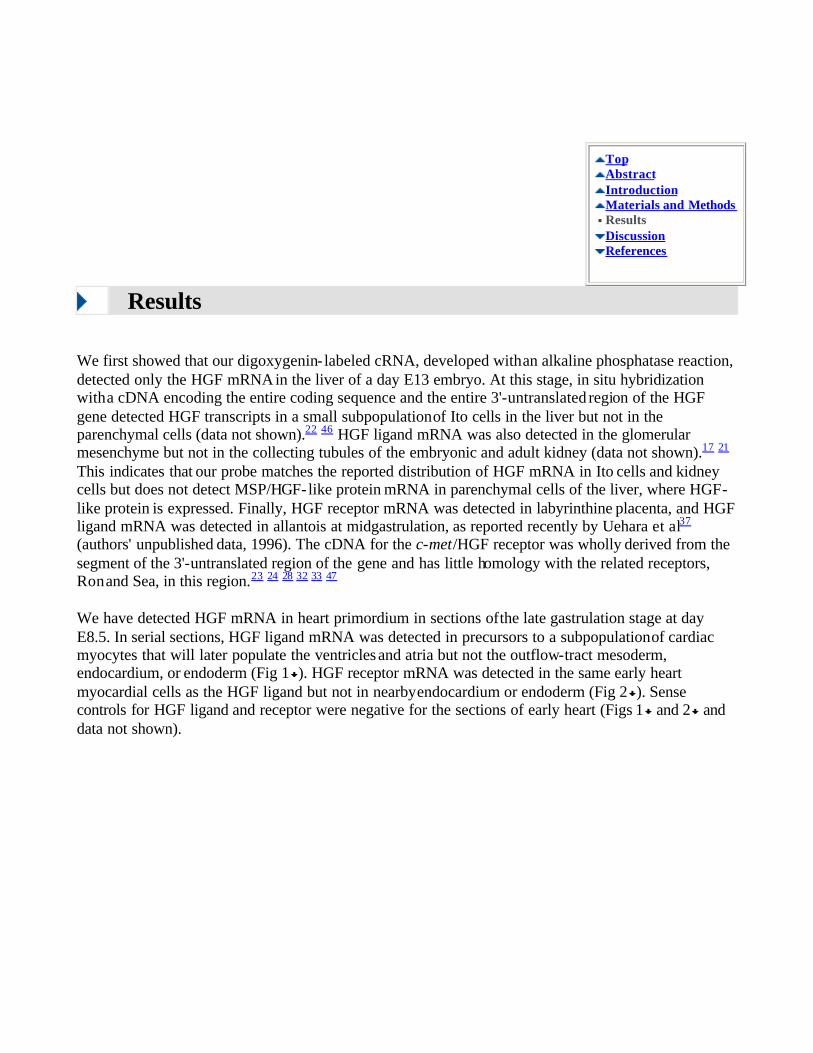
We have detected HGF mRNA in heart primordium in sections of the late gastrulation stage at day E8.5. In serial sections, HGF ligand mRNA was detected in precursors to a subpopulation of cardiac myocytes that will later populate the ventricles and atria but not the outflow-tract mesoderm, endocardium, or endoderm (Fig 1 ). HGF receptor mRNA was detected in the same early heart myocardial cells as the HGF ligand but not in nearby endocardium or endoderm (Fig 2 ). Sense controls for HGF ligand and receptor were negative for the sections of early heart (Figs 1 and 2 and data not shown).
Top Abstract Introduction Materials and Methods Results Discussion References
View larger version
(121K): [in this window]
[in a new window]
Figure 1. HGF mRNA is expressed in early myocardial cells enveloping the endocardium (EC) in the day E8.5 mouse embryo. Day E8.5 mouse embryos were sectioned transversely and fixed, and HGF mRNA was detected by in situ hybridization using antisense HGF ligand cRNA in a modified digoxygenin protocol. In panels A and B, NF indicates neural fold; MYO, myocardium. Note that the fused ventricular MYO envelops the EC, which is unstained. Panel C shows the partial atrial myocardium (stained) and the connected structure above, the unstained outflow tract. In panels B and D, the same sections as in panels A and C, respectively, are shown after staining with Hoechst to detect nuclei. Original magnification x250. Sense controls were negative (not shown).

View larger
version (83K): [in this window]
[in a new window]
Figure 2. HGF receptor mRNA is expressed in early myocardial cells enveloping the endocardium (EC) in the day E8.5 mouse embryo. Day E8.5 mouse embryos were sectioned transversely and fixed, and HGF receptor mRNA was detected by in situ hybridization using antisense HGF receptor cRNA in a modified digoxygenin protocol. In panels A and B, PE indicates pharyngeal endoderm; MYO, myocardium. In panels A and B, the fused ventricular MYO envelops the EC. HGF receptor mRNA is detected in the MYO but not the EC. In panels C and D, EN indicates endoderm; NF, neural fold; OT, outflow tract, and MYO, atrial MYO. Note the identity in patterns of expression in panels A and C in Figs 1 and 2 from adjacent sections at anterior-posterior levels of the heart. In panels B and D, the same sections as in panels A and C, respectively, are shown after staining with Hoechst to detect nuclei. Panel E is a similar section of the E8.5 day embryo hybridized with sense HGF receptor mRNA as a negative control, and panel F is the same section after staining with Hoechst to detect nuclei. Original magnification x250.
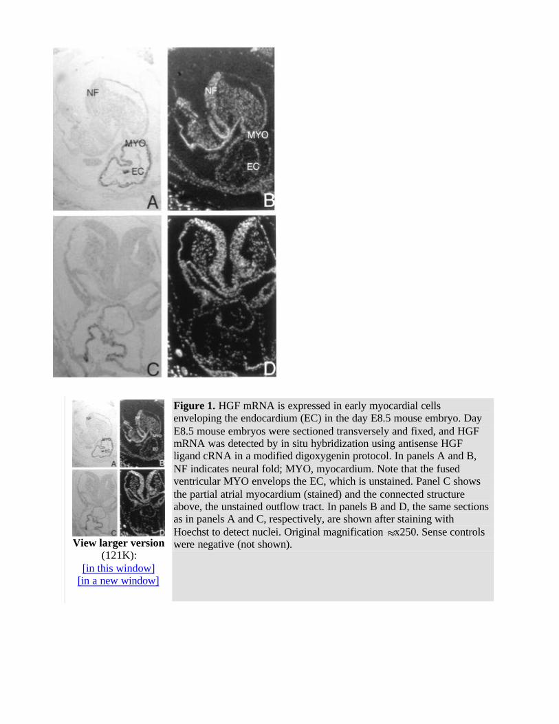
To time the beginning and end of expression of the HGF ligand and receptor mRNA, whole-mount in situ hybridizations were performed on 60 embryos at day E7.75-8.75. Using antisense digoxygenin-tagged cRNA with an enzymatic development with alkaline phosphatase for whole-mount in situ hybridization, HGF receptor mRNA was detected in the presomitic day E7.75 embryo in the precardiac mesoderm and in adjacent endoderm (Fig 3 and Table ). HGF receptor mRNA was detected in the 5-somite heart primordium just as it was about to begin beating.1 By the 11-somite stage, expression of HGF receptor mRNA had decreased, and after the 14-somite stage, HGF receptor mRNA was not detected (Fig 3 and Table ). Sense controls for whole embryos were negative for HGF receptor (data not shown). HGF receptor mRNA was detected primarily in mesodermal precursors of the heart but appeared to be detected in the endoderm next to the presumptive heart (Fig 3 ). However, no HGF receptor mRNA was detected in endoderm after postsectioning of the whole mounts or in sections of day E7.5-8.0 embryos (data not shown, Fig 5 below). HGF ligand mRNA was not detected in the early myocardial mesoderm or in the endoderm that is adjacent to the early myocardial mesoderm (Fig 4 , embryos 1 and 2). The heart-specific expression of HGF ligand mRNA began later than that of HGF receptor. HGF receptor mRNA was detected in early myocardial mesoderm (Fig 3 ), but HGF mRNA was not detected in the early myocardial mesoderm in similar embryos at any headfold stage (Fig 4 and Table ; data not shown). HGF ligand mRNA was first detected in the fused cardiac mesoderm of the 2- to 3-somite embryo before looping of the heart had started and persisted through the 16- to 18-somite stage, whereas the c-met/HGF receptor was expressed in the early headfold stage
through the 8- to 14-somite stage (Figs 3 and 4 and Table ). Sense controls for whole embryos were negative for HGF ligand (data not shown). The c-met/HGF receptor and HGF ligand were always detected in the heart before the looping of the heart and turning of the embryo (Table ). c-met/HGF receptor and HGF ligand were always detected before the first beating of the heart, which occurred between the 4- and 8-somite stage (data not shown). Relative to each other, turning, somite number,
and the first heartbeat, were variable. To determine the expression of HGF ligand and receptor in more detail, the embryos were embedded and sectioned after whole-mount in situ hybridization. In these sections, HGF ligand and receptor mRNA were detected in all myocardium after fusion of the migrating heart mesoderm to form a closed tube. However, no HGF ligand or receptor mRNA was conclusively detected in endoderm or in mesoderm before fusion (data not shown). To further test for expression of HGF ligand and receptor mRNA, embryos at E7.5-8.5 were sectioned and processed
using a more sensitive technique, by hybridization with runoff synthetic RNA labeled with 35S ribonucleotides to high specific activity (109 cpm/µg). HGF ligand and receptor mRNA were not detected in endoderm or endocardium; however, HGF receptor mRNA was detected in unfused migrating myocardial mesoderm (Fig 5 ). However, HGF ligand was not detected at the same stage.
This embryo was staged between E7.5 and E8.5 (Reference 4848 , plate 9e,f therein), since it lacked extensive cardiac morphology, including any fused structures (characteristic of day E7.5), yet had a foregut diverticulum with endoderm that formed some pharyngeal pouches (characteristic of day E8.0; note arrowheads in Fig 5A , 5C , 5E , and 5G ). In addition, there was an intraembryonic coelom (precursor to the pericardial cavity), a cardiogenic plate, (subjacent to this) an asymmetric unilateral sheet of endocardium, and (further subjacent to this) an asymmetric unilateral sheet of myocardium. In hematoxylin and eosin–counterstained sections, two continuous sheets of flattened nuclei with a intercalated basement membrane were seen forming a circle around the entire embryo (and the cavity in the deciduum where the embryo was found) and adjacent to the maternal deciduum (Fig 5C , data not shown). These two sheets were determined to be parietal endoderm and trophectoderm, by
comparison with similar structures at E6.5-7.5.48 HGF receptor was detected solely in the slip of myocardium between the cardiogenic plate/endocardium and the parietal endoderm. We speculate that the unilateral expression of HGF receptor mRNA has two possible explanations. The first explanation is that the transverse section was cut obliquely and detected fused or unfused myocardium only on one
side. Since the expression was observed in five consecutive 5-µm sections and the morphology of the
transverse section was near normal,48 it is more likely that this was unfused myocardium. The second
explanation is that mesoderm migration from the cardiogenic plate is asymmetric in time and occurs "sloppily" and that the fusion resolves this at a later stage. We have noted some asymmetry of migration in five embryos (see also Fig 3 , embryo 1) and favor the second explanation.
View larger version (38K):
[in this window] [in a new window]
Figure 3. HGF receptor mRNA is expressed in mouse heart from days E7.75 to E8.75. Whole-mount in situ hybridization was performed using digoxygenin- labeled antisense cRNA probes and developed using alkaline phosphatase. From left to right, embryos are as follows: 1, E7.75; 2, E7.9; 3, E8.0; 4, E8.25; 5, E8.25; and 6, E8.5. Arrowheads show the heart primordia or heart at each stage. In embryo 2, a second arrowhead shows expression in the two fusing myocardial primordia. Note some asymmetry of expression in embryo 1. The embryos have been placed in the following orientations: in all, anterior is on top and posterior is on bottom. Embryos 1, 2, and 3 are shown from the ventral surface, and embryos 4 and 5 show the ventral surface facing to the left. All five embryos show positive staining in the myocardial mesoderm. The scale of the embryos in the two orientations is not the same. Control-sense hybridizations showed no staining.
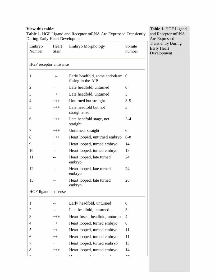
View this table: Table 1. HGF Ligand and Receptor mRNA Are Expressed Transiently During Early Heart Development
Embryo Number
Heart Stain
Embryo Morphology Somite number
HGF receptor antisense
1 +/- Early headfold, some endoderm
fusing in the AIP 0
2 + Late headfold, unturned 0
3 ++ Late headfold, unturned 3
4 +++ Unturned but straight 3-5
5 +++ Late headfold but not straightened
3
6 +++ Late headfold stage, not straight
3-4
7 +++ Unturned, straight 6
8 +++ Heart looped, unturned embryo 6-8
9 + Heart looped, turned embryo 14
10 -- Heart looped, turned embryo 18
11 -- Heart looped, late turned embryo
24
12 -- Heart looped, late turned embryo
24
13 -- Heart looped, late turned embryo
28
HGF ligand antisense
1 -- Early headfold, unturned 0
2 -- Late headfold, unturned 3
3 +++ Heart fused, headfold, unturned 4
4 ++ Heart looped, turned embryo 8
5 ++ Heart looped, turned embryo 11
6 ++ Heart looped, turned embryo 11
7 + Heart looped, turned embryo 13
8 +++ Heart looped, turned embryo 14
9 +++ Heart looped, turned embryo 17
Table 1. HGF Ligand and Receptor mRNA Are Expressed Transiently During Early Heart Development

9 +++ Heart looped, turned embryo 17
10 -- Heart looped, turned embryo 17
11 -- Heart looped, turned embryo 19
12 -- Heart looped, turned embryo 21
13 -- Heart looped, turned embryo 21
14 -- Heart looped, turned embryo 26
15 -- Heart looped, turned embryo 30
Evaluation of whole-mount in situ hybridizations like those represented in Figs 3 and 4. Intensity of heart staining is given on a scale of -- to +++. Morphology of heart and embryo is given in terms of whether the heart is fused or looped. Embryo somite number ( 3 to 5 at day E8.0) is given, along with indication of whether or not the embryo has "turned" (which occurs at day E8.5). The somite number is approximate. Variation in the intensity of staining of the HGF ligand at somites 13 and 17 may be due to incomplete penetration of reagents during hybridization, incorrect somite counts, or biological diversity.
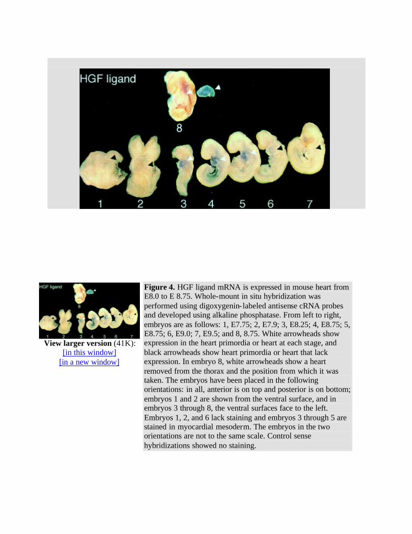
View larger version (41K):
[in this window] [in a new window]
Figure 4. HGF ligand mRNA is expressed in mouse heart from E8.0 to E 8.75. Whole-mount in situ hybridization was performed using digoxygenin- labeled antisense cRNA probes and developed using alkaline phosphatase. From left to right, embryos are as follows: 1, E7.75; 2, E7.9; 3, E8.25; 4, E8.75; 5, E8.75; 6, E9.0; 7, E9.5; and 8, 8.75. White arrowheads show expression in the heart primordia or heart at each stage, and black arrowheads show heart primordia or heart that lack expression. In embryo 8, white arrowheads show a heart removed from the thorax and the position from which it was taken. The embryos have been placed in the following orientations: in all, anterior is on top and posterior is on bottom; embryos 1 and 2 are shown from the ventral surface, and in embryos 3 through 8, the ventral surfaces face to the left. Embryos 1, 2, and 6 lack staining and embryos 3 through 5 are stained in myocardial mesoderm. The embryos in the two orientations are not to the same scale. Control sense hybridizations showed no staining.
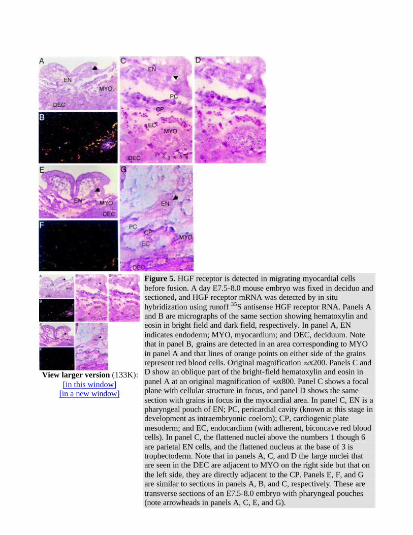
View larger version (133K):
[in this window] [in a new window]
Figure 5. HGF receptor is detected in migrating myocardial cells before fusion. A day E7.5-8.0 mouse embryo was fixed in deciduo and sectioned, and HGF receptor mRNA was detected by in situ hybridization using runoff 35S antisense HGF receptor RNA. Panels A and B are micrographs of the same section showing hematoxylin and eosin in bright field and dark field, respectively. In panel A, EN indicates endoderm; MYO, myocardium; and DEC, deciduum. Note that in panel B, grains are detected in an area corresponding to MYO in panel A and that lines of orange points on either side of the grains represent red blood cells. Original magnification x200. Panels C and D show an oblique part of the bright-field hematoxylin and eosin in panel A at an original magnification of x800. Panel C shows a focal plane with cellular structure in focus, and panel D shows the same section with grains in focus in the myocardial area. In panel C, EN is a pharyngeal pouch of EN; PC, pericardial cavity (known at this stage in development as intraembryonic coelom); CP, cardiogenic plate mesoderm; and EC, endocardium (with adherent, biconcave red blood cells). In panel C, the flattened nuclei above the numbers 1 though 6 are parietal EN cells, and the flattened nucleus at the base of 3 is trophectoderm. Note that in panels A, C, and D the large nuclei that are seen in the DEC are adjacent to MYO on the right side but that on the left side, they are directly adjacent to the CP. Panels E, F, and G are similar to sections in panels A, B, and C, respectively. These are transverse sections of an E7.5-8.0 embryo with pharyngeal pouches (note arrowheads in panels A, C, E, and G).
Discussion
HGF ligand and receptor mRNA transcripts are detected from day E7.75-8.75 (from midgastrulation, soon after the heart has been determined) to one-half day past the time of the first beating of the heart.
HGF ligand and receptor mRNA transcripts are always detected before the heart begins to beat or the heart tube starts to loop. The pattern of transient expression and coexpression early in heart
determination and morphogenesis suggests that the HGF ligand and receptor may mediate one of the functions required for early heart development: determination, mitosis, migration, or differentiation.
However, it is apparent that HGF is not required for early heart development. Null mutants for HGF and HGF receptor are lethal at days E12.0 to E18.0 because of the failure of placentation and liver formation.36 37 38 In these mutants, occasional cardiac arrhythmias were observed.36 These null mutants have not been studied in detail for defects in heart development before the lethality occurred. The heart is the first organ to develop, and it deve lops when the embryo reaches a size at which diffusion can no longer accomplish nutrient and gas exchange for all embryonic cells. Null mutants in genes required for the development of the early heart or blood supply result in embryonic lethality between days E9 and E11.49 50 The Nkx2-5 null mutant embryo develops a heart that forms a tube but fails to loop properly or express MLC-2v and dies at E9-10.51 Genes that affect heart development more subtly produce a later-stage lethal phenotype. An example of this is the retinoid X receptor- null mutant, which has defects in thickness and septation of ventricular walls and dies at E13.5-16.5.51 It is possible that loss of expression of single genes may have even smaller defects resulting in late embryonic or early postnatal lethality. This may be the case for HGF, but since HGF null mutants die from placental failure starting at E11.5, a full cardiac deficiency may not have time to develop. Genes that have subtle phenotypes in the heart of null mutants are likely to be clinically interesting. The most common type of birth defect, occurring in 0.5% of live births, is in the heart.8 Some of these heart defects may be due to misexpression or loss of expression of single genes.
The first expression of HGF ligand and receptor mRNA is detected after the expression of Csx/Nkx2-5, a putative transcription factor whose homologue in Drosophila is required for heart development
(references cited in References 2 and 32 3 ). Csx/Nkx2-5 mRNA is detected in the earliest heart mesoderm primordium in the mouse and may be a regulatory gene for the heart.2 3 Because HGF ligand and receptor mRNA are expressed after Csx/Nkx2-5 mRNA, it is unlikely that HGF mediates
Top Abstract Introduction Materials and Methods Results Discussion References
the earliest steps in determination of the heart. HGF ligand and receptor mRNA are first expressed concomitantly with cardiac actin (which is first detected near the headfold at day E7.5) and MLC-2v4 5 and may be part of a set of genes regulated by master regulatory genes for heart development.
Since HGF and its receptor are coexpressed in the developing heart, HGF signaling may also be involved with autoregulation and maintenance of the phenotype of early myocardial cells once they have been determined. HGF ligand is generally expressed in mesoderm-derived cells apposite the ectoderm-derived cells that express HGF receptor/c-met.25 26 27 The HGF ligand-receptor pair are not commonly coexpressed. Interestingly, HGF ligand and receptor are coexpressed in somites before the myotomes disperse and form skeletal muscle of the trunk.26 The expression of the HGF receptor in somites is absolutely required for migration of somitic muscle precursors into the limb bud, where the HGF ligand is expressed.38 It is possible that HGF ligand and receptor mediate autocrine and/or paracrine mechanisms of morphogenetic events common to the development of cardiac and skeletal muscle. Since the HGF receptor is detected in migratory myocardial mesoderm before fusion in the day E7.5-8.0 embryo, it is suggested that HGF plays a role in this migration. The location of a ligand that binds the HGF receptor in migrating myocardial mesoderm is not known. It may be that HGF ligand is expressed in the same myocardial mesoderm but is below the threshold of detection. An alternate possibility is that another, as yet unidentified, ligand for the HGF receptor is expressed.
In some amphibians and in the chick, pharyngeal endoderm induces mesoderm set aside during midgastrulation to form the heart (References 1 and 61 6 and M. Mercola, personal communication, 1995). Since HGF ligand is not detected in pharyngeal endoderm, it is unlikely that HGF mediates the induction of heart mesoderm by endoderm in the mouse. It is also possible that HGF, below the levels
detected by our in situ hybridization, may mediate such an induction.
A number of observations of the expression and function of HGF at midgastrulation suggest a role for HGF in the setting aside of mesoderm fated to become heart. At day E7.0-7.5, the midgastrulation
embryo expresses HGF ligand and receptor in the mesoderm and ectoderm (authors' unpublished data, 1995). The restriction of HGF ligand and receptor to heart precursor cells occurs between days E7.5 and E8.0. At a parallel time in the chick, HGF induces neuroectoderm and may also be a chemoattractant for mesoderm.34 35 In the mouse, HGF induces rapid migration of primitive streak cells isolated from the midgastrulation embryo (C.A. Burdsal and D.A. Rappolee, unpublished data, 1995). One possibility is that HGF produced in the midgastrulation primitive streak may participate in the induction of heart mesoderm as well as inducing neuroectoderm.
Lack of gross defects in heart development of HGF null mutants may be due to functional redundancy of HGF and related ligands during early heart development. Ron and Sea are related to the HGF c-met/receptor.32 33 47 These have homology in the extracellular domain, but HGF/SF and HGF-like protein/MSP have no cross-reactivity through these receptors.32 33 41 The expression of the HGF-related
ligand and HGF-like protein/MSP have not been determined at this stage of development. However, HGF and MSP probably do not compete for the HGF c-met/receptor or Ron or Sea,23 24 suggesting that known ligands and receptors of the family would not be redundant or compensatory. But as-yet-undiscovered ligands may bind the HGF receptor expressed in the early cardiac myocytes in the absence of HGF. Ligands not related to HGF may also be functionally redundant during heart development. TGF-ß2 ligand and FGF-8 are also expressed in pharyngeal endoderm and early myocardial mesoderm at day E7.75, at a time when these are candidates for mediating endodermal induction of heart mesoderm or migration.15 16 These unrelated growth factors may have overlapping function with HGF.
We have presented evidence for transient expression, and largely coexpression, of mRNA transcripts for HGF ligand and receptor in early cardiac myocytes. Expression occurs in a period of time after the heart is determined and before the heart begins to beat or to loop. It is likely that HGF plays a role in the migration of early myocardial cells before formation of the heart tube or in coordinating the program of differentiation or growth of myocardial cells. It is unlikely that HGF plays a role in the early induction of heart by endoderm or in the determination of heart cells. Although null mutants for HGF do not have gross heart deformation as a phenotype, signaling by HGF family members may play an important role in the early development of the heart.
Selected Abbreviations and Acronyms
E (followed by a number) = days after fertilization
FGF = fibroblast growth factor
HGF = hepatocyte growth factor
MLC = myosin light chain
MLC-2v = ventricular isoform of MLC-2
MMLV = Moloney murine leukemia virus
MSP = macrophage stimulatory protein
TGF = transforming growth factor
Acknowledgments
Drs Rappolee and Patel were supported by funds from the National Institute of Child Health and Human Development (R29 HD30739-01A1), from the American Cancer Society, Illinois (No. 95-10), the Feinberg Cardiovascular Research Institute at Northwestern University Medical School, and the Markey Charitable Trust. We thank Drs Ken Chien, Bob Decker, Phil Iannaccone, and Mark Mercola for their comments on the manuscript and Deborah Barron for editorial assistance.
Received December 27, 1995; accepted March 1, 1996.
References
1. Dehaan RL. Morphogenesis of the vertebrate heart. In: Dehaan RL, ed. Organogenesis. New York, NY: Holt, Rinehart & Winston; 1965:377-419.
2. Komuro I, Izumo S. Csx: a murine homeobox-containing gene specifically expressed in the developing heart. Proc Natl Acad Sci U S A. 1993;90:8145-8149. [Abstract/Free Full Text]
3. ints TJ, Parsons LM, Hartley L, Lyons I, Harvey RP. Nkx- 2.5: a novel murine homeobox gene expressed in early heart progenitor cells and their myogenic descendants. Development. 1993;119:419-431. [Abstract/Free Full Text]
4. Sassoon DA, Garner I, Buckingham M. Transcripts of -cardiac and -skeletal actins are early markers for myogenesis in the mouse embryo. Development. 1988;104:155-164. [Abstract]
5. Miller-Hance WC, LaCorbiere M, Fuller SJ, Evans SM, Lyons G, Schmidt C, Robbins J, Chien KR. In vitro chamber specification during embryonic stem cell cardiogenesis: expression of the ventricular myosin light chain-2 gene is independent of heart tube formation. J Biol Chem. 1993;268:25244-25252. [Abstract/Free Full Text]
6. Nascone N, Mercola M. An inductive role for the endoderm in Xenopus cardiogenesis. Development. 1995;121:515-523. [Abstract/Free Full Text]
7. Bisaha JG, Bader D. Identification and characterization of a ventricular-specific avian myosin heavy chain, VMHC1: expression in differentiating cardiac and skeletal muscle. Dev Biol. 1991;148:355-364. [Medline]
8. Chien KR, Zhu H, Knowlton KU, Miller-Hance WC, van-Bilsen M, O'Brien TX, Evans SM. Transcriptional regulation during cardiac growth and development. Annu Rev Physiol. 1993;55:77-95. [Medline]
9. O'Brien TX, Lee KJ, Chien KR. Positional specification of ventricular myosin light chain 2 expression in the primitive murine heart tube. Proc Natl Acad Sci U S A. 1993;90:5157-5161. [Abstract]
Top Abstract Introduction Materials and Methods Results Discussion References
10. Zhu H, Nguyen VT, Brown AB, Pourhosseini A, Garcia AV, van Bilsen M, Chien KR. A novel, tissue-restricted zinc finger protein (HF-1b) binds to the cardiac regulatory element (HF-1b/MEF-2) in the rat myosin light-chain 2 gene. Mol Cell Biol. 1993;13:4432-4444. [Abstract]
11. Kubalak SW, Miller-Hance WC, O'Brien TX, Dyson E, Chien KR. Chamber specification of atrial myosin light chain-2 expression precedes septation during murine cardiogenesis. J Biol Chem. 1994;269:16961-16970. [Abstract/Free Full Text]
12. Montgomery MO, Litvin J, Gonzalez-Sanchez A, Bader D. Staging of commitment and differentiation of avian cardiac myocytes. Dev Biol. 1994;164:63-71. [Medline]
13. Yutzey KE, Rhee JT, Bader D. Expression of the atrial-specific myosin heavy chain AMHC1 and the establishment of anteroposterior polarity in the developing chicken heart. Development. 1994;120:871-883. [Abstract/Free Full Text]
14. Potts JD, Vincent EB, Runyan RB, Weeks DL. Sense and antisense TGF-beta 3 mRNA levels correlate with cardiac valve induction. Dev Dyn. 1992;193:340-345. [Medline]
15. Crossley PH, Martin GR. The mouse Fgf8 gene encodes a family of polypeptides and is expressed in regions that direct outgrowth and patterning of developing embryos. Development. 1995;121:439-451. [Abstract/Free Full Text]
16. Dickson MC, Slager HG, Duffie E, Mummery CL, Akhurst RJ. RNA and protein localisations of TGF beta 2 in the early mouse embryo suggest an involvement in cardiac development. Development. 1993;117:625-639. [Abstract/Free Full Text]
17. Weidner KM, Hartmann G, Sachs M, Birchmeier W. Properties and functions of scatter factor/hepatocyte growth factor and its receptor c-Met. Am J Respir Cell Mol Biol. 1993;8:229-237. [Medline]
18. Nakamura T, Nishizawa T, Hagiya M, Seki T, Shimonishi M, Suimura A, Tashiro K, Shimuzu S. Molecular cloning and expression of human hepatocyte growth factor. Nature. 1989;342:440-443. [Medline]
19. Stoker M, Gherardi M, Perryman M, Gray J. Scatter factor is a fibroblast-derived modulator of epithelial cell mobility. Nature. 1989;327:239-242.
20. Naldini L, Tamagnone L, Vigna E, Sachs M, Hartmann G, Birchmeier W, Daikuhara Y, Tsubouchi H, Blasi F, Comoglio PM. Extracellular proteolytic cleavage by urokinase is required for activation of hepatocyte growth factor/scatter factor. EMBO J. 1992;11:4825-4833. [Abstract]
21. Matsumoto K, Nakamura T. Hepatocyte growth factor: molecular structure, roles in liver regeneration, and other biological functions. Crit Rev Oncog. 1992;3:27-54. [Medline]
22. Bezerra JA, Witte DP, Aronow BJ, Friezner Degen SJ. Hepatocyte- specific expression of the mouse hepatocyte growth factor- like protein. Hepatology. 1993;18:394-399. [Medline]
23. Wang M-H, Ronsin C, Gesnel M-C, Coupey L, Skeel A, Leonard EJ, Breathnach R. Identification of the ron product as the receptor for human macrophage stimulating protein. Science. 1994;266:117-119. [Medline]
24. Guadino G, Follenzi A, Naldini L, Collesi C, Santoro M, Gallo KA, Godowski PJ, Comoglio PM. Ron is a heterodimeric tyrosine kinase receptor activated by the HGF homologue MSP. EMBO J. 1994;13:3524-3632. [Abstract]
25. Defrances MC, Wolf HK, Michalopolous GK, Zarnegar R. The presence of hepatocyte growth factor in the developing rat. Development. 1992;116:387-395. [Abstract/Free Full Text]
26. Sonnenberg E, Meyer D, Weidner KM, Birchmeier C. Scatter factor/hepatocyte growth factor and its receptor, the c-met tyrosine kinase, can mediate a signal exchange between mesenchyme and epithelia during mouse development. J Cell Biol. 199;123:223-235.
27. Santos OF, Barros EJ, Yang XM, Matsumoto K, Nakamura T, Park M, Nigam SK. Involvement of hepatocyte growth factor in kidney development. Dev Biol. 1994;163:525-529. [Medline]
28. Bottaro D, Rubin J, Faletto D, Chan A, Kmiecik T, Vande Woude G, Aaronson S. Identification of the hepatocyte growth factor receptor as the c-met proto-oncogene. Science. 1991;251:802-804. [Medline]
29. Rodrigues GA, Park M. Dimerization mediated through a leucine zipper activates the oncogenic potential of the met receptor tyrosine kinase. Mol Cell Biol. 1993;13:6717-6722.
30. Villa-Moruzzi E, Lapi S, Prat M, Gaudino G, Comoglio PM. A protein tyrosine phosphatase activity associated with the hepatocyte growth factor receptor. J Biol Chem. 1993;268:18176-18180. [Abstract/Free Full Text]
31. Ponzetto C, Bardelli A, Zhen Z, Maina F, dalla Zonca P, Giordano S, Graziani A, Panayotou, Comoglio PM. A multifunctional docking site mediates signaling and transformation by the hepatocyte growth factor/scatter factor receptor family. Cell. 1994;77:261-271. [Medline]
32. Huff JL, Jelinek MA, Borgman CA, Lansing TJ, Parsons TJ. The protooncogene c-sea encodes a transmembrane protein-tyrosine kinase related to the Met/hepatocyte growth factor/scatter factor receptor. Proc Natl Acad Sci U S A. 1993;90:6140-6144. [Abstract]
33. Ronsin C, Muscatelli F, Mattei M-G, Breathnach R. A novel putative receptor protein tyrosine kinase of the met family. Oncogene. 1993;8:1195-1202. [Medline]
34. Stern CD, Ireland GW, Herrick SE, Gherardi E, Gray J, Perryman M, Stoker M. Epithelial scatter factor and development of the chick embryonic axis. Development. 1990;110:1271-1284. [Abstract]
35. Streit A, Stern CD, Thery C, Ireland GW, Aparicio S, Sharpe MJ, Gherardi E. A role for HGF/SF in neural induction and its expression in Hensen's node during gastrulation. Development. 1995;121:813-824. [Abstract/Free Full Text]
36. Schmidt C, Bladt F, Goedecke S, Brinkmann V, Zschlesche W, Sharpe M, Gherardi E, Birchmeier C. Scatter factor/hepatocyte growth factor is essential for liver development. Nature. 1995;373:699-702. [Medline]
37. Uehara Y, Minowa O, Mori C, Shiota K, Kuno J, Noda T, Kitamura N. Placental defect and embryonic lethality in mice lacking hepatocyte growth factor/scatter factor. Nature. 1995;373:702-705. [Medline]
38. Bladt F, Riethmacher D, Isenmann S, Aguzzi A, Birchmeier C. Essential role for the c-met receptor in the migration of myogenic precursor cells into the limb bud. Nature. 1995;376:768-771. [Medline]
39. Hogan B, Costantini F, Lacy E. Manipulating the Mouse Embryo: A Laboratory Manual. Cold Spring Harbor, NY: Cold Spring Harbor Laboratory; 1994.
40. Rappolee DA, Basilico C, Patel Y, Werb Z. Expression and function of FGF-4 in peri-implantation deve lopment in mouse embryos. Development. 199;120:2259-2269.
41. Kochhar K, Linnekin D, Iyer A. PDGF-independent activation of PDGF-beta receptors in NIH-3T3 cells transformed by c-met protooncogene. Exp Cell Res. 1994;212:414-421. [Medline]
42. Ausubel FM, Brent R, Kingston RE, Moore DD, Seidman GJ, Smith JA, Struhl K. Current Protocols in Molecular Biology. New York, NY: John Wiley & Sons Inc; 1993.
43. Harkey MA, Whiteley HR, Whiteley AH. Differential expression of the msp130 gene among skeletal lineage cells in the sea urchin embryo: a three dimensional in situ hybridization analysis. Mech Dev. 1992;37:173-184. [Medline]
44. Wilcox JN. Fundamental principles of in situ hybridization. J Histochem Cytochem. 1993;41:1725-1733. [Abstract/Free Full Text]
45. Rosen B, Beddington RSP. Whole mount in situ hybridization in the mouse embryo: gene expression in three dimensions. Trends Genet. 1994;9:162-167.
46. Schirmacher P, Geerts A, Pietrangelo A, Dienes HP, Rogler CE. Hepatocyte growth factor/hepatopoietin A is expressed in fat-storing cells from rat liver but not myofibroblast- like cells derived from fat-storing cells. Hepatology. 1992;15:5-11. [Medline]
47. Rappolee DA, Werb Z. The role of growth factors in early mammalian development. Adv Dev Biol. 1994;4:41-70.
48. Kaufman MH. The Atlas of Mouse Development. London, England: Academic Press Inc; 1992.
49. Copp AJ. Death before birth: clues from gene knockouts and mutations. Trends Genet. 1995;11:87-93. [Medline]

50. Lyons I, Parsons LM, Hartley L, Li R, Andrews JE, Robb L, Harvey RP. Myogenic and morphogenetic defects in the heart tubes of murine embryos lacking the homeobox gene Nkx 2-5. Genes Dev. 1995;9:1654-1666. [Abstract]
51. Sucov HM, Dyson E, Gumeringer CL, Price J, Chien KR, Evans RM. RXR mutant mice establish a genetic basis for vitamin A signaling in heart morphogenesis. Genes Dev. 1994;8:1007-1018.[Abstract]