AFS2FMS Migration Guide Data Transport and Migration Tool ...
Upload
independentCategory
view
5download
0
A Role for Tbx5 in Proepicardial Cell Migration During Cardiogenesis
Cathy J. Hatcher*1,2, Nata Y. S.-G. Diman*1,2, Min-Su Kim*1,2, David Pennisi2,
Yan Song1,2, Marsha M. Goldstein3, Takashi Mikawa2, and Craig T. Basson1,2
1Molecular Cardiology Laboratory, Greenberg Cardiology Division, Department of
Medicine, Weill Medical College of Cornell University, 525 E. 68th Street, New
York, NY 10021
2Department of Cell and Developmental Biology, Weill Medical College of Cornell
University, 1300 York Avenue, New York, NY 10021
3BioReference Laboratories, 481 Edward H. Ross Drive, Elmwod Park, NJ 07407
*These authors contributed equally to this manuscript.
Running Title: TBX5 and Proepicardial Migration During Cardiogenesis
Correspondence should be addressed to: Craig T. Basson, M.D., Ph.D.
Greenberg Cardiology Division,
Dept. of Medicine
Weill Medical College of Cornell University
525 E. 68th Street
New York, NY 10021
Tel. 212-746-2201
Fax 212-746-2222
Email: [email protected]
Articles in PresS. Physiol Genomics (May 11, 2004). 10.1152/physiolgenomics.00060.2004
Copyright © 2004 by the American Physiological Society.
2
ABSTRACT
Transcriptional regulatory cascades during epicardial and coronary vascular development
from proepicardial progenitor cells remain to be defined. We have used
immunohistochemistry of human embryonic tissues to demonstrate that the TBX5
transcription factor is expressed not only in the myocardium but also throughout the
embryonic epicardium and coronary vasculature. TBX5 is not expressed in other human
fetal vascular beds. Furthermore, immunohistochemical analyses of human embryonic
tissues reveals that unlike their epicardial counterparts, delaminating epicardial-derived
cells do not express TBX5 as they migrate through the subepicardium before undergoing
epithelial-mesenchymal transformation required for coronary vasculogenesis. In the
chick, Tbx5 is expressed in the embryonic proepicardial organ which is comprised of the
epicardial and coronary vascular progenitor cells. Retrovirus-mediated overexpression of
human TBX5 inhibits cell incorporation of infected proepicardial cells into the nascent
chick epicardium and coronary vasculature. TBX5 overexpression as well as antisense-
mediated knockdown of chick Tbx5 produce a cell autonomous defect in the
proepicardial organ (PEO) that prevents proepicardial cell migration. Thus, both
increasing and decreasing Tbx5 dosage impairs development of the proepicardium.
Culture of explanted PEOs demonstrates that untreated chick proepicardial cells
downregulate Tbx5 expression during cell migration. Therefore, we propose that Tbx5
participates in regulation of proepicardial cell migration, a critical event in the
establishment of the epicardium and coronary vasculature.
Keywords: Vasculature, Epicardium, Cardiac Development
3
INTRODUCTION
T-box transcription factors play critical roles in cardiovascular development.
Mutations in the human TBX5 gene cause abnormal cardiac morphogenesis in the context
of autosomal dominant Holt-Oram syndrome (3, 32). Investigation of altered Tbx5 gene
dosage in several animal models has demonstrated defects in cardiac septation and
myocardial growth and development similar to those observed in human individuals with
Holt-Oram syndrome (6, 10, 23, 34). Normal cardiogenesis may reflect local myocardial
balances between expression of T-box genes (57). Recent studies have also implicated T-
box transcription factors in establishment of vascular structure. Murine studies
demonstrated a role for Tbx1 in aortic arch development, and loss of human TBX1 may
contribute to congenital cardiovascular abnormalities in patients with DiGeorge
syndrome who have chromosome 22q deletions encompassing the TBX1 gene (19, 27, 36,
46). The zebrafish T-box gene hrT, a homologue of chick and mouse Tbx20, is not only
required for normal myocardial development and cardiac looping, but also for formation
of the dorsal aorta (25, 28, 29, 45, 66). Expression analyses suggest a potential role for
Tbx18 in epicardial and vascular development since this gene is highly expressed in the
proepicardial organ (PEO) and the epicardium (28); both of these structures contribute to
coronary vasculogenesis.
The PEO, a grape-like cluster of vesicles, comprises a discrete organ in the chick
and a portion of the septum transversum in mammals. In the chick, the PEO initially
forms as an outgrowth of the dorsal wall of the intraembryonic coelom adjacent to the
4
developing liver and becomes visible at the sinoatrial pole of the heart at Hamburger-
Hamilton (HH) stage 13 (21). Each vesicle of the PEO is composed of multiple
mesothelial cells, surrounding a fluid-filled lumen. By HH stage 18, the PEO contacts
myocardium via villous projections forming tissue bridges. Proepicardial cells
subsequently migrate over the myocardium to form the pericardium and the epicardial
monolayer (42). Between HH stages 19-23, some epithelial cells migrate out of this layer
into the subepicardial matrix and nascent myocardium. These cells undergo an epithelial-
to-mesenchymal cell transformation (EMT) that contributes both to valvulogenesis in the
endocardial cushions (18) and to differentiation of cardiac fibroblasts, endothelial cells
and coronary smooth muscle cells, all of which can participate in formation of the
coronary vasculature (49).
Genetic cues originating in the myocardium are presumed to regulate the
differentiation and localization of the epicardial mesenchyme and coronary blood vessels.
Several genes, including the transcription factors Tbx18 (28), Gata4 (53), and Fog2 (12,
65, 69), are expressed in the epicardium and coronary vasculature, but the molecular
pathways that initiate and propagate epicardial EMT and coronary vasculogenesis remain
to be delineated. Data suggest fine regulation of a balance among growth factors such as
FGF and TGFb isoforms as well as VEGF. Cell-cell and cell-matrix interactions are
required since this process requires connexin-43, VCAM-1, and alpha-4 integrins (31, 33,
73). In addition, endothelin mediated communication between epicardial derived cells
(EPDCs) and primitive cardiomyocytes contributes to Purkinje fiber development (67).
Transcriptional regulation during epicardial EMT involves WT-1Ets transcription factors,
5
and probably GATA transcription factors whose roles are suggested by a requirement for
Fog2 (12, 63, 69). A functional role for T-box transcription factors in epicardial EMT and
coronary vasculogenesis has not yet been defined.
In this study, we demonstrate that human TBX5 is expressed in the coronary
vasculature during embryogenesis, and that variations in human TBX5 expression during
cardiac morphogenesis correlate with activation of EPDC migration. We further
analyzed the contribution of Tbx5 to proepicardial cell activity during chick
cardiogenesis. Chick Tbx5 is expressed in the PEO, and overexpression of human TBX5
inhibits cell migration out of the PEO and thereby impairs proepicardial cell contribution
to the coronary vasculature. Proepicardial cell migration is altered by other perturbations
in Tbx5 dosage since antisense mediated inhibition of Tbx5 translation also inhibits
proepicardial cell migration. Notably, though, non-genetically engineered proepicardial
cells repress Tbx5 expression during cell migration. Therefore, we propose that TBX5
can contribute to embryonic events that regulate proepicardial cell migration and thereby
impact on epicardial and coronary vascular development.
6
MATERIALS AND METHODS
Retroviral TBX5 Constructs and Infection of Chick Embryos In Ovo
The CXIZ retrovirus and construction of the derivative wt-TBX5-CXIZ
and Gly80Arg-TBX5-CXIZ replication defective retroviruses, encoding wildtype and
mutant TBX5 isoforms respectively, have been previously described (23). Replication
defective viruses were propagated and titers assayed per published protocols (23, 47, 67,
71). Ten to 100 viral particles in <10 nl containing100 mg/ml polybrene were pressure-
injected in ovo into the PEO. PEO pressure injection was usually associated with a small
amount of leakage of retrovirus into the adjacent myocardium. Eggs were resealed with
parafilm and the embryos were maintained at 38oC until they were sacrificed. Sacrificed
embryos were fixed with 4% paraformaldehyde and stained overnight for b-galactosidase
activity with X-Gal.
Proepicardial Organ Explant, Transfection, and Cell Culture
PEOs were microdissected from embryonic chicks at HH stages 16-18 prior to
migration of the PEO over the myocardium. They were carefully trimmed to ensure the
absence of any possible contaminating myocardium. Explanted PEOs were maintained in
culture at 37oC, 5% CO2 with DMEM media supplemented with 10% fetal bovine serum
(FBS) and 2% chick serum for <72 hours. For experiments involving overexpression of
human TBX5, isolated PEOs were either infected with retrovirus or transfected with
pEGFP-C1-TBX5 plasmid (11) or pEGFP-C1-TBX20 plasmid. To construct pEGFP-C1-
TBX20 plasmid, human TBX20 1287 bp cDNA was reverse transcribed (OneStep RT-
PCR; Qiagen) from archived 14 week human fetal heart (22) with primer hTBX20F
7
(5’- ATTAGAATTCATGCTGTTCTTTCCAGATCTTTCCTTG -3’) and hTBX20R (5’-
TAATTCTAGATCATACAAATGGCGTCATCACAG-3’) and cloned into the EcoRI
and XbaI restriction enzyme sites, shown in bold, in pEGFP-C1 plasmid (Clontech).
Integrity of all constructs was confirmed by automated sequencing on an ABI 3100
(Applied Biosystems).
For retroviral infections, isolated PEOs were infected for 24 hours with >1X10⁶
virions of CXIZ derived retroviruses in the presence of 10 mg/ml polybrene followed by
continued culture for a further 48 hours in virus-free media. For the plasmid
transfections, PEOs were isolated, allowed to attach to the cell culture dish for 4 hours,
then transiently transfected with either pEGFP-C1, pEGFP-C1-TBX5 or pEGFP-C1-
TBX20 in Opti-MEM media (Gibco) using Lipofectamine Plus reagent per the
manufacturer’s instructions (Invitrogen). The cells were loaded 24 hours later with
Hoechst 33362 according to manufacturer’s protocols (Sigma) and visualized by
fluorescence microscopy (Nikon Diaphot 200) using SpotFinder software.
To study the consequences of gene knockdown, isolated PEOs were cultured in
12-millimeter wells and transfected with morpholino (MO) antisense oligomers for
cTbx5, cTbx20 or the corresponding inverted (INV) sequences of each morpholino, as
follows: cTbx5-MO (5’-GCCTTCCTCGGTGTCCGCCATGTTA-3’), cTbx20-MO ( 5’-
CGGTGTGTACTCCATGGCGAGCCCC-3’), cTbx5-INV (5’-
ATTGTACCGCCTGTGGCTCCTTCCG-3’) or cTbx20-INV (5’-
CCCCGAGCGGTACCTCATGTGTGGC-3’ ) Each oligomer was covalently labeled
with Lissamine (sulforhodamine B) fluorescent dye and synthesized to be used with the
“Special Delivery” ethoxylated polyethylamine (EPEI) system (Gene Tools). The

8
cTbx5-MO and cTbx20-MO sequences were designed to include sequence (-4 to +21
and -10 to +15) flanking the cTbx5 and cTbx20 translational start sites, respectively.
Morpholino modified antisense oligomers were transfected into PEOs according to the
manufacturer’s protocols and as previously described (51). Fluorescent microscopy of
cultures permitted identification of transfected cells. After 3 hours, transfection media
was changed to fresh DMEM growth media.
Number of cells migrating out of proepicardial organs after 24, 48, and 72 hours
of culture were determined by direct visualization with light microscopy. b-galactosidase
staining marked retrovirus infected cells, and Lissamine fluorescence marked antisense
oligomer transfected cells . For antisense studies, distribution of PEO cells was
determined as the number of cells within 200 mm concentric rings surrounding the
residual PEO explant.. Statistical comparisons were made by ANOVA analysis with GB-
STAT software.
Primary cultures of PEO cells were prepared using the previously described
protocol for embryonic heart dispersion (30), with the exception of only one 15 minute
digestion with 0.5 mg/ml Type II Collagenase at 37oC (Worthington). To assess
proliferation rates of retrovirus-infected cultured PEO cells, PEO cells were allowed to
adhere to the culture surface for 3 hours. For some studies, they were infected for 8
hours with retroviruses and then grown for a further 13 hour period in virus-free media.
For other studies, cultured proepicardial cells were transfected with morpholino modified
antisense oligomers as described above. 24 hours after plating collagenase digested PEO
9
cells, cultures either were used for RNA preparation or were fixed in 4%
paraformaldehyde and underwent immunohistochemical staining with anti-b-
galactosidase (ICN) and anti-PCNA (DAKO) antibodies using the LSAB2 kit with the
EnVision Doublestain System (DAKO) per the manufacturer’s instructions and as
previously described (23). The fraction of PCNA positive retrovirus-infected cells
(marked by b-galactosidase positivity) was determined by direct visualization under light
microscopy in triplicate samples. Statistical comparisons were made by ANOVA analysis
with GB-STAT software.
RT-PCR of Isolated PEO
Total RNA was isolated from either whole hearts or PEOs of HH stage 16-18
chicks with Trizol per the instructions of the manufacturer (Invitrogen). In some cases,
the explanted PEOs were maintained in culture for 72 hours during which time some
proepicardial cells migrated out of the explant. The initial PEO explant was detached
from the dish and removed, leaving surrounding migrating PEO-derived cells. These
PEO-derived cells were trypsinized, pelleted by centrifugation and total RNA was
isolated from them as well as from the removed PEO explant. RT-PCR for chick Tbx5
was performed on both populations of cells. To analyze chick Tbx5 mRNA expression by
RT-PCR, 500 ng RNA were reverse transcribed and subsequently amplified by the
OneStep RT-PCR kit (Qiagen) under the following conditions: (95oC X 30s, 55oC X 45s,
72oC X 1 min) X 30 cycles followed by 72oC X 10 mins. The primers used to amplify
this 887 bp product were : cTbx5F (5’-CTGTGGCTGAAATTTCACGAGGTG-3’) and
10
cTbx5R (5’-GGTAACTGGACCTGTACAAAGGAT-3’). Chick GAPDH mRNA
expression was also analyzed in the same cell populations using primers
5’-ATGATTCTACACACGGACACTTCA-3’ and
5’-CTCATTGTCATACCAGGAAACAAG-3’. PCR products were analyzed on 2%
agarose gels.
Quantitative RT-PCR
Quantitative RT-PCR was performed to assess expression levels of candidate
chick genes in primary proepicardial cell cultures. Total RNA was isolated from cultures
with Trizol (Invitrogen), and cDNA was prepared from 1mg of total RNA with the iScript
cDNA synthesis kit (BioRad). This cDNA served as template for real-time PCR studies
using the Quantitech SYBR Green kit (Qiagen) and a Cepheid Smart Cycler. Reaction
conditions were 95 °C x 15 minutes and then 95 °C x 30 seconds, 55 °C x 30 seconds, 72
°C x 30 seconds x 45 cycles. Genes analyzed and primers used were cGAPDH (5’-
TGGGTGTCAACCATGAGAAATATG-3’; 5’-ACCTCTGTCATCTCTCCACAGCTT-
3’); cTbx18 (5’-GTGCGCTGTACGGATATAACTTTT-3’; 5’-
CTAAGTAAGTGCACTCCTTCCACA-3’), cTbx20 (5’-
AATTCGTTGAGAAGTCCTCCTG-3’; 5’-CAGGAGATTTTGGCCATCTCC-3’),.
Primer pairs were tested in standard PCR and shown to amplify a single product by
agarose gel electrophoresis prior to use in real-time PCR assays. All real-time PCR
reactions were matched with non-reverse transcribed control reactions Data from 6
replicate cultures were analyzed per published protocols (13).
11
In Vitro Isolated Cell Migration Assays
Cell migration was measured using a quantitative assay of sheet migration as
previously described (4, 5, 58) that was modified . Briefly, 1.25 X 10⁶ D17 canine
osteosarcoma cells were infected for 48 hours with 107 virions of CXIZ derived retrovirus
in the presence of 10 mg/ml of polybrene. Type I collagen coated dishes were prepared as
previously described (58). Retrovirus-infected D17 cells were trypsinized and
resuspended in DMEM with 7% FBS to which 10 mM hydroxyurea was added to inhibit
cell proliferation. One million cells were plated into the 10 cm central circular core of a
32 cm stainless steel circular fence (Yale Department of Biomedical Engineering) placed
in a Type I collagen-coated dish. After the cells attached to the dish overnight, the fence
was removed. The cells were gently washed with PBS and allowed to migrate radially in
culture media containing hydroxyurea. After migrating for 10 days, b-galactosidase
positive cells were visualized as previously described (23). Dishes were imaged on a
flatbed scanner and the diameter of the migrated cells was outlined and measured (Multi-
Analyst software, BioRad). The migration area was calculated as the diameter of cells
after 10 days of migration minus the diameter of cells immediately following removal of
the fence prior to the beginning of the migration assay. This value was expressed as
mm2. Samples were analyzed in 25-30 replicate dishes, and statistical comparisons were
made by ANOVA with GB-STAT software.
12
RNA Probes and In Situ Hybridization
cDNA of cTbx5 was synthesized from HH 16-18 chick heart RNA by RT-PCR
using the previously named primer pair, cTbx5F and cTbx5R. The PCR product was TA-
cloned into the pCRII vector (Invitrogen) and sequenced in both directions. Sense and
antisense DIG-labeled RNA probes were transcribed from XhoI and SpeI linearized
plasmids, respectively, according to the manufacturer’s instructions (Roche). Whole-
mount in situ hybridization on HH 16-18 chick embryos was performed as previously
described (67). Following color development, embryos were paraffin embedded and
sectioned at 10 mm as previously described (48).
Immunohistochemistry of Human Tissue
Human cardiac tissue was obtained as waste surgical pathology material from
therapeutic abortions of ten 10-15 gestational week embryos with informed consent and
approval of the Cornell Committee on Human Rights on Research and tissues were
prepared for immunohistochemistry as previously described (22). TBX5 protein was
detected on 5 mm sections which were analyzed and imaged on a Nikon Microphot
microscope.
13
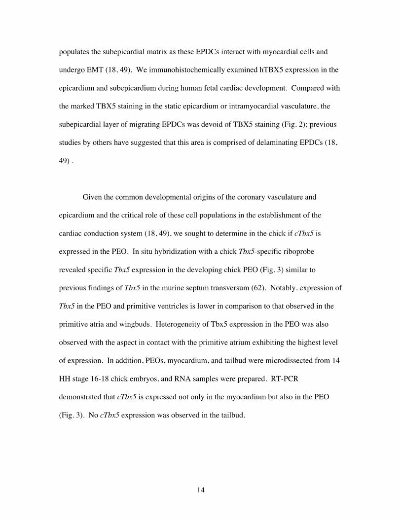
RESULTS
TBX5 expression in the human epicardium and coronary vasculature andin the
chick PEO
Human embryonic tissue (10-15 weeks of gestation) was analyzed by
immunohistochemistry for TBX5 expression (Fig. 1) with a previously characterized (22)
antibody to TBX5 that is directed against a specific TBX5 sequence carboxyl to the
TBX5 T-box. In addition to previously described (22) expression in cardiomyocytes and
atrioventricular nodal cells, marked staining for TBX5 was observed in the epicardium
and coronary vasculature. TBX5 expression was observed in the coronary arterial and
venous endothelium as well as in coronary arterial smooth muscle cells (Fig. 1A). TBX5
expression was not evident in vascular cells outside of the heart (Fig. 1B, C), i.e. aorta,
pulmonary artery, lungs, liver, kidney, skeletal muscle. Absence of staining with the
antibody used in the developing aorta (Fig. 1C) and foot (Fig. 1D) where TBX20 and
TBX18, respectively, are known to be expressed (28, 29, 45) confirmed that the anti-
TBX5 antibody used does not cross react with these other T-box genes that are expressed
in some vascular cells. As demonstrated previously, specificity of the antibody was
verified by reacting tissue sections with anti-TBX5 antibody preadsorbed with the TBX5
peptide immunogen; no staining was observed in these sections (data not shown) (22).
Further analyses of human embryonic tissues also suggested EPDC inactivation of
TBX5 expression in association with cell migration. During cardiogenesis, a
subpopulation of EPDCs migrates out of and delaminates from the epicardium and then
14
populates the subepicardial matrix as these EPDCs interact with myocardial cells and
undergo EMT (18, 49). We immunohistochemically examined hTBX5 expression in the
epicardium and subepicardium during human fetal cardiac development. Compared with
the marked TBX5 staining in the static epicardium or intramyocardial vasculature, the
subepicardial layer of migrating EPDCs was devoid of TBX5 staining (Fig. 2); previous
studies by others have suggested that this area is comprised of delaminating EPDCs (18,
49) .
Given the common developmental origins of the coronary vasculature and
epicardium and the critical role of these cell populations in the establishment of the
cardiac conduction system (18, 49), we sought to determine in the chick if cTbx5 is
expressed in the PEO. In situ hybridization with a chick Tbx5-specific riboprobe
revealed specific Tbx5 expression in the developing chick PEO (Fig. 3) similar to
previous findings of Tbx5 in the murine septum transversum (62). Notably, expression of
Tbx5 in the PEO and primitive ventricles is lower in comparison to that observed in the
primitive atria and wingbuds. Heterogeneity of Tbx5 expression in the PEO was also
observed with the aspect in contact with the primitive atrium exhibiting the highest level
of expression. In addition, PEOs, myocardium, and tailbud were microdissected from 14
HH stage 16-18 chick embryos, and RNA samples were prepared. RT-PCR
demonstrated that cTbx5 is expressed not only in the myocardium but also in the PEO
(Fig. 3). No cTbx5 expression was observed in the tailbud.
15
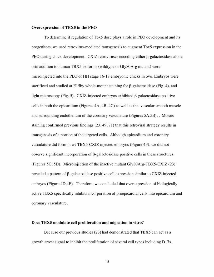
Overexpression of TBX5 in the PEO
To determine if regulation of Tbx5 dose plays a role in PEO development and its
progenitors, we used retrovirus-mediated transgenesis to augment Tbx5 expression in the
PEO during chick development. CXIZ retroviruses encoding either b-galactosidase alone
orin addition to human TBX5 isoforms (wildtype or Gly80Arg mutant) were
microinjected into the PEO of HH stage 16-18 embryonic chicks in ovo. Embryos were
sacrificed and studied at E15by whole-mount staining for b-galactosidase (Fig. 4), and
light microscopy (Fig. 5). CXIZ-injected embryos exhibited b-galactosidase positive
cells in both the epicardium (Figures 4A, 4B, 4C) as well as the vascular smooth muscle
and surrounding endothelium of the coronary vasculature (Figures 5A,5B), . Mosaic
staining confirmed previous findings (23, 49, 71) that this retroviral strategy results in
transgenesis of a portion of the targeted cells. Although epicardium and coronary
vasculature did form in wt-TBX5-CXIZ injected embryos (Figure 4F), we did not
observe significant incorporation of b-galactosidase positive cells in these structures
(Figures 5C, 5D). Microinjection of the inactive mutant Gly80Arg-TBX5-CXIZ (23)
revealed a pattern of b-galactosidase positive cell expression similar to CXIZ-injected
embryos (Figure 4D,4E). Therefore, we concluded that overexpression of biologically
active TBX5 specifically inhibits incorporation of proepicardial cells into epicardium and
coronary vasculature.
Does TBX5 modulate cell proliferation and migration in vitro?
Because our previous studies (23) had demonstrated that TBX5 can act as a
growth arrest signal to inhibit the proliferation of several cell types including D17s,
16
MEQC myc-transformed avian cardiomyocyte-like cells (26), and embryonic chick
cardiomyocytes, we considered the possibility that TBX5 overexpression might inhibit
the proliferation of a subset of proepicardial cells within the PEO and thereby prevent
their clonal expansion and further incorporation into PEO-derived structures. However,
immunostaining for PCNA in PEOs infected with either CXIZ or wt-TBX5-CXIZ both
revealed evidence of proliferation in virtually all cells. To further quantitatively
determine if TBX5 overexpression altered proepicardial cell proliferation, primary
cultures of proepicardial cells were established by disaggregating PEOs microdissected
from HH stage 16-18 chick embryos. Cultures were infected with CXIZ, wt-TBX5-
CXIZ, or Gly80Arg-TBX5-CXIZ retroviruses and then fixed and immunostained for
PCNA within 24 hours of initial proepicardial cell plating. These studies demonstrated
no significant difference in the fraction of PCNA-positive cells regardless of TBX5
isoform overexpression (data not shown). Quantitation of these studies is shown in Fig.
6A.
Since our experiments suggested that TBX5 does not significantly alter
proepicardial cell proliferation, we tested the hypothesis that TBX5 might inhibit
proepicardial cell migration out of the PEO and thereby inhibit incorporation of
retrovirus-infected cells into the epicardium and coronary vasculature. Because it had
been previously suggested that TBX5 regulation of cell migration might play a role in
establishment of the limb bud (2), we first sought to determine if TBX5 inhibited in vitro
migration of D17 cells. Osteosarcoma cell activity in vitro has been previously utilized
as an experimental model of bone formation (44, 61). We had previously shown that
17
TBX5 inhibited D17 cell proliferation, but recent data has suggested that the effects of
TBX5 on cell migration and cell proliferation might contribute to limb development.
D17 cells were infected with CXIZ, wt-TBX5-CXIZ or Gly80Arg-TBX5-CXIZ
retroviruses. Infected cells were allowed to migrate in culture for 10 days in the presence
of hydroxyurea, a potent inhibitor of cell proliferation. Quantification of cell migration
(Fig. 6B) revealed that wt-TBX5-CXIZ infected D17s migrated significantly less than
D17s infected with CXIZ or Gly80Arg-TBX5-CXIZ (p<0.0001). To determine if TBX5-
mediated inhibition of cell migration was cell autonomous, we assessed the migration of
CXIZ infected D17 cells co-cultured with D17s infected with wt-TBX5-CXIZ. Cultures
analyzed contained 0, 14, or 41% wt-TBX5-CXIZ infected D17s, so the majority of cells
at the migrating front would be CXIZ infected D17s. After 10 days of co-culture, we did
not observe (Fig. 6C) significant alterations (p=0.9) in D17 migration regardless of the
amount of wt-TBX5-CXIZ infected D17s present. Therefore, we concluded that TBX5
inhibited D17 cell migration in vitro in a cell autonomous fashion.
Because D17 osteosarcoma cell behavior may not mimic proepicardial cell
behavior, we explored the consequences of TBX5 overexpression on proepicardial cell
migration. Explanted PEOs were either infected in culture with CXIZ, wt-TBX5-CXIZ,
or Gly80Arg-TBX5-CXIZ retroviruses for 24 hours and maintained for a further 48 hours
in virus-free media, or they were transfected with pEGFP-C1 or pEGFP-C1-TBX5 for 4
hours and then maintained in normal media for a further 24 hours. Migration of cells was
assessed by b-galactosidase staining in retrovirus infected cultures or by fluorescence
microscopy in plasmid transfected cultures. As previously described (30), proepicardial
18
cells migrated out of the PEO in a radial fashion during culture of explanted PEOs. In
CXIZ and Gly80Arg-TBX5-CXIZ infected cultures, 88 and 91% respectively of b-
galactosidase positive cells had migrated out of the explant (Fig. 7A). By contrast, only
25% of b-galactosidase positive cells were seen in the wt-TBX5-CXIZ migrating
population, and most remained within the explanted PEO (Fig.7A). Notably, even
though a minority of proepicardial cells were infected by the retrovirus, there was no
evidence of inhibition of migration of noninfected cells out of the PEO. In cultures
transfected with EGFP-C1 plasmid alone, 81% of EGFP positive cells migrated out of the
explant (Fig. 7B). However, in EGFP-C1-TBX5 transfected cultures, only 32% of EGFP
positive cells migrated out of the explant while the majority remained within the
explanted PEO (Fig. 7C). Not only do these data support the hypothesis that TBX5
overexpression inhibits proepicardial cell migration, but they also confirm that this effect
is cell autonomous.
We further considered whether TBX5 activity is essential for proepicardial cell
migration. To evaluate this hypothesis, we determined the consequences of loss of cTbx5
activity on proepicardial cell migration in vitro by treatment with cTbx5 morpholino
antisense oligonucleotides (cTbx5-MO) derived from sequences flanking the cTbx5
consensus Kozak sequence. Similar oligonucleotides have been used by Ng et al. (2002),
Ahn et al. (2002), and Garrity et al. (16) to inhibit Tbx5 translation during zebrafish
pectoral limb bud and heart development. PEO explants were transfected with cTbx5-
MO or a control antisense oligomer comprised of the same sequence but in an inverted
orientation (cTbx5-INV). Since both oligomers were covalently tagged with Lissamine,
19
transfection efficiency was assessed by fluorescent microscopy, and 40-50% of cells
incorporated both oligomers. Proepicardial cell migration was assessed after 24 hours in
culture. In explants treated with either cTbx5-MO or cTbx5-INV, we observed no
change in migration of proepicardial cells that did not take up the antisense oligomers.
However, migration of cTbx5-MO transfected proepicardial cells was markedly inhibited
compared with the migration of cTbx5-INV transfected cells; most cTbx5-MO
proepicardial cells failed to migrate out of the explanted PEO (Fig. 8). These studies
further suggest that not only is the contribution of Tbx5 to cell migration cell
autonomous, but also that regulation of Tbx5 expression is required for normal
proepicardial cell migration.
TBX5 overexpression inhibits proepicardial cell migration in vivo
To determine if TBX5 has similar effects in vivo as it does on cell migration in
vitro, we analyzed the fate of TBX5 overexpressing proepicardial cells in the developing
chick in vivo (Fig. 9). PEOs of HH stages 16-18 embryonic chicks were microinjected
with CXIZ, Gly80Arg-TBX5-CXIZ, and wt-TBX5-CXIZ retrovirus, and the embryos
were sacrificed and stained for b-galactosidase activity at 8, 24 and 48 hours following
microinjection. In both CXIZ and wt-TBX5-CXIZ infected embryos, b-galactosidase
staining was evident in the PEO by 8 hours post-microinjection. By 24 hours post-
microinjection, CXIZ-infected proepicardial cells were noted to have begun to migrate
over the surface of the looping heart and, by 48 hours, were observed to incorporate into
nascent epicardium and vascular structures. Similar data was obtained for Gly80Arg-
TBX5-CXIZ infected cells (data not shown). However, at these 24 and 48 hour time
20
points, wt-TBX5-CXIZ infected proepicardial cells remained in the PEO and failed to
migrate over the heart surface.
Native Tbx5 expression in migrating proepicardial cells
Our studies suggested that disruption of Tbx5 signaling in genetically engineered
cells perturbs their capacity for migration. We therefore hypothesized that native Tbx5
expression might be dynamically regulated in cells that alternate between stationary and
migratory states during development. To address this, we compared cTbx5 expression in
chick proepicardial cells that were stationary or migrating. PEOs were explanted from 81
stage 16-18 chick embryos and maintained in organ culture for 72 hours by which time a
subpopulation of proepicardial cells had migrated out of the PEO onto the culture dish
while the rest remained within the PEO. From each culture, the residual, nonmigrating
PEO was removed by microdissection, and then the migrating proepicardial cells were
removed from the dish by trypsinization. RT-PCR analyses of cTbx5 expression were
performed on RNA samples from both populations of cells. These studies revealed that
like HH stage 16-18 proepicardial organs in vivo (Fig. 3), the nonmigrating cells from
cultured PEOs continued to express cTbx5 (Fig. 10). However, migrating proepicardial
cells no longer expressed detectable levels of cTbx5 (Fig. 10). Thus, migration of
proepicardial cells out of the PEOs in this culture model was associated with inactivation
of cTbx5 expression.
21
Do Tbx18 and Tbx20 contribute to Tbx5 effects on proepicardial cell migration?
Concomitant alterations in the expression levels of multiple T-box transcription
factors, e.g., Tbx5, Tbx2, Tbx3, and Tbx20, have been suggested to play a role in
myocardial development (19,51), and our observations of proepicardial cell behavior may
not be solely due to Tbx5 but rather imbalances of several PEO T-box genes. Both
Tbx18 and Tbx20 have been implicated in vascular development and are expressed in the
PEO/Septum transversum (8, 28). We used quantitative RT-PCR to determine if
proepicardial expression of Tbx18 and Tbx20 changed in response to Tbx5 knockdown
via antisense inhibition of Tbx5 expression as described above. Tbx18 expression was not
significantly changed in PEO cultures treated with cTbx5-MO compared with those
treated with cTbx5-INV (1.3 + 0.2 x). However, Tbx5 knockdown was associated with a
significant increase (1.9 + 0.3 x) in Tbx20 expression. Recently, Plageman et al. (51)
suggested that maintenance of Tbx5 and Tbx20 balance may be critical for myocardial
development. To determine if proepicardial cell migration was similarly regulated, we
directly tested the effect of altered Tbx20 expression on proepicardial cell migration. We
studied the consequences of both Tbx20 knockdown (using morpholino antisense
oligonucleotides) and Tbx20 overexpression (using pEGFP-C1-TBX20 plasmids – see
Fig. 7A and D) on proepicardial cell migration out of cultured PEO explants just as we
had for Tbx5. 83% and 89% of proepicardial cells subject to cTbx20 knockdown or
TBX20 overexpression, respectively, migrated out of the proepicardial explant. These
proportions were similar to the high proportion of migrating cells seen in control cultures
treated with inverted cTbx20 antisense oligomers (82%) or pEGFP-C1 plasmid (81%) but
significantly differed (p<0.01) from the low proportion of cells that migrated out of the

22
proepicardial explant in cultures treated with cTbx5 antisense oligomers (42%) or with
pEGFP-C1-wtTBX5 (16%).
23
DISCUSSION
In this study, we demonstrate that TBX5 is expressed in the PEO that contains
epicardial and coronary vascular progenitor cells and that TBX5 inhibits proepicardial
cell migration in vitro and in vivo. Mosaic overexpression of TBX5 in the PEO in vivo
during cardiogenesis inhibits proepicardial cell migration out of the PEO with consequent
impaired incorporation of transgenic cells into the epicardium and coronary blood
vessels. Proepicardial cell migration in vitro is affected by changes in TBX5 dosage and
genetically engineered augmentation or inhibition of TBX5 expression both inhibit
proepicardial cell migration. Furthermore, analyses of cultured chick proepicardial cells
and of human fetal tissues suggest that physiologic regulation of TBX5 expression may
occur in vivo in concert with both initiation and cessation of cell migration during
embryogenesis. Since we do not observe TBX5 expression in migrating proepicardial
cells, our studies suggest that there may be distinct temporal requirements for TBX5, and
that our genetic manipulations impair proepicardial cell migration by modifying TBX5
expression in the premigratory cells of the PEO.
We have previously shown that TBX5 inhibits cardiomyocyte proliferation during
myocardial development (23). Our data now extend the profile of TBX5 activity to
include regulation of migration and indicate that inhibition of migration by TBX5 is
partially independent of this transcription factor’s effects on cell proliferation. Analyses
of PCNA expression in TBX5 transgenic chick proepicardial cells demonstrate no effect
of TBX5 on proepicardial cell proliferation. Furthermore, unlike TBX5 inhibition of

24
proliferation, TBX5 inhibition of migration is cell autonomous. Co-culture of D17s with
D17s genetically engineered to overexpress TBX5 fails to modify their migration in vitro.
While TBX5 overexpression retards proepicardial cell migration out of cultured PEO
explants, migration of non-transgenic cells out of the same PEOs is not impeded.
Moreover, in the setting of mosaic overexpression of TBX5 in the chick PEO, further
cardiogenic development does support the establishment of epicardium and coronary
vasculature from non-transgenic cells.
TBX5 modulation of cell differentiation has been reported in both the heart and
the limb. In this study, inhibition of proepicardial cell migration occurs before the
differentiating events that constitute EMT and prevents the physical interaction of EPDCs
with myocardial cells that is required for EMT. Thus, we hypothesize that TBX5
regulation of cell migration is also independent of its effects on cell differentiation.
However, we cannot exclude TBX5 modulation of early, as yet uncharacterized,
differentiating events that might occur within the PEO before cells migrate over and into
the myocardium.
Investigation of Tbx5’s role in zebrafish fin development also suggests
independent contributions of Tbx5 to regulation of cell migration, proliferation, and
differentiation. Although there is a clear role for Tbx5 in cell differentiation and
proliferation during limb specification, Ahn et al. (2) demonstrated that the contribution
of Tbx5 to limb development commences prior to limb bud formation. Antisense
knockdown of Tbx5 impairs mesenchymal aggregation of limb precursor cells and, in a
25
cell autonomous fashion, prevents lateral plate mesodermal cells from migrating to the
nascent pectoral fin bud. Cell adhesive properties may also be changed during fin
development in response to altered Tbx5 dose, and likewise, defective proepicardial cell
adhesion during migration out of the PEO may contribute to the impaired epicardial
formation and coronary vasculogenesis in our chick studies. Altered dosage of other T-
box transcription factors has also been shown to modify cell migration. Loss of Tbx16 in
the zebrafish spadetail mutant causes a cell-autonomous distorted convergence of
marginal mesodermal cells during gastrulation (20). Wilson et al. (72) proposed that
abnormal notochord morphogenesis in the setting of loss of T(Brachyury) is a
consequence of a defective migration of posterior mesodermal cells through the primitive
streak that may involve abnormal cell adhesive properties. Future studies to decipher
mechanisms underlying TBX5 inhibition of proepicardial cell migration will explore
potential regulation of cell-cell adhesion molecules such as VCAM-1 (31) and BVES
(70), and cell-matrix adhesion molecules such as alpha-4 integrin (73) that are associated
with proepicardial development (59) as well as transcriptional regulators of epicardial
EMT such as Ets-1 and Ets-2 (35).
The molecular genetic events downstream from Tbx5 that contribute to regulation
of cell migration and EMT remain to be defined. FGF isoforms (1, 2, and 7), TGF-ß
isoforms (1, 2, and 3), Ets-1, Ets-2 and Fog2 have all been implicated in coronary
vasculogenesis and cardiac EMT (35, 50, 64, 69). Related molecules have all been tied
to T-box gene activity in other experimental models. FGF10 has been proposed to be a
downstream target of Tbx5 in murine limb development (1, 54). In Drosophila, the

26
TGFß homolog decapentaplegic acts to regulate expression of the T-box gene optomotor
blind during development of the wing imaginal disc (52). GATA-4 (the binding partner
of Fog2) interacts with Tbx5 and Nkx2.5 to activate synergistically expression of the
atrial natriuretic factor gene (15, 55, 68) and Gata-4 expression is altered in Tbx5
homozygous null mice (10). Thus, these are all intriguing candidate members of a Tbx5
dependent pathway operant in proepicardial cells and their descendants.
Although our data exclude altered Tbx18 and Tbx20 expression as explanations
for Tbx5-mediated modulation of proepicardial cell migration, they do not exclude a role
in other proepicardial cell behaviors including proliferation and differentiation. Nor do
our data exclude a role for Tbx5 in these other properties either. Additionally, the
observation that antisense knockdown of Tbx5 inhibits proepicardial cell migration while
proepicardial physiologic inactivation of Tbx5 expression is associated with promotion of
cell migration suggests that non-Tbx5 dependent pathways are operant in vivo as well.
Human and animal genetic studies have previously predicted that T-box gene dose is
finely regulated in normal development, and perturbations that either increase or decrease
T-box gene dose lead to abnormal organogenesis. Murine models of overexpression or
haploinsufficiency of Tbx1 all lead to aortic arch abnormalities that are also seen in
humans with DiGeorge syndrome who have deletions encompassing the Tbx1 gene (19,
27, 36, 46). Abnormal cardiogenesis is a feature of murine models of Tbx5
overexpression and haploinsufficiency (10, 34) and is similar to the human congenital
heart disease seen in Holt-Oram patients with TBX5 haploinsufficiency and in humans
with chromosome 12q2 duplications that include TBX5 (3, 32). Vascular anomalies
27
(patent ductus arteriosus, persistent left superior vena cava, anomalous pulmonary venous
return, aortic coarctation) have been variably noted in these human patients and animal
models, but coronary artery anatomy has not been well studied in these individuals.
TBX5 and other members of TBX5 dependent pathways in PEO development will be
appropriate candidates for genetic analyses in individuals with coronary artery and
epicardial anomalies.
28
ACKNOWLEDGMENTS
This work was supported by the March of Dimes Birth Defects Foundation (CTB), the
Edward Mallinckrodt, Jr. Foundation (CTB), the Cornell Vascular Medicine Foundation
(CTB), NIH R01-HL66214 (CTB), and an NHLBI Minority Investigator Research Award
(CJH). The authors are grateful to Andy Wessels, Maurice J.B. van den Hoff, David
Reese, Romulo Hurtado, and Theresa Zagreda for advice regarding PEO manipulation
and histologic analysis.
29
REFERENCES
1. Agarwal P, Wylie JN, Galceran J, Arkhitko O, Li C, Deng C, Grosschedl R and
Bruneau BG. Tbx5 is essential for forelimb bud initiation following patterning of the
limb field in the mouse embryo. Development 130: 623-633, 2003.
2. Ahn DG, Kourakis MJ, Rohde LA, Silver LM and Ho RK. T-box gene tbx5 is
essential for formation of the pectoral limb bud. Nature 417: 754-758, 2002.
3. Basson CT, Bachinsky DR, Lin RC, Levi T, Elkins JA, Soults J, Grayzel D,
Kroumpouzou E, Traill TA, Leblanc-Straceski J, Renault B, Kucherlapati R,
Seidman JG and Seidman CE. Mutations in human TBX5 cause limb and cardiac
malformation in Holt-Oram syndrome. Nat Genet 15: 30-35, 1997.
4. Basson CT, Knowles WJ, Bell L, Albelda SM, Castronovo V, Liotta LA and
Madri JA. Spatiotemporal segregation of endothelial cell integrin and nonintegrin
extracellular matrix-binding proteins during adhesion events. J Cell Biol 110: 789-801,
1990.
5. Basson CT, Kocher O, Basson MD, Asis A and Madri JA. Differential modulation
of vascular cell integrin and extracellular matrix expression in vitro by TGF-beta 1
correlates with reciprocal effects on cell migration. J Cell Physiol 153: 118-128, 1992.
6. Begemann G and Ingham PW. Developmental regulation of Tbx5 in zebrafish
embryogenesis. Mech Dev 90: 299-304, 2000.
7. Bray N, Dubchak I and Pachter L. AVID: A global alignment program. Genome Res
13: 97-102, 2003.
30
8. Brown DD, Binder O, Pagratis M, Parr BA and Conlon FL. Developmental
expression of the Xenopus laevis Tbx20 orthologue. Dev Genes Evol 212: 604-607, 2003.
9. Bruneau BG, Logan M, Davis N, Levi T, Tabin CJ, Seidman JG and Seidman
CE. Chamber-specific cardiac expression of Tbx5 and heart defects in Holt-Oram
syndrome. Dev Biol 211: 100-108, 1999.
10. Bruneau BG, Nemer G, Schmitt JP, Charron F, Robitaille L, Caron S, Conner
DA, Gessler M, Nemer M, Seidman CE and Seidman JG. A murine model of Holt-
Oram syndrome defines roles of the T-box transcription factor Tbx5 in cardiogenesis and
disease. Cell 106: 709-721, 2001.
11. Collavoli A, Hatcher CJ, He J, Okin D, Deo R and Basson CT. TBX5 nuclear
localization is mediated by dual cooperative intramolecular signals. J Mol Cell Cardiol
35: 1191-1195, 2003.
12. Crispino JD, Lodish MB, Thurberg BL, Litovsky SH, Collins T, Molkentin JD
and Orkin SH. Proper coronary vascular development and heart morphogenesis depend
on interaction of GATA-4 with FOG cofactors. Genes Dev 15: 839-844, 2001.
13. Drabkin HA, Parsy C, Ferguson K, Guilhot F, Lacotte L, Roy L, Zeng C, Baron
A, Hunger SP, Varella-Garcia M, Gemmill R, Brizard F, Brizard A and Roche J.
Quantitative HOX expression in chromosomally defined subsets of acute myelogenous
leukemia. Leukemia 16: 186-195, 2002.
14. Dubchak I, Brudno M, Loots GG, Pachter L, Mayor C, Rubin EM and Frazer
KA. Active conservation of noncoding sequences revealed by three-way species
comparisons. Genome Res 10: 1304-1306, 2000.

31
15. Garg V, Kathiriya IS, Barnes R, Schluterman MK, King IN, Butler CA,
Rothrock CR, Eapen RS, Hirayama-Yamada K, Joo K, Matsuoka R, Cohen JC and
Srivastava D. GATA4 mutations cause human congenital heart defects and reveal an
interaction with TBX5. Nature 424: 443-447, 2003.
16. Garrity DM, Childs S and Fishman MC. The heartstrings mutation in zebrafish
causes heart/fin Tbx5 deficiency syndrome. Development 129: 4635-4645, 2002.
17. Ghosh TK, Packham EA, Bonser AJ, Robinson TE, Cross SJ and Brook JD.
Characterization of the TBX5 binding site and analysis of mutations that cause Holt-
Oram syndrome. Hum Mol Genet 10: 1983-1994, 2001.
18. Gittenberger-de Groot AC, Vrancken Peeters MP, Mentink MM, Gourdie RG
and Poelmann RE. Epicardium-derived cells contribute a novel population to the
myocardial wall and the atrioventricular cushions. Circ Res 82: 1043-1052, 1998.
19. Goldmuntz E and Emanuel BS. Genetic disorders of cardiac morphogenesis. The
DiGeorge and velocardiofacial syndromes. Circ Res 80: 437-443, 1997.
20. Griffin KJ, Amacher SL, Kimmel CB and Kimelman D. Molecular identification
of spadetail: regulation of zebrafish trunk and tail mesoderm formation by T-box genes.
Development 125: 3379-3388, 1998.
21. Hamburger V and Hamilton HL. A series of normal stages in the development of
the chick embryo. J Morph 88: 49-92, 1951.
22. Hatcher CJ, Goldstein MM, Mah CS, Delia CS and Basson CT. Identification and
localization of TBX5 transcription factor during human cardiac morphogenesis. Dev Dyn
219: 90-95, 2000.
32
23. Hatcher CJ, Kim MS, Mah CS, Goldstein MM, Wong B, Mikawa T and Basson
CT. TBX5 transcription factor regulates cell proliferation during cardiogenesis. Dev Biol
230: 177-188, 2001.
24. Heicklen-Klein A and Evans T. T-box binding sites are required for activity of a
cardiac GATA-4 enhancer. Dev Biol 267: 490-504, 2004.
25. Iio A, Koide M, Hidaka K and Morisaki T. Expression pattern of novel chick T-
box gene, Tbx20. Dev Genes Evol 211: 559-562, 2001.
26. Jaffredo T, Chestier A, Bachnou N and Dieterlen-Lievre F. MC29-immortalized
clonal avian heart cell lines can partially differentiate in vitro. Exp Cell Res 192: 481-491,
1991.
27. Jerome LA and Papaioannou VE. DiGeorge syndrome phenotype in mice mutant
for the T-box gene, Tbx1. Nat Genet 27: 286-291, 2001.
28. Kraus F, Haenig B and Kispert A. Cloning and expression analysis of the mouse T-
box gene Tbx18. Mech Dev 100: 83-86, 2001.
29. Kraus F, Haenig B and Kispert A. Cloning and expression analysis of the mouse T-
box gene Tbx20. Mech Dev 100: 87-91, 2001.
30. Kubalak SW, Doevendans PA, Rockman HA, Hunter JJ, Tanaka N, Ross Jr. J
and Chien KR. Molecular analysis of cardiac muscle diseases based on mouse genetics.
In: Human molecular genetics, edited by Adolph KW. San Diego, Califormoa: Academic
Press, 1996, p. 470-487.
31. Kwee L, Baldwin HS, Shen HM, Stewart CL, Buck C, Buck CA and Labow MA.
Defective development of the embryonic and extraembryonic circulatory systems in
33
vascular cell adhesion molecule (VCAM-1) deficient mice. Development 121: 489-503,
1995.
32. Li QY, Newbury-Ecob RA, Terrett JA, Wilson DI, Curtis AR, Yi CH, Gebuhr T,
Bullen PJ, Robson SC, Strachan T, Bonnet D, Lyonnet S, Young ID, Raeburn JA,
Buckler AJ, Law DJ and Brook JD. Holt-Oram syndrome is caused by mutations in
TBX5, a member of the Brachyury (T) gene family. Nat Genet 15: 21-29, 1997.
33. Li WE, Waldo K, Linask KL, Chen T, Wessels A, Parmacek MS, Kirby ML and
Lo CW. An essential role for connexin43 gap junctions in mouse coronary artery
development. Development 129: 2031-2042, 2002.
34. Liberatore CM, Searcy-Schrick RD and Yutzey KE. Ventricular expression of
tbx5 inhibits normal heart chamber development. Dev Biol 223: 169-180, 2000.
35. Lie-Venema H, Gittenberger-de Groot AC, van Empel LJ, Boot MJ, Kerkdijk
H, de Kant E and DeRuiter MC. Ets-1 and Ets-2 transcription factors are essential for
normal coronary and myocardial development in chicken embryos. Circ Res 92: 749-756,
2003.
36. Lindsay EA, Vitelli F, Su H, Morishima M, Huynh T, Pramparo T, Jurecic V,
Ogunrinu G, Sutherland HF, Scambler PJ, Bradley A and Baldini A. Tbx1
haploinsufficieny in the DiGeorge syndrome region causes aortic arch defects in mice.
Nature 410: 97-101, 2001.
37. Loots GG, Ovcharenko I, Pachter L, Dubchak I and Rubin EM. rVista for
comparative sequence-based discovery of functional transcription factor binding sites.
Genome Res 12: 832-839, 2002.
34
38. Lu J, Richardson JA and Olson EN. Capsulin: a novel bHLH transcription factor
expressed in epicardial progenitors and mesenchyme of visceral organs. Mech Dev 73:
23-32, 1998.
39. Lu JR, McKinsey TA, Xu H, Wang DZ, Richardson JA and Olson EN. FOG-2, a
heart- and brain-enriched cofactor for GATA transcription factors. Mol Cell Biol 19:
4495-4502, 1999.
40. Macias D, Perez-Pomares JM, Garcia-Garrido L, Carmona R and Munoz-
Chapuli R. Immunoreactivity of the ets-1 transcription factor correlates with areas of
epithelial-mesenchymal transition in the developing avian heart. Anat Embryol (Berl)
198: 307-315, 1998.
41. Majka SM and McGuire PG. Regulation of urokinase expression in the developing
avian heart: a role for the Ets-2 transcription factor. Mech Dev 68: 127-137, 1997.
42. Manner J. Does the subepicardial mesenchyme contribute myocardioblasts to the
myocardium of the chick embryo heart? A quail-chick chimera study tracing the fate of
the epicardial primordium. Anat Rec 255: 212-226, 1999.
43. Mayor C, Brudno M, Schwartz JR, Poliakov A, Rubin EM, Frazer KA, Pachter
LS and Dubchak I. VISTA : visualizing global DNA sequence alignments of arbitrary
length. Bioinformatics 16: 1046-1047, 2000.
44. Mealey KL, Barhoumi R, Rogers K and Kochevar DT. Doxorubicin induced
expression of P-glycoprotein in a canine osteosarcoma cell line. Cancer Lett 126: 187-
192, 1998.
35
45. Meins M, Henderson DJ, Bhattacharya SS and Sowden JC. Characterization of
the human TBX20 gene, a new member of the T-Box gene family closely related to the
Drosophila H15 gene. Genomics 67: 317-332, 2000.
46. Merscher S, Funke B, Epstein JA, Heyer J, Puech A, Lu MM, Xavier RJ, Demay
MB, Russell RG, Factor S, Tokooya K, Jore BS, Lopez M, Pandita RK, Lia M,
Carrion D, Xu H, Schorle H, Kobler JB, Scambler P, Wynshaw-Boris A, Skoultchi
AI, Morrow BE and Kucherlapati R. TBX1 is responsible for cardiovascular defects in
velo-cardio-facial/DiGeorge syndrome. Cell 104: 619-629, 2001.
47. Mikawa T, Cohen-Gould L and Fischman DA. Clonal analysis of cardiac
morphogenesis in the chicken embryo using a replication-defective retrovirus. III:
Polyclonal origin of adjacent ventricular myocytes. Dev Dyn 195: 133-141, 1992.
48. Mikawa T and Fischman DA. Retroviral analysis of cardiac morphogenesis:
discontinuous formation of coronary vessels. Proc Natl Acad Sci U S A 89: 9504-9508,
1992.
49. Mikawa T and Gourdie RG. Pericardial mesoderm generates a population of
coronary smooth muscle cells migrating into the heart along with ingrowth of the
epicardial organ. Dev Biol 174: 221-232, 1996.
50. Morabito CJ, Dettman RW, Kattan J, Collier JM and Bristow J. Positive and
negative regulation of epicardial-mesenchymal transformation during avian heart
development. Dev Biol 234: 204-215, 2001.
51. Morcos PA. Achieving efficient delivery of morpholino oligos in cultured cells.
Genesis 30: 94-102, 2001.
36
52. Nellen D, Burke R, Struhl G and Basler K. Direct and long-range action of a DPP
morphogen gradient. Cell 85: 357-368, 1996.
53. Nemer G and Nemer M. Transcriptional activation of BMP-4 and regulation of
mammalian organogenesis by GATA-4 and -6. Dev Biol 254: 131-148, 2003.
54. Ng JK, Kawakami Y, Buscher D, Raya A, Itoh T, Koth CM, Rodriguez Esteban
C, Rodriguez-Leon J, Garrity DM, Fishman MC and Izpisua Belmonte JC. The limb
identity gene Tbx5 promotes limb initiation by interacting with Wnt2b and Fgf10.
Development 129: 5161-5170, 2002.
55. Ogura T. Tbx genes and development of vertebrate limb and heart. The 1st
international symposium on etiology and morphogenesis of congenital cardiovascular
disease in the post-genomic era, edited by Nakazawa M, Tokyo, Japan, 2002, p. 48-49.
56. Paxton C, Zhao H, Chin Y, Langner K and Reecy J. Murine Tbx2 contains
domains that activate and repress gene transcription. Gene 283: 117-124, 2002.
57. Plageman TF, Jr. and Yutzey KE. Differential expression and function of Tbx5 and
Tbx20 in cardiac development. J Biol Chem, 2004.
58. Pratt BA, Harris AS, Morrow JS and Madri JA. Mechanisms of cytoskeletal
regulation: modulation of aortic endothelial cell spectrin by the extracellular matrix. Am
J Pathol 117: 349-354, 1984.
59. Reese DE, Mikawa T and Bader DM. Development of the coronary vessel system.
Circ Res 91: 761-768, 2002.
60. Rosenthal N and Xavier-Neto J. From the bottom of the heart: anteroposterior
decisions in cardiac muscle differentiation. Curr Opin Cell Biol 12: 742-746, 2000.
37
61. Shoieb AM, Hahn KA and Barnhill MA. An in vivo/in vitro experimental model
system for the study of human osteosarcoma: canine osteosarcoma cells (COS31) which
retain osteoblastic and metastatic properties in nude mice. In Vivo 12: 463-472, 1998.
62. Stennard FA, Costa MW, Elliott DA, Rankin S, Haast SJ, Lai D, McDonald LP,
Niederreither K, Dolle P, Bruneau BG, Zorn AM and Harvey RP. Cardiac T-box
factor Tbx20 directly interacts with Nkx2-5, GATA4, and GATA5 in regulation of gene
expression in the developing heart. Dev Biol 262: 206-224, 2003.
63. Svensson EC, Huggins GS, Dardik FB, Polk CE and Leiden JM. A functionally
conserved N-terminal domain of the friend of GATA-2 (FOG-2) protein represses
GATA4-dependent transcription. J Biol Chem 275: 20762-20769, 2000.
64. Svensson EC, Huggins GS, Lin H, Clendenin C, Jiang F, Tufts R, Dardik FB and
Leiden JM. A syndrome of tricuspid atresia in mice with a targeted mutation of the gene
encoding Fog-2. Nat Genet 25: 353-356, 2000.
65. Svensson EC, Tufts RL, Polk CE and Leiden JM. Molecular cloning of FOG-2: a
modulator of transcription factor GATA-4 in cardiomyocytes. Proc Natl Acad Sci U S A
96: 956-961, 1999.
66. Szeto DP, Griffin KJ and Kimelman D. HrT is required for cardiovascular
development in zebrafish. Development 129: 5093-5101, 2002.
67. Takebayashi-Suzuki K, Yanagisawa M, Gourdie RG, Kanzawa N and Mikawa
T. In vivo induction of cardiac Purkinje fiber differentiation by coexpression of
preproendothelin-1 and endothelin converting enzyme-1. Development 127: 3523-3532,
2000.
38
68. Takeuchi JK, Ohgi M, Koshiba-Takeuchi K, Shiratori H, Sakaki I, Ogura K,
Saijoh Y and Ogura T. Tbx5 specifies the left/right ventricles and ventricular septum
position during cardiogenesis. Development 130: 5953-5964, 2003.
69. Tevosian SG, Deconinck AE, Tanaka M, Schinke M, Litovsky SH, Izumo S,
Fujiwara Y and Orkin SH. FOG-2, a cofactor for GATA transcription factors, is
essential for heart morphogenesis and development of coronary vessels from epicardium.
Cell 101: 729-739, 2000.
70. Wada AM, Reese DE and Bader DM. Bves: prototype of a new class of cell
adhesion molecules expressed during coronary artery development. Development 128:
2085-2093, 2001.
71. Wei Y and Mikawa T. Fate diversity of primitive streak cells during heart field
formation in ovo. Dev Dyn 219: 505-513, 2000.
72. Wilson V, Manson L, Skarnes WC and Beddington RS. The T gene is necessary
for normal mesodermal morphogenetic cell movements during gastrulation. Development
121: 877-886, 1995.
73. Yang JT, Rayburn H and Hynes RO. Cell adhesion events mediated by alpha 4
integrins are essential in placental and cardiac development. Development 121: 549-560,
1995.

39
FIGURE LEGENDS
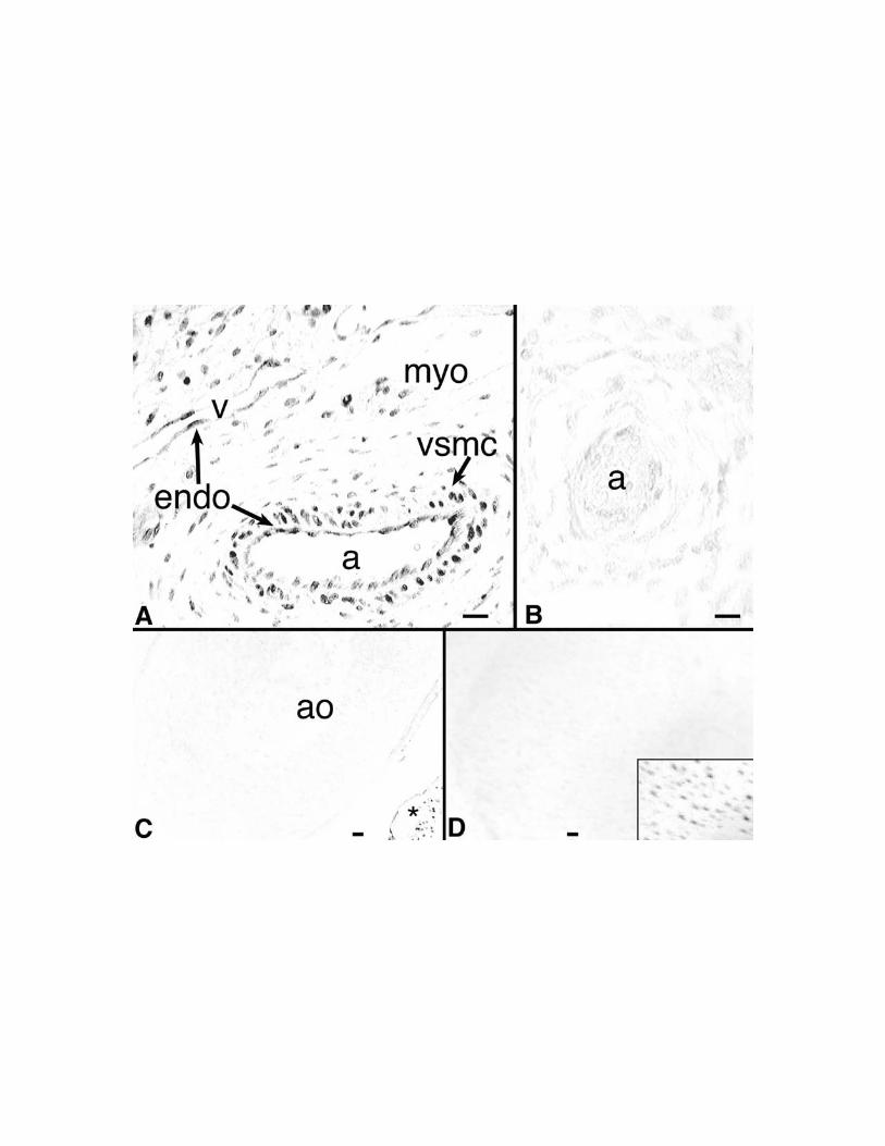
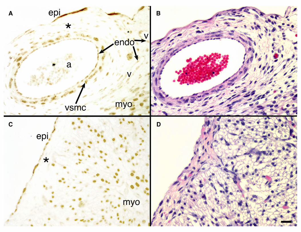
Figure 1. Immunohistochemical detection of TBX5 in human embryonic heart and
lung. Immunostaining for TBX5 in 15 weeks of gestation human embryonic (A) left
ventricle and (B) lung is shown. (A) In the embryonic heart, TBX5 is expressed not only
in the myocardium but also in coronary arteries (a) cells and veins (v). Vascular
endothelial (endo) and smooth muscle (vsmc) cells are indicated as well as myocardial
cardiomyocytes (myo). (B) In the embryonic lung, no staining is seen in the vasculature;
pulmonary arteriole is shown (a) (C) No staining is observed in the aorta (ao) and the
nascent outflow tract although staining is observed in adjacent atrial myocardium (*).
(D) No staining is observed in the distal phalanx of the great toe despite staining of
osteoblasts in the thumb (inset) co-stained in the same paraffin section.
[Bars = 20 mm.]
Figure 2. TBX5 is not expressed in the subepicardium of the embryonic human
heart. Immunostaining for TBX5 was performed in 15 week of gestation human heart.
Shown are sections through the lateral wall of the embryonic left ventricle (A,B) and
through the right atrial free wall (C,D). Immunohistochemistry with anti-TBX5 are
shown in panels (A,C). Although TBX5 is expressed in epicardial, vascular, and
myocardial cells, the subepicardium (*) comprised of delaminating EPDCs that will
ultimately form the nonmigrating vasculature is devoid of TBX5 expression. Labels
indicate epicardium (epi), myocardium (myo), arteriole (a), vein (v), vascular endothelial
40
cells (endo), and vascular smooth muscle cells (vsmc). Hematoxylin and eosin staining
of parallel sections (B,D) demonstrates the cellularity of the subepicardium
[Bar = 25 mm.]
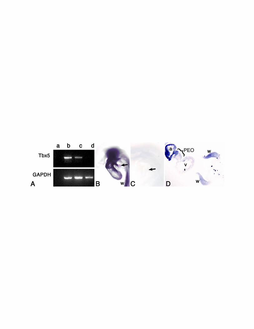
Figure 3. Analysis of cTbx5 expression in the embryonic chick heart and
proepicardial organ. (A)Total RNA was prepared from HH stage 16-18 embryonic
chick myocardium (lane b), PEO (lane c), and tailbud (lane d). 887 bp and 806 bp
segments of the chick Tbx5 and GAPDH genes respectively were amplified by RT-PCR.
cTbx5 is expressed in both the myocardium and PEO but not in the tailbud. Negative RT-
PCR control in which RNA was omitted from the sample undergoing amplification (lane
a). (B) In situ hybridization analysis of stage 17 chick embryo with an antisense cTbx5-
specific riboprobe. cTbx5 mRNA expression was observed in the PEO (arrow) as well as
in the wing bud (w) and heart. (C) Embryos hybridized with a sense cTbx5 riboprobe
showed no staining of the PEO (arrow). (D) Cross sectional analysis of a stage 17 chick
embryo confirmed cTbx5 expression in the PEO (indicated) as well as in the wing buds
(w) and primitive atrium (a) and ventricle (v).
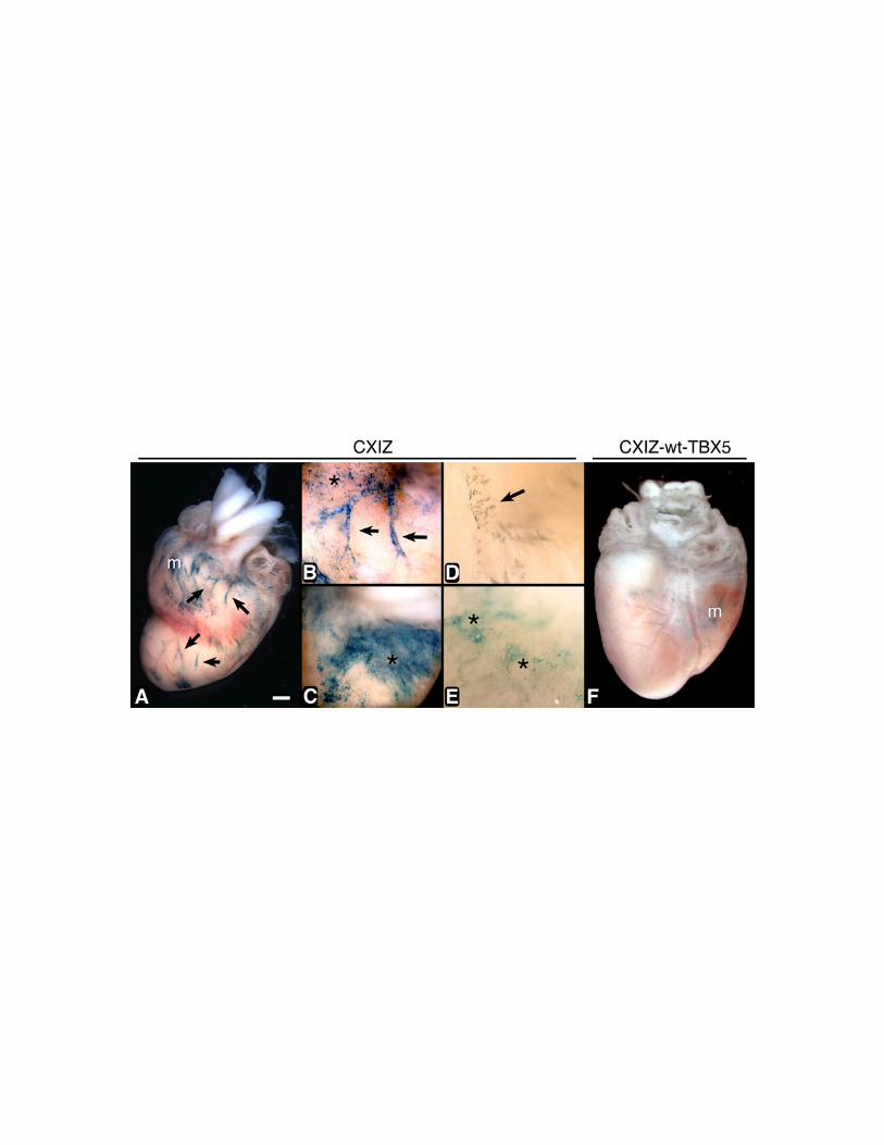
Figure 4. Retrovirus-mediated expression of wildtype and mutant hTBX5 in
embryonic chick PEO during heart development. CXIZ (A,B,C), Gly80Arg-TBX5-
CXIZ (D,E) and wt-TBX5-CXIZ (F) retroviruses were microinjected into chick PEOs in
ovo at HH stage 16-18. Embryos were sacrificed at E15, and whole mounts were stained
(blue) for b-galactosidase. Microscopic inspection revealed widespread expression of the
transgene in the epicardial cell layer (asterisks) and underlying coronary vessels (arrows)
41
in the CXIZ (A,B,C) and Gly80Arg-TBX5-CXIZ (D,E) infected hearts. (F) In wt-
TBX5-CXIZ infected hearts, staining of the epicardium and coronary vasculature was not
detected. In all cases, minimal myocyte expression of the transgene (m) is observed due
to leakage of the retrovirus into the myocardium during pressure microinjection, and thus
verifies both the infectivity of the retrovirus and expression of the transgene.
[Bar shown in panel A = (A,F) 800 mm, (B,C) = 400 mm, (D,E) = 100 mm.]
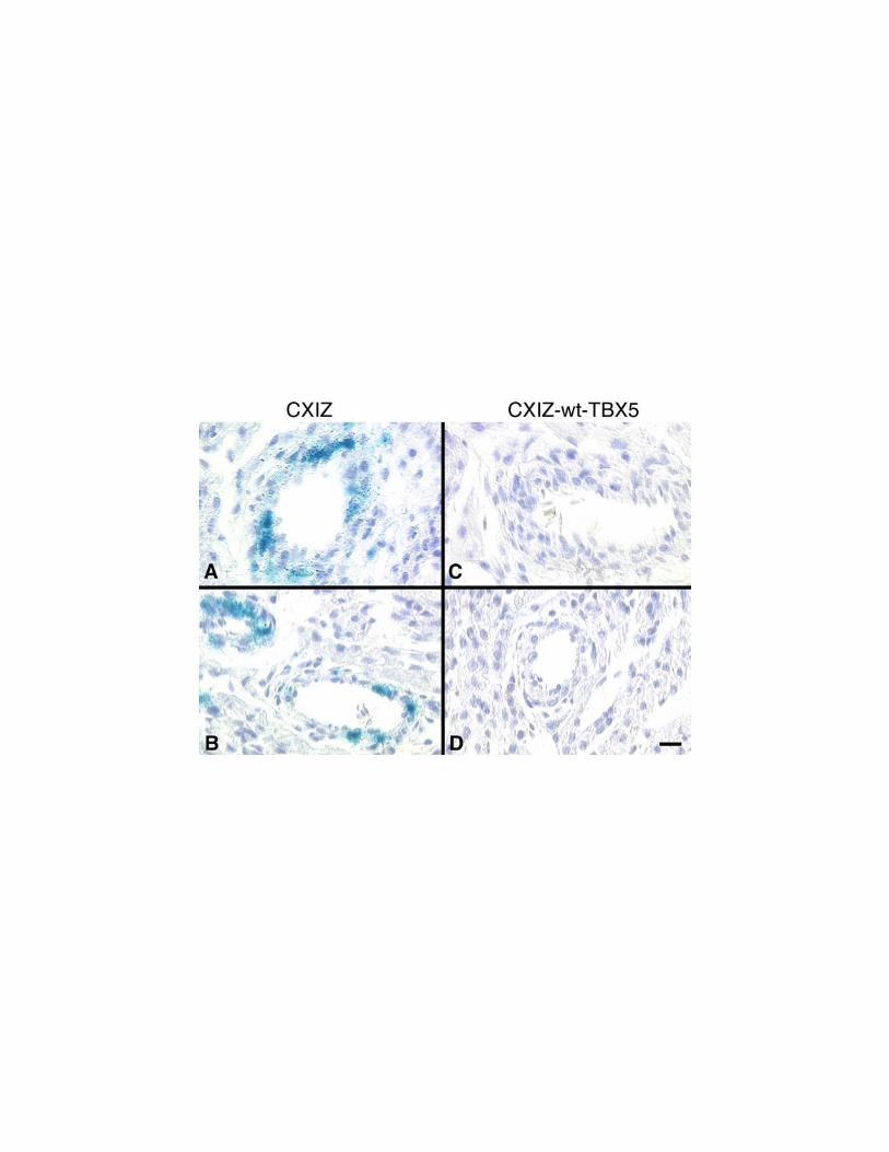
Figure 5. Histologic detection of retrovirus-mediated human TBX5 expression in
ovo. Histologic sections of embryonic chick hearts stained for b-galactosidase activity
after being infected with CXIZ (A,B) or wt-TBX5-CXIZ (C,D) retroviruses revealed
expression of b-galactosidase in the intimal endothelium and medial smooth muscle of
the coronary arteries in CXIZ microinjected hearts. However, this was undetectable in
the wt-TBX5-CXIZ hearts. [Bar = 20 mm.]
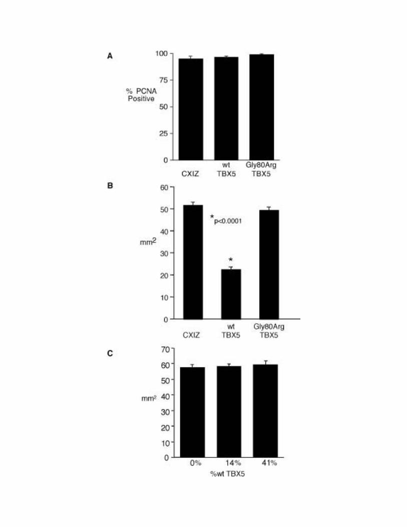
Figure 6. Effect of TBX5 overexpression on proepicardial cell proliferation and
D17 cell migration in vitro. (A) Primary cultures of proepicardial cells were infected
with CXIZ, wt-TBX5-CXIZ or Gly80Arg-TBX5-CXIZ retroviruses and immunostained
with anti-PCNA 24 hours after initial plating to determine the fraction of proliferating
cells. No significant effect of TBX5 on PEO cell proliferation was observed (p=0.532).
(B) Migration rates of D17 cells infected with CXIZ, wt-TBX5-CXIZ or Gly80Arg-
TBX5-CXIZ retroviruses were determined during a 10 day sheet migration assay.
Compared with CXIZ and Gly80Arg-TBX5-CXIZ retroviruses, infection of D17s with
wt-TBX5-CXIZ significantly inhibits migration. (p<0.0001). (C) D17 cells infected with
42
CXIZ were co-cultured with D17s infected with wt-TBX5-CXIZ at varied fractions and
subjected to a 10 day sheet migration assay. Increasing proportions of wt-TBX5-CXIZ
infected D17s produced no significant differences (p=0.9) in D17 migration.
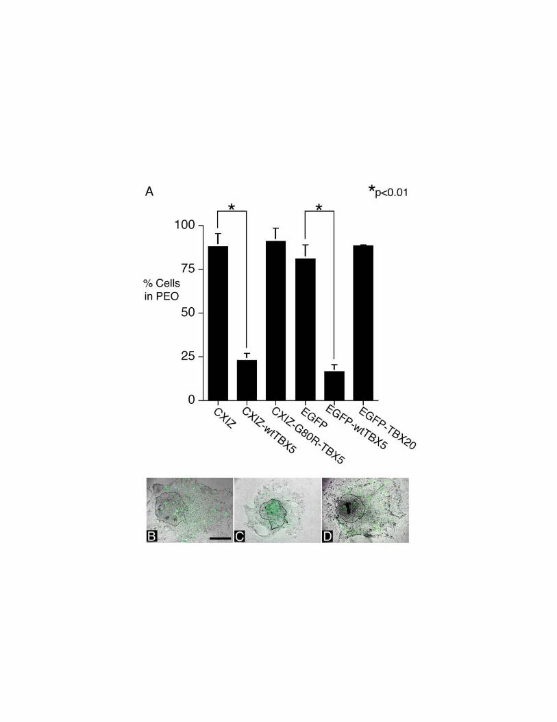
Figure 7. Effect of TBX5 overexpression on proepicardial cell migration in vitro.
The consequences of retroviral or plasmid mediated overexpression of TBX5 in
explanted PEOs were studied. (A) Quantitative determination of proepicardial cell
migration in vitro after infection with CXIZ retrovirus or retrovirus encoding wt-TBX5 or
Gly80Arg mutant TBX5. Significant inhibition of migration by wildtype TBX5 but not
by Gly80Arg TBX5 was observed. (B) A PEO explant transfected with pEGFP-C1 is
shown under phase microscopy. Most of the transfected cells (green), visualized by
fluorescent microscopy, migrate out of the initial PEO explant, outlined in black. (C)
Most of the pEGFP-C1-wtTBX5 transfected cells shown fail to migrate out of the PEO
explant. (D) Most of the pEGFP-C1-TBX20 transfected cells shown migrate out of the
PEO explant. [Bar = 300 mm for panels B,C,D]
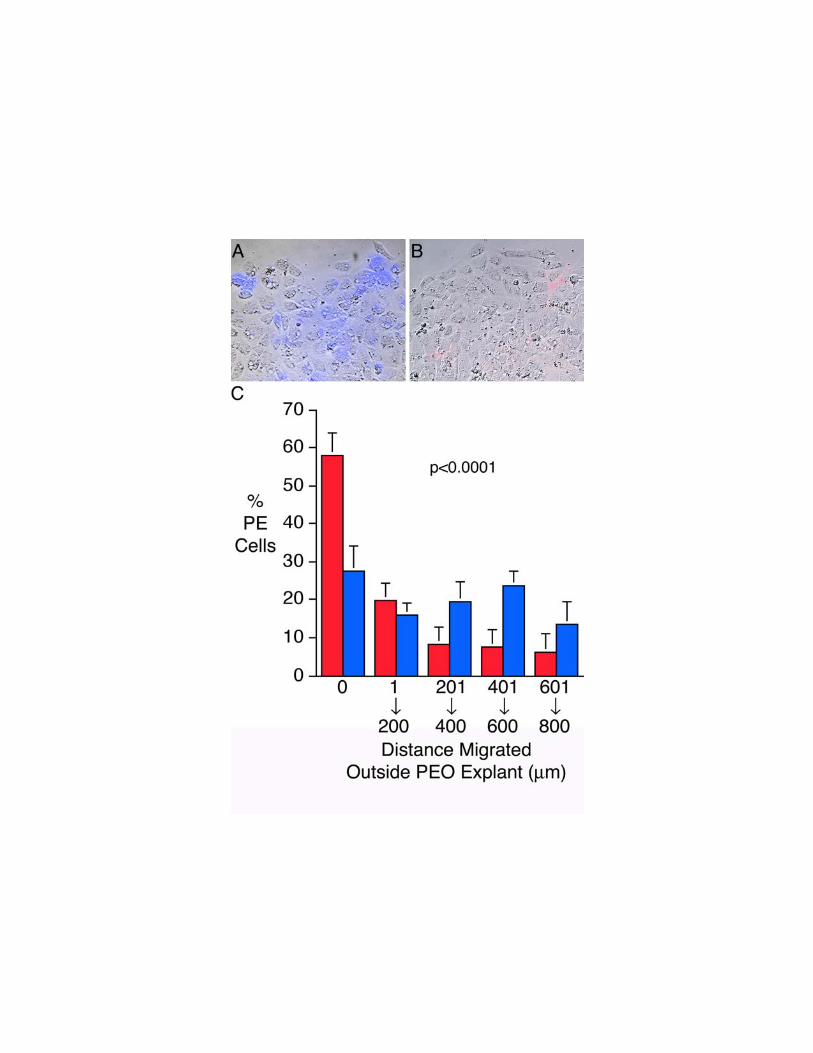
Figure 8. Antisense suppression of cTbx5 inhibits proepicardial cell migration in
vitro. Fluorescence microscopy of proepicardial explants 24 hours after being
transfected with (A) cTbx5-MO antisense oligonucleotide or (B) the cTbx5-INV control
oligonucleotide reveals that cTbx5 suppression prevents treated cells from migrating out
of the explanted organ. Leading edges of migration are shown, and direction of
migration is towards the top of the figure. Transfected cells (blue in Panel A, red in
Panel B) are detected by fluorescence microscopy. (C) Quantitative analysis
43
demonstrates that unlike cTbx5-INV (blue) transfected cells that migrate progressively
out of the proepicardial explant, cTbx5-MO (red) transfected cells largely remain in the
proepicardial organ explant, and concentric rings further from the explant contain fewer
and fewer cTbx5-MO transfected cells. Shown are the percentages of transfected
proepicardial cells in each culture that are located within the explant (i.e. 0 mm migrated)
or at varying distances outside the explant (1-200 mm, 201-400 mm, 301-600 mm, or 601-
800 mm).
Figure 9. Effect of TBX5 overexpression on proepicardial cell migration in vivo. 8
(A,D), 24 (B,E) and 48 (C,F) hours following microinjection of PEOs in ovo with CXIZ
(A,B,C) or wt-TBX5-CXIZ (D,E,F) retroviruses, chick embryos were sacrificed, fixed,
and stained for b-galactosidase activity. (A,D) b-galactosidase staining (arrows) was
evident in PEOs 8 hours post-microinjection regardless of which retrovirus was injected.
(B) By 24 hours post-microinjection, CXIZ-infected proepicardial cells (arrow) began
migrating out of the PEO and onto the surface of the naked myocardium. (C) By 48
hours, the PEO had involuted and CXIZ infected proepicardial cells had incorporated into
the epicardium and underlying coronary vessels. (E) However, at 24 hours post-
injection of wt-TBX5-CXIZ into embryos, infected proepicardial cells (arrow) remained
in the PEO. (F) By 48 hours, only rare wt-TBX5-CXIZ infected proepicardial cells
(arrow) were evident outside of the now involuted PEO.
44
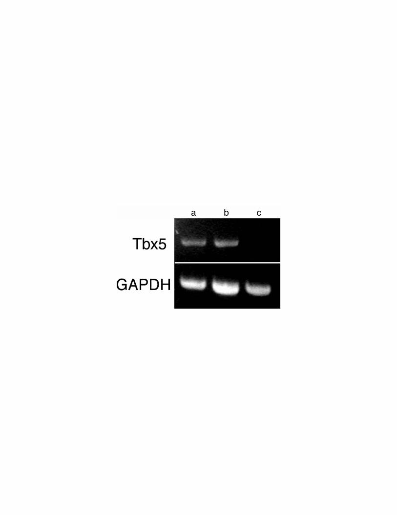
Figure 10. RT-PCR analysis of cTbx5 expression in embryonic chick PEO explants
and migrating proepicardial cells. After 72 hours in culture, proepicardial cells
migrated onto the culture dish out of PEO explants. These explants were microdissected
from the culture dish, separate RNA pools were prepared from the residual stationary
PEO and the migrating proepicardial cells. RT-PCR analyses for cTbx5 and cGAPDH
were performed as in Figure 2 from RNA derived from (a) chick myocardium [positive
control for cTbx5 and cGAPDH expression], (b) PEO explant, and (c) migrating
proepicardial cells. cTbx5 is expressed in the residual nonmigrating PEO explant (b), but
not in the migratory proepicardial cells (c).

Copyright © 2022 FDOKUMEN