Dynamics of Neutrophil Migration in Lymph Nodes during Infection
10
Immunity Article Dynamics of Neutrophil Migration in Lymph Nodes during Infection Tatyana Chtanova, 1,3 Marie Schaeffer, 1,3 Seong-Ji Han, 1,3 Giel G. van Dooren, 2 Marcelo Nollmann, 1 Paul Herzmark, 1 Shiao Wei Chan, 1 Harshita Satija, 1 Kristin Camfield, 1 Holly Aaron, 1 Boris Striepen, 2 and Ellen A. Robey 1, * 1 Department of Molecular and Cell Biology, Life Sciences Addition, University of California, Berkeley, CA 94720, USA 2 Center for Tropical & Emerging Global Diseases and Department of Cellular Biology, University of Georgia, Paul Coverdell Center, Athens, GA 30602, USA 3 These authors contributed equally to this work *Correspondence: [email protected] DOI 10.1016/j.immuni.2008.07.012 SUMMARY Although the signals that control neutrophil migration from the blood to sites of infection have been well characterized, little is known about their migration patterns within lymph nodes or the strategies that neutrophils use to find their local sites of action. To address these questions, we used two-photon scan- ning-laser microscopy to examine neutrophil migra- tion in intact lymph nodes during infection with an intracellular parasite, Toxoplasma gondii. We found that neutrophils formed both small, transient and large, persistent swarms via a coordinated migration pattern. We provided evidence that cooperative ac- tion of neutrophils and parasite egress from host cells could trigger swarm formation. Neutrophil swarm formation coincided in space and time with the removal of macrophages that line the subcapsu- lar sinus of the lymph node. Our data provide insights into the cellular mechanisms underlying neutrophil swarming and suggest new roles for neutrophils in shaping immune responses. INTRODUCTION Neutrophils are the most abundant nucleated cell in the blood and play a crucial role in immune responses to pathogens. Neutrophils are best known for their role in phagocytosis and killing of extracel- lular bacteria; however, they can provide protection against a di- verse set of pathogens, and do so by performing a variety of differ- ent functions (reviewed in Appelberg [2007] and Nathan [2006]). These functions include tissue remodeling, antigen presentation, recruiting other blood cells, and polarizing T cell responses (Beau- villain et al., 2007; Megiovanni et al., 2006; Pesce et al., 2008; Tvin- nereim et al., 2004). For example, neutrophils play an important protective role during infection with the intracellular protozoan parasite Toxoplasma gondii (Bennouna et al., 2003; Bliss et al., 2001; Denkers et al., 2004; Sayles and Johnson, 1996) in spite of the fact that the parasite is relatively resistant to killing by neu- trophils (Channon et al., 2000; Nakao and Konishi, 1991b). There is evidence that the protective effect of neutrophils during Toxo- plasma infection is due in part to the production of interleukin-12 (IL-12) by neutrophils and the subsequent shaping of the adaptive immune response (Bennouna et al., 2003). However, the precise roles of neutrophils during infection remain poorly understood. Neutrophils are produced in the bone marrow, circulate in the blood, and are rapidly recruited to sites of infection in response to a variety of chemoattractants produced by inflamed tissues (reviewed in Baggiolini [1998] and Scapini et al. [2000]). A number of studies have shown that neutrophils can also traffic to lymph nodes in response to infection (Abadie et al., 2005; Maletto et al., 2006; Pesce et al., 2008), raising the possibility that neutrophils may modulate immune responses within lymph nodes. One powerful tool for investigating the function of cells within lymph nodes is two-photon scanning-laser microscopy (TPSLM), an imaging method that provides dynamic information about cell migration and interactions within tissue samples (reviewed in Bousso and Robey [2004], Germain et al. [2006], Sumen et al. [2004], and Cahalan and Parker [2008]). Thus far, this approach has been primarily used to examine responses to model antigens by T and B cells and is just beginning to be applied in the setting of infection and to examine immune responses of nonlymphoid cells (Egen et al., 2008; Zinselmeyer et al., 2008). Despite their abundance, physiological importance, and clear indications that they can traffic to lymph nodes during infection, we know very little about what neutrophils do in the lymph node. Do neutrophils work individually, or in groups? What are the cues that guide neutrophil migration within lymph nodes? What impact do neutrophils have on other cell types in lymph nodes? Here, we addressed these questions using TPSLM and a Toxo- plasma gondii-mouse infection model. We found that neutrophils accumulate in the subcapsular sinus of the draining lymph node after infection and form both small, transient and large, persistent swarms via a highly coordinated migration pattern. We provided evidence that cooperative action of neutrophils and parasite egress from host cells can trigger swarm formation and that neu- trophil swarms lead to the removal of macrophages that line the subcapsular sinus of the lymph node. These results provide insight into the cellular mechanisms that lead to neutrophil swarms and suggest new potential functions for neutrophils in lymph nodes. RESULTS An Experimental Model to Examine Neutrophil Migration in Lymph Nodes In order to examine the behavior of neutrophils in lymph nodes during infection, we infected mice with the intracellular Immunity 29, 487–496, September 19, 2008 ª2008 Elsevier Inc. 487
-
Upload
independent -
Category
Documents
-
view
3 -
download
0
Transcript of Dynamics of Neutrophil Migration in Lymph Nodes during Infection

Immunity
Article
Dynamics of Neutrophil Migrationin Lymph Nodes during InfectionTatyana Chtanova,1,3 Marie Schaeffer,1,3 Seong-Ji Han,1,3 Giel G. van Dooren,2 Marcelo Nollmann,1 Paul Herzmark,1
Shiao Wei Chan,1 Harshita Satija,1 Kristin Camfield,1 Holly Aaron,1 Boris Striepen,2 and Ellen A. Robey1,*1Department of Molecular and Cell Biology, Life Sciences Addition, University of California, Berkeley, CA 94720, USA2Center for Tropical & Emerging Global Diseases and Department of Cellular Biology, University of Georgia, Paul Coverdell Center, Athens,GA 30602, USA3These authors contributed equally to this work
*Correspondence: [email protected]
DOI 10.1016/j.immuni.2008.07.012
SUMMARY
Although the signals that control neutrophil migrationfrom the blood to sites of infection have been wellcharacterized, little is known about their migrationpatterns within lymph nodes or the strategies thatneutrophils use to find their local sites of action. Toaddress these questions, we used two-photon scan-ning-laser microscopy to examine neutrophil migra-tion in intact lymph nodes during infection with anintracellular parasite, Toxoplasma gondii. We foundthat neutrophils formed both small, transient andlarge, persistent swarms via a coordinated migrationpattern. We provided evidence that cooperative ac-tion of neutrophils and parasite egress from hostcells could trigger swarm formation. Neutrophilswarm formation coincided in space and time withthe removal of macrophages that line the subcapsu-lar sinus of the lymph node. Our data provide insightsinto the cellular mechanisms underlying neutrophilswarming and suggest new roles for neutrophils inshaping immune responses.
INTRODUCTION
Neutrophils are the most abundant nucleated cell in the blood and
play a crucial role in immune responses to pathogens. Neutrophils
arebest known for their role inphagocytosis and killingof extracel-
lular bacteria; however, they can provide protection against a di-
verse set of pathogens, and do so by performing a variety of differ-
ent functions (reviewed in Appelberg [2007] and Nathan [2006]).
These functions include tissue remodeling, antigen presentation,
recruiting other blood cells, and polarizing T cell responses (Beau-
villain et al., 2007; Megiovanni et al., 2006; Pesce et al., 2008; Tvin-
nereim et al., 2004). For example, neutrophils play an important
protective role during infection with the intracellular protozoan
parasite Toxoplasma gondii (Bennouna et al., 2003; Bliss et al.,
2001; Denkers et al., 2004; Sayles and Johnson, 1996) in spite
of the fact that the parasite is relatively resistant to killing by neu-
trophils (Channon etal., 2000; Nakaoand Konishi, 1991b). There is
evidence that the protective effect of neutrophils during Toxo-
plasma infection is due in part to the production of interleukin-12
(IL-12) by neutrophils and the subsequent shaping of the adaptive
Im
immune response (Bennouna et al., 2003). However, the precise
roles of neutrophils during infection remain poorly understood.
Neutrophils are produced in the bone marrow, circulate in the
blood, and are rapidly recruited to sites of infection in response
to a variety of chemoattractants produced by inflamed tissues
(reviewed in Baggiolini [1998] and Scapini et al. [2000]). A number
of studies have shown that neutrophils can also traffic to lymph
nodes in response to infection (Abadie et al., 2005; Maletto et al.,
2006; Pesce et al., 2008), raising the possibility that neutrophils
may modulate immune responses within lymph nodes. One
powerful tool for investigating the function of cells within lymph
nodes is two-photon scanning-laser microscopy (TPSLM), an
imaging method that provides dynamic information about cell
migration and interactions within tissue samples (reviewed in
Bousso and Robey [2004], Germain et al. [2006], Sumen et al.
[2004], and Cahalan and Parker [2008]). Thus far, this approach
has been primarily used to examine responses to model antigens
by T and B cells and is just beginning to be applied in the setting
of infection and to examine immune responses of nonlymphoid
cells (Egen et al., 2008; Zinselmeyer et al., 2008).
Despite their abundance, physiological importance, and clear
indications that they can traffic to lymph nodes during infection,
we know very little about what neutrophils do in the lymph node.
Do neutrophils work individually, or in groups? What are the
cues that guide neutrophil migration within lymph nodes? What
impact do neutrophils have on other cell types in lymph nodes?
Here, we addressed these questions using TPSLM and a Toxo-
plasma gondii-mouse infection model. We found that neutrophils
accumulate in the subcapsular sinus of the draining lymph node
after infection and form both small, transient and large, persistent
swarms via a highly coordinated migration pattern. We provided
evidence that cooperative action of neutrophils and parasite
egress from host cells can trigger swarm formation and that neu-
trophil swarms lead to the removal of macrophages that line the
subcapsular sinusof the lymphnode.These resultsprovide insight
into the cellular mechanisms that lead to neutrophil swarms and
suggest new potential functions for neutrophils in lymph nodes.
RESULTS
An Experimental Model to Examine Neutrophil Migrationin Lymph NodesIn order to examine the behavior of neutrophils in lymph nodes
during infection, we infected mice with the intracellular
munity 29, 487–496, September 19, 2008 ª2008 Elsevier Inc. 487

Immunity
Neutrophil Migration in Lymph Nodes
protozoan parasite Toxoplasma gondii. After injection of live fluo-
rescent parasites into the earflap, parasites can be found within 1
hr in the subcapsular sinus of the draining lymph node in associ-
ation with LYVE1+ lymphatic vessels (Figure 1A). Many parasites
are found within the CD169+ macrophages that line the subcap-
sular sinus (Figure 1B, middle), a distribution similar to that seen
with other particulate antigens, such as viruses and immune
complexes (Carrasco and Batista, 2007; Junt et al., 2007; Phan
et al., 2007).
In order to track neutrophils relative to red fluorescent protein
(RFP)-labeled parasites, we used reporter mice in which GFP is
under the control of the lysozyme M promoter (lysGFP) (Faust
et al., 2000). Although this reporter is also expressed by mac-
rophages and a subset of dendritic cells, the vast majority of
Figure 1. Location of T. gondii Relative
to Lymphatics, CD169 Macrophages, and
Neutrophils in Draining Lymph Nodes after
Ear-Flap Infection
(A) A 20 mm frozen section of a draining lymph
node 4 hr after an earflap injection with RFP (red)
parasites. The middle and right panels show stain-
ing with LYVE-1 (blue) used for visualization of the
lymphatic system. The right panel shows a high-
magnification image.
(B) A 20 um frozen section of a draining lymph
node from a mouse bearing the LysGFP reporter
(green) 4 hr after an earflap injection with RFP
(red) parasites and stained with CD169 (blue) to
label subcapsular sinus macrophages. The middle
panel shows an enlarged area with parasites
inside CD169+ cells. The right panel shows an en-
larged area with intact parasites inside neutro-
phils.
(C) Same samples as in (A) showing signal from the
LysGFP reporter (green).
(D) Clusters of LysGFP high cells (green) (left
panel) stain positive for the neutrophil marker
Ly6G (blue). The right panel shows the same
image without the LysGFP signal.
the GFPhi cells in the lymph nodes at
1–5 hr after infection are neutrophils, on
the basis of their cell-surface phenotype
(Ly6GhiCD11bhiCD11clo) (Rydstrom and
Wick, 2007; Sasmono et al., 2007) (see
Figure S1A available online). Neutrophil
recruitment to the draining lymph nodes
occurred rapidly and was dependent on
the Toll-like receptor (TLR)-interleukin-1
receptor (IL-1R) adaptor protein,
MyD88 (Figure S1A). Interestingly, many
neutrophils were associated with
LYVE1+ lymphatic vessels (Figure 1C).
This is consistent with the possibility
that neutrophils entered the lymph
node via lymphatics, as has been re-
ported previously (Abadie et al., 2005;
Maletto et al., 2006). We also detected
neutrophils within blood vessels of in-
fected lymph nodes (data not shown),
suggesting that neutrophils may enter lymph nodes via both
blood and lymph.
At early times after infection, neutrophils contained propor-
tionally more parasites compared to macrophages and den-
dritic cells (Figure S1B). Although phagocytic destruction of
pathogens is one protective mechanism used by neutrophils,
the parasites inside neutrophils appear intact (Figure 1B, right).
This is consistent with evidence that T. gondii can divide
in vitro in human neutrophils (Channon et al., 2000; Nakao
and Konishi, 1991b) and indications that the protective effects
of neutrophils during T. gondii infection are due to immunoreg-
ulation rather than direct killing mechanisms (Bennouna et al.,
2003; Bliss et al., 2001; Denkers et al., 2004; Sayles and
Johnson, 1996).
488 Immunity 29, 487–496, September 19, 2008 ª2008 Elsevier Inc.
Immunity
Neutrophil Migration in Lymph Nodes
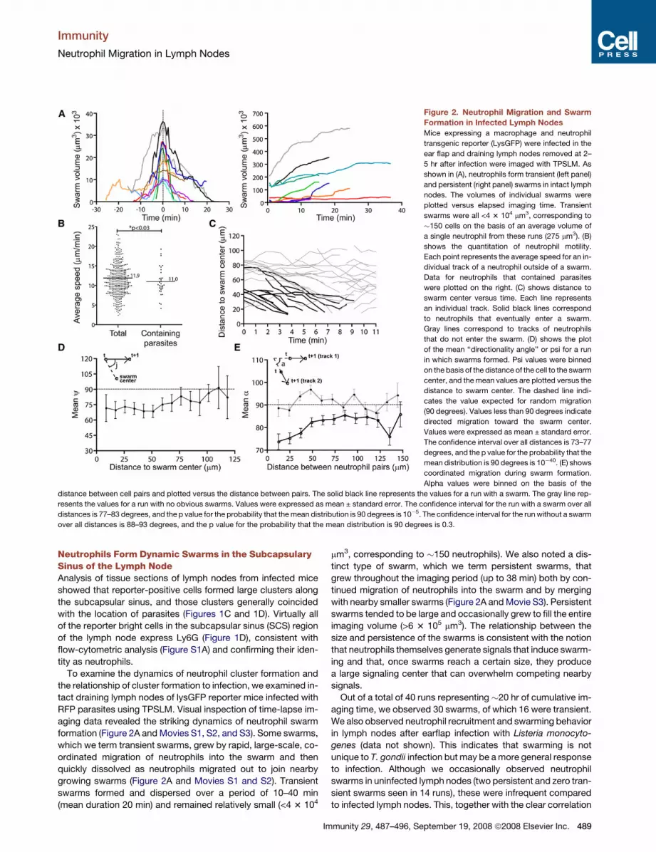
Figure 2. Neutrophil Migration and Swarm
Formation in Infected Lymph Nodes
Mice expressing a macrophage and neutrophil
transgenic reporter (LysGFP) were infected in the
ear flap and draining lymph nodes removed at 2–
5 hr after infection were imaged with TPSLM. As
shown in (A), neutrophils form transient (left panel)
and persistent (right panel) swarms in intact lymph
nodes. The volumes of individual swarms were
plotted versus elapsed imaging time. Transient
swarms were all <4 3 104 mm3, corresponding to
�150 cells on the basis of an average volume of
a single neutrophil from these runs (275 mm3). (B)
shows the quantitation of neutrophil motility.
Each point represents the average speed for an in-
dividual track of a neutrophil outside of a swarm.
Data for neutrophils that contained parasites
were plotted on the right. (C) shows distance to
swarm center versus time. Each line represents
an individual track. Solid black lines correspond
to neutrophils that eventually enter a swarm.
Gray lines correspond to tracks of neutrophils
that do not enter the swarm. (D) shows the plot
of the mean ‘‘directionality angle’’ or psi for a run
in which swarms formed. Psi values were binned
on the basis of the distance of the cell to the swarm
center, and the mean values are plotted versus the
distance to swarm center. The dashed line indi-
cates the value expected for random migration
(90 degrees). Values less than 90 degrees indicate
directed migration toward the swarm center.
Values were expressed as mean ± standard error.
The confidence interval over all distances is 73–77
degrees, and the p value for the probability that the
mean distribution is 90 degrees is 10�40. (E) shows
coordinated migration during swarm formation.
Alpha values were binned on the basis of the
distance between cell pairs and plotted versus the distance between pairs. The solid black line represents the values for a run with a swarm. The gray line rep-
resents the values for a run with no obvious swarms. Values were expressed as mean ± standard error. The confidence interval for the run with a swarm over all
distances is 77–83 degrees, and the p value for the probability that the mean distribution is 90 degrees is 10�5. The confidence interval for the run without a swarm
over all distances is 88–93 degrees, and the p value for the probability that the mean distribution is 90 degrees is 0.3.
Neutrophils Form Dynamic Swarms in the SubcapsularySinus of the Lymph NodeAnalysis of tissue sections of lymph nodes from infected mice
showed that reporter-positive cells formed large clusters along
the subcapsular sinus, and those clusters generally coincided
with the location of parasites (Figures 1C and 1D). Virtually all
of the reporter bright cells in the subcapsular sinus (SCS) region
of the lymph node express Ly6G (Figure 1D), consistent with
flow-cytometric analysis (Figure S1A) and confirming their iden-
tity as neutrophils.
To examine the dynamics of neutrophil cluster formation and
the relationship of cluster formation to infection, we examined in-
tact draining lymph nodes of lysGFP reporter mice infected with
RFP parasites using TPSLM. Visual inspection of time-lapse im-
aging data revealed the striking dynamics of neutrophil swarm
formation (Figure 2A and Movies S1, S2, and S3). Some swarms,
which we term transient swarms, grew by rapid, large-scale, co-
ordinated migration of neutrophils into the swarm and then
quickly dissolved as neutrophils migrated out to join nearby
growing swarms (Figure 2A and Movies S1 and S2). Transient
swarms formed and dispersed over a period of 10–40 min
(mean duration 20 min) and remained relatively small (<4 3 104
Im
mm3, corresponding to �150 neutrophils). We also noted a dis-
tinct type of swarm, which we term persistent swarms, that
grew throughout the imaging period (up to 38 min) both by con-
tinued migration of neutrophils into the swarm and by merging
with nearby smaller swarms (Figure 2A and Movie S3). Persistent
swarms tended to be large and occasionally grew to fill the entire
imaging volume (>6 3 105 mm3). The relationship between the
size and persistence of the swarms is consistent with the notion
that neutrophils themselves generate signals that induce swarm-
ing and that, once swarms reach a certain size, they produce
a large signaling center that can overwhelm competing nearby
signals.
Out of a total of 40 runs representing �20 hr of cumulative im-
aging time, we observed 30 swarms, of which 16 were transient.
We also observed neutrophil recruitment and swarming behavior
in lymph nodes after earflap infection with Listeria monocyto-
genes (data not shown). This indicates that swarming is not
unique to T. gondii infection but may be a more general response
to infection. Although we occasionally observed neutrophil
swarms in uninfected lymph nodes (two persistent and zero tran-
sient swarms seen in 14 runs), these were infrequent compared
to infected lymph nodes. This, together with the clear correlation
munity 29, 487–496, September 19, 2008 ª2008 Elsevier Inc. 489

Immunity
Neutrophil Migration in Lymph Nodes
Figure 3. Neutrophil Swarm Formation Can Occur in Two Temporal Stages
(A) Left panel shows a time point during stage 1 with the tracks of early arriving neutrophils depicted as white lines. The right panel shows a time point during stage
2 with the tracks of late-arriving neutrophils depicted as white lines.
(B) Distance to swarm center versus time for the tracks of neutrophils that enter the swarm. The tracks in red correspond to early-arriving neutrophils (stage 1), and
the tracks in black correspond to late-arriving neutrophils (stage 2).
between neutrophil recruitment and infection seen from analysis
of tissue sections and flow cytometry (Figure 1 and Figure S1),
indicates that most neutrophil swarms form as a consequence
of infection.
Quantitation of Directed, Coordinated Migrationby NeutrophilsTo relate neutrophil migration patterns to swarm formation, we
tracked individual neutrophils before they entered swarms and
during migration without swarming (Figures 2B–2E and Movies
S4 and S5). Neutrophils that were not in swarms migrated with
average speeds of 11.9 mm/min, substantially faster than a recent
report of neutrophil migration in the footpad (Zinselmeyer et al.,
2008) but similar to that of naive T cells in intact lymph nodes
(Miller et al., 2002 and data not shown). Neutrophils containing
parasites migrated only slightly more slowly that those that did
not contain parasites (Figure 2B).
In principle, swarms could form either by directed migration or
by random migration and retention of cells at sites where swarms
were growing, two possibilities that can be distinguished by dy-
namic imaging. Visual inspection of time-lapse runs in which
neutrophil swarms were forming showed large-scale directed
migration of neutrophils into swarm center (Movies S1, S2, and
S3). To quantitate this directional migration during swarm forma-
tion, we measured the distance of neutrophils to the swarm cen-
ter and plotted the changes in this distance for individual tracks
over time. These analyses confirmed that individual neutrophils
moved persistently toward the swarm over time, indicative of di-
rected migration (Figure 2C). Persistent movement toward the
swarm centers could be seen even when neutrophils were >70
mm away from the swarm center. As an alternative method for
quantitating directed migration, we also calculated the angle
(psi) defined by the migration trajectory vector and the vector be-
tween initial neutrophil position and swarm center and plotted
the average psi as function of distance to swarm center
(Figure 2D). The mean value for psi should be 90 degrees for ran-
dom movement, and less than 90 degrees for directed migration
490 Immunity 29, 487–496, September 19, 2008 ª2008 Elsevier Inc.
into swarms. We found that mean psi was markedly less than 90
degrees even for cells at distances >75 mm away from the swarm
center. Together, these analyses indicate that the growth of
swarms occurs by large-scale directed migration of neutrophils
toward swarm centers.
We noted that neutrophils tended to migrate in ‘‘streams’’ with
multiple neutrophils following parallel paths while entering or
leaving swarms (Movies S1, S6, and S7). To quantitate this phe-
nomenon, we calculated the angle between the migration trajec-
tories of pairs of cells (alpha) and plotted mean alpha as a func-
tion of spatial distance between cells in the pair (Figure 2E). For
runs in which swarming was not observed, the mean alpha was
close to 90 degrees, consistent with lack of coordinated move-
ment. In contrast, for runs in which swarms formed, alpha was
substantially less than 90 degrees, a trend that could be seen
even for cell pairs that were > 100 microns apart. This streaming
behavior could reflect communication between migrating cells,
as been described for swarm formation in Dictyostelium (Kriebel
et al., 2003) and/or may reflect a common response to compet-
ing attractive signals from fluctuating swarm centers.
Neutrophil Swarms Are Initiated by Pioneer Neutrophilsand Parasite EgressTo obtain clues about the signals that initiate swarm formation,
we carefully examined parasite and neutrophil behavior during
each recorded example of swarm formation. In some cases,
swarms formed in regions in which no parasites were visible
and in absence of any obvious initiating event. However, in
40% (9/22) of swarm initiation events examined, swarm forma-
tion occurred in two distinct temporal stages (Figure 3,
Figure S2, and Movies S6, S7, S8, and S9). In these examples,
the formation of a few ‘‘pioneer’’ neutrophils into a small cluster
was followed a few minutes later by large-scale migration of cells
into the cluster. Importantly, during the initial phase of swarm for-
mation, some neutrophils could be seen migrating randomly
past the swarm center and did not begin their directional migra-
tion toward the swarm until several minutes after the arrest of the

Immunity
Neutrophil Migration in Lymph Nodes
pioneer neutrophils (Figure 3, Figure S2, and Movies S6 and S8).
This suggests that only the pioneer neutrophils responded to the
initial signal and that the late-arriving neutrophils were respond-
ing to an amplified signal generated by the pioneers.
Another clue to the initiation of swarm formation came from
examination of runs with particularly heavily infected lymph no-
des. In these samples, we occasionally observed groups of
closely apposed nonmotile parasites that suddenly became mo-
tile and migrated rapidly away from the group (Figure 4A,
Figure S2A, and Movies S9, S10, and S11). Such behavior is typ-
ical for parasite egress from infected cells leading to cell lysis and
invasion of neighboring cells (Black and Boothroyd, 2000).
Interestingly, egress coincided closely in space and time with
the initiation of a swarm in most (4/5) examples observed. The re-
sponse of neutrophils to parasite egress was extremely rapid,
with directed migration detectable at the same time points or
even seconds before increased parasite motility was first detect-
able (Figure 4B and Figure S2B).
Neutrophil Swarms Lead to Removal of SubcapsularySinus MacrophagesBecause neutrophils are known to degrade tissues by releasing
matrix metalloproteinases, the appearance of swarms in the
subcapsular sinus of lymph nodes raised the possibility that neu-
trophil recruitment to lymph nodes during infection could alter
lymph-node structure. Indeed we found that, whereas unin-
fected lymph nodes had a continuous layer of CD169+ macro-
phages along the lymph-node sinus (Figure 5A, left panels), in-
fected lymph nodes showed gaps in the CD169 staining that
often coincided with the location of neutrophil clusters (Figures
5A and 5B, right panels, arrows). We also saw a loss of
CD169+ cells by flow-cytometric analysis of infected lymph no-
Figure 4. Parasite Egress Coincides with
Neutrophil Clustering
Parasite egress is indicated by close apposition of
nonmotile parasites and then the sudden acquisi-
tion of parasite motility. As shown in (A), neutro-
phils (lysGFP reporter) are in green and T. gondii
(RFP) are in red. The tracks of neutrophils that en-
ter cluster are indicated as yellow lines. The left
panel shows a time point before parasite egress.
The nonmotile group of parasites is indicated by
a dashed white circle. The middle panel shows
the time point when parasite motility is first de-
tected. Newly motile parasites are indicated by ar-
rowheads. The right panel shows a time point after
egress. (B) shows the distance from neutrophil at
each time point to the site of parasite egress.
Black lines correspond to the tracks of individual
cells that migrate toward the site of parasite
egress. Shaded lines indicate tracks that do not
join the cluster.
des (Figure S3), suggesting that SCS
macrophage did not migrate elsewhere
in the lymph node. In vivo depletion of
neutrophils prior to infection mostly pre-
vented the appearance of gaps in the
layer of CD169+ macrophages, even in
regions of the subcapsular sinus that were heavily infected
(Figure 5C). Thus although parasite egress also produces
some cell lysis, neutrophil swarms, rather than infection per se,
were primarily responsible for the gaps in the layer of SCS mac-
rophages reported here. To examine the temporal relationship
between swarming and the removal of subcapsular sinus macro-
phages, we visualized neutrophil migration in real time in
samples from lysGFP reporter mice that had been injected
with fluorescent CD169+ antibodies along with RFP-labeled par-
asites (Figure 5D and Movie S12). At the beginning of the run,
a continuous region of CD169 staining was visible, and after
a few minutes a transient swarm appeared and dissolved, leav-
ing behind a gap in the CD169 staining in the same location as
the transient swarm. Because spectral overlap between the
GFP from neutrophils can obscure the loss of CD169 staining,
we also examined lymph nodes from infected mice in which
CD169 cells were labeled but neutrophils were not (Figure 5E
and Movie S13). In these samples, we also observed regions
of CD169 clearing with a size and rate of appearance that sug-
gested that they coincide with neutrophil swarms. Together,
these data indicate that neutrophil swarms lead to the removal
of subcapsular sinus macrophages.
Neutrophil Swarming and Removal of SubcapsularySinus Macrophages Also Occurred in Lymph Nodes afterOral InfectionTo confirm our observations using a more physiological route of
infection, we infected LysGFP reporter mice orally with cysts
generated from RFP-expressing parasites and examined neutro-
phil recruitment, localization, and migration in mesenteric lymph
nodes. Although few neutrophils were detected in mesenteric
lymph nodes of uninfected mice, neutrophils (GFPhi Ly6G+)
Immunity 29, 487–496, September 19, 2008 ª2008 Elsevier Inc. 491
Immunity
Neutrophil Migration in Lymph Nodes
were readily detectable by day 4–5 after oral infection (Figures
6A and 6B). Neutrophils formed clusters throughout the lymph
nodes of infected mice, including some near the subcapsular si-
nus (Figure 6B, white arrows). TPSLM of intact mesenteric lymph
nodes revealed migration dynamics of reporter positive cells
similar to that observed in draining lymph nodes after earflap in-
fection (Movie S14). Moreover, we also detected gaps in the
layer of CD169+ SCS macrophage in mesenteric lymph nodes
of infected mice, and these often coincided with the location of
clusters of reporter-positive cells (Figure 6C, white arrow). It is
noteworthy that, although parasites and neutrophils were con-
centrated at the SCS of draining lymph nodes after earflap infec-
tion, parasites and neutrophils were found both in the SCS and in
deeper regions of the mesenteric lymph node after oral infection.
This is likely to reflect the less synchronous arrival of parasites in
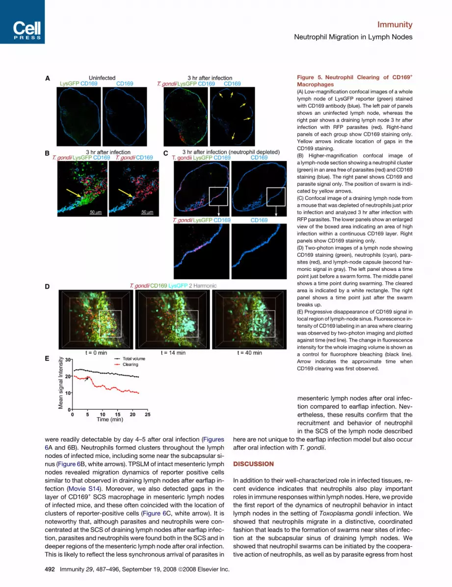
Figure 5. Neutrophil Clearing of CD169+
Macrophages
(A) Low-magnification confocal images of a whole
lymph node of LysGFP reporter (green) stained
with CD169 antibody (blue). The left pair of panels
shows an uninfected lymph node, whereas the
right pair shows a draining lymph node 3 hr after
infection with RFP parasites (red). Right-hand
panels of each group show CD169 staining only.
Yellow arrows indicate location of gaps in the
CD169 staining.
(B) Higher-magnification confocal image of
a lymph-node section showing a neutrophil cluster
(green) in an area free of parasites (red) and CD169
staining (blue). The right panel shows CD169 and
parasite signal only. The position of swarm is indi-
cated by yellow arrows.
(C) Confocal image of a draining lymph node from
a mouse that was depleted of neutrophils just prior
to infection and analyzed 3 hr after infection with
RFP parasites. The lower panels show an enlarged
view of the boxed area indicating an area of high
infection within a continuous CD169 layer. Right
panels show CD169 staining only.
(D) Two-photon images of a lymph node showing
CD169 staining (green), neutrophils (cyan), para-
sites (red), and lymph-node capsule (second har-
monic signal in gray). The left panel shows a time
point just before a swarm forms. The middle panel
shows a time point during swarming. The cleared
area is indicated by a white rectangle. The right
panel shows a time point just after the swarm
breaks up.
(E) Progressive disappearance of CD169 signal in
local region of lymph-node sinus. Fluorescence in-
tensity of CD169 labeling in an area where clearing
was observed by two-photon imaging and plotted
against time (red line). The change in fluorescence
intensity for the whole imaging volume is shown as
a control for fluorophore bleaching (black line).
Arrow indicates the approximate time when
CD169 clearing was first observed.
mesenteric lymph nodes after oral infec-
tion compared to earflap infection. Nev-
ertheless, these results confirm that the
recruitment and behavior of neutrophil
in the SCS of the lymph node described
here are not unique to the earflap infection model but also occur
after oral infection with T. gondii.
DISCUSSION
In addition to their well-characterized role in infected tissues, re-
cent evidence indicates that neutrophils also play important
roles in immune responses within lymph nodes. Here, we provide
the first report of the dynamics of neutrophil behavior in intact
lymph nodes in the setting of Toxoplasma gondii infection. We
showed that neutrophils migrate in a distinctive, coordinated
fashion that leads to the formation of swarms near sites of infec-
tion at the subcapsular sinus of draining lymph nodes. We
showed that neutrophil swarms can be initiated by the coopera-
tive action of neutrophils, as well as by parasite egress from host
492 Immunity 29, 487–496, September 19, 2008 ª2008 Elsevier Inc.

Immunity
Neutrophil Migration in Lymph Nodes
cells, and that neutrophil swarms lead to the removal of macro-
phages that line the subcapsular sinus of the lymph nodes. Our
data provide a new perspective for future studies on the role of
neutrophils in lymph nodes.
Perhaps the most striking feature of neutrophil behavior re-
ported here is the highly cooperative nature of their migration
patterns. This is reflected in the ‘‘paparazzi-like’’ behavior of neu-
trophils in which the initial arrest of a small number of neutrophils
was followed minutes later by a massive influx of cells. This be-
havior is likely to be mediated by multiple chemoattractants in-
cluding CXCL8 (IL-8), CXCL1, and leukotriene B4, which are
both produced by, and attractive to, neutrophils (Baggiolini,
1998; Scapini et al., 2000). The cooperative nature of neutrophil
migration is also reflected in their streaming behavior during
swarm formation, which is reminiscent of swarm formation by
the slime mold Dictyostelium. In the case of Dictyostelium, evi-
dence suggests that individual cells leave trails of chemoattrac-
tants that are sensed by neighboring cells, leading to a head-to-
tail migration pattern as cells migrate toward a swarm (Kriebel
et al., 2003). Neutrophils and Dictyostelium appear to use similar
signal transduction systems for chemoattraction (Mahadeo
et al., 2007) and it will be interesting to explore whether similar
mechanisms also underlie their co-operative migration patterns.
Figure 6. Neutrophil Recruitment and
Swarm Formation in Mesenteric Lymph
Nodes after Oral Infection
(A) Neutrophils (LysGFPhiLy6G+) in the mesenteric
lymph nodes of orally infected mice. Flow-cyto-
metric analysis of mesenteric lymph nodes of un-
infected LysGFP mice, or LysGFP reporter mice
infected 5 days earlier with 20 cysts.
(B) Shown are 20 um frozen sections of mesenteric
lymph nodes of LysGFP reporter (green) mice be-
fore infection (left panel) or 4 days after oral infec-
tion with 45 cysts of Pruniaud-RFP (red) stained
with Ly6G (blue). Arrows indicate neutrophil clus-
ter at the subcapsulary sinus. Bottom panels
show a high-magnification image of a lymph
node from an infected mouse. Clusters of LysGFP
high cells (green) (left panel) stain positive for the
neutrophil marker Ly6G (blue).
(C) We stained the same samples as in (B) to visu-
alize CD169+ subcapsular sinus macrophages
(blue). A panel on the right shows a high-magnifi-
cation image with an arrow indicating a region of
CD169 clearing.
Another indication of cooperative ac-
tion of neutrophils in swarm formation is
correlation between the size and persis-
tence of neutrophil swarms. Swarms con-
taining fewer than 150 neutrophils tended
to be transient, with neutrophils migrating
rapidly and directionally toward swarm
center, and then migrating equally rapidly
out of the swarm to nearby join neighbor-
ing growing swarm centers. In contrast,
swarms with > 300 neutrophils invariably
kept growing over the time they were
observed, often merging with nearby
swarms. Although it is likely that persistent swarms would disap-
pear eventually in vivo, this would likely result from neutrophil ap-
optosis and phagocytosis by macrophages, in contrast to the
outward migration of neutrophils seen during the dissolution of
transient swarms. The correlation between size and persistence
strongly suggests that the neutrophils themselves contribute to
the gradient of chemoattractants that generates a swarm, and
that once the swarm reaches a certain size, these signals be-
come sufficiently strong to generate a stable swarm center that
can override other competing signals in the environment.
Another striking aspect of neutrophil behavior is their ex-
tremely rapid migration toward sites of parasite egress. Because
this response occurred within seconds of parasite egress, it is
unlikely to be related to the IL-1-dependent neutrophil recruit-
ment in response to dying cells has been characterized previ-
ously (Chen et al., 2007). Although the nature of the neutrophil-
attracting signal that correlates with parasite egress is unclear,
it is possible that material from lysed cells could be directly at-
tractive for neutrophils, a phenomenon termed ‘‘necrotaxis’’
that has been previously described in vitro (Debru, 1993). Alter-
natively, the released parasites themselves could attract neutro-
phils, either because of a direct response to parasite PAMPS
(Debierre-Grockiego et al., 2007; Nakao and Konishi, 1991a;
Immunity 29, 487–496, September 19, 2008 ª2008 Elsevier Inc. 493

Immunity
Neutrophil Migration in Lymph Nodes
Yarovinsky et al., 2005) or because of limited complement
activation by parasites (Fuhrman and Joiner, 1989). An in vitro
system to recapitulate neutrophil swarming in response to Toxo-
plasma infection would help to address these possibilities.
Given the multitude of signals known to attract neutrophils,
and the indications from our data that neutrophils are responding
to multiple, competing signals, it seems likely that many different
types of chemoattractants contribute to neutrophil swarming be-
havior. It is unlikely that adaptive immunity plays a major role in
the neutrophil behavior that we report here because these re-
sponses occurred in naive mice a few hours after exposure to
the parasites, time points well before T or B cells would be ex-
pected to be activated. Moreover, we have not observed any sig-
nificant differences in neutrophil behavior in immunized versus
naive mice (data not shown). One major challenge to identify
these signals will be to selectively block candidate signals during
the swarming phase of the response. For example, while
TLR-MyD88 signals are good candidates for mediating swarm
formation, our observation that this pathway is also required
for neutrophils to be recruited to the lymph node in the first place
complicates testing this idea.
Our data also add to the growing body of evidence for the
importance of CD169+ subcapsular sinus macrophage of the
lymph nodes (Carrasco and Batista, 2007; Hickman et al.,
2008; Junt et al., 2007; Phan et al., 2007). Parasites are found
predominantly within these macrophage a few hours after infec-
tion, and neutrophil swarms in infected regions of the subcapsu-
lar sinus lead to the appearance of gaps in the layer of CD169+
cells, which normally form a continuous layer around an unin-
fected lymph node. Although the fate of the SCS macrophages
is not known, we suspect that they are removed by neutrophils
on the basis of several considerations. First, we have never ob-
served any migrating CD169+ macrophages, even while imaging
areas in which gaps were forming. Second, we are able to detect
the loss of CD169+ cells by flow-cytometric analysis, consistent
with the removal of these cells and arguing against the possibility
that they have migrated to a different part of the lymph node. Fi-
nally, a direct role for neutrophils is supported by the well-docu-
mented ability of neutrophils to produce matrix metalloproteases
and participate in tissue remodeling. Regardless of the mecha-
nism, given the emerging evidence for the importance of sub-
capsular sinus of the lymph node in trapping pathogens and ini-
tiating adaptive immune responses (Carrasco and Batista, 2007;
Hickman et al., 2008; Junt et al., 2007; Phan et al., 2007), it will be
important to examine whether the removal of SCS macrophage
by neutrophils has an impact on subsequent immune responses.
In summary, our analysis of neutrophil migration in intact
lymph nodes has provided evidence that signals released during
parasite egress from host cells and cooperative action of neutro-
phils can induce the formation of dynamic neutrophil swarms in
the SCS, leading to the removal of macrophages that reside
there. In addition to direct phagocytic killing, neutrophils also
use a variety of other non-cell-autonomous mechanisms to pro-
tect against pathogens, including the release of neutrophil extra-
cellular traps (NETs) (Brinkmann et al., 2004), containing DNA,
microbiocidal products, and tissue remodeling factors such as
matrix metalloproteinase (Appelberg, 2007; Nathan, 2006). It is
tempting to speculate that the swarming behavior of neutrophils
reported here could increase the local concentration of these
494 Immunity 29, 487–496, September 19, 2008 ª2008 Elsevier Inc.
released products and improve the effectiveness of these mech-
anisms to induce inflammation and provide protection against
pathogens.
EXPERIMENTAL PROCEDURES
Mice
All mice were bred and housed in pathogen-free conditions at the AALAC-ap-
proved animal facility at Life Science Addition, University of California,
Berkeley. All animal experiments were approved by the Animal Care and
Use Committee of UC Berkeley. LysGFP reporter mice were a gift from
T. Graf (Albert Einstein College of Medicine, Bronx, NY) and have been previ-
ously described (Faust et al., 2000). MyD88 gene KO mice (originally generated
in S. Akira’s laboratory, Osaka University, Osaka, Japan) (Adachi et al., 1998)
were backcrossed onto C57BL/6 background.
Parasites
To generate a T. gondii cell line expressing the tandem (td) Tomato variant of
red fluorescent protein (RFP) (Shaner et al., 2004), we amplified tdTomato
using the primers 50-AGTCCCTAGGGTGAGCAAGGGCGAGGAG-30 and 50-
AGTCCCCGGGCTTGTACAGCTCGTCCATGC-30. We digested the resultant
PCR product with AvrII and XmaI and ligated this into the corresponding re-
striction enzyme sites of the pCTG vector (G.v.D. and R. Opperman, unpub-
lished data) to generate the vector pCTR2T, in which expression of cytosol-lo-
calized tdTomato is driven from the constitutive a-tubulin promoter. We
transfected this construct into the RHDhxgprt strain of T. gondii and generated
stable lines expressing tdTomato by chloramphenicol selection as previously
described (Gubbels et al., 2005). In brief, 1 3 107 parasites were resuspended
in cytomix containing 50 mg of linearized pCTR2T vector. The parasites mixture
was added to a 2 mm electroporation cuvette and electroporated with a BTX
ECM 630 electroporator at 1500 V, 25 U, and 25 mF. Transfected cells were
passed onto fresh host cells. Chloramphenicol was added to a final concentra-
tion of 6.8 mg/mL 16 hr after transfection. Cells were passaged in the presence
of chloramphenicol until stable lines were generated. Clonal lines of the RH/
tdTomato parasite strain were obtained by fluorescence-activated cell sorting.
In brief, parasites were filtered through a 3 mm filter, pelleted, and resuspend in
phosphate-buffered saline. Parasites were sorted with a MoFlo cytometer
(Dako, Ft Collins, CO) with an Enterprise 631 laser tuned to 488 nm for excita-
tion and an emission filter with a band pass of 570/40 nm. Bright parasites were
sorted into 96-well plates, and wells containing single plaques after 1 week of
growth were considered to consist of a clonal parasite line. All parasites were
maintained in confluent human foreskin fibroblasts.
For mouse infections, parasites were prepared from almost fully lysed fibro-
blast cultures by first releasing the parasites from fibroblasts by passing them
through 21 G1 1/2 gauge and 23 G1 needles five to ten times. Parasites were
then filtered through a 3 mm filter, pelleted, and resuspended in phosphate-
buffered saline. A total of 5 3 105 to 1 3 107 parasites (typically 5 3 106) in
10 ml volume were injected into the earflap. No differences in the anatomical
distribution of parasites or neutrophils in draining lymph nodes were observed
over this range of infection. Some of the mice used for two-photon experi-
ments were injected with 106 irradiated parasites intraperitoneally at least 4
weeks prior to infection and imaging. No differences in neutrophil behavior
were observed between immunized and naive animals. For neutrophil deple-
tion experiments, mice were injected i.p. with 1 mg of Ly6G antibody (clone
1A8) 2 days prior to infection and also i.v. with 0.5 mg of Ly6G antibody 1
day prior to infection.
For oral infections, Prugnaud-RFP parasites were engineered essentially as
described above for RH parasites, and cysts were isolated from brain homog-
enates of CBA/J mice (Jackson Laboratory) infected with 300–400 parasites
i.p. 4–6 months prior. Cysts were counted after staining with Dolichos Biflorus
Agglutinin (Vector Laboratories). LysGFP reporter mice were infected with
20–45 cysts by gavage. Mesenteric lymph nodes were used for microscopy
or analyzed by flow cytometry.
Antibodies and Flow Cytometry
Lymph nodes were dissociated by collagenase digestion. Cell suspensions
were filtered, stained, and analyzed by flow cytometry. The following

Immunity
Neutrophil Migration in Lymph Nodes
antibodies were from eBioscience: phycoerythrin-Cy5 conjugated anti-CD11b
(clone M1/70) and phycoerythrin-Cy5 conjugated anti-CD11c (clone N418).
Fluorescein-isothiocyanate-conjugated anti-Ly6G (clone 1A8) was from BD
Biosciences. Fluorescein-isothiocyanate-conjugated CD169 antibody (clone
3D6.112) was purchased from AbD Serotec. Acquisitions were performed
with a Coulter Epics XL-MCL flow cytometer (Beckman-Coulter), and data
were analyzed with the FlowJo software (Tree Star).
Statistical Analysis
Values were expressed as mean ± standard error (SE). Levels of significance
were calculated by unpaired t tests with the GraphPad Prism program. Differ-
ences were considered significant at p < 0.05.
Two-Photon Imaging
One to five hours after ear-flap injection, mice were sacrificed, and dorsal cer-
vical (draining) lymph nodes were isolated and imaged by two-photon laser
scanning microscopy (TPLSM) while being perfused with warmed, oxygenated
medium as described previously (Witt et al., 2005). Imaging was performed on
either upright Zeiss NLO 510 or a custom-built microscope, both using a Spec-
tra-Physics MaiTai laser tuned to 900–920nm. For the Zeiss microscope, GFP
and tdTomato emission light was separated with a 560 dichroic mirror and col-
lected with non-descanned detectors. For the custom-built microscope, the
emission light was separated with 495 and 560 dichroics and collected with
3 PMT detectors. Bandpass filters HQ 450/80M and HQ 645/75M were used
for minimization of spectral overlap.
Typical imaging volumes (164 3 164 3 40 mm or 172 3 143 3 80 mm, respec-
tively) corresponded to regions of the lymph nodes extending up to 200 mm be-
low the surface of the capsule and were scanned every 13–37 s for 20–40 min.
Mesenteric lymph nodes were imaged under similar conditions 4–5 days after
oral infection.
Data Analysis
The x, y, z coordinates of individual cells over time were obtained with Imaris
Bitplane Software. Motility parameters were calculated with Matlab (code
available upon request). Parameters reported here include speed (defined as
path length over time; mm/minute) and distance to swarm center (mm). For
measurement of the volumes of individual neutrophil swarms, isosurfaces
were generated in the GFP channel (threshold of 130, 6 mm Gaussian filter),
and the volume of each isosurface was determined over time. The volume of
a single neutrophil was estimated with the same method (with 1 mm Gaussian
filter), and 15 individual cell volumes were averaged. For analysis of directed
migration, psi is defined as the angle between the migration vector and the di-
rection vector from the initial position of the migrating cell to the center of the
swarm (see inset, Figure 2D). The ‘‘coordination angle’’ alpha is defined as the
angle between the migration vectors for pairs of time points within two individ-
ual tracks (see inset, Figure 2E).
Immunofluorescence
Isolated lymph nodes were fixed with 4% formalin/10% sucrose in PBS for 1
hr, sequentially submerged in 10%, 20%, and 30% sucrose for 18–24 hr
each, and frozen over dry ice in OCT. We generated 20 psi serial sections by
cryosectioning (MICROM H550, Microm GmbH) and stored them at �80�C.
Sections were brought to room temperature, fixed with cold acetone for 10
min, air-dried, and incubated with 10% mouse serum in Fc blocking reagent
(2.4G2 culture supernatant) for 1 hr. The tissue was stained with unconjugated
anti-Ly6G (BD Biosciences), purified rat anti mouse CD169 (AbD Serotec), or
biotin-conjugated anti-LYVE-1 (R&D Systems) overnight at 4�C. After primary
staining, slides were washed 43 in PBS and incubated with either anti-rat
Alexa 647 or streptavidin Alexa 633 (Invitrogen) for 2 hr at room temperature.
Sections were then washed 43 in PBS and coverslipped with VectaShield
(Vector Laboratories) mounting medium. Stained lymph-node sections were
visualized on Zeiss 510 Axioplan META NLO upright microscope with a 103
air objective (Plan-Neofluar 103/0.3) and a 403 oil objective (Plan-Neofluar
403/1.3 oil WD = 0.17 mm) with 488 nm, 543 nm, and 633 nm laser lines. Im-
ages were analyzed and assembled with Adobe Photoshop and Imaris Bit-
plane.
I
SUPPLEMENTAL DATA
Supplemental Data include three figures and fourteen movies and can be
found with this article online at http://www.immunity.com/cgi/content/full/
29/3/487/DC1/.
ACKNOWLEDGMENTS
We thank H. Nolla (UCB) and J. Nelson (CTEGD) for assistance with flow cy-
tometry, M. Rivera for flow-cytometry analysis, T. Graf for providing lysMGFP
reporter mice, Y.F. Liao for tracking neutrophils, E. Ladi for assistance with
quantitating neutrophil motility, the CHPS mouse/virus core for providing
mouse strains, and D. Portnoy, M. Welsh, and members of the Robey lab for
comments on the manuscript. This work was funded by the NIH (B.S. and
E.R.), Human Frontier Science Program Fellowship (T.C.), and C.J. Martin
Overseas Fellowship (400489) from the Australian National Health and Medical
Research Council (G.v.D.).
Received: June 14, 2008
Revised: July 18, 2008
Accepted: July 25, 2008
Published online: August 21, 2008
REFERENCES
Abadie, V., Badell, E., Douillard, P., Ensergueix, D., Leenen, P.J., Tanguy, M.,
Fiette, L., Saeland, S., Gicquel, B., and Winter, N. (2005). Neutrophils rapidly
migrate via lymphatics after Mycobacterium bovis BCG intradermal vaccina-
tion and shuttle live bacilli to the draining lymph nodes. Blood 106, 1843–1850.
Adachi, O., Kawai, T., Takeda, K., Matsumoto, M., Tsutsui, H., Sakagami, M.,
Nakanishi, K., and Akira, S. (1998). Targeted disruption of the MyD88 gene
results in loss of IL-1- and IL-18-mediated function. Immunity 9, 143–150.
Appelberg, R. (2007). Neutrophils and intracellular pathogens: Beyond phago-
cytosis and killing. Trends Microbiol. 15, 87–92.
Baggiolini, M. (1998). Chemokines and leukocyte traffic. Nature 392, 565–568.
Beauvillain, C., Delneste, Y., Scotet, M., Peres, A., Gascan, H., Guermonprez,
P., Barnaba, V., and Jeannin, P. (2007). Neutrophils efficiently cross-prime
naive T cells in vivo. Blood 110, 2965–2973.
Bennouna, S., Bliss, S.K., Curiel, T.J., and Denkers, E.Y. (2003). Cross-talk in
the innate immune system: Neutrophils instruct recruitment and activation of
dendritic cells during microbial infection. J. Immunol. 171, 6052–6058.
Black, M.W., and Boothroyd, J.C. (2000). Lytic cycle of Toxoplasma gondii.
Microbiol. Mol. Biol. Rev. 64, 607–623.
Bliss, S.K., Gavrilescu, L.C., Alcaraz, A., and Denkers, E.Y. (2001). Neutrophil
depletion during Toxoplasma gondii infection leads to impaired immunity and
lethal systemic pathology. Infect. Immun. 69, 4898–4905.
Bousso, P., and Robey, E. (2004). Dynamic behavior of T cells and thymocytes
in lymphoid organs as revealed by 2-photon microscopy. Immunity 21,
349–355.
Brinkmann, V., Reichard, U., Goosmann, C., Fauler, B., Uhlemann, Y., Weiss,
D.S., Weinrauch, Y., and Zychlinsky, A. (2004). Neutrophil extracellular traps
kill bacteria. Science 303, 1532–1535.
Cahalan, M.D., and Parker, I. (2008). Choreography of cell motility and interac-
tion dynamics imaged by two-photon microscopy in lymphoid organs. Annu.
Rev. Immunol. 26, 585–626.
Carrasco, Y.R., and Batista, F.D. (2007). B cells acquire particulate antigen in
a macrophage-rich area at the boundary between the follicle and the subcap-
sular sinus of the lymph node. Immunity 27, 160–171.
Channon, J.Y., Seguin, R.M., and Kasper, L.H. (2000). Differential infectivity
and division of Toxoplasma gondii in human peripheral blood leukocytes.
Infect. Immun. 68, 4822–4826.
Chen, C.J., Kono, H., Golenbock, D., Reed, G., Akira, S., and Rock, K.L. (2007).
Identification of a key pathway required for the sterile inflammatory response
triggered by dying cells. Nat. Med. 13, 851–856.
mmunity 29, 487–496, September 19, 2008 ª2008 Elsevier Inc. 495
Immunity
Neutrophil Migration in Lymph Nodes
Debierre-Grockiego, F., Campos, M.A., Azzouz, N., Schmidt, J., Bieker, U.,
Resende, M.G., Mansur, D.S., Weingart, R., Schmidt, R.R., Golenbock, D.T.,
et al. (2007). Activation of TLR2 and TLR4 by glycosylphosphatidylinositols
derived from Toxoplasma gondii. J. Immunol. 179, 1129–1137.
Debru, C. (1993). A particular form of chemotaxis: Necrotaxis. An historical
view. Blood Cells 19, 5–19.
Denkers, E.Y., Butcher, B.A., Del Rio, L., and Bennouna, S. (2004). Neutro-
phils, dendritic cells and Toxoplasma. Int. J. Parasitol. 34, 411–421.
Egen, J.G., Rothfuchs, A.G., Feng, C.G., Winter, N., Sher, A., and Germain,
R.N. (2008). Macrophage and T cell dynamics during the development and dis-
integration of mycobacterial granulomas. Immunity 28, 271–284.
Faust, N., Varas, F., Kelly, L.M., Heck, S., and Graf, T. (2000). Insertion of en-
hanced green fluorescent protein into the lysozyme gene creates mice with
green fluorescent granulocytes and macrophages. Blood 96, 719–726.
Fuhrman, S.A., and Joiner, K.A. (1989). Toxoplasma gondii: Mechanism of re-
sistance to complement-mediated killing. J. Immunol. 142, 940–947.
Germain, R.N., Miller, M.J., Dustin, M.L., and Nussenzweig, M.C. (2006). Dy-
namic imaging of the immune system: Progress, pitfalls and promise. Nat.
Rev. Immunol. 6, 497–507.
Gubbels, M.J., Striepen, B., Shastri, N., Turkoz, M., and Robey, E.A. (2005).
Class I major histocompatibility complex presentation of antigens that escape
from the parasitophorous vacuole of Toxoplasma gondii. Infect. Immun. 73,
703–711.
Hickman, H.D., Takeda, K., Skon, C.N., Murray, F.R., Hensley, S.E., Loomis,
J., Barber, G.N., Bennink, J.R., and Yewdell, J.W. (2008). Direct priming of an-
tiviral CD8+ T cells in the peripheral interfollicular region of lymph nodes. Nat.
Immunol. 9, 155–165.
Junt, T., Moseman, E.A., Iannacone, M., Massberg, S., Lang, P.A., Boes, M.,
Fink, K., Henrickson, S.E., Shayakhmetov, D.M., Di Paolo, N.C., et al. (2007).
Subcapsular sinus macrophages in lymph nodes clear lymph-borne viruses
and present them to antiviral B cells. Nature 450, 110–114.
Kriebel, P.W., Barr, V.A., and Parent, C.A. (2003). Adenylyl cyclase localization
regulates streaming during chemotaxis. Cell 112, 549–560.
Mahadeo, D.C., Janka-Junttila, M., Smoot, R.L., Roselova, P., and Parent,
C.A. (2007). A chemoattractant-mediated Gi-coupled pathway activates ad-
enylyl cyclase in human neutrophils. Mol. Biol. Cell 18, 512–522.
Maletto, B.A., Ropolo, A.S., Alignani, D.O., Liscovsky, M.V., Ranocchia, R.P.,
Moron, V.G., and Pistoresi-Palencia, M.C. (2006). Presence of neutrophil-
bearing antigen in lymphoid organs of immune mice. Blood 108, 3094–3102.
Megiovanni, A.M., Sanchez, F., Robledo-Sarmiento, M., Morel, C., Gluckman,
J.C., and Boudaly, S. (2006). Polymorphonuclear neutrophils deliver activation
signals and antigenic molecules to dendritic cells: A new link between leuko-
cytes upstream of T lymphocytes. J. Leukoc. Biol. 79, 977–988.
Miller, M.J., Wei, S.H., Parker, I., and Cahalan, M.D. (2002). Two-photon imag-
ing of lymphocyte motility and antigen response in intact lymph node. Science
296, 1869–1873.
496 Immunity 29, 487–496, September 19, 2008 ª2008 Elsevier Inc
Nakao, M., and Konishi, E. (1991a). Neutrophil chemotactic factors secreted
from Toxoplasma gondii. Parasitology 103, 29–34.
Nakao, M., and Konishi, E. (1991b). Proliferation of Toxoplasma gondii in
human neutrophils in vitro. Parasitology 103, 23–27.
Nathan, C. (2006). Neutrophils and immunity: Challenges and opportunities.
Nat. Rev. Immunol. 6, 173–182.
Pesce, J.T., Liu, Z., Hamed, H., Alem, F., Whitmire, J., Lin, H., Liu, Q., Urban,
J.F., Jr., and Gause, W.C. (2008). Neutrophils clear bacteria associated with
parasitic nematodes augmenting the development of an effective th2-type re-
sponse. J. Immunol. 180, 464–474.
Phan, T.G., Grigorova, I., Okada, T., and Cyster, J.G. (2007). Subcapsular en-
counter and complement-dependent transport of immune complexes by
lymph node B cells. Nat. Immunol. 8, 992–1000.
Rydstrom, A., and Wick, M.J. (2007). Monocyte recruitment, activation, and
function in the gut-associated lymphoid tissue during oral Salmonella infec-
tion. J. Immunol. 178, 5789–5801.
Sasmono, R.T., Ehrnsperger, A., Cronau, S.L., Ravasi, T., Kandane, R., Hickey,
M.J., Cook, A.D., Himes, S.R., Hamilton, J.A., and Hume, D.A. (2007). Mouse
neutrophilic granulocytes express mRNA encoding the macrophage colony-
stimulating factor receptor (CSF-1R) as well as many other macrophage-spe-
cific transcripts and can transdifferentiate into macrophages in vitro in re-
sponse to CSF-1. J. Leukoc. Biol. 82, 111–123.
Sayles, P.C., and Johnson, L.L. (1996). Exacerbation of toxoplasmosis in neu-
trophil-depleted mice. Nat. Immun. 15, 249–258.
Scapini, P., Lapinet-Vera, J.A., Gasperini, S., Calzetti, F., Bazzoni, F., and Cas-
satella, M.A. (2000). The neutrophil as a cellular source of chemokines. Immu-
nol. Rev. 177, 195–203.
Shaner, N.C., Campbell, R.E., Steinbach, P.A., Giepmans, B.N., Palmer, A.E.,
and Tsien, R.Y. (2004). Improved monomeric red, orange and yellow fluores-
cent proteins derived from Discosoma sp. red fluorescent protein. Nat. Bio-
technol. 22, 1567–1572.
Sumen, C., Mempel, T.R., Mazo, I.B., and von Andrian, U.H. (2004). Intravital
microscopy: Visualizing immunity in context. Immunity 21, 315–329.
Tvinnereim, A.R., Hamilton, S.E., and Harty, J.T. (2004). Neutrophil involve-
ment in cross-priming CD8+ T cell responses to bacterial antigens. J. Immu-
nol. 173, 1994–2002.
Witt, C.M., Raychaudhuri, S., Schaefer, B., Chakraborty, A.K., and Robey, E.A.
(2005). Directed migration of positively Selected thymocytes visualized in real
time. PLoS Biol. 3, e160.
Yarovinsky, F., Zhang, D., Andersen, J.F., Bannenberg, G.L., Serhan, C.N.,
Hayden, M.S., Hieny, S., Sutterwala, F.S., Flavell, R.A., Ghosh, S., and Sher,
A. (2005). TLR11 activation of dendritic cells by a protozoan profilin-like pro-
tein. Science 308, 1626–1629.
Zinselmeyer, B.H., Lynch, J.N., Zhang, X., Aoshi, T., and Miller, M.J. (2008).
Video-rate two-photon imaging of mouse footpad - a promising model for
studying leukocyte recruitment dynamics during inflammation. Inflamm. Res.
57, 93–96.
.