DC-SIGN from African green monkeys is expressed in lymph nodes and mediates infection in trans of...
14
2004, 78(2):798. DOI: 10.1128/JVI.78.2.798-810.2004. J. Virol. Barré-Sinoussi and Michaela C. Müller-Trutwin Kooyk, Ali Amara, Olivier Schwartz, Françoise A. Soares, Eric Nerrienet, Roger Le Grand, Yvette Van Marcelo Sol-Foulon, Lorenzo Mortara, Abdourahmane Faye, Mickaël J.-Y. Ploquin, Ousmane M. Diop, Nathalie Immunodeficiency Virus SIVagm of Simian trans Infection in Expressed in Lymph Nodes and Mediates DC-SIGN from African Green Monkeys Is http://jvi.asm.org/content/78/2/798 Updated information and services can be found at: These include: REFERENCES http://jvi.asm.org/content/78/2/798#ref-list-1 at: This article cites 62 articles, 42 of which can be accessed free CONTENT ALERTS more» articles cite this article), Receive: RSS Feeds, eTOCs, free email alerts (when new http://journals.asm.org/site/misc/reprints.xhtml Information about commercial reprint orders: http://journals.asm.org/site/subscriptions/ To subscribe to to another ASM Journal go to: on October 2, 2014 by guest http://jvi.asm.org/ Downloaded from on October 2, 2014 by guest http://jvi.asm.org/ Downloaded from
Transcript of DC-SIGN from African green monkeys is expressed in lymph nodes and mediates infection in trans of...

2004, 78(2):798. DOI: 10.1128/JVI.78.2.798-810.2004.J. Virol. Barré-Sinoussi and Michaela C. Müller-TrutwinKooyk, Ali Amara, Olivier Schwartz, FrançoiseA. Soares, Eric Nerrienet, Roger Le Grand, Yvette Van
MarceloSol-Foulon, Lorenzo Mortara, Abdourahmane Faye, Mickaël J.-Y. Ploquin, Ousmane M. Diop, Nathalie Immunodeficiency Virus SIVagm
of SimiantransInfection in Expressed in Lymph Nodes and Mediates DC-SIGN from African Green Monkeys Is
http://jvi.asm.org/content/78/2/798Updated information and services can be found at:
These include:
REFERENCEShttp://jvi.asm.org/content/78/2/798#ref-list-1at:
This article cites 62 articles, 42 of which can be accessed free
CONTENT ALERTS more»articles cite this article),
Receive: RSS Feeds, eTOCs, free email alerts (when new
http://journals.asm.org/site/misc/reprints.xhtmlInformation about commercial reprint orders: http://journals.asm.org/site/subscriptions/To subscribe to to another ASM Journal go to:
on October 2, 2014 by guest
http://jvi.asm.org/
Dow
nloaded from
on October 2, 2014 by guest
http://jvi.asm.org/
Dow
nloaded from

JOURNAL OF VIROLOGY, Jan. 2004, p. 798–810 Vol. 78, No. 20022-538X/04/$08.00�0 DOI: 10.1128/JVI.78.2.798–810.2004Copyright © 2004, American Society for Microbiology. All Rights Reserved.
DC-SIGN from African Green Monkeys Is Expressed in Lymph Nodesand Mediates Infection in trans of Simian Immunodeficiency
Virus SIVagmMickael J.-Y. Ploquin,1 Ousmane M. Diop,2 Nathalie Sol-Foulon,3 Lorenzo Mortara,1†
Abdourahmane Faye,2 Marcelo A. Soares,1‡ Eric Nerrienet,4 Roger Le Grand,5Yvette Van Kooyk,6 Ali Amara,7§ Olivier Schwartz,3 Francoise Barre-Sinoussi,1
and Michaela C. Muller-Trutwin1*Unite de Biologie des Retrovirus,1 Virus et Immunite,3 and Unite d’Immunologie Virale,7 Institut Pasteur, Paris,and CEA, Fontenay aux Roses,4 France; Laboratoire de Retrovirologie, Institut Pasteur, Dakar, Senegal2; Centre
Pasteur du Cameroun, Yaounde, Cameroon5; and Department of Molecular Cell Biology, FreeUniversity Medical Center, Amsterdam, The Netherlands6
Received 19 June 2003/Accepted 3 October 2003
African green monkeys (AGMs) infected by simian immunodeficiency virus (SIV) SIVagm are resistant toAIDS. SIVagm-infected AGMs exhibit levels of viremia similar to those described during pathogenic humanimmunodeficiency virus type 1 (HIV-1) and SIVmac infections in humans and macaques, respectively, butcontain lower viral loads in their lymph nodes. We addressed the potential role of dendritic cell-specificintercellular adhesion molecule 3-grabbing nonintegrin (DC-SIGN; CD209) in viral dissemination. In previousstudies, it has been shown that human DC-SIGN and macaque DC-SIGN allow transmission of HIV andSIVmac to T cells. Here, we looked at the ability of DC-SIGN derived from AGM lymph nodes to interact withSIVagm. We show that DC-SIGN-expressing cells are present mainly in the medulla and often within the cortexand/or paracortex of AGM lymph nodes. We describe the isolation and characterization of at least threeisoforms of dc-sign mRNA in lymph nodes of AGMs. The predicted amino acid sequence from the predominantmRNA isoform, DC-SIGNagm1, is 92 and 99% identical to the corresponding human and rhesus macaqueDC-SIGN amino acid sequences, respectively. DC-SIGNagm1 is characterized by the lack of the fourth motifin the repeat domain. This deletion was also detected in the dc-sign gene derived from thirteen animalsbelonging to five other African monkey species and from four macaques (Macaca fascicularis and M. mulatta).Despite three- to seven-amino-acid modifications compared to DC-SIGNmac, DC-SIGNagm1 allows transmis-sion of SIVagm to T cells. Furthermore, AGM monocyte-derived dendritic cells (MDDC) expressed at least100,000 DC-SIGN molecules and were able to transmit SIVagm to T cells. At a low multiplicity of infection(10�5 50% tissue culture infective doses/cell), viral transmission by AGM MDDC was mainly DC-SIGNdependent. The present study reveals that DC-SIGN from a natural host species of SIV has the ability to actas an efficient attachment and transmission factor for SIVagm and suggests the absence of a direct linkbetween this ability and viral load levels in lymph nodes.
Dendritic cells (DC) residing in mucosal tissues are amongthe first cells targeted by human immunodeficiency virus(HIV) and simian immunodeficiency virus (SIV) SIVmac dur-ing pathogenic infections in humans and macaques, respec-tively (22, 51). DC can transmit these viruses to T cells in vitro(6, 17, 40). One mechanism by which DC enhance infection ofT cells implies DC-SIGN (DC-specific ICAM-3-grabbing non-integrin; CD209) (9, 13). DC-SIGN, a C-type (calcium-depen-dent) lectin, has been detected on monocyte-derived DC(MDDC). It also facilitates the formation of the immunologi-
cal synapse between DC and T cells by binding to intercellularadhesion molecule 3 (ICAM-3) (14). In addition, HIV boundto DC-SIGN-positive cells can be transmitted to cells express-ing CD4 and a coreceptor, resulting in efficient virus infectionin trans (13, 38). DC-SIGN cannot replace CD4, CCR5, orCXCR4 in the process of HIV type 1 (HIV-1) entry but retainsinfectious particles for several days in culture (13) in a low-pHendosomal compartment (27). Similarly, DC-SIGN from ma-caques also acts as an attachment factor for SIVmac (2, 12, 25,61). In addition, coexpression of human DC-SIGN with CD4and an appropriate coreceptor can enhance infection of HIVand SIVmac in cis (28).
HIV-1 is thought to subvert the trafficking capacity of DC togain access to the CD4�-T-cell compartment (19, 42, 53). Ithas been shown that SIVmac-infected DC are able to migratefrom original sites of infection to draining lymph nodes (LNs)(22). Similar to “Trojan horses,” DC-SIGN expressing DC mayserve as a shuttle for the transfer of HIV-1 from submucosa tosecondary lymphoid organs (13, 52). During the formation ofthe immunological synapse with T cells, it is possible that
* Corresponding author. Mailing address: Unite de Biologie desRetrovirus, Institut Pasteur, 28, rue du Docteur Roux, 75724 ParisCedex 15, France. Phone: (33)1-40-61-39-69. Fax: (33)1-45-68-89-57.E-mail: [email protected].
† Present address: Department of Clinical and Biological Sciences,School of Medicine, University of Insubria, Varese, Italy.
‡ Present address: Departamento de Genetica, Instituto de Biolo-gia-Universidade Federal do Rio de Janeiro, Rio de Janeiro, Brazil.
§ Present address: Skirball Institute of Biomolecular Medicine, NewYork University Medical Center, New York, NY 10016.
798
on October 2, 2014 by guest
http://jvi.asm.org/
Dow
nloaded from

infected DC then transmit the virus to permissive T cells withinthe secondary lymphoid organs (6, 18, 30, 40, 60).
To understand the role of DC-SIGN in viral disseminationto LNs and pathogenesis, we studied the function of DC-SIGNin a nonhuman primate model in which infection with a lenti-virus is nonpathogenic. SIVagm infection in all four species ofAfrican green monkeys (AGM, Chlorocebus pygerythrus, C. ae-thiops, C. sabaeus, and C. tantalus) is associated with a lack ofdisease, although the associated viremia levels in plasma canreach those described for pathogenic HIV-1 and SIVmac in-fections (5, 10, 16, 21). In contrast to plasma viremia, the DNAand RNA viral loads in LNs of SIVagm-infected AGMs aregenerally significantly lower than those reported for HIV andSIVmac pathogenic infections (4, 5, 10). Here we examinedwhether potential structural and functional differences be-tween DC-SIGNagm and human DC-SIGN and/or differencesin the expression levels underlie the low viral load in AGMLNs.
MATERIALS AND METHODS
Animals and specimen collection. The AGMs—C. sabaeus, C. tantalus, and C.pygerythrus—were kept in captivity at the Pasteur Institute in Senegal, at thePasteur Institute in Bangui (Central African Republic), and at the Centre In-ternational de Recherches Medicales, in Franceville (Gabon), respectively. Patasmonkeys (Erythrocebus patas) were held in captivity at the Pasteur Institute inBangui. L’Hoest’s monkeys (Cercopithecus lhoesti lhoesti), white-collared manga-beys (Cercocebus torquatus lunulatus), gray-cheeked mangabeys (Cercocebus al-bigena), and a naturally SIVcpz-infected chimpanzee (Pan troglodytes, Cam5)were housed in European and Cameroonian zoos (33, 34). Rhesus (Macacamulatta) and cynomolgus (M. fascicularis) macaques were housed at the primatecenter of CEA, Fontenay aux Roses, France. All animals were held according tonational guidelines and institutional policies. For the studies on genomic DNA,peripheral blood mononuclear cells (PBMC) of these nonhuman primates wereFicoll gradient purified, and total DNA was isolated. All of the studies on thecharacterization, the expression levels, and the function of DC-SIGN were per-formed with AGMs of the C. sabaeus species (animals 96028, 97008, 97026,97029, 00017, 00021, 02001, 02007, 02008, 02013, and 02026). These animalswere negative for SIV, except for 97026 and 98013, which were experimentallyinfected with SIVagm.sab92018 (10). Blood was collected from these animals,and excisional inguinal and axillar LN biopsies were performed. For AGMs97026 and 98013, the axillar LNs were collected at days 7 and 120 postinfection,respectively.
Virus stocks. We collected PBMC from the naturally SIVagm-infected animalD46 (32). SIVagm.sabD46 was isolated by coculture of the PBMC with SUPT1cells, a human lymphoblastoid T-CD4�-cell line. For HIV-1, we used an HIV-1Lai isolate that was cultured on SUPT1 cells. Virus production was followed bythe measurement of core antigens (SIV p27 and HIV p24; Coulter) and ofreverse transcriptase activity in the supernatants. The infectious viral titer wasdetermined by a limiting-dilution assay with SUPT1 cells as described previously(10). The infectious titer was expressed as the 50% tissue culture infectious dose(TCID50) determined by the Karber calculation method. The titers of HIV-1Lai
and SIVagm.sabD46 stocks were 250 and 400 TCID50/ml, respectively. Concen-trations of core antigens for HIV-1Lai and SIVagm.sabD46 stocks were 1,700 and860 ng/ml, respectively.
Cells and cell lines. SupT1 cells were cultured in RPMI 1640 with Glutamax(Invitrogen) supplemented with 10% fetal calf serum (FCS), penicillin-strepto-mycin (100 IU/ml each) and HEPES (10 mM; Invitrogen). HeLa cells weremaintained in Dulbecco modified Eagle medium with Glutamax (glucose, 1,000mg/liter; Invitrogen) supplemented with 10% FCS (Invitrogen), penicillin-strep-tomycin (100 IU/ml each), and HEPES (10 mM; Invitrogen). P4P cells are CD4�
CXCR4� CCR5� HeLa cells (59). These cells were maintained in the culturemedium described above supplemented with puromycin (1 �g/ml; Sigma) andGeneticin G418 (0.5 mg/ml; Invitrogen).
To obtain MDDC, we first enriched monocytes from PBMC of human donorsand of AGMs by a 1-h 30-min plastic adhesion step at 37°C, followed by extensivewashing in prewarmed FCS-free culture medium in order to remove all nonad-herent cells. The adherent cells were then cultured in RPMI 1640 supplementedwith 5% FCS, granulocyte-macrophage colony-stimulating factor (1,000 IU/ml;
Leucomax [Novartis]) and interleukin-4 (IL-4; 200 IU/ml; R&D Systems) for 4 to5 days. The phenotype of the cells was checked by flow cytometry. The cellsexpressed the molecules typically found on immature MDDC, such as CD11c,major histocompatibility complex class II (DR), CD80, and CD86 (unpublisheddata). The purity of the cells was �95% as measured by anti-CD3 and anti-CD20labeling.
Isolation and sequencing of dc-signagm mRNA. Total mRNA of LN cellsderived from AGM 96028 and 97026 was extracted and then reverse transcribedand amplified in one step by reverse transcription-PCR (RT-PCR; SuperscriptOne-Step RT-PCR System; Invitrogen). The primers used for the RT-PCR step,designed by using a previously published human dc-sign cDNA sequence (9),were MDC1 (forward, 5�-CAGGAGTTCTGGACACTG-3�) and MDC2 (re-verse, 5�-GATGGAGAGAAGGAACTGTAG-3�). To detect potential minoramplicons, we subjected the amplification products obtained by RT-PCR to anested PCR. The primers used for the nested PCR were MDC3 (forward,5�-CTGGGGGAGAGTGGGGTG-3�) and MDC4 (reverse, 5�-CTTAAAAGGGGGTGAAGTTC-3�). Annealing of primers occurs on the 5� and 3� ends of thecoding region. Cycling conditions were as previously described (10). After thenested PCR, the amplicons were gel purified (gel extraction kit; Qiagen) andsubcloned (TOPO TA Cloning kit; Invitrogen). The sequences were determinedfor both strands (BigDye terminator cycle sequencing ready reaction kit; Perkin-Elmer).
PCR on genomic DNA. Total DNA was extracted from simian cells andsubmitted to PCR by using the primers DCS-EX4S (forward; 5�-CCCAGCTCCATAAGTCAGGAACAA-3�) and DCS-EX4AS (reverse; 5�-ACACAACGACCATCTCAGGCCCAAG-3�). Annealing of primers occurs on the 5� part of exonIII and the 5� part of intron III of dc-sign when the organization of the humangene is considered (35). Cycling conditions were as previously described (10).
Assessment of DC-SIGNagm-mediated infection in trans. The efficiency ofDC-SIGNagm-mediated virus transfer was first assessed by using human celllines. Nonpermissive HeLa cells were transduced transiently with an expressionvector (pcDNA3.1/myc-his) containing either dc-signagm1 or dc-signhu cDNA (20)or with vector alone by using the Polyfect transfection reagent (Qiagen). After48 h, the level of expression of DC-SIGN at the cell surface was determined byflow cytometry by using two mouse monoclonal antibodies (MAbs), 8A5 and1B10, raised against DC-SIGNhu. 1B10 recognizes the carbohydrate recognitiondomain (CRD) and was described previously (20). Transduced-nonpermissiveHeLa cells (5 � 105 cells) were exposed to low infectious doses of virus (multi-plicity of infection [MOI] of 10�4 or 10�5 TCID50/cell). DC-SIGNhu- andDC-SIGNagm1-expressing cells were exposed for 2 h at 37°C to HIV-1Lai and toSIVagm.sabD46, respectively. The cells were then vigorously washed. After 24 h,cells were washed again and cultured with SupT1 cells (HeLa/SupT1 ratio � 0.5),permissive to both HIV-1Lai and SIVagm. Virus production was determined bymeasuring HIV p24 or SIV p27 core antigens in the supernatants. For inhibitionof transmission, viral exposure was performed as described above after the cellswere stained with neutralizing MAb 1B10 at 50 �g/ml (15 min, 4°C), 5 mMEGTA (Sigma), or mannan (Sigma) at 50 �g/ml.
To examine the capacity of primary AGM cells to transfer virus to T cells, 105
AGM MDDC were incubated with SIVagm.sabD46 (MOI of 10�3, 10�4, or 10�5
TCID50/cell) for 3 h at 37°C in a 5% CO2 incubator. As a positive control, weused human MDDC. After exposure to the virus, AGM and human MDDC wereextensively washed with prewarmed Hanks balanced salt solution, resuspendedin culture medium containing granulocyte-macrophage colony-stimulating factorand IL-4, and either immediately cocultured with permissive SupT1 cells or after3 days washed again extensively and cocultured with SupT1 (MDDC/SupT1 ratio� 0.5). At 3- to 4-day intervals, supernatants were harvested and tested for coreantigen by enzyme-linked immunosorbent assay to monitor virus replication. Forinhibition assays, AGM MDDC were incubated with 65 �g of anti-DC-SIGNhuMAb (1B10) or control mouse immunoglobulin G2a (IgG2a)/ml for 20 min at4°C prior to the addition of SIVagm.sabD46. AGM MDDC were also treatedwith mannan (50 �g/ml) prior to the addition of SIVagm.sabD46. To inhibit viralreplication, 10 �M zidovudine (AZT; Sigma) was added to the MDDC 3 h beforetheir exposure to virus and maintained until the start of the coculture with SupT1cells. We have previously shown that 5 �M AZT blocks infection in humanmacrophages (29). In addition, it has been shown that 10 �M AZT inhibitsHIV-1 replication in human Langerhans cells (24).
Quantification of DC-SIGN molecules. MDDC were stained in phosphate-buffered saline supplemented with 2% FCS for 45 min at 4°C with an anti-DC-SIGNhu MAb, 1B10, at a final concentration of 65 �g/ml. The samples werewashed and incubated with phycoerythrin-Cy5-conjugated goat anti-mouse Fabfragments (Dako; 1/20) for 15 min at 4°C and then washed and resuspended inbuffer containing 1% paraformaldehyde. The samples were analyzed with anEPICS XL (Coulter) cell analyzer. For quantification, we used the Quantum
VOL. 78, 2004 CHARACTERIZATION OF DC-SIGN FROM AGMs 799
on October 2, 2014 by guest
http://jvi.asm.org/
Dow
nloaded from

Simply Cellular microbead kit (Sigma). The quantification was performed byconverting the mean fluorescence intensity (MFI) into the antibody-bindingcapacity (ABC) per cell. For this, the kit contains a mixture of five microbeadpopulations of uniform size, coated with goat anti-mouse antibodies that havedifferent abilities to bind mouse antibodies (i.e., ranging from 0 to �215,000molecules). All samples, including the microbeads, were processed separatelyand analyzed identically. The binding capacities of the stained microbeads werethen regressed against the corresponding MFI of each microbead population.The MFI of the antigen analyzed on the cells was converted to ABC per cell bycomparison with the regression curve generated. The MFIs of the samples werecorrected by subtracting the MFI of the isotype control for each experiment, andthe value was then converted to the ABC. This technology can accurately mea-sure ABC values up to ca. 215,000. Values in excess of this amount fell outsideof the standard values. Therefore, they should not be deemed as definitive.
Immunohistochemical analysis. Sections (5 �m) of cryopreserved OCT-em-bedded tissues were analyzed by immunohistochemistry to determine whetherDC-SIGN is expressed on cells in AGM LNs and to study the distribution ofDC-SIGN� cells within this tissue. Immunostaining was performed with theanti-DC-SIGN MAb clone 8A5 (2.4 �g/ml) or clone 1B10 (6.5 �g/ml), as well aswith the anti-CD68 MAb (clone EBM11; Dako). Immunostaining with primarymouse antibody was revealed by using Dako EnVision horseradish peroxidaseconjugated to a secondary antibody. Diaminobenzidine was used as a high sen-sitivity chromogenic substrate system. The slides were counterstained with Har-ris’s hematoxylin for coloration of nuclei.
Nucleotide sequence accession numbers. The sequences of the cloned DC-SIGNagm1, DC-SIGNagm2, and DC-SIGNagm3 have been deposited in Gen-Bank under accession numbers AY189945, AY189946, and AY189947, respec-tively. The nucleotide sequence of dc-signagm1 cDNA from AGM 97026 differsfrom that of AGM 96028 only by three synonymous mutations (data not shown).The latter sequence has been deposited in GenBank under the accession numberAY189944.
RESULTS
Isolation and sequence of dc-signagm mRNA from AGM LNs.In order to assess the role of DC-SIGN in SIVagm infection,we first determined whether dc-sign mRNA is expressed inAGM LNs. Axillary and inguinal LNs were obtained from twoAGMs (animals 96028 and 97026). Animal 96028 was negativefor SIV, whereas 97026 had been experimentally infected withSIVagm.sab92018 (10). Total mRNA was extracted from LNs.Since a wide repertoire of transcripts of the human dc-sign hasbeen reported (35), we designed a nested PCR to amplifypotential minor transcripts of the dc-sign gene within AGMLNs. We detected a predominant 1,150-bp amplicon after asingle round of PCR, as well as additional fragments withsmaller sizes after the nested PCR (data not shown). Sequenceanalyses revealed three distinct cDNAs. Alignment of the pre-dicted amino acid sequence of the predominant isoform,named DC-SIGNagm1, with DC-SIGNhu (9) and DC-SIGNmac(M. mulatta, allele 1) (2) revealed identities of 92.08 and 98.95%,respectively (Fig. 1A). The N-linked glycosylation sites (NxT/S)(38) and internalization motifs, such as LL and YxxL (50), as wellas the transmembrane domain, are conserved in DC-SIGNagm1.The minor transcripts, called dc-signagm2 and dc-signagm3, bothdiffer from dc-signagm1, dc-signhu, and dc-signmac by the absencesof (i) exon Ic, encoding a small region in the cytoplasmic tail(CT); (ii) exon III, encoding the repeat domain (RD); and (iii)the 5� part of exon IV encoding the first 40 amino-acids of theCRD (Fig. 1). dc-signagm2 and dc-signagm3 may have arisen fromcomplex alternative splicing. Although transcripts encoding mem-brane-associated isoforms characterized by the deletion of exonsIII and IV have been described for humans, none showed adeletion of exon Ic (35). Furthermore, we have not detectedmRNA deletions in exon II (TM) which could encode potentially
soluble proteins. The latter have been detected in several humancells such as PBMC, DC derived from CD34� peripheral bloodhematopoietic progenitors, and resting CD14� human monocytes(35). Finally, all DC-SIGNagm isoforms differ from DC-SIGNhuby the absence of 23 amino acids within the RD, corresponding tothe fourth motif (Fig. 1).
Deletion within the region coding for the RD of the dc-signagm gene. To determine whether the presence of 6.5 insteadof 7.5 motifs is the result of differential splicing or a deletion inexon III (exon IV according to Soilleux et al. [48]) encodingthe repeat region (35), we performed PCR on genomic DNAfrom AGM 96028 with primers annealing to the 5� part of exonIII and the 5� part of intron IV. The expected size for thehuman fragment is 587 bp. The fragments amplified fromAGM 96028 DNA were about 70 nucleotides shorter thanthose obtained from human DNA (Fig. 2), indicating that thelack of the fourth motif in the cDNA resulted from a deletionin the dc-sign gene rather than from alternative splicing.
Deletion in the dc-sign gene present in animals from 10distinct Old World monkey species. To study the frequency ofoccurrence of the deletion of the fourth motif in AGMs, weperformed PCR on genomic DNAs from 28 other wild-bornAGMs belonging to three distinct AGM species. The studyincluded 22 SIVagm-infected animals. We also studiedgenomic DNA from the commonly used AGM cell line Vero,derived from a vervet. The same deletion was observed inDNAs from each AGM tested, as well as in DNA from Verocells (Fig. 2).
Among African nonhuman primates studied to date, onlydc-sign mRNA from one chimpanzee has been analyzed so farand it failed to show this deletion (12). In order to determinewhether this deletion in exon III is present or not in otherAfrican nonhuman primates, we examined genomic DNA of 13animals from five distinct species: patas monkey (Erythrocebuspatas), l’Hoest monkey (Cercopithecus lhoesti), white-collaredmangabey (Cercocebus torquatus lunulatus), gray-cheekedmangabey (Cercocebus albigena), and chimpanzee (Pan troglo-dytes troglodytes) (33). The latter corresponds to a naturallySIVcpz-infected chimpanzee (Cam5) (34). We observed againa smaller amplicon, indicating the same deletion in all of theanimals studied (Fig. 2).
Since a deletion of this fourth motif was reported in DC-SIGN from macaques by some authors, whereas others did notobserve it (2, 12, 44, 61), we analyzed genomic DNA from fourmacaques (two M. fascicularis and two M. mulatta macaques ofChinese origin). Again, only fragments of smaller sizes com-pared to the human fragment were detected in all four studiedmacaques. Our study therefore suggests that the allele with the6.5 motifs in exon III is more frequent than the allele with the7.5 motifs in Old World nonhuman primates, at least in thoseof African origin.
Reactivity of DC-SIGNagm1 with anti-DC-SIGNhu MAbs.To study the function of DC-SIGN from AGM, we first ana-lyzed DC-SIGNagm expression with MAb as a molecular tool.HeLa cells were transfected transiently with an expressionvector (pcDNA3.1/myc-his) containing either dc-signagm1 ordc-signhu cDNA (20) or with vector alone. After 48 h, the levelof expression of DC-SIGN at the cell surface was analyzed byflow cytometry using two mouse MAbs, 8A5 and 1B10 (20),raised against DC-SIGNhu. Cells transfected with either dc-
800 PLOQUIN ET AL. J. VIROL.
on October 2, 2014 by guest
http://jvi.asm.org/
Dow
nloaded from

FIG. 1. (A) Alignment of predicted amino acid sequences of DC-SIGNagm1, DC-SIGNagm2, and DC-SIGNagm3 with reported sequences ofDC-SIGNhu and DC-SIGNmac. The sequence of DC-SIGNhu (9) and the third reported sequence of DC-SIGN isolated from a macaque (M. mulatta)(61) wree used for the alignment. The three isoforms isolated from animal AGM 97026 are shown. The The dileucine (LL) and YxxL internalizationmotifs are in boldface. The fourth repeat motif is indicated in italics. Arrows above the sequences highlight the different domains of DC-SIGN: CT,cytoplasmic tail; TM, transmembrane; and ND, neck domain. Dots and dashes indicate, respectively, conserved amino acids and deletions. (B) Schematicrepresentation of DC-SIGNagm1, DC-SIGNagm2, and DC-SIGNagm3. DC-SIGNhu and DC-SIGNmac were represented as proposed by Pohlmann etal. (38). Note that DC-SIGNagm and DC-SIGNmac (2, 12, 44, 61) are characterized by the lack of the fourth motif repeat within the RD, in contrastto DC-SIGNhu. DC-SIGNagm isoforms 2 and 3 are characterized by the absence of exon III coding the RD and of the exon IV region encoding the 5�part of the CRD. DC-SIGNagm2 also exhibits the absence of exon Ic encoding a small region in the CT.
VOL. 78, 2004 CHARACTERIZATION OF DC-SIGN FROM AGMs 801
on October 2, 2014 by guest
http://jvi.asm.org/
Dow
nloaded from

signagm1 or dc-signhu cDNA stained positive, showing that bothMAbs are cross-reactive against DC-SIGNagm from AGMs(Fig. 3). The staining was always higher in percentage andfluorescence intensity for DC-SIGNhu than for DC-SIGNagmdue to either more efficient transfection and/or better affinityof the MAbs for DC-SIGNhu. Similar results were obtainedwith another MAb raised against DC-SIGNhu, AZND1 (13;data not shown).
DC-SIGNagm1 ability of SIVagm transmission to T cells.The presence of a shorter RD in DC-SIGN in both macaquesand AGMs indicates that it cannot, by itself, be responsible forpotential functional differences in viral transmission. However,DC-SIGNagm1 also differs by at least three amino acids fromDC-SIGN of M. mulatta (Fig. 1A) and by seven amino acidsfrom DC-SIGN of M. nemestrina (2, 12, 44, 61). Several dif-ferences were located within the CRD that is known to beinvolved in binding to HIV/SIVmac (15, 38). The CRD ofDC-SIGNagm1 differs by at least one amino acid from therhesus macaque CRD (Fig. 1A) and by three amino acids fromthe pigtailed macaque CRD (2). To evaluate whether any ofthese amino acid differences in DC-SIGNagm1 could affect itsfunction as a viral attachment and transmission factor, wedetermined whether DC-SIGNagm1-expressing cells couldtransmit SIVagm to T cells
dc-signagm1 transiently transduced nonpermissive HeLa cellswere exposed to low infectious doses of virus (MOIs of 10�4
and 10�5 TCID50/cell). DC-SIGNhu and DC-SIGNagm1 ex-pressing cells were, respectively, exposed to HIV-1Lai andSIVagm.sabD46. After 24 h, cells were washed again and cul-tured with a human T-cell line (SupT1) permissive to bothHIV-1 and SIVagm. As a positive control, we first confirmedthe ability of DC-SIGNhu to mediate HIV-1 infection in trans(Fig. 4A). We then tested the properties of DC-SIGNagm. Weconsistently detected efficient SIVagm replication in SupT1cells cultured with HeLa cells expressing DC-SIGNagm1 butnot in SupT1 cells cultured with HeLa cells transfected withvector alone (Fig. 4B). Similar results were observed with an-other SIVagm.sab virus, SIVagm.sab92018 (data not shown).Moreover, transmission was largely inhibited by EGTA, a
known inhibitor of C-type lectin-binding activity (Fig. 4C). Thetransmission was also inhibited by using mannan or an MAbraised against DC-SIGNhu, 1B10 (data not shown). Theseexperiments demonstrate that DC-SIGNagm1 mediates trans-mission of SIVagm to T cells.
Coexpression of DC-SIGNagm1 along with CD4 and anappropriate coreceptor on HeLa cells enhances 3-fold SIVagminfection by permissive cells (data not shown). DC-SIGNagm1
therefore enhances infection in cis.DC-SIGN expression on AGM MDDC. After we showed
that the dc-sign mRNA in LNs codes for an efficient attach-ment and transmission molecule for SIVagm, we attempted toanalyze whether primary AGM cells would be able to transmitSIVagm to T cells. We therefore determined whether AGMMDDC express DC-SIGN. Staining of AGM MDDC withMAb 1B10 revealed that DC-SIGN is expressed on their sur-face (Fig. 5A).
It has been shown that the transmission efficiency dependson the expression levels of DC-SIGN and that 60,000 mole-cules per cell are necessary for efficient viral transmission (38).We therefore measured DC-SIGN expression on AGMMDDC by using a quantitative cell cytometry technology. Thenumber of surface molecules on MDDC was determined forsix SIVagm-seronegative AGM and six HIV-1-seronegativedonors. All AGM and human MDDC studied expressed atleast 100,000 molecules per cell. On AGM and human MDDC,respectively, we found a median of 168,000 (range, 118,000 to517,000) and 244,000 (range, 141,000 to 347,000) DC-SIGNcopies/cell (Fig. 5B). Statistical analysis did not reveal anysignificant differences between the levels of DC-SIGN expres-sion on the analyzed AGM and human MDDC (Mann Whit-ney, P � 0.2623). The molecule numbers rather varied accord-ing to the donors, to similar degrees in humans and AGMs.Similar variations have also been previously reported in an-other study on human MDDC (1).
Capacity of AGM MDDC to transmit SIVagm to T cells andimplication of DC-SIGN. To determine whether AGM MDDCare able to transmit SIVagm to T cells, MDDC from twoanimals (97008 and 02001) on which the level of DC-SIGNexpression had been assessed (516,000 and 166,000 copies/cell,respectively) were exposed to SIVagm (MOI of 10�3 TCID50/cell). It has been reported that DC-SIGN� THP-1 humanMDDC efficiently retain infectious HIV particles for 4 days(13). To increase the specificity of our assay, we increased thetime between viral exposure and coculture with SupT1 cells bytwo more days compared to the assay with HeLa cells. MDDCwere thus cultured with SupT1 cells only 3 days after viralexposure and after repeated washings. In parallel to AGMMDDC we treated human MDDC under the same experimen-tal conditions. We detected a viral replication only in SIVagm-exposed human MDDC cocultured with SupT1 but not inSIVagm-exposed human MDDC alone (Fig. 6A). Thus, weshow that human MDDC are able to transmit SIVagm. Fur-thermore, AGM MDDC are capable to transmit SIVagm to Tcells (Fig. 6A). In AGM MDDC cultured alone, again we didnot detect signs of SIVagm replication, as measured by p27quantification in supernatants.
In order to further increase the specificity of the assay and towork with infectious doses even closer to physiological condi-tions in vivo, we also tested viral transmission by AGM MDDC
FIG. 2. Amplification of the genomic region coding for the RD insimian and human dc-sign genes. PCR was performed with genomicDNA of 30 AGMs (14 C. sabaeus, 13 C. tantalus, 2 C. pygerythrus, andVero cells), 4 patas monkeys (Erythrocebus patas), 2 l’Hoest monkeys(Cercopithecus lhoesti), 5 white-collared mangabeys (Cercocebus tor-quatus lunulatus), 1 gray-cheeked mangabey (Cercocebus albigena), 1naturally SIVcpz-infected chimpanzee (Cam5), 2 rhesus macaques (M.mulatta), 2 cynomolgus macaques (M. fascicularis), and 1 human do-nor. Representative results obtained for one individual of each speciesand for nine AGMs are shown. The primers used were designed toamplify exon III encoding the RD, corresponding to a 587-bp fragmentof the human dc-sign gene. Lanes: MW, molecular weight; 1, AGM,Sab96028; 2, AGM, C. sabaeus; 3, AGM, C. sabaeus; 4, AGM, C.sabaeus; 5, AGM, C. pygerythrus; 6, AGM, C. pygerythrus; 7, AGM, C.tantalus; 8, AGM, C. tantalus; 9, AGM, Vero cells; 10, Erythrocebuspatas; 11, Cercopithecus lhoesti lhoesti; 12, Cercocebus torquatus lunula-tus; 13, Cercocebus albigena; 14, Pan troglodytes troglodytes; 15, M.mulatta; 16, M. fascicularis; H, Homo sapiens sapiens.
802 PLOQUIN ET AL. J. VIROL.
on October 2, 2014 by guest
http://jvi.asm.org/
Dow
nloaded from

with 10�4 and 10�5 MOI of SIVagm.sab. This assay was per-formed with MDDC derived from two animals (02007 and02008) for which the number of DC-SIGN molecules ex-pressed on their surface had also been determined (118,000and 170,000 copies/cell, respectively). Due to the limitation ofthe amount of blood that can be taken from the animals, whichare about 20 times smaller than humans, the number of gen-erated DC is low. Consequently, it was impossible to performall experiments with DC from a same animal. However, eachassay was always repeated with at least two donors. AGMMDDC were again able to transmit SIVagm to T cells (Fig. 6Band C).
To address whether this SIVagm transmission was due toDC-SIGN, the MDDC derived from the two latter animalswere also exposed to SIVagm.sab (MOIs of 10�4 and 10�5
TCID50/cell) in the presence of an anti-DC-SIGNhu MAb(1B10) or mannan. The latter is the most potent saccharideinhibitor of DC-SIGN (9). The presence of mannan reducedsignificantly SIVagm transmission at MOIs of 10�4 (60% in-hibition) and 10�5 (85% inhibition) (data not shown). Thepresence of an anti-DC-SIGN MAb reduced the trans infectionof T cells by MDDC of both animals. The inhibition was 40%and 60 to 95% at MOIs of 10�4 and 10�5, respectively (Fig. 7).At the lower MOI (10�5), the viral transmission was thus
FIG. 3. Cross-reactivity of anti-DC-SIGNhu MAbs toward DC-SIGNagm1. HeLa and P4P cells, transiently expressing DC-SIGNhu orDC-SIGNagm1, were stained 48 h posttransfection with anti-DC-SIGNhu MAbs (either 8A5 or 1B10). On all histograms, the dotted curverepresents staining with an isotype control antibody (IgG2a), whereas the black curve represents DC-SIGN staining. The percent positive cells andMFIs are indicated. The results obtained were similar with HeLa and P4P cells. One of four representative experiments with HeLa cells is shown.Both MAbs were cross-reactive with DC-SIGNagm1.
VOL. 78, 2004 CHARACTERIZATION OF DC-SIGN FROM AGMs 803
on October 2, 2014 by guest
http://jvi.asm.org/
Dow
nloaded from

mainly DC-SIGN dependent. In order to confirm the observedresults, we repeated the experiment at an MOI of 10�5 withMDDC from two other animals (00017 and 02026). Again, weobserved significant inhibition (70%) of transmission (Fig. 7).Consequently, AGM MDDC sufficiently express high numbersof DC-SIGN molecules, retain SIVagm particles in an infec-tious state for at least 3 days after viral exposure, and promoteSIVagm transmission to T cells through a mechanism thatpreferentially involves DC-SIGN when low doses of virus areused.
Nevertheless, at the lower MOI (10�5 TCID50/cell), the anti-DC-SIGN MAb failed to completely abolish viral transmission.One explanation might be the implication of others moleculeswith trans attachment activity (56). Another explanation couldbe that SIVagm productively infects AGM MDDC which could
FIG. 4. Ability of DC-SIGNagm to transmit SIVagm to T cells.Nonpermissive HeLa cells transiently expressing DC-SIGN were ex-posed to low infectious doses of virus (MOIs of 10�4 and 10�5) andcocultured for 24 h postexposure with SupT1 cells. Viral replicationwas determined by measuring the viral core antigen in supernatantsevery 2 or 3 days (HIV-1 p24 and SIV p27; Coulter). The resultsobtained with a virus MOI of 10�4 are shown. Experiments wereperformed in duplicate. The values measured at day 3 or 4 and day 6of coculture are indicated. The results of one representative experi-ment of three is shown. We used HeLa cells transiently transducedwith vector alone as a negative control. (A) Transmission of HIV-1Laiby DC-SIGNhu. (B) Cells transfected with vector containingdc-signagm1 cDNA or vector alone were exposed to SIVagm.sabD46.(C) The DC-SIGNagm1-expressing cells were exposed to SIVagm.s-abD46 in the presence of 5 mM EGTA in order to test whethertransmission of SIVagm from HeLa to SupT1 cells is dependent onCa2�, a feature consistent with a C-type lectin activity. EGTA wasadded during the 2-h exposure of cells to the virus. P27 levels detectedat day 6 of a 10-day culture are shown.
FIG. 5. Quantitative measurement of DC-SIGN on MDDC. Im-mature MDDC were obtained from six uninfected AGM donors andsix healthy human donors. We quantified the surface expression levelof DC-SIGN by using a MAb raised against DC-SIGNhu (1B10) anda phycoerythrin-Cy5-coupled goat F(ab�)2 anti-mouse antibody in con-junction with the Quantum Simply Cellular microbead kit (Sigma).(A) DC-SIGN expression on AGM and human MDDC. On all histo-grams, the dotted curve represents staining with an isotype controlantibody (IgG2a), whereas the solid curve represents DC-SIGN stain-ing. (B) The values are expressed as the ABC per cell for each animaland each human donor. Each value is represented by a symbol, andhorizontal bars show the median value.
804 PLOQUIN ET AL. J. VIROL.
on October 2, 2014 by guest
http://jvi.asm.org/
Dow
nloaded from

result in high virus replication in cocultures through infectionof T cells independently of DC-SIGN, explaining the failure tocompletely inhibit viral transmission with anti-DC-SIGN MAb.We were not able to detect any significant production of SIVp27 antigen in the 14-day cultures of MDDC exposed toSIVagm.sab (Fig. 6), but we cannot exclude a very low level ofvirus replication undetectable by the enzyme-linked immu-nosorbent assay. To address this question, we performed twokind of approaches. First, we added the T cells immediatelyafter exposure of MDDC to SIVagm to allow viral transmis-sion before any potential viral de novo synthesis and comparedp27 production to that determined when the T cells wereadded 3 days after virus exposure. The viral production in Tcells cocultured immediately with MDDC was either higher(animal 00021) or equal to the levels observed in T cells cocul-tured 3 days after the SIVagm-exposure of MDDC (animal97007) (Fig. 8A). The lower levels we detected if we coculturedMDDC with Supt1 after 3 days might be explained by a po-tential degradation of virus particles, as shown for HIV-1 par-ticles (36). In any case, the 3-day time frame was associatedwith an increase of viral replication in DC-T cocultures. Thissuggested that, if low-level de novo synthesis of viral particlesoccurs within the 3-day period, it does not significantly add toviral transmission.
In order to confirm this, we blocked viral replication with anRT inhibitor. MDDC from two animals were treated with AZTduring the 3 days before the start of the coculture. We ob-served that AZT treatment of MDDC did not reduce viraltransmission compared to levels of viral replication in medium-treated MDDC (Fig. 8B). For both animals, levels of viralreplication in DC-T cocultures were similar whether the DCwere treated with AZT or not. We confirmed that virus trans-mission after 3 days is efficient even after treatment with AZTfor MDDC from two further AGM donors (data not shown).Thus, virus transmission is efficient in the absence of early de
FIG. 6. Assessment of SIVagm transmission by MDDC. ImmatureAGM MDDC were exposed to SIVagm.sabD46 (MOIs of 10�3, 10�4,and 10�5 TCID50/cell). Three days later, the cells were washed andcocultured with SupT1 cells. We exposed, in parallel, human MDDCto SIVagm.sabD46 (MOI of 10�3 TCID50/cell). The experiments wereperformed in duplicate. Virus production after the start of the cocul-ture was monitored by SIV p27gag antigen assay. Plain and broken linesindicate levels of SIV p27gag antigen in the MDDC-SupT1 coculturesor in the cultures of MDDC alone, respectively. (A) SIVagm.sab trans-mission by AGM and human MDDC. The MDDC were exposed to avirus MOI of 10�3. (B) Transmission by AGM MDDC exposed toSIVagm at an MOI of 10�4. (C) Transmission by AGM MDDC ex-posed to SIVagm at an MOI of 10�5.
FIG. 7. Inhibition of SIVagm transmission by AGM MDDC withan anti-DC-SIGNhu MAb. AGM MDDC were exposed to SIVagm.sabD46 in the presence of anti-DC-SIGNhu (1B10), a control IgG2a,or medium alone. Each experiment was performed in duplicate. Inorder to express percentage of inhibition, the p27gag antigen values ofthe IgG2a-treated cells, obtained for day 14, were normalized to 100%.The results obtained with MDDC from two AGM (animals 02007 and02008) exposed to MOIs of 10�4 and 10�5 of SIVagm are shown. Theresults obtained with MDDC from two further animals (00017 and02026) exposed to an SIVagm MOI of 10�5 are also presented here.
VOL. 78, 2004 CHARACTERIZATION OF DC-SIGN FROM AGMs 805
on October 2, 2014 by guest
http://jvi.asm.org/
Dow
nloaded from

novo synthesis. These experiments also indicate that infectiousparticles are maintained at least 3 days in AGM MDDC.
In order to confirm the implication of DC-SIGN in viraltransmission, AZT-treated MDDC from four animals wereexposed to SIVagm in the presence of anti-DC-SIGN MAband cocultured with SupT1 cells 3 days after. In these condi-tions viral transmission was reduced between 70 and 95% (Fig.8C) showing again that the viral transmission was promoted ina predominant DC-SIGN-dependent manner.
The presence of an RT inhibitor within the 3-day period thusfailed to significantly reduce viral transmission, indicating thatthe failure to fully inhibit transmission by using an anti-DC-SIGN MAb was not due to an undetectable productive infec-tion of MDDC. These findings support data showing that othermolecules than DC-SIGN also contribute to viral transmission(55–57) albeit, as we show here, to a lower degree than DC-SIGN when low virus doses are used.
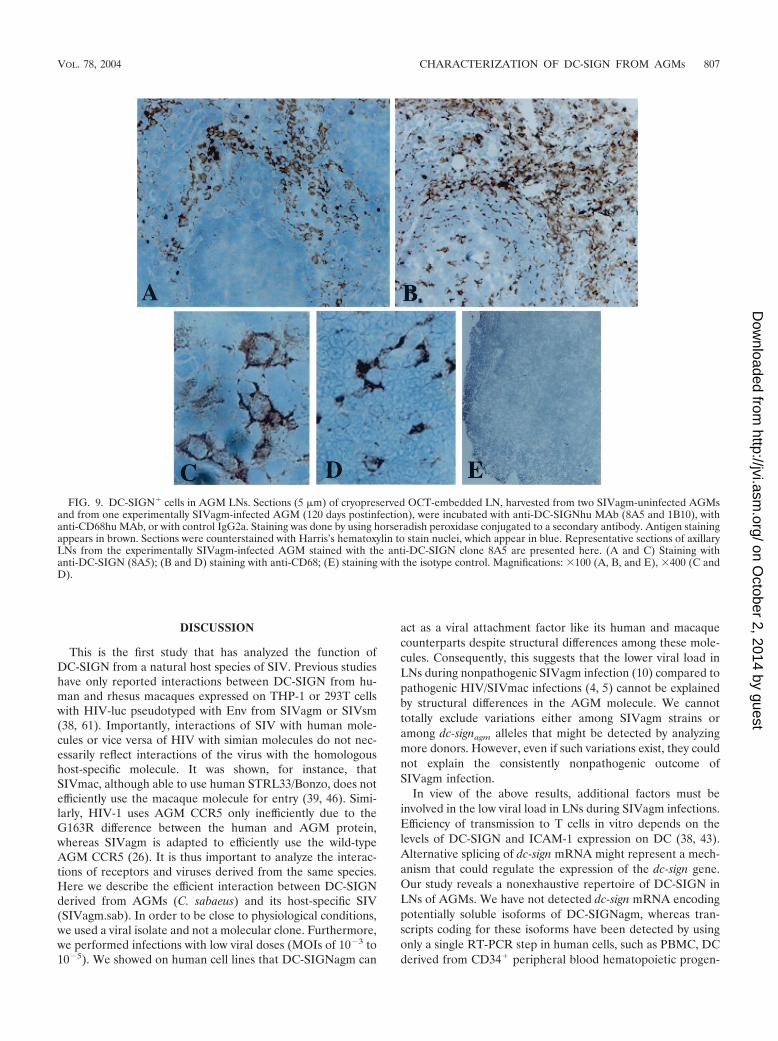
DC-SIGN-expressing cells in AGM LNs. Our data demon-strate that AGM MDDC express high levels of DC-SIGNcomparable to human MDDC and that DC-SIGN plays themain role in SIVagm transmission by AGM MDDC in thepresence of lower doses of virus. The study also revealed thatAGM LN cells abundantly express dc-sign mRNA, suggestingthat DC-SIGN is expressed at significant levels in LN in vivo.To determine whether not only the mRNA but also the proteinis expressed on the surface of cells within LNs, we performedimmunohistochemistry analyses on LN sections derived fromtwo SIVagm uninfected AGMs (animals 02001 and 00021) forwhich the level of DC-SIGN expression on MDDC had beenevaluated (166,000 and 238,000 copies/cell, respectively) andfrom one chronically SIVagm-infected AGM (animal 98013).We labeled the sections with two distinct anti-DC-SIGN MAb(1B10 and 8A5) raised against to two distinct epitopes. Thedistribution of the DC-SIGN� cells was similar whatever theMAb we used (data not shown). We detected DC-SIGN� cellsmainly within the medulla (Fig. 9A) and often within the cortexand/or paracortex (cortex/paracortex) in all three tested ani-mals (Fig. 9A and data not shown). We did not detect anydifferences in the distribution of DC-SIGN� cells betweeninfected and uninfected animals (data not shown). DC-SIGN�
cells both in the medulla and in the cortex/paracortex exhibitedan irregular morphology (Fig. 8C), and some DC-SIGN�
round cells were present within medullary sinuses (a findingconsistent with sinusoidal macrophages) (data not shown). Thedistribution and the morphology of the DC-SIGN� cells inAGM LNs were thus identical to that described in LNs ofmacaques (8, 23, 45, 62). In rhesus macaques, it has beenpreviously described that in SIV-uninfected spleen and in SIV-infected LN, DC-SIGN� cells with dendritic morphology wereuniformly positive for the monocyte/macrophage lineagemarker CD68 (8, 45). The latter remains abundant even afterSIVmac infection (45). In AGM LNs, CD68 staining was alsoabundant in medulla and cortex/paracortex areas. The distri-bution of CD68� and DC-SIGN� cells differed in germinalcenters (GC) (Fig. 9B). CD68� cells were present in GC,whereas DC-SIGN� cells remain always distributed outsidethe GC (Fig. 9A and B). Numerous CD68� cells also exhibitedan irregular morphology (Fig. 9D).
FIG. 8. Viral transmission without lag time and in the absence of viralreplication between exposure of MDDC and coculture with T cells. AGMMDDC were exposed to SIVagm.sabD46 (MOI of 10�5) and coculturedwith SupT1 cells. Each experiment was performed in duplicate. Virusproduction was monitored by SIV p27gag antigen assay after the start ofthe coculture. (A) Viral replication in DC-T cocultures started immedi-ately or 3 days after exposure of MDDC to SIVagm. Solid and brokenlines indicate SIVagm replication in cocultures started immediately orwith a 3-day lag after viral exposure of MDDC, respectively. (B) Assess-ment of AZT treatment of MDDC on viral transmission. DC-T cocultureswere started 3 days after exposure of MDDC from four animals toSIVagm. An RT inhibitor (AZT) was added during the 3-day period toinhibit any potential SIVagm replication in MDDC. Broken and solidlines indicate MDDC treated with or without AZT, respectively. (C) Roleof DC-SIGN in viral transmission by AZT-treated AGM MDDC. AGMMDDC from four animals treated with AZT before viral exposure andduring the 3-day period before the start of the coculture were exposed toSIVagm in the presence of anti-DC-SIGNhu MAb (1B10), a controlIgG2a, or medium alone. In order to express the percent inhibition, thep27gag antigen values of the IgG2a-treated cells, obtained for day 14, werenormalized to 100%.
806 PLOQUIN ET AL. J. VIROL.
on October 2, 2014 by guest
http://jvi.asm.org/
Dow
nloaded from
DISCUSSION
This is the first study that has analyzed the function ofDC-SIGN from a natural host species of SIV. Previous studieshave only reported interactions between DC-SIGN from hu-man and rhesus macaques expressed on THP-1 or 293T cellswith HIV-luc pseudotyped with Env from SIVagm or SIVsm(38, 61). Importantly, interactions of SIV with human mole-cules or vice versa of HIV with simian molecules do not nec-essarily reflect interactions of the virus with the homologoushost-specific molecule. It was shown, for instance, thatSIVmac, although able to use human STRL33/Bonzo, does notefficiently use the macaque molecule for entry (39, 46). Simi-larly, HIV-1 uses AGM CCR5 only inefficiently due to theG163R difference between the human and AGM protein,whereas SIVagm is adapted to efficiently use the wild-typeAGM CCR5 (26). It is thus important to analyze the interac-tions of receptors and viruses derived from the same species.Here we describe the efficient interaction between DC-SIGNderived from AGMs (C. sabaeus) and its host-specific SIV(SIVagm.sab). In order to be close to physiological conditions,we used a viral isolate and not a molecular clone. Furthermore,we performed infections with low viral doses (MOIs of 10�3 to10�5). We showed on human cell lines that DC-SIGNagm can
act as a viral attachment factor like its human and macaquecounterparts despite structural differences among these mole-cules. Consequently, this suggests that the lower viral load inLNs during nonpathogenic SIVagm infection (10) compared topathogenic HIV/SIVmac infections (4, 5) cannot be explainedby structural differences in the AGM molecule. We cannottotally exclude variations either among SIVagm strains oramong dc-signagm alleles that might be detected by analyzingmore donors. However, even if such variations exist, they couldnot explain the consistently nonpathogenic outcome ofSIVagm infection.
In view of the above results, additional factors must beinvolved in the low viral load in LNs during SIVagm infections.Efficiency of transmission to T cells in vitro depends on thelevels of DC-SIGN and ICAM-1 expression on DC (38, 43).Alternative splicing of dc-sign mRNA might represent a mech-anism that could regulate the expression of the dc-sign gene.Our study reveals a nonexhaustive repertoire of DC-SIGN inLNs of AGMs. We have not detected dc-sign mRNA encodingpotentially soluble isoforms of DC-SIGNagm, whereas tran-scripts coding for these isoforms have been detected by usingonly a single RT-PCR step in human cells, such as PBMC, DCderived from CD34� peripheral blood hematopoietic progen-
FIG. 9. DC-SIGN� cells in AGM LNs. Sections (5 �m) of cryopreserved OCT-embedded LN, harvested from two SIVagm-uninfected AGMsand from one experimentally SIVagm-infected AGM (120 days postinfection), were incubated with anti-DC-SIGNhu MAb (8A5 and 1B10), withanti-CD68hu MAb, or with control IgG2a. Staining was done by using horseradish peroxidase conjugated to a secondary antibody. Antigen stainingappears in brown. Sections were counterstained with Harris’s hematoxylin to stain nuclei, which appear in blue. Representative sections of axillaryLNs from the experimentally SIVagm-infected AGM stained with the anti-DC-SIGN clone 8A5 are presented here. (A and C) Staining withanti-DC-SIGN (8A5); (B and D) staining with anti-CD68; (E) staining with the isotype control. Magnifications: �100 (A, B, and E), �400 (C andD).
VOL. 78, 2004 CHARACTERIZATION OF DC-SIGN FROM AGMs 807
on October 2, 2014 by guest
http://jvi.asm.org/
Dow
nloaded from

itors, and resting CD14� monocytes (35). Indeed, the expres-sion pattern of dc-sign transcripts may depend on the origin, aswell as the maturation or activation state, of the cells (35). Itremains unknown whether transcripts coding for potentiallysoluble isoforms also exist in human LN or in other nonhumanprimates such as macaques. Although soluble isoforms do notseem to be able to mediate enhancement of infection (27), theymay regulate expression of the membrane-associated forms ofDC-SIGN (35). However, we demonstrate here that AGMMDDC show high surface-expressing levels of DC-SIGN pro-tein. This contrasts with one study which reported very lowlevels of DC-SIGN on rhesus macaque MDDC (61). The ex-pression levels of DC-SIGN may vary according to the primatespecies and/or the donor (25, 61). Numbers of DC-SIGN mol-ecules on AGM MDDC did indeed vary according to theAGM donor, as shown for human MDDC (1). However, theAGM myeloid DC (MDDC) from all studied AGM donorsexpressed at least 100,000 DC-SIGN molecules and often morethan 200,000 molecules per cell, a level sufficient to promoteviral transmission to T cells in vitro.
In vivo, DC-SIGN� cells could be easily detected within themedulla sinuses and the paracortex of AGM LNs. We cannotexclude the possibility that the detection of positive cells in themedulla sinuses could also be due to the related L-SIGN2 (3).However, the distribution of DC-SIGN� cells in the LNs ofAGMs was very similar to that described in macaques (8, 45,62). Rhesus macaque antigen-presenting cells in the medullaand cortex/paracortex in organized lymphoid tissues (LNs andspleen) have been shown to express DC-SIGN, although thenumber of positive cells has not been quantified (8, 45, 62).The nature of the AGM DC-SIGN� cells is unknown, but thecells exhibited a morphology characteristic of DC. It has beensuggested in similar studies in humans and rhesus macaquetissues that the DC-SIGN� cells likely correspond to interdig-itating DC and sinusoidal macrophages (14, 45, 49, 62). Thedistribution of DC-SIGN� cells in the medulla and paracortexwas similar to that of cells expressing a marker of the mono-cyte/macrophage lineage (CD68). However, only CD68� cellswere detected in germinal center, showing that not all CD68�
cells express DC-SIGN. Furthermore, it has been shown that inmacaque spleens and LNs all DC-SIGN� cells exhibiting adendritic morphology express CD68 (45). Altogether, our datashow a similar morphology and distribution of DC-SIGN�
cells compared to these cells in humans and macaques. Wealso observed that mRNA coding for functional DC-SIGN isabundantly expressed in AGM LNs. This suggests that DC-SIGN expression on DC in AGM LN is not lower than inhumans and macaques.
We cannot totally exclude a distinct regulation of DC-SIGNexpression in response to SIVagm infection. The level of hu-man DC-SIGN at the cell surface is indeed influenced byHIV-1 proteins. A dramatic increase of lymphocyte adhesionto HIV-1-infected DC in vitro due to an upregulation of DC-SIGN mediated by Nef has been reported (50). This phenom-enon, however, is not consistently observed for all donors (31).It is not excluded that Nef encoded by SIVagm interacts dif-ferentially with host-specific cellular proteins compared toHIV/SIVmac Nef in human and macaque cells, which couldresult in lower levels of DC-SIGN expression and/or lowerT-cell activation profiles in SIVagm-infected AGMs. Lower
T-cell activation states are indeed observed in nonpathogenicSIV infections in their natural hosts (7, 11, 37, 47). However,there are no signs thus far of SIVagm replication within AGMMDDC with at least up to 10�3 TCID50/cell and after p27values up to 2 weeks. This might also be dependent on theSIVagm strains used. Further studies are needed to determinethe susceptibility of DC to SIV in nonpathogenic SIV infectionmodels.
Finally, the expression level might depend on the cytokineenvironment in vivo after SIV infection. Several studies havereported that IL-4 induces DC-SIGN expression on humanperipheral blood monocytes in vitro (35, 41). This IL-4-depen-dent expression is negatively regulated by alpha interferon(IFN-�), transforming growth factor , and IFN- (41). Re-cently, it has been shown in another model of nonpathogenicSIVinfection, SIVsm infection in the sooty mangabeys, that thechronic phase of infection is characterized by higher levels ofIL-4 and lower levels of IFN- production (47). Thus, there areas yet no data that could support any downregulation of DC-SIGN expression after SIV infection in the natural host.
We show here that MDDC from AGMs not only expresshigh levels of DC-SIGN but also efficiently transmit SIVagm toT cells. This has been confirmed here for MDDC from 10distinct AGM donors. Furthermore, as shown by inhibitionassays with anti-DC-SIGN MAb, viral transmission by AGMMDDC is DC-SIGN dependent. This has been confirmed withMDDC from six distinct AGM donors. This result supports thefindings of an earlier study of Chinese macaque species, M.fascicularis, in which viral transmission is also largely DC-SIGN dependent (25). However, it contrasts with a study of M.mulatta MDDC that indicated no discernible role of DC-SIGNin viral transmission (61). These controversial results might beexplained by variations of DC-SIGN expression according toindividuals, especially if only a few animals were studied,and/or according to monkey species. Interestingly, we observedthat the viral transmission by AGM MDDC was significantlyblocked by anti-DC-SIGN MAbs only when a low MOI (10�5)was used. It has been reported that the affinity of HIV-1 gp120for DC-SIGNhu is higher than for CD4hu (9), and some datasuggested that it is also higher than for the mannose receptor(56). Thus, the use of high viral infectious doses could probablyallow attachment to other molecules as well. This might alsoexplain why in previous studies anti-DC-SIGN MAb or man-nan could not consistently block viral entry or Env/DC-SIGNinteraction in MDDC. Indeed, in these previous studies, veryhigh doses of HIV AD8 (MOI of 45) (57) or a high amount ofgp120 (twofold more than the predetermined concentrationfor cellular saturation) (56) were used.
DC seem to be a “hiding place” for HIV-1 (58). However,the mechanism of viral transfer via DC-SIGN has yet to befully elucidated. The function of DC-SIGN in the transmissionof HIV-1 depends on its cellular context (54). DC-SIGN ex-pressed by DC or the monocytic cell line THP-1 but not 293and HOS cells internalizes and retains HIV-1Bal in a highlyinfectious state for more than 5 days (13, 54). Immature humanMDDC promote HIV-1Bal transfer by a mechanism that isDC-SIGN dependent (40 to 100% according to the donorstested) at least up to 2 days after viral exposure (54). Recently,others suggest most endocytosed virus is already degradedafter 24 h (55). We demonstrated here that sufficient amounts
808 PLOQUIN ET AL. J. VIROL.
on October 2, 2014 by guest
http://jvi.asm.org/
Dow
nloaded from

of infectious SIVagm particles remain intact in AGM MDDCup to 3 days, even when low doses of virus are used. Thisefficiency might be due to the fact that we used a virus isolateand MDDC to assess viral transfer to highly permissive T cells.
We analyzed here for the first time the virological functionsof DC-SIGN from an animal species resistant to AIDS. Thepresent study revealed similar activities for DC-SIGNagm re-garding trans infection compared to previous reports on humanand macaque DC-SIGN. We demonstrated that DC-SIGN�
cells are present in AGM LNs and that AGM MDDC expresslevels of DC-SIGN similar to those expressed by humanMDDC. We showed that AGM MDDC are able to efficientlytransmit SIVagm to T cells. Finally, we showed that SIVagmtransmission is largely DC-SIGN dependent at low MOIs. Al-together, our data indicate that the virus-binding properties ofDC-SIGN are not directly associated with viral load levels inLNs. In conclusion, additional factors, such as the frequency inLNs of major target cells (activated T CD4� lymphocytes), aremore likely determinating factors for viral replication levels inLNs.
ACKNOWLEDGMENTS
We are indebted to Robert Bassin for critical reading of the manu-script. We thank Arnaud Moris for helpful discussion and Lisa An forcontribution in editing of the manuscript. We thank Sandrine DosSantos and M. C. Cumont for excellent technical help and AhidjoAyouba and veterinarians from the Cameroonian and European zoosfor DNA samples.
This study was supported by grants from the French Agency forAIDS Research (ANRS). M.J.-Y.P. was a recipient of a scholarshipfrom the Ministere de l’Education Nationale. L.M. and M.A.S. wererecipients of ANRS fellowships.
REFERENCES
1. Baribaud, F., S. Pohlmann, G. J. Leslie, F. Mortari, and R. W. Doms. 2002.Quantitative expression and virus transmission analysis of DC-SIGN onmonocyte-derived dendritic cells. J. Virol. 76:9135–9142.
2. Baribaud, F., S. Pohlmann, T. Sparwasser, M. T. Y. Kimata, Y. Choi, B. S.Haggarty, N. Ahmad, T. Macfarlan, T. G. Edwards, G. J. Leslie, J. Arnason,T. A. Reinhart, J. T. Kimata, D. R. Littman, J. A. Hoxie, and R. W. Doms.2001. Functional and antigenic characterization of human, rhesus macaque,pigtailed macaque and murine DC-SIGN. J. Virol. 75:10281–10289.
3. Bashirova, A. A., L. Wu, J. Cheng, T. D. Martin, M. P. Martin, R. E.Benveniste, J. D. Lifson, V. N. KewalRamani, A. Hughes, and M. Car-rington. 2003. Novel member of the CD209 (DC-SIGN) gene family inprimates. J. Virol. 77:217–227.
4. Beer, B., J. Scherer, J. Megede, S. Norley, M. Baier, and R. Kurth. 1996.Lack of dichotomy between virus load of peripheral blood and lymph nodesduring long term simian immunodeficiency virus infection of African greenmonkeys. Virology 219:367–375.
5. Broussard, S. R., S. I. Staprans, R. White, E. M. Whitehead, M. B. Feinberg,J. S. Allan, T. W. Chun, L. Stuyver, S. B. Mizell, L. A. Ehler, J. A. Mican, M.Baseler, A. L. Lloyd, M. A. Nowak, and A. S. Fauci. 2001. Simian immuno-deficiency virus replicates to high levels in naturally infected African greenmonkeys without inducing immunologic or neurologic disease. J. Virol. 75:2262–2275.
6. Cameron, P. U., P. S. Freudenthal, J. M. Barker, S. Gezelter, K. Inaba, andR. M. Steinmann. 1992. Dendritic cells exposed to human immunodeficiencyvirus type-1 transmit a vigorous cytopathic infection to CD4� cells. Science257:383–387.
7. Chakrabarti, L. A., S. R. Lewin, L. Zhang, A. Gettie, A. Luckay, L. N. Martin,E. Skulski, D. D. Ho, C. Cheng-Mayer, and P. A. Marx. 2000. Normal T-cellturnover in sooty mangabeys harboring active simian immunodeficiency virusinfection. J. Virol. 74:1209–1223.
8. Choi, Y. K., B. A. Fallert, M. A. Murphey-Corb, and T. A. Reinhart. 2003.Simian immunodeficiency virus dramatically alters expression of homeostaticchemokines and dendritic cell markers during infection in vivo. Blood 101:1684–1691.
9. Curtis, B. M., S. Scharnowske, and A. J. Watson. 1992. Sequence andexpression of a membrane-associated C-type lectin that exhibits CD4-inde-pendent binding of human immunodefficiency virus envelope glycoproteingp120. Proc. Natl. Acad. Sci. USA 89:8356–8360.
10. Diop, O. M., A. Gueye, M. Dias-Tavares, C. Kornfeld, A. Faye, P. Ave, M.Huerre, S. Corbet, F. Barre-Sinoussi, and M. C. Muller-Trutwin. 2000. Highlevels of viral replication during primary SIVagm infection are rapidly andstrongly controlled in African green monkeys. J. Virol. 74:7538–7547.
11. Estaquier, J., T. Idziorek, F. de Bels, F. Barre-Sinoussi, B. Hurtrel, A. M.Aubertin, A. Venet, M. Mehtali, E. Muchmore, P. Michel, et al. 1994. Pro-grammed cell death and AIDS: significance of T-cell apoptosis in pathogenicand nonpathogenic primate lentiviral infections. Proc. Natl. Acad. Sci. USA91:9431–9435.
12. Geijtenbeek, T. B. H., G. Koopman, G. C. F. Van Duijnhoven, S. J. van Vliet,A. C. Van Schijndel, A. Engering, J. L. Heeney, and Y. Van Kooyk. 2001.Rhesus macaque and chimpanzee DC-SIGN act as HIV/SIV gp120 trans-receptor, similar to human DC-SIGN. Immunol. Lett. 79:101–107.
13. Geijtenbeek, T. B. H., D. S. Kwon, R. Torensma, S. J. van Vliet, G. C. F. VanDuijnhoven, J. Middel, I. L. M. H. A. Cornelissen, D. R. Littman, C. G.Figdor, and Y. van Kooyk. 2000. DC-SIGN, a dendritic cell-specific HIV-1-Binding protein that enhances trans-infection of T cells. Cell 100:587–597.
14. Geijtenbeek, T. B. H., R. Torensma, S. J. Van Vliet, G. C. F. Van Duijnhoven,G. J. Adema, and Y. Van Kooyk. 2000. Identification of DC-SIGN, a noveldendritic cell-specific ICAM-3 receptor that supports primary immune re-sponses. Cell 100:575–585.
15. Geijtenbeek, T. B. H., G. C. F. Van Duijnhoven, S. J. van Vliet, E. Krieger,G. Vriend, C. G. Figdor, and Y. Van Kooyk. 2002. Identification of differentbinding sites in the dendritic cell-specific receptor for ICAM-3 and HIV-1.J. Biol. Chem. 277:11314–11320.
16. Goldstein, S., I. Ourmanov, C. R. Brown, B. E. Beer, W. R. Elkins, R.Plishka, A. Buckler-White, V. M. Hirsch, T. W. Chun, L. Stuyver, S. B.Mizell, L. A. Ehler, J. A. Mican, M. Baseler, A. L. Lloyd, M. A. Nowak, andA. S. Fauci. 2000. Wide range of viral load in healthy African green monkeysnaturally infected with simian immunodeficiency virus. J. Virol. 74:11744–11753.
17. Granelli-Piperno, A., E. Delgado, V. Finkel, W. Paxton, and R. M. Steinman.1998. Immature dendritic cells selectively replicate macrophagetropic (M-tropic) human immunodefficiency virus type 1, while mature cells efficiencytransmit both M- and T-tropic virus to T cells. J. Virol. 72:2733–2737.
18. Granelli-Piperno, A., V. Finkel, E. Delgado, and R. M. Steinman. 1999. Virusreplication begins in dendritic cells during the transmission of HIV-1 frommature dendritic cells to T cells. Curr. Biol. 9:21–29.
19. Grouard, G., and E. A. Clark. 1997. Role of dendritic and follicular dendriticcells in HIV infection and pathogenesis. Curr. Opin. Immunol. 9:562–567.
20. Halary, F., A. Amara, H. Lortat-Jacob, M. Messerle, T. Delaunay, C. Houles,F. Fieschi, F. Arenzana-Seisdedos, J.-F. Moreau, and J. Dechanet-Merville.2002. Human cytomegalovirus binding to DC-SIGN is required for dendriticcell infection and target cell trans-infection. Immunity 17:653–664.
21. Holzammer, S., E. Holznagel, A. Kaul, R. Kurth, and S. Norley. 2001. Highvirus loads in naturally and experimentally SIVagm-infected African greenmonkeys. Virology 283:324–331.
22. Hu, J., M. B. Gardner, and C. J. Miller. 2000. Simian immunodeficiencyvirus rapidly penetrates the cervicovaginal mucosa after intravaginal inocu-lation and infects intraepithelial dendritic cells. J. Virol. 74:6087–6095.
23. Jameson, B., F. Baribaud, S. Pohlmann, D. Ghavimi, F. Mortari, R. W.Doms, and A. Iwasaki. 2002. Expression of DC-SIGN by dendritic cells ofintestinal and genital mucosae in humans and rhesus macaques. J. Virol.76:1866–1875.
24. Kawamura, T., F. O. Gulden, M. Sugaya, D. T. McNamara, D. L. Borris,M. M. Lederman, J. M. Orenstein, P. A. Zimmerman, and A. Blauvelt. 2003.R5 HIV productively infects Langerhans cells, and infection levels are reg-ulated by compound CCR5 polymorphisms. Proc. Natl. Acad. Sci. USA100:8401–8406.
25. Kimata, M. T. Y., M. Cella, J. E. Biggins, C. Rorex, R. White, S. Hicks, J. M.Wilson, P. G. Patel, J. S. Allan, M. Colonna, and J. T. Kimata. 2002. Captureand transfer of simian immunodeficiency virus by macaque dendritic cells isenhanced by DC-SIGN. J. Virol. 76:11827–11836.
26. Kuhmann, S., N. Madani, O. M. Diop, E. J. Platt, J. Morvan, M. C. Muller-Trutwin, F. Barre-Sinoussi, and D. Kabat. 2001. Frequent substitution poly-morphism in African green monkey CCR5 cluster at active sites for infec-tions by SIVagm, implying ancient virus-host coevolution. J. Virol. 75:8449–8460.
27. Kwon, D. S., G. Gregorio, N. Bitton, W. A. Hendrickson, and D. R. Littman.2002. DC-SIGN-mediated internalization of HIV is required for trans-en-hancement of T-cell infection. Immunity 16:135–144.
28. Lee, B., G. Leslie, E. Soilleux, U. O’Doherty, S. Baik, E. Levroney, K.Flummerfelt, W. Swiggard, N. Coleman, M. Malim, and R. W. Doms. 2001.Cis expression of DC-SIGN allows for more efficient entry of human andsimian immunodeficiency viruses via CD4 and a coreceptor. J. Virol. 75:12028–12038.
29. Marechal, V., M. C. Prevost, C. Petit, E. Perret, J. M. Heard, and O.Schwartz. 2001. Human immunodeficiency virus type 1 entry into macro-phages mediated by macropinocytosis. J. Virol. 75:11166–11177.
30. McDonald, D., L. Wu, S. M. Bohks, V. N. KewalRamani, D. Unutmaz, andT. J. Hope. 2003. Recruitment of HIV and its receptors to dendritic cell-Tcell junctions. Science 300:1295–1297.
VOL. 78, 2004 CHARACTERIZATION OF DC-SIGN FROM AGMs 809
on October 2, 2014 by guest
http://jvi.asm.org/
Dow
nloaded from

31. Messmer, D., J.-M. Jacque, C. Santisteban, C. Bristow, S.-H. Han, L. Vil-lamide-Herrera, E. Mehlhop, P. A. Marx, R. M. Steinman, and M. Pope.2002. Endogenously expressed nef uncouples cytokine and chemokine pro-duction from membrane phenotypic maturation in dendritic cells. J. Immu-nol. 169:4172–4182.
32. Muller, M. C., N. K. Saksena, E. Nerrienet, C. Chappey, V. M. A. Herve, J.-P.Durand, P. Legal-Campodonico, M.-C. Lang, J.-P. Digoutte, A. J. Georges,M.-C. Georges-Courbot, P. Sonigo, and F. Barre-Sinoussi. 1993. Simianimmunodeficiency viruses from central and western Africa: evidence for anew species-specific lentivirus in tantalus monkeys. J. Virol. 67:1227–1235.
33. Muller-Trutwin, M. C., S. Corbet, J. Hansen, M. C. Georges-Courbot, O.Diop, J. Rigoulet, F. Barre-Sinoussi, and A. Fomsgaard. 1999. Mutations inCCR5 coding sequences are not associated with SIV carrier status in Africannonhuman primates. AIDS Res. Hum. Retrovir. 15:931–939.
34. Muller-Trutwin, M. C., S. Corbet, S. Souquiere, P. Roques, P. Versmisse, A.Ayouba, S. Delarue, E. Nerrienet, J. Lewis, P. Martin, F. Simon, F. Barre-Sinoussi, and P. Mauclere. 2000. SIVcpz from a naturally infected Cam-eroonian chimpanzee: biological and genetic comparison with HIV-1 N.J. Med. Primatol. 29:166–172.
35. Mummidi, S., G. Catano, L. Lam, A. Hoefle, V. Telles, K. Begum, F. Jimenez,S. S. Ahuja, and S. K. Ahuja. 2001. Extensive repertoire of membrane-boundand soluble DC-SIGN1 and DC-SIGN2 isoforms: inter-individual variationin expression of DC-SIGN transcripts. J. Biol. Chem. 276:33196–33212.
36. Nobile, C., A. Moris, F. Porrot, N. Sol-Foulon, and O. Schwartz. 2003.Inhibition of human immunodeficiency virus type 1 Env-mediated fusion byDC-SIGN. J. Virol. 77:5313–5323.
37. Onanga, R., C. Kornfeld, I. Pandrea, J. Estaquier, S. Souquiere, P. Rouquet,V. Poaty Mavoungou, O. Bourry, S. M’Boup, F. Barre-Sinoussi, F. Simon, C.Apetrei, P. Roques, and M. C. Muller-Trutwin. 2002. High levels of viralreplication contrast with only transient changes in CD4� and CD8� cellnumbers during the early phase of experimental infection with simian im-munodeficiency virus SIVmnd-1 in Mandrillus sphinx. J. Virol. 76:10256–10263.
38. Pohlmann, S., F. Baribaud, B. Lee, G. J. Leslie, M. D. Sanchez, K.Hiebenthal-Millow, J. Munch, F. Kirchhoff, and R. W. Doms. 2001. DC-SIGN interactions with human immunodefficiency virus type 1 and 2 andsimian immunodefficiency virus. J. Virol. 75:4664–4672.
39. Pohlmann, S., B. Lee, S. Meister, M. Krumbiegel, G. Leslie, R. W. Doms,and F. Kirchhoff. 2000. Simian immunodeficiency virus utilizes human andsooty mangabey but not rhesus macaque STRL33 for efficient entry. J. Virol.74:5075–5082.
40. Pope, M., M. G. Betjes, N. Romani, H. Hirmand, P. U. Cameron, L. Hoff-man, S. Gezelter, G. Schuler, and R. M. Steinman. 1994. Conjugates ofdendritic cells and memory T lymphocytes from skin facilitate productiveinfection with HIV-1. Cell 78:389–398.
41. Relloso, M., A. Puig-Kroger, O. Muniz Pello, J. L. Rodriguez-Fernandez, G.de la Rosa, N. Longo, J. Navarro, M. A. Munoz-Fernandez, P. Sanchez-Mateo, and A. L. Corbi. 2002. DC-SIGN (CD209) expression is IL-4 depen-dent and is negatively regulated by IFN, TGF-, and anti-inflammatoryagents. J. Immunol. 168:2634–2643.
42. Rowland-Jones, S. L. 1999. HIV: the deadly passenger in dendritic cells.Curr. Biol. 9:R248–R250.
43. Sanders, R. W., E. C. de Jong, C. E. Baldwin, J. H. N. Schuitemaker, M. L.Kapsenberg, and B. Berkhout. 2002. Differential transmission of humanimmunodeficiency virus type 1 by distinct subsets of effector dendritic cells.J. Virol. 76:7812–7821.
44. Santos, P. R., K. Petitprez, and C. Butor. 2002. Gene for Chinese rhesusmacaque DC-SIGN predicts the existence of A but not B isoforms of theprotein. AIDS Res. Hum. Retrovir. 18:977–981.
45. Schwartz, A., X. Alvarez, and A. A. Lackner. 2002. Distribution and immu-
nophenotype of DC-SIGN-expressing cells in SIV-infected and uninfectedmacaques. AIDS Res. Hum. Retrovir. 18:1021–1029.
46. Sharron, M., S. Pohlmann, K. Price, E. Lolis, M. Tsang, F. Kirchhoff, R. W.Doms, and B. Lee. 2000. Expression and coreceptor activity of STRL33/Bonzo on primary peripheral blood lymphocytes. Blood 96:41–49.
47. Silvestri, G., D. L. Sodora, R. A. Koup, M. Paiardini, S. P. O’Neil, H. M.McClure, S. I. Staprans, and M. B. Feinberg. 2003. Nonpathogenic SIVinfection of sooty mangabeys is characterized by limited bystander immuno-pathology despite chronic high-level viremia. Immunity 18:441–452.
48. Soilleux, E. J., R. Barten, and J. Trowsdale. 2000. DC-SIGN, a related gene,and CD23 form a cluster on 19p13. J. Immunol. 165:2937–2942.
49. Soilleux, E. J., L. S. Morris, G. Leslie, J. Chemini, Q. Luo, E. Levroney, J.Trowsdale, L. J. Montaner, R. W. Doms, D. Weissman, N. Coleman, and B.Lee. 2002. Constitutive and induced expression of DC-SIGN on dendritic celland macrophage subpopulations in situ and in vitro. J. Leukoc. Biol. 71:445–457.
50. Sol-Foulon, N., A. Moris, C. Nobile, C. Boccaccio, A. Engering, J.-P. Abas-tado, J.-M. Heard, Y. Van Kooyk, and O. Schwartz. 2002. HIV-1 Nef-induced upregulation of DC-SIGN in dendritic cells promotes lymphocyteclustering and viral spread. Immunity 16:145–155.
51. Soto-Ramirez, L. E., B. Renjifo, M. F. McLane, R. Marlink, C. O’Hara, R.Sutthent, C. Wasi, P. Vithayasi, V. Vithayasi, C. Apichartpiyakul, P.Auewarakul, V. Pena Cruz, D. Chui, R. Osathamondh, K. Mayer, T. Lee, andM. Essex. 1996. HIV-1 Langherans cells tropism associated with heterosex-ual transmission of HIV. Science 271:1291–1293.
52. Steinman, R. M. 2000. DC-SIGN: a guide to some mysteries of dendriticcells. Cell 100:491–494.
53. Steinman, R. M., and K. Inaba. 1999. Myeloid dendritic cells. J. Leukoc.Biol. 66:205–208.
54. Trumpfheller, C., C. Gyu Park, J. Finke, R. M. Steinman, and A. Granelli-Piperno. 2003. Cell type-dependent retention and transmission of HIV-1 byDC-SIGN. Int. Immunol. 15:289–298.
55. Turville, S., J. Wilkinson, P. Cameron, J. Dable, and A. L. Cunningham.2003. The role of dendritic cell C-type lectin receptors in HIV pathogenesis.J. Leukoc. Biol. 74:710–718.
56. Turville, S. G., J. Arthos, K. Mac Donald, G. Lynch, H. Naif, G. Clark, D.Hart, and A. L. Cunningham. 2001. HIV gp120 receptors on human den-dritic cells. Blood 98:2482–2488.
57. Turville, S. G., P. U. Cameron, A. Handley, G. Lin, S. Pohlmann, R. W.Doms, and A. L. Cunningham. 2002. Diversity of receptors binding HIV ondendritic cell subsets. Nat. Immunol. 3:975–983.
58. Van Kooyk, Y., and T. B. Geijtenbeek. 2003. DC-SIGN: escape mechanismfor pathogens. Nat. Rev. Immunol. 3:697–709.
59. Verrier, F. C., P. Charneau, R. Altemeyer, S. Laurent, A. M. Borman, and M.Girard. 1997. Antibodies to several conformation-dependent epitopes ofgp120/gp41 inhibit CCR5-dependent cell-to-cell fusion mediated by the na-tive envelope glycoprotein of a primary macrophage-tropic HIV-1 isolate.Proc. Natl. Acad. Sci. USA 94:9326–9331.
60. Weissman, D., Y. Li, J.-M. Orenstein, and A. S. Fauci. 1995. Both a precur-sor and a mature population of dendritic cells can bind HIV: however, onlythe mature population that expressed CD80 can pass infection to unstimu-lated CD4� T cells. J. Immunol. 155:4111–4117.
61. Wu, L., A. A. Bashirova, T. D. Martin, L. Villamide, E. Mehlhop, A. O.Chertov, D. Unutmaz, M. Pope, M. Carrington, and V. N. KewalRamani.2002. Rhesus macaque dendritic cells efficiently transmit primate lentivirusesindependently of DC-SIGN. Proc. Natl. Acad. Sci. USA 99:1568–1573.
62. Zimmer, M. I., A. T. Larregina, C. M. Castillo, S. Capuano III, L. D. Falo,Jr., M. Murphey-Corb, T. A. Reinhart, and S. M. Barratt-Boyes. 2002.Disrupted homeostasis of Langerhans cells and interdigitating dendritic cellsin monkeys with AIDS. Blood 99:2859–2868.
810 PLOQUIN ET AL. J. VIROL.
on October 2, 2014 by guest
http://jvi.asm.org/
Dow
nloaded from