Development of the optokinetic system in macaque monkeys
11
Vision Research 39 (1999) 3909 – 3919 Section 3 Development of the optokinetic system in macaque monkeys C. Distler a, *, F. Vital-Durand b , R. Korte c , H. Korbmacher a , K.-P. Hoffmann a a Allgemeine Zoologie & Neurobiologie, Ruhr -Uni6ersita ¨t Bochum, Postfach 102148, D-44780 Bochum, Germany b INSERM U371, Cer6eau et Vision, 18 A6enue du Doyen Le ´pine, F -69675 Bron, France c Co6ance Laboratories GMBH, D-48163 Mu ¨nster, Germany Received 30 December 1998; received in revised form 31 May 1999 Abstract Optokinetic nystagmus in response to horizontal movement of a whole field random dot pattern was measured in infant macaque monkeys from the first week to about 5 months after birth using electrooculography. During monocular and binocular viewing conditions stimulus velocities were varied between 10 and 120 °/s. Monocular stimulation in the temporonasal direction yielded slow phase gain of the optokinetic system which was relatively constant for a given stimulus velocity over the whole period of observation. Gain during nasotemporal stimulation was also clearly present but significantly lower at early stages and increased during further development. This asymmetry of monocular horizontal optokinetic nystagmus (OKN) clearly depended on the stimulus velocity. At lower stimulus velocities (10 – 20 °/s) OKN was largely symmetrical at 2–5 weeks of age. At higher stimulus velocities (40 °/s) symmetry was reached at about 12 weeks of age or even much later (80–120 °/s). © 1999 Elsevier Science Ltd. All rights reserved. Keywords: Optokinetic reflex; OKN asymmetry; Monkey; Motion perception; Primate www.elsevier.com/locate/visres 1. Introduction The optokinetic reflex (OKR), a mechanism to stabilize global motion of the environment on the retina can be used to judge an animals ability to react to visual motion. This visual reflex has been thoroughly investigated in a large variety of vertebrates, especially in lateral-eyed afoveate and in frontal-eyed foveate mammals (Tauber & Atkin, 1968). During monocular vision, the OKR in lateral-eyed mammals is largely asymmetric, i.e. the optokinetic response is stronger during motion of the visual world from temporal to nasal than during motion from nasal to temporal (e.g. rat: Hess, Precht, Reber & Cazin, 1985; rabbit: Collewijn, 1965; Dubois & Collewijn, 1979; guinea pig: Hayes & Ireland, 1969; Benassi, Biral & Corazza, 1992). By contrast, frontal-eyed foveate mammals exhibit a largely symmetrical monocular OKR with equally vigorous response during temporal to nasal and nasal to temporal visual world motion (e.g. cat: Wood, Spear & Braun, 1973; Hamada, 1983; monkey: Koerner & Schiller, 1972; man: Schor & Narayan, 1981; Westall & Schor, 1985; Van den Berg & Collewijn, 1988; Schor, 1993). Developmental studies in cats (Van Hof-Van Duin, 1978; Atkinson, 1979; Malach, Strong & van Sluyters, 1981) and human infants (Atkinson, 1979; Naegele & Held, 1982; Roy, Lachapelle & Lepore ´, 1989) have shown that in early childhood monocular OKR is largely asymmetric thus resembling OKR in afoveate animals and becomes symmetrical during the first few weeks to months of life. In cat, there is good evidence from behavioural, anatomical, and physiological data that the appearance of symmetry of OKR coincides with the maturation of a binocular cortical input to the nucleus of the optic tract and dorsal terminal nucleus of the accessory optic system (NOT-DTN), the visuomotor interface in the subcortical OKR pathway (Distler & Hoffmann, 1993). Whether or not this is true also for primates remains to be determined. Quantitative studies using electrooculography in hu- man infants showed that symmetry of monocular OKR * Corresponding author. Tel.: +49-234-7004365; fax: +49-234- 7094185. E-mail address: [email protected] (C. Dis- tler) 0042-6989/99/$ - see front matter © 1999 Elsevier Science Ltd. All rights reserved. PII:S0042-6989(99)00122-4
-
Upload
ruhr-uni-bochum -
Category
Documents
-
view
5 -
download
0
Transcript of Development of the optokinetic system in macaque monkeys

Vision Research 39 (1999) 3909–3919
Section 3
Development of the optokinetic system in macaque monkeys
C. Distler a,*, F. Vital-Durand b, R. Korte c, H. Korbmacher a, K.-P. Hoffmann a
a Allgemeine Zoologie & Neurobiologie, Ruhr-Uni6ersitat Bochum, Postfach 102148, D-44780 Bochum, Germanyb INSERM U371, Cer6eau et Vision, 18 A6enue du Doyen Lepine, F-69675 Bron, France
c Co6ance Laboratories GMBH, D-48163 Munster, Germany
Received 30 December 1998; received in revised form 31 May 1999
Abstract
Optokinetic nystagmus in response to horizontal movement of a whole field random dot pattern was measured in infantmacaque monkeys from the first week to about 5 months after birth using electrooculography. During monocular and binocularviewing conditions stimulus velocities were varied between 10 and 120 °/s. Monocular stimulation in the temporonasal directionyielded slow phase gain of the optokinetic system which was relatively constant for a given stimulus velocity over the whole periodof observation. Gain during nasotemporal stimulation was also clearly present but significantly lower at early stages and increasedduring further development. This asymmetry of monocular horizontal optokinetic nystagmus (OKN) clearly depended on thestimulus velocity. At lower stimulus velocities (10–20 °/s) OKN was largely symmetrical at 2–5 weeks of age. At higher stimulusvelocities (40 °/s) symmetry was reached at about 12 weeks of age or even much later (80–120 °/s). © 1999 Elsevier Science Ltd.All rights reserved.
Keywords: Optokinetic reflex; OKN asymmetry; Monkey; Motion perception; Primate
www.elsevier.com/locate/visres
1. Introduction
The optokinetic reflex (OKR), a mechanism to stabilizeglobal motion of the environment on the retina can beused to judge an animals ability to react to visual motion.This visual reflex has been thoroughly investigated in alarge variety of vertebrates, especially in lateral-eyedafoveate and in frontal-eyed foveate mammals (Tauber& Atkin, 1968). During monocular vision, the OKR inlateral-eyed mammals is largely asymmetric, i.e. theoptokinetic response is stronger during motion of thevisual world from temporal to nasal than during motionfrom nasal to temporal (e.g. rat: Hess, Precht, Reber &Cazin, 1985; rabbit: Collewijn, 1965; Dubois & Collewijn,1979; guinea pig: Hayes & Ireland, 1969; Benassi, Biral& Corazza, 1992). By contrast, frontal-eyed foveatemammals exhibit a largely symmetrical monocular OKRwith equally vigorous response during temporal to nasal
and nasal to temporal visual world motion (e.g. cat:Wood, Spear & Braun, 1973; Hamada, 1983; monkey:Koerner & Schiller, 1972; man: Schor & Narayan, 1981;Westall & Schor, 1985; Van den Berg & Collewijn, 1988;Schor, 1993).
Developmental studies in cats (Van Hof-Van Duin,1978; Atkinson, 1979; Malach, Strong & van Sluyters,1981) and human infants (Atkinson, 1979; Naegele &Held, 1982; Roy, Lachapelle & Lepore, 1989) have shownthat in early childhood monocular OKR is largelyasymmetric thus resembling OKR in afoveate animalsand becomes symmetrical during the first few weeks tomonths of life. In cat, there is good evidence frombehavioural, anatomical, and physiological data that theappearance of symmetry of OKR coincides with thematuration of a binocular cortical input to the nucleusof the optic tract and dorsal terminal nucleus of theaccessory optic system (NOT-DTN), the visuomotorinterface in the subcortical OKR pathway (Distler &Hoffmann, 1993). Whether or not this is true also forprimates remains to be determined.
Quantitative studies using electrooculography in hu-man infants showed that symmetry of monocular OKR
* Corresponding author. Tel.: +49-234-7004365; fax: +49-234-7094185.
E-mail address: [email protected] (C. Dis-tler)
0042-6989/99/$ - see front matter © 1999 Elsevier Science Ltd. All rights reserved.
PII: S 0 0 4 2 -6989 (99 )00122 -4

C. Distler et al. / Vision Research 39 (1999) 3909–39193910
is reached for intermediate stimulus velocities (25–34°/s) at 5–6 months of age, for higher stimulus velocities(48 °/s) at about 6 months of age, whereas symmetry atslow velocities (14 °/s) is not attained for years (Naegele& Held, 1982; Roy et al., 1989). By contrast, observa-tions of the time spent with optokinetic nystagmus(OKN) at medium stimulus velocity (30 °/s) indicatethat symmetry may even be reached much earlier at 2–3months of age (Atkinson, 1979). Furthermore, symme-try of monocular OKR not only depends on stimulusvelocity but also on the visibility of the stimulus (e.g.Teller, Succop & Mar, 1993).
Studying the development of the cortical pathway formotion analysis in infant macaques by means of the2-deoxy-glucose method, we found that the metabolicactivity in the associated areas was adultlike at 3 monthsof age (Distler, Bachevalier, Kennedy, Mishkin &Ungerleider, 1996). It is well known that at least someof these cortical motion analyzing areas project directlyto the NOT-DTN, a key structure in the pathwayunderlying the optokinetic reflex (Hoffmann, Distler &Erickson, 1991; Hoffmann, Distler & Ilg, 1992; Mustari,Fuchs, Kaneko & Robinson, 1994; Lui, Gregory,Blanks & Giolli, 1995). Thus, in search of a behaviouralcorrelate for the developmental time course in corticalareas we studied the optokinetic reflex in baby monkeysranging from 1 week to about 5 months of age.
There are surprisingly little data available about thedevelopment of OKR and motion perception in non-hu-man primates. By quantifying the number of resettingsaccades Sireteanu, Katz, Mohn and Vital-Durand(1992) found that monocular OKR is asymmetric at 2weeks of age, but symmetric at 6 months of age.Measuring the time spent with following eye movementsduring optokinetic stimulation at high stimulus velocity(50 °/s), Atkinson (1979) found symmetry of OKR at2–3 weeks of age. A different approach using thepreferential looking method indicates that the ability todetect visual motion improves over the first 3 months oflife but is not adult-like by the end of this period(Mikami & Fujita, 1992).
Thus, to achieve a solid quantitative data base aboutOKR development in infant macaque monkeys we lon-gitudinally tested optokinetic eye velocities with elec-trooculography applying a broad range of stimulusvelocities. Although it would have been very informa-tive we did not vary the spatial frequency of the stimulusto distinguish between contrast frequency and velocityas the critical stimulus parameter for the developmentalchanges in OKN because experimental sessions withthese young monkeys had to be kept short (see also deGraaf, Wertheim, Bles & Kremers, 1990). Neverthelessour data can be compared to a large amount of pub-lished work in other species and will serve as thebehavioural basis for forthcoming physiological andanatomical studies.
2. Methods
2.1. Subjects
Six newborn and three adult cynomolgous monkeys(Macaca fascicularis) were used in the present study.Two infants were born in the breeding colony of IN-SERM, Bron, France, the remaining four infants wereborn in the breeding colony of Covance LaboratoriesGMBH, Munster, Germany. All infants were raised bytheir mothers and only ‘borrowed’ for the recordingsessions. In four of the six animals, optokinetic eyemovements could be recorded for up to about 5 monthsof age on a weekly basis. In the remaining two monkeysthe recordings were abandoned at an earlier stage due tolack of cooperation on the infant’s or the mother’s side.Table 1 summarizes the measured periods for the indi-vidual animals. After the completion of the measure-ments the animals remained at their respective homeinstitution.
All experiments were carried out in accordance withthe European Communities Council Directive of 24November 1986 (S6 609 EEC) and NIH guidelines forcare and use of animals for experimental procedures.
2.2. Visual stimulation
The visual stimulus consisted of bright dots of differ-ent size projected by a planetarium centered above theanimals head into a hemisphere 107 cm in diameter. Thestimulus could be moved horizontally in clockwise andcounterclockwise direction at stimulus velocities rangingfrom 10 to 120 °/s thus covering the range of stimulusvelocities used in other studies on human and monkeyinfants. This stimulus proved to be a somewhat lesseffective optokinetic stimulus when compared to squarewave gratings (own unpublished observations oncats and ferrets). We chose this stimulus because itwould not drive the optokinetic system into saturationmost of the time thus allowing to observe the steadystate optokinetic gain in its more linear range. Also itclosely resembles the random dot pattern we use for
Table 1Summary of OKN-testing periods
Animal Birth 1 meas. Last meas.
20.11.94 1 wka941 8wksaxxx 17.01.95 0.5 wk 10.5 wks
18 wks2 wks04.06.9554754191 11.06.95 1.5 wks 17 wks
19 wks5247 31.05.95 3 wks5234 5 wks 7 wks14.05.95A1 11 yrs14.10.84A2 27.12.84 11 yrs
10 yrs02.04.86A3

C. Distler et al. / Vision Research 39 (1999) 3909–3919 3911
visual stimulation of NOT-DTN neurons in electro-physiological experiments.
2.3. EOG measurements
Eye movements were measured using electrooculog-raphy (EOG) because the animals were borrowed onlyfor the sessions and returned to their mothers there-after. During the sessions, which took place in a roomclose to the breeding colony, the animals were wrappedin towels and placed snugly into a plastic cylinder.After topical application of local anesthesia, thin (100mm) bare tip varnish coated Ag/AgCl wire electrodeswere acutely attached to the skin at the animal’s tem-ples and connected to an EOG amplifier. The signalcould be observed on an oscilloscope screen and storedon the disc of a PC computer for off-line computationof frequency histograms of slopes occurring during 100ms segments of slow eye movements. Steady state op-tokinetic eye movements were measured during 30 speriods under monocular and binocular viewing condi-tions. To avoid habituation or charging of the velocitystorage integrator, clockwise and counterclockwisestimulation was alternated from trial to trial. A newtrial was only started when optokinetic nystagmus andafternystagmus had completely ceased. Slow and faststimulus velocities were randomly intermingled.
Periods with obvious head movements, flat EOG dueto drowsiness, and artifacts due to body movementswere excluded from further analysis. The initial jump ofOKN was highly variable and only unreliably elicited.To avoid any influence of the animals inattentivenesson our data, we only included steady state optokineticeye movements in our analysis.
2.4. Control measurements
Control EOG measurements were carried out inthree adult cynomolgous monkeys. The monkeys wereseated in a primate chair with the head free in thecenter of a circular arena 170 cm in diameter. Again,the planetarium was mounted above the animals head(Distler, 1996). Otherwise, the same stimulus procedureand data analysis was used as for the infant monkeys.
2.5. Data analysis
Because the EOG signal could not be calibrated wecould not calculate the true gain of the optokineticreflex. Thus, to exclude variations of EOG signal qual-ity between sessions, we have to introduce a normaliza-tion procedure, the outcome of which we call relati6egain. We treated the change in voltage of the EOGsignal during slow following eye movements as a quali-tative measure of eye velocity (Hoffmann, Distler &Gruesser, 1998). To reach an estimate of steady state
eye velocity the eye position signal during constantvelocity stimulation was differentiated and slopessmoothed over 100 ms were calculated and displayed ina slope frequency histogram. To obtain a value whichallowed to compare OKN performance across velocitiesthe median of this slope distribution was divided by thestimulus velocity applied during its accumulation. Tobe able to compare these values (median of slopes/stim-ulus velocity) across different sessions and animals wenormalized them in a given session to the maximalvalue (median of slopes/stimulus velocity) obtainedacross all velocities in that session. The relative gainwas calculated with reference to only 0.95 of this maxi-mal value because in our calibrated coil measurementsusing the same stimulus conditions gain hardly ever washigher than 0.95. By multiplying the relative gain withthe stimulus velocity applied during its measurement weobtained the respective eye velocity estimates (EVE).
To assess the asymmetry of the eye movements, wecalculated an asymmetry index (ASI) by simply divid-ing the difference between the optokinetic responses(medians of slopes; see above) to temporonasal (tn) andnasotemporal (nt) stimulation by the stronger of thetwo: ASI= (tn−nt)/tn for tn\nt, and ASI= (tn−nt)/nt for tnBnt. An ASI of 1.0 indicates no reactionin nasotemporal direction and thus total asymmetrywhereas an ASI of 0 indicates equal reaction in bothdirections and thus symmetry. Measurements where theslow phase of OKN pointed against the stimulus direc-tion received an ASI greater than 1.
3. Results
The OKN performance of the infant monkeys verymuch depended on the degree of attention. For exam-ple, monkey 5475 as a rule performed very well whereasin other cases more time between trials had to beallowed to regain the animal’s attention. Therefore, tominimize variability, we measured two complete veloc-ity tunings for each viewing condition (binocular, lefteye only, right eye only) per weekly session and thenaveraged the data. In addition, the sequence of theviewing conditions was altered from one weekly sessionto the next.
In the very young animals optokinetic eye move-ments were easiest elicited at 20 and 40 °/s, whereas 10°/s appeared to be very difficult to follow smoothly. Atolder ages, high stimulus velocities (80 °/s) were alsoquite effective although the highest velocity tested (120°/s) remained difficult to stabilize even for adults.
3.1. Binocular measurements
To test if any of the animals showed asymmetricoptokinetic eye movements during binocular vision,

C. Distler et al. / Vision Research 39 (1999) 3909–39193912
Fig. 1. An example of an EOG record of optokinetic eye movements of a 6 day old macaque during monocular stimulation at 20 °/s. (A)Stimulation in temporonasal direction. (B) Stimulation in nasotemporal direction. The record gives EOG voltage (ordinate) over time ofstimulation (abscissa).
each weekly session contained binocular measurements.In no case did we see any systematic or significantasymmetry of slow eye movements between clockwiseand counterclockwise stimulus movement in thiscondition.
3.2. Monocular measurements
In all animals and in all sessions, both the left andthe right eyes were tested. With this procedure, wenever saw any indication of a stronger or more symmet-ric as compared to a weaker or more asymmetric eye inany of our animals. However, one of the animals (axxx)differed from the other monkeys in two respects: (1)with the exception of the lowest stimulus velocity testedthe relative gain was very low during the first 2–3weeks of life followed by a steep increase in bothtemporonasal and nasotemporal direction; (2) very of-ten nasotemporal stimulation elicited stronger re-sponses than temporonasal stimulation. For thesereasons, the animal was not included in the cumulativeanalyses shown below.
In all other animals already at the earliest age tested(at 3 days after birth), optokinetic eye movements couldbe elicited both in temporonasal and in nasotemporaldirection. However, the optokinetic response in na-sotemporal direction was clearly weaker and less reli-able and depended even more on the attention of theanimal. Figure 1 shows the EOG record of a 6 day oldinfant monkey tested monocularly during temporonasal(A) and nasotemporal (B) stimulus movement at 20 °/s.
To quantify the development of the slow phase op-tokinetic eye movements, we calculated the relative gainand from that the eye velocity estimate (EVE) (seemethods). Figure 2 gives the eye velocity estimates
pooled over all animals across the different age groupsat the various stimulus velocities tested. Responses totemporonasal (left columns) and nasotemporal (rightcolumns) stimulation are plotted separately. In the veryyoung age groups (1–2 and 3–4 weeks), the optokineticresponse in temporonasal direction is clearly presentbut highly variable especially at higher stimulus veloc-ities. With increasing age, the range of scatter as indi-cated by the speckled rectangles and vertical barsdecreases. At 5–8 weeks of age, EVE in the tem-poronasal direction has reached its final state for allvelocities but 80 °/s and especially 120 °/s. By contrast,the optokinetic response to nasotemporal stimulation israther low and hardly depends on stimulus velocity inthe early age groups as indicated by the flat velocitytuning. In nasotemporal direction, EVE continues toincrease for stimulus velocities between 40 and 120 °/suntil the end of our observation period. This indicatesthat the decrease of asymmetry of OKN during devel-opment (see below) is mainly the result of the increasein response during nasotemporal stimulation.
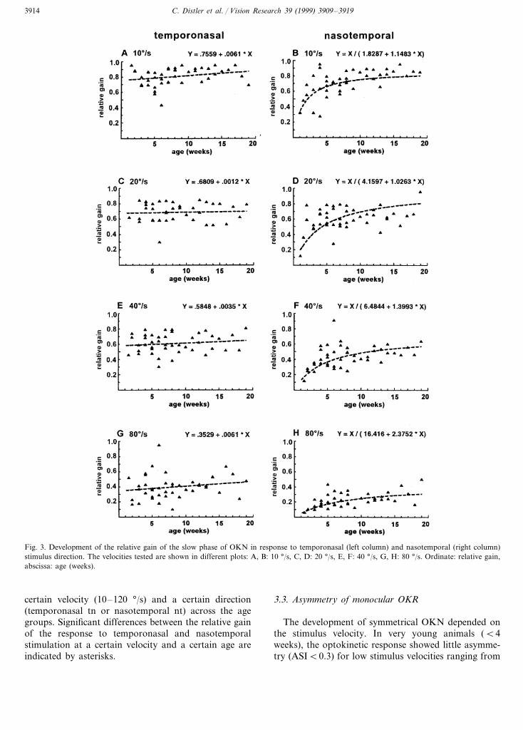
This trend is also evident if the development is in-spected on the basis of the relative gain. In Fig. 3, therelative gain of responses to temporonasal (leftcolumns) and nasotemporal (right columns) stimulationare plotted separately to emphasize the different devel-opmental course. Also, the development of the re-sponses is shown separately for the different stimulusvelocities tested. For each plot a set of regressionfunctions were tested (using the program GB-Stat®),the one with the highest correlation is plotted. Tworesults are clearly demonstrated by that figure: first, theresponse to temporonasal stimulation is almost con-stant during the observation period as indicated by thelow slope of the linear regression line. By contrast, the

C. Distler et al. / Vision Research 39 (1999) 3909–3919 3913
responses to nasotemporal stimulation are very low inyoung animals and increase especially over the first5–12 weeks of life, depending on the stimulus velocityas indicated by the reciprocal X and Y regression.Second, stimulation at different velocities leads to dif-ferent levels of response gain with low stimulus veloc-
ities eliciting eye movements with higher gain thanhigher stimulus velocities.
Table 2 gives the mean relative gain values and onestandard deviation of all animals. Vertical columns givethe gain values at different velocities at certain ages inweeks (w), horizontal columns give the values at a
Fig. 2. Development of estimated eye velocity (EVE) during monocular stimulation in temporonasal (left columns, A, C, E, G) and nasotemporaldirection (right columns, B, D, F, H). The dark diamonds indicate the median of the data points of all animals, the speckled rectangles representthe scatter of the data (25–75 % range), the vertical bars indicate the 10–90 % range. Ordinate: estimated eye velocity EVE (°/s), abscissa: stimulusvelocity (°/s). The data were combined in the following age groups: 1–2 weeks (A, B), 3–4 weeks (C, D), 5–8 weeks (E, F), 9–16 weeks (G, H).
C. Distler et al. / Vision Research 39 (1999) 3909–39193914
Fig. 3. Development of the relative gain of the slow phase of OKN in response to temporonasal (left column) and nasotemporal (right column)stimulus direction. The velocities tested are shown in different plots: A, B: 10 °/s, C, D: 20 °/s, E, F: 40 °/s, G, H: 80 °/s. Ordinate: relative gain,abscissa: age (weeks).
certain velocity (10–120 °/s) and a certain direction(temporonasal tn or nasotemporal nt) across the agegroups. Significant differences between the relative gainof the response to temporonasal and nasotemporalstimulation at a certain velocity and a certain age areindicated by asterisks.
3.3. Asymmetry of monocular OKR
The development of symmetrical OKN depended onthe stimulus velocity. In very young animals (B4weeks), the optokinetic response showed little asymme-try (ASIB0.3) for low stimulus velocities ranging from

C. Distler et al. / Vision Research 39 (1999) 3909–3919 3915
10 to 20 °/s, and high asymmetry (ASI=0.8) at 80–120°/s. With increasing age, lower stimulus velocities elic-ited more and more similar responses to temporonasaland nasotemporal stimulation, whereas at higher stimu-lus velocities the response to nasotemporal stimulationremained weaker for a longer period of time (see alsoTable 2). Assuming an artificial threshold for ‘nearsymmetry’ of 0.2, symmetry is reached for 10 °/s atabout 2–3 weeks of age (Fig. 4A), for 20 °/s at about4–5 weeks of age (Fig. 4B), for 40 °/s at about 14 weeks(Fig. 4C), and for 80 °/s or higher, symmetry was notreached during our testing period (Fig. 4D).
4. Discussion
The present study shows that infant macaques canshow a remarkably strong and symmetrical optokineticresponse to monocular stimulation already a few daysafter birth. Quite early on after birth the response totemporonasal stimulation is more reliable and becomesadult-like within the first month, whereas the strength ofthe response to nasotemporal stimulation is on averageweaker and continues to improve until several monthsafter birth. Symmetry is therefore reached at differentages depending on stimulus velocity. It is difficult todirectly compare our data with those of Atkinson (1979)because of the different methods employed. WhereasAtkinson (1979) assessed response strength only qualita-tively by judging the time spent with OKN, we have aquantitative comparison between the relative gain in thetwo directions from the EOG measurements. This mayexplain why Atkinson (1979) found symmetry of OKNat 50 °/s at 3 weeks of age, whereas in our datasymmetry at 40 °/s is reached only at about 4 months ofage.
4.1. Comparison with studies in human infants
Infant monkey visual system development closelymimics human infant visual system development whenthe ‘weeks to months rule’ is applied to the temporaldimension, so that 4 weeks in the monkey are equivalentto 4 months in the infant (Boothe, Dobson & Teller,1985). This relation holds true for most parameters ofspatial vision, including binocular interactions (Wiesel& Hubel, 1974; Chino, Smith, Hatta & Cheng, 1997)and stereoacuity, a product of binocular vision whichhas been recently investigated (O’Dell & Boothe, 1997).
In the human infant, eye movements are present frombirth but less mature than in monkey. Steady fixation,pursuit of a slow moving object, saccades and elementsof OKN have been described (for review see Hainline,1993). Specifically, OKN has been studied in detail withthe goal that it could constitute a convenient probe totest the integrity and normal development of motionprocessing pathways. Just like monkeys, human infantsare born with a weaker response to monocular nasotem-poral stimulation. Response to such a stimulation in-creases over the first 3–6 months of life when it becomessimilar to the response to temporonasal stimulation(symmetrical) (Atkinson, 1979; Naegele & Held, 1982).It constitutes a clinically significant sign because thelack of symmetrization is usually indicative of a defector lag of major visual functions. Three observationshave led investigators to link the symmetrization ofOKN to the development of binocular vision andstereopsis. One is based on anatomical and physiologi-cal data from studies in the cat (see below), the secondone is the close temporal coincidence between the twoevents. The third one is the common observation thatearly onset strabismus, accompanied by a maldevelop-ment
Table 2Relative gain of slow phase eye movements: means all casesa
3–41–2Age (weeks) 5–6 12–149–117–8 15–19 Adult
Velocity.709 .13.759 .12 .849 .13.709 .3210°/s tnb .849 .14.859 .10.849 .13** .809 .12
.719 .12.689 .13.739 .19.519 .1710°/s nt .729 .15.809 .10.809 .13 .869 .0820°/s tn .759 .14** .749 .16** .639 .20 .719 .13* .839 .10.699 .13 .699 .15 .709 .12
.609 .20.579 .16 .719 .13.429 .26 .639 .1420°/s nt .779 .13.639 .10.629 .13.619 .14** .559 .17* .649 .16** .589 .12** .649 .14*40°/s tn .639 .12**.469 .28* .669 .11.409 .15 .459 .17 .459 .16 .419 .04 .519 .0940°/s nt .519 .09.179 .09 .589 .12
.339 .09.419 .17.459 .06***.379 .08**.399 .14**80°/s tn .399 .21*.419 .14***.269 .20**.179 .07 .259 .11 .249 .11 .229 .0880°/s nt .259 .06.069 .05 .289 .13 .339 .10
.209 .11*.129 .05.039 .04120°/s tn .169 .05.199 .15.269 .05***.179 .10.209 .08*.089 .03 .149 .05.139 .07.159 .02.139 .06.109 .02.029 .03 .129 .10120°/s nt
a Significance levels Mann–Whitney U-test:* P5.05; ** P5.01; ***P5.001.b tn, temporonasal; nt, nasotemporal stimulation; significance levels are marked between tn and nt values at the same velocity.

C. Distler et al. / Vision Research 39 (1999) 3909–39193916
Fig. 4. Development of symmetry of monocular OKN tested with different stimulus velocities and plotted against age. For each plot data weretaken across all animals. A, 10 °/s; B, 20°/s; C, 40°/s; D, 80°/s. Ordinate: asymmetry index, abscissa: age (weeks).
of binocular vision is usually characterized by an arrestor a gross deficit of monocular OKN in response tonasotemporal stimulation (Tychsen & Lisberger, 1986).However, this point is controversial and some authorshave claimed that only a proportion of early strabismicpatients showed asymmetrical monocular OKN (Wat-tam-Bell, Braddick, Atkinson & Day, 1987), or havemade shallow unilateral visual deprivation the primarycause of the lack of symmetrization (Shawkat, Harris,Taylor, Thompson, Russell-Eggitt & Kriss, 1995).
In any case, many authors have pointed out a closerelation between infantile esotropia and a deficit ofmotion processing expressed by a lack of sensitivity orinadequate pursuit and saccades (Tychsen & Lisberger,1986; Kapoula, Bucci, Eggert & Garraud, 1997;Fawcett, Raymond, Astle & Skov, 1998). Furthermore,clear deficits of smooth pursuit eye movements and/orOKN have been described in monkeys and in humansafter lesions located either in the occipital cortex, in theposterior parietal cortex or in the region of the frontaleye field (Lynch & McLaren, 1983; Zee, Tusa, Herd-man, Butler & Gucer, 1986; Thurston, Leigh, Craw-ford, Thompson & Kennard, 1988; Rizzo & Hurtig,1989; Jacobs, Shawkat, Harris, Kriss & Taylor, 1993;Morrow & Sharpe, 1995; Heide, Kurzidim & Kompf,1996; Keating, Pierre & Chopra, 1996; Lekwuwa &Barnes, 1996).
As a consequence, OKN is used in the clinic to assessthe physiological condition of both the subcortical andcortical motion specific pathways and the oculomotorplant (Van Hof-Van Duin & Mohn, 1983; Buquet &
Charlier, 1996; Prechtl, Einspieler, Cioni, Bos, Ferrari& Sontheimer, 1997).
4.2. Comparison with data in subprimate mammals
In the present study, we have shown that in monkey,already shortly after birth monocular OKN can beelicited reliably not only in temporonasal but also,albeit weaker, in nasotemporal direction. This clearlydiffers from data in the cat where at 3 weeks of ageonly OKN in temporonasal direction can be elicited,whereas optokinetic response to nasotemporal stimula-tion first occurs at 4 weeks of age (Van Hof-Van Duin,1978; Malach et al., 1981). Over the following weeksOKN remains asymmetric until about 6 months of age.
To try to explain this difference, we have to look atthe neuronal substrate underlying the optokinetic reflex.In all mammals investigated to date, a common path-way has been identified (rat: Cazin, Precht & Lannou,1980; Precht & Strata, 1980; Cazin, Lannou & Precht,1984; rabbit: Collewijn, 1975a,b; guinea pig: Lui, Giolli,Blanks & Tom, 1994; cat: Hoffmann & Schoppmann,1975, 1981; Grasse & Cynader, 1984; ferret: Klauer,Sengpiel & Hoffmann, 1990; opossum: Volchan,Rocha-Miranda, Picanco-Diniz, Zinsmeisser, Bernardes& Franca, 1989; wallaby: Hoffmann, Distler, Mark,Marotte, Henry & Ibbotson, 1995; monkey: Kato,Harada, Hasegawa, Igarashi, Koike & Kawasaki, 1986;Hoffmann, Distler, Erickson & Mader, 1988; Kato,Harada, Hasegawa & Igarashi, 1988; Schiff, Cohen &Raphan, 1988; Hoffmann & Distler, 1989; Cohen,

C. Distler et al. / Vision Research 39 (1999) 3909–3919 3917
Schiff & Buettner-Ennever, 1990; Mustari & Fuchs,1990; Hoffmann et al., 1991; Hoffmann et al., 1992; ).The key structure in this pathway is the pretectalnucleus of the optic tract together with the dorsalterminal nucleus of the accessory optic system (NOT-DTN) serving as the sensorimotor interface. Retinal slipneurons in the NOT-DTN are characterized by theirdirection selective response to ipsiversive stimulus move-ment, i.e. retinal slip neurons in the left NOT-DTNprefer leftward movement, those in the right NOT-DTNprefer rightward movement. In nonprimate species, theyreceive direct retinal input almost exclusively from thecontralateral eye (Ballas, Hoffmann & Wagner, 1981;Klooster, Want & van der Vrensen, 1983). Retinal slipneurons project to the inferior olive, the nucleus prae-positus hypoglossi, the dorsolateral pontine nucleus,and the nucleus reticularis tegmenti pontis (Aas, 1989;Mustari et al., 1994; Buettner-Ennever, Cohen, Horn &Reisine, 1996). The information is then transmitted tothe vestibular nuclei and via climbing fibers to theflocculus of the cerebellum. Projections to the nucleiinnervating the extraocular muscles close the loop (forreview see Simpson, Giolli & Blanks, 1988). In some butnot all of the mammals investigated, an additionalcortical input to the NOT-DTN has been identified (rat:Schmidt, Zhang & Hoffmann, 1993; guinea pig: Lui etal., 1994; cat: Schoppmann, 1981, 1985; ferret: Klauer etal., 1990; monkey: Hoffmann et al., 1991; Hoffmann etal., 1992; Lui et al., 1995). Studies in cats with corticallesions (Wood et al., 1973; Grasse, Cynader & Douglas,1984) or with defective binocular vision (Cynader &Hoffmann, 1981; Hoffmann, 1983; Distler & Hoffmann,1996) have indicated that at least in this species thecortical loop transmits binocular information as well asinformation about high velocity stimuli to the NOT-DTN. This has been confirmed in a developmentalstudy. At 3 weeks of age, almost all NOT-DTN cells inkittens were exclusively driven by the contralateral eye.At this age, no cortical input to the NOT-DTN can beidentified neither physiologically nor anatomically. At 4weeks of age, however, the majority of the NOT-DTNcells receives an additional input from the ipsilateraleye, i.e. they are binocular. At this age, for the first time,their velocity tuning curves show clear optima, includingresponses to high velocities. This is also the age when afunctional cortical input to the midbrain can first beidentified (Plummer & Behan, 1989; Norita, Stein &McHaffie, 1991; Distler & Hoffmann, 1993) and, for thefirst time, a reliable optokinetic response to nasotempo-ral stimulation is present (Van Hof-Van Duin, 1978;Malach et al., 1981).
If we extrapolate the interpretation of the cat data tothe monkey, we would have to postulate that alreadyshortly after birth the NOT-DTN contains binocularneurons which would then be responsible for the optoki-netic responses in both horizontal directions during
monocular viewing. One possible anatomical substratefor this binocular convergence in infant monkeys couldbe provided by the strong bilateral retinal projection tothe NOT-DTN present already at birth (Kourouyan &Horton, 1997). Data about the development of thecortical input to the NOT-DTN in monkeys are notavailable yet. If, like in cat, the cortical loop to themidbrain matures only some time after birth, one canpropose the following hypothesis: The direct retinalinput from both eyes to primate NOT-DTN is able todrive the system moderately symmetrically early inpostnatal life. As the cortical input becomes functional,the role and importance of the retinal input is graduallyreduced until the system is clearly dominated by thecortical input in adulthood (Hoffmann et al., 1988).Since binocular convergence by retinal input from botheyes is present in this model already at birth, no abruptqualitative change in the symmetry of OKR would beexpected at the time when the cortical input becomesfunctional. Thus, only the gradual developmentalchanges observed in OKR with respect to eye velocityand symmetry would still need to be explained bygradual maturation of the cortical areas and/or theirprojection to the NOT-DTN.
Acknowledgements
We wish to thank Dr G. Dorrscheidt and W. Junkefor software development, and G. Tinney and G. Reuterfor hardware development. We are also indebted to theanimal caretakers of Covance Laboratories GMBH fortheir kind and patient help during the experiments. Thisstudy was supported by a Lise Meitner stipend from theLand Nordrhein-Westfalen to C. Distler.
References
Aas, J.-E. (1989). Subcortical projections to the pontine nuclei in thecat. Journal of Comparati6e Neurology, 282, 331–354.
Atkinson, J. (1979). Development of optokinetic nystagmus in thehuman infant and monkey infant: an analogue to development inkittens. In R. D. Freeman, De6elopmental neurobiology of 6ision(pp. 277–287). New York: Plenum.
Ballas, I., Hoffmann, K.-P., & Wagner, H. J. (1981). Retinal projec-tion to the nucleus of the optic tract in the cat as revealed byretrograde transport of horseradish peroxidase. Neuroscience Let-ters, 26, 197–202.
Benassi, C., Biral, G. P., & Corazza, R. (1992). Optokinetic nystag-mus in monocularly viewing guinea pig. Riunione CongiuntaS.I.B.S., S.I.F., S.I.N.U., Roma.
Boothe, R. G., Dobson, V., & Teller, D. Y. (1985). Postnataldevelopment of vision in human and nonhuman primates. AnnualRe6iews in Neuroscience, 8, 495–545.
Buettner-Ennever, J. A., Cohen, B., Horn, A. K. E., & Reisine, H.(1996). Pretectal projections to the oculomotor complex of themonkey and their role in eye movements. Journal of Comparati6eNeurology, 366, 348–359.
C. Distler et al. / Vision Research 39 (1999) 3909–39193918
Buquet, C., & Charlier, J. (1996). Evaluation of sensory visualdevelopment based on measures of oculomotor responses. In F.Vital-Durand, J. Atkinson, & O. J. Braddick III, Infant 6ision (pp.291–306). Oxford: Oxford University Press.
Cazin, L., Precht, W., & Lannou, J. (1980). Pathway mediatingoptokinetic responses of vestibular nucleus neurons in the rat.Pflugers Archi6es, 386, 19–29.
Cazin, L., Lannou, J., & Precht, W. (1984). An electrophysiologicalstudy of pathways mediating optokinetic responses to the vestibu-lar nucleus in the rat. Experimental Brain Research, 54, 337–348.
Chino, Y. M., Smith, E. L. III, Hatta, S., & Cheng, H. (1997).Postnatal development of binocular disparity sensitivity in neu-rons of the primate visual cortex. Journal of Neuroscience, 17,296–307.
Cohen, B., Schiff, D., & Buettner-Ennever, J. (1990). Contribution ofthe nucleus of the optic tract to optokinetic nystagmus andoptokinetic afternystagmus in the monkey: clinical implications.In B. Cohen, & I. Bodis-Wollner, Vision and the brain (pp.233–255). New York: Brain Research.
Collewijn, H. (1965). Optokinetic eye movements in the rabbit:input–output relations. Vision Research, 9, 117–132.
Collewijn, H. (1975a). Direction selective units in the rabbit nucleusof the optic tract. Brain Research, 100, 489–508.
Collewijn, H. (1975b). Oculomotor areas in the rabbit’s midbrain andpretectum. Journal of Neurobiology, 6, 3–22.
Cynader, M., & Hoffmann, K.-P. (1981). Strabismus disrupts binocu-lar convergence in cat nucleus of the optic tract. De6elopmentalBrain Research, 1, 132–136.
de Graaf, B., Wertheim, A. H., Bles, W., & Kremers, J. (1990).Angular velocity, not temporal frequency determines circularvection. Vision Research, 30, 637–646.
Distler, C. (1996). Neuronal basis of optokinetic reflex pathology innaturally strabismic monkeys. Strabismus, 4, 111–126.
Distler, C., Bachevalier, J., Kennedy, C., Mishkin, M., & Unger-leider, L. G. (1996). Functional development of the corticocorticalpathway for motion analysis in the macaque monkey: a 14C-2-de-oxyglucose study. Cerebral Cortex, 6, 184–195.
Distler, C., & Hoffmann, K.-P. (1993). Visual receptive field proper-ties in kitten pretectal nucleus of the optic tract and dorsalterminal nucleus of the accessory optic tract. Journal of Neuro-physiology, 70, 814–827.
Distler, C., & Hoffmann, K.-P. (1996). Retinal slip neurons in thenucleus of the optic tract and dorsal terminal nucleus in cats withcongenital strabismus. Journal of Neurophysiology, 75, 1483–1494.
Dubois, M. F. W., & Collewijn, H. (1979). The optokinetic reactionsof the rabbit: relation to the visual streak. Vision Research, 19,9–17.
Fawcett, S., Raymond, J. E., Astle, W. F., & Skov, C. M. (1998).Anomalies of motion perception in infantile esotropia. In6estiga-ti6e Ophthalmology and Visual Sciences, 39, 724–735.
Grasse, K. L., & Cynader, M. S. (1984). Electrophysiology of thelateral and dorsal terminal nuclei of the cat accessory opticsystem. Journal of Neurophysiology, 51, 276–293.
Grasse, K. L., Cynader, M. S., & Douglas, R. M. (1984). Alterationsin response properties in the lateral and the dorsal terminalnucleus of the cat accessory optic system following visual cortexlesions. Experimental Brain Research, 55, 69–80.
Hainline, L. (1993). Conjugate eye movements of infants. In K.Simmons, Early 6isual de6elopment, normal and abnormal (pp.47–88). New York: Oxford University Press.
Hamada, T. (1983). Binocular and monocular optokinetic nystagmusin cats to textured visual patterns. Neuroscience Letters, 40,127–131.
Hayes, W. N., & Ireland, L. C. (1969). Optokinetic response of theguinea pig. Journal of Comparati6e Physiology and Psychology, 88,199–202.
Heide, W., Kurzidim, K., & Kompf, D. (1996). Deficits of smoothpursuit eye movements after frontal and parietal lesions. Brain,119, 1951–1969.
Hess, B. J. M., Precht, W., Reber, A., & Cazin, L. (1985). Horizontaloptokinetic ocular nystagmus in the pigmented rat. Neuroscience,15, 97–107.
Hoffmann, K.-P. (1983). Effects of early monocular deprivation onvisual input to cat nucleus of the optic tract. Experimental BrainResearch, 51, 236–246.
Hoffmann, K.-P., & Distler, C. (1989). Quantitative analysis of visualreceptive fields of neurons in the nucleus of the optic tract and thedorsal terminal nucleus of the accessory optic tract in macaquemonkeys. Journal of Neurophysiology, 62, 416–428.
Hoffmann, K.-P., Distler, C., & Erickson, R. (1991). Functionalprojections from striate cortex and superior temporal sulcus to thenucleus of the optic tract (NOT) and dorsal terminal nucleus ofthe accessory optic tract (DTN) of macaque monkeys. Journal ofComparati6e Neurology, 313, 707–724.
Hoffmann, K.-P., Distler, C., Erickson, R., & Mader, W. (1988).Physiological and anatomical identification of the nucleus of theoptic tract and dorsal terminal nucleus of the accessory optic tractin monkeys. Experimental Brain Research, 69, 635–644.
Hoffmann, K.-P., Distler, C., & Gruesser, O.-J. (1998). Optokineticreflex in squirrel monkeys after long-term monocular deprivation.European Journal of Neuroscience, 10, 1136–1144.
Hoffmann, K.-P., Distler, C., & Ilg, U. (1992). Cortical contributionsto ipsilateral visual field representation and direction selectivity inthe macaque monkey’s nucleus of the optic tract and dorsalterminal nucleus. Journal of Comparati6e Neurology, 321, 150–162.
Hoffmann, K.-P., Distler, C., Mark, R. F., Marotte, L. R., Henry, G.H., & Ibbotson, M. R. (1995). Neural and behavioral effects ofearly eye rotation on the optokinetic system in the wallaby,Macropus eugenii. Journal of Neurophysiology, 73, 727–735.
Hoffmann, K.-P., & Schoppmann, A. (1975). Retinal input to direc-tion selective cells in the nucleus of the optic tract of the cat. BrainResearch, 99, 359–366.
Hoffmann, K.-P., & Schoppmann, A. (1981). A quantitative analysisof the direction specific response of neurons of the cat’s nucleus ofthe optic tract. Experimental Brain Research, 42, 1–12.
Jacobs, M., Shawkat, F., Harris, C. M., Kriss, A., & Taylor, D.(1993). Eye movement and electrophysiological findings in aninfant with hemispheric pathology. De6elopmental Medicine andChild Neurology, 35, 431–448.
Kapoula, Z., Bucci, M. P., Eggert, T., & Garraud, L. (1997). Impair-ment of the binocular coordination of saccades in strabismus.Vision Research, 37, 2757–2766.
Kato, I., Harada, K., Hasegawa, T., & Igarashi, T. (1988). Role ofthe nucleus of the optic tract in monkeys in optokinetic nystag-mus and optokinetic afternystagmus. Brain Research, 474, 16–26.
Kato, I., Harada, K., Hasegawa, T., Igarashi, T., Koike, Y., &Kawasaki, T. (1986). Role of the nucleus of the optic tract inmonkeys in relation to optokinetic nystagmus. Brain Research,364, 12–22.
Keating, E. G., Pierre, A., & Chopra, S. (1996). Ablation of thepursuit area in the frontal cortex of the primate degrades fovealbut not optokinetic smooth eye movements. Journal of Neuro-physiology, 76, 637–641.
Klauer, S., Sengpiel, F., & Hoffmann, K.-P. (1990). Visual responseproperties and afferents of nucleus of the optic tract in the ferret.Experimental Brain Research, 83, 178–189.
Klooster, J., Want, J. J. L., & van der Vrensen, G. (1983). Retinopre-tectal projections in albino and pigmented rabbits: an autoradio-graphic study. Brain Research, 288, 1–12.
Koerner, F., & Schiller, P. H. (1972). The optokinetic response underopen and closed loop conditions in the monkey. ExperimentalBrain Research, 14, 318–330.

C. Distler et al. / Vision Research 39 (1999) 3909–3919 3919
Kourouyan, H. D., & Horton, J. C. (1997). Transneuronal retinalinput to the primate Edinger–Westphal nucleus. Journal of Com-parati6e Neurology, 381, 68–80.
Lekwuwa, G. U., & Barnes, G. R. (1996). Cerebral control of eyemovements. I. The relationship between cerebral lesion sites andsmooth pursuit deficits. Brain, 119, 473–490.
Lui, F., Giolli, R. A., Blanks, R. H., & Tom, E. M. (1994). Patternof striate cortical projections to the pretectal complex in theguinea pig. Journal of Comparati6e Neurology, 344, 598–609.
Lui, F., Gregory, K. M., Blanks, R. H. I., & Giolli, R. A. (1995).Projections from visual areas of the cerebral cortex to pretectalnuclear complex, terminal accessory optic nuclei, and superiorcolliculus in macaque monkey. Journal of Comparati6e Neurology,363, 439–460.
Lynch, J. C., & McLaren, J. W. (1983). Optokinetic nystagmusdeficits following parieto-occipital cortex lesions in monkeys. Ex-perimental Brain Research, 49, 125–130.
Malach, R., Strong, N., & van Sluyters, R. C. (1981). Analysis ofmonocular optokinetic nystagmus in normal and visually deprivedkittens. Brain Research, 210, 367–372.
Mikami, A., & Fujita, K. (1992). Development of the ability to detectvisual motion in infant macaque monkeys. De6elopmental Psycho-biology, 25, 345–354.
Morrow, M. J., & Sharpe, J. A. (1995). Deficits of smooth-pursuit eyemovement after unilateral frontal lobe lesions. Annals of Neurol-ogy, 37, 443–451.
Mustari, M. J., & Fuchs, A. F. (1990). Discharge patterns of neuronsin the pretectal nucleus of the optic tract (NOT) in the behavingprimate. Journal of Neurophysiology, 64, 77–90.
Mustari, M. J., Fuchs, A. F., Kaneko, C. R. S., & Robinson, F. R.(1994). Anatomical connections of the primate pretectal nucleus ofthe optic tract. Journal of Comparati6e Neurology, 349, 111–128.
Naegele, J. R., & Held, R. (1982). The postnatal development ofmonocular optokinetic nystagmus in infants. Vision Research, 22,341–346.
Norita, M., Stein, B. E., & McHaffie, J. G. (1991). Postnatal ontoge-nesis of corticostriatal and corticotectal projections from the catlateral suprasylvian cortex. Society for Neuroscience Abstracts, 17,1378.
O’Dell, C., & Boothe, R. G. (1997). The development of stereoacuityin infant rhesus monkeys. Vision Research, 37, 2675–2684.
Plummer, K. L., & Behan, M. (1989). Development of corticotectalaxon arbors labeled with Phasaeolus vulgaris leucoagglutinin(PHAL). Society for Neuroscience Abstracts, 15, 1337.
Precht, W., & Strata, P. (1980). On the pathway mediating optoki-netic responses in vestibular nuclear neurons. Neuroscience, 5,777–787.
Prechtl, H. F. R., Einspieler, C., Cioni, G., Bos, A. F., Ferrari, F., &Sontheimer, D. (1997). An early marker for neurological deficitsafter perinatal brain lesions. Lancet, 349, 1361–1363.
Rizzo, M., & Hurtig, R. (1989). The effect of bilateral visual cortexlesions on the development of eye movements and perception.Neurology, 39, 406–413.
Roy, M.-S., Lachapelle, P., & Lepore, F. (1989). Maturation of theoptokinetic nystagmus as a function of the speed of stimulation infullterm and preterm infants. Clinical Vision Sciences, 4, 357–366.
Schiff, D., Cohen, B., & Raphan, T. (1988). Nystagmus induced bystimulation of the nucleus of the optic tract in the monkey.Experimental Brain Research, 70, 1–14.
Schmidt, M., Zhang, H.-Y., & Hoffmann, K.-P. (1993). OKN-relatedneurons in the rat nucleus of the optic tract and dorsal terminalnucleus of the accessory optic system receive a direct corticalinput. Journal of Comparati6e Neurology, 330, 147–157.
Schoppmann, A. (1981). Projections from areas 17 and 18 of thevisual cortex to the nucleus of the optic tract. Brain Research, 223,1–17.
Schoppmann, A. (1985). Functional and developmental analysis ofvisual corticopretectal pathway in the cat: a neuroanatomical andelectrophysiological study. Experimental Brain Research, 60, 363–374.
Schor, C. M. (1993). Development of OKN. In F. A. Miles, & J.Wallman, Visual motion and its role in the stabilization of gaze (pp.301–320). Amsterdam: Elsevier.
Schor, C., & Narayan, V. (1981). The influence of field size upon thespatial frequency response of optokinetic nystagmus. Vision Re-search, 21, 985–994.
Shawkat, F. S., Harris, C. M., Taylor, D. S. I., Thompson, D. A.,Russell-Eggitt, I., & Kriss, A. (1995). The optokinetic responsedifferences between congenital profound and nonprofound unilat-eral visual deprivation. Ophthalmology, 102, 1615–1622.
Simpson, J. I., Giolli, R. A., & Blanks, R. H. I. (1988). The pretectalnuclear complex and the accessory optic system. In J. A. Buettner-Ennever, Neuroanatomy of the oculomotor system (pp. 335–364).Amsterdam: Elsevier.
Sireteanu, R., Katz, B., Mohn, G., & Vital-Durand, F. (1992). Telleracuity cards for testing visual development and the effects ofexperimental manipulation in macaques. Clinicial Vision Sciences,7, 107–117.
Tauber, E. S., & Atkin, A. (1968). Optomotor responses to monocularstimulation. Relation to visual system organization. Science, 160,1365–1367.
Teller, D. Y., Succop, A., & Mar, C. (1993). Infant eye movementasymmetries: stationary counterphase gratings elicit temporal-to-nasal optokinetic nystagmus in two-month-old infants undermonocular test conditions. Vision Research, 33, 1859–1864.
Thurston, S. E., Leigh, R. J., Crawford, T., Thompson, A., &Kennard, C. (1988). Two distinct deficits of visual tracking causedby unilateral lesions of cerebral cortex in humans. Annals ofNeurology, 23, 266–273.
Tychsen, L., & Lisberger, S. G. (1986). Maldevelopment of visualmotion processing in humans who had strabismus with onset ininfancy. Journal of Neuroscience, 6, 2495–2508.
Van den Berg, A. V., & Collewijn, H. (1988). Directional asymmetriesof human optokinetic nystagmus. Experimental Brain Research,70, 597–604.
Van Hof-Van Duin, J. (1978). Direction preference of optokineticresponses in monocularly tested normal kittens and light deprivedcats. Archi6es Italiennes de Biologie, 116, 471–477.
Van Hof-Van Duin, J., & Mohn, G. (1983). Optokinetic and sponta-neous nystagmus in children with neurological disorders. Be-ha6ioural Brain Research, 10, 163–175.
Volchan, E., Rocha-Miranda, C. E., Picanco-Diniz, C. W., Zinsmeis-ser, B., Bernardes, R. F., & Franca, J. G. (1989). Visual responseproperties of pretectal units in the nucleus of the optic tract of theopossum. Experimental Brain Research, 78, 380–386.
Wattam-Bell, J., Braddick, O., Atkinson, J., & Day, J. (1987). Mea-sures of infant binocularity in a group at risk for strabismus.Clinical and Visual Sciences, 4, 327–336.
Westall, C. A., & Schor, C. M. (1985). Asymmetries of optokineticnystagmus in amblyopia: the effect of selected retinal stimulation.Vision Research, 25, 1431–1438.
Wiesel, T. N., & Hubel, D. H. (1974). Ordered arrangement oforientation columns in monkeys lacking visual experience. Journalof Comparati6e Neurology, 158, 307–318.
Wood, C. C., Spear, P. D., & Braun, J. J. (1973). Direction specificdeficits in horizontal optokinetic nystagmus following removal ofvisual cortex in the cat. Brain Research, 60, 231–237.
Zee, D. S., Tusa, R. J., Herdman, S. J., Butler, P. H., & Gucer, G.(1986). Effects of occipital lobectomy upon eye movements inprimate. Journal of Neurophysiology, 58, 883–907.
.