Myb-Related Schizosaccharomyces pombe cdc5p Is Structurally and Functionally Conserved in Eukaryotes
Upload
independentCategory
view
2download
0
Immunogenetics (2004) 56: 480–489DOI 10.1007/s00251-004-0716-8
ORIGINAL PAPER
Fabio Castillo . Carlos Guerrero . Esperanza Trujillo .Gabriela Delgado . Pilar Martinez . Luz M. Salazar .Paola Barato . Manuel E. Patarroyo .Carlos Parra-López
Identifying and structurally characterizing CD1b in Aotusnancymaae owl monkeys
Received: 29 April 2004 / Revised: 21 July 2004 / Published online: 10 September 2004# Springer-Verlag 2004
Abstract This study reports the molecular characteriza-tion and tissue expression of the non-human Aotusnancymaae primate CD1b isoform in the search for anexperimental animal model to be used in evaluating therole of non-peptide antigen-presentation molecules in theimmune response to infectious agents. CD1b expressionon the surface of A. nancymaae peripheral blood mono-cyte-derived dendritic cells, shown by flow cytometry, wasmade possible by using human CD1b isoform antibodies.Studying the expression of CD1b-encoded transcriptsrevealed this molecule’s broad distribution in severaltissues. The A. nancymaae CD1b transcript-encodedamino-acid sequence showed 95.5% identity with thehuman sequence. Such high sequence homology wasreflected in the identical structural conservation of howpockets A′, C′ and F′ and tunnel T′ conforming theantigen’s binding site are organized, the similar arrange-ment of those amino-acids interacting with the T-cellreceptor (TCR) during antigen presentation, and theconservation of YQNI-motif sequence in the cytoplas-matic tail (responsible for the molecule’s intracellulartrafficking in humans). Comparing the structure of humanCD1a and CD1b and mouse CD1d proteins with CD1bstructure in A. nancymaae obtained by minimizationrevealed that changes in the latter molecule’s α1 and α2domains imposed a narrowing of the antigen-bindinggroove in A. nancymaae CD1b. The high structuralsimilarity between A. nancymaae CD1b and that from
humans presented in this study leads to A. nancymaaebeing proposed as a suitable experimental animal modelfor analyzing CD1b in vivo, mainly in bacterial andparasite infections such as tuberculosis and malaria,respectively.
Keywords CD1b . Aotus nancymaae . Dendritic cells .Structural analysis
Introduction
T-lymphocytes recognize peptide antigens within thecontext of class I and class II major histocompatibilitycomplex (MHC) molecules generated in cytosol and theendocytic pathway (endosomes and lysosomes) in antigen-presenting cells (APCs). Those complexes so formed aretransported to the cell surface where they can becomerecognized by the T-cell receptor (TCR). It was thought formany years that this system was sufficient for achieving aneffective T-cell response against different pathogens.However, due to characterizing T-lymphocytes which arestimulated by non-proteic antigens (lipids and glycoli-pids), it has been established during the last decade thatCD1 molecules are responsible for presenting these typesof antigen (Beckman et al. 1994; Sieling et al. 1995).
The CD1 molecule family is composed of severalproteins encoded by various non-polymorphic geneswhich are expressed independently of classical MHCclass I and class II molecules (Beckman et al. 1994;Porcelli 1995). CD1 genes encode ~45-kDa heterodimericglycoproteins present on the cell surface, composed of aheavy 33-kDa glycosylated chain (H) non-covalentlyassociated with the β2-microglobulin (β2m) ~12-kDachain. The CD1 H-chain corresponds to type I integralmembrane proteins, having an extracellular part foldedinto three domains (α1, α2 and α3) similar in size andstructure to those domains conforming the α chain ofMHC class I molecules. The α1 and α2 domains form ahydrophobic pocket together with β2m which binds theantigen’s lipid tail hydrocarbonated chains (Zeng et al.
F. Castillo . E. Trujillo . G. Delgado . P. Martinez .L. M. Salazar . P. Barato . M. E. Patarroyo .C. Parra-López (*)Fundación Instituto de Inmunologia de Colombia (FIDIC) andUniversidad Nacional de Colombia,Carrera 50 No 26-00,Bogotá DC, Colombiae-mail: [email protected].: +57-1-481521969Fax: +57-1-4815269
C. GuerreroUniversidad Nacional de Colombia,Bogotá DC, Colombia

1997). The molecule is bound to the membrane by atransmembrane segment followed by a short cytoplasmatictail in the carboxy-terminus favoring the intracellulartrafficking of these molecules (Zeng et al. 1997).
Five CD1 isoforms have been identified (CD1a, b, c, dand e), which are classified into two groups according totheir nucleotide and amino-acid sequence homology(Calabi et al. 1989a). Group 1 includes human CD1A, B,and C gene products and their homologues in othermammals. Group 2 is represented by the human CD1Dgene product and their close homologues in mice, rats andone of the two CD1 forms of rabbit identified by cDNAcloning. The CD1e protein sequence has intermediatehomology with the two groups. It is known that all CD1genes are transcribed and translated into protein in humans(Martin et al. 1987; Calabi et al. 1989b; Blumberg et al.1991; Angénieux et al. 2003). These CD1 genes andproteins have also been studied in ungulates (cows andsheep) (Dutia and Hopkins 1991; Machugh et al. 1988;Parsons and Machugh 1991; Rhin et al. 1996), lagomorphs(rabbits and guinea pigs) (Calabi et al. 1989c; Dascher etal. 2002), rodents (mice and rats) (Balk et al. 1991; Kasaiet al. 1997; Mansura et al. 1997) and primates (Angénieuxet al. 2003; Kashiwase et al. 2003).
Within the CD1 family, CD1b seems to have becamespecifically adapted for sampling the different compart-ments of the endocytic pathway (early and late endosomesand lysosomes) (Sugita et al. 2000; Briken et al. 2002;Moody et al. 2002) and to present microbial lipids to T-lymphocytes (Sieling et al. 1995; Moody et al. 1997; Ernstet al. 1998). The expression of more than one CD1bisoform in those mammals studied to date and non-expression of CD1b in mice and non-human primates havehampered the study of the importance of pathogen lipidantigen in vivo presentation. In the search for a suitableexperimental animal model for studying this protein’sfunction in vivo, this study reports the tissue distributionand structural analyses of Aotus nancymaae CD1bisoform; this is a non-human primate widely used as ananimal model in evaluating anti-malarial vaccine candi-dates (Patarroyo et al. 1987; Moreno et al. 1999; Rivera etal. 2002; Rosas et al. 2002), whose immune systemexhibits a high degree of homology with the humanimmune system in terms of its cellular and molecularcomponents (Favre et al. 1998; Vecino et al. 1999; Diaz etal. 2000a, 2000b, 2002; Niño-Vasquez et al. 2000;Hernandez et al. 2002). The CD1b isoform’s highstructural similarity in this primate with that in humans,together with its wide tissue distribution, leads to it beingpredicted that A. nancymaae is a suitable model forstudying the importance of lipid components derived frompathogens presented by CD1b in the immune responseagainst malaria and other infectious agents such astuberculosis.
Materials and methods
Animals and tissues
Samples from three A. nancymaae monkeys were obtainedfrom our primate station in Leticia, Colombia. None of theanimals included in the study were subjected to anyantigenic stimulus. The different tissues (spleen, liver,kidneys, lungs, heart and thymus) were obtained from anA. nancymaae which died in captivity due to naturalcauses. In order to remove contaminating blood fromdifferent tissue samples, the tissue-specimens werethoroughly washed with large volumes of completeRPMI medium to prior tissues being homogenized in amortar in dry ice for RNA extraction.
Peripheral blood lymphocyte isolation, dendritic cellpreparation and flow cytometric analysis
A. nancymaae whole blood was obtained by femoralbleeding. Peripheral blood mononuclear cells (PBMCs)were isolated by Ficoll-Hypaque method (Sigma, USA).After isolation, cells were washed three times with RPMI-1640 medium (Sigma). Experimental procedures forobtaining dendritic cells (DCs) from A. nancymaae weresimilar to those described for human DCs (Delgado et al.2003) and A. nancymaae DCs (Delgado et al., submitted).In brief, PBMCs were obtained by density gradientcentrifugation in Ficoll-Hypaque (Lymphoprep, NycomedPharma, Oslo) (1.077 density). Total PBMCs isolated from3 ml of blood samples from two animals were cultured for2 h on plastic Petri dishes (6.5 cm diameter) in 4-mlcomplete media (RPMI-1640 supplemented with 10%autologous plasma). Non-adherent cells were removedafter 2 h. Adherent cells were cultured in 4 ml completemedia in the presence of human GM-CSF (250 ng/ml)(Leucomax, Essex Chimie, Lucerne) and 100 ng/ml IL-4produced in supernantants by IL-4 T6 transfectants (kindlyprovided by Dr. Antonio Lanzavecchia, Basel Institute forImmunology). Culture media was supplemented every 3days with fresh amounts of IL-4 and GM-CSF cytokines.
An aliquot of DCs was suspended in 200 μl PBS forfluorocytometric analysis 7 days after being cultured in IL-4 and GM-CSF. DCs were stained (using a 1:200 dilution)with the following human antibodies: anti-CD14-PE, anti-CD80-PE, anti-HLA-DR-FITC and anti-CD1b-FITC(Pharmingen, San Diego, Calif., USA). Histogramsmeasuring relative fluorescence intensity on CD1b posi-tive cells in FL1 (FITC) channel were constructed andanalyzed after acquiring 10,000 events in a flow cytometer(FACScan flow cytometer, Becton Dickinson, USA).
Total RNA isolation and reverse transcription
Tissues were homogenized in a mortar keeping themsubmerged in liquid nitrogen; total RNAwas then isolatedfollowing the Trizol one-step procedure (Gibco). RNAwas
481

quantified and its quality verified by gel electrophoresis.The total RNA obtained was treated with DNAse RQ1(Invitrogen). Total RNA (200 ng) was denatured for10 min at 70°C for reverse transcription. First-strandsynthesis was performed at 37°C for 1 h in a 30-μlreaction mixture containing 1× reverse transcriptase buffer(Gibco), 10 mM dithiothreitol (Gibco), 4 units RNAsin(Promega), 0.5 mM dNTPs (Promega), 200 units Maloneymouse leukemia virus reverse transcriptase (M-MLV-RTGibco) and oligo(dT) (Gibco). The reaction was stoppedby heating the tube at 95°C for 2 min.
Polymerase chain reaction
Each product was first amplified with two commercialhuman β-actin-specific primers: sense, 5′-ATCTGGCAC-CACACCTTCTACAATGAGCTGCG-3′; anti-sense, 5′-CGTCATACTCCTGCTTGCTGATCCACATCTGC-3′(Clontech, USA). In verifying cDNA suitability an 838-bpband should be obtained. Various sets of primers weredesigned on the human CD1b gene nucleotide sequence.The primers used were: sense (5′→3′) P1 40-AGT-GAAATGCTGCTGCT-56, P2 418-GGAGGTGCCA-TAGT AAGC-435 and P3 727-GGATTCTACC-CAAAGCC-743; antisense (5′→3′) P1 451-GAAATC-CAATCCTCCTAG-468, P2 965-AAGGAAGGCACTAT-TATTG-983 and P3 1061-TTGCGAATGGGAGAGGAG-
1078. Optimal amplification conditions were standardizedfor these primers using a clone containing the humanCD1b sequence as template (kindly supplied by Dr.Michael B. Brenner, Rheumatology Division, Immunolo-gy and Allergy, Brigham and Women’s Hospital andHarvard Medical School, Boston). Copy DNAs obtainedfrom A. nancymaae tissues and the human DNA controlwere denatured at 95°C for 5 min, followed by 35 cycles at95°C for 1 min, 55°C for 1 min, 72°C for 1 min, and afinal extension cycle at 72°C for 5 min. PCR productswere revealed on 1% agarose gels and visualized using aUV transilluminator. The gel image was captured byimage analyzer (BioRad).
Cloning DNA sequencing and data analysis
The 429-bp, 566-bp, and 352-bp fragments amplified fromA. nancymaae PBMC cDNA (using P1, P2 and P3primers, respectively) were purified and ligated intopGEMT easy cloning vector (Promega). They were thenused to transform Escherichia coli strain DH5α competentcells. Positive clones were selected and Miniprep DNAwas prepared using the Wizard DNA purification system(Promega, Madison, Wis.). They were then submitted toautomatic sequencing. The DNA nucleotide sequences ofcloned fragments were determined by using specificprimers for flanking T7 and SP6 polymerase sites in
Fig. 1 Detecting CD1b expres-sion in human and a,b A.nancymaae PBMC and c,dmonocyte-derived DCs by flowcytometry using monoclonalmAb against human CD1b. aAotus and b human monocyteswere stained with human anti-CD1b (interrupted line) or withIgG-FITC isotype control (con-tinuous line). CD1b expressionon A. nancymaae and humanDCs (c and d, respectively) aftertreating immature DCs with IL-4 and GM-CSF. CD1b (dashedline); IgG-FITC isotype control(continuous line)
482

pGEMT easy vector. All sequencing was performed on anABI PRISM 310 automated sequencer (Applied Biosys-tems, USA). The reported sequences represent at leastthree independent amplifications from three monkeys inwhich both DNA strands were sequenced. The nucleotideand protein sequences obtained were compared withcurrently available sequences in the GenBank databaseby using the BLAST program (Altschul et al. 1990). CD1bprotein sequence alignment and dendrogram were pro-cessed by CLUSTAL W software.
A. nancymaae CD1b structural analysis by molecularmodeling
The CD1b human molecule crystal structure complexed toβ2m (PDB:1GZQ.pdb) (Gadola et al. 2002) was used as atemplate for further minimization. Human CD1b aminoacids were replaced by A. nancymaae amino acidsdeduced from the cDNA sequence by using Insight II(2000) Biopolymer module software (Accelrys, USA) runon an Indigo 2 Station (Silicon Graphics). The Discover_3program (Accelrys) was used for a simple minimizationstrategy using 10,000 steps and 0.001 Å RMSD; the A.nancymaae CD1b model was thus obtained. Human CD1aand CD1b and mouse CD1d proteins were structurallycompared with A. nancymaae CD1b protein obtained byminimization.
Structure coordinates and factors were obtained fromthe Protein Data Bank using the following access codes:1ONQ for human CD1a, IGZQ for human CD1b and1CD1 for mouse CD1d.
Results
CD1b expression in immature DCs derived fromperipheral blood monocytes
We recently succeeded in obtaining human (Delgado et al.2003) and A. nancymaae (Delgado et al. submitted) DCsafter treating peripheral blood monocytes with humanGM-CSF and IL-4. Since DCs are professional APCsover-expressing high levels of co-stimulatory molecules(e.g., CD80, CD86), MHC class I, II and CD1 antigen-presenting molecules (Takahashi et al. 1997; Satin et al.1999), we thus decided to evaluate the degree of CD1bisoform expression in PBMCs and A. nancymaae DCsusing anti-human CD1b antibodies. Important up-regula-tion of CD14, HLA-DR and CD80 molecules in DCsderived from PBMC was evidenced using antibodiesagainst human-CD14, human-HLA-DR, human-CD80 andanti-CD1b (data not shown and Delgado et al., submitted)these results led us to argue that DCs were efficientlyobtained from A. nancymaae monocytes. Figure 1 showsthat the undetected expression of CD1b on monocytesfrom both species (Fig. 1a,b) increased when they weretreated with human cytokines. CD1b expression in A.nancymaae and human DCs went up in 58.29% (MFI:
62.69) and 54% (MFI: 54.4%), respectively, of matureDCs (Fig. 1c,d).
CD1b expression in A. nancymaae tissues
Three sets of specific primers were designed based on thenucleotide sequence encoded by the human CD1B gene,allowing this region to be amplified (429-, 566- and 352-bp fragments, respectively). RT-PCR amplification fromthese probes was used for studying the tissue distributionof transcripts encoding CD1b in various A. nancymaaeorgans. Figure 2 shows a high level of amplification of the429- and 566-bp fragments from the CD1b transcript in allorgans examined, with the exception of heart and kidneyswhere reduced amplification of these transcripts was seen.
Nucleotide and amino-acid sequences and A.nancymaae CD1b structural analysis
Fragments amplified by RT-PCR specific for CD1Bobtained from A. nancymaae PBMC cDNAs weresequenced and the complete codifying sequence wassent to GenBank (accession number AY605931). Analyz-ing the nucleotide sequence corresponding to the CD1Bgene’s codifying region revealed 97% homology with thehuman CD1B gene sequence. The transcript encoding A.nancymaae CD1b was 1,002 bp long, encoding a 334-amino-acid long protein sequence. Aligning the A.nancymaae CD1b amino-acid sequence with the humansequence (Fig. 3) showed that the leader sequence andcytoplasmatic region were identical in both proteins andthat α1, α2, α3 extra-cellular domain and transmembraneregion amino-acid sequences presented 98.9, 91.4, 95.7and 96.9% identity. There was 95.5% identity for thecomplete amino-acid sequence.
Fig. 2a–c Identifying CD1b in different A. nancymaae tissue byRT-PCR. Total RNA was isolated from several tissues, cDNA wasobtained by reverse transcription and CD1b regions were amplifiedusing specific primers. a CD1b fragment with P1 primers (α1 andα2 leader domains). b CD1b fragment with P2 primers (α2 and α3domains). c Fragment of β-actin amplified with specific primers fordemonstrating the integrity and equivalent quantities of cDNA usedas template in each tissue (one PBL, two spleen, three lungs, fourliver, five kidneys, six heart, seven positive-control DNAs from thehuman CD1b clone and positive-control DNA from the human β-actin clone). Negative control of PCR amplification (PCR mixwithout Aotus cDNA) was always negative in these experiments
483
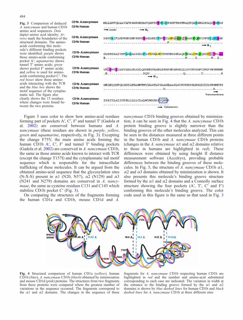
Figure 3 uses color to show how amino-acid residuesforming part of pockets A′, C′, F′ and tunnel T′ (Gadola etal. 2002) are conserved between humans and A.nancymaae (these residues are shown in purple, yellow,green and aquamarine, respectively, in Fig. 3). Exceptingthe change F75V, the main amino acids forming thehuman CD1b A′, C′, F′ and tunnel T′ binding pockets(Gadola et al. 2002) are conserved in A. nancymaae CD1b,the same as those amino acids known to interact with TCR(except the change T157I) and the cytoplasmatic tail motifsequence which is responsible for the intracellulartrafficking of these molecules. It can be argued from theobtained amino-acid sequence that the glycosylation sites(N-X-S) present in α1 (N20, N57), α2 (N129) and α3(N241 and N279) domains are conserved in A. nancy-maae, the same as cysteine residues C131 and C145 whichstabilize CD1b pocket C′ (Fig. 3).
On comparing the structures of the fragments formingthe human CD1a and CD1b, mouse CD1d and A.
nancymaae CD1b binding grooves obtained by minimiza-tion, it can be seen in Fig. 4 that the A. nancymaae CD1bprotein binding groove is slightly narrower than thebinding grooves of the other molecules analyzed. This canbe seen in the distances measured at three different pointsin the human CD1b and A. nancymaae CD1b proteins(changes in the A. nancymaae α1 and α2 domains relativeto those in humans are highlighted in red). Thesedifferences were obtained by using Insight II distancemeasurement software (Accelrys), providing probabledifferences between the binding grooves of these mole-cules. In Fig. 5, the structure of A. nancymaae CD1b α1,α2 and α3 domains obtained by minimization is shown. Italso presents this molecule’s binding groove structureformed by the α1 and α2 domains and a Connolly surfacestructure showing the four pockets (A′, T′, C′ and F′)conforming this molecule’s binding groove. The colorcode used in this figure is the same as that used in Fig. 3
Fig. 3 Comparison of deducedA. nancymaae and human CD1bamino acid sequences. Dotsdepict amino acid identity. Ar-rows mark the boundaries of thestructural domains. The amino-acids conforming this mole-cule’s different binding pocketswere identified: purple showsthose amino-acids conformingpocket A′; aquamarine showstunnel T′ amino acids; greenshows pocket F′ amino acids;and yellow is used for amino-acids conforming pocket C′. Thered boxes show those amino-acids interacting with the TCRand the blue box shows themotif sequence of the cytoplas-matic tail. The figure alsoclearly shows the 15 residueswhere changes were found be-tween the two proteins
Fig. 4 Structural comparison of human CD1a (yellow), humanCD1b (blue), A. nancymaae CD1b (black) obtained by minimizationand mouse CD1d (pink) proteins. The structures from two fragmentsfrom these proteins were compared where the greatest number ofvariations in the sequence occurred. The fragments correspond tothe α1 and α2 domains. The changes in the sequence of these
fragments for A. nancymaae CD1b respecting human CD1b arehighlighted in red and the number and amino-acid substitutedcorresponding to each case are indicated. The variation in width atthe entrance to the binding groove formed by the α1 and α2domains is shown by blue dashed lines for human CD1b and blackdashed lines for A. nancymaae CD1b at three different sites
484

where the amino acids forming each CD1b binding pocketare defined.
CD1b is highly conserved throughout species
Figure 6 shows the amino-acid sequence alignment of allCD1b sequences reported to date in different species. Thisfigure shows that the α1 and α2 domains (the domainsconforming the binding groove and having specificity fordifferent lipid ligands) are where the greatest variabilitywas found in these proteins. On the other hand, thephylogenetic tree shown in Fig. 7 suggest that A.nancymaae CD1b is the sequence with the greatesthomology with human CD1b amongst all sequencesreported to date (only full-length proteins), probably dueto evolutionary closeness between the two species.
Discussion
This study has established the CD1B nucleotide sequencefrom which the CD1b amino-acid sequence was inferredin the new world primate A. nancymaae. This primate’shigh CD1b protein structural homology with that ofhumans was initially shown by the high degree of humananti-CD1b antibody reactivity with CD1b moleculesexpressed on A. nancymaae DCs. This isoform’s expres-sion in non-human primate DCs is important since CD1bhas only been described to date in doubly positive CD4+CD8+ thymus lymphocytes in adult cynomolgus mon-keys (Macaca fascicularis) (Akari et al. 1997). The widetissue distribution of CD1b (spleen, liver, lungs, kidneys,heart and blood cells) suggested by our results (together
with the preservation of the elements necessary for thismolecule’s intracellular traffic) indicates that CD1b, anisoform that seems to have evolved towards specificpresentation of pathogen antigenic lipids (Moody et al.1997; Stenger et al. 1998; Sieling et al. 1999; Briken et al.2002), may promote an immune response against infec-tious agents lipids in A. nancymaae. It is worth mentioningthat similar low level of CD1b expression detected inAotus heart and kidneys has been reported in pigs (Chun etal. 1999).
The human CD1b protein requires a motif sequence inits cytoplasmatic tail consisting of a YXXZ motif forintracellular traffic (where Y is a tyrosine, X can be anyamino acid, and Z a voluminous and hydrophobic aminoacid). It is possible that, just as happens in humans, theYQNI motif sequence favors A. nancymaae CD1b asso-ciation with adapter proteins 2 and 3 (AP-2 and AP-3). Inturn, this may favor lipid antigen sampling by CD1bbetween cell membrane and endocyte route compartmentssuch as early endosomes, late endosomes, lysosomes andMHC class II compartments (MIIC) (Sugita et al. 1999;Dascher et al. 2002).
The structure of human CD1b-lipid-complex (phospha-tidyl inositol and ganglioside GM2) has recently beenresolved at 2.3- and 2.8-Å resolution (Gadola et al. 2002).This work has introduced nomenclature regarding thethree CD1b binding channels (A′, C′ and F′) connectingdirectly to the surface with a tunnel designated T′ inanalogy with the MHC class I and mouse CD1d bindingpocket (Gadola et al. 2002). Like human CD1b, A.nancymaae CD1b differs markedly from MHC class I inthe peak height attained by the kink in the α2 helix(residues 150–154 in CD1b and 149–152 in MHC class I,respectively).
Fig. 5a–c A. nancymaae CD1bstructure. This was obtained bymolecular modeling using thereported human CD1b structureand replacing the correspondingamino acids. a The main chainfor the α1, α2 and α3 domains,depicted schematically in ribbonrepresentation (lateral view). bThe α1 and α2 domains areshown conforming the ligand-binding groove of CD1b(viewed from above). c A Con-nolly surfaces structure showsthe four pockets (A′, T′, C′ andF′) conforming the molecule’sligand-binding groove
485

The high degree of A. nancymaae CD1b moleculeconservation with those from human can be corroboratedwhen structural comparison of the A. nancymaae CD1bbinding groove is made with human CD1a and CD1b andmouse CD1d molecule binding grooves. It can be seen thatthese structures can be almost perfectly superimposed,even though the A. nancymaae binding groove is slightlynarrower than the other molecules’ binding grooves(Figs. 4, 5). Structurally comparing these previouslyreported molecules (Zeng et al. 1997; Gadola et al.2002; Kayvan et al. 2001; Zajonc et al. 2003) has revealedthat pocket A′ in the CD1a, CD1b and CD1d molecules isthe most conserved region in the lipid antigen binding
groove. An important difference of the CD1a bindinggroove is that pocket F′ connects directly with pocket A′,whilst pockets A′ and F′ in CD1b structure (as in A.nancymaae) are separated by an additional pocket, C′,stabilized by a disulfide bridge between Cys131 and Cys145. Other differences worth noting between thesemolecules are the 1,300, 2,200 and 1,650 Å3 binding-groove volumes for CD1a, CD1b and CD1d, respectively,partly reflecting the length of the ligand alkyl chain theycan accommodate.
Regarding changes in α1 and α2 domain sequences, thechange of E103G may have an important effect on antigenbinding due to its being located on the bottom of the
Fig. 6 CD1b protein sequencesand alignments. The A. nancy-maae CD1b protein sequencewas generated by conceptualtranslation of cDNA sequenceand aligned with all CD1bsequences from the other spe-cies consigned in GenBank byusing CLUSTAL W multiplealignment software. Conservedamino acid regions are shaded.A consensus sequence is dis-played along the bottom. Arrowsmark the boundaries of thestructural domains. Variable po-sitions in the consensus se-quence are shown by dashes
486

binding groove and possibly increasing binding groovehydrophobicity. One of the changes which could influenceTCR interaction with the CD1b molecule is T157I, whichhas been seen to make contact with the TCR α chainCDR2 loop and could contribute towards stabilizing theglycolipid head group specific orientation by hydrogenbridges (Batuwangala et al. 2004). Finally, the changeF144V falls within the structure of pocket F′ (shown ingreen in Fig. 3); however, data regarding the reactivity ofantibodies conformationally recognizing this region (thatalso includes the change I149M) suggest that these tworesidues possibly do not impose an important conforma-tional change on the molecule (Kayvan et al. 2001).
Regarding the A. nancymaae CD1b binding grooveregion making contact with the TCR, a recent studyevaluated the importance of 36 residues forming part ofhuman CD1b pockets A′ and F′ for presenting glucosemonomycholate (GMM) and a synthetic derivate to T-specific lymphocytes (Kayvan et al. 2001). Fifteenresidues forming part of pocket A′ and 15 residues frompocket F′ evaluated by site-directed mutagenesis werefound in this study to be wholly conserved in A.nancymaae. The foregoing leads to the suggestion thatresidues from human CD1b molecule pockets A′ and F′(described in this work as being necessary for GMMrecognition) are equally participating in presenting thistype of lipid by CD1b in A. nancymaae.
Even though CD1b genes have been found in variousmammals examined to date, important differences havebeen found from species to species regarding locus size
and complexity. Whilst sheep have around seven genes(≥3 CD1b genes) (Dutia and Hopkins 1991; Rhin et al.1996), guinea pigs have ten (≥4 CD1b genes) (Dascher etal. 1999) and rabbits eight (≥1 CD1b) (Hayes and Knight2001), homologies amongst members of these species arearound 55–70%. Analyzing the degree of A. nancymaaeCD1b homology with other species (including humans)reveals that this protein’s amino-acid sequence in A.nancymaae is evolutionarily closer to the isoformexpressed in humans.
The importance of CD1b presence has been shown byseveral studies which have analyzed the host’s defenseagainst intracellular pathogens (such asM. leprae) where astrong correlation has been seen between the expression ofthree CD1 proteins (CD1a, b and c) and the presence ofthe host’s effective immunity against this pathogen(Sieling et al. 1999). On the other hand M. tuberculosislipids lipoarabinomaman (LAM), glucose monomicholate(GMM), mycolic acids, phosphatidylinositol mannoside(PIM) and phosophatidylinositol (PI) has been proved toelicit CD1b-restricted T-cells (Ernst et al. 1998) thatsecrete high levels of IFN-γ, lyse infected cells and have abactericidal effect on M. tuberculosis localized in macro-phages (Stenger et al. 1997; Kawashima et al. 2003).These data support the possible role of CD1b-restrictedcells in controlling mycobacterial infections. A direct invivo test of the CD1b protein’s role in host response tomycobacteria has not been done and the absence of dataconcerning CD1b expression in rhesus macaque or mice(commonly used animal models in evaluating vaccinecandidates). Hence, CD1b expression in A. nancymaaewill make this primate a suitable animal model forstudying the function of these types of antigen-presentingmolecules in vivo. The interesting structural homologyfound between CD1b in A. nancymaae and the isoformexpressed in humans suggests that the degree ofconservation of immune system molecular componentsbetween the two species extends to molecules responsiblefor presenting non-peptide antigens (such as CD1molecules). This is supported by that already describedfor cytokines (Hernandez et al. 2002), TCR (Favre et al.1998; Vecino et al. 1999; Guerrero et al. 2003), MHCClass-II (Diaz et al. 2000b, 2002; Niño-Vasquez et al.2000) and class I (Suárez et al. 2003) molecules, as well asimmunoglobulin gene organization (Diaz et al. 2000a).This high degree of homology between this new worldprimate’s immune system molecular components andthose of humans reaffirms the importance of Aotus speciesas a suitable experimental animal model for studying anddeveloping vaccines for human use against infectiousdiseases such as tuberculosis, leprosy and malaria.
Acknowledgments This research has been supported by theColombian President’s Office and the Colombian Ministry ofHealth. The experiments reported here comply with the currentMinistry of Health laws and other Colombian institutions’regulations. We would like to thank Jason Garry for patientlyreading this manuscript.
Fig. 7 Phylogenetic analysis of the deduced amino-acid sequencesof known CD1 proteins (only full-length proteins) using CLUSTALW multiple alignment software. The A. nancymaae protein is in thebox, and group 1 and group 2 classifications are noted. The CD1protein sequences were obtained from GenBank. Horizontaldistances throughout the tree are proportional to the degree ofdivergence
487

References
Akari H, Terao K, Murayama Y, Nam KH, Yoshikawa Y (1997)Peripheral blood CD4+CD8+ lymphocytes in cynomolgusmonkeys are of resting memory T lineage. Int Immunol9:591–597
Altschul SF, Gish W, Miller W, Myers EW, Lipman DJ (1990) Basiclocal alignment search tool. J Mol Biol 215:403–410
Angénieux C, Salamero J, Fricker D, Wurtz J-M, Maitre B,Cazenave J-P, Hanau D, de la Salle H (2003) Commoncharacteristics of the human and rhesus macaque CD1emolecules: conservation of biochemicals and biological proper-ties during primate evolution. Immunogenetics. 54:842–849
Balk SP, Bleicher PA, Terhorst C (1991) Isolation and expression ofcDNA encoding the murine homologues of CD1. J Immunol146:768–774
Batuwangala T, Shepherd D Gadola SD, Gibson KJC, Zaccai NR,Fersht AR, Besra GS, Cerendulo V, Jones EY (2004) Thecrystal structure of human CD1b with a bound bacterialglycolipid. J Immunol 172:2382–2388
Beckman EM, Porcelli SA, Morita CT, Behar SM, Furlong ST,Brenner MB (1994) Recognition of a lipid antigen by CD1-restricted αβ+ T-cells. Nature 372:691–694
Blumberg RS, Terhorst C, Bleicher P, McDermott FV, Allan CH,Landau SB, Trier JS, Balk SP (1991) Expression of a non-polymorphic MHC class-I-like molecule, CD1D, by humanintestinal epithelial cells. J Immunol 147:2518–2524
Briken V, Jackman RM, Dasgupta S, Hoening S, Porcelli SA (2002)Intracellular trafficking of newly synthesised CD1b molecules.EMBO J 21:825–832
Calabi F, Belt KT, Yu CY, Bradbury A, Mandy WJ, Milstein C(1989a) The rabbit CD1 and evolutionary conservation of theCD1 gene family. Immunogenetics 30:370–377
Calabi F, Bilsland CAG, Yu CY, Bradbury A, Belt KT, Martin LH,Milstein C (1989b) Recent progress in the molecular study ofCD1. In: Knapp W et al (eds) Leukocyte Typing IV. OxfordUniversity Press, Oxford, pp 254–258
Calabi F, Jarvis JM, Martin L, Milstein C (1989c) Two cases of CD1genes. Eur J Immunol 19:285–292
Chun T, Wang K, Zuckermann FA, Gaskins HR (1999) Molecularcloning and characterization of a novel CD1 gene from the pig.J Immunol 162:6562–6571
Dascher CC, Hiromatsu K, Naylor JW, Brauer PP, Brown KA,Storey JR, Behar SM, Kawasaki ES, Porcelli SA, Brenner MB,LeClair KP (1999) Conservation of CD1 multigene family inthe guinea pig. J Immunol 163:5478–5488
Dascher CC, Hiromatsu K, Xiong X, Sugita M, Buhlmann JE,Dodge IL, Lee SY, Roura-Mir C, Watts GF, Roy CJ, Behar SM,Clemens DL, Porcelli SA, Brenner MB (2002) Conservation ofCD1 intracellular trafficking patterns between mammalianspecies. J Immunol 169:6951–6958
Delgado G, Parra-Lopez CA, Vargas LE, Hoya R, Estupinan M,Guzman F, Torres A, Alonso C, Velez ID, Spinel C, PatarroyoME (2003) Characterizing cellular immune response to kine-toplastid membrane protein-11 (KMP-11) during Leishmania(Viannia) panamensis infection using dendritic cells (DCs) asantigen presenting cells (APCs). Parasite Immunol 25:199–209
Diaz D, Naegeli M, Rodríguez R, Niño-Vasquez J, Moreno A,Patarroyo ME, Pluschke G, Daubenberger C (2000a) Sequenceand diversity of MHC DQA and DQB genes of the owl monkeyAotus nancymaae. Immunogenetics 51:528–537
Diaz OL, Daubenberger CA, Rodriguez R, Naegeli M, Moreno A,Patarroyo M, Pluschke G (2000b) Immunoglobulin kappa light-chain V, J, and C gene sequences of the owl monkey Aotusnancymaae. Immunogenetics 51:212–218
Diaz D, Daubenberger CA, Zalac T, Rodriguez R, Patarroyo ME(2002) Sequence and expression of MHC-DPB1 molecules ofthe New World monkey Aotus nancymaae, a primate model forPlasmodium falciparum. Immunogenetics 54:251–259
Dutia BM, Hopkins J (1991) Analysis of the CD1 cluster in sheep.Vet Immunol Immunolpathol 27:189–194
Ernst WA, Maher J, Cho S, Niazi KR, Chatterjee D, Moody DB,Besra GS, Watanabe Y, Jensen PE, Porcelli SA, Kronenberg M,Modlin RL (1998) Molecular interaction of CD1b withlipoglycan antigens. Immunity 8:331–340
Favre N, Daubenberger C, Marfurt J, Moreno A, Patarroyo M,Pluschke G (1998) Sequence and diversity of T-cell receptoralpha V, J and C genes of the Aotus nancymaae owl monkey.Immunogenetics 48:253–259
Gadola SD, Zaccai NR, Harlos K, Shepherd D, Castro-Palomino JC,Ritter G, Schmidt RR, Jones EY, Cerendulo V (2002) Structureof human CD1b with bound ligands at 2.3 Å, a maze for alkylchains. Nat Immunol 3:721–726
Guerrero JE, Pacheco DP, Suarez CF, Martinez P, Aristizabal F,Moncada CA, Patarroyo ME, Patarroyo MA (2003) Character-izing T-cell receptor γ-variable gene in Aotus nancymaae owlmonkey peripheral blood. Tissue Antigens 61:1–11
Hayes SH, Knight KL (2001) Group 1 CD1 genes in rabbit. JImmunol 166:403–410
Hernandez E, Suarez CF, Mendez JA, Echeverry SJ, Murillo LA,Patarroyo ME (2002) Identification, cloning, and sequencing ofdifferent cytokine genes in four species of owl monkey.Immunogenetics 54:645–653
Kasai K, Matsuura A, Kikuchi K, Hashimoto Y, Ichimiya S (1997)Localization of rat CD1 transcripts and protein in rats tissues.An analysis of rat CD1 expression by in situ hybridization andimmunohistochemistry. Clin Exp Immunol 109:317–322
Kashiwase K, Kikuchi A, Ando Y, Nicol A, Porcelli SA, TokunagaK, Omine M, Satake M, Juji T, Nieda M, Koezuka Y (2003)The CD1d natural killer T-cell antigen presentation pathway ishighly conserved between humans and rhesus macaques.Immunogenetics 54:776–781
Kawashima T, Norose Y, Watanabe Y, Enomoto Y, Narazaki H,Watari E, Tanaka S, Takahashi H, Yano I, Brenner MB, SugitaM (2003) Cutting edge: major CD8 T cell response to livebacillus Calmette-Guérin is mediated by Cd1 molecules. JImmunol 170:5345–5348
Kayvan RN, Chiu MW, Mendoza RM, Degano M, Moody DB,Melian A, Wilson IA, Kronenberg M, Porcelli SA, Modlin RL(2001) The A′ and F′ pockets of human CD1b are both requiredfor optimal presentation of lipid antigens to T cells. J Immunol166:2562–2570
Machugh ND, Bensaid A, Davis WC, Howard CJ, Parsons KR,Jones B, Kaushal A (1988) Characterization of a bovine thymicdifferentiation antigen analogous to CD1 in the human. Scand JImmunol 27:541–547
Martin LH, Calabi F, Lefebvre F-A, Bilsland CAG, Milstein C(1987) Structure and expression of the human thymocyteantigens CD1a, CD1b, and CD1c. Proc Natl Acad Sci USA84:9189–9193
Mansura A, Hashimoto Y, Kinebuchi M, Kasai K, Ichimiya S,Katabami S, Chen H, Shimisu T, Kikuchi K (1997) Rat CD1antigen: structure, expression and function. Transplant Proc29:1705–1706
Moody DB, Reinhold BB, Guy MR, Beckman EM, Frederique DE,Furlong ST, Ye S, Reinhold VN, Sieling PA, Modlin RL, BesraGS, Porcelli SA (1997) Structural requirements for glycolipidantigen recognition by CD1b-restricted T-cells. Science278:283–286
Moody DB, Briken V, Cheng TY, Roura-Mir C, Guy MR, GehoDH, Tykocinski ML, Besra GS, Porcelli SA (2002) Lipidlength controls antigen entry into endosomal and non-endosomal pathways for CD1b presentation. Nat Immunol3:435–442
Moreno CA, Rodriguez R, Oliveira GA, Ferreira V, NussenzweigRS, Moya Castro ZR, Calvo-Calle JM, Nardin E (1999)Preclinical evaluation of a synthetic Plasmodium falciparumMAP malaria vaccine in Aotus monkeys and mice. Vaccine18:89–99
Niño-Vasquez J, Vogel D, Rodriguez R, Moreno A, Patarroyo M,Pluschke G, Daubenberger C (2000) Sequence and diversity ofDRB genes of Aotus nancymaae, a primate model for humanmalaria parasites. Immunogenetics 51:219–230
488

Parsons KR, Machugh ND (1991) Individual antigens of cattle.Bovine CD1 (boCD1). Vet Immunol Immunolpathol 27:37–41
Patarroyo ME, Romero P, Torres ML, Clavijo P, Moreno A,Martinez A, Rodriguez R, Guzman F, Cabezas E (1987)Induction of protective immunity against experimental infec-tion with malaria using synthetic peptides. Nature 328:629–632
Porcelli SA (1995) The CD1 family: a third lineage of antigen-presenting molecules. Adv Immunol 59:1
Rhin SM, Dutia BM, Howard CJ, Hopkins J (1996) Discriminationof two subsets of CD1 molecules in the sheep. Vet ImmunolImmunolpathol 52:265–270
Rivera Z, Granados G, Pinto M, Varon D, Carvajal C, Chaves F,Calvo J, Rodriguez R, Guzman F, Patarroyo ME (2002) Doubledimer peptide constructs are immunogenic and protectiveagainst Plasmodium falciparum in the experimental Aotusmonkey model. J Pept Res 59:62–70
Rosas JE, Pedraz JL, Hernandez RM, Gascon AR, Igartua M,Guzman F, Rodriguez R, Cortes J, Patarroyo ME (2002)Remarkably high antibody levels and protection against P.falciparum malaria in Aotus monkeys after a single immunisa-tion of SPf66 encapsulated in PLGA microspheres. Vaccine20:1707–1710
Satin AD, Hermonat PL, Ravaggi A, Chiriva-Internati M, CannonMJ, Hiserodt JC, Pecorelli S, Parham GP (1999) Expression ofsurface antigens during the differentiation of human dendriticcells vs macrophages from blood monocytes in vitro. Immu-nobiology 200:187–204
Sieling PA, Chatterjee D, Porcelli SA, Prigozy TI, Mazzaccaro RJ,Soriano T, Bloom BR, Brenner MB, Kronenberg M, BrennanPJ, Modlin RL (1995) CD1-restricted T-cell recognition ofmicrobial lipoglycan antigens. Science 269:227–230
Sieling PA, Jullien D, Dahlem M, Tedder TF, Rea TH, Modlin RL,Porcelli SA (1999) CD1 expression by dendritic cells in humanleprosy lesions: correlation with effective host immunity. JImmunol 162:1851–1858
Stenger S, Mazzaccaro RJ, Uyemura K, Cho S, Barnes PF, Rosat JP,Sette A, Brenner MB, Porcelli SA, Bloom BR, Modlin RL(1997) Differential effects of cytolytic T-cell subsets onintracellular infection. Science 276:1684–1687
Stenger S, Niazi KR, Modlin RL (1998) Down-regulation of CD1on antigen-presenting cells by infection with Mycobacteriumtuberculosis. J Immunol 161:3582–3588
Suárez CF, Cardenas PP, Llanos-Ballestas EJ, Martinez P, ObregónM, Patarroyo ME, Patarroyo MA (2003) α1 and α2 domains ofAotus MHC class-I and Catarrhini MHC class-Ia share similarcharacteristics. Tissue Antigens 61:362–373
Sugita M, grant EP, van Donselaar E, Hsu VW, Rogers RA, PetersPJ, Brenner MB (1999) Separate pathways for antigen presen-tation by CD1 molecules. Immunity 11:743
Sugita M, Vander Wel N, Rogers RA, Peters PJ, Brenner MB (2000)CD1c molecules broadly survey the endocytic system. ProcNatl Acad Sci USA 97:8445–8450
Takahashi K, Honeyman MC, Harrison LC (1997) Dendritic cellsgenerated from human blood in granulocyte macrophage-colony stimulating factor and interleukin-7. Hum Immunol55:103–116
Vecino W, Daubenberger C, Rodriguez R, Moreno A, Patarroyo M,Pluschke G (1999) Sequence and diversity of T-cell receptor β-chain V and J genes of the Aotus nancymaae owl monkey.Immunogenetics 49:792–799
Zajonc DM, Elsliger MA, Teyton L, Wilson IA (2003) Crystalstructure of CD1a in complex with a sulfatide self at aresolution of 2.15 Å. Nat Immunol 4:808–815
Zeng Z, Castaño AR, Segelke BW, Stura EA, Peterson PA, WilsonIA (1997) Crystal structure of mouse CD1: an MHC-like foldwith a large hydrophobic binding groove. Science 277:339–345
489
Copyright © 2022 FDOKUMEN