Two structurally different RNA molecules are bound by the spliceosomal protein U1A using the same...
11
Two structurally different RNA molecules are bound by the spliceosomal protein U1A using the same recognition strategy Luca Jovine, Chris Oubridge, Johanna M Avis † and Kiyoshi Nagai* Background: Human U1A protein binds to hairpin II of U1 small nuclear RNA (snRNA) and, together with other proteins, forms the U1 snRNP essential in pre-mRNA splicing. U1A protein also binds to the 3′ untranslated region (3′UTR) of its own pre-mRNA, inhibiting polyadenylation of the 3′ end and thereby downregulating its own expression. The 3′UTR folds into an evolutionarily conserved secondary structure with two internal loops; one loop contains the sequence AUUGCAC and the other its variant AUUGUAC. The sequence AUUGCAC is also found in hairpin II of U1 snRNA; hence, U1A protein recognizes the same heptanucleotide sequence in two different structural contexts. In order to better understand the control mechanism of the polyadenylation process, we have built a model of the U1A protein–3′UTR complex based on the crystal structure of the U1A protein–hairpin II RNA complex which we determined previously. Results: In the crystal structure of the U1A protein–hairpin II RNA complex the AUUGCAC sequence fits tightly into a groove on the surface of U1A protein. The conservation of the heptanucleotide in the 3′UTR strongly suggests that U1A protein forms identical sequence-specific contacts with the heptanucleotide sequence when complexed with the 3′UTR. The crystal structure of the hairpin II complex and the twofold symmetry in the 3′UTR RNA provide sufficient information to restrict the conformation of the 3′UTR RNA and have enabled us to build a model of the 3′UTR complex . Conclusions: In the U1A–3′UTR complex, sequence-specific interactions are made entirely by the conserved heptanucleotide and the last base pair (C:G) of the stem. The structure is stabilized by protein–protein contacts and by electrostatic interactions between basic amino acids of the protein and the phosphate backbone of the RNA stem regions. The formation of a protein dimer necessary for the inhibition of poly(A) polymerase requires a conformational change of the C termini of the proteins upon RNA binding. This mechanism could prevent the inhibition of poly(A) polymerase by free U1A protein. The model is consistent with biochemical data, and the protein–protein interactions within the 3′UTR complex account for the cooperativity of U1A protein binding to the 3′UTR. The model also serves as an important structural guide for designing further experiments to understand the interaction between the U1A–3′UTR complex and poly(A) polymerase. Introduction U1A spliceosomal protein exists in the nucleus as a com- ponent of U1 snRNP (small nuclear ribonucleoprotein), a large RNA–protein complex involved in pre-mRNA splic- ing [1]. U1A protein binds to hairpin II of U1 snRNA, which has a ten nucleotide AUUGCACUCC loop (Fig. 1a) [2–4]. U1A protein consists of 282 residues and contains two RNP domains, one at the N terminus and one at the C terminus, which are connected by a protease sensitive linker [5]. The RNP domain is a commonly occurring RNA-binding domain of 80–90 amino acids [6,7]. Scherly et al. [8] showed that a fragment of U1A protein containing the first 102 residues, which contains one of the two RNP domains, binds to hairpin II of U1 snRNA with the same affinity as the full length protein. The structure of a fragment of U1A protein containing residues 2–95 was solved by X-ray crystallography [9]. This showed that the RNP motif is folded into a globular domain consisting of a four stranded b sheet flanked on one side by two a helices. A subsequent NMR study, using a longer fragment of U1A protein (residues 2–102), showed that an additional a helix (helix C) is present around residue 95 but did not establish its position in rela- tion to the RNP domain [10]. Address: MRC Laboratory of Molecular Biology, Hills Road, Cambridge CB2 2QH, UK. † Present address: Department of Biochemistry and Applied Molecular Biology, UMIST, Manchester M60 1QD, UK. *Corresponding author. E-mail: [email protected] Key words: molecular modelling, polyadenylation, post-transcriptional regulation, RNA–protein recognition, U1A spliceosomal protein, 3′ untranslated region Received: 22 Jan 1996 Revisions requested: 19 Feb 1996 Revisions received: 4 Mar 1996 Accepted: 5 Mar 1996 Structure 15 May 1996, 4:621–631 © Current Biology Ltd ISSN 0969-2126 Research Article 621
Transcript of Two structurally different RNA molecules are bound by the spliceosomal protein U1A using the same...
Two structurally different RNA molecules are bound by thespliceosomal protein U1A using the same recognition strategyLuca Jovine, Chris Oubridge, Johanna M Avis† and Kiyoshi Nagai*
Background: Human U1A protein binds to hairpin II of U1 small nuclear RNA(snRNA) and, together with other proteins, forms the U1 snRNP essential inpre-mRNA splicing. U1A protein also binds to the 3′ untranslated region (3′UTR)of its own pre-mRNA, inhibiting polyadenylation of the 3′ end and therebydownregulating its own expression. The 3′UTR folds into an evolutionarilyconserved secondary structure with two internal loops; one loop contains thesequence AUUGCAC and the other its variant AUUGUAC. The sequenceAUUGCAC is also found in hairpin II of U1 snRNA; hence, U1A proteinrecognizes the same heptanucleotide sequence in two different structuralcontexts. In order to better understand the control mechanism of thepolyadenylation process, we have built a model of the U1A protein–3′UTRcomplex based on the crystal structure of the U1A protein–hairpin II RNAcomplex which we determined previously.
Results: In the crystal structure of the U1A protein–hairpin II RNA complex theAUUGCAC sequence fits tightly into a groove on the surface of U1A protein.The conservation of the heptanucleotide in the 3′UTR strongly suggests thatU1A protein forms identical sequence-specific contacts with the heptanucleotidesequence when complexed with the 3′UTR. The crystal structure of the hairpin IIcomplex and the twofold symmetry in the 3′UTR RNA provide sufficientinformation to restrict the conformation of the 3′UTR RNA and have enabled usto build a model of the 3′UTR complex .
Conclusions: In the U1A–3′UTR complex, sequence-specific interactions aremade entirely by the conserved heptanucleotide and the last base pair (C:G) ofthe stem. The structure is stabilized by protein–protein contacts and byelectrostatic interactions between basic amino acids of the protein and thephosphate backbone of the RNA stem regions. The formation of a protein dimernecessary for the inhibition of poly(A) polymerase requires a conformationalchange of the C termini of the proteins upon RNA binding. This mechanismcould prevent the inhibition of poly(A) polymerase by free U1A protein. Themodel is consistent with biochemical data, and the protein–protein interactionswithin the 3′UTR complex account for the cooperativity of U1A protein binding tothe 3′UTR. The model also serves as an important structural guide for designingfurther experiments to understand the interaction between theU1A–3′UTR complex and poly(A) polymerase.
IntroductionU1A spliceosomal protein exists in the nucleus as a com-ponent of U1 snRNP (small nuclear ribonucleoprotein), alarge RNA–protein complex involved in pre-mRNA splic-ing [1]. U1A protein binds to hairpin II of U1 snRNA,which has a ten nucleotide AUUGCACUCC loop(Fig. 1a) [2–4]. U1A protein consists of 282 residues andcontains two RNP domains, one at the N terminus andone at the C terminus, which are connected by a proteasesensitive linker [5]. The RNP domain is a commonlyoccurring RNA-binding domain of 80–90 amino acids[6,7]. Scherly et al. [8] showed that a fragment of U1A
protein containing the first 102 residues, which containsone of the two RNP domains, binds to hairpin II of U1snRNA with the same affinity as the full length protein.The structure of a fragment of U1A protein containingresidues 2–95 was solved by X-ray crystallography [9].This showed that the RNP motif is folded into a globulardomain consisting of a four stranded b sheet flanked onone side by two a helices. A subsequent NMR study,using a longer fragment of U1A protein (residues 2–102),showed that an additional a helix (helix C) is presentaround residue 95 but did not establish its position in rela-tion to the RNP domain [10].
Address: MRC Laboratory of Molecular Biology,Hills Road, Cambridge CB2 2QH, UK.
†Present address: Department of Biochemistryand Applied Molecular Biology, UMIST,Manchester M60 1QD, UK.
*Corresponding author. E-mail: [email protected]
Key words: molecular modelling, polyadenylation,post-transcriptional regulation, RNA–proteinrecognition, U1A spliceosomal protein,3′ untranslated region
Received: 22 Jan 1996Revisions requested: 19 Feb 1996Revisions received: 4 Mar 1996Accepted: 5 Mar 1996
Structure 15 May 1996, 4:621–631
© Current Biology Ltd ISSN 0969-2126
Research Article 621
622 Structure 1996, Vol 4 No 5
The precise stereochemistry of the interaction with U1snRNA hairpin II was revealed by the crystal structure at1.92 Å resolution of the human U1A protein RNA-bindingdomain (residues 2–98, referred to as RBD throughout thispaper) complexed with a 21 nucleotide RNA represent-ing hairpin II of U1 snRNA (Figs 1b,2a) [11]. TheAUUGCAC heptanucleotide of hairpin II fits into agroove formed between loop 3 (situated between b2 andb3 strands) and the C-terminal region of the RNP domain.The heptanucleotide, together with the C:G base pairwhich closes the loop, forms an extensive direct andwater-mediated hydrogen bond network with amino acidresidues on the surface of the b sheet. The RNA basesstack onto either an adjacent base, a protein side chain, or
both, showing the importance of base stacking for thestabilization of RNA structure and in RNA–protein inter-actions. In contrast, the bases of the last three nucleotidesof the loop do not interact with the protein and are lesswell ordered [11]. The crystal structure of the U1ARBD–RNA hairpin II complex shows that helix C extendsparallel to the b2 strand and that its position is stabilizedby hydrophobic interactions between Ile58, Ile93 andIle94 and by a hydrogen bond between Ser91 and Thr11. We have recently carried out an NMR study of a fragment of U1A protein (containing residues 2–117) and found that helix C extends across the b sheet [12]. This shows that RNA binding is accompanied by a large movement of helix C.
Figure 1
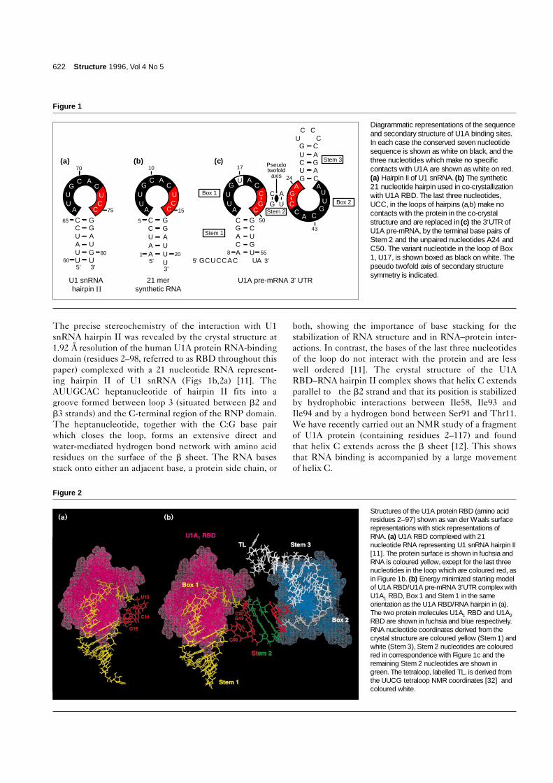
Diagrammatic representations of the sequenceand secondary structure of U1A binding sites.In each case the conserved seven nucleotidesequence is shown as white on black, and thethree nucleotides which make no specificcontacts with U1A are shown as white on red.(a) Hairpin II of U1 snRNA. (b) The synthetic21 nucleotide hairpin used in co-crystallizationwith U1A RBD. The last three nucleotides,UCC, in the loops of hairpins (a,b) make nocontacts with the protein in the co-crystalstructure and are replaced in (c) the 3′UTR ofU1A pre-mRNA, by the terminal base pairs ofStem 2 and the unpaired nucleotides A24 andC50. The variant nucleotide in the loop of Box1, U17, is shown boxed as black on white. Thepseudo twofold axis of secondary structuresymmetry is indicated.
10 17
UC
C 15
C5 GC GU AA U
20A1 UU
21 mer synthetic RNA
G CU AC GU AG C
CACC UA8 55
50
24
43Stem 1
Box 1
Stem 2
Stem 3Pseudo twofold
axis
Box 2
UCG5'5' 3'
5'3'
3'
70
C ACG
U UU C
A C
C CU C
75
C65 GC GU AA U
80U60
GUU
U ACG
U C C
G
A
UU GA C
C GG CA UC GA U
U1 snRNA hairpin II
(a) (b) (c)
C ACG
UU
A
AAUU
G
G
CAC
C
U1A pre-mRNA 3' UTR
Figure 2
Structures of the U1A protein RBD (amino acidresidues 2–97) shown as van der Waals surfacerepresentations with stick representations ofRNA. (a) U1A RBD complexed with 21nucleotide RNA representing U1 snRNA hairpin II[11]. The protein surface is shown in fuchsia andRNA is coloured yellow, except for the last threenucleotides in the loop which are coloured red, asin Figure 1b. (b) Energy minimized starting modelof U1A RBD/U1A pre-mRNA 3′UTR complex withU1A1 RBD, Box 1 and Stem 1 in the sameorientation as the U1A RBD/RNA hairpin in (a).The two protein molecules U1A1 RBD and U1A2RBD are shown in fuchsia and blue respectively.RNA nucleotide coordinates derived from thecrystal structure are coloured yellow (Stem 1) andwhite (Stem 3), Stem 2 nucleotides are colouredred in correspondence with Figure 1c and theremaining Stem 2 nucleotides are shown ingreen. The tetraloop, labelled TL, is derived fromthe UUCG tetraloop NMR coordinates [32] andcoloured white.

U1A protein also binds to the 3′ untranslated region(3′UTR) of its own pre-mRNA and regulates its expres-sion through an unprecedented mechanism [13]. U1A istransported into the nucleus by virtue of its nuclear local-ization signal and incorporated into U1 snRNP [14].Nuclear levels of U1A protein are tightly regulated, pre-sumably because a large excess of U1A protein has a dele-terious effect on pre-mRNA splicing. U1A proteindownregulates its own expression by binding to the3′UTR of its pre-mRNA and thereby inhibiting thepolyadenylation of the pre-mRNA [13] through a directinteraction with poly(A) polymerase [15]. A sequencecomparison of the 3′UTR of the U1A protein gene fromhuman, Xenopus and mouse showed that the pre-mRNAcan be folded into an evolutionarily conserved secondarystructure with two internal loops, one with theAUUGCAC heptanucleotide (identical to that found inhairpin II of U1 snRNA) and the other with one basechange, AUUGUAC [16]. Based on the crystal structure ofthe U1A RBD–RNA hairpin complex we previously pro-posed that the interaction between the U1A RBD and theAUUGCAC heptanucleotide together with the C5:G16loop-closing base pair (Fig. 1b) is conserved in each of thetwo internal loops in the 3′UTR complex. We also pro-posed that the heptanucleotide and the middle stem(Stem 2) can be linked by one base pair of Stem 2(C20:G49; C46:G23) and one unpaired nucleotide (C50;A24) (Fig. 1c) [11].
We have carried out molecular modelling of a complexconsisting of two molecules of the U1A RBD bound to the3′UTR of U1A pre-mRNA. A set of stereochemicallyplausible models can be constructed on the basis of thecrystal structure of the hairpin II complex. This studyyielded one structure which is consistent with the enzy-matic protection and mutagenesis experiments reportedby van Gelder et al. [16] and allows a hypothesis to beformed which explains the molecular basis of cooperativityof U1A protein binding to the 3′UTR RNA.
The 3′UTR of mRNA is often used as a binding site forproteins that regulate translation, degradation and localiza-tion of specific mRNAs [17–19]. These proteins are there-fore important determinants of temporal and spatialexpression of developmental genes [19–21]. Our study ofthe interaction between U1A protein and the 3′UTR of itsmRNA could provide important insight into the molecularmechanism of these essential biological processes.
ResultsInitial model buildingThe crystal structure of the U1A RBD–RNA hairpin IIcomplex [11] shows that the U1A RBD forms an extensivenetwork of hydrogen bonds and stacking interactions withthe AUUGCAC heptanucleotide and the C5:G16 basepair which closes the stem. Considering the conservation
of the heptanucleotide sequence between hairpin II andthe 3′UTR sequences [15], it is almost certain that theseinteractions are conserved in the U1A–3′UTR complex(Fig. 1a,c). The conformation of these conservednucleotides (Fig. 2a, represented in yellow) and theprotein structure was therefore kept unaltered for the3′UTR model. In the crystal structure of the hairpin IIcomplex the bases of the last three loop nucleotides, UCC(shown in red in Fig. 2a), do not make any sequence spe-cific contact with the protein and merely act as a linkerbetween the heptanucleotide and the stem [11]. This isconsistent with the RNA selection experiments by Tsai etal. [22] which showed that RNAs selected from a randompool, which are bound by U1A protein with high affinity,share the constant heptanucleotide sequence followed bythree variable nucleotides. We have therefore proposedthat these three nucleotides could be replaced by one basepair and one nucleotide in the 3′UTR complex [11], asshown schematically in Figure 2b. Our proposal is furthersupported by a recent paper which showed that thesethree nucleotides can be replaced by an ethylene glycollinker without affecting U1A protein binding [23].
The initial model of the U1A–3′UTR complex was con-structed using the crystal structure coordinates of the U1ARBD–RNA hairpin II complex (PDB ID code: 1URN),excluding the last three nucleotides of the loop (UCC),and the program MC-SYM [24–26] as explained in Figure3a. This program scans a nucleotide conformational data-base, derived from RNA structures determined by X-raycrystallography and NMR, to generate all the stereochem-ically possible RNA conformers that are consistent with aset of user-defined constraints. A series of binding assaysconducted by van Gelder et al. [16] showed that, in orderfor two molecules of U1A protein to bind the 3′UTR oftheir pre-mRNA and to inhibit its polyadenylation, thebase pairing ability of the two strands of Stem 2 must bemaintained. An NMR study of an unbound syntheticRNA resembling half of the 3′UTR showed that thenucleotides corresponding to Stem 2 adopt A-form geo-metry [27]. The canonical A-form structure of duplexRNA was therefore used as a starting model for Stem 2(shown in blue in Fig. 3a). We attempted to appendStem 2 nucleotides (C20–G23 and C46–G49) as A-formhelix (MC-SYM conformational set type_A) to the lastresidue of the loop heptanucleotide, C12 in the crystalstructure RNA (Fig. 1b) or C19 in the 3′UTR sequence(Fig. 1c), leaving nucleotide C50 (shown in red in Fig. 3a)free to adopt any possible conformation (full_A′ set: 36alternative conformations derived from a connection sam-pling; represented by a square in Fig. 3a). This did notyield any solution, since the phosphate group of theunpaired nucleotide C50 could not be brought closeenough to the O3′ of G51 to form a phosphodiester bondand close the loop. We therefore relaxed the A-form con-straints of nucleotides C20:G49, the first base pair of Stem
Research Article RNA binding to U1A Jovine et al. 623

2, by assigning to them the conformational set sample_A(17 alternative conformations for C3′-endo, antinucleo-tides; indicated by a circle in Fig. 3a). In both cases, thisled to a localized distortion of the RNA helix, with a lossof base pairing between C20 and G49. We chose the con-former in which the two bases were closest to having basepair geometry and manually modified their mutual orien-tation with the program O [28]. The ‘refine zone’ optionof the program was used to automatically refine the geom-etry of the entire Stem 2, so that complete base pairingbetween its two strands was established. These manipula-tions resulted in a slight deviation of the Stem 2 helixfrom the canonical A form.
The only unknown parameter remaining was the positionof the unpaired base of nucleotide C50, which connectsthe conserved part of the complex (one U1A RBD [U1A1],Stem 1 and the loop heptanucleotide) with the middlestem. There were three possibilities: the C50 base couldstack onto base G49, it could stack onto base G51, or itcould stack on neither. The last possibility is energeticallyunfavourable as it would expose a large hydrophobicsurface of base C50 to solvent. This position would be pos-sible if C50 is involved in a specific interaction with theprotein, similar to that observed in the structures of theglutaminyl- and aspartyl-tRNA synthetase-tRNA com-plexes, where tRNA anticodon bases are specifically recog-nized by the protein [29–31]. However, the identity ofbase 50 in Xenopus laevis pre-mRNA 3′UTR (where Box 1and Box 2 sequences (Fig. 2c) are swapped), is changedfrom C to A, while the corresponding nucleotide in Box 2,nucleotide 24, is still A [16]; this argues against a specificinteraction of the unpaired bases 24 and 50 with protein.We then tested, both by manual building and with MC-SYM, whether it was possible to stack base C50 onto thebase of nucleotide G51. In all cases it was not possible toobtain this type of stacking without bringing C50 too farfrom the preceding base G49 for them to be connected. Itis also not possible to stack C50 onto G51 as the latter isinvolved in many interactions with the protein and alsopartially stacked against base A13 (Fig. 3b); stacking ofbase C50 on G51 was therefore sterically hindered unlessthese interactions were broken. These considerations ledus to conclude that C50 must stack onto base G49. Explo-ration of this possibility with MC-SYM yielded two solu-tions, both of which introduced a sharp turn in the RNAsugar–phosphate backbone between nucleotides C50 andG51. Only one of these solutions showed satisfactory stack-ing base geometry and was therefore chosen for subse-quent analysis. The 3′ terminal nucleotide of Stem 1, U55,corresponding to hairpin nucleotide U20 in the 21-mersynthetic RNA, which was not well defined in the crystal-lographic structure, was added in A form to the RNA chain.
As can be seen in Figure 1c, there is an evident secondarystructure symmetry between the two parts of the 3′UTR
624 Structure 1996, Vol 4 No 5
Figure 3
(a) Modelling of RNA Stem 2, unpaired base C50 and 3′ terminal base U55onto the 21 nucleotide RNA structure from the U1A RBD–RNA hairpin IIcomplex. The conformation of RNA derived from the crystal structure, includingthe conserved heptanucleotide and vertical stem, is kept unaltered (shown inblack). Nucleotides (shown in red and blue) corresponding to Stem 2 (Fig. 1c)and unpaired C50, are built onto the RNA derived from the crystal structure.The direction in which every nucleotide was appended to the chain is indicatedby arrows. The red circles around C20 and G49 and the red box around C50represent the MC-SYM conformational sets sample_A and full_A′, that wererespectively assigned to these nucleotides (see the Results section). Threevertical bars indicate the Watson–Crick base pair between G23 and C46, asimplemented in the MC-SYM script. The bar between C50 and G51 symbolizesa loop closure constraint of 2.0 Å. Numbers in black refer to those in the hairpinII complex and numbers in blue and red to those in the 3′UTR (see Fig. 1b,c).The second half of the 3′UTR binding site is generated by a symmetry operationaround the pseudo twofold axis. (b) Details of C50 stacking on G49.Nucleotides A13, G49, C50, G51 and amino acid residues Ser48, Leu49 andArg52 are shown as space-filling models; all other bonds are shown in stickrepresentation. C50 is prevented from stacking on G51 by protein side chainslying above it, particularly Leu49, and by the purine base of A13.
GC GU AA UA5'
3'
| 10 (17)
G
C
U
AC
G
G
C
➔
➔
➔
➔
➔
➔➚
C
20 23
46 49
50
1 (8) -
U 55
➔
➔
- 19 (54)
AUUG
C AC
C5 (12) -
➘
•
•
(a)
(b)

RNA. The symmetry can be defined by a pseudo twofoldaxis passing through the middle of Stem 2, not taking intoaccount the nucleotides upstream or downstream of Stem1 or the tetraloop which closes Stem 3 (Fig. 1c). The struc-ture of the other half of the 3′UTR complex, including thesecond U1A RBD and Stem 3, was generated by symme-try operation around the pseudo twofold axis.
The UUCG tetraloop structure, determined by NMR[32], was then added to the newly created Stem 3 (TL,Fig. 2b). Finally, the RNA sequence was changed to thenatural human U1A pre-mRNA 3′UTR sequence with theInsight II program (BIOSYM Technologies, Inc., SanDiego, CA). U1A RBD in the hairpin II complex crystalcontains two mutations, Tyr31→His and Gln36→Arg,which were reverted to the wild type residues in themodel. As the crystal structure did not completely deter-mine all amino acid side-chain atom positions, the model-ling enabled us to complete these side chains whileretaining the coordinates defined by the crystal structure.
Refinement of initial modelThe initial model was refined by energy minimization.The application of differential constraints to differentregions of the model, as described in the Materials andmethods, was crucial in order to smoothly integrate the denovo generated parts of the structure with those derivedfrom experimental data. This resulted in excellent overallgeometry of the model, as evaluated by both PROLSQand PROCHECK [33]. Moreover, it permitted us to relaxthe variant Box 1 base (U17) and the protein side chains inits vicinity in order to improve their geometry. The mini-mized starting model is depicted in Figure 2b. Its rootmean square (rms) deviation from the unminimized struc-ture (excluding hydrogens) is 0.170 Å, the largest localchanges being in the unpaired nucleotides A24 and C50.Movement of these nucleotides was expected since theirbackbone conformation was strained in the unminimizedmodel to allow the closing of the internal loops.
Generation of alternative conformers of the starting modelIn order to determine whether other conformations wereenergetically and geometrically feasible, the model wassearched for regions of RNA with potential flexibility.The model consists of three domains with little intrinsicflexibility: the two conserved domains, derived from thecrystal structure of the U1A RBD–hairpin II complex(Fig. 2b: yellow/fuchsia and white/blue, respectively), themiddle stem (Stem 2) and the two unpaired nucleotides(shown in red and green in Fig. 2b). Four phosphodiesterbonds which join these three domains are sites of poten-tial flexibility, as is evident from Figure 4. These bondshave rotational freedom, and two hinges could be definedacross the Stem 2 helix axis by the two pairs of phospho-rus atoms G51:P–C20:P and G25:P–C46:P. We then per-formed a series of simultaneous rotations of the two
conserved domains of the model around the two hingeswith increments of 5° to generate possible alternativeconformers. The rotations were executed in both clock-wise (negative) and anticlockwise (positive) pairs from thestarting positions so that the overall pseudo twofold sym-metry of the model was maintained. The removal of allthe conformers that showed steric clashes left structureswhich had been rotated within the range –45° to +130°from the starting structure. All models, including thestarting structure, were then subjected to further energyminimization. Stem 2, the two unpaired nucleotides andtheir adjacent O3′ atoms (the region between the two‘hinges’) were left unconstrained to allow movements inthe de novo modelled RNA region; this was to correct poorgeometry resulting from the rotations, as described in Materials and methods. All charged protein side chains were unconstrained to allow optimal electrostaticinteractions to occur at the RNA–protein interface. Tyr31 and Gln36 side chains built de novo were also uncon-strained. Final total geometric R factors and total energies were computed for each minimized structure (data not shown). The conformers with rotations of –40° to +75° were geometrically and energetically favourable after energy minimization.
Research Article RNA binding to U1A Jovine et al. 625
Figure 4
RNA coordinates from the energy minimized starting model, excludingthe tetraloop, with a ribbon representation traced along the phosphategroups. Bonds are shown in stick representation. The hinges aboutwhich rotations were made, to generate a series of differentconformers, are shown as thick red lines between the hingephosphorus atoms, which are shown as large red spheres. Blue arrowheads on the hinges point away from the origins of the rotational axes.Semicircular blue arrows show the direction of two anticlockwise(positive) rotations. The RNA is colour coded as in Figure 2b, exceptfor Stem 2, which is shown all in green. Protein molecules have beenomitted for clarity.

The 5° stepwise search found the –35° structure to havethe lowest energy. The step size was then reduced tosearch around this structure. The conformer with thelowest total energy, 44.3 kcal below the mean energy ofthe geometrically and energetically favourable subset, isproduced by a –37.5° pair of rotations (this structure willbe referred to henceforth as UTR-37.5).
Evaluation of UTR-37.5Top and side views of the lowest energy conformer (theUTR–37.5 structure) are shown in Figures 5 and 6. Thetwo U1A RBDs have been brought into close proximityand the amino acid residues on loop 2 (between helix Aand b2), loop 4 (between b3 and helix B) and helix C ofthe two subunits form an extensive interface. Further rota-tion of the conserved domains about the hinges results inprotein–protein clashes, UTR–37.5 therefore lies at oneextreme of acceptable hinge angles. Extensive proteincontacts are made to bases of the conserved heptanu-cleotide sequences and the phosphate backbone of therest of the RNA molecule. In Figure 6, U1A RBDs areshown as surface representations and coloured accordingto electrostatic potential. The RNA is folded into anS-shaped conformation, when viewed along the pseudotwofold axis. The major groove side of each arm of theS shape (Stem 1 and Stem 3) of the RNA embraces one
U1A RBD. The RNA backbone of Stem 1 and Stem 3thus wraps around the protein molecules to form a cup-like structure. The phosphate backbones of these stemsform the cups’ rims, in which the most basic regions of theproteins sit. The interactions between the protein andRNA in the Stem 1 and Stem 3 regions are left unalteredfrom those in the U1A RBD–hairpin II complex structure.In the case of U1A1 RBD, Lys20 and Lys22 interact withthe phosphate groups of C9–C12. The same two Lysresidues in U1A2 RBD interact with the phosphate groupsof C34–A37. Stem 2 phosphates can make long rangeelectrostatic interactions with Lys23 and Lys27.
The overall complementarity of the surfaces of the twoprotein molecules is excellent, as can be seen in Figure 6.Since the only degree of freedom in the model approxi-mates to one dimensional rotation around the two hingeregions, the protein surfaces which contact each other inthe UTR–37.5 model cannot be significantly alteredunless the Stem 2 helix is severely distorted from its posi-tion in UTR–37.5. Although it is extremely difficult topredict residue–residue contacts at the protein interface,our UTR–37.5 model exhibits favourable polar interac-tions. These contacts are related by the pseudo twofoldaxis and are between Lys96 and Asp24 (~5.2 Å apart) andbetween Lys60 and Gln39 (at a favourable hydrogen
626 Structure 1996, Vol 4 No 5
Figure 5
Side (a) and top (b) stereo views of theUTR–37.5 model structure. RNA is plotted instick representation with a white ribbon tracedalong the phosphate groups, which are shownas purple spheres, carbon atoms are shown inyellow, nitrogens in cyan, oxygens in red.Proteins U1A1 RBD and U1A2 RBD arerepresented in fuchsia and blue respectivelyby a ribbon traced along the a-carbons. The Nand C termini of the proteins and the 5′ and 3′ends of the RNA are marked.

Research Article RNA binding to U1A Jovine et al. 627
bonding distance [~2.9 Å]). Further optimization of thesubunit interactions would be possible by altering sidechain conformation at the interface, but no such attemptwas made here as high constraints were imposed on theside chain atoms of uncharged amino acids.
The binding of the two protein molecules to the 3′UTRbinding sites is cooperative: the affinities of U1A for Box 1or Box 2 alone are ~80-fold and ~threefold lower respec-tively than the overall affinity for the intact 3′UTR region[16]. The simplest explanation for this cooperativity is thatthere is direct interaction between the protein moleculesbound at the two sites, and the protein–protein contacts in
the UTR–37.5 model strongly support this hypothesis. Itis also possible that the binding of the first protein mol-ecule contributes to cooperativity by stabilizing the RNAin a conformation for which the second protein has ahigher affinity. In the crystal structure of free U1A proteintwo molecules of U1A protein interact through ahydrophobic surface on the opposite side of the RNA-binding surface [9]. However, the dimer of U1A proteins
Figure 6
Side (a) and top (b) views of the UTR–37.5 model structure, orientedas in Figure 5. The proteins are shown as electrostatic surfacerepresentations with positively and negatively charged regions in blueand red respectively. RNA is shown in stick representation with carbonsin white, nitrogens in blue, oxygens in red and phosphorous in yellow.
Figure 7
(a) Gel mobility shift assay of 21 nucleotide RNA hairpins with U1ARBD at concentrations a=128nM, b=32nM, c=8nM, d=2nM,e=0.5nM, f=no protein. A tendency for dimers to form in short RNAhairpins was noted, presumably due to base pairing between thecomplementary hairpin stems. These dimers are not significantly boundby protein as compared with the hairpin monomers (data not shown).50% binding of the 21 nucleotide RNA used for co-crystallization withU1A RBD (for sequence see Fig. 1b) was observed between 2nM and8nM protein concentration. The 21 nucleotide RNA with a C10→Ureplacement, representing the variant sequence in Box 1 of the 3′UTR(see Fig. 1c), was 50% bound at a protein concentration of between32nM and 128nM. The C10→U replacement is therefore bound withat least fourfold less affinity. (b) C10 forms hydrogen bonds (greendotted lines) with Ala87 amine, Tyr86 carbonyl and Gln85 amideoxygen in the U1A RBD–RNA complex crystal structure [11]. (c) Thereis a definite loss of the hydrogen bond with Tyr86 carbonyl when thecytosine N4 is replaced with the O4 of uracil. The bond with the Gln85amide oxygen is also lost, but can be replaced via a ~180° χ3 rotationof the amide group of the side chain of Gln85, which allows ahydrogen bond to form between the Gln85 amine and the uracil O4.

in our 3′UTR model is distinct from that observed in thefree protein crystal. Biochemical [4] and NMR [12] experi-ments have shown that U1A protein exists predominantlyas a monomer in solution, even at high concentration. Thisraises the question as to how protein–protein interactionscan account for the cooperativity of U1A protein bindingto the 3′UTR RNA. It must be noted that, in our model,helix C is involved in the formation of the protein dimerand the position of helix C is different in free and boundprotein. Avis et al. [12] showed that the binding of RNA to U1A protein induces a large movement of helix C.Hence the protein dimer interface involving helix C canbe stabilised only when complexed with the RNA.
Differences in binding the variant Box 1 heptanucleotidesequenceBox 1 of the 3′UTR (which contains the C→U substitu-tion, Fig. 1c) has been reported to have a lower affinity(~27-fold) for U1A than Box 2 [16]. A 21 nucleotide RNAhairpin (Fig. 1b) and its variant with a C10→U substitu-tion were compared for protein binding affinity in a gelmobility assay with U1A RBD (Fig. 7a). The substitutionof this base in the context of the U1 snRNA hairpin IIsequence leads to at least a fourfold decrease in affinity.
Figure 7b shows the crystal structure of the U1ARBD–hairpin II complex in the vicinity of the cytosine atposition 10. We have attempted to find a possible struc-tural reason for the lower affinity of U1A for the C→Uvariant by substituting C for U in the crystal structure.This substitution resulted in the loss of a hydrogen bondwith the main chain carbonyl oxygen of Tyr86 when theN4 of cytosine is replaced by the O4 of uracil (Fig. 7b,c). Asecond hydrogen bond with the side chain amide oxygenof Gln85 may be replaced by a hydrogen bond with theside chain carbonyl group, as the glutamine side chain isfree to rotate around its χ3 angle (Fig. 7c). This replace-ment, therefore, results in the loss of at least one hydrogenbond and can reasonably account for the lower affinity ofU1A protein for this sequence.
DiscussionThe model presented in this paper was built systematicallyfrom, for the most part, experimentally determined struc-tures. De novo modelled RNA was based on a database ofknown nucleotide conformations. Modelling was thereforerestricted to varying torsion angles at two defined regionsof flexibility. The definition of flexible and rigid regionswithin protein and RNA molecules, and the subsequentuse of these definitions to limit the exploration of confor-mational space during molecular dynamics calculations,have been described [34–37].
The construction of this model depended upon theassumption that U1A RBD makes the same contacts withthe 3′UTR internal loops as it does with the U1 snRNA
hairpin II heptanucleotide and the C:G base pair closingthe loop. The complex of a U1A protein RBD and anRNA resembling half of the 3′UTR has recently beeninvestigated by NMR [38] and the results are entirelyconsistent with our assumption.
In our model both protein molecules lie on the same sideof the 3′UTR RNA. This could be predicted on the basisof RNA geometry, and was suggested by van Gelder et al.[16], although further details of the relationship betweenthe two proteins could not be foreseen. Two U1A proteinmolecules must bind to the 3′UTR in order to down-regulate polyadenylation in vitro [13,16]. U1A protein alsohas to directly interact with poly(A) polymerase for down-regulation to occur [15]. This suggests that both U1A mol-ecules directly contact poly(A) polymerase to inhibit itsactivity. A fragment of U1A protein containing the N-ter-minal 101 residues is sufficient for the cooperative bindingto the 3′UTR but sequences C-terminal to the U1A RBD(amino acids 99–138) are required for the interaction withpoly(A) polymerase (IW Mattaj, personal communication).As shown in Figure 5, the C termini of both U1A RBDs areon the face of the protein dimer opposite to that involvedin the interaction with the RNA. This arrangement allowsthe protein residues following the N-terminal RNPdomains to lie on top of the structure shown in Figure 6a.Therefore, although it cannot predict specific features ofthe U1A–poly(A) polymerase interaction, the model is con-sistent with the above data in showing that the parts of thetwo U1A molecules required for interaction with poly(A)polymerase could be presented to its surface while theU1A molecules were still bound to the RNA. A recentNMR study [12] showed that, upon binding RNA, a largeconformational change occurs in U1A residues 95–117,which are C-terminal to the N-terminal RBD. This confor-mational change has two very important consequenceswhich have implications for the interaction of poly(A)polymerase with this region. Firstly, helix C can promoteprotein–protein interactions only in the RNA-bound formand hence the conformational change contributes to coop-erative protein binding to the 3′UTR sequence. Secondly,the conformational change causes the C-terminal exten-sions (residues 99–138) of both bound N-terminal RBDswhich are necessary for inhibition of poly(A) polymerase tobe displayed on the same side of the protein dimer so theycould simultaneously interact with poly(A) polymerase.Hence U1A protein is able to inhibit poly(A) polymeraseactivity only when bound to 3′UTR RNA, as has been pre-viously described [15]. A system of high specificity andsensitivity can be envisaged in which the protein con-formation required to downregulate polyadenylation isachieved by two protein molecules cooperatively bindingto specific RNA-binding sites in close proximity.
It has been shown that changing the conserved length ofStem 2 has a detrimental effect on both the binding of
628 Structure 1996, Vol 4 No 5

U1A protein and the inhibition of polyadenylation (IW Mattaj, personal communication). As is obvious fromthe helical nature of Stem 2, lengthening of this stemmoves the two proteins further apart and also causes rota-tion of one protein with respect to the other (Fig. 5). Theinteraction of the two protein molecules suggested by ourmodel would be completely prevented by these twoeffects (separation and rotation).
Our model proposes a number of interactions betweenU1A RBDs and regions of the phosphate backbone of the3′UTR. We predict that U1A protein binding to the 3′UTRwill protect the phosphates of Stem 2 and of the unpairednucleotides A24 and C50 from chemical ethylation, forexample by ethylnitrosourea. Stems 1 and 3 are expectedto have patterns of protection similar to that of the stem inthe U1A–hairpin II complex [4]. We also expect that aminoacid replacements at our proposed protein–protein inter-face would affect the cooperativity of protein binding.
Biological implications.In the majority of DNA–protein complexes DNA recog-nition occurs via a protein a helix. This so-called ‘recog-nition’ a helix fits into the wide and shallow major grooveof duplex DNA and makes sequence-specific contacts.In contrast, the major groove of double-stranded RNA isnarrow and deep and rarely used as a sequence-specificprotein binding site. Hairpins, bulges and internal loopsare common RNA secondary structural elements, inwhich the major groove is widened and bases are moreexposed. These secondary structural elements in RNAare often used as binding sites for proteins. Therefore,while in the case of DNA, which is always duplex, onlythe sequence must be evolutionary conserved, conserva-tion of binding sites on RNA molecules involves bothsequence and secondary structure context.
Human U1A spliceosomal protein binds to hairpin II ofU1 small nuclear RNA (snRNA) and, together withother proteins, forms U1 snRNP, a large protein–RNAcomplex essential for pre-mRNA splicing. U1A proteinalso binds to the 3′ untranslated region (3′UTR) of itsown pre-mRNA and inhibits polyadenylation at the 3′end. Unpolyadenylated pre-mRNA is rapidly degradedand production of U1A protein is prevented. Hence, thenuclear level of U1A protein is autoregulated by aunique mechanism. The U1A protein binding site in the3′UTR is a double-stranded region which contains twointernal loops. How is U1A protein able to recognizethese two different binding sites?
Hairpin II of U1 snRNA has a ten nucleotide loop,containing the heptanucleotide sequence AUUGCAC,and the C:G base pair which closes the loop. The crystal structure of the U1A protein–hairpin II complex has previously been determined and shows that the
AUUGCAC heptanucleotide sequence and a stem con-taining a C:G base pair interact extensively with U1Aprotein. These interactions include stacking of RNAbases and aromatic protein side chains, and direct andwater-mediated hydrogen bonds between RNA basesand hydrogen bond donors and acceptors of the protein.The RNA stem also interacts with the basic region of the protein.
One of the internal loops of the 3′UTR of U1A pre-mRNA contains the same AUUGCAC heptanucleotidefound in hairpin II; the other internal loop contains itsvariant sequence AUUGUAC. The C:G base pair,which makes sequence specific contacts, is also con-served at the equivalent positions in the 3′UTR. Hence,the heptanucleotide and C:G base pair of the 3′UTR arelikely to interact with U1A protein in the same manneras they do in the hairpin II complex. On the basis of thispremise and the twofold symmetry inherent in the3′UTR secondary structure, we have used the crystalstructure of the U1A–hairpin II complex to construct amodel of the U1A–3′UTR complex .
Although the structures of the two binding sites of U1Aprotein appear quite different, their complexes with U1Aare formed using exactly the same sequence-specificinteractions between the heptanucleotide and the proteinand the same electrostatic interactions between the RNAstem and protein. The U1A–3′UTR complex is furtherstabilized by protein–protein interactions and electrosta-tic interactions between a short middle stem situatedbetween the two internal loops, and the protein. This canexplain the previously observed cooperative proteinbinding to the 3′UTR RNA. The two U1A protein mol-ecules in the complex lie on the same side of the 3′UTR,allowing direct, simultaneous interaction with poly(A)polymerase, the enzyme responsible for polyadenylation.The model is consistent with existing biochemical dataand provides a structural framework for understandingthe interaction between the U1A–3′UTR complex andpoly(A) polymerase. Two U1A protein binding sitesarose which allow the same RNA–protein contacts tooccur within different secondary structures. This is anintriguing example of molecular evolution.
The 3′UTR of mRNA is often used as a binding site forproteins that regulate translation, degradation and local-ization of mRNA. Our model provides the first exampleof one such interaction at the molecular level.
Materials and methodsModel buildingStem 2, C50 and U55 were generated by the RNA modelling programMC-SYM, version 1.3, using a conformational database derived from allthe nucleic acid structures determined so far by X-ray crystallographyand NMR [24,25]. The same program was used to stack the unpairednucleotide C50 onto base G49. The program O [28] was used for the
Research Article RNA binding to U1A Jovine et al. 629

joining of RNA Stem 1, Box 1, Stem 2 and protein U1A1 RBD with theRNA Stem 3, Box 3 and protein U1A2 RBD. O was also used for theaddition of the UUCG tetraloop [32] to RNA Stem 3, the last two basepairs of which were also derived from the tetraloop NMR structure, asdescribed in the Results. The ‘refine zone’ option of O was used toimprove RNA geometry following these steps. RNA sequences weremutated to the human U1A 3′UTR sequence using the ‘NucleotideReplace’ feature of the Biopolymer module of Insight II 2.3.0 (BIOSYMTechnologies, Inc., San Diego, CA). The ‘Residue Replace’ option ofBiopolymer was used to mutate His31 to Tyr and Arg36 to Gln on bothprotein molecules and to complete partially defined side chains Lys20and Lys96, which were not represented by clear electron density in thecrystal structure.
The starting model was optimized by energy minimization using theprogram Discover, version 2.97 (BIOSYM Technologies, Inc., SanDiego, CA), interfaced to the AMBER forcefield [39–41] and driven bythe program AutoDiscover 2.0 (written by ourselves). A distance-dependent dielectric constant set to e(r) = 4r was used to simulate thescreening effects of solvent. The 1–4 non-bonded interactions werescaled by a factor of 0.5. A non-bonded cut-off of 12.0 Å with a bufferwidth of 3.5 Å and a switching distance of 1.5 Å were imposed. Energyminimization was performed using steepest descent algorithm (SD) for500 iterations when the maximum derivative was ~20 kcal mol–1 Å–1;conjugate gradient algorithm (CG) was then applied until the maximumderivative was less than 0.001 kcal mol–1 Å–1. Differential constraintswere applied to different parts of the model by adapting to theRNA–protein complex a set of constraints suitable for proteins (N Taylor, personal communication). RNA nucleotides derived from the crystal structure were highly restrained during minimization using100 kcal Å–1 with a maximum force of 200 kcal mol–1 Å–1. Tetraloopnucleotides and the last two base pairs of Stem 3 were restrainedusing 10 kcal Å–1 with a maximum force of 100 kcal mol–1 Å–1, whilemodelled nucleotides together with the variant Box 1 nucleotide U17were left relatively unrestrained (1 kcal Å–1, maximum force10 kcal mol–1 Å–1). Protein U1A2 RBD, which binds to the consensusBox 2 sequence, was highly restrained using 100 kcal Å–1 with amaximum force of 200 kcal mol–1 Å–1 for main chain atoms and10 kcal Å–1 with a maximum force of 100 kcal mol–1 Å–1 for side chainatoms. Residues of protein U1A1 RBD were restrained using the sameparameters as for U1A2 RBD, except for residues Glu5, Tyr13, Phe56and Gln85–Thr89. These residues are in close proximity to nucleotideU17, and were restrained with 10 kcal Å–1 with a maximum force of100 kcal mol–1 Å–1 for main chain atoms and 1 kcal Å–1 with amaximum force of 10 kcal mol–1 Å–1 for side chain atoms. All watermolecules from the crystal structure were retained and relatively unre-strained with 10 kcal Å–1 with a maximum force of 100 kcal mol–1 Å–1.
Rotamers of the energy minimized model were created in Insight II 2.3.0as specified in the Results. The geometry of the resulting structures wasevaluated with PROLSQ 2.14 [33] using a dictionary adapted forRNA–protein complexes (PR Evans, personal communication) and theirenergies were calculated with Insight II 2.3.0. These structures werefurther energy minimized using the same general parameters as for theoriginal model, except that following 500 iterations of SD the CG algo-rithm was applied until the maximum derivative was less than1.0 kcal mol–1 Å–1. RNA restraints used during minimization were100 kcal Å-1 with a maximum force of 200 kcal mol–1 Å–1, except for theStem 2 region, the two strands of which were defined as going from the O3′ atom of C19 to the phosphate group of G25, and from the O3′atom of C45 to the phosphate group of G51. This part of the RNA was subjected to a restraint of 1 kcal Å–1 with a maximum force10 kcal mol–1 Å–1. Both protein molecules were highly restrained, asspecified for the U1A2 RBD protein in the first round of minimization,except for all charged side chains and de novo built side chains ofresidues Lys20, Tyr31, Gln36 and Lys96, which were not subjected toany restraint (see Results). Main chain atoms of these residues wererestrained using 10 kcal Å–1 with a maximum force of 100 kcal mol–1 Å–1.Water molecules were included and restrained as described previously.
The rotamer UTR–37.5 was further minimized until the maximum deriva-tive was less than 0.001 kcal mol–1 Å–1 and its final coordinates havebeen deposited in the Brookhaven Protein Data Bank (ID code 3UTR).
RNA helical parameters were evaluated using the ‘Nucleotide Measure’feature of the Biopolymer module of Insight II 2.3.0, which is based on RE Dickerson’s NEWHEL91 program suite. Coordinate formatconversions were performed with the program PDBInsight2PDB 1.0written by L Jovine.
XRNA 5.0 (B Weiser and HF Noller, personal communication), O5.10.3 and Insight II 2.3.0 were used for the visualization of structures.Figures were drawn using the latter program, except for Figure 6,which was made with GRASP 1.1 [42]. All the modelling was per-formed on a Silicon Graphics Indigo 2 Extreme 64 Mb; the molecularmechanics computations of the initial model were performed on aSilicon Graphics POWER Challenge 256 Mb.
Gel mobility shift assayBinding reactions were carried out in a total volume of 20ml for 20 minat room temperature. The reactions contained 10mM HEPES.NaOH,pH 7.4, 50mM KCl, 1mM MgCl2, 30mM NaCl, 60mg ml–1 sonicated calfthymus DNA, <0.1nM 5′ 32P-labelled RNA probe, ~6.6mM Escherichiacoli mixed tRNA as cold competitor (66000-fold molar excess). Proteindilutions were in 50mM NaCl, 100mg ml–1 sonicated calf thymus DNA.RNA probes were 5′ 32P-labelled using T4 polynucleotide kinase andgel purified. Free and bound RNA were separated on a 12% 19:1 acry-lamide:bis-acrylamide gel containing 90mM Tris.borate, pH 8.0 (1× TB)and 0.1% Triton X-100 and run in 1× TB buffer at 220V. Gels weredried against DE81 paper before autoradiography or exposure againstan imaging plate. The amount of complexed and free RNA in gel bandswas quantified from the imaging plate or film images. The proportion ofcomplex to total RNA (excluding dimers) was plotted and best fits to splots were calculated, essentially as described in [43]. Apparent Kdswere taken as the protein concentration necessary for 50% RNAbinding, and these were used to judge relative affinities.
AcknowledgementsWe would like to thank Neil Taylor, Tadeusz Skarzynski and Alan Wonacottof Glaxo Wellcome Research and Development UK for constructive discus-sions and help with energy minimization. Thanks to Ignacio Tinoco fortetraloop coordinates, Phil Evans for help with PROLSQ, Jong Park forinsights and precious machine time, Eric Westhof for comments, JamesGilbert for film scanning, Joanna Westmoreland for Figure 1, ChristianKambach, Robert Young, Gianna Panetta and Rachel Marshall for helpfulcriticism. LJ is supported by Glaxo Wellcome Research and DevelopmentUK. This project was funded by the Medical Research Council and HumanFrontier Science Program.
References1. Lührmann, R., Kastner, B. & Bach, M. (1990). Structure of
spliceosomal snRNPs and their role in pre-mRNA splicing. Biochim.Biophys. Acta 1087, 265–292.
2. Scherly, D., Boelens, W., van Venrooij, W.J., Dathan, N.A., Hamm, J. &Mattaj, I.W. (1989). Identification of the RNA binding segment ofhuman U1A protein and definition of its binding site on U1snRNA.EMBO J. 8, 4163–4170 .
3. Lutz-Freyermuth, C., Query, C.C. & Keene, J.D. (1990). Quantitativedetermination that one of two potential RNA-binding domains of the Aprotein component of the U1 small nuclear ribonucleoprotein complexbinds with high affinity to stem-loop II of U1 RNA. Proc. Natl. Acad.Sci. USA. 87, 6393–6397.
4. Jessen, T.H., Oubridge, C., Teo, C.H., Pritchard, C. & Nagai, K.(1991). Identification of molecular contacts between the U1 A smallnuclear ribonucleoprotein and U1 RNA. EMBO J. 10, 3447–3456.
5. Sillekens, P.T.G., Habets, W.J., Beijer, R.P. & van Venrooij, W.J.(1987). cDNA cloning of the human U1 snRNA associated A protein:extensive homology between U1 and U2 snRNP-specific proteins.EMBO J. 6, 3841–3848.
6. Nagai, K., Oubridge, C., Ito, N., Avis, J. & Evans, P. (1995). The RNPdomain: a sequence-specific RNA-binding domain involved inprocessing and transport of RNA. Trends Biochem. Sci. 20, 235–240.
630 Structure 1996, Vol 4 No 5
7. Birney, E., Kumar, S. & Krainer, A.R. (1993). Analysis of the RNArecognition motif and RS and RGG domains: conservation inmetazoan pre-mRNA splicing factors. Nucleic Acids Res. 21,5803–5816.
8. Scherly, D., Boelens, W.C., Dathan, N.A., van Venrooij, W.J. & Mattaj,I.W. (1990). Major determinants of the specificity of interactionbetween small nuclear ribonucleoproteins U1A and U2B′′ and theircognate RNAs. Nature 345, 502–506.
9. Nagai, K., Oubridge, C.J., Jessen, T.H., Li, J. & Evans, P.R. (1990).Crystal structure of the RNA binding domain of the small nuclearribonucleoprotein A. Nature 348, 515–520.
10. Howe, P., Nagai, K., Neuhaus, D. & Varani, G. (1994). NMR-studies ofU1 snRNA recognition by the N-terminal RNP domain of the humanU1A protein. EMBO J. 13, 3873–3881.
11. Oubridge, C., Ito, N., Evans, P.R., Teo, C.H. & Nagai, K. (1994).Crystal structure at 1.92 Å resolution of the RNA-binding domain ofthe U1A spliceosomal protein complexed with an RNA hairpin. Nature372, 432–438.
12. Avis, J.M., Allain, F.H.-T., Howe, P., Varani, G., Nagai, K. & Neuhaus, D.(1996). Solution structure of the N-terminal RNP domain of U1Aprotein: the role of the C-terminal residues in structure stability andRNA binding. J. Mol. Biol. 257, 398–411.
13. Boelens, W.C., Jansen, E.J., van Venrooij, W.J., Stripecke, R., Mattaj,I.W. & Gunderson, S.I. (1993). The human U1 snRNP-specific U1Aprotein inhibits polyadenylation of its own pre-mRNA. Cell 72,881–892.
14. Kambach, C. & Mattaj, I.W. (1992). Intracellular distribution of theU1A protein depends on active transport and nuclear binding to U1snRNA. J. Cell Biol. 118, 11–21.
15. Gunderson, S.I., et al., & Mattaj, I.W. (1994). The human U1A snRNPprotein regulates polyadenylation via a direct interaction with poly(A)polymerase. Cell 76, 531–541.
16. van Gelder, C.W.G., et al., & van Venrooij, W.J. (1993). A complexsecondary structure in U1A pre-mRNA that binds two molecules ofU1A is required for regulation of polyadenylation. EMBO J. 12,5191–5200.
17. Standart, N. & Jackson, R.J. (1994). Regulation of translation byspecific protein/mRNA interactions. Biochimie 76, 867–879.
18. Singer, R.H. (1993). RNA zipcode for cytoplasmic addresses. Curr. Biol. 3, 719–721.
19. St Johnston, D. (1995). The intracellular localization of messengerRNAs. Cell 81, 161–170.
20. Dubnau, J. & Struhl, G. (1996). RNA recognition and translationalregulation by a homeodomain protein. Nature 379, 694–699.
21. Rivera-Pomar, R., Niessing, D., Schmidt-Ott, U., Gehring, W.J. &Jäckle, H. (1996). RNA binding and translational suppression bybicoid. Nature 379, 746–749.
22. Tsai, D.E., Harper, D.S. & Keene, J.D. (1993). U1-snRNP-A proteinselects a ten nucleotide consensus sequence from a degenerate RNApool presented in various structural contexts. Nucleic Acids Res. 19,4931–4936.
23. Williams, D.J. & Hall, K.H. (1996). RNA hairpins with non-nucleotidespacers bind efficiently to the human U1A protein. J. Mol. Biol. 257,265–275.
24. Major, F., Turcotte, M., Gautheret, D., Lapalme, G., Fillion, E. &Cedergren, R. (1991). The combination of symbolic and numericalcomputation for three-dimensional modeling of RNA. Science 253,1255–1260.
25. Gautheret, D., Major, F. & Cedergren, R. (1993). Modeling the three-dimensional structure of RNA using discrete nucleotideconformational sets. J. Mol. Biol. 229, 1049–1064.
26. Major, F., Gautheret, D. & Cedergren, R. (1993). Reproducing thethree-dimensional structure of a tRNA molecule from structuralconstraints. Proc. Natl. Acad. Sci. USA. 90, 9408–9412.
27. Gubser, C.C. & Varani, G. (1996). Structure of the polyadenylationregulatory element of the human U1A pre-mRNA 3′-untranslatedregion and interaction with the U1A protein. Biochemistry 35,2253–2267.
28. Jones, T.A., Zou, J.Y., Cowan, S.W. & Kjeldgaard, M. (1991). Improvedmethods for building protein models in electron density maps and thelocation of errors in these models. Acta Cryst. A 47, 110–119.
29. Rould, M.A., Perona, J.J., Söll, D. & Steitz, T.A. (1989). Structure of E.coli glutaminyl-tRNA synthetase complexed with tRNAGln and ATP at2.8 Å resolution. Science 246, 1135–1142.
30. Rould, M.A., Perona, J.J. & Steitz, T.A. (1991). Structural basis ofanticodon loop recognition by glutaminyl-tRNA synthetase. Nature352, 213–218.
31. Cavarelli, J., Rees, B., Ruff, M., Thierry, J.C. & Moras, D. (1993). YeasttRNA(Asp) recognition by its cognate class II aminoacyl-tRNAsynthetase. Nature 362, 181–184.
32. Cheong, C., Varani, G. & Tinoco, I., Jr. (1990). Solution structure of anunusually stable RNA hairpin, 5′GGAC(UUCG)GUCC. Nature 346,680–682.
33. Collaborative Computational Project Number 4. (1994). The CCP4suite: programs for protein crystallography. Acta Cryst. D 50,760–776.
34. Harvey, S.C. & McCammon, J.A. (1981). Intramolecular flexibility inphenylalanine transfer RNA. Nature 294, 286–287.
35. Tung, C.S., Harvey, S.C. & McCammon, J.A. (1984). Large-amplitudebending motions in phenylalanine transfer RNA. Biopolymers 23,2173–2193.
36. Amadei, A., Linssen, A.B.M. & Berendsen, H.J.C. (1993). Essentialdynamics of proteins. Proteins 17, 412–425.
37. van Aalten, D.M.F., Amadei, A., Linssen, A.B.M., Eijsink, V.G.H., Vriend,G. & Berendsen, H.J.C. (1995). The essential dynamics ofthermolysin: confirmation of the hinge-bending motion and comparisonof simulations in vacuum and water. Proteins 22, 45–54.
38. Allain, F.H.-T., Gubser, C.C., Howe, P.W.A., Nagai, K., Neuhaus, D. &Varani, G. (1996). Specificity of ribonucleoprotein interactiondetermined by RNA folding during complex formation. Nature 380,646–650.
39. Weiner, P.K. & Kollman, P.A. (1981). AMBER: Assisted ModelBuilding with Energy Refinement. A general program for modelingmolecules and their interactions. J. Comput. Chem. 2, 287–303.
40. Weiner, S.J., et al., & Winer, P. (1984). A new forcefield for molecularmechanical simulation of nucleic acids and proteins. J. Am. Chem.Soc. 106, 765–784.
41. Weiner, S.J., Kollman, P.A., Nguyen, D.T. & Case, D.A. (1986). An all atom forcefield for simulations of proteins and nucleic acids. J. Comput. Chem. 7, 230–252.
42. Nicholls, A., Sharp, K.A. & Honig, B. (1991). Protein folding andassociation: insights from the interfacial and thermodynamicproperties of hydrocarbons. Proteins 11, 282–296.
43. Burd, C.G. & Dreyfuss, G. (1994). RNA binding specificity of hnRNPA1: significance of hnRNP A1 high-affinity binding sites in pre-mRNAsplicing. EMBO J. 13, 1197–1204.
Research Article RNA binding to U1A Jovine et al. 631