The fully-active and structurally-stable form of the mitochondrial ATP synthase of Polytomella sp....
13
The fully-active and structurally-stable form of the mitochondrial ATP synthase of Polytomella sp. is dimeric Alexa Villavicencio-Queijeiro & Miriam Vázquez-Acevedo & Araceli Cano-Estrada & Mariel Zarco-Zavala & Marietta Tuena de Gómez & Julio A. Mignaco & Monica M. Freire & Helena M. Scofano & Debora Foguel & Pierre Cardol & Claire Remacle & Diego González-Halphen Received: 1 February 2009 / Accepted: 5 February 2009 / Published online: 26 February 2009 # Springer Science + Business Media, LLC 2009 Abstract Mitochondrial F 1 F O -ATP synthase of chlorophy- cean algae is a stable dimeric complex of 1,600 kDa. It lacks the classic subunits that constitute the peripheral stator-stalk and the orthodox polypeptides involved in the dimerization of the complex. Instead, it contains nine polypeptides of unknown evolutionary origin named ASA1 to ASA9. The isolated enzyme exhibited a very low ATPase activity (0.03 Units/mg), that increased upon heat treatment, due to the release of the F 1 sector. Oligomycin was found to stabilize the dimeric structure of the enzyme, providing partial resistance to heat dissociation. Incubation in the presence of low concentrations of several non-ionic detergents increased the oligomycin-sensitive ATPase activity up to 7.0–9.0 Units/mg. Incubation with 3% (w/v) taurodeoxycholate monomerized the enzyme. The monomeric form of the enzyme exhibited diminished activity in the presence of detergents and diminished oligomycin sensitivity. Cross-linking experiments carried out with the dimeric and monomeric forms of the ATP synthase suggested the participation of the ASA6 subunit in the dimerization of the enzyme. The dimeric enzyme was more resistant to heat treatment, high hydrostatic pressures, and protease digestion than the monomeric enzyme, which was readily disrupted by these treatments. We conclude that the fully-active algal mitochondrial ATP synthase is a stable catalytically active dimer; the monomeric form is less active and less stable. Monomer-monomer interactions could be mediated by the membrane-bound subunits ASA6 and ASA9, and may be further stabilized by other polypeptides such as ASA1 and ASA5. Keywords Mitochondrial F 1 F O -ATP synthase . Dimeric complex V . Monomeric complex V . Chlorophycean algae . Chlamydomonas reinhardtii . Polytomella sp. . ASA subunits Introduction Mitochondrial F 1 F O -ATP synthase (complex V), a key participant in oxidative phosphorylation, is a heteromulti- meric protein complex embedded in the inner membrane. The enzyme is a proton-driven molecular motor that utilizes the electrochemical gradient generated by the respiratory J Bioenerg Biomembr (2009) 41:1–13 DOI 10.1007/s10863-009-9203-0 Alexa Villavicencio-Queijeiro and Miriam Vázquez-Acevedo have contributed equally to this work. A. Villavicencio-Queijeiro : M. Vázquez-Acevedo : A. Cano-Estrada : M. Zarco-Zavala : M. Tuena de Gómez : D. González-Halphen Instituto de Fisiología Celular, Universidad Nacional Autónoma de México, México D.F., Mexico J. A. Mignaco : M. M. Freire : H. M. Scofano : D. Foguel Instituto de Bioquímica Médica, Centro de Ciências da Saúde, Universidade Federal do Rio de Janeiro, Rio de Janeiro, Brazil P. Cardol : C. Remacle Genetics of Microorganisms, Institute of Plant Biology, University of Liège, Liège, Belgium D. González-Halphen (*) Departamento de Genética Molecular, Instituto de Fisiología Celular, UNAM, Apartado Postal 70-600, Delegación Coyoacán, 04510 México D.F., México e-mail: [email protected]
Transcript of The fully-active and structurally-stable form of the mitochondrial ATP synthase of Polytomella sp....
The fully-active and structurally-stable form
of the mitochondrial ATP synthase of Polytomella
sp. is dimeric
Alexa Villavicencio-Queijeiro & Miriam Vázquez-Acevedo & Araceli Cano-Estrada &
Mariel Zarco-Zavala & Marietta Tuena de Gómez & Julio A. Mignaco &
Monica M. Freire & Helena M. Scofano & Debora Foguel & Pierre Cardol &
Claire Remacle & Diego González-Halphen
Received: 1 February 2009 /Accepted: 5 February 2009 /Published online: 26 February 2009# Springer Science + Business Media, LLC 2009
Abstract Mitochondrial F1FO-ATP synthase of chlorophy-cean algae is a stable dimeric complex of 1,600 kDa. Itlacks the classic subunits that constitute the peripheralstator-stalk and the orthodox polypeptides involved in thedimerization of the complex. Instead, it contains ninepolypeptides of unknown evolutionary origin namedASA1 to ASA9. The isolated enzyme exhibited a verylow ATPase activity (0.03 Units/mg), that increased uponheat treatment, due to the release of the F1 sector.Oligomycin was found to stabilize the dimeric structure ofthe enzyme, providing partial resistance to heat dissociation.
Incubation in the presence of low concentrations of severalnon-ionic detergents increased the oligomycin-sensitiveATPase activity up to 7.0–9.0 Units/mg. Incubation with3% (w/v) taurodeoxycholate monomerized the enzyme. Themonomeric form of the enzyme exhibited diminished activityin the presence of detergents and diminished oligomycinsensitivity. Cross-linking experiments carried out with thedimeric and monomeric forms of the ATP synthasesuggested the participation of the ASA6 subunit in thedimerization of the enzyme. The dimeric enzyme was moreresistant to heat treatment, high hydrostatic pressures, andprotease digestion than the monomeric enzyme, which wasreadily disrupted by these treatments. We conclude that thefully-active algal mitochondrial ATP synthase is a stablecatalytically active dimer; the monomeric form is less activeand less stable. Monomer-monomer interactions could bemediated by the membrane-bound subunits ASA6 andASA9, and may be further stabilized by other polypeptidessuch as ASA1 and ASA5.
Keywords Mitochondrial F1FO-ATP synthase .
Dimeric complex V.Monomeric complex V.
Chlorophycean algae .Chlamydomonas reinhardtii .
Polytomella sp. . ASA subunits
Introduction
Mitochondrial F1FO-ATP synthase (complex V), a keyparticipant in oxidative phosphorylation, is a heteromulti-meric protein complex embedded in the inner membrane.The enzyme is a proton-driven molecular motor that utilizesthe electrochemical gradient generated by the respiratory
J Bioenerg Biomembr (2009) 41:1–13
DOI 10.1007/s10863-009-9203-0
Alexa Villavicencio-Queijeiro and Miriam Vázquez-Acevedo havecontributed equally to this work.
A. Villavicencio-Queijeiro :M. Vázquez-Acevedo :
A. Cano-Estrada :M. Zarco-Zavala :M. Tuena de Gómez :
D. González-HalphenInstituto de Fisiología Celular,Universidad Nacional Autónoma de México,México D.F., Mexico
J. A. Mignaco :M. M. Freire :H. M. Scofano :D. FoguelInstituto de Bioquímica Médica, Centro de Ciências da Saúde,Universidade Federal do Rio de Janeiro,Rio de Janeiro, Brazil
P. Cardol :C. RemacleGenetics of Microorganisms, Institute of Plant Biology,University of Liège,Liège, Belgium
D. González-Halphen (*)Departamento de Genética Molecular,Instituto de Fisiología Celular, UNAM,Apartado Postal 70-600, Delegación Coyoacán,04510 México D.F., Méxicoe-mail: [email protected]
chain to synthesize ATP. Two of the best characterized ATPsynthases, the beef heart enzyme and rat liver are comprisedof at least 15 subunit types, i.e., α, β, g, δ, ε, a, b, c, d, e, f,g, F6, A6L, and OSCP. They may also bind one or moreregulatory peptides, e.g., IF1 and factor b, under certainphysiological conditions (Golden and Pedersen 1998; Ko etal. 2000; Walker and Dickson 2006; Hong and Pedersen2008).
The mitochondrial ATP synthase from chlorophyceanalgae, like the one from C. reinhardtii and Polytomella sp.,is a dimer of 1,600 kDa (van Lis et al. 2003; van Lis et al.2005; Dudkina et al. 2005; Vázquez-Acevedo et al. 2006;van Lis et al. 2007). Electron microscopy studies evidencedthe unique dimeric structure of the algal enzyme (Dudkinaet al. 2005; Dudkina et al. 2006). The structure exhibits tworobust peripheral stalks, not evident in the dimers of bovineor yeast ATP synthases (Minauro-Sanmiguel et al. 2005;Dudkina et al. 2006). In addition, the algal enzyme differsstrongly in its polypeptide composition from the mitochon-drial ATP synthases of the vast majority of eukaryotes. Itonly contains the eight orthodox subunits α, β, g, δ, ε, a(ATP6), c (ATP9), and OSCP, and in addition, it has nineatypical polypeptides of unknown evolutionary originnamed ASA1 to ASA9 (Cardol et al. 2005; Vázquez-Acevedo et al. 2006). Subunits ASA1 to ASA9 are thoughtto substitute for subunits b, d, e, f, g, IF1, A6L, and F6,which in the conventional enzymes are involved in theformation of the peripheral stalk (b, d, f, A6L, and F6), inthe dimerization of the complex (e and g), and in theregulation of the enzyme activity (IF1). Therefore, some ofthe ASA subunits are thought to be the building blocks of adistinct peripheral stalk, others may participate in thedimerization of the complex, and others may have a yetunknown regulatory function (Vázquez-Acevedo et al.2006; van Lis et al. 2007).
In this work, we explored the ATP hydrolytic activity ofthe mitochondrial ATP synthase purified from Polytomella
sp. The isolated enzyme exhibited a very low ATPaseactivity that increased upon heat treatment, due to itsdissociation and the concomitant release of the F1 sector.Oligomycin was found to stabilize the dimeric structure ofthe enzyme, providing partial resistance to heat dissociation.Incubation in the presence of several non-ionic detergentsincreased the ATPase activity up to 7.0–9.0 Units/mg. Thedetergent-activated enzyme is fully sensitive to oligomycinand is readily inactivated by heat treatment. It was previouslyshown that upon incubation at 60°C for short periods oftime the mitochondrial ATP synthase purified from Poly-
tomella sp. dissociated into monomers, and subsequentlydisassembled into F1 moieties and free polypeptides(Vázquez-Acevedo et al. 2006).
The role of monomeric versus dimeric forms of ATPsynthases remains obscure. Here, it was found that
incubation with 3% taurodeoxycholate (TDOC) in ice for30 min also monomerized the algal ATP synthase, a processthat was ocassionally accompanied by the partial release ofsome of the small, membrane-bound subunits. Once thealgal ATP synthase was obtained in a predominantmonomeric form, the properties of the monomeric anddimeric enzymes were compared. The effects of heattreatment, high hydrostatic pressure, and protease digestionupon the monomeric and dimeric forms of the enzyme wereassayed. While the dimeric enzyme was relatively resistantto different treatments, the monomeric enzyme was found tobe extremely prone to dissociation. The data suggest that thefully functional form of the enzyme is dimeric, and that themonomeric form represents a labile form of the enzyme withdiminished ATPase activity and diminished oligomycinsensitivity. The data also suggest that monomer-monomerinteractions are mainly mediated by several small,membrane-bound subunits (ASA6 and ASA9) that may befurther stabilized by larger polypeptides like ASA1 andASA5.
Materials and methods
Algal strains and growth conditions Polytomella sp.(198.80, E.G. Pringsheim) was grown as previouslydescribed (van Lis et al. 2005).
Detergents Octaethylene-glycoldodecyl ether (C12E8) andn-dodecanoyl sucrose were obtained from Calbiochem.Lauryldimethylamine oxide (LDAO) was from MillmasterOnyx International (New Jersey, USA). Octanoyl-N-methylglucamide and Decanoyl-N-methylglucamide wereobtained from Boehringer Mannheim. The rest of theutilized detergents were from Sigma.
Polytomella ATP synthase purification The procedurepreviously described was followed (Vázquez-Acevedo et al.2006).
Protein analysis Proteins were solubilized in the presenceof n-dodecyl-β-maltoside and subjected to BN-PAGE(Schägger 1994). BN-PAGE was usually carried out in4%–12% acrylamide gradient gels. Denaturing SDS-Tricine-PAGE was carried out as in Schägger (1994).Protein concentrations were estimated according to Markwellet al. (1978). Cross-linking experiments were carried out withthe water-insoluble, homo-bifunctional, thiol-cleavablereagent dithiobis(succinimidyl)propionate (DSP). The en-zyme, in its dimeric or monomeric form (3 mg protein /ml),was incubated in a buffer containing 20 mM Hepes (pH 7.4),1 mM sodium EDTA, 10 mM succinate, 35 mM NaCl,2 mM ATP and 0.1 mg/ml of n-dodecyl-β-maltoside, in the
2 J Bioenerg Biomembr (2009) 41:1–13

presence of 0.2 mM DSP, for 30 min, at 4 °C. The reactionwas stopped by the addition of 25 mM Tris (pH 8.0) (finalconcentration). The sample (120 μg of protein) wassubjected to SDS- Tricine-PAGE [7% (w/v) acrylamide] innon-reducing conditions. The lanes of interest were cut andincubated for 1 h in the presence of 50 mM 1,4-
dithiothreitol, 0.1% SDS, 0.1 M Tris, 0.1 M Tricine(pH 8.25) and loaded onto 2D-SDS-Tricine-PAGE [12%(w/v) acrylamide].
Chymotrypsin cleavage experiments were carried out atroom temperature in a 1:100 ratio (mg protein of protease:mg protein ATP synthase), in a buffer containing 20 mM
Fig. 1 Heat activation and ef-
fect of oligomycin upon the
heat-induced dissociation of the
ATP synthase from Polytomella
sp. Panel A) ATPase activity ofthe purified algal ATP synthaseas a function of time at 45 °C.Filled rhomboids, activity in theabsence of oligomycin; openrhomboids, activity in the pres-ence of oligomycin. The ATPaseassay medium did not containany added detergent. Inset: BN-PAGE showing samples re-trieved from the time course ofincubation at 45 °C. Numbersindicate time in minutes. Lanemarked as F1 was loaded withthe algal enzyme incubated at60 °C for 2 min. V2 denotes thedimer, V the monomer, and F1the F1 sector. Panel B) BN-PAGE showing samples re-trieved from a time course ofincubation (in minutes) of thealgal ATP synthase at 37 °C inthe absence and presence ofoligomycin (preincubation for30 min with 40 μg/ml). Thepositions of dimeric (V2) andmonomeric (V) complex V andof the F1 subcomplex are indi-cated. 50 μg of protein wereloaded in each lane. Panel C) In-gel ATPase activity staining ofthe gel shown in Panel B.Additional oligomycin was notadded to the medium during thein-gel ATPase activity assay.50 μg of protein were loaded ineach lane. Dimer (V2), monomer(V); F1 sector (F1)
J Bioenerg Biomembr (2009) 41:1–13 33
Tris-HCl (pH 7.5), 2 mM ATP, 1 mM sodium EDTA and0.1 mg/ml lauryl maltoside (LM). Protease cleavage wasstopped by the addition of 1 mM phenylmethanesulphonyl-fluoride (PMSF) and 1mMNα-Tosyl-Lys-chloromethylketoneHCl (TLCK) final concentrations.
ATPase activity measurements ATPase activity was mea-sured spectrophotometrically following the oxidation ofNADH at 340 nm, using the ATP regenerating systemdescribed by Pullman et al. (1960) with modifications. Theassay medium contained 3 mM MgSO4, 30 mM K2SO4,25 mM Tris-sulfate (pH 7.4), 3 mM sodium ATP, 3 units ofpyruvate kinase, 4.5 units of lactate dehydrogenase,1.5 mM of phosphoenol pyruvate, and 0.3 mM NADH.Where indicated, detergents at different concentrations wereadded. ATPase activity is expressed in Units/mg (μmolesATP hydrolyzed min−1 mg protein−1). Usually, assays werecarried out with 20 μg of protein. To assay oligomycinsensitivity, samples were preincubated 30 min in thepresence of 10 μg/ml oligomycin. The inhibitor was alsoadded to the assay medium at the same final concentrationto attain maximal inhibition (98%). Preincubation alone inthe presence of the inhibitor resulted only in a 50% loss ofactivity. DMSO was used as vehicle when adding theinhibitor.
In-gel ATPase activity ATPase activity was visualized afterBN-PAGE (Zerbetto et al. 1997; Wittig and Schägger2005). The gels were incubated for 3 h in a buffercontaining 270 mM glycine and 35 mM Tris-HCl(pH 8.4). Then, the following reagents were added: 8 mMATP, 14 mM MgSO4, and 0.2% Pb(NO3)2 until the whiteprecipitates of lead phosphate were visible. The reactionwas stopped by adding 50% methanol.
Compression of the algal mitochondrial ATP synthase Ex-periments were carried out in the high pressure pumppreviously described (Souza et al. 2004) at a constanttemperature of 25 °C. Samples of the dimeric or monomericalgal ATP synthase (100 μg of protein) were compressedfor 15 min at the indicated pressures. All samples (1 mg/mlof protein) were compressed in a buffer containing 20 mMTris-HCl (pH 7.5), 2 mM ATP, 1 mM sodium EDTA and0.1 mg/ml LM.
Results
The mitochondrial ATP synthase from Polytomella sp., as
isolated, exhibits a very low ATPase activity that increases
upon heat treatment The algal enzyme, as obtained fromthe glycerol gradient centrifugation step (Vázquez-Acevedo
et al. 2006), exhibits a very low ATPase activity, of 0.03 to0.07 Units/mg, in the absence of detergent in the assaymedium. The hydrolytic activity of the enzyme increasedby heat treatment. Incubation at 45 °C activated the enzymeas a function of time accompanied by a concomitant loss ofoligomycin sensitivity (Fig. 1A). In order to explore thechanges in the oligomeric state of the complex, samplesretrieved from a time-course of thermal activation weresubjected to BN-PAGE. The electrophoretic patternrevealed that during the time of heating there was aprogressive diminution of the dimeric enzyme that paralleledaccumulation of the monomeric species and F1 subcomplexes.Therefore, heat activation and loss of oligomycin sensitivityseem to be due to the progressive release of free, active F1sector. BN-PAGE electrophoretic patterns and in-gel ATPaseactivity staining also revealed that the enzyme monomerizesas a function of time when it is incubated at 36 °C; andoligomycin partially prevented the heat-induced dissociationof the dimeric enzyme (Fig. 1B). Both the dimers andmonomers were catalytically active, as shown in-gel ATPaseactivity staining (Fig. 1C). To allow the expresion of thein-gel ATP hydrolytic activity, no oligomycin was added tothe in-gel activity assay medium.
The algal mitochondrial ATPase is also activated by non-
ionic detergents We explored if there are other conditionsin which the enzyme may be activated without being
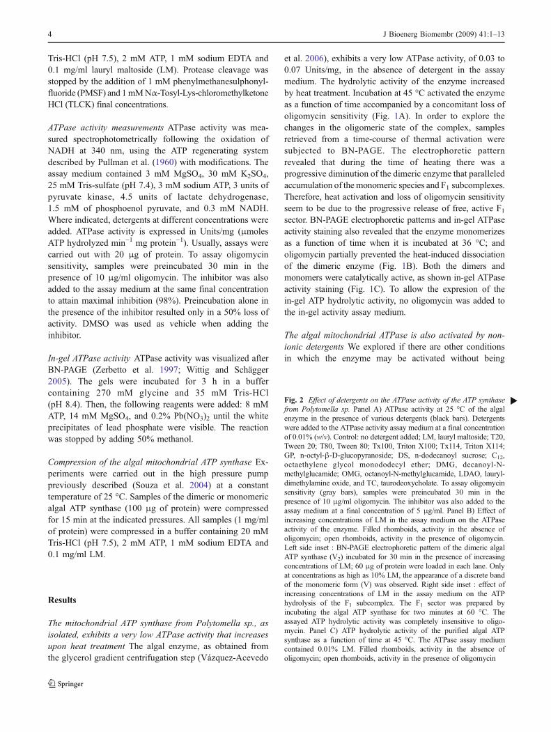
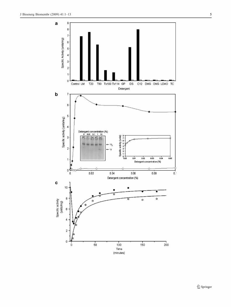
Fig. 2 Effect of detergents on the ATPase activity of the ATP synthase
from Polytomella sp. Panel A) ATPase activity at 25 °C of the algalenzyme in the presence of various detergents (black bars). Detergentswere added to the ATPase activity assay medium at a final concentrationof 0.01% (w/v). Control: no detergent added; LM, lauryl maltoside; T20,Tween 20; T80, Tween 80; Tx100, Triton X100; Tx114, Triton X114;GP, n-octyl-β-D-glucopyranoside; DS, n-dodecanoyl sucrose; C12,octaethylene glycol monododecyl ether; DMG, decanoyl-N-methylglucamide; OMG, octanoyl-N-methylglucamide, LDAO, lauryl-dimethylamine oxide, and TC, taurodeoxycholate. To assay oligomycinsensitivity (gray bars), samples were preincubated 30 min in thepresence of 10 μg/ml oligomycin. The inhibitor was also added to theassay medium at a final concentration of 5 μg/ml. Panel B) Effect ofincreasing concentrations of LM in the assay medium on the ATPaseactivity of the enzyme. Filled rhomboids, activity in the absence ofoligomycin; open rhomboids, activity in the presence of oligomycin.Left side inset : BN-PAGE electrophoretic pattern of the dimeric algalATP synthase (V2) incubated for 30 min in the presence of increasingconcentrations of LM; 60 μg of protein were loaded in each lane. Onlyat concentrations as high as 10% LM, the appearance of a discrete bandof the monomeric form (V) was observed. Right side inset : effect ofincreasing concentrations of LM in the assay medium on the ATPhydrolysis of the F1 subcomplex. The F1 sector was prepared byincubating the algal ATP synthase for two minutes at 60 °C. Theassayed ATP hydrolytic activity was completely insensitive to oligo-mycin. Panel C) ATP hydrolytic activity of the purified algal ATPsynthase as a function of time at 45 °C. The ATPase assay mediumcontained 0.01% LM. Filled rhomboids, activity in the absence ofoligomycin; open rhomboids, activity in the presence of oligomycin
b
4 J Bioenerg Biomembr (2009) 41:1–13
J Bioenerg Biomembr (2009) 41:1–13 55
dissociated. To this end, different detergents were added tothe ATPase activity assay medium. Some non-ionicdetergents induced a large increase in the ATPase activityof the complex (Fig. 2A). The detergent-activated enzymeswere fully sensitive to oligomycin. The non-ionic detergentsdodecyl-β-D-maltopyranoside (lauryl maltoside, LM), octa-ethylene glycol monododecyl ether (C12E8), Tween 20,Tween 80, and n-dodecanoyl sucrose strongly activated theenzyme. In contrast, Triton X100 and Triton X114 induced amodest activating effect, while n-octyl-β-D-glucopyranoside,octanoyl-N-methylglucamide decanoyl-N-methylglucamide,lauryldimethylamine oxide (LDAO) and sodium taurodeox-ycholate had no noticeable effect on the activity (Fig. 2A).The activation by LM was further explored. Figure 2Bshows titration curves with increasing concentrations of LMin the assay medium in the absence and presence ofoligomycin. Maximal activations were observed at concen-trations of 0.01% (w/v) of LM (7.0 Units/mg). The detergent-activated enzyme was fully sensitive to oligomycin(Fig. 2B). The activation of the enzyme induced bydetergents was not due to dissociation of the algal ATPsynthase, since increasing detergent concentrations (up to1%) did not disrupt the dimeric enzyme, as judged by BNE-PAGE (Fig. 2B, inset). Only when the enzyme wasincubated in ice for 30 min in the presence of very highconcentrations of detergent (10% LM), the appearance of adiscrete population of monomeric forms of the enzymebecame evident (Fig. 2B, inset). LM also activated the ATPhydrolytic activity of the F1 sector. The F1 sector wasprepared by heating the dimeric enzyme for 2 min at 60 °C.In these conditions, the algal F1Fo-ATP synthase is dis-rupted, giving rise to an active F1 subcomplex that exhibits arelatively high ATPase activity (around 4.0 Units/mg).Increasing concentrations of LM to the ATPase assaymedium further increased this activity up to values around7.0 Units/mg (Fig. 2B, inset).
When the algal enzyme was incubated at 45 °C forincreasing periods of time and its activity measured in thepresence of detergent in the assay medium (LM 0.01%), adifferent pattern of activation was observed (Fig. 2C). In afirst phase, the original activity, which was fully sensitive tooligomycin, decreased sharply during the first minutes,simultaneous to a discrete loss in oligomycin sensitivity. Ina second phase, the ATPase activity of the enzymeincreased as a function of time; this was accompanied bya parallel loss of oligomycin sensitivity (Fig. 2C). Weattribute this second phase to the progressive disassemblyof the F1Fo-ATP synthase complexes and the consequentrelease of free, active, oligomycin-insensitive F1 sectors.
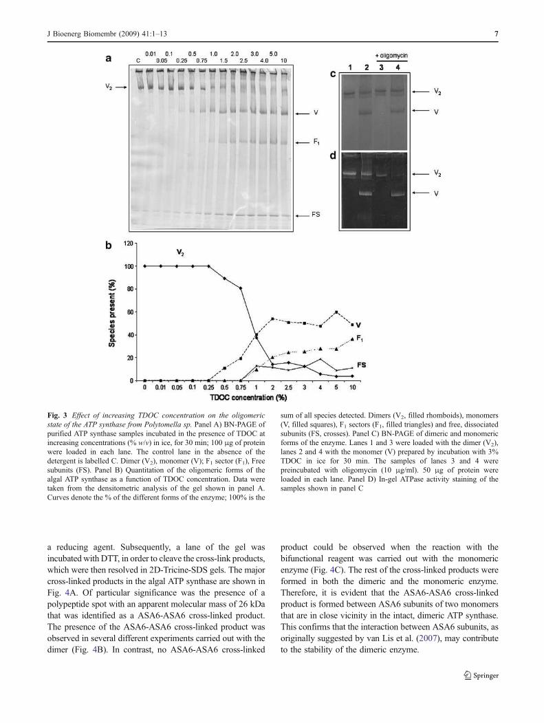
Taurodeoxycholate monomerizes the algal mitochondrial
ATP synthase When the dimeric enzyme is incubated atrelatively high temperatures (i.e., 60 °C) for brief periods of
time, it dissociates into its monomers, and subsequently,into F1 moieties and free independent subunits (Vázquez-Acevedo et al. 2006). We searched for conditions, otherthan heat treatment, that induce dissociation of the enzymeinto stable monomers. The incubation of the enzyme withsodium taurodeoxycholate (TDOC), the hydrophylic, bilesalt-related anionic detergent, induced a concentration-dependent dissociation of the enzyme into monomers(Fig. 3A); at relatively high concentrations of TDOC, F1was released, and the amount of free, dissociated subunitsincreased. Treatment with TDOC has the advantage overheat treatment in that it allows for a more precise control ofthe dissociation process, and in consequence, it is possibleto work with a population of enzymes in which themonomeric form predominates, albeit in coexistance withundissociated dimers and free F1 moieties (Fig. 3B). Aconcentration of 3% (w/v) taurodeoxycholate was chosenfor all further experiments. The mixture of ATP synthasesgenerated by treatment with 3% TDOC will be referred toas the monomeric form of the enzyme, although it actuallyrepresents a population with 50% monomers and 50% of amixture of dimers, F1 sectors and free subunits (Fig. 3B).TDOC-induced monomerization was irreversible, sincetreatment with the detergent, followed by extensive dialysisin the presence of 0.1% LM, did not restore the dimericform of the complex (data not shown). The 3%-TDOC-treated enzyme exhibited an ATPase activity of 0.6 Units/mg in the absence of detergent in the assay medium, andincreased in the presence of 0.01% LM to 2.0–4.0 Units/mg. In addition, the monomer was only partially sensitiveto oligomycin (45% inhibition). The contrasting behaviorbetween the dimeric and monomeric forms of the enzymecould also be observed by BNE-PAGE followed by in-gelATPase activity staining (Fig. 3C). Both the dimer and themonomer exhibited in-gel ATP hydrolysis activity, however,the dimer wasmore sensitive to oligomycin than themonomer.
Neither heat treatment nor TDOC-induced monomer-ization elicited major changes in the subunit composition ofthe enzyme. The polypeptide patterns of the dimer and themonomer in SDS-Tricine-PAGE were essentially the same(data not shown). In some experiments, partial loss of thesmall, membrane bound subunits ASA6 and/or ASA9 wasobserved.
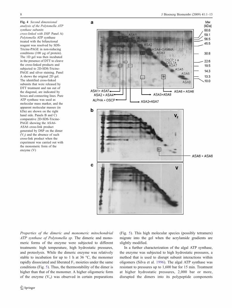
Crosslinking experiments suggest that subunit ASA6 may be
involved in monomer-monomer interactions In order toidentify possible close-neighbor interactions between thePolytomella ATP synthase subunits, the dimeric enzymewas incubated with dithiobis(succinimidyl)propionate (DSP)a homobifunctional, thiol-cleavable and membrane permeablecrosslinker that has a spacer arm of 12 Å. The cross-linkproducts were analyzed by two-dimensional gel electropho-resis. A first-dimension SDS-PAGE was run in the absence of
6 J Bioenerg Biomembr (2009) 41:1–13
a reducing agent. Subsequently, a lane of the gel wasincubated with DTT, in order to cleave the cross-link products,which were then resolved in 2D-Tricine-SDS gels. The majorcross-linked products in the algal ATP synthase are shown inFig. 4A. Of particular significance was the presence of apolypeptide spot with an apparent molecular mass of 26 kDathat was identified as a ASA6-ASA6 cross-linked product.The presence of the ASA6-ASA6 cross-linked product wasobserved in several different experiments carried out with thedimer (Fig. 4B). In contrast, no ASA6-ASA6 cross-linked
product could be observed when the reaction with thebifunctional reagent was carried out with the monomericenzyme (Fig. 4C). The rest of the cross-linked products wereformed in both the dimeric and the monomeric enzyme.Therefore, it is evident that the ASA6-ASA6 cross-linkedproduct is formed between ASA6 subunits of two monomersthat are in close vicinity in the intact, dimeric ATP synthase.This confirms that the interaction between ASA6 subunits, asoriginally suggested by van Lis et al. (2007), may contributeto the stability of the dimeric enzyme.
Fig. 3 Effect of increasing TDOC concentration on the oligomeric
state of the ATP synthase from Polytomella sp. Panel A) BN-PAGE ofpurified ATP synthase samples incubated in the presence of TDOC atincreasing concentrations (% w/v) in ice, for 30 min; 100 μg of proteinwere loaded in each lane. The control lane in the absence of thedetergent is labelled C. Dimer (V2), monomer (V); F1 sector (F1), Freesubunits (FS). Panel B) Quantitation of the oligomeric forms of thealgal ATP synthase as a function of TDOC concentration. Data weretaken from the densitometric analysis of the gel shown in panel A.Curves denote the % of the different forms of the enzyme; 100% is the
sum of all species detected. Dimers (V2, filled rhomboids), monomers(V, filled squares), F1 sectors (F1, filled triangles) and free, dissociatedsubunits (FS, crosses). Panel C) BN-PAGE of dimeric and monomericforms of the enzyme. Lanes 1 and 3 were loaded with the dimer (V2),lanes 2 and 4 with the monomer (V) prepared by incubation with 3%TDOC in ice for 30 min. The samples of lanes 3 and 4 werepreincubated with oligomycin (10 μg/ml). 50 μg of protein wereloaded in each lane. Panel D) In-gel ATPase activity staining of thesamples shown in panel C
J Bioenerg Biomembr (2009) 41:1–13 77
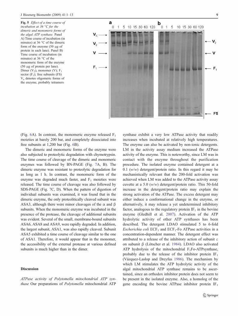
Properties of the dimeric and monomeric mitochondrial
ATP synthase of Polytomella sp. The dimeric and mono-meric forms of the enzyme were subjected to differenttreatments: high temperature, high hydrostatic pressures,and proteolysis. While the dimeric enzyme was relativelystable to incubation for up to 1 h at 36 °C, the monomerrapidly dissociated and liberated F1 moieties under the sameconditions (Fig. 5). Thus, the thermostability of the dimer ishigher than that of the monomer. A higher oligomeric formof the enzyme (Vx) was observed in certain preparations
(Fig. 5). This high molecular species (possibly tetramers)migrate into the gel when the acrylamide gradients areslightly modified.
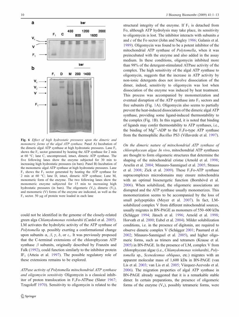
In a further characterization of the algal ATP synthase,the enzyme was subjected to high hydrostatic pressures, amethod that is used to disrupt subunit interactions withinoligomers (Silva et al. 1996). The algal ATP synthase wasresistant to pressures up to 1,600 bar for 15 min. Treatmentat higher hydrostatic pressures, 2,000 bar or more,disrupted the dimers into its polypeptide components
Fig. 4 Second dimensional
analysis of the Polytomella ATP
synthase subunits
cross-linked with DSP. Panel A)Polytomella ATP synthasetreated with the bifunctionalreagent was resolved by SDS-Tricine-PAGE in non-reducingconditions (100 μg of protein).The 1D gel was then incubatedin the presence of DTT to cleavethe cross-linked products andsubjected to 2D-SDS-Tricine-PAGE and silver staining. PanelA shows the original 2D gel.The identified cross-linkedsubunits that were released byDTT treatment and ran out ofthe diagonal, are indicated byboxes and connecting lines. PureATP synthase was used asmolecular mass marker, and theapparent molecular masses (inkDa) are shown on the righthand side. Panels B and C)comparative 2D-SDS-Tricine-PAGE showing the ASA6-ASA6 cross-link productgenerated by DSP on the dimer(V2) and the absence of suchcross-link product when theexperiment was carried out withthe monomeric form of theenzyme (V)
8 J Bioenerg Biomembr (2009) 41:1–13
(Fig. 6A). In contrast, the monomeric enzyme released F1moieties at barely 200 bar, and completely dissociated intofree subunits at 1,200 bar (Fig. 6B).
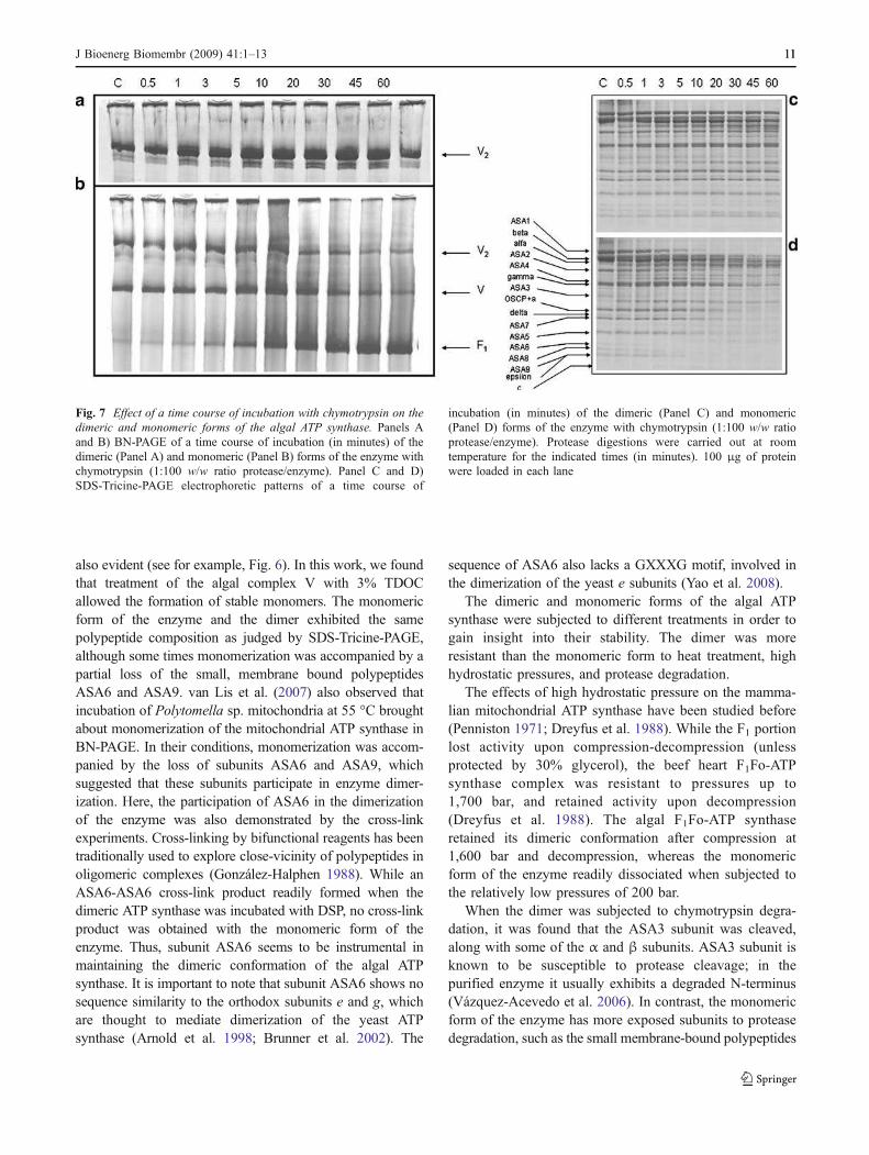
The dimeric and monomeric forms of the enzyme werealso subjected to proteolytic degradation with chymotrypsin.The time course of cleavage of the dimeric and monomericenzymes was followed by BN-PAGE (Fig. 7A, B). Thedimeric enzyme was resistant to proteolytic degradation foras long as 1 h. In contrast, the monomeric form of theenzyme was degraded much faster, and F1 moieties werereleased. The time course of cleavage was also followed bySDS-PAGE (Fig. 7C, D). When the pattern of digestion ofindividual subunits was examined, it was found that in thedimeric enzyme, the only proteolitically cleaved subunit wasASA3, although there were minor cleavages of the α and β
subunits. When the monomeric enzyme was incubated in thepresence of the protease, the cleavage of additional subunitswas evident. Several of the small, membrane-bound subunitsASA6, ASA8 and ASA9, were rapidly degraded. In addition,the largest subunit, ASA1, was also rapidly cleaved. SubunitASA5 exhibited a time course of cleavage similar to the oneof ASA1. Therefore, it would appear that in the monomer,the accessibility of the external protease at various definedsubunits is much higher than in the dimer.
Discussion
ATPase activity of Polytomella mitochondrial ATP syn-
thase Our preparations of Polytomella mitochondrial ATP
synthase exhibit a very low ATPase activity that readilyincreases when incubated at relatively high temperatures.The enzyme can also be activated by non-ionic detergents.LM in the activity assay medium increased the ATPaseactivity of the enzyme. This is noteworthy, since LM was incontact with the enzyme throughout the purificationprocedure. The isolated enzyme contained detergent at a0.1 (w/w) detergent/protein ratio. In this regard it may bemechanistically relevant that the 200-fold activation wasachieved when LM was added to the ATPase activity assaycuvette at a 5.0 (w/w) detergent/protein ratio. This 50-foldincrease in the detergent/protein ratio may explain thestrong activation of the ATPase. The excess detergent mayeither induce a conformational change in the enzyme, oralternatively, it may release a yet undetermined inhibitoryfactor, analogous to the regulatory protein IF1 in the bovineenzyme (Gledhill et al. 2007). Activation of the ATPhydrolytic activity of other ATP synthases has beendescribed. The detergent LDAO stimulated 5 to 6-foldEscherichia coli ECF1 and ECF1-Fo ATPase activities in aconcentration-dependent manner. The detergent effect wasattributed to a release of the inhibitory action of subunit εon subunit β (Lötscher et al. 1984). LDAO also activatedATP hydrolysis of the mitochondrial F1Fo-ATPsynthase,probably due to the release of the inhibitor protein IF1(Vázquez-Laslop and Dreyfus 1986). The mechanism bywhich LM stimulates the ATP hydrolytic activity of thealgal mitochondrial ATP synthase remains to be ascer-tained, since an orthodox inhibitor protein does not seem tobe present in the isolated enzyme. Also, a homolog of thegene encoding the bovine ATPase inhibitor protein IF1
Fig. 5 Effect of a time course of
incubation at 36 °C for the
dimeric and monomeric forms of
the algal ATP synthase. PanelA) Time course of incubation (inminutes) at 36 °C of the dimericform of the enzyme (50 μg ofprotein in each lane). Panel B)Time course of incubation (inminutes) at 36 °C of themonomeric form of the enzyme(50 μg of protein per lane).Dimer (V2), monomer (V); F1sector (F1); free subunits (FS).Vx denotes oligomeric forms ofthe enzyme, probably tetramers
J Bioenerg Biomembr (2009) 41:1–13 99
could not be identified in the genome of the closely-relatedgreen alga Chlamydomonas reinhardtii (Cardol et al. 2005).LM activates the hydrolytic activity of the ATP synthase ofPolytomella sp. possibly exerting a conformational changeupon subunits α, β, g, δ, or ε,. It was previously proposedthat the C-terminal extensions of the chlorophycean ATPsynthase β subunits, originally described by Franzén andFalk (1992), could function similarly to the inhibitor proteinIF1 (Atteia et al. 1997). The possible regulatory role ofthese extensions remains to be explored.
ATPase activity of Polytomella mitochondrial ATP synthase
and oligomycin sensitivity Oligomycin is a classical inhib-itor of proton translocation in F1Fo-ATPase (Slater 1967;Tzagoloff 1970). Sensitivity to oligomycin is related to the
structural integrity of the enzyme. If F1 is detached fromFo, although ATP hydrolysis may take place, its sensitivityto oligomycin is lost. The inhibitor interacts with subunits aand c of the Fo sector (John and Nagley 1986; Galanis et al.1989). Oligomycin was found to be a potent inhibitor of themitochondrial ATP synthase of Polytomella, when it waspreincubated with the enzyme and also added in the assaymedium. In these conditions, oligomycin inhibited morethan 98% of the detergent-stimulated ATPase activity of thecomplex. The high sensitivity of the algal ATP synthase tooligomycin, suggests that the increase in ATP activity bynon-ionic detergents does not involve dissociation of thedimer, indeed, sensitivity to oligomycin was lost whendissociation of the enzyme was induced by heat treatment.This process was accompanied by monomerization andeventual disruption of the ATP synthase into F1 sectors andfree subunits (Fig. 1A). Oligomycin also seems to partiallyprevent the heat-induced dissociation of the dimeric algal ATPsynthase, providing some ligand-induced thermostability tothe complex (Fig. 1B). In this regard, it is noted that bindingof ligands may confer thermostability to ATP synthases, i.e.,the binding of Mg2+-ADP to the F1Fo-type ATP synthasefrom the thermophilic Bacillus PS3 (Villaverde et al. 1997).
On the dimeric nature of mitochondrial ATP synthase of
chlorophycean algae In vivo, mitochondrial ATP synthasesare thought to form oligomeric structures that determine theshaping of the mitochondrial cristae (Arnold et al. 1998;Arselin et al. 2004; Minauro-Sanmiguel et al. 2005; Strausset al. 2008; Zick et al. 2009). These F1Fo-ATP synthasesupracomplexes microdomains may ensure mitochondriawith an optimal bioenergetic function (Bornhövd et al.2006). When solubilized, the oligomeric associations aredisrupted and the ATP synthase usually monomerizes. Thismonomerization seems to be accompanied by the loss ofsmall polypeptides (Meyer et al. 2007). In fact, LM-solubilized complex V from different mitochondrial sources,usually migrates in BN-PAGE as monomers of 550–600 kDa(Schägger 1994; Jänsch et al. 1996; Arnold et al. 1998;Horvath et al. 2000; Eubel et al. 2004). Milder solubilizationconditions, i.e. in the presence of digitonin, are required toobserve dimeric complex V (Schägger 2001; Paumard et al.2002; Minauro-Sanmiguel et al. 2005), and higher oligo-meric forms, such as trimers and tetramers (Krause et al.2005) in BN-PAGE. In the presence of LM, complex V fromchlorophycean algae (i.e., Chlamydomonas reinhardtii, Poly-tomella sp., Scenedesmus obliquus, etc.) migrates with anapparent molecular mass of 1,600 kDa in BN-PAGE (vanLis et al. 2003; van Lis et al. 2005; Vázquez-Acevedo et al.2006). The migration properties of algal ATP synthase inBN-PAGE already suggested that it is a remarkable stabledimer. In certain preparations, the presence of oligomericforms of the enzyme (Vx), possibly tetrameric forms, were
Fig. 6 Effect of high hydrostatic pressures upon the dimeric and
monomeric forms of the algal ATP synthase. Panel A) Incubation ofthe dimeric algal ATP synthase at high hydrostatic pressures. Lane F1shows the F1 sector generated by heating the ATP synthase for 2 minat 60 °C; lane C, uncompressed, intact, dimeric ATP synthase. Thefive following lanes show the enzyme subjected for 30 min toincreasing high hydrostatic pressures (in bars). Panel B) Incubation ofthe monomeric algal ATP synthase at high hydrostatic pressures. LaneF1 shows the F1 sector generated by heating the ATP synthase for2 min at 60 °C; lane D, intact, dimeric ATP synthase; Lane M,monomeric form of the enzyme. The two following lanes show themonomeric enzyme subjected for 15 min to increasing highhydrostatic pressures (in bars). The oligomeric (Vx), dimeric (V2),and monomeric (V) forms of the enzyme are indicated, as well as theF1 sector. 50 μg of protein were loaded in each lane
10 J Bioenerg Biomembr (2009) 41:1–13
also evident (see for example, Fig. 6). In this work, we foundthat treatment of the algal complex V with 3% TDOCallowed the formation of stable monomers. The monomericform of the enzyme and the dimer exhibited the samepolypeptide composition as judged by SDS-Tricine-PAGE,although some times monomerization was accompanied by apartial loss of the small, membrane bound polypeptidesASA6 and ASA9. van Lis et al. (2007) also observed thatincubation of Polytomella sp. mitochondria at 55 °C broughtabout monomerization of the mitochondrial ATP synthase inBN-PAGE. In their conditions, monomerization was accom-panied by the loss of subunits ASA6 and ASA9, whichsuggested that these subunits participate in enzyme dimer-ization. Here, the participation of ASA6 in the dimerizationof the enzyme was also demonstrated by the cross-linkexperiments. Cross-linking by bifunctional reagents has beentraditionally used to explore close-vicinity of polypeptides inoligomeric complexes (González-Halphen 1988). While anASA6-ASA6 cross-link product readily formed when thedimeric ATP synthase was incubated with DSP, no cross-linkproduct was obtained with the monomeric form of theenzyme. Thus, subunit ASA6 seems to be instrumental inmaintaining the dimeric conformation of the algal ATPsynthase. It is important to note that subunit ASA6 shows nosequence similarity to the orthodox subunits e and g, whichare thought to mediate dimerization of the yeast ATPsynthase (Arnold et al. 1998; Brunner et al. 2002). The
sequence of ASA6 also lacks a GXXXG motif, involved inthe dimerization of the yeast e subunits (Yao et al. 2008).
The dimeric and monomeric forms of the algal ATPsynthase were subjected to different treatments in order togain insight into their stability. The dimer was moreresistant than the monomeric form to heat treatment, highhydrostatic pressures, and protease degradation.
The effects of high hydrostatic pressure on the mamma-lian mitochondrial ATP synthase have been studied before(Penniston 1971; Dreyfus et al. 1988). While the F1 portionlost activity upon compression-decompression (unlessprotected by 30% glycerol), the beef heart F1Fo-ATPsynthase complex was resistant to pressures up to1,700 bar, and retained activity upon decompression(Dreyfus et al. 1988). The algal F1Fo-ATP synthaseretained its dimeric conformation after compression at1,600 bar and decompression, whereas the monomericform of the enzyme readily dissociated when subjected tothe relatively low pressures of 200 bar.
When the dimer was subjected to chymotrypsin degra-dation, it was found that the ASA3 subunit was cleaved,along with some of the α and β subunits. ASA3 subunit isknown to be susceptible to protease cleavage; in thepurified enzyme it usually exhibits a degraded N-terminus(Vázquez-Acevedo et al. 2006). In contrast, the monomericform of the enzyme has more exposed subunits to proteasedegradation, such as the small membrane-bound polypeptides
Fig. 7 Effect of a time course of incubation with chymotrypsin on the
dimeric and monomeric forms of the algal ATP synthase. Panels Aand B) BN-PAGE of a time course of incubation (in minutes) of thedimeric (Panel A) and monomeric (Panel B) forms of the enzyme withchymotrypsin (1:100 w/w ratio protease/enzyme). Panel C and D)SDS-Tricine-PAGE electrophoretic patterns of a time course of
incubation (in minutes) of the dimeric (Panel C) and monomeric(Panel D) forms of the enzyme with chymotrypsin (1:100 w/w ratioprotease/enzyme). Protease digestions were carried out at roomtemperature for the indicated times (in minutes). 100 μg of proteinwere loaded in each lane
J Bioenerg Biomembr (2009) 41:1–13 1111
ASA6 to ASA9, and other subunits like ASA1 and ASA5.Altogether, the data suggest that the algal ATP synthase formsstable dimers, less susceptible to be monomerized bydetergents than the mitochondrial ATP synthases from variousspecies, including mammals, yeast and plants. It is likely thatthe robust dimeric nature of the algal complex may be relevantin vivo, and could be related to the tightly regulated ATPaseactivity of the enzyme or/and to maintain the highlystructured tubular cristae typical of Polytomella mitochondria(Dudkina et al. 2006). Therefore, the data indicate that thefully-active form of the algal ATPase is the structurally stabledimer, and that the monomeric form of the enzyme is a lessactive, unstable complex.
Acknowledgments We thank Prof. Armando Gómez-Puyou for hiscritical review of the paper. This research was supported by grants56619 from CONACyT (Mexico) IN217108 from DGAPA, UNAM(Mexico), CNPq and FAPERJ (Brazil), and by grants from the BelgianFRFC (2.4587.04, 2.4582.05 and F.4735.06). PC is posdoctoral fellowof the FRS-FNRS (Belgium).
References
Arnold I, Pfeiffer K, Neupert W, Stuart RA, Schägger H (1998) Yeastmitochondrial F1F0-ATP synthase exists as a dimer: identificationof three dimer-specific subunits. EMBO J 17:7170–7178
Arselin G, Vaillier J, Salin B, Schaeffer J, Giraud MF, Dautant A,Brèthes D, Velours J (2004) The modulation in subunits e and gamounts of yeast ATP synthase modifies mitochondrial cristaemorphology. J Biol Chem 279:40392–40399
Atteia A, Dreyfus G, González-Halphen D (1997) Characterization ofthe alpha and beta-subunits of the F0F1-ATPase from the algaPolytomella spp., a colorless relative of Chlamydomonas rein-
hardtii. Biochim Biophys Acta 1320:275–284Bornhövd C, Vogel F, Neupert W, Reichert AS (2006) Mitochondrial
membrane potential is dependent on the oligomeric state of F1F0-ATP synthase supracomplexes. J Biol Chem 281:13990–13998
Brunner S, Everard-Gigot V, Stuart RA (2002) Su e of the yeast F1Fo-ATP synthase forms homodimers. J Biol Chem 277:48484–48489
Cardol P, González-Halphen D, Reyes-Prieto A, Baurain D, MatagneRF, Remacle C (2005) The mitochondrial oxidative phosphory-lation proteome of Chlamydomonas reinhardtii deduced from theGenome Sequencing Project. Plant Physiol 137:447–459
Dreyfus G, Guimaraes-Motta H, Silva JL (1988) Effect of hydrostaticpressure on the mitochondrial ATP synthase. Biochemistry27:6704–6710
Dudkina NV, Heinemeyer J, Keegstra W, Boekema EJ, Braun HP(2005) Structure of dimeric ATP synthase from mitochondria: anangular association of monomers induces the strong curvature ofthe inner membrane. FEBS Lett 579:5769–5772
Dudkina NV, Sunderhaus S, Braun HP, Boekema EJ (2006)Characterization of dimeric ATP synthase and cristae membraneultrastructure from Saccharomyces and Polytomella mitochondria.FEBS Lett 580:3427–3432
Eubel H, Heinemeyer J, Braun HP (2004) Identification andcharacterization of respirasomes in potato mitochondria. PlantPhysiol 134:1450–1459
Franzén LG, Falk G (1992) Nucleotide sequence of cDNA clonesencoding the beta subunit of mitochondrial ATP synthase fromthe green alga Chlamydomonas reinhardtii: the precursor proteinencoded by the cDNA contains both an N-terminal presequenceand a C-terminal extension. Plant Mol Biol 19:771–780
Galanis M, Mattoon JR, Nagley P (1989) Amino acid substitutions inmitochondrial ATP synthase subunit 9 of Saccharomyces
cerevisiae leading to venturicidin or ossamycin resistance. FEBSLett 249:333–336
Gledhill JR, Montgomery MG, Leslie AG, Walker JE (2007) How theregulatory protein, IF(1), inhibits F(1)-ATPase from bovinemitochondria. Proc Natl Acad Sci USA 104:15671–15676
Golden TR, Pedersen PL (1998) The oligomycin sensitivity conferringprotein of rat liver mitochondrial ATP synthase: arginine 94 isimportant for the binding of OSCP to F1. Biochemistry37:13871–13881
González-Halphen D, Lindorfer MA, Capaldi RA (1988) Subunitarrangement in beef heart complex III. Biochemistry 27:7021–7031
Hong S, Pedersen PL (2008) ATP synthase and the actions ofinhibitors utilized to study its roles in human health, disease, andother scientific areas. Microbiol Mol Biol Rev 72:590–641
Horvath A, Kingan TG, Maslov DA (2000) Detection of themitochondrially encoded cytochrome c oxidase subunit I in thetrypanosomatid protozoan Leishmania tarentolae. Evidence fortranslation of unedited mRNA in the kinetoplast. J Biol Chem275:17160–17165
Jänsch L, Kruft V, Schmitz UK, Braun HP (1996) New insights intothe composition, molecular mass and stoichiometry of the proteincomplexes of plant mitochondria. The Plant Journal 9:357–368
John UP, Nagley P (1986) Amino acid substitutions in mitochondrialATPase subunit 6 of Saccharomyces cerevisiae leading tooligomycin resistance. FEBS Lett 207:79–83
Ko YH, Hullihen J, Hong S, Pedersen PL (2000) Mitochondrial F(0)F(1) ATP synthase. Subunit regions on the F1 motor shielded by F(0), Functional significance, evidence for an involvement of theunique F(0) subunit F(6). J Biol Chem 275:32931–32939
Krause F, Reifschneider NH, Gotom S, Dencher NA (2005) Activeoligomeric ATP synthases in mammalian mitochondria. BiochemBiophys Res Commun 329:583–590
Lötscher HR, deJong C, Capaldi RA (1984) Interconversion of highand low adenosinetriphosphatase activity forms of Escherichia
coli F1 by the detergent lauryldimethylamine oxide. Biochemistry23:4140–4143
Markwell MAK, Hass SM, Biber LL, Tolbert NE (1978) Amodification of the Lowry procedure to simplify proteindetermination in membrane and lipoprotein samples. AnalBiochem 87:206–210
Meyer B, Wittig I, Trifilieff E, Karas M, Schägger H (2007)Identification of two proteins associated with mammalian ATPsynthase. Mol Cell Proteomics 10:1690–1699
Minauro-Sanmiguel F, Wilkens S, García JJ (2005) Structure ofdimeric mitochondrial ATP synthase: novel F0 bridging featuresand the structural basis of mitochondrial cristae biogenesis. ProcNatl Acad Sci (USA) 102:12356–12358
Paumard P, Arselin G, Vaillier J, Chaignepain S, Bathany K,Schmitter JM, Brèthes D, Velours J (2002) Two ATP synthasescan be linked through subunits i in the inner mitochondrialmembrane of Saccharomyces cerevisiae. Biochemistry 41:10390–10396
Penniston JT (1971) High hydrostatic pressure and enzymic activity:inhibition of multimeric enzymes by dissociation. Arch BiochemBiophys 142:322–332
Pullman ME, Penefsky HS, Datta A, Racker E (1960) Partialresolution of the enzymes catalyzing oxidative phosphorylation.
12 J Bioenerg Biomembr (2009) 41:1–13
I. Purification and properties of soluble dinitrophenol-stimulatedadenosine triphosphatase. J Biol Chem 235:3322–3329
Schägger H (2001) Respiratory chain supercomplexes. IUBMB Life52:119–128
Schägger H, in: von Jagow G, Schägger H, (Eds.) (1994) A PracticalGuide to Membrane Protein Purification, Academic, San Diego,pp. 59–79
Silva JL, Foguel D, Da Poian AT, Prevelige PE (1996) The use ofhydrostatic pressure as a tool to study viruses and othermacromolecular assemblages. Curr Opin Struct Biol 6:166–175
Slater EC (1967) Application of inhibitors and uncouplers for a studyof oxidative phosphorylation. Methods Enzymol 10:48–57
Souza MO, Creczynski-Pasa TB, Scofano HM, Gräber P, Mignaco JA(2004) High hydrostatic pressure perturbs the interactionsbetween CF(0)F(1) subunits and induces a dual effect on activity.Int J Biochem Cell Biol 36:920–930
Strauss M, Hofhaus G, Schröder RR, Kühlbrandt W (2008) Dimerribbons of ATP synthase shape the inner mitochondrial membrane.EMBO J 27:1154–1160
Tzagoloff A (1970) Assembly of the mitochondrial membrane system.3. Function and synthesis of the oligomycin sensitivity-conferring protein of yeast mitochondria. J Biol Chem 245:1545–1551
van Lis R, Atteia A, Mendoza-Hernández G, González-Halphen D(2003) Identification of novel mitochondrial protein componentsof Chlamydomonas reinhardtii. A proteomic approach. PlantPhysiol 132:318–330
van Lis R, González-Halphen D, Atteia A (2005) Divergence of themitochondrial electron transport chains from the green algaChlamydomonas reinhardtii and its colorless close relativePolytomella sp. Biochim Biophys Acta 1708:23–34
van Lis R, Mendoza-Hernández G, Groth G, Atteia A (2007) Newinsights into the unique structure of the FoF1-ATP synthase fromthe chlamydomonad algae Polytomella sp. and Chlamydomonas
reinhardtii. Plant Physiol 144:1190–1199Vázquez-Acevedo M, Cardol P, Cano-Estrada A, Lapaille M, Remacle
C González-Halphen D (2006) The mitochondrial ATP synthaseof chlorophycean algae contains eight subunits of unknownorigin involved in the formation of an atypical stator-stalk and inthe dimerization of the complex. J. Bioenerg Biomembr 38:271–282
Vázquez-Laslop N, Dreyfus G (1986) Mitochondrial H+-ATPaseactivation by an amine oxide detergent. J Biol Chem 261:7807–7810
Villaverde J, Cladera J, Padrós E, Rigaud J-L, Duñach M (1997)Effect of Nucleotides on the Thermal Stability and on theDeuteration Kinetics of the Thermophilic F0F1 ATP Synthase.Eur J Biochem 244:441–448
Walker JE, Dickson VK (2006) The peripheral stalk of themitochondrial ATP synthase. Biochim Biophys Acta 1757:286–296
Wittig I, Schägger H (2005) Advantages and limitations of clear-native PAGE. Proteomics 5:4338–4346
Yao H, Stuart RA, Cai S, Sem DS (2008) Structural characterizationof the transmembrane domain from subunit e of yeast F1Fo-ATPsynthase: a helical GXXXG motif located just under the micellesurface. Biochemistry 47:1910–1917
Zerbetto E, Vergani L, Dabbeni-Sala F (1997) Quantification ofmuscle mitochondrial oxidative phosphorylation enzymes viahistochemical staining of blue native polyacrylamide gels.Electrophoresis 18:2059–2064
Zick M, Rabl R, Reichert AS (2009) Cristae formation-linkingultrastructure and function of mitochondria. Biophys BiochimActa 1793:5–19
J Bioenerg Biomembr (2009) 41:1–13 1313