Myb-Related Schizosaccharomyces pombe cdc5p Is Structurally and Functionally Conserved in Eukaryotes
13
MOLECULAR AND CELLULAR BIOLOGY, 0270-7306/98/$04.0010 July 1998, p. 4097–4108 Vol. 18, No. 7 Copyright © 1998, American Society for Microbiology. All Rights Reserved. Myb-Related Schizosaccharomyces pombe cdc5p Is Structurally and Functionally Conserved in Eukaryotes RYOMA OHI, 1 * ANNA FEOKTISTOVA, 2 STACEY MCCANN, 3 VIRGINIA VALENTINE, 4 A. THOMAS LOOK, 4 JOSEPH S. LIPSICK, 3 AND KATHLEEN L. GOULD 1,2 Howard Hughes Medical Institute 2 and Department of Cell Biology, 1 School of Medicine, Vanderbilt University, Nashville, Tennessee 37232; Department of Pathology and Program in Molecular and Genetic Medicine, School of Medicine, Stanford University, Stanford, California 94305 3 ; and Department of Experimental Oncology, St. Jude Children’s Research Hospital, Memphis, Tennessee 38105 4 Received 16 January 1998/Returned for modification 10 March 1998/Accepted 10 April 1998 Schizosaccharomyces pombe cdc5p is a Myb-related protein that is essential for G 2 /M progression. To explore the structural and functional conservation of Cdc5 throughout evolution, we isolated Cdc5-related genes and cDNAs from Saccharomyces cerevisiae, Caenorhabditis elegans, Drosophila melanogaster, and Homo sapiens. Supporting the notion that these Cdc5 gene family members are functionally homologous to S. pombe cdc5 1 , human and fly Cdc5 cDNAs are capable of complementing the temperature-sensitive lethality of the S. pombe cdc5-120 mutant. Furthermore, S. cerevisiae CEF1 (S. cerevisiae homolog of cdc5 1 ), like S. pombe cdc5 1 , is essential during G 2 /M. The location of the cdc5-120 mutation, as well as mutational analyses of Cef1p, indicate that the Myb repeats of cdc5p and Cef1p are important for their function in vivo. However, we found that unlike in c-Myb, single residue substitutions of glycines for hydrophobic residues within the Myb repeats of Cef1p, which are essential for maintaining structure of the Myb domain, did not impair Cef1p function in vivo. Rather, multiple W-to-G substitutions were required to inactivate Cef1p, and many of the substitution mutants were found to confer temperature sensitivity. Although it is possible that Cef1p acts as a transcriptional activator, we have demonstrated that Cef1p is not involved in transcriptional activation of a class of G 2 /M- regulated genes typified by SWI5. Collectively, these results suggest that Cdc5 family members participate in a novel pathway to regulate G 2 /M progression. The entrance of eukaryotic cells into mitosis from G 2 is a highly regulated event which involves a complex series of in- teractions among an evolutionarily conserved set of kinases and phosphatases (reviewed in references 41 and 57). Genetic studies with the fission yeast Schizosaccharomyces pombe have identified molecules that participate in the regulation of the G 2 /M transition through phosphorylation (34, 44–46), includ- ing cdc2 1 , cdc13 1 , cdc25 1 , wee1 1 , and mik1 1 . cdc2 1 encodes the single essential cyclin-dependent kinase (CDK) required to drive fission yeast cells through the cell cycle. During G 2 , cdc2p is associated with the mitotic B-type cyclin encoded by cdc13 1 . The activity of cdc2p/cdc13p complexes is inhibited by phos- phorylation of cdc2p at tyrosine 15, an event which is catalyzed by the wee1p and mik1p protein tyrosine kinases. As fission yeast cells achieve a critical size required for entry into mitosis, the cdc25p protein tyrosine phosphatase activates cdc2p/ cdc13p complexes by catalyzing the dephosphorylation of ty- rosine 15. In addition to genes which regulate cdc2p function directly, two other S. pombe genes, cdc5 1 and cdc28 1 , are known to be required during G 2 for entry into mitosis. cdc28 1 encodes a DEAH-box protein homologous to the Saccharomyces cerevi- siae splicing factors Prp2p, Prp16p, and Prp22p, suggesting that RNA processing is in some way required to promote the G 2 /M transition (33). The cdc5 1 gene product contains a putative DNA-binding domain (DBD) that is most similar to those contained within Myb-related proteins and is the only putative transcription factor cloned in S. pombe that performs an es- sential role during G 2 /M progression (49). Recently identified Arabidopsis thaliana, Homo sapiens, and Xenopus laevis cDNAs encode proteins closely related to S. pombe cdc5p (4, 23, 58). Of these, the A. thaliana cDNA, AtCDC5, was found to be a functional homolog of cdc5 1 since it was capable of complementing the temperature-sensitive growth defect of the fission yeast cdc5-120 mutant (23). A partial X. laevis Cdc5 cDNA clone was isolated in a screen designed to identify mitotic phosphoproteins that can be phos- phorylated in vitro by cyclin B-Cdc2 (58). In addition, Bern- stein and Coughlin (4) have reported that the human Cdc5 protein, PCDC5RP (pombe Cdc5-related protein), translo- cates from the cytoplasm to the nucleus of cultured mamma- lian cells stimulated with serum, implicating the human protein as a potential effector in a mitogen-activated signaling path- way. To explore further the structural and functional conserva- tion of cdc5p, we have isolated cDNAs and genes from S. cerevisiae, Caenorhabditis elegans, Drosophila melanogaster, and H. sapiens that encode homologs of S. pombe cdc5p and report here an analysis of these genes. Supporting a role for this subfamily of Myb-related proteins in G 2 /M cell cycle progres- sion, budding yeast cells lacking CEF1 (S. cerevisiae homolog of cdc5 1 ) arrest growth during G 2 /M. Furthermore, we dem- onstrate that human and fly Cdc5 proteins are functionally homologous to S. pombe cdc5p because they are able to com- plement the temperature-sensitive lethality of the S. pombe cdc5-120 mutant. Aside from the sequence similarity of Cdc5-related proteins to the transcriptional activator c-Myb, two lines of evidence have implicated Cdc5 in DNA binding. First, the Myb repeats of S. pombe cdc5p bind DNA-cellulose (49). Second, the Myb * Corresponding author. Mailing address: Department of Cell Biol- ogy, Vanderbilt University, School of Medicine, B2309 MCN, 1161 21st Ave. South, Nashville, TN 37232. Phone: (615) 343-9502. Fax: (615) 343-4539. E-mail: [email protected]. 4097 on November 24, 2015 by guest http://mcb.asm.org/ Downloaded from on November 24, 2015 by guest http://mcb.asm.org/ Downloaded from on November 24, 2015 by guest http://mcb.asm.org/ Downloaded from
-
Upload
independent -
Category
Documents
-
view
0 -
download
0
Transcript of Myb-Related Schizosaccharomyces pombe cdc5p Is Structurally and Functionally Conserved in Eukaryotes

MOLECULAR AND CELLULAR BIOLOGY,0270-7306/98/$04.0010
July 1998, p. 4097–4108 Vol. 18, No. 7
Copyright © 1998, American Society for Microbiology. All Rights Reserved.
Myb-Related Schizosaccharomyces pombe cdc5p Is Structurallyand Functionally Conserved in Eukaryotes
RYOMA OHI,1* ANNA FEOKTISTOVA,2 STACEY MCCANN,3 VIRGINIA VALENTINE,4
A. THOMAS LOOK,4 JOSEPH S. LIPSICK,3 AND KATHLEEN L. GOULD1,2
Howard Hughes Medical Institute2 and Department of Cell Biology,1 School of Medicine, Vanderbilt University,Nashville, Tennessee 37232; Department of Pathology and Program in Molecular and Genetic Medicine,School of Medicine, Stanford University, Stanford, California 943053; and Department of Experimental
Oncology, St. Jude Children’s Research Hospital, Memphis, Tennessee 381054
Received 16 January 1998/Returned for modification 10 March 1998/Accepted 10 April 1998
Schizosaccharomyces pombe cdc5p is a Myb-related protein that is essential for G2/M progression. To explorethe structural and functional conservation of Cdc5 throughout evolution, we isolated Cdc5-related genes andcDNAs from Saccharomyces cerevisiae, Caenorhabditis elegans, Drosophila melanogaster, and Homo sapiens.Supporting the notion that these Cdc5 gene family members are functionally homologous to S. pombe cdc51,human and fly Cdc5 cDNAs are capable of complementing the temperature-sensitive lethality of the S. pombecdc5-120 mutant. Furthermore, S. cerevisiae CEF1 (S. cerevisiae homolog of cdc51), like S. pombe cdc51, isessential during G2/M. The location of the cdc5-120 mutation, as well as mutational analyses of Cef1p, indicatethat the Myb repeats of cdc5p and Cef1p are important for their function in vivo. However, we found that unlikein c-Myb, single residue substitutions of glycines for hydrophobic residues within the Myb repeats of Cef1p,which are essential for maintaining structure of the Myb domain, did not impair Cef1p function in vivo.Rather, multiple W-to-G substitutions were required to inactivate Cef1p, and many of the substitution mutantswere found to confer temperature sensitivity. Although it is possible that Cef1p acts as a transcriptionalactivator, we have demonstrated that Cef1p is not involved in transcriptional activation of a class of G2/M-regulated genes typified by SWI5. Collectively, these results suggest that Cdc5 family members participate ina novel pathway to regulate G2/M progression.
The entrance of eukaryotic cells into mitosis from G2 is ahighly regulated event which involves a complex series of in-teractions among an evolutionarily conserved set of kinasesand phosphatases (reviewed in references 41 and 57). Geneticstudies with the fission yeast Schizosaccharomyces pombe haveidentified molecules that participate in the regulation of theG2/M transition through phosphorylation (34, 44–46), includ-ing cdc21, cdc131, cdc251, wee11, and mik11. cdc21 encodesthe single essential cyclin-dependent kinase (CDK) required todrive fission yeast cells through the cell cycle. During G2, cdc2pis associated with the mitotic B-type cyclin encoded by cdc131.The activity of cdc2p/cdc13p complexes is inhibited by phos-phorylation of cdc2p at tyrosine 15, an event which is catalyzedby the wee1p and mik1p protein tyrosine kinases. As fissionyeast cells achieve a critical size required for entry into mitosis,the cdc25p protein tyrosine phosphatase activates cdc2p/cdc13p complexes by catalyzing the dephosphorylation of ty-rosine 15.
In addition to genes which regulate cdc2p function directly,two other S. pombe genes, cdc51 and cdc281, are known to berequired during G2 for entry into mitosis. cdc281 encodes aDEAH-box protein homologous to the Saccharomyces cerevi-siae splicing factors Prp2p, Prp16p, and Prp22p, suggesting thatRNA processing is in some way required to promote the G2/Mtransition (33). The cdc51 gene product contains a putativeDNA-binding domain (DBD) that is most similar to thosecontained within Myb-related proteins and is the only putative
transcription factor cloned in S. pombe that performs an es-sential role during G2/M progression (49).
Recently identified Arabidopsis thaliana, Homo sapiens, andXenopus laevis cDNAs encode proteins closely related to S.pombe cdc5p (4, 23, 58). Of these, the A. thaliana cDNA,AtCDC5, was found to be a functional homolog of cdc51 sinceit was capable of complementing the temperature-sensitivegrowth defect of the fission yeast cdc5-120 mutant (23). Apartial X. laevis Cdc5 cDNA clone was isolated in a screendesigned to identify mitotic phosphoproteins that can be phos-phorylated in vitro by cyclin B-Cdc2 (58). In addition, Bern-stein and Coughlin (4) have reported that the human Cdc5protein, PCDC5RP (pombe Cdc5-related protein), translo-cates from the cytoplasm to the nucleus of cultured mamma-lian cells stimulated with serum, implicating the human proteinas a potential effector in a mitogen-activated signaling path-way.
To explore further the structural and functional conserva-tion of cdc5p, we have isolated cDNAs and genes from S.cerevisiae, Caenorhabditis elegans, Drosophila melanogaster, andH. sapiens that encode homologs of S. pombe cdc5p and reporthere an analysis of these genes. Supporting a role for thissubfamily of Myb-related proteins in G2/M cell cycle progres-sion, budding yeast cells lacking CEF1 (S. cerevisiae homologof cdc51) arrest growth during G2/M. Furthermore, we dem-onstrate that human and fly Cdc5 proteins are functionallyhomologous to S. pombe cdc5p because they are able to com-plement the temperature-sensitive lethality of the S. pombecdc5-120 mutant.
Aside from the sequence similarity of Cdc5-related proteinsto the transcriptional activator c-Myb, two lines of evidencehave implicated Cdc5 in DNA binding. First, the Myb repeatsof S. pombe cdc5p bind DNA-cellulose (49). Second, the Myb
* Corresponding author. Mailing address: Department of Cell Biol-ogy, Vanderbilt University, School of Medicine, B2309 MCN, 116121st Ave. South, Nashville, TN 37232. Phone: (615) 343-9502. Fax:(615) 343-4539. E-mail: [email protected].
4097
on Novem
ber 24, 2015 by guesthttp://m
cb.asm.org/
Dow
nloaded from
on Novem
ber 24, 2015 by guesthttp://m
cb.asm.org/
Dow
nloaded from
on Novem
ber 24, 2015 by guesthttp://m
cb.asm.org/
Dow
nloaded from

repeats of AtCDC5 were found to be capable of preferentiallybinding to the DNA sequence CTCAGCG (23). In this report,we demonstrate by mutational analysis of the Myb repeats ofCef1p and localization of the cdc5-120 mutation to the Mybrepeat R1 of cdc5p that the Myb repeats of these proteins areessential for their function in vivo. In the budding yeast S.cerevisiae, the Mcm1p transcription factor, in concert with anuncloned gene encoding a protein named Sff (SWI5 transcrip-tion factor), is required for transcription of at least six genesthat normally exhibit a G2/M-specific expression pattern,namely, CLB1, CLB2, CDC5, SWI5, ACE2, and ASE1 (1, 26,35). On the basis of phenotypic analysis of S. cerevisiae cellsgenetically depleted of CEF1 activity and the ability of thesecells to transcribe gene targets of Mcm1p/Sff, we conclude thatCEF1 does not encode Sff. Our work demonstrates that fissionyeast cdc5p is a member of a highly conserved subfamily ofMyb-related proteins that plays a key role in cell cycle progres-sion at the G2/M boundary.
MATERIALS AND METHODS
Strains, growth media, and genetic methods. The S. cerevisiae and S. pombestrains used in this study are listed in Table 1. Media used to grow S. pombe andgeneral genetic manipulations of S. pombe were described previously (39). S.pombe transformations were performed by electroporation (50). Repression oftranscription from the nmt11 promoter (37) was achieved by addition of thia-mine to a concentration of 2 mM.
S. cerevisiae strains were grown in either synthetic minimal medium with theappropriate nutritional supplements or complex yeast extract-peptone (YP) me-dium supplemented with either 2% glucose or 2% galactose–2% raffinose as thecarbon source(s). Genetic methods were as described by Guthrie and Fink (21).Transformation of S. cerevisiae was performed by the lithium acetate method(25).
Isolation and molecular cloning of CEF1 and the Cdc5Ce, Cdc5Dm, andCdc5Hs. A 2,555-bp fragment encompassing the CEF1 gene was amplified fromgenomic DNA by using primers cef1.P5 (59-CCCGGATCCGGCTTCTTATCTGTGGTC-39) and cef1.P6 (59-CCCCTGCAGCCGGATGGTACTTCTTAG-39)(introduced restriction enzyme sites are italicized). cef1.P5 introduces a BamHIsite onto the 59 end of the CEF1 gene, and cef1.P6 introduces a PstI site onto the39 end of the CEF1 gene. The PCR product was cleaved with BamHI and PstIand ligated into pRS415 (9), which had been cut with BamHI and PstI to producethe plasmid pCEF1. The CEF1 open reading frame (ORF) was sequenced toensure the absence of PCR-induced mutations. A URA3-CEN plasmid (pRS416)containing the CEF1 gene, which was used in all plasmid shuffle assays, wasconstructed by inserting the BamHI/HindIII CEF1 fragment from pCEF1 intopRS416, which had been cleaved with these enzymes. The 1,770-bp CEF1 ORFwas amplified from S. cerevisiae genomic DNA by using primers SC5531 (59-GCCGATATCTACTAGCATTTCAAGATGCCC-39) SC5351 (59-GAAGATATCCTATACTAATGCTATATGGAA-39). Both primers were designed to add
EcoRV restriction sites to the ends of the coding region during amplification.The amplified fragment was cut with EcoRV and cloned into the EcoRV site ofpSKREG351 to produce pSKREG351CEF1. pSKREG351 is pBS(SK1) (Strat-agene, LaJolla, Calif.) containing 585 bp of nucleotide sequence immediatelyupstream of the S. pombe cdc51 ATG. The cdc51 promoter sequence wasamplified by using primers 5REG351 (59-GAAGATATCAACCCTGAC-39) andT7 and cloned into pBS(SK1) which had been linearized with EcoRV and treatedwith dTTP and Taq polymerase. The EcoRV fragment from pSKREG351CEF1was subcloned into the SmaI site of the 2 mm LEU2-based shuttle vectorpRS425GAL1 (42) to produce pGAL1-CEF1. pCEF1 and pGAL1-CEF1 bothproduce functional Cef1p since they are capable of restoring growth to a strainlacking the CEF1 gene.
A C. elegans l cDNA clone, YK82fl, whose partial translation product hadhigh sequence identity to a portion of the Myb repeats of cdc5p, was obtainedfrom Yuji Kohara of the Gene Library Lab at the National Institute of Genetics(Mishima, Japan). The insert of this clone was subcloned and sequenced in itsentirety on both strands.
The fly homolog of the cdc51 gene was isolated by sequential nested PCR withD. melanogaster genomic DNA by using the following degenerate primers: Aout,59-A(G/A)A T(T/C/A)(T/C) TNA A(G/A)G CNG CNG T-39; Ain, 59-AA(G/A)TA(T/C) GGN AA(G/A) AA(T/C) CA(A/G) TGG-39; Bout, 59-NGC (T/C)TTNCC (T/C)TG NGT (A/G)TT-39; Bin, 59-(T/C)TC (A/G)TC (T/C)TC (A/G)TCCAT (G/A)TC (A/T/G)AT-39. The corresponding amino acid sequences of theseprimers are as follows: Aout, EILKAAV; Ain, KYGNQW; Bout, NTQGKKA; Bin,IDMDEDE. Following the second round of PCR, a band of 431 bp was obtainedand subcloned into the TA vector (InVitrogen, Carlsbad, Calif.). Five cloneswere sequenced and found to be identical. This 431-bp fragment was then usedto screen an adult Drosophila Canton S strain genomic l library (Clontech, PaloAlto, Calif.; a gift from M. Fuller). A 4.5-kb XhoI fragment encompassing theentire Cdc5Dm gene locus was subcloned from a positive l clone and sequencedin its entirety. The amino acid sequence was inferred from the genomic sequencebased on homology to other Cdc5 family members.
To isolate Cdc5Hs cDNAs, an I.M.A.G.E. clone (Consortium Clone ID 74654)that contains a 185-bp human expressed sequence tag (EST) whose translationproduct has high sequence identity to a portion of the Myb repeats of cdc5p wasobtained from the American Type Culture Collection (Rockville, Md.) and usedto screen a cDNA library constructed from a human microvascular endothelialcell line (a gift from T. O. Daniel). Eight positive clones were obtained afterscreening approximately 5 3 105 phage clones. The longest clone,pSKCdc5Hs.10, was cleaved with convenient restriction enzymes to generate abank of truncated clones to facilitate sequencing. The sequence of the Cdc5Hsgene was determined on both strands by using either custom synthesized oligo-nucleotides (Operon, Alameda, Calif.), M13 reverse primer, or T7 primers.
Chromosomal mapping of Cdc5Hs. To determine the chromosomal locationof the Cdc5Hs gene, three PAC clones (11107, 11108, and 11109; GenomeSystems, St. Louis, Mo.) were isolated by using full-length Cdc5Hs cDNA as aprobe. PAC clone 11108 was labeled with digoxigenin-11-dUTP (BoehringerMannheim, Indianapolis, Ind.) by nick translation. The labeled probe was com-bined with sheared human DNA and hybridized to normal metaphase chromo-somes derived from phytohemagglutinin-stimulated peripheral blood lympho-cytes in a solution containing 50% formamide, 10% dextran sulfate, and 23 SSC
TABLE 1. Yeast strains used in this study
Strain(s) Genotype Reference or source
S. cerevisiaeYPH274 MATa ura3-53 lys2-801 ade2-101 trp1-D1 his3-D200 leu2-D1 A. Weil (Vanderbilt University,
Nashville, Tenn.)MATa ura3-53 lys2-801 ade2-101 trp1-D1 his3-D200 leu2-D1KGY856 and
KGY857MATa ura3-53 lys2-801 ade2-101 trp1-D1 his3-D200 leu2-D1 cef1-D1::HIS3 This study
MATa ura3-53 lys2-801 ade2-101 trp1-D1 his3-D200 leu2-D1 CEF1KGY1120 MATa ura3-53 lys2-801 ade2-101 trp1-D1 his3-D200 leu2-D1
cef1-D1::HIS3/pRS425GAL1::CEF1This study
KGY1140 MATa ura3-53 lys2-801 ade2-101 trp1-D1 his3-D200 leu2-D1cef1-D1::HIS3/pRS425GAL1::CEF1
This study
KGY1338 MATa ura3-53 lys2-801 ade2-101 trp1-D1 his3-D200 leu2-D1cef1-D1::HIS3/pRS416CEF1
This study
S. pombeKGY69 975 h1 This laboratoryKGY155 cdc25-22 h1 This laboratoryKGY162 cdc5-120 h1 This laboratoryKGY360 cdc5-120 leu1-32 his3-D1 h2 This laboratoryKGY450 cdc5::ura41/cdc51 ura4-D18/ura4-D18 leu1-32/leu1-32 ade6-M210/ade6-M216 h1/h2 This laboratory
4098 OHI ET AL. MOL. CELL. BIOL.
on Novem
ber 24, 2015 by guesthttp://m
cb.asm.org/
Dow
nloaded from
(13 SSC is 0.15 M NaCl plus 0.015 M sodium citrate). Specific hybridizationsignals were detected by incubating the hybridized slides in fluorescein-conju-gated sheep antibodies to digoxigenin (Boehringer Mannheim). The chromo-somes were then counterstained with 49,6-diamidino-2-phenylindole (DAPI) andanalyzed. Definitive chromosomal assignment to chromosome 6 was confirmedby cohybridization of clone 11108 with a biotinylated chromosome 6 centromere-specific probe (D6Z1; Oncor Inc., Gaithersburg, Md.). Specific probe signalswere detected by incubating the hybridized slides in fluorescein-conjugatedsheep antibodies to digoxigenin and Texas red avidin (Vector Laboratories,Burlington, Calif.) followed by counterstaining with DAPI. Band assignment wasmade by conducting fractional length measurements of 10 specifically hybridizedchromosomes 6.
Disruption of CEF1 and construction of KGY1120. Deletion of the CEF1coding sequence was performed by replacing nearly the entire ORF with a DNAfragment containing the HIS3 gene. The cef1-D1::HIS3 null allele was generatedby PCR as described by Baudin et al. (3; see also reference 53), using the primers59-GCATTTCAAGATGCCCCCCGTACCAATATACGTGAGCAGATTGTACTGAG-39 and 59-CATGGCTTGAAGACGCTTTCTTCTACGACCCTCAAGCGCTCCTTACGCATCTG-39 and pRS313 (55) as a template to amplify theHIS3 gene. The underlined portions of each primer correspond to the HIS3flanking sequence. The resulting PCR product contains the complete HIS3 geneflanked by 35 bp of CEF1 59 sequence corresponding to nucleotides 210 to 125and 35 bp of CEF1 39 sequence corresponding to nucleotides 11714 to 11748.When substituted at the CEF1 locus, this DNA fragment deletes the entire CEF1gene except for the initial 8 codons and final 18 codons. The diploid strainYPH274 was transformed with this DNA fragment, and two independent trans-formants (KGY856 and KGY857) that contained a cef1-D1::HIS3 allele substi-tuted correctly at one of the two CEF1 loci were identified by Southern analysis.These strains were sporulated, and tetrads were dissected and analyzed.
To construct KGY1120, KGY856 was transformed with pGAL-CEF1. Thisstrain was sporulated and tetrads were dissected on synthetic medium containing2% raffinose–2% galactose. KGY1120 is one of the His1 Leu1 colonies whicharose from the tetrad analysis. KGY1140 is derived from a colony from the sametetrad that is His2 Leu1.
Cytological methods and flow cytometry. To visualize nuclei, cells were fixedwith 70% ethanol overnight at 4°C, washed twice with phosphate-buffered saline,and resuspended in 1 mg of DAPI per ml. For immunostaining of tubulin, cellswere processed as outlined by Pringle et al. (51) and stained with TAT-1 mono-clonal antibody (65) followed by a Texas red-conjugated goat-anti mouse immu-noglobulin G secondary antibody. Cells were viewed with a Zeiss Axioskop20with the appropriate set of filters, and images were captured with a Zeiss ZVS-47DEC image-capturing system. Yeast cells were processed for flow cytometricanalysis as described previously (49).
Mutagenesis of Cef1p. To generate single amino acid substitutions in Cef1p,pCEF1 was mutagenized by using the Muta-gene in vitro mutagenesis kit (Bio-Rad Laboratories, Hercules, Calif.) or the Chameleon double-stranded mu-tagenesis kit (Stratagene). All mutations were confirmed by the presence ofintroduced restriction enzyme sites and sequencing. Double, triple, and quadru-ple mutants were constructed by using a unique BglII site that cuts between thecodon for the third tryptophan in Myb repeat R1 and that for the secondtryptophan in Myb repeat R2.
pGAL1-CEF12R contains the sequence for the first two Myb repeats of Cef1pinserted downstream of the GAL1 promoter in pRS425GAL1 (42). The codingsequence for R1R2 was amplified by using pCEF1 as the template and theprimers Cef12R.59 (59-AAAGGATCCATGCCCCCCGTACCAATA-39) andCef12R.39 (59-TTTCTGCAGTTTCTAACTAGTTTGAGTTTCAGCGTTAGG-39), which insert BamHI and PstI sites onto the 59 and 39 ends of the PCRproduct, respectively. pGAL1-CEF1CT contains the sequence for the C-terminalportion of Cef1p including the Myb-like repeat 3 (MLR3) sequence inpRS425GAL1. The coding sequence for this region of Cef1p was also amplifiedby using pCEF1 as the template and the primers Cef1CT.59 (59-AAAGGATCCATGGCTAGACCAGATAATGG-39) and Cef1CT.39 (59-TTTCTGCAGTTTCTAACTAGTTATGGAAGAAGAAGAATTTAGCATGGCTTG-39), which in-sert BamHI and PstI sites onto the 59 and 39 ends of the PCR product,respectively. Both of the amplification products were cloned into the BamHI andPstI sites of pRS425GAL1 and sequenced to ensure the absence of PCR-inducedmutations.
The pGAL1-CEF1dR1 and pGAL1-CEF1dR2 constructs were generated asfollows. The coding sequence for Cef1p lacking R1 was generated by PCR usingthe primers CEF1dR1 (59-AAAGGATCCATGTTGAATTTTACAGAGTTC-39) and Cef1CT.39. The CEF1dR1 primer inserts a BamHI site at the 59 end ofthe PCR product. The amplification product was cloned into the BamHI and PstIsites of pRS425GAL1 and sequenced to ensure the absence of PCR-inducedmutations. pGAL1-CEF1dR2 was constructed by inserting HindIII sites at theend of the coding sequence for R1 and at the beginning of the coding sequencefor MLR3 by using pGAL1-CEF1 as the template. The internal HindIII fragmentcoding for R2 was then released from the vector, and the vector was religated toitself. The mutagenesis results in sequence coding for the insertion of a singleamino acid, leucine, between R1 and MLR3. The oligonucleotides used for thismutagenesis are as follows: EndR1.HIII, 59-TGGAATGAATATTTAAATCCAAAGCTTAATTTTACAGAGTTCTCGAAGG-39; BeginMLR3.HIII, 59-GATATTAATCCTAACGCTGAAAAGCTTATGGCTAGACCAGATAATGGTG-
39; SWPstBam, 59-CAAGCCATGCTAAATTCTTCTTCTTAAATAACTAGTTAGTATAGGATGGGGGATCCGAATTCGATATCAAGCTTATCG-39.
Preparation of total RNA and Northern blot analysis. S. pombe wild-type(strain 972) and temperature-sensitive strains were grown to mid-log phase at25°C in yeast extract medium. Aliquots of the cultures were collected while theremaining portions were shifted to 37°C. Cells at 37°C were collected followinga 5-h incubation. S. cerevisiae KGY1120 cells were grown to mid-log phase insynthetic medium containing the appropriate supplements and 2% raffinose–2%galactose (SRG medium) as the carbon sources. A portion of the culture wascollected for processing, while the remainder of the culture was harvested andresuspended in synthetic medium containing glucose (SD medium) and theappropriate supplements. Cells were collected for processing at 8, 10, 12, 14, and16 h following the SRG-to-SD medium shift. Total RNA was prepared from S.cerevisiae and S. pombe cells by glass bead disruption as described by Moreno etal. (39) or by RNeasy columns (Qiagen, Chatsworth, Calif.). Twenty microgramsof total RNA was electrophoresed on 1% formaldehyde agarose gels, blottedonto Gene Screen Plus membranes (DuPont-NEN, Boston, Mass.), and fixed byincubation at 80°C under vacuum for 2 h. Hybridization was performed asdescribed by Church and Gilbert (10). Templates for probes were prepared fromgenomic DNA by PCR or by releasing appropriate fragments from plasmids byrestriction enzyme digestion. Probes for hybridization were prepared by using theRediprime random primer labeling system (Amersham, Arlington Heights, Ill.).
RESULTS
S. pombe cdc5p is conserved throughout evolution: isolationof and structural analysis of Cdc5 gene homologs. We previ-ously reported that the S. pombe cdc51 gene encodes a proteinthat has significant sequence similarity to the c-Myb family ofproteins (49). More recently, genome sequencing efforts haverevealed that gene products much more closely related tocdc5p than c-Myb family members exist in diverse eukaryoticorganisms, including S. cerevisiae, C. elegans, and H. sapiens.Additionally, Hirayama and Shinozaki (23), Bernstein andCoughlin (4), and Stukenberg et al. (58) have reported theidentification of cDNAs from A. thaliana, H. sapiens, and X.laevis, respectively, whose predicted proteins are closely re-lated to cdc5p. To determine if the cell cycle function of S.pombe cdc51 also has been conserved throughout evolution,we isolated presumptive homologs of cdc51 from S. cerevisiae,C. elegans, D. melanogaster, and H. sapiens.
Comparison of the complete amino acid sequence of cdc5pwith those of all the translation products of the ORFs presentwithin the S. cerevisiae genome identified a single translationproduct which displays significant similarity to cdc5p. For thisreason, we have designated the corresponding previously un-characterized ORF (YMR213w) CEF1 for S. cerevisiae ho-molog of cdc51. The 1,770-bp CEF1 ORF is located on chro-mosome XIII and encodes a protein of 590 amino acids (aa)with a predicted molecular size of 68 kDa (Fig. 1A).
To identify possible C. elegans homologs of cdc51, the com-plete amino acid sequence of cdc5p was used to search asix-way conceptual translation of the database of expressedsequence tags (dbEST) (6). This analysis identified a 360-bpnematode EST (l clone YK82fl) whose translation product hashigh sequence identity to a portion of the Myb repeats ofcdc5p. The complete primary structure of the presumptive C.elegans homolog was revealed by sequence analysis of the lclone and by sequencing of cosmid D1081 by the C. elegansgenome sequencing project. The Cdc5Ce gene encodes a pro-tein of 755 aa with a predicted molecular size of 86 kDa (Fig.1A).
A fragment of the D. melanogaster cdc51 homolog, theCdc5Dm gene, was identified by sequential nested PCR usingprimers designed to anneal to highly conserved portions ofCdc5 family members (see Materials and Methods). This frag-ment was then used as a probe to isolate a genomic clonecontaining the entire Cdc5Dm gene coding sequence. cDNAclones were then isolated by reverse transcription-PCR and
VOL. 18, 1998 CONSERVATION OF S. POMBE cdc5p 4099
on Novem
ber 24, 2015 by guesthttp://m
cb.asm.org/
Dow
nloaded from

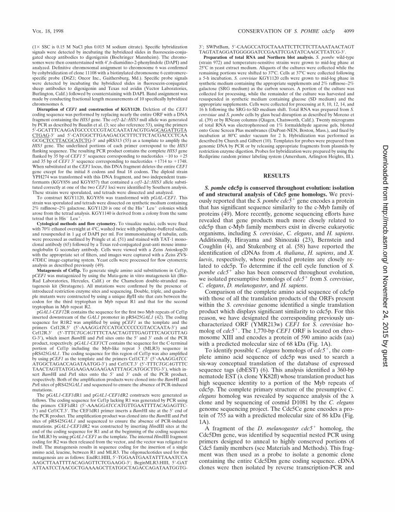
FIG. 1. Isolation of homologs of S. pombe cdc51. (A) Alignment of S. pombe cdc5p homologs by using ClustalW1.6 (14, 60). Residues found to be identical orsimilar to those of Cdc5Hs are highlighted in black or grey, respectively, to emphasize the evolutionary conservation of Cdc5 homologs. Roman numerals signify thebeginning of a Myb repeat. Asterisks indicate the positions of the amino acid residues which are classically occupied by tryptophan residues in Myb repeats. Percentidentities among the various Cdc5 family members are indicated at the bottom of this panel. (B) Alignment of MLR3 of Cdc5 family members with R3 of human c-,A-, and B-Myb and Drosophila Myb. Alignment was performed by using ClustalW1.6. Identical and similar residues found within the various proteins are highlightedin black and grey, respectively. Residues found to be identical between all proteins are indicated by arrows. Sp, S. pombe; Sc, S. cerevisiae; Hs, H. sapiens; Dm, D.melanogaster. (C) Phylogenetic analysis of Cdc5 and c-Myb family members.
4100
on Novem
ber 24, 2015 by guesthttp://m
cb.asm.org/
Dow
nloaded from

sequenced. The Cdc5Dm protein is 814 aa in length and has apredicted molecular size of 93 kDa (Fig. 1A).
By using a 185-bp human EST (Consortium CloneID74654), a full-length cDNA clone encoding the putative humanhomolog of cdc5p, Cdc5Hs, was found to contain a 2,406-bpORF capable of coding for an 802-aa protein with a predictedmolecular size of 93 kDa (Fig. 1A). The predicted amino acidsequence of the Cdc5Hs gene is identical to that of PCDC5RPreported by Bernstein and Coughlin (4). In agreement with theresults obtained by Bernstein and Coughlin (4), we have de-tected a Cdc5Hs gene transcript in a variety of human tissuesand cell lines of ;3.4 kb, which is consistent with the size of theisolated full-length cDNA clones (data not shown). To deter-mine the chromosomal location of the Cdc5Hs gene, a humangenomic PAC clone spanning the Cdc5Hs gene locus was iso-lated and used for fluorescence in situ hybridization (see Ma-terials and Methods). This analysis demonstrated that theCdc5Hs gene is localized to chromosome 6p21 (data notshown).
Comparison of the amino acid sequences of Cdc5 familymembers has revealed that they are structurally similar to eachother throughout their entire lengths (see bottom of Fig. 1Afor percent identities among the various Cdc5 family mem-bers). The first ;100 aa of Cdc5 relatives, which encompasstwo complete Myb repeats, resemble classical Myb repeats inthat each repeat contains three tryptophan residues that areseparated by an interval of 18 or 19 aa (reviewed in reference32). The single exception to this is that the third position in thesecond repeat (R2) of Cdc5 family members is occupied by atyrosine residue (Fig. 1A). The presence of hydrophobic resi-dues at these positions is essential for the DNA binding andtranscriptional activation properties of c-Myb (27, 54). Muta-tional and modeling studies of c-Myb predicted that the Mybrepeat forms a structure related to a helix-turn-helix motif inwhich each tryptophan residue forms the hydrophobic back-bone of a helix (15, 17). This prediction has been confirmedthrough nuclear magnetic resonance solution spectroscopy,and these studies have further demonstrated that the minimalMyb DBD (R2R3) contacts DNA through the third helix ofeach imperfect repeat (47, 48). Mutational studies and analysisof the oncogenic avian myeloblastosis virus product, v-Myb,have demonstrated that alteration of a number of other resi-dues within the DBD of c-Myb results in loss of DNA bindingand transcriptional activation capabilities (7, 15, 17, 19, 22, 24,43). A number of these residues are conserved between c-Myband Cdc5 family members (49) (Fig. 1A).
Cdc5 family members contain a ;60-aa Myb-like repeatlocated immediately adjacent to the second repeat (Fig. 1Aand B). At the amino acid level, the third Cdc5 Myb-like repeat(labeled MLR3 in Fig. 1A) is significantly divergent from thatfound in c-Myb, although certain residues are conservedamong all c-Myb and Cdc5 family members analyzed (Fig. 1B).Interestingly, MLR3 lacks the hydrophobic residues whichform the helical backbones in a Myb repeat. As observed byHirayama and Shinozaki (23) and Bernstein and Coughlin (4)regarding AtCDC5, human Cdc5, and S. pombe Cdc5p, thecentral third of Cdc5 family members harbors a significantnumber of potential phosphorylation sites for proline-directedkinases (S/TPXK/R or S/TPK/R), which suggests that Cdc5activity may be regulated by phosphorylation in vivo. Consis-tent with this notion is the finding that X. laevis Cdc5 is phos-phorylated by mitotic egg extracts and cyclin B-Cdc2 (58).Sequence similarity among Cdc5 family members decreases inthe C-terminal third of the sequence, where significant identityis observed primarily among the C. elegans, D. melanogaster, A.thaliana, and H. sapiens proteins.
The temperature-sensitive growth defect of the S. pombecdc5-120 mutant is complemented by both human and fly Cdc5cDNAs. To determine if CEF1 and the Cdc5Ce, Cdc5Dm, andCdc5Hs cDNAs were capable of rescuing the temperature-sensitive lethality of the S. pombe cdc5-120 mutant (46, 49), weintroduced these cDNAs into cdc5-120 cells under control ofthe thiamine-repressible nmt11 promoter (37). Full-lengthcDNAs for CEF1 and the Cdc5Ce and Cdc5Hs cDNAs wereincapable of rescuing growth of cdc5-120 cells at 36°C in boththe presence and absence of thiamine (Fig. 2). In contrast, thefull-length Cdc5Dm cDNA was capable of rescuing growth ofcdc5-120 cells when induced (in the absence of thiamine) butnot when repressed (in the presence of thiamine) (Fig. 2).Microscopic examination of cdc5-120 cells overexpressingCEF1 and the Cdc5Hs cDNA revealed that many of them wereelongated at 25°C (data not shown). Overexpression of full-length CEF1 and the Cdc5Hs cDNA had the same phenotypicconsequence in wild-type cells (data not shown).
We have previously shown that the Myb repeats of S. pombecdc5p are sufficient to rescue the temperature-sensitive lethal-ity of cdc5-120 (49). Since overproduction of full-length Cef1pand Cdc5Hs proteins appeared to be toxic in S. pombe, 39-truncated versions of both CEF1 and Cdc5Hs cDNA ORFswere constructed and placed under control of the nmt11 pro-moter. 39-Truncated versions of both the Cdc5Dm and Cdc5CecDNAs placed under control of nmt11 were also constructed.The ability of these deletion mutants to rescue growth of cdc5-120 cells at 36°C was then tested. As shown in Fig. 2, C-terminal truncations of 441 aa (DEcoRI) or 196 aa (DAcc651)in Cdc5Hs were able to complement the temperature-sensitivegrowth defect of cdc5-120 cells in the absence of thiamine butnot in the presence of thiamine. Cdc5Dm truncated by 293 aaat the C terminus was also capable of complementing thetemperature-sensitive growth defect of cdc5-120 in the absenceof thiamine but not in the presence of thiamine. A CEF1 ORFcoding for a protein truncated by 150 aa at the C terminus,which did not cause elongation of S. pombe cells in the absenceof thiamine (data not shown), was unable to restore growth ofcdc5-120 cells at 36°C (Fig. 2). Cdc5Ce truncated by 337 aa wasalso unable to rescue growth of cdc5-120 cells at 36°C (Fig. 2).The inability of Cef1p and Cdc5Ce to restore growth of cdc5-120 cells at 36°C could be due to inappropriate levels of pro-tein expression and/or insufficient structural similarity to S.pombe cdc5p. The ability of the Cdc5Dm cDNA and truncatedversions of the Cdc5Hs cDNA to rescue growth of cdc5-120
FIG. 1—Continued.
VOL. 18, 1998 CONSERVATION OF S. POMBE cdc5p 4101
on Novem
ber 24, 2015 by guesthttp://m
cb.asm.org/
Dow
nloaded from

cells at 36°C, however, clearly establishes these genes as func-tional homologs of S. pombe cdc51.
CEF1 is essential during G2/M. To help determine the roleof CEF1 in budding yeast, a complete deletion of one genomiccopy of the gene was created in a diploid strain (see Materialsand Methods). Precise replacement of one of the copies ofCEF1 by a CEF1::HIS3 disruption fragment was confirmed bySouthern blot analysis and PCR (data not shown). Two inde-pendent cef1-D1::HIS3/CEF1 heterozygotes were sporulated,and tetrads were dissected. In each case, only two His2 colo-nies grew, demonstrating that CEF1 is essential for viability inS. cerevisiae. His1 haploid colonies could be recovered follow-ing sporulation of the cef1-D1::HIS3/CEF1 diploid heterozy-gote if a plasmid carrying the CEF1 gene was introduced intothe strain prior to sporulation. Neither the human Cdc5 cDNAnor S. pombe cdc51 was found to be capable of complementingthe lethality of S. cerevisiae cef1-D1::HIS3 cells. Microscopicobservation of cef1-D1::HIS3 cells in dissected tetrads revealedthat they underwent one or two rounds of cell division and thenexperienced growth arrest as large budded cells (data notshown).
To analyze more carefully the phenotype of cells lackingCEF1 function, a strain in which CEF1 expression could beconditionally inactivated was constructed (see Materials andMethods). In this strain, KGY1120, CEF1 expression wasdriven by the galactose-inducible GAL1 promoter. Analysis ofthe KGY1120 strain and an otherwise isogenic wild-type CEF1strain (KGY1140) in liquid SRG medium revealed no differ-ences in growth rate. Both strains grew with a doubling time ofapproximately 3.5 h at 32°C (Fig. 3B and C). However, asexpected, KGY1120 cells failed to grow on solid medium con-taining glucose (Fig. 3A) and, upon transfer to liquid SD me-dium, underwent growth arrest 10 h following the SRG-to-SDmedium shift (Fig. 3C).
To determine if growth arrest of KGY1120 cells in SD me-dium occurred during a specific stage of the cell cycle, we usedflow cytometric analysis to determine the DNA content ofKGY1120 cells at 2-h intervals following the SRG-to-SD me-dium shift. By 6 h following the shift, KGY1120 cells began toexhibit a noticeably higher percentage of cells which contained2N DNA (49%) than did isogenic wild-type KGY1140 cells(34%) (Fig. 3D). By 10 h following the SRG-to-SD mediumshift, greater than 80% of KGY1120 cells exhibited a 2N con-tent of DNA whereas KGY1140 cells displayed no change inDNA content profile. The percentage of KGY1120 cells with a2N content of DNA did not change appreciably between 10and 16 h following the SRG-to-SD medium shift (Fig. 3D).
The cell and nuclear morphologies of growth-arrestedKGY1120 cells were analyzed by phase-contrast microscopyand staining with the DNA-binding dye DAPI. After 8 h inglucose-containing medium, KGY1120 cells began to accumu-late as round budded cells (Table 2). This accumulation wasmore apparent after a 12-h incubation in SD medium whengreater than 80% of KGY1120 cells were round budded,whereas only 35% of KGY1140 cells were budded in SD me-dium (Table 2). An unusual phenotype was present in nearly25% of budded, growth-arrested KGY1120 cells. These cellscontained a single nucleus present at either the end of themother cell most distal to the mother-bud neck or immediatelyadjacent to the cell wall offset from the mother-bud neck (Fig.3E and Table 2). In many cases, these nuclei were compressedagainst the cell wall and thus appeared to be crescent shaped.These abnormal nuclear morphologies were never observed inthe isogenic KGY1140 strain.
Arrested KGY1120 cells were stained with the antitubulinmonoclonal antibody TAT-1 (65) to view microtubule struc-tures. As shown in Fig. 3, KGY1120 cells cultivated in SDmedium contained short intranuclear spindles. A portion of
FIG. 2. The temperature-sensitive growth defect of cdc5-120 is complemented by human and fly Cdc5 proteins. Full-length cDNAs for CEF1 and the Cdc5Ce,Cdc5Dm, and Cdc5Hs cDNAs were placed under control of the nmt11 promoter in pREP3X. In addition, sequences for products of these cDNAs were truncated attheir C termini by 129 aa (CEF1dCT), 337 aa (Cdc5Ce dCT), 293 aa (Cdc5Dm dCT), 441 (DEcoRI), and 196 (DAcc651) aa (Cdc5Hs) and placed under control of thenmt11 promoter in pREP3X. S. pombe cdc51 is pIRT2 (39) harboring full-length cdc51 cDNA under control of 585 bp of the cdc51 promoter sequence.pREP81cdc5Sp is pREP81 (2) harboring full-length cdc51 cDNA. KGY360 transformants carrying Cdc5Hs constructs were struck to minimal medium with or withoutthiamine and incubated for 18 h at 25°C to induce or repress expression of the inserts, respectively, and then shifted to 36°C for 3 days. KGY360 transformants carryingCEF1, Cdc5Ce cDNA, and Cdc5Dm cDNA constructs were struck to minimal medium with or without thiamine at 36°C and grown for 3.5 days.
4102 OHI ET AL. MOL. CELL. BIOL.
on Novem
ber 24, 2015 by guesthttp://m
cb.asm.org/
Dow
nloaded from

arrested KGY1120 cells appeared to contain a single spindlepole body “plaque” (Fig. 3E), which most likely representsduplicated spindle pole bodies that had not separated to adegree detectable by fluorescence microscopy. Collectively, thecytological and fluorescence-activated cell sorter analyses ofKGY1120 cells indicated that CEF1 activity is required pre-dominantly during G2/M in S. cerevisiae.
Mutational analysis of Cef1p. The importance of the twoMyb repeats and MLR3 of Cdc5 family members to theirfunction is predicted by the high degree of sequence identityamong them (Fig. 1A). As expected, Myb repeats R1 and R2of Cef1p are essential for its function in vivo; when expressedunder control of the GAL1 promoter, a truncated version ofCef1p lacking the R1 and R2 Myb repeats (pGAL1-CEF1CT)is incapable of rescuing the growth of the cef1-D1::HIS3 mu-tant (Fig. 4). Furthermore, the mutation responsible for caus-ing the temperature sensitivity of the S. pombe cdc5-120 mu-
tant was sequenced and found to be present in the first Mybrepeat (R1). In cdc5-120p, the second tryptophan of R1 (W29)is converted to an arginine residue as a result of a T:A to C:Gmutation at the corresponding codon (TGG to CGG). Al-though these results underscore the importance of the Cef1pand cdc5p Myb repeats, a construct containing only R1 and R2of Cef1p under control of the GAL1 promoter (pGAL1-CEF12R) is incapable of rescuing the growth of thecef1-D1::HIS3 mutant, indicating that other regions of Cef1pare essential for function as well (Fig. 4).
As discussed above, Myb domains are comprised of threeregularly spaced hydrophobic residues, each of which formsthe backbone of a helix. Mutation of any one of the six tryp-tophan residues in c-Myb repeats R2 and R3, which constitutethe minimal DBD of c-Myb, to alanine or glycine results in lossof DNA binding and transcriptional activation (27, 54). Fur-thermore, in R1 of Cdc5 family members and R2 of c-Myb, a
FIG. 3. Deletion of CEF1 blocks cell growth at G2/M. (A) KGY1120 fails togrow on medium containing glucose as a carbon source. KGY1120 and theisogenic wild-type strain, KGY1140, were struck onto solid medium containingeither 2% glucose or 2% raffinose–2% galactose as a carbon source(s) andincubated at 30°C for 4 days. (B and C) Growth of KGY1140 (B) or KGY1120(C) cells in liquid SD medium or SRG medium is shown. KGY1140 or KGY1120cells were grown to mid-log phase in SRG medium at 32°C and then shifted toSD medium (■), or maintained in SRG ({). The medium shift was performed attime (t) 0 h. (D) Cells from experiments shown in panels B and C were collectedevery 2 h, stained with propidium iodide, and subjected to flow cytometricanalysis as described in Materials and Methods. Linear fluorescence histogramsshow relative DNA content in arbitrary units on the horizontal axis and cellnumber on the vertical axis. Arrows indicate the positions of 1N and 2N DNApeaks. (E) Growth-arrested KGY1120 cells contain short intranuclear spindles.Microtubules were detected by indirect immunofluorescence using the a-tubulinmonoclonal antibody TAT-1 (65) followed by staining with Texas red-conjugatedsecondary antibodies. DNA was visualized by using DAPI. Arrowheads indicatecells that contain a single tubulin plaque instead of a short intranuclear spindle.Raf/Gal, medium containing 2% raffinose–2% galactose; Glu, medium contain-ing 2% glucose.
VOL. 18, 1998 CONSERVATION OF S. POMBE cdc5p 4103
on Novem
ber 24, 2015 by guesthttp://m
cb.asm.org/
Dow
nloaded from

basic residue is present at position 26 relative to the thirdtryptophan. Substitution of this lysine with an asparagine inc-Myb results in loss of DNA binding (15), and mutationalanalysis of this residue in Petunia MYB.Ph3 indicates that thisresidue plays a role in determining sequence-specific bindingof MYB.Ph3 to DNA (56). Finally, Cdc5 family members con-tain a cysteine residue at position 24 relative to the thirdtryptophan in both R1 and R2 (Fig. 1A). Cef1p is exceptional
in this case because it contains a serine in place of a cysteine inR1. In c-Myb, replacement of a cysteine residue in a homolo-gous position in R2 with a serine results in a dramatic reduc-tion of DNA binding in vitro (20, 43), raising the possibilitythat modulation of the oxidation state of this cysteine may bea way of regulating the DNA-binding activity of c-Myb.
To analyze the in vivo consequences of altering these keyresidues within the Cef1p Myb repeats which are conservedamong Cdc5 family members and c-Myb, we generated muta-tions within the Myb repeats of Cef1p by site-directed mu-tagenesis (see Materials and Methods) and tested the ability ofthese mutants to restore growth of cells which lack CEF1. Allof the single mutations that were constructed result in noncon-servative amino acid substitutions and are located in positionsthat are important for maintaining structure of the second(W33R, W33G, W84G) and third (R46N, W52A, W52G,C98S, Y102A, Y102G) helices in the corresponding Myb re-peats (Fig. 4) (15, 17, 27, 43). Various double, triple, andquadruple mutations which are predicted to disrupt conforma-tion of the second and third helices in both of the canonicalMyb repeats of Cef1p were also constructed.
As shown in Fig. 4, all of the single mutants were found to befully capable of rescuing the growth of the cef1-D1::HIS3 mu-tant, demonstrating that the substitutions in these mutants donot abolish Cef1p function in vivo. Interestingly, reconstruc-tion of the cdc5-120 mutation in CEF1 (cef1-120) does notproduce a temperature-sensitive phenotype in S. cerevisiae. Allof the double-mutant combinations were also able to rescuegrowth of cells lacking CEF1. Notably, however, the W33GW84G and W52G W84G mutants were not able to rescue thegrowth of cef1-D1::HIS3 cells as well as the wild-type CEF1protein at 30°C. Furthermore, cef1-D1::HIS3 cells harboringthese double mutants were temperature sensitive (Fig. 4) andunderwent growth arrest following two to four cell divisions at36°C as large-budded cells (data not shown). Of the four triplemutants that were analyzed, only two, W33G W52G Y102Gand W52G W84G Y102G, rescued the growth of thecef1-D1::HIS3 mutant, and the latter conferred only poorgrowth to cells lacking the CEF1 gene. Similar to the W33GW84G and W52G W84G mutants, these triple mutants wereincapable of restoring growth to cells lacking CEF1 at 36°C.The W33G W52G W84G Y102G quadruple mutant was un-able to rescue growth of the cef1-D1::HIS3 mutant.
The ability of the W33G W52G and W84G Y102G doublemutants, which are predicted to disrupt the second and thirdhelices in R1 and R2, respectively, to rescue growth of cells
FIG. 4. Mutational analysis of Cef1p. Amino acids predicted to be critical formaintaining the structure of the Myb repeats of Cef1p were replaced with theindicated amino acid(s) as described in Materials and Methods. Substitutionmutations were constructed in pCEF1, which is pRS415 (9) harboring a 2.5-kbgenomic DNA fragment encompassing the CEF1 coding region. In addition toperforming site-directed mutagenesis of the Cef1p gene, various domains ofCef1p were removed. pGAL1-CEF12R, Cef1p R1R2; pGAL1-CEF1CT, Cef1pCT including MLR3; pGAL1-CEF1dR1, Cef1p lacking R1; pGAL1-CEF1dR2,Cef1p lacking R2. These mutants were assayed for their abilities to rescue growthof the cef1-D1::HIS3 mutant by tetrad analysis and/or plasmid shuffle. Plasmidshuffle with the pRS425GAL1-based plasmids was performed on 5-fluorooroticacid medium containing raffinose and galactose as the carbon sources. 11,rescue or growth comparable to that of wild-type CEF1; 1, reduced rescue ofgrowth and colony formation; 1/2, poor rescue of growth; 2, failure to rescuegrowth of the cef1-D1::HIS3 mutant or no growth; NA, not applicable.
TABLE 2. Quantitation of cell and nuclear morphologies of cells lacking CEF1 activitya
Strain Medium Time (h)
% of total cells scored with indicated morphologyb
nc
KGY1140 Raf/Gal 68 14 6 11 500Glucose 65 10 10 15 500
KGY1120 Glucose 8 39 11 18 10 22 521Glucose 10 31 15 24 9 19 1 1 513Glucose 12 18 21 21 9 25 2 4 648Glucose 14 19 29 18 10 20 1 3 525Glucose 16 15 28 18 10 23 1 5 523
a KGY1120 or the isogenic wild-type strain KGY1140 (Table 2) was grown to the logarithmic phase at 32°C in synthetic complete medium containing 2%raffinose–2% galactose (SRG medium) as the carbon source and then shifted into synthetic complete medium containing 2% glucose (SD medium) (Glucose) ormaintained in SRG medium (Raf/Gal). The nuclear and cellular morphologies of the two strains were scored at the indicated time points.
b The criteria used for each morphologic class scored are shown schematically.c n 5 total cells scored.
4104 OHI ET AL. MOL. CELL. BIOL.
on Novem
ber 24, 2015 by guesthttp://m
cb.asm.org/
Dow
nloaded from

lacking CEF1 raised the issue of whether R1 and R2 arefunctionally redundant in vivo. We tested this directly by indi-vidually removing R1 (pGAL1-CEF1dR1) and R2 (pGAL1-CEF1dR2) from the pGAL1-CEF1 construct. Neither of thedeletion mutants was capable of rescuing the growth of thecef1-D1::HIS3 mutant, demonstrating that the Cef1p Myb re-peats are not functionally redundant. Collectively, these resultsindicate that the Myb repeats of Cef1p perform an essentialfunction and that structural alteration of the Myb repeats iscapable of eliciting a temperature-sensitive phenotype or abol-ishing function of Cef1p in vivo.
CEF1 and S. pombe cdc51 are not required for expression ofgenes which are known to be transcriptionally up-regulatedduring G2/M. The G2/M and G2 growth arrest phenotypes of S.cerevisiae cells lacking CEF1 activity and S. pombe cdc5-120,respectively, and the similarity of Cdc5 family members toc-Myb prompted us to examine a potential involvement ofthese proteins in the transcriptional activation of genes knownto be up-regulated during G2/M. In S. cerevisiae, at least sixgenes show a G2/M-specific pattern of transcription: SWI5,CLB1, CLB2, ACE2, CDC5, and ASE1 (1, 26, 35). The first fiveof these genes, and very likely the sixth as well, require theactivity of the MADS-box transcription factor encoded byMCM1 and an uncloned protein termed Sff (1, 26, 35). To test
whether Cef1p was required for transcriptional activation ofthese genes, total RNA was prepared from cells depleted ofCEF1 activity and subjected to Northern blot analysis usingportions of CLB1 and SWI5 ORFs as probes. As shown in Fig.5A, CLB1 and SWI5 transcripts persist in cells depleted ofCEF1 activity, suggesting that CEF1 is not required for expres-sion of these genes.
In S. pombe, cdc13p and cdc25p are required during G2 forentry into mitosis. The levels of these proteins oscillatethroughout the cell cycle, with maximal amounts present dur-ing G2 and mitosis. The amount of cdc251 transcript in the cellmirrors cdc25p levels, indicating that transcriptional activationof cdc251 is an important aspect of its regulation (40). cdc13pprotein levels are primarily determined at the posttranslationallevel (12, 66). To determine if a functional cdc51 product wasrequired for transcription of cdc131 or cdc251, total RNA wasisolated from wild-type, cdc5-120, and cdc25-22 cells grown ateither 25 or 37°C for 5 h and subjected to Northern analysisusing portions of cdc131 and cdc251 as probes. The cdc25-22strain, which, like cdc5-120, blocks during G2, was used as acontrol to identify any nonspecific changes in transcript levelsinduced by a G2 arrest. As shown in Fig. 5B, cdc251 andcdc131 transcript levels are not significantly altered in thecdc5-120 mutant grown at 37°C.
DISCUSSION
In this study, we have reported the identification and char-acterization of cDNAs and genes from S. cerevisiae, C. elegans,D. melanogaster, and H. sapiens that encode proteins closelyrelated to S. pombe cdc5p. Bernstein and Coughlin (4) haveindependently reported the isolation of human Cdc5(PCDC5RP). In addition, Hirayama and Shinozaki (23) havereported the isolation and characterization of AtCDC5, a genewhich encodes the A. thaliana homolog of fission yeast cdc5p.Stukenberg et al. (58) have identified an X. laevis relative, andgenome sequencing efforts have revealed the existence of aCdc5-related protein in mice (for an example, see the ESTunder GenBank accession no. AA269568). When subjected tophylogenetic analysis, all of the analyzed Cdc5 family membersdisplay significantly greater similarity to S. pombe cdc5p thanto any other Myb-related protein (Fig. 1C). cdc5p is thus amember of a family of Myb-related proteins that has beenhighly conserved throughout the eukaryotic lineage. In S. cer-evisiae, only one gene, CEF1, encodes a protein that bears anysignificant similarity to fission yeast cdc5p. This also appears tobe the case in complex metazoan systems, since all of about 20Cdc5Hs-related human sequences in dbEST appear to repre-sent overlapping segments of a single gene which we havemapped to chromosome 6p21. Since mammalian Cdc5 activityis likely encoded by a single genetic locus, it is possible thatinappropriate modulation of Cdc5 activity may be associatedwith the onset or progression of tumorigenesis. Although noobvious previously described cancer or disease genes havebeen mapped to this locus, knowledge of its chromosomallocation provides an opportunity to investigate such a possibil-ity directly.
Several lines of evidence suggest that the cell cycle functionof S. pombe cdc51 has also been conserved throughout theeukaryotic lineage. Like S. pombe cdc51, S. cerevisiae CEF1 isessential for viability. Haploid cells lacking a functional CEF1gene undergo growth arrest with a 2N DNA content and shortintranuclear spindles, indicating that they are arrested duringG2/M. Interestingly, a noticeable population of cells lackingCEF1 activity display abnormal nuclear morphologies, indicat-ing that Cef1p may be important for maintenance of nuclear
FIG. 5. CEF1 and cdc51 are not required for transcriptional activation ofgenes known to be transcriptionally up-regulated during G2/M. (A) CEF1 activityis not required for transcription of SWI5 and CLB1. KGY1120 cells were grownunder permissive (SRG medium) or restrictive (SD medium) nutritional condi-tions as described in the legend to Fig. 3 and Materials and Methods. Total RNAwas isolated from cells at 8, 10, 12, 14, and 16 h and subjected to Northern blotanalysis using portions of ACT1, SWI5 and CLB1 ORFs as probes. ACT1 tran-script levels served as a loading control. WT, wild type; RG, synthetic mediumcontaining 2% galactose and 2% raffinose as carbon sources. (B) cdc5-120 is notdefective for transcription of cdc131 and cdc251. Logarithmically growing wild-type (strain 972) or temperature-sensitive (ts) strains cdc25-22 or cdc5-120 werecollected during growth at 25°C or after incubation at 37°C for 5 h. Total RNAwas prepared from these cells and subjected to Northern blot analysis usingportions of ura41, cdc131, and cdc251 ORFs as probes. ura41 transcript levelsserved as a loading control.
VOL. 18, 1998 CONSERVATION OF S. POMBE cdc5p 4105
on Novem
ber 24, 2015 by guesthttp://m
cb.asm.org/
Dow
nloaded from

integrity prior to or during the process of nuclear division.Similarly, in S. pombe, approximately 30% of fission yeast cellslacking the cdc51 gene also display aberrant nuclear morphol-ogies (49). Despite the sequence identity possessed by Cef1pand cdc5p, CEF1 is unable to complement the temperature-sensitive growth defect of S. pombe cdc5-120 at its restrictivetemperature of 36°C. Similar to AtCDC5 (23), H. sapiens and S.cerevisiae Cdc5 cDNAs are unable to rescue growth of S.pombe cells harboring a deletion of cdc51. Human and fissionyeast Cdc5 proteins were also observed to be incapable ofrescuing the lethality of S. cerevisiae cef1D cells. The inability ofvarious Cdc5 family members to rescue growth of cef1D orcdc5D cells is not surprising given that the C termini of Cdc5family members are less well conserved than the putativeDBDs and that the C termini of S. pombe cdc5p and S. cerevi-siae Cef1p have indispensable functions (49, 49a) (Fig. 4).Full-length Cdc5Dm and partially truncated human Cdc5,however, were capable of rescuing growth of cdc5-120 cells at36°C, thus demonstrating that Cdc5Hs and Cdc5Dm are func-tional homologs of fission yeast cdc5p. The inability of theCdc5Ce cDNA and S. cerevisiae CEF1 to rescue growth ofcdc5-120 is most simply explained by a limited degree of se-quence identity possessed by the Myb repeats of S. pombecdc5p, Cdc5Dm, and Cdc5Hs (Fig. 1A). Recently, Bernsteinand Coughlin (4) have reported that an epitope-tagged versionof human Cdc5 translocates from the cytoplasm to the nucleiof serum-deprived cultured mammalian cells upon stimulationwith serum. Based on these results, the authors speculated thatCdc5 may function in a mitogen-activated signaling pathwayduring the G0/G1 transition. Although a definitive role(s) forCdc5 in the mammalian cell cycle requires further analysis, ourdata indicate that an essential, conserved function of Cdc5 inthe eukaryotic cell cycle is at the G2/M transition.
In S. cerevisiae, the Mcm1p transcription factor, in combi-nation with the uncloned SWI5 factor (Sff), is required duringG2/M to activate transcription of at least six genes which arenecessary for entry into and completion of mitosis: CLB1,CLB2, ACE2, SWI5, CDC5, and ASE1 (1, 26, 35). Sff is abso-lutely required for transcription of SWI5 because mutation ofthe Sff binding site results in the loss of SWI5 upstream acti-vating sequence activity (35). S. cerevisiae cells lacking MCM1activity undergo growth arrest with replicated DNA and elon-gated buds (1), a phenotype commonly associated with S. cer-evisiae mutants which possess low Clb-associated Cdc28p(Cdk1) kinase activity (31, 52, 59). Cells lacking CEF1 undergoarrest with a round-budded phenotype, which indicates thepresence of Clb-associated Cdk1 activity (31). The phenotypicdifference between cells lacking MCM1 and CEF1 activitiesindicates that CEF1 is unlikely to encode Sff. We have con-firmed this hypothesis by demonstrating that cells depleted ofCEF1 activity are capable of accumulating transcripts ofMcm1p and Sff target genes.
In S. pombe, cdc13p and cdc25p levels oscillate in a cellcycle-dependent manner, with maximal levels observed nearthe G2/M transition (12, 40). cdc251 transcript is most abun-dant near the G2/M transition, indicating that accumulation ofcdc25p during G2 is achieved, at least in part, through itstranscriptional activation (40). The cdc5-120 mutant is notdefective for transcription of these mitotic regulators. We con-clude from these results and those obtained from Cef1p-de-pleted cells that Cdc5 does not regulate the G2/M transition ineukaryotic cells at the level of transcription of these knownmitotic regulators.
Although cdc5p and Cef1p do not appear to functionthrough activating transcription of genes known to be up-reg-ulated during G2/M, it is still tempting to assign these proteins
a role as DNA-binding proteins for the following reasons: (i)all Cdc5 family members analyzed thus far contain two highlyconserved Myb repeats, (ii) the S. pombe cdc5-120 mutationconverts the second tryptophan in R1 of cdc5p to an arginineresidue, and (iii) the Myb repeats of AtCDC5 have been shownto preferentially bind a specific nucleotide sequence (23). Forthese reasons, we have tried to identify a preferential DNA-binding sequence for S. pombe cdc5p and S. cerevisiae Cef1p.Specifically, we employed the cyclic amplification and selectionof target sequence (CASTing) approach with the binding (23,62) and washing (16) conditions described previously. TheN-terminal 201 amino acids of Cef1p, which contain R1, R2,and MLR3, were fused downstream of glutathione S-trans-ferase (GST), and this fusion protein was used for CASTing.An oligonucleotide library containing a core of 20 randomnucleotides was used as a probe. A similar approach was usedto identify a target sequence of AtCDC5 (23). While it is clearthat the Myb repeats of Cef1p and cdc5p have the ability tobind DNA, we have not been able to select a high-affinitybinding sequence for these proteins although under the sameconditions a fusion protein between GST and the DBD ofAML was able to select the sequence TGT/cGGT, the knownhigh-affinity target sequence of AML (38, 49, 49b). In a sepa-rate experiment, a hexahistidine-tagged version of the DBD ofCdc5Dm was used in an electrophoretic mobility shift assaywith the AtCDC5 binding sequence as a probe. We reasonedthat since the DBDs of these proteins have 80% identity, theymay be capable of binding the same sequence. While a modestbinding of Cdc5Dm to this probe was observed, the bindingwas abrogated in the presence of increasing amounts of non-specific competitor, indicating that this interaction occurs withlow affinity (37a). In this regard, it is noteworthy that AtCDC5binding to the sequence CTCAGCG was also reduced withnonspecific competitor DNA (23). While these data may arguethat Cdc5 does not bind a particular DNA sequence with highaffinity, we cannot exclude the possibility that additional re-gions of these proteins are required for sequence-specific DNAbinding or that they require additional factors to bind DNAwith high affinity.
In this study, we have analyzed the in vivo consequences ofaltering residues within the Cef1p Myb repeats which are pre-dicted to disturb the conformation of these domains. Of par-ticular relevance to this work, mutational studies of c-Mybhave demonstrated that hydrophobic residues forming thebackbones of the DBD helices are essential for the DNA-binding activity of c-Myb in vitro and its trans-activation activ-ity in reporter assays (27, 54). Surprisingly, we found that theCef1p Myb repeats are quite resiliant to similar changes; sin-gle, double, or even certain triple mutant combinations inresidues which form the hydrophobic backbones of the secondor third helices in both R1 and R2 do not abolish Cef1pfunction in vivo, although many produce a temperature-sensi-tive growth defect. Although these results are unexpected, it isimportant to bear in mind that our assay determined the func-tional effects of altering helical backbone residues in vivo andis therefore not directly comparable to the DNA binding, tran-scriptional activation, and differentiation assays that have tra-ditionally been used in mutational analyses of c-Myb. To ourknowledge, systematic mutational analyses of Myb domains inother proteins, such as those presented here, have yet to bereported. Thus, it is possible that interaction of Cef1p withother proteins in vivo may stabilize the conformation of mutantMyb repeats, thus allowing Cef1p to bind DNA.
An intriguing, equally plausible interpretation is that in vivo,Cef1p may cooperate with a second DNA-binding proteinwhose activity compensates for a diminution of Cef1p DNA-
4106 OHI ET AL. MOL. CELL. BIOL.
on Novem
ber 24, 2015 by guesthttp://m
cb.asm.org/
Dow
nloaded from

binding activity. Evidence does exist for Myb-related proteinsfunctioning in heterodimeric complexes. c-Myb itself cooper-ates with heat shock factor 3 (HSF3) to activate transcriptionfrom the hsp70 promoter (28, 29). In S. cerevisiae and Zeamays, the Myb-related proteins Bas1p and C1, respectively, arethought to act in concert with a second DNA-binding proteinto activate transcription of target genes; Bas1p cooperates withthe homeobox protein Bas2p, and C1 is thought to dimerizewith basic helix-loop-helix proteins such as B, R, SN, and LC(13, 18, 36, 61). In the cases of C1 and c-Myb, heterodimer-ization has been shown to require the Myb domains (18, 28).The in vivo activity of Cef1p clearly requires both R1 and R2.Perhaps, as in the case of C1, R1R2 of Cef1p functions as botha protein-protein interaction domain and a DBD. Evidencealso exists for heterodimeric transcription factor complexesthat require only one active DNA-binding subunit. For exam-ple, mutational studies have demonstrated that elimination ofthe sequence-specific DNA-binding activity of the yeast home-odomain protein a-2 does not destroy its ability to bind DNAin concert with a1 (64). Additionally, c-Myb, in concert withHSF3, stimulates transcription from the hsp70 promoterthrough the heat shock element without apparently binding tothis sequence (28, 29).
Although the precise function of Cdc5 family members is notunderstood presently, it is clear that the product of the cdc51
gene in S. pombe and its homolog in S. cerevisiae are requiredduring G2/M. At present, all available evidence indicates a rolefor this protein family in DNA binding. However, it is quitepossible that Cdc5 family members do not function as tran-scriptional activators. Indeed, there is precedence for Myb-related proteins which play roles in biological processes otherthan activation of transcription. For example, human TRF1and TRF2 and fission yeast taz1p are Myb-related proteins thatnegatively regulate telomere length (5, 8, 11, 63) and the Myb-related budding yeast protein Reb1p functions in transcrip-tional termination as well as transcriptional activation of rRNAgenes (30). An approach utilizing a combination of genetic,biochemical, and cytological analyses of the isolated Cdc5 fam-ily members should lead to an understanding of how Cdc5facilitates the G2/M transition in eukaryotic cells.
ACKNOWLEDGMENTS
We thank members of the laboratories of A. P. Weil and J. Flick foryeast strains and expression plasmids and T. O. Daniel and E. Stein forthe human microvascular endothelial cell line library and invaluabletechnical advice. S. Hiebert kindly provided the plasmid to produceGST-AML. We are grateful to J. Price for flow cytometric analyses. Allmembers of the Gould laboratory, including M. K. Balasubramanianand D. McCollum, are appreciated for valuable discussions and tech-nical advice.
This work was supported by NIH grant GM47728 to K.L.G. andUSPHS grants RO1 CA43592 and PO1 CA70404 to J.S.L. andCA71907 to A.T.L. S.M. was supported by USPHS grant NRSA 5T32CA09302. K.L.G. is an assistant investigator of the Howard HughesMedical Institute.
REFERENCES1. Althoefer, H., A. Schleiffer, K. Wassmann, A. Nordheim, and G. Ammerer.
1995. Mcm1 is required to coordinate G2-specific transcription in Saccha-romyces cerevisiae. Mol. Cell. Biol. 15:5917–5928.
2. Basi, G., E. Schmid, and K. Maundrell. 1993. TATA box mutations in theSchizosaccharomyces pombe nmt1 promoter affect transcription efficiencybut not the transcription start point or thiamine repressibility. Gene 123:131–136.
3. Baudin, A., O. Ozier-Kalogeropoulos, A. Denouel, F. Lacroute, and C. Cul-lin. 1993. A simple and efficient method for direct gene deletion in Saccha-romyces cerevisiae. Nucleic Acids Res. 21:3329–3330.
4. Bernstein, H. S., and S. R. Coughlin. 1997. Pombe Cdc5-related protein. Aputative human transcription factor implicated in mitogen-activated signal-ing. J. Biol. Chem. 272:5833–5837.
5. Bilaud, T., C. Brun, K. Ancelin, C. E. Koering, T. Laroche, and E. Gilson.1997. Telomeric localization of TRF2, a novel human telobox protein. Nat.Genet. 17:236–239.
6. Boguski, M. S., T. M. Lowe, and C. M. Tolstoshev. 1993. dbEST—databasefor “expressed sequence tags”. Nat. Genet. 4:332–333. (Letter.)
7. Brendeford, E. M., A. H. Myrset, A. B. Hegvold, M. Lundin, and O. S.Gabrielsen. 1997. Oncogenic point mutations induce altered conformation,redox sensitivity, and DNA binding in the minimal DNA binding domain ofavian myeloblastosis virus v-Myb. J. Biol. Chem. 272:4436–4443.
8. Broccoli, D., A. Smogorzewska, L. Chong, and T. de Lange. 1997. Humantelomeres contain two distinct Myb-related proteins, TRF1 and TRF2. Nat.Genet. 17:231–235.
9. Christianson, T. W., R. S. Sikorski, M. Dante, J. H. Shero, and P. Hieter.1992. Multifunctional yeast high-copy-number shuttle vectors. Gene 110:119–122.
10. Church, G. M., and W. Gilbert. 1984. Genomic sequencing. Proc. Natl. Acad.Sci. USA 81:1991–1995.
11. Cooper, J. P., E. R. Nimmo, R. C. Allshire, and T. R. Cech. 1997. Regulationof telomere length and function by a Myb-domain protein in fission yeast[see comments]. Nature 385:744–747.
12. Creanor, J., and J. M. Mitchison. 1996. The kinetics of the B cyclin p56cdc13and the phosphatase p80cdc25 during the cell cycle of the fission yeastSchizosaccharomyces pombe. J. Cell Sci. 109:1647–1653.
13. Daignan-Fornier, B., and G. R. Fink. 1992. Coregulation of purine andhistidine biosynthesis by the transcriptional activators BAS1 and BAS2. Proc.Natl. Acad. Sci. USA 89:6746–6750.
14. Feng, D. F., and R. F. Doolittle. 1990. Progressive alignment and phyloge-netic tree construction of protein sequences. Methods Enzymol. 183:375–387.
15. Frampton, J., T. J. Gibson, S. A. Ness, G. Doderlein, and T. Graf. 1991.Proposed structure for the DNA-binding domain of the Myb oncoproteinbased on model building and mutational analysis. Protein Eng. 4:891–901.
16. Funk, W. D., D. T. Pak, R. H. Karas, W. E. Wright, and J. W. Shay. 1992. Atranscriptionally active DNA-binding site for human p53 protein complexes.Mol. Cell. Biol. 12:2866–2871.
17. Gabrielsen, O. S., A. Sentenac, and P. Fromageot. 1991. Specific DNAbinding by c-Myb: evidence for a double helix-turn-helix-related motif. Sci-ence 253:1140–1143.
18. Goff, S. A., K. C. Cone, and V. L. Chandler. 1992. Functional analysis of thetranscriptional activator encoded by the maize B gene: evidence for a directfunctional interaction between two classes of regulatory proteins. GenesDev. 6:864–875.
19. Grasser, F. A., K. LaMontagne, L. Whittaker, S. Stohr, and J. S. Lipsick.1992. A highly conserved cysteine in the v-Myb DNA-binding domain isessential for transformation and transcriptional trans-activation. Oncogene7:1005–1009.
20. Guehmann, S., G. Vorbrueggen, F. Kalkbrenner, and K. Moelling. 1992.Reduction of a conserved Cys is essential for Myb DNA-binding. NucleicAcids Res. 20:2279–2286.
21. Guthrie, C., and G. R. Fink (ed.). 1991. Guide to yeast genetics and molec-ular biology, vol. 194. Academic Press, Inc., San Diego, Calif.
22. Hegvold, A. B., and O. S. Gabrielsen. 1996. The importance of the linkerconnecting the repeats of the c-Myb oncoprotein may be due to a positioningfunction. Nucleic Acids Res. 24:3990–3995.
23. Hirayama, T., and K. Shinozaki. 1996. A cdc51 homolog of a higher plant,Arabidopsis thaliana. Proc. Natl. Acad. Sci. USA 93:13371–13376.
24. Introna, M., J. Golay, J. Frampton, T. Nakano, S. A. Ness, and T. Graf. 1990.Mutations in v-myb alter the differentiation of myelomonocytic cells trans-formed by the oncogene. Cell 63:1289–1297.
25. Ito, H., Y. Fukuda, K. Murata, and A. Kimura. 1983. Transformation ofintact yeast cells treated with alkali cations. J. Bacteriol. 153:163–168.
26. Juang, Y. L., J. Huang, J. M. Peters, M. E. McLaughlin, C. Y. Tai, and D.Pellman. 1997. APC-mediated proteolysis of Ase1 and the morphogenesis ofthe mitotic spindle. Science 275:1311–1314.
27. Kanei-Ishii, C., A. Sarai, T. Sawazaki, H. Nakagoshi, D. N. He, K. Ogata, Y.Nishimura, and S. Ishii. 1990. The tryptophan cluster: a hypothetical struc-ture of the DNA-binding domain of the myb protooncogene product. J. Biol.Chem. 265:19990–19995.
28. Kanei-Ishii, C., J. Tanikawa, A. Nakai, R. I. Morimoto, and S. Ishii. 1997.Activation of heat shock transcription factor 3 by c-Myb in the absence ofcellular stress. Science 277:246–248.
29. Kanei-Ishii, C., T. Yasukawa, R. I. Morimoto, and S. Ishii. 1994. c-Myb-induced trans-activation mediated by heat shock elements without sequence-specific DNA binding of c-Myb. J. Biol. Chem. 269:15768–15775.
30. Lang, W. H., B. E. Morrow, Q. Ju, J. R. Warner, and R. H. Reeder. 1994. Amodel for transcription termination by RNA polymerase I. Cell 79:527–534.
31. Lew, D. J., and S. I. Reed. 1993. Morphogenesis in the yeast cell cycle:regulation by Cdc28 and cyclins. J. Cell Biol. 120:1305–1320.
32. Lipsick, J. S. 1996. One billion years of Myb. Oncogene 13:223–235.33. Lundgren, K., S. Allan, S. Urushiyama, T. Tani, Y. Ohshima, D. Frendewey,
and D. Beach. 1996. A connection between pre-mRNA splicing and the cellcycle in fission yeast: cdc281 is allelic with prp81 and encodes an RNA-
VOL. 18, 1998 CONSERVATION OF S. POMBE cdc5p 4107
on Novem
ber 24, 2015 by guesthttp://m
cb.asm.org/
Dow
nloaded from

dependent ATPase/helicase. Mol. Biol. Cell 7:1083–1094.34. Lundgren, K., N. Walworth, R. Booher, M. Dembski, M. Kirschner, and D.
Beach. 1991. mik1 and wee1 cooperate in the inhibitory tyrosine phosphor-ylation of cdc2. Cell 64:1111–1122.
35. Lydall, D., G. Ammerer, and K. Nasmyth. 1991. A new role for MCM1 inyeast: cell cycle regulation of SW15 transcription. Genes Dev. 5:2405–2419.
36. Martin, C., and J. Paz-Ares. 1997. MYB transcription factors in plants.Trends Genet. 13:67–73.
37. Maundrell, K. 1990. nmt1 of fission yeast. A highly transcribed gene com-pletely repressed by thiamine. J. Biol. Chem. 265:10857–10864.
37a.McCann, S., and J. S. Lipsick. Unpublished observations.38. Meyers, S., J. R. Downing, and S. W. Hiebert. 1993. Identification of AML-1
and the (8;21) translocation protein (AML-1/ETO) as sequence-specificDNA-binding proteins: the runt homology domain is required for DNAbinding and protein-protein interactions. Mol. Cell. Biol. 13:6336–6345.
39. Moreno, S., A. Klar, and P. Nurse. 1991. Molecular genetic analysis of fissionyeast Schizosaccharomyces pombe. Methods Enzymol. 194:795–823.
40. Moreno, S., P. Nurse, and P. Russell. 1990. Regulation of mitosis by cyclicaccumulation of p80cdc25 mitotic inducer in fission yeast. Nature 344:549–552.
41. Morgan, D. O. 1995. Principles of CDK regulation. Nature 374:131–134.42. Mumberg, D., R. Muller, and M. Funk. 1994. Regulatable promoters of
Saccharomyces cerevisiae: comparison of transcriptional activity and theiruse for heterologous expression. Nucleic Acids Res. 22:5767–5768.
43. Myrset, A. H., A. Bostad, N. Jamin, P. N. Lirsac, F. Toma, and O. S.Gabrielsen. 1993. DNA and redox state induced conformational changes inthe DNA-binding domain of the Myb oncoprotein. EMBO J. 12:4625–4633.
44. Nasmyth, K., and P. Nurse. 1981. Cell division cycle mutants altered in DNAreplication and mitosis in the fission yeast Schizosaccharomyces pombe. Mol.Gen. Genet. 182:119–124.
45. Nurse, P., and P. Thuriaux. 1980. Regulatory genes controlling mitosis in thefission yeast Schizosaccharomyces pombe. Genetics 96:627–637.
46. Nurse, P., P. Thuriaux, and K. Nasmyth. 1976. Genetic control of the celldivision cycle in the fission yeast Schizosaccharomyces pombe. Mol. Gen.Genet. 146:167–178.
47. Ogata, K., H. Hojo, S. Aimoto, T. Nakai, H. Nakamura, A. Sarai, S. Ishii,and Y. Nishimura. 1992. Solution structure of a DNA-binding unit of Myb:a helix-turn-helix-related motif with conserved tryptophans forming a hydro-phobic core. Proc. Natl. Acad. Sci. USA 89:6428–6432.
48. Ogata, K., S. Morikawa, H. Nakamura, A. Sekikawa, T. Inoue, H. Kanai, A.Sarai, S. Ishii, and Y. Nishimura. 1994. Solution structure of a specific DNAcomplex of the Myb DNA-binding domain with cooperative recognitionhelices. Cell 79:639–648.
49. Ohi, R., D. McCollum, B. Hirani, G. J. Den Haese, X. Zhang, J. D. Burke,K. Turner, and K. L. Gould. 1994. The Schizosaccharomyces pombe cdc51
gene encodes an essential protein with homology to c-Myb. EMBO J 13:471–483.
49a.Ohi, R., and K. L. Gould. Unpublished observations.49b.Ohi, R., C. G. Burns, and K. L. Gould. Unpublished observations.50. Prentice, H. L. 1992. High efficiency transformation of Schizosaccharomyces
pombe by electroporation. Nucleic Acids Res. 20:621.51. Pringle, J., A. E. M. Adams, D. G. Drubin, and B. K. Haarer. 1991. Immu-
nofluorescence methods for yeast, p. 565–601. In C. Guthrie and G. R. Fink(ed.), Guide to yeast genetics and molecular biology. Academic Press, Inc.San Diego, Calif.
52. Richardson, H., D. J. Lew, M. Henze, K. Sugimoto, and S. I. Reed. 1992.Cyclin-B homologs in Saccharomyces cerevisiae function in S phase and inG2. Genes Dev. 6:2021–2034.
53. Rothstein, R. J. 1983. One-step gene disruption in yeast. Methods Enzymol.101:202–211.
54. Saikumar, P., R. Murali, and E. P. Reddy. 1990. Role of tryptophan repeatsand flanking amino acids in Myb-DNA interactions. Proc. Natl. Acad. Sci.USA 87:8452–8456.
55. Sikorski, R. S., and P. Hieter. 1989. A system of shuttle vectors and yeasthost strains designed for efficient manipulation of DNA in Saccharomycescerevisiae. Genetics 122:19–27.
56. Solano, R., A. Fuertes, L. Sanchez-Pulido, A. Valencia, and J. Paz-Ares.1997. A single residue substitution causes a switch from the dual DNAbinding specificity of plant transcription factor MYB.Ph3 to the animalc-MYB specificity. J. Biol. Chem. 272:2889–2895.
57. Solomon, M. J. 1993. Activation of the various cyclin/cdc2 protein kinases.Curr. Opin. Cell. Biol. 5:180–186.
58. Stukenberg, P. T., K. D. Lustig, T. J. McGarry, R. W. King, J. Kuang, andM. W. Kirschner. 1997. Systematic identification of mitotic phosphoproteins.Curr. Biol. 7:338–348.
59. Surana, U., H. Robitsch, C. Price, T. Schuster, I. Fitch, A. B. Futcher, andK. Nasmyth. 1991. The role of CDC28 and cyclins during mitosis in thebudding yeast S. cerevisiae. Cell 65:145–161.
60. Thompson, J. D., D. G. Higgins, and T. J. Gibson. 1994. CLUSTAL W:improving the sensitivity of progressive multiple sequence alignment throughsequence weighting, position-specific gap penalties and weight matrix choice.Nucleic Acids Res. 22:4673–4680.
61. Tice-Baldwin, K., G. R. Fink, and K. T. Arndt. 1989. BAS1 has a Myb motifand activates HIS4 transcription only in combination with BAS2. Science246:931–935.
62. Urao, T., K. Yamaguchi-Shinozaki, S. Urao, and K. Shinozaki. 1993. AnArabidopsis myb homolog is induced by dehydration stress and its geneproduct binds to the conserved MYB recognition sequence. Plant Cell5:1529–1539.
63. van Steensel, B., and T. de Lange. 1997. Control of telomere length by thehuman telomeric protein TRF1 [see comments]. Nature 385:740–743.
64. Vershon, A. K., Y. Jin, and A. D. Johnson. 1995. A homeo domain proteinlacking specific side chains of helix 3 can still bind DNA and direct tran-scriptional repression. Genes Dev. 9:182–192.
65. Woods, A., T. Sherwin, R. Sasse, T. H. MacRae, A. J. Baines, and K. Gull.1989. Definition of individual components within the cytoskeleton ofTrypanosoma brucei by a library of monoclonal antibodies. J. Cell Sci.93:491–500.
66. Yamano, H., J. Gannon, and T. Hunt. 1996. The role of proteolysis in cellcycle progression in Schizosaccharomyces pombe. EMBO J. 15:5268–5279.
4108 OHI ET AL. MOL. CELL. BIOL.
on Novem
ber 24, 2015 by guesthttp://m
cb.asm.org/
Dow
nloaded from

ERRATA
Myb-Related Schizosaccharomyces pombe cdc5p Is Structurallyand Functionally Conserved in Eukaryotes
RYOMA OHI, ANNA FEOKTISTOVA, STACEY MCCANN, VIRGINIA VALENTINE,A. THOMAS LOOK, JOSEPH S. LIPSICK, AND KATHLEEN L. GOULD
Howard Hughes Medical Institute and Department of Cell Biology, School of Medicine, Vanderbilt University,Nashville, Tennessee 37232; Department of Pathology and Program in Molecular and Genetic Medicine,School of Medicine, Stanford University, Stanford, California 94305; and Department of Experimental
Oncology, St. Jude Children’s Research Hospital, Memphis, Tennessee 38105
Volume 18, no. 7, p. 4097–4108, 1998. Page 4098, Table 1: The genotype for Saccharomyces cerevisiae KGY1140 should readMATa ura3-53 lys2-801 ade2-101 trp1-D1 his3-D200 leu2-D1/pRS425GAL1::CEF1.
Selective Disruption of Genes Transiently Induced in DifferentiatingMouse Embryonic Stem Cells by Using Gene Trap Mutagenesis
and Site-Specific RecombinationIRMGARD S. THOREY, KATRIN MUTH, ANDREAS P. RUSS, JURGEN OTTE,
ARMIN REFFELMANN, AND HARALD VON MELCHNER
Laboratory for Molecular Hematology, Department of Hematology, University ofFrankfurt Medical School, Frankfurt am Main, Germany
Volume 18, no. 5, p. 3081–3088, 1998. Page 3082, left column, third line before Results. The GenBank accession number for the1C7a gene trap flanking sequence is AJ223718.
6164