The Travelling-Wave Primate System: A New Solution for Magnetic Resonance Imaging of Macaque Monkeys...
18
RESEARCH ARTICLE The Travelling-Wave Primate System: A New Solution for Magnetic Resonance Imaging of Macaque Monkeys at 7 Tesla Ultra-High Field Tim Herrmann 1 *, Johannes Mallow 1 , Markus Plaumann 1 , Michael Luchtmann 1 , Jörg Stadler 2 , Judith Mylius 2 , Michael Brosch 2,3 , Johannes Bernarding 1,3 1 Department of Biometrics and Medical Informatics, OvG University, Magdeburg, Germany, 2 Leibniz Institute for Neurobiology (LIN), Magdeburg, Germany, 3 Center for Behavioral Brain Sciences, Magdeburg, Germany * [email protected] Abstract Introduction Neuroimaging of macaques at ultra-high field (UHF) is usually conducted by combining a volume coil for transmit (Tx) and a phased array coil for receive (Rx) tightly enclosing the monkey’s head. Good results have been achieved using vertical or horizontal magnets with implanted or near-surface coils. An alternative and less costly approach, the travelling- wave (TW) excitation concept, may offer more flexible experimental setups on human whole-body UHF magnetic resonance imaging (MRI) systems, which are now more widely available. Goal of the study was developing and validating the TW concept for in vivo pri- mate MRI. Methods The TW Primate System (TWPS) uses the radio frequency shield of the gradient system of a human whole-body 7 T MRI system as a waveguide to propagate a circularly polarized B1 field represented by the TE11 mode. This mode is excited by a specifically designed 2-port patch antenna. For receive, a customized neuroimaging monkey head receive-only coil was designed. Field simulation was used for development and evaluation. Signal-to-noise ratio (SNR) was compared with data acquired with a conventional monkey volume head coil con- sisting of a homogeneous transmit coil and a 12-element receive coil. Results The TWPS offered good image homogeneity in the volume-of-interest Turbo spin echo im- ages exhibited a high contrast, allowing a clear depiction of the cerebral anatomy. As a pre- requisite for functional MRI, whole brain ultrafast echo planar images were successfully acquired. PLOS ONE | DOI:10.1371/journal.pone.0129371 June 11, 2015 1 / 18 OPEN ACCESS Citation: Herrmann T, Mallow J, Plaumann M, Luchtmann M, Stadler J, Mylius J, et al. (2015) The Travelling-Wave Primate System: A New Solution for Magnetic Resonance Imaging of Macaque Monkeys at 7 Tesla Ultra-High Field. PLoS ONE 10(6): e0129371. doi:10.1371/journal.pone.0129371 Academic Editor: Noam Harel, University of Minnesota, UNITED STATES Received: August 14, 2014 Accepted: May 7, 2015 Published: June 11, 2015 Copyright: © 2015 Herrmann et al. This is an open access article distributed under the terms of the Creative Commons Attribution License, which permits unrestricted use, distribution, and reproduction in any medium, provided the original author and source are credited. Data Availability Statement: All relevant data are within the paper and its Supporting Information files. Funding: This work was supported by grant SFB 779, of Deutsche Forschungsgemeinschaft (www.dfg. de), to JMY and MB. Competing Interests: The authors have declared that no competing interests exist.
-
Upload
ifn-magdeburg -
Category
Documents
-
view
1 -
download
0
Transcript of The Travelling-Wave Primate System: A New Solution for Magnetic Resonance Imaging of Macaque Monkeys...

RESEARCH ARTICLE
The Travelling-Wave Primate System: A NewSolution for Magnetic Resonance Imaging ofMacaque Monkeys at 7 Tesla Ultra-High FieldTim Herrmann1*, Johannes Mallow1, Markus Plaumann1, Michael Luchtmann1,Jörg Stadler2, Judith Mylius2, Michael Brosch2,3, Johannes Bernarding1,3
1 Department of Biometrics and Medical Informatics, OvG University, Magdeburg, Germany, 2 LeibnizInstitute for Neurobiology (LIN), Magdeburg, Germany, 3 Center for Behavioral Brain Sciences, Magdeburg,Germany
Abstract
Introduction
Neuroimaging of macaques at ultra-high field (UHF) is usually conducted by combining a
volume coil for transmit (Tx) and a phased array coil for receive (Rx) tightly enclosing the
monkey’s head. Good results have been achieved using vertical or horizontal magnets with
implanted or near-surface coils. An alternative and less costly approach, the travelling-
wave (TW) excitation concept, may offer more flexible experimental setups on human
whole-body UHF magnetic resonance imaging (MRI) systems, which are now more widely
available. Goal of the study was developing and validating the TW concept for in vivo pri-
mate MRI.
Methods
The TW Primate System (TWPS) uses the radio frequency shield of the gradient system of
a human whole-body 7 T MRI system as a waveguide to propagate a circularly polarized B1
field represented by the TE11 mode. This mode is excited by a specifically designed 2-port
patch antenna. For receive, a customized neuroimaging monkey head receive-only coil was
designed. Field simulation was used for development and evaluation. Signal-to-noise ratio
(SNR) was compared with data acquired with a conventional monkey volume head coil con-
sisting of a homogeneous transmit coil and a 12-element receive coil.
Results
The TWPS offered good image homogeneity in the volume-of-interest Turbo spin echo im-
ages exhibited a high contrast, allowing a clear depiction of the cerebral anatomy. As a pre-
requisite for functional MRI, whole brain ultrafast echo planar images were successfully
acquired.
PLOS ONE | DOI:10.1371/journal.pone.0129371 June 11, 2015 1 / 18
OPEN ACCESS
Citation: Herrmann T, Mallow J, Plaumann M,Luchtmann M, Stadler J, Mylius J, et al. (2015) TheTravelling-Wave Primate System: A New Solution forMagnetic Resonance Imaging of Macaque Monkeysat 7 Tesla Ultra-High Field. PLoS ONE 10(6):e0129371. doi:10.1371/journal.pone.0129371
Academic Editor: Noam Harel, University ofMinnesota, UNITED STATES
Received: August 14, 2014
Accepted: May 7, 2015
Published: June 11, 2015
Copyright: © 2015 Herrmann et al. This is an openaccess article distributed under the terms of theCreative Commons Attribution License, which permitsunrestricted use, distribution, and reproduction in anymedium, provided the original author and source arecredited.
Data Availability Statement: All relevant data arewithin the paper and its Supporting Information files.
Funding: This work was supported by grant SFB779, of Deutsche Forschungsgemeinschaft (www.dfg.de), to JMY and MB.
Competing Interests: The authors have declaredthat no competing interests exist.

Conclusion
The TWPS presents a promising new approach to fMRI of macaques for research groups
with access to a horizontal UHF MRI system.
IntroductionUltra-high field (UHF) functional magnetic resonance imaging (fMRI)[1] in monkey brain im-aging imposes novel technical requirements, especially for excitation and signal detection.Good results have been shown by some research groups performing fMRI on monkeys usingvertical UHF magnetics [2,3], with the advantage of lower acquisition costs and sitting monkeyposition but the disadvantage of limited space inside. For larger animals such as macaques,widely available horizontal human whole-body MRI systems offer more space for several ex-perimental setups. Here, good results were presented at lower fields by Vanduffel et al. 2001several years ago [4]. Ultra-high field MRI offers the chance to acquire images with a highersignal-to-noise ratio. For macaque brain imaging on a UHF 7 T human whole-body MRI sys-tem, a combination of volume transmit (Tx) [5] and phased array receive (Rx) only coils [6] ora multi-transmit coils (Tx/Rx) [7] is usually deployed, both tightly enclosing the macaque'shead. MRI volume transmit radio frequency (RF) coils are not always suitable for fMRI on ma-caques because the RF coil tightly encloses their heads in order to generate a homogenous B1
+
excitation, often limiting the number of necessary auditory or visual stimulation units that canbe attached, which restricts some experimental setups. Furthermore, using a fixation device formacaques cuts the use of volume coils and leaves space only for multi-transmit (Tx/Rx) orphased array (Rx) coils on a UHF 7 T human whole-body MRI system. Important require-ments for excitation and reception are a highly homogeneous irradiated B1
+ field, which servesto achieve excitation and a high signal-to-noise ratio (SNR). In particular, many fMRI experi-ments require stimulation units near the head, e.g. for visual or auditory stimulation. Designingthe experimental setting such that the RF coils and the monkey fixation unit leave enoughspace and flexibility for different experimental units would thus greatly enhance the potentialfor more experiments.
To achieve the most homogenous B1+ excitation, a whole-body resonator is usually the best
choice. For MRI systems with lower fields (1.5 T and 3 T) this homogeneous B1+ field excita-
tion is usually provided by whole-body resonators in the birdcage design [5]. At present thisbody resonator is not available on UHF human whole-body MRI systems because the standardbirdcage architecture is of only limited use at system frequencies beyond 200 MHz. The travel-ling-wave (TW) [8] excitation approach makes excitation possible on human whole-body UHFMRI systems and can serve as transmitter to achieve homogeneous B1
+ excitation for neuroim-aging applications with crab-eating macaques [9]. This approach offers more space by usingphased array coils for flexible experimental setups for close-fitting auditory or visual stimula-tion with fMRI and allows for usage of a monkey fixation device. In the TW excitation ap-proach, the RF shield of the UHF whole-body MRI system acts as a waveguide to direct theexcitation wave to the object of interest. With an antenna, an electromagnetic wave radiatesinto the magnet bore; if the bore diameter is sufficiently large, RF energy attenuation is negligi-bly low and the wave propagates through the bore. The frequency condition meeting the aboverestrictions is known as the cut-off frequency. For an RF shield diameter of, e.g. 64 cm, thecut-off frequency for the TE11 mode of the circular waveguide is 275 MHz, which is sufficientlysmaller than the 7 T Larmor frequency of 297.2 MHz for 1H (hydrogen) [10]. This lower
The Travelling-Wave Primate System: Imaging at 7 T Ultra-High Field
PLOS ONE | DOI:10.1371/journal.pone.0129371 June 11, 2015 2 / 18

cut-off frequency allows waves to be propagated inside the RF shield of the human whole-bodyUHF MRI system. The advantage of the TW excitation approach lies in its simple implementa-tion in a human whole-body UHF MRI system. By adapting this approach to macaque fMRI,we developed the TW Primate System and evaluated and verified it in vivo experiments.
Materials and Methods
Experimental setupThe TW Primate System consists of four main components (Fig 1): a patch antenna to generatea circularly polarized B1 field, an RF interface box for driving the patch antenna, a 3-elementphased array monkey head coil for receive-only together with an external preamplifier box,and a fixation device for the crab-eating macaques. The 2-port patch antenna (system frequen-cy 297.2 MHz), which generates a circularly polarized B1 field for a more efficient B1
+ field, wasdesigned to work inside the RF shield of the human whole-body 7 T MRI system. Its antennawas designed (Fig 2A) and optimized using field simulation software CST Microwave Studio2014 [11]. To reduce the diameter, the patch antenna was fabricated on a polymethylmethacry-late (PMMA) substrate. In this case the diameter d of the complete antenna was reduced to
Fig 1. Scheme of the Travelling-Wave Primate System. Including the main parts: patch antenna to generate a circularly polarized B1 field; RF interfacebox for driving the patch antenna, 3-element phased array primate head coil, and an external preamplifier interface box.
doi:10.1371/journal.pone.0129371.g001
Fig 2. Patch antenna for transmit of the B1 field.CADmodel of the patch antenna with dimensions (a). Front view of the constructed patch antenna withtuning capacitors (red arrows) (b). Back side view of the patch antenna with matching capacitors (red arrows) and transport carriage (c).
doi:10.1371/journal.pone.0129371.g002
The Travelling-Wave Primate System: Imaging at 7 T Ultra-High Field
PLOS ONE | DOI:10.1371/journal.pone.0129371 June 11, 2015 3 / 18
44 cm, which fits optimally into the 7 T inner bore diameter of 60 cm. The diameter of thesquare copper patch dPA was 30 cm. The 2-port patch antenna was driven with a 90° phaseshift to produce the circularly polarized B1 field for the excitation. The nominally supplied RFpower was limited to 8 kW. Trim capacitors for matching and tuning were used to optimizethe RF energy balance (Fig 2B and 2C). In addition, the antenna was tuned and matched insidethe 7 T MRI system. This provides a strong RF energy coupling between the patch antenna andthe RF shield of the 7 T MRI system. The patch antenna was driven by an in-house designedRF interface box, which includes two Wilkinson power dividers, two transmit/receive switches,and two low-noise preamplifiers (Siemens 7576–312 modified to 7 T by the company StarkContrast). For positioning the patch antenna, a transport carriage consisting of non-magneticpolyethylene (PE) material was designed and constructed (Fig 2C). The antenna was installed onthe front side of the magnet bore 120 cm away from the iso-center. It had a Q-factor of 27 forboth ports and a decoupling of -15 dB for the loaded condition. For the high-sensitivity in vivomonkey brain imaging a 3-element phased array head coil (Fig 3A) with passive detuning and ahigh B1 filling factor was designed and optimized using the field simulation software CSTMicro-wave Studio 2014. This monkey head coil consists of three receive elements with capacitive de-coupling and had a Q-factor of 29 under unloaded conditions and 10 under loaded conditions(matched to the monkey) for each element. All Q measurements were performed without pream-plifier box. The decoupling between neighboring coil elements was -11 dB and -20 dB for next-nearest neighbor coil elements. Due to space limitations the 7 T low noise preamplifiers (Siemens101-85-702 and adapter board 101-85-751) were installed in an external acrylic box. To preventlosses of the received signal by cable damping, this acrylic housing was located very close to the3-element phased array head coil, which was prepared for acoustic functional experiments andleaves enough space for insertion of earphones (Fig 3B). The fixation device for the crab-eatingmacaques was designed (Fig 3C) to work inside the human whole-body 7 TMRI system and wasfabricated with PMMA as a non-magnetic material (Fig 3D). This type of device is usually usedfor monkey imaging in horizontal MRI systems [12,13]. It places the crab-eating macaque in theso-called sphinx position [12] to reduce motion artifacts and artifacts from breathing. Con-structed to fixate the head, it thereby allows for fMRI studies on awake monkeys.
However, this device does not reduce artifacts created by jaw motions that affect the B0 fieldand thus can decrease the image quality for awake monkey fMRI considerably. These artifactscan be reduced by both conditioning the animal and attaching a jaw motion sensor, as shownby Keliris et al. 2007 [14].
To assess the performance of the TW Primate System, high-resolution anatomical datasets ofthe monkey brain were acquired with a conventional volume head coil for monkeys. These data-sets were compared in terms of SNR and tissue homogeneity. The 7 TMRI monkey volume headcoil was designed solely for the purpose of anatomical imaging and manufactured by the Neuro-science Research Institute/Gachon University in Incheon/South Korea. It (Fig 4A) consists of anouter volume coil Tx in Dual-Helmholtz (DH) design [15] and an internal receiver coil (Rx) inphased array design with 12 elements (Fig 4B). The DH excitation coil, which disposes of an ac-tive detuning circuit, has an inner diameter of 22.5 cm and a length of 13.5 cm. The housing ofthe DH volume coil is made of a PMMAmaterial. This coil is equivalent to the birdcage coil ar-chitecture [5] and was also driven with the RF interface box. Disposing of a passive detuning cir-cuit, the inner receiver coil has an inner diameter of 13.5 cm and a length of 9 cm.
MRI systemTo test the performance of the TW Primate System, all in vivo experiments were performed ona 7 T whole-body MRI system (Magnetom 7 T, Siemens Healthcare, Erlangen, Germany)
The Travelling-Wave Primate System: Imaging at 7 T Ultra-High Field
PLOS ONE | DOI:10.1371/journal.pone.0129371 June 11, 2015 4 / 18

equipped with a SC72d gradient coil with a maximum amplitude of 70 mT/m and maximumslew rate of 200 mT/m/ms. The coil was equipped with a 147 cm long RF shield with a diameterof 64 cm. The system control software version was VB17 (UHF). The linear pulse power ampli-fier (LPPA 13080W, COMET Stolberg, Stolberg, Germany) providing the RF power wasequipped with eight 1 kW RF power amplifiers combined with a power combiner to generatein sum about 8 kW. By including the power losses from RF power amplifiers to the RF coil orpatch antenna, the supplied RF power is estimated at approx. 6.5 kW.
Fig 3. 3-element phased array receive-only primate head coil andmonkey fixation device. CADModel of the 3-element phased array primate head coilwith dimensions (a). 3-element phased array receive-only primate head coil with clay model of monkey head (b). CADmodel of the fixation device (c). In-house designed fixation device with PMMA as non-magnetic base material (d).
doi:10.1371/journal.pone.0129371.g003
The Travelling-Wave Primate System: Imaging at 7 T Ultra-High Field
PLOS ONE | DOI:10.1371/journal.pone.0129371 June 11, 2015 5 / 18

In vivo experimentsIn vivo experiments were performed on two adult female crab-eating macaques (Macaca fasci-cularis; monkeys, age 6–9 years, weight 5–7 kg). They were approved by the authority for ani-mal care and ethics of the German federal state of Saxony-Anhalt (No. 28-42502-2-1129 IfN)and conformed to the rules for animal experimentation of the European Communities CouncilDirective (2010/63/EU). The monkeys were kept in a designated room of the animal house ofthe Leibniz Institute for Neurobiology. They were housed in compatible pairs or groups, withplenty of vertical space and perches for climbing and swinging and access to areas where theycould play with toys or find privacy when resting. The cage was enriched with ropes, balls,boxes and sacks, some of which stuffed with dried fruit cubes, cereal (sugarless), popcorn,mealworms, seeds, grains, and nuts to allow the monkeys to forage for their food. Further en-richment was provided by a near cage located TV. The animals had unlimited access to waterand food (fruits, vegetables, pellets). A veterinarian periodically (about every three months) ex-amined the animals. During MRI measurements, which lasted 1.5 hours, monkeys were keptunder general light anesthesia with a mixture of ketamine (2mg/kg) and xylazine (5mg/kg).Subsequently the monkeys woke from anesthesia and were returned to their home cage. TheTW Primate System was used for one animal and the DH volume head coil for another. Afterinduction of anesthesia, the monkey allocated to the TW Primate System was held in thesphinx position by using a fixation device and the monkey allocated to the monkey DH volumehead coil in a side-lying position. Fig 5A shows the experimental setup for the TW Primate Sys-tem on the 7 T MRI system. For imaging with both approaches, a B1
+ flip angle mapping se-quence based on a turbo FLASH sequence preceded by a magnetization preparation (Siemenswork in progress package, provided by Hans-Peter Fautz) and a 3D turbo spin echo (TSE) se-quence were used.
The turbo FLASH B1+ flip angle mapping sequence [16] was used with the following param-
eter settings: TR = 5000 ms, TE = 1.8 ms, matrix = 128x128, field of view (FoV) = 200x200mm, slice thickness = 8 mm, α = 90°, rectangular pre-saturation pulse duration = 1 ms. The 3DTSE sequence was used with the following parameter settings: TE = 200 ms, TR = 4000 ms,
Fig 4. Primate volume head coil. Side view of the primate head coil with outer volume coil (Tx) (red arrow) in the Dual-Helmholtz design; side view (a).Primate head coil with inner receiver coil (Rx) consisting of 12 elements in phased array design (red arrow); frontal view.
doi:10.1371/journal.pone.0129371.g004
The Travelling-Wave Primate System: Imaging at 7 T Ultra-High Field
PLOS ONE | DOI:10.1371/journal.pone.0129371 June 11, 2015 6 / 18

matrix = 320x320, FoV = 150x150 mm, slice thickness = 0.5 mm, number of slices = 160, slicedistance = 1 mm, voxel size: 0.47x0.47x0.5 mm, α = var. The 3D TSE sequence was used to ob-tain high SNR in an acceptable acquisition time (TA) while reducing the specific absorptionrate (SAR) exposure [17]. To demonstrate the fMRI capability of the TW Primate System a 2Decho planar imaging (EPI) sequence [18] was used with the following parameter settings:TE = 23 ms, TR = 4500 ms, matrix = 200x200, FoV = 160x160 mm, slice thickness = 1 mm,number of slices = 30, slice distance = 0.2 mm, voxel size: 0.8x0.8x0.2 mm α = 90°, fatsaturation = 40°.
SAR evaluationFor in vivo UHFMRI measurements, the SAR is usually the most important limiting factor.For UHF human whole-body MRI systems, the legal limits are set to a maximum of 10 W/kgfor local SAR of the head, averaged over 10 g of tissue [19]. The patch antenna generates anelectromagnetic (EM) field inside the whole RF shield, which leads to an SAR-based limitationfor the transmitted RF energy of the TW Primate System. Prior to the in vivo experiments,field simulation was used to calculate the SAR. The patch antenna (tuned and matched to theresonance frequency of 297.2 MHz) was designed using CST Microwave Studio 2014 softwareand the corresponding electromagnetic fields were calculated. To estimate the SAR for the invivo experiments we used a biologically inspired monkey model with appropriate geometricand electromagnetic characteristics. This model was generated by a basic segmentation of awhole-body dataset acquired with an Achieva dStream 3 T (Philips, Best, Netherlands). Thematerial parameters of the entire monkey model (head and body), except for the easily seg-mented eyes, were taken as averaged parameters to mimic the average properties of the monkey
Fig 5. Simulation model for SAR validation of the TW Primate System in vivo setup. (1) Simulation model of a monkey. Body with extremities and thehead were segmented from anatomic MRI data of a crab-eating macaque in a sphinx position with fixation device; Voxel were ascribed with theelectromagnetic permittivity values for body (εr = 60) and eyes (εr = 69). (2) Patch antenna (3) 3-element phased array primate head coil; (4) RF shield of thegradient system. Perspective view (a). Side view (b).
doi:10.1371/journal.pone.0129371.g005
The Travelling-Wave Primate System: Imaging at 7 T Ultra-High Field
PLOS ONE | DOI:10.1371/journal.pone.0129371 June 11, 2015 7 / 18

head and body based on human tissue material parameters [20] with εr = 60, ρ = 1040 kg/m3,σ = 0.8 S/m for head and body, and εr = 69, ρ = 1009 kg/m3, σ = 1.52 S/m for the eyes. This ap-proach of simplifying the monkey tissue parameters to a few relevant tissue structures reducesthe SAR prediction accuracy within an acceptable range [21]. Potential deviations of the SARin the in vivo measurements were taken into account by adding a 20% safety range. For theTW Primate System the complete MRI system as described above was included in the simula-tion (Fig 5A). Fig 5B shows an axial cut through the monkey model, with the patch antenna onthe right side and the monkey model with a fixation device at the bore center. With this setupwe calculated the maximum operating performance of the patch antenna (accepted power,with minimum reflections to the excitation ports), while keeping the local SAR below thelimits.
ResultsFig 6A presents the results of the SAR bioelectromagnetic (BioEM) field simulation showingthe distribution of the SAR on a sagittal, centrally located slice through the biological monkeymodel. In Fig 6B and 6C the distribution of the maximum local SAR on a transversal and coro-nal are shown.
The SAR decreases strongly within an area of approx. 6 cm2 from the neck boundaries. Amaximum value of 10 W/kg averaged over 10 g tissue can be detected on the head side, wherethe wave penetrates the biological monkey model first. To achieve the maximum of 10 W/kglocal SAR, the patch antenna (matched to 50 O) has to radiate with an accepted RF power rootmean square (RMS) of 249 W. Setting the excitation to the value of 249 W, the RF power ab-sorbed by all lossy components inside the MRI system bore was 67 W. Only 7.8 W of the 67 Wwere absorbed by the simulated biological monkey model. The total whole-body SAR was 1.04W/kg for the monkey model with a whole-body mass of 7.5 kg.
Fig 6. Color-encoded values of the SAR as simulated with the biological monkeymodel. Distribution ofSAR on the sagittal plane cut in z-direction (a). Distribution of SAR on the head region transversal plane cut(b). A mid-coronal plane cut (c). For details of the simulation, see text.
doi:10.1371/journal.pone.0129371.g006
The Travelling-Wave Primate System: Imaging at 7 T Ultra-High Field
PLOS ONE | DOI:10.1371/journal.pone.0129371 June 11, 2015 8 / 18

The results of the in vivo B1 flip angle maps acquired with the TW Primate System and withthe monkey volume head coil are shown in Fig 7A–7D, with two exemplary slices in the trans-versal and coronal plane of the crab-eating macaque brain. For a better comparison of the B1
+
flip angle maps with the applied flip angle of 90°, the standard deviation σα was calculated for a
Fig 7. B1 flip angle map comparison of in vivo results from the TW Primate System and the Dual-Helmholtz primate volume head coil. Exemplarytransversal slice of the B1 flip angle map in vivo results acquired with the TW Primate System (σα, ROI1 = 3.1°) (a). Exemplary coronal slice of the B1 flip anglemap (in vivo results acquired with the TW Primate System) (σα, ROI2 = 3.9°) (b). Exemplary transversal slice of the B1 flip angle map acquired with the Dual-Helmholtz primate volume head coil (σα, ROI3 = 5.8°) (c). Exemplary coronal slice of the B1 flip angle map acquired with the Dual-Helmholtz primate volumehead coil (σα, ROI4 = 5.9°) (d). (see S1 File)
doi:10.1371/journal.pone.0129371.g007
The Travelling-Wave Primate System: Imaging at 7 T Ultra-High Field
PLOS ONE | DOI:10.1371/journal.pone.0129371 June 11, 2015 9 / 18

circular ROI in the cerebrum with a 3 cm diameter. The standard deviations were σα = 3.1° inROI1 (transversal plane) and σα = 3.9° in ROI2 (coronal plane) of the TW Primate System.The standard deviations of the monkey volume head coil were σα = 5.8° in ROI3 for the trans-versal plane and σα = 5.9° in ROI4 for the coronal plane.
The results of the in vivo experiments with the TW Primate System and the 3D TSE se-quence are shown in Fig 8A–8F, with six exemplary consecutive slices in sagittal plane of themacaque brain. The reference RMS voltage amplitude for the TW Primate System was Uref =270 V. Taking into account the calculated SAR limits, the available RF power of the system wassufficient to enable a 3D TSE method for anatomic imaging. For comparison, the results of thein vivo experiments with the monkey volume head coil and the 3D TSE sequence are shown inFig 9A–9F, with six exemplary consecutive slices in sagittal plane of the macaque brain. Theamplitude of the reference RMS voltage for the monkey volume head coil was Uref = 197 V. Forboth approaches the images exhibit a high contrast and tissue homogeneity over the entirebrain, depicting gray and white matter clearly. The low signal spot in Fig 8D and 8F and Fig 9Band 9E is localized in the striatum. The striatum has a lower signal intensity in T2-weighted im-ages such as TSE (long TR and long TE). The high-resolution provides fine details of the
Fig 8. In vivomeasurements using the TW Primate System. Exemplary slices of high-resolution 3D TSE of images of an anesthetized crab-eatingmacaque. Every tenth slice in the medial portion of the volume of interest (VOI) is shown to depict the main parts of the monkey brain. Gray and white matter,cerebellum and hippocampus are well delineated (a-f).
doi:10.1371/journal.pone.0129371.g008
The Travelling-Wave Primate System: Imaging at 7 T Ultra-High Field
PLOS ONE | DOI:10.1371/journal.pone.0129371 June 11, 2015 10 / 18
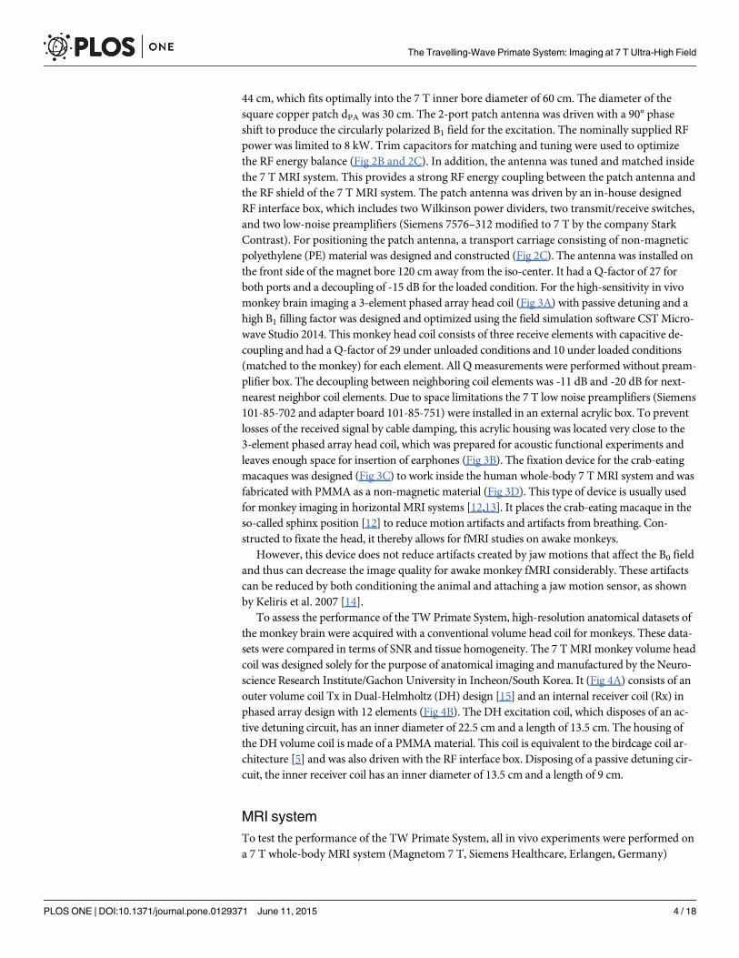
anatomy. In addition to the sulci and gyri of the telencephalon, the cerebellum and the hippo-campus are most apparent in the shown images; further smaller structures can be identified aswell. The anatomical results of the monkey volume head coil also cover the brainstem andneck. For comparison of the sensitivity of the TW Primate System and monkey volume headcoil, the SNR was measured in a selected 3D TSE image sum-of-squares combined dataset bythe mean value approach [22]. Fig 10 shows two comparable slices, one acquired by the TWPrimate System (Fig 10A), with μPixel = 286 for ROI 1 (signal intensity gray matter), μPixel =262 for ROI 2 (signal intensity white matter), and μPixel = 5.69 for ROI 3 (noise) and anotheracquired with the monkey volume head coil (Fig 10B), with μPixel = 114 for ROI 1 (signal inten-sity gray matter), μPixel = 126 for ROI 2 (signal intensity white matter), and μPixel = 11.78 forROI 3 (noise). For calculation of the SNR the associated background-noise coil-element correc-tion factors for the mean value approach have to be applied [23]. This factor was CF3E = 2.35for the 3-element phased array coil and CF12E = 4.85 for the 12-element phased array coil ofthe monkey volume head coil. The SNR of the TW Primate System in gray SNRGray = 118 andwhite matter SNRWhite = 108 and that of the monkey volume head coil was SNRGray = 46 andSNRWhite = 52. The results of the in vivo experiments for potential fMRI applications with theTW Primate System and the 2D EPI sequences are shown in Fig 11A–11C, with three exempla-ry consecutive slices in coronal plane of the crab-eating macaque brain. These images representthe entire brain of the crab-eating macaque, with a good depiction of gray and white mattermainly in the frontal and parietal lobes.
Fig 9. In vivomeasurements using the Dual-Helmholtz primate volume head coil. Exemplary slices of high-resolution 3D TSE of images of ananesthetized crab-eating macaque Every tenth slice in the medial portion of the volume of interest (VOI) is shown to depict the main parts of the monkeybrain. Gray and white matter, hippocampus, cerebellum and brain stem are well delineated (a-f).
doi:10.1371/journal.pone.0129371.g009
The Travelling-Wave Primate System: Imaging at 7 T Ultra-High Field
PLOS ONE | DOI:10.1371/journal.pone.0129371 June 11, 2015 11 / 18

DiscussionOur results show that the TW excitation approach [8] reliably facilitates excitation within a 7 Thuman whole-body UHF MRI system equipped with an SC72d whole-body gradient system.The patch antenna serves as a transmitter for a homogenous B1
+ field in a central region of in-terest (ROI) with a diameter of about 10 cm. The B1
+ field homogeneity in this central ROI isnearly equivalent to the "gold-standard" birdcage architecture, as shown in Mallow et al. 2013[9].
Fig 10. Comparison of the SNR in the high-resolution 3D TSE images from the TW Primate System and the Dual-Helmholtz primate volume headcoil. Selected slice of the anatomical in vivo results of anesthetized crab-eating macaque for the TW Primate System (a). Selected slice for the Dual-Helmholtz primate volume head coil (b).
doi:10.1371/journal.pone.0129371.g010
Fig 11. High-resolution 2D EPI in vivo images of an anesthetized crab-eating macaque acquired with the TW Primate System. Three exemplaryconsecutive coronal slices are displayed. Gray and white matter in the frontal and parietal lobes are well delineated. Images are not interpolated so that theoriginal resolution is visible (a-c).
doi:10.1371/journal.pone.0129371.g011
The Travelling-Wave Primate System: Imaging at 7 T Ultra-High Field
PLOS ONE | DOI:10.1371/journal.pone.0129371 June 11, 2015 12 / 18

This good B1+ field homogeneity was used for rodent imaging at 16.4 T with a 26 cm hori-
zontal bore magnet attached to a BioSpec spectrometer (Bruker BioSpin MRI GmbH, Ger-many) in Shajan et al. 2012 [24]. In addition, the TW approach was used on a 9.4 T (400 MHz)whole-body MRI system (Siemens Healthcare, Erlangen, Germany) that was equipped with thewhole-body gradient system introduced by Geschewski et al. 2013 [25] and on a similar whole-body MRI system equipped with an AC84 head-only gradient system introduced by Hoffmannet al. 2012 [26]. Hoffmann et al. [26] implemented the TW approach for human head imagingwith the AC84 head-only gradient insert with a 40 cm diameter RF shield. Unlike to a whole-body gradient system, RF shield diameter was thus decreased, leading to an increase of the cut-off frequency to 440 MHz and therefore to an adverse reduction of RF performance. The condi-tions on a 7 T whole-body MRI system are well suited for animal imaging when neuroimagingof macaques is the primary interest. However, some prerequisites have to be fulfilled for thisapproach. The TW Primate System works only in those UHF MRI systems that match the geo-metric conditions for the propagation of the circular TE11 waveguide mode [9]. This meansthat, depending on the excitation frequency, the diameter of the gradient RF shield needs to belarger than 60 cm diameter, which is not the case in all human 7 T UHF MRI systems. Due toincreased SAR and the inhomogeneous field distribution under UHF conditions, no commer-cial UHF whole-body MRI system possesses a body coil resonator. Vaughan et al. 2009 [27]showed that using the transverse electromagnetic (TEM) volume coil design as a body coil in aUHF MRI system resulted in a good B1
+ transmission efficiency. Compared with the TW exci-tation approach, however, the TEM design has fewer degrees of freedom because its architec-ture requires more space inside the inner bore of the MRI system. Furthermore, TEM is moreexpensive than the TW excitation approach. Similarly, Orzada et al. 2013 [28] demonstratedthe usability of an eight-channel transmit coil array in combination with B1-shimming forhuman body imaging. Compared with the TW excitation approach, this setup delivers good re-sults for human body imaging but would not be suitable for monkey imaging in combinationwith a fixation device. The search for a fully sufficient UHF body coil resonator is thus anongoing process.
Instead, RF coils consisting of a transmit volume coil in combination with phased array re-ceive coil are the standard architecture to provide homogeneous excitation and the receivingsensitivity required for imaging in UHF conditions. However, these coils usually enclose theobject tightly (to achieve highest sensitivity), leaving less space for additional experimentalequipment as needed for auditory and visual brain research. By contrast, more space is avail-able in the TW Primate System, in which it is also possible to install additional devices. Otherappealing advantages include the possibility to use a fixation device for fMRI in the sphinx po-sition as well as the much lower costs for the components and the production of a patch anten-na compared with those for a volume head coil in birdcage-like architecture. The sphinxposition for imaging of the human head with the TW excitation approach was already pre-sented by Zhang et al. [29] but is seldom used because this position is not comfortable for hu-mans in studies entailing extended periods of time. As a further advantage, the patch antennain combination with the appropriate high sensitive phased array coil is usable in every experi-mental setup.
The results of the B1 flip angle maps show a sufficient homogeneity of the applied flip angleof 90° in the central brain region for both excitation approaches. The TW Primate Systemshowed a slightly better homogeneity in comparison to the monkey volume head coil. This dif-ference is partly due to the non-optimal monkey head position inside the monkey volumehead coil.
A comparison of the in vivo B1+ transmission efficiency of both imaging approaches
shows a 37% higher B1+ transmission efficiency for the monkey volume head coil, with
The Travelling-Wave Primate System: Imaging at 7 T Ultra-High Field
PLOS ONE | DOI:10.1371/journal.pone.0129371 June 11, 2015 13 / 18

B1_PVHC = 13.3 μT/pkW (Uref = 197 V) for it and B1_TWPS = 9.7 μT/
pkW (Uref = 270 V) for
the TW Primate System. The B1+ transmission efficiency is given in [μT/
pkW] and can be cal-
culated by the measured reference voltage (Uref) that represents the needed RF energy for a180° pulse (B1 = 11.75 μT) converted in μT divided by the square root RMS power
pP in kW.
The lower B1+ transmission efficiency of the TW excitation is due to a smaller B1
+ filling factorfor the volume-of-interest. In comparison with volume resonators, the according B1
+ transmis-sion efficiency decreases—a finding also reported by other groups [30],[31]—which constitutesthe main disadvantage of the TW excitation approach. The 37% B1
+ transmission efficiencygap between both imaging approaches increases dramatically to approx. 93% by using low di-electric materials such as silicon oil, as shown in Mallow et al. 2013 [9]. This leads to the con-clusion that the TW excitation approach is effective only in combination with high dielectricmaterials such as water containing tissue. To avoid B1
+ homogeneities in high dielectric tissues,the degrees of freedom for B1
+-shimming are limited by the provided RF power and low B1+
transmission efficiency [32]. Recently, interesting alternative approaches were proposed to in-crease the latter's B1
+ transmission efficiency [33]. Furthermore, the SAR results show the samelow RF efficiency of the TW excitation approach in comparison to the more efficient volumeresonator structures. Only a small amount of the RF energy radiated by the patch antenna isdissipated in the lossy tissue material of the monkey body. The RF energy balance is much bet-ter for resonator coils than for the TW excitation approach. The T2-weighed in vivo 3D TSEsequence measurements showed that high-resolution anatomic images could be acquired forboth coil architectures: The TW Primate System and the monkey volume head coil providedhigh contrast and tissue homogeneity over the entire brain for a clear depiction of gray andwhite matter. The high-resolution provides fine details of the anatomy. Besides the sulci andgyri of the telencephalon, the cerebellum and the hippocampus are clearly visible in the images.Given its larger array size, the monkey volume head coil covers the brainstem and neck. Thiscoverage is dominated by the receive coil. This slightly bigger relative field of view (FoV) maybe an advantage if those regions have to be examined. However, most monkey MRI focuses onfunctional imaging and is thus mainly interested in the cerebrum. In the gray matter, theSNRGray = 118 of the TW Primate System is approx. three times higher than that of the monkeyvolume head coil with SNRGray = 47. In the white matter, the SNR is approx. twice as high forthe TW Primate System with SNRWhite = 108 and SNRWhite = 52 for the monkey volume headcoil. One reason for this large difference in sensitivity is the non-optimal monkey head positioninside the monkey volume head coil and the larger inner diameter of the receive coil consistingof 12 elements. Even the coil correction factors cannot compensate these experimental bound-ary conditions. These correction factors eliminate the additionally added background noisefrom the sum-of-square combined element signals if the coil fits optimally to the load; using acoil with more elements normally increases the SNR. The monkey volume head coil allows alimited degree of positioning of the macaque in return for an optimum imaging position of theanaesthetized monkey. Its major disadvantage is that there is no option for using a sphinx posi-tion. The 2D EPI in vivo measurements were acquired to test whether functional imaging asthe major neuroimaging application would be feasible with the TW approach. The imagesshow that most parts of the brain are well mapped, even showing a delineation between grayand white matter mainly in the frontal and parietal lobes. The caudal parts exhibit increasingB0-shiming artifacts, but these may be reduced by optimizing the image sequence parameterand B0-shim.
Motion artifacts that affect the B0 field could also decrease the image quality for awake mon-key fMRI. The fixation device in combination with the sphinx position reduces normal motionartifacts and artifacts from breathing, but not those from jaw motion. The latter artifacts can bereduced by intensive conditioning of the monkey and a jaw motion sensor in combination with
The Travelling-Wave Primate System: Imaging at 7 T Ultra-High Field
PLOS ONE | DOI:10.1371/journal.pone.0129371 June 11, 2015 14 / 18

image motion correction technics, as shown by Keliris et al. 2007 [14,34,35]. This issue couldbe more relevant when using the horizontal MRI system with a sphinx position in comparisonto vertical MRI systems with the monkey in a more natural sitting position. Motion artifacts re-main a challenge for both the TW Primate System and the conventional volume coil approach,which are equally susceptible to them. Monkey conditioning and application of a jaw motionsensor in combination with image motion correction techniques can improve the results of theTW Primate System in the future. However, our limited experimental time budget did notallow us to training monkeys or develop and validated such a jaw motion sensor in combina-tion with image motion correction in order to reduce motion artifacts, so we were not yet ableto run in vivo fMRI with awake monkeys.
The experimental procedure also emphasized one of the major advantages of the TW Pri-mate System: the easy positioning of the monkey inside the MRI system because the sphinx po-sition is not disturbed when using the 3-element phased array receive-only head coil. Incomparison with a conventional volume head coil concept, the 3-element phased array headcoil leaves more space for additional equipment and resulted in a higher SNR. The SNR in grayand white matter (SNRGray = 118, SNRWhite = 108) acquired with the TW Primate System washigher than the SNR of Kolster et al. 2009 [12], namely 35 and 80 for a 4- and 8-element receivecoil inside the cerebrum. Kolster et al. 2009 used a 7 T whole-body MRI in combination with asurface coil for Tx and a 4- and 8-element receive coil for fMRI of rhesus monkeys in thesphinx position. The difference in SNR is mainly attributable to the different excitation meth-ods. A surface coil as heterogeneous resonator for Tx, as used in the study of Kolster et al. 2009,cannot generate the same flip angle across the whole monkey brain.
As shown by Pfeuffer et al. 2004, the achieved SNR and image quality can be increased byusing implanted coils and a better gradient system [36]. This high SNR is caused by the high re-ceive B1 filling factor provided by the monkey head being tightly fixated as well as the fact thatthe preamplifier could be installed in close proximity to the RF coil. The use of this special 3-el-ement phased array head coil also leads to a higher flexibility in presenting stimuli in functionalMRI, e.g. when using earphones or visual stimulation devices. In comparison to vertical magnetMRI systems [3], horizontal UHF magnet MRI systems with the presented TW Primate Systemmay provide an improvement if more space is needed for stimulation devices or when scanninglarger monkeys (or even two norm-sized monkeys in a same scan).
The results of this work thus add additional evidence that for examination of non-humanprimates a human whole-body MRI system may represent a good alternative, especially as theequipment is cost-effective and 7 T UHF scanners are becoming much more widely available.Vertical monkey MRI systems with smaller gradient system such as the 7 T vertical MRI with a38 cm inner diameter (Bruker BGA-38) provide better imaging conditions in terms of the gra-dient system and B0-shim quality, which directly affects the EPI quality (T2� imaging). In con-trast, achieving a high B0 homogeneity is more difficult when placing the relatively smallmonkey inside the larger bore of the human 7 T whole-body MRI system. However, B0-shim-ming up to the 3rd order may still facilitate increasing the field homogeneity on a 7 T whole-body MRI system with a 60 cm diameter, as described by Sengupta et al. 2011 and Pan et al.2012 [37,38].
The vertical gradient system used by Pfeuffer et al. 2004 [36] provides 80 mT/m with a slewrate of 400 mT/m/ms, which increases image quality considerably compared to our horizontalMRI gradient system with a maximum amplitude of 70 mT/m and maximum slew rate of 200mT/m/ms. This difference is significant for SNR and EPI quality. The optimized vertical MRIsystems are dedicated exclusively to specialized animal experiments. A further optimization ofthe TW Primate System should render the presented approach a promising new alternative for
The Travelling-Wave Primate System: Imaging at 7 T Ultra-High Field
PLOS ONE | DOI:10.1371/journal.pone.0129371 June 11, 2015 15 / 18

research groups aspiring to perform monkey MRI but having access solely to horizontalhuman whole-body UHF MRI systems.
Supporting InformationS1 File. B1 flip angle maps (DICOM format) of the TW Primate System and the Dual-Helmholtz primate volume head coil. The transversal and coronal slices. (TW_Tra.dcm;TW_Cor.dcm; DH_Tra.dcm; DH_Cor.dcm).(ZIP)
S2 File. Fig 1 and Fig 7 as EPS vector format file.(ZIP)
AcknowledgmentsThe authors thank Dirk Salbert and Jürgen Kanstorf for their technical advice in the construc-tion of the specifically designed RF transmitter. Further thanks go to Dr. Kyoung-Nam Kim forhis technical advice.
Author ContributionsConceived and designed the experiments: JS J. Mylius TH. Performed the experiments: JS J.Mylius TH. Analyzed the data: TH J. Mylius. Contributed reagents/materials/analysis tools: J.Mallow MB. Wrote the paper: JB THMB J. Mallow MPML.
References1. Ogawa S, Lee TM, Kay AR, Tank DW. Brain magnetic resonance imaging with contrast dependent on
blood oxygenation. Proc Natl Acad Sci U S A. 1990; 87: 9868–9872. PMID: 2124706
2. Goense JBM, Ku S-P, Merkle H, Tolias AS, Logothetis NK. fMRI of the temporal lobe of the awake mon-key at 7 T. NeuroImage. 2008; 39: 1081–1093. doi: 10.1016/j.neuroimage.2007.09.038 PMID:18024083
3. Kagan I, Iyer A, Lindner A, Andersen RA. Space representation for eye movements is more contralater-al in monkeys than in humans. Proc Natl Acad Sci. 2010; 107: 7933–7938. doi: 10.1073/pnas.1002825107 PMID: 20385808
4. Vanduffel W, Fize D, Mandeville JB, Nelissen K, Van Hecke P, Rosen BR, et al. Visual Motion Process-ing Investigated Using Contrast Agent-Enhanced fMRI in Awake Behaving Monkeys. Neuron. 2001;32: 565–577. doi: 10.1016/S0896-6273(01)00502-5 PMID: 11719199
5. Hayes CE. The development of the birdcage resonator: a historical perspective. NMR Biomed. 2009;22: 908–918. doi: 10.1002/nbm.1431 PMID: 19856386
6. Roemer PB, Edelstein WA, Hayes CE, Souza SP, Mueller OM. The NMR phased array. Magn ResonMed. 1990; 16: 192–225. doi: 10.1002/mrm.1910160203 PMID: 2266841
7. Orzada S, Maderwald S, Göricke SL, Parohl N, Ladd SC, Ladd ME, et al. Design and comparison oftwo eight-channel transmit/receive radiofrequency arrays for in vivo rodent imaging on a 7 T humanwhole-body MRI system. Med Phys. 2010; 37: 2225–2232. PMID: 20527556
8. Brunner DO, Zanche ND, Fröhlich J, Paska J, Pruessmann KP. Travelling-wave nuclear magnetic reso-nance. Nature. 2009; 457: 994–998. doi: 10.1038/nature07752 PMID: 19225521
9. Mallow J, Herrmann T, Kim K-N, Stadler J, Mylius J, Brosch M, et al. Ultra-high field MRI for primate im-aging using the travelling-wave concept. Magn Reson Mater Phys Biol Med. 2013; 26: 389–400. doi:10.1007/s10334-012-0358-z
10. Purcell EM, Torrey HC, Pound RV. Resonance Absorption by Nuclear Magnetic Moments in a Solid.Phys Rev. 1946; 69: 37–38. doi: 10.1103/PhysRev.69.37
11. CST AG. CSTMicrowave Studio 2014. 2014;
12. Kolster H, Mandeville JB, Arsenault JT, Ekstrom LB, Wald LL, Vanduffel W. Visual Field Map Clustersin Macaque Extrastriate Visual Cortex. J Neurosci Off J Soc Neurosci. 2009; 29: 7031–7039. doi: 10.1523/JNEUROSCI.0518-09.2009
The Travelling-Wave Primate System: Imaging at 7 T Ultra-High Field
PLOS ONE | DOI:10.1371/journal.pone.0129371 June 11, 2015 16 / 18

13. Denys K, Vanduffel W, Fize D, Nelissen K, Peuskens H, Essen DV, et al. The Processing of VisualShape in the Cerebral Cortex of Human and Nonhuman Primates: A Functional Magnetic ResonanceImaging Study. J Neurosci. 2004; 24: 2551–2565. doi: 10.1523/JNEUROSCI.3569-03.2004 PMID:15014131
14. Keliris GA, Shmuel A, Ku S-P, Pfeuffer J, Oeltermann A, Steudel T, et al. Robust controlled functionalMRI in alert monkeys at high magnetic field: Effects of jaw and body movements. NeuroImage. 2007;36: 550–570. doi: 10.1016/j.neuroimage.2007.02.057 PMID: 17509896
15. Cho Z-H, Han J-Y, Hwang S-I, Kim D, Kim K-N, Kim N-B, et al. Quantitative analysis of the hippocam-pus using images obtained from 7.0 T MRI. NeuroImage. 2010; 49: 2134–2140. doi: 10.1016/j.neuroimage.2009.11.002 PMID: 19909820
16. Klose U. Mapping of the radio frequency magnetic field with a MR snapshot FLASH technique. MedPhys. 1992; 19: 1099–1104. doi: 10.1118/1.596828 PMID: 1518473
17. Hennig J. Multiecho imaging sequences with low refocusing flip angles. J Magn Reson 1969. 1988; 78:397–407. doi: 10.1016/0022-2364(88)90128-X
18. Mansfield P. Real-time echo-planar imaging by NMR. Br Med Bull. 1984; 40: 187–190. PMID: 6744006
19. IEC. Medical electrical equipment—part 2–33 particular requirements for the safety of magnetic reso-nance equipment for medical diagnosis. 3rd ed. 2010.
20. Gabriel S, Lau RW, Gabriel C. The dielectric properties of biological tissues: II. Measurements in thefrequency range 10 Hz to 20 GHz. Phys Med Biol. 1996; 41: 2251. doi: 10.1088/0031-9155/41/11/002PMID: 8938025
21. Wolf S, Diehl D, Gebhardt M, Mallow J, Speck O. SAR simulations for high-field MRI: Howmuch detail,effort, and accuracy is needed? Magn Reson Med. 2013; 69: 1157–1168. doi: 10.1002/mrm.24329PMID: 22611018
22. Constantinides CD, Atalar E, McVeigh ER. Signal-to-Noise Measurements in Magnitude Images fromNMR Phased Arrays. Magn Reson Med Off J Soc Magn Reson Med Soc Magn Reson Med. 1997; 38:852–857.
23. Dietrich O, Raya JG, Reeder SB, Ingrisch M, Reiser MF, Schoenberg SO. Influence of multichannelcombination, parallel imaging and other reconstruction techniques on MRI noise characteristics. MagnReson Imaging. 2008; 26: 754–762. doi: 10.1016/j.mri.2008.02.001 PMID: 18440746
24. Shajan G, Hoffmann J, Balla DZ, Deelchand DK, Scheffler K, Pohmann R. Rat brain MRI at 16.4Tusing a capacitively tunable patch antenna in combination with a receive array. NMR Biomed. 2012;25: 1170–1176. doi: 10.1002/nbm.2786 PMID: 22344898
25. Geschewski FH, Brenner D, Felder J, Jon Shah N. Optimum coupling and multimode excitation of trav-eling-waves in a whole-body 9.4T scanner. Magn Reson Med. 2013; 69: 1805–1812. doi: 10.1002/mrm.24403 PMID: 22782491
26. Hoffmann J, Shajan G, Budde J, Scheffler K, Pohmann R. Human brain imaging at 9.4 T using a tun-able patch antenna for transmission. Magn Reson Med. 2012; n/a–n/a. doi: 10.1002/mrm.24367
27. Vaughan JT, Snyder CJ, DelaBarre LJ, Bolan PJ, Tian J, Bolinger L, et al. 7 T Whole Body Imaging:Preliminary Results. Magn Reson Med Off J Soc Magn Reson Med Soc Magn Reson Med. 2009; 61:244–248. doi: 10.1002/mrm.21751
28. Orzada S, Johst S, Maderwald S, Bitz AK, Solbach K, Ladd ME. Mitigation of B1+ inhomogeneity onsingle-channel transmit systems with TIAMO. Magn Reson Med. 2013; 70: 290–294. doi: 10.1002/mrm.24453 PMID: 22886695
29. Zhang B, Brown R, Wiggins C, Sodickson DK, Stoeckel B, Wiggins GC. A 7T Coil System for ImagingHumans in the Sphinx Position to Evaluate the Effect of Head Orientation Relative to B0 for MR Imag-ing. 19th Annu ISMRM Sci Meet Exhib 2011 Vol 3. 2011; 162.
30. Alt S, Müller M, UmathumR, Bolz A, Bachert P, Semmler W, et al. Coaxial waveguide MRI. MagnReson Med. 2012; 67: 1173–1182. doi: 10.1002/mrm.23069 PMID: 22021117
31. Brunner DO, Paška J, Froehlich J, Pruessmann KP. Traveling-wave RF shimming and parallel MRI.Magn Reson Med. 2011; 66: 290–300. doi: 10.1002/mrm.22817 PMID: 21695729
32. Orzada S, Maderwald S, Poser BA, Bitz AK, Quick HH, Ladd ME. RF excitation using time interleavedacquisition of modes (TIAMO) to address B1 inhomogeneity in high-field MRI. Magn Reson Med. 2010;64: 327–333. doi: 10.1002/mrm.22527 PMID: 20574991
33. Erni D, Liebig T, Rennings A, Koster NHL, Frohlich J. Highly adaptive RF excitation scheme based onconformal resonant CRLHmetamaterial ring antennas for 7-Tesla traveling-wave magnetic resonanceimaging. 2011 Annual International Conference of the IEEE Engineering in Medicine and Biology Soci-ety,EMBC. 2011. pp. 554–558. doi: 10.1109/IEMBS.2011.6090102
The Travelling-Wave Primate System: Imaging at 7 T Ultra-High Field
PLOS ONE | DOI:10.1371/journal.pone.0129371 June 11, 2015 17 / 18

34. Schulz J, Siegert T, Bazin P-L, Maclaren J, Herbst M, Zaitsev M, et al. Prospective slice-by-slice motioncorrection reduces false positive activations in fMRI with task-correlated motion. NeuroImage. 2014;84: 124–132. doi: 10.1016/j.neuroimage.2013.08.006 PMID: 23954484
35. Maclaren J, Herbst M, Speck O, Zaitsev M. Prospective motion correction in brain imaging: A review.Magn Reson Med. 2013; 69: 621–636. doi: 10.1002/mrm.24314 PMID: 22570274
36. Pfeuffer J, Merkle H, Beyerlein M, Steudel T, Logothetis NK. Anatomical and functional MR imaging inthe macaque monkey using a vertical large-bore 7 Tesla setup. Magn Reson Imaging. 2004; 22: 1343–1359. doi: 10.1016/j.mri.2004.10.004 PMID: 15707785
37. Pan JW, Lo K-M, Hetherington HP. Role of very high order and degree B0 shimming for spectroscopicimaging of the human brain at 7 tesla. Magn Reson Med. 2012; 68: 1007–1017. doi: 10.1002/mrm.24122 PMID: 22213108
38. Sengupta S, Avison MJ, Gore JC, BrianWelch E. Software compensation of Eddy current fields in mul-tislice high order dynamic shimming. J Magn Reson. 2011; 210: 218–227. doi: 10.1016/j.jmr.2011.03.007 PMID: 21458339
The Travelling-Wave Primate System: Imaging at 7 T Ultra-High Field
PLOS ONE | DOI:10.1371/journal.pone.0129371 June 11, 2015 18 / 18