Three dimensional echo-planar imaging at 7 Tesla
6
Technical Note Three dimensional echo-planar imaging at 7 Tesla B.A. Poser a,b, ⁎, P.J. Koopmans b , T. Witzel c,d , L.L. Wald c,d , M. Barth a,b a Erwin. L. Hahn Institute for Magnetic Resonance Imaging, University Duisburg-Essen, Essen, Germany b Radboud University Nijmegen, Donders Institute for Brain, Cognition and Behaviour, Centre for Cognitive Neuroimaging, Nijmegen, The Netherlands c Department of Radiology, Massachusetts General Hospital, A.A. Martinos Center, Charlestown Massachusetts, USA d Harvard-MIT Division of Health Sciences Technology, Cambridge Massachusetts, USA abstract article info Article history: Received 26 November 2009 Revised 15 January 2010 Accepted 30 January 2010 Available online 6 February 2010 Functional MRI (fMRI) most commonly employs 2D echo-planar imaging (EPI). The advantages for fMRI brought about by the increasingly popular ultra-high field strengths are best exploited in high-resolution acquisitions, but here 2D EPI becomes unpractical for several reasons, including the very long volume acquisitions times. In this study at 7 T, a 3D EPI sequence with full parallel and partial Fourier imaging capability along both phase encoding axes was implemented and used to evaluate the sensitivity of 3D and corresponding 2D EPI acquisitions at four different spatial resolutions ranging from small to typical voxel sizes (1.5–3.0 mm isotropic). Whole-brain resting state measurements (N = 4) revealed a better, or at least comparable sensitivity of the 3D method for gray and white matter. The larger vulnerability of 3D to physiological effects was outweighed by the much shorter volume TR, which moreover allows whole-brain coverage at high resolution within fully acceptable limits for event-related fMRI: TR was only 3.07 s for 1.5 mm, 1.88 s for 2.0 mm, 1.38 s for 2.5 mm and 1.07 s for 3.0 mm isotropic resolution. In order to investigate the ability to detect and spatially resolve BOLD activation in the visual cortex, functional 3D EPI experiments (N = 8) were performed at 1 mm isotropic resolution with parallel imaging acceleration of 3 × 3, resulting in a TR of only 3.2 s for whole-brain coverage. From our results, and several other practical advantages of 3D over 2D EPI found in the present study, we conclude that 3D EPI provides a useful alternative for whole-brain fMRI at 7 T, not only when high-resolution data are required. © 2010 Elsevier Inc. All rights reserved. Introduction Since the discovery of the BOLD effect, 2D gradient-echo echo- planar imaging (EPI) has been the workhorse of functional MRI, largely because of its high sampling speed and excellent sensitivity to signal changes related to brain activation, at both sufficient temporal (∼ 2–3 s) and spatial resolution (∼ 3–4 mm isotropic) with whole- brain coverage (Norris, 2006). Recently, it has been shown that higher spatial resolution would be beneficial to reduce the influence of physiological fluctuations (Triantafyllou et al., 2005, 2006). However, sampling at higher (isotropic) spatial resolution in conventional multi-slice 2D EPI considerably increases measurement times as the echo train length (ETL) becomes larger, and more slices have to be acquired to obtain the same covered volume. Methods such as partial Fourier (PF) sampling (Jesmanowicz et al., 1998) or partial parallel imaging (PPI) (Griswold et al., 2002; Pruessmann et al., 1999) provide an efficient means for shortening the ETL, and are hence well suited for obtaining high-resolution functional images, images with reduced EPI distortion artifacts (de Zwart et al., 2002, 2006; Preibisch et al., 2003; Schmidt et al., 2005), or single-shot acquisition of multiple echoes to improve BOLD sensitivity (Poser and Norris, 2009; Poser et al., 2006). Considerable effort has gone into the optimization of 2D EPI protocols and the practical challenges associated with fMRI at high field (Speck et al., 2008). In the conventional 2D EPI sequence, slice selective excitation is performed and the signal subsequently acquired under an oscillating read and blipped phase encode gradient, so as to acquire the data for a full 2D image in a single shot within TR slice . After repetition time TR slice ∙N slices , all N slices slices have been acquired, and the process is repeated. The effectiveness of both the PF and PPI methods for the reduction of acquisition time is hence rather limited in the case of 2D EPI, as the acceleration can only be performed along the (one and only) phase encode (PE) direction. The small potential time gain from under-sampling in 2D EPI is primarily due to the fact that TE should be kept constant to obtain the same functional contrast: For example, if the ETL is shortened by an acceleration factor of 2, the effective reduction in sampling time per slice is only TR slice − ETL/4, which for a typical protocol is a time saving of only about 10%–15%. NeuroImage 51 (2010) 261–266 ⁎ Corresponding author. Erwin L. Hahn Institute for Magnetic Resonance Imaging, UNESCO World Cultural Heritage Zollverein Arendahls Wiese 199, Tor 3,45141 Essen, Germany. Fax: +49 201 183 6073. E-mail address: [email protected] (B.A. Poser). 1053-8119/$ – see front matter © 2010 Elsevier Inc. All rights reserved. doi:10.1016/j.neuroimage.2010.01.108 Contents lists available at ScienceDirect NeuroImage journal homepage: www.elsevier.com/locate/ynimg
-
Upload
independent -
Category
Documents
-
view
0 -
download
0
Transcript of Three dimensional echo-planar imaging at 7 Tesla
NeuroImage 51 (2010) 261–266
Contents lists available at ScienceDirect
NeuroImage
j ourna l homepage: www.e lsev ie r.com/ locate /yn img
Technical Note
Three dimensional echo-planar imaging at 7 Tesla
B.A. Poser a,b,⁎, P.J. Koopmans b, T. Witzel c,d, L.L. Wald c,d, M. Barth a,b
a Erwin. L. Hahn Institute for Magnetic Resonance Imaging, University Duisburg-Essen, Essen, Germanyb Radboud University Nijmegen, Donders Institute for Brain, Cognition and Behaviour, Centre for Cognitive Neuroimaging, Nijmegen, The Netherlandsc Department of Radiology, Massachusetts General Hospital, A.A. Martinos Center, Charlestown Massachusetts, USAd Harvard-MIT Division of Health Sciences Technology, Cambridge Massachusetts, USA
⁎ Corresponding author. Erwin L. Hahn Institute forUNESCO World Cultural Heritage Zollverein ArendahlsGermany. Fax: +49 201 183 6073.
E-mail address: [email protected] (B.A.
1053-8119/$ – see front matter © 2010 Elsevier Inc. Adoi:10.1016/j.neuroimage.2010.01.108
a b s t r a c t
a r t i c l e i n f oArticle history:Received 26 November 2009Revised 15 January 2010Accepted 30 January 2010Available online 6 February 2010
Functional MRI (fMRI) most commonly employs 2D echo-planar imaging (EPI). The advantages for fMRIbrought about by the increasingly popular ultra-high field strengths are best exploited in high-resolutionacquisitions, but here 2D EPI becomes unpractical for several reasons, including the very long volumeacquisitions times. In this study at 7 T, a 3D EPI sequence with full parallel and partial Fourier imagingcapability along both phase encoding axes was implemented and used to evaluate the sensitivity of 3D andcorresponding 2D EPI acquisitions at four different spatial resolutions ranging from small to typical voxelsizes (1.5–3.0 mm isotropic). Whole-brain resting state measurements (N=4) revealed a better, or at leastcomparable sensitivity of the 3D method for gray and white matter. The larger vulnerability of 3D tophysiological effects was outweighed by the much shorter volume TR, which moreover allows whole-braincoverage at high resolution within fully acceptable limits for event-related fMRI: TR was only 3.07 s for1.5 mm, 1.88 s for 2.0 mm, 1.38 s for 2.5 mm and 1.07 s for 3.0 mm isotropic resolution. In order toinvestigate the ability to detect and spatially resolve BOLD activation in the visual cortex, functional 3D EPIexperiments (N=8) were performed at 1 mm isotropic resolution with parallel imaging acceleration of 3 ×3, resulting in a TR of only 3.2 s for whole-brain coverage.From our results, and several other practical advantages of 3D over 2D EPI found in the present study, weconclude that 3D EPI provides a useful alternative for whole-brain fMRI at 7 T, not only when high-resolutiondata are required.
Magnetic Resonance Imaging,Wiese 199, Tor 3,45141 Essen,
Poser).
ll rights reserved.
© 2010 Elsevier Inc. All rights reserved.
Introduction
Since the discovery of the BOLD effect, 2D gradient-echo echo-planar imaging (EPI) has been the workhorse of functional MRI,largely because of its high sampling speed and excellent sensitivity tosignal changes related to brain activation, at both sufficient temporal(∼2–3 s) and spatial resolution (∼3–4 mm isotropic) with whole-brain coverage (Norris, 2006). Recently, it has been shown that higherspatial resolution would be beneficial to reduce the influence ofphysiological fluctuations (Triantafyllou et al., 2005, 2006). However,sampling at higher (isotropic) spatial resolution in conventionalmulti-slice 2D EPI considerably increases measurement times as theecho train length (ETL) becomes larger, and more slices have to beacquired to obtain the same covered volume. Methods such as partialFourier (PF) sampling (Jesmanowicz et al., 1998) or partial parallelimaging (PPI) (Griswold et al., 2002; Pruessmann et al., 1999) provide
an efficient means for shortening the ETL, and are hence well suitedfor obtaining high-resolution functional images, images with reducedEPI distortion artifacts (de Zwart et al., 2002, 2006; Preibisch et al.,2003; Schmidt et al., 2005), or single-shot acquisition of multipleechoes to improve BOLD sensitivity (Poser and Norris, 2009; Poser etal., 2006). Considerable effort has gone into the optimization of 2D EPIprotocols and the practical challenges associated with fMRI at highfield (Speck et al., 2008).
In the conventional 2D EPI sequence, slice selective excitation isperformed and the signal subsequently acquired under an oscillatingread and blipped phase encode gradient, so as to acquire the data for afull 2D image in a single shot within TRslice. After repetition timeTRslice∙Nslices, all Nslices slices have been acquired, and the process isrepeated. The effectiveness of both the PF and PPI methods for thereduction of acquisition time is hence rather limited in the case of 2DEPI, as the acceleration can only be performed along the (one andonly) phase encode (PE) direction. The small potential time gain fromunder-sampling in 2D EPI is primarily due to the fact that TE should bekept constant to obtain the same functional contrast: For example, ifthe ETL is shortened by an acceleration factor of 2, the effectivereduction in sampling time per slice is only TRslice − ETL/4, which fora typical protocol is a time saving of only about 10%–15%.

Table 1Summary of the measurement protocols used for the sensitivity comparison.
Resolution 2D 3D
TR AF TR AF
1.5 mm 5.96 s 2 3.07 s 2×22.0 mm 3.65 s 2 1.88 s 2×22.5 mm 2.70 s 2 1.38 s 2×23.0 mm 2.10 s 2 1.07 s 2×2
262 B.A. Poser et al. / NeuroImage 51 (2010) 261–266
Alternatively, a 3D sampling scheme can be employed wherebythe same thick slab of tissue corresponding to the volume of interest isrepeatedly excited, and a kx–ky plane of k-space acquired each timewith a different kz increment. For application to fMRI, this concept hasattracted rapidly growing interest in the recent years, and 3Dimplementations with Cartesian (Poser and Norris, 2009; van derZwaag et al., 2009) and spiral (Hu and Glover, 2007a,b; Lai and Glover,1998) k-space trajectory have been presented. For EPI-like sequences,a similar scheme was first suggested by Song et al. (1994) andMansfield et al. (1995) who proposed to acquire even multiple kzplanes in a single shot. All these methods have in common that sliceselection is replaced by a secondary phase encoding direction, and thecomplete 3D image then obtained by 3D Fourier transform uponacquisition of all kz planes, again after time TRslice∙Nslices. Theimportant potential of 3D EPI here lies in the fact that entire kzencoding steps (kx–ky planes) can be ‘skipped’ by the use of PF or PPIalong the secondary phase direction, permitting considerable reduc-tions in the volume sampling time TRvolume: For both these under-sampling strategies, or their combination, the fractional time saving isdirectly given by the fraction of non-acquired secondary phaseencodings. A 3D acquisition scheme where undersampling can beperformed along the secondary PE direction will therefore besubstantially more time efficient.
In terms of MR imaging, the implication of using 3D vs. 2D EPI istwofold. First, the time interval between subsequent excitations of thesame tissue is much reduced, and as a result the steady statemagnetization that is available forMR signal formation is lower; this isgiven by Eq. (1) and maximal when the flip angle α is equal to theErnst angle (Eq. (2)) which should be calculated for the T1 relaxationtime of gray matter tissue. Second, the fact that Nslices more datapoints contribute to each data point in the 3D Fourier reconstructionleads to an intrinsic signal-to-noise (SNR) advantage of √Nslices whichhence increases with increasing slice count.
M = M0 sinα 1− exp − TRslice
T1
� �� �= 1− cosα � exp − TRslice
T1
� �� �
ð1Þ
αErnst = arccos exp − TRT1
� �� �ð2Þ
In this study, we propose the use of 3D EPI and to exploit thegreater flexibility given by the second phase encoding along the thirdspatial dimension to either obtain higher spatial resolution within thesame TRvolume, or to sample at the same spatial resolution within amuch shorter time as compared to conventional 2D EPI. The proposed3D EPI and conventional 2D EPI are compared by in vivo applicationwith and without functional stimulation, and the various implicationsof using 3D EPI are discussed.
Methods
MRI measurements
A 3D EPI sequence with full partial Fourier and parallel imagingcapability and flexible z-encoding order was implemented for theSiemens Magnetom scanner family (Siemens Healthcare, Erlangen,Germany). Image reconstruction was performed entirely through thevendor-provided software which uses GRAPPA parallel imagingreconstruction, including EPI specific functionality for removingNyquist ghosting and a zeroth order phase correction to minimizeB0 fluctuation from scanner drift and subject breathing. Both RF andgradient spoiling were used in the sequence. Measurements wereperformed on a total of 12 subjects. The experiments of this studyconsisted of two parts which were conducted separately on twodifferent Magnetom 7 T scanners; each part had been approved by therespective local ethics committee.
(a) Temporal SNRCorresponding 2D and 3D protocols with different spatial resolu-
tions were used on four subjects using an 8-channel Tx/Rx head array(RAPID Biomedical, Germany). The following resolutions (isotropicvoxel size) were used: 1.5 mm, 2.0 mm, 2.5 mm, and 3.0 mm and thenumber of slices was adapted to cover the same volume at eachresolution. An in-plane acceleration factor (AF) of 2 was used for eachprotocol. For the 3D protocols, an additional factor of 2 was used in theslice encoding direction, resulting in a total AF of 4. For parallelundersampling along the secondary phase encoding direction, thecircular geometric arrangement of the elements in the head coilrequired sagittal slice orientation which was used for all protocols inpart (a). The corresponding volume acquisition times at the fourresolutions where TRvolume, 2D=[5.96 s, 3.65 s, 2.7 s 2.1 s], andTRvolume, 3D=[3.07 s, 1.88 s, 1.38 s, 1.07 s], respectively. Theseprotocols are summarized in Table 1. In each scan, TE was 23 ms, andthe flip angle set to the Ernst angle (see Eq. (2) above). In case the SARlimit was exceeded, the largest possible flip angle was used as allowedby the online SAR monitor. A time series of 50 volumes was acquiredwithout the subjects performing a specific task.
(b) Visual task3D EPI fMRI experiments with visual stimulation were performed
on 8 subjects, using the following settings: 1 mm isotropic resolutionusing a 32-channel head coil (Wiggins et al., 2006), TE=23 ms,TRslice=50 ms, TRvolume, 3D=3.2 s, AF=3×3, BW=2000 Hz/px,matrix size 180×180, 104 slices in an axial orientation with sliceoversampling of 25% to prevent signal fold-over and intensityvariation across the slab due to pulse profile imperfection. The fMRItask consisted of 3 min visual stimulation in blocks of 20/20 s on/off.
Analysis
(a) Temporal SNRThe data sets of each subject were first motion corrected and high-
pass filtered (N 0.01 Hz) using FEAT (FMRI Expert Analysis Tool,version 5.98, part of FSL (FMRIB's Software Library, www.fmrib.ox.ac.uk/fsl, Oxford, UK). The 2D data sets were then linearly coregisteredto the 1.5 mm resolution 2D data set using an affine 9 parametertransformation and normalized correlation as a cost function (Flirt,FSL, Oxford). The 3D data sets were coregistered to the 2D data setswith the corresponding resolution using only a 6 parameter rigid bodytransformation, since the in-plane geometric distortions of the 2D and3D protocols are identical. In ROIs for the different brain tissues, i.e.occipital gray matter (GM), parietal white matter (WM) andcerebrospinal fluid in the lateral ventricle (CSF), the sensitivity ofthe protocols was compared as follows: first, temporal SNR (tSNR)was obtained pixelwise in the conventional manner as the meansignal over imaging volumes divided by standard deviation along thetimecourse. To facilitate comparison of protocols with differentTRvolume (but the same TE), the measure for ‘sensitivity' was takenas tSNR/√TRvolume.
(b) Visual taskfMRI data processing was carried out using FEAT. First, an analysis
with a relatively large spatial smoothing kernel (3mm)wasperformed

263B.A. Poser et al. / NeuroImage 51 (2010) 261–266
mainly for visualization purposes and to analyze the functional data ina commonly used way. The following pre-statistics processing wasapplied;motion correctionusingMCFLIRT (Jenkinsonet al., 2002) non-brain removal using BET (Smith, 2002); spatial smoothing using aGaussian kernel of 3 mm FWHM; grand-mean intensity normalizationof the entire 4D dataset by a single multiplicative factor; high-passtemporal filtering (Gaussian-weighted least-squares straight linefitting,with sigma=20.0 s). Time series statistical analysiswas carriedout using FILM(Woolrich et al., 2001). Z-statistic images (GaussianizedT/F) were thresholded using clusters determined by ZN5.3 and a(corrected) cluster significance threshold of P=0.00005. In onesubject, no significantly activated cluster was found. The largestcluster of activated voxels in the visual cortex was then used to assessthe number of activated pixels, average and maximum z-values, andrelative signal change. In addition, noise was estimated from theresiduals of the GLM analysis and used to calculate CNR as signalchange divided by noise and tSNR as mean signal divided by noise.Subsequently, for visualization purposes and to show the potential ofthe high spatial resolution acquisition, the analysis was repeated byusing only very little (0.75 mm kernel) or no spatial smoothing, whileleaving the other parameters except the significance threshold (ZN4.3for 0.75 mm smoothing; ZN3.3 for no smoothing) unchanged.
Results
Using Eqs. (1) and (2), the sensitivity of 2D vs. 3D EPI as a functionof slice number was simulated under the assumption of Gaussiannoise, a gray matter T1 at 7 T of 1400 ms, a constant TRslice of 65 ms,and no use of parallel acceleration along the slice direction. Note thatsince the resulting volume TR is identical for both methods, their
Fig. 1. Theoretical sensitivity of 2D and 3D EPI as a function of the number of acquiredslices (top). The relative performance of 3D vs. 2D EPI (bottom) in the absence ofphysiological noise as derived from the signal equations (Eqs. (1) and (2)) for the casewithout parallel undersampling. Since the resulting volume TR is identical for bothmethods, their relative sensitivities scale with the MR signal. The dashed lines indicatethe approximate number of slices needed for whole-brain coverage (≥100 mm axialslab) at the different spatial resolutions.
sensitivities are directly given by their respective MR signal; this isshown in the top panel of Fig. 1. The bottom panel of the figureindicates performance gain of 3D EPI over 2D EPI. The vertical dashedlines indicate the approximate number of slices required for whole-brain coverage (taken here as a 100 mm axial slab) at the differentslice thicknesses. The plot also clearly illustrates the increasing benefitof 3D with increasing resolution along the slice direction.
Table 2 summarizes the results of the tSNR and sensitivity analysisof 2D and 3D EPI protocols: shown are the tSNR and sensitivity valuesfor the two methods in the gray matter, white matter and CSF ROIs, ateach of the four spatial resolutions. As expected, and in accordancewith Eq. 1, tSNR is higher for 2D EPI due to the longer TR which passesbetween successive excitations of the same slice. Correcting for thedifferent TR by factor 1/√TR yields the sensitivity which for 3D issignificantly higher than for 2D at the highest resolution (1.5 mm, Pb0.05) and comparable at the lower resolutions for gray matter; forwhite matter the sensitivity is consistently higher. In contrast, the CSFsensitivity is lower in 3D than in 2D EPI. For illustration purposes, theresults are plotted in Fig. 2, showing the values for gray matter, whitematter and CSF in the top, middle and bottom panel, respectively.
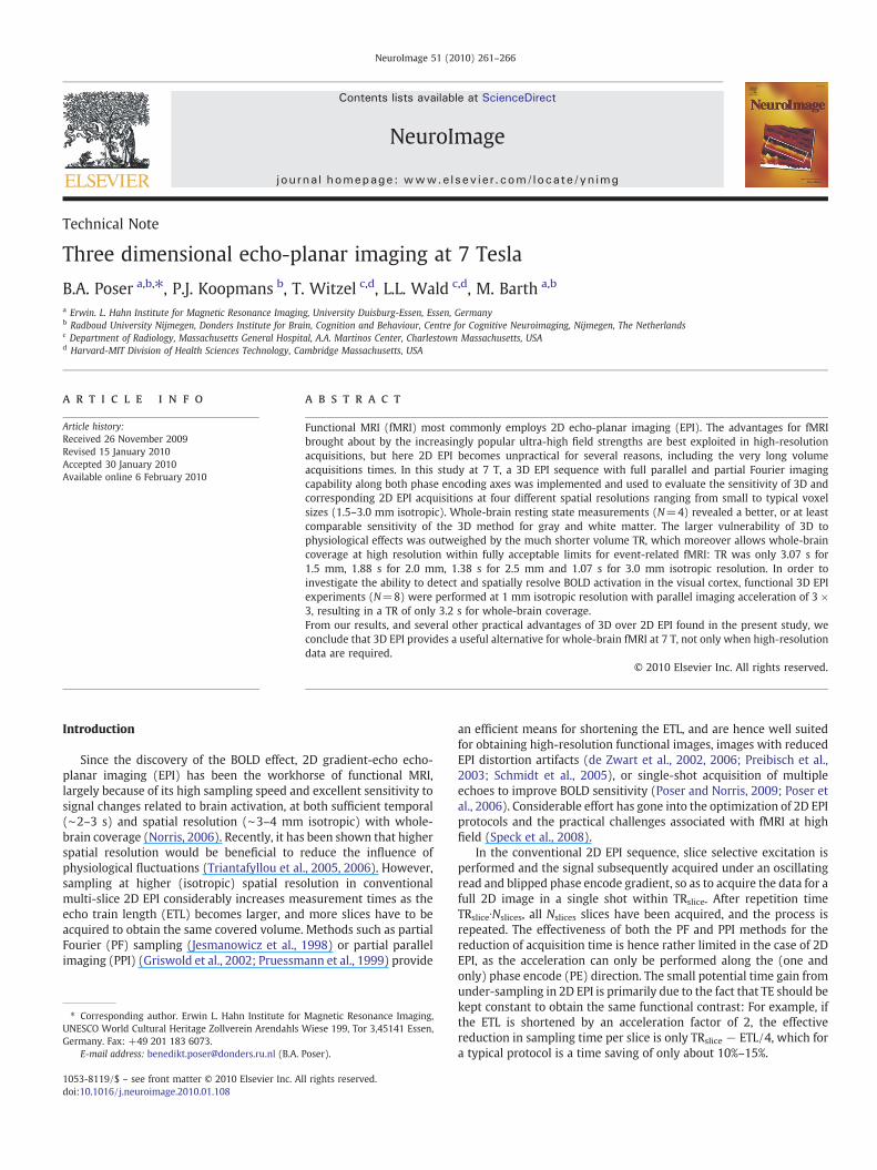
The results from the functional experiments (part b) are shown inTable 3, which gives the number of activated voxels, average andmaximum Z-score, relative BOLD signal changes, CNR and tSNR foreach subject. In Fig. 3, the temporal mean of all slices of the 1 mmresolution data set from a single subject is shown (panel a). Note theexcellent image quality and gray-white matter contrast, as well as thedetailed depiction of veins and deep brain nuclei which is seen best inzoomed view of one selected slice (slice 49). Sections b–d give azoomed view of 10 slices covering the occipital cortex for bettervisibility of the activation pattern. Overlaid on these images are thesignificantly activated clusters after spatially smoothing with a 3 mmkernel (b) whichwas used for improved visualization of the activationclusters. In panel c, smoothingwas reduced to a 0.75mmkernel and inpanel d, no spatial smoothing was used. Note how the activationnicely follows the sulci of the occipital cortex. In these experimentswith whole-brain acquisition the AF of 3×3 resulted in a TR of only3.2 s.
Table 2Temporal SNR (tSNR) and corresponding sensitivity of the different EPI protocols usedin part (a) of the study.
(a) tSNR of 2D and 3D EPI
1.5 mm 2 mm 2.5 mm 3 mm
2DGM 31.4±2.3 42.5±4.1 52.2±4.0 50.7±6.8WM 30.7±2.8 46.5±2.2 71.1±1.7 81.5±3.0CSF 59.1±7.2 64.7±7.9 77.4±9.6 75.7±11.5TRvolume 5960 ms 3650 ms 2700 ms 2090 ms3DGM 29.9±1.3 32.4±3.8 35.8±4.9 45.5±4.0WM 31.4±1.6 39.9±1.8 54.6±3.4 65.3±1.9CSF 30.5±3.6 40.5±4.5 38.0±3.4 53.0±6.8TRvolume 3068 ms 1880 ms 1376 ms 1066 ms
(b) Corresponding sensitivity of 2D and 3D EPI
1.5 mm 2 mm 2.5 mm 3 mm
2DGM 12.9±1.0 22.3±2.2 31.8±2.5 35.1±4.7WM 12.6±1.1 24.4±1.2 43.3±1.1 56.4±2.1CSF 24.2±2.9 33.9±4.1 47.1±5.8 52.4±8.0TRvolume 5960 ms 3650 ms 2700 ms 2090 ms3DGM 17.1±0.7 23.7±2.7 30.6±4.2 44.1±3.8WM 17.9±0.9 29.1±1.3 46.5±2.9 63.3±1.8CSF 17.4±2.0 29.6±3.3 32.4±2.9 51.3±6.6TRvolume 3068 ms 1880 ms 1376 ms 1066 ms
The effective functional sensitivity for fMRI is calculated as tSNR / √TRvolume. The errorterms give the standard error over subjects (N=4).

Fig. 2. Plots of sensitivity against voxel volume for the 2D (dashed lines) versus 3D EPIprotocols (straight lines), shown separately for GM (top), WM (middle) and CSF(bottom). The errors bars indicate standard error over subjects (N=4).
Table 3Number of activated pixels, average andmaximum z-values, relative signal change, andBOLD contrast-to-noise (CNR) and tSNR in the ROI defined by the activation cluster.
Subject # voxels Avg. z Max z Rel dS CNR tSNR
dc001 23,070 6.85 10.66 5.50% 3.42 62.20dc002 6396 6.78 10.29 4.95% 3.33 67.26dc004 872 6.05 7.92 5.25% 2.79 53.15dc005 2729 6.60 9.74 4.81% 3.25 67.54dc006 6355 6.32 8.90 5.44% 2.94 54.05dc007 7638 6.47 9.71 4.78% 3.10 64.96dc008 1226 6.30 9.35 5.33% 2.96 55.47Mean 6898 6.48 9.51 5.15% 3.11 60.66SD 2880.53 0.11 0.34 0.11% 0.09 2.38
264 B.A. Poser et al. / NeuroImage 51 (2010) 261–266
Discussion and conclusion
The purpose of the present study was to investigate the usefulnessof parallel accelerated 3D EPI for high-resolution fMRI at 7 T. Severalpractical aspects, in particular the reduced volume acquisition time,make 3D EPI an attractive option; it has, however, been far from clearthat 3Dwould be the better choice in terms of functional sensitivity. Inthe presence of physiological noise as caused by breathing, cardiacpulsation, and motion, and at typical spatial resolution for fMRI, asingle-shot 2D sampling strategy is usually preferable since here thedetrimental effects are weaker: as a consequence of the shortereffective sampling window (TRslice vs. TRvolume), physiologicalfluctuations have less time to impose themselves. For voxel resolu-tions ranging from (1.5 mm)3 to (3.0 mm)3, we find in this study alarger, or at least comparable, functional sensitivity of 3D EPI for the
GM and WM signal. The value of fast 3D methods for fMRI has beenexplored in earlier studies at 1.5 T, however, using the PRESTO-SENSEsequence (Golay et al., 2000; Klarhofer et al., 2003) which combinesthe ‘principle of echo shifting with a train of observations’ technique(Liu et al., 1993) with SENSE parallel imaging. In a comparative studyat 3 T (Neggers et al., 2008), the authors found a highly acceleratedPRESTO-SENSE sequence (AF=2×1.8, TR=0.5 s) to be moresensitive than a corresponding 2D EPI protocol (TR=1.9 s), whichis attributed to the larger number of observations per unit time thanksto the short TR. This finding at 3 T is hence consistent with our resultsat 7 T, and underpins the importance of rapid sampling of the BOLDsignal, even at the price of image SNR, in the regime where temporalnoise is dominated by physiological fluctuations.While very attractiveat clinical field strengths, PRESTO is not a viable option at 7 T andhigher particularly when high spatial resolution is desired, as thetypically short required TE will not leave enough time for acquiringthe shifted echo from the previous excitation.
There are several important considerations for the choice ofapproach in practice: Immediately obvious is the desire, or need, forshort repetition times which should ideally not exceed 3 s foradequate sampling of the BOLD response when using event-relatedstimulation paradigms (Norris, 2006). For a typical TRslice of 60 ms,this would limit the possible number of 2D slices to only about 50,which only just allows whole-brain coverage with a stack of axial2 mm slices. The greatest advantage thus lies in the capacity of 3D EPIto be accelerated in the second (‘slice’) phase encoding direction,moreover, with time savings given by the nominal acceleration factor.Furthermore, as the demand for large slice numbers increases, theadvantage of 3D sampling simultaneously increases due to theinherent factor √Nslice sensitivity increase as a result of Nslice k-spacepoints contributing to each image voxel. Finally decisive for a suitablechoice – if TR is not an issue and 2D hence remains an option – is theeffective functional sensitivity of each available alternative. Even if thewithin-image SNR, or tSNR calculated along the time series, is lowerfor a given protocol, it may nevertheless be the better choice if theeffect of shorter TR, and hence more sampling points per unit timeoutweigh the SNR loss. This was indeed the case for the resultsobtained in this study (re. Table 2), where we found a greater GM andWM sensitivity for 3D EPI for all but one protocol (2.5 mm, sensitivity31.8 vs. 30.6).
An advantage that follows from 3D acquisition is that thesensitivity reduction (‘g’-factor noise) due to parallel undersamplingis less when spread out over the two phase encoding dimensions; thisis also favored by the circular or helmet design of most phased arraycoils. Increasingly high acceleration factors are possible as RF coilarrays with large numbers of elements becomemore andmore readilyavailable; it has furthermore been shown that the SNR sacrifice due tohigh acceleration factors is relatively reduced at 3 T and above(Wiesinger et al., 2004). It is worth pointing out here that the data forthe tSNR and sensitivity analysis in part (a) were acquired with onlyan eight-element coil, implying that substantially larger benefits of 3Dvs. 2D EPI in absolute terms can be expected for the same protocols

Fig. 3. Whole-brain mean intensity images with 1 mm3 resolution, obtained with 3D EPI at 7 T using the 32 channel coil and factor 3×3 acceleration, resulting in a TR of only 3.2 s.Panel a shows all 104 slices in mosaic view as well as a zoomed view of slice 49. Panels b–d give a zoomed view of ten slices covering the occipital cortex, overlaid with the visualactivation obtained with a smoothing kernel of 3 mm (b), 0.75 mm (c) and no smoothing (d).
265B.A. Poser et al. / NeuroImage 51 (2010) 261–266
when using the 16-, 24- or 32- channel coils that are now available onmost human 7 T systems.
One observation that was repeatedlymade throughout the study isthat image artifacts related to the use parallel imaging (i.e. effectscomparable to ‘residual fold-over' in SENSE) were generally lower in3D EPI. Especially in 2D slices near inferior brain regions, relativelystronger artifacts were sometimes observed, which can likely beattributed to the low SNR signal from these regions thatmakes the coilweight estimation for the GRAPPA reconstruction less reliable. Theconsequence is a further enhanced SNR difference between inferior
and superior brain regions, potentially confounding for statistical fMRIanalysis. In case of 3D EPI, one set of coil weights is fitted in 3D k-space, and hence there is no such slice dependence introduced via thereconstruction algorithm.
Independent of the considerations related to functional sensitivity,there are three practical limitations of 2D EPI sometimes encounteredin high resolution protocols: First, SAR safety limits are rapidlyreached in GE-EPI even without fat-saturation due to the large Ernstflip angle that is needed for the typically long TR (Speck et al., 2008);the factor Nslices shorter TR in 3D requires very much lower flip angles

266 B.A. Poser et al. / NeuroImage 51 (2010) 261–266
and hence energy deposition. Second, the effective spatial resolutionand SNRmay be compromised by the imperfect 2D slice profile; this isnot an issue with 3D phase encoding, where only a few edge slicesneed discarding in case of axial slice positioning (no slices needdiscarding in case of sagittal whole-brain acquisitions). A sharp 2Dslice profile can in principle be achieved by using pulses with a highbandwidth-time product, but this further increases SAR values and/ormakes the pulses too long. Third, the slice-select gradient amplituderequired for thin slices (b2 mm) easily reaches system limits and/orcauses nerve stimulation unless the pulse duration, and thereby theminimum possible TE, is increased; for thick (3D) slabs this is noconcern.
The studies by Triantafyllou et al. (2005, 2006) suggest that toincrease functional sensitivity it may be beneficial to sample at ahigher spatial resolution than actually required for a given study, andto retrospectively ‘smooth down’ to the desired resolution in a post-processing step. This way, the relative effect of physiological noisesources can be reduced during acquisition, but the SNR loss from thesmaller voxel volume is fully regained by the smoothing. Whileconceptionally appealing, this is only to some degree practicable in 2DEPI due to the additional required investment into TR. This is not so in3D EPI where undersampling can be applied along the slice direction,making this strategy much more viable in practice.
In conclusion, a 3DEPI sequencewith full parallel andpartial Fourierimaging capability along both phase encoding axes was implementedon a Siemens platform. 3D and corresponding 2D EPI acquisitions witha resting state paradigm at four different spatial resolutions (1.5–3.0 mm isotropic voxels) revealed a comparable or better sensitivity ofthe 3D method. The larger vulnerability of 3D to physiological effectswas outweighed by the much shorter volume TR, which moreoverallows whole-brain coverage at high resolution (1 mm isotropicvoxels) within acceptable limits for event-related fMRI.
Together with the improved practical aspects as discussed above(slice profile, reduced artifacts, shorter volume TR), our results showthat 3D EPI can be a superior alternative to 2D EPI at 7 T and should beconsidered especially if (near) whole-brain coverage is required andconventional 2D acquisition would lead to impracticably long TR.
Acknowledgments
Two authors (T.W. and L.L.W.) acknowledge support fromNationalInstitutes of Health grants: NCRR P41RR14075, NIBIB R01EB006847and research support from Siemens Healthcare. One of the authors(L.L.W.) has obtained consulting income from Siemens Healthcare.
References
de Zwart, J.A., van Gelderen, P., Golay, X., Ikonomidou, V.N., Duyn, J.H., 2006. Acceleratedparallel imaging for functional imaging of the human brain. NMR Biomed. 19, 342–351.
de Zwart, J.A., van Gelderen, P., Kellman, P., Duyn, J.H., 2002. Application of sensitivity-encoded echo-planar imaging for blood oxygen level-dependent functional brainimaging. Magn. Reson. Med. 48, 1011–1020.
Golay, X., Pruessmann, K.P., Weiger, M., Crelier, G.R., Folkers, P.J., Kollias, S.S., Boesiger,P., 2000. PRESTO-SENSE: an ultrafast whole-brain fMRI technique. Magn. Reson.Med. 43, 779–786.
Griswold, M.A., Jakob, P.M., Heidemann, R.M., Nittka, M., Jellus, V., Wang, J., Kiefer, B.,Haase, A., 2002. Generalized autocalibrating partially parallel acquisitions (GRAP-PA). Magn. Reson. Med. 47, 1202–1210.
Hu, Y., Glover, G., 2007a. Increasing the sensitivity to BOLD contrast in high resolutionfMRI studies by using 3D spiral technique. Proceedings of the 15th Annual Meetingof ISMRM, Berlin, p. 1945.
Hu, Y., Glover, G.H., 2007b. Three-dimensional spiral technique for high-resolutionfunctional MRI. Magn. Reson. Med. 58, 947–951.
Jenkinson, M., Bannister, P., Brady, M., Smith, S., 2002. Improved optimization for therobust and accurate linear registration and motion correction of brain images.Neuroimage 17, 825–841.
Jesmanowicz, A., Bandettini, P.A., Hyde, J.S., 1998. Single-shot half k-space high-resolution gradient-recalled EPI for fMRI at 3 Tesla. Magn. Reson. Med. 40,754–762.
Klarhofer, M., Dilharreguy, B., van Gelderen, P., Moonen, C.T., 2003. A PRESTO-SENSEsequence with alternating partial-Fourier encoding for rapid susceptibility-weighted 3D MRI time series. Magn. Reson. Med. 50, 830–838.
Lai, S., Glover, G.H., 1998. Three-dimensional spiral fMRI technique: a comparison with2D spiral acquisition. Magn. Reson. Med. 39, 68–78.
Liu, G., Sobering, G., Duyn, J., Moonen, C.T., 1993. A functional MRI technique combiningprinciples of echo-shifting with a train of observations (PRESTO). Magn. Reson.Med. 30, 764–768.
Mansfield, P., Coxon, R., Hykin, J., 1995. Echo-volumar imaging (EVI) of the brain at 3.0T: first normal volunteer and functional imaging results. J. Comput. Assist. Tomogr.19, 847–852.
Neggers, S.F., Hermans, E.J., Ramsey, N.F., 2008. Enhanced sensitivity with fast three-dimensional blood-oxygen-level-dependent functional MRI: comparison of SENSE-PRESTO and 2D-EPI at 3 T. NMR Biomed. 21, 663–676.
Norris, D.G., 2006. Principles of magnetic resonance assessment of brain function.J. Magn. Reson. Imaging 23, 794–807.
Poser, B.A., Norris, D.G., 2009. Investigating the benefits of multi-echo EPI for fMRI at 7T. Neuroimage 45, 1162–1172.
Poser, B.A., Versluis, M.J., Hoogduin, J.M., Norris, D.G., 2006. BOLD contrast sensitivityenhancement and artifact reduction with multiecho EPI: parallel-acquiredinhomogeneity-desensitized fMRI. Magn. Reson. Med. 55, 1227–1235.
Preibisch, C., Pilatus, U., Bunke, J., Hoogenraad, F., Zanella, F., Lanfermann, H., 2003.Functional MRI using sensitivity-encoded echo planar imaging (SENSE-EPI).Neuroimage 19, 412–421.
Pruessmann, K.P., Weiger, M., Scheidegger, M.B., Boesiger, P., 1999. SENSE: sensitivityencoding for fast MRI. Magn. Reson. Med. 42, 952–962.
Schmidt, C.F., Degonda, N., Luechinger, R., Henke, K., Boesiger, P., 2005. Sensitivity-encoded (SENSE) echo planar fMRI at 3T in the medial temporal lobe. Neuroimage25, 625–641.
Smith, S.M., 2002. Fast robust automated brain extraction. Hum. Brain Mapp. 17,143–155.
Song, A.W., Wong, E.C., Hyde, J.S., 1994. Echo-volume imaging. Magn. Reson. Med. 32,668–671.
Speck, O., Stadler, J., Zaitsev, M., 2008. High resolution single-shot EPI at 7T. Magma 21,73–86.
Triantafyllou, C., Hoge, R.D., Krueger, G., Wiggins, C.J., Potthast, A., Wiggins, G.C., Wald,L.L., 2005. Comparison of physiological noise at 1.5 T, 3 T and 7 T and optimizationof fMRI acquisition parameters. Neuroimage 26, 243–250.
Triantafyllou, C., Hoge, R.D., Wald, L.L., 2006. Effect of spatial smoothing onphysiological noise in high-resolution fMRI. Neuroimage 32, 551–557.
van der Zwaag, W., Kober, T., Marques, J., Glover, G., Gruetter, R., Krueger, G., 2009.Comparison of single-shot 3D EPI and segmented EVI acquisition for fMRI at 7T.Proceedings of the 17th Annual Meeting of ISMRM, Honolulu, p. 1550.
Wiesinger, F., Boesiger, P., Pruessmann, K.P., 2004. Electrodynamics and ultimate SNR inparallel MR imaging. Magn. Reson. Med. 52, 376–390.
Wiggins, G.C., Wiggins, C.J., Potthast, A., Alagappan, V., Kraff, O., Reykowski, A., Wald,L.L., 2006. A 32 channel receive-only head coil and detunable transmit birdcage coilfor 7 Tesla brain imaging. Proc. Intl. Soc. Mag. Reson. Med. 415.
Woolrich, M.W., Ripley, B.D., Brady, M., Smith, S.M., 2001. Temporal autocorrelation inunivariate linear modeling of FMRI data. Neuroimage 14, 1370–1386.