Effect of a weak polar misalignment of the magnetic field on the stabilization of the Hadley flow
Upload
independentCategory
view
3download
0
Disorders of vertical optokinetic nystagmus in patientswith ocular misalignment
Siobhan Garbutt a, Yanning Han b, Arun N. Kumar b, Mark Harwood a,Rahim Rahman b, R. John Leigh b,*
a Department of Ophthalmology, Great Ormond Street Hospital for Children NHS Trust, and Department of Visual Science, Institute of Child Health,
London WC1N 1EH, UKb Department of Neurology, Department of Veterans Affairs Medical Center and University Hospitals, Case Western Reserve University, 11100
Euclid Avenue, Cleveland, OH 44106-5040, USA
Received 6 February 2002; received in revised form 24 June 2002
Abstract
Purpose. To compare responses to vertical and horizontal optokinetic (OK) stimulation in patients with disorders of ocular
alignment.
Methods. Using the magnetic search coil technique, we measured horizontal and vertical rotations of both eyes in six patients with
strabismus since childhood and eight normal subjects. The OK stimulus subtended 72� horizontally and 60� vertically, consisted ofblack-and-white stripes with a spatial frequency of 0.04 cycles/degree, and moved either vertically or horizontally at 22.5 or 12�/s. Allpatients and controls were tested with both eyes viewing and monocularly.
Results. Vertical OK responses were asymmetric in most normals and patients. The direction of this asymmetry varied between
individuals, but upward stimuli more commonly elicited a greater response than downward stimuli. Monocular horizontal OK
responses were symmetric in normals; patients showed either an asymmetry with greater responses for nasal motion, or a directional
bias. During monocular and binocular viewing, vertical OK stimulation induced vertical nystagmus in normal subjects, but all
patients showed diagonal responses, with horizontal components that were significantly greater than controls. The inappropriate
horizontal component of the response increased at the higher stimulus speed, and was not simply due to latent nystagmus.
Conclusions. Patients with disorders of ocular alignment since childhood show an inappropriate horizontal response to vertical
OK stimuli, indicating directional abnormality of either motion vision pathways or the ocular motor response.
� 2002 Elsevier Science Ltd. All rights reserved.
Keywords: Strabismus; Amblyopia; Latent nystagmus; Optokinetic nystagmus
1. Introduction
A substantial body of research has documented an
asymmetry in the monocular horizontal optokinetic
(OK) response in human subjects who have had normal
visual development interrupted early in life (for a review,
see Schor, 1993). In these subjects, the speed and am-plitude of the slow phases, and the beat frequency of OK
nystagmus (OKN), are lower when the stimulus moves
in the nasal-to-temporal direction of the viewing eye
than in the temporal-to-nasal direction. Infants, who
have not yet developed binocular vision, show similar
nasal–temporal asymmetries (Atkinson, 1984). Monkeys
monocularly or binocularly deprived of pattern vision,
or who early in life were made esotropic surgically, also
show a greater response to an OK stimulus moving in
the nasal direction (Kiorpes, Walton, O�Keefe, Movs-hon, & Lisberger, 1996; Mustari, Tusa, Burrows, Fuchs,
& Livingston, 2001; Sparks, Mays, Gurski, & Hickey,1986; Tusa, Mustari, Burrows, & Fuchs, 2001; Tusa,
Mustari, Das, & Boothe, 2002; Tychsen, Leibole, &
Drake, 1996). Electrophysiological studies have sug-
gested that this asymmetry reflects failure to develop
binocular driving of neurons in the middle temporal
visual area (MT or V5) and the pretectal nucleus of the
optic tract (NOT) (Distler, Vital-Durand, Korte, Kor-
bmacher, & Hoffmann, 1999; Kiorpes et al., 1996;Mustari et al., 2001; Tusa et al., 2002).
*Corresponding author. Tel.: +1-216-421-3224; fax: +1-216-231-
3461.
E-mail address: [email protected] (R. John Leigh).
0042-6989/03/$ - see front matter � 2002 Elsevier Science Ltd. All rights reserved.
PII: S0042-6989 (02 )00387-5
Vision Research 43 (2003) 347–357
www.elsevier.com/locate/visres

Vertical OK responses in human subjects lacking
binocular vision have not been so well defined. Schor
and Levi (1980) reported reduced velocity of responses
to upward OK stimuli presented monocularly to a group
of individuals with strabismic and anisometropic am-
blyopia. This report has received some recent support
(Proudlock, McLean, Farooq, & Gottlob, 2001). On the
other hand, Tychsen, Hurtig, and Thalacker (1984) re-ported that patients with early onset strabismus show
reductions of binocular vertical OKN in response to
downward stimulus motion, whereas clinically similar
patients whose strabismus was of later onset did not
show a marked vertical asymmetry of OKN. Normal
human infants are reported to show a greater upward
responses to vertical OK stimuli (Hainline, Lemerise,
Abramov, & Turkel, 1984).The goal of the present study was to compare si-
multaneous measures of vertical and horizontal OK
responses in a group of patients with disorders of ocular
alignment since childhood. A serendipitous finding was
that vertical OK stimulation induced diagonal OKN in
these patients, with horizontal components that were
greater than controls.
2. Methods
2.1. Subjects and recording methods
We studied six patients with misalignment of their
visual axes since childhood. Clinical details are sum-
marized in Table 1. Three patients (P1, P2, P5) had some
degree of amblyopia. Patient 4 had intermittent stra-
bismus throughout childhood that became symptomatic
when she was in her teens, as episodes of left esotropia
with pupillary constriction. She had normal distancevision and preserved stereopsis. Three patients (P2, P4,
P6) had undergone recent surgery to correct strabismus.
One amblyopic patient (P5) had undergone resection of
a low-grade cerebellar astrocytoma. This left him with a
large midline defect that involved the fastigial nucleus.
P6 had apparently experienced normal binocular vision
until aged 12 years when he suffered eye trauma. A
cataract developed and was removed 18 months later.Although he was fitted for a contact lens on that eye, he
did not wear it. From then until age 35 years, when an
artificial lens was implanted, he had defocused vision.
After the lens implant, he noted variable diplopia and
abnormal motion of vision in his left eye that he could
not control. He underwent strabismus surgeries on his
left eye at ages 38 and 39 to correct exotropia, but these
were only temporarily successful. We also studied eighthealthy normal subjects; age ranges 24–54 years. All
subjects and patients gave informed, written consent, in
accordance with Declaration of Helsinki and the Insti-
tutional Review Board of Cleveland Veterans Affairs
Medical Center.
We measured horizontal and vertical movements of
each eye using the magnetic search-coil technique, with
6-foot field coils that used a rotating magnetic field in
the horizontal plane and an alternating magnetic field in
the vertical plane. Search coils were calibrated before
each experimental session. The system was 98.5% linearover an operating range of �20 deg, the standard de-viation of system noise was less than 0.02 deg and
crosstalk between vertical and horizontal channels was
less than 2.5%.
2.2. Visual stimuli
The OK stimulus was rear-projected onto a semi-
translucent tangent screen at a viewing distance of 1 m.
The stimulus subtended 72� horizontally and 60� verti-cally. The OK stimuli were generated by a Cambridge
Research Systems VSG2/5 visual stimulus generator andprojected using an Epson Powerlite 9100i video projec-
tor. The stimulus consisted of alternating black-and-
white stripes, with luminance of 0.7 and 13.7 cd/m2,
respectively. The spatial frequency of the stimulus was
0.04 cycles/deg, chosen to optimize responses in am-
blyopic eyes (Schor, 1983). The display was carefully
aligned so that stimulus motion was either earth-vertical
or earth-horizontal. The visual stimuli moved at 22.5and 12�/s for 20 s, first up, then down, then to the leftand then to the right. The screen was blanked for 10 s
between stimuli. Each sequence of stimuli was viewed
first with both eyes, then with the right eye viewing (left
occluded) and, finally, with the left eye viewing (right
occluded). Subjects were instructed to keep gazing into
the center of the pattern, to try to maintain optimal
clarity of the stripes and not to deliberately follow anyone stripe.
2.3. Data analysis
To avoid aliasing, coil signals were passed through
Krohn–Hite Butterworth filters (bandwidth 0–150 Hz)
before digitization at 500 Hz with 16-bit resolution.
These digitized coil signals were then passed thought an
80-point Remez FIR (bandwidth 0–140 Hz), and dif-
ferentiated to give an estimate of eye velocity. We
measured the horizontal and vertical components of
each OKN slow phase, which were almost invariablytime-locked, by placing a cursor at the beginning and
end. Eye movements contaminated by blinks were ex-
cluded. We separately analyzed data from each eye
during right-eye, left-eye, or binocular stimulation. We
also measured horizontal and vertical eye drift during
fixation of a stationary target with either eye, in order to
quantify any latent nystagmus (LN).
348 S. Garbutt et al. / Vision Research 43 (2003) 347–357

During preliminary experiments to investigate verti-
cal OKN in patients with ocular misalignment since
childhood, we noted an inappropriate horizontal com-
ponent to the response (Fig. 1). After eliminating any
Table 1
Summary of clinical findings of patients with ocular misalignment
Sex Agea Visual acuityb/
StereopsiscCurrent ocular alignment Nystagmusd Other information
P1 F 56 0.67 OD; 0.29 OS;
absent stereovision
15� Left exotropia; 4�left DVD
L-beating No operations
P2 F 49 0.8 OD; 0.1 OS;
absent stereovision
6� Left esotropia;right DVD
L-beating Originally esotropia; three opera-
tions OS; exotropia age 17–44;
esotropia since operation 2001
P3 F 31 0.8 OD; 0.75 OS;
absent stereovision
3� Right esotropia;bilateral DVD
R-beating Esotropia becoming symptomatic
during teens, becoming sustained
with diplopia at far. Operation
2001 OD corrected it
P4 F 18 0.8 OD; 0.67 OS; 50 s of
stereovision
Intermittent 3� leftesotropia, 9� right DVD
R-beating Recession right superior rectus
1998; recession right medial rectus
1999; Intermittent vergence spasms
with constriction of L pupil
P5 M 51 0.6 OD; CF OS;
absent stereovision
6� Left esotropia R-beating Life-long strabismus and left am-
blyopia; Midline cerebellar defect
following resection of tumor 1995
P6 M 40 1.0 OD; 0.75 OS;
absent stereovision
3� Left esotropia andvariable left hypertropia
Slow vertical drifts
of left eye
Lost left lens in trauma at age 12;
lens implant age 35; strabismus OS;
surgeries at ages 38 and 39 to
correct exotropiaaAges are in years.bVisual acuity tested at near, expressed as a decimal. OD: right eye; OS: left eye; L: left; R: right; CF: counting fingers; DVD: dissociated vertical
deviation.c Stereopsis at 40 cm viewing Titmus test plates.d Spontaneous nystagmus during attempted fixation (neither eye occluded); right- and left-beating refers to the direction of the quick phases of the
nystagmus.
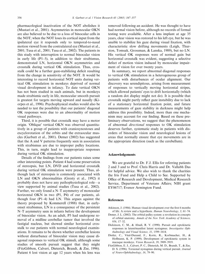
Fig. 1. Representative records of left eye of P1 during monocular viewing (right eye was occluded); upward deflections indicate upward or rightward
movements. In the left panel, during fixation of a stationary target, she shows rightward eye drifts (LN) with negligible vertical component. During
viewing of the 22.5�/s upward OK stimulus (middle panel), her response consists of diagonal nystagmus, with slow phases directed up and rightward.During viewing of the downward OK stimulus (right panel), her response consists of diagonal nystagmus, with slow phases directed down and
rightward. Note that the slow phases of the horizontal component of her nystagmus are faster (increased slope) during vertical OK stimulation than
during fixation.
S. Garbutt et al. / Vision Research 43 (2003) 347–357 349

methodological factors that might cause crosstalk, we
compared the vectorial nature of the response to vertical
stimulus motion in these patients with the group of
normal subjects. To do this we calculated the median
horizontal and the median vertical slow phase eye ve-
locity during each 20 s of vertical OK stimulation. We
then subtracted the median horizontal and vertical drift
velocity during fixation from the respective componentsof slow phase eye velocity during OKN. From these
data, we measured the horizontal crosstalk during ver-
tical OK stimulation (corrected horizontal velocity/cor-
rected vertical velocity), expressed as a percentage. We
compared these measurements for each response of each
patient with the pooled data from our normal subjects
for the corresponding stimulus. Because most of the
data were not normal in distribution, we calculatedmedians and used the Mann–Whitney rank-sum test to
compare responses of patients with pooled responses of
normal subjects to corresponding stimuli.
3. Results
3.1. Optokinetic nystagmus slow phase velocity
3.1.1. Vertical optokinetic nystagmus
In the normal subjects, during binocular and mon-
ocular viewing, vertical OKN gain was usually asym-
metrical with the response to downward motion having
a lower gain (up–down asymmetry). These data are
summarized in Tables 2 and 3.
In four patients (P1, P2, P4, P5) vertical OKN gain,at one stimulus speed at least, was significantly less
ðp < 0:05Þ than that for our control subjects. VerticalOKN asymmetries were evident in all of our patients
and these asymmetries at times exceeded the 95% con-
fidence intervals for our control subjects (Tables 2 and
3). An up–down asymmetry was the most common
finding in the patients. However, down–up asymmetries
(the upward response having a lower gain than the
downward response) were also evident but these asym-
metries were never statistically significant. The direction
of the asymmetry (up–down or down–up) could varyaccording to the speed of the stimulus or according to
whether the patient was viewing with both eyes, with the
right eye or with the left eye. As up–down and down–up
asymmetries occurred in both strabismic and non-
strabismic eyes, the direction of the asymmetry did not
appear to be related to which eye was deviating. How-
ever, a down–up asymmetry did occur more frequently
when the patients viewed the stimulus at the higherspeed.
3.1.2. Horizontal optokinetic nystagmus
For the control subjects a clear preference for right-
to-left or left-to-right stimulation during binocular or
monocular stimulation was absent (Tables 4 and 5).
One patient (P1) demonstrated a substantial nasal-
to-temporal (N–T) asymmetry during monocularviewing. In this patient the slow phase velocity was
significantly greater ðp < 0:05Þ in response to nasallymoving stimuli (to the left when viewing with the right
eye and to the right when viewing with the left eye)
compared with temporally moving stimuli. The mon-
ocular temporal-to-nasal (T–N) response had slow
phases in the same direction as the stimulus motion,
whereas in response to N–T stimulation the slow phasescould be in the correct direction, although diminished,
or could be in the inappropriate direction (the opposite
direction to stimulus movement). When both eyes were
Table 2
Summary of vertical binocular OKN gain values
Patient Up 12�/s Dn 12�/s Up 22.5�/s Dn 22.5�/s Up–Dn 12�/s Up–Dn 22.5�/s
1 RE 0.58�� 0.6 0.33�� 0.37 )0.02 )0.03LE 0.78 0.46�� 0.38 0.32 0.32� 0.06
2 RE 0.65 0.49�� 0.61 0.55� 0.16� 0.06
LE 0.62�� 0.41�� 0.58 0.57 0.21� 0.01
3 RE 0.73 0.69� 0.62 0.54� 0.04 0.08
LE 0.65 0.67 0.54 0.55 )0.02 )0.014 RE cb cb 0.24�� 0.16�� cb 0.08
LE 0.11�� 0.00�� 0.17�� 0.11�� 0.11 )0.065 RE 0.65 0.60 0.53 0.53� 0.05 0.00
LE 0.49�� 0.46�� 0.36 0.35 0.03 0.01
6 RE cb cb 0.86� 0.65� cb 0.21
LE 0.87� 0.62 0.86� 0.65� 0.25� 0.21�
NS RE 0:70� 0:04 0:61� 0:03 0:50� 0:08 0:39� 0:06 0:09� 0:03 0:11� 0:05LE 0:71� 0:04 0:64� 0:04 0:50� 0:08 0:43� 0:07 0:07� 0:03 0:07� 0:05
RE: right eye; LE: left eye; Up: upward moving stimulus; Dn: downward moving stimulus; cb: scleral search coil broke during this part of
experimental session; (��): value less than the lower 5% confidence interval for normal subjects means; (�): value exceeded upper 95% confidenceinterval for normal subjects means; Up–Dn: Difference between upward and downward gain, positive values indicate that upward gain was greater
than downward gain and negative values indicate that downward gain was greater than upward gain. Data are means from viewing eye; for normal
subjects (NS) data are pooled means (� standard errors).
350 S. Garbutt et al. / Vision Research 43 (2003) 347–357

viewing, the slow phases of the OK response were al-
ways in the appropriate direction and higher gains were
recorded in response to rightward moving stimuli. Pa-
tient 4 also showed a N–T asymmetry, but the gain
values were low.
Two patients (P3, P5), during monocular viewing
conditions, rather than the classic N–T asymmetry
demonstrated a significant bias ðp < 0:05Þ for one di-rection of horizontal motion. The same directional bias
was also apparent when these patients viewed the stim-
ulus binocularly. Thus, in P3 and P5 gain was signifi-
cantly greater ðp < 0:05Þ for leftward moving OK
stimuli irrespective of which eye was viewing. Patient 2
also had a directional bias of horizontal OKN with
higher rightward gains but during monocular viewing
this was only significant ðp < 0:05Þ when she viewed astimulus moving at 22.5�/s with her left eye.Paired comparison of monocular horizontal and
vertical OK responses for each patient�s eye (Tables 3and 5) showed substantial variability, and no consistentdifferences. However, in general, patients with poor
horizontal responses had somewhat better vertical re-
sponses, whereas other patients showed similar gain
values for horizontal and vertical responses.
Table 3
Summary of vertical monocular OKN gain values
Patient Up 12�/s Dn 12�/s Up 22.5�/s Dn 22.5�/s Up–Dn 12�/s Up–Dn 22.5�/s
1 RE 0.62 0.41�� 0.21�� 0.20�� 0.21� 0.01
LE 0.72 0.67 0.18�� 0.29�� 0.05 )0.112 RE 0.47�� 0.29�� 0.42 0.47 0.18� )0.05
LE 0.7 0.41�� 0.54 0.27�� 0.29� 0.27�
3 RE 0.71 0.51 0.61 0.53� 0.20� 0.08
LE 0.80� 0.67 0.62 0.40 0.13� 0.22�
4 RE cb cb 0.07�� 0.23�� cb )0.16LE 0.25�� 0.13�� 0.17�� 0.07�� 0.12� 0.10
5 RE 0.64 0.53 0.34�� 0.21�� 0.11 0.13
LE 0.40�� 0.14�� 0.30�� 0.10�� 0.26� 0.20�
6 RE cb cb 0.82� 0.65� cb 0.17
LE 0.97� 0.69 0.79� 0.67� 0.28� 0.12
NS RE 0:66� 0:06 0:60� 0:06 0:52� 0:07 0:42� 0:05 0:06� 0:03 0:10� 0:04LE 0:64� 0:07 0:61� 0:07 0:55� 0:05 0:48� 0:07 0:03� 0:02 0:07� 0:03
RE: right eye; LE: left eye; Up: upward moving stimulus; Dn: downward moving stimulus; cb: scleral search coil broke during this part of
experimental session; (��): value less than the lower 5% confidence interval for normal subjects means; (�): value exceeded upper 95% confidenceinterval for normal subjects means; Up–Dn: Difference between upward and downward gain, positive values indicate that upward gain was greater
than downward gain and negative values indicate that downward gain was greater than upward gain. Data are means from viewing eye; for normal
subjects (NS) data are pooled means (� standard errors).
Table 4
Summary of horizontal binocular OKN gain values
Patient R 12�/s L 12�/s R 22.5�/s L 22.5�/s R–L 12�/s R–L 22.5�/s
1 RE 0.72 0.43�� 0.20�� 0.07�� 0.29� 0.13�
LE 0.82 0.14�� 0.20�� 0.07�� 0.68� 0.13�
2 RE 0.61�� 0.39�� 0.7 0.57 0.22� 0.13�
LE 0.61�� 0.38�� 0.75 0.55 0.23� 0.20�
3 RE 0.64�� 0.83 0.63 0.75 )0.19� )0.12�
LE 0.55�� 0.83 0.69 0.78 )0.28� )0.09�
4 RE cb cb 0.34�� 0.23�� cb 0.11�
LE 0.62�� 0.20�� 0.08�� 0.19�� 0.42� )0.11�
5 RE 0.50�� 0.62�� 0.16�� 0.48�� )0.12� )0.32�
LE 0.57�� 0.60�� 0.18�� 0.47�� )0.03 )0.29�
6 RE cb cb 0.81� 0.88� cb )0.07�
LE 0.66�� 0.59�� 0.76 0.73 0.07� 0.03
NS RE 0:80� 0:05 0:79� 0:06 0:66� 0:07 0:66� 0:064 0:06� 0:02 0:03� 0:01LE 0:81� 0:04 0:79� 0:05 0:68� 0:07 0:66� 0:07 0:04� 0:01 0:02� 0:01
RE: right eye; LE: left eye; R: rightward moving stimulus; L: leftward moving stimulus; cb: scleral search coil broke during this part of experimental
session; negative values indicate that slow phase were in inappropriate direction (i.e. opposite direction to stimulus motion); (��): value less than thelower 5% confidence interval for normal subjects means; (�): value exceeded upper 95% confidence interval for normal subjects means; R–L:
Difference between rightward and leftward gain; for patient, data positive values indicate that rightward gain was greater than leftward gain, and
negative values indicate that leftward gain was greater than rightward gain. Data are means from viewing eye; for normal subjects (NS) data are
pooled means (� standard errors).
S. Garbutt et al. / Vision Research 43 (2003) 347–357 351

3.2. Crosstalk
3.2.1. Responses to vertical optokinetic stimulus motion
Vertical stimulus motion induced nystagmus with a
horizontal component that exceeded 95% confidence
intervals for our control subjects, in at least one direc-
tion, in all six patients. Representative data from P1 are
shown in Fig. 1, with corresponding plots of slow phase
velocity in Fig. 2. Our patients showed eye drift during
monocular fixation (LN in P1–4); these data are sum-
marized in Table 6. However, drifts during fixation
could not account for the magnitude of horizontal
Table 5
Summary of horizontal monocular OKN gain values
Patient R 12�/s L 12�/s R 22.5�/s L 22.5�/s R–L 12�/s R–L 22.5�/s
1 RE )0.19�� 0.22�� )0.06�� 0.18�� )0.41� )0.24�
LE 0.69 )0.31�� 0.40�� )0.14�� 1.0� 0.54�
2 RE 0.38�� 0.35�� 0.53 0.49 0.03 0.04
LE 0.53�� 0.43�� 0.45�� 0.36�� 0.10 0.09�
3 RE 0.64 0.83 0.59 0.80� )0.19� )0.21�
LE 0.55�� 0.78 0.67 0.74 )0.23� )0.07�
4 RE cb cb )0.01�� 0.04�� cb )0.05LE 0.36�� 0.13�� 0.18�� 0.12�� 0.23� 0.06
5 RE 0.21�� 0.76 0.22�� 0.43 )0.55� )0.21�
LE 0.00�� 0.57 0.09�� 0.37�� )0.57� )0.28�
6 RE cb cb 0.81� 0.86� cb )0.05LE 0.69 0.66 0.81� 0.77� 0.03 0.04
NS RE 0:66� 0:05 0:71� 0:06 0:52� 0:08 0:56� 0:08 0:05� 0:01 0:06� 0:01LE 0:72� 0:07 0:66� 0:06 0:62� 0:08 0:59� 0:08 0:07� 0:03 0:04� 0:01
RE: right eye; LE: left eye; R: rightward moving stimulus; L: leftward moving stimulus; cb: scleral search coil broke during this part of experimental
session; negative values indicate that slow phase were in inappropriate direction (i.e. opposite direction to stimulus motion); (��): value less than thelower 5% confidence interval for normal subjects means; (�): value exceeded upper 95% confidence interval for normal subjects means; R–L:
Difference between rightward and leftward gain; for patient, data positive values indicate that rightward gain was greater than leftward gain, and
negative values indicate that leftward gain was greater than rightward gain. Data are means from viewing eye; for normal subjects (NS) data are
pooled means (� standard errors).
Fig. 2. Plots of horizontal and vertical components of slow phase velocity of nystagmus during monocular vertical OK stimulation at 22.5�/s, andduring monocular fixation (LN) from patient 1. Dotted lines are 95% prediction intervals for normal subjects. Solid lines connect median points
during fixation and OK stimulation (black, right eye, gray, left eye), and indicate the change of direction of nystagmus produced by OK stimulation.
Note that the change in velocity of horizontal components induced by vertical OK stimuli (crosstalk) is predominantly to the right, irrespective of
which eye views.
352 S. Garbutt et al. / Vision Research 43 (2003) 347–357

components of OK responses to vertical stimulus mo-
tion. Thus, in P1–3, P5 and P6, the horizontal velocity of
the viewing eye during vertical OK stimulation at 22.5�/swas significantly greater ðp < 0:01Þ than during fixation.P4 (who had low-gain OK responses) showed greater
horizontal velocity during OK stimulation than during
fixation when she viewed with her left eye, but not when
she viewed with her right eye.After correcting for drifts during fixation, we calcu-
lated the percentage of crosstalk (see Section 2); these
data are also summarized in Table 6. All patients
showed significantly greater horizontal crosstalk ðp <0:001Þ for at least one vertical OK stimulus, com-
pared with controls. Fig. 3 summarizes the change in
median eye velocity vector during vertical OK stimu-
lation, for each viewing-eye response of each patient.Each vector was calculated by subtracting the median
horizontal and vertical drift velocity during fixation
from the respective components of slow phase eye ve-
locity during OKN. In general, the percentage of cros-
stalk was greater in response to the faster (22.5�/s)stimulus.
The direction of horizontal crosstalk tended to re-
main the same for each patient. Thus, in P1 (Figs. 1 and
2), the direction of horizontal crosstalk (to her right)
remained the same no matter which eye viewed (or inwhich direction she manifested her LN). In general, the
direction of horizontal crosstalk corresponded with the
direction of drift of slow phases with both eyes viewing
the OK stimulus (Table 6), except for P6 who showed no
consistent horizontal drifts. Horizontal crosstalk oc-
curred with either upward or downward OK stimuli, or
both, idiosyncratically for each patient. During binoc-
ular OK stimulation, responses were qualitatively simi-lar to those during viewing with the right eye in P2–5,
and with the left eye in P1 and P6.
Table 6
Summary of median eye velocity during fixation and median percentage crosstalk during vertical OK stimulus motion
Patient Eye velocitya Eye velocitya % OKN crosstalk % OKN crosstalk % OKN crosstalk % OKN crosstalk
RE fixation LE fixation RE view 22.5�/s LE view 22.5�/s RE view 12�/s LE view 12�/s
1 REH: )3.64 REH: þ3.81 Up: RE: 10.3b Up: RE: 44.8b Up: RE: 29.2b Up: RE: 3.9
REV: )0.71 REV: )0.69 LE: 17.0b LE: 44.8b LE: 31.7b LE: 15.8c
LEH: )3.08 LEH: þ5.00 Dn: RE: 58.4b Dn: RE: 40.9b Dn: RE: 76.9b Dn: RE: 35.8b
LEV: )1.41 LEV: )0.17 LE: 65.6b LE: 35.7b LE: 89.8b LE: 42.5b
2 REH: þ0.02 REH: þ0.16 Up: RE: 2.5 Up: RE: 2.7c Up: RE: 4.5c Up: RE: 2.7
REV: þ0.04 REV: þ0.02 LE: 9.3b LE: 1.4 LE: 2.1 LE: 1.2
LEH: )0.02 LEH: þ0.44 Dn: RE: 5.8b Dn: RE: 3.7 Dn: RE: 3.8b Dn: RE: 3.3
LEV: þ0.01 LEV: þ0.09 LE: 30.3b LE: 14.1b LE: 27.8b LE: 14.1
3 REH: )0.54 REH: þ0.15 Up: RE: 12.7b Up: RE: 2.9 Up: RE: 8.6c Up: RE: 3.4
REV: þ0.01 REV: )0.13 LE: 6.0b LE: 7.8 LE: 18.7b LE: 7.0
LEH: )0.67 LEH: þ0.01 Dn: RE: 4.5c Dn: RE: 0.7 Dn: RE: 5.7c Dn: RE: 9.9c
LEV: )0.48 LEV: þ0.04 LE: 7.8c LE: 10.5b LE: 12.0b LE: 5.5
4 REH: )0.22 REH: þ0.04 Up: RE: 39.9b Up: RE: 6.5 Up: RE: cb Up: RE: cb
REV: )0.10 REV: þ0.52 LE: 26.5b LE: 5.3 LE: 34.4b LE: 12.0
LEH: )0.22 LEH: þ0.33 Dn: RE: 101.0 Dn: RE: 8.1 Dn: RE: cb Dn: RE: cb
LEV: þ0.10 LEV: þ0.07 LE: 29.8c LE: 28.1b LE: 64.6b LE: 13.9b
5 REH: )0.20 REH: )0.21 Up: RE: 15.8b Up: RE: 1.1 Up: RE: 14.1c Up: RE: 0.1
REV: þ0.13 REV: þ0.09 LE: 63.4b LE: 30.4b LE: 57.4b LE: 28.3b
LEH: )0.02 LEH: )0.27 Dn: RE: 23.4b Dn: RE: 16.0b Dn: RE: 9.6c Dn: RE: 13.6b
LEV: þ0.04 LEV: þ0.27 LE: 21.7b LE: 18.7b LE: 20.9b LE: 15.8b
6 REH: )0.06 REH: )0.42 Up: RE: 6.1b Up: RE: 31.9b Up: RE: cb Up: RE: cb
REV: þ0.01 REV: )0.21 LE: 4.1b LE: 20.0b LE: 20.0b LE: 15.2b
LEH: þ0.61 LEH: )0.05 Dn: RE: 4.9b Dn: RE: 11.6 Dn: RE: cb Dn: RE: cb
LEV: þ0.92 LEV: )0.01 LE: 15.3b LE: 4.5 LE: 13.2b LE: 3.2
NS REH: þ0.02 REH: þ0.03 Up: RE: 1.4 Up: RE: 7.7 Up: RE: 0.3 Up: RE: 6.8
REV: )0.08 REV: )0.02 LE: 1.8 LE: 6.3 LE: 0.5 LE: 6.6
LEH: þ0.12 LEH: þ0.06 Dn: RE: 2.0 Dn: RE: 7.4 Dn: RE: 1.6 Dn: RE: 5.4
LEV: )0.11 LEV: )0.11 LE: 2.3 LE: 1.0 LE: 4.5 LE: 3.5
LEH: left eye horizontal; LEV: left eye vertical; REH: right eye horizontal; REV: right eye vertical; cb: connection on scleral search coil broke during
this part of the experimental session; NS: pooled data from normal subjects.a Velocity in �/s; positive values indicate rightward and upward movements.bCrosstalk significantly different from control subjects p < 0:01.c Crosstalk significantly different from control subjects p < 0:05.
S. Garbutt et al. / Vision Research 43 (2003) 347–357 353

The direction of crosstalk did not appear to be related
to which eye was amblyopic. Thus, P1 and P5 both hadleft-sided amblyopia, but P1 showed a rightward hori-
zontal crosstalk whereas P5 showed leftward crosstalk.
In two patients crosstalk could be generally related to
the directional bias of horizontal OKN. Thus, P2 had
better rightward OKN under all viewing conditions and,
when present, her crosstalk was rightward, whereas P5
had better leftward OKN and showed leftward cros-
stalk. We also considered whether a change in hori-zontal eye position (i.e., a deviation) during vertical
stimulation could account for the changes in horizontal
eye velocity during vertical OK stimulation. Although
some patients did develop exotropia (P1 and P3 during
right eye viewing) or esotropia (P2 during right eye
viewing), in each case the horizontal component (cros-stalk) was substantially changed by the direction of the
vertical OK stimulus (Fig. 3), although the horizontal
deviation remained similar.
3.2.2. Responses to horizontal optokinetic stimulus motion
No patient showed a vertical component induced by
horizontal stimulus motion that exceeded 95% confi-
dence intervals for our control subjects. Thus, althoughvertical stimulus motion induced substantial horizontal
movements, horizontal stimulus motion did not induce
vertical crosstalk in our patients.
Fig. 3. Summary of responses of viewing eyes to vertical OK stimulation from all patients, comparing them to normal subjects. The data are
summarized as a form of polar plot. The amplitude of each response (length of line) was calculated from the median change of each component of eye
velocity during OK stimulation compared with during fixation of a stationary target (LN). The angle of the line away from vertical varied between
patients, and reflects the magnitude of the horizontal component (see Table 6). The data points are pooled responses from all normal subjects. The
asterisks (�) indicate that the complete response (of which only medians are shown) was significantly different from normal subjects ðp < 0:001Þ. Notethat missing data for some patients is due to a coil breaking during that part of the experimental session.
354 S. Garbutt et al. / Vision Research 43 (2003) 347–357

4. Discussion
4.1. Optokinetic nystagmus slow phase velocity
An asymmetry of vertical OKN, with upward stim-
ulus motion eliciting a greater response than downward
stimulus motion, was the most common finding in both
normal subjects and patients in our study. This up–down asymmetry has been reported previously in nor-
mal human subjects (Murasugi & Howard, 1989; van
den Berg & Collewijn, 1988). However, in some of our
patients, downward stimulus motion elicited greater
responses than upward stimulus motion (down–up
asymmetries), although these asymmetries were never
significant. Schor and Levi (1980) reported a number of
abnormalities in the monocular vertical OK response ofadult amblyopes. The most common deficit they noted
was a reduced velocity for upward slow phases resulting
in a down–up asymmetry. This was observed in both
the amblyopic and non-amblyopic eyes of some sub-
jects. Similarly, in a group of sixteen patients with early-
onset strabismus, Proudlock et al. (2001) reported poor
upward gains in the deviating eye of four patients and
the non-deviating eyes of six patients. We noted sig-nificantly reduced velocities for upward and downward
slow phases in the deviating and non-deviating eyes of a
number of our patients compared with control subjects
(Tables 2 and 3). On the other hand, two patients (P3,
P6) had vertical OK responses with a high gain. It is not
possible for us to draw any conclusions concerning the
relative influence of dissociated vertical deviation
(DVD), amblyopia or age of onset of deviation onvertical OK responses from the small and heteroge-
neous sample of patients that we studied.
A N–T horizontal OKN asymmetry was evident in P1
and also in P4, although her gain values were low. In
these patients, the monocular temporal-to-nasal re-
sponse had slow phases in the normal direction whereas
the response to nasal-to-temporal stimulation was in the
correct direction, although diminished, or was in theinappropriate direction. This inappropriate response to
an OK stimulus moving in the nasal-to-temporal direc-
tion has previously been reported in patients with latent/
manifest latent nystagmus (LN/MLN) (Dickinson &
Abadi, 1990; Kommerell & Mehdorn, 1982; Milojevic,
Windsor, & Burian, 1967; Tsutsui & Fukai, 1979).
Further, it has been suggested that subjects with LN/
MLN do not have a genuine N–T OKN deficit and thatany apparent asymmetry or reversal of monocular OKN
might be the result of the summation of the horizontal
OKN with the spontaneous oscillation with the latter
being changed in some way by the stimulus (Dickinson
& Abadi, 1990).
Three patients (P2, P3, P5) displayed an asymmetry
in horizontal OKN that resembled a directional bias
(leftward or rightward asymmetry rather than a N–T
asymmetry). This is not the typical asymmetry associ-
ated with abnormal binocular visual development re-
ported in the literature. We note, however, that in P5
this directional asymmetry could be due to the patient�scerebellar defect and may have disguised any N–T
asymmetry.
4.2. Crosstalk
A novel finding of these experiments was that vertical
OK stimulation, in individuals who lack normal binoc-
ular visual development, caused diagonal nystagmus
responses. The magnitude and direction of the hori-zontal component varied between patients, and tended
to be greater with the faster OK stimulus. The hori-
zontal responses induced by vertical OK stimuli cannot
simply be ascribed to superposition of the slow phases of
LN during monocular fixation, which was slower and
sometimes in the opposite direction (for example during
right eye viewing in P1, as shown in Figs. 2 and 3). What
is the possible significance of these misdirected OK re-sponses?
We considered three possibilities. First, that lacking
normal binocular visual development, the responses
corresponded to those encountered in afoveate, laterally
eyed animals such as the rabbit (Tan, van der Steen,
Simpson, & Collewijn, 1993). During normal locomo-
tion, the optic flow causes an OK stimulation that is
stronger in the nasal-to-temporal direction horizontally,and usually downward, the ground being more proxi-
mate that the sky. In foveate subjects with normal bin-
ocular vision, it is possible to point both eyes at an
object and use ‘‘smooth pursuit’’ tracking to hold gaze
on target despite the effects of the optic flow (Miles,
1993). However, in species that do not possess binocu-
lar, foveate vision an OK bias for movement in the
temporal-to-nasal direction would partly compensatefor this asymmetry of optic flow. We wondered whether
the vertical–horizontal crosstalk that we observed could
be a component of this OK bias to negate the effects of
optic flow in the absence of binocular vision. However, a
number of our patients did not follow the classic N–T
asymmetry, but had an asymmetry that resembles a di-
rectional bias. Nevertheless, crosstalk and the OK bias
were generally in the same direction.Second, it seems possible that an ‘‘uncalibrated’’
motion vision system is responsible for the variable re-
sponses that we encountered. Monkeys who have been
deprived of normal binocular vision from birth lack
normal binocular responses in cortical area MT (Kior-
pes et al., 1996) and the pretectal NOT (Mustari et al.,
2001). In these animals, in contrast to normal monkeys
in whom all NOT units are sensitive to stimuli to eithereye, the NOT becomes monocular, the majority of units
being dominated by the contralateral eye. This change in
NOT activity has been shown to contribute to LN since
S. Garbutt et al. / Vision Research 43 (2003) 347–357 355
pharmacological inactivation of the NOT abolishes it
(Mustari et al., 2001). Asymmetries in monocular OKN
are also believed to be due to a loss of binocular cells in
the NOT; when the NOT loses its cortical input from the
ipsilateral eye it responds only to temporal-to-nasal
motion viewed from the contralateral eye (Mustari et al.,
2001; Tusa et al., 2001; Tusa et al., 2002). The patients in
this study with interruption to normal binocular visionin early life (P1–5), in addition to their strabismus,
demonstrated LN, horizontal OKN asymmetries and
crosstalk during vertical OK stimulation. Thus, cros-
stalk could be a further gaze-stabilizing deficit resulting
from the change in sensitivity of the NOT. It would be
interesting to record horizontal NOT units during ver-
tical OK stimulation in monkeys deprived of normal
visual development in infancy. To date vertical OKNhas not been studied in such animals, but in monkeys
made strabismic early in life, monocular smooth pursuit
is greatest for targets moving upward and nasally (Ki-
orpes et al., 1996). Psychophysical studies would also be
needed to test the possibility that our patients� diagonalOK responses were due to an abnormality of motion
visual pathways.
Third, it is possible that crosstalk may have a motororigin. �Oblique� vertical OKN was observed quantita-tively in a group of patients with craniosynostoses and
excyclorotation of the orbits and the extraocular mus-
cles (Garbutt et al., 2001). Demer (2002) has suggested
that the A and V patterns frequently seen in association
with strabismus are due to improper pulley locations.
This, in turn, might lead to inappropriate responses
during vertical OK stimulation.Details of the findings from our patients raises some
other interesting points. Patient 4 had some preservation
of stereopsis, but LN, DVD and horizontal crosstalk
during vertical OK stimulation were present. Thus, al-
though lack of stereopsis is commonly associated with
LN and OKN abnormalities (Gresty et al., 1992) it
probably does not have any pathophysiological role––a
view supported by animal studies (Tusa et al., 2002).Further, we only found a N–T asymmetry of monocular
horizontal OKN in two (P1, P4) of our patients, al-
though four (P1–4) had LN. This argues against the
theory proposed by Kommerell (1988) that, in early-
onset strabismus, LN is a consequence of the persistent
N–T asymmetry seen in infants before the development
of binocular vision. As an adult, P5 had undergone re-
moval of a midline cerebellar tumor that involved thefastigial nucleus, but showed similar horizontal cros-
stalk to our patients with normal neurological examin-
ations. It remains to be shown whether cerebellar lesions
without disturbance of binocular vision may cause di-
agonal responses to vertical OK stimuli, although some
studies of smooth pursuit suggest that they might
(FitzGibbon, Calvert, Dieterich, Brandt, & Zee, 1996).
Patient 6 lost vision at age 12 years when his lens was
removed following an accident. He was thought to have
had normal vision before, although no records of formal
testing were available. After a lens implant at age 35
years, clear vision was restored to his left eye, but he was
unable to stabilize his gaze during visual fixation, with
characteristic slow drifting movements (Leigh, Thur-
ston, Tomsak, Grossman, & Lanska, 1989), but no LN.
His vertical OK responses were of normal gain buthorizontal crosstalk was evident, suggesting a selective
defect of motion vision induced by monocular impair-
ment of vision for over twenty years.
In summary, we report abnormal diagonal responses
to vertical OK stimulation in a heterogeneous group of
patients with disturbances of ocular alignment. Our
discovery was serendipitous, arising from measurement
of responses to vertically moving horizontal stripes,which allowed patients� eyes to drift horizontally (whicha random dot display might not do). It is possible that
crosstalk might partly reflect gaze instability due to lack
of a stationary horizontal fixation point, and future
measurements of gaze stability in darkness might help
address this possibility. Indeed, more than one mecha-
nism may account for our finding. Based on these pre-
liminary observations, we suggest that the phenomenonof abnormal directional responses to OK stimulation
deserves further, systematic study in patients with dis-
orders of binocular vision and neurological lesions of
areas that normally ensure that eye movements are in
the appropriate direction (such as the cerebellum).
Acknowledgements
We are grateful to Dr. F.J. Ellis for referring patients
2 and 3 and to Prof. Chris Harris and Dr. Vallabh Das
for helpful advice. We also wish to thank the charities
the Iris Fund and Help a Child to See. Supported by
Office of Research and Development, Medical Research
Service, Department of Veterans Affairs; NIH grant
EY06717; Evenor Armington Fund.
References
Atkinson, J. (1984). Human visual development over the first 6 months
of life. A review and a hypothesis. Human Neurobiology, 3, 61–74.
Demer, J. L. (2002). The orbital pulley system: a revolution in concepts
of orbital anatomy. Annals of the New York Academy of Sciences,
956, 17–32.
Dickinson, C. M., & Abadi, R. V. (1990). Pursuit and optokinetic
responses in latent/manifest latent nystagmus. Investigative Oph-
thalmology and Visual Science, 31, 1599–1614.
Distler, C., Vital-Durand, F., Korte, R., Korbmacher, H., &
Hoffmann, K. P. (1999). Development of optokinetic system in
macaque monkeys. Vision Research, 39, 3909–3919.
FitzGibbon, E. J., Calvert, P. C., Dieterich, M. D., Brandt, T., & Zee,
D. S. (1996). Torsional nystagmus during vertical pursuit. Journal
of Neuro-Ophthalmology, 16, 79–90.
356 S. Garbutt et al. / Vision Research 43 (2003) 347–357

Gresty, M. A., Metcalfe, T., Timms, C., Elston, J., Lee, J., & Liu,
C. (1992). Neurology of latent nystagmus. Brain, 115, 1303–
1321.
Hainline, L., Lemerise, E., Abramov, I., & Turkel, J. (1984).
Orientational asymmetries in small-field optokinetic nystagmus in
human infants. Behavioral Brain Research, 13, 217–230.
Kiorpes, L., Walton, P. J., O�Keefe, P., Movshon, J. A., & Lisberger,S. G. (1996). Effects of early-onset artificial strabismus on pursuit
eye movements and on neuronal responses in area MT of macaque
monkeys. Journal of Neuroscience, 16, 6537–6553.
Kommerell, G. (1988). Ocular motor phenomena in infantile strabis-
mus. In C. Kennard & F. Clifford Rose (Eds.), Physiological
aspects of clinical neuro-ophthalmology (pp. 357–375). London:
Chapman and Hall.
Kommerell, G., & Mehdorn, E. (1982). Is the optokinetic defect the
cause of congenital and latent nystagmus?. In G. Lennerstrand, D.
S. Zee, & E. L. Keller (Eds.), Functional basis of ocular motility
disorders (pp. 159–167). Oxford: Pergamon Press.
Leigh, R. J., Thurston, S. E., Tomsak, R. L., Grossman, G. E., &
Lanska, D. J. (1989). Effect of monocular visual loss upon stability
of gaze. Investigative Ophthalmology and Visual Science, 30, 288–
292.
Miles, F. A. (1993). The sensing of rotational and translational optic
flow by the primate optokinetic system. In F. A. Miles & J.
Wallman (Eds.), Visual motion and its role in the stabilization of
gaze (pp. 393–403). Elsevier Science Publishers B.V.
Milojevic, B., Windsor, C. E., & Burian, H. M. (1967). Electronystag-
mographical study of latent ocular nystagmus. Archives of Otolar-
yngology, 85, 283–286.
Murasugi, C. M., & Howard, I. P. (1989). Up-down asymmetry in
human vertical optokinetic nystagmus and afternystagmus: contri-
butions of the central and peripheral retinae. Experimental Brain
Research, 77, 183–192.
Mustari, M. J., Tusa, R. J., Burrows, A. F., Fuchs, A. F., &
Livingston, C. A. (2001). Gaze-stabilising deficits and latent
nystagmus in monkeys with early-onset deprivation: Role of the
pretectal NOT. Journal of Neurophysiology, 86, 662–675.
Proudlock, F. A., McLean, R. J., Farooq, S., & Gottlob, I. (2001).
Vertical asymmetries during monocular OKN in early onset
strabismus. Investigative Ophthalmology and Visual Science
(Suppl.), 42, 53.
Schor, C. M. (1983). Subcortical binocular suppression affects the
development of latent and optokinetic nystagmus. American
Journal of Optometry and Physiological Optics, 60, 481–502.
Schor, C. M. (1993). Development of OKN. In F. A. Miles & J.
Wallman (Eds.), Visual motion and its role in the stabilisation of
gaze (pp. 301–320). Amsterdam: Elsevier.
Schor, C. M., & Levi, D. M. (1980). Disturbances of small-field
horizontal and vertical nystagmus in amblyopia. Investigative
Ophthalmology and Visual Science, 19, 668–683.
Sparks, D. L., Mays, L. E., Gurski, M. R., & Hickey, T. L. (1986).
Long- and short-term monocular deprivation in the rhesus
monkey: effects on visual fields and optokinetic nystagmus. Journal
of Neuroscience, 6, 1771–1780.
Tan, H. S., van der Steen, J., Simpson, J. I., & Collewijn, H. (1993).
Three-dimensional organization of optokientic responses in the
rabbit. Journal of Neurophysiology, 69, 303–317.
Tsutsui, J., & Fukai, S. (1979). Human strabismic cases suggestive of
asymmetric projection of asymmetric projection of the visual
pathway. In R. D. Reinecke (Ed.), Strabismus (pp. 79–88). New
York: Grune and Stratton.
Tusa, R. J., Mustari, M. J., Burrows, A. F., & Fuchs, A. F. (2001).
Gaze-stabilising deficits and latent nystagmus in monkeys with
brief, early-onset visual deprivation: Eye movement recordings.
Journal of Neurophysiology, 86, 651–661.
Tusa, R. J., Mustari, M. J., Das, V. E., & Boothe, R. G. (2002). Animal
models for visual deprivation-induced strabismus and nystagmus.
Annals of the New York Academy of Sciences 2002, 956, 346–360.
Tychsen, L., Hurtig, R. R., & Thalacker, J. A. (1984). Defective
downward smooth pursuit in infantile strabismus. Investigative
Ophthalmology and Visual Science (Suppl.), 25, 74.
Tychsen, L., Leibole, M., & Drake, D. (1996). Comparison of manifest
latent nystagmus and nasotemporal asymmetries of optokinetic
nystagmus in adult humans and macaque monkeys who have
infantile strabismus. Strabismus, 4, 177.
van den Berg, A. V., & Collewijn, H. (1988). Directional asymmetries
of human optokinetic nystagmus. Experimental Brain Research, 70,
597–604.
S. Garbutt et al. / Vision Research 43 (2003) 347–357 357
Copyright © 2022 FDOKUMEN