A study of low-strain and medium-strain grain boundary engineering

Patterns of Strain in the Macaque Ulna During FunctionalActivity
BRIGITTE DEMES,1,3,* JACK T. STERN, JR.,1,3
MICHAEL R. HAUSMAN,2 SUSAN G. LARSON,1,3
KENNETH J. MCLEOD,3 AND CLINTON T. RUBIN1,3
1Department of Anatomical Sciences, School of Medicine, State Universityof New York, Stony Brook, New York 11794-80812Department of Orthopedics, Mount Sinai School of Medicine,New York, New York 100293Program in Biomedical Engineering, Health Sciences Center, StateUniversity of New York, Stony Brook, New York 11794-8181
ABSTRACT In vivo bone strain experiments were performed on the ulnaeof three female rhesus macaques to test how the bone deforms duringlocomotion. The null hypothesis was that, in an animal moving its limbspredominantly in sagittal planes, the ulna experiences anteroposterior bend-ing. Three rosette strain gauges were attached around the circumference ofthe bone slightly distal to midshaft. They permit a complete characterizationof the ulna’s loading environment. Strains were recorded during walking andgalloping activities. Principal strains and strain directions relative to the longaxis of the bone were calculated for each gauge site. In all three animals, thelateral cortex experienced higher tensile than compressive principal strainsduring the stance phase of walking. Compressive strains predominated at themedial cortex of two animals (the gauge on this cortex of the third animal didnot function). The posterior cortex was subject to lower strains; the nature ofthe strain was highly dependent on precise gauge position. The greaterprincipal strains were aligned closely with the long axis of the bone in twoanimals, whereas they deviated up to 45° from the long axis in the thirdanimal.
A gait change from walk to gallop was recorded for one animal. It was notaccompanied by an incremental change in strain magnitudes. Strains are atthe low end of the range of strain magnitudes recorded for walking gaits ofnonprimate mammals.
The measured distribution of strains in the rhesus monkey ulna indicatesthat mediolateral bending, rather than anteroposterior bending, is thepredominant loading regime, with the neutral axis of bending running fromanterior and slightly medial to posterior and slightly lateral. A variabledegree of torsion was superimposed over this bending regime. Ulnar mediolat-eral bending is apparently caused by a ground reaction force vector thatpasses medial to the forearm. The macaque ulna is not reinforced in the planeof bending. The lack of buttressing in the loaded plane and the somewhatcounterintuitive bending direction recommend caution with regard to conven-tional interpretations of long bone cross-sectional geometry. Am J PhysAnthropol 106:87–100, 1998. r 1998 Wiley-Liss, Inc.
KEY WORDS in vivo bone strain; macaque ulna; functionalmorphology
Inherent to functional interpretations ofshapes of bones are assumptions about theirmechanical behavior and the forces andmoments to which they are exposed. It is
Contract grant sponsor: National Science Foundation; Con-tract grant number: SBR 9507078.
*Correspondence to: Brigitte Demes, Department of Anatomi-cal Sciences, State University of New York, Stony Brook, NY11794-8081. E-mail: [email protected].
Received 15 July 1997; accepted 31 January 1998.
AMERICAN JOURNAL OF PHYSICAL ANTHROPOLOGY 106:87–100 (1998)
r 1998 WILEY-LISS, INC.

common practice to model long bones asbeams loaded by muscle, joint, and sub-strate reaction forces. The external forcesand moments are extrapolated from observa-tions of limb action during postural andlocomotor activities. Ideally, quantified limbkinematics and experimentally determinedsubstrate reaction forces and muscle activ-ity are used to predict the loading patterns.Even then, the situation is complicated bythe irregular shapes of long bones, whichdeviate from the simple geometric beamanalogues. Consequently, actual loadings canbe deduced only with a certain degree ofplausibility. Does limb action in sagittalplanes translate into bending in this plane(see, e.g., Ruff and Runestad, 1992; Demesand Jungers, 1993)? Does the greater versa-tility of movements that is said to character-ize primates imply a greater range of load-ing patterns of their limb bones and,consequently, the need for more robust bones(Kimura, 1995; Polk et al., 1997)? The as-sumptions inherent in the models can betested only by measuring the deformationsof bones directly during various activities.
The numbers of in vivo studies of longbone deformation are limited and are re-stricted to mostly cursorial, nonprimatemammals (Lanyon and Smith, 1969, 1970;Lanyon and Baggott, 1976; Goodship et al.,1979; Lanyon and Bourn, 1979; Lanyon etal., 1979; Carter et al., 1981; Rubin andLanyon, 1982, 1984; Biewener et al., 1983,1988; Bouvier and Hylander, 1984; Bie-wener and Taylor, 1986; Gross et al., 1992;Swartz et al., 1992; Biewener and Bertram,1993). In one of these studies, Biewener etal. (1983) demonstrated discrepancies be-tween loading patterns reconstructed fromthe orientation of the ground reaction forcevector and limb posture and those deter-mined from in vivo bone strain recordingsfor the horse metacarpus, substantiatingthe suspicion that loading reconstructionsmay lead to spurious results.
In vivo bone strain data for primate post-cranial bones exist for the proximal ulna of aspider monkey (Fleagle et al., 1981), theforelimb bones of gibbons (Swartz et al.,1989), and the tibia of humans (Lanyon etal., 1975; Burr et al., 1996). Macaques werechosen for our project to add the first strictly
quadrupedal primate to the data base. Thethree primate species for which strain datahave been recorded so far rely exclusively, orto a significant extent, on only one pair oflimbs for locomotion, which makes it diffi-cult to compare them directly with the non-primate quadrupeds for which strain dataare available. Various kinematic and kineticaspects of quadrupedal locomotion differ be-tween primates and nonprimate mammals.Primates have been shown to walk withlonger strides, greater angular excursions,and lower stride frequencies (Alexander andMaloiy, 1984; Reynolds, 1987; Demes et al.,1994). They tend to carry more weight ontheir hind limbs (Kimura et al., 1979; Demeset al., 1994). Primates also have relativelylonger limbs than nonprimate mammals(Alexander et al., 1979). It remains to beseen whether primate quadrupeds differfrom nonprimate quadrupeds in bone strainlevels or in any other aspect of the strainenvironment.
To completely characterize the loadingregime of a long bone, information fromthree, three-element gauges attached to thebone at the same transverse plane is re-quired (Gross et al., 1992). Gauge implanta-tion should be accomplished without cuttingthrough muscles, and gauge sites should bedistant enough from muscle attachment sitesto avoid local stress concentrations unre-lated to the global deformation of the bone.Only a few long bones lend themselves tosuch an analysis. We have chosen to studystrain in the ulna. Based on limb actionprimarily in the sagittal plane coupled withan active tripeps muscle during the stancephase of locomotion, we hypothesized thatthe ulna experiences anteroposterior bend-ing with the posterior surface in tension andthe with the anterior surface in compres-sion.
The ulna and the elbow display significantvariation among primates that correlateswith locomotor modes (see, e.g., Fleagle etal., 1975; Fleagle, 1983; Rose, 1988; Harri-son, 1989; Richmond et al., in press). Theelbow joint of terrestrial mammals is charac-terized by lateral and medial flanges at themargins of the trochlea that supposedly actto stabilize the joint (Jenkins, 1973). Cerco-pithecoids display this morphology, with a
88 B. DEMES ET AL.

posterolateral flange that contacts the ulnarolecranon in more extended joint positionsand an anteromedial flange that has moreextensive contact in flexed positions (Rose,1988). The supposed function of mediolat-eral buttressing of the elbow joint remainshypothetical without knowledge of the forcesacting in the frontal plane.
MATERIALS AND METHODS
Experiments were performed on threeadult female rhesus macaques. Prior to sur-gery, the animals were trained to walk witha pole attached to a collar. Strain gaugeimplantation and data collection followedthe protocol outlined in Gross et al. (1992).While the animal was under general anesthe-sia, a single incision approximately 6 cmlong was made on the dorsoulnar aspect ofthe forearm. Three rectangular, three-ele-ment rosette gauges (Kenkyujo, Tokyo, Ja-
pan; 2 mm long) were placed around thecircumference of the ulna at approximatelyone-third of its length from the distal end.Muscles and tendons were retracted, but nomuscle attachments were interrupted. Thegauge sites were exposed by scraping smallareas of the cortical surface clean of perios-teum. Subsequently, the surfaces were driedwith 100% chloroform, and the gauges wereglued on with isobutyl 2-cyanoacrylate mono-mer. To provide strain relief to the gauges,small expoxy resin flanges were screwedinto the bone 2 cm proximal to the gaugesites, several times the distance at whichlocal stress concentrations around the screwwould affect strains measured by the gauges(Timoshenko, 1958; Gross et al., 1992). Thewires from the gauges were run subcutane-ously to the vicinity of the shoulder, wherethey exited through a small incision. There,they were soldered to a 25-pin connector.
Fig. 1. Experimental setup. Strain gauges are at-tached to the ulna on the subject’s left side. Wires runsubcutaneously to the shoulder, where they surface andare attached to a connector carried in a pouch of a vestworn by the animal. The cable from the connector runsup a pole attached to the animal’s neck collar and, from
there, to the animal trainer and in a long loop to thecomputer. The superimposed trace is the reading from asingle element of one of the rosette gauges, which wasused to synchronize strain records and animal action.The image was taken from videotape.
89IN VIVO STRAIN IN THE MACAQUE ULNA
The animals wore a vest to hold the connec-tor in place and to prevent them from reach-ing it (Fig. 1).
In the first two experiments, data werecollected with the Vishay MeasurementGroup (Raleigh, NC) 2110 amplifiers, andchannels were sampled at 100 Hz; for thethird experiment, Syminex Inc. (Marseille,France) model SX 500 amplifiers collectedstrains at 409.6 Hz. Individual samplingperiods were 10 seconds long. The animalswere videotaped during the trials. One strainchannel was displayed on an oscilloscope. Avideo image of the oscilloscope was superim-posed on that of the animal with a special-effects generator, thereby enabling synchro-nization of strain events and animal activity.
Qualitative comparisons were performedbetween gaits recorded preoperatively andpostoperatively. Analyzed sequences werenot observably different from those seenprior to the instrumentation of the ulna.Because the animals were allowed to movefreely in any direction, video recordingswere not obtained in standard planes re-quired for quantitative gait analysis. Theanimals engaged in apparently normal loco-motion shortly after recovery from anesthe-sia. No signs of lameness or favoring of limbswere apparent. Data were gathered at thistime and again 2 days after surgery. Strainsduring walking, galloping, and climbing wererecorded. After the second period of data
collection, the animal was again anesthe-tized, and x-rays and computed tomography(CT) scans of the forearm were taken todocument gauge positions. Prior to gaugeremoval, passive loading of the forearm ofthe anesthetized animal in axial compres-sion, axial tension, and bending in fourdirections was performed to check propergauge function and to verify gauge positions.All three gauges were found to be bondedfirmly to the bone upon removal in twoanimals (animals 1 and 3). One of the threegauges was found loose in animal 2, anddata from that gauge were discarded. Lowstrain readings had already raised the suspi-cion that this gauge was not functioningproperly. Recovery was uneventful in each ofthe three animals.
Data were analyzed in a subroutine of theMacintosh program Igor (WaveMetrics, Inc.Lake Oswego, NY). By definition, principalstrains are the greatest tensile (positive)and compressive (negative) strains in a pla-nar strain field and are at right angles to oneanother. They were calculated for each ro-sette and for each sampling point over alocomotor cycle by using standard engineer-ing equations (Dally and Riley, 1991). Inaddition, the angle of the maximum (tensile)principal strain relative to the long axis ofthe bone was calculated by using the orienta-tion of the gauge relative to the long axis, asdetermined on the x-rays (Fig. 2). The exact
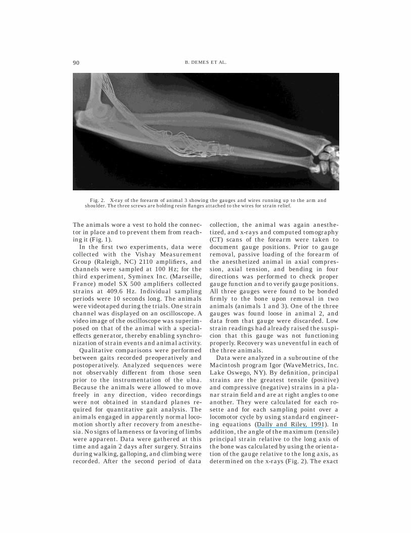
Fig. 2. X-ray of the forearm of animal 3 showing the gauges and wires running up to the arm andshoulder. The three screws are holding resin flanges attached to the wires for strain relief.
90 B. DEMES ET AL.

positions of the gauges around the circumfer-ence of the bone were identified on the CTscans. Gauge sites were not exactly homolo-gous in the three animals (see below). Foreach support phase, peak principal strainswere identified, and means of the peakstrains for all analyzed support phases werecalculated for each gauge. For the two ani-mals with strain readings from a completeset of three gauges, the distribution of nor-mal (tensile and compressive) strains in thecross section of the ulna at the level of thegauge sites was calculated by using com-bined beam and finite element model analy-sis (see Rybicki et al. 1977; Gross et al.,1992; calculations courtesy T. Gross andY.-X. Qin). This required knowledge of the
cross-sectional geometry of the bone at thelevel of the gauge sites, which was deter-mined from CT scans.
RESULTS
Peak principal strains in walking
Although an effort was made to placegauges on the medial, lateral, and posteriorcortices of the ulna, in two animals, theactual positions deviated slightly from thesegross anatomical directions. This is indi-cated in Figures 3–5. Nevertheless, we willrefer to the gauge sites as medial, lateral,and posterior across animals. We reportstrains for walking steps for all three ani-mals and for gallops for one of the animals.
Fig. 3. Principal strain magnitudes anddirections for three gauge sites of animal 1.The graphs on the left indicate the principaltensile (positive) and principal compressive(negative) strains as well as the angle of theprincipal tensile strain to the long axis ofthe bone for four consecutive walking steps.The horizontal bars indicate the supportphases. The arrows are graphic representa-tions of the mean peak principal tensile andcompressive strains for each gauge location.Exact gauge locations are shown on theright.
91IN VIVO STRAIN IN THE MACAQUE ULNA

Sampling frequencies of the amplifiers inthe first two experiments were not highenough to capture strain dynamics ad-equately at the higher speed gait. Averagevalues of peak principal strains and strainangles for all working gauges over all ana-lyzed gait cycles are given in Table 1. Fig-ures 3–5 illustrate strains over time for fourconsecutive steps of each animal. The meanmagnitude and orientation of the peak prin-cipal strains relative to the long axis of thebone are also indicated.
Compressive strain predominates at themedial cortex (Table 1). Strain angles arevery consistent within each animal but varyacross the two animals for which data fromthis aspect of the bone are available. Theprincipal compressive strain is aligned moreclosely with the long axis of the bone in thesetwo animals. The strain on the medial cortexin the direction of the long axis of the ulna,therefore, is compressive (Figs. 3–5).
At the lateral cortex, principal tensilestrain is higher than principal compressivestrain (Table 1). Strain magnitudes varyconsiderably between animals. The strainangles also vary, with the principal tensilestrain closely aligned with the long axis ofthe bone in animals 1 and 2 and deviating byalmost 45° from the long axis in animal 3.Nonetheless, because the principal tensilestrain is much higher than the principalcompressive strain in animal 3, the strain inthe direction of the long axis of the bone istensile, as it is in animals 1 and 2.
The posterior cortex experiences principalstrains that are more similar in magnitudeto one another, with principal compressionslightly higher in animals 1 and 3 andprincipal tension higher in animal 2. Equallyvariable is the alignment of the principalstrains with the long axis of the bone. Thelongitudinal strain for this gauge site in
Fig. 4. Principal strain magnitudesand directions for two gauge sites of ani-mal 2. For explanation, see Figure 3 leg-end.
92 B. DEMES ET AL.

animal 1 is very low and compressive, ishigher and tensile in animal 2, and is ofintermediate magnitude and compressive inanimal 3 (Figs. 3–5, Table 1).
Normal strain distribution in walking
For animals 1 and 3, in which all rosettegauges were operative, normal strains calcu-lated across a section through the ulna atthe level of the gauge sites indicate a bend-ing regime. The neutral axis ran from antero-medial to posterolateral throughout thestance phase in both animals (Figs. 6, 7). Atthe neutral axis of bending, normal strainsgo through zero and change from tension tocompression. The direction of the neutral
axis remained almost unchanged through-out the support phase, but the amount ofbending changed, as indicated by the maxi-mum strain magnitudes at the medial andlateral margins of the bone. Bending strainswere highest around midsupport. In animal2, the strains recorded from the two surfaceswith functioning gauges are consistent witha bending regime similar to that of the othertwo animals.
Principal strains in galloping
Peak strain magnitudes and directions donot differ between walking and gallopingsteps for all three gauge sites in animal 3
Fig. 5. Principal strain magnitudesand directions for three gauge sites ofanimal 3. For explanation, see Figure 3legend.
93IN VIVO STRAIN IN THE MACAQUE ULNA

(Table 1). Figure 8 illustrates the principalstrains and strain angles for a sequence ofsteps that includes a change in gait fromwalking to galloping. The transitional stepis characterized by a slight drop in strainmagnitudes, but, otherwise, strain patternsare unchanged.
DISCUSSION
The data suggest that one loading re-gime—mediolateral bending—is present inthe ulna of all three animals. Although thereare subtle differences in strain magnitudesand directions, in essence, the longitudinalstrain on the lateral cortex is tensile, andthe longitudinal strain on the medial cortex
TABLE 1. Peak principal strains and principal strain angles for the stance phase of locomotion1
Lateral Posterior Medial
Walking stepsAnimal 1 (n 5 17)2
Max. principal strain 495 6 67 217 6 44 361 6 128Min. principal strain 2182 6 28 2273 6 31 2993 6 150Angle 12 6 2 29 6 3 73 6 2
Animal 2 (n 5 12)Max. principal strain 421 6 100 669 6 83Min. principal strain 2393 6 133 2472 6 175Angle 0 6 4 8 6 2
Animal 3 (n 5 18)Max. principal strain 1335 6 318 317 6 89 536 6 84Min. principal strain 2503 6 128 2555 6 197 2745 6 267Angle 41 6 3 63 6 16 126 6 4
GallopsAnimal 3 (n 5 6)
Max. principal strain 1099 6 240 187 6 47 346 6 52Min. principal strain 2385 6 90 2555 6 135 2588 6 102Angle 44 6 1 66 6 8 128 6 2
1 Strains are given in microstrain: Mean values 6 1 standard deviation. Angle of the maximum (Max.) principal strain with thelongitudinal axis of the bone in counterclockwise direction. Min., minimum.2 For the medial gauge on the second day of recording, n 5 11.
Fig. 6. Distribution of normal (tensile and compres-sive) strains in cross sections through the ulna of animal1. Numbers indicate strains in the outer fibers of thebone, and lines represent the neutral axis of bending.Normal strains are shown at 20%, 40%, 60%, and 80%through the support phase.
Fig. 7. Distribution of normal strains in the crosssection of the ulna of animal 3. Peak strains for fourstance phases are shown. Numbers indicate strains inthe outer fibers of the bone, and lines represent theneutral axis of bending.
94 B. DEMES ET AL.

is compressive. The posterior cortex showssome variability in magnitude and direction,much of which can be attributed to smalldifferences in the gauge attachment sites.More specifically, as illustrated in Figures3–5, the ‘‘posterior’’ gauges in animals 1 and2 are posterolateral gauges, and, with theneutral axis of bending inclined from antero-medial to posterolateral, these gauges wouldfall into tension or read low strains, depend-ing on their precise position relative to theneutral axis of bending. For animal 3, theposterior gauge would lie on the compres-sive side of such a neutral axis.
Mediolateral bending is not the loadingregime that we had predicted for the rhesusmonkey ulna. Other in vivo studies of longbone strain report anteroposterior bending
(sheep radius: Lanyon and Baggott, 1976;radius and tibia of horse and dog: Rubin andLanyon, 1982; horse radius: Biewener et al.,1983; goat tibia and radius: Biewener andTaylor, 1986; horse tibia: Biewener et al.,1988), and this was our anticipation in thecurrent study. On the other hand, Gross etal. (1992) report tension not only for theanterior cortex but also for a small part ofthe lateral cortex of the horse metacarpal,and Biewener and Bertram (1993) mea-sured compression on the cranial (anterior)and medial cortices of the chicken tibiotar-sus, a pattern similar to our findings.
To determine an explanation for our find-ings, we examined kinematic and kineticdata collected on the same macaques. Weobserved that the animals closely align their
Fig. 8. Principal strain magnitudes andangles of the principal tensile strain with thelong axis of the ulna for a sequence of steps ofanimal 3 that includes a gait transition fromwalk to gallop.
95IN VIVO STRAIN IN THE MACAQUE ULNA
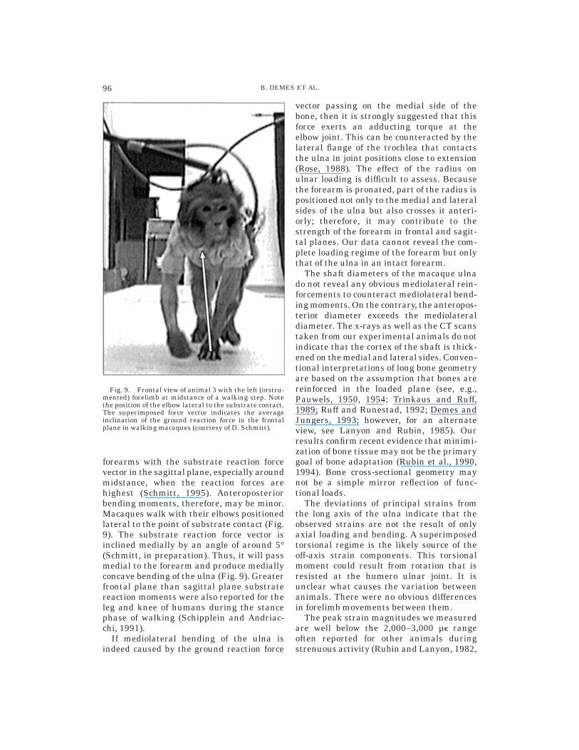
forearms with the substrate reaction forcevector in the sagittal plane, especially aroundmidstance, when the reaction forces arehighest (Schmitt, 1995). Anteroposteriorbending moments, therefore, may be minor.Macaques walk with their elbows positionedlateral to the point of substrate contact (Fig.9). The substrate reaction force vector isinclined medially by an angle of around 5°(Schmitt, in preparation). Thus, it will passmedial to the forearm and produce mediallyconcave bending of the ulna (Fig. 9). Greaterfrontal plane than sagittal plane substratereaction moments were also reported for theleg and knee of humans during the stancephase of walking (Schipplein and Andriac-chi, 1991).
If mediolateral bending of the ulna isindeed caused by the ground reaction force
vector passing on the medial side of thebone, then it is strongly suggested that thisforce exerts an adducting torque at theelbow joint. This can be counteracted by thelateral flange of the trochlea that contactsthe ulna in joint positions close to extension(Rose, 1988). The effect of the radius onulnar loading is difficult to assess. Becausethe forearm is pronated, part of the radius ispositioned not only to the medial and lateralsides of the ulna but also crosses it anteri-orly; therefore, it may contribute to thestrength of the forearm in frontal and sagit-tal planes. Our data cannot reveal the com-plete loading regime of the forearm but onlythat of the ulna in an intact forearm.
The shaft diameters of the macaque ulnado not reveal any obvious mediolateral rein-forcements to counteract mediolateral bend-ing moments. On the contrary, the anteropos-terior diameter exceeds the mediolateraldiameter. The x-rays as well as the CT scanstaken from our experimental animals do notindicate that the cortex of the shaft is thick-ened on the medial and lateral sides. Conven-tional interpretations of long bone geometryare based on the assumption that bones arereinforced in the loaded plane (see, e.g.,Pauwels, 1950, 1954; Trinkaus and Ruff,1989; Ruff and Runestad, 1992; Demes andJungers, 1993; however, for an alternateview, see Lanyon and Rubin, 1985). Ourresults confirm recent evidence that minimi-zation of bone tissue may not be the primarygoal of bone adaptation (Rubin et al., 1990,1994). Bone cross-sectional geometry maynot be a simple mirror reflection of func-tional loads.
The deviations of principal strains fromthe long axis of the ulna indicate that theobserved strains are not the result of onlyaxial loading and bending. A superimposedtorsional regime is the likely source of theoff-axis strain components. This torsionalmoment could result from rotation that isresisted at the humero ulnar joint. It isunclear what causes the variation betweenanimals. There were no obvious differencesin forelimb movements between them.
The peak strain magnitudes we measuredare well below the 2,000–3,000 µe rangeoften reported for other animals duringstrenuous activity (Rubin and Lanyon, 1982,
Fig. 9. Frontal view of animal 3 with the left (instru-mented) forelimb at midstance of a walking step. Notethe position of the elbow lateral to the substrate contact.The superimposed force vector indicates the averageinclination of the ground reaction force in the frontalplane in walking macaques (courtesy of D. Schmitt).
96 B. DEMES ET AL.

1984). Indeed, this latter level of strain hasbeen proposed to be beneficial for bone tis-sue and to be maintained through kinematicadjustments (speed, joint angles) over wideranges of body sizes and locomotor modes(‘‘dynamic strain similarity’’: Rubin andLanyon, 1984; Rubin et al., 1994; ‘‘stresssimilarity’’: Biewener, 1989). Even if peakstrain levels are compared for walking gaitsonly, the measured strains in the macaqueulna are still relatively low, but they dooverlap with strains measured in the humantibia, sheep and goat tibiae and radii, anddog and horse tibiae (Table 2). Low strainmagnitudes could be a result of low groundreaction forces. However, primate quadru-peds, including macaques, are not character-ized by lower reaction forces acting on theirforelimbs during walking on the groundthan nonprimate quadrupeds (Fig. 10). Pri-mate long bones also do not possess greaterrigidity in static bending (Polk et al., 1997).An alternative explanation for the ratherlow strains in the ulna is that the radiustransmits a greater share of the loads actingon the forearm.
Contrary to other studies that have docu-mented strain gradients at gait transitionsand an increase in strain magnitudes with
speed for horses, dogs, and goats (Rubin andLanyon, 1982; Biewener et al., 1983, 1988;Biewener and Taylor, 1986), we found thatstrain magnitudes were similar for walksand gallops and found no incremental changeat gait transition. The change from trot togallop in nonprimate mammals is usuallyaccompanied by a decrease in strains, where-ase the walk-to-trot transition is character-ized by an increase. Primates do not use thehigh-cadence, high-force trot but change fromwalking directly into a gallop (Hildebrand,1967; Vilensky, 1983; Preuschoft et al., 1996).Strains in nonprimate mammals gallopingat slow speeds are considerably higher thanstrains at fast walks (Rubin and Lanyon,1982; Biewener and Taylor, 1986). This sug-gests that at least the slow gallop in pri-mates may be more compliant than thegallop of nonprimate mammals (cf. Schmitt,1995). This is confirmed by ground reactionforces that do not change much at gaittransition in primates (Demes et al., 1994).
CONCLUSIONS
Bone strain in the macaque ulna indicatesmediolateral bending as the predominantloading regime during the stance phase ofwalking. The bending moment is probably
TABLE 2. Comparison of average peak principal strain magnitudes for the stance phase of walking
Species Bone Cortex ActivityGreatest
principal strain1 Source
Sheep Tibia CranialCaudal
WalkWalk
7092666
Lanyon and Bourn, 1979
Sheep Radius CranialCaudal
WalkWalk
68821161
Lanyon et al., 1979
Sheep Radius CranialCaudal
WalkWalk
80721317
Lanyon and Baggott, 1976
Goat Tibia CaudalCaudal
Slow walkFast walk
27252845
Biewener and Taylor, 1986
Radius Caudal Slow walk 2822Caudal Fast walk 2832
Dog Tibia CaudalCaudal
Slow walkFast walk
270221059
Rubin and Lanyon, 1982
Dog Radius Caudal Walk 21503Horse Tibia Caudal
CaudalSlow walkFast walk
293921347
Radius Caudal Slow walk 21777Caudal Fast walk 21965
Horse TibiaRadius
CaudalCaudal
Fast walkFast walk
299821247
Biewener and Taylor, 1986
Human Tibia Medial Walk 2544 Burr et al., 1996Human Tibia Medial Walk 2400 Lanyon et al., 1975Macaque Ulna Medial
LateralPosterior
WalkWalkWalk
2869915433
Present study; mean values for all animals
1 Strains are given in microstrains and represent mean values for several step cycles that, in most cases, cover a range of speeds.
97IN VIVO STRAIN IN THE MACAQUE ULNA

caused by the ground reaction force vectorthat passes medial to the forearm. Themacaque ulna is not reinforced in the planeof bending. The lack of buttressing in theloaded plane and the somewhat counterintui-tive bending direction recommend cautionwith regard to conventional interpretationsof long bone cross-sectional geometry. Gaitchange from walk to gallop is not accompa-nied by an incremental change in strainmagnitudes. This result mirrors the absenceof a significant change in ground reactionforces at the walk-gallop transition of pri-mates.
ACKNOWLEDGMENTS
Ted Gross and Yi-Xian Qin provided thecalculations of normal strain distributionsin the cross sections of the ulnae. Ted Grossand Susannah and Chris Fritton helped indata collection and analysis. David Reimassisted in the surgery. Terry Button tookthe CT scans and x-rays in the middle of thenight. Xinbin Chen wrote part of the pro-grams used for data analysis. We thank allof them for their invaluable contributions tothis project.
LITERATURE CITED
Alexander RMN, Hayes AS, Maloiy GMO, and WathutaEM (1979) Allometry of the limb bones of mammalsfrom shrews (Sorex) to elephant (Loxodonta). J. Zool.Lond. 189:305–314.
Alexander RMN, and Maloiy GMO (1984) Stride lengthsand stride frequencies of primates. J. Zool. London202:577–582.
Biewener AA (1989) Scaling body support in mammals:Limb posture and muscle mechanics. Science 245:45–48.
Biewener AA, and Bertram JEA (1993) Skeletal strainpatterns in relation to exercise training during growth.J. Exp. Biol. 185:51–69.
Biewener AA, and Taylor CR (1986) Bone strain: Adeterminant of gait and speed? J. Exp. Biol. 123:383–400.
Biewener AA, Thomason J, Goodship A, and Lanyon LE(1983) Bone stress in the horse forelimb during locomo-tion at different gaits: A comparison of two experimen-tal methods. J. Biomech. 16:565–576.
Biewener AA, Thomason JJ, and Lanyon LE (1988)Mechanics of locomotion and jumping in the horse(Equus): In vivo stress in the tibia and metatarsus. J.Zool. London 214:547–565.
Bouvier M, and Hylander WL (1984) In vivo bone strainon the dog tibia during locomotion.ActaAnat. 118:187–192.
Budsberg SC, Verstraete MC, and Soutas-Little RW(1987) Force plate analysis of the walking gait inhealthy dogs. Am. J. Vet. Res. 48:915–918.
Burr DB, Milgrom C, Fyhrie D, Forwood M, Nysaka M,Finestone A, Hoshaw S, Saiag E, and Simkin A (1996)In vivo measurement of human tibial bone strainduring vigorous activity. Bone 18:405–410.
Carter DR, Vasu R, Spengler DM, and Dueland RT(1981) Stress fields in the unplated and plated caninefemur calculated from in vivo strain measurements. J.Biomech. 14:63–70.
Dally JW, and Riley WF (1991) Experimental StressAnalysis, 3rd ed. New York: McGraw-Hill.
Demes B, and Jungers WL (1993) Long bone cross-sectional dimensions, locomotor adaptations and bodysize in prosimian primates. J. Hum. Evol. 25:57–74.
Demes B, Larson SG, Stern JT Jr, Jungers WL, Biknev-icius AR, and Schmitt D (1994) The kinetics of primatequadrupedalism: ‘‘Hindlimb drive’’ reconsidered. J.Hum. Evol. 26:353–374.
Fig. 10. Comparison of walking groundreaction force magnitudes for the fore-limbs of primate and nonprimate quadru-peds. Peak stance phase forces are indi-cated in multiples of body weights; in mostcases, they represent means for forcesrecorded for a range of speeds not specifi-cally stated in the references (Budsberg etal., 1987; Demes et al., 1994; Ishida et al.,1990; Kimura, 1985; Nieschalk, 1991;Pandy et al., 1988; Reynolds, 1985; Roushand McLaughlin, 1994; Ueda et al., 1981;Biknevicius, personal communication).
98 B. DEMES ET AL.

Fleagle JG (1983) Locomotor adaptations of Oligoceneand Miocene hominoids and their phyletic implica-tions. In Ciochon RL and RS Corruccini (eds.): NewInterpretations of Ape and Human Ancestry. NewYork: Plenum Press, pp. 301–324.
Fleagle JG, Simons EL, and Conroy GC (1975) Ape limbbone from the Oligocene of Egypt. Science 189:135–137.
Fleagle JG, Stern JT Jr, Jungers WL, Susman RL,Vangor AK, and Wells JP (1981) Climbing: A biome-chanical link with brachiation and with bipedalism.Symp. Zool. Soc. London 48:359–375.
Goodship AE, Lanyon LE, and MacFie H (1979) Func-tional adaptation of bone to increased stress. J. BoneJoint Surg. 61A:539–546.
Gross TS, McLeod KJ, and Rubin CT (1992) Characteriz-ing bone strain distributions in vivo using three triplerosette strain gauges. J. Biomech. 25:1081–1087.
Harrison T (1989) New postcranial remains of Victoriapithecus from the middle Miocene of Kenya. J. Hum.Evol. 18:3–54.
Hildebrand M (1967) Symmetrical gaits of primates.Am. J. Phys. Anthropol. 26:119–130.
Ishida H, Jouffroy, FK, and Nakano Y (1990) Compara-tive dynamics of pronograde and upsidedown horizon-tal quadrupedalism in the slow loris (Nycticebus cou-cang). In Jouffroy FK, MH Stack, and C Niemitz(eds.): Gravity, Posture and Locomotion in Primates.Firence: Il Sedicesimo, pp. 209–220.
Jenkins FA Jr (1973) The functional anatomy andevolution of the mammalian humero-ulnar articula-tion. Am. J. Anat. 137:281–298.
Kimura T (1985) Bipedal and quadrupedal walking ofprimates: Comparative dynamics. In Kondo S (ed.):Primate Morphophysiology, Locomotor Analyses andHuman Bipedalism. Tokyo: University of Tokyo Press,pp. 81–104.
Kimura T (1995) Long bone characteristics of primates.Z. Morphol. Anthropol. 80:265–280.
Kimura T, Okada M, and Ishida H (1979) Kinesiologicalcharacteristics of primate walking: Its significance inhuman walking. In Morbeck ME, H Preuschoft, and NGomberg (eds.): Environment, Behavior and Morphol-ogy: Dynamic Interactions in Primates. New York: G.Fischer, pp. 297–311.
Lanyon LE, and Baggott DG (1976) Mechanical functionas an influence on the structure and form of bone. J.Bone Joint Surg. 58B:436–443.
Lanyon LE, and Bourn S (1979) The influence of me-chanical function on the development and remodellingof the tibia. An experimental study in sheep. J. BoneJoint Surg. 61A:263–273.
Laynon LE, and Rubin CT (1985) Functional adaptationin skeletal structures. In Hildebrand M, DM Bramble,KF Liem, and DB Wake (eds.): Functional VertebrateMorphology. Cambridge: Harvard University Press,pp. 1–25.
Lanyon LE, and Smith RN (1969) Measurement of bonestrain in the walking animal. Res. Vet. Sci. 10:93–94.
Lanyon LE, and Smith RN (1970) Bone strain in thetibia during normal quadrupedal locomotion. ActaOrthop. Scand. 41:238–248.
Lanyon LE, Hampson WGJ, Goodship AE, and Shah JS(1975) Bone deformation recorded in vivo from straingauges attached to the human tibial shaft. ActaOrthop. Scand. 46:256–268.
Lanyon LE, Magee PT, and Baggott DG (1979) Therelationship of functional stress and strain to the
processes of bone remodelling. An experimental studyon the sheep radius. J. Biomech. 12:593–600.
Nieschalk U (1991) Fortbewegung und Funktionsmor-phologie von Loris tardigradus und anderen kleinenquadrupeden Halbaffen in Anpassung an unter-schiedliche Habitate [Ph.D. thesis]. Ruhr-UniversitatBochum.
Pandy MG, Kumar V, Berme N, and Waldron KJ (1988)The dynamics of quadrupedal locomotion. J. Biomech.Eng. 110:230–237.
Pauwels F (1950) Die Bedeutung der Muskelkrafte furdie Regelung der Beanspruchung des Rohrenkno-chens wahrend der Bewegung der Glieder. Z. Anat.Entwickl. Gesch. 115:326–351.
Pauwels F (1954) Die statische Bedeutung der Lineaaspera. Z. Anat. Entwickl. Gesch. 117:497–503.
Polk JD, Demes B, Jungers WL, Heinrich RE, Biknev-icius AR, and Runestad JA (1997) Cross-sectionalproperties of primate and nonprimate limb bones. Am.J. Phys. Anthropol. 24(Suppl.):188.
Preuschoft H, Witte H, Christian A, and Fischer M(1996) Size influences on primate locomotion and bodyshape, with special emphasis on the locomotion of‘small mammals’. Folia Primatol. 66:93–112.
Reynolds TR (1985) Stresses on the limbs of quadrupe-dal primates. Am. J. Phys. Anthropol. 67:351–362.
Reynolds TR (1987) Stride length and its determinantsin humans, early hominids, primates, and mammals.Am. J. Phys. Anthropol. 72:101–115.
Richmond BG, Fleagle JG, Kappelman J, and SwisherCC (1998) First hominoid from the Miocene of Ethio-pia and the evolution of the catarrhine elbow. Am. J.Phys. Anthropol. (in press).
Rose MD (1988) Another look at the anthropoid elbow. J.Hum. Evol. 17:193–224.
Roush JK, and McLaughlin RM (1994) Effects of subjectstance time and velocity on ground reaction forces inclinically normal Greyhounds at the walk. Am. J. Vet.Res. 55:1672–1676.
Rubin CT, and Lanyon LE (1982) Limb mechanics as afunction of speed and gait. J. Exp. Biol. 101:187–211.
Rubin CT, and Lanyon LE (1984) Dynamic strain simi-larity in vertebrates: An alternative to allometric limbbone scaling. J. Theor. Biol. 107:321–327.
Rubin CT, McLeod KJ, and Bain SD (1990) Functionalstrains and cortical bone adaptation: Epigenetic assur-ance of skeletal integrity. J. Biomech. 23(Suppl. 1):43–54.
Rubin C, Gross T, Donahue H, Guilak F, and McLeod K(1994) Physical and environmental influences on boneformation. In: Brighton CT, GE Friedlaender, and JMLane (eds.): Bone Formation and Repair. Rosemont:American Academy of Orthopedic Surgeons, pp.61–78.
Ruff CB, and Runestad JA (1992) Primate limb bonestructural adaptations. Annu. Rev. Anthropol. 21:407–433.
Rybicki EF, Mills EJ, Turner AS, and Simonen FA(1977): In vivo and analytical studies of forces andmoments in equine long bones. J. Biomech. 10:701–795.
Schipplein OD, and Andriacchi TP (1991) Interactionsbetween active and passive knee stabilizers duringlevel walking. J. Orthop. Res. 9:113-119.
Schmitt D (1994) Forelimb mechanics as a function ofsubstrate type during quadrupedalism in two anthro-poid primates. J. Hum. Evol. 26:441–457.
Schmitt D (1995) A Kinematic and Kinetic Analysis ofForelimb Use DuringArboreal and Terrestrial Quadru-pedalism in Old World Monkeys [PhD thesis]. StateUniversity of New York at Stony Brook.
99IN VIVO STRAIN IN THE MACAQUE ULNA

Swartz SM, Bertram JEA, and Biewener AA (1989)Telemetered in vivo strain analysis of locomotormechanics of brachiating gibbons. Nature 342:270–272.
Swartz SM, Bennett MB, and Carrier DR (1992) Wingbone stresses in free flying bats and the evolution ofskeletal design for flight. Nature 359:726–729.
Timoshenko S (1958) Strength of Materials. Part II, 3rded. New York: D. van Norstand Co.
Trinkaus E, and Ruff C (1989) Diaphyseal cross-
sectional morphology and biomechanics of the Fond-de-Foret 1 femur and the Spy 2 femur and tibia. Anthro-pologie Prehistoire 100:33–42.
Ueda Y, Niki Y, Yoshida K, and Masumitsu H (1981)Force plate study of equine biomechanics—Floor reac-tion force of normal walking and trotting horses. Bull.Equine Res. Inst. 18:28–41.
Vilensky JA (1983) Gait characteristics of two ma-caques, with emphasis on relationships with speed.Am. J. Phys. Anthropol. 61:255–265.
100 B. DEMES ET AL.
Copyright © 2022 FDOKUMEN