Responses of Macaque Perirhinal Neurons during and after Visual Stimulus Association Learning
13
Responses of Macaque Perirhinal Neurons during and after Visual Stimulus Association Learning Cynthia A. Erickson and Robert Desimone Laboratory of Neuropsychology, National Institute of Mental Health, National Institutes of Health, Bethesda, Maryland 20892-4415 Recent lesion studies have implicated the perirhinal cortex in learning that two objects are associated, i.e., visual association learning. In this experiment we tested whether neuronal re- sponses to associated stimuli in perirhinal cortex are altered over the course of learning. Neurons were recorded from mon- keys during performance of a visual discrimination task in which a predictor stimulus was followed, after a delay, by a GO or NO-GO choice stimulus. Association learning had two major influences on neuronal responses. First, responses to fre- quently paired predictor–choice stimuli were more similar to one another than was the case with infrequently paired stimuli. Second, the magnitude of activity during the delay was corre- lated with the magnitude of responses to both the predictor and choice stimuli. Both of these learning effects were found only for stimulus pairs that had been associated on at least 2 d of training. Early in training, the delay activity was correlated only with the response to the predictor stimuli. Thus, with long-term training, perirhinal neurons tend to link the representations of temporally associated stimuli. Key words: single unit; electrophysiology; Macaca mulatta; perirhinal cortex; inferior temporal cortex; paired associates; association; learning; memory; primates; medial temporal lobe Anatomical, behavioral, and neurophysiological studies indicate that the perirhinal portion of the inferior temporal cortex in primates plays an important role in visual memory. Perirhinal cortex receives substantial input from visual area TE (Suzuki and Amaral, 1994), a high-order visual-processing area, and projects via entorhinal cortex into the hippocampal formation. Animals with selective lesions of perirhinal cortex are impaired on tests of recognition memory, such as the delayed matching to sample (DMS) (Meunier et al., 1993) or nonmatching to sample tasks (Murray and Mishkin, 1986; Zola-Morgan et al., 1989, 1993; Meunier et al., 1993; Suzuki et al., 1993; Gaffan, 1994). Many perirhinal neurons exhibit mnemonic properties in such tasks (Eskandar et al., 1992; Fahy et al., 1993; Li et al., 1993; Miller et al., 1993; Sobotka and Ringo, 1993, 1996; Lueschow et al., 1994). Specifically, for some cells the response to a repeated visual stimulus is suppressed if it matches a previously viewed stimulus. Such suppression appears to occur automatically for any stimulus repetition, regardless of behavioral relevance, and serves to dis- tinguish novel stimuli from familiar ones. For other perirhinal cells, the response to the choice stimulus is enhanced if it matches the sample, and in contrast to suppression, enhancement appears to depend on active working memory for the sample (Miller and Desimone, 1994). Many perirhinal neurons also respond differ- entially during the period immediately after stimulus presenta- tion (Fuster and Jervey, 1981, 1982; Miller et al., 1993). Together, the heightened delay activity and the enhanced response to the sought-for choice stimulus have suggested that some perirhinal neurons are sensitized, or primed, to respond to stimuli held actively in working memory. In addition to a role in recognition memory, perirhinal cortex may also be involved in associative memory, i.e., establishing linkages between different stimuli that have some meaningful connection. Combined lesions of the perirhinal and neighboring entorhinal cortex result in profound deficits in the learning of, and memory for, stimulus associations in monkeys (Murray et al., 1993). To test the role of perirhinal neurons in associative mem- ory, Sakai and Miyashita (1991) recorded from perirhinal neu- rons in a paired-associate task. In their task, the monkey was shown a sample stimulus followed, after a delay, by a choice stimulus and was rewarded for indicating whether the choice stimulus was the correct paired associate of the sample. They reported that some perirhinal cells had delay activity that was selective for the expected choice stimulus after a given sample and that some cells responded preferentially to specific sample– choice pairs. Because all training took place before recording, it was not possible to examine the time course of any response changes relative to the association learning. In contrast to the association effects reported by Sakai and Miyashita, two other groups (Sobotka and Ringo, 1993; Gochin et al., 1994), using two different association tasks, failed to find evidence of inferior temporal participation in association learning. To reconcile these conflicting reports and to test further how perirhinal neurons might contribute to associative memory, we developed a task in which the monkey could form associative memories of multiple pairs of stimuli over the course of a single recording session. Perirhinal neuronal responses were recorded during performance of the task using either novel (learned within a single session) or familiar (stimulus pairs with which the mon- key had been trained in one or more previous sessions) stimuli. This task provided us with the opportunity to examine two aspects of association learning, namely, changes in the responses Received Jan. 11, 1999; revised Sept. 2, 1999; accepted Sept. 3, 1999. This work was supported by the IRP National Institute of Mental Health. We thank Drs. Earl Miller, Bharathi Jagadeesh, and Martin Pare ´ for helpf ul suggestions on task design and data analysis. Barbara K. Changizi, Amy C. Durham, and Sudabeh Shirazi helped with technical aspects of the experiment. Correspondence should be addressed to Dr. Robert Desimone, Laboratory of Neuropsychology, Building 49, Room 1B80, National Institute of Mental Health, Bethesda, MD 20892-4415. E-mail: [email protected]. Copyright © 1999 Society for Neuroscience 0270-6474/99/1910404-13$05.00/0 The Journal of Neuroscience, December 1, 1999, 19(23):10404–10416
Transcript of Responses of Macaque Perirhinal Neurons during and after Visual Stimulus Association Learning

Responses of Macaque Perirhinal Neurons during and after VisualStimulus Association Learning
Cynthia A. Erickson and Robert Desimone
Laboratory of Neuropsychology, National Institute of Mental Health, National Institutes of Health, Bethesda,Maryland 20892-4415
Recent lesion studies have implicated the perirhinal cortex inlearning that two objects are associated, i.e., visual associationlearning. In this experiment we tested whether neuronal re-sponses to associated stimuli in perirhinal cortex are alteredover the course of learning. Neurons were recorded from mon-keys during performance of a visual discrimination task inwhich a predictor stimulus was followed, after a delay, by a GOor NO-GO choice stimulus. Association learning had two majorinfluences on neuronal responses. First, responses to fre-quently paired predictor–choice stimuli were more similar toone another than was the case with infrequently paired stimuli.
Second, the magnitude of activity during the delay was corre-lated with the magnitude of responses to both the predictor andchoice stimuli. Both of these learning effects were found onlyfor stimulus pairs that had been associated on at least 2 d oftraining. Early in training, the delay activity was correlated onlywith the response to the predictor stimuli. Thus, with long-termtraining, perirhinal neurons tend to link the representations oftemporally associated stimuli.
Key words: single unit; electrophysiology; Macaca mulatta;perirhinal cortex; inferior temporal cortex; paired associates;association; learning; memory; primates; medial temporal lobe
Anatomical, behavioral, and neurophysiological studies indicatethat the perirhinal portion of the inferior temporal cortex inprimates plays an important role in visual memory. Perirhinalcortex receives substantial input from visual area TE (Suzuki andAmaral, 1994), a high-order visual-processing area, and projectsvia entorhinal cortex into the hippocampal formation. Animalswith selective lesions of perirhinal cortex are impaired on tests ofrecognition memory, such as the delayed matching to sample(DMS) (Meunier et al., 1993) or nonmatching to sample tasks(Murray and Mishkin, 1986; Zola-Morgan et al., 1989, 1993;Meunier et al., 1993; Suzuki et al., 1993; Gaffan, 1994). Manyperirhinal neurons exhibit mnemonic properties in such tasks(Eskandar et al., 1992; Fahy et al., 1993; Li et al., 1993; Miller etal., 1993; Sobotka and Ringo, 1993, 1996; Lueschow et al., 1994).Specifically, for some cells the response to a repeated visualstimulus is suppressed if it matches a previously viewed stimulus.Such suppression appears to occur automatically for any stimulusrepetition, regardless of behavioral relevance, and serves to dis-tinguish novel stimuli from familiar ones. For other perirhinalcells, the response to the choice stimulus is enhanced if it matchesthe sample, and in contrast to suppression, enhancement appearsto depend on active working memory for the sample (Miller andDesimone, 1994). Many perirhinal neurons also respond differ-entially during the period immediately after stimulus presenta-tion (Fuster and Jervey, 1981, 1982; Miller et al., 1993). Together,the heightened delay activity and the enhanced response to thesought-for choice stimulus have suggested that some perirhinal
neurons are sensitized, or primed, to respond to stimuli heldactively in working memory.
In addition to a role in recognition memory, perirhinal cortexmay also be involved in associative memory, i.e., establishinglinkages between different stimuli that have some meaningfulconnection. Combined lesions of the perirhinal and neighboringentorhinal cortex result in profound deficits in the learning of, andmemory for, stimulus associations in monkeys (Murray et al.,1993). To test the role of perirhinal neurons in associative mem-ory, Sakai and Miyashita (1991) recorded from perirhinal neu-rons in a paired-associate task. In their task, the monkey wasshown a sample stimulus followed, after a delay, by a choicestimulus and was rewarded for indicating whether the choicestimulus was the correct paired associate of the sample. Theyreported that some perirhinal cells had delay activity that wasselective for the expected choice stimulus after a given sampleand that some cells responded preferentially to specific sample–choice pairs. Because all training took place before recording, itwas not possible to examine the time course of any responsechanges relative to the association learning. In contrast to theassociation effects reported by Sakai and Miyashita, two othergroups (Sobotka and Ringo, 1993; Gochin et al., 1994), using twodifferent association tasks, failed to find evidence of inferiortemporal participation in association learning.
To reconcile these conflicting reports and to test further howperirhinal neurons might contribute to associative memory, wedeveloped a task in which the monkey could form associativememories of multiple pairs of stimuli over the course of a singlerecording session. Perirhinal neuronal responses were recordedduring performance of the task using either novel (learned withina single session) or familiar (stimulus pairs with which the mon-key had been trained in one or more previous sessions) stimuli.This task provided us with the opportunity to examine twoaspects of association learning, namely, changes in the responses
Received Jan. 11, 1999; revised Sept. 2, 1999; accepted Sept. 3, 1999.This work was supported by the IRP National Institute of Mental Health. We
thank Drs. Earl Miller, Bharathi Jagadeesh, and Martin Pare for helpful suggestionson task design and data analysis. Barbara K. Changizi, Amy C. Durham, andSudabeh Shirazi helped with technical aspects of the experiment.
Correspondence should be addressed to Dr. Robert Desimone, Laboratory ofNeuropsychology, Building 49, Room 1B80, National Institute of Mental Health,Bethesda, MD 20892-4415. E-mail: [email protected] © 1999 Society for Neuroscience 0270-6474/99/1910404-13$05.00/0
The Journal of Neuroscience, December 1, 1999, 19(23):10404–10416
of cells to the paired stimuli and differential delay activity after asample stimulus and before its paired choice stimulus.
MATERIALS AND METHODSSubjectsThe two adult male, 7–9 kg, rhesus monkeys (Macaca mulatta) used inthis experiment were cared for according to National Institutes of Healthguidelines.
StimuliThe stimuli were 1–3° square-shaped, colored pictures presented on acomputer monitor. The pictures were cropped and modified from digi-tized photographs, artwork, or diagrams. Some of the images wereclearly recognizable objects, whereas others were abstract designs orpatterns. For each training set, 16 stimuli were randomly chosen from astimulus pool of over 1000 images and were randomly paired to formeight paired associates (stimulus pairs).
Stimuli were defined as novel if the monkey had never seen thembefore the start of recording. In these recording sessions, the monkey’sfirst experience with an individual stimulus was the first trial of therecording session. Stimuli were defined as familiar if the monkey hadsuccessfully learned the stimuli on at least 1 previous day. Examples ofthe types of stimuli used are shown in Figure 1. After a stimulus set hadbeen established, those stimuli were not used in a different stimulus set.
Behavioral taskTask rationale. Traditional paired-associate association-learning tasks inprimates are typically based on conditional performance rules, such as ifA is followed by C, press the bar, if B is followed by C, do not press thebar, if A is followed by D, press the bar, if B is followed by D, do not pressthe bar, etc. (Gochin et al., 1994). In some conditional designs, twochoice stimuli are presented simultaneously, such as if A is followed by Band C, choose B, if D is followed by B and C, choose C, etc. (Sakai andMiyashita, 1991). Conditional designs such as these often require lengthy
training on specific pairs of stimuli, and there is often no obligatorytemporal relationship between the first and second stimulus in the pair;e.g., stimulus A may be followed equally often by B or C.
To examine how perirhinal neurons might contribute to associativememories acquired over both short (within a day) and long (multipledays) periods of exposure to stimuli, we developed a task in which themonkey could form associative memories of multiple pairs of stimuli overthe course of a single recording session. The two previous studies thatfailed to find associative effects (see introductory remarks) influencedour experiment design. First, we hypothesized that neurons that respondselectively to associated stimuli were not found in the Gochin et al.(1994) experiment because a given sample stimulus was paired an equalnumber of times with two different choice stimuli, and both types ofpairings were behaviorally relevant. Second, in the Sobotka and Ringo(1993) experiment, the associated stimuli were presented together simul-taneously, and the task did not require that the animals perceive thestimulus pairs as two separate stimuli. The paired stimuli might havebeen perceived as a single compound stimulus. These results suggestedthat it might be important to maintain consistent pairings between theassociated stimuli and that the stimuli be perceived as separate objects.We therefore used a paradigm based on discrimination learning, a task inwhich monkeys can learn new stimuli quickly after acquiring the discrim-ination rule. In some sessions, the monkeys learned eight new discrimi-nanda, or choice stimuli, concurrently (termed novel stimuli). In othersessions they were tested with eight previously learned stimuli (termedfamiliar stimuli). Each of the eight choice stimuli was preceded by aspecific predictor stimulus. Each predictor stimulus predicted the occur-rence of its paired choice stimulus with 80–90% probability (“validtrials”). Thus, there were eight pairs of predictor–choice stimuli or 16stimuli in total per session. No awareness of the stimulus–stimulusrelationship was necessary for successful completion of a trial, but themonkeys could potentially respond more quickly if they learned thespecific predictor–choice relationships.
When a predictor stimulus was followed by the expected choice stim-ulus, it was termed a valid trial. In “probe trials,” a given predictor
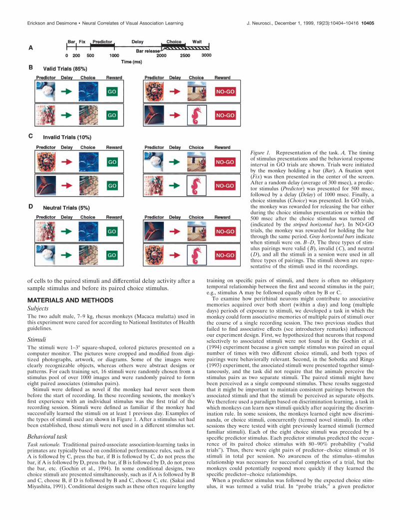
Figure 1. Representation of the task. A, The timingof stimulus presentations and the behavioral responseinterval in GO trials are shown. Trials were initiatedby the monkey holding a bar (Bar). A fixation spot(Fix) was then presented in the center of the screen.After a random delay (average of 300 msec), a predic-tor stimulus (Predictor) was presented for 500 msec,followed by a delay (Delay) of 1000 msec. Finally, achoice stimulus (Choice) was presented. In GO trials,the monkey was rewarded for releasing the bar eitherduring the choice stimulus presentation or within the500 msec after the choice stimulus was turned off(indicated by the striped horizontal bar). In NO-GOtrials, the monkey was rewarded for holding the barthrough the same period. Gray horizontal bars indicatewhen stimuli were on. B–D, The three types of stim-ulus pairings were valid ( B), invalid ( C), and neutral(D), and all the stimuli in a session were used in allthree types of pairings. The stimuli shown are repre-sentative of the stimuli used in the recordings.
Erickson and Desimone • Neural Correlates of Visual Association Learning J. Neurosci., December 1, 1999, 19(23):10404–10416 10405

stimulus was followed by an unexpected choice stimulus. If the monkeyslearned to associate each choice stimulus with its paired predictor, thenresponse times to the choice stimuli should be faster in valid trialscompared with the probe trials. In this sense, the design is similar to thecued target detection task used to study spatial attention by Posner et al.(1980), except that in this case the cues and targets were specific patternsrather than specific spatial locations. We refer to this task as the “passivepaired-associate task” because in contrast to the standard paired-associate task, it does not require active memory for the stimulus pairsand the subjects may not even be aware of the pairings.
Task description. Monkeys initiated each trial by grabbing a bar in thefront of a standard monkey chair for 200 msec, after which a fixation spot(0.1° in diameter) appeared in the center of the monitor (task time lineis illustrated in Fig. 1 A). Approximately 300 msec after the monkeysfixated the spot, one of eight predictor stimuli appeared on the monitorfor 500 msec, centered on the fixation spot. After a random delay periodof 950 –1050 msec, one of eight choice stimuli appeared at the fixationspot (a small subset of neurons was recorded with random delays of500–1000 msec). Half of the choice stimuli were GO stimuli, and halfwere NO-GO. In the GO trials, the monkey was rewarded with a drop oforange juice if it released the bar within 100–1000 msec after choicestimulus onset. To encourage rapid responses, two juice rewards weregiven if the monkey released the bar within 300 msec of stimulus onset.The choice stimuli remained on for 500 msec or until the monkeyreleased the bar, whichever came first. Response latencies were definedas the time between the onset of the choice stimulus and the release ofthe bar. In NO-GO trials, the choice stimulus remained on for 500 msec,and the monkeys were rewarded for not releasing the bar between 100and 1000 msec after stimulus onset. Monkeys learned via trial and errorwhich choice stimuli were GO and which were NO-GO. Trials wereaborted if the monkey broke fixation at any time during the trial beforethe bar release or if it released the bar before or within the first 100 msecof choice stimulus presentation (Fig. 1 B). These trials are referred to asvalid because a given predictor is paired with its associated choicestimulus in 80–90% of the trials.
Probe trials. The remaining two types of trials were probe trials,“invalid” and “neutral.” Probe trials were added to the task after themonkeys were performing at or above 85% correct for the valid trials.The purpose of the probe trials was to enable us to measure the differ-ence between the behavioral response latencies in trials with expectedstimuli (valid trials) and behavioral latencies in trials with unexpectedstimuli.
In the invalid probe trials, both the behavioral response and the choicestimulus were different from that of valid trials. In such trials, a NO-GOchoice was preceded by a predictor stimulus that normally predicted aGO choice stimulus, or a GO choice stimulus was preceded by a predictorstimulus that normally predicted a NO-GO choice stimulus (Fig. 1C).When it saw the predictor stimulus in an invalid trial, the monkey mightprepare a motor response and later suppress it, or the monkey might failto prepare a motor response and then later have to prepare and executea bar release. Slower response times in the invalid GO trials comparedwith that in valid GO trials provided a measure of association learning forspecific predictor–choice pairings in addition to an indication of the timerequired to reprogram the behavioral response. Because associationlearning and motor preparedness are confounded in this type of probetrial, we also included neutral probe trials.
In neutral probe trials, a GO choice stimulus was preceded by apredictor that normally predicted a GO choice, and a NO-GO choicestimulus was preceded by a predictor that normally predicted a NO-GOchoice, but in each case the specific predictor–choice pairing was differ-ent from that of valid trials (Fig. 1 D). For example, consider the predic-tor–choice pairings of A–B and C–D on valid trials, with B and D bothGO choice stimuli. In neutral trials, the stimuli would be repaired as A–Dand C–B. Slower response times in the neutral GO trials, compared withthat in valid GO trials, provided a measure of association learning forspecific predictor–choice pairings without the additional factor of re-sponse reprogramming. All choice and predictor stimuli used in thesession appeared in all three types of trials, i.e., valid, neutral, andinvalid.
SurgeryBefore surgery, the monkeys were placed in a plastic stereotaxic frameand scanned with magnetic resonance imaging (MRI). Appropriate ste-reotaxic coordinates for the recording chamber were calculated from theMRI scans. During surgery, under isoflurane anesthesia, a recording
chamber was implanted on the dorsal surface of the skull over theperirhinal cortex. In addition, a post for restraining the head and amagnetic search coil for monitoring eye movements were implanted(Robinson, 1963). Analgesics and antibiotics were administered duringthe recovery period.
Localization of electrode tracksElectrode placement into the perirhinal cortex was guided by construct-ing individual brain atlases from the MRI scans (Alvarez-Royo et al.,1991). Electrode locations were verified with x rays [similar to butsimplified from the method of Nahm et al. (1994)]. Comparisons betweenx rays and MRI scans were made by identifying the stereotaxic planecorresponding to the line between the auditory canal and the orbitalridge. The electrodes themselves were too small to be seen on the x rays,but the guide tubes were clearly visible, and the distance from the tip ofthe electrode to the tip of the guide tube was known. The depth and theanterior–posterior coordinates of the probes were verified from thesagittal-view x rays, and the medial–lateral coordinates were measuredrelative to the midline from the coronal-view x rays. Histological exam-ination of the brain tissue was not possible because both monkeys are stillalive.
Electrophysiological recording techniquesElectrodes. Individual neurons were recorded with either tetrodes orsingle sharpened tungsten electrodes (ROBOZ Microprobe, Rockville,MD). Tetrodes were constructed by twisting together four strands ofwire and attaching them to a multipin connector with conductive epoxyor silver print. Two types of wire were used for the tetrodes: 20 mmlacquer-coated tungsten wire (California Fine Wire, Grover City, CA) or12.5 mm Teflon-coated nichrome wire (H. P. Reid Company, Neptune,NJ). The Teflon-insulated tetrode wires were held together by warmingthe insulation briefly until it reached a tacky state. The lacquer-coatedwire was held in place with a small amount of superglue. Nichrome wirewas gold plated before use to reduce the electrode impedance. Electrodeimpedance ranged from 100 to 500 kV and from 500 kV to 1 MV for thetungsten and nichrome tetrodes, respectively. Impedance averaged 1 MVfor the standard tungsten electrodes.
Microdrives. Tetrodes were advanced using minimicrodrives [similar tothose described by Nichols et al. (1998)] attached to a cylindrical grid thatfit snugly into a standard recording chamber (Crist et al., 1988). Briefly,an arm was attached to a threaded rod that was inserted into the grid.One rotation of the screw moved the arm 300 mm. Tetrodes were loweredinto the perirhinal cortex through the grid via a telescoping guide tubesystem. The grids and minimicrodrives could remain in place for as longas 1 month; however, the electrodes could be moved on a daily basis sothat different neurons were recorded each day. A standard manuallydriven hydraulic microdrive was used to manipulate the sharpened tung-sten electrodes (for details, see Miller et al., 1993).
Data collection and analysisA multipin head stage connected the tetrodes to a multichannel amplifier.Signals were amplified from ;10,000 to 23,000 times and bandpassfiltered from 300 Hz to 8 kHz. Waveforms from all four channels of atetrode were collected if one of the channels crossed a threshold. Wave-forms were digitized at 25 kHz and stored on computer disk for off-linespike sorting (Datawave Technologies, Longmont, CO). Individual neu-rons were identified on the basis of their relative amplitudes or widthsfrom the different tetrode channels (McNaughton et al., 1983; Gray et al.,1995).
Data were collected if at least one of the neurons appeared to beresponsive to some aspect of the task. Neuronal responses to stimuli weremeasured as the average firing rate (Hz) during a 175 msec epochstarting 75 msec and ending 250 msec after stimulus onset (before themonkey’s behavioral response). The very few trials in which the monkeyresponded in ,250 msec were eliminated from further analysis. Delayactivity was measured as the mean firing rate starting 200 msec andending 800 msec after the predictor stimulus was turned off. These fixedepoch windows were used for all calculations.
Stimulus selectivity was assessed using ANOVAs (evaluated at p ,0.05) calculated on the trial-by-trial data for each neuron to the eightpredictor and choice stimuli separately, using the time windows de-scribed above. Delay period selectivity was determined in the same wayfor the delay periods after each of the eight predictor stimuli. A measureof stimulus selectivity (r 2) was calculated from the ANOVA table foreach neuron by simply dividing the sum of squared deviations for the
10406 J. Neurosci., December 1, 1999, 19(23):10404–10416 Erickson and Desimone • Neural Correlates of Visual Association Learning

treatment by the total sum of squared deviations from the grand mean(Keppel and Zedeck, 1989). The R 2 statistic provides an estimate of howmuch of the variance in firing rate can be accounted for by specific stimuliand, unlike F ratios and p values, is not influenced by sample size. Thenormalized magnitude of the neuronal response was measured by com-puting a z score for each stimulus relative to the distribution of baselinefiring rates.
Response similarity to paired stimuli was measured as the correlationbetween the mean responses to the predictor and choice stimuli for eachpair. A large r value indicates that the responses to predictor and choicestimuli were similar, such that if a neuron responded well to one stimulusin a pair then it also responded well to the other of the pair. Correlationswere also computed between the firing rate during the delay and theresponse to the preceding (predictor) stimulus and the following (choice)stimulus. All r values were converted to Fisher z scores before statisticalanalysis.
RESULTSBehavioral performanceThe monkeys typically learned eight new choice stimuli to acriterion of 85% correct in ,250 trials or ;30 presentations perstimulus. Learning curves are shown in Figure 2A. The percentcorrect for each trial type was calculated after the performancereached asymptote and probe trials were added. The percentcorrect (Fig. 2B) for the data sets recorded with familiar stimuli(97.9%; n 5 87) was slightly, but significantly, better than that forthe novel (90.4%; n 5 41) stimuli [F(1,126) 5 29.7; p , 0.0001].There was a small, but significant, difference in the percentcorrect for the invalid trials compared with either the neutral orvalid trials [mean percent correct values for valid, neutral, andinvalid trial types were 95.5, 88.6, and 97.1%, respectively;F(2,252) 5 44.8; p , 0.0001]. That is, mistakes were more likelywhen the rewarded behavioral response was unexpected. Al-though both predictor type and experience influenced the meanresponse latencies, there was no interaction between these twofactors [F(2,252) 5 1.3; p 5 0.268]. Overall, the monkeys learnedthe stimuli quickly and performed at a very high level.
Response latenciesBehavioral evidence of association learning was determined bythe differences in response latencies to expected (valid trials) andunexpected (invalid and neutral trials) stimuli. The responselatencies to the familiar stimuli remained relatively stablethroughout the session. However, the response latencies to thenovel stimuli were, on average, 100 msec slower than the latenciesto the familiar stimuli at the beginning of the data set anddeclined over the course of the recording session, approaching(but not reaching) the mean latency of the familiar stimuli after500 trials (Fig. 2C).
The relationship between the effect of experience (novel andfamiliar) and predictor type (valid, invalid, and neutral) on re-sponse time latencies was determined by computing a two-wayANOVA, with predictor type as a within-session factor andexperience as a between-session factor. There was no significantinteraction between the factors [F(2,252) 5 2.5; p 5 0.082]. Therewere, however, significant main effects for both predictor types[F(2,252) 5 117.2; p , 0.0001] and experience level [F(1,126) 5 20.1;p , 0.0001]. The mean response latencies were slightly faster tofamiliar stimuli than were the responses to the novel stimuli evenafter the percent correct for the novel stimuli reached the level ofthe familiar stimuli. The mean response time latencies for thevalid trials were 398.3 and 482.6 msec for the familiar and novelstimuli, respectively, after probe trials were added. For both thenovel and familiar stimuli, the monkeys’ response latencies wereshorter for the valid compared with the neutral trials, suggesting
Figure 2. Behavioral performance. A, Percent correct in 50 trial bins forthe first 500 trials for each recording session. Data are shown separatelyfor novel and familiar stimuli. B, Average percent correct in valid, invalid,and neutral trials. Asterisks indicate a significant difference from theperformance in the valid trials (t test, p , 0.05). C, Reaction times for 50trial bins, during the first 500 trials of the recording sessions. Symbols aredescribed in A. D, Mean reaction times in valid, invalid, and neutral trials.Asterisks indicate a significance difference from the reaction times of validtrials (t test, p , 0.05). Vertical bars are described in B. Error bars in these,and subsequent, graphs indicate the SEM.
Erickson and Desimone • Neural Correlates of Visual Association Learning J. Neurosci., December 1, 1999, 19(23):10404–10416 10407

that the monkeys did learn to associate the predictor stimuli withthe paired choice stimuli as well as with the ultimate behavioralresponse after the choice.
The mean response times were 389.3, 402.4, and 536.6 msec forvalid, neutral, and invalid trials, respectively, for the familiarstimuli (Fig. 2D). Subsequent contrast computations indicatedthat the response time difference between valid and invalid trialswas significant at both experience levels. Most importantly, theresponse time difference between valid and neutral trials wassignificant for both the novel (mean latency difference 5 25.5 611.0 msec; p 5 0.026) and familiar (mean latency difference 513.1 6 4.0 msec; p 5 0.002) stimulus pairs. These results indicatethat the monkeys learned the stimulus pair associations for bothnovel and familiar stimuli.
It was not possible to pinpoint the exact time the monkeyslearned the association between the two stimuli. The probe trialswere added only after the monkeys were performing at .85% inthe valid trials. There was a much smaller number of probe trialsin each data set compared with the valid trials; thus, it was notpossible to track the learning on a trial-by-trial basis. The latencydifferences between the valid and neutral trials were significantlydifferent in ;25% of the recording sessions for both novel andfamiliar recording sessions.
Anatomical location of electrode penetrationsThe majority of penetrations were made within the boundaries ofthe perirhinal cortex as defined by Suzuki and Amaral (1994),except for two penetrations made in TE, near the anterior middletemporal sulcus, in monkey A (Fig. 3). The estimated recordingareas for both monkeys are shown in Figure 3.
Basic response properties of neuronsOf the 386 neurons recorded, 127 were recorded while monkeyslearned new stimuli (novel). The remaining 259 neurons wererecorded during presentation of familiar stimuli. Of the cellsstudied with familiar and novel stimuli, 47 and 52%, respectively,showed significant stimulus selectivity responses to both predictorand choice stimulus sets, according to one-way ANOVAs.
The basic stimulus-selective properties of the neurons did notdiffer for cells tested with novel and familiar stimuli (Fig. 4). Themean firing rate was higher for cells tested with novel compared
with familiar stimuli, and the number of stimuli that elicited asignificant response (z scores above the baseline firing rate) washigher for cells studied with novel stimuli, but both of thesedifferences just failed to reach conventional measures of signifi-cance [t(2990) 5 1.61; p 5 0.054; t(372) 5 1.64; p 5 0.051].Likewise, there was no significant difference between novel andfamiliar stimuli in the amount of the variance in firing rate, on atrial-to-trial basis, accounted for by the specific stimuli (r 2, t , 1).Although other studies have found significant decreases in infe-rior temporal responses as novel stimuli have become familiarwithin a single recording session (e.g., Fahy et al., 1993; Li et al.,1993), these decreases were apparently not large enough to causesubstantial differences in firing rates between the cells studied inthe first versus subsequent recording sessions in the present study.
Experience-dependent changes in responses to stimuliTo test whether training on the paired associates altered theselectivity of the cells, we examined the pattern of stimulusselectivity for the predictor and choice stimuli. The analysis wasdirected at the 66 and 121 cells that gave stimulus-selectiveresponses to the novel and familiar stimuli, respectively. Anexample of one cell’s responses to the predictors and choices isshown in Figure 5. As the figure demonstrates, the cell’s re-sponses to the eight predictor stimuli varied from a good response(predictor stimulus 5) to virtually no response (predictor stimulus4). Importantly, the responses to the associated choice stimulifollowed the same order; i.e., the responses to the predictor andchoice stimuli were highly correlated. The Pearson correlationcoefficient for the responses to predictors and associated choiceswas 0.85 for this cell (see Fig. 5B).
The distribution of r values for the entire population of selec-tive cells is shown separately for novel and familiar stimuli inFigure 6. Although the responses of any given cell to the predictorand choice stimuli might be correlated by chance, the stimuli for
Figure 3. Location of recording sites on the ventral view of the brain.The recording regions for both monkeys are shown in the same hemi-sphere in this illustration; however, monkey A was recorded from the righthemisphere, and monkey B was recorded from the left hemisphere. Therecording sites for both monkeys are shown in different shading patterns.A, Anterior; L, lateral; M, medial; ots, occipital temporal sulcus; P,posterior; sts, superior temporal sulcus.
Figure 4. Basic neuronal response properties for novel and familiarstimuli. A, Neuronal responses, as measured by the mean firing rates, werenot different between novel and familiar stimuli. B, The normalizedresponses, as measured by z scores, were slightly but not significantlysmaller for the familiar compared with the novel stimuli. C, D, But therewas no difference in the amount of the response variance (r 2) accountedfor by individual predictor (C) or choice ( D) stimuli between the noveland familiar stimuli.
10408 J. Neurosci., December 1, 1999, 19(23):10404–10416 Erickson and Desimone • Neural Correlates of Visual Association Learning

each cell were chosen randomly; thus, the mean of the distribu-tions of r value should be 0 if association learning did not influ-ence the relative responses to the predictors and choices. As canbe seen in the figure, the distribution of r values for the novelstimuli was very close to 0 (mean r 5 20.002), which was notdifferent from chance [t(65) 5 0.32; p 5 0.974]. However, thedistribution of r values for the familiar stimuli was positive (meanr 5 0.145) and significantly different from 0 [t(120) 5 3.65; p ,0.0001]. Furthermore, the means of the distributions of r valuesfor the novel and familiar stimuli were also significantly differentfrom one another [t(185) 5 2.28; p 5 0.024]. Thus, the associationlearning appeared to cause the responses to paired predictor andchoice stimuli to become more similar to one another for thefamiliar, but not the novel, stimuli (all statistical analyses wereperformed on Fisher z scores, not on r values). Furthermore, theaverage amount of the variance in mean responses to choicestimuli accounted for by the responses to the predictor stimuli wasgreater for familiar stimuli (R 2 5 0.184 6 0.019) than for novelstimuli (R 2 5 0.131 6 0.022).
To check further the distributions of r values against chance,we randomly shuffled the pairings between predictor and choicestimuli for each cell 1000 times and computed a correlationcoefficient each time. This gave us a random distribution of rvalues for each cell, which enabled us to measure the differencebetween the actual r value and the mean of the shuffled values inz-score units. The z score of the r value for the cell shown inFigure 5, for example, was 2.31, which was significantly differentfrom 0 ( p , 0.010; see Fig. 5C). There were a total of fourneurons with significant z scores (evaluated at p , 0.05) withnovel stimuli, which was not different from chance according to abinomial test (X2 5 0.09; p . 0.05). However, there were 17neurons with significant z scores with familiar stimuli, which wassignificantly greater than chance (binomial test, X2 5 5.0; p ,
0.05). Again, these results indicate that association learningcaused the responses to predictor and choice stimuli to becomemore similar with prolonged association learning for at least somecells.
We also added all of the shuffled r distributions from all cellsinto population distributions of shuffled values, which are shownin Figure 6. The means of the shuffled distributions for both noveland familiar stimuli were both 0, as expected. Comparing theshapes of the distributions for the actual and shuffled r valuesacross the population, there appears to be an excess of cells withhigh actual r values, but this is only true for cells tested withfamiliar stimuli.
Classifying neurons by behavioral evidence ofassociation learningBecause the behavioral task did not actually require that theanimals learn to associate the predictor and choice stimuli, it waspossible that the animals learned to associate the pairs in somerecording sessions but not in others. If so, then it was also possiblethat the effects of association learning on responses would belarger in those recording sessions in which the reaction time datademonstrated learning than in those sessions in which there wasno significant difference between reaction times to valid andneutral probes. To test this, we grouped cells according towhether or not the monkey showed significantly increased re-sponse latencies during neutral probe trials compared with validtrials. It was possible to classify 38 stimulus-selective neuronsrecorded during presentation of novel stimuli and 66 neuronsduring presentation of familiar stimuli. The relationship betweenexperience and behavioral evidence of association learning wastested with a two-way ANOVA. There was a main effect ofexperience [novel vs familiar; F(1,170) 5 4.3; p 5 0.039], but therewas no significant effect of behavioral evidence of association
Figure 5. A, A single neuron’s responses to predictor and choice stimulus pairs. The largest responses are to both stimuli in pair 5, and the smallestresponses are to both stimuli in pair 4. Each gray horizontal bar indicates the first 250 msec of stimulus presentation. Only valid trials are included in thehistograms and analysis. B, Mean firing rate for each predictor stimulus plotted against the response to the paired choice stimulus, for the cell shownin A. Response similarity was measured by the Pearson correlation coefficient for the mean response to predictor–choice pairs. C, Distribution ofcorrelation coefficients for randomly shuffled pairs compared with the actual stimulus pairs for the same cell.
Erickson and Desimone • Neural Correlates of Visual Association Learning J. Neurosci., December 1, 1999, 19(23):10404–10416 10409

learning ( p 5 0.845) and no significant interaction between thetwo factors ( p 5 0.646). Experience, i.e., having learned thestimuli on a previous day, was the only significant factor inthe increased stimulus pair correlations (Fig. 7).
Neurons recorded with both novel and familiar stimuliOne possible, but unlikely, explanation for the difference in re-sponse correlations between novel and familiar stimulus pairs isthat there might have been systematic differences between theneurons studied with familiar stimuli compared with neuronsstudied with novel stimuli. To address this issue, seven stimulus-selective neurons were tested with both novel and familiar stimuli.These neurons had a positive mean correlation (r 5 0.169) forresponses to the familiar stimulus pairs and a negative meancorrelation (r 5 20.172) for responses to the novel pairs (Fig. 8).The difference between the two experience conditions was sig-nificant [mean difference 5 0.341; t(12) 5 2.97; p 5 0.012]. Thus,even when the same cells were tested with both types of stimuli,there was evidence of association learning in the responses to thefamiliar stimuli but not the novel ones.
Delay activityA subpopulation of the stimulus-selective neurons also respondeddifferentially during the delay after the predictor stimulus and
Figure 8. Correlation between responses to paired predictor and choicestimuli, for seven cells studied with both novel and familiar stimuli. Themean correlation differed significantly for the novel and familiar stimuli (ttest, p , 0.05). The mean correlation between predictor and choicestimuli was near 0 for randomly paired stimuli.
Figure 7. Correlation between responses to paired predictor and choicestimuli, shown separately for recording sessions in which the animal did(Yes) and did not (No) show behavioral evidence of association learning.Behavioral evidence consisted of significant reaction time differences forvalid versus neutral stimulus pairs.
Figure 6. Distrubution of actual and shuffled stimulus pair correlationcoefficients. A, The distribution of correlation coefficients was not differ-ent from chance for the novel stimuli; i.e., there were just as many neuronswith negative correlations as there were with positive correlations. Thewhite vertical bars indicate neurons with z-score significance levels at p ,0.05, and the diagonal-striped vertical bars indicate z scores with p , 0.10.B, The mean correlation for the novel paired stimuli was not differentfrom the mean for the shuffled pairs. C, In contrast, the responses of theneurons to the pairs of familiar stimuli were more likely to be positivelycorrelated; i.e., the paired stimuli had similar responses (even though theywere visually quite distinct). D, The mean distribution for the familiarshuffled pairs is shown. There was no difference in the distribution ofnovel and familiar correlations for the distribution of shuffled stimuluscorrelations. The vertical dotted line is above 0 in all four histograms.Labels on the x-axis indicate the center of the bin. Mean r values(indicated by arrows) and the number of neurons for each group areindicated on the graphs.
10410 J. Neurosci., December 1, 1999, 19(23):10404–10416 Erickson and Desimone • Neural Correlates of Visual Association Learning
before the choice stimulus. This subpopulation was identified viaANOVAs calculated on the firing rates during the delay period.All neurons included in this analysis were selective during each ofthe three time periods of a trial (predictor, delay, and choice). Ofthe 66 stimulus-selective cells studied with novel stimuli, 42(63.6%) showed significant stimulus selectivity during the delayperiod, as did 71 (58.7%) of the 121 stimulus-selective cellsstudied with familiar stimuli.
It has been shown previously in DMS tasks that the magnitudeof delay activity after different predictor stimuli is correlated witha cell’s selectivity for the different predictors, i.e., that the delayactivity is determined by a “retrospective” memory of the pre-dictor stimulus (Fuster and Jervey, 1981, 1982; Miller et al., 1993).For example, such a cell would have the highest amount of delayactivity after a predictor stimulus that elicited a good responseand the least delay activity after a predictor stimulus that eliciteda poor response. By contrast, it has been reported that in paired-associate learning tasks, the magnitude of the delay activity be-tween the predictor and the choice is determined by the cell’sselectivity for the choice stimulus, i.e., that the delay activity isdetermined by a “prospective” memory for an expected stimulus(Sakai and Miyashita, 1991; Naya et al., 1996). To test for thesetwo possibilities, for each neuron we calculated the Pearsoncorrelations between the firing rate during the delay period andthe magnitudes of the responses to both the predictor and choicestimuli.
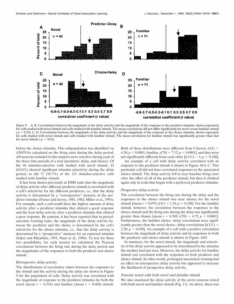
Retrospective delay activityThe distributions of correlation values between the responses tothe stimuli and the activity during the delay are shown in Figure9 for the population of cells. Delay activity was correlated withthe magnitude of response to the predictor stimulus for both thenovel (mean r 5 0.316) and familiar (mean r 5 0.404) stimuli.
Both of these distributions were different from 0 [novel, t(41) 54.38; p , 0.0001; familiar, t(70) 5 7.12; p , 0.0001], and they werenot significantly different from each other [t(111) 5 1; p 5 0.340].
An example of a cell with delay activity correlated with itsresponse to the predictor stimuli is shown in Figure 10A–C. Thisparticular cell did not have correlated responses to the associatedchoice stimuli. The delay activity fell to near baseline firing ratesafter the offset of all of the predictor stimuli, but then it climbedagain only in trials that began with a preferred predictor stimulus.
Prospective delay activityThe correlation between the firing rate during the delay and theresponses to the choice stimuli was near chance for the novelstimuli [mean r 5 0.079; t(41) 5 1.34; p 5 0.188]. For the familiarstimuli, however, the correlation between the responses to thechoice stimuli and the firing rate during the delay was significantlygreater than chance [mean r 5 0.269; t(70) 5 4.75; p , 0.0001].Furthermore, the familiar choice–delay correlations were signif-icantly greater than the novel choice–delay correlations [t(111) 52.20; p 5 0.030]. An example of a cell with a positive correlationbetween the magnitude of delay activity and its responses to boththe predictor and choice stimuli is shown in Figure 10D.
In summary, for the novel stimuli, the magnitude and selectiv-ity of the delay activity appeared to be determined by the stimulusthe monkey had just seen. However, the delay activity for familiarstimuli was correlated with the responses to both predictor andchoice stimuli. In other words, prolonged association training hadno effect on retrospective delay activity but appeared to increasethe likelihood of prospective delay activity.
Neurons tested with both novel and familiar stimuliWe also examined the delay activity of the seven neurons testedwith both novel and familiar stimuli (Fig. 11). As above, there was
Figure 9. A, B, Correlations between the magnitude of the delay activity and the magnitude of the response to the predictor stimulus, shown separatelyfor cells studied with novel stimuli and cells studied with familiar stimuli. The mean correlations did not differ significantly for novel versus familiar stimuli( p 5 0.34). C, D, Correlations between the magnitude of the delay activity and the magnitude of the response to the choice stimulus, shown separatelyfor cells studied with novel stimuli and cells studied with familiar stimuli. The mean correlation for familiar stimuli was significantly greater than thatfor novel stimuli ( p 5 0.03).
Erickson and Desimone • Neural Correlates of Visual Association Learning J. Neurosci., December 1, 1999, 19(23):10404–10416 10411

no effect of experience on the retrospective delay activity (r 50.46 for novel compared with r 5 0.30 for familiar), but there wasa large increase in prospective delay activity (r 5 20.31 for novelcompared with r 5 0.39 for familiar).
Relationship between delay activity andpredictor–choice correlationsAs described in the previous sections, some cells showed positivecorrelations between their responses to the predictors andchoices, and some cells showed positive correlations betweentheir delay activity and their responses to both the predictors andchoices (Fig. 10D). This raised the possibility that the positivecorrelation between the delay activity and choice responses foundfor familiar stimuli was actually a side effect of the increasedcorrelations between the predictor and choice responses foundfor familiar stimuli.
We used partial correlations to control statistically for thepotential confound of the increased correlations between theresponses to the stimulus pairs with experience (Keppel, 1973).This analysis provided a way to separate the correlation betweenpredictor–choice responses from the correlation between choiceresponses and delay activity. The mean difference between thepartial correlation and raw correlation coefficient was 0.065 (t ,1; p 5 0.456) and 0.030 (t , 1; p 5 0.705) for the predictor–delay
Figure 11. Correlation between the magnitude of response to the pre-dictor and choice stimuli and the magnitude of the associated delayactivity, for seven cells tested with both novel and familiar stimuli. Themean correlations differed significantly for novel versus familiar stimuli( p , 0.001). Delay-selective neurons tested with both novel and familiarstimuli showed the same shift in delay activity correlated with the choicestimuli (n 5 7). Experience with stimuli changed the sign of the correla-tion ( p , 0.001).
Figure 10. Spike density histograms of neurons ex-hibiting stimulus-selective delay activity. A, Each col-ored line represents the response of the same neuron toone of eight different pairs of associated stimuli. Theresponse to the best predictor stimulus in the set ofeight (red line) returns to baseline after the stimulus isturned off and then climbs during the delay period. B,The enlarged scale for the delay activity of the samecell shows that the magnitude of delay activity iscorrelated with the responses to the predictor stimuli.C, Mean responses are shown to the predictor stimuliand choice stimuli, both plotted against the magnitudeof delay activity, for the same cell shown in A and B.Mean delay activity was calculated during the intervalfrom 200 to 1000 msec after the predictor stimulus wasturned off. D, An example from a different neuron, inwhich the magnitude of response to the predictor, themagnitude of activity during the delay, and the mag-nitude of response to the paired choice stimulus wereall correlated with one another, is shown. The stimu-lus presentation times are indicated by gray horizontallines.
10412 J. Neurosci., December 1, 1999, 19(23):10404–10416 Erickson and Desimone • Neural Correlates of Visual Association Learning

and choice–delay correlations, respectively. The predictor–choice relationship accounted for none of the retrospective delayactivity and some, but not all, of the prospective delay activitycorrelations (Fig. 12). Thus, association learning appears to causean increase in prospective delay activity that is not accounted forby the increased correlation between the responses to the predic-tor and choice stimuli.
Classifying neurons by behavioral evidence ofassociation learningNext we examined whether the relationship between the delayactivity and stimulus-evoked responses varied according towhether or not the monkey showed behavioral evidence of asso-ciation learning in a given recording session (Fig. 13). For thepredictor stimuli, we conducted a two-way ANOVA on the cor-relations between the delay activity and the response to thepredictor stimuli, with experience level (novel vs familiar) andevidence of association learning (reaction time difference be-tween valid and neutral trials) as the two factors. The resultsindicated that experience level had no effect on the mean r values(no main effect of experience, p 5 0.348). However, there was asignificant main effect of association learning, with a mean r of0.429 in sessions with no evidence of association learning com-
pared with a mean r of 0.232 in sessions with significant evidenceof association learning [F(1,100) 5 5.2; p 5 0.024]. There was nosignificant interaction between the effects of experience and as-sociation learning ( p 5 0.929). Thus, when the neurons wererecorded during sessions in which there was evidence of associa-tion learning, the delay activity was actually less correlated withthe response to the predictor stimulus (Fig. 13A). The reason forthis superficially paradoxical result is suggested by the analysisbelow.
When we conducted the same analysis on the correlationsbetween the delay activity and the responses to the choice stimuli,we found a complementary pattern of results (Fig. 13B). Accord-ing to the two-way ANOVA, there were significant main effects ofexperience and association learning (experience [F(1,100) 5 6.6;p 5 0.012]; association learning [F(1,100) 5 12.7; p 5 0.001]), andthere was a significant interaction between the two factors[F(1,100) 5 4.8; p 5 0.032]. In other words, in sessions withevidence of association learning, the correlation between delayactivity and the response to the predictor stimuli fell at the sametime that the correlation between the delay activity and theresponse to the choice stimuli rose. The highest correlationsbetween the delay activity and responses to the choice stimuliwere found for those neurons recorded with familiar stimuli and
Figure 12. Partial correlations between the magnitude of response to thepredictor and choice stimuli and the associated delay activity. A, B, TheVenn diagrams conceptually describe the difference between the actualcorrelations and the partial correlations. C, D, Little of the predictor–delay relationship can be accounted for by the predictor–choicecorrelations.
Figure 13. Comparison of the effects of experience and behavioralevidence of association learning on delay activity. A, Correlation betweendelay activity and responses to the predictor stimuli. Delay activity wasless tightly correlated with responses to the predictor stimuli for thosedata sets in which the monkeys showed evidence of association learning(main effect of learning, p , 0.05). B, Correlations between delay activityand responses to choice stimuli. Delay activity was mostly tightly corre-lated with the responses to the choice stimuli in those sessions withbehavioral evidence of association learning as well as in those sessionsusing familiar stimuli (main effect of both experience and learning).
Erickson and Desimone • Neural Correlates of Visual Association Learning J. Neurosci., December 1, 1999, 19(23):10404–10416 10413

with behavioral evidence of association learning in the recordingsession (mean r 5 0.572).
In summary, on the first day of training, the responses to pairedstimuli were uncorrelated, and the magnitude of delay activitywas correlated almost solely with the response to the predictorstimulus, i.e., the stimulus that immediately precedes the delay.On subsequent days, the responses to paired stimuli becamecorrelated, and the delay activity became correlated with theresponses to both by the predictor and choice stimuli, i.e., thestimulus the animal has just seen and the stimulus to follow. Insessions with behavioral evidence of association learning, thedelay activity tended to switch from being correlated with thepredictor stimuli to being correlated with the choice stimuli. Wetake these shifts in the correlations with delay activity to indicatethat as the monkey learns the relationship between the stimuliand uses the knowledge of the relationship to respond morequickly on valid trials, the delay activity shifts from reflecting thestimulus that was just seen to predicting the stimulus to come.
DISCUSSIONBy using an associative-learning task that monkeys could learnquickly, we were able to measure separately neural correlates ofassociative memory for recently learned (i.e., novel) associatesand well learned (i.e., familiar) associates. When tested with thefamiliar associated pairs of stimuli, perirhinal neurons exhibitedtwo properties that seemed directly related to the requirementsfor associative learning. First, neurons responded more similarlyto associated stimuli (predictor and choice stimuli) than would beexpected by chance. If, for example, a cell responded well to aparticular predictor stimulus, then it would tend to respond wellto the particular choice stimulus with which it was associated.Second, the magnitude of activity during the delay interval be-tween predictor and choice was correlated with the responses toboth the predictor and choice stimuli. In other words, delayactivity was often higher on trials when the predictor and/orchoice stimuli elicited good responses from the cell than whenthey elicited poor responses. These delay effects were largest insessions with behavioral evidence of association learning.
By contrast, neither of these associative effects on neuronalactivity was found when the stimulus pairs were novel, i.e.,learned on the day of the recording. Across the population ofcells, there was no correlation between the responses to associ-ated predictor and choice stimuli on the first day. Furthermore,although the magnitude of delay activity was often correlatedwith the magnitude of responses to the preceding predictor stim-ulus on the first day, there was no correlation between the delayactivity and the responses to the associated choice stimulus. Inspite of this failure to find any perirhinal correlates of associativelearning on the first day of learning, the monkeys’ behavioralperformance indicated that they did indeed learn to associate thestimuli on the first day. With novel stimuli, behavioral reactiontimes were faster when choice stimuli were followed by frequentlyassociated predictors than when they were followed by infre-quently associated predictors, and this was found for both invalidand neutral predictors. Thus, if we assume that the perirhinalresponse changes we found with familiar stimuli actually contrib-ute to associative memory, then our failure to find these samechanges with novel stimuli suggests that different mechanisms areused to perform the task on the first day than on subsequent daysof training. On the first day, the critical mechanisms may belocated outside of the perirhinal cortex. If so, the changes inperirhinal activity we found on the second and later days of
training might be caused by a consolidation of the memory tracein perirhinal cortex after initial learning-induced activity changesin other medial temporal lobe structures. We also cannot elimi-nate the possibility that the critical sites of plasticity remainoutside the perirhinal cortex even after extensive training andthat the effects of learning we found on perirhinal responses werecaused by feedback from these critical sites.
We have often found that there is little obvious perceptualsimilarity among the stimuli to which a given perirhinal neuronresponds well and a corresponding lack of similarity among thestimuli to which a given neuron responds poorly (unpublishedobservations). The fact that we found that neuronal responses totemporally associated stimuli tend to become similar to oneanother with experience provides a possible explanation for theresponse properties of many perirhinal neurons. Specifically,perirhinal responses may be specific for behaviorally significantstimulus categories, rather than stimulus features per se. Onecould imagine, for example, that a perirhinal neuron might re-spond to both the image of a piece of fruit and the image of a leafbecause they are often associated in time, although they share fewphysical features. The modification of response properties fortemporally associated stimuli might also be a mechanism forlearning to associate different three-dimensional views of thesame object. In agreement with this, Logothetis has found thatTE neurons respond more commonly to different three-dimensional views of the same object than would be expected bychance (Logothetis et al., 1995).
Although the mean correlation we found between responses tofamiliar predictor and choice stimuli was highly significant, theactual magnitude of this correlation (0.145) was quite small acrossthe population of cells. We estimated that 18.4% of the variancein mean firing rates of choice stimuli could be accounted for bythe associated predictor stimulus. One reason why the small sizeof this correlation could be misleading is that associative learningmight affect the responses of only a subpopulation of cells. Be-cause we were unable to track changes in response properties overthe course of daily sessions, we had no independent way toseparate cells affected by learning from cells that were not.However, the increase in the number of neurons with strongpositive correlations after training can be seen in the distributionof correlation coefficients for familiar stimuli.
Using a different behavioral task, Miyashita and colleagues(Sakai and Miyashita, 1991; Higuchi and Miyashita, 1996) foundthat the correlation between the responses to paired-associatestimuli in perirhinal cortex was similar in magnitude to what wefound. They did not test for correlations in responses to novelstimuli, because all of the stimuli used in their study were highlyfamiliar to the animal.
In contrast to the work by Miyashita and colleagues as well asour own, Sobotka and Ringo (1993) failed to observe any re-sponse similarity between paired-associate stimuli. In their study,two stimuli were presented simultaneously as a complex image,and a monkey had to discriminate GO from NO-GO stimuluspairs. It is possible that the two stimuli presented on the screenwere treated by the monkey as a single complex stimulus ratherthan as two independent, but associated, stimuli, which mightaccount for the failure to find any effects of learning on responses.Gochin et al. (1994) also failed to find any effects of associativelearning on TE responses. In their study, which used a conditionalassociation task with five stimuli, monkeys were trained to re-spond differently depending on which stimulus of a pair camefirst. Gochin et al. (1994) may have failed to find effects of
10414 J. Neurosci., December 1, 1999, 19(23):10404–10416 Erickson and Desimone • Neural Correlates of Visual Association Learning
association learning on responses because individual stimuli wereused in more than one pair. For example, A was followed by B ona “GO” trial, but C was followed by B on a “NO-GO” trial.Although stimulus B had a different meaning depending on whatpreceded it, both A and C were paired with B an equal number oftimes.
Sakai and Miyashita (1991) found that delay activity was cor-related with the responses to the second stimulus in a sequentialpaired-associate task, similar to what we found in the presentstudy. They concluded that delay activity in perirhinal cortex isprospective in the paired-associative task. However, we foundthat delay activity was almost exclusively retrospective with novelstimuli in our associative task and was both retrospective andprospective with familiar stimuli. Retrospective delay activity hasbeen found in several other studies of perirhinal and TE cortex(Li et al., 1993; Miller et al., 1993).
In a different study, Miyashita (1988) presented stimuli in afixed temporal pattern and measured the delay activity after eachstimulus. They reported that the delay activity of TE neurons wasmore similar after stimuli presented nearby in time comparedwith delay activity after stimuli not linked in time. Yakovlev et al.(1998) conducted a similar experiment and reported that thedelay activity was primarily correlated with the response to theprevious stimulus in the sequence. They suggested that delayactivity develops and becomes longer lasting after experiencingstimuli in a fixed sequence. Neither of these two studies reportedchanges in stimulus-evoked responses with experience, but ourresults suggest a complex relationship between stimulus-evokedresponses and delay activity that changes with experience. Wefind that delay activity is equally common with novel and familiarstimuli; however, with familiar stimuli, responses to paired stimulibecome more similar, and delay activity shifts from being nearlyentirely retrospective to being both retrospective and prospective.
What is the function of delay activity in the formation ofassociative memories? If the association is between two stimuliseparated in time, then a likely function of retrospective delayactivity is to maintain a representation of the first stimulus duringthe delay. If the neural representation of the first image is activeduring the presentation of the second image, this simultaneousactivation of different populations could facilitate Hebbianchanges in connectivity between cells in the two representations.As a result, a new stimulus class could be formed with the twoassociated stimuli as members. After learning takes place, pro-spective delay activity may serve to bias the development of aneural representation of the expected stimulus, for the sake ofefficiency. Alternatively, the fact that association learning leads toa common response that starts with the presentation of the firststimulus and continues through the delay and presentation of thesecond stimulus suggests that the perirhinal cells serve to linkvisual stimuli across time, possibly representing these temporallyassociated stimuli as an “event.”
REFERENCES
Alvarez-Royo P, Clower RP, Zola-Morgan S, Squire LR (1991) Stereo-taxic lesions of the hippocampus in monkeys: determination of surgicalcoordinates and analysis of lesions using magnetic resonance imaging.J Neurosci Methods 38:223–232.
Crist C, Yamasaki D, Komatsu H, Wurtz R (1988) A grid system and amicrosyringe for single cell recording. J Neurosci Methods 26:117–122.
Eskandar EN, Optican LM, Richmond BJ (1992) Role of inferior tem-poral neurons in visual memory. II. Multiplying temporal waveformsrelated to vision and memory. J Neurophysiol 68:1296–1306.
Fahy FL, Riches IP, Brown MW (1993) Neuronal activity related tovisual recognition memory: long-term memory and the encoding ofrecency and familiarity information in the primate anterior and medialinferior temporal and rhinal cortex. Exp Brain Res 96:457–472.
Fuster JM, Jervey JP (1981) Inferotemporal neurons distinguish andretain behaviorally relevant features of visual stimuli. Science212:952–955.
Fuster JM, Jervey JP (1982) Neuronal firing in the inferotemporal cortexof the monkey in a visual memory task. J Neurosci 2:361–375.
Gaffan D (1994) Dissociated effects of perirhinal cortex ablation, fornixtransection and amygdalectomy: evidence for multiple memory systemsin the primate temporal lobe. Exp Brain Res 99:411–422.
Gochin PM, Colombo M, Dorfman GA, Gerstein GL, Gross CG (1994)Neural ensemble coding in inferior temporal cortex. J Neurophysiol71:2325–2337.
Gray CM, Maldonado PE, Wilson M, McNaughton BL (1995) Tetrodesmarkedly improve the reliability and yield of multiple single-unit iso-lation from multi-unit recordings in cat striate cortex. J Neurosci Meth-ods 63:43–54.
Higuchi S, Miyashita Y (1996) Formation of mnemonic neuronal re-sponses to visual paired associates in inferotemporal cortex is impairedby perirhinal and entorhinal lesions. Proc Natl Acad Sci USA93:739–743.
Keppel G, Zedeck S (1989) Data analysis for research designs: analysisof variance and multiple regression/correlation approaches. New York:Freeman.
Li L, Miller EK, Desimone R (1993) The representation of stimulusfamiliarity in anterior inferior temporal cortex. J Neurophysiol69:1918–1929.
Logothetis NK, Pauls J, Poggio T (1995) Shape representation in theinferior temporal cortex of monkeys. Curr Biol 5:552–563.
Lueschow A, Miller EK, Desimone R (1994) Inferior temporal mecha-nisms for invariant object recognition. Cereb Cortex 4:523–531.
McNaughton BL, O’Keefe J, Barnes CA (1983) The stereotrode: a newtechnique for simultaneous isolation of several single units in thecentral nervous system from multiple unit records. J Neurosci Methods8:391–397.
Meunier M, Bachevalier J, Mishkin M, Murray EA (1993) Effects onvisual recognition of combined and separate ablations of the entorhinaland perirhinal cortex in rhesus monkeys. J Neurosci 13:5418–5432.
Miller EK, Desimone R (1994) Parallel neuronal mechanisms for short-term memory. Science 263:520–522.
Miller EK, Li L, Desimone R (1993) Activity of neurons in anteriorinferior temporal cortex during a short-term memory task. J Neurosci13:1460–1478.
Miyashita Y (1988) Neuronal correlate of visual associative long-termmemory in the primate temporal cortex. Nature 335:817–820.
Murray EA, Mishkin M (1986) Visual recognition in monkeys followingrhinal cortical ablations combined with either amygdalectomy or hip-pocampectomy. J Neurosci 6:1991–2003.
Murray EA, Gaffan D, Mishkin M (1993) Neural substrates of visualstimulus-stimulus association in rhesus monkeys. J Neurosci13:4549–4561.
Nahm FKD, Dale AM, Albright TD, Amaral DG (1994) In vivo micro-electrode localization in the brain of the alert monkey: a combinedradiographic and magnetic resonance imaging approach. Exp BrainRes 98:401–411.
Naya Y, Sakai K, Miyashita Y (1996) Activity of primate inferotemporalneurons related to a sought target in pair-association task. Proc NatlAcad Sci USA 93:2664–2669.
Nichols AM, Ruffner TW, Sommer MA, Wurtz RH (1998) A screwmicrodrive for adjustable chronic unit recording in monkeys. J NeurosciMethods 81:185–188.
Posner MI, Snyder CR, Davidson BJ (1980) Attention and the detectionof signals. J Exp Psychol 109:160–174.
Robinson DA (1963) A method of measuring eye movement using ascleral search coil in a magnetic field. IEEE Trans Biomed Eng10:137–145.
Sakai K, Miyashita Y (1991) Neural organization for the long-termmemory of paired associates. Nature 354:152–155.
Erickson and Desimone • Neural Correlates of Visual Association Learning J. Neurosci., December 1, 1999, 19(23):10404–10416 10415

Sobotka S, Ringo JL (1993) Investigation of long-term recognition andassociation memory in unit responses from inferotemporal cortex. ExpBrain Res 96:28–38.
Sobotka S, Ringo JL (1996) Mnemonic responses of single units re-corded from monkey inferotemporal cortex, accessed via transcommis-sural versus direct pathways: a dissociation between unit activity andbehavior. J Neurosci 16:4222–4230.
Suzuki WA, Amaral DG (1994) Perirhinal and parahippocampal corti-ces of the macaque monkey: cortical afferents. J Comp Neurol350:497–533.
Suzuki WA, Zola-Morgan S, Squire LR, Amaral DG (1993) Lesions ofthe perirhinal and parahippocampal cortices in the monkey produce
long-lasting memory impairment in the visual and tactual modalities.J Neurosci 13:2430–2451.
Yakovlev V, Fusi S, Berman E, Zohary E (1998) Inter-trial neuronalactivity in inferior temporal cortex: a putative vehicle to generatelong-term visual association. Nat Neurosci 1:310–317.
Zola-Morgan S, Squire LR, Amaral DG, Suzuki WA (1989) Lesions ofperirhinal and parahippocampal cortex that spare the amygdala andhippocampal formation produce severe memory impairment. J Neuro-sci 9:4355–4370.
Zola-Morgan S, Squire LR, Clower RP, Rempel NL (1993) Damage tothe perirhinal cortex exacerbates memory impairment following lesionsto the hippocampal formation. J Neurosci 13:251–265.
10416 J. Neurosci., December 1, 1999, 19(23):10404–10416 Erickson and Desimone • Neural Correlates of Visual Association Learning