Sociality, stress, and the corpus striatum of the green anolis lizard

Enhanced Proliferation of Progenitor Cellsin the Subventricular Zone and LimitedNeuronal Production in the Striatum andNeocortex of Adult Macaque MonkeysAfter Global Cerebral Ischemia
Anton B. Tonchev,1,2 Tetsumori Yamashima,1* Kazunobu Sawamoto,3,4 andHideyuki Okano4
1Department of Restorative Neurosurgery, Division of Neuroscience, Kanazawa University GraduateSchool of Medical Sciences, Kanazawa, Japan2Division of Cell Biology, Department of Forensic Medicine, Varna University of Medicine,Varna, Bulgaria3Bridgestone Laboratory of Developmental and Regenerative Neurobiology,Keio University School of Medicine, Tokyo, Japan4Department of Physiology, Keio University School of Medicine, Tokyo, Japan
Cerebral ischemia in adult rodent models increases theproliferation of endogenous neural progenitor cells resid-ing in the subventricular zone along the anterior horn ofthe lateral ventricle (SVZa) and induces neurogenesis inthe postischemic striatum and cortex. Whether the adultprimate brain preserves a similar ability in response to anischemic insult is uncertain. We used the DNA synthesisindicator bromodeoxyuridine (BrdU) to label newly gener-ated cells in adult macaque monkeys and show here thatthe proliferation of cells with a progenitor phenotype(double positive for BrdU and the markers Musashi1,Nestin, and bIII-tubulin) in SVZa increased during thesecond week after a 20-min transient global brain ische-mia. Subsequent progenitor migration seemed restrictedto the rostral migratory stream toward the olfactory bulband ischemia increased the proportion of adult-generatedcells retaining their location in SVZa with a progenitorphenotype. Despite the lack of evidence for progenitorcell migration toward the postischemic striatum orprefrontal neocortex, a small but sustained proportionof BrdU-labeled cells expressed features of postmitoticneurons (positive for the protein NeuN and the transcrip-tion factors Tbr1 and Islet1) in these two regions forat least 79 days after ischemia. Taken together, ourdata suggest an enhanced neurogenic response in theadult primate telencephalon after a cerebral ischemicinsult. VVC 2005 Wiley-Liss, Inc.
Key words: monkey; cerebral ischemia; progenitorcell; neurogenesis; gliogenesis
The adult mammalian brain retains a limited abilityfor neuronal regeneration by progenitor cells residing intwo well-recognized neurogenic niches: the subventricu-
lar zone (SVZ) of the lateral ventricle and the subgranu-lar zone (SGZ) of the hippocampal dentate gyrus (DG;Gage, 2000). Extensively studied in rodents, in vivoneurogenesis has also been demonstrated in SVZ (Kor-nack and Rakic, 2001a; Pencea et al., 2001) and SGZ(Eriksson et al., 1998; Gould et al., 1999a; Kornack andRakic, 1999) of healthy primates, but its existence out-side these regions under normal conditions remains con-troversial (Gould et al., 1999b, 2001; Kornack andRakic, 2001b; Bedard et al., 2002; Koketsu et al., 2003).
In the rodent brain, cerebral ischemia increasesproliferation and neuronal differentiation of neural pro-genitor cells in both global (Liu et al., 1998; Kee et al.,2001; Yagita et al., 2001; Nakatomi et al., 2002; Iwaiet al., 2003) and focal (Jin et al., 2001; Zhang et al.,2001; Arvidsson et al., 2002; Parent et al., 2002) ische-mic models. The newly generated cells have beenreported to replace neurons in the postischemic hippo-campus (Liu et al., 1998; Kee et al., 2001; Yagita et al.,
Supplementary Material for this article is available online at http://www.mrw.interscience.wiley.com/suppmat/0360-4012/suppmat/(www.interscience.wiley.com).Contract grant sponsor: Japanese Ministry of Education, Culture, Sports,
Science and Technology; Contract grant sponsor: National Science Fund
of Bulgaria; Contract grant number: L1311/03.
*Correspondence to: Dr. Tetsumori Yamashima, Department of Restora-
tive Neurosurgery, Division of Neuroscience, Kanazawa University
Graduate School of Medical Sciences, Takara-machi 13-1, 920-8641
Kanazawa, Japan. E-mail: [email protected]
Received 27 March 2005; Revised 14 May 2005; Accepted 27 May
2005
Published online 26 July 2005 in Wiley InterScience (www.
interscience.wiley.com). DOI: 10.1002/jnr.20604
Journal of Neuroscience Research 81:776–788 (2005)
' 2005 Wiley-Liss, Inc.

2001; Nakatomi et al., 2002), striatum (Arvidsson et al.,2002; Parent et al., 2002; Zhang et al., 2004), olfactorybulb (Iwai et al., 2003), and cortex (Gu et al., 2000;Nakatomi et al., 2002). Interestingly, progenitor cells inthe rostral migratory stream, originally destined tobecome olfactory bulb neurons, are redirected towardthe injured striatum and cortex after ischemia (Jin et al.,2003). In our previous study we investigated the subven-tricular zone along the inferior horn of the lateral ven-tricle (SVZi) and DG of adult monkeys after ischemia(Tonchev et al., 2003a). We found that although anincrease in postischemic precursor cells was observed inthe monkey DG (similar to the situation in rodents), themonkey response was much smaller than was the rodentresponse (Liu et al., 1998; Kee et al., 2001), in terms ofthe proliferation and extent of the neuronal differentia-tion of progenitor cells.
In this study, we used the same monkey model butfocused on different brain regions. Our first goal was tomonitor the phenotype and behavior of newly generatedcells in SVZ of the anterior horn of the lateral ventricle(SVZa) after 20 min of global brain ischemia in adultmonkeys. The adult-born cells were labeled with thethymidine analogue bromodeoxyuridine (BrdU) and theimmunophenotype of BrdU-positive (BrdUþ) cells wasdetermined using a panel of selective cell markers at var-ious times after the insult. Our second goal was toexplore the potential route(s) of SVZa progenitor cellmigration after ischemia. Our third goal was to identifythe immunophenotype of BrdUþ cells in the postische-mic striatum and neocortex. Ischemia caused an equalfor the two hemispheres neuronal damage of 30–40% inthese regions, using the same ischemic model used in thepresent study (Yoshida et al., 2002). This injury was muchless pronounced than was the damage inflicted to thehippocampal CA1 sector, which exhibited an almost com-plete neuronal loss (Yamashima et al., 1998; Yamashima,2000). It was also much less pronounced than the striatal/cortical injuries induced by experimental focal ischemicinfarction (Arvidsson et al., 2002; Parent et al., 2002). Wethus investigated progenitor cell proliferation and pheno-type in the context of a moderate ischemic injury.
MATERIALS AND METHODS
Surgical Procedures
All experimental procedures were carried out in strictadherence to the guidelines of the Animal Care and EthicsCommittee of Kanazawa University and the NIH Guide forthe Care and Use of Laboratory Animals. The monkeys(female Macaca fuscata) were bred in air-conditioned cages andwere allowed free daily access to food and water. Under ster-ile conditions and a general anesthesia with artificial ventila-tion, 19 adult (5–11 years old) macaques underwent transient,complete, whole brain ischemia (n ¼ 12) or a sham surgery(n ¼ 7), as described previously (Yamashima et al., 1998;Yamashima, 2000; Tonchev et al., 2003a). Briefly, the chestwas opened, the sternum was removed, and the innominateand left subclavian arteries were transiently clipped for
20 min. The effectiveness of clipping was demonstrated by analmost complete absence (0.5 6 1.0 ml/100 g brain/min) ofcerebral blood flow, as monitored using a laser Doppler (Vasa-medics, St. Paul, MN). One neonatal monkey was sacrificedon postnatal day 14 (P14), without any preceding surgery.
BrdU Labeling
All 19 adult monkeys received daily injections (100 mg/kg, intravenously) of BrdU (Sigma) for 5 consecutive days.The animals in the short-term survival group (n ¼ 9)were sacrificed 2 hr after the last injection on days 4 (n ¼ 2),9 (n ¼ 2) and 15 (n ¼ 2) after ischemia or on days 4 (n ¼ 1)and 9 (n ¼ 2) after the sham operation (Fig. 1E2; Tonchevet al., 2003a). The animals in the long-term survival group(n ¼ 10) received BrdU injections on days 5–9 after surgeryand were sacrificed 2, 5, and 10 weeks after the last injection,i.e., on days 23 (n ¼ 2), 44 (n ¼ 2), and 79 (n ¼ 2) afterischemia or on days 23 (n ¼ 2) and 44 (n ¼ 2) after the shamoperation (Fig. 2E2). The neonatal monkey received twoBrdU injections, one on the day of birth and one on P14,and was euthanized 2 hr after the second injection.
Histology
The monkeys were anesthetized with lethal doses ofsodium pentobarbital and intracardially perfused with 4% par-aformaldehyde. The brains were removed, tissue blocks werecryoprotected in sucrose, embedded in OCT medium (Tis-sue-Tek, Sakura Finetech Co., Tokyo, Japan) and frozen at�708C. Coronal (right hemisphere) or horizontal (left hemi-sphere) sections (40 lm thick) were cut sequentially on a cry-omicrotome and stored free-floating at �208C in a cryopre-servation buffer until analysis. BrdU staining was preceded byDNA denaturation as described previously (Eriksson et al.,1998; Tonchev et al., 2003a), and incorporated BrdU wasdetected using mouse anti-BrdU (1:100; Becton Dickinson,San Jose, CA) or rat anti-BrdU (1:100; Harlan Sera-Lab,Loughborough, UK) antibodies. The following antibodies forphenotypic markers were applied in combination with anti-BrdU: mouse anti-Ki67 (1:50; Novocastra, Newcastle, UK),rabbit anti-phosphorylated histone H3 (1:50; Cell Signaling,Beverly, MA), rat anti-Musashi1 (1:100; Kaneko et al., 2000),rabbit anti-Musashi1 (1:200; Chemicon, Temecula, CA), rab-bit or mouse anti-Nestin (1:200; Chemicon), mouse anti-neu-ronal nuclei (NeuN, 1:100; Chemicon), rabbit or mouse anti-b-tubulin class III (1:400; Covance, Richmond, CA), goatanti-Doublecortin (1:200; Santa Cruz Inc., Santa Cruz, CA),mouse anti-polysialylated neural cell adhesion molecule (PSA-NCAM, 1:200; a gift from Tatsunori Seki), rabbit anti-glialfibrillary acidic protein (GFAP, 1:400; Sigma), mouse anti-S100b (1:500; Sigma), rabbit anti-Iba1 (1:800; a gift fromYoshinori Imai), and rabbit anti-glutamic acid decarboxylase(GAD)65/67 (1:400; Chemicon). The rabbit antibody againstIslet1 (1:400) was obtained from Chemicon and the goat anti-Tbr1 antibody (1:200) was obtained from Santa Cruz. Thestaining for activated caspase-3 was carried out using a rabbitanti-activated caspase-3 antibody (1:50; Cell Signaling). Theprimary antibodies were revealed by appropriate secondaryantibodies conjugated to biotin for immunoperoxidase labeling
Neurogenesis in Postischemic Monkey Brain 777

(1:30–1:100, Vector ABC kit; Vector Laboratories, Burlin-game, CA), or AlexaFluor 488, 546, 633 (1:100; MolecularProbes, Eugene, OR) and tetramethylrhodamine isothiocya-nate (TRITC, 1:30; Jackson ImmunoResearch, West Grove,PA) for immunofluorescent labeling. For double and triplestaining, the respective primary antibodies were from differentspecies and were applied sequentially to minimize crossreactivity.
Terminal Deoxynucleotidyltransferase-Mediated UTPNick-End Labeling Assay
Free-floating sections were subjected to the terminaldeoxynucleotidyltransferase (TdT)-mediated UTP nick-endlabeling (TUNEL) assay carried out using the ApopTag in situcell death detection kit (Intergene, Purchase, NY), asdescribed previously (Tonchev et al., 2003a). Briefly, the sec-tions were dehydrated in an ascending ethanol/dH2O series(50, 70, and 90%; 5 min each) followed by one 15-min incu-bation in 100% ethanol and rehydration in ethanol/dH2O (90,70, and 50%; 5 min each). Sections were then subjected toproteinase K (20 lg/mL, 15 min) followed by equilibrationbuffer and TdT/reaction buffer mixture at 378C for 2 hr.Reactions involving digoxigenin incorporation were visualizedby appropriate antibodies. Control stainings included DNasepretreatment (positive control for reaction specificity) or TdTomission (negative control). For TUNEL/BrdU double label-ing, the reaction was developed until the enzyme step, DNAdenaturation for BrdU, was carried out and then the incorpo-rated digoxigenin and BrdU were visualized using immuno-staining. For NeuN/TUNEL/BrdU triple labeling, the anti-body against NeuN was applied and visualized at the end ofthe procedure.
Image Analysis
The numbers and densities of BrdUþ cells were deter-mined on immunoperoxidase-labeled sections. Tissue blocksfrom the left hemisphere, starting dorsally at the level of thecorpus callosum topping the roof of the anterior horn of thelateral ventricle and spanning ventrally until the level of theanterior commissure (ac), were cut into horizontal sections.Tissue blocks from the right hemisphere, acþ7 mm anteriorlyto acþ1 mm posteriorly, were cut into coronal sections. Every12th section was evaluated using a computerized microscopesystem and positive cells were displayed on a computer screen.The number of cells that had incorporated BrdU in SVZa wasdetermined using 800 lm 3 100 lm grids placed over eachof the five aspects of SVZa (dorsal, ventral, septal, caudate,and anterior), as outlined on the maps in Figure 1. TheBrdUþ cells in the striatum and frontal cortex were countedusing 880 lm 3 680 lm grids. Numbers and densities wereaveraged to obtain a mean density value for each region/ani-mal group. Double and triple labeling to determine theexpression of phenotypic markers and transcription factors byBrdU-expressing cells were verified using confocal laser scan-ning microscopy (LSM 510; Carl Zeiss, Tokyo, Japan). AlexaFluor 488 was assigned to the green channel, TRITC orAlexa Fluor 546 were assigned to the red channel, and AlexaFluor 633 was assigned to the blue channel. Each fluoro-chrome was scanned separately and sequentially to minimizethe probability of signal transfer among channels. Z sectioningat intervals of 0.5–1 lm was carried out and optical stacks ofat least 20 images were used for analysis. Digital three-dimen-sional reconstructions were created using Zeiss LSM v2.3 soft-ware. Within each region and animal group, 100 BrdUþ cellsper marker were sampled for coexpression on at least five dif-
Fig. 1. Cell proliferation in the monkeygroup with short-term survival afterBrdU. BrdUþ cells in the septal (A1,A3), caudate (A2, A4), ventral (B), dor-sal (D) and anterior (C) aspects of SVZaof control (A1, A2, B1, C1, D1) andpostischemic (A3, A4, B2, C2, D2)day 9 brains. M, medial; L, lateral; D,dorsal; V, ventral; A, anterior; P, poste-rior; Str, striatum. The lumen of the lat-eral ventricle is shown by an asterisk. E1:Quantitative evaluation of the BrdUþcells. Note the increase in BrdUþ cellson postischemic day 9 in the ventral (V),anterior (A), dorsal (D), and caudate (C)SVZa. In contrast, no change wasobserved in the septal (S) SVZa. *P <0.05 vs. control animals. E2: BrdU infu-sion protocol, with gray horizontal barsdepicting periods of BrdU infusion. D0,day of surgery. Scale bar¼ 50 lm.
778 Tonchev et al.

ferent sections, whereas 200 BrdUþ cells were investigatedfor colabeling with NeuN.
Statistical Analysis
Data were expressed as the mean 6 standard errorof the mean (SEM). Differences between means were
determined using one-way analysis of variance (ANOVA)followed by Tukey-Kramer’s posthoc comparison and two-sided t-test. For comparing percentages of cells retainedin SVZa, nonparametric test was also applied (Mann-Whitney). Differences were considered significant whenP < 0.05.
Fig. 2. Cell proliferation in the group with long-term survival afterBrdU treatment. Representative images of the BrdUþ cells in the dor-sal (A), caudate (B), anterior (C), and ventral (D) SVZa aspects of con-trol day 44 (A1, B1, C1, D1) or postischemic day 44 (A2, B2, C2,D2) and day 79 (B3) brains are shown. Note the greater number ofBrdUþ cells in the dorsal, caudate, or anterior SVZa aspects afterischemia. M, medial; L, lateral; D, dorsal; V, ventral; A, anterior; P,posterior; Str, striatum. The lumen of the lateral ventricle is shown byan asterisk. Scale bar ¼ 50 lm. E1: Quantitative evaluation of the
BrdUþ cells. The density of BrdUþ cells in the postischemic brainswas significantly higher than was that of the control brains with identi-cal survival times after BrdU treatment. *P < 0.05, ***P < 0.001 vs.sham-operated control with the same survival time. E2: BrdU infusionprotocol, with gray horizontal bars depicting periods of BrdU infusion.D0, day of surgery. F: Proportions of BrdUþ cells retaining presencein SVZa. Densities of BrdUþ cells on day 44 after sham/ischemia werecalculated as a percentage of control/postischemic day 9 densities, inthe presented SVZa aspects. *P < 0.05 vs. sham-operated control.
Neurogenesis in Postischemic Monkey Brain 779

RESULTS
Increased Proliferation in SVZa After Ischemia
To label de novo generated cells, we injectedBrdU in ischemic or sham-operated adult macaquemonkeys. We then investigated cellular proliferation infive representative locations within the SVZa (Fig. 1A–D): (1) septal SVZa along the medial wall of the lateralventricle, (2) caudate SVZa along the lateral wall, (3)ventral SVZa at the floor of the ventricle, (4) dorsalSVZa at the roof of the ventricle capping the striatum,and (5) anterior SVZa at the rostral tip of the ventricle.In the short-term survival group, BrdUþ cells wereinvestigated within the first 2 weeks after surgery (days4, 9, and 15). A postischemic BrdUþ cell increase wasobserved in all examined aspects of SVZa except in theseptal aspect, with a statistically significant peak on posti-schemic day 9 (Fig. 1E).
Sustained Presence of Newly Generated Cells inthe Caudate, Dorsal, and Anterior Aspects ofSVZa
To evaluate the long-term fate of proliferating cellsin the striatum and frontal cortex, we injected BrdUduring days 5–9 after surgery. Monkeys were sacrificedon days 23, 44, or 79 after ischemia or on days 23 or 44after the sham operation (i.e., 2, 5, or 10 weeks after thelast BrdU injection, respectively; Fig. 2). In the dorsal(Fig. 2A), caudate (Fig. 2B), and anterior (Fig. 2C)aspects of SVZa, the postischemic monkey brains con-tained more BrdUþ cells than did the respective sham-operated controls (Fig. 2E). The postischemic monkeyswith BrdU survival time of 10 weeks (sacrificed on pos-tischemic day 79) had a greater number of BrdUþ cellsin the anterior, dorsal, and caudate SVZa than did thesham-operated controls with BrdU survival time of 5weeks (24.4 6 3.7 vs. 12.4 6 2.4 in the anterior SVZa,18.3 6 1.7 vs. 8.1 6 1.1 in the dorsal SVZa, and21.3 6 1.9 vs. 10.8 6 1.7 in the caudate SVZa, respec-tively; P < 0.05, paired t-test). In ventral (Fig. 2D) andseptal (data not shown) SVZa, no differences wereobserved between ischemic and control animals. Evalua-tion of the percentage of BrdUþ cells sustaining theirlocalization in SVZa on day 44 after surgery showed sig-nificantly higher proportions of retained cells in the post-
ischemic dorsal, caudate, and anterior SVZa, but not inventral SVZa (Fig. 2F).
Proliferating Cells in SVZa Are Neural andNeuronal Progenitors
We confirmed that BrdUþ cells were indeed pro-liferating by double labeling BrdU with Ki67, a markerof cells in the G1-M phases of the cell cycle (Kee et al.,2002), and phosphorylated histone H3, an M-phasemarker (Hendzel et al., 1997). We observed an almostcomplete colabeling of BrdU and Ki67 in SVZa of themonkeys from the short-term survival group, whereas asmaller fraction of the BrdUþ cells coexpressed phos-phorylated histone H3 (Fig. 3A), consistent with ourprevious observation in other brain regions of thesemonkeys (Tonchev et al., 2003b). To determine thephenotype of proliferating cells, we utilized markers forneural (Musashi1 and Nestin) or neuronal (bIII-tubulin)progenitors (Kaneko et al., 2000; Pencea et al., 2001;Tonchev et al., 2003a). In SVZa, numerous BrdUþ cells(either single or in clusters) were double labeled forMusashi1 (Fig. 3B) and were typically negative for theastrocyte marker GFAP. We previously detected BrdUþcells in the monkey DG and olfactory bulb that expressMusashi1 and Nestin but are GFAP� (Tonchev et al.,2003a,b). As Musashi1 is expressed in both embryonicprogenitors and astrocytes (Kaneko et al., 2000), theMusashi1þ/BrdUþ/GFAP� immunophenotype mostprobably represents neural progenitor cells. Consistent withsuch a conclusion, the same cell type also expressed Nestin(Fig. 3C). The proportion of Musashi1þ/BrdUþ cells outof the total BrdUþ cells was over 70% on postischemicday 9 (Fig. 3G). At 2–10 weeks after BrdU infusion,numerous BrdUþ cells expressed the neuronal progenitormarker bIII-tubulin (Fig. 3D), whereas the proportion ofMusashi1þ/BrdUþ cells decreased (Fig. 3G).
Ischemia caused an increase of cells positive for theTUNEL assay in the striatum and neocortex (Supple-mentary Fig. 1); however, BrdUþ cells never colabeledwith either TUNEL or with active caspase-3, a markerof apoptotic cells (Fig. 3E). Furthermore, we observedMusashi1þ/BrdUþ progenitors coexpressing Ki67 notonly in the short-term survival group but also the long-termsurvival group, as late as postischemic day 79 (Fig. 3F).
Fig. 3. Expression of proliferation and progenitor cell markers by BrdUþcells. A: Triple labeling for phosphorylated histone H3 (pHis3), Ki67, andBrdU in postischemic day 9 SVZa. The boxed area in A1 is shown in A2with color separation. B: Triple immunostaining for Musashi1, glial fibril-lary acidic protein (GFAP) and BrdU in postischemic day 9 SVZa. AMusahsi1þ/BrdUþ cluster (arrow in B1) is GFAP�, as confirmed by 3Dreconstruction (the x- and y-planes are on the right and top, respectively)and shown with color separation in B2. C: Triple labeling for Nestin,Musashi1, and BrdU shows a proliferating cell cluster (arrow) expressingboth Musashi1 and Nestin in postischemic day 9 SVZa. The cluster in A1is shown in A2 with color separation.D: Double immunofluorescence forbIII-tubulin and BrdU in postischemic day 23 SVZa. The boxed area inD2 is shown in D3 with color separation. E: Double labeling for BrdU/
TUNEL (E1, E2) or BrdU/active caspase-3 (E3, E4) in postischemic day9 (E1, E3) or day 79 (E2, E4) SVZa. BrdUþ cells (red) are depicted byarrows, whereas TUNELþ or Caspaseþ cells (green) are shown by arrow-heads. F: Triple immunofluorescence for Musashi1, Ki67, and BrdU inpostischemic day 79 SVZa. A triple-stained cell is depicted by arrows,and a BrdUþ/Ki67þ/Musashi1� cell is depicted by arrowheads. Thelumen of the lateral ventricle is shown by an asterisk. G: Proportions ofBrdUþ cells coexpressing Musashi1 or bIII-tubulin in SVZa (BrdUþ cellswere sampled in dorsal, caudate, ventral, and anterior aspects) of postische-mic monkeys. BrdUþ/Musashi1�/bIII-tubulin� cells are designated‘‘other.’’ Scale bar ¼ 50 lm in B1 (for A1, B1); 20 lm in A2, B2, C1,and D2 (for D1 and D2); 10 lm in C2, D3, and E1 (for E1–E4); and5 lm in F1 (for F1–F4).
"
780 Tonchev et al.

Figure 3.
Neurogenesis in Postischemic Monkey Brain 781
SVZa Progenitor Migration Is Restricted Along aPathway to the Olfactory Bulb
The postischemic enhancement of progenitor pro-liferation in SVZa might potentially lead to the migra-tion of newly generated cells to either the olfactory bulbalong a pathway known as the rostral migratory stream(RMS; Iwai et al., 2003) or to other brain regions, asdemonstrated in rodent models of focal cerebral ischemiain the neocortex (Zhang et al., 2001; Jin et al., 2003) orstriatum (Arvidsson et al., 2002; Parent et al., 2002). Wetherefore investigated whether any of these possiblemigration pathways existed in postischemic monkeybrains.
As the ventral aspect of the SVZa had the highestdensity of BrdUþ cells, we investigated the white matterlocated ventrally to SVZa. On sections stained for BrdUalone, we found chains of BrdUþ cells that were partic-ularly dense in the monkey brains with short-term sur-vival after BrdU treatment and that gradually became lesspopulated by BrdUþ cells in the long-term survivalbrains (Fig. 4A). The BrdUþ cells within these chains,apparently part of the ‘‘ventral extension of SVZ’’ (Pen-cea et al., 2001) that together with RMS contributes toolfactory bulb neurogenesis, did not deviate away fromthe chains toward adjacent telencephalic structures (Fig.4A). BrdUþ chains of cells were not observed in thesubcortical white matter on the dorsal aspect of theSVZa (data not shown).
We applied bIII-tubulin/BrdU immunohistochem-istry followed by confocal analysis to investigate whethermigration of immature neurons may take place in posti-schemic monkey brain. We found no evidence for eitherbIII-tubulinþ/BrdUþ cells or for bIII-tubulinþ chainsin the white matter between the lateral ventricle and theprefrontal neocortex or along the border between thewhite matter and the striatal edge of ischemic monkeybrain (Fig. 4C). Because this negative result could havebeen caused by an inadequacy of our protocol, we testedthe reliability of our immunohistochemistry procedureusing brain sections from a neonatal monkey that hadbeen injected with BrdU on the day of birth and onP14, and then sacrificed 2 hr later. The brain sectionsfrom the P14 monkey were processed simultaneouslywith the sections from the adult monkeys. Staining forbIII-tubulin revealed numerous bIII-tubulinþ clusters inthe white matter adjacent to the prefrontal neocortex(Fig. 4B), some of which were double labeled for BrdU(inset in Fig. 4B). Identical results were obtained withtwo independent markers of migrating immature neu-rons, PSA-NCAM (Seki and Arai, 1993) or Doublecor-tin (Gleeson et al., 1999; Supplementary Fig. 2).
In the rostral migratory stream (Fig. 4D), we foundnumerous bIII-tubulinþ/BrdUþ cells that were typi-cally arranged in chains similar to the BrdUþ cells inthe ventral portions of the SVZa. The lack of evidence
for SVZa progenitor cell migration outside the RMS tothe olfactory bulb combined with the lack of data forthe apoptosis of newly generated cells (Fig. 3E) suggeststhat ischemia likely increases the number of BrdUþ cellsin RMS and the olfactory bulb. Consistent with thisnotion, we found an increased number of BrdUþ cellsalong the postischemic RMS at postischemic day 23 (2weeks after BrdU), compared to that in the sham-oper-ated controls (Fig. 4E) with the same survival time afterBrdU treatment (158.8 6 25.1 vs. 54.1 6 9.3 BrdUþcells/mm2; P < 0.05, paired t-test). In addition, anincreased number of BrdUþ cells existed in the posti-schemic olfactory bulbs of the monkey group with a5-week survival period after BrdU treatment (postische-mic day 44) compared to that in the olfactory bulbs ofsham-operated monkeys (Fig. 4F) with the same survivaltime (59.5 6 8.8 vs. 33.1 6 2.4 BrdUþ cells/mm2;P < 0.05, paired t-test).
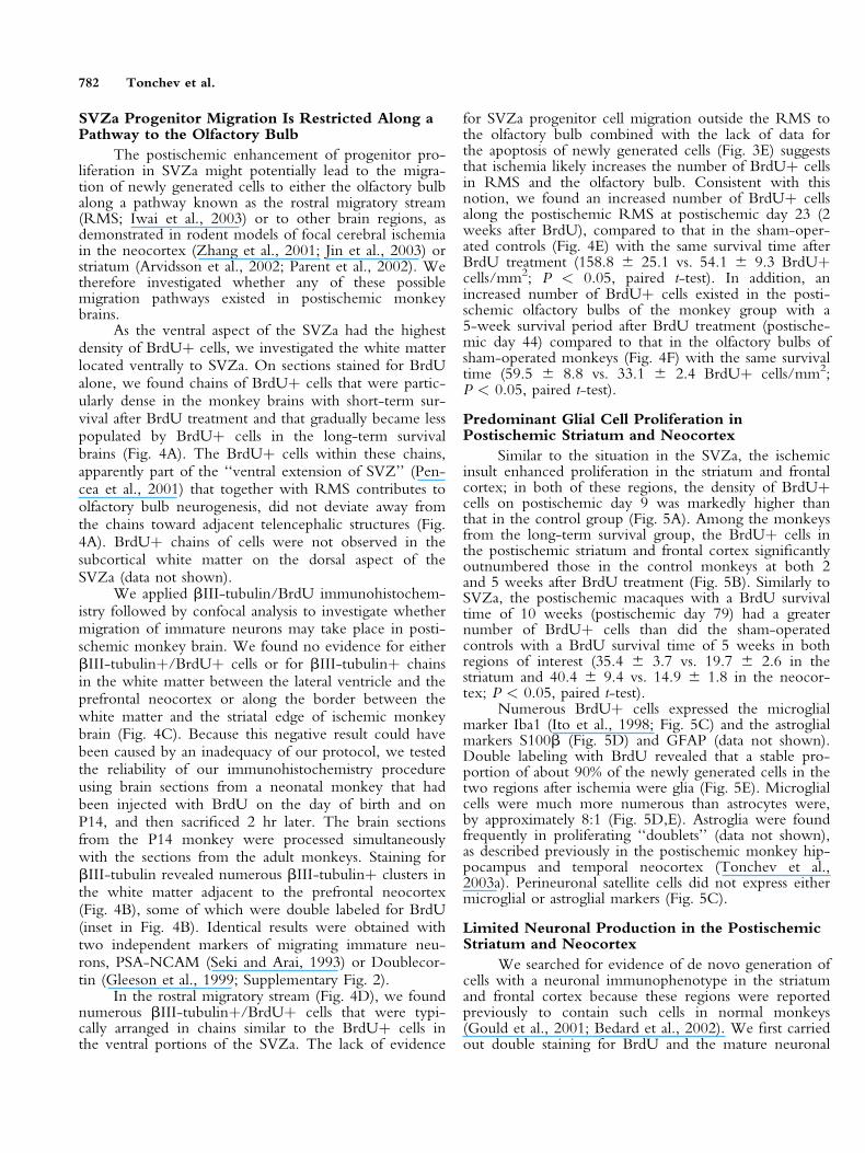
Predominant Glial Cell Proliferation inPostischemic Striatum and Neocortex
Similar to the situation in the SVZa, the ischemicinsult enhanced proliferation in the striatum and frontalcortex; in both of these regions, the density of BrdUþcells on postischemic day 9 was markedly higher thanthat in the control group (Fig. 5A). Among the monkeysfrom the long-term survival group, the BrdUþ cells inthe postischemic striatum and frontal cortex significantlyoutnumbered those in the control monkeys at both 2and 5 weeks after BrdU treatment (Fig. 5B). Similarly toSVZa, the postischemic macaques with a BrdU survivaltime of 10 weeks (postischemic day 79) had a greaternumber of BrdUþ cells than did the sham-operatedcontrols with a BrdU survival time of 5 weeks in bothregions of interest (35.4 6 3.7 vs. 19.7 6 2.6 in thestriatum and 40.4 6 9.4 vs. 14.9 6 1.8 in the neocor-tex; P < 0.05, paired t-test).
Numerous BrdUþ cells expressed the microglialmarker Iba1 (Ito et al., 1998; Fig. 5C) and the astroglialmarkers S100b (Fig. 5D) and GFAP (data not shown).Double labeling with BrdU revealed that a stable pro-portion of about 90% of the newly generated cells in thetwo regions after ischemia were glia (Fig. 5E). Microglialcells were much more numerous than astrocytes were,by approximately 8:1 (Fig. 5D,E). Astroglia were foundfrequently in proliferating ‘‘doublets’’ (data not shown),as described previously in the postischemic monkey hip-pocampus and temporal neocortex (Tonchev et al.,2003a). Perineuronal satellite cells did not express eithermicroglial or astroglial markers (Fig. 5C).
Limited Neuronal Production in the PostischemicStriatum and Neocortex
We searched for evidence of de novo generation ofcells with a neuronal immunophenotype in the striatumand frontal cortex because these regions were reportedpreviously to contain such cells in normal monkeys(Gould et al., 2001; Bedard et al., 2002). We first carriedout double staining for BrdU and the mature neuronal
782 Tonchev et al.

Fig. 4. Proliferation of progenitorcells in the macaque rostral migratorystream (RMS) and subcortical whitematter. A: BrdU immunohistochemis-try in the white matter located ventralto the ventral aspect of SVZa on post-ischemic days 9 (A1), 15 (A2), and 44(A3). Note the stream of BrdUþ cellsand the lack of cells deviating awayfrom the stream. B, C: Staining forbIII-tubulin in the subcortical whitematter and in the vicinity of the puta-men of an intact postnatal (B) andadult postischemic day 23 (C1, C2)monkey brain. Note the lack of posi-tive cells in the adult monkey whitematter contrasting the numerous bIII-tubulinþ clusters (arrows in B) in thepostnatal (P14) brain. The boxed clusteris magnified in the inset with arrow-heads depicting a bIII-tubulinþ/BrdUþ cell. Cells positive for bIII-tubulin in SVZa are also shown(arrows). WM, white matter; Str,striatum; F, frontal cortex. The lumenof the lateral ventricle is shown by anasterisk. D: Double labeling for bIII-tubulin and BrdU in control and post-ischemic monkey RMS (horizontalbrain sections at the level of the ante-rior commissure), 23 days after sur-gery. Virtually all BrdUþ cells arecolocalized with bIII-tubulin; none ofthe cells positive for either markerdeviate away from the RMS. Pu,putamen; ac, anterior commissure.E: BrdU immunohistochemistry inRMS of a control (E1) and postische-mic day 23 (E2) monkey. F: BrdUimmunohistochemistry in the olfactorybulb parenchyma of a control (F1)and postischemic day 44 (F2) monkey.Scale bar ¼ 400 lm in A3 (for A1–A3); 400 lm in C2 (for B, C, C2);200 lm in D2 (for D1, D2); and200 lm in F2 (for E, F).

Fig. 5. Cell proliferation in thestriatum and frontal cortex.A: Representative images ofBrdUþ cells in the striatum ofcontrol (A1) and postischemicday 9 monkey brains (A2).Note the greatly increased den-sity of BrdUþ cells on day 9.B: Quantitative evaluation ofthe BrdUþ cells in striatum(B1) and frontal cortex (B2) atvarious intervals after ischemia/sham operation. The density ofBrdUþ cells in the postische-mic brains is significantlygreater than that in the controlbrains with identical survivalperiods after BrdU treatment.*P < 0.05, ***P < 0.001 vs.control animals. C: Triplelabeling for Iba1, NeuN, andBrdU in the postischemic day44 striatum. Iba1þ/BrdUþ cellsare indicated by arrows, whereas aperineuronal satellite cell is indi-cated by an arrowhead. D: Triplelabeling for Iba1, S100b, andBrdU in the postischemic day 44frontal cortex. Iba1þ/BrdUþcells (arrows) are much morenumerous than are the S100bþ/BrdUþ cells (arrowhead). E: Pro-portions of BrdUþ cells coex-pressing Iba1 or S100b in the post-ischemic monkeys. BrdUþ/Iba1�/S100b� cells are desig-nated ‘‘other.’’ Scale bar ¼ 50lm in A; 20 lm in C, D; 10lm in E, F.

Fig. 6. Neuronal immunophenotype ofBrdUþ cells in the postischemic neo-cortical or striatal parenchyma. A: Dou-ble labeling for NeuN and BrdU in neo-cortical layers I–IV (A1) reveals a dou-ble-labeled cell at the boundary of layersII and III (the box in A1 is magnified inA2) on postischemic day 23. The cellboxed in A2 is presented as orthogonalprojections along the x- and y-axes (A3)to confirm the colocalization of the twosignals. Note its identical morphology toneighboring Neu Nþ/BrdU� cells(A2). B: Triple labeling for BrdU,NeuN, and Tbr1 at the boundary ofneocortical layers IV and V on day 44.The boxed cell in B1 is shown in B2with color separation and orthogonalprojections. C: Triple labeling for BrdU,NeuN and Islet1 in the putamen on day44. The boxed cell in C1 is shown in C2with color separation and orthogonalprojections. D: Triple-labeling forBrdU, NeuN, and the GAD67 in theputamen on day 79. The boxed cell inD1 is shown inD2 with color separationand orthogonal projections. E: Triplestaining for NeuN (E1), BrdU (E2) andTUNEL (E3) in layers II-IV of the neo-cortex on postischemic day 23.TUNELþ cells (arrows) and the BrdUþcells (arrowheads) are distinct cell types.Examples of a NeuNþ/TUNELþ celland a NeuNþ/BrdUþ cell boxed inE1-E3 are magnified on the right side ofthe figure. Scale bar ¼ 200 lm in A1; 20lm in A2, B1, C1 and D1; 50 lm inE1–E3.

marker NeuN (Eriksson et al., 1998; Gould et al. 1999b,2001; Kornack and Rakic, 1999, 2001b; Arvidssonet al., 2002; Parent et al., 2002), searching for double-labeled cells whose size and morphology were similar tothe surrounding NeuNþ/BrdU� neurons (Fig. 6A). Asystematic confocal analysis revealed that a few of theBrdUþ cells were NeuNþ/BrdUþ cells: 8/600 in theneocortex (2/200 at day 23, 3/200 at day 44, and 3/200at day 79) and 7/600 in the striatum (2/200 at day 23,3/200 at day 44, and 2/200 at day 79).
We further confirmed the putative neuronal phe-notype of the NeuNþ/BrdUþ cells by demonstratingthat they express markers of committed neuronal pro-genitors and postmitotic neurons in the striatum (Islet1;Stenman et al., 2003) or the neocortex (Tbr1; Englundet al., 2005). In addition to the 1,200 BrdUþ cellsexamined for NeuN/BrdU double labeling as describedabove, we investigated 300 BrdUþ cells (100 in each ofthe postischemic groups sacrificed on days 23, 44, and79) for Tbr1/NeuN/BrdU triple labeling in the neocor-tex and 300 BrdUþ cells for Islet1/NeuN/BrdU triplelabeling in the striatum. Three-dimensional confocalanalysis with digital reconstructions demonstrated that allscanned NeuNþ/BrdUþ cells coexpressed Tbr1 in theneocortex (4/300; Fig. 6B) or Islet1 in the striatum (5/300; Fig. 6C). Finally, we found that the NeuNþ/BrdUþ cells coexpressed GAD, a marker of g-aminobu-tyric acid (GABA)ergic neurons (Jongen-Relo et al.,1999). We observed triple-labeled GADþ/NeuNþ/BrdUþ cells in both the striatum (2/200 BrdUþ cells;Fig. 6D) and neocortex (2/200 BrdUþ cells). In thesham-operated monkeys, BrdUþ/NeuNþ cells wereonly observed in striatum and neocortex of the day 44brains (1/200 BrdUþ cells).
In a rodent ischemic model, BrdU incorporation inneurons was shown to precede their apoptosis, asrevealed by positive TUNEL staining (Kuan et al.,2004). Combining BrdU/TUNEL double labeling withNeuN immunohistochemistry in the striatum and neo-cortex of postischemic day 23 monkeys, we found noTUNELþ/NeuNþ cells incorporating BrdU, and allNeuNþ/BrdUþ cells remained negative for TUNEL(Fig. 6E).
DISCUSSION
The present study has 3 major findings: (1) theenhanced and sustained presence of progenitors in SVZa,particularly after ischemia; (2) the restriction of migrationof progenitor cells away from SVZa toward the olfactorybulb, but not toward the postischemic striatum or frontalcortex; and (3) the ability of the adult primate brain tosustain striatal and neocortical neuronal production aftera global ischemic insult.
We have demonstrated previously that global ische-mia in adult macaque monkeys increases the progenitorcell proliferation in the SVZ along the inferior horn ofthe lateral ventricle (SVZi; Tonchev et al., 2003a). Here,we extend those results by investigating the SVZ along
the anterior horn of the lateral ventricle (SVZa). Ourresults demonstrate for the first time that in primates,global cerebral ischemia enhances cell proliferation inthis region. The presence of SVZa progenitor cells innormal adult macaque monkeys has been reported in aprevious study (Kornack and Rakic, 2001a). The presentstudy, however, demonstrates the first quantitative analy-sis of the BrdUþ cells that persist in SVZ for a longtime. The data indicate that although SVZa progenitorssustain their presence in SVZa of both control and ische-mic monkeys, postischemic SVZa retained a significantlyhigher proportion of progenitor cells than did controlSVZa (Fig. 2F). In addition, long-term BrdU-retainingcells expressed Musashi1 and Ki67, indicating theyremained uncommitted to neuronal or glial lineage neu-ral progenitors in the active phases of their cell cycle.Interestingly, a subpopulation of stem cells implanted inthe lateral ventricles of embryonic monkeys retainedtheir location in SVZa 4 weeks after implantation,exhibiting features of undifferentiated progenitors (Our-ednik et al., 2001). Our results extend these findings bydemonstrating the potential of the endogenous SVZaprogenitors of the adult monkey brain to retain theirlocation in the niche, particularly after injury.
The finding of long-term BrdU-retaining cells inSVZa can be explained as follows. First, we specificallyused a high BrdU dose (five daily injections of 100 mg/kg each) in an attempt to diminish the possibility ofBrdU label dilution by cell division, with a resulting lossin BrdU detection using immunohistochemistry. A highBrdU dose should guarantee BrdU detection throughoutlong-term survival periods, especially because turnoverof some adult-generated cells in the monkey brain is notrapid (Kornack and Rakic, 2001a; Gould et al., 2001).Second, in rodents, stem cells residing in SVZa have theability to exit and then reenter the cell cycle (Maslovet al., 2004). If some primate progenitors possessed asimilar ability, the long-term BrdU label-retaining cellsin the SVZa that are colabeled by Ki67 might representprecursor cells or their progeny reentering the cell cycle.Third, the retention of SVZa progenitors might be dueto a lack of migration or differentiation signals or, alter-natively, to an enhanced signal maintaining the stem/progenitor phenotype. The molecular nature of suchputative signals in the primate remains to be elucidated.Fourth, long-term BrdU-label retaining cells might rep-resent a pathologic, abnormal, state. These cells remainednegative for markers of DNA damage and apoptosis,TUNEL and active caspase-3, however, arguing againstthis latter possibility.
In rodent focal ischemic models, SVZa emerged asthe main source of precursor cells for neuronal produc-tion in the striatum and cortex (Zhang et al., 2001,2004; Arvidsson et al., 2002; Parent et al., 2002; Jinet al., 2003). Different from these rodent focal ischemicmodels, however, in adult primate brains after globalischemia we failed to find evidence that SVZa contrib-utes to the generation of striatal or neocortical neurons.Although we observed chains of proliferating neuronal
786 Tonchev et al.

progenitors in the RMS migrating toward the olfactorybulb, no such ‘‘stream’’ (Gould et al., 1999b, 2001) wasevident in the subcortical white matter. In comparison,in the early postnatal (P14) monkey brain, we foundnumerous nests of immature neurons in the subcorticalwhite matter. We observed a significantly increased pro-liferation in RMS at 2 weeks after BrdU treatment (day23) and in the olfactory bulb parenchyma at 5 weeksafter BrdU treatment (day 44). Such results are consistentwith the notion that the postischemia-generated SVZaprecursor cells are directed selectively toward the olfac-tory bulb. Notably, however, our model of transientglobal ischemia caused a less pronounced injury (Yoshidaet al., 2002) than did the large focal injury induced inthe rat stroke models (Zhang et al., 2001, 2004; Arvids-son et al., 2002; Parent et al., 2002; Jin et al., 2003).The differences in progenitor cell migration thus mightbe related to the strength of the insult, i.e., a milderischemic insult might be unable to trigger progenitor cellmigration toward the postischemic striatum or cortex.Application of a monkey focal ischemic model in futurestudies would be important to define more precisely theability of primate progenitors to respond to stroke.
Despite the lack of evident progenitor migrationtoward the postischemic neocortex or striatum, weinvestigated a total of 2,200 BrdUþ cells in these tworegions and found 28 that were colabeled by NeuN(slightly over 1% of the BrdUþ cells examined). Theseputative newly generated neurons survived for up to 70days after BrdU treatment (79 days after ischemia) andremained a stable proportion of the BrdUþ cells in thecortex and striatum of the monkeys, with long-term sur-vival after BrdU treatment. In contrast, newly generatedneurons in normal monkey neocortex have a transientexistence (Gould et al., 2001). We have provided threelines of evidence supporting our identification of adult-generated neurons in the monkey neocortex and stria-tum: (1) we observed NeuNþ/BrdUþ cells that weremorphologically identical to neighboring NeuNþ/BrdU� neurons; (2) the NeuNþ/BrdUþ cells expressedregion-specific transcription factors expressed by neuro-nal progenitors during their transition into postmitoticneurons and in postmitotic neurons themselves, namelyIslet1 in the striatum (Stenman et al., 2003) and Tbr1 inthe neocortex (Englund et al., 2005); and (3) theNeuNþ/BrdUþ cells expressed the enzyme GAD andtherefore could be GABAergic interneurons, consistentwith their preferential localization in neocortical layers II–IV.
Recently, ischemia was shown to induce BrdUincorporation in dying neurons before apoptosis, asrevealed by positive TUNEL staining (Kuan et al.,2004). The dying neurons gradually disappeared by pos-tischemic day 28, whereas BrdUþ cells with a neuronalmorphology were observed at later time points (days 44and 79) in the present study. We also demonstrated thatin the striatum and neocortex on postischemic day 23,BrdU was not incorporated in TUNELþ/NeuNþ cellsand that NeuNþ/BrdUþ cells remained negative forTUNEL (Fig. 6E). These results indicate that in our
model and under the current BrdU protocol, theNeuNþ/BrdUþ cells we described were not dying neu-rons with aberrant DNA synthesis. This is consistentwith our findings in the postischemic monkey hippo-campal CA1 sector (Tonchev et al., 2003a).
Our data suggests the possibility that newly gener-ated neocortical and striatal neurons might arise fromparenchymal progenitors. Previous findings in primatesare in accordance with such a conclusion: (1) the invitro isolation of progenitor cells from adult human grayand white matter (Arsenijevic et al., 2001; Nunes et al.,2003); and (2) the observation that a small fraction ofneural stem cells migrating from the SVZa to the devel-oping monkey neocortex remain undifferentiated in thebrain parenchyma (Ourednik et al., 2001) and thus couldremain dormant during development into adulthooduntil activated by injury. Very recently, Dayer et al.(2005) provided in vivo evidence in adult rodents that atleast in the neocortex, GABAergic interneurons mightbe generated by a local pool of precursors.
A more detailed understanding of the molecularmechanisms controlling adult primate progenitors mayprovide an important insight into their manipulation insitu (Magavi et al., 2000) to restore neurologic functionin human disease as well as in deciphering species- andage-selective differences in their behavior.
ACKNOWLEDGMENTS
The present research was supported by a Grant-in-Aid for Scientific Research (Kiban-Kennkyu [B]) fromthe Japanese Ministry of Education, Culture, Sports, Sci-ence and Technology and from the National ScienceFund of Bulgaria (L1311/03).
We thank Y. Imai (National Institute of Neuro-science, Tokyo, Japan) for the anti-Iba1 antibody,T. Seki (Juntendo University, Tokyo, Japan) for theanti-PSA-NCAM antibody, and G. Chaldakov for val-uable assistance.
REFERENCES
Arsenijevic Y, Villemure JG, Brunet JF, Bloch JJ, Deglon N, Kostic C,
Zurn A, Aebischer P. 2001. Isolation of multipotent neural precursors
residing in the cortex of the adult human brain. Exp Neurol 170:48–
62.
Arvidsson A, Collin T, Kirik D, Kokaia Z, Lindvall O. 2002. Neuronal
replacement from endogenous precursors in the adult brain after stroke.
Nat Med 8:963–970.
Bedard A, Cossette M, Levesque M, Parent A. 2002. Proliferating cells
can differentiate into neurons in the striatum of normal adult monkey.
Neurosci Lett 328:213–216.
Dayer AG, Cleaver KM, Abouantoun T, Cameron HA. 2005. GABAer-
gic interneurons in the adult neocortex and striatum are generated from
different precursors. J Cell Biol 168:415–427.
Englund C, Fink A, Lau C, Pham D, Daza RA, Bulfone A, Kowalczyk
T, Hevner RF. 2005. Pax6, Tbr2, and Tbr1 are expressed sequentially
by radial glia, intermediate progenitor cells, and postmitotic neurons in
developing neocortex. J Neurosci 25:247–251.
Eriksson PS, Perfilieva E, Bjork-Eriksson T, Alborn AM, Nordborg C,
Peterson DA, Gage FH. 1998. Neurogenesis in the adult human hippo-
campus. Nat Med 4:1313–1317.
Neurogenesis in Postischemic Monkey Brain 787

Gage FH. 2000. Mammalian neural stem cells. Science 287:1433–1438.
Gleeson JG, Lin PT, Flanagan LA, Walsh CA. 1999. Doublecortin is a
microtubule-associated protein and is expressed widely by migrating
neurons. Neuron 23:257–271.
Gould E, Reeves AJ, Fallah M, Tanapat P, Gross CG, Fuchs E. 1999a.
Hippocampal neurogenesis in adult Old World primates. Proc Natl
Acad Sci USA 96:5263–5267.
Gould E, Reeves AJ, Graziano MS, Gross CG. 1999b. Neurogenesis in
the neocortex of adult primates. Science 286:548–552.
Gould E, Vail N, Wagers M, Gross CG. 2001. Adult-generated hippo-
campal and neocortical neurons in macaques have a transient existence.
Proc Natl Acad Sci USA 98:10910–10917.
Gu W, Brannstrom T, Wester P. 2000. Cortical neurogenesis in adult
rats after reversible photothrombotic stroke. J Cereb Blood Flow Metab
20:1166–1173.
Hendzel MJ, Wei Y, Mancini MA, Van Hooser A, Ranalli T, Brinkley
BR, Bazett-Jones DP, Allis CD. 1997. Mitosis-specific phosphorylation
of histone H3 initiates primarily within pericentromeric heterochroma-
tin during G2 and spreads in an ordered fashion coincident with mitotic
chromosome condensation. Chromosoma 106:348–360.
Ito D, Imai Y, Ohsawa K, Nakajima K, Fukuuchi Y, Kohsaka S. 1998.
Microglia-specific localization of a novel calcium binding protein,
Iba1. Brain Res Mol Brain Res 57:1–9.
Iwai M, Sato K, Kamada H, Omori N, Nagano I, Shoji M, Abe K.
2003. Temporal profile of stem cell division, migration, and differen-
tiation from subventricular zone to olfactory bulb after transient fore-
brain ischemia in gerbils. J Cereb Blood Flow Metab 23:331–341.
Jin K, Minami M, Lan JQ, Mao XO, Batteur S, Simon RP, Greenberg
DA. 2001. Neurogenesis in dentate subgranular zone and rostral sub-
ventricular zone after focal cerebral ischemia in the rat. Proc Natl Acad
Sci USA 98:4710–4715.
Jin K, Sun Y, Xie L, Peel A, Mao XO, Batteur S, Greenberg DA. 2003.
Directed migration of neuronal precursors into the ischemic cerebral
cortex and striatum. Mol Cell Neurosci 24:171–189.
Jongen-Relo AL, Pitkanen A, Amaral DG. 1999. Distribution of
GABAergic cells and fibers in the hippocampal formation of the maca-
que monkey: an immunohistochemical and in situ hybridization study.
J Comp Neurol 408:237–271.
Kaneko Y, Sakakibara S, Imai T, Suzuki A, Nakamura Y, Sawamoto K,
Ogawa Y, Toyama Y, Miyata T, Okano H. 2000. Musashi1: an evolu-
tionally conserved marker for CNS progenitor cells including neural
stem cells. Dev Neurosci 22:139–153.
Kee NJ, Preston E, Wojtowicz JM. 2001. Enhanced neurogenesis after
transient global ischemia in the dentate gyrus of the rat. Exp Brain Res
136:313–320.
Kee N, Sivalingam S, Boonstra R, Wojtowicz JM. 2002. The utility of
Ki-67 and BrdU as proliferative markers of adult neurogenesis. J Neu-
rosci Methods 115:97–105.
Koketsu D, Mikami A, Miyamoto Y, Hisatsune T. 2003. Nonrenewal of
neurons in the cerebral neocortex of adult macaque monkeys. J Neuro-
sci 23:937–942.
Kornack DR, Rakic P. 1999. Continuation of neurogenesis in the hip-
pocampus of the adult macaque monkey. Proc Natl Acad Sci USA
96:5768–5773.
Kornack DR, Rakic P. 2001a. The generation, migration, and differen-
tiation of olfactory neurons in the adult primate brain. Proc Natl Acad
Sci USA 98:4752–4757.
Kornack DR, Rakic P. 2001b. Cell proliferation without neurogenesis in
adult primate neocortex. Science 294:2127–2130.
Kuan CY, Schloemer AJ, Lu A, Burns KA, Weng WL, Williams MT,
Strauss KI, Vorhees CV, Flavell RA, Davis RJ, Sharp FR, Rakic P. 2004.
Hypoxia-ischemia induces DNA synthesis without cell proliferation in
dying neurons in adult rodent brain. J Neurosci 24:10763–10772.
Liu J, Solway K, Messing RO, Sharp FR. 1998. Increased neurogenesis
in the dentate gyrus after transient global ischemia in gerbils. J Neurosci
18:7768–7778.
Magavi SS, Leavitt BR, Macklis JD. 2000. Induction of neurogenesis in
the neocortex of adult mice. Nature 405:951–955.
Maslov AY, Barone TA, Plunkett RJ, Pruitt SC. 2004. Neural stem cell
detection, characterization, and age-related changes in the subventricu-
lar zone of mice. J Neurosci 24:1726–1733.
Nakatomi H, Kuriu T, Okabe S, Yamamoto S, Hatano O, Kawahara N,
Tamura A, Kirino T, Nakafuku M. 2002. Regeneration of hippocam-
pal pyramidal neurons after ischemic brain injury by recruitment of
endogenous neural progenitors. Cell 110:429–441.
Nunes MC, Roy NS, Keyoung HM, Goodman RR, McKhann G 2nd,
Jiang L, Kang J, Nedergaard M, Goldman SA. 2003. Identification and
isolation of multipotential neural progenitor cells from the subcortical
white matter of the adult human brain. Nat Med 9:439–447.
Ourednik V, Ourednik J, Flax JD, Zawada WM, Hutt C, Yang C, Park
KI, Kim SU, Sidman RL, Freed CR, Snyder EY. 2001. Segregation of
human neural stem cells in the developing primate forebrain. Science
293:1820–1824.
Parent JM, Vexler ZS, Gong C, Derugin N, Ferriero DM. 2002. Rat
forebrain neurogenesis and striatal neuron replacement after focal stroke.
Ann Neurol 52:802–813.
Pencea V, Bingaman KD, Freedman LJ, Luskin MB. 2001. Neurogenesis
in the subventricular zone and rostral migratory stream of the neonatal
and adult primate forebrain. Exp Neurol 172:1–16.
Seki T, Arai Y. 1993. Distribution and possible roles of the highly poly-
sialylated neural cell adhesion molecule (NCAM-H) in the developing
and adult central nervous system. Neurosci Res 17:265–290.
Stenman J, Toresson H, Campbell K. 2003. Identification of two distinct
progenitor populations in the lateral ganglionic eminence: implications
for striatal and olfactory bulb neurogenesis. J Neurosci 23:167–174.
Tonchev AB, Yamashima T, Zhao L, Okano HJ, Okano H. 2003a. Pro-
liferation of neural and neuronal progenitors after global brain ischemia
in young adult macaque monkeys. Mol Cell Neurosci 23:292–301.
Tonchev AB, Yamashima T, Zhao L, Okano H. 2003b. Differential pro-
liferative response in the postischemic hippocampus, temporal cortex
and olfactory bulb of young adult macaque monkeys. Glia 42:209–224.
Yagita Y, Kitagawa K, Ohtsuki T, Takasawa KI, Miyata T, Okano H,
Hori M, Matsumoto M. 2001. Neurogenesis by progenitor cells in the
ischemic adult rat hippocampus. Stroke 32:1890–1896.
Yamashima T, Kohda Y, Tsuchiya K, Ueno T, Yamashita J, Yoshioka
T, Kominami E. 1998. Inhibition of ischaemic hippocampal neuronal
death in primates with cathepsin B inhibitor CA-074: a novel strategy
for neuroprotection based on ‘‘calpain-cathepsin hypothesis.’’ Eur J
Neurosci 10:1723–1733.
Yamashima T. 2000. Implication of cysteine proteases calpain, cathepsin
and caspase in ischemic neuronal death of primates. Prog Neurobiol
62:273–295.
Yoshida M, Yamashima T, Zhao L, Tsuchiya K, Kohda Y, Tonchev
AB, Matsuda M, Kominami E. 2002. Primate neurons show different
vulnerability to transient ischemia and response to cathepsin inhibition.
Acta Neuropathol (Berl) 104:267–272.
Zhang RL, Zhang ZG, Zhang L, Chopp M. 2001. Proliferation and
differentiation of progenitor cells in the cortex and the subventricular
zone in the adult rat after focal cerebral ischemia. Neuroscience 105:
33–41.
Zhang R, Zhang Z, Wang L, Wang Y, Gousev A, Zhang L, Ho KL,
Morshead C, Chopp M. 2004. Activated neural stem cells contribute
to stroke-induced neurogenesis and neuroblast migration toward the
infarct boundary in adult rats. J Cereb Blood Flow Metab 24:
441–448.
788 Tonchev et al.
Copyright © 2022 FDOKUMEN