Context-Dependent Modulation by D1 Receptors: Differential Effects in Hippocampus and Striatum
16
Context-Dependent Modulation by D 1 Receptors: Differential Effects in Hippocampus and Striatum Kathryn M. Gill and Sheri J. Y. Mizumori University of Washington Place-specific firing by hippocampal and striatal neurons was recorded simultaneously following injec- tion of a D 1 receptor antagonist (SCH23390) and during spatial working memory task performance. SCH23390-induced changes in unit responses were observed during light and dark test conditions. Although hippocampal place field locations were altered by the contextual change, the reliability and specificity of place fields was disrupted only by combining D 1 antagonism and a change in context. Striatal place field locations were reorganized after either contextual change or D 1 antagonism, without altering place field reliability and specificity. Disrupted velocity encoding by place cells in both regions was induced by darkness, whereas greater stability in acceleration encoding followed removal of D 1 receptor activity. Dopamine may differentially regulate hippocampal context learning and striatum-based predictive codes. Keywords: place cells, spatial memory, rat, dopamine, spatial context Imaging studies have shown that hippocampal activation is associated with use of spatial cues or the evaluation of contextual information related to spatial memory (Maguire et al., 1998; Rosenbaum, Gao, Richards, Black, & Moscovitch, 2005). Without an intact hippocampus (HPC), humans demonstrate impaired per- formance during tasks requiring the use of spatial information (Parslow et al., 2005). In rodents, hippocampal pyramidal neurons fire selectively when animals occupy specific locations ( place fields) within a given environment (O’Keefe & Dostrovsky, 1979). Current research has indicated that these “place cells” may encode the situational relevance of locations in space, or spatial context, and reinforce a role for HPC in spatial processing (Jeffery, Ander- son, Hayman, & Chakraborty, 2004; Mizumori, Cooper, Leutgeb, & Pratt, 2000; Mizumori, Ragozzino, Cooper, & Leutgeb, 1999; Nadel & Hardt, 2004). It is likely that other brain regions contribute to spatial learning. In rodents, lesions of the dorsal striatum (STR) result in spatial learning deficits (Devan, McDonald, & White, 1999; Sakamoto & Okaichi, 2001). In addition, striatal place fields have been identi- fied (Mizumori et al., 1999, Mizumori, Cooper, et al., 2000), and these, like hippocampal place fields, change locations, or reorga- nize, after alterations in spatial context (Yeshenko, Guazzelli, & Mizumori, 2004). Similar to hippocampal place fields, striatal place fields dynamically respond to context changes regardless of the task at hand. It appears that HPC and STR continually process context infor- mation regardless of whether the evaluation of context is essential for task performance. Seemingly inconsistent with the unit results, hippocampal and striatal damage results in distinct deficits in spatial and response learning, respectively (McDonald & White, 1993; Packard & McGaugh, 1996). That is, HPC-lesioned rats show spatial learning deficits but intact nonspatial learning, whereas STR-lesioned rats demonstrate the opposite pattern of learning impairments. As mentioned previously, HPC is necessary in humans for the active use of currently available spatial or contextual cues as part of its role in episodic memory formation (Holdstock et al., 2002; Rosenbaum, Winocur, & Moscovitch, 2001). In contrast, damage to STR selectively interferes with procedural learning while leaving the spatial memory process relatively intact (Packard & Knowlton, 2002). To account for the finding of parallel neural representation in HPC and STR, and differing effects of hippocampal and striatal lesions on learning, it had been proposed (Mizumori, Yeshenko, Gill, & Davis, 2004) that task-relevant firing by hippocampal or striatal neurons may come to control behavioral expression systems during learning as the relative strength of hippocampal or striatal output changes during learning. Output strength can be determined according to task demands. In this way, hippocampal or striatal modes of processing can have greater or lesser influence on behavioral output in a task-relevant manner. It has been suggested that neu- romodulators (e.g., dopamine) contribute to the determination of the relative strengths of the efferent signals (Mizumori et al., 2004). The present study provides a first test of this hypothesis by assessing whether dopamine has different effects on neural repre- sentation by hippocampal and striatal neurons. Behavioral evidence has indicated that dopamine can act locally within HPC and STR to differentially impact processing relevant to different kinds of learning. Selective lesions of striatal and hippocampal dopamine afferent systems impair response or spatial learning, respectively (Da Cunha, et al., 2003; Gasbarri, Sulli, Kathryn M. Gill and Sheri J. Y. Mizumori, Department of Psychology, University of Washington. This research was supported by National Institute of Mental Health Grant MH 58755. We thank Rebecca Birdsong and Emily Wood for help with behavioral testing. Correspondence concerning this article should be addressed to Sheri J. Y. Mizumori, Department of Psychology, University of Washington, Box 351525, Guthrie Hall, Seattle, WA 98195. E-mail: mizumori@ u.washington.edu Behavioral Neuroscience Copyright 2006 by the American Psychological Association 2006, Vol. 120, No. 2, 377–392 0735-7044/06/$12.00 DOI: 10.1037/0735-7044.120.2.377 377
Transcript of Context-Dependent Modulation by D1 Receptors: Differential Effects in Hippocampus and Striatum
Context-Dependent Modulation by D1 Receptors: Differential Effects inHippocampus and Striatum
Kathryn M. Gill and Sheri J. Y. MizumoriUniversity of Washington
Place-specific firing by hippocampal and striatal neurons was recorded simultaneously following injec-tion of a D1 receptor antagonist (SCH23390) and during spatial working memory task performance.SCH23390-induced changes in unit responses were observed during light and dark test conditions.Although hippocampal place field locations were altered by the contextual change, the reliability andspecificity of place fields was disrupted only by combining D1 antagonism and a change in context.Striatal place field locations were reorganized after either contextual change or D1 antagonism, withoutaltering place field reliability and specificity. Disrupted velocity encoding by place cells in both regionswas induced by darkness, whereas greater stability in acceleration encoding followed removal of D1
receptor activity. Dopamine may differentially regulate hippocampal context learning and striatum-basedpredictive codes.
Keywords: place cells, spatial memory, rat, dopamine, spatial context
Imaging studies have shown that hippocampal activation isassociated with use of spatial cues or the evaluation of contextualinformation related to spatial memory (Maguire et al., 1998;Rosenbaum, Gao, Richards, Black, & Moscovitch, 2005). Withoutan intact hippocampus (HPC), humans demonstrate impaired per-formance during tasks requiring the use of spatial information(Parslow et al., 2005). In rodents, hippocampal pyramidal neuronsfire selectively when animals occupy specific locations ( placefields) within a given environment (O’Keefe & Dostrovsky, 1979).Current research has indicated that these “place cells” may encodethe situational relevance of locations in space, or spatial context,and reinforce a role for HPC in spatial processing (Jeffery, Ander-son, Hayman, & Chakraborty, 2004; Mizumori, Cooper, Leutgeb,& Pratt, 2000; Mizumori, Ragozzino, Cooper, & Leutgeb, 1999;Nadel & Hardt, 2004).
It is likely that other brain regions contribute to spatial learning.In rodents, lesions of the dorsal striatum (STR) result in spatiallearning deficits (Devan, McDonald, & White, 1999; Sakamoto &Okaichi, 2001). In addition, striatal place fields have been identi-fied (Mizumori et al., 1999, Mizumori, Cooper, et al., 2000), andthese, like hippocampal place fields, change locations, or reorga-nize, after alterations in spatial context (Yeshenko, Guazzelli, &Mizumori, 2004). Similar to hippocampal place fields, striatalplace fields dynamically respond to context changes regardless ofthe task at hand.
It appears that HPC and STR continually process context infor-mation regardless of whether the evaluation of context is essentialfor task performance. Seemingly inconsistent with the unit results,hippocampal and striatal damage results in distinct deficits inspatial and response learning, respectively (McDonald & White,1993; Packard & McGaugh, 1996). That is, HPC-lesioned ratsshow spatial learning deficits but intact nonspatial learning,whereas STR-lesioned rats demonstrate the opposite pattern oflearning impairments. As mentioned previously, HPC is necessaryin humans for the active use of currently available spatial orcontextual cues as part of its role in episodic memory formation(Holdstock et al., 2002; Rosenbaum, Winocur, & Moscovitch,2001). In contrast, damage to STR selectively interferes withprocedural learning while leaving the spatial memory processrelatively intact (Packard & Knowlton, 2002). To account for thefinding of parallel neural representation in HPC and STR, anddiffering effects of hippocampal and striatal lesions on learning, ithad been proposed (Mizumori, Yeshenko, Gill, & Davis, 2004)that task-relevant firing by hippocampal or striatal neurons maycome to control behavioral expression systems during learning asthe relative strength of hippocampal or striatal output changesduring learning. Output strength can be determined according totask demands. In this way, hippocampal or striatal modes ofprocessing can have greater or lesser influence on behavioraloutput in a task-relevant manner. It has been suggested that neu-romodulators (e.g., dopamine) contribute to the determination ofthe relative strengths of the efferent signals (Mizumori et al.,2004). The present study provides a first test of this hypothesis byassessing whether dopamine has different effects on neural repre-sentation by hippocampal and striatal neurons.
Behavioral evidence has indicated that dopamine can act locallywithin HPC and STR to differentially impact processing relevantto different kinds of learning. Selective lesions of striatal andhippocampal dopamine afferent systems impair response or spatiallearning, respectively (Da Cunha, et al., 2003; Gasbarri, Sulli,
Kathryn M. Gill and Sheri J. Y. Mizumori, Department of Psychology,University of Washington.
This research was supported by National Institute of Mental HealthGrant MH 58755. We thank Rebecca Birdsong and Emily Wood for helpwith behavioral testing.
Correspondence concerning this article should be addressed to SheriJ. Y. Mizumori, Department of Psychology, University of Washington,Box 351525, Guthrie Hall, Seattle, WA 98195. E-mail: [email protected]
Behavioral Neuroscience Copyright 2006 by the American Psychological Association2006, Vol. 120, No. 2, 377–392 0735-7044/06/$12.00 DOI: 10.1037/0735-7044.120.2.377
377

Innocenzi, Pacitti, & Brioni, 1996; Miyoshi et al., 2002). Similarly,posttraining infusion of selective dopamine agonists in STR canenhance win–stay or stimulus–response learning on the radialmaze, whereas win–shift performance is enhanced by similar in-fusions in HPC (Packard & White, 1991). The physiologicalmechanism underlying the dissociation between hippocampal andstriatal function is not known. Anatomically, the pattern of dopa-mine innervation of HPC and STR is distinct. Although dopamineterminals within the STR have multiple synaptic targets, thosearriving in HPC typically exhibit single synaptic connections (Fal-lon, 1981). HPC receives dopamine input from the ventral teg-mental area, whereas the substantia nigra provides the same inputto the STR (Beckstead, Domesick, & Nauta, 1979; Gerfen, Staines,Arbuthnott, & Fibiger, 1982). Despite different patterns of con-nectivity, dopamine appears to exert significant influences on bothstriatal and hippocampal neuroplasticity. For example, dopamineD1 receptor activity has been shown to be important for either theinduction or maintenance of long-term potentiation in both HPCand STR (Calabresi, Centonze, Gubellini, Marfia, & Bernardi,1999; Centonze, Gubellini, Pisani, Bernardi, & Calabresi, 2003;Frey, Matthies, Reymann, & Matthies, 1991; Kerr & Wickens,2001) and of the stabilization of hippocampal place fields (Ken-tros, Agnihotri, Streater, Hawkins, & Kandel, 2004).
In this study, we sought to determine whether dopamine plays asignificant role in determining the relative strengths of hippocam-pal or striatal neural firing patterns or output signals. Presumably,more reliable and specific neural signaling has a greater likelihoodof impacting efferent structures. Consequently, the reliability andspecificity of neural representations could be taken to reflect thestrength of the output signal. The specific hypothesis then is thatmanipulation of the dopaminergic system differentially affects thereliability or specificity of the neural codes within HPC and STR.Because place cells are common to both the STR and HPC(O’Keefe, 1979; Mizumori et al., 1999; Mizumori, Cooper, et al.,2000), we compared the responses of simultaneously recordedstriatal and hippocampal cells with the selective D1-receptor an-tagonist, SCH23390 (Sigma Chemical, St. Louis, MO). Rats weretrained on a spatial working memory task in which accurateperformance requires an intact HPC (Becker, Walker, & Olton,1980). Our previous work showed that imposed darkness causesreliable behavioral impairments and neural responses during per-formance of the spatial working memory task (Ragozzino, Leu-tgeb, & Mizumori, 2001). Therefore, this study tested the effects ofD1 antagonism on behavioral and neural responses to dark testingto maximize our ability to explore the extent to which D1 receptorsgate context-dependent neural plasticity. By selecting a spatialtask, we hoped to bias the signaling strength of hippocampalneurons such that hippocampal place fields would appear moreselective and/or reliable than would striatal place fields. If thedopaminergic system contributes to the biased signal strength, thendisruption of dopamine function should have a preferentiallygreater effect on the specificity and reliability of hippocampalplace fields than striatal place fields.
Method
Subjects
Male Long-Evans rats (N � 12) obtained from Charles River Labora-tories (Raleigh, NC) were used in the following experiment. Rats were
housed individually in a temperature- and humidity-controlled environ-ment with a 12-hr light cycle (lights on at 7 a.m.). Rats were given 1 weekon arrival to acclimate to the laboratory environment prior to any experi-mental procedures. During this time, rats had ad libitum access to food andwater while being handled and weighed daily. Once behavioral trainingcommenced, rats were maintained at approximately 80% of their free-feeding weight.
Apparatus
All behavior in this study was conducted on a semiautomated modifiedeight-arm radial maze, consisting of eight black Plexiglas runways (58 �5.5 cm) that extended from a central platform (19.5 cm in diameter) andwere 79 cm tall. Each runway was hinged in the center and could be raisedand lowered via remote control. Rats were able to reach the reward at theends of maze arms only after the arms were raised to be flush with thecenter platform. The maze was enclosed within a circular black curtain (10�in diameter) hung from an overhead track. Visual cues were hung on thecurtain in constant locations.
Behavioral Training
During pretraining, rats were made accustomed to the chocolate milkreward to be used during training by receiving it in their home cage. OnDay 1 of the maze exposure all eight arms were available and baited. Oncerats consistently retrieved rewards from all arms, they were then traineddaily to perform a spatial working memory task on the eight-arm radialmaze. Each trial consisted of an initial training phase of the randompresentation of a four-arm forced-choice sequence. When rats consumedthe reward on the fourth forced-choice arm, the test phase commenced andall arms were made available. Errors were recorded when a rat placed allfour paws on an arm previously visited in either the training phase of thetest phase. Time to complete each trial was measured by the rat’s initialentry into the first forced-choice arm in the training phase until the returnto the center platform following reward consumption on the final arm. Priorto surgery, rats were trained to perform 10 trials until an accuracy rate of80% of trials completed without errors was attained.
After allowing 1 week of recovery following surgery, rats were retrainedto asymptotic levels prior to any experimental manipulations to ensurestable performance and to allow acclimation to the headstage assembly.Subsequently, unit recording began along with counterbalanced presenta-tion of both pharmacological and environmental manipulations. Duringeach day of testing, rats performed five baseline trials to provide bothcontrol unit and behavioral data (Block 1). Rats were then removed fromthe maze and were administered a subcutaneous injection of either saline orthe D1 antagonist, SCH23390 (5 �g/kg) before being returned to the centerplatform for a 5-min postinjection interval. Rats then performed fiveadditional trials with either normal room lighting or all lights extinguished.Therefore, there were four treatment conditions: saline–light, saline–dark,SCH23390–light, and SCH23390–dark.
Electrode Construction and Surgical Procedures
Stereotrode and microdrive construction was based on techniques pro-vided by McNaughton, Barnes, and O’Keefe (1983a). Two Teflon-coatedplatinum wires were twisted together and coated in Epoxylite prior to beingloaded into a 30-gauge cannula, leaving 1–2 mm of wire exposed at thebottom. Each drive assembly consisted of two to three loaded cannulasspaced 0.4 mm apart. Two microdrives were placed above each hemi-sphere, one above STR and the other above HPC. Prior to surgery, thestereotrode tips were cut at a 45° angle and gold plated to an impedance of100–200 Kohm (tested at 1 kHz). Rats were anesthetized with sodiumPentobarbital (40 mg/kg initial dose and 0.05 cc supplements as needed)and fixed in a stereotaxic apparatus (David Kopf Instruments, Tujunga,
378 GILL AND MIZUMORI
CA). To minimize respiratory distress, atropine sulfate was administered aswell (0.2 mg/kg). Burr holes were drilled through the skull and electrodedrive assemblies were then bilaterally placed above the STR (0.2–1.2 mmanterior to bregma, 1.7 mm lateral, 1.8 mm ventral to the brain surface) andHPC (�4.5–5.5 mm posterior to bregma, 2.5 mm lateral, 1.8 mm ventral).A reference electrode (114 �m Teflon-coated stainless steel wire) wasinserted into the corpus callosum, and a ground screw was attached to theskull. To prevent infection, all rats were administered Baytril (5 mg/kg,im), and Ketofen (5 mg/kg, im) was given as a postsurgical analgesic. Ratswere allowed 1 week of recovery, during which time they were allowedfree access to food. Food restriction was reinstituted prior to advancementof drives as was monitoring of unit activity on each stereotrode in prepa-ration for behavioral testing.
Drug Preparation and Administration
SCH23390 was mixed fresh daily in 0.9% saline and administered bysubcutaneous injection. Pilot studies have shown 5 �g/kg to be an effectivesubcutaneous dose that elicits changes in striatal and hippocampal unitactivity without causing an inability to complete the task. Other studies thathave used similar doses have illustrated reductions in reaction times andanticipatory responses that are indicative of impaired voluntary movement(Bushnell & Levin, 1993; Courtiere, Hardouin, Goujon, Vidal, & Has-broucq, 2003).
Behavioral Monitoring
The movement of each rat was monitored via a pair of front and backinfrared light-emitting diode arrays. An automatic tracking system sampledthe position of the front diode array (20 Hz) and determined the rat’sposition in the maze (resolution � 2.5 cm/pixel). Both diode arrays wereused to determine the directional heading of the rat. Time stamps for bothpositional and unit data were recorded by Cheetah data acquisition soft-ware (Neuralynx, Tucson, AZ).
Unit Identification
Four stereotrodes were used to record cellular activity in each of the STRand HPC. The preamplification headstage (NB Labs, Denison, TX) con-sisted of 48 high-input field-effect transistors. Using the Cheetah dataacquisition system (Neuralynx, Tucson, AZ), each waveform was ampli-fied 1,000 to 10,000 times, and filtered at 600 Hz and 6 kHz. Prior tobehavioral testing, stereotrodes were observed for the presence of sponta-neous cellular activity. If no clear units were present, stereotrodes werelowered in 22-�m increments or up to 200 �m per day. Only signalsexhibiting activity that was at least 3 times greater than background levelsand exceeded a user-defined threshold were recorded. Units were clusteredoffline by using MCLust software (by A. Redish, University of Minnesota,Minneapolis). Additional template matching analysis routines were pro-vided by Chris Higginson. Mean spike amplitude, spike width, and averagefiring rate were calculated for each cell. Place cells were defined in part bytheir low firing rates (�3 Hz) and broad spike widths (�300 ms; latencydifference between the maximum and minimum voltage points of theanalog signal).
Data Analysis
Several analysis routines were used to compare unit characteristics withbehavioral events (custom software provided by Chris Higginson, Univer-sity of Washington, Seattle). Positional data for each recording session wasviewed offline. Event flags were assigned to the beginning of each trial(when the rat entered the first forced-choice arm) and to the end of eachtrial (when the rat returned to the center platform after consuming thereward on the last remaining arm). In addition, flags were inserted to divide
the record into Block 1 (initial five baseline trials) and Block 2 (five trialsperformed after pharmacological and environmental manipulations). Theaverage firing rates during Block 1, Block 2, and the entire session weredetermined for each cell. The behavior of each rat was assessed in terms ofthe average number of errors performed in each block as well as latency tocomplete each trial.
Classification and Analysis of Location Specific Neurons
To determine the spatial distribution of cell firing, the maze area wasdivided into pixels of equal size (2.8 � 2.8 cm), and an average firing ratewas calculated for each pixel. For illustration purposes, the spatial firingpattern was revealed by highlighting those pixels in which the firing rateexceeded 20% of the maximum firing rate for the session. Place fields werecharacterized by several criteria: (a) The area of highest firing encom-passed at least four adjacent highlighted pixels, (b) the firing rate thatoccurred inside the field area was at least twice as large as the rate thatoccurred at locations outside of the field, and (c) the cell that fired duringat least 50% of the passes through the location of the principle (i.e., largest)field was considered to have a reliability of at least 50%. The same criteriafor establishing spatially selective neurons were used in STR and HPC.
Once a neuron was recognized to possess spatial properties during eitherthe baseline or manipulation phases, it was subject to further analysis.Reliability (defined previously) and specificity measures for the primaryfield of each cell were calculated. Place-field specificity scores reflectedthe probability that the rat was in a given location when the cell fired.Because both positive and negative changes in these measures were ob-served, the absolute value of the change in field reliability and specificityacross blocks was calculated, and an analysis of variance (ANOVA) wasused to determine the effects of treatment condition. In addition, linearregression was used to compare baseline and treatment condition placefield reliability and specificity. A spatial correlation score was also ob-tained by calculating a pixel by pixel Pearson correlation of cell firingacross commonly visited pixels in Block 1 and Block 2. The spatialcorrelation scores were used as a measure of place field reorganization.ANOVA of the average spatial correlation values for each treatmentcondition was used to identify differences between striatal and hippocam-pal neural responses to D1 antagonism or contextual changes.
Analysis of Velocity and Acceleration Encoding byLocation Specific Neurons
Past studies have reported the encoding of egocentric movement byplace cells in HPC (Czurko, Hirase, Csicsvari, & Buzsaki, 1999; Mc-Naughton, et al., 1983b). Consequently, all place cells recorded in the HPCand STR were analyzed for potential correlations between firing rates andmovement velocity or acceleration. Significant (linear) relationships be-tween neural firing rates and velocity (2.24 cm/s bin size) or acceleration(2.24 cm/s2) were identified on the basis of a 95% confidence interval (� �.05). The number of cells gaining or losing significant correlations withvelocity or acceleration during the manipulation phase of testing wasdetermined. For cells that remained significantly correlated with velocity oracceleration across both phases of testing, a Wilcoxon’s analysis (� � .05)was performed to determine whether the distribution of firing acrossdifferent velocity or acceleration bins was altered as a function of exper-imental manipulation. The proportion of cells that showed a significantchange from baseline in terms of velocity or acceleration correlates wasdetermined by adding the number of cells that exhibited significantlydifferent linear relationships after a manipulation to the number of cellsgaining or losing significant correlations with velocity or acceleration. Forboth HPC and STR, chi-square analysis was used to determine whether theproportions of cells changing after manipulations were different from thoseobserved in the saline–light condition.
379D1 MODULATION IN HIPPOCAMPUS AND STRIATUM

Histological Procedures for Electrode PlacementVerification
Once electrodes had been lowered past the region of interest (5.0 mmventral to the brain surface for STR and 4.0 mm for HPC), rats were deeplyanesthetized with sodium pentobarbital and perfused transcardially with a0.9% buffered NaCl solution followed by 10% formalin. Brains were slicedin 40 �m sections on a vibratome and stained with cresyl violet. Electrodetrack verification was accomplished by comparing depth measurements atthe time of recording with electrode track reconstructions from serialsections from each hemisphere.
Results
Behavioral Effects of Dopamine Antagonism
Behavioral data were pooled from eight rats that received allpossible treatment combinations (e.g., saline–light, saline–dark,SCH23390–light, SCH23390–dark) and four rats tested only dur-ing the two dark condition procedures (saline–dark andSCH23390–dark). For each day of testing, the average number oferrors committed and the amount of time spent per arm entry werecalculated for both the baseline and manipulation phases. Differ-ence scores for both measures were obtained by subtracting themanipulation phase average values from those recorded during thebaseline phase. In cases when an individual rat received multipleexposures to a given treatment condition, average difference scoresfor each condition were used in the statistical analysis. Analysis ofthe difference scores revealed treatment effects in terms of anincrease in errors during the manipulation phase (see Figure 1A),F(3, 36) � 18.30, p � .001. Tukey’s post hoc comparisons of thenumber of errors between saline–light (M � SEM � 0.23 � 0.50)and each of the dark conditions (saline–dark and SCH23390–dark) revealed an increase in the average number of errors (5.9 �0.90 errors, p � .001), and (6.67 � 0.91 errors, p � .001),respectively. This effect on behavior could not be attributed to D1
antagonism per se, because performance during the SCH23390–light condition (0.11 � 0.68 errors, ns) was not significantlydifferent from the saline control and because saline–dark showedthe same effect as SCH23390–dark.
The differences in choice latency between baseline and manip-ulation phases were used as a measure of experimental-inducedalterations in motor function. Overall, there was a significantdifference across the treatment conditions in terms of the amountof time required for each arm entry (see Figure 1B), F(3, 36) �5.20, p � .01. In both SCH23390–light and SCH23390–darkconditions, the average arm entry time was significantly longerthan during the baseline phase (4.60 � 2.01 s, p � .05, and 5.41 �0.74 s, p � .001, respectively). This pattern indicates thatSCH23390 had the effect of slowing performance regardless oflighting condition. To determine whether this slowed performancewas related to choice accuracy, we tested whether latency wascorrelated with the number of errors. For only one condition(SCH23390–dark) did we find a significant negative correlationbetween arm entry time and the error difference scores (r � .62,p � .05). In this case, contrary to a motor impairment-inducedincrease in errors, rats committed fewer errors when the arm-entrytime increased (see Figure 2).
Overall, altering the visual environment had more devastating
effects than did D1 antagonism on the ability of the rats to performthe working memory task accurately. D1-antagonism may havebehavioral effects as well, yet given the substantial nature of thedark-induced impairment, we may not have been able to detectfurther decline in performance accuracy from the SCH23390 in-jection. However, SCH23390 was ineffective in increasing errorswhen administered without a simultaneous contextual change butdid have an effect in disrupting response latency. Thus, D1 antag-onism per se did not impact choice accuracy despite having effectson response latency.
Neural Responses to Dopamine (DA) Antagonism
We conducted analysis of spatial firing properties on a total of78 hippocampal (n � 6 rats) and 76 striatal (n � 9 rats) place cells.
Figure 1. Summary of the behavioral responses to D1 antagonism andimposed darkness. A: Relative to the baseline period, an increase in errorswas detected by calculating a difference score (average errors per baselinephase – average errors per manipulation phase). Rats committed signifi-cantly more errors during dark trials. There was no SCH23390 inducedincrease in errors. B: Relative to the baseline phase, the change in time(seconds) required to make a choice was determined (difference in theaverage amount of time per arm choice across phases of testing). It wasfound that animals took significantly longer for each arm choice during thetwo drug treatment conditions. An asterisk denotes significant differences,p � .05. Sal/Light � saline–light condition; Sal/Dark � saline–darkcondition; Sch/Light � SCH23390–light condition; Sch/Dark �SCH23390–dark condition.
380 GILL AND MIZUMORI

Figures 3A and 3B illustrate the location of recording electrodes inHPC and STR where place cells were observed. All hippocampaland striatal place cells discharged at relatively low rates (1.15 �0.13 Hz and 1.54 � 0.18 Hz, respectively). The average spikewidth for hippocampal place cells (321.29 � 9.71 �s) was signif-icantly greater than that observed for striatal place cells (235.91 �2.89 �s), F(1, 152) � 27.91, p � .01. The average amplitude forhippocampal (91.29 �V � 2.89) and striatal (87.79 �V � 3.07)place cells did not differ, F(1, 152) � 0.69, ns. There were nostatistical differences in the responses of neurons recorded fromthe dentate and CA1. As a consequence, reliability, specificity, andspatial correlation values from both regions were combined toexamine manipulation effects.
Place-Field Reliability
Baseline (i.e., pretreatment) reliability scores were calculatedfor each treatment condition. Baseline reliability measures of hip-pocampal neurons did not differ across the four treatment condi-tions, F(3, 74) � 0.77, ns. Likewise in STR, baseline reliabilitymeasures were consistent across treatment groups, F(3, 73) �1.70, ns. Because baseline reliability did not vary across thetreatment conditions, all reliability values were pooled for eachstructure to establish whether the baseline reliability of hippocam-pal and striatal place fields differed. The baseline reliability mea-sures for hippocampal and striatal fields were not different, F(1,152) � 0.00, ns.
Both dopamine and environment manipulations acted to in-crease or decrease reliability of hippocampal and striatal cell firingwithin the place field. This increase in variability might haveobscured an effect of D1 antagonism or contextual manipulation onplace field reliability. Consequently, analysis of variance of theabsolute value of the change in place field reliability was used tocompare treatment effects. The effect on hippocampal place fieldreliability difference scores varied significantly across the fourtreatment conditions, F(3, 75) � 7.37, p � .01. Tukey’s post hoccomparisons revealed that similar to the modest change in reliabil-ity that typically occurred during the saline–light condition forhippocampal place cells (difference score � .14 � .02; see Figure4A, left panel: HPC), neither darkness nor D1 antagonism alonesignificantly altered field reliability (.20 � .04, p � .05, and .13 �
.03, p � .05, respectively). A significant change in reliability wasobserved only following the combination of SCH23390–darkmanipulations (.26 � .05, p � .05). It is interesting to note that thedifference scores obtained during the SCH23390–dark manipula-tion were greater than the other three treatment groups, whichsuggests that this combination of D1 antagonism and darkness hadthe most profound effects on place field reliability.
Unlike hippocampal place fields, striatal place fields did notexhibit manipulation–induced instability in reliability differencescores, F(3, 73) � 2.56, ns. Relative to the saline–light condition(.14 � .04), average STR reliability difference scores did notdistinguish the three treatment conditions: saline–dark (.24 � .06),SCH23390–light (.22 � .04), and SCH23390–dark (.19 � .06;see Figure 4A, right panel: striatum).
To further demonstrate the changes in reliability after experi-mental manipulation, we next determined whether the initial reli-ability measures during the baseline condition were predictive ofthe reliability measures during the treatment condition. We con-ducted linear regression analyses on the raw reliability scores tocompare the baseline (Block 1) and treatment (Block 2) conditions.The reliability of hippocampal and striatal place fields duringBlock 1 was significantly correlated with place field reliabilityduring Block 2 for the saline–light condition, F(1, 17) � 8.21, p �.05, and F(1, 12) � 8.56, p � .05, respectively (see Figure 5A).These results indicate a consistency in place field reliability whenenvironmental variables remain constant. SCH23390 and darknesshad differential effects in altering hippocampal and striatal placefield reliability across the testing phases. In HPC, the SCH23390–light condition resulted in a nearly identical positive linear rela-tionship as observed in the saline–light condition. Although hip-pocampal place field reliability during Block 1 of the saline–darkcondition also predicted reliability during Block 2, the absolutevalue of the reliability score was consistently lower in Block 2 thanin Block 1. The combined SCH23390–dark treatment, in contrast,completely eliminated the linear relationship between hippocam-pal place field reliability for Blocks 1 and 2, F(1, 22) � .47, p �.05. This reinforces the conclusion that the combination of dark-ness and D1 receptor blockade causes the greatest alterations in thereliability of hippocampal place fields.
In contrast to the manipulation-specific effects on hippocampalplace field reliability, the predictability of striatal place field reli-ability during Block 2, on the basis of Block 1 reliability scores,was disrupted for all treatment conditions: saline–dark, F(1, 25) �0.51, ns; SCH23390–light, F(1, 17) � 0.17, ns; SCH23390–dark,F(1, 14) � 2.49, ns (see Figure 5B).
Figure 3. Schematic of coronal sections illustrating recording sites inhippocampus (A) and dorsal striatum (B; modified from Swanson, 2003).Some dots represent multiple cells recorded at a single depth. Reprintedfrom Brain Maps: Structure of the Rat Brain (3rd ed.), Larry Swanson,Levels 35 and 39, Copyright 2003, with permission from Elsevier.
Figure 2. A significant relationship between the increase in choice la-tency and increase in errors was observed only for the SCH23390–darkcondition. In this case, when animals spent more time for each arm choicein the manipulation phase, they committed fewer errors.
381D1 MODULATION IN HIPPOCAMPUS AND STRIATUM
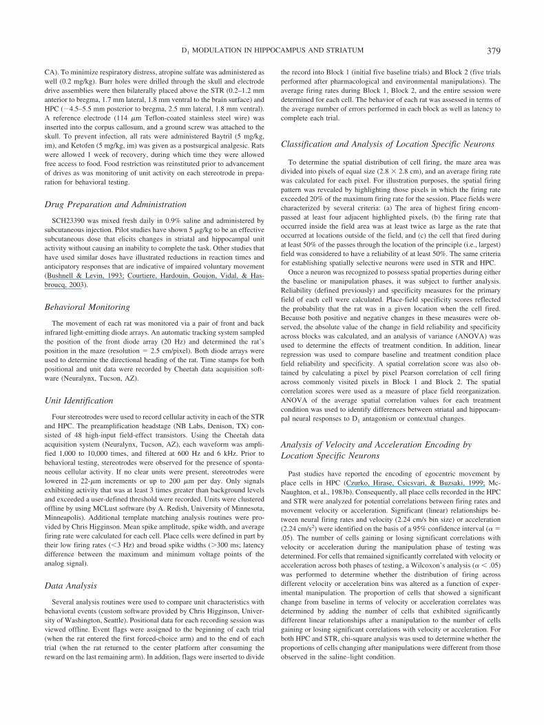
Figure 4. Summary of changes in hippocampal and striatal place field reliability, specificity, and spatialcorrelation resulting from D1 antagonism and/or changes in the visual environment. Difference scores forreliability and specificity were calculated using the absolute value of the change in place field reliability orspecificity across baseline and manipulation phases of testing (see text). A: The combination of SCH23390 anddarkness produced the greatest disruption in hippocampal place field reliability. Striatal place field reliability wasunaffected by darkness, injections of SCH23390, or the combination. B: Summary of changes in hippocampaland striatal place field specificity in response to a D1 antagonist and/or changes in the visual environment.Darkness, as well as injections of SCH23390, did not cause significant changes in hippocampal place fieldspecificity. Similar to the affect observed on hippocampal place field reliability, the combination of SCH23390and darkness produced the greatest disruption in hippocampal place field specificity. Striatal place fieldspecificity was unaltered by darkness, SCH23390, or the combination. C: Summary of spatial reorganization ofhippocampal and striatal place fields resulting from a contextual change and/or a D1 antagonist. Spatial
382 GILL AND MIZUMORI

According to both difference scores and linear regression anal-yses, removal of D1 receptor input influences hippocampal placefield reliability in a context-dependent way. In contrast, althoughcontext or dopamine manipulations did not affect the magnitude ofchange in striatal place field reliability, all treatments eliminatedthe predictability of place field reliability during Block 2 (on thebasis of Block 1 scores). These data are consistent with thehypothesis that dopamine differentially effects hippocampal andstriatal neural codes during learning.
Place-Field Specificity
Baseline place field specificity values did not distinguish treat-ment conditions for hippocampal place cells, F(3, 74) � .67, ns.There were also no group differences in baseline specificity valuesfor place cells recorded in STR, F(3, 73) � 1.40, ns. Therefore, foreach structure, the baseline specificity measures for each treatmentcondition were pooled. It is interesting to note that the two struc-tures differed significantly in terms of the specificity of their placefields, F(1, 152) � 5.12, p � .05. During the baseline phase,hippocampal place fields (.14 � .01) were more specific thanstriatal place fields (.11 � .01) even though both populationssatisfied standard criteria for place fields. A similar result ofstructural disparity in baseline specificity values has been de-scribed previously (Mizumori, Ragozzino, & Cooper, 2000;
Yeshenko et al., 2004). Similar to changes in reliability, bothincreases and decreases in place field specificity were observedfollowing environment and dopamine manipulations. As a conse-quence, analysis of variance of the absolute values of thesechanges were used to identify treatment effects in both regions. InHPC, significant alterations in specificity were similar to those thatoccurred for reliability, F(3, 74) � 3.65, p � .05 (see Figure 4B,left panel: HPC). Tukey’s post hoc comparisons revealed that theaverage change in specificity that occurred during the saline–lightcondition (difference score � .08 � .01) was significantly lessthan that observed during the SCH23390–dark condition (.14 �.03, p � .05). This effect appeared to result from the combinationof darkness and D1 antagonism, because the average values re-corded for saline–dark (.06 � .02) and SCH23390–light (.06 �.01) failed to differ from the saline–light control (see Figure 4B,left panel: HPC). Overall the pattern of results on hippocampalplace field reliability and specificity indicates that the combinationof compromised dopamine processing along with the challenge ofnavigating in darkness causes the greatest disruption of normalplace cell activity. In contrast, and consistent with the lack oftreatment effect on the magnitude of difference scores for striatalplace field reliability, the average striatal specificity differencescores did not distinguish the four treatment conditions, F(3, 73) �1.62, p � .05 (saline–light: .14 � .06, saline–dark: .09 � .04,
Figure 4 (opposite). correlation scores are based on a pixel-by-pixel correlation analysis across training blocks.In hippocampus, darkness caused reorganization (i.e., low correlation) of place fields. Although SCH23390injections alone did not induce the reorganization of hippocampal place fields, the combination of darkness andSCH23390 caused significant reorganization. Striatal, but not hippocampal, place fields underwent reorganiza-tion following SCH23390 injections. As in hippocampus, darkness, as well as the combination of D1 antagonismand darkness, caused significant reorganization of striatal place fields. An asterisk denotes significance, p � .05.Sal/Light � saline–light condition; Sal/Dark � saline–dark condition; Sch/Light � SCH23390–light condition;Sch/Dark � SCH23390–dark condition.
Figure 5. Linear regression analysis of hippocampal and striatal place field reliability observed across testingphases (Blocks 1 and 2). Hippocampal and striatal place fields exhibited a significant linear relationship betweenbaseline and testing phase reliability. A: In hippocampus (HPC), the combination of darkness and D1 antagonismeliminated this relationship, consistent with the changes that were observed in the reliability difference scores.B: In striatum (STR), all treatment combinations eliminated the significant linear relationship in place fieldreliability across testing blocks that was observed in the saline–light condition. Sal/Light � saline–lightcondition; Sal/Dark � saline–dark condition; SCH/Light � SCH23390–light condition; SCH/Dark �SCH23390–dark condition.
383D1 MODULATION IN HIPPOCAMPUS AND STRIATUM

SCH23390–light: .05 � .01, SCH23390–dark: .09 � .02; seeFigure 4B, right panel: striatum).
We also conducted linear regression analysis of the baseline andtreatment condition specificity scores to test the predictability ofspecificity scores during Block 1 for specificity scores duringBlock 2. During the saline–light condition, hippocampal, but notstriatal, place fields displayed a significant linear relationship inplace field specificity across phases, F(1, 17) � 21.33, p � .01,and F(1, 12) � 0.07, ns, respectively. There was no across blockrelationship in HPC specificity following context manipulations inthe saline–dark and SCH23390–dark conditions, F(1, 15) � 1.46,ns, and F(1, 22) � 0.09, ns, respectively. This loss of predictabilityappeared to be context-induced, because the positive relationshipin hippocampal place field specificity across blocks that wasobserved during the saline–light condition was also seen in theSCH23390–light condition, F(1, 16) � 9.02, p � .01. WithinSTR, only the dark conditions produced a significant positivelinear relationship between baseline and treatment phase placefield specificity that was not present in the saline–light condition:saline–dark, F(1, 25) � 6.15, p � .05, and SCH23390–dark, F(1,14) � 58.76, p � .01, respectively.
Consistent with the effects on hippocampal place field reliabil-ity, specificity measures showed a context-dependent effect ofD1-receptor antagonism. The predictability of specificity scoresduring Block 1 for Block 2 was diminished following context butnot following dopamine manipulation. This indicates that hip-pocampal field specificity is determined by nondopaminergic in-put. Consistent with the lack of effects observed for striatal placefield reliability, field specificity difference scores did not changeafter context or dopamine manipulation. Unlike HPC, during base-line conditions, there was no significant relationship between thespecificity of striatal place fields during Block 1 and Block 2. Asignificant correlation emerged following a context shift.
Spatial Correlation Analysis
Spatial reorganization of place field locations was evidenced bysignificant reductions in the spatial correlation values, which com-pared the spatial distribution of activity in the baseline phase tothat observed during the manipulation phase. For both hippocam-pal and striatal neurons, the degree of place field reorganizationvaried significantly across the treatment conditions, F(3, 74) �11.62, p � .001, and F(3, 73) � 5.47, p � .01, respectively (see
Figure 4C). For hippocampal place fields, Tukey’s post hoc com-parisons showed that compared with average saline–light correla-tion values (.39 � .05; see Figure 4C, left panel: HPC), there wassignificant reorganization during only the saline–dark andSCH23390–dark manipulations (.12 � .05, p � .0001, and .05 �.03, p � .0001, respectively). In addition, as shown in Figure 4C,striatal place field spatial correlation values varied across the fourtreatment groups. Relative to correlation values obtained duringthe saline–light condition (.28 � .05), average striatal spatialcorrelation values differed significantly for all three treatmentgroups: saline–dark (.13 � .03), SCH23390–light (.15 � .03), andSCH23390–dark (.14 � .03), ps � .01 (see Figure 4C, right panel:striatum). The latter three conditions did not differ from eachother.
Although both hippocampal and striatal place fields reorganizedfollowing a change in context, only striatal neurons exhibitedindependent responses to D1 antagonist treatment and darkness.The stability of hippocampal place field locations, however, ap-peared to be selectively sensitive to the changes in context, be-cause the effects were observed for only the saline–dark andSCH23390–dark conditions. It appears that D1 receptor activityand context information impacts the reliability, specificity, andlocation of hippocampal and striatal place fields. It is important tonote, however, that the details of the effects vary for the twostructures (see summary in Table 1). In general, hippocampal placefields were most sensitive to the context manipulations. Dopaminemanipulations were observed only when they co-occurred with thecontext change. Striatal place fields, in contrast, tended to beroughly equally and perhaps independently sensitive to context ordopamine manipulations. Figure 6 provides examples from indi-vidual hippocampal and striatal place cells that illustrate the typ-ical patterns of neural changes associated with either darkness orSCH23390.
Velocity and Acceleration Encoding
Both HPC and STR contained place cells whose firing wasclearly related to movement velocity or acceleration in addition tospatial selectivity. It was anticipated that interfering with dopa-mine processing would differentially disrupt the natural encodingof egocentric movement in these two structures. To address thisprediction, the number of cells either gaining or losing a significantrelationship between firing rate and movement was first summed.
Table 1Effect of D1 Receptor Activity and Context Information on Hippocampal (HPC) and Striatal(STR) Place Fields
Condition
Reliability Specificity Changein spatial
correlationvalue
Change inabsolute value
Change inpredictability
Change inabsolute value
Change inpredictability
HPC STR HPC STR HPC STR HPC STR HPC STR
Saline–dark A A A 2 A A 2 1 2 2SCH–light A A A 2 A A A A A 2SCH–dark 2 A 2 2 2 A 2 1 2 2
Note. SCH � SCH23390; A � no change; 2 � decrease; 1 � increase.
384 GILL AND MIZUMORI

In addition, Wilcoxon’s analyses of cells that retained significantcorrelations with velocity or acceleration across baseline and ma-nipulation blocks of trials was performed to ascertain whether thedegree of correlation changed across blocks. Thus, the total num-
ber of cells that changed movement correlates was equal to thenumber that lost, gained, or changed velocity–acceleration-correlated firing. Subsequently, the proportion of cells in HPC thatchanged during the saline–light condition (n � 6/19 cells) was
Figure 6. Color spatial density plots illustrating the effects of D1 antagonism alone or in combination with darknesson hippocampal and striatal place fields recorded while animals performed the spatial working memory task. For eachcell, colors represent areas associated with the maximum firing (shown in red), as well as proportions of the maximumfiring in 25% increments (from blue to red). hippocampal place fields appeared resistant to reorganization (A) unlessalterations in dopaminergic function were accompanied by a change in context (B). In contrast, striatal place fieldsreorganized during all treatment conditions (C and D). (Cell A max firing rate � 12.71 Hz, Cell B max firing rate �10.31 Hz, Cell C max firing rate � 10.47 Hz, Cell D max firing rate � 9.58 Hz).
385D1 MODULATION IN HIPPOCAMPUS AND STRIATUM

used for chi-square comparisons with the other treatment conditionvalues. An identical analysis was used to analyze striatal move-ment data.
In HPC, the percentage of cells unable to maintain their initialrelationship with velocity differed across treatment conditions (seeTable 2), �2(3, N � 78) � 30.45, p � .001. A greater percentageof cells exhibited alterations from the baseline correlation withvelocity during the saline–dark (n � 8/17 cells) and SCH23390–dark (n � 14/24 cells) conditions, �2(1, N � 36) � 5.02, p � .05,and �2(1, N � 43) � 27.01, p � .001, respectively. It is interestingto note that when the same analysis was performed for accelerationencoding by hippocampal place cells, a different pattern of resultsoccurred. Once again, there was a significant difference across thetreatment conditions in terms of changes in acceleration-relatedfiring (see Table 2), �2(3, N � 78) � 24.50, p � .01. However, theSCH23390–dark (n � 6/24 cells) and SCH23390–light (n � 6/18cells) conditions varied significantly from the saline–light (n �10/19 cells) condition with a reduction in proportion of cells losingtheir baseline correlation with acceleration, �2(1, N � 43) �16.07, p � .001, and �2(1, N � 37) � 7.60, p � .001. Accelerationencoding did not exhibit context-induced alterations in the saline–dark (n � 9/17 cells) condition. It appears that velocity correlatesof hippocampal neurons are regulated by context, whereas theacceleration correlate of the same neurons is regulated more bydopamine. Figures 7A and 8A provide individual examples ofdifferent types of changes in velocity and acceleration correlate ofhippocampal neurons.
Given the alleged role of STR in mediating egocentric behav-iors, it was anticipated that altering dopamine signaling would alsocause disruptions in velocity and acceleration encoding. In STR,there was a significant difference between the treatment groups inthe proportion of cells exhibiting altered velocity encoding (seeTable 2), �2(3, N � 76) � 20.20, p � .001. Specifically, only thecombination of darkness and SCH23390 destabilized velocity cor-relation (see Figures 7B and 8B; n � 12/16 cells), �2(1, N � 30) �7.11, p � .01. This effect was not seen in the saline–dark (n �13/27 cells) or SCH23390–light (n � 9/19 cells) conditions, �2(1,N � 41) � 1.62, p � .05, and �2(1, N � 33) � 1.92, p � .05,respectively. Thus, whereas hippocampal velocity correlationsseemed context-sensitive (with or without a dopamine challenge),striatal velocity correlations were sensitive to only the combinationof context and dopamine manipulation.
There was also treatment-induced variation in the relationshipbetween behavioral acceleration and the firing rates of striatalplace cells (see Table 2), �2(3, N � 76) � 8.93, p � .001. This wasthe case despite the fact that striatal place cells did not show stableacceleration codes during the saline–light condition: More than50% failed to maintain their baseline relationship with accelerationafter saline injection. The instability during baseline saline–lightconditions suggests that acceleration encoding by striatal placecells may be regulated by other internally regulated variables. Onlythe SCH23390–light condition exhibited a reduction in the per-centage of cells that changed baseline acceleration encoding, thatis, the correlation with acceleration became more consistent (seeFigure 8B, n � 7/19 cells), �2(1, N � 33) � 8.27, p � .001. Thiseffect was eliminated in the SCH23390–dark condition (n � 8/16cells), �2(1, N � 30) � 7.11, p � .05. It seems that the addition ofdarkness interfered with any stability conferred by D1 antagonism.
To summarize, dopamine appears to exert different types ofeffects on similar kinds of neural (i.e., movement) representationin HPC and STR. For striatal neurons, both context manipulationand SCH23390 impacted the stability in the velocity and acceler-ation encoding by place cells. In contrast, a context change in-duced instability of velocity encoding for hippocampal neurons,whereas SCH23390 stabilized acceleration-correlated tuning byhippocampal neurons.
Relationship Between Behavioral and Unit Effects
In terms of establishing a link between dopamine effects onspatial processing in HPC and STR and effective spatial naviga-tion, attempts were made to correlate the number of errors madeduring the manipulation phase and the various unit changes de-scribed earlier. There was no consistent relationship between thebehavioral accuracy of the rat and the changes in spatial correla-tion, reliability, or specificity of the spatial processing in the HPCand STR (all ps �.05).
Because there was a SCH23390-induced increase in arm-choicelatencies, it was necessary to determine whether these latencychanges were also correlated with changes in the unit responses.The average arm-entry latency differences between baseline andmanipulation blocks were not correlated with spatial correlationvalues obtained during any treatment condition for either HPC or
Table 2Percentages and Chi-Square Values for Hippocampal (HPC) and Striatal (STR) Place CellsExhibiting Changes From Baseline Velocity and Acceleration
Condition
Velocity Acceleration
HPC STR HPC STR
% �2 % �2 % �2 % �2
Saline–light
31.58 57.14 52.63 57.14
Saline–dark 47.06* 5.02 48.15 1.62 52.94 .002 51.85 0.56SCH–light 38.89 1.17 47.36 1.92 33.33* 7.60 36.84* 8.27SCH–dark 68.33* 27.01 75.00* 7.11 25.00* 16.07 50.00 1.02
Note. SCH � SCH23390.* p � .05.
386 GILL AND MIZUMORI

STR. This indicates that the place field reorganization observedwas not related to slowed movement of the rat. The change inhippocampal place field reliability was also not correlated with theincrease in arm-entry latencies observed in the SCH23390–dark
condition. Consistent with the effect on reliability, the increaseddisruption of place field specificity during the combined D1 an-tagonism and dark condition was not associated with the increasein arm-entry latencies. Although striatal place fields did not exhibit
Figure 7. Individual examples of velocity encoding by hippocampal and striatal place cells during baseline andmanipulation phases. Each line represents the linear regression between firing rate (Hz) and velocity (cm/sec).A: In hippocampus, greater disparity between the baseline and manipulation regression lines occurred insaline–dark conditions. B: In striatum, a greater difference between the baseline and manipulation regressionlines occurred in the SCH23390–dark condition. Sal/Light � saline–light condition; Sal/Dark � saline–darkcondition; SCH/Light � SCH23390–light condition; SCH/Dark � SCH23390–dark condition.
387D1 MODULATION IN HIPPOCAMPUS AND STRIATUM
context- or SCH23390-induced alterations in reliability or speci-ficity, it was still possible for changes in either measure to becorrelated with changes in egocentric movement. However, thesignificant increase in arm choice latency was not correlated with
changes in reliability or specificity during any treatment condition.In sum, the effect of D1 antagonism on increasing arm-entrylatencies was not associated with changes in place field specificity,reliability, or organization during the two drug conditions.
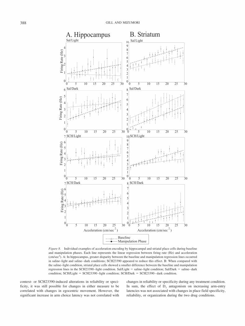
Figure 8. Individual examples of acceleration encoding by hippocampal and striatal place cells during baselineand manipulation phases. Each line represents the linear regression between firing rate (Hz) and acceleration(cm/sec2). A: In hippocampus, greater disparity between the baseline and manipulation regression lines occurredin saline–light and saline–dark conditions; SCH23390 appeared to reduce this effect. B: When compared withthe saline–light condition, striatal place cells showed a smaller difference between the baseline and manipulationregression lines in the SCH23390–light condition. Sal/Light � saline–light condition; Sal/Dark � saline–darkcondition; SCH/Light � SCH23390–light condition; SCH/Dark � SCH23390–dark condition.
388 GILL AND MIZUMORI

Discussion
To test the hypothesis that dopamine differentially regulates theoutput signals from HPC and STR, this study examined the re-sponses of place cells recorded from these structures to D1 receptorantagonism and to a change in the environmental context. Wehypothesized that hippocampal and striatal place fields wouldexhibit disparate responses to context alterations (i.e., imposeddarkness) and to D1 receptor antagonism (SCH23390 injection).To the extent that dopamine and context effects are interdependent,it was expected that place fields would display an increasedsensitivity to the combination of darkness and SCH23390.
Hippocampal and striatal place fields exhibited unique alter-ations following the loss of D1 receptor input. The reliability andspecificity of hippocampal fields was affected by the dopaminemanipulation but only after a context change. The location of placefields, however, was changed after the context change regardlessof the presence of SCH23390. In contrast, the reliability andspecificity scores of striatal place fields did not respond to eithercontext or dopamine manipulation. Rather, a context change ordopamine treatment altered the extent to which baseline (Block 1)reliability and specificity scores predicted Block 2 reliability andspecificity measures. The location of striatal place fields shifted inresponse to either dopamine or context manipulation. This patternof effects has general implications. First, there are likely multiplefactors that determine the specificity, reliability, and location ofplace fields for both HPC and STR. Second, these data suggest thatdopamine plays an important role in defining striatal place fieldlocations during the performance of a spatial task independent ofcontext. In HPC, however, dopamine appears to contribute todetermining the reliability and specificity of the spatial code onlywhen there is a change in context.
The movement component of hippocampal and striatal placecell codes was also differentially sensitive to context and dopa-mine manipulations. The velocity correlate of hippocampal neu-rons was more sensitive to darkness than to SCH23390, whereasthe acceleration correlate of the same neurons was more sensitiveto SCH23390 than to darkness. Striatal place cells, however,exhibited velocity and acceleration correlates that were sensitive toeither darkness or SCH23390. Thus, the response or movementcomponents of hippocampal representations appear to be moreprecisely tuned (or affected) by specific types of context change(e.g., visual context or reinforcement state; Mizumori, Cooper, etal., 2000). In contrast, the response component of striatal placefields appears to be more generally sensitive to perhaps multipleforms of context change.
In general then, hippocampal neural representations seem mostconsistently responsive to the context manipulation. Dopaminemay help to signify a shift in context, because dopamine antago-nism reduced darkness-induced changes in reliability and speci-ficity of HPC place fields. Dopamine signaling may also guidemovement coding in HPC, because selective effects of SCH23390were observed on the velocity and acceleration correlates of placecells. In contrast to what was observed for hippocampal placecells, the most striking response of striatal place fields to eitherdarkness or dopamine antagonism was a change in the predictabil-ity of reliability and specificity. When combined, these data areconsistent with the view that during spatial learning, dopamineserves different roles in HPC and STR.
Behavioral Effects of D1 Antagonism and Context Change
Although darkness caused a significant increase in the numberof errors, SCH23390 did not impair choice accuracy despite in-creasing the amount of time for each arm visit. In contrast with ourresults, other studies have shown an effect of D1 receptor manip-ulation on spatial choice accuracy. Genetic deletion of D1 recep-tors produced impairments in tasks requiring the use of spatial cues(El-Ghundi ’et al., 1999; Smith et al., 1998). However, pharma-cological manipulation of D1 receptor function shows more variedresults. Systemic injection of low doses of SCH23390 results inmovement slowing without necessarily causing memory impair-ments in asymptotic performance of a delayed nonmatching-to-position task (Bushnell & Levin, 1993). In contrast, place- orresponse-learning enhancements are found following local infu-sion of D1-receptor agonists into HPC or STR, respectively (Pack-ard & White, 1991), and spatial learning impairments in agedanimals can be reversed by D1-agonist treatment (Arnsten, Cai,Murphy, & Goldman-Rakic, 1994; Bach, et al., 1999).
Given the evidence that suggests a link between D1-receptoractivity and spatial learning, it was unexpected that SCH23390 didnot impair learning in this study. A behavioral effect may not havebeen observed, because asymptotic performance that is measuredor a contextual change that is presented during asymptotic perfor-mance is not sufficient to produce a vulnerability to D1 receptorblockade. Indeed, although phasic activity of dopamine neurons iscrucial during the early learning of appropriate stimulus–responseassociations (Packard & Knowlton, 2002; Schultz, Tremblay, &Hollerman, 2003), a role for dopamine during asymptotic perfor-mance is less clear. Theoretically, it has been proposed that dopa-mine may coordinate striatal responses with descending corticalinput to produce a system by which ongoing behavior can bematched against expected outcomes during acquisition and asymp-totic performance. This general function may rely on more thanjust the D1-receptor system (Mizumori, Cooper, et al., 2000;Mizumori et al., 2004).
In addition, darkness was used as an environmental contextmanipulation in an effort to induce reliable behavioral impairmentsthat could be compared with corresponding unit changes. Such aglobal interference with multiple sensory systems could likely bethe cause of the robust increase in working memory errors. It ispossible then that an interaction effect between darkness and D1
antagonism was obscured by a floor effect on behavioralperformance.
Hippocampal and Striatal Unit Responses to D1
Antagonism and Context Change
Despite the fact that SCH23390 did not affect choice accuracyin our task, it clearly had profound neurophysiological effects. Thecombination of SCH23390 with a spatial context change caused agreater alteration of spatial encoding of place cells in HPC thaneither treatment alone. D1 dopamine receptors have previouslybeen shown to contribute to hippocampal plasticity in response tospatial novelty. For example, CA1 long-term potentation that isinduced by exposing animals to novel environments can be
389D1 MODULATION IN HIPPOCAMPUS AND STRIATUM

blocked by the application of SCH23390 (Li, Cullen, Anwyl, &Rowan, 2003). In addition, place learning has been associated withelevated CREB in HPC, which may rely on dopamine-mediatedsignaling (Colombo, Brightwell, & Countryman, 2003). Therefore,changes in the visual environment may account for the increasedsensitivity of hippocampal neurons to D1 receptor blockade. Thispattern suggests that dopamine may play a special role in contextdiscrimination by HPC.
The dissociation of effects of D1 antagonism on striatal placefield reorganization, reliability, and specificity could reflect selec-tive responses of medium spiny neurons to D1 receptor activation.Presumably, removing D1 receptor influence disrupted the naturalgating of multiple cortical, glutamatergic synapses converging ona single cell (for a review, see Nicola, Surmeier, & Malenka,2000). Perhaps this low dose of SCH23390 selectively enhanced asubset of cortical input that allowed these connections to havegreater influence. This could translate into an alteration in thespatial location of place fields without affecting the reliability ofthe spatial signal. Similarly, dysregulation of cortical input to STRcould result in changes in the precise coordination of movementvelocity and acceleration with cell firing rates. It is not knownwhether the altered movement codes are causes or consequences ofthe longer latencies observed after SCH23390 treatment.
One of the most striking and consistent effects of either darknessor D1 antagonism on striatal (but not hippocampal) place fieldswas the change in the predictive nature of baseline measures ofreliability and specificity (see Table 1). This suggests that thepostulated striatal function of predicting future reinforcement con-ditions (Mizumori, Pratt, & Ragozzino, 1999; Schultz et al., 2003)is modulated not only by the dopamine system, but also by theexternal environmental context.
Relationship Between Behavioral and Unit Responses
The present study revealed a mismatch between the various unitresponses to the SCH23390 manipulation and any increase inerrors. Other studies have shown similar disparity between alter-ations in hippocampal place fields and lack of correspondingchanges in behavior (Cooper & Mizumori, 2001; Jeffery et al.,2003). As discussed previously, asymptotic performance may rep-resent a situation in which stable place field responses are nolonger a sufficient predictor of behavior. That is, studies of earlierstages of learning may yield better concordance between placefield stability and effective navigation. Performance of a well-learned task, such as the one used in the present study, may reflectcoordinated activity across multiple neural systems (Mizumori,Cooper, et al., 2000). Such coordination could allow animalsto compensate behaviorally (i.e., switch cognitive strategies)when a single physiological process (e.g., D1-receptor system)malfunctions.
The saline–dark and SCH23390–dark conditions, which re-sulted in the greatest change in working memory, were also theconditions that generated the greatest reorganization of hippocam-pal place fields. There was no such correspondence between stri-atal place field reorganization and behavioral impairments. Thiscould reflect the greater importance of HPC, rather than STR,during performance of this task. Failure to retrieve the same neuralpattern (N-methyl-D-aspartate –mediated) could result in behav-ioral impairments (Kentros et al., 1998). In this study, the main-
tenance of hippocampal place fields was likely important forselection of the appropriate behavioral strategy in a given spatialcontext. Therefore, in the two dark treatment conditions, unsuit-able hippocampal reorganization may have interfered with spatialnavigation. Similar declines in spatial performance that occur withage are also associated with alterations in connectivity and acti-vation patterns in CA1 and dentate (Barnes, Rao, & Shen, 1997).
Implications for Multiple Memory Systems Function
The present study provides clear evidence that the dopaminesystem differentially regulates similar types of neural representa-tions (i.e., place fields) in HPC and STR during spatial perfor-mance on a maze. In this way, dopamine may bias the efferentmessages of these structures such that during spatial memoryperformance, HPC comes to exert stronger (i.e., more reliable orspecific) control over other neural systems that determine behav-ioral output (Morris, Arkadir, Nevet, Vaadia, & Bergman, 2004).Such a mechanism is entirely consistent with the finding of parallelneural representations in HPC and STR during spatial and re-sponse tasks (Yeshenko et al., 2004) and the finding that hip-pocampal and striatal lesions result in differential effects on spatialand response learning (McDonald & White, 1993; Packard &McGaugh, 1996).
It is likely that other neurotransmitters, such as acetylcholine(ACh), could also contribute to the selection of behaviors orbehavioral strategies. Peak ACh levels in HPC and STR areassociated with place and response strategy on the plus maze,respectively (Chang & Gold., 2003; McIntyre, Marriott, & Gold,2003). In addition, effective performance during a spatial workingmemory task may involve interactions between the cholinergic anddopaminergic systems. Working memory deficits on the radial armmaze caused by interfering with cholinergic transmission, eithervia nicotinic receptor antagonists or lesions of the medial cholin-ergic pathway, are exacerbated by a mixed D1–D2 antagonist oralleviated by D1 or D2 agonist treatment (Levin & Rose, 1995;McGurk, Levin, & Butcher, 1992).
In both HPC and STR, the interactions between the dopaminer-gic and cholinergic systems may account for the contributions ofthese two neurotransmitter systems to behavior. In STR, ACh anddopamine have opposing effects on the regulation of long-termpotentiation in medium spiny neurons (Centonze et al., 2003).Similar effects occur in the HPC, where cholinergic-induced fieldpotential oscillatory activity can be suppressed by D1 receptors(Weiss, Veh, & Heinnemann, 2003). Thus, in future experiments,it would be of interest to determine whether ACh can reverse theeffects reported in this study following the compromise of dopa-mine function. Furthermore, potential dopamine and ACh contri-butions to the modulation of spatial processing in STR during anonspatial (response) task warrants exploration.
References
Arnsten, A. F., Cai, J. X., Murphy, B. L., & Goldman-Rakic, P. S. (1994).Dopamine D1 receptor mechanisms in the cognitive performance ofyoung adult and aged monkeys. Psychopharmacology, 116, 143–151.
Bach, M. E., Barad, M., Son, H., Zhuo, M., Lu, Y. F., Shih, R., Mansuy,I., Hawkins, R. D., & Kandel, E. R. (1999). Age-related defects in spatialmemory are correlated with defects in the late phase of hippocampallong-term potentiation in vitro and are attenuated by drugs that enhance
390 GILL AND MIZUMORI
the cAMP signaling pathway. Proceedings of the National Academy ofSciences, USA, 96, 5280–5285.
Barnes, C. A., Rao, G., & Shen, J. (1997). Age-related decrease in theN-Methyl-D-AspartateR-mediated excitatory post-synaptic potential inhippocampal region CA1. Neurobiology of Aging, 18, 445–452.
Becker, J. T., Walker, J. A., & Olton, D. S. (1980). Neuroanatomical basisof spatial memory. Brain Research, 200, 307–320.
Beckstead, R. M., Domesick, V. B., & Nauta, W. J. (1979). Efferentconnections of the substantia nigra and ventral tegmental area in the rat.Brain Research, 175, 191–217.
Bushnell, P. J., & Levin, E. D. (1993). Effects of dopaminergic drugs onworking and reference memory in rats. Pharmacology Biochemistry andBehavior, 45, 765–776.
Calabresi, P., Centonze, D., Gubellini, P., Marfia, G. A., & Bernardi, G.(1999). Glutamate-triggered events inducing corticostriatal long-termdepression. Journal of Neuroscience, 19, 6102–6110.
Centonze, D., Gubellini, P., Pisani, A., Bernardi, G., & Calabresi, P.(2003). Dopamine, acetylcholine and nitric oxide systems interact toinduce corticostriatal synaptic plasticity. Reviews in the Neurosciences,14, 207–216.
Chang, Q., & Gold, P. E. (2003). Switching memory systems duringlearning: Changes in patterns of brain acetylcholine release in the HPCand striatum in rats. Journal of Neuroscience, 23, 3001–3005.
Colombo, P. J., Brightwell, J. J., & Countryman, R. A. (2003). Cognitivestrategy-specific increases in phosphorylated cAMP response element-binding protein and c-Fos in the HPC and dorsal striatum. Journal ofNeuroscience, 23, 3547–3554.
Cooper, B. G., & Mizumori, S. J. Y. (2001). Temporary inactivation of theretrosplenial cortex causes a transient reorganization of spatial coding inthe HPC. Journal of Neuroscience, 21, 2986–3001.
Courtiere, A., Hardouin, J., Goujon, A., Vidal, F., & Hasbroucq, T. (2003).Selective effects of low-dose dopamine D1 and D2 receptor antagonistson rat information processing. Behavioral Pharmacology, 14, 589–598.
Czurko, A., Hirase, H., Csicsvari, J., & Buzsaki, G. (1999). Sustainedactivation of hippocampal pyramidal cells by ““space clamping”” in arunning wheel. European Journal of Neuroscience, 11, 344–352.
Da Cunha, C., Wietzikoski, S., Wietzikoski, E. C., Miyoshi, E., Ferro,M. M., Anselmo-Franci, J. A., & Canteras, N. S. (2003). Evidence forthe substantia nigra pars compacta as an essential component of amemory system independent of the hippocampal memory system. Neu-robiology of Learning and Memory, 79, 236–242.
Devan, B. D., McDonald, R. J., & White, N. M. (1999). Effects of medialand lateral caudate-putamen lesions on place- and cue-guided behaviorsin the water maze: Relation to thigmotaxis. Behavioral Brain Research,100, 5–14.
El-Ghundi, M., Fletcher, P. J., Drago, J., Sibley, D. R., O’Dowd, B. F., &George, S. R. (1999). Spatial learning deficit in dopamine D1 receptorknockout mice. European Journal of Pharmacology, 27, 95–106.
Fallon, J. H. (1981). Collateralization of monoamine neurons: mesotelen-cephalic dopamine projections to caudate, septum, and frontal cortex.Journal of Neuroscience, 1, 1361–1368.
Frey, U., Matthies, H., Reymann, K. G., & Matthies, H. (1991). The effectof dopaminergic D1 receptor blockade during tetanization on the expres-sion of long-term potentiation in the rat CA1 region in vitro. Neuro-science Letters, 129, 111–114.
Gasbarri, A., Sulli, A., Innocenzi, R., Pacitti, C., & Brioni, J. D. (1996).Spatial memory impairment induced by lesion of the mesohippocampaldopaminergic system in the rat. Neuroscience, 74, 1037–1044.
Gerfen, C. R., Staines, W. A., Arbuthnott, G. W., & Fibiger, H. C. (1982).Crossed connections of the substantia nigra in the rat. Journal of Com-parative Neurology, 207, 283–303.
Holdstock, J. S., Mayes, A. R., Roberts, N., Cezayirli, E., Isaac, C. L.,O’Reilly, R. C., & Norman, K. A. (2002). Under what conditions is
recognition spared relative to recall after selective hippocampal damagein humans? Hippocampus, 12, 341–351.
Jeffery, K. J., Anderson, M. I., Hayman, R., & Chakraborty, S. (2004). Aproposed architecture for the neural representation of spatial context.Neuroscience and Biobehavioral Reviews, 28, 201–218.
Kentros, C., Hargreaves, E., Hawkins, R. D., Kandel, E. R., Shapiro, M.,& Muller, R. V. (1998). Abolition of long-term stability of new hip-pocampal place cell maps by NMDA receptor blockade. Science, 280,2121–2126.
Kentros, C. G., Agnihotri, N. T., Streater, S., Hawkins, R. D., & Kandel,E. R. (2004). Increased attention to spatial context increases both placefield stability and spatial memory. Neuron, 42, 283–295.
Kerr, J. N., & Wickens, J. R. (2001). Dopamine D-1/D-5 receptor activa-tion is required for long-term potentiation in the rat neostriatum in vitro.Journal of Neurophysiology, 85, 117–124.
Levin, E. D., & Rose, J. E. (1995). Acute and chronic nicotinic interactionswith dopamine systems and working memory performance. Annals ofthe New York Academy of Sciences, 757, 245–252.
Li, S., Cullen, W. K., Anwyl, R., & Rowan, M. J. (2003). Dopamine-dependent facilitation of LTP induction in hippocampal CA1 by expo-sure to spatial novelty. Nature Neuroscience, 6, 526–531.
Maguire, E. A., Burgess, N., Donnett, J. G., Frackowiak, R. S., Frith, C. D.,& O’Keefe, J. (1998). Knowing where and getting there: A humannavigation network. Science, 280, 921–924.
McDonald, R. J., & White, N. M. (1993). A triple dissociation of memorysystems: Hippocampus, amygdala, and dorsal striatum. Behavioral Neu-roscience, 107, 3–22.
McGurk, S. R., Levin, E. D., & Butcher, L. L. (1992). Dopaminergic drugsreverse the impairment of radial-arm maze performance caused bylesions involving the medial cholinergic pathway. Neuroscience, 50,129–135.
McIntyre, C. K., Marriott, L. K., & Gold, P. E. (2003). Patterns of brainacetylcholine release predict individual differences in preferred learningstrategies in mice. Neurobiology of Learning and Memory, 79, 177–183.
McNaughton, B. L., Barnes, C. A., & O’Keefe, J. (1983a). The stereotrode:A new technique for simultaneous isolation of several single units in thecentral nervous system from multiple unit records. Journal of Neuro-science Methods, 8, 391–397.
McNaughton, B. L., Barnes, C. A., & O’Keefe, J. (1983b). The contribu-tions of position, direction, and velocity to single unit activity in theHPC of freely-moving rats. Experimental Brain Research, 2, 41–49.
Miyoshi, E., Wietzikoski, S., Camplessei, M., Silveira, R., Takahashi,R. N., & Da Cunha, C. (2002). Impaired learning in a spatial workingmemory version and in a cued version of the water maze in rats withMPTP-induced mesencephalic dopaminergic lesions. Brain ResearchBulletin, 58, 41–47.
Mizumori, S. J., Cooper, B. G., Leutgeb, S., & Pratt, W. E. (2000). Aneural systems analysis of adaptive navigation. Molecular Neurobiol-ogy, 21, 3001–3005.
Mizumori, S. J., Pratt, W. E., & Ragozzino, K. E. (1999). Function of thenucleus accumbens within the context of the larger striatal system.Psychobiology, 27, 214–224.
Mizumori, S. J., Ragozzino, K. E., & Cooper, B. G. (2000). Location andhead direction representations in the dorsal striatum of rats. Psychobi-ology, 28, 441–462.
Mizumori, S. J., Ragozzino, K. E., Cooper, B. G., & Leutgeb, S. (1999).Hippocampal representational organization and spatial context. Hip-pocampus, 9, 444–451.
Mizumori, S. J., Yeshenko, O., Gill, K. M., & Davis, D. M. (2004). Parallelprocessing across neural systems: Implications for a multiple memorysystem hypothesis. Neurobiology of Learning and Memory, 82, 278–298.
Morris, G., Arkadir, D., Nevet, A., Vaadia, E., & Bergman, H. (2004).
391D1 MODULATION IN HIPPOCAMPUS AND STRIATUM
Coincident but distinct messages of midbrain dopamine and striataltonically active neurons. Neuron, 43, 133–143.
Nadel, L., & Hardt, O. (2004). The spatial brain. Neuropsychology, 18,473–476.
Nicola, S. M., Surmeier, J., & Malenka, R. C. (2000). Dopaminergicmodulation of neuronal excitability in the striatum and nucleus accum-bens. Annual Review of Neuroscience, 23, 185–215.
O’Keefe, J. (1979). A review of the hippocampal place cells. Progress inNeurobiology, 13, 419–439.
O’Keefe, J., & Dostrovsky, J. (1979). The HPC as a spatial map. Prelim-inary evidence from unit activity in the freely-moving rat. Brain Re-search, 34, 171–175.
Packard, M. G., & Knowlton, B. J. (2002). Learning and memory functionsof the basal ganglia. Annual Review of Neuroscience, 25, 563–593.
Packard, M. G., & McGaugh, J. L. (1996). Inactivation of the HPC orcaudate nucleus with lidocaine differentially affects expression of placeand response learning. Neurobiology of Learning and Memory, 65,65–72.
Packard, M. G., & White, N. M. (1991). Dissociation of HPC and caudatenucleus memory systems by posttraining intracerebral injections ofdopamine agonists. Behavioral Neuroscience, 105, 295–306.
Parslow, D. M., Morris, R. G., Fleminger, S., Rahman, Q., Abrahams, S.,& Recce, M. (2005). Allocentric spatial memory in humans with hip-pocampal lesions. Acta Psychological, 118, 123–147.
Ragozzino, K. E., Leutgeb, S., & Mizumori, S. J. Y. (2001). Dorsal striatalhead direction and hippocampal place representations during spatialnavigation. Experimental Brain Research, 139, 372–376.
Rosenbaum, R. S., Gao, F., Richards, B., Black, S. E., & Moscovitch, M.(2005). “Where to?” Remote memory for spatial relations and landmark
identity in former taxi drivers with Alzheimer’s disease and encephalitis.Journal of Cognitive Neuroscience, 17, 446–462.
Rosenbaum, R. S., Winocur, G., & Moscovitch, M. (2001). New views onold memories: Re-evaluating the role of the hippocampal complex.Behavioural Brain Research, 127, 83–97.
Sakamoto, T., & Okaichi, H. (2001). Use of win-stay and win-shift strat-egies in place and cue tasks by medial caudate putamen (MCPu) le-sioned rats. Neurobiology of Learning and Memory, 76, 192–208.
Schultz, W., Tremblay, L., & Hollerman, J. (2003). Changes in behavior-related neuronal activity in the striatum during learning. Trends inNeuroscience, 6, 321–328.
Smith, D. R., Striplin, C. D., Geller, A. M., Mailman, R. B., Drago, J.,Lawler, C. P., & Gallagher, M. (1998). Behavioural assessment of micelacking D1A dopamine receptors. Neuroscience, 86, 135–146.
Swanson, L. (2003). Brain maps: Structure of the rat brain (3rd ed.). SanDiego, CA: Academic Press.
Weiss, T., Veh, R. W., & Heinnemann, U. (2003). Dopamine depressescholinergic oscillatory network activity in rat HPC. European Journal ofNeuroscience, 18, 2573–2580.
Yeshenko, O., Guazzelli, A., & Mizumori, S. J. Y. (2004). Context-dependent reorganization of spatial and movement representations bysimultaneously recorded hippocampal and striatal neurons during per-formance of allocentric and egocentric tasks. Behavioral Neuroscience,118, 751–769.
Received August 1, 2005Revision received November 22, 2005
Accepted January 3, 2006 �
392 GILL AND MIZUMORI