Cyclin D1 fine-tunes the neurogenic output of embryonic retinal progenitor cells
Upload
hms-harvardCategory
view
0download
0
MOLECULAR AND CELLULAR BIOLOGY,0270-7306/97/$04.0010
Sept. 1997, p. 5338–5347 Vol. 17, No. 9
Copyright © 1997, American Society for Microbiology
Cyclin D1 Stimulation of Estrogen Receptor TranscriptionalActivity Independent of cdk4†
ELIZABETH NEUMAN,1 MOHAMED H. LADHA,1 NANCY LIN,1 TODD M. UPTON,1
SUSAN J. MILLER,1 JAMES DIRENZO,1 RICHARD G. PESTELL,2 PHILIP W. HINDS,3
STEVEN F. DOWDY,4 MYLES BROWN,1 AND MARK E. EWEN1*
The Dana-Farber Cancer Institute and Harvard Medical School1 and Department of Pathology,Harvard Medical School,3 Boston, Massachusetts 02115; The Albert Einstein Cancer Center,Departments of Medicine and Developmental and Molecular Biology, Albert Einstein Collegeof Medicine, Bronx, New York 104612; and Howard Hughes Medical Institute and Division of
Molecular Oncology, Departments of Pathology and Medicine, Washington UniversitySchool of Medicine, St. Louis, Missouri 631104
Received 25 March 1997/Returned for modification 9 May 1997/Accepted 30 May 1997
Cyclin D1 plays an important role in the development of breast cancer and is required for normal breast cellproliferation and differentiation associated with pregnancy. We show that ectopic expression of cyclin D1 canstimulate the transcriptional activity of the estrogen receptor in the absence of estradiol and that this activitycan be inhibited by 4-hydroxytamoxifen and ICI 182,780. Cyclin D1 can form a specific complex with theestrogen receptor. Stimulation of the estrogen receptor by cyclin D1 is independent of cyclin-dependent kinase4 activation. Cyclin D1 may manifest its oncogenic potential in breast cancer in part through binding to theestrogen receptor and activation of the transcriptional activity of the receptor.
The three D cyclins are differentially expressed in a celllineage-specific manner as part of a delayed early response tomitogens. D-type cyclins are rate limiting and essential forprogression through the G1 phase of the cell cycle (48, 49).One of the known biochemical functions of D cyclins is to bindto and activate cyclin-dependent kinase 4 (cdk4) and cdk6. Inaddition, cyclins D1, D2, and D3 can bind to the retinoblas-toma protein Rb, and related proteins, in the absence of kinasein vitro. This binding is thought to direct cdk4 and cdk6 to Rb,allowing for efficient phosphorylation of the substrate. In sup-port of the notion that Rb is a critical downstream target of Dcyclins, cells lacking functional Rb do not require cyclin D-dependent kinases for passage from G1 into S phase (50).Emerging evidence suggests that D-type cyclins are not redun-dant. The three D cyclins have different affinities for Rb (15,17, 31). Ectopic expression of cyclins D2 and D3, but not cyclinD1, can inhibit granulocyte differentiation (32). Cyclin D1- andD2-deficient mice show different, specific developmental de-fects (19, 51, 52). Cyclin D1, and not cyclins D2 and D3, isoverexpressed in a high percentage of certain tumors (24).
Cyclin D1 is amplified or overexpressed in a high percentage(.50%) of human breast adenocarcinomas (3, 8, 12, 21, 41, 57)and is oncogenic in vivo, in breast epithelial cells, and in vitro(26, 38, 56). While cyclin D1 is not essential for the develop-ment of most murine tissues and organs, female cyclin D12/2
mice are markedly deficient in breast epithelial cell prolifera-tion associated with pregnancy (19, 52). Specifically, ductalside branching and lobuloalveolar development are severelyimpaired in these mice despite normal levels of circulatingovarian hormones. It has been suggested that steroid hor-mone-induced breast epithelial cell proliferation and/or differ-entiation during pregnancy requires the action of cyclin D1.
Here, we describe the functional interaction of cyclin D1 withthe estrogen receptor.
MATERIALS AND METHODS
Plasmids. The following plasmids have been described previously:21745CD1Luc (human cyclin D1 promoter-luciferase reporter) (2); p(ERE)2-tk-luc (estrogen response element [ERE]-luciferase reporter) (34), a gift from P.Chambon; pCMV-hER (60), a gift from D. J. Shapiro; BBCAT (b-casein pro-moter-chloramphenicol acetyltransferase [CAT] construct) (47), a gift from M. J.Bissell and C. Myers, Monsanto; pRc/CMV-cyclin D1, D2, and D3 (15); pRc/CMV-cyclin D1-KE (26); pRc/CMV-cyclin A (27); glutathione S-transferase(GST)-Rb (17, 29); and pcDNA3-p16 (46). To construct pcDNA3.1-hER-DAF-1, a 1,816-bp EcoRI fragment containing the full-length cDNA encodinghuman estrogen receptor was subcloned from pHEG0 (34) into pALTER-1(Promega). Oligonucleotide-directed mutagenesis was used to introduce an SpeIrestriction site flanking codons 173 and 174, so that subsequent subcloning of thismutant at the introduced SpeI site resulted in hER-DAF-1 (amino acids 174 to596). The sequence of the mutagenic oligonucleotide was 59-CAGTACCAATGACAAGACTAGTATGGCTATGGAATC-39. The hER-DAF-1 cDNA was ex-cised as a SpeI-to-EcoRI fragment and ligated into pcDNA3.1 (Invitrogen) cutwith XbaI and EcoRI. Constructs encoding hemagglutinin (HA)-tagged humanD-type cyclin fusion proteins (to allow detection with the mouse monoclonalantibody 12CA5) were made by adding the amino acid sequence GYPYDVP-DYA to the C termini of the proteins. For cyclin D1, PCR was performed usingthe 59 primer CGCAAACACGCGCAGACC located upstream of the region tobe modified and the 39 primer GAAGCTCCGGATCAGATATCGGCGTAGTCCGGGACGTCGTAGGGGTAGCCGATGTCCACGTC, encoding the tagand a BspEI restriction site used for subcloning. The PCR product (Vent poly-merase; New England Biolabs) was cleaved with restriction enzymes StuI andBspEI, purified, and substituted for the wild-type fragment in the constructpRc/CMV-cyclin D1. As a result, 80 bp of the 39 untranslated sequence of cyclinD1 located between the stop codon and the BspEI site was lost. Dideoxynucle-otide sequencing by standard techniques confirmed that this construct containedthe HA sequence. The HA tag sequence was added to pRc/CMV-cyclin D2 bysubcloning a double-stranded oligonucleotide that encoded the tag and wasflanked by EcoRV and MluI restriction sites. Upon substitution of this oligonu-cleotide for the wild-type sequence, the tag was present at the C terminus of theprotein and 30 bp of 39 untranslated sequence was deleted immediately after thestop codon and preceding the MluI site. The single-stranded oligonucleotidesused were ATCGACCTGGGCTACCCCTACGACGTCCCGGACTACGCCGACCTGTGAA and CGCGTTCACAGGTCGGCGTAGTCCGGGACGTCGTAGGGGTAGCCCAGGTCGAT. An oligonucleotide flanked by restriction sitesfor ApaI and XbaI was used to create an HA-tagged cyclin D3 by the methoddescribed for cyclin D2. Substitution of this oligonucleotide into pRc/CMV-cyclin D3 resulted in a cDNA encoding a C-terminally tagged protein lacking 855bp of 39 untranslated sequences between the stop codon and the XbaI site.
* Corresponding author.† M. E. Ewen dedicates this work to a friend and teacher, Charles
Brenton Huggins, Nobel Laureate.
5338

Deletion of 735 nucleotides internal to this region had previously been found toincrease expression from the parental cyclin D3 cDNA. The oligonucleotidesused to construct HA-tagged cyclin D3 were CAGCCAGACCAGCACTCCTACAGATGTCACAGCCATACACCTGGGCTACCCCTACGACGTCCCGGACTACGCCCACCTGTAGT and CTAGACTACAGGTGGGCGTAGTCCGGGACGTCGTAGGGGTAGCCCAGGTGTATGGCTGTGACATCTGTAGGAGTGCTGGTCTGGCTGGGCC.
SCp2 cell differentiation. To generate stable pools of SCp2 cells expressingBBCAT, cells were transfected with calcium phosphate precipitates (22) by using3 mg of pBabe-neo (for neomycin selection) or pBabe-puro (for puromycinselection) and 27 mg of BBCAT. The precipitate was removed after 4 h, and thecells were glycerol shocked (25% glycerol in 25 mM HEPES [pH 7.6]–140 mMNaCl) for 90 s, washed three times, and fed in Dulbecco modified Eagle medium(DMEM)-F12 supplemented with 5% fetal bovine serum and insulin (5 mg/ml).The following day, selection was started with either G418 (400 mg/ml) or puro-mycin (1.5 mg/ml). After 2 to 3 weeks of selection, colonies were pooled and useddirectly in differentiation assays. Alternatively, these cells were used to generatestable pools of SCp2 cells expressing BBCAT plus either 21745CD1Luc orp(ERE)2-tk-luc, as described above. For differentiation studies, pooled popula-tions were plated either on plastic (1.6 3 106 cells per 60-mm-diameter dish;5.2 3 106 cells per 100-mm-diameter dish) or on plates precoated with anextracellular matrix (ECM) (Matrigel; Collaborative Research) from the Engle-breth-Holm-Swarm tumor (1.3 3 106 cells per 60-mm-diameter dish; 4.3 3 106
cells per 100-mm-diameter dish) in DMEM-F12 containing 2% serum. Thefollowing day, the cells were induced to differentiate by the addition of DMEM-F12 without serum plus insulin (5 mg/ml), hydrocortisone (1 mg/ml), and prolac-tin (3 mg/ml) (differentiation medium) (for details, see reference 13). At thetimes indicated below, cells were removed from the ECM by incubation for 2 hat 37°C with the enzyme dispase (Collaborative Research) or removed fromplastic by cell scraping with a rubber policeman prior to the various analysesbeing performed.
Estrogen receptor transcriptional activation assays. CV1P and SAOS-2 cellswere maintained in DMEM supplemented with 10% Fetal Clone I (Hyclone),penicillin, and streptomycin. Twenty-four hours prior to transfection, cells weresplit and fed with phenol red-minus DMEM supplemented with charcoal-dext-ran-stripped fetal bovine serum (HyClone) plus antibiotics. Cells were trans-fected with the indicated plasmids by the calcium phosphate protocol (22).Sixteen to eighteen hours posttransfection, the precipitate was washed from thecells and estradiol or antiestrogens were added as indicated. Forty-eight hourslater, luciferase activity was measured (16). b-Galactosidase activity was alsodetermined (16) and used as an internal control to which luciferase activity wasnormalized.
cdk4 assays. Kinase assays were essentially as described previously (40). At theindicated times after the induction of differentiation, SCp2 cells were harvestedas described above. The cells were pelleted by centrifugation at 1,500 3 g for 5min and then washed three times with cold phosphate-buffered saline. The pelletwas resuspended in 800 ml of DIP buffer (50 mM HEPES [pH 7.2], 150 mMNaCl, 1 mM EDTA, 2.5 mM EGTA, 10% glycerol, and 0.1% Tween 20) con-taining freshly added 1 mM dithiothreitol (DTT), 1 mM NaF, 0.5 mM phenyl-methylsulfonyl fluoride (PMSF), 0.5 mM sodium orthovanadate, 10 mM b-glycerophosphate, 1 mg of aprotinin per ml, and 1 mg of leupeptin per ml. Lysiswas carried out for 30 min at 4°C with rocking. Samples were passed through a23-gauge needle five times and then pelleted by centrifugation at 16,000 3 g for10 min. The resulting supernatant was precipitated with an antibody specific forcdk4 (C-22; Santa Cruz Biotechnology, Inc.) for 90 min at 4°C with rocking.Immune complexes were collected with protein A-Sepharose beads (50 ml of a10% slurry containing beads preequilibrated in DIP with 4% bovine serumalbumin [BSA]) while the mixture was rocked at 4°C for 90 min. The beads werethen washed four times with DIP buffer containing fresh inhibitors (as describedabove) and twice with kinase buffer (50 mM HEPES [pH 7.2], 10 mM MgCl2, 5mM Mn Cl2, and freshly added 1 mM DTT) and suspended in 25 ml of kinasebuffer containing 20 mM ATP, 10 mCi of [g-32P]ATP, and 1 mg of substrate,GST-Rb (C terminus) fusion protein [GST-Rb(C)] (see below). The reactionproceeded for 30 min at 37°C and was stopped with the addition of 23 sodiumdodecyl sulfate (SDS) gel sample buffer. After being boiled for 5 to 10 min, thesamples were electrophoresed by using an SDS–10% polyacrylamide gel. Thephosphorylated GST-Rb was visualized by autoradiography. The substrate, GST-Rb(C), was prepared by growing Escherichia coli DH5a transformed withpGEX-Rb (792-928) overnight at 37°C in Luria broth containing ampicillin. Theculture was then diluted 1:10 in Luria broth with ampicillin and grown for 1 to2 h at 37°C. GST fusion protein was induced by growth in 100 mM isopropyl-thioglycoside (IPTG) for 3 to 4 h at 37°C. The bacteria were pelleted by cen-trifugation at 5,000 3 g for 5 min at 4°C and resuspended in cold HEMG buffer(25 mM HEPES [pH 7.6], 0.1 mM EDTA, 12.5 mM MgCl2, 100 mM KCl, and10% glycerol) with freshly added 1 mM DTT, 1 mg of leupeptin per ml, 1 mg ofaprotinin per ml, 400 mM sodium orthovanadate, 500 mM PMSF, and 50 mMNaF and lysed by sonication on ice. The lysate was cleared by centrifugation at10,000 3 g for 5 min at 4°C and incubated with 500 ml of a 50% slurry containingglutathione-Sepharose 4B (Pharmacia) preequilibrated with kinase buffer. Afterthe mixture was rocked at 4°C for 15 min and washed three times with cold kinasebuffer, the GST-Rb(C) was eluted by resuspending the beads in 300 ml of kinasebuffer containing 200 mM reduced glutathione and 1% Tween 20 and rocking the
mixture at 4°C for 10 min. The eluate was aliquotted and stored at 270°C. Theamount of purified GST-Rb(C) in samples was quantitated by Bradford assay, aswell as SDS-polyacrylamide gel electrophoresis, in comparison with bovine se-rum albumin of known concentration. Proteins were visualized by Coomassieblue staining.
Western blot analysis. Cells were washed twice in ice-cold phosphate-bufferedsaline and lysed in 300 ml of RIPA-B buffer (20 mM sodium phosphate [pH 7.4],150 mM NaCl, and 1% Triton X-100). Equal amounts of protein (as determinedby Bradford assay) were separated by SDS-polyacrylamide gel electrophoresis.The proteins were transferred to a polyvinylidene difluoride membrane (NEN)and detected with antibodies specific for either cyclin D1 (DCS-6; NeoMarkers),CDK4 (C-22; Santa Cruz Biotechnology, Inc.), or estrogen receptor (TE111.5D11;NeoMarkers) by using a chemiluminescence detection system (DuPont, NEN).
Immunoprecipitations and reimmunoprecipitations. SAOS-2 cells were trans-fected (22) with 2 mg of pCMV-hER and 5 mg of pRc/CMV-cyclin D1, 10 mg ofpRc/CMV-cyclin D2, 10 mg of pRc/CMV-cyclin D3, or 10 mg of pRc/CMV-cyclinA (six plates of each). Forty-eight hours posttransfection, the cells were meta-bolically labeled with [35S]methionine for 3 h and then lysed in 50 mM Tris-HCl(pH 8.0)–200 mM NaCl–5 mM EDTA–0.5% Nonidet P-40 containing 50 mg ofPMSF per ml, 10 mg of aprotinin per ml, 20 mM NaF, 20 mM b-glycerophos-phate, and 0.1 mM sodium orthovanadate. The lysates were subjected to immu-noprecipitation with a monoclonal antibody to the estrogen receptor(TE111.5D11; NeoMarkers) at 4°C for 45 min with rocking. Following a 30-minincubation (4°C) with protein A-Sepharose, samples were washed five times inNETN (10 mM Tris-HCl [pH 8.0], 100 mM NaCl, 1 mM EDTA, 0.5% NonidetP-40), resuspended in 100 ml of 50 mM Tris-HCl (pH 7.5)–5 mM DTT–0.5%SDS, and boiled for 10 min. Ten percent of this sample was removed for directanalysis in an SDS-polyacrylamide gel, and to the remainder was added 1 ml oflysis buffer (above). The protein A-Sepharose was removed from the lysate bybrief centrifugation at 16,000 3 g, and replicate samples of the cleared lysatewere subjected to a second immunoprecipitation (4°C for 60 min with rocking)with appropriate monoclonal antibodies to either the estrogen receptor(TE111.5D11 and AER314; NeoMarkers) or HA (12CA5), cyclin A (C160; giftfrom E. Harlow), or adenovirus E1A (M73). After immune complexes werecollected with protein A-Sepharose for 45 min, samples were washed five timesin NETN, resuspended in SDS gel sample buffer, boiled, and analyzed by elec-trophoresis using an SDS–10% polyacrylamide gel. Replicate samples were alsoanalyzed following a first immunoprecipitation with 12CA5 or C160 and a secondimmunoprecipitation with antibodies to either the estrogen receptor, HA, oradenovirus E1A (see above). Similar experiments were also performed withpolyclonal sera to each of the D-type cyclins (gift from C. Sherr), with the sameresults.
In similar experiments, COS-1 cells were transfected with plasmids encodingcyclin D1 and either wild-type estrogen receptor or pcDNA3.1-hER-DAF-1, amutant estrogen receptor lacking AF-1. The cells were metabolically labeled andlysed, and cleared lysates were subjected to immunoprecipitation with cyclin D1antibody or antibody to the estrogen receptor as described above. Immunecomplexes were resolved in a denaturing gel.
RESULTS
In order to investigate the possible relationship betweencyclin D1 and the action of steroid hormone receptors, we usedthe murine cell line SCp2. This cell line is derived from thebreasts of mice in midpregnancy and is thought to be of ductalepithelial origin (13). SCp2 cells express transcriptionally ac-tive estrogen receptor (44). In the presence of lactogenic hor-mones and an ECM, SCp2 cells show the essential features ofmammary gland differentiation: the formation of alveoli madeof polarized cells capable of vectorial secretion and sequestra-tion of milk proteins.
Approximately 57% of an asynchronous population of SCp2cells, cultured on plastic, was in the G1 phase of the cell cycle.When plated on an ECM in the presence of lactogenic hor-mones and in the absence of serum, the cells withdrew fromthe cell cycle within 50 h, with .85% of the cells in G1 asdetermined by flow cytometry (Fig. 1A). As a marker of breastcell differentiation, b-casein promoter activity in cells previ-ously transfected with a b-casein reporter construct was mea-sured. By 4 days after the induction of differentiation, SCp2cells showed significant b-casein promoter activity that contin-ued to increase to day 6 (Fig. 1B), consistent with previousresults (13). In contrast, SCp2 cells cultured in differentiationmedium on plastic had no detectable b-casein promoter activ-ity.
VOL. 17, 1997 CYCLIN D1 STIMULATION OF ESTROGEN RECEPTOR 5339

5340

cdk4 activity was measured during a time course of SCp2 celldifferentiation. cdk4 was immunoprecipitated from cleared ly-sates prepared from SCp2 cells at various times after beingplated on an ECM in the presence of lactogenic hormones.Immunoprecipitates, normalized for protein content, were in-cubated with recombinant GST-Rb used as a substrate in thekinase reaction (40). As shown in Fig. 1C, cdk4 activity de-creased significantly in cells cultured for 12 h in differentiationmedium on ECM compared to that in asynchronously growingcells in maintenance media (all of the experimental samples inthis figure were separated on the same gel and exposed to filmfor the same length of time). cdk4 activity continued to de-crease during the first 48 h of differentiation on ECM andremained low relative to that of either cells cultured on plasticin differentiation medium or asynchronous populations ofcells, through 6 days of differentiation. As a control, cdk4activity was measured in cells that were cultured in differenti-ation medium on plastic and did not show an induction ofb-casein promoter activity (Fig. 1B). Although the amount ofcdk4 activity in cells cultured for 12 h in differentiation me-dium on plastic was also decreased relative to that of asynchro-nously growing cells, cdk4 activity in these cells steadily in-creased during the time course of differentiation. These resultswere in part attributable to cdk4 protein levels. Although theamount of cdk4 protein was smaller in cells plated on ECMand cultured overnight in 2% serum than in asynchronouslygrowing cells, during the time course of differentiation therewas no change in cdk4 protein levels (Fig. 1D). As expected,the decrease in cdk4 activity in cells induced to differentiate onECM in the presence of lactogenic hormones was seen withinthe same window of time during which these cells arrest in G1.
To further characterize cell cycle components during SCp2cell differentiation, we measured cyclin D1 promoter activityduring differentiation. This was in part motivated by the ob-servation that steroid hormone agonists and antagonists affectthe expression of cyclin D1 in breast cancer cell lines (43, 58).A human cyclin D1 reporter construct (2) was introduced intoSCp2 cells, and resultant pooled populations were induced todifferentiate in the presence of an ECM and lactogenic hor-mones. Figure 1E shows the results obtained with two inde-pendently derived pools of cells expressing the cyclin D1 pro-moter. The activity of the cyclin D1 promoter was inducedwithin 24 h after the induction of differentiation (Fig. 1E), incontrast to that of an asynchronous population, which showeda low, constant level (not shown). Some experimental variationin the activity of the cyclin D1 promoter was noted during thefirst 24 h of differentiation (Fig. 1E and data not shown).Cyclin D1 promoter activity continued to increase through 48(experiment 1) and 144 (experiment 2) h of differentiation(Fig. 1E). This elevation in cyclin D1 promoter activity oc-curred before b-casein promoter activity was first detected (96h of differentiation; Fig. 1B). These results paralleled the
changes in cyclin D1 protein levels during differentiation. Cy-clin D1 protein was induced when cells were cultured overnighton ECM in 2% serum, compared to the protein level in asyn-chronously growing cells (Fig. 1F). During differentiation onECM, the level of cyclin D1 protein continued to increase. Incontrast to the levels in cells induced to differentiate on ECM,there was no change in cyclin D1 protein levels in cells culturedin differentiation medium on plastic (Fig. 1F). These resultsare consistent with the observation that in mice, cyclin D1expression is high during pregnancy but not during lactation(53).
In a parallel study, we also measured estrogen receptoractivity during a time course of differentiation both in thepresence and in the absence of estradiol (Fig. 1G and data notshown). A reporter containing two EREs upstream of a lucif-erase gene (34) was transfected into SCp2 cells, and pooleddrug-resistant clones were analyzed. Figure 1G shows the re-sults obtained with two independently derived pools of cells.Interestingly, there was a peak of estrogen receptor transcrip-tional activity in the absence of exogenous estradiol within thefirst 3 days of differentiation (Fig. 1G). The activity continuedto increase through 48 h of differentiation and then eitherdecreased slightly (experiment 1) or reached a plateau (exper-iment 2). The change in the level of estrogen receptor activityroughly paralleled that seen for cyclin D1 promoter activityand protein levels. During the first 24 h of differentiation, therewas significant variation in the activity of the integrated ERE-luciferase reporter, thus making it difficult to draw conclusionsfrom these earlier time points. Importantly, neither cyclin D1promoter activity nor estrogen receptor transcriptional activityincreased in cells cultured in differentiation medium on plasticcompared to that of asynchronously growing cells (not shown).The results were not attributable to changes in estrogen recep-tor protein levels, since they did not change when SCp2 cellswere cultured in differentiation medium on plastic or ECM forthe indicated times, in comparison to asynchronously growingcells (Fig. 1H).
Our observation of simultaneous induction of cyclin D1 andrepression of cdk4 activity and expression as SCp2 cells with-draw from the cell cycle during differentiation suggested thatcyclin D1 may have a role in breast epithelial cell differentia-tion independent of its ability to activate cdk4. The correlationbetween the increase in cyclin D1 levels and an increase inligand-independent estrogen receptor activity during SCp2 celldifferentiation prompted us to explore a possible functionalrelationship between cyclin D1 and the estrogen receptor.
As one possible explanation of the observations notedabove, we first sought to determine whether cyclin D1 func-tions upstream of the estrogen receptor to stimulate its tran-scriptional activity. To this end, CV1P cells were transfectedwith plasmids encoding the human estrogen receptor (60) andan ERE-luciferase reporter (34) together with an expression
FIG. 1. Characterization of murine SCp2 breast epithelial cells during differentiation. (A to C) SCp2 cells containing an integrated b-casein reporter were plated oneither an ECM or tissue culture plastic in the presence of lactogenic hormones (differentiation medium). At the indicated time points, replicate plates of cells wereprocessed for fluorescence-activated cell sorter (FACS) analysis (ECM, closed circle; plastic, open circles) (A), CAT assays measuring b-casein promoter activity asa marker of differentiation (B), or cdk4 kinase assays using recombinant GST-Rb as a substrate (C). All lanes of the Rb kinase assay are from the same gel and havebeen exposed to film for the same length of time. (D, F, and H) Whole-cell lysates isolated from asynchronously growing SCp2 cells or SCp2 cells cultured indifferentiation medium on either plastic or ECM for the times indicated were resolved on an SDS–10% acrylamide denaturing gel. Proteins were detected by Westernblot analysis with antibodies specific for cdk4 (D), cyclin D1 (F), or estrogen receptor (H), using a chemiluminescence detection system. (E and G) Independentlyderived pools of cells stably expressing the BBCAT construct plus either 21745CD1Luc or p(ERE)2-tk-luc were used in differentiation assays as described in Materialsand Methods. At the indicated times, cells were harvested and assayed for luciferase activity to measure cyclin D1 promoter activity (E) or estrogen receptortranscriptional activity (G). Activity was normalized for protein concentration as determined by Bradford analysis and to the level of activity in an asynchronouspopulation, which was set to 1. Graphs are representative of the results obtained with two independently derived pooled populations (designated experiments 1 and2) for each reporter construct being tested. Cells were also assayed for b-casein promoter activity as a measure of appropriate differentiation (not shown). ER, estrogenreceptor; asyn, asynchronously growing cells; LUC, luciferase; hr, hours.
VOL. 17, 1997 CYCLIN D1 STIMULATION OF ESTROGEN RECEPTOR 5341

vector encoding human cyclin D1, D2, or D3 (15). Coexpres-sion of cyclin D1 consistently led to a two- to threefold increasein estrogen receptor transcriptional activity in the absence ofadded estradiol compared to the activity in vector controltransfections (Fig. 2A). This increase in transcriptional activitywas approximately half the activation seen in the presence ofestradiol and in the absence of ectopically expressed cyclin D1.Coexpression of cyclin D1 and estradiol did not result in anadditive effect, since maximal activation in the presence ofestradiol appeared not to be influenced by expression of exog-enous cyclin D1 (Fig. 2A and data not shown). Similar resultswere found with SCp2 cells and MCF-7 cells (44). Both cyclinsD2 and D3, expressed at levels similar to that of cyclin D1,were found to induce estrogen receptor transcriptional activityin the absence of ligand; however, the effect was less pro-nounced than that of cyclin D1. In similar experiments per-formed using the progesterone receptor, ectopic expression ofcyclin D1 had no effect on the transcriptional activity of thissteroid hormone receptor (44). These results suggested thatcyclin D1 can activate the estrogen receptor in a ligand-inde-pendent fashion.
The estrogen receptor has two transactivation domains,AF-1 and AF-2, the latter of which is induced by estrogenbinding. The antiestrogen 4-hydroxytamoxifen is thought toblock primarily the activity of AF-2, and pure antiestrogenssuch as ICI 182,780 and ICI 164,384 inhibit the activity of bothAF-1 and AF-2 (5, 7, 20). We investigated the effect of anties-trogens on the cyclin D1-induced activation of the estrogenreceptor. Both ICI 182,780 and, to a lesser extent, 4-hydroxyta-moxifen inhibited the effect of cyclin D1 on estrogen receptor
activity in the presence or absence of estradiol (Fig. 2A anddata not shown).
On the basis of the known biochemical and biological func-tions of cyclin D1, several scenarios for how cyclin D1 mightactivate estrogen receptor transcriptional activity can be envi-sioned. To determine if cyclin D1 influences the estrogen re-ceptor by its effect on the cell cycle, we made use of SAOS-2cells. These cells do not express wild-type Rb and have unde-tectable levels of cyclins D1 and D2 and moderate levels ofcyclin D3 (17). Importantly, expression of D-type cyclins inSAOS-2 cells does not alter their cell cycle profile (17). This isconsistent with the observations that Rb-minus cells do nothave a requirement for cyclin D-cdk4 activity (39) and thatSAOS-2 cells express high levels of the cdk4- and cdk6-specificinhibitor p16 (54). Transient transfection of a cyclin D1 ex-pression plasmid together with plasmids encoding the estrogenreceptor and an ERE reporter led to an approximately three-fold increase in estrogen receptor transcriptional activity rela-tive to that of control transfections (Fig. 2B). Similar resultswere observed with COS-1, VA13, and HeLa cells, in which thefunction of Rb is compromised by the expression of viral on-coproteins (44). In SAOS-2 cells, expression of cyclins D2 andD3 showed a less pronounced effect than cyclin D1 on estrogenreceptor activity. These results suggest that cyclin D1 can stim-ulate estrogen receptor transcriptional activity in a cell cycle-and Rb-independent manner.
Given that SAOS-2 cells express high levels of the cdk4- andcdk6-specific inhibitor p16, ectopic expression of cyclin D1 islikely not to activate these kinases in these cells. In addition,cyclin D1, unlike cyclins D2 and D3, cannot activate cdk2 (17).
FIG. 2. Cyclin D1 stimulation of estrogen receptor transcriptional activity independent of its influence on the cell cycle. (A) Estrogen receptor activation by cyclinD1 and, to a lesser extent, cyclins D2 and D3. CV1P cells were transfected with pCMV-hER (1 mg), encoding the human estrogen receptor; p(ERE)2-tk-luc (3 mg),an ERE luciferase reporter; pCMV-bGal (3 mg) together with a plasmid encoding human cyclin D1, D2, or D3; or the empty vector pRc/CMV (5 mg). After theprecipitate was washed off, the cell cultures were either left untreated or had estradiol (10 nM), 4-hydoxytamoxifen (4-OHT; 100 nM), or ICI 182,780 (ICI; 100 nM)added to them. Forty-eight hours later, luciferase activity was measured, and the values were normalized to b-galactosidase activity. The fold activation was calculatedwith respect to the luciferase activity in the absence of D-type cyclins, estradiol, 4-OHT, or ICI, which was set to 1. The graph shows the averages plus standard errorsof at least three independent experiments. (B) Same as for panel A except that SAOS-2 cells were used. The graph shows the averages plus standard errors of at leastthree independent experiments.
5342 NEUMAN ET AL. MOL. CELL. BIOL.

Thus, cdk4 and cdk6 are the only known kinases that cyclin D1can activate. These observations, together with the resultsshown in Fig. 2B, suggest that cyclin D1 might activate theestrogen receptor in a cdk-independent manner. To explorethis possibility further, we determined whether overexpressionof p16 influenced the ability of cyclin D1 to stimulate estrogenreceptor transcriptional activity. As shown in Fig. 3A, coex-pression of p16, driven from a cytomegalovirus (CMV) pro-moter (46), with cyclin D1 did not affect the ability of thiscyclin to stimulate estrogen receptor activity. Under these con-ditions, the levels of p16 were 5- to 10-fold greater than theendogenous cdk4 levels (44). Furthermore, we have not de-tected measurable cdk4 activity in SAOS-2 cells in either theabsence or the presence of exogenous p16 (44).
As an alternative approach to demonstrating that cyclin D1can stimulate the estrogen receptor in the absence of cdk4activation, we made use of a cyclin D1 mutant in which thelysine at amino acid position 112 was changed to glutamic acid.This genetic alteration in the evolutionarily conserved lysineresidue resulted in a mutant, termed cyclin D1-KE, that retainsthe ability to bind cdk4 but not the ability to activate cdk4 (14,26). Expression of cyclin D1-KE in either CV1P or SAOS-2cells was as effective as wild-type cyclin D1 in stimulating thetranscriptional activity of the estrogen receptor in a ligand-independent manner (Fig. 3). Similarly to the situation for thewild-type protein, the stimulation in transcriptional activity bycyclin D1-KE could be inhibited by ICI 182,780. Together,these results suggest that cyclin D1-mediated stimulation ofthe transcriptional activity of the estrogen receptor is indepen-dent of the ability of cyclin D1 to activate cdk4 and cdk6.
Our observation that the ability of cyclin D1 to affect estro-gen receptor activity did not involve its ability to activate cdk4suggested that the communication between cyclin D1 and theestrogen receptor might be direct. To test this possibility, weexamined whether cyclin D1 and the estrogen receptor couldbe found in the same complex by coimmunoprecipitation. Cellwere transfected with plasmids encoding the estrogen receptortogether with cyclin D1, D2, or D3. HA-tagged versions of thethree D-type cyclins were used to ensure that the abilities toimmunoprecipitate each of these cyclins were equivalent. Thecells were metabolically labeled with [35S]methionine, and thenlysates were prepared. Cleared lysates were subjected first toimmunoprecipitation with either anti-estrogen receptor or HAmonoclonal antibody. The anti-HA precipitates were then an-alyzed to determine if the estrogen receptor coprecipitatedwith cyclin D1, D2, or D3. The precipitates were boiled in thepresence of SDS, diluted in lysis buffer, and then subjected toa second round of immunoprecipitation with anti-estrogen re-ceptor monoclonal antibody (Fig. 4A, lanes 8, 11, and 14) orcontrol antibody (lanes 9, 12, 15). Alternatively, the first im-munoprecipitation was performed with anti-estrogen receptorantibody, and after boiling, the second immunoprecipitationwas performed with anti-HA antibody (Fig. 4A, lanes 17, 20,and 23) or control antibody (lanes 18, 21, and 24). A fractionof the first immunoprecipitation was saved for analysis (Fig.4A, lanes 1 to 6). The levels of input estrogen receptor weredetermined by anti-estrogen receptor immunoprecipitationfollowed by anti-estrogen antibody precipitation (Fig. 4A,lanes 7, 10, and 13), and the same procedure was used for theinput cyclins (lanes 16, 19, and 22). Cyclin D1 and, to a lesser
FIG. 3. Cyclin D1 stimulation of estrogen receptor transcriptional activity independent of cdk4 activation. (A) Cyclin D1 stimulation of estrogen receptortranscriptional activity independent of cell cycle influence and cdk4 activation. SAOS-2 cells were transfected with pCMV-hER, p(ERE)2-tk-luc, or pCMV-bGal, asdescribed for Fig. 2A, together with pRc/CMV-cyclin D1 or pRc/CMV-cyclin D1-KE (D1-KE) in the presence or absence of an equivalent amount (5 mg) ofp16-encoding plasmid pcDNA3-p16 or pRc/CMV (Invitrogen). Replicate plates of the transfected cultures were treated with estradiol (10 nM; not shown) or ICI182,780 (ICI; 100 nM). Fold activation in luciferase activity was determined as described for Fig. 2A. The graph shows the averages plus standard errors of at least threeindependent experiments. (B) Activation of the estrogen receptor by a cyclin D1 mutant incapable of activating cdk4 in CV1P cells. CV1P cells were transfected asindicated below the graph, and fold activation in luciferase activity was determined. The graph shows the averages plus standard errors of at least three independentexperiments.
VOL. 17, 1997 CYCLIN D1 STIMULATION OF ESTROGEN RECEPTOR 5343
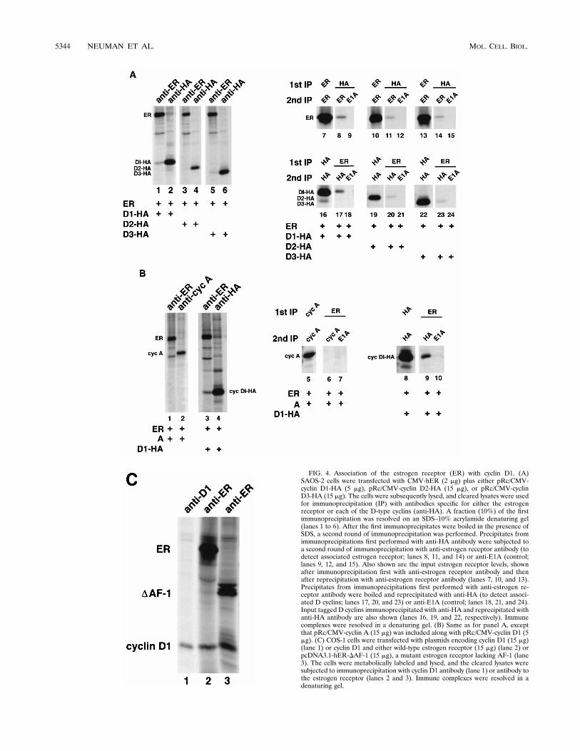
FIG. 4. Association of the estrogen receptor (ER) with cyclin D1. (A)SAOS-2 cells were transfected with CMV-hER (2 mg) plus either pRc/CMV-cyclin D1-HA (5 mg), pRc/CMV-cyclin D2-HA (15 mg), or pRc/CMV-cyclinD3-HA (15 mg). The cells were subsequently lysed, and cleared lysates were usedfor immunoprecipitation (IP) with antibodies specific for either the estrogenreceptor or each of the D-type cyclins (anti-HA). A fraction (10%) of the firstimmunoprecipitation was resolved on an SDS–10% acrylamide denaturing gel(lanes 1 to 6). After the first immunoprecipitates were boiled in the presence ofSDS, a second round of immunoprecipitation was performed. Precipitates fromimmunoprecipitations first performed with anti-HA antibody were subjected toa second round of immunoprecipitation with anti-estrogen receptor antibody (todetect associated estrogen receptor; lanes 8, 11, and 14) or anti-E1A (control;lanes 9, 12, and 15). Also shown are the input estrogen receptor levels, shownafter immunoprecipitation first with anti-estrogen receptor antibody and thenafter reprecipitation with anti-estrogen receptor antibody (lanes 7, 10, and 13).Precipitates from immunoprecipitations first performed with anti-estrogen re-ceptor antibody were boiled and reprecipitated with anti-HA (to detect associ-ated D cyclins; lanes 17, 20, and 23) or anti-E1A (control; lanes 18, 21, and 24).Input tagged D cyclins immunoprecipitated with anti-HA and reprecipitated withanti-HA antibody are also shown (lanes 16, 19, and 22, respectively). Immunecomplexes were resolved in a denaturing gel. (B) Same as for panel A, exceptthat pRc/CMV-cyclin A (15 mg) was included along with pRc/CMV-cyclin D1 (5mg). (C) COS-1 cells were transfected with plasmids encoding cyclin D1 (15 mg)(lane 1) or cyclin D1 and either wild-type estrogen receptor (15 mg) (lane 2) orpcDNA3.1-hER-DAF-1 (15 mg), a mutant estrogen receptor lacking AF-1 (lane3). The cells were metabolically labeled and lysed, and the cleared lysates weresubjected to immunoprecipitation with cyclin D1 antibody (lane 1) or antibody tothe estrogen receptor (lanes 2 and 3). Immune complexes were resolved in adenaturing gel.
5344 NEUMAN ET AL. MOL. CELL. BIOL.

extent, cyclins D2 and D3 were found to associate with theestrogen receptor in the first immunoprecipitation with anti-estrogen receptor antibody (Fig. 4A, compare lanes 1, 3, and 5,lanes 8, 11, and 14 and lanes 17, 20, and 23). With the use ofspecific antibodies to each of the D-type cyclins and non-HA-tagged versions, cyclin D1 and, to a lesser extent, cyclins D2and D3 were found to associate with the estrogen receptor (notshown). Together, these results parallel those found in thetransactivation studies in which cyclins D2 and D3 were foundto activate estrogen receptor transcriptional activity to a lesserextent than was found for cyclin D1.
Given the above observations and the fact that D-type cyc-lins show a significant degree of sequence homology, we de-termined if cyclin A might form a complex with the estrogenreceptor. In an experiment performed in parallel with an ex-periment for cyclin D1, cyclin A was found not to associate toany detectable degree with the estrogen receptor (Fig. 4B).This is not attributable to the antibody to cyclin A which wasused, for two reasons. First, the monoclonal antibody is veryefficient in precipitating denatured cyclin A (Fig. 4B). Second,unlike for cyclin D1, anti-estrogen receptor immunoprecipi-tates were found not to contain cyclin A.
To begin to explore the genetics of the estrogen receptor-cyclin D1 interaction, COS-1 cells were cotransfected withplasmids encoding cyclin D1 and either the wild-type estrogenreceptor or a mutant lacking the AF-1 transactivation domain.The cells were metabolically labeled, and then cleared lysateswere subjected to immunoprecipitation with a monoclonal an-tibody to the estrogen receptor. Immune complexes, resolvedin a denaturing gel, were found to contain a protein whichcomigrated with cyclin D1 (Fig. 4C, lanes 2 and 3) when com-pared to a cyclin D1 immunoprecipitation (lane 1).
DISCUSSION
Our results suggest that cyclin D1 can activate estrogenreceptor transcriptional activity. Transcriptional activation ap-pears to be mediated by the binding of cyclin D1 to the estro-gen receptor. Consistent with the idea that activation of theestrogen receptor can be mediated by binding to cyclin D1, therelative abilities of the three D-type cyclins to activate theestrogen receptor correlated with their relative abilities to as-sociate with the estrogen receptor. In addition, the data sug-gesting that cyclin D1 activates estrogen receptor transcrip-tional activity in the absence of activation of cdk4 also suggesta mechanism involving direct protein-protein interaction. Thatcyclin D1 and the cyclin D1-KE mutant might activate an as yetunidentified kinase which is not inhibited by p16 and is in-volved in estrogen receptor activation remains a formal possi-bility.
The experiments performed linking cyclin D1 to activationof the estrogen receptor were motivated by the characteriza-tion of SCp2 murine breast cells during differentiation (Fig. 1).At present, the increase in cyclin D1 levels seems to correlatewith increased estrogen receptor transcriptional activity duringdifferentiation. We are trying to determine if there is a directcausal relationship between these two events in an effort todemonstrate a physiological role for cyclin D1 in the activationof the estrogen receptor. The results, however, like those inprevious reports (28, 62), raise the possibility that D-type cy-clins regulate gene expression via interaction with transcriptionfactors.
During completion of this work, Zwijsen et al. (62) pub-lished data similar, but not identical, to those reported here.Although we did occasionally see cooperativity between cyclinD1 and estradiol in the activation of the estrogen receptor, this
was not seen consistently. In the data reported by Zwijsen et al.(62), a synergistic effect of cyclin D1 and estradiol on thetranscriptional activation of the estrogen receptor was found.We have consistently found that cyclins D2 and D3 activate theestrogen receptor to some extent, while Zwijsen et al. foundthat cyclins D2 and D3 were completely defective with respectto activation of the estrogen receptor. Lastly, we have consis-tently found that 4-hydroxytamoxifen and, to a greater extent,ICI 182,780 significantly inhibited cyclin D1-mediated activa-tion of the estrogen receptor, which is in contrast to the find-ings of Zwijsen et al. Of interest, ICI 182,780 does not appearto inhibit the association of cyclin D1 and the estrogen recep-tor (negative data not shown). It will be particularly importantto further analyze the abilities of various antiestrogens to in-hibit cyclin D1 activation of the estrogen receptor. In thesetting of primary breast adenocarcinomas which specificallyrecur with resistance to antiestrogen treatment, following aninitial treatment with antiestrogens, it will be particularly in-teresting to determine if overexpression of cyclin D1 might beresponsible for the development of resistance to hormonaltreatment.
Cyclin D1 can transcriptionally activate the estrogen recep-tor in the absence of an agonist, suggesting the possibility thatcyclin D1 operates through the ligand-independent AF-1transactivation domain of the estrogen receptor. However, ac-tivation of the estrogen receptor by cyclin D1 was at leastpartially inhibited by the antagonist 4-hydroxytamoxifen (Fig.2), and cyclin D1 was found to associate with an estrogenreceptor mutant lacking AF-1 (Fig. 4B), suggesting the possi-bility that cyclin D1 stimulates AF-2-dependent transactiva-tion. Efficient steroid hormone-dependent activation (AF-2) ofthe estrogen receptor has been shown to involve the direct orindirect association with a number of nuclear receptor coacti-vators: RIP140/160, SRC1, TRIP1, and TIF1 (10, 23, 36, 37, 45,55) and CBP/p300 (11, 25, 30, 59). Importantly, the associationof these coactivators with nuclear receptors is ligand depen-dent, which is in contrast to the situation with cyclin D1 and theestrogen receptor. Whether the binding of the estrogen recep-tor to cyclin D1 is direct or involves a functional interactionwith coactivators or CBP/p300 remains to be determined. Thepossible involvement of coactivators in cyclin D1-estrogen re-ceptor association might explain why a high percentage ofinput cyclin D1 and estrogen receptor in transfected cells wasnot found associated.
There exist data suggesting that estrogen-dependent activa-tion of the estrogen receptor leads to stimulation of the Ras/mitogen-activated protein (MAP) kinase pathway (42). TheRas/MAP kinase pathway has been implicated in the regula-tion of cyclin D1 expression (2, 35). There are also data sug-gesting that growth factors such as epidermal growth factorand insulin-like growth factor can stimulate AF-1-dependenttranscription of the estrogen receptor via activation of anddirect phosphorylation by MAP kinase (9, 33). Together, thesedata suggest that the estrogen receptor functions upstream ofcyclin D1. However, they also suggest that MAP kinase oper-ates both upstream and downstream of the estrogen receptor.Our data indicate that cyclin D1 operates upstream of theestrogen receptor. Consistent with this observation, somegroups have shown that overexpression of cyclin D1 in breastcancer correlates with an estrogen receptor-positive status (1,4, 6, 8, 12, 18, 41, 61). Importantly, most estrogen receptor-positive breast cancers occur in postmenopausal women whohave low estrogen levels. Thus, overexpression of cyclin D1 inpostmenopausal breast cancer may allow for abnormal estro-gen receptor activity in the presence of low estrogen levels. Itremains to be determined whether the presence of the estro-
VOL. 17, 1997 CYCLIN D1 STIMULATION OF ESTROGEN RECEPTOR 5345

gen receptor is required for the oncogenic action of cyclin D1in breast cancer.
ACKNOWLEDGMENTS
We thank M. Bissell and Monsanto for the SCp2 cells; C. A. Myers(Monsanto) for b-casein reporter; C. Sherr for D-type cyclin antibod-ies; E. Harlow for the cyclin A antibody; D. J. Shapiro for pCMV-hER;P. Chambon for p(ERE)2-tk-luc; T. Sheldon, J. Stageman, and ZenecaPharmaceuticals for ICI 182,780 and 4-hydroxytamoxifen; N. Boud-reau and J. Hatier for advice concerning the SCp2 cells; and E. Flem-ington, F. Hofmann, D. Livingston, and W. Sellers for critical review ofthe manuscript.
E.N. is supported by a fellowship from the Women’s Cancers Pro-gram at the Dana-Farber Cancer Institute. S.J.M. is supported by afellowship from the American Cancer Society. P.W.H. is a Scholar forthe Leukemia Society of America. S.F.D. is an Assistant Investigator ofthe Howard Hughes Medical Institute. M.B. is supported in part by agrant from the National Cancer Institute (CA57374). R.G.P. is sup-ported by grant 1R29CA70897-02 and Cancer Center Core NationalInstitutes of Health grant 5-P30-CA13330-26. This work was supportedby a Breast Cancer Research Grant from the Massachusetts Depart-ment of Public Health and grants from the National Cancer Institute(CA65842) and the Sandoz/Dana-Farber Drug Discovery program toM.E.E.
REFERENCES
1. Adnane, J., P. Gaudray, M.-P. Simon, J. Simony-Lafontaine, P. Jeanteur,and C. Theillet. 1989. Proto-oncogene amplification and human breast can-cer tumor phenotype. Oncogene 4:1389–1395.
2. Albanese, C., J. Johnson, G. Watanabe, N. Eklund, D. Vu, A. Arnold, andR. G. Pestell. 1995. Transforming p21ras mutants and c-Ets-2 activate thecyclin D1 promoter through distinguishable regions. J. Biol. Chem. 270:23589–23597.
3. Bartkova, J., J. Lukas, H. Muller, D. Luzhoft, M. Strauss, and J. Bartek.1994. Cyclin D1 protein expression and function in human breast cancer. Int.J. Cancer 57:353–361.
4. Berns, E. M., J. G. Klijn, I. L. van Staveren, H. Portengen, E. Noordegraaf,and J. A. Foekens. 1992. Prevalence of amplification of oncogenes c-myc,HER2/neu, and int-2 in one thousand human breast tumours: correlationwith steroid receptors. Eur. J. Cancer 28:697–700.
5. Berry, M., D. Metzger, and P. Chambon. 1990. Role of the two activatingdomains of the oestrogen receptor in the cell-type and promoter-contextdependent agonistic activity of the anti-oestrogen 4-hydroxytamoxifen.EMBO J. 9:2811–2818.
6. Borg, A., H. Sigurdsson, G. M. Clark, M. Ferno, S. A. Fuqua, H. Olsson, D.Killander, and W. L. McGurie. 1991. Association of INT2/HST1 coamplifi-cation in primary breast cancer with hormone-dependent phenotype andpoor prognosis. Br. J. Cancer 63:136–142.
7. Brown, M. 1994. Estrogen receptor molecular biology. Hematol./Oncol.Clin. N. Am. 8:101–112.
8. Buckley, M. F., K. J. E. Sweeney, J. A. Hamilton, R. L. Sini, D. L. Manning,R. I. Nicholson, A. deFazio, C. K. W. Watts, E. A. Musgrove, and R. L.Sutherland. 1993. Expression and amplification of cyclin genes in humanbreast cancer. Oncogene 8:2127–2133.
9. Bunone, G., P. A. Briand, R. J. Miksicek, and D. Picard. 1996. Activation ofthe unliganded estrogen receptor by EGF involves the MAP kinase pathwayand direct phosphorylation. EMBO J. 15:2174–2183.
10. Cavailles, V., S. Dauvois, F. L’Horset, G. Lopez, S. Hoare, P. J. Kushner, andM. G. Parker. 1995. Nuclear factor RIP140 modulates transcriptional acti-vation by the estrogen receptor. EMBO J. 14:3741–3751.
11. Chakravarti, D., V. J. LaMorte, M. C. Nelson, T. Nakajima, I. G. Schulman,H. Juguilon, M. Montminy, and R. M. Evans. 1996. Role of CBP/P300 innuclear receptor signalling. Nature 383:99–103.
12. Courjal, F., G. Louason, P. Speiser, D. Katsaros, R. Zeillinger, and C.Theillet. 1996. Cyclin gene amplification and overexpression in breast andovarian cancers: evidence for the selection of cyclin D1 in breast and cyclinE in ovarian tumors. Int. J. Cancer 69:247–253.
13. Desprez, P.-Y., C. Roskelley, J. Campisi, and M. J. Bissell. 1993. Isolation offunctional cell lines from a mouse mammary epithelial cell strain: the im-portance of basement membrane and cell-cell interaction. Mol. Cell. Diff.1:99–110.
14. Dowdy, S. F., and P. W. Hinds. Unpublished observations.15. Dowdy, S. F., P. W. Hinds, K. Louie, S. I. Reed, A. Arnold, and R. A.
Weinberg. 1993. Physical interaction of the retinoblastoma protein withhuman D cyclins. Cell 73:499–511.
16. Ewen, M. E., C. J. Oliver, H. K. Sluss, S. J. Miller, and D. S. Peeper. 1995.p53-dependent repression of CDK4 translation in TGF-b-induced cell-cyclearrest. Genes Dev. 9:204–217.
17. Ewen, M. E., H. K. Sluss, C. J. Sherr, H. Matsushime, J.-Y. Kato, and D. M.Livingston. 1993. Functional interactions of the retinoblastoma protein withmammalian D-type cyclins. Cell 73:487–497.
18. Fantl, V., M. A. Richards, R. Smith, G. A. Lammie, G. Johnstone, D. Allen,W. Gregory, G. Peters, C. Dickson, and D. M. Barnes. 1990. Gene amplifi-cation on chromosome band 11q13 and oestrogen receptor status in breastcancer. Eur. J. Cancer 26:423–429.
19. Fantl, V., G. Stamp, A. Andrews, I. Rosewell, and C. Dickson. 1995. Micelacking cyclin D1 are small and show defects in eye and mammary glanddevelopment. Genes Dev. 9:2364–2372.
20. Fawell, S. E., R. White, S. Hoare, M. Sydenham, M. Page, and M. G. Parker.1990. Inhibition of estrogen receptor-DNA binding by the “pure” antiestro-gen ICI 164,384 appears to be mediated by impaired receptor dimerization.Proc. Natl. Acad. Sci. USA 87:6883–6887.
21. Gillett, C., V. Fantl, R. Smith, C. Fisher, J. Bartek, C. Dickson, D. Barnes,and G. Peters. 1994. Amplification and overexpression of cyclin D1 in breastcancer detected by immunohistochemical staining. Cancer Res. 54:1812–1817.
22. Graham, F. L., and A. van der Eb. 1973. A new technique for the assay ofinfectivity of human adenovirus 5 DNA. J. Virol. 52:456–467.
23. Halachmi, S., E. Marden, G. Martin, H. MacKay, C. Abbondanza, and M.Brown. 1994. Estrogen receptor-associated proteins: possible mediators ofhormone-induced transcription. Science 264:1455–1458.
24. Hall, M., and G. Peters. 1996. Genetic alterations of cyclins, cyclin-depen-dent kinases, and Cdk inhibitors in human cancer. Adv. Cancer Res. 68:67–108.
25. Hanstein, B., R. Eckner, J. DiRenzo, S. Halachmi, H. Liu, B. Searcy, R.Kurokawa, and M. Brown. 1996. p300 is a component of an estrogen recep-tor coactivator complex. Proc. Natl. Acad. Sci. USA 93:11540–11545.
26. Hinds, P. W., S. F. Dowdy, E. N. Eaton, A. Arnold, and R. A. Weinberg. 1994.Function of a human cyclin gene as an oncogene. Proc. Natl. Acad. Sci. USA91:709–713.
27. Hinds, P. W., S. Mittnacht, V. Dulic, A. Arnold, S. I. Reed, and R. A.Weinberg. 1992. Regulation of retinoblastoma protein functions by ectopicexpression of human cyclins. Cell 70:993–1006.
28. Hirai, H., and C. J. Sherr. 1996. Interaction of D-type cyclins with a novelmyb-like transcription factor, DMP1. Mol. Cell. Biol. 16:6457–6467.
29. Kaelin, W. G., D. C. Pallas, J. A. DeCaprio, F. J. Kaye, and D. M. Livingston.1991. Identification of cellular proteins that can interact specifically with theT/E1A-binding region of the retinoblastoma gene product. Cell 64:521–532.
30. Kamei, Y., L. Xu, T. Heinzel, J. Torchai, R. Kurokawa, B. Gloss, S.-C. Lin,R. A. Heyman, D. W. Rose, C. K. Glass, and M. G. Rosenfeld. 1996. A CBPintegrator complex mediates transcriptional activation and AP-1 inhibitionby nuclear receptors. Cell 85:403–414.
31. Kato, J.-Y., H. Matsushime, S. W. Hiebert, M. E. Ewen, and C. J. Sherr.1993. Direct binding of cyclin D to the retinoblastoma gene product (pRb)and pRb phosphorylation by the cyclin D-dependent kinase cdk4. GenesDev. 7:331–342.
32. Kato, J.-Y., and C. J. Sherr. 1993. Inhibition of granulocyte differentiation byG1 cyclins D2 and D3 but not D1. Proc. Natl. Acad. Sci. USA 90:11513–11517.
33. Kato, S., H. Endoh, Y. Masuhiro, T. Kitamoto, S. Uchiyama, H. Sasaki, S.Masushige, Y. Gotoh, E. Nishida, H. Kawashima, D. Metzger, and P. Cham-bon. 1995. Activation of the estrogen receptor through phosphorylation bymitogen-activated protein kinase. Science 270:1491–1494.
34. Kumar, V., S. Green, G. Stack, M. Berry, J. R. Jin, and P. Chambon. 1987.Functional domains of the human estrogen receptor. Cell 51:941–951.
35. Lavoie, J. N., G. L’Allemain, A. Brunet, R. Muller, and J. Pouyssegur. 1996.Cyclin D1 expression is regulated positively by the p42/p44MAPK and nega-tively by the p38/HOGMAPK pathway. J. Biol. Chem. 271:20608–20616.
36. Le Douarin, B., C. Zechel, J. M. Garnier, Y. Lutz, L. Tora, P. Pierrat, D.Heery, H. Gronemeyer, P. Chambon, and R. Losson. 1995. The N-terminalpart of TIF1, a putative mediator of the ligand-dependent activation function(AF-2) of nuclear receptors, is fused to B-raf in the oncogenic protein T18.EMBO J. 14:2020–2033.
37. Lee, J. W., F. Ryan, J. C. Swaffield, S. A. Johnston, and D. D. Moore. 1995.Interaction of thyroid-hormone receptor with a conserved transcriptionalmediator. Nature 374:91–94.
38. Lovec, H., A. Sewing, F. C. Lucibello, R. Muller, and T. Moroy. 1994.Oncogenic activity of cyclin D1 revealed through cooperation with Ha-ras:link between cell cycle control and malignant transformation. Oncogene9:323–326.
39. Lukas, J., J. Bartkova, M. Rohde, M. Strauss, and J. Bartek. 1995. Cyclin D1is dispensable for G1 control in retinoblastoma gene-deficient cells indepen-dently of cdk4 activity. Mol. Cell. Biol. 15:2600–2611.
40. Matsushime, H., D. E. Quelle, S. A. Shurtleff, M. Shibuya, C. J. Sherr, andJ.-Y. Kato. 1994. D-type cyclin-dependent kinase activity in mammalian cells.Mol. Cell. Biol. 14:2066–2076.
41. Michalides, R., P. Hageman, H. van Tinteren, L. Houben, E. Wientjens, R.Klompmaker, and J. Peterse. 1996. A clinicopathological study on overex-pression of cyclin D1 and of p53 in a series of 248 patients with operablebreast cancer. Br. J. Cancer 73:728–734.
5346 NEUMAN ET AL. MOL. CELL. BIOL.

42. Migliaccio, A., M. Di Domenico, G. Castoria, A. de Falco, P. Bontempo, E.Nola, and F. Auricchio. 1996. Tyrosine kinase/p21ras/MAP-kinase pathwayactivation by estradiol-receptor complex in MCF-7 cells. EMBO J. 15:1292–1300.
43. Musgrove, E. A., J. A. Hamilton, C. S. L. Lee, K. J. E. Sweeney, C. K. W.Watts, and R. L. Sutherland. 1993. Growth factor, steroid, and steroidantagonist regulation of cyclin gene expression associated with changes inT-47D human breast cancer cell cycle progression. Mol. Cell. Biol. 13:3577–3587.
44. Neuman, E., M. H. Ladha, N. Lin, and M. E. Ewen. Unpublished observa-tions.
45. Onate, S., S. Y. Tsai, M.-J. Tsai, and B. W. O’Malley. 1995. Sequence andcharacterization of a coactivator for the steroid hormone receptor super-family. Science 270:1354–1357.
46. Peeper, D. S., T. M. Upton, M. H. Ladha, E. Neuman, J. Zalvide, R. Ber-nards, J. A. DeCaprio, and M. E. Ewen. 1997. Ras signalling linked to thecell-cycle machinery by the retinoblastoma protein. Nature 386:177–181.
47. Schmidhauser, C., G. F. Casperson, C. A. Myers, K. T. Sanzo, S. Bolten, andM. J. Bissell. 1992. A novel transcriptional enhancer is involved in theprolactin- and extracellular matrix-dependent regulation of b-casein geneexpression. Mol. Biol. Cell 3:699–709.
48. Sherr, C. J. 1993. Mammalian G1 cyclins. Cell 73:1059–1065.49. Sherr, C. J. 1994. G1 phase progression: cycling on cue. Cell 79:551–555.50. Sherr, C. J. 1996. Cancer cell cycles. Science 274:1672–1677.51. Sicinski, P., J. L. Donaher, Y. Geng, S. B. Parker, H. Gardner, M. Y. Park,
R. L. Robker, J. S. Richards, L. K. McGinnis, J. D. Biggers, J. J. Eppig, R. T.Bronson, S. J. Elledge, and R. A. Weinberg. 1996. Cyclin D2 is an FSH-responsive gene involved in gonadal cell proliferation and oncogenesis. Na-ture 384:470–474.
52. Sicinski, P., J. L. Donaher, S. B. Parker, T. Li, A. Fazeli, H. Gardner, S. Z.Haslam, R. T. Bronson, S. Elledge, and R. A. Weinberg. 1995. Cyclin D1provides a link between development and oncogenesis in the retina andbreast. Cell 82:621–630.
53. Stepanova, L., X. Leng, S. B. Parker, and J. W. Harper. 1996. Mammalian
p50Cdc37 is a protein kinase-targeting subunit of Hsp90 that binds andstabilizes Cdk4. Genes Dev. 10:1491–1502.
54. Tam, S. W., J. W. Shay, and M. Pagano. 1994. Differential expression andcell cycle regulation of the cyclin-dependent kinase 4 inhibitor p16Ink4.Cancer Res. 22:5816–5826.
55. vom Baur, E., C. Zechel, D. Heery, M. J. Heine, J. M. Garnier, V. Vivat, B.Le Douarin, H. Gronemeyer, P. Chambon, and R. Losson. 1996. Differentialligand-dependent interactions between AF-2 activating domain of nuclearreceptors and the putative transcriptional intermediary factors mSUG1 andTIF1. EMBO J. 15:110–124.
56. Wang, T. C., R. D. Cardiff, L. Zukerberg, E. Lees, A. Arnold, and E. V.Schmidt. 1994. Mammary hyperplasia and carcinoma in MMTV-cyclin D1transgenic mice. Nature 369:669–671.
57. Weinstat-Saslow, D., M. J. Merino, R. E. Manrow, J. A. Lawrence, R. F.Bluth, K. D. Wittenbel, J. F. Simpson, D. L. Page, and P. S. Steeg. 1995.Overexpression of cyclin D mRNA distinguishes invasive and in situ breastcarcinomas from nonmalignant lesions. Nat. Med. 1:1257–1260.
58. Wilcken, N. R. C., B. Sarcevic, E. A. Musgrove, and R. L. Sutherland. 1996.Differential effects of retinoids and antiestrogens on cell cycle progressionand cell cycle regulatory genes in human breast cancer cells. Cell GrowthDiffer. 7:65–74.
59. Yao, T.-P., G. Ku, N. Zhou, R. Scully, and D. M. Livingston. 1996. Thenuclear hormone receptor coactivator SRC-1 is a specific target of p300.Proc. Natl. Acad. Sci. USA 93:10626–10631.
60. Zhuang, Y., B. S. Katzenellenbogen, and D. J. Shapiro. 1995. Estrogenreceptor mutants which do not bind 17b-estradiol dimerize and bind to theestrogen response element in vivo. Mol. Endocrinol. 9:457–466.
61. Zukerberg, L. R., W. I. Yang, M. Gadd, A. D. Thor, F. C. Koerner, E. V.Schmidt, and A. Arnold. 1995. Cyclin D1 (PRAD1) protein expression inbreast cancer: approximately one-third of infiltrating mammary carcinomasshow overexpression of the cyclin D1 oncogene. Mod. Pathol. 8:560–567.
62. Zwijsen, R. M. L., E. Wientjens, R. Klompmaker, J. van der Sman, R.Bernards, and R. J. A. M. Michalides. 1997. CDK-independent activation ofestrogen receptor by cyclin D1. Cell 88:405–415.
VOL. 17, 1997 CYCLIN D1 STIMULATION OF ESTROGEN RECEPTOR 5347
Copyright © 2022 FDOKUMEN