P53, Rb, and Cyclin D1 Expression In Human Oral Verrucous Carcinomas
7
17 p53, Rb, and Cycli Verrucous Carcino Irma B. Gimenez-Conti, D.D.s.’ Ana M. Collet, D.D.s.’-~ Hector Lanfranchi, D.D.S? Maria E. Itoiz, D.D.s.~ Mario Luna, M.D! Shi-Xue Hu, M.D? William F. Benedict, M.D.~ Claudio J. Conti, D.v.M., Phn’ Hong-Ji XU, M.D., Ph.D.’ I Science Park-Research Division, The Univer- sity of Texas M. D. Anderson Cancer Center, Smithville, Texas. Department of Pathology, The University of Texas M.D. Anderson Cancer Center, Houston, Texas. Oral Pathology Department, Faculty of Odon- tology, University of Buenos Aires, Buenos Ai- res, Argentina. Department of Molecular Oncology, The Uni- versity of Texas M.D. Anderson Cancer Center, Houston, Texas. Department of Hematology, The University of Texas M.D. Anderson Cancer Center, Houston, Texas. Supported by Grant CTR 53322R1 from the Council for Tobacco Research and CA54672 from the National Cancer institute. The authors thank the histology laboratory for their technical assistance, the art department for their services, and Carrie McKinley for her secretarial help. Address for reprints: Claudio J. Conti, D.V.M., Ph.D., M. D. Anderson Cancer Center, Science Park-Research Division, P.O. Box 389, Smiths- ville, TX 78957. Received March 19, 1996; accepted April 2, 1996. n D1 Expression in Human Oral mas BACKGROUND. The verrucous carcinoma (VC), a tumor with low grade malignancy, appears to be associated with tobacco and human papillomavirus. The pathobiol- ogy of these tumors has not been extensively studied, and molecular genetic alter- ations have not been reported. In this study we investigated by immunohistocheni- istry the expression of p53, Rb, and cyclin D1 in a series of well-defined oral VC. Changes in the expression of these genes have been commonly reported in a variety of human tumors. METHODS. We studied 29 cases of VC, fixed in formalin and embedded in paraffin. Immunohistochemistry was carried out using the avidin-biotin immunoperoxidase technique. Polyclonal antibody CM-1 was used for p53, a rabbit polyclonal human RB antibody, Rb-WL-1 antibody for Rb and a rabbit polyclonal human cyclin D antibody for cyclin D1. RESULTS. Positive p53 expression (protein accumulation) was detected in 15 of the 29 VC analyzed. In some cases, p53-positive areas were small foci but in most of the cases extensive positive areas were observed. None of the cases studied showed alterations of Rb protein. The expression of cyclin D1 was determined in 18 cases of VC. Positive nuclear immunostaining was seen in 11 cases. CONCLUSIONS. p53 protein accumulation is frequently observed in these tumors suggesting possible mutations of this gene in VC. Overexpression of cyclin D1 but no alterations of Rb staining were also observed in this low grade tumor suggesting that Rb may be functionally inactivated by overexpression of cyclin D1 or HPV infection. Cancer 1996; 7817-23. 0 1996 American Cancer Society. KEYWORDS: p53, Rb, Cyclin D1, head and neck, verrucous carcinomas. errucous carcinoma (VC) was defined by Ackerman in 1948 as a V distinct pathology of the oral cavity.’ It appears to be associated with tobacco and human papillomavirus (HPV) as the two most im- portant rick Clinically, histologically, and biologically VC is an entity with low grade malignancy, slow growth, and no metastatic potential. The typi- cal oral lesion differs from the frequent squamous cell carcinoma (SCC) in that it is generally slow growing and chiefly exophytic, pro- ducing a warty clinical appearance. Endophytic growth results in local invasion, with little risk of Microscopically, VC is characterized by extreme thickening of the epithelium with broad bulbous rete ridges pushing into the connec- tive tissue along a broad, even front. The basement membrane is intact, differentiation is of a high order, and the epithelium shows little mitotic activity, pleomorphism, or hyperchromatism. However, more aggressive types may occur which exhibit nuclear atypias and even in situ carcinoma areas. Inadequate treatment of these cases may lead to transformation into SCC. Characteristically, cleftlike spaces lined by a thick layer of parakeratin extend from the surface 0 1996 American Cancer Society
Transcript of P53, Rb, and Cyclin D1 Expression In Human Oral Verrucous Carcinomas

17
p53, Rb, and Cycli Verrucous Carcino
Irma B. Gimenez-Conti, D.D.s.’ Ana M. Collet, D.D.s.’-~ Hector Lanfranchi, D.D.S? Maria E. Itoiz, D.D.s.~ Mario Luna, M.D!
Shi-Xue Hu, M.D? William F. Benedict, M.D.~ Claudio J. Conti, D.v.M., P h n ’
Hong-Ji XU, M.D., Ph.D.’
I Science Park-Research Division, The Univer- sity of Texas M. D. Anderson Cancer Center, Smithville, Texas.
Department of Pathology, The University of Texas M.D. Anderson Cancer Center, Houston, Texas.
Oral Pathology Department, Faculty of Odon- tology, University of Buenos Aires, Buenos Ai- res, Argentina.
Department of Molecular Oncology, The Uni- versity of Texas M.D. Anderson Cancer Center, Houston, Texas.
Department of Hematology, The University of Texas M.D. Anderson Cancer Center, Houston, Texas.
Supported by Grant CTR 53322R1 from the Council for Tobacco Research and CA54672 from the National Cancer institute.
The authors thank the histology laboratory for their technical assistance, the art department for their services, and Carrie McKinley for her secretarial help.
Address for reprints: Claudio J. Conti, D.V.M., Ph.D., M. D. Anderson Cancer Center, Science Park-Research Division, P.O. Box 389, Smiths- ville, TX 78957.
Received March 19, 1996; accepted April 2, 1996.
n D1 Expression in Human Oral mas
BACKGROUND. The verrucous carcinoma (VC), a tumor with low grade malignancy, appears to be associated with tobacco and human papillomavirus. The pathobiol- ogy of these tumors has not been extensively studied, and molecular genetic alter- ations have not been reported. In this study we investigated by immunohistocheni- istry the expression of p53, Rb, and cyclin D1 in a series of well-defined oral VC. Changes in the expression of these genes have been commonly reported in a variety of human tumors. METHODS. We studied 29 cases of VC, fixed in formalin and embedded in paraffin. Immunohistochemistry was carried out using the avidin-biotin immunoperoxidase technique. Polyclonal antibody CM-1 was used for p53, a rabbit polyclonal human RB antibody, Rb-WL-1 antibody for Rb and a rabbit polyclonal human cyclin D antibody for cyclin D1. RESULTS. Positive p53 expression (protein accumulation) was detected in 15 of the 29 VC analyzed. In some cases, p53-positive areas were small foci but in most of the cases extensive positive areas were observed. None of the cases studied showed alterations of Rb protein. The expression of cyclin D1 was determined in 18 cases of VC. Positive nuclear immunostaining was seen in 11 cases. CONCLUSIONS. p53 protein accumulation is frequently observed in these tumors suggesting possible mutations of this gene in VC. Overexpression of cyclin D1 but no alterations of Rb staining were also observed in this low grade tumor suggesting that Rb may be functionally inactivated by overexpression of cyclin D1 or HPV infection. Cancer 1996; 7817-23. 0 1996 American Cancer Society.
KEYWORDS: p53, Rb, Cyclin D1, head and neck, verrucous carcinomas.
errucous carcinoma (VC) was defined by Ackerman in 1948 as a V distinct pathology of the oral cavity.’ It appears to be associated with tobacco and human papillomavirus (HPV) as the two most im- portant rick
Clinically, histologically, and biologically VC is an entity with low grade malignancy, slow growth, and no metastatic potential. The typi- cal oral lesion differs from the frequent squamous cell carcinoma (SCC) in that it is generally slow growing and chiefly exophytic, pro- ducing a warty clinical appearance. Endophytic growth results in local invasion, with little risk of
Microscopically, VC is characterized by extreme thickening of the epithelium with broad bulbous rete ridges pushing into the connec- tive tissue along a broad, even front. The basement membrane is intact, differentiation is of a high order, and the epithelium shows little mitotic activity, pleomorphism, or hyperchromatism. However, more aggressive types may occur which exhibit nuclear atypias and even in situ carcinoma areas. Inadequate treatment of these cases may lead to transformation into SCC. Characteristically, cleftlike spaces lined by a thick layer of parakeratin extend from the surface
0 1996 American Cancer Society
18 CANCER July 1,1996 / Volume 78 / Number 1
deeply into the lesion. Parakeratin plugging also ex- tends into the epithelium.* The diagnosis of VC is based on a review of both clinical and pathologic fea- tures, including growth rate, spread, and gross and microscopic appearance.
Molecular genetic alterations have not been stud- ied in VC and the only important finding regarding the pathogenesis of this lesion is the presence of HPV in a large number of cases.'-.' However, HPV may be present only as a passenger virus since the well differ- entiated epithelium of VC is a favorite host cell envi- ronment for the replication of the papilloma viruses.
In this study, we investigated possible alterations of the p53, Rb genes, and cyclin D1 in a series of well- defined oral VC. p53 and Rb are well-characterized tu- mor suppressor genes. p53 encodes a nuclear phospho- protein that regulates proliferation and apoptosis and seems to play a role in preventing DNA damage. Muta- tions of the p53 gene have been shown to be the most frequently occurring genetic abnormalities in human malignancies." In the head and neck region, ,953 gene mutations have been identified as occurring in SCC cell linesl I I I ? as well as in primary tumor^.'^^'“ Also, overex- pression of p53 protein, as detected by immunohisto- chemistry technique, has been reported in 54 to 67% of oral cancers. 15-" Another suppressor gene that has been studied in head and neck or other tumors is the retino- blastoma gene. This gene is a prototypical tumor sup- pressor gene initially identified in patients with the he- reditary form of retinoblast~ma.'~.'~ The Rb gene en- codes a 110 to 116 kilodalton (kD) nuclear protein with cell cycle-specific changes in phosphorylation and DNA- binding proper tie^.'^-^^ The loss of Rb function impli- cated in the development of retinoblastoma has also been identified as playing a key role in the pathogenesis by initiation and/or progression of some of the most common human malignancies, including carcinomas of the lung, breast, prostate, and bladder, and sarcomas of the soft tissue and b ~ n e . ~ ~ - ~ "
Cyclins are another family of proteins that have been recognized in playing a role in the development of neo- plasia. :"." These proteins are the regulatory domain of cyclin dependent kinases (CDK) and are believed to regulate transit through the cell cycle. Particularly cyclin D1, expressed in early G1, and shown to be amplified, rearranged, or overexpressed in a wide variety of tumors including head and neck carcinoma^.^^-^^
We report here that p53 protein accumulation and cyclin D1 overexpression are frequently observed in this low grade tumor but alterations in Rb protein were not observed.
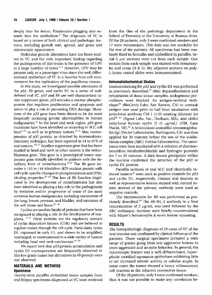
MATERIALS AND METHODS Specimens Twenty-nine paraffin embedded tissue samples from oral biopsy specimens diagnosed as VC were retrieved
from the files of the pathology department in the School of Dentistry at the University of Buenos Aires. Of the 28 patients, only 3 were confirmed smokers and 17 were nonsmokers. This data was not available for the rest of the patients. All specimens had been rou- tinely fixed in formalin and embedded in paraffin. Se- rial 5 pm sections were cut from each sample. One section from each sample was stained with hematoxy- lin and eosin (H & E); the adjacent sections on poly- L-lysine coated slides were immunostained.
lmrnunohistochemical Studies Immunostaining for p53 and cyclin D1 was performed as previously described.37 After deparaffinization and rehydration of these tissue samples, endogenous per- oxidases were blocked. An antigen-retrieval tech- nique3' (BioGenix Labs, San Ramon, CA) to unmask antigen was used prior to half hour incubation with polyclonal antibody CM-1 (1:35 working dilution) for p533g9-41 (Signet Labs, Inc., Dedham, MA), and rabbit polyclonal human cyclin D (5 pglmL) (UBI, Lake Placid, NY) .42 A biotinylated antirabbit immunoglobu- lin (Ig) (Vector Laboratories, Burlingame, CA) was then applied for 30 minutes before the use of the avidin- biotin complex (ABC) (Vector Laboratories). The speci- mens were then incubated with a solution of diamino- benzidine tetrahydrochloride and hydrogen peroxide for 7 to 10 minutes. A dark brown precipitate within the nucleus confirmed the presence of the p53 or cyclin D1 protein.
Paraffin sections of oral SCC and chemically in- duced tumors4' were used as positive controls for p53 and cyclin D1, respectively. Human oral mucosa as well as representative lesions stained with normal se- rum instead of the primary antibody were used as negative controls.
The immunostain for R b was performed as pre- viously described.43 The Rb-WL-1 antibody in a final concentration of 2 pg/mL was used followed by the ABC technique. Sections were briefly counterstained with Mayer's hematoxylin & eosin before mounting.
RESULTS The histopathologic diagnosis of 29 cases of VC of the oral mucosa was confirmed by clinical followup of the patients. These surgical specimens included a wide range of grades going from less aggressive lesions to more aggressive and invasive behavior. In general, the microscopic feature was a well-differentiated, hyper- plastic stratified squamous epithelium exhibiting little or no increased mitotic activity or cellular atypia. In some cases the tumor showed a heavy inflammatory cell reaction in the adjacent connective tissue.
Of the 28 patients, only 3 were confirmed smokers, thus it was not possible to make any correlation be-

p53, Rb, Cyclin in Verrucous CarcinomalGimenez-Conti et al. 19
TABLE I Expression of p53, and Cyclin D1 in Human Oral Verrucous Carcinomas by Immunohistochemistry
Immunohistochemistry
Sample # Agelsex P53 Rb Cyclin D1
1 NIA tt t t 2 64IF t t 3 741M t -
6 NIA t t 7 67IM t -
9 54IM ttt t NIA 10 741F tt t t 11 60IM - NIA -
12 74IF ttt t NIA 13 751F tttt t t 17 75IM - NIA NIA 18a 52IM tttt NIA NIA 20 651F tt NIA NIA 21 46IM t NIA N /A 22 NIA - NIA NIA 23 63IF tttt NIA NIA 24a 521M tttt NIA NIA 26 34IM tttt N /A NIA 27 NIA - t NIA 28 801F t t 29 801F t t 30 67IF t t 31 64IM t -
32 831M tt t -
33 381M t -
34 75IF tt t t 35 781F t t 36 72IF ttt t -
37 38IF tt t t Total: 29
-: no immunostained nuceli; t: few, scattered (<lo%); tt: small foci (<lo%); ttt: large area (10- 50%); t t t -: all (50- 100%); NIA: no available data. " Both samples are from the same patient. Sample I8 is a surgical biopsy made one year after sample 24. Samples 28 and 29 are from the same patient.
- - - -
- - - -
-
-
tween tobacco history and histologic or molecular phenotype of the lesion.
Immunohistochemical studies to detect accumu- lation of p53 were carried out using the polyclonal antibody CM- 1. We previously demonstrated that this antibody, used under the same conditions of this study, does not detect normal levels of p53 and that it only labels cells that have accumulated p53 protein as a result of mutations or other physiologic or patho- logic condition^.^'
Elevated p53 expression was detected in 15 of the 29 VC analyzed (Table 1). In all cases the staining was restricted to the nuclei. In Cases 1, 10, and 21, the p53 positive areas were small foci (Fig. lA), but in other cases (Fig. lB) , extensive positive areas were observed. No positive staining was observed in the stromal or inflammatory cells. Immunostaining was also carried
out in the adjacent normal mucosa of all the cases in which was available and in 5 normal mucosa from noncancer patients. All those specimens were p53 neg- ative. (Fig. 1G). From the 14 VC p53 negative cases, 8 were well-differentiated lesions and the rest only presented some areas with dysplasia.
Immunohistochemical detection of Rb was car- ried out using the Rb-Wl-1 polyclonal antibody, as described by Xu et al.43 None of the cases studied showed alterations of the Rb protein. Positively stained nuclei were observed in the basal and su- prabasal layers of the epithelia and in the VCs. Posi- tive staining of stromal cells was used as an internal, positive control. Typical Rb nuclear staining is shown in Figures 1C and D.
The expression of cyclin D1 was determined by immunohistochemistry in 18 cases of VC. We have previously shown that this immunohistochemical technique only detects cells overexpressing cyclin D 1 and does not detect cyclin D1 in normal proliferative cells4' Cyclin D1 immunostain was seen in 11 cases as a nuclear staining. Positive cases represented moder- ately invasive lesions with some dysplastic areas (Figs. 1E and F). The more aggressive cases and the noninva- sive, hyperplastic lesions as well as the normal oral mucosa (five cases) (Fig. 1H) were negative (Table 1).
DISCUSSION In the last few years several tumor suppressor genes have been identified and cloned. Among them, the p53 and Rb genes are the best characterized. The p53 tumor suppressor gene has been found mutated in a wide variety of human cancers, and mutations in this gene are, furthermore, the most common genetic al- teration found in human n e ~ p l a s i a . ~ ~ - ~ ~ Although the function of the p53 gene has not been completely elu- cidated, p53 appears to be a nuclear transcription fac- tor that plays a role in the control of cell proliferation, apoptosis, and maintenance of the fidelity of DNA du- p l i ~ a t i o n . ~ ~ Mutations in the p53 gene increase the stability of the p53 protein and change the specificity of its transcription factor a~tivity.~' The increased sta- bility of p53 results in accumulation of the protein in the nuclei of tumor cells bearing a mutation in the
Since normal levels of p53 are not detected by immunohistochemical studies on paraffin sections, the presence of positive staining has been accepted as an indicator of possible p53 mutations.
In this study, we show protein accumulation in 15 of 29 VC of the oral mucosa. To our knowledge there is no previous report of the occurrence of p53 in these tumors. The presence of p53 gene alterations was, to some extent, unexpected because p53 alterations have been frequently shown to be related to poor prognosis or advanced stages of disease.52 In this regard, VC is

20 CANCER July 1, 1996 I Volume 78 / Number 1
FIGURE 1. Verrucous carcinoma is shown. (A) p53 expression in the basal and suprabasal layer of a well differentiated verrucous carcinoma. (6) p53 expression in an invasive verrucous carcinoma. (W) Rb protein staining in a verrucous carcinoma. (E&F) cyclin D1 expression. (G) Negative p53 staining in normal human oral mucosa. (H) Negative cyclin 01 staining in normal human oral mucosa. All cases were counterstained with hematoxylin.

P53,
considered a very low-degree malignancy lesion that although locally destructive, rarely metastasizes and almost never jeopardizes the life of the ~ a t i e n t . ~ - ~
However, there seems to be marked differences in the timing of p53 alterations among tumors of differ- ent origins. In some cases, i.e., in colon, thyroid, breast, and prostate carcinomas, mutations of this gene appear to be a late event in the development of the n e o p l a ~ i a . ~ ~ ~ ” ~ In contrast, mutations of the p53 gene have been shown to be present in premalignant lesions of skin, larynx, and e ~ ~ p h a g u s . ~ ~ , ~ ~ . ~ ~ * ~ ~ Our re- sults are also consistent with previous studies carried out in basal cell carcinomas (BCCs), a tumor of the skin, which similarly to VC, may be locally very aggres- sive but not metastatic. Rady et al.55 found heterozy- gous mutations in 50% of BCCs and Shea et al.56 found protein accumulation in 30 of 36 specimens. The re- sults of our study taken with the data from those 2 studies indicate that p53 alteration in squamous epi- thelia is an indicator of local neoplastic growth but does not characterize the evolution to a more malig- nant phenotype in a tumor.
The Rb gene was the first recessive human cancer- susceptibility gene i s ~ l a t e d . ~ ~ - ~ ~ Although originally found to be altered in retinoblastoma, it has now been shown to be involved in the pathogenesis of several other tumors, including prostate carcinomas, osteo- sarcomas, and bladder carcinoma^.^^^^^^^' In this study, the expression of Rb was studied in a selected number of VCs using the same antibody. As shown previously, positive staining of the nucleus using this antibody is an indicator of a functional Rb gene, as tumors pre- senting homozygous mutations of the gene do not nor- mally show expression of the All the cases of VC studied showed a normal staining of the antibody, suggesting that alteration of this tumor suppressor gene does not play a role in the pathogenesis of VC. Similarly, Rb alterations do not seem to be common in more aggressive forms of head and neck SCC. Yo0 et al. have shown that although loci surrounding Rb in chromosome 13 showed high frequency of Loss of Heterozygosity (LOH), Rb alterations are restricted to a small number of cases suggesting that there is another tumor suppressor gene in chromosome 13 which is the actual target of the LOH.62
The involvement of cell cycle regulatory protein, e.g., cyclin D1, CDKs, CDK inhibitors, in cancer has only been recognized in the last few years.32 Derange- ment of the normal expression of cyclin D1 has been shown in a wide variety of turn or^.^^-^^ In head and neck carcinomas, the cyclin D1 gene was shown to be frequently amplified and ove rexpre~sed .~~~”-~~
In this study we have shown that 61% of the VCs express cyclin D1 as determined by immunohisto- chemical methods. Several laboratories have used im-
Rb, Cyclin in Verrucous CarcinomalGimenez-Conti et al. 21
munohistochemical methods to detect overexpression of cyclin D1 in breast cancer, lymphoma, and head and neck carcinoma^.^^*^^-^^ We have previously used immunohistochemical staining to show overex- pression of cyclin D1 protein in mouse skin experi- mental tumors that had shown to overexpress cyclin D1 by Northern blot and immunopre~ipitation.~’~~~
The absence of alterations at the level of Rb pro- tein in VC may be related to overexpression of cyclin D1. It has been postulated that the Rb function may be abrogated as hyperphosphorylation of the Rb pro- tein by overactivation of the cyclin D1-CDK4 complex or loss of the CDK4 regulator p16.7’.72 Alternatively, the Rb function may be lost as a result of HPV infection. The E7 gene of several HPV’s interact with the Rb pro- tein releasing the E2F transcription HPV have been frequently found in VCZm3 and may represent another form of functional Rb inactivation in these tumors.
It is presently unclear why the neoplasms devel- oping from cells with the potential to form squamous epithelia may produce either fully malignant tumors, i.e., SCCs, or tumors with moderate malignant poten- tial that very rarely develop into more malignant le- sions, i.e., VC and BCC. It is possible that alterations of critical genes in early stages of tumor development may be the determinant in the type of tumors. Alterna- tively, it is possible that different target stem cells are involved in the neoplasia of squamous epithelia. Fu- ture studies are necessary to fully understand the na- ture of malignant progression in this neoplasia.
REFERENCES 1.
2.
3.
4.
5.
6.
7.
8.
9.
Ackerman LV. Verrucous carcinoma of the oral cavity. Sur- gery 1948;23:670-8. Adler-Storthz K, Newland JR, Tessin BA, Yendall WA, Shil- litoe EJ. Human papillomavirus type 2 DNA in oral verrucous carcinoma. J Oral Puthol 1986; 15:472-5. Brandsma JL, Steinberg BM, Abramson AL, Winkler B. Pres- ence of human papillomavirus type 16 related sequences in verrucous carcinoma of the larynx. Cancer Res 1986;
Rajendran R, Sugathan CK, Augustine J, Vasudevan DM, Vi- jayakumar T. Ackerman’s tumour (verrucous carcinoma) of the oral cavity: a clinico-epidemiologic study of 426 cases. Singapore Dent J 1989; 14:48-53. Kamath W, Varma RR, Gadewar DR, Muralidhar M. Oral venu- cous carcinoma. J Crnnionum7bfm Szirg 1989; 17:309-14. Rink B. Das verrukose Karzinom der Mundschleinihaut. Lnr- yngo Rhino Otol 1991;70:542-5. Iriarte-Ortabe J-I, Laka A, Marbaix E, Reychler H. Le carci- nome verruqueux buccal. Analyse de 6 cas cliniques et revue de la litterature. J Fr D’Oto-Rhino-Laryngol1991;40:404- 13. Grinspan D, Abulafia J. Oral florid papillomatosis (verrucous carcinoma). Inter J Derm 1979; 18:608-22. Shafer WG, Hine MK, Levy BM. In: Saunders WE3 Editor. A Textbook of Oral Pathology. Philadelphia, London, Toronto, Mexico City, Rio de laniero, Sydney, Tokyo: W.B. Saunders
462185-8.
CO. 1983~127-9.
22 CANCER July 1,1996 / Volume 78 I Number 1
10. Vogelstein B. A deadly inheritance. Nature 1990;348:681-2. 11. Sakai E, Tuschida N. Most human squamous cell carcinomas
in the oral cavity contained mutated p53 tumor-suppressor genes. Oncogene 1992;7:927-33.
12. Somers KD, Merrick MA, Incognito LS, Schechter GL, Casey G. Frequent p53 mutations in head and neck cancer. Cancer Res 1992; 525997-6000.
13. Maestro R, Dolcetti R, Gasparotto D, Dolglioni C, Pelucchi S, Barzan L, et al. High frequency of p53 gene alterations associated with protein overexpression in human squamous cell carcinoma of the larynx. Oncogene 1992;7:1159-66.
14. Brachman DG, Graves D, Vokes E, Beckett M, Haraf D, Mon- tag A, et al. Occurrence of p53 gene deletions and human papilloma virus infection in human head and neck cancer. Cancer Res 1992;52:4832-6.
15. Field JK, Spandidos DA, Malliri A, Gosney JR, Yagnisis M, Stell PM. Elevated p53 expression correlates with a history of heavy smoking in squamous cell carcinoma of the head and neck. Br J Cancer 1991;64:573-7.
16. Ogden GR, Kiddie RA, Lunny DP, Lane DP. Assessment of p53 protein expression in normal, benign, and malignant oral mucosa. J Patlzol 1992; 166:389-94.
17. Shin DM, Kim J , Ro JY, Hittleman I, Roth ]A, Hittleman WN. Activation of p53 gene expression in premalignant lesions during head and neck tumorigenesis. Cancer Res 1994;
18. Murphree AL, Benedict WF. Retinoblastoma: clues for hu- man oncogenesis. Science 1984; 223: 1028-33.
19. Knudson AG. Hereditary cancer, oncogenes and antionco- genes. Cancer Res 1985;45:1437-43.
20. Lee W-H, Shew I-Y, Hong FD, Sery TW, Donoso LA, Young L-J, et al. The retinoblastoma susceptibility gene encodes a nuclear phosphoprotein associated with DNA binding activ- ity. Nature 1987;329:642-5.
21. Xu H-J, Hu S-X, Hashimoto T, Takahashi R, Benedict WF. The retinoblastoma susceptibility gene product: a character- istic pattern in normal cells and abnormal expression in malignant cells. Oncogene 1989; 4:807- 12.
22. DeCaprio ]A, Ludlow JW, Lynch D, Furukawa Y, Griffin 1, Worms HP, et al. The production of the retinoblastoma sus- ceptibility gene has properties of a cell cycle regulatory ele- ment. Cell 1989;58:1085-95.
23. Buchkovich K, Duffy LA, Harlow E. The retinoblastoma pro- tein is phosphorylated during specific phases of the cell cy- cle. Cell 1989;85:1097-105.
24. Mihara K, Cao X-R, Yen A, Chandler S, Driscoll B, Murphree AL, et al. Cell cycle-dependent regulation of phosphoryla- tion of the human retinoblastoma gene product. Science
25. Harbour JW, Li S-L, Whang-Peng I, Gazdar AF, Minna JD, Kaye FJ. Abnormalities in structure and expression of the human retinoblastoma gene in SCLC. Science 1988;241:353-7.
26. Yokota J , Akiyania T, Fung Y-KT, Benedict WF, Namba Y, Hanaoka M, et al. Altered expression of the retinoblastoma (RB) gene in small-cell carcinoma of the lung. Oncogene 1988;3:471-5.
27. Tang A, Varley JM, Chakraborty S, Murphree AL, Fung Y- KT. Structural rearrangement of the retinoblastoma gene in human breast carcinoma. Science 1988;242:263-6.
28. Lee EY-HP, To H, Shew J-Y, Brookstein R, Scully P, Lee W- H. Inactivation of the retinoblastoma susceptibility gene in human breast cancers. Science 1988;241:218-21.
29. Horowitz JM, Yandell DW, Park S-H, Canning S, Whyte P, Buchkovich K, et al. Point mutational inactivation of the retinoblastoma antioncogene. Science 1989; 243:937-40.
30. Brookstein R, Shew U-Y, Chen P-L, ScuUy P, Lee W-H. Suppres-
54:321-6.
1988; 246:1300-3.
sion of tumorigenicity of human prostate carcinoma cells by replacing a mutated RB gene. Science 1990;247712-5.
31. Sherr C. Mammalian G1 cyclins. Cell 1993;73:1059-65. 32. Motokura T, Arnold A. Cyclins and oncogenesis. Biochim
33. Arnold A, Kim HG, Gaz RD, Eddy RL, Fukushima Y, Byers MG, et al. Molecular cloning and chromosomal mapping of DNA rearranged with the parathyroid hormone gene in a parathyroid adenoma. J Clin Invest 1989; 83:2034.
34. Lammie GA, Fantl V, Smith R, Schuuring E, Brookes S, Mi- chalides R, et al. DllS287, a putative oncogene on chromo- some llq13, is amplified and expressed in squamous cell and mammary carcinomas linked to BCWL-1. Oncogene
35. Jiang W, Zhang Yj, Kahn SM, Hollstein M, Santela RM, Lu SH, et al. Altered expression of the cyclin D1 and the retino- blastoma genes in human esophageal cancer. Proc NatlAcad Sci USA 1993;90:9026-30.
36. Leach FS, Elledge SJ, Sherr CJ, Wilson JKV, Markowitz S, Kinzler KW, et al. Amplification of cyclin genes in colorectal carcinomas. Cancer Res 1993, 53:1986-9.
37. Navone NM, Troncoso P, Pisters LL, Goodrow TL, Palmer JL, Nichols WW, et al. p53 accumulation and gene mutation in the progression of human prostate carcinoma. J Nut1 Can- cer Ins t 1993;85:1657-69.
38. Shi S-R, Key ME, Kalra KL. Antigen retrieval in formalin- fixed paraffin-embedded tissues: an enhancement method for immunohistochemical obtaining based on microwave oven heating of tissue sections. J Histochem Cytochem
39. Bennet WP, Hollstein MC, Metcalf RA, Welsh JA, He A, Zhu S, et al. p53 mutation and protein accumulation during multistage human esophageal carcinogenesis. Cancer Res
40. Dolcetti R, Doglioni C, Maestro R, Gasparotto D, Barzan L, Pastore A, et al. p53 over-expression is an early event in the development of human squarnous-cell carcinoma of the larynx: genetic and prognostic implications. Int / Cancer
41. Anwar K, Nakakuki K, Imai H, Naiki H, Inuzuka M. Overex- pression of p53 protein in human laryngeal carcinoma. Int / Cancer 1993;53:952-6.
42. Robles Al, Conti CJ. Early overexpression of cyclin D1 protein in mouse sldn carcinogenesis. Carcinogenesis 1995; 16781 -6.
43. Xu H-1, Hu S-X, Cagle PT, Moore GE, Benedict WF. Absence of retinoblastoma protein expression in primary non-small cell lung carcinomas. Cancer Res 1991;51:2735-9.
44. Nigro JM, Baker SJ, Preisinger AC, Jessup JM, Hostetter R, Cleary K, et al. Mutations in the p53 gene occur in diverse human tumor types. Nature 1989;342:705-8.
45. Harris AL. Mutant p53 the commonest genetic abnormality in human cancer. J Path01 1990; 1625-6.
46. Levine AJ, Momand 1, Finaly CA. The p53 tumor suppressor gene. Nature 1991;351:453-6.
47. O'Rourke RW, Miller CW, Kato GI, Simon KJ, Chen D-L, Dang CV, et al. A potential transcriptional activation element in the p53 protein. Oncogene 1990;5:1829-32.
48. Vogelstein B, Kinzler KW. p53 function and disfunction. Cell
49. Cattoretti G, Rilke F, Andreola S, D'Amato L, Delia D. P53 expression in breast cancer. Int / Cancer 1988;41:178-83.
50. Bartek J, lggo R, Gannon J, Lane DP. Genetic and immuno- chemical analysis of mutant p53 in human breast cancer. Oncogene 1990; 5:893-9.
Biophys Acts 1993; 115553-78.
1991; 6:439-44.
1991;39(6)741-8.
1992; 52:6092-7.
1992; 52178-82.
1992; 70~523-6.

p53, Rb, Cyclin in Verrucous Carcinoma/Gimenez-Conti et al. 23
51. Iggo R, Gatter K, Bartek J, Lane D, Harris AL. Increased ex- pression of mutant forms of p53 oncogene in primary lung cancer. Lancet 1990;335:675-9.
52. Boyle JO, Hakim J, Koch W, van der Riet P, Hruban RH, Roa RA, et al. The incidence of p53 mutations increases with progression of head and neck cancers. Cancer Res 1993;53:
53. Campbell C, Auinn AG, Roy Y-S, Angus B, Rees JL. p53 muta- tions are common and early events that precede tumor inva- sion in squamous cell neoplasia of the skin. /Invest Dermatol
54. Bennett WP, Hollstein MC, Metcalf RA, Welsh ]A, He A, Zhu S, Kusters I, et al. p53 mutation and protein accumulation during multistage human esophageal carcinogenesis. Can- cw Res 1992;52:6092-7.
55. Rady P, Scinicariello F, Wagnaer RF Jr., Tyring SIC p53 mutations in basal cell carcinomas. Cancer Res 1992; 52(13)3804-6.
56. Shea CR, McNutt NS, Volkenandt M, Lug0 J, Prioleau PG, Albino AP. Overexpression of p53 protein in basal cell carci- nomas of human skin. Am J Pafhol 1992; 141(1):25-9.
57. Friend SH, Bernards R, Rogelj S, Weinberg RA, Rapaport JM, Albert DM, et al. A human DNA segment with properties of the gene that predisposes to retinoblastoma and osteosar- coma. Nature 323:643-6.
58. Lee WH, Bookstein R, Hong F, Young LJ, Shew JY, Lee EY- HP. Human retinoblastoma susceptibility gene: cloning, identification and sequence. Science 1987; 235:1394-9.
59. Fung YKT, Murphree AL, T’Ang A, Qian 1, Hinrichs SH, Bene- dict WF. Science 1987;236:1657-61.
60. Shew JY, Ling N, Yang XM, Fodstad 0, Lee WH. Antibodies detecting abnormalities of the retinoblastoma susceptibility gene product (ppllO(Rb)) in osteosarcomas and synovial sarcomas. Oncogene Res 1989;4:205-14.
61. Takahashi R, Hashimoto T, Xu HJ, Matsui T, Miki T, Bigo M H, et al. The retinoblastoma gene functions as a growth and tumor suppressor in human bladder carcinoma cells. Proc Natl Acad Sci USA 1991;88(12):5257-61.
62. Yo0 GH, Xu HJ, Brennan JA, Westra W, Hruban RH, Koch W, et al. Infrequent inactivation of the retinoblastoma gene despite frequent loss of chromosome 13q in head and neck squamous cell carcinoma. Cancer Res 1994;54:4603-6.
63. Jares P, Fernandez PL, Campo E, Nadal A, Bosch F, Aiza G, et al. PRAD-llcyclin D1 gene amplification correlates with
4477-80.
1993; 746-8.
messenger RNA overexpression and tumor progression in human laryngeal carcinomas. Cancer Res 1994; 54(17):
64. Callender T, el-Naggar AK, Lee MS, Frankenthaler R, Luna MA, Batsakis JG. PRAD-1 (CCNDl)/cyclin D1 oncogene am- plification in primary head and neck squamous cell carci- noma. Cancer 1994; 74 (1): 152 -8.
65. Williams ME, Gaffey MI, Weiss LM, Wilczynski SP, Schuuring E, Levine PA. Chromosome llQ13 amplification in head and neck squamous cell carcinoma. Arch Otolarygol Head Neck Surg 1993; 119(11):1238-43.
66. Zhang SY, Caamano J, Cooper F, Guo X, Klein-Szanto AJ. Immunohistochemistry of cyclin D1 in human breast can- cer. A m J Clin Pathol 1994; 102(5):695-8.
67. Gillett C, Fantl V, Smith R, Fisher C, Bartek J, Dickson C, et al. Amplification and overexpression of cyclin D1 in breast cancer detected by immunohistochemical staining. Cancer Res 1994;54(7):1812-7.
68. Bartkova I, Lukas J, Strauss M, Bartek 1. The PRAD-l/cyclin D1 oncogene product accumulates aberrantly in a subset of colorectal carcinomas. bit J Cancer 1994;58(4):568-73.
69. Bartkova I, Lukas J, Muller H, Lutzhoft D, Strauss M, Bartek I. Cyclin D1 protein expression and function in human breast cancer. Int J Cancer 1994;57(3):353-61.
70. Bianchi AB, Fischer SM, Robles AI, Rinchik EM, Conti CJ. Overexpression of cyclin D1 in mouse skin carcinogenesis. Oncogene 1993;8:1127-33.
71. Dowdy SF, Hinds PW, Louie K, Reed S1, Arnold A, Weinberg RA. Physical interaction of the retinoblastoma protein with human D cyclins. Cell 1993; 73:499-511.
72. Ziang W, Zhang YJ, Kahn SM, Hoolstein MC, Santella RM, Lu SH, et al. Altered expression of the cyclin D1 and retino- blastoma genes in human esophageal cancer. Proc NatZAcad Sci USA 1993;90:9026-30.
73. Scheffner M, Munger K, Huibregtse JM, Howley PM. Tar- geted degradation of the retinoblastoma protein by human papillomavirus E7-E6 fusion proteins. EMBO J 1992;
74. Chellappan S, Kraus VB, Kroger B, Munger K, Howley PM, Phelps WC, et al. Adenovirus EIA, simian virus 40 tumor antigen, and human papillomavirus E7 protein share the capapcity to disrupt the interaction between transcription factor E2F and the retinoblastoma gene product. Proc Nntl Acad Sci USA 1992;89:4549-953.
4813-7.
11 2425-31.