Obesity-Associated Differentially Methylated Regions in Colon ...
Upload
independentCategory
view
1download
0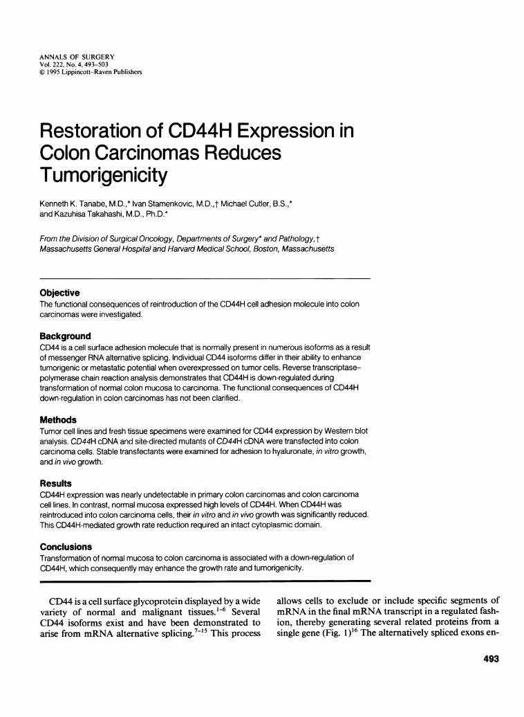
ANNALS OF SURGERYVol. 222, No. 4,493-503© 1995 Lippincott-Raven Publishers
Restoration of CD44H Expression inColon Carcinomas ReducesTumorigenicityKenneth K. Tanabe, M.D.,* Ivan Stamenkovic, M.D.,t Michael Cutler, B.S.,*and Kazuhisa Takahashi, M.D., Ph.D.*
From the Division of Surgical Oncology, Departments of Surgery* and Pathology,tMassachusetts General Hospital and Harvard Medical School, Boston, Massachusetts
ObjectiveThe functional consequences of reintroduction of the CD44H cell adhesion molecule into coloncarcinomas were investigated.
BackgroundCD44 is a cell surface adhesion molecule that is normally present in numerous isoforms as a resultof messenger RNA alternative splicing. Individual CD44 isoforms differ in their ability to enhancetumorigenic or metastatic potential when overexpressed on tumor cells. Reverse transcriptase-polymerase chain reaction analysis demonstrates that CD44H is down-regulated duringtransformation of normal colon mucosa to carcinoma. The functional consequences of CD44Hdown-regulation in colon carcinomas has not been clarified.
MethodsTumor cell lines and fresh tissue specimens were examined for CD44 expression by Western blotanalysis. CD44H cDNA and site-directed mutants of CD44H cDNA were transfected into coloncarcinoma cells. Stable transfectants were examined for adhesion to hyaluronate, in vitro growth,and in vivo growth.
ResultsCD44H expression was nearly undetectable in primary colon carcinomas and colon carcinomacell lines. In contrast, normal mucosa expressed high levels of CD44H. When CD44H wasreintroduced into colon carcinoma cells, their in vitro and in vivo growth was significantly reduced.This CD44H-mediated growth rate reduction required an intact cytoplasmic domain.
ConclusionsTransformation of normal mucosa to colon carcinoma is associated with a down-regulation ofCD44H, which consequently may enhance the growth rate and tumorigenicity.
CD44 is a cell surface glycoprotein displayed by a wide allows cells to exclude or include specific segments ofvariety of normal and malignant tissues.16 Several mRNA in the final mRNA transcript in a regulated fash-CD44 isoforms exist and have been demonstrated to ion, thereby generating several related proteins from aarise from mRNA alternative splicing.7'5 This process single gene (Fig. 1)16 The alternatively spliced exons en-
493

494 Tanabe and Others
67bp 166bp 134bp 69bp 231bp 129bp 126bp 1 14bp 117bp 129bp 132bp 102bp 90bp 204bp 63bp 72bp 79bp 947bp 898bp
CDU
CD44TR ][11 IZIZExtracellular TM Cytoplasmic
code a portion of the extracellular domain, suggestingthat the inclusion ofone or more ofthese exons may con-fer changes in ligand specificity or ligand affinity of theresulting CD44 protein. The CD44H isoform serves asthe principal cell surface receptor for hyaluronate, a pro-teoglycan found in abundance in extracellular ma-trix.'7-'9
In vitro studies ofCD44 have indicated that it plays arole in hyaluronate-mediated adhesion,'3"17'20-22 motil-ity,23'24 hyaluronate degradation,25 homotypic aggrega-tion,26 and adhesion to lymphoid tissue.20'27 30 Theseproperties are among several that are required by inva-sive and metastatic tumor cells.3' In several animalmodels, experimental overexpression of specific CD44alternative splice variants on tumor cells has resulted inenhanced metastatic potential."'24'32'33 Moreover, thestage ofdisease and overall survival correlates with CD44expression levels in patients with colon cancer,6'34'35breast cancer,35'36 non-Hodgkin's lymphoma,37-40 andgastric cancer.4'We have previously demonstrated that transformation
of normal colonic mucosa is associated with alterationsin CD44 alternative splicing that result in a down-regu-
Presented at the 115th Annual Meeting ofthe American Surgical Asso-ciation, April 6-8, 1995, Chicago, Illinois.
Supported by NIH grant CA64454 (K.K.T.), CA55735 (I.S.), and DK43351 (core facilities). Dr. Tanabe is supported by a Career Devel-opment Award from the American Cancer Society, and Dr. Sta-menkovic is a Scholar of the Leukemia Society ofAmerica.
Address reprint requests to Kenneth K. Tanabe, M.D., Division ofSur-gical Oncology, Cox 626, Massachusetts General Hospital, Boston,MA 02 114.
Accepted for publication April 10, 1995.
Figure 1. Exon map of the CD44gene. The top row is a schematic rep-resentation of all known exons in theCD44 gene, and the lower rows areschematic representations of severalCD44 isoforms. The white boxes rep-resent constitutively spliced exons,and the striped boxes represent alter-natively spliced exons in the highlyvariable membrane proximal domain.The stippled boxes represent 3' un-translated regions. Many additionalCD44 transcripts with different combi-nations of exons have been identifiedand are not depicted in this figure. Sev-eral isoforms have no designatedname, and no standard nomenclaturehas been developed for the numerousCD44 isoforms. TM is the transmem-brane region of the corresponding pro-tein.
lation of CD44H relative to several other CD44 tran-scripts, including CD44E.' Several investigators have ex-amined the functional consequences ofincreased expres-sion of several CD44 alternative splice products intumors.""'42 In the current study, however, we exam-ined the functional consequences ofthe CD44H proteindown-regulation that occurs during the transformationofnormal colonic mucosa to carcinoma. Specifically, weintroduced CD44H back into colon carcinoma cell linesby stable transfection and examined its influence on ad-hesion to hyaluronate, in vitro growth, and in vivogrowth.
MATERIALS AND METHODS
Cell Lines, Tumor Specimens, andMonoclonal Antibodies
The human colon carcinoma cell lines HT29 andLOVO were obtained from the American Type CultureCollection. Human colon carcinoma cell linesKM 1 2L4,KM 12C6, and KM20 were generously provided by Dr.Isaiah Fidler (M.D. Anderson Cancer Center, Houston,TX). Human colon carcinoma cell lines SW620 andSNU C2B were provided by Dr. Lee Ellis (M.D. Ander-son Cancer Center). Cells were maintained in Dulbecco'smodified Eagle's medium with Hamm's F 12 supplementand 8% (v/v) fetal calf serum. Human tissue specimenswere immediately frozen in liquid nitrogen in the oper-ating room and stored until further processing.The monoclonal antibodies F 10-44-2 (Biodesign In-
ternational, Kennebunkport, ME) and BU52 (BindingSite, Inc., San Diego, CA) recognize epitopes in the ex-
Ann. Surg. * October 1995

CD44 Expression and Tumorigenicity 495
tracellular domain common to all previously describedCD44 isoforms.
Western Blot and ImmunoprecipitationFor Western blot analysis, tumor tissue was homoge-
nized in 50 mM Tris (pH = 8), 150 mM NaCl, 0.2% so-dium azide, 100 ,ug/mL phenylmethylsulfonyl fluoride,1 mg/mL aprotinin, and 1% Triton X-100. Total proteinconcentration was measured with the BCA assay (PierceChemical Co., Rockford, IL). Lysates were subjected tosodium dodecyl sulfate polyacrylamide gel electrophore-sis (SDS-PAGE) under nonreducing conditions andtransferred to nitrocellulose filters by electroblotting at 4C. After being blocked for 1 hour in phosphate bufferedsaline (PBS) containing 5% dry milk, the filters were in-cubated with FIO-44-2, washed in PBS containing 1%dry milk and 0.2% Tween-20, incubated with horserad-ish peroxidase-conjugated anti-mouse antibody (Amer-sham Corp., Arlington Heights, IL), and washed in 150mM NaCl, 10 mM Tris (pH = 8), 0.05% Tween-20. Spe-cific proteins were detected with an enhanced chemilu-minescence system (Amersham Corp.).For immunoprecipitation, tissue culture cells were
harvested in PBS containing 5 mM ethylene diaminetet-raacetic acid (EDTA) and washed in PBS. Cell surfaceproteins were labeled with NHS-LC-biotin (PierceChemical) in PBS for 1 hour at 4 C. Excess biotin wasthen washed away with PBS, and cells were lysed in abuffer containing 0.25% Triton X-100, 10 ,ug/mL leu-peptin, 100 units/mL aprotinin, and 100 ig/mL phenyl-methylsulfonyl fluoride. Nuclei and debris were re-moved by centrifugation, and lysates were preclearedwith protein G-agarose beads (Oncogene Science, Cam-bridge, MA). The supernatant was then incubated withprotein G-agarose beads coated with monoclonal anti-body F 10-44-2 for 1 hour at 4 C. The beads were washedand precipitates eluted by boiling. Immunoprecipitateswere analyzed by SDS-PAGE under reducing conditionsand electroblotted described as above. After the filterswere blocked, specific proteins were detected with horse-radish peroxidase-conjugated streptavidin and an en-hanced chemiluminescence system.
CD44H Expression Vectors andTransfection
Previously described cDNA for CD44H20 was clonedinto the multiple cloning site of the vector pRC/CMV(Invitrogen, San Diego, CA), which contains the cyto-megalovirus (CMV) promoter for constitutive gene ex-pression as well as a neomycin-resistant gene for selec-tion of clones resistant to the neomycin analogue G4 18.
Site-directed mutagenesis of CD44H cDNA to obtainCD44H with only three amino acids in the cytoplasmicdomain has been described previously.23 This cDNA wassimilarly cloned into pRC/CMV.
Five million colon carcinoma cells were electropor-ated using 10 qg ofpurified plasmid DNA linearized withBgl II. The Cell-Porator (Gibco/BRL, Gaithersburg,MD) was set at 800 V/cm and 800 ,uF; cells were electro-porated at 4C in Hepes buffered saline (HBS). After 3days in complete media, G4 18 (Sigma Chemical Co., St.Louis, MO) was added to a final concentration of 500,ug/mL for selection of drug resistant clones. Clonestransfected with cDNA for CD44H, CD44H with a trun-cated cytoplasmic region, and vector only (no insert)were designated with the suffices AH, ATR, and Aneo,respectively.
Adhesion AssayNinety-six-well flat bottom plates (Corning, Corning,
NY) were coated with chondroitin sulfate A, chondroitinsulfate C, hyaluronate, vitronectin, laminin, or heat de-natured bovine serum albumin in PBS overnight at 4 C(all but the bovine serum albumin were obtained fromSigma). After washing with PBS, nonspecific bindingsites were blocked with 1 mg/mL bovine serum albuminin PBS for 2 hours at 37 C. Colon carcinoma cells de-tached from plates with 5 mM EDTA in PBS were resus-pended carefully as a single-cell suspension, and 1 X 10⁵cells were added to each well. Adhesion was allowed toproceed for 1 hour at 4 C, at which time the plates wereinverted and centrifuged for 4 minutes at 150 g. Unat-tached cells were aspirated, and the number of adherentcells was measured by methylthiotetrazole (MTT; SigmaChemical Corp.) labeling (see below). Adhesion was nor-malized for the number of cells plated as measured byMTT labeling.
In Vitro Growth AnalysisCells were detached from plates with 5 mM EDTA in
PBS, and 2 X 10³ cells were added to each well in tripli-cate. After growth for 1, 3, 5, or 7 days, the cells wereplaced in 0.5 mg/mL MTT in RPMI-1640 without phe-nol red for 2 hours at 37 C. The media was removed,and the formazan crystals were solubilized with 50 ,uLdimethyl sulfoxide. The number of viable cells at eachtime point was calculated by measuring the optical den-sity on an automatic plate reader using a 550 nm testwavelength and a 650 nm reference wavelength.
In Vivo Growth AnalysisPathogen-free 4- to 5-week-old male athymic BALB/c
nude mice (Steele Laboratory, Boston, MA) were al-
Vol. 222 - No. 4
496 Tanabe and Others
kDa
#1I
N P
#2
N P
#3
N P
200-
97.4-
69-
Figure 2. Western blot of CD44 expression in human tissue specimenswith use of monoclonal antibody F10-44-2. Tissue specimens represent-ing normal mucosa (N) and primary colon carcinomas (P) from three pa-tients were analyzed for CD44 expression by Western blot. Molecular-weight markers are indicated on the left.
lowed to acclimate for 1 week. Colon carcinoma cellsgrowing in log phase were harvested with 5 mM EDTAin PBS, washed, and resuspended as a single-cell suspen-sion. Cell viability was confirmed to be greater than 90%,as measured by trypan blue dye exclusion. Five millioncells in a total volume of 50 ,L were implanted subcuta-neously. Animals were killed at 16 days for measurementoftumor weight.
RESULTS
CD44 Expression in Human Colon Mucosaand Carcinomas
We examined CD44 expression on primary colon car-cinomas and paired normal mucosa from three patients(Fig. 2). The broad band detectable at 80 to 90 kDa rep-resents CD44H, an isoform whose transcript does notcontain any of the alternatively spliced exons in the cen-tral portion of the transcript. The normal mucosa ex-pressed significantly more CD44H than the primary tu-mors. Furthermore, normal mucosa did not express thehigh-molecular-weight CD44 isoforms seen in two ofthethree primary colon carcinomas. These higher-molecu-lar-weight CD44 isoforms arose from inclusion of addi-tional alternatively spliced exons in the correspondingtranscripts (data not shown). These data indicated that
CD44H was significantly down-regulated in primary tu-mors as compared with paired normal mucosa.
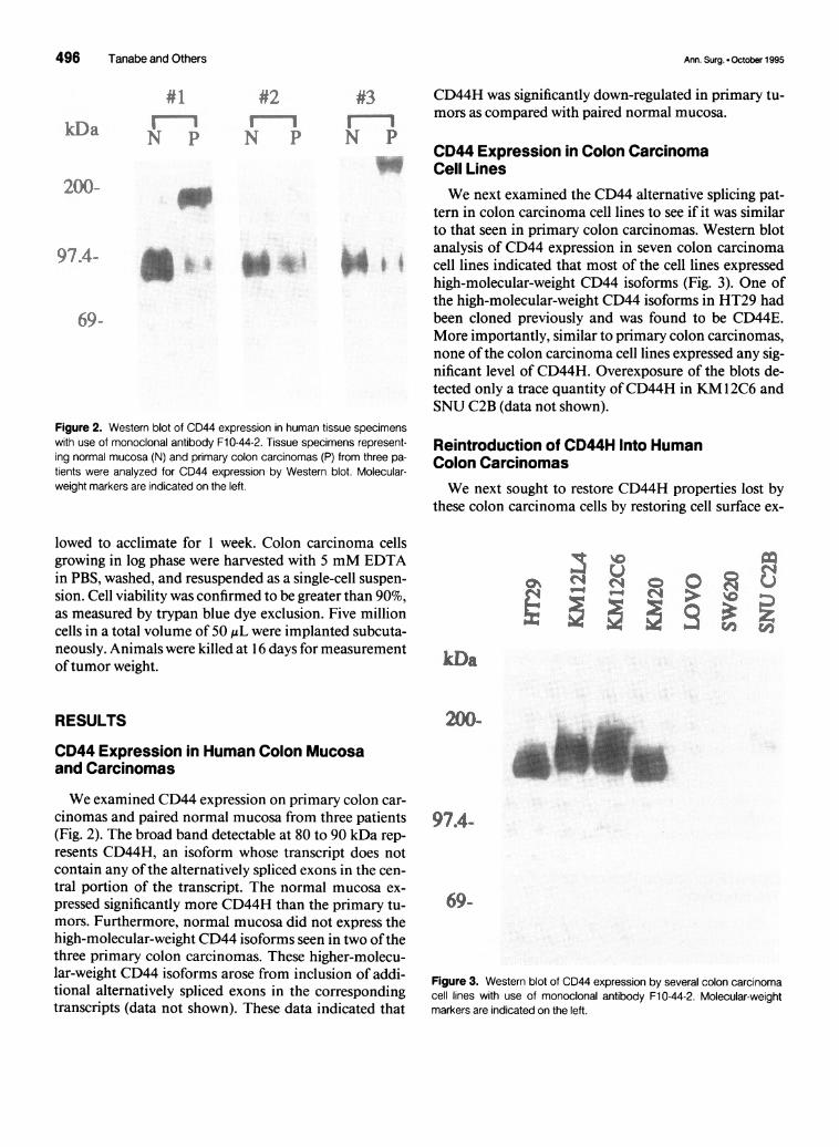
CD44 Expression in Colon CarcinomaCell LinesWe next examined the CD44 alternative splicing pat-
tern in colon carcinoma cell lines to see if it was similarto that seen in primary colon carcinomas. Western blotanalysis of CD44 expression in seven colon carcinomacell lines indicated that most of the cell lines expressedhigh-molecular-weight CD44 isoforms (Fig. 3). One ofthe high-molecular-weight CD44 isoforms in HT29 hadbeen cloned previously and was found to be CD44E.More importantly, similar to primary colon carcinomas,none of the colon carcinoma cell lines expressed any sig-nificant level of CD44H. Overexposure of the blots de-tected only a trace quantity ofCD44H in KM12C6 andSNU C2B (data not shown).
Reintroduction of CD44H Into HumanColon CarcinomasWe next sought to restore CD44H properties lost by
these colon carcinoma cells by restoring cell surface ex-
U
kDa
200-
97.4-
69-
Figure 3. Western blot of CD44 expression by several colon carcinomacell lines with use of monoclonal antibody Fl 0-44-2. Molecular-weightmarkers are indicated on the left.
Ann. Surg. - October 1995
0 0(4 >
AMLAOL'i": !:. a
ii:
in
0. V-4

CD44 Expression and Tumorigenicity 497
88_~ss
U U(4 N
A-
2,
-rZt
69 -
BFigure 4. Immunoprecipitation of cell surface labelled CD44 on transfec-tants with use of monoclonal antibody F10-44-2. HT29 and KM1 2C6 weretransfected with either CD44H cDNA (designated with the suffix AH),CD44TR (designated with the suffix ATR), or vector containing no cDNA(designated with the suffix Aneo). Immunoprecipitation of cell lysates fromG418-resistant clones with use of monoclonal antibody Fl 0-44-2 was per-formed, and results of representative clones are shown. Molecular-weightmarkers are indicated on the left.
pression of CD44H. The HT29 and KM 12C6 cell lineswere selected and electroporated with CD44H cDNAcloned into the pRC/CMV expression vector. G4 18-re-sistant colonies expressing cell surface CD44H wereidentified by immunoprecipitation and retained for fur-ther experiments. For analysis of the role of the cyto-plasmic domain of the CD44 molecule, CD44H cDNAaltered to encode only a three-amino-acid cytoplasmicdomain was also transfected into the KM 1 2C6 cell line.Immunoprecipitation of CD44 from representativeclones is shown in Figure 4.
C .J
Adhesion Properties of CD44 on ColonCarcinoma Cells
fashion (Fig. 5A). The small amount ofCD44H detectedon the surface of KM 12C6 cells may account for itsgreater affinity for hyaluronate than that of HT29 cells.Adhesion to hyaluronate could be abrogated completely
The HT29 and KM 12C6 cells expressed predomi-nantly high-molecular-weight CD44 isoforms. Thesehigh-molecular-weight CD44 isoforms demonstratedmoderate affinity for hyaluronate in a dose-dependent
Figure 5. Adhesion to extracellular matrix substances by colon carci-noma cells. Adhesion of untransfected HT29 or KM12C6 cells to (A) hy-aluronate and (B) other extracellular matrix components is shown. (C)Cells transfected with CD44H cDNA (AH), CD44TR cDNA (ATR), and no
cDNA (Aneo) were also tested for adhesion to hyaluronate.
kDa
200-
97.4 -
KMK 2)!iX
IIA :s r.:r;l :,':5 :
oKV.II
M K%1:2'01,
l<E .N14
m AI'R
K % 's to
T
4-
bIL; IT214
Vol. 222 - No. 4
-1 () -
Ae.--.Ak Am"-.dL
A'".'..
..Mm

498 Tanabe and Others
with anti-CD44 blocking antibodies (data not shown).Minimal binding to other extracellular matrix sub-stances was detectable (Fig. SB). In contrast, HT29 andKM 1 2C6 transfectants expressing high levels of cell sur-face CD44H demonstrated significantly enhancedaffinity for hyaluronate, compared with control transfec-tants (Fig. 5C). This adhesion to hyaluronate could becompletely abolished with the addition of specific CD44antibodies (data not shown). The CD44H cytoplasmicdomain was not necessary for hyaluronate binding; site-directed mutagenesis ofthe CD44H cytoplasmic domainto reduce it to only three amino acids did not signifi-cantly interfere with its ability to bind hyaluronate (Fig.5C).
In Vitro and In Vivo Colon CarcinomaGrowth After Reintroduction of CD44HThe CD44H, CD44TR, and control transfectants were
examined to determine their in vitro growth characteris-tics. Reintroduction ofCD44H into both the HT29 andKM 12C6 cell lines reduced their in vitro growth rates(Fig. 6). Although these assays were performed on stan-dard tissue culture plastic 96-well plates (not coated withhyaluronate), we have previously demonstrated thatthese cells produce and secrete hyaluronate (unpublisheddata).KM 12C6ATR was also tested for its ability to alter in
vitro growth kinetics. Despite its ability to bind hyaluro-nate avidly (Fig. 5), this cytoplasmic deletion mutant didnot reduce in vitro growth (Fig. 6). These data suggestthat the CD44H cytoplasmic domain is necessary for itsnegative influence on cell growth, possibly to interactwith cytoplasmic proteins for signal transduction.
These cell lines were then implanted subcutaneouslyinto nude mice to examine the role ofCD44H in tumor-igenicity. Tumor weights measured after 16 days are
shown in Figure 7. Tumorigenicity as determined by tu-mor weights was significantly decreased with the reintro-duction ofCD44H into the cell lines. These results werereproducible with additional clones. In vivo growth ratesparalleled in vitro growth rates; reintroduction ofCD44H reduced tumorigenicity. Interestingly, althoughexpression ofthe cytoplasmic deletion mutant CD44TRon KM 1 2C6 cells did not reduce in vitro growth, expres-sion of this mutant did reduce in vivo growth. These datasuggest that the additional environmental elements pres-
ent in vivo but not in vitro are using only the extracellulardomain ofCD44 to reduce cell growth.
DISCUSSIONWe have previously reported that transformation of
colonic mucosa is associated with a relative decrease in
11
~~~00
0
500
0.
1 3 5 7
daysFigure 6. In vitro growth analysis of CD44 transfectants. In vitro growthof (A) HT29 or (B) KM1 2C6 cells transfected with either no cDNA (Aneo),CD44H cDNA (AH), or CD44TR cDNA (ATR) was measured by the MTTassay.
CD44H transcript expression.' Furthermore, we andothers have demonstrated that this down-regulationarises from messenger RNA alternative splicing.7-'5 Wehave extended these findings in the current study andhave demonstrated that these alternative splicingchanges result in a decrease in expression of cell surfaceCD44H on colon carcinoma cells compared with normalmucosa. Furthermore, this decrease in CD44H may pro-vide cells with a growth advantage based on the findingthat reintroduction of CD44H into colon carcinomacells reduces their growth in vitro and in vivo.
Several studies involving immunohistochemistry haveindicated that many high-molecular-weight CD44 iso-forms are up-regulated in colon carcinomas relative tonormal mucosa.6'3435 The expression pattern of these
. HT9gH
........-** H29}{H0In
A8Y?
1000 -
500 -
0
Ann. Surg. * October 1995

CD44 Expression and Tumorigenicity 499
A
(I. _
0).1 -
1[1 Anezz
23 .%II
B
0.6 -
(0. -
o.2
HT29
a Aneo
E All
TR
KNI I2C6
Figure 7. In vivo growth analysis of CD44 transfectants. In vivo growthof (A) HT29 and (B) KM1 2C6 transfectants was determined through mea-
surement of tumor weights 16 days after subcutaneous implantation of 5X 10⁶ cells.
isoforms correlates with stage of disease and metastaticpotential. However, the absence of antibodies that reactwith epitopes specific to CD44H has precluded an im-munohistochemical analysis of CD44H expression incolon carcinomas. In the current study, Western blotanalysis of tissue specimens indicated that CD44H isdown-regulated in colon carcinomas compared withnormal mucosa. The exact cellular localization of thisprotein cannot be ascertained by Western blot analysisof heterogeneous tissue specimens. Nonetheless, West-ern blot analysis of several colon carcinoma cell linessupported the hypothesis that this CD44 isoform is sig-nificantly down-regulated in colon carcinomas.We as well as other investigators have demonstrated
functional differences between CD44H and CD44E iso-forms in nonepithelial tumors. CD44E binds hyaluro-nate with significantly less affinity than does CD44Hwhen expressed on melanoma and lymphomacells.'3.32'33 This pattern was also noted in the colon car-cinoma cell lines used in this study. Although post-translational CD44 modification differs between lym-phoma cells and colon carcinoma cells (unpublishedobservations), these differences do not appear to signifi-cantly alter hyaluronate affinity.
Regulation of cell growth involves a complex integra-tion of intracellular signals within the context of the ex-
tracellular environment. It is well established that mostnormal cells require attachment to a substrate to grow,and this anchorage dependence is reduced in malignantcells.43 Several molecules are important in epithelial celladhesion/anchorage, including cadherins, integrins, se-
lectins, carcinoembryonic antigen, and CD44.4446
CD44H displays high affinity for hyaluronate, a glycos-aminoglycan that is present in high levels in extracellularmatrix. Consequently, it is not surprising that CD44Hmay play a significant role in transducing signals that in-fluence cell growth based on its interaction with the ex-tracellular environment.Our finding that reintroduction ofCD44H into colon
carcinoma cells reduces in vitro and in vivo growth par-allels the results seen with a5fl, integrin (VLA-5) controlof cell growth. The a5#3 integrin is a well characterizedcell surface receptor for fibronectin.47'48 Similar toCD44H down-regulation in colon carcinomas, a5f3, in-tegrin levels are reduced in malignant cells comparedwith those in normal cells.49 Reintroduction of a5f3 in-tegrin into Chinese hamster ovary cells rendered the cellsnontumorigenic when injected subcutaneously intonude mice.50 The tumorigenicity of these cells is in-versely proportional to the level of cell surface a5#3 inte-grin.51 Our results demonstrating decreased in vitro andin vivo growth after reintroduction ofCD44H into coloncarcinoma cells also support the concept that cellular in-teraction with extracellular matrix substances directlyinfluences cell growth kinetics.We can only postulate the normal role for CD44H-
mediated growth control. Epithelial cells at the bases ofcolonic mucosal crypts proliferate more rapidly thancells that have migrated to the tops of these crypts. Adelicate balance between cell division in the crypt basesand apoptosis or sloughing in the crypt apexes is requiredfor normal mucosal homeostasis. Cell adhesion mole-cules, such as CD44H or a5#1 integrin, may an play im-portant role in this homeostasis by signaling cells to re-duce their growth rate as the cell/cell and cell/matrix in-teractions change with migration toward the cryptapexes.The function of the CD44 cytoplasmic domain re-
quires further study. This region ofthe protein shares nohomologic features with any other cell surface receptorsand has no known enzymatic activity.20 Recently, ezrin,moesin, and radixin have been shown to bind CD44.2Our results indicate that the cytoplasmic domain ofCD44H is not necessary for hyaluronate binding, butmust be intact to reduce in vitro growth. These resultssuggest that intracellular signaling proteins, such as ez-rin, radixin, and moesin, may interact with the CD44Hcytoplasmic domain to modulate growth control.
Transformation of colon mucosa to carcinoma is as-sociated with a down-regulation ofCD44H. Reintroduc-tion ofCD44H into these cells by stable transfection re-sults in growth suppression but, moreover, provides asystem to analyze the function of this receptor in trans-formation and tumor progression. Identification of theCD44H signal transduction pathway may provide in-
Vol. 222 - No. 4

500 Tanabe and Others
sights into normal colonic epithelial cell growth controlas well as new targets for therapeutic intervention in co-lon carcinomas.
Acknowledgment
The authors thank Dr. Lee Ellis for providing colon mucosa and car-cinoma tissue specimens.
References1. Tanabe KK, Ellis LM, Saya H. Expression of the CD44Rl adhe-
sion molecule is increased in human colon carcinomas and metas-tases. Lancet 1993; 341:725-726.
2. Nagasaka S, Tanabe KK, Bruner JM, et al. Alternative RNA splic-ing of the hyaluronic acid receptor, CD44, in normal human brainand brain tumors. J Neurosurg 1995; 82:858-863.
3. Cannistra SA, Kansas GS, NiloffJ, et al. Binding of ovarian cancercells to peritoneal mesothelium in vitro is partly mediated byCD44H. Cancer Res 1993; 53:3830-3838.
4. Fox SB, Fawcett J, Jackson DG, et al. Normal human tissues, inaddition to some tumors, express multiple different CD44 iso-forms. Cancer Res 1994; 54:4539-4546.
5. Li H, Hamou M-F, de Tribolet N, et al. Variant CD44 adhesionmolecules are expressed in human brain metastases but not in glio-blastomas. Cancer Res 1993; 53:5345-5349.
6. Wielenga VJM, Heider K-H, Offerhaus JA, et al. Expression ofCD44 variant proteins in human colorectal cancer is related to tu-mor progression. Cancer Res 1993; 53:4754-4756.
7. Screaton GR, Bell MV, Jackson DG, et al. Genomic structure ofDNA encoding the lymphocyte homing receptor CD44 reveals atleast 12 alternatively spliced exons. Proc Natl Acad Sci USA 1992;89:12160-12164.
8. ToTg C, Hofmann M, Herrlich P, Ponta H. Splicing choice fromten variant exons establishes CD44 variability. Nuc Acids Res1993; 21:1225-1229.
9. Tanabe KK, Nishi T, Saya H. Novel variants ofCD44 arising fromalternative splicing: changes in the CD44 alternative splicing pat-tern of MCF-7 breast carcinoma cells treated with hyaluronidase.Mol Carcinog 1993; 7:212-220.
10. Dougherty GJ, Landorp PM, Cooper DL, Humphries RK. Molec-ular cloning of CD44R I and CD44R2, two novel isoforms of thehuman CD44 lymphocyte "homing" receptor expressed by hemo-poietic cells. J Exp Med 1991; 174:1-5.
11. Gunthert U, Hofmann M, Rudy W, et al. A new variant of glyco-protein CD44 confers metastatic potential to rat carcinoma cells.Cell 1991;65:13-24.
12. Jackson DG, Buckley J, Bell JI. Multiple variants of the humanlymphocyte homing receptor CD44 generated by insertions at asingle site in the extracellular domain. J Biol Chem 1992; 267:4732-4739.
13. Stamenkovic 1, Aruffo A, Amiot M, Seed B. The hematopoieticand epithelial forms ofCD44 are distinct polypeptides with differ-ent adhesion potentials for hyaluronate-bearing cells. EMBO J1991; 10:343-348.
14. Cooper DL, Dougherty G, Harn HJ, et al. The complex CD44transcriptional unit: alternative splicing of three internal exonsgenerates the epithelial form ofCD44. Biochem Biophys Res Com-mun 1992; 182:569-578.
15. Brown TA, Bouchard T, St. John T, et al. Human keratinocytesexpress a new CD44 core protein (CD44E) as a heparan-sulfate
Ann. Surg. * October 1995
intrinsic membrane proteoglycan with additional exons. J Cell Biol1991; 113:207-221.
16. Smith CWJ, Patton JG, Nadal-Ginard B. Alternative splicing inthe control of gene expression. Annu Rev Genet 1989; 23:527-577.
17. Aruffo A, Stamenkovic I, Melnick M, et al. CD44 is the principalcell surface receptor for hyaluronate. Cell 1990; 61:1303-1313.
18. Culty M, Miyake K, Kincade PW, et al. The hyaluronate receptoris a member ofthe CD44 (H-CAM) family of cell surface glycopro-teins. J Cell Biol 1990; 111:2765-2774.
19. Miyake K, Underhill CB, Lesley J, Kincade PW. Hyaluronate canfunction as a cell adhesion molecule and CD44 participates in hy-aluronate recognition. J Exp Med 1990; 172:69-75.
20. Stamenkovic I, Amiot M, Pesando JM, Seed B. A lymphocyte mol-ecule implicated in lymph node homing is a member of the carti-lage link protein family. Cell 1989; 56:1057-1062.
21. Lesley J, He Q, Miyake K, et al. Requirements for hyaluronic acidbinding by CD44: a role for the cytoplasmic domain and activationby antibody. J Exp Med 1992; 175:257-266.
22. Dougherty GJ, Cooper DL, Memory JF, Chiu RK. Ligand bindingspecificity of alternatively spliced CD44 isoforms; recognition andbinding of hyaluronan by CD44R 1. J Biol Chem 1994; 269:9074-9078.
23. Thomas L, Byers R, Vink J, Stamenkovic I. CD44H regulates tu-mor cell migration on hyaluronate-coated substrate. J Cell Biol1992; 113:971-977.
24. Birch M, Mitchell S, Hart IR. Isolation and characterization ofhuman melanoma cell variants expressing high and low levels ofCD44. Cancer Res 1991; 51:6660-6667.
25. Culty M, Nguyen HA, Underhill CB. The hyaluronan receptor(CD44) participates in the uptake and degradation of hyaluronan.J Cell Biol 1992; 116:1055-1062.
26. St. John T, Meyer J, Idzerda R, Gallatin WM. Expression ofCD44confers a new adhesive phenotype on transfected cells. Cell 1990;60:45-52.
27. Jalkanen S, Steere AC, Fox RI, Butcher EC. A distinct endothelialcell recognition system that controls lymphocyte traffic into in-flamed synovium. Science 1986; 233:556-558.
28. Jalkanen ST, Bargatze RF, Herron LR, Butcher EC. A lymphoidcell surface glycoprotein involved in endothelial cell recognitionand lymphocyte homing in man. Eur J Immunol 1986; 16:1195-1202.
29. Jalkanen S, Bargatze RF, Toyos J, Butcher EC. Lymphocyte rec-ognition of high endothelium: antibodies to distinct epitopes ofan85-95-kD glycoprotein antigen differentially inhibit lymphocytebinding to lymph node, mucosal, or synovial endothelial cells. JCell Biol 1987; 105:983-990.
30. Jalkanen S, Jalkanen M, Bargatze R, et al. Biochemical propertiesofglycoproteins involved in lymphocyte recognition of high endo-thelial venules in man. J Immunol 1988; 141:1615-1623.
31. Fidler IJ. Critical factors in the biology of human cancer metasta-sis: Twenty-eighth G.H.A. Clowes Memorial Award Lecture. Can-cer Res 1990; 50:6130-6138.
32. Sy MS, Guo YJ, Stamenkovic I. Distinct effects of two CD44 iso-forms on tumor growth in vivo. J Exp Med 1991; 174:859-866.
33. Bartolazzi A, Peach R, Aruffo A, Stamenkovic I. Interaction be-tween CD44 and hyaluronate is directly implicated in the regula-tion oftumor development. J Exp Med 1994; 180:53-66.
34. Mulder J-WR, Kruyt PM, Sewnath M, et al. Colorectal cancerprognosis and expression of exon-v6-containing CD44 proteins.Lancet 1994; 344:1470-1472.
35. Matsumura Y, Tarin D. Significance of CD44 gene products for

Vol. 222 . No. 4
cancer diagnosis and disease evaluation. Lancet 1992; 340:1053-1058.
36. Joensuu H, Klemi PJ, Toikkanen S, Jalkanen S. GlycoproteinCD44 expression and its association with survival in breast cancer.Am J Pathol 1993; 143:867-874.
37. Pals ST, Horst E, Ossekoppele GJ, et al. Expression oflymphocytehoming receptor as a mechanism of dissemination in non-Hodg-kin's lymphoma. Blood 1989; 73:885-888.
38. Jalkanen S, Joensuu H, Soderstrom K-O, Klemi P. Lymphocytehoming and clinical behavior ofnon-Hodgkin's lymphoma. J ClinInvest 1992; 87:1835-1840.
39. Horst E, Meijer CJLM, Radaszkiewicz T, et al. Adhesion mole-cules in the prognosis of diffuse large-cell lymphoma: Expressionofa lymphocyte homing receptor (CD44), LFA- I (CD I1 a/CD 18),and ICAM- 1 (CD54). Leukemia (Baltimore) 1990; 4:595-599.
40. Joensuu HRR, Klemi PJ, Jalkanen S. Lymphocyte homing recep-tor (CD44) expression is associated with poor prognosis in gastro-intestinal lymphoma. Br J Cancer 1993; 68:428-432.
41. Guo Y-J, Liu G, Wang X, et al. Potential use of soluble CD44 inserum as indicator oftumor burden and metastasis in patients withgastric or colon cancer. Cancer Res 1994; 54:422-426.
42. Hofmann M, Rudy W, Zoller M, et al. CD44 splice variants confermetastatic behavior in rats: homologous sequences are expressedin human tumor cell lines. Cancer Res 1991; 51:5292-5297.
43. Folkman J, Moscona A. The role of cell shape in growth control.Nature 1978; 273:345-349.
44. Takeichi M. Cadherins: A molecular family important in selectivecell-cell adhesion. Annu Rev Biochem 1990; 59:237-252.
45. Ruoslahti E, Pierschbacher MD. New perspectives in cell adhe-sion: RGD and integrins. Science 1987; 238:491-497.
46. Williams AF, Barclay AN. The immunoglobulin superfamily-do-mains for cell surface recognition. Annu Rev Immunol 1988; 6:381-405.
47. Brown PJ, Juliano RL. Monoclonal antibodies to a cell surfaceglycoprotein selectively inhibit fibronectin mediated cell adhesion.Science 1985; 228:1448-1451.
48. Pytela R, Pierschbacher MD, Ruoslahti E. Identification and iso-lation ofa 150 kd cell surface glycoprotein with properties expectedofa fibronectin receptor. Cell 1985; 40:191-198.
49. Plantefaber LC, Hynes RO. Changes in integrin receptors on on-cogenically transformed cells. Cell 1989; 56:281-290.
50. Giancotti FG, Ruoslahti E. Elevated levels of the a53, fibronectinreceptor suppress the transformed phenotype of Chinese hamsterovary cells. Cell 1990; 60:849-859.
51. Schreiner C, Fisher M, Hussein S, Juliano RL. Increased tumori-genicity of fibronectin receptor deficient Chinese hamster ovarycell variants. Cancer Res 1991; 51:1738-1740.
52. Tsukita S, Oichi K, Sato N, et al. ERM family members as molec-ular linkers between the cell surface glycoprotein CD44 and actin-based cytoskeletons. J Cell Biol 1994; 126:391-401.
Discussion
DR. SAMUEL A. WELLS (St. Louis, Missouri): I very muchenjoyed the paper presented by Dr. Tanabe and his associates.This group has played a very important role in defining thebiochemistry and molecular oncology ofCD44 and its variants.I would like to ask Dr. Tanabe four questions.Do the various isoforms of CD44 confer different tumori-
genic properties to epithelial cells compared with lymphoidcells? For example, as I recall, your group has previously shown
CD44 Expression and Tumorigenicity 501
that Burkitt lymphoma cells stably transfected with CD44Hhave both greatly enhanced tumor formation and the propen-sity to metastasize, whereas cells transfected with CD44E donot. In the present experiment, however, you found that trans-fection ofcolon carcinoma cell lines with CD44H reduced theirgrowth both in vivo and in vitro. What accounts for this differ-ence in cell response and mechanistically how does the pres-ence or absence of any specific CD44 isoform enhance or re-duce the malignant potential ofa given cell type?
It has been shown that in nonmetastasizing rat pancreaticadenocarcinoma cell lines, metastatic properties are acquiredby the cells when they are transfected with a rat homologue ofCD44E. In these experiments, the carcinoma cells express therat homologous CD44H, and supposedly, the enhanced meta-static potential of the transfected cells was in part related tocoexpression of both CD44 isoforms. In your experiments,were both the CD44H and CD44E isoforms coexpressed in theCD44 transfected HT29 cells? Did the coexpression or lackthereof have any effect on the growth potential of thetransfected cells?Would you say something about the clinical utility of this
abnormal activity of CD44, both in the diagnosis of small tu-mors and the assessment of their metastatic potential? Withcurrent technology it is possible to detect CD44 splice variantsin extremely small numbers oftumor cells either in peripheralblood or other body fluids.
In experimental animal studies, and also in humans, if it isknown, is there any relationship between the expression of aCD44 isoform and the pattern of metastatic spread of the tu-mor? For example, there are some reasons that certain tumorspreferentially seed the liver, or the lung, or the brain, or theadrenal gland. The CD44 could perhaps in part hold the key tothis fascinating clinical problem.
DR. JAMES C. THOMPSON (Galveston, Texas): Anybody whoknows me knows that this work is at the limits ofor beyond mycompetence. But rest assured, I have had help.
This is an excellent paper that describes a novel finding re-garding CD44, the H variant, a molecule that is responsiblein part for the anchorage-dependence of normal cells to theirextracellular matrix; that is, it fixes them in place. Normal co-lonic mucosa expresses high levels of CD44H, but it is down-regulated in colon cancer, as the authors have shown. And ina painstaking set of stable-transfection experiments, they haveshown that when CD44H is reintroduced into colon cells bytransfection, the cancer cell growth is significantly reduced bothin vivo and in vitro.The authors have thereby identified a role, possibly unique,
for this cell-surface glycoprotein in the regulation ofgrowth ofcolon cancer. This may provide not only a better understand-ing of the mechanisms for transformation of normal colonicmucosa to cancer, but it may also provide a new target for noveltherapeutic interventions.
I have a few questions. The first two relate to a possible asso-ciation of this molecule with degrees of malignancy, and theyoverlap with a couple ofquestions that Dr. Wells asked.
First, where exactly in the progression from normal to can-cerous mucosa does this down-regulation of CD44H occur?
Copyright © 2022 FDOKUMEN