Detection of colon cancer by using SVM classifier technique
10
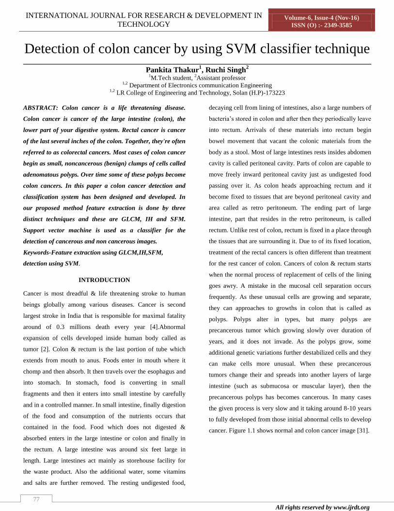
INTERNATIONAL JOURNAL FOR RESEARCH & DEVELOPMENT IN TECHNOLOGY Volume-6, Issue-4 (Nov-16) ISSN (O) :- 2349-3585 All rights reserved by www.ijrdt.org 77 Detection of colon cancer by using SVM classifier technique __________________________________________________________________________________________ Pankita Thakur 1 , Ruchi Singh 2 1 M.Tech student, 2 Assistant professor 1,2 Department of Electronics communication Engineering 1,2 LR College of Engineering and Technology, Solan (H.P)-173223 ABSTRACT: Colon cancer is a life threatening disease. Colon cancer is cancer of the large intestine (colon), the lower part of your digestive system. Rectal cancer is cancer of the last several inches of the colon. Together, they're often referred to as colorectal cancers. Most cases of colon cancer begin as small, noncancerous (benign) clumps of cells called adenomatous polyps. Over time some of these polyps become colon cancers. In this paper a colon cancer detection and classification system has been designed and developed. In our proposed method feature extraction is done by three distinct techniques and these are GLCM, IH and SFM. Support vector machine is used as a classifier for the detection of cancerous and non cancerous images. Keywords-Feature extraction using GLCM,IH,SFM, detection using SVM. INTRODUCTION Cancer is most dreadful & life threatening stroke to human beings globally among various diseases. Cancer is second largest stroke in India that is responsible for maximal fatality around of 0.3 millions death every year [4].Abnormal expansion of cells developed inside human body called as tumor [2]. Colon & rectum is the last portion of tube which extends from mouth to anus. Foods enter in mouth where it chomp and then absorb. It then travels over the esophagus and into stomach. In stomach, food is converting in small fragments and then it enters into small intestine by carefully and in a controlled manner. In small intestine, finally digestion of the food and consumption of the nutrients occurs that contained in the food. Food which does not digested & absorbed enters in the large intestine or colon and finally in the rectum. A large intestine was around six feet large in length. Large intestines act mainly as storehouse facility for the waste product. Also the additional water, some vitamins and salts are further removed. The resting undigested food, decaying cell from lining of intestines, also a large numbers of bacteria’s stored in colon and after then they periodically leave into rectum. Arrivals of these materials into rectum begin bowel movement that vacant the colonic materials from the body as a stool. Most of large intestines rests insides abdomen cavity is called peritoneal cavity. Parts of colon are capable to move freely inward peritoneal cavity just as undigested food passing over it. As colon heads approaching rectum and it become fixed to tissues that are beyond peritoneal cavity and area called as retro peritoneum. The ending part of large intestine, part that resides in the retro peritoneum, is called rectum. Unlike rest of colon, rectum is fixed in a place through the tissues that are surrounding it. Due to of its fixed location, treatment of the rectal cancers is often different than treatment for the rest cancer of colon. Cancers of colon & rectum starts when the normal process of replacement of cells of the lining goes awry. A mistake in the mucosal cell separation occurs frequently. As these unusual cells are growing and separate, they can approaches to growths in colon that is called as polyps. Polyps alter in types, but many polyps are precancerous tumor which growing slowly over duration of years, and it does not invade. As the polyps grow, some additional genetic variations further destabilized cells and they can make cells more unusual. When these precancerous tumors change their and spreads into another layers of large intestine (such as submucosa or muscular layer), then the precancerous polyps has becomes cancerous. In many cases the given process is very slow and it taking around 8-10 years to fully developed from those initial abnormal cells to develop cancer. Figure 1.1 shows normal and colon cancer image [31].
-
Upload
khangminh22 -
Category
Documents
-
view
1 -
download
0
Transcript of Detection of colon cancer by using SVM classifier technique
INTERNATIONAL JOURNAL FOR RESEARCH & DEVELOPMENT IN
TECHNOLOGY Volume-6, Issue-4 (Nov-16)
ISSN (O) :- 2349-3585
All rights reserved by www.ijrdt.org
77
Detection of colon cancer by using SVM classifier technique
__________________________________________________________________________________________
Pankita Thakur1, Ruchi Singh
2
1M.Tech student,
2Assistant professor
1,2 Department of Electronics communication Engineering
1,2 LR College of Engineering and Technology, Solan (H.P)-173223
ABSTRACT: Colon cancer is a life threatening disease.
Colon cancer is cancer of the large intestine (colon), the
lower part of your digestive system. Rectal cancer is cancer
of the last several inches of the colon. Together, they're often
referred to as colorectal cancers. Most cases of colon cancer
begin as small, noncancerous (benign) clumps of cells called
adenomatous polyps. Over time some of these polyps become
colon cancers. In this paper a colon cancer detection and
classification system has been designed and developed. In
our proposed method feature extraction is done by three
distinct techniques and these are GLCM, IH and SFM.
Support vector machine is used as a classifier for the
detection of cancerous and non cancerous images.
Keywords-Feature extraction using GLCM,IH,SFM,
detection using SVM.
INTRODUCTION
Cancer is most dreadful & life threatening stroke to human
beings globally among various diseases. Cancer is second
largest stroke in India that is responsible for maximal fatality
around of 0.3 millions death every year [4].Abnormal
expansion of cells developed inside human body called as
tumor [2]. Colon & rectum is the last portion of tube which
extends from mouth to anus. Foods enter in mouth where it
chomp and then absorb. It then travels over the esophagus and
into stomach. In stomach, food is converting in small
fragments and then it enters into small intestine by carefully
and in a controlled manner. In small intestine, finally digestion
of the food and consumption of the nutrients occurs that
contained in the food. Food which does not digested &
absorbed enters in the large intestine or colon and finally in
the rectum. A large intestine was around six feet large in
length. Large intestines act mainly as storehouse facility for
the waste product. Also the additional water, some vitamins
and salts are further removed. The resting undigested food,
decaying cell from lining of intestines, also a large numbers of
bacteria’s stored in colon and after then they periodically leave
into rectum. Arrivals of these materials into rectum begin
bowel movement that vacant the colonic materials from the
body as a stool. Most of large intestines rests insides abdomen
cavity is called peritoneal cavity. Parts of colon are capable to
move freely inward peritoneal cavity just as undigested food
passing over it. As colon heads approaching rectum and it
become fixed to tissues that are beyond peritoneal cavity and
area called as retro peritoneum. The ending part of large
intestine, part that resides in the retro peritoneum, is called
rectum. Unlike rest of colon, rectum is fixed in a place through
the tissues that are surrounding it. Due to of its fixed location,
treatment of the rectal cancers is often different than treatment
for the rest cancer of colon. Cancers of colon & rectum starts
when the normal process of replacement of cells of the lining
goes awry. A mistake in the mucosal cell separation occurs
frequently. As these unusual cells are growing and separate,
they can approaches to growths in colon that is called as
polyps. Polyps alter in types, but many polyps are
precancerous tumor which growing slowly over duration of
years, and it does not invade. As the polyps grow, some
additional genetic variations further destabilized cells and they
can make cells more unusual. When these precancerous
tumors change their and spreads into another layers of large
intestine (such as submucosa or muscular layer), then the
precancerous polyps has becomes cancerous. In many cases
the given process is very slow and it taking around 8-10 years
to fully developed from those initial abnormal cells to develop
cancer. Figure 1.1 shows normal and colon cancer image [31].

Paper Title:- Detection of colon cancer by using SVM classifier technique
ISSN:-2349-3585 |www.ijrdt.org 78
(a)
(b)
Fig. 1.1: Normal and cancerous colon image
Early and proper detection of colon cancer is essential key for
proper treatment. So we require accurate tool for proper
treatment. Detection involves finding the presence of tumor,
segmentation involves the detection of size and location of
tumor and classification involves the detection of stage of
tumor. Now a day’s different computer added tools are used in
medical field. These tools possess a property of quick and
accurate results [2]. In this paper, we used SVM classifier to
detect colon cancer. The rest of the paper is organized as
follows. In section II, the methodology is defined. And
experimental results, different feature parameters are
presented in section III. Section IV is conclusion.
II METHODOLOGY
The proposed methodology of discrimination between
cancerous and non cancerous images is shown in figure 1.2.
The method used two set of images i.e. training and testing set
then uses different steps segmentation, feature extraction and
classification. The work at hands is implemented using Mat
lab version 2013a.
FLOW CHART
The different feature extractions approaches are classified as
follows:
Grayscale Features Extraction Technique
1) GLCM
Number of texture features is extracted from the GLCM.
GLCM is statistical method to finding textures that consider
spatial relationship in the pixels. The GLCM function in the
MATLAB forms GLCM by computing how frequently pixel
with intensity value e occurred in particular structural
relationship of pixel with value f. By default, the spatial
relationship is defined as the pixel of interest and the pixel to
its immediate right (horizontally adjacent), but we can specify
other spatial relationship in between two pixels. Every element
(e and f) in resultant GLCM a combination of the numbers of
time pixel with a value E occur in stated structural relationship
of pixels with the value F in input image.
Table-1
Feature computed from GLCM
Paper Title:- Detection of colon cancer by using SVM classifier technique
ISSN:-2349-3585 |www.ijrdt.org 79
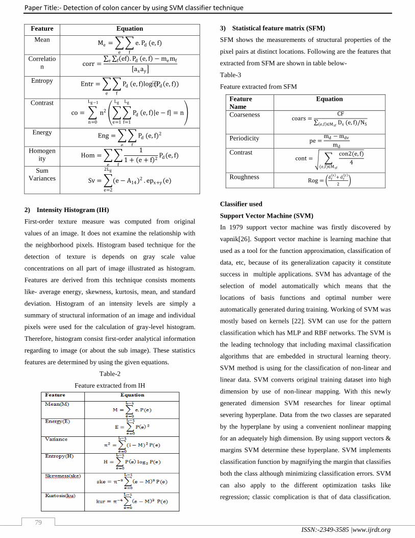
Feature Equation
Mean Me = e. Pd
fe
(e, f)
Correlatio
n corr = ef . Pdf e, f − memfe
ax ay
Entropy Entr = Pd
fe
e, f log(Pd e, f )
Contrast
co = n2
Lg−1
n=0
Pd
Lg
f=1
Lg
e=1
e, f e − f = n
Energy Eng = Pd
fe
(e, f)2
Homogen
ity Hom =
1
1 + e + f 2
fe
Pd (e, f)
Sum
Variances Sv = (e− A14)2
2Lg
e=2
. epx+y (e)
2) Intensity Histogram (IH)
First-order texture measure was computed from original
values of an image. It does not examine the relationship with
the neighborhood pixels. Histogram based technique for the
detection of texture is depends on gray scale value
concentrations on all part of image illustrated as histogram.
Features are derived from this technique consists moments
like- average energy, skewness, kurtosis, mean, and standard
deviation. Histogram of an intensity levels are simply a
summary of structural information of an image and individual
pixels were used for the calculation of gray-level histogram.
Therefore, histogram consist first-order analytical information
regarding to image (or about the sub image). These statistics
features are determined by using the given equations.
Table-2
Feature extracted from IH
3) Statistical feature matrix (SFM)
SFM shows the measurements of structural properties of the
pixel pairs at distinct locations. Following are the features that
extracted from SFM are shown in table below-
Table-3
Feature extracted from SFM
Feature
Name
Equation
Coarseness coars =
CF
Ds e,f ∈Msf(e, f)/NS
Periodicity pe =md − mdv
md
Contrast cont =
con2(e, f)
4(e,f)∈Msf
Roughness Rog =
d f x
+ d f(y )
2
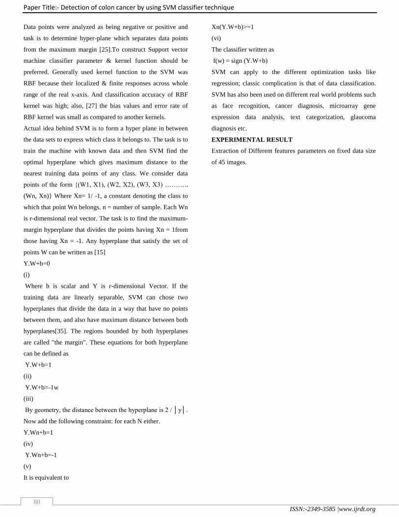
Classifier used
Support Vector Machine (SVM)
In 1979 support vector machine was firstly discovered by
vapnik[26]. Support vector machine is learning machine that
used as a tool for the function approximation, classification of
data, etc, because of its generalization capacity it constitute
success in multiple applications. SVM has advantage of the
selection of model automatically which means that the
locations of basis functions and optimal number were
automatically generated during training. Working of SVM was
mostly based on kernels [22]. SVM can use for the pattern
classification which has MLP and RBF networks. The SVM is
the leading technology that including maximal classification
algorithms that are embedded in structural learning theory.
SVM method is using for the classification of non-linear and
linear data. SVM converts original training dataset into high
dimension by use of non-linear mapping. With this newly
generated dimension SVM researches for linear optimal
severing hyperplane. Data from the two classes are separated
by the hyperplane by using a convenient nonlinear mapping
for an adequately high dimension. By using support vectors &
margins SVM determine these hyperplane. SVM implements
classification function by magnifying the margin that classifies
both the class although minimizing classification errors. SVM
can also apply to the different optimization tasks like
regression; classic complication is that of data classification.
Paper Title:- Detection of colon cancer by using SVM classifier technique
ISSN:-2349-3585 |www.ijrdt.org 80
Data points were analyzed as being negative or positive and
task is to determine hyper-plane which separates data points
from the maximum margin [25].To construct Support vector
machine classifier parameter & kernel function should be
preferred. Generally used kernel function to the SVM was
RBF because their localized & finite responses across whole
range of the real x-axis. And classification accuracy of RBF
kernel was high; also, [27] the bias values and error rate of
RBF kernel was small as compared to another kernels.
Actual idea behind SVM is to form a hyper plane in between
the data sets to express which class it belongs to. The task is to
train the machine with known data and then SVM find the
optimal hyperplane which gives maximum distance to the
nearest training data points of any class. We consider data
points of the form {(W1, X1), (W2, X2), (W3, X3) ………..
(Wn, Xn)} Where Xn= 1/ -1, a constant denoting the class to
which that point Wn belongs. n = number of sample. Each Wn
is r-dimensional real vector. The task is to find the maximum-
margin hyperplane that divides the points having Xn = 1from
those having Xn = -1. Any hyperplane that satisfy the set of
points W can be written as [15]
Y.W+b=0
(i)
Where b is scalar and Y is r-dimensional Vector. If the
training data are linearly separable, SVM can chose two
hyperplanes that divide the data in a way that have no points
between them, and also have maximum distance between both
hyperplanes[35]. The regions bounded by both hyperplanes
are called "the margin". These equations for both hyperplane
can be defined as
Y.W+b=1
(ii)
Y.W+b=-1w
(iii)
By geometry, the distance between the hyperplane is 2 / │y│.
Now add the following constraint: for each N either.
Y.Wn+b=1
(iv)
Y.Wn+b=-1
(v)
It is equivalent to
Xn(Y.W+b)>=1
(vi)
The classifier written as
f(w) = sign (Y.W+b)
SVM can apply to the different optimization tasks like
regression; classic complication is that of data classification.
SVM has also been used on different real world problems such
as face recognition, cancer diagnosis, microarray gene
expression data analysis, text categorization, glaucoma
diagnosis etc.
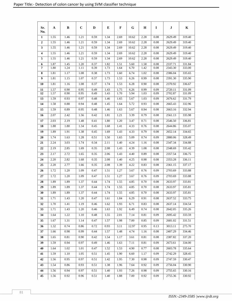
EXPERIMENTAL RESULT
Extraction of Different features parameters on fixed data size
of 45 images.
Paper Title:- Detection of colon cancer by using SVM classifier technique
ISSN:-2349-3585 |www.ijrdt.org 81
Sr.
No.
A
B
C
D
E
F
G
H
I
J
K
1 1.55 1.46 1.21 0.59 1.34 2.69 10.62 2.28 0.08 2629.49 319.40
2 1.55 1.46 1.21 0.59 1.34 2.69 10.62 2.28 0.08 2629.49 319.40
3 1.55 1.46 1.21 0.59 1.34 2.69 10.62 2.28 0.08 2629.49 319.40
4 1.55 1.46 1.21 0.59 1.34 2.69 10.62 2.28 0.08 2629.49 319.40
5 1.55 1.46 1.21 0.59 1.34 2.69 10.62 2.28 0.08 2629.49 319.40
6 1.87 1.45 1.20 0.37 1.82 1.51 5.60 1.58 0.08 2337.71 331.84
7 1.80 1.24 1.11 0.39 1.73 1.64 6.70 1.42 0.08 2343.30 333.00
8 1.81 1.17 1.08 0.38 1.73 1.60 6.74 1.02 0.08 2386.04 335.65
9 1.81 1.15 1.07 0.37 1.73 1.53 6.26 0.99 0.08 2391.30 335.90
10 1.81 1.16 1.08 0.37 1.74 1.53 6.28 0.90 0.08 2370.92 336.67
11 1.57 0.90 0.95 0.49 1.43 1.75 6.26 0.99 0.09 2720.11 331.09
12 1.57 0.90 0.95 0.49 1.43 1.70 5.94 1.03 0.09 2702.87 331.09
13 1.59 0.93 0.97 0.48 1.46 1.65 5.67 1.03 0.09 2676.62 331.79
14 1.58 0.88 0.94 0.48 1.45 1.64 5.72 0.93 0.08 2665.43 332.96
15 1.59 0.89 0.95 0.48 1.46 1.63 5.67 0.94 0.08 2663.16 332.94
16 2.07 2.42 1.56 0.42 1.81 1.21 3.39 0.79 0.08 2561.13 335.98
17 2.03 2.19 1.48 0.41 1.80 1.20 3.47 0.71 0.08 2546.50 336.81
18 1.88 1.80 1.34 0.45 1.68 1.41 4.33 0.76 0.08 2644.86 335.07
19 1.89 1.91 1.38 0.45 1.69 1.43 4.33 0.79 0.08 2652.14 334.65
20 1.74 1.63 1.28 0.51 1.50 1.65 5.09 0.74 0.09 2880.06 328.49
21 2.24 3.03 1.74 0.34 2.11 1.40 4.24 1.16 0.08 2347.34 334.88
22 2.19 2.85 1.69 0.35 2.08 1.43 4.39 1.08 0.08 2348.69 335.42
23 2.17 2.72 1.65 0.35 2.06 1.43 4.40 0.89 0.08 2357.54 336.75
24 2.20 2.82 1.68 0.35 2.08 1.40 4.25 0.98 0.08 2353.28 336.11
25 2.20 2.77 1.66 0.35 2.08 1.39 4.22 0.83 0.08 2361.15 337.17
26 1.72 1.20 1.09 0.47 1.51 1.27 3.67 0.76 0.09 2703.69 333.88
27 1.72 1.20 1.09 0.47 1.51 1.27 3.67 0.76 0.09 2703.69 333.88
28 1.89 1.89 1.37 0.44 1.74 1.55 4.85 0.70 0.08 2633.97 333.81
29 1.89 1.89 1.37 0.44 1.74 1.55 4.85 0.70 0.08 2633.97 335.81
30 1.89 1.89 1.37 0.44 1.74 1.55 4.85 0.70 0.08 2633.97 335.81
31 1.71 1.43 1.20 0.47 1.61 1.84 6.29 0.91 0.08 2637.52 333.75
32 1.70 1.41 1.19 0.46 1.62 1.93 6.71 0.83 0.08 2637.14 334.54
33 1.71 1.43 1.20 0.46 1.63 1.92 6.49 0.74 0.08 2642.05 335.26
34 1.64 1.22 1.10 0.48 1.55 2.01 7.14 0.81 0.09 2695.42 333.59
35 1.67 1.31 1.14 0.47 1.57 1.98 7.00 0.85 0.09 2681.02 331.51
36 1.32 0.74 0.86 0.72 0.93 3.11 12.97 0.95 0.13 3013.11 275.70
37 1.66 0.98 0.99 0.44 1.57 1.48 4.74 1.16 0.08 2407.29 334.46
38 1.65 0.81 0.90 0.42 1.54 1.17 3.61 0.81 0.08 2387.82 337.20
39 1.59 0.94 0.97 0.49 1.46 1.63 7.11 0.81 0.09 2673.61 334.00
40 1.64 1.02 1.01 0.47 1.52 1.53 4.90 0.77 0.08 2603.78 335.64
41 1.59 1.10 1.05 0.51 1.45 1.90 6.60 1.17 0.09 2742.29 328.45
42 1.56 0.95 0.97 0.51 1.42 1.95 7.30 0.98 0.09 2747.59 330.47
43 1.54 0.86 0.93 0.51 1.38 1.96 7.64 0.92 0.09 2756.04 330.90
44 1.56 0.94 0.97 0.51 1.40 1.93 7.26 0.98 0.09 2755.65 330.16
45 1.56 0.92 0.96 0.51 1.40 1.88 7.00 0.92 0.09 2755.36 330.92

Paper Title:- Detection of colon cancer by using SVM classifier technique
ISSN:-2349-3585 |www.ijrdt.org 82
Sr.
No.
L
M
N
O
P
Q
R
S
T
U
V
W
X
Y
Z
1 .33 .51 .82 .96 .63 11.03 1.69 5.03 0.41 2.17 -0.06 -0.80 .82 10.18 74.38
2 .33 .51 .82 .96 .63 11.03 1.69 5.03 0.41 2.17 -0.06 -0.80 .82 10.18 74.38
3 .33 .51 .82 .96 .63 11.03 1.69 5.03 0.41 2.17 -0.06 -0.80 .82 10.18 74.38
4 .33 .51 .82 .96 .63 11.03 1.69 5.03 0.41 2.17 -0.06 -0.80 .82 10.18 78.38
5 .33 .51 .82 .96 .63 11.03 1.69 5.03 0.41 2.17 -0.06 -0.80 .82 10.18 74.38
6 .23 .47 .85 .97 .51 12.21 1.20 3.39 0.48 1.53 0.00 -0.90 .86 6.80 84.30
7 .02 .47 .84 .97 .51 12.22 1.18 3.18 0.47 1.38 0.01 -0.91 .86 8.47 70.93
8 .15 .48 .81 .98 .53 12.51 1.08 2.59 0.45 1.00 0.04 -0.95 .87 10.30 61.45
9 .14 .48 .80 .98 .54 12.14 1.07 2.43 0.45 0.97 0.05 -0.95 .87 10.67 59.60
10 .13 .48 .80 .98 .52 12.21 1.09 2.47 0.44 0.88 0.05 -0.96 .87 10.69 60.57
11 .14 .53 .74 .98 .65 11.07 1.47 3.30 0.34 0.97 0.05 -0.92 .85 8.84 82.25
12 .15 .53 .75 .98 .64 11.12 1.47 3.34 0.34 1.01 0.04 -0.92 .85 8.70 82.37
13 .15 .52 .75 .98 .63 11.23 1.46 3.30 0.35 1.00 0.04 -0.93 .85 8.40 84.74
14 .13 .52 .75 .98 .63 11.29 1.43 3.16 0.34 0.92 0.05 -0.94 .86 8.90 78.90
15 .13 .52 75 .98 .63 11.30 1.43 3.16 0.35 0.92 0.05 -0.94 .86 8.81 79.82
16 .11 .51 .76 .99 .60 11.67 1.35 2.82 0.36 0.78 0.06 -0.96 .86 5.89 112.75
17 .10 .51 .76 .99 .60 11.73 1.32 2.69 0.36 0.70 0.07 -0.97 .87 7.09 99.30
18 .11 .52 .74 .99 .62 11.41 1.39 2.88 0.34 0.75 0.07 -0.96 .86 7.86 93.28
19 .11 .52 .74 .99 .63 11.37 1.40 2.93 0.34 0.78 0.06 -0.95 .86 7.09 99.84
20 .11 .56 .69 .99 .68 10.46 1.50 3.16 0.29 0.73 0.07 -0.93 .85 8.84 85.27
21 .17 .48 .82 .98 .50 12.24 1.16 2.87 0.45 1.13 0.03 -0.94 .87 6.05 98.48
22 .15 .48 .82 .98 .50 12.25 1.16 2.79 0.44 1.06 0.04 -0.95 .87 6.76 90.38
23 .13 .48 .80 .98 49 12.25 1.14 2.54 0.43 0.88 0.06 -0.97 .87 7.59 81.59
24 .14 .48 .81 .98 .50 12.25 1.15 2.66 0.44 0.96 0.05 -0.96 .87 6.95 87.76
25 .12 .48 .80 .99 .50 12.25 1.12 2.42 0.43 0.82 0.06 -0.97 .87 7.68 80.96
26 .11 .53 .73 .99 .64 11.20 1.42 2.96 0.33 0.75 0.07 -0.95 .86 8.80 80.50
27 .11 .53 .73 .99 .64 11.20 1.42 2.96 0.33 0.75 0.07 -0.95 .86 8.80 80.50
28 .10 .52 .74 .99 .62 11.46 1.37 2.78 0.34 0.69 0.07 -0.96 .86 7.63 93.97
29 .10 .52 .74 .99 .62 11.46 1.37 2.78 0.34 0.69 0.07 -0.96 .86 7.63 93.97
30 .10 5.2 .74 .99 .62 11.46 1.37 2.78 0.34 0.69 0.07 -0.96 .86 7.63 93.97
31 .13 .52 .75 .98 .62 11.40 1.41 3.09 0.35 0.89 0.05 -0.94 .86 6.91 95.81
32 .12 .52 .75 .99 .62 11.42 1.40 2.97 0.34 0.81 0.06 -0.95 .86 7.58 94.60
33 .11 .52 .74 .99 .62 11.42 1.38 2.85 0.34 0.73 0.07 -0.96 .86 8.19 90.24
34 .12 .53 .74 .99 .64 11.22 1.42 3.01 0.33 0.80 0.06 -0.95 .86 9.40 78.65
35 .12 .52 .74 .98 .63 11.26 1.42 3.05 0.34 0.83 0.06 -0.94 .86 8.45 83.28
36 .14 .68 .50 .98 .81 7.37 1.79 4.20 0.93 0.05 -0.81 -0.81 .77 13.29 73.90
37 .17 .48 .81 .98 .55 12.07 1.31 3.13 1.13 0.03 -0.93 -0.93 .86 9.83 71.81
38 .12 .49 .79 .99 .53 12.18 1.21 2.57 0.80 0.06 -0.97 -0.97 .87 11.71 58.30
39 .12 .52 .74 .99 .63 11.29 1.41 2.99 0.80 0.06 -0.95 -0.95 .86 11.38 63.81
40 .11 .51 .75 .99 .61 11.54 1.37 2.84 0.75 0.07 -0.96 -0.96 .86 11.53 61.43
41 .17 .53 .74 .98 .65 10.92 1.52 3.59 0.34 1.14 0.03 -0.90 .84 7.76 87.19
42 .14 .53 .73 .98 .65 10.96 1.48 3.32 0.33 0.96 0.05 -0.92 .85 9.17 76.25
43 .13 .53 .73 .98 .65 10.95 1.48 3.24 0.32 0.90 0.05 -0.93 .85 11.01 66.21
44 .14 .53 .73 .98 .65 10.93 1.49 3.34 0.33 0.96 0.05 -0.92 .85 9.67 73.53
45 13 .53 .73 .98 .65 10.95 1.48 3.24 0.32 0.90 0.05 -0.93 .85 10.66 70.91

Paper Title:- Detection of colon cancer by using SVM classifier technique
ISSN:-2349-3585 |www.ijrdt.org 83
Sr. No. AA AB AC
1 .55 2.37 1
2 .55 2.37 1
3 .55 2.37 1
4 .55 2.37 1
5 .55 2.37 1
6 .49 2.46 1
7 .50 2.44 1
8 .56 2.36 1
9 .56 2.33 1
10 .56 2.34 1
11 .53 2.39 1
12 .54 2.37 1
13 .54 2.37 1
14 .55 2.39 1
15 .55 2.39 1
16 .57 2.33 1
17 .58 2.30 1
18 .62 2.25 1
19 .60 2.30 1
20 .62 2.25 1
21 .56 2.34 1
22 .58 2.31 1
23 .60 2.28 1
24 .57 2.34 1
25 .60 2.28 1
26 .57 2.34 2
27 .57 2.34 2
28 .58 2.32 2
29 .58 2.32 2
30 .58 2.32 2
31 .55 2.36 2
32 .57 2.32 2
33 .61 2.25 2
34 .60 2.26 2
35 .59 2.29 2
36 .58 2.29 2
37 .57 2.33 2
38 .58 2.32 2
39 .59 2.28 2
40 .60 2.28 2
41 .54 2.38 2
42 .56 2.35 2
43 .58 2.32 2
44 .58 2.34 2
45 .60 2.30 2
The feature extraction table shows 30 different features and
features are
A Mean
B Variance
C Standard Deviation
D Energy
E Entropy
F Skewness
G Kurtosis
H Contrast
I Correlation
J Cluster_Prominence
K Cluster_ Shades
L Dissimilarity
M Energy
N Entropy
O Homogenity
P Maximum Probability
Q Sum of Square
R Sum _Avg
S Sum Variance
T Sum Entropy
U DV
V Diff Entropy
W CF1
X CF2
Y FcrS
Z Fcon
AA Fper
AB Frgh
AC Lable
Table-4
Performance Evaluation Table for the SVM classifier
MeanSe
n
MeanS
pe
MeanPP
V
MeanAccura
cy
MeanAU
C
91.67
80
90
87.5
0.85
Fig1.3: ROC Curve for performance evaluation of Sensitivity
Vs specificity
CONCLUSION
The area of disease analysis is continuously developed and it
is a very active field of research. The purpose of current study
was to classify the colon cancer. A novel technique for
classification of colon cancer nodule using SVM classifier has
been proposed here. Various Textural and structural features
used for categorizing cancerous & non- cancerous images. The
Results obtained are very supporting; data was tested on SVM
classifier with the RBF kernel getting an accuracy of 87.5%.
Paper Title:- Detection of colon cancer by using SVM classifier technique
ISSN:-2349-3585 |www.ijrdt.org 84
In future work we can improve the classification accuracy by
extracting more features and increasing the training data sets.
REFERENCES
[1]. Rajeshwar, Nalbalwar, Umakankt Majhi, RajPatil, prof.
Sudhanshu Gonge, “Detection of Brain Tumor by using
ANN,” international Journal of research in advent
Technology, Vol. 2. No. 4, April 2014 pp. 279: 282
[2]. C.Logeswaran, P.Bharathi, M.Gowtham, “Brain tumor
Detection using Hybrid Techniques and support vector
Machine,” International journal of advanced research in
computer science and software engineering,Vol. 5,May
2015 pp.248:255
[3]. Biniya Kocharakal Binoy, Divya Shetty and Jose Alex
Mathew, “A Comparative study of different Techniques
used for Brain Tumor classification,” International
journal of electronic and electrical engineering. volume
7, Number 1(2014), pp.31-36
[4]. Prashant Naresh, Dr. Rajashree Shettar, “Early
Detection of Lung Cancer Using NN Techniques, ”
International journal of engineering research and
Applications Vol.4,Issue8,Augest2014 pp.78-83
[5]. P.Nithya, B.Umamaheswari, R.Deepa, “Detection of
lung cancer using Data ,Mining Classification
Techniques,” International journal of advanced research
in computer science and software engineering,” vol.
5,Issue 7,july 2015 pp.1060-1062
[6]. Mariam A.Sheha, Mai S.Mbrouk, Amr Sharawy,”
Automatic Detection of Melanoma Skin Cancer using
Texture Analysis,” international journal of computer
applications (0975-8887) vol42-No.20,March(2012)
[7]. Murat Karabatak, ”A new classifier for breast cancer
detection based on
NaïveBayesian,“Measurement72(2015)32-36
[8]. S. Kharya, D.Dubey, and S.Soni, “Predictive Machine
Learning Techniques for Breast Cancer Detection,”
International journal of computer science and
information Technologies,Vol.4(6) 2013,1023-1028
[9]. Swin.R.B, J.Abdul Jaleel, SibiSalim, ”Implementation
of ANN Classifier using MATLAB for skin Cancer
Detection,” International journal of computer science
and Mobile Computing ,ICMIC13,December -2013 pp.
87-94
[10]. V. Jeya Ramya, J. Navarajan, R. Prathipa and L. Ashok
Kumar, ”Detection of Melanoma Skin Cancer using
Digital Camera images,” ARPN journal of engineering
and applied science Vol. 10,No. 7,April 2015 pp. 3028-
3085
[11]. Vijay L. Agrawal.” Study of optimal classifiers based
on computational intelligence techniques for the
Diagnosis of Lung cancer,” International journal of
scientific & engineering research, vol.4, Issue 6 June
2013
[12]. Aytug Onan, ”A fuzzy-rough nearest neighbor classifier
combined with consistency –based subset evaluation
and instance selection for automated diagnosis of breast
cancer,” expert systems with Application 42 (2015)
6844-6852
[13]. K.Arutchelvan,Dr.ponperiasamy,”Prognosis of lung
cancer using data mining techniques,” International
journal of advanced research in computer science and
software engineering volume 6, Issue3,March 2016
pp.245-247
[14]. Anita kumar,”A study of cancer perpetuation using the
classification algorithms,” International journal of
recent research in mathematics computer science and
information technology vol.2, Issue 1,pp.(96-99),April
2015-september 2015
[15]. Quratul Ain, M. Arfan Jaffar, Tae-Sun Choi,” Fuzzy
anisotropic diffusion based segmentation and texture
based ensemble classification of brain tumor,” applied
soft computing 21 (2014) 330-340
[16]. P. Mohana, P. Sathyanarayana, L. Gurukumar, “Image
Texture Feature Extraction Using GLCM approach,”
International journal of scientific and research
publications, Vol.3, Issue 5, May 2013
[17]. subasini, “ Analysis of classifier to improve Medical
Diagnosis for Breast Cancer Detection using Data
Mining Techniques,” International journal of advanced
Networking and applications ,Vol. 5 Issue 6 pp. 2117-
2122

Paper Title:- Detection of colon cancer by using SVM classifier technique
ISSN:-2349-3585 |www.ijrdt.org 85
[18]. Mini Puri, JyotiMann, ”A Cancer Detection Technique
using image processing: A review, “International
journal of advanced research in computer science and
software engineering,”vol.5,Issue5,May(2015)
[19]. Rajeshwari G Tayade, Miss. P.H. Patil, Miss.Prachi
A.Sonawane,“A Review on various Techniques of
Brain Tumor Detection,” International journal of
computer science Trends and Technology-
vol.4Issue2April 2016
[20]. Dena Nadir George, Hashem B. Jehlol, Anwer Subhi
AbhulhusseinOleiwi ,”Brain Tumor Detection using
shape features and machine Learning
Algorithm,”International journal of advancerd research
in computer science and software engineering volume
5 ,10 October 2015
[21]. Goud .I. Salama, M.B. Abdelhalim and Magdy Abd
elghanyzied,” Breast Cancer Diagnosis on Three
Different Datasets using Multiclassifers,” International
journal of computer and information technology (2277-
0764) Volume 01-Issue01, September 2012
[22]. Y. Ireaneus AnnacRejani, Dr. S. Thamari Selvi, “Early
detection of breast cancer using SVM classifier
technique,” International journal on computer science
and engineering volume 1(3), 2009, 127-130
[23]. Joel George R, Anitha Jeba Kumari D, “Segmentation
and analysis of lung cancer images using optimization
Technique,” International journal of engineering and
innovative technology volume 3, Issue 10, April 2014
[24]. Sangeeta Sehrawat, Ritu Khatri, “ Brain tumor
detection using FCM and BPNN,” International journal
of basic and applied biology, volume 2, Number 1;
October 2014 pp. 83-88
[25]. Dr. S. Vijayarani, Mr. S. Dhayanand, “Liver Disease
Prediction using SVM and Naïve Bayes Algorithms,”
International journal of science, engineering and
technology research. Volume 4, issue 4, April 2015
[26]. Harun Ugyz, Gur Emre Guraksin, Huseyin Hakli,
“Support vector machines classification based on
particle swarm optimization for bone age
determination,” Elsevier publications, Science direct,
page no. 597-602
[27]. M. Gomathi, Dr. P. Thangaraj, “An effective
classification of Benign and Malignant Nodules using
Support vector machine,” Journal of Global Research in
computer Science. Volume 3,No. 7, July 2012
[28]. Aswin. P.B, J. Abdul Jaleel, Sibi Salim,
“Implementation of ANN classifier using MATLAB for
Skin Cancer Detection,” International journal of
computer science and Mobile Computing, December
2013, pg.87-94
[29]. Ms. Swati P. Tidke, Prof Vrishali A. Chakkarwar, “
Classification of Lung Tumor using SVM,”
International journal of computional engineering
Research, September 2012 Issue 5 pp. 1254-1257
[30]. Chandra Prasetyo Utomo, Aan Kardiana, Rika
Yuliwulandari, “ Breast Cancer Diagnosis using ANN
with Exterme Learning Techniques,” International
journal of advanced Research in artificial intelligence,
volume 3, No 7, 2014
[31]. Cancer scenario in India available at
http://www.dailyexcelsior.com/cancer-scenario-
india/http://www.medicinenet.com/cancer/page5.htm
[32]. Monica Subashini M, Sarat Kumar Sahoo, “Brain MR
image segmentation for Tumor Detection using
Artificial Networks.” ISSN 0975-4024 volume No 2
April –May 2013
[33]. National cancer institute available at
http://http://www.cancer.gov/about-cancer/causes-
prevention/risk
[34]. M. Harsha Vardhan, S. Visweswara Rao, “ GLCM
Architecture for image Extraction,” International
journal of advanced research in electronics and
communication engineering, Volume 3, Issue 1,
January 2014
[35]. Poulami Haldar, Joydeep Mukherjee, “Content based
image retrieval using Histogram, Color and Edge,”
International journal of computer applications (0975-
888) Volume 48- No. 11, June 2012
[36]. Tadashi Araki, Nobutaka Ikeda, Devarshi Shukla,
Narendra D. Londhe, Vimal K. Shrivastava, Sumit K.
Banchhor, Luca Saba, “A new method for IVUS-based
coronary artery disease risk stratification: A link
Paper Title:- Detection of colon cancer by using SVM classifier technique
ISSN:-2349-3585 |www.ijrdt.org 86
between coronary & carotid ultrasound plaque
burdens,” Computer Method and programs in
biomedicine xxx (2015) xxx-xxx
[37]. Chung- Ming Wu, Yung- Chang Chen, “Statistical
feature Matrix for Texture analysis”, Elsevier Volume
54, Issue 5 September 1992 pp. 407-419
[38]. Neha, Tanvi Jain, “ Feature Extraction Techniques for
image retrieval using HAAR and GLCM,” International
conference on science, Technology and Mangement, 27
september 2015
[39]. Rathore S, Hussain M, Khan A “ Automated colon
cancer detection using hybrid of novel geometric
features and some Traditional features,” Elsevier 2015
oct 1; 65:279-96
[40]. Rafif Al Saady, Ahmed Bouridan “ Medical and
computing insights into colorectal Tumors,”
International journal of life sciences biotechnology and
pharma research volume 4, No. 2 april 2015
[41]. In.mathwork.com/help/images/image types- in the
toolbox.html
[42]. www.yorku.ca/jdc/Matlab/Lesson1.htm