Storage Temperature or Thermal Treatments During Long Egg ...
Upload
khangminh22Category
view
0download
0
Chapter 10
Current Immunotherapeutic Treatments in Colon Cancer
Zodwa Dlamini, Thandeka Khoza, Rodney Hull,Mpho Choene and Zilungile Mkhize-Kwitshana
Additional information is available at the end of the chapter
http://dx.doi.org/10.5772/63212
Abstract
The immune system is able to act against cancer cells and consequently these cells havedeveloped a range of responses to evade or suppress the immune systems anticancerresponses. The concept of cancer immunotherapy is based on techniques developed torestore or boost the ability of the immune system to recognize and target tumor cells. Itis known that colon cancer does initiate an immune response and that this type of cancerinitiates pathways and responses to evade or suppress the immune system. This chapterwill discuss some of the dominant therapies being developed to treat colon cancer basedon the concept of cancer immunotherapy. Cancer vaccines are based on the concept ofproviding the immune system with antigen targets derived from tumor-specificmolecules, while monoclonal antibodies involve the development of antibodiesspecifically targeting proteins expressed on the surface of tumor cells. Antibody-based immunotherapy has further applications in the use of bispecific antibodies(BsAb), which are synthetic antibodies designed to be able to recognize two differentantigens or epitopes and in this way can increase the immunoresponse and limitimmune evasion observed in mono-targeted therapy. Immune checkpoint inhibitorstarget proteins that are responsible for keeping immune responses in check. Tumor cellsoverexpress these proteins in order to evade the immune response. Blocking theseproteins will lead to an increased immune response against these cells. Cytokine-based immunotherapies involve the use of the immune systems’ own molecularmessengers that are responsible for a robust immune response, to boost the antitumorresponse of the immune system. Oncolytic viral therapy is based on the use of virusesthat selectively infect and replicate in cancer and associated endothelial cells andsubsequently kills these cells. Adoptive immunotherapy involves the use of immunecells from the patient to be cultured and altered in the laboratory and then reintro‐duced to boost the immune response. This is normally performed with T cells.Immunotherapy may be the next logical step in the development of an effective therapyfor colon cancer and other cancers. The combination of these therapies with tradition‐al chemotherapy or radiotherapy has shown promise in cancer treatment.
© 2016 The Author(s). Licensee InTech. This chapter is distributed under the terms of the Creative CommonsAttribution License (http://creativecommons.org/licenses/by/3.0), which permits unrestricted use, distribution,and reproduction in any medium, provided the original work is properly cited.
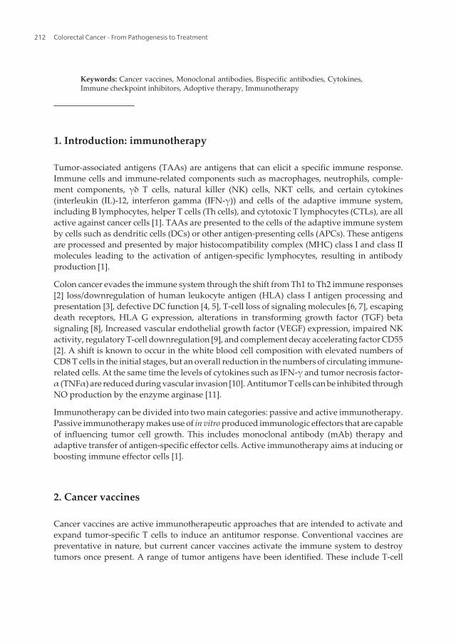
Keywords: Cancer vaccines, Monoclonal antibodies, Bispecific antibodies, Cytokines,Immune checkpoint inhibitors, Adoptive therapy, Immunotherapy
1. Introduction: immunotherapy
Tumor-associated antigens (TAAs) are antigens that can elicit a specific immune response.Immune cells and immune-related components such as macrophages, neutrophils, comple‐ment components, γδ T cells, natural killer (NK) cells, NKT cells, and certain cytokines(interleukin (IL)-12, interferon gamma (IFN-γ)) and cells of the adaptive immune system,including B lymphocytes, helper T cells (Th cells), and cytotoxic T lymphocytes (CTLs), are allactive against cancer cells [1]. TAAs are presented to the cells of the adaptive immune systemby cells such as dendritic cells (DCs) or other antigen-presenting cells (APCs). These antigensare processed and presented by major histocompatibility complex (MHC) class I and class IImolecules leading to the activation of antigen-specific lymphocytes, resulting in antibodyproduction [1].
Colon cancer evades the immune system through the shift from Th1 to Th2 immune responses[2] loss/downregulation of human leukocyte antigen (HLA) class I antigen processing andpresentation [3], defective DC function [4, 5], T-cell loss of signaling molecules [6, 7], escapingdeath receptors, HLA G expression, alterations in transforming growth factor (TGF) betasignaling [8], Increased vascular endothelial growth factor (VEGF) expression, impaired NKactivity, regulatory T-cell downregulation [9], and complement decay accelerating factor CD55[2]. A shift is known to occur in the white blood cell composition with elevated numbers ofCD8 T cells in the initial stages, but an overall reduction in the numbers of circulating immune-related cells. At the same time the levels of cytokines such as IFN-γ and tumor necrosis factor-α (TNFα) are reduced during vascular invasion [10]. Antitumor T cells can be inhibited throughNO production by the enzyme arginase [11].
Immunotherapy can be divided into two main categories: passive and active immunotherapy.Passive immunotherapy makes use of in vitro produced immunologic effectors that are capableof influencing tumor cell growth. This includes monoclonal antibody (mAb) therapy andadaptive transfer of antigen-specific effector cells. Active immunotherapy aims at inducing orboosting immune effector cells [1].
2. Cancer vaccines
Cancer vaccines are active immunotherapeutic approaches that are intended to activate andexpand tumor-specific T cells to induce an antitumor response. Conventional vaccines arepreventative in nature, but current cancer vaccines activate the immune system to destroytumors once present. A range of tumor antigens have been identified. These include T-cell
Colorectal Cancer - From Pathogenesis to Treatment212
epitope peptides, defined carbohydrates of glycoproteins and glycolipids, antibody-basedanti-idiotype vaccines, plasmid DNA and recombinant viral vector vaccines, allogeneic orautologous whole tumor cell vaccines, DC-based vaccines, oncolysates, or autologous heat-shock protein (HSP)-peptide complex vaccines. An ideal prophylactic cancer vaccine wouldbe affordable, stable, and safe. It would induce effective immunity rapidly and require fewimmunizations (ideally one) to induce protection [12]. This section aims to address advancesmade in developing vaccines against colon cancer. This will include vaccines that are currentlyin use and vaccines still undergoing clinical trials. It will report on the safety, side effects, andefficacy of these vaccines.
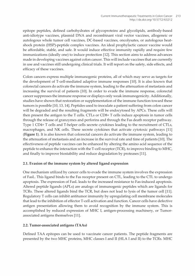
Colon cancers express multiple immunogenic proteins, all of which may serve as targets forthe development of T-cell-mediated adaptive immune responses [10]. It is also known thatcolorectal cancers do activate the immune system, leading to the attenuation of metastasis andincreasing the survival of patients [10]. In order to evade the immune response, colorectalcancer suppresses the immune response or displays only weak immunogenicity. Additionally,studies have shown that restoration or supplementation of the immune function toward thesetumors is possible [10, 13, 14]. Peptides used to inoculate a patient suffering from colon cancerwill be degraded and the resulting fragments will be endocytosed by APCs. These cells willthen present the antigen to the T cells. CTLs or CD8+ T cells induce apoptosis in tumor cellsthrough the release of granzymes and perforins and through the Fas death receptor pathway.Type 1 CD4+ T cells and T-helper cells secrete cytokines leading to the recruitment of CTLs,macrophages, and NK cells. These secrete cytokines that activate cytotoxic pathways [11](Figure 1). It is also known that colorectal cancers do activate the immune system, leading tothe attenuation of metastasis and an increase in the survival rate and time of patients [10]. Theeffectiveness of peptide vaccines can be enhanced by altering the amino acid sequence of thepeptide to enhance the interaction with the T-cell receptor (TCR), to improve binding to MHC,and finally to improve biostability and reduce degradation by proteases [11].
2.1. Evasion of the immune system by altered ligand expression
One mechanism utilized by cancer cells to evade the immune system involves the expressionof FasL. This ligand binds to the Fas receptor present on CTL, leading to the CTL to undergoapoptosis. The expression of FasL leads to the increased resistance to Fas-induced apoptosis.Altered peptide ligands (APLs) are analogs of immunogenic peptides which are ligands forTCRs. These altered ligands bind the TCR, but does not lead to lysis of the tumor cell [11].Regulatory T cells can inhibit antitumor immunity by upregulating cell membrane moleculesthat lead to the inhibition of effector T-cell activation and function. Cancer cells have defectiveantigen presentation allowing them to avoid recognition by the immune system. This isaccomplished by reduced expression of MHC I, antigen-processing machinery, or Tumor-associated antigens themselves [11].
2.2. Tumor-associated antigens (TAAs)
Defined TAA epitopes can be used to vaccinate cancer patients. The peptide fragments arepresented by the two MHC proteins, MHC classes I and II (HLA I and II) to the TCRs. MHC
Current Immunotherapeutic Treatments in Colon Cancerhttp://dx.doi.org/10.5772/63212
213
class I presents the vaccine-derived peptide to naive CTLs. Primed CTLs recognize the tumorantigen on the surface of the tumor and send out a death signal to the tumor. Helper T cellsare generated by MHC class II proteins [11] (Figure 1). However, there are not many specificpeptides that can be targeted as cancer specific. In order to increase uptake and presentationof the antigens by APCs, an adjuvant is added [1]. Another strategy is to inject the DNAsequences coding for specific TAAs to be taken up. The target ill be transcribed into mRNA,translated into a protein, and processed into peptides by APCs. This can be done by usingviruses engineered to express TAAs. However, the immune system may preferentially reactto the viral antigens rather than the TAAs, leading to the attenuation of the antitumor immuneresponse [1]. The earliest example of the therapeutic use of tumor antigens was in the form ofcrude tumor lysates being administered to patients. These lysates are still used as a means toprime DCs, facilitating peptide presentation [11]. This is because the ideal source of TAAs isall the TAAs the tumor itself expresses. By incubating DCs with dead tumor cell lysate, theseantigens will all be presented by MHC class I (cross-presentation) and MHC class II pathways.This will result in a diversified immune response involving CTLs as well as CD4+ T-helpercells [1].
The use of tumor lysates has largely been superseded by the use of synthetic peptides. Thesehave certain advantages over tumor lysates. They provide a higher amount of specific antigenand allow for modification of the target peptide. It is also easy to monitor the immune responseto vaccination with a single peptide as only one CTL type requires evaluation [11]. Tumor-specific antigens (TSAs) are mutated or virus-derived epitopes and contain unique immuno‐genic neo-antigens that can be recognized by the immune system. These include N-RAS andp53 [1].
Figure 1. Antitumor effect of peptide vaccine therapy: following introduction of peptide vaccine to the bloodstream, itis processed and presented by the APC leading to the activation of CD4+ helper T cells and CD8+ cytotoxic T cells.Interaction between MHC I molecules on APC and TCR during antigen presentation facilitated by CD8 molecule leadsto the generation of tumor-specific CTLs capable of lysing tumor cells.
Colorectal Cancer - From Pathogenesis to Treatment214
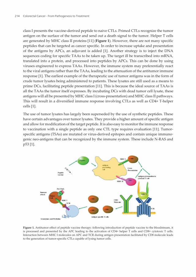
Peptides Mechanism Study details References
Tyrosine kinase receptorephrin type-A receptor 2(EphA2-derived peptide)
EphaA2 EphA2-specific CTL High level of immunity against colorectalcancer in murine model
[15]
RNF43-721 Phase 1 clinical trial [16]
ABT-737 Inhibition of antiapoptotic Bcl-2family
Sensitized cancer cells in mouse colon cancermodel
[17]
Epitopes of HER2, MVF,GMP, and n-MDP
Multiple targets Phase 1 clinical trial [18]
Endoglin Inhibition of angiogenesis Inhibition of tumor growth in mouse model [19]
CEA CEA691 Induction of tumor-specificCTLs
Increase in survival rate in colon carcinomamouse model
[20]
OX40L – TNF familyprotein
Inhibition of tumor growth in mouse model [21]
Mucin 1: MUC1 a cellsurface-associated protein
Stimulation of antigen-specific CTL,abundant secretion of IFN-γ.Tumor burden was significantly reduced incoloncancer mouse model
[22]
Heat-shock protein Gp96 Induction of tumor-specificCTLs
Two-year overall survival and disease-freesurvival were significantly improved
SART3-tumor-rejectionantigen
Induction of tumor-specificCTLs
Increased cellular immune responses to thetumor. No improved clinical outcome
[23, 24]
Lck-derived peptides Induction of tumor-specificCTLs
[24]
Survivin-2B Induction ofHLA-A24-restricted cytotoxic Tcells resulting inhigh toxicity against HLA-A24-positive survivin-2B-positivecancer in vitro
Increased proportion of peptide-specificCTL. No significantly improved clinicaloutcome
[25]
βHCG CTP37-DT(Avicine)
Phase II trials showed improved patientsurvival
[26]
CDX 1307 Fusion between βHCG and anantibody against the mannosereceptor
Phase I trial. Inoculation leads to DCactivation as well as cytotoxic T-cell activityagainst tumor cells
[27]
p53 (SLP) p53-specific CD4+ Th cell SLP isa p53 synthetic long peptide
Antitumor response against p53-overexpressing tumors. The p53-SLP vaccineinduces p53-specific T-cell responses
[10]
Current Immunotherapeutic Treatments in Colon Cancerhttp://dx.doi.org/10.5772/63212
215
Peptides Mechanism Study details References
EGFR2 gefitinib orerlotinib
EGFR mutations enhancetyrosine kinase activity inresponse to EGF, increasing theefficacy of anti-EGFR
In a phase I trial, the vaccine elicitedantibody responsephase II cancer
[28]
Gastrin: G17DT(gastroimmune)
Antigastrin-17 immunogen,raising antibodies that blockadegastrin-stimulated tumorgrowth
Phase II trials showed gastroimmunecombined with irinotecan chemotherapyincreased patient survival
[29]
Examples of peptide-based vaccine targets, their mechanism, as well as the current results of any trials performedusing the vaccines to treat colon cancer.
Table 1. Peptide targets and mechanism of action.
Discussed below are examples of peptide-based vaccines and their targets that have been usedto treat colon cancer. More examples are listed in Table 1. Beta human chorionic gonadotropin(βHCG) is not produced by normal colorectal cells. The increase in the expression of thisantigen in colon cancer cells leads to an increase in tumor invasiveness, higher metastaticincidence and promotion of tumor growth, neovascularization, and immune system suppres‐sion. This makes it an attractive target for the development of an antibody-based vaccine [10].Carcinoembryonic antigen (CEA) is an oncofetal antigen that can serve as a target for vaccinedevelopment. It is found overexpressed on the surface of colon cancer cells, with very lowlevels of expression on normal cells. Unfortunately this protein is normally expressed duringfetal development and is therefore tolerated by the immune system. This led to the creation ofan artificial CEA. CeaVac is based on anti-idiotypic antibodies and mimics CEA [10]. Anotheroncofetal protein 5T4 is a leucine-rich membrane glycoprotein. Once again it is nearly absentin normal tissues but is overexpressed in colon cancer cells and developing cells. Its presenceis associated with poor survival. The drug TroVax uses 5T4 with a pox virus vector and amodified vaccinia virus. Preclinical trials in mouse models resulted in a 90% reduction of tumorburden [10].
Onyvax-105 is another anti-idiotype antibody mimicking the glycosylphosphatidylinositol-anchored protein CD55. CD55 regulates complement activation protecting cells againstcomplement attack thereby enhancing tumor cell survival. The gastric acid–stimulatinghormone gastrin is a hormone the precursors of which are overexpressed in colon cancer,where they act as growth factors. This leads to increases in angiogenesis and cell proliferation.Vaccines raised against this protein would therefore result in inhibition of cell growth,proliferation, and metastasis [10]. Onartuzumab is a mAb that targets human growth factorreceptor (HGFR). It is a monovalent HGF antagonist antibody against MET Proto-Oncogene,Receptor Tyrosine Kinase that benefits patients who overexpress HGFR [30].
The FANG TM vaccine consists of tumor cells from the patient and a plasmid expressinggranulocyte-macrophage colony-stimulating factor (GM-CSF) and bifunctional short hairpinRNAfurin (bi-shRNAfurin). The growth and production of DCs are induced by GM-CSF. The
Colorectal Cancer - From Pathogenesis to Treatment216
enzyme furin transforms precursor proteins into active proteins and the presence of bi-shRNAifurin inhibits the production of active proteins. This particularly inhibits the production ofTGF β1 and 2 (TGFβ). Overexpression of TGFβ is associated with cancer progression andimmune suppression by inhibiting GM-CSF and the consequent production of dendritic andother APCs. The vaccine therefore prevents the overexpression of TGFβ and leads to immunecell activation and the inhibition of cancer cell proliferation [30]. The vaccine was manufac‐tured using GM-CSF and IL-13 to generate DCs from monocytes. The DCs were loaded with6HLA-A*0201-binding peptides derived, among others, from CEA, MAGE-2 (melanomaantigen overexpressed in gastrointestinal cancer), and HER2/neu [10, 30].
TroVax is an attenuated strain of vaccinia virus that encodes the 5T4 protein. This protein isan oncofetal antigen and is a transmembrane glycoprotein. It is highly expressed in coloncancers and is virtually absent in normal tissue. The receptor is thought to play a role inmetastasis and the expression level increases with the advancement of the stage of the cancer.This vaccine is able to induce an effective immune response, as it results in the formation ofantibodies for both the 5T4 antigen and the viral particle [31].
2.3. Heat-shock proteins (HSPs)
HSPs are widely expressed in tumors, where they promote cancer progression. HSP 72 andglucose-regulated protein 96 (gp96) are two of these proteins that are highly expressed in coloncancer [32]. These proteins are thought to play a role in cell growth and signal transductionand expression of these proteins is higher in tumors undergoing metastasis. This makes themuseful as diagnostic and prognostic markers. However, this expression is not related to patientsurvival [32].
HSPs enhance antigen-specific tumor immunity as they play an important role in the presen‐tation of antigens to CD8+ T cells through the MHC I pathway. This is because of the roles HSP70s play as chaperones and in the transport of peptides to the heterodimeric transportersassociated with antigen processing [33]. Similarly gp96 is a major chaperone involved in thelumen of the endoplasmic reticulum (ER), where it facilitates the folding of the MHC Iβ-2microglobulin-peptide complexes in the ER [34].
Vaccines based on HSPs have been tested in animal trials and been found to be highly effectivein the treatment of cancers [32, 35]. The function of the HSP in transporting and presentingother peptides as surface antigens has led many researchers to propose that HSPs can be usedto create a HSP target protein fusion. This booster strategy would therefore enhance the abilityof the target protein to be used as an antigen by T cells [36]. Two of these proteins that can becoupled to HSPs to improve their immunogenicity and usefulness as a cancer vaccine arealpha-fetoprotein (AFP) in hepatocellular carcinoma and CD44 in colonic carcinomas [32].
3. Targeted therapy: monoclonal antibodies
Recently, a new class of targeted agents have been identified, which bind to the ligand or theextracellular domain of a receptor. This results in alteration of intracellular signal transduction
Current Immunotherapeutic Treatments in Colon Cancerhttp://dx.doi.org/10.5772/63212
217
pathways which will affect cell proliferation, dedifferentiation, inhibition of apoptosis, andstimulation of neoangiogenesis [37]. This section will look into VEGF-targeted drugs (e.g.,ramucirumab (Cyramza®) and bevacizumab (Avastin®)), EGFR-targeted drugs (e.g., cetuxi‐mab (Erbitux®) and panitumumab (Vectibix®)), and others such as those that target kinases[37].
3.1. Bevacizumab and ramucirumab
VEGF is a potent angiogenic factor and functions by binding to one of three VEGF receptorslocated on endothelial cells and angioblasts. The VEGF receptor-2 is overexpressed on up to50% of colorectal cancer cell surfaces. VEGF-A and other proangiogenic factors promote thedegradation of the extracellular matrix. This enables proliferation and migration of endothelialcells [37]. The ligands of the VEGF family include VEGF-A, VEGF-B, VEGF-C, VEGF-D, andVEGF-E; and the receptors are VEGFR-1, R-2, and R-3. In colon cancer the ligand that is mostabundant is VEGF-A [38]. Sustained angiogenesis is a hallmark of cancer; and targetedinhibition of blood vessel development is an established strategy for antitumor therapy [38].Anti-VEGF therapies have been associated with a survival benefit across multiple malignan‐cies including colon cancer [38].
Bevacizumab is a humanized mAb against VEGF and it acts by preventing ligand binding bybinding to VEGF. This prevents downstream intracellular signal transduction; however, theresponse to bevacizumab appears to be independent of VEGF expression or high microvesseldensity (MVD) [37]. MVD assessment is a good predictor of metastasis, with selective anti‐bodies, such as endoglin, distinguishing between tumor neovascularization and preexistingvessels. VEGF expression is highest in patients with metastatic tumors and the level isassociated with cancer stage [38]. Bevacizumab is typically used in combination with otherchemotherapeutic agents, and it is also indicated in improving the delivery of chemotherapyby changing tumor vasculature and decreasing the elevated interstitial pressure in tumors.The combination of therapies results in improved survival [38].
Ramucirumab is a fully humanized IgG1 mAb targeting the extracellular domain of VEGFreceptor 2 (VEGFR2). Large-scale trials have indicated that ramucirumab shows promisingantitumor effects and is well tolerated. The origin of this antibody was through the use of alarge phage display library with tailored in vitro selection methods to identify a high-affinityantibody [39]. Measurement of VEGFA and soluble VEGFR1/2 during phase I trials of theantibody indicated that there is an increase in the expression of VEGF as well as a decrease inVEGFR1/2 levels. These changes were not dose related, which suggests that the receptor wassaturated [39]. Phase I trial results were promising and phase II trials resulted in a highpercentage of patients presenting with progression free survival at 6 months. Phase III trialsshowed an increase in overall patient survival [39]. Adverse reactions to ramucirumabincluded hypertension, vascular thrombotic events, and proteinuria [40].
Aflibercept is a recombinant fusion protein consisting of the second immunoglobulin (Ig)domain of VEGFR-1 and the third Ig domain of VEGFR-2, fused to human IgG1. It exhibitsaffinity for VEGF-A, VEGF-B, and PlGF. The antibody displayed effective activity against coloncancers with improvements in the primary endpoint of overall survival and overall response
Colorectal Cancer - From Pathogenesis to Treatment218
rate, as well as displaying a high degree of tolerability in patients [40]. VEGFR-1 also plays arole in colon cancer and inhibiting its signaling could also play a role in cancer treatment. Anantibody developed to target this receptor named IMC-18F1 has been developed. This is ahigh-affinity human VEGFR-1-neutralizing antibody that specifically binds the extracellulardomain of VEGFR-1. It exhibits antiangiogenic and antiproliferative activity [40].
3.2. Panitumumab and cetuximab
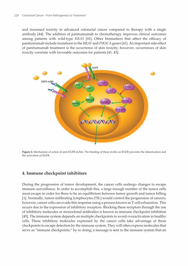
The epidermal growth factor receptor (EGFR) is a target for the therapeutic monoclonalantibodies panitumumab and cetuximab the treatment of metastatic colorectal cancer.Panitumumab is a fully human Ig G2 mAb that binds the EGFR extracellular domain with highaffinity and inhibits ligand-induced EGFR tyrosine phosphorylation, tumor cell activation,and tumor cell proliferation (Figure 2). Cetuximab is a chimeric human-mouse IgG1 mAb [41].Cetuximab and panitumumab are both Food and Drug Administration (FDA) approved foradvanced colorectal cancer therapy, and both have clear benefits for colon cancer treatment ofmost patients. The exception is those patients that carry KRAS mutations at codons 12 and 13[42]. KRAS mutations occur in approximately 35–40% of colorectal tumors, and KRAS is amember of the rat sarcoma virus (Ras) gene family of oncogenes and is involved in integratingthe signaling cascades controlling gene transcription, including many EGFR-mediatedpathways [41]. The ligands of the EGFR transmembrane tyrosine kinase receptor include EGF,TGFα, epiregulin, amphiregulin, β-cellulin, and heparin. EGFR activates downstream signal‐ing pathways such as the Ras/Raf/mitogen-activated protein kinase (MAPK) pathway, thephosphatidylinositol 3-kinase (PI3K)/AKT pathway, and the signal transducer and activatorof transcription (STAT) pathway. These downstream pathways activate cellular survival,proliferation, invasion, metastasis, and angiogenesis. Abnormal activation of the EGFRsignaling network due to excessive overexpression is common in colon cancer (Figure 2). EGFRis composed of an extracellular ligand-binding domain, a hydrophobic transmembrane region,and an intracellular domain with tyrosine kinase activity [41, 43].
Cetuximab competes with EGFR ligands, such as EGF or TGFα, with a high affinity(Kd = 1 × 10⁻¹⁰ M). This results in the inhibition of cell cycle progression and arrest of cell cyclein G1 phase, inhibition of angiogenesis, inhibition of metastasis by reduction of production ofmatrix metalloproteinase, inhibition of apoptosis, and potentiation of antitumor activities ofchemotherapy and radiotherapy [43]. Panitumumab treatment results in improved clinicaloutcomes in patients with chemotherapy-refractory colon cancer [41]. Panitumumab also hasa high affinity for EGFR (Kd = 5 × 10⁻¹¹ M) and acts to arrest cell cycle progression and blockcancer growth; however, it is an IgG2 and does not act through antibody-dependent cellcytotoxicity [43].
Using a single type of mAb to block a single transduction pathway may only have a limitedeffect, as tumors can shift to other alternate pathways. One solution is to combine monoclonalantibodies to block two signaling transduction pathways [44]. Use of multiple monoclonalantibodies has other advantages including limited overlapping toxicity and few to no phar‐macokinetic interactions between antibodies. However, some studies indicate that certaincombinations such as bevacizumab with cetuximab or panitumumab lead to shorter survival
Current Immunotherapeutic Treatments in Colon Cancerhttp://dx.doi.org/10.5772/63212
219
and increased toxicity in advanced colorectal cancer compared to therapy with a singleantibody [44]. The addition of panitumumab to chemotherapy improves clinical outcomesamong patients with wild-type KRAS [41]. Other biomarkers that affect the efficacy ofpanitumumab include mutations in the BRAF and PIK3CA genes [41]. An important side effectof panitumumab treatment is the occurrence of skin toxicity; however, occurrences of skintoxicity correlate with favorable outcomes for patients [41, 43].
Figure 2. Mechanism of action of anti-EGFR mAbs. The binding of these mAbs on EGFR prevents the dimerization andthe activation of EGFR.
4. Immune checkpoint inhibitors
During the progression of tumor development, the cancer cells undergo changes to escapeimmune surveillance. In order to accomplish this, a large enough number of the tumor cellsmust escape in order for there to be an equilibrium between tumor growth and tumor killing[1]. Normally, tumor-infiltrating lymphocytes (TIL) would control the progression of cancers;however, cancer cells can evade this response using a process known as T-cell exhaustion. Thisoccurs due to the expression of inhibitory receptors. Blocking these receptors through the useof inhibitory molecules or monoclonal antibodies is known as immune checkpoint inhibition[45]. The immune system depends on multiple checkpoints to avoid overactivation in healthycells. These inhibitory molecules expressed by the cancer cells take advantage of thesecheckpoints to escape detection by the immune system. They will often express molecules thatserve as “immune checkpoints,” by so doing; a message is sent to the immune system that an
Colorectal Cancer - From Pathogenesis to Treatment220
immune response is not necessary. Drugs are being developed to block immune checkpointmolecules from binding to their molecular partners, thus allowing the body to elicit an immuneresponse and therefore attack cancer cells. An analysis of the expression patterns in coloncancers revealed a large overexpression of immune checkpoint-related proteins [46].
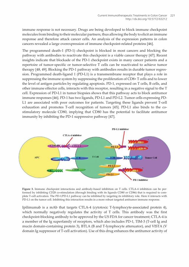
The programmed death-1 (PD-1) checkpoint is blocked in most cancers and blocking thepathway with antibodies to reactivate this checkpoint is a viable cancer therapy [47]. Recentinsights indicate that blockade of the PD-1 checkpoint exists in many cancer patients and arepertoire of tumor-specific or tumor-selective T cells can be reactivated to achieve tumortherapy [48, 49]. Blocking the PD-1 pathway with antibodies results in durable tumor regres‐sion. Programmed death-ligand 1 (PD-L1) is a transmembrane receptor that plays a role insuppressing the immune system by suppressing the proliferation of CD8+ T cells and to lowerthe level of antigen particles by regulating apoptosis. PD-1, expressed on T cells, B cells, andother immune effector cells, interacts with this receptor, resulting in a negative signal to the Tcell. Expression of PD-L1 in tumor biopsies shows that this pathway acts to block antitumorimmune responses [46]. PD-1 has two ligands, PD-L1 and PD-L2. Tumor cells expressing PD-L1 are associated with poor outcomes for patients. Targeting these ligands prevent T-cellexhaustion and promotes T-cell recognition of tumors [45]. PD-L1 also binds to the co-stimulatory molecule CD80, implying that CD80 has the potential to facilitate antitumorimmunity by inhibiting the PD-1 suppressive pathway [47].
Figure 3. Immune checkpoint interactions and antibody-based inhibition on T cells. CTLA-4 inhibition can be per‐formed by inhibiting CD28 co-stimulation (through binding with its ligands CD80 or CD86) that is required to com‐plete T-cell activation. The PD-1/PD-L1 pathway can be inhibited by targeting its inhibitory role. Here it interacts withPD-L1 on the tumor cell. Inhibiting this interaction results in a more robust targeted antitumor immune response.
Ipilimumab is a mAb that targets CTLA-4 (cytotoxic T-lymphocyte-associated protein 4),which normally negatively regulates the activity of T cells. This antibody was the firstcheckpoint-blocking antibody to be approved by the US FDA for cancer treatment; CTLA-4 isa member of the Ig superfamily of receptors, which also includes PD-1, TIM-3 (T-cell Ig andmucin domain-containing protein 3), BTLA (B and T-lymphocyte attenuator), and VISTA (Vdomain Ig suppressor of T-cell activation). Use of this drug enhances the antitumor activity of
Current Immunotherapeutic Treatments in Colon Cancerhttp://dx.doi.org/10.5772/63212
221
CD8 T cells and inhibits the suppressive function of Tregs [45]. This was followed by a secondantibody, pembrolizumab, which targets the programmed death 1 (PD-1; CD279) molecule[50] (Figure 3). Trials of these antibody therapies have only shown modest clinical benefits,and this may indicate that tumors use multiple and nonoverlapping immunosuppressivemechanisms to evade the immune response [45]. Multiple studies indicate that effectivetherapy involves the targeting of multiple immunosuppressive pathways [51].
Tumor cells can also evade the immune system through the production of extracellularadenosine by CD73 which is expressed on lymphocytes and endothelial and epithelial cells.CD73 performs an endothelial cell barrier function, protecting cells from ischemia andregulating immune responses. This receptor is overexpressed in many types of cancer, withhigh CD73 expression being associated with poor outcomes for patients due to increases intumor immune escape and metastasis [45]. Blocking CD73 can induce potent antitumorimmune responses. Additionally the inhibition of molecular pathway components upstreamof CD73 such as CD39 also has similar therapeutic effects. This treatment can also be used tosupplement immune checkpoint therapies that make use of anti-CTLA-4 and anti-PD-1 mAbs,to increase the effectiveness of such therapies [45].
Another strategy to target immune checkpoints is the use of small molecule drugs that targetcritical survival pathways. These include Gleevec and ibrutinib, both of which are tyrosinekinase inhibitors. Ibrutinib is a covalent inhibitor of BTK (Bruton’s tyrosine kinase), a keyenzyme in B-cell receptor signaling [49].
Controlling the immune response to cancer cells through the use of anti-inflammatory drugsto control the inflammatory components reduces the risk of developing certain types of cancer.Aspirin is able to reduce the incidence of colon cancer and slow down tumor progression.Cyclooxygenase (Cox) enzymes 1 and 2 are the targets of aspirin. These enzymes are overex‐pressed in tumor cells. Immunosuppressive drugs such as cyclosporine A (CsA) and tacroli‐mus (FK506) inhibit the calcium/calmodulin-dependent phosphatase calcineurin, which actsupon members of the nuclear factor of activated T cells (NFAT) [52]. These transcription factorsare important for cytokine production by T cells and are required for the normal function ofB cells, DCs, and mast cells. Expression of NFAT family members leads to tumor suppression[53]. Treatment of tumor cells with CsA is capable of inducing necroptosis and a mild G0/G1cell cycle arrest [52].
5. Cytokines
Cytokines are signaling proteins produced by white blood cells that help control the growthand activity of immune system cells. The two types of cytokines that are used in the treatmentof cancer are IFNs and ILs. Cytokines stimulate a broad-based immune response as opposedto generating a targeted response to a specific antigen [54]. Tumors secrete factors to recruitinflammatory cells and/or activate stromal cells. Inflammation plays a major role in tumorpromotion and progression. The soluble factors that drive inflammation are cytokines and
Colorectal Cancer - From Pathogenesis to Treatment222
chemokines produced by tumor cells themselves and by the cells recruited to the tumormicroenvironment [55].
Several cytokines are capable of activating and recruiting specific immune cells that canenhance antitumor immunity; these include IL-2, IL-12, IL-15, TNFα, and GM-CSF. Thesecytokines can be used as single-agent therapies or in combination with other immunothera‐peutic strategies. GM-CSF immunization leads to APC recruitment. Tumors activate Stat3 andBraf, which leads to the release of IL-10, inhibiting the tumoricidal activity of NK cells. Stat3activation in DCs leads to these cells becoming tolerogenic DC [11].
Additionally, TNFα, hepatocyte growth factor, PDGF, and FGF19 activate Wnt/β-cateninsignaling in tumor cells. This is the oncogenic pathway activated in the majority of coloncancers. This pathway results in β-catenin accumulation in the cytoplasm, which activates cellgrowth and differentiation pathways. IL-1β is a potent activator of Wnt signaling in coloncancer cells leading to increased survival of colon cancer cells [55].
Oncogenic signaling through the Wnt and NF-kB pathways is activated through TNFα.Pharmacological inhibition of TNFα by neutralizing TNFα antibodies has been used to treatboth irritable bowel disorders and colon cancer. Results from trials using enbrel or remicadesuggest that these neutralizing antibodies have activity against colon cancer cells. TNFαsignaling initiates NF-kB signaling. NF-kB is continuously expressed in certain tumors, leadingto enhanced survival by protecting the tumor cells from apoptosis. Treatment with theTNFα antagonist, etanercept led to inhibition of Wnt/β-catenin6 signaling as seen by the ,reduced expression of active β-catenin [55].
5.1. Interleukins 1β, IL-6, and IL-1
The proinflammatory cytokine IL-1β is produced by activated macrophages. In turn, IL-1βinduces the expression of TNFα, IL-6, IL-8, IL-17, Cox-2, and PGE2, promoters of tumor cellgrowth. Inducing the expression of IL-1β leads to increased incidence of cancer in wild-typemice. The IL-1β signaling pathway functions through the receptors IL-1RI and IL-1RII toinduce NF-kB activity. The pathway involves the two adaptor proteins, MyD88 and IRAK.Macrophages are stimulated to release IL-1β and activate NF-kB and Wnt pathways, butIL-1β signaling requires STAT1. The silencing of STAT1 expression leads to decreased IL-1βrelease and prevented cancer cell growth [55].
IL-6 is secreted by stimulated monocytes, fibroblasts, and endothelial cells, macrophages, Tcells, and B lymphocytes. Macrophages are stimulated by colon cancer cells to produce IL-6and activate STAT3 in tumor cells. Inhibition of IL-6 signaling interferes with the growth oftumor cells and protects them from apoptosis. Research indicates that decreasing the expres‐sion or inhibiting the activity of STAT3 may have adverse effects on tumor promotion.Targeting STAT3 will affect the expression of β-catenin and the co-expression of STAT3 andβ-catenin is associated with poor survival of colon cancer patients [55].
A subset of T-helper cells produces the cytokines IL-17, IL-22, and TNFα (Th17 cells). Panethcells also produce IL-17. Th17 cells require IL-6, TGFβ, IL-1β, and IL-23, while IFN-γ and IL-4negatively regulate differentiation of Th17 cells. IL-17 induces IL-6 and STAT3, promoting the
Current Immunotherapeutic Treatments in Colon Cancerhttp://dx.doi.org/10.5772/63212
223
survival of cancer cells. This cytokine may also have an anticancer function by enhancingantitumor immunity [55].
5.2. Tumor necrosis factor-related apoptosis-inducing ligand (TRAIL)
TRAIL (also known as Apo2L) activates the apoptotic cascade. Tumor cells can evade thisapoptosis signal through the action of β-catenin. TRAIL’s role in tumor surveillance has beenconfirmed in knockdown experiments and is a promising candidate to be used in cancertherapy, because it selectively kills cancer cells while leaving normal cells unharmed [55].
Sorafenib a Raf kinase inhibitor sensitized A TRAIL –resistant colon cancer line to TRAIL-induced apoptosis by preventing NF-kB-dependent expression of the antiapoptotic genes,IAP2 and MCl-1.
6. Oncolytic virus (OV) therapy
Over a century ago, researchers observed that viral infection both in human and animal modelsresults in the expression of targets that can be recognized by T cells and/or antibodies.Subsequently, vaccination has been used to treat an array of diseases such as hepatitis B virusand human papillomavirus 15 which can cause liver and cervical cancer respectively. Vacci‐nation against infections is used to induce neutralizing antibodies that act prophylactically.However, with regard to cancer vaccination, cancer vaccine candidates should induce andexpand immune responses that can cause disruption of biological pathways that supportcancer growth.
The concept of cancer immunotherapy is based on the ability of the immune system torecognize cancer cells and affect their growth and replication. Researchers have observed thatcancer regression would occur spontaneously in patients after viral infection [56, 57]. Forexample, studies conducted by Lindeman and Klein 1967 showed that oncolysis of tumor cellsby influenza virus increased immunogenicity of tumor cell antigens. The recent advances insuccessful sequencing of the cancer genome together with insights into how tumors evade theimmune system have led cancer research to evolve from searching for a gene that causesindividual cancer to one that blocks or disrupts biological pathways that support cancergrowth [58, 59]. As a result, cancer vaccines are now being designed with the aim to boost theimmune system to protect itself from carcinogenesis and progression of cancer. In 2010, theFDA approved Provenge which is a therapeutic vaccine for cancer [60]. It is designed to treatadvances in prostate cancer and has shown to increase the survival rate. The success ofProvenge resulted in stimulating the interest in the development of other therapeutic cancervaccines.
In recent years, OV has been shown to be effective in treating cancer in both preclinical modelsand clinical trials. Toda and coworkers showed that genetically modified oncolytic HSV G207is a potential cancer vaccine for induction of specific antitumor immunity in CT26 colon cancercells [64, 61, 62]. This type of immunotherapy is largely dependent on the network of the host
Colorectal Cancer - From Pathogenesis to Treatment224
immune system to fight cancer by (i) boosting the patient’s immune system, (ii) decreasingcancer-induced immunosuppression, and (iii) increasing the immunogenicity of the tumoritself [63, 64]; OVs can be RNA- or DNA-based virus derived from human or animals.
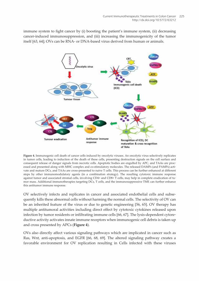
Figure 4. Immunogenic cell death of cancer cells induced by oncolytic viruses. An oncolytic virus selectively replicatesin tumor cells, leading to induction of the death of these cells, presenting destruction signals on the cell surface andconsequent release of danger signals from necrotic cells. Apoptotic bodies are engulfed by APC, and TAAs are proc‐essed and presented along with MHC complex and co-stimulatory molecules. The released DAMPs (and PAMPs) acti‐vate and mature DCs, and TAAs are cross-presented to naive T cells. This process can be further enhanced at differentsteps by other immunomodulatory agents (in a combination strategy). The resulting cytotoxic immune responseagainst tumor and associated stromal cells, involving CD4+ and CD8+ T cells, may help in complete eradication of tu‐mor mass. Additional immunotherapies targeting DCs, T cells, and the immunosuppressive TME can further enhancethis antitumor immune response.
OV selectively infects and replicates in cancer and associated endothelial cells and subse‐quently kills these abnormal cells without harming the normal cells. The selectivity of OV canbe an inherited feature of the virus or due to genetic engineering [56, 65]. OV therapy hasmultiple antitumoral activities including direct effect by cytotoxic cytokines released uponinfection by tumor residents or infiltrating immune cells [66, 67]. The lysis-dependent cytore‐ductive activity activates innate immune receptors when immunogenic cell debris is taken upand cross presented by APCs (Figure 4).
OVs also directly affect various signaling pathways which are implicated in cancer such asRas, Wnt, anti-apoptosis, and EGFR [66, 68, 69]. The altered signaling pathway creates afavorable environment for OV replication resulting in Cells infected with these viruses
Current Immunotherapeutic Treatments in Colon Cancerhttp://dx.doi.org/10.5772/63212
225
showing sustained proliferation, resisting cell death, evading growth suppressors andescaping immune surveillance. Cancer cells also show increased genomic instability and DNAdamage stress, which is favorable to OV replication [70–72]. Genetic manipulation of OVenables these viruses to be (i) safe for use as a vaccine, (ii) highly selective for specific cancertype, and (iii) altering virus tropism. In comparison with current regimes for cancer treatment,OVs are advantageous because (i) they have a low chance for generation of resistance becausethey use multiple ways to exert cytotoxicity and (ii) virus dose in a tumor increases with timedue to in situ virus replication whereas in the classical drug Pharmacokinetics, dose decreaseswith time [71, 73].
The major drawbacks in the use of OV include nonimmune human serum, development ofanti-OV antibodies resulting from the use of human virus, and appropriate delivery into thetumor. Various delivery mechanisms have been explored to enable delivery of OV to tumorcells. For an example, cell carriers such as neural stem cells and myeloid-derived suppressorcells have been used to deliver OV to specific tumor cells. The cells protect the virus from anti-OV antibody neutralization, thereby facilitating virus deliver [71, 73]. In using OV to treatcolon cancer, ONYX-015 has advanced to phase II clinical trials and is used in combinationwith chemotherapy [74, 75]. Recently, adenovirus 5 (PSE-EA1 and E deleted) has beenapproved to treat prostate cancer in China giving hope to development of OV as an alternativecancer treatment.
7. Bispecific antibody
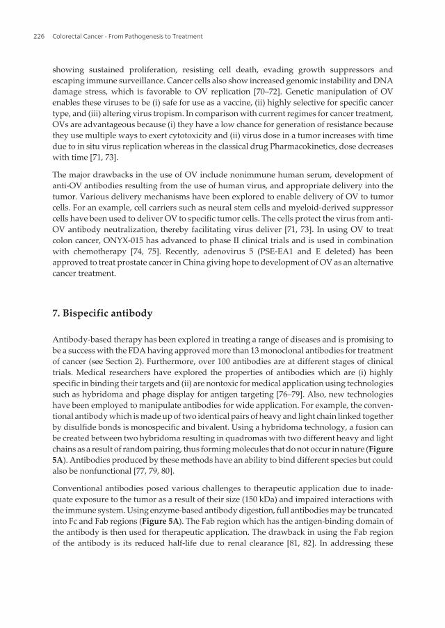
Antibody-based therapy has been explored in treating a range of diseases and is promising tobe a success with the FDA having approved more than 13 monoclonal antibodies for treatmentof cancer (see Section 2). Furthermore, over 100 antibodies are at different stages of clinicaltrials. Medical researchers have explored the properties of antibodies which are (i) highlyspecific in binding their targets and (ii) are nontoxic for medical application using technologiessuch as hybridoma and phage display for antigen targeting [76–79]. Also, new technologieshave been employed to manipulate antibodies for wide application. For example, the conven‐tional antibody which is made up of two identical pairs of heavy and light chain linked togetherby disulfide bonds is monospecific and bivalent. Using a hybridoma technology, a fusion canbe created between two hybridoma resulting in quadromas with two different heavy and lightchains as a result of random pairing, thus forming molecules that do not occur in nature (Figure5A). Antibodies produced by these methods have an ability to bind different species but couldalso be nonfunctional [77, 79, 80].
Conventional antibodies posed various challenges to therapeutic application due to inade‐quate exposure to the tumor as a result of their size (150 kDa) and impaired interactions withthe immune system. Using enzyme-based antibody digestion, full antibodies may be truncatedinto Fc and Fab regions (Figure 5A). The Fab region which has the antigen-binding domain ofthe antibody is then used for therapeutic application. The drawback in using the Fab regionof the antibody is its reduced half-life due to renal clearance [81, 82]. In addressing these
Colorectal Cancer - From Pathogenesis to Treatment226
challenges, bispecific antibodies (BsAb) were developed in 1961 [83]. Just as their name implies,this class of antibodies binds two different antigens or two different epitopes on the sameregion.
Figure 5. (A) Different forms of bispecific antibody. Trifunctional antibodies consist of two heavy and two light chainsfrom two different antibodies. This results in an antibody with binding sites for two different antigens as well as an Fcregion made up of two heavy chains forming a third binding site. A diabody consists of scFvs with very short linkerpeptides that force the closely positioned variable regions to fold together, forcing the scFvs to dimerize. Chemicallycoupled Fabs consist of the antigen-binding regions of two different monoclonal antibodies linked by a chemicalmeans. Bispecific T-cell antigens are fusion proteins of two scFvs from four separate genes. (B) Development of bispe‐cific compounds. Bispecific compounds enable simultaneous inhibition of two cell surface receptors, simultaneousblocking of two ligands, cross-linking of two receptors, and/or the recruitment of T cells to the proximity of tumor cells(redirected immune cell killing).
BsAb represent a class of antibodies that are yet to be fully explored in the treatment of cancerand other diseases. BsAb have a greater potential therapeutic efficiency than mono-targetedtherapy, since they allow simultaneous engagements of two targets and limit potential escapepathways [84–86]. Numerous studies have shown that there is evidence of cross talk betweenreceptor tyrosine kinases such as MET, VEGFR, and IGFR-IR which are known to promotecancer progression and drug resistance. And patients with colon cancer are known not torespond to anti-EGFR drugs with resistance emerging after initial usage [87, 88]. Engelman etal. showed that MET amplification leads to gefitinib resistance by activating the ERBB3pathway, showing the complexity of tumor signaling pathways and a need to treat patientswith drugs that target multiple targets [89].
In the use of BsAb, T cells are targeted because of their high cytotoxic retention, abundance inbloodstream, surveillance function, and proven ability to control malignant diseases [90, 91].During cancer progression, cancer cells escape immune recognition by interfering with antigenpresentation or T-cell activation or differentiation. In using the bispecific antibody, most
Current Immunotherapeutic Treatments in Colon Cancerhttp://dx.doi.org/10.5772/63212
227

targeted antigens for tumor therapy are differentiation antigens such as CD19, CD33, CEA,EpCAM Epithelial cell adhesion molecule, PMSA Prostate-specific membrane antigen , andEGF receptors. In most cases these antigens are overexpressed in cancer cells compared to thenormal cells.
Blinatumomab is an example of a bispecific antibody that has shown great promise clinicallyin cancer patients. Blinatumomab is a 55 kDa-fusion protein comprised of two single-chainantibodies to CD19 and CD3, recombinantly joined by a flexible, non-glycosylated five-aminoacid non-immunogenic linker that affords a very short distance between arms [92, 93].Blinatumomab has high affinity for CD19 which is important in sustaining the malignant B-cell phenotype via mechanisms of proliferation, cell survival, and self-renewal [94, 95]. It drawsmalignant B cells in close proximity to CD3-positive T cells without regard to TCR specificityor reliance on MHC class I molecules on the surface of APCs for activation. The nonspecificbinding of the polyclonal T-cell population prevents resistances to T-cell-based therapies as aresult of downregulation of MHC molecules. CD19 and CD3 binding results in T-cell activa‐tion, marked by upregulation of T-cell activation markers CD25, CD69, CD2, IFN-γ, TNFα,and IL-2, IL-6, and IL-10 [96]. Cell lysis is mediated by secretion of perforin and variousgranzymes stored in the secretory vesicles of cytotoxic T cells [97]. In vitro data suggest thatefficacy of blinatumomab is not compromised or dependent upon T cells, which may be limitedin number in heavily pretreated patients [98]. Also blinatumomab-activated T cells appear toeffectively induce serial target cell killing [92, 93].
8. Adoptive immunotherapy
In the wake of cancer treatment challenges or therapies, adoptive immunotherapy is one ofthe novel strategies being researched for cancer treatment. This concept was presented fivedecades ago [99–101] and is based on the transfer of ex vivo expanded antitumor CD8 T cellsinto affected patients (Figure 6). Delorme and Alexander [101] showed that the transfer ofimmune lymphocytes could inhibit the growth rate of carcinogen-induced sarcoma.
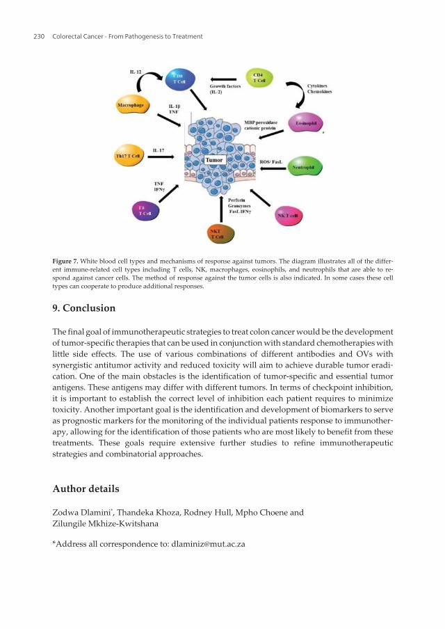
The immune system is responsible for the prevention of tumors or elimination of pathogensthat can cause inflammation or an inflammatory environment for tumorigenesis or destroytumor cells expressing TSAs or molecules induced by stress [102, 103]. Therefore, tumordevelopment and progression are largely dependent on the patient’s immune system toeffectively inhibit cancer growth using its network of immune cell types. Each and every celltype has a specific function in inhibiting tumor growth (Figure 7). Consequently, the successof adoptive immunotheraphy depends on approaches which will target different immunesubsets.
Adoptive immunotherapies have explored the use of infiltrating T cells (CD8+ effector T cellsand CD8+ effector memory cells), NK cells, and IL-2 for cancer-targeted therapies. The T cellsare able to destroy tumor cells using cytotoxic granules containing perforin and granzymesand by using cell surface receptor such as TNF-related apoptosis-inducing ligand [104, 105].Studies using mice have shown that adoptive transfer of T cells successfully induces antitumor
Colorectal Cancer - From Pathogenesis to Treatment228
response. Also, only a small number is required to mediate effective regression of tumor andsurvival [106–108]. Genetic modification of T cells has been successfully used to broaden theireffective application by pairing with antigen receptors that recognize a range of differentTAAs. Genetic engineering has also been employed to alter T cells so that they are able to avoidor be resistant to immune invasion strategies used by tumors such as the production ofcytokines. Another modification of T cells involves attaching stimulatory signals for theiractivation [109, 110].
NK cells target and kill diseased cells using various mechanisms such as perforins andgranzyme. The use of NK cells was first explored by Rosenberg et al. [111]. Lymphokine-activated killer cells were co-administered with IL-2 and resulted in a positive response inpeople with metastatic cancer [111]. Another combination of chemotherapy with transfer ofallogeneic NK cells resulted in disease remission [112]. It is anticipated that a hybrid of T andNK cell will have great potential in the treatment of cancer using adoptive immunotherapy.However we are still a long way from the development of such a model treatment that can bedeveloped for clinical trials and introduced into clinical practice.
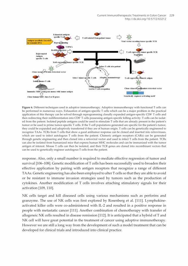
Figure 6. Different techniques used in adoptive immunotherapy. Adoptive immunotherapy with functional T cells canbe performed in numerous ways. Exhaustion of antigen-specific T cells which can be a major problem in the practicalapplication of this therapy can be solved through reprogramming clonally expanded antigen-specific CD8+ T cells andthen redirecting their redifferentiation into CD8+ T cells possessing antigen-specific killing activity. T cells can be isolat‐ed from the patient. Isolated peptide antigens could be used to stimulate T cells that are already present in the patient'stumor or be used to prime tumor-specific T cells. If the T-cell populations generated are specific for the patient's tumor,they could be expanded and adoptively transferred if they are of human origin. T cells can be genetically engineered torecognize TAAs. TCRs from T cells that show a good antitumor response can be cloned and inserted into retroviruses,which are used to infect autologous T cells from the patient. Chimeric antigen receptors (CARs) can be generatedthrough genetic engineering and then cloned into a retroviral vector and used to infect T cells from the patient. TCRscan also be isolated from humanized mice that express human MHC molecules and can be immunized with the tumorantigen of interest. Mouse T cells can then be isolated, and their TCR genes are cloned into recombinant vectors thatcan be used to genetically engineer autologous T cells from the patient.
Current Immunotherapeutic Treatments in Colon Cancerhttp://dx.doi.org/10.5772/63212
229
9. Conclusion
The final goal of immunotherapeutic strategies to treat colon cancer would be the developmentof tumor-specific therapies that can be used in conjunction with standard chemotherapies withlittle side effects. The use of various combinations of different antibodies and OVs withsynergistic antitumor activity and reduced toxicity will aim to achieve durable tumor eradi‐cation. One of the main obstacles is the identification of tumor-specific and essential tumorantigens. These antigens may differ with different tumors. In terms of checkpoint inhibition,it is important to establish the correct level of inhibition each patient requires to minimizetoxicity. Another important goal is the identification and development of biomarkers to serveas prognostic markers for the monitoring of the individual patients response to immunother‐apy, allowing for the identification of those patients who are most likely to benefit from thesetreatments. These goals require extensive further studies to refine immunotherapeuticstrategies and combinatorial approaches.
Author details
Zodwa Dlamini*, Thandeka Khoza, Rodney Hull, Mpho Choene andZilungile Mkhize-Kwitshana
*Address all correspondence to: [email protected]
Figure 7. White blood cell types and mechanisms of response against tumors. The diagram illustrates all of the differ‐ent immune-related cell types including T cells, NK, macrophages, eosinophils, and neutrophils that are able to re‐spond against cancer cells. The method of response against the tumor cells is also indicated. In some cases these celltypes can cooperate to produce additional responses.
Colorectal Cancer - From Pathogenesis to Treatment230

Research, Innovation & Engagements Portfolio, Mangosuthu University of Technology,Durban, South Africa
References
[1] Cornelissen R, Heuvers ME, Maat AP, Hendriks RW, Hoogsteden HC, Aerts JG,Hegmans JP: New roads open up for implementing immunotherapy in mesothelioma.Clinical and Developmental Immunology 2012, 2012:927240.
[2] Evans C, Dalgleish AG, Kumar D: Review article: immune suppression and colorectalcancer. Alimentary Pharmacology & Therapeutics 2006, 24(8):1163–1177.
[3] Aptsiauri N, Cabrera T, Mendez R, Garcia-Lora A, Ruiz-Cabello F, Garrido F: Role ofaltered expression of HLA class I molecules in cancer progression. Advances in Experi‐mental Medicine and Biology 2007, 601:123–131.
[4] Ma Y, Shurin GV, Peiyuan Z, Shurin MR: Dendritic cells in the cancer microenviron‐ment. Journal of Cancer 2013, 4(1):36–44.
[5] Ma YJ, He M, Han JA, Yang L, Ji XY: A clinical study of HBsAg-activated dendritic cellsand cytokine-induced killer cells during the treatment for chronic hepatitis B. Scandi‐navian Journal of Immunology 2013, 78(4):387–393.
[6] Roy S, Majumdar APN: Signaling in colon cancer stem cells. Journal of MolecularSignaling 2012, 7:11.
[7] Maccalli C, Pisarra P, Vegetti C, Sensi M, Parmiani G, Anichini A: Differential loss ofT cell signaling molecules in metastatic melanoma patients' T lymphocyte subsetsexpressing distinct TCR variable regions. Journal of Immunology 1999, 163(12):6912–6923.
[8] Calon A, Espinet E, Palomo-Ponce S, Tauriello Daniele VF, Iglesias M, Céspedes MaríaV, Sevillano M, Nadal C, Jung P, Zhang Xiang HF et al: Dependency of colorectal canceron a TGF-β-driven program in stromal cells for metastasis initiation. Cancer Cell 2012,22(5):571–584.
[9] Erdman SE, Poutahidis T: Cancer inflammation and regulatory T cells. InternationalJournal of Cancer 2010, 127(4):768–779.
[10] Merika E, Saif MW, Katz A, Syrigos K, Morse M: Review. Colon cancer vaccines: anupdate. In Vivo 2010, 24(5):607–628.
[11] Bartnik A, Nirmal AJ, Yang S-Y: Peptide vaccine therapy in colorectal cancer. Vac‐cines 2013, 1(1):1.
[12] Beverley PCL: Immunology of vaccination. British Medical Bulletin 2002, 62(1):15–28.
Current Immunotherapeutic Treatments in Colon Cancerhttp://dx.doi.org/10.5772/63212
231
[13] Heriot AG, Marriott JB, Cookson S, Kumar D, Dalgleish AG: Reduction in cytokineproduction in colorectal cancer patients: association with stage and reversal byresection. British Journal of Cancer 2000, 82(5):1009–1012.
[14] Galizia G, Lieto E, De Vita F, Romano C, Orditura M, Castellano P, Imperatore V,Infusino S, Catalano G, Pignatelli C: Circulating levels of interleukin-10 and interleu‐kin-6 in gastric and colon cancer patients before and after surgery: relationship withradicality and outcome. Journal of Interferon & Cytokine Research 2002, 22(4):473–482.
[15] Yamaguchi S, Tatsumi T, Takehara T, Sasakawa A, Yamamoto M, Kohga K, Miyagi T,Kanto T, Hiramastu N, Akagi T et al: EphA2-derived peptide vaccine with amphiphilicpoly(γ-glutamic acid) nanoparticles elicits an anti-tumor effect against mouse livertumor. Cancer Immunology, Immunotherapy 2009, 59(5):759–767.
[16] Hazama S, Nakamura Y, Takenouchi H, Suzuki N, Tsunedomi R, Inoue Y, Tokuhisa Y,7 Iizuka N, Yoshino S, Takeda K et al: A phase I study of combination vaccine treat‐ment8 of five therapeutic epitope-peptides for metastatic colorectal cancer; safety,immuno–9 logical response, and clinical outcome. Journal of Translational Medicine2014, 12:63.
[17] Begley J, Vo DD, Morris LF, Bruhn KW, Prins RM, Mok S, Koya RC, Garban HJ, Comin-Anduix B, Craft N et al: Immunosensitization with a Bcl-2 small molecule inhibitor.Cancer Immunology, Immunotherapy 2009, 58(5):699–708.
[18] Kaumaya PTP, Foy KC, Garrett J, Rawale SV, Vicari D, Thurmond JM, Lamb T, ManiA, Kane Y, Balint CR et al: Phase I active immunotherapy with combination of twochimeric, human epidermal growth factor receptor 2, B-cell epitopes fused to apromiscuous T-cell epitope in patients with metastatic and/or recurrent solid tumors.Journal of Clinical Oncology 2009, 27(31):5270–5277.
[19] Tan G-H, Li Y-N, Huang F-Y, Wang H, Bai R-Z, Jang J: Combination of recombinantxenogeneic endoglin DNA and protein vaccination enhances anti-tumor effects.Immunological Investigations 2007, 36(4):423–440.
[20] Saha A, Chatterjee SK, Foon KA, Celis E, Bhattacharya-Chatterjee M: Therapy ofestablished tumors in a novel murine model transgenic for human carcinoembryonicantigen and HLA-A2 with a combination of anti-idiotype vaccine and CTL peptides ofcarcinoembryonic antigen. Cancer Research 2007, 67(6):2881–2892.
[21] Ali SA, Ahmad M, Lynam J, McLean CS, Entwisle C, Loudon P, Choolun E, McArdleSEB, Li G, Mian S et al: Anti-tumour therapeutic efficacy of OX40L in murine tumourmodel. Vaccine 2004, 22(27–28):3585–3594.
[22] Mukherjee P, Pathangey LB, Bradley JB, Tinder TL, Basu GD, Akporiaye ET, GendlerSJ: MUC1-specific immune therapy generates a strong anti-tumor response in a MUC1-tolerant colon cancer model. Vaccine 2007, 25(9):1607–1618.
[23] Miyagi Y, Imai N, Sasatomi T, Yamada A, Mine T, Katagiri K, Nakagawa M, Muto A,Okouchi S, Isomoto H et al: Induction of cellular immune responses to tumor cells and
Colorectal Cancer - From Pathogenesis to Treatment232

peptides in colorectal cancer patients by vaccination with SART3 peptides. ClinicalCancer Research 2001, 7(12):3950–3962.
[24] Imai N, Harashima N, Ito M, Miyagi Y, Harada M, Yamada A, Itoh K: Identification ofLck-derived peptides capable of inducing HLA-A2-restricted and tumor-specific CTLsin cancer patients with distant metastases. International Journal of Cancer 2001, 94(2):237–242.
[25] Umansky V, Malyguine A, Shurin M: New perspectives in cancer immunotherapy andimmunomonitoring. Future Oncology 2009, 5(7):941–944.
[26] Moulton HM, Yoshihara PH, Mason DH, Iversen PL, Triozzi PL: Active specificimmunotherapy with a β-human chorionic gonadotropin peptide vaccine in patientswith metastatic colorectal cancer: antibody response is associated with improvedsurvival. Clinical Cancer Research 2002, 8(7):2044–2051.
[27] Morse MA, Bradley DA, Keler T, Laliberte RJ, Green JA, Davis TA, Inman BA:CDX-1307: a novel vaccine under study as treatment for muscle-invasive bladdercancer. Expert Review of Vaccines 2011, 10(6):733–742.
[28] Lynch TJ, Bell DW, Sordella R, Gurubhagavatula S, Okimoto RA, Brannigan BW, HarrisPL, Haserlat SM, Supko JG, Haluska FG et al: Activating mutations in the epidermalgrowth factor receptor underlying responsiveness of non-small-cell lung cancer togefitinib. New England Journal of Medicine 2004, 350(21):2129–2139.
[29] Watson SA, Gilliam AD: G17DT – a new weapon in the therapeutic armoury forgastrointestinal malignancy. Expert Opinion on Biological Therapy 2001, 1(2):309–317.
[30] Comeau JM, Labruzzo Mohundro B: From bench to bedside: promising colon cancerclinical trials. American Journal of Managed Care 2013, 19(1 Spec No):SP32–SP37.
[31] Rowe J, Cen P: TroVax in colorectal cancer. Human Vaccines and Immunotherapeutics2014, 10(11):3196–3200.
[32] Wang X, Wang Q, Lin H, Li S, Sun L, Yang Y: HSP72 and gp96 in gastroenterologicalcancers. Clinica Chimica Acta 2013, 417:73–79.
[33] Walker KB, Keeble J, Colaco C: Mycobacterial heat shock proteins as vaccines – a modelof facilitated antigen presentation. Current Molecular Medicine 2007, 7(4):339–350.
[34] Binder RJ, Han DK, Srivastava PK: CD91: a receptor for heat shock protein gp96. NatureImmunology 2000, 1(2):151–155.
[35] Liu B, Ye D, Song X, Zhao X, Yi L, Song J, Zhang Z, Zhao Q: A novel therapeutic fusionprotein vaccine by two different families of heat shock proteins linked with HPV16 E7generates potent antitumor immunity and antiangiogenesis. Vaccine 2008, 26(10):1387–1396.
Current Immunotherapeutic Treatments in Colon Cancerhttp://dx.doi.org/10.5772/63212
233
[36] Huang C, Zhao J, Li Z, Li D, Xia D, Wang Q, Jin H: Multi-chaperone-peptide-richmixture from colo-carcinoma cells elicits potent anticancer immunity. Cancer Epidemi‐ology 2010, 34(4):494–500.
[37] Tol J, Punt CJ: Monoclonal antibodies in the treatment of metastatic colorectal cancer:a review. Clinical Therapeutics 2010, 32(3):437–453.
[38] Martins SF, Reis RM, Rodrigues AM, Baltazar F, Filho AL: Role of endoglin and VEGFfamily expression in colorectal cancer prognosis and anti-angiogenic therapies. WorldJournal of Clinical Oncology 2011, 2(6):272–280.
[39] Clarke JM, Hurwitz HI: Targeted inhibition of VEGF receptor 2: an update on ramu‐cirumab. Expert Opinion on Biological Therapy 2013, 13(8):1187–1196.
[40] Saif MW: Anti-VEGF agents in metastatic colorectal cancer (mCRC): are they all alike?Cancer Management and Research 2013, 5:103–115.
[41] Peeters M, Cohn A, Köhne C-H, Douillard J-Y: Panitumumab in combination withcytotoxic chemotherapy for the treatment of metastatic colorectal carcinoma. ClinicalColorectal Cancer 2012, 11(1):14–23.
[42] Vale CL, Tierney JF, Fisher D, Adams RA, Kaplan R, Maughan TS, Parmar MKB, MeadeAM: Does anti-EGFR therapy improve outcome in advanced colorectal cancer? Asystematic review and meta-analysis. Cancer Treatment Reviews 2012, 38(6):618–625.
[43] You B, Chen EX: Anti-EGFR monoclonal antibodies for treatment of colorectal cancers:development of cetuximab and panitumumab. Journal of Clinical Pharmacology 2012,52(2):128–155.
[44] Henricks LM, Schellens JHM, Huitema ADR, Beijnen JH: The use of combinations ofmonoclonal antibodies in clinical oncology. Cancer Treatment Reviews 2015, 41(10):859–867.
[45] Allard B, Pommey S, Smyth MJ, Stagg J: Targeting CD73 enhances the antitumoractivity of anti-PD-1 and anti-CTLA-4 mAbs. Clinical Cancer Research 2013, 19(20):5626–5635.
[46] Llosa NJ, Cruise M, Tam A, Wicks EC, Hechenbleikner EM, Taube JM, Blosser RL, FanH, Wang H, Luber BS et al: The vigorous immune microenvironment of microsatelliteinstable colon cancer is balanced by multiple counter-inhibitory checkpoints. CancerDiscovery 2015, 5(1):43–51.
[47] Ostrand-Rosenberg S, Horn LA, Alvarez JA: Novel strategies for inhibiting PD-1pathway-mediated immune suppression while simultaneously delivering activatingsignals to tumor-reactive T cells. Cancer Immunology, Immunotherapy 2015, 64(10):1287–1293.
Colorectal Cancer - From Pathogenesis to Treatment234
[48] Brahmer JR, Tykodi SS, Chow LQ, Hwu WJ, Topalian SL, Hwu P, Drake CG, CamachoLH, Kauh J, Odunsi K et al: Safety and activity of anti-PD-L1 antibody in patients withadvanced cancer. New England Journal of Medicine 2012, 366(26):2455–2465.
[49] Brahmer JR, Pardoll DM: Immune checkpoint inhibitors: making immunotherapy areality for the treatment of lung cancer. Cancer Immunology Research 2013, 1(2):85–91.
[50] Sagiv-Barfi I, Kohrt HEK, Czerwinski DK, Ng PP, Chang BY, Levy R: Therapeuticantitumor immunity by checkpoint blockade is enhanced by ibrutinib, an inhibitor ofboth BTK and ITK. Proceedings of the National Academy of Sciences of the United States ofAmerica 2015, 112(9):E966–E972.
[51] Curran MA, Montalvo W, Yagita H, Allison JP: PD-1 and CTLA-4 combinationblockade expands infiltrating T cells and reduces regulatory T and myeloid cells withinB16 melanoma tumors. Proceedings of the National Academy of Sciences of the United Statesof America 2010, 107(9):4275–4280.
[52] Werneck MB, Hottz E, Bozza PT, Viola JP: Cyclosporin A inhibits colon cancer cellgrowth independently of the calcineurin pathway. Cell Cycle 2012, 11(21):3997–4008.
[53] Mancini M, Toker A: NFAT proteins: emerging roles in cancer progression. NatureReviews Cancer 2009, 9(11):810–820.
[54] Koido S, Ohkusa T, Homma S, Namiki Y, Takakura K, Saito K, Ito Z, Kobayashi H,Kajihara M, Uchiyama K et al: Immunotherapy for colorectal cancer. World Journal ofGastroenterology : WJG 2013, 19(46):8531–8542.
[55] Klampfer L: Cytokines, inflammation and colon cancer. Current Cancer Drug Targets2011, 11(4):451–464.
[56] Kelly E, Russell SJ: History of oncolytic viruses: genesis to genetic engineering.Molecular Therapy 2007, 15(4):651–659.
[57] Lindenmann J, Klein PA: Viral oncolysis: increased immunogenicity of host cell antigenassociated with influenza virus. Journal of Experimental Medicine 1967, 126(1):93–108.
[58] Bell J, McFadden G: Viruses for tumor therapy. Cell Host & Microbe 2014, 15(3):260–265.
[59] Hayden R, Pounds S, Knapp K, Petraitiene R, Schaufele RL, Sein T, Walsh TJ: Galacto‐mannan antigenemia in pediatric oncology patients with invasive aspergillosis.Pediatric Infectious Disease Journal 2008, 27(9):815–819.
[60] Cheever MA, Higano CS: PROVENGE (Sipuleucel-T) in prostate cancer: the first FDA-approved therapeutic cancer vaccine. Clinical Cancer Research 2011, 17(11):3520–3526.
[61] Toda M, Martuza RL, Kojima H, Rabkin SD: In situ cancer vaccination: an IL-12defective vector/replication-competent herpes simplex virus combination induces localand systemic antitumor activity. Journal of Immunology 1998, 160(9):4457–4464.
Current Immunotherapeutic Treatments in Colon Cancerhttp://dx.doi.org/10.5772/63212
235
[62] Toda M, Rabkin SD, Kojima H, Martuza RL: Herpes simplex virus as an in situ cancervaccine for the induction of specific anti-tumor immunity. Human Gene Therapy 1999,10(3):385–393.
[63] Davis ID, Jefford M, Parente P, Cebon J: Rational approaches to human cancer immu‐notherapy. Journal of Leukocyte Biology 2003, 73(1):3–29.
[64] Bauzon M, Hermiston T: Armed therapeutic viruses – a disruptive therapy on thehorizon of cancer immunotherapy. Frontiers in Immunology 2014, 5:74.
[65] Stanford MM, Barrett JW, Nazarian SH, Werden S, McFadden G: Oncolytic virotherapysynergism with signaling inhibitors: rapamycin increases myxoma virus tropism forhuman tumor cells. Journal of Virology 2007, 81(3):1251–1260.
[66] Prestwich RJ, Harrington KJ, Pandha HS, Vile RG, Melcher AA, Errington F: Oncolyticviruses: a novel form of immunotherapy. Expert Review of Anticancer Therapy 2008, 8(10):1581–1588.
[67] Wongthida P, Diaz RM, Galivo F, Kottke T, Thompson J, Pulido J, Pavelko K, Pease L,Melcher A, Vile R: Type III IFN interleukin-28 mediates the antitumor efficacy ofoncolytic virus VSV in immune-competent mouse models of cancer. Cancer Research2010, 70(11):4539–4549.
[68] Guo ZS, Thorne SH, Bartlett DL: Oncolytic virotherapy: molecular targets in tumor-selective replication and carrier cell-mediated delivery of oncolytic viruses. Biochimicaet Biophysica Acta 2008, 1785(2):217–231.
[69] Russell SJ, Peng K-W, Bell JC: Oncolytic virotherapy. Nature Biotechnology 2012, 30(7):658–670.
[70] Cattaneo R, Miest T, Shashkova EV, Barry MA: Reprogrammed viruses as cancertherapeutics: targeted, armed and shielded. Nature Reviews Microbiology 2008, 6(7):529–540.
[71] Chiocca EA, Rabkin SD: Oncolytic viruses and their application to cancer immuno‐therapy. Cancer Immunology Research 2014, 2(4):295–300.
[72] Hanahan D, Weinberg Robert A: Hallmarks of cancer: the next generation. Cell 2011,144(5):646–674.
[73] Casares N, Pequignot MO, Tesniere A, Ghiringhelli F, Roux S, Chaput N, Schmitt E,Hamai A, Hervas-Stubbs S, Obeid M et al: Caspase-dependent immunogenicity ofdoxorubicin-induced tumor cell death. Journal of Experimental Medicine 2005, 202(12):1691–1701.
[74] Galanis E, Okuno SH, Nascimento AG, Lewis BD, Lee RA, Oliveira AM, Sloan JA,Atherton P, Edmonson JH, Erlichman C et al: Phase I–II trial of ONYX-015 in combi‐nation with MAP chemotherapy in patients with advanced sarcomas. Gene Therapy2005, 12(5):437–445.
Colorectal Cancer - From Pathogenesis to Treatment236
[75] Nemunaitis J, Khuri F, Ganly I, Arseneau J, Posner M, Vokes E, Kuhn J, McCarty T,Landers S, Blackburn A et al: Phase II trial of intratumoral administration of ONYX-015,a replication-selective adenovirus, in patients with refractory head and neck cancer.Journal of Clinical Oncology 2001, 19(2):289–298.
[76] Carter PJ: Potent antibody therapeutics by design. Nature Reviews Immunology 2006,6(5):343–357.
[77] Hoogenboom HR: Selecting and screening recombinant antibody libraries. NatureBiotechnology 2005, 23(9):1105–1116.
[78] Kohler G, Milstein C: Continuous cultures of fused cells secreting antibody of prede‐fined specificity. Nature 1975, 256(5517):495–497.
[79] Lonberg N: Human antibodies from transgenic animals. Nature Biotechnology 2005,23(9):1117–1125.
[80] Staerz UD, Kanagawa O, Bevan MJ: Hybrid antibodies can target sites for attack by Tcells. Nature 1985, 314(6012):628–631.
[81] Glennie MJ, McBride HM, Worth AT, Stevenson GT: Preparation and performance ofbispecific F(ab' gamma)2 antibody containing thioether-linked Fab' gamma fragments.Journal of Immunology 1987, 139(7):2367–2375.
[82] Repp R, van Ojik HH, Valerius T, Groenewegen G, Wieland G, Oetzel C, StockmeyerB, Becker W, Eisenhut M, Steininger H et al: Phase I clinical trial of the bispecificantibody MDX-H210 (anti-FcgammaRI × anti-HER-2/neu) in combination withFilgrastim (G-CSF) for treatment of advanced breast cancer. British Journal of Cancer2003, 89(12):2234–2243.
[83] Nisonoff A, Rivers MM: Recombination of a mixture of univalent antibody fragmentsof different specificity. Archives of Biochemistry and Biophysics 1961, 93:460–462.
[84] Chan AC, Carter PJ: Therapeutic antibodies for autoimmunity and inflammation.Nature Reviews Immunology 2010, 10(5):301–316.
[85] Kontermann RE: Alternative antibody formats. Current Opinion in Molecular Therapeu‐tics 2010, 12(2):176–183.
[86] Kontermann RE: Dual targeting strategies with bispecific antibodies. MAbs 2012, 4(2):182–197.
[87] Dienstmann R, De Dosso S, Felip E, Tabernero J: Drug development to overcomeresistance to EGFR inhibitors in lung and colorectal cancer. Molecular Oncology 2012,6(1):15–26.
[88] Nahta R, Esteva FJ: HER2 therapy: molecular mechanisms of trastuzumab resistance.Breast Cancer Research 2006, 8(6):215.
Current Immunotherapeutic Treatments in Colon Cancerhttp://dx.doi.org/10.5772/63212
237
[89] Engelman JA, Zejnullahu K, Mitsudomi T, Song Y, Hyland C, Park JO, Lindeman N,Gale CM, Zhao X, Christensen J et al: MET amplification leads to gefitinib resistance inlung cancer by activating ERBB3 signaling. Science 2007, 316(5827):1039–1043.
[90] Galon J, Costes A, Sanchez-Cabo F, Kirilovsky A, Mlecnik B, Lagorce-Pages C, TosoliniM, Camus M, Berger A, Wind P et al: Type, density, and location of immune cells withinhuman colorectal tumors predict clinical outcome. Science 2006, 313(5795):1960–1964.
[91] Wahlin BE, Sander B, Christensson B, Kimby E: CD8+ T-cell content in diagnostic lymphnodes measured by flow cytometry is a predictor of survival in follicular lymphoma.Clinical Cancer Research 2007, 13(2 Pt 1):388–397.
[92] Hoffman L, Gore L: Blinatumomab, a bispecific anti-CD19/CD3 BiTE® antibody for thetreatment of acute lymphoblastic leukemia: perspectives and current pediatric appli‐cations. Frontiers in Oncology 2014, 4.
[93] Hoffmann P, Hofmeister R, Brischwein K, Brandl C, Crommer S, Bargou R, Itin C, PrangN, Baeuerle PA: Serial killing of tumor cells by cytotoxic T cells redirected with a CD19-/CD3-bispecific single-chain antibody construct. International Journal of Cancer 2005,115(1):98–104.
[94] Fujimoto M, Poe JC, Inaoki M, Tedder TF: CD19 regulates B lymphocyte responses totransmembrane signals. Seminars in Immunology 1998, 10(4):267–277.
[95] Rickert RC, Rajewsky K, Roes J: Impairment of T-cell-dependent B-cell responses andB-l cell development in CD19-deficient mice. Nature 1995, 376(6538):352–355.
[96] Brandl C, Haas C, d'Argouges S, Fisch T, Kufer P, Brischwein K, Prang N, Bargou R,Suzich J, Baeuerle PA et al: The effect of dexamethasone on polyclonal T cell activationand redirected target cell lysis as induced by a CD19/CD3-bispecific single-chainantibody construct. Cancer Immunology, Immunotherapy 2007, 56(10):1551–1563.
[97] Haas C, Krinner E, Brischwein K, Hoffmann P, Lutterbüse R, Schlereth B, Kufer P,Baeuerle PA: Mode of cytotoxic action of T cell-engaging BiTE antibody MT110.Immunobiology 2009, 214(6):441–453.
[98] Loffler A, Gruen M, Wuchter C, Schriever F, Kufer P, Dreier T, Hanakam F, BaeuerlePA, Bommert K, Karawajew L et al: Efficient elimination of chronic lymphocyticleukaemia B cells by autologous T cells with a bispecific anti-CD19//anti-CD3 single-chain antibody construct. Leukemia 2003, 17(5):900–909.
[99] Choi D, Kim T-G, Sung YC: The past, present, and future of adoptive T cell therapy.Immune Network 2012, 12(4):139–147.
[100] Rosenberg SA, Restifo NP, Yang JC, Morgan RA, Dudley ME: Adoptive cell transfer: aclinical path to effective cancer immunotherapy. Nature Reviews Cancer 2008, 8(4):299–308.
Colorectal Cancer - From Pathogenesis to Treatment238
[101] Delorme EJ, Alexander P: Treatment of primary fibrosarcoma in the rat with immunelymphocytes. Lancet 1964, 2(7351):117–120.
[102] Dunn GP, Bruce AT, Ikeda H, Old LJ, Schreiber RD: Cancer immunoediting: fromimmunosurveillance to tumor escape. Nature Immunology 2002, 3(11):991–998.
[103] Swann JB, Smyth MJ: Immune surveillance of tumors. Journal of Clinical Investigation2007, 117(5):1137–1146.
[104] Sallusto F, Lenig D, Forster R, Lipp M, Lanzavecchia A: Two subsets of memory Tlymphocytes with distinct homing potentials and effector functions. Nature 1999,401(6754):708–712.
[105] Darcy PK, Neeson P, Yong CSM, Kershaw MH: Manipulating immune cells foradoptive immunotherapy of cancer. Current Opinion in Immunology 2014, 27:46–52.
[106] Gattinoni L, Zhong XS, Palmer DC, Ji Y, Hinrichs CS, Yu Z, Wrzesinski C, Boni A,Cassard L, Garvin LM et al: Wnt signaling arrests effector T cell differentiation andgenerates CD8+ memory stem cells. Nature Medicine 2009, 15(7):808–813.
[107] Hacein-Bey-Abina S, Garrigue A, Wang GP, Soulier J, Lim A, Morillon E, Clappier E,Caccavelli L, Delabesse E, Beldjord K et al: Insertional oncogenesis in 4 patients afterretrovirus-mediated gene therapy of SCID-X1. Journal of Clinical Investigation 2008,118(9):3132–3142.
[108] Klebanoff CA, Gattinoni L, Torabi-Parizi P, Kerstann K, Cardones AR, Finkelstein SE,Palmer DC, Antony PA, Hwang ST, Rosenberg SA et al: Central memory self/tumor-reactive CD8+ T cells confer superior antitumor immunity compared with effectormemory T cells. Proceedings of the National Academy of Sciences of the United States ofAmerica 2005, 102(27):9571–9576.
[109] Pule MA, Savoldo B, Myers GD, Rossig C, Russell HV, Dotti G, Huls MH, Liu E, GeeAP, Mei Z et al: Virus-specific T cells engineered to coexpress tumor-specific receptors:persistence and antitumor activity in individuals with neuroblastoma. Nature Medi‐cine 2008, 14(11):1264–1270.
[110] Shook DR, Campana D: Natural killer cell engineering for cellular therapy of cancer.Tissue Antigens 2011, 78(6):409–415.
[111] Rosenberg SA, Lotze MT, Muul LM, Leitman S, Chang AE, Ettinghausen SE, MatoryYL, Skibber JM, Shiloni E, Vetto JT et al: Observations on the systemic administrationof autologous lymphokine-activated killer cells and recombinant interleukin-2 topatients with metastatic cancer. New England Journal of Medicine 1985, 313(23):1485–1492.
[112] Miller JS, Soignier Y, Panoskaltsis-Mortari A, McNearney SA, Yun GH, Fautsch SK,McKenna D, Le C, Defor TE, Burns LJ et al: Successful adoptive transfer and in vivoexpansion of human haploidentical NK cells in patients with cancer. Blood 2005, 105(8):3051–3057.
Current Immunotherapeutic Treatments in Colon Cancerhttp://dx.doi.org/10.5772/63212
239

Copyright © 2022 FDOKUMEN