Coffee, colon function and colorectal cancer
7
Coffee, colon function and colorectal cancer Paola Vitaglione, a Vincenzo Fogliano a and Nicoletta Pellegrini * b Received 24th February 2012, Accepted 16th April 2012 DOI: 10.1039/c2fo30037k For several years the physiological effects of coffee have been focused on its caffeine content, disregarding the hundreds of bioactive coffee components, such as polyphenols, melanoidins, carbohydrates, diterpenes, etc. These compounds may exert their protection against colorectal cancer (CRC), the third most common cancer worldwide. However, the amount and type of compounds ingested with the beverage may be highly different depending on the variety of coffee used, the roasting degree, the type of brewing method as well as the serving size. In this frame, this paper reviews the mechanisms by which coffee may influence the risk of CRC development focusing on espresso and filtered coffee, as well as on the components that totally or partially reach the colon i.e. polyphenols and dietary fiber, including melanoidins. In particular the effects of coffee on some colon conditions whose deregulation may lead to cancer, namely microbiota composition and lumen reducing environment, were considered. Taken together the discussed studies indicated that, due to their in vivo metabolism and composition, both coffee chlorogenic acids and dietary fiber, including melanoidins, may reduce CRC risk, increasing colon motility and antioxidant status. Further studies should finally assess whether the coffee benefits for colon are driven through a prebiotic effect. Introduction Coffee is one of the most consumed beverage worldwide with a yearly world average consumption of 1.1 kg per capita, reaching 4.5 kg in industrialized countries. 1 For this reason the association between its consumption and the development of chronic diseases, which may be modulated by environmental and lifestyle factors, has been described in several epidemiological studies. 2,3 Colorectal cancer (CRC) is the third most common cancer worldwide, 4 colon cancer being approximately 70%–80% of the whole colorectal cancers in developed countries. 5,6 The high variation of incidence rates (more than 25-folds) across countries indicates that this pathology is highly influenced by lifestyle and dietary pattern factors. 4 For several years the physiological effects of coffee have been focused on its caffeine content, actually, coffee contains hundreds of compounds beyond caffeine that may potentially act in vivo, such as polyphenols, melanoidins, carbohydrates, Paola Vitaglione Paola Vitaglione got her M.Sc. degree in Food Science and Technology in 1998 and her PhD in Food and Health in 2004. Since 2007 she has been a lecturer of Human Nutrition in the Department of Food Science at University of Naples, Italy. Her research activity is mainly focused on the development of functional foods and the evalua- tion of their efficacy in human trials, bioavailability of bioac- tive compounds, and evaluation of satiating effect of foods. Vincenzo Fogliano Vincenzo Fogliano is Professor of Food Chemistry and Func- tional Foods at the Department of Food Science, University of Naples, Italy. He has published many papers on the Maillard reaction, food bioactive compounds and dietary fibre aiming at elucidating the biochemical pathways relating food components and the diet with human health and well being. a Department of Food Science, Federico II University of Naples, Parco Gussone, 80055, Portici (Napoli), Italy b Department of Public Health, University of Parma, Via Volturno 39, 43125, Parma, Italy. E-mail: [email protected]; Fax: + 39 0521 903832; Tel: +39 0521 903709 This journal is ª The Royal Society of Chemistry 2012 Food Funct. Dynamic Article Links C < Food & Function Cite this: DOI: 10.1039/c2fo30037k www.rsc.org/foodfunction REVIEW Downloaded by RSC Internal on 30 May 2012 Published on 25 May 2012 on http://pubs.rsc.org | doi:10.1039/C2FO30037K View Online / Journal Homepage
Transcript of Coffee, colon function and colorectal cancer

Dynamic Article LinksC<Food & Function
Cite this: DOI: 10.1039/c2fo30037k
www.rsc.org/foodfunction REVIEW
Dow
nloa
ded
by R
SC I
nter
nal o
n 30
May
201
2Pu
blis
hed
on 2
5 M
ay 2
012
on h
ttp://
pubs
.rsc
.org
| do
i:10.
1039
/C2F
O30
037K
View Online / Journal Homepage
Coffee, colon function and colorectal
cancerPaola Vitaglione,a Vincenzo Foglianoa and Nicoletta Pellegrini*b
Received 24th February 2012, Accepted 16th April 2012
DOI: 10.1039/c2fo30037k
For several years the physiological effects of coffee have been focused on its caffeine content,
disregarding the hundreds of bioactive coffee components, such as polyphenols, melanoidins,
carbohydrates, diterpenes, etc. These compounds may exert their protection against colorectal cancer
(CRC), the third most common cancer worldwide. However, the amount and type of compounds
ingested with the beverage may be highly different depending on the variety of coffee used, the roasting
degree, the type of brewing method as well as the serving size. In this frame, this paper reviews the
mechanisms by which coffee may influence the risk of CRC development focusing on espresso and
filtered coffee, as well as on the components that totally or partially reach the colon i.e. polyphenols and
dietary fiber, including melanoidins. In particular the effects of coffee on some colon conditions whose
deregulation may lead to cancer, namely microbiota composition and lumen reducing environment,
were considered. Taken together the discussed studies indicated that, due to their in vivo metabolism
and composition, both coffee chlorogenic acids and dietary fiber, including melanoidins, may reduce
CRC risk, increasing colon motility and antioxidant status. Further studies should finally assess
whether the coffee benefits for colon are driven through a prebiotic effect.
Introduction
Coffee is one of the most consumed beverage worldwide with
a yearly world average consumption of 1.1 kg per capita,
reaching 4.5 kg in industrialized countries.1 For this reason the
association between its consumption and the development of
chronic diseases, which may be modulated by environmental and
Paola Vitaglione
Paola Vitaglione got her M.Sc.
degree in Food Science and
Technology in 1998 and her
PhD in Food and Health in
2004. Since 2007 she has been
a lecturer of Human Nutrition in
the Department of Food Science
at University of Naples, Italy.
Her research activity is mainly
focused on the development of
functional foods and the evalua-
tion of their efficacy in human
trials, bioavailability of bioac-
tive compounds, and evaluation
of satiating effect of foods.
aDepartment of Food Science, Federico II University of Naples, ParcoGussone, 80055, Portici (Napoli), ItalybDepartment of Public Health, University of Parma, Via Volturno 39,43125, Parma, Italy. E-mail: [email protected]; Fax: + 390521 903832; Tel: +39 0521 903709
This journal is ª The Royal Society of Chemistry 2012
lifestyle factors, has been described in several epidemiological
studies.2,3
Colorectal cancer (CRC) is the third most common cancer
worldwide,4 colon cancer being approximately 70%–80% of the
whole colorectal cancers in developed countries.5,6 The high
variation of incidence rates (more than 25-folds) across countries
indicates that this pathology is highly influenced by lifestyle and
dietary pattern factors.4
For several years the physiological effects of coffee have been
focused on its caffeine content, actually, coffee contains
hundreds of compounds beyond caffeine that may potentially act
in vivo, such as polyphenols, melanoidins, carbohydrates,
Vincenzo Fogliano
Vincenzo Fogliano is Professor
of Food Chemistry and Func-
tional Foods at the Department
of Food Science, University of
Naples, Italy. He has published
many papers on the Maillard
reaction, food bioactive
compounds and dietary fibre
aiming at elucidating the
biochemical pathways relating
food components and the diet
with human health and well
being.
Food Funct.

Dow
nloa
ded
by R
SC I
nter
nal o
n 30
May
201
2Pu
blis
hed
on 2
5 M
ay 2
012
on h
ttp://
pubs
.rsc
.org
| do
i:10.
1039
/C2F
O30
037K
View Online
diterpenes, etc. These compounds may exert their potential
protection against CRC in terms of antimutagenic properties
(e.g., insoluble hemicellulose fiber, melanoidins, high molecular-
weight polyphenols), antioxidant properties (e.g., chlorogenic
acids), reductions of bile acid (a promoter of colon cancer)
secretions into the colon and the elimination of several carcino-
gens by the coffee diterpenes cafestol and kahweol.7 However,
the amount and type of compounds ingested with the beverage
may be highly different depending on the variety of coffee used,
the roasting degree, the type of brewing method, as well as the
serving size. The different preference and behavior associated
with coffee consumption among the population may account for
the inconsistency of epidemiological data as regards the risk of
some pathologies and coffee consumption.
In this frame, starting from the most recent epidemiological
data about coffee consumption and CRC risk, the aim of this
paper is to review the mechanisms by which the coffee might
positively influence the bowel functions. In particular, the
putative protective effects of coffee consumption on some bowel
functions linked to colon cancer, such as stool output,8 modifi-
cation of microbiota9 and maintenance of reducing environment
in the lumen10 will be discussed. The focus will be on coffee types
majorly consumed in the world, namely espresso and filtered
coffee, as well as on components that are assumed to reach the
colon (totally or partially) i.e. polyphenols and dietary fiber,
including melanoidins.
Coffee and CRC epidemiology
In 2007, the report by the World Cancer Research Fund
(WCRF) and the American Institute for Cancer Research
determined that no firm conclusions could be reached on the
associations between coffee consumption and risk of colon
cancer because of inconsistent epidemiological evidence.5
Although several case-control studies showed an inverse
association between coffee consumption and CRC risk, such
association was inconsistent in prospective cohort studies.11,12
The meta-analysis by Giovannucci11 published in 1998 combined
the results of 12 case-control studies and five prospective cohort
Nicoletta Pellegrini
Prof. Dr Nicoletta Pellegrini is
Associate Professor in Human
Nutrition at University of
Parma, Italy. She studied food
sciences and technologies in
Milan and did her PhD in
Human and Experimental
Nutrition at the University of
Florence. Her research activities
focus on the effect of food
phytochemicals on cardiovas-
cular risk factors, also in rela-
tionship with technological and
domestic treatments. She is
involved in the development and
improvement of analytical
procedures for the measurement of total antioxidant capacity, of
bioavailability and of biological activity of phytochemicals.
Food Funct.
studies. They found that people who drank 4 or more cups of
coffee daily had a 24% CRC lower risk than that of nondrinkers;
however, null association was found if only the prospective
cohort studies were considered.11 Similar findings were succes-
sively reported by Tavani and La Vecchia.12
The inconsistency between case-control and prospective
studies is likely due to the inclusion of more cancer cases in the
case-control studies than in prospective cohort studies, and to
more frequent recall bias, with respect to coffee consumption,
and selection bias, with respect to the control group, in the case-
control studies.
Looking at the most recent meta-analyses of both case-control
and prospective studies, the situation is quite changed, especially
when prospective studies were evaluated.
The results of the recent meta-analysis of case-control studies
by Galeone et al.13 confirmed a moderate reduced CRC risk due
to coffee consumption that was consistent across study design
(hospital vs. population based), geographic area, and various
confounding factors (such as smoking). The two most recent
meta-analyses of prospective studies were not completely in
accordance each other.7,14 Je and co-workers,7 including in their
analysis 12 prospective cohort studies and a much higher number
of cases than the previous meta-analysis (5403 cases vs. 931 of
Giovannucci11) confirmed that coffee drinkers do not have
a significant decreased risk of CRC.7 However, they found
a slight inverse association between colon cancer and coffee
consumption in women drinking $4 cups of coffee per day, in
studies that adjusted for smoking and alcohol, and in studies
with short follow-up times (<10 years).7 On the contrary, Yu and
co-workers14 analyzed results from 15 cohorts from Japan,
Norway, Finland, Singapore, Sweden, and the United States
and found that coffee consumption had a significant inverse
association with the CRC risk.
Thus, it can be concluded that recent epidemiological studies
of CRC have demonstrated some evidence of protection,15 but
overall, at this time, there is a disconcerting heterogeneity by
study design and gender. Consequently, no firm correlation can
be established, as already concluded in 2007 by WCRF.
Coffee bioactives: polyphenols and melanoidins
From the bean to the cup
The variation of coffee beverage composition in relation to the
coffee varieties (Arabica, Robusta or blend of them), the roasting
degree and the brewing method, has been studied by several
researchers. Coffee polyphenols are mainly constituted by
chlorogenic acids (CGA). They include esters between certain
hydroxycinnamic acids and quinic acid, namely caffeoylquinic
acids, dicaffeoylquinic acids, feruloylquinic acids, and p-cou-
maroylquinic acids.16
As regards coffee variety, Coffea robusta has a higher amount
of polyphenols than Coffea arabica, but their content may be
reduced by 60–98% by the light or dark roasting process,
respectively.17,18
During roasting, the formation of a high molecular weight
fraction constituted by coffee melanoidins parallels polyphenol
reduction. Chemical structure of this coffee moiety is complex
and only recent studies shed some light on its composition.19
This journal is ª The Royal Society of Chemistry 2012
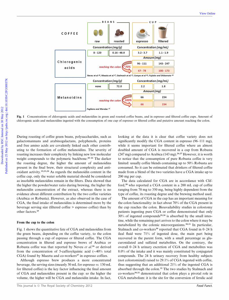
Fig. 1 Concentrations of chlorogenic acids and melanoidins in green and roasted coffee beans, and in espresso and filtered coffee cups. Amount of
chlorogenic acids and melanoidins ingested with the consumption of one cup of espresso or filtered coffee and putative amount reaching the colon.
Dow
nloa
ded
by R
SC I
nter
nal o
n 30
May
201
2Pu
blis
hed
on 2
5 M
ay 2
012
on h
ttp://
pubs
.rsc
.org
| do
i:10.
1039
/C2F
O30
037K
View Online
During roasting of coffee green beans, polysaccharides, such as
galactomannans and arabinogalactans, polyphenols, proteins
and free amino acids are covalently linked each other contrib-
uting to the formation of coffee melanoidins. The severity of
roasting increases their complexity by linking new low molecular
weight compounds to the polymeric backbone.20–22 The darker
the roasting degree, the higher the amount of melanoidins
present in the final brew, their structural complexity and anti-
oxidant activity.19,23,24 As regards the melanoidin content in the
coffee cup, only the water soluble material should be considered
as insoluble melanoidins remain in the filters. Data showed that
the higher the powder/water ratio during brewing, the higher the
melanoidin concentration of the extract, whereas there is no
evidence about different contribution by the two coffee varieties
(Arabica or Robusta). However, as also observed in the case of
CGA, the final intake of melanoidins is determined more by the
beverage serving size (filtered coffee > espresso coffee) than by
other factors.25
From the cup to the colon
Fig. 1 shows the quantitative fate of CGA and melanoidins from
the green beans, depending on the coffee variety, to the colon
passing through a cup of espresso or filtered coffee. The CGA
concentration in filtered and espresso brews of Arabica or
Robusta coffee was that reported by Neveu et al.26 or derived
from the concentration of 5-caffeoylquinic acid (�40% total
CGA) found by Maeztu and co-workers27 in espresso coffees.
Although espresso brew produces a more concentrated
beverage, the serving sizes (meanly 30 mL for espresso vs. 130 mL
for filtered coffee) is the key factor influencing the final amount
of CGA and melanoidins present in the cup: so the higher the
volume, the higher will be CGA and melanoidin intake. In fact,
This journal is ª The Royal Society of Chemistry 2012
looking at the data it is clear that coffee variety does not
significantly modify the CGA content in espresso (96–111 mg),
while it seems important for filtered coffee where an almost
doubled amount of CGA is recovered in a cup from Robusta
(247 mg) compared to Arabica (143 mg).26,27 However, it is worth
to notice that the consumption of pure Robusta coffee is very
limited: usually coffee blends containing up to 50% Robusta are
consumed. So it can be estimated that drinkers of filtered coffee
made from a blend of the two varieties have a CGA intake up to
200 mg per cup.
The data calculated for CGA are in accordance with Clif-
ford,28 who reported a CGA content in a 200 mL cup of coffee
ranging from 70 mg to 350 mg, being highly dependent from the
type of coffee, its roasting degree and the brewing method used.
The amount of CGA in the cup has an important meaning for
the colon functionality: in fact about 70% of the CGA present in
the cup reaches the colon. Bioavailability studies in colostomy
patients ingesting pure CGA or coffee demonstrated that only
30% of ingested compounds29,30 is absorbed by the small intes-
tine, while the remaining part arrives to the colon where it may be
metabolized by the colonic microorganisms.30,31 In particular,
Stalmach and co-workers30 reported that CGA found in 0–24 h
ileal fluid were 71% of ingested dose, the main part being
recovered in the parent form, with a small percentage as glu-
curonidated and sulfated metabolites. On the contrary, the
overall 0–24 h urinary excretion of CGA and metabolites was
8.0% of the intake and it was mainly constituted by conjugated
compounds. The 24 h urinary recovery from healthy subjects
(not colostomized) raised to 29.1% of CGA ingested with coffee,
thus suggesting that an additional 21% of the ingested CGA is
absorbed through the colon.32 The two studies by Stalmach and
co-workers30,32 demonstrated that colon plays a pivotal role in
CGA metabolism: it is the site for the conversion of ferulic acid
Food Funct.

Dow
nloa
ded
by R
SC I
nter
nal o
n 30
May
201
2Pu
blis
hed
on 2
5 M
ay 2
012
on h
ttp://
pubs
.rsc
.org
| do
i:10.
1039
/C2F
O30
037K
View Online
to feruloylglycine and dihydroferulic acid and for metabolism of
caffeic acid to dihydrocaffeic acid, which is further converted to
dihydro(iso)ferulic acid.
As regards melanoidins, the structural separation between
‘‘true’’ coffee melanoidins and coffee dietary fiber constituted by
the polysaccharides remaining un-derivatized after roasting is
very difficult,19,33 as clearly explained by Silv�an et al.19 who
introduced the concept of Maillardization of coffee dietary fiber.
This overlap between dietary fiber and melanoidins also exists
from the nutritional standpoint. In fact, on one hand melanoi-
dins do not fall into the definition of dietary fiber because they
are not exactly ‘‘polysaccharides naturally present in raw foods’’.
Some attempts have been done to enlarge the definition of die-
tary fiber to other non-digestible compounds, such as melanoi-
dins formed upon processing, however, the presence of a protein
moiety and the uncertainty of the structure represents a serious
limitation to take into consideration their physiological role as
dietary fiber. On the other hand, coffee melanoidins can be
considered a dietary fiber de facto: they arrive in their intact form
to the colon and they are partly fermented by colonic microor-
ganisms.34 The amount of coffee melanoidins reaching the colon
has been recently estimated in the range of 0.5–2.0 g per day;25 so
considering that 10 g is the recommended daily intake of soluble
dietary fiber and that most of people fail to reach this target,35
coffee melanoidin intake might significantly contribute to the
health benefits associated to this food component.
Dietary factors have multiple effects on CRC
The role of the diet in modulating the risk of CRC is mainly
associated with its influence on: i) colonic motility, whose
reduction may increase the exposure of epithelial cells to
potential carcinogens; ii) synthesis and secretion of bile acids,
that some evidence indicates as potential promoters of colon
carcinogenesis; iii) oxidative environment in the colon, leading to
formation of free radicals that can induce carcinogenesis through
increased local and systemic inflammation; iv) insulin sensitivity
and obesity, which is even considered an independent risk factor
for CRC risk.
Moreover, since the introduction of the dietary fiber hypoth-
esis in the early 1970s, indigestible bulk carbohydrates have been
considered the components responsible for the beneficial effects
of plant foods on bowel function. Bowel habit, a marker of bowel
function, is usually defined in terms of frequency of defecation,
stool consistency and form, and stool weight.36 Low stool weight
has been associated with the increased risk of colon cancer.8 In
fact, stool weights of around 100 g day�1 are associated with
a high risk of colon cancer (25/100 000 of the population). On the
contrary, diets characterized by high fiber intake (approximately
18 g day�1) are associated with stool weights of 150 g day�1 and
able to cut the risk by about 50% (around 10/100 000).8
In addition, mounting evidence indicates that the mutualistic
relation between an individual and his/her gut microbiota
sometimes becomes pathological, as in obesity, diabetes,
atherosclerosis, and inflammatory bowel diseases.37–39
For example, it is known that in both humans and mice, the
development of obesity correlates with an increase of Firmicutes
with respect to Bacteroidetes, including the genus Bacteroides
and Prevotella, in the gut.40–42 Moreover, transferring the gut
Food Funct.
microbiota from obese mice to germ-free wild-type recipients
leads to an increase in fat mass in the recipients, thus leading to
the hypothesis that the gut microbiota promotes obesity by
increasing the capacity of the host to extract energy (calories)
from ingested food.43
On the other hand, it has also been demonstrated that dietary
patterns highly contribute with age and genetics in influencing
microbiome composition.44 A recent analysis of gut microbial
communities across the world proposed three predominant
variants, or ‘‘enterotypes,’’ dominated by Bacteroides, Prevotella,
and Ruminococcus, respectively44 and although the basis for
enterotype clustering appears independent of nationality, gender,
age, or body mass index its linkage with long-term dietary
pattern has been finally demonstrated.45
Coffee and colon health
The ability of coffee and its non-digestible compounds to
influence colon health may mainly pass through the modulation
of colonic microbiota as well as of the markers of bowel func-
tions, such as stool output, evacuation frequency, etc.
Although in vitro studies have revealed several pharmacolo-
gical properties of pure CGA, such as antioxidant activity,46,47
ability to increase hepatic glucose utilization,48–52 and inhibition
of the mutagenicity of carcinogenic compounds,53 and although
it is known that 71% of coffee CGA reaches the colon, the
literature is lacking in investigations focused on the direct effects
of coffee polyphenols on this organ. Moreover, due to the
extensive metabolism of CGA, once in the colon it is unclear how
much antioxidant activity contributes in vivo at local and
systemic level, as circulating metabolites (mainly conjugated
compounds) often have lower antioxidant activity than the
parental chlorogenic and caffeic acids.54 One study dealing with
coffee CGA and microorganisms evaluated the antimicrobial
effect of coffee extract on enterobacteria in plates and did not
find differences among coffee extracts with different CGA
content.55 Thus, due to the paucity of data about the specific
effect of coffee CGA in the colon, the present paragraph is
mainly focused therein after on the effects of coffee non-digesti-
ble material, including both melanoidins and phenolic
compounds.
There is much in vitro evidence that, once in the colon, coffee
melanoidins may be fermented by colon microorganisms. In
three recent papers by the same research group, the degradation
of different fractions of coffee, i.e. soluble dietary fiber and non-
digestible high molecular weight ethanol soluble fractions, by
human fecal microbiota has been reported.9,56,57 Results
demonstrated that after 24 h of fermentation: i) 85% of total
carbohydrates was degraded (arabinogalactans being generally
less utilized than the galactomannans), while only 29% of the
isolated Maillard reaction product was partially degraded or
modified; ii) acetate, propionate and butyrate were released with
a high molar proportion of acetate and propionate; iii) bacterial
cells belonging to the Bacteroides-Prevotella group increased by
60% upon fermentation of the coffee soluble dietary fiber and
from 2- to 40-fold depending on the molecular weight of the
fraction and the degree of roast.9,56,57
It is worth noticing that in these papers the increased growth of
bifidobacteria and lactobacilli was never observed, thus
This journal is ª The Royal Society of Chemistry 2012

Dow
nloa
ded
by R
SC I
nter
nal o
n 30
May
201
2Pu
blis
hed
on 2
5 M
ay 2
012
on h
ttp://
pubs
.rsc
.org
| do
i:10.
1039
/C2F
O30
037K
View Online
suggesting that coffee melanoidins do not have a prebiotic effect.
In fact, a condicio sine qua non for an ingredient to be claimed as
prebiotic is that it elicits ‘‘the selective stimulation of growth and/
or activity(ies) of one or a limited number of microbial genus(era)/
species in the gut microbiota that confer(s) health benefits to the
host’’, bifidobacteria and lactobacilli being actually the only
genera whose benefits for hosts are well established (emerging
genera are Eubacterium, Faecalibacterium and Roseburia,
although more evidence is needed on their physiological
properties).58
On the other hand, a previous study conducted on coffee sil-
verskin, which is a by-product of coffee roasting containing 60%
dietary fiber, including coffee melanoidins, demonstrated the
ability of this material to increase bifidobacteria but not lacto-
bacilli concentrations.59 In this study a gut model, validated to
mimic the three parts of human colon was used and it was fed
with a 4-fold higher amount of coffee dietary fiber than the other
studies using batch cultures.9,56,57 These differences could explain
the discrepancy between the result obtained with coffee silver-
skin59 and that found with dietary fiber extracted from coffee
beverage.9,56,57
From a nutritional perspective, these in vitro findings are not
enough to corroborate the hypothesis that higher coffee drinkers,
providing a higher amount of coffee melanoidins into the gut,
may also take benefits from a prebiotic effect in vivo. However,
this is one of the main area to investigate in order to explain the
stronger associations with reduced risk of disease found in
epidemiological studies for subjects consuming >4 coffee day�1.
Few and limited human studies have been performed to
explore the prebiotic activity of coffee: data from Jaquet et al.60
demonstrated that the consumption of three cups of instant
coffee for 3 weeks increased Bifidobacterium spp. Interestingly,
the authors of this study administered a coffee preparation
resulting from water co-extraction of green and roasted coffee
beans. According to the authors this beverage should have a final
composition similar to that of a light roasted coffee. Since no
chemical characterization of the beverage was reported, this
assumption is flawed. However, this human study is the first one
establishing that a moderate consumption of a coffee-type
beverage has an effect on particular members of the colonic
microbiota (represented in feces), principally bifidobacteria,
without a major impact on the dominant bacterial groups.
Two other human studies showed that the content of Bifido-
bacterium significantly increased in feces after 2 weeks of
consumption of mannooligosaccharides (MOS) obtained
from spent coffee by thermal hydrolysis at two doses61 and
a coffee mix drink containing MOS,62 thus supporting a possible
prebiotic effect related to coffee consumption. Also in this case
the composition of the beverage, which is quite far from that of
a regular coffee, is a severe limitation to ascertain the prebiotic
activity of coffee.
Looking at the influence of coffee on the gut microbiota the
increase of Bacteroides-Prevotella group, elicited by coffee die-
tary fiber,9,56,57 should be carefully considered as it may be
associated with a protective effect on human health. In fact some
evidence showed a decrease in Firmicutes and an increase in
Bacteroidetes, including Bacteroides-Prevotella group, during
weight loss in overweight and obese adolescents63 and an
increased representation of members of the Firmicutes in
This journal is ª The Royal Society of Chemistry 2012
individuals who became overweight by the age of 7.64 In this
framework, the daily coffee consumption sustaining the growth
of Bacteroides might favor the maintenance of a low Firmicutes/
Bacteroides ratio, creating the conditions for a potentially
healthier microbiota.
A scant literature has explored the role of coffee on the
markers of bowel functionality. Some years ago, a positive
correlation between coffee consumption and fecal weight was
observed in 14 healthy volunteers on a free-diet.65 Defecation
frequency during the MOS supplementation and the coffee mix
drink containing MOS intake was significantly higher than
before this period.61,62 In the case of coffee mix drink containing
MOS, the defecation volume also significantly increased.62
The effect of unfiltered coffee consumption on colonic cell
hyperproliferation, which is thought to be an early event in the
multi-step process of the colonic adenocarcinoma sequence,66
fecal soluble bile acid concentration and detoxification enzymes,
was studied in a crossover design study in 64 healthy volun-
teers.67 The results demonstrated that drinking 1 L of unfiltered
coffee every day for 2 weeks did not affect the colorectal cell
proliferation and the cytotoxic secondary fecal soluble bile acid
concentration, suggesting that the cytotoxic secondary bile acids
are not involved in the putative protective effect of coffee in CRC
development. Conversely, coffee increased the detoxification
capacity and anti-mutagenic properties in the colorectal mucosa
through an increase in glutathione concentration.67
Concluding remarks and research gaps
Coffee is the major source of CGA in the diet of coffee drinkers.29
Clifford28 estimated that daily intake of CGA in coffee drinkers
is 0.5–1 g, while coffee abstainers usually ingest only 100 mg per
day by other sources such as apples, pears, berries, artichoke and
aubergine. Detailing the intake according to the use of Arabica
or Robusta and brewing methods (i.e., espresso or filtered
coffees), we calculated that for a heavy coffee drinker (6 cups
per day) the intake of CGA can range from 402–468 mg and
600–1038 mg for an espresso or a filtered coffee drinker,
respectively, the minimum value of each range is calculated for
Arabica and the maximum for Robusta coffee.
Coffee melanoidins intake is in an order of magnitude of
0.5–2 g per day for moderate and heavy drinkers, respectively:
thus coffee is also the major contributor of dietary soluble
melanoidin intake.25
The studies discussed in this review indicated that, due to their
in vivo metabolism and composition, both coffee CGA and die-
tary fiber, including melanoidins, may play a major role in colon
protection from CRC development. The three possible mecha-
nisms of coffee protection towards a reduction of CRC risk are
depicted in Fig. 2. The more direct beneficial mechanism is the
coffee ability to increase colon motility that underlies increased
fecal output in coffee drinkers and the consequent increased
elimination rate of carcinogens.
An indirect benefit of coffee on colon inflammation can be
hypothesized from the findings of human intervention studies
performed by Cani et al.68 and Pendyala et al.69 They observed
that an improvement of microbiota balance induces the
amelioration of insulin sensitivity and a consequent weight loss
due to reduced fat deposition. It is worth noting that
Food Funct.

Fig. 2 Possible pathways correlating coffee intake to the reduction of colorectal cancer (CRC) risk. A) The modulation of gut microbiota is associated
with the amelioration of insulin sensitivity and appetite control.68 These mechanisms canmodulate body fat deposition and can underlie body weight loss
that, through reduction of colorectal mucosa inflammation, may indirectly reduce CRC risk69 or, even directly, as obesity is an independent risk factor
for CRC. B) Coffee antioxidant dietary fiber may reduce, through several mechanisms, the inflammation in colon mucosa, thus directly reducing CRC
risk.84 C) The increase of colon motility would increase the carcinogen elimination rate, which is directly associated with a reduced CRC risk. The two
red question marks represent the two main passages, which are very speculative up to now and must be addressed by future studies. They regard: ?1 the
prebiotic activity of coffee and ?2 the anti-inflammatory action of coffee components inside the gastrointestinal tract.
Dow
nloa
ded
by R
SC I
nter
nal o
n 30
May
201
2Pu
blis
hed
on 2
5 M
ay 2
012
on h
ttp://
pubs
.rsc
.org
| do
i:10.
1039
/C2F
O30
037K
View Online
epidemiology associates habitual coffee consumption to
a reduced risk of type 2 diabetes, even though caffeine intake
acutely lowered insulin sensitivity and increased glucose
concentrations in short-term intervention studies.70,71 The asso-
ciation between coffee consumption and ameliorated insulin
control may be explained by the prebiotic activity of coffee die-
tary fiber; however this hypothesis was never investigated thus
far. The future perspective is that further studies will be designed
to demonstrate the coffee prebiotic activity in humans (the red
question mark in Fig. 2). This will be the starting point to
elucidate the entangled network of actions determining the
amelioration of metabolic syndrome associated to coffee
consumption.72
Last but not least, the chemical structure of coffee melanoidins
has all the features of an ‘‘antioxidant dietary fiber’’. Due to
the presence in melanoidins of reductones and fragments of
phenol compounds, these polymers perfectly fit into the defini-
tion of the antioxidant dietary fiber coined by Saura-Calixto.73
The physiological relevance of the antioxidants bound to poly-
saccharides has been recently outlined for cereals.74,75 It was
hypothesized that this material may act in the gastrointestinal
tract as a ‘‘sponge’’ for free radicals that continuously are formed
due to endogenous reactions and processes.76
Coffee melanoidins, exactly as the cereal dietary fiber, may
elicit a ‘‘radical-sponge action’’ in the gastrointestinal tract
because i) coffee melanoidins have antioxidant capacity in vitro
assessed by different methods;57,77–80 ii) after 24 h of in vitro
fermentation in the presence of human gut microorganisms
coffee melanoidins still elicit 25% of native antioxidant
capacity;57 iii) human and animal studies have demonstrated that
dietary melanoidins are majorly recovered in the feces;81 iv) the
antioxidant capacity of feces from healthy subjects is associated
to coffee consumption.65
The second question mark in Fig. 2 is related to this aspect: it is
necessary that future studies will address the issue related to the
relevance of coffee consumption in reducing inflammation along
Food Funct.
the gastrointestinal tract, switching off several biochemical
pathways leading to CRC.10
References
1 FAO, Food and Agricultural Organization, Food balance sheets,http://www.fao.org/.
2 C. La Vecchia and A. Tavani, Eur. J. Cancer Prev., 2007, 16, 385–389.3 J. V. Higdon and B. Frei,Crit. Rev. Food Sci. Nutr., 2006, 46, 101–123.4 J. Ferlay, F. Bray, P. Pisani, and D. Parkin, IARC CancerBase, No. 5version 2.0. Lyon, France: IARC Press; 2004.
5 Word Cancer Research Fund, American Institute for Cancer ResearchExpert Panel, Food, Nutrition and the Prevention of Cancer A GlobalPerspective, Washington, DC, American Institute for CancerResearch, 2007.
6 E. K. Wei, E. Giovannucci, K. Wu, B. Rosner, C. S. Fuchs,W. C. Willett and G. A. Coldits, Int. J. Cancer, 2004, 108, 433–442.
7 Y. Je, W. Liu and E. Giovannucci, Int. J. Cancer, 2009, 124, 1662–1668.
8 J. H. Cummings, S. A. Bingham, K. W. Heaton andM. A. Eastwood,Gastroenterology, 1992, 103, 1783–1789.
9 D. Gniechwitz, N. Reichardt, M. Blaut, H. Steinhart and M. Bunzel,J. Agric. Food Chem., 2007, 55, 6989–6996.
10 L. Ricciardiello, F. Bazzoli and V. Fogliano, Nat. Rev. Gastroenterol.Hepatol., 2011, 8, 592–596.
11 E. Giovannucci, Am. J. Epidemiol., 1998, 147, 1043–1052.12 A.Tavani andC.LaVecchia,Cancer,CausesControl, 2004, 15, 743–757.13 C. Galeone, F. Turati, C. La Vecchia and A. Tavani, Cancer, Causes
Control, 2010, 21, 1949–1959.14 X. Yu, Z. Bao, J. Zou and J. Dong, BMC Cancer, 2011, 11, 96–107.15 L. Arab, Nutr. Cancer, 2010, 62, 271–283.16 M. N. Clifford, J. Sci. Food Agric., 2000, 80, 1033–1043.17 C. L. Ky, J. Louarn, S. Dussert, B. Guyot, S. Hamon and M. Noriot,
Food Chem., 2001, 75, 223–230.18 L. C. Trugo and R. A. Macrae, Food Chem., 1984, 15, 219–227.19 J. M. Silv�an, F. J. Morales and F. Saura-Calixto, J. Agric. Food
Chem., 2010, 58, 12244–12249.20 F. M. Nunes, A. Reis, M. R. M. Domingues and M. A. Coimbra, J.
Agric. Food Chem., 2006, 54, 3428–3439.21 P. Montavon, A. F. Mauron and E. Duruz, J. Agric. Food Chem.,
2003, 51, 2335–2343.22 E. K. Bekedam, H. A. Schols, M. A. J. S. Van Boekel and G. Smit, J.
Agric. Food Chem., 2006, 54, 7658–7666.23 R. C. Borrelli, A. Visconti, C.Mennella,M. Anese and V. Fogliano, J.
Agric. Food Chem., 2002, 50, 6527–6533.
This journal is ª The Royal Society of Chemistry 2012

Dow
nloa
ded
by R
SC I
nter
nal o
n 30
May
201
2Pu
blis
hed
on 2
5 M
ay 2
012
on h
ttp://
pubs
.rsc
.org
| do
i:10.
1039
/C2F
O30
037K
View Online
24 F. M. Nunes and M. A. Coimbra, J. Agric. Food Chem., 2007, 55,3967–3977.
25 V. Fogliano and F. J. Morales, Food Funct., 2011, 2, 117–123.26 V. Neveu, J. Perez-Jim�enez, F. Vos, V. Crespy, L. du Chaffaut,
L. Mennen, C. Knox, R. Eisner, J. Cruz, D. Wishart andA. Scalbert, Database, 2010, DOI: 10.1093/database/bap024.
27 L. Maeztu, S. Andueza, C. Iba~nez, M. Paz de Pe~na, J. Bello andC. Cid, J. Agric. Food Chem., 2001, 49, 4743–4747.
28 M. N. Clifford, J. Sci. Food Agric., 1999, 79, 362–372.29 M. R. Olthof, P. C. Hollman and M. B. Katan, J. Nutr., 2001, 131,
66–71.30 A. Stalmach, H. Steiling, G. Williamson and A. Crozier, Arch.
Biochem. Biophys., 2010, 501, 98–105.31 M. R. Olthof, P. C. Hollman, M. N. Buijsman, J. M. van Amelsvoort
and M. B. Katan, J. Nutr., 2003, 133, 1806–1814.32 A. Stalmach,W.Mullen, D. Barron, K. Uchida, T. Yokota, C. Cavin,
H. Steiling, G. Williamson and A. Crozier, Drug Metab. Dispos.,2009, 37, 1749–1758.
33 M. E. D�ıaz-Rubio and F. Saura-Calixto, J. Agric. Food Chem., 2007,55, 1999–2003.
34 F. J. Morales, V. Somoza and V. Fogliano, Amino Acids, 2012, 42,1097–1109.
35 C. S. Brennan, Mol. Nutr. Food Res., 2005, 49, 560–570.36 M. A. Bianchi, F. Scazzina, D. Del Rio, S. Valtue~na, N. Pellegrini,
L. Franzini, M. L. Callegari, C. Pellacani, A. Buschini, I. Zavaroniand F. Brighenti, Br. J. Nutr., 2010, 104, 1500–1507.
37 L. V. Hooper and J. I. Gordon, Science, 2001, 292, 1115–1118.38 A. Emminger, E. Kahmann and D. S. Savage, Cancer Lett., 1977, 2,
273–278.39 F. B€ackhed, H. Ding, T. Wang, L. V. Hooper, G. Y. Koh, A. Nagy,
C. F. Semenkovich and J. I. Gordon, Proc. Natl. Acad. Sci. U. S. A.,2004, 101, 15718–15723.
40 R.E.Ley,F.B€ackhed,P.Turnbaugh,C.A.Lozupone,R.D.KnightandJ. I. Gordon, Proc. Natl. Acad. Sci. U. S. A., 2005, 102, 1070–1075.
41 R. E. Ley, P. J. Turnbaugh, S. Klein and J. I. Gordon, Nature, 2006,444, 1022–1023.
42 P. J. Turnbaugh, M. Hamady, T. Yatsunenko, B. L. Cantarel,A. Duncan, R. E. Ley, M. L. Sogin, W. J. Jones, B. A. Roe,J. P. Affourtit, M. Egholm, B. Henrissat, A. C. Heath, R. Knightand J. I. Gordon, Nature, 2009, 457, 480–484.
43 P. J. Turnbaugh, R. E. Ley, M. A. Mahowald, V. Magrini,E. R. Mardis and J. I. Gordon, Nature, 2006, 444, 1027–1031.
44 M. Arumugam, J. Raes, E. Pelletier, D. Le Paslier, T. Yamada,D. R. Mende, G. R. Fernandes, J. Tap, T. Bruls, J. M. Batto,M. Bertalan, N. Borruel, F. Casellas, L. Fernandez, L. Gautier,T. Hansen, M. Hattori, T. Hayashi, M. Kleerebezem, K. Kurokawa,M. Leclerc, F. Levenez, C. Manichanh, H. B. Nielsen, T. Nielsen,N. Pons, J. Poulain, J. Qin, T. Sicheritz-Ponten, S. Tims,D. Torrents, E. Ugarte, E. G. Zoetendal, J. Wang, F. Guarner,O. Pedersen, W. M. de Vos, S. Brunak, J. Dor�e,MetaHIT Consortium, M. Antol�ın, F. Artiguenave, H. M. Blottiere,M. Almeida, C. Brechot, C. Cara, C. Chervaux, A. Cultrone,C. Delorme, G. Denariaz, R. Dervyn, K. U. Foerstner, C. Friss,M. van de Guchte, E. Guedon, F. Haimet, W. Huber, J. vanHylckama-Vlieg, A. Jamet, C. Juste, G. Kaci, J. Knol, O. Lakhdari,S. Layec, K. Le Roux, E. Maguin, A. M�erieux, R. Melo Minardi,C. M’rini, J. Muller, R. Oozeer, J. Parkhill, P. Renault, M. Rescigno,N. Sanchez, S. Sunagawa, A. Torrejon, K. Turner,G. Vandemeulebrouck, E. Varela, Y. Winogradsky, G. Zeller,J.Weissenbach, S.D. Ehrlich and P. Bork,Nature, 2011, 473, 174–180.
45 G. D. Wu, J. Chen, C. Hoffmann, K. Bittinger, Y. Y. Chen,S. A. Keilbaugh, M. Bewtra, D. Knights, W. A. Walters,R. Knight, R. Sinha, E. Gilroy, K. Gupta, R. Baldassano,L. Nessel, H. Li, F. D. Bushman and J. D. Lewis, Science, 2011,334, 105–108.
46 Y. Kono, K. Kobayashi, S. Tagawa, K. Adachi, A. Ueda, Y. Sawaand H. Shibata, Biochim. Biophys. Acta, Gen. Subj., 1997, 1335,335–342.
47 M. R. Luzia, K. C. C. Paixa~no, R. Marcilio, L. C. Trugo,L. M. C. Quinteiro and C. A. B. de Maria, Int. J. Food Sci.Technol., 1997, 32, 15–19.
48 W. Herling, H. J. Burger, D. Schwab, H. Hemmerle, P. Below andG. Schubert, Am. J. Physiol., 1998, 274, 1087–1093.
49 J. Shearer, A. Farah, T. de Paulis, D. P. Bracy, R. R. Pencek,T. E. Graham and D. H. Wasserman, J. Nutr., 2003, 133, 3529–3532.
This journal is ª The Royal Society of Chemistry 2012
50 H. Hemmerle, H. J. Burger, P. Bellow, G. Schubert, R. Rippel,P. W. Schindler, E. Paulus and A. Herling, J. Med. Chem., 1997,40, 137–145.
51 W. J. Arion, W. K. Canfield, F. C. Ramos, P. W. Schinder,H. J. Burger, H. Hemmerle, G. Schubert, P. Below andA. W. Herling, Arch. Biochem. Biophys., 1997, 339, 315–122.
52 K. L. Johnston, M. Clifford and L. M. Morgan, Am. J. Clin. Nutr.,2003, 78, 728–733.
53 H. F. Stich, M. P. Rosin and L. Bryson, Mutat. Res., Fundam. Mol.Mech. Mutagen., 1982, 95, 119–128.
54 K. Iwai, N. Kishimoto, Y. Kakino, K. Mochida and T. Fujita, J.Agric. Food Chem., 2004, 52, 4893–4898.
55 A. A. Almeida, A. Farah, D. A. Silva, E. A. Nunan andM. B. Gl�oria,J. Agric. Food Chem., 2006, 54, 8738–8743.
56 D. Gniechwitz, N. Reichardt, E. Meiss, J. Ralph, H. Steinhart,M. Blaut and M. Bunzel, J. Agric. Food Chem., 2008, 56, 5960–5969.
57 N. Reichardt, D. Gniechwitz, H. Steinhart, M. Bunzel and M. Blaut,Mol. Nutr. Food Res., 2009, 53, 287–299.
58 M. Roberfroid, G. R. Gibson, L. Hoyles, A. L. McCartney,R. Rastall, I. Rowland, D. Wolvers, B. Watzl, H. Szajewska,B. Stahl, F. Guarner, F. Respondek, K. Whelan, V. Coxam,M. J. Davicco, L. L�eotoing, Y. Wittrant, N. M. Delzenne,P. D. Cani, A. M. Neyrinck and A. Meheust, Br. J. Nutr., 2010,104(Suppl 2), S1–63.
59 R. C. Borrelli, F. Esposito, A. Napolitano, A. Ritieni andV. Fogliano, J. Agric. Food Chem., 2004, 52, 1338–1343.
60 M. Jaquet, I. Rochat, J. Moulin, C. Cavin and R. Bibiloni, Int. J.Food Microbiol., 2009, 31, 117–21.
61 M. Asano, S. Umemura, H. Fujii and H. HoshinoIino, Food Sci.Technol. Res., 2004, 10, 93–97.
62 M. Umemura, S. Fujii, I. Asano, H. Hoshino and H. Iino, Food Sci.Technol. Res., 2004, 10, 195–198.
63 I. Nadal, A. Santacruz, A. Marcos, J. Warnberg, M. Garagorri,L. A. Moreno, M. Martin-Matillas, C. Campoy, A. Mart�ı,A. Moleres, M. Delgado, O. L. Veiga, M. Garc�ıa-Fuentes,C. G. Redondo and Y. Sanz, Int. J. Obes., 2009, 33, 758–767.
64 M. Kalliomaki, M. C. Collado, S. Salminen and E. Isolauri, Am. J.Clin. Nutr., 2008, 87, 534–538.
65 M. Garsetti, N. Pellegrini, C. Baggio and F. Brighenti, Br. J. Nutr.,2000, 84, 705–710.
66 B. Vogelstein and K. W. Kinzler, Trends Genet., 1993, 9, 138–141.67 M. J. Grubben, C. C. Van Den Braak, R. Broekhuizen, R. De Jong,
L. Van Rijt, E. De Ruijter, W. H. Peters, M. B. Katan andF. M. Nagengast, Aliment. Pharmacol. Ther., 2000, 14, 1181–1190.
68 P. D. Cani, E. Lecourt, E. M. Dewulf, F. M. Sohet, B. D. Pachikian,D. Naslain, F. De Backer, A. M. Neyrinck and N. M. Delzenne, Am.J. Clin. Nutr., 2009, 90, 1236–1243.
69 S. Pendyala, L. M. Neff, M. Su�arez-Fari~nas and P. R. Holt, Am. J.Clin. Nutr., 2011, 93, 234–242.
70 R. M. Van Dam and F. B. Hu, JAMA, J. Am. Med. Assoc., 2005, 294,97–104.
71 R. Huxley, C. M. Lee, F. Barzi, L. Timmermeister, S. Czernichow,V. Perkovic, D. E. Grobbee, D. Batty and M. Woodward, Arch.Intern. Med., 2009, 169, 2053–2063.
72 E. Esteve, W. Ricart and J. M. Fern�andez-Real, Curr. Opin. Clin.Nutr. Metab. Care, 2011, 14, 483–490.
73 F. Saura-Calixto, J. Agric. Food Chem., 1998, 46, 4303–4306.74 P. Vitaglione, A. Napolitano and V. Fogliano, Trends Food Sci.
Technol., 2008, 19, 451–463.75 V. Gokmen, A. Serpen and V. Fogliano, Trends Food Sci. Technol.,
2009, 20, 278–288.76 C. F. Babbs, Free Radical Biol. Med., 1990, 8, 191–200.77 R. C. Borrelli, A. Visconti, C.Mennella,M. Anese and V. Fogliano, J.
Agric. Food Chem., 2002, 50, 6527–6533.78 M. Anese and C. Nicoli, J. Agric. Food Chem., 2003, 51, 942–946.79 M. D. del Castillo, J. M. Ames and M. H. Gordon, J. Agric. Food
Chem., 2002, 50, 3698–3703.80 C. Delgado-Andrade, J. A. Rufian-Henares and F. J. Morales, J.
Agric. Food Chem., 2005, 53, 7832–7836.81 V. Faist and H. F. Erbersdobler, Ann. Nutr. Metab., 2001, 45, 1–12.82 C. Campa, S. Doulbeau, S. Dussert, S. Hamon and M. Noirot, Food
Chem., 2005, 93, 135–139.83 K. Fujioka and T. Shibamoto, Food Chem., 2008, 106, 217–221.84 D. N. Seril, J. Liao, G. Y. Yang and C. S. Yang,Carcinogenesis, 2003,
24, 353–362.
Food Funct.