Immunomodulatory and Immunotherapeutic Properties of Recombinant 7Interferon and Recombinant Tumor...
9
1987;47:2563-2570. Cancer Res James E. Talmadge, Henry R. Tribble, Robin W. Pennington, et al. Factor in Mice -Interferon and Recombinant Tumor Necrosis γ Recombinant Immunomodulatory and Immunotherapeutic Properties of Updated version http://cancerres.aacrjournals.org/content/47/10/2563 Access the most recent version of this article at: E-mail alerts related to this article or journal. Sign up to receive free email-alerts Subscriptions Reprints and . [email protected] Department at To order reprints of this article or to subscribe to the journal, contact the AACR Publications Permissions . [email protected] Department at To request permission to re-use all or part of this article, contact the AACR Publications Research. on February 26, 2014. © 1987 American Association for Cancer cancerres.aacrjournals.org Downloaded from Research. on February 26, 2014. © 1987 American Association for Cancer cancerres.aacrjournals.org Downloaded from
-
Upload
independent -
Category
Documents
-
view
0 -
download
0
Transcript of Immunomodulatory and Immunotherapeutic Properties of Recombinant 7Interferon and Recombinant Tumor...
1987;47:2563-2570. Cancer Res James E. Talmadge, Henry R. Tribble, Robin W. Pennington, et al. Factor in Mice
-Interferon and Recombinant Tumor NecrosisγRecombinant Immunomodulatory and Immunotherapeutic Properties of
Updated version
http://cancerres.aacrjournals.org/content/47/10/2563
Access the most recent version of this article at:
E-mail alerts related to this article or journal.Sign up to receive free email-alerts
Subscriptions
Reprints and
To order reprints of this article or to subscribe to the journal, contact the AACR Publications
Permissions
To request permission to re-use all or part of this article, contact the AACR Publications
Research. on February 26, 2014. © 1987 American Association for Cancercancerres.aacrjournals.org Downloaded from
Research. on February 26, 2014. © 1987 American Association for Cancercancerres.aacrjournals.org Downloaded from
[CANCER RESEARCH 47, 2563-2570, May 15, 1987]
Immunomodulatory and Immunotherapeutic Properties of Recombinant7-Interferon and Recombinant Tumor Necrosis Factor in Mice1
James E. Talmadge,2 Henry R. Tribble, Robin W. Pennington, Hamblin Phillips, and Robert H. Wiltrout
Preclinical Screening Laboratory [J. E. T., H. R. T., R. W. P., H. P.], Program Resources, Inc., and Biological Therapeutics Branch, Biological Response ModifiersProgram [R. H. W.], Division of Cancer Treatment, National Cancer Institute-Frederick Cancer Research Facility, Frederick, Maryland 21701
ABSTRACT
These studies were designed to examine the immunomodulatory andimmunotherapeutic properties of recombinant murine interferon gamma(rM IFN-7) and recombinant human tumor necrosis factor (rii INF).We report that rM IFN-7 activated murine natural killer cells andmacrophages in a dose-dependent manner in vivo. The rM IFN-7, whichdemonstrated a bell-shaped therapeutic response curve, must be administered at specific doses and schedules to produce optimal therapeuticactivity. Optimal activity was observed after i.v. administration of 50,000I /animal rM IKN-7 three times per week. In contrast, rl ITNF producedits major therapeutic activity in the treatment of metastatic disease afteri.v. but not i.p. administration. The therapeutic effects of rl I TNF wereas great in these in vivo systems as those of rM IFN-7. Furthermore,rii TNF had additive therapeutic activity when administered in conjunction with suboptimal doses of rM IFN-7. Unlike rM IFN-7, rH TNFdid not activate natural killer cells in vivo or in vitro but did augment invivo and in vitro macrophage tumoricidal activity. It also had synergisticcytostatic properties with rM IFN-7 for some murine tumor cell lines invitro. High levels of rH TNF were readily detected hi the serum with ahalf-life of approximately 30 min after i.v. administration. In contrast,only minimal serum TNF activity occurred after i.p. administration,suggesting that i.v. administration may more efficiently facilitate systemictherapeutic activity. In summary, rH TNF and rM IFN-7 have therapeutic activity for metastatic disease as individual agents and additive therapeutic activity when used in combination. Furthermore, it appears thatin addition to therapeutic potential as cytostatic agents, the immunomodulatory properties of rH TNF have a role in its therapeutic properties.
INTRODUCTION
The adequate evaluation of the immunomodulatory and therapeutic activities of cytokines was hampered until recently bythe lack of sufficient amounts of purified materials. Recombi-nant DNA technology has made it possible to produce many ofthese cytokines in large quantities. Nonetheless, the initialclinical trials with rH IFN3 have yielded only limited clinical
responses, similar to those observed with natural interferons(1-15). One possible explanation for these disappointing resultsis that the therapeutic protocols for IFN have not been optimized for inducing immunomodulation (13, 16). In the studiesreported here, we examined the immunomodulatory and im-
Received 6/2/86; revised 10/10/86, 2/6/87; accepted 2/13/87.The costs of publication of this article were defrayed in part by the payment
of page charges. This article must therefore be hereby marked advertisement inaccordance with 18 U.S.C. Section 1734 solely to indicate this fact.
1This research was sponsored by the Biological Resources Branch, Biological
Response Modifiers Program, Division of Cancer Treatment of the NationalCancer Institute, Department of Health and Human Services, under contract no.N01-23910 with Program Resources, Inc. The contents of this publication do notnecessarily reflect the views or policies of the Department of Health and HumanServices, nor does mention of trade names, commercial products, or organizationsimply endorsement by the U. S. Government.
-To whom requests for reprints should be addressed, at National Cancer
Institute, Frederick Cancer Research Facility, Preclinical Screening Laboratory,P.O. Box B, Frederick, MD 21701.
3The abbreviations used are: rH IFN, recombinant human interferon; IFN,interferon; BRM, biological response modifier; rM IFN, recombinant murineinterferon; rH TNF, recombinant human tumor necrosis factor; NK, naturalkiller, TNF, tumor necrosis factor, Poly(I,C)-LC, polyinosinic-polycytidylic acidcomplexée)with pnly-i.-lysine and carboxymethylcellulose; LPS, lipopolysaccha-ride; CMF-HBSS, Calcium-magnesium-free Hanks' balanced salts solution.
munotherapeutic properties of rM IFN-7 and rH TNF aloneand in combination.
Most clinical trials with IFN (recombinant and natural) havebeen undertaken with human a-interferon, and only recentlyhave clinical trials been initiated to examine immunomodulation and therapeutic efficacy of rH IFN-7. IFNs are secretedproteins that induce an antiviral state in their target cells andhave both immunomodulatory and direct antitumor properties.Few data are presently available concerning the immunomodulatory and therapeutic properties of IFN-7. The antiprolifer-ative effect of IFN-7 has been reported to be 10-100-foldgreater than that of IFN-a or IFN-/3 (17), suggesting greatertherapeutic potential. rM IFN-7 was also found to be 10-100-fold more potent than IFN-a in the augmentation of cytotox-icity mediated by NK cells (18) and macrophages (19).
The cytokine that became known as TNF was first reportedby Carswell et al. (20). Sera from endotoxin-treated rodents,which had been sensitized with an immunopotentiator such asBacillus Calmette-Guérin, contained a substance that, wheninjected into mice with transplantable tumors, caused extensivehemorrhagic necrosis of the tumors. TNF is also found inculture supernatants from macrophages treated with endotoxin.Partially purified and recombinant preparations of TNF havebeen tested for direct cytotoxicity against murine and humancell lines in vitro and in vivo (21-27). Tumor but not normalcell lines from both species are susceptible to the cytotoxicactivity of murine and human TNF. Furthermore, murine TNFis active against human and murine tumor transplants in nudeand normal mice, respectively. However, few immunotherapeutic studies have been undertaken with either recombinant, partially purified or impurifica TNF, and most of these have beenagainst s.c. or intradermal tumors. Therefore, in the presentstudies, we examined the therapeutic activity of rH TNF aloneand in combination with rM IFN-7 for the treatment of established métastases.
MATERIALS AND METHODSAnimals. Specific pathogen-free male C57BL/6N mice (H-2b) and
C3H/HeN MTV mice (H-2"), 3 or 4 weeksof age, were obtained fromthe Animal Production Area of the National Cancer Institute-FrederickCancer Research Facility.
Tumors. These studies used the Moloney virus-induced lymphomaYAC-1 (28) of A/SN (H-2") origin. Adherent cell lines included themetastatic melanoma variant B16-BL6 (29) from the B16 melanoma,which arose spontaneously in a C57BL/6N (H-2b) mouse and thespontaneous lung carcinomas M109 (30) and 3LL, which are syngeneicto the BALB/c (/f-2'0 and C57BL/6 mice, respectively.The metastaticvariant 3LL-M2 was isolated from a pool of spontaneous 3LL lungmétastases(31). The L-cell variant L-929 was also used (32). Alladherent cell lines were maintained as monolayers in Eagle's minimumessential medium supplemented with 5% fetal bovine serum, 2-fold-concentrated vitamin solution, glutamina, sodium pyruvate, and non-essential amino acids. The YAC-1 tumor cell line was grown in RPMI1640medium supplemented with 10%fetal bovineserum and the samemedium supplements used with Eagle's minimum essential medium.
All cell lines were free of Mycoplasma and pathogenic murine viruses(33).
2563
Research. on February 26, 2014. © 1987 American Association for Cancercancerres.aacrjournals.org Downloaded from
THERAPEUTIC PROPERTIES OF IFN-y AND TNF
Agents. Polyinosinic-polycytidylic acid complexed with poly(I,C)-LCwas provided by Dr. Hilton Levy, National Institute of Allergy andInfectious Disease, Frederick, MD. The rM IFN-7 (1.3 x IO7 U/mg),rH TNF (5 x IO7 U/mg), and rH TNF were generously provided by
Genentech (S. San Francisco, CA). All media, salt solutions, and agentswere endotoxin negative, as determined by the Limulus lysate assay(- ". I ng/ml).
Cytostasis Assay. The cytostatic properties of rH TNF and rMIFN-7 were assessed following the coculture of 10,000 tumor cells andvarious doses of the agents in 96-well plates. After a 24-h incubation at37°C,the wells were pulsed with 1 /¿Ci[3H]thymidine and incubated an
additional 24 h. The wells were washed with warm medium and thecells were trypsinized and harvested with a MASH harvester (33). Theincorporation of [3H]thymidine was determined using a ßscintillation
counter. We used the following formula to determine the percentage ofcytostasis:
of cytotoxicity = 100 x experimental cpmcontrol cpm
(4)
Assay of Macrophage-mediated Cytotoxicity. We collected thiogly-collate-elicited peritoneal exúdatecells and performed the macrophagecytotoxicity assay as previously described (33). Briefly, 100,000 macrophages were plated into each well of a 96-microwell plate, andnonadherent cells were removed. The monolayers were incubated withcontrol medium or activating agents for 24 h, after which the mediumwas removed and replaced with medium containing 10,000 125I-idoxu-ridine-radiolabeled target cells. In v/Vo-activated macrophages wereobtained by harvesting peritoneal exúdatecells 24 h after i.p. injectionof phosphate buffered saline (negative control), poly(I,C)-LC (positivecontrol), or a recombinant cytokine. These peritoneal exúdate cellswere adherence purified and cocultured with radiolabeled B16-BL6target cells. In some wells, 5 ng/ml LPS was added with the tumor cellsas a second signal. The cytotoxicity assays were terminated 72 h afterthe addition of target cells by washing the monolayers, lysing theremaining viable adherent cells, and monitoring radioactivity with a ycounter. Under the conditions of our assay, untreated macrophages arenot cytotoxic to neoplastic cells. The cytotoxic activity of the macrophages was calculated according to this formula:
% of cytotoxicity = 100
(cpm in target cells cultured with normal macrophages)—(cpm in target cells cultured with test macrophages)
(cpm in target cells cultured with normal macrophages)' (B)
Augmentation of NK Cell Activity. In vivo augmentation of NKactivity was assessed after i.v. injection of various doses of each recombinant cytokine, poly(I,C)-LC (positive control), or saline (negativecontrol). NK augmentation studies routinely used 3-week-old C3H/HeN mice, because mice of this age provide a very low base line of NKactivity (33). NK cell activity was assessed in a 4-h "Cr-release assay
using YAC target cells as previously described (33). Organ (lung andliver) lymphocytes for NK cell assays were isolated as previously described (18, 33). Prior to isolation of these parenchyma! lymphocytes,the mice were exsanguinated by cutting the renal arteries and the lungsand liver perfused, to prevent any possible contamination by peripheralblood lymphocytes (18).
Therapy of Established Métastases.Experimental lung métastaseswere established in 8-week-old C57BL/6 mice by i.v. injection of 50,000in v/Vro-propagated B16-BL6 melanoma cells in 0.2 ml CMF-HBSS.The schedule for therapy of métastasesby BRM varied in each experiment and is described within the text. We continued therapy for 4weeks and mice that survived 5 weeks after tumor challenge were killedand necropsied. Determinations of therapeutic efficacy were based onthe number of pulmonary métastases.
We also evaluated the therapeutic efficacy of recombinant cytokinesagainst spontaneous métastasesderived from B16-BL6 melanoma.B16-BL6 melanoma cells 50,000 in 0.05 ml CMF-HBSS were injectedinto the posterior footpads of 8-week-old syngeneic mice. When theprimary tumor reached a diameter of 0.8-1 cm, the tumor-bearing leg
was resected at midfemur to include the popliteal lymph node. Weinitiated therapy 24 h later, using various protocols for a total of 4weeks. Animals were necropsied 1 week after the last injection, and thenumber of lung métastasesin each group was determined.
Statistical Analyses. The NK and macrophage assays had threesamples per group, while the cytostasis assays used four samples pergroup. The paired Student's t test was used to determine any significant
difference from the appropriate control groups. The difference betweenthe extent of metastasis (experimental or spontaneous) of control(CMF-HBSS-treated animals) and experimental groups was determined using the nonparametric Mann-Whitney U test. Ten animalswere included in each group.
RESULTS
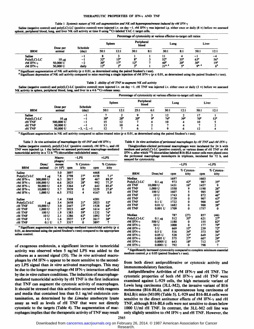
Augmentation of NK Activity. Injection (i.v.) of rM IFN-7significantly augmented NK activity in a dose-dependent manner in the spleen and peripheral blood, as well as in the lungsand the liver, which are common sites of metastatic formation(34). Repeated administration of rM IFN-7 resulted in hypo-responsiveness of NK activity at these sites (Table 1). NK cellactivity was assessed 24 h after the last of eight daily injectionsin the multiple-treated mice. The NK activity of mice whichreceived a single injection of rM IFN-7 was assessed in thisexperiment 48 h after administration. In previous studies, wehave found that NK activity peaks at 48-h postinjection, although similar high levels of activity are seen at 24 h (18 andresults not shown). We also assessed the ability of rH TNF toaugment NK activity in the various anatomical compartmentsto determine its in vivo immunomodulatory properties. In contrast to rM IFN-7, rH TNF was unable to augment NK cellactivity in any of the compartments examined (Table 2). RHTNF was administered at 50,000 or 500,000 U/animal 1 daybefore assay or daily beginning 3 days before the examinationof NK activity. Under these conditions, rH TNF was unable toaugment NK cell activity. In contrast, the positive control,poly(I,C)-LC, significantly augmented NK cell activity in everycompartment examined. In other studies, rH TNF doses from100 to 1 x IO6 U/animal were found to have no splenic NK
cell augmentation properties.Augmentation of Macrophage-mediated Cytotoxicity by rM
IFN-7 and rH TNF. The ability of rM IFN-7 and rH TNF toactivate macrophages in vivo was assessed using peritonealexúdate macrophages obtained 24 h after i.p. injection of eithercytokine. The data presented in Table 3 demonstrate that theseagents were able to augment macrophage-médiate cytotoxicityin vivo. Macrophage augmentation in vivo occurs at levels ofrH TNF > 1 U/animal and > 10,000 U/animal of rM IFN-7.The level of cytotoxicity induced by rM IFN-7 or rH TNF wasfurther augmented by adding 5 ng/ml LPS to the cytotoxicityassay. In other studies, increased macrophage activation hasbeen observed following multiple injections of rM IFN-7 compared to a single injection. A "bell-shaped curve" is also com
monly observed in these studies (results not shown). This invivo augmentation of macrophage-mediated tumoricidal activity may be secondary to TNF production or release of a cytokinethat acts as a secondary macrophage-activating agent. Theresults shown in Table 4 demonstrate that even high concentrations of rH TNF were only minimally effective in augmentingmacrophage-mediated /// vitro cytotoxicity in the absence of asecondary LPS signal. In vitro activation of macrophage tumoricidal activity with a secondary LPS signal occurs at levelsof TNF that do not result in direct cytotoxicity to B16-BL6tumor cells. Although both rH TNF and rM IFN-7 can significantly activate macrophage tumoricidal activity in the absence
2564
Research. on February 26, 2014. © 1987 American Association for Cancercancerres.aacrjournals.org Downloaded from
THERAPEUTIC PROPERTIES OF IFN-y AND TNF
Table 1 Systemic nature ofNK cell augmentation and NK cell hyporesponsiveness induced by rM IFN-ySaline (negative control) and poly(I,C)-LC (positive control) were injected i.v. on day -1. rM IFN--y was injected i.p. either once or daily (8 x) before we assessed
splenic, peripheral blood, lung, and liver NK cell activity at time 0 using "Cr-labeled YAC-1 target cells.
Percentage of cytotoxicity at various effector-to-target cellratiosBRMSaline
Poly(I,Q-LCrM IFN-7rM IFN-7Dose
peranimal10
Mg50,000 U50,000 USchedule
(day)-1-2
dailySpleen50:18
32*30"12»12:13
15«ir2»Peripheral
blood30:11
8"3*8:11 371Lung30:11152"
40°21°-*8:14
35"20°
5"Liver50:1-1
63°30°12:1-4
56°19°6»
" Significant augmentation of NK cell activity (p < 0.01, as determined using the paired Student's i test).'' Significant depression of NK cell activity compared to mice receiving a single injection of rM 11-N -•>Q»s 0,01, IS determined using the paired Student's t test).
Table 2 Ability ofrH TNF to augment NK cell activitySaline (negative control) and poly(I,C)-LC (positive control) were injected i.v. on day —1. rH TNF was injected i.v. either once or daily (3 x) before we assessed
NK activity in spleen, peripheral blood, lung, and liver in a 4-h "Cr-release assay.
Percentage of cytotoxicity at various effector-to-target cellratiosBRMSaline
Poly(I,C)-LCrHTNFrHTNFrHTNFDose
peranimal10
Mg500,000U
50,000 U50,000 USchedule
(day)-1
-1-1
-1-3,-2, -1Spleen50:17
38"
1513
1212:12
28"12107Peripheral
blood25:19
20'7
466:13
9"4
21Lung30:112
74'
107912:12
39°
446Liver50:117
l(f10
71212:11
52°
524
" Significant augmentation in NK cell activity compared to saline-treated mice (p < 0.01, as determined using the paired Student's / test).
Table 3 In vivo activation of macrophages by rH INF and rM IFN-ySaline (negative control), poly(I,C)-LC (positive control), rM IFN-i, and rH
TNF were injected i.p. 1 day before we assessed peritoneal macrophage-mediatedtumoricidal activity in a 72-h 125I-idoxuridine radiolabeled assay.
BRMSalinePoly(I,C)-LCrM
IFN-7rMIFN-7rMIFN-7rMIFN-7rMIFN-7SalinePoly(I,C)-LCrHTNFrHTNFrHTNFrHTNFrHTNFrHTNFMacro
phages/Dose/ mouseanimal (x10')luÃ500,000
U100,000U50,000U10,000
U1,000UI
Mg10,000U1,000
U100U10U1U0.1
U5.07.86.36.64.95.76.51.43.43.02.71.92.11.61.7-LPSt+LPS%
Cytotox- '
cpm icitycpm389727982817282533643939375233682658244720231383128629373317ir28*28"14'0421*27*40«59*62"13'0446841584679926423229416742012023152816141232109226173897%Cytotoxicity7.1*89.5°77.3°85.6°27.6°0.952«64«62"74"74'38*7
Table 4 In vitro activation of peritoneal macrophages by rH TNF and rM IFN-yThioglycollate-elicited peritoneal macrophages were incubated for 24 h with
medium and poly(I,C)-LC (positive control), or various doses of rH TNF or rMIFN-7, after which '•'"I-klo\ uriti iin- Iain-led B16-BL6 tumor cells were plated onto
the peritoneal macrophage monolayers in triplicate, incubated for 72 h, andassayed for cytotoxicity.
-LPS +LPS
BRM Dose/ml%Cytotox- % Cytotox-
cpm icity cpm icily
" Significant augmentation in macrophage-mediated tumoricidal activity (p <0.01, as determined using the paired Student's r test) compared to the appropriate
saline control.
of exogenous endotoxin, a significant increase in tumoricidalactivity was observed when 5 ng/ml LPS was added to thecultures as a second signal (35). The in vivo activated macrophages by rM IFN-7 appear to be more sensitive to the secondary LPS signal than in vitro activated macrophages. This maybe due to the longer macrophage rM IFN-7 interaction affordedby the in vitro culture conditions. The induction of macrophage-
mediated tumoricidal activity by rH TNF is the first indicationthat TNF can augment the cytotoxic activity of macrophages.It should be stressed that this activation occurred with reagentsand media that contained less than 0.1 ng/ml endotoxin contamination, as determined by the Limulus amebocyte lysateassay as well as levels of rH TNF that were not directlycytostatic to the targets (Table 4). The augmentation of macrophages implies that the therapeutic activity of TNF may result
MedianPoly(l,C)-LCrHTNFrHTNFrHTNFrHTNFrHTNFrHTNFrHTNFrHTNFMedianPoIy(I,C)-LCrM
IFN-7rMIFN-7rMIFN-7rMIFN-7rMIFN-7rMIFN-7rMIFN-7rM
IFN-70.1
Mg10,000U1,000
U100U10
U1U0.1U0.01U0.001U0.1
Mg500USOU5U0.5
U0.05U0.005U0.0005U0.0001
U1697973143115501607174317391722168217097875121180100066951652857564379242*16'9500000(27)35°0015'34'32"27*18"0160398714571190985872907900988175785762115517723937364960771279835°926°38«46"43'44«38*0(46)27"82«79°72*56'24'29*ir7" Significantly increased cytotoxicity compared to complete minimum essential
medium control; p £0.05 (paired Student's t test).
from both direct antiproliferative or cytotoxic activity andimmunomodulatory function.
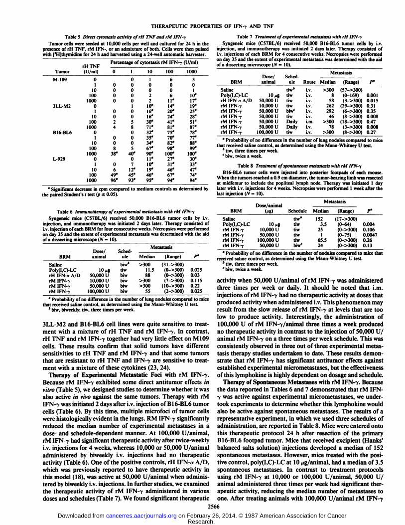
Antiproliferative Activities of rM IFN--y and rH TNF. Thecytostatic properties of both rM IFN-7 and rH TNF wereexamined against L-929 cells, the high metastatic variant ofLewis lung carcinoma (3LL-M2), the invasive variant of B16melanoma (B16-BL6), and a spontaneous lung carcinoma ofBALB/c mice (Ml09) (Table 5). L-929 and B16-BL6 cells weresensitive to the direct antitumor effects of rM IFN-7 and rHTNF, although B16-BL6 cells were not sensitive to doses below1000 U/ml rH TNF. In contrast, the 3LL-M2 cell line wasonly slightly sensitive to rM IFN-7 and rH TNF. However, the
2565
Research. on February 26, 2014. © 1987 American Association for Cancercancerres.aacrjournals.org Downloaded from
THERAPEUTIC PROPERTIES OF IFN--X AND TNF
Table 5 Direct cytoslasis activity ofrH TNF and rM IFN-y
Tumor cells were seeded at 10,000 cells per well and cultured for 24 h in thepresence of rH TNF, rM IFN-, or an admixture of both. Cells were then pulsedwith | 'I I|ih\mulini- for 24 h and harvested using a 24-well automatic harvester.
TumorM
IIW3LL-M2B16-BL6L-929rHTNF(U/ml)01101001000011010010000110100100001101001000Percentage
of cytostasis rM IFN--y(U/ml)00000002400838«0649"96«10000010058000540*0712*45"93«101002210«16'16'¡or71'32«35*34'67«90«11«10"19«46«95"100600611«14«20«24«41«81«75«70«82«98°100"27"31«46«67«94"100030110«ir19«25«28"51«87"78"rr88«99°100°30°33«47«74«94«
°Significant decrease in cpm compared to medium controls as determined bythe paired Student's / test (p < 0.05).
Table 6 Immunotherapy of experimental metastasis with rM IFN-ySyngeneic mice (C57BL/6) received 50,000 B16-BL6 tumor cells by i.v.
injection, and immunotherapy was initiated 2 days later. Therapy consisted ofi.v. injection of each BRM for four consecutive weeks. Necropsies were performedon day 35 and the extent of experimental metastasis was determined with the aidof a dissecting microscope (N = 10).
BRMSaline
Poly(I,C)-LCrH IFN-a A/DrM IFN-7rM IFN-7rM IFN-7Dose/
animal10
Mg50,000
U10,000 U50,000 U
100,000 USched
ulebiw*
tÃwbiwbiwbiwbiwMetástasisMedian>300
11.588
>300>300
55(Range)(31->300)
(0->300)(0->300)(7->300)
(10->300)(2->300)P°0.025
0.030.1150.220.025
" Probability of no difference in the number of lung nodules compared to micethat received saline control, as determined using the Mann-Whitney U test.
* biw, biweekly; tiw, three times per week.
3LL-M2 and B16-BL6 cell lines were quite sensitive to treatment with a mixture of rH TNF and rM IFN-7. In contrast,rH TNF and rM IFN-7 together had very little effect on M109cells. These results confirm that solid tumors have differentsensitivities to rH TNF and rM IFN-7 and that some tumorsthat are resistant to rH TNF and IFN-7 are sensitive to treatment with a mixture of these cytokines (23, 24).
Therapy of Experimental Metastatic Foci with rM IFN-7.Because rM IFN-7 exhibited some direct antitumor effects invitro (Table 5), we designed studies to determine whether it wasalso active in vivo against the same tumors. Therapy with rMIFN-7 was initiated 2 days after i.v. injection of B16-BL6 tumorcells (Table 6). By this time, multiple microfoci of tumor cellswere histologically evident in the lungs. RM IFN-7 significantlyreduced the median number of experimental métastasesin adose- and schedule-dependent manner. At 100,000 U/animal,rM IFN-7 had significant therapeutic activity after twice-weeklyi.v. injections for 4 weeks, whereas 10,000 or 50,000 U/animaladministered by biweekly i.v. injections had no therapeuticactivity (Table 6). One of the positive controls, rH IFN-a A/D,which was previously reported to have therapeutic activity inthis model ( 18), was active at 50,000 U/animal when administered by biweekly i.v. injections. In further studies, we examinedthe therapeutic activity of rM IFN-7 administered in variousdoses and schedules (Table 7). We found significant therapeutic
Table 7 Treatment of experimental metastasis with rH IFN-y
Syngeneic mice (C57BL/6) received 50,000 Hid HI 6 tumor cells by i.v.injection, and immunotherapy was initiated 2 days later. Therapy consisted ofi.v. injections of each BRM for 4 consecutive weeks. Necropsies were performedon day 35 and the extent of experimental metastasis was determined with the aidof a dissecting microscope (N ~- 10).
BRMSalinePoly(I,C)-LCrH
IFN-aA/DrMIFN-7rMIFN-7rMIFN-TrMIFN-frMIFN-7rM
IFN-7Dose/
animal10
Mg50,000U10,000U50,000U50,000U50,000U50,000
U100,000USched
uletiw*tiwtÃwtiwbiw*tiwDailyDailytiwMetástasisRoutei.v.I.V.I.V.I.V.LV.I.V.i.m.LV.I.V.Median>30085826229246>30078>300(Range)(57->300)(0-169)(3->300)(29->300)(6->300)(8->300)(18->300)(3->300)(8->300)P"0.0010.0150.310.350.0080.470.0080.27
" Probability of no difference in the number of lung nodules compared to micethat received saline control, as determined using the Mann-Whitney U test.
* tiw, three times per week.' biw, twice a week.
Table 8 Treatment of spontaneous metastasis with rM ¡FN-yB16-BL6 tumor cells were injected into posterior footpads of each mouse.
When the tumors reached a 0.9 cm diameter, the tumor-bearing limb was resectedat midfemur to include the popliteal lymph node. Therapy was initiated 1 daylater with i.v. injections for 4 weeks. Necropsies were performed 1 week after thelast injection (N =10).BRMSaline
Poly(I,C)-LCrM IFN-7rM IFN-7rM IFN-yrM IFN-7Dose/animal
0<g)10
Mg10,000 U50,000 U
100,000 U50,000 UMetastasisScheduletiw*
tÃwtÃwtÃwtiwbiw*Median152
3.525
165.524(Range)(17->300)
(0-64)(0->300)(0-75)(0->300)(0->300)P-0.004
0.1060.00470.260.13
" Probability of no difference in the number of nodules compared to mice thatreceived saline control, as determined using the Mann-Whitney U test.
' tiw, three times per week.' biw, twice a week.
activity when 50,000 U/animal of rM IFN-7 was administeredthree times per week or daily. It should be noted that i.m.injections of rM IFN-7 had no therapeutic activity at doses thatproduced activity when administered i.v. This phenomenon mayresult from the slow release of rM IFN-7 at levels that are toolow to produce activity. Interestingly, the administration of100,000 U of rM IFN-7/animal three times a week producedno therapeutic activity in contrast to the injection of 50,000 U/animal rM IFN-7 on a three times per week schedule. This wasconsistently observed in three out of three experimental metastasis therapy studies undertaken to date. These results demonstrate that rM IFN-7 has significant antitumor effects againstestablished experimental micrometastases, but the effectivenessof this lymphokine is highly dependent on dosage and schedule.
Therapy of Spontaneous Métastaseswith rM IFN-7. Becausethe data reported in Tables 6 and 7 demonstrated that rM IIN7 was active against experimental micrometastases, we undertook experiments to determine whether this lymphokine wouldalso be active against spontaneous métastases.The results of arepresentative experiment, in which we used three schedules ofadministration, are reported in Table 8. Mice were entered ontothis therapeutic protocol 24 h after resection of the primaryB16-BL6 footpad tumor. Mice that received excipient (Hanks'
balanced salts solution) injections developed a median of 152spontaneous métastases.However, mice treated with the positive control, poly(I,C)-LC at 10 Mg/animal, had a median of 3.5spontaneous métastases.In contrast to treatment protocolsusing rM IFN-7 at 10,000 or 100,000 U/animal, 50,000 U/animal administered three times per week had significant therapeutic activity, reducing the median number of métastasestoone. After treating animals with 100,000 U/animal rM IFN-7
2566
Research. on February 26, 2014. © 1987 American Association for Cancercancerres.aacrjournals.org Downloaded from
THERAPEUTIC PROPERTIES OF IFN-f AND TNF
three times per week (median of 65.5 métastasesper animal),we observed no significant therapeutic activity, compared tosaline or 50,000 U/animal rM IFN-7 administered three timesper week; these results support the findings of our experimentalmetastasis therapy studies (Table 7). In agreement with theexperimental metastasis assay shown in Table 6, 50,000 U/animal administered twice a week had no therapeutic activity.Therefore, rM IFN-7 appears to have a schedule- and dosage-dependent optima of therapeutic activity at 50,000 U/animaladministered three times per week for the treatment of experimental and spontaneous métastases.
Therapy of Experimental and Spontaneous MétastaseswithrM IFN-7 Alone or in Combination with rH TNF. The resultsof treating spontaneous métastaseswith three times per weeki.v. injection of rH TNF are shown in Table 9. These animalswere treated with 1,000,000, 500,000, 50,000, 5,000, or 500U/animal of rH TNF. When rH TNF was administered by i.v.injection, 10 ¿¿g/animalwas close to the maximum tolerateddose; indeed this dose of rH TNF is the LD-50 when the agentis administered by i.v. injection. We observed significant therapeutic activity when 50,000 or 5,000 U/animal rH TNF wasadministered by i.v. injection, but none of the doses producedsignificant activity when an i.p. route of administration wasused (Table 10). Unlike, rH TNF, daily i.p. injections of 50,000U/animal rH IFN-7 had significant therapeutic activity. Interestingly, when 50,000 U/animal of rH TNF was admixed withrM IFN-7 and administered daily by i.p. injection for 4 weeks,we noted a significant increase in therapeutic activity comparedto the activity of rM IFN-7 alone (p = 0.01). In a similar studyof B16-BL6, spontaneous métastaseswere treated with therapyinitiated 24 h after surgical resection of footpad tumors (averagediameter 1 cm). When we administered daily i.p. injections of
Table 9 Treatment of spontaneous metastasis with rH TNFB16-BL6 tumor cells were injected into the left or right posterior footpad of
each mouse. When the tumors reached a 0.9 cm diameter, the tumor-bearing limbwas resected at midfemur to include the popliteal lymph node. Therapy wasinitiated 1 day later with tiw (three times per week) i.v. injections for 4 weeks.Necropsies were performed on day 29, 1 day after the last injection (N = 10mice).
Metastasis
BRM Dose/animal MST Median (Range)
HBSSPoly(I,C)-LCrHTNFrHTNFrHTNFrHTNFrHTNF10/ig1,000,000
U500,000U50,000
U5,000U500
U>29>29317>29>29>29111000125(16-27)(0-4)(0-1)(0-65)(0-36)(0-36)(0-65)0.0000.0000.0270.0340.0200.496' Probability of no difference in the number of nodules compared to mice that
received saline control, as determined using the Mann-Whitney U test.
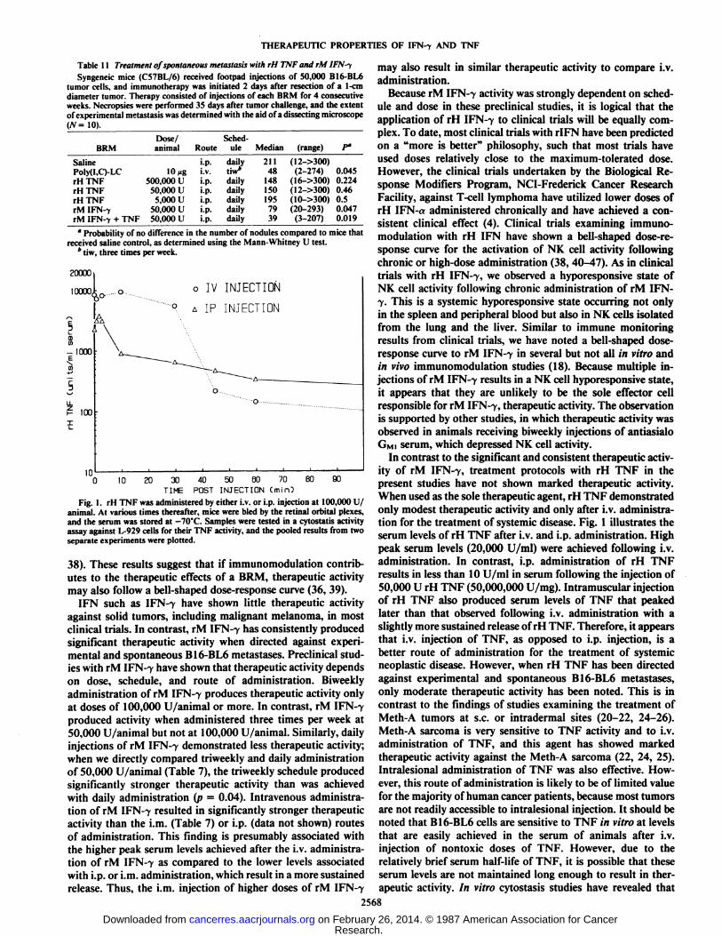
rH TNF over a three-log dose range, no significant therapeuticactivity was observed (Table 11). In contrast, poly(I,C)-LC anddaily i.p. injections of 50,000 U/animal rM IFN-7 producedsignificant therapeutic activity. However, we observed significant activity when i.p. injections of rM IFN-7 (50,000 U/animal) and rH TNF (500,000 U/animal) were concomitantlyadministered on a daily basis. Indeed, the administration ofthese two cytokines together produced significantly more activity than rM IFN-7 (P = 0.041). This increase in therapeuticactivity was suggested by the in vitro cytostatis studies reportedin Table 5. Therefore, it appears that at least part of thetherapeutic activity of these cytokines is associated with theircytostatic properties. However, this is unlikely to be the complete mechanism of activity, because we found approximately1,000 U/ml of serum TNF after the i.p. administration of100,000 U/animal of rH TNF (Fig. 1). This level, which wasmaintained for 10 min after i.p. administration, is sufficient invitro to produce cytostatic and cytotoxic activity against B16-BL6 cells but only if maintained for 24 h. It is somewhat morelikely that any therapeutic activity achieved with rH TNF byi.p. injection is associated with the immunomodulatory properties of that agent. After i.v. administration of 100,000 U/animal of rH TNF, TNF levels of approximately 10,000 U/mlare obtained, which is sufficient to kill over 95% of B16-BL6tumor cells in vitro (results not shown). However, this highlevel of serum rH TNF activity was transient (30 min) and isnot maintained long enough to produce cytostatic activityagainst B16-BL6 tumor cells. It seems likely therefore, that theimmunomodulatory properties of rH TNF also have a role inits therapeutic activity.
DISCUSSION
The most challenging aspect of cancer treatment is the control of métastasesthat are present at primary tumor diagnosis(33, 34, 36). Therefore, central to the application of a BRM toclinical oncology is the systematic preclinical development ofprotocols for the treatment of preexisting, well-established métastases. These studies can then be used as a guide for the designof clinical protocols. Optimization of administration protocolsshould be applicable to Phase Ib and/or Phase II clinical trials.In addition to determining a maximum tolerated dose, preclinical models should be used to determine the optimal therapeuticprotocol that may subsequently be translated into clinical trials.The optimal therapeutic property depends not only on dosagebut also on the schedule, duration, and route of administration.In several preclinical and clinical studies of immunomodula-tion, a bell-shaped dose-response curve has been observed (37,
Table 10 Treatment of experimental metastasis with rH TNF and rM IFN-yC57BL/6 mice were injected i.v. with 5 x IO1 B16-BL6 tumor cells, and immunotherapy was initiated 2 days later. Therapy consisted of the injection of each
BRM for 4 consecutive weeks. Necropsies were performed 35 days following tumor challenge, and the extent of experimental métastaseswas determined with the aidof a dissecting microscope (N - 10).
MetastasisBRMSalinePoly(I,C)-LCrHTNFrHTNFrHTNFrHTNFrM
IFN-7rHTNF/rM IFN-?Dose/animal10
Mg500,000U50,000
U500,000U50,000U50,000U50,000
URoutei.p.I.V.M>.l.p.l.p.I.V.i.p.i.p.Scheduledailytiw4dailydailydailydailydailydailyMedian5532629292471(range)(0->300)(0-17)(0-81)(5-141)(0-257)(6-88)(0-54)(0-5)%
of tumor-bearingmice909090100901008070P"0.00120.070.350.160.0410.0030.009e
°Probability of no difference in the number of nodules compared to the mice which received the HBSS control as determined using the Mann-Whitney U test.* tiw, three times per week.' Probability of no difference in the number of nodules compared to the mice which received the -, II \ alone (/' < 0.01) as determined using the Mann-Whitney
U test.
2567
Research. on February 26, 2014. © 1987 American Association for Cancercancerres.aacrjournals.org Downloaded from
THERAPEUTIC PROPERTIES OF IFN--y AND TNF
Table 11 Treatment of spontaneous metastasis with rH TNF and rM IFN-tSyngeneic mice (C57BL/6) received footpad injections of 50,000 B16-BL6
tumor cells, and immunotherapy was initiated 2 days after resection of a 1-cmdiameter tumor. Therapy consisted of injections of each BRM for 4 consecutiveweeks. Necropsies were performed 35 days after tumor challenge, and the extentof experimental metastasis was determined with the aid of a dissecting microscope</V=10).
BRMSaline
Poly(I,C)-LCrHTNFrHTNFrHTNFrMIFN-rrM
IFN-7 + TNFDose/
animal10
fig500,000
U50,000U5,000
U50,000U50,000
USched-
Routeulei.p.
dailyi.v.tiw*i.p.
dailyi.p.dailyi.p.dailyi.p.dailyi.p.
dailyMedian211
481481501957939(range)(12->300)(2-274)(16->300)(12->300)(10->300)(20-293)(3-207)f0.0450.2240.460.50.0470.019
" Probability of no difference in the number of nodules compared to mice thatreceived saline control, as determined using the Mann-Whitney U test.
* tiw, three times per week.
20000
o IV INJECTION-9.A IP INJECTION
30 40 50 60 70 80 90
TIME POST INJECTION Cmin)
Fig. 1. rH TNF was administered by either i.v. or i.p. injection at 100,000 U/animal. At various times thereafter, mice were bled by the retinal orbital plexes,and the serum was stored at -70'C. Samples were tested in a cytostatis activityassay against L-929 cells for their TNF activity, and the pooled results from twoseparate experiments were plotted.
38). These results suggest that if immunomodulation contributes to the therapeutic effects of a BRM, therapeutic activitymay also follow a bell-shaped dose-response curve (36, 39).
IFN such as IFN-7 have shown little therapeutic activityagainst solid tumors, including malignant melanoma, in mostclinical trials. In contrast, rM IFN-7 has consistently producedsignificant therapeutic activity when directed against experimental and spontaneous B16-BL6 métastases.Preclinical studies with rM IFN-7 have shown that therapeutic activity dependson dose, schedule, and route of administration. Biweeklyadministration of rM IFN-7 produces therapeutic activity onlyat doses of 100,000 U/animal or more. In contrast, rM IFN-7produced activity when administered three times per week at50,000 U/animal but not at 100,000 U/animal. Similarly, dailyinjections of rM IFN-7 demonstrated less therapeutic activity;when we directly compared triweekly and daily administrationof 50,000 U/animal (Table 7), the triweekly schedule producedsignificantly stronger therapeutic activity than was achievedwith daily administration (p = 0.04). Intravenous administration of rM IFN-7 resulted in significantly stronger therapeuticactivity than the i.m. (Table 7) or i.p. (data not shown) routesof administration. This finding is presumably associated withthe higher peak serum levels achieved after the i.v. administration of rM IFN-7 as compared to the lower levels associatedwith i.p. or i.m. administration, which result in a more sustainedrelease. Thus, the i.m. injection of higher doses of rM IFN-7
may also result in similar therapeutic activity to compare i.v.administration.
Because rM IFN-7 activity was strongly dependent on schedule and dose in these preclinical studies, it is logical that theapplication of rH IFN-7 to clinical trials will be equally complex. To date, most clinical trials with rIFN have been predictedon a "more is better" philosophy, such that most trials have
used doses relatively close to the maximum-tolerated dose.However, the clinical trials undertaken by the Biological Response Modifiers Program, NCI-Frederick Cancer ResearchFacility, against T-cell lymphoma have utilized lower doses ofrH IFN-«administered chronically and have achieved a consistent clinical effect (4). Clinical trials examining immunomodulation with rH IFN have shown a bell-shaped dose-response curve for the activation of NK cell activity followingchronic or high-dose administration (38, 40-47). As in clinicaltrials with rH IFN-7, we observed a hyporesponsive state ofNK cell activity following chronic administration of rM IFN-7. This is a systemic hyporesponsive state occurring not onlyin the spleen and peripheral blood but also in NK cells isolatedfrom the lung and the liver. Similar to immune monitoringresults from clinical trials, we have noted a bell-shaped dose-response curve to rM IFN-7 in several but not all in vitro and/// vivo immunomodulation studies (18). Because multiple injections of rM IFN-7 results in a NK cell hyporesponsive state,it appears that they are unlikely to be the sole effector cellresponsible for rM IFN-7, therapeutic activity. The observationis supported by other studies, in which therapeutic activity wasobserved in animals receiving biweekly injections of antiasialoGMI serum, which depressed NK cell activity.
In contrast to the significant and consistent therapeutic activity of rM IFN-7, treatment protocols with rH TNF in thepresent studies have not shown marked therapeutic activity.When used as the sole therapeutic agent, rH TNF demonstratedonly modest therapeutic activity and only after i.v. administration for the treatment of systemic disease. Fig. 1 illustrates theserum levels of rH TNF after i.v. and i.p. administration. Highpeak serum levels (20,000 U/ml) were achieved following i.v.administration. In contrast, i.p. administration of rH TNFresults in less than 10 U/ml in serum following the injection of50,000 U rH TNF (50,000,000 U/mg). Intramuscular injectionof rH TNF also produced serum levels of TNF that peakedlater than that observed following i.v. administration with aslightly more sustained release of rH TNF. Therefore, it appearsthat i.v. injection of TNF, as opposed to i.p. injection, is abetter route of administration for the treatment of systemicneoplastic disease. However, when rH TNF has been directedagainst experimental and spontaneous B16-BL6 métastases,only moderate therapeutic activity has been noted. This is incontrast to the findings of studies examining the treatment ofMeth-A tumors at s.c. or intradermal sites (20-22, 24-26).Meth-A sarcoma is very sensitive to TNF activity and to i.v.administration of TNF, and this agent has showed markedtherapeutic activity against the Meth-A sarcoma (22, 24, 25).Intralesional administration of TNF was also effective. However, this route of administration is likely to be of limited valuefor the majority of human cancer patients, because most tumorsare not readily accessible to intralesional injection. It should benoted that B16-BL6 cells are sensitive to TNF in vitro at levelsthat are easily achieved in the serum of animals after i.v.injection of nontoxic doses of TNF. However, due to therelatively brief serum half-life of TNF, it is possible that theseserum levels are not maintained long enough to result in therapeutic activity. In vitro cytostasis studies have revealed that
2568
Research. on February 26, 2014. © 1987 American Association for Cancercancerres.aacrjournals.org Downloaded from
THERAPEUTIC PROPERTIES OF IFN-y AND TNF
several hours of continuous exposure to rH TNF is needed toresult in cytostasis (23, 48). A continuous infusion might beused to advantage in clinical trials, but this method may alsoincrease toxicity.
Although our preclinical studies of rH TNF alone, directedagainst metastatic disease, have produced disappointing results,this compound has consistently demonstrated complementarytherapeutic activity when administered with rM IFN-7 whenboth were administered i.p. It should be noted that rH TNFwas used in these studies and that greater activity might occurwith rH TNF. Most treatment protocols have involved i.p.administration of rH TNF and rM IFN-7 to allow daily administration. Additive therapeutic activity was observed in experimental and spontaneous metastasis models against B16-BL6tumors. Similar results were observed against B16-BL6, M109,and 3LL-M2 tumors located at an s.c. site (results not shown).However, when rM IFN-7 was administered i.v. using an optimal therapeutic protocol, no significant increase in therapeuticactivity was observed with the incorporation of rH TNF, to theprotocol. Thus, the increased therapeutic activity requiressuboptimal rM IFN-7 protocols using either i.p. administrationor lower doses of the rM IFN-7 (10,000 U/animal administeredi.v.). However, the utilization of an optimal protocol of rHIFN-7 within a clinical trial is at present unlikely and therapeutic synergism with rTNF and rIFN-7 compared to either alonemay be observed. The mechanism of this complementary effectis not yet understood, although IFN-7 and rH TNF are additivefor the augmentation of macrophage-mediated tumoricidal activity (results not shown). An alternative, additional explanationis that therapeutic activity may result from the additive cytostasis associated with IFN-7 and TNF (Table 5). This mechanism may involve an upregulation of receptors for TNF, aphenomenon that has been suggested to occur after IFN-7exposure (48). However, it is likely that both mechanisms ofaction are involvedand that TNF acts as an immunomodulatoras well as a cytostatic or cytotoxic agent. In addition, rH TNFappears to have numerous indirect effects on platelets and thefibrin cascade and may also have toxic and therapeutic activityvia, this mechanism.4
In summary, the systematic preclinical study of immunomod-ulatory and immunotherapeutic parameters can be very informative. It remains to be determined whether such an approachwill facilitate the transition of an agent into clinical studies andsimilar clinical trials will be required to determine the validityof such an approach. A growing body of evidence suggests thattreating patients with IFN-7 at or near the maximum tolerateddose may not always be optimal because therapeutic activitymay also be apparent at lower immunomodulatory doses. It isimportant to stress that immunomodulators need to be chronically administered but in a defined manner. Unlike treatmentprotocols using cytostatic or cytotoxic agents, BRMs must besequentially or continuously administered over a prolongedperiod. Administering a moderate immunomodulatory dose ofIFN-7 three times per week over a prolonged period results inan increased therapeutic response compared to the results obtained with higher dose therapeutic protocols. Unlike IFN-7,immunotherapeutic protocols for rH TNF have yet to be optimized either preclinically or clinically. It appears that i.v.injection is the most effective route of administration althoughit is possible that continuous infusion i.v. may be superior to apush i.v. administration. Preclinical studies suggest that theoptimal therapeutic response occurs after concomitant admin-
4J. E. Talmadge, H. R. Tribble, R. W. Pennington, H. Phillips, and R. H.
Wiltrout, manuscript in preparation.
istration of TNF and IFN-7 or some other agent that potentiates TNF therapeutic activity.
REFERENCES
1. Foon, K. A., Sherwin, S. A., Abrams, P. G., Longo, D. L., Fer, M. F.,Stevenson, H. C, Ochs, J. J., Bottino, G. C., Schoenberger, C. S., Zeffren,J., Jalïe,E. S., and ( »illuni. R. K. Treatment of advanced non-Hodgkin's
lymphoma with recombinant leukocyte A interferon. N. Engl. J. Med., ill:1148-i 152, 1984.
2. Quesada, J. R., Swanson, D. A., Trindade, A., and Gutterman, J. U. Renalcell carcinoma: antitumor effects of leukocyte interferon. Cancer Res., 43:940-947, 1983.
3. Neidhart, J. A., Gagen, M. M., Young, D., Tuttle, R., Melink, T. J.,Ziccarrelli, A., and Kisner, D. Interferon-a therapy of renal cancer. CancerRes., 44:4140-4143, 1984.
4. Bunn, P. A., Jr., Foon, K. A., Ihde, D. C., Longo, D. L., Eddy, J., Winkler,C. F., Veach, S. R., Zeffren, J., Sherwin, S., and Oldham, R. Recombinantleukocyte A interferon: an active agent in advanced cutaneous T-cell lympho-mas. Ann. Intern. Med., 101:484-487, 1984.
5. Quesada, J. R., and Gutterman. J. U. Clinical study of recombinant DNA-produced leukocyte interferon (clone A) in an intermittent schedule in cancerpatients. J. Nati. Cancer Inst., 70: 1041-1046, 1983.
6. Sherwin, S. A., Foon, K. A., Abrams, P. G., Heyman, M. R., Ochs, J. J.,Watson, T., Maluish, A., and Oldham, R. K. A preliminary phase I trial ofpartially purified interferon--y in patients with cancer. J. Biol. ResponseModif., 3:599-607, 1984.
7. Kirkwood. J. M., and Ernstoff, M. S. Interferons in the treatment of humancancer. J. Clin. Oncol., 2: 336-357, 1984.
8. Krown, S. E., Burk, M. W., Kirkwood, J. M., Kerr, D., Morton. D. L., andOettgen, H. F. Human leukocyte (alpha) interferon in metastatic malignantmelanoma: The American Cancer Society phase II trial. Cancer Treat. Rep.,68:723-726, 1984.
9. Borden, E. C. Interferons and cancer: how the promise is being kept. In: I.Gresser (ed), Interferons, p. 43. London: Academic Press, 1984.
10. van der Burg, M., Edelstein, M., Gerlis, L., Liang, C-M., Hirschi, M., andDawson, A. Recombinant interferon-7 (Immuneren): results of a phase Itrial in patients with cancer. J. Biol. Response Mudii'.. 4: 264-272, 1985.
11. Schulof, R. S., Lloyd, M. J., Stallings, J. J., Mai, D., Phillips, T. M., Jones,G. J., and Schechter, G. P. Recombinant leukocyte A interferon in B-cellchronic lymphocytic leukemia: in vivo effects on autologous antitumor immunity. J. Biol. Response Modif., 4: 310-323, 1985.
12. Berek, J. S., Hacker, N. F., Lichtenstein, A., Jung, T., Spina, C., Knox, R.M., Brady, J., Greene, T., Ettinger, L. M., Lagasse, L. D., Bonnern, E. M.,Spiegel, R. J., and Zighelboim, J. Intraperitoneal recombinant a-imerferonfor "salvage" immunotherapy in state III epithelial ovarian cancer: a gynecologic oncology group study. Cancer Res., 45:4447-4453, 1985.
13. Edwards, B. S., Merritt, J. A., Fuhlbrigge, R. C., and Borden, E. C. Lowdoses of interferon alpha result in more effective clinical natural killer cellactivation. J. Clin. Invest., 75: 1908-1913, 1985.
14. Bonnern, E. M., and Spiegel, R. J. Interferon-a: current status and futurepromise. J. Biol. Response Modif., 3: 580-598, 1985.
15. Sherwin, S. A., Foon, K. A., Abrams, P. G., Heyman, M. R., Ochs, J. J.,Watson, T., Maluish, A., and Oldham, R. K. A preliminary phase I trial ofpartially purified interferon--y in patients with cancer. J. Biol. ResponseModif., 3: 599-607, 1984.
16. Biological response modifiers: subcommittee report. Nat. Cancer Inst.Monog. 63:1983.
17. Rubin, B. Y.. and Gupta, S. L. Differential efficacies of human type I andtype II interferons as antiviral and antiproliferative agents. Proc. Nati. Acad.Sci. USA, 77:5928-5932, 1980.
18. Talmadge, J. E., Herberman, R. B., Chirigos, M. A., Maluish, A. E., Schneider, M. A., Adams, J. S., Phillips, H., Thurman, G. B., Varesio, L., Long,C. W., Oldham, R. K., and Wiltrout, R. H. Hyporesponsiveness to augmentation of murine natural killer cell activity in different anatomical compartments by multiple injections of various immunomodulators including recombinant interferons and interleukin 2. J. Immunol., 135: 2483-2491, 1985.
19. Varesio, L., Blasi, E., Thurman, G. B., Talmadge, J. E., Wiltrout, R. H., andHerberman, R. B. Potent activation of mouse macrophages by recombinantinterferon-7. Cancer Res., 44:4465-4469, 1984.
20. Carswell, E. A., Old, L. J., Kassel, R. L., Green, S., Fiore, N., and Williamson, B. An endotoxin-induced serum factor that causes necrosis of tumors.Proc. Nati. Acad. Sci. USA 72: 3666-3670, 1975.
21. Haranaka, K., and Satomi, N. Cytotoxic activity of tumor necrosis factor(TNF) on human cancer cells in vitro. Jpn. J. Exp. Med., 51:191-194,1981.
22. Haranaka, K., Satomi, N., and Sakurai, A. Antitumor activity of murinetumor necrosis factor (TNF) against transplanted murine tumors and heter-otransplanted human tumors in nude mice. Int. J. Cancer, 34: 263-267,1984.
23. Sugarman, B. J., Aggarwal, B. B., Hass, P. E., Figari, I. S., Palladino, M.A., Jr., and Shepard, H. M. Recombinant human tumor necrosis factor-alpha: effects on proliferation of normal and transformed cells in vitro.Science (Wash. DC), 230:943-945, 1985.
24. Williamson, B. D., Carswell, E. A., Rubin, B. Y., Prendergast, J. S., and Old,L. J. Human tumor necrosis factor produced by human B-cell lines: syner-
2569
Research. on February 26, 2014. © 1987 American Association for Cancercancerres.aacrjournals.org Downloaded from
THERAPEUTIC PROPERTIES OF IFN-7 AND TNF
gistic cytotoxic interaction with human interferon. Proc. Nati. Acad. Sci.USA, «0:5397-5401,1983.
25. Wang, A. M., Creasey, A. A., Ladner, M. B., Lin, L. S., Strickler, J., VanArsdell, J. N., Yamamoto, R., and Mark, D. F. Molecular cloning of thecomplementary DNA for human tumor necrosis factor. Science (Wash. DC),22«:149-154, 1985.
26. Ruff, M. R., and Gifford, G. E. Tumor necrosis factor. In: Lymphokines,Vol. 2. New York: Academic Press, 1981.
27. Shirai, T., Yamaguchi, H., Ito, H., Todd, C. W., and Wallace, R. B. Cloningand expression in Escherichia coli of the gene for human tumour necrosisfactor. Nature (Lond.), ¡lì:803-806, 1985.
28. Klein, E., and Klein, G. Antigenic properties of lymphomas induced by theMoloney agent. J. Nati. Cancer Inst., 32: 547-568, 1964.
29. Hart, 1. R. The selection and characterization of an invasive variant of B16melanoma. Am. J. Palhol., 97:587-600, 1979.
30. Si hull/, R. M., Papamatheakis, J. IV. and Chirigos, M. A. Interferon: aninducer of macrophage activation by polyanions. Science (Wash. DC), 197:674-676, 1977.
31. Talmadge, J. E., and Fidler, I. J. Enhanced metastatic potential of tumorcells harvested from spontaneous métastasesof heterogeneous murine tumors. J. Nail. Cancer Inst., 69: 975-980, 1982.
32. Mannel, D. N., Parrar, J. J., and Mergenhagen, S. E. In: A. L. de Weck, F.Kristensen, and M. Landy (eds.), Biochemical Characterization of Lymphokines. p. 303. New York: Academic Press, 1980.
33. Talmadge, J. E., Fidler, I. J., and Oïdium. R. K. Screening for BiologicalResponse Modifiers: Methods and Rationale. Boston: Martinus Nijhoff,1985.
34. Talmadge, J. E., Liotta, L. A., and Kohn, R. R. Biology and biochemistry ofmetastatic cells. In: Peterson, Elias, and Sonis (eds.). Head and Neck Management. The Hague: Martinus Nijhoff, 1986.
35. Russell, S. W., Doe, W. F., Mclntoch, A. T. Functional characterization ofa stable noncytolytic stage of macrophage activation in tumors. J. Exp. Med.,146:IS\\, 1977.
36. Talmadge, J. E., and Herberman, R. B. The preclinical screening laboratory:evaluation of immunomodulatory and therapeutic properties of biologicalresponse modifiers. Cancer Treat. Rep., 70: 171-182, 1986.
37. Talmadge. J. E., Adams, J., Phillips, H., Collins, M., Lenz, B., Schneider,M., Schlick, E., Ruffman, R., Wiltrout, R. H., and Chirigos, M. A. Immunomodulatory effects in mice of polyinosinic-polycytidylic acid complexed withpoly-L-lysine and carboxymethylcellulose. Cancer Res. 45: 1058-1065,1985.
38. Ernstoff, M. S., Reich, S., Nishoda V., and Kirkwood, J. M. Immunologia!assessment of melanoma patients treated with recombinant interferon gamma(rll N -,. Biogen, Inc., Cambridge, MA) in a phase 111 trial. Immunology,26: 280-286, 1985.
39. Talmadge, J. E., Adams, J. S., Phillips, H., Collins, M., Lenz, B., Schneider,M., and Chirigos, M. A. Immunotherapeutic potential in murine tumormodels of polyinosinic-polycytidylic acid and poly-L-lysine solubilized bycarboxymethylcellulose. Cancer Res., 45:1066-1072, 1985.
40. Maluish, A. E., Ortaldo. J. R., Conlon, J. < .. Sherwin, S. A., Leavitt, R.,Strong, D. M., Wiernik, P., oïdium. R. K., and Herberman, R. B. Depressionof natural killer cytotoxicity after in vivo administration of recombinantleukocyte interferon. J. Immunol., 131: 503-507, 1983.
41. Spina, C. A., Fahey, J. L., Dur kos Smith, D., Dorey, F., and Sarna, G.Suppression of natural killer cell cytotoxicity in the peripheral blood ofpatients receiving interferon therapy. J. Biol. Response Modif., 2:458-469,1983.
42. Maluish, A. E., Leavitt, R., Sherwin, S. A., Oldham, R. K., and Herberman,R. B. Effects of recombinant interferon-a on immune function in cancerpatients. J. Biol. Response Modif., 2:470-481, 1983.
43. Maluish, A. E., Ortaldo, J. R., Sherwin, S. A., Oldham, R. K., and Herber-niiin. R. B. Changes in immune function in patients receiving natural leukocyte interferon. J. Biol. Response Modif., 2:418-427, 1983.
44. Huddlestone, J. R., Merigan, T. ( '.. Jr., and Oldstone, M. B. A. Induction
and kinetics of natural killer cells in humans following interferon therapy.Nature (Lond.), 2«2:417-419, 1979.
45. Einhorn, S., Ahre, A., Blomgren, H., Johansson, B., Mellstedt, H., andStrander, H. Interferon and natural killer activity in multiple myeloma. Lackof correlation between interferon-induced enhancement of natural killeractivity and clinical response to human interferon-a. Int. J. Cancer, Ml: 167-172, 1982.
46. Golub, S. H., D'Amore, P., and Rainey, M. Systemic administration of
human leukocyte interferon to melanoma patients. II. Cellular events associated with changes in natural killer cytotoxicity. J. Nati. Cancer Inst., 6«:711-717, 1982.
47. Hawrylowicz, C. M., Rees, R. C, Hancock, B. W., and Potter, C. W.Depressed spontaneous natural killing and interferon augmentation in patients with malignant lymphoma. Eur. J. Cancer Clin. On«il., I«:1081-1088, 1982.
48. Aggarwal, B. B., Eessalu, T. E., and Hass, P. E. Characterization of receptorsfor human tumour necrosis factor and their regulation by •x-interferon.Nature(Lond.), 318: 665-667, 1985.
2570
Research. on February 26, 2014. © 1987 American Association for Cancercancerres.aacrjournals.org Downloaded from