Recombinant-derived interleukin-1α stabilized against specific deamidation
6
Protein Engineering vol 1 no 5 pp 413-417, 1987 Recombinant-derived interleukin-la stabilized against specific deamidation Paul T. Wingfield, Robert J.Mattaliano 1 , H.Robson MacDonaliP, Stewart Craig 3 , G.Marius Clore 4 , Angela M.Gronenborn 4 and Ursula Schmeissner Biogen SA, PO Box 1211 Geneva, Switzerland, 'Biogen Research Corporation, 14 Cambridge Center, Cambridge, MA 02142, USA, 2 Ludwig Institute for Cancer Research, t Mianiv Branch, 1066 Epalinges, Switzerland, 3 Department of Biochemistry, University of Newcastle, Newcastle upon Tyne NE1 7RU, UK and 4 Max-Planck-Instrtut fur Biochemie, 8033 Martinsned, FRG Recombinant-derived human interieuldn-la (IL-la), purified from Escherichia coti, was resolved by isoelectric focusing on polyacrylamide gels into two species of isoelectric points (pi) 5.45 and 5.20, which constituted - 7 5 % and - 2 5 % of the total IL-la protein respectively. The pi 5.45 and pi 5.20 species were separated by chromatofocusing and subjected to N-terminal sequence analysis. The pi 5.45 species contained the expected Asn residue at position 36 of the mature pro- tein sequence whereas the pi 5.20 species contained an Asp residue at the same position. A mutant protein in which Asn-36 was substituted for a Ser residue was isolated from E.coli and shown to be homogeneous on isoelectric focusing analysis with a pi = 5.45. 'H-n.m.r. and circular dkhroism analyses of wild-type and the mutant IL-la indicated a similar conformation which was also indicated by the identical recep- tor binding affinities of IL-la with Asn, Asp or Ser in position 36. The mutant protein was stabilized against specific base- catalysed and temperature-induced deamidation, and may be more suitable than the wild-type position for physical and structural studies. Key words: tnterleukin-la/sequence determination/deamidation/ site-specific mutagenesis/protein conformation Introduction We have recently described the purification of recombinant human interleukin-la (IL-la) from Escherichia coli using relatively mild conditions which avoided denaturating solvents and extremes of pH and temperature (Wingfield et al., 1987a). Nevertheless, the preparation was heterogeneous by isoelectric focusing. Two distinct IL-la species of pi 5.45 (70-75% of protein) and pi 5.20 (25-30% of protein) were observed. Titra- tion curve analysis on polyacrylamide gel (performed as described by Wingfield et al., 1987b) indicated that the pi 5.20 species was a once-deamidated protein. This observation was of particular interest since Cameron et al. (1986) had reported the purifica- tion of two species of authentic human IL-la from mononuclear cell cultures with similar isoelectric points as the E.cofi-derived protein. Furthermore, these authors demonstrated that residue 36 of the pi 5.20 species was an Asp, whereas the pi 5.45 species contained an Asn in the same position (note residue 36 of the mature protein sequence is equivalent to residue 148 of the precursor sequence listed by March et al., 1985). They suggested that this sequence difference might account for the small charge difference between the two species and may be due to either a single base change or deamidation. Both authentic IL-la species were shown to have similar biological activities. In this report, by direct N-terminal sequencing of the E.coli- derived IL-la species, we confirm the presence of Asn-36 in the pi 5.45 species and Asp-36 in the pi 5.20 species. Using site- specific mutagenesis to substitute the Asn-36 for a serine residue, we show that this mutant IL-la is purified from E.coli as a single charge species (pi = 5.45) with similar properties to the wild- type protein. The IL-la mutant, thus stabilized against specific deamidation, is suggested to be a more suitable starting material for physical characterization studies such as 'H-n.m.r. and X- ray crystallographic analysis. The use of site-specific mutagenesis to substitute protein Asn residues susceptible to deamidation was previously reported by Ahem et al. (1987) studying the thermostability of yeast triosephosphate isomerase. Materials and methods Preparation of wild-type and mutant IL-la Site-specific mutagenesis was used to introduce the point muta- tion in the IL-la molecule. The template was the phage M13 derivative mp8 carrying the coding region for mature IL-la. Mutagenesis was carried out by the double-primer procedure (Norris et al., 1983) with oligonucleotides carrying one or two mismatches. After vertification of the mutation by the dideoxy sequencing method (Sanger et al., 1977), the modified IL-la gene was transferred to a plasmid to be expressed under control of the \ P L promoter and the bacteriophage Mu ner gene ribosome binding site. The E.coli host was W3110 cI857, carrying the gene for the temperature-sensitive repressor of phage X. Fermentation and protein purification procedures were as previously described (Wingfield et al., 1987a). Analytical separation methods SDS-PAGE and isoelectric focusing on thin-layer poly- acrylamide gels were performed as previously described (Wingfield et al., 1987b). Chemical analysis Prior to analysis, samples were either directly dialysed against 0.1% (w/v) SDS and 2 mM NaCl, or electroeluted from SDS-PAGE gel slices (Hunkapillar et al., 1983) and then dialysed. N-terminal amino acid sequence determination by automated Edman degradation was performed with a gas-phase sequencer (Applied Biosystems Model 470A) using the regular program 03RpTH. In order to minimize the normal increase in cycle lag after Pro residues, the time of the cleavage step for Pro residue 3 was extended by 50%. Identification and quantita- tion of the phenylthiohydantoin derivatives was performed on line using an Applied Biosystems Model 120A PTH amino acid analyser with external standard quantitation. Amino acid analysis was carried out as previously described (Wingfield et al., 1986). © 1RL Press Limited, Oxford, England 413 at National Institutes of Health Library on March 27, 2012 http://peds.oxfordjournals.org/ Downloaded from
-
Upload
independent -
Category
Documents
-
view
1 -
download
0
Transcript of Recombinant-derived interleukin-1α stabilized against specific deamidation
Protein Engineering vol 1 no 5 pp 413-417, 1987
Recombinant-derived interleukin-la stabilized against specificdeamidation
Paul T. Wingfield, Robert J.Mattaliano1, H.RobsonMacDonaliP, Stewart Craig3, G.Marius Clore4, AngelaM.Gronenborn4 and Ursula Schmeissner
Biogen SA, PO Box 1211 Geneva, Switzerland, 'Biogen ResearchCorporation, 14 Cambridge Center, Cambridge, MA 02142, USA, 2LudwigInstitute for Cancer Research, t Mianiv Branch, 1066 Epalinges,Switzerland, 3Department of Biochemistry, University of Newcastle,Newcastle upon Tyne NE1 7RU, UK and 4Max-Planck-Instrtut furBiochemie, 8033 Martinsned, FRG
Recombinant-derived human interieuldn-la (IL-la), purifiedfrom Escherichia coti, was resolved by isoelectric focusing onpolyacrylamide gels into two species of isoelectric points (pi)5.45 and 5.20, which constituted -75% and -25% of thetotal IL-la protein respectively. The pi 5.45 and pi 5.20species were separated by chromatofocusing and subjectedto N-terminal sequence analysis. The pi 5.45 species containedthe expected Asn residue at position 36 of the mature pro-tein sequence whereas the pi 5.20 species contained an Aspresidue at the same position. A mutant protein in whichAsn-36 was substituted for a Ser residue was isolated fromE.coli and shown to be homogeneous on isoelectric focusinganalysis with a pi = 5.45. 'H-n.m.r. and circular dkhroismanalyses of wild-type and the mutant IL-la indicated a similarconformation which was also indicated by the identical recep-tor binding affinities of IL-la with Asn, Asp or Ser in position36. The mutant protein was stabilized against specific base-catalysed and temperature-induced deamidation, and may bemore suitable than the wild-type position for physical andstructural studies.
Key words: tnterleukin-la/sequence determination/deamidation/site-specific mutagenesis/protein conformation
IntroductionWe have recently described the purification of recombinanthuman interleukin-la (IL-la) from Escherichia coli usingrelatively mild conditions which avoided denaturating solventsand extremes of pH and temperature (Wingfield et al., 1987a).Nevertheless, the preparation was heterogeneous by isoelectricfocusing. Two distinct IL-la species of pi 5.45 (70-75% ofprotein) and pi 5.20 (25-30% of protein) were observed. Titra-tion curve analysis on polyacrylamide gel (performed as describedby Wingfield et al., 1987b) indicated that the pi 5.20 specieswas a once-deamidated protein. This observation was of particularinterest since Cameron et al. (1986) had reported the purifica-tion of two species of authentic human IL-la from mononuclearcell cultures with similar isoelectric points as the E.cofi-derivedprotein. Furthermore, these authors demonstrated that residue36 of the pi 5.20 species was an Asp, whereas the pi 5.45 speciescontained an Asn in the same position (note residue 36 of themature protein sequence is equivalent to residue 148 of theprecursor sequence listed by March et al., 1985). They suggestedthat this sequence difference might account for the small charge
difference between the two species and may be due to either asingle base change or deamidation. Both authentic IL-la specieswere shown to have similar biological activities.
In this report, by direct N-terminal sequencing of the E.coli-derived IL-la species, we confirm the presence of Asn-36 inthe pi 5.45 species and Asp-36 in the pi 5.20 species. Using site-specific mutagenesis to substitute the Asn-36 for a serine residue,we show that this mutant IL-la is purified from E.coli as a singlecharge species (pi = 5.45) with similar properties to the wild-type protein. The IL-la mutant, thus stabilized against specificdeamidation, is suggested to be a more suitable starting materialfor physical characterization studies such as 'H-n.m.r. and X-ray crystallographic analysis.
The use of site-specific mutagenesis to substitute protein Asnresidues susceptible to deamidation was previously reported byAhem et al. (1987) studying the thermostability of yeasttriosephosphate isomerase.
Materials and methodsPreparation of wild-type and mutant IL-laSite-specific mutagenesis was used to introduce the point muta-tion in the IL-la molecule. The template was the phage M13derivative mp8 carrying the coding region for mature IL-la.Mutagenesis was carried out by the double-primer procedure(Norris et al., 1983) with oligonucleotides carrying one or twomismatches. After vertification of the mutation by the dideoxysequencing method (Sanger et al., 1977), the modified IL-lagene was transferred to a plasmid to be expressed under controlof the \P L promoter and the bacteriophage Mu ner generibosome binding site. The E.coli host was W3110 cI857,carrying the gene for the temperature-sensitive repressor of phageX. Fermentation and protein purification procedures were aspreviously described (Wingfield et al., 1987a).
Analytical separation methodsSDS-PAGE and isoelectric focusing on thin-layer poly-acrylamide gels were performed as previously described(Wingfield et al., 1987b).
Chemical analysisPrior to analysis, samples were either directly dialysed against0.1% (w/v) SDS and 2 mM NaCl, or electroeluted fromSDS-PAGE gel slices (Hunkapillar et al., 1983) and thendialysed. N-terminal amino acid sequence determination byautomated Edman degradation was performed with a gas-phasesequencer (Applied Biosystems Model 470A) using the regularprogram 03RpTH. In order to minimize the normal increase incycle lag after Pro residues, the time of the cleavage step forPro residue 3 was extended by 50%. Identification and quantita-tion of the phenylthiohydantoin derivatives was performed online using an Applied Biosystems Model 120A PTH amino acidanalyser with external standard quantitation. Amino acid analysiswas carried out as previously described (Wingfield et al., 1986).
© 1RL Press Limited, Oxford, England 413
at National Institutes of H
ealth Library on M
arch 27, 2012http://peds.oxfordjournals.org/
Dow
nloaded from

P.T.WlngtteM et al.
- 5 ©
— 52. - 4 6
L-3-5
A B C D E F G H
Fig. 1. Isodectric focusing of wild-type IL-la and IL-la Asn-36—SerLanes A - H and S refer to isoelectnc focusing on thin-layer polyacrylanudegels Lane S, standard proteins, the pi values are indicated; lanes A and E,wild-type IL-la; lane B, wild-type IL-la incubated at pH 10 5 for 3 h at30°C; lane F, wild-type IL-la in 100 mM Tns-HCl, pH 7 5, incubatedfor 12 h at 42°C, lanes C and G, mutant IL-la Asn-36-Ser, lanes D andH as B and F, for IL-la Asn-36—Ser. The apparent differences in pi forthe same charge species across the gel was due to heating effects
1H-n.m.r. spectroscopySample preparation and measurements were made as detailedelsewhere (Gronenborn et al., 1986; MacDonald et al., 1986)
Circular dichroismCircular dichroism spectra were measured using a Jobin—YonDichrograph IV linked to a BBC microcomputer for recordingdata. Spectra are the average of four scans with the base linesubstracted. Protein concentrations were estimated using acalculated AWm = 11.4 at 280 nm and the mean residue mol.wt was calculated as 113.2. All solutions were filtered (0.22 /on,Millipore GVWP filters) before use.
IL-la receptor binding assayThe assay procedure has been described in detail elsewhere(Lowenthal and MacDonald, 1986; MacDonald et al., 1986).
ResultsChemical chatacterizationAnalytical isoelectric focusing of IL-la resolved a major andminor species (75% and 25% of the total protein) with respec-tive pi values of 5.45 and 5.20 (Figure 1, lane A). These twospecies were separated by chromatofocusing as previouslydescribed (Wingfield et al., 1987a) and subjected to N-terminalprotein sequence analysis. Analysis of the first 40 residues ofthe IL-ot pi 5.45 species confirmed the sequence of the matureprotein, residues 113 — 152 of the precursor sequence describedby March etaL (1985). Analysis of the first 40 residues of IL-lapi 5.20 species also confirmed the IL-la N-terminal sequenceexcept that residue 36 was an Asp residue rather than the Asnpredicted by the cDNA gene sequence. The difference in pi be-tween the pi 5.45 and 5.20 species thus appears to arise as aresult of deamidation of Asn-36.
Electrophoretic titration curve analysis is a technique whichdisplays the net charge of a protein as a function of pH. Whenapplied to IL-la which had not been subjected to thechromatofocusing separation, two mobility curves (D) and (N)
D
>H9.5
Fig. 2. Titrabon curve of IL-la. Titration curve of IL-la in the pH range3.5—9 5 Conditions for the preparation of the 7.5% (w/v) polyacrylamidegel and runnmg conditions were as described by Wingfield et aL (1987b).In the lower nght-hand comer the two double arrows with positive andnegative symbols indicate the direction and polarity of isoelectnc focusing(i.e f) and electrophoresis (Eph.). Mobility curves (N) and (D) correspondto non- and once-deamidated IL-la respectively. Insert (A) is thetheoretical titration curves corresponding to non- and once-deamidatedIL-la The curves were generated using a simple calculation procedurebased on the Henderson—Hasselbach equation The pA", values of theutratable groups were based on those given by Matthew et al (1978) exceptfor His where an average value of 6 8 was used. Insert (B) is the utraooncurve analysis of IL-la treated with alkali and corresponds to the sampleshown in Figure 1, lane B (N) and (D) used in the inserts have the samesignificance as described above
were observed (Figure 2) corresponding to the pi 5.20 and pi5.45 species observed by one-dimensional isoelectric focusing.Theoretical titration curves (Figure 2, insert A) of non- and once-deamidated IL-la were similar to the experimentally derivedcurves supporting the interpretation that the experimentallyderived curves (D) and (N) correspond to non- and once-deamidated IL-la.
In an attempt to prepare an IL-la stabilized against the specificdeamidation, we substituted Asn-36 for a Ser residue by site-specific mutagenesis. The mutant protein was purified from E. coliand subjected to isoelectric focusing (Figure 1, lane C). Onlyone major species (97% of the total protein) was observed whichhad a pi (5.45) identical to the non-deamidated component ofwild-type IL-la. In addition, a trace amount (3%) of a pi 5.65species was observed which was also present in the wild-typeprotein. Polyacrylamide gel electrophoresis in the presence ofSDS indicated a single band of 18 000 Mr similar to the wild-type position (result not shown).
The amino acid analysis of the mutant protein (Table I) com-pared with that of the wild-type protein indicates, as expected,one residue less of Asx/mol protein and a slightly higher Ser con-
414
at National Institutes of H
ealth Library on M
arch 27, 2012http://peds.oxfordjournals.org/
Dow
nloaded from
IL-la stabilized against specific deamidatioa
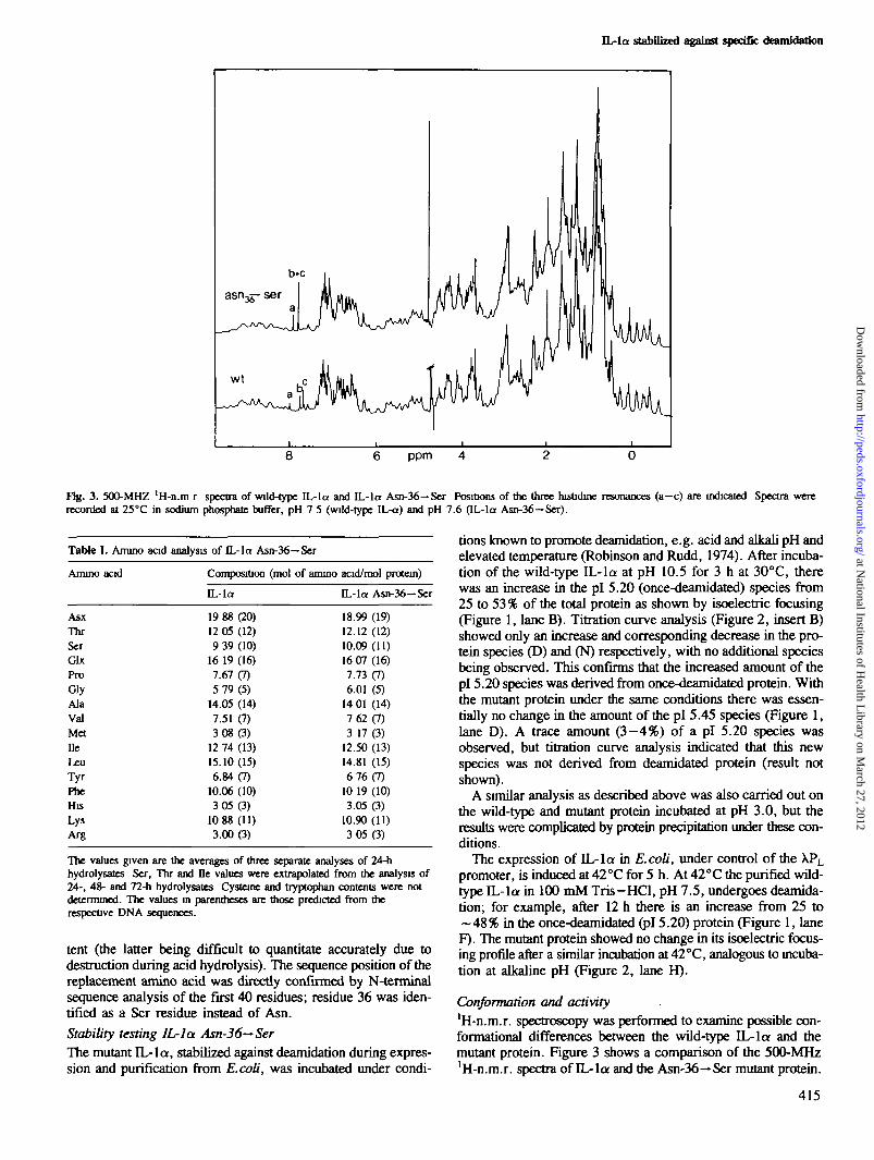
ppm
Fig. 3. 500-MHZ 'H-n.ra r spectra of wild-type IL-la and IL-la Asn-36—Ser Positions of the three hisOdine resonances (a—c) are indicated Spectra wererecorded at 25°C in sodium phosphate buffer, pH 7 5 (wild-type IL-a) and pH 7.6 (IL-la Asn-36—Ser).
tions known to promote deamidation, e.g. acid and alkali pH andelevated temperature (Robinson and Rudd, 1974). After incuba-tion of the wild-type IL-la at pH 10.5 for 3 h at 30°C, therewas an increase in the pi 5.20 (once-deamidated) species from25 to 53 % of the total protein as shown by isoelectric focusing(Figure 1, lane B). Titration curve analysis (Figure 2, insert B)showed only an increase and corresponding decrease in the pro-tein species (D) and (N) respectively, with no additional speciesbeing observed. This confirms that the increased amount of thepi 5.20 species was derived from once-deamidated protein. Withthe mutant protein under the same conditions there was essen-tially no change in the amount of the pi 5.45 species (Figure 1,lane D). A trace amount (3—4%) of a pi 5.20 species wasobserved, but titration curve analysis indicated that this newspecies was not derived from deamidated protein (result notshown).
A similar analysis as described above was also carried out onthe wild-type and mutant protein incubated at pH 3.0, but theresults were complicated by protein precipitation under these con-ditions.
The expression of IL-la in E.coli, under control of the XPL
promoter, is induced at 42°C for 5 h. At 42°C the purified wild-type IL-la in 100 mM Tris-HCl, pH 7.5, undergoes deamida-tion; for example, after 12 h there is an increase from 25 to- 4 8 % in the once-deamidated (pi 5.20) protein (Figure 1, laneF). The mutant protein showed no change in its isoelectric focus-ing profile after a similar incubation at 42 °C, analogous to incuba-tion at alkaline pH (Figure 2, lane H).
Conformation and activity'H-n.m.r. spectroscopy was performed to examine possible con-formational differences between the wild-type IL-la and themutant protein. Figure 3 shows a comparison of the 500-MHz'H-n.m.r. spectra of IL-la and the Asn-36-* Ser mutant protein.
Table]
Ammo
AsxThrSerGlxProGlyAlaValMetDeLeuTyrPbeHisLysArg
I. Amino acid analysis of IL-la Asn-36—Ser
acid Composition (mol of ammo
IL-la
19 88(20)12 05 (12)9 39 (10)
16 19 (16)7.67(7)5 79(5)
14.05 (14)7.51 (7)3 08 (3)
12 74 (13)15.10(15)6.84(7)
10.06 (10)3 05(3)
10 88 (11)3.00(3)
acid/mol protein)
IL-la Asn-36—Ser
18.99 (19)12.12 (12)10.09(11)16 07 (16)7.73(7)6.01 (5)
14 01 (14)762(7)3 17 (3)
12.50 (13)14.81 (15)6 76(7)
10 19 (10)3.05(3)
10.90 (11)3 05(3)
The values given are the averages of three separate analyses of 24-hhydrolysates Ser, Thr and De values were extrapolated from the analysis of24-, 48- and 72-h hydrolysates Cysteine and tryptophan contents were notdetermined. The values in parentheses are those predicted from therespective DNA sequences.
tent (the latter being difficult to quantitate accurately due todestruction during acid hydrolysis). The sequence position of thereplacement amino acid was directly confirmed by N-terminalsequence analysis of the first 40 residues; residue 36 was iden-tified as a Ser residue instead of Asn.Stability testing IL-la Asn-36-~SerThe mutant IL-la, stabilized against deamidation during expres-sion and purification from E.coli, was incubated under condi-
415
at National Institutes of H
ealth Library on M
arch 27, 2012http://peds.oxfordjournals.org/
Dow
nloaded from

- 2 0260 270 280 290 300 310 320 330
WAVELENGTH (NH)
Fig. 4. Near-u.v. circular dichroism spectra of wild-type IL-la and IL-laAsn-36-Ser. (H) Wild-type IL-la (0.78 mg/ml) and (M) mutant IL-la(0 65 mg/ml) The buffer was 0.1 M Tns-HCl, pH 7.3. Spectra wererecorded at 20°C using a 1-cm pathlengtfa and a 1-nm bandwidth
Both spectra are very similar, exhibiting only minor changes.This indicates that no gross structural differences exist betweenthe mutant and the native protein. The apparent shift differencesfor the three histidine el proton resonances a, b, c are due to:(i) a slightly different pH in both samples (resonance a) and (ii)to a shift caused by a change in pK^ for resonances b and c, asevaluated from complete titration curves (data not shown). ThispAT change of 0.2 and 0.4 pH units towards higher pH valuesis clearly due to minor conformational changes influencing theseparticular histidine resonances. These changes are most likelyto be local ones since they do not affect the overall protein struc-ture in a major way. Further evidence for conformational iden-tity was that the near u.v. circular dichroism spectra of the twoproteins were almost indistinguishable (Figure 4).
The receptor binding affinities of the mutant protein, the once-deamidated (pi 5.20) and non-deamidated (pi 5.45) wild-typeproteins were all very similar (Figure 5). These data indicate thatneither deamidation nor substitution of Asn-36 for a Ser residueaffects the conformation of the protein receptor binding region.We have previously shown that the biological activity, measuredby the lymphocyte-activating factor assay (IL-l/LAF), is the samefor the wild-type IL-la deamidated and non-deamidated proteins(Wingfield et al., 1987a). The IL-l/LAF activity of the mutantprotein was, as expected, similar to the wild-type protein (datanot shown).
DiscussionIL-la and j3 are polypeptide hormones displaying a wide rangeof biological activities involved in immune and inflammatory416
o5mB
o
LU
uK1110.
1 0 0 -
8 0 -
40-
2 0 - A IL-1 a (pi 5.2)a IL-1 a (pi 5.45)o IL-1 a(AsnM-» ser)
I I I0 1 1 10 100
UNLABELED IL-1 a CONCENTRATION (ng/ml)
Fig. 5. Competition binding of IL-la: comparison of control, deamidatedand mutant (Asn-36—Ser) proteins The indicated concentrations ofunlabelled protems were mixed at 4°C with [l25I]IL-la (1 ng/ml) prior toaddition of EL4.6.1. cells (5 x lOVtube) After 4 h at 4°C, boundradioactivity was evaluated by centnfuganon of cells through an oil gradient.Data are expressed as per cent inhibition of [125I]IL-la binding as comparedwith untreated controls. Half-maximal binding inhibition was achieved with1 9 ng/ml IL-la (pi 5 2), 1 7 ng/ml IL-la (pi 5.45) and 1 6 ng/ml IL-la(Asn-36-Ser).
9 13S A P F S F L S N V K Y N F H R I I K Y
25 29 36E F I L N D A L N Q S I I R A N D Q Y L
47T A A A L H N L D E A V K . F D M G A Y K
S S K D D A K I T V I L R I S K T Q L Y
V T A Q D E D Q P V L L K E M P E I P K
108 120T I T G S E T N L L F F V E T H G T K N
129Y F T S V A H P N L F I A T K Q D Y W V
157C L A G G P P S I T D F Q I L E N Q A
Fig. 6. Ammo acid sequence of IL-la as predicted from the gene sequence.Sequence positions of the 10 asparagme (N) residues are indicated. Thesequence surrounding Asn-36 is underlined.
responses (for a review see Oppenheim et al., 1986). The struc-tural analysis of these proteins, therefore, is important. We haveplaced special emphasis upon obtaining well-characterizedhomogeneous protein preparations suitable for this purpose. ForIL-1/3, we have previously described methods for obtainingchemically and physically homogeneous preparations (Wingfieldet al., 1986, 1987b,c). In the case of IL-la, however, therecombinant-derived protein contains variable amounts ofdeamidated protein. Although these forms can be separated
at National Institutes of H
ealth Library on M
arch 27, 2012http://peds.oxfordjournals.org/
Dow
nloaded from
IL-la stabilized against spedfic dwmlrtarton
(Wingfield et al., 1987a), the possibility of further deamidationduring storage and handling exists. Furthermore, even thoughdeamidation has been shown in this report to have no major effecton receptor binding affinity and biological activity, it is clearlyundesirable to have preparations with charge heterogeneity forcrystallization purposes and for studying physical properties ingeneral.
We have shown that Asn-36 of IL-la undergoes partialdeamidation during its preparation from E.coli. This deamida-tion is not E.coli related since authentic IL-la isolated fromhuman monocytes also contains partially deamidated Asn-36(Cameron et al., 1986). The deamidation of Asn-36 is the causeof the charge heterogeneity observed upon isoelectric focusingand titration curve analysis of the recombinant-derived and also,apparently, of the authentic IL-la (Cameron et al., 1986). Sub-stituting Asn-36 for Ser by site-specific mutagenesis resulted inthe isolation of an IL-la preparation homogeneous upon isoelec-tric focusing, and with an isoelectric point (5.45) similar to thatof unmodified wild-type IL-la. The mutant IL-la does notundergo deamidation under conditions which promote furtherdeamidation of the wild-type protein. Thus, out of the 18 amideresidues present in IL-la, 10 of which are Asn residues (Figure6), Asn-36 appears most susceptible to deamidation.
The analysis of the mutant protein by techniques which areexpecially sensitive to protein tertiary structure, namely JH-n.m.r. and near-u.v. circular dichroism spectroscopy, indicatedeither no or minor conformational differences compared withwild-type IL-la. The unchanged receptor binding affinity andbiological activity of the mutant protein also support this con-clusion. The structure of the mutant IL-la is currently beingstudied by high-resolution two-dimensional 'H-n.m.r. analysisand an attempt to prepare crystals suitable for X-ray analysis isalso underway (A.M.Gronenborn and G.M.Clore, manuscriptin preparation).
We have recently shown that IL-1/3 also appears to undergoa specific base-catalysed deamidation which changes the isoelec-tric point of the protein from 6.7 to ~5.6 (P.Wingfield, un-published observations). We have not determined which amideresidue was deamidated but have shown that it had no effect onin vitro biological activity. Related to this, Kronheim et al. (1986)described a recombinant-derived IL-1/3 with an isoelectric pointsimilar to the aforementioned assumed once-deamidated protein.This result could have been due to the protein deamidation dur-ing the acid extraction step used in their purification procedure.The actual mechanism(s) of the IL-1-specific deamidations andany possible in vivo implications remain to be established.
AcknowledgementsWe thank Pierre Graber, Gerardo Turcatti (Biogen SA), Cynthia Bume,E.Pingchang Chow (Biogen Research Corp ) and A.L.Peitrequm (Ludwig Institute)for excellent technical assistance. We also thank Nadine Huber for preparationof the manuscript.
References
Ahem.TJ , CaseU I • Petsko.G.A and Klibanov.A M (1987) Proc. Nad. AcadSet. USA, 84, 675-679.
Cameron.P.M , Limjuco.G A , ChinJ , SiIberstein.L. and SchmidtJ.A. (1986)/. Exp Med, 164, 237-250.
Gronenbom,A.M.,Clore,G.M.,SchnKissDer,U and Wingfield.P. (1986) Eur.J. Biochem.. 161, 37-43.
Hunkapular.M.W., Lujan.E., Ostrander.F. and Hood.L.E. (1983) MethodsEnzymol, 91, 227-236.
LowenthaU.W. and MacDonald.H.R. (1986)7. Exp. Med, 164, 1060-1074.MacDonald.H R., WingfieW.P., Schmeissner.U., Shaw.A , Clore.G M. and
Gronenborn.A M. (1986) FEBS Lett., 290, 295-298March.CJ., Mosley3-, Larsen.A., Cerrctti.D.B., Braedt,G , Pnce.V., Gilhs.S ,
Henney.C.S , Kronheim.S.R., Grabstein.K., Conlon.P.J., Hopp.T.P. andCosman.D. (1985) Nature, 315, 641-647.
MatthewJ.B., Fnend.S.H., Botelho.L.H., Lehman,L.D., Hanama.G.I. andGurd.F.R.N (1978) Biochem. Bwphys. Res. Comnuou, 81, 416-421.
Noms.K., NorrisJ1, Christoansen,L. and Fiil.N. (1983) Nucleic Adds Res., 11,5103-5112.
Kronhetm.S.R., Cantrefl.M A., DeeJey.M.C, March.CJ., Qackin,PJ., Ander-son.D.M , Hemenway.T., MernamJ.E., Cosman.D. and Hopp.T P (1986)Bio-Technology, 4, 1078-1082.
OppenheuiU J., Kovacs^EJ , MatsushimaJC. and Durum,S.K. (1986) ImmunolToday, 7, 45-56.
Robinson^ B. and Rudd.C J. (1974) Curr. Top. Cell ReguL, 8, 247-295.Sanger.F., Nicklen.S. and Coulsoa.A.R. (1977) Proc. NatL Acad. Set. USA,
74, 5463-5467.Wmgfield.P , Payton.M., TavermerJ., Barnes,M , Shaw^A., RoseJC , Simona,M G., Demaczuk,S., Wffliamson,K. and DayerJ.-M. (1986) Eur. J. Biochem.,
160,491-497Wingfield.P., Payton.M , Graber.P., RoseJC., DayerJ.-M., Shaw^A. and
Schmeissner.U (1987a) Eur. J. Biochem., 165, 537-541.Wingfield.P , Graber.P., Rose,K , Simona,M.G and Hughes.G.J (1987b) /
Chromatogr., 387, 291-300.Wingfield.P., Graber.P , Mowa,N R , Clore.G.M , Gronenbom.A.M and Mac-
Donald.H.R. (1987c) FEBS Lett , 215, 160-164.
Received on July 6, 1987, revised on August 24, 1987
417
at National Institutes of H
ealth Library on M
arch 27, 2012http://peds.oxfordjournals.org/
Dow
nloaded from
at National Institutes of H
ealth Library on M
arch 27, 2012http://peds.oxfordjournals.org/
Dow
nloaded from