Chemotactic Activity of CXC Chemokines Interleukin-8 ...
8
Chemotactic Activity of CXC Chemokines Interleukin-8, Growth-Related Oncogene—, and Epithelial Cell—Derived Neutrophil-Activating Protein—78 in Urine of Patients with Urosepsis Dariusz P. Olszyna, Steven M. Opal, Jan M. Prins, David L. Horn, Peter Speelman, Sander J. H. van Deventer, Tom van der Poll Downloaded from https://academic.oup.com/jid/article/182/6/1731/915627 by guest on 23 May 2022
-
Upload
khangminh22 -
Category
Documents
-
view
6 -
download
0
Transcript of Chemotactic Activity of CXC Chemokines Interleukin-8 ...

Chemotactic Activity of CXC Chemokines Interleukin-8,
Growth-Related Oncogene—, and Epithelial
Cell—Derived Neutrophil-Activating Protein—78 in Urine
of Patients with Urosepsis
Dariusz P. Olszyna, Steven M. Opal, Jan M. Prins, David L. Horn, Peter Speelman,
Sander J. H. van Deventer, Tom van der Poll
Dow
nloaded from https://academ
ic.oup.com/jid/article/182/6/1731/915627 by guest on 23 M
ay 2022

1731
Chemotactic Activity of CXC Chemokines Interleukin-8, Growth-RelatedOncogene–a, and Epithelial Cell–Derived Neutrophil-Activating Protein–78in Urine of Patients with Urosepsis
Dariusz P. Olszyna,1,2 Steven M. Opal,3 Jan M. Prins,2
David L. Horn,4,a Peter Speelman,2
Sander J. H. van Deventer,1 and Tom van der Poll1,2
Departments of 1Experimental Internal Medicine and 2InternalMedicine, Division of Infectious Diseases, Tropical Medicine
and AIDS, Academic Medical Center, University of Amsterdam,Amsterdam, The Netherlands; 3Brown University School of Medicine,Memorial Hospital, Pawtucket, Rhode Island; 4Bristol-Myers Squibb,
Plainsboro, New Jersey
CXC chemokines are chemotactic cytokines that specifically act on neutrophils. To obtaininsight into the extent of local production of CXC chemokines during acute pyelonephritis,interleukin (IL)–8, growth-related oncogene (GRO)–a, and epithelial cell–derived neutrophil-activating protein (ENA)–78 were measured in urine and plasma samples from patients withculture-proven urosepsis ( ), healthy human control subjects with sterile urine ( ),n p 33 n p 31and human volunteers intravenously injected with endotoxin ( ). Patients had pro-n p 11foundly elevated urine concentrations of chemokines with no (GRO-a and ENA-78) or little(IL-8) elevation in plasma. Endotoxin-challenged subjects demonstrated transient increasesin plasma chemokine concentrations, with no (GRO-a) or little (IL-8 and ENA-78) elevationin urine. Urine from patients exerted chemotactic activity toward neutrophils, which waspartially inhibited by neutralizing antibodies against IL-8, GRO-a, or ENA-78. During uro-sepsis, CXC chemokines are predominantly produced within the urinary tract, where theyare involved in the recruitment of neutrophils to the urinary compartment.
Chemokines are a family of small chemotactic proteins thatplay an important role in inflammatory responses as mediatorsof leukocyte trafficking and activation [1–4]. Depending on theirstructure, chemokines can be classified into several families. In-terleukin (IL)–8, growth-related oncogene (GRO)–a, and epi-thelial cell–derived neutrophil-activating protein (ENA)–78 be-long to the CXC chemokine family and act primarily ongranulocytes, cells that often are found in the urine of patientswith urinary tract infection (UTI).
The infected urinary tract is a frequent source of gram-nega-tive sepsis [5, 6]. Urosepsis is usually diagnosed on the basis ofa positive urine culture and of clinical findings associated withsepsis. Although increased urinary and serum IL-8 levels havebeen widely reported in UTI [7–9], little is known about theproduction of chemokines, other than IL-8, in this condition.
Accumulating evidence suggests that CXC chemokines play
Received 7 April 2000; revised 1 September 2000; electronically published23 October 2000.
The study was approved by the institutional scientific and ethics com-mittees. Written informed consent was obtained from all patients and healthysubjects.
Financial support: Merck and Co., Dutch Kidney Foundation (to D.P.O.),and Royal Dutch Academy of Arts and Sciences (to T.v.d.P.).
a Previously affiliated with Merck and Co., Westpoint, Pennsylvania.Reprints or correspondence: Dr. D.P. Olszyna, Academic Medical Center,
Room G2-105, Dept. of Experimental Internal Medicine, Meibergdreef 9,1105 AZ Amsterdam, The Netherlands ([email protected]).
The Journal of Infectious Diseases 2000;182:1731–7q 2000 by the Infectious Diseases Society of America. All rights reserved.0022-1899/2000/18206-0020$02.00
an important role in neutrophil migration during UTI. In vitrostudies have demonstrated that epithelial cells stimulated withpathogens can secrete chemokines and that IL-8 stimulates neu-trophil migration across uroepithelial cell layers infected withEscherichia coli [10]. Recently, Hang et al. [11] showed that mac-rophage inflammatory protein (MIP)–2, a murine homologue ofhuman IL-8, is required for neutrophil migration across the epi-thelium of the infected urinary tract in mice in vivo.
Patients with urosepsis, whose source of infection is located(by definition) in the urinary tract, potentially provide the op-portunity for obtaining insight into the extent of chemokineproduction at the site of the infection, namely by measurementsin urine. Therefore, in the present study, we sequentially mea-sured plasma and urine concentrations of IL-8, GRO-a, andENA-78 in patients with urosepsis during the first 8 h after theinitiation of antibiotic treatment and compared these findingswith chemokine plasma and urine concentrations in healthyhumans intravenously (iv) injected with E. coli endotoxin (i.e.,without a local inflammatory stimulus in the urinary tract). Wealso examined the migration of polymorphonuclear leukocytes(PMNL) induced by urine from patients with urosepsis and theextent to which each of these 3 chemokines (IL-8, GRO-a, andENA-78) contributed to the chemotactic activity exerted bypatient urine.
Materials and Methods
Patients with urosepsis and control subjects. Patients 118 yearsold who were suspected of having gram-negative urosepsis and for
Dow
nloaded from https://academ
ic.oup.com/jid/article/182/6/1731/915627 by guest on 23 M
ay 2022

1732 Olszyna et al. JID 2000;182 (December)
Table 1. Median (range) concentrations of interleukin (IL)–8, growth-related oncogene (GRO)–a and epithelial cell–derived neutrophil-activating protein (ENA)–78 in urine from healthy control subjects ( ), patients with urosepsis ( ) and healthy subjects injectedn p 16 n p 33with endotoxin ( ).n p 11
CytokineHealthy
control subjects
Patients with urosepsis Experimental endotoxemia
Admission 2 Hours 4 Hours 8 Hours 0 Hours 0–3 Hours 3–6 Hours
IL-8 !0.01(!0.01–0.14)
0.38(!0.01–3.13)a
0.43(!0.01–2.00)
0.20(!0.01–2.06)
0.15(0.03–1.60)
!0.01(!0.01–0.03)
!0.01(!0.01–0.01)
0.01(!0.01–0.03)
GRO-a !0.03(!0.03–0.29)
0.10(!0.03–0.51)a
0.10(!0.03–1.14)
0.10(!0.03–1.11)
0.03(!0.03–0.45)
0.17(!0.03–1.18)
!0.03(!0.03–0.15)
0.07(!0.03–0.62)
ENA-78 !0.03 0.27(!0.03–1.66)a
0.24(!0.03–1.93)
0.26(!0.03–2.86)
0.25(!0.03–3.08)
!0.03 !0.03(!0.03–0.11)
!0.03
NOTE. Concentrations are expressed in ng/mL.a versus healthy control subjects. Only IL-8 decreased significantly in urine during follow-up.P ! .05
whom antibiotic treatment was indicated were eligible if they alsomet the following criteria: acute symptoms of UTI, pyruria (110leukocytes/high-power field [hpf] and !5 epithelial cells/hpf), urineGram’s stain with gram-negative bacteria, and metabolic or he-matologic signs of infection (including 3 of the following indicators:tachycardia [190 beats/min], leukocytosis [110,000/mm3], and fever[1387C]). Exclusion criteria were antibiotic use in the previous 7days, poor clinical condition, known hypersensitivity to any b-lactam antibiotic, any major underlying disease likely to alter base-line chemokine levels, renal insufficiency (estimated creatinineclearance, !30 mL/min), pregnancy or breast feeding, use of sys-temic corticosteroids or other immunosuppressive agents in the past3 months, history of seizures, use of any investigational drug withinthe past 30 days, or any clinically significant medical condition thatwould pose a risk to the patient should he or she participate.
Clinical data were collected, and the APACHE II score wasassessed before the start of treatment (0 h). Patients received iv 1dose of ceftazidime (1000 mg) or imipenem (500 mg), followed 8h later by an antibiotic chosen by the clinician. Blood (heparinized)and urine samples were collected before the start of treatment andat 2, 4, and 8 h thereafter. Plasma (heparinized) and urine sampleswere also collected from 31 healthy individuals, all of whom hadsterile urine. The samples were centrifuged at 1500 g for 20 min.Supernatants were collected and stored at 2757C until assays wereperformed.
Experimental endotoxemia. In addition to the patients with uro-sepsis, 11 healthy subjects (mean , years) were stud-age 5 SE 24 5 1ied after iv administration of endotoxin (lipopolysaccharide, LPS).The subjects did not smoke, use any medication, or have a febrileillness in the month preceding the study. They were admitted to theclinical research unit at the Academic Medical Center (University ofAmsterdam, Amsterdam) after their medical history, physical ex-amination, hematologic and biochemical tests, chest radiograph, andelectrocardiograph proved to be normal. Endotoxin (LPS standardlot G from E. coli; United States Pharmacopeia Convention, Rock-ville, MD) was given for 1 min in an antecubital vein at a dose of4 ng/kg body weight. Blood was collected by venipunctures directlybefore LPS administration and at 0.5, 1, 1.5, 2, 3, 4, 5, 6, 8, 12, and24 h thereafter. EDTA plasma was obtained by centrifugation at1500 g for 20 min. All urine produced by the subjects was collectedbefore and 3 and 6 h after LPS administration.
Assays. Chemokine concentrations were measured by ELISA.
All measurements of 1 chemokine in patient and control sampleswere done on 1 day in 1 ELISA run. IL-8 was measured accordingto the instructions of the manufacturer (Central Laboratory of theNetherlands Red Cross Blood Transfusion Service [CLB], Am-sterdam). For determination of GRO-a levels, purified monoclonalmouse anti–human GRO-a (4 mg/mL; R&D Systems, Abingdon,UK) was used as a coating antibody, biotinylated affinity-purifiedgoat IgG anti–human GRO-a (20 ng/mL; R&D Systems) was usedas a detecting antibody, and recombinant human GRO-a (R&DSystems) was used as the standard. For determination of ENA-78,monoclonal mouse anti–human ENA-78 (1 mg/mL; R&D Systems)was used as a coating antibody, biotinylated goat anti–humanENA-78 (80 ng/mL; R&D Systems) was used as a detecting anti-body, and recombinant human ENA-78 (R&D Systems) was usedas the standard.
Detection limits in plasma and urine diluted 1:2 with ELISA high-performance buffer (CLB) were 1.2 pg/mL (IL-8) and 28.6 pg/mL(GRO-a and ENA-78). Urine concentrations of chemokines are ex-pressed both in nanograms per milliliter urine (table 1) and, to correctfor dilution of urine, per micromole creatinine (figures 1–4).
Chemotaxis assay. The migration of PMNL was measured byuse of a multiwell chemotaxis chamber (Neuro Probe, Cabin John,MD) and a 5.0 mm–pore polycarbonate membrane (Nuclepore;Costar, Cambridge, MA), as described elsewhere [12]. In brief,PMNL were obtained from heparinized blood samples of healthyvolunteers, using Polymorphprep (Nycomed Pharma, Oslo), ac-cording to the manufacturer’s instructions, and were suspended inendotoxin-free RPMI 1640 (BioWittaker, Verviers, Belgium) con-taining 5% normal human serum (BioWittaker).
Urine samples from 8 patients (those with the highest leukocytecounts in urine) were pooled and diluted 1:2 in RPMI 1640, withor without the following reagents: monoclonal mouse-derived anti–human IL-8 neutralizing antibody (R&D Systems; final concen-tration 10 mg/mL), monoclonal mouse-derived anti–human GRO-a neutralizing antibody (R&D Systems; final concentration, 10 mg/mL), monoclonal mouse-derived anti–human ENA-78 neutralizingantibody (R&D Systems; final concentration, 50 mg/mL), or mouseIgG1 (R&D Systems; final concentration, 50 mg/mL). During invitro cell stimulation, these concentrations of anti–chemokine an-tibodies completely neutralize activity of recombinant IL-8, GRO-a, and ENA-78 when added at 2–3-log higher concentrations thanthose detected in urine from patients with urosepsis (information
Dow
nloaded from https://academ
ic.oup.com/jid/article/182/6/1731/915627 by guest on 23 M
ay 2022

JID 2000;182 (December) CXC Chemokines in Urosepsis and Endotoxemia 1733
Figure 1. Plasma and urine levels of interleukin (IL)–8 in healthysubjects ( ) and patients with urosepsis ( ) at admissionn p 31 n p 33and 2, 4, and 8 h after initiation of antibiotic therapy. Horizontal linesrepresent medians. v, Patients with positive blood culture results (de-termined on admission). V, Patients with negative blood culture resultsand healthy control subjects. Patients at admission had higher plasmaand urine levels than did control subjects (both ). In patients,P ! .001urine, but not plasma, IL-8 concentrations decreased after admission( ) and during follow-up ( vs. ). Because of the lowt p 0 P ! .05 t p 0detection limit of the assay, a line indicating the limit is not visible.
on the neutralizing capacities of the antibodies provided by themanufacturer). The respective antibodies used do not have cross-reactivity with other chemokines (information provided by themanufacturer).
Urine from 16 healthy control subjects was also diluted 1:2 inRPMI 1640, to serve as a negative control, and, in combinationwith recombinant human IL-8 (rhIL-8; R&D Systems; final con-centration 5 ng/mL), it served as a positive control. Urine specimens(25-mL volumes) were placed in the lower chamber in triplicate.Volumes of 50 mL containing PMNL were added to the44.0 3 10upper well. The chamber was incubated for 55 min in an humidifiedenvironment at 377C. The membrane was removed, nonmigratedcells were washed off, and the membrane was fixed in methanoland stained with DiffQuik (Dade Behring, Dudingen, Switzerland).Migrated cells in each well were counted at 10003 magnificationfor 5 random fields.
Statistical analysis. Log-transformed concentrations of che-mokines were analyzed in time by 1-way analysis of variance(ANOVA) and followed (for uroseptic patients and healthy subjectsgiven endotoxin) by Dunnett’s test. The difference between the 2antibiotic regimens in chemokine concentrations was assessed byuse of the repeated-measures ANOVA. The Mann-Whitney U testwas used to compare chemokine concentrations in urosepsis pa-tients (at 0 h) with those in healthy control subjects and concen-trations in patients with positive blood culture results with thosein patients with negative results. Correlations between chemokineconcentrations and duration of symptoms were assessed by cal-culating Spearman’s correlation coefficient. Comparisons of num-bers of migrated cells in the chemotaxis assay between patientswith urosepsis and healthy control subjects were done by using theMann-Whitney U test. The effect of neutralizing antibodies on themigration of cells in urine from patients with urosepsis was assessedby use of the Wilcoxon signed rank test. was consideredP ! .05significant.
Results
Healthy subjects. IL-8 was detectable in urine (median,!2.0 ng/mmol; range, !2.0–8.5 ng/mmol) but not in plasmafrom healthy individuals. Median GRO-a levels in plasma were0.20 ng/mL (range, !0.03–0.45 ng/mL) and in urine they were2.7 ng/mmol (range, !2.0–48.3 ng/mmol). ENA-78 concentra-tions were detectable in plasma (median, 0.03 ng/mL; range,!0.03–0.41 ng/mL) but not in urine.
Patients with urosepsis. Thirty-three of 35 patients (mean, years) who had been randomized to treat-age 5 SE 42 5 3
ment with imipenem or ceftazidime completed the study. Theduration of symptoms before the first urine and plasma sampleswere taken was ( ) days. All patients had2.9 5 0.3 mean 5 SEsystemic symptoms, and there was no correlation between theduration of symptoms before inclusion in the study and che-mokine levels at admission. E. coli was cultured from the urineof all but 1 patient. Ten patients had bacterium-positive bloodculture results (1 for Proteus mirabilis and 9 for E. coli). Atadmission, the median APACHE II score was 6 (range, 0–17).
All patients fully recovered after treatment. The type of anti-biotic regimen did not significantly influence chemokine levelsin urine or plasma (data not shown); therefore, these 2 patientgroups were combined in further analyses.
Compared with control subjects, patients with urosepsis hadhigher levels of IL-8 in plasma (median, 0.03 ng/mL; range,0.01–0.65 ng/mL) and urine (median, 60.0 ng/mmol; range,!2.0–1237.0 ng/mmol; for both) at admission (figureP ! .0011). In addition, compared with healthy control subjects, patientswith positive and negative blood cultures had higher plasmaconcentrations ( for both) and urine concentrationsP ! .001 D
ownloaded from
https://academic.oup.com
/jid/article/182/6/1731/915627 by guest on 23 May 2022
1734 Olszyna et al. JID 2000;182 (December)
Figure 2. Plasma and urine levels of growth-related oncogene(GRO)–a in healthy subjects ( ) and in patients with urosepsisn p 31( ) at admission and 2, 4, and 8 h after initiation of antibioticn p 33therapy. Horizontal lines represent medians. v, Patients with positiveblood cultures (determined at admission). V, Patients with negativeblood culture results and healthy control subjects. Urine, but notplasma, levels were higher in patients at admission than in controlsubjects ( ). In patients, urine and plasma concentrations didP ! .001not change significantly during follow-up. Dotted line depicts the de-tection limit of the assay.
Figure 3. Plasma and urine levels of epithelial cell–derived neutro-phil-activating protein (ENA)–78 in healthy subjects ( ) and pa-n p 31tients with urosepsis ( ) at admission and 2, 4, and 8 h aftern p 33initiation of antibiotic therapy. Horizontal lines represent medians. v,Patients with positive blood culture results (determined on admission).V, Patients with negative blood culture results and healthy controlsubjects. Urine, but not plasma, levels were higher in patients at ad-mission than in control subjects ( ). In patients, urine andP ! .001plasma concentrations of ENA-78 did not change significantly duringfollow-up. Dotted line (upper panel) depicts the detection limit of theassay.
( for those with positive blood cultures andP ! .001 P p .005for those with negative blood cultures) of IL-8. Furthermore,plasma but not urine IL-8 levels were higher in patients withpositive blood cultures than in patients with negative bloodcultures ( ; table 2). GRO-a was elevated only in urineP ! .05(median, 15.0 ng/mmol; range, !2.0–217.0 ng/mmol; P ! .001vs. healthy control subjects; figure 2). This increase was sig-nificant in patients with positive and negative blood cultureresults (both ).P ! .001
Only patients with positive blood culture results had elevatedplasma levels of ENA-78 (median, 0.32 ng/mmol; range,
!0.03–6.57 ng/mmol; vs. healthy control subjects; figureP ! .0053). Urine ENA-78 levels were significantly elevated in patientswith positive and those with negative blood culture results( for both) and were higher in patients with positiveP ! .001blood culture results than in patients whose blood cultures werenegative ( ; table 2). Of all 3 chemokines measured inP ! .005plasma and urine, only urine levels of IL-8 decreased significantlyduring follow-up ( ; figure 1). Urine concentrations of che-P ! .05mokines expressed per milliliter of urine are given in table 1.
Endotoxemia in healthy humans. The iv injection of E. coli
Dow
nloaded from https://academ
ic.oup.com/jid/article/182/6/1731/915627 by guest on 23 M
ay 2022

JID 2000;182 (December) CXC Chemokines in Urosepsis and Endotoxemia 1735
Table 2. Median concentrations (range) of interleukin (IL)–8,growth-related oncogene (GRO)–a and epithelial cell–derived neutro-phil-activating protein (ENA)–78 in plasma and urine of patients withurosepsis with both positive and negative blood cultures.
CytokinePositive blood culture
(n p 10)Negative blood culture
(n p 23) P
IL-8Plasma, ng/mL 0.06 (0.02-0.51) 0.02 (0.01–0.65) !.05Urine, ng/mmol 37.0 (!2.0–246.0) 76.0 (!2.0–1237.0) NS
GRO-aPlasma, ng/mL 0.19 (0.09–0.37) 0.23 (0.07–2.05) NSUrine, ng/mmol 14.0 (!2.0–68.0) 15.0 (!2.0–217.0) NS
ENA-78Plasma, ng/mL 0.32 (!0.03–6.57) 0.07 (!0.03–0.32) !.005Urine, ng/mmol 54.0 (!2.0–129.0) 47.0 (!2.0–1029.0) NS
NOTE. Urine concentrations are expressed per mmol creatinine. NS, notsignificant
Figure 4. plasma and urine concentrations of interleukin (IL)–8, growth-related oncogene (GRO)–a, and epithelial cell–derivedMean 5 SEneutrophil-activating protein (ENA)–78 in healthy subjects ( ) after intravenous administration of endotoxin. Plasma concentrations weren p 11measured before endotoxin injection ( ) and during a 12-h follow-up. Urine concentrations were measured before endotoxin injection andt p 0in all urine excreted within 3 h after infusion of endotoxin ( ) and between 3 and 6 h after endotoxin administration ( ). *Significantt p 0–3 t p 3–6difference from the levels at . Dotted lines depict the detection limit of the assay.t p 0
LPS was associated with transient increases in the plasma con-centrations of all 3 chemokines measured. In addition, urineIL-8 and ENA-78 concentrations modestly increased after ivadministration of LPS (figure 4). LPS induced an increase inplasma IL-8 levels, starting at 1.5 h and peaking at 3 h afteradministration ( , ng/mL; ).mean 5 SE 1.79 5 0.16 P ! .001Urine IL-8 increased between 3 and 6 h after LPS administra-tion ( , ng/mmol; ). Plasma levelsmean 5 SE 0.7 5 0.2 P ! .005of GRO-a increased significantly within 3 h after LPS( , pg/mL; ). Urine GRO-a didmean 5 SE 0.47 5 0.05 P ! .005not change significantly after LPS. Plasma levels of ENA-78increased from 3 h onwards, reaching peak concentrations at4 h ( , ng/mL; ). Urine concen-mean 5 SE 0.45 5 0.05 P ! .001trations of ENA-78 increased to ng/mmol ( )8.3 5 1.6 P ! .001within 3 h after LPS administration. No leukocytes were foundin urine from any of the subjects injected with LPS at any time
point. Urine chemokine concentrations expressed in ng/mLurine are given in table 1.
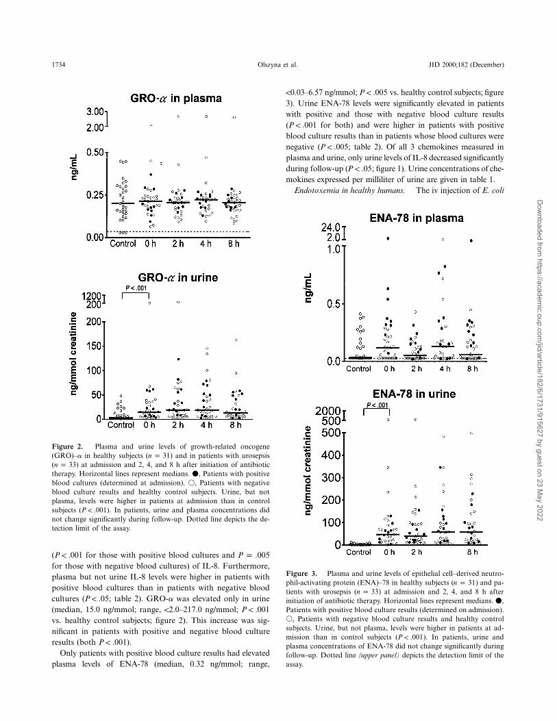
Chemotactic activity of urine from patients with urosepsis.Urine from healthy control subjects attracted few PMNL( , cells/hpf). Significantly more PMNL mi-mean 5 SE 0.2 5 0.1grated toward pooled urine from patients with urosepsis( cells/hpf; vs. normal urine). Addition of24.8 5 3.0 P ! .005either anti–IL-8, anti–GRO-a, or anti–ENA-78 to urine frompatients with urosepsis significantly reduced chemotactic activityof this urine on neutrophils (all vs. urine from patientsP ! .05with or without control antibody; figure 5). None of the anti-chemokine antibodies completely neutralized the chemotactic ac-tivity of urine from patients. In fact, anti–IL-8, anti–GRO-a, andanti–ENA-78 antibody treatment inhibited neutrophil chemo-taxis toward patient urine to a similar extent (to ∼60% of mi-gration in the absence of anti–chemokine antibodies). Additionof all 3 anti–chemokine antibodies together did not further reduceurine chemotactic activity, compared with either antibody alone(data not shown).
Discussion
IL-8, GRO-a, and ENA-78 are members of the CXC che-mokine family, in which the first 2 cysteine residues are separatedby a single amino acid. They primarily target neutrophils, cellsthat are found in urine from patients with UTI. The aim of thisstudy was to obtain insight into the extent of local productionof these chemokines in patients with urosepsis and into the con-tribution of these mediators to the chemotactic activity towardneutrophils of urine from patients with urosepsis.
Urine IL-8, GRO-a, and ENA-78 concentrations were
Dow
nloaded from https://academ
ic.oup.com/jid/article/182/6/1731/915627 by guest on 23 M
ay 2022
1736 Olszyna et al. JID 2000;182 (December)
Figure 5. number of polymorphonuclear lymphocytesMean 5 SE(PMNL) per high-power field (hpf) that migrated toward urine (fromhealthy control subjects) containing 5 ng/mL recombinant human in-terleukin (IL)–8 ( ), urine from healthy control subjects alonen p 16( ), and urine from patients with urosepsis with pyuria ( ).n p 16 n p 8Neutralization of IL-8, growth-related oncogene (GRO)–a or epithelialcell–derived neutrophil-activating protein (ENA)–78 in urine from pa-tients with urosepsis resulted in attenuation of this migration. *Sig-nificant difference in migration of PMNL toward patient urine eitherwith or without control antibody ( ).P ! .05
strongly elevated in patients but not (or less elevated) in healthysubjects after iv injection of E. coli LPS. In vitro, IL-8, GRO-a, and ENA-78 contributed to the chemotactic activity of in-fected urine toward neutrophils. These data suggest that CXCchemokines are produced within the urinary tract during uro-sepsis and that they are involved in the recruitment of neutro-phils to the urinary compartment.
Several studies have documented elevated IL-8 concentra-tions in urine of patients with UTI [7–9]. In patients with acutepyelonephritis, IL-8 levels are often higher in urine than inplasma [8]. In addition, deliberate colonization of the humanurinary tract with E. coli resulted in a rapid increase in urineIL-8 levels without a detectable rise in IL-8 in serum [13]. To-gether, these data suggest that IL-8 is produced locally at thesite of the infection during UTI. In accordance, we found highIL-8 concentrations in the urine of patients with urosepsiscaused by E. coli but not in volunteers injected iv with E. coliLPS. In this latter experiment, IL-8 concentrations were es-pecially elevated in plasma, with little elevation (conceivablythrough renal clearance) in urine. Cell types that can produceIL-8 within the urinary tract include epithelial, mesangial, andendothelial cells, and renal fibroblasts [14]. To our knowledge,this study is the first to document that, besides IL-8, 2 otherCXC chemokines (i.e., GRO-a and ENA-78) are released inurine during acute pyelonephritis. Of interest, GRO-a andENA-78 concentrations were elevated in urine but not inplasma of patients with urosepsis; however, plasma levels butnot urine levels of GRO-a and ENA-78 increased to a signif-
icant extent after iv injection of LPS. Hence, it is likely thatGRO-a and ENA-78 also are produced at the site of the in-fection during acute pyelonephritis.
In a previous investigation, Ko et al. [8] reported neutrophilchemotactic activity of urine specimens from patients with UTI,whereas no such activity was found in urine from normal sub-jects. Adsorption of infected urine onto an anti–IL-8 columnreduced the chemotactic activity by ∼55% [8]. Using a slightlydifferent approach (i.e., direct addition of anti–chemokine an-tibodies to urine samples), we found that anti–IL-8 inhibitedthe neutrophil chemotactic activity of urine from patients withurosepsis to a similar extent. Furthermore, we extend thesefindings by showing that anti–GRO-a and anti–ENA-78 treat-ments also inhibit the chemotactic activity of infected urinetoward neutrophils. Concurrent addition of anti–IL-8, anti–GRO-a, and anti–ENA-78 did not reduce the chemotactic ac-tivity of urine any more than did the addition of either antibodyalone, indicating that other factors present in urine, such asLPS, contribute significantly to the chemotactic potential ofinfected urine. In this respect, it should be noted that GRO-aand ENA-78 preferentially interact with CXCR2 (also knownas IL-8 receptor type B) on neutrophils, whereas IL-8 can bindboth CXCR1 (IL-8 receptor type A) and CXCR2 with highaffinity [1]. Both CXCR1 and CXCR2 can mediate chemotaxisof neutrophils [15–18]. IL-8 induces neutrophil chemotaxis pre-dominantly via CXCR1, but other CXC chemokines, includingGRO-a and ENA-78, can stimulate this inflammatory responsevia CXCR2 [15, 19].
Our data differ from in vitro studies reported by Godaly etal. [10], who found that an anti–CXCR1 antibody, but not ananti–CXCR2 antibody, reduced E. coli–induced transuroepi-thelial neutrophil migration in vitro [10]. Apparently, in theirin vitro system, CXC chemokines that preferentially interactwith CXCR2 (such as GRO-a and ENA-78) are not involvedin neutrophil migration. Whether CXCR2 ligands were pro-duced in their system was not determined. Further studies arewarranted to establish the respective roles of CXCR1 andCXCR2 in neutrophil recruitment to urine.
Relatively little is known about the kinetics of CXC che-mokine release during infection. Of interest, injection of LPSwas associated with a brisk and transient increase in the plasmaconcentrations of IL-8 and, to a lesser extent, of GRO-a,whereas ENA-78 increased and dissipated more gradually. Dif-ferences in the rates of production and clearance of differentchemokines may obviously influence the timing of their re-spective effects during the innate immune response to an in-fection. Further research is warranted to evaluate this issue.
Chemokines play a central role in host defense against in-fectious diseases. Here, we report elevated levels of the CXCchemokines IL-8, GRO-a, and ENA-78 in the urine of patientswith urosepsis. CXC chemokines likely are produced primarily
Dow
nloaded from https://academ
ic.oup.com/jid/article/182/6/1731/915627 by guest on 23 M
ay 2022

JID 2000;182 (December) CXC Chemokines in Urosepsis and Endotoxemia 1737
in the urinary tract during urosepsis, where they are involvedin the attraction of neutrophils to the urinary compartment.
References
1. Rollins BJ. Chemokines. Blood 1997;90:909–28.2. Luster AD. Chemokines: chemotactic cytokines that mediate inflammation.
N Engl J Med 1998;338:436–45.3. Furie MB, Randolph GJ. Chemokines and tissue injury. Am J Pathol 1995;
146:1287–301.4. Friedland JS. Chemokines and human infection. Clin Sci 1995;88:393–400.5. Kreger BE, Craven DE, Carling PC, McCabe WR. Gram-negative bacte-
remia. III. Reassessment of etiology, epidemiology and ecology in 612patients. Am J Med 1980;68:332–43.
6. Bahnson RR. Urosepsis. Urol Clin North Am 1986;13:627–35.7. Prins JM, van Agtmael MA, Kuijper EJ, van Deventer SJ, Speelman P.
Antibiotic-induced endotoxin release in patients with gram-negative uro-sepsis: a double-blind study comparing imipenem and ceftazidime. J InfectDis 1995;172:886–91.
8. Ko YC, Mukaida N, Ishiyama S, et al. Elevated interleukin-8 levels in theurine of patients with urinary tract infections. Infect Immun 1993;61:1307–14.
9. Benson M, Jodal U, Agace W, et al. Interleukin (IL)–6 and IL-8 in childrenwith febrile urinary tract infection and asymptomatic bacteriuria. J InfectDis 1996;174:1080–4.
10. Godaly G, Proudfoot AE, Offord RE, Svanborg C, Agace WW. Role ofepithelial interleukin-8 (IL-8) and neutrophil IL-8 receptor A in Escher-ichia coli–induced transuroepithelial neutrophil migration. Infect Immun1997;65:3451–6.
11. Hang L, Haraoka M, Agace WW, et al. Macrophage inflammatory protein–2is required for neutrophil passage across the epithelial barrier of the in-fected urinary tract. J Immunol 1999;162:3037–44.
12. Goodman RB, Forstrom JW, Osborn SG, Chi EY, Martin TR. Identificationof two neutrophil chemotactic peptides produced by porcine alveolar mac-rophages. J Biol Chem 1991;266:8455–63.
13. Agace WW, Hedges SR, Ceska M, Svanborg C. Interleukin-8 and the neu-trophil response to mucosal gram-negative infection. J Clin Invest 1993;92:780–5.
14. Schlondorff D, Nelson PJ, Luckow B, Banas B. Chemokines and renal dis-ease. Kidney Int 1997;51:610–21.
15. Hammond ME, Lapointe GR, Feucht PH, et al. IL-8 induces neutrophilchemotaxis predominantly via type I IL-8 receptors. J Immunol 1995;155:1428–33.
16. Wuyts A, Proost P, Lenaerts JP, Ben-Baruch A, Van Damme J, Wang JM.Differential usage of the CXC chemokine receptors 1 and 2 by interleukin-8, granulocyte chemotactic protein–2 and epithelial-cell–derived neutro-phil attractant–78. Eur J Biochem 1998;255:67–73.
17. Loetscher P, Seitz M, Clark-Lewis I, Baggiolini M, Moser B. Both interleukin-8 receptors independently mediate chemotaxis. Jurkat cells transfectedwith IL-8R1 or IL-8R2 migrate in response to IL-8, GRO alpha andNAP-2. FEBS Lett 1994;341:187–92.
18. Chuntharapai A, Kim KJ. Regulation of the expression of IL-8 receptor A/B by IL-8: possible functions of each receptor. J Immunol 1995;155:2587–94.
19. Ahuja SK, Murphy PM. The CXC chemokines growth-regulated oncogene(GRO)–alpha, GRObeta, GROgamma, neutrophil-activating peptide–2,and epithelial cell–derived neutrophil-activating peptide–78 are potent ag-onists for the type B, but not the type A, human interleukin-8 receptor.J Biol Chem 1996;271:20545–50.
Dow
nloaded from https://academ
ic.oup.com/jid/article/182/6/1731/915627 by guest on 23 M
ay 2022