Optogenetic EB1 inactivation shortens metaphase spindles by ...
Upload
independentCategory
view
0download
0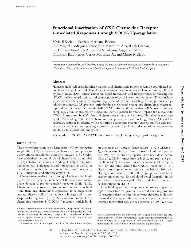
J. Exp. Med.
The Rockefeller University Press • 0022-1007/2002/08/311/11 $5.00Volume 196, Number 3, August 5, 2002 311–321http://www.jem.org/cgi/doi/10.1084/jem.20012041
311
Functional Inactivation of CXC Chemokine Receptor4–mediated Responses through SOCS3 Up-regulation
Silvia F. Soriano, Patricia Hernanz-Falcón, José Miguel Rodríguez-Frade, Ana Martín de Ana, Ruth Garzón,Carla Carvalho-Pinto, Antonio J. Vila-Coro, Angel Zaballos,
Dimitrios Balomenos, Carlos Martínez-A., and Mario Mellado
Department of Immunology and Oncology, Centro Nacional de Biotecnología/Consejo Superior de Investigaciones Cientifícas, Universidad Autónoma de Madrid, Campus de Cantoblanco, E-28049 Madrid, Spain
Abstract
Hematopoietic cell growth, differentiation, and chemotactic responses require coordinated ac-
tion between cytokines and chemokines. Cytokines promote receptor oligomerization, followedby Janus kinase
(
JAK) kinase activation, signal transducers and transactivators of transcription
(
STAT) nuclear translocation, and transcription of cytokine-responsive genes. These includegenes that encode a family of negative regulators of cytokine signaling, the suppressors of cy-tokine signaling (SOCS) proteins. After binding their specific receptors, chemokines trigger re-ceptor dimerization and activate the JAK/STAT pathway. We show that SOCS3 overexpressionor up-regulation, stimulated by a cytokine such as growth hormone, impairs the response toCXCL12, measured by Ca
2
�
flux and chemotaxis in vitro and in vivo. This effect is mediatedby SOCS3 binding to the CXC chemokine receptor 4 receptor, blocking JAK/STAT and G
�
i
pathways, without interfering with cell surface chemokine receptor expression. The data pro-vide clear evidence for signaling cross-talk between cytokine and chemokine responses inbuilding a functional immune system.
Key words: SOCS3 • JAK-STAT activation • chemokine signaling • cytokine signaling
Introduction
The chemokines comprise a large family of low molecularweight (8–10 kD) cytokines, with chemotactic and pro-acti-vatory effects on different leukocyte lineages (1–3). Studieshave established the central role of chemokines in a numberof physiological situations, including T helper responses,hematopoiesis, angiogenesis, and homeostasis, as well as inpathological conditions such as asthma, tumor rejection,HIV-1 infection, and arteriosclerosis (4–8).
Chemokines mediate their biological effects after bind-ing to specific receptors, members of the seven-transmem-brane domain G protein–coupled receptor family (1, 2).Chemokine receptors are promiscuous, as each can bindmore than one chemokine; expression is heterogeneousamong different cells of the leukocyte lineage and is tran-scriptionally regulated (3, 5). An exception is the CXCchemokine receptor 4 (CXCR4)
*
receptor, which binds
only stromal cell–derived factor (SDF)-1
�
(CXCL12) (1,2), a chemokine isolated from stromal cell culture superna-tants (9). Its chemotactic properties have been described inPBLs (10), CD34
�
progenitor cells (11), and pre- and pro-B cell lines (12). Knockout mice lacking the CXCL12 pro-tein (10) and mice lacking the CXCR4 receptor (13, 14)display similar phenotypes: animals die before birth, dis-playing abnormalities in B cell lymphopoiesis and bonemarrow myelopoiesis, lack of blood vessel formation in thegut, severe ventricular septal defects, and altered cerebellarneuron migration (13–15).
After binding to their receptors, chemokines trigger re-ceptor association of guanine nucleotide-binding proteins(G proteins; reference 16); this activates signaling moleculesthat mediate changes in the cytoskeletal apparatus and tran-scription factors that regulate cell growth (17–19). We have
Address correspondence to Carlos Martínez-A., Department of Immu-nology and Oncology, Centro Nacional de Biotecnología/CSIC, Uni-versidad Autónoma de Madrid, Campus de Cantoblanco, E-28049Madrid, Spain. Phone: 34-91-585-4660; Fax: 34-91-372-0493; E-mail:[email protected]
*
Abbreviations used in this paper:
CXCR, CXC chemokine receptor;
EMSA, electrophoretic mobility shift assay; GH, growth hormone; JAK,Janus kinase; RT, room temperature; SIE, sis-inducible element;
siRNA,silent RNA; SOCS, suppressors of cytokine signaling; STAT, signaltransducers and transactivators of transcription; Tg, transgenic.
on February 6, 2015
jem.rupress.org
Dow
nloaded from
Published July 29, 2002
312
SOCS3 Inhibits CXCL12 Signal Transduction
shown that CXCL12 (16), as well as other chemokinessuch as monocyte chemotactic protein
(
MCP)-1 (CCL2)and regulated upon activation, normal T cell–expressed andsecreted (RANTES; CCL5) (20, 21), exert their effects viadimerization of their receptors and activation of Janus ki-nase (JAK) kinases, a protein family originally implicated incytokine signaling (22). This in turn phosphorylates thechemokine receptors in tyrosine residues and activates sig-nal transducers and transactivators of transcription (STAT)transcription factors (23).
The suppressors of cytokine signaling (SOCS) familyproteins have been identified as feedback regulators ofJAK/STAT activation, through their binding to JAK ki-nases or cytokine receptors (24–28). The majority of cyto-kines analyzed to date, such as LIF, IL-2, IL-3, IL-6,growth hormone (GH), IFN-
�
, and leptin, induce severalSOCS family members in a tissue-specific manner (24, 28).The regulatory role of SOCS molecules is not limited tothe cytokine receptor superfamily, however, as interactionhas been described between SOCS and other cellular tar-gets, such as the IGF-I receptor, suggesting that SOCS pro-teins may have an extensive role in receptor signaling (29).
Here we show that through JAK/STAT activation,CXCR4 occupancy by CXCL12 upregulates a member ofthe SOCS protein family, SOCS3. Cytokine-stimulatedoverexpression or upregulation of SOCS3 interferes withsignaling by certain chemokines, including CXCL12. Bybinding to CXCR4, SOCS3 blocks JAK/STAT com-plex activation and association to CXCR4, inhibitingCXCL12-mediated responses both in vivo and in vitro.These data, discussed here in the context of cross-talk be-tween cytokine and chemokine responses, will aid in un-derstanding the functional role of this chemokine/chemo-kine receptor pair and add a new element to the complexfunction of the immune system.
Materials and Methods
Biological Materials.
IM-9 (ATCC CCL159) and HEK-293cells (ATCC TIB202) were from the American Type CultureCollection. Antibodies used include anti-CXCR4 (CXCR4–01)and anti-hGH receptor (hGHR-05) mAb generated in our labo-ratory (16, 30), horseradish peroxidase (PO)-labeled anti-PTyrmAb (4G10; Upstate Biotechnology), anti-phosphothreonine and -phosphoserine mAb (Calbiochem), anti-
�
2
microglobulin mAb(BD Biosciences), anti-Flag M2 mAb (Sigma-Aldrich), anti-CD71 mAb (Zymed Laboratories), FITC-labeled anti-CD3(Southern Biotechnology Associates, Inc.), FITC-anti-CD11band PE-b220 (BD Biosciences); rabbit anti-PTyr (Promega);anti-JAK1, -JAK2, and -JAK3 (Upstate Biotechnology); anti-G
�
�
, -G
�
s
, anti-STAT1 to -STAT6, anti-pol II (Santa CruzBiotechnology, Inc.), and anti–
�
-actin (Sigma-Aldrich). Forimmunostaining, we used anti-STAT3 and -phospho-STAT3antibody (New England Biolabs, Inc.). AG490 and AG9 werefrom Calbiochem and pertussis toxin
(
Ptx) was from Sigma-Aldrich.
Flow Cytometry Analysis.
Cells were centrifuged (250
g
, 10min, room temperature), plated in V-bottom 96-well plates(2.5
�
10
5
cells/well), and incubated with biotin-labeled
hGHR-05 mAb (1
�
g/50
�
l/well, 60 min, 4
C). Cells werewashed twice in PBS with 2% BSA and 2% FCS and centrifuged(250
g
, 5 min, 4
C). Fluorescein isothiocyanate-labeled strepta-vidin (Southern Biotechnology Associates, Inc.) was added, in-cubated (30 min, 4
C), plates washed twice, and cell-boundfluorescence determined in a Profile XL flow cytometer at 525 nm(Beckman Coulter).
Immunocytochemical Analysis.
IM-9 cells were cultured inRPMI with 1% BSA for 2 h, alone or CXCL12-stimulated (50nM), washed with PBS at room temperature (RT), fixed with100% methanol (10 min,
20
C), and permeabilized with 0.2%Triton X-100 in TBS (15 min, RT). After washing with TBS(3
�
, 5 min, RT), nonspecific binding sites were blocked with 5%goat serum in TBS (45 min, RT). Cells were washed with TBS,then incubated with anti-STAT3 or anti-phospho-STAT3 mAbdiluted in TBS, 2% BSA, 2% normal goat serum, followed byCy3-labeled anti–rabbit IgG antibody (Jackson ImmunoResearchLaboratories). Vectashield mounting medium with 4
�
,6-diami-dino-2-phenylindole (DAPI; Vector Laboratories) was added andcells analyzed in a Leica fluorescence microscope.
Cell Migration.
Cells were placed (0.25
�
10
6
cells in 0.1 ml)in the upper well of 24-well transmigration chambers (5
�
mpore; Transwell; Costar Corp.) whose membrane was precoatedwith type VI collagen (20
�
g/ml; Sigma-Aldrich); 50 nMCXCL12 or 10 nM CCL20 (in 0.6 ml RPMI with 0.25% BSA;PeproTech) was then added to the lower well. Plates were incu-bated (120 min, 37
C) and cells that migrated to the lower cham-ber were counted as described (23). When recombinant hGH(Genotropin; Pharmacia AB) treatment was used, cells (10
6
/ml)were incubated in RPMI 1640 with 0.1% BSA and 10
�
g/mlhGH for the time indicated.
To evaluate primary cells, mouse spleen, lymph node, andbone marrow cells (0.25
�
10
6
cells in 0.1 ml) were placed in theupper well of 24-well transmigration chambers (3
�
m pore;Transwell) as described above. In spleen and bone marrow cellpreparations, erythrocytes were lysed with NH
4
Cl (5 min, 37
C).In assays in which depletion of fresh isolated primary cells was
necessary, cells were plated (10
6
cells in 1 ml) in RPMI 1640 sup-plemented with 0.1% BSA and cultured (2 h, 37
C). After wash-ing, migration in response to CXCL12 was evaluated as above.
HEK-293 cell migration was studied in a 96-well microcham-ber (NeuroProbe Inc.; reference 31). Chemokines at several con-centrations were loaded into lower wells (30
�
l/well), and cells(200
�
l/well, 3
�
10
6
cells/ml) in upper wells. Polyvinylpyrroli-done-free filters (10
�
m pore; NeuroProbe Inc.) were precoated(2 h, 37
C) with 20
�
g/ml type VI collagen (Sigma-Aldrich).The chamber was incubated (5 h, 37
C), after which filters wereremoved and the cells on the upper part wiped off. Cells on thefilters were fixed and stained (0.5% crystal violet, 20% methanol).Blue spots developed at positions at which cell migration had oc-curred, allowing densitometric quantitation of migration (Na-tional Institutes of Health Image software). The migration indexwas calculated by mean spot intensity.
In Vivo Biological Activity of CXCL12.
3-mo-old bGH-trans-genic (Tg) and non-Tg littermate mice received intraperitonealinjection of CXCL12 (1
�
g in 400
�
l sterile PBS) or PBS alone.Mice were killed 6 h after injection and migrated cells extractedfrom the peritoneal cavity by injecting 6 ml of sterile PBS. Cellswere counted, centrifuged (250
g
, 10 min), and the distinct cellpopulations enumerated by flow cytometry analysis using specificcell surface markers. Migration is calculated for each cell popula-tion from CXCL12-treated mice as the
x
-fold increase over neg-ative control (PBS-treated mice).
on February 6, 2015
jem.rupress.org
Dow
nloaded from
Published July 29, 2002
313
Soriano et al.
Immunoprecipitation, SDS-PAGE, and Western Blot Analysis.
CXCL12-stimulated cells (10
�
10
6
) were lysed in detergentbuffer (20 mM triethanolamine, pH 8.0, 300 mM NaCl, 2 mMEDTA, 20% glycerol, 1% digitonin, with 10
�
M sodium ortho-vanadate, 10
�
g/ml leupeptin, and 10
�
g/ml aprotinin; 30 min,4
C, continuous rocking), then centrifuged (15,000
g
, 15 min).Immunoprecipitation was as described (16); protein G-Sepharosewas used to analyze SOCS3 immunoprecipitates. Precipitates orprotein extracts were separated in sodium dodecyl sulfate PAGE(SDS-PAGE) and transferred to nitrocellulose membranes.Western blot analysis was as described (16), using 2% BSA inTBS as blocking agent for anti-phosphotyrosine analyses. Tostrip, membranes were incubated (60 min, 60
C) with 62.5 mMTris-HCl, pH 7.8, containing 2% SDS and 0.5%
�
-mercapto-ethanol. Protein loading was controlled using a protein detectionkit (Pierce Chemical Co.) and, when necessary, by reprobing themembrane with the immunoprecipitating antibody. For drugtreatment, cells were incubated (16 h, 37
C) with Ptx (0.1
�
g/ml) and AG490 or AG9 (50
�
M). When necessary, quantitationwas performed using the National Institutes of Health Imagesoftware.
Preparation of Nuclear Extracts.
Nuclear extracts were pre-pared from CXCL12-stimulated IM-9 cells (10
�
10
6
; refer-ence 16). Cells were washed with ice-cold PBS, resuspended in1 ml of buffer A (50 mM NaCl, 0.5 M sucrose), incubated (2min, 4
C), and pelleted (4,500
g
, 3 min, 4
C). They were thenresuspended in 1 ml of buffer B (50 mM NaCl, 25% glycerol),pelleted (4,500
g
, 3 min, 4
C), and incubated in 60
�
l of buffer C(350 mM NaCl, 25% glycerol) for 30 min at 4
C with continu-ous rocking. After centrifugation (20,000
g
, 20 min, 4
C), super-natants containing nuclear extracts were aliquoted and stored at
80
C. All buffers contained 0.5 mM spermidine, 0.15 mMspermine, 0.1 mM EDTA, 10 mM HEPES, pH 8, 0.5 mMPMSF, 2
�
g/ml leupeptin, 3
�
g/ml pepstatin, 0.2 IU/ml aproti-nin, 1.75 mM
�
-mercaptoethanol, 1 mM orthovanadate, and 10mM NaF. Nuclear extract (20
�
g) was separated in SDS-PAGEand transferred to nitrocellulose membranes for Western blotanalysis, performed as above, and developed with anti-STAT3and -STAT5b antibodies.
Electrophoretic Mobility Shift Assay.
Nuclear extracts (10
�
g),prepared as above from untreated or chemokine-treated cells,were analyzed in binding reactions. Extracts were incubated with0.5 ng
32
P-end-labeled double-stranded oligodeoxynucleotidescontaining the sis-inducible element (SIE) of the c-fos promotersequence (GGGGTGCATTTCCCGTAAATCTTGTCT) (wildtype; wt-SIE); where indicated, a mutant version was used thatis unable to bind STAT proteins (GGGGTGCATCCACCG-TAAATCTTGTCT) (mut-SIE). The binding reaction was per-formed in electrophoretic mobility shift assay (EMSA) buffer (30min, RT; final volume 10
�
l) with 1.5
�
g poly(dI-dC) and,where indicated, a 20-fold molar excess of unlabeled SIE or non-specific oligonucleotide competitor. Protein–DNA complexeswere resolved on a 4.5% polyacrylamide gel using 0.5
�
Tris-borate-EDTA running buffer. EMSA buffer contained 10 mMTris, pH 7.5, 50 mM NaCl, 1 mM dithiothreitol, 1 nM EDTA,and 5% glycerol. Protein amount in nuclear extracts was con-firmed before Western blot or EMSA using a protein detectionkit (Pierce Chemical Co.).
Cell Transfection.
Human embryonic kidney cells (HEK-293) were transiently transfected with pEF-Flag-I/mSOCS1,mSOCS2, or mSOCS3 constructs (donated by Dr. T. Willson,Walter and Eliza Hall Institute of Medical Research, Victoria,Australia) using lipofectamine (GIBCO BRL) according to the
manufacturer’s protocol. When required, IM9 cells or CCR6-stably transfected BaF/6 cells (32) were transiently transfectedby electroporation (12
�
10
6
cells/500
�
l Optimem, 280 V,975 mF).
RNA Interference.
We synthesized a silent RNA (siRNA) se-quence targeting SOCS3 position 80–101 relative to the startcodon, and a scrambled siRNA duplex as control (DharmaconResearch). For transfection, we used 1.2
�
M of the siRNA or 20
�
g of the pEF-Flag-I/mSOCS3 construct. Cell migration wasevaluated as described 48 h after electroporation. Transfection ef-ficiency was controlled by evaluating SOCS3 mRNA in North-ern blot.
Northern Hybridization.
Total RNA from 3-mo-old bGH Tgand non-Tg littermate mouse tissues and IM-9 cells, untreated ortreated (60 min, 37
C) with 60 nM CXCL12 or 10
�
g/ml GH,was extracted using Tri-reagent (Sigma-Aldrich). RNA sampleswere resolved on denaturing formaldehyde-agarose gels andtransferred to nylon membrane (Hybond N
�
; Amersham Bio-tech). Membranes were hybridized with
32
P-labeled cDNA fromthe pEF-Flag-I/mSOCS3 construct.
Results
CXCL12-induced SOCS3 Up-regulation Inhibits FunctionalCXCR4 Activity.
The human IM-9 B cell line respondsto CXCL12 through CXCR4-mediated Ca
2
�
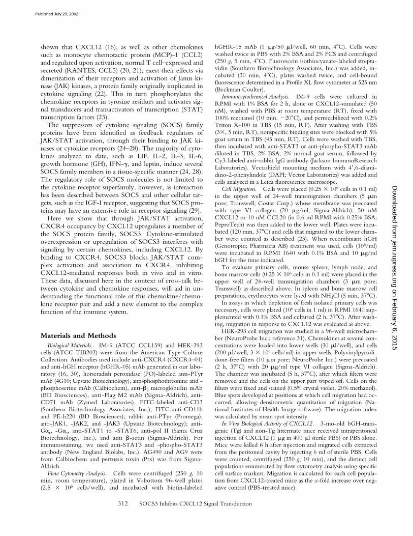
mobilizationand cell migration (see below). As for other cell lineages(16), pretreatment with AG490, a specific JAK inhibitor,completely abrogates CXCL12-mediated responses (Fig. 1A), suggesting JAK involvement. CXCL12 promotes JAK1and JAK3 association with CXCR4 (Fig. 1 B), followed bytheir rapid tyrosine phosphorylation (Fig. 1 C). As a conse-quence, STAT1, -2, -3, and -5, but not STAT4 or -6, arealso activated, indicated by their association to CXCR4(not shown) and their phosphorylation pattern (Fig. 2, Aand B). As in the case of the cytokines (33), CXCL12 alsopromotes Ser/Thr phosphorylation of STAT. Using spe-cific antibodies, we found that STAT3 is Ser/Thr phos-phorylated after stimulation with CXCL12, and that thekinetics correlate with its tyrosine phosphorylation (Fig. 2C); in both cases, maximum phosphorylation occurs 15min after stimulation.
Nuclear extracts of CXCL12-stimulated IM-9 cells wereanalyzed in Western blot using anti-STAT antibodies, andmaximum nuclear translocation was observed 30 min afteractivation (Fig. 2 D, top). Nuclear extracts analyzed inWestern blot using anti-pol II antibody as a nuclear marker(Fig. 2 D, bottom) were free of cytoplasmic contamination,confirmed by developing with an anti–
�
-actin cytoplasmicmarker antibody (not shown). As predicted, SIE bindingactivity was detected after chemokine treatment, measuredby EMSA (Fig. 2 E). Protein levels in nuclear extracts werecontrolled using a protein detection kit. We thus con-cluded that CXCL12 triggers JAK/STAT pathway activa-tion and the nuclear translocation of STAT.
Cytokine activation of the JAK/STAT pathway leads toSOCS up-regulation, implicated in turn in the negativefeedback of cytokine signaling. We tested whether SOCSare up-regulated as a consequence of CXCL12-induced
on February 6, 2015
jem.rupress.org
Dow
nloaded from
Published July 29, 2002
314 SOCS3 Inhibits CXCL12 Signal Transduction
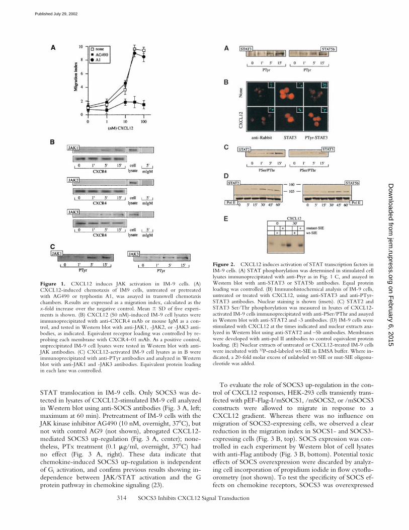
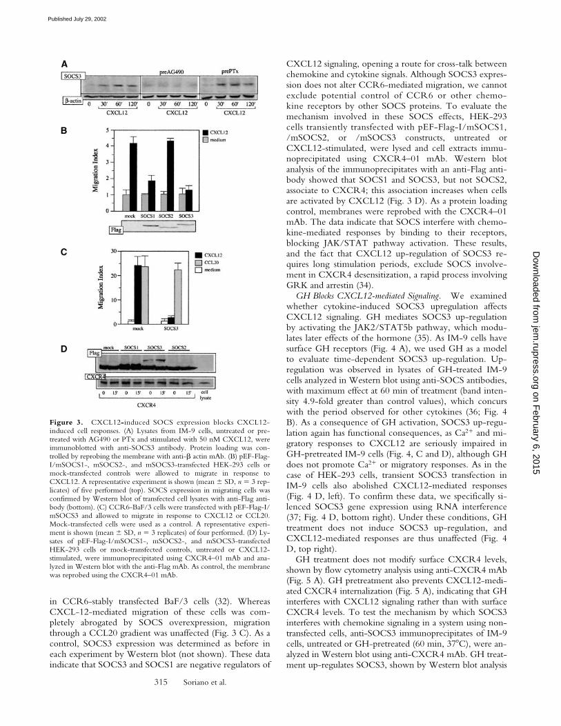
STAT translocation in IM-9 cells. Only SOCS3 was de-tected in lysates of CXCL12-stimulated IM-9 cell analyzedin Western blot using anti-SOCS antibodies (Fig. 3 A, left;maximum at 60 min). Pretreatment of IM-9 cells with theJAK kinase inhibitor AG490 (10 nM, overnight, 37C), butnot with control AG9 (not shown), abrogated CXCL12-mediated SOCS3 up-regulation (Fig. 3 A, center); none-theless, PTx treatment (0.1 �g/ml, overnight, 37C) hadno effect (Fig. 3 A, right). These data indicate thatchemokine-induced SOCS3 up-regulation is independentof Gi activation, and confirm previous results showing in-dependence between JAK/STAT activation and the Gprotein pathway in chemokine signaling (23).
To evaluate the role of SOCS3 up-regulation in the con-trol of CXCL12 responses, HEK-293 cells transiently trans-fected with pEF-Flag-I/mSOCS1, /mSOCS2, or /mSOCS3constructs were allowed to migrate in response to aCXCL12 gradient. Whereas there was no influence onmigration of SOCS2-expressing cells, we observed a clearreduction in the migration index in SOCS1- and SOCS3-expressing cells (Fig. 3 B, top). SOCS expression was con-trolled in each experiment by Western blot of cell lysateswith anti-Flag antibody (Fig. 3 B, bottom). Potential toxiceffects of SOCS overexpression were discarded by analyz-ing cell incorporation of propidium iodide in flow cytoflu-orometry (not shown). To test the specificity of SOCS ef-fects on chemokine receptors, SOCS3 was overexpressed
Figure 1. CXCL12 induces JAK activation in IM-9 cells. (A)CXCL12-induced chemotaxis of IM9 cells, untreated or pretreatedwith AG490 or tyrphostin A1, was assayed in transwell chemotaxischambers. Results are expressed as a migration index, calculated as thex-fold increase over the negative control. Mean � SD of five experi-ments is shown. (B) CXCL12 (50 nM)-induced IM-9 cell lysates wereimmunoprecipitated with anti-CXCR4 mAb or mouse IgM as a con-trol, and tested in Western blot with anti-JAK1, -JAK2, or -JAK3 anti-bodies, as indicated. Equivalent receptor loading was controlled by re-probing each membrane with CXCR4–01 mAb. As a positive control,unprecipitated IM-9 cell lysates were tested in Western blot with anti-JAK antibodies. (C) CXCL12-activated IM-9 cell lysates as in B wereimmunoprecipitated with anti-PTyr antibodies and analyzed in Westernblot with anti-JAK1 and -JAK3 antibodies. Equivalent protein loadingin each lane was controlled.
Figure 2. CXCL12 induces activation of STAT transcription factors inIM-9 cells. (A) STAT phosphorylation was determined in stimulated celllysates immunoprecipitated with anti-Ptyr as in Fig. 1 C, and assayed inWestern blot with anti-STAT3 or STAT5b antibodies. Equal proteinloading was controlled. (B) Immunohistochemical analysis of IM-9 cells,untreated or treated with CXCL12, using anti-STAT3 and anti-PTyr-STAT3 antibodies. Nuclear staining is shown (insets). (C) STAT2 andSTAT3 Ser/Thr phosphorylation was measured in lysates of CXCL12-activated IM-9 cells immunoprecipitated with anti-PSer/PThr and assayedin Western blot with anti-STAT2 and -3 antibodies. (D) IM-9 cells werestimulated with CXCL12 at the times indicated and nuclear extracts ana-lyzed in Western blot using anti-STAT2 and –5b antibodies. Membraneswere developed with anti-pol II antibodies to control equivalent proteinloading. (E) Nuclear extracts of untreated or CXCL12-treated IM-9 cellswere incubated with 32P-end-labeled wt-SIE in EMSA buffer. Where in-dicated, a 20-fold molar excess of unlabeled wt-SIE or mut-SIE oligonu-cleotide was added.
on February 6, 2015
jem.rupress.org
Dow
nloaded from
Published July 29, 2002
315 Soriano et al.
in CCR6-stably transfected BaF/3 cells (32). WhereasCXCL-12-mediated migration of these cells was com-pletely abrogated by SOCS overexpression, migrationthrough a CCL20 gradient was unaffected (Fig. 3 C). As acontrol, SOCS3 expression was determined as before ineach experiment by Western blot (not shown). These dataindicate that SOCS3 and SOCS1 are negative regulators of
CXCL12 signaling, opening a route for cross-talk betweenchemokine and cytokine signals. Although SOCS3 expres-sion does not alter CCR6-mediated migration, we cannotexclude potential control of CCR6 or other chemo-kine receptors by other SOCS proteins. To evaluate themechanism involved in these SOCS effects, HEK-293cells transiently transfected with pEF-Flag-I/mSOCS1,/mSOCS2, or /mSOCS3 constructs, untreated orCXCL12-stimulated, were lysed and cell extracts immu-noprecipitated using CXCR4–01 mAb. Western blotanalysis of the immunoprecipitates with an anti-Flag anti-body showed that SOCS1 and SOCS3, but not SOCS2,associate to CXCR4; this association increases when cellsare activated by CXCL12 (Fig. 3 D). As a protein loadingcontrol, membranes were reprobed with the CXCR4–01mAb. The data indicate that SOCS interfere with chemo-kine-mediated responses by binding to their receptors,blocking JAK/STAT pathway activation. These results,and the fact that CXCL12 up-regulation of SOCS3 re-quires long stimulation periods, exclude SOCS involve-ment in CXCR4 desensitization, a rapid process involvingGRK and arrestin (34).
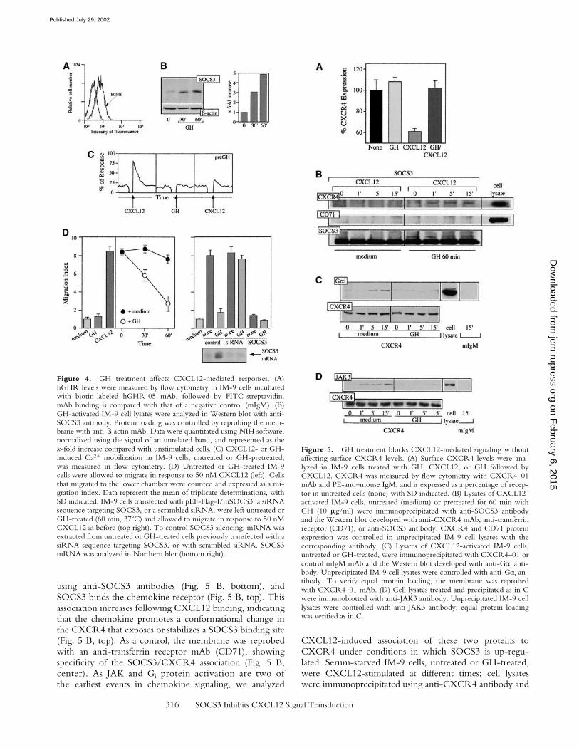
GH Blocks CXCL12-mediated Signaling. We examinedwhether cytokine-induced SOCS3 upregulation affectsCXCL12 signaling. GH mediates SOCS3 up-regulationby activating the JAK2/STAT5b pathway, which modu-lates later effects of the hormone (35). As IM-9 cells havesurface GH receptors (Fig. 4 A), we used GH as a modelto evaluate time-dependent SOCS3 up-regulation. Up-regulation was observed in lysates of GH-treated IM-9cells analyzed in Western blot using anti-SOCS antibodies,with maximum effect at 60 min of treatment (band inten-sity 4.9-fold greater than control values), which concurswith the period observed for other cytokines (36; Fig. 4B). As a consequence of GH activation, SOCS3 up-regu-lation again has functional consequences, as Ca2� and mi-gratory responses to CXCL12 are seriously impaired inGH-pretreated IM-9 cells (Fig. 4, C and D), although GHdoes not promote Ca2� or migratory responses. As in thecase of HEK-293 cells, transient SOCS3 transfection inIM-9 cells also abolished CXCL12-mediated responses(Fig. 4 D, left). To confirm these data, we specifically si-lenced SOCS3 gene expression using RNA interference(37; Fig. 4 D, bottom right). Under these conditions, GHtreatment does not induce SOCS3 up-regulation, andCXCL12-mediated responses are thus unaffected (Fig. 4D, top right).
GH treatment does not modify surface CXCR4 levels,shown by flow cytometry analysis using anti-CXCR4 mAb(Fig. 5 A). GH pretreatment also prevents CXCL12-medi-ated CXCR4 internalization (Fig. 5 A), indicating that GHinterferes with CXCL12 signaling rather than with surfaceCXCR4 levels. To test the mechanism by which SOCS3interferes with chemokine signaling in a system using non-transfected cells, anti-SOCS3 immunoprecipitates of IM-9cells, untreated or GH-pretreated (60 min, 37C), were an-alyzed in Western blot using anti-CXCR4 mAb. GH treat-ment up-regulates SOCS3, shown by Western blot analysis
Figure 3. CXCL12-induced SOCS expression blocks CXCL12-induced cell responses. (A) Lysates from IM-9 cells, untreated or pre-treated with AG490 or PTx and stimulated with 50 nM CXCL12, wereimmunoblotted with anti-SOCS3 antibody. Protein loading was con-trolled by reprobing the membrane with anti-� actin mAb. (B) pEF-Flag-I/mSOCS1-, mSOCS2-, and mSOCS3-transfected HEK-293 cells ormock-transfected controls were allowed to migrate in response toCXCL12. A representative experiment is shown (mean � SD, n 3 rep-licates) of five performed (top). SOCS expression in migrating cells wasconfirmed by Western blot of transfected cell lysates with anti-Flag anti-body (bottom). (C) CCR6-BaF/3 cells were transfected with pEF-Flag-I/mSOCS3 and allowed to migrate in response to CXCL12 or CCL20.Mock-transfected cells were used as a control. A representative experi-ment is shown (mean � SD, n 3 replicates) of four performed. (D) Ly-sates of pEF-Flag-I/mSOCS1-, mSOCS2-, and mSOCS3-transfectedHEK-293 cells or mock-transfected controls, untreated or CXCL12-stimulated, were immunoprecipitated using CXCR4–01 mAb and ana-lyzed in Western blot with the anti-Flag mAb. As control, the membranewas reprobed using the CXCR4–01 mAb.
on February 6, 2015
jem.rupress.org
Dow
nloaded from
Published July 29, 2002
316 SOCS3 Inhibits CXCL12 Signal Transduction
using anti-SOCS3 antibodies (Fig. 5 B, bottom), andSOCS3 binds the chemokine receptor (Fig. 5 B, top). Thisassociation increases following CXCL12 binding, indicatingthat the chemokine promotes a conformational change inthe CXCR4 that exposes or stabilizes a SOCS3 binding site(Fig. 5 B, top). As a control, the membrane was reprobedwith an anti-transferrin receptor mAb (CD71), showingspecificity of the SOCS3/CXCR4 association (Fig. 5 B,center). As JAK and Gi protein activation are two ofthe earliest events in chemokine signaling, we analyzed
CXCL12-induced association of these two proteins toCXCR4 under conditions in which SOCS3 is up-regu-lated. Serum-starved IM-9 cells, untreated or GH-treated,were CXCL12-stimulated at different times; cell lysateswere immunoprecipitated using anti-CXCR4 antibody and
Figure 4. GH treatment affects CXCL12-mediated responses. (A)hGHR levels were measured by flow cytometry in IM-9 cells incubatedwith biotin-labeled hGHR-05 mAb, followed by FITC-streptavidin.mAb binding is compared with that of a negative control (mIgM). (B)GH-activated IM-9 cell lysates were analyzed in Western blot with anti-SOCS3 antibody. Protein loading was controlled by reprobing the mem-brane with anti-� actin mAb. Data were quantitated using NIH software,normalized using the signal of an unrelated band, and represented as thex-fold increase compared with unstimulated cells. (C) CXCL12- or GH-induced Ca2� mobilization in IM-9 cells, untreated or GH-pretreated,was measured in flow cytometry. (D) Untreated or GH-treated IM-9cells were allowed to migrate in response to 50 nM CXCL12 (left). Cellsthat migrated to the lower chamber were counted and expressed as a mi-gration index. Data represent the mean of triplicate determinations, withSD indicated. IM-9 cells transfected with pEF-Flag-I/mSOCS3, a siRNAsequence targeting SOCS3, or a scrambled siRNA, were left untreated orGH-treated (60 min, 37C) and allowed to migrate in response to 50 nMCXCL12 as before (top right). To control SOCS3 silencing, mRNA wasextracted from untreated or GH-treated cells previously transfected with asiRNA sequence targeting SOCS3, or with scrambled siRNA. SOCS3mRNA was analyzed in Northern blot (bottom right).
Figure 5. GH treatment blocks CXCL12-mediated signaling withoutaffecting surface CXCR4 levels. (A) Surface CXCR4 levels were ana-lyzed in IM-9 cells treated with GH, CXCL12, or GH followed byCXCL12. CXCR4 was measured by flow cytometry with CXCR4–01mAb and PE-anti–mouse IgM, and is expressed as a percentage of recep-tor in untreated cells (none) with SD indicated. (B) Lysates of CXCL12-activated IM-9 cells, untreated (medium) or pretreated for 60 min withGH (10 �g/ml) were immunoprecipitated with anti-SOCS3 antibodyand the Western blot developed with anti-CXCR4 mAb, anti-transferrinreceptor (CD71), or anti-SOCS3 antibody. CXCR4 and CD71 proteinexpression was controlled in unprecipitated IM-9 cell lysates with thecorresponding antibody. (C) Lysates of CXCL12-activated IM-9 cells,untreated or GH-treated, were immunoprecipitated with CXCR4–01 orcontrol mIgM mAb and the Western blot developed with anti-G�i anti-body. Unprecipitated IM-9 cell lysates were controlled with anti-G�i an-tibody. To verify equal protein loading, the membrane was reprobedwith CXCR4–01 mAb. (D) Cell lysates treated and precipitated as in Cwere immunoblotted with anti-JAK3 antibody. Unprecipitated IM-9 celllysates were controlled with anti-JAK3 antibody; equal protein loadingwas verified as in C.
on February 6, 2015
jem.rupress.org
Dow
nloaded from
Published July 29, 2002

317 Soriano et al.
immunoblotted with specific anti-G�i (Fig. 5 C) and -JAK3antibodies (Fig. 5 D). This experiment showed that GH-induced SOCS3 up-regulation blocks both JAK3 and G�i
association to the CXCR4. Protein loading equivalencewas controlled by reprobing membranes with anti-CXCR4antibody. The data indicate that SOCS3 affects CXCL12-mediated responses; by binding to CXCR4, SOCS3 blocksJAK/STAT pathway activation without affecting cell sur-face CXCR4 levels.
SOCS3 Blocks CXCL12-mediated Responses In Vivo.We then explored whether CXCL12-mediated responsesare affected in cells that coexpress GHR and CXCR4, ob-tained from transgenic mice expressing a fusion gene cod-ing for bovine GH (bGH-Tg; reference 38). It was previ-ously determined that B cell numbers in bGH-Tg mouse
spleen increased two- to fourfold, with a lesser increase inT cells. Some differences were also found in peripherallymph nodes, in which B cells increased fivefold in bGH-Tg mice. Analysis of bone marrow cells showed that bGH-Tg mice are deficient in pre–B cells, whereas B cell num-bers are preserved (38). CXCL12 was injected into theperitoneal cavity of bGH-Tg mice and control littermates;after 6 h, migrating cells were recovered and analyzed byflow cytometry using specific cell markers. In CXCL12-injected bGH-Tg mice, B cell and granulocyte/macro-phage recovery is impaired compared with wild-type mice,shown by double staining with B220/CD45 and CD11b/CD45, respectively (Fig. 6 A). In contrast, T lymphocyterecovery is unimpaired, shown by CD3/CD45 doublestaining (not shown).
Figure 6. bGH-Tg mice have an impaired chemotactic response to CXCL12. (A) CXCL12 (1 �M) was injected intraperitoneally in bGH-Tg mice orcontrol littermates. After 60 min, cells that had migrated into the peritoneal cavity were recovered and the populations characterized by flow cytometryusing specific cell markers. The figure shows specific staining of b220/CD45 and CD11b/CD45 cells; the percentage of each population is also indicated.Results are shown of one representative experiment of five performed. (B) CXCL12-mediated cell migration of spleen, lymph node, and bone marrowcells from bGH-Tg mice or wild-type littermates were evaluated in 24-well transmigration chambers; migration was calculated as in Fig. 1 A. Data rep-resent the mean � SD of triplicate determinations. (C) Northern analysis of SOCS3 mRNA from spleen, lymph node, and bone marrow of bGH-Tgmice compared with control littermates. As a control, the membrane was rehybridized with eF1�. (D) mRNA was extracted from bone marrow cells ofbGH-Tg mice and control littermates, undepleted or GH-depleted in vitro, and SOCS3 analyzed in Northern blot. Loading was controlled as in C. (E)Bone marrow cells from mice as in D, undepleted (open symbols) or GH-depleted (filled symbols), were allowed to migrate in response to CXCL12.Migration was calculated as in Fig. 1 D. Data represent the mean � SD of triplicate determinations.
on February 6, 2015
jem.rupress.org
Dow
nloaded from
Published July 29, 2002
318 SOCS3 Inhibits CXCL12 Signal Transduction
The percentage of B220lowCD45� cells recovered fromthe peritoneal cavity of wild-type mice is markedly in-creased after CXCL12 treatment (5 � 0.3-fold) comparedwith PBS-treated controls. In contrast, no increase was ob-served in this cell population in CXCL12-treated bGH-Tgmice (0.73 � 0.06-fold). Analysis showed that CD11b�
CD45� cells also accumulate in the peritoneal cavityof CXCL12-treated wild-type mice (3.8 � 0.2-fold),whereas moderate migration was observed in treatedbGH-Tg mice (2.07 � 0.2-fold). No notable differenceswere found for B220highCD45� cells from CXCL12-treated bGH-Tg mice (2.8 � 0.2-fold) and wild-type mice(3.2 � 0.3-fold; Fig. 6 A). A similar effect is observedwhen spleen, lymph node, and bone marrow cells from3-mo-old bGH-Tg mice are allowed to migrate in vitrotoward a CXCL12 gradient. Whereas control non-Tgmouse cells respond normally to CXCL12, bGH-Tg cellsshowed a marked decrease in migration (Fig. 6 B). North-ern blot analysis showed that bGH-Tg mouse spleen,lymph node, and bone marrow cells express higher SOCS3levels than those from non-Tg littermates (Fig. 6 C).When SOCS3 levels were reduced by in vitro depletion(120 min) of bone marrow cells from bGH-Tg mice inGH-free medium (Fig. 6 D), their migratory response toCXCL12 was restored compared with undepleted cells(Fig. 6 E), indicating that SOCS3 has a critical role in che-mokine response regulation. All together, these results as-sign a direct role to the SOCS proteins in controlling che-motactic responses to CXCL12 and expose new aspects ofthe relationship between cytokines and chemokines in im-mune response control.
DiscussionFunctional lymphoid microenvironments, organogen-
esis, and leukocyte patrolling are established by cell mi-gration. This requires the integrated action of cell surfaceligands such as integrins and selectins, with soluble medi-ators including cytokines, chemokines, and their recep-tors. Whereas the former group participates in rollingand cell adhesion, the latter has a key role in the lympho-hematopoiesis and cell polarization required for cell mo-tility (39). In addition to their selective role under physi-ological conditions, chemokines also have an importantfunction in inflammation, wound healing, and angiogen-esis (6).
In recent years it has been established that hematopoi-etic cell growth, differentiation, and function are con-trolled by the coordinated action of the cytokine–chemo-kine network (40). After cytokine binding to its receptoron the cell surface, receptor oligomerization takes place,inducing JAK kinase activation. The activated JAK ki-nases then phosphorylate the cytokine receptors, leadingto recruitment and subsequent activation of other signal-ing molecules, such as the STAT family proteins. Acti-vated STAT proteins translocate to the nucleus and medi-ate transcription of a range of cytokine-responsive genes(22), including those that code for the SOCS proteins.
These molecules have recently attracted interest, as theyexercise their effect directly on the JAK/STAT pathway.Indeed, the majority of cytokines analyzed to date, such asLIF, IL-2, IL-3, IL-6, GH, IFN-�, and leptin, induceseveral SOCS family members in a tissue-specific manner(28).
Much like cytokines, the chemokines trigger oligomer-ization of their receptors and activation of the JAK/STATpathway (31, 41, 42). The similarity among chemokine re-ceptors, including conservation of the DRY motif, sug-gests that oligomerization and JAK/STAT pathway activa-tion are not exclusive to CCR2, in which they were firstdescribed (20, 23); CCR5 and CXCR4 both activate sev-eral JAK/STAT family members in a cell lineage–depen-dent fashion. Agonist-induced dimerization has beendescribed for other GPCR receptors, including the �2-adrenergic (43), opioid (44), and �-amino butyric acid(GABA) receptors (45); furthermore, the angiotensin II(46), TSH (47), and �-melanocyte-stimulating hormone(�-MSH) receptors (48) not only dimerize but also activatethe JAK/STAT pathway.
As described for cytokines (25, 26), CXCL12-medi-ated STAT activation and nuclear translocation promoteSOCS3 protein up-regulation. By binding to the CXCR4,overexpressed SOCS1 or SOCS3 proteins prevent achemotactic response to CXCL12 in HEK-293 cells. Thisis comparable to previous reports of SOCS3 association toother transmembrane receptors (49–51); here, the conse-quence is blockade of JAK and Gi association to CXCR4and impaired chemotactic responses. CXCL12-mediatedSOCS3 up-regulation depends directly on JAK/STATpathway activation, as AG490 treatment completely elimi-nates SOCS3 in lysates of CXCL12-stimulated cells. PTxtreatment does not affect SOCS3 up-regulation, however,indicating that JAK/STAT activation is G protein pathwayindependent. These data, together with the observationthat both PTx and AG490 treatments block CXCL12-mediated responses, suggest that JAK activation is a veryearly event in chemokine signaling. The data confirm ourprevious finding that AG490 treatment of Mono Mac-1cells inhibits JAK and Gi association to the CCR2b recep-tor after CCL2 binding, whereas PTx does not affect JAKassociation (23). The time course of CXCL12-mediatedSOCS3 up-regulation excludes the role of these suppressormolecules in the rapid ligand-triggered desensitization ofchemokine receptors, which requires GRK and arrestin re-cruitment (34).
SOCS blockade of chemokine responses may thus beenvironmentally influenced, as the presence of other che-mokines and/or cytokines alters these responses. Althoughmore exhaustive study is required, preliminary data showthat, as for cytokines (28), SOCS3 blockage selectively af-fects the responses of certain chemokines without interfer-ing with others. We nonetheless cannot exclude the in-volvement of other SOCS family members in the controlof chemokine signals, in which case the specificity of theeffect would depend on differential SOCS protein expres-sion in diverse cell types. A distinct SOCS expression pat-
on February 6, 2015
jem.rupress.org
Dow
nloaded from
Published July 29, 2002
319 Soriano et al.
tern has been described that correlates with differentiationinto Th1 or Th2 phenotype (52).
GH belongs to the cytokine family, which also com-prises placental lactogen and prolactin; its biological effectsvary widely, and include skeletal growth during childhoodand regulation of a variety of anabolic processes in adultlife. Lymphocytes also have GH receptors, as defined bybiochemical, molecular, and functional evidence, and GHhas been implicated as a growth and differentiation factorin the hematopoietic system (53). After binding GH, thereceptor dimerizes and signals through JAK2 kinase (35).This pathway includes tyrosine phosphorylation of severalproteins, among them the latent cytoplasmic transcriptionfactors, STATs. This activation leads to upregulation of avariety of genes in vivo and in vitro, including SOCS2,SOCS3, and CIS mRNA (54). SOCS inhibit receptor sig-naling to STAT5b via phosphotyrosine-dependent bindinginteractions with the tyrosine kinase JAK2 (SOCS1) and/or the cytoplasmic tail of GHR (CIS and SOCS3; refer-ence 55).
Given the ability of both GH and CXCL12 to stimulatethe JAK/STAT pathway and to upregulate SOCS3, westudied the relationship between chemokines and cyto-kines using CXCL12 and GH as a model. We show thatthe IM-9 cell line does not migrate in response to GH,but does so in response to CXCL12, a process that isblocked by pretreatment with the JAK2-specific inhibitorAG490. When cells were pretreated with GH under con-ditions that up-regulate SOCS3, cell migration to aCXCL12 gradient was impaired; when SOCS3 wasdownregulated or its expression silenced by RNA inter-ference (37), however, the chemotactic response wasrecovered. These data concur with the absence of a che-motactic response to CXCL12 in SOCS3-transfectedHEK-293 cells. GH does not affect membrane CXCR4levels, shown by anti-CXCR4 staining of GH-treatedcells. SOCS3 is nonetheless associated to CXCR4 underconditions in which it is up-regulated by GH treatment ofIM9 cells. This association increases after CXCL12 stimu-lation, indicating that the ligand may promote conforma-tional changes in the CXCR4, which then exposes theSOCS3 binding site or stabilizes the complex. As a conse-quence, neither JAK nor Gi associates to the CXCR4,blocking chemotactic responses; a model is outlined inFig. 7. The data thus indicate that CXCL12 is able to acti-vate CXCR4 under conditions in which SOCS3 is up-regulated, not only in vitro but also in vivo, as observed inbGH-Tg mice. These mice, which have altered hemato-poiesis, show a defective CXCR4 response in cells ex-pressing the GH receptor, compatible with the highSOCS3 levels in these cells. As predicted, similar cell pop-ulations in wild-type littermates show no SOCS3 up-reg-ulation and thus have normal responses to CXCL12.Furthermore, when bGH-Tg mouse cells are depletedof growth factor in vitro, SOCS3 levels diminish andCXCL12-mediated responses are restored.
Identification of the chemokine-activated JAK/STAT/SOCS pathway has opened a new avenue in signal trans-
duction research, by integrating this pathway with thoseof cytokine signaling. It is now of interest to identify thecytokine/chemokine combination and the SOCS expres-sion pattern required by each lineage during develop-ment, as well as those of cells mobilized in normal im-mune responses and the inflammatory response. In vivoexperiments indicate a possible role for such interactions,as neutrophils purified from acromegalic or hyperpro-lactinemic individuals show decreased in vitro chemotaxisto an N-formylmethionyl-phenylalanine gradient (56).Chemokine response defects may also collaborate in thereduced B cell lymphopoiesis, reduced myelopoiesis in fe-tal liver, and virtual absence of myelopoiesis in bone mar-row described in bGH-Tg mice (38). These in vivo andin vitro studies could thus lead to the development of spe-cific pharmacological inhibitors of cytokine and chemo-kine signaling that are able to interfere with inflammatoryresponses.
We thank Drs. T. Willson, R. Starr, and D. Hilton (Walter andEliza Hall Institute of Medical Research) for the gift of SOCS ex-pression vectors, Dr. J.P. García-Ruiz and E. Delgado for bovineGH transgenic mice, Dr. L. Kremer for the gift of CCR6-trans-fected BaF/3 cells, J. Palacín and L. Gómez for expert animal care,Ma C. Moreno-Ortíz and Dr. I. López-Vidiero for help withFACS® analysis, and C. Bastos and C. Mark for secretarial and edi-torial assistance, respectively.
CEC-P is supported by the Brazilian Coordenaçao de Aper-feiçoamento de Pessoal de Nivel Superior Foundation and by theUniversidade Federal Fluminense, Niteroi-Rio de Janeiro, Brazil.This work was partially supported by grants from the SpanishCICyT and the Comunidad de Madrid. The Department of Im-munology and Oncology was founded and is supported by theSpanish Council for Scientific Research (CSIC) and by the Phar-macia Corporation.
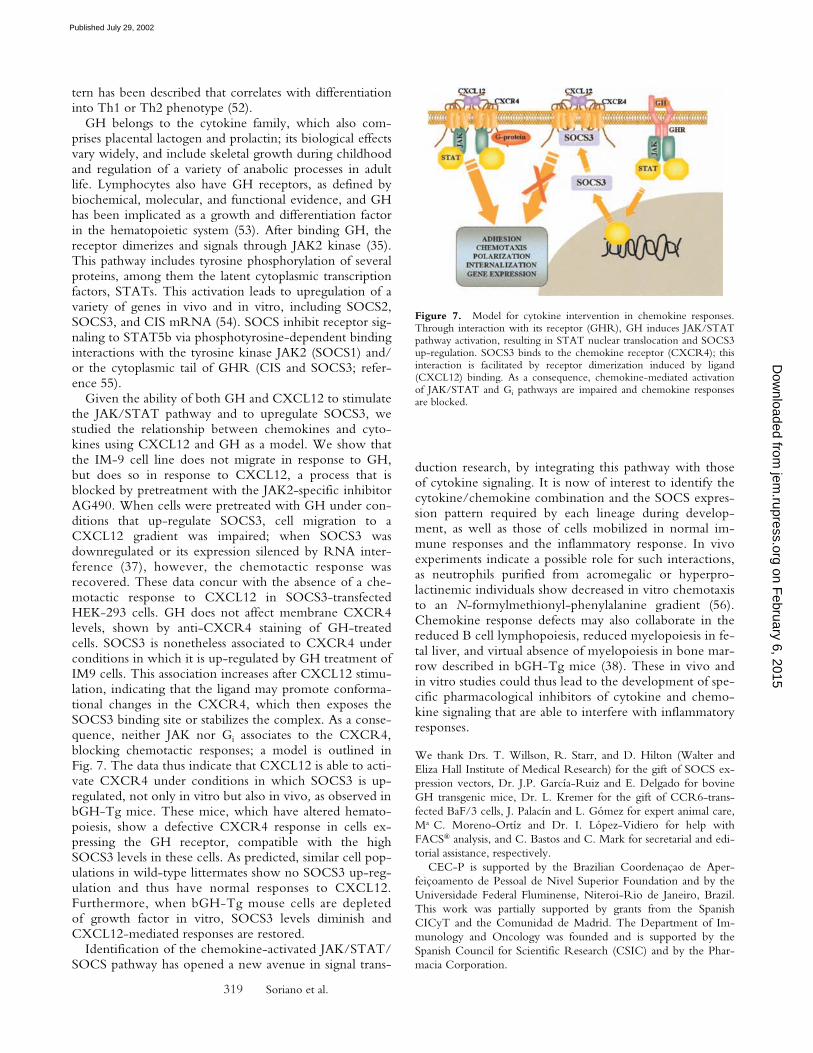
Figure 7. Model for cytokine intervention in chemokine responses.Through interaction with its receptor (GHR), GH induces JAK/STATpathway activation, resulting in STAT nuclear translocation and SOCS3up-regulation. SOCS3 binds to the chemokine receptor (CXCR4); thisinteraction is facilitated by receptor dimerization induced by ligand(CXCL12) binding. As a consequence, chemokine-mediated activationof JAK/STAT and Gi pathways are impaired and chemokine responsesare blocked.
on February 6, 2015
jem.rupress.org
Dow
nloaded from
Published July 29, 2002
320 SOCS3 Inhibits CXCL12 Signal Transduction
References1. Baggiolini, M., B. Dewald, and B Moser. 1997. Human che-
mokines: an update. Ann. Rev. Immunol. 15:675–705.2. Rollins, B.J. 1997. Chemokines. Blood. 90:909–928.3. Mackay, C.R. 2001. Chemokine: immunology’s high impact
factors. Nat. Immunol. 2:95–101.4. Schall, T.J., and K.B Bacon. 1994. Chemokines, leukocyte
trafficking and inflammation. Curr. Opin. Immunol. 6:865–873.5. Rossi, D., and A. Zlotnik. 2000. The biology of chemokines
and their receptors. Annu. Rev. Immunol. 18:217–242.6. Gerard, C., and B.J. Rollins. 2001. Chemokines and diseases.
Nat. Immunol. 2:108–115.7. Moser, B., and P. Loetscher. 2001. Lymphocyte traffic con-
trol by chemokines. Nat. Immunol. 2:123–128.8. Luther, S.A., and J.G. Cyster. 2001. Chemokines as regula-
tors of T cell differentiation. Nat. Immunol. 2:102–107.9. Bleul, C.C., R.C. Fuhlbrigge, J.M. Casasnovas, A. Aiuti,
and T.A. Springer. 1996. A highly efficacious lymphocytechemoattractant, stromal cell-derived factor 1 SDF-1. J. Exp.Med. 184:1101–1109.
10. Nagasawa, T., S. Hirota, K. Tachibana, N. Takakura, S.Nishikawa, Y. Kitamura, N. Yoshida, H. Kikutani, and T.Kishimoto. 1996. Defects of B cell lymphopoiesis and bonemarrow myelopoiesis in mice lacking the CXC chemokinePBSF/SDF-1. Nature. 382:635–638.
11. Aiuti, A., I.J. Webb, C. Bleul, T.A. Springer, and J.C. Gu-tierrez-Ramos. 1997. The chemokine SDF-1 is a chemoat-tractant for human CD34� hematopoietic progenitor cellsand provides a new mechanism to explain the mobilization ofCD34� progenitors to peripheral blood. J. Exp. Med. 185:111–120.
12. D’Apuzzo, M., A. Rolink, M. Loetscher, J.A. Hoxie, I.Clark-Lewis, F. Melchers, M. Baggiolini, and B. Moser.1997. The chemokine SDF-1, stromal cell-derived factor 1,attracts early stage B cell precursors via the chemokine recep-tor CXCR4. Eur. J. Immunol. 27:1788–1793.
13. Zou, Y.-R., A.H. Kottmann, M. Kuroda, I. Taniuchi, andD.R. Littman. 1998. Function of the chemokine receptorCXCR4 in haematopoiesis and in cerebellar development.Nature. 393:595–599.
14. Tachibana, K., S. Hirota, H. Iizasa, H. Yoshida, K. Kawa-bata, Y. Kataoka, Y. Kitamura, K. Matsushima, N. Yoshida,S. Nishikawa, et al. 1998. The chemokine receptor CXCR4is essential for vascularization of the gastrointestinal tract. Na-ture. 393:591–594.
15. Ma, Q., D. Jones, P.R. Borghesani, RA. Segal, T. Nagasawa,T. Kishimoto, R.T. Bronson, and T.A. Springer. 1998. Im-paired B-lymphopoiesis, myelopoiesis, and derailed cerebellarneuron migration in CXCR4- and SDF-1-deficient mice.Proc. Natl. Acad. Sci. USA. 95:9448–9453.
16. Vila-Coro, A.J., J.M. Rodríguez-Frade, A.M. Martín deAna, M.C. Moreno-Ortíz, C. Martínez-A., and M. Mellado.1999. The chemokine SDF-1� triggers CXCR4 receptordimerization and activates the JAK/STAT pathway. FASEBJ. 13:1699–1710.
17. Wang, J.F., I.W. Park, and J.E. Groopman. 2000. Stromalcell-derived factor-1� stimulates tyrosine phosphorylation ofmultiple focal adhesion proteins and induces migration of he-matopoietic progenitor cells: roles of phosphoinositide-3 ki-nase and protein kinase C. Blood. 95:2505–2513.
18. Ganju, R.K., S.A. Brubaker, J. Meyer, P. Dutt, Y. Yang, S.Qin, W. Newman, and J.E. Groopman. 1998. The �-chemo-kine, stromal cell-derived factor-1�, binds to the transmem-
brane G-protein-coupled CXCR4 receptor and activatesmultiple signal transduction pathways. J. Biol. Chem. 273:23169–23175.
19. Vicente-Manzanares, M., M. Rey, D.R. Jones, D. Sancho,M. Mellado, J.M. Rodriguez-Frade, M.A. del Pozo, M.Yañez-Mó, A. Martín de Ana, C. Martínez-A., et al. 1999.Involvement of phosphatidylinositol 3-kinase in stromal cell-derived factor-1�-induced lymphocyte polarization andchemotaxis. J. Immunol. 163:4001–4012.
20. Rodríguez-Frade, J.M., A. Vila-Coro, A.M. Martín de Ana,J.P. Albar, C. Martínez-A., and M. Mellado. 1999. Thechemokine monocyte chemoattractant protein-1 inducesfunctional responses through dimerization of its receptorCCR2. Proc. Natl. Acad. Sci. USA. 96:3628–3633.
21. Rodríguez-Frade, J.M., A.J. Vila-Coro, A.M. Martín deAna, M. Nieto, F. Sánchez-Madrid, A.E.I. Proudfoot,T.N.C. Wells, C. Martínez-A., and M. Mellado. 1999. Simi-larities and differences in RANTES- and (AOP)-RANTES-triggered signals: implications for chemotaxis. J. Cell Biol.144:755–765.
22. Leonard, W.J., and J.J. O’Shea. 1998. JAK and STATs: bio-logical implications. Annu. Rev. Immunol. 16:293–322.
23. Mellado, M., J.M. Rodríguez-Frade, A.M. Aragay, G. delReal, A. Vila-Coro, A. Martin de Ana, A. Serrano, F. Mayor,Jr., and C. Martínez-A. 1998. The chemokine MCP-1 trig-gers tyrosine phosphorylation of the CCR2B receptor andthe JAK2/STAT3 pathway. J. Immunol. 161:805–813.
24. Yoshimura, A., T. Ohkubo, T. Kiguchi, N.A. Jenkins, D.J.Gilbert, N.G. Copeland, T. Hara, and A. Miyajima. 1995. Anovel cytokine-inducible gene CIS encodes an SH2-contain-ing protein that binds to tyrosine-phosphorylated interleukin3 and erythropoietin receptors. EMBO J. 14:2816–2826.
25. Starr, R., T.A. Willson, E.M. Viney, L.J. Murray, J.R.Rayner, B.J. Jenkins, T.J. Gonda, W.S. Alexander, D. Met-calf, N.A. Nicola, and D.J. Hilton. 1997. A family of cyto-kine-inducible inhibitors of signalling. Nature. 387:917–921.
26. Endo, T.A., M. Masuhara, M. Yokouchi, R. Suzuki, H.Sakamoto, K. Mitsui, A. Matsumoto, S. Tanimura, M. Oht-subo, H. Misawa, et al. 1997. A new protein containing anSH2 domain that inhibits JAK kinases. Nature. 387:921–924.
27. Naka, T., M. Narazaki, M. Hirata, T. Matsumoto, S. Mina-moto, A. Aono, N. Nishimoto, T. Kajita, T. Taga, K.Yoshizaki, et al. 1997. Structure and function of a newSTAT-induced STAT inhibitor. Nature. 387:924–929.
28. Chen, X.P., J.A. Losman, and P. Rothman. 2000. SOCSproteins, regulators of intracellular signaling. Immunity. 13:287–290.
29. Dey, B.R., R.W. Furlanetto, and P. Nissley. 2000. Suppres-sor of cytokine signaling (SOCS-3) protein interacts with theinsulin-like growth factor-I receptor. Biochem. Biophys. Res.Commun. 278:38–43.
30. Mellado, M., J.M. Rodríguez-Frade, L. Kremer, and C.Martínez-A. 1996. Characterization of monoclonal antibod-ies which specific for the human growth hormone 22K and20K isoforms. J. Clin. Endocrinol. Metab. 81:1613–1618.
31. Mellado, M., J.M. Rodríguez-Frade, S. Mañes, and C. Mar-tínez-A. 2001. Chemokine signaling and functional re-sponses: The role of receptor dimerization and TK pathwayactivation. Annu. Rev. Immunol. 19:397–421.
32. Kremer, L., L. Carramolino, I. Goya, A. Zaballos, J. Gutier-rez, M.C. Moreno-Ortiz, C. Martínez-A., and G. Márquez.2001. The transient expression of C-C chemokine receptor 8
on February 6, 2015
jem.rupress.org
Dow
nloaded from
Published July 29, 2002
321 Soriano et al.
in thymus identifies a thymocyte subset committed to be-come CD4� single-positive T cells. J. Immunol. 166:218–225.
33. Decker, T., and P. Kovarik. 2000. Serine phosphorylation ofSTATs. Oncogene. 19:2628–2637.
34. Orsini, M.J., J.L. Parent, S.J. Mundell, J.L. Benovic, and A.Marchese. 1999. Trafficking of the HIV coreceptor CXCR4.Role of arrestins and identification of residues in the c-termi-nal tail that mediate receptor internalization. J. Biol. Chem.274:31076–31086.
35. Herrington, J., and C. Carter-Su. 2001. Signaling pathwaysactivated by the growth hormone receptor. Trends Endocrinol.Metab. 12:252–257.
36. Zhang, J.G., A. Farley, S.E. Nicholson, T.A. Willson, L.M.Zugaro, R.J. Simpson, R.L. Moritz, D. Cary, R. Richard-son, G. Hausmann, et al. 1999. The conserved SOCS boxmotif in suppressors of cytokine signaling binds to elongins Band C and may couple bound proteins to proteasomal degra-dation. Proc. Natl. Acad. Sci. USA. 96:2071–2076.
37. Sharp, P.A. 2001. RNA interference. Genes Dev. 15:485–490.
38. Gonzalo, J.A., R. Mazuchelli, M. Mellado, J.M.R. Frade,A.C. Carrera, C. von Kobbe, I. Mérida, and C. Martínez-A.1996. Enterotoxin septic shock protection and deficient Thelper 2 cytokine production in growth hormone transgenicmice. J. Immunol. 157:3298–3304.
39. von Andrian, U.H., and C.R. Mackay. 2000. T cell functionand migration: two sides of the same coin. N. Engl. J. Med.343:1020–1034.
40. Youn, B.S., C. Mantel, and H.E. Broxmeyer. 2000.Chemokines, chemokine receptors and hematopoiesis. Immu-nol. Rev. 177:150–174.
41. Mellado, M., J.M. Rodríguez-Frade, A.J. Vila-Coro, S.Fernández, A.M. Martín de Ana, D.R. Jones, J.L. Torán, andC. Martínez-A. 2001. Chemokine receptor homo- or het-erodimerization activates distinct signaling pathways. EMBOJ. 20:2497–2507.
42. Rodríguez-Frade, J.M., M. Mellado, and C. Martínez-A.2001. Chemokine receptor dimerization in cell signaling:two are better than one. Trends Immunol. 22:612–617.
43. Hebert, T.E., S. Moffett, J.-P. Morello, T.P. Loisel, D.G.Bichet, C. Barret, and M. Bouvier. 1996. A peptide derivedfrom a �2-adrenergic receptor transmembrane domain inhib-its both receptor dimerization and activation. J. Biol. Chem.271:28612–28616.
44. Cvejic, S., and L.A. Devi. 1997. Dimerization of the deltaopioid receptor: implication for a role in receptor internaliza-tion. J. Biol. Chem. 272:26959–26964.
45. Kuner, R., G. Köhr, S. Grünewald, G. Eisenhardt, A. Bah,and H.-C. Kornau. 1999. Role of heteromer formation inGABAB receptor function. Science. 283:74–77.
46. Marrero, M.B., B. Schieffer, W.G. Paxton, L.H. Eerdt, B.C.Berk, P. Delafontaine, and K.E. Bernstein. 1995. Direct stim-ulation of Jak/STAT pathway by the angiotensin II AT1 re-ceptor. Nature. 375:247–250.
47. Park, E.S., H. Kim, J.M. Suh, S.J. Park, S.H. You, H.K.Chung, K.W. Lee, O.Y. Kwon, B.Y. Cho, Y.K. Kim, et al.2000. Involvement of JAK/STAT (Janus kinase/signal trans-ducer and activator of transcription) in the thyrotropin signal-ing pathway. Mol. Endocrinol. 114:662–670.
48. Buggy, J.J. 1998. Binding of a melanocyte-stimulating hor-mone to its G protein-coupled receptor on B lymphocytesactivates the JAK/STAT pathway. Biochem. J. 331:211–216.
49. Emanuelli, B., P. Peraldi, C. Filloux, D. Sawka-Verhelle, D.Hilton, and E. Van Obberghen. 2000. SOCS-3 is an insulin-induced negative regulator of insulin signaling. J. Biol. Chem.275:15985–15991.
50. Pezet, A., H. Favre, P.A. Kelly, and M. Edery. 1999. Inhibi-tion and restoration of prolactin signal transduction by sup-pressors of cytokine signaling. J. Biol. Chem. 274:24497–24502.
51. Sporri, B., P.E. Kovanen, A. Sasaki, A. Yoshimura, and W.J.Leonard. 2001. JAB/SOCS1/SS-1 is an interleukin-2-inducedinhibitor of IL-2 signaling. Blood. 97:221–226.
52. Egwuagu, C.E., C.R. Yu, M. Zhang, R.M. Mahdi, S.J. Kim,and I. Gery. 2002. Suppressors of cytokine signaling proteinsare differentially expressed in Th1 and th2 cells: implicationsfor th cell lineage commitment and maintenance. J. Immunol.168:3181–3187.
53. Dorshkind, K., and N.D. Horseman. 2000. The roles of pro-lactin, growth hormone, insulin-like growth factor-I andthyroid hormones in lymphocyte development and function:insights from genetic models of hormone and hormone re-ceptor deficiency. Endocr. Rev. 21:292–312.
54. Tollet-Egnell, P., A. Flores-Morales, A. Stavreus-Evers, L.Sahlin, and G. Norstedt. 1999. Growth hormone regulationof SOCS-2, SOCS-3, and CIS messenger ribonucleic acidexpression in the rat. Endocrinology. 140:3693–3704.
55. Ram, P.A., and D.J. Waxman. 2000. Role of the cytokine-inducible SH2 protein CIS in desensitization of STAT5b sig-naling by continuous growth hormone. J. Biol. Chem. 275:39487–39496.
56. Fornari, C., F.M. Palacios, R.A. Diez, and A.D. Intebi. 1994.Decreased chemotaxis of neutrophils in acromegaly and hy-perprolactinemia. Eur. J. Endocrinol. 130:463–468.
on February 6, 2015
jem.rupress.org
Dow
nloaded from
Published July 29, 2002
Copyright © 2022 FDOKUMEN