A streptococcal protease that degrades CXC chemokines and impairs bacterial clearance from infected...
10
A streptococcal protease that degrades CXC chemokines and impairs bacterial clearance from infected tissues Carlos Hidalgo-Grass 1,4 , Inbal Mishalian 1,4 , Mary Dan-Goor 1 , Ilia Belotserkovsky 1 , Yoni Eran 1 , Victor Nizet 2 , Amnon Peled 3 and Emanuel Hanski 1, * 1 Institute of Microbiology, The Hebrew University-Hadassah Medical School, Jerusalem, Israel, 2 Department of Pediatrics, School of Medicine, University of California, San Diego, La Jolla, CA, USA and 3 Goldyne Savad Institute of Gene Therapy, Hadassah University Hospital, Jerusalem, Israel Group A Streptococcus (GAS) causes the life-threatening infection in humans known as necrotizing fasciitis (NF). Infected subcutaneous tissues from an NF patient and mice challenged with the same GAS strain possessed high bacterial loads but a striking paucity of infiltrating polymorphonuclear leukocytes (PMNs). Impaired PMN recruitment was attributed to degradation of the chemo- kine IL-8 by a GAS serine peptidase. Here, we use bioinfor- matics approach coupled with target mutagenesis to identify this peptidase as ScpC. We show that SilCR pheromone downregulates scpC transcription via the two-component system—SilA/B. In addition, we demon- strate that in vitro, ScpC degrades the CXC chemokines: IL-8 (human), KC, and MIP-2 (both murine). Furthermore, using a murine model of human NF, we demonstrate that ScpC, but not the C5a peptidase ScpA, is an essential virulence factor. An ScpC-deficient mutant is innocuous for untreated mice but lethal for PMN-depleted mice. ScpC degrades KC and MIP-2 locally in the infected skin tissues, inhibiting PMN recruitment. In conclusion, ScpC repre- sents a novel GAS virulence factor functioning to directly inactivate a key element of the host innate immune response. The EMBO Journal advance online publication, 14 September 2006; doi:10.1038/sj.emboj.7601327 Subject Categories: immunology; microbiology & pathogens Keywords: chemokines; group A Streptococcus; peptidase; polymorphonuclear neutrophils; virulence Introduction Group A Streptococcus (GAS, Streptococcus pyogenes) is a major Gram-positive bacterial pathogen associated with a wide spectrum of human diseases, ranging from superficial throat and skin infections to life-threatening invasive condi- tions such as necrotizing fasciitis (NF) (Cunningham, 2000; Bisno et al, 2003). NF is an infection that results in a rapid and progressive destruction of fascia and fat (Kaul et al, 1997) that is frequently complicated by development of toxic shock syndrome (TSS). Even with prompt antibiotic treatment and surgical debridement, reported mortality rates in GAS NF and TSS are high, ranging from 20 to 60% (Davies et al, 1996; Kaul et al, 1997; Hassell et al, 2004). Although NF can be produced by GAS of various serotypes (Johnson et al, 2002; Bisno et al, 2003; Hassell et al, 2004), M1 and M3 strains have been most commonly reported (Cleary et al, 1992; Monnickendam et al, 1997; O’Brien et al, 2002; Sharkawy et al, 2002). Our prospective, population-based study of invasive GAS infections in Israel further identified M14 strains as strongly associated with severe soft tissue infections (Moses et al, 2003). In vivo screening of a transposon-tagged mutant library from an invasive M14 GAS isolate identified the streptococcal invasion locus (sil) as essential for rapid dissemination of GAS in a murine model of NF (Hidalgo-Grass et al, 2002). GAS sil shares homology with the genetic competence reg- ulon of Streptococcus pneumoniae (Claverys and Havarstein, 2002). It includes the two-component system (TCS) SilA/B and the ABC-type transporter SilD/E. Situated between these entities and preceded by a combox-like promoter (Morrison and Lee, 2000) lies the small open reading frame (ORF) silC. Highly overlapping silC, but transcribed from the reverse strand, is a sixth ORF SilCR, which is a putative competence stimulating peptide (CSP). In the M14 serotype strains, a start codon mutation elim- inates expression of the putative CSP SilCR (Hidalgo-Grass et al, 2002). We have hypothesized that sil, and SilCR in particular, may function to negatively regulate the invasive disease potential of GAS. Support for this hypothesis was provided in experiments in which mice were challenged with M14 GAS JS95 wild-type (WT) strain with or without simul- taneous injection of the mature synthetic SilCR peptide. Compared to mice challenged with WT alone, co-administra- tion of SilCR dramatically reduced bacterial proliferation, tissue necrosis, and mortality (Hidalgo-Grass et al, 2004). In mice inoculated with the WT alone, we observed a rapid spread of bacteria into the necrotic soft tissues in conjunction with a striking paucity of polymorphonuclear leukocyte (PMN) infiltration. Administration of SilCR altered the equa- tion, triggering PMN recruitment to the site of infection with restriction of infectious spread (Hidalgo-Grass et al, 2004). The observation that a culture medium of the WT strain degrades in vitro the CXC chemokine IL-8, but when grown in the presence of the SilCR peptide IL-8 degradation is lost (Hidalgo-Grass et al, 2004), offers a potential mechanism for this effect. In the present study, we apply a bioinformatics analysis and real-time RT–PCR measurement coupled with targeted Received: 19 May 2006; accepted: 16 August 2006 *Corresponding author. Faculty of medicine, Institute of Microbiology, The Hebrew University-Hadassah Medical School, Jerusalem 91010, Israel. Tel.: þ 972 2 6758196; Fax: þ 972 2 6434170; E-mail: [email protected] 4 These authors contributed equally to this work The EMBO Journal (2006), 1–10 | & 2006 European Molecular Biology Organization | All Rights Reserved 0261-4189/06 www.embojournal.org & 2006 European Molecular Biology Organization The EMBO Journal EMBO THE EMBO JOURNAL THE EMBO JOURNAL 1
-
Upload
independent -
Category
Documents
-
view
1 -
download
0
Transcript of A streptococcal protease that degrades CXC chemokines and impairs bacterial clearance from infected...
A streptococcal protease that degrades CXCchemokines and impairs bacterial clearancefrom infected tissues
Carlos Hidalgo-Grass1,4, Inbal Mishalian1,4,Mary Dan-Goor1, Ilia Belotserkovsky1,Yoni Eran1, Victor Nizet2, Amnon Peled3
and Emanuel Hanski1,*1Institute of Microbiology, The Hebrew University-Hadassah MedicalSchool, Jerusalem, Israel, 2Department of Pediatrics, School ofMedicine, University of California, San Diego, La Jolla, CA, USA and3Goldyne Savad Institute of Gene Therapy, Hadassah UniversityHospital, Jerusalem, Israel
Group A Streptococcus (GAS) causes the life-threatening
infection in humans known as necrotizing fasciitis (NF).
Infected subcutaneous tissues from an NF patient and mice
challenged with the same GAS strain possessed high
bacterial loads but a striking paucity of infiltrating
polymorphonuclear leukocytes (PMNs). Impaired PMN
recruitment was attributed to degradation of the chemo-
kine IL-8 by a GAS serine peptidase. Here, we use bioinfor-
matics approach coupled with target mutagenesis to
identify this peptidase as ScpC. We show that SilCR
pheromone downregulates scpC transcription via the
two-component system—SilA/B. In addition, we demon-
strate that in vitro, ScpC degrades the CXC chemokines:
IL-8 (human), KC, and MIP-2 (both murine). Furthermore,
using a murine model of human NF, we demonstrate that
ScpC, but not the C5a peptidase ScpA, is an essential
virulence factor. An ScpC-deficient mutant is innocuous
for untreated mice but lethal for PMN-depleted mice. ScpC
degrades KC and MIP-2 locally in the infected skin tissues,
inhibiting PMN recruitment. In conclusion, ScpC repre-
sents a novel GAS virulence factor functioning to directly
inactivate a key element of the host innate immune
response.
The EMBO Journal advance online publication, 14 September
2006; doi:10.1038/sj.emboj.7601327
Subject Categories: immunology; microbiology & pathogens
Keywords: chemokines; group A Streptococcus; peptidase;
polymorphonuclear neutrophils; virulence
Introduction
Group A Streptococcus (GAS, Streptococcus pyogenes) is a
major Gram-positive bacterial pathogen associated with a
wide spectrum of human diseases, ranging from superficial
throat and skin infections to life-threatening invasive condi-
tions such as necrotizing fasciitis (NF) (Cunningham, 2000;
Bisno et al, 2003). NF is an infection that results in a rapid
and progressive destruction of fascia and fat (Kaul et al, 1997)
that is frequently complicated by development of toxic shock
syndrome (TSS). Even with prompt antibiotic treatment
and surgical debridement, reported mortality rates in GAS
NF and TSS are high, ranging from 20 to 60% (Davies et al,
1996; Kaul et al, 1997; Hassell et al, 2004). Although NF can
be produced by GAS of various serotypes (Johnson et al,
2002; Bisno et al, 2003; Hassell et al, 2004), M1 and M3
strains have been most commonly reported (Cleary et al,
1992; Monnickendam et al, 1997; O’Brien et al, 2002;
Sharkawy et al, 2002). Our prospective, population-based
study of invasive GAS infections in Israel further identified
M14 strains as strongly associated with severe soft tissue
infections (Moses et al, 2003).
In vivo screening of a transposon-tagged mutant library
from an invasive M14 GAS isolate identified the streptococcal
invasion locus (sil) as essential for rapid dissemination of
GAS in a murine model of NF (Hidalgo-Grass et al, 2002).
GAS sil shares homology with the genetic competence reg-
ulon of Streptococcus pneumoniae (Claverys and Havarstein,
2002). It includes the two-component system (TCS) SilA/B
and the ABC-type transporter SilD/E. Situated between these
entities and preceded by a combox-like promoter (Morrison
and Lee, 2000) lies the small open reading frame (ORF) silC.
Highly overlapping silC, but transcribed from the reverse
strand, is a sixth ORF SilCR, which is a putative competence
stimulating peptide (CSP).
In the M14 serotype strains, a start codon mutation elim-
inates expression of the putative CSP SilCR (Hidalgo-Grass
et al, 2002). We have hypothesized that sil, and SilCR in
particular, may function to negatively regulate the invasive
disease potential of GAS. Support for this hypothesis was
provided in experiments in which mice were challenged with
M14 GAS JS95 wild-type (WT) strain with or without simul-
taneous injection of the mature synthetic SilCR peptide.
Compared to mice challenged with WT alone, co-administra-
tion of SilCR dramatically reduced bacterial proliferation,
tissue necrosis, and mortality (Hidalgo-Grass et al, 2004). In
mice inoculated with the WT alone, we observed a rapid
spread of bacteria into the necrotic soft tissues in conjunction
with a striking paucity of polymorphonuclear leukocyte
(PMN) infiltration. Administration of SilCR altered the equa-
tion, triggering PMN recruitment to the site of infection with
restriction of infectious spread (Hidalgo-Grass et al, 2004).
The observation that a culture medium of the WT strain
degrades in vitro the CXC chemokine IL-8, but when grown
in the presence of the SilCR peptide IL-8 degradation is lost
(Hidalgo-Grass et al, 2004), offers a potential mechanism for
this effect.
In the present study, we apply a bioinformatics analysis
and real-time RT–PCR measurement coupled with targetedReceived: 19 May 2006; accepted: 16 August 2006
*Corresponding author. Faculty of medicine, Institute of Microbiology,The Hebrew University-Hadassah Medical School, Jerusalem 91010,Israel. Tel.: þ 972 2 6758196; Fax: þ 972 2 6434170;E-mail: [email protected] authors contributed equally to this work
The EMBO Journal (2006), 1–10 | & 2006 European Molecular Biology Organization | All Rights Reserved 0261-4189/06
www.embojournal.org
&2006 European Molecular Biology Organization The EMBO Journal
EMBO
THE
EMBOJOURNAL
THE
EMBOJOURNAL
1

mutagenesis to identify ScpC as the protease responsible for
IL-8 degradation. We show that ScpC degrades the murine
CXC chemokines KC and MIP-2 in vitro and in the tissues of
experimentally infected mice, thereby critically impairing the
PMN innate immune response.
Results
Identification of GAS serine proteases transcriptionally
repressed by SilCR
We hypothesized that SilCR may reduce IL-8 degradation by
repressing the transcription of the gene encoding the GAS IL-8
peptidase. To identify possible gene candidates for such a
peptidase, we used the MEROPS database http://merops.
sanger.ac.uk (Rawlings et al, 2004), which classified the
GAS serine peptidases into 12 families. Excluding those
groups associated with basic cellular maintenance functions,
we concentrated on five families S1C, S8A, S9C, S15, and
S54 (description of the families of these peptidases and their
possible roles in virulence and chemokine degradation is
detailed in Supplementary data).
Transcription of each serine peptidase gene in the WT was
determined in the presence or absence of SilCR using real-
time RT–PCR (Figure 1A), with primers designed to amplify
any GAS allele belonging to the indicated families
(Supplementary Table I). Of the entire candidate peptidase
genes tested, only the transcription of SPy0416 (which we
termed ScpC; see below) was significantly affected by SilCR
administration, with a 10-fold reduction in mRNA levels. This
result suggested that the SPy0416 could represent the serine
peptidase activity, under negative regulation of SilCR, which
functions to promote GAS chemokine degradation. Indeed, a
mutant defective in SilA (silA), the response regulator of the
TCS of the sil locus, had a 10-fold lower transcription of the
protease than the WT and lost completely the ability to
degrade IL-8 in vitro (Supplementary Figure 1S).
Coupled with the knowledge of the recent description
of IL-8 degradation by a partially purified cell-envelope
peptidase (CEP) preparation SpyCEP (Edwards et al, 2005).
We were encouraged to pursue molecular genetic studies to
establish the contribution of the protease to disease patho-
genesis. There are several different CEPs in GAS, and SpyCEP
has an extensive homology to the C5a peptidase ScpA. To
conform more closely to standard GAS gene nomenclature,
we elected therefore to call this gene and encoded peptidase
scpC and ScpC, respectively (for streptococcal chemokine
protease).
Analysis of ScpC domains
The GAS gene encoding ScpC resides between a gene encod-
ing lactate oxidase (lctO) and an ORF of unknown function
(Spy0421) (Figure 1B). By homology inference to other
subtilisin-like serine CEPs (Siezen, 1999) (detailed in Supple-
mentary data), we found that ScpC is composed of six
putative domains: (PP) the pre-pro domain, which begins
with a signal sequence; (PR) the peptidase domain, which
contains the candidate catalytic triad of AspþHisþ Ser; (A)
and (B/H) domains, potentially serving as spacers for proper
positioning of the active site; (W) and (AN), cell wall-
anchoring domains that are separated by the LPxTG motif
(Ton-That et al, 2004) (Figure 1B). The scpC gene is present in
all 12 available GAS genomes encoding proteins that are
highly homologous (87.8% amino-acid (aa) identity, 99.6%
aa similarity).
Construction of ScpA- and ScpC-deficient mutants and
assessment of their role in virulence
ScpA and ScpC prevent adequate recruitment and activation
of PMN in vitro (DeMaster et al, 2002; Edwards et al, 2005);
thus, the two peptidases might have overlapping roles in GAS
pathogenesis. To test the contribution to virulence of ScpC in
the absence of ScpA, we first constructed a DscpA mutant in
the WT M14 GAS strain. The DscpA mutant was obtained by
replacing 2752 bp from the coding region of the gene with
aad9 (Supplementary Tables I and II). Loss of ScpA expres-
sion in the mutant was confirmed by dot blot analysis
(Supplementary Figure 2S). Secondly, we deleted scpC in
the scpA-deficient background. The DscpA/DscpC double
mutant was constructed by replacing a 753 bp internal
EcoRV fragment of scpC coding region (Figure 1B), including
the critical serine residue of the catalytic triad with O-Km2
element. The fidelity of scpC replacement was confirmed as
detailed in Materials and methods.
Relative transcript abundance
degP [SPy2216]
scpA [SPy2010]
scpC [SPy0416]
rbfA [SPy1892]
pepXP [SPy1858]
aarA [SPy0223]
P < 0.001
0.01 0.1 1 10 100
scpC
SPy0421
PP PR A B/H W AN
D151 H279 S617
lctO
LPxTG
251 aa
EcoRV EcoRV EcoRV
A
B
Figure 1 Identification of ScpC as a putative IL-8 protease. (A) Theeffect of SilCR on the transcription of GAS serine peptidase. Theabundance of the indicated serine peptidase transcripts relative tothat of gyrA was determined by real-time RT–PCR in RNA derivedfrom WT grown to OD of 0.4 at 600 nm in the absence (clear bars)or presence (shaded bars) of SilCR (10mg/ml). The values are meanobtained from analysis in duplicate of three independent RNAsamples. Error bars represent standard deviation (s.d.). SilCR down-regulates the transcription of scpC. Po0.001 (Student’s test). (B)Genomic arrangement and domain organization of ScpC. Upperpanel: The arrows depict the identified ORFs and their direction oftranscription. The 50 and 30 EcoRV sites at positions 1801 and2554 bp from the beginning of scpC were used to replace the internalscpA coding sequence with Okm2. spy0421 encodes a product of236 aa with no homology to characterized proteins. Lower panel:The map of the motifs identified in the predicted sequence of ScpCincludes the following: the pre-pro (PP) domain (residues 1–123)containing the signal sequence (residues 1–34) depicted as anarrow, protease domain (PR) (residues 124–688) containing Asp,His, and Ser forming the catalytic triad; the A domain (residues689–1128) and the B/H domain (residues 1129–1560), the cell walldomain (W) (residues 1561–1613), the cell wall anchor domain(AN) (residues 1613–1647), and the LPxTG motif starting at residue1613. The DNA region removed by the EcoRV digestion contains asegment of 251 aa (hatched) including Ser617 of the catalytic triad.
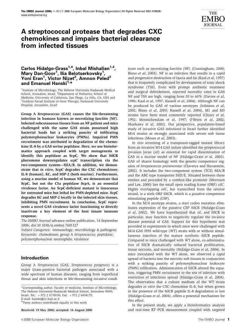
S. pyogenes CXC chemokine peptidaseC Hidalgo-Grass et al
The EMBO Journal &2006 European Molecular Biology Organization2

The WT GAS parental strain and its isogenic DscpA and
DscpA/DscpC mutants demonstrated equivalent logarithmic
growth in Todd–Hewitt Yeast (THY) media (Supplementary
Figure 3Sa) and similar proliferation rates in nonimmune
whole human blood (Supplementary Figure 3Sb). This result
indicates that neither of the peptidases contributes signifi-
cantly to GAS whole blood survival. As surface-expressed M
and M-like proteins are absolutely required for GAS resis-
tance to phagocytic killing by complement and non-stimu-
lated PMNs (Bisno et al, 2003), these results also imply that
both DscpA and DscpA/DscpC mutants maintain adequate
levels of M protein expression. Despite different strategies
used and several attempts, we were unable to inactivate scpC
without first inactivating scpA (for details see Supplementary
Figure 4S). This may imply that there could be a functional
linkage between ScpA and ScpC in the M14 strain containing
the sil locus.
Next, we tested the relative contribution of ScpA and
ScpC to virulence in a murine model of GAS NF. All mice
challenged with either the WT or the DscpA mutant experi-
enced a rapidly progressing infection and death between
2 and 7 days after challenge (Figure 2A). The Kaplan–Meier
analysis showed that the rates of death of mice inoculated
with the WT and the DscpA mutant were similar. In marked
contrast, only three mice out of the 28 challenged with the
DscpA/DscpC mutant succumbed to the infection (Figure 2A),
proving it to be significantly less virulent than either the WT
or the DscpA mutant. As shown, weight changes paralleled
the data shown in Figure 2A. Mice challenged with WT and
the DscpA mutant showed progressive weight loss before
death, whereas mice challenged with the DscpA/DscpC mu-
tant regained weight by day 2 with progressive weight gain
thereafter (Figure 2B). Mice challenged with either the WT or
the DscpA mutant developed visible necrotic skin lesions of
similar size 1 day after the inoculation (Figure 2C). At day 2
after inoculation, the lesion size of the WT and the DscpA
mutant expanded and continued to increase in size until mice
died by day 4 after the infection. In contrast, mice challenged
with the DscpA/DscpC double mutant developed small
lesions peaking in size on day 2 (Figure 2C), followed by
slow resolution and complete healing by day 14 (not shown).
Grossly, the small lesions caused by DscpA/DscpC mutant
were superficial in nature, whereas the lesions produced by
either the WT GAS or the DscpA mutant were deeper and
more necrotic.
In vitro degradation of chemokines by ScpC
To provide direct evidence that ScpC was responsible for
chemokine degradation, we tested the set of the isogenic
mutants using an in vitro IL-8 degradation assay. The DscpA
mutant degraded IL-8 as efficiently as the parental WT strain
(Figure 3A). In contrast, the isogenic DscpA/DscpC double
Per
cent
sur
viva
lTime (days)
20
40
60
80
100
0
1 2 3 4 5 6 7
Time (days)10 2 3 4 5 6 7
Time (days)10 2 3 4 5 6 7
9
11
13
15
17
Wei
ght (
g)A
rea
(cm
2 )
0
0.5
1
1.5
2
2.5
A
B
C
Figure 2 ScpC significantly contributes to virulence in the murinemodel of GAS necrotizing soft tissue infections. (A) Inactivation ofscpC but not of scpA abolished lethality of GAS. Mice were injectedsubcutaneously with 1�108 CFU of WT (— n¼ 32), DscpA (—n¼ 31), and DscpA/DscpC (— n¼ 28) and survival was monitoreddaily. The Kaplan–Meier analysis performed on five different ex-periments shows Po0.001 for DscpA/DscpC versus WT or versusDscpA and P40.05 for WT versus DscpA (log rank (Mantel–Cox)test). (B) Weight change in control mice (K) and mice challengedwith 1�108 CFU of WT (~), DscpA (&), and DscpA/DscpC (m).Experiments were repeated five times with similar results. (C) Meantotal lesion size (cm2). Mice were injected with 1�108 CFU of WT(~ n¼ 9), DscpA (& n¼ 15), and DscpA/DscpC (m n¼ 9), photo-graphed daily, and the area was calculated using ImageJ software.Error bars represent s.d. WT and DscpA versus DscpA/DscpCPo0.05 (Student’s test) for time points 1 and 2 and Po0.001(Student’s test) for time points 3 and 4. WT versus DscpA P40.05(Student’s test) at all time points.
S. pyogenes CXC chemokine peptidaseC Hidalgo-Grass et al
&2006 European Molecular Biology Organization The EMBO Journal 3
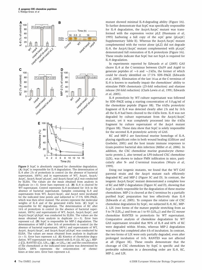
mutant showed minimal IL-8-degrading ability (Figure 3A).
To further demonstrate that ScpC was specifically responsible
for IL-8 degradation, the DscpA/DscpC mutant was trans-
formed with the expression vector pLZ (Husmann et al,
1995) harboring a full copy of the scpC gene (pLscpC;
Supplementary Table II). Whereas the DscpA/DscpC mutant
complemented with the vector alone (pLZ) did not degrade
IL-8, the DscpA/DscpC mutant complemented with pLscpC
demonstrated full restoration of IL-8 proteolysis (Figure 3A).
These results indicate that ScpC but not ScpA is required for
IL-8 degradation.
In experiments reported by Edwards et al (2005) GAS
cleave IL-8 at the C-terminus between Glu59 and Arg60 to
generate peptides of B6 and B2 kDa, the former of which
could be clearly identified on 17.5% SDS–PAGE (Edwards
et al, 2005). Elimination of the last 14 aa at the C-terminus of
IL-8 is known to markedly impair the chemokines’ ability to
stimulate PMN chemotaxis (25-fold reduction) and elastase
release (50-fold reduction) (Clark-Lewis et al, 1991; Edwards
et al, 2005).
IL-8 proteolysis by WT culture supernatant was followed
by SDS–PAGE using a starting concentration of 3.0 mg/ml of
the chemokine peptide (Figure 3B). The 6 kDa proteolytic
fragment of IL-8 was detected clearly after 2 h and by 16 h
all the IL-8 had been cleaved to the 6 kDa form. IL-8 was not
degraded by culture supernatant from the DscpA/DscpC
mutant, yet it was completely processed into the 6 kDa
fragment by culture supernatant of the DscpA mutant
(Figure 3B). These data show that ScpC is solely responsible
for the secreted IL-8 proteolytic activity of GAS.
KC and MIP-2 are functional murine homologs of IL-8,
playing significant roles in both wound healing (Gillitzer and
Goebeler, 2001) and the host innate immune responses to
Gram-positive bacterial skin infection (Miller et al, 2006). In
addition, the CXC chemokine murine granulocyte chemo-
tactic protein 2, also termed as LPS-induced CXC chemokine
(LIX), was shown to induce PMN infiltration in mice, parti-
cularly after N- and C-terminal truncation (Wuyts et al,
1999).
Using our isogenic mutants, we found that the WT GAS
parental strain and the DscpA mutant each efficiently
degraded KC and MIP-2 (Figure 3C and D). In contrast, the
isogenic DscpA/DscpC mutant demonstrated a complete loss
of KC and MIP-2 degradation (Figure 3C and D), showing that
ScpC is solely responsible for the degradation of these murine
chemokines. MIP-2 is cleaved at the C-terminus by a partially
purified ScpC preparation but less effectively than IL-8
(Edwards et al, 2005). To compare the relative rate of CXC
chemokine degradation by ScpC, we subjected IL-8, KC, MIP-
2, LIX (two forms of the mature peptide stretching from aa
5 to 78 (LIX74) and from aa 1 to 93 (LIX93)) and the CC mouse
chemokine RANTES to proteolysis by WT supernatant.
Comparative analysis of chemokine degradation by WT
GAS supernatant revealed that 90% of IL-8 and 60% of KC
were degraded within 30 min, whereas MIP-2 degradation
was slower but completed after 6 h of incubation. In contrast,
the two forms of LIX were only partially degraded, even after
prolonged incubation of 16 h. RANTES was not affected
at all (Figure 3E). These results demonstrate that the
cleavage of CXC chemokines by ScpC is specific and the
preferable substrates in a descending order are IL-8, KC,
MIP-2, and LIX.
16 160.5 28 kDa6 kDa
Time (h)
0
20
40
60
80
100
IL-8
(%
of c
ontr
ol)
0
20
40
60
80
100
KC
(%
of c
ontr
ol)
0
20
40
60
80
100
MIP
-2 (
% o
f con
trol
)
WT
WT
Contro
l
∆scpA
∆scpA
∆scpA
/∆sc
pC
∆scpA
/∆sc
pC
∆scpA
/∆sc
pC-p
Lscp
C
WT
∆scpA
∆scpA
/∆sc
pC
∆scpA
/∆sc
pC-p
Lscp
C WT
∆scpA
∆scpA
/∆sc
pC
∆scpA
/∆sc
pC-p
Lscp
C
∆scpA
/∆sc
pC-p
L Z
00 2 4 6 8 10 12 14 16
20
40
60
80
100
Time (h)
Che
mok
ine
(%)
P < 0.001 P < 0.001
P < 0.001 P < 0.001 P < 0.001P < 0.001
A B
C D
E
Figure 3 ScpC is absolutely required for chemokine degradation.(A) ScpC is responsible for IL-8 degradation. The determination ofIL-8 after 2 h of proteolysis in control (in the absence of bacterialsupernatant, 100%) and in supernatants of WT, DscpA, DscpA/DscpC, DscpA/DscpC-pLscpC, and DscpA/DscpC-pLZ was conductedby ELISA. The values are the mean obtained from analysis induplicate (n¼ 3). Error bars represent s.d. (B) IL-8 is cleaved byWT supernatant. Control represents IL-8 incubated for 16 h in theabsence of bacterial supernatant. Samples containing IL-8 andsupernatants from WT, DscpA/DscpC, and DscpA were incubatedfor the indicated time points and subjected to 17.5% SDS–PAGE,which was then silver stained. The arrows represent the molecularweights of IL-8 and of the generated 6 kDa form. (C) ScpC isresponsible for KC degradation. The determination of KC after16 h of proteolysis in control (in the absence of bacterial super-natant, 100%) and supernatants of WT, DscpA, DscpA/DscpC, andDscpA/DscpC-pLScpC was conducted by ELISA. The values are themean obtained from analysis in duplicate (n¼ 3). Error barsrepresent s.d. (D) ScpC is responsible for MIP-2 degradation. Thedetermination of MIP-2 after 16 h of proteolysis in control (in theabsence of bacterial supernatant, 100%) and supernatants of WT,DscpA, DscpA/DscpC, and DscpA/DscpC-pLScpC was conducted byELISA. The values are mean obtained from analysis in duplicate(n¼ 3). Error bars represent s.d. (E) Kinetics of chemokine degra-dation. WT supernatant was incubated with IL-8 ( ), KC ( ), MIP-2 ( ), RANTES ( ), LIX74 ( ), or LIX93 ( ) and the concentrationof the chemokines at the indicated time points was determined byELISA. 100% represents the concentration of chemo-kines at time zero. Error bars represent s.d.
S. pyogenes CXC chemokine peptidaseC Hidalgo-Grass et al
The EMBO Journal &2006 European Molecular Biology Organization4

In vivo degradation of chemokines by ScpC and its
impact on PMN recruitment
We sought to establish a connection between the ability of
ScpC to degrade CXC chemokines in vitro (Figure 3), and the
clear contribution of ScpC to GAS virulence in the murine
model of NF (Figure 2). KC, MIP-2, and LIX protein levels in
extracts from skin biopsies taken at 0, 6, 12, 24, and 48 h after
inoculation of mice were measured by ELISA. KC levels in the
skin of mice infected with DscpA/DscpC mutant rose rapidly
within 6 h of bacterial challenge, peaking at 15 ng/mg
extracted protein at 12 h. KC levels in the skin of mice
challenged with either WT or the DscpA mutant did not
increase significantly and remained 6- to 13-fold lower than
the corresponding levels measured for the double mutant
(Figure 4A). Skin levels of MIP-2 exhibited a similar behavior,
peaking at 9 ng/mg of extracted protein for DscpA/DscpC-
infected mice, and showing 2- to 4.8-fold lower quantities for
animals challenged with either the WT or the DscpA mutant
(Figure 4B). In contrast, mice challenged with either the WT
or the mutants had similar skin levels of LIX, peaking at
3.5 ng/mg of extracted protein (Figure 4C). The deferential
ability of ScpC to degrade KC, MIP-2, and LIX in vivo, as
reflected in the results of Figure 4A–C, respectively, is in
agreement with the cleavage specificity of ScpC as deter-
mined in vitro (Figure 3E). In contrast to the deferential
degradation of KC and MIP-2 in the skin, the levels of these
chemokines in spleens (Figure 4D and E) and sera (Figure 4F
and G) from all three groups of mice did not differ signifi-
cantly. These findings demonstrate that GAS ScpC acts locally
in the infected skin.
To ascertain that the WT, the DscpA, and the DscpA/DscpC
mutants triggered similar immune responses in the murine
infected skin, we quantified the mRNA levels of KC, IL-6, and
IL-1b by real-time RT–PCR. The corresponding mRNA levels
(normalized to that of b-actin) were considerably induced
in challenged animals compared to animals injected with
PBS alone, reaching similar levels in the three groups of mice
(Supplementary Figure 5S).
MIP-2 and KC, which were degraded both in vitro
(Figure 3) and in vivo (Figure 4), were the most effective
CXC chemokines in mobilizing murine PMNs, as determined
in migration assays of isolated PMNs from mouse bone
marrow (Supplementary Figure 6S). Neither LIX93 nor LIX74
induced a significant chemotactic activity at a concentration
of 100 ng/ml, under which both MIP-2 and KC fully stimu-
lated PMN migration (Supplementary Figure 6S).
We assessed myeloperoxidase (MPO) activity, which is a
well-established marker for PMN activity (Hansson et al,
2006), and wound histopathology in mice infected with the
isogenic GAS strains. Homogenates of skin lesions produced
by either the WT GAS or the DscpA mutant strains possessed
significantly less (at least 3.5-fold) MPO activity than homo-
genates from skin lesions of mice challenged with the DscpA/
DscpC mutant (Figure 5A). To demonstrate that the PMN
response is local, we challenged mice with the WT and the
MIP-2
02
468
1012
KCSkin
ng/m
g pr
otei
n
00 12 24 36 48
4
8
12
16
20
24
Time (h)0 12 24 36 48
Time (h)
A
ng/m
g pr
otei
n
0 12 24 36 48Time (h)
LIX
0
2
4
6C
B
0 12 24 36 48Time (h)
Spleen
ng/s
plee
n
0
1
2
3
4
5 KCD
Serum
ng/m
l
0
20
40
60
80
100
0 12 24 36 48Time (h)
F
0 12 24 36 48Time (h)
0
0.1
0.2
0.3MIP-2E
0
2
4
6
8
10
0 12 24 36 48Time (h)
G
Figure 4 ScpC is responsible for degradation of KC and MIP-2 ininfected skin. (A) KC levels (ng/mg protein) extracted from skinlesions of mice inoculated with WT ( ), DscpA (n), and DscpA/DscpC (m). Each point represents mean of the determinationsperformed on three mice killed at the indicated time points afterinfection. Assays by ELISA were conducted in duplicate. Error barsrepresent s.d. Po0.05 (Student’s test) of DscpA/DscpC versus WTorversus DscpA for each time point. P40.05 (Student’s test) for WTversus DscpA. (B) MIP-2 levels (ng/mg protein) extracted from skinlesions of mice inoculated with WT ( ), DscpA (&), and DscpA/DscpC (&). Each point represents mean of the determinationsperformed on three mice killed at the indicated time points afterinfection. Assays by ELISA were conducted in duplicate. Error barsrepresent s.d. Po0.01 (Student’s test) of DscpA/DscpC versus WTorversus DscpA. P40.05 (Student’s test) for WT versus DscpA foreach time point. (C) LIX levels (ng/mg protein) extracted from skinlesions of mice inoculated with WT ( ), DscpA (J), and DscpA/DscpC (K). Each point represents mean of the determinationsperformed on three mice killed at the indicated time points afterinfection. Assays by ELISA were conducted in duplicate. Error barsrepresent s.d. P40.05 (Student’s test) of DscpA/DscpC versus WTor versus DscpA. P40.05 (Student’s test) for WT versus DscpA foreach time point. (D) KC levels (ng/spleen) in spleens of three micefrom the same groups of mice described in panel A. Assays byELISA were conducted in duplicate. Error bars represent s.d.P40.05 (Student’s test) of DscpA/DscpC versus WT or versusDscpA for each time point. (E) MIP-2 levels (ng/spleen) in spleensof three mice from the same groups of mice described in panel B.Assays by ELISA were conducted in duplicate. Error bars represents.d. P40.05 (Student’s test) of DscpA/DscpC versus WT or versusDscpA for each time point. (F) KC levels (ng/ml) in sera of threemice from the same groups of mice described in panel A. Assaysby ELISA were conducted in duplicate. Error bars represent s.d.P40.05 (Student’s test) of DscpA/DscpC versus WTor versus DscpAfor each time point. (G) MIP-2 levels (ng/ml) in sera of three micefrom the same groups of mice described in panel B. Assays by ELISAwere conducted in duplicate. Error bars represent s.d. P40.05(Student’s test) of DscpA/DscpC versus WT or versus DscpA foreach time point.
S. pyogenes CXC chemokine peptidaseC Hidalgo-Grass et al
&2006 European Molecular Biology Organization The EMBO Journal 5

DscpA/DscpC mutant by injecting the same mouse at opposite
flanks with each of the bacterial strains. After 48 h, we found
that the MPO activity in the lesions created by the WT was
significantly lower (by 2.3-fold) than that in the lesions
created by the DscpA/DscpC mutant (Supplementary Figure
7Sa). An opposite relationship was observed for the
lesion size and its macroscopic appearance (Supplementary
Figure 7Sb).
Sections of epidermis, dermis, adipose-panniculus, sub-
cutaneous fat-connective tissue, and skeletal muscle were
examined histologically. Twenty-four hours after inoculation,
with any of the three GAS strains, mice displayed a focal
necrosis of the epidermis and dermis-panniculus with some
edema. In subcutaneous fat-connective tissue, we observed
focal necrosis, cell debris, many GAS bacteria, and some
PMN infiltration. PMN infiltration was more pronounced in
animals infected with the DscpA/DscpC mutant. No signifi-
cant differences in skeletal muscle histology were observed
among the three groups, with all mice showing superficial
edema. Two days after inoculation, histological differences
between the groups were evident. Extensive necrosis, vascu-
lar degeneration, bacterial spread, and a paucity or complete
absence of PMNs were observed in the subcutaneous fat-
connective tissue and skeletal muscles of mice challenged
with either the WT or the DscpA mutant strains (Figure 5B).
In comparison, tissue sections of mice inoculated with
the DscpA/DscpC mutant exhibited necrosis and showed
bacterial presence but revealed a marked PMN infiltration
(Figure 5B).
To confirm that PMN infiltration plays a major role in
rendering mice resistant to the infection by DscpA/DscpC
mutant, we tested the virulence of the DscpA/DscpC mutant
in PMN-depleted mice. PMN depletion was achieved by
intraperitoneal injection of mice with either cyclophospha-
mide or the anti-GR1 antibody (RB6-8C5). Depletion of PMNs
by both means rendered mice sensitive to the double mutant
as compared to control mice, which were completely resistant
(Figure 5C). The cyclophosphamide-treated mice were more
sensitive than the RB6-8C5-treated mice, probably because
the drug reduces also the populations of lymphocytes and
monocytes (Braff et al, 2005). In conclusion, ScpC through its
ability to degrade CXC chemokines impairs PMN recruitment
to the site of infection, facilitating GAS survival and systemic
spread.
Discussion
GAS NF is characterized by extensive local necrosis of sub-
cutaneous soft tissues and skin and its microbiologic etiology
is confirmed by isolation of the pathogen from a normally
sterile body site. Because of the rapid progression of necrosis
in GAS NF patients, medical treatment typically includes
extensive debridement of soft tissues and occasionally neces-
× 40 × 400 × 1000
WT
scpA
scpA / scpC
0.0
0.5
1.0
1.5
Contro
lW
T
P < 0.001
2.0
2.5M
PO
(u/
mg
prot
ein)
∆scpA
/∆sc
pC
∆scpA
Per
cent
sur
viva
l
Time (days)
20
40
1 2 3 4 5 6
80
100
60
0
A B
C
Figure 5 ScpC impairs PMN recruitment. (A) Forty-eight hours after inoculation, lesional (GAS) and control (PBS) 6 mm punch biopsyspecimens were taken and the amount of MPO activity (units/mg protein) was determined. Each bar represents the mean7s.d. of twodeterminations conducted on four specimens. Po0.001 (Student’s test) of DscpA/DscpC versus either WT or DscpA. (B) Representativephotomicrographs of sections labeled with H&E prepared 2 days after inoculation with WT and its derived mutants. The gray arrow indicatespresence of bacteria whereas the black arrow indicates presence of PMN. (C) PMN depletion renders mice sensitive to the DscpA/DscpCmutant. Mice were injected subcutaneously with 1�108 CFU of DscpA/DscpC and survival was monitored daily in cyclophosphamide-treated(— n¼ 7), RB6-8C5-treated (— n¼ 10), or PBS-treated (— n¼ 17) mice. The Kaplan–Meier analysis shows statistically significantdifference (Po0.001) in the survival of the three groups of mice (using the log rank (Mantel–Cox) test).
S. pyogenes CXC chemokine peptidaseC Hidalgo-Grass et al
The EMBO Journal &2006 European Molecular Biology Organization6

sitates amputation of extremities. Consequently, GAS has
been coined by the public as ‘the flesh-eating bacterium’.
Impairment of PMN recruitment plays a central role in the
pathogenesis of NF. A recent review of histopathological
analysis of soft tissues debrided from human NF patients
with highest disease severity revealed very few or absence of
PMNs at the infection site (Bakleh et al, 2005). In the baboon
model of human NF, surviving baboons have an intense PMN
influx into the site of inoculation, whereas those that die have
no PMN influx at all (Taylor et al, 1999). In our previous
studies, we show that mice challenged with the WT GAS M14
strain develop a lethal infection that is typified by the absence
of PMN migration to the initial site of infection, mirroring the
pathological findings in the NF patient from which the
bacterium was first isolated (Hidalgo-Grass et al, 2004).
There, PMN infiltration was absent in necrotic tissues with
high bacterial load but was clearly apparent in the non-
necrotic surrounding tissues that were free of bacteria
(Hidalgo-Grass et al, 2004). Thus, inhibition of PMN recruit-
ment by GAS may be a local phenomenon evident only in the
immediate vicinity of invading bacteria.
In this study, we used targeted mutagenesis to demonstrate
that ScpC (also known as SpyCEP) is the GAS peptidase
responsible for proteolysis of both human (IL-8) and the
mouse (KC and MIP-2) CXC PMN chemokines. N- and
C-terminal truncated LIX have been suggested to act as func-
tional homologs of IL-8 in the mouse (Wuyts et al, 1999). LIX
is a poor substrate of ScpC, as revealed by in vitro assays of
degradation and by measuring LIX content in GAS-infected
murine skin. Even so, LIX contribution to murine PMN
recruitment seems to be negligible compared to that of MIP-
2 or KC, as assessed in assays of PMN migration.
We show that IL-8 is not further degraded beyond
its cleavage into two fragments, even following prolonged
incubation with WT GAS culture supernatant. This strongly
suggests that ScpC is solely responsible for inactivation of the
host CXC chemokines. Indeed, it was reported that both IL-8
and MIP-2 are cleaved by partially purified ScpC at analogous
positions, between Gln59 and Arg60 and Gln60 and Lys61,
respectively (Edwards et al, 2005).
scpC transcription is negatively regulated by the SilCR CSP
peptide in the M14 strain and this requires the SilA/B TCS.
Blood isolates of GAS were better able to degrade human IL-8
than throat isolates, suggesting that ScpC expression is
upregulated in invasive GAS isolates (Edwards et al, 2005).
It would be interesting to decipher the mode of scpC regula-
tion in these isolates, particularly in the highly invasive M1
and M3 serotype strains that lack the sil locus.
Here, we provide the first evidence that ScpC is an essen-
tial GAS virulence factor in the pathogenesis of invasive skin
and soft tissue infection. This is best established by the
innocuous phenotype of DscpA/DscpC mutant (compared to
lethal phenotype of the WT) in untreated mice, in contrast to
its lethal phenotype in PMN-depleted mice. The GAS C5a
peptidase (ScpA) could also be speculated to interfere with
PMN recruitment through inactivation of the chemotactic
component C5a of the complement system (Wexler et al,
1985). However, our finding that ScpA does not contribute to
GAS virulence in the murine NF model may reflect the
observation that a homolog of ScpA, in Streptococcus agalac-
tiae, has far lower activity against murine C5a than the
human C5a (Bohnsack et al, 1993). ScpA has also been
shown to have no effect on mortality in a mouse air sac
model (Ji et al, 1996).
In summary, we provide the first direct in vivo evidence of
a novel bacterial virulence mechanism: degradation of host
CXC chemokines to prevent PMN recruitment with conse-
quent systemic bacterial spread from the initial tissue focus of
infection. The peptidase responsible for this virulence pheno-
type, ScpC, is highly conserved among all eight available
genomes of GAS. Therapies to neutralize ScpC activity or to
block its expression (e.g. through administration of SilCR CSP
(Hidalgo-Grass et al, 2004) and see also Supplementary
Figure 1Sb) can represent a novel strategy to enhance the
host innate immune response to invasive GAS infection.
Materials and methods
Bacterial strains, growth conditions, and plasmidsPrimers, bacterial strains, and plasmids used in the study aredescribed in Supplementary Tables I and II. All experiments andstrain construction employed GAS strain JS95 of M14 serotype(Hidalgo-Grass et al, 2002; Moses et al, 2003). Molecular cloningexperiments utilized Escherichia coli JM109 strain, which wascultured in Laria-Bertani broth, Lennox (Becton, Dickinson, Sparks,MD, USA). For culturing of GAS, we employed Todd-Hewitt medium(Becton, Dickinson) supplemented with 0.2% yeast extract (Becton,Dickinson) (THY media) with incubation at 371C in sealed tubeswithout agitation. To produce solid media, BactoTM Agar (Becton,Dickinson) was added to a final concentration of 1.4%. Antibioticswere added at the following concentrations when necessary: forGAS: 250 mg/ml kanamycin (Km), 50 mg/ml spectinomycin (Spec),1 mg/ml erythromycin (Erm), and 7 mg/ml chloramphenicol (Cm);for E. coli: 100 mg/ml ampicillin (Amp), 50 mg/ml Spec, 750mg/mlErm, and 10 mg/ml Cm. All the antibiotics were purchased fromSigma-Aldrich (St Louis, MO, USA).
Manipulation of DNAPlasmid DNA was isolated by mini-preps (Roche Applied Science,Basel, Switzerland) or midi-preps (Promega, Madison, WI, USA)according to the manufacturer’s instructions and used to transformE. coli by standard methods and to transform GAS by electropora-tion as described previously (Caparon and Scott, 1991). Restrictionendonucleases, ligases, and polymerases were used according to therecommendations of the manufacturers. Chromosomal DNA waspurified from GAS as described previously (Caparon and Scott,1991) or by using the Wizards Genomic DNA Purification Kit(Promega). Linear DNA fragments were purified using CertifiedTM
low-melt agarose (Bio-Rad, Hercules, CA, USA) before electropora-tion into GAS strains. PCR products were purified using acommercial kit (QIAquick PCR Purification Kit, Qiagen, Hilden,Germany). All other procedures were conducted according tostandard protocols (Sambrock and Maniatis, 1989).
RNA isolation and real-time RT–PCRFor RNA preparations, WT or derivative mutants were grown inTHY in the absence or presence of SilCR (10 mg/ml) to an OD600 ofB0.4. SilCR was synthesized and purified to 96% purity by BioSight(Karmiel, Israel). RNA was prepared by hot acidic phenol extractionas described previously (Ravins et al, 2000). Representative sampleswere assessed for RNA integrity by electrophoretic analysis andmeasurement of the A260/A280 ratio was used to determine the RNAconcentration and purity (accepted if 41.8). Contaminating DNAwas removed by DNase treatments according to the manufacturer’sinstructions (RQ1 RNase free DNase, Promega). Samples wererejected if PCR amplification preformed with RNA templates(primers V-sra-f and V-sra-r; Supplementary Table I) indicated thepresence of contaminating DNA. Total RNA (5 mg) was used forcDNA synthesis using M-MLV reverse transcriptase (Promega),according to the manufacturer’s protocol. For real-time RT–PCR,primers (Supplementary Table I) were designed using PrimerExpressTM software v2.0 (Applied Biosystems, Foster City, CA).SYBR-green mix (Applied Biosystems) was used for fluorescencedetection with the ABI Prism 7000 SDS real-time PCR system(Applied Biosystems) according to the manufacturer’s protocol. The
S. pyogenes CXC chemokine peptidaseC Hidalgo-Grass et al
&2006 European Molecular Biology Organization The EMBO Journal 7

level of transcription of gyrase subunit A (gyrA) was used tonormalize expression data for each target gene. Transcription ofgyrA is constant under a variety of in vitro experimental conditions(Graham et al, 2002). Each assay was performed in duplicate withat least three RNA templates prepared from bacteria fromindependent cultures on different days. The data were analyzedaccording to the standard curve method (Applied Biosystemsupport) and are presented as abundance of transcript relative tothat of gyrA. Tissue samples were isolated from lesional (GAS) andcontrol (PBS) mice using 6 mm punch biopsy (Acuderm Inc., USA)24 h after inoculation. The samples were homogenized by aPolytron (Kinematica AG, Lucerne, Switzerland) in the presenceof guanidine thiocyanate and b-mercaptoethanol to preventdegradation by ribonucleases. Total RNA was isolated using SVTotal RNA Isolation System (Promega) according to the manufac-turer’s recommendations. Quantitative real-time PCRs were per-formed as described above. Primer sequences for KC, IL-6, IL-1b,and the normalizer, b-actin, are provided in Supplementary Table I.
Strategy of mutants’ constructionMutant strains of the genotypes silA�, Demm, DscpA, and DscpA/DscpC were derived from strain WT by insertion inactivation or byreplacing the corresponding chromosomal genes with either aad9or Okm2. Demm was constructed as described previously (Hidalgo-Grass et al, 2002). The deletion mutant DscpA gene was constructedby replacing 1911 bp of the scpA gene with aad9. This was carriedout by cloning a fragment containing 453 bp of the upstream regionof ScpA, aad9, and 509 bp of the 30 region of ScpA. The upstreamregion contained the intragenic region between mga and ScpA and21 bp of the 50 end of scpA and was PCR amplified using the primersM-scpC50-f and M-scpA50-r (Supplementary Table I). aad9 was PCRamplified using the plasmid pFW11 as a template and the primersM-aad9NcoI-f and M-aad9PstI-r (Supplementary Tables I and II).The downstream region of scpA was amplified using the primers M-scpA30-f and M-scpA30-r (Supplementary Table I). aad9 and itsupstream and downstream flanking fragments were cloned into thetemperature-sensitive E. coli-streptococcal shuttle vector pJRS233yielding the plasmid pJscpAaad9 (Supplementary Table II). Theplasmid was electroporated into WT, and Erm- and Spec-resistanttransformants were isolated at the permissive temperature (301C).Growth of transformants at the non-permissive temperature (371C)resulted in the integration of the plasmid. For the secondrecombination event, one of the single recombination mutantswas further passaged for 8 days at 301C without Erm and colonieswere replica plated on THY plates containing either Erm or Spec.Growth of transformants in the presence of Spec but not in thepresence of Erm indicated that a second recombination eventoccurred resulting in scpA exchange with the aad9 and excision ofpJRS233 yielding the strain DscpA. The replacement was verifiedusing the primers M-scpA50-f and M-scpA30-r (Supplementary TableI). The loss of C5a peptidase expression was confirmed by dot blotanalysis (Supplementary Figure 2S).
To construct the silA-disrupted mutant, the internal region of thegene was PCR amplified using the primers M-silA-f and M-silA-r(Supplementary Table I), cloned into the pJRS233 vector andtransformed into WT, and mutants resistant to Erm were isolated.The fidelity of the single integration was verified by two sets ofprimers: V-silA50-f/V-M13-r and V-M13-f/V-silB30-r (SupplementaryTable I).
To construct the double deletion mutant DscpA/DscpC, a DNAfragment of 5313 bp containing scpC, 326 bp upstream and 42 bpdownstream, was PCR amplified with the high-fidelity polymerasePwo (Roche) using the primers M-scpC-f and M-scpC-r (Supple-mentary Table I). Adenine was added to the ends of the PCR productby Klenow polymerase in the presence of dATP and the DNAfragment was then cloned into pGEM-T-easy vector (Promega)according to the manufacturer’s instructions to yield pGscpC(Supplementary Table II). An EcoRV restriction released a 753 bpfrom within the scpC including the serine 617 required forproteolysis (see Figure 1B). The EcoRV fragment was replaced withSmaI-digested Okm2 (2043 bp) derived from pBROkm2 (Perez-Casal et al, 1991), yielding the plasmid pGscpCOkm2 (Supplemen-tary Table II). The fidelity of the construction was verified bysequencing using the primers V-scpC-f and V-scpC-r (SupplementaryTable I). The scpCOkm2 fragment was released by digestion ofpGscpCOkm2 with EcoRI and the linear fragment of 6623 bp waselectroporated into DscpA and transformants were selected for
double homologous recombination on plates containing Km andSpec. The fidelity of scpC replacement with scpCOkm2 allele inDscpA/DscpC was verified by PCR with the primers V-scpC-f andV-scpC-r (Supplementary Table II).
For the complementation of DscpA/DscpC with scpC, the latterwas released from pGscpC by digestion with EcoRI and cloned intopLZ12 (Supplementary Table II), which was digested with the sameenzyme and dephosphorylated before ligation. The E. coli coloniesresistant to Km and Cm were screened for scpC presence using theprimers V-scpC-f and V-scpC-r (Supplementary Table I). The plasmidpLscpC was electroporated into DscpA/DscpC and transformantswere selected on plates containing Km and Cm.
Sequence analysesSerine peptidases of GAS were retrieved from MEROPS database(Rawlings et al, 2004) using Streptococcus pyogenes as the keyword for an organism. Sequences of scpC in the GAS genomeswere identified by database homology search using BLAST(BLAST with microbial genomes, NCBI) (Altschul et al, 1997).The complete aa sequences of ScpC from the various GAS genomes(S. pyogenes M1 strain SF370 (Spy0416, NP_268723), M3 strainMGAS315 (SpyM3_0298, NP_664102), M3 SSI-1 (SPs1559,BAC64654), M5 strain MGAS5005 (M5005_Spy0341, AAZ50960),M6 strain MGAS10394 (M6_Spy0367, AAT86502), M18 strainMGAS8232 (spyM18_0464, NP_606696), M28 strain MGAS6180(M28_Spy0329, AAX71443), and M49 strain M49 591(SpyoM01000941, ZP_00365806)), CspA from GBS (AAN85092),and PrtS from Streptococcus thermophilus (AAG09771) wereanalyzed and compared by multiple sequence alignment usingVNTI (Vector NTI 9.1.0 2004; Invitrogen Corporation, Carlsbad, CA,USA) and ClustalX (Thompson et al, 1997). Domains wereidentified and analyzed using NCBI Conserved DomainDatabase Search CDD http://www.ncbi.nlm.nih.gov/Structure/cdd/wrpsb.cgi and by Conserved Domain Architecture RetrievalTool (CDART) http://www.ncbi.nlm.nih.gov/Structure/lexington/lexington.cgi?cmd¼ rps. Motifs were identified using ProSite (MotifScan) (Falquet et al, 2002) and by Block Search http://blocks.fhcrc.org/blocks/blocks_search.html. We used SignalP 3.0 Server (Bend-tsen et al, 2004) to predict the presence and location of signalpeptide cleavage sites.
Analysis of ScpA expressionTo analyze the expression of ScpA on GAS surface, strains werecultured overnight in THY, washed in PBS by repeated centrifuga-tion, and resuspended in PBS to an OD600 B 0.8. Bacterialsuspensions were diluted 1:50 and then diluted serially by two-fold and 3ml was spotted on two membranes of nitrocellulose. Afterblocking (10% non-fat milk), the membranes were incubated withanti-ScpA (a generous gift from P Cleary, Minnesota, USA andPatrick Trieu-Cuot, Paris, France) diluted (1:1000) or with anti-GAS(Fitzgerald, USA) diluted (1:3000) antibodies for 2 h. Then, themembranes were washed three times with Tris-buffered salinesupplemented with 0.03% Tween 20 (TBS-T), followed by 1 hincubation with a secondary goat anti-rabbit IgG HRP-conjugated(Promega) at a dilution of 1:20 000. Membranes were washed fourtimes in TBS-T and incubated for 5 min with chemiluminescentsubstrate Super SignalsWestPico (Pierce, Rockford, IL, USA) anddots were visualized on X-ray film after 1 min of exposure.
Blood survival, mouse infection assays, and histologyThe ability of GAS to survive in non-human blood as well as themurine model soft tissue infection both were performed as detailedpreviously (Hidalgo-Grass et al, 2002, 2004). For histologicalanalysis, skin samples including underlying bone (lumbar area)were analyzed. Central full-thickness specimens were made andfixed in formalin (10%). Two apposing halves were placed in theblock to be processed, so that the examined tissue consisted of thecentral area of the skin sample. Tissues were decalcified for 4 h andplaced in formalin, dissected, and embedded in paraffin. Hematox-ylin and eosin (H&E) staining was performed on the paraffin-coatedsections. All procedures were carried out by Diagnostic VeterinaryPathology Services (PathoVet, Kfar Bilu, Israel). For analysis, weexamined the following sections: epidermis, dermis, adipose-panniculus, subcutaneous fat-connective tissue, and skeletalmuscle surrounding vertebrae. Measurements of total lesion size(cm2) were made by analyzing digital photographs (Nikon Coolpix5700) of mice taken every day between days 1 and 4 after
S. pyogenes CXC chemokine peptidaseC Hidalgo-Grass et al
The EMBO Journal &2006 European Molecular Biology Organization8

inoculation and at day 8 for mice inoculated with the DscpA/DscpCdouble mutant. Analysis was performed with the software program‘Image J’ (NIH Research Service Branch http://rsbweb.nih.gov/ij/)and a millimeter ruler as a reference. The Institutional EthicsCommittee for animal care approved all animal procedures.
Assay of chemokine degradation in vitroIL-8, KC, and MIP-2 degradation was performed and quantified byELISA using the Quantikine kit (R&D Systems, Minneapolis, MN,USA) as detailed previously (Hidalgo-Grass et al, 2004). To followthe kinetics of IL-8 degradation by SDS–PAGE, the concentration ofIL-8 in the proteolysis reaction (Hidalgo-Grass et al, 2004) wasincreased from 1 to 8mg/ml and the fetal calf serum in the bacterialsupernatant was lowered to 0.1% to enable detection of IL-8 on17.5% SDS–PAGE. Samples of 24ml from proteolysis reaction werewithdrawn after 30 min, 2 h, and 16 h, then 6 ml of 5� SDS–PAGEsample buffer was added and samples were boiled for 2 min.Samples were subjected to electrophoresis and protein bands werevisualized by silver staining (silver stain kit (Bio-Rad)). To followthe kinetics of IL-8, KC, MIP-2, and RANTES degradation, thechemokines were incubated at an initial concentration of 150 ng/mland samples of 0.1 ml were withdrawn from the reaction at thedesired time points. The amount of the relevant chemokine wasdetermined by ELISA (R&D Systems) as described above.
Assay of chemokine degradation in vivoThe WT and its derived mutants DscpA and DscpA/DscpC weregrown and prepared for subcutaneous inoculation exactly as forevaluating mice survival in the soft tissue model of human NF(Hidalgo-Grass et al, 2002, 2004). At specific times after injection,mice were killed and various tissues were isolated. Tissue sectionssurrounding the lesion were incised and minced with scissors. Thechemokines were extracted from the disrupted tissues by incubationfor 1 h at room temperature with lysis buffer containing 10 mMTris–HCl pH 7.8, supplemented with 1% NP-40, 150 mM NaCl,and 40 mM EDTA. The presence or absence of complete Minimixprotease inhibitors (Roche) did not affect the content of theextracted chemokines. The extracts were spun at 17000 g for5 min at room temperature. The supernatants were stored at �801Cuntil all samples were collected. The amounts of KC, MIP-2, and LIXwere determined by ELISA (R&D Systems) and normalizedaccording to the protein content of the corresponding samples,which was measured by BIO-RAD protein assay (Bio-Rad Labora-tories). Isolated spleens were gently disrupted and suspended inDulbecco’s PBS (Sigma), containing complete minimix proteaseinhibitors (Roche). Supernatants of spleen suspensions wereaccumulated and the amounts of KC and MIP-2 were determinedas described above. Blood samples were obtained by cardiacpuncture. After coagulation, sera were collected and the amounts ofKC and MIP-2 were determined as described.
MPO assayLesional 6 mm punch biopsy (Acuderm) specimens were incisedand then homogenized for 30 s at 01C by Polytron (Kinematica AG,Lucerne, Switzerland). MPO activity was determined with an MPOassay kit according to the manufacturer’s recommendations(Cytostore, Calgary, Alberta, Canada). The MPO units of activitywere normalized according to the protein content present in the
corresponding samples, which was measured by BIO-RAD proteinassay (Bio-Rad Laboratories).
Neutrophil isolation and transwell migration assaysBones from BALB/c mice were splashed with PBS and cells weretreated with RBC lysing solution (0.155 M NH4Cl, 0.01 M KHCO3,0.01 mM EDTA, pH 7.4), washed, and then re-suspended in RPMI(600 ml) supplemented with 1% FCS. The assay of PMN migrationto the indicated concentrations (2, 10, and 100 ng/ml) of KC, MIP-2,LIX74, and LIX93 (R&D Systems and Cytolab/Peprotech Asia) wasperformed as described previously (Beider et al, 2003). Briefly,RPMI (600 ml) supplemented with 1% FCS containing the chemo-kines at the indicated concentrations was placed into the lowerchamber of a Costar 24-well transwell (Corning, NY). Cells (2�105)in 100ml medium were placed into the upper chamber (pore size5 mm) and cells were collected from both chambers after 4 h ofmigration at 371C and counted by flow cytometry (FACSsort, BectonDickinson, San Jose, CA) after labeling with anti-GR1 antibody.Percentage of migrating PMN was calculated by dividing thenumber of migrating PMNs by the total number of PMNs present.
PMN depletionPMN depletion was achieved by administration of either cyclo-phosphamide or the monoclonal antibody RB6-8C5 (R&D) into theBALB/c mice used for the model of human NF, according to theprocedure described previously (Braff et al, 2005) with thefollowing modifications. Mice were injected with DscpA/DscpCmutant 48 h after cyclophosphamide treatment with 3mg/mouse.After 48 h, the number of circulating PMNs in the peripheral blooddropped from 6% to less than 0.6% as determined by flowcytometry (FACSsort, Becton Dickinson, San Jose, CA), followinglabeling with anti-GR1 antibody (R&D). The amount of adminis-tered monoclonal antibody RB6-8C5 was 0.1mg/mouse. Twenty-four hours after treatment, the number of circulating PMNs in theperipheral blood dropped from 9 to 2%.
Supplementary dataSupplementary data are available at The EMBO Journal Online(http://www.embojournal.org).
Acknowledgements
We thank Pat Cleary (Department of Microbiology, University ofMinnesota, Minneapolis, MN, USA) and Patrick Trieu-Cuot (Unit ofGram-positive Bacterial Pathogens, Institute Pasteur, Paris, France)for providing us with the anti-ScpA antibody and Miriam Ravins(Institute of Microbiology, The Hebrew University-HadassahMedical School) for participating in the construction of the scpA-deficient mutant and for critical reading of the manuscript. Wethank Hana Wald from the Goldyne Savad Institute of Gene Therapyfor technical assistance. We thank Nahum Shpigel for encourage-ment and for valuable suggestions concerning the MPO assay. Thiswork was supported by grants from the USA–Israel BinationalScience Foundation (to EH and VN), the Center for the Study ofEmerging Diseases (to EH), and The Israeli Science Foundationadministered by the Israel Academy of Science and Humanities (toEH). EH is an international research scholar from the HowardHughes Medical Institute.
References
Altschul SF, Madden TL, Schaffer AA, Zhang J, Zhang Z, Miller W,Lipman DJ (1997) Gapped BLAST and PSI-BLAST: a new genera-tion of protein database search programs. Nucleic Acids Res 25:3389–3402
Bakleh M, Wold LE, Mandrekar JN, Harmsen WS, Dimashkieh HH,Baddour LM (2005) Correlation of histopathologic findingswith clinical outcome in necrotizing fasciitis. Clin Infect Dis 40:410–414
Beider K, Nagler A, Wald O, Franitza S, Dagan-Berger M, Wald H,Giladi H, Brocke S, Hanna J, Mandelboim O, Darash-Yahana M,Galun E, Peled A (2003) Involvement of CXCR4 and IL-2 in thehoming and retention of human NK and NK T cells to the bonemarrow and spleen of NOD/SCID mice. Blood 102: 1951–1958
Bendtsen JD, Nielsen H, von Heijne G, Brunak S (2004) Improvedprediction of signal peptides: signalP 3.0. J Mol Biol 340:783–795
Bisno AL, Brito MO, Collins CM (2003) Molecular basis of group Astreptococcal virulence. Lancet Infect Dis 3: 191–200
Bohnsack JF, Chang JK, Hill HR (1993) Restricted ability of group Bstreptococcal C5a-ase to inactivate C5a prepared from differentanimal species. Infect Immun 61: 1421–1426
Braff MH, Zaiou M, Fierer J, Nizet V, Gallo RL (2005) Keratinocyteproduction of cathelicidin provides direct activity against bacter-ial skin pathogens. Infect Immun 73: 6771–6781
Caparon MG, Scott JR (1991) Genetic manipulation of pathogenicstreptococci. Methods Enzymol 204: 556–586
S. pyogenes CXC chemokine peptidaseC Hidalgo-Grass et al
&2006 European Molecular Biology Organization The EMBO Journal 9

Clark-Lewis I, Schumacher C, Baggiolini M, Moser B (1991)Structure–activity relationships of interleukin-8 determinedusing chemically synthesized analogs. Critical role of NH2-term-inal residues and evidence for uncoupling of neutrophil chemo-taxis, exocytosis, and receptor binding activities. J Biol Chem 266:23128–23134
Claverys JP, Havarstein LS (2002) Extracellular-peptide control ofcompetence for genetic transformation in Streptococcus pneumo-niae. Front Biosci 7: d1798–d1814
Cleary PP, Kaplan EL, Handley JP, Wlazlo A, Kim MH, Hauser AR,Schlievert PM (1992) Clonal basis for resurgence of seriousStreptococcus pyogenes disease in the 1980s. Lancet 339: 518–521
Cunningham MW (2000) Pathogenesis of group A streptococcalinfections. Clin Microbiol Rev 13: 470–511
Davies HD, McGeer A, Schwartz B, Green K, Cann D, Simor AE,Low DE (1996) Invasive group A streptococcal infections inOntario, Canada. Ontario Group A Streptococcal Study Group.N Engl J Med 335: 547–554
DeMaster E, Schnitzler N, Cheng Q, Cleary P (2002) M(+) group Astreptococci are phagocytized and killed in whole blood byC5a-activated polymorphonuclear leukocytes. Infect Immun 70:350–359
Edwards RJ, Taylor GW, Ferguson M, Murray S, Rendell N,Wrigley A, Bai Z, Boyle J, Finney SJ, Jones A, Russell HH,Turner C, Cohen J, Faulkner L, Sriskandan S (2005) SpecificC-terminal cleavage and inactivation of interleukin-8 by invasivedisease isolates of Streptococcus pyogenes. J Infect Dis 192:783–790
Falquet L, Pagni M, Bucher P, Hulo N, Sigrist CJ, Hofmann K,Bairoch A (2002) The PROSITE database, its status in 2002.Nucleic Acids Res 30: 235–238
Gillitzer R, Goebeler M (2001) Chemokines in cutaneous woundhealing. J Leukoc Biol 69: 513–521
Graham MR, Smoot LM, Migliaccio CA, Virtaneva K, Sturdevant DE,Porcella SF, Federle MJ, Adams GJ, Scott JR, Musser JM (2002)Virulence control in group A Streptococcus by a two-componentgene regulatory system: global expression profiling and in vivoinfection modeling. Proc Natl Acad Sci USA 99: 13855–13860
Hansson M, Olsson I, Nauseef WM (2006) Biosynthesis, processing,and sorting of human myeloperoxidase. Arch Biochem Biophys445: 214–224
Hassell M, Fagan P, Carson P, Currie BJ (2004) Streptococcalnecrotising fasciitis from diverse strains of Streptococcus pyogenesin tropical northern Australia: case series and comparison withthe literature. BMC Infect Dis 4: 60
Hidalgo-Grass C, Dan-Goor M, Maly A, Eran Y, Kwinn LA, Nizet V,Ravins M, Jaffe J, Peyser A, Moses AE, Hanski E (2004) Effect of abacterial pheromone peptide on host chemokine degradation ingroup A streptococcal necrotising soft-tissue infections. Lancet363: 696–703
Hidalgo-Grass C, Ravins M, Dan-Goor M, Jaffe J, Moses AE, HanskiE (2002) A locus of group A Streptococcus involved in invasivedisease and DNA transfer. Mol Microbiol 46: 87–99
Husmann LK, Scott JR, Lindahl G, Stenberg L (1995) Expression ofthe Arp protein, a member of the M protein family, is notsufficient to inhibit phagocytosis of Streptococcus pyogenes.Infect Immun 63: 345–348
Ji Y, McLandsborough L, Kondagunta A, Cleary PP (1996) C5apeptidase alters clearance and trafficking of group A streptococciby infected mice. Infect Immun 64: 503–510
Johnson DR, Wotton JT, Shet A, Kaplan EL (2002) A comparison ofgroup A streptococci from invasive and uncomplicated infections:are virulent clones responsible for serious streptococcal infec-tions? J Infect Dis 185: 1586–1595
Kaul R, McGeer A, Low DE, Green K, Schwartz B (1997) Population-based surveillance for group A streptococcal necrotizing fasciitis:clinical features, prognostic indicators, and microbiologic analy-sis of seventy-seven cases. Ontario Group A Streptococcal Study.Am J Med 103: 18–24
Miller LS, O’Connell RM, Gutierrez MA, Pietras EM, Shahangian A,Gross CE, Thirumala A, Cheung AL, Cheng G, Modlin RL (2006)MyD88 mediates neutrophil recruitment initiated by IL-1R but notTLR2 activation in immunity against Staphylococcus aureus.Immunity 24: 79–91
Monnickendam MA, McEvoy MB, Blake WA, Gaworzewska ET,Hallas G, Tanna A, Efstratiou A, George RC (1997) Necrotisingfasciitis associated with invasive group A streptococcal infectionsin England and Wales. Adv Exp Med Biol 418: 87–89
Morrison DA, Lee MS (2000) Regulation of competence for genetictransformation in Streptococcus pneumoniae: a link between quo-rum sensing and DNA processing genes. Res Microbiol 151: 445–451
Moses AE, Hidalgo-Grass C, Dan-Goor M, Jaffe J, Shetzigovsky I,Ravins M, Korenman Z, Cohen-Poradosu R, Nir-Paz R (2003)emm typing of M nontypeable invasive group A streptococcalisolates in Israel. J Clin Microbiol 41: 4655–4659
O’Brien KL, Beall B, Barrett NL, Cieslak PR, Reingold A, Farley MM,Danila R, Zell ER, Facklam R, Schwartz B, Schuchat A (2002)Epidemiology of invasive group a streptococcus disease in theUnited States, 1995–1999. Clin Infect Dis 35: 268–276
Perez-Casal J, Caparon MG, Scott JR (1991) Mry, a trans-actingpositive regulator of the M protein gene of Streptococcus pyogeneswith similarity to the receptor proteins of two-componentregulatory systems. J Bacteriol 173: 2617–2624
Ravins M, Jaffe J, Hanski E, Shetzigovski I, Natanson-Yaron S,Moses AE (2000) Characterization of a mouse-passaged, highlyencapsulated variant of group A streptococcus in in vitro andin vivo studies. J Infect Dis 182: 1702–1711
Rawlings ND, Tolle DP, Barrett AJ (2004) MEROPS: the peptidasedatabase. Nucleic Acids Res 32: D160–D164
Sambrock JFFF, Maniatis T (1989) Molecular Cloning: A LaboratoryManual. Cold Spring Harbor, NY: Cold Spring Harbor Press
Sharkawy A, Low DE, Saginur R, Gregson D, Schwartz B, JessamineP, Green K, McGeer A (2002) Severe group a streptococcalsoft-tissue infections in Ontario: 1992–1996. Clin Infect Dis 34:454–460
Siezen RJ (1999) Multi-domain, cell-envelope proteinases of lacticacid bacteria. Antonie Van Leeuwenhoek 76: 139–155
Taylor Jr FB, Bryant AE, Blick KE, Hack E, Jansen PM, Kosanke SD,Stevens DL (1999) Staging of the baboon response to group Astreptococci administered intramuscularly: a descriptive study ofthe clinical symptoms and clinical chemical response patterns.Clin Infect Dis 29: 167–177
Thompson JD, Gibson TJ, Plewniak F, Jeanmougin F, Higgins DG(1997) The CLUSTAL_X windows interface: flexible strategies formultiple sequence alignment aided by quality analysis tools.Nucleic Acids Res 25: 4876–4882
Ton-That H, Marraffini LA, Schneewind O (2004) Protein sorting tothe cell wall envelope of Gram-positive bacteria. Biochim BiophysActa 1694: 269–278
Wexler DE, Chenoweth DE, Cleary PP (1985) Mechanism of actionof the group A streptococcal C5a inactivator. Proc Natl Acad SciUSA 82: 8144–8148
Wuyts A, Govaerts C, Struyf S, Lenaerts JP, Put W, Conings R,Proost P, Van Damme J (1999) Isolation of the CXC chemo-kines ENA-78, GRO alpha and GRO gamma from tumor cellsand leukocytes reveals NH2-terminal heterogeneity. Func-tional comparison of different natural isoforms. Eur J Biochem260: 421–429
S. pyogenes CXC chemokine peptidaseC Hidalgo-Grass et al
The EMBO Journal &2006 European Molecular Biology Organization10